inhibition of fc r-mediated phagocytosis by ivig is independent of igg-fc sialylation and fc riib...
TRANSCRIPT
doi:10.1182/blood-2014-05-576835Prepublished online October 28, 2014;
KuijpersRosina Plomp, Manfred Wuhrer, Gestur Vidarsson, Theo Rispens, Timo K. van den Berg and Taco W. Sietse Q. Nagelkerke, Gillian Dekkers, Iwan Kustiawan, Fleur S. van de Bovenkamp, Judy Geissler,
RIIb in human macrophagesγsialylation and FcR-mediated phagocytosis by IVIg is independent of IgG-FcγInhibition of Fc
http://www.bloodjournal.org/site/misc/rights.xhtml#repub_requestsInformation about reproducing this article in parts or in its entirety may be found online at:
http://www.bloodjournal.org/site/misc/rights.xhtml#reprintsInformation about ordering reprints may be found online at:
http://www.bloodjournal.org/site/subscriptions/index.xhtmlInformation about subscriptions and ASH membership may be found online at:
digital object identifier (DOIs) and date of initial publication. indexed by PubMed from initial publication. Citations to Advance online articles must include final publication). Advance online articles are citable and establish publication priority; they areappeared in the paper journal (edited, typeset versions may be posted when available prior to Advance online articles have been peer reviewed and accepted for publication but have not yet
Copyright 2011 by The American Society of Hematology; all rights reserved.Hematology, 2021 L St, NW, Suite 900, Washington DC 20036.Blood (print ISSN 0006-4971, online ISSN 1528-0020), is published weekly by the American Society of
For personal use only.on November 10, 2014. by guest www.bloodjournal.orgFrom For personal use only.on November 10, 2014. by guest www.bloodjournal.orgFrom
1
Inhibition of FcγR-mediated phagocytosis by IVIg is independent of IgG-Fc
sialylation and FcγRIIb in human macrophages
Sietse Q Nagelkerke1, Gillian Dekkers2, Iwan Kustiawan3, Fleur S van de Bovenkamp3,
Judy Geissler1, Rosina Plomp4, Manfred Wuhrer4, Gestur Vidarsson2, Theo Rispens3,
Timo K van den Berg1 and Taco W Kuijpers1,5
1 Department of Blood Cell Research, Sanquin Research, and Landsteiner Laboratory,
Academic Medical Center, University of Amsterdam, Amsterdam, The Netherlands
2 Department of Experimental Immunohematology, Sanquin Research, and Landsteiner
Laboratory, Academic Medical Center, University of Amsterdam, Amsterdam, The
Netherlands
3 Department of Immunopathology, Sanquin Research, and Landsteiner Laboratory,
Academic Medical Center, University of Amsterdam, Amsterdam, The Netherlands
4 Center for Proteomics and Metabolomics, Leiden University Medical Center, Leiden,
The Netherlands
5 Emma Children’s Hospital, Academic Medical Center, University of Amsterdam,
Amsterdam, The Netherlands
Corresponding Author:
Sietse Quirijn Nagelkerke
Sanquin Research
Department of Blood Cell Research
Plesmanlaan 125
1066 CX Amsterdam
The Netherlands
E-mail: [email protected]
Tel: 0031 20 5123343
Fax: 0031 20 5123310
Blood First Edition Paper, prepublished online October 28, 2014; DOI 10.1182/blood-2014-05-576835
Copyright © 2014 American Society of Hematology
For personal use only.on November 10, 2014. by guest www.bloodjournal.orgFrom
2
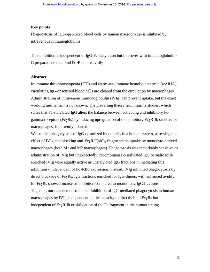
Key points
Phagocytosis of IgG-opsonized blood cells by human macrophages is inhibited by
intravenous immunoglobulins
This inhibition is independent of IgG-Fc sialylation but improves with immunoglobulin-
G preparations that bind FcγRs more avidly
Abstract
In immune thrombocytopenia (ITP) and warm autoimmune hemolytic anemia (wAIHA),
circulating IgG-opsonized blood cells are cleared from the circulation by macrophages.
Administration of intravenous immunoglobulin (IVIg) can prevent uptake, but the exact
working mechanism is not known. The prevailing theory from murine studies, which
states that Fc-sialylated IgG alters the balance between activating and inhibitory Fc-
gamma receptors (FcγRs) by inducing upregulation of the inhibitory FcγRIIb on effector
macrophages, is currently debated.
We studied phagocytosis of IgG-opsonized blood cells in a human system, assessing the
effect of IVIg and blocking anti-FcγR F(ab’)2 fragments on uptake by monocyte-derived
macrophages (both M1 and M2 macrophages). Phagocytosis was remarkably sensitive to
administration of IVIg but unexpectedly, recombinant Fc-sialylated IgG or sialic acid-
enriched IVIg were equally active as unsialylated IgG fractions in mediating this
inhibition - independent of FcγRIIb expression. Instead, IVIg inhibited phagocytosis by
direct blockade of FcγRs. IgG fractions enriched for IgG-dimers with enhanced avidity
for FcγRs showed increased inhibition compared to monomeric IgG fractions.
Together, our data demonstrate that inhibition of IgG-mediated phagocytosis in human
macrophages by IVIg is dependent on the capacity to directly bind FcγRs but
independent of FcγRIIb or sialylation of the Fc fragment in the human setting.
For personal use only.on November 10, 2014. by guest www.bloodjournal.orgFrom
3
Introduction
Human immunoglobulin G (IgG) antibodies directed against blood cells can cause
destruction of these cells leading to anemia or thrombocytopenia. This occurs for instance
in warm antibody auto-immune hemolytic anemia (wAIHA) or immune
thrombocytopenia (ITP) where IgG auto-antibodies against erythrocytes or thrombocytes,
respectively, are formed. These IgG-opsonised blood cells are cleared by macrophages in
the spleen and liver, which recognize and destroy IgG-opsonized cells by Fc-gamma
receptors (FcγRs).1 Human FcγRs are divided into high-affinity receptors (FcγRI (CD64),
which has a high affinity for its ligand2), and the low-affinity receptors (the different
isoforms of FcγRs II (CD32) and III (CD16)), which have a lower affinity for IgG and
bind monomeric IgG less efficiently than FcγRI, but do bind to dimeric and multimeric
IgG.2,3 Five of these human FcγRs potentially play a role in the phagocytosis of IgG-
opsonized blood cells by macrophages. The classical activating FcγRs (FcγRI, IIa and
IIIa) are all independently capable of inducing phagocytosis by monocyte-derived
macrophages when crosslinked,4 but the relative contributions of each of these receptors
to phagocytosis of IgG-opsonized particles by different types of macrophages are not
well known. FcγRIIc, an activating receptor that is expressed only in a minority of
individuals, may also play a role and its presence has been shown to contribute to ITP
susceptibility.5 On the other hand, FcγRIIb, the only inhibitory FcγR, has been reported
to inhibit the pro-phagocytic signals.6
Therapeutic strategies for the treatment of ITP and wAIHA include the administration of
intravenous immunoglobulin (IVIg).7-9 IVIg is a first-line treatment for ITP in situations
in which a rapid increase in the number of platelets is warranted.8 Although many
different modes of action for the immune-modulating effect of IVIg have been proposed,
the exact mechanism by which IVIg immediately alleviates the clearance of IgG-
opsonized blood cells is still debated. The therapeutic effects of IVIg are somewhat
surprising considering that its main component, IgG, is normally present in plasma at
high concentrations. Possibly, a specific fraction of the IgG present in IVIg preparations
is responsible for the therapeutic effect. Since high doses of 1-2 g/kg of IVIg are needed
to induce a good therapeutic response, more targeted therapies may potentially be
developed. It is known that the effect of IVIg lies within the Fc-fragments of the IgG,
For personal use only.on November 10, 2014. by guest www.bloodjournal.orgFrom
4
because purified Fc-fragments alone were an effective treatment for ITP,10 whereas IVIg
preparations depleted of Fc-fragments were not effective.11 Direct blockade of the FcγRs
by the Fc-fragments of IgG molecules is one of the possible explanations,1,12 and IgG-
dimers and -multimers (present at low concentrations in IVIg) have been implicated as an
active fraction,13,14 due to their capacity to more efficiently bind and block low-affinity
FcγRs. Several murine studies have indeed suggested IgG-dimers to be the most effective
part of IVIg in ameliorating ITP,13-16 although a recent report showed that they are not
absolutely required, as purely monomeric fractions were also able to ameliorate murine
ITP.17
Over the past decade however, the focus of research on immune modulation by IVIg has
shifted to the role of the inhibitory FcγRIIb and glycosylation of IgG molecules at the Fc
tail. Upregulation of FcγRIIb on splenic macrophages was implicated as the working
mechanism of IVIg in a mouse model of ITP.18 Since then, several reports have suggested
that the minor IgG fraction with sialic acid-containing N-linked glycans at Asn297 in the
Fc region is important for this immune modulating effect of IVIg.19-21 This has led to a
model for IVIg activity in ITP in which sialylated IgG binds to SIGN- R1 or its human
homologue DC-SIGN on macrophages, leading to increased expression of the inhibitory
FcγRIIb on effector macrophages.22-24 However, sialylation did not affect efficacy of
IVIg in two other reports using mouse models of ITP.25-27 Taken together, the studies in
mouse models are inconclusive with regard to the working mechanism of specific
fractions of IVIg.
Besides these inconsistent results in murine models, human FcγRs are very different from
those in mice. Therefore, we set out to directly compare the different components of IVIg
in a fully human in vitro system. Using monocyte-derived macrophages, we studied the
effect on the phagocytosis of human erythrocytes that were opsonized with a human
polyclonal anti-RhD serum. The study revealed that IVIg had a direct blocking effect on
phagocytosis, which was even stronger in the presence of dimeric and multimeric IgG
preparations, whereas sialic acid-enriched fractions of IVIg or recombinant Asn297-
α2,6sialic acid-containing IgG did not show increased effects. Saturation of FcγRI and
FcγRIIa were sufficient to explain the inhibitory effects of IVIg, whereas no inhibitory
role could be ascribed to FcγRIIb in human macrophages.
For personal use only.on November 10, 2014. by guest www.bloodjournal.orgFrom
5
Materials and Methods
Human samples
Heparinized blood samples were obtained from healthy volunteers. The study was
approved by the Medical Ethics Committee of the Academic Medical Center, and was
performed in accordance with the Declaration of Helsinki.
Culture of monocyte-derived macrophages
Monocytes were isolated from peripheral blood mononuclear cells with a CD14 MACS
isolation kit (Miltenyi Biotec). Cells were cultured in 6-wells (1 x 10⁶ cells/well) or 24-
wells (0.2 x 10⁶ cells/well) plates in complete IMDM-medium (Gibco) containing 10%
FCS (Bodinco, Alkmaar, The Netherlands) and antibiotics, stimulated with either 50
ng/ml M-CSF (eBioscience) or 10 ng/ml GM-CSF (Peprotech) for 9 days.
Phagocytosis assay
Erythrocytes of donors positive for the RhD blood group were isolated and stained with
cell tracker CFSE (Life technologies) for 30 minutes, and were subsequently opsonized
with clinical grade human anti-RhD antibody (RheDQuin, Sanquin, The Netherlands) at
1.56 IE/ml for 30 minutes, after which excess anti-RhD antibody was washed away, or
were left unopsonized.
Erythrocytes were added to macrophages in the 24-wells plates in which they were
cultured at a ratio of 10 erythrocytes to 1 macrophage, and were incubated at 37°C for 20
minutes (M-CSF macrophages) or 2 hours (GM-CSF macrophages). Phagocytosis was
stopped by transferring cells to ice, and erythrocytes that were not phagocytozed were
lyzed using an isotonic ammonium chloride lysis buffer at 4°C for 5 minutes, two times.
Lysis buffer was washed away with PBS. Microscopy pictures were taken with an EVOS
microscope (Life Technologies) and macrophages were subsequently detached from the
surface of culture plates by vigorous pipetting after a 15 minute incubation with a PBS
buffer containing 125 mM lidocaine (Sigma Aldrich) and 10 mM EDTA (Merck).
Ingestion of CFSE-positive erythrocytes by macrophages was quantified by flow
cytometry on a FACS CANTO II (BD). IgG-mediated phagocytosis was calculated as
For personal use only.on November 10, 2014. by guest www.bloodjournal.orgFrom
6
percentage positive macrophages subtracted by the percentage positive macrophages of
the unopsonized control.
Specific MoAbs or their fragments to block phagocytosis were incubated with the
macrophages for 5 minutes prior to the addition of erythrocytes, all at 10 μg/ml. Intact
anti-CD64 clone 10.1 (BioLegend), F(ab’) 2 fragment anti-CD64 clone 10.1 (Ancell),
F(ab’) 2 fragment anti-CD32 clone 7.3 (Ancell), Fab fragments anti-CD32b clone 2B6 (a
generous gift from Macrogenics), F(ab’) 2 fragment anti-CD16 clone 3G8 (Ancell).
Different IVIg preparations (see below) were also added 5 minutes prior to the start of
phagocytosis, unless indicated otherwise. Data of blocking of phagocytosis are shown
normalized to unblocked IgG-mediated phagocytosis. Experiments with less than 5%
IgG-mediated phagocytosis (only 6/138 experiments) were excluded from analysis in
blocking studies.
Preparation of monomeric and dimeric IgG
Normal IVIg was Nanogam 50 mg/ml (Sanquin, The Netherlands). Nanogam was
fractionated into different size components in the following way: 2 ml Nanogam
(containing 50mg/ml polyclonal IgG) was fractionated with LC-SEC (AKTA system, GE
Healthcare, Sweden) using Superdex200 column 16/60 and PBS pH 7.4 as elution buffer.
The fraction corresponding to dimeric IgG was pooled from several runs.
Dimeric idiotype-anti-idiotype immune complexes were generated starting from
equimolar amounts of a human IgG1 monoclonal anti-Adalimumab antibody 2.6
(described previously28) and Adalimumab (Human IgG1, AbbVie). The composition of
each preparation was analyzed by injecting 50 μl of 30 mg/ml (Nanogam), 3 mg/ml
(dimer-enriched IVIg), or 100 μg/ml (Adalimumab/anti-adalimumab complexes) into
LC_SEC using superdex200 column 10/30 with PBS pH 7.4 as elution buffer.
IgG preparations with altered levels of galactosylation and sialylation.
Fractions of IVIg enriched or depleted for sialic acid by SNA were prepared as described
previously25. Neuraminidase treatment of IVIg was performed essentially as described
previously19,26, see Supplementary Methods for details. Recombinant IgG was enriched
for sialic acid-Fc by co-transfection of HEK-293F Freestyle cells with human IgG1 anti-
For personal use only.on November 10, 2014. by guest www.bloodjournal.orgFrom
7
2,4,6-trinitrophenol (TNP) and galactosyl- and sialyltransferases as described in detail in
the Supplementary Methods.
Flow cytometry, Mass Spectrometry, Genotyping and Quantitative mRNA analysis.
Flow cytometry for surface expression on monocyte-derived macrophages, Mass
Spectrometry for IgG-Fc glycosylation, Genotyping of healthy individuals for FCGR
genetic variation and quantitative mRNA analysis of IVIg-stimulated macrophages were
performed as described in the Supplemental Methods.
Statistical analysis
Statistical analysis was performed using GraphPad Prism 6.02. For comparison of
expression levels and the level of IgG-mediated phagocytosis a Kruskal-Wallis test or
Mann-Whitney test was used. For comparison of blocking studies ANOVA was used. In
case this revealed a p-value < 0.05 (Bonferroni corrected per figure, uncorrected p-values
are shown), subsequent testing was performed with unpaired t-tests.
Results
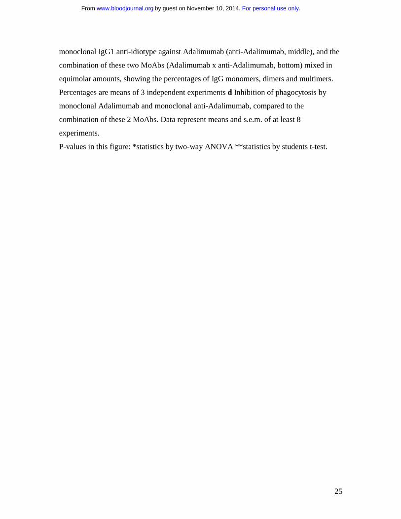
FcγRI, FcγRIIa, FcγRIIb and FcγRIII are expressed on M-CSF and GM-CSF
cultured macrophages
Culturing of peripheral blood monocytes with M-CSF is known to induce differentiation
towards a general anti-inflammatory (M2) macrophage phenotype, whereas GM-CSF
induces macrophages with a pro-inflammatory (M1) phenotype29 (Suppl.Fig.1). When
analyzing the expression pattern of the different FcγRs after differentiation with these
stimulants for 9 days, we detected FcγRI, IIa, IIb and III on the cell surface (Fig.1a-e).
Staining with MoAb AT10, recognizing FcγRIIa but also FcγRIIb and c, resulted in the
highest fluorescence intensities, especially in the M-CSF macrophages (Fig.1c). To
evaluate expression of FcγRIIb, we stained with MoAb 2B6, which actually binds the
identical extracellular domains of both FcγRIIb and FcγRIIc30. To ensure specificity for
FcγRIIb, we excluded individuals carrying the open reading frame allele of FCGR2C
from this analysis. FcγRIIb expression was indistinguishable for M-CSF and GM-CSF
macrophages (Fig.1d), indicating that the observed increased FcγRII expression
For personal use only.on November 10, 2014. by guest www.bloodjournal.orgFrom
8
originated from FcγRIIa. Whereas FcγRI and FcγRII isoforms were always present on the
whole population of macrophages, FcγRIIIa showed a bimodal distribution (Fig.1a), with
very low or absent expression on the majority of cells, but higher expression on a subset
of the macrophages (Fig.1f). A time course of the expression levels during differentiation
into macrophages revealed that FcγRI was re-expressed on the macrophages after an
initial decrease compared to levels on monocytes, whereas expression levels of the other
FcγRs steadily increased (Suppl.Fig.2).
Macrophages phagocytose erythrocytes opsonized by human anti-RhD IgG
To assess the capacity of the macrophages to phagocytose IgG-opsonized blood cells we
used a phagocytosis assay with RhD-positive human erythrocytes opsonized with
polyclonal human anti-RhD antibodies as the phagocytic target. Incubation of IgG-
opsonized erythrocytes with the macrophages at 37 °C led to binding and phagocytosis
(Fig.2a). To distinguish binding from phagocytosis, non-ingested extracellular
erythrocytes were lysed (Fig.2a). This efficiently lysed all non-ingested erythrocytes, as
no erythrocytes could be detected after lysis in macrophages pretreated with cytochalasin
B, which inhibits phagocytosis by interfering with actin polymerization (Fig.2b).
Phagocytosis was quantified by CFSE-labeling of the erythrocytes and detection of
macrophages that have taken up one or more erythrocytes by flow cytometry (Fig.2b). A
time course showed that phagocytosis was faster and more extensive in M-CSF
macrophages than in GM-CSF macrophages (Fig.2c). Overnight incubation did not
increase the percentage positive macrophages in GM-CSF cultures (data not shown).
Incubation times of 20 minutes (M-CSF) and 2 hours (GM-CSF) were chosen as optimal
times for read-out and were used in all subsequent experiments. An overview of the range
of phagocytosis at these timepoints is given in Fig.2d. The extent of phagocytosis was not
significantly correlated to FcγR expression levels (data not shown) or to SNPs in the low-
affinity FcγRs (Suppl.Fig.3).
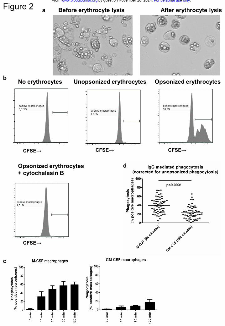
Differential role of FcγRs in M-CSF and GM-CSF macrophages
To determine the relative contribution of the different FcγRs to the phagocytosis of IgG-
opsonized erythrocytes, we used FcγR-specific blocking F(ab’)2 fragments. The
For personal use only.on November 10, 2014. by guest www.bloodjournal.orgFrom
9
combination of F(ab’)2 fragments against all FcγRs induced an almost complete
inhibition of phagocytosis, especially in GM-CSF macrophages (Fig.3). The blocking
F(ab’)2 against FcγRII in M-CSF macrophages was strongest, whereas F(ab’)2 against
FcγRI had most effect in inhibiting phagocytosis with GM-CSF macrophages. Blocking
Fab fragments of MoAb 2B6 (against FcγRIIb) and F(ab’)2 fragments against FcγRIII
had only a minor effect in both types of macrophages. Whereas the MoAbs against
FcγRII (clone 7.3), FcγRIIb (clone 2B6) and FcγRIII (clone 3G8) that we used recognize
the IgG binding-site of the FcγRs and block IgG binding as F(ab’)2 fragments31-33,
blocking experiments with F(ab’)2 fragments of MoAb 10.1 were more difficult to
interpret, because the epitope of FcγRI recognized by this antibody is not located at the
IgG-binding site itself, although close enough to interfere with binding34. To compare the
blocking capacity of 10.1 F(ab’)2 fragments to intact 10.1, we performed a rosetting assay
with FCGR1A1-transfected 293T cells (Suppl.Fig.4a-c). This revealed that intact 10.1
was able to completely inhibit binding of IgG-opsonized erythrocytes to FcγRI at the
concentration used in our phagocytosis assay, but F(ab’)2 fragments could not inhibit
binding completely, even at high concentrations. In fact, when intact 10.1 was used in the
phagocytosis assay, inhibition was much more striking in both types of macrophages
(Suppl.Fig.4d), and phagocytosis could be completely inhibited with a combination of
intact antibodies against all three FcγRs. However, with the use of intact antibodies,
blocking of other FcγRs through the Fc-fragment (Kurlander phenomenon35) cannot be
excluded.
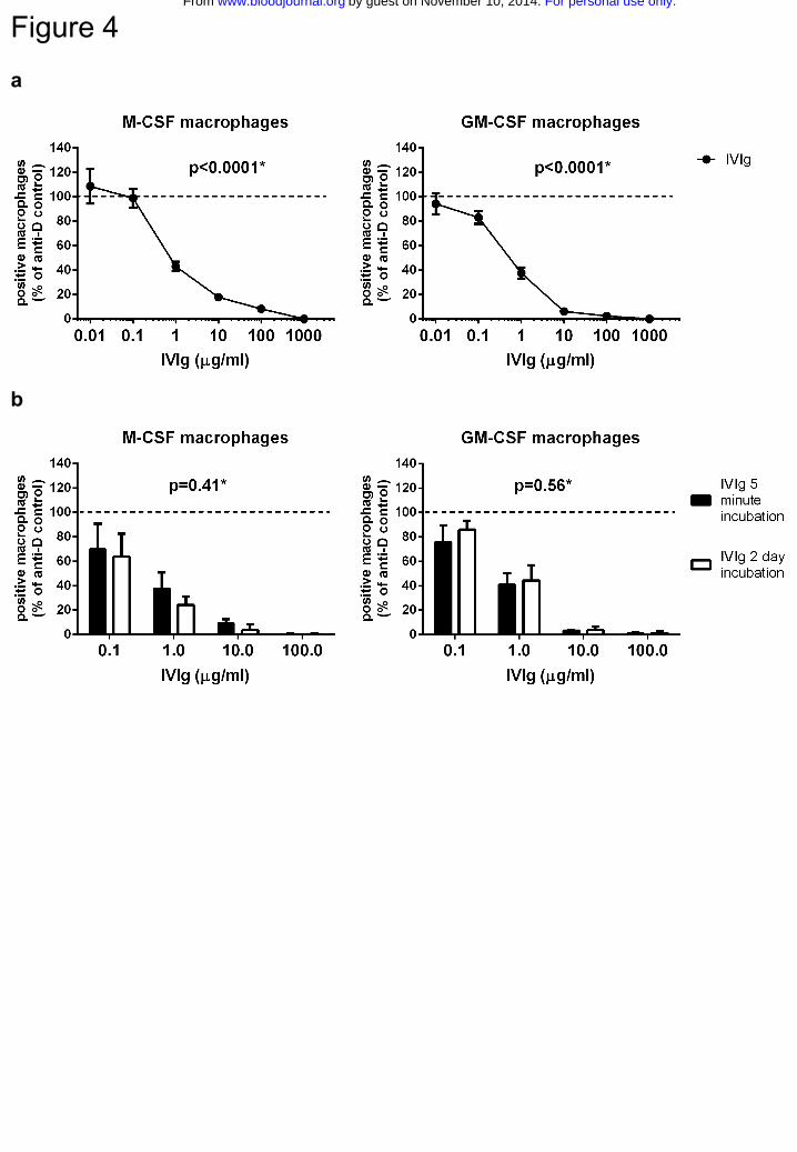
IVIg directly inhibits phagocytosis of IgG-opsonized erythrocytes
IVIg has previously been shown to be able to inhibit macrophage phagocytosis of IgG-
opsonized erythrocytes in experimental setups in which macrophages were incubated for
one hour with IVIg, after which the IVIg was washed away before the start of
phagocytosis14,36. To test if IVIg also had the capacity to directly inhibit IgG-mediated
phagocytosis in our in vitro system, we added IVIg to our macrophages just prior to
starting the phagocytosis. IVIg inhibited phagocytosis in a dose-dependent manner, with
doses as low as 1 μg/ml still showing some effect in both types of macrophages (Fig.4a).
For personal use only.on November 10, 2014. by guest www.bloodjournal.orgFrom
10
IVIg was equally effective when added two days before the phagocytosis was initiated
(Fig.4b).
Inhibition of phagocytosis by IVIg is independent of Fc-sialylation and does not
require an increase of FcγRIIb expression
The inhibitory effect of sialylated IVIg was first reported using an enrichment by
Sambucus nigra agglutinin (SNA) lectin fractionation19. When we used preparations that
were enriched or depleted for sialic acid in this way, we could not detect any differences
in capacity to inhibit phagocytosis (Fig.5a). However, enrichment of IVIg for sialic acid
by SNA lectin fractionation only results in an enrichment of Fab-sialylated but not Fc-
sialylated IgG25,37. Because the presumed effects of sialylated IgG would be Fc-sialic
acid specific21, we subsequently used neuraminidase-treated IVIg to determine if a
preparation depleted of Fc-sialic acid was still capable of inhibiting phagocytosis. Despite
the fact that neuraminidase removed almost 85% of the sialic acid residues at the N297
linked glycan, the neuraminidase-treated IVIg was equally effective as untreated IVIg
(Fig.5b). Finally, we tried an IgG preparation enriched or depleted for sialic acid Fc by
using a non-Fab-glycosylated recombinant anti-2,4,6-trinitrophenyl MoAb, enriched for
Fc-sialylation by expressing the antibody with or without galactosyltransferases and
sialyltransferases. This resulted in an α2,6-sialic acid content of 14.5% in the Fc-
sialylated IgG and 0.4% in non Fc-sialylated IgG (Fig.5c). These were equally capable of
inhibiting phagocytosis, showing that the inhibition of phagocytosis was completely
independent of IgG-Fc sialylation status (Fig.5d).
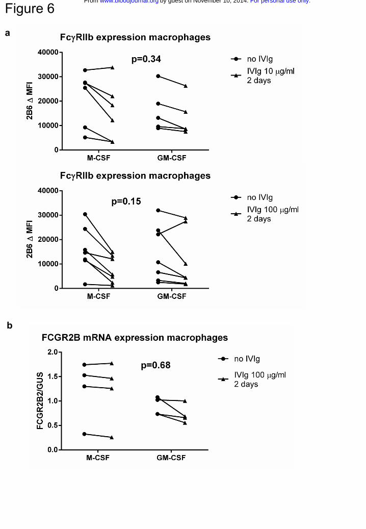
To determine if the observed inhibitory effects were the result of an upregulation of
FcγRIIb, as previously proposed18, we determined FcγRIIb expression levels by staining
macrophages pre-incubated with IVIg for 2 days with MoAb 2B6. This revealed no
increase in expression levels of FcγRIIb in either M-CSF or GM-CSF macrophages
(Fig.6a). To rule out the possibility that we could not detect an increased expression of
FcγRIIb because of competition of the IgG in IVIg for the binding site of MoAb 2B6, we
also measured mRNA levels of FCGR2B in macrophages incubated with IVIg. This
revealed no increase in FCGR2B mRNA (Fig.6b). Together, these results indicate that
For personal use only.on November 10, 2014. by guest www.bloodjournal.orgFrom
11
IVIg could inhibit phagocytosis directly, independent of changes in expression of
FcγRIIb.
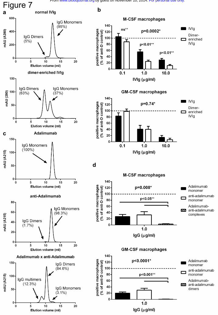
Dimeric IgG most effectively blocks phagocytosis of IgG-opsonized erythrocytes
We then tested whether preparations of IVIg rich in dimeric IgG, with a higher avidity for
FcγRs, were more capable of inhibiting phagocytosis. We first prepared a fraction of
IVIg enriched for IgG-dimers by HPLC fractionation of IVIg, which led to an increase of
dimeric IgG from ~5% to ~63% (Fig.7a). This fraction was even more effective than
normal IVIg in inhibiting phagocytosis in the M-CSF, but not the GM-CSF macrophages
(Fig.7b). Subsequently, to test IgG preparations with even higher contents of dimeric
IgG, we made use of the ability of a human anti-idiotype MoAb against the TNF-
blocking MoAb adalimumab to form complexes with adalimumab28. Monoclonal
fractions of these two antibodies consist of IgG monomers, but a combination of the two
results in the formation of dimers and also some multimers (Fig.7c). When using either of
the two monoclonal antibodies alone, phagocytosis was still inhibited to the same extent
as inhibition with normal IVIg, showing that also purely monomeric IgG has an
inhibiting effect on phagocytosis. However, the combination of the two antibodies,
having formed dimers, was much more effective than either of the two monoclonal
antibodies alone, even in the GM-CSF macrophages (Fig.7d).
Discussion
Our data demonstrate that the inhibiting effect of IVIg on the phagocytosis of IgG-
opsonized blood cells,14,36 is a very direct effect in our fully human in vitro system, since
IVIg was effective after a very short incubation time of only five minutes. Addition of
IVIg to cultured macrophages resulted in a dose-dependent inhibition of phagocytosis of
IgG-opsonized erythrocytes, which could be enhanced with preparations containing more
IgG dimers and multimers but was independent of IgG-Fc and IgG-Fab sialylation. Since
IgG was effective after a very short incubation we presume that the most likely working
mechanism is a direct blockade of FcγRs.
For personal use only.on November 10, 2014. by guest www.bloodjournal.orgFrom
12
Following the discovery that IVIg induces a steep increase in the circulating platelet
counts in ITP7, many theories regarding the working mechanism have been proposed,
including inhibition of phagocytosis as a result of blockade of activating FcγRs. Despite
the fact that many observations support this mechanism1,12,38, the current paradigm is that
IVIg induces upregulation of inhibitory FcγRIIb on effector macrophages18,23, although
this was only shown in mice. However, we did not detect any upregulation of this
receptor even after two days of incubation with IVIg in our macrophages, clearly
showing that IVIg has very potent effects that are independent of an increased expression
of FcγRIIb. Although we cannot completely rule out upregulation of FcγRIIb in response
to IVIg in vivo, it was recently postulated that this receptor may not actually be required
for the protective effect of IVIg in murine models in all circumstances39. In any case, our
findings do not support any role for FcγRIIb in the working mechanism of IVIg in
preventing clearance of opsonized blood cells by human macrophages. Neither could we
detect a direct effect of sialylation of the N-linked Fc-glycan at position 297 on the
clearance of IgG-opsonized blood cells. Our findings, in conjunction with the conflicting
results obtained in murine studies of ITP comparing sialic acid enriched versus sialic acid
depleted IVIg 22,25,26, do not support a role for the sialylation of IgG-Fc in the working
mechanism of IVIg preventing uptake of IgG-opsonized blood cells.
Being such a potent direct inhibitor of FcγR-mediated phagocytosis in vitro, we assume
that IVIg will also be able to block this process in macrophages in vivo. The notion that
FcγRs can be blocked by IgG in vivo may seem surprising given that IgG is normally
present in the circulation at ~7–16 mg/ml, which would be expected to block FcγRs in
vivo already under steady state conditions. Monomeric IgG has been shown to bind
FcγRIIa40. Therefore, monomeric IgG will probably form an equilibrium with low-
affinity FcγRs in vivo, with a proportion of the low-affinity FcγRs bound by IgG and a
proportion freely available. The administration of IVIg could shift this balance towards a
higher proportion of FcγRs being occupied, leaving too little FcγRs freely available for
binding to IgG-opsonized blood cells. IgG dimers and polymers in IVIg will even more
efficiently block the low-affinity FcγRs, as they have a higher avidity.
For personal use only.on November 10, 2014. by guest www.bloodjournal.orgFrom
13
We indeed found an increased effect of IgG-dimers and -multimers in our phagocytosis
studies, although the presence was not absolutely necessary, and in M1 macrophages it
could only be detected with preparations with a high IgG-dimer and -multimer content.
This differential sensitivity reflects the differences in FcγR usage between M1 and M2
macrophages. Phagocytosis by M-CSF macrophages relies greatly on the low-affinity
FcγRII which is much more effectively blocked by IgG-dimers than monomers, whereas
GM-CSF macrophages rely more exclusively on FcγRI which is already fully occupied
by IgG-monomers alone. Our experiments with F(ab’)2 fragments indicate that FcγRI is
relevant to the phagocytosis by monocyte-derived macrophages, especially when cultured
with GM-CSF. The effects with the F(ab’)2 fragments will be an underrepresentation of
the actual role of FcγRI, as MoAb 10.1 (nor any other available MoAb against FcγRI)
does not block efficiently as a F(ab’)2 fragment. However, the effects of the intact 10.1
MoAb may rather be an overrepresentation of the role of FcγRI, as the Fc-tail can block
other adjacent FcγRs as described by Kurlander35.
Macrophages of the spleen and liver are responsible for the clearance of IgG-opsonized
blood cells in vivo, but remarkably little is known about expression levels of FcγRs on
these macrophages. It has been proposed that mainly the low-affinity FcγRs are
involved1, based only on circumstantial evidence such as association studies with genetic
polymorphisms in the low-affinity FcγRs5,41, and in vivo blocking studies with specific
FcγR MoAbs in limited series of patients42,43. If it is indeed the case that these
macrophages have a predominant expression of low-affinity FcγRs, we may expect the
difference between dimeric and monomeric IgG to be even greater than the difference we
have found in monocyte-derived macrophages.
Concluding, the IVIg-induced inhibition of phagocytosis of IgG-opsonized blood cells by
human macrophages -contrasting the current prevailing theory23- is not dependent on Fc-
sialylation or FcγRIIb upregulation. The lack of support for this theory from our data in
human macrophages is in line with multiple recent studies challenging the concept of Fc-
sialylation and/or FcγRIIb being important for IVIg efficacy in various diseases, as
studied in murine models25,26,39,44 or with human material37,45,46. Therefore, the
For personal use only.on November 10, 2014. by guest www.bloodjournal.orgFrom
14
importance of IgG Fc-sialylation and upregulation of FcγRIIb in IVIg treatment needs to
be reconsidered.
Instead, the capacity of IgG molecules to bind and block FcγRs seems to be more
important, at least when inhibiting phagocytosis of IgG-opsonized blood cells. These
findings can help to improve the treatment of ITP or wAIHA with IVIg, as preparations
with increased binding to low-affinity FcγRs, such as IgG-dimers or recombinant
engineered IgGs with an increased affinity of the Fc-tail for FcγR may form suitable
alternatives requiring a lower dosage than normal IVIg.
Acknowledgements
This work was supported by a grant from the Landsteiner Foundation for
Bloodtransfusion Research (LSBR #0916), and a grant of the PPOP program 'Sweet
IVIG: a blend of different tastes' (PPOP-12-001). The authors would like to thank Mrs.
Ninotska IL Derksen and Mrs. Ornella Felizolla (Sanquin, Amsterdam, the Netherlands)
and Carolien Koeleman (Leiden University Medical Center) for assistance with the
experiments.
Author Contributions
S.Q.N. performed experiments, analyzed data and wrote the manuscript, G.D., I.K.,
F.S.B., J.G. and R.P. performed experiments and analyzed data, M.W., G.V., T.R. and
T.K.B. designed experiments, interpreted and discussed data, T.W.K. discussed data,
designed the study and wrote the manuscript.
Conflict-of-interest Disclosure
All authors declare to have no competing financial interest
Reference List
1. Crow AR, Lazarus AH. Role of Fcgamma receptors in the pathogenesis and
treatment of idiopathic thrombocytopenic purpura. J.Pediatr.Hematol.Oncol.
2003;25 Suppl 1:S14-S18.
For personal use only.on November 10, 2014. by guest www.bloodjournal.orgFrom
15
2. Bruhns P, Iannascoli B, England P et al. Specificity and affinity of human
Fcgamma receptors and their polymorphic variants for human IgG subclasses.
Blood 2009;113(16):3716-3725.
3. Lux A, Yu X, Scanlan CN, Nimmerjahn F. Impact of immune complex size and
glycosylation on IgG binding to human FcgammaRs. J.Immunol.
2013;190(8):4315-4323.
4. Anderson CL, Shen L, Eicher DM, Wewers MD, Gill JK. Phagocytosis mediated by
three distinct Fc gamma receptor classes on human leukocytes. J.Exp.Med.
1990;171(4):1333-1345.
5. Breunis WB, van Mirre E, Bruin M et al. Copy number variation of the activating
FCGR2C gene predisposes to idiopathic thrombocytopenic purpura. Blood
2008;111(3):1029-1038.
6. Hunter S, Indik ZK, Kim MK et al. Inhibition of Fcgamma receptor-mediated
phagocytosis by a nonphagocytic Fcgamma receptor. Blood 1998;91(5):1762-1768.
7. Imbach P, Barandun S, d'Apuzzo V et al. High-dose intravenous gammaglobulin for
idiopathic thrombocytopenic purpura in childhood. Lancet 1981;1(8232):1228-
1231.
8. Provan D, Stasi R, Newland AC et al. International consensus report on the
investigation and management of primary immune thrombocytopenia. Blood
2010;115(2):168-186.
For personal use only.on November 10, 2014. by guest www.bloodjournal.orgFrom
16
9. Flores G, Cunningham-Rundles C, Newland AC, Bussel JB. Efficacy of
intravenous immunoglobulin in the treatment of autoimmune hemolytic anemia:
results in 73 patients. Am.J.Hematol. 1993;44(4):237-242.
10. Debre M, Bonnet MC, Fridman WH et al. Infusion of Fc gamma fragments for
treatment of children with acute immune thrombocytopenic purpura. Lancet
1993;342(8877):945-949.
11. Burdach SE, Evers KG, Geursen RG. Treatment of acute idiopathic
thrombocytopenic purpura of childhood with intravenous immunoglobulin G:
comparative efficacy of 7S and 5S preparations. J.Pediatr. 1986;109(5):770-775.
12. Bussel JB. Fc Receptor Blockade and Immune Thrombocytopenic Purpura.
Seminars in Hematology. 2000;37(3):261-266.
13. Teeling JL, Jansen-Hendriks T, Kuijpers TW et al. Therapeutic efficacy of
intravenous immunoglobulin preparations depends on the immunoglobulin G
dimers: studies in experimental immune thrombocytopenia. Blood
2001;98(4):1095-1099.
14. Bazin R, Lemieux R, Tremblay T, St-Amour I. Tetramolecular immune complexes
are more efficient than IVIg to prevent antibody-dependent in vitro and in vivo
phagocytosis of blood cells. Br.J.Haematol. 2004;127(1):90-96.
15. Machino Y, Suzuki E, Higurashi S et al. Chemically dimerized intravenous
immunoglobulin has potent ameliorating activity in a mouse immune
For personal use only.on November 10, 2014. by guest www.bloodjournal.orgFrom
17
thrombocytopenic purpura model. Biochem.Biophys.Res.Commun.
2012;418(4):748-753.
16. Bazin R, Lemieux R, Tremblay T. Reversal of immune thrombocytopenia in mice
by cross-linking human immunoglobulin G with a high-affinity monoclonal
antibody. Br.J.Haematol. 2006;135(1):97-100.
17. Tremblay T, Pare I, Bazin R. Immunoglobulin G dimers and immune complexes are
dispensable for the therapeutic efficacy of intravenous immune globulin in murine
immune thrombocytopenia. Transfusion 2013;53(2):261-269.
18. Samuelsson A, Towers TL, Ravetch JV. Anti-inflammatory activity of IVIG
mediated through the inhibitory Fc receptor. Science 2001;291(5503):484-486.
19. Kaneko Y, Nimmerjahn F, Ravetch JV. Anti-inflammatory activity of
immunoglobulin G resulting from Fc sialylation. Science 2006;313(5787):670-673.
20. Anthony RM, Kobayashi T, Wermeling F, Ravetch JV. Intravenous gammaglobulin
suppresses inflammation through a novel T(H)2 pathway. Nature
2011;475(7354):110-113.
21. Anthony RM, Wermeling F, Karlsson MC, Ravetch JV. Identification of a receptor
required for the anti-inflammatory activity of IVIG. Proc.Natl.Acad.Sci.U.S.A
2008;105(50):19571-19578.
For personal use only.on November 10, 2014. by guest www.bloodjournal.orgFrom

18
22. Schwab I, Biburger M, Kronke G, Schett G, Nimmerjahn F. IVIg-mediated
amelioration of ITP in mice is dependent on sialic acid and SIGNR1.
Eur.J.Immunol. 2012;42(4):826-830.
23. Schwab I, Nimmerjahn F. Intravenous immunoglobulin therapy: how does IgG
modulate the immune system? Nat.Rev.Immunol. 2013;13(3):176-189.
24. Schwab I, Mihai S, Seeling M et al. Broad requirement for terminal sialic acid
residues and FcgammaRIIB for the preventive and therapeutic activity of
intravenous immunoglobulins in vivo. Eur.J.Immunol. 2014;44(5):1444-1453.
25. Guhr T, Bloem J, Derksen NI et al. Enrichment of sialylated IgG by lectin
fractionation does not enhance the efficacy of immunoglobulin G in a murine model
of immune thrombocytopenia. PLoS.One. 2011;6(6):e21246.
26. Leontyev D, Katsman Y, Ma XZ et al. Sialylation-independent mechanism involved
in the amelioration of murine immune thrombocytopenia using intravenous
gammaglobulin. Transfusion 2012;52(8):1799-1805.
27. von Gunten S, Shoenfeld Y, Blank M et al. IVIG pluripotency and the concept of
Fc-sialylation: challenges to the scientist. Nat.Rev.Immunol. 2014;14(5):349.
28. Rispens T, Ooijevaar-de HP, Derksen NI et al. Nanomolar to sub-picomolar affinity
measurements of antibody-antigen interactions and protein multimerizations:
fluorescence-assisted high-performance liquid chromatography. Anal.Biochem.
2013;437(2):118-122.
For personal use only.on November 10, 2014. by guest www.bloodjournal.orgFrom
19
29. Verreck FA, de BT, Langenberg DM et al. Human IL-23-producing type 1
macrophages promote but IL-10-producing type 2 macrophages subvert immunity
to (myco)bacteria. Proc.Natl.Acad.Sci.U.S.A 2004;101(13):4560-4565.
30. van der Heijden J, Breunis WB, Geissler J et al. Phenotypic variation in IgG
receptors by nonclassical FCGR2C alleles. J.Immunol. 2012;188(3):1318-1324.
31. Ierino FL, Hulett MD, McKenzie IF, Hogarth PM. Mapping epitopes of human Fc
gamma RII (CDw32) with monoclonal antibodies and recombinant receptors.
J.Immunol. 1993;150(5):1794-1803.
32. Fleit HB, Wright SD, Unkeless JC. Human neutrophil Fc gamma receptor
distribution and structure. Proc.Natl.Acad.Sci.U.S.A 1982;79(10):3275-3279.
33. Veri MC, Gorlatov S, Li H et al. Monoclonal antibodies capable of discriminating
the human inhibitory Fcgamma-receptor IIB (CD32B) from the activating
Fcgamma-receptor IIA (CD32A): biochemical, biological and functional
characterization. Immunology 2007;121(3):392-404.
34. Dougherty GJ, Selvendran Y, Murdoch S, Palmer DG, Hogg N. The human
mononuclear phagocyte high-affinity Fc receptor, FcRI, defined by a monoclonal
antibody, 10.1. Eur.J.Immunol. 1987;17(10):1453-1459.
35. Kurlander RJ. Blockade of Fc receptor-mediated binding to U-937 cells by murine
monoclonal antibodies directed against a variety of surface antigens. J.Immunol.
1983;131(1):140-147.
For personal use only.on November 10, 2014. by guest www.bloodjournal.orgFrom
20
36. Foo AH, Ramkumar S, Helke S, Branch DR. Chemical treatment of anti-D results
in improved efficacy for the inhibition of Fcgamma receptor-mediated
phagocytosis. Transfusion 2007;47(12):2250-2259.
37. Kasermann F, Boerema DJ, Ruegsegger M et al. Analysis and functional
consequences of increased Fab-sialylation of intravenous immunoglobulin (IVIG)
after lectin fractionation. PLoS.One. 2012;7(6):e37243.
38. Clarkson SB, Bussel JB, Kimberly RP et al. Treatment of refractory immune
thrombocytopenic purpura with an anti-Fc gamma-receptor antibody.
N.Engl.J.Med. 1986;314(19):1236-1239.
39. Leontyev D, Katsman Y, Branch DR. Mouse background and IVIG dosage are
critical in establishing the role of inhibitory Fcgamma receptor for the amelioration
of experimental ITP. Blood 2012;119(22):5261-5264.
40. van Mirre E, Teeling JL, van der Meer JW, Bleeker WK, Hack CE. Monomeric IgG
in intravenous Ig preparations is a functional antagonist of FcgammaRII and
FcgammaRIIIb. J.Immunol. 2004;173(1):332-339.
41. Miescher S, Spycher MO, Amstutz H et al. A single recombinant anti-RhD IgG
prevents RhD immunization: association of RhD-positive red blood cell clearance
rate with polymorphisms in the FcgammaRIIA and FcgammaIIIA genes. Blood
2004;103(11):4028-4035.
For personal use only.on November 10, 2014. by guest www.bloodjournal.orgFrom
21
42. Bussel JB, Kimberly RP, Clarkson SB et al. Infusion of a monoclonal anti-FcR III
in patients with refractory ITP. Neo-Adjuvant Chemotherapy II. Vol 169. 1988:883-
887.
43. Ericson SG, Coleman KD, Wardwell K et al. Monoclonal antibody 197 (anti-Fc
gamma RI) infusion in a patient with immune thrombocytopenia purpura (ITP)
results in down-modulation of Fc gamma RI on circulating monocytes.
Br.J.Haematol. 1996;92(3):718-724.
44. Campbell IK, Miescher S, Branch DR et al. Therapeutic effect of IVIG on
inflammatory arthritis in mice is dependent on the Fc portion and independent of
sialylation or basophils. J.Immunol. 2014;192(11):5031-5038.
45. Ogata S, Shimizu C, Franco A et al. Treatment response in kawasaki disease is
associated with sialylation levels of endogenous but not therapeutic intravenous
immunoglobulin G. PLoS.One. 2013;8(12):e81448.
46. Yu X, Vasiljevic S, Mitchell DA, Crispin M, Scanlan CN. Dissecting the molecular
mechanism of IVIg therapy: the interaction between serum IgG and DC-SIGN is
independent of antibody glycoform or Fc domain. J.Mol.Biol. 2013;425(8):1253-
1258.
For personal use only.on November 10, 2014. by guest www.bloodjournal.orgFrom
22
Figure legends
Figure 1. FcγR expression on monocyte-derived macrophages
a Histograms showing expression levels of different FcγRs in monocyte-derived
macrophages cultured for 9 days with M-CSF. Gray shading: relevant isotype control b-e
comparison of expression levels of FcγRI, FcγRIIa,b, FcγRIIb and FcγRIII between M-
CSF and GM-CSF macrophages. Data represent the mean of experiments with
macrophages from 34 individuals, or 22 independent experiments in the case of FcγRII
(including only individuals without an FCGR2C-ORF). Some individuals were measured
twice, means of the measurements were used for these. Error bars represent s.e.m.,
statistics with Mann-Whitney test f percentages of FcγRIII-low and FcγRIII-high
macrophages (left panel, individual experiments are shown) and comparison of the
expression levels of the low and high populations (right panel, data represent the mean
and s.e.m. of at least 12 individuals)
Figure 2. Phagocytosis of anti-RhD opsonized erythrocytes by monocyte-derived
macrophages
a RhD positive erythrocytes opsonized with anti-RhD after phagocytosis by monocyte-
derived macrophages cultured for 9 days, before (left) and after (right) lysis of
erythrocytes. b Representative images of the quantification of phagocytosis by
flowcytometry. Unopsonized or anti-RhD opsonized CFSE labeled RhD positive
erythrocytes were incubated with macrophages, in the presence or absence of
cytochalasin B. Percentages indicated the percentage CFSE positive macrophages relative
to the total macrophage population. Data are representative of at least 6 (cytochalasin B)
experiments. c Time course of phagocytosis of anti-RhD opsonized erythrocytes by M-
CSF and GM-CSF macrophages. Data are representative of 3 independent experiments,
error bars represent s.e.m. d Comparison of IgG-mediated phagocytosis (corrected for
unopsonized phagocytosis) for M-CSF and GM-CSF macrophages (note different
timepoints). Mean and dots representing experiments with macrophages of different
individuals (n=58 for M-CSF, n=55 for GM-CSF) are shown. Some individuals were
For personal use only.on November 10, 2014. by guest www.bloodjournal.orgFrom
23
measured twice, mean of the measurements is shown for these. Statistics with Mann-
Whitney test.
Figure 3. M-CSF macrophages phagocytose mainly through FcγRIIa, whereas GM-
CSF macrophages phagocytose mainly through FcγRI
Effect of FcγR receptor blocking F(ab’)2 (α-FcγRI, α-FcγRIIa,b, α-FcγRIII) or Fab (α-
FcγRIIb) fragments on the phagocytosis of anti-RhD IgG-opsonized erythrocytes. Data
are normalized against phagocytosis of unblocked macrophages. For experiments with
Fab α-FcγRIIb individuals with an FCGR2C-ORF are excluded. Data represent means
and s.e.m. from 5-12 independent experiments. P values indicate results of one-way
ANOVA, subsequent unpaired t-tests were performed with results depicted as follows: *
p<0.05, ** p<0.01, ***p<0.001 (bonferroni corrected p values)
Figure 4. IVIg directly inhibits phagocytosis of IgG-opsonized erythrocytes
a Dose-response curve of IVIg inhibiting the phagocytosis of anti-RhD IgG-opsonized
erythrocytes by monocyte-derived macrophages cultured with M-CSF (left panel) and
GM-CSF (right panel). IVIg was added 5 minutes prior to start of phagocytosis. Data
represent means and s.e.m. of at least 9 experiments for each concentration b comparison
of inhibiting effect on phagocytosis when IVIg was added 2 days or 5 minutes prior to the
incubation. Means and s.e.m. of at least 3 experiments are shown. P values in this figure
were determined using one-way (a) or two-way (b) ANOVA
Figure 5. Inhibition of phagocytosis by IVIg is not enhanced in fractions enriched
for sialic acid
a Inhibition of phagocytosis by IVIg enriched or depleted for sialic acid by SNA
fractionation. Means and s.e.m. of at least 4 experiments for each concentration are
shown. b Inhibition of phagocytosis by IVIg treated with neuraminidase to remove sialic
acid residues, compared to sham-treated IVIg. The level of sialylation of IVIg was
determined by mass spectrometry as in c), the results of which are indicated by
percentages in the figure. Means and s.e.m. of 4 experiments for each concentration are
shown. c Example of mass spectrometry spectra, in this case showing trypsin-generated
For personal use only.on November 10, 2014. by guest www.bloodjournal.orgFrom
24
glycopeptides of anti-TNP IgG1 produced in the absence (IgG1 Fc sialylation -) or
presence (IgG1 Fc sialylation +) of galactosyl- and sialyltransferase. Percentage Fc-
sialylation is indicated. A minority of the peaks appear to belong to glycopeptides with a
modified peptide mass, and are labeled accordingly: * for a mass increase of +218.1 Da;
** for +436.2 = 2*218.1 Da. Pep = peptide; green circle = mannose; yellow circle =
galactose; blue square = N-acetylglucosamine; red triangle = fucose; purple diamond =
N-acetylneuraminic acid (=sialic acid). d Inhibition of phagocytosis by recombinant
human IgG1 from b) with very low or enhanced Fc-sialylation. Means and s.e.m. of at
least 5 experiments for each concentration are shown. P-values in this figure were
determined using two-way ANOVA
Figure 6. Incubation of macrophages with IVIg at phagocytosis inhibiting
concentrations does not lead to an increased FcγRIIb expression
a Staining with MoAb 2B6, corrected for isotype control, in M-CSF or GM-CSF
macrophages cultured for 9 days, left either unstimulated (circles) or stimulated with 10
μg/ml (upper panel) or 100 μg/ml (lower panel) IVIg for 2 days (triangles). b FCGR2B2
mRNA expression, compared to expression of housekeeping gene GUS, in M-CSF or
GM-CSF macrophages cultured for 9 days, left either unstimulated (circles) or stimulated
with 100 μg/ml IVIg for 2 days (triangles). Dots represent individual measurements, with
lines linking the paired experiments with cells of the same individual. P-values in this
figure were determined using two-way ANOVA
Figure 7. IgG-dimers are more potent than IgG-monomers in inhibiting
phagocytosis of IgG-opsonized erythrocytes
a HPLC fractionation of IVIg (top) and IVIg after dimer enrichment by prior HPLC
fractionation (bottom). Graphs are representative graphs of at least 4 independent
experiments, percentages indicate the average amount of IgG monomers and IgG dimers
b Inhibition of phagocytosis by a preparation of IVIg enriched for dimeric IgG by HPLC
fractionations compared to normal IVIg, for M-CSF (top) and GM-CSF (bottom). Means
and s.e.m. of at least 8 experiments for each concentration are shown c Representative
HPLC-graphs of a recombinant human IgG1 (anti-TNF, Adalimumab, top), a human
For personal use only.on November 10, 2014. by guest www.bloodjournal.orgFrom
25
monoclonal IgG1 anti-idiotype against Adalimumab (anti-Adalimumab, middle), and the
combination of these two MoAbs (Adalimumab x anti-Adalimumab, bottom) mixed in
equimolar amounts, showing the percentages of IgG monomers, dimers and multimers.
Percentages are means of 3 independent experiments d Inhibition of phagocytosis by
monoclonal Adalimumab and monoclonal anti-Adalimumab, compared to the
combination of these 2 MoAbs. Data represent means and s.e.m. of at least 8
experiments.
P-values in this figure: *statistics by two-way ANOVA **statistics by students t-test.
For personal use only.on November 10, 2014. by guest www.bloodjournal.orgFrom
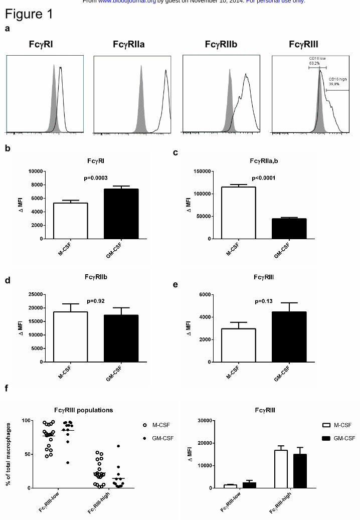
Figure 1a
b
f
FcγRI FcγRIIa FcγRIIb FcγRIII
d e
c
For personal use only.on November 10, 2014. by guest www.bloodjournal.orgFrom
No erythrocytes Unopsonized erythrocytes Opsonized erythrocytes
Opsonized erythrocytes + cytochalasin B
Figure 2 a Before erythrocyte lysis After erythrocyte lysis
c
b
CFSE→ CFSE→CFSE→
CFSE→
d
For personal use only.on November 10, 2014. by guest www.bloodjournal.orgFrom
Figure 3For personal use only.on November 10, 2014. by guest www.bloodjournal.orgFrom
a
b
Figure 4For personal use only.on November 10, 2014. by guest www.bloodjournal.orgFrom
a
b
c
d
IgG1 FcSialylation -
IgG1 FcSialylation +
Figure 5For personal use only.on November 10, 2014. by guest www.bloodjournal.orgFrom
a
b
Figure 6For personal use only.on November 10, 2014. by guest www.bloodjournal.orgFrom
d
b
c
aFigure 7
For personal use only.on November 10, 2014. by guest www.bloodjournal.orgFrom