the significance of trisomy 8 in de novo acute myeloid leukaemia: the accompanying chromosome...
TRANSCRIPT

The significance of trisomy 8 in de novo acute myeloidleukaemia: the accompanying chromosome aberrationsdetermine the prognosis
CLAUDIA SCHOC H,1 DET LEF HAASE,1 CHRISTA FONATSCH,2 TORSTEN HAFERLACH,1 HELMUT LOF FLER,3
BRIGITTE SCHLE GELBERGER,4 DIETER-KURT HOS SFELD,5 REINHARD BEC HER,6 MARIA CRISTINA SAUERLAND,7
AC HIM HEINECKE,7 BERNHARD WORMANN,1 THOMAS BUCHNER8
AND WOLFGANG HIDDEMANN1
for the German AML Cooperative Study Group 1Department of Haematology and Oncology, University of Gottingen,Germany, 2Institute of Medical Biology, University of Vienna, Austria, 3II. Medical Department, and4Institute of Human Genetics, University of Kiel, 5Department of Haematology and Oncology, University of Hamburg,6Department of Haematology and Oncology, University of Essen, 7Institute of Medical Statistics and Biomathematics,and 8Department of Haematology and Oncology, University of Munster, Germany
Received 26 May 1997; accepted for publication 12 September 1997
Summary. Trisomy 8 is the most frequent numericalchromosome aberration in acute myeloid leukaemia (AML).It occurs either as the sole anomaly or together with otherclonal chromosome aberrations. We investigated whetheraccompanying chromosome anomalies influence the clinicaloutcome in patients with trisomy 8 and de novo AML. Since1986, in 713 AML cases treated according to the protocols ofthe German AMLCG trials, chromosome analyses have beensuccessfully performed. The overall incidence of trisomy 8was 7·6%. Complete clinical follow-up data were availablefor 51 patients who were divided into three differentcategories: group 1: trisomy 8 as the sole cytogeneticanomaly (n ¼ 20); group 2: trisomy 8 in addition tofavourable chromosome aberrations (t(8;21)(q22;q22),t(15;17)(q22;q21), inv(16)(p13q22)) (n ¼ 10); and group3: trisomy 8 accompanied by other anomalies, in most casesof complex type (n ¼ 21). Complete remission (CR) rates were
70%, 90% and 67% for groups 1, 2 and 3, respectively.Event-free survival (EFS) at 3 years differed significantlybetween patients with trisomy 8 only (37·5%), patients withtrisomy 8 in combination with favourable aberrations(55·0%) and patients with trisomy 8 and other accompany-ing anomalies, mostly complex chromosome aberrations(9·0%) (group 1 v group 2: P ¼ 0·12; group 1 v group 3:P ¼ 0·005; group 2 v group 3: P ¼ 0·05). In this studypatients with þ8 as the sole cytogenetic anomaly had anintermediate prognosis, patients with þ8 in addition tofavourable chromosome aberrations maintained a goodclinical outcome, and patients with þ8 in combinationwith other abnormalities showed the worst prognosis.
Keywords: acute myeloid leukaemia, trisomy 8, cytogenetics,prognosis.
In recent years the karyotype of leukaemic blasts has beenshown to be the single most important prognostic determi-nant for acute myeloid leukaemia (AML) both for initialresponse to induction therapy as well as for remissionduration and overall survival. Data from several studiesuniformly indicate that patients with t(8;21)(q22;q22) orinv(16)(p13q22) respond well to standard induction therapy(Hiddemann et al, 1995). They also appear to benefit furtherfrom high-dose cytosine arabinoside intensification
(Bloomfield et al, 1997). A favourable outcome is alsofound in patients with promyelocytic leukaemia andt(15;17)(q22;q21), 60–70% of whom achieve long-termdisease-free survival after treatment with all-trans retinoicacid followed by anthracycline/cytosine arabinoside basedregimens (Warrell, 1997). In contrast, patients with ¹5/5q¹, ¹7/7q¹ or complex karyotype anomalies show verylow complete remision rates and short leukaemia-free andoverall survival (Mrozek et al, 1997; Haferlach, 1996).
Although trisomy 8 is the most frequent numericalchromosome aberration in acute myeloid leukaemia (AML),only few data are available regarding its prognostic sig-nificance. Trisomy 8 occurs as a solitary change in 5–6% of
British Journal of Haematology, 1997, 99, 605–611
605q 1997 Blackwell Science Ltd
Correspondence: Dr med. Claudia Schoch, Abteitung fur Hamato-logie/Onkologie, Zentrum Innere Medizin, Georg-August-UniversitatGottingen, Robert-Koch-Strasse 40, 37075 Gottingen, Germany.

all cytogenetically abnormal acute myeloid leukaemias. Ifthose with þ8 and additional aberrations are included, thisfrequency doubles (Heim & Mitelman, 1992). AML withtrisomy 8 shows no specific association with FAB subtypes orother features of clinical presentation. Whereas þ8 as thesole anomaly is most common in FAB-subtypes M1, M4 andM5 (in that order of frequency), it is relatively frequent inM2, M3 and M4eo as an additional karyotypic change(Second MIC Cooperative Study Group, 1988; Johansson etal, 1994). Most studies show an intermediate (FIWCL, 1984;Yunis et al, 1984; Berger et al, 1987; Samuels et al, 1988;Dastugue et al, 1995) or poor prognosis (Bloomfield & de laChapelle, 1987; Keating et al, 1987; Schiffer et al, 1989;Joventino et al, 1995; Byrd et al, 1996) of patients with þ8 ingeneral. Only Fenaux et al (1989) and Larson et al (1983)found a relatively favourable outcome in patients with tri-somy 8. Due to the small number of cases reported ineach study, patients were not further subdivided accordingto the chromosome aberrations that occurred in additionto trisomy 8. As it was shown for patients with t(8;21)(q22;q22) (Schoch et al, 1996) or t(15;17)(q22;q21)(Hiorns et al, 1997) that certain additional chromosomeabnormalities have an impact on prognosis, the currentstudy was performed to evaluate the relevance of accom-panying chromosome anomalies on the prognostic implica-tion of trisomy 8.
This question seems to be especially important, sinceinterphase cytogenetics with centromeric probes are fre-quently applied as the only cytogenetic diagnostic in AMLand the question whether the information of the detection oftrisomy 8 in AML by FISH only is sufficient to predict theprognosis has to be addressed.
MATERIAL AND METHODS
Since 1986 chromosome analyses were successfully per-formed in 713 patients who met the inclusion criteria of themulticentre trials of the German Cooperative AML StudyGroup. In 54 patients a gain of chromosome 8 was observed,so that the overall incidence of trisomy 8 was 7·6%. Com-plete follow-up data were available for 51 patients.
All patients were treated according to the protocols of theGerman AML Cooperative Group (AMLCG). They comprisethe following regimens: two courses of double inductionconsisting randomly of either TAD/TAD or TAD/HAM fol-lowed by TAD (thioguanine, cytosine arabinoside, dauno-rubicin) consolidation and subsequent maintenance orsequential HAM (high-dose cytosine arabinoside, mito-xantrone). Patients > 60 years old received only a secondinduction course of TAD in case of persisting bone marrowblasts (Buchner et al, 1985, 1991; Hiddemann et al, 1988).
Chromosome analyses were performed according to stan-dard protocols using G- or R-banding. The chromosomeswere classified according to the ISCN (International Systemfor Human Cytogenetic Nomenclature, 1995). Morphologicalevaluation was carried out according to FAB criteria(Bennett et al, 1976, 1985).
Complete remission was determined to be achieved whenthe bone marrow was normocellular, containing <5% blasts
and neutrophil granulocytes in peripheral blood had recoveredto 1·5 × 109/l and platelets to 100 × 109/l, according to theCancer and Leukemia Group B (CALGB) criteria (Preisler et al,1979). Causes of treatment failure were subdivided into twocategories: persistent leukaemia (non-response) and deathwithin 42 d of beginning induction therapy (early death).Overall survival was measured from the time of entry into thestudy to the time of death, event-free survival from time oftherapy start to either failure to attain CR, relapse of AML afterachievement of CR or death from any cause, respectively.
Analyses of survival were performed according to themethod of Kaplan & Meier (1958). Log-rank test was used tocompare survival of different subgroups (Peto et al, 1977).
RESULTS
Clinical, morphological and cytogenetic dataBased on the results of karyotype analysis, 51 patients withtrisomy 8 and complete clinical follow-up data were dividedinto three different categories: group 1: trisomy 8 as the solecytogenetic anomaly; group 2: trisomy 8 in addition tofavourable chromosome aberrations (t(8;21)(q22;q22),t(15;17)(q22;q21), inv(16)(p13q22)); and group 3: trisomy8 accompanied by other anomalies, mostly of complex type.Clinical and morphological data for each group are shown indetail in Table I.
Group 1. In 20 patients the gain of one chromosome 8 wasthe only karyotypic change observed.
Group 2. In 10 patients trisomy 8 was observed in additionto a favourable chromosome aberration, in two patients eachin addition to t(8;21)(q22;q22) and t(15;17)(q22;q21) andin six patients together with inv(16)(p13q22). One femalepatient with t(8;21) and þ8 also showed loss of oneX-chromosome. In one patient with t(15;17) and þ8 adeletion 9q (del(9)(q22)) was observed in addition. Threepatients with inv(16) showed gains of chromosomes 21 (twopatients) or 22 (one patient) in addition to trisomy 8.
Group 3. In 21 patients trisomy 8 was accompanied byother anomalies. In two patients þ8 was observed in addi-tion to t(9;22)(q34;q11) and in one case together witht(10;11)(p13;q14). In 18 cases trisomy 8 occurred withincomplex karyotypes (three or more numerical and/or struc-tural chromosome anomalies).
Patients with þ8 and favourable chromosome aberrationswere significantly younger than patients with only þ8, orþ8 and other abnormalities (median age 47 v 57 v 57 years)(P<0·05). Although the sex ratio was balanced in the groupof patients with trisomy 8 in addition to favourable anom-alies, male gender predominated in the group with only þ8(male:female ¼ 2·3:1) and female gender in the group withþ8 and other anomalies (male:female ¼ 1:2). No significantdifferences were observed between the groups concerningleucocyte count, platelet count and haemoglobin concentra-tion at diagnosis.
Clinical outcomePatients with trisomy 8 as the sole anomaly (group 1). 14/20
patients (70%) entered complete remission after inductionchemotherapy, four patients did not respond, and two
q 1997 Blackwell Science Ltd, British Journal of Haematology 99: 605–611
606 Claudia Schoch et al

607Trisomy 8 in Acute Myeloid Leukaemia
q 1997 Blackwell Science Ltd, British Journal of Haematology 99: 605–611
patients died within 6 weeks due to infection. 7/14 patientsachieving CR relapsed 3–67 months (median 5 months)after entering CR. Three of these seven patients were instable second CR at last follow-up 44, 88 and 124months after diagnosis and 20, 17 and 118 months afterattaining second CR, respectively. One patient received anallogeneic bone marrow transplantation in second CR; theother two patients were treated with second-line chemo-therapeutic regimens. Seven patients, including onepatient receiving an allogeneic BMT in first CR, were infirst complete remission at last follow-up 14–123 monthsafter diagnosis with a median follow-up time of 49months. Median event-free and overall survival were 5and 20·5 months, respectively.
Patients with trisomy 8 in addition to favourable anomalies(group 2). 9/10 patients (90%) entered complete remissionafter induction chemotherapy; only one patient witht(15;17) and þ8 died within 6 weeks due to bleeding. Nonon-responders were observed. Two patients died in CR dueto treatment-related complications (sepsis, bleeding). Onepatient with t(15;17), þ8 and del(9)(q22) relapsed after 13months and died 19 months later due to progression of AML.Six patients were in continuous complete remission 6–81months after diagnosis, with a median follow-up time of 56months. All three patients who received an allogeneic bonemarrow transplantation in first CR were alive 68, 81 and 81months after diagnosis. Median event-free and overallsurvival have not been reached yet.
Table I. Clinical and morphological data of 51 patients with AML and trisomy 8.
Only þ8þ8 and favourableanomalies þ8 and others
No. of patients 20 10 21Male/female 14:6 ¼ 2·3:1 5:5 ¼ 1:1 7:14 ¼ 1:2Age range (years) 24–76 24–53 26–75Median age (years) 57 47 57Leucocyte count range (× 109/l) 0·7–127 0·1–115 0·9–97·1Median leucocyte count (× 109/l) 9·7 10·6 12Platelet count range (× 109/l) 16–147 7–169 12–206Median platelet count (× 109/l) 49 24·5 42Haemoglobin concentration range (g/dl) 5·0–11·9 4·6–11·3 6·9–13·8Median haemoglobin concentration (g/dl) 9·0 7·9 9·2FAB morphology M1: 5 M2: 2 M1: 3
M2: 5 M3: 2 M2: 7M4: 3 M4eo: 6 M4: 4M5: 5 M5: 3M6: 1 M6: 1No data: 1 M7: 1
No data: 2
Table II. Survival data of 51 patients with AML and trisomy 8.
Only þ8 þ8 and favourable anomalies þ8 and others
No. of patients 20 10 21Complete remission rate 14/20 ¼ 70% 9/10 ¼ 90% 14/21 ¼ 67%Early death rate 2/20 ¼ 10% 1/10 ¼ 10% 1/21 ¼ 4·8%Relapses 7/14 ¼ 50% 1/9 ¼ 11% 11/14 ¼ 78·6%Alive in first CR 7/14 (14þ to 123þ months) 6/9 (6þ to 81þ months) 2/14 (80þ and 101þ
months)Alive in second CR 3/14 (44þ, 88þ and 124þ
months)1/9 (32þ months) 1/14 (74þ months)
Median follow-up (months) 49 56 Only 3 patients aliveMedian time to relapse (months) 5 (3–67) 13 6 (1–15)Median CR duration (months) 35 Not reached 6Median event-free survival 5 Not reached 2Event-free survival at 3 years (%) 37·5 55·0 9·0Median overall survival (months) 20·5 Not reached 8Overall survival at 3 years (%) 49·0 56·0 15·0No. of patients receiving an allogeneic
bone marrow transplantation2(1 in first CR, 1 in second CR)
3(all in first CR)
1(in second CR)

Patients with trisomy 8 and other anomalies (group 3). 14/21patients (67%) entered complete remission after inductionchemotherapy, six patients did not respond, and one patientdied within 6 weeks due to infection. 11/14 patients whoachieved CR relapsed 1–15 months (median 6 months) after
entering CR. Only one of these 11 patients achieved a stablesecond CR, received an allogeneic BMT in second CR and wasalive 74 months after diagnosis and 68 months afterattaining second CR, respectively. One patient died in CRdue to infection. Only two patients were in first complete
q 1997 Blackwell Science Ltd, British Journal of Haematology 99: 605–611
608 Claudia Schoch et al
Fig 1. Overall survival of 51 patients with þ8 subdivided into three prognostic groups: 10 patients with þ8 in addition to favourable anomalies(t(8;21), inv(16) or t(15;17)), 20 patients with þ8 only, and 21 patients with trisomy 8 accompanied by other aberrations, of which 18 caseswere of complex type.
Fig 2. Event-free survival of 51 patients with þ8 subdivided into three prognostic groups: 10 patients with þ8 in addition to favourableanomalies (t(8;21), inv(16) or t(15;17)), 20 patients with þ8 only, and 21 patients with trisomy 8 accompanied by other aberrations, including18 cases of complex type. Event-free survival differs significantly between patients with þ8 only (group 1), patients with þ8 and favourableaberrations (group 2) and patients with þ8 and other acompanying anomalies (group 3) (group 1 v group 2: P ¼ 0·12, group 1 v group 3:P ¼ 0·005; group 2 v group 3: P ¼ 0·05).
609Trisomy 8 in Acute Myeloid Leukaemia
q 1997 Blackwell Science Ltd, British Journal of Haematology 99: 605–611
remission at last follow-up 80 and 101 months after diagnosis.No special characteristics were observed in these two patientsthat might explain the good outcome. Median event-free andoverall survival are 2 and 8 months, respectively.
Patients with þ8 and favourable anomalies had thehighest complete remission rate (90%) and the lowest relapserate (11%) compared to patients with only þ8 (CR 70%,relapse rate 50%) and patients with þ8 and otherabnormalities (CR 67%, relapse rate 79%). Median event-free survival differed significantly between group 1 andgroup 3 (5 v 2 months, P ¼ 0·005) and between group 2 andgroup 3 (not reached v 2 months, P ¼ 0·05).
According to the treatment protocol, patients <50 yearsold with a HLA-identical family donor were offered allo-geneic BMT in first CR. Due to the fact that patients withfavourable chromosome abnormalities were younger andthat a higher proportion achieved stable CR, more patients inthis group received an allogeneic BMT in first CR comparedto both other groups, in which 50% and 79% relapsed with amedian time to relapse of 5 and 6 months, respectively. Allfour patients receiving an allogeneic BMT in first CR andboth patients transplanted in second CR were alive incomplete remission at last follow-up.
Survival data for each group are shown in detail in TableII. Figs 1 and 2 show the overall and the event-free survivalcurves of 51 patients with trisomy 8 divided into the threeprognostic subgroups.
DISCUSSION
A number of specific chromosomal abnormalities have beenidentified in AML which are closely associated with distinctmorphological and clinical subsets of this disease. Manyinvestigators have shown that specific chromosome aberra-tions are independent predictors of response to therapy andsurvival. Although trisomy 8 is the most frequent numericalchromosome abnormality in AML and several study groupsreported on clinical outcome of patients with AML and þ8,no clear picture has arisen concerning the prognostic impactof this aberration. Complete remission rates of patients withþ8 varied between 29% and 91%, data on median remissionduration ranged from 8 to 24 months (Mrozek et al, 1997).These differences may be related to the very small number ofpatients in some reports, but more importantly due to thecytogenetic heterogeneity of the cases. In most studies, caseswith isolated trisomy 8 were analysed together with patientswho showed trisomy 8 and additional other anomalies(Larson et al, 1983; FIWCL, 1984; Bloomfield & de laChapelle, 1987; Samuels et al, 1988; Fenaux et al, 1989;Stasi et al, 1993; Joventino et al, 1995) (Table III). Our dataclearly show significant differences in clinical outcome ofcertain subgroups of patients with trisomy 8. Patients withtrisomy 8 as the sole cytogenetic anomaly have an inter-mediate prognosis. Patients with favourable chromosomeaberrations and trisomy 8 maintain a good clinical outcome,
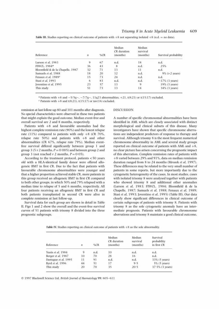
Table III. Studies reporting on clinical outcome of patients with þ8 not separating isolated þ8 (n.d. ¼ no data).
Reference n %CR
MedianCR duration(months)
Mediansurvival(months) Survival probability
Larson et al, 1983 9 67 n.d. 18 n.d.FIWCL, 1984* 36 43 8 n.d. 25%Bloomfield & de la Chapelle, 1987 32 71 13 11 n.d.Samuels et al, 1988 18 20 12 n.d. 9% (>2 years)Fenaux et al, 1989† 15 73 24 n.d. n.d.Stasi et al, 1993 6 83 n.d. n.d. ,17% (3 years)Joventino et al, 1995 23 57 13 9 19% (3 years)This study 51 73 13 14 34% (3 years)
* Patients with þ8 and ¹5/5q¹, ¹7/7q¹, 11q23 abnormalities, þ21, t(8;21) or t(15;17) excluded.† Patients with þ8 and t(8;21), t(15;17) or inv(16) excluded.
Table IV. Studies reporting on clinical outcome of patients with þ8 as the sole abnormality.
Reference n %CR
MedianCR duration(months)
Mediansurvival(months)
Survivalprobabilityin first CR
Yunis et al, 1984 9 n.d. 10 n.d. n.d.Berger et al, 1987 10 70 28 16 n.d.Dastugue et al, 1995 11 91 n.d. n.d. 33% (5 years)Byrd et al, 1996 44 51 17 9·5 5% (5 years)This study 20 70 35 20·5 37·5% (3 years)

whereas cases with trisomy 8 in combination with complexanomalies face the worst prognosis.
So far only four studies have reported on survival datawith þ8 as the sole anomaly (Yunis et al, 1984; Berger et al,1987; Dastugue et al, 1995; Byrd et al, 1996) (Table IV).Complete remission rates varied from 51% to 91% and long-term survival in first complete remission from 5% to 33%. Inour study using a therapy protocol with double inductioncombined with consolidation and bone marrow transplanta-tion or maintenance therapy a high remission rate of 70%resulting in an overall survival of 49% at 3 years wasachieved. The variances in survival between the differentstudies cited above could be due to differences in patientcharacteristics but possibly reflect differences in treatmentprotocols. To optimize treatment results, further studies areneeded which compare the influence of different therapystrategies on survival in defined cytogenetic subgroups. Theresults of these studies could then serve as a basis for thedevelopment of risk-adapted treatment modalities.
As the leukaemic karyotype is the single most importantprognostic factor in AML, a reliable and exact geneticmethod is required to enable risk assessment. Whereaschromosome banding analysis was the only technique todetect karyotype anomalies for over two decades, newtechniques were developed during the recent years. AML-specific translocations can now also be detected by reversetranscriptase–polymerase chain reaction (RT-PCR) (Borrowet al, 1992; Miller et al, 1992; Soekarman et al, 1992;Downing et al, 1993; Claxton et al, 1994; Yamamoto et al,1994; Campana & Pui, 1995). However, only a fewaberrations can be identified by this technique. With thedevelopment of a large variety of DNA probes, fluorescence insitu hybridization (FISH) is increasingly used for geneticanalysis of AML (Le Beau, 1993; Fischer et al, 1996). FISHcan also easily be applied to detect þ8 (Jenkins et al, 1992;Kibbelaar et al, 1993), but alone is not sufficient to determineprognosis, because the accompanying chromosome anoma-lies would be missed by this approach.
In conclusion, median event-free survival differs signifi-cantly between patients with trisomy 8 as the sole cyto-genetic anomaly, patients with trisomy 8 in combinationwith favourable aberrations and patients with trisomy 8 andother accompanying anomalies, mostly complex chromo-some aberrations. Therefore a complete karyotype analysiswhich also detects the accompanying anomalies is necessaryto properly define biological entities in AML.
ACKNOWLEDGMENTS
We thank all participating centres of the AMLCG-trial forsending material for cytogenetic analyses and documenta-tion of clinical data. The AMLCG is supported by DeutscheKrebshilfe, Proj. No. M17/92/Bu1.
REFERENCES
Bennett, J.M., Catovsky, D., Daniel, M.T., Flandrin, G., Galton, D.A.G.,Gralnick, H.R. & Sultan, C. (1976) Proposal for the classificationof the acute leukaemias. British Journal of Haematology, 33, 451–458.
Bennett, J.M., Catovsky, D., Daniel, M.T., Flandrin, G., Galton, D.A.G.,Gralnick, H.R. & Sultan, C. (1985) Proposed revised criteria forthe classification of acute myeloid leukemia: a report of theFrench–American–British Cooperative Group. Annals of InternalMedicine, 103, 626–629.
Berger, R., Bernheim, A., Ochoa-Noguera, M.E., Daniel, M.-T.,Valensi, F., Sigaux, F., Flandrin, G. & Boiron, M. (1987) Prognosticsignificance of the chromosomal abnormalities in acute non-lymphocytic leukemia: a study of 343 patients. Cancer Genetics andCytogenetics, 28, 293–299.
Bloomfield, C.D. & de la Chapelle, A. (1987) Chromosome abnor-malities in acute nonlymphocytic leukemia: clinical and biologicalsignificance. Seminars in Oncology, 14, 372–383.
Bloomfield, C.D., Herzig, G.P. & Caligiuri, M.A. (1997) Introduction:acute leukemia: recent advances. Seminars in Oncology, 24, 1–2.
Borrow, J., Goddard, A.D., Gibbons, B., Katz, F., Swirsky, D., Fioretos,T., Dube, I., Winfield, D.A., Kingston, J., Hagemeijer, A., Rees, J.,Lister, T.A. & Solomon, E. (1992) Diagnosis of acute promyelocyticleukemia by RT-PCR: detection of PML-RARA and RARA-PMLfusion transcripts. British Journal of Haematology, 82, 529–540.
Buchner, T., Hiddemann, W., Loffler, H., Gassmann, W., Maschmeyer,G., Heit, W., Hossfeld, D., Weh, H., Ludwig, W.-D., Thiel, E.,Nowrousian, M., Aul, C., Lengfelder, E., Lathan, B., Mainzer, K.,Urbanitz, D., Emmerich, B., Middelhoff, G., Donhuijsen, H.R.,Hellriegel, H.-P. & Heinecke, A. (1991) Improved cure rate by veryearly intensification combined with prolonged maintenancechemotherapy in patients with acute myeloid leukemia: datafrom the AML cooperative group. Seminars in Hematology, 28, 76–79.
Buchner, T., Urbanitz, D., Hiddemann, W., Ruhl, H., Ludwig, W.-D.,Fischer, J., Aul, H.C., Vaupel, H.A., Kuse, R., Zeile, G., Nowrousian,M.R., Konig, H.J., Walter, M., Wendt, F.C., Sodomann, H., Hossfeld,D.K., von Paleske, A., Loffler, H., Gassmann, W., Hellriegel, K.-P.,Fulle, H.H., Lunscken, Ch., Emmerich, B., Pralle, H., Pees, H.W.,Pfreundschuh, M., Bartels, H., Koeppen, K.-M., Schwerdtfeger, R.,Donhuijsen-Ant, R., Mainzer, K., Bonfert, B., Koppler, H., Zuborn,K.-H., Ranft, K., Thiel, E. & Heinecke, A. (1985) Intensifiedinduction and consolidation with or without maintenance chemo-therapy for acute myeloid leukemia (AML): two multicenterstudies of the German AML Cooperative Group. Journal of ClinicalOncology, 3, 1583–1589.
Byrd, J.C., Lawrence, A.D., Arthur, D.C., Davey, F., Schiffer, C.A. &Bloomfield, C.D. (1996) Acute myeloid leukemia (AML) patientswith pre-treatment isolated trisomy 8 are rarely cured withchemotherapy: results of CALGB 8461. (Abstract). Proceedings ofthe American Association of Cancer Research, 37, 188.
Campana, D. & Pui, C.-H. (1995) Detection of minimal residualdisease in acute leukemia: methodologic advances and clinicalsignificance. Blood, 85, 1416–1434.
Claxton, D. F., Liu, P., Hsu, H.B., Marlton, P., Hester, J., Collins, F.,Deisseroth, A.B., Rowley, J.D. & Siciliano, M.J. (1994) Detection offusion transcripts generated by the inversion 16 chromosome inacute myelogenous leukemia. Blood, 83, 1750–1756.
Dastugue, N., Payen, C., Lafage-Pochitaloff, M., Bernard, P., Leroux,D., Huguet-Rigal, F., Stoppa, A.M., Marit, G., Molina, L., Michallet,M., Maraninchi, D., Attal, M. & Reiffers, J. (1995) Prognosticsignificance of karyotype in de novo adult acute myeloid leukemia.Leukemia, 9, 1491–1498.
Downing, J.R., Head, D.R., Curcio-Brint, A.M., Hulshof, M.G.,Motroni, T.A., Raimondi, S.C., Carroll, A.J., Drabkin, H.A., Will-man, C., Theil, K.S., Civin, C.I. & Erickson, P. (1993) An AML1/ETO fusion transcript is consistently detected by RNA-basedpolymerase chain reaction in acute myelogenous leukemiacontaining the (8;21)(q22;q22) translocation. Blood, 81, 2860–2865.
q 1997 Blackwell Science Ltd, British Journal of Haematology 99: 605–611
610 Claudia Schoch et al

611Trisomy 8 in Acute Myeloid Leukaemia
q 1997 Blackwell Science Ltd, British Journal of Haematology 99: 605–611
Fenaux, P., Preudhomme, C., Lay, J.L., Morel, P., Beuscart, R. &Bauters, F. (1989) Cytogenetics and their prognostic value in denovo acute myeloid leukaemia: a report on 283 cases. BritishJournal of Haematology, 73, 61–67.
Fischer, K., Scholl, C., Salat, J., Froehling, S., Schlenk, R., Bentz, M.,Stilgenbauer, S., Lichter, P. & Doehner, H. (1996) Design and validationof DNA probe sets for a comprehensive interphase cytogeneticanalysis of acute myeloid leukemia. Blood, 88, 3962–3971.
Fourth International Workshop on Chromosomes in Leukemia(FIWCL) (1984) Cancer Genetics and Cytogenetics, 11, 259–350.
Haferlach, T. (1996) More individual markers are necessary forpatients with acute myeloid leukemia (AML). Does cytomorphol-ogy or cytogenetics define the biological entity? Leukemia, 10,(Suppl. 3), S5–S9.
Heim, S. & Mitelman, F. (1992) Cytogenetic analysis in the diagnosisof acute leukemia. Cancer, 70, 1701–1709.
Hiddemann, W., Buchner, T., Essink, M., Koch, O., Stenzinger, W. &van de Loo, J. (1988) High dose cytosine arabinoside andmitoxantrone: preliminary results of a pilot study with sequentialapplication (S-HAM) indicating a high antileukemic activity inrefractory acute leukemias. Onkologie, 11, 10–12.
Hiddemann, W., Fonatsch, C., Wormann, B., Heinecke, A.,Sauerland, C., Scharnhorst, S. & Buchner, Th. (1995) Cytogeneticsubgroups of AML and outcome from high dose versus conven-tional dose ARA-C as part of double induction. (Abstract). Blood,86, (Suppl. 1), 267a.
Hiorns, L.R., Swansbury, G.J., Mehta, J., Min, T., Dainton, M.G.,Treleaven, J., Powles, R.L. & Catovsky, D. (1997) Additionalchromosome abnormalities confer worse prognosis in acute pro-myelocytic leukaemia. British Journal of Haematology, 96, 314–321.
ISCN (1995) Guidelines for Cancer Cytogenetics. Supplement to: AnInternational System for Human Cytogenetic Nomenclature (ed. byF. Mitelman). S. Karger, Basle.
Jenkins, R.B., Le Beau, M.M., Kraker, W.J., Borell, T.J., Stalboerger,P.G., Davis, G.M., Penland, L., Fernald, A., Espinosa, R., III, Schaid,D.J., Noel, P. & Dewald, G.W. (1992) Fluorescence in situ hybrid-ization: a sensitive method for trisomy 8 detection in bone marrowspecimens. Blood, 79, 3307–3315.
Johansson, B., Mertens, F. & Mitelman, F. (1994) Secondary chromo-somal abnormalities in acute leukemias. Leukemia, 8, 953–962.
Joventino, L.P., Stock, W., Lane, N.J., Daly, K.M., Mick, R., Le Beau, M.M. & Larson, R.A. (1995) Certain HLA antigens are associatedwith specific morphologic and cytogenetic subsets of acute mye-loid leukemia. Leukemia, 9, 433–439.
Kaplan, E. & Meier, P. (1958) Nonparametric estimation fromincomplete observations. Journal of the American StatisticsAssociation, 53, 456–481.
Keating, M.J., Cork, A., Broch, Y., Smith, T., Waltes, R.S., McCredie,K.B., Trujillo, J. & Freireich, E.J. (1987) Toward a clinically rele-vant cytogenetic classification of acute myelogenous leukemia.Leukemia Research, 11, 119–133.
Kibbelaar, R.E., Mulder, J.W.R., Dreef, E.J., van Kamp, H., Fibbe, W.E.,Wessels, J.W., Beverstock, G.C., Haak, H.L. & Kluin, Ph.M. (1993)Detection of monosomy 7 and trisomy 8 in myeloid neoplasia: acomparison of banding and fluorescence in situ hybridization.Blood, 82, 904–913.
Larson, R.A., Le Beau, M.M., Vardiman, J.W., Testa, J.R., Golomb,H.M. & Rowley, J.D. (1983) The predictive value of initial cyto-genetic studies in 148 adults with acute nonlymphocyticleukemia: a 12-year study (1970–1982). Cancer Genetics andCytogenetics, 10, 219–236.
Le Beau, M.M. (1993) Detecting genetic changes in human tumorcells: have scientists ‘gone fishing?’. Blood, 81, 1979–1983.
Miller, W.H., Jr, Kakizuka, A., Frankel, S.R., Warrel, R.P., Jr,DeBlasio, A., Levine, K., Evans, R.M. & Dmitrovsky, E. (1992)Reverse transcription polymerase chain reaction for the re-arranged retinoic acid receptor a clarifies diagnosis and detectsminimal residual disease in acute promyelocytic leukemia. Pro-ceedings of the National Academy of Sciences of the United States ofAmerica, 89, 2694–2698.
Mrozek, K., Heinonen, K., De la Chapelle, A. & Bloomfield, C.D.(1997) Clinical significance of cytogenetics in acute myeloidleukemia. Seminars in Oncology, 24, 17–31.
Peto, R., Pike, M.C., Armitage, P., Breslow, N.E., Cox, N.R., Howard,S.V., Mantel, N., McPherson, K., Peto, J. & Smith, P.G. (1977)Design and analysis of randomized clinical trials requiring pro-longed observation of each patient. British Journal of Cancer, 35,1–39.
Preisler, H.D., Rustum, Y., Henderson, S., Bjornsson, S., Creaven, P.J.,Higby, D.J., Freeman, A., Gailani, S. & Naeher, C. (1979) Treat-ment of acute nonlymphocytic leukemia: use of an anthracycline-cytosine arabinoside induction therapy and comparison of twomaintenance regimens. Blood, 53, 455–464.
Samuels, B.L., Larson, R.A., Le Beau, M.M., Daly, K.M., Bitter, M.A.,Vardiman, J.W., Barker, C.M., Rowley, J.D. & Golomb, H.M. (1988)Specific chromosomal abnormalities in acute nonlymphocyticleukemia correlate with drug susceptibility in vivo. Leukemia, 2,79–83.
Schiffer, C.A., Lee, E.J., Tomiyasu, T., Wiernik, P.H. & Testa, J.R. (1989)Prognostic impact of cytogenetic abnormalities in patients with denovo acute nonlymphocytic leukemia. Blood, 73, 263–270.
Schoch, C., Haase, D., Haferlach, T., Gudat, H., Buechner, T., Freund,M., Link, H., Lengfelder, E., Wandt, H., Sauerland, M.C., Loeffler, H. &Fonatsch, C. (1996) Fifty-one patients with acute myeloid leukemiaand translocation t(8;21)(q22;q22): an additional deletion in 9q isan adverse prognostic factor. Leukemia, 10, 1288–1295.
Second MIC Cooperative Study Group (1988) Morphologic, immuno-logic, and cytogenetic (MIC) working classification of the acutemyeloid leukaemias. British Journal of Haematology, 68, 487–494.
Soekarman, D., von Lindern, M., van der Plas, D.C., Selleri, L.,Bartram, C.R.I., Marlial, P., Culligan, D., Padua, R.A., Hasper-Vogt, K.P., Hagemeijer, A. & Grosveld, G. (1992) Dek-Canrearrangement in translocation (6;9)(p23:q34). Leukemia, 6,489–494.
Stasi, R., Del Poeta, G., Masi, M., Tribalto, M., Venditti, A., Papa, G.,Nicoletti, B., Vernole, P., Tedeschi, B., Delaroche, I., Mingarelli, R.& Dallapiccola, B. (1993) Incidence of chromosome abnormalitiesand clinical significance in de novo acute myeloid leukemia.Cancer Genetics and Cytogenetics, 67, 28–34.
Warrell, R.P., Jr (1997) Clinical and molecular aspects of retinoidtherapy for acute promyelocytic leukemia. International Journal ofCancer, 70, 496–497.
Yamamoto, K., Seto, M., Iida, S., Komatsu, H., Kamada, N., Kojima,S., Kodera, Y., Nakazawa, S., Saito, H., Takahashi, T. & Ueda, R.(1994) A reverse transcriptase–polymerase chain reaction detectsheterogeneous chimeric mRNAs in leukemias with 11q23abnormalities. Blood, 83, 2912–2921.
Yunis, J.J., Brunning, R.D., Howe, R.B. & Lobell, M. (1984) Highresolution chromosomes as an independent prognostic indicatorin adult acute nonlymphocytic leukemia. New England Journal ofMedicine, 311, 812–818.