the role of slit-robo signaling in the generation, migration and morphological differentiation of...
TRANSCRIPT

Available online at www.sciencedirect.com
13 (2008) 648–658www.elsevier.com/developmentalbiology
Developmental Biology 3
The role of Slit-Robo signaling in the generation, migration andmorphological differentiation of cortical interneurons
William Andrews a,⁎,1, Melissa Barber a,1, Luis R. Hernadez-Miranda a, Jian Xian b, Sonja Rakic a,Vasi Sundaresan c, Terence H. Rabbitts d,e, Richard Pannell d, Pamela Rabbitts e,Hannah Thompson f, Lynda Erskine f, Fujio Murakami g, John G. Parnavelas a
a Department of Anatomy and Developmental Biology, University College London, Gower Street, London WC1E 6BT, UKb Department of Oncology, Hutchinson-MRC Research Centre, Cambridge, UK
c Princess Alexandra NHS Trust, Harlow, UKd Laboratory of Molecular Biology, MRC Centre Hills Road, Cambridge, UK
e Leeds Institute of Molecular Medicine, St. James’s University Hospital, Leeds, UKf Division of Visual Science and Molecular Genetics, Institute of Ophthalmology, University College London, UK
g Osaka University, Osaka, Japan
Received for publication 3 July 2007; revised 12 October 2007; accepted 31 October 2007Available online 13 November 2007
Abstract
Cortical interneurons in rodents are generated in the ventral telencephalon and migrate tangentially into the cortex. This process requires thecoordinated action of many intrinsic and extrinsic factors. Here we show that Robo1 and Robo2 receptor proteins are dynamically expressedthroughout the period of corticogenesis and colocalize with interneuronal markers, suggesting that they play a role in the migration of these cells.Analysis of Robo mutants showed a marked increase in the number of interneurons in the cortices of Robo1−/−, but not Robo2−/−, animalsthroughout the period of corticogenesis and in adulthood; this excess number of interneurons was observed in all layers of the developing cortex.Using BrdU incorporation in dissociated cell cultures and phosphohistone-3 labeling in vivo, we demonstrated that the increased number ofinterneurons in Robo1−/− mice is, at least in part, due to increased proliferation. Interestingly, a similar increase in proliferation was observed inSlit1−/−/Slit2−/− mutant mice, suggesting that cell division is influenced by Slit-Robo signaling mechanisms. Morphometric analysis of migratinginterneurons in Robo1−/−, Robo2−/− and Slit1−/−/Slit2−/−, but not in Slit1−/− mice, showed a differential increase in neuronal process length andbranching suggesting that Slit-Robo signaling also plays an important role in the morphological differentiation of these neurons.© 2007 Elsevier Inc. All rights reserved.
Keywords: Robo; Slit; Interneuron; Morphology
Introduction
The origins and migratory routes of cortical interneurons inrodents are now well documented. Tracing studies haveconfirmed that these cells arise in different parts of theganglionic eminence (GE) in the ventral telencephalon andmigrate in tangentially oriented streams to enter the cortex(Corbin et al., 2001; Marín and Rubenstein, 2003; Métin et al.,
⁎ Corresponding author. Fax: +44 20 7679 7349.E-mail address: [email protected] (W. Andrews).
1 Both authors contributed equally to this manuscript.
0012-1606/$ - see front matter © 2007 Elsevier Inc. All rights reserved.doi:10.1016/j.ydbio.2007.10.052
2006). At the early stages of corticogenesis (E12.5 in mouse),they appear almost exclusively in the preplate layer (PPL),while at later embryonic ages the streams are at the levels of theintermediate zone (IZ)/subventricular zone (SVZ), marginalzone (MZ) and subplate (SP; after the split of the PPL). Once inthe cortex, they leave their migratory streams to assumepositions in the cortical plate (CP) where they assemble intofunctional circuits with their pyramidal counterparts, contribut-ing to a precise balance of synaptic excitation and inhibition inthe cortex. It has been suggested that disruption of this balanceresults in neuropathological conditions such as epilepsy andParkinson's disease (Sloviter, 1987; Cobos et al., 2005; Kumarand Buckmaster, 2006; Mallet et al., 2006).

649W. Andrews et al. / Developmental Biology 313 (2008) 648–658
The molecular mechanisms that guide the migration ofinterneurons from the GE, around the corticostriatal notchand into the cortex are the subject of continuing investi-gations, but a number of molecules have already beendemonstrated to play important roles (Nadarajah andParnavelas, 2002; Marín and Rubenstein, 2003; Métin etal., 2006). These include the Slit proteins and their receptorsof the Robo family (Andrews et al., 2006). Evidence from invitro experiments indicates that the migration of interneuronsis initiated by the chemorepulsive activity of Slit secretedfrom the ventricular zone (VZ) of the GE (Hu, 1999; Wu etal., 1999; Zhu et al., 1999). However, it has been reportedthat migration of cortical interneurons is normal in Slit1−/−/Slit2−/− mutants, prompting speculation that Slits do not playa major role in tangential migration (Marín et al., 2003;Marín and Rubenstein, 2003). In an attempt to investigatewhether Robo receptors are involved in this process, werecently analyzed the phenotype of Robo1−/− mice. Wenoted, in addition to abnormalities in the formation of majoraxonal tracts, a significant increase in the number of inter-neurons that enter the cortex from the ventral forebrainthroughout the period of corticogenesis (Andrews et al.,2006). This shows that Robo1−/− mice have a different phe-notype from Slit mutants, suggesting that additional ligands,receptors or receptor partners are likely to be involved in theseprocesses.
In addition to regulating axon guidance and cell migration,Slit-Robo signaling plays a role in process outgrowth andbranching. Specifically, Slit has been reported to promoteaxonal elongation and branching in sensory neurons (Wang etal., 1999; Ozdinler and Erzurumlu, 2002; Ma and Tessier-Lavigne, 2007) and dendritic growth and branching in corticalcells (Whitford et al., 2002). Furthermore, Slit has been foundto promote branching and elongation of neurites of GABA-containing interneurons in embryonic forebrain cultures (Sanget al., 2002).
In the present study, we investigated the role of Slit-Robosignaling in the generation and migration of corticalinterneurons and in their morphological differentiation. Wefirst analyzed the localization of Robo1 and Robo2 in thedeveloping forebrain and found that these proteins showcomplementary and dynamic patterns of expression through-out the period of cortex formation. Further, we found that bothreceptors are expressed in cortical interneurons duringcorticogenesis, suggesting that they may play a role in theirmigration. Analysis of Robo1−/− and Robo2−/− animalsshowed no change in the positions of the streams of migratinginterneurons in the cortices of both groups of mice, which issimilar to that reported for Slit mutants (Marín et al., 2003).However, an increased number of cells were observed in theRobo1−/− mutants, and this increase persisted to adulthood. Inaddition, we found that removal of Robo1, Robo2 or bothSlit1/Slit2 (but not Slit1 alone) differentially affected themorphology of migrating interneurons. These findingsdemonstrate that Slit-Robo signaling plays an important rolein the development of the interneuron population of thecerebral cortex.
Materials and methods
Animals
All experimental procedures were performed in accordance with the UKAnimals (Scientific Procedures) Act 1986 and institutional guidelines. Wild-type animals were C57/bl6J mice obtained from Charles River Ltd. Robo2−/−
and Slit1−/−/ Slit2−/− mice were generated as described previously (Lu et al.,2007; Plump et al., 2002, respectively). Robo1 full gene (Dulox) mutant micewere generated as outlined below and in Supplementary Fig. 1. GAD67-GFP(Δneo) mice (Tamamaki et al., 2003; kindly provided by Drs. Y. Yanagawa andK. Obata, Japan) used in this study were also maintained in C57/bl6Jbackground. The day the vaginal plug was found was considered as embryonicday (E) 0.5.
Generation of Robo1 (Dulox) mutant mice
A genomic library of E14 TG2α embryonic stem (ES) cell DNA wasscreened with a mouse Robo1 cDNA clone corresponding to exon 1 and exon22, and positive clones were purified and subcloned into pBlucecript. A 5.4-kbBamHI fragment containing exon 1 of the mouse Robo1 gene and a 7.0-kb XbaIfragment containing exon 22 of the same gene were used for construction of thetargeting vectors. Each construct comprised a selection marker gene(PGKhygpA or PGKpuropA) and a loxP sequence to enable Cre-mediatedrecombination events. The vectors were designed for positive–negativeselection of targeted cells and contained a thymidine kinase gene expressioncassette at the end of one of the homology arms. The final vectors werelinearized with XhoI for electroporation. Twenty-five micrograms of exon 1targeting vector linearized with XhoI was electroporated into 1×10⁷ CCB EScells, and cells were selected in hygromycin B (125 μg/ml) and ganciclovir(2.5 μM). Targeted clones were identified by Southern blotting of XbaI-digestedDNA and hybridization with an exon 1 probe corresponding to a sequenceexternal to the vector homology arm (probe a). Southern blotting was carried outas described previously by Xian et al. (2001).
In the second transfection, the exon 22 targeting vector linearized with XhoIwas electroporated into 1×10⁷ cells of the exon 1 targeted cell line derivedfrom the first round of transfection, and cells were selected in puromycin(0.5 μg/ml) and ganciclovir. Targeted clones were identified by Southernblotting of XbaI-digested DNA and hybridization with an exon 22 probecorresponding to a sequence external to the vector homology arm (probe b). EScells from two independent clones were used for injection into blastocystsderived from C57/bl6J mice. Blastocysts were transferred to pseudo-pregnantfemales, and chimeric offspring were detected by the presence of agouti colouron a non-agouti background. Chimeric males were mated to C57/bl6J femalesto produce ES cell-derived offspring. Their genotype was confirmed bySouthern blot analysis of tail DNA. Mice heterozygous for the gene-targetingevent – i.e., deletions of the whole Robo1 gene – were intercrossed to generatehomozygotes.
Immunohistochemistry
Embryonic brains (E13.5–E18.5) were fixed in 4% paraformaldehyde (PFA)in phosphate-buffered saline (PBS) for 4–8 h, depending on the age of theembryos. Adult mice were perfused with 4% PFA, and their brains wereremoved and immersed in fixative solution overnight. Brains were subsequentlycryoprotected in 30% sucrose in PBS, embedded in Tissue-Tek OCT (SakuraFinetek Europe, Zoeterwoude, The Netherlands) and sectioned in the coronalplane at 25 μm using a Cryostat (Bright Instruments, Huntingdon, UK). Sectionswere washed in PBS and blocked in a solution of 5% serum (v/v) and 0.3%Triton X-100 (v/v) (Sigma-Aldrich, Dorset, UK) in PBS for 2 h. Normal goatserum (Vector Laboratories, Burlingame, CA) or normal donkey serum (JacksonImmunoResearch, Soham, UK) was used for primary antibodies made in mouse,rabbit or goat, respectively. Sections were incubated overnight in one of thefollowing primary antibodies: rabbit anti-calbindin (1:10000; D-28K, Swant,Bellinzona, Switzerland); mouse anti-bromodeoxyuridine (BrdU; 1:100; Pro-gen, Heidelberg, Germany); rabbit anti-phosphohistone 3 (1:1000; Abcam Ltd.,UK); goat anti-Robo1 (1:100; R&D Systems); goat anti-Robo2 (1:100; R&D

650 W. Andrews et al. / Developmental Biology 313 (2008) 648–658
Systems); rabbit anti-Robo1 and anti-Robo2 (1:2,000; antibodies prepared byProfessor F. Murakami); and rabbit anti-ARX (1:1000; Poirier et al., 2004).They were then washed in PBS and incubated in biotinylated goat anti-mouse(1:200; Vector Laboratories), biotinylated goat anti-rabbit (1:200; VectorLaboratories) or biotinylated donkey anti-goat (1:200; Jackson Immuno-Research Laboratory) for 2 h and processed using a conventional immuno-histochemistry protocol. Other secondary antibodies used were mouse anti-rabbit 488 and rabbit anti-mouse 488 (Alexa, Invitrogen Corp., UK). Robostaining was enhanced using a tyramide signal amplification system (TSA,Perkin Elmer, Boston, MA) according to manufacturer's instructions. Sectionswere washed and incubated with 4′-6-diamidino-2-phenyllindole (DAPI,1:20,000; Sigma-Aldrich) in PBS or bisbenzimide (10 min in 2.5 μg/mlsolution in PBS; Sigma-Aldrich). Images were collected using an SP2 Leicaconfocal microscope (Leica Microsystems, UK). Sequential images weresubsequently reconstructed using Metamorph imaging software (UniversalImaging Corporation, West Chester, PA).
Quantification of interneuron distribution
Calbindin-positive cells were counted in coronal strips (400 μm wide)spanning the thickness of the middle (along the rostro-caudal axis) regions of thecortex at E15.5 (minimum of 8 sections from each of 3 animals for eachcondition). In all counts, the experimenter did not know the condition of theanimal. Strips were divided into 5 bins arranged parallel to the pial surface thatcorresponded to the different layers of the developing cortex (VZ, SVZ/LIZ, IZ,SP, CP), from bin1 (VZ) to bin 5 (upper CP). The extent of the layers wasdetermined by methyl green counterstaining (Vector Laboratories).
Morphometric analysis
Calbindin-positive neurons were drawn at a primary magnification of ×400using a drawing apparatus attached to a Zeiss Photomicroscope. Morphometric
Fig. 1. Robo1 and Robo2 expression in the mouse embryonic forebrain. Immunohistrostral, middle and caudal levels of the embryonic mouse forebrain. Robo1 and Robganglia and cortex during early (E13.5) and later phases (E15.5, E17.5) of developmecaudal ganglionic eminence; Cx, Cortex. Scale bar, 300 μm.
parameters including number of primary processes, number of branch points andtotal process length for each cell were measured using imaging analysis software(Imagej; NIH, version 1.34n) with custom made programming macros (seeSupplementary Fig. 2 for details). Means and standard error of the mean (SEM)were calculated, and the differences were tested using an unpaired Student's ttest. Significance was set at a P value of b0.05.
Dissociated cell cultures
Dissociated cell cultures were prepared from E12.5 mouse telencephalonsaccording to the method of Cavanagh et al. (1997). Briefly, GEs were dissectedfrom embryonic forebrains in Hanks' solution under a stereo microscope, andisolated tissue was dissociated enzymatically in Neurobasal media with trypsin(0.1%) and DNase I (0.001%) at 37 °C for 15 min. Trypsin was inactivated by10% fetal calf serum (FCS) in Neurobasal media for 5 min, and cells weredissociated by delicate trituration with a sterile pipette tip. The resultingsuspension was centrifuged at 1000×g for 3 min, the supernatant was discarded,and the cells were resuspended in neurobasal media containing B27 supplement,100 μg/ml penicillin/streptomycin and 2 mM L-glutamine. They were thenplated at a density of 2×10⁵ cells on poly-L-lysine (10 μg/ml) and laminin(10 μg/ml)-coated 13-mm coverslips in 24-well plates. Cultures were kept in ahumidified incubator (95% air/5% CO2) at 37 °C, and cells were allowed toattach to the coverslips for 30 min. Fresh media were then added and again onthe following morning.
Proliferation rate
The rate of proliferation, assessed in dissociated GE cultures derived fromE12.5 mouse embryos, was determined as the proportion of calbindin-positivecells that incorporated BrdU. Cultures were pulsed with 10 μM of BrdU for 2 hin order to label as many cells in S-phase without allowing them to enter mitosis(for details, see Cavanagh et al., 1997). Cells were washed and fixed with 4%
ochemical localization of Robo1 and Robo2 proteins in coronal sections taken ato2 are expressed throughout the rostro-caudal extent of the differentiating basalnt. LGE, lateral ganglionic eminence; MGE, medial ganglionic eminence; CGE,

651W. Andrews et al. / Developmental Biology 313 (2008) 648–658
PFA, co-immunostained for BrdU and calbindin and counterstained with DAPI.Cell counts were made with a ×40 objective in nine fields of view for eachsample. Statistical significance was evaluated using Student's t-test.
To determine the effect of Slit on interneuron proliferation, we preparedE12.5 GE cultures from GAD67-GFP mice. These were incubated overnight inthe presence or absence of 4 μg/ml recombinant mouse Slit3 (R&D Systems).Cells were washed and fixed with 4% PFA, immunostained for phosphohistone3 and counterstained with DAPI. Cell counts were made with a ×40 objective inten fields of view for each sample, carried out in quadruplicate.
Fig. 2. Colocalization of Robo proteins with the cortical interneuron marker,calbindin. (A–F) Immunohistochemical localization of calbindin (green) andRobo1 or Robo2 (red) staining in E15.5 coronal sections through the cortex.Colocalization of calbindin and both Robo receptors (arrows) can be seen inindividual cells at all levels of the developing cortex. (G) Graphic representationof the percentage of calbindin-positive cells that also express either Robo1(blue) or Robo2 (red) in all cortical layers at E15.5. Scale bars in panelD=100 μm in panels A, D; in panel E=50 μm in panels B, C, E and F.
Results
Robo protein expression during forebrain development
Robo1 and Robo2 mRNA are localized in the developingcortex and the proliferative zone of the GE (Marillat et al., 2002;Whitford et al., 2002), suggesting that these receptors may beassociated with the movement of cortical interneurons awayfrom the ventral telencephalon. Here we examined Robo1 andRobo2 protein expression patterns in coronal sections of mousebrains from early (E13.5), mid (E15.5) and late (E17.5) stagesof corticogenesis. Staining for Robo1 and Robo2 was observedthroughout the rostro-caudal extent of the developing forebrainat all ages examined (Fig. 1). The expression of these receptorswas largely complementary within the early differentiatingventral telencephalon (E13.5; Fig. 1), with strong Robo1staining localized throughout the mantle zone of the MGE, andRobo2 restricted more laterally to the differentiating LGE.Robo1 and Robo2 expression expanded by E15.5, overlappingto a greater degree within the differentiating basal ganglia. Inthe cortex, there was strong expression of both Robosthroughout the PPL and in the zones that result from itssplitting, the MZ and the SP, as well as in the tangentialmigratory routes travelled by interneurons at this developmentalstage, and especially within the LIZ/SVZ. By E17.5, theexpression of both Robo receptors was somewhat down regu-lated in the ventral telencephalon but remained strong within theSVZ of the cortex (Fig. 1).
Immunohistochemical staining of E15.5 coronal sectionsfor Robo1 and calbindin, a marker of cortical interneurons inearly development, showed that both molecules colocalizeextensively in neurons throughout the cortical anlage (Fig.2A). On closer examination, individual Robo1/calbindindouble-labeled cells appeared to be leaving the SP and IZ/SVZ migratory streams and moving into the developing CPand VZ, respectively (arrows, Figs. 2B, C). Robo2 stainingwas confined to the upper part of the developing cortex (Fig.2D), which was particularly strong in the SP and MZ. Similarto Robo1, individual Robo2/calbindin double-labeled inter-neurons were present throughout the developing cortex (Figs.2E, F). Quantitative analysis of the proportion of calbindin-positive cells expressing either Robo protein at E15.5revealed that more than 90% of these cells express eitherRobo receptor in all cortical layers, except for the VZ where70–80% of interneurons express either receptor (Fig. 2G);similar results were obtained at E13.5 (data not shown). Thus,most interneurons appear to express both Robo receptors inall cortical layers throughout early and mid phases of corti-
cogenesis. These observations suggest that Robo1 and Robo2may have roles not only in the migration of interneuronsfrom the GE, but also in their final positioning within theneocortex.
Cortical interneurons in Robo and Slit mutant mice
We have previously reported a significant increase in thenumber of interneurons present in the cortices of Robo1 (Exon 5deleted) mutant (−/−) animals compared to wild-type (+/+)
652 W. Andrews et al. / Developmental Biology 313 (2008) 648–658
littermates (Andrews et al., 2006), suggesting that Robo1 plays arole in the development of these cells. In order to furtherassess the roles of Slit-Robo proteins in this process, weexamined the cortices of Robo1−/−, Robo2−/−, Slit1−/−Slit2+/+
and Slit1−/−Slit2−/− mice. While our study was carried out onthe previously described Robo2 and Slit mutant mousestrains (Lu et al., 2007; Plump et al., 2002, respectively), theRobo1 analysis was performed on the newly generated Dulox
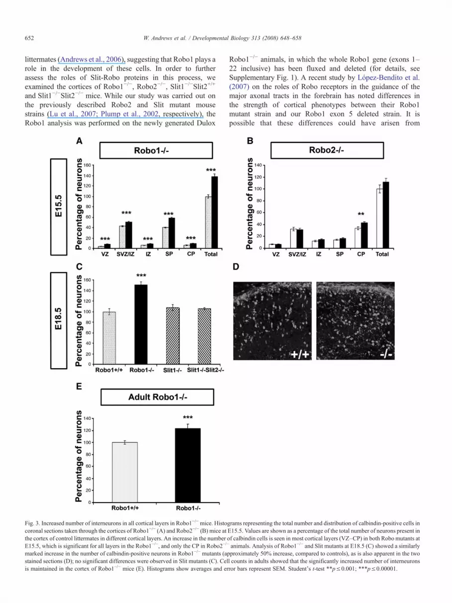
Fig. 3. Increased number of interneurons in all cortical layers in Robo1−/−mice. Histocoronal sections taken through the cortices of Robo1−/− (A) and Robo2−/− (B) mice atthe cortex of control littermates in different cortical layers. An increase in the number oE15.5, which is significant for all layers in the Robo1−/−, and only the CP in Robo2−/−
marked increase in the number of calbindin-positive neurons in Robo1−/− mutants (apstained sections (D); no significant differences were observed in Slit mutants (C). Celis maintained in the cortex of Robo1−/− mice (E). Histograms show averages and er
Robo1−/− animals, in which the whole Robo1 gene (exons 1–22 inclusive) has been fluxed and deleted (for details, seeSupplementary Fig. 1). A recent study by López-Bendito et al.(2007) on the roles of Robo receptors in the guidance of themajor axonal tracts in the forebrain has noted differences inthe strength of cortical phenotypes between their Robo1mutant strain and our Robo1 exon 5 deleted strain. It ispossible that these differences could have arisen from
grams representing the total number and distribution of calbindin-positive cells inE15.5. Values are shown as a percentage of the total number of neurons present inf calbindin cells is seen in most cortical layers (VZ–CP) in both Robo mutants atanimals. Analysis of Robo1−/− and Slit mutants at E18.5 (C) showed a similarlyproximately 50% increase, compared to controls), as is also apparent in the twol counts in adults showed that the significantly increased number of interneuronsror bars represent SEM. Student's t-test ∗∗p≤0.001; ∗∗∗p≤0.00001.

653W. Andrews et al. / Developmental Biology 313 (2008) 648–658
variations in mouse strains used or could be due to geneticalterations, resulting in their mouse line being a hypermorphrather than a true mutant. In order to avoid potentiallyconflicting results, we decided to produce a mouse mutant thatlacks the entire Robo1 gene.
Coronal sections taken through middle (along the rostro-caudal axis) regions of the cortex at E15.5 were immunostainedfor calbindin, and the number and the distribution of labeled cellsthroughout the cortical thickness were analyzed. Our analysisshowed a significant increase in the number of calbindin-positivecells in all cortical layers of Dulox Robo1−/− mutants, andparticularly in the SP (p≤8.9×10⁻¹⁶), compared to control litter-mates (Fig. 3A, n=8 heterozygotes, Robo1+/−, n=7 Robo1−/−).This result compared favorably with our earlier findings in theRobo1 Exon 5 deletionmutant mouse (Andrews et al., 2006; n=3Robo1+/−, n=4 Robo1−/−; data not shown). The significantlyincreased number of cortical interneurons in Dulox Robo1−/−
mice was maintained at E18.5 (Figs. 3C, D; n=6, Robo1+/+ 100±6.2%, Robo1−/− 151±5.7%). To assess cell numbers in adultanimals, we employed ARX, another interneuron marker(Friocourt et al., 2007), as calbindin is known to label a numberof cortical pyramidal cells in postnatal life. This analysis showedthat the increase in interneuron numbers in the cortices of DuloxRobo1−/− mice does persist into adulthood (Fig. 3E; n=3,Robo1+/+ 100±2.9%, Robo1−/− 123±6.9%).
Counts in Robo2−/− animals at E15.5 (n=4 Robo2+/+, n=4Robo2−/−) revealed only a small increase in the number ofcalbindin cells restricted to the CP (Fig. 3B). However, analysis atE17.5 showed no significant changes in the number ofinterneurons in the cortices of Robo2−/− animals compared tocontrols (n=4Robo2+/+, n=4Robo2−/−, data not shown). Similarto Robo2−/− mice, analysis of Slit mutants, Slit1−/−Slit2+/+ orSlit1−/−/Slit2−/− at E18.5 revealed no significant changes in thenumber of interneurons (Fig 3C; n=2 Slit1−/−Slit2+/+; n=2Slit1−/−/Slit2−/−). Further analysis of either Robo2−/− orSlit double mutants at later stages was precluded as these mutantsdie at birth. Our results suggest that only Robo1 has a pronouncedeffect on the number of interneurons entering the cortex, andthis increase is apparent in all layers.
Increased proliferation in Robo1−/− and Slit1−/−Slit2−/− mutantmice
The increase in the number of calbindin-positive cellsmigrating into the neocortex of Robo1−/− mice could be due toa defect in migration and/or due to an increase in proliferation orreduction in cell death. We suggest that the latter possibility israther unlikely in view of the fact that, overall, little apoptosis isobserved in the developing and adult cortex, apart from the SVZat birth and in the first 2 postnatal weeks (Thomaidou et al., 1997).
In order to study the potential effects on proliferation, weinitially prepared dissociated cell cultures from the GEs ofRobo1−/− mutant mice. Cultures were incubated for 18 h andthen pulsed with BrdU for 2 h. These were then fixed in 4% PFAand double labeled to identify BrdU and calbindin-positivecells. Fig. 4C shows the percentage of calbindin expressing cellsthat are also BrdU positive in cultures prepared from mutant and
control GEs. The results indicate a significant increase in theproportion of calbindin-positive cells that had incorporatedBrdU in Robo1−/− mouse derived cultures (n=4; 71.2±1.4%,p≤0.0017) compared with cultures prepared from either wild-type (n=6; 56.5±2.4%) or heterozygote (n=5; 51.1±4.3%)littermates (Figs. 4A–C). Similar results were also obtainedwith Robo1 exon Δ5 mutants (data not shown).
In order to study this further, we assessed the proliferationrates in Robo1 and Slit mutant mice using the mitotic markerphosphohistone 3 (PH3), as changes in cell proliferation has notpreviously been shown for Slit mutants. Coronal sections takenthrough middle (along the rostro-caudal axis) regions of thecortex (E12.5–E16.5) were immunostained for PH3, and thenumber of labeled cells present within the ventricular zone ofthe GE was determined. Our analysis of Dulox Robo1 mutantsat E15.5 indicated, similar to our observations in vitro, a markedincrease in the percentage of cells that were PH3 positive (n=3Robo1−/− 132.6±4.7%, p≤0.0012) compared to wild-typelittermates (n=3 Robo1+/+ 100±3.5%) (Figs. 4D–F). Similarresults were obtained at E12.5 (n=3 Robo1−/−; n=3 Robo1+/+,data not shown). Interestingly, our analysis of Slit1−/−Slit2−/−
mice also revealed a significant increase in the percentage ofcells that were PH3 positive (n=3 138.6±4.4%, p≤0.0006)compared to Slit1−/−Slit2+/+ (n=3 100±3.1%) littermates atE13.5 (Figs. 4G–I). Similar results were also obtained at E16.5(n=2 Slit1−/−Slit2−/−; n=2 Slit1−/−Slit2+/+, data not shown).
Our data also support the notion that a Slit-mediated Robosignal transduction mechanism is involved in proliferation sincewe observed very similar phenotypes in both Slit and Robomutants. Given that the absence of Slit or Robo leads toincreased proliferation, we were interested to know whether theconverse was true, i.e., does the addition of Slit lead to areduction in proliferation? To test this, we added Slit to GEdissociated cultures prepared from GAD67-GFP mice (in whichall interneurons are GFP positive) and stained for theproliferation marker PH3. Since no functional differences areknown to exist between the different Slit proteins, we decided touse a commercially available source of recombinant mouseSlit3 (mSlit3) in these experiments. We used a concentration ofmSlit3 similar to that applied in experiments that demonstratedan effect of this molecule on the migration of cerebellar granulecells (Guan et al., 2007). In the absence of mSlit3, we observedthat 27.97±1.98% of interneurons were PH3 positive, while inthe presence of mSlit3 this value was significantly reduced to7.36±0.67% (p≤0.0001) (Figs. 4J–L). These results suggestthat mSlit3 is a potent inhibitor of cell proliferation in the GE.Similar results were obtained with human Slit1 and Slit2 (datanot shown). As well as having an effect on interneuron pro-liferation, we also found that the presence of mSlit3 results in asignificant decrease in neurite length, in agreement with aprevious study that examined the effects of Slit1 on neuronalprocess branching and elongation (Sang et al., 2002).
The effect of Slit-Robo on cortical interneuron morphology
As well as investigating the role of Slit-Robo in corticalinterneuron migration, we were also interested in evaluating

654 W. Andrews et al. / Developmental Biology 313 (2008) 648–658
655W. Andrews et al. / Developmental Biology 313 (2008) 648–658
their effects on the morphology of these cells. Previous reportshave documented the effect of Slit and Robo on neuronalprocess length and branching in the CNS (Murray andWhitington, 1999; Wang et al., 1999; Ozdinler and Erzurumlu,2002; Ma and Tessier-Lavigne, 2007) and in particular in GEexplants and dissociated GABAergic cell cultures (Zhu et al.,1999; Sang et al., 2002). Here we wanted to study the effect ofloss of Slit-Robo function on interneuron morphology in vivousing Robo and Slit mutant mice. Coronal sections takenthrough the middle (along the rostro-caudal axis) regions of thecortex of E15.5 mice were immunostained for calbindin, whichlabels neuronal cell bodies and processes. The total neuritelength, number of neurite processes and number of branchpoints were quantified in Robo1−/− (n=6 Robo1+/−, n=7Robo1−/−), Robo2−/− (n=4 Robo2+/+, n=5 Robo2−/−) and Slit(n=3 Slit1−/−Slit2+/+; n=3 Slit1−/−Slit2−/−) mutant mice (Fig.5). Our analysis focused on the SP and SVZ/IZ steams ofmigrating interneurons, which contain the greatest number ofreadily quantifiable calbindin-positive cells at this age (30–50neurons were measured per stream per animal). Our resultsshowed that mean total process length was significantly longerin the SVZ/IZ of Robo1−/− mutants (Robo1+/−, 46.6±1.8 μm;Robo1−/− 54.5±1.6 μm, p≤0.0015), but even more so in theSP compared to heterozygote littermates (Robo1+/− 43.4±1.8 μm; Robo1−/− 71.0±2.9 μm, p≤8×10⁻¹⁵) (Figs. 5A andJ–K). Similarly, an increase in the number of neurites was seenin Robo1−/− mutant mice, both in the SVZ/IZ (Robo1+/− 1.51±0.04; Robo1−/− 1.86±0.04, p≤2.1×10⁻⁸) and SP (Robo1+/−
1.60±0.05, Robo1−/− 2.02±0.05, p≤2.2×10⁻⁸) compared toheterozygote littermates (Fig. 5B). Interestingly, an increase inthe degree of branching was also observed in Robo1−/− animalsin both SVZ/IZ (Robo1+/− 0.38±0.04; Robo1−/− 0.63±0.04,p≤1.17×10− 5) and SP streams (Robo1+/− 0.51±0.05;Robo1−/− 0.86±0.05, p≤2.3×10⁻⁶) compared to heterozy-gote littermates (Fig. 5C). Identical patterns were seen in bothRobo1 Exon 5 deleted and in the Robo1 Dulox transgeniclines, not only in terms of process length, but also in terms ofnumber of neurites and branching (data not shown).
Similar morphological analysis performed in the cortices ofRobo2−/− mice showed no major changes compared to wild-type littermates, except for an increase in process length in theSVZ/IZ (Figs. 5G–I). Thus, Robo1, but not Robo2, appears tohave a significant effect on cortical interneurons, not only interms of number but also on cellular morphology within thedeveloping cortex.
Analysis of Slit1−/−Slit2+/+ animals revealed no significantalterations in interneuron morphology (neurite length and
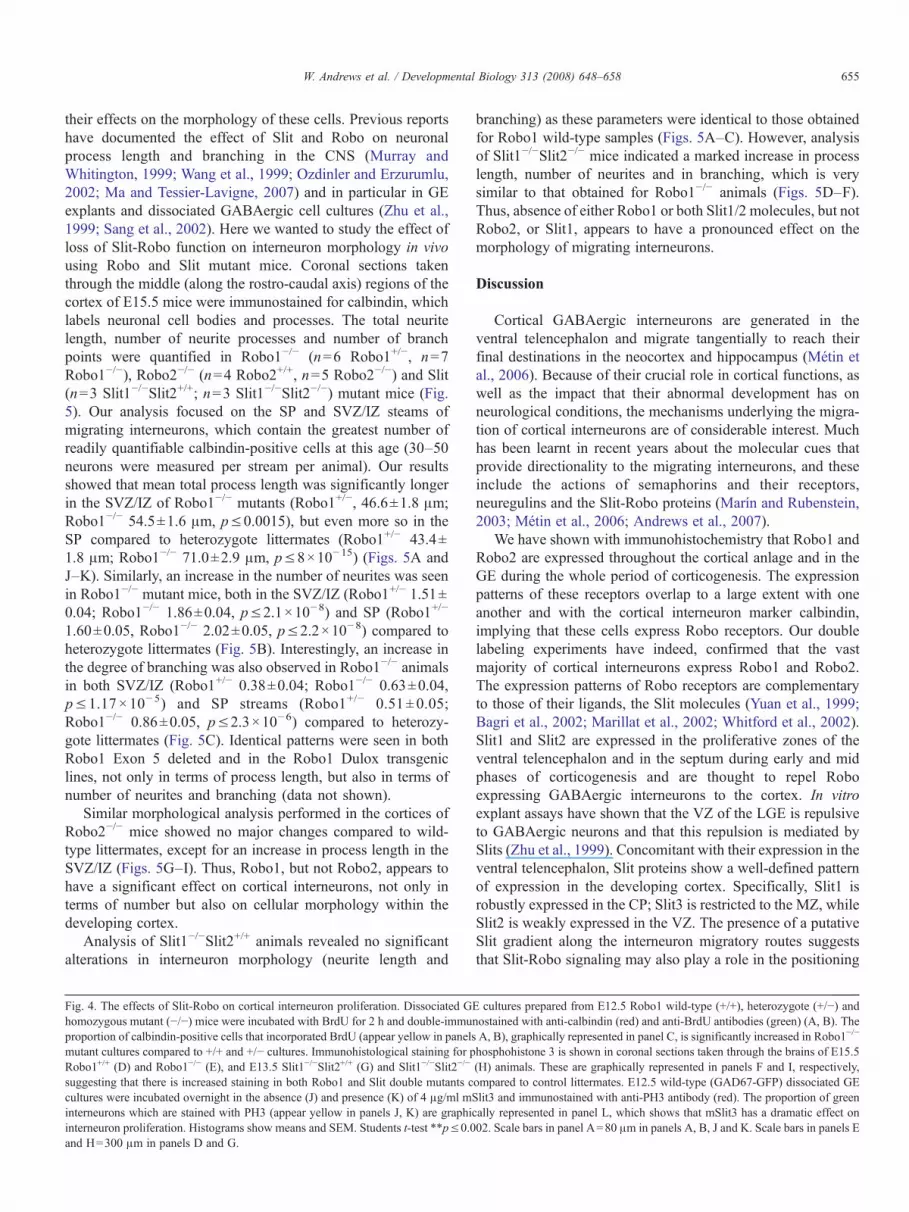
Fig. 4. The effects of Slit-Robo on cortical interneuron proliferation. Dissociated Ghomozygous mutant (−/−) mice were incubated with BrdU for 2 h and double-immuproportion of calbindin-positive cells that incorporated BrdU (appear yellow in panelsmutant cultures compared to +/+ and +/− cultures. Immunohistological staining for pRobo1+/+ (D) and Robo1−/− (E), and E13.5 Slit1−/−Slit2+/+ (G) and Slit1−/−Slit2−/−
suggesting that there is increased staining in both Robo1 and Slit double mutants ccultures were incubated overnight in the absence (J) and presence (K) of 4 μg/ml mSinterneurons which are stained with PH3 (appear yellow in panels J, K) are graphicinterneuron proliferation. Histograms show means and SEM. Students t-test ∗∗p≤0.0and H=300 μm in panels D and G.
branching) as these parameters were identical to those obtainedfor Robo1 wild-type samples (Figs. 5A–C). However, analysisof Slit1−/−Slit2−/− mice indicated a marked increase in processlength, number of neurites and in branching, which is verysimilar to that obtained for Robo1−/− animals (Figs. 5D–F).Thus, absence of either Robo1 or both Slit1/2 molecules, but notRobo2, or Slit1, appears to have a pronounced effect on themorphology of migrating interneurons.
Discussion
Cortical GABAergic interneurons are generated in theventral telencephalon and migrate tangentially to reach theirfinal destinations in the neocortex and hippocampus (Métin etal., 2006). Because of their crucial role in cortical functions, aswell as the impact that their abnormal development has onneurological conditions, the mechanisms underlying the migra-tion of cortical interneurons are of considerable interest. Muchhas been learnt in recent years about the molecular cues thatprovide directionality to the migrating interneurons, and theseinclude the actions of semaphorins and their receptors,neuregulins and the Slit-Robo proteins (Marín and Rubenstein,2003; Métin et al., 2006; Andrews et al., 2007).
We have shown with immunohistochemistry that Robo1 andRobo2 are expressed throughout the cortical anlage and in theGE during the whole period of corticogenesis. The expressionpatterns of these receptors overlap to a large extent with oneanother and with the cortical interneuron marker calbindin,implying that these cells express Robo receptors. Our doublelabeling experiments have indeed, confirmed that the vastmajority of cortical interneurons express Robo1 and Robo2.The expression patterns of Robo receptors are complementaryto those of their ligands, the Slit molecules (Yuan et al., 1999;Bagri et al., 2002; Marillat et al., 2002; Whitford et al., 2002).Slit1 and Slit2 are expressed in the proliferative zones of theventral telencephalon and in the septum during early and midphases of corticogenesis and are thought to repel Roboexpressing GABAergic interneurons to the cortex. In vitroexplant assays have shown that the VZ of the LGE is repulsiveto GABAergic neurons and that this repulsion is mediated bySlits (Zhu et al., 1999). Concomitant with their expression in theventral telencephalon, Slit proteins show a well-defined patternof expression in the developing cortex. Specifically, Slit1 isrobustly expressed in the CP; Slit3 is restricted to the MZ, whileSlit2 is weakly expressed in the VZ. The presence of a putativeSlit gradient along the interneuron migratory routes suggeststhat Slit-Robo signaling may also play a role in the positioning
E cultures prepared from E12.5 Robo1 wild-type (+/+), heterozygote (+/−) andnostained with anti-calbindin (red) and anti-BrdU antibodies (green) (A, B). TheA, B), graphically represented in panel C, is significantly increased in Robo1−/−
hosphohistone 3 is shown in coronal sections taken through the brains of E15.5(H) animals. These are graphically represented in panels F and I, respectively,ompared to control littermates. E12.5 wild-type (GAD67-GFP) dissociated GElit3 and immunostained with anti-PH3 antibody (red). The proportion of greenally represented in panel L, which shows that mSlit3 has a dramatic effect on02. Scale bars in panel A=80 μm in panels A, B, J and K. Scale bars in panels E

Fig. 5. Morphological differences in migrating interneurons in Robo and Slit mutants. Histograms representing morphological parameters of calbindin-positive cells incoronal sections taken through the cortices of Robo1−/− (A–C; Robo1+/−white, Robo1−/− black), Slit (D–F; Slit1−/−Slit2+/+ white, Slit1−/−Slit2−/− black) and Robo2−/−
(G–I; Robo2+/+ white, Robo2−/− black) mutant mice at E15.5. An increase in total neurite length (μm) was found in Robo1−/− (A), Robo2−/− (G) and Slit1−/−Slit2−/−
(D) animals compared to control littermates. This is also illustrated for Robo1 in panels J–K.We also noted an increase in the number of neurites (B, E) and the numberof branch points (C, F) for both Robo1−/− and Slit1−/−Slit2−/− animals compared to controls. Student's t-test ∗∗pb0.002, ∗∗∗pb0.00001. Scale bar in panel K=20 μmfor panels J and K.
656 W. Andrews et al. / Developmental Biology 313 (2008) 648–658
of these tangential paths within the developing cortex.However, analysis of the migration and numbers of interneuronsidentified with a variety of markers at different embryonicstages showed no difference in the cortex between wild-typeand Slit1−/−Slit2−/− double- and Slit1−/−Slit2−/−netrin−/− triplemutants (Marín et al., 2003). These observations suggest thatSlit1 and Slit2 are not necessary for the tangential migration ofinterneurons to the cortex. However, these proteins appear toregulate neuronal migration within the basal telencephalon(Marín et al., 2003).
Although Slit mutants do not show any difference in thenumber and distribution of cortical interneurons (shown hereand Marín et al., 2003), we noted a marked increase in thenumber of such cells in all layers in Robo1−/− mutantsthroughout corticogenesis and in adulthood. The fact thatsuch phenotype was not observed in the Slit1−/−Slit2−/− animalssuggests that this may be a Slit independent event, or that anunidentified member of the Slit family of molecules may beinvolved. We speculated that the cell increase could be due to a
change in migration rate or a consequence of failure to respondto inhibitory cues, normally imposed by the presence of Slit inthe cortex, or due to changes in proliferation/apoptosis. We havedemonstrated here that the increased number of interneurons inthe cortex of Robo1−/− animals is, at least in part, due toincreased proliferation in the GE. Interestingly, we observed asimilar increase in proliferation in Slit1−/−Slit2−/− mutants,suggesting that Slit-Robo signalling mechanisms are involvedin regulating cell division.
While Slit has been shown to play a role in asymmetricalcell division in Drosophila (Mehta and Bhat, 2001), this is thefirst report of Slit-Robo signaling having a direct effect on cellproliferation in a neuronal cell type in vertebrates. It ispertinent to note that abundant evidence points to an active rolefor Slit-Robo signaling in tumor development. Specifically,tumor suppressor gene activity has been proposed for Slit2 andRobo1 in lung and breast cancer (Sundaresan et al., 1998), andseveral studies have shown that both genes are frequentlyinactivated in lung adenocarcinomas and lymphomas by

657W. Andrews et al. / Developmental Biology 313 (2008) 648–658
methylation of the gene promoters (Dallol et al., 2002; Xian etal., 2004). Moreover, Xian and colleagues (2004) havedemonstrated that Dutt1/Robo1 transgenic mice show a higherincidence of lymphomas and carcinomas than wild-type litter-mates, suggesting that Dutt1/Robo1 acts as a tumor suppressorgene.
Although we observed an increase in proliferation inSlit1−/−Slit2−/− mutant mice, we did not note a correspondingincrease in the number of interneurons entering the cortex. Onepossible explanation may be that the continued presence of Slit3in the cortices of Slit1−/−Slit2−/− mutant mice prevents inter-neurons from migrating into the cortex prematurely. Secondly,we observed recently that interneurons migrate into the nor-mally repulsive striatum in Robo1−/− mutants (Andrews et al.,2006). Thus, we speculated that some interneurons take a“short-cut” through this region, which could explain theirincreased number in the cortices of Robo1−/− mice, unlike theSlit1−/−Slit2−/− animals where interneurons have not beenshown to enter the striatal region (Marín et al., 2003). Thus, thedifferent migratory paths taken by interneurons in Slit andRobo1 mutants could account for the differences seen in thenumber of cells entering the cortex. We are actively testing thesehypotheses at present.
There are several lines of evidence to suggest that Slit-Roboproteins play a role in neuronal process elongation andbranching in a number of developing systems (Ozdinler andErzurumlu, 2002; Ma and Tessier-Lavigne, 2007). In morepertinent studies that utilized MGE explants and cortical cellcultures (Sang et al., 2002; Sang and Tan, 2003; Whitford et al.,2002), Slit was found to promote process elongation andbranching on cortical interneurons. At first glance, these resultsseem to contradict our in vivo findings. However, not allinterneuron cohorts respond to Slit in a similar fashion, as thework of Sang and colleagues (2002, 2003) has indicated that theresponse varies according to the age of the dissociated cells.Whereas interneuron cultures established from early stages(E13.5 and E15.5) showed suppressed neurite growth, in linewith our observations in E15.5 Slit mutants, interneuronscultured from E17.5 brains responded to Slit by increasingneurite branching.
In summary, we found that the majority of corticalinterneurons express both Robo1 and Robo2 and that absenceof Robo1, but not Robo2 or Slit proteins, leads to an increasein the number of interneurons migrating to the cortex from theventral telencephalon. This increase may be attributed, at leastin part, to increased cell proliferation. Analysis of themorphology of cortical interneurons in the same mutantsrevealed that lack of Robo1 or Slit1/Slit2, but not Robo2 orSlit1, has a pronounced effect on process elongation andbranching. These observations suggest that the same Robo1–Slit1/Slit2 signal transduction mechanism is utilized within thecortex to regulate interneuron morphology.
Acknowledgments
We are grateful to Drs. Clare Faux and Gaelle Friocourtfor helpful comments in the preparation of the manuscript.
We are also grateful to Dr. M. Tessier-Lavigne for supplyingSlit mutants and to Drs. Y. Yanagawa and K. Obata forgiving us the GAD67-GFP mice used in this study. The workwas supported by a Wellcome Trust Grant Programme Grant(074549).
Appendix A. Supplementary data
Supplementary data associated with this article can be found,in the online version, at doi:10.1016/j.ydbio.2007.10.052.
References
Andrews, W., Liapi, A., Plachez, C., Camurri, L., Zhang, J., Mori, S.,Murakami, F., Parnavelas, J.G., Sundaresan, V., Richards, L.J., 2006. Robo1regulates the development of major axon tracts and interneuron migration inthe forebrain. Development 133, 2243–2252.
Andrews, W.D., Barber, M., Parnavelas, J.P., 2007. Slit-Robo interactionsduring cortical development. J. Anat. 221, 188–189.
Bagri, A., Marín, O., Plump, A.S., Mak, J., Pleasure, S.J., Rubenstein, J.L.,Tessier-Lavigne, M., 2002. Slit proteins prevent midline crossing anddetermine the dorsoventral position of major axonal pathways in themammalian forebrain. Neuron 33, 233–248.
Cavanagh, J.F., Mione, M.C., Pappas, I.S., Parnavelas, J.G., 1997. Basicfibroblast growth factors prolongs the proliferation of rat cortical progenitorcells in vitro without altering their cell cycle parameters. Cereb. Cortex 4,293–302.
Cobos, I., Calcagnotto, M.E., Vilaythong, A.J., Thwin, M.T., Noebels, J.L.,Baraban, S.C., Rubenstein, J.L., 2005. Mice lacking Dlx1 show subtype-specific loss of interneurons, reduced inhibition and epilepsy. Nat. Neurosci.8, 1059–1068.
Corbin, J.G., Nery, S., Fishell, G., 2001. Telencephalic cells take a tangent: non-radial migration in the mammalian forebrain. Nat. Neurosci. 1177–1182(Suppl.).
Dallol, A., Fernandes Da Silva, N., Viacava, P., Minna, J.D., Bieche, I., Maher,E.R., Latif, F., 2002. Slit2, a human homologue of the Drosophila Slit2gene, has tumour suppressor activity and is frequently inactivated in lungand breast cancers. Cancer Res. 62, 5874–5880.
Friocourt, G., Liu, J.S., Antypa, M., Rakic, S., Walsh, C.A., Parnavelas, J.G.,2007. Both doublecortin and doublecortin-like kinase play a role in corticalinterneuron migration. J. Neurosci. 27, 3875–3883.
Guan, C.B., Xu, H.T., Jin, M., Yuan, X.B., Poo, M.M., 2007. Long-range Ca2+
signaling from growth cone to soma mediates reversal of neuronal migrationinduced by slit-2. Cell 129, 385–395.
Hu, H., 1999. Chemorepulsion of neuronal migration by Slit2 in the developingmammalian forebrain. Neuron 23, 703–711.
Kumar, S.S., Buckmaster, P.S., 2006. Hyperexcitability, interneurons, and lossof GABAergic synapses in entorhinal cortex in a model of temporal lobeepilepsy. J. Neurosci. 26, 4613–4623.
López-Bendito, G., Flames, N., Ma, L., Fouquet, C., Di Meglio, T., Chedotal, A.,Tessier-Lavigne,M.,Marín, O., 2007. Robo1 and Robo2 cooperate to controlthe guidance of major axonal tracts in the mammalian forebrain. J. Neurosci.27, 3395–3407.
Lu,W., vanEerde,A.M., Fan,X.,Quintero-Rivera, F., Kulkarni, S., Ferguson,H.,Kim, H.G., Fan, Y., Xi, Q., Li, Q.G., Sanlaville, D., Andrews, W.,Sundaresan, V., Bi, W., Yan, J., Giltay, J.C., Wijmenga, C., de Jong, T.P.,Feather, S.A., Woolf, A.S., Rao, Y., Lupski, J.R., Eccles, M.R., Quade, B.J.,Gusella, J.F., Morton, C.C., Maas. R, L., 2007. Disruption of ROBO2 isassociated with urinary tract anomalies and confers risk of vesicoureteralreflux. Am. J. Hum. Genet. 80, 616–632.
Ma, L., Tessier-Lavigne, M., 2007. Dual branch-promoting and branch-repellingactions of Slit/Robo signaling on peripheral and central branches ofdeveloping sensory axons. J. Neurosci. 27, 6843–6851.
Mallet, N., Ballion, B., Le Moine, C., Gonon, F., 2006. Cortical inputs andGABA interneurons imbalance projection neurons in the striatum ofParkinsonian rats. J. Neurosci. 26, 3875–3884.

658 W. Andrews et al. / Developmental Biology 313 (2008) 648–658
Marillat, V., Cases, O., Nguyen-Ba-Charvet, K.T., Tessier-Lavigne, M.,Sotelo, C., Chedotal, A., 2002. Spatiotemporal expression patterns of slitand robo genes in the rat brain. J. Comp. Neurol. 442, 130–155.
Marín, O., Rubenstein, J.L., 2003. Cell migration in the forebrain. Annu. Rev.Neurosci. 26, 441–483.
Marín, O., Plump, A.S., Flames, N., Sanchez-Camacho, C., Tessier-Lavigne,M., Rubenstein, J.L., 2003. Directional guidance of interneuron migration tothe cerebral cortex relies on subcortical Slit1/2-independent repulsion andcortical attraction. Development 130, 1889–1901.
Mehta, B., Bhat, K.M., 2001. Slit signaling promotes the terminal asymmetricdivision of neural precursor cells in Drosophila CNS. Development 128,3161–3168.
Métin, C., Baudoin, J.P., Rakic, S., Parnavelas, J.G., 2006. Cell and molecularmechanisms involved in the migration of cortical interneurons. Eur. J.Neurosci. 23, 894–900.
Murray, M.J., Whitington, P.M., 1999. Effects of roundabout on growth conedynamics, filopodial length, and growth cone morphology at the midline andthroughout the neuropile. J. Neurosci. 19, 7901–7912.
Nadarajah, B., Parnavelas, J.G., 2002. Modes of neuronal migration in thedeveloping cerebral cortex. Nat. Rev., Neurosci. 3, 423–432.
Ozdinler, P.H., Erzurumlu, R.S., 2002. Slit2, a branching-arborization factor forsensory axons in the mammalian CNS. J. Neurosci. 22, 4540–4549.
Plump, A.S., Erskine, L., Sabatier, C., Brose, K., Epstein, C.J., Goodman, C.S.,Mason, C.A., Tessier-Lavigne, M., 2002. Slit1 and Slit2 cooperate to preventpremature midline crossing of retinal axons in the mouse visual system.Neuron 33, 219–232.
Poirier, K., Van Esch, H., Friocourt, G., Saillour, Y., Bahi, N., Backer, S.,Souil, E., Castelnau-Ptakhine, I., Beldjord, C., Francis, F., Bienvenu, T.,2004. Neuroanatomical distribution of ARX in brain and its localisation inGABAergic neurons. Mol. Brain Res. 122, 35–46.
Sang, Q., Tan, S.S., 2003. Contact-associated neurite outgrowth and branchingof immature cortical interneurons. Cereb. Cortex 13, 677–683.
Sang, Q., Wu, J., Rao, Y., Hsueh, Y.P., Tan, S.S., 2002. Slit promotes branchingand elongation of neurites of interneurons but not projection neurons fromthe developing telencephalon. Mol. Cell. Neurosci. 21, 250–265.
Sloviter, R.S., 1987. Decreased hippocampal inhibition and a selective loss ofinterneurons in experimental epilepsy. Science 235, 73–76.
Sundaresan, V., Chung, G., Heppel-Parton, A., Xiong, J., Grundy, C.,Roberts, I., James, L., Cahn, A., Bench, A., Douglas, J., Minna, J.,Sekido, Y., Lerman, M., Latif, F., Bergh, J., Li, H., Lowe, N., Ogilvie, D.,Rabbitts, P., 1998. Homozygous deletions at 3p12 in breast and lungcancer. Oncogene 17, 1723–1729.
Tamamaki, N., Yanagawa, Y., Tomioka, R., Miyazaki, J., Obata, K., Kaneko, T.,2003. Green fluorescent protein expression and colocalization withcalretinin, parvalbumin, and somatostatin in the GAD67-GFP knock-inmouse. J. Comp. Neurol. 467, 60–79.
Thomaidou, D., Mione, M.C., Cavanagh, J.F., Parnavelas, J.G., 1997. Apoptosisand its relation to the cell cycle in the developing cerebral cortex.J. Neurosci. 17, 1075–1085.
Wang, K.H., Brose, K., Arnott, D., Kidd, T., Goodman, C.S., Henzel, W.,Tessier-Lavigne, M., 1999. Biochemical purification of a mammalian slitprotein as a positive regulator of sensory axon elongation and branching.Cell 96, 771–784.
Whitford, K.L., Marillat, V., Stein, E., Goodman, C.S., Tessier-Lavigne, M.,Chedotal, A., Ghosh, A., 2002. Regulation of cortical dendrite developmentby Slit-Robo interactions. Neuron 33, 47–61.
Wu, W., Wong, K., Chen, J., Jiang, Z., Dupuis, S., Wu, J.Y., Rao, Y., 1999.Directional guidance of neuronal migration in the olfactory system by theprotein Slit. Nature 400, 331–336.
Xian, J., Clark, K.J., Fordham, R., Pannell, R., Rabbitts, T.H., Rabbitts, P.H.,2001. Inadequate lung development and bronchial hyperplasia in mice witha targeted deletion in the Dutt1/Robo1 gene. Proc. Natl. Acad. Sci. U. S. A.98, 15062–15066.
Xian, J., Aitchison, A., Bobrow, L., Corbett, G., Pannell, R., Rabbitts, T.H.,Rabbitts, P.H., 2004. Targeted disruption of the 3p12 gene, Dutt1/Robo1,predisposes mice to lung adenocarcinomas and lymphomas with methyla-tion of the gene promoter. Cancer Res. 64, 6432–6437.
Yuan, W., Zhou, L., Chen, J.H., Wu, J.Y., Rao, Y., Ornitz, D.M., 1999. Themouse SLIT family: secreted ligands for ROBO expressed in patterns thatsuggest a role in morphogenesis and axon guidance. Dev. Biol. 212,290–306.
Zhu, Y., Li, H., Zhou, L., Wu, J.Y., Rao, Y., 1999. Cellular and molecularguidance of GABAergic neuronal migration from an extracortical origin tothe neocortex. Neuron 23, 473–485.