the pediatric and adult interventional cardiac symposium
TRANSCRIPT
Published online: December 2016
Meeting Abstracts
Journal of Structural Heart Disease, December 2016, Volume 2, Issue 6:241-306DOI: http://dx.doi.org/10.12945/j.jshd.2016.16.013
Fax +1 203 785 3346 E-Mail: [email protected]://structuralheartdisease.org/
© 2016 Journal of Structural Heart DiseasePublished by Science International Corp. ISSN 2326-4004
Accessible online at: http://structuralheartdisease.org/
#0001 EVALUATION OF RADIATION DOSES FOR PEDIATRIC PATIENTS DURING INTERVENTIONAL CARDIOLOGY PROCEDURES AT HAMAD GENERAL HOSPITAL, STATE OF QATAR.Hesham Al-Saloos, Antar Aly, Huda Al-Naemi,Weill Cornell Medicine, Doha, Qatar
The estimated risks associated with radiation exposure are higher in children compared to adults. The use of fluoroscopy in diagnostic and interventional cardiac catheterizations being done in children requires accurate determination of the associated effective dose. Diagnostic procedures such as right and left heart studies (R&L Heart) and interventional procedures like closure of patent ductus arteriosus (PDA) or atrial septal defect (ASD), pericardial tap and balloon angioplasty of pulmonary or aortic valves, branch pulmo-nary arteries or coarctation of the aorta are among the commonest procedures done for the pediatric age group undergoing cardiac catheterization.
In this study the results of an analysis of doses recorded for 203 cases over 2 years (2013 and 2014) carried out in pediatric patients. These data likely represent the largest set of radiation doses recorded in children undergoing cardiac catheterization. The maximum Kerma Aria Product (KAP) recorded for those patients were 9779 cGy.cm2. The maximum Cumulative Dose at the Interventional Reference point (CD_IRP) was also evaluated and found to be approximately 999 mGy. Body weight (BW) and body surface area (BSA) were also considered.
Materials and Methods: Two X-ray biplane fluoroscopy were used in this study. patient’s age, weight, height, gender, and procedure type and fluoroscopy time. Kerma Aria Product (KAP) and Cumulative dose data were recorded for 198 patients. The average pediatric age, weight and height were 3.03 years, 13.8 kg and 88.4 cm respectively. Peak voltage was 60.8kVp – 80 kVp.
Conclusion: Evaluation of KAP and CD_IRP doses are important indi-cators for the pediatric dose management and it is recommended to
include all those data in patient’s records. Body weight is an impor-tant factor in determining the radiation dose for children undergoing cardiac catheterization. Using a newer technology and adopt differ-ent imaging protocols (reducing the P/s and F/s) would lower the radiation dose without compromising the image quality.
#0002 TWO CASES OF TURNER SYNDROME WITH HYPOPLASTIC LEFT HEART MANAGED WITH HYBRID PROCEDUREAmr Matoq, Robert English, Michael Shillingford, Jose Ettedgui,UF- College of medicine- Jacksonville, Jacksonville, FL, USA
Background: The presence of low birth weight, extra cardiac anom-alies or genetic syndromes has been associated with poor outcome with Norwood procedures for palliation of Hypoplastic left heart syn-drome (HLHS). Children with Turner syndrome and HLHS have a very high operative mortality and post-operative complications from per-sistent pleural effusions. Thus, for these high-risk patients, a hybrid approach to the Norwood operation can be an alternative palliation strategy. There is limited literature about outcomes of hybrid proce-dures in patients with Turner syndrome.
Methods: We present two cases of Turner syndrome with HLHS, who successfully underwent hybrid procedures as initial palliation for HLHS who subsequently underwent surgical repair achieving two-ventricle circulation.
Results: Two patients with Turner syndrome underwent hybrid Norwood palliation at 9 and 14 days of age. Weight at the time of intervention was 2320 grams and 2300 grams and both had placement of an 15mm x 5mm and 17mm x 7mm pre-mounted stent expanded and the pulmonary arteries were banded down to 3.5 mm diameter. These procedures were performed in the cath-eterization laboratory. The CHSS risk score was +17.93 and 15.35 (positive number favors a Univentricular repair, with the magni-tude of the difference expressed by the number). In both children
The Pediatric and Adult Interventional Cardiac Symposium (PICS/AICS)20th Annual Meeting, Miami Beach, Florida, January 16, 2017 – January 19, 2017
Journal of Structural Heart Disease, December 2016 Volume 2, Issue 6:241-306
Meeting Abstracts 242
there was growth of the left heart with successful uncomplicated 2nd stage procedure at 6 months and 10 months of age. One child had residual pulmonary artery stenosis post 2nd stage operation that required balloon pulmonary angioplasty. At 6 months follow up, both cases were doing well clinically and had normal function in echocardiogram. Both had mild sub-aortic stenosis with no obstruction or regurgitation.
Conclusion: The 2 patients in this series had successful initial pal-liation with a hybrid approach to the Norwood operation with no significant procedural complications or pleural effusions. Both sustained adequate growth of the left heart that subsequently allowed biventricular repair. Hybrid approach to the Norwood pro-cedure carries the advantage of avoiding cardiopulmonary bypass and early aortic arch reconstruction and should be considered for palliation of high-risk patients with HLHS, especially with Turner syndrome.
#0003 INITIAL EXPERIENCE OF ATRIAL SEPTAL DEFECT CLOSURE USING THE NEW GENERATION CARDIA ULTRASEPT IITM DEVICE IN MEXICO.Roberto MijangosPediatric Hospital, Tuxtla Gutiérrez, Chiapas, Mexico
We present the initial experience in Mexico of atrial septal defect closure using the new Cardia Ultrasept IITM device. We present a series of 5 patients with ASD previously selected as candidates with favourable anatomy (less than 38mm defect, rims greater tan 5mm) to be subjected for closure of the defect through interventionism treated in the period April-August 2016. Prospective, observational, transverse and descriptive study. The group included 3 female patients (60%) with a mean age of 10 ±2.12 years. The haemody-namic and anatomical data were as follows: pulmonary artery sys-tolic pressure 25.2 ±3.5 mmHg, pulmonary to systemic flow ratio 2.78 ±0.52, septal defect diameter 17.78 ±6.18 mm, expanded defect diameter 20.7 ±6.56 mm. All septal occluder were delivered successfully. No residual shunt evidenced by angiography and int-racardiac echocardiography. At follow-up to one month, all patients showed complete closure of the defect and continuous decreased of right ventricular diastolic diameter (38.6 ±2.33 mm (Z-score 2.97 ±0.22) vs 34.26 ±3.13 mm (Z-score 2.46 ±0.33)) p=0.78. No compli-cations at follow-up have been reported. The new generation of the Cardia Ultrasept IITM device is a good alternative to percutaneously treat atrial septal defect.
#0004 RECONSTRUCTIVE SURGERY OF HYPOPLASIA OF THE AORTIC ARCHElnur Hasanov, Faig Mirzazada, Elnur Imanov, Shahmardan Danyalov, Nigar Suleymanzada, Samira Karimova, Vusala KazimovaPediatric Cardiac Surgery Centre, Baku, Azerbaijan
Objective: to evaluate the function of baroreceptors in patients after different types of surgical correction of hypoplastic aortic arch.
Materials and Methods: In this prospective cohort study evaluated the results of surgical treatment of 54 patients who underwent sur-gical treatment for aortic coarctation. The patients were divided into two groups according to the method of correction of the defect: reconstruction with the use of a modified reverse plasty of LPA (group I, n=27) and reconstruction using the “extended” anastomosis (group II, n=27 patients).
Results: the Postoperative period of observation was 25 (21-30) months. Spontaneous sensitivity of the baroreceptors differed between groups and was significantly higher in group II is 11.6 (10,5; 12,6) vs 9,1 (8,2;10,1) in group I, p -0,04. The velocity of pulse blood flow was also higher in group II 7,7 (5,8;9) (m/s) -1 compared to 6.5 (5,4;7,1) (m/s) -1 in group I and differed between groups P – 0,04.
Conclusions: Reduced sensitivity of baroreceptors in patients after a modified reverse plastic of the left subclavian artery may be regarded as the method of choice in patients with coarctation and hypoplasia of the arch as a method of reducing the frequency of arterial hyper-tension in the late postoperative period.
#0005 RECANNULATION OF LEFT PULMONARY ARTERY WITH RADIO-FREQUENCY PERFORATION AND STENT ANGIOPLASTY AFTER FAILED HYBRID STENT ANGIOPLASTYMatthew Brown, John BreinholtThe University of Texas Health Science Center at Houston, Houston, TX, USA
An infant with pulmonary atresia and ventricular septal defect (VSD), s/p right-sided modified Blalock-Taussig shunt as a neo-nate, underwent VSD closure and placement of a 13 mm homo-graft right ventricle-to-pulmonary artery (RV-PA) conduit with augmentation of proximal branch pulmonary arteries at 9 months of age. After hospital discharge, there were concerns about flow into the left pulmonary artery (LPA), and he was referred for cath-eterization at 11 months of age, where he was found to have discontinuity of the left pulmonary artery (LPA). Transcatheter attempts at entering the LPA were unsuccessful, although he required stent angioplasty of right pulmonary artery branches. He was taken to the operating room (non-hybrid suite), where, after blind dissection and progressive dilation with Hegar dila-tors up to 3.5 mm, the surgeon positioned, and we deployed, a 16 mm long EV3 Intrastent Max LD stent mounted on an 8x20 mm Z-Med II balloon. A poor-quality C-arm angiogram, without the ability to record, demonstrated contrast to the end of the stent without flow in the LPA, indicating that the stent was extravas-cular. The patient was taken to cardiac catheterization lab, where simultaneous angiograms in the LPA stent and left pulmonary vein wedge injection demonstrated overlapping of the stent and the LPA. The Baylis Radio-Frequency system was used to perfo-rate from the blind end of stent into the LPA, allowing for pas-sage of the microcatheter into the LPA, followed by angiography in the LPA to confirm position. We then deployed a Palmaz Blue 7x15 mm stent over a V-18 wire, overlapping with the existing stent, resulting in patency of the LPA from the RV-PA conduit, and no extravasation of contrast. Follow-up echocardiograms, and
Hijazi, Z 20th Annual PICS/AICS Meeting Abstracts
243 Meeting Abstracts
#0007 SUCCESFUL TRANSCATETHER AORTO-RIGTH ATRIAL FISTULA CLOSSURE USING AMPLATZER VASCULAR PLUG IIManfred Hermanni1, Federico Borges2, Fernando Prieto1, Ernesto Urbano3, Elka Maria Marcano1, Victor Julio Bellera1
1Centro Policlinico La Viña, Valencia - Carabobo, Venezuela2Hospital De Niños Jm De Los Rios, Caracas, Venezuela3Hospital Dr Raul Leoni, Puerto Ordaz, Venezuela
Aorto cameral fistula are rare, usually found in right cavities, aquired fistula are more common than congenital, there are few published cases. We report a 7 year old girl refered for cardiac murmur and his-tory of frequent respiratory disease, ecochardiogram evidenced color flow, turbulent, tortous between aorta and righ atrium, 6 mm diameter, sitolyc maximum gradient 92 mmhg, and mild right cav-ities dilation. Cardiac cath was performed: aortic root pig tail injec-tion revealed tortous fistula originated in right coronary sinus next to right coronary ostium, 6 mm diameter, and ended in posterior right atrial wall. Normal coronarigraphy. A 4 fr jr catheter is advanced to the more distal segment of the fistula and trough double guire maneuver the delivery sistem was placed, a vascular plug ii 12 mm, was acu-rately positioned, and realeased with mild residual shunt trough the device, no complications ocurred.
Discussion and Conclusions: Aortocameral fistula clossure is recom-mended even in asympomatis patients, due to low rate procedure complications, the risk of ventricule volume overload, bacterial endo-carditis, pulmonary vascular disease, aneurysm formation and spon-tanoeus rupture due to constant permeability we report a succesful percutaneous aota right atrial congenital fistula clossure.
Key words: Aorto cameral • fistula • amplatzer vascular plug ii
#0008 RISK FACTORS FOR AN ELEVATED PRE-FONTAN VENTRICULAR END-DIASTOLIC PRESSURE IN PATIENTS WITH SINGLE VENTRICLE CONGENITAL HEART DISEASEMatthew Schwartz, Michael Brock, David Nykanen, William DeCampliArnold Pamler Hospital, Orlando, USA
Introduction: Systemic ventricular end-diastolic pressure (SVEDP) is an important determinant of pulmonary artery pressure in those with Fontan circulation. Predictors of an elevated SVEDP have been incompletely identified in this population.
Methods: All patients who underwent Fontan operation at our center between 1/2009 and 12/2013 were retrospectively identified. SVEDP at pre-Fontan catheterization and other relevant patient variables were extracted. Analysis was performed to identify variables associ-ated with pre-Fontan SVEDP.
Results: We identified 61 patients with mean age at pre-Fontan catheterization of 3.9 ± 1.6 yrs and 36 (59%) had a systemic right ventricle (RV). Pre-Fontan SVEDP was positively associated with
cardiac catheterization nearly one year later demonstrated unob-structed flow from the RV-PA conduit to the LPA.
#0006 FIRST EXPERIENCE OF INTRODUCTION OF ENDOVASCULAR METHOD TREATMENT OF CONGENITAL HEART DISEASES AT INFANTS IN THE FIRST YEAR OF BIRTH AT PEDIATRIC CARDIAC CENTER OF THE MINISTRY OF HEALTH OF AZERBAIJAN Elnur Imanov1, Vasiliy Vasilyevich Lazorishinech2, Igor Alexandrovich Ditkovskiy2, Elnur Hasanov2, Faiq Mirzazada2, Shahmardan Danyalov2, Suleymanzade Nigar2, Fuad Abdullayev2
1Health Ministry of Republic of Azerbaijan Scientific Center of Surgery Named after M.A.Topcubashov, Baku Yasamal, Azerbaijan2National Institute of Cardiovascular Surgery Named after N.M. Amosov, Kiev, Ukraine
Objective: comparison of quality and quantity of the operated patients with critical congenital heart diseases by endovascular treat-ment method at newly opened cardio-surgical center and already famous and one of the leading centers of the world.
Methods: Endovascular treatment method of critical congenital heart diseases at infants.
Results: 32 patients under 1 year out of 209 patients with critical con-genital heart diseases have been operated by endovascular treatment method since the opening of the Pediatric Cardio-surgical Center of the Ministry of Health of Azerbaijan in 2011. They were patients with such diagnoses as: closed interatrial septum – 11 patients, balloon dilatation of the pulmonary artery – 80 patients, closed arterial duct – 75 patients, balloon dilatation of the coarctation aorta – 5 patients, balloon dilatation of the aortic valve – 12 patients, as well as such kind of complex congenital heart diseases as cosed arterial duct + balloon dilatation of the pulmonary artery – 15 patients, balloon dilatation of the pulmonary artery + closed interatrial septum – 10 patients, perforation of the pulmonary artery + Paltayif PBV, balloon atriosep-tostomy – 5 patients. Thus, lethality was 2.3 %. 438 patients out of 600 with critical congenital heart diseases have been operated by endovascular treatment method within 2007 – 2016 at the National Institute of Cardiovascular Surgery named after N.M.Amosov. They were with diagnoses as АoS+ 63; CoAo – 74; Sp – 58; Paltayif PBV ТОF – 12; Rashkind – 163; Stent PBA/MARCA – 17; closed arterial duct – 15. Positive result has been obtained at 95% cases and the patients were discharged in satisfactory condition. Thereby, mortality was 5%.
Conclusion: The World Pediatric cardio – surgery of critical congeni-tal heart diseases is based on the endovascular treatment methods; 70% of critical congenital heart diseases requires palliative or radical endovascular intervention; wider range of endovascular methods of treatment of critical congenital heart diseases is conducted in the leading cardio-surgical centers; endovascular treatment of con-genital heart diseases can be adopted quickly and efficiently in new medical centers supplied with relevant equipment and training of specialists in the leading cardiac clinics of the world.
Journal of Structural Heart Disease, December 2016 Volume 2, Issue 6:241-306
Meeting Abstracts 244
successfully with low morbidity and mortality. Long-term results are good; percutaneous closure of VSD is less invasive and could be taken as a reasonable proven alternative in the treatment of perimembra-nous ventricular septal defects as well.
#0010 USE OF SYMMETRICAL HYPERIOM PERI-MEMBRANOUS VSDO FOR PERVENTRICULAR CLOSURE OF MUSCULAR SEPTAL DEFECTJusto Santiago, Sara Mendoza, Juan Gallego, Igor Donis, Javier CastroColombian Cardiovascular Foundation, Bucaramanga, Santander, Colombia
The per ventricular closure of the muscular interventricular septal defects it’s use in children with lightweight and it’s incrementing with more promising results everytime. The devices used have been occlusors designed for muscular septum, which possess a length of 7 mm. We described a case which is treated with this clo-sure mode through a new device designed for Perimembranous Ventricular Septal Defect in a 3 month old infant with 4,3 kg of weight and history of intrauterine growth retardation whom was diagnosed with the presence of muscular interventricular com-munication with length of 6 mm which is associated with the presence of aortic coarctation. It was taken to the surgery room and under general anesthesia a correction of aortic coarctation with terminal technical term extended by left lateral thoracotomy, sternotomy sequentially and interventricular communication approach per ventricular puncture was performed and a septal occluder device perimenbranous of 8 mm designed for interven-tricular communication was placed achieving a complete occlu-sion of the defect.
The device used constitutes a feasible alternative in this patients and we consider the smallest waist length an advantage. Offering a more apt configuration further adapting the diameters of the interventricular septum in this age, which exposes less of the device’s material towards the ventricular cavity.
Key words: Ventricular Septal defect • Perventricular closure • Hyperiom
#0011 EXPERIENCE IN MANAGEMENT OF AORTIC COARCTATION DIAGNOSED DURING PREGNANCY Bogdan Cherpak1, Nataliia Yashchuk1, Igor Dytkivskyy1, Julia Davydova2, Sergey Siromakha1, Vasyliy Lazoryshynets1
1Amosov National Institute of Cardiovascular Surgery, Kiev, Ukraine2Institute of Pediatrics, Obstetrics and Gynecology, Kiev, Ukraine
Introduction: Native severe coarctation is a condition in which preg-nancy is at risk – WHO IV, which means pregnancy is contraindicated. Diagnosis of aortic coarctation is quite poor in developing countries. Pregnancy is not rare in patients with coarctation and possibility of existence of this abnormality should be considered in every case of hypertension occurring during pregnancy, especially in cases of drug resistance. Treatment of the arterial hypertension is mandatory when the blood pressure is higher than 160/90 mmHg.
systemic ventricular systolic pressure (beta=0.4, p=0.004), aortic sys-tolic pressure (beta=0.3, p=0.007), aortic mean pressure (beta=0.3, p=0.02), and decreased ventricular shortening (p=0.03). Compared to those with pre-Fontan SVEDP ≤ 7 mmHg, patients with SVEDP > 7 mmHg had higher average ventricular systolic pressure (85.0 ± 7.5 mmHg vs. 78.7 ± 8.3 mmHg, p=0.003), higher average descend-ing aorta mean pressure (62.4 ± 4.9 mmHg vs. 58.6 ± 8.1 mmHg, p=0.03), and a higher incidence of decreased ventricular shortening (36% vs. 15%, p=0.07). The pre-Fontan SVEDP was similar between those with systemic RV (7.3 ± 2.0 mmHg) and systemic left ventri-cle (LV) (7.2 ± 1.8 mmHg) (p=NS). For those with a systemic RV, the SVEDP decreased significantly from pre-Stage 2 to pre-Fontan mea-surements (8.7 ± 2.6 mmHg vs. 7.3 ± 2.0 mmHg, p=0.02), but not for those with a systemic LV (7.8 ± 2.0 mmHg vs. 7.2 ± 1.8 mmHg, p=0.3).
Conclusions: In patients undergoing Fontan operation, pre- Fontan SVEDP was associated with decreased ventricular shortening and markers of systemic afterload. Systemic blood pressure may be an important determinant of SVEDP in this population. SVEDP decreased significantly after Stage 2 for those with systemic RV, but not for those with systemic LV; the systemic RV may be particularly vulnerable to pre-Stage 2 volume loading and benefit more than the LV from unloading at the stage 2 operation.
#0009 PERCUTANEOUS TRANSCATHETER CLOSURE OF PERIMEMBRANOUS VENTRICULAR SEPTAL DEFECTS IN ONE WORKING GROUP, LONGTERM FOLLOW UPFederico Borges1, Angelo Sparano1, Yudith Robles1, Ernesto Urbano1, Manfred Hermanni1, Carlos Garcia1, Rosa Zabala1, Guillermo Viloria2, Manuel Acuña1, Hugo Castro1, Roshec Bravo1, Ericson Ramirez1, Carlos Troconis3
1Hospital De Niños J.M. De Los Rios, Caracas, Venezuela2Centro Medico Docente La Trinidad, Caracas, Venezuela3Ucqne, Caracas, Venezuela
Our goal in this work was to evaluate the safety and efficacy of per-cutaneous transcatheter closure of ventricular septal defects (VSD), mostly perimembranous types (VSDpm) and long-term results. The VSD is the most common congenital heart disease. Transcatheter percutaneous closure have been a novel technique. Material and Methods: Between December 2004 and December 2013, 300 patients with medical record of VSD were admitted to our study, previously admitted to the cath lab at our center for percutaneous treatment of their VSD with various types of devices. All patients were followed until December 2013, 1 to 109 months. VSD type treated: perimem-branous (VSDpm) 93.85 % and muscular (VSDM) 6.14%. The VSD measures before the procedure by echocardiography or at cardiac cath ventriculography were 2 - 18 mm. Successful implantation of the device was 91.4 % in all attempted cases. The type of device used was Amplatzer 73.30 % and the Nit Occlud Coil 26.69 %. Complications were mostly minor, major complications were 2.49% including the late follow-up. They were complete AV block in 2 cases, 0.99 %; 2 cases need late surgery in the follow up secondary to the VSD closure procedure, 0.99 % and 1 case that required removal of the device in surgery because of Hemolysis 0.5 %. Conclusions: Percutaneous closure of VSD in experienced hands can be performed safely and
Hijazi, Z 20th Annual PICS/AICS Meeting Abstracts
245 Meeting Abstracts
Materials and methods: We are presenting seven cases of second-ary arterial hypertension management in pregnant women due to aortic coarctation. One of the women had mid-aortic syndrome without involvement of visceral vessels and another one was diag-nosed with hypoplastic transverse arch after coarctation repair. Mean age of patients was 25,71±5,28 years, mean body weight was 69,57±9,74 kg. Mean term of gestation at the time of diagno-sis was 23,28±5,76 weeks. Mean systolic blood pressure on admission was 175,71±32,58 [from 140 to 240] mmHg.
Results. All patients received antihypertensive drugs. Mean SBP on medication was 147,86±29,70 [from 110 to 200] mmHg. Four patients whose SBP was higher than 160 mmHg had percutaneous interven-tion for their coarctation. Three of them had coarctation stenting. The woman with transverse aortic arch hypoplasia had arch stenting when she was at the 15-th week of pregnancy. She experienced spon-taneous rupture of the fetal membrane at the day of intervention which was managed conservatively. Spontaneous uneventful vagi-nal delivery occurred in three women who had intervention before labor. The patient with transverse arch stenting had caesarian sec-tion done due to the residual arterial hypertension after procedure. Three patients had coarctation stenting after childbirth. All patients with native coarctation were managed with caesarian section and strict blood pressure control. Of them one woman experienced acute aortic dissection type A on the day of caesarian section. On the same day she had coarctation stenting and supracoronary ascend-ing aorta replacement. All pregnancies were completed success-fully with healthy babies born in term. Mean SBP after intervention was 126,42±10,69 mmHg. Mean pressure gradient decreased from 55,0±20,81 to 13,71±8,79 mmHg.
Conclusion. Stenting of coarctation during pregnancy seems to be safe and effective option. There is no sufficient evidence still to draw definite conclusions about the optimal time of interventions. But, in our opinion it should be done before the labor due to high risk of cardiovascular complications despite strict blood pressure control. Interventions before 24-th week of gestation should be avoided as well in order to prevent miscarriages. Further multicenter investiga-tions are warranted.
Key words: coarctation of aorta • pregnancy • arterial hypertension.
#0012 A NOVEL USE OF THE AMPLATZER VASCULAR PLUG III IN PERCUTANEOUS CLOSURE OF VENTRICULAR SEPTAL DEFECTSAnas Abu Hazeem, Abdullah Al Huzaimi, Mansour Aljoufan, Ghassan Siblini, Fadel AlfadleyKing Faisal Specilaist Hospital and Research Center, Riyadh, Saudi Arabia
Background: Percutaneous ventricular septal defect (VSD) closure was first reported by Lock in 1988. Since then, the procedure has undergone many modifications to the technique and devices to avoid complications, especially to the conduction system. The search for an ideal device for VSD closure that simplifies the procedure and minimizes complications is still ongoing. Here we report the first use of the Amplatzer Vascular Plug III in VSD closure.
Methods: Charts and baseline electrocardiograms (ECG) of patients who underwent VSD closure using AVP III were retrospectively reviewed. VSD dimensions and other relevant measurements were obtained from intra-operative trans-esophageal echocardiograms (TEE) and angiography. The patients’ first post-operative echocardio-gram and ECG were reviewed as well as latest follow-up if present.
Results: 16 patients (9 males, 7 females) underwent successful clo-sure of VSD using the AVP III (13 were peri-membranous (pm), 3 were muscular). Median age was 5.2 (1.6 to 16) years and median weight 14.5 (8.7-52.2) Kg. The VSD size was 5.2 (2.6-10) mm on the left ventric-ular side and 4.2 (2.7-7.3) mm on the right ventricular side. There were no major complications to any of the patients. Procedure and fluoros-copy times were 140.2 (80-200) and 30.4 (13-48) minutes respectively. Only one patient had trivial residual shunt on next day post-operative TTE. One patient developed mild tricuspid regurgitation (TR) post VSD closure and 2 patients had resolution of previously present TR. None of the patients developed new conduction system abnormalities. Follow up is available in 6 patients. None of the patients had increased TR nor have any developed new conduction system abnormalities.
Conclusion: The AVP III’s oblong shape can be a good match for select pm-VSDs and small muscular VSDs. The small profile of the device’s wings minimizes interference with the aortic or tricuspid valves and the small central pedicle also decreases the risk of conduction sys-tem complications. The device can be delivered through a soft guide catheter, which can ease the manipulation of the device into the VSD. Our initial experience with this device to close VSDs is promising but long-term follow up is required.
#0013 TRANSCATHETER CLOSURE OF VENTRICULAR SEPTAL DEFECT USING DIFFERENT AMPLATZER DEVICE OCCLUDERS : INITIAL EXPERIENCE OF SOUHAG UNIVERSITY HOSPITALSafaa Ali1, Redaa Abuelatta2, Sharaf Eldeen Mahamoud1
1Souhag University Hospital, Souhag, Egypt2Madina Cardiac Centter, Madina, Saudi Arabia
Objective: To assess the challenges, feasibility, and efficacy of tran-scatheter closure of ventricular septal defect (VSD); perimembera-nous or muscular using different Amplatzer device occluders in initial experience of Souhag University hospital.
Patients/Methods: between 2013 to 2016, 26 patients (14 male, 12 female) underwent percutaneous closure of VSD using different Amplatzer device occluders . After obtaining the size of VSD from the ventriculogram and TTE or TEE, a device of 2 mm larger than the narrow-est diameter was chosen. The device deployed either by creation of arte-riovenous loop or by retrograde arterial approach. The procedure was done under guidance of TTE or TEE . Follow up evaluations were done 1 month, 6 months, 12 months and yearly after procedure with transt-horacic echocardiography and 12 lead electrocardiography. A retrospec-tive review of the treatment results and adverse events was performed.
Results: Successful device placement was achieved in 25/26 of patients (96.2%). Median defect diameter was 6.7 mm (range 6 to 11 mm). Median weight was 21KG(range 12 to50). Median age was 7 years (range
Journal of Structural Heart Disease, December 2016 Volume 2, Issue 6:241-306
Meeting Abstracts 246
2 to 15). Of 26 VSDs; there were 6 midmuscular, one apical muscular and 19 PM VSDs. Muscular VSDs were closed by muscular occluders but PM VSDs were closed by 10ADO 1,3 ADOII and 6 muscular occluders. Three patients underwent successfully combined transcatheter intervention; one patients underwent ASD closure with PM VSD closure, the second patient underwent PDA closure with PMVSD closure and the third patient underwent balloon pulmonary valvuloplasty with midmuscu-lar VSD closure . The device embolized in one case to right pulmonary artery (6.3%). Retrieval attempt was unsuccessful. The VSD occlusion rate was 69 % at completion of the procedure, rising up to 83% at dis-charge and 97 % during follow-up. Small residual shunts were seen at completion of the procedure, but they disappeared during follow-up in all except one patient. The median follow-up period was 8 months (range 1 to 15 months). Complete atrioventricular block (CAVB), major complication or death was not observed in our study.
Conclusions: Percutaneous closure of muscular or PM VSDs with different Amplatzer device occluders is safe and feasible with good mid-term outcomes , but large numbers of patients are required.
#0014 INTRAVASCULAR ULTRASOUND IN PULMONARY VEIN STENOSIS INTERVENTIONSMichael D. Seckeler1, Alicia Hudson2, Kwan Lee3
1Department of Pediatrics (Cardiology), Banner University Medical Center - Tucson/University of Arizona, Tucson, AZ, USA2University of Arizona College of Medicine, Tucson, AZ, USA3Department of Cardiology, Banner University Medical Center - Tucson/University of Arizona, Tucson, AZ, USA
Background: Pulmonary vein stenosis (PVS) is an aggressive disease with high rates of morbidity and mortality. Surgical and catheter interven-tional approaches have yielded modest success, at best. Refinements in catheter intervention could potentially improve outcomes.
Methods: Single-center, retrospective review of patients with PVS undergoing cath from 3/2015 – 8/2016. As part of the diagnostic cath, the left atrium was entered via an existing septal defect or by transseptal puncture. Systemic heparinization was provided to main-tain ACT>250. Intravascular ultrasound (IVUS) of the pulmonary veins was performed using an Eagle Eye® Platinum IVUS catheter (Volcano Corp, Rancho Cordova, CA).
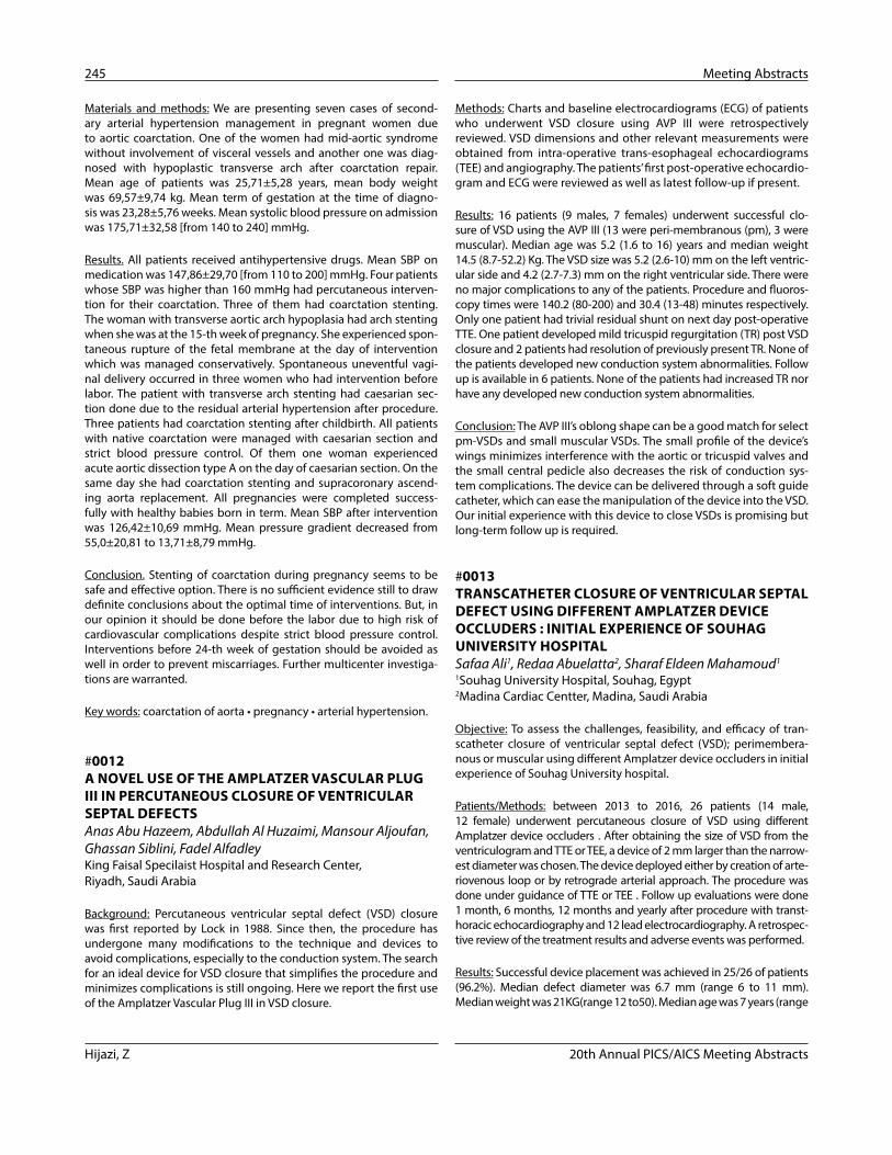
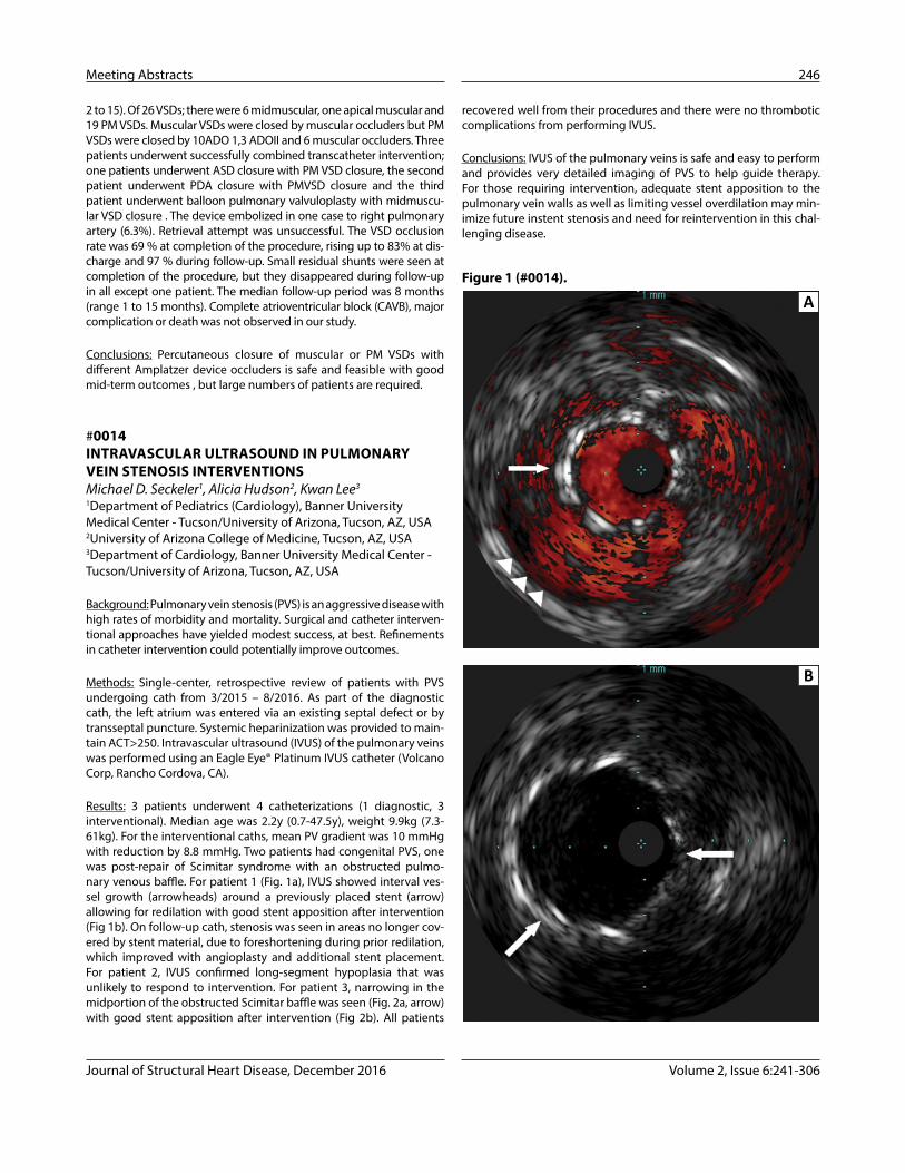
Results: 3 patients underwent 4 catheterizations (1 diagnostic, 3 interventional). Median age was 2.2y (0.7-47.5y), weight 9.9kg (7.3-61kg). For the interventional caths, mean PV gradient was 10 mmHg with reduction by 8.8 mmHg. Two patients had congenital PVS, one was post-repair of Scimitar syndrome with an obstructed pulmo-nary venous baffle. For patient 1 (Fig. 1a), IVUS showed interval ves-sel growth (arrowheads) around a previously placed stent (arrow) allowing for redilation with good stent apposition after intervention (Fig 1b). On follow-up cath, stenosis was seen in areas no longer cov-ered by stent material, due to foreshortening during prior redilation, which improved with angioplasty and additional stent placement. For patient 2, IVUS confirmed long-segment hypoplasia that was unlikely to respond to intervention. For patient 3, narrowing in the midportion of the obstructed Scimitar baffle was seen (Fig. 2a, arrow) with good stent apposition after intervention (Fig 2b). All patients
recovered well from their procedures and there were no thrombotic complications from performing IVUS.
Conclusions: IVUS of the pulmonary veins is safe and easy to perform and provides very detailed imaging of PVS to help guide therapy. For those requiring intervention, adequate stent apposition to the pulmonary vein walls as well as limiting vessel overdilation may min-imize future instent stenosis and need for reintervention in this chal-lenging disease.
A
B
Figure 1 (#0014).
Hijazi, Z 20th Annual PICS/AICS Meeting Abstracts
247 Meeting Abstracts
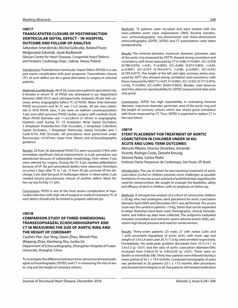
#0015 DEVICE TO THE RESCUETawfiq Shahriar HuqNational Heart Foundation Hospital & Research Institute, Dhaka, Bangladesh
9 year old girl, presented with palpitation and progressive shortness of breath.Underwent ICR for TOF 3 years back.Echo showed a residual perimembranous VSD measuring 8 mm.
#0016 PERCUTANEOUS CLOSURE OF PATENT DUCTUS ARTERIOSUS – 20 YEARS EXPERIENCE OF ONE CENTERMichal Galeczka, Jacek Bialkowski, Malgorzata Szkutnik, Roland Fiszer, Sebastian SmerdzinskiSilesian Center for Heart Diseases, Congenital Heart Defects and Pediatric Cardiology Dept., Zabrze, Silesia, Poland
Introduction: Percutaneous closure has become a method of choice in treatment of patent ductus arteriosus (PDA). Recently different types of occluders have evolved. We present our own experience in this field.
Materials and Methods: Between 1996 and 2016 840 patients (pts; 543 f; 0,3-84,5y; median 4y) had percutaneous PDA closure performed in our center. PDA A type was present in 415pts, B in 23pts, C in 60pts, D in 105pts, E in 192pts, G in 25pts, moreover, 20pts had previously ligated PDA recanalized. Reversible pulmonary hypertension (PH) was observed in 121pts (14,4%). We divided this period in double umbrella (DU) era (1996-2000), detachable coils (C) era (1996-2014) and nitinol wire mesh devices era: like Amplatzer Duct Occluder type I (ADO I) (1997-now), Amplatzer Duct Occluder type II (ADO II) (2009-now) and Amplatzer Duct Occluder type II Additional Sizes (ADO II AS) (2014-now). Application techniques of mentioned above devices were routine. In special situations another devices were used.
Results: We have applied DU in 25pts with 88% success rate (1 embo-lisation), C in 463pts with 96,8% success rate (7 embolisations), in 250 pts ADO I and like ADO I occluders with 100% success rate (ADO I in 140pts, Cera Occluder in 8pts, Cardi-o-Fix in 64pts, HeartR in 27pts, Hyperion in 11pts), ADO II in 15pts with 100% success rate and ADO II AS in 71pts with 98,6% success rate. Moreover, in type B of PDA 5 CardioSEAL/STARflex devices and 2 ASD nitinol wire mesh occluders; in type D of PDA 3 Amplatzer Vascular Plugs type II and in 6 pts with higher PH Muscular Amplatzer VSO were used with 100% success rate. Residual shunt 24 hours after the procedure was observed in 24% of DU, 16,7% of C and no residual shunts were observed in other groups of pts.
Conclusions: PDA percutaneous closure methods have quickly devel-oped and nowadays they are not only safe and efficient but repre-senting also relatively low complication rate.
A
B
Figure 2 (#0014).1. LV graphy
showingresidual VSD
2. Arterio-venous loop 3. 12/10 ADO 1 Device
4. Deviceposi�oned at
VSD
4. (cont) Deviceposi�oned at
VSD5. Device posi�oned checked 6. Device posi�on confirmed
Figure 1 (#0015).
Journal of Structural Heart Disease, December 2016 Volume 2, Issue 6:241-306
Meeting Abstracts 248
#0017 TRANSCATHTER CLOSURE OF POSTINFARCTION VENTRICULAR SEPTAL DEFECT – IN-HOSPITAL OUTCOME AND FOLLOW-UP ANALYSISSebastian Smerdzinski, Michal Galeczka, Roland Fiszer, Malgorzata Szkutnik, Jacek BialkowskiSilesian Center for Heart Diseases, Congenital Heart Defects and Pediatric Cardiology Dept., Zabrze, Silesia, Poland
Introduction: Postinfarction Ventricular Septal Defect (PIVSD) is a rare and severe complication with poor prognosis. Transcatheter closure (TC) of such defect can be a good alternative to surgery in selected patients.
Materials and Methods: All of 26 consecutive patients (pts) (64,6±10y; 9 female) in whom TC of PIVSD was attempted in our department between 2000-2015 were retrospectively analyzed. All pts had cor-onary artery angiography before TC of PIVSD. Mean time between PIVSD occurrence and its TC was 11±5 weeks. All pts were admit-ted in III/IV NYHA class, 9 pts were on balloon counterpulsation; 3 pts were after previous PIVSD cardiac surgery with residual shunt. Mean PIVSD diameter was 11,4±3,8mm (5-19mm) in angiography. Implants used during TC: 19 Amplatzer Atrial Septal Occluders, 3 Amplatzer Postinfarction VSD Occluders, 2 Amplatzer Cribriform Septal Occluders, 1 Amplatzer Ventricular Septal Occluder and 1 Cardi-O-Fix ASD Occluder. All procedures were performed under fluoroscopic (10-87min; mean time 39min) and echocardiographic guidance.
Results: 20 from 26 attempted PIVSD TCs were successful (74%) with immediate significant clinical improvement. In 6 pts procedure was abandoned because of unfavorable morphology, from whom 3 pts were referred for surgery. During the TC 4 pts needed defibrillation because of VF. No peri-procedural deaths were observed. Hemolysis occurred 2 days after TC in 1 pt. 15 from 20 pts survived till the dis-charge; 5 pts died because of multiorgan failure. In observation 2 pts needed second percutaneous closure of another defect. Mean fol-low-up was 6±4,6y (11 pts).
Conclusions: PIVSD is one of the most severe complication of myo-cardial infarction with high risk of surgical or medical treatment. TC of such defect should only be limited to properly selected pts.
#0018 COMPARISON STUDY OF THREE-DIMENSIONAL TRANSESOPHAGEAL ECHOCARDIOGRAPHY AND CT IN MEASURING THE SIZE OF AORTIC RING AND THE HEIGHT OF CORONARYCuizhen Pan, Xue Yang, Daxin Zhou, Wenzhi Pan, Weipeng Zhao, Xianhong Shu, Junbo GeDepartment of Echocardiography, Zhongshan Hospital of Fudan University, Shanghai, China
To investigate the difference between three-dimensional transesoph-ageal echocardiography (3DTEE) and CT in measuring the size of aor-tic ring and the height of coronary ostium.
Methods: 15 patients were recruited and were treated with the trans-catheter aortic valve implantation (TAVI). Routine transtho-racic echocardiography, two-dimensional and three-dimensional echocardiography (2DTEE, 3DTEE) and CT examinations were taken preoperatively.
Results: The minimal diameter, maximum diameter, perimeter, area of the aortic ring measured by 3DTTE showed strong correlation and consistency with those measured by CT [r=0.88, P<0.0001, ICC=0.928 (0.788-0.976); r=0.81, P=0.0003, ICC=0.890 (0.673-0.963), r=0.85, p=0.0001, ICC=0.914 (0.744-0.971); r=0.88, p<0.0001, ICC=0.932 (0.799-0.977)]. The height of the left and right coronary artery mea-sured by 3DTT also showed strong correlation and consistency with those measured by MDCT [r=0.87, P<0.0001, ICC=0.923 (0.777-0.975); r=0.82, P<0.0002, ICC=0.897 (0.693-0.965)]. Besides, inter-observer and intra-observer reproducibility for 3DTEE measurement data were very good.
Conclusions: 3DTEE has high repeatability in evaluating minimal diameter, maximum diameter, perimeter, area of the aortic ring and the height of coronary ostium, which also shows good correlation with those measured by CT. Thus, 3DTEE is expected to replace CT in the near future.
#0019 STENT PLACEMENT FOR TREATMENT OF AORTIC COARCTATION IN CHILDREN UNDER 30 KG: ACUTE AND LONG-TERM OUTCOMESMarcelo Ribeiro, Vinícius Destefani, Amanda Vicente, Rodrigo Costa, Daniela Kreuzig, Simone Pedra, Carlos PedraInstituto Dante Pazzanese de Cardiologia, São Paulo, SP, Brazil
Introduction: The use of stents for percutaneous treatment of aortic coarctation (CoAo) in children presents some challenges as possible limitations of vascular access and lack of published data regarding the need for reintervention. We sought to evaluate the feasibility, safety and efficacy of stent in children, with an emphasis on follow-up.
Methods: A retrospective analysis of a cohort of consecutive children (<30 kg) who had undergone stent placement for aortic coarctation between April/2009 and December/2015 was performed. The access route was the carotid in patients <10 kg. Stents that can be expanded to larger diameters have been used. Demographic, clinical, hemody-namic and follow-up data were collected. The endpoints evaluated included: immediate and mid-term severe adverse events (SAE), per-sistent high blood pressure and need for reintervention.
Results: Thirty-seven patients (25 male, 27 with native CoAo and 1 with associated hypoplasia of aortic arch), with mean age and weight of 5.4±3.4 years and 20.7±11.0 kg were enrolled respectively. Immediately, the peak-peak gradient decreased from 33.7±15.1 to 5.4±5.3 (p <0.01) and the ratio of aortic coarctation diameter/DAo increased from 0.40±0.16 to 0.95±0.20 (p <0.01). There were no deaths or immediate SAE. Thirty-four patients were followed during a mean period of 43.1 ± 19.4 months. Computed tomography of aorta was performed in 26 patients (23.1 ± 17.6 months after procedure) and showed stent integrity in all. Five patients still needed medication
Hijazi, Z 20th Annual PICS/AICS Meeting Abstracts
249 Meeting Abstracts
#0021 RETRIEVAL OF A FIGULLA OCCLUTECH SEPTAL OCCLUDER EMBOLIZED DEVICE FROM RIGHT VENTRICLE USING ITS NATIVE DELIVERY SYSTEM. CASE REPORT OF A NOVEL APPROACHAmjad Mehmood, Nadeem Sadiq, Maad UllaAFIC & NIHD, Rawalpindi, Pakistan
A 27-year-old lady was diagnosed as having a large secundum atrial septal defect and moderate pulmonary artery hypertension on echocardiogram. She was planned for transcatheter device clo-sure of atrial septal defect as transesophageal echocardiography revealed ASD secundum (31 mm × 29 mm) with adequate rims. The patient was taken to the catheterization laboratory for transcathe-ter closure under local anaesthesia. A 14 F Cook sheath was selected to deliver 33 mm device. The device was loaded on to the delivery system and delivered across the defect using the right upper pulmo-nary vein technique. The device fitted nicely on to the septum and final position confirmed on TOE and released. The next morning to confirm position of device on TTE before discharging the patient, it was revealed that the device had embolized in to RV. The patient was again taken to the cath lab and after multiple attempts with a 15 mm Amplatz gooseneck snare (eV3 Endovascular Inc., Plymouth, MN, USA) the device could not be pulled in to the sheath satisfac-tarily. Finally we used native delivery cable within 14 F sheah. After few attempts we were able to retrieve the 33 mm ASD device with locking mechanism of delivery cable. Then we closed the same ASD with 36 mm occlutech septal occluder with no further complication. The patient was discharged next day with advise of tab loprin 150 mg daily for 6 months.
Conclusion: In certain situations of embolized occlutech devices, native delivery cable with two prongs can be very effective and safe with stability to retrieve when snare is unable to capture and stabilize. However the capturing area of prongs is much less than snare.
#0022 SUCCESSFUL HEMOSTASIS OF ACUTE LUNG BLEEDING USING AMPLATZER VASCULAR PLUG AND COILS IN A PATIENT WITH PULMONARY HYPERTENSIONKenji Suda, Yoshiyuki Kagiyama, Yusuke Koteda, Shintaro KishimotoKurume University School of Medicine, Kurume, Japan
Lung bleeding is the dreadful complication of cardiac catheterization that can directly result in demise in patients with pulmonary hyper-tension. We report successful hemostasis achieved by combined occlusion of Amplatzer Vascular Occluder and coils.
Case: A 61-year-old female patient who is known to have pulmonary hypertension with mean pulmonary artery pressure of 66 mmHg underwent follow-up diagnostic catheterization. She had a large pat-ent arterial duct with a diameter of 12 mm treated with Amplatzer Septal Occluder 7 months ago and her past history includes pulmo-nary tuberculosis resulted in chronic respiratory insufficiency that required home oxygen treatment.
for high blood pressure. Seven patients required percutaneous rein-tervention (36.1 ± 19.0 months after initial treatment) due to aortic aneurysm (1), residual stenosis above the stent (1) and adjustment to somatic growth (5). One patient required surgery due to residual hypoplasia of aortic arch (15.1 months later). All of reinterventions have been carried out successfully, with no SAE.
Conclusion: Stenting for treatment of CoAo in children was feasi-ble, safe and effective in reducing blood pressure levels and gra-dient. A significant rate of reintervention was observed because of the previously known need to stent adjustment to somatic growth in most cases (SAE were rare). On the same way, post dila-tion of stents for CoAo in children has proved to be feasible and effective.
#0020 PARTICLE EMBOLIZATION OF SYSTEMIC-TO-PULMONARY COLLATERAL ARTERY NETWORKS IN CONGENITAL HEART DISEASE: TECHNIQUE AND CONSIDERATIONSSarosh Batlivala, William Briscoe, Makram EbeidUniversity of Mississippi Medical Center, Jackson, MS, USA
Background: Systemic-to-pulmonary artery collateral (SPC) networks commonly develop in patients with single ventricle physiology and chronic hypoxemia. Though these networks augment pulmonary blood flow, much of the flow is ineffective which contributes to cardiac volume loading. This volume loading can have detrimental effects, especially for single ventricle patients. Some data suggest that occluding these collaterals may improve outcomes after sub-sequent operations, especially when the volume of collateral flow is significant. For other patients—e.g. with hemoptysis—collateral occlusion is a crucial therapy. Traditional practice has been to coil occlude the feeding vessel, but this technique has limitations.
Cases & Technique: We reviewed all procedures that included SPC embolization from August 2013 to June 2016. We perform particle embolization utilizing a co-axial catheter system to deliver 510-700 micron particles deep into feeding arteries (figures).
Results: We performed particle embolization during 42 catheteriza-tions on 34 patients. The majority of patients had single ventricle physiology; a few patients presented with hemoptysis or had inci-dentally noted SPCs. Particle embolization was acutely successful with the majority (93%) having no residual flow. No complications occurred.
Discussion: Traditional coil occlusion of SPCs is suboptimal for mul-tiple reasons. First, occlusion of fistulous connections is ideally per-formed as distally as possible. Coil occlusion generally occludes the most proximal source. Second, SPCs tend to recur from the same feeding vessel. Coil occlusion prevents re-access of the feeder, impeding occlusion of recurrent SPCs. Particle occlusion avoids both these limitations as the technique occludes distal connections while maintaining patency of the feeder vessel. The occlusion is therefore more immediately effective—by occluding distal connections—and avoids problems with re-accessing feeders. Thus, embolization with particles may be a superior technique than coil occluding the proxi-mal feeding vessel.
Journal of Structural Heart Disease, December 2016 Volume 2, Issue 6:241-306
Meeting Abstracts 250
#0024 COMPLICATIONS FOLLOWING PERVENTRICULAR DEVICE CLOSURE OF MUSCULAR VENTRICULAR SEPTAL DEFECTMassimo Caputo, Damien Kenny, Karim Diab, Gia Marzano, Amy Wilhelmi, Bassel NijresRush University Medical Center, Chicago, IL, USA
A male infant was born with tricuspid valve (TV) dysplasia with severe regurgitation and Swiss-cheese muscular ventricular septal defects (MVSD). Diagnostic cardiac catheterization demonstrated significant left-to-right shunting with a Qp:Qs ratio of 2.29:1. He underwent attempted perventricular device closure of the MVSDs with surgical repair of the TV. A purse-string was placed into the mid-portion of the right ventricle (RV) free wall to provide direct access to the ante-rior MVSD. Attempts to close the apical-MVSD with a device were unsuccessful due to its crowding with RV trabeculations. Hence, the decision was made to leave the apical-MVSD without further intervention. Transesophageal echocardiogram suggested a small outpouching posterior to the left ventricle (LV) free wall measuring about 5x7mm that was concerning for a potential LV-PSA (pseudoan-eurysm) caused by the wire vs. sheath across the VSD. Therefore, car-diopulmonary bypass was initiated, the TV was repaired and a MPA band was placed.
An echocardiogram showed enlargement of the LV-PSA to 3.7x4.0x2.7 cm, which was confirmed with cardiovascular magnetic resonance, reveal-ing a narrow neck at its origin from the LV apex. Due to the concern for rupture, the aneurysm was repaired surgically. The two devices were extracted and the aneurysmal sack was completely resected.
#0025 INITIAL EXPERIENCE OF ATRIAL SEPTAL DEFECT CLOSURE USING THE NEW GENERATION CARDIA ULTRASEPT IITM DEVICE IN MEXICO.Roberto Mijangos VázquezPediatric Hospital, Tuxtla Gutiérrez, Chiapas, Mexico
We present the initial experience in Mexico of atrial septal defect closure using the new Cardia Ultrasept IITM device. We present a series of 9 patients with ASD previously selected as candidates with favourable anatomy (less than 38mm defect, rims greater tan 5mm) to be subjected for closure of the defect through interventionism treated in the period April-August 2016. Preliminary prospective, observational, transverse and descriptive study. The group included 7 female patients (76%) with a median age of 8 years (1-13). The hae-modynamic and anatomical data were as follows: pulmonary artery systolic pressure 25.14 ±3.9 mmHg, pulmonary to systemic flow ratio 2.38 ±0.66, septal defect diameter 17.78 ±6.18 mm, expanded defect diameter 22.6 ±5.82 mm. All septal occluder were delivered success-fully. No residual shunt evidenced by angiography and intracardiac echocardiography. At follow-up to 2.1 months, all patients showed complete closure of the defect and continuous decreased of right ventricular diastolic diameter with an initial median of 38mm (30–40) and after catheterization of 28.5mm (23–30), p=0.01 and Z-score of 3 (2.87–3.15) vs 1.8 (1.5–1.95), respectively, p=0.01. The new generation of the Cardia Ultrasept IITM device is a good alternative to percutane-ously treat atrial septal defect.
When we tried to measure right pulmonary wedge pressure, she sud-denly complained chest pain and showed massive hemoptysis that progressively suffocated her. She fell in shock, but was successfully treated by volume expansion, intravenous adrenalin infusion, and placement of laryngeal mask successfully replaced with blind tra-cheal intubation.
Repeated exploratory pulmonary angiography showed unconfined pulmonary bleeding that could lead to continuous bleeding without treatment. We placed a 4 French Judkins left coronary type catheter into right lower branch and placed 2 Flipper coils with loop- diameters of 6.5 mm and 5 mm. However, there was still continuous bleeding and we place a Amplatzer Vascular Plug of 8 mm in diameter that could suc-cessfully brought complete occlusion and stop pulmonary bleeding.
Conclusion: Cardiac catheterization of patients with pulmonary hypertension carries a risk of severe complication, namely pulmonary bleeding. Preparation of laryngeal mask and occlusion devices such as coils and Amplatzer vascular plugs are mandatory in this setting.
#0023 INTRA-CARDIAC ECHOCARDIOGRAPHY GUIDED TRANS-CATHETER CLOSURE OF PATENT DUCTUS ARTERIOSUS WITHOUT CONTRAST ANGIOGRAPHYKenji Suda1, Hironori Kuwahara1, Yoshiyuki Kagiyama1, Hironaga Yoshimoto2, Yusuke Koteda1, Shintaro Kishimoto1, Motofumi Iemura2
1Kurume University School of Medicine, Kurume, Japan2St. Mary’s Hospital, Kurume, Japan
Background: Though contrast angiography is the standard guidance of trans-catheter closure of patent ductus arteriosus (TC-PDA), it is contra-indicated in patients with severe renal disease that often seen in senile patients. We have developed intra-cardiac echocardiog-raphy (ICE) guided TC-PDA (Cathet Cardiovasc Intervent 2015). We report sequential 6 cases that successfully underwent TC-PDA with-out contrast angiography.
Materials and Methods: Subjects were 5 patients with PDA and median age of 57.4 (35.4 - 66.1) years old. The median size of PDA was 4.6 (3.2 - 11.7) mm with median Qp/Qs of 1.9 (1.4 and 2.4), respectively. The oldest patient suffered from renal dysfunction and 2 patients had pulmonary hypertension. Prior to the TC-PDA, all patients underwent contrast X-ray computed tomography to clarify the anatomy. ICE catheter was inserted through 2nd sheath at femoral vein and placed at main or left pulmonary artery. During the TC-PDA, we primarily used ICE to guide the procedure.
Results: We could successfully place Amplatzer Duct Occluders in 4 and Amplatzer Septal Occluder in 1 without contrast angiography. ICE at main or left pulmonary artery has allowed us to determine the diameter and length of PDA, to monitor the device placement, and to determine the residual shunts. ICE did not increase the risk of compli-cation except for transient arrhythmia, though new operator needs some learning time to understand orientation of ICE.
Conclusion: ICE-guided TC-PDA without contrast angiography is fea-sible and can be the standard treatment for adult patients with renal dysfunction.
Hijazi, Z 20th Annual PICS/AICS Meeting Abstracts
251 Meeting Abstracts
skin to long term malignancies, which ultimately can result in serious morbidity or death. Due to the concerns of radiation exposure, our institution has initiated changes to our practice to help reduce the radiation exposure to our patients and staff.
During 2014, our cath lab implemented a lower fluoroscopy rate of 3 frames/sec (when felt to be adequate by the provider), increased use of radiation shields and collimators, and increased the use of “fluoroscopy record” and “fluoroscopy save”. After reviewing the average radiation doses (mGy) and (mGy/min), it was found that our changes did indeed have a positive impact in decreasing the radiation exposure significantly. Prior to those changes, in 2013, the average total radiation dose for diagnostic caths was 164.4 mGy and for six types of interventional caths was 289.6 mGy. Following those changes in 2015, the average total radiation dose for diag-nostic caths decreased to 107.7 mGy (34% reduction), and for these same six types of interventional caths it decreased to 136.4 mGy (53% reduction). During that same time period the radiation dose/min of fluoroscopy decreased from 9.7 mGy/min to 5.4 mGy/min (44% reduction) for diagnostic caths and from 11.8 mGy/min to 6.1 mGy/min (48% reduction) for the interventional caths. The radiation exposure for each of the providers, as measured by a radi-ation badge, decreased by from 1546.5mGy in 2013 to 670mGy in 2015 (56.7% reduction). During this same time period, there was no increase in complication rate.
The data obtained demonstrates how relatively small changes in our practice and radiation awareness can decrease the amount of radi-ation exposure to the patients and staff in the cardiac cath lab. Our program will continue to remain aware of radiation exposure and strive to keep the dose as low as possible. With the guidance of the National Cardiovascular Data Registry (NCDR®) Impact Registry™, we are able to maintain and keep track of our improvements.
#0028 IMPACT OF ANESTHETIC MANAGEMENT DURING DIAGNOSTIC CATHETERIZATIONS PERFORMED IN SINGLE VENTRICLE PATIENTS PRIOR TO STAGE II PALLIATIONRyan Romans, Nicole Wilder, Issac Davidovich, Olubukola Nafiu, Jefftey ZampiUniversity of Michigan, Ann Arbor, MI, USA
Background: Infants with single ventricle (SV) physiology who undergo elective diagnostic cardiac catheterization prior to stage II palliation (pre-stage II catheterization) require either conscious seda-tion (CS) or general anesthesia (GA). Ideal anesthetic management provides a steady-state hemodynamic assessment with little impact on procedural safety. We sought to compare the safety and efficacy of various anesthetic management strategies (CS vs. GA) during the pre-stage II catheterization.
Methods: We performed a single center retrospective cohort study of SV patients undergoing pre-stage II catheterization between 2010 and 2015. Safe sedation was defined by absence of cardiac or respi-ratory events during or within 24 hours of the procedure and no need for post-catheterization ICU admission. Effective sedation was defined by stable hemodynamics (<2 episodes of heart rate increase
#0026 EMBOLIZATION OF A LARGE VENO-VENOUS COLLATERAL FROM THE INNOMINATE VEIN INTO THE LEFT ATRIUM WITH TWO AMPLATZER DUCT OCCLUDERS II VIA A HYBRID APPROACH AFTER FONTAN-OPERATIONHeike Schneider, Matthias Sigler, Ulrich Krause, Thomas PaulUniversity Goettingen, Goettingen, Germany
Veno-venous collaterals (VVC) may lead to significant systemic desaturation after Fontan operation. We present a 16-year-old girl with univentricular physiology who had undergone palliation with a Damus-Kaye-Stansel procedure, Glenn operation and Fontan com-pletion at another institution. Cardiac catherterization had revealed mean Fontan pressures of 23 mmHg. Subsequent treatment with Bosentan had resulted in a drop to 15 mmHg several years before. A large VVC from the innominate vein to the left atrium had been iden-tified with systemic SaO2 ranging from 85-90%. Interventional closure had been attempted several times at three different large volume pediatric cardiac centers. Finally, this patient was transferred to our institution in severe heart failure with ascites, systemic desaturation and poor ventricular function. Under mechanical ventilation with FiO2 1.0 and NO 20 ppm, arterial SaO2 could not be increased above 60%. Despite optimal medical management, her status deteriorated rapidly. A single chamber cardiac assist device (Berlin Heart) was implanted and the girl was listed for urgent heart transplantation. Due to continued severe hypoxemia and metabolic acidosis it was decided to close the large VVC via a hybrid approach as other access routes had failed before. Access via the left ventricle was chosen in order to achieve a straight angle to the insertion of the VVC into the roof of the LA. After introduction of a sheath into the LV, the VVC could be entered with a right Judkins catheter and an 8F Amplatzer sheath could be advanced deep into the VVC subsequently. On angiography the VVC measured 12 x 14 mm in diameter at its nar-rowest portion, the widest site was 22 mm. Accordingly, an 18 mm Amplatzer Vascular Plug (AVP) II was deployed in the distal VVC. For complete closure a second AVP II (20 mm) was implanted slightly more proximally. Arterial oxygen saturation immediately increased to 80% thereafter. Final angiogram demonstrated adequate and stable position of the AVPs. The chest could be closed without complica-tions. Unfortunately, despite this successful intervention the patient died due to multi-system organ failure while awaiting heart trans-plantation. Conclusion: Embolization of significant VVC should not be deferred if typical access ways fail. Hybrid procedures may offer alternative access routes to benefit the patient’s individual needs.
#0027 THE EFFECTS OF APPLYING RADIATION DOSE REDUCTION MEASURES DURING PEDIATRIC CARDIAC CATHETERIZATION PROCEDURESBeth Price, Saar Danon, Jodi Hundley, Saadeh Jureidini,SSM Health Cardinal Glennon Children’s Hospital, St. Louis, Missouri, USA
Exposure to radiation has become an increasing concern among healthcare providers, workers and patients. The effects of radiation can result in several complications, ranging from acute burns on the
Journal of Structural Heart Disease, December 2016 Volume 2, Issue 6:241-306
Meeting Abstracts 252
no significant pulmonary stenosis or PR from cine-angiography and echocardiography in all patients. Chest X-ray showed good valved-stent position at targeted RVOT area. All patients discharged 4 days after PPVI without any problem. One patient completed 6 months follow-up after PPVI and showed decreased indexed RV end-diastolic volume from 181.7 to 126.7 mL/m2 from cardiac MRI.
Conclusion: First human implantation of self-expandable percuta-neous pulmonary valve using knitted nitinol wire mounted with a tri-leaflet porcine pericardial valve developed in South Korea was feasible and effective at short-term follow-up. A clinical trial for fea-sibility to evaluate the safety and short-term effectiveness of this self-expandable valved-stent for 10 patients is ongoing for the con-genital heart disease with pulmonary valve disease in South Korea.
#0030 SAFETY OF OUTPATIENT CARDIAC CATHETERIZATION IN INFANTS WITH SINGLE VENTRICLE CONGENITAL HEART DISEASE.Jamie Colombo, Michael Spaeder, Michael HainstockUniversity of Virginia, Charlottesville, VA, USA
Background: The benefits of outpatient cardiac catheterization (cath) were first assessed in the 1980s; it reduces anxiety to patients and families and decreases cost. Cardiac cath is routinely performed in patients with single ventricle congenital heart disease (SVCHD) to aid in hemodynamic assessment, intervention and surgical plan-ning. There is significant morbidity and mortality associated with interstage SVCHD in shunt-dependent patients and substantial intra-
procedural variation exists between centers. Post-procedural best practices following cardiac cath of infants with SVCHD are unknown. Our institutional strategy has been to discharge patients following a 4-6 hour post-procedure observation period. The objective of this study was to investigate the incidence and causes of readmission of infants with SVCHD following outpatient cardiac cath. Methods: We performed a retrospective review of all patients less than one year of age with SVCHD who underwent cardiac cath between 2007 and 2015 at our institution querying the Society of Thoracic Surgeons Database. Unplanned readmissions were defined as an admission to the hospital <48 hours following discharge after cardiac cath. Results: 92 patients were included in the analysis. Median age was 134 days (105-179 days) with median weight of 5.6kg (5-6.4kg). 62 patients were discharged following a 4-6 hour observation period. Of those, 5 underwent a cath intervention. Two of the 62 patients initially discharged were readmitted within 48 hours of discharge due to fever and hypoxemia. Of the remaining 30 patients, 18 stayed for 23-hour observation; 9 of those had an intervention. The other 12 patients were admitted to the hospital for >23 hours; 4 under-went intervention. There were no differences in age, weight, sex, shunt- dependence, arterial access or use of general anesthesia between those patients discharged and those admitted follow-ing cath. Patients who underwent intervention were more likely to be admitted (p<0.001), though nearly one third were discharged home without readmission. Readmission was rare (3%). No intra- or peri-procedural deaths occurred. Conclusion: Outpatient cardiac cath of infants with SVCHD can be performed with low readmission rate. Further investigation will compare cost-effectiveness of uni-versal 23-hour overnight observation vs. outpatient discharge with potential readmission.
requiring anesthetic medication boluses) and lack of failed sedation (need to convert from CS to GA). Multivariate logistic regression mod-els were utilized to compare results.
Results: Of 104 infants who underwent a pre-stage II catheterization, 82 (79%) had safe sedation and 56 (54%) had effective sedation. CS was utilized in 91 (88%) patients and 8 (10%) required conversion to GA (failed sedation). There were no differences between CS and GA patients in baseline demographics, shunt type, procedural dura-tion, intra-procedural lowest pH/highest PCO2, or rates of safe and effective anesthetic management. However, ICU admission was more common in GA patients (23% vs 2%, p=0.013) and patients with higher intra-procedural PCO2 (OR 1.17, p=0.013). Higher PCO2 was also associated with greater odds of a failed sedation (OR 1.18, p=0.004). Higher baseline oxygen saturation was independently associated with a safe catheterization (AOR 1.12, p=0.014) and higher weight was independently associated with both safe (AOR 2.86, p=0.004) and effective (AOR 1.74, p=0.013) anesthetic management.
Conclusions: CS can provide safe anesthetic management for SV infants undergoing a pre-stage II diagnostic catheterization and few patients require conversion to GA. However, hemodynamic steady state can be difficult to achieve regardless of the anesthetic manage-ment strategy.
#0029 FIRST HUMAN CASES OF SELF-EXPANDABLE PERCUTANEOUS PULMONARY VALVE IMPLANTATION USING KNITTED NITINOL-WIRE STENT MOUNTED WITH A TRI-LEAFLET PORCINE PERICARDIAL VALVEGi Beom Kim, Mi Kyung Song, Eun Jung Bae, Chung Il NohSeoul National University Children’s Hospital, Seoul, Republic of Korea
Background: Severe pulmonary regurgitation (PR) and associated right ventricular (RV) dilatation in native right ventricular outflow tract (RVOT) is challenging and still on clinical trial. We report first human cases of self-expandable percutaneous pulmonary valve implantation (PPVI) using newly made knitted nitinol-wire stent mounted with a tri-leaflet porcine pericardial valve developed in South Korea.
Methods: We reviewed 8 cases of self-expandable PPVI at the Seoul National University Children’s Hospital. This self-expandable valved-stent was newly developed by our research team with the cooperation of the TaeWoong medical company in South Korea. This valved-stent was made by knitted nitinol-wire backbone with tissue valve using porcine pericardium with multiple steps for tissue preservation including decellularization and alpha-galactosidase treatment.
Results: Eight patients underwent total correction of Tetralogy of Fallot previously and showed severe PR (mean PR fraction: 43.7%, range: 35.4-56) and enlarged RV volume (mean indexed RV end- diastolic volume; 188.6 mL/m2, range: 167.5–209.8). Their median age at PPVI was 21 years old (range: 13-26). At the targeted RVOT area, 4 patients were implanted with 28 mm diameter valved-stent and 4 patients were implanted with 26 mm diameter valved-stent loaded in the 18 French delivery cable. There were no significant peri- procedural complications in all patients. After procedure, there was
Hijazi, Z 20th Annual PICS/AICS Meeting Abstracts
253 Meeting Abstracts
Introduction: In this paper, we aimed to present transcatheter treat-ment of patients with a single ventricle physiology, experiencing low cardiac output (LCOS) or severe systemic desaturation (SSD) after a Glenn or a Fontan operation
Method: We retrospectively evaluated 30 patients between 2007 and 2016.
Results: The mean age was 7.6 years (6 months-21 years) and the weight was 25.2 kg (6-54). 31 attempts were made in 30 patients. The procedures were performed after a Kawashima, Glenn and Fontan surgery in 3, 12 and 15 patients, respectively. SSD was encountered in 15 patients. Amongst these patients, closure of a Fontan fenestration was performed in 7. We occluded a decompressing vein in 5 and a pulmonary arteriovenous fistula closure in one. Closure of a residual right SVC-atrium connection was performed in one and stent implan-tation to reroute the hepatic blood flow to the right lung in one, after a Kawashima operation. The mean oxygen saturation of 79.3±8.1 % increased to 92.2±5.6 and the mean PA pressure increased from 11.9±2.2 mmHg (8-16) to 13.5±2.1 mmHg (10-17). LCOS and / or increased PA pressure was detected in the remaining 15. One patient was on an ECMO support. Amongst these 15 patients, an antegrade pulmonary flow was occluded using a number of devices in 7, ante-grade flow was closed with the use of a covered stent, resolving an associated left PA stenosis at the same time in one. Among 4 patients suffering from branch PA stenosis, 3 received stent implantation while the remaining was treated via cutting balloon angioplasty. Two sepa-rate stents were needed to treat branch PA and extracardiac conduit stenosis in one. In the patient on ECMO support, Fontan fenestration was dilated with a balloon to ensure cardiac output at the expense of systemic desaturation. And fenestration was created in one. In patients with LCOS, the preprocedural PA pressure decreased from 20.6 mmHg (15-27) to 14.9±1.8 mmHg (11-18). There was no proce-dural mortality. Circulatory failure regressed in all cases except one.
Discussion and conclusion: To avoid reopening of the antegrade flow, surgeons should not only ligate but divide the PAs from the ventricle. In the presence of LCOS or SSD, urgent catheterization should be con-sidered. Significant PA stenosis should be treated even if there exists no pressure gradient throughout the circulation.
#0033 PDA CLOSURE WITH CERAFLEX OCCLUDER: IS THERE ANY ADDITIONAL BENEFIT?Ahmet Celebi, Ilker Kemal Yucel, Mustafa Orhan Bulut, Sevket Balli, Mehmet KucukDr. Siyami Ersek Hospital for Cardiology and Cardiovascular Surgery, Department of Pediatric Cardiology, Istanbul, Turkey
Introduction: Although transcatheter closure of PDA is an established standard method, most frightening complication is protrusion of the aor-tic disc to the DAO which may cause iatrogenic COA, especially in small children with small aorta. Ceraflex duct occluder (CDO) is a new device with similar properties with Amplatzer duct occluder (ADO). Device comes preassembled with the delivery cable by a loop connection through the holes and ready to load via the loader on the delivery cable. The loop made of surgical thread that provides the device to become flexible in 3600 direction and fit to the ductal shape before releasing.
#0031 PERCUTANEOUS PULMONARY VALVE IMPLANTATION WITH SAPIEN VALVES IN NATIVE AND LARGE RVOT; EARLY AND MID-TERM RESULTS Ahmet Celebi, Ilker Kemal Yucel, Mustafa Orhan Bulut, Sevket Balli, Mehmet KucukDr. Siyami Ersek Hospital for Cardiology and Cardiovascular Surgery, Istanbul, Turkey
Introduction: Percutaneous pulmonary valve implantation (PPVI) has been used mainly for conduit dysfunction in right ventricular outflow tract (RVOT). Until recently, native RVOT without stenosis used to be considered a relative contraindication to transcatheter valvulation. We present early and midterm results of PPVI with Edwards–Sapien XT (ES-XT) in repaired tetralogy of Fallot (TOF) patients with native-large RVOTs.
Method: 53 s/p repaired TOF patients who had native RVOT with with severe/free pulmonary regurgitation, significant dilatation of the RV and without significant RVOT stenosis (peak pressure gradi-ent between RV and main pulmonary artery (MPA) < 25 mmHg on TTE), and with a minimum RVOT / (MPA) diameter of ≤ 26 mm on TTE included into the study. Balloon sizing was performed with compliant (34 mm Amplatzer sizing) and semi-compliant balloons for interro-gation (BI). The size of the Z-Med /BIB balloons that the Andra Stents XXL would be mounted on was decided up to the indentation diam-eter occurred during BI; as at least 1 mm larger than the indentation diameter.
Results: Mean age and weight of the patients were 17 ± 7.7 (7-50) years and 49 ± 16 (22-84) kg, respectively. Before presenting pressure gradient between RV and MPA was 4.8 ± 3.4 (0-14) mmHg. Indentation diameter with BI was 26.2 ±2.7 (22-32) mm. Balloon size used for prestenting was 28.1±2 (24-30) mm. Successful PPVI was achieved in 45 patients; 29 mm in 38 and 26 mm in seven. PPVI was performed in same session in five and 3-12 weeks after prestenting in 40. 8 patients are waiting for valvulation after presenting. One patient has severe tricuspid insufficiency and underwent to surgery after valvulation. Valve function was good in all immediate after and at the last fol-low-up; a median of 10 months (2-25 months). RV volumes decreased and mild paravalvar leakage was observed only in five. Stent fracture has not been observed and no reintervention required yet.
Conclusion: PPVI with ES-XT valve, which has larger sizes as 26 and 29 mm, is feasible and safe in adolescents and adult’s patients with native RVOT without stenosis. Newer delivery systems, which is used through the smaller sheaths, gives us also an opportunity of early transcatheter valvulation in smaller patients. Prestenting for provid-ing a secure landing zone is the most important part of the proce-dure. Only Andra XXL stents which has an expansion capacity up to 32 mm can be used for this purpose, currently.
#0032 TRANSCATHETER INTERVENTIONS AFTER GLENN ANASTOMOSIS AND FONTAN OPERATION IN PATIENTS WITH UNIVENTRICULAR HEART Ahmet Celebi, Ilker Kemal Yucel, Mustafa Orhan Bulut, Sevket Balli, Mehmet KucukDr. Siyami Ersek Hospital for Cardiology and Cardiovascular Surgery, Department of Pediatric Cardiology, Istanbul, Turkey
Journal of Structural Heart Disease, December 2016 Volume 2, Issue 6:241-306
Meeting Abstracts 254
shunt. DE and DD was encountered after PDA closure in 8. DE was observed in 5 and DD into the descending aorta (DAO) was seen in 3. The median age was 2.5 years (1 month to 8 years), and the weight was 9.5 kg (3.3-24). PDA was conical in shape in 6 and tubular in 2. The median diameter of the PDA was 8.6 mm (3.7- 11.7). Except for one, pulmonary hypertension (PHT) equal to systemic pressure was present in all. Amongst 5 with DE, four devices embolized to the PA and to the DAO in one. 3 cases were referred for surgery. The devices were retrieved with the use of a snare from PA and DAO in 2. In one of these pts, PDA was occluded with a larger device. In the remaining, it could not be achieved even with a larger device. DD into the DAO led to a coarctation of the aorta in three. All of these patients had severe PHT. Two of these devices were repositioned successfully with the use of a bioptom, antegradely. In other patient, device was retrieved and PDA was closed with a muscular VSD device
Conclusion: The risk factors for DE after ASD closure are found to be the presence of a large defect, a deficient rim with an aneurysmatic septum, mismeasurement of the defect. The most important risk factor for DE after the PDA closure are found to be the presence of severe PHT. The retrieval rate is lower after with PDA devices than ASD devices.
#0035 ECHOCARDIOGRAPHY-GUIDED TRANSCATHETER CLOSURE OF PATENT DUCTUS ARTERIOSUS IN CHILDREN: FEASIBILITY AND SAFETY OF A NEW STRATEGYTianli Zhao, Weizhi Zhang, Lei Gao, Wancun Jin, Qin Wu, Yifeng YangDepartment of Cardiovascular Surgery, Changsha, Hunan, China
Objective: The aim of this study was to evaluate the feasibility and safety of transcatheter closure of PDA under transesopha-geal echocardiaographic (TEE) guidance without fluoroscopy and angiography.
Background: To avoid radiation exposure and contrast agent usage, the feasibility of transcatheter closure of atrial septal defects (ASD) without fluoroscopy has been proven. However, it is unknown that weather or not the procedure is eligible for patent ductus arteriosus (PDA) closure.
Methords: From June 2014 to May 2016, a total of 102 children (38 males, 64 females), aged from 1 to 14 years (median, 2.3 years), and weighted from 6 to 46kg (median, 11.6kg), with isolated PDA (diameter of 2.5 to 7.5mm, median 3.8mm) underwent attempted transcatheter device closures. The procedures were performed under TEE guidance with-out fluoroscopy and angiography and the occluders were deployed by using modified delivery system via femoral venous access alone.
The patients were followed up by clinical examination, electrocardio-gram, and TTE at 1, 3 and 6 months, and then yearly.
Results: PDA were successfully closed in 99 patients (97.1%). There were no acute procedural complications or severe adverse events. The procedure time ranged from 10 to 45 minutes (median, 21
Method: 21 patients underwent transcatheter closure with CDO from November 2015 to August 2016. Decision for device size selection was based on the narrowest diameter of the PDA according to the manufacturer recommendation as the aortic end of the occluder shank to be at least 1.5-2.0 mm larger than the narrowest diameter of the duct.
Results: The median age of the patients was 1.2 years (6 months to 28 years) and weight was 9.6 kg (5.4 to 82 kg). 11 patients were under one-year-old and 11 had pulmonary hypertension (mean PA pressure >25 mmHg). All patients had continuous cardiac murmur on examination and all PDAs were type A. Narrowest PDA diameter at pulmonary side was 4.1 ± 1.7 mm (2.2-8.2 mm, median 3.8 mm). Intervention was successful in all. Final angiogram showed complete closure in 17/21 of them. Echocardiography achieved complete occlu-sion an all on the next day. In a patient with Down syndrome PDA was closed with 4/6 mm device, and device embolized to descending aorta after persistent cough 24 hours later. Than device was snared via femoral vein approach and closed with 6/8 mm device. None of the patients showed evidence of stenosis at branch pulmonary artery and descending aorta during the follow-up.
Discussion and conclusion: Our results showed us CDO is a safe and efficacious in closure moderate to large PDA’s whose duct morphol-ogy fit to the ADO I. Its uniquely designed delivery/releasing system has an advantage in view of no applying tension to the device which provides the device in stable position and not changing the device position during and immediate after the releasing. It may give us an opportunity to be sure that the device not protrudes to the aorta after releasing, especially in infants those have small descending aorta.
#0034 THE RESULTS OF EMBOLIZATION AND DISLOCATION OF THE DEVICES AFTER TRANSCATHETER CLOSURE OF ATRIAL SEPTAL DEFECT AND PATENT DUCTUS ARTERIOSUS Ahmet Celebi, Ilker Kemal Yucel, Mustafa Orhan Bulut, Sevket Balli, Evic Zeynep Akgun, Gokmen Akgun, Emine Hekim Yilmaz, Mehmet KucukDr. Siyami Ersek Hospital for Cardiology and Cardiovascular Surgery, Department of Pediatric Cardiology, Istanbul, Turkey
Introduction: In this paper, we aimed to present the results and treatment methods of device embolization (DE) or malposition/ dislocations (DD) seen after transcatheter ASD and PDA device closure. Method: Between 2004 and 2016, the patients were retro-spectively analyzed regarding the DD and DE.
Results: Amongst the patients in whom a device closure of ASD was performed, DE and DD was encountered in ten and one, respectively. The median age of the patients was 19.5 (5-52) years. The septum was aneurysmatic in 2 , while the rims of the defect were either deficient or thin and mobile in 5. DE occurred due to spontaneous disconnec-tion of the devices during repeated attempts in three. The device embolized into PA in 5, into the left ventricle in 2, into the ascending aorta in one, into the right atrium in one and onto the mitral valve in one. The devices were retrieved with the use of a bioptome or a snare in 6 and the defect was closed with a larger device in 5 of these patients. 5 patients were referred for surgery. One patient was also referred for surgery due to DD at the aortic site causing significant
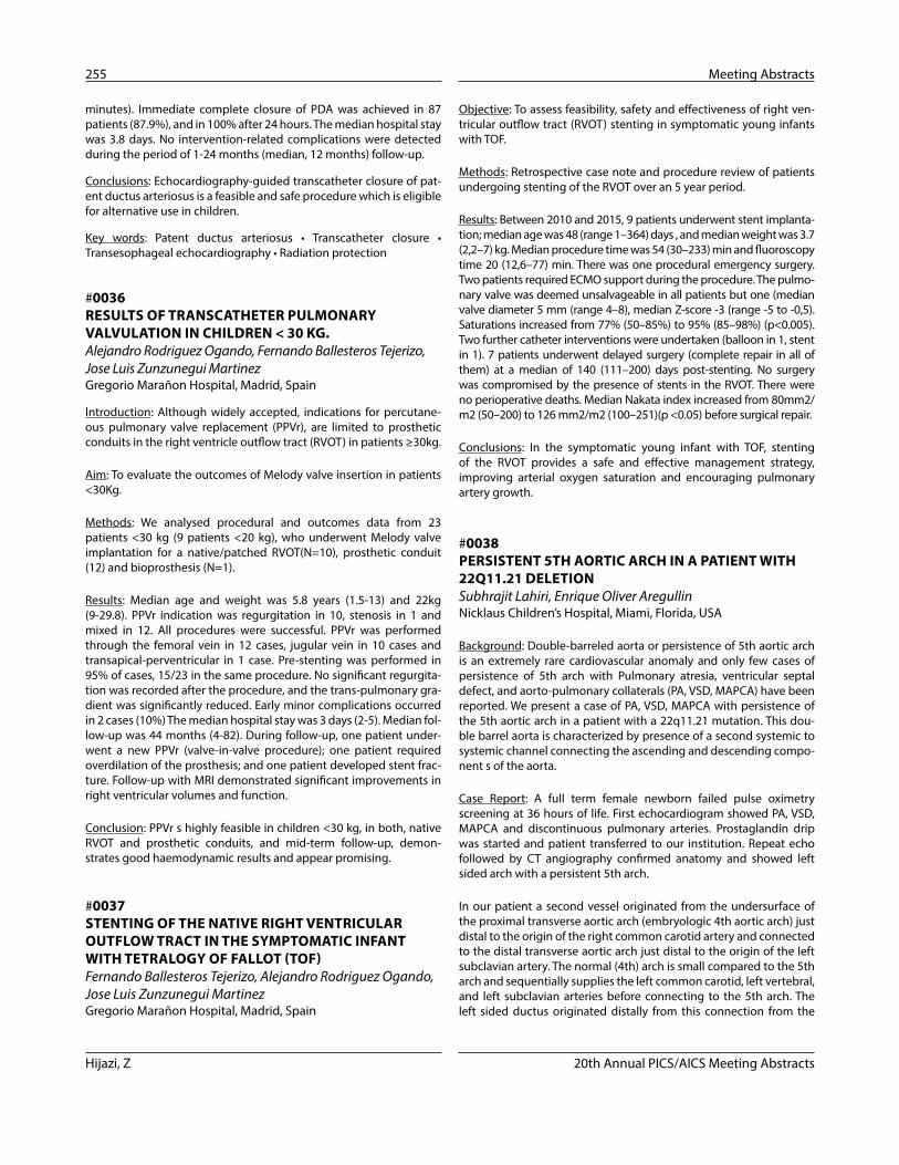
Hijazi, Z 20th Annual PICS/AICS Meeting Abstracts
255 Meeting Abstracts
Objective: To assess feasibility, safety and effectiveness of right ven-tricular outflow tract (RVOT) stenting in symptomatic young infants with TOF.
Methods: Retrospective case note and procedure review of patients undergoing stenting of the RVOT over an 5 year period.
Results: Between 2010 and 2015, 9 patients underwent stent implanta-tion; median age was 48 (range 1–364) days , and median weight was 3.7 (2,2–7) kg. Median procedure time was 54 (30–233) min and fluoroscopy time 20 (12,6–77) min. There was one procedural emergency surgery. Two patients required ECMO support during the procedure. The pulmo-nary valve was deemed unsalvageable in all patients but one (median valve diameter 5 mm (range 4–8), median Z-score -3 (range -5 to -0,5). Saturations increased from 77% (50–85%) to 95% (85–98%) (p<0.005). Two further catheter interventions were undertaken (balloon in 1, stent in 1). 7 patients underwent delayed surgery (complete repair in all of them) at a median of 140 (111–200) days post-stenting. No surgery was compromised by the presence of stents in the RVOT. There were no perioperative deaths. Median Nakata index increased from 80mm2/m2 (50–200) to 126 mm2/m2 (100–251)(p <0.05) before surgical repair.
Conclusions: In the symptomatic young infant with TOF, stenting of the RVOT provides a safe and effective management strategy, improving arterial oxygen saturation and encouraging pulmonary artery growth.
#0038 PERSISTENT 5TH AORTIC ARCH IN A PATIENT WITH 22Q11.21 DELETION Subhrajit Lahiri, Enrique Oliver AregullinNicklaus Children’s Hospital, Miami, Florida, USA
Background: Double-barreled aorta or persistence of 5th aortic arch is an extremely rare cardiovascular anomaly and only few cases of persistence of 5th arch with Pulmonary atresia, ventricular septal defect, and aorto-pulmonary collaterals (PA, VSD, MAPCA) have been reported. We present a case of PA, VSD, MAPCA with persistence of the 5th aortic arch in a patient with a 22q11.21 mutation. This dou-ble barrel aorta is characterized by presence of a second systemic to systemic channel connecting the ascending and descending compo-nent s of the aorta.
Case Report: A full term female newborn failed pulse oximetry screening at 36 hours of life. First echocardiogram showed PA, VSD, MAPCA and discontinuous pulmonary arteries. Prostaglandin drip was started and patient transferred to our institution. Repeat echo followed by CT angiography confirmed anatomy and showed left sided arch with a persistent 5th arch.
In our patient a second vessel originated from the undersurface of the proximal transverse aortic arch (embryologic 4th aortic arch) just distal to the origin of the right common carotid artery and connected to the distal transverse aortic arch just distal to the origin of the left subclavian artery. The normal (4th) arch is small compared to the 5th arch and sequentially supplies the left common carotid, left vertebral, and left subclavian arteries before connecting to the 5th arch. The left sided ductus originated distally from this connection from the
minutes). Immediate complete closure of PDA was achieved in 87 patients (87.9%), and in 100% after 24 hours. The median hospital stay was 3.8 days. No intervention-related complications were detected during the period of 1-24 months (median, 12 months) follow-up.
Conclusions: Echocardiography-guided transcatheter closure of pat-ent ductus arteriosus is a feasible and safe procedure which is eligible for alternative use in children.
Key words: Patent ductus arteriosus • Transcatheter closure • Transesophageal echocardiography • Radiation protection
#0036 RESULTS OF TRANSCATHETER PULMONARY VALVULATION IN CHILDREN < 30 KG.Alejandro Rodriguez Ogando, Fernando Ballesteros Tejerizo, Jose Luis Zunzunegui MartinezGregorio Marañon Hospital, Madrid, Spain
Introduction: Although widely accepted, indications for percutane-ous pulmonary valve replacement (PPVr), are limited to prosthetic conduits in the right ventricle outflow tract (RVOT) in patients ≥30kg.
Aim: To evaluate the outcomes of Melody valve insertion in patients <30Kg.
Methods: We analysed procedural and outcomes data from 23 patients <30 kg (9 patients <20 kg), who underwent Melody valve implantation for a native/patched RVOT(N=10), prosthetic conduit (12) and bioprosthesis (N=1).
Results: Median age and weight was 5.8 years (1.5-13) and 22kg (9-29.8). PPVr indication was regurgitation in 10, stenosis in 1 and mixed in 12. All procedures were successful. PPVr was performed through the femoral vein in 12 cases, jugular vein in 10 cases and transapical-perventricular in 1 case. Pre-stenting was performed in 95% of cases, 15/23 in the same procedure. No significant regurgita-tion was recorded after the procedure, and the trans-pulmonary gra-dient was significantly reduced. Early minor complications occurred in 2 cases (10%) The median hospital stay was 3 days (2-5). Median fol-low-up was 44 months (4-82). During follow-up, one patient under-went a new PPVr (valve-in-valve procedure); one patient required overdilation of the prosthesis; and one patient developed stent frac-ture. Follow-up with MRI demonstrated significant improvements in right ventricular volumes and function.
Conclusion: PPVr s highly feasible in children <30 kg, in both, native RVOT and prosthetic conduits, and mid-term follow-up, demon-strates good haemodynamic results and appear promising.
#0037 STENTING OF THE NATIVE RIGHT VENTRICULAR OUTFLOW TRACT IN THE SYMPTOMATIC INFANT WITH TETRALOGY OF FALLOT (TOF)Fernando Ballesteros Tejerizo, Alejandro Rodriguez Ogando, Jose Luis Zunzunegui MartinezGregorio Marañon Hospital, Madrid, Spain
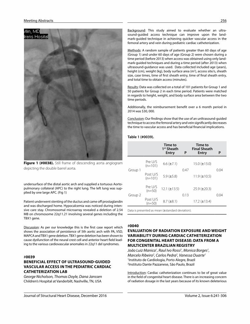
Journal of Structural Heart Disease, December 2016 Volume 2, Issue 6:241-306
Meeting Abstracts 256
Background: This study aimed to evaluate whether an ultra-sound-guided access technique can improve upon the land-mark-guided technique in achieving quicker vascular access in the femoral artery and vein during pediatric cardiac catheterization.
Methods: A random sample of patients greater than 60 days of age (Group 1) and under 60 days of age (Group 2) were chosen during a time period (before 2013) when access was obtained using only land-mark-guided techniques and during a time period (after 2013) when ultrasound-guidance was used. Data collected included age (years), height (cm), weight (kg), body surface area (m2), access site/s, sheath size, case times, time of first sheath entry, time of final sheath entry, and total time to obtain access (minutes).
Results: Data was collected on a total of 101 patients for Group 1 and 50 patients for Group 2 in each time period. Patients were matched in regards to height, weight, and body surface area between the two time periods.
Additionally, the reimbursement benefit over a 6 month period in 2014 was $30, 000.
Conclusion: Our findings show that the use of an unltrasound-guided technique to access the femoral artery and vein significantly decreases the time to vascular access and has beneficial financial implications.
#0040 EVALUATION OF RADIATION EXPOSURE AND WEIGHT VARIABILITY DURING CARDIAC CATHETERIZATION FOR CONGENITAL HEART DISEASE: DATA FROM A MULTICENTER BRAZILIAN REGISTRY João Luiz Manica1, Raul Ivo Rossi1, Monica Borges1, Marcelo Ribeiro2, Carlos Pedra2, Vanessa Duarte1
1Instituto de Cardiologia, Porto Alegre, Brazil2Instituto Dante Pazzanese, São Paulo, Brazil
Introduction: Cardiac catheterization continues to be of great value in the field of congenital heart disease. There is an increasing concern of radiation dosage in the last years because of its known deleterious
undersurface of the distal aortic arch and supplied a tortuous Aorto-pulmonary collateral (APC) to the right lung. The left lung was sup-plied by one large APC. (Fig 1)
Patient underwent stenting of the ductus and came off prostaglandin and was discharged home. Hypocalcemia was noticed during inten-sive care stay. Chromosomal microarray revealed a deletion of 2.54 MB on chromosome 22q11.21 involving several genes including the TBX1 gene.
Discussion: As per our knowledge this is the first case report which shows the association of persistence of 5th aortic arch with PA, VSD, MAPCA and TBX1 gene deletion. TBX1 gene deletion has been shown to cause dysfunction of the neural crest cell and anterior heart field lead-ing to the various cardiovascular anomalies in 22q11 del syndromes.
#0039 BENEFICIAL EFFECT OF ULTRASOUND-GUIDED VASCULAR ACCESS IN THE PEDIATRIC CARDIAC CATHETERIZATION LABGeorge Nicholson, Thomas Doyle, Dana JanssenChildren’s Hospital at Vanderbilt, Nashville, TN, USA
Table 1 (#0039).
Time to 1st Sheath
Entry P
Time to Final Sheath
Entry P
Group 1
Pre U/S (n=101) 6.6 (±7.1)
0.47
15.0 (±13.0)
0.04Post U/S (n=101) 5.9 (±5.8) 11.9 (±10.5)
Group 2
Pre U/S (n=50) 12.1 (±13.5)
0.13
25.9 (±20.3)
0.04Post U/S (n=50) 8.7 (±8.1) 17.2 (±13.4)
Data is presented as mean (±standard deviation).
Figure 1 (#0038). Still frame of descending aorta angiogram depicting the double barrel aorta.
Hijazi, Z 20th Annual PICS/AICS Meeting Abstracts
257 Meeting Abstracts
regurgitation presented in 65 (84.1%) patients. Procedures included 25 valvuloplasty alone, 27 valvuloplasty + ductal ligation, 20 valvu-loplasty + ductal ligation + mBT, and 5 valvuloplasty + ductal ligation + BDG. RV outflow tract continuity were established in all patients without early mortality and early reoperation. After valvuloplasty, SO2 increased from 73.5 ± 25.7% to 93.0 ± 3.4%. Four (5.2%) patients had a residual pressure gradient more than 30 mmHg. During 6.5 years (4 months-11 years) follow-up, there were 7 late deaths, 2 re-balloon dilatations, 2 ductal occlusions and 2 mBT occlusions. According to the hemodynamics, 2 patients underwent subsequent BDG and 2 patients underwent subsequent total cavopulmoanry connection. At the latest follow-up, 59 patients had biventricular cir-culation, 5 patients had one and a half ventricular circulation and 2 patients had univentricular palliation. In multi-variate analysis, tricus-pid z score was identified as independent protective factor for post-operative death (p=0.02, OR=0.21).
Conclusion: Outcomes of hybrid balloon valvuloplasty for PA/IVS is favorable with satisfactory potential to biventricular circulation.
#0042 NOVEL TRANSCATHETER INTERVENTION IN COR TRIATRIATUM DEXTERI.B VijayalakshmiBMC&RI Super Specialty Hospital (PMSSY), Bengaluru, India
Cor triatriatum Dexter is an extremely rare congenital anomaly (0.025%), in which the right atrium is divided into two chambers by a septum, diagnosed on autopsy in the past. We describe ante-mortem diagnosis, by 2D transthoracic echocardiography with agi-tated saline contrast echocardiography and inferior venacava or superior venacaval venography. To the best of our knowledge, for the first time in world, we report a novel method of transcatheter balloon disruption of membrane in five cases, along with the bal-loon dilatation of rheumatic mitral stenosis in two cases and device closure of atrial septal defect in one case, to prevent morbidity and mortality.
Objective: To describe the importance of transthoracic echocardiog-raphy with agitated saline contrast echocardiography and assess the feasibility and efficacy of transcatheter intervention in Cor triatriatum Dexter
Material and Results: Out of fifteen consecutive patients of Cor tri-atriatum Dexter diagnosed with transthoracic echocardiography with agitated saline contrast echocardiography, five patients who underwent transcatheter balloon disruption of membrane and other interventions formed the material for study. Three patients were boys and two were girls, age ranged between 3 to 17 years, median age was 10 years. Three patients presented with extertional dyspnea and two were asymptomatic. Two who had rheumatic heart disease with mitral stenosis underwent balloon mitral valvuloplasty and one case with atrial septal defect without pulmonary artery hypertension underwent device closure.
Conclusion: The Cor triatriatum Dexter is not benign as mortality occurs due to pulmonary embolism. Timely diagnosis with transtho-racic echocardiography with simple agitated saline contrast echo fol-lowed by balloon disruption can prevent cyanosis, pulmonary artery hypertension, morbidity and mortality.
effect. Children have more radio sensitivity but the evaluation of this exposition is more difficult mainly due to the weight variability. Finally, radiation dosage is not yet well standardized in this population.
Methods: This was a multicenter observational study. Data was col-lected from a Brazilian Registry of cardiac catheterization on congenital heart disease from march/2013 to june/2014. Patients aged more than 18 years old were excluded from this study. Four hundred and ninety five patients had dose-area product (DAP) measured and were included in this study. Median and interquartile intervals (25th and 75th) of age and weight were respectively 50 months (10 ; 103) and 15 Kg (7 ; 28).
Results: The median and interquartile intervals (25th and 75th) of DAP was 742,2 (288,8 ; 1791,5) uGy.m2. Interventional procedures had higher DAP than diagnostic procedures (25th, 50th, 75th percentiles: 230, 715, 1534 versus 315, 751, 2095 uGy.m2). DAP correlated with body weight (r = 0,56) but best correlated with weight-fluoroscopic time product (R = 0,75). DAP/weight showed no difference between diagnostic and interventional procedures ((25th, 50th, 75th percen-tiles: 23, 57, 110 versus 30, 57, 139).
Conclusions: DAP/weight has been considered in the last few years as the most reliable unit to evaluate radiation exposure in pediatric population. However, to date, there are only two reports in the litera-ture. This study reports similar doses when comparing to the bench-mark doses previously published by the CCISC. It is very useful to report these data in order to be continuously evaluating the impact of strategies to lower radiation exposure in this vulnerable popula-tion. Differences between institutions can alert for increased radia-tion exposure in patients and staff and support a program of quality improvements for radiation safety.
#0041 HYBRID BALLOON VALVULOPLASTY FOR PULMONARY ATRESIA AND INTACT VENTRICULAR SEPTUM: 10 YEAR EXPERIENCES Shoujun Li, Kai Ma, Shengshou Hu, Zhongdong Hua, Keming Yang, Hao Zhang, Jun Yan, Changwei Zhang, Kunjing PangFuwai Hospital, National center for cardiovascular disease, CAMS & PUMC, Beijing, China
Objective: Management of pulmonary atresia with intact ventricular septum (PA/IVS) remains challenging for the unfavourable outcomes after surgical or percutaneous interventional approaches. This study aimed to report outcomes of our hybrid balloon valvuloplasty via sternotomy for PA/IVS.
Methods: From 2005 to 2015, consecutive patients with PA/IVS who underwent hybrid balloon valvuloplasty in our institution were included into the current study. Ductal ligation, bidirectional Glenn shunt (BDG) and modified B-T shunt (mBT) were applied according to the post-valvuloplasty hemodynamics. Exclusion criteria included: right ventricular (RV) dependent coronary circulation and muscular atresia.
Results: A total of 77 consecutive patients were recruited. The median age at balloon valvuloplasty was 3 months (1 day to 24 months). Fifteen (19.4%) patients were neonate, 19 (24.7%) patients had mild or moderate RV hypoplasia, and more than moderate tricuspid
Journal of Structural Heart Disease, December 2016 Volume 2, Issue 6:241-306
Meeting Abstracts 258
study. Age ranged 3 days to 17 years ( mean 8 years ). The device clo-sure was done for PDA in 2, VSD in 15, ASD -1, ARVT in 1, ARAT in 1, ABV in 4, PBV in 2, aortoplasty in 1, PTMC in 2, pericardiocentesis in 3. 5 patients underwent two procedures in the same sitting. They were ABV and PBV, ABV and PTMC, ABV and PDA device closure, ASD and VSD device closure and PDA and VSD device closure. 3 cases of VSD were post- operative residual and one was closed with multi-ple devices. One 8 months infant had apical VSD closed with ADO II. Another 2 year old child underwent hybrid surgery for closure of VSD with 14 mm device. One child with mirror image dextrocardia and midmuscular VSD was closed with device. In one case procedure was abandoned as 18 mm VSD device slipped.
Discussion: Procedures in ANC is risky in presence LV/or RV dysfunc-tion with or without thrombosis. Positioning the device in apical VSD in ANC cases is very challenging as the device gets caught in trabe-culae in RV and if more tug is given the device slips through spongy myocardium. The results of interventions are very gratifying as the superadded pump failure due to pressure or volume overload caused by associated lesions improves significantly. One patient with severe AS and mitral stenosis had reverse May Thurner syndrome (obstruc-tion of right common iliac vein by right common ileac artery), hence procedure was done through left femoral puncture.
Conclusion: Associated lesions in ANC worsen the pump failure. Transcatheter interventions though challenging are feasible safe effective and are lifesaving. Transcatheter interventions certainly reduce the morbidity and mortality in ANC patients who are at high risk for surgery or redo surgery.
#0045 RETROGRADE TRANSCATHETER CLOSURE OF VENTRICULAR SEPTAL DEFECTS I.B VijayalakshmiBMC&RI Super Specialty Hospital (PMSSY), Bengaluru, India
Background: Amplatzer Duct Occluder II (ADO II) is designed for clos-ing long ducts in infants. There are few reports of “off-label” use of ADO II in non-ductal positions.
Aim: To evaluate the advantages and disadvantages of retrograde transcatheter closure of ventricular septal defects (VSD) with ADO II.
Material and Results: 102 cases of VSDS closed by retrograde tran-scatheter method with ADO II, formed the material for the pro-spective study. Age: 8 months to 23 years (mean 9.1 years). 74 perimembraneous VSDs, 14 muscular VSDs,13 Gerbode defects, one midmuscular VSD with dextrocardia, were closed. The shortest fluo-roscopic time was 4.2 min, mean was 8.4± 4.1min. In six cases there was initially a small residual shunt which had closed on three months follow up. Only in one case the device embolized to left pulmonary artery and it was retrieved . Eleven cases developed transient com-plete heart block which resolved and only one of them needed tem-porary pacing.
Discussion: ADO II has a very low profile and can be easily delivered through a 5F guiding catheter needs very short fluoroscopic time as artero-venous (AV) loop is not needed. The cost is 1/3 the cost of
#0043 CHALLENGES OF TRANSCATHETER INTERVENTIONS FOR CONGENITAL HEART DISEASES IN DEXTROCARDIAI.B VijayalakshmiBMC&RI Super Specialty Hospital (PMSSY), Bengaluru, India
Background: Several challenges are faced by interventionalists while performing various percutaneous interventions for congenital heart disease (CHD) in patients with dextrocardia. The anatomical altera-tions in dextrocardia especially the lie of the interventricular septum (IVS) can cause impediment for device closure of ventricular septal defect (VSD) and lie of interatrial septum (IAS) for puncture.
Aim: The aim of our study is to evaluate the challenges, feasibility and efficacy of transcatheter interventions in children with CHD in dextrocardia.
Materials and Results: Out of 60 patients of CHD with dextrocardia catheterized, only 9 patients (15%) underwent transcatheter interven-tions. The age was 4 months to 16 years (mean 5.4 years), weight - 4.1 to 40 kgs (mean 8.4 kgs). 3 cases underwent successful device closure for patent ductus arteriosus (PDA). 2 cases of midmuscular VSD (MVSD) were closed. One had to be closed with Amplatzer ventricular septal occlude through the jugular approach and the other with Amplatzer duct occluder II (ADO II).There was a difficulty in puncturing IAS during balloon valvuloplasty for mitral stenosis in a case of right-sided May Thurner Syndrome (MTS). Balloon valvuoplasty was done in one infant with severe pulmonary stenosis by flipping the cine image. One very sick patient with inferior vena cava web died after cavoplasty and stenting.
Conclusion: The catheter interventions in CHD with dextrocardia though difficult is feasible. The device closure of PDA and MVSD is not difficult especially with ADO II. The balloon mitral and aortic valvu-loplasty in the complex cardiac anatomy of situs inversus totallis is feasible and safe. Rarely right-sided MTS may cause problem for right femoral access during transcatheter procedure.
#0044 CHALLENGES OF INTERVENTIONS FOR ASSOCIATED LESIONS IN CASES OF APICAL NON-COMPACTION I.B VijayalakshmiBMC&RI Super Specialty Hospital (PMSSY), Bengaluru, India
Background: Isolated left ventricular non-compaction is reported extensively. But apical non-compaction (ANC) of both ventricles and septum is not reported much in literature. For the first time in the world, we are reporting the challenges of various interventions for different associated lesions in ANC.
Aim: To know the challenges and feasibility of transcatheter interven-tions for the associated lesions in cases of ANC to reduce the pump failure.
Material and Results: Out of 100 consecutive patients diagnosed as ANC by transthoracic echocardiography (TTE), 30 cases underwent various transcatheter interventions, formed the material for this
Hijazi, Z 20th Annual PICS/AICS Meeting Abstracts
259 Meeting Abstracts
#0047 RE-STENTING IMPROVES VESSEL PATENCY, LUMEN AREA AND ENDOTHELIALIZATION: A FEASIBILITY STUDY IN PIGLETS DEMONSTRATING RE-STENTING WITH SIMULTANEOUS INTENTIONAL FRACTURE OF PREVIOUSLY PLACED STENTSAndrás Bratincsák3, William Van Alstine2, Lindsay Koren2, Kimberly Stoughton2, José Negrón-Garcia2, John Moore1, Howaida El-Said1
1University of California Sand Diego, Rady Children’s Hospital, San Diego, California, USA2Cook Medical, Bloomington, Indiana, USA3Kapi’olani Medical Center for Women and Children, Honolulu, Hawaii, USA
Background: Intentional stent fracture in-vivo has shown medial dis-section/vessel injury. Spontaneous stent fracture in humans has been reported to cause stent collapse, hemodynamic compromise and embolization of stent fragments, many of which were treated with re-stenting.
Objective: To demonstrate feasibility and propose re-stenting prior to intentional stent fracture to avoid such complications. We sought to compare stent fracture with and without a stabilizing additional stent and study the effects on the tissue.
Methods: Five months after fourteen low profile stents where implanted in the aorta of four piglets, they were intentionally fractured using high-pressure balloons, one group with another stent placed inside (re-stent group) and one without (single stent group). Vessel/stent were studied at both short-term and long term intervals. Results: The re-stent group demonstrated significantly larger vessel segment (113±28mm2 versus 59±17mm2, p=0.0476), less long term luminal diameter loss 45.5% versus 73.7%, p=0.0065), lack of strut protrusion and higher degree of endotheliziation (95.7±2.1% versus 74.1±7%, p=0.0145). There was similar degree of vessel wall injury at the time of the frac-ture, however at long term evaluation the injury score in the re-stent group demonstrated statistically significant improvement (p=0.0426). No damage to the external part of the blood vessels or the surrounding soft tissue was noted in either group.
Conclusion: Re-stenting at the time of intentional stent fracture provides numerous advantages including larger vessel diameter, maintained vessel patency, more complete endothelizalization, lack of stent strut protrusion with decreased chance of injury and future complications. Human studies are needed to confirm our observations.
#0048 VSD CLOSURE USING CERA® DEVICESJesus Damsky Barbosa, Victorio Lucini, Adelia Marques, Judith Ackerman, Fernanda Biancolini, Julio Biancolini, Ana de Dios”Pedro de Elizalde”, Ciudad Autonoma de Buenos Aires, Argentina
Objective: the aim of this study is to describe the immediate to long follow up in 15 patients (p) with perimembranous (PMVSD)
regular ventricular septal occluder. However, it is not useful in VSDs measuring more than 6 mm and in those with insufficient aortic rim.
Conclusion: ADO II is an excellent device in ventricular septal defects. The procedure time and the cost are significantly lesser than regular devices. The success rate is very high and complication rate is very low.
#0046 TRANSCATHETER DUCTUS ARTERIOSUS STENTING WITH EXPECTED REDILATION AS A SURGICAL ALTERNATIVEJesse Lee, Howaida El-Said, Kanishka Ratnayaka, John MooreUniversity of California San Diego/Rady Children’s Hopistal, San Diego, CA, USA
Background: Transcatheter ductus arteriosus stenting (DAS) has been used to provide a reliable temporary source for pulmonary blood flow in cyanotic congenital heart patients. However, even in the context of surgical mortality rates approaching 10%, most modern congenital cardiovascular centers continue to rely almost exclusively on surgical shunts. Data on DAS from centers with sophisticated surgical services is limited. We report DAS experience from a modern comprehensive congenital heart center in North America.
Methods: This is a single-center retrospective review of all patients who were taken to the cardiac catheterization lab with the intent of DAS from January 2006 to August 2016.
Results: Successful DAS was performed in 30/32 patients (97%), median age 7 days (range 1-176), median weight 3.43kg (1.5-6.8). Diagnoses included 14 PA/IVS, 5 critical PS, 4 TOF, 2 PA/VSD, 3 TGA, 1 DORV/TGA, 1 tricuspid atresia, 1 Ebstein’s & 1 Heterotaxy/AVC/PS. 2/32 required urgent surgical shunt placement, due to ductal spasm prior to stenting in one and stent thrombosis in the other. Bare metal coronary artery stents were used, median stent diameter 3.5 mm (2.5-4). Median saturation at discharge was 91% (80-99). At a median fol-low up of 3.9yrs (2 mos-9.4yrs), 17 (55%) had reinterventions for low saturations (balloon dilation alone n=5 and restent n=12). Median time between implant and re-intervention was 74 days (33-186). Stent thrombosis at the time of re-intervention occurred in 1/17; patient was referred for an early Glenn. Complications involving fem-oral vessel compromise occurred in 7 (22%), 3 resolved with anticoag-ulation, 4 formed sufficient collaterals. 16/30 progressed to surgical repair or next stage palliation; 14 did not require further surgical intervention with adequate prograde pulmonary blood flow [balloon pulmonary valvuloplasty (n=13), bilateral DA stenting as destination palliation (n=1)]. There were no deaths associated with the DAS. One patient died after a Glenn procedure while awaiting heart transplant 3 months post DAS.
Conclusions: DAS appears to be a safe and effective procedure for establishing a reliable temporary source of pulmonary blood flow in selected cyanotic congenital heart patients. Reintervention for stent redilation is common and should be a planned component of this management strategy. It is likely that more experience from centers with sophisticated surgical services will be necessary before commu-nity practice evolves toward greater reliance upon DAS.
Journal of Structural Heart Disease, December 2016 Volume 2, Issue 6:241-306
Meeting Abstracts 260
Methods and Results: 31 catheterizations performed on 25 patients (18 males) weighing <10kg from Jan 2008 to June 2016 with the intention to relieve RVOTO were retrospectively reviewed. 18 procedures were first palliations, 2 with initial surgical palliation, 6 being restenting procedures and 5 were on post-operative RVOTs. The primary diag-nosis was Tetralogy of Fallot including double outlet ventriculo-arte-rial connection in 10. 9 patients (36%) had associated cardiac lesions, with atrioventricular septal defect being the commonest (n=4, 16%). Prematurity and genetic disorder accounted for 32% and 24% respec-tively. The indication for RVOT stenting were low body weight preclud-ing surgery (n=16, median 2.59kg, IQR 1.9-3.42kg), high risk associated cardiac lesions (n=5), high risk comorbidities (n=3), severe cyanotic spells (n=2) and post-operative residual RVOTO (n=5). 9 (36%) patients were on prostaglandin infusion, 9 (36%) ventilated and 3 (12%) were on propranolol. Eight procedures (26%) were performed as emergent or salvage therapy. Overall procedural success rate was 93.5% (29/31 procedures) with 32 stents implanted and 3 procedures required 2 stents. Significant improvement of SaO2 was observed (71±14.6% to 88.7±7.9%;p<0.001). Six complications were recorded (19%) including 1 cardiac perforation that required no treatment, and 1 sudden car-diac arrest during diagnostic catheterization, the only in-lab mortality; others were minor complications. Catheter reinterventions were nec-essary for 11 patients, 2 patients required 2 reinterventions. Median time to reintervention was 8.4 months (IQR≈0.8-14.5). A lower main pulmonary artery z score (-4.3±1.7 vs -2.4±1.4, p=0.02) and pulmonary annular z score (-5.2±1.5 vs -3.4±2.2, p=0.04) were associated with rein-tervention. Twenty patients survived definitive surgery, with a mean RVOT velocity of 2.6±0.7m/s from their latest echocardiogram; while 2≈patients were awaiting definitive surgery.
Conclusion: RVOT stenting achieved good palliation with high success rate in this group of high-risk symptomatic TOF patients.
#0050 SUCCESSFUL TRANSFEMORAL TREATMENT OF AN INNOMINATE ARTERY PSEUDOANEURYSM COMPLICATION FROM EXTRACORPOREAL MEMBRANE OXYGENATION IN A NEONATE WITH REVIEW OF THE LITERATUREAamisha Gupta, Zahid Amin, Emily Masoumi, Carlee LeopardChildren’s Hospital of Georgia, Augusta, GA, USA
Background: We report successful repair of a large pseudo-aneurysm in a 7 week neonate which developed off of the innominate artery as a complication of ECMO cannulation. From review of the literature, this is the first reported case to be successfully treated via transfemo-ral approach using covered stents.
Methods: A full term infant with history of biventricular hypertrophy and pulmonary hypertension was placed on VA ECMO soon after birth. At 7 weeks of life, post decannulation, a large pseudoaneurysm was found off of the innominate artery by echocardiogram. A CT with con-trast confirmed the diagnosis. During cardiac catheterization, angiog-raphy revealed that the pseudoaneurysm measured 24 mm x 25 mm. The diameter of the subclavian artery was 4 mm and the origin of the innominate artery measured approximately 5mm. The length that needed to be covered was approximately 25 mm. A wire was placed in the distal subclavian artery and exchanged of a 6F x 55 cm Flexor
and muscular VSDs (MUSC VSD) treated with Cera® devices (Lifetech Scientific, Shenzhen, China).
Background: Cera VSD Occluder is formed of a nitinol wire mesh, covered with a ceramic coating which reportedly reduces the risk of thrombus formation, encourages endothelialization, and minimizes systemic nickel ion release.
Material and Method: from October 2013 to September 2016, 14 p were treated with CERAÒ devices. This is a prospective analysis. Median age: 11 y/o (2.6 to 25 y/o). Median weight: 40 kg (14 to 70 kg). Gender: 9/14p M 6/14p F. All the p had femoral artery and venous approach. All the p with general anesthesia. X: QP/QS: >1.3:1. All the procedures were carried out with arterio-venous loop. LV angio-grams: a) perimembranous VSD: 60° cranial-20° LAO, b) muscular VSD: 40° cranial-20° LAO. All the procedures were guided with TEE and TTE. Measurements by echo and angiography: right and left diameter and length of the defect.
Results: 7/14 p: perimembranous VSD, 3/14 p post surgical (1/14 p double committed VSD, 1p PM VSD and 1/14 p Pulmonary atre-sia with VSD), 4/14 p: muscular VSD (1/14 mid ventricular VSD, 1/14 multiple VSD, 2/14 muscular-membranous VSD). Total proce-dures: 17 (1 p suffered an AV block during the catheterization of the VSD so that the procedure was cancel and in a second session the VSD was totally occluded). 1p the device embolized. In a sec-ond procedure, the device was retrieved and a bigger device was successfully positioned. Devices used: 1 type I, 8 type II, 2 type III, 3 muscular VSD. Complications: 1 embolization, the device was snared and the VSD was treated with a bigger device. This emboli-zation was in the third p. 3 AV blocks in the first 24 hs, treated with steroids. The p were discharged with sinusal rithm.One of them had sick sinus syndrome.
Follow up: 3p with residual shunt (2 mild and 1 moderate who is waiting for a second procedure). There weren’t av-block. As the initial experience was good, we decided to close VSDs in p under 10 kg. 1p with 8 grs had a perimemb VSD. It was closed with 7 type III peri-memb device with correct result.
Conclusions: 1) Lifetech devices are a good choice to close VSDs. 2) The AV-blocks after procedures were considered as inflammatory process for the maneuvers. The steroids diminished the inflamma-tion. 3) The embolization described was due to the little experience. 4) There weren’t AV-blocks during follow up.
#0049 RIGHT VENTRICULAR OUTFLOW TRACT STENTING IN HIGH-RISK SYMPTOMATIC TETRALOGY OF FALLOT UNDER 10KG - A SINGLE CENTRE EXPERIENCERobin Hay Son Chen, Robert Yates, Sachin Khambadkone, Graham DerrickGreat Ormond Street Hospital for Children, London, UK
Introduction: Right ventricular outflow tract (RVOT) stenting is an attractive option for symptomatic patients who are at a high risk for surgical repair of Tetralogy of Fallot (TOF). We present our centre’s experience in RVOT stenting for patients <10kg.
Hijazi, Z 20th Annual PICS/AICS Meeting Abstracts
261 Meeting Abstracts
was not different (67.3% vs 50%, p=0.08). The surgical cohort had a greater incidence of mechanical ventilation requirement ≥7 days (p=0.02), wound infection (p=0.01), unplanned cardiac catheteriza-tion (p=0.05), surgical exploration (p=0.04), and low cardiac output syndrome (p=0.02). The incidence of venous or arterial thrombosis in the PDA stent cohort was relatively low (6.8% & 11.4 %, respec-tively). CONCLUSION: Percutaneous stenting of the PDA is an effec-tive strategy for stabilization of ductal dependent pulmonary blood flow. While there were no significant differences in early and overall mortality between cohorts, PDA stenting was associated with signifi-cantly less morbidity and need for re-intervention.
#0052 INFERIOR VENA CAVA COMPLETE OCCLUSION AFTER LIVER TRANSPLANT TREATED WITH RADIOFREQUENCY PERFORATION AND STENTING IN A PEDIATRIC PATIENT. Francisco Gonzalez Barlatay, Fernando Alvarez, Michel Lallier, Joaquim MiroHôpital Sainte-Justine. Université de Montréal., Montréal/ Quebec, Canada
We present a 15 month old boy with biliary atresia, who underwent liver transplantation. Surgery was complicated by a bowel perforation and thrombosis of the hepatic artery. Four weeks after the incident he was retransplanted. A thrombosis at the inferior vena cava anasto-mosis was diagnosed by ultrasound in the immediate post-surgical period. Five months after surgery an increase in serum aminotrans-ferases (8 x normal values) and gamma-glutamyl trasferases (2 x normal values) was observed. A liver biopsy showed signs of hepatic vein outflow obstruction. A cavography showed the complete occlu-sion of the inferior vena cava with partial decompression towards the azygos vein system. A 16.9 mm distance between superior and inferior stumps of the IVC was measured. After multidisciplinary discussion, the patient was brought to the catheterisation laboratory, where a radiofrequency needle (Baylis medical, Toronto, Canada) was advanced via the inferior stump, reaching the right atrial stump of IVC. The guidewire was snared and exteriorized by the right jugu-lar vein. On that guidewire rail, a 28 mm covered CP stent (NuMed, Hopkinton, USA) was deployed using a 10 mm x 3 cm BIB balloon (NuMed). The initial clinical response was very encouraging, with ascites significantly reduced and normalisation of laboratory tests. The patient needed balloon redilations, due to caudal stenosis of the stent, at two and four months post-procedure. One year after the procedure the stent is still permeable and without significant gradi-ent. The patient is still under enoxaparin and aspirin. Liver function is normal.
Conclusion: Percutaneous approach of this rare complication was the alternative to avoid a third liver transplant in our patient. The good results and the feasibility of the technique make it a very encouraging option in this group of patients.
#0053 THE EFFECT ON SOMATIC GROWTH OF SURGICAL AND TRANSCATHETER TREATMENT OF SECUNDUM ATRIAL SEPTAL DEFECTSMeghan Chlebowski1, Hongying Dai2, Stephen Kaine1
Tuohy-Borst delivery sheath. We deployed two 6 mm x 22 mm Atrium Cast premounted covered stents in a tandem fashion to effectively iso-late the aneurysm and maintain flow to the subclavian artery. Result: Follow up angiography demonstrated excellent result with nearly complete occlusion of the pseudoaneurysm and normal opacification of the right subclavian artery through the stent. There were no proce-dural or post procedural complications. Patient was electively started on heparin after sheath removal for 6 hour. The femoral artery pulses remained normal. Follow up echocardiography revealed trace shunt from the where the stents were overlapped. The patient was trans-ferred to the NICU and discharged home when his condition stabilized.
Conclusion: We reviewed the literature and found one case report where pseudoaneurysm had developed after ECMO, but no account was made as to the management of the pseudoaneurysm. Hence, to our knowledge, this is the first successfully closed pseudoaneurysm case in a neonate. Management options in this patient included tran-scatheter management or proximal ligation of the innominate artery to prevent further dilation and rupture. Since the patient had the right carotid artery ligated after the ECMO cannula was removed, we wanted to maintain adequate blood flow to the right upper extremity and avoid steal phenomenon form the vertebral artery.
#0051 PERCUTANEOUS STABILIZATION OF DUCTAL-DEPENDENT PULMONARY BLOOD FLOW USING PDA STENT IS ASSOCIATED WITH DECREASED MORBIDITY COMPARED TO SURGICAL SHUNT PLACEMENTBritton Keeshan, Agustin Rubio, Brian Morray, Bhawna Arya, D. Michael McMullan, Lester Permut, Troy Johnston, Jonathan Chen, Thomas JonesSeattle Children’s Hospital / University of Washington, Seattle, WA., USA
Background: Transcatheter stenting of the patent ductus arteriosus (PDA) is becoming an accepted alternative to surgical aortopulmo-nary shunt for stabilization of ductal-dependent pulmonary blood flow. However, there is limited data comparing procedural and inter-mediate-term morbidity between these two palliative strategies. Methods: This is a retrospective single-center review of patients who underwent stenting of the PDA or surgical aortopulmonary shunt from 2005 to 2015. Primary outcomes were survival, length of stay and freedom from re-intervention. Patient charts and the Society for Thoracic Surgeons database were queried for procedure-related adverse events. Results: 55 patients were referred for PDA stenting and 55 patients were referred for surgical intervention with a Blalock-Taussig or central shunt. PDA stent placement was successful in 44 (80%) patients. Baseline characteristics were similar between the 2 groups. While there was no significant difference in 30-day survival (95.4% vs. 97.9%, p=0.24), there was a trend towards improved over-all survival in the PDA stent group (93% vs. 81.1%, p=0.09, median follow-up 941 vs. 1967 days, respectively). The proportion of patients who underwent biventricular repair was similar between study groups. Patients with PDA stents had shorter median ICU and hospi-tal length of stays (2 vs. 7 days, p=0.04; 6 vs. 17 days, p<0.01, respec-tively), were less likely to require nasogastric tube feeds at discharge (p<0.01) and were less likely to require invasive intervention in the first 30 days after initial palliation (p=0.04). There were more adverse events in the surgical shunt cohort (119 vs. 36, p<0.01), although the proportion of patients who experienced ≥1 adverse outcome
Journal of Structural Heart Disease, December 2016 Volume 2, Issue 6:241-306
Meeting Abstracts 262
results. The excimer laser catheter (Spectranetics, Corporation) is an over the wire system that is available in 4 diameters (0.9mm; 1.2mm; 1.5mm and 2.0mm) and creates a distinct circumferential orifice equivalent to the catheter diameter at the site of intervention.
Methods: Since 2001, we have utilized the Excimer laser system, with or without a guide catheter, for the treatment of neonatal congenital heart defects with critical obstruction vs other various established methods including surgical intervention.
Results: Ten neonates with pulmonary valve atresia/critical steno-sis and intact ventricular septum, 4 neonates with hypoplastic left heart syndrome and intact atrial septum, 1 premature neonate with an occluded right pulmonary artery secondary to thrombus and 1 Fontan baffle with failing physiology underwent successful perfora-tion and additional intervention using the excimer laser system (vid-eos of the technique would be provided for oral presentation). All laser perforations were successfully performed utilizing fluoros-copy with or without transesophageal echocardiographic imaging. In the case of pulmonary valve atresia and HLHS, only the 0.9mm diameter laser was utilized which facilitated without difficulty, the ability to dilate the valve or atrial septum with a single large diame-ter Mini-Tyshak balloon thereby avoiding the need for a gradational approach. The single neonate with an acutely occluded right pulmo-nary artery underwent serial laser dilation with all 4 diameter laser catheters prior to conventional balloon angioplasty. There was a single inadvertent perforation of the RV outflow tract which sealed spontaneously and no other complications were reported secondary to the laser procedure.
Conclusion: Excimer laser therapy is an excellent alternative therapy for the treatment of neonatal congenital heart disease with critical obstruction. The creation of a distinct 1mm or greater orifice at the site of intervention, facilitates the utilization of a single diameter bal-loon dilation catheter, thereby avoiding a gradational approach and reducing fluoroscopy exposure.
#0055 3D ROTATIONAL ANGIOGRAPHY (3DRA) GUIDANCE OF NON-TRADITIONAL ASCENDING AORTA STENTING Rodrigo Rios, Nasser Moiduddin, Kanishka Ratnayaka, Howaida El-Said, John MooreRady Children’s Hospital, University of California - San Diego, San Diego, CA, USA
Introduction: Ascending aorta narrowing is traditionally addressed by surgical intervention. We present a case series in which acquired narrowing of the ascending aorta was addressed successfully via 3D Rotational Angiography (3DRA) guided transcatheter stent placement.
Method: 3DRA was utilized for a roadmap. Balloon sizing was used for stent delivery balloon selection. Apnea and hypotension induced by rapid ventricular pacing aided stent positioning and delivery. Precise positioning was essential to avoid complications such as aortic valve/coronary artery compromise or brachiocephalic branch vessel jailing.
1Children’s Mercy Hospital, Department of Cardiology, Kansas City, Missouri, USA2Children’s Mercy Hospital, Department of Biostatistics, Kansas City, Missouri, USA3University of Missouri - Kansas City, Kansas City, Missouri, USA
Objective: To evaluate the effect of secundum ASD closure on somatic growth, we conducted a single center, retrospective review of patients presenting for surgical or transcatheter closure during two distinct time periods.
Background: Natural history studies suggest an association between atrial septal defect (ASD) and growth retardation with inconsistent improvement following defect closure. Limited data exist regard-ing the effect of ASD closure on growth in the era of transcatheter therapy.
Methods: Four hundred patients with isolated ASD were divided into three cohorts: early surgical (January 1997-January 2002, N = 138), contemporary surgical (January 2006-January 2015, N = 142), and transcatheter closure (January 2006-January 2015, N= 120). Data collected at treatment and all follow up encounters included demo-graphics; height, weight, and body mass index (BMI) percentiles; hemodynamics at catheterization; and presence of any comorbidity affecting growth.
Results: Age at repair differed between groups (p < 0.0001) with a median age of 2.5 years in the early surgical, 5 years in the contempo-rary surgical and 6 years in the transcatheter cohorts. For all cohorts, there was no statistically significant change in height or weight per-centiles during two years of follow-up after ASD closure. There was a statistically, but not clinically, significant change in BMI percentile for the contemporary surgical cohort. In the transcatheter cohort, mean Qp:Qs was 1.65 +/- 0.54. Bivariate analysis did not show a negative correlation for any growth index demonstrating no association of greater Qp:Qs with decreased somatic growth. Subgroup analysis for patients in any cohort having an initial growth percentile <5th percen-tile demonstrated a significant change in weight and BMI percentiles in the first two years after closure (p < 0.0004).
Conclusion: We found no significant change in weight and height percentiles during two years follow-up after ASD closure. Only patients with initial weight and BMI <5th percentile had improved growth after treatment. The advent of catheter device therapy shifted the institutional practice to later age at repair for both surgical and transcatheter cohorts. Concerns over poor growth should not be an indication for early ASD repair except in children with existing signif-icant growth failure.
#0054 EXCIMER LASER THERAPY FOR NEONATAL CONGENITAL HEART DEFECTS WITH CRITICAL OBSTRUCTION: A SINGLE INSTITUTIONAL EXPERIENCE.Howard WeberPenn State Hershey Childrens Hospital, Hershey, USA
Interventional procedures for neonatal congenital heart defects with critical obstruction have evolved over the past 25 years with variable
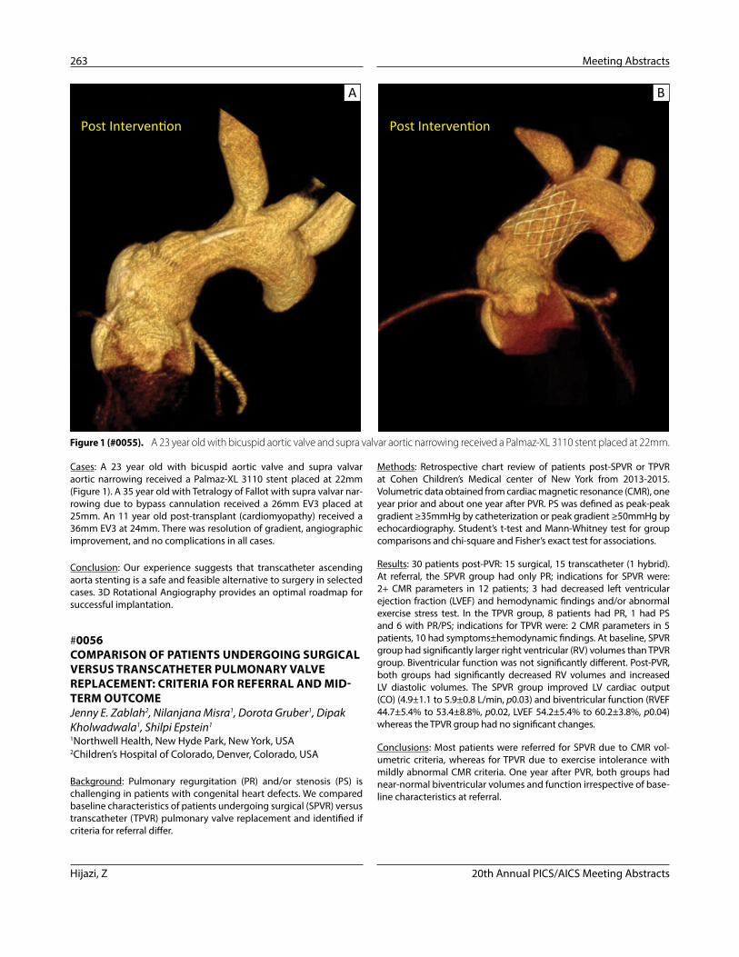
Hijazi, Z 20th Annual PICS/AICS Meeting Abstracts
263 Meeting Abstracts
Methods: Retrospective chart review of patients post-SPVR or TPVR at Cohen Children’s Medical center of New York from 2013-2015. Volumetric data obtained from cardiac magnetic resonance (CMR), one year prior and about one year after PVR. PS was defined as peak-peak gradient ≥35mmHg by catheterization or peak gradient ≥50mmHg by echocardiography. Student’s t-test and Mann-Whitney test for group comparisons and chi-square and Fisher’s exact test for associations.
Results: 30 patients post-PVR: 15 surgical, 15 transcatheter (1 hybrid). At referral, the SPVR group had only PR; indications for SPVR were: 2+ CMR parameters in 12 patients; 3 had decreased left ventricular ejection fraction (LVEF) and hemodynamic findings and/or abnormal exercise stress test. In the TPVR group, 8 patients had PR, 1 had PS and 6 with PR/PS; indications for TPVR were: 2 CMR parameters in 5 patients, 10 had symptoms±hemodynamic findings. At baseline, SPVR group had significantly larger right ventricular (RV) volumes than TPVR group. Biventricular function was not significantly different. Post-PVR, both groups had significantly decreased RV volumes and increased LV diastolic volumes. The SPVR group improved LV cardiac output (CO) (4.9±1.1 to 5.9±0.8 L/min, p0.03) and biventricular function (RVEF 44.7±5.4% to 53.4±8.8%, p0.02, LVEF 54.2±5.4% to 60.2±3.8%, p0.04) whereas the TPVR group had no significant changes.
Conclusions: Most patients were referred for SPVR due to CMR vol-umetric criteria, whereas for TPVR due to exercise intolerance with mildly abnormal CMR criteria. One year after PVR, both groups had near-normal biventricular volumes and function irrespective of base-line characteristics at referral.
Cases: A 23 year old with bicuspid aortic valve and supra valvar aortic narrowing received a Palmaz-XL 3110 stent placed at 22mm (Figure 1). A 35 year old with Tetralogy of Fallot with supra valvar nar-rowing due to bypass cannulation received a 26mm EV3 placed at 25mm. An 11 year old post-transplant (cardiomyopathy) received a 36mm EV3 at 24mm. There was resolution of gradient, angiographic improvement, and no complications in all cases.
Conclusion: Our experience suggests that transcatheter ascending aorta stenting is a safe and feasible alternative to surgery in selected cases. 3D Rotational Angiography provides an optimal roadmap for successful implantation.
#0056 COMPARISON OF PATIENTS UNDERGOING SURGICAL VERSUS TRANSCATHETER PULMONARY VALVE REPLACEMENT: CRITERIA FOR REFERRAL AND MID-TERM OUTCOMEJenny E. Zablah2, Nilanjana Misra1, Dorota Gruber1, Dipak Kholwadwala1, Shilpi Epstein1
1Northwell Health, New Hyde Park, New York, USA2Children’s Hospital of Colorado, Denver, Colorado, USA
Background: Pulmonary regurgitation (PR) and/or stenosis (PS) is challenging in patients with congenital heart defects. We compared baseline characteristics of patients undergoing surgical (SPVR) versus transcatheter (TPVR) pulmonary valve replacement and identified if criteria for referral differ.
Post Interven�onPost Interven�on
A B
Figure 1 (#0055). A 23 year old with bicuspid aortic valve and supra valvar aortic narrowing received a Palmaz-XL 3110 stent placed at 22mm.
Journal of Structural Heart Disease, December 2016 Volume 2, Issue 6:241-306
Meeting Abstracts 264
Method: From Jan 2010 to Sep 2015, total 217 patients underwent catheterization in our hospital. Male was 110, and median age of patients was16.7 years old. Median duration from Fontan procedure was 11.8 years. We reviewed their record retrospectively.
Results: Of them, 118 patients underwent interventional procedure; 15 patients underwent the closure of fenestration in Fontan tract, 58 patients underwent the occlusion of venous collaterals, 34 patients underwent occlusion of arterial collaterals, 31 patients underwent balloon angioplasty, 11 patients underwent stent implantation. There was no intervention- associated mortality. Transient severe headache developed in 1 patient but disappeared in 1 week. It might be due to air emboli or thrombi. Mild hemoptysis occurred in 1 patients but disappeared after 1 day.
Discussion: In spite of good early results of Fontan operation, the long-term complication is not rare and the close follow-up and appropriate intervention is needed. There are many alternative imag-ing and treatment modalities. However, the well-prepared transcath-eter intervention is not risky and good alternative to surgery
#0059 IMMEDIATE AND SHORT TERM OUTCOME POST VSD CLOSURE USING NITOCCLUD PFM COIL – COMPLICATIONS ARE NOT UNCOMMONDina Adel, Amr Mansour, Alaa Roushdy, Heba Attya, Azza Elfiky, Maiy ElsayedCardiolog Department Ain Shams University, Cairo, Egypt
Objective: We sought to study the immediate and short term out-come post VSD closure using nitocclud PFM coil to document the safety and efficacy of the procedure.
Patients and Methods: The study included 16 patients with perimem-branous subaortic VSD who were scheduled for elective trans cathe-ter VSD closure using nitocclud PFM coil in the period from May 2014 to July 2016. All patients underwent full clinical examination, ECG and full echocardiographic study immediately before trans catheter clo-sure as well as 24 hour, 1 month and every 6 month after the proce-dure. Any intra or post procedural complications and their respective management were recorded.
Results: The mean age of the study subjects was 6.3 ± 3.3 years. The distance between the defect and the aortic valve was an average of 5.4 ± 1.8 mm, and the left ventricular opening averaged 10.6 ± 3.7 mm. Immediate closure of the VSD was acheived in 25% of the cases this percentage increased to 75% after 1 month. Intravascular hemo-lysis developed 3 days after the procedure in one patient with residual shunt and was successfully managed by PDA amplatzer occluder device implanted in the residual shunt. One patient reported syncope 1 week after discharge and was admitted with complete heart block for which he received transient pacemaker and steroid therapy for 2 weeks after which he regained normal sinus rhythm. The same patient had 2 more attacks of loss of consciousness 2 years after device implantation and complete heart block was documented by ECG and a VVIR pacemaker was eventually implanted for this child. 2 more children had transient self limiting bradycardia and junctional rhythm during the procedure.
#0057 COMPARISON OF THE LIFETECH CERA AND AMPLATZER VENTRICULAR SEPTAL OCCLUDERS FOR VENTRICULAR SEPTAL DEFECTS CLOSURE IN 147 CHILDRENOsman Baspinar1, Ayse Sulu1, Derya Aydin Sahin1, Huseyin Yildiz1, Orhan Ozer2, Mehmet Kervancioglu1, Metin Kilinc1, Gokhan Gokaslan3
1Gaziantep University Medical Faculty, Pediatrid Cardiology, Gaziantep, Turkey2Gaziantep University Medical Faculty, Adult Cardiology, Gaziantep, Turkey3Gaziantep University Medical Faculty, Cardiovascular Surgery, Gaziantep, Turkey
Objective: The Amplatzer and modified double-disc Lifetech Cera ventricular septal defect (VSD) occluders allow the transcatheter clo-sure of the VSDs. The Amplatzer membraneous devices are not used anymore because of increased complete atrioventricular block risk. Therefore, a comparison of these devices will show us the exact risk of the differences of the devices.
Methods: From May 2009 to July 2016, 147 consecutive patients (mean age 8.5±4.2, range 1.4-26 year) underwent transcatheter closure of VSD. Used devices were Amplatzer membranous in 35 patients (23.8%), Amplatzer muscular in 32 (21.8%), Cera symmetric in 42 (28.4%), Cera muscular in 23 (15.6%), Cera asymetric in 4 (2.7%) patients. And also 9 patients took Amplatzer ductal occluder I or II and 2 patients Occlutech muscular VSD occluders.
Results: There were no differences in age, sex, defect type and shunt ratio between groups. Membranous defect ratio was 71%. Amplatzer device sizes (7.22±2.11, range 4-16 mm) were bigger than Cera devices (6.22±1.83, range 4-10 mm) (p=0.009). Pacemaker implantation was performed temporary in 3 and permanently in 1 patient at Amplatzer group and none in Cera group. The follow-up period was statistically longer at the Amplatzer group (22.15±16.56 vs 4.76±5.52 months, p<0.001). The other complication and residual flow and success rate were similar at both devices (p>0.05). Membranous Amplatzer devices used in 72.9% of membranous defect but Cera symmetric and asym-metric devices used 94.5% of membranous defects (p<0.001), because of increased block risk of Amplatzer devices.
Conclusion: Although success rate was similar, Cera devices compare favorably with lower complete block risk. And they make more alter-native with choosing symmetric, asymmetric and eccentric types.
#0058 INTERVENTIONAL TREATMENT IN ADULTS WITH FONTAN PHYSIOLOGYSo ick jang, Sang yun lee, Su jin park, Eun young choi, Hye won kwon, Seong ho kimSejong General Hospital, Bucheon-Si, Gyeonggi-do, Republic of Korea
Introduction: The Fontan procedure made increasing survival rate in patients with single ventricle. And early outcomes were reported as good. However, recently the long-term complications have reported by many authors and they need to be regularly followed up with/without catheterization. So, we studied the when and how to do interventional treatment in adults Fontan physiology.
Hijazi, Z 20th Annual PICS/AICS Meeting Abstracts
265 Meeting Abstracts
#0061 PRELIMINARY EXPERIENCE WITH THE HYBRID APPROACH TO MANAGE PATIENTS WITH PULMONARY ATRESIA, VENTRICULAR SEPTAL DEFECTS AND MULTIPLE AORTO-PULMONARY COLLATERALSJonathan Lombardi, Paulo Calamita, Rafael Medina, Rodrigo Costa, Marcelo Ribeiro, Patricia Marques, Marcelo Jatene, Carlos PedraHospital do Coracao, Sao Paulo, Brazil
Introduction: Pulmonary atresia with ventricular septal defect and multiple aorto-pulmonary collaterals (PA/VSD MAPCAS) has a broad variety of clinical and anatomic presentation. Surgical repair gener-ally requires a RV to PA conduit with unifocalization of pulmonary blood flow and closure of MAPCAS. Catheter closure of such vessels before surgery may occasionally result in significant desaturation. On the other hand, intra-op ligation of these vessels may be challenging due to their location and friability. Therefore we hypothesized that a hybrid management of patients with this disease could minimize the need of repeat interventions and optimize outcomes.
Objective: To report immediate outcomes after a novel hybrid approach of patients with PA/VSD and MAPCAS.
Material and Methods: This is a prospective, longitudinal, observa-tional study with a single cohort of pediatric patients with PA/VSD and MAPCAS. Data was collected retrospectively with analysis of charts. Patients underwent cardiac catheterization and angio CT to assess pulmonary arteries (PAs) and collaterals before the hybrid approach. Patients were taken to a dedicated hybrid room and a median ster-notomy was performed under general anesthesia. Before the initi-ation of CPB, significant collaterals were closed through an arterial approach via cut-down using a variety of devices, mainly Amplatzer vascular plugs. Surgical repair included BTT shunts or a RV-PA conduit and recruitment of collaterals with anastomosis to the pulmonary arteries (PAs). The decision to close the VSD was dependent on the size and distribution of the PAs. An exit angio after cessation of CPB was performed if judged appropriate and further interventions were carried out if needed.
Results: Between 01/10 and 8/16, 7 patients with PA/VSD, MAPCAS and small PAs underwent the hybrid approach at a median age and weight of 17.7 (6-102) months and 9.8 (4.2-19.8) kgs, respectively. Two patients had undergone previous palliations including modified BTT shunts and unifocalization procedures. All collaterals were success-fully closed before CPB. Six pts had a RV-PA conduit and 1 had a BTT shunt. Mean CPB, anoxic and surgical time was 139.1±37.3, 89.2±23.2 and 494.6±105.2 minutes, respectively. In one patient, PA stenting through the RV-PA conduit was required after exit angio. Pre and post Sats were 77.7±6.6 e 86.6±6.5 % (p< 0.01), respectively. Median ICU, ventilator and vasoactive meds time was 6 days (5-98), 2 days (1-15) and 5 days (0-18), respectively. One patient underwent early post-op catheter closure of a collateral due to significant residual shunting and 2 patients underwent early balloon/stenting angioplasty of the PA. Median in-hospital period was 29 days (7-155). There were no deaths or vascular complications.
Conclusion: VSD closure using nitocclud PFM coil is safe and effec-tive in selected patients. However, we report hemolysis in a patient with residual shunts as well as delayed onset complete heart block in another patient which needs careful follow up and prompt management.
#0060 PRELIMINARY EXPERIENCE WITH THE USE OF THE AMPLATZER VASCULAR PLUG IV (AVP IV) FOR PERCUTANEOUS CLOSURE OF THE SMALL PATENT DUCTUS ARTERIOSUSRodrigo Costa1, Marcelo Ribeiro1, Luis Otavio Campanha2, Rafael Medina1, Paulo Calamita1, Jonathan Lombardi1, Valmir Fontes1, Carlos Pedra1
1Instituto Dante Pazzanese de Cardiologia, Sao Paulo, Brazil, 2Hospital do Coracao, Sao Paulo, Brazil
Introduction: Percutaneous occlusion of the small patent ductus arteriosus (PDA) with controlled or uncontrolled release coils can be fraught with residual shunting, mal position and embolization of the device. Although the Amplatzer Vacular Plug IV has been designed to close unwanted vascular structures using low profile catheters, its use for closure of the PDA has not been reported.
Objective: In this preliminary experience, we report the outcomes after the use of this device to close small PDAs in children.
Material and Methods: This is an observational, prospective, longitu-dinal study with a single cohort of pediatric patients (pts) who under-went percutaneous closure of the small PDA with the AVP IV. Data collection was performed retrospectively through analysis of records. Closure was performed through the retrograde approach using 4 Fr right Judkins diagnostic catheters under general anesthesia. Size of the device was at least twice the size of the minimal diameter of the PDA. Transthoracic echocardiography (TTE) was employed to assess short-term follow up outcomes.
Results: Between 9/15 and 7/16 11 pts underwent closure. Median age and weight was 64.9 (14.1 to 745) months and 20 (7 to 78) kgs, respectively. The mean minimal diameter of the PDA at the pulmo-nary end was 1.3 ± 0.6 mm and 6.6 ± 2.5 mm at the aortic ampulla mouth. No patient had pulmonary arterial hypertension. The most frequently used device diameter was 4 mm, with a mean of 4.7 ± 1.3 mm. One pt had undergone a previous unsuccessful attempt at clo-sure with a Gianturco coil, which was removed due to malposition inside the aortic ampulla. Technical success and immediate closure was observed in all patients with no complications. Median fluoros-copy time was 3 minutes. During a mean follow up of 6 months, TTE showed closure in all patients with no aortic or left pulmonary artery stenosis.
Conclusion: Percutaneous closure of the small PDA with the AVP IV was feasible and simple from the technical standpoint, safe and effec-tive. Its low profile and possibility of recapture and reposition are the main advantages. Further studies with larger number of pts including infants are warranted.
Journal of Structural Heart Disease, December 2016 Volume 2, Issue 6:241-306
Meeting Abstracts 266
A 6 year old girl with a history of double discordance, narrow bulboven-tricular foramen and hypoplastic right ventricle, had been surgically palliated at one year to ensure systemic flow to the aorta, through Aortopulmonary window with main pulmonary artery banding to reduce the flow and pressure to the lungs and guarantee the systemic output. At 4 years catheterization and later Glenn performed, it was decided to keep intentionally the anterograde flow through the pul-monary artery. At 6 years was completed the cavopulmonary deriva-tion, Cath hemodynamic parameters: (Main Pulmonary Artery: 13/10, mean:10mmHg, Left Ventricle: 80/8 mmhg, Index Pulmonary Vascular Resistance 1.2 Wood Units, pulmonary branches normal Z score). She underwent extracardiac Fontan with fenestration, at the same time the main pulmonary artery was ligated. The immediate outcome was satisfactory. Almost to completed the second week post-surgical the right pleural drainage increase. Echocardiogram reveal anterograde flow through the pulmonary artery and stenosis at the confluence of the pulmonary artery and proximal third of the left branch. She stayed hospitalized for 10 weeks, high right lung drainage persists even though medical therapy was optimized and local procedures were conducted. Cath findings were: the proximal third of the left branch was 6 mm (Z-4.4) and the distal branch was 10.6 mm (Z -0.17). We realize that both superior and inferior vena cava had selective flow to the right pulmonary branch. Fenestration was not evident. Hemodynamic findings: Superior and inferior vena cava mean pres-sure 20mmHg, and distal prebranching left artery 15mmHg. End dias-tole ventricular pressure was 9 mmHg. PROCEDURE: A test occlusion in the pulmonary artery using a noncompliant balloon (simulating delivered covered stent) and simultaneously angiography in ascend-ing aorta to confirm the occlusion of the anterograde flow through the aorto-pulmonary window was carried out, and we assess the free drainage of the superior and inferior vena cava. By using a 12Fr Mullins long sheet we implanted a CP Stent™ covered 10 Zigs of 39 mm, mounted on 12x40 mm POWERFLEX®Pro balloon, and later stent redilation to 14 mm with MAXI LD™ balloon, without complication. Post angiography result was evaluated, finding homogeneous flow in both pulmonary branches and the Fontan pressure was the same throughout the system, the accessory pulmonary flow was totally blocked. Three days later, the pleural drainage was minimum and she was discharge 10 days after the intervention.
Conclusion: There are numerous causes that can complicate a patient with Fontan circulation, persistent pleural effusion is one of the most common, it´s imperative to perform catheterization early in patients with failed Fontan. Nowadays we should seek for the procedure that brings the better outcome and with less morbidity and mortality to our patients such as the case we discussed, in which discharge was possible 1 week later, after a long hospital stay.
#0064 PERCUTANEOUS AORTIC PARAVALVULAR LEAK CLOSURE USING AMPLATZER VASCULAR PLUG IIIManfred Hermanni1, Federico Borges2, Victor Julio Bellera1, Elka M Marcano1, Victor Ortega1, Antonio Guzman1, Ernesto Fonseca1, Franco Lotta1
1Centro Policlinico La Viña, Valencia, Venezuela2Hospital De Niños J.M. De Los Rios, Caracas, Venezuela
Paravalvular leak (PVL) is a relatively common complication of valve replacement surgery. Although most PVLs are small and
Conclusions: The hybrid approach for pts with PA/VSD, MAPCAS and small PAs is a promising new therapeutic modality. Although this approach was feasible and safe in our hands, repeat procedures were still common in the early post-op period. A more aggressive attitude towards intra-op rehabilitation of the PAs after CPB may be required as experience evolves.
#0062 CATHETER CLOSURE OF PATENT DUCTUS ARTERIOSUS IN ADULT PATIENTS USING AN OUTPATIENT PROTOCOLBasil(Vasileios) Thanopoulos1, Petros Dardas1, Vlassis Ninios1, George Giannakoulas2, Dan Deleanou3
1”Agios Loukas” Hospital, Thessaloniki,, Greece2Ahepa”University Hospital, Thessaloniki, Greece3Ares Medical Center, Bucharest, Romania
Introduction: Transcatheter closure is the treatment of choice for the majority of patients with a patent ductus arteriosus (PDA). However, the standard technique of this procedure uses an arterial access and requires immobilization of the patients for 24 hours and may be associated with arterial complications. The aim of this study was to report experience with catheter closure of PDA in 68 consecutive adult patients the Cocoon PDA occluder on outpatient basis using an exclusive venous approach.
Methods: The age of the patients ranged from 16-72 (median 36 years) and the weight from 52-74 Kg (median 64 Kg). The anatomy and size of PDA were defined by transvenous retrograde aortography using a Pigtail or a Berman catheter. The PDA occluder was implanted through an 8-9 F delivery sheath (DS). The procedure was guided using hand injections of contrast media through the DS and 2D and color Doppler echocardiography from suprasternal and parasternal long and short axis, respectively.
Results: The PDA occluders were permanently implanted in 65/68 of the 68 patients. The mean PDA diameter (at the pulmonary end) was 3.8±0.9 mm (range, 1.2 to 9.8 mm). The mean device diameter was 6 ± 3 mm (range 4 to 12 mm). Complete echocardiographic closure of the ductus at 1-month follow-up was observed in all 65 patients (100%). Eight minor groin venous hematomas were the only complications of the procedure. The hospital stay of the patients ranged from 6-8 hours.
Conclusions: Exclusive transvenous PDA occlusion using combined angiographic and echocardiographic guidance is an effective and safe method that prevents the arterial complications of the standard approach. In addition, in adult patients, this technique, may be used on outpatient basis resulting in reduce hospital cost.
#0063 PERSISTENT PLEURAL EFFUSION, PULMONARY STENOSIS, FAILED FONTAN CIRCULATION. “HEADACHE” SOLVED IN THE CATH LAB.Alberto Zarate Fuentes2, Liborio Solano Fiesco1, Iñaki Navarro Castellanos2, Julio Erdemenger Orellana1
1Childrens Hospital of Mexico, Mexico City, Mexico2General Hospital “Adolfo Lopez Mateos”, Mexico City, Mexico
Hijazi, Z 20th Annual PICS/AICS Meeting Abstracts
267 Meeting Abstracts
Results: Ten patients (5 females) underwent transcatheter PDA clo-sure. The mean age was 7.3 + 4.7 months and mean weight was 5.4 + 1.5 kg. All patients had shortness of breath and 9 of ten had evidence of failure to thrive. The PDA measured a mean of 5.1 + 1.0 mm at the PA end with an average length of 10.4 + 2.8 mm. All patients had evidence of large shunt with the mean aortic pulse pressure being 55.4 + 8.9 mm Hg. The mean difference between the aortic and pul-monary systolic pressure heads was 16.2 + 9.6 mm Hg suggestive of minimally restricted ducts. The AVPII size ranged from 6 to 10 mm. The mean device to PDA size ratio was 141 + 9.5%. The device was deployed from the venous side in all cases and deployed completely within the duct. Successful occlusion of the PDA was achieved in 8 out of 10 cases (80%) with no evidence of aortic or left pulmonary artery obstruction. In one case the device embolized after release and the child was shifted for surgical device retrieval and PDA closure. In another case, the device was felt to be unstable after deployment and hence was not released. The child was sent for surgical PDA ligation electively. The average device length to PDA length ratio in both the cases was 102.5% compared to an average of 59.1% in those where the procedure was successful. At a mean follow up of 10.1 months (range 1-15 months), all patients were doing well with no evidence of any residual shunt on TTE. The LPA and the aortic isthmic flows were laminar with no evidence of obstruction
Conclusion: We report successful use of AVPII for closure of large, long and tubular PDAs with significant pulmonary arterial hypertension in young infants. The device tends to retain its position after intraductal deploy-ment even in the absence of a retention disc by virtue of ductal walls offering the support to stabilize the device. Inadequate ductal length can result in the device getting unstable and may lead to embolization.
#0066 LEAFLET MORPHOLOGY VARIATION OF THE MELODY TRANSCATHETER PULMONARY VALVE: EFFECT ON PERFORMANCE AND OUTCOMEBrian Boe, Aimee Armstrong, Darren Berman, Joanne Chisolm, Sharon Cheatham, John CheathamNationwide Children’s Hospital, Columbus, OH, USA
Background: The Melody® Transcatheter Pulmonary Valve (TPV) is con-structed using a harvested bovine jugular venous valve, which is rig-orously tested for competency prior to commercial release. Natural anatomic leaflet variations are seen in the TPV when viewed en face. These valve morphologies have not been described, nor evaluated in the TPV.
Methods: A TPV morphology classification system was devised after reviewing 1/3 of the available photographic images of implanted TPVs at our institution. All images were blindly reviewed by implanters and TPVs were classified based on the consensus of the reviewers. The TPV outcomes (echocardiographic function and complications) were compared based on leaflet morphology. Complications included endocarditis, stent fracture, TPV replacement (surgical or transcath-eter), and patient death.
Results: A total of 62 TPVs were categorized into the following leaflet morphology types: A -symmetric trileaflet (47%); B-asymmetric tri-leaflet with a single small leaflet (32%); C-asymmetric trileaflet with a single large leaflet (16%); D-rudimentary leaflet with near bicuspid
asymptomatic, 2% to 5% are clinically relevant and associated with major complications, such as heart failure, hemolytic anemia, arrhythmias, and infective endocarditis, surgical reoperation is effective however associated with morbidity and mortality espe-cially in high risk patients. To date percutaneous PVLs closure has probed high efficacy, with different device or occluders, the most used is amplatzer vascular plug III. We report a 40 year male with aortic bivalve, and severe aortic regurgitation who underwent 2 years ago aortic valve replacement with magna ease bioprothesic aortic valve, after surgery developed heart failure, arrhythmia. Echocardiography showed severe aortic paravalvular leak, Under general anesthesia, transesophageal echocardiography guidance (TEE) trough right femoral artery cardiac cath was performed to close the leak, angiography and TEE evidenced a leak on left coronary sinus, measures long axis 12 mm, short axis 4 mm. AL 2 catheter and hydrophilic wire was used to cross the leak, With exchanged wire in left ventricle an amplatzer vascular plug III 14x3 mm was delivered, placed in the leak and release, control angiog-raphy and TEE showed good position and minimal residual shunt, the patient was discharged next day. On follow up 8 months the patient refers clinical improvement, is not receiving anthyarritmyc or diuiretic. Echocardiography normal left ventricular function, mild LV dilation, and mild residual PVL. We report a successful Amplatzer vascular plug III aortic PVL closure with good clinical improvement, and mild peri device residual leak.
Key words: Amplatzer vascular plug III • aortic paravalvular leak • percutaneous
#0065 CLOSURE OF LARGE AND LONG TUBULAR PATENT DUCTUS ARTERIOSUS IN INFANTILE AGE GROUP USING THE AMPLATZER® VASCULAR PLUG II Shreepal Jain1, Kshitij Seth2, Robin Pinto3, Bharat Dalvi4
1Sir HN Reliance Foundation Hospital, Mumbai, Maharashtra, India2Dr. Balabhai Nanavati Hospital, Mumbai, Maharashtra, India3Holy Family Hospital, Mumbai, Maharashtra, India4Glenmark Cardiac Center, Mumbai, Maharashtra, India
Objective: To evaluate the safety and efficacy of the Amplatzer® Vascular Plug II (AVPII) in closing large and long tubular Patent ductus arteriosus (PDA) in infants and young children.
Background: Large and long tubular PDA’s in young infants (Figure 1a & 2a) are not amenable for closure with the conventional duct occlud-ers and necessitate surgical closure. Previous case series and reports have described use of AVPII for closure of such PDAs in premies or for smaller sized ducts in younger infants. We describe the use of AVPII for closure of such large and long PDAs in young infants (Fig 1b & 2b).
Methods: Data of patients undergoing transcatheter PDA closure between May 2015 & August 2016 were retrospectively reviewed. Transthoracic echocardiography (TTE) was done in all cases to con-firm the diagnosis as well as to characterize the PDA in terms of size at pulmonary (PA) and aortic end, shape, length and gradient across the PDA and size of aortic isthmus and left pulmonary artery. Procedural steps, complications and results were recorded.
Journal of Structural Heart Disease, December 2016 Volume 2, Issue 6:241-306
Meeting Abstracts 268
exchanged over a guidewire and the tract was balloon dilated allowing placement of a delivery sheath into the systemic ventricle and a bipolar active fixation lead was placed and an atrial lead was placed on the free wall of the lateral tunnel.
Results: The patient made an uneventful recovery and has excellent pacing thresholds. He is anticoagulated and in the short term (3 months) has not suffered a thromboembolic event. There is no significant systemic AV valve regurgitation.
Conclusion: This innovative method provides an option for trans-venous pacing in patients with non-fenestrated intracardiac or extracardiac Fontan connections when epicardial leads are not fea-sible. The long term risk of thromboembolism and AV valve function-ing will need to be evaluated.
#0068 NOVEL TECHNIQUE IN RIGHT VENTRICULAR OUTFLOW STENTING IN AN EXTREMELY SMALL PREMATURE INFANT WITH TETRALOGY OF FALLOTSalim Jivanji, Robert Yates, Graham Derrick,Great Ormond Street Hospital NHS Trust, London, UK
Right ventricular outflow tract stenting has had significant advances since the first stents were placed into surgically placed right ventri-cle to pulmonary artery conduits in the early 1990s. Catheter and stent technology have made it increasingly possible to treat smaller infants, more recently with echocardiography guidance. There have been multiple series reports of successful palliations using these car-diac procedures in larger patients, using easily available equipment. Smaller patients present an additional challenge to an already com-plex situation.
We discuss a case of a 1.3kg premature infant who underwent com-plex right ventricular outflow tract stenting as an emergent proce-dure by employing a novel technique using neurovascular catheters, to aid wire exchanges.
#0069 PREDICTORS OF PROCEDURE TIME PROLONGATION DURING PERCUTANEOUS TRANSCATHETER CLOSURE OF ATRIAL SEPTAL DEFECT; A RETROSPECTIVE STUDYMilad El-Segaier1, Shehla Jadoon1, Tariq Javid1, Tariq A. Wani2, Mohammed Omar Galal1
1Department of Paediatric Cardiology, King Fahad Medical City, KSHC, Riyadh, Saudi Arabia2Clinical and Translational Research Department, King Fahad Medical City, Riyadh, Saudi Arabia
Background: Percutaneous transcatheter closure (PTCC) of atrial sep-tal defect (ASD) may convert to a long procedure. We aimed to iden-tify predictors of prolonged procedure.
Methods: Under transesophageal echocardiography and fluoroscopy guidance, 81 children with ASD underwent PTCC. Retrospectively,
appearance (5%). Seven TPVs were excluded from analysis as they were not implanted in the pulmonary valve position. Only one TPV (Type C) had >mild regurgitation on the first post-procedural echocardio-gram. Over a median follow-up of 1.9 years (range 0.02-7.8 years), 9 TPVs developed complications with 7 requiring re-intervention. The majority of complications were not attributed to the valve morphology including right ventricular outflow tract obstruction (RVOTO)/valve stent fracture (n=4, 1 with pre-stent), and ventricular arrhythmia (n=1). Endocarditis was diagnosed in 3 patients (2- Type B and 1- Type A), one resulting in death. Two TPVs (2- Type A) had >mild regurgitation over the follow-up period and both were secondary to stenosis associated with a complication (endocarditis and RVOTO). There was one patient death of unclear etiology (Type B). There was no significant difference in outcomes based on TPV leaflet morphology type.
Conclusions: The Melody® TPV can be classified into one of four cat-egories based on leaflet morphology. In this cohort, there was a low incidence of complications which were not associated with leaflet morphology. Implanters should be aware of the variation in TPV mor-phology, and documentation may lead to better understanding of associated outcomes.
#0067 NOVEL APPROACH TO TRANSVENOUS PACING IN THE FONTAN CIRCULATIONJess Randall, Osamah Aldoss, Ian Law, Abhay DivekarUniversity of Iowa Childrens Hospital, Iowa City, IA, USA
Introduction: The need for pacing in the Fontan circulation increases with age. Although epicardial pacing is most common, transvenous pacing is required in some.
Objective: To describe a novel approach to transvenous pacing in a patient with non-fenestrated Fontan circulation.
Methods: An 18 year-old male with hypoplastic left heart syndrome was palliated with an intracardiac lateral tunnel. His surgical fenes-tration and intracardiac baffle leaks were previously occluded. Due to progressive high grade AV block, an epicardial system was placed, upgraded to a cardiac resynchronization system, and revised three times for lead failure. During the last revision, his sixth sternotomy, dense adhesions limited surgical access and adequate pacing sites. He presented with need for pacemaker revision secondary to increasing impedance thresholds; a surgical approach was deemed prohibitive. A novel approach to gain access to the systemic circulation for ventricu-lar pacing was proposed. A left-sided pre-pectoral pocket was created. The left subclavian vein was accessed using Seldinger technique. An 8 Fr Bard Channel steerable sheath was advanced to the floor of the pul-monary artery, where it is adjacent to the roof of the systemic atrium. An arterial catheter was advanced retrograde across the AV valve into the systemic atrium. Simultaneous angiograms were performed in the branch pulmonary arteries and the systemic atrium. Based on the information from a preoperative CT scan, catheter positions and the angiograms, the trajectory for the puncture was defined; the esti-mated soft tissue distance to be traversed was 6-7 mm. The steerable sheath directed and maintained the trajectory of a coaxial 4 Fr verte-bral catheter - Nykanen Baylis RF wire system. Three applications of energy (5 watts/2 seconds each) resulted in successful access to the systemic atrium from the floor of the pulmonary artery. The system was
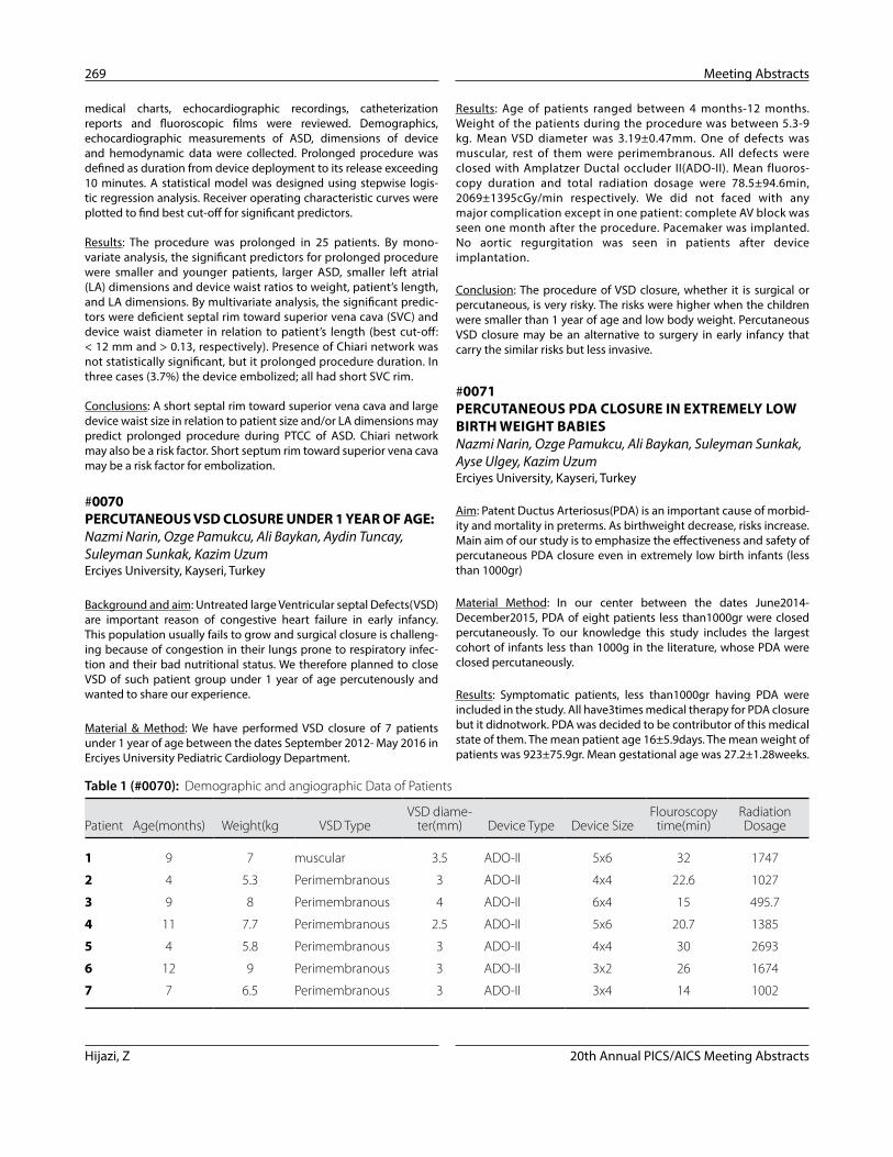
Hijazi, Z 20th Annual PICS/AICS Meeting Abstracts
269 Meeting Abstracts
Results: Age of patients ranged between 4 months-12 months. Weight of the patients during the procedure was between 5.3-9 kg. Mean VSD diameter was 3.19±0.47mm. One of defects was muscular, rest of them were perimembranous. All defects were closed with Amplatzer Ductal occluder II(ADO-II). Mean fluoros-copy duration and total radiation dosage were 78.5±94.6min, 2069±1395cGy/min respectively. We did not faced with any major complication except in one patient: complete AV block was seen one month after the procedure. Pacemaker was implanted. No aortic regurgitation was seen in patients after device implantation.
Conclusion: The procedure of VSD closure, whether it is surgical or percutaneous, is very risky. The risks were higher when the children were smaller than 1 year of age and low body weight. Percutaneous VSD closure may be an alternative to surgery in early infancy that carry the similar risks but less invasive.
#0071 PERCUTANEOUS PDA CLOSURE IN EXTREMELY LOW BIRTH WEIGHT BABIESNazmi Narin, Ozge Pamukcu, Ali Baykan, Suleyman Sunkak, Ayse Ulgey, Kazim UzumErciyes University, Kayseri, Turkey
Aim: Patent Ductus Arteriosus(PDA) is an important cause of morbid-ity and mortality in preterms. As birthweight decrease, risks increase. Main aim of our study is to emphasize the effectiveness and safety of percutaneous PDA closure even in extremely low birth infants (less than 1000gr)
Material Method: In our center between the dates June2014-December2015, PDA of eight patients less than1000gr were closed percutaneously. To our knowledge this study includes the largest cohort of infants less than 1000g in the literature, whose PDA were closed percutaneously.
Results: Symptomatic patients, less than1000gr having PDA were included in the study. All have3times medical therapy for PDA closure but it didnotwork. PDA was decided to be contributor of this medical state of them. The mean patient age 16±5.9days. The mean weight of patients was 923±75.9gr. Mean gestational age was 27.2±1.28weeks.
medical charts, echocardiographic recordings, catheterization reports and fluoroscopic films were reviewed. Demographics, echocardiographic measurements of ASD, dimensions of device and hemodynamic data were collected. Prolonged procedure was defined as duration from device deployment to its release exceeding 10 minutes. A statistical model was designed using stepwise logis-tic regression analysis. Receiver operating characteristic curves were plotted to find best cut-off for significant predictors.
Results: The procedure was prolonged in 25 patients. By mono-variate analysis, the significant predictors for prolonged procedure were smaller and younger patients, larger ASD, smaller left atrial (LA) dimensions and device waist ratios to weight, patient’s length, and LA dimensions. By multivariate analysis, the significant predic-tors were deficient septal rim toward superior vena cava (SVC) and device waist diameter in relation to patient’s length (best cut-off: < 12 mm and > 0.13, respectively). Presence of Chiari network was not statistically significant, but it prolonged procedure duration. In three cases (3.7%) the device embolized; all had short SVC rim.
Conclusions: A short septal rim toward superior vena cava and large device waist size in relation to patient size and/or LA dimensions may predict prolonged procedure during PTCC of ASD. Chiari network may also be a risk factor. Short septum rim toward superior vena cava may be a risk factor for embolization.
#0070 PERCUTANEOUS VSD CLOSURE UNDER 1 YEAR OF AGE:Nazmi Narin, Ozge Pamukcu, Ali Baykan, Aydin Tuncay, Suleyman Sunkak, Kazim UzumErciyes University, Kayseri, Turkey
Background and aim: Untreated large Ventricular septal Defects(VSD) are important reason of congestive heart failure in early infancy. This population usually fails to grow and surgical closure is challeng-ing because of congestion in their lungs prone to respiratory infec-tion and their bad nutritional status. We therefore planned to close VSD of such patient group under 1 year of age percutenously and wanted to share our experience.
Material & Method: We have performed VSD closure of 7 patients under 1 year of age between the dates September 2012- May 2016 in Erciyes University Pediatric Cardiology Department.
Table 1 (#0070): Demographic and angiographic Data of Patients
Patient Age(months) Weight(kg VSD TypeVSD diame-
ter(mm) Device Type Device SizeFlouroscopy
time(min)Radiation Dosage
1 9 7 muscular 3.5 ADO-II 5x6 32 1747
2 4 5.3 Perimembranous 3 ADO-II 4x4 22.6 1027
3 9 8 Perimembranous 4 ADO-II 6x4 15 495.7
4 11 7.7 Perimembranous 2.5 ADO-II 5x6 20.7 1385
5 4 5.8 Perimembranous 3 ADO-II 4x4 30 2693
6 12 9 Perimembranous 3 ADO-II 3x2 26 1674
7 7 6.5 Perimembranous 3 ADO-II 3x4 14 1002
Journal of Structural Heart Disease, December 2016 Volume 2, Issue 6:241-306
Meeting Abstracts 270
#0073 POST-IMPLANTATION MODIFICATION OF ENDOVASCULAR STENTS IN THE TREATMENT OF COMPLEX CONGENITAL AND ACQUIRED VASCULAR STENOSESBryan Goldstein, Wendy Whiteside, Russel Hirsch, Konstantin Averin, Gruschen VeldtmanCincinnati Children’s Hospital Medical Center, Cincinnati, OH, USA
Background: Optimal treatment of complex congenital and acquired vascular stenoses may benefit from post-implantation modification of standard endovascular stents. Utilization of open-cell design stents allows for side-hole creation with dilation of stent struts to minimize impact on adjacent vessels and/or treat adjacent stenotic lesions. We sought to describe our experience with this technique.
Methods: Single-center retrospective review at a large congenital heart center from 2011 – 2016. Cases with endovascular stent place-ment followed by side-hole creation with or without additional stent implantation, or with side-hole modification of an existing implant, were included. Demographic and procedural details were collected.
Results: Twenty catheterization procedures in 19 patients met inclu-sion criteria. Cases were performed in 55% female patients with a median age of 12.8 (IQR 4.2, 25.4) years and a mean weight of 45.1 ± 33.3 kg. Indications for stent angioplasty included: complex RVOT and branch pulmonary artery (PA) stenosis (45%), lobar PA stenosis (30%), aortic coarctation (15%), atrio-pulmonary Fontan connection steno-sis (10%), and systemic venous stenosis (10%). During 18 procedures a new stent was placed, while an existing implant was modified in 2 procedures. Three types of open-cell stents were modified following implantation: ev3 IntraStent® LD Mega (45%), ev3 IntraStent® LD Max (35%) and Cook Formula 418 (30%). A lobar (45%) or branch (30%) PA were the most commonly jailed vessels, followed by left subcla-vian artery (15%), right internal jugular vein (5%) and hepatic vein (5%). Indications for side-hole dilation included: relief of side-branch stenosis caused by primary stent implant (25%), avoidance of stent material (struts) crossing an important side-branch (50%) and relief of pre-existing side-branch vascular stenosis (25%). Side strut angio-plasty was performed to a variety of diameters (4 mm – 24 mm) using high pressure or ultra-high pressure angioplasty balloons. In 4 cases, a second stent was placed through the newly generated side-hole. Median procedure time was 211.5 (123.5, 272.5) min and mean fluoroscopy time was 56.2 ± 23.8 min. There were no local vascular injuries related to post-implant stent modification but 1 patient did have a self-limited access site complication.
Conclusion: Post-implant modification of endovascular stents via side strut dilation is safe, feasible and may improve outcomes in the defin-itive treatment of complex vascular lesions.
#0074 PERCUTANEOUS CLOSURE OF POSTMYOCARDIAL INFARCTION VENTRICULAR SEPTAL DEFECTSAdrian Sanchez, Jose Antonio Garcia, Carlos Zabal, Aldo Campos, Guillermo Aristizabal, Carlos Guerrero, Hector Gonzalez, Juan Pablo Sandoval
Mean PDA diameter was 2.48±0.5mm. Mean Qp/Qs was 1.7±0.2. Morphology of PDA: 5of them were conical, 3of them were tubular. In all patients ADOII-AS device were used for PDA closure (Table1). Steps of percutaneous PDA closure procedure was shown by Figure1. In all patients, we have done closure by venous route. We did not ever used arterial route in 4 patients. There were no major complications reported. Left pulmonary arterial stenosis was detected in 2 patients which were all resolved in 6 months duration.
Conclusion: Interventional catheterization procedures are more com-monly used, in the recent years. The advantages of percutaneous PDA closure include a high success rate, shorter length of hospital stay, reduced blood loss, low morbidity rate, and no traumatic scars. Since the length of hospital stay decreases with catheterization, it is much more cost-effective than surgery. We want to emphasize that in experienced centers percutaneous closure of PDA can be an alternative to surgery even in the extremely low birth weight babies.
#0072 INTENTIONAL LACERATION OF THE ANTERIOR MITRAL LEAFLET TO PREVENT LEFT VENTRICULAR OUTFLOW TRACT OBSTRUCTION (LAMPOON) DURING TRANSCATHETER MITRAL VALVE IMPLANTATION: PRE-CLINICAL FINDINGSJaffar Khan1, Toby Rogers1, Adam Greenbaum2, Vasilis Babaliaros3, Marcus Chen1, Robert Lederman1
1NHLBI, Bethesda, USA2Henry Ford Hospital, Detroit, USA3Emory, Atlanta, USA
Background: Left ventricular outflow tract (LVOT) obstruction is a life-threatening complication of transcatheter mitral valve implan-tation (TMVR), caused by septal displacement of the anterior mitral leaflet (AML). We propose a novel transcatheter resection of the AML.
Methods: LAMPOON was developed in vivo in swine, guided by biplane X-ray fluoroscopy and intracardiac echocardiography. Two transfemoral coronary guiding catheters were advanced retrograde through the aortic valve, either side of the AML. The ‘LVOT catheter’ directed an electrified guidewire across the center and base of the AML towards a snare directed by the ‘left atrial catheter’. The snared externalized guidewire loop was electrified, lacerating the AML along the centerline from base to tip.
Results: The AML was successfully lacerated in all (n=8) swine. Benchtop analysis demonstrated an increase in neo-LVOT with some residual obstruction (65% ± 10% vs. 31 ± 18% patency, p<0.01). Mean blood pressure fell (54 ± 6 to 30 ± 4mm Hg, p<0.01) due to acute mitral regurgitation from LAMPOON, but remained steady until planned euthanasia. There was no collateral tissue injury on necropsy.
Conclusions: Using simple catheter electrosurgery techniques, we mimicked surgical chord-sparing AML resection prior to TMVR. We recommend pre-positioning the transcatheter heart valve to minimize time between laceration and TMVR, though laceration may be better tolerated in patients with chronic mitral valve disease. Applied cautiously, this technique may enable TMVR in high risk surgical patients with prohibitive risk of LVOT obstruction.
Hijazi, Z 20th Annual PICS/AICS Meeting Abstracts
271 Meeting Abstracts
and valve function were investigated in 172 consecutive pts referred for PVI (median weight 56.7kg [12.3-161.3]; median age 18.6 years [4.7-65.5]) from 2007 to 2016. Diagnoses included tetralogy of Fallot (126), PA/PS (IVS) (21), and other (25). Indication for PVI was PI in 107, PS in 31 and PS/PI in 34. The number of PVI procedures increased from 9 to 27 per year with the availability of transcatheter pulmo-nary valve implantation (TcPVI). Prior to the availability of TcPVI, 34 pts were referred for surgery. Of the next 138 pts, 101 were referred for TcPVI and 37 went directly to surgery. TcPVI was attempted and successful in 85/101. The remaining 16 were referred for surgical pul-monary valve implantation (sPVI) without any attempt to implant a percutaneous valve due to RVOT size (11), coronary or aortic com-pression (4) and conduit tear pre-PARCS (1). 8/11 pts referred for surgery due to RVOT size would have been candidates for the 29mm Sapien valve if it had been available. TcPVI was successful in 27/27 pts with a bioprosthetic pulmonary valve (BPV), 24/27 with homografts and 34/47 with transannular patch repair (TAP). A Melody valve was implanted in all pts with a BPV or homograft and in 22/34 TAP pts. The other 12 pts received a Sapien valve. In total, 53 pts were referred for surgery in the TcPVI era. The RVOT was too large for TcPVI (44), too small (4), coronary or aortic compression (4) and conduit tear (1); 2 declined surgery. Comparing TcPVI to sPVI in the same time frame, freedom from > mild PI is 84/85 (TcPVI) and 36/51(sPVI) p<.001; 4/51 sPVI underwent subsequent Melody valve implantation for PI. Obstruction developed in 1 pt in each group p=ns. A second Melody valve was implanted in the 1 TcPVI pt with obstruction. SBE occurred in 1/85 (TcPVI) vs 2/51 (sPVI).
Since the introduction of TcPVI, referrals for PVI have tripled and 63% have had a percutaneous approach. Pts treated with TcPVI have had better long-term valve function in comparison to sPVI over the same time period. Given the superiority of TcPVI and the ability to avoid repeat open heart surgery, earlier intervention to reduce the incidence of RV dysfunction and development of symptoms may be warranted.
#0076 THROMBOPROPHYLAXIS STRATEGIES FOR CHILDREN WITH SINGLE VENTRICLE CIRCULATIONS (SUPERIOR OR TOTAL CAVO-PULMONARY CONNECTIONS) AFTER STENT IMPLANTATIONYinn Khurn Ooi, Michael Kelleman, Holly Bauser-Heaton, Dennis Kim, Robert Vincent, Christopher PetitEmory University, Atlanta, GA, USA
Background: The most effective thromboprophylaxis strategy after stent implantation in the superior or total cavopulmonary connec-tion (SCPC or TCPC) is unclear.
Methods: We reviewed our cardiac database over a 13-year period ending in 2015. Patients were divided into those who received aspi-rin (ASA) alone versus advanced anti-coagulation (AA), including warfarin, enoxaparin, heparin, or clopidogrel. Primary endpoint was presence of thrombus on advanced imaging, such as cardiac cathe-terization, CT-angiogram or MRI. Other endpoints included presence of thrombus on echocardiogram and bleeding complications.
National Institute of Cardiology ¨Ignacio Chavez¨, Mexico City, Mexico
Background: Interventricular septum rupture following acute myo-cardial infarction (MI) is a rare (≈0.2%) but serious and potentially life threatening complication. Current AHA/ACC guidelines recom-mend urgent surgical repair even in the absence of hemodynamic compromise. However, surgical mortality rate remains high (reported between 20 to 80%) particularly in patients with cardiogenic shock. Percutaneous closure is an attractive, less invasive alternative yet this approach remains to be performed only in few selected centers around the world.
Methods: Single centre, retrospective study of 17 consecutive patients diagnosed with postmyocardial infarction ventricular septal defect (pMIVSD) that underwent percutaneous device closure between July 2003 (index procedure) and July 2016. Patients were further divided into those with (n=8) (Group A) and without cardiogenic shock (n=9) (Group B) at the time of intervention. The latter was defined as those requiring inotropic support or intra-aortic balloon counterpulsation to sustain cardiac output.
Results: Nine (47%) patients were female. Median age was 65 years (range 51-75 y). Median time in days between MI and pMIVSD clo-sure was 8 days (range 1-25) for group A and 11 days (range 3-91) for Group B. Systolic pulmonary artery pressure/systemic pressure ratio was 0.57 (range 0.38-0.7) and Qp:Qs 1.83:1 (1.72-3.6) for patients in Group A and 0.6(range 0.33-0.89) and Qp:Qs 3:1 (1.58-4.39) for patients in group B (NS). A variety of devices were used to occlude the defect: muscular VSD Amplatzer n=6(35%), postinfarct VSD Amplatzer n=5(30%), ASO n=4(24%), other (AVP II and PFO) n=2(11%). Device embolization occurred in only one patient (device was snared and retrieved) and a second occluder used. In group A, significant residual shunt was found in two cases; hence they were brought back to the lab to deploy a second occluder. No reintervention was required for patients in group B, mild to moderate residual shunt was present in 6 out of 9 (66%) patients. Early (<30 day) mortality was 0% in Group B and 62% (5 out of 8 patients) in Group A.
Conclusion: Transcatheter device closure of pMIVSD can be consid-ered a reasonably safe and effective procedure in selected patients. Our series is consistent with previous experience reported in other series regarding high mortality in patients with cardiogenic shock despite intervention.
#0075 THE IMPACT OF TRANSCATHETER PULMONARY VALVE IMPLANTATION ON THE MANAGEMENT OF POSTOPERATIVE RIGHT VENTRICULAR OUTFLOW TRACT DYSFUNCTIONCatherine Tomasulo, Jeremy Asnes, Robert Elder, John Fahey, Gary Kopf, William HellenbrandYale School of Medicine, New Haven, CT, USA
Pulmonary valve implantation (PVI) for pts with CHD has undergone revolutionary changes in recent years with the advent of transcath-eter therapies. Surgical and transcatheter procedure characteristics
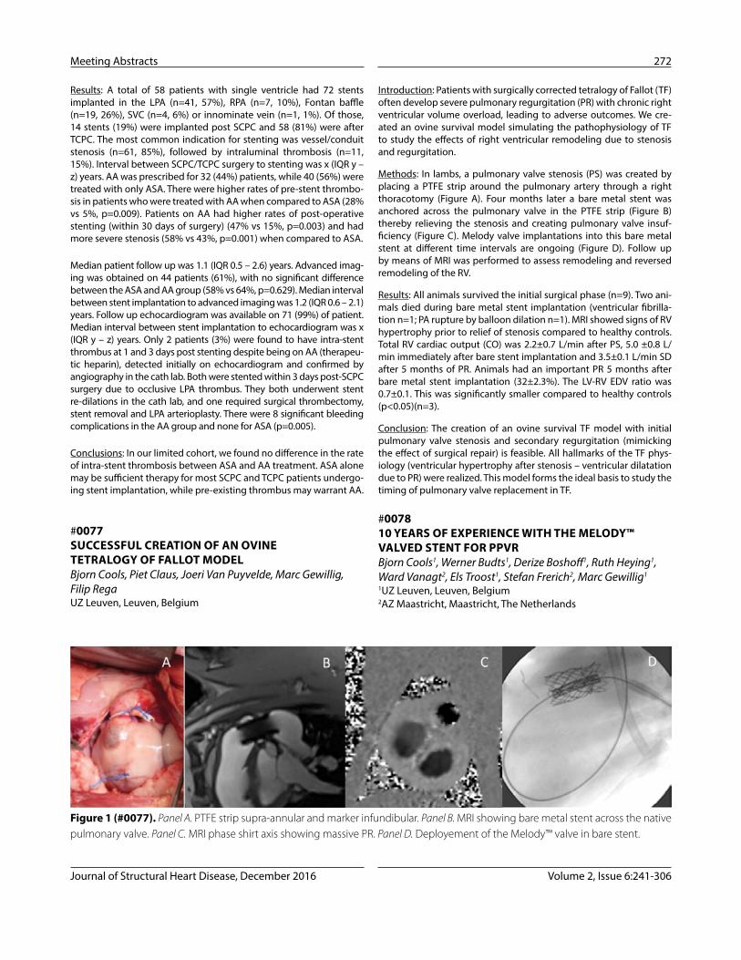
Journal of Structural Heart Disease, December 2016 Volume 2, Issue 6:241-306
Meeting Abstracts 272
Introduction: Patients with surgically corrected tetralogy of Fallot (TF) often develop severe pulmonary regurgitation (PR) with chronic right ventricular volume overload, leading to adverse outcomes. We cre-ated an ovine survival model simulating the pathophysiology of TF to study the effects of right ventricular remodeling due to stenosis and regurgitation.
Methods: In lambs, a pulmonary valve stenosis (PS) was created by placing a PTFE strip around the pulmonary artery through a right thoracotomy (Figure A). Four months later a bare metal stent was anchored across the pulmonary valve in the PTFE strip (Figure B) thereby relieving the stenosis and creating pulmonary valve insuf-ficiency (Figure C). Melody valve implantations into this bare metal stent at different time intervals are ongoing (Figure D). Follow up by means of MRI was performed to assess remodeling and reversed remodeling of the RV.
Results: All animals survived the initial surgical phase (n=9). Two ani-mals died during bare metal stent implantation (ventricular fibrilla-tion n=1; PA rupture by balloon dilation n=1). MRI showed signs of RV hypertrophy prior to relief of stenosis compared to healthy controls. Total RV cardiac output (CO) was 2.2±0.7 L/min after PS, 5.0 ±0.8 L/min immediately after bare stent implantation and 3.5±0.1 L/min SD after 5 months of PR. Animals had an important PR 5 months after bare metal stent implantation (32±2.3%). The LV-RV EDV ratio was 0.7±0.1. This was significantly smaller compared to healthy controls (p<0.05)(n=3).
Conclusion: The creation of an ovine survival TF model with initial pulmonary valve stenosis and secondary regurgitation (mimicking the effect of surgical repair) is feasible. All hallmarks of the TF phys-iology (ventricular hypertrophy after stenosis – ventricular dilatation due to PR) were realized. This model forms the ideal basis to study the timing of pulmonary valve replacement in TF.
#0078 10 YEARS OF EXPERIENCE WITH THE MELODY™ VALVED STENT FOR PPVRBjorn Cools1, Werner Budts1, Derize Boshoff1, Ruth Heying1, Ward Vanagt2, Els Troost1, Stefan Frerich2, Marc Gewillig1
1UZ Leuven, Leuven, Belgium2AZ Maastricht, Maastricht, The Netherlands
Results: A total of 58 patients with single ventricle had 72 stents implanted in the LPA (n=41, 57%), RPA (n=7, 10%), Fontan baffle (n=19, 26%), SVC (n=4, 6%) or innominate vein (n=1, 1%). Of those, 14 stents (19%) were implanted post SCPC and 58 (81%) were after TCPC. The most common indication for stenting was vessel/conduit stenosis (n=61, 85%), followed by intraluminal thrombosis (n=11, 15%). Interval between SCPC/TCPC surgery to stenting was x (IQR y – z) years. AA was prescribed for 32 (44%) patients, while 40 (56%) were treated with only ASA. There were higher rates of pre-stent thrombo-sis in patients who were treated with AA when compared to ASA (28% vs 5%, p=0.009). Patients on AA had higher rates of post-operative stenting (within 30 days of surgery) (47% vs 15%, p=0.003) and had more severe stenosis (58% vs 43%, p=0.001) when compared to ASA.
Median patient follow up was 1.1 (IQR 0.5 – 2.6) years. Advanced imag-ing was obtained on 44 patients (61%), with no significant difference between the ASA and AA group (58% vs 64%, p=0.629). Median interval between stent implantation to advanced imaging was 1.2 (IQR 0.6 – 2.1) years. Follow up echocardiogram was available on 71 (99%) of patient. Median interval between stent implantation to echocardiogram was x (IQR y – z) years. Only 2 patients (3%) were found to have intra-stent thrombus at 1 and 3 days post stenting despite being on AA (therapeu-tic heparin), detected initially on echocardiogram and confirmed by angiography in the cath lab. Both were stented within 3 days post-SCPC surgery due to occlusive LPA thrombus. They both underwent stent re-dilations in the cath lab, and one required surgical thrombectomy, stent removal and LPA arterioplasty. There were 8 significant bleeding complications in the AA group and none for ASA (p=0.005).
Conclusions: In our limited cohort, we found no difference in the rate of intra-stent thrombosis between ASA and AA treatment. ASA alone may be sufficient therapy for most SCPC and TCPC patients undergo-ing stent implantation, while pre-existing thrombus may warrant AA.
#0077 SUCCESSFUL CREATION OF AN OVINE TETRALOGY OF FALLOT MODELBjorn Cools, Piet Claus, Joeri Van Puyvelde, Marc Gewillig, Filip RegaUZ Leuven, Leuven, Belgium
A/ PTFE strip supra-annular and marker infundibular B/MRI showing bare metal stent accross the na�vepulmonary valve C/MRI phase shirt axis showing massive PR C/ deployement of Melody TM valve in bare stentFigure 1 (#0077). Panel A. PTFE strip supra-annular and marker infundibular. Panel B. MRI showing bare metal stent across the native pulmonary valve. Panel C. MRI phase shirt axis showing massive PR. Panel D. Deployement of the Melody™ valve in bare stent.
Hijazi, Z 20th Annual PICS/AICS Meeting Abstracts
273 Meeting Abstracts
This is the case of a 43 yr-old male with a truncus arteriosus who underwent three previous cardiac surgeries : - 1972 : pulmonary banding; - 1979 : prosthetic valved tube between right ventricle and pulmonary artery + aortic valve prosthesis (Bjork)+VSD closure; - 1991: homograft EHB 22 mm in pulmonic position, homograft EHB 22 mm in aortic position replacing the Bjork prosthesis. He was admit-ted in April 2016 in the intensive care unit for a severe heart failure. Echocardiograpy showed a severe aortic regurgitation, a pulmonic valve stenosis, enlarged and poor function of right and left ventricles. Diagnostic cardiac catheterization showed a heavy calcified and ste-notic (minimum diameter : 10 mm; RV-PA gradient : 44 mmHg) pul-monic homograft ; the aortic homograft was severely calcified, had a free regurgitation(grade 4).Angio showed a pseudoaneurysm of the left ventricular outflow tract, an abnormal origin of the coronary arteries and an occlusion of the right femoral artery. Because of the hostile chest and the very high Euroscore, the heart team decided to attempt a percutaneous valvular treatment on this grown-up con-genital heart. The plan was to begin with the aortic valve which was the main problem of the patient and to do pulmonic replacement in a stage procedure.TAVI was challenging in this case because of the severe regurgitation of the degenerated homograft without any pre-cise annular plane, with heavy calcifications and a tortuous shape ; in addition, the pseudo-aneurysm of the LVOT was a risk factor for para-valvular leak at this level and the abnormal origin of coronary arteries was a potential danger for occlusion in case of suboptimal valve posi-tionning. Finally, the right femoral artery occlusion was also an issue. Despite all these limitations, an Evolut-R 26 mm was deployed by the left femoral artery without any residual leak, improving immediately the aortic regurgitation and the hemodynamics of the patient. This second generation device is a recapturable and repositionable device allowing an evaluation of the result before final release, adjusting if needed to achieve a final accurate positioning. After a short period of recovery, the pulmonic homograft will be replaced by a Melody valve in a few weeks. In conclusion,this case illustrates how struc-turalinterventional cardiology can offer an effective treatment in grown-up congenital heart with contra-indication for repeated com-plex surgeries.
#0080 THE DETERMINATION OF HYPERTENSION PERSISTENCE IN CHILDREN WHOSE AORTIC COARCTATION WERE TREATED WITH CP STENTAli Baykan, Ayse Demiraldi, Nazmi Narin, Kazim Uzum, Özge PamukcuErciyes University Medical Faculty Dept. of Pediatric Cardiology, Kayseri, Turkey
Objective: Recent studies reveal that hypertension and cardiovascular diseases are more prevalent in patients with Aortic coarctation (AC), even it is treated. Our aim is to address presence of hypertension and risk for cardiovascular diseases in patients with stent applied AC by using data from ambulatory blood pressure monitorization (ABPM)/arteriography, echocardiography and radiological and biochemical evaluations.
Material and method: Twenty stented pediatric patient and 20 age- and sex-matched controls were enrolled. Physical examination findings and ECG data were recorded. Echocardiography performed
Introduction : Since 10 years the Melody™ valved stent is used for percutaneous pulmonary valve replacement (PPVR) with over 10.000 implants worldwide. It has been proven to be a safe and effective intervention. The main risk factors of the valve compared to surgical alternatives are stent fractures with subsequent valve dysfunction and endocarditis. Most biological valves have limited long term pre-served function requiring redo interventions.
Aim : evaluation of the valve function and its risks from a prospective ongoing registry
Patients and methods : all data are collected from the Belgian registry which started in 2006. Follow-up data are prospectively and ongoing collected. 97% are pre-stented in this cohort. The analyzed data : leaf-let function by means of echocardiography, re-interventions, mortal-ity, endocarditis (Duke’s criteria) and stent fractures by means of X-ray. Data presented is single center experience.
Results : 159 valves have been implanted 2006-2016. The indication was stenosis 44.0%, regurgitation 33.3% and mixed 22.6 %; male/female ratio was 2:1, the mean age at implant was 19.6 y (3.5 – 81.6). Original conduit diameter was median 22 mm (10-26 mm).
1. Overall survival was 98 % at 10 years, there were no procedural or valve related deaths.
2. Endocarditis occurred in 9.4% (15/159) with a freedom from endo-carditis of 78% at 10 y. The maximal annual incidence of endocarditis was 3.3% in 2015. Mean time of occurrence of endocarditis was 2.4 y (0.7 – 8.9) after implant. Mean age at endocarditis was 18.2 y (8.1 – 45.6), male 87%.
3. Chest X-ray was performed in 87 % of the patients showing a stent fracture in 8 % median 2.9 y (0.4 – 6.9) after implant. None of the fractures led to hemodynamic problems and no re-intervention was necessary.
4. Re-intervention overall 11 % : explantation of valve in 4.4% (endo-carditis n= 6, residual subvalvular stenosis n=1); balloon dilation due to somatic growth in 5 % and 1.9 % received second PPVR (post endocarditis).
5. Valve function: a/ indication PS : mean PS 65 mmHg (SD 17) at implant drops to 23 mmHg (SD 11) post implant and is 37,5 mmHg (SD 17) 10 y later. B/ Indication PR : median 4/4 at implant drops to 0/4 post and is 2/4 at 10 y follow-up.
Discussion : Ten year follow-up of the Melody™ valved stent shows good long term preservation of the valve function with current no replacement for intrinsic valve dysfunction. No hemodynamic import-ant stent fractures occur since the pre-stenting era. Acceptable risk of endocarditis remains in male young adult patients.
#0079 PERCUTANEOUS DOUBLE VALVE IMPLANTATION IN A GROWN-UP CONGENITAL HEART WITH DEGENERATED HOMOGRAFT IN BOTH AORTIC AND PULMONIC POSITION.Joelle Kefer, Sophie Pierard, Jean Renkin, Thierry SluysmansCliniques universitaires Saint-Luc, Brussels, Belgium
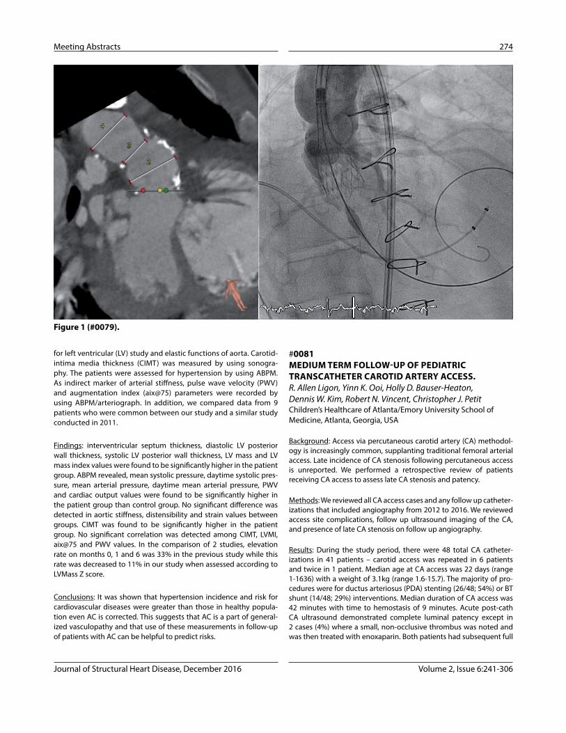
Journal of Structural Heart Disease, December 2016 Volume 2, Issue 6:241-306
Meeting Abstracts 274
#0081 MEDIUM TERM FOLLOW-UP OF PEDIATRIC TRANSCATHETER CAROTID ARTERY ACCESS.R. Allen Ligon, Yinn K. Ooi, Holly D. Bauser-Heaton, Dennis W. Kim, Robert N. Vincent, Christopher J. PetitChildren’s Healthcare of Atlanta/Emory University School of Medicine, Atlanta, Georgia, USA
Background: Access via percutaneous carotid artery (CA) methodol-ogy is increasingly common, supplanting traditional femoral arterial access. Late incidence of CA stenosis following percutaneous access is unreported. We performed a retrospective review of patients receiving CA access to assess late CA stenosis and patency.
Methods: We reviewed all CA access cases and any follow up catheter-izations that included angiography from 2012 to 2016. We reviewed access site complications, follow up ultrasound imaging of the CA, and presence of late CA stenosis on follow up angiography.
Results: During the study period, there were 48 total CA catheter-izations in 41 patients – carotid access was repeated in 6 patients and twice in 1 patient. Median age at CA access was 22 days (range 1-1636) with a weight of 3.1kg (range 1.6-15.7). The majority of pro-cedures were for ductus arteriosus (PDA) stenting (26/48; 54%) or BT shunt (14/48; 29%) interventions. Median duration of CA access was 42 minutes with time to hemostasis of 9 minutes. Acute post-cath CA ultrasound demonstrated complete luminal patency except in 2 cases (4%) where a small, non-occlusive thrombus was noted and was then treated with enoxaparin. Both patients had subsequent full
for left ventricular (LV) study and elastic functions of aorta. Carotid-intima media thickness (CIMT) was measured by using sonogra-phy. The patients were assessed for hypertension by using ABPM. As indirect marker of arterial stiffness, pulse wave velocity (PWV) and augmentation index (aix@75) parameters were recorded by using ABPM/arteriograph. In addition, we compared data from 9 patients who were common between our study and a similar study conducted in 2011.
Findings: interventricular septum thickness, diastolic LV posterior wall thickness, systolic LV posterior wall thickness, LV mass and LV mass index values were found to be significantly higher in the patient group. ABPM revealed, mean systolic pressure, daytime systolic pres-sure, mean arterial pressure, daytime mean arterial pressure, PWV and cardiac output values were found to be significantly higher in the patient group than control group. No significant difference was detected in aortic stiffness, distensibility and strain values between groups. CIMT was found to be significantly higher in the patient group. No significant correlation was detected among CIMT, LVMI, aix@75 and PWV values. In the comparison of 2 studies, elevation rate on months 0, 1 and 6 was 33% in the previous study while this rate was decreased to 11% in our study when assessed according to LVMass Z score.
Conclusions: It was shown that hypertension incidence and risk for cardiovascular diseases were greater than those in healthy popula-tion even AC is corrected. This suggests that AC is a part of general-ized vasculopathy and that use of these measurements in follow-up of patients with AC can be helpful to predict risks.
Figure 1 (#0079).
Hijazi, Z 20th Annual PICS/AICS Meeting Abstracts
275 Meeting Abstracts
catheterization would have the advantage of being able to perform interventions, specifically embolization of APCs.
Key words: aorto-pulmonary • collateral vessels • coil embolization • cardiac catheterization
#0084 EFFICIENCY OF TRANSCATHETER PATENT FORAMEN OVALE (PFO) CLOSURE WITH HYPERION PFO OCCLUDER*.Malgorzata Szkutnik1, Jacek Bialkowski1, Roland Fiszer1, Blandyna Karwot1, Sebastian Smerdzinski1, Maciej Wawrzynczyk2
1Silesian Center for Heart Diseases, Congenital Heart Defects and Pediatric Cardiology Dept., Zabrze, Silesia, Poland2Department of Neurology, Public Clinical Hospital No. 1, Medical University of Silesia, Zabrze, Silesia, Poland
Introduction: Patent Foramen Ovale (PFO) may result in paradoxical embolism event (EE). Transcatheter closure of PFO still remains an alternative and interesting treatment for selected patients. Experience with application of Hyperion PFO Occluders (similar to Amplatzer PFO Occluders) in such circumstances has been not evaluated yet.
Materials and Methods: 67 patients (pts) with cryptogenic EE in whom in years 2014-2016 PFO was closed percutaneously with Hyperion PFO occluder in our institution were retrospectively analyzed. There were 36 woman with mean age 41 (21-62) and 31 man with mean age 41 (19-68) years. The indications for the procedure were estab-lished by neurologist and included Transient Ischemic Attack (TIA) in 16 pts (among them 2 pts were divers), ischemic stroke in 49 pts and migraine with aura in 3 pts. Transcranial Doppler (TCD) was per-formed in all pts (with MES ≥2nd degree in all pts). Mean PFO tunnel length was 8 (4-16) mm. Follow up was performed 3, 6 and 12 months after the procedure and thereafter yearly. Control TCD examination was performed 6 months after the procedure and in case of residual shunt presence – 3 months afterwards again.
Results: The procedure was successfully completed in all patients and no procedure-related complications were observed during hospital-ization. Following devices were used (accordingly to the anatomy of PFO) – PFO Hyperion Occluder 18 mm in 6 pts, 24 mm in 49 pts, 30 mm in 8 pts and 34 mm in 4 pts. Fluoroscopy time was 2,4 (0,9-22) min. During follow-up no new neurological events were observed in any pt. Control TCD results were available in 54 pts (4 pts were lost from follow-up): 48 pts had complete PFO closure and 6 pts had a residual shunt. In 1 pt paroxysmal atrial fibrillation was observed.
Conclusions: Transcatheter PFO closure with Hyperion devices is safe and effective procedure, however, long-term follow-up and random-ized study with other devices are necessary.
*Device produced as Hyperion PFOO, Comed BV, Netherlands / SHSMA, Shanghai, China
resolution on follow up imaging within 72 hours. Late CA imaging was available in 33 patients (80%) at a median of 105 days (range 7-1121) after index CA procedure with 9130 patient-days total of fol-low up. Among these 33 patients, there were 51 total cases of late CA follow-up imaging – all demonstrating a widely patent CA. There was no CA stenosis nor lumen irregularity noted in 33 cases (65%) with 18 cases (35%, 14 patients) demonstrating a median of 4.7% (range 2.3-12.5%) stenosis. No patient underwent intervention on the CA for vessel stenosis. No risk factors for mild CA stenosis were identified when evaluating vessel stenosis and sheath size at index procedure, interval from index procedure, weight at index procedure, or duration of CA access during index procedure.
Conclusion: Use of the CA for interventional catheterization is associated with excellent late patency with no appreciable CA stenosis noted late after the procedure. In patients with CA stenosis after cath-eterization, the stenosis is mild. This data supports the utilization of percutaneous carotid access during pediatric catheterization.
#0083 NOVEL APPROACH TO THE DIAGNOSIS AND TREATMENT OF HEMOPTYSIS IN CHILDRENSyed Javed Hasan Zaidi, Dhaval R. Patel, Alexander J. Javois, Javeed AkhterAdvocate Children’s Hospital, Oak Lawn, Illinois, USA
Rationale: Hemoptysis in children is an uncommon presenting symp-tom but can be life-threatening if massive. Cardiac catheterization and coil embolization of aorto-pulmonary collateral vessels (APCs) is uncommon in patients with hemoptysis and without congenital heart disease. We present a series of 16 children with hemoptysis, all of whom underwent cardiac catheterization to look for and intervene upon aorto-pulmonary vessels if found.
Objective: To determine the presence and size of APCs in children with hemoptysis and assess resolution of symptoms after coil embo-lization of these vessels.
Methods: A detailed chart abstraction and review of bronchoscopy and cardiac catheterization images and reports was performed in this retrospective cohort study of children presenting with hemoptysis in a tertiary center from January 1995 to January 2015.
Main Results: A total of 16 patients were identified who presented with hemoptysis and underwent cardiac catheterization after bron-choscopy at our institution. The mean age was 12.6 years. Structural cardiovascular anomalies were present in 4 (25%). 13 (81%) had evi-dence of bleeding on bronchoscopy. Cardiac catheterization showed significant APCs (>2mm) in 9 (56%) all of which were coil embolized. 8 (89%) had complete resolution with no recurrences. 7 patients had no significant APCs. Of the 4 patients with structural cardiac anoma-lies, 2 had significant APCs that needed coil embolization.
Conclusions: Aorto-pulmonary collateral vessels should be high on the differential diagnosis for pulmonary hemorrhage in an infant and besides early consultation with pediatric pulmonology, a con-sultation with pediatric cardiology is also warranted. Following a bronchoscopy, other imaging modalities may be helpful but cardiac
Journal of Structural Heart Disease, December 2016 Volume 2, Issue 6:241-306
Meeting Abstracts 276
Wendy Whiteside, Konstatin Averin, Russel Hirsch, Karen Minsterman, Ross Schierling, Barbara Bear, Michael Harris, Bryan H. GoldsteinCincinnati Children’s Hospital Medical Center, Cincinnati, OH, USA
Background: ALARA (As Low As Reasonably Achievable) princi-ples dictate best practice using the lowest possible radiation dose. Radiation reduction has been a particular focus of the congenital cardiac catheterization laboratory due to increased radiation sus-ceptibility in children and frequent need for repeat procedures. We sought to assess the incremental impact of a radiation reduction ini-tiative and subsequent acquisition of a next generation angiographic imaging system on radiation dose.
Methods: Beginning in 2014, a radiation reduction initiative was devel-oped to emphasize radiation reduction within our program. This effort included equipment settings and practice changes to promote radi-ation lowering techniques. Subsequently, a capital upgrade to a next generation angiographic imaging system (Philips AlluraClarity) was installed in 1/2016. Single center radiation data collected prospectively for quality improvement purposes was reviewed retrospectively. The impact of these dose changes was assessed via longitudinal tracking of mean Air Kerma across the same 4 catheterization providers.
Results: From 1/2014 through 8/2016, 2083 catheterization proce-dures were performed at our institution. At baseline, mean Air Kerma per case was 229±476 mGy. Following initial modifications to default frame rates and ALARA-guided practice changes, we saw a 25% reduction in mean Air Kerma per case to 173±348 mGy. The instal-lation of a next generation imaging system combined with further changes to default imaging settings generated an additional 57% reduction in mean dose per case. After 7 months with next gener-ation equipment, mean Air Kerma per case was 49±80 mGy, a 79% reduction in dose from baseline. From 1/2014-12/2015, there were on average 5 high exposure cases (>2000 mGy) per 6-month period. Over the 7 months since equipment upgrade, there has been only one high exposure case despite similar average fluoroscopy time per case and an unchanged case-mix.
Conclusions: Changes in catheterization lab culture, practice habits, and equipment settings are effective in reducing radiation dose, but the availability of next generation imaging equipment allows for expo-nential dose decrease. These combined approaches result in a nearly 80% reduction in radiation dose for congenital cardiac patients.
#0087 FEASIBILITY AND VALIDITY OF PRINTING 3D HEART MODELS FROM ROTATIONAL ANGIOGRAPHYSaar Danon, Manoj Parimi, John Buelter, Vignan Thanugundla, Sri Condoor, Wilson KingSaint Louis University, Saint Louis, MO, USA
Background: Rotational angiography (RA) has proven to be an excel-lent adjunctive method for evaluating congenital disease (CHD) in the cardiac cath lab, permitting acquisition of 3D datasets with excel-lent spatial resolution. This technique has not been routinely imple-mented for 3D printing. We describe our case series of models printed from RA and validate our technique.
#0085 COST OF A STANDARDIZED APPROACH TO MANAGEMENT OF PULSE LOSS FOLLOWING PEDIATRIC CARDIAC CATHETERIZATION Wendy Whiteside, Jeffrey Anderson, Christina M. Metcalf, Betsy Newkirk, Christine Combs, Janene Allen, Karen Minsterman, Bryan H. GoldsteinCincinnati Children’s Hospital Medical Center, Cincinnati, OH, USA
Background: Pulse loss is a known complication of pediatric cardiac catheterization. A standardized approach to the diagnosis and man-agement of post-catheterization arterial thrombosis has been shown to result in thrombus resolution in the majority of patients. The cost burden of this practice has not been reported previously and may be a perceived barrier to utilization of a clinical practice guideline (CPG) for post-catheterization pulse loss.
Methods: Single center data collected prospectively for quality improvement efforts was reviewed retrospectively. Beginning in 5/2013, all patients with post-catheterization pulse loss, identified by clinical pulse exam 1-2 hours after sheath removal, were managed according to an institutional CPG. The CPG included initial evaluation by lower extremity Doppler ultrasound, prompt initiation of anti-co-agulation using low-molecular weight heparin (LMWH), and serial re-evaluation by clinical exam, Doppler ultrasound and anti-Xa levels until thrombus resolution or until 12 weeks of therapy had been com-pleted. Charges for protocol related items: CPG follow-up visits, diag-nostic imaging, medication and laboratory testing were collected.
Results: From 5/2013- 2/2016, post-catheterization pulse loss occurred in 64 cases (61 patients) with a median age of 105 days (IQR 67, 154) and weight of 5.2 kg (4.3, 6.2). Incidence of pulse loss in cases with arterial access was 4.5%. By 12 weeks, restoration of pulse occurred in 92.3% of patients. Median charge per patient for CPG-related care was $2,672 ($1,587, $4,087) with the largest contri-bution coming from lower extremity ultrasound. Patients received a mean of 3±2 ultrasounds for a median radiology charge of $2,344. Pharmacy charges were minimal, with median duration of LMWH therapy of 7 days (1,23) and median charge of $82 ($25, $371). For patients with restoration of pulse prior to hospital discharge (£1 day of LMWH), median charge was $1,583 ($801, $1837). One patient on protocol was noted incidentally to have a pseudoaneurysm at 1-week follow-up, for which therapeutic thrombin injection was performed by interventional radiology. There were no bleeding events or other complications. Social burdens of chronic LMWH administration, clini-cal and laboratory visits for monitoring, and the financial implications of lost work/school time were not evaluated.
Conclusions: Implementation of a CPG for management of post-cath-eterization pulse loss results in a high-rate of pulse restoration (92%) with only modest associated charges.
#0086 COMBINED IMPACT OF A RADIATION REDUCTION INITIATIVE AND NEXT GENERATION ANGIOGRAPHIC IMAGING SYSTEM ON RADIATION DOSE IN A CONGENITAL CARDIAC CATHETERIZATION LABORATORY
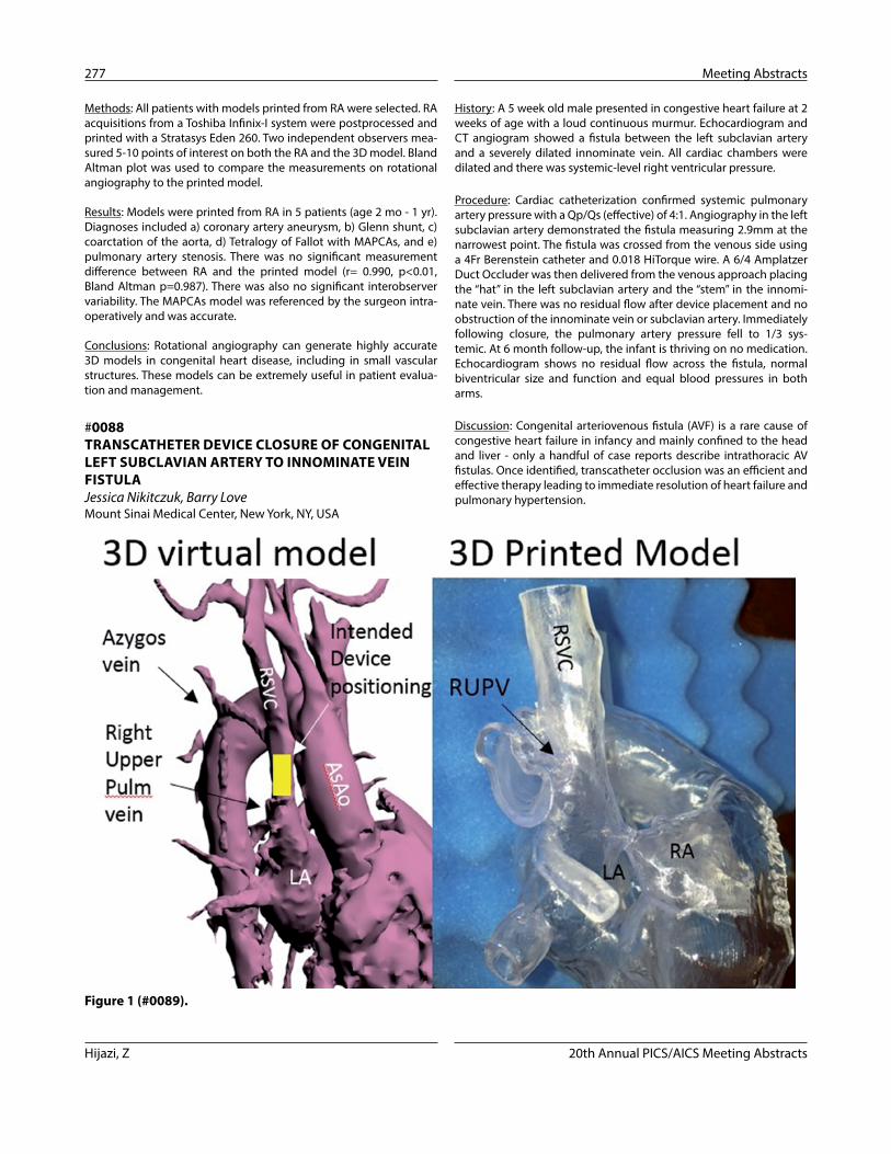
Hijazi, Z 20th Annual PICS/AICS Meeting Abstracts
277 Meeting Abstracts
History: A 5 week old male presented in congestive heart failure at 2 weeks of age with a loud continuous murmur. Echocardiogram and CT angiogram showed a fistula between the left subclavian artery and a severely dilated innominate vein. All cardiac chambers were dilated and there was systemic-level right ventricular pressure.
Procedure: Cardiac catheterization confirmed systemic pulmonary artery pressure with a Qp/Qs (effective) of 4:1. Angiography in the left subclavian artery demonstrated the fistula measuring 2.9mm at the narrowest point. The fistula was crossed from the venous side using a 4Fr Berenstein catheter and 0.018 HiTorque wire. A 6/4 Amplatzer Duct Occluder was then delivered from the venous approach placing the “hat” in the left subclavian artery and the “stem” in the innomi-nate vein. There was no residual flow after device placement and no obstruction of the innominate vein or subclavian artery. Immediately following closure, the pulmonary artery pressure fell to 1/3 sys-temic. At 6 month follow-up, the infant is thriving on no medication. Echocardiogram shows no residual flow across the fistula, normal biventricular size and function and equal blood pressures in both arms.
Discussion: Congenital arteriovenous fistula (AVF) is a rare cause of congestive heart failure in infancy and mainly confined to the head and liver - only a handful of case reports describe intrathoracic AV fistulas. Once identified, transcatheter occlusion was an efficient and effective therapy leading to immediate resolution of heart failure and pulmonary hypertension.
Methods: All patients with models printed from RA were selected. RA acquisitions from a Toshiba Infinix-I system were postprocessed and printed with a Stratasys Eden 260. Two independent observers mea-sured 5-10 points of interest on both the RA and the 3D model. Bland Altman plot was used to compare the measurements on rotational angiography to the printed model.
Results: Models were printed from RA in 5 patients (age 2 mo - 1 yr). Diagnoses included a) coronary artery aneurysm, b) Glenn shunt, c) coarctation of the aorta, d) Tetralogy of Fallot with MAPCAs, and e) pulmonary artery stenosis. There was no significant measurement difference between RA and the printed model (r= 0.990, p<0.01, Bland Altman p=0.987). There was also no significant interobserver variability. The MAPCAs model was referenced by the surgeon intra-operatively and was accurate.
Conclusions: Rotational angiography can generate highly accurate 3D models in congenital heart disease, including in small vascular structures. These models can be extremely useful in patient evalua-tion and management.
#0088 TRANSCATHETER DEVICE CLOSURE OF CONGENITAL LEFT SUBCLAVIAN ARTERY TO INNOMINATE VEIN FISTULAJessica Nikitczuk, Barry LoveMount Sinai Medical Center, New York, NY, USA
Figure 1 (#0089).
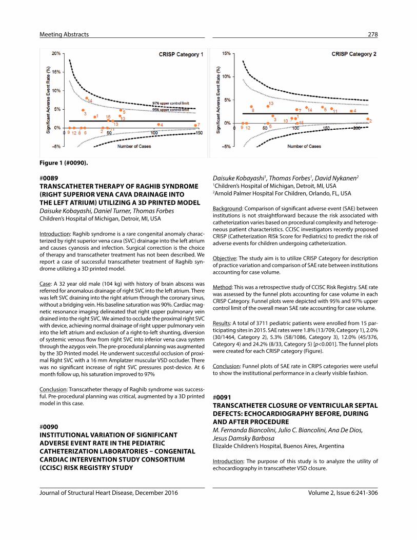
Journal of Structural Heart Disease, December 2016 Volume 2, Issue 6:241-306
Meeting Abstracts 278
Daisuke Kobayashi1, Thomas Forbes1, David Nykanen2
1Children’s Hospital of Michigan, Detroit, MI, USA2Arnold Palmer Hospital For Children, Orlando, FL, USA
Background: Comparison of significant adverse event (SAE) between institutions is not straightforward because the risk associated with catheterization varies based on procedural complexity and heteroge-neous patient characteristics. CCISC investigators recently proposed CRISP (Catheterization RISk Score for Pediatrics) to predict the risk of adverse events for children undergoing catheterization.
Objective: The study aim is to utilize CRISP Category for description of practice variation and comparison of SAE rate between institutions accounting for case volume.
Method: This was a retrospective study of CCISC Risk Registry. SAE rate was assessed by the funnel plots accounting for case volume in each CRISP Category. Funnel plots were depicted with 95% and 97% upper control limit of the overall mean SAE rate accounting for case volume.
Results: A total of 3711 pediatric patients were enrolled from 15 par-ticipating sites in 2015. SAE rates were 1.8% (13/709, Category 1), 2.0% (30/1464, Category 2), 5.3% (58/1086, Category 3), 12.0% (45/376, Category 4) and 24.2% (8/33, Category 5) [p<0.001]. The funnel plots were created for each CRISP category (Figure).
Conclusion: Funnel plots of SAE rate in CRIPS categories were useful to show the institutional performance in a clearly visible fashion.
#0091 TRANSCATHETER CLOSURE OF VENTRICULAR SEPTAL DEFECTS: ECHOCARDIOGRAPHY BEFORE, DURING AND AFTER PROCEDURE M. Fernanda Biancolini, Julio C. Biancolini, Ana De Dios, Jesus Damsky BarbosaElizalde Children’s Hospital, Buenos Aires, Argentina
Introduction: The purpose of this study is to analyze the utility of echocardiography in transcatheter VSD closure.
#0089 TRANSCATHETER THERAPY OF RAGHIB SYNDROME (RIGHT SUPERIOR VENA CAVA DRAINAGE INTO THE LEFT ATRIUM) UTILIZING A 3D PRINTED MODELDaisuke Kobayashi, Daniel Turner, Thomas ForbesChildren’s Hospital of Michigan, Detroir, MI, USA
Introduction: Raghib syndrome is a rare congenital anomaly charac-terized by right superior vena cava (SVC) drainage into the left atrium and causes cyanosis and infection. Surgical correction is the choice of therapy and transcatheter treatment has not been described. We report a case of successful transcatheter treatment of Raghib syn-drome utilizing a 3D printed model.
Case: A 32 year old male (104 kg) with history of brain abscess was referred for anomalous drainage of right SVC into the left atrium. There was left SVC draining into the right atrium through the coronary sinus, without a bridging vein. His baseline saturation was 90%. Cardiac mag-netic resonance imaging delineated that right upper pulmonary vein drained into the right SVC. We aimed to occlude the proximal right SVC with device, achieving normal drainage of right upper pulmonary vein into the left atrium and exclusion of a right-to-left shunting, diversion of systemic venous flow from right SVC into inferior vena cava system through the azygos vein. The pre-procedural planning was augmented by the 3D Printed model. He underwent successful occlusion of proxi-mal Right SVC with a 16 mm Amplatzer muscular VSD occluder. There was no significant increase of right SVC pressures post-device. At 6 month follow up, his saturation improved to 97%
Conclusion: Transcatheter therapy of Raghib syndrome was success-ful. Pre-procedural planning was critical, augmented by a 3D printed model in this case.
#0090 INSTITUTIONAL VARIATION OF SIGNIFICANT ADVERSE EVENT RATE IN THE PEDIATRIC CATHETERIZATION LABORATORIES – CONGENITAL CARDIAC INTERVENTION STUDY CONSORTIUM (CCISC) RISK REGISTRY STUDY
Figure 1 (#0090).
Hijazi, Z 20th Annual PICS/AICS Meeting Abstracts
279 Meeting Abstracts
symptoms like fatigue, dyspnea, angina and congestive heart failure). We describe two patients who had congenital coronary artery fistula to ventricle which was diagnosed in-utero and confirmed after deliv-ery. One of them had congestive heart failure and the other developed ST-T segment abnormalities in the infero-lateral leads concerning for coronary steal and potential myocardial ischemia. We hereby describe trans-catheter management of the large coronary to ventricle fistula. To the best of our knowledge, this is also the first description of left anterior descending and circumflex coronary ostial atresia in the setting of a large left main coronary artery to left ventricle fistula presenting in a newborn. We aim to describe the anatomy, trans-catheter closure and potential therapeutic options with concomitant limitations.
#0093 EARLY RESULTS ON THE BEIJING MED-ZENITH PT VALVE FOR PULMONARY REGURGITATION IN THE OVINE MODEL Sharon Cheatham1, Elizabeth Clark1, Chris Breuer1, Joanne Chisolm1, Qingliang Zhou2, Jian Meng2, Kan Hor1, Andrew Yates1, John Cheatham1
1Nationwide Children’s Hospital, Columbus, OH, USA2Beijing Med-Zenith Medical Scientific Co., Beijing, China
Approximately 22% of all congenital heart disease (CHD) patients have anomalies of the right ventricular outflow tract (RVOT). 77% of these patients have severe pulmonary regurgitation (PR) after surgical repair. Restoring pulmonary valve function is imperative to maintain-ing RV function. In the past, surgical pulmonary valve replacement was the only option in these patients. The success of the Melody TPV in patients with a dysfunctional RV-PA conduit is well known. We describe the early animal results of the Med Zenith PT Valve (MZPTV), designed for the native RVOT with severe PR.
Methods: The MZPTV is composed of a symmetric nitinol frame with a porcine pericardial covering and a porcine pericardial valve inside. There are 3 valve sizes and 5 frame dimensions that are delivered through a 21 French transcatheter system. The MZPTV was implanted from the right internal jugular vein (RIJV) in 6 lambs from Nov 2015-Feb 2016: 20mm valve (n=4), 23mm (n=1), 26mm (n=1). Early experience resulted in significant ventricular arrhythmias during device delivery that precluded safe implantation. Therefore, a pre-treatment antiar-rhythmic protocol was initiated prior to the interventional procedure.
Results: All lambs were followed for 6 months post implant. There was no device-associated morbidity or mortality. No significant PR and only 1 small paravalve leak (PVL) was noted from the MPA. 4/6 had a variable PVL from the RVOT that was felt to be secondary to an inadequate device size, which was confirmed at necropsy. There was variable tricuspid regurgitation noted in 2/6 that was associated with the delivery catheter from the RIJV.
Necropsy: Smooth neotissue was overlaying the stent & valve leaflets. There was no evidence of endocarditis or thrombosis on TPV leaflets. A small area of endomyocardial fibrosis was noted in the RVOT and was attributed to the delivery catheter. Two animals had a torn tricus-pid valve leaflet.
Materials and Methods: descriptive and retrospective analysis based on the review of clinical records of patients (p) with transcatheter VSD closure during the period between May 2010 and March 2016 at Pedro de Elizalde Children’s Hospital. 51 procedures were performed in 47p. Median age was 7 years old (range 0,33 to 15y). Median weight was 26 Kg (range 4,3-83Kg); 5 procedures were done in p less than 10Kg. Sex distribution was: 28 females and 19 males.
Results: 43 procedures were successful (84,3%). 44 VSDs closed in 43 procedures. Regarding the type of VSD: 3p had residual POP VSD, the rest were native. 2p had multiple VSDs: 1p had 2 VSDs closed in the same procedure, the other needed 3 different procedures. Concerning VSD location within the septum: 20 were PM VSDs with aneurysm (46,5%); Musc: 11 infundibular (25,5%), 6 midmuscu-lar (13,9%), 3 apical (6,9%) and 3 POP (6,9%). Respecting the size of the VSD: Median diameter of left orifice was 9.3mm, right orifice 4.7mm, and median length of 7.12mm. 12 VSDs had more than one right orifice. In regard to the relation with adjacent structures: PM VSDs had aneurysm from the septal leaflet of the tricuspid valve, 1p had mild prolapse of the right coronary cusp with partial occlusion of the VSD. 7p presented with prolapse of noncoronary cusp and 2 with right coronary cusp (1 musc infundibular, 1 POP of doubly com-mitted VSD and the rest PM VSD). 3p revealed left ventricular-right auricular shunt, but only one more than mild (moderate). With refer-ence to complications: 3p evidenced major complications (6,9%): 1p tricuspid stenosis (PFM); 1p hematuria with decrease in blood count (PFM); 1p, <10kg with musc infundibular VSD and no aortic prolapse or insufficiency pre procedure, had severe aortic insufficiency imme-diately post procedure (Amplatzer) and died. Minor complications: 11p arrhythmia, 3p needed corticosteroid treatment (2 Lifetech 1 Amplatzer); 1p device embolization to right pulmonary branch, res-cue was achieved with successful re-implantation (Lifetech); 2p tran-sient hematuria (PFM); 1p coil fracture (PFM), 2p moderate tricuspid insufficiency (PFM), 1p mild to moderate aortic insufficiency (PFM).
Discussion: Echocardiography was part of the learning curve and allowed to define fundamental aspects for the correct device selection as VSD ana-tomic characteristics and its relation with adjacent structures. It’s a complementary tool to guide the procedure and provides direct view in key situations as arteriovenous loop, progress, deployment and release of the device, among others. It’s essential to assess the correct implantation and stability of the device. It identifies complication during and post pro-cedure and allows decision-making. Transcatheter VSD closure is a less invasive alternative and may become the gold-standard treatment for per-imembranous and muscular VSDs in patients with adequate criteria, and echocardiography is crucial to define this population.
#0092 CONGENITAL CORONARY ARTERY FISTULA: PRESENTATION IN FETUS AND TRANS-CATHETER CLOSURE AFTER BIRTHVarun Aggarwal, Venkatachalam Mulukutla, Athar Qureshi, Henri JustinoTexas Children’s Hospital, Houston, TX, USA
Congenital coronary artery fistula is a rare coronary anomaly. Most commonly, such fistulae drain into the right side of the heart or the pul-monary artery. Most children with these fistulae are asymptomatic and patients with coronary fistulae are usually diagnosed in adulthood (with
Journal of Structural Heart Disease, December 2016 Volume 2, Issue 6:241-306
Meeting Abstracts 280
1Hospital San Borja Arriaran & Clínica Alemana, Santiago, Chile2Hospital Regional Dr. Guillermo Grant Benavente, Concepción, Chile3Hospital Regional Temuco, Temuco, Chile4Hospital Dipreca, Santiago, Chile
Most of the cases of left atrial appendage (LAA) occlusion are performed under general anesthesia using transesophageal echocardiography (TEE) guidance. ICE-guided LAA occlusion experience is limited and has been typically performed from the right atrium, pulmonary artery or coronary sinus where complete visualization of the LAA anatomy is often suboptimal. We report the feasibility, efficacy and safety of LAA occlusion using ICE guidance from the left atrium (LA).
Methods: Twenty-two patients with non-valvar atrial fibrillation, sig-nificant risk for stroke, and long-term contraindication to anticoagu-lation underwent LAA closure with the Amplatzer Amulet™ (St Jude Medical, Inc, St Paul, MN) device under mild sedation and ICE guid-ance from the left atrium. The ICE catheter and device delivery sheaths were advanced into the LA through a single transseptal puncture in all cases. Baseline patient characteristics, procedural data, effective-ness of ICE imaging, quality of occlusion, and complications were prospectively recorded. Clinical and TEE follow-up were done one to three months after the procedure. Procedural success was defined as the implantation of the closure device at the intended delivery site with adequate (grade 3 or more by angiography and ICE).
Results: Procedural success was achieved in all patients (100%). Mean age was 75±8.6 years. Previous ischaemic stroke was recorded in 9 patients (42.9%). Median CHADS2 and HASBLED scores were 3.8 (2;6) and 3.6 (4;6) respectively. Indication for LAA closure was bleeding in 5 (23.8%), ischaemic stroke under therapeutic anticoagulation in 6 (28.6%) and high bleeding risk in 1 patient(s) (2.7%). Conscious seda-tion was performed using midazolam and/or fentanyl as needed. The amplatzer AmuletTM device were used in all patients. Median hospital stay was 1 day. There was no procedural stroke, pericardial effusion or device embolization. No ICE or transseptal related compli-cation was noted. At the time of writing, during the follow-up period, there were no cardioembolic events. One month after TEE showed complete LAA exclusion with no residual peri-device flow. In all cases, ICE provided adequate procedural guidance, closure assessment and LAA and device visualization, including their relation with adjacent structures.
Conclusions: Initial experience suggests LAA occlusion with the Amplatzer Amulet device using ICE guidance from the left atrium is feasible, reproducible, and safe.
#0097 SURGERY ASSOCIATED VASCULAR INJURY MAY MIMIC RESTRICTIVE SYSTEMIC-TO-PULMONARY SHUNT PHYSIOLOGY AND IS AMENABLE TO TRANSCATHETER INTERVENTIONPieter Verhoeven, Russel Hirsch, Konstantin Averin, Wendy Whiteside, Bryan GoldsteinCincinnati Children’s Hospital, Cincinnati, OH, USA
Background: Restrictive systemic-to-pulmonary (SP) shunt physiol-ogy, typically due to anatomic shunt stenosis and/or thrombosis, is a
Conclusions: The MZPTV can be delivered in a safe and effective man-ner in the ovine model. Care must be taken to pre- treat ventricular arrhythmias that may be unique to the animal model and delivery site. The MZPTV remains competent and without significant throm-bosis or infection after 6 months. PVL from the MPA is rare, while a more appropriate chosen MZPTV frame may solve PVL from the RVOT. Modification of the loading and delivery systems are underway and a clinical trial for the MZPTV is planned in China for 2017.
#0094 OUTCOMES OF PERCUTANEOUS PULMONARY VALVE IMPLANTATION – CLEVELAND CLINIC EXPERIENCEPatcharapog Suntharos1, Julia Simkowski2, Lourdes Prieto1
1Cleveland Clinic Children’s, Cleveland, OH, USA2Case Western Reserve University, Cleveland, OH, USA
Background: Percutaneous pulmonary valve implantation (PPVI) has become a standard procedure performed in selected patients who have right ventricular to pulmonary artery conduit dysfunction. This procedure has been expanded to include patient with native pulmo-nary valve regurgitation as a result of previous surgical procedure. The aim of this study is to review our institutional experience and outcome since starting the PPVI program in 2010.
Method: Cleveland Clinic Children’s Cardiac Catheterization Database was retrospectively reviewed. All patients who underwent PPVI from 6/1/2010 to 12/31/2015 were included. Patients’ demographic and cardiac diagnosis were recorded. Clinical symptoms before and after PPVI were compared. Cardiac catheterization procedure and complications were also reviewed.
Results: Seventy-nine patients underwent PPVI during the study period with a median age of 30 years at the time of procedure (range 8 to 79 years), and 42 (53%) were male. Median follow-up time was 2.7 years (range 4 months to 6 years). The most common diagnosis was tetral-ogy of Fallot (49%), followed by pulmonary stenosis (24%). Majority of the patients (92%) received the Melody® Valve. The procedure was per-formed in RV to PA conduit in 50 (64%) patients. Eleven patients (14%) had post-procedural complications, all were considered minor. Two patients (2.5%) subsequently required balloon dilation of percutaneous valve for evidence of valve obstruction. Two patients (2.5%) required second PPVI, one for obstruction and one for regurgitation. No surgical re-interventions were performed in our cohort. Three patients (3.8%) developed endocarditis, all were Melody® Valve which successfully treated with long term intravenous antibiotic. Two patients died from non-PPVI related cause. Of the 78 patients for whom change in New York Heart Association Functional Classification could be determined, 30 (38%) had a decrease in classification and 48 (62%) were unchanged.
Conclusion: PPVI provides good mid-term outcome in our cohort. Similar to other studies, there was an incidence of Melody® Valve endocarditis which responded to medical management.
#0096 TRANSCATHETER LEFT ATRIAL APPENDAGE CLOSURE USING INTRACARDIAC ECHOCARDIOGRAPHIC GUIDANCE. MULTICENTER EXPERIENCE. Daniel Aguirre1, Gabriel Maluenda1, Cristian Pincetti3, Luis Perez2, Eduardo Lecanelier2, Carlos Deck4
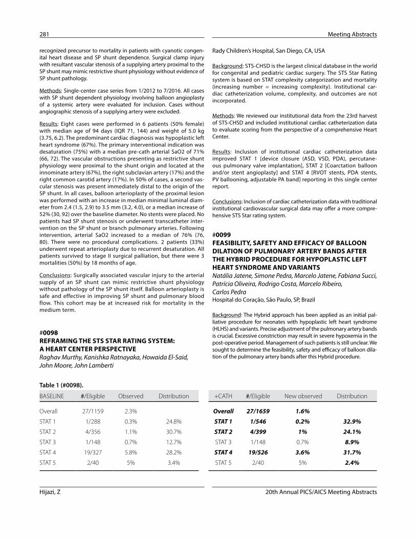
Hijazi, Z 20th Annual PICS/AICS Meeting Abstracts
281 Meeting Abstracts
Rady Children’s Hospital, San Diego, CA, USA
Background: STS-CHSD is the largest clinical database in the world for congenital and pediatric cardiac surgery. The STS Star Rating system is based on STAT complexity categorization and mortality (increasing number = increasing complexity). Institutional car-diac catheterization volume, complexity, and outcomes are not incorporated.
Methods: We reviewed our institutional data from the 23rd harvest of STS-CHSD and included institutional cardiac catheterization data to evaluate scoring from the perspective of a comprehensive Heart Center.
Results: Inclusion of institutional cardiac catheterization data improved STAT 1 [device closure (ASD, VSD, PDA), percutane-ous pulmonary valve implantation], STAT 2 [Coarctation balloon and/or stent angioplasty] and STAT 4 [RVOT stents, PDA stents, PV ballooning, adjustable PA band] reporting in this single center report.
Conclusions: Inclusion of cardiac catheterization data with traditional institutional cardiovascular surgical data may offer a more compre-hensive STS Star rating system.
#0099 FEASIBILITY, SAFETY AND EFFICACY OF BALLOON DILATION OF PULMONARY ARTERY BANDS AFTER THE HYBRID PROCEDURE FOR HYPOPLASTIC LEFT HEART SYNDROME AND VARIANTSNatália Jatene, Simone Pedra, Marcelo Jatene, Fabiana Succi, Patrícia Oliveira, Rodrigo Costa, Marcelo Ribeiro, Carlos PedraHospital do Coração, São Paulo, SP, Brazil
Background: The Hybrid approach has been applied as an initial pal-liative procedure for neonates with hypoplastic left heart syndrome (HLHS) and variants. Precise adjustment of the pulmonary artery bands is crucial. Excessive constriction may result in severe hypoxemia in the post-operative period. Management of such patients is still unclear. We sought to determine the feasibility, safety and efficacy of balloon dila-tion of the pulmonary artery bands after this Hybrid procedure.
recognized precursor to mortality in patients with cyanotic congen-ital heart disease and SP shunt dependence. Surgical clamp injury with resultant vascular stenosis of a supplying artery proximal to the SP shunt may mimic restrictive shunt physiology without evidence of SP shunt pathology.
Methods: Single-center case series from 1/2012 to 7/2016. All cases with SP shunt dependent physiology involving balloon angioplasty of a systemic artery were evaluated for inclusion. Cases without angiographic stenosis of a supplying artery were excluded.
Results: Eight cases were performed in 6 patients (50% female) with median age of 94 days (IQR 71, 144) and weight of 5.0 kg (3.75, 6.2). The predominant cardiac diagnosis was hypoplastic left heart syndrome (67%). The primary interventional indication was desaturation (75%) with a median pre-cath arterial SaO2 of 71% (66, 72). The vascular obstructions presenting as restrictive shunt physiology were proximal to the shunt origin and located at the innominate artery (67%), the right subclavian artery (17%) and the right common carotid artery (17%). In 50% of cases, a second vas-cular stenosis was present immediately distal to the origin of the SP shunt. In all cases, balloon arterioplasty of the proximal lesion was performed with an increase in median minimal luminal diam-eter from 2.4 (1.5, 2.9) to 3.5 mm (3.2, 4.0), or a median increase of 52% (30, 92) over the baseline diameter. No stents were placed. No patients had SP shunt stenosis or underwent transcatheter inter-vention on the SP shunt or branch pulmonary arteries. Following intervention, arterial SaO2 increased to a median of 76% (76, 80). There were no procedural complications. 2 patients (33%) underwent repeat arterioplasty due to recurrent desaturation. All patients survived to stage II surgical palliation, but there were 3 mortalities (50%) by 18 months of age.
Conclusions: Surgically associated vascular injury to the arterial supply of an SP shunt can mimic restrictive shunt physiology without pathology of the SP shunt itself. Balloon arterioplasty is safe and effective in improving SP shunt and pulmonary blood flow. This cohort may be at increased risk for mortality in the medium term.
#0098 REFRAMING THE STS STAR RATING SYSTEM: A HEART CENTER PERSPECTIVERaghav Murthy, Kanishka Ratnayaka, Howaida El-Said, John Moore, John Lamberti
Table 1 (#0098).
BASELINE #/Eligible Observed Distribution +CATH #/Eligible New observed Distribution
Overall 27/1159 2.3% Overall 27/1659 1.6%
STAT 1 1/288 0.3% 24.8% STAT 1 1/546 0.2% 32.9%
STAT 2 4/356 1.1% 30.7% STAT 2 4/399 1% 24.1%
STAT 3 1/148 0.7% 12.7% STAT 3 1/148 0.7% 8.9%
STAT 4 19/327 5.8% 28.2% STAT 4 19/526 3.6% 31.7%
STAT 5 2/40 5% 3.4% STAT 5 2/40 5% 2.4%
Journal of Structural Heart Disease, December 2016 Volume 2, Issue 6:241-306
Meeting Abstracts 282
Between 2005-15 all neonates with PA-IVS undergoing transcath-eter RV decompression were included. Technical factors evaluated included: use and characteristics of radiofrequency (RF) application, maximal balloon:PV annulus ratio (BAR), infundibular diameter, and RV systolic pressure pre- and post-valvuloplasty (BPV). Primary endpoint was cardiac perforation or death; the secondary endpoint was ReINT.
Results: 93 neonates underwent RV decompression at median 3d (IQR 2-5) of age, including 61 pts via RF perforation of the PV and 32 via wire perforation. There were 12 (13%) complications with 7 occurring in the first 2 years of the 10 year study period. Complications included 8 (8.6%) cardiac perforations, of which, there were two deaths. Cardiac perforation was associated with the use of RF (p=0.047), RF duration (3.5 vs 2.0 seconds, p=0.02) and maximum RF energy (7.5 vs 5.0 Joules, p<0.01) but not with patient weight (p=0.09), PV diameter (p=0.23) or infundibular diameter (p=0.57). ReINT was performed in 36 patients including repeat BPV (n=25) or surgery (n=11). ReINT was associated with higher RV pressure (median 60 vs 50mmHg, p=0.041) and with residual valve gradient (p=0.046) post initial BPV, but not with BAR, atmospheric pressure used during BPV or the presence of a residual balloon waist during BPV. ReINT was not associated with any RV ana-tomic measurements, including PV annulus.
Conclusion: Technical factors surrounding transcatheter RV decom-pression in PA/IVS influence the risk of procedural complications but not the future need for ReINT. Cardiac perforation is associ-ated with use of RF energy as well as RF application characteristics. Reintervention after RV decompression for PA-IVS is common, and appears related to final gradient and RV pressure after initial BPV.
#0101 ENDOCARDITIS ASSOCIATED WITH TRANSCATHETER PULMONARY VALVE IMPLANTATION: AN INSTITUTIONAL EXPERIENCEAshish Ankola, Julie Vincent, Matthew Crystal, Mariel Turner, Alejandro Torres1
Columbia University Medical Center, New York, NY, USA
Objective: Transcatheter pulmonary valve (TPV) implantation has become a viable therapy for right ventricular outflow tract (RVOT) dysfunction. While rare, infective endocarditis (IE) following TPV has been reported. In this study, we describe our center’s experience with IE after Melody TPV (Medtronic, Minneapolis, MN).
Methods: Single-center retrospective review of pts who underwent TPV implantation with Melody valve from 2007-2014. IE was defined using the modified Duke’s criteria.
Results: A total of 85 pts underwent TPV implantation during the study period, of which 12 (14%) developed IE. Median time to the develop-ment of IE was 32 months (range 2-100). 2 pts had a history of IE and 2 of IV drug abuse. One pt was diagnosed with severe gingivitis prior to IE. Seven pts were on ASA at the time of IE. Compliance with SBE prophylaxis was documented in only 2 pts at the time of IE. Baseline RVOT anatomy in IE pts included homografts in 8 pts, prosthetic PV in 2 and direct RV-PA anastomosis in 2. Stenting prior to TPV implantation was performed in 8 (67%) pts. There was no significant difference in demographics, type of RVOT reconstruction, pre-stenting, and residual RVOT gradient after TPV implantation between IE and non-IE pts. Eleven pts were febrile at presen-tation. Eight pts (67%) presented with RVOT obstruction, 5 of which had a
Material and Methods: This was an observational study in a cohort of infants with HLHS or variants. Data collection was retrospective. The bands were configured from 3 mm EPTFE tubes at the initial procedure. Indication for balloon dilation included persistent hypox-emia (Sats < 70%) and increased gradients on echocardiography (> 60 mmHg). Balloon dilation was performed using 2.5 to 3.5 mm angio-plasty balloons. Measures of outcomes included increase in the min-imal diameter of the band, increase in sats, gradient reduction across the band on echo and appropriate timing of the Norwood Glenn operation.
Results: Between March 2012 and January 2016, out of 68 neonates who underwent the Hybrid procedure, 7 were diagnosed with tight PA bands and underwent dilation at 12 band sites. The median age and weight were 60 days and 2.9 Kg, respectively. Five had HLHS (2 had variants). Three patients needed additional interventions: atrial septostomy, new ductal stenting, stenting the vertical vein and arch stenting. Arrhythmias were observed in 3 patients and minor air emboli occurred in one patient with no deleterious effects. There was no mortality. There was an increase in the minimal diameter from 0.8 ± 0.5 to 1.7±0.5 mm (p<0.001) and in the systemic saturation from 67±9 to 84±4% (p<0.001) One patient underwent a new dilation one month afterwards. In all patients the Norwood Glenn operation could be performed at an appropriate time at a median age of 6 months with no mortality. At surgery, there was a band fracture in just a single vessel. There were no lesions to adjacent vessels in any of the patients.
Conclusions: Balloon dilation of the PA bands after the Hybrid proce-dure was feasible, safe and effective, resulting in clinical stabilization allowing performing the Norwood Glenn operation at an appropriate time. The mechanism of dilation is probably stretching of the eptfe, which may occasionally result in fracture. No significant trauma to the adjacent vessel was observed at surgery.
#0100 TECHNICAL FACTORS ARE ASSOCIATED WITH OUTCOMES FOLLOWING RIGHT VENTRICLE DECOMPRESSION FOR NEONATES WITH PULMONARY ATRESIA AND INTACT VENTRICULAR SEPTUM: RESULTS FROM A MULTICENTER COLLABORATIVEChristopher Petit1, Athar Qureshi4, Andrew Glatz2, Michael Kelleman1, Courtney McCracken1, Allen Ligon1, Namrita Mozumdar2, Wendy Whiteside3, Asra Khan4, Bryan Goldstein3,1Emory University School of Medicine, Atlanta, GA, USA2Children’s Hospital of Philadelphia, Philadelphia, PA, USA3Cincinnati Children’s Hospital Medical Center, Cincinnati, OH, USA4Texas Children’s Hospital, Houston, TX, USA
Background: Transcatheter right ventricle (RV) decompression in neo-nates with pulmonary atresia and intact ventricular septum (PA-IVS) is technically challenging, with risk of cardiac perforation and death. Further, despite successful RV decompression, repeat intervention (ReINT) on the pulmonary valve (PV) is common. The association between technical factors during RV decompression and the risk of complications and ReINT are not well described.
Methods: Multicenter retrospective study at the 4 member centers of the Congenital Catheterization Research Collaborative (CCRC).
Hijazi, Z 20th Annual PICS/AICS Meeting Abstracts
283 Meeting Abstracts
Introduction: Progressive pulmonary valve regurgitation is a significant morbidity after surgical correction of tetralogy of Fallot. This study reviewed a single institution’s experience with valve- sparing repair and investigated the role of intraoperative balloon valvuloplasty.
Methods: A retrospective chart review identified 7 patients who underwent valve sparing repair of tetralogy of Fallot by intraopera-tive balloon pulmonary valvuloplasty. We evaluated the pulmonary valve function by echocardiography in immediate post-operative and short-term later.
Results: Among 7 patients, the number of male were 3 and median age was 136 days old. The median annulus diameter of pulmonary valve was 6.8mm (Z score -2.4) and Z score of pulmonary valve annu-lus was from -2.0 to -3.7. The high pressure balloon of various size were used from 8 to 12 mm and median ratio of annulus to balloon was 1.29. In immediate post-operative period, most patients had below moderate pulmonary regurgitation and stenosis. Median fol-low up duration from TOF repair was 22 months and most patients had mild or moderate pulmonary stenosis and regurgitation. We had 1 mortality case and she expired by endocrinology problem as adre-nal dysfunction after operation.
Conclusions: In our results, the intraoperative balloon pulmonary val-vuloplasty in valve-sparing repair of TOF showed feasible outcomes and long term follow up was required for more explainable results.
#0104 USING A CORE TEAM TO IMPROVE PATIENT CARE DELIVERY FOR ASD/PFO CLOSURES IN AN ADULT CARDIAC CATHETERIZATION LABORATORY.Ami VaughnHouston Methodist Hospital, Houston, TX, USA
Background: A knowledge deficit exists at this institution in the cardiac catheterization laboratory in regards to the treatment of Adult Congenital Heart Disease (ACHD). Limited exposure to congenital cases results in a learning curve for the nursing staff and radiology technologists assisting in the procedures. Due to improvements in treatment as children, more patients are living longer, so the need for specialized adult congenital heart care has dramatically increased and will continue to do so for the foresee-able future. With the addition of Houston’s only board certified Adult Congenital Heart specialist, the caseload of ACHD patients has increased at this institution.
Method: This institution’s cardiac catheterization laboratory has recently implemented a core team of people that assist in these congenital heart procedures as well as briefing with the cardiologist prior to case start. Additionally, the physician meets with our clini-cal manager to discuss the cases coming up the following week and ensure availability of supplies that will be needed for the cases. For these purposes, we have only retrospectively assessed time to case start and total in room time in the lab for the ASD/PFO closures at this institution from 2014 to present.
Results: We reviewed the case times for ASD/PFO closures from 2014 (13), 2015 (7) and 2016 (16), specifically looking at time in
vegetation in the Melody TPV on echo. Three of these 5 pts had poor out-comes: Two presented in septic shock, one was immunocompromised and died while the other underwent surgical valve replacement due to severe residual RVOT obstruction. A second death occurred in an afebrile pt with terminal heart failure who presented with multi-organ failure and IE. The other 9 pts were treated medically and none had residual RVOT obstruction after completing antibiotics. One pt developed Strep Mitis IE 2 years after TPV implantation and 5 months later had a second IE epi-sode due to MSSA. TEE was negative both times. Most common organ-isms isolated were MSSA in 4 (33%) pts and Strep Mitis in 3 (25%).
Conclusion: At our institution, IE occurred in 14% of pts after Melody TPV implantation. No specific risk factors were identified. TPV dys-function was present in 67% of cases with mortality occurring in 2 high-risk pts. One pt had their TPV explanted while the rest were successfully treated medically. Multicenter studies are needed to better understand and identify predictors of IE in this population.
#0102 PERCUTANEOUS CLOSURE OF PARAVALVULAR LEAK AND VALVE-IN-VALVE IMPLANTATION IN AORTIC REGURGITATION AFTER TRANSCATHETER AORTIC VALVE REPLACEMENT Vasutakarn Chongthammakun, Mauricio CohenDivision of Cardiology, University of Miami Miller School of Medicine, Miami, FL, USA
Paravalvular leak (PVL) is a common complication in transcatheter aortic valve replacement (TAVR) especially when significant cal-cification of the native valve is present resulting in underexpan-sion of the transcatheter valve. We present a case of a 91-year-old female patient with severe aortic stenosis who underwent right transfemoral TAVR with a 20-mm Edwards Sapien 3 Valve. After the valve was deployed, transaortic gradient normalized; how-ever, aortography revealed severe aortic insufficiency. Elevated left ventricular end-diastolic pressure (LVEDP) and equalization of LVEDP and diastolic aortic pressure were noted on pressure tracings. Transesophageal echocardiography (TEE) demonstrated severe central and paravalvular regurgitation in the region of the non-coronary cusp. Balloon post-dilation of the transcatheter valve did not result in improvement of aortic insufficiency. The decision was then made to close the leak. Two Amplatzer Vascular Plug-II devices were used, which successfully closed the PVL. The degree of paravalvular regurgitation substantially decreased on subsequent TEE. Given central aortic regurgitation remained pres-ent, a second 20-mm Edwards Sapien 3 Valve was implanted in a valve-in-valve fashion. Final aortography and TEE showed res-olution of aortic insufficiency. PVL is a known predictor of worse outcome post-TAVR and percutaneous closure of PVL should be considered as it has been shown to improve symptoms, minimize risks of hemolysis requiring blood transfusion, and is associated with improved long-term mortality.
#0103 THE SHORT-TERM RESULT OF INTRAOPERATIVE BALLOON PULMONARY VALVULOPLASTY IN VALVE-SPARING REPAIR OF TETRALOGY OF FALLOTSangyun Lee, Seongho Kim, Soick Jang, Soojin Park, Changha LeeSejong General Hospital, Bucheon-si, Geynggi-do, Republic of Korea
Journal of Structural Heart Disease, December 2016 Volume 2, Issue 6:241-306
Meeting Abstracts 284
Emily L. Marshall, James C. Fudge, Dhanashree Rajderkar, David Borrego, Wesley E. BolchUniversity of Florida, Gainesville, FL, USA
Objective: This study focuses on the creation and implementation of a reporting system of patient radiation doses in a large institution pediatric cardiac catheterization laboratory.
Background: In 2015 the Journal of Pediatrics released a study assessing long-term outcome in children with congenital heart dis-ease (CHD) (Razzaghi 2015). The study utilized the National Health Interview Survey’s 1997-2011 data which indicates that 85% of chil-dren born with CHD in the US today will survive into adulthood, thus requiring multiple cardiac interventions before management is achieved. With increasing improvements in pediatric patients’ long-term outcome, the need for in-clinic patient dose tracking emerged.
Methods: The researcher’s institution developed a library of pediatric computational phantoms to represent patients of varying gender, age, height, and weight. These human models were created based on the statistics of the current US population. The fluoroscopy unit at the institution produces a logfile for each patient’s procedure. These logfiles are created to track machine motion and output, they include numerous exposure parameters per irradiation event such as: energy, table position, filter, source angle, and the kerma-area product (KAP) meter value. The fluoroscopy unit was calibrated to accurately create a model of the machine in Monte Carlo computer simulation space. Calibrations included: field isocenter determination, beam half-value layers, and KAP meter calibration. Utilizing the radiation transport code Monte Carlo N-Particle, a simulation of the procedure is possi-ble using the logfiles, modelled fluoroscopy unit, and the phantoms.
Results: Patient phantoms selected based on height and weight per-formed within 15-30% of their patient-specific modelled counterparts. Isocentric positioning of phantoms when compared to image aligned simulations cited in-field organ relative errors between 0.5-20%. Attenuation through the procedure table and pad was matched in the computer model to the clinical beam within 1.5-4%. To date, the labo-ratory has successfully completed dose reconstructions for 40 patients.
Conclusion: Implementation of an effective organ dose reporting sys-tem in a large institution catheterization lab has proven successful. Clinical accessibility of organ dose reports will enable physicians to continue improving pediatric patient long-term outcome.
#0107 RADIATION-FREE MRI HEART CATHETERIZATION IN CHILDRENKanishka Ratnayaka1, Joshua P. Kanter2, Anthony Z. Faranesh3, Elena K. Grant2, Laura J. Olivieri2, Russell R. Cross2, Ileen F. Cronin2, Karin S. Hamann2, Adrienne E. Campbell-Washburn3, Kendall J. O’Brien3, Toby Rogers3, Michael S. Hansen3, Robert J. Lederman3
1Rady Children’s Hospital, San Diego, CA, USA2Children’s National Health System, Washington, DC, USA3National Heart Lung and Blood Institute, National Institutes of Health, Bethesda, MD, USA
room prior to case start and total time in room for each ASD closure. Median in room to case start times in minutes for 2014, 2015 and 2016 are 32.8, 32.8, and 23.1 respectively. Total in room time in min-utes for the same is 145.8, 149.8, and 134.4. Since implementation, the team times are 18.3 minutes from in room to case start and 99 minutes total in room time.
Conclusion: While our results are preliminary, these findings suggest the team approach improves the quality of care of ACHD patients while undergoing cardiac catheterization and percutaneous interventions.
#0105 LOWER LIMB REGIONAL OXYGENATION MONITORING USING NEAR INFRARED SPECTROSCOPY DURING CARDIAC CATHETERIZATION OF INFANTS.Rodrigo Rios, Rohit Loomba, Susan Foerster, Todd Gudausky, Rebecca RussellChildren’s Hospital of Wisconsin, Milwaukee, WI, USA
Introduction: Limb ischemia secondary to femoral arterial injury is a rare but potentially catastrophic complication of cardiac catheteriza-tion in infants. Early detection or prevention of compromised lower extremity perfusion is ideal. Near-infrared Spectroscopy (NIRS) is a well-established non-invasive method of monitoring regional oxygen saturation. The purpose of this study was to characterize intra-oper-ative values and postoperative values of NIRS in infants undergoing cardiac catheterization involving femoral vascular access. A second-ary aim was to determine if values were predictive of arterial femoral thrombus after such intervention.
Methods: Bilateral lower extremity NIRS monitoring was performed during cardiac catheterization. All infants (< 1 years old) 6kg in weight or less that underwent cardiac catheterization with femoral vascular access were eligible. Demographic and procedural data was col-lected. Lower extremity NIRS data was blinded as to not affect the course of the catheterizations.
Results: Data was collected for 16 consecutive infants undergoing cardiac catheterization with femoral vascular access. Cardiac diagno-sis and purpose of catheterization varied between patients. Of these catheterizations, 9 were interventional. The mean lower extremity arterial-venous oxygen saturation difference (LEAVD) in the accessed leg was 21 immediately prior to catheterization, 45 immediately after sheath insertion, 52 immediately prior to sheath removal, and then 30 at the time of discharge. LEAVD at any time point was not predic-tive of femoral arterial thrombus.
Conclusion: Lower extremity NIRS monitoring demonstrates a widening of LEAVD during catheterization with femoral vascular access. Data on additional infants is required to determine if lower extremity NIRS monitoring in infants undergoing cardiac catheteriza-tion could be a useful and non-invasive method for early detection and potential prevention of femoral artery compromise.
#0106 FROM START TO FINISH: THE PHYSICS BEHIND IMPLEMENTING A RADIATION DOSE REPORTING SYSTEM IN THE PEDIATRIC CARDIAC CATHETERIZATION LAB
Hijazi, Z 20th Annual PICS/AICS Meeting Abstracts
285 Meeting Abstracts
independent operation. Innovative prospective five-year indepen-dence plan includes visiting in-country clinical service and training trips, encouraging autonomy through targeted case selection (diag-nostic, balloon pulmonary valvuloplasty, patent ductus arteriosus device closure), out-of-country training fellowships, and weekly tele-medicine case discussion/mentorship sessions.
Results: Since inception, Uganda Heart Institute has performed 245 congenital heart disease catheterization procedures [diagnostic (85); balloon pulmonary valvuloplasty (37); patent ductus arterio-sus device closure (102); other (21)]. The number and percentage of independent Uganda Heart Institute procedures (no international provider presence) has steadily grown from 0, 0% (2012); 3, 7.8% (2013); 17, 29.8 % (2014); 55, 77.5% (2015) to 53, 82.8% (Jan-Sept 2016). In-country clinical service and training trips have been con-ducted by five organizations [total n = 10; World Children’s Initiative (4), Gift of Life/Chain of Hope (5)] traveling with physicians, nurses, and technologists. Two Uganda Heart Institute catheterization opera-tors were trained at Amrita Institute of Medical Science and Research for 5 months observing and participating in 226 procedures. Weekly telemedicine conferences (n = 51 months) have facilitated dialogue and education covering clinical case selection, planning, and review. Challenges include inventory procurement, domestic hardware sup-port, and expert support staff (anesthesia, intensive care) shortage.
Conclusions: Through mentoring, Uganda Heart Institute is progress-ing toward the goal of 100 annual independent cases by 2017 five years after program inception.
#0109 AMPLATZER VASCULAR PLUG II DEVICE TO OCCLUDE DIFFERENT TYPES OF PATENT DUCTUS ARTERIOSUS IN PEDIATRIC PATIENTSAlper Guzeltas1, Ibrahim Cansaran Tanidir1, Mehmet Akin Topkarci2, Gulhan Tunca Sahin1, Murat Sahin1
1Mehmet Akif Ersoy Thoracic and Cardiovascular Surgery Center Department of Pediatric Cardiology, Istanbul, Turkey2Mehmet Akif Ersoy Thoracic and Cardiovascular Surgery Center Department of Pediatric Anesthesia, Istanbul, Turkey
Objective: To evaluate the outcome of the Amplatzer Vascular Plug II (AVP-II) for closure of the patent ductus arteriosus (PDA) in children.
Methods: All patients undergoing transcatheter closure of PDA with AVP-II from June 2013 to November 2016 were retrospectively evaluated. In one patient device had to remove due to pulmonary hypertensive crisis. this patient excluded from study. Clinical, angio-graphic, and echocardiographic data were analyzed.
Results: Thirtyfive procedures were performed in 34 patients. Median age was 30 months (6–202 months) with median weight 13 kg (4.2–63). The morphological PDA classification was Type A in 14/34 (41%), Type B in 2/34 (6%), Type C in 15/34 (44%), and Type E in 3/34 (9%). The median minimum PDA diameter, maximum and length was 3.5 mm (1.5-8.3 mm), 8.2 mm (2.2-21 mm), and 13.1 mm (8.2-21 mm) respectively. The implanted device sizes were: 6 mm in 4/35 (11%), 8 mm in 14/35 (40%), 10 mm in 15/35 (43%) and 14 mm in 2/35 (6%) procedures. The implanted device was mean of
Background: Children with heart disease may require repeated X-Ray cardiac catheterization procedures, are more radiosensitive, and more likely to survive to experience oncologic risks of medical radiation. Cardiac MRI is radiation-free and offers information about structure, function, and perfusion but not hemodynamics. We aim to perform radiation-free diagnostic right heart catheterization entirely using MRI guidance in an unselected cohort of pediatric patients.
Methods and Results: We performed 45 MRI guided comprehensive transfemoral right heart catheterizations in 35 pediatric (12.7 ± 4.8 years) subjects referred for clinically indicated cardiac catheterization (post-heart transplant 37%, shunt 31%, cardiomyopathy 14%, pulmo-nary hypertension 12%, valvular heart disease 3%, other 3%) during an sixteen month period (3/2015 – 8/2016). Radiation-free MRI right heart catheterization attempts were all successful using passive cath-eters. In two subjects with septal defects, right and left heart catheter-ization was performed. There were no complications. One subject had six such procedures. Most subjects (51%) had undergone multiple (5.8 ± 5) previous X-Ray cardiac catheterizations. Retained thoracic surgical or transcatheter implants (40%) did not preclude successful MRI heart catheterization. During the procedure, two subjects were receiving vasopressor infusions at baseline because of poor cardiac function, and in six procedures, multiple hemodynamic conditions were tested.
Conclusions: Comprehensive MRI guided right heart catheterization was feasible and safe in this small cohort of pediatric subjects. This includes subjects with previous metallic implants, those requiring continuous vasopressor medication infusions, and those requiring pharmacologic provocation. Children requiring multiple, serial X-Ray cardiac catheterizations may benefit most from radiation sparing. This is a step toward wholly MRI guided diagnostic (right and left heart) cardiac catheterization and future MRI guided cardiac intervention.
#0108 UPDATE ON UGANDA HEART INSTITUTE 5 YEAR PROSPECTIVE PLAN TOWARD INDEPENDENT PEDIATRIC CARDIAC CATHETERIZATIONSulaiman Lubega1, Twalib Aliku1, Peter Lwabi1, Sanjay V. Daluvoy2, Craig A. Sable3, Shakeel Qureshi4, Krishna Kumar5, Kanishka Ratnayaka6
1Uganda Heart Institute, Kampala, Uganda2Duke Global Health Institute, Durham, NC, USA3Children’s National Health System, Washington, DC, USA4Evelina London Children’s Hospital, London, UK5Amrita Institute of Medical Science and Research, Kochi, India6Rady Children’s Hospital, San Diego, CA, USA
Background: Population statistics estimate 10,000 children are born each year with congenital heart disease in Uganda. Over the past decade, Uganda Heart Institute has developed a cardiovascular surgi-cal program. A complementary cardiac catheterization program was started in February 2012 with the aim of developing independent operation after five years of mentorship.
Methods: A biplane X-ray cardiac catheterization lab was built adjacent to a new cardiovascular operating room. Uganda Heart Institute physicians, nurses, and technologists began training with government and non-governmental organization support toward
Journal of Structural Heart Disease, December 2016 Volume 2, Issue 6:241-306
Meeting Abstracts 286
after the procedure while 4 patients died under ECMO support during the ICU stay.
Conclusion: If the patients cannot be weaned from ECMO support due to hemodynamic instability, catheter angiography should be performed urgently. We think that for better and high quality views in patients with antegrade flow, who cannot be weaned from ECMO support, ECMO cannulas can be clipped just before the injections.
#0111 A LIFE SAVING INTERVENTION IN CRITICALLY ILL CHILDREN WITH OBSTRUCTED SUPRACARDIAC TOTAL ANOMALOUS PULMONARY VENOUS CONNECTION: VERTICAL VEIN STENTINGAlper Guzeltas, Ibrahim Cansaran Tanidir, Erkut OzturkMehmet Akif Ersoy Thoracic and Cardiovascular Surgery Center Department of Pediatric Cardiology, Istanbul, Turkey
Background: Obstructed total anomalous pulmonary venous con-nection (TAPVC) and duct dependent pulmonary flow are two cardiac pathologies which need urgent surgical intervention in pedi-atric cardiovascular surgery. Obstructed TAPVC has still high risk of surgical burden. Surgical mortality rate is high especially when the patients clinical condition is worse, in which preoperative stabiliza-tion by temporarily relieving the obstruction may improve outcomes in this population.
Methods: Between June 2014 and September 2016, 5 patients under-went urgent cardiac catheterization to relieve the obstruction in the vertical vein.
Results: All patients’ clinical condition was worse and intubated on the day of diagnosis. Interventional cardiac catheterization was performed within the first 24 hours after the admission. Four of the patients were newborn. Two out of five patients had isolated suprac-ardiac obstructive TAPVC where as others had additionally right atrial isomerism, unbalanced complete atrioventricular septal defect, pul-monary stenosis in addition to and pulmonary venous hypertension in echocardiography. One of them had also duct dependent pulmo-nary flow.
Either 6mm or 7mm stent implantation could be achieved in all patients. In the patient with supracardiac TAPVC draining into two dif-ferent parts of the SVC stenting could be performed only for one side.
Two of the patients with isolated TAPVC was operated and other one is in the waiting list.
One of the newborn patient with heterotaxy syndrome and duct dependent pulmonary flow underwent PDA stenting 5 days after the first intervention. This patient had Glenn operation and TAPVC repair when he was 6-month-old.
Remaining one patients with heterotaxy syndrome were died within two days after the procedure.
Conclusion: Stenting of the vertical vein might be an effective ther-apy to acutely stabilize a sick neonate with obstructed supracardiac TAPVC. Catheter intervention can be considered as part of the preop-erative cardiovascular stabilization strategy for high-risk infants with obstructed supracardiac TAPVC.
3.0 ± 1.2 times the ductus narrowest diameter and mean of 1.3 ± 0.75 times the ductus largest diameter in successful procedures. The median procedure and fluoroscopy time was 30 minutes (15-60 minutes) and 6,5 minutes (2.8-20.3 minutes). In four patients closure was performed from the arterial side. All procedures except one 100% ‘in-lab’ and 100% closure on post-procedural echocardiogram was achieved. No left pulmonary artery stenosis and aortic obstruc-tion observed with a median follow-up duration of 12 months (1-40 months).
Complications: In one patient 8 mm device was embolized in to the pulmonary artery due to ductal spasm which caused underestima-tion of the duct and on the next day 8mm device was retrieved and a 10 mm device was implanted.
Conclusions: The AVP II seems to be an effective and safe device for PDA closure in children. It is particularly useful in type A and C ductus with adequate length and in small infants where it eliminated the risk of device related aortic obstruction.
#0110 CARDIAC CATHETERIZATION IN PAEDIATRIC PATIENTS DURING EXTRACORPOREAL MEMBRANE OXYGENATION SUPPORTAlper Guzeltas, Ibrahim Cansaran Tanidir, Taner Kasar, Erkut OzturkMehmet Akif Ersoy Thoracic and Cardiovascular Surgery Center Department of Pediatric Cardiology, Istanbul, Turkey
Background: Extracorporeal membrane oxygenation (ECMO) is a lifesaving intervention for pediatric patients with respiratory and/or cardiovascular failure. Cardiac catheterization while on ECMO was ini-tially reserved for left atrial decompression with the balloon or blade atrial septostomy procedure, indications for ECMO support have evolved particularly by the time. Nowadays it is suggested that early catheterization after initiation of extracorporeal membrane oxygen-ation support in children is associated with improved survival.
Methods: Between January 2012 and October 2016, 98 patients (5.2% of the surgery patients) needed ECMO support after cardiac surgery. Diagnostic or interventional cardiac catheterization was performed in (16.3%) 16 of these 98 patients.
Results: The diagnostic catheterization was performed in 7 of the patients while invasive procedures were performed in 9 of the patients. In patients with antegrade flow, ECMO cannulas were clipped before the injections for higher quality. Median age was 6.7 months (3.3–60 months) with median weight 7.0 kg (3.7–16 kg). The median duration from ECMO support institution to catheterization procedure was 3 days (1-11 days).
4/7 patients underwent surgical palliation after diagnostic catheter-ization. Left pulmonary arterial (LPA) balloon angioplasty was per-formed in 1/9, LPA stenting in 2/9, RPA stenting in 1/9, RVOT stenting in 1/9, both LPA and RVOT stentin in 1/9, both LPA and mBT shunt stenting in 1/9, both LPA and RPA balloon angioplasty in 1/9, both RPA and LPA stenting was performed in 1/9 of the patients during interventional catheterization procedure. During RVOT stenting AV block occurred and during procedure ECMO flow increased due to hypotension. In one patient ECMO tubing disconnected from the arterial line. Twelve patients were successfully weaned from ECMO
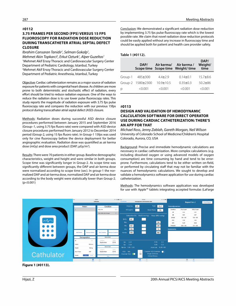
Hijazi, Z 20th Annual PICS/AICS Meeting Abstracts
287 Meeting Abstracts
Conclusion: We demonstrated a significant radiation dose reduction by implementing 3.75 fps pulse fluoroscopy rate which is the lowest possible rate. We claim that novel radiation dose reduction protocols could be easily applied without any increase in fluoroscopy time and should be applied both for patient and health care provider safety.
Table 1 (#0112).
DAP/ Scope time
Air kerma/ Scope time
Air kerma / Weight/ time
DAP/ Weight/
time
Group-1 485±300 4.4±2.9 0.14±0.1 15.7±8.6
Group-2 1580±2300 10.9±10.5 0.35±0.3 50.2±86
p <0.001 <0.001 <0.001 <0.001
#0113 DESIGN AND VALIDATION OF HEMODYNAMIC CALCULATION SOFTWARE FOR DIRECT OPERATOR USE DURING CARDIAC CATHETERIZATION: THERE’S AN APP FOR THATMichael Ross, Jenny Zablah, Gareth Morgan, Neil WilsonUniversity of Colorado School of Medicine/Children’s Hospital Colorado, Aurora, CO, USA
Background: Precise and immediate hemodynamic calculations are necessary in cardiac catheterization. More complex calculations (e.g. including dissolved oxygen or using advanced models of oxygen consumption) are time consuming by hand and tend to be error-prone. Furthermore, calculations tend to be either written on-field, or performed by circulating staff that may not be familiar with the nuances of hemodynamic calculations. We sought to develop and validate a hemodynamics software application for use during cardiac catheterization.
Methods: The hemodynamics software application was developed for use with Apple™ tablets integrating accepted formulas (LaFarge
#0112 3.75 FRAMES PER SECOND (FPS) VERSUS 15 FPS FLUOROSCOPY FOR RADIATION DOSE REDUCTION DURING TRANSCATHETER ATRIAL SEPTAL DEFECT CLOSUREIbrahim Cansaran Tanidir1, Selman Gokalp1, Mehmet Akin Topkarci2, Erkut Ozturk1, Alper Guzeltas1
1Mehmet Akif Ersoy Thoracic and Cardiovascular Surgery Center Department of Pediatric Cardiology, Istanbul, Turkey2Mehmet Akif Ersoy Thoracic and Cardiovascular Surgery Center Department of Pediatric Anesthesia, Istanbul, Turkey
Objective: Cardiac catheterization remains as a major source of radiation exposure for patients with congenital heart disease. As children are more prone to both deterministic and stochastic effect of radiation, every effort should be tried to reduce radiation exposure. One of the ways to reduce the radiation dose is to use lower pulse fluoroscopy rates. This study reports the magnitude of radiation exposure with 3.75 fps pulse fluoroscopy rate and compares the reduction with our previous 15fps protocol during transcathater atrial septal defect (ASD) closure.
Methods: Radiation doses during successful ASD device closure procedures performed between January 2015 and September 2016 (Group–1, using 3.75 fps fluoro rate) were compared with ASD device closure procedures performed from January 2012 to December 2014 period (Group-2, using 15 fps fluoro rate). In Group-1 15fps was used only for cine fluoroscopy before the device deployment for better angiographic evaluation. Radiation dose was quantified as air kerma dose (mGy) and dose area product (DAP; µGy/m2).
Results: There were 70 patients in either group. Baseline demographic characteristics, weight and height and were similar in both groups. Scope time was significantly longer in Group-2. As scope time was significantly different between groups, the DAP and air kerma dose were normalized according to scope time (sec). In group-1 the nor-malized DAP and air kerma dose, normalized DAP and air kerma dose according to the body weight were statistically lower than Group-2. (p<0.001)
Figure 1 (#0113).
Journal of Structural Heart Disease, December 2016 Volume 2, Issue 6:241-306
Meeting Abstracts 288
with the highest doses to these 5 organs had a reported dose at the reference point of 297 mGy, which was the sixth highest in the group of 15 indicating poor correlation between these dose metrics.
Conclusions: There is poor correlation between the commonly used dose metric of dose at the reference point and the dose metric of patient organ doses. Patient organ doses provide a more accurate measure of patient radiation dose and thus may provide a more effec-tive way of evaluating the future risk of patient radiation exposure.
#0115 INTRA-PROCEDURAL 3D ROTATIONAL ANGIOGRAPHY: SETUP TIME AND THE IMPACT OF LEARNING CURVE IN A DEDICATED “CORE TEAM” APPROACH FOR ADULT CONGENITAL HEART DISEASE INTERVENTIONSAmy Vaughn1, Ponraj Chinnadurai3, Akanksha Thakkar1, Nakeisha Washington1, Gary Monteiro1, John Breinholt2, Huie Lin1
1Methodist DeBakey Heart and Vascular Center, Houston Methodist Hospital, Houston, TX, USA2Department of Pediatrics, The University of Texas Health Science Center, Houston, TX, USA3Advanced Therapies, Siemens Medical Solutions USA Inc.,, Hoffman Estates, IL, USA
Background: Intra-procedural 3D Rotational angiography (3DRA) in the cardiac catheterization laboratory has been shown to add clini-cal value especially during complex structural and congenital heart disease interventions. Although 3D information enhances 2D fluo-roscopy, some limitations that may preclude its optimal utilization include exposure to additional radiation, time needed for setup and the associated learning curve.
Methods: All adult congenital cardiac procedures performed in a single institution with intra-procedural 3DRA imaging between December 2014 and August 2016 were retrospectively reviewed. Additional 3DRA setup time was defined as the difference in the time stamps between 3DRA acquisition and preceding 2D fluoroscopic/cine acquisition. Case times and total radiation dose from fluoros-copy and 3DRA were noted for each procedure.
Results: A total of 21 patients underwent 3D rotational angiography acquisition during the study period for procedures including diag-nostic pulmonary angiogram for CTEPH evaluation, trans-catheter pulmonary valve implantation, aortic coarctation and pulmonary vein stenting. The median additional setup time for 3D rotation angi-ography was 9.87 minutes (25th percentile: 7.87 min, 75th percentile: 19.02). Typical reasons for additional setup time include: selection of optimal imaging and contrast injection protocols, patient position-ing, imaging setup and acquisition. The median radiation dose area product (DAP) was 2815.9 microGy-m2 (25th percentile: 1918, 75th percentile: 3603). There was a trend towards reduction in the set up time as operators progressed along the learning curve. Median time to case start and total in-room time were 38 minutes and 212 minutes respectively.
and Miettinen) to obtain the following: cardiac output, pulmonary flow, pulmonary vascular resistance, and systemic vascular resistance all indexed to body surface area. The relationship of pulmonary flow to systemic flow and relationship of pulmonary to systemic vascu-lar resistance was also calculated. For validation, retrospective col-lection of hemodynamic information was recorded and calculated in PedCath (v7.7.3) for 50 patients, age 3 months to 17 years old. Indications selected for analysis included pulmonary hypertension, atrial septal defect, and single ventricle physiology. Hemodynamic calculations were processed with the hemodynamics software and comparisons of difference between the two programs made.
Results: No significant difference between PedCath and the hemo-dynamics software were found. The standard error for all variables compared was <0.007.
Conclusion: We have successfully developed and validated a reliable and simple tool in the catheterization the catheterization laboratory to obtain quick and accurate hemodynamic information.
#0114 OUT WITH THE OLD AND IN WITH THE NEW: QUANTIFYING RADIATION EXPOSURE AND ITS IMPACT ON PEDIATRIC PATIENTS UNDERGOING CARDIAC CATHETERIZATIONJames C. Fudge, Emily L. Marshall, David Borrego, Wesley E. BolchUniversity of Florida, Gainesville, FL, USA
Objective: The application of advanced dosimetry methods to track radiation organ doses for patients undergoing cardiac catheterization.
Background: As the field of interventional cardiology in congenital heart disease continues to grow, we are becoming more aware of the potential secondary impacts of interventional treatments such as the long-term risks of radiation exposure. Currently, most institutions use metrics such as the kerma area product meter, dose at the reference point, and fluoroscopy time to track radiation. These values, while useful in charting quality control and improvements, have inherent limitations for broader application. The reporting of patient organ doses is now available to provide clinicians with an accurate method for dose tracking and the potential for patient risk modelling into the future.
Methods: A radiation dose reporting system developed at the researchers’ institution was implemented within the institution’s pediatric cardiac catheterization laboratory. Organ dose report-ing was performed for a group of 40 patients within the laboratory population and a subgroup analysis was performed on 15 trans-plant patients undergoing cardiac catheterization in order to further evaluate clinical dose metrics.
Results: Among the entire group, the highest reported organ doses seen were in the lungs, heart, adrenals, liver and the esophagus. Among the 15 transplant patients further analysed, the average dose at the reference point for this patient group was 284 mGy. Average organ doses were 19 mGy, 12 mGy, 6 mGy, 7 mGy, and 17 mGy to the lungs, esophagus, liver, adrenals, and heart respectively. The patient
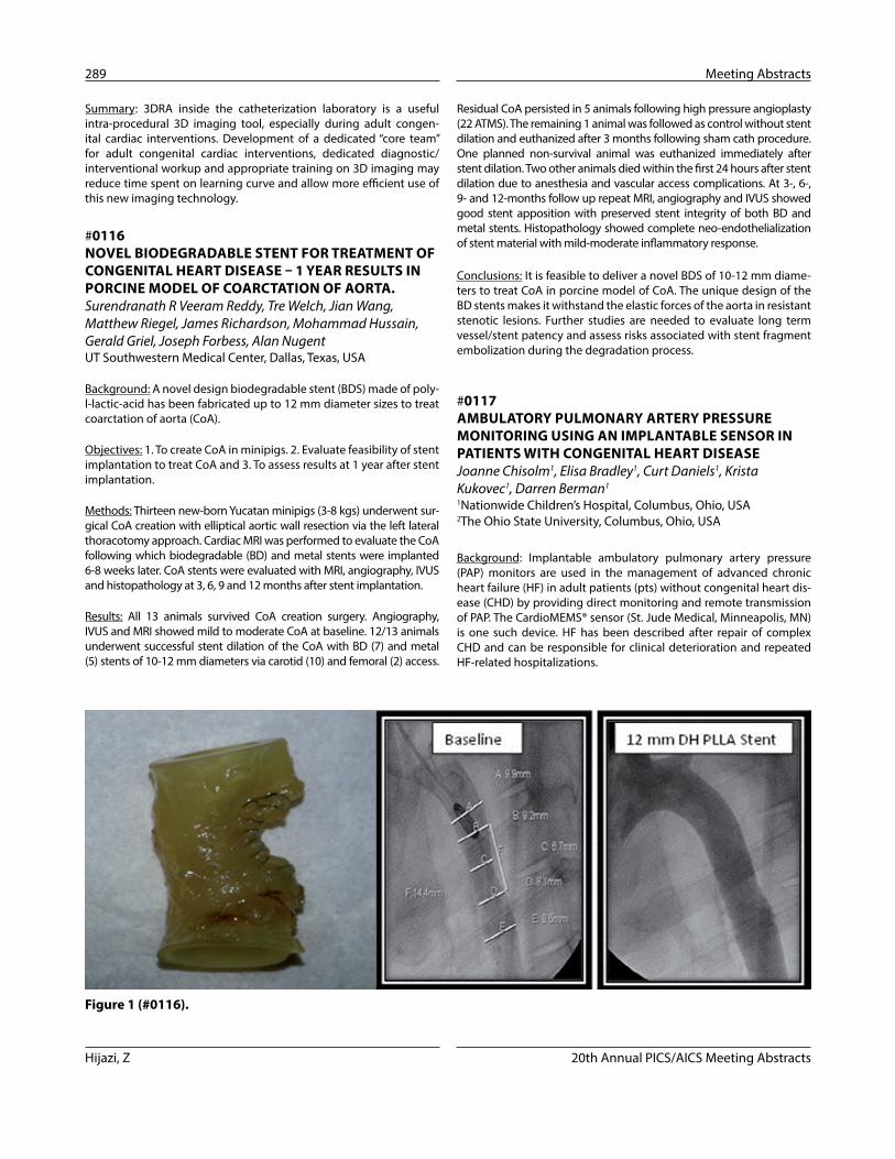
Hijazi, Z 20th Annual PICS/AICS Meeting Abstracts
289 Meeting Abstracts
Residual CoA persisted in 5 animals following high pressure angioplasty (22 ATMS). The remaining 1 animal was followed as control without stent dilation and euthanized after 3 months following sham cath procedure. One planned non-survival animal was euthanized immediately after stent dilation. Two other animals died within the first 24 hours after stent dilation due to anesthesia and vascular access complications. At 3-, 6-, 9- and 12-months follow up repeat MRI, angiography and IVUS showed good stent apposition with preserved stent integrity of both BD and metal stents. Histopathology showed complete neo-endothelialization of stent material with mild-moderate inflammatory response.
Conclusions: It is feasible to deliver a novel BDS of 10-12 mm diame-ters to treat CoA in porcine model of CoA. The unique design of the BD stents makes it withstand the elastic forces of the aorta in resistant stenotic lesions. Further studies are needed to evaluate long term vessel/stent patency and assess risks associated with stent fragment embolization during the degradation process.
#0117 AMBULATORY PULMONARY ARTERY PRESSURE MONITORING USING AN IMPLANTABLE SENSOR IN PATIENTS WITH CONGENITAL HEART DISEASEJoanne Chisolm1, Elisa Bradley1, Curt Daniels1, Krista Kukovec1, Darren Berman1
1Nationwide Children’s Hospital, Columbus, Ohio, USA2The Ohio State University, Columbus, Ohio, USA
Background: Implantable ambulatory pulmonary artery pressure (PAP) monitors are used in the management of advanced chronic heart failure (HF) in adult patients (pts) without congenital heart dis-ease (CHD) by providing direct monitoring and remote transmission of PAP. The CardioMEMS® sensor (St. Jude Medical, Minneapolis, MN) is one such device. HF has been described after repair of complex CHD and can be responsible for clinical deterioration and repeated HF-related hospitalizations.
Summary: 3DRA inside the catheterization laboratory is a useful intra-procedural 3D imaging tool, especially during adult congen-ital cardiac interventions. Development of a dedicated “core team” for adult congenital cardiac interventions, dedicated diagnostic/interventional workup and appropriate training on 3D imaging may reduce time spent on learning curve and allow more efficient use of this new imaging technology.
#0116 NOVEL BIODEGRADABLE STENT FOR TREATMENT OF CONGENITAL HEART DISEASE – 1 YEAR RESULTS IN PORCINE MODEL OF COARCTATION OF AORTA.Surendranath R Veeram Reddy, Tre Welch, Jian Wang, Matthew Riegel, James Richardson, Mohammad Hussain, Gerald Griel, Joseph Forbess, Alan NugentUT Southwestern Medical Center, Dallas, Texas, USA
Background: A novel design biodegradable stent (BDS) made of poly-l-lactic-acid has been fabricated up to 12 mm diameter sizes to treat coarctation of aorta (CoA).
Objectives: 1. To create CoA in minipigs. 2. Evaluate feasibility of stent implantation to treat CoA and 3. To assess results at 1 year after stent implantation.
Methods: Thirteen new-born Yucatan minipigs (3-8 kgs) underwent sur-gical CoA creation with elliptical aortic wall resection via the left lateral thoracotomy approach. Cardiac MRI was performed to evaluate the CoA following which biodegradable (BD) and metal stents were implanted 6-8 weeks later. CoA stents were evaluated with MRI, angiography, IVUS and histopathology at 3, 6, 9 and 12 months after stent implantation.
Results: All 13 animals survived CoA creation surgery. Angiography, IVUS and MRI showed mild to moderate CoA at baseline. 12/13 animals underwent successful stent dilation of the CoA with BD (7) and metal (5) stents of 10-12 mm diameters via carotid (10) and femoral (2) access.
Figure 1 (#0116).
Journal of Structural Heart Disease, December 2016 Volume 2, Issue 6:241-306
Meeting Abstracts 290
soon after subclavian flap repair in 1. All procedures were performed in the catheterization laboratory. Two patients underwent TEVAR alone and 3 underwent hybrid procedure combining TEVAR with vascular bypass sur-gery (carotid to left subclavian in 2, aorta to left common carotid in 1). Three types of endovascular stent grafts were utilized: the Gore TAG Thoracic Endoprosthesis was implanted in 2 patients, Cook Zenith TX2 in 2 and Medtronic Valiant in 1. Successful deployment was achieved in all patients. Balloon dilation of the stent graft after implantation was performed in 3 patients. Minimum aortic diameter increased from 1 ± 0.4 cm preopera-tively to 1.8 ± 0.3 cm (p=<0.05). Mean blood pressure gradient between upper and lower extremities was reduced from 37 ± 18 to 7 ± 11 mmHg (p=<0.05). The number of antihypertensive medications in all patients decreased or was eliminated, mean 2.8 (range 1-6) pre to 1.4 (range 0-3) post. No major bleeding, femoral artery injury or other early procedural complications were encountered. However, 2/3 vascular bypasses had late thrombosis with transient arm ischemia in 1 and neurologic ischemia in 1.
Conclusions: Complex native or recurrent coarctation of the aorta with large anureurysm/pseudoaneurysm can be safely and effec-tively managed by TEVAR in selected patients. These procedures are a viable alternative to cardiac surgery.
#0119 THE USE OF BIORESORBABLE VASCULAR SCAFFOLD (ABSORB) IN RESCUE TREATMENT OF VASCULAR INJURY OF EXTERNAL ILIAC ARTERY IN TWO CHILDREN UNDERGOING INTERVENTIONAL CARDIAC CATHETERIZATIONIyad Al-Ammouri, Tareq Goussous, Omar ObedatThe University of Jordan, Amman, Jordan
Bioresorbable vascular scaffold (ABSORB, Abbott Vascular, Santa Clara, California) has been recently approved as a treatment alterna-tive for coronary artery disease. Use of bioresorbable technology has long been thought to have a theoretical advantage in children, where stented vessel may retain their growth potential. We report the use of bioresorbable stents as rescue procedure in two children with arterial vascular injury during cardiac catheterization.
Two girls, age 9 and 13 year old, who underwent interventional cardiac catheterization for a large PDA, and severe aortic stenosis, respectively, had dissection with near total occlusion of the right external iliac artery. In the first child, dissection occurred after retrieval of an embolized PDA occluder device, and the second child with withdrawal of a large balloon that failed to be withdrawn into the arterial sheath. Both children were treated with a 4 mm Absorb stent with excellent immediate results. In one child, follow up angiography done at the time of a subsequent car-diac catheterization for 2 months after the stent showed patent vessel with mild narrowing at the site of stent, no reintervention was done due to absence of clinical symptoms. Further clinical follow up 6 months after the stenting showed no symptoms with normal distal pulses. In the other child there were no symptoms of vascular occlusion, and distal pulses were normal at the time of discharge, with planned follow up.
Conclusion: We report the use of Bioresorbable stents as a rescue treatment in two children with arterial injury with good immediate results. The theoretical benefit of retaining the potential for growth in stented vessels need to be validated.
Methods: Retrospective descriptive review of early use of the CardioMEMS® monitoring system in pts with complex CHD. Clinical and procedural data were reviewed from 2 IRB approved studies.
Results: From 5/2015-8/2016, 8 pts (4 male) underwent catheteriza-tion with the intent to implant a CardioMEMS® device. Median age: 36.7 yr (20.8-43.8); median wt: 68.1 kg (52.4-79). Diagnoses: sin-gle ventricle s/p Fontan - 5, D-TGA s/p Mustard – 3. NYHA class: III - 7, IV – 1. Recent CHD-related admission in 7; 2 awaiting transplant. Procedural details: Standard right heart catheterization; 5 with gen-eral anesthesia, 3 with deep sedation. Vessel access for device deliv-ery: RFV – 7, RIJV – 1; delivery sheath 11-12 Fr; device implant site: LPA – 7, RPA – 1. Concomitant procedures: LPA stent – 2, collateral vessel occlusion – 1. Successful sensor calibration and transmission of PAP in all prior to procedure completion. No procedural complica-tions. All Fontan pts were discharged on warfarin; others were placed on clopidogrel and aspirin. In follow-up, all have successfully trans-mitted diagnostic quality PAP tracings. Interference with a previously implanted loop recorder was encountered in 1 pt with subsequent loop recorder explantation. There has been no evidence of thrombus formation. There have been 2 deaths in the group, unrelated to the procedure/device.
Conclusions: Implantation of the CardioMEMS® sensor is safe and feasible in adult pts with HF secondary to complex CHD. Sensor cali-bration is feasible and accurate in a non-pulsatile Fontan circulation and ambulatory transmitted recordings are comparable to those obtained at implant catheterization. Continued follow-up is needed to determine the impact on clinical management. This technology has the potential to provide previously unknown insight into single ventricle ambulatory physiology.
#0118 THE ROLE OF TEVAR IN THE MANAGEMENT OF ADULTS WITH COARCTATION OF THE AORTAJonathan Ginns, Melissa Argenio, Francesca Prandi, Alejandro TorresColumbia University Medical Center, New York, USA
Objectives: The purpose of this study was to review the utility of thoracic endovascular aortic repair (TEVAR) in adults with coarctation of the aorta.
Background: Adults with coarctation of the aorta or recoarctation may have complex anatomical substrates with recurrent obstruction or other complications including aneurysm or pseudoaneurysm for-mation, making traditional stenting procedures less than ideal.
Methods: Retrospective chart review was performed of all coarcta-tion patients who had undergone TEVAR between 5/12 and 2/15 at our institution.
Results: Five patients with coarctation of the aorta underwent TEVAR. Mean age was 53 ± 13 years. Two patients had bicuspid aortic valve (one with asymptomatic severe aortic stenosis) and one had previous multives-sel PCI. Two patients had native coarctations and 3 were previous subcla-vian flap repairs of coarctation. Indications for stenting included complex native coarctation with adjacent aneurysms in 2 patients, recurrent coarc-tation with repair related aneurysm in 2 and pseudoaneurysm formation
Hijazi, Z 20th Annual PICS/AICS Meeting Abstracts
291 Meeting Abstracts
1Our Lady’s Children’s Hospital, Dublin, Ireland2Cedar Sinai Medical Center, Los Angeles, Los Angeles, CA, USA3Miami Children’s Hospital, Miami, FL, USA4Morgan Stanley Children’s Hospital, New York, USA5Children’s Mercy, Kansas City, MO, USA6Children’s Hospital of Philadelphia, Philadelphia, PA, USA7Yale New Haven Hospital, New Haven, CT, USA8Children’s Healthcare of Atlanta, Atlanta, GA, USA9Nationwide Children’s Hospital, Columbus, OH, USA10Sidra Cardiovascular Center of Excellence, Doha, Qatar
Background: Early safety and efficacy of the Edwards SAPIEN tran-scatheter heart valve (THV) in the pulmonary position has been established through a multicenter clinical trial. This study provides one-year follow-up results in an extended number of patients under-going SAPIEN THV implantation for moderate-to-severe pulmonary regurgitation with or without stenosis.
Methods: Eligible patients were screened if body weight was greater than 35kgs and in situ conduit diameter ≥ 16mm and ≤ 24mm. Standardized implantation and follow-up protocols were used.
Results: Sixty-seven patients completed 12-month follow-up from a total of 69 implants in 81 enrolled patients. Mean weight was 71.2 ± 22.3 kgs. Indication for THV implantation was mixed (79.7%), ste-nosis (7.6%) regurgitation (12.7%). Twenty-two patients (31.9%) underwent implantation of 26mm valve. Patients with class I NYHA symptoms increased from 21.7% pre-procedure to 83.6% at one-year follow-up. Mean peak conduit gradient decreased from 37.7±25.4mmHg to 17.1±11.7mmHg (p<0.001). Mean estimated RV pressure decreased from 59.6±17.7mmHg to 43.4±11.9mmHg (p< 0.001). Pulmonary regurgitation was mild or less in 96.7%. Freedom from device or procedure related death or reintervention was 97.1%. Two patients required early valve-in-valve reintervention, secondary to valve dysfunction. One of these developed further early valve ste-nosis and underwent surgical valve replacement. One patient who did not receive the SAPIEN died secondary to bacterial endocarditis involving the surgical valve. Overall freedom from endocarditis was 97.1% at one year. There were no stent fractures.
Conclusions: Transcatheter pulmonary valve replacement using the Edwards SAPIEN THV demonstrates excellent valve function and durability at one-year follow-up.
#0122 PULMONARY REPERFUSION INJURY FOLLOWING RIGHT VENTRICULAR OUTFLOW TRACT STENTING.Guillermo Aristizabal, Omar Garcia, Jose Garcia Montes, Carlos Zabal, Adrian Sanchez, Aldo Campos, Carlos Guerrero, Juan Pablo SandovalNational Institute of Cardiology “Ignacio Chavez”, Mexico City, Mexico
Background: Pulmonary reperfusion injury (PRI) has been described in children with tetralogy of Fallot (ToF) or pulmonary atresia with mul-tiple aortopulmonary collateral arteries undergoing unifocalization procedures to reconstruct the central pulmonary arteries. Severity has
#0120 INDICATIONS, OUTCOME AND FUNDING SOURCES OF CARDIAC CATHETERIZATION IN REFUGEE CHILDREN: EXPERIENCE AT A LOW INCOME COUNTRY OF JORDANIyad Al-Ammouri, Mohamad ArarawiThe University of Jordan, Amman, Jordan
Introduction: Since March 2011, hundreds of thousands of Syrian refu-gees crossed into Jordan. More than 650 thousand persons are registered with the United Nations Higher Commissioner for Refugees (UNHCR) in Jordan, 51% are under the age of 18 years. Demand for health care is overwhelming. Symptomatic children and young adults with suspicion of congenital heart disease were referred for evaluation at Jordan University hospital. In this study we report our experience with cardiac catheteriza-tion in refugee patients with congenital heart disease with emphasis on types of procedures performed, outcome and funding sources.
Methods: From August 2012 to September 2016, data on refugees referred to the pediatric cardiology department at Jordan University Hospital and were found to have structural heart disease needing cardiac catheterization was recorded. Data included diagnosis, indi-cation for catheterization, outcome, mortality and funding sources for the procedure.
Results: Of 310 refugees with heart disease evaluated in the pedi-atric cardiology department, cardiac catheterization was indicated in 133 patients. Funding was secured from various sources for 120 procedures in 113 patients with a median age of 4years (5days- 30 years). Of these, 80 procedures were interventional and 40 were diagnostic. Most common intervention was closure of patent ductus arteriosus in 22 patients, pulmonary valvuloplasty in 21, coarctation angioplasty in 10, closure of ventricular septal defect in 7, closure of atrial septal defect in 6, and other miscellaneous indications in 13 patients. Three of the interventional procedures were not successful (failed aortic valvuloplasty, failed pulmonary valvuloplasty, and failed atrial septostomy for intact atrial septum). There were 10 deaths, five occurred following surgical interventions, and 5 occurred for patient waiting for surgical interventions. Mean cost of catheterization was 1050 dollars for diagnostic, and 2510 dollars for interventional pro-cedures. Funding sources for performed procedures were individual donations in 55 (46%), nongovernment organizations in 31 (25%), UNHCR fund in 27 (23%), and patient out-of-pocket pay in 7 (6%).
Conclusion: Refugee children with heart disease who needed cardiac catheterization are more likely to be funded by charity than by United nations fund. For patients requiring surgical interventions after either a diagnostic catheterization, or palliative or failed intervention, there is a relatively high mortality, where death may occur post-operatively or while patients are waiting for funding.
#0121 CONGENITAL MULTICENTER TRIAL OF PULMONIC VALVE REGURGITATION STUDYING THE SAPIEN TRANSCATHETER HEART VALVE (COMPASSION): ONE-YEAR FOLLOW-UP. Damien Kenny1, Saibal Kar2, Evan Zahn3, Julie Vincent4, Girish Shirali5, Mark Fogel6, John Fahey7, Dennis Kim8, Aimee Armstrong9, Ziyad Hijazi10
Journal of Structural Heart Disease, December 2016 Volume 2, Issue 6:241-306
Meeting Abstracts 292
Ebstein Anomaly with PA and DIUV with PA with Bidirectional GLENN and LPA Banding) with severe Sytemic desturation (69%), high PA pressure and defuncionalizated Glenn because of critical left pul-monary artery stenosis after pulmonary banding, that underwent Percutaneous Pulmonary Rehabilitation with Stent angioplasty.
There were 3 adults and one child with severe systemic dessaturation even with AoP Shunt. We have performed Pulmonary angioplasty with 5 CP stent implanted in 4 patients, 2 of them covered in order to occlude the AoP Shunt, and the other nude stent. The mean diameter of stenosis was 1,8 mm for a Mean Pulmonary arteries diameter of 9 mm. After Stent implantation the mean diameter was of 7,8 mm and the systemic saturation rised from 68% to 89% in all of 4 patients. The LPA MP was fram 17 mmHg to 13 mmHg.
All of patients are doing well and waiting the second stage.
Concusion: Ballon expandable stent are useful and safe in pulmo-nary artery rehabilitation after BTShunt or PAB . Follow up of these patients is need to know the medium term evolution and resolution of the third stage.
#0124 COMPLETE LEFT PULMONARY ARTERY OBSTRUCTION FOLLOWING LARGE PDA DEVICE CLOSURE: SUCCESSFUL IMMEDIATE LPA STENTING TO RESTORE PULMONARY BLOOD FLOWAldo Campos, José García Montes, Carlos Zabal, Adrian Sánchez, Guillermo Aristizabal, Carlos Guerrero, Juan Pablo Sandoval1
National Institute of Cardiology “Ignacio Chavez”, Mexico, City, Mexico
Introduction: Over the last two decades, transcatheter therapy has become the first line of treatment for closure of patent ductus arteriosus (PDA). Both safety and efficacy have been established in many series and currently the procedure can be considered relatively straightforward. However, rare complications have been reported such as device embolization or device obstruction to aortic or pul-monary blood flow, particularly in small children. We present our nightmare case involving complete PDA device obstruction of the left pulmonary artery (LPA) with subsequent LPA stenting to restore left pulmonary blood flow.
Our case involves a 3 year-old, 10 kg girl with failure to thrive (< 5th percentile). Physical exam and echocardiographic findings confirmed the presence of a large PDA and the patient was brought to the cath lab to attempt closure. Baseline hemodynamic measurements included a mean pulmonary artery pressure of 45mmHg, systolic pulmonary artery pressure was 60% systemic, Qp:Qs was >3:1 and normal PVRi (2 WU). Aortography revealed a large, type A (Krichenko) PDA, pulmonary end diameter was 9mm.
A 22/24 mm Cera™ Occluder (Lifetech Scientific) was used to close the defect. The device was deployed within the duct using standard ante-grade approach. Prior release, control aortography demonstrated the device sitting within the duct without significant aortic or LPA obstruction. However, upon release, immediate proximal retraction
been related to the degree of stenosis with diffuse and/or localized lung opacifications (pulmonary edema) becoming apparent on chest x-ray films within hours of having restored previously restricted pulmonary flow. We sought to determine if PRI was present following right ventricu-lar outflow tract (RVOT) stenting. We hypothesized PRI would be related to the severity of RVOT obstruction prior stenting and would reflect on longer days of ventilation and hospital stay.
Methods: Prospective study including 12 consecutive children with ToF (25% female) that underwent RVOT stenting between January and July of 2016. Clinical and procedural data were reviewed. A pedi-atric radiologist evaluated chest x-ray films before and after interven-tion to determine presence and severity of PRI, as well as its impact on days of ventilation and length of hospital stay.
Results: Median age and weight were 32 months (range 1-139) and 10.8kg (1.8-22) respectively. SaO2 increased from a mean of 71± 9.8 % to 97±6 % post RVOT stenting (p<0.0001); PaO2 was 43±7.4mmHg and 126±94mmHg pre and post stenting respectively (p<0.001). Pre-stent RVOT diameter (mm) and PaO2 were negatively correlated (r -0.9, p 0.037). Median time of ventilation was 5 days (range 2-8). A positive correlation was found between diameter of the pre-stent RVOT and days of ventilation (r 0.851, p 0.032) and length of hospital stay (r 0.87 p 0.024) respectively. Similarly, positive correlation was found between post-stent RVOT diame-ter (mm) and days of hospital stay (r 0.683 p 0.014). Regarding radiological analysis results were as follows: Out of the 12 patients, diffuse lung opaci-fications were classified as severe in n=5(41%); moderate n=1(8%); mild n=3(25%); none n=3(25%) within 24hs post RVOT-stenting. Pulmonary infiltrates affected the right lung in 8 (66.7%) patients. Seventy-two hours post RVOT-stent, 8(67%) of patients had complete resolution of previous abnormal findings on chest X-ray.
Conclusion: Transient PRI is present after RVOT stenting. Degree of pre-stent RVOT stenosis reflected on longer duration of ventilation and length of hospital stay.
#0123 PERCUTANEOUS PULMONARY ARTERIES REHABILITATION IN UNIVENTRICULAR PATIENTS WITH GLENN AND CRITICAL STENOSIS OR PULMONARY ARTERY BANDINGLiliana Ferrin, Juan Manuel Lange, Teresa Escudero, Alejandro Romero, Leticia MattaCorrientes Cardiovascular Institute, Corrientes, Argentina
Patients with univentricular physiology usually undergo bidirectional cavopulmonary anasthomosis previous to Total Cavopulmonary Bypass. When there is high right or left pulmonary arteries pressure the Genn univentricular physiology does not work and some times the banding of them is performed with unsuccessfully result.
Objetives: The aim of this work is to present the results of Ballon expandable Stent implantation in critical stenosis and adquired deconnected pulmonary arteries in univentricular patients with Pulmonary artery banding and/or AoP Shunt.
Material and Methods: We report our experience in treatment of 4 patients with univentricular physiology ( DIUV –LTGA with AP in 2,
Hijazi, Z 20th Annual PICS/AICS Meeting Abstracts
293 Meeting Abstracts
Procedure-related mortality occurred in 4 (8%) patients, 2 of which occurred within the initial 5 years of establishing PDA stenting as an alternative to BT shunt in our institution. Early mortality (< 30 days) was 28%.
Conclusion: PDA stenting is a reasonably effective alternative to sur-gical shunts to maintain pulmonary blood flow and increase systemic oxygenation in selected patients. In our series, procedure related mortality has decreased as we have increased our curve of experi-ence regarding this complex technique albeit early (<30 day) mortal-ity remains high in the neonatal period as demonstrated in our series.
#0126 TRANSCATHETER DEVICE OCCLUSION OF LARGE (AND HYPERTENSIVE) PATENT DUCTUS ARTERIOSUS USING THE CERA™ PDA OCCLUDERAldo Campos, José García-Montes, Carlos Zabal, Adrian Sánchez, Guillermo Aristizabal, Carlos Guerrero, Juan Pablo SandovalNational Institute of Cardiology “Ignacio Chavez”, Mexico, City, Mexico
Background: Transcatheter device occlusion of large patent ductus arteriosus (PDA) is no simple task. Traditionally, non conventional double-disk devices (atrial and ventricular septal occluders) have been used provided they are available in large diameters albeit their shape is not specifically designed to accommodate into most of the frequently encountered PDA morphologies. In this setting, the Cera™ PDA occluder (Lifetech, Scientific) is similar to the standard Amplatzer Duct Occluder but has a broader range of diameters (up to a maxi-mum of 24mm) making it an attractive device to close large defects.
Methods: Single centre, retrospective analysis of 39 consecutive patients (female N=27, 69%) with large PDA that underwent closure using the Cera™ PDA occluder between May 2012 and November 2015. Standard implantation technique was performed in all cases. Careful hemodynamic assessment was mandatory. Vasorreactivity test (100% O2 and/or Iloprost) and/or temporary PDA balloon occlusion was per-formed if moderate to severe pulmonary arterial hypertension was present and closure considered if pulmonary vascular resistance was ≤ 6 Wood units. Continuous variables were expressed as median and range or mean and standard deviation were appropriate, while cate-gorical data were expressed as numbers and percentages. Paired t-test was used for pre-post intervention hemodynamic observations.
Results: Median pulmonary and aortic end diameters were 10(6.8-12.7) and 17.5(10-27) respectively. Mean pulmonary systolic artery pressure (PSAP) was 69±21mmHg with a Qp:Qs ratio of 3.6 ±1.7 to 1. Successful closure was achieved in 38 (97%) cases. Mean PSAP decreased to 47±14mmHg (p<0.001). Device embolization occurred in 2(5%) cases. The first patient had the original device snared and retrieved uneventfully followed by successful deployment of a larger device. The other patient required surgical retrieval of the device and PDA closure. Follow-up was available in all patients with a median follow-up of 24(9,61) months. Currently, 30(79%) patients have completed 1-year follow-up. In this group, mean PSAP was 37±13mmHg on echo. No significant left pulmo-nary artery obstruction (≥10mmHg) has been encountered.
Conclusion: The Cera™ PDA occluder is safe and effective for closure of large and hypertensive defects.
of the device occurred obstructing complete flow into the LPA. Initial attempts to snare and retrieve the device were unsuccessful. We then decided to restore LPA patency by stenting the vessel. In order to do so, after persistent manoeuvring we were able to pass a coronary wire into the distal portion of the LPA. Sequential coronary (1.5, 2.5 and 4mm) and conventional 4 and 7mm balloons were used to dilate and most importantly allow positioning of a long sheath. A PG1910 stent was successfully deployed achieving enough displacement of the occluder to restore LPA flow. In addition, adequate PDA occlusion was not affected by stent placement. Post-intervention, mean pul-monary artery pressure dropped to 25mmHg. There was no gradient between distal LPA and the pulmonary trunk or between ascending and descending aorta. The patient was discharged uneventfully the following morning.
#0125 PATENT DUCTUS ARTERIOSUS STENTING IN NEONATES: 10 YEAR SINGLE- CENTER EXPERIENCE. Carlos Guerrero, Carlos Zabal, Jose Antonio Garcia-Montes, Aldo Campos, Adrian Sanchez, Guillermo Aristizabal, Juan Pablo SandovalNational Institute of Cardiology Mexico City, Mexico City, Mexico
Background: Current conventional management of patients with duc-tal-dependent pulmonary blood flow consists of a surgically created aortopulmonary shunt (i.e. BT shunt). Experience has shown morbid-ity and mortality are higher when surgical shunts are performed in the neonatal period. Stenting of the patent ductus arteriosus (PDA) has become an accepted, effective and reasonably safe alternative in selected patients with ductal-dependent pulmonary circulation. We sought to report our experience with PDA stenting in neonates with ductal dependent pulmonary blood flow over the last decade.
Methods: Single center, retrospective review including all neonates with ductal-dependent pulmonary blood flow that underwent PDA stenting as an alternative to a surgical shunt between 2005 to 2015 at the National Institute of Cardiology Mexico City. Demographic, clinical, and interventional aspects of the procedure were reviewed. Procedure-related and early (<30 day) mortality were also included for analysis.
Results: PDA stenting was performed in 49 neonates (n=29 female). Median age and weight at intervention was 10 (range 1-30) and 3 (1.6-4kg) respectively. Clinical diagnosis included: n=22(46%) pul-monary atresia (PA) intact ventricular septum or critical pulmonary stenosis; n=10(20%) right atrial isomerism-PA; n=6(12%) PA with ventricular septal defect; 6(12%) single-ventricle physiology-PA associated with single ventricle physiology and other n=5 (10%) (i.e tetralogy of Fallot, transposition of great arteries-subPS). PDA stent-ing was performed through retrograde approach (via femoral artery) in n=27(55%) cases; antegrade (venous) approach in 12(25%) and (in more recent years) via carotid cut-down in n=10(20%). PDA stenting was performed using different diameter bare metal coronary stents: 3mm(n=2), 3.5mm (n=29(59%)), 4mm(n=17(34%)) and 5mm(n=1). In six(12%) patients an additional stent was required to span the entire length of the ductus. O2 sats ranged preoperatively from 55± 20 to 89 ± 15 % post intervention (p< 0.0001). Six (12%) patients required an additional BT shunt due to persistence and/or recurrence of cyanosis.
Journal of Structural Heart Disease, December 2016 Volume 2, Issue 6:241-306
Meeting Abstracts 294
performed by a right internal jugular vein approach with two 4mm Medtronic Integrity stents. He was extubated after the procedure and remained stable for several weeks. He developed progressive hypox-emia and pulmonary edema associated with increasing gradient across the stents by echocardiogram. The stents were dilated with a 5mm Bard Dorado balloon with relief of the gradient and this allowed for stabilization until he underwent surgical repair at DOL 60 and a weight of 2.8kg. He had an uneventful postoperative course and was ultimately discharged home. Case Two: A 1.4kg infant, born at 30 weeks gestation developed respiratory distress in the immediate post-natal period and was diagnosed with supra-cardiac TAPVR draining to the left innominate vein. Due to progressive obstruction, stenting of his vertical vein was performed at DOL 41 with a 4mm Medtronic Integrity stent through femoral vein approach with relief of the gradient. He developed progressive obstruction across his stent, and he was taken back to the catheterization lab where a 5mm Cook Formula stent was placed within the previous stent and post dilated with a 6mm Sterling balloon providing relief of the gradient. This allowed stabilization to surgical repair at DOL 79 and a weight of 2.9kg. He had an uneventful postoperative course and was ultimately discharged home. Serial cath-eter-based palliative interventions in premature infants with TAPVR should be considered as a management option that allows them to achieve an acceptable weight and maturity for surgical repair.
#0129 PERCUTANEOUS PULMONARY VALVE REPLACEMENT USING THE SELF-EXPANDABLE VENUS P-VALVE®. INITIAL CLINICAL EXPERIENCE IN LATAM.Francisco Garay1, Alejandro Peirone2, Carlos Pedra3, Alejandro Contreras2, Daniel Springmuller1, Rodrigo Costa3, Adolfo Ferrero2, Ziyad Hijazi4, Qi-Ling Cao4
1Pontifical Catholic University of Chile, Santiago, Chile2Private University Hospital of Cordoba, Cordoba, Argentina3Instituto Dante Pazzanese of Cardiology, Sao Paulo, Brazil4Sidra Medical and Research Center, Doha, Qatar
Background: Percutaneous pulmonary valve replacement (PPVR) with balloon expandable valves is limited to patients with RV-PA conduits and bioprosthetic valves. In LATAM, most patients with TOF are operated with transannular patches and have enlarged right ventricular outflow tracts (RVOT), being not suitable for the available technology. We report the early outcomes after using a new device to restore pulmonary competence.
Methods: The Venus P-Valve® (Venus MedTech, Shanghai, China) is a self-expanding nitinol stent with a tri-leaflet porcine pericardium valve with flared ends for anchoring at pulmonary artery bifurcation and RVOT. Pts with severe PR after transannular patch repair were selected under a pre-established study protocol. Demographics, pre-proce-dural, intra-procedural and short-term follow-up data were reviewed.
Results: Since March 2016, 5 pts (4 females) underwent PPVR with the Venus P-Valve®. Mean age, weight and height were 17 years (13-25), 61.6 kgs (49-80) and 1.57 mts (1.51-1.63), respectively. All were in NYHA functional class II and had severe PR. By surface echocardiog-raphy, they had a mean annulus diameter of 23.5 mm (19-26) and a mean RVOT diameter of 40.2 mm (34-43). By MRI, mean RVEDV index was 151.4 ml/m2 (142-179), PR fraction was 46.4 % (29-58) and RV ejection fraction was 47 % (42-53). Successful implantation was achieved in all with a mean fluoroscopy time of 26.6 min (23.7-29.4).
#0127 RELIEF OF PULMONARY VEIN STENOSIS VIA HYBRID APPROACH IN A PATIENT WITH HETEROTAXY, FUNCTIONAL SINGLE VENTRICLE, AND FONTAN PALLIATIONGregory Fleming, Kevin Hill, Jake JaquissDuke University, Durham, NC, USA
Pulmonary vein obstruction is poorly tolerated in patients with Fontan palliation and total anomalous pulmonary venous return (TAPVR). A 19 year old male with heterotaxy and single ventricle, bilateral superior vena cavae (SVC) and TAPVR to the SVC- right atrium (RA) junction who was had previously undergone extracardiac non-fenestrated Fontan was transferred to our institution after being admitted with cyanosis and chronic pleural effusions. Diagnostic cardiac catheterization revealed multiple veno-atrial collaterals, elevated Fontan pressures at 30mmHg, and elevated pulmonary capillary wedge pressures (PCWP) of 24mmhg compared to end dia-stolic pressure of 9mmHg. Transesophageal echocardiography (TEE) showed pulmonary vein stenosis localized to the entrance of the pul-monary venous confluence to the SVC. Due to the perceived difficulty of an intervention from the femoral vein approach, a hybrid approach was performed in a single plane hybrid operating room. The right atrium was exposed through a repeat median sternotomy incision and a pledgetted pursetring suture was placed in the anterior wall. A 14 French short sheath was inserted into the RA, and using TEE and fluoroscopic guidance, a wire was easily manipulated across the pul-monary vein confluence into the right lower pulmonary vein. Balloon compliance testing was performed with a 16mm x 3cm Tyshak II bal-loon demonstrating a discrete waist that resolved easily with infla-tion but returned upon deflation. A 26mm EV3 Mega LD stent was hand crimped on a 20mm Numed BIB balloon and deployed across the stenosis. There was unobstructed flow and no significant gradi-ent by TEE or catheter pullback pressure following stent deployment. The patient recovered well from the procedure. All chest tubes were removed by postoperative day (POD) 8, and he was discharged home on POD 12 with oxygen saturations in the low 90’s in room air. This case demonstrates the utility of hybrid procedures in treating com-plex obstructions in high risk single ventricle patients.
#0128 ELECTIVE PALLIATION WITH SERIAL CATHETER-BASED INTERVENTIONS AS A BRIDGE TO SURGICAL REPAIR IN PREMATURE INFANTS WITH OBSTRUCTED TOTAL ANOMALOUS PULMONARY VENOUS RETURNGregory Fleming, Kevin Hill, Andrew DodgenDuke University, Durham, NC, USA
Premature infants born with obstructed total anomalous pulmonary venous return (TAPVR) are often not considered candidates for surgery and therefore pose a significant treatment dilemma. We describe the course of two premature infants with obstructed TAPVR who were palliated with serial catheter based interventions. Case One: A 1.9kg infant, born at 32 weeks gestational age, was diagnosed with infra-diaphragmatic TAPVR and mild obstruction at the level of the ductus venosus. Although he could be stabilized initially with aggressive respiratory support, serial echocardiograms demonstrated increasing obstruction. On day of life (DOL) 10, stenting of his ductus venosus was
Hijazi, Z 20th Annual PICS/AICS Meeting Abstracts
295 Meeting Abstracts
As the medium-term evolution of the uncorrected defect, patients have a very bad prognosis. Previously there was only the possibility of closure by surgery, but now the closure in the cath lab is a reality.
#0131 COMPREHENSIVE ASSESSMENT OF CORONARY ARTERY COMPRESSION DURING TRANSCATHETER PULMONARY VALVE PLACEMENT: THE ROLE OF DYNAMIC PROJECTION CORONARY ARTERY ASSESSMENTJenny Zablah1, Michael Ross1, Neil Wilson1, Gareth Morgan1, Joseph Kay1, Thomas Fagan2
1University of Colorado School of Medicine/Children’s Hospital Colorado, Aurora, USA2University of Tennessee Health Science Center, Memphis, USA
Background: Coronary Artery (CA) compression is a known devastat-ing complication of Transcatheter Pulmonary Valve Placement (TPVP). Fixed Projection Imaging (FPI) is limited in visualizing the complex relative anatomy of the CAs and the Right Ventricular Outflow Tract (RVOT). We have developed a strategy involving selective CA angi-ography during conduit dilation for CA compression testing (CACT) using Dynamic Projection Coronary Artery Assessment (DPCAA) (3-dimensional rotational angiography, CA Swing angiography, or manual rotation CA angiography). We sought to compare these DPCAA to FPI in the setting of CACT prior to TPVR.
Methods: Retrospective study from subjects who have undergone CACT during from 2010 to 2015. Demographic, echocardiographic, hemodynamic and angiographic data were reviewed. Patients who underwent selective CA angiography during CACT were included for comparison. Comparison of the number of angiograms needed for CACT was performed between those who underwent FPI to those with DPCAA.
Results: There were 132 subjects with intent for TPVP. 102 subjects met inclusion criteria with a median age of 22 years (IQR: 14.3 – 29), weight of 60.5kg. (IQR: 51.4-72.1), with 36% female gender. The indication for TPVP was: pulmonary stenosis 30(29%), pulmonary insufficiency 22(22%), and mixed pulmonary stenosis/pulmonary insufficiency 50(49%). Nine subjects (9%) were found to have coro-nary artery compression precluding TPVP. Sixty-one subjects under-went selective coronary artery assessment. Of these, 16 subjects (15%) underwent DPCAA. There was a significant decrease in the number angiograms needed in patients who underwent DPCAA vs. FPI (3.60 vs. 4.74 angiograms, p-Value: <0.01).
Conclusion: Dynamic Projection Coronary Artery Assessment can aid in decreasing the number of angiograms needed to diagnose coronary artery compression during Transcatheter Pulmonary Valve Placement.
#0132 FEASIBILITY AND INITIAL OUTCOME OF TRANSCATHETER CLOSURE OF PERIMEMBRANOUS VENTRICULAR SEPTAL DEFECT WITH DIFFERENT DEVICES Manish Shrestha Shrestha1, Worakan Promphan1,
Implanted valve diameters were 26 (n=1), 28 (n=1), 30 (n=2) and 32 mm (n=1). Four valves were 30 mm in length and one was 25 mm. No pt had a significant RVOT gradient or PR after the procedure. Mean length of stay was 3 days (1-5). Two pts had low-grade fever after the implantation with negative blood cultures. In a mean follow-up of 3.4 months (1-5) PR is grade 0 in all pts, mean Doppler systolic peak gra-dient across the valve was 15.8 mm Hg (14-19), 1 pt developed a sus-tained ventricular tachycardia 18 days after implantation requiring cardioversion and anti-arrhythmic medication and there was 1 case of stent strut fracture on fluoroscopy with normal valve function.
Conclusions: PPVR using the Venus P-Valve® in pts with enlarged RVOT was feasible, safe and effective in this preliminary experience. Immediate pulmonary competence was seen. Further follow-up studies are needed to evaluate the long-term valve performance.
#0130 TRANSCATHETER CLOSURE OF A RUPTURED SINUS OF VALSALVA ANEURYSM, IMMEDIATE AND FOLLOW-UP RESULTS.Iñaki Navarro, Alberto Zárate, Liborio Solano, Julio Erdmenger, Alexis ArévaloHospital Infantil de Mexico, Ciudad de Mexico, Mexico
This pathology is rare and difficult to see in pediatric population. In our pediatric National Insitute of Health (Hospital Infantil de México Federico Gómez) according to the Archive of Biostatistics and dis-cussed with the doctors assigned to the service (personal communi-cation) we have not found any cases reported in the hospital at least since 1980 to 2015.
Most of these cases in the world are usually close by open heart surgery with extracorporeal circulation pump, which is why the importance of presenting the clinical case.
We present the case of a patient 10 years old, in which the initial malformation was a membranous ventricular septal defect (VSD) 12 x 17mm (1.9 cm2 / ASC) with an aortic ring 12mm, discharged appar-ently without complications.
Four years later of the closure of VSD, it was found in the outpatient clinic: weight and height in the 50 percentile, with a new murmur, Ross functional class II.
The echocardiogram showed a systole-diastolic flow of non-coronary sinus of Valsalva into the right ventricle with a gradient of 70 mmHg.
In the Cath Lab: pulmonary arterial pressure 48/13 mmHg mean of 28 against the aortic 98/50 mean of 70 mm Hg, mixed venous saturation of 75% and in the right ventricle of 85%, saturation was in the left cavities stable at 96%. With a pulmonary vascular resistance of 3.58 uW and systemic vascular resistance 17.43 uW, with QP / QS = 1.9: 1
With the previous information and the angiographies: We decided to closure the defect with a PDA Amplatzer® 10/8 device, using an arteriovenous loop.
One day after the closure there was mild residual flow, after 6 months the residual flow disappear and the patient is asymptomatic with a Ross functional class I.

Journal of Structural Heart Disease, December 2016 Volume 2, Issue 6:241-306
Meeting Abstracts 296
asymptomatic. We could not find in a review of the literature whether patients presenting with severe depression of the LV wall motion could be reversible after closure. We describe a case where the EF normalized on ECHO the following day and is normal on follow up to 6 months.
Methods: A 48 y.o. male presented with dyspnea; exertional fatigue and exercise intolerance and was found to be in atrial fibrillation and had an EF of 25%. He was placed on a life vest; Eliquis (Bristol-Myers Sqibb) and limited activity restrictions. His atrial fibrillation was rate controlled with no improvement in his EF. He therefore had a cardiac cath, which demonstrated a mod-erate sized tortuous coronary artery fistula arising off of the distal LAD, which appeared to be draining to the LV. He was therefore referred for percutaneous closure.
Selective left coronary artery injections revealed a very tortuous fistula off the LAD measuring 5 x 3.9mm coursing across the RV apex with an intramural course with drainage to the LV with nar-rowing to 3.5mm. Balloon test occlusion was performed without ST segment changes. The fistula was then closed with a 6mm × 20 cm Interlock-18 Coil (Boston Scientific), followed by a 5mm × 8 cm Interlock-18 Coil with complete occlusion. His Eliquis was continued.
Results: Pre-cath ECHO EF was 25%. Post-cath ECHO EF the follow-ing day was 64%. The patient’s symptoms have resolved and patient is now able to exercise again. He had a routine screening cath per-formed 4 months after the procedure which demonstrated complete closure; no proximal extension of thrombus and improved filling of proximal branch vessels of the LAD.
Conclusion: This is only a single case report. However, our experi-ence would suggest that poor LV wall motion secondary to a cor-onary artery fistula may be reversed with closure even in the adult patient with presumed long standing shunting. In addition, per-cutaneous coil occlusion is a possible, safe treatment option even when draining the LV. Long term anticoagulation strategies are not well established.
#0134 HYBRID APPROACH TO PREMATURE INFANTS WITH COMPLEX CONGENITAL HEART DISEASE OF JEHOVAH’S WITNESS PARENTS PURSUING BLOODLESS TREATMENT OPTIONS. Thomas Summitt, Mary PorischMethodist Children’s Hospital, San Antonio, TX, USA
Introduction: There are very few programs that offer intervention and there are limited options for neonates born with complex congenital heart disease whose parents are Jehovah’s Witness and are pursuing bloodless options. We present two patients that underwent a percu-taneous intervention in the neonatal period with subsequent blood-less full surgical repair after one year of age.
Methods/Results: Case #1: 4 month former 32 weeker with birth weight of 1.8kg infant with pulmonary atresia, VSD, hypoplastic confluent branch PAs and stenotic MAPCAs with O2 sats of 68%. She was referred for possible percutaneous intervention. She underwent
Thanarat Layangool1, Supaporn Roymanee2, Kanjarut Wongwaitawe2, Pimpak Prachasilchai1, Tawatchai Kirawittaya1, Chaisit Sangtawesin1, Panthip Pattarakunwiwat1
1Bangkok, Bangkok, Thailand2Prince of Songkla University, Songkla, Thailand
Background: Transcatheter closure of perimembranous ventricu-lar septal defect (pmVSD) remains controversial due to its asso-ciation with higher incidence of complete heart block (CHB). Various devices are being developed to minimize the proce-dure-related complications. The aim of this study is to analyse the feasibility of closing pmVSD with different devices by a percuta-neous approach and determining the initial six-month outcome of the procedure.
Methods: A retrospective longitudinal cohort study was conducted in all consecutive patients who underwent transcatheter closure of pmVSD from September 2009 to March 2015 in two major cardiac centers in Thailand. The enrolled subjects were followed up for six months with electrocardiogram and transthoracic echocardiogram.
Results: 146 patients underwent transcatheter closure of pmVSD. The median age at intervention was 7.5 years (ranging from 9 months to 28 years). Seventy-five patients (51.4%) were male. The median weight at intervention was 22.5 kg (ranging from 6.4 to 100 kg). Thirteen different devices were used to occlude pmVSD. Transcatheter pmVSD closure was successfully performed in 142 cases (97.2%). There were two cases (1.4%) of new onset signifi-cant aortic regurgitation (AR) and one case of CHB (0.7%). During a six-month follow-up, there was no mortality although 21 patients had minor adverse events. At immediate follow-up, 32% had mild to moderate residual shunt and 20% had trivial to mild AR. At 6-month follow-up, 11% had mild residual shunt and 14% had trivial to mild AR.
Conclusion: With proper case selection, good expertise and judicious use of various devices with respect to anatomic details of pmVSD, transcatheter closure is feasible with promising outcomes.
Key words: complete heart block • device • perimembranous VSD • and transcatheter closure
#0133 PERCUTANEOUS COIL CLOSURE OF LARGE LEFT CORONARY ARTERY FISTULA IN A 48 Y.O. WITH NORMALIZATION OF SEVERELY DEPRESSED LV WALL MOTION. Mary Porisch, Thomas SummittMethodist Children’s Hospital, San Antonio, TX, USA
Introduction: Coronary artery fistulas are a rare cardiac defect with an incidence of 0.002% and account for 0.2-0.4% of all congenital heart defects. They most commonly arise from the left anterior descending or right coronary artery with the majority of fistulas draining to the right-sided heart structures. Small case series report approximately 4% drain to the left ventricle. The majority of fistulas are small and

Hijazi, Z 20th Annual PICS/AICS Meeting Abstracts
297 Meeting Abstracts
Iñaki Navarro, Alberto Zárate, Liborio Solano, Julio Erdmenger, Alexis ArévaloHospital Infantil de México, Mexico City, Mexico
The first transcatheter closure of an Atrial Septal Defect (ASD) in our NIH was in 2000, at the beginning all patients under 12 years were operated for the closure of the ASD. The objective of our report is to present the evolution in this group of patients, the change of the indications of transcatheter closure (TC) vs surgery, the percent-age of success cases, and the risk factors for complications in this group of patients. We examined the outcome variables through analysis of the record files, comparing the patients who underwent catheterism to those who underwent open surgery for the closure of the ASD.
We present a total of 44 patients under 12 years from 2000 to 2015. The proportion of TC/Surgery for closure of ASD changed signifi-cantly during the years, and the most representative years are 2006 when we closed 13 ASD by surgery vs 2 by TC, and 2014 with a total of 12 ASD closed by TC vs 2 by surgery.
Total of 44 patients, 10 (23%) cases were classified as complex, 97% had a successful closure. In the first 24 hours they present mild flow thru the device in 10 patients (27%) and moderate in 1 patient (2.7%), at 6 months 100% had had a total closure of the defect. A total of 3 cases had complications (6.8%) of which 2 were rhythm disorders (4.4%), 1 case the device embolized (2.2%). Patients <20kg were more likely to had a complex ASD.
Percutaneous closure of atrial septal defect is a safe procedure in patients under 12 years. This study demonstrates the natural evolu-tion in the expertise to perform a procedure, as cases solved by per-cutaneous closure of the ASD in 2015 would have underwent surgical closure in 2000. We still need to acquire experience in our center for percutaneous closure in patients under 4 years old (16 kg).
#0137 RISK FACTORS FOR REINTERVENTION IN CHILDREN UNDERGOING TRANSCATHETER ATRIAL SEPTAL INTERVENTIONS FOR ATRIAL DECOMPRESSION AND MIXINGBhavesh Patel, Holly Bauser-Heaton, Yinn Khurn Ooi, Dennis Kim, Robert Vincent, Christopher PetitEmory University, Atlanta, GA, USA
Objectives: Catheter-based atrial septal interventions (ASI) to cre-ate or enlarge atrial septal defects (ASD) are performed for left atrial decompression (LAD), right atrial decompression (RAD) or to enhance atrial level mixing or streaming in infants and children. Occasionally, reintervention (RI) is necessary due to restriction of the ASD. The goal of our study was to identify risk factors for RI based on patient factors and technical approaches.
Methods: A retrospective chart review was performed on all patients that underwent ASI between 2005-15, including: balloon atrial sep-tostomy (BAS), septal stent, or static balloon dilation (SBD). Infants with transposition of the great arteries were excluded. Data collected
perforation of the 3.5 mm pulmonary valve into a 1.8mm MPA and placement of a 4mm Formula 418 Stent (Cook Medical) in the RVOT and MPA with confluent branch PAS measuring 1.5mm each. Her O2 sats increased to 88%. She underwent a second cath at 8 months of age to redilate the RVOT stent to 5mm. She underwent a successful bloodless repair at 14 months of age with RV-PA homograph/VSD closure.
Case #2: 36 weeker with birth weight of 2.4kg with Hypoplastic Aortic Arch and VSD. Patient underwent placement of a 4mm X 8mm Rebel Stent (Boston Scientific) in the PDA and placement of branch PA stents at 2 weeks of life. He had the stent redilated at 6 months of age to 5mm. He underwent a successful bloodless arch repair and VSD closure at 17 months of age.
Conclusion: Both patients had 2 caths and one full repair on bypass without receiving any blood products. This percutaneous hybrid approach provides a safe alternative and allows patients to delay the first surgery until after a year of life and at least 9 kg to be able to undergo a bloodless surgical repair.
#0135 BALLOON-EXPANDABLE COVERED STENT IMPLANTATION FOR TREATMENT OF A TRAUMATIC PEDIATRIC PULMONARY ARTERY PSEUDOANEURYSMVincent Palmieri, M. Elisabeth Heal, Shahryar M. Chowdhury, Varsha M. BandisodeMedical University of South Carolina, Charleston, SC, USA
Traumatic pseudo-aneurysms of the aorta are well described con-sequences of motor vehicle collisions in adults, but pseudo-an-eurysms of the pulmonary arteries are more often attributed to infectious etiologies, such as necrotizing pneumonia, bacterial endocarditis, tuberculosis, aspergillus, or vasculitis. Traumatic pseu-do-aneurysms in the pediatric population are extremely rare, and often require a novel approach with considerations for continued somatic growth of the vessel and minimally invasive techniques. Here, we detail the case of an 8-year-old male passenger in a motor vehicle collision, whose trauma led to the formation of a pulmo-nary artery pseudoaneurysm. Given the poor candidacy for open surgical repair, an endovascular approach was performed using the NuMed Cheatham Platinum (CP) covered balloon-expandable stent (NuMed, Inc., Hopkinton, New York). The advantage of the use of a balloon expandable CP covered stent in the pediatric setting is the stent’s ability for further dilation on subsequent evaluations if necessary and with somatic growth. The first CP covered stent was placed over the distal pseudo-aneurysm in the left pulmonary artery (LPA), and a second CP covered stent was coupled to the first stent extending into the main pulmonary artery (MPA) and “flow-ered” to obtain apposition to the MPA wall without dilating the LPA. Subsequent CT imaging demonstrated stable stent placement without evidence of complication or a distal filling defect.
#0136 15 YEARS OF EXPERIENCE WITH PERCUTANEOUS TRANSCATHETER CLOSURE OF ATRIAL SEPTAL DEFECTS IN PATIENTS LESS THAN 12 YEARS OLD, MEXICO CITY.
Journal of Structural Heart Disease, December 2016 Volume 2, Issue 6:241-306
Meeting Abstracts 298
• Describe MRI tract
• Communicate with MRI team
• Transfer back Cath Lab via track
• Re-prep for intervention
• Dress access sites
• Dismiss to ICU
#0139 PERCUTANEOUS RECONSTRUCTION OF INTERRUPTED AORTIC ARCH IN ADOLESCENTS AND ADULTS Hussein AbdulwahabCardiology, Baghdad, Iraq
Percutaneous stenting seems to be an attractive & preferred alter-native to surgical therapy for treatment of severe aortic coarcta-tion in the adolescents & young adults . However such procedures are challenging. In the last 2 years, five of all patients who were referred to our center as a cases of coarctation of aorta were found to have interrupted aortic arch during cardiac catheterization. The clinical examination & CT- angiogram were suggestive of severe coarctation of the aorta. Cardiac angiography showed an interrupted aortic arch, just distal to the origin of the left subcla-vian artery with a gap ranging from 5 – 8 mm between the proxi-mal & distal segments.
Patients & Methods: Right axillary & femoral arterial access were obtained. Antegrade & retrograde aortograms were done in diago-nally apposite projections to analyze the relation between proximal & distal segments. A straight end hole catheter was crossed in the dis-tal segment & another pigtail or straight end hole catheter was firmly engaged in the floor of the proximal segment to perform an angiogram to visualize both segments simultaneously. In some cases BMW 0.014 Fr. x 182 mm guide wire can be crossed from the proximal segment to the distal one, then a guide wire 0.021 Fr. or 0.025 Fr. x 150 mm crossed antegradely to enlarge the hole in the membrane. If this method was unsuccessful, the proximal gap was perforated antegradely by a prog-ress guide wire 0.014 x 182 mm or pilot guide wire 100-150 x 182 mm using proximal & distal aortogram as a road map thereafter there are 2 methods to dilate the tiny hole either by inflating a PTCA balloon 4 x 20 mm (sprinter legenb) up to 16 barr or by sequential dilatation of the hole with an incremental sized-dilators until 12- 14 Fr. Cook sheath crosses to the proximal segment where Z-mid or BIB-balloon mounted covered CP- stent progressed over the Amplatz guide wire 0.035 Frx260 mm was inflated at the site of interrupted segment. After stent infla-tion, pressures were taken in the proximal & distal segments with post-stenting aortogram showing good anterograde flow without any complications.
Results: All those patients underwent successful procedures with no residual pressure gradient or procedure- related complications after CP- stenting of the interrupted segment. At 6 months follow up, they were asymptomatic & their blood pressure was controlled.
Conclusion: Although transcatheter reconstruction of interrupted aortic arch is technically difficult & challenging but it is feasible & associated with high success rate . Different techniques & tricks posi-tively influence the outcome.
included pre- and post-intervention septal gradient. The primary endpoint was RI on the ASD - surgical or catheter-based.
Results: 84 patients underwent creation (n=17, 20%) or enlargement (n=67, 80%) of an ASD at median age of 45 days (IQR 7-109) for mix-ing or streaming (n=26, 31%), LAD (n=47, 56%), and RAD (n=11, 13%). Among the cohort, 56 (67%) had BAS, 12 (14%) had SBD, and 16 (19%) had septal stent. The median baseline septal gradient was 5.5 mmHg (IQR 2-12.8) and decreased to 1 mmHg (IQR 0.8-2) after ASI (p < 0.001). There was no difference in gradient reduction by method of ASI. There were 53 (63%) single ventricle (SV) patients, of whom ASD creation was performed for mixing or streaming in 12 (23%), LAD in 32 (60%), and RAD in 9 (17%). 14 patients (17%) had cardiomyopathy requiring LAD following extra-corporeal membranous oxygenation. Baseline gradient in patients requiring RAD was lower (median 3.5 mmHg, IQR 0.3-8.3) compared to patients who required LAD (median 9.5 mmHg, IQR 5-18.25, p= 0.002). Of the 84 patients, 34 (40%) required RI, 31/34 had SV, and 26/34 had RI as a part of a scheduled surgery. There was no significant difference in risk of RI (p = 0.34) or time to RI (p = 0.42) with regards to method of initial ASI. However, patients with SV (RR 6.0, p = 0.001) and patients that did not have cardiomyopathy (RR 6.6, p = 0.004) were more likely to require RI.
Conclusions: ASI is acutely effective for mixing or streaming, RAD, and LAD, albeit with a high rate of RI. Method of ASI does not appear to affect risk of RI, while patients with SV are at greater risk of RI fol-lowing ASI and patients with cardiomyopathy are at lower risk of RI following ASI.
#0138 THE INTRA-PROCEDURAL CARE OF THE LYMPHATIC PATIENTStephanie Piacentino, Erin HengyCHOP, Philadelphoa, PA, USA
Purpose: To describe the care of the patient during a lymphatic case. To describe the responsibilities of the nurse/Tech during a lymphatic procedure to streamline care.
Method: data was collected through direct experiences during lym-phatic cases.
Results: Because this a new program, Care is ongoing and continues to progress and change to make care of the lymphatic patient cohe-sive and streamlined.
• Pt lays supine on the cath lab table.
• Foley placed
• Skin assessment; protect bony prominences with protective devices
• Raise heels slightly
• No straps
• Arms at the side
• Prep from groins to mid thigh
• Minimal sterile field… just towels, no tape, sterile sheet
• Ultrasound guided access of groin lymph nodes
• Must take great care to not have needles dislodge
• Remind pt not to move
Hijazi, Z 20th Annual PICS/AICS Meeting Abstracts
299 Meeting Abstracts
to deliver the Ensemble in the proper position. To achieve this, the extra stiff exchange wire had to be manually shaped according to the underlying anatomy to enable a smooth and safe placement of the long sheaths and delivery system. This configuration provided us the proper support and accessibility for all prior interventions as for the PPVI. The duration of the procedure and the radiation time was comparable to those reported in the literature.
Conclusion: Based on this unique experience, PPVI is possible even in patients with unusual anatomy. Establishing a “U” shape form of the guide wire according to the underlying anatomy is a helpful tool to enable a safe positioning of the valve.
Background: Patients with functional aortic interruption of the descending thoracic aorta at the isthmus due to severe coarctation are extremely rare and frequently solve by surgery.
Case presentation: We describe case with radial access (stiff guide) because the impossibility to reach the ascending aorta from the fem-oral arteries. After that we use an a arterio-arterial loop with the help of a Snare catheter, and a successful percutaneous reconstruction using a covered stent (CP).
Conclusions: This report is an attempt to highlight the role of mini-mal invasive approach in the management of a severe coarctation of the aorta and the utility of use the radial access in pediatric patients to avoid morbidity and mortality associated with more invasive procedures.
#0143 VALEO STENT IMPLANTATION FOR RECURRENT AORTIC ARCH OBSTRUCTION FOLLOWING THE NORWOOD OR DAMUS-KAYE-STANSEL PROCEDURE.Brian McCrossan1, Lars Nolke1, Paul Oslizlok1, Damien Kenny1, Gloria Crispino2, Kevin Walsh1, Colin McMahon1
1Our Lady’s Children’s Hospital, Crumlin, Dublin, Ireland2National Clinical Research Centre, Dublin, Ireland
Background: Stent implantation for recoarctation of the aorta follow-ing Norwood or Damus-Kaye-Stansel (DKS) procedure is well estab-lished as an effective treatment.
Methods: Evaluate the prevalence and outcome of stent implan-tation for recoarctation in children following Norwood or DKS procedure over the last decade at a single national cardi-ology centre. In particular we sought to assess the performance of the Valeo stent (Bard Peripheral Vascular, Tempe AZ) in this setting.
Results: Of 114 children who underwent Norwood procedure or DKS between January 2003 and March 2013, 80 patients survived the peri-opeative period. Of these fifteen children underwent stent implantation for recoarctation. A Valeo stent was employed in 11 children, a Palmaz Genesis stent in 2 patients, a MultiLink stent in one child, and a Jo stent/covered stent in one child. The only pre-dictive factor of need for stent placement was previous angioplasty (p valve<0.01, Chi square 11.5). Eight of these patients had previous balloon angioplasty.
#0141 IS A LEFT-SIDED HEMIAZYGOS CONTINUATION OF THE INFERIOR CAVAL VEIN AND MESOCARDIA A CONTRAINDICATION FOR A PERCUTANEUS PULMONARY VALVE IMPLANTATION? A CASE DESCRIPTION.Marinos Kantzis1, Majed Kanaan1, Christof Happel3, Deniz Kececioglou1, Nikolaus Haas2
1Centre for Congenital Heart Disease- Heart and Diabetes Centre, North Rhine Westphalia, Ruhr University of Bochum, Bad Oeynhausen, Germany2Department of Paediatric Cardiology and Paediatric Intensive Care, Ludwig Maximillians University, Grosshadern Clinic, Munich, Germany3Department of Pediatric Cardiology and Pediatric Intensive Care, MHH, Hannover, Germany
Introduction: Percutaneous pulmonary valve implantation (PPVI) is today an establishedintervention. In the majority of the cases is pre-ferred the trans femoral approach and only in special circumstances is used the right trans jugular approach.
Methods: We describe PPVI by a 14 year old female with tetralogy of Fallot, mesocardia and Hemiazygos continuation of the inferior caval vein, left sided superior caval vein (LSVC) draining in the coronary sinus. At 4 months of age underwent for a ventricular septal defect closure, with homograft insertion from the right ventricle to the pulmonary artery, patch augmentation of the left pulmonary artery (LPA) and creation of an atrial communication (ASD). Thereafter she underwent numerous catheterizations due to a stenosis of the LPA and at the age of 5 she underwent device closure of the ASD.
At the age of 12 due to a Homograft stenosis she underwent for a balloon dilatation and one year later for a stent implantation (covered CP 8Z34 of NuMED Inc. over a 20mm Z MED II Balloon of NuMED Inc.) in the Homograft as a preparation for a future PPVI. Prior to the PPVI she underwent a second stent implantation into the LPA (AndraStent XL 30mm from Andramed over 15 mm Z MED II Balloon) and in the Homograft distal to the first one (AndraStent XXL 39 mm over a Z MED II Balloon 22 mm). Finally, a 22 mm Melody® Valve was implanted through the Hemiazygos vein.
A pre formed extra stiff guide wire (Lunderquist, Cook Medical inc.) was placed through the Hemiazygos -LSVC in the LPA and formed a “U” type root. The large delivery sheath of the Melody Ensemble kept a smooth undistorted shape, in order to deliver the valve. The valve was positioned precise in the landing zone.
The procedure was performed under deep sedation according to our standard protocol. The duration of the procedure was 172 min and the radiation time was 24.9 min.
Discussion: Reports in the literature support the root of jugu-lar vein as feasible and safe for PPVI. There are no reports as far to our best knowledge of literature, which describe a PVVI through a Hemiazygos- LSVC- CS pathway.
The “U” type configuration, which is the same configuration as for the access from the right superior caval vein was crucial in order
Journal of Structural Heart Disease, December 2016 Volume 2, Issue 6:241-306
Meeting Abstracts 300
#0145 IMPLANTATION OF THE EDWARDS SAPIEN 3 TRANSCATHETER HEART VALVE IN REGURGITANT NON-CONDUIT RIGHT VENTRICULAR OUTFLOW TRACTS: IMMEDIATE RESULTS.Shabana Shahanavaz, Toby Rockefeller, David BalzerWashington University in St Louis, St Louis/MO, USA
Background: Pulmonary valve replacement (PVR) is indicated in patients with significant pulmonary regurgitation (PR). While once an exclusively surgical procedure, many patients now have PVR accom-plished using transcatheter techniques with excellent early outcomes. Current literature is limited to the Melody valve and the Edwards SAPIEN XT. We aim to evaluate the procedural and immediate post-pro-cedural results of PVR with the newest generation Edwards SAPIEN 3 valve in regurgitant non-conduit RV outflow tracts (RVOT).
Methods: After receiving IRB approval, a retrospective review of all transcatheter pulmonary valve replacements performed in our insti-tution was conducted. We included all patients receiving SAPIEN 3 valves in regurgitant non-conduit RVOTs and excluded all others. Pre-procedural data included patient demographics, anatomy, surgical history, and baseline assessment of tricuspid insufficiency, PR and RV size/function. Procedural data included access site(s), baseline RV pressure, RVOT gradient, RVOT dimension, subsequent stented diam-eter, and final RVOT dimensions with assessment of any residual valve gradient or regurgitation.
Results: The SAPIEN 3 valve was implanted in 12 patients with non-conduit RVOTs between July 2015 and October 2016. Mean body weight was 61 kg (31-100). Successful valve deployment was achieved in all 12 patients (10 via transfemoral approach, 2 via Hybrid surgical approach). All patients had free PR on baseline assessment without significant RVOT stenosis. Mean RV pressure at baseline was 30 mmHg (24-37) with minimal RVOT pressure gradient (0-7). Mean RVOT dimension during balloon sizing was 28 x 27 mm. Pre-stenting was performed in 10 (77%), and 8 of those 10 underwent staged intervention. All of the 12 patients received 29mm valves. Rapid ven-tricular pacing was utilized during valve deployment in all 11 patients undergoing percutaneous delivery. There were no procedural com-plications. Immediate results demonstrated excellent resolution of PR (trace in 10, none in 2) with minimal residual pressure gradient on pullback measurements or follow-up echocardiography. Tricuspid regurgitation was unchanged. Median length of stay was 1.5 days (1-6 days).
Conclusions: The SAPIEN 3 valve is well suited for use in regurgitant non-conduit RVOTs demonstrating excellent immediate results, but additional studies are needed to make meaningful comparisons to the historical outcomes with surgical PVR.
#0146 TRANSCATHETER CLOSURE OF MEMBRANOUS AND MUSCULAR OUTFLOW VSD’S USING A RETROGRADE ARTERIAL APPROACH.Ross Foley1, Damien Kenny1, Colin Mc Mahon1, Orla Franklin1, Terence Prendiville1, David Coleman1, Ivan Casserly2, Kevin Walsh1
1Our Lady’s Children’s Hospital Crumlin, Dublin, Ireland2Mater Misericordiae University Hospital, Dublin, Ireland
The Valeo stent was inserted at a median age = 3.8months (IQR 3.8 – 10.2 months). The median reduction in aortic arch gradient was 24mmHg (IQR =14.5- 29mmHg) [p<0.01]. The median increase in aortic diameter = 3.1mm (IQR 2.7 – 4.2mm) [p<0.01] and the median increase in coarctation index = 0.40 (IQR = 0.33 – 0.46) [p<0.01]. There were no procedural deaths but stent migration occurred in one child. One patient (1/11) died during follow-up due to poor systemic ven-tricular function. Six patients required redilation of the stent with no complications.
Conclusions: The prevalence of stent placement for recoarctation following Norwood/DKS operation was 15 %. Recoarctation was suc-cessfully addressed by percutaneous insertion of a Valeo stent. Due to its low profile and facilitiy to re-dilate to much larger diameters, the Valeo stent is a useful addition to the interventionalist’s armamentar-ium and in particular for relief of recoarctation of aorta in univentric-ular infants.
#0144 RELIEF OF OBSTRUCTED NATIVE PARTIAL ANOMALOUS PULMONARY VENOUS CONNECTIONS WITH FORMULA STENT IMPLANTATIONBrian McCrossan, Barry O’Callaghan, Kevin WalshOur Lady’s Children’s Hospital, Dublin, Ireland
Background: Transcatheter relief of total anomalous pulmonary venous connections (TAPVC) has been employed to stabilise patients pre-operatively. There are a number of encouraging case reports of stent implantation for obstructed TAPVC as an initial intervention but none relating to partial APVC (PAPVC).
Report: Ex 32 week gestation infant unable to wean off ventilatory and inotropic support. Echocardiography demonstrated PAPVC of the left upper pulmonary vein to the innominate vein, a small secun-dum atrial septal defect with bidirectional shunting, mild right heart dilatation, moderate right ventricular hypertrophy but well preserved biventricular systolic function (left ventricular ejection fraction = 86%). There was moderate pulmonary regurgitation and mild tricus-pid regurgitation. The development of left sided pulmonary oedema occasioned a CT angiogram which suggested vertical vein stenosis with no apparent extrinsic compression.
At eight weeks (weight 3.3kg) cardiac catheterisation was performed which confirmed ¾ systemic pulmonary arterial pressures and a severe discrete stenosis in the vertical vein receiving the left upper and part of the left lower lobe pulmonary veins (Figure 1). The peak pressure gradient across the stenosis= 9mmHg.
A premounted Formula 414, 6mmx12mm (Cook Medical, Bloomington, IN) stent was placed across the stenosis, over a 0.014” Asahi Grand Slam wire (Abbot, Illinois, USA).and inflated to 22 ATM. There was excellent angiographic relief of the stenosis with a 1mmHg pressure gradient across the stent. The procedure was well tolerated.
Conclusion: Obstructed PAPVC may potentiate pulmonary hyperten-sion necessitating intervention. Endovascular stent implantation for obstructed supracardiac PAPVC is technically straight-forward, but the stenosis is often resistant, and may provide effective relief of the stenosis with reduction in pulmonary artery pressure.
Hijazi, Z 20th Annual PICS/AICS Meeting Abstracts
301 Meeting Abstracts
Introduction: Stenting of the patent ductus arteriosus (PDA) in cya-notic patients (pts) with ductal dependent pulmonary blood flow (PBF) can be technically challenging. The purpose of this study was to devise a classification scheme for ductal morphology in this pop-ulation and to evaluate its association with outcomes of PDA stent implantation.
Methods: Between 2008-15 all pts < 1 yr of age who underwent PDA stenting for PBF at the 4 centers comprising the Congenital Catheterization Research Collaborative (CCRC) were included. A clas-sification scheme for PDA morphology was devised based on tortuos-ity index (TI) (1-mild: relatively straight, II-moderate: 1 turn, III- severe: >1 turn); and curvature index (CI) based on the entire length of the PDA including all curves (L1) and the direct distance between aortic and pulmonary artery (PA) insertion of the PDA (L2), with CI= (L1– L2)/L1. Primary outcome was reintervention to treat cyanosis (RI) and secondary outcomes included procedure time, branch PA jailing, and subsequent surgical PA plasty.
Results: 107 pts underwent PDA stenting in whom the TI was type I (n= 61, 57%), type II (n=23, 21%) and type III (n=23, 21%). Pts with a higher CI had greater discrepancy between the pre-intervention PDA length and ultimate stented PDA length (rs = 0.3; 95% CI 0.11-0.47). 46 (41%) pts required RI, which was less common in pts with TI of I versus II/III (34% vs. 50%, p=0.1). Procedure times did not differ based on TI or CI. Partial (n=18) and complete (n=4) PA jailing occurred in 22 pts. Higher TI and CI were associated with PA jailing (p < 0.01 for both). PA plasty was performed in 65 pts (61%) at time of subsequent surgi-cal repair, with no difference in risk based upon presence or absence of a jailed PA (p=0.17).There were 14 (13.1%) complications and no mortality.
Conclusions: We devised a classification scheme based on qualitative and quantitative measures of PDA morphology that may be helpful in anticipating outcomes in cyanotic pts undergoing PDA stenting. PDA stenting is achievable even in highly tortuous PDAs, although those with higher TI had greater RI rates and those with higher TI and CI were associated with higher risk of branch PA jailing, and greater discrep-ancy between measured PDA length and ultimate stented PDA length. Despite this, PA plasty was not more common in pts with PA jailing.
#0148 USE OF THE SAPIEN S3 VALVE FOR TRANSCATHETER VALVE REPLACEMENT IN CONGENITAL HEART DISEASE: A MULTI-INSTITUTIONAL EXPERIENCESanjay Sinha1, Morris Salem4, Jamil Aboulhosn1, Evan Zahn2, Bryan Goldstein3, Daniel Levi1
1UCLA, Los Angeles, CA, USA2Cedar Sinai, Los Angeles, CA, USA3Cincinnati Children’s Hospital, Cincinnati, OH, USA4Kaiser Permanente, Losa Angeles, CA, USA
Objectives: To describe a multi-center experience and techniques used for percutaneous transcatheter pulmonary valve replacement (TCPVR) using the Edwards Sapien™ S3 Valve.
Background: Off-label use of the Sapien S3 valve can allow for large diameter transcatheter valve replacement in patients with congenital
Background: Classically, transcatheter closure of membranous and muscular outflow ventricular septal defects (VSD’s) has involved device deployment from the venous side following creation of an arteriovenous (AV) loop. This may be time-consuming and lead to hemodynamic instability particularly in small infants. We report our recent experience with transcatheter VSD device delivery via a direct retrograde left ventricular approach.
Methods: Peri-procedural and follow-up analysis of patients under-going retrograde VSD closure between November 2014 and October 2016 from two tertiary referral centres was carried out.
Results: 22 patients (16 paediatric, 9 female) with a median age 7.5 years (range 6 months to 59 years), median weight 24.3kg (range 6kg to 80kg) underwent retrograde transcatheter VSD closure. Nineteen VSDs were perimembranous, two were muscular and one case involved an LV to RA shunt. The median VSD diameter was 6mm (range 3.5mm to 11mm). Indications for closure included left ventric-ular volume overload (n=13), failure to thrive (n=2), previous bacte-rial endocarditis infection (n=2), haemodynamically significant (n=2), symptomatic (n=2) and left ventricular dysfunction (n=1). A variety of closure devices were used including Lifetech Symmetric Membranous (n=12), Amplatzer Ductal Occluder II (n=5), Lifetech Asymmetric Membranous (n=3), Amplatzer muscular (n=1), Lifetech Muscular (n=1) and Lifetech Eccentric Membranous (n=1). Mean fluoroscopy time was 15.6mins (range 5.6 to 33.2 minutes). Device delivery was successful in all cases. In one patient, the device prolapsed through the VSD during an antegrade AV loop approach. The retrograde approach was then successfully used to close the defect with a larger occluder. Immediate complete closure was seen in 15 cases with trivial to mild leak seen in the remaining 7 patients. There was no increase in the pre-procedural grading of aortic incompetence. One patient developed intermittent junctional rhythm within 24 hours however this resolved sponta-neously. At median follow-up of 2.1 months (range 1.25 to 7 months), all patients were well. There were no rhythm abnormalities seen at fol-low-up. Trivial to mild residual leaks remained in four patients.
Conclusions: Retrograde transcatheter VSD closure is safe and effec-tive and may avoid some of the technical challenges associated with creation of an arteriovenous loop.
#0147 CLASSIFICATION SCHEME FOR DUCTAL MORPHOLOGY IN CYANOTIC PATIENTS WITH DUCTAL DEPENDENT PULMONARY BLOOD FLOW AND INFLUENCE ON OUTCOMES OF PATENT DUCTUS ARTERIOSUS STENTINGAthar M. Qureshi1, Bryan H. Goldstein2, Andrew C. Glatz3, Hitesh Agrawal1, Varun Aggarwal1, R Allen Ligon4, Courtney McCracken4, Alicia McDonnell3, Timothy M. Buckey3, Wendy Whiteside2, Christina M. Metcalf2, Christopher J. Petit4
1The Lillie Frank Abercrombie Section of Cardiology, Texas Children’s Hospital, Baylor College of Medicine, Houston, TX, USA2The Heart Institute, Cincinnati Children’s Hospital Medical Center, Cincinnati, OH, USA3The Cardiac Center at the Children’s Hospital of Philadelphia, University of Pennsylvania School of Medicine, Philadelphia, PA, USA4Children’s Healthcare of Atlanta, Emory University School of Medicine, Atlanta, GA, USA
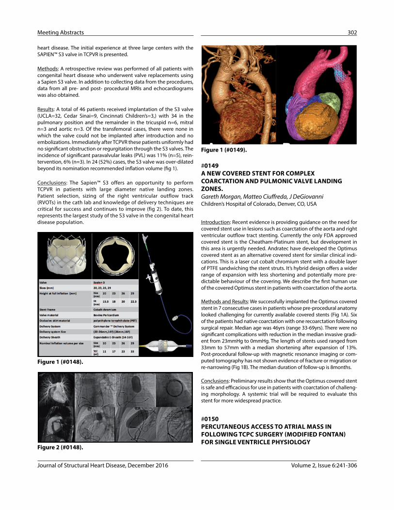
Journal of Structural Heart Disease, December 2016 Volume 2, Issue 6:241-306
Meeting Abstracts 302
#0149 A NEW COVERED STENT FOR COMPLEX COARCTATION AND PULMONIC VALVE LANDING ZONES.Gareth Morgan, Matteo Ciuffreda, J DeGiovanniChildren’s Hospital of Colorado, Denver, CO, USA
Introduction: Recent evidence is providing guidance on the need for covered stent use in lesions such as coarctation of the aorta and right ventricular outflow tract stenting. Currently the only FDA approved covered stent is the Cheatham-Platinum stent, but development in this area is urgently needed. Andratec have developed the Optimus covered stent as an alternative covered stent for similar clinical indi-cations. This is a laser cut cobalt chromium stent with a double layer of PTFE sandwiching the stent struts. It’s hybrid design offers a wider range of expansion with less shortening and potentially more pre-dictable behaviour of the covering. We describe the first human use of the covered Optimus stent in patients with coarctation of the aorta.
Methods and Results: We successfully implanted the Optimus covered stent in 7 consecutive cases in patients whose pre-procedural anatomy looked challenging for currently available covered stents (Fig 1A). Six of the patients had native coarctation with one recoarctation following surgical repair. Median age was 46yrs (range 33-69yrs). There were no significant complications with reduction in the median invasive gradi-ent from 23mmHg to 0mmHg. The length of stents used ranged from 33mm to 57mm with a median shortening after expansion of 13%. Post-procedural follow-up with magnetic resonance imaging or com-puted tomography has not shown evidence of fracture or migration or re-narrowing (Fig 1B). The median duration of follow-up is 8months.
Conclusions: Preliminary results show that the Optimus covered stent is safe and efficacious for use in patients with coarctation of challeng-ing morphology. A systemic trial will be required to evaluate this stent for more widespread practice.
#0150 PERCUTANEOUS ACCESS TO ATRIAL MASS IN FOLLOWING TCPC SURGERY (MODIFIED FONTAN) FOR SINGLE VENTRICLE PHYSIOLOGY
heart disease. The initial experience at three large centers with the SAPIEN™ S3 valve in TCPVR is presented.
Methods: A retrospective review was performed of all patients with congenital heart disease who underwent valve replacements using a Sapien S3 valve. In addition to collecting data from the procedures, data from all pre- and post- procedural MRIs and echocardiograms was also obtained.
Results: A total of 46 patients received implantation of the S3 valve (UCLA=32, Cedar Sinai=9, Cincinnati Children’s=3,) with 34 in the pulmonary position and the remainder in the tricuspid n=6, mitral n=3 and aortic n=3. Of the transfemoral cases, there were none in which the valve could not be implanted after introduction and no embolizations. Immediately after TCPVR these patients uniformly had no significant obstruction or regurgitation through the S3 valves. The incidence of significant paravalvular leaks (PVL) was 11% (n=5), rein-tervention, 6% (n=3). In 24 (52%) cases, the S3 valve was over-dilated beyond its nomination recommended inflation volume (fig 1).
Conclusions: The Sapien™ S3 offers an opportunity to perform TCPVR in patients with large diameter native landing zones. Patient selection, sizing of the right ventricular outflow track (RVOTs) in the cath lab and knowledge of delivery techniques are critical for success and continues to improve (fig 2). To date, this represents the largest study of the S3 valve in the congenital heart disease population.
Figure 1 (#0148).
Figure 2 (#0148).
Figure 1 (#0149).
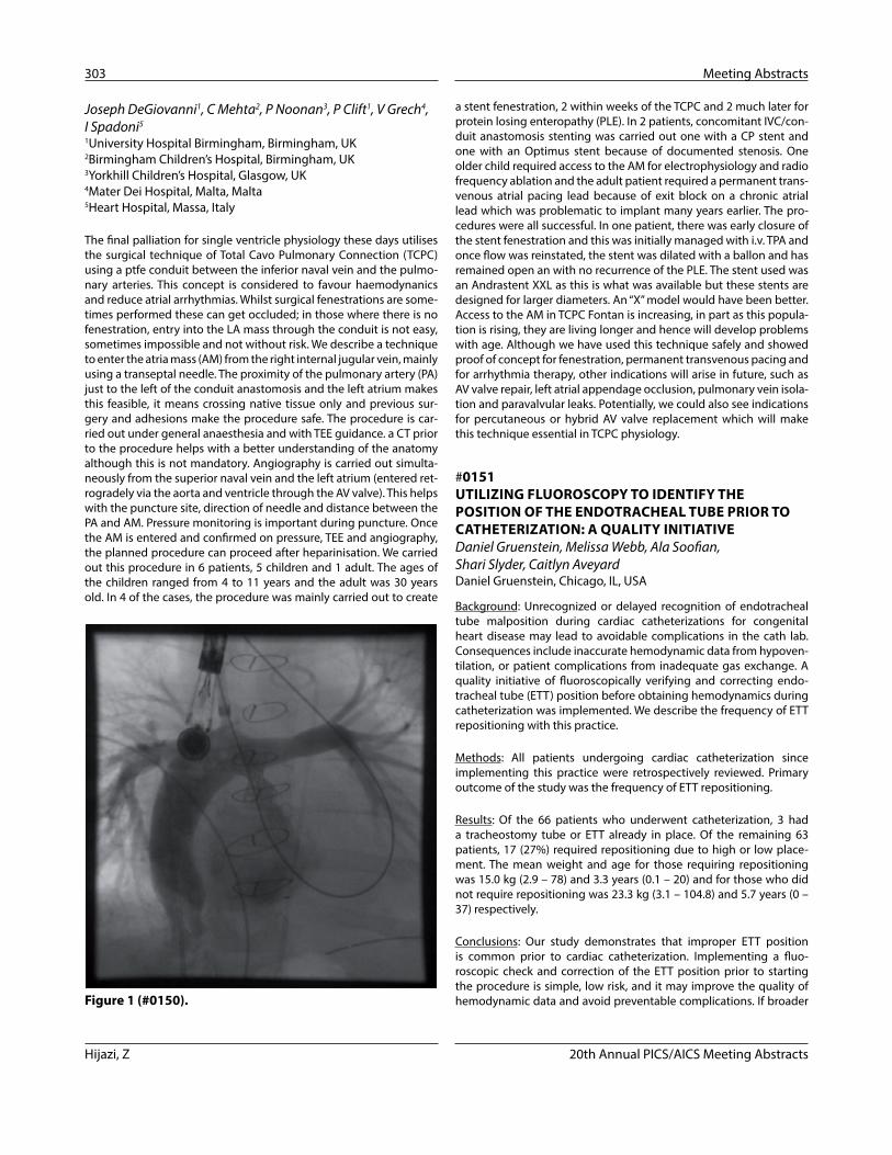
Hijazi, Z 20th Annual PICS/AICS Meeting Abstracts
303 Meeting Abstracts
a stent fenestration, 2 within weeks of the TCPC and 2 much later for protein losing enteropathy (PLE). In 2 patients, concomitant IVC/con-duit anastomosis stenting was carried out one with a CP stent and one with an Optimus stent because of documented stenosis. One older child required access to the AM for electrophysiology and radio frequency ablation and the adult patient required a permanent trans-venous atrial pacing lead because of exit block on a chronic atrial lead which was problematic to implant many years earlier. The pro-cedures were all successful. In one patient, there was early closure of the stent fenestration and this was initially managed with i.v. TPA and once flow was reinstated, the stent was dilated with a ballon and has remained open an with no recurrence of the PLE. The stent used was an Andrastent XXL as this is what was available but these stents are designed for larger diameters. An “X” model would have been better. Access to the AM in TCPC Fontan is increasing, in part as this popula-tion is rising, they are living longer and hence will develop problems with age. Although we have used this technique safely and showed proof of concept for fenestration, permanent transvenous pacing and for arrhythmia therapy, other indications will arise in future, such as AV valve repair, left atrial appendage occlusion, pulmonary vein isola-tion and paravalvular leaks. Potentially, we could also see indications for percutaneous or hybrid AV valve replacement which will make this technique essential in TCPC physiology.
#0151 UTILIZING FLUOROSCOPY TO IDENTIFY THE POSITION OF THE ENDOTRACHEAL TUBE PRIOR TO CATHETERIZATION: A QUALITY INITIATIVEDaniel Gruenstein, Melissa Webb, Ala Soofian, Shari Slyder, Caitlyn AveyardDaniel Gruenstein, Chicago, IL, USA
Background: Unrecognized or delayed recognition of endotracheal tube malposition during cardiac catheterizations for congenital heart disease may lead to avoidable complications in the cath lab. Consequences include inaccurate hemodynamic data from hypoven-tilation, or patient complications from inadequate gas exchange. A quality initiative of fluoroscopically verifying and correcting endo-tracheal tube (ETT) position before obtaining hemodynamics during catheterization was implemented. We describe the frequency of ETT repositioning with this practice.
Methods: All patients undergoing cardiac catheterization since implementing this practice were retrospectively reviewed. Primary outcome of the study was the frequency of ETT repositioning.
Results: Of the 66 patients who underwent catheterization, 3 had a tracheostomy tube or ETT already in place. Of the remaining 63 patients, 17 (27%) required repositioning due to high or low place-ment. The mean weight and age for those requiring repositioning was 15.0 kg (2.9 – 78) and 3.3 years (0.1 – 20) and for those who did not require repositioning was 23.3 kg (3.1 – 104.8) and 5.7 years (0 – 37) respectively.
Conclusions: Our study demonstrates that improper ETT position is common prior to cardiac catheterization. Implementing a fluo-roscopic check and correction of the ETT position prior to starting the procedure is simple, low risk, and it may improve the quality of hemodynamic data and avoid preventable complications. If broader
Joseph DeGiovanni1, C Mehta2, P Noonan3, P Clift1, V Grech4, I Spadoni5
1University Hospital Birmingham, Birmingham, UK2Birmingham Children’s Hospital, Birmingham, UK3Yorkhill Children’s Hospital, Glasgow, UK4Mater Dei Hospital, Malta, Malta5Heart Hospital, Massa, Italy
The final palliation for single ventricle physiology these days utilises the surgical technique of Total Cavo Pulmonary Connection (TCPC) using a ptfe conduit between the inferior naval vein and the pulmo-nary arteries. This concept is considered to favour haemodynanics and reduce atrial arrhythmias. Whilst surgical fenestrations are some-times performed these can get occluded; in those where there is no fenestration, entry into the LA mass through the conduit is not easy, sometimes impossible and not without risk. We describe a technique to enter the atria mass (AM) from the right internal jugular vein, mainly using a transeptal needle. The proximity of the pulmonary artery (PA) just to the left of the conduit anastomosis and the left atrium makes this feasible, it means crossing native tissue only and previous sur-gery and adhesions make the procedure safe. The procedure is car-ried out under general anaesthesia and with TEE guidance. a CT prior to the procedure helps with a better understanding of the anatomy although this is not mandatory. Angiography is carried out simulta-neously from the superior naval vein and the left atrium (entered ret-rogradely via the aorta and ventricle through the AV valve). This helps with the puncture site, direction of needle and distance between the PA and AM. Pressure monitoring is important during puncture. Once the AM is entered and confirmed on pressure, TEE and angiography, the planned procedure can proceed after heparinisation. We carried out this procedure in 6 patients, 5 children and 1 adult. The ages of the children ranged from 4 to 11 years and the adult was 30 years old. In 4 of the cases, the procedure was mainly carried out to create
Figure 1 (#0150).
Journal of Structural Heart Disease, December 2016 Volume 2, Issue 6:241-306
Meeting Abstracts 304
procedure, MRI study and CT scan to delineate the exact morphology of the RVOT. Before implantation, balloon dilatation of the RVOT with control aortography to obviate any coronary artery compression was performed in all. Prestenting was realized in all with LD max stent (Ev3).
A 22-mm Melody valve was implanted in 9 patients, a 20-mm valve in 2 (9/11 under left ventricular pacing). The folding techniques were employed in 8 patients, the PA branch jailing in 7, and the Russian dolls technique in 3. The folding technique on both extremities of the stent (n=1) and only on distal end (n=7) was performed because of short pulmonary artery trunk with early PA bifurcation. These tech-niques were combined in 6 patients. RVOT dilated up to 25-26 mm in diameter could be thus corrected by PPVR with a 22 mm Melody using these techniques.
During follow-up (1 to 19 months), no patient had reintervention. No endocarditis was observed.
These advanced techniques using the Melody valve can extend the clas-sical indications for PPVR beyond the revalvulation of conduit. Patched RVOT up to 25-26 mm in diameter can be repaired. These initial results are promising but more experience and longer follow-up are mandatory.
#0154 IS THE NEW OCCLUTECH PDA OCCLUDER A GOOD ALTERNATIVE FOR TRANSCATHETER CLOSURE OF PDA?François Godart, Ali HoueijehPediatric Cardiology, Lille, France
Purpose: One tertiary centre experience with the new Occlutech PDA occluder for arterial duct occlusion.
Methods: From March 2013 to October 2016, 29 patients (20 females and 9 males) underwent percutaneous arterial duct closure with the new Occlutech PDA occluder. All patients had significant L-to-R shunt with enlarged left ventricle.
At implantation, the median age was 26 months and median weight was 12kg (4.1 to 57 kg). The procedure was realized under local anaes-thesia in the majority of patients. Size of the duct was 2.94 ± 0.85 mm on angiography. According to PDA Krichenko classification, ducts were: type A (n=21), type E (n=7) and B (n=1). The systolic pulmo-nary artery pressure was 42 ± 15 mm Hg. Implantation succeeded in all. Closure was performed by the standard 4/6 mm occluder (n=14), the standard 5/7 mm occluder (n=9), the standard 6/8 mm occluder (n=4), the standard 8/10 mm occluder (n=1), and the standard 3.5/5 mm (n=1) using a 6 or 7 F delivery sheath. Technique of implantation is similar to that of the Amplatzer Duct Occluder and there is no learn-ing curve with the use of this device. After implantation, trivial shunt was noticed on angiography in 21 patients, 8 had no shunt. The mean radiation dose was 7.2 Gycm2. Neither embolization nor haemolysis was observed. After closure, femoral thrombosis was noticed in 3 patients but resolved completely under heparin therapy. On control Doppler echocardiography, 6 patients had tiny residual shunt after implantation. At the control one month later, shunt was closed in 5 of them; in the remaining one, full occlusion was observed 18 months
implementation of this practice demonstrates similar early recogni-tion of inadequate ETT position, this simple practice may be recom-mended as the standard for all laboratories.
#0152 RADIAL ARTERIAL ACCESS FOR CARDIAC CATHETERIZATION: CASE REPORTDaniel Gruenstein, Melissa Webb, S Nathan, Brojendra AgarwalaUniversity of Chicago, Chicago, IL, USA
Background: Alternative access for percutaneous interventions is becoming more common for pediatric patients with difficult access. The smallest reported child receiving radial access for cardiac cathe-terization is 39 kg and youngest 12.1 years.
Methods: A 7 year old child with Tetralogy of Fallot, pulmonary atre-sia, and MAPCAS status post RV-PA conduit and pulmonary artery unifocalization required conduit stenting due to severe right ven-tricle hypertension and conduit gradient. He had bilateral femoral artery occlusion from previous cardiac catheterizations. Left radial artery access was used for left heart catheterization and conduit bal-loon-occlusion testing for coronary compression angiograms. Post-procedural testing showed no vascular injury a patent and intact radial artery.
Conclusion: Successful radial arterial catheterization is possible with-out complication in the smaller and younger patients than previously reported. This approach for may provide safe opportunities complete procedures in patients with difficult access. The specific techniques and tips discussed in this case may help operators perform radial arterial catheterization for small pediatric patients.
#0153 ADVANCED TECHNIQUES FOR PERCUTANEOUS PULMONARY VALVE REPLACEMENT USING THE MELODY VALVE. François Godart, Ali Houijeh, Marie-Paule Guillaume, Pauline Gras, Olivia DomanskiCardiac Hospital, Lille, France
Percutaneous pulmonary valve replacement (PPVR) with the Melody valve (Medtronic) may be challenging in patients with unfavourable RVOT. Different advanced techniques have been proposed to over-come this problem: jailing and/or Russian dolls techniques or the folded valve techniques.
From March 2015 to October 2016, 11 patients (5F/6M) with a mean age of 19 ± 10 years (11-45 years) underwent these procedures. Initial pathology included tetralogy of Fallot (n=7), transposition of the great vessels (n=2), pulmonary valve stenosis (n=1), and aortic valve stenos (n=1). These patients had undergone a median of 2 pre-vious surgical repairs. The RVOT had been previously repaired with a transannular patch and 1 patient had homograft. The indications for pulmonary valve replacement were: significant pulmonary regur-gitation (n=7) and a mixed lesion (n=4). All patients had before the
Hijazi, Z 20th Annual PICS/AICS Meeting Abstracts
305 Meeting Abstracts
sinus. However, extreme caution and experienced interventionists are prerequisites to consider this type of procedure due to technical difficulties and possible complications.
#0156 RESULTS OF LD ADVANTA™ V12 COVERED STENT TRIAL FOR COARCTATION OF THE AORTA. Elchanan Bruckheimer1, Carlos Pedra2
1Schneider Children’s Medical Center Israel, Petach Tivka, Israel2Dante Pazzanese, Sao Paolo, Brazil
Objective: To assess the safety and effectiveness of the Large Diameter Advanta™ V12 Covered Stent for treatment of coarctation of the aorta.
Methods: A prospective, multicenter, non-randomized, single arm study. Primary Efficacy Endpoint: Significant reduction in gradient assessed by echo-Doppler DV (diastolic velocity) and DV/SV (diastolic velocity /systolic velocity) at pre-stent vs. immediately post stent and pre-stent vs. 12 month follow-up. Primary Safety Endpoint; MAE and MAVE occurring within 30 days.
Results: Between 2009 and 2013, 70 patients [43M, 27F] of median age 17.0 yrs [6.0-75.0] and weight 57.4Kg [28.2-120] with coarctation of the aorta were enrolled. Stents were successfully implanted and in all patients. Mean CoA diameter increased from 5.5±3.6 to 13.1±3.2mm [p<0.001]. Mean peak gradient decreased from 35.8±16.0 to 5.4±7.8mmHg [p<0.001]. A Baseline DV/SV was 0.59± 0.17 dropped to 0.34 ± 0.13 [p<0.001] after stenting and was unchanged at 12 month follow-up. DV was 216.3 ± 77.5cm/s dropped to 90.9 ± 46.5cm/s [p<0.001] and was unchanged at 12 months. 61 patients underwent CTA at ~12 months with mean stent diameter maintained at 13.3 ± 2.1mm, 91 ± 0.1% of diameter of the transverse arch.
Complications: Acute MAVE was 1.4% [1 femoral artery occlusion].MAVE at 12 months was 4.3% including 1 aortic hematoma treated with a second stent, 1 small aneurysm at distal edge of the stent treated with a second stent. At 12 months there was no new wall injury on CT. 3 patients had late [>1 yr] stent infolding requiring re-intervention.
Conclusions: The Large Diameter Advanta™ V12 Covered Stent is safe and effective for treatment in coarctation of the aorta. Long term fol-low up to assess aortic wall injury and stent integrity is required.
#0157 PREDICTORS OF PROCEDURAL FAILURE IN PATIENTS UNDERGOING PERCUTANEOUS CLOSURE OF LARGE ATRIAL SEPTAL DEFECTS USING THE AMPLATZER SEPTAL OCCLUDER.Mounir RiahiMontreal Heart Institute and St. Paul’s Hospital/University of British Columbia, Montreal/Vancouver, Canada
Aims: Owing to their more complex anatomy. large atrial septal defects can present unique challenges for percutaneous closure. Previous reports observed a much higher procedural failure rate in
after implantation. No obstruction of the left pulmonary artery or isthmic stenosis was noticed. These results are very similar to those published with the Occlutech device including 171 patients from 4 articles: successful implantation (95.4–100%), full occlusion during follow-up (96-100%); complications were observed in 4 patients: fem-oral thrombosis (n=2) and embolization (n=2).
Conclusions: Transcatheter closure of PDA with the new Occlutech PDA occluder is a safe and effective. In fact, the results are very simi-lar to that reported with the Amplatzer Duct Occluder. The Occlutech PDA occluder appears as a valuable therapeutic option for transcath-eter occlusion of the arterial duct. Further studies with longer fol-low-up are necessary to confirm these results.
#0155 SUCCESSFUL TRANSCATHETER CLOSURE OF UNROOFED CORONARY SINUS USING COVERED STENTS IN AN ADULT WITH DRAINAGE OF THE CORONARY SINUS TO THE RIGHT VENTRICLEBassel Mohammad Nijres1, Damien Kenny2, Suhaib Kazmouz3, Ziyad Hijazi4
1Rush University Children’s Hospital, Chicago, IL, USA2Our Lady’s Children’s Hospital, Dublin, Ireland3Rockford Memorial Hospital, Rockford, IL, USA4Weill Cornell Medicine & Sidra Medical and Research Center, Doha, Qatar
We present a rare case of a 41 year-old female with unroofed coronary sinus who underwent supra-annular tricuspid valve replacement with consequent drainage of the coronary sinus to the right ventricle. It is unclear whether the coronary sinus was unroofed congenitally or iatrogenically (as a result of increased pressure load on the coro-nary sinus). Either way this rare setup resulted in a significant central cyanosis as a result of shunting the desaturated blood from the right ventricle to the left atrium. In addition, she was at high risk of devel-oping paradoxical embolism and other complications of chronic cyanosis. This connection was successfully closed via trans-catheter delivery of covered stents and lead to resolution of the cyanosis. Post stenting transesophageal echocardiogram with contrast injection in the coronary sinus revealed no residual communication with the left atrium. Our patient recovered well from the procedure. On follow-up, the patient has normal systemic oxygen saturation with stable car-diac function and improved exercise tolerance.
Catheter-based management of unroofed coronary sinus is still evolving; there is only one case reporting deployment of a covered stent and limited cases reporting successful closure using Amplatzer@
Ductal Occluders. Use of covered stents may be preferable as it restores normal physiologic circulation and eliminates right to left shunting. However, it may be challenging especially in complete unroofing. Furthermore, it may be more difficult to ensure stent sta-bility and requires extreme caution to avoid obstructing coronary venous drainage into the coronary sinus. This might result in abnor-malities in electrophysiological conduction.
In conclusion, transcatheter occlusion of unroofed coronary sinus, although it is technically difficult, is feasible and provides an alterna-tive to cardiac surgery as a first line in managing unroofed coronary
Journal of Structural Heart Disease, December 2016 Volume 2, Issue 6:241-306
Meeting Abstracts 306
diameter (OR=1,6 per mm; p<0,0001), absence of any type of mar-gin (OR= 4,8, p<0,0001) and absence of postero-inferior margin (OR= 39,3; p<0,0001). Multivariate regression analysis identified non-stretched diameter (OR= 1,17 per mm; p=0,05), absence of any type of margin (OR=5,25; p=0,0004) and absence of postero-inferior mar-gin (OR>100, p=0,05) as independent predictors of failure.
Conclusion: Success rate of transcatheter closure of large atrial sep-tal defects using the amplatzer device is lower when compared to small to moderate defects. A large unstretched diameter, absence of any type of margin and more strongly absence of postero-inferior margin were independantly associated with procedural failure. These findings can be assessed prior to the procedure and could improve patient selection.
Comment on this Article or Ask a Question
Cite this article as: Hijazi ZM. The Pediatric and Adult Interventional Cardiac Symposium (PICS/AICS) – 20th Annual Meeting. Structural Heart Disease 2016;2(6):241-306. DOI: http://dx.doi.org/10.12945/j.jshd.2016.16.013
this specific subgroup when compared to small and moderate size defects. We sought to determine clinical and anatomical predictors of procedural failure in patients undegoing percutaneous closure of atrial septal defects larger than 24mm using the AmplatzerTM Occluder device (St.Jude Medical Inc.).
Method & Results: Adult patients refered between January 2006 and July 2013 for percutaneous closure of a secundum atrial sep-tal defect (ASD) with a stretched diameter higher than 24 mm were included in this study. Clinical, imaging and procedural data were retrospectively collected from index admission to last follow-up. Patients underwent ASD closure according to standard indications under transeosophageal and fluoroscopic guidance. A total of 90 patients, mean age 47±16 years, 60% female, met inclusion crite-ria. Patients presented with right heart failure symptoms in 81,1 % of cases, 93,3 % had right ventricular enlargement and 7,8 % had a prior embolic event. Median non-stretched and stretched ASD diameters were 24,0 mm (20;28) and 29,0 mm (25,5;34,0) respec-tively. Procedural success rate was 81,1 %, one patient (2,2%) rec-quired emergent surgery because of a ruptured septum after sizing-balloon inflation resulting in cardiac tamponnade. During a median follow-up of 19,5 months, we observed 4 (4,4%) episodes of systemic thrombo-embolism, 5 (5,5%) new onset supraventricular tachyarrhytmias, no cardiac erosion, no device embolization and no cardiovascular death.
In univariate logistic regression analysis predictors of failure were non-stretched diameter (OR= 1,25 per mm; p=0,0002), stretched