the origin of the "xestoleberis-spot"
TRANSCRIPT
Copyright © 1988 by Kodansha Ltd., Tokyo, Japan .. Evolutionary Biology on Ostracoda" Proceedings of the Ninth International Symosium on Ostracoda, edited by T. Hanai, N. Ikeya and K. Ishizaki
The Origin of the "Xestoleberis-spot"
DIETMAR I{EYSER
Zoologisches Institut und Museum, Hamburg, F.R. Germany
ABSTRACT
The Xestoleberis-spot of Xestoleberis aurantia is studied in detail. The main feature of the spot is an irregularity in the inner part of the calcified exocuticle. Apart from that it is just a place where two muscle scars are present. The question of whether the varying size of the spinning gland lying in between these muscles is the cause of noncalcification in the Xestoleberis-spot is discussed.
INTRODUCTION
Among the podocopid ostracods, the family Xestoleberididae stands apart from the other families. This family sprang up, as many others did, in Cretaceous times (Deroo, 1966; Van Veen, 1936). Since then it has not changed much in character. Nothing is known about its ancestry and its relationship to other families.
It is characterised by a smooth, highly arched shell which is pointed anteriorly and flattened ventrally. A diagnostic feature of the whole group is the so-called "Xestoleberis-spot" (or "nierenformiger Fleck"). This spot is a conspicuous irregularity visible on the inner surface ofthe detached valve. G.W. Mfiller in 1884 was the first to notice this structure and believed that this feature was a remnant of a "valve-gland". However in 1894 he recanted this and mentioned only that this structure belonged to the inner chitin layer of the calcified lamella of the shell (G. W. M filler, 1894, p. 93). Wagner (1957) mentioned this spot in his thesis as antennal muscle scars, although with a question mark. An outstanding photograph of this spot was presented by Triebel (1958, PI. 2, fig. 9) but without explanation. McKenzie (1972) referred to it as "eye-scar", an explanation which is found more often in the younger literature. Bonaduce et al. (1980) figured the spot on the outside of Ornatoleberis, where it appears on a pitted surface as a smooth area, comparable to the region of the eye.
The only conclusion that can be drawn from all these works is that nothing is known about the morphology, origin and function of this peculiar structure. The present study was undertaken to gather more information on this structure and to solve the problem of the function of the "Xestoleberis-spot. "
MATERIAL AND METHODS
The material for this study was mainly Xestoleberis aurantia (Baird, 1838) collected in the Baltic
177
178 D. KEYSER
Sea. About fifty specimens were available for light microscope, transmission electron microscope and scanning electron microscope studies. The specimens were collected with a handnet, 180 J1.m mesh, in the phytal zone at a depth of between 1 and 8 metres. They were picked out individually under a stereo-microscope using a pipette. The animals were kept alive in a small bowl at 15°C with 14 hours of light, a weekly change of water and no extra food.
Several live animals were observed under the stereo-microscope. Afterwards they were fixed in 70 % ethyl alcohol, dissected and embedded in polyvinyl-Iactophenol stained with Orange-G. Some were heated in KOH prior to dissection and were then embedded. Others were put in clove oil for a fortnight to make them translucent and then examined under the microscope. Specimens used for sectioning and examination under the light microscope (LM) and in the TEM were treated in the same manner. They were initially fixed in 2.5 % glutardialdehyde in 0.05 mol phosphate-buffer and 5 % sucrose and then washed in buffer with sucrose three times. Postfixed with 2 % OS04 in the same buffer, they were then washed three times in buffer, decalcified in EDTA and dehydrated in graded acetone, before being embedded in Spurr's resin (Spurr, 1969). Semi-thin and ultra-thin sections were cut with a Reichert Om/U II Ultramicrotom. Semi-thin sections were stained with Toluidinblue and Pyronin after Holstein and Wulfhenkel (1971), ultra-thin sections with uranylacetate (Stemper and Ward, 1964) and leadcitrate (Reynolds, 1963). Photographs were taken with a Leitz Dialux for light microscope and a Zeiss EM 9 for TEM.
Specimens for the SEM were fixed in 2 % glutardialdehyde in phosphate-buffer, dehydrated in acetone and critical-point dried with COz in a Balzers CPT. They were sputtered in a GEA-004 S manufactured in Graz, Austria, and viewed under an SEM Cambridge S-4. These dried specimens were broken up with fine needles to allow the inner structure to be seen.
RESULTS
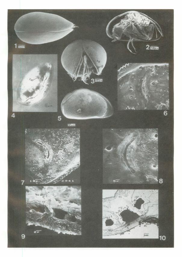
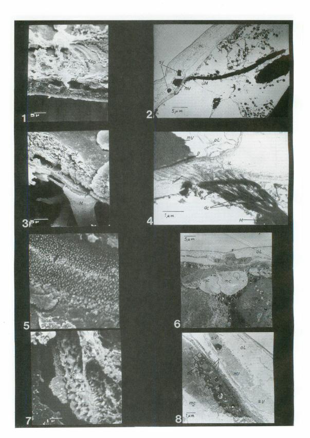
Xestoleberis auraniia (Baird, 1838) is an ostracod of 500 J1.m length. The shell is roughly round when seen end on and triangular from the side. In dorsal view it is slightly egg-shaped with a pointed anterior (PI. 1, fig. I). The ventral part is flattened. The surface of the valves is smooth with several simple or sieve type pore canals. The fused zone is broad. The calcified inner lamella is especially broad in the anterior part thus forming a pronounced vestibule (PI. 1, fig. 5). The hinge is merodont. The central muscle field consists of four vertically arranged scars and a V-shaped frontal scar. Behind the eye region is a typical reniform spot, which is diagnostic for all Xestoleberididae, the so-called "Xestoleberis-spot" (PI. 1, figs. 4-8).
The nauplius-eye of Xestoleberis aurantia is divided into three parts. Two are laterally coalesced with the valves, just in front of the frontal end of the hinge. The third eye is found at the top of the forehead on the same level as both lateral eyes (PI. 1, fig. 3). The "Xestoleberis-spot" is situated behind the lateral eyes. It is always separated from the eye-scar. From the outside no imprint is visible in this area on the surface. In transmitted light an elongated inclusion, bordered by several bubble-like structures (PI. 1, fig. 4, s.a. Triebel, 1958) is visible.
In single detached valves the inner view reveals mostly a slitlike, elongated and elevated struc-
PLATE I-Xestoleberis aurantia (Baird, 1838) Fig. 1. Dorsal view. Fig. 2. Lateral view of the complete male animal with removed right valve. Fig. 3.
Frontal view. Fig. 4. Light microscopic view of the animal, showing the "Xestoleberis-spot". Fig. 5. Interior view of the left valve, showing the "Xestoleberis-spot" with two slits. Fig. 6. "Xestoleberis-spot" with two muscle scars. Fig. 7. "Xestoleberis-spot" with two slits. Fig. 8. "Xestoleberis-spot" with one big slit. Fig. 9. Transverse fracture through the "Xestoleberis-spot" showing the main and two side vesicles within the calcified outer layer of the cuticle. Fig. 10. The same area as in Fig. 9 only in a TEM section.
Origin of"Xestoleberis-spot" 181
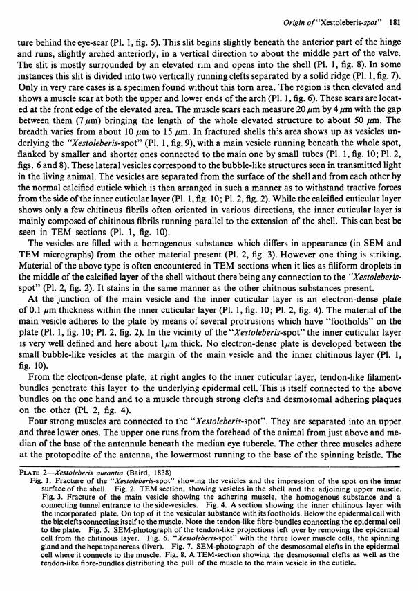
ture behind the eye-scar (PI. 1, fig. 5). This slit begins slightly beneath the anterior part of the hinge and runs, slightly arched anteriorly, in a vertical direction to about the middle part of the valve. The slit is mostly surrounded by an elevated rim and opens into the shell (PI. 1, fig. 8). In some instances this slit is divided into two vertically running clefts separated by a solid ridge (PI. 1, fig. 7). Only in very rare cases is a specimen found without this torn area. The region is then elevated and shows a muscle scar at both the upper and lower ends of the arch (PI. 1, fig. 6). These scars are located at the front edge of the elevated area. The muscle scars each measure 20 pm by 4 pm with the gap between them (7 pm) bringing the length of the whole elevated structure to about 50 pm. The breadth varies from about 10 pm to 15 pm. In fractured shells th:s area shows up as vesicles underlying the "Xestoleberis-spot" (PI. 1, fig. 9), with a main vesicle running beneath the whole spot, flanked by smaller and shorter ones connected to the main one by small tubes (PI. 1, fig. 10; PI. 2, figs. 6 and 8). These lateral vesicles correspond to the bubble-like structures seen in transmitted light in the living animal. The vesicles are separated from the surface of the shell and from each other by the normal calcified cuticle which is then arranged in such a manner as to withstand tractive forces from the side of the inner cuticular layer (PI. 1, fig. 10; PI. 2, fig. 2). While the calcified cuticular layer shows only a few chitinous fibrils often oriented in various directions, the inner cuticular layer is mainly composed of chitinous fibrils running parallel to the extension of the shell. This can best be seen in TEM sections (PI. 1, fig. 10).
The vesicles are filled with a homogenous substance which differs in appearance (in SEM and TEM micrographs) from the other material present (PI. 2, fig. 3). However one thing is striking. Material of the above type is often encountered in TEM sections when it lies as filiform droplets in the middle of the calcified layer of the shell without there being any connection to the "Xestoleberisspot" (PI. 2, fig. 2). It stains in the same manner as the other chitnous substances present.
At the junction of the main vesicle and the inner cuticular layer is an electron-dense plate of 0.1 pm thickness within the inner cuticular layer (PI. 1, fig. 10; PI. 2, fig. 4). The material of the main vesicle adheres to the plate by means of several protrusions which have "footholds" on the plate (PI. 1, fig. 10; PI. 2, fig. 2). In the vicinity of the "Xestoleberis-spot" the inner cuticular layer is very well defined and here about Ipm thick. No electron-dense plate is developed between the small bubble-like vesicles at the margin of the main vesicle and the inner chitinous layer (PI. 1, fig. 10).
From the electron-dense plate, at right angles to the inner cuticular layer, tendon-like filamentbundles penetrate this layer to the underlying epidermal cell. This is itself connected to the above bundles on the one hand and to a muscle through strong clefts and desmosomal adhering plaques on the other (PI. 2, fig. 4).
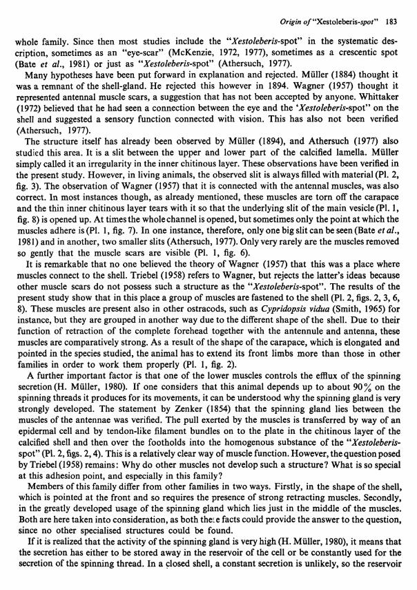
Four strong muscles are connected to the "Xestoleberis-spot". They are separated into an upper and three lower ones. The upper one runs from the forehead of the animal from just above and median of the base of the antennule beneath the median eye tubercle. The other three muscles adhere at the protopodite of the antenna, the lowermost running to the base of the spinning bristle. The
PLATE 2-Xestoleberis aurantia (Baird, 1838) Fig. 1. Fracture of the "Xestoleberis-spot" showing the vesicles and the impression of the spot on the inner
surface of the shell. Fig. 2. TEM section, showing vesicles in the shell and the adjoining upper muscle. Fig. 3. Fracture of the main vesicle showing the adhering muscle, the homogenous substance and a connecting tunnel entrance to the side-vesicles. Fig. 4. A section showing the inner chitinous layer with the incorporated plate. On top of it the vesicular substance with its footholds. Below the epidermal cell with the big clefts connecting itself to the muscle. Note the tendon-like fibre-bundles connecting the epidermal cell to the plate. Fig. 5. SE M-photograph of the tendon-like projections left over by removing the epidermal cell from the chitinous layer. Fig. 6. "Xestoleberis-spot" with the three lower muscle cells, the spinning gland and the hepatopancreas (liver). Fig. 7. SE M-photograph of the desmosomal clefts in the epidermal cell where it connects to the muscle. Fig. 8. A TEM-section showing the desmosomal clefts as well as the tendon-like fibre-bundles distributing the pull of the muscle to the main vesicle in the cuticle.
182 D. KEYSER
TEXT-FIG. l-Schematic drawing of the internal organisation of the functionally important parts related to the "Xestoleberis-spot".
muscle scars which are sometimes visible in an inner view of the" Xestoleberis-spot" are the adhesion points of the upper and all three lower muscles (Text-fig. 1). Between the space of the upper and lower muscles lies the spinning gland with its reservoir. In the posterior part it is linked to the hepatopancreas (PI. 2, fig. 6).
The upper muscle meets a second one which connects to the shell more dorsally than the "Xestoleberis-spot". Both lie in the vicinity of the lateral eye and run ventrally of the lateral eye nerve to the frontal part of the head. The lower muscle follows more or less the path of the excretion tube of the spinning gland. A characteristic feature of the muscle cells is a swelling of their base at the shell. The reason for the swelling is not yet understood since one part of the cytoplasma does not have any inclusions and stains very light, and another part of the cell opposite the muscle fibres appears normal with dense cytoplasma (PI. 2, fig. 6).
A connection to the eye is not visible. There is also no hint in the morphologic structure of an underlying gland. The area does not show any special nervous connections. The only specialized region in the "Xestoleberis-spot" is the three arched vault in the non-living material of the calcified lamella in the shell.
DISCUSSION
Since 1884, when Muller (p. 14) first mentioned the "Xestoleberis-spot", not many investigations have dealt with this feature. Muller (1894) himself noticed the systematic value of the spot when he used it in his generic diagnosis. He also noticed this spot in the genus Microxestoleberis. Most of the later authors did not mention this diagnostic feature for the genus (Sars, 1928; Klie, 1938). However this changed when Triebel (1958) recognized that the spot was a diagnostic feature for the
Origin o!"Xesto)eberis-spot" 183
whole family. Since then most studies include the "Xestoleberis-spot" in the systematic description, sometimes as an "eye-scar" (McKenzie, 1972, 1977), sometimes as a crescentic spot (Bate et al., 1981) or just as "Xestoleberis-spot" (Athersuch, 1977).
Many hypotheses have been put forward in explanation and rejected. Muller (1884) thought it was a remnant of the shell-gland. He rejected this however in 1894. Wagner (1957) thought it represented antennal muscle scars, a suggestion that has not been accepted by anyone. Whittaker (1972) believed that he had seen a connection between the eye and the 'Xestoleberis-spot" on the shell and suggested a sensory function connected with vision. This has also not been verified (Athersuch, 1977).
The structure itself has already been observed by Muller (1894), and Athersuch (1977) also studied this area. It is a slit between the upper and lower part of the calcified lamella. Muller simply called it an irregularity in the inner chitinous layer. These observations have been verified in the present study. However, in living animals, the observed slit is always filled with material (PI. 2, fig. 3). The observation of Wagner (1957) that it is connected with the antennal muscles, was also correct. In most instances though, as already mentioned, these muscles are torn off the carapace and the thin inner chitinous layer tears with it so that the underlying slit of the main vesicle (PI. 1, fig. 8) is opened up. At times the whole channel is opened, but sometimes only the point at which the muscles adhere is (PI. 1, fig. 7). In one instance, therefore, only one big slit can be seen (Bate et al., 1981) and in another, two smaller slits (Athersuch, 1977). Only very rarely are the muscles removed so gently that the muscle scars are visible (PI. 1, fig. 6).
It is remarkable that no one believed the theory of Wagner (1957) that this was a place where muscles connect to the shell. Triebel (1958) refers to Wagner, but rejects the latter's ideas because other muscle scars do not possess such a structure as the "Xestoleberis-spot". The results of the present study show that in this place a group of muscles are fastened to the shell (PI. 2, figs. 2, 3, 6, 8). These muscles are present also in other ostracods, such as Cypridopsis vidua (Smith, 1965) for instance, but they are grouped in another way due to the different shape of the shell. Due to their function of retraction of the complete forehead together with the antennule and antenna, these muscles are comparatively strong. As a result of the shape of the carapace, which is elongated and pointed in the species studied, the animal has to extend its front limbs more than those in other families in order to work them properly (PI. 1, fig. 2).
A further important factor is that one of the lower muscles controls the efflux of the spinning secretion(H. Muller, 1980). If one considers that this animal depends up to about 90% on the spinning threads it produces for its movements, it can be understood why the spinning gland is very strongly developed. The statement by Zenker (1854) that the spinning gland lies between the muscles of the antennae was verified. The pull exerted by the muscles is transferred by way of an epidermal cell and by tendon-like filament bundles on to the plate in the chitinous layer of the calcified shell and then over the footholds into the homogenous substance of the "Xestoleberisspot" (pI. 2, figs. 2, 4). This is a relatively clear way of muscle function. However, the question posed by Triebel (1958) remains: Why do other muscles not develop such a structure? What is so special at this adhesion point, and especially in this family?
Members of this family differ from other families in two ways. Firstly, in the shape of the shell, which is pointed at the front and so requires the presence of strong retracting muscles. Secondly, in the greatly developed usage of the spinning gland which lies just in the middle of the muscles. Both are here taken into consideration, as both the:.e facts could provide the answer to the question, since no other specialised structures could be found.
If it is realized that the activity of the spinning gland is very high (H. Muller, 1980), it means that the secretion has either to be stored away in the reservoir of the cell or be constantly used for the secretion of the spinning thread. In a ~losed shell, a constant secretion is unlikely, so the reservoir

184 D. KEYSER
spG
TEXT-FIG. 2-Muscle direction with empty reservoir of spinning gland.
uM
TEXT-FIG. 3-Muscle direction with filled reservoir of spinning gland.
of the spinning gland has to change constantly its volume. If the muscles contract when the reservoir is empty, the force of the muscles is applied at right angles to the shell (Text-fig. 2). If the reservoir is filled, the muscles have to strain around this swollen, ball-like structure (Text-fig. 3). This means a different direction of pull at the shell. Such constant change in the direction of pull at the muscle scar must account for the development of the "Xestoleberis-spot". Two facts therefore explain the reason for this spot. Firstly, the scars must sit on the shell in such a way that it is flexible. This is achieved by the morphological structure found. Secondly, during calcification after moulting, it is not possible to develop calcite in this area because of continuous changing stress. The vesicles, therefore, contain only the matrix of the calcified layer of the cuticle. Such an explanation would also account for it having the same staining quality as is found in the other chitinous parts of the shell.
Several questions must follow such an explanation. With a spinning gland developed in many Cytheracea (Hartmann, 1966), why is such a spot only developed in the Xestoleberididae? The answer must lie in the different internal organisation and body shape of the animals. For example, in Hirschmannia the corresponding muscles are very small, in Semicytherura they lie in front of the gland, in Hemicythere the gland is not so strong and in Paradoxostoma there are no muscles in the vicinity.
A further question must be: Why are the spots so different that their shape can sometimes be characteristic for a species (Hartmann pers. comm.)? It appears that the outline of the carapace in connection with the position of the spinning gland can account for these difference",. In ",hort, high specimens the spot is often more prominant than in longer, more depressed forms. (Hartmann, 1978, 1979, 1980, 1981; Athersuch, 1976). Several other questions still remain to be solved. For example: what is the development in larval animals? why do the muscle cells have such swollen bases? and several others.
SUMMARY
The "Xestoleberis-spot" of Xestoleberis aurantia (Baird, 1838) was investigated. The main feature of the spot is an irregularity in the calcified layer of the shell. The spot consists of a main vesicle with several bubble-like extensions within in the calcified matrix of the cuticle. These structures are filled with a homogenous substance which has the same staining qualities as the other chitinous substan-
Origin o/"Xestoleberis-spot" 185
ces of the calcified shell. Between these vesicles in the calcified layer and the epidermal cells is a distinct inner chitinous layer. It is stabilized at the border to the main channel in a plate which connects tendon-like fibre-bundles from the epidermal cell with small foot-like extensions from the homogenous substance.
Four muscles are connected to the "Xestoleberis-spot", one upper and three lower ones. The upl\et on.e connects the shell with the forehead medially from the first antenna. The lower muscles run to the prodopodite of the second antenna. Enclosed by the muscles is the spinning gland.
The different degree of filling of the reservoir of the spinning gland alters the direction of the pulling force of the muscles. This is thought to be the reason for the flexible nature of the "Xestoleberis-spot" .
The different size of the spot in different species is due to the difference in the shape of the body and also to the position of spinning gland.
ACKNOWLEDGEMENTS
I would like to thank Prof. Dr. G. Hartmann, Hamburg, for his advice and help during the preparation of this paper. My thanks are also due to Mr. J. Mallwitz and Miss B. Rhode for all the fruitful discussions we had.
For technical assistance I am indebted to Miss E. Ganss, Miss Schacht, Miss M. Hiinel and Miss K. Meyer, all of Hamburg. For reading and making comments on the manuscript, I want to express my thanks to Dr. D. L. Biirkel of Hamburg.
REFERENCES
ATHERSUCH, J. 1976. The genus Xestoleberis (Crustacea: Ostracoda) with particular reference to Recent Mediterranean species. Pubbl. Staz. Zool. Napoli, 40, 282-343.
BATE, R.H., WHITTAKER, I.E. and MAYES, C.A. 1981. Marine Ostracoda of the Galapagos Islands and Ecuador. Zool. J. Linnean Soc., 73, 1-79.
BONADUCE, G., MASOLI, M., MINICHELLI, G. and PUGLIESE, N. 1980. Some new Bentic Marine Ostracod species from the Gulf of Aqaba (Red Sea). Boil. Soc. Paleont. Italiana, 19, 143-178.
DEROO, G. 1966. Cytheracea (Ostracodes) du Maastrichtien de Maastricht (Pays-Bas) et des regions voisines; resultats stratigraphiques et paleontologiques de leur etude. Medel. Geol. Stichting Serie C, 2, 1-197.
HARTMANN, G. 1966-1975. Ostracoda. In. Bronns Klassen und Ordnungen des Tierreichs, 5. Bd. (Arthropoda), 1. Abt. Crustacea, 2. Buch. IV Teil, 1-4, Lie/erung, 1-786. Akademische Verlagsgesellschaft Leipzig.
-- 1979. Die Ostracoden der Ordnung Podocopida G.W. Miiller, 1894 der warmtemperierten (antiborealen) West- und Siidwestkiiste Australiens (zwischen Perth im Norden und Eucla im Siiden). Mitt. hamb. zoo!. Mus. Inst. 76, 219-301.
--1980. Die Ostracoden der Ordnung Podocopida G.W. Miiller, 1894 der warmtemperierten und subtropischtropischen Kiistenabschnitte der Siid- und Siidostkiiste Australiens (zwischen Ceduna im Western und Lakes Entrance im Osten). Ibid., 77, 111-204.
-- 1981. Die Ostracoden der Ordnung Podocopida G.W. Miiller, 1894 der subtropisch-tropischen Ostkiiste AustraIiens (zwischen Eden im Siiden und Heron-Island im Norden). Ibid., 78, 97-149.
-- PURl, H.S. 1974. Summary of Neontologic and Paleontological Classification of Ostracoda. Ibid, 70, 7-73. HARTMANN-SCHRODER, G. and HARTMANN, G. 1978. Zur Kenntnis des Eulitora1s der australischen Kiisten unter
besonderer Beriicksichtigung der Polychaeten und Ostracoden. Ibid. 75, 63-219. HOLSTEIN, A.F. and WULFHENKEL, u. 1971. Die Semidiinnschnitt-Technik als Grundlage fUr eine cytologische
Beurteilung der Spermatogenese des Menschen. Andrologie 3, 65-69. KUE, w. 1938. Ostracoda. In Dah/'s Die Tierwelt Deutschlands 34. Crustacea 3, 1-230. MCKENZIE, K.G. 1972. New data on the ostracode genera Laocoonella deVos and Stock, Redekea deVos, and
Aspidoconcha deVos; with a key to the family Xestoleberididae and a resume of symbiosis in Ostracoda. Beau/ortia 19, 152-162.
186 D. KEYSER
-- 1977. La Faune terrestre de rIle de Sainte-Helene, Ostracoda. Koninkl. Mus. MiddenAfrika. Zool. Wetensch 220, 444-451.
MULLER, G.W. 1884. Zur naheren Kenntnis der Cytheriden. Arch. Naturgesch. SO, 1-18. -- 1894. Die Ostracoden des Golfes von Neapel und der angrenzenden Meeres Abschnitte. Fauna und Flora
des Golfes von Neapel, 21, 1-404. . MULLER, H. 1980. Untersuchungen zur Struktur und biologischen Funktion des Spinndrusenkomplexes der Cytheracea
(Ostracoda. Crustacea) unter besonderer Berucksichtigung von Xestoleberis aurantia. Unpubl. Diss. Univers. Hamburg 1980, 1-142.
REYNOLDS, E.S. 1963. The use of lead citrate at high pH as an electronopaque stain in electron microscopy. J. Cell. Bioi. 17, 208
SARS, G.O. 1928. An account of the crustacea of Norway, 9 Ostracoda, 1-277. SMITH, R.N. 1965. Musculature and Muscle scars of Chlamydotheca arcuata (Sars) and Cypridopsis vidua (O.F.
Miiller) (Ostracoda-Cyprididae). National Science Foundation Project, GB-26, Report 3, 1-40. SPURR, A.R. 1969. A low-viscosity epoxy resin embedding medium for electron microscopy. J. Ultrstr. Res. 26, 31. STEMPER, I.C. and WARD, R.T. 1964. An improved staining method for electron micrcscopy. J. Cell Bioi. 22, 697. TRIEBEL, E. 1958. Zwei neue Ostracoden-Gattungen aus dem Lutet des Pariser Beckens. Senckenbergiana leth. 39,
105-117. VAN VEEN, I.E. 1936. Die Cytheridae der Maastrichter Tuffkreide und des Kuurader Korallenkalkes von Siid-Limburg.
Ill. Die Gatatungen Loxoconcha, Monoceratina, Paracytheridea, Xestoleberis, Cytheropteron und Cytherura. Natuurhist. Maandbl. 25, 21-113.
WAGNER, C.W. 1957. Sur les Ostracodes du Quaternaire Recent des Pays-Bas et leur utilisation dans !'etude geologique des depots holocenes. (These, Univ. Paris, 's-Gravenhage), 259 pp. Mouton and Co., The Hague.
WHITTAKER, J.E. 1972. Recent Ostracoda from Christchurch Harbour, The Fleet and Weymouth Bay. Unpubl. Thesis, University College of Wales, Aberystwyth.
ZENKER, W. 1854. Monographie der Ostracoden. Arch. Naturgesch. 20, 1-87.
ABBREVIATIONS
AI = antennule All = antenna dc = desmosomal cleft ec = epidermal cell H = hinge HP = hepatopancreas (liver) iL = inner chitinous layer of the cuticle lE = lateral eye 1 M = lower muscles mc = muscle cell mE = median eye
m V = main vesicle oL = outer calcified layer of the cuticle P = plate in the inner chitinous layer R = reservoir of the spinning gland spB = spinning bristle spG = spinning gland sV = side vesicle t = tendon-like filament bundles tu = tunnel from main vesicle to side vesicle uM = upper muscle Xsp = Xestoleberis-spot
DISCUSSION
Kaesler: How variable is the size of the spinning gland? Keyser: It varies from 35-40 Jlm in diameter when it is full, down to 5 Jlm in diameter when it is
empty. Schweitzer: Did you use critical-point drying techniques to mount your SEM specimens? Keyser: Yes. I did; you will find the methods used in the written paper.