the fha and brct domains recognize adp-ribosylation during dna damage response
TRANSCRIPT
The FHA and BRCT domains recognizeADP-ribosylation during DNA damageresponse
Mo Li,1 Lin-Yu Lu,1 Chao-Yie Yang,2,3,4 Shaomeng Wang,2,3,4 and Xiaochun Yu1,5
1Division of Molecular Medicine and Genetics, Department of Internal Medicine, University of Michigan, Ann Arbor, Michigan48109, USA; 2Department of Internal Medicine, 3Department of Pharmacology, 4Department of Medicinal Chemistry,University of Michigan, Ann Arbor, Michigan 48109, USA
Poly-ADP-ribosylation is a unique post-translational modification participating in many biological processes, suchas DNA damage response. Here, we demonstrate that a set of Forkhead-associated (FHA) and BRCA1 C-terminal(BRCT) domains recognizes poly(ADP-ribose) (PAR) both in vitro and in vivo. Among these FHA and BRCTdomains, the FHA domains of APTX and PNKP interact with iso-ADP-ribose, the linkage of PAR, whereas theBRCT domains of Ligase4, XRCC1, and NBS1 recognize ADP-ribose, the basic unit of PAR. The interactionsbetween PAR and the FHA or BRCT domains mediate the relocation of these domain-containing proteins to DNAdamage sites and facilitate the DNA damage response. Moreover, the interaction between PAR and the NBS1BRCT domain is important for the early activation of ATM during DNA damage response and ATM-dependentcell cycle checkpoint activation. Taken together, our results demonstrate two novel PAR-binding modules thatplay important roles in DNA damage response.
[Keywords: poly(ADP-ribose); PAR binding domain; DNA damage]
Supplemental material is available for this article.
Received February 15, 2013; revised version accepted July 19, 2013.
Post-translational modifications, such as protein phos-phorylation, ubiquitination, acetylation, and methylation,are important for numerous biological processes. ProteinADP-ribosylation is a unique post-translational modi-fication that has been shown to play critical roles inmany cellular events, especially DNA damage response(Schreiber et al. 2006; Gibson and Kraus 2012).
Poly-ADP-ribosylation is catalyzed by poly-ADP-ribo-sylation polymerases (PARPs) (Ame et al. 2004; Hottigeret al. 2010). Using NAD+ as the donor, ADP-ribose iscovalently linked to the side chains of arginine, asparticacid, and glutamic acid residues in target proteins. Aftercatalyzing the first ADP-ribose on the target, other ADP-riboses can be covalently linked onto the first ADP-riboseto form both liner and branched polymers, known aspoly(ADP-ribose) (PAR) (Schreiber et al. 2006; Luo andKraus 2012). Following DNA damage, massive poly-ADP-ribosylation is synthesized at DNA lesions within a fewseconds (D’Amours et al. 1999; Kim et al. 2005). To date,two major nuclear substrates of poly-ADP-ribosylation areknown: the PARP1 enzyme itself and histones (Schreiberet al. 2006; Messner and Hottiger 2011). Once PAR is
synthesized at the DNA damage site, it is also quicklyrecognized and hydrolyzed by PAR glycosylase (PARG)(D’Amours et al. 1999; Gagne et al. 2006; Kim et al.2007a). Thus, the half-life of PAR at DNA damage sitesis very short, and the biological function of this dynamicpost-translation modification at DNA damage sites re-mains elusive.
Recently, several PAR-binding proteins have been iden-tified as the ‘‘readers’’ to recognize PAR signals (Karraset al. 2005; Ahel et al. 2008; Wang et al. 2012), suggestingthat PAR is likely to function as recruiting signals toinduce DNA damage response factors to DNA damagesites. To identify other PAR-binding modules, we exam-ined both the Forkhead-associated (FHA) domain and theBRCA1 C-terminal (BRCT) domain. The FHA and BRCTdomains are known as phospho-protein-binding domains.Many FHA domain- or BRCT domain-containing proteinsare involved in DNA damage response (Li et al. 2002;Glover et al. 2004; Mahajan et al. 2008; Mohammad andYaffe 2009). It has been shown that the FHA domainsrecognize phospho-Thr (pThr) motifs (Sun et al. 1998;Durocher et al. 1999, 2000; Li et al. 2002; Mahajan et al.2008). For example, the FHA domain of Rad53 recognizesthe pThr motif of Rad9 in budding yeast (Sun et al. 1998),the FHA domain of fission yeast NBS1 recognizes thepThr of Ctp1(Williams et al. 2009), and the FHA domain
5Corresponding authorE-mail [email protected] is online at http://www.genesdev.org/cgi/doi/10.1101/gad.226357.113.
1752 GENES & DEVELOPMENT 27:1752–1768 � 2013, Published by Cold Spring Harbor Laboratory Press; ISSN 0890-9369/13; www.genesdev.org
Cold Spring Harbor Laboratory Press on June 11, 2016 - Published by genesdev.cshlp.orgDownloaded from
of human NBS1 and RNF8 recognizes the pThr motifs ofMDC1 (Chapman and Jackson 2008; Spycher et al. 2008;Wu et al. 2008; Lloyd et al. 2009). While the BRCTdomains have been shown to recognize phospho-Ser (pSer)motifs (Manke et al. 2003; Yu et al. 2003), the BRCA1BRCT domain binds pSer motifs in several downstreampartners (Yu et al. 2003; Yu and Chen 2004; Kim et al.2007b; Liu et al. 2007; Wang et al. 2007). The MDC1 BRCTdomain recognizes the pS139 site of H2AX (Stucki et al.2005). These phospho-protein-dependent interactionsare important for DNA damage checkpoint activationand DNA damage repair. However, based on the peptidescreening, not all of the FHA and BRCT domains havehigh affinity to phospho-proteins (Durocher et al. 2000;Rodriguez et al. 2003). In particularly, our recent studysuggests that the BARD1 BRCT domain recognizesADP-ribose (Baer 2013; Li and Yu 2013). Thus, we askedwhether these domains have other binding partners besidesphospho-proteins.
Following the DNA damage, PAR is quickly synthe-sized at the DNA damage sites. Interestingly, one ADP-ribose residue contains two phosphate groups. Thus,massive PAR synthesized upon DNA damage brings hugeamounts of phosphate moieties at DNA damage sites ina very short period. We therefore wondered whether theFHA and BRCT domains could recognize PAR. In thisstudy, we screened 19 FHA and BRCT domains and foundthat five of them bind PAR both in vitro and in vivo.Moreover, the interaction with PAR facilitates the fastrecruitment of these FHA or BRCT domain-containingproteins to DNA lesions and the relevant DNA damagerepair process.
Results
A set of BRCT and FHA domains binds PAR
Since CHFR and APLF are known PAR-binding proteins,which bind PAR via their PBZ motifs (Ahel et al. 2008; Liet al. 2010; Oberoi et al. 2010), we used GST, recombinantCHFR, and APLF PBZ motifs as negative and positivecontrols, respectively, to screen PAR-binding domains.We examined 19 FHA or BRCT domains and found thattwo FHA domains (from PNKP and APTX), two BRCTdomains (from Ligase4 and XRCC1), and an FHA–BRCTfusion domain (from NBS1) interacted with PAR (Fig. 1A).Reverse pull-down assays confirmed the direct interac-tion between PAR and these FHA/BRCT domains (Fig.1B). Using isothermal titration calorimetry (ITC) assays,we measured the affinity between PAR and these FHA/BRCT domains (Fig. 1C), which is in the physiologicallyrelevant range and is similar to that between PAR andother PAR-binding domains (Karras et al. 2005; Wanget al. 2012). Since PAR is the ADP-ribose polymer withmixed length and contains both linear and branchedforms, the affinity between the PAR-binding domainand PAR could not be accurately measured. We thenmeasured the affinity between these FHA/BRCT domainsand ADP-ribose, the basic unit of PAR. The ITC resultsshow that the BRCT domains of Ligase4 and XRCC1 and
the FHA–BRCT fusion domain of NBS1 recognize ADP-ribose. However, the FHA domains of PNKP and APTXdo not interact with ADP-ribose (Fig. 1D). Since the FHAdomains of PNKP and APTX recognize PAR, we wonderwhether these FHA domains interact with the linkagebetween each ADP-ribose in PAR. We used phosphodies-terase to digest PAR into iso-ADP-ribose, the linkagebetween two individual ADP-riboses, and found that theFHA domains of PNKP and APTX have high affinity withiso-ADP-ribose (Fig. 1E). Following DNA damage, PAR isheavily synthesized at DNA lesions (D’Amours et al. 1999;Kim et al. 2005). It has been reported that PNKP, APTX,Ligase4, XRCC1, and NBS1 all participate in DNA damageresponse (Su 2006; Polo and Jackson 2011). We examinedthe in vivo interactions between PAR and PNKP, APTX,Ligase4, XRCC1, or NBS1. With ionizing radiation (IR)treatment, PAR was significantly synthesized in thecells and interacted with PNKP, APTX, Ligase4, XRCC1,or NBS1 (Fig. 1F). Taken together, these results demon-strate that a set of FHA/BRCT domains interacts withPAR.
PAR-binding pockets are conserved in the BRCTand FHA domains
Next, we examined the PAR-binding sites in these FHA/BRCT domains. Different FHA or BRCT domains arepredicted to fold into similar secondary structures, re-spectively, with binding pockets that can recognize pThror pSer motifs (Fig. 2A; Glover et al. 2004; Mahajan et al.2008; Mohammad and Yaffe 2009). We asked whether thesimilar binding pockets recognize iso-ADP-ribose andADP-ribose, respectively. For the FHA domains of PNKPand APTX, two conserved arginine residues in eachbinding pocket were mutated into alanines. Both mutantsabolished the interaction with PAR in the pull-down andreciprocal pull-down assays (Fig. 2B,C). Moreover, we didnot detect any affinity between the mutant FHA domainsand iso-ADP-ribose using ITC assays (Fig. 2D). FollowingIR treatment, the FHA domain mutations abolished theinteraction between PAR and PNKP or APTX in vivo (Fig.2E). Single mutation in the binding pocket of the BRCA1BRCT domain abolished the interaction between the pSermotif and the BRCA1 BRCT domain (Shiozaki et al.2004). Based on the similarity of the secondary structurein the BRCT domain, we mutated the conserved Ser orLys residues to disrupt the putative binding pocket in theBRCT domain of Ligase4 and XRCC1 (Fig. 2A). Like thoseFHA domain mutants, these BRCT domain mutantsabolished the interaction with PAR in vitro (Fig. 2B andC). The full-length proteins bearing these BRCT domainmutations failed to interact with PAR in vivo (Fig. 2E). Inaddition to these FHA domains and BRCT domains,NBS1 has a FHA domain and a BRCT domain that arefused together. This FHA–BRCT domain also recognizesPAR. To study which subdomain of this FHA–BRCTfusion domain recognizes ADP-ribose, we mutated theconserved residues in each binding pocket. As shown inFigure 2, B and D, mutation of conserved Lys residue inthe FHA fold did not significantly affect the interaction
The FHA and BRCT domains bind poly(ADP-ribose)
GENES & DEVELOPMENT 1753
Cold Spring Harbor Laboratory Press on June 11, 2016 - Published by genesdev.cshlp.orgDownloaded from
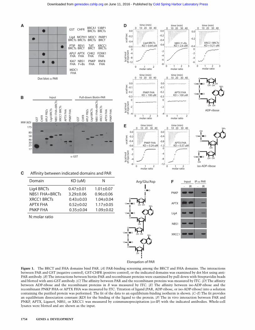
Figure 1. The BRCT and FHA domains bind PAR. (A) PAR-binding screening among the BRCT and FHA domains. The interactionsbetween PAR and GST (negative control), GST-CHFR (positive control), or the indicated domains was examined by dot blot using anti-PAR antibody. (B) The interactions between biotin-PAR and recombinant proteins were examined by pull-down with Streptavidin beadsand blotted with anti-GST antibody. (C) The affinity between PAR and the recombinant proteins was measured by ITC. (D) The affinitybetween ADP-ribose and the recombinant proteins in B was measured by ITC. (E) The affinity between iso-ADP-ribose and therecombinant PNKP FHA or APTX FHA was measured by ITC. Titration of ligand (PAR, ADP-ribose, or iso-ADP-ribose) into a solutioncontaining the purified protein was performed. The fit of the data to an equilibrium-binding isotherm is shown. (C–E) The fit providesan equilibrium dissociation constant (KD) for the binding of the ligand to the protein. (F) The in vivo interaction between PAR andPNKP, APTX, Ligase4, NBS1, or XRCC1 was measured by coimmunoprecipitation (co-IP) with the indicated antibodies. Whole-celllysates were blotted and are shown as the input.
1754 GENES & DEVELOPMENT
Cold Spring Harbor Laboratory Press on June 11, 2016 - Published by genesdev.cshlp.orgDownloaded from
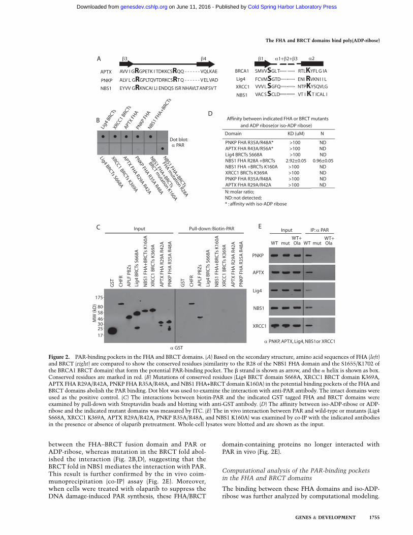
between the FHA–BRCT fusion domain and PAR orADP-ribose, whereas mutation in the BRCT fold abol-ished the interaction (Fig. 2B,D), suggesting that theBRCT fold in NBS1 mediates the interaction with PAR.This result is further confirmed by the in vivo coim-munoprecipitation (co-IP) assay (Fig. 2E). Moreover,when cells were treated with olaparib to suppress theDNA damage-induced PAR synthesis, these FHA/BRCT
domain-containing proteins no longer interacted withPAR in vivo (Fig. 2E).
Computational analysis of the PAR-binding pocketsin the FHA and BRCT domains
The binding between these FHA domains and iso-ADP-ribose was further analyzed by computational modeling.
Figure 2. PAR-binding pockets in the FHA and BRCT domains. (A) Based on the secondary structure, amino acid sequences of FHA (left)and BRCT (right) are compared to show the conserved residues (similarity to the R28 of the NBS1 FHA domain and the S1655/K1702 ofthe BRCA1 BRCT domain) that form the potential PAR-binding pocket. The b strand is shown as arrow, and the a helix is shown as box.Conserved residues are marked in red. (B) Mutations of conserved residues (Lig4 BRCT domain S668A, XRCC1 BRCT domain K369A,APTX FHA R29A/R42A, PNKP FHA R35A/R48A, and NBS1 FHA+BRCT domain K160A) in the potential binding pockets of the FHA andBRCT domains abolish the PAR binding. Dot blot was used to examine the interaction with anti-PAR antibody. The intact domains wereused as the positive control. (C) The interactions between biotin-PAR and the indicated GST tagged FHA and BRCT domains wereexamined by pull-down with Streptavidin beads and blotting with anti-GST antibody. (D) The affinity between iso-ADP-ribose or ADP-ribose and the indicated mutant domains was measured by ITC. (E) The in vivo interaction between PAR and wild-type or mutants (Lig4S668A, XRCC1 K369A, APTX R29A/R42A, PNKP R35A/R48A, and NBS1 K160A) was examined by co-IP with the indicated antibodiesin the presence or absence of olaparib pretreatment. Whole-cell lysates were blotted and are shown as the input.
The FHA and BRCT domains bind poly(ADP-ribose)
GENES & DEVELOPMENT 1755
Cold Spring Harbor Laboratory Press on June 11, 2016 - Published by genesdev.cshlp.orgDownloaded from
The structure of several FHA domains, including the FHAdomains of PNKP, APTX, CHK2, and RNF8, has beensolved (Li et al. 2002; Huen et al. 2007; Ali et al. 2009;Becherel et al. 2010). We examined the binding pockets ofthese FHA domains. The electrostatic potential of thesefour proteins, calculated by the MM-PBSA program,indicated that the peptide-binding sites are primarilypositively charged that are complementary and primedfor the recognizing negatively charged molecules such asphosphate moieties. The FHA domain of PNKP has beenshown to recognize an adjacent pS–pT motif derived fromXRCC1 (Whitehouse et al. 2001; Loizou et al. 2004; Aliet al. 2009). However, the affinity between the PNKPFHA domain and iso-ADP-ribose is even slightly higherthan that between the PNKP FHA domain and the pS–pTpeptide (Supplemental Fig. 1). Consistently, the bindingpocket formed by the R35 and R48 of the PNKP FHAdomain well accommodates iso-ADP-ribose, in whichtwo phosphate groups mimic the two phosphate groupsin the pS–pT peptide and form salt bridges with Argresidues (Supplemental Fig. 1A). The binding model wasfurther validated by a rigorous binding free energy calcu-lation; namely, the MM-PBSA method (Rastelli et al.2010), which has been employed for estimating the rela-tive binding free energy of protein with peptide and proteinwith small molecules (Kollman et al. 2000), including theBRCA1 with phospho-peptides (Anisimov et al. 2011) andviruses with ADP-ribose (Rungrotmongkol et al. 2010)reported recently. Of note, such calculations provideestimates of the relative binding free energy but not theabsolute binding free energy of the protein with a ligand.Thus, the calculated binding free energies are ideal to becompared with experimental binding affinities of differ-ent ligands with the same protein. The binding freeenergy between the PNKP FHA domain and iso-ADP-ribose, calculated by the MM-PBSM method, is lowerthan that between the PNKP FHA domain and pS–pTpeptide (Supplemental Fig. 1B), indicating a strongerbinding affinity between the PNKP FHA domain andiso-ADP-ribose. These results are consistent with thebinding affinities measured by ITC assays (Supplemen-tal Fig. 1D). The binding pocket formed by R29 and R42in the FHA domain of APTX is similar to that of PNKP(Supplemental Fig. 2A). It has been reported that theFHA domain of APTX recognizes the pS-D–pT-D motif ofMDC1 (Becherel et al. 2010). The affinity between theFHA domain of ATPX and the pS-D–pT-D peptide isconsistent with previous published results. However,the binding between the FHA domain of ATPX and iso-ADP-ribose is much stronger. Again, based on the struc-ture of the FHA domain of APTX (Becherel et al. 2010),the analyses by the MM-PBSM method suggest that thebinding free energy between the APTX FHA and iso-ADP-ribose is much lower than that between the APTX FHAdomain and the pS-D–pT-D peptide, which is consistentwith the binding affinity measured by ITC assays (Sup-plemental Fig. 2B–D). Moreover, the R44 in PNKP isreplaced by the more flexible K38 in APTX. The mobilityof the K38 in APTX may contribute to a less favorablebinding to the phospho-peptide than iso-ADP-ribose. As
indicated by the models in Supplemental Figure 2A, K38in APTX moves away from the peptide, whereas it co-operates with R29 and R42 to interact with the phos-phate group in iso-ADP-ribose. Moreover, the two phos-phate groups in iso-ADP-ribose are on the surface of FHAdomains in PNKP and APTX and can connect with addi-tional units of poly-(ADP-ribose) in PAR (SupplementalFigs. 1A, 2A).
The structure of the FHA domain of RNF8 and CHK2 isquite different from that of PNKP and APTX (Li et al.2002; Huen et al. 2007). Both FHA domains failed tointeract with iso-ADP-ribose or PAR (Supplemental Fig.3). The FHA domain of RNF8 recognizes the pT-Q motifsin MDC1 (Huen et al. 2007; Kolas et al. 2007; Mailandet al. 2007). The binding pocket in the FHA domain ofRNF8 includes two hydrophobic residues (L57 and L82),creating a narrower and electro-neutral binding site thatfavors binding Glu residue in the peptide. Although thefirst phosphate group in iso-ADP-ribose could form saltbridge with R61 of RNF8, the second phosphate groupdoes not mimic the Glu of the binding peptide, which canbe attributed to the steric hindrance caused by L57 inRNF8 (Supplemental Fig. 3A). Similarly, only one phos-phate group could be fitted into the binding pocket inthe FHA domain of CHK2 (Supplemental Fig. 3A). Thecalculated binding free energy suggests that the FHAdomains of RNF8 and CHK2 favor interacting with pTmotifs over iso-ADP-ribose, which is also consistent withour ITC analyses (Supplemental Fig. 3B,C).
We also examined the binding pocket in the BRCTdomains of Ligase4, XRCC1, BRCA1, and MDC1 becausethe structure of these BRCT domains has been solved(Zhang et al. 1998; Sibanda et al. 2001; Shiozaki et al.2004; Stucki et al. 2005; Wu et al. 2009; Campbell et al.2010; Cuneo et al. 2011). However, the binding pocketsin the BRCT domains of BRCA1 and MDC1 are muchlarger compared with those in the FHA domain. Al-though one phosphate group in ADP-ribose can form saltbridges with K1936 and T1898 in MDC1 or with K1702and S1655 in BRCA1, the pockets form few contactswith ribose surges and the other phosphate in ADP-ribose. Once the adenine of ADP-ribose is docked intothe binding pockets, one ribose sugar could not be linkedwith other unit of PAR (Supplemental Fig. 4A). More-over, the binding free energy suggests that both BRCTdomains favor binding to their phospho-protein partnersover ADP-ribose, which once again is consistent withour results from ITC assays (Supplemental Fig. 4B,C).The phospho-peptide-binding partners of the BRCT do-mains of Ligase4 and XRCC1 have not been identified.However, both BRCT domains recognize ADP-ribose.Thus, we could not compare their binding affinities withphospho-proteins as well as the binding models. Based onthe structure of the unbound BRCT domains and muta-tion analyses in Supplemental Figure 5, we showed thatthe phosphate groups in ADP-ribose interact with K675in Ligase4 and K369 in XRCC1. Based on the bindingmodels, the ADP-ribose adopts an extended conforma-tion to bind to the binding site, allowing two sugar groupsto connect with the remaining units of PAR. Due to
Li et al.
1756 GENES & DEVELOPMENT
Cold Spring Harbor Laboratory Press on June 11, 2016 - Published by genesdev.cshlp.orgDownloaded from
lacking the structure of the human NBS1 FHA–BRCTdomain, we are currently unable to examine the detailsof the interaction between the NBS1 BRCT domain andADP-ribose.
Collectively, the model analyses allow us to examinethe details of the interaction between the FHA/BRCTdomains and PAR, which might provide the molecularbasis of the interactions.
PAR mediates the early recruitment of NBS1 duringDNA damage response
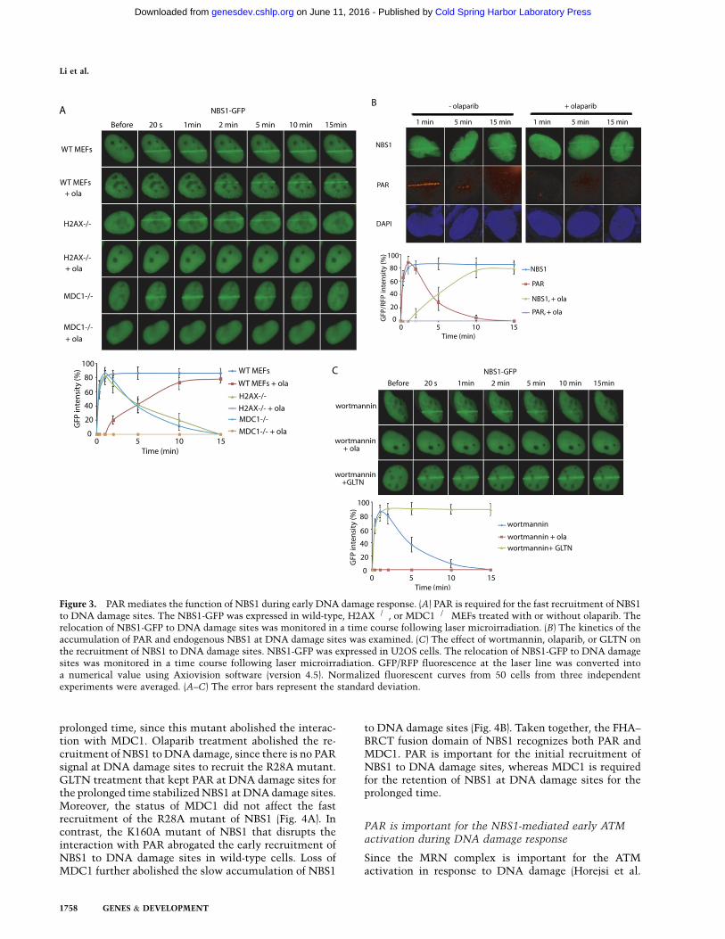
Massive poly-ADP-ribosylation occurs at DNA lesions,suggesting that PAR could provide critical signals torecruit PAR-binding proteins to DNA lesions and allowthese PAR-binding proteins to fulfill their DNA damagerepair missions. To study the biological function of thesenovel PAR-binding domains, we used laser microirradia-tion to examine whether PAR recruits the FHA and BRCTdomains to DNA damage sites. The advantage of the lasermicroirradiation approach is that we can monitor theearly DNA damage response in live cells, since the half-life of PAR is very short at DNA lesions. The times of cellexposure to the laser beam and the pulse energy werestrictly equal in every operation (see the Materials andMethods for details), which ensured the same laser micro-irradiation treatment to all the cells. We first examinedNBS1, since NBS1 is an important subunit in the MRNcomplex that activates ATM in response to DNA damage(Horejsi et al. 2004; Lee and Paull 2004; Stracker and Petrini2011). As shown in Figure 3A, NBS1 was quickly recruitedto DNA damage sites within 20 sec. The quick loading ofNBS1 at DNA damage sites is consistent with the quickPAR synthesis at DNA damage sites (Fig. 3B), suggestingthat PAR at DNA damage sites mediates the fast recruit-ment of NBS. Next, we treated cells with olaparib, thePARP inhibitor, to suppress PAR synthesis. Olaparib treat-ment abolished the early recruitment of NBS1 to DNAdamage, although NBS1 still slowly accumulated at DNAdamage sites (Fig. 3A), confirming that PAR is required forthe fast relocation of NBS to DNA damage sites. The half-life of PAR is relatively short at DNA damage sites becausePAR is quickly hydrolyzed by PARG at DNA damage sites.However, NBS1 was still retained at DNA damage siteseven after PAR was digested (Fig. 3B). These results suggestthat PAR mediates the fast recruitment of NBS1 to DNAdamage sites, while other signals at DNA damage sites areimportant for the prolonged retention of NBS1 at DNAdamage sites. Following DNA damage, gH2AX plays animportant role in retaining DNA damage response factorsat DNA damage sites (Celeste et al. 2003; Bonner et al.2008; Lukas et al. 2011; Polo and Jackoson 2011). Thus,we examined whether gH2AX was associated with theprolonged retention of NBS1. Interestingly, in H2AX�/�
cells, the stable retention of NBS1 at DNA damage siteswas impaired. Following the hydrolysis of PAR, NBS1was dropped from DNA damage sites (Fig. 3A). Moreover,additional olaparib treatment in H2AX�/� cells abolishedboth the fast recruitment and slow accumulation of NBS1to DNA damage sites. These results suggest that there are
two stages of the recruitment of NBS1 to DNA damagesites. The quick relocation of NBS1 to DNA damage sitesis mediated by PAR at DNA damage sites, whereas theslow accumulation or stable retaining of NBS1 at DNAdamage sites is regulated by H2AX. The FHA–BRCTfusion domain is essential for the relocation of NBS1 toDNA damage sites (Kobayashi et al. 2004; Stracker andPetrini 2011). The BRCT fold of NBS1 recognizes PAR,whereas the FHA fold of NBS1 could recognize the pThrmotifs in MDC1 (Chapman and Jackson 2008; Spycheret al. 2008; Wu et al. 2008; Lloyd et al. 2009), a functionalpartner of gH2AX (Stucki et al. 2005; Lou et al. 2006).Again, in MDC1�/� cells, the stable retention of NBS1 toDNA damage sites is impaired, and olaparib treatmentabolished the relocation of NBS1 to DNA damage sites inMDC1�/� cells (Fig. 3A). Similar results were observedwhen we examined the relocation of the FHA+BRCTdomain of NBS1 to DNA damage sites (Supplemental Fig.6), and olaparib treatment did not affect the relocationkinetics of gH2AX or MDC1 at DNA damage sites inwild-type cells (Supplemental Fig. 7A). Thus, these re-sults suggest that PAR is the initial signal that inducesthe recruitment of NBS1 to DNA damage sites, whileMDC1 retains NBS1 at DNA damage sites for the prolongedperiod. To validate this model, cells were treated withwortmannin, a PI3 kinase inhibitor, to suppress gH2AXand the recruitment of MDC1 to DNA lesions (Paull et al.2000; Goldberg et al. 2003) but not affect the DNA damage-induced PAR synthesis at DNA damage sites (SupplementalFig. 7B). With wortmannin treatment, NBS1 could still berecruited to DNA damage sites. However, it was dropped offfrom DNA damage sites when PAR was hydrolyzed (Fig.3C). When cells were treated with both wortmannin andolaparib to suppress both the gH2AX-dependent pathwayand PAR synthesis, NBS1 failed to be recruited to DNAdamage sites (Fig. 3C). Moreover, with the treatment ofgallotannin (GLTN), a cell-permeable PARG inhibitor (Yinget al. 2001; Fathers et al. 2012), to suppress PARG-dependentPAR hydrolysis, the half-life of PAR at DNA damage siteswas significantly prolonged (Supplemental Fig. 7C). Withboth wortmannin and GLTN treatment to suppress gH2AXand prolong the half-life of PAR, NBS1 was still stablyretained by PAR at DNA damage sites for the prolongedtime (Fig. 3C). Collectively, these results demonstrate thatPAR synthesis at DNA damage sites is critical for therecruitment of NBS1.
The interaction between PAR and the BRCT domainof NBS1 is important for the recruitment of NBS1to DNA damage sites
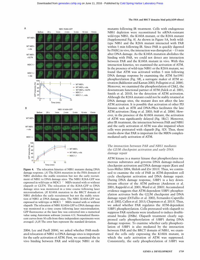
Since the BRCT fold of NBS1 is a PAR-binding motif andthe FHA fold of NBS1 is a phospho-protein-binding motif,we further dissected the functions of these two motifs invivo. We mutated Arg28 into alanine (R28A) in the FHAfold of NBS1, which does not affect the interactionbetween NBS1 and PAR. As shown in Figure 4A, in thewild-type mouse embryonic fibroblasts (MEFs), the R28Amutant could still be recruited to DNA damage sites butwere not able to stay at DNA damage sites for the
The FHA and BRCT domains bind poly(ADP-ribose)
GENES & DEVELOPMENT 1757
Cold Spring Harbor Laboratory Press on June 11, 2016 - Published by genesdev.cshlp.orgDownloaded from
prolonged time, since this mutant abolished the interac-tion with MDC1. Olaparib treatment abolished the re-cruitment of NBS1 to DNA damage, since there is no PARsignal at DNA damage sites to recruit the R28A mutant.GLTN treatment that kept PAR at DNA damage sites forthe prolonged time stabilized NBS1 at DNA damage sites.Moreover, the status of MDC1 did not affect the fastrecruitment of the R28A mutant of NBS1 (Fig. 4A). Incontrast, the K160A mutant of NBS1 that disrupts theinteraction with PAR abrogated the early recruitment ofNBS1 to DNA damage sites in wild-type cells. Loss ofMDC1 further abolished the slow accumulation of NBS1
to DNA damage sites (Fig. 4B). Taken together, the FHA–BRCT fusion domain of NBS1 recognizes both PAR andMDC1. PAR is important for the initial recruitment ofNBS1 to DNA damage sites, whereas MDC1 is requiredfor the retention of NBS1 at DNA damage sites for theprolonged time.
PAR is important for the NBS1-mediated early ATMactivation during DNA damage response
Since the MRN complex is important for the ATMactivation in response to DNA damage (Horejsi et al.
Figure 3. PAR mediates the function of NBS1 during early DNA damage response. (A) PAR is required for the fast recruitment of NBS1to DNA damage sites. The NBS1-GFP was expressed in wild-type, H2AX�/�, or MDC1�/� MEFs treated with or without olaparib. Therelocation of NBS1-GFP to DNA damage sites was monitored in a time course following laser microirradiation. (B) The kinetics of theaccumulation of PAR and endogenous NBS1 at DNA damage sites was examined. (C) The effect of wortmannin, olaparib, or GLTN onthe recruitment of NBS1 to DNA damage sites. NBS1-GFP was expressed in U2OS cells. The relocation of NBS1-GFP to DNA damagesites was monitored in a time course following laser microirradiation. GFP/RFP fluorescence at the laser line was converted intoa numerical value using Axiovision software (version 4.5). Normalized fluorescent curves from 50 cells from three independentexperiments were averaged. (A–C) The error bars represent the standard deviation.
Li et al.
1758 GENES & DEVELOPMENT
Cold Spring Harbor Laboratory Press on June 11, 2016 - Published by genesdev.cshlp.orgDownloaded from
2004; Lee and Paull 2004), we asked whether PAR-medi-ated relocation of NBS1 to DNA damage sites is importantfor the early activation of ATM. First, we examined the invivo binding between PAR and wild-type NBS1 or the
mutants following IR treatment. Cells with endogenousNBS1 depletion were reconstituted by siRNA-resistantwild-type NBS1, the K160A mutant, or the R28A mutant(Supplemental Fig. 8). As shown in Figure 5A, both wild-type NBS1 and the R28A mutant interacted with PARwithin 5 min following IR. Since PAR is quickly digestedby PARG in vivo, the interaction was disrupted at ;15 minafter DNA damage. As the K160A mutation abolishes thebinding with PAR, we could not detect any interactionbetween PAR and the K160A mutant in vivo. With thisinteraction kinetics, we examined the activation of ATM.In the presence of wild-type NBS1 or the R28A mutant, wefound that ATM was activated within 5 min followingDNA damage response by examining the ATM Ser1981phosphorylation (Fig. 5B), a surrogate maker of ATM ac-tivation (Bakkenist and Kastan 2003; Pellegrini et al. 2006).Moreover, we examined the phosphorylation of Chk2, thedownstream functional partner of ATM (Falck et al. 2001;Smith et al. 2010), for the detection of ATM activation.Although the R28A mutant could not be stably retained atDNA damage sites, the mutant does not affect the lateATM activation. It is possible that activation of other PI3kinases such as ATR and DNA-PKcs facilitates the lateATM activation (Yang et al. 2003; Stiff et al. 2006). How-ever, in the presence of the K160A mutant, the activationof ATM was significantly delayed (Fig. 5B,C). Moreover,after IR treatment, the interaction between PAR and NBS1and the early activation of ATM was also impaired whencells were pretreated with olaparib (Fig. 5D). Thus, theseresults show that PAR is important for the MRN complex-mediated early activation of ATM.
The interaction between PAR and NBS1 mediatesthe G2/M checkpoint activation and early DNAdamage repair
ATM kinase is a master kinase that phosphorylates nu-merous substrates and governs DNA damage-inducedcheckpoint activation and DNA damage repair (Kurz andLees-Miller 2004; Shiloh and Ziv 2013). Thus, we contin-ued to examine the role of PAR in ATM-dependent cellcycle checkpoint activation and DNA damage repair.During DNA damage response, 53BP1 is a key down-stream effector of the ATM pathway (Anderson et al.2001; Rappold et al. 2001; Ward et al. 2003). Accumulatedevidence suggests that ATM-dependent 53BP1 phosphor-ylation activates both the G2/M checkpoint and DNAdamage repair (DiTullio et al. 2002; Fernandez-Capetilloet al. 2002; Callen et al. 2013; Chapman et al. 2013). Thus,we asked whether PAR regulates the ATM-dependent53BP1 phosphorylation. Cells pretreated with olaparib tosuppress PAR synthesis were irradiated to induce double-strand breaks (DSBs). Olaparib treatment clearly sup-pressed early phosphorylation of 53BP1 during DNAdamage response. To examine whether early phosphory-lation of 53BP1 is also mediated by the interactionbetween PAR and the BRCT domain of NBS1, we exam-ined the cells only expressing the K160A mutant, inwhich the early activation of ATM was suppressed.Consistently, the early phosphorylation of 53BP1 was
Figure 4. The relocation kinetics of NBS1 mutants during DNAdamage response. (A) The R28A mutation in the FHA domain ofNBS1 abolishes the stable retention but not the early recruit-ment of NBS1 to DNA damage sites. The NBS1 R28A-GFP wasexpressed in wild-type or MDC1�/�MEFs treated with or withoutolaparib or GLTN. The relocation of the R28A-GFP to DNAdamage sites was monitored in a time course following lasermicroirradiation. (B) K160A mutation in the BRCT domain ofNBS1 abolishes the early recruitment but not the stable reten-tion of NBS1 at DNA damage sites. The NBS1 K160A-GFP wasexpressed in wild-type or MDC1�/� MEFs treated with or withoutolaparib. The relocation of NBS1 K160A-GFP to DNA damage siteswas monitored in a time course following laser microirradiation.GFP fluorescence at the laser line was converted into a numericalvalue using Axiovision software (version 4.5). Normalized fluores-cent curves from 50 cells from three independent experiments wereaveraged. (A,B) The error bars represent the standard deviation.
The FHA and BRCT domains bind poly(ADP-ribose)
GENES & DEVELOPMENT 1759
Cold Spring Harbor Laboratory Press on June 11, 2016 - Published by genesdev.cshlp.orgDownloaded from

suppressed in the presence of the K160A mutant of NBS1compared with that in the presence of wild-type NBS1(Fig. 6A). 53BP1 has been shown to mediate the G2/Mcheckpoint activation and DNA damage repair. (DiTullioet al. 2002; Fernandez-Capetillo et al. 2002; Callen et al.
2013; Chapman et al. 2013) Here, we examined both theG2/M checkpoint activation and DNA damage repair.Following DSBs, cells are transiently arrested beforeentering mitosis to provide enough time for DNA damagerepair. This short and transient cell cycle arrestment atthe G2/M boundary is named as the G2/M checkpoint(Lukas et al. 2004). To examine the G2/M checkpoint, wemonitored mitotic population by examining the phospho-histone H3 population following DSBs, which is a stan-dard assay for studying the transient G2/M checkpoint(Wang et al. 2002; Wu et al. 2011). As shown in Figure 6B,with IR treatment, normal cells were arrested beforemitosis, as phospho-histone H3 positively stained cellswere significantly reduced. However, with olaparibtreatment, cells could not be fully arrested at the G2/Mboundary, suggesting the loss of the transient G2/Mcheckpoint. Moreover, compared with wild-type NBS1,the K160A mutant also abrogated the G2/M checkpointfollowing IR-induced DSBs. Next, we examined theearly DNA damage repair using comet assays. Again,DNA damage repair was significantly impaired whencells were treated with olaparib or only expressed theK160A mutant of NBS1 (Fig. 6C). Taken together, theseresults suggest that the PAR-mediated ATM activationis likely to be critical for the early checkpoint activa-tion and DNA damage repair.
PAR mediates the early recruitment of PNKP, APTX,Ligase4, and XRCC1 to DNA damage sites
Next, we examined the role of PAR for the recruitment ofother FHA and BRCT domain-containing proteins. ForPNKP, it was recruited to DNA damage sites, and olaparibtreatment impaired the early recruitment of PNKP (Sup-plemental Fig. 9A). Like NBS1, PNKP could not be stablyretained at DNA damage sites in the H2AX�/� cells.Lacking both PAR and H2AX totally abolished the re-location of PNKP to DNA damage sites. Interestingly, theR35A/R48A mutant PNKP also abolished the relocationof PNKP to DNA damage sites, suggesting that the FHAdomain of PNKP is important not only for the earlyrecruitment of PNKP, but also for the PNKP retention atDNA damage sites. A similar phenomenon was observedon the relocation of the PNKP FHA domain to DNAdamage sites (Supplemental Fig. 9B). Thus, it is likely thatthe FHA domain of PNKP recognizes phosphate groupsin other molecule besides PAR, which is important forthe stability of PNKP at DNA damage sites. It has beenreported that PNKP interacted with XRCC1 (Whitehouseet al. 2001; Mani et al. 2007), which also recognizes PARat DNA damage sites. However, the recruitment of PNKPto DNA damage sites is independent of XRCC1 (Supple-mental Fig. 9C). Moreover, when cells were treated withwortmannin to suppress DNA damage–induced phos-phorylation signal (Sarkaria et al. 1998), the retention ofPNKP at DNA damage sites was significantly impaired.Wortmannin and olaparib treatment together additivelyabolished the recruitment of PNKP to DNA damage sites(Supplemental Fig. 9D). Thus, it is likely that the re-cruitment of PNKP to DNA damage sites is via the direct
Figure 5. PAR is important for the NBS1-mediated early ac-tivation of ATM in response to DNA damage. (A) NBS1 bindsPAR during the early DNA damage response. U2OS cells withendogenous NBS1 knockdown were reconstituted by siRNA-resistant wild-type NBS1, the R28A mutant, or the K160Amutant. Cells were lysed at the indicated time points after IR.The in vivo interaction between PAR and wild-type NBS1 or themutants was measured by co-IP. Whole-cell lysates were blottedand are shown as the input. (B,C) PAR is important for theNBS1-mediated early activation of ATM and Chk2 during DNAdamage response. U2OS cells with endogenous NBS1 knock-down were reconstituted by siRNA-resistant wild-type NBS1,the R28A mutant, or the K160A mutant. Following IR treat-ment, cells were lysed at the indicated time points and sub-jected to Western blot detected by anti-pATM (S1981) and anti-pChk2 (T68) antibodies. (D) The NBS1–PAR interaction and theearly activation of ATM and Chk2 during DNA damage re-sponse were abolished by the PARP inhibitor olaparib. Follow-ing IR treatment, cells pretreated by olaparib were lysed at 5min (left) or the indicated time points (right). (Left) The NBS1–PAR binding was detected by co-IP. (Right) The early activationof ATM and Chk2 was detected by Western blot.
Li et al.
1760 GENES & DEVELOPMENT
Cold Spring Harbor Laboratory Press on June 11, 2016 - Published by genesdev.cshlp.orgDownloaded from
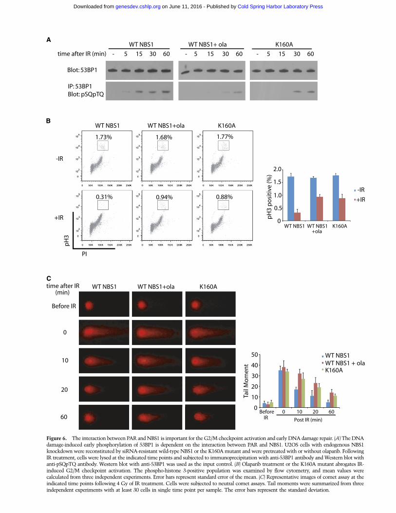
Figure 6. The interaction between PAR and NBS1 is important for the G2/M checkpoint activation and early DNA damage repair. (A) The DNAdamage-induced early phosphorylation of 53BP1 is dependent on the interaction between PAR and NBS1. U2OS cells with endogenous NBS1knockdown were reconstituted by siRNA-resistant wild-type NBS1 or the K160A mutant and were pretreated with or without olaparib. FollowingIR treatment, cells were lysed at the indicated time points and subjected to immunoprecipitation with anti-53BP1 antibody and Western blot withanti-pSQpTQ antibody. Western blot with anti-53BP1 was used as the input control. (B) Olaparib treatment or the K160A mutant abrogates IR-induced G2/M checkpoint activation. The phospho-histone 3-positive population was examined by flow cytometry, and mean values werecalculated from three independent experiments. Error bars represent standard error of the mean. (C) Representative images of comet assay at theindicated time points following 4 Gy of IR treatment. Cells were subjected to neutral comet assays. Tail moments were summarized from threeindependent experiments with at least 30 cells in single time point per sample. The error bars represent the standard deviation.
Cold Spring Harbor Laboratory Press on June 11, 2016 - Published by genesdev.cshlp.orgDownloaded from
interaction between the FHA domain of PNKP and PAR,and the retention of PNKP at DNA damage sites ismediated by the interaction between the FHA domainand its other phospho-binding partners. Moreover, weobserved a very similar phenomenon on APTX (Supple-mental Fig. 10A).
For DNA Ligase4, the olaparib treatment impairedthe early recruitment of DNA Ligase4 to DNA damagesites. Lacking H2AX significantly affected the stability ofLigase4 at DNA damage sites (Supplemental Fig. 10B).However, the S668A mutant in the BRCT domain did notaffect the slow accumulation of Ligase4 at DNA damagesites, suggesting that other motifs of Ligase4 mediate theH2AX-dependent retention of Ligase4 at DNA damagesites. Moreover, olaparib treatment did not show anadditional delay for the recruitment of the S668A mutant(Supplemental Fig. 10B). It has been shown that Ligase4forms a complex with other partners, such as XRCC4 andXLF, via other regions in the BRCT domain (Chen et al.2000; Sibanda et al. 2001; Riballo et al. 2009). It is possiblethat other partners of Ligase4 facilitate the prolongedretention of Ligase4 at the sites of DNA damage.
For XRCC1, both the olaparib treatment and the BRCTdomain mutation abolished fast recruitment of XRRC1 toDNA damage sites. Although lacking H2X mildly im-paired the retention of XRCC1 at DNA damage sites, theolaparib treatment in H2AX�/� cells did not totallyabolish the slow accumulation of XRCC1 to DNA dam-age sites (Supplemental Fig. 10C). These results suggestthat PAR and the BRCT domain of XRCC1 are essentialfor the fast recruitment of XRCC1 to DNA damage sites.However, DNA damage-induced signals other thangH2AX are required for the retention of XRCC1 atDNA damage sites (Supplemental Fig. 10C). Collectively,our results demonstrate that PAR is a bona fide signal forthe fast recruitment of various DNA damage responsefactors to DNA lesions. The retention of these DNAdamage response factors at DNA damage sites is medi-ated by different mechanisms.
To examine the biological significance of PAR in re-sponse to DNA damage, we treated wild-type or H2AX�/�
MEFs with olaparib followed by a low dose of IR. Lackingeither PAR or H2AX, cells could still be resistant to theIR-induced DNA damage. However, after loss of bothPAR and H2AX, cells were hypersensitive to a low dose ofIR (Supplemental Fig. 10D). These results suggest thatPAR synergizes with H2AX to recruit a set of DNAdamage factors to DNA lesions for damage repair.
Discussion
Taken together, we identified two novel classes of PAR-binding module that are involved in DNA damage re-sponse. Although both the FHA and BRCT domains areknown as phospho-protein-binding domains, here wefound that a set of FHA and BRCT domains recognizePAR. Interestingly, the PAR-binding pocket coincideswith the phospho-amino acid-binding pocket in the FHAor BRCT domain. Since both ADP-ribose and iso-ADP-ribose have two phosphate groups, it is likely that the
PAR-binding pockets of the FHA or BRCT domains rec-ognize the phosphate groups in the ADP-ribose or iso-ADP-ribose. In particular, the FHA domain of PNKPis also known to recognize the pS–pT peptide. The twophosphate groups in iso-ARP-ribose might mimic thetwo phosphate groups on the Ser and Thr residues in thepS–pT peptide and mediate the interaction with the FHAdomain of PNKP (Supplemental Fig. 1). Moreover, theFHA domain of APTX can interact with the pS-D–pT-Dpeptide, in which the Asp residues are also negativelycharged and mimic the phosphate group. Thus, the bind-ing mode between the APTX FHA domain and iso-ADP-ribose could be very similar to that between the FHA andphospho-peptide.
Different from the FHA domains of PNKP and APTX,the BRCT domains of Ligase4, XRCC1, and NBS1 recog-nize ADP-ribose. It is possible that these BRCT domainsmight also recognize site-specific mono-ADP-ribose. How-ever, in PARP1�/� cells where PAR synthesis is largelysuppressed in response to DNA damage, the relocation ofthese BRCT domain-containing proteins to DNA lesionsis also significantly suppressed (Supplemental Fig. 11). Itsuggests that, at least in the DNA damage context, theseBRCT domains recognize PAR at DNA damage sites.Moreover, the binding pockets in these BRCT domainsare associated with protein PARylation and PARPs duringevolution. For example, PARylation and PARPs only existedin multicellular eukaryotes. Coincidently, XRCC1 doesnot exist in prokaryotes and yeast. Although both NBS1and Ligase4 exist in yeast, the key residues in the bindingpockets of the BRCT domains are missing in their yeastorthologs (Supplemental Fig. 12), and the function of theBRCT fold of yeast NBS1 is likely to recognize othersignals or facilitate the FHA fold recognizing the phos-pho-amino acid (Lloyd et al. 2009). Thus, PAR synthesisat DNA damage sites could be very important for therecruitment of these BRCT domain-containing proteinsto DNA lesions for DNA damage repair in other multi-cellular eukaryotes.
A previous study indicated that a region within XRCC1(amino acids 379–400) interacted with PAR (Pleschkeet al. 2000). We generated the peptide of this region butdid not detect any binding between the peptide with PARin a dot blot, pull-down, or ITC assay (data not shown).Indeed, this 22-amino-acid region in XRCC1 is too shortto be correctly folded based on the structural analysis(Zhang et al. 1998) and does not directly contribute to thePAR-binding pocket of XRCC1. Moreover, not all of theFHA and BRCT domains could interact with PAR. Inparticular, the FHA domain of RNF8 and CHK2 and theBRCT domain of MDC1 and BRCA1 do not have theaffinity with PAR. Computational model analyses also donot support the binding between the phospho-peptide-binding pockets in these domains and ADP-ribose/iso-ADP-ribose. Additional structural analysis will reveal thedetails of these PAR-mediated interactions.
Following DNA damage, PAR is massively synthesizedat the DNA damage sites, which provides the platformto recruit DNA damage response proteins to lesions(D’Amours et al. 1999; Kim et al. 2005; Gibson and Kraus
Li et al.
1762 GENES & DEVELOPMENT
Cold Spring Harbor Laboratory Press on June 11, 2016 - Published by genesdev.cshlp.orgDownloaded from
2012). Here, we analyzed the biological function of theinteraction between PAR and NBS1 because it maypartially explain the molecular mechanism by whichthe MRN complex recognizes the DSBs and induces theearly ATM activation. Our study demonstrates that bothDNA damage-induced PAR synthesis and the ADP-ribose-binding pocket in NBS1 are important for earlyATM activation, which controls the transient G2/Mcheckpoint and early DNA damage repair via the ATM-dependent signal transduction pathway. Accumulatedevidence suggests that 53BP1 is a key downstream ef-fector in the ATM-dependent pathway and could bephosphorylated by ATM and CHK2 (Anderson et al. 2001;Rappold et al. 2001; Ward et al. 2003). Phosphorylated53BP1 is known to regulate its functional partners togovern the transient G2/M checkpoint and DNA damagerepair (DiTullio et al. 2002; Fernandez-Capetillo et al.2002; Callen et al. 2013; Chapman et al. 2013), which isconsistent with our observations. Moreover, the PAR-dependent early ATM activation controls the transientG2/M checkpoint, which plays an important role inmaintaining genomic stability. The transient G2/Mcheckpoint allows the completion of quick DNA dam-age repair before entering into mitosis so that the DNAlesions would not be transmitted from mother cells todaughter cells (Chen et al. 2000; Abraham 2001). How-ever, the G2/M checkpoint only transiently exists.Prolonged arresting at the G2/M boundary will causethe mitotic exit and genomic instability (Hirose et al.2001; Chiu et al. 2005; Yang et al. 2010). Meanwhile, DNAdamage repair should also be completed quickly if cellsare at the G2/M transition, since the G2/M checkpointonly transiently exists. Thus, the early activation ofATM and the ATM-dependent pathway at least plays animportant role in maintaining the genomic stability ofcells during the G2/M transition period. Moreover, DNAdamage-induced PAR synthesis may regulate multiplelayers of DNA damage repair, since PAR recruits manyother repair machineries—including these FHA andBRCT domain-containing proteins—to DNA damagesites. These repair machineries may function togetherwith ATM in a complicated network for early DNAdamage repair.
Interestingly, PAR is also quickly degraded withina few minutes following DNA damage (D’Amours et al.1999; Gagne et al. 2006; Kim et al. 2007a). Withoutother DNA damage response signals or other functionalpartners such as gH2AX, these DNA damage responsefactors could not stay at DNA lesions for the prolongedtime (Su 2006; Polo and Jackoson 2011). Thus, DNAdamage response signals such as gH2AX provide theselection for retaining DNA damage response factors atthe DNA lesions for different repair mechanisms. Lossof both PAR and gH2AX, a portion of DNA damagerepair proteins would not be able to reach DNA lesions,which causes cell lethality. Similar mechanism hasbeen implicated in the cancer clinical trials, whichcombined PARP inhibitors and PI3 kinase inhibitors toboost the efficacy of cancer chemotherapy (Ibrahimet al. 2012; Juvekar et al. 2012).
Materials and methods
Plasmids and antibodies
For GST fusion proteins, the BRCA1 BRCT domain, 53BP1BRCT domain, Ligase4 BRCT domain, MCPH1 BRCT2+3,MDC1 BRCT domain, PARP1 BRCT, PTIP BRCT3+4, REV1BRCT, TdT BRCT, XRCC1 BRCT domain, APLF FHA, APTXFHA, CHK2 FHA, FOXK1 FHA, Ki67 FHA, NBS1 FHA+BRCTdomain, PNKP FHA, RNF8 FHA, and MDC1 FHA were clonedinto the pGEX-4T1 vector, respectively. CHFR with an N-terminalGST tag and the NBS1 FHA+BRCT domain with a C-terminal GSTtag were cloned into pFastBac1 vector. For the constructs used inmicroirradiation experiments, PNKP, APTX, Ligase4, NBS1,XRCC1, and their indicated domains were cloned into pEGFPvector to generate plasmids encoding GFP fusion proteins (ordomains). The mutations in the proteins or domains above weregenerated using the QuikChange site-directed mutagenesiskit (Stratagene). For the constructs used to establish stablecell lines, the siRNA-resistant full-length cDNA of NBS1 andNBS1 K160A were cloned into pCMV-Tag 4A vector. The siRNAduplexes were purchased from Dharmacon Research. The sequencesof NBS1 and XRCC1 siRNA used were 59-GTACGTTGTTGGAAGGAAAdTdT-39 and 59-GGGAAGAGGAAGTTGGATTdTdT-39,respectively. siRNAs were transfected into cells using Oligofect-amine (Invitrogen) according to the manufacturer’s instructions.Anti-pATM, anti-pCHK2, anti-pSQpTQ, anti-XRCC1, and anti-NBS1 antibodies were purchased from Cell Signaling; anti-phosphorylated histone H3 antibody was purchased from Up-state Biotechnology; anti-Flag and anti-b-actin antibodies werepurchased from Sigma; and anti-PAR antibody was purchasedfrom Trevigen.
Immunoprecipitation and Western blot
U2OS cells were lysed with NETN-100 buffer (20 mM Tris-HClat pH 8.0, 100 mM NaCl, 1 mM EDTA, 0.5% nonidet P-40) onice. Soluble fractions were subjected to immunoprecipitationand Western blot and probed with antibodies as indicated.
Immunofluorescence
Cells were fixed in 3% paraformaldehyde for 25 min andpermeabilized in 0.5% Triton X-100 for 20 min at room temper-ature. Samples were blocked with 5% goat serum and thenincubated in primary antibody for 60 min. Samples were thenwashed with PBS three times and incubated with secondaryantibody for 30 min. After PBS wash, the nuclei were stained byDAPI. The signals were visualized by fluorescence microscope.
Generation and purification of PAR and iso-ADP-ribose
PAR (or biotin-PAR) was synthesized and purified in vitroaccording to the previous work as described (Fahrer et al. 2007)with some modifications. Briefly, PAR was synthesized in a 15-mL incubation mixture comprising 100 mM Tris-HCl (pH 7.8),10 mM MgCl2, 1 mM NAD+, 10 mM DTT, 60 mg/mL histoneH1, 60 mg/mL histone type IIa, 50 mg/mL octameric oligonu-cleotide GGAATTCC, and 150 nM human PARP-1. The reactionwas stopped after 60 min by addition of 20 mL of ice-cold 20%TCA. Following precipitation, the pellet was washed with ice-cold 99.8% ethanol. Polymer was detached using 0.5 M KOH/50mM EDTA and was purified by phenol-chloroform extractionand isopropanol precipitation. Purified PAR was fractionatedaccording to chain length by anion exchange high-pressure liquidchromatography (HPLC) protocol. Iso-ADP-ribose was generated
The FHA and BRCT domains bind poly(ADP-ribose)
GENES & DEVELOPMENT 1763
Cold Spring Harbor Laboratory Press on June 11, 2016 - Published by genesdev.cshlp.orgDownloaded from
and purified in vitro according to the procedure by Wang et al.(2012). Briefly, the purified PAR was digested by 50 U of snakevenom phosphodiesterase (Worthington) with 15 mM MgCl2
overnight at room temperature. The product of the phosphodi-esterase digestion, iso-ADPR, was further purified by ion ex-change chromatography and Superdex 75 on fast protein liquidchromatography (FPLC). Purified iso-ADP-ribose was dried in air,dissolved by ddH2O to 50 mM, stored at �20°C.
Dot blot
Recombinant proteins (10 pmol) were conjugated to the gluta-thione beads and incubated with PAR (100 pmol, calculated asthe ADP-ribose unit) for 2 h at 4°C. The beads were washed fourtimes with NETN-100 buffer. GST fusion proteins were elutedfrom beads by glutathione and spotted onto a nitrocellulosemembrane. The membrane was blocked with TBST buffer (0.15M NaCl, 0.01 M Tris-HCl at pH 7.4, 0.1% Tween 20) supple-mented with 5% milk and extensively washed with TBST. Afterdrying in the air, the membrane was examined by anti-PARantibody.
GST fusion protein expression and pull-down assay
GST fusion proteins were expressed in Escherichia coli or usingthe Bac-to-Bac baculovirus expression system (for recombinantCHFR and NBS1) (Invitrogen) and purified under standard pro-cedures. Purified GST fusion proteins (1 pmol) were incubatedwith biotin-labeled PAR (5 pmol) and streptavidin beads for 2 hat 4°C. After washing with NETN-100 buffer four times, thesamples were boiled in the SDS sample buffer. The elutes wereanalyzed by Western blot with anti-GST antibody.
ITC
ITC was carried out at 16°C with an ITC 200 Microcalorimeter(GE Healthcare). Proteins were dialyzed extensively into thebuffer containing 10 mM Na2HPO4 (pH 7.5), and 100 mM NaClat the final concentration of 20;60 mM. Ligands (PAR, ADP-ribose,or iso-ADP-ribose) in the injection syringe were also diluted by thesame buffer at the final concentration of 150;750 mM (theconcentration of PAR was calculated as the ADP-ribose unit).A typical titration consisted of 19 consecutive 2 mL injectionsof ligands following a preinjection of 0.4 mL of ligands into theprotein solution at time intervals of 120 sec while stirring at1000 rpm. Binding isotherms were integrated and analyzedusing the software Origin 7.0 (OriginLab) provided by themanufacturer.
Molecular modeling
For the FHA domain proteins, crystal structures of CHK2/peptide (HFD-pT-YLIR; Protein Data Bank [PDB] ID: 1GXC) (Liet al. 2002), RNF8/peptide (ELK-pT-ERY; PDB ID: 2PIE) (Huenet al. 2007), PNKP/peptide (YAG-pS–pT-DEN; PDB ID:2W3O)(Ali et al. 2009), and APTX (PDB ID:3KT9) (Becherel et al. 2010)were used. To construct the APTX peptide (D-pS–D-pT-DA),APTX was aligned with the PNKP/XRCC1 peptide, and theXRCC1 peptide was mutated into the D-pS–D-pT-DA peptidefollowed by a structural minimization using the MOE program(Chemical Computing Group). For the comparison of the bindingaffinity between iso-ADP-ribose, peptides, and the FHA domain,five amino acids neighboring the pSer or pThr (capped with ACEand NME at their N and C termini) from the peptides compara-ble with the size of the iso-ADP-ribose were used in the bindingfree energy calculations. For the BRCT domain proteins, crystal
structures of MDC1/peptide (pS-QEY; PDBID: 3K05) (Campbellet al. 2010), BRCA1/peptide (ISRST-pS-PTFNK; PDB ID: 1T29)(Shiozaki et al. 2004), NMR (nuclear magnetic resonance)structure of Ligase4 (PDB ID: 2E2W), and XRCC1 (PDB ID:2D8M) were used.
For docking simulations, all of the protein structures wereprocessed, and the protons were added according to the pH 7.0using the MOE program (Chemical Computing Group) beforethey were used in the docking simulations. Two docking pro-grams were used to evaluate and select the best binding poses.They were the GOLD program (version 4.0.1) (Jones et al. 1997)and the Glide module from Schrodinger program suite (Friesneret al. 2006). For the FHA domains of CHK2, RNF8, PNKP, andAPTX, iso-ADP-ribose was docked into the binding site, whereasADP-ribose was docked into the binding sites of MDC1, BRCA1,XRCC1, and the Ligase4 BRCT domain. In the docking simu-lation using the GOLD program, the centers of the binding sitesfor the proteins were selected at the residues mutated in theexperiments and showed to be important for binding to peptides,iso-ADP-ribose, and ADP-ribose experimentally. The radius of thebinding site was defined as 13 A, large enough to cover the bindingpockets. For each genetic algorithm (GA) run, a maximum of200,000 operations were performed on a population of five is-lands of 100 individuals. Operator weights for crossover, muta-tion, and migration were set to 95, 95, and 10, respectively. Thedocking simulations were terminated after 20 runs for eachligand. GoldScore implemented in Gold 4.0.1 was used as thefitness function to evaluate the docked conformations. In thedocking simulation using Glide, the center of the box wasselected at the amino acids mutated in each protein, which werefound important for binding, and the XP mode was used indocking. All of the top-ranked binding poses of iso-ADP-riboseand ADP-ribose with the proteins from two docking programswere inspected, and the poses with the phosphate groups similarto the phospho-peptides in the crystal structures and compatiblewith PAR were selected and are shown. The electrostatic poten-tial surfaces of the proteins were calculated using the APBS(Baker et al. 2001) module in the PyMOL program (http://www.pymol.org) based on parameters generated from the PDB2PQRserver (Dolinsky et al. 2004).
The selected binding models were then subjected to the MDsimulation using the Amberprogram suite (version 12) , and thebinding free energy was calculated using the Amber programsuite (version 10). The force field parameters for ADP-ribose andiso-ADP-ribose were derived by using the Antechamber modulein Amber. The point charge parameters of both ligands werederived from the minimized geometry at the RHF level usinga 6-31G* basis set with Gaussian09 and followed by the RESPfitting of the electrostatic field potential generated from thepoint charges at each atom site to those calculated fromGaussian09.
The topology and coordinate files for each protein–ligandcomplex were prepared by first adding counter-ions to neutralizethe charges of the system before it was solvated in a 12 A cubicbox of the TIP3P (Jorgensen et al. 1983) water. The system wasinitially minimized by a 1000-step steepest decent and a 2000-step conjugate gradient minimization procedures to the solvents.Then, a 2-psec simulation was performed to raise the tempera-ture of the system to 150K, followed by another 18 psec ofsimulation to increase the temperature further to 298K, wherethe protein ligands were fixed using 10 kcal/mol force constantsin reference to the initial structure. A second 60-nsec equilibra-tion of the system at 298K was performed by constraining thebackbone atoms of the system with a 2 kcal/mol force constant.The production run was 2 nsec. Conformations were saved from
Li et al.
1764 GENES & DEVELOPMENT
Cold Spring Harbor Laboratory Press on June 11, 2016 - Published by genesdev.cshlp.orgDownloaded from
the trajectory at intervals of 1 psec. Conformations collectedfrom 0.5–2 nsec were used for the binding affinity predictioncalculations. The MD simulations were performed using theGPU accelerated version of the PMEMD program (Gotz et al.2012) in the isothermal isobaric (NTP, T = 298K and P = 1 atm)ensemble. The SHAKE (Ryckaert et al. 1977) algorithm was usedto fix bonds involving hydrogen. The PME method (Darden et al.1993) was used, and the nonbonded cutoff distance was set at10 A. The time step was 2 fsec, and the neighboring pairs listwas updated every 20 steps.
The MM-PBSA method was used for binding free energycalculations. In the MM-PBSA calculation, the 31 conformationscorresponding to 50-psec intervals in the trajectory were usedfor the molecular mechanics calculations. Eight conformations(taken at intervals of 200 psec) from the 1.5-nsec trajectory werechosen for the normal mode calculations for entropic contribu-tion to the binding free energy. In the normal mode calculations,a distance-dependent dielectric constant of �4r was used, themaximum cycle was set to 60,000, and the convergence tolerancewas 0.0002 kcal mol�1A�. For the solvent-accessible surface areacalculation, the default value of 0.0072 kcal/mol 3 A2 for thesurface tension coefficient was used.
For Ligase4 and XRCC1, we analyzed all 20 conformations (ormodels) of the NMR structures and performed docking simula-tions followed by MD simulations to evaluate the stability ofbinding models. For Ligase4, we found that the first and 18thconformations of the NMR structures are suitable for bindingmodel determination. The binding model of ADP-ribose with the18th conformation from the NMR structure yielded stablestructures in the 4-nsec MD simulations. For XRCC1, weperformed molecular dynamics simulations of the protein li-gand-binding model based on four different XRCC1 conforma-tions. We found that the second, third, sixth, and 17th confor-mations of the NMR structures gave well-defined and openbinding sites and are suitable for generating the binding modelswith ADP-ribose for further MD simulations. Only the bind-ing model between ADP-ribose and the 17th conformation ofXRCC1 gave stable structures in a 4-nsec MD simulation.
Laser microirradiation and live-cell imaging
U2OS cells and MEFs were plated on glass-bottomed culturedishes (Mat Tek Corporation). Laser microirradiation was per-formed using an IX 71 microscope (Olympus) coupled with theMicoPoint laser illumination and ablation system (PhotonicInstruments, Inc.). A 337.1-nm laser diode (3.4 mW) transmittedthrough a specific dye cell and then yielded a 365-nm wavelengthlaser beam that was focused through 603 UPlanSApo/1.35 oilobjective to yield a spot size of 0.5–1 mm. The time of cellexposure to the laser beam was ;3.5 nsec. The pulse energy was170 mJ at 10 Hz. Images were taken by the same microscope withthe CellSens software (Olympus). GFP fluorescence at the laserline was converted into a numerical value using Axiovisionsoftware (version 4.5). Normalized fluorescent curves from 50cells from three independent experiments were averaged. Theerror bars represent the standard deviation.
Comet assays
Single-cell gel electrophoretic comet assays were performedunder neutral conditions according to a previous study (Oliveand Banath 2006). Briefly, U2OS cells were treated with orwithout 4 Gy of IR and recovered in normal culture mediumfor the indicated time at 37°C. Cells were collected and rinsedtwice with ice-cold PBS; 2 3 104 cells per milliliter werecombined with 1% LMAgarose at 40°C at the ratio of 1:3 (v/v)
and immediately pipetted onto slides. For cellular lysis, theslides were immersed in the neutral lysis solution (2% sarkosyl,0.5 M Na2EDTA, 0.5 mg/mL proteinase K at pH 8.0) overnight at37°C in the dark followed by washing in the rinse buffer (90 mMTris buffer, 90 mM boric acid, 2 mM Na2EDTA at pH 8.5) for30 min with two repeats. Next, the slides were subjected toelectrophoresis at 20 V (0.6 V/cm) for 25 min and stained in2.5 mg/mL propidium iodide for 20 min. All images were takenwith a fluorescence microscope and analyzed by Comet AssayIV software.
IR treatment and colony formation assay
Cells were irradiated with a 137Cs source at a dose of 10 Gy (or atthe indicated doses). After irradiation, cells were lysed at theindicated time points for immunoprecipitation or Western blot.For colony formation assay, 500 wild-type or H2AX�/� MEFswere seeded into six-well plates and then treated by variousdoses of IR with or without olaparib. After a 7-d culture, theviable cells were fixed and stained with crystal violet. Thenumber of colonies (>50 cells for each colony) was calculated.
G2/M checkpoints assay
Cells expressing the wild-type NBS1 or NBS1 K160A pretreatedwith or without olaparib were treated with or without 2 Gy of IR.After 1 h of recovery, cells were fixed with 70% (v/v) ethanol,stained with rabbit antibody to phospho-histone H3 (pSer10), andthen incubated with FITC-conjugated goat secondary antibodyto rabbit. The stained cells were treated with RNase A and thenincubated with propidium iodide. Samples were analyzed byflow cytometry.
Drug treatment
For live-cell imaging, immunoprecipitation, or Western blot, 100nM olaparib, 10 mM GLTN, or 10 mM wortmannin was addedinto the cell culture medium 1 h before laser microirradiation orcell lysis. For colony formation assay, 100 nM olaparib was addedinto the medium during the culture.
Statistical analyses
All experiments were performed in triplicates unless indicatedotherwise. Means and standard deviations were plotted. Stu-dent’s t-test was used for statistical analyses.
Acknowledgments
We thank Drs. Ming Lei and Feng Zhang for technical support.This work was supported by the National Institute of Health(CA132755 and CA130899 to X.Y.). X.Y. is a recipient of the Eraof Hope Scholar Award from the Department of Defense.
References
Abraham RT. 2001. Cell cycle checkpoint signaling through theATM and ATR kinases. Genes Dev 15: 2177–2196.
Ahel I, Ahel D, Matsusaka T, Clark AJ, Pines J, Boulton SJ, WestSC. 2008. Poly(ADP-ribose)-binding zinc finger motifs inDNA repair/checkpoint proteins. Nature 451: 81–85.
Ali AA, Jukes RM, Pearl LH, Oliver AW. 2009. Specific recog-nition of a multiply phosphorylated motif in the DNA repairscaffold XRCC1 by the FHA domain of human PNK. Nucleic
Acids Res 37: 1701–1712.Ame JC, Spenlehauer C, de Murcia G. 2004. The PARP super-
family. Bioessays 26: 882–893.
The FHA and BRCT domains bind poly(ADP-ribose)
GENES & DEVELOPMENT 1765
Cold Spring Harbor Laboratory Press on June 11, 2016 - Published by genesdev.cshlp.orgDownloaded from
Anderson L, Henderson C, Adachi Y. 2001. Phosphorylation andrapid relocalization of 53BP1 to nuclear foci upon DNAdamage. Mol Cell Biol 21: 1719–1729.
Anisimov VM, Ziemys A, Kizhake S, Yuan Z, Natarajan A,Cavasotto CN. 2011. Computational and experimental stud-ies of the interaction between phospho-peptides and theC-terminal domain of BRCA1. J Comput Aided Mol Des
25: 1071–1084.Baer R. 2013. Luring BRCA1 to the scene of the crime. Cancer
Cell 23: 565–567.Baker NA, Sept D, Joseph S, Holst MJ, McCammon JA. 2001.
Electrostatics of nanosystems: Application to microtubulesand the ribosome. Proc Natl Acad Sci 98: 10037–10041.
Bakkenist CJ, Kastan MB. 2003. DNA damage activates ATMthrough intermolecular autophosphorylation and dimer dis-sociation. Nature 421: 499–506.
Becherel OJ, Jakob B, Cherry AL, Gueven N, Fusser M, Kijas AW,Peng C, Katyal S, McKinnon PJ, Chen J, et al. 2010. CK2phosphorylation-dependent interaction between aprataxinand MDC1 in the DNA damage response. Nucleic AcidsRes 38: 1489–1503.
Bonner WM, Redon CE, Dickey JS, Nakamura AJ, SedelnikovaOA, Solier S, Pommier Y. 2008. gH2AX and cancer. Nat Rev
Cancer 8: 957–967.Callen E, Di Virgilio M, Kruhlak MJ, Nieto-Soler M, Wong N,
Chen HT, Faryabi RB, Polato F, Santos M, Starnes LM, et al.2013. 53BP1 Mediates Productive and Mutagenic DNA Re-pair through Distinct Phosphoprotein Interactions. Cell 153:1266–1280.
Campbell SJ, Edwards RA, Glover JN. 2010. Comparison of thestructures and peptide binding specificities of the BRCTdomains of MDC1 and BRCA1. Structure 18: 167–176.
Celeste A, Fernandez-Capetillo O, Kruhlak MJ, Pilch DR,Staudt DW, Lee A, Bonner RF, Bonner WM, NussenzweigA. 2003. Histone H2AX phosphorylation is dispensable forthe initial recognition of DNA breaks. Nat Cell Biol 5: 675–679.
Chapman JR, Jackson SP. 2008. Phospho-dependent interactionsbetween NBS1 and MDC1 mediate chromatin retention ofthe MRN complex at sites of DNA damage. EMBO Rep 9:795–801.
Chapman JR, Barral P, Vannier JB, Borel V, Steger M, Tomas-LobaA, Sartori AA, Adams IR, Batista FD, Boulton SJ. 2013. RIF1 isessential for 53BP1-dependent nonhomologous end joiningand suppression of DNA double-strand break resection. Mol
Cell 49: 858–871.Chen L, Trujillo K, Sung P, Tomkinson AE. 2000. Interactions of
the DNA ligase IV-XRCC4 complex with DNA ends and theDNA-dependent protein kinase. J Biol Chem 275: 26196–26205.
Chiu CC, Li CH, Ung MW, Fuh TS, Chen WL, Fang K. 2005.Etoposide (VP-16) elicits apoptosis following prolongedG2-M cell arrest in p53-mutated human non-small cell lungcancer cells. Cancer Lett 223: 249–258.
Cuneo MJ, Gabel SA, Krahn JM, Ricker MA, London RE. 2011.The structural basis for partitioning of the XRCC1/DNAligase III-a BRCT-mediated dimer complexes. Nucleic AcidsRes 39: 7816–7827.
D’Amours D, Desnoyers S, D’Silva I, Poirier GG. 1999. Poly(ADP-ribosyl)ation reactions in the regulation of nuclear functions.Biochem J 342: 249–268.
Darden TA, York DM, Pedersen L. 1993. Particle mesh Ewald:An N-log(N) method for Ewald sums in large systems. J Chem
Phys 98: 10089–10092.DiTullio RA Jr, Mochan TA, Venere M, Bartkova J, Sehested M,
Bartek J, Halazonetis TD. 2002. 53BP1 functions in an ATM-
dependent checkpoint pathway that is constitutively acti-vated in human cancer. Nat Cell Biol 4: 998–1002.
Dolinsky TJ, Nielsen JE, McCammon JA, Baker NA. 2004.PDB2PQR: An automated pipeline for the setup of Poisson–Boltzmann electrostatics calculations. Nucleic Acids Res 32:W665–W667.
Durocher D, Henckel J, Fersht AR, Jackson SP. 1999. The FHAdomain is a modular phosphopeptide recognition motif. Mol
Cell 4: 387–394.Durocher D, Taylor IA, Sarbassova D, Haire LF, Westcott SL,
Jackson SP, Smerdon SJ, Yaffe MB. 2000. The molecular ba-sis of FHA domain:phosphopeptide binding specificity andimplications for phospho-dependent signaling mechanisms.Mol Cell 6: 1169–1182.
Fahrer J, Kranaster R, Altmeyer M, Marx A, Burkle A. 2007.Quantitative analysis of the binding affinity of poly(ADP-ribose) to specific binding proteins as a function of chainlength. Nucleic Acids Res 35: e143.
Falck J, Mailand N, Syljuasen RG, Bartek J, Lukas J. 2001. TheATM–Chk2–Cdc25A checkpoint pathway guards against ra-dioresistant DNA synthesis. Nature 410: 842–847.
Fathers C, Drayton RM, Solovieva S, Bryant HE. 2012. In-hibition of poly(ADP-ribose) glycohydrolase (PARG) specifi-cally kills BRCA2-deficient tumor cells. Cell Cycle 11: 990–997.
Fernandez-Capetillo O, Chen HT, Celeste A, Ward I, RomanienkoPJ, Morales JC, Naka K, Xia Z, Camerini-Otero RD, MotoyamaN, et al. 2002. DNA damage-induced G2–M checkpointactivation by histone H2AX and 53BP1. Nat Cell Biol 4:993–997.
Friesner RA, Murphy RB, Repasky MP, Frye LL, Greenwood JR,Halgren TA, Sanschagrin PC, Mainz DT. 2006. Extra pre-cision glide: Docking and scoring incorporating a model ofhydrophobic enclosure for protein�ligand complexes. J Med
Chem 49: 6177–6196.Gagne JP, Hendzel MJ, Droit A, Poirier GG. 2006. The expanding
role of poly(ADP-ribose) metabolism: Current challenges andnew perspectives. Curr Opin Cell Biol 18: 145–151.
Gibson BA, Kraus WL. 2012. New insights into the molecularand cellular functions of poly(ADP-ribose) and PARPs. Nat
Rev Mol Cell Biol 13: 411–424.Glover JN, Williams RS, Lee MS. 2004. Interactions between
BRCT repeats and phosphoproteins: Tangled up in two.Trends Biochem Sci 29: 579–585.
Goldberg M, Stucki M, Falck J, D’Amours D, Rahman D,Pappin D, Bartek J, Jackson SP. 2003. MDC1 is required forthe intra-S-phase DNA damage checkpoint. Nature 421:952–956.
Gotz AW, Williamson MJ, Xu D, Poole D, Le Grand S, WalkerRC. 2012. Routine microsecond molecular dynamics simu-lations with AMBER on GPUs. 1. Generalized born. J Chem
Theory Comput 8: 1542–1555.Hirose Y, Berger MS, Pieper RO. 2001. p53 effects both the
duration of G2/M arrest and the fate of temozolomide-treatedhuman glioblastoma cells. Cancer Res 61: 1957–1963.
Horejsi Z, Falck J, Bakkenist CJ, Kastan MB, Lukas J, Bartek J.2004. Distinct functional domains of Nbs1 modulate thetiming and magnitude of ATM activation after low doses ofionizing radiation. Oncogene 23: 3122–3127.
Hottiger MO, Hassa PO, Luscher B, Schuler H, Koch-Nolte F.2010. Toward a unified nomenclature for mammalian ADP-ribosyltransferases. Trends Biochem Sci 35: 208–219.
Huen MS, Grant R, Manke I, Minn K, Yu X, Yaffe MB, Chen J.2007. RNF8 transduces the DNA-damage signal via histoneubiquitylation and checkpoint protein assembly. Cell 131:901–914.
Li et al.
1766 GENES & DEVELOPMENT
Cold Spring Harbor Laboratory Press on June 11, 2016 - Published by genesdev.cshlp.orgDownloaded from
Ibrahim YH, Garcia-Garcia C, Serra V, He L, Torres-Lockhart K,Prat A, Anton P, Cozar P, Guzman M, Grueso J, et al. 2012.PI3K inhibition impairs BRCA1/2 expression and sensitizesBRCA proficient triple negative breast cancer to PARP in-hibition. Cancer Discov 2: 1036–1047.
Jones G, Willett P, Glen RC, Leach AR, Taylor R. 1997. De-velopment and validation of a genetic algorithm for flexibledocking. J Mol Biol 267: 727–748.
Jorgensen WL, Chandrasekha J, Madura JD, Impey RW, KleinML. 1983. Comparison of simple potential functions forsimulating liquid water. J Chem Phys 79: 926–935.
Juvekar A, Burga LN, Hu H, Lunsford EP, Ibrahim YH, BalmanaJ, Rajendran A, Papa A, Spencer K, Lyssiotis CA, et al. 2012.Combining a PI3K inhibitor with a PARP inhibitor providesan effective therapy for a mouse model of BRCA1-relatedbreast cancer. Cancer Discov 2: 1048–1063.
Karras GI, Kustatscher G, Buhecha HR, Allen MD, Pugieux C,Sait F, Bycroft M, Ladurner AG. 2005. The macro domain isan ADP-ribose binding module. EMBO J 24: 1911–1920.
Kim MY, Zhang T, Kraus WL. 2005. Poly(ADP-ribosyl)ation byPARP-1: ‘PAR-laying’ NAD+ into a nuclear signal. Genes
Dev 19: 1951–1967.Kim H, Chen J, Yu X. 2007a. Ubiquitin-binding protein RAP80
mediates BRCA1-dependent DNA damage response. Science316: 1202–1205.
Kim H, Huang J, Chen J. 2007b. CCDC98 is a BRCA1–BRCTdomain-binding protein involved in the DNA damage re-sponse. Nat Struct Mol Biol 14: 710–715.
Kobayashi J, Antoccia A, Tauchi H, Matsuura S, Komatsu K.2004. NBS1 and its functional role in the DNA damageresponse. DNA Repair 3: 855–861.
Kolas NK, Chapman JR, Nakada S, Ylanko J, Chahwan R,Sweeney FD, Panier S, Mendez M, Wildenhain J, ThomsonTM, et al. 2007. Orchestration of the DNA-damage responseby the RNF8 ubiquitin ligase. Science 318: 1637–1640.
Kollman PA, Massova I, Reyes C, Kuhn B, Huo S, Chong L, LeeM, Lee T, Duan Y, Wang W, et al. 2000. Calculating structuresand free energies of complex molecules: Combining molecu-lar mechanics and continuum models. Acc Chem Res 33:889–897.
Kurz EU, Lees-Miller SP. 2004. DNA damage-induced activationof ATM and ATM-dependent signaling pathways. DNA Re-pair) 3: 889–900.
Lee JH, Paull TT. 2004. Direct activation of the ATM proteinkinase by the Mre11/Rad50/Nbs1 complex. Science 304:93–96.
Li M, Yu X. 2013. Function of BRCA1 in the DNA damageresponse is mediated by ADP-ribosylation. Cancer Cell 23:693–704.
Li J, Williams BL, Haire LF, Goldberg M, Wilker E, Durocher D,Yaffe MB, Jackson SP, Smerdon SJ. 2002. Structural andfunctional versatility of the FHA domain in DNA-damagesignaling by the tumor suppressor kinase Chk2. Mol Cell 9:1045–1054.
Li GY, McCulloch RD, Fenton AL, Cheung M, Meng L, Ikura M,Koch CA. 2010. Structure and identification of ADP-riboserecognition motifs of APLF and role in the DNA damageresponse. Proc Natl Acad Sci 107: 9129–9134.
Liu Z, Wu J, Yu X. 2007. CCDC98 targets BRCA1 to DNAdamage sites. Nat Struct Mol Biol 14: 716–720.
Lloyd J, Chapman JR, Clapperton JA, Haire LF, Hartsuiker E, LiJ, Carr AM, Jackson SP, Smerdon SJ. 2009. A supramodularFHA/BRCT-repeat architecture mediates Nbs1 adaptor func-tion in response to DNA damage. Cell 139: 100–111.
Loizou JI, El-Khamisy SF, Zlatanou A, Moore DJ, Chan DW, QinJ, Sarno S, Meggio F, Pinna LA, Caldecott KW. 2004. The
protein kinase CK2 facilitates repair of chromosomal DNAsingle-strand breaks. Cell 117: 17–28.
Lou Z, Minter-Dykhouse K, Franco S, Gostissa M, Rivera MA,Celeste A, Manis JP, van Deursen J, Nussenzweig A, PaullTT, et al. 2006. MDC1 maintains genomic stability byparticipating in the amplification of ATM-dependent DNAdamage signals. Mol Cell 21: 187–200.
Lukas J, Lukas C, Bartek J. 2004. Mammalian cell cycle check-points: Signalling pathways and their organization in spaceand time. DNA Repair 3: 997–1007.
Lukas J, Lukas C, Bartek J. 2011. More than just a focus: Thechromatin response to DNA damage and its role in genomeintegrity maintenance. Nat Cell Biol 13: 1161–1169.
Luo X, Kraus WL. 2012. On PAR with PARP: Cellular stresssignaling through poly(ADP-ribose) and PARP-1. Genes Dev
26: 417–432.Mahajan A, Yuan C, Lee H, Chen ES, Wu PY, Tsai MD. 2008.
Structure and function of the phosphothreonine-specific FHAdomain. Sci Signal 1: re12.
Mailand N, Bekker-Jensen S, Faustrup H, Melander F, Bartek J,Lukas C, Lukas J. 2007. RNF8 ubiquitylates histones at DNAdouble-strand breaks and promotes assembly of repair pro-teins. Cell 131: 887–900.
Mani RS, Fanta M, Karimi-Busheri F, Silver E, Virgen CA,Caldecott KW, Cass CE, Weinfeld M. 2007. XRCC1 stimu-lates polynucleotide kinase by enhancing its damage dis-crimination and displacement from DNA repair intermedi-ates. J Biol Chem 282: 28004–28013.
Manke IA, Lowery DM, Nguyen A, Yaffe MB. 2003. BRCTrepeats as phosphopeptide-binding modules involved in pro-tein targeting. Science 302: 636–639.
Messner S, Hottiger MO. 2011. Histone ADP-ribosylation inDNA repair, replication and transcription. Trends Cell Biol
21: 534–542.Mohammad DH, Yaffe MB. 2009. 14-3-3 proteins, FHA domains
and BRCT domains in the DNA damage response. DNA
Repair 8: 1009–1017.Oberoi J, Richards MW, Crumpler S, Brown N, Blagg J, Bayliss R.
2010. Structural basis of poly(ADP-ribose) recognition bythe multizinc binding domain of checkpoint with forkhead-associated and RING domains (CHFR). J Biol Chem 285:39348–39358.
Olive PL, Banath JP. 2006. The comet assay: A method to mea-sure DNA damage in individual cells. Nat Protoc 1: 23–29.
Paull TT, Rogakou EP, Yamazaki V, Kirchgessner CU, Gellert M,Bonner WM. 2000. A critical role for histone H2AX inrecruitment of repair factors to nuclear foci after DNAdamage. Curr Biol 10: 886–895.
Pellegrini M, Celeste A, Difilippantonio S, Guo R, Wang W,Feigenbaum L, Nussenzweig A. 2006. Autophosphorylationat serine 1987 is dispensable for murine Atm activation invivo. Nature 443: 222–225.
Pleschke JM, Kleczkowska HE, Strohm M, Althaus FR. 2000.Poly(ADP-ribose) binds to specific domains in DNA damagecheckpoint proteins. J Biol Chem 275: 40974–40980.
Polo SE, Jackson SP. 2011. Dynamics of DNA damage responseproteins at DNA breaks: A focus on protein modifications.Genes Dev 25: 409–433.
Rappold I, Iwabuchi K, Date T, Chen J. 2001. Tumor suppressorp53 binding protein 1 (53BP1) is involved in DNA damage-signaling pathways. J Cell Biol 153: 613–620.
Rastelli G, Del Rio A, Degliesposti G, Sgobba M. 2010. Fast andaccurate predictions of binding free energies using MM-PBSAand MM-GBSA. J Comput Chem 31: 797–810.
Riballo E, Woodbine L, Stiff T, Walker SA, Goodarzi AA, JeggoPA. 2009. XLF-Cernunnos promotes DNA ligase IV-XRCC4
The FHA and BRCT domains bind poly(ADP-ribose)
GENES & DEVELOPMENT 1767
Cold Spring Harbor Laboratory Press on June 11, 2016 - Published by genesdev.cshlp.orgDownloaded from
re-adenylation following ligation. Nucleic Acids Res 37: 482–492.
Rodriguez M, Yu X, Chen J, Songyang Z. 2003. Phosphopeptidebinding specificities of BRCA1 COOH-terminal (BRCT)domains. J Biol Chem 278: 52914–52918.
Rungrotmongkol T, Nunthaboot N, Malaisree M, Kaiyawet N,Yotmanee P, Meeprasert A, Hannongbua S. 2010. Molecularinsight into the specific binding of ADP-ribose to the nsP3macro domains of chikungunya and Venezuelan equineencephalitis viruses: Molecular dynamics simulations andfree energy calculations. J Mol Graph Model 29: 347–353.
Ryckaert J-P, Ciccotti G, Berendsen HJC. 1977. Numericalintegration of the cartesian equations of motion of a systemwith constraints: Molecular dynamics of n-alkanes. J Com-
put Phys 23: 327–341.Sarkaria JN, Tibbetts RS, Busby EC, Kennedy AP, Hill DE,
Abraham RT. 1998. Inhibition of phosphoinositide 3-kinaserelated kinases by the radiosensitizing agent wortmannin.Cancer Res 58: 4375–4382.
Schreiber V, Dantzer F, Ame JC, de Murcia G. 2006. Poly(ADP-ribose): Novel functions for an old molecule. Nat Rev Mol
Cell Biol 7: 517–528.Shiloh Y, Ziv Y. 2013. The ATM protein kinase: Regulating the
cellular response to genotoxic stress, and more. Nat Rev MolCell Biol 14: 197–210.
Shiozaki EN, Gu L, Yan N, Shi Y. 2004. Structure of the BRCTrepeats of BRCA1 bound to a BACH1 phosphopeptide:Implications for signaling. Mol Cell 14: 405–412.
Sibanda BL, Critchlow SE, Begun J, Pei XY, Jackson SP, BlundellTL, Pellegrini L. 2001. Crystal structure of an Xrcc4–DNAligase IV complex. Nat Struct Biol 8: 1015–1019.
Smith J, Tho LM, Xu N, Gillespie DA. 2010. The ATM–Chk2and ATR–Chk1 pathways in DNA damage signaling andcancer. Adv Cancer Res 108: 73–112.
Spycher C, Miller ES, Townsend K, Pavic L, Morrice NA,Janscak P, Stewart GS, Stucki M. 2008. Constitutive phos-phorylation of MDC1 physically links the MRE11-RAD50-NBS1 complex to damaged chromatin. J Cell Biol 181: 227–240.
Stiff T, Walker SA, Cerosaletti K, Goodarzi AA, Petermann E,Concannon P, O’Driscoll M, Jeggo PA. 2006. ATR-dependentphosphorylation and activation of ATM in response to UVtreatment or replication fork stalling. EMBO J 25: 5775–5782.
Stracker TH, Petrini JH. 2011. The MRE11 complex: Startingfrom the ends. Nat Rev Mol Cell Biol 12: 90–103.
Stucki M, Clapperton JA, Mohammad D, Yaffe MB, Smerdon SJ,Jackson SP. 2005. MDC1 directly binds phosphorylatedhistone H2AX to regulate cellular responses to DNA dou-ble-strand breaks. Cell 123: 1213–1226.
Su TT. 2006. Cellular responses to DNA damage: One signal,multiple choices. Annu Rev Genet 40: 187–208.
Sun Z, Hsiao J, Fay DS, Stern DF. 1998. Rad53 FHA domainassociated with phosphorylated Rad9 in the DNA damagecheckpoint. Science 281: 272–274.
Wang B, Matsuoka S, Carpenter PB, Elledge SJ. 2002. 53BP1,a mediator of the DNA damage checkpoint. Science 298:1435–1438.
Wang B, Matsuoka S, Ballif BA, Zhang D, Smogorzewska A,Gygi SP, Elledge SJ. 2007. Abraxas and RAP80 form a BRCA1protein complex required for the DNA damage response.Science 316: 1194–1198.
Wang Z, Michaud GA, Cheng Z, Zhang Y, Hinds TR, Fan E,Cong F, Xu W. 2012. Recognition of the iso-ADP-ribosemoiety in poly(ADP-ribose) by WWE domains suggests ageneral mechanism for poly(ADP-ribosyl)ation-dependentubiquitination. Genes Dev 26: 235–240.
Ward IM, Minn K, van Deursen J, Chen J. 2003. p53 Bindingprotein 53BP1 is required for DNA damage responses andtumor suppression in mice. Mol Cell Biol 23: 2556–2563.
Whitehouse CJ, Taylor RM, Thistlethwaite A, Zhang H, Karimi-Busheri F, Lasko DD, Weinfeld M, Caldecott KW. 2001.XRCC1 stimulates human polynucleotide kinase activityat damaged DNA termini and accelerates DNA single-strandbreak repair. Cell 104: 107–117.
Williams RS, Dodson GE, Limbo O, Yamada Y, Williams JS,Guenther G, Classen S, Glover JN, Iwasaki H, Russell P,et al. 2009. Nbs1 flexibly tethers Ctp1 and Mre11-Rad50 tocoordinate DNA double-strand break processing and repair.Cell 139: 87–99.
Wu L, Luo K, Lou Z, Chen J. 2008. MDC1 regulates intra-S-phase checkpoint by targeting NBS1 to DNA double-strandbreaks. Proc Natl Acad Sci 105: 11200–11205.
Wu PY, Frit P, Meesala S, Dauvillier S, Modesti M, Andres SN,Huang Y, Sekiguchi J, Calsou P, Salles B, et al. 2009.Structural and functional interaction between the humanDNA repair proteins DNA ligase IV and XRCC4. Mol CellBiol 29: 3163–3172.
Wu J, Chen Y, Lu LY, Wu Y, Paulsen MT, Ljungman M, FergusonDO, Yu X. 2011. Chfr and RNF8 synergistically regulateATM activation. Nat Struct Mol Biol 18: 761–768.
Yang J, Yu Y, Hamrick HE, Duerksen-Hughes PJ. 2003. ATM,ATR and DNA-PK: Initiators of the cellular genotoxic stressresponses. Carcinogenesis 24: 1571–1580.
Yang L, Besschetnova TY, Brooks CR, Shah JV, Bonventre JV.2010. Epithelial cell cycle arrest in G2/M mediates kidneyfibrosis after injury. Nat Med 16: 535–543.
Ying W, Sevigny MB, Chen Y, Swanson RA. 2001. Poly(ADP-ribose) glycohydrolase mediates oxidative and excitotoxicneuronal death. Proc Natl Acad Sci 98: 12227–12232.
Yu X, Chen J. 2004. DNA damage-induced cell cycle checkpointcontrol requires CtIP, a phosphorylation-dependent bindingpartner of BRCA1 C-terminal domains. Mol Cell Biol 24:9478–9486.
Yu X, Chini CC, He M, Mer G, Chen J. 2003. The BRCT domainis a phospho-protein binding domain. Science 302: 639–642.
Zhang X, Morera S, Bates PA, Whitehead PC, Coffer AI,Hainbucher K, Nash RA, Sternberg MJ, Lindahl T, FreemontPS. 1998. Structure of an XRCC1 BRCT domain: A newprotein-protein interaction module. EMBO J 17: 6404–6411.
Li et al.
1768 GENES & DEVELOPMENT
Cold Spring Harbor Laboratory Press on June 11, 2016 - Published by genesdev.cshlp.orgDownloaded from
10.1101/gad.226357.113Access the most recent version at doi: 2013 27: 1752-1768 Genes Dev.
Mo Li, Lin-Yu Lu, Chao-Yie Yang, et al. damage responseThe FHA and BRCT domains recognize ADP-ribosylation during DNA
Material
Supplemental
http://genesdev.cshlp.org/content/suppl/2013/08/20/27.16.1752.DC1.html
References
http://genesdev.cshlp.org/content/27/16/1752.full.html#ref-list-1
This article cites 107 articles, 43 of which can be accessed free at:
License
Commons Creative
.http://creativecommons.org/licenses/by-nc/3.0/Creative Commons License (Attribution-NonCommercial 3.0 Unported), as described at
). After six months, it is available under ahttp://genesdev.cshlp.org/site/misc/terms.xhtmlsix months after the full-issue publication date (see This article is distributed exclusively by Cold Spring Harbor Laboratory Press for the first
ServiceEmail Alerting
click here.right corner of the article orReceive free email alerts when new articles cite this article - sign up in the box at the top
http://genesdev.cshlp.org/subscriptionsgo to: Genes & Development To subscribe to
© 2013, Published by Cold Spring Harbor Laboratory Press
Cold Spring Harbor Laboratory Press on June 11, 2016 - Published by genesdev.cshlp.orgDownloaded from