adp-ribosylation factor 6 mediates e-cadherin recovery by chemical chaperones
TRANSCRIPT

ADP-Ribosylation Factor 6 Mediates E-Cadherin Recoveryby Chemical ChaperonesJoana Figueiredo1,2, Joana Simoes-Correia1,3, Ola Soderberg4, Gianpaolo Suriano1, Raquel Seruca1,2*
1 Institute of Molecular Pathology and Immunology of the University of Porto, Porto, Portugal, 2 Medical Faculty of the University of Porto, Porto, Portugal, 3 Centre of
Ophthalmology and Vision Sciences – Institute of Biomedical Research in Light and Image, Coimbra, Portugal, 4 Department of Genetics and Pathology, Uppsala
University, Uppsala, Sweden
Abstract
E-cadherin plays a powerful tumor suppressor role. Germline E-cadherin mutations justify 30% of Hereditary Diffuse GastricCancer (HDGC) and missense mutations are found in 30% of these families. We found possible to restore in vitro mutant E-cadherin associated to HDGC syndrome by using Chemical Chaperones (CCs). Herein, our aim was to disclose the molecularmechanisms underlying the CCs effects in E-cadherin regulation. Using cells stably expressing WT E-cadherin or two HDGC-associated missense mutations, we show that upon DMSO treatment, not only mutant E-cadherin is restored and stabilizedat the plasma membrane (PM), but also Arf6 and PIPKIc expressions are altered. We show that modulation of Arf6expression partially mimics the effect of CCs, suggesting that the cellular effects observed upon CCs treatment are mediatedby Arf6. Further, we show that E-cadherin expression recovery is specifically linked to Arf6 due to its role on endocytosis andrecycling pathways. Finally, we demonstrated that, as DMSO, several others CCs are able to modulate the traffickingmachinery through an Arf6 dependent mechanism. Interestingly, the more effective compounds in E-cadherin recovery toPM are those that simultaneously inhibit Arf6 and stimulate PIPKIc expression and binding to E-cadherin. Here, we presentthe first evidence of a direct influence of CCs in cellular trafficking machinery and we show that this effect is of crucialimportance in the context of juxtamembrane E-cadherin missense mutations associated to HDGC. We propose that thisinfluence should be taken into account when exploring the therapeutic potential of this type of chemicals in geneticdiseases associated to protein-misfolding.
Citation: Figueiredo J, Simoes-Correia J, Soderberg O, Suriano G, Seruca R (2011) ADP-Ribosylation Factor 6 Mediates E-Cadherin Recovery by ChemicalChaperones. PLoS ONE 6(8): e23188. doi:10.1371/journal.pone.0023188
Editor: Iris Schrijver, Stanford University, United States of America
Received June 3, 2011; Accepted July 11, 2011; Published August 10, 2011
Copyright: � 2011 Figueiredo et al. This is an open-access article distributed under the terms of the Creative Commons Attribution License, which permitsunrestricted use, distribution, and reproduction in any medium, provided the original author and source are credited.
Funding: This work was supported by grants from the Fundacao para a Ciencia e Tecnologia, Portugal: PTDC/SAUOBD/64319/2006, PTDC/SAUOBD/104017/2008, SFRH/BD/43763/2008, and PTDC/SAU-ONC/110294/2009. The funders had no role in study design, data collection and analysis, decision to publish, orpreparation of the manuscript.
Competing Interests: The authors have declared that no competing interests exist.
* E-mail: rseruca@ipatimup
Introduction
Cadherins are a family of adhesion molecules that play pivotal
roles in tissue patterning during development and tissue architec-
ture in adult. E-cadherin is a prototypical member of the classic
cadherin family and is the best characterized cadherin, being the
major component of the Adherens Junctions (AJs). The extracel-
lular domain of E-cadherin binds Ca2+ and forms complexes with
the extracellular domains of other E-cadherin molecules on
neighboring cells [1,2,3]. The cytoplasmic domain of E-cadherin
interacts with the actin cytoskeleton through a complex of
anchoring proteins including a-, b-, p120- and c-catenins [1,4].
In normal tissues, E-cadherin plays a powerful tumor suppressor
role and the assembly and maintenance of cadherin-catenins
interactions is tightly regulated [2,5].
E-cadherin expression is partially or completely loss in various
types of cancer [6,7] and this is associated to increased cell invasion
and metastatic potential [8,9]. In sporadic diffuse gastric cancer and
lobular breast cancer, E-cadherin loss is associated with somatic
mutations, loss of heterozygosity, promoter hypermethylation,
aberrant glycosylation and overexpression of transcriptional
repressors [10,11,12,13,14,15], but these mechanisms explain only
a rather limited percentage of cases with loss of E-cadherin.
More recently, post-transcriptional mechanisms such as endo-
cytic and exocytic trafficking of E-cadherin have been described to
be involved in E-cadherin regulation at the plasma membrane
(PM) [16,17,18,19]. Recent studies demonstrate that mature AJs
are dynamic at the cell surface, following a continuous turnover
through the activity of the cellular vesicle transport machinery,
even in cells that do not appear to be moving [16,17,18,19]. Thus,
the redistribution of E-cadherin, spatially regulated by endocytosis
and exocytosis, contributes to cell adhesion, cell polarity and cell
rearrangement.
We have previously used cell lines stably expressing two distinct
E-cadherin germline missense mutations found in the context of
carriers of Hereditary Diffuse Gastric Cancer to map the key
domain E-cadherin for its stable expression at PM [20]. Both
mutants (R749W and E757K) are localized at E-cadherin
juxtamembrane intracellular domain and lead to a significant
decrease of E-cadherin membrane expression, impairing E-
cadherin mediated cell-adhesion and inducing cell invasion [20].
However, we were able to relocate both mutant E-cadherin
proteins to the PM by treating mutant expressing cells with
Chemical Chaperones (CC), suggesting an interesting therapeutic
possibility. Using CC, besides fully restoring E-cadherin PM
expression, we were also able to rescue E-cadherin functionality
PLoS ONE | www.plosone.org 1 August 2011 | Volume 6 | Issue 8 | e23188

for one of the mutants. Nevertheless, the mechanism involved in
CC-dependent mutant E-cadherin rescue was not identified at that
time.
In the present work, we aim to disclose the mechanism
responsible for mutant E-cadherin expression rescue to the PM
by the action of CCs. We hypothesize that CCs may act through
the modulation of proteins involved in E-cadherin regulation and
cellular trafficking, and focused our attention on ADP-ribosylation
factor 6 (Arf6). Arf6 GTPase activation has been shown to
promote E-cadherin endocytosis and induce invasion, by inter-
acting and recruiting several other trafficking partners from the
cytosol to the PM, namely Nm23-H1 and Gep100 [21,22,23].
To test our hypothesis we used the previous model system (E-
cadherin negative cell lines lines stably transduced with the E-
cadherin WT or missense mutations - R749W and E757K) treated
with distinct CC and modulated the expression of Arf6, by RNAi
or overexpression of Arf6 mutants. We show that upon CCs
treatment, these cell lines have a decrease of Arf6 expression at the
RNA and protein level. Additionally, we demonstrate that
modulation of Arf6 expression partially mimics the effect of CCs
on mutant E-cadherin recovery to the PM. Both results support
the hypothesis that CC interfere with E-cadherin localization and
function through the regulation of particular trafficking proteins.
Furthermore, we found that DMSO is the most efficient CC in E-
cadherin recovery to PM by simultaneously inhibiting Arf6 and
stimulating Type Ic phosphatidylinositol phosphate kinase
(PIPKIc) expression and binding to E-cadherin, supporting E-
cadherin exocytosis and stabilization at the basolateral PM.
All our findings demonstrate that the cellular effects obtained
upon CCs treatment are mediated by the modulation of the
trafficking machinery, responsible for E-cadherin stability and
turnover.
Results
DMSO rescues E-cadherin and modulates Arf6 expressionIn order to understand the DMSO-dependent mechanism
responsible for the rescue of mutant E-cadherin to the PM, we
treated cells expressing wild type (WT) or R749W and E757K E-
cadherin mutants with DMSO and analyzed the pattern of Arf6
expression.
In this study we confirmed that E-cadherin expression is
strongly increased upon DMSO treatment – 2.6 fold increase in
the WT context, from 0.8 to 2.0 (2.5 fold) in the mutant R749W
and from 0.4 to 1.5 (3.75 fold) in the mutant E757K, Figure 1A –
and we verified that DMSO treatment leads to decreased Arf6
expression (Figure 1A–C), both at the protein and RNA level.
Taken together, the above referred results could suggest that E-
cadherin recovery by CCs is mediated by an endocytosis decline.
Distinct Chemical Chaperones recover E-cadherinexpression and modulate Arf6
In order to clarify if the effects on Arf6 were specific of the
treatment with the chemical chaperone DMSO, or if other CCs
shared the same mechanism, we treated the cell lines stably
expressing WT or mutant forms of E-cadherin with different CCs,
Figure 1. DMSO increases E-cadherin expression and modulates Arf6 expression. (A) CHO cells stably transduced with the empty vector(Mock) or with WT, R749W or E757K hEcadherin were treated with 2% DMSO for 24 h. E-cadherin, Arf6 and Actin were detected in whole cell lysatesby Western Blot. Actin was used as a loading control. The images shown are representative of three independent experiments. The intensity of thebands was quantified and normalized against the non-treated WT E-cadherin expressing cells. The intensity average is shown below the respectiveimages. (B) The graph shows the average + SE of Arf6 protein level in cells with or without DMSO treatment, in three independent experiments. (C)ARF6 and 18S mRNA levels were analyzed by real-time PCR. 18S was used as endogenous control. The graph represents the average + SE of ARF6mRNA, n = 3 (* represents p#0.05).doi:10.1371/journal.pone.0023188.g001
Chemical Chaperones Modulate Trafficking Machinery
PLoS ONE | www.plosone.org 2 August 2011 | Volume 6 | Issue 8 | e23188

and analyzed total and surface expression of E-cadherin, together
with Arf6 expression. The set of CCs used was similar to what
previously used by our group [20]. Further, MG132 proteasome
inhibitor was also tested.
Flow cytometry results (Figure 2A–C) showed that within the set
of CCs used, the most effective in E-cadherin rescue to the PM is
DMSO, followed by 4-PBA. Interestingly, these treatments, as well
as MG132, not only led to an increased number of E-cadherin
positive cells (Figure 2B) but also generated more intense staining
(Figure 2C), meaning that more E-cadherin molecules are rescued
to the PM. The increased number of positive cells was statistically
significant for the E757K expressing cells treated with DMSO
(p = 0.0004) or with MG132 (p = 0.0007) when compared with
untreated cells. The fluorescence intensity was significantly
increased upon treatment with DMSO (WT p = 0.002, R749W
p = 0.032 and E757K p = 0.0011), 4-PBA (WT p = 0.012) and
MG132 (WT p = 0.043 and E757K p = 0.036).
As shown in Figure 2D and 2E, total E-cadherin expression was
increased upon DMSO, 4-PBA, Glycerol and MG132 treatments.
The increase of total E-cadherin expression induced by CCs or by
MG132 proteasome inhibitor is accompanied by the downregu-
lation of Arf6 (Figure 2D–F). These results reveal that E-cadherin
enhancement of expression is associated to Arf6 downregulation,
independently of the CC used. Interestingly, we found that
DMSO is the only CC able to simultaneously inhibit Arf6 and
stimulate PIPKIc expression (Figure 2D and 2G) and PIPKIc is
described to be an important partner involved in E-cadherin
exocytosis and transport to PM [16,24].
The proteasome inhibition by MG132 generate the highest
increase of E-cadherin total expression, which is also reflected in
the PM, but also leads to Arf6 downregulation in the mutant E-
cadherin expressing cells. In this context, the decrease of Arf6
expression could be the result of the degradation machinery
blockage by MG132 – indicating the involvement of Arf6 in
degradation pathways – and, as consequence, it leads to mutant E-
cadherin accumulation.
Arf6 inhibition leads to an increase of WT and mutantE-cadherin expression
To understand the relevance of Arf6 downregulation in the
process of E-cadherin recovery (WT and mutant) to the PM, we
specifically inhibited Arf6 expression by siRNA on cells stably
transduced with WT or mutant E-cadherin [20]. The inhibition of
Arf6 was validated by western blot and the efficiency was
maximum (up to 90%) in all tested cell lines at 48 h, using
50 nM of siRNA (Figure 3A).
We observed that knockdown of Arf6 causes an increase of E-
cadherin protein levels in cells expressing WT or mutant E-
cadherin. The level of E-cadherin protein recovery was signifi-
cantly more pronounced in the mutant E757K (p = 0.045)
(Figure 3A).
We used flow cytometry (FCM) to test whether the increase in
E-cadherin protein levels promoted by Arf6 silencing reflects an
increase in E-cadherin localized at the PM. We show that Arf6
inhibition does not alter the number of cells expressing surface E-
cadherin in WT cells, while for mutant E757K it induces a
significant (p = 0.035) increase in cells positive for E-cadherin at
the PM (Figure 3B). We believe that this difference between
mutant and WT background may be related to the initial amount
of surface E-cadherin. Accordingly, in cells transduced with WT
E-cadherin, the consequences of inhibiting Arf6-dependent
endocytosis are milder due to the initial WT overload at the PM.
Further, we have analyzed E-cadherin localization upon Arf6
siRNA treatment using immunocytochemistry (Figure 3C) and
show that Arf6 silencing induces also accumulation of the protein
in the cytoplasm in both WT and mutant background. This effect
resembles the results obtained previously upon proteasome
inhibition as a consequence of Endoplasmic Reticulum Associated
Degradation (ERAD) [20].
Effects of Arf6 activation or inactivation in the regulationof mutant E-cadherin
Our results suggest that Arf6 downregulation is involved in the
rescue of mutant E-cadherin to the PM. Accordingly, we decided
to dissect the functionality of Arf6 in our model. To this end, we
transiently transfected stable E-cadherin expressing cells with
vectors expressing mutant forms of Arf6: ARF6 Q67L (constitutive
active form) and ARF6 T27N (dominant negative form). These
vectors are well characterized and have been widely used to
elucidate the role of Arf6 in several studies [23,25,26,27].
Different time points were tested for analysis (data not shown)
and 24 h post-transfection was chosen, when a stronger effect with
less cell toxicity was observed. After transfection, cells were either
lysed, prepared for flow cytometry or for immunofluorescence.
Analysis of protein expression by western blot demonstrated that
the transfection procedure was efficient. As shown in Figure 4A,
cells transfected with the two vectors express higher levels of Arf6
than untransfected cells (ARF6 Q67L: 10.2 fold increase in the
WT context, and 8.3 and 13.1 in the mutants R749W and E757K,
respectively; ARF6 T27N: 8.2 fold increase in WT, 8.5 in R749W
and 11.5 in E757K). The two bands present in the transfected
conditions correspond to the endogenous Arf6 (bottom band) and
the hemagglutinin (HA)-tagged Arf6 (the top band). As observed
by Palacios F. and colleagues, the transfection of artificial mutant
forms of Arf6 produced an effect on Arf6 endogenous expression
[23]. It seems that cells have an autoregulation mechanism for
Arf6.
The expression of constitutive active form of Arf6, defective in
GTP binding and hydrolysis (ARF6 Q67L), showed a strong effect
in the expression of E-cadherin WT, R749W or E757K. Upon this
transfection, cells lose cell-cell contacts and the epithelial
phenotype, becoming isolated and with round-like morphology
(Figure 4B). In E-cadherin WT expressing cells, FCM results
(Figure 4C–D) show that constitutive active Arf6 leads to a
statistically significant E-cadherin delocalization from PM (17%
decrease, p = 0.029). Immunofluorescence results show that this
decrease in PM is accompanied by E-cadherin cytoplasmic
accumulation (Figure 4B). We also observe a significant
(p = 0.033) increase in total E-cadherin expression (Figure 4A)
and this effect is probably due to the continuous de novo synthesis of
E-cadherin. To test this hypothesis, we inhibited protein synthesis
using Cyclohexamide but this treatment also blocked the synthesis
of ARF6 Q67L, not allowing to demonstrate our assumption (data
not shown).
For the mutants R749W or E757K, the Arf6 Q67L transfection
also produced decreased PM expression and intracellular
accumulation. However, this effect is less evident for mutant E-
cadherin as a consequence of lower amount of E-cadherin in the
cell due to the constitutive premature degradation of the mutant
forms as previously described [20].
The dominant negative, GDP-bound, mutant of Arf6 (ARF6
T27N) generates increased levels of E-cadherin expression in the
different cell lines, as shown by western blot (Figure 4A), similarly to
the results observed upon Arf6 silencing by siRNA. Flow cytometry
demonstrates that the dominant negative Arf6 produces a slight
increase (3.2%) in the number of cells expressing mutant E757K E-
cadherin at the cell surface and a more intense staining in WT and in
mutant R749W cells, from 1.0 to 1.5 fold increase in the WT context
Chemical Chaperones Modulate Trafficking Machinery
PLoS ONE | www.plosone.org 3 August 2011 | Volume 6 | Issue 8 | e23188
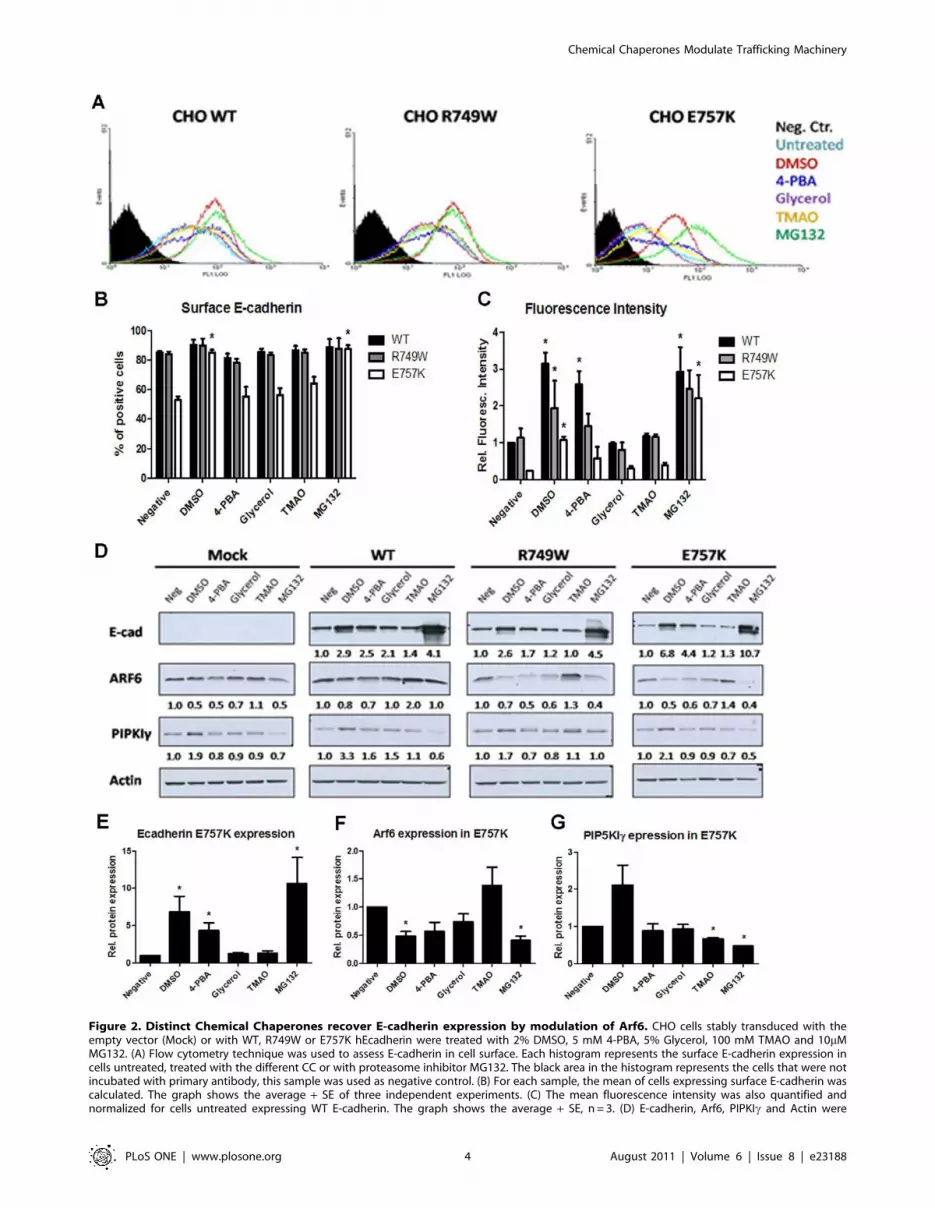
Figure 2. Distinct Chemical Chaperones recover E-cadherin expression by modulation of Arf6. CHO cells stably transduced with theempty vector (Mock) or with WT, R749W or E757K hEcadherin were treated with 2% DMSO, 5 mM 4-PBA, 5% Glycerol, 100 mM TMAO and 10mMMG132. (A) Flow cytometry technique was used to assess E-cadherin in cell surface. Each histogram represents the surface E-cadherin expression incells untreated, treated with the different CC or with proteasome inhibitor MG132. The black area in the histogram represents the cells that were notincubated with primary antibody, this sample was used as negative control. (B) For each sample, the mean of cells expressing surface E-cadherin wascalculated. The graph shows the average + SE of three independent experiments. (C) The mean fluorescence intensity was also quantified andnormalized for cells untreated expressing WT E-cadherin. The graph shows the average + SE, n = 3. (D) E-cadherin, Arf6, PIPKIc and Actin were
Chemical Chaperones Modulate Trafficking Machinery
PLoS ONE | www.plosone.org 4 August 2011 | Volume 6 | Issue 8 | e23188

and from 0.6 to 1.0 fold increase in the mutant R749W (Figure 4C–
D). Moreover, and similarly to what was verified by siRNA, the
ARF6 T27N also results in intracellular accumulation of E-cadherin
in cells expressing either WT or mutant E-cadherin (Figure 4B) and
this might be the result of the failure of Arf6 role in recycling [27,28].
As expected, the effect of this non-functional Arf6 is less evident than
that of the siRNA-mediated knockdown, due to the presence of
endogenous, functional, Arf6.
These results confirm the involvement of Arf6 in E-cadherin
trafficking and regulation and show that manipulation of Arf6
could be used to restore E-cadherin. Accordingly, in cells with
normal E-cadherin expression, the constitutive activation of Arf6
induces E-cadherin internalization and consequently cells loose the
epithelial phenotype. By contrast, in E-cadherin mutant context
with decreased PM expression, the inactivation of Arf6 induces
E-cadherin rescue at the plasma membrane.
Pharmacological inhibition of endocytosis promotesE-cadherin increase at the plasma membrane
Knowing that Arf6 has a role in the endocytic process but also
in recycling pathways, we investigated whether the increase of WT
and mutant E-cadherin expression was a specific feature of
endocytosis inhibition or an effect related to the inhibition of E-
cadherin recycling. To answer this question we treated the cell
lines stably expressing WT or the mutant forms of E-cadherin with
two pharmacological inhibitors of endocytosis, Dynasore and
MiTMAB. To control the efficiency of the endocytic process
blockage by these inhibitors, we tested the internalization of
detected in whole cell lysates by Western Blot. Actin was used as a loading control. The intensity of the bands was quantified and normalized againstthe untreated cells. The average of three independent experiments is shown below the respective sample. (E) The graph shows the average + SE ofE-cadherin protein level in mutant E757K cells after treatments. (F) The bars represent the average + SE of Arf6 protein level in mutant E757K cellsafter treatments. (G) Quantification of PIPKIc protein level in mutant E757K cells after treatments. * represents p#0.05.doi:10.1371/journal.pone.0023188.g002
Figure 3. Arf6 specific inhibition by siRNA leads to an increase of E-cadherin protein expression. Arf6 inhibition by siRNA was performedin CHO cells stably transduced with WT or E757K hEcadherin. (A) Cell lysates were extracted 48 h after transfection. E-cadherin, Arf6 and Actin wereanalyzed by Western Blot. Actin was used as a loading control. The intensity of the bands was quantified and normalized against the siRNA control. Inthe graph, bars represent the average + SE of E-cadherin or Arf6 protein expression of three independent experiments. (B) Flow cytometry techniquewas used to assess E-cadherin cell surface expression. Each histogram represents the cell surface expression of E-cadherin in WT or E757K cells treatedwith siRNA control (blue) or with specific siRNA for Arf6 (green). The black area in the histogram represents the cells that were not incubated withprimary antibody; this sample was used as negative control. For each sample, the number of cells expressing surface E-cadherin as well as the meanfluorescence intensity (arbitrary units) was calculated. The graphs show the average + SE of three independent experiments (* represents p#0.05). (C)Cells were fixed 48 h after transfection and immunostained with anti-human E-cadherin antibody. Nucleus was counterstained with DAPI. Thepictures were taken under a 406objective.doi:10.1371/journal.pone.0023188.g003
Chemical Chaperones Modulate Trafficking Machinery
PLoS ONE | www.plosone.org 5 August 2011 | Volume 6 | Issue 8 | e23188

Transferrin conjugates. The Transferrin uptake is the approach
commonly used to assess the endocytosis inhibition [29,30]. In
addition, we analyzed the expression of Clathrin heavy chain, the
main structural protein of clathrin-coated pits [31], as it was
described that Dynasore reduces the clathrin coated pit formation
[29]. We could confirm the action of the inhibitors by a decrease
in Clathrin protein levels (Figure 5A), concomitant with a decrease
of Transferrin594 internalization (Figure 5B).
After pharmacological inhibition of endocytosis, we evaluated
E-cadherin total protein level by western blot. In the three cell
lines, Dynasore or MiTMAB did not promote significant
alterations in E-cadherin level, however Arf6 protein is reduced
confirming its involvement in the endocytic pathway (Figure 5A).
Under the same treatment conditions, E-cadherin subcellular
localization and surface expression were further evaluated by
immunofluorescence staining and FCM (Figure 5C–D). We
verified that the number of cells expressing E-cadherin in the
PM is increased by Dynasore: 5.1% in WT (p = 0.035), 4.3% in
R749W and 9.0% in E757K cells; and by MITMAB: 2.6%, 0.1%
and 4.8%, respectively (Figure 5D). The mean fluorescence
intensity was also increased with the endocytosis blockage by
Dynasore or MITMAB, although the differences were not
statistically significant (Figure 5D). Importantly, the endocytosis
inhibition does not induce an abnormal E-cadherin accumulation
in the cytoplasm of the cells, as happens with the specific
modulation of Arf6 by siRNA or by Arf6 mutant constructs.
DMSO improves E-cadherin interaction with PIPKIc,b- and p120-catenins leading to increased E-cadherinexocytosis and stability
In order to test whether DMSO also promotes E-cadherin
exocytosis and stability at PM, we tested if cells upon DMSO
treatment exhibited an increased interaction between E-cadherin
and PIPKIc, justifying the high efficiency of DMSO in E-cadherin
recovery to the PM. It is well known that PIPKIc binds directly to
E-cadherin in a homeostatic situation [24]. Using Proximity
Ligation Assay (PLA), we could verify that, under normal
conditions, the mutants R749W and E757K interact less with
PIPKIc than WT E-cadherin, 0.5 and 0.8 respectively, and this
decrease is statistically significant for the mutant R749W
(p = 0.005), as showed in the Figure 6A. However, it is possible
to significantly improve this interaction with DMSO treatment:
2.3 fold for WT (p = 0.05), from 0.5 to 1.5 (3.0 fold, p = 0.03) for
the mutant R749W and from 0.8 to 2.2 (2.75 fold) for the mutant
E757K.
Because p120- and b-catenins link directly to E-cadherin
[1,4,16,17,19,32] and cadherin-catenins interaction is essential to
Figure 4. Arf6 activation or inactivation interferes with E-cadherin expression and localization. CHO WT, CHO R749W and CHO E757Kwere transiently transfected with vectors expressing mutant forms of Arf6: ARF6 Q67L (constitutive active form) and ARF6 T27N (dominant negativeform). (A) Arf6, E-cadherin and Actin were detected in whole cell lysates by Western Blot. Actin was used as a loading control. The intensity of thebands was quantified and normalized against the transfection control. The intensity average of three independent experiments is shown below therespective sample. (B) Cells were fixed and immunostained with anti-human E-cadherin antibody. Nucleus was counterstained with DAPI. Thepictures were taken under a 406 objective. (C) Flow cytometry technique was used to assess E-cadherin cell surface expression. Each histogramrepresents surface E-cadherin in cells untransfected (yellow), transfected with ARF6 Q67L (blue) or with ARF6 T27N (red) and the transfection control(green). The black area in the histogram represents the cells that were not incubated with primary antibody; this sample was used as negativecontrol. The results are representative of three independent experiments. (D) For each sample, the number of cells expressing surface E-cadherin wascalculated. The mean fluorescence intensity was also quantified and normalized against the transfection control of WT expressing cells. The graphsshow the average + SE, n = 3 (* represents p#0.05).doi:10.1371/journal.pone.0023188.g004
Chemical Chaperones Modulate Trafficking Machinery
PLoS ONE | www.plosone.org 6 August 2011 | Volume 6 | Issue 8 | e23188

E-cadherin transport and stabilization on plasma membrane and
consequently affect E-cadherin function, we analyzed these
interactions in the same context: medium or DMSO treatment.
The PLA results show that, without treatment, both E-cadherin
mutants are significantly less able to interact with p120-catenin
(Figure 6B; p = 0.017 for the mutant R749W) and b-catenin
(Figure 6C; p = 0.0005 for the mutant R749W and p = 0.001 for
the mutant E757K). However, DMSO strongly increases its
binding ability (Figure 6B–C). WT E-cadherin increases its
interaction with p120 4.2 fold and with b-catenin 1.8 fold
(p = 0.025). In the case of R749W E-cadherin, its interaction with
p120 increases from 0.3 to 0.9 and with b-catenin from 0.5 to 1.2
(p = 0.006). For the mutant E757K, the relation with p120
improves from 0.4 to 1.6 (p = 0.04) and with b-catenin from 0.2 to
1.0 (p = 0.02).
Discussion
Germline mutations of the CDH1 gene result in loss of
function of E-cadherin gene in approximately one third of the
cases of Hereditary Diffuse Gastric Cancer (HDGC). Most of the
germline CDH1 mutations identified to date are of the nonsense
type, leading to alternative premature termination codons. They
are commonly downregulated by non-sense mediated decay
[33], a mechanism of mRNA surveillance, making their
pathogenic significance easy to infer. However, germline
missense mutations occur in about 30% of the families [34],
and in contrast to truncating mutations, their pathogenicity is
not straight-forward, therefore constituting a problem in terms
of genetic counseling. To circumvent this limitation and im-
prove the genetic counseling, we have established an in vitro
cellular model to classify the significance of missense CDH1
mutations [35]. Within all the mutants functionally tested
[20,35,36,37,38,39,40,41], the juxtamembrane domain missense
E-cadherin mutants R749W and E757K, besides showing
impaired E-cadherin dependent adhesion and increased inva-
siveness, also display reduced total and surface protein
expression, 60% and 25% respectively, due to ERAD [20].
Interestingly, in our previous study, we showed that CC DMSO
was able to recover mutant E-cadherin expression to the PM and
function but the molecular mechanism underlying this effect
remained to be determined [20].
Figure 5. Endocytosis inhibition increases E-cadherin at the plasma membrane. CHO cells stably transduced with the empty vector (Mock)or with WT, R749W or E757K hEcadherin were treated with Dynasore or MiTMAB for 17h. (A) Clathrin, E-cadherin and Arf6 expression were analyzed inwhole cell lysates by Western Blot. Actin was used as a loading control. The intensity of the bands was quantified and normalized against theuntreated WT cells. The graphs show the average + SE of protein level, in three independent experiments. (B) After treatment, Transferrin 594 wasadded to cells. Nucleus was counterstained with DAPI. The pictures were taken under a 636objective. (C) Cells were fixed and immunostained withanti-human E-cadherin antibody. Nucleus was counterstained with DAPI. The pictures were taken under a 406 objective. (D) Flow cytometrytechnique was used to assess E-cadherin cell surface expression. Each histogram represents the cell surface expression of E-cadherin in cells WT,R749W or E757K, treated with Dynasore (red) or MiTMAB (green) or untreated (blue). The black area in the histogram represents the cells that werenot incubated with primary antibody, this sample was used as negative control. For each sample, the number of cells expressing surface E-cadherinwas calculated. The mean fluorescence intensity was also quantified and normalized against the control of WT expressing cells. The graphs show theaverage + SE, n = 3 (* represents p#0.05).doi:10.1371/journal.pone.0023188.g005
Chemical Chaperones Modulate Trafficking Machinery
PLoS ONE | www.plosone.org 7 August 2011 | Volume 6 | Issue 8 | e23188

In the present work, we focused on the effect of the CC on Arf6,
a key protein involved in E-cadherin regulation and cellular
trafficking [21,23,42]. We have found that DMSO treatment
rescues E-cadherin expression and at the same time inhibits Arf6
expression at the RNA and protein level (Figure 1). These findings
suggest that the stabilization of E-cadherin in the PM, in response
to CC treatment, could be mediated by a decrease of the endocytic
pathway, resulting from Arf6 downregulation.
To understand the significance of Arf6 downregulation in this
background, we performed specific inhibition of Arf6 in cells stably
transduced with WT or mutant E-cadherin. The knockdown of
Arf6 by siRNA leads to increased total E-cadherin expression, both
in the WT and mutant background, with a significant increase
observed for the mutant E757K. Furthermore, the quantity of E-
cadherin molecules present in the cell surface is improved,
independently of E-cadherin context. The observations that Arf6
silencing leads to increased total E-cadherin expression and
cytoplasmic accumulation of E-cadherin protein (Figure 3), suggests
that Arf6 silencing interferes with the process of E-cadherin
recycling and degradation. Our findings are in accordance with
the role previously described for Arf6, regarding endosomes
recycling and protein degradation [27,28]. In 1995, the work of
D’Souza-Schorey and collaborators showed that, in CHO cells,
expression of the dominant negative mutant, ARF6 T27N, resulted
in intracellular distribution of transferrin receptors and an inhibition
of transferrin recycling to the cell surface [25]. A large number of
studies followed, corroborating that finding [26,43,44,45]. More
recently, evidence has emerged suggesting Arf6 as an indirect
regulator of the proteasome. Arf6 promotes actin remodeling
through its interaction with POR1 (partner of Rac1) and Arfaptin-2
[46,47]. It was shown that Arfaptin-2 inhibits the function of 26S
proteasome, and as such modulates some cellular processes that are
dependent on proteasome function [48,49].
To further dissect the regulation of mutant E-cadherin by Arf6,
we have used Arf6 mutants defective in GTP hydrolysis (Q67L,
constitutively active) and GDP binding (T27N, dominant negative).
As expected, the constitutive active Arf6 (Q67L) promoted
E-cadherin internalization from cell surface, resulting in decreased
Figure 6. DMSO improves E-cadherin interaction with PIPKIc, b- and p120-catenins. CHO cells transduced with the empty vector (Mock) orwith WT, R749W or E757K hEcadherin were treated with 2% DMSO or with Medium, and the interaction of E-cadherin with PIPKIc (A), p120- (B) andb-catenins (C) was assessed by PLA. Cells were fixed and incubated with antibodies against E-cadherin and PIPKIc or E-cadherin and p120 or E-cadherin and b-catenin. In the negative control, CHO WT cells were incubated only with the antibody against E-cadherin. Close proximity ofoligonucleotide-ligated secondary antibodies allows a rolling-circle amplification and the detection of the rolling-circle amplification product by afluorescently labelled probe. Nuclei were counterstained with DAPI. The pictures were taken under a 406objective. The number of spots per cell wasquantified in each condition. The graphs show the average of relative number of blobs per cell + SE, n = 3 (* represents p#0.05).doi:10.1371/journal.pone.0023188.g006
Chemical Chaperones Modulate Trafficking Machinery
PLoS ONE | www.plosone.org 8 August 2011 | Volume 6 | Issue 8 | e23188

levels of E-cadherin at the PM, and cytoplasmic accumulation of E-
cadherin protein (Figure 4). In contrast, expression of a dominant
negative mutant (T27N) resulted in an increased PM expression and
also intracellular accumulation of E-cadherin (Figure 4).
To clarify if the increase of total and surface E-cadherin, in
response to Arf6 modulation, could be related to an effect of
endocytosis inhibition or due to the blockage of E-cadherin
recycling, we decided to perform endocytosis-inhibition assays
using pharmacological inhibitors of the dynamin-dependent
endocytic pathways, Dynasore and MiTMAB. These two com-
pounds are potent Dynamin-GTPase activity inhibitors [29,30].
Dynamin is a GTPase enzyme essential for vesicle formation and is
required for membrane constriction and fission during endocytosis
[29,30,50]. The major route of E-cadherin internalization is
reported to be a Clathrin dependent route [50,51,52,53,54],
although non-Clathrin-dependent pathways have been implicated,
including Caveolae-mediated [55] and an EGF-induced macro-
pinocytosis pathway [56]. Nevertheless, all mentioned pathways are
dependent of Dynamin activity [50,57]. Upon endocytosis inhibi-
tion, we observe that, independently of the E-cadherin background,
E-cadherin level of expression is not significantly altered, although
there is an increase in the number of cells expressing E-cadherin in
PM as well as an increase in the number of E-cadherin molecules
present in the PM (Figure 5). These results suggest that the E-
cadherin increase in the PM, in response to Arf6 modulation, is a
consequence of the endocytic pathway decrease but E-cadherin
accumulation in the cytoplasm of the cells, is probably due to the
inhibition of E-cadherin recycling and degradation.
Finally, we demonstrated that, together with DMSO, several
other CCs are able to modulate Arf6 expression. Interestingly, the
more effective compound in E-cadherin recovery to PM is the one
that simultaneously inhibit Arf6 and stimulate PIPKIc (Figure 2)
showing that stabilization of E-cadherin at the cell surface is due to
a decrease of the endocytic and recycling machinery and
stimulation of exocytic pathways. It is known that the role of
Arf6 at the cell surface is mediated by its effect on phospholipid
metabolism [58,59,60] and it was shown that Arf6 regulates
PIPKIc [59]. Moreover it was demonstrated that PIPKIc binds
directly to E-cadherin, modulating its cellular trafficking [24].
PIPKIc depletion or disruption of its binding to E-cadherin results
in defects in E-cadherin transport and blocks AJs assembly [24].
Therefore, we tested whether the increase of PIPKIc expression
upon DMSO treatment could imply increased binding to E-
cadherin and, in consequence, increased E-cadherin transport to
the PM and stabilization at AJs. To verify the interaction between
E-cadherin and PIPKIc, we used PLA [61] and have shown that
DMSO was able to improve the interplay of E-cadherin with
PIPKIc, b- and p120-catenins, which have crucial roles in E-
cadherin exocytosis, transport and stability in the PM (Figure 6).
Proteasome inhibition by MG132 also results in E-cadherin
increase and stabilization at the PM (Figure 2). It was already
described that MG132 regulates the stability of E-cadherin at the
PM through blocking its endocytosis induced by Transforming
Growth Factor (TGF)-b, however the molecular mechanism
implicated was unknown [62]. Our findings are not only in
agreement with that study, but also show that MG132 effect is also
mediated by downregulation of Arf6.
Together, our results show that mutant E-cadherin expression
rescue by the CCs, is mediated by the modulation of E-cadherin
trafficking partners, namely Arf6 and PIPKIc. In this context,
we propose that Arf6 downregulation results in the blockage of
E-cadherin endocytosis, but also in its recycling to the PM and
possibly its degradation blockage, leading to a significant
cytoplasmic accumulation of the protein. Artificial manipulation
of Arf6 shows that Arf6 inhibition is essential, but not sufficient, to
completely restore the E-cadherin mutant associated to Hereditary
Diffuse Gastric Cancer syndrome to the PM. In turn, PIPKIcbinding, as well as b- and p120-catenins, are responsible for the
improvement of E-cadherin exocytosis, transport and stabilization
in the PM.
Thus, by dissecting the mechanism of action of CCs, and its
influence on E-cadherin trafficking regulators, we propose that
mutant E-cadherin may be stabilized at the PM by inhibition of
endocytosis and decreased recycling and degradation, but also
increased exocytosis. Accordingly, we propose that the CCs effect
is the result of a balance between the different trafficking processes:
exocytosis, endocytosis, recycling and degradation.
With this work, we present the first evidence of a direct
influence of CCs in the cellular trafficking machinery and we
propose that this influence should be taken into account when
exploring the therapeutic potential of these types of molecules.
CCs have been already studied experimentally and reported to
reverse and repair conformational defects of some mutated
proteins [63]. Therefore CCs are promising therapies in cancer
and in a large number of protein-misfolding diseases as
Alzheimer’s disease, Parkinson’s disease, Huntington’s disease,
Creutzfeldt–Jakob disease, cystic fibrosis, nephrogenic diabetes
insipidus, Gaucher’s disease and many other degenerative and
neurodegenerative disorders [63]. Nevertheless, the toxicity of
these types of molecules is often problematic as therapy. The
success of CCs in therapeutic approaches depends on our ability to
understand their mechanism of action, enabling the alternative of
manipulating the involved molecules in a viable way.
Materials and Methods
Cell CultureCHO (Chinese Hamster Ovary) cells (ATCC number: CCL-61)
were transduced with the following vectors: empty vector (Mock),
WT hE-cad, R749W and E757K, as previously described by
Simoes-Correia et al. [20]. The transduced cells were selected by
antibiotic resistance to blasticidin (5mg/ml). Cells were grown at
37uC under 5% CO2 humidified air, in a-MEM (+) medium
(Gibco, Invitrogen) supplemented with 10% fetal bovine serum
(HyClone, Perbio), 1% penicillin/streptomycin (Gibco, Invitrogen)
and blasticidin (Gibco, Invitrogen).
Cell TreatmentsCells were plated in 6-well plates and incubated with Chemical
Chaperones for 24 h and with endocytosis inhibitors or protea-
some inhibitor MG132 for 17 h. The CCs used were 2% dimethyl
sulfoxide (DMSO; Sigma), 5 mM 4-phenylbutyric acid (4-PBA;
Sigma), 5% glycerol (Sigma) and 100 mM trimethylamine-N-
oxide (TMAO; Sigma). The endocytosis inhibitors used were
Dynasore (80mM; Sigma) and MiTMAB (20mM; Calbiochem).
The MG132 (Calbiochem) was used at 10mM concentration.
TransfectionsA set of 4 different siRNAs targeting ARF6 mRNA was purchased
from Dharmacon and prepared according to the manufacturer’s
instructions. In parallel, nonsilencing siRNA duplexes (Dharmacon)
were used as negative control. Before transfection, 60% confluent
monolayers of CHO WT, CHO R749W or CHO E757K cells
plated onto 6-well plates were washed with PBS and incubated in
serum and antibiotic-free medium. Cells were transiently transfected
with 0-150 nM siRNA, using Lipofectamine 2000 transfection
reagent (Invitrogen). At the end of each transfection, putative
cytotoxic effects were evaluated, analyzing cell viability. Efficiency of
Chemical Chaperones Modulate Trafficking Machinery
PLoS ONE | www.plosone.org 9 August 2011 | Volume 6 | Issue 8 | e23188

depletion was maximum at 48 h, and ARF6-07 and was chosen
from the set of siRNAs and used at 50 nM.
For plasmid transient transfections, 1mg of DNA of each vector
was used and the transfection procedure was the same as described
above. The Q67L and T27N ARF6 constructs were kindly
provided by D’Souza-Schorey C. (Department of Biological
Sciences, University of Notre Dame, Notre Dame, USA).
Flow cytometry (FCM)Cells were grown to a confluent monolayer, detached with
Versene (Gibco, Invitrogen) and resuspended in ice-cold PBS with
0.05 mg/ml CaCl2. For all conditions, 56105 cells were
centrifuged for 5 min at 1500 rpm and 4uC and washed in PBS
with 0.05 mg/ml CaCl2 and 3% BSA. Cells were incubated for
30 min with the extracellular primary antibody against E-
cadherin, HECD1 (Zymed Laboratories) at 1:50 dilution. Cells
were washed twice and incubated with Alexa Fluor 488 goat anti-
mouse (1:250; Invitrogen) in the dark for 30 min. Finally, the cells
were washed and resuspended in 0.5 ml of washing solution. At
least 56104 cells were analyzed in a Coulter Epics XL-MCL flow
cytometer. The data were analyzed with WinMDI software.
Immunofluorescence stainingCells were seeded on 6-weell plates on top of glass coverslips and
grown to at least 80% confluence. Fixation was performed in ice-
cold methanol for 20 min, followed by washing and blocking in
5% BSA in PBS for 30 min at room temperature. The mouse
monoclonal E-cadherin antibody (BD Biosciences) was used at
1:300 dilution in PBS with BSA 5% and incubated for 1 h at room
temperature. An Alexa Fluor 488 goat anti-mouse (1:500;
Invitrogen) was applied for 1 h in dark as secondary antibody.
The coverslips were mounted on slides using Vectashield with
DAPI (Vector Laboratories). Images were acquired on a Carl Zeiss
Apotome Axiovert 200 M Fluorescence Microscope using 206and 406 objectives. Images were taken with an Axiocam HRm
camera and processed with the Zeiss Axion Vision 4.8 software.
Transferrin uptakeCells were plated on glass coverslips and grown to at least 80%
confluence. After cell treatment with Dynasore (80mM; Sigma) and
MiTMAB (20mM; Calbiochem), as described before, 5mg/ml
Transferrin 594 (Invitrogen) was added to each well and incubated
for 15 min at 37uC. Fixation was performed in 4% formaldehyde in
PBS for 30 min at 4uC followed by blocking of the aldehyde groups
with 50 mM NH4Cl in PBS for 10 min at room temperature.
Permeabilization was done with 0.2% Triton X-100 in PBS for
10 min. The coverslips were mounted on slides using Vectashield
with DAPI (Vector Laboratories). Images were acquired on a Carl
Zeiss Apotome Axiovert 200M Fluorescence Microscope using 406and 636 objectives. Images were taken with an Axiocam HRm
camera and processed with the Zeiss Axion Vision 4.8 software.
Western blottingCells were lysed in cold Catenin lysis buffer – 1% Triton X-100
(Sigma), 1% Nonidet P-40 (Sigma) in PBS – enriched with a
protease inhibitor cocktail (Roche) and a phosphatase inhibitor
cocktail (Sigma). The proteins were quantified using a modified
Bradford assay (Bio-Rad). For analysis of total protein samples,
25mg of proteins were eluted in sample buffer, and loaded in 7.5%
or 12% SDS–PAGE, depending on the mass of the molecules. The
proteins were then electroblotted onto a Hybond ECL membrane
(Amersham Biosciences). Membranes were blocked with 5%
non-fat milk or 4% BSA and 0.5% Tween-20 in PBS and
immunoblotted with antibodies against E-cadherin (1:1000, BD
Biosciences), Arf6 (1:100, Santa Cruz Biotechnology), PIP5K1C
(1:1000, Cell Signaling Technologies), Clathrin Heavy Chain
(1:1000, Cell Signaling Technologies) or Actin (1:1000, Santa Cruz
Biotechnology). Donkey anti-rabbit (Amersham Biosciences), sheep
anti-mouse (Amersham Biosciences) or donkey anti-goat (Santa
Cruz Biotechnology) HRP-conjugated secondary antibodies were
used, followed by ECL detection (Amersham Biosciences). Immu-
noblots were quantified with the Quantity One Software (Bio-Rad).
Real-Time PCRCells were grown to a confluent monolayer and total RNA was
extracted with Tripure (Roche) according to the manufacturers
protocol. cDNA was produced from 1mg of RNA with Superscript II
Reverse Transcriptase (Invitrogen) and random hexamer primers
(Invitrogen). Quantitative real time-PCR (qRT-PCR) was carried out
in triplicates for the target ARF6 and for the endogenous control 18S
using as probe sets Mm00500208_s1 and Hs99999901_s1 (Applied
Biosystems), respectively. Data was analyzed by the absolute
quantification method, applying a standard curve of serial dilutions
for the target gene and endogenous control, in each plate, using an
ABI Prism 7000 Sequence Detection System (Applied Biosystems).
Proximity Ligation AssayCells seeded on top of glass coverslips were treated with DMSO,
as previously described. After treatment, cells were fixed in ice-
cold methanol for 20 min and subjected to PLA using Duolink
Detection kit (Olink Bioscience, Uppsala, Sweden) according to
the manufacturer’s instructions for Duolink Blocking solution and
Detection protocol. Briefly, slides were blocked, incubated with
antibodies directed against E-cadherin (from BD Biosciences,
mouse, 1:100 or from Cell Signaling, rabbit, 1:30), PIP5K1C (BD
Biosciences, mouse, 1:30), p120 (BD Biosciences, mouse, 1:30) and
b-catenin (Sigma, rabbit, 1:100) and thereafter incubated with
PLA probes, which are secondary antibodies (anti-mouse Minus
and anti-rabbit Plus) conjugated to unique oligonucleotides.
Amplification oligonucleotides were hybridized to probe pairs
and circularized by ligation. The DNA circle was then amplified
using rolling circle amplification into a bundle of single stranded
DNA anchored to one of the antibodies and could be detected by
addition of complementary fluorophore-labeled oligonucleotides.
The coverslips were mounted on slides using Vectashield with
DAPI (Vector Laboratories). Images were acquired on a Carl Zeiss
Apotome Axiovert 200M Fluorescence Microscope using 206and
406objectives. Images were taken with an Axiocam HRm camera
and processed with the Zeiss Axion Vision 4.8 software. The
quantification of the dots was performed using BlobFinder V3.2.
Statistical analysisTwo-tailed paired Student’s t-test was used to perform statistical
analysis. In all analysis p,0.05 was required for statistical
significance. Statistical analysis was done using StatView software
program (PC version).
Acknowledgments
The authors would like to thank to Professor Crislyn D’Souza-Schorey
(Department of Biological Sciences, University of Notre Dame, Notre
Dame, USA) for the ARF6 plasmids kindly provided.
Author Contributions
Conceived and designed the experiments: JF JS-C OS GS RS. Performed
the experiments: JF. Analyzed the data: JF JS-C OS GS RS. Contributed
reagents/materials/analysis tools: OS. Wrote the paper: JF JS-C OS GS RS.
Chemical Chaperones Modulate Trafficking Machinery
PLoS ONE | www.plosone.org 10 August 2011 | Volume 6 | Issue 8 | e23188

References
1. Aberle H, Schwartz H, Kemler R (1996) Cadherin-catenin complex: proteininteractions and their implications for cadherin function. J Cell Biochem 61:
514–523.
2. Angst BD, Marcozzi C, Magee AI (2001) The cadherin superfamily. J Cell Sci
114: 625–626.
3. Parisini E, Higgins JM, Liu JH, Brenner MB, Wang JH (2007) The crystalstructure of human E-cadherin domains 1 and 2, and comparison with other
cadherins in the context of adhesion mechanism. J Mol Biol 373: 401–411.
4. Okamoto R, Irie K, Yamada A, Katata T, Fukuhara A, et al. (2005)
Recruitment of E-cadherin associated with alpha- and beta-catenins andp120ctn to the nectin-based cell-cell adhesion sites by the action of 12-O-
tetradecanoylphorbol-13-acetate in MDCK cells. Genes Cells 10: 435–445.
5. Wheelock MJ, Johnson KR (2003) Cadherins as modulators of cellular
phenotype. Annu Rev Cell Dev Biol 19: 207–235.
6. Popov Z, Gil-Diez de Medina S, Lefrere-Belda MA, Hoznek A, Bastuji-Garin S,et al. (2000) Low E-cadherin expression in bladder cancer at the transcriptional
and protein level provides prognostic information. Br J Cancer 83: 209–214.
7. Gabbert HE, Mueller W, Schneiders A, Meier S, Moll R, et al. (1996) Prognostic
value of E-cadherin expression in 413 gastric carcinomas. Int J Cancer 69:184–189.
8. Guilford P, Hopkins J, Harraway J, McLeod M, McLeod N, et al. (1998)
E-cadherin germline mutations in familial gastric cancer. Nature 392: 402–405.
9. Huntsman DG, Carneiro F, Lewis FR, MacLeod PM, Hayashi A, et al. (2001)
Early gastric cancer in young, asymptomatic carriers of germ-line E-cadherinmutations. N Engl J Med 344: 1904–1909.
10. Becker KF, Atkinson MJ, Reich U, Becker I, Nekarda H, et al. (1994)
E-cadherin gene mutations provide clues to diffuse type gastric carcinomas.Cancer Res 54: 3845–3852.
11. Graff JR, Herman JG, Myohanen S, Baylin SB, Vertino PM (1997) Mappingpatterns of CpG island methylation in normal and neoplastic cells implicates
both upstream and downstream regions in de novo methylation. J Biol Chem272: 22322–22329.
12. Berx G, Becker KF, Hofler H, van Roy F (1998) Mutations of the human
E-cadherin (CDH1) gene. Hum Mutat 12: 226–237.
13. Peinado H, Quintanilla M, Cano A (2003) Transforming growth factor beta-1
induces snail transcription factor in epithelial cell lines: mechanisms for epithelialmesenchymal transitions. J Biol Chem 278: 21113–21123.
14. Machado JC, Oliveira C, Carvalho R, Soares P, Berx G, et al. (2001) E-cadherin
gene (CDH1) promoter methylation as the second hit in sporadic diffuse gastric
carcinoma. Oncogene 20: 1525–1528.
15. Pinho SS, Reis CA, Paredes J, Magalhaes AM, Ferreira AC, et al. (2009) Therole of N-acetylglucosaminyltransferase III and V in the post-transcriptional
modifications of E-cadherin. Hum Mol Genet 18: 2599–2608.
16. Delva E, Kowalczyk AP (2009) Regulation of cadherin trafficking. Traffic 10:
259–267.
17. Bryant DM, Stow JL (2004) The ins and outs of E-cadherin trafficking. TrendsCell Biol 14: 427–434.
18. D’Souza-Schorey C (2005) Disassembling adherens junctions: breaking up ishard to do. Trends Cell Biol 15: 19–26.
19. Yap AS, Crampton MS, Hardin J (2007) Making and breaking contacts: the
cellular biology of cadherin regulation. Curr Opin Cell Biol 19: 508–514.
20. Simoes-Correia J, Figueiredo J, Oliveira C, van Hengel J, Seruca R, et al. (2008)
Endoplasmic reticulum quality control: a new mechanism of E-cadherinregulation and its implication in cancer. Hum Mol Genet 17: 3566–3576.
21. Palacios F, Schweitzer JK, Boshans RL, D’Souza-Schorey C (2002) ARF6-GTP
recruits Nm23-H1 to facilitate dynamin-mediated endocytosis during adherensjunctions disassembly. Nat Cell Biol 4: 929–936.
22. Morishige M, Hashimoto S, Ogawa E, Toda Y, Kotani H, et al. (2008) GEP100links epidermal growth factor receptor signalling to Arf6 activation to induce
breast cancer invasion. Nat Cell Biol 10: 85–92.
23. Palacios F, Price L, Schweitzer J, Collard JG, D’Souza-Schorey C (2001) Anessential role for ARF6-regulated membrane traffic in adherens junction
turnover and epithelial cell migration. EMBO J 20: 4973–4986.
24. Ling K, Bairstow SF, Carbonara C, Turbin DA, Huntsman DG, et al. (2007)
Type Igamma phosphatidylinositol phosphate kinase modulates adherensjunction and E-cadherin trafficking via a direct interaction with mu 1B adaptin.
J Cell Biol 176: 343–353.
25. D’Souza-Schorey C, Li G, Colombo MI, Stahl PD (1995) A regulatory role for
ARF6 in receptor-mediated endocytosis. Science 267: 1175–1178.
26. D’Souza-Schorey C, van Donselaar E, Hsu VW, Yang C, Stahl PD, et al. (1998)ARF6 targets recycling vesicles to the plasma membrane: insights from an
ultrastructural investigation. J Cell Biol 140: 603–616.
27. Donaldson JG (2003) Multiple roles for Arf6: sorting, structuring, and signaling
at the plasma membrane. J Biol Chem 278: 41573–41576.
28. D’Souza-Schorey C, Chavrier P (2006) ARF proteins: roles in membrane trafficand beyond. Nat Rev Mol Cell Biol 7: 347–358.
29. Macia E, Ehrlich M, Massol R, Boucrot E, Brunner C, et al. (2006) Dynasore, a
cell-permeable inhibitor of dynamin. Dev Cell 10: 839–850.
30. Quan A, McGeachie AB, Keating DJ, van Dam EM, Rusak J, et al. (2007)
Myristyl trimethyl ammonium bromide and octadecyl trimethyl ammoniumbromide are surface-active small molecule dynamin inhibitors that block
endocytosis mediated by dynamin I or dynamin II. Mol Pharmacol 72:
1425–1439.
31. Sorkin A, von Zastrow M (2009) Endocytosis and signalling: intertwining
molecular networks. Nat Rev Mol Cell Biol 10: 609–622.
32. van Roy F, Berx G (2008) The cell-cell adhesion molecule E-cadherin. Cell Mol
Life Sci 65: 3756–3788.
33. Karam R, Carvalho J, Bruno I, Graziadio C, Senz J, et al. (2008) The NMD
mRNA surveillance pathway downregulates aberrant E-cadherin transcripts in
gastric cancer cells and in CDH1 mutation carriers. Oncogene 27: 4255–4260.
34. Carneiro F, Oliveira C, Seruca R Pathology and genetics of familial gastric
cancer. Int J Surg Pathol 18: 33S–36S.
35. Suriano G, Oliveira C, Ferreira P, Machado JC, Bordin MC, et al. (2003)
Identification of CDH1 germline missense mutations associated with functional
inactivation of the E-cadherin protein in young gastric cancer probands. Hum
Mol Genet 12: 575–582.
36. Suriano G, Mulholland D, de Wever O, Ferreira P, Mateus AR, et al. (2003)
The intracellular E-cadherin germline mutation V832 M lacks the ability to
mediate cell-cell adhesion and to suppress invasion. Oncogene 22: 5716–5719.
37. Kaurah P, MacMillan A, Boyd N, Senz J, De Luca A, et al. (2007) Founder and
recurrent CDH1 mutations in families with hereditary diffuse gastric cancer.
JAMA 297: 2360–2372.
38. More H, Humar B, Weber W, Ward R, Christian A, et al. (2007) Identification
of seven novel germline mutations in the human E-cadherin (CDH1) gene. Hum
Mutat 28: 203.
39. Brooks-Wilson AR, Kaurah P, Suriano G, Leach S, Senz J, et al. (2004)
Germline E-cadherin mutations in hereditary diffuse gastric cancer: assessment
of 42 new families and review of genetic screening criteria. J Med Genet 41:
508–517.
40. Suriano G, Yew S, Ferreira P, Senz J, Kaurah P, et al. (2005) Characterization
of a recurrent germ line mutation of the E-cadherin gene: implications for
genetic testing and clinical management. Clin Cancer Res 11: 5401–5409.
41. Keller G, Vogelsang H, Becker I, Plaschke S, Ott K, et al. (2004) Germline
mutations of the E-cadherin(CDH1) and TP53 genes, rather than of RUNX3
and HPP1, contribute to genetic predisposition in German gastric cancer
patients. J Med Genet 41: e89.
42. Paterson AD, Parton RG, Ferguson C, Stow JL, Yap AS (2003) Characterization
of E-cadherin endocytosis in isolated MCF-7 and chinese hamster ovary cells:
the initial fate of unbound E-cadherin. J Biol Chem 278: 21050–21057.
43. Klein S, Franco M, Chardin P, Luton F (2006) Role of the Arf6 GDP/GTP
cycle and Arf6 GTPase-activating proteins in actin remodeling and intracellular
transport. J Biol Chem 281: 12352–12361.
44. Jovanovic OA, Brown FD, Donaldson JG (2006) An effector domain mutant of
Arf6 implicates phospholipase D in endosomal membrane recycling. Mol Biol
Cell 17: 327–335.
45. Schweitzer JK, Pietrini SD, D’Souza-Schorey C (2009) ARF6-mediated
endosome recycling reverses lipid accumulation defects in Niemann-Pick Type
C disease. PLoS One 4: e5193.
46. Shin OH, Exton JH (2001) Differential binding of arfaptin 2/POR1 to ADP-
ribosylation factors and Rac1. Biochem Biophys Res Commun 285: 1267–1273.
47. D’Souza-Schorey C, Boshans RL, McDonough M, Stahl PD, Van Aelst L (1997)
A role for POR1, a Rac1-interacting protein, in ARF6-mediated cytoskeletal
rearrangements. EMBO J 16: 5445–5454.
48. Peters PJ, Ning K, Palacios F, Boshans RL, Kazantsev A, et al. (2002) Arfaptin 2
regulates the aggregation of mutant huntingtin protein. Nat Cell Biol 4:
240–245.
49. Rangone H, Pardo R, Colin E, Girault JA, Saudou F, et al. (2005)
Phosphorylation of arfaptin 2 at Ser260 by Akt Inhibits PolyQ-huntingtin-
induced toxicity by rescuing proteasome impairment. J Biol Chem 280:
22021–22028.
50. Mosesson Y, Mills GB, Yarden Y (2008) Derailed endocytosis: an emerging
feature of cancer. Nat Rev Cancer 8: 835–850.
51. Le TL, Yap AS, Stow JL (1999) Recycling of E-cadherin: a potential mechanism
for regulating cadherin dynamics. J Cell Biol 146: 219–232.
52. Izumi G, Sakisaka T, Baba T, Tanaka S, Morimoto K, et al. (2004) Endocytosis
of E-cadherin regulated by Rac and Cdc42 small G proteins through IQGAP1
and actin filaments. J Cell Biol 166: 237–248.
53. Ivanov AI, Nusrat A, Parkos CA (2004) Endocytosis of epithelial apical
junctional proteins by a clathrin-mediated pathway into a unique storage
compartment. Mol Biol Cell 15: 176–188.
54. Xiao K, Garner J, Buckley KM, Vincent PA, Chiasson CM, et al. (2005) p120-
Catenin regulates clathrin-dependent endocytosis of VE-cadherin. Mol Biol Cell
16: 5141–5151.
55. Lu Z, Ghosh S, Wang Z, Hunter T (2003) Downregulation of caveolin-1
function by EGF leads to the loss of E-cadherin, increased transcriptional activity
of beta-catenin, and enhanced tumor cell invasion. Cancer Cell 4: 499–515.
56. Bryant DM, Kerr MC, Hammond LA, Joseph SR, Mostov KE, et al. (2007)
EGF induces macropinocytosis and SNX1-modulated recycling of E-cadherin.
J Cell Sci 120: 1818–1828.
57. Kumari S, Mg S, Mayor S Endocytosis unplugged: multiple ways to enter the
cell. Cell Res 20: 256–275.
Chemical Chaperones Modulate Trafficking Machinery
PLoS ONE | www.plosone.org 11 August 2011 | Volume 6 | Issue 8 | e23188

58. Honda A, Nogami M, Yokozeki T, Yamazaki M, Nakamura H, et al. (1999)
Phosphatidylinositol 4-phosphate 5-kinase alpha is a downstream effector of thesmall G protein ARF6 in membrane ruffle formation. Cell 99: 521–532.
59. Krauss M, Kinuta M, Wenk MR, De Camilli P, Takei K, et al. (2003) ARF6
stimulates clathrin/AP-2 recruitment to synaptic membranes by activatingphosphatidylinositol phosphate kinase type Igamma. J Cell Biol 162: 113–124.
60. Wenk MR, De Camilli P (2004) Protein-lipid interactions and phosphoinositidemetabolism in membrane traffic: insights from vesicle recycling in nerve
terminals. Proc Natl Acad Sci U S A 101: 8262–8269.
61. Weibrecht I, Leuchowius KJ, Clausson CM, Conze T, Jarvius M, et al. (2010)
Proximity ligation assays: a recent addition to the proteomics toolbox. Expert
Rev Proteomics 7: 401–409.
62. Saitoh M, Shirakihara T, Miyazono K (2009) Regulation of the stability of cell
surface E-cadherin by the proteasome. Biochem Biophys Res Commun 381:
560–565.
63. Chaudhuri TK, Paul S (2006) Protein-misfolding diseases and chaperone-based
therapeutic approaches. FEBS J 273: 1331–1349.
Chemical Chaperones Modulate Trafficking Machinery
PLoS ONE | www.plosone.org 12 August 2011 | Volume 6 | Issue 8 | e23188