the efficacy of botulinum toxin injection in acute sixth nerve palsy
TRANSCRIPT
THE EFFICACY OF BOTULINUM TOXIN INJECTION
IN ACUTE SIXTH NERVE PALSY –A PROSPECTIVE
OBSERVATIONAL STUDY
Dissertation submitted to The Tamil Nadu Dr. M.G.R. Medical
University in partial fulfilment of the requirements for the degree of
MS Ophthalmology
BRANCH - III
OPHTHALMOLOGY
THE TAMIL NADU
DR. M.G.R. MEDICAL UNIVERSITY
CHENNAI –600032
MAY 2020

THE EFFICACY OF BOTULINUM TOXIN INJECTION
IN ACUTE SIXTH NERVE PALSY –A PROSPECTIVE
OBSERVATIONAL STUDY
Dissertation submitted to The Tamil Nadu Dr. M.G.R. Medical
University in partial fulfilment of the requirements for the degree of
MS Ophthalmology
BRANCH - III
OPHTHALMOLOGY
Register number: 221713459
THE TAMIL NADU
DR. M.G.R. MEDICAL UNIVERSITY
CHENNAI –600032
MAY 2020
CERTIFICATE
This is to certify that this dissertation titled “ THE EFFICACY
OF BOTULINUM TOXIN INJECTION IN ACUTE SIXTH NERVE
PALSY – A PROSPECTIVE OBSERVATIONAL STUDY” is a
bonafide done by Dr. V. R. Saranya under the guidance and supervision
in the department of Paediatric Ophthalmology, Aravind Eye Hospital
and Post Graduate Institute of Ophthalmology in Madurai during her
residency period June 2017 to May 2020.
Dr.N.Venkatesh Prajna, DO, DNB, FRCOphth., Professor and Head of the department Aravind Eye hospital Madurai-20
Dr.R.Rathinam, DO,DNB, Ph.D., Principal Aravind Eye Hospital Madurai -20
Dr.Shashikant Shetty,M.S., Chief, Paediatric Ophthalmology & Strabismus Services Aravind Eye Hospital Madurai-20

DECLARATION
I, Dr. V. R. Saranya solemnly declare the dissertation titled “THE
EFFICACY OF BOTULINUM TOXIN INJECTION IN ACUTE
SIXTH NERVE PALSY – A PROSPECTIVE OBSERVATIONAL
STUDY” has been prepared by me. I also declare that this bonafide work
or a part of this work was not submitted by me or any other for award,
degree, diploma to any other university board either in India or abroad.
This dissertation is submitted to The Tamil Nadu Dr. M. G. R.
Medical university, Chennai in partial fulfilment of the rules and
regulation for the award of M. S. Ophthalmology (BRANCH-III) to be
held in May 2020.
Place : Madurai
Date :
Dr. V. R. Saranya
Register no: 221713459
Aravind Eye hospital & Post Graduate
Institute of Ophthalmology
Madurai-20
ACKNOWLEDGEMENT
At the outset, I take this opportunity to pay my respect and homage
to our founder Late Dr. G. Venkataswamy, whose ideals and philosophy
have guided this institution in all its successful endeavours.
I wish to express my heartfelt and sincere gratitude to my guide
Dr.Shashikant shetty, Professor and Head of the department, Paediatric
Ophthalmology, Aravind Eye Hospital, Madurai for having guided me in
the successful completion of this dissertation and also for having created
an environment enriched with all the facilities.
I am highly grateful to my co-guide Dr. B.Sahithya, Medical
consultant for being a constant source of motivation and encouragement
which ultimately structured my thesis and provided valuable guidance
for completion of this work.
I would also thank Dr.Sathya T Ravilla for her guidance and
motivation throughout my study.
It is a proud privilege and pleasure to express my sincere thanks
towards my mentor Dr. N. Venkatesh Prajna, Director of Academics
and Professor & Head of department, Cornea services, Aravind Eye
Hospital, Madurai for his constant encouragement, guidance and support
throughout my residency.
I am grateful to Dr. R. D. Ravindran, Chairman, Aravind Eye
Care System, Dr. P. Namperumalsamy, Chairman Emeritus, Director of
Research, Aravind Eye Care system, Dr. G. Natchiar, Director Emeritus
(Human Resource department), Dr. M. Srinivasan Director Emeritus
and other scholars of ophthalmology at Aravind Eye Hospital, Madurai.

I offer my sincere thanks to Dr. Mahesh kumar, Prof & Head of
Department of Neuro-ophthalmology, Aravind Eye Hospital, Madurai
and Dr. A. Kowsalya, Medical consultant, Aravind Eye Hospital,
Madurai for guiding me throughout my thesis.
I sincerely thank Ms.Muthulakshmi, Ms.Vani,
Ms.Mangalasundari sister who guided and helped me to collect the
data.
I would like to thank Mrs.Iswarya, Mr.Mohammed siddiq
Biostatistician for their valuable help in statistical analysis.
I am much grateful to Mrs.Kumaragurupari, Senior Librarian
and Mr. R. Govindarajan, Assistant Librarian for their prompt
responses in providing all the articles and academic support.
My sincere thanks to all the patients without whom this study
would not have been possible.
Last but not the least I would like to thank my parents, husband for
all their sacrifices and unfailing love towards me and finally my friends
for being a constant support.
CONTENTS
PART I
S. NO. TITLE PAGE. NO.
1 INTRODUCTION 1
2 ANATOMY OF SIXTH NERVE PALSY 2
3 BOTULINUM TOXIN 10
4 INVESTIGATION 19
5 TREATMENT 43
6 REVIEW OF LITERATURE 46
PART - II
S. NO. TITLE PAGE NO.
1 AIMS AND OBJECTIVE 51
2 MATERIALS AND METHOD 53
3 RESULTS 60
4 DISCUSSION 80
5 CONCLUSION 82
6 ANNEXURES
Bibliography
Abbreviations
Proforma
Consent form
Institutional Review Board approval
Plagiarism Report
Master chart
i
v
vi
ix
xi
xii
xiv

PART- I
1
INTRODUCTION
Abducent nerve is the longest cranial nerve in the body which
travels from pons and supplies the lateral rectus muscle. Nerve damage at
any point along its course results in limitation of abduction and diplopia.
The incidence of lateral rectus palsy is 2.5 cases per 1,00,0001. Due to
higher incidence of diabetes in India, it has become one of the major risk
factor for the occurrence of nerve palsy. Other known factors include
hypertension, hyperlipidemia, trauma, tumor and infections. Indian
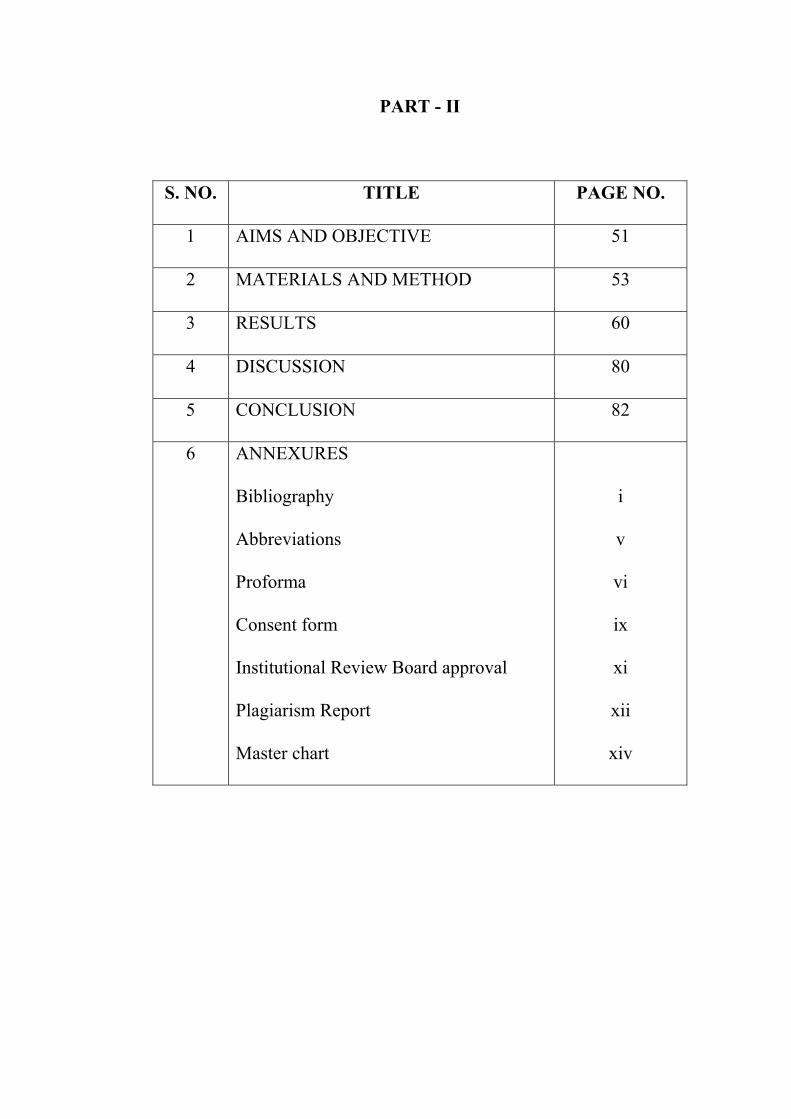
population based study published in 2014 reveals the incidence of lateral
rectus palsy and according to that around 86.36% cases are non
traumatic2.
Figure 1.1 Incidence of Sixth nerve palsy
0.00%
10.00%
20.00%
30.00%
40.00%
50.00%
60.00%
vasculopathy congenital inflammatory tumor idiopathic
2
ANATOMY OF SIXTH NERVE PALSY
Sixth nerve is entirely a motor nerve with two functional components5
1) Somatic efferent - lateral movement of the eye
2) General somatic afferent - proprioceptive impulses from lateral
rectus muscle
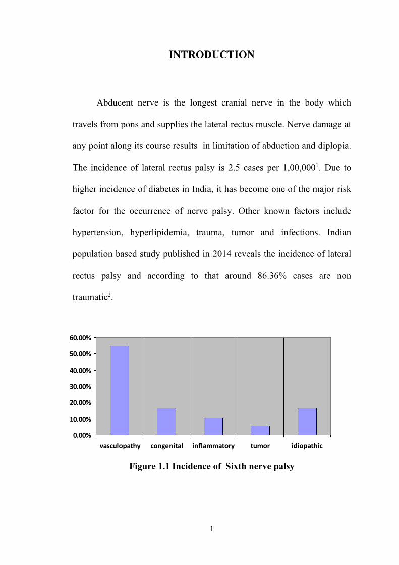
NUCLEUS
Location of nucleus:
Lower part of pons, beneath the floor of fourth ventricle and is
related to the fasciculus of the facial nerve.
Figure 1.2 Location of sixth nerve nucleus
3
It consists of two types of multipolar cells
a) Large multipolar cells – gives rise to fibres of abducent nerve
b) Small multipolar cells – forms para - abducent nucleus and relays in
oculomotor nerve via the medial longitudinal fasciculus.
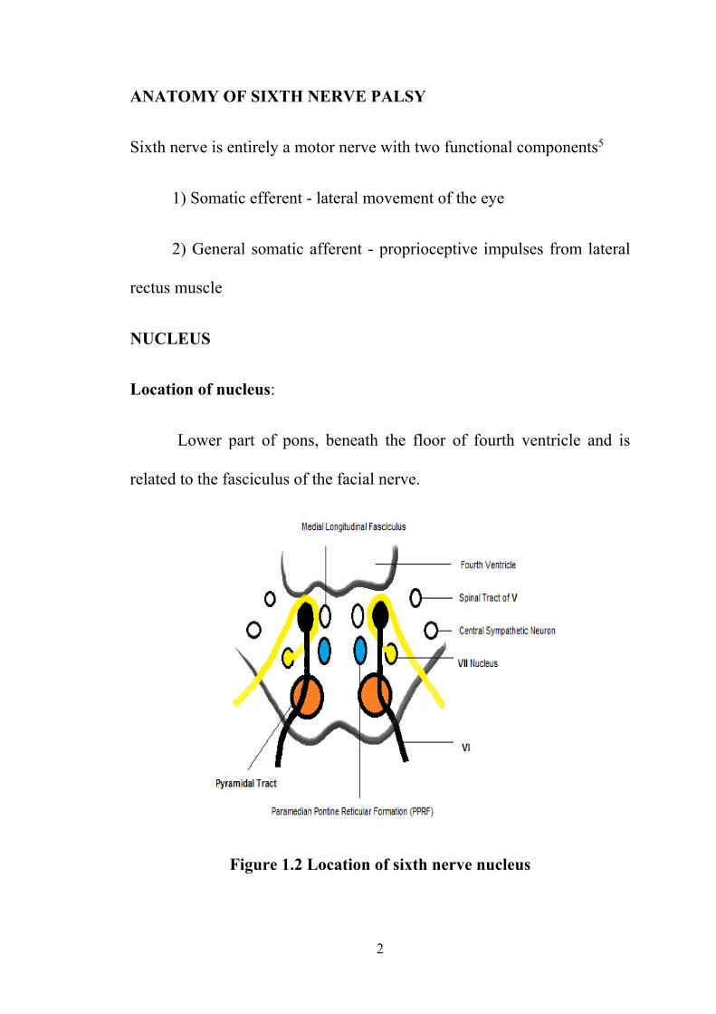
Course of sixth nerve
It is divided into four parts namely fasicular, basilar, cavernous and
intraorbital parts
Figure 1.3 Course of sixth nerve
4
a) Fascicular part
Starts from the nucleus and traverses the medial leminiscus and
pyramidal tract and finally emerges as 7 to 8 rootlets
b) Basilar part - brainstem till cavernous sinus
Nerve runs forwards through cisterna pontis between pons and
occipital bone and runs upwards on back of petrous temporal bone. At
sharp upper border it bends forward at right angle to enter the Dorello’s
canal. It travels along the inferior petrosal sinus and enter the cavernous
sinus through its posterior wall.
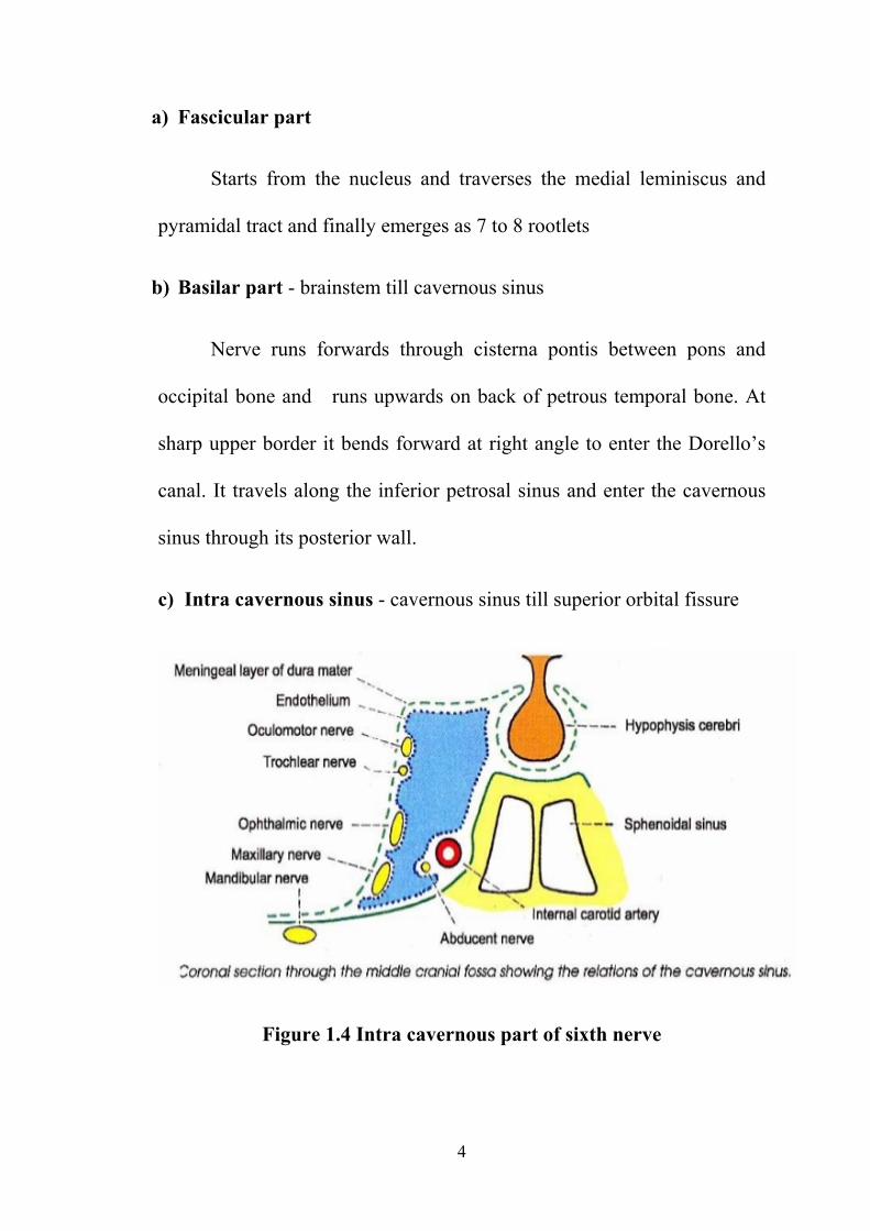
c) Intra cavernous sinus - cavernous sinus till superior orbital fissure
Figure 1.4 Intra cavernous part of sixth nerve
5
Inside the cavernous sinus the nerve lies below and lateral to the
internal carotid artery. It leaves the cavernous sinus and enters the orbit
through the middle part of superior orbital fissure.
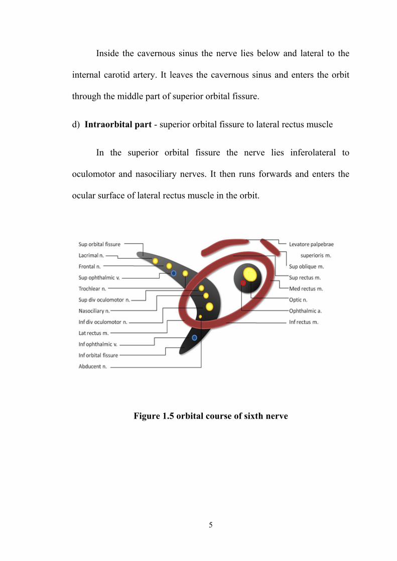
d) Intraorbital part - superior orbital fissure to lateral rectus muscle
In the superior orbital fissure the nerve lies inferolateral to
oculomotor and nasociliary nerves. It then runs forwards and enters the
ocular surface of lateral rectus muscle in the orbit.
Figure 1.5 orbital course of sixth nerve
6
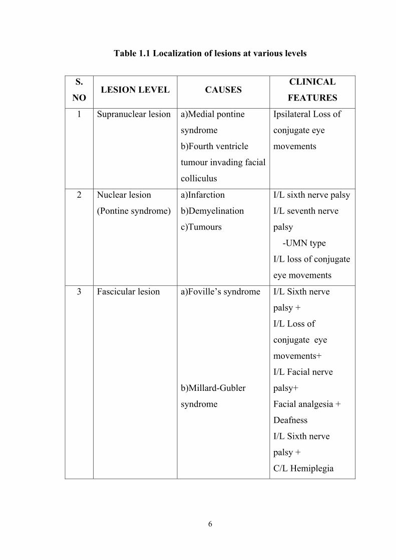
Table 1.1 Localization of lesions at various levels
S.
NO LESION LEVEL CAUSES
CLINICAL
FEATURES
1 Supranuclear lesion a)Medial pontine
syndrome
b)Fourth ventricle
tumour invading facial
colliculus
Ipsilateral Loss of
conjugate eye
movements
2 Nuclear lesion
(Pontine syndrome)
a)Infarction
b)Demyelination
c)Tumours
I/L sixth nerve palsy
I/L seventh nerve
palsy
-UMN type
I/L loss of conjugate
eye movements
3 Fascicular lesion a)Foville’s syndrome
b)Millard-Gubler
syndrome
I/L Sixth nerve
palsy +
I/L Loss of
conjugate eye
movements+
I/L Facial nerve
palsy+
Facial analgesia +
Deafness
I/L Sixth nerve
palsy +
C/L Hemiplegia

7
4 Basilar part lesion a)CP angle tumors
-acoustic neuroma
-meningoma
b)Clivus leison
c)Gradenigo
syndrome
Sixth nerve palsy
Reduced corneal
sensation
Hearing loss
Facial pain /
numbness/ palsy
5 Intracavernous part
lesion
a)Aneurysms
b)Meningioma
c)Carotid cavernous
fistula
d)Vascular lesion
-diabetes ,
hypertension
Sixth nerve palsy
Horner syndrome
6 Intraorbital lesion a)Orbital apex
syndrome
b)Superior orbital
fissure syndrome

8
Causes of sixth nerve palsy in adults6
Infections:
• Arachnoiditis, Lyme’s disease, Psittacosis, Staphylococcus aureus
infection, Syphilis, Varicella zoster
Trauma:
• Head injury, skull fractures, cervical spine fractures
Neoplasm:
• Chondroma, chondrosarcoma, chordoma, cylindroma, metastasis
Systemic disorders:
• Vascular – atherosclerosis, diabetes, hypertension, pre-eclampsia
• Hematological – leukemia, lymphomatous meningitis
Other vascular causes:
• Aneurysm, arteriovenous malformations, cerebrovascular insults
Associated neurologic disorder:
• Cluster headache, demyelinating disease, elevated intracranial pressure,
intracranial hypertension
9
Iatrogenic:
• Myelography, nerve blocks in the head and neck, post lumbar puncture,
post spinal or epidural anaesthesia
Others:
• Idiopathic, inflammatory, interferon toxicity, lithium toxicity.
FEATURES OF ISOLATED SIXTH NERVE PALSY
a) Diplopia - uncrossed horizontal diplopia which becomes worse
towards the direction of action of the paralyzed muscle
b) Abnormal head posture - Face turn towards the action of paralyzed
muscle to avoid diplopia
c) Esotropia - primary gaze due to unopposed action of medial rectus
muscle
d) Limitation of abduction - due to weakness of lateral rectus muscle

10
BOTULINUM TOXIN
Evolution of botox injection:
In early 19th century during Napoleonic warfare time, there was an
increased incidence of fatal food poisoning mainly due to consumption of
meat and blood sausages7 which documents the first food borne botulism.
In 1895, an outbreak led to the discovery of the pathogen, Clostridium
botulinum9.
Figure 1.6 Microscopic view of Clostridium botulinum
In Latin, the word Botulinum means Sausage8. In early 1970, Alan
B. Scott et al used Botulinum toxin widely in the treatment of strabismus
surgery7.
11
Apart from strabismus, Botulinum toxin is used in various
ophthalmic conditions like nystagmus, oscillopsia, thyroid related
orbitopathy to correct upper eyelid retraction, glabellar frowning,
therapeutic ptosis induction in case of corneal exposure. They are also
used in various dermatological conditions for cosmetic purpose and
dystonia9.
BOTULINUM TOXIN
Botulinum toxin is a neurotoxin produced by gram positive
bacilli, Clostridium botulinum. Totally there are eight antigenically
different types of exotoxins - A,B,C1,C2,D,E,F,G 10. Toxin A is the most
potent and is currently used in various medical fields. In 2002, botox
was approved for use by FDA for cosmetic purpose.
Biochemical properties:
Botulinum toxins are relatively inactive. It consists of heavy chain
and light chain of 100 kDa and 50 kDa respectively, linked by disulphide
bond11 .
12
Mechanism of action:
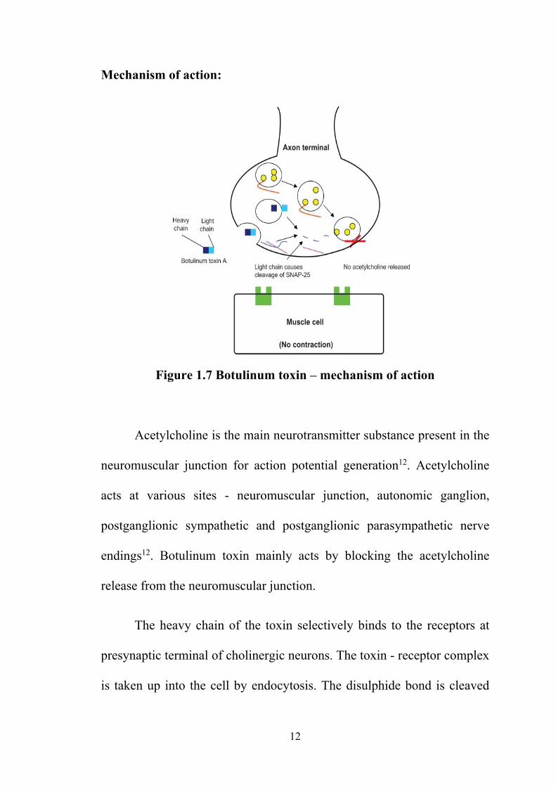
Figure 1.7 Botulinum toxin – mechanism of action
Acetylcholine is the main neurotransmitter substance present in the
neuromuscular junction for action potential generation12. Acetylcholine
acts at various sites - neuromuscular junction, autonomic ganglion,
postganglionic sympathetic and postganglionic parasympathetic nerve
endings12. Botulinum toxin mainly acts by blocking the acetylcholine
release from the neuromuscular junction.
The heavy chain of the toxin selectively binds to the receptors at
presynaptic terminal of cholinergic neurons. The toxin - receptor complex
is taken up into the cell by endocytosis. The disulphide bond is cleaved
13
and the light chain interacts with (SNAP)25 protein, syntaxin and vesicle
associated membrane protein(VAMP). This prevents the fusion of
acetylcholine vesicle with the cell membrane13. The toxin has its peak
effect 5-7days post injection14 .
Botulinum toxin inhibits the transmission of alpha motor neurons
in the neuromuscular junction and gamma motor neuron in the muscle
spindle and can result in reflex overactivity15.
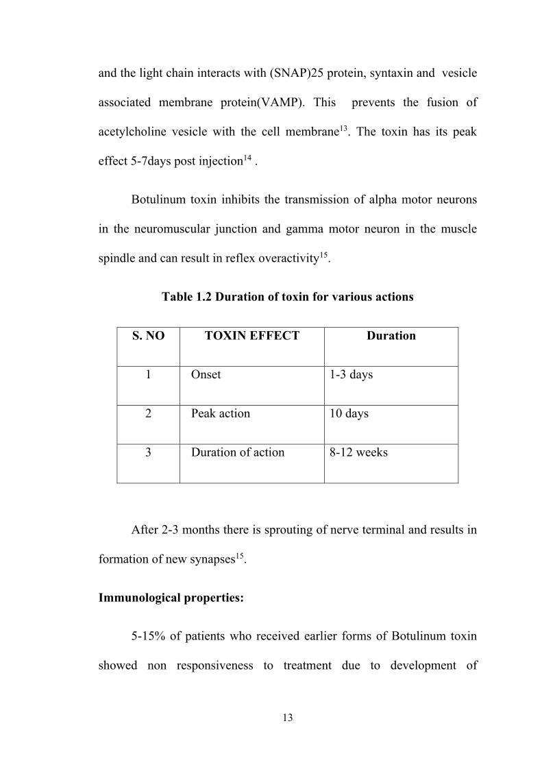
Table 1.2 Duration of toxin for various actions
S. NO TOXIN EFFECT Duration
1 Onset 1-3 days
2 Peak action 10 days
3 Duration of action 8-12 weeks
After 2-3 months there is sprouting of nerve terminal and results in
formation of new synapses15.
Immunological properties:
5-15% of patients who received earlier forms of Botulinum toxin
showed non responsiveness to treatment due to development of
14
neutralizing antibodies. So newer formulations of Botox (BCB2024)
with less protein and immunogenicity was made which resulted in less
chances of antibody production16
Risk factors for neutralizing antibodies:
Injecting more than 200 units/session
Repeat/ booster dose of injection within one month
period
TYPES OF FORMULATIONS
In 1989, FDA approved the use of botox as an orphan drug in
the treatment of strabismus and hemifacial spasm14. Serotype A is the
only commercially available drug in market. Other serotypes like B,C,F
are under trial. All the formulations are expressed in terms of mouse
units which means the dose of toxin required to kill 50% of a group of
18-20g of female Swiss Webster mice17 .
Two forms of Botulinum toxin A exists :
1) Botox
2) Dysport
15
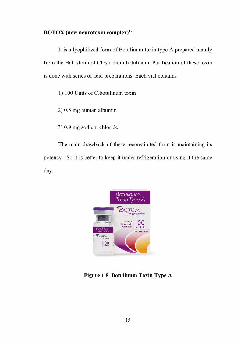
BOTOX (new neurotoxin complex)17
It is a lyophilized form of Botulinum toxin type A prepared mainly
from the Hall strain of Clostridium botulinum. Purification of these toxin
is done with series of acid preparations. Each vial contains
1) 100 Units of C.botulinum toxin
2) 0.5 mg human albumin
3) 0.9 mg sodium chloride
The main drawback of these reconstituted form is maintaining its
potency . So it is better to keep it under refrigeration or using it the same
day.
Figure 1.8 Botulinum Toxin Type A
16
DYSPORT
In this type purification is done by a technique of column based
purification. One vial consists of 500 Units. It is easy to store at room
temperature but its potency is lower than botox type.
One unit of botox = 4 units of dysport
MYOBLOC18 :
Type B Botulinum toxin preparation
Advantage : reconstituted shelf life - >12 months
Disadvantage
1) higher protein content –increased chance of neutralizing
antibody formation
2) Lower potency than Botox
Not approved for clinical use
RECONSTITUTION AND STORAGE
Botox is available as vacuum dried powder format. The diluents
used for reconstitution include 0.9% sodium chloride or 0.9% benzyl
alcohol which helps in reducing the micro-organisms.
Method:
Inject 1-10ml diluent inside the vial along the sides gently without
bubbling and agitation which results in denaturation of toxin. Discard
the vial if the vaccum is so high and it does not pull the diluent in.
17
DRAWBACK
If the solution becomes more concentrated, it reduces the reliability
in delivering a specific unit dose.
More dilute solution leads to greater diffusion of toxin
STORAGE
Since the toxin loses its potency, special attention is needed for
maintaining that.
The powdered form before reconstitution should be stored in a
freezer at or below -5o C. After reconstitution, refrigerate the solution at
2-8oC. Studies report that no substantial loss of potency in the
reconstituted solution kept refrigerated for 1 month19.
TECHNIQUE OF INJECTION
Anaesthesia: Under local anaesthesia – subtenon injection
Anaesthetic drug : Plain lignocaine / lignocaine with adrenaline .
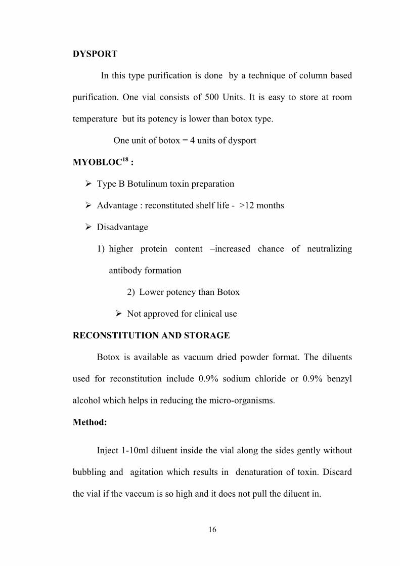
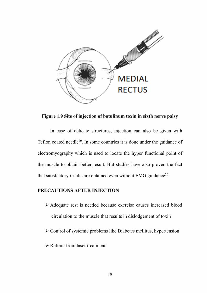
Procedure
Under aseptic precautions, tenon’s capsule is dissected and
muscle belly is exposed. 0.2ml or 5 IU of Botox toxin is injected into the
ipsilateral medial rectus muscle with the use of a 30 gauge needle or
Insulin syringe.
18
Figure 1.9 Site of injection of botulinum toxin in sixth nerve palsy
In case of delicate structures, injection can also be given with
Teflon coated needle20. In some countries it is done under the guidance of
electromyography which is used to locate the hyper functional point of
the muscle to obtain better result. But studies have also proven the fact
that satisfactory results are obtained even without EMG guidance20.
PRECAUTIONS AFTER INJECTION
Adequate rest is needed because exercise causes increased blood
circulation to the muscle that results in dislodgement of toxin
Control of systemic problems like Diabetes mellitus, hypertension
Refrain from laser treatment

19
INVESTIGATIONS32
Ocular motility disorders are mainly due to defect in the nervous
system either in efferent or afferent pathway. They are discussed under
the following category
Pupillary reflex – Hirschberg test
Monocular / binocular alignment
Monocular / binocular movements
HIRSCHBERG TEST:
Hirschberg test is done with the assessment of pupillary reflex. It
should be checked in both uniocular and binocular condition .
With both eyes open, patient is asked to fix light and observe
position of light reflex in both eyes. Repeat the same test with one eye
closed to check monocular position of light reflex.
If the reflex is in the centre of pupil it indicates orthophoria. In case
of strabismus, the reflex of one eye will move away from its monocular
position .
20
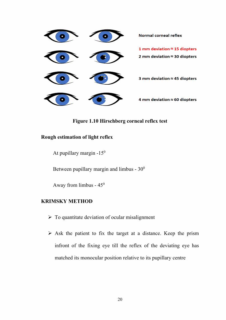
Figure 1.10 Hirschberg corneal reflex test
Rough estimation of light reflex
At pupillary margin -150
Between pupillary margin and limbus - 300
Away from limbus - 450
KRIMSKY METHOD
To quantitate deviation of ocular misalignment
Ask the patient to fix the target at a distance. Keep the prism
infront of the fixing eye till the reflex of the deviating eye has
matched its monocular position relative to its pupillary centre

21
Figure 1.11 Krimsky test for corneal reflex
COVER TEST
Objective method used for evaluation of squint and measurement
of deviation
Three segments
1) Fixation of target
2) Unilateral cover test
3) Alternating cover test with or without prism
22
Requirements
1) Occluder / cover paddle
2) Target – distance and near
3) Well illuminated room
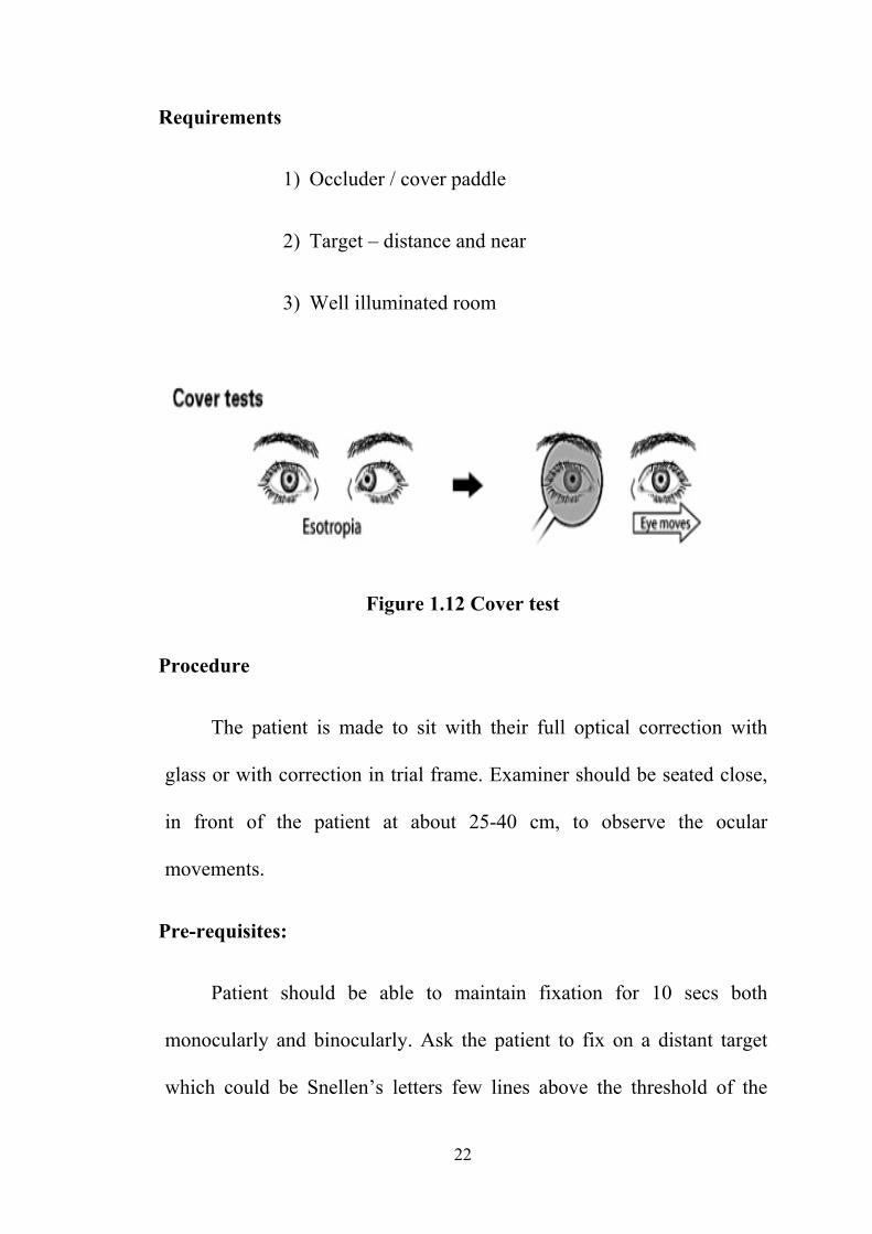
Figure 1.12 Cover test
Procedure
The patient is made to sit with their full optical correction with
glass or with correction in trial frame. Examiner should be seated close,
in front of the patient at about 25-40 cm, to observe the ocular
movements.
Pre-requisites:
Patient should be able to maintain fixation for 10 secs both
monocularly and binocularly. Ask the patient to fix on a distant target
which could be Snellen’s letters few lines above the threshold of the
23
worse eye. Then ask the patient to read the letter, look for any unsteady
monocular movements of the eye which indicates eccentric fixation.
If the other eye moves to take up fixation when the fixing eye is
occluded, it indicates heterotropia
UNILATERAL COVER TEST : Cover and uncover test
Aim
1) To detect tropia or phoria and its component direction
2) To detect alternating or unilateral ( manifest ) deviation
3) Constant or intermittent deviation
4) Test for incomitance in all gaze
5) Small flick of eye prior to taking fixation indicates large phoria
broken by occlusion of the other eye
6) Other movements are also due to small angle esotropia and
uncorrected residual anisometropia have convergence of
hyperopic eye, so manifest as phoria
Procedure
Ask the patient to fix on a distant target, close one eye with the
occluder and ask the patient to fix with the other eye . As soon as the
24
occluder is removed, the fixing eye is observed. Assess the movements in
both eyes while covering and uncovering the eye. First observe the fixing
(uncovered) eye for few cycles and covered eye for another few cycles .
Based on the movement, it could be either orthophoria or heterophoria.
Allow the eyes to fuse target before switching the occluder from one eye
to another.
Results during cover and uncover test
In case of phoria if the eye moves in to out i.e., adducted to
abducted position indicates esophoria and reverse is true in case of
exophoria. If the eye moves from up to down it is hyperphoria and
reverse in case of hypophoria .
UNILATERAL STRABISMUS
In case of unilateral strabismus, if we cover or uncover the
strabismic eye, the normal eye will remain in the fixation position and no
movement occurs. In case of covering the non strabismic eye, the
deviated eye will take up fixation. It indicates heterotropia. The direction
and magnitude of deviation should also be analyzed.

25
ALTERNATING STRABISMUS
In this condition each eye is able to fix under binocular condition.
In case of covering or uncovering the deviated eye, it remains stationary
and no movement occurs. In case of covering the normal eye, the
deviated eye takes up fixation. If the normal eye is uncovered, both eyes
will remain stationary.
Fixation switches from one eye to another in case of alternating
strabismus.In unilateral cover test, alternating strabismus appears
orthophoric which misleads the diagnosis. So we have to confirm with
cover and uncover test in both eyes.
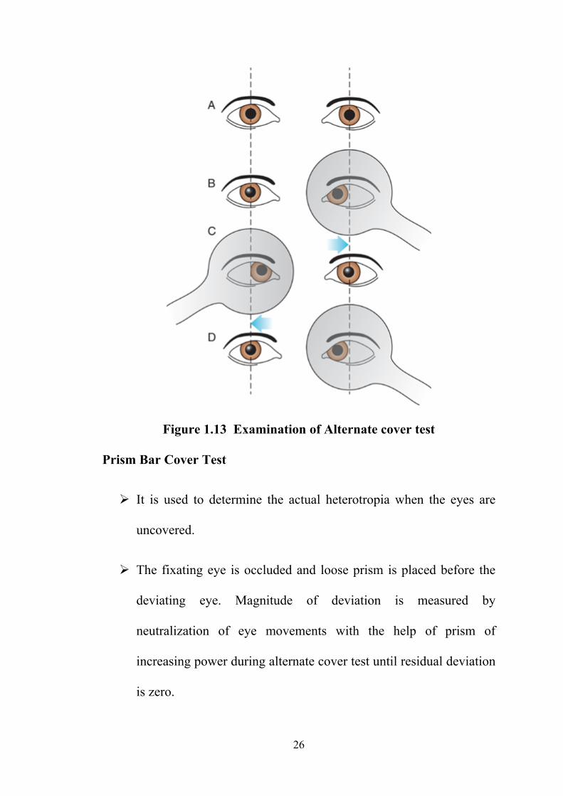
ALTERNATE COVER TEST
In this test the paddle is quickly moved from one eye to another
with a pause of 2-3 seconds over each eye which allows for fixation.
Based on the direction of movement we call it as exo, eso, hyper or hypo
deviation. Measurement of deviation is done using prism bar or loose
prism.
26
Figure 1.13 Examination of Alternate cover test
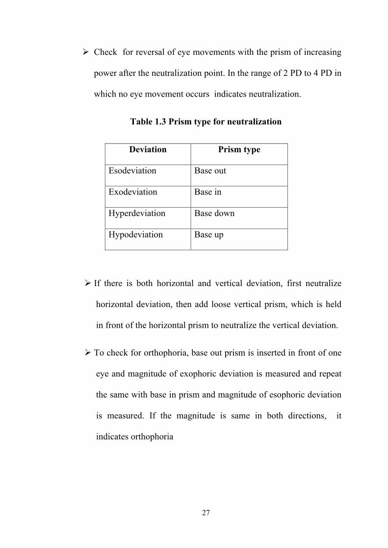
Prism Bar Cover Test
It is used to determine the actual heterotropia when the eyes are
uncovered.
The fixating eye is occluded and loose prism is placed before the
deviating eye. Magnitude of deviation is measured by
neutralization of eye movements with the help of prism of
increasing power during alternate cover test until residual deviation
is zero.
27
Check for reversal of eye movements with the prism of increasing
power after the neutralization point. In the range of 2 PD to 4 PD in
which no eye movement occurs indicates neutralization.
Table 1.3 Prism type for neutralization
Deviation Prism type
Esodeviation Base out
Exodeviation Base in
Hyperdeviation Base down
Hypodeviation Base up
If there is both horizontal and vertical deviation, first neutralize
horizontal deviation, then add loose vertical prism, which is held
in front of the horizontal prism to neutralize the vertical deviation.
To check for orthophoria, base out prism is inserted in front of one
eye and magnitude of exophoric deviation is measured and repeat
the same with base in prism and magnitude of esophoric deviation
is measured. If the magnitude is same in both directions, it
indicates orthophoria
28
Figure 1.14 Evaluation of Prism Bar Cover Test
First do for distance and repeat the same for near by keeping the
target at 40 cm in front of the spectacle plane.

29
HESS CHART39
History
In 1874, Hirschberg marked a tangent scale on the wall of his
examining room to document the field of action of individual muscle in
various gaze fields. In this test, the separation of diplopic image by the
patient was joined with prism, so this did not allow for full dissociation of
deviation.
In 1907, based on Hirschberg’s tangent screen, Ohm designed a
black cloth screen with blue string outlining the coordinates.
Complimentary red and blue filters with red arrow created colour
dissociation. He further constructed the transparent screen with wire
mesh.
In 1908, Krusius designed a glass screen which allowed examiner
to observe the ocular movements and corneal reflection. In 1927, Sattler
designed a black screen using red-green dissociation but with green
coordinate lines and further modifications were analyzed by Sloane.
Hess, the famous neurophysiologist got noble prize in 1949 for his
research work regarding the functional organization of vegetative nervous
system. Original screen was constructed with the help of black cloth of
size 80*80 cm. A pointer of 50cm was used with a green arrow. Hess
used red, green colour dissociation. The patient wore red and green lenses
30
mounted in a spectacle frame. Other screen tests designed or modified
after Hess are Lancaster red-green test and Lees screen.
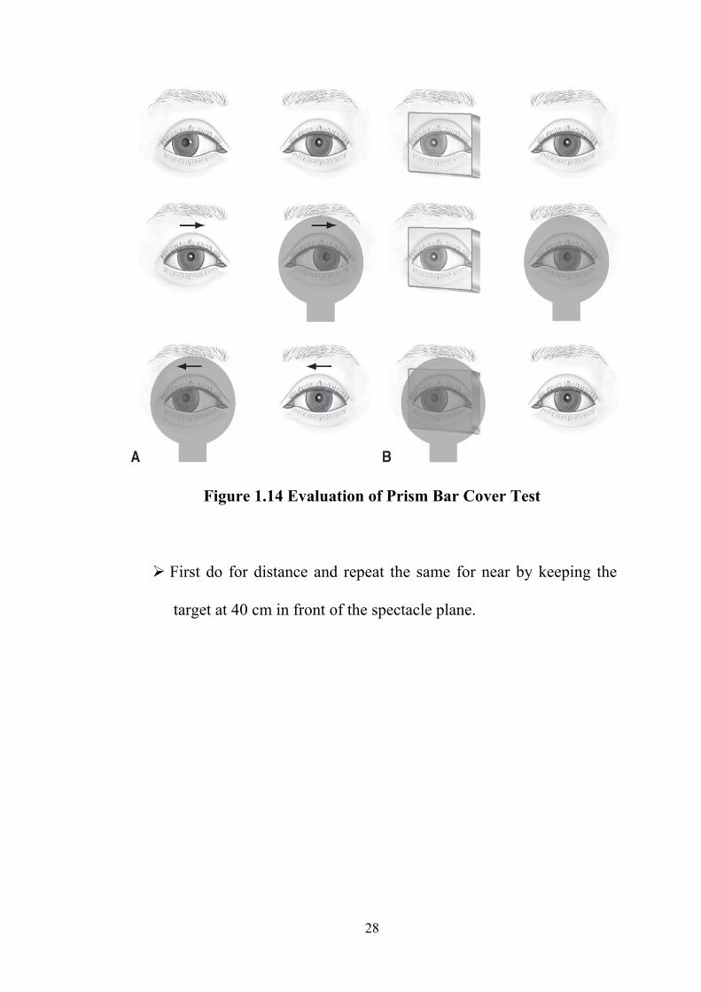
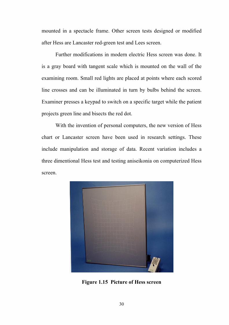
Further modifications in modern electric Hess screen was done. It
is a gray board with tangent scale which is mounted on the wall of the
examining room. Small red lights are placed at points where each scored
line crosses and can be illuminated in turn by bulbs behind the screen.
Examiner presses a keypad to switch on a specific target while the patient
projects green line and bisects the red dot.
With the invention of personal computers, the new version of Hess
chart or Lancaster screen have been used in research settings. These
include manipulation and storage of data. Recent variation includes a
three dimentional Hess test and testing aniseikonia on computerized Hess
screen.
Figure 1.15 Picture of Hess screen
31
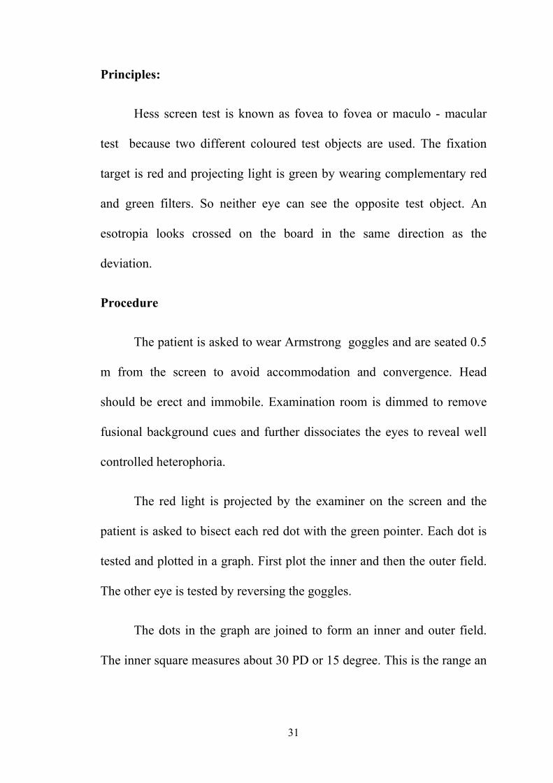
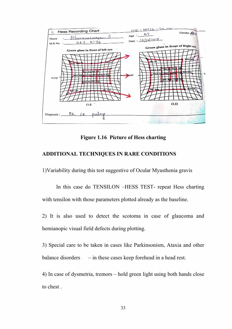
Principles:
Hess screen test is known as fovea to fovea or maculo - macular
test because two different coloured test objects are used. The fixation
target is red and projecting light is green by wearing complementary red
and green filters. So neither eye can see the opposite test object. An
esotropia looks crossed on the board in the same direction as the
deviation.
Procedure
The patient is asked to wear Armstrong goggles and are seated 0.5
m from the screen to avoid accommodation and convergence. Head
should be erect and immobile. Examination room is dimmed to remove
fusional background cues and further dissociates the eyes to reveal well
controlled heterophoria.
The red light is projected by the examiner on the screen and the
patient is asked to bisect each red dot with the green pointer. Each dot is
tested and plotted in a graph. First plot the inner and then the outer field.
The other eye is tested by reversing the goggles.
The dots in the graph are joined to form an inner and outer field.
The inner square measures about 30 PD or 15 degree. This is the range an
32
individual eye will move to view a target away from its primary gaze
without moving the head. The outer field measures twice the amount.
Interpretation:
1) The eye with smaller field indicates the affected eye
2) Overaction in opposite field - comitance or incomitance
3) Patterns looks paralytic or restrictive
4) Course of disorder – Resolving / progressing / stabilizing
-helps in management decisions
5) Long standing or recent palsy – using nature of comitance and spread
of muscle sequelae.
These help in planning the surgery in case of two staged procedure,
to assess which muscle recession should be done first .
33
Figure 1.16 Picture of Hess charting
ADDITIONAL TECHNIQUES IN RARE CONDITIONS
1)Variability during this test suggestive of Ocular Myasthenia gravis
In this case do TENSILON –HESS TEST- repeat Hess charting
with tensilon with those parameters plotted already as the baseline.
2) It is also used to detect the scotoma in case of glaucoma and
hemianopic visual field defects during plotting.
3) Special care to be taken in cases like Parkinsonism, Ataxia and other
balance disorders – in these cases keep forehead in a head rest.
4) In case of dysmetria, tremors – hold green light using both hands close
to chest .
34
5) Hess chart is also used in differentiating certain disorders with the help
of pattern observed
Duane’s and Brown syndrome - Restriction in affected gaze +
over action of ipsilateral antagonist
Acquired SO palsy- Asymmetric bilateral involvement
Disadvantages:
1) It is difficult to interpret torsion with Hess charting. Torsion is better
Quantitated using double Maddox rod test / Harms or Lancaster screen.
2) Hess charting is not possible in case of suppression or anomalous
retinal correspondence.
DIPLOPIA CHART
Diplopia chart is the record of double images in the nine positions
of gaze. It is a type of subjective test.
Procedure:
The patient should be seated with head erect position. The test
should be carried out in a dark room. A red glass is put in front of right
eye. It is better to use Armstrong goggles since they were shaped to fit
the orbital margin. The examiner holds the vertical source of light infront
of the patient at a distance of 1 metre. If there is no double vision in

35
primary position, it should be checked in other eight gazes to find the
position in which double vision appears and maximal separation has to
be noted. If torsion is present, coloured pencils can be given to the
patient to show the separation in torsion. Also, in each gaze the patient
should be asked for the amount of separation subjectively in inches.
Interpretation of diplopia charting:
To interpret the diplopia chart, the points to consider are
1. The position in which diplopia appears
2. The position in which separation of images is the greatest
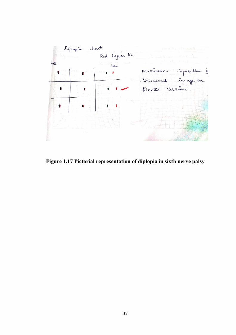
The double vision or the separation of images will be greatest in the
direction of the affected muscle because of over action of the antagonist
muscle and yoke muscle.
Diplopia chart considerations:
1. While recording the diplopia on a paper, the right and left is the
patient’s right and left and not the examiner’s.
2. Always note the distance at which the diplopia charting was
done.
3. Note down the distance of separation of the images in each
position as told by the patient subjectively.
36
4. Tilting of image is drawn as the patient describes.
Diplopia chart disadvantages:
1. Only gives a picture of the patient’s double vision.
2. Maximal separation of images may give an idea about the
paralyzed muscle.
3. Not very useful in recording paresis.
4. Not much helpful in diagnosing various muscle pathologies.
5. Always correlate diplopia chart with clinical examination and
Hess chart to arrive at a diagnosis.
37
Figure 1.17 Pictorial representation of diplopia in sixth nerve palsy
38
OCULAR MOVEMENTS5,33
Eye movements are controlled by the muscles innervated by
Oculomotor, Trochlear and Abducent nerve. Ocular movements helps us
to maintain the image of object on the fovea and ensures good visual
acuity21.
The most common symptom is double vision in case of nerve
palsy. Oculomotor function can be divided into extraocular muscle
function and intrinsic ocular muscles like sphincter pupillae and ciliary
muscle. The extraocular muscles namely the medial, inferior and superior
recti, the inferior oblique and levator palpebrae muscles are innervated
by the oculomotor nerve (III); the superior oblique muscle is innervated
by the trochlear nerve (IV); and the lateral rectus muscle is innervated by
the abducens nerve (VI). The intrinsic eye muscles are innervated by the
parasympathetic component of third nerve and the radial pupil dilator
muscles are innervated by the ascending cervical sympathetic system T1
to T3.
The pupil is directed towards the nose in case of adduction and
laterally in case of abduction. Other movements includes elevation and
depression in which the pupil moves up and down respectively.
39
Isolated muscle weakness results in deviation of the eye due to the
unopposed action of all of the remaining muscles and the resting muscle
tone.
The person with recent onset muscle palsy usually complaints of
double vision because of inability to fuse the images on the macular
region of both eyes. Since the weak muscle is unable to focus image on
the macula, the image falls on a more peripheral part of the retina. The
image falling on a retinal region with fewer cones forms the blurred
image.
Assess the ocular movements in six positions of gaze and the
direction of diplopia gives clue to the direction of the affected muscle.
For example, horizontal diplopia is due to problems with the medial and
lateral recti muscle, while vertical diplopia is due to problems with one or
more of the other muscles. These include conjugate horizontal gaze,
conjugate vertical gaze, smoothly tracking objects, convergence and other
eye movements resulting from head movements due to vestibular reflexes
for eye stabilization.

40
ACTION OF EXTRAOCULAR MUSCLES
S.NO Muscle Primary Secondary Tertiary
1 Medial rectus Adduction - -
2 Lateral rectus Abduction - -
3 Superior rectus Elevation Intortion Adduction
4 Inferior rectus Depression Extortion Adduction
5 Superior oblique Intorsion Depression Abduction
6 Inferior oblique Extorsion Elevation Abduction
Duction movements are graded upto -4 for under action and 0 to +4
for overaction. This is a subjective test which results in more
interobserver variability. Other methods are electro-oculogram, purkinje
image trackers, video based methods, Goldmann perimeter and
synoptophore 23.

41
Figure 1.18 Schematic representation of ocular movements
Kestenbaum limbal test :
Kestenbaum limbal test measures the excursion of ocular
movements in millimetres with the help of transparent ruler infront of the
cornea. Note the limbal position in primary gaze and compare it with the
new gaze. This test has better interobserver repeatability. The mean value
for abduction, adduction and depression is 9-10mm and 7 mm for
elevation24.
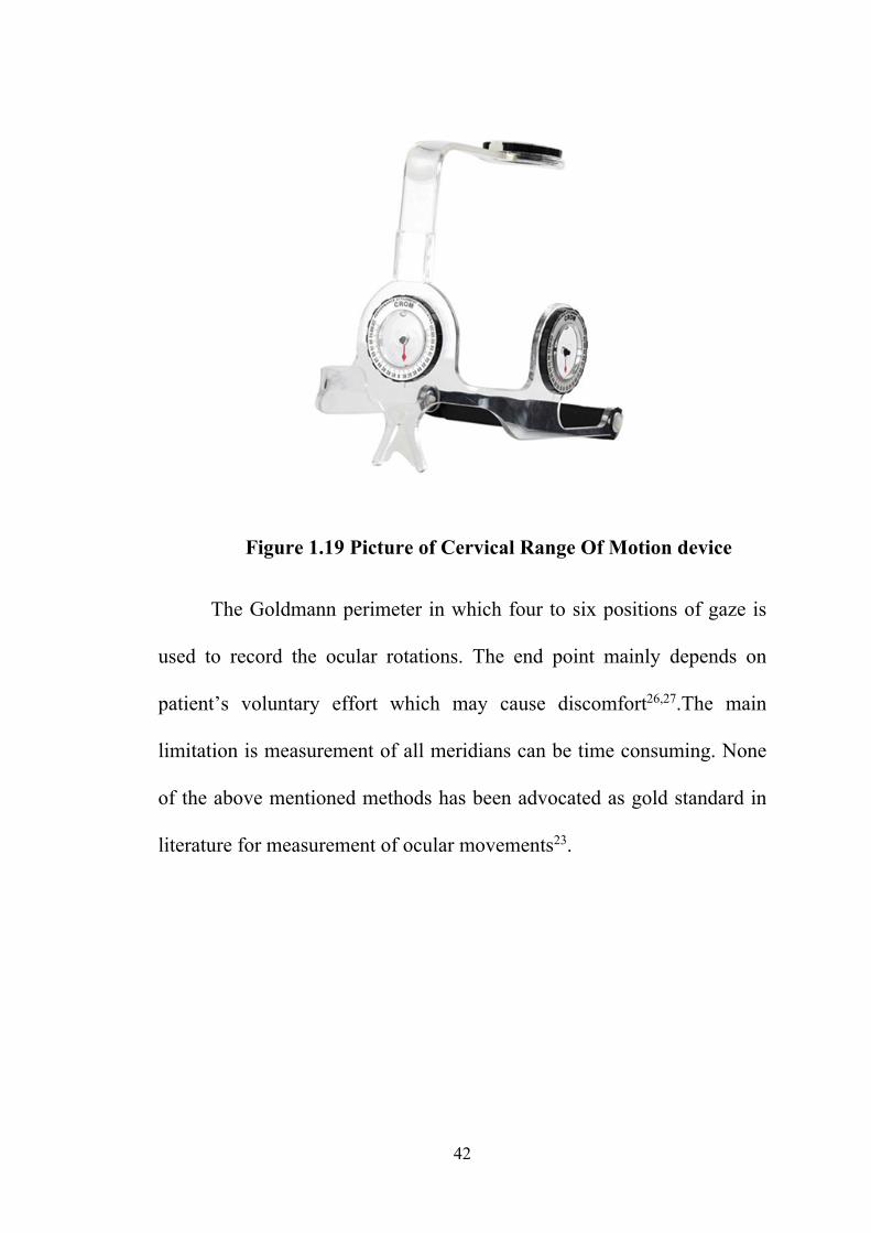
Kushner proposed an instrument for ocular movement recording
called the Cervical Range Of Motion device ( CROM )25. It was designed
to assess the ocular rotation, abnormal head posture and the field of
binocular single vision. It has three magnetic device dials which assess
the head position23.
42
Figure 1.19 Picture of Cervical Range Of Motion device
The Goldmann perimeter in which four to six positions of gaze is
used to record the ocular rotations. The end point mainly depends on
patient’s voluntary effort which may cause discomfort26,27.The main
limitation is measurement of all meridians can be time consuming. None
of the above mentioned methods has been advocated as gold standard in
literature for measurement of ocular movements23.
43
TREATMENT OF SIXTH NERVE PALSY28 :
In general, underlying systemic condition has to be controlled first.
Microangiopathy is the most common etiology for sixth nerve palsy
which recovers spontaneously in most of the cases, hence requires
conservative treatment. For diplopia in primary gaze, consider options
like Occlusion therapy, Fresnel prism, Botulinum toxin injection.
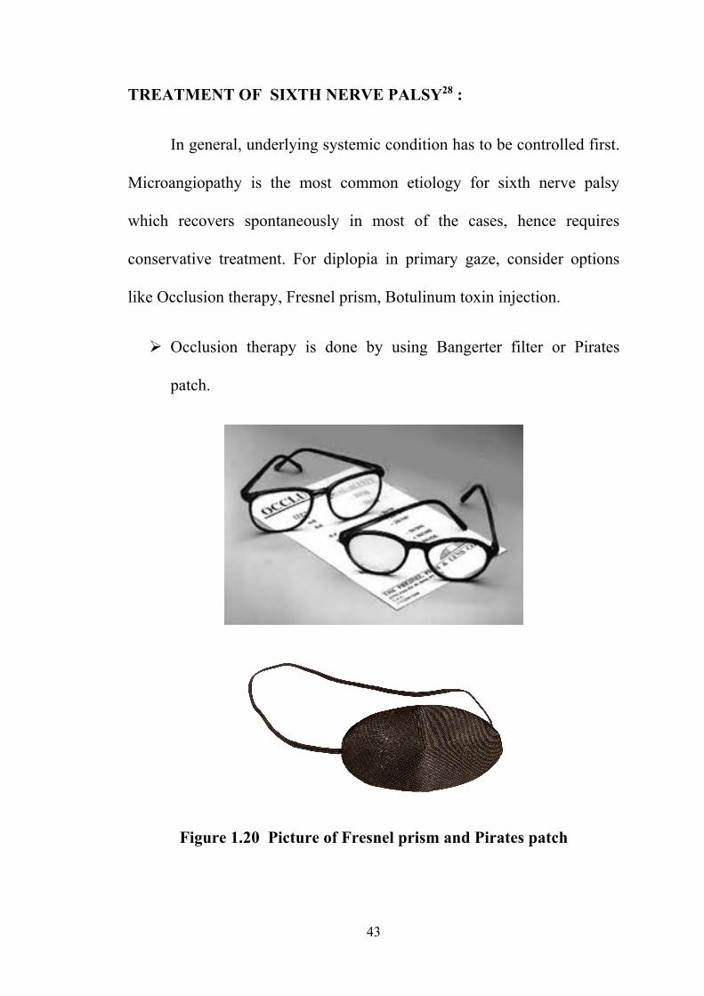
Occlusion therapy is done by using Bangerter filter or Pirates
patch.
Figure 1.20 Picture of Fresnel prism and Pirates patch
44
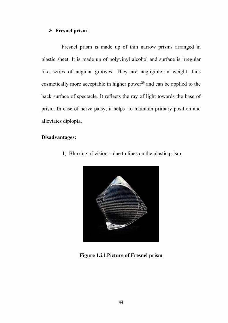
Fresnel prism :
Fresnel prism is made up of thin narrow prisms arranged in
plastic sheet. It is made up of polyvinyl alcohol and surface is irregular
like series of angular grooves. They are negligible in weight, thus
cosmetically more acceptable in higher power29 and can be applied to the
back surface of spectacle. It reflects the ray of light towards the base of
prism. In case of nerve palsy, it helps to maintain primary position and
alleviates diplopia.
Disadvantages:
1) Blurring of vision – due to lines on the plastic prism
Figure 1.21 Picture of Fresnel prism
45
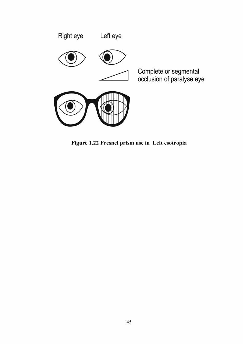
Figure 1.22 Fresnel prism use in Left esotropia
46
REVIEW OF LITERATURE
In the early 1970 Scott and his associates had interest in treating
the lateral rectus palsy pharmacologically by weakening the extra ocular
muscles. Specific value of the botulinum toxin was reported in 1981 and
in the following year, with the approval of Food and Drug Administration
in the United States Botulinum toxin was used as a therapeutic modality.
They have conducted several studies for the treatment of Lateral rectus
palsy with botulinum toxin. Later it was also approved by the American
Academy of Ophthalmology publication for horizontal strabismus of 15 -
50 prism diopters for injection in the medial rectus muscle in recent sixth
nerve palsy. Botulinum Toxin injected into the ipsilateral medial rectus
muscle has been advocated in the management of Acute sixth nerve
palsy.
39Scott and Kraft suggested that in conservatively managed cases,
contracture of medial rectus muscle may prevent recovery of lateral
rectus muscle. They further postulated that Botulinum toxin reduces
contracture of medial rectus muscle and allows more complete restoration
of duction.
47
In 1988, Henry S. Metz et al38 in Graefe's Archive Ophthalmology
analysed botulinum toxin treatment of acute sixth nerve and third nerve
palsy. He included 34 patients with acute sixth nerve palsy treated with
botulinum toxin injection and 52 patients in control group. Bilateral cases
were also included. Results of his study concluded that among the control
group, few patients recovered spontaneously. Recovery in bilateral cases
is not as good as unilateral cases and Botulinum toxin was not that much
effective in chronic sixth nerve palsy.
Early and late botulinum toxin injection in treatment of acute sixth
nerve palsy. 1989 Aug;17(3):239-4534
In 1989, A D N Murray34 conducted a study in acute sixth nerve
palsy. He treated his patients with botulinum toxin in medial rectus
muscle. 10 patients were in this study and followed over a period of 14
months after last injection. 6 patients were treated within 8 weeks of
onset of palsy and appeared to recover earlier than those who were treated
late after onset. Patients also gained fusion. But he was not able to
differentiate whether the recovery was due to spontaneous resolution or
due to the botulinum toxin. So he was advised to conduct a double blind
study to determine the effectiveness of Botulinum injection.
48
Results of a prospective randomized trial of botulinum toxin therapy
in acute sixth nerve palsy. [01 Sep 1994, 31(5):283-286]35
Most of the studies are retrospective in nature. So in 1994, John
Lee et al35, made research on results of a prospective randomized trial of
Botulinum toxin in acute unilateral sixth nerve palsy. Among the total of
47 patients included, 22 patients were given injection and 25 were
controls. Most of them were due to microvascular (72.3%) etiology. He
stated that medial rectus contracture leads to persistent esotropia even
when there is recovery of lateral rectus function. So contracture is
reduced by chemo-denervation with botulinum toxin. In this study,
control group and injected group had a final recovery rate of about 80%
and 86% respectively. So he concluded that there was no evidence for
prophylactic effect of botulinum toxin in the group. He explained the
need for randomized control trial to determine the effectiveness.
Previous few studies explained that spontaneous recovery rate is
lower for traumatic sixth nerve palsy when compared to microvascular
etiology that needs an early intervention or treatment with botulinum
toxin. He also suggested the need for more accurate estimation of
spontaneous recovery rate.
49
In 1998, Jonathan M. Holmes et al40 conducted a multi centered
study in the natural history of acute traumatic sixth nerve palsy or paresis
with the help of 19 investigators . He concluded that the overall recovery
rate in conservatively treated group in unilateral cases was higher than
previously reported. The main drawback of this study was low statistical
power failure of follow up, investigator bias in deciding treatment.
Botulinum toxin treatment versus conservative management in acute
traumatic sixth nerve palsy or paresis. JAAPOS. 2000 Jun;4(3):145-
936.
In 2000, Holmes et al further made a study in Acute traumatic sixth
nerve palsy patients treated with botulinum toxin versus conservative
management. It was a non- randomized, prospective, multicentered study
done over a period of 2 years to evaluate the recovery rates in both
groups. In this study 84 patients were enrolled by 46 investigators.
Among those, 62 patients (74%) were treated conservatively and 22
patients (26%) with botulinum toxin. Both groups were found to have
similar recovery rates. They have included bilateral cases also and few
received multiple botulinum toxin injections in treated group. The final
recovery rates was 73% in Botox treated and 71% in conservatively
treated patients. It suggested the need for RCT and large number of
patients to make the study with acceptable statistical power.
50
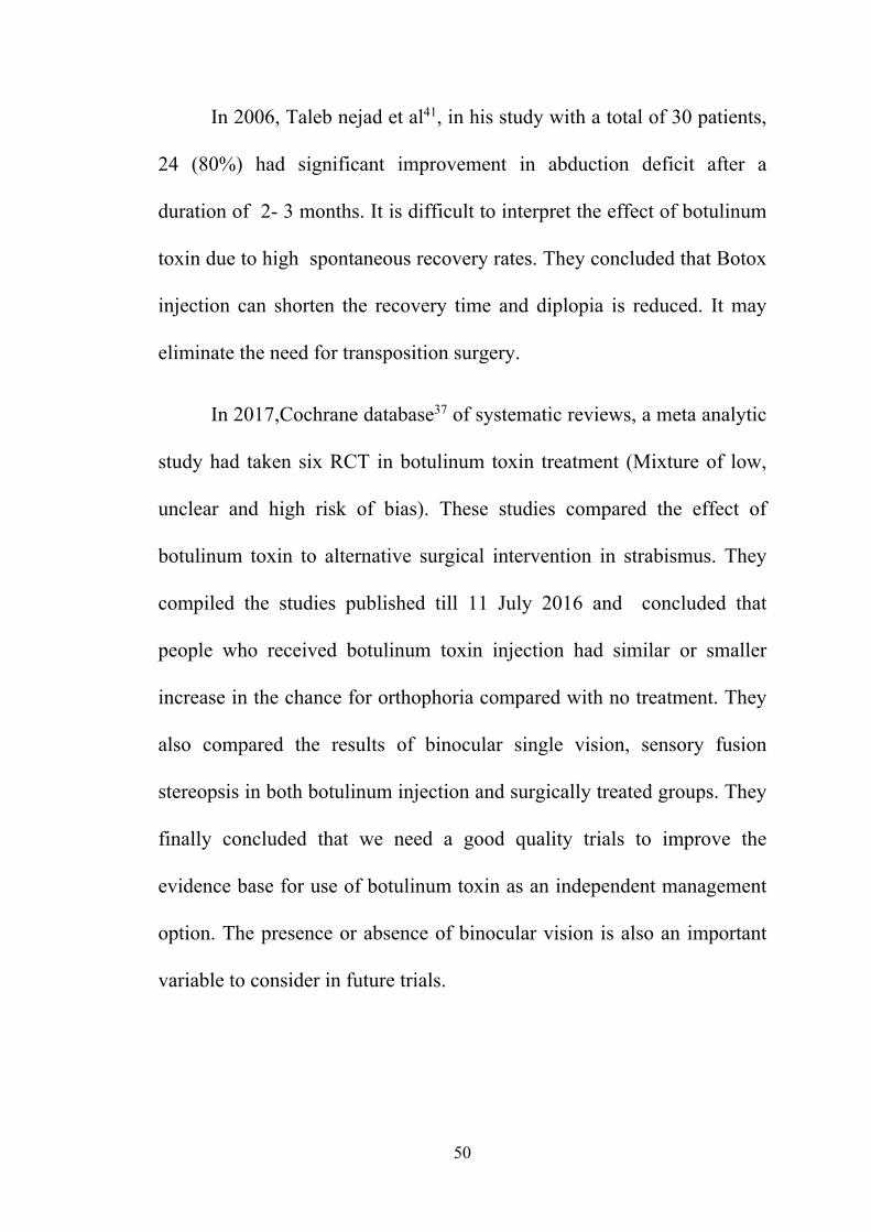
In 2006, Taleb nejad et al41, in his study with a total of 30 patients,
24 (80%) had significant improvement in abduction deficit after a
duration of 2- 3 months. It is difficult to interpret the effect of botulinum
toxin due to high spontaneous recovery rates. They concluded that Botox
injection can shorten the recovery time and diplopia is reduced. It may
eliminate the need for transposition surgery.
In 2017,Cochrane database37 of systematic reviews, a meta analytic
study had taken six RCT in botulinum toxin treatment (Mixture of low,
unclear and high risk of bias). These studies compared the effect of
botulinum toxin to alternative surgical intervention in strabismus. They
compiled the studies published till 11 July 2016 and concluded that
people who received botulinum toxin injection had similar or smaller
increase in the chance for orthophoria compared with no treatment. They
also compared the results of binocular single vision, sensory fusion
stereopsis in both botulinum injection and surgically treated groups. They
finally concluded that we need a good quality trials to improve the
evidence base for use of botulinum toxin as an independent management
option. The presence or absence of binocular vision is also an important
variable to consider in future trials.

PART – II
51
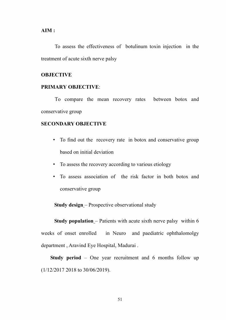
AIM :
To assess the effectiveness of botulinum toxin injection in the
treatment of acute sixth nerve palsy
OBJECTIVE
PRIMARY OBJECTIVE:
To compare the mean recovery rates between botox and
conservative group
SECONDARY OBJECTIVE
• To find out the recovery rate in botox and conservative group
based on initial deviation
• To assess the recovery according to various etiology
• To assess association of the risk factor in both botox and
conservative group
Study design – Prospective observational study
Study population – Patients with acute sixth nerve palsy within 6
weeks of onset enrolled in Neuro and paediatric ophthalomolgy
department , Aravind Eye Hospital, Madurai .
Study period – One year recruitment and 6 months follow up
(1/12/2017 2018 to 30/06/2019).
52
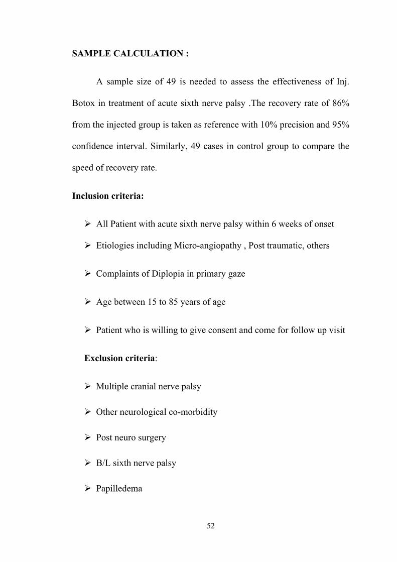
SAMPLE CALCULATION :
A sample size of 49 is needed to assess the effectiveness of Inj.
Botox in treatment of acute sixth nerve palsy .The recovery rate of 86%
from the injected group is taken as reference with 10% precision and 95%
confidence interval. Similarly, 49 cases in control group to compare the
speed of recovery rate.
Inclusion criteria:
All Patient with acute sixth nerve palsy within 6 weeks of onset
Etiologies including Micro-angiopathy , Post traumatic, others
Complaints of Diplopia in primary gaze
Age between 15 to 85 years of age
Patient who is willing to give consent and come for follow up visit
Exclusion criteria:
Multiple cranial nerve palsy
Other neurological co-morbidity
Post neuro surgery
B/L sixth nerve palsy
Papilledema
53
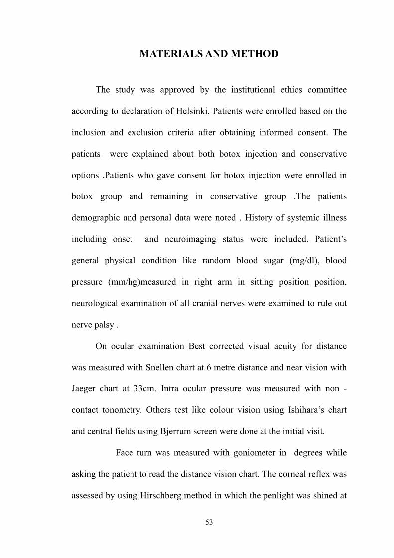
MATERIALS AND METHOD
The study was approved by the institutional ethics committee
according to declaration of Helsinki. Patients were enrolled based on the
inclusion and exclusion criteria after obtaining informed consent. The
patients were explained about both botox injection and conservative
options .Patients who gave consent for botox injection were enrolled in
botox group and remaining in conservative group .The patients
demographic and personal data were noted . History of systemic illness
including onset and neuroimaging status were included. Patient’s
general physical condition like random blood sugar (mg/dl), blood
pressure (mm/hg)measured in right arm in sitting position position,
neurological examination of all cranial nerves were examined to rule out
nerve palsy .
On ocular examination Best corrected visual acuity for distance
was measured with Snellen chart at 6 metre distance and near vision with
Jaeger chart at 33cm. Intra ocular pressure was measured with non -
contact tonometry. Others test like colour vision using Ishihara’s chart
and central fields using Bjerrum screen were done at the initial visit.
Face turn was measured with goniometer in degrees while
asking the patient to read the distance vision chart. The corneal reflex was
assessed by using Hirschberg method in which the penlight was shined at
54
33cm infront of the patient and the location of first purkinje image was
noted.
According to the position of the reflex, the degree was measured.
15 degree- At pupillary margin
30 degree- Between pupillary margin to limbus
45 degree – Away from limbus
Ocular movements were assessed in all nine gaze in both eyes ,
mainly the amount of abduction deficit was measured by asking the
patient to abduct fully from primary gaze. Depending on the lateral
excursion of eyeball , abduction deficit was graded according to Scott and
Kraft method as follows
-1 = 75% full rotation
-2 = to 50% full rotation
-3 = to 25% full rotation
-4 = to midline
-5 = inability to abduct to the midline
Then binocular single vision was analysed by worth four dot test
for distance at 6 metre (wall mounted device ) and near at 33cm (hand
held torch) . The patient was asked to wear red-green glasses such that
red glass was placed in front of right eye , ask the patient to see the target

55
consisting of 4 dots –one red , two green and one white. Assessment was
done according to that number of dots reported by the patient .
Seeing 2 dots indicates – suppression scotoma in left eye
Seeing 3 dots indicates – suppression scotoma in right eye
Seeing 4 dots indicates –normal retinal correspondence or
harmonious ARC
Seeing 5 dots indicates – diplopia response
Figure 1.23 Interpretation of Worth Four Dot Test
56
Diplopia charting was done by asking the patient to be seated with
head erect and red-green glasses on. The test should be carried out in a
dark room. The examiner holds a vertical source of light at around one
metre. Initially the light is directly held in front of the patient. If no
double vision is reported in primary position, then it should be checked
in other eight gazes to find where diplopia occurs. The position were
maximal separation of images is reported, is to be noted. Also, in each
gaze the patient should be asked for the amount of separation subjectively
in inches.
Prism bar cover test was done with loose prisms placed in front
of one eye and by alternatively occluding either eyes. Magnitude of
deviation was measured by neutralization of eye movements with the help
of prisms. The prism strength required for arresting the deviation was
noted.
Then anterior segment examination was done with slit lamp
biomicroscopy and posterior segment examination using 90D lens on slit
lamp biomicroscopy.
Both groups were evaluated for the presence of any systemic
co-morbidity. Patients were advised strict control of their systemic
illness. Botox patient after the completion of preoperative evaluation got

57
physician fitness for further procedure. Preoperatively the patients were
given prophylactic topical antibiotic eyedrops.
PREPARATION OF BOTOX INJECTION :
Botulinum toxin type – A (Botox) – which is a purified neurotoxin
complex was available in powdered form. One bottle contains 100 IU. It
was reconstituted with 4 ml of sodium chloride (Nacl) solution drop by
drop along the sides of the bottle and avoid shaking the bottle. Such that
4ml contains 100 IU and 0.1ml contains 2.5IU. These bottles were stored
in refrigerator at 4°C for a maximum period of 30 days.
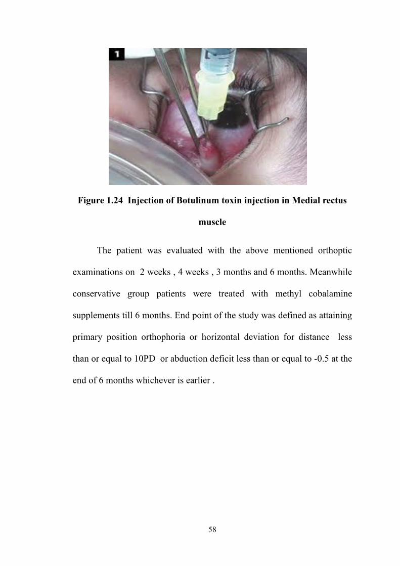
SURGICAL TECHNIQUE:
Under sterile aseptic precautions, incision was made in inferonasal
quadrant and 2.5ml of 2% lignocaine with or without adrenalin was
given in the subtenon’s space. Through the same fornix based incision,
the medial rectus muscle was exposed and 0.2 ml of botox injection was
given directly into the muscle belly under direct observation. Excess of
seeped out drug was wiped with merocel sponge. Conjunctiva was
opposed with 3- 4 interrupted absorbable 8 -0 – vicryl sutures. Pad and
bandage was applied .Steroid with antibiotic drops were given for one
week.
58
Figure 1.24 Injection of Botulinum toxin injection in Medial rectus
muscle
The patient was evaluated with the above mentioned orthoptic
examinations on 2 weeks , 4 weeks , 3 months and 6 months. Meanwhile
conservative group patients were treated with methyl cobalamine
supplements till 6 months. End point of the study was defined as attaining
primary position orthophoria or horizontal deviation for distance less
than or equal to 10PD or abduction deficit less than or equal to -0.5 at the
end of 6 months whichever is earlier .
59
STATISTICAL METHOD FOR ANALYSIS :
Data will be collected as per data collection form
Mean (SD) or Frequency (Percentage) will be used to describe
summary . Chi-square test or Fisher’s exact test will be used to assess the
association between categorical variables.
Paired t-test or Wilcoxon sign rank test will be used to compare
the Pre-op and Post-op values. P-value is less than 0.05 considered as
statistically significant. All statistical analysis will be done by STATA
11.1 (Texas, USA).
Statistically the variables which included date of presentation from
onset of palsy, abduction deficit and deviation in prism diopters were
analysed based on their percentage of recovery at 6 months, average
week of recovery in both groups .
60
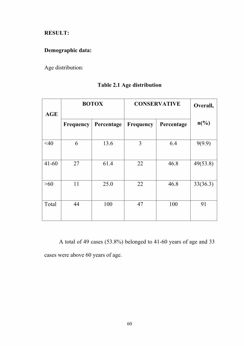
RESULT:
Demographic data:
Age distribution:
Table 2.1 Age distribution
AGE
BOTOX CONSERVATIVE Overall,
n(%) Frequency Percentage Frequency Percentage
<40 6 13.6 3 6.4 9(9.9)
41-60 27 61.4 22 46.8 49(53.8)
>60 11 25.0 22 46.8 33(36.3)
Total 44 100 47 100 91
A total of 49 cases (53.8%) belonged to 41-60 years of age and 33
cases were above 60 years of age.
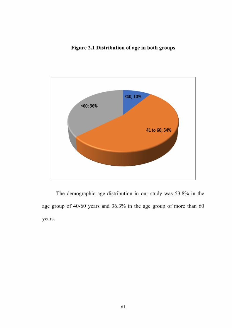
61
Figure 2.1 Distribution of age in both groups
The demographic age distribution in our study was 53.8% in the
age group of 40-60 years and 36.3% in the age group of more than 60
years.
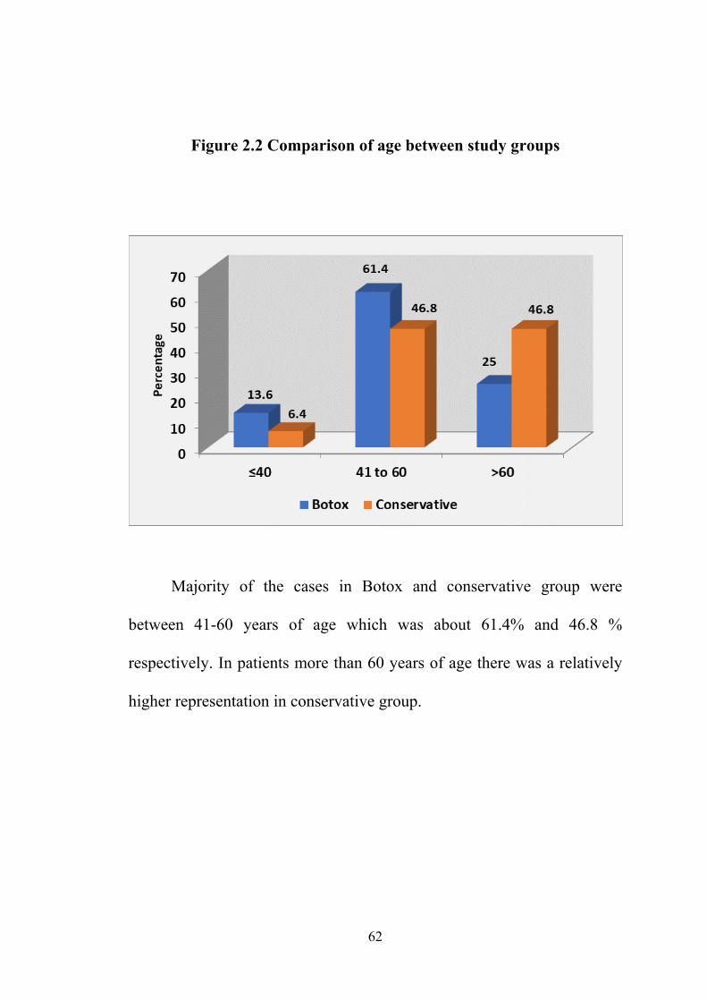
62
Figure 2.2 Comparison of age between study groups
Majority of the cases in Botox and conservative group were
between 41-60 years of age which was about 61.4% and 46.8 %
respectively. In patients more than 60 years of age there was a relatively
higher representation in conservative group.
63
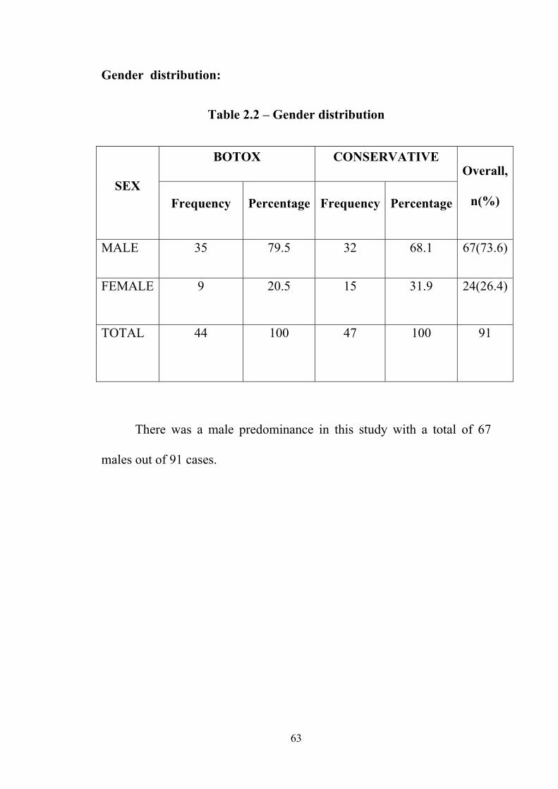
Gender distribution:
Table 2.2 – Gender distribution
SEX
BOTOX CONSERVATIVE Overall,
n(%) Frequency Percentage Frequency Percentage
MALE 35 79.5 32 68.1 67(73.6)
FEMALE 9 20.5 15 31.9 24(26.4)
TOTAL 44 100 47 100 91
There was a male predominance in this study with a total of 67
males out of 91 cases.

64
Figure 2.3 Distribution of gender in both groups
Sex ratio distribution was attributed to various socio economic and
cultural barriers .
65
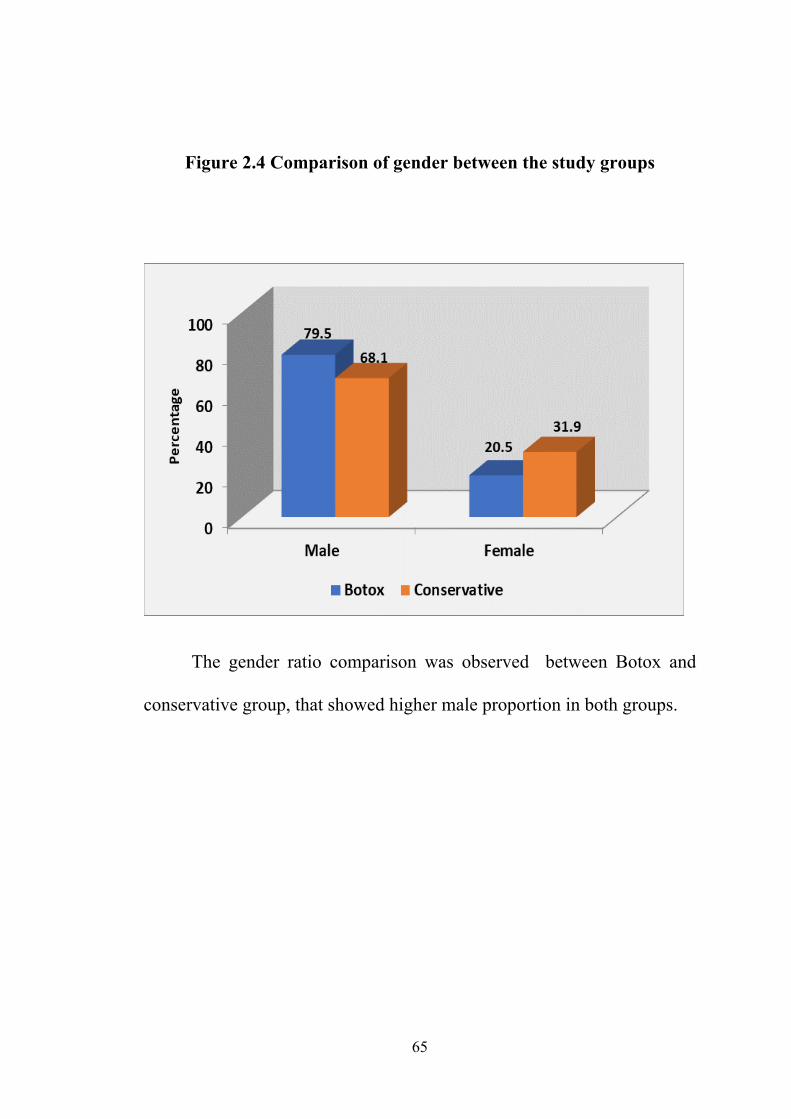
Figure 2.4 Comparison of gender between the study groups
The gender ratio comparison was observed between Botox and
conservative group, that showed higher male proportion in both groups.
66
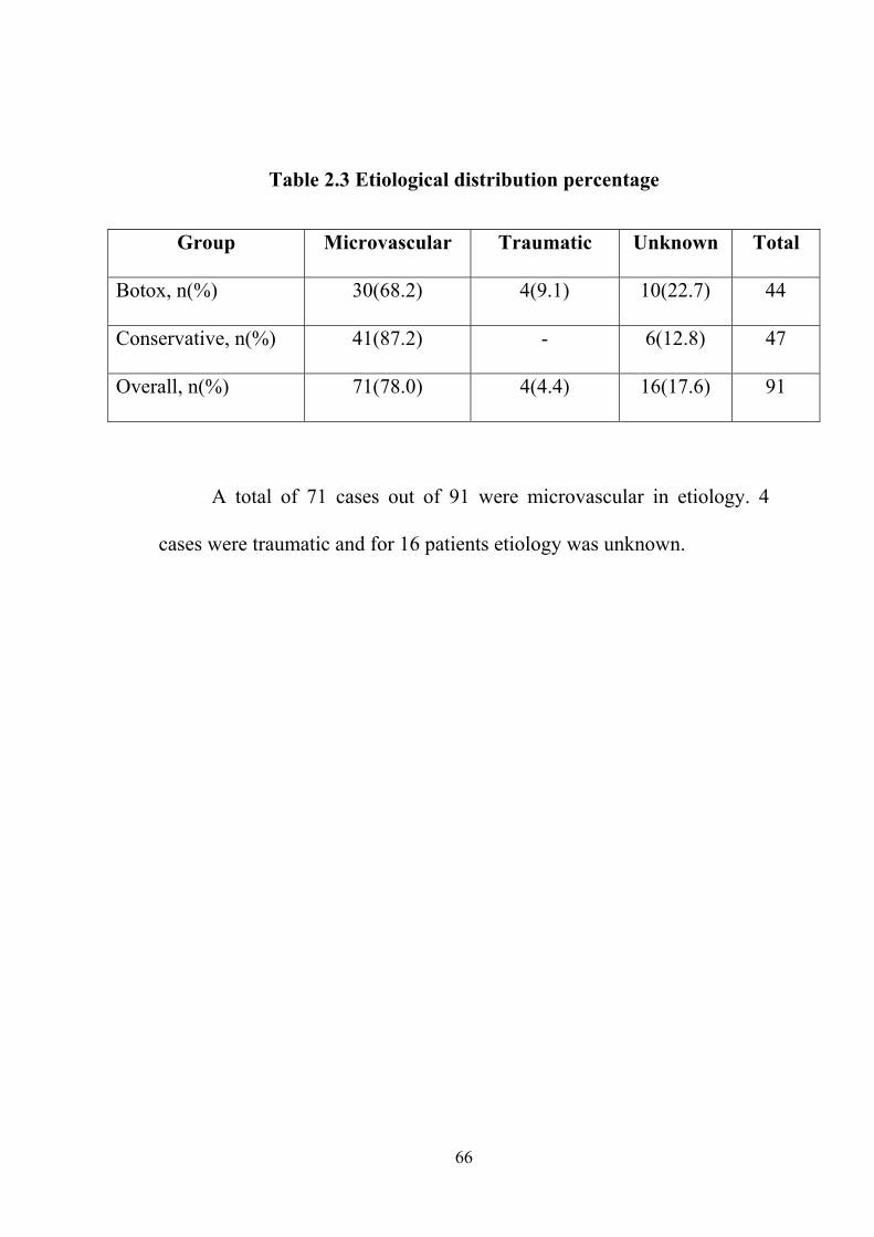
Table 2.3 Etiological distribution percentage
Group Microvascular Traumatic Unknown Total
Botox, n(%) 30(68.2) 4(9.1) 10(22.7) 44
Conservative, n(%) 41(87.2) - 6(12.8) 47
Overall, n(%) 71(78.0) 4(4.4) 16(17.6) 91
A total of 71 cases out of 91 were microvascular in etiology. 4
cases were traumatic and for 16 patients etiology was unknown.
67
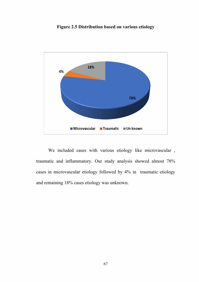
Figure 2.5 Distribution based on various etiology
We included cases with various etiology like microvascular ,
traumatic and inflammatory. Our study analysis showed almost 78%
cases in microvascular etiology followed by 4% in traumatic etiology
and remaining 18% cases etiology was unknown.
68
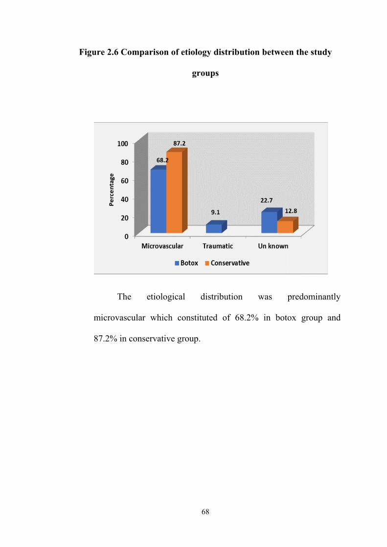
Figure 2.6 Comparison of etiology distribution between the study
groups
The etiological distribution was predominantly
microvascular which constituted of 68.2% in botox group and
87.2% in conservative group.
69
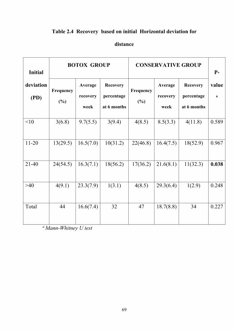
Table 2.4 Recovery based on initial Horizontal deviation for
distance
Initial
deviation
(PD)
BOTOX GROUP CONSERVATIVE GROUP P-
value
a Frequency
(%)
Average
recovery
week
Recovery
percentage
at 6 months
Frequency
(%)
Average
recovery
week
Recovery
percentage
at 6 months
<10 3(6.8) 9.7(5.5) 3(9.4) 4(8.5) 8.5(3.3) 4(11.8) 0.589
11-20 13(29.5) 16.5(7.0) 10(31.2) 22(46.8) 16.4(7.5) 18(52.9) 0.967
21-40 24(54.5) 16.3(7.1) 18(56.2) 17(36.2) 21.6(8.1) 11(32.3) 0.038
>40 4(9.1) 23.3(7.9) 1(3.1) 4(8.5) 29.3(6.4) 1(2.9) 0.248
Total 44 16.6(7.4) 32 47 18.7(8.8) 34 0.227
a Mann-Whitney U test
70
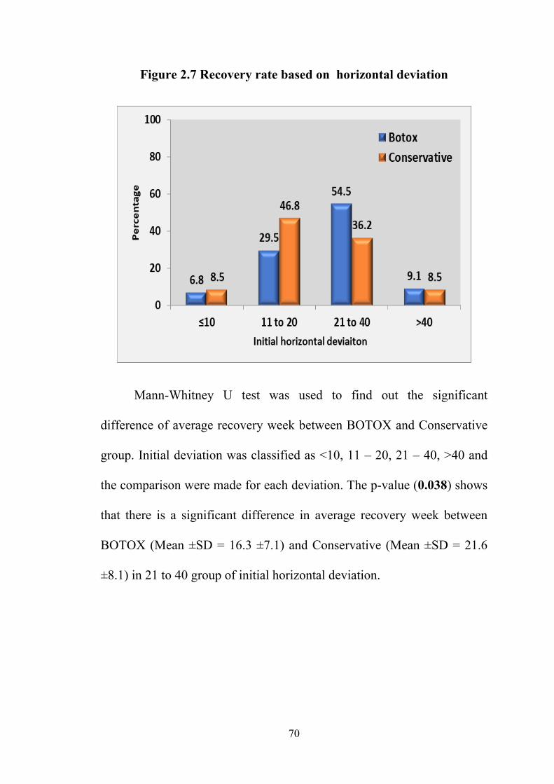
Figure 2.7 Recovery rate based on horizontal deviation
Mann-Whitney U test was used to find out the significant
difference of average recovery week between BOTOX and Conservative
group. Initial deviation was classified as <10, 11 – 20, 21 – 40, >40 and
the comparison were made for each deviation. The p-value (0.038) shows
that there is a significant difference in average recovery week between
BOTOX (Mean ±SD = 16.3 ±7.1) and Conservative (Mean ±SD = 21.6
±8.1) in 21 to 40 group of initial horizontal deviation.
71
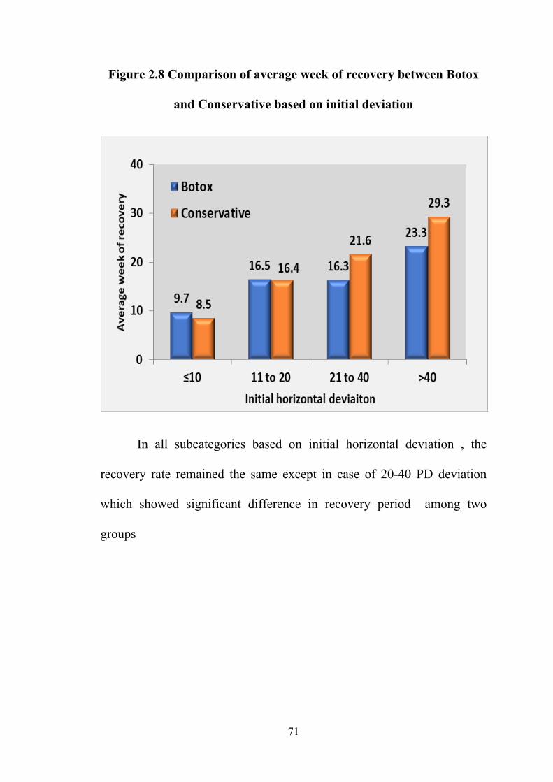
Figure 2.8 Comparison of average week of recovery between Botox
and Conservative based on initial deviation
In all subcategories based on initial horizontal deviation , the
recovery rate remained the same except in case of 20-40 PD deviation
which showed significant difference in recovery period among two
groups

72
Table 2.5 Recovery based on abduction deficit: Paresis (-2, -3);
Palsy (-4, -5)
Abduction
deficit
BOTOX GROUP CONSERVATIVE GROUP P-
value
a
Frequency
(%)
Average
recovery
week
Recovery
percentage
at 6 months
Frequency
(%)
Average
recovery
week
Recovery
percentage at
6 months
Paresis 33(75.0) 15.1(6.8) 26(81.3) 39(83.0) 17.4(8.5) 29(85.3) 0.175
Palsy 11(25.0) 20.5(7.9) 6(18.7) 8(17.0) 24.1(8.3) 5(14.7) 0.360
a Mann-Whitney U test
Based on severity of abduction deficit, -2 and -3 deficit were
classified as paresis , -4 and -5 as palsy group . p-value more than 0.05
showed no statistical difference in mean week of recovery in both
groups based on abduction deficit.
73
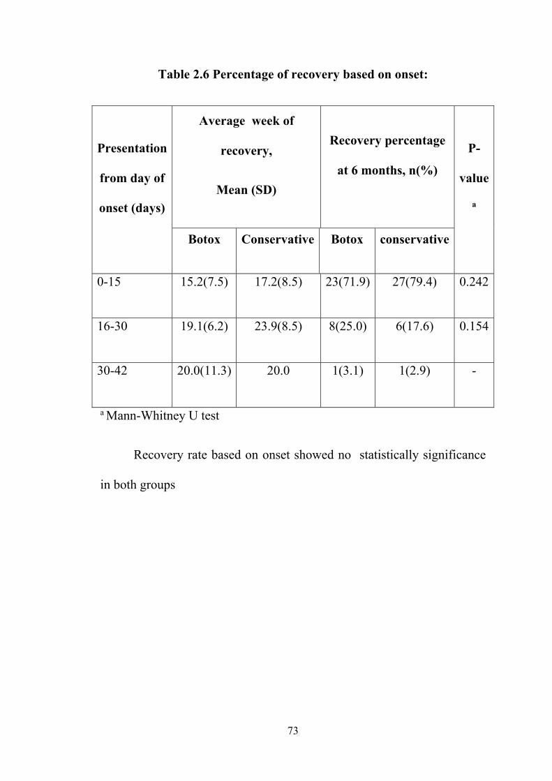
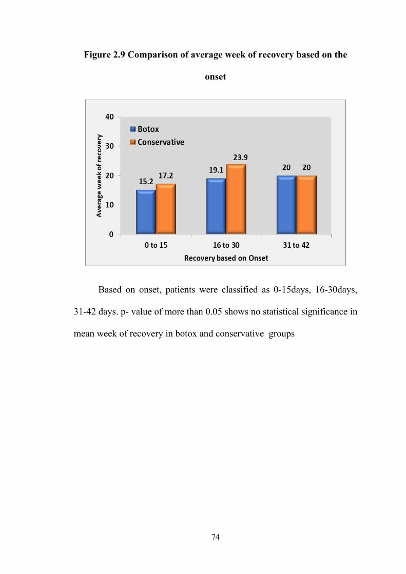
Table 2.6 Percentage of recovery based on onset:
Presentation
from day of
onset (days)
Average week of
recovery,
Mean (SD)
Recovery percentage
at 6 months, n(%)
P-
value
a
Botox Conservative Botox conservative
0-15 15.2(7.5) 17.2(8.5) 23(71.9) 27(79.4) 0.242
16-30 19.1(6.2) 23.9(8.5) 8(25.0) 6(17.6) 0.154
30-42 20.0(11.3) 20.0 1(3.1) 1(2.9) -
a Mann-Whitney U test
Recovery rate based on onset showed no statistically significance
in both groups
74
Figure 2.9 Comparison of average week of recovery based on the
onset
Based on onset, patients were classified as 0-15days, 16-30days,
31-42 days. p- value of more than 0.05 shows no statistical significance in
mean week of recovery in botox and conservative groups
75
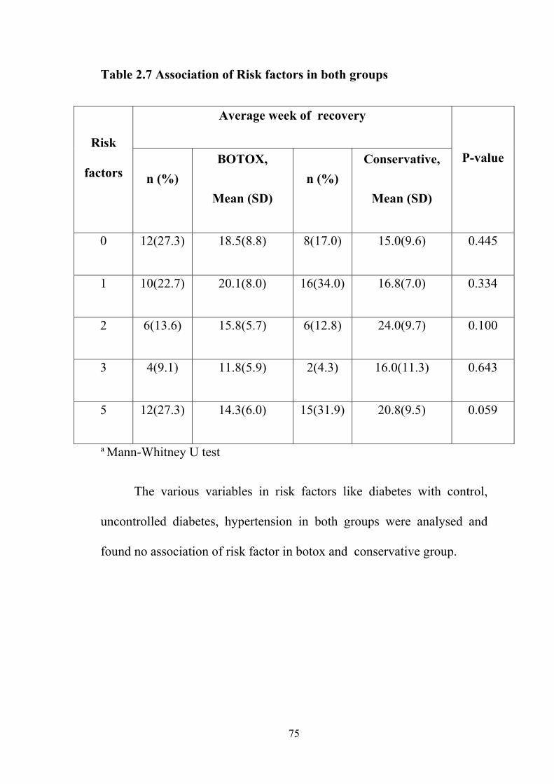
Table 2.7 Association of Risk factors in both groups
Risk
factors
Average week of recovery
P-value
n (%)
BOTOX,
Mean (SD)
n (%)
Conservative,
Mean (SD)
0 12(27.3) 18.5(8.8) 8(17.0) 15.0(9.6) 0.445
1 10(22.7) 20.1(8.0) 16(34.0) 16.8(7.0) 0.334
2 6(13.6) 15.8(5.7) 6(12.8) 24.0(9.7) 0.100
3 4(9.1) 11.8(5.9) 2(4.3) 16.0(11.3) 0.643
5 12(27.3) 14.3(6.0) 15(31.9) 20.8(9.5) 0.059
a Mann-Whitney U test
The various variables in risk factors like diabetes with control,
uncontrolled diabetes, hypertension in both groups were analysed and
found no association of risk factor in botox and conservative group.
76
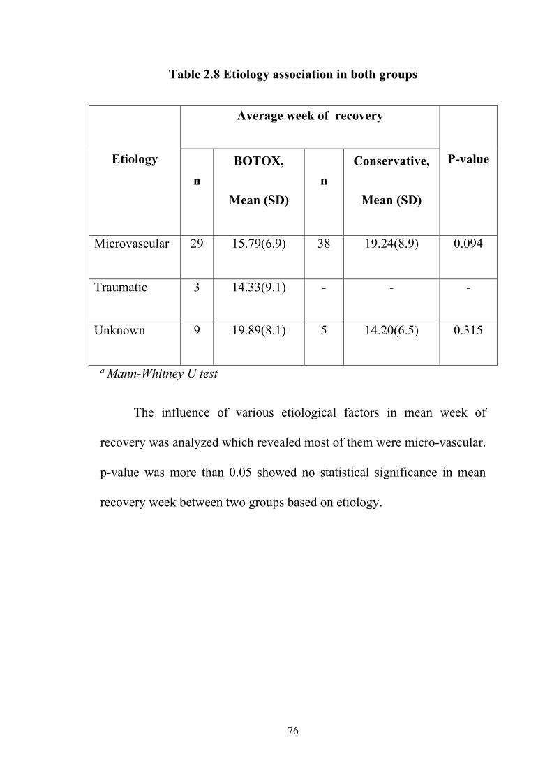
Table 2.8 Etiology association in both groups
Etiology
Average week of recovery
P-value
n
BOTOX,
Mean (SD)
n
Conservative,
Mean (SD)
Microvascular 29 15.79(6.9) 38 19.24(8.9) 0.094
Traumatic 3 14.33(9.1) - - -
Unknown 9 19.89(8.1) 5 14.20(6.5) 0.315
a Mann-Whitney U test
The influence of various etiological factors in mean week of
recovery was analyzed which revealed most of them were micro-vascular.
p-value was more than 0.05 showed no statistical significance in mean
recovery week between two groups based on etiology.
77
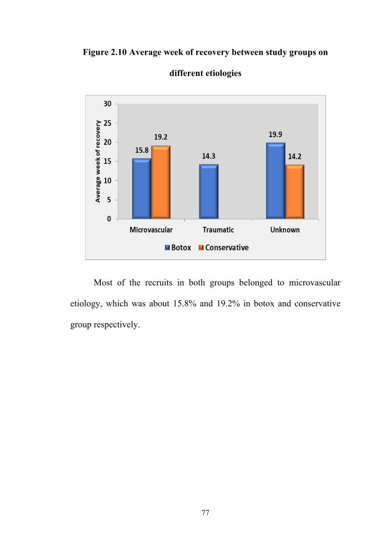
Figure 2.10 Average week of recovery between study groups on
different etiologies
Most of the recruits in both groups belonged to microvascular
etiology, which was about 15.8% and 19.2% in botox and conservative
group respectively.
78
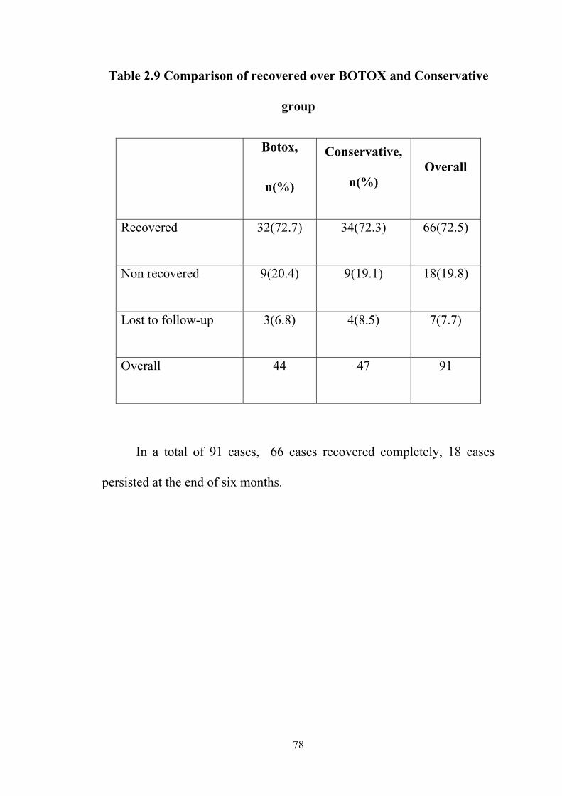
Table 2.9 Comparison of recovered over BOTOX and Conservative
group
Botox,
n(%)
Conservative,
n(%) Overall
Recovered 32(72.7) 34(72.3) 66(72.5)
Non recovered 9(20.4) 9(19.1) 18(19.8)
Lost to follow-up 3(6.8) 4(8.5) 7(7.7)
Overall 44 47 91
In a total of 91 cases, 66 cases recovered completely, 18 cases
persisted at the end of six months.
79
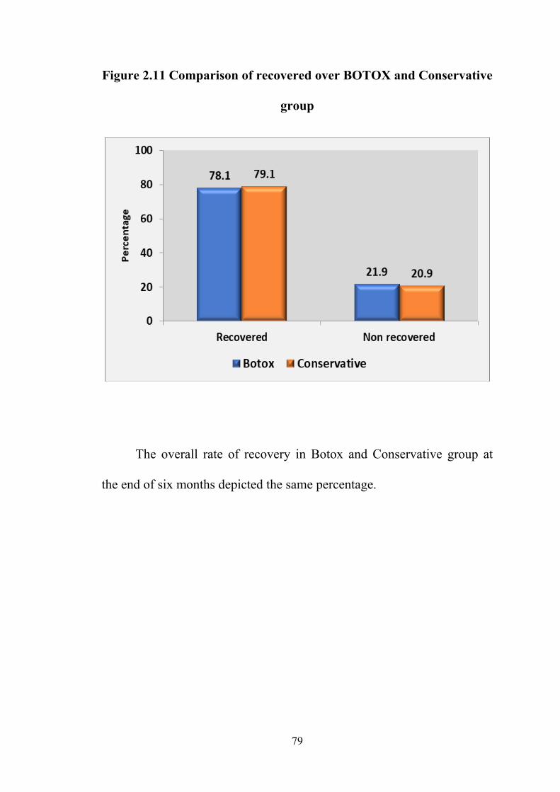
Figure 2.11 Comparison of recovered over BOTOX and Conservative
group
The overall rate of recovery in Botox and Conservative group at
the end of six months depicted the same percentage.
80
DISCUSSION
Previous studies that analysed the efficacy of botulinum toxin
injection in cases of abducent nerve palsy were retrospective, a few were
multi-centred and were done with inadequate sample size .This is a
prospective observational study to determine the efficacy of botulinum
toxin injection in acute sixth nerve palsy or paresis presenting within six
weeks of onset. A total of 91 patients were recruited, 44 in botox group
and 47 in conservative group. Out of these, 3 in botox group and 4 in
conservative group were lost to follow up after 1 month of recruitment.
Majority of the patients (53.8%) were between 41 and 60 years of age
and the remaining (36.3%) were above 60 years of age. There was a male
predominance (73.6%) in both groups taken together, that could probably
be attributed to various socio economic and cultural reasons in the
society; wherein males presented to hospital more than females. Out of
91 patients , 78% had an underlying microvascular pathology.
In this study, the patients who presented with an initial deviation
of 21-40 Prism Dioptres in botox and conservative group had a mean
recovery of 16.3±7.1 and 21.6±8.1 weeks respectively. The p value
(0.038) showed a significant difference in mean recovery between the
two groups. The initial deviation less than 20PD and more than 40PD
showed no statistically significant difference in recovery.
81
In previous studies, they have considered the overall recovery rate
rather than comparison at sub category level which includes onset,
aetiology and deviation at presentation. On comparing the recovery based
on the severity of abduction deficit (paresis/palsy), no statistical
significance was observed. The sub categorical assessment made on the
onset of presentation and various systemic risk factors in both groups
showed no statistical significance (p >0.05). Cases in our study were
mainly of microvascular aetiology and only a few were traumatic. To
ascertain a statistical as well as clinical significance on various etiological
(such as traumatic, inflammatory, etc.,) recovery, further studies are
needed with adequate sample size in each group.
14 cases (6.16%) in botox group acquired a vertical deviation
around 2 weeks post injection and recovered completely during follow
up. We did not observe any adverse effects.
Although natural course of recovery of ischemic abducent palsy is
spontaneous, our study showed a significant difference in the rate of
recovery in botox group with an initial deviation of 21-40PD. However,
the overall percentage of recovery remained the same in both groups.
LIMITATIONS
Assessment of diplopia or effect of diplopia reduction on quality of
life was not studied .The influence of other factors like time of botulinum
toxin reconstitution and dose of toxin were not included.
82
CONCLUSION
The overall recovery rate was similar in botox and conservative
group. However in cases ischemic paresis with presenting deviation
21-40 prism dioptres, injection of botulinum toxin hastened the mean
recovery rate significantly.
In view of benefit and no disadvantage, botulinum injection can be
considered in cases of acute ischemic sixth nerve palsy to alleviate
symptoms of diplopia and to provide a functionally better recovery
period.
Further, Randomised controlled trials evaluating the functional and
quality of life assessment, botulinum toxin reconstitution and dose of
toxin are needed for accurate assessment of efficacy of botulinum toxin
injection.
i
REFERENCES
1. Hofer JE, Scavone BM. Cranial nerve VI palsy after dural-arachnoid puncture.
Anesth Analg. 2015;120:644–6.
2. Nair AG, Ambika S, Noronha VO, Gandhi RA. The diagnostic yield of
neuroimaging in sixth nerve palsy--Sankara Nethralaya Abducens Palsy Study
(SNAPS): Report 1. Indian J Ophthalmol. 2014;62:1008–12.
3. Rowe FJ, Hanna K, Evans JR, Noonan CP, Garcia-Finana M, Dodridge CS, et al.
Interventions for eye movement disorders due to acquired brain injury. Cochrane
Database Syst Rev. 2018 05;3:CD011290.
4. Bron A, Tripathi R, Tripathi B. Wolff’s anatomy of the eye and orbit, 8th ed.
London: Chapman and Hall Medical; 1998. p. 189.
5. Khurana AK. Anatomy and physiology of the eye. 3rd ed. New Delhi: CBS
Publishers; 2017. p. 549.
6. Rosenbaum AL, Santiago AP. Clinical Strabismus Management: Principles and
Surgical Techniques. Saunders; 1999.
7. Erbguth FJ. From poison to remedy: the chequered history of botulinum toxin. J
Neural Transm (Vienna). 2008;115:559–65.
8. Erbguth FJ. Historical notes on botulism, Clostridium botulinum, botulinum toxin,
and the idea of the therapeutic use of the toxin. Mov Disord. 2004;19:S2-6.
9. Nigam PK, Nigam A. Botulinum toxin. Indian J Dermatol. 2010;55:8–14.
10. Münchau A, Bhatia KP. Uses of botulinum toxin injection in medicine today. BMJ.
2000 Jan 15;320:161–5.
11. Brin MF. Botulinum toxin: chemistry, pharmacology, toxicity, and immunology.
Muscle Nerve Suppl. 1997;6:S146-168.

ii
12. Burgen ASV, Dickens F, Zatman LJ. The action of botulinum toxin on the neuro-
muscular junction. J Physiol (Lond). 1949;109:10–24.
13. Sellin LC. The pharmacological mechanism of botulism. Trends Pharmacol Sci
1985;6:80-2.
14. Stanley EF, Drachman DB. Botulinum toxin blocks quantal but not non-quantal
release of ACh at the neuromuscular junction. Brain Res. 1983;261:172–5.
15. Hoffman RO, Helveston EM. Botulinum in the treatment of adult motility disorders.
Int Ophthalmol Clin. 1986;26:241–50.
16. Göschel H, Wohlfarth K, Frevert J, Dengler R, Bigalke H. Botulinum A toxin
therapy: neutralizing and nonneutralizing antibodies--therapeutic consequences.
Exp Neurol. 1997;147:96–102.
17. Odergren T, Hjaltason H, Kaakkola S, Solders G, Hanko J, Fehling C, et al. A
double blind, randomised, parallel group study to investigate the dose equivalence
of Dysport and Botox in the treatment of cervical dystonia. J Neurol Neurosurg
Psychiatry. 1998;64:6–12.
18. Brin MF, Lew MF, Adler CH, Comella CL, Factor SA, Jankovic J, et al. Safety and
efficacy of NeuroBloc (botulinum toxin type B) in type A-resistant cervical
dystonia. Neurology. 1999;53:1431–8.
19. Ranoux D, Gury C, Fondarai J, Mas JL, Zuber M. Respective potencies of Botox
and Dysport: a double blind, randomised, crossover study in cervical dystonia. J
Neurol Neurosurg Psychiatry. 2002;72:459–62.
20. Malhotra SP, Danahey DG. Botulinum toxin injections to improve facial aesthetics.
Available from: https://emedicine.medscape.com/article/841964-images
iii
21. Sato F, Akao T, Kurkin S, Fukushima J, Fukushima K. Adaptive changes in
vergence eye movements induced by vergence-vestibular interaction training in
monkeys. Exp Brain Res. 2004;156:164–73.
22. Haggerty H, Richardson S, Mitchell KW, Dickinson AJ. A modified method for
measuring uniocular fields of fixation: reliability in healthy subjects and in patients
with Graves orbitopathy. Arch Ophthalmol. 2005;123:356–62.
23. Hanif S, Rowe FJ, O’connor AR. A comparative review of methods to record ocular
rotations. Br Ir Orthopt J. 2009;6:47.
24. Kestenbaum A. Clinical methods of neuro-ophthalmological examinations, 2nd
edition. New York: Grune & Stratton; 1961.
25. Kushner BJ. The usefulness of the cervical range of motion device in the ocular
motility examination. Arch Ophthalmol. 2000;118:946–50.
26. Gerling J, Lieb B, Kommerell G. Duction ranges in normal probands and patients
with Graves’ ophthalmopathy, determined using the Goldmann perimeter. Int
Ophthalmol. 1997 1998;21:213–21.
27. Garrity JA. Assessment of disease severity. In: Prummel MF, editor. Recent
developments in graves’ ophthalmopathy. Norwell, MA: Academic Publishers;
2000. p.45.
28. American Academy of Ophthalmology. Basic and clinical science course (BCSC)
Section 6: Pediatric ophthalmology and strabismus 2016-2017. San Francisco, CA:
American Academy of Ophthalmology; 2016. p. 100.
29. Flanders M, Sarkis N. Fresnel membrane prisms: clinical experience. Can J
Ophthalmol. 1999;34:335–40.
30. Antony J. Prisms in clinical practice. Kerala J Ophthalmol. 2017;29:79.
31. Food and Drug Administration. FDA Talk Paper [FDA Website] April 15, 2002.

iv
32. Borish IM. Borish’s clinical refraction. Philadelphia: W.B. Saunders; 1998. p. 329-
336.
33. Reeves AG, Swenson RS. Disorders of the nervous system: A primer. Available
from: http://www.dartmouth.edu/~dons/
34. Walsh FB, Hoyt WF. Clinical neuro-ophthalmology. 3rd ed. Baltimore: Williams &
Wilkins; 1969.
35. Murray AD. Early and late botulinum toxin treatment of acute sixth nerve palsy.
Aust N Z J Ophthalmol. 1989;17:239–45.
36. Lee J, Harris S, Cohen J, Cooper K, MacEwen C, Jones S. Results of a prospective
randomized trial of botulinum toxin therapy in acute unilateral sixth nerve palsy. J
Pediatr Ophthalmol Strabismus. 1994;31:283–6.
37. Holmes JM, Droste PJ, Beck RW. The natural history of acute traumatic sixth nerve
palsy or paresis. J AAPOS. 1998;2:265–8.
38. Rowe FJ, Noonan CP. Botulinum toxin for the treatment of strabismus. Cochrane
Database Syst Rev. 2017;3:CD006499.
39. Metz HS, Mazow M. Botulinum toxin treatment of acute sixth and third nerve palsy.
Graefes Arch Clin Exp Ophthalmol. 1988;226:141–4.
40. Scott AB, Kraft SP. Botulinum toxin injection in the management of lateral rectus
paresis. Ophthalmology. 1985 May;92(5):676–83.
41. Holmes JM, Beck RW, Kip KE, Droste PJ, Leske DA. Botulinum toxin treatment
versus conservative management in acute traumatic sixth nerve palsy or paresis. J
AAPOS. 2000;4:145-9.
42. TalebNejad DM-R, Eghtedari DM. Botulinum Toxin-A Injection in Acute Sixth
Nerve Palsy. Iranian Journal of Ophthalmology. 2006;19:4.
43. Roper-Hall G. The hess screen test. Am Orthopt J. 2006;56:166–74.
v
ABBREVIATIONS
I/ L Ipsilateral
UMN Upper motor neuron
C/L Contralateral
CP angle Cerebropontine angle
FDA Food and Drug Administration
SNAP25 Synaptosomal nerve – associated protein25
VAMP Vesicle Associated Membrane Protein
EMG Electromyography
PD Prism Diopter
cm Centimeter
SO Superior Oblique
IO Inferior oblique
SR Superior Rectus
IR Inferior Rectus
MR Medial Rectus
LR Lateral Rectus
CROM Cervical Range Of Motion Device
BCVA Best corrected visual acuity
BSV Binocular Single Vision
WFDT Worth Four Dot Test
D Dioptre
Nacl Sodium chloride
SD Standard Deviation
vi
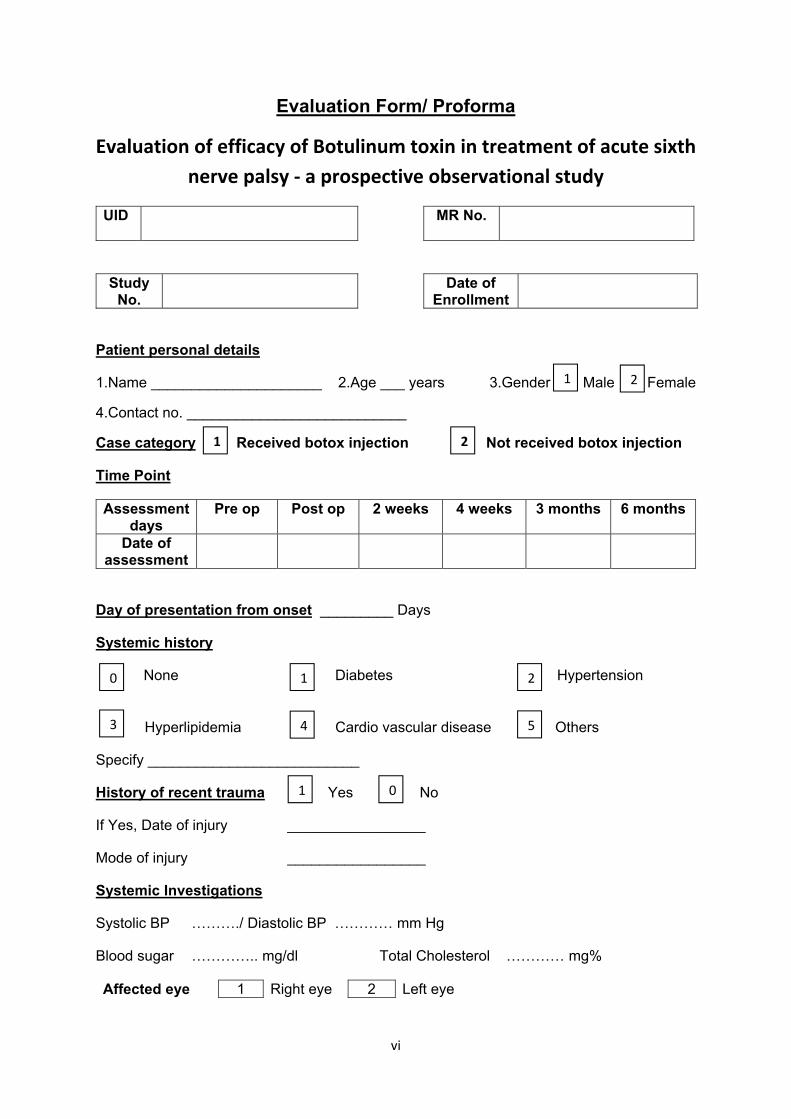
Evaluation Form/ Proforma
Evaluation of efficacy of Botulinum toxin in treatment of acute sixth nerve palsy - a prospective observational study
UID MR No.
Study No.
Date of Enrollment
Patient personal details
1.Name _____________________ 2.Age ___ years 3.Gender Male Female
4.Contact no. ___________________________
Case category Received botox injection Not received botox injection
Time Point
Assessment days
Pre op Post op 2 weeks 4 weeks 3 months 6 months
Date of assessment
Day of presentation from onset _________ Days
Systemic history
None Diabetes Hypertension
Hyperlipidemia Cardio vascular disease Others
Specify __________________________
History of recent trauma Yes No
If Yes, Date of injury _________________
Mode of injury _________________
Systemic Investigations
Systolic BP ………./ Diastolic BP ………… mm Hg
Blood sugar ………….. mg/dl Total Cholesterol ………… mg%
Affected eye 1 Right eye 2 Left eye
10
1
2
4 3 5
1 0
1 2
2
vii
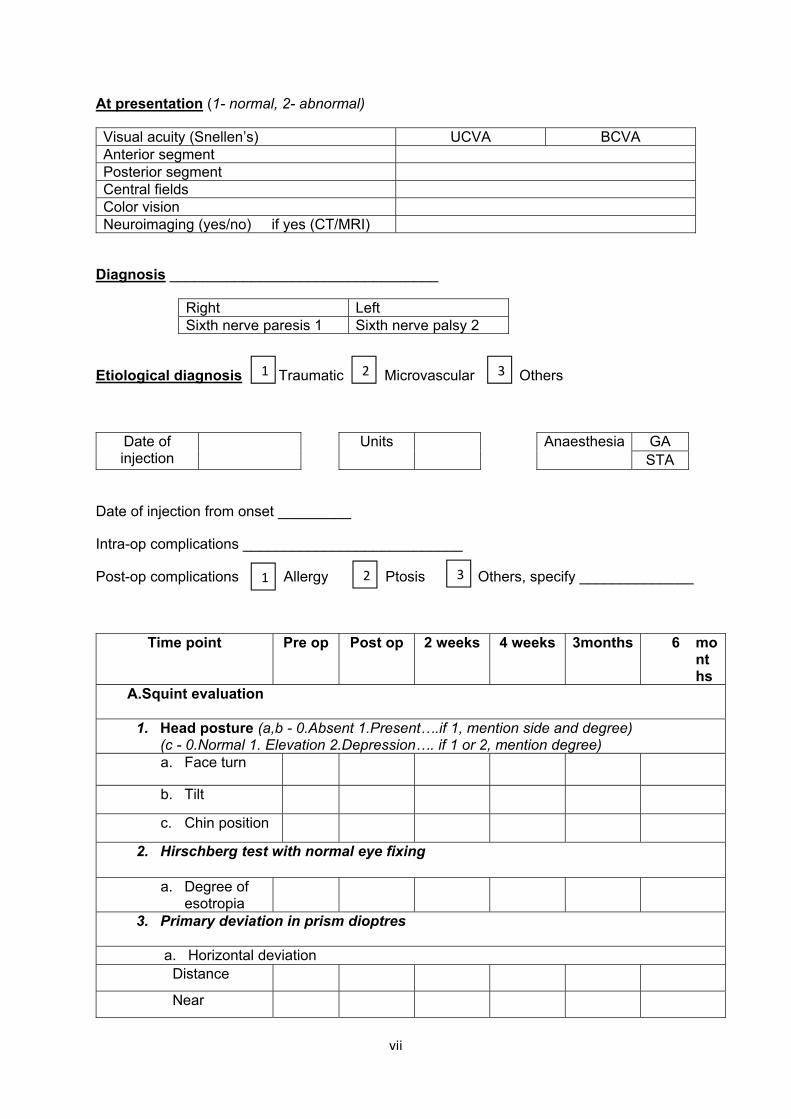
At presentation (1- normal, 2- abnormal)
Visual acuity (Snellen’s) UCVA BCVA Anterior segment Posterior segment Central fields Color vision Neuroimaging (yes/no) if yes (CT/MRI)
Diagnosis _________________________________
Right Left Sixth nerve paresis 1 Sixth nerve palsy 2
Etiological diagnosis Traumatic Microvascular Others
Date of injection
Units Anaesthesia GA STA
Date of injection from onset _________
Intra-op complications ___________________________
Post-op complications Allergy Ptosis Others, specify ______________
Time point Pre op Post op 2 weeks 4 weeks 3months 6 months
A.Squint evaluation
1. Head posture (a,b - 0.Absent 1.Present….if 1, mention side and degree) (c - 0.Normal 1. Elevation 2.Depression…. if 1 or 2, mention degree) a. Face turn
b. Tilt
c. Chin position
2. Hirschberg test with normal eye fixing
a. Degree of esotropia
3. Primary deviation in prism dioptres
a. Horizontal deviation Distance
Near
2 31
1 2 3

viii
b. Vertical deviation Distance
Near
4. Extra ocular movements
a. Limitation of abduction
5. Hess chart (c – 0.absent 1.present)
a. Primary position
b. Degree of lateral rectus underaction
c. Ipsilateral medial rectus overaction
ix
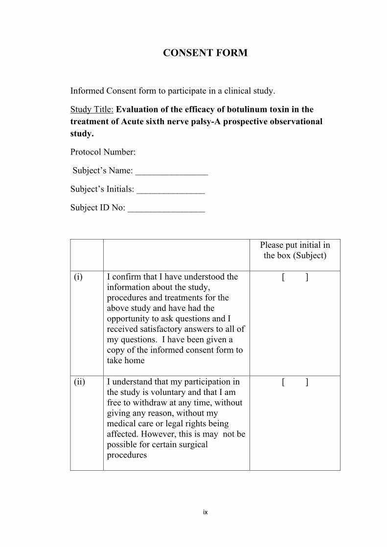
CONSENT FORM
Informed Consent form to participate in a clinical study.
Study Title: Evaluation of the efficacy of botulinum toxin in the treatment of Acute sixth nerve palsy-A prospective observational study.
Protocol Number:
Subject’s Name: ________________
Subject’s Initials: _______________
Subject ID No: _________________
Please put initial in the box (Subject)
(i) I confirm that I have understood the
information about the study, procedures and treatments for the above study and have had the opportunity to ask questions and I received satisfactory answers to all of my questions. I have been given a copy of the informed consent form to take home
[ ]
(ii) I understand that my participation in the study is voluntary and that I am free to withdraw at any time, without giving any reason, without my medical care or legal rights being affected. However, this is may not be possible for certain surgical procedures
[ ]
x
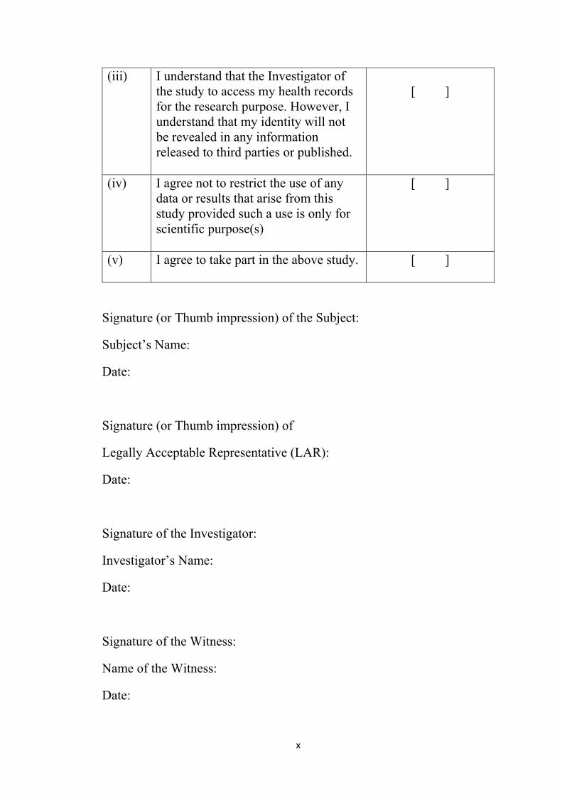
(iii) I understand that the Investigator of the study to access my health records for the research purpose. However, I understand that my identity will not be revealed in any information released to third parties or published.
[ ]
(iv) I agree not to restrict the use of any data or results that arise from this study provided such a use is only for scientific purpose(s)
[ ]
(v) I agree to take part in the above study.
[ ]
Signature (or Thumb impression) of the Subject:
Subject’s Name:
Date:
Signature (or Thumb impression) of
Legally Acceptable Representative (LAR):
Date:
Signature of the Investigator:
Investigator’s Name:
Date:
Signature of the Witness:
Name of the Witness:
Date:
xi

xii
xiii
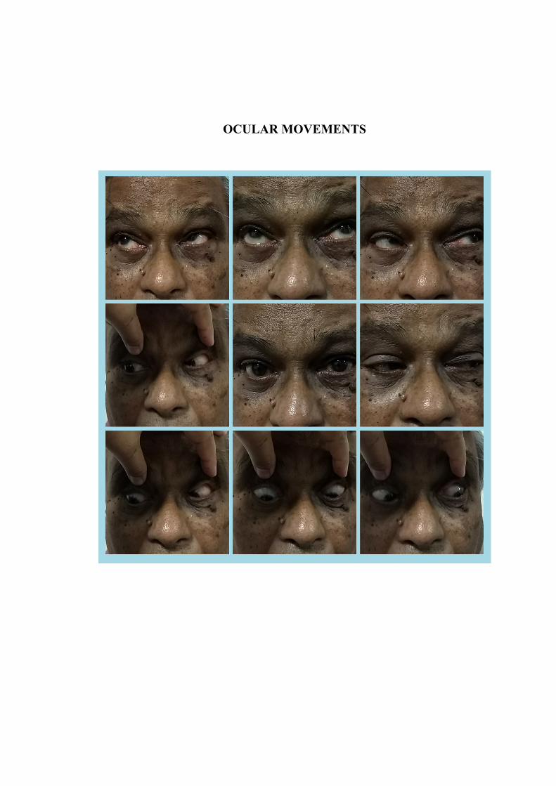
OCULAR MOVEMENTS
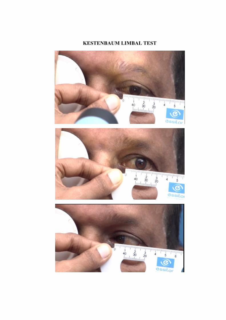
KESTENBAUM LIMBAL TEST
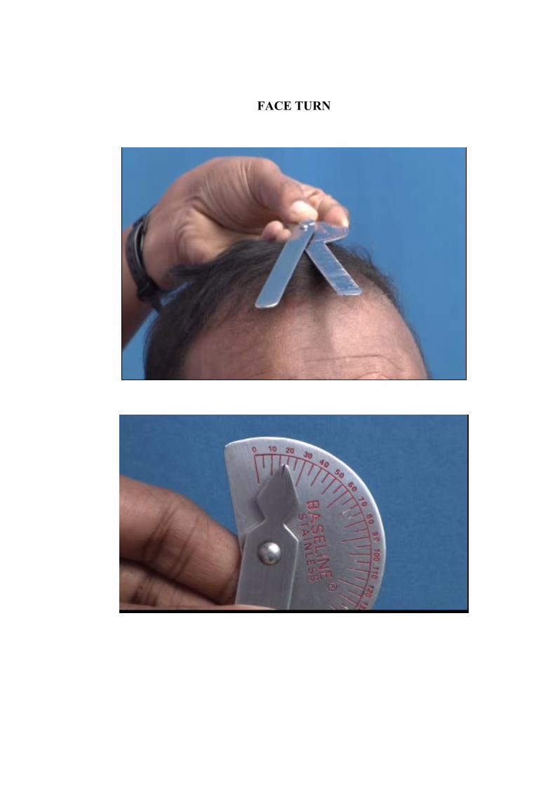
FACE TURN
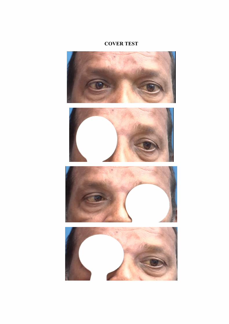
COVER TEST
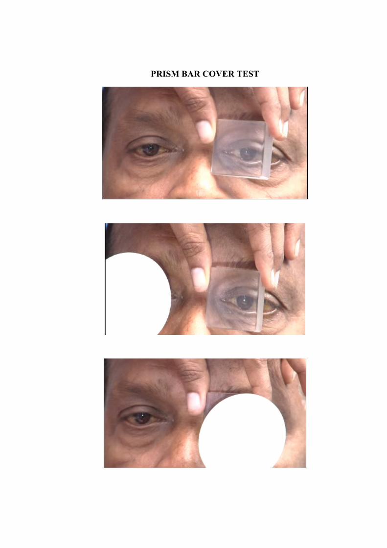
PRISM BAR COVER TEST
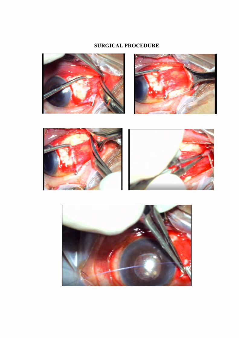
SURGICAL PROCEDURE