the bromodomain inhibitor jq1 as novel therapeutic option
TRANSCRIPT

The bromodomain inhibitor JQ1 as novel therapeutic option for
type II testicular germ cell tumours
The role of SOX2 and SOX17 in regulating germ cell tumour
pluripotency
Dissertation
zur
Erlangung des Doktorgrades (Dr.rer.nat.)
der
Mathematisch-Naturwissenschaftlichen Fakultät
der
Rheinischen Friedrich-Wilhelms-Universität Bonn
vorgelegt von
Sina Verena Jostes
aus
Kassel
Bonn, 2019

Angefertigt mit Genehmigung der Mathematisch-Naturwissenschaftlichen
Fakultät der Rheinischen Friedrich-Wilhelms-Universität Bonn
1. Gutachter: Prof. Dr. Hubert Schorle
2. Gutachter: Priv.-Doz. Dr. Reinhard Bauer
Tag der Promotion: 29. Juli 2019
Erscheinungsjahr: 2019

Eidesstattliche Erklärung
I
Eidesstattliche Erklärung
Hiermit erkläre ich, dass diese Dissertation von mir selbst und ohne unerlaubte Hilfe angefertigt
wurde. Es wurden keine anderen als die angegebenen Hilfsmittel benutzt. Ferner erkläre ich,
dass die vorliegende Arbeit an keiner anderen Universität als Dissertation eingereicht wurde.
Teile dieser Arbeit wurden bereits in folgenden Publikationen veröffentlicht:
The bromodomain inhibitor JQ1 triggers growth arrest and apoptosis in testicular germ
cell tumours in vitro and in vivo. Jostes S, Nettersheim D, Fellermeyer M, Schneider S,
Hafezi F, Honecker F, Schumacher V, Geyer M, Kristiansen G, Schorle H. J Cell Mol Med.
2016 Dec 27
Epigenetic drugs and their molecular targets in testicular germ cell tumours.
Jostes S, Nettersheim D, Schorle H. Nat Rev Urol. 2019 Feb 14
Bonn, 2019 ___________________________
Sina Verena Jostes

List of Abbreviations
II
List of Abbreviations
AFP Alpha-fetoprotein
AP Alkaline Phosphatase
BETi BET Inhibitor
BMP Bone Morphogenic Protein
BRD Bromodomain
ChIP Chromatin Immunoprecipitation
CRISPR Clustered Regularly Interspaced Short Palindromic Repeats
DAPI 6-Diamidino-2-Phenylindole Dihydrochloride
DNA Deoxyribonucleic acid
DNMT De novo Methyltransferase
EAU European Association of Urology
EC Embryonal Carcinoma
ESC Embryonic Stem Cell
ESMO European Society for Medical Oncology
FACS Fluorescence-Activated Cell Sorting
GCNIS Germ Cell Neoplasia In Situ
GSEA Gene Set Enrichment Analysis
HAT Histone Acetyltransferase
HCG Human Chorionic Gonadotropin
HDAC Histone Deacetylase
HDACi HDAC Inhibitor
HOMER Hypergeometric Optimization of Motif EnRichment
I.p. Intraperitoneally
IP Immunoprecipitation
iPSC Induced Pluripotent Stem Cell
LDH Lactate Dehydrogenase
MSigDB Molecular Signatures Database
PARP Poly (ADP-ribose) Polymerase
PGC Primordial Germ Cell
qRT-PCR Quantitative real-time PCR
RA Retinoic Acid
RPLND Retroperitoneal Lymph Node Dissection
SAM Synergistic Activation Mediator
SDS Sodium dodecyl sulfate
TET Ten-Eleven Translocaton Enzyme
TGCT Testicular Germ Cell Tumour
TSS Transcription Start Site
WNT Wingless/Integrated
YST Yolk-Sac-Tumour

Table of Contents
III
Table of Contents
Eidesstattliche Erklärung...................................................................................................... I
List of Abbreviations ............................................................................................................ II
Table of Contents ................................................................................................................ III
List of Figures..................................................................................................................... VI
List of Tables .................................................................................................................... VIII
Summary I ........................................................................................................................... IX
Summary II ........................................................................................................................... X
1. Introduction ............................................................................................................... 1
1.1. Male germ cell development ............................................................................ 1
1.1.1. Transcription factors determining male germ cell fate ...................................... 1
1.2. Malignant germ cell development .................................................................... 3
1.2.1. Type I testicular germ cell tumours .................................................................. 4
1.2.2. Type II testicular germ cell tumours ................................................................. 4
1.2.3. Type III testicular germ cell tumours ................................................................ 6
1.3. Model systems for type II testicular germ cell tumours ..................................... 6
1.3.1. The seminoma-like cell line TCam-2 ................................................................ 8
1.3.2. The EC cell line NCCIT, NT2/D1, 2102EP ....................................................... 8
1.4. Transcription factors determining type II TGCT cell fate................................... 9
1.4.1. SOX and POU transcription factors in type II TGCTs ....................................... 9
1.4.2. The plasticity of type II TGCTs....................................................................... 11
1.5. Treatment of type II TGCTs ........................................................................... 13
1.5.1. Treatment of stage I-III seminoma and non-seminoma ................................. 13
1.6. Epigenetic therapies as alternative treatment option ..................................... 14
1.6.1. HDAC inhibitors ............................................................................................ 15
1.6.2. BET inhibitors ............................................................................................... 16
2. Materials and Methods ........................................................................................... 18
2.1. Materials ........................................................................................................ 18
2.1.1. Mouse Strains ............................................................................................... 18
2.1.2. Cell Lines ...................................................................................................... 18
2.1.3. Chemicals and reagents ................................................................................ 19

Table of Contents
IV
2.1.4. Kits ................................................................................................................ 20
2.1.5. Buffers and recipes........................................................................................ 20
2.1.6. Consumables ................................................................................................ 21
2.1.7. Cell culture accessories ................................................................................. 22
2.1.8. Equipment ..................................................................................................... 22
2.1.9. Antibodies ..................................................................................................... 23
2.1.10. qRT-PCR primers .......................................................................................... 25
2.1.11. Primers for ChIP validation ............................................................................ 26
2.1.12. Plasmids ........................................................................................................ 26
2.1.13. Software and databases ................................................................................ 27
2.2. Molecular biological methods ......................................................................... 29
2.2.1. Standard cell culture conditions ..................................................................... 29
2.2.2. Transfection................................................................................................... 29
2.2.3. Protein isolation ............................................................................................. 29
2.2.4. Western blot analysis .................................................................................... 29
2.2.5. Co-immunoprecipitation ................................................................................. 30
2.2.6. RNA Isolation ................................................................................................ 30
2.2.7. cDNA Synthesis and qRT-PCR ..................................................................... 31
2.3. JQ1 Project .................................................................................................... 31
2.3.1. JQ1 treatment of cell lines ............................................................................ 31
2.3.2. AnnexinV-7-AAD FACS ................................................................................. 31
2.3.3. PI FACS ........................................................................................................ 31
2.3.4. XTT assay ..................................................................................................... 32
2.3.5. Illumina HumanHT-12 v4 Expression Array ................................................... 32
2.3.6. JQ1 treatment of TGCT-xenografted nude mice ............................................ 32
2.3.7. Tumour dissection for IHC staining ................................................................ 33
2.4. Identification of SOX2 and SOX17 targets in TGCT cells ............................... 33
2.4.1. Fixation and chromatin preparation ............................................................... 33
2.4.2. Chromatin immunoprecipitation (ChIP) ......................................................... 33
2.4.3. ChIP-sequencing and bioinformatics analysis ................................................ 34
2.4.4. SOX17-knockout in TCam-2 cells .................................................................. 34
2.4.5. Immunofluorescence ..................................................................................... 34
2.4.6. Alkaline phosphatase staining ....................................................................... 35
2.4.7. SOX17 overexpression in EC cells ................................................................ 35
2.4.8. Virus Production ............................................................................................ 35
2.4.9. Generation of MS2-P65-HSF1 Helper Cell Lines ......................................... 36
Table of Contents
V
2.4.10. Transduction of Helper Cell Lines with SOX17 SAM Virus ............................ 36
3. Results I ................................................................................................................... 37
3.1. TGCT cell lines and somatic control cells express the JQ1 targets BRD2,
BRD3 and BRD4............................................................................................ 37
3.2. JQ1 induces apoptosis and cell cycle arrest in TGCT cells and in cisplatin-
resistant EC cells ........................................................................................... 39
3.3. The molecular effects of JQ1 treatment in TGCT cells ................................... 41
3.4. The molecular effects of JQ1 on the testis microenvironment ........................ 50
3.5. Combination therapy with JQ1 and romidepsin in TGCT cell lines ................. 53
3.6. JQ1 treatment of TGCT xenografts ................................................................ 55
3.7. Combination therapy with JQ1 and romidepsin in TGCT xenografts .............. 56
4. Discussion I............................................................................................................. 58
5. Results II .................................................................................................................. 63
5.1. Introduction .................................................................................................... 63
5.2. SOX17 (seminoma) and SOX2 (embryonal carcinoma) partner with OCT4,
but not NANOG ............................................................................................. 63
5.3. SOX17 and SOX2 bind to the regulatory regions of pluripotency genes in
TGCT cells..................................................................................................... 64
5.4. The majority of regions bound by SOX17 in seminoma cells contains the
compressed motif and is found near transcriptional start sites ...................... 70
5.5. In seminoma cells SOX17 binds to the regulatory regions of neuro-
ectodermal genes, as well as pluripotency and germ-cell related genes ........ 74
5.6. In seminoma cells SOX17 regulates TFAP2C and PRDM1 expression ......... 80
5.7. SOX17 maintains latent pluripotency of seminoma cells ................................ 82
5.8. In TGCT cells NANOG is a common downstream target of SOX2
and SOX17 .................................................................................................... 89
5.9. The Role of SOX2 and SOX17 in TGCT plasticity.......................................... 91
6. Discussion II............................................................................................................ 94
7. Bibliography ............................................................................................................ 98
8. Appendix ............................................................................................................... 110
9. Publications .......................................................................................................... 118
10. Acknowledgements .............................................................................................. 120

List of Figures
VI
List of Figures
Figure 1 Germ cell specification in the human system ......................................... 2
Figure 2 Male testicular germ cell tumours (type I-III) .......................................... 3
Figure 3 Overview of type II testicular germ cell tumours .................................... 5
Figure 4 SOX2/OCT4 and SOX17/OCT4 DNA binding motifs........................... 10
Figure 5 The plasticity of type II TGCTs ............................................................... 12
Figure 6 The chromatin landscape ....................................................................... 14
Figure 7 Expression of BRD2, BRD3, BRD4 and BRDT in TGCT cell lines and
somatic control cells ................................................................................ 38
Figure 8 JQ1 induces apoptosis and G0/G1 arrest in cisplatin-sensitive and
cisplatin-resistant TGCT cells ........................................................... 40
Figure 9 Upregulation of stress markers and downregulation of pluripotency
genes in TGCT cells following JQ1 treatment ..................................... 42
Figure 10 Downregulation of pluripotency in TGCT cell lines .............................. 43
Figure 11 Gene ontology analysis of genes upregulated in NCCIT cells 72 hours
after JQ1 Treatment ................................................................................ 48
Figure 12 MYC protein levels in JQ1 treated TGCT cell lines.............................. 49
Figure 13 JQ1 induces apoptosis in FS1 Sertoli cells but not fibroblasts ........... 50
Figure 14 Microarray analysis of 100 nM JQ1 treated Sertoli cells ..................... 51
Figure 15 Genes commonly deregulated in TGCT and Sertoli cells ................... 52
Figure 16 Cell viability of TGCT cells treated with JQ1 and romidepsin ............ 54
Figure 17 Tumour growth of TGCT xenografts treated with JQ1 ......................... 55
Figure 18 Ki67 and CD31 staining of TGCT xenografts treated with JQ1 .......... 56
Figure 19 Tumour growth of TGCT xenografts treated with JQ1 + romidepsin . 57
Figure 20 Effects of JQ1 and romidepsin treatment in TGCT cells ..................... 60
Figure 21 SOX17 and SOX2 interact with OCT4, but not NANOG ..................... 64
Figure 22 Validation of ChIP-grade antibodies ...................................................... 66
Figure 23 SOX17 binds pluripotency genes in TCam-2 cells ............................... 67
Figure 24 Expression of pluripotency genes in TCam-2 and 2102EP cells ........ 68

List of Figures
VII
Figure 25 SOX2 binds pluripotency genes in 2102EP cells ................................. 69
Figure 26 SOX17 occupies canonical and compressed binding sites in
seminoma cells ........................................................................................ 71
Figure 27 SOX2 occupies canonical binding sites in EC cells ............................. 73
Figure 28 Scatterplot of TCam-2 gene expression data with SOX17 peaks ...... 77
Figure 29 Scatterplot of 2102EP gene expression data with SOX2 peaks ......... 79
Figure 30 SOX17 regulates the SOX17-PRDM1-TFAP2C network in seminoma
.................................................................................................................. 81
Figure 31 CRISPR/Cas9 mediated gene editing of SOX17 gene locus.............. 82
Figure 32 Expression of pluripotency and germ cell markers after depletion of
SOX17 in TCam-2 cells .......................................................................... 83
Figure 33 Depletion of SOX17 in TCam-2 cells ..................................................... 84
Figure 34 Morphology of TCam-2 Δ SOX17 bulk .................................................. 85
Figure 35 Alkaline phosphatase activity of TCam-2 Δ SOX17 bulk ..................... 85
Figure 36 SOX17, OCT4 and TFAP2C protein expression in TCam-2 Δ SOX17
bulk ........................................................................................................... 87
Figure 37 Expression of germ cell markers and trophoblast differentiation
markers after depletion of SOX17 in TCam-2 cells.............................. 88
Figure 38 SOX17 and GATA3 protein expression in TCam-2 Δ SOX17 bulk
population ................................................................................................. 89
Figure 39 SOX2 and SOX17 regulate a common set of pluripotency genes ..... 90
Figure 40 SOX17 overexpression in NCCIT Cells ................................................ 92
Figure 41 The effects of SOX17 overexpression in EC cells on the expression of
SOX17 target genes................................................................................ 93
Figure 42 Transcription factor network maintaining seminoma pluripotency...... 96

List of Tables
VIII
List of Tables
Table 1 Type II TGCT cell lines ............................................................................ 7
Table 2 Overview of selected HDAC inhibitors .................................................. 15
Table 3 Overview of selected BET inhibitors ...................................................... 17
Table 4 GSEA of genes downregulated in TCam-2 cells 72 hours after JQ1
treatment .................................................................................................. 45
Table 5 GSEA of genes downregulated in NCCIT cells 72 hours after JQ1
treatment .................................................................................................. 45
Table 6 Gene ontology analysis of genes upregulated in NCCIT cells 72 hours
after JQ1 treatment ................................................................................. 45
Table 7 MSigDB GSEA of SOX17 targets in TCam-2 (normalized to IgG
background) ............................................................................................. 74
Table 8 MSigDB GSEA of SOX17 targets in TCam-2 (normalized to 2% input)
.................................................................................................................. 75
Table 9 MSigDB GSEA of SOX2 targets in 2102EP (normalized to IgG
background) ............................................................................................. 75
Table 10 MSigDB GSEA of SOX2 targets in 2102EP (normalized to 2% input)
.................................................................................................................. 76
Table 11 Top 20 SOX17 (vs IgG)-peaks with gene expression > 8 in TCam-2 ....
.................................................................................................................. 78
Table 12 Top 20 SOX2 (vs IgG)-peaks with gene expression > 8 in 2102EP .. 79

Summary I
IX
Summary I
Type II testicular germ cell tumours (TGCTs) represent the most common malignancy
in young men (19-35 years). They are classified as seminoma or embryonal carcinoma
(EC; the stem cell population of non-seminomas). TGCTs are highly sensitive to radio-
and chemotherapy, however 1-5% of TGCTs may develop resistance mechanisms to
standard therapy regimens. Epigenetic drugs open a new avenue to cancer therapy
and may present a promising alternative to treat recurrent TGCTs. JQ1 is an inhibitor
of the BET family of bromodomain reader proteins. In TGCT cell lines, JQ1 treatment
leads to upregulation of stress markers (i.e. CDKN1C, DDIT4, TSC22D1, TXNIP),
induction of the differentiation marker HAND1, and downregulation of pluripotency-
associated genes (i.e. LIN28, DPPA4, UTF1) [1]. This results in growth arrest and
apoptosis in cisplatin-sensitive and cisplatin-resistant EC cells (at doses ≥ 100 nM) and
seminoma cells (at doses ≥ 250 nM) [1, 2]. In line, EC xenografts in nude mice show
reduced tumour burden when treated with JQ1 (50 mg / kg) compared to solvent
controls. Additionally, JQ1-treated tumours showed reduced blood vessel count (lower
CD31+), possibly due to JQ1-mediated downregulation of VEGFB. Altogether, this
reflects the therapeutic potential of bromodomain inhibition for TGCTs. However,
similar to TGCT cells, somatic control cells (here: Sertoli cells) responded with cell
cycle arrest and apoptosis to JQ1 treatment. Thus, a more detailed analysis of possible
side effects of JQ1 administration is recommended, before commissioning the drug for
clinical use. Interestingly, JQ1 treatment had similar effects on TGCT cells as the
HDAC inhibitor romidepsin (i.e. induction of stress markers GADD45A, GADD45B,
RHOB, ID2) [1-3]. I now showed that JQ1 and romidepsin may elicit additive or
synergistic effects on cytotoxicity levels of TGCT cells in vitro and in vivo. Since a
combination of both drugs may, however, also increase potential side effects, the exact
efficacy vs toxicity relationship of this treatment strategy needs further evaluation.

Summary II
X
Summary II
TGCTs can be characterized as seminoma or EC. While seminomas display limited
differentiation capacities, ECs display features of pluri- to totipotency. Previous data
suggests that pluripotency in EC cells is maintained by cooperative binding of SOX2-
OCT4 to the canonical (SOX2/OCT4) motifs at pluripotency genes. Indeed, SOX2
binding in EC cells is enriched at canonical motifs and SOX2 target genes showed
significant overlap with embryonic stem cell signatures. In contrast, seminomas lack
expression of SOX2, but display high levels of OCT4 and SOX17. In embryonic stem
cells cooperative binding of SOX17-OCT4 to the compressed (SOX17/OCT4) motif on
DNA induces endodermal differentiation. However, seminomas maintain an
undifferentiated state, indicated by expression of pluripotency genes and lack of
expression of typical differentiation markers. We therefore asked, whether the SOX17-
OCT4 complex in seminoma cells binds to canonical (SOX2/OCT4) binding sites to
regulate and maintain seminoma pluripotency. High-throughput chromatin
immunoprecipiation (ChIP)-sequencing analysis revealed that the majority of genes
bound by SOX17 in seminoma cells has functions in neuronal differentiation and that
26% of SOX17 peaks contain the compressed (SOX17/OCT4) binding motif. These
findings are is in disagreement with the latent pluripotent state of seminoma cells.
However, a small subset of SOX17-bound genes has roles in pluripotency
maintenance (e.g. NANOG, POU5F1 (OCT4), PRDM1 and TFAP2C) and 10% of
SOX17 peaks include the described canonical (SOX2/OCT4) binding motif. This
suggests that, next to somatic genes, SOX17 regulates pluripotency genes in
seminoma cells by binding to the canonical motif. In line, CRISPR/Cas9-mediated
deletion of SOX17 in TCam-2 resulted in a strong reduction of OCT4 and TFAP2C
protein levels, as well as alkaline phosphatase activity. qRT-PCR analysis showed that
loss of SOX17 induces differentiation into trophoblast-like lineages. I conclude that
SOX17 shares a similar role in seminoma cells as in primordial germ cells (PGC),
which is to maintain a latent pluripotent state and to suppress cellular differentiation
(i.e. via downstream activation of the PGC specifiers PRDM1 and TFAP2C and by
direct activation of pluripotency genes such as NANOG and POU5F1).

Introduction
1
1. Introduction
1.1. Male germ cell development
Germ cells are the founder cells of new life. They harbour all necessary genetic and
epigenetic information, which is propagated from one generation to the next. Like in
other mammals, human germ cells are formed early during embryogenesis. These
germ cells then later undergo meiosis to form haploid spermatocytes (in males) or
oocytes (in females).
1.1.1. Transcription factors determining male germ cell fate
Human germ cell development is initiated two weeks after fertilization with the
formation of primordial germ cells (PGCs). Similar to PGCs in primates, it is believed
that human PGCs are specified in the nascent amnion, which expresses BMP4 and
WNT3A at high levels [4]. Both signalling molecules are crucial for human germ cell
development [4]. Recent data suggests that WNT signalling leads to EOMES induction,
which in turn transactivates SOX17, resulting in upregulation of BLIMP1 [5]. At the
same time BMP2 (expressed in the extraembryonic mesoderm) and BMP4 activate
TFAP2C expression [5]. Together, SOX17, BLIMP1 and TFAP2C form a tripartite
transcription factor network and the core circuitry for human germ cell specification [5-
7] (Fig. 1). Each factor exerts unique, but also overlapping roles in activating the germ
cell program and in sustaining the epigenetic program of PGCs, thereby maintaining
pluripotency and suppressing somatic differentiation [8]. Once specified, human PGCs
migrate along the hindgut to the genital ridges. During this migration PGCs are stalled
in G2 phase of the cell cycle, while they undergo global DNA demethylation and
imprinting erasure [9]. 4-6 weeks following implantation of the embryo in the uterus,
PGCs arrive in the genital ridge where they continue to amplify by mitosis [9]. From
now on the male germ cells are referred to as gonocytes and express the following
markers: MAGE-A4, DAZL, KIT, PLAP, POU5F1, TFAP2C, UTF1, VASA [10].
Approximately 6 months after birth the undifferentiated gonocytes (Adark-
spermatogonia) settle at the basal membrane of the seminiferous tubules of the testis,
where they lose their pluripotent state and develop further into Apale-spermatogonia [3,
11]. These remain quiescent until the age of ~ 10 years, when spermatogenesis starts
Introduction
2
with the development of B-spermatogonia, which differentiate further into primary
spermatocytes and subsequently undergo two rounds of meiotic division to become
haploid spermatids [12]. During meiosis the developing spermatocytes lose contact
with the basal membrane and migrate toward the lumen of the seminiferous tubules
[12]. Finally, the haploid round spermatids differentiate into mature, motile
spermatozoa (a process referred to as spermiogenesis) [13]. This process starts within
the seminiferous tubules, from which the elongated spermatozoa are transported via
the rete testis into the epididymis, where they mature and are stored [14]. The whole
process of spermatogenesis (including spermiogenesis) in humans consumes
approximately 74 days [15].
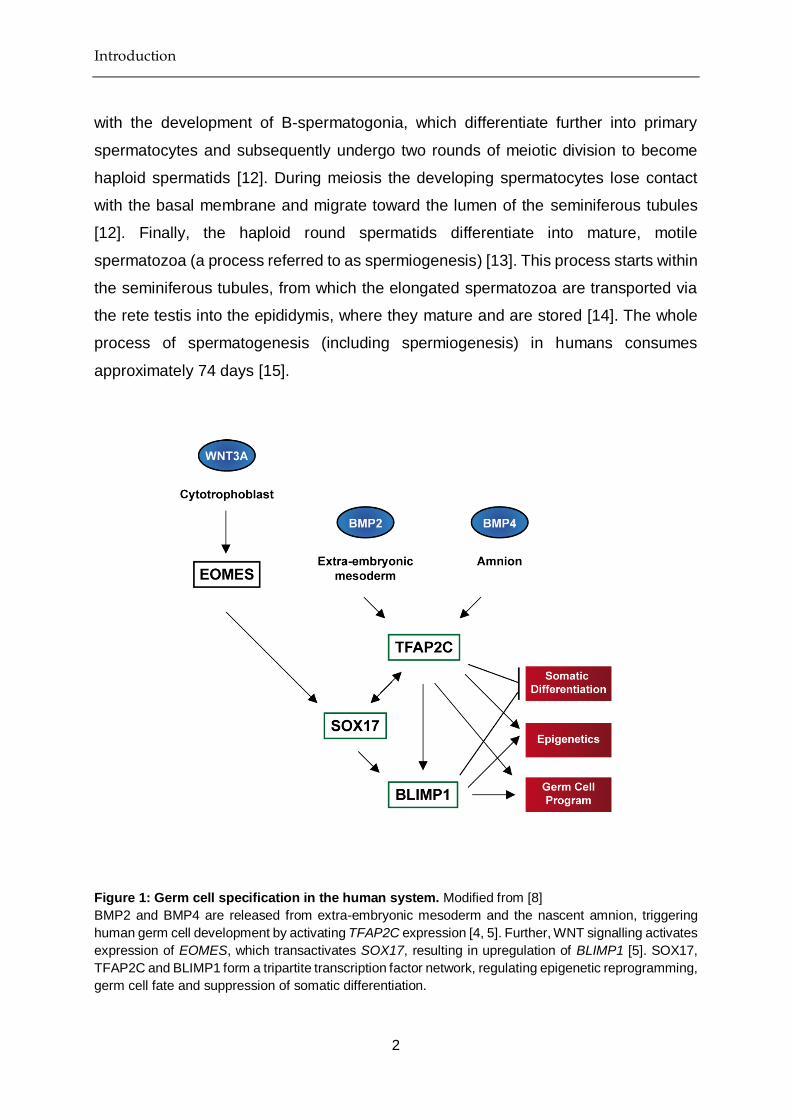
Figure 1: Germ cell specification in the human system. Modified from [8]
BMP2 and BMP4 are released from extra-embryonic mesoderm and the nascent amnion, triggering
human germ cell development by activating TFAP2C expression [4, 5]. Further, WNT signalling activates
expression of EOMES, which transactivates SOX17, resulting in upregulation of BLIMP1 [5]. SOX17,
TFAP2C and BLIMP1 form a tripartite transcription factor network, regulating epigenetic reprogramming,
germ cell fate and suppression of somatic differentiation.

Introduction
3
1.2. Malignant germ cell development
Testicular germ cell cancer is the most common form of cancer among males between
15 and 35 years [16]. It comprises a heterogeneous group of neoplasms that originates
from male germ cells and is therefore anatomically distributed along the migration route
of PGCs [17, 18]. Although the exact cause of germ cell cancer is unknown, it is
believed that environmental factors, e.g. endocrine disruptors, may contribute to the
risk of developing germ cell cancer [19]. Germ cell cancer incidence is globally
increasing, however the development of novel chemotherapeutic treatment regimens
has led to a drastic decline in mortality rates [20]. There are three types (I-III) of
testicular germ cell tumours (TGCTs), which can be discriminated according to their
anatomical site, stage of maturation and pattern of genomic imprinting [21] (Fig. 2).
Figure 2: Male testicular germ cell tumours (type I-III). Modified from [8, 21]
Type I TGCTs comprise of teratoma and yolk-sac-tumour of children and infants. They putatively arise
early during primordial germ cell development. Type II TGCTs comprise of seminomas and non-
seminomas of adolescents and adults. These tumours arise from a common precursor lesion, the germ
cell neoplasia in situ (GCNIS), which develops from an error in late primordial germ cell (PGC)
maturation. Type III TGCTs are spermatocytic seminomas, which frequently occur in older men and
develop from late spermatogonia or spermatocytes.

Introduction
4
1.2.1. Type I testicular germ cell tumours
Type I TGCTs account for 1-2% of solid tumours in children [22]. The vast majority of
type I TGCTs may either be classified as teratoma or yolk-sac-tumour (YST) according
to tumour histology and marker expression [21]. YSTs are defined by high levels of
alpha-fetoprotein (AFP) and morphologically present as endodermal or yolk-sac-like
tissue [23]. These tumours account for ~ 50% of testicular tumours in children [24, 25].
Pediatric teratomas are the second most common testicular tumours in children with a
relative frequency of ~ 15% [24, 26]. Pre-pubertal teratomas present as well-
differentiated (mature) and are of benign nature [25, 26]. They are usually derived from
cells of all three embryonic germ layers (endoderm, mesoderm, ectoderm).
Chromosomal abnormalities frequently found in pediatric YSTs and teratomas are loss
of 1p, 4 and 6q, and gain of 1q, 12(p13) and 20q [27]. Since type I TGCTs show a
partially erased imprinting pattern, it is believed that they develop early during germ
cell development [28] (Fig. 2), however risk factors remain poorly understood.
1.2.2. Type II testicular germ cell tumours
Type II TGCTs comprise of seminomas and non-seminomas [21]. These tumours are
found in young adolescents and adults and are the most frequent cause of cancer in
young men [29]. Type II TGCTs are characterized by high levels of the pluripotency
marker OCT4 and gain of the 12 p chromosomal region [30]. Both seminomas and
non-seminomas arise from a common precursor lesion called germ cell neoplasia in
situ (GCNIS) [31]. It is generally accepted that GCNIS formation occurs as a
consequence of an arrest in late PGCs development, due to the acquisition of genetic
mutations or epigenetic aberrations (Fig. 2) [10, 30-32]. We believe that GCNIS may
additionally arise from adult stage spermatogonial cells via reacquisition of the germ
cell pluripotency program, although not formally proven [10]. This is supported by the
observation that pluripotent cells can be derived from adult murine and human
testicular cells [10]. GCNIS are non-invasive and asymptomatic and therefore only
rarely diagnosed [31]. 8-10 years after puberty, however, they transform into a
malignant seminoma or non-seminoma (Fig. 3).

Introduction
5
Figure 3: Overview of type II testicular germ cell tumours. Modified from [8, 21]
Type II TGCTs comprise of seminomas and non-seminomas. Both subtypes arise from the GCNIS
precursor lesion. Non-seminomas initially present as ECs. ECs are pluri- to totipotent and can further
differentiate into embryonic (teratoma) and extraembryonic tissues (YST, choriocarcinoma).
Seminomas are highly similar to PGCs and GCNIS in terms of their overall marker
expression (they express LIN28, OCT4, NANOG, PRDM1, TFAP2C and cKIT) and
epigenetic profile (global hypomethylation) [10]. Non-seminomas initially present as
embryonal carcinomas (ECs) (Fig. 3). ECs are described as the stem cell compartment
of non-seminomas [10]. These cells are pluri- to totipotent and are therefore able to
differentiate into embryonic (teratoma) and extra-embryonic tissues (choriocarcinoma
or YST) (Fig. 3) [10]. Similar to seminomas, ECs express TFAP2C, GDF3, DPPA3,
OCT4 and NANOG at high levels, but additionally express DNMT3B, DNMT3L,
NODAL, CRIPTO, CD30 and SOX2 [10]. A major distinction between seminomas and

Introduction
6
embryonal carcinomas is the differential expression of the biomarkers and transcription
factors SOX2 (EC) and SOX17 (seminoma) [33, 34] (Fig. 3). Chromosomal
abnormalities that frequently occur in seminomas and non-seminomas are loss of 1p,
11, 13, 18 and gain of 7, 8, 12p, 21 and X [27]. 12 p gain is the most frequent
chromosomal alteration in Type II TGCTs, which is why overexpression of pluripotency
associated genes (e.g. NANOG, GDF3, DPPA3) encoded in this region is a common
event in these tumours [35].
1.2.3. Type III testicular germ cell tumours
Type III TGCTs are spermatocytic seminomas [21]. These tumours are rare (0.3-0.8
per one million men affected) and predominantly occur in men older than 50 years [36].
Spermatocytic seminomas display partial imprinting [21]. Since this imprinting pattern
resembles the one of spermatogonia or spermatocytes, it is generally believed that
these tumours develop at later stages during male germ cell development [21] (Fig. 2).
Gain of chromosome 9 is a common karyotypic alteration in these tumours [36].
Looijenga et al. proposed the transcriptional regulator DMRT1, which is encoded on
chromosome 9, as a driving factor for the development of spermatocytic seminomas
[37].
1.3. Model systems for type II testicular germ cell tumours
Several cell lines have successfully been derived from type II TGCTs and established
for in vitro culture, reviewed in Nettersheim et al. (2016) [10] (Table 1). Further,
xenotransplantation of these cell lines into the flank, brain or testes of immunodeficient
Crl:CD1-Foxn1nu (CD-1 nude) mice allows for in vivo tumour analysis. However,
changes in the cellular microenvironment following xenotransplantation may cause a
shift in cell fate (Table 1). For example, the seminoma-like cell line TCam-2 grows as
EC after injection into the flank of nude mice, while transplantation into the testis results
in seminoma growth [10] (Table 1).

Introduction
7
Table 1: Type II TGCT cell lines. From [10]
Cell Line Origin Growth in vitro as Growth in vivo as Reference
TCam-2 Seminoma Seminoma EC (flank), CIS /
Seminoma (testis)
[38]
JKT-1 Seminoma, EC Seminoma / EC
intermediate
Seminoma [39]
SEM-1 Seminoma Seminoma / EC
intermediate
Mediastinal GCC [40]
1411HP Seminoma, EC,
Teratoma, YST
EC EC, YST [41]
169A/218A/228A/
240A
EC EC Not determined [42]
1777N Retriperitoneal
metastasis of
non-seminoma
EC EC [43]
2102EP EC, Teratoma EC EC [44]
833Ke Seminoma, EC,
Teratoma,
Choriocarcinoma
EC EC [45]
GCT27 EC, Teratoma EC EC, Teratoma, YST [46]
GCT35 EC, Teratoma EC EC, YST [46]
GCT44/46/72 EC, Teratoma EC YST [46]
GCT48 EC, Teratoma EC EC [46]
H12.1/.5/.7 Seminoma, EC,
Teratoma,
Choriocarcinoma
EC EC [47]
NCCIT EC, Teratoma EC EC, Teratoma [48]
NEC-8/-14/-15 EC, YST,
Choriocarcinoma
EC EC, Teratoma [49]
SuSa EC, Teratoma EC Not determined [50]
TERA1/2 EC, Teratoma EC EC [51]
577MF/L/RPLN Metastasis of
non-seminoma
Undifferentiated
carcinoma
Teratoma [52]
BeWo Choriocarcinoma Choriocarcinoma Choriocarcinoma [53]
JAR Choriocarcinoma Choriocarcinoma Choriocarcinoma [54]
JEG-3 Choriocarcinoma Choriocarcinoma Choriocarcinoma [55]

Introduction
8
1.3.1. The seminoma-like cell line TCam-2
The only seminoma-like cell line that has been adapted to cell culture is the TCam-2
cell line [38, 56] (Table 1). Two other cell lines have been isolated from seminoma
patients (JKT-1, SEM-1), however SEM-1 cells show characteristics of both seminoma
and non-seminoma components, while JKT-1 lack the typical characteristics of a type
II TGCT (such as gain of the 12p chromosomal region) [40, 56, 57]. Other attempts for
the in vitro cultivation of seminoma cells have failed, since these cells undergo
spontaneous cell death (anoikis) when isolated from their microenvironment [58].
However, it is still unclear what determines the survival of TCam-2 cells in vitro.
Thorough analysis has demonstrated that TCam-2 cells display expression of the early
germ cell and TGCT markers OCT4, NANOG, TFAP2C and LIN28, and the seminoma
markers SOX17, PRDM1 (nuclear expression) and KIT [10]. At the same time TCam-
2 lack expression of EC markers SOX2 and CD30 [38, 59]. Furthermore, TCam-2 cells
show an aneuploid karyotype with the characteristic gain of the 12p region [38].
Morphologically, TCam-2 cells appear polygonal and flat in shape, with a large
cytoplasm and a round nucleus [56, 60]. Further, TCam-2 cells have a relatively long
doubling time of approximately 58 hours, which is reminiscent of migratory PGCs [56].
1.3.2. The EC cell lines NCCIT, NT2/D1 and 2102EP
The NCCIT cell line is a pluripotent EC cell line derived from a mixed germ cell tumour
[48] (Table 1). NCCIT are positive for alkaline phosphatase and the pluripotent stem
cell markers TRA-1-60 and TRA-1-81 [48]. They show epithelial-like morphology and
form dense cell clusters upon in vitro cultivation. When exposed to retinoic acid (RA),
NCCIT cells differentiate into cells of all three germ layers, mimicking teratoma growth
[48]. When transplanted into nude mice, NCCIT cells differentiate into mixed non-
seminoma [48].
The NT2/D1 (NTERA-2 cl.D1) cell line was derived by cloning the pluripotent EC line
NTERA-2. NTERA-2 were established from a nude mouse xenograft of the Tera-2 cell
line (Table 1). Similar to NCCIT cells, NT2/D1 grow in tight colonies and appear
epithelial-like [61]. In response to RA NT2/D1 cells differentiate into neuronal lineages

Introduction
9
[62]. When transplanted into nude mice, NT2/D1 cells grow as mixed non-seminoma
[63].
2102EP cells resemble undifferentiated EC cells and stain positive for alkaline
phosphatase and the pluripotency markers SSEA-4, TRA-1-60 and TRA-1-81 [64]
(Table 1). Although 2102EP cells resemble human ES cells on transcriptome level and
express a number of core pluripotency genes (TDGF1, DNMT3A, DNMT3B, POU5F1,
NANOG, GDF3, UTF1, SOX2), these cells lack the capacity to differentiate [64, 65].
This suggests that 2102EP cells have acquired additional mutations restricting their
differentiation capacities, therefore characterizing 2102EP cells as nullipotent [65].
When transplanted into nude mice, 2102EP cells grow as EC.
1.4. Transcription factors determining type II TGCT cell fate
Seminomas and ECs can be discriminated by their differential expression of SOX17
(seminoma: high, EC: low) and SOX2 (seminoma: low, EC: high) [33]. Both factors
belong to the SOX family of transcription factors. SOX factors have important roles in
orchestrating stem cell self-renewal and differentiation [66]. Along with OCT4, KLF4
and MYC, SOX2 is also well known for its function in the generation of induced
pluripotent stem cells (iPSC) and stem cell maintenance [67]. In contrast, SOX17 is a
known specifier of endodermal lineage decisions [68].
1.4.1. SOX and POU transcription factors in type II TGCTs
In mouse ES cells it was shown that SOX2 and SOX17 partner with the POU
transcription factor OCT4 and act as opposing forces in regulating cell fate decisions
[69]. SOX2 and OCT4 dimerize and bind to the canonical (SOX2/OCT4) binding motif
on the DNA, which is composed of the SOX and OCT4 binding motifs separated by a
single basepair [69, 70], (Fig. 4 A). SOX17 and OCT4 dimerize and bind to the
compressed motif, which is similarly composed of the SOX and OCT4 binding motifs,
but lacking the central basepair separating the two motifs [69], (Fig. 4 B). In both
human and mouse embryonic stem cells (ESCs) SOX2-OCT4 binding to the canonical
motif results in upregulation of pluripotency and stemness-associated genes [70]. In
contrast, binding of the SOX17-OCT4 complex to the compressed motif results in

Introduction
10
upregulation of endodermal-associated genes [69, 71, 72]. Interestingly, the group of
Prof. Jauch demonstrated that in mouse ESCs the SOX17-OCT4 heterodimer can also
bind to the canonical (SOX2/OCT4) motif (Fig. 4 C), however with reduced affinity
compared to binding to the compressed (SOX17/OCT4) motif: 13.6% canonical motif,
33.5% compressed motif [69]. In contrast, SOX2/OCT4 heterodimer formation on the
compressed motif is not possible, due to sterical hindrance (Fig. 4 D).
Figure 4: SOX2/OCT4 and SOX17/OCT4 DNA binding motifs. Modified from [69, 72]
(A) The canonical motif is composed of the SOX and OCT4 motif, separated by a single basepair.
Binding of SOX2-OCT4 to this motif regulates pluripotency.
(B) The compressed motif is composed of the SOX and OCT4 motif, lacking the central basepair
separating the two motifs. Binding of SOX17-OCT4 to this motif regulates endodermal
differentiation.
(C) Similar to SOX2-OCT4, the SOX17-OCT4 complex is able to bind to the canonical motif (additive
binding).
(D) SOX2-OCT4 cannot bind to the compressed motif, due to sterical hindrance.

Introduction
11
Since EC cells show high expression of core pluripotency genes (POU5F1 (OCT4),
SOX2, NANOG, KLF4 and ZIC3), it was suggested that self-renewal and pluripotency
in EC cells is also maintained by cooperative binding of the SOX2/OCT4 complex to
the regulatory regions of these genes. In contrast, seminoma cells display high levels
of OCT4 and the transcription factor SOX17, therefore Nettersheim et al. hypothesized
that SOX17 in seminomas is replacing for the lack of SOX2 in regulating pluripotency
genes [60]. Seminomas maintain a latent pluripotent cell state and do not undergo
endodermal differentiation, thus it was suggested that SOX17 together with OCT4 in
seminoma cells regulates pluripotency by binding to the canonical motif [60] (Fig. 4 C).
1.4.2. The plasticity of type II TGCTs
It was a long-standing belief that seminoma cells were not able to differentiate, due to
expression of the PGC program, which is inhibiting the differentiation. However,
Nettersheim et al. demonstrated in 2011 that in vitro cultivation of the seminoma-like
cell line TCam-2 in medium supplemented with TGFβ1, EGF and FGF4 results in
conversion into a mixed non-seminomateous or choriocarcinoma-like phenotype [73]
(Fig. 5). This differentiation process was initiated by inhibition of BMP signalling and
subsequent downregulation of BLIMP1 expression [73]. Since BLIMP1 normally
associates with the histone methyltransferase PRMT5 to regulate symmetrical
dimethylation of arginine 3 on histone H2A and/or H4 tails [74] (which is characteristic
for TCam-2 and PGC cells), loss of BLIMP1 additionally led to reduction in H2a/H4
dimethylation [73].
In a different study it was shown that xenotransplantation of TCam-2 cells into the
murine flank or brain results in transition into an EC-like phenotype [75] (Fig. 5). This
differentiation process was also initiated by inhibition of BMP signalling, leading to
activation of NODAL signalling and acquisition of a pluripotent state [75]. At the same
time DNMT3B-mediated de novo methylation silenced seminoma-specific genes,
leading to an EC-like epigenetic signature [75]. It was shown that SOX2-/- TCam-2
cells xenotransplanted in the murine flank grow as seminoma-like with a few cell foci
displaying mixed non-seminoma morphology [60] (Fig. 5). SOX2 upregulation was
indispensable for this seminoma to EC transition, demonstrating that SOX2 is a key
determinant of EC cell fate [60]. EC cells are able to differentiate further into a mixed

Introduction
12
non-seminoma cell fate (teratoma, yolk-sac-tumour or choriocarcinoma) (Fig. 5). This
can be observed upon xenotransplantation of EC lines into the murine system or in
patient tumour samples that have been diagnosed with both EC and mixed non-
seminoma components (Fig. 5).
However, so far it was questionable whether a direct conversion of EC to seminoma
fate was possible (Fig. 5). Nettersheim et al. suggested that cultivation of EC cells in
“4i”-medium (+GSK3 inhibitor, +MEK inhibitor, +P38‐kinase inhibitor, +JNK inhibitor)
supplemented with TGF‐β1 and bFGF may induce seminoma-like cell fate under
simultaneous overexpression of the PGC and seminoma specifier SOX17 [76] (Fig. 5).
In a previous study, Irie et al. could already demonstrate successful derivation of PGC-
like cells from ESCs under these conditions [6].
Figure 5: The plasticity of type II TGCTs. From [76]
Seminoma cells grow seminoma-like when transplanted into the murine testis. Xenotransplantation of
seminoma cells into the flank or brain of nude mice results in seminoma to EC transition. SOX2-deficient
TCam-2 cells keep a seminoma-like cell fate or differentiate into mixed non-seminoma when
transplanted into the murine flank. EC cells may further differentiate into mixed non-seminoma in vivo
or in the patient (Ter = teratoma, Ys-t = YST, Cc = choriocarcinoma). EC to seminoma transition may
be achieved by cultivation of seminoma cells in “4i”-medium supplemented with TGFβ1 and FGF4 under
simultaneous overexpression of SOX17.

Introduction
13
1.5. Treatment of type II TGCTs
Type II TGCTs are diagnosed according to histological appearance and AFP, lactate
dehydrogenase (LDH) and human chorionic gonadotropin (HCG) serum levels [77]. In
all cases, radical orchiectomy (removal of the testis) is the first line of treatment [77].
Additionally, tumour serum markers are determined both before and after orchiectomy
to ensure correct tumour classification and staging [77]. Following the EAU and ESMO
guidelines of clinical practice for Type II TGCTs three stages of seminomas and non-
seminomas can be distinguished and the following treatment strategies have to be
adjusted accordingly [77, 78].
1.5.1. Treatment of stage I-III seminoma and non-seminoma
Stage I seminomas (low risk: absence of rete testis invasion and tumour size < 4 cm,
high risk: presence of rete testis invasion or tumour size ≥ 4 cm) are typically treated
by surveillance [78]. In case of relapse, low-risk tumours are alternatively treated with
radiotherapy [78]. The majority of low-risk patients (70%) respond very well to this
treatment, since seminoma cells are highly sensitive to radiotherapy [78]. In case of
relapse following salvage radiotherapy, tumours can additionally be treated by
chemotherapy [78]. High-risk tumours are directly treated by chemotherapy [77, 78].
Stage II/III seminomas are typically treated by 3-4 cycles of chemotherapy and / or
radiotherapy [77]. In case of relapse, tumour tissue may be surgically removed, if
feasible, and patients may be treated by either salvage chemotherapy or localised
radiotherapy [78].
Stage I non-seminomas are preferably treated by surveillance [77]. However, about
30% of patients show relapse after being treated with surveillance alone [77]. Patients
may then additionally be treated by chemotherapy and / or retroperitoneal lymph node
dissection (RPLND) [77]. In case of post-chemotherapy relapse patients are treated by
salvage chemotherapy [77]. Stage II/III non-seminomas are treated by chemotherapy
and, if applicable, additional RPLND [77]. In case of residual disease or relapse,
tumour tissue may be surgically removed and patients may be treated by salvage
chemotherapy [77]. 4-8 weeks following therapy, AFP, LDH and HCG serum levels are
determined and patients are checked for residual tumour masses by X-ray, CT scan
or MRI [77]. However, 2-3% of type II TGCT patients remain AFP, LDH or HCG-positive

Introduction
14
and / or show relapse shortly or even ≥ 2 years after therapy [78]. Patients with tumours
resistant to standard therapeutic approaches should be included in clinical trials for
individualized therapy and next-generation drugs.
1.6. Epigenetic therapies as alternative treatment option
The principles of epigenetics were first described by Conrad H. Waddington in 1956,
when he demonstrated acquisition of the bithorax phenotype in a population of
Drosophila melanogaster in response to an environmental stimulus [79, 80]. Today, it
is well-established that certain phenotypic changes do not involve alterations of the
primary DNA sequence, but can solely be explained by chemical modifications on the
DNA or on those proteins responsible for DNA compaction (histones) (Fig. 6) [79].
These chemical modifications (i.e. DNA methylation, histone acetylation, histone
methylation) alter the accessibility of DNA and therefore may ultimately result in
changes in gene expression [79]. Epigenetic modifications are carried out by three
different classes of enzymes (I-III): writers (I), readers (II) and erasers (III) [79] (Fig. 6).
Writers add chemical modifications to histones tails or DNA, erasers remove these
modifications and readers recognize and bind these modifications in order to recruit
other components of the transcriptional machinery to shut on or to shut off gene
expression [79].
Figure 6: The chromatin landscape. Modified from [79]
Silent or condensed chromatin is called heterochromatin. In this state of compaction the DNA is highly
methylated and histones are deacetylated. Active or open chromatin is called euchromatin. In this state
DNA is unmethylated and histones are highly acetylated. DNA methylation is carried out by de novo
methyltransferases (DNMTs) and erased by ten-eleven translocation (TET) enzymes. Histone
acetylation is carried out by histone acetyltransferases (HATs), erased by histone deacetylases
(HDACs) and recognized or ‘read’ by bromodomain (BRD) proteins.

Introduction
15
1.6.1. HDAC inhibitors
Histone deacetylases (HDACs) are important players of the epigenetic machinery.
These enzymes remove acetyl groups from histone tails, thus producing hypo-
acetylated chromatin regions [79]. In contrast, histone acetyl transferases (HATs) add
acetyl groups to histone tails, thus producing hyper-acetylated chromatin regions [79].
In general, hypo-acetylated regions mark transcriptionally silent chromatin, while
hyper-acetylated regions mark transcriptionally active chromatin [81]. The balance
between HDAC and HAT proteins fundamentally regulates chromatin state and
compaction [81]. In humans, there are 18 HDAC proteins that can be categorized into
four classes (Class I: HDAC1-3, HDAC8; Class II: HDAC4-7, HDAC9-10; Class III:
SIRT1-7; Class IV: HDAC11) based on sequence similarity [79, 82].
Overrepresentation of a number HDAC proteins was shown to correlate with poor
prognosis in cancer, for example in neuroblastoma (HDAC8, HDAC10), lung (HDAC1-
3, HDAC5, HDAC10), gastric (HDAC1-3, HDAC4, HDAC10) or liver cancer (HDAC1-
3, HDAC5, HDAC6) [82]. Due to the oncogenic role of HDAC proteins, a number of
HDAC inhibitors (HDACi) have been developed for cancer therapy (Table 2).
Table 2: Overview of selected HDAC inhibitors. From [83]
Class HDAC Inhibitor Target HDAC
Class
Clinical Status
hydroxamic
acids
Trichostatin A
SAHA
Belinostat
Panabinostat
Givinostat
Resminostat
Abexinostat
Quisinostat
Rocilinostat
Practinostat
CHR-3996
Pan
Pan
Pan
Pan
Pan
Pan
Pan
Pan
II
I, II, IV
I
Preclinical
approved for cutaneous T-cell lymphoma
approved for peripheral T-cell lymphoma
approved for multiple myeloma
phase II clinical trials
phase I and II clinical trials
phase II clinical trial
phase I clinical trial
phase I clinical trial
phase II clinical trial
phase I clinical trial
short chain
fatty acids
Valproic acid
Butyric acid
I, IIa
I, II
approved for epilepsia, bipolar disorders
and migraine
phase II clinical trials

Introduction
16
Phenylbutyric acid I, II phase I clinical trials
benzamides Entinostat
Tacedinaline
4SC202
Mocetinostat
I
I
I
I, IV
phase II clinical trials
phase III clinical trial
phase I clinical trial
phase II clinical trials
cyclic
tetrapeptides
Romidepsin I approved for cutaneous T-cell lymphoma
sirtuins
inhibitor
Nicotinamide
Sirtinol
Cambinol
EX-527
all class III
SIRT 1 and 2
SIRT 1 and 2
SIRT 1 and 2
phase III clinical trial
Preclinical
Preclinical
cancer preclinical, phase I and II clinical
trials
In general, HDAC inhibitors were shown to induce cell cycle arrest, apoptosis and / or
differentiation in tumour cells [83]. These effects often could be enhanced when HDACi
treatment was combined with already approved treatment regimens like the
demethylating agent 5-aza-2’-deoxycytidine or the chemotherapeutic drugs
bortezomib and cisplatin [83].
1.6.2. BET inhibitors
The Bromo- and Extra-Terminal domain (BET) family belongs to the class of epigenetic
readers or BRD proteins [79]. Members of this family include BRD2, BRD3, BRD4 and
BRDT [84]. BRD proteins recognize acetylated lysine chains on histone tails and
thereby shape the transcriptome either directly or indirectly by interaction with other
chromatin-remodelling enzymes or transcriptional co-factors [85]. Similar to HDACs,
also the malfunction of BRD proteins has been implicated in cancer development. It
was demonstrated that the development of NUT-midline carcinoma underlies an
oncogenic fusion of nuclear protein in testis (NUT) with the BRD4 reader protein [86].
Similarly, BRD4 was shown to be an important driver of MYC expression, an oncogene
which is frequently upregulated in cancer [87-89]. In ESCs BRD4 is required for
pluripotency regulation and maintenance [1, 90, 91]. To date, a number of BET
inhibitors (BETi) have been developed for cancer therapy, which have reached clinical
trials [79, 92] (Table 3). In general, these BETi have shown to induce growth arrest

Introduction
17
and apoptosis in different tumour settings [93-96]. In particular, BETi mediate anti-
tumoural effects by disruption of BRD4 occupancy at super-enhancers that feature key
oncogenic drivers, such as MYC [79, 97]. To date, there is a number of pre-clinical
studies showing that BETi-mediated cytotoxicity can be synergistically enhanced by
simultaneous administration of HDACi, providing a rationale for combination therapy
[1, 95, 98].
Table 3: Overview of selected BET inhibitors. Modified from [92]
BET Inhibitor Target BET Member Clinical Status
ABBV-075 BRD2/3/4, BRDT I
CPI-0610 BRD4 I
FT-1101 BRD2/3/4, BRDT I
GSK525762/I-
BET762
BRD2/3/4, BRDT I/II
GSK2820151/I-
BET151
BRD2/3/4 I
OTX015/MK-8628 BRD2/3/4 I
PLX51107 BRD4 I
ZEN003694 BRD2/3/4, BRDT I
Parts of this chapter have been discussed in:
Epigenetic drugs and their molecular targets in testicular germ cell tumours
Jostes S, Nettersheim D, Schorle H.
Nature Reviews Urology. 2019 Feb 14

Materials and Methods
18
2. Materials and Methods
2.1. Materials
2.1.1. Mouse strains
Mouse Strain Description Company
Crl:NU-Foxn1nu Immunodeficient mouse. The animal lacks
a thymus and is therefore unable to
produce T-cells.
Charles River Laboratories
2.1.2. Cell lines
Cell Line Standard Growth
Medium
Reference
2102EP DMEM (+ 10% FBS,
50 U/ml P/S, 2 mM L-
glutamine)
Prof. Dr. L. Looijenga, Erasmus MC, Daniel den Hoed Cancer
Center, Josephine Nefkens Institute, Rotterdam, Netherlands
2102EP-R DMEM (+ 10% FBS,
50 U/ml P/S, 2 mM L-
glutamine)
Dr. F. Honecker, Breast and Tumor Center, ZeTup
Silberturm, St Gallen, Switzerland
FS1 DMEM (+ 20% FBS,
50 U/ml P/S, 2 mM L-
glutamine, 1x NEAA)
Dr. Valerie Schumacher, Nephrology Research Center,
Boston, USA
HEK-293T DMEM (+ 10% FBS,
50 U/ml P/S, 2 mM L-
glutamine)
Dr. Michael Peitz, Bonn University, Institute of Reconstructive
Neurobiology, Bonn, Germany
MPAF DMEM (+ 10% FBS,
50 U/ml P/S, 2 mM L-
glutamine, 1x NEAA)
Dr. Michael Peitz, Bonn University, Institute of Reconstructive
Neurobiology, Bonn, Germany
NCCIT DMEM (+ 10% FBS,
50 U/ml P/S, 2 mM L-
glutamine)
Prof. Dr. L. Looijenga, Erasmus MC, Daniel den Hoed Cancer
Center, Josephine Nefkens Institute, Rotterdam, Netherlands
NCCIT-R DMEM (+ 10% FBS,
50 U/ml P/S, 2 mM L-
glutamine)
Dr. F. Honecker, Breast and Tumor Center, ZeTup
Silberturm, St Gallen, Switzerland
NT2/D1 DMEM (+ 10% FBS,
50 U/ml P/S, 2 mM L-
glutamine)
Prof. Dr. L. Looijenga, Erasmus MC, Daniel den Hoed Cancer
Center, Josephine Nefkens Institute, Rotterdam, Netherlands

Materials and Methods
19
NT2/D1-R DMEM (+ 10% FBS,
50 U/ml P/S, 2 mM L-
glutamine)
Dr. F. Honecker, Breast and Tumor Center, ZeTup
Silberturm, St Gallen, Switzerland
TCam-2 RPMI (+ 10% FBS, 50
U/ml P/S, 2 mM L-
glutamine)
Dr. Janet Shipley, Institute of Cancer Research, Sutton,
England
2.1.3. Chemicals and reagents
(2-Hydroxypropyl)-β-cyclodextrin (HP-β-CD) Sigma-Aldrich, St. Louis, USA
6-Diamidino-2-phenylindole dihydrochloride
(DAPI)
AppliChem, Darmstadt, Germany
Acetic acid AppliChem, Darmstadt, Germany
Acrylamide Mix Roth, Karlsruhe, Germany
Ammonium persulfate (APS) Carl Roth, Karlsruhe, Germany
Bovine Serum Albumin (BSA) Sigma-Aldrich, München, Germany
Coomassie Brilliant Blue Biomol, Hamburg, Germany
Diagenode Crosslink Gold Diagenode
Dimethylsulfoxide (DMSO) Sigma-Aldrich, München, Germany
dNTPs Thermo Fisher Scientific, Waltham, USA
Ethanol VWR, Darmstadt, Germany
Ethylene diamine tetra acetic acid (EDTA) Sigma-Aldrich, München, Germany
Formaldehyde (37 %) for ChIP AppliChem, Darmstadt, Germany
Formaldehyde (4%) for immunofluorescence
and immunohistochemistry
Merck, Darmstadt, Germany
JQ1 Jay Bradner, Dana Farber Institute, USA
Methanol VWR, Darmstadt, Germany
Oligonucleotide (Primer) Sigma-Aldrich, München, Germany
PageRuler Prestained Protein Ladder Thermo Fisher Scientific, Waltham, USA
Paraffin Wax Paraplast Plus McCormick Scientific, St Louis, USA
Phosphate buffered saline (PBS) tablets AppliChem, Darmstadt, Germany
PMS Sigma-Aldrich, St. Louis, USA
Ponceau S Sigma-Aldrich, St. Louis, USA
Propidium Idodide (PI) Sigma-Aldrich, St. Louis, USA
RNAse A AppliChem, Darmstadt, Germany
Romidepsin Celgene, Signal Pharmaceuticals, San Diego, USA
Roti-Load (4× concentrated) Carl Roth, Karlsruhe, Germany
Rotiphorese Gel 30 Carl Roth, Karlsruhe, Germany

Materials and Methods
20
Skimmed milk powder Nestle, Soest, Germany
Sodium Chloride (NaCl) Merck, Darmstadt, Germany
Sodium dodecyl sulfate (SDS) Sigma-Aldrich, St. Louis, USA
Tetramethylethylenediamine (TEMED) VWR, Darmstadt, Germany
TG-SDS running buffer, 10× liquid
concentrate
Amresco, Solon, USA
Tris-HCl Roth, Karlsruhe, Germany
Triton X AppliChem, Darmstadt, Germany
Tween 20 AppliChem, Darmstadt, Germany
XTT (sodium salt) Sigma-Aldrich, St. Louis, USA
β-Mercaptoethanol Sigma-Aldrich, St. Louis, USA
2.1.4. Kits
Alkaline Phosphatase Detection Kit Merck, Darmstadt, Germany
BCA protein assay kit Thermo Fisher Scientific, Waltham, USA
Dynabeads® Protein G Thermo Fisher Scientific, Waltham, USA
Genomeplex® Single Cell Whole Genome
Amplification Kit (WGA4)
Sigma-Aldrich, St. Louis, USA
Maxima First Strand cDNA synthesis Kit Thermo Fisher Scientific, Waltham, USA
Maxima SYBR Green Master Mix Thermo Fisher Scientific, Waltham, USA
PE Annexin V Apoptosis Detection Kit I BD Biosciences, Heidelberg, Germany
ProFection® Mammalian Transfection
System
Promega, Mannheim, Germany
RNeasy Mini Kit Qiagen, Hilden, Germany
Simple ChIP® Enzymatic Chromatin IP Kit
(Magnetic Beads)
Cell Signaling Technology, Danvers, USA
SuperSignal West Pico Chemiluminescent
Substrate
Thermo Fisher Scientific, Waltham, USA
TruSeq ChIP Library Preparation Kit Illumina, San Diego, USA
2.1.5. Buffers and recipes
1 × Western blot transfer buffer 700 ml H2O, 200 ml methanol, 100 ml 10× Western
blot transfer buffer
10 × Western blot transfer buffer 24.2 g Tris base, 144.1 g glycine, 5 ml 20 % SDS,
H2O ad 1 l

Materials and Methods
21
12% SDS Gel 12% Separation Gel: 1.6 ml H2O, 2.0 ml
Rotiphorese Gel 30, 1.3 ml 1.5 M Tris (pH 8.8), 50
µl 10% SDS, 50 µl 10% APS, 2 µl TEMED
Stacking Gel: 2.1 ml H2O, 500 µl Rotiphorese Gel
30, 380 µl 1.0 M Tris (pH 6.8), 30 µl 10% SDS, 30
µl 10% APS, 3 µl TEMED
Coomassie Brilliant Blue staining solution 0.25 g Coomassie Brilliant Blue, 10 ml acetic acid,
45 ml MetOH, 45 ml H2O
Coomassie destaining solution 10 ml acetic acid, 45 ml MetOH, 45 ml H2O
Low pH glycine buffer 100 mM Glycine pH 2.5 (adjusted with HCl)
PBST 1 PBS Tablet, 1000 ml H2O, 1 ml Tween 20
Ponceau S staining solution 0.5 g Ponceau S, 5 ml acetic acid, H2O ad 500 ml
RIPA buffer 10 mM Tris-Cl (pH 8.0), 1 mM EDTA, 1% Triton X-
100, 0.1% sodium deoxycholate, 0.1% SDS, 140
mM NaCl, 1 mM phenylmethylsulfonyl fluoride
Western blot stripping buffer 5 ml 20 % SDS, 3.125 ml 1 M Tris (pH 8.8), 390 µl
β-Mercaptoethanol, H2O ad 50 ml
2.1.6. Consumables
1.5 ml microcentrifuge tube Sarstedt, Nümbrecht, Germany
100 µl PCR reaction tubes Axygen, California, USA
2 ml microcentrifuge tube Sarstedt, Nümbrecht, Germany
384-well PCR plates for qRT-PCR 4titude, Wotton, United Kingdom
96-well plates BD Biosciences, Le Pont de Claix, France
Blotting papers Macherey-Nagel, Düren, Germany
Cell culture dishes and plates TPP, Trasadingen, Austria
FACS tubes BD Biosciences, Heidelberg, Germany
Falcon tubes (15 ml and 50 ml) Greiner, Kremsmünster, Austria
Filter tips (10 µl, 100 µl, 200 µl, 1000 µl) Nerbe Plus, Winsen/Luhe, Germany
Parafilm M Pechiney Plastic Packaging, Chicago, USA
Pipette tips (10 µl, 100 µl, 200 µl, 1000 µl) Greiner Bio-One, Kremsmünster, Austria
Pipette tips (filtered) Nerbe Plus, Winsen, Germany
qPCR seals 4titude, Wotton, United Kingdom
Roti-PVDF membrane Carl Roth, Karlsruhe, Germany
Steri-pipette Corning, Amsterdam, Netherlands

Materials and Methods
22
2.1.7. Cell culture accessories
0.05 % Trypsin-EDTA Thermo Fisher Scientific, Waltham, USA
Advanced DMEM Thermo Fisher Scientific, Waltham, USA
Dulbecco's Modified Eagle Medium (DMEM) Thermo Fisher Scientific, Waltham, USA
Fetal bovine serum (FBS) Sigma-Aldrich, St. Louis, USA
FuGene® HD Transfection Reagent Promega, Mannheim, Germany
Hygromycin B Santa Cruz, Dallas, Texas
L-Glutamine Thermo Fisher Scientific, Waltham, USA
Matrigel Matrix Corning, Corning, USA
MEM Non-Essential Amino Acids Solution
(NEAA)
Thermo Fisher Scientific, Waltham, USA
Penicillin/Streptomycin (P/S) Thermo Fisher Scientific, Waltham, USA
Phosphate Buffered Saline (PBS) Thermo Fisher Scientific, Waltham, USA
Poly-l-lysine Thermo Fisher Scientific, Waltham, USA
RPMI 1640 Medium (RPMI) Thermo Fisher Scientific, Waltham, USA
2.1.8. Equipment
Autostainer 480 S Thermo Fisher Scientific, Waltham, USA
Balance PT 120 Sartorius, Göttingen, Germany
BioAnalyser 2100 Agilent Technologies, Santa Clara, USA
Blot documentation ChemiDoc MP Bio-Rad Laboratories, Hercules, USA
Centrifuge 5415 D Eppendorf, Hamburg, Germany
Centrifuge 5417 R Eppendorf, Hamburg, Germany
Centrifuge Biofuge fresco Thermo Fisher Scientific, Waltham, USA
Centrifuge Galaxy mini VWR, Darmstadt, Germany
Centrifuge Megafuge 1.0 Thermo Fisher Scientific, Waltham, USA
Centrifuge Multifuge 3SR Thermo Fisher Scientific, Waltham, USA
Cool centrifuge Eppendorf AG, Hamburg, Germany
Electrophoresis power supply EV243 PEQLAB, Erlangen, Germany
FACS CantoTM BD Biosciences, Heidelberg, Germany
Illumina High Seq 2500 Illumina, San Diego, USA
Illumina Human HT-12 v4 Bead Chip Illumina, San Diego, USA
iMark Microplate Absorbance Reader Bio-Rad Laboratories, Hercules, USA
Incubator Cytoperm2 Thermo Fisher Scientific, Waltham, USA
Incubator Heracell 240i Thermo Fisher Scientific, Waltham, USA
Leitz Labovert cell culture microscope Leica Microsystems, Wetzlar, Germany
Magnetic separation rack Active Motif, La Hulpe, Belgium

Materials and Methods
23
Microscope Axiovert 40 C Carl Zeiss, Oberkochen, Germany
Microscope DM IRB Leica Microsystems, Wetzlar, Germany
Microscope Labovert FS Leica Microsystems, Wetzlar, Germany
Microwave NN 5256 Panasonic, Wiesbaden, Germany
Nano Drop 1000 Spectrophotometer Thermo Fisher Scientific, Waltham, USA
Orbital shaker 3005 Gesellschaft für Labortechnik, Burgwedel, Germany
PCR machine PTC-200 MJ Research, Waltham, USA
Pipette controller accu-jet BRAND, Wertheim, Germany
Pipette Set Research Eppendorf AG, Hamburg, Germany
Power Supply Consort E143 Sigma-Aldrich, München, Germany
Real-Time PCR ViiA7 Thermo Fisher Scientific, Waltham, USA
Sample mixer HulaMixer Thermo Fisher Scientific, Waltham, USA
SDS-Page electrophoresis chamber Mini-
PROTEAN Tetra Cell
Bio-Rad Laboratories, Hercules, USA
Shaking incubator Innova 4000 Eppendorf, Hamburg, Germany
Sterile workbench BSB 6A Gelaire, Sydney, Australia
Sterile workbench Herasafe Thermo Fisher Scientific, Waltham, USA
Thermal cycler 2720 Thermo Fisher Scientific, Waltham, USA
Thermomixer compact Eppendorf AG, Hamburg, Germany
Tissue-Tek® VIP Sakura Finetek Europe B.V., Alphen aan den Rijn,
Netherlands
Trans Blot Turbo blotting chamber Bio-Rad Laboratories, Hercules, USA
Ultrasonic bath Bioruptor Diagenode, Seraing, Belgium
Vortex mixer Bio Vortex V1 PEQLAB, Erlangen, Germany
Vortex mixer Top-Mix 94323 Heidolph Instruments, Schwabach, Germany
Waterbath TW8 Julabo, Seelbach, Germany
2.1.9. Antibodies
Primary antibodies
Antibody Application Company # Number
BRD2 Western Blot Sigma Aldrich HPA042816
BRD3 Western Blot AbCam ab50818
BRD4 Western Blot Active Motif 39909
CD31 Immunohistochemistry PECAM SZ31
Cleaved PARP Western Blot AbCam ab4830

Materials and Methods
24
GATA3 Immunofluorescence Santa Cruz sc268
GDF3 Western Blot AbCam ab38547
Goat IgG ChIP Santa Cruz sc2028
HDAC1 Western Blot Santa Cruz sc81598
Ki67 Immunohistochemistry Dako MIB-1
LIN28A Western Blot R&D AF3757
MYC Western Blot Cell Signaling 5605
NANOG Western Blot Santa Cruz sc134218
OCT3/4 (C-10) Western Blot,
Immunofluorescence, co-IP
Santa Cruz sc5279
Rabbit IgG ChIP Cell Signaling 2729
SOX17 ChIP, Western Blot R&D AF1924
SOX2 ChIP AbCam ab59776
SOX2 Western Blot R&D MAB2018
TFAP2C Western Blot,
Immunofluorescence
Santa Cruz sc8977
β-ACTIN Western Blot Sigma Aldrich a5441
Secondary antibodies
Antibody Application Company # Number
Alexa-Fluor anti-goat
secondary antibody
Immunofluorescence Thermo Fisher
Scientific
A11055
Alexa-Fluor anti-mouse
secondary antibody
Immunofluorescence Thermo Fisher
Scientific
A11005
Alexa-Fluor anti-mouse
secondary antibody
Immunofluorescence Thermo Fisher
Scientific
A11037
HRP-conjugated anti-
goat secondary antibody
Western Blot Dako P0160
HRP-conjugated anti-
mouse secondary
antibody
Western Blot Dako P0260
HRP-conjugated anti-
rabbit secondary
antibody
Western Blot Dako P0448

Materials and Methods
25
2.1.10. qRT-PCR primers
Target Gene Forward (5’->3’) Reverse (5’->3’)
ALPL AACATCAGGGACATTGACGTG GTATCTCGGTTTGAAGCTCTTCC
ATF3 AAGAACGAGAAGCAGCATTTGAT TTCTGAGCCCGGACAATACAC
BRD2 CTACGTAAAGAAACCCCGGAAG GCTTTTTCTCCAAAGCCAGTT
BRD3 CCTCAGGGAGATGCTATCCA ATGTCGTGGTAGTCGTGCAG
BRD4 AGCAGCAACAGCAATGCTGAG GCTTGCACTTGTCCTCTTCC
BRDT GCTCGGACACAGGAACTCATACG CCACCATTGCTTCTCTCCTCCTC
CDKN1C GCGGCGATCAAGAAGCTGT GCTTGGCGAAGAAATCGGAGA
CDX2 TTCCCATCTGGCTTTTTCTG AGAGAAGAGCTGGGGAGGAG
EOMES CGGCCTCTGTGGCTCAAA AAGGAAACATGCGCCTGC
GAPDH TGCCAAATATGATGACATCAAGAA GGAGTGGGTGTCGCTGTTG
GATA3 TCTGACCGAGCAGGTCGTA CCTCGGGTCACCTGGGTAG
HAND1 AATCCTCTTCTCGACTGGGC TGAACTCAAGAAGGCGGATG
KIT CGTTCTGCTCCTACTGCTTCG CCCACGCGGACTATTAAGTCT
LIN28 TGTAAGTGGTTCAACGTGCG TGTAAGTGGTTCAACGTGCG
NANOG GATTTGTGGGCCTGAAGAAA AAGTGGGTTGTTTGCCTTTG
NANOS3 ACAAGGCGAAGACACAGGAC AGGTGGACATGGAGGGAGA
POU5F1 GGGAGATTGATAACTGGTGTGTT GTGTATATCCCAGGGTGATCCTC
PRDM1 GGGTGCAGCCTTTATGAGTC CCTTGTTCATGCCCTGAGAT
PRDM14 ACACGCCTTTCCCGTCCTA GGGCAGATCGTAGAGAGGCT
RHOB GGGACAGAAGTGCTTCACCT CGACGTCATTCTCATGTGCT
SOX17 GATGCGGGATACGCCAGTGAC GCTCTGCCTCCTCCACGAAG
SOX2 ATGCACCGCTACGACGTGA CTTTTGCACCCCTCCCATT
SPRY4 TCTGACCAACGGCTCTTAGAC GTGCCATAGTTGACCAGAGT
TFAP2C CCCACTGAGGTCTTCTGCTC AGAGTCAC ATGAGCGGCTTT
THY1 ATCGCTCTCCTGCTAACAGTC CTCGTACTGGATGGGTGAACT
αHCG GTGCAGGATTGCCCAGAAT CTGAGGTGACGTTCTTTTGGA

Materials and Methods
26
2.1.11. Primers for ChIP validation
Name Forward Reverse Reference
DPPA4 ACCCAGACAAAAGTCAC
CCC
AAGTCTCCTCCCACTTCC
TG
[99]
LEFTY2 TCTCCACTCAGACCCTC
AGA
GGCAGCCTGAAGAGTTT
TGT
[99]
LIN28A GGGTTGGGTCATTGTCT
TTTAG
AAAGGGTTGGTTCGGAG
AAG
[100]
NANOG GCTCGGTTTTCTAGTTCC
CC
CCCTACTGACCCACCCT
TG
[3]
PRDM1 GAGAAGCAGGAATGCAA
GGTC
GGTCGGAGGCAGTAATT
AGTGG
[101]
PRDM14 CCTAGACTGAGGCTCGT
TACT
ATGCCTGCCTATTGATGA
GC
[99]
SOX2 GGATAACATTGTACTGG
GAAGGGACA
CAAAGTTTCTTTTATTCG
TATGTGTGAGCA
[102]
2.1.12. Plasmids
Name Purpose gRNA Sequence
(5’→3’)
Reference
Lenti-SAMv2 Transcriptional activation
of endogenous genes
- Addgene number: 75112
MS2-P65-
HSF1_Hygro
Transcriptional activation
of endogenous genes
(see Lenti-SAMv2)
- Addgene number: 61426
pEGFP-N3 GFP expressing control
vector
- Clontech number: 6080-1
pMD2.G envelope expressing
plasmid for lentiviral
production
- Addgene number: 12259
psPAX2 gag / pol expressing
plasmid for lentiviral
production
- Addgene number: 12260
PX330-
SOX17gRNA1
SOX17 Knockout in
TCam-2 cells (backbone:
PX330-U6-Chimeric_BB-
CBh-hSpCas9)
ACGGGTAGCCGTC
GAGCGG
Cloned from addgene
number: 42230

Materials and Methods
27
PX330-
SOX17gRNA2
SOX17 Knockout in
TCam-2 cells (backbone:
PX330-U6-Chimeric_BB-
CBh-hSpCas9)
GGCACCTACAGCT
ACGCGC
Cloned from addgene
number: 42230
SOX17 SAM
gRNA1
SOX17 Overexpression in
NCCIT cells (backbone:
Lenti-SAMv2)
CTGCCCCCGGGAA
AACTAGC
Cloned from addgene
number: 75112
SOX17 SAM
gRNA2
SOX17 Overexpression in
NCCIT cells (backbone:
Lenti-SAMv2)
GTGGGGTTGGACT
GGGACGT
Cloned from addgene
number: 75112
2.1.13. Software and databases
Name Purpose Reference
CRISPR.mit.edu CRISPR design tool, selection of
gRNAs and off-target prediction
http://crispr.mit.edu
Note: this tool is not available any more
Ensembl Analysis and visualization of
genomic data
http://www.ensembl.org/
Genetrail 2 (1.6) Statistical analysis of molecular
signatures (i.e. Gene Ontology,
KEGG Pathways)
https://genetrail2.bioinf.uni-sb.de/
Graphpad Prism
version 5.03 for
Windows
Customization and design of
graphs, bar charts and scatter
plots
San Diego, California, USA
HOMER Motif
Analysis
Motif discovery and next
generation sequencing analysis
for ChIP-seq
http://homer.ucsd.edu/homer/
[103]
Illustrator CS3 for
Windows
Graphics illustration and design
tool
Adobe, San Jose, CA, USA
ImageJ Analysis and graphical illustration
of immunohistochemical stainings
http://imagej.nih.gov/ij/
Molecular
Signatures
Database
(MiSigDB)
Compute overlap with other gene
sets by gene set enrichment
analysis
http://software.broadinstitute.org/gsea/msi
gdb/
NCBI Collection of biomedical and
genomic information
http://www.ncbi.nml.nih.gov/

Materials and Methods
28
Papers 3.257 for
Windows
Reference software Mekentosj B.V., Dordrecht, Netherlands
Serial Cloner 2.6
for Windows
Provides assistance for DNA
cloning and vector mapping
http://serialbasics.free.fr/Serial_Cloner.ht
ml/
STRING Analyse and predict protein-
protein interactions
https://string-db.org
Venny 2.1 Create Venn diagram
representing the overlap of
different datasets
http://bioinfogp.cnb.csic.es/tools/venny/
[104]

Materials and Methods
29
2.2. Molecular biological methods
2.2.1. Standard cell culture conditions
Cells were grown at 37°C and 7.5% CO2 and passaged two times / week in order to
keep sub-confluent conditions. For passaging, cells were washed with PBS and
incubated with 0.05% Trypsin-EDTA for 3-5 min at 37°C. Trypsin was inactivated by
addition of standard growth medium. One part of the cell suspension was then
transferred into a new cell culture flask containing fresh standard growth medium.
2.2.2. Transfection
24 h prior to transfection, cells were seeded at a defined cell number in 6-well plates.
Transfection mixtures were prepared using FuGene® HD Transfection Reagent in a
1:5 ratio (1 µg DNA, 5 µl FuGene) in 100 µl standard growth medium (w/o P/S, w/o
FBS, w/o L-glutamine) and incubated for 15 min. Cells were supplemented with fresh
standard growth medium (w/o P/S) and the transfection mix was added for overnight
incubation. Next day, cells were washed 1x with PBS and supplemented with fresh
complete standard growth medium.
2.2.3. Protein isolation
For protein isolation, cells were harvested using 0.05% Trypsin-EDTA and cell
suspension was pelleted by centrifugation at 12.000 rpm for 5 min. To remove residual
media components the cell pellet was washed 1x with PBS and then resuspended in
1x RIPA buffer for protein isolation. The protein lysate was incubated for 10 min on ice
and then pelleted by centrifugation at 10.000 rpm for 10 min at 4°C. The supernatant
was stored at -20°C or -80°C.
2.2.4. Western blot analysis
Prior to Western blot analysis, protein concentrations were determined using the BCA
protein assay kit. Typically, 20 µg protein (diluted in 1 x Roti-load) were loaded per lane
on a 12% SDS-PAGE (sodium dodecyl sulfate polyacrylamide gel electrophoresis) gel
together with PageRuler Prestained Protein Ladder and separated by electrophoresis.

Materials and Methods
30
Proteins were then transferred onto a Roti-PVDF membrane in 1 x Western blot
transfer buffer using the semi-dry Trans Blot Turbo blotting chamber. Successful
transfer was confirmed by staining with Ponceau S or Coomassie Brilliant Blue staining
solution. Membranes were rinsed in distilled H2O or destained using Coomassie
destaining solution and then blocked in 5% skimmed milk powder / BSA in PBST.
Membrane was then incubated with primary antibody overnight at 4°C or for 3 hours
at room temperature. Membrane was then washed 3x in PBST and incubated with
HRP-conjugated secondary antibody either overnight at 4°C or for 1 hour at room
temperature. After three additional washing steps in PBST, signal was detected using
SuperSignal West Pico Chemiluminescent Substrate on a Blot documentation
ChemiDoc MP. For detection of a second protein, membrane was afterwards
incubated with 1 x Western blot stripping buffer for 25 min at 65°C to remove bound
antibodies. The membrane was then washed once in PBST and again processed as
described above.
2.2.5. Co-immunoprecipitation
For co-immunoprecipitation (co-IP), typically 50 µl (= 1.5 mg) of Dynabeads® Protein
G were coated with 10 µg of primary antibody, according to the manufacturer’s
instructions. After washing the beads-antibody complex with PBS pH 7.4 (+0.02%
Tween 20) 1 mg of whole protein lysate were added and incubated with the beads
under constant rotation for 30 min at room temperature. After three washing steps, the
beads-antibody-antigen complexes were eluted in 15 µl low pH glycine buffer + 5 µl
Roti-Load for 5 min at 95 °C. The beads were then separated from the antibody-antigen
complex in a magnetic rack and the clear supernatant was loaded on a 12% SDS-
PAGE gel for visualization. 20 µg protein lysate served as 2% input sample.
2.2.6. RNA isolation
For RNA isolation, cells were harvested using 0.05% Trypsin-EDTA and cell
suspension was pelleted by centrifugation at 12.000 rpm for 5 min. To remove residual
media components the cell pellet was washed 1x with PBS and then RNA was isolated
using the RNeasy Mini Kit. RNA concentration and purity was assessed using a Nano

Materials and Methods
31
Drop 1000 Spectrophotometer. A 260/280 nm ratio of 1.8-2.2 was generally accepted
as ‘pure’ for RNA.
2.2.7. cDNA synthesis and qRT-PCR
Typically, 500 ng of total RNA were reverse-transcribed into complementary DNA
(cDNA) using the Maxima Reverse Transcriptase Kit according to the instructions
specified by the manufacturer. For qRT-PCR, 7.58 ng of cDNA were pipetted in
technical triplicates with Maxima SYBR Green qPCR Master Mix. For qRT-PCR primer
details, see 2.1.10. qRT-PCR was performed using the ViiA7 RealTime PCR System.
Quantitative values were obtained from the Ct and GAPDH was used as housekeeping
gene.
2.3. JQ1 Project
2.3.1. JQ1 treatment of cell lines
For JQ1 treatment, cells were seeded at a defined cell number in 6-well plates. After
24 hours cells were treated with 100, 250 or 500 nM of JQ1 dissolved in DMSO. As
negative control, cells were treated with equal amounts of DMSO only.
2.3.2. AnnexinV-7-AAD FACS
For AnnexinV-7AAD FACS, cells were washed with PBS and then harvested using
0.05% Trypsin-EDTA. Cells were collected in FACS tubes and then stained with PE
Annexin V and 7-AAD using the PE Annexin V Apoptosis Detection Kit I. Samples were
measured in a FACS CantoTM.
2.3.3. PI-FACS
For PI FACS analysis, cells were washed with PBS, harvested using 0.05% Trypsin-
EDTA and collected in a 1.5 ml microcentrifuge tube. Cells were then pelleted by
centrifugation for 3 min at 5000 rpm. Then, cells were washed with PBS and again
pelleted for 3 min at 5000 rpm. The remaining pellet was resuspended in 300 µl PBS

Materials and Methods
32
and 700 µl ice-cold methanol was added dropwise for fixation, while gently vortexing.
The samples were either stored at -80 °C or further processed for FACS analysis. For
this, samples were again centrifuged for 3 min at 5000 rpm and the resulting pellet was
resuspended in 1 ml of DNA staining solution (2.5 µg / ml PI + 0.5 mg / ml RNAse A)
in PBS. Cells were stained for 15 min in the dark and then measured using a FACS
CantoTM.
2.3.4. XTT assay
The effects of JQ1 and romidepsin treatment on cell viability and cell proliferation were
determined by XTT assay. For this, cells were seeded into 96-well plates at a density
of 3000 cells / well in 80 µl standard growth medium. The next day different
concentrations of JQ1 and / or romidepsin were added in 20 µl standard growth
medium. After 24 / 48 / 72 / 96 hours of treatment 50 µl XTT medium (1 mg / ml in
standard growth medium) + 1 µl PMS (1.25 mM in PBS) were added to each well.
Absorbance was measured 4 hours later in an iMark microplate absorbance reader
(450 nm vs. 650 nm). Samples were measured in four technical replicates.
2.3.5. Illumina humanHT-12 v4 expression array
For Illumina HumanHT-12 v4 microarray analysis, total RNA was extracted and RNA
quality was assessed by gel electrophoresis in a BioAnalyser 2100. Samples were
processed by the Institute for Human Genetics, Bonn, Germany, and measured on an
Illumina Human HT-12 v4 Bead Chip. Bioinformatic analysis and data normalization
was done by Andrea Hofman, Institute for Human Genetics, Bonn, Germany.
2.3.6. JQ1 treatment of TGCT-xenografted nude mice
For xenotransplantation 1 x 10⁷ cells were resuspended in 500 μl of 4°C cold Matrigel
and injected into the flank of Crl:NU-Foxn1nu mice. During the procedure, samples
were kept on ice at all times to avoid hardening of Matrigel. Tumours were then grown
for 2 weeks. Afterwards, mice were treated with JQ1 (+ Romidepsin) to analyse drug
efficacy in vivo. JQ1 was administered at a dosage of 50 mg / kg on 5 days / week
intraperitoneally. 10% HP-β-CD solution was used as a vehicle in order to improve
drug solubility. For combination treatment, mice were injected intraperitoneally with 50

Materials and Methods
33
mg / kg JQ1 + 0.5 mg / kg romidepsin 3 days / week in 10% HP-β-CD solution. As
control, mice were injected with solvent only. Injection volume was 10 µl / g of body
weight. Tumour burden was continuously measured using a calliper.
2.3.7. Tumour dissection for IHC staining
Tumours were dissected at defined time points and fixed in 4% formaldehyde at 4°C
overnight. Afterwards tissues were processed in paraffin wax using a Tissue-Tek® VIP.
For immunohistochemistry, 4 µm sections were cut from embedded tissues and then
stained in-house using a semi-automatic Autostainer 480 S. Antibody details are given
in 2.1.9. Ki67 and CD31 stainings were quantified from three individual tumours and
significance was calculated using a two-tailed t-test.
2.4. Identification of SOX2 and SOX17 targets in TGCT cells
2.4.1. Fixation and chromatin preparation
For ChIP qPCR chromatin was prepared from 1 x 10⁷ cells / IP. For this, cells were
fixed in 15 cm cell culture dishes for 30 min at room temperature using Diagenode
Crosslink Gold. After two washing steps with PBS cells were fixed again for 10 min in
1% formaldehyde (in PBS). For ChIP-seq analysis cells were fixed for 10 min in 1%
formaldehyde (in PBS) only. Crosslinking of proteins to DNA is a critical step, since
poor crosslinking results in low yield, while over-fixation will reduce shearing efficiency
and negatively affect the reverse cross-linking procedure.
2.4.2. Chromatin immunoprecipitation (ChIP)
Chromatin immunoprecipitation was typically carried out using 200 µg chromatin lysate
and 5 µg antibody. The protocol was then performed using the Simple ChIP®
Enzymatic Chromatin IP Kit. 2% Input (= 2 µg chromatin) and IgG-IP served as
controls. For verification of successful chromatin immunoprecipitation 10 µl of IP
samples were amplified using the Genomeplex® Single Cell Whole Genome
Amplification Kit (WGA4) and subjected to qPCR. Primers were selected according to
already published information on SOX2-OCT4 binding sites, see 2.1.11.

Materials and Methods
34
2.4.3. ChIP-sequencing and bioinformatics analysis
ChIP-seq libraries were generated and processed by Microsynth (Microsynth AG,
Balgach, Switzerland). Libraries were prepared using the TruSeq ChIP Library
Preparation Kit. Samples were sequenced on the Illumina High Seq 2500 using 30 M
single-end reads (1 x 75 bp). Reads were mapped to the human genome (hg38) and
data was analysed using HOMER (Hypergeometric Optimization of Motif EnRichment)
Software (http://homer.ucsd.edu/homer/). Peak count frequency was analysed with
help of the Core Unit for Bioinformatics Analysis, Bonn.
2.4.4. SOX17-knockout in TCam-2 cells
For the generation of TCam-2 Δ SOX17 cells, TCam-2 cells were transfected with 250
ng PX330-SOX17gRNA1 + 250 ng PX330-SOX17gRNA2 using FuGENE® HD
transfection reagent in a 1:5 ratio (= 1 µg DNA : 5 µl FuGENE reagent). SOX17 single
guide RNAs (gRNAs) were designed and selected using the CRISPR.mit.edu tool. As
control, cells were transfected with 500 ng pEGFP-N3.
2.4.5. Immunofluorescence
For immunofluorescence, cells were typically grown in 12-well or 24-well plates. At the
desired time point, cells were washed with PBS and then fixed in 4% formaldehyde for
10 min at room temperature. Afterwards, cells were permeabilized using 0.5% Triton
X diluted in PBS for 5 min at room temperature. After two washing steps in PBS, cells
were blocked in 2% BSA diluted in PBS for 1 hour at room temperature. Afterwards,
cells were incubated with primary antibody diluted in blocking solution (500 µl for 12-
well plate, 250 µl for 6-well plate) for 2 hours at room temperature or 4°C overnight.
After three washing steps in PBS, cells were incubated with Alexa Fluor® secondary
antibody diluted in blocking solution for 1 hour at room temperature or 4°C overnight.
Again, cells were washed three times in PBS and then counterstained with DAPI in
PBS for 5 min at room temperature. Cells were then washed three times in PBS and
staining was evaluated under the fluorescent microscope.

Materials and Methods
35
2.4.6. Alkaline phosphatase staining
Alkaline phosphatase (AP) staining can be used to discriminate pluripotent cells (AP-
positive) from differentiated cell types (AP-negative). For detection of AP activity, cells
were typically seeded in 6-well plates. At the desired time point of analysis, cells were
fixed for 1 min in 4% formaldehyde and stained for AP activity using the Alkaline
Phosphatase Detection Kit. AP-expressing cells will appear red-violet, while AP-
negative cells appear colourless.
2.4.7. SOX17 overexpression in EC cells
Overexpression of SOX17 in NCCIT cells was achieved using the CRISPR/Cas9
Synergistic Activation Mediator (SAM) system that was established by the Zhang Lab
[105]. It includes a catalytically inactive Cas9-VP46 fusion protein, the single guide
RNA incorporating two MS2 RNA aptamers and a MS2-P65-HSF1 activation helper
protein. For efficient overexpression two viral particles were generated: I) The MS2-
P65-HSF1 helper virus containing the HSF1 and P65 activation domains and II) the
SOX17 SAM virus coding for the gRNA specific for the SOX17 upstream regulatory
region and the Cas9-VP64 fusion protein.
2.4.8. Virus production
Lentiviral particles were generated using calcium-phosphate precipitation. For this,
HEK-293T were seeded onto poly-l-lysine coated 10 cm dishes and medium was
changed to Advanced DMEM containing 2% FBS. Transfection mixtures were
prepared using the ProFection® Mammalian Transfection System according to the
following scheme:
10 cm dish
2 M CaCl2 61.5 µl
Lentiviral Vector DNA 18.5 µg
Helper DNA (psPAX2) 9.25 µg
Envelope DNA (pMD2.g) 9.25 µg
Fill up with H20 to 600 µl
2x HBS Buffer 600 µl

Materials and Methods
36
Transfection mixtures were then added dropwise to ensure an even distribution on the
plate. 5-6 hours later the medium was changed to Advanced DMEM containing 5%
FBS. Next morning, the medium was again changed to fresh Advanced DMEM
containing 5% FBS (6 ml / 10 cm dish). Virus supernatant was harvested after 24 and
48 hours and sterile-filtered using a pore size of 0.45 µm.
2.4.9. Generation of MS2-P65-HSF1 helper cell lines
MS2-P65-HSF viral particles were generated according to 2.4.8. For lentiviral plasmid
details (MS2-P65-HSF1) see 2.1.12. Cell lines were then transduced with 500 µl MS2-
P65-HSF1 virus supernatant and afterwards selected for stable vector integration
under growth in 50 mg / ml hygromycin B medium for 2 weeks.
2.4.10. Transduction of helper cell lines with SOX17 SAM virus
SOX17 SAM virus was prepared using equimolar amounts of SOX17 SAM gRNA1 and
SOX17 SAM gRNA2, see 2.1.12. Virus supernatant was generated according to 2.4.8.
Cell lines were then transduced with 100, 200 or 500 µl SOX17 SAM virus. SOX17
overexpression was verified by qRT-PCR and Western blot analysis.

Results I
37
3. Results I
As previously described, TGCTs are highly sensitive to chemotherapeutic agents and
radiotherapy [106]. However, despite cure rates of ≥ 95% TGCTs may develop
resistance mechanisms to standard therapy regimens. These subtypes are difficult to
treat and even multiple cycles of high-dose chemotherapy can remain ineffective. In
this aspect epigenetic drugs open a new avenue to cancer therapy and may present a
promising alternative to standard therapies [79]. Demethylating agents (5-aza, SGI-
110), histone demethylase inhibitors (CBB1003, CBB1007, CBB3001) and HDAC
inhibitors (romidepsin) have already shown promising effects in pre-clinical studies [3,
79, 107, 108]. Additionally, the bromodomain inhibitor JQ1 was tested for its efficacy
in TGCT cell lines, demonstrating cytotoxic effects (G0/G1 arrest, apoptosis) in EC
cells at doses ≥ 100 nM and seminoma cells at doses ≥ 250 nM [1, 2, 79]. The
apoptosis and cell cycle arrest of EC and seminoma cells following JQ1 treatment
reflects the therapeutic potential of bromodomain inhibition for TGCTs. The first aim of
this thesis was to analyse the molecular effects of the bromodomain inhibitor JQ1 in
TGCT cell lines in more detail.
3.1. TGCT cell lines and somatic control cells express the JQ1 targets
BRD2, BRD3 and BRD4
JQ1 is an inhibitor of the BET family of bromodomain proteins (BRD2, BRD3, BRD4
and BRDT). Since JQ1 treatment markedly induced growth arrest and apoptosis in the
TGCT cell lines TCam-2, NCCIT, NT2/D1 and 2102EP [1, 2], I asked, which BET
members were expressed and could be inhibited by JQ1 in these cell lines. First,
mRNA expression in seminoma (TCam-2), EC (NCCIT, NT2/D1, 2102EP) and
cisplatin-resistant EC (NCCIT-R, NT2/D1-R, 2102EP-R) cells was analysed (Fig. 7 A).
Sertoli (FS1) and fibroblast (MPAF) served as controls (Fig. 7 A). Across all cell lines
analysed, we found highest mRNA expression for BRD2 and lower expression for
BRD3 and BRD4, while BRDT expression was absent. Protein levels of BRD2, BRD3
and BRD4 were similarly detected in TCam-2, NCCIT, NT2/D1, 2102EP and FS1 cells
(Fig. 7 B). To see whether expression of BRD2, BRD3 and BRD4 can also be
confirmed in vivo, I performed a meta-analysis of previously published microarray data
of TGCT tissues [109]. BRD2, BRD3 and BRD4 were equally expressed in normal

Results I
38
testis tissue (NTT), GCNIS tissue, seminoma (SEM) tissue and EC tissue samples
(Fig. 7 C). Also BRDT was expressed in all analysed tissues (Fig. 7 C). According to
literature, BRDT should only be expressed in spermatocytes and early spermatids
[110]. The BRDT expression detected in TGCT and normal testis tissues might come
from BRDT-expressing germ cells present in the isolated tissues.
Figure 7: Expression of BRD2, BRD3, BRD4 and BRDT in TGCT cell lines and somatic control
cells. Modified from [2]
(A) mRNA expression of BRD2, BRD3, BRD4 and BRDT in human TGCT cell lines (TCam-2, NCCIT(-
R), NT2/D1(-R), 2102EP(-R)), Sertoli cells (FS1) and human fibroblasts (MPAF), determined by
qRT-PCR. Expression levels were normalized to GAPDH as housekeeping gene.
(B) Western blot of BRD2, BRD3 and BRD4 protein levels in nuclear (NF) and cytoplasmic fractions
(CF) of TGCT cell lines (TCam-2, NCCIT, NT2/D1, 2102EP) and Sertoli cells (FS1). HDAC1 served
as loading control.
(C) mRNA expression of BRD2, BRD3, BRD4 and BRDT in normal testis tissue (NTT) (n = 4), germ
cell neoplasia in situ (GCNIS) (n = 3), seminoma (SEM) (n = 4) and embryonal carcinoma (EC) (n
= 3) tissue, as determined by cDNA microarray analysis. Error bars indicate standard deviation
from the mean.
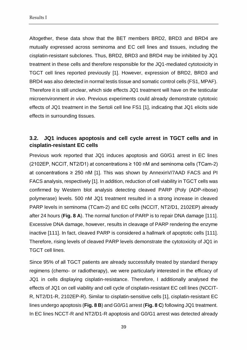
Results I
39
Altogether, these data show that the BET members BRD2, BRD3 and BRD4 are
mutually expressed across seminoma and EC cell lines and tissues, including the
cisplatin-resistant subclones. Thus, BRD2, BRD3 and BRD4 may be inhibited by JQ1
treatment in these cells and therefore responsible for the JQ1-mediated cytotoxicity in
TGCT cell lines reported previously [1]. However, expression of BRD2, BRD3 and
BRD4 was also detected in normal testis tissue and somatic control cells (FS1, MPAF).
Therefore it is still unclear, which side effects JQ1 treatment will have on the testicular
microenvironment in vivo. Previous experiments could already demonstrate cytotoxic
effects of JQ1 treatment in the Sertoli cell line FS1 [1], indicating that JQ1 elicits side
effects in surrounding tissues.
3.2. JQ1 induces apoptosis and cell cycle arrest in TGCT cells and in
cisplatin-resistant EC cells
Previous work reported that JQ1 induces apoptosis and G0/G1 arrest in EC lines
(2102EP, NCCIT, NT2/D1) at concentrations ≥ 100 nM and seminoma cells (TCam-2)
at concentrations ≥ 250 nM [1]. This was shown by AnnexinV/7AAD FACS and PI
FACS analysis, respectively [1]. In addition, reduction of cell viability in TGCT cells was
confirmed by Western blot analysis detecting cleaved PARP (Poly (ADP-ribose)
polymerase) levels. 500 nM JQ1 treatment resulted in a strong increase in cleaved
PARP levels in seminoma (TCam-2) and EC cells (NCCIT, NT2/D1, 2102EP) already
after 24 hours (Fig. 8 A). The normal function of PARP is to repair DNA damage [111].
Excessive DNA damage, however, results in cleavage of PARP rendering the enzyme
inactive [111]. In fact, cleaved PARP is considered a hallmark of apoptotic cells [111].
Therefore, rising levels of cleaved PARP levels demonstrate the cytotoxicity of JQ1 in
TGCT cell lines.
Since 95% of all TGCT patients are already successfully treated by standard therapy
regimens (chemo- or radiotherapy), we were particularly interested in the efficacy of
JQ1 in cells displaying cisplatin-resistance. Therefore, I additionally analysed the
effects of JQ1 on cell viability and cell cycle of cisplatin-resistant EC cell lines (NCCIT-
R, NT2/D1-R, 2102EP-R). Similar to cisplatin-sensitive cells [1], cisplatin-resistant EC
lines undergo apoptosis (Fig. 8 B) and G0/G1 arrest (Fig. 8 C) following JQ1 treatment.
In EC lines NCCT-R and NT2/D1-R apoptosis and G0/G1 arrest was detected already

Results I
40
after 16 hours of JQ1 treatment. A prolonged exposure time of 20 hours led to similar
effects in 2102EP-R. Since the same trend was observed in parental NCCIT, NT2/D1
and 2102EP cells [1], it can be concluded that JQ1 shows therapeutic effects in both
cisplatin-sensitive and cisplatin-resistant cells.
Figure 8: JQ1 induces apoptosis and G0/G1 arrest in cisplatin-sensitive and cisplatin-resistant
TGCT cells
(A) Western blot of cleaved PARP levels in 500 nM JQ1-treated TGCT cell lines compared to solvent
controls (-). ACTIN was used as loading control.
(B) Cell viability (in % to control) of JQ1-treated cisplatin-resistant EC lines as determined by AnnexinV-
7AAD FACS analysis. Time after JQ1 treatment is given in hours (h) below.
(C) Cell Cycle (in % to control) of JQ1-treated cisplatin-resistant EC lines as determined by PI FACS
analysis. Time after JQ1 treatment is given in hours (h) below.

Results I
41
3.3. The molecular effects of JQ1 treatment in TGCT cells
Microarray data showed that JQ1 treatment of EC (NCCIT) and seminoma (TCam-2)
cells leads to upregulation of stress markers (CDKN1C, DDIT4, TSC22D1, TXNIP,
ATF3, RHOB, BTG1, JUN), strong induction of the differentiation marker HAND1, and
downregulation of pluripotency-associated genes (LIN28, DPPA4, UTF1, ZSCAN10)
as well as germ-cell related markers (SPRY4, THY1) [1]. We hypothesized that
deregulation of these genes is responsible for the JQ1-mediated cytotoxicity in TGCT
cell lines. To confirm the microarray data and to determine whether these deregulations
are common for all TGCT cells, I measured mRNA expression of CDKN1C, ATF3,
RHOB, HAND1, POU5F1, LIN28, SPRY4 and THY1 in seminoma (TCam-2) and EC
cells (NCCIT, NT2/D1, 2102EP). A common effect of JQ1 treatment across all TGCT
cell lines was the downregulation of germ cell – associated genes SPRY4 and THY1
(Fig. 9). Further, I verified induction of the stress markers CDKN1C, ATF3 and RHOB
in the pluripotent EC lines NCCIT and NT2/D1 cells after 100 nM JQ1 treatment (Fig.
9). Additionally, strong induction of the differentiation marker HAND1 and
downregulation of pluripotency genes POU5F1 and LIN28 suggested loss of
pluripotency and induction of differentiation in pluripotent EC cells (Fig. 9). In contrast,
the nullipotent EC line 2102EP showed mild induction of CDKN1C, ATF3 and RHOB,
but only at higher JQ1 concentrations (750 nM). Downregulation of pluripotency
markers POU5F1 and LIN28 and upregulation of HAND1 was similarly seen only at
higher JQ1 concentrations (750 nM) in this cell line (Fig. 9). It is tempting to speculate
that 2102EP cells are less sensitive to JQ1-induced deregulation, due to their
nullipotent character. In contrast to the pluripotent EC lines NCCIT and NT2/D1,
2102EP cells lack the ability to differentiate into mixed non-seminoma cells. In
comparison, the seminoma-like cell line TCam-2 shows induction of stress markers
CDKN1C, ATF3 and RHOB, mild upregulation of the differentiation marker HAND1 and
downregulation of pluripotency, indicated by loss of LIN28 expression (Fig. 9). mRNA
levels of POU5F1, however, remained unchanged in TCam-2 cells (Fig. 9).

Results I
42
Figure 9: Upregulation of stress markers and downregulation of pluripotency genes in TGCT
cells following JQ1 treatment. Modified from [2]
Verification of cDNA microarray data by qRT-PCR. Expression values were calculated as fold change
compared to solvent control and normalized against GAPDH as housekeeping gene.
To confirm the downregulation of pluripotency after JQ1 treatment in TGCT cells on
protein level, I screened for changes in POU5F1 (OCT4) and LIN28 expression, as
well as levels of the stem cell marker and BMP inhibitor GDF3. I detected
downregulation of GDF3 and POU5F1 in EC lines (NCCIT, NT2/D1) and mild
downregulation of LIN28 in the seminoma line TCam-2, which is consistent with the
microarray and qRT-PCR data [1] (Fig. 9-10). Although the effects of JQ1 on

Results I
43
pluripotency marker expression are not identical between seminoma (TCam-2) and EC
(NCCIT, NT2/D1) cell lines, I was able to confirm an overall downregulation of
pluripotency in both TGCT subtypes on RNA and protein level.
Figure 10: Downregulation of pluripotency in TGCT cell lines. Modified from [2]
GDF3, POU5F1 and LIN28 protein levels in 100 nM JQ1-treated TGCT cell lines compared to solvent
controls (-). ACTIN was used as loading control and for data normalization.
Notably, the overall state of pluripotency differs between seminoma and EC cells.
While seminoma cells share the latent pluripotent character of early PGCs, EC cells
show features of totipotency similar to early ESCs. Hence, EC cells are capable of
differentiating into cells of embryonic and extra-embryonic lineages. In contrast,
seminoma cells express some of the well-known pluripotency and stem cell markers
(e.g. POU5F1, LIN28), but are highly restricted in their differentiation potential. For a
long time it was believed that seminoma cells can not differentiate at all, however in a
previous publication by our group it was shown that seminoma cells can differentiate
into mixed non-seminomatous lineages in presence of TGF-β, EGF and FGF4 [73].
Due to the differences in the differentiation potential of seminomas and ECs, however,
I proceeded with analysing the effects of JQ1 specifically on TGCT pluripotency in
more detail. Since the pluripotent EC lines NCCIT and NT2/D1 showed much higher
sensitivity to JQ1 than the TCam-2 cell line or the nullipotent 2102EP cell line, I

Results I
44
speculated that JQ1-mediated cytotoxicity may in part be mediated via downregulation
of pluripotency of these cells.
Gene set enrichment analysis (GSEA) of the previously obtained microarray data [1]
revealed enrichment of stem cell signatures among downregulated genes in TCam-2
(LIM_MAMMARY_STEM_CELL_UP, BENPORATH _ES_1) (Table 4) and an even
more prominent enrichment of stem cell signatures among downregulated genes in
NCCIT (BENPORATH_ES_1, BENPORATH_NANOG_TARGETS, KORKOLA_
CORRELATED_WITH_POU5F1, BENPORATH_SOX2_TARGETS, BHATTACHA
RYA_EMBRYONIC_STEM_CELL, CONRAD _STEM_CELL, BENPORATH_ES_2)
(Table 5). Since the EC cell line NCCIT is capable of differentiating into cells of
embryonic and extra embryonic lineages I additionally analysed the list of genes
upregulated following JQ1 treatment by gene ontology analysis. In line with the
downregulation of pluripotency in this cell line, gene ontology analysis demonstrated
enrichment of biological processes associated with embryonic differentiation among
genes induced by 100 nM JQ1 (Table 6). Categorization of these genes into embryonic
lineages reveals that the majority of these processes are associated with mesoderm
differentiation (Fig. 11). In summary, this shows that JQ1 treatment of TGCT cells
results in downregulation of pluripotency or stem cell associated genes. According to
the GSEA, this downregulation is more significant in the pluripotent EC line NCCIT (p
≥ 2.41E-59) compared to the latent pluripotent seminoma cell line TCam-2 (p ≥ 7.5E-
13) and downregulation of pluripotency in NCCIT goes in hand with upregulation of
differentiation markers (mainly mesoderm).

Results I
45
Table 4: GSEA of genes downregulated in TCam-2 cells 72 hours after JQ1
treatment
Gene Set Name
P-value
SMID_BREAST_CANCER_LUMINAL_B_DN 1.77E-16
HOSHIDA_LIVER_CANCER_SUBCLASS_S1 1.82E-13
LEI_MYB_TARGETS 4.14E-13
CHICAS_RB1_TARGETS_CONFLUENT 5.56E-13
RODWELL_AGING_KIDNEY_UP 7.05E-13
LIM_MAMMARY_STEM_CELL_UP 7.5E-13
SERVITJA_ISLET_HNF1A_TARGETS_UP 1.47E-12
BENPORATH_ES_1 4.39E-12
SMID_BREAST_CANCER_BASAL_UP 4.59E-12
VERHAAK_AML_WITH_NPM1_MUTATED_DN 6.36E-12
Table 5: GSEA of genes downregulated in NCCIT cells 72 hours after JQ1
treatment
Gene Set Name
P-value
BENPORATH_ES_1 2.41E-59
BENPORATH_NANOG_TARGETS 1.34E-23
KORKOLA_CORRELATED_WITH_POU5F1 1.71E-19
BENPORATH_SOX2_TARGETS 5.9E-19
KRIEG_HYPOXIA_NOT_VIA_KDM3A 2.28E-18
ELVIDGE_HYPOXIA_UP 1.27E-15
BHATTACHARYA_EMBRYONIC_STEM_CELL 1.71E-15
CONRAD_STEM_CELL 7.56E-15
BENPORATH_ES_2 1.00E-14
ELVIDGE_HYPOXIA_BY_DMOG_UP 1.1E-14
Table 6: Gene ontology analysis of genes upregulated in NCCIT cells 72 hours
after JQ1 treatment
Process q-value
heart morphogenesis 1.83e-13
regionalization 6.62e-12
regulation of cellular response to growth factor stimulus 5.90e-11
cardiac chamber development 9.06e-11
cardiac ventricle development 1.25e-10
appendage development&limb development 1.39e-10
cardiac chamber morphogenesis 1.39e-10

Results I
46
mesenchymal cell development 1.39e-10
regulation of transmembrane receptor protein serine threonine kinase signaling pathway
1.39e-10
stem cell development 1.39e-10
mesenchymal cell differentiation 2.29e-10
mesenchyme development 3.21e-10
appendage morphogenesis&limb morphogenesis 4.22e-10
cardiac septum development 1.24e-9
stem cell differentiation 1.72e-9
regulation of ossification 2.07e-9
negative regulation of DNA binding 2.85e-9
connective tissue development 2.86e-9
osteoblast differentiation 6.12e-9
positive regulation of ossification 7.76e-9
epithelial tube morphogenesis 1.06e-8
odontogenesis of dentin containing tooth 1.06e-8
regulation of DNA binding 1.06e-8
cell cell junction organization 1.27e-8
cell junction assembly 1.62e-8
cardiac muscle tissue development 1.76e-8
embryonic appendage morphogenesis&embryonic limb morphogenesis 1.76e-8
BMP signaling pathway 2.33e-8
cartilage development 3.39e-8
in utero embryonic development 3.61e-8
response to BMP(4)&cellular response to BMP stimulus 3.86e-8
regulation of osteoblast differentiation 5.96e-8
cardiac septum morphogenesis 6.48e-8
regulation of BMP signaling pathway 1.36e-7
gastrulation 2.66e-7
outflow tract morphogenesis 3.68e-7
mesenchyme morphogenesis 5.71e-7
response to mechanical stimulus 6.81e-7
stem cell proliferation 1.09e-6
anterior posterior pattern specification 1.09e-6
ventricular septum development 1.16e-6
epithelial to mesenchymal transition 1.24e-6
endoderm development 1.81e-6
formation of primary germ layer 1.86e-6
ventricular cardiac muscle tissue development 1.89e-6
skeletal system morphogenesis 1.99e-6
actin filament bundle assembly 2.50e-6
embryonic organ morphogenesis 2.73e-6
actin filament bundle organization 2.95e-6

Results I
47
regulation of canonical Wnt signaling pathway 3.03e-6
negative regulation of growth 3.42e-6
palate development 3.42e-6
regulation of Wnt signaling pathway 3.42e-6
embryonic hindlimb morphogenesis 3.64e-6
kidney epithelium development 4.20e-6
ureteric bud development 5.37e-6
regulation of protein localization to nucleus 5.57e-6
mesonephric tubule development&mesonephric epithelium development 5.63e-6
positive regulation of osteoblast differentiation 5.75e-6
segmentation 5.90e-6
chondrocyte differentiation 6.59e-6
mesonephros development 6.59e-6
positive regulation of BMP signaling pathway 6.59e-6
sensory organ morphogenesis 6.83e-6
cardiac ventricle morphogenesis 7.98e-6
regulation of cartilage development 7.98e-6
positive regulation of transmembrane receptor protein serine threonine kinase signaling pathway
8.43e-6
regulation of cellular response to transforming growth factor beta stimulus 8.82e-6
regulation of transforming growth factor beta receptor signaling pathway 8.82e-6
embryonic forelimb morphogenesis 9.51e-6
actomyosin structure organization 9.67e-6
neural crest cell development 1.10e-5
morphogenesis of embryonic epithelium 1.18e-5
hindlimb morphogenesis 1.22e-5
canonical Wnt signaling pathway 1.26e-5
negative regulation of transcription regulatory region DNA binding 1.29e-5
negative regulation of cell development 1.42e-5
adherens junction organization 1.49e-5
cardiocyte differentiation 1.80e-5
regulation of stem cell differentiation 2.02e-5
neural crest cell differentiation 2.08e-5
forelimb morphogenesis 2.53e-5
mesoderm development 2.82e-5
regulation of chondrocyte differentiation 2.82e-5
skin development 4.38e-5
extracellular matrix disassembly 4.45e-5
morphogenesis of a branching epithelium 5.07e-5
transforming growth factor beta receptor signaling pathway 5.24e-5
regulation of cell shape 7.12e-5
morphogenesis of a branching structure 8.59e-5

Results I
48
Figure 11: Gene ontology analysis of genes upregulated in NCCIT cells 72 hours after JQ1
Treatment. Modified from [2]
Categorization of biological processes enriched among genes upregulated in NCCIT cells 72 hours after
100 nM JQ1 treatment determined by gene ontology analysis.
Interestingly, in different cancer models (e.g. esophageal squamous cell carcinoma,
multiple myeloma) JQ1 treatment additionally resulted in strong downregulation of the
proto-oncogene MYC [93, 97, 112]. In these studies MYC was described as one of the
primary targets of JQ1 treatment and JQ1-mediated cytotoxicity was dependent on
MYC downregulation [93]. Since transcription of MYC was described to be regulated
by BRD4, JQ1-mediated BRD4 inhibition in these cells resulted in downregulation of
MYC mRNA, further leading to the loss of MYC protein expression. In TGCT cells,
however, no downregulation of MYC mRNA was observed in response to JQ1
treatment [1]. Surprisingly, MYC mRNA levels were even upregulated following JQ1
treatment in seminoma (TCam-2) and EC cells (NCCIT, NT2/D1, 2102EP) [1]. In order
to see whether MYC upregulation was similarly observed on protein level in TGCT
cells, I analysed whole protein lysates of seminoma (TCam-2) and EC cells (NCCIT,
NT2/D1, 2102EP) following JQ1 treatment. I found that MYC protein levels were
unaltered in EC lines (NCCIT, NT2/D1, 2102EP) and TCam-2 cells at 100 nM JQ1
(Fig. 12 A). At 500 nM JQ1 TCam-2 cells display mild downregulation of MYC protein,
while MYC protein levels in EC cells remain unaffected (Fig. 12 B-C). Thus,
upregulation of MYC mRNA does not correlate with upregulation of MYC protein levels
in TGCT cells, possibly pointing at post-translational modifications regulating MYC

Results I
49
levels. In summary, those TGCT cells being the most sensitive to JQ1-induced
cytotoxicity (NCCIT, NT2/D1) demonstrated no change in MYC expression. Thus, JQ1-
associated cytotoxicity in TGCT cells seems to be independent of MYC expression.
Also, downregulation of MYC protein in 500 nM treated TCam-2 cells might be a
secondary effect of JQ1 treatment, since cytotoxicity of JQ1 in TCam-2 cells is already
observed at doses ≥ 250 nM.
Figure 12: MYC protein levels in JQ1 treated TGCT cell lines. Modified from [2]
(A-C) Western blot of MYC protein levels in JQ1-treated TGCT cell lines compared to solvent controls
(-). JQ1 concentrations are indicated below in nM. ACTIN was used as loading control. Time of JQ1
treatment is given in hours (h).

Results I
50
3.4. The molecular effects of JQ1 on the testis microenvironment
The effects of JQ1 were additionally tested on somatic control cells (adult fibroblasts
and a Sertoli cell line), demonstrating cytotoxicity (G2/M arrest, apoptosis) also in
Sertoli cells, while adult fibroblasts remained nearly unaffected (G0/G1 arrest, but no
apoptosis) [1, 2]. The JQ1-mediated cytotoxicity of Sertoli cells, however, may indicate
the possibility of side effects in the testicular microenvironment when used in patients.
I verified JQ1-mediated cytotoxicity in FS1 Sertoli cells additionally by measuring levels
of cleaved PARP (Fig. 13). In contrast to adult fibroblasts (MPAF), human Sertoli cells
(FS1) display strong induction of cleaved PARP levels, indicative for JQ1-mediated
cytotoxicity (Fig. 13).
Figure 13: JQ1 induces apoptosis in FS1 Sertoli cells but not fibroblasts. Modified from [2]
Western blot of cleaved PARP levels in 100, 250 and 500 nM JQ1-treated FS1 Sertoli cells and adult
fibroblasts (MPAF) compared to solvent controls (-). ACTIN was used as loading control. Time of JQ1
treatment is given in hours (h).
To analyse the molecular effects of JQ1 on the testicular microenvironment in more
detail, I performed microarray analysis of JQ1 treated Sertoli cells (FS1) after 24 and
72 hours (Fig. 14 A-B). After 24 hours, 31 genes (1 upregulated, 30 downregulated)
were differentially expressed between JQ1 treated Sertoli cells and solvent controls
(Fig. 14 A). After 72 hours 33 genes (2 upregulated, 31 downregulated) were
differentially expressed (Fig. 14 B). In comparison to TCam-2 (24 hours: 242 genes,
72 hours: 212 genes) and NCCIT (24 hours: 225 genes, 72 hours: 512 genes), the
number of deregulated genes after JQ1 treatment in FS1 Sertoli cells is much smaller
(Fig. 14 and 15). Thus, TGCT cells are more susceptible to JQ1-induced gene

Results I
51
deregulations than the somatic control cell line FS1. However, similar to TGCT cells,
FS1 cells respond with cell cycle arrest and apoptosis to JQ1 treatment. It remains
unclear, which of the deregulated genes is responsible for the cytotoxicity of JQ1
treatment in FS1 Sertoli cells.
Figure 14: Microarray analysis of 100 nM JQ1 treated Sertoli cells. Modified from [2]
Genes deregulated in FS1 Sertoli cells following 100 nM JQ1 treatment after 24 hours (A) and 72 hours
(B) determined by cDNA microarray analysis. Expression is given as fold change compared to solvent
control.

Results I
52
Figure 15: Genes commonly deregulated in TGCT and Sertoli cells
Venn diagram showing genes commonly deregulated in FS1 Sertoli cells, TCam-2 cells and NCCIT cells
following 100 nM JQ1 treatment after 72 hours, as determined by cDNA microarray analysis.
Notably, none of the genes deregulated in TGCT cells after JQ1 treatment that have
been annotated a role in cytotoxicity (GADD45B, TSC22D1, TXNIP, RHOB, ATF3,
JUN, ID2) [2] were deregulated in FS1 Sertoli cells after JQ1 treatment. Further, none
of the commonly deregulated genes in FS1 Sertoli cells and NCCIT (HIST1H2BK,
KLF6, NBPF20, NBPF10) or FS1 and TCam-2 (LYPD1, KRT19) are directly regulating
apoptosis or cell cycle arrest (Fig. 15). Only KLF6 was described to induce apoptosis
via upregulation of ATF3 in prostate cancer cells [113]. However, following JQ1
treatment KLF6 was downregulated in FS1 cells, while being upregulated in NCCIT
cells [1, 2]. Thus, a common mechanism of JQ1-mediated cytotoxicity in TGCT and
Sertoli cells could not be identified. Although it remains unclear, which of the
deregulated genes are responsible for the JQ1-mediated toxicity in FS1 cells, it is
evident that BET inhibition can lead to apoptosis and cell cycle arrest in somatic control
cells (here: Sertoli cells). I recommend a more detailed analysis of possible side effects
and adverse events of JQ1 administration, before commissioning the drug for clinical
use.

Results I
53
3.5. Combination therapy with JQ1 and romidepsin in TGCT cell lines
I noted during the analyses that the genes deregulated by JQ1 treatment in TGCT cells
were similar to those deregulated by treatment with the HDAC inhibitor romidepsin
(induction of GADD45A, GADD45B, RHOB, ID2) [1-3]. Since a combination of 100,
250 or 500 nM JQ1 with 5 nM romidepsin also markedly increased apoptosis levels in
TCam-2 cells compared to single agent treatment, drug synergy effects for the
HDACi+BETi combination were postulated [1, 2].
In order to test, whether a combination of JQ1 and romidepsin will synergistically
decrease TGCT cell viability compared to single agent treatment I performed XTT
assay on JQ1 and romidepsin treated TCam-2 and NCCIT cells (Fig. 16). In contrast
to previous experiments where JQ1 was administered every second day due to its
relatively short half-life (~1 hour in plasma), in this line of experiments both substances
were administered only once before the start of the measurement (timepoint: 0 hours
(h)). Under these conditions administration of neither 100 nM nor 250 nM JQ1 showed
cytotoxic effects in TCam-2 cells (Fig. 16 A). In contrast, 2 nM romidepsin treatment
led to a decrease in cell viability of ~ 40% in TCam-2 cells. A combination of 2 nM
romidepsin with either 100 or 250 nM JQ1 further reduced cell viability to ~ 30%. This
shows that even though a one-time administration of JQ1 alone has no effect on TCam-
2 cell viability, the substance can significantly increase cytotoxicity of romidepsin
treatment (Fig. 16 A). Cytotoxicity levels of high-dose romidepsin treatment (5 nM),
however, are not increased by addition of JQ1.

Results I
54
Figure 16: Cell viability of TGCT cells treated with JQ1 and romidepsin. Modified from [2]
Cell viability of TCam-2 (A) and NCCIT (B) cells treated with JQ1, romidepsin (Rdps) or JQ1 and
romidepsin (JQ1 + Rdps) in combination determined by XTT assay. Cell viability is given in % to solvent
control.
Similarly, I observed that a one-time administration of 2 nM romidepsin showed no
cytotoxic effects in NCCIT cells (Fig. 16 B). In contrast, 100 nM or 250 nM JQ1 led to
a reduction of cell viability to 40-50% and 5 nM romidepsin treatment led to a reduction
of cell viability to 50% (Fig. 16 B). While a combination treatment of 100 or 250 nM
JQ1 + 2 nM romidepsin had no additional effect on cytotoxicity levels, the combination
of 5 nM romidepsin and 100 or 250 nM JQ1 further decreased cell viability to ~ 20%
(Fig. 16 B). In summary, these data demonstrate that under certain conditions JQ1
and romidepsin may show additive or synergistic effects on cytotoxicity levels of TGCT
cells. However, the interplay of both drugs and their exact efficacy vs toxicity (side
effects) relationship needs further evaluation using in vivo model systems.

Results I
55
3.6. JQ1 treatment of TGCT xenografts
In order to investigate the efficacy of JQ1 treatment (also in combination with
romidepsin) in vivo, I analysed tumour growth of NCCIT and NT2/D1 xenografts in CD-
1 nude mice under drug administration. For this, cell lines were injected into the flank
of nude mice and after two weeks of initial tumour growth mice were intraperitoneally
(i.p.) injected with drug or solvent control. Tumour growth was continuously monitored.
Mice treated with JQ1 (50 mg / kg) presented reduced tumour burden compared to
solvent controls (Fig. 17). This reduction was significant already after 7 days of
treatment in NCCIT (Fig. 17 A-C) and only 4 days of treatment in NT2/D1 xenografts
(Fig. 17 D-F).
Figure 17: Tumour growth of TGCT xenografts treated with JQ1. Modified from [2]
Tumour size of NCCIT (A-C) and NT2/D1 (D-F) xenografts treated with JQ1 compared to xenografts
treated with solvent control (i.p. injection, 5 days / week for two weeks).
A reduction in tumour growth was also indicated by a significant reduction of Ki67+
tumour cells in NCCIT (p = 0.026) and NT2/D1 (p = 0.0002) tumours, as determined
by immunohistochemical staining (Fig. 18 A). Ki67 is a marker of proliferative active
cells, thus a relative reduction in the Ki67+ cell population shows the growth-inhibitory

Results I
56
effects of JQ1 in TGCT tumours. I noted further the reduction of blood vessel
invagination in JQ1 treated tumours (Fig. 17). In order to quantify this difference,
NCCIT tumours were additionally stained for the endothelial marker CD31. In line with
our observations I measured a significant reduction of CD31+ cells in JQ1 treated
tumour samples (p = 0.018) (Fig. 18 B). Thus, JQ1 does not only significantly inhibit
TGCT tumour growth, but also blood vessel formation within these tumours.
Figure 18: Ki67 and CD31 staining of TGCT xenografts treated with JQ1. Modified from [2]
Ki67 staining of NCCIT and NT2/D1 xenografts treated with JQ1 or solvent control (A) and CD31 staining
of NCCIT xenografts treated with JQ1 or solvent control (B) (i.p. injection, 5 days / week for two weeks).
Scale = 100 µm. Errors bars indicate standard deviation from the mean. Significance was calculated by
student’s t-test.
3.7. Combination therapy with JQ1 and romidepsin in TGCT xenografts
I then proceeded to analyse the efficacy of a combination therapy with JQ1 and
romidepsin in NCCIT and NT2/D1 xenografts. For this, cell lines were again injected
into the flank of nude mice and after two weeks of tumour growth mice were injected
i.p. with drug or solvent control. Similar to JQ1-treated mice, mice treated with JQ1 (50
mg / kg) + romidepsin (0.5 mg / kg) presented reduced tumour burden compared to

Results I
57
solvent controls (Fig. 19 A-D). Interestingly, JQ1 + romidepsin was only administered
3 days / week, while JQ1 alone was administered 5 days / week. Thus, fewer
applications of combination therapy were sufficient to achieve a similar therapeutic
outcome compared to JQ1 alone. The reduction of tumour growth under combination
therapy was significant already after 2 days of treatment in NCCIT (Fig. 19 A-B) and
NT2/D1 xenografts (Fig. 19 C-D). In line, the amount of Ki67+ cells was significantly
(p = 0.003) reduced in NCCIT xenografts (Fig. 19 E) and, although not significant,
markedly reduced in NT2/D1 xenografts (Fig. 19 F). These results indicate that TGCT
patients may benefit from a combination therapy with JQ1 and romidepsin, since the
combined administration of both substances allows for a less frequent application
scheme and lower doses.
Figure 19: Tumour growth of TGCT xenografts treated with JQ1 + romidepsin. Modified from [2]
Tumour size of NCCIT (A, B) and NT2/D1 (C, D) xenografts treated with JQ1 + romidepsin (JQ1 + Rdps)
compared to xenografts treated with solvent control (i.p. injection, 3 days / week for 10 days). Ki67
staining of NCCIT (E) and NT2/D1 (F) xenografts treated with JQ1 + romidepsin (JQ1 + Rdps) compared
to xenografts treated with solvent control. Errors bars indicate standard deviation from the mean.
Significance was calculated by student’s t-test.

Discussion I
58
4. Discussion I
Since the development of the first BET inhibitors JQ1 (2011) and I-BET (2010) [114,
115], a multitude of studies have shown the therapeutic effects of these epigenetic
compounds in cancer therapy [79, 93, 116-120]. Due to their small-molecule
characteristics, both substances show high bioavailability even in difficult-to-penetrate
tissues such as brain and testis [79, 114, 121]. Initially, JQ1 was discussed as male
contraceptive, due to its inhibitory effect on the testis-specific BET member BRDT [122,
123]. JQ1-mediated inhibition of BRDT led to a reversible contraceptive effect in mice,
by completely inhibiting spermatogenesis during course of treatment [122, 123].
However, further studies focussed on the role of JQ1 as cancer therapeutic, probably
owing to the fact that JQ1 does not only inhibit the testis-specific BET member BRDT,
but also BRD2, BRD3 and BRD4, which are ubiquitously expressed among different
tissues of the human body.
Previously, no study had described the effects of JQ1 on testicular cancer, or more
precisely, on type II TGCTs. Therefore, I analysed the effects of JQ1 in both TGCT cell
lines and TGCT xenografts and compared it to the effects of JQ1 on somatic control
cells (fibroblasts and Sertoli cells). JQ1 treatment led to G0/G1 growth arrest and
apoptosis in both cisplatin-sensitive, as well as cisplatin-resistant TGCT cell lines (Fig.
20) [1]. Cytotoxicity of JQ1 treatment in TGCT cells increased in a time- and dose-
dependent manner. In both seminoma and EC cells induction of the DNA damage and
stress response genes GADD45B, TSC22D1, TXNIP, RHOB, ATF3, JUN and ID2 was
observed (Fig. 20) [1]. Although not formally proven, induction of these genes may
relate to the JQ1-mediated cell cycle arrest and apoptosis seen in TGCT cells.
Interestingly, previous studies have shown the transcriptional activation of the
oncogene MYC via BRD4-mediated pTEFb activation in different cancer types, and
further MYC downregulation following BRD4 inhibition by JQ1 [112, 124-126]. In some
cancer types JQ1-mediated downregulation of MYC was even responsible for the
cytotoxicity of BET inhibition [93, 112]. However, other studies reported JQ1-mediated
cytotoxicity in cancer cells independent of MYC levels [94, 127]. In this study
downregulation of the oncogene MYC was not observed in TGCT cells on mRNA level.
Only on protein level MYC was downregulated in seminoma, but not EC cells. Since
seminoma cells, however, were in general less sensitive to JQ1 treatment compared

Discussion I
59
to EC cells, downregulation of MYC can be excluded as primary cause of JQ1-
mediated cytotoxicity in TGCT cells.
One of the main observations of this study was the strong upregulation of the
differentiation marker HAND1 in both seminoma and EC cells following JQ1 treatment
and the downregulation of pluripotency-associated genes (such as UTF1, THY1,
LIN28, ZSCAN10, DPPA4) (Fig. 20) [1, 2]. Interestingly, this effect was even more
dramatic in EC cells only, which additionally showed robust downregulation of the key
pluripotency markers NANOG, POU5F1, GDF3 and JARID2 [1, 2]. Whereas
seminoma cells are restricted in their differentiation potential (a feature termed ‘latent
pluripotent’), EC cells are capable of differentiating into embryonic and extra-
embryonic cell lineages. Therefore, EC cells not only showed upregulation of the
differentiation marker HAND1 following JQ1 treatment, but also many other genes
involved in differentiation processes (mainly mesodermal differentiation). This
suggests a possible role of BET bromodomain reader proteins in pluripotency
regulation, at least for TGCT (in particular EC) cells [79]. In support of this hypothesis,
in mouse ESCs expression of the pluripotency factor NANOG (which is also highly
expressed TGCTs) is maintained and regulated by the JQ1 target protein BRD4 [90,
128]. Also, in both human and mouse ESCs, BRD4 occupies regulatory super-
enhancer regions of POU5F1 and PRDM14 stem cell genes, which are similarly
important for TGCT cell identity and pluripotency [129].
It is tempting to speculate that BRD4 has a similar role in maintaining pluripotency and
stem cell identity in EC cells, which share many of the characteristics of ESCs. Also,
sensitivity of TGCT cells to JQ1 treatment correlates with the downregulation of
pluripotency and induction of differentiation, since pluripotent EC cell lines (such as
NCCIT) show higher sensitivity to JQ1 treatment than the nullipotent EC line 2102EP
and the latent pluripotent seminoma cell line TCam-2. Together, this suggests a
possible link between TGCT sensitivity towards JQ1 treatment and their ability to
differentiate.

Discussion I
60
Figure 20: Effects of JQ1 and romidepsin treatment in TGCT cells. Modified from [2].
Schematic showing the molecular effects of romidepsin and JQ1 treatment in seminoma (SEM) and EC
cells.
Next to the observed effects of JQ1 treatment on cell stress and pluripotency levels of
TGCT cells, in vivo xenograft studies showed a reduced blood vessel formation in JQ1
treated tumours (Fig. 20). Suppression of angiogenesis in response to JQ1 treatment
was similarly reported for models of childhood sarcoma, breast cancer as well as non-
cancerous HUVEC (Human umbilical vein endothelial cells) [118, 130, 131]. In these
cell types, reduction of blood vessel formation resulted from suppression of VEGF-
driven angiogenesis [118, 130, 131]. Downregulation of VEGFB was also noted
following JQ1 treatment in both seminoma and EC cells [1], thus offering a plausible
explanation for the loss of tumour vascularization in TGCT xenografts.
Altogether, the presented data highlight JQ1 as possible therapeutic option for TGCT
(ECs in particular). A few studies have additionally reported a synergistic effect of BET
inhibition and HDAC inhibition in different cancer models (such as lymphoma,
pancreatic ductal adenocarcinoma, breast cancer) [79, 95, 98, 132]. While testing the
downstream molecular effects of JQ1 on TGCT cell lines I also noted a strong overlap
of differentially expressed genes in TGCT cells following JQ1 and romidepsin

Discussion I
61
treatment [1]. Further experiments demonstrated an additive effect of JQ1- and
romidepsin- mediated cytotoxicity in TGCT cells in vitro. The combination (JQ1 +
romidepsin) treatment of TGCT xenografts further confirmed these findings, since
combination therapy allowed for lower doses and a less-frequent application of both
substances compared to single-drug application [2]. It is still not known, however, why
HDAC inhibitors and BET inhibitors show synergy effects, since both drugs have a very
distinct mode of action: While HDAC inhibition results in histone hyperacetylation and
euchromatin formation, BET inhibition prevents the ‘reading’ of the histone code and
may thereby modulate gene transcription [2]. Borbely et al. and Mazur et al. have
identified the stress sensors USP17 and CDKN1C as possible common mediators of
BETi and HDACi-induced cytotoxicity, respectively [2, 79, 98, 132]. Thus, the cellular
stress response induced by both substances may explain the synergy of combination
therapy. In theory, different modes of action may apply for different cancer types.
Therefore, future studies would be necessary to evaluate the molecular background of
a combination therapy employing BET and HDAC inhibitors in TGCT cells in more
detail.
Altogether, I propose JQ1 in combination with romidepsin, as a potential therapeutic
option for TGCTs. While JQ1 was one of the first lead compounds presented in the
field of BET inhibitors, several next-generation BET inhibitors (such as ABBV-075,
BAY1238097, BI 894999) have now been developed, which have already reached
clinical trials [79, 133-135]. Especially BI 894999 is a highly potent BET inhibitor,
demonstrating an IC50 of 5 and 41 nM for BRD4-BD1 and BRD4-BD2 bromodomains,
respectively [133]. In comparison, JQ1 had a considerably higher IC50 of 77 and 22
nM for BRD4-BD1 and BRD4-BD2 bromodomains, respectively [121]. It is structural
improvements like these that may positively influence drug specificity and sensitivity
levels, and thereby also minimize drug-associated adverse events. Clinical trials need
to explore the pharmacokinetic profile of these compounds in more detail. While I
believe that the use of these drugs for stage I-II TGCTs is highly unlikely, since
standard therapy regimens already result in complete remission in most cases,
epigenetic drugs may present an alternative strategy to treat late-stage or cisplatin-
resistant TGCTs [79].

Discussion I
62
Parts of this chapter have been published in:
The bromodomain inhibitor JQ1 triggers growth arrest and apoptosis in
testicular germ cell tumours in vitro and in vivo
Jostes S, Nettersheim D, Fellermeyer M, Schneider S, Hafezi F, Honecker F,
Schumacher V, Geyer M, Kristiansen G, Schorle H.
J Cell Mol Med. 2016 Dec 27
Parts of this chapter have been discussed in:
Epigenetic drugs and their molecular targets in testicular germ cell tumours
Jostes S, Nettersheim D, Schorle H.
Nature Reviews Urology. 2019 Feb 14

Results II
63
5. Results II
5.1. Introduction
The plasticity of seminoma and embryonal carcinoma cells has been described in
various studies [60, 73, 75, 136]. While it was a longstanding belief that seminomas
are not able to differentiate and adopt other cell fates, our group was able to show in
2011 that in presence of FGF4, heparin and TGFβ seminomas differentiate into mixed
non-seminoma cell fate in vitro [73]. Additionally, it was demonstrated that seminoma
cells transdifferentiate into an EC-like cell fate after xenotransplantation into the flank
of nude mice [75]. Inhibition of BMP signalling is an initial driver of this process and
activates NODAL signalling, which initiates the downregulation of SOX17, as well as
the upregulation of SOX2 and other stem cell markers [75]. Collectively, these
publications show that seminomas and EC fates are plastic, dependent on the
surrounding microenvironment and the associated signalling cues. Regarding the
described plasticity, it is surprising that seminomas and ECs are so well-discriminated
by their exclusive expression of SOX2 (EC) and SOX17 (seminoma). It is generally
accepted that the role of SOX2 in EC cells is similar to the role of SOX2 in ESCs, which
is being a key determinant of pluripotency and stem cell fate. In seminomas, however,
knowledge about the role of SOX17 is lacking. Thus, I was interested in whether
SOX17 may have similar function in seminomas as SOX2 in ECs and whether both
factors regulate a common set of downstream target genes.
5.2. SOX17 (seminoma) and SOX2 (embryonal carcinoma) partner with
OCT4, but not NANOG
In general, SOX factors have weak DNA binding specificity [137]. Higher specificity,
however, is achieved by partnering with other factors, such as POU transcription
factors [137]. In ESCs SOX17 partners with OCT4 to bind to the compressed motif
(CATTGTATGCAAAT-like sequence), thereby driving endodermal genes [70]. By co-
immunoprecipitation I confirmed interaction of SOX17 and OCT4 in seminoma cells
(TCam-2) (Fig. 21), which was previously demonstrated by us [60]. SOX2 partners
with the POU transcription factor OCT4 to bind to the canonical motif
(CATTGTCATGCAAAT-like sequence) in ESCs, thereby driving pluripotency genes

Results II
64
[70]. This pluripotency circuitry is further supported by SOX2-OCT4 interaction with the
stem cell factor NANOG [138]. A similar interaction of SOX2, OCT4 and NANOG was
postulated to maintain EC pluripotency. By co-immunoprecipitation I verified interaction
of SOX2 with OCT4 in the EC line 2102EP, while I could not determine direct
interaction with NANOG protein (Fig. 21).
Figure 21: SOX17 and SOX2 interact with OCT4, but not NANOG
Co-immunoprecipitation of OCT4 and NANOG with SOX17 in TCam-2 cells and with SOX2 in 2102EP
cells. Immunoprecipitation with no antibody (no AB) served as negative control. 2% Input (= 20 µg
protein lysate) served as positive control.
Alike SOX2, SOX17 did not interact with the pluripotency factor NANOG (Fig. 21).
Collectively, this shows that SOX2 and SOX17 partner with OCT4 and can, as a protein
complex, regulate downstream target genes in EC and seminoma cells. However,
neither SOX2 nor SOX17 do bind to / partner with NANOG, like it was described for
the SOX2-OCT4-NANOG regulatory network in ESCs [138]. Thus far, however, we
cannot exclude that NANOG regulates similar genes as SOX2-OCT4 or SOX17-OCT4
by binding to nearby DNA binding motifs without a direct SOX2-NANOG and SOX17-
NANOG interaction.
5.3. SOX17 and SOX2 bind to the regulatory regions of pluripotency
genes in TGCT cells
In order to determine the role of SOX17 in seminoma cells and compare it to the role
of SOX2 in EC cells, I proceeded by analysing genome-wide SOX17 / SOX2 DNA
occupancy in TCam-2 and 2102EP cells, respectively. First, I confirmed suitability of

Results II
65
the SOX17 antibody for chromatin immunoprecipiation (ChIP). For this, the SOX17
ChIP sample was loaded on a 12% SDS gel and SOX17 protein was detected by
Western blot analysis. I verified presence of SOX17 protein in the SOX17 ChIP sample,
as well as in the 2% input control and in TCam-2 whole protein lysate (Fig. 22 A) [101].
Expectedly, SOX17 was not detected in whole protein lysate of EC lines 2102EP,
NT2/D1 and NCCIT (Fig. 22 A). A weak signal for SOX17 protein, however, was
detected in the goat-IgG negative control (Fig. 22 A), indicating a low unspecific
background signal for the IgG ChIP sample.
Next, suitability of the SOX2 antibody for ChIP was confirmed (Fig. 22 B) [101]. SOX2
protein could be detected in the SOX2 ChIP sample, as well as in the 2% input control
and whole protein lysates of EC lines 2102EP, NT2/D1 and NCCIT (Fig. 22 B). As
expected, SOX2 protein was absent in the TCam-2 whole protein lysate and the rabbit-
IgG negative control. Notably, the antibody used for SOX2 detection in the Western
blot was different from the SOX2 antibody used for ChIP-seq. This minimizes the
possibility that the ChIP-seq antibody binds to an unspecific product, which is not
SOX2. This experiment was performed together with Martin Fellermeyer during his
Bachelor Thesis in 2016 [101].
It was previously hypothesized by our group that in seminoma cells SOX17 takes over
a similar role as SOX2 in EC cells, which is the maintenance of pluripotency. We
hypothesized that SOX17 binds to canonical (SOX2/OCT4) motifs within the regulatory
regions of pluripotency genes. This is in contrast to ESCs where SOX17 activates
endodermal genes via the compressed (SOX17/OCT4) motif. Therefore, I tested
whether SOX17 is enriched at previously described canonical (SOX2/OCT4) binding
motifs [3, 99, 100, 102, 139]. I detected enrichment of SOX17 at the canonical
(SOX2/OCT4) binding sites within SOX2, NANOG, DPPA4, LIN28A and PRDM14
regulatory regions (Fig. 23), strongly suggesting that in seminoma cells SOX17
regulates pluripotency genes via canonical (SOX2/OCT4) binding sites.

Results II
66
Figure 22: Validation of ChIP-grade antibodies. Modified from [101]
(A) Digested and crosslinked chromatin of TCam-2 cells was immunoprecipitated using a negative
control goat IgG antibody or the ChIP-grade SOX17 antibody. 2% Input represents 2% of the
TCam-2 chromatin fraction. Protein lysates of 2102EP, NT2/D1 and NCCIT served as negative
control. TCam-2 protein lysate served as positive control.
(B) Digested and crosslinked chromatin of 2102EP cells was immunoprecipitated using a negative
control rabbit IgG antibody (IgG) or the ChIP-grade SOX2 antibody. 2% Input represents 2% of the
2102EP chromatin fraction. Protein lysates of 2102EP, NT2/D1 and NCCIT served as positive
control. TCam-2 protein lysate served as negative control.
SOX17 enrichment was stronger for those genes that are highly expressed in TCam-
2 cells (NANOG, LIN28A, DPPA4, PRDM14) (Fig. 23 and 24), while there was no
enrichment of SOX17 for LEFTY2 detected (Fig. 23). LEFTY2 is exclusively expressed
in EC cells (Fig. 24). Additionally, I determined whether SOX17 was enriched at a
compressed (SOX17/OCT4) binding site approximately 900 bp upstream of the TSS
of PRDM1 [101] (Fig. 23, red label), which is highly expressed in seminoma and
TCam-2 cells (Fig. 24). Notably, this compressed-like PRDM1 binding site
(CATTGTATGCCATC) was manually assessed by screening for the compressed motif
within the PRDM1 promoter [101]. Indeed, SOX17 was strongly enriched at the
compressed motif within the PRDM1 promoter (Fig. 23) This is in line with a previous
publication of Irie et al., showing that SOX17 acts upstream of PRDM1 in maintaining
PGC fate [6]. In contrast, previously published SOX17 ChIP-seq data in differentiated

Results II
67
mesoderm, endoderm or mesendoderm-like lineages showed no enrichment of SOX17
within the PRDM1 promoter region [140]. Only a distal regulatory region -15300 bp
upstream of PRDM1 (-of TSS) was detected, which may not have functional relevance
[140]. This suggests that SOX17 binding to the PRDM1 promoter is a unique feature
of seminoma cells or PGCs.
Figure 23: SOX17 binds pluripotency genes in TCam-2 cells
qPCR of known canonical binding motifs in the regulatory regions of SOX2, NANOG, DPPA4, LIN28A,
LEFTY2 and PRDM14, and a putative compressed binding motif in the regulatory region of PRDM1 in
the SOX17 ChIP. Measurements were performed of three independent experiments. Goat IgG-ChIP
served as negative control.

Results II
68
Figure 24: Expression of pluripotency genes in TCam-2 and 2102EP cells
mRNA expression of pluripotency genes in TCam-2 (n=5) and 2102EP (n=5) cells, determined by
previously published microarray data [2, 3]. Different datapoints represent different mRNA probes for
the respective genes. Genes were categorized according to their expression levels in EC and SEM
(seminoma). The dashed line represents the expression threshold.
As control, I then determined enrichment of SOX2 for canonical (SOX2/OCT4) binding
motifs (Fig. 25). In line with the role of SOX2 as regulator of pluripotency, I detected
strong enrichment of SOX2 in the regulatory regions of SOX2, NANOG, DPPA4,
LIN28A, LEFTY2 and PRDM14 (Fig. 25). Notably, all of these genes are also highly
expressed in 2102EP cells (Fig. 24), suggesting that SOX2 transactivates these
genes. Next, I showed that there was no enrichment of SOX2 at the compressed
(SOX17/OCT4) binding site within the PRDM1 regulatory region.

Results II
69
Figure 25: SOX2 binds pluripotency genes in 2102EP cells
qPCR of known canonical binding motifs in the regulatory regions of SOX2, NANOG, DPPA4, LIN28A,
LEFTY2 and PRDM14, and a putative compressed binding motif in the regulatory region of PRDM1 in
the SOX2 ChIP. Measurements were performed of three independent experiments. Rabbit IgG-ChIP
served as negative control.
Together, the data demonstrate that in EC cells SOX2 uses some of the canonical
SOX2-OCT4 binding sites described in ESCs. Therefore, in EC cells SOX2 has a
similar role like SOX2 in ESCs, which is the maintenance of pluripotency by interacting
with OCT4 to regulate downstream target genes. Further, SOX2 seems to regulate its
own expression by binding to the canonical motif within the SOX2 regulatory region,
thus creating an auto-regulatory feedback loop (Fig. 25). Similarly, SOX17 in
seminoma cells, at least in part, binds to canonical (SOX2/OCT4) binding motifs.
Although weak binding of the SOX17-OCT4 complex to canonical (SOX2/OCT4) motifs
was already described in biochemical assays [141], there was no evidence whether
this binding may have functional consequences in some cells or simply be a bystander
effect. Some of the analysed genes showing SOX17 binding are also highly expressed
in these cells (NANOG, LIN28A, DPPA4), making a regulatory function of SOX17 for

Results II
70
these genes in seminoma cells possible. However, it seems that other genes like
PRDM1 are also regulated via SOX17 binding to its compressed motifs in seminoma
cells.
5.4. The majority of regions bound by SOX17 in seminoma cells contains
the compressed motif and is found near transcriptional start sites
In seminoma cells I could demonstrate that SOX17 binds to canonical (SOX2/OCT4)
and compressed (SOX17/OCT4) binding motifs of pluripotency and germ-cell related
genes. In 2102EP cells SOX2 binds to canonical SOX2/OCT4 motifs only. In order to
analyse genome-wide binding occupancy of SOX17 and SOX2 in TGCT cells I next
performed high-throughput ChIP-sequencing (ChIP-seq). For bioinformatic analysis
only those peaks were considered significant that were present in the control group
(Input or IgG) and in the SOX2/SOX17 IP and that were at least four times higher in
the SOX2/SOX17 IP compared to the control group.
Analysis of the SOX17 ChIP-seq data revealed a total of 931 and 904 peaks in the
SOX17 vs IgG and SOX17 vs Input dataset, respectively (8.1). The subsequent
analyses were performed using the SOX17 vs IgG dataset. Analysis of peak count
frequency showed that most SOX17 peaks are allocated directly upstream or
downstream of the transcription start site (TSS) of genes (Fig. 26 A). Further, HOMER
Motif analysis identified a total of 375 of 931 peaks (=40.28%) harbouring a SOX17
binding motif and 246 of 931 peaks (=26.24%) harbouring the compressed
(SOX17/OCT4) binding motif (Fig. 26 B-C). Additionally, 355 of 931 peaks (=38.13%)
contain the known SOX2 binding motif and 192 of 931 peaks (=20.62%) the known
OCT4 binding motif (Fig. 26 C). Interestingly, 101 of 931 peaks (=10.85%) contain the
described canonical (SOX2/OCT4) binding motif (Fig. 26 C). In line with the qPCR data
this indicates that SOX17 binds, and possibly regulates genes via compressed
(SOX17/OCT4) (26.24%), but also via canonical (SOX2/OCT4) (20.62%) binding sites.
Interestingly, also TFAP2C (also known as AP2γ) and PRDM1 (also known as
BLIMP1) motifs were found among top enriched binding motifs in SOX17-bound
regions (368 of 931 peaks and 188 of 931 peaks) (Fig. 26 C).

Results II
71
Figure 26: SOX17 occupies canonical and compressed binding sites in seminoma cells
(A) Peak Count Frequency of SOX17 ChIP peaks upstream and downstream of transcription start sites
(TSS).
(B) Number and percentage of SOX17 ChIP peaks (SOX17 vs IgG) that harbour the SOX17 binding
motif and the compressed SOX17/OCT4 binding motif.
(C) Homer motif analysis of known binding motifs enriched in SOX17 ChIP (SOX17 vs IgG).

Results II
72
Both transcription factors are highly expressed in PGCs and described as key
specifiers of germ-cell fate along with SOX17 [8]. Overall, this reflects the germ-cell
origin of TCam-2 cells. In PGCs PRDM1 and TFAP2C have been described to
suppress somatic differentiation [8]. The fact that PRDM1 and TFAP2C binding sites
are detected close to SOX17 peaks may indicate that some of those genes otherwise
activated by SOX17 during endodermal differentiation are suppressed by binding of
TFAP2C and PRDM1 in germ cell tumours.
In comparison, ChIP-seq data for SOX2 revealed a total of 3314 and 1259 peaks in
the SOX2 vs IgG and SOX2 vs Input dataset, respectively (8.1). The subsequent
analyses were performed using the SOX2 vs IgG control dataset. Analysis of peak
count frequency showed that most SOX2 peaks cluster upstream of the transcription
start site (TSS) of genes (Fig. 27 A). HOMER Motif analysis identified a total of 962 of
3314 peaks (=29,03%) harbouring a SOX2 binding motif and 419 of 3314 peaks
(=12,64%) harbouring the canonical SOX2/OCT4 binding motif (Fig. 27 B-C).
Additionally, 666 of 3314 peaks (=20.10%) contain the known OCT4 binding motif and
560 of 3314 (=16.90%) contain the known KLF4 binding motif (Fig. 27 C). This
indicates that the genes, which are bound by SOX2 can also be bound and regulated
by the pluripotency factors KLF4 and OCT4.
Altogether, these findings demonstrate that in seminoma cells SOX17 binds to
canonical (SOX2/OCT4) motifs, similar to SOX2 in ECs. This way, both SOX17 (in
seminoma) and SOX2 (in EC) can mediate the regulation of an overlapping set of
downstream target genes, such as pluripotency genes NANOG, DPPA4, LIN28A and
PRDM14. Surprisingly, however, many SOX17 peaks detected in TCam-2 cells also
contain the compressed (SOX17/OCT4) motif. SOX17 binding to this motif is known to
activate genes responsible for endodermal cell-fate decisions in ESCs. Further
analysis will show whether the compressed (SOX17/OCT4) motifs bound by SOX17 in
seminoma cells map to the regulatory regions of endodermal genes and whether this
binding leads to an activation or a suppression of genes.

Results II
73
Figure 27: SOX2 occupies canonical binding sites in EC cells
(A) Peak Count Frequency of SOX2 ChIP peaks upstream and downstream of transcription start sites
(TSS).
(B) Number and percentage of SOX2 ChIP peaks (SOX2 vs IgG) that harbour the SOX2 binding motif
and the canonical SOX2/OCT4 binding motif.
(C) Homer motif analysis of known binding motifs enriched in SOX2 ChIP (SOX2 vs IgG).

Results II
74
5.5. In seminoma cells SOX17 binds to the regulatory regions of neuro-
ectodermal genes, as well as pluripotency and germ-cell related genes
In order to see whether the SOX17 peaks obtained in TCam-2 correlate with
pluripotency gene signatures or endodermal genes I performed gene set enrichment
analysis (GSEA) using the Molecular Signature Database (MSigDB). Here, individual
SOX17 peaks were first annotated to the genes with the nearest TSS. GSEA was then
performed for the two ChIP-seq datasets SOX17 vs IgG and SOX17 vs Input. I
detected a strong overlap of SOX17 peaks with neuronal signatures and genes
associated with embryonic development or differentiation (Table 7-8, green labels). In
line with the previous ChIP-qPCR data, however, the SOX17 peaks additionally show
significant enrichment for stem cell-associated signatures, although less pronounced
(Table 7-8, blue labels). In comparison, the same analysis on SOX2 datasets (SOX2
vs IgG, SOX2 vs Input) showed robust enrichment for different stem cell-associated
and pluripotency-associated signatures (Table 9-10, blue labels), while enrichment for
neuronal signatures was minor (Table 9-10, green labels).
Table 7: MSigDB GSEA of SOX17 targets in TCam-2 (normalized to IgG
background)
TERM logP
AACTTT_UNKNOWN -27.36
GO_TISSUE_DEVELOPMENT -26.47
GO_REGULATION_OF_MULTICELLULAR_ORGANISMAL_DEVELOPMENT -22.92
GO_REGULATION_OF_NERVOUS_SYSTEM_DEVELOPMENT -22.17
CTTTGT_V$LEF1_Q2 -20.95
GO_REGULATION_OF_CELL_DEVELOPMENT -18.93
GO_EPITHELIUM_DEVELOPMENT -18.46
V$HNF3B_01 -17.95
GGGAGGRR_V$MAZ_Q6 -17.71
GO_REGULATION_OF_NEURON_DIFFERENTIATION -17.59
BENPORATH_SOX2_TARGETS -17.53
AAAYWAACM_V$HFH4_01 -17.52
BENPORATH_OCT4_TARGETS -17.25
GO_REGULATION_OF_NEURON_PROJECTION_DEVELOPMENT -17.06
GO_TUBE_DEVELOPMENT -17.05
TGGTGCT,MIR-29A,MIR-29B,MIR-29C -17.01
GO_POSITIVE_REGULATION_OF_DEVELOPMENTAL_PROCESS -16.77
GO_REGULATION_OF_CELL_PROJECTION_ORGANIZATION -16.30
GO_NEUROGENESIS -16.20
GO_REGULATION_OF_CELL_DIFFERENTIATION -16.13

Results II
75
Table 8: MSigDB GSEA of SOX17 targets in TCam-2 (normalized to 2% input)
TERM logP
AACTTT_UNKNOWN -39.35
GOZGIT_ESR1_TARGETS_DN -24.50
TTGTTT_V$FOXO4_01 -24.27
NIKOLSKY_BREAST_CANCER_8Q12_Q22_AMPLICON -18.28
chr8q21 -16.82
YNGTTNNNATT_UNKNOWN -16.12
V$S8_01 -15.70
V$IPF1_Q4 -15.55
CTTTGT_V$LEF1_Q2 -15.54
V$OCT1_04 -15.07
V$FOXO4_01 -14.92
CTTTGA_V$LEF1_Q2 -14.86
GO_NEUROGENESIS -14.54
PLASARI_TGFB1_TARGETS_10HR_DN -14.34
YTATTTTNR_V$MEF2_02 -14.11
AAAYWAACM_V$HFH4_01 -13.93
ACEVEDO_LIVER_CANCER_WITH_H3K27ME3_UP -13.89
WGTTNNNNNAAA_UNKNOWN -13.86
V$OCT1_07 -13.62
TGACATY_UNKNOWN -13.20
Table 9: MSigDB GSEA of SOX2 targets in 2102EP (normalized to IgG
background)
TERM ID logP
Chr19p13 -75.26
Chr12q24 -36.97
PILON_KLF1_TARGETS_DN -29.89
BENPORATH_ES_1 -28.60
BENPORATH_NANOG_TARGETS -28.50
LASTOWSKA_NEUROBLASTOMA_COPY_NUMBER_DN -22.14
GCCATNTTG_V$YY1_Q6 -21.10
BENPORATH_SOX2_TARGETS -21.46
NIKOLSKY_BREAST_CANCER_17Q21_Q25_AMPLICON -21.20
CTTTGT_V$LEF1_Q2 -21.14
KORKOLA_CORRELATED_WITH_POU5F1 -20.87
RAO_BOUND_BY_SALL4 -20.68
BHATTACHARYA_EMBRYONIC_STEM_CELL -20.61
BENPORATH_NOS_TARGETS -19.59
Chr20q11 -18.15
Chr12p13 -18.01
LASTOWSKA_NEUROBLASTOMA_COPY_NUMBER_UP -17.83
BENPORATH_OCT4_TARGETS -17.34
MARTENS_BOUND_BY_PML_RARA_FUSION -17.30
BLUM_RESPONSE_TO_SALIRASIB_DN -16.73

Results II
76
Table 10: MSigDB GSEA of SOX2 targets in 2102EP (normalized to 2% input)
TERM ID logP
CTTTGT_V$LEF1_Q2 -19.84
BENPORATH_ES_1 -18.86
TTGTTT_V$FOXO4_01 -18.70
DACOSTA_UV_RESPONSE_VIA_ERCC3_DN -18.61
AACTTT_UNKNOWN -18.07
chr1p31 -15.79
GO_NEGATIVE_REGULATION_OF_NITROGEN_COMPOUND_METABOLIC_PROCESS
-15.28
chr3q26 -14.32
V$CDPCR3HD_01 -13.93
RAO_BOUND_BY_SALL4 -13.76
TAATTA_V$CHX10_01 -13.30
GO_NEUROGENESIS -12.98
V$OCT1_04 -12.21
DACOSTA_UV_RESPONSE_VIA_ERCC3_COMMON_DN -12.07
CTTTGTA,MIR-524 -12.04
WGTTNNNNNAAA_UNKNOWN -12.00
V$HFH3_01 -11.75
GO_REGULATION_OF_NEURON_DIFFERENTIATION -11.74
GO_REGULATION_OF_NERVOUS_SYSTEM_DEVELOPMENT -11.54
V$POU3F2_02 -11.32
In order to see whether these genes are not only bound, but also expressed (and
therefore transactivated by SOX17 and SOX2) in TCam-2 and 2102EP cells I
performed a meta-analysis of previously published gene expression datasets from
TCam-2 and 2102EP cells [1-3] and compared it to the SOX17 vs IgG and SOX2 vs
IgG datasets, respectively. In TCam-2 cells pluripotency genes are bound by SOX17
(NANOG, TFAP2C, POU5F1, LIN28B, ZIC3, KLF4, TRIM71, ZIC4) (Fig. 28,
highlighted in red). Of these only NANOG (peak score: 10.3) and TFAP2C (peak score:
6.44) are strongly enriched for SOX17 binding. With a peak score < 5 the remainder
do not seem to be primary targets of SOX17 binding (Table 11). In contrast, 19 genes
showed a SOX17 peak score > 10 and a gene expression > 8: TUBB, AACS, BCL11B,
ZHX2, COL23A1, CD9, BEND7, PSEN2, DCTD, NCBP2, GBAS, UBTD2, PSAP,
ADAMTS1, RNF130, SLC39A10, NANOG, ZNF281, HABP4 (Fig. 28). The majority of
these genes is linked to neuro-ectodermal development (ZHX2, COL23A1, CD9,
BEND7, PSEN2, GBAS, UBTD2, PSAP, ADAMTS1 and ZNF281). This suggests a
role of SOX17 in regulating somatic genes (in addition to pluripotency genes) in TCam-
2 cells, which is in line with the presented MiSigDB GSEA (compare Table 7-8). Next
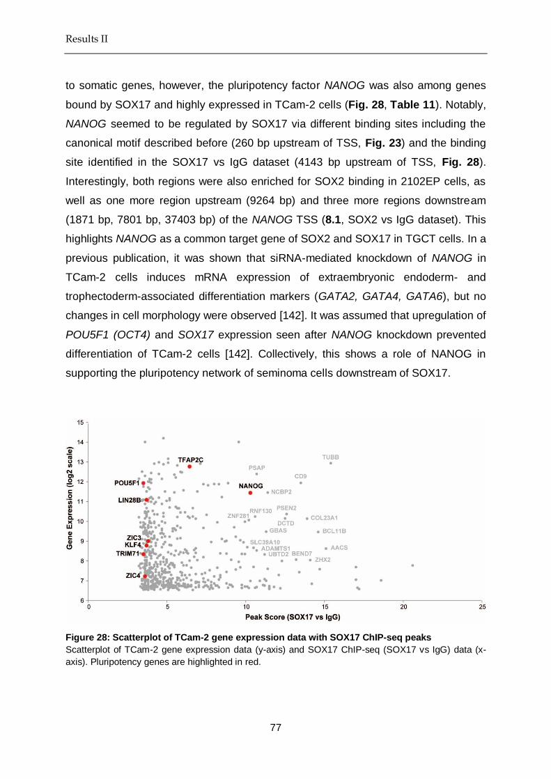
Results II
77
to somatic genes, however, the pluripotency factor NANOG was also among genes
bound by SOX17 and highly expressed in TCam-2 cells (Fig. 28, Table 11). Notably,
NANOG seemed to be regulated by SOX17 via different binding sites including the
canonical motif described before (260 bp upstream of TSS, Fig. 23) and the binding
site identified in the SOX17 vs IgG dataset (4143 bp upstream of TSS, Fig. 28).
Interestingly, both regions were also enriched for SOX2 binding in 2102EP cells, as
well as one more region upstream (9264 bp) and three more regions downstream
(1871 bp, 7801 bp, 37403 bp) of the NANOG TSS (8.1, SOX2 vs IgG dataset). This
highlights NANOG as a common target gene of SOX2 and SOX17 in TGCT cells. In a
previous publication, it was shown that siRNA-mediated knockdown of NANOG in
TCam-2 cells induces mRNA expression of extraembryonic endoderm- and
trophectoderm-associated differentiation markers (GATA2, GATA4, GATA6), but no
changes in cell morphology were observed [142]. It was assumed that upregulation of
POU5F1 (OCT4) and SOX17 expression seen after NANOG knockdown prevented
differentiation of TCam-2 cells [142]. Collectively, this shows a role of NANOG in
supporting the pluripotency network of seminoma cells downstream of SOX17.
Figure 28: Scatterplot of TCam-2 gene expression data with SOX17 ChIP-seq peaks
Scatterplot of TCam-2 gene expression data (y-axis) and SOX17 ChIP-seq (SOX17 vs IgG) data (x-
axis). Pluripotency genes are highlighted in red.

Results II
78
Table 11: Top 20 SOX17 (vs IgG)-peaks with gene expression > 8 in TCam-2
Gene Name Peak Score Gene Expression
TUBB 15.4 12.9
AACS 15.1 8.6
BCL11B 14.6 9.5
ZHX2 14.1 8.0
COL23A1 13.9 10.1
CD9 13.5 11.9
BEND7 13.2 8.1
PSEN2 12.6 10.4
DCTD 12.5 10.1
NCBP2 11.4 11.5
GBAS 11.3 9.5
UBTD2 11.2 8.3
PSAP 10.7 12.4
ADAMTS1 10.7 8.5
RNF130 10.6 10.2
SLC39A10 10.5 8.7
NANOG 10.3 11.4
ZNF281 10.2 10.1
HABP4 10.1 8.7
NFIB 9.95 10.0
Altogether I found opposing functions of SOX17 in regulating genes involved with
pluripotency, but also differentiation in seminoma cells. In contrast, in ECs there is a
clear correlation between the expression of pluripotency-associated genes (GDF3,
DPPA3, TRIM71, DPPA5, SALL4, PRDM14 and NANOG) and SOX2 binding (Fig. 29,
Table 12). This finding confirms the role of SOX2 as a positive regulator of pluripotency
in EC cells and nicely aligns with the previously generated GSEA (compare Table 9-
10) and ChIP-qPCR data (compare Fig. 25). 21 genes showed a SOX2 peak score >
15 and a gene expression > 8: LASP1, ETV6, LOC643770, NANOG, KPNA2, SLC2A3,
DPP9, NDUFS7, COMMD7, RCC2, FAM60A, RAC1, EIF5A, VDAC1, UBE2D2,
HMGN4, SLC2A14, BCAS4, DNAJB6, NACC1 and ABT1 (Fig. 29). Most of these
genes have a role in self-renewal of stem cells or cancer stem cells and therefore may
be associated with cancer malignancy [143-149].
These data show that in EC cells SOX2 binds and activates different regulators of
pluripotency (i.e. NANOG), similar to the role of SOX2 in ESCs. Interestingly, SOX17
may support pluripotency of seminoma cells also in part via transactivation of

Results II
79
pluripotency-associated genes, such as NANOG. The functional consequences of
SOX17 for binding to genes with roles in neuronal differentiation and embryonic
development, however, remain unclear. So far, a mechanism by which SOX17 acts as
a repressor of genes has not been described. Therefore, SOX17 most likely binds and
transactivates these genes.
Figure 29: Scatterplot of 2102EP gene expression data with SOX2 ChIP-seq peaks
Scatterplot of 2102EP gene expression data (y-axis) and SOX2 ChIP-seq (SOX2 vs IgG) data (x-axis).
Pluripotency genes are highlighted in red.
Table 12: Top 20 SOX2 (vs IgG)-peaks with gene expression > 8 in 2102EP
Gene Name Peak Score Gene Expression
LASP1 58.2 12.4
ETV6 47.9 8.5
NANOG 39.7 12.1
KPNA2 37.8 11.6
SLC2A3 37.3 13.9
DPP9 32.5 8.4
NDUFS7 30.8 10.9
COMMD7 29.4 10.5
RCC2 28.4 13.1
FAM60A 27.5 12.7
RAC1 27.1 12.7
EIF5A 26 11.3
FAM60A 25.3 12.7
VDAC1 24.5 12.6

Results II
80
UBE2D2 22.9 8.9
HMGN4 22 10.7
SLC2A14 21.7 8.9
NANOG 21.6 12.1
BCAS4 21.6 8.4
DNAJB6 21.3 10.7
5.6. In seminoma cells SOX17 regulates TFAP2C and PRDM1 expression
The question remains, why seminoma cells keep a state of latent pluripotency, if the
majority of genes bound by SOX17 has roles in neuronal differentiation and embryonic
development. Similar to seminoma cells, human PGCs express TFAP2C, PRDM1 and
SOX17 at high levels, which form a tripartite transcription factor network governing
PGC cell fate [1] (Fig. 1). Importantly, TFAP2C and PRDM1 have roles in the
suppression of somatic differentiation in PGCs [6]. I here showed that in seminoma
cells SOX17 activates PRDM1 and TFAP2C expression (Fig. 23 and 28). Although
PRDM1 was not detected as SOX17 target in the SOX17 vs IgG dataset, it was
detected in the SOX17 vs Input dataset (8.1) and further binding of SOX17 to the
compressed motif within the PRDM1 regulatory region was demonstrated by qPCR
(Fig. 23). A closer look at the SOX17 ChIP-seq profiles at PRDM1, TFAP2C and
SOX17 regulatory regions confirms that SOX17 binds and putatively regulates this
transcription factor network, including the activation of its own expression (Fig. 30 A-
B). Although SOX17 was not calculated as SOX17 ChIP-seq target in the
bioinformatics analysis (maybe due to the stringent settings of the analysis), the ChIP-
seq profile indicates that SOX17 may indeed regulate its own expression (Fig. 30 A).
Although SOX17 additionally binds somatic genes (linked to in neuro-ectodermal
development) in seminoma cells, these findings indicate a regulatory role of SOX17 for
maintaining latent pluripotency via activation of PRDM1 and TFAP2C expression,
thereby in turn suppressing somatic differentiation. A meta-analysis of our SOX17
ChIP-seq data with PRDM1 and TFAP2C ChIP-seq (i.e. in human fetal testis) data
could help to confirm this hypothesis.

Results II
81
Figure 30: SOX17 regulates the SOX17-PRDM1-TFAP2C network in seminoma
(A) SOX17 ChIP-seq profiles at PRDM1, TFAP2C and SOX17 genetic loci (log2 scale). IgG ChIP-seq
profiles are given as negative control.
(B) STRING analysis demonstrating interaction of SOX17, PRDM1 and TFAP2C.

Results II
82
5.7. SOX17 maintains latent pluripotency of seminoma cells
Since seminomas always show high expression of both SOX17 and OCT4, an
essential role of these two transcription factors for maintaining seminoma cell fate was
suggested. So far, I could demonstrate that SOX17 binds to somatic (i.e.
neuroectodermal) genes, but also to pluripotency genes in seminoma cells via the
compressed (SOX17/OCT4) and the canonical (SOX2/OCT4) binding sites. I have
hypothesized that the transactivation of PRDM1 and TFAP2C expression by SOX17
may be essential to suppress the somatic differentiation program in seminoma cells
otherwise activated by SOX17. Now, functional analysis needs to demonstrate whether
a loss of SOX17 results in suppression of TFAP2C and PRDM1 expression, as well as
an overall loss of pluripotency and induction of differentiation.
Therefore, I continued by analysing the effects of SOX17 depletion in seminoma cells.
For this, TCam-2 cells were transfected with two different single guide RNAs (gRNAs)
homologous to the second exon of the SOX17 gene locus. CRISPR/Cas9-mediated
gene editing using both gRNAs should result in a deletion of approximately 130 bp
(Fig. 31). TCam-2 cells contain six copies of chromosome 8 [59], where SOX17 is
encoded. Therefore CRISPR/Cas9-mediated gene editing results in a mixture of cells
displaying deletions in 0-6 alleles of SOX17.
Figure 31: CRISPR/Cas9 mediated gene editing of SOX17 gene locus
Two gRNAs were designed, directed against the second exon of the human SOX17 gene (yellow
arrows). CRISPR/Cas9-mediated gene editing using both gRNAs should result in a final deletion of
approximately 130 bp.

Results II
83
Within 72 hours following transfection of TCam-2 cells with the CRISPR/Cas9
constructs qRT-PCR demonstrated significant reduction of SOX17 expression,
suggesting successful gene editing at least in some cells (Fig. 32). Additionally, I was
able to demonstrate significant downregulation of the pluripotency markers NANOG,
TFAP2C, POU5F1, PRDM14, ALPL and PRDM1 (Fig. 32). All of these genes were
shown to be bound by SOX17 according to our ChIP analysis. The fact that
downregulation of SOX17 results in downregulation of these genes shows that SOX17
transactivates these genes. Furthermore, these analyses show that downregulation or
loss of SOX17 ultimately results in a loss of the latent pluripotent state in seminoma
cells, possibly allowing for cellular differentiation. Notably, since differentiated cells
lose their capacity to self-renew and divide the derivation of single cell clones was
prohibited and the following analyses were performed on the TCam-2 Δ SOX17 bulk
population only.
Figure 32: Expression of pluripotency and germ cell markers after depletion of SOX17 in TCam-
2 cells
qRT-PCR of ChIP-validated targets of SOX17-mediated transcription (red) in TCam-2 Δ SOX17 bulk
population and GFP-transfected TCam-2 as control (72 hours following transfection). Expression is
normalized against GAPDH as housekeeping gene.

Results II
84
While reduction of SOX17 and its downstream target genes NANOG, TFAP2C,
POU5F1, PRDM14, ALPL and PRDM1 was clearly evident on mRNA level, protein
levels of TFAP2C, OCT4, LIN28A and NANOG in the TCam-2 Δ SOX17 bulk
population were not affected (Fig. 33). However, due to the heterogeneity of the TCam-
2 Δ SOX17 bulk population and the presence of SOX17 wildtype cells within this
population, Western blot analysis of the whole protein lysate may not have been
sensitive enough to detect the effects of SOX17 depletion in individual single cells.
Also, mRNA and protein levels can deviate from one another, due to the prolonged
half-life of proteins compared to mRNA.
Figure 33: Depletion of SOX17 in TCam-2 cells
Western blot showing levels of SOX17, TFAP2C, OCT4, LIN28A and NANOG protein 72 hours following
CRISPR/Cas9 mediated gene editing of SOX17 gene locus in the TCam-2 Δ SOX17 bulk population
(Δ SOX17). The wildtype control represents TCam-2 cells that were transiently transfected with a GFP-
coding plasmid. ACTIN was used as loading control.
The analysis of TCam-2 cell morphology 10-15 days following gene editing of SOX17,
however, revealed signs of cell differentiation, such as the formation of polynucleated
cells and an enlarged cytoplasm within these differentiated cell colonies (Fig. 34).
Differentiated areas were negative for the pluripotency marker alkaline phosphatase
(AP), while cells resembling TCam-2 wildtype cells stained positive for AP activity (Fig.
35). In comparison, TCam-2 control cells that were transfected with a GFP-coding
plasmid remained 100% positive for AP activity (Fig. 35). This confirms that the

Results II
85
reduction or loss of SOX17 in TCam-2 cells leads to the downregulation of pluripotency
resulting in induction of differentiation.
Figure 34: Morphology of TCam-2 Δ SOX17 bulk
Morphology of TCam-2 cells 10 and 15 days following CRISPR/Cas9 mediated gene editing of SOX17
gene locus (right) compared to wildtype TCam-2 control cells (left). Scalebar = 250 µm.
Figure 35: Alkaline phosphatase activity of TCam-2 Δ SOX17 bulk
AP activity of TCam-2 cells following CRISPR/Cas9 mediated gene editing of SOX17 gene locus (right)
compared to TCam-2 (GFP-transfected) control cells (left). Scalebar = 250 µm.

Results II
86
However, since the analysis of the whole protein lysate by Western blot was not
sensitive enough to detect loss of TFAP2C and OCT4 on protein level in the TCam-2
Δ SOX17 bulk population, I additionally performed immunofluorescence staining on
individual cells that stained either weakly or completely negative for SOX17 protein
(Fig. 36). As expected, those cells that showed only weak staining for SOX17 protein
also showed reduced levels of OCT4 and TFAP2C protein. This correlates with the
ChIP-seq and qRT-PCR data and again confirms TFAP2C and OCT4 as direct targets
of SOX17-mediated transcriptional activation in TCam-2 cells and shows that depletion
of SOX17 results in loss of pluripotency and germ-cell-identity in TCam-2. Since
morphological alterations already suggested induction of differentiation of TCam-2
cells (Fig. 34), I addressed the question whether the cells differentiate into random cell
fates or if the induced differentiation is restricted to a specific cell fate.
Due to the resemblance to multinucleated trophoblast giant cells I analysed expression
of trophectodermal markers (GATA3, HAND1, αHCG, CDX2, EOMES) (Fig. 37), as
well as additional germ-cell related markers (SPRY4, NANOS3) (Fig. 37) in the TCam-
2 Δ SOX17 bulk population. Notably, in humans HAND1 is expressed in the
trophectoderm layer, where it regulates formation of the amniotic membrane [150].
GATA3 is expressed within the stem cell compartment of the placenta [151]. Human
chorionic gonadotropin (αHCG) is a hormone involved in trophoblast differentiation and
fusion [152]. qRT-PCR demonstrated a loss in germ-cell related markers and
significant induction of GATA3, HAND1, as well as upregulation of αHCG (Fig. 37).
Upregulation of GATA3 was additionally confirmed by immunofluorescence staining
(Fig. 37).

Results II
87
Figure 36: SOX17, OCT4 and TFAP2C protein expression in TCam-2 Δ SOX17 bulk
Immunofluorescence showing expression of SOX17, TFAP2C and OCT4 protein following
CRISPR/Cas9-mediated gene editing of SOX17 gene locus. The wildtype control represents TCam-2
cells that were transiently transfected with a GFP-coding plasmid. Scalebar = 250 µm.

Results II
88
Figure 37: Expression of germ cell markers and trophoblast differentiation markers after
depletion of SOX17 in TCam-2 cells
qRT-PCR of germ cell related markers (brown) and markers of extra-embryonic lineages (green) in
TCam-2 Δ SOX17 bulk and GFP-transfected TCam-2 as control. Expression is normalized against
GAPDH as housekeeping gene.
Altogether this indicates that reduction or loss of SOX17 in TCam-2 cells forces the
cells to initiate differentiation to a trophectodermal cell fate. Interestingly, GATA3
protein was only detected in TCam-2 cells that were low, but not completely devoid of
SOX17 protein (Fig. 38). Thus, it seems like different levels of SOX17 lead to formation
of different cell types, meaning only a reduction but not a complete loss of SOX17 will
lead to a GATA3+ cell population. Collectively, this shows that SOX17 is essential to
maintain the latent pluripotency of seminoma cells and to prevent cellular
differentiation. The analysis of additional markers specific for embryonic (mesoderm,
endoderm, ectoderm) lineages, as well as for different extra-embryonic cell types of
the placenta may help to fully understand the plasticity and differentiation potential of
TCam-2 cells after reduction or complete loss of SOX17.

Results II
89
Figure 38: SOX17 and GATA3 protein expression in TCam-2 Δ SOX17 bulk population
Immunofluorescence showing expression of SOX17 and GATA3 protein following CRISPR/Cas9
mediated gene editing of SOX17 gene locus. The wildtype control represents TCam-2 cells that were
transiently transfected with a GFP-coding plasmid. Scalebar = 250 µm.
5.8. In TGCT cells NANOG is a common downstream target of SOX2 and
SOX17
So far, analyses have shown that both SOX2 and SOX17 are key determinants of
TGCT cell fate by activating expression of pluripotency genes and preventing
differentiation (either directly or via downstream factors, such as PRDM1 and
TFAP2C). By comparing SOX2- and SOX17 regulated genes (meaning those genes
that are bound by SOX2 in EC and SOX17 in seminoma cells), I calculated a common
overlap of 56 genes (Fig. 39 A). Expectedly, NANOG was found as a common
downstream target gene of SOX2 in EC and SOX17 in seminoma cells (Fig. 39 A).
Furthermore, GSEA of these 56 genes revealed that 13 of these genes (CDC42EP4,
ID1, SGK1, PSEN2, KRT18, PDPN, NANOG, FRAT2, AP3B1, SERINC3, NIF3L1,
HN1, ALKBH7) are additional NANOG targets in ESCs (gene set:
‘BENPORATH_NANOG_TARGETS’) [153]. These findings again highlight NANOG as
a supporting factor in maintaining TGCT cell fate (and pluripotency). Although SOX2

Results II
90
and SOX17 do not physically interact with NANOG in EC and seminoma cells,
respectively, NANOG regulates some of the same target genes.
Together with other known factors and regulators of pluripotency (TRIM71, LIN28B,
KLF4, TFAP2C, ALPL) [67, 154-157] that are also bound by SOX2 in EC and SOX17
in seminoma cells, NANOG forms a regulatory network supporting TGCT pluripotency
(Fig. 39 B). Collectively, these results suggest that in seminoma cells SOX17 is able
to take over the role of SOX2 in regulating a set of target genes with roles in
pluripotency maintenance (via the canonical motif). The binding of SOX17 (and not
SOX2) to compressed motifs within regulatory regions of somatic genes, however,
illustrates that both transcription factors are not completely redundant to one another
in regulating TGCT cell fate.
Figure 39: SOX2 and SOX17 regulate a common set of pluripotency genes
(A) Venn diagram depicting common overlap of SOX2- and SOX17-regulated genes in TGCT cells
(B) STRING diagram shows interaction of pluripotency regulators that are regulated by SOX2 in
2102EP and SOX17 in TCam-2 cells.

Results II
91
5.9. The Role of SOX2 and SOX17 in TGCT plasticity
As previously described, we already showed that seminoma cells differentiate into an
EC-like phenotype after xenotransplantation into the flank of nude mice [75]. During
this transition SOX17 is downregulated and SOX2 is upregulated. However, so far no
transition of EC cells to seminoma-like cells was ever demonstrated. We asked
whether a transition of EC to seminoma could be induced by overexpression of SOX17.
Therefore, I used the dCas9 CRISPR synergistic activator system (SAM) to induce
expression of SOX17 in the EC cell line NCCIT. Lentiviral transduction with 100, 200
and 500 µl SOX17 SAM virus led to a dose-dependent induction of SOX17 protein
(Fig. 40 A). SOX17 overexpression following transduction with 500 µl virus was further
confirmed by qRT-PCR (Fig. 40 B). However, this did not lead to morphological
changes in NCCIT cells (Fig. 40 C). Also, protein levels of OCT4 did not change
following overexpression of SOX17 (Fig. 40 A).
I continued to analyse the downstream effects of SOX17 overexpression on selected
markers by qRT-PCR. Although there was significant overexpression of SOX17
detected after transduction with 500 µl SOX17 SAM virus (Fig. 40 B), there was no
significant change in the expression levels of the pluripotency and germ cell-associated
genes POU5F1, KIT, TFAP2C and PRDM14, which were all identified as targets of
SOX17 in seminoma cells (Fig. 41, 8.1). Here, it is necessary to note that POU5F1,
TFAP2C and PRDM14 are already highly expressed in EC cells, therefore no further
induction may be expected. In contrast, however, PRDM1 and KIT are markers of
seminomas (Fig. 24). This may explain, why a significant induction of PRDM1 and
mild, but not significant induction of KIT were observed (Fig. 41).

Results II
92
Figure 40: SOX17 overexpression in NCCIT Cells
(A) Western Blot of SOX17 and OCT4 protein levels in NCCIT cells after transduction with 100, 200
and 500 µl SOX17 SAM virus (SOX17 OE). As negative control, NCCIT cells were transduced with
mCherry virus. TCam-2 protein lysate served as positive control. ACTIN was used as loading
control.
(B) qRT-PCR demonstrating SOX17 overexpression in NCCIT cells after transduction with 500 µl
SOX17 SAM virus (SOX17 OE). As negative control, NCCIT cells were transduced with mCherry
virus. Expression levels were normalized to GAPDH as housekeeping gene.
(C) Morphology of NCCIT cells 72 hours after transduction with 500 µl mCherry virus (left) or SOX17
overexpression (SOX17 OE) SAM construct (right). Scalebar = 250 µm.
But does the overexpression of SOX17 and downstream effectors (i.e. PRDM1) lead
to a transition into a seminoma-like cell fate? Since no morphological changes were
seen (Fig. 40 C) and so far only PRDM1 upregulation was detected, it seems like the
overexpression of SOX17 alone is not sufficient to force an EC to seminoma transition,
at least in the NCCIT cell line. Also, SOX2 was still expressed at high levels (Fig. 41)
Importantly, SOX2 expression is absent in PGCs, TCam-2 and seminoma cells. In
presence of both SOX2 and SOX17 the two factors might compete for OCT4 binding.
Thus, in order to achieve a conversion of EC to seminoma cells it may be necessary
to additionally knockout / knockdown or inhibit SOX2.

Results II
93
Figure 41: The effects of SOX17 overexpression in EC cells on the expression of SOX17 target
genes
qRT-PCR of selected genes in NCCIT cells after transduction with 500 µl SOX17 SAM virus (SOX17
OE). As negative control, NCCIT cells were transduced with mCherry virus. Expression levels were
normalized to GAPDH as housekeeping gene.

Discussion II
94
6. Discussion II
Here, I showed that SOX2, which is highly expressed in EC cells, partners with OCT4
to bind to canonical (SOX2/OCT4) binding sites on the DNA. This way, SOX2 regulates
important players of pluripotency in ECs, such as SOX2, NANOG, DPPA4, LIN28A,
SALL4, TRIM71, GDF3 and PRDM14. I therefore conclude that SOX2 in EC cells has
a similar role as SOX2 in ESCs, which is to maintain pluripotency and to prevent
differentiation. Although NANOG does not directly interact with SOX2 and OCT4 in
ECs to regulate downstream target genes, I believe that it has a supporting role in
pluripotency maintenance downstream of SOX2.
In seminoma cells we find a high expression of SOX17. In this cell type SOX17 partners
with OCT4 to bind to both compressed (SOX17/OCT4) and canonical (SOX2/OCT4)
binding sites on the DNA. Gene set enrichment analysis has shown that the majority
of SOX17-bound genes in seminoma cells are having roles in neuronal differentiation
(such as ZHX2, COL23A1, CD9, BEND7, PSEN2, GBAS, UBTD2, PSAP, ADAMTS1
and ZNF281). This is surprising, since seminoma cells maintain a latent pluripotent
state and do not spontaneously differentiate into embryonic (and / or neuronal)
lineages like ECs, evident by the expression of i.e. LIN28, PRDM14, TFAP2C, OCT4
and NANOG. Only in presence of TGFβ, EGF and FGF4 it was demonstrated that
TCam-2 cells differentiate into mixed non-seminoma lineages (then showing features
of choriocarcinomas, teratomas and yolk-sac tumours) [73]. But what prevents the
differentiation of seminoma cells into neuronal lineages? Most importantly, I could
show that SOX17, in seminoma cells, also binds and regulates markers of pluripotency,
such as NANOG, DPPA4, LIN28A, PRDM14, POU5F1 and TRIM71. Therefore SOX17
directly contributes to the expression of the seminoma pluripotency cluster and in this
respect replaces SOX2 in EC cells. Additionally, SOX17 induces expression of the
PGC specifiers PRDM1 and TFAP2C. Both factors are known to suppress somatic
differentiation in PGCs [8]. Thus, PRDM1 and TFAP2C may similarly suppress
(neuronal) differentiation in seminoma cells. In line with this hypothesis, I could
demonstrate that many of those regions bound by SOX17 in seminoma cells (i.e.
neuronal genes) harbour additionally binding motifs of PRDM1 and TFAP2C. So, in
seminoma cells SOX17, PRDM1 and TFAP2C could cooperate to maintain TGCT cell
fate and prevent somatic differentiation.

Discussion II
95
In line, loss of SOX17 in seminoma cells resulted in loss of TFAP2C and PRDM1
expression, resulting in differentiation into trophectodermal-like lineages. Therefore, it
seems plausible that SOX17 has a regulatory function for maintaining pluripotency in
seminoma cells, in part via activation of PRDM1 and TFAP2C expression, but also by
directly activating expression of germ-cell and pluripotency-associated target genes,
such as NANOG, DPPA4 and LIN28A (Fig. 42). It would be interesting to see, whether
expression of TFAP2C or PRDM1 protein could rescue the effects of loss of SOX17 in
seminoma cells and prevent differentiation.
Interestingly, we have shown in a different study that the cancer-testis antigen PRAME
is also repressing differentiation of seminoma cells (Fig. 42) [136]. PRAME is highly
expressed in seminoma and TCam-2 cells and absent in ECs [136]. PRAME
knockdown in seminoma cells led to a downregulation of pluripotency and germ-cell-
related markers and induction of genes associated with endodermal or mesodermal
differentiation [136]. Also, during seminoma to EC transition downregulation of SOX17
was first followed by downregulation of PRAME, which suggested that PRAME acts
downstream of SOX17 [75, 136]. However, here PRAME was not identified as a direct
target of SOX17-mediated transcription. Nonetheless, our previous analyses indicate
that PRAME also represses somatic differentiation in seminoma cells, without being a
direct transcriptional target of SOX17 (Fig. 42).
While we had already postulated a role for SOX17 in maintaining pluripotency of
seminoma cells, I could now additionally show that loss of SOX17 induces
differentiation of these cells into extra-embryonic lineages. In a previous study by Irie
et al. it was demonstrated that siRNA-mediated knockdown of SOX17 in TCam-2 cells
leads to a loss of pluripotency- and germ cell- related markers [6]. However, the
authors did not investigate the long-term effects of SOX17 knockdown in TCam-2 cells.
Here, the differentiation of seminoma cells into extra-embryonic cell types after SOX17
loss resembles the differentiation of seminoma cells seen after treatment with TGFβ,
EGF and FGF4 into mixed non-seminoma [73], although this differentiation was not
accompanied by a loss in SOX17 expression [73].

Discussion II
96
Figure 42: Transcription factor network maintaining seminoma pluripotency
SOX17 and OCT4 interact and bind to the DNA, thereby activating expression of somatic genes and
genes associated with pluripotency, including NANOG. Also, SOX17 regulates TFAP2C and PRDM1
expression, which in turn are able to suppress somatic differentiation and maintain pluripotency. PRAME
may additionally support the pluripotency network of seminoma cells by repressing somatic
differentiation.
Nonetheless, it seems interesting that seminoma cells respond to different signalling
cues (i.e. treatment with TGFβ, EGF and FGF4) or dysregulations (i.e. loss of SOX17)
with the differentiation into extraembryonic lineages that resemble mixed non-
seminoma / choriocarcinoma. Especially the multinucleated cell types within these
differentiated colonies are reminiscent of syncytial trophoblastic cells. Such
trophoblast-like foci are sometimes seen within patient seminoma samples [158], thus
the data presented here could serve to explain the development of these foci. In light
of the presented data, I speculate that distinct signalling cues or cytokines present
within the tumour microenvironment could favour a downregulation of SOX17 levels in
seminomas, thereby inducing seminoma differentiation into extra-embryonic cell tyes.
In conclusion, seminomas may only keep their cell identity by expressing SOX17 (and
its partner OCT4). Due to the resemblance of seminoma cells to GCNIS and PGCs,
seminomas are considered the default pathway of GCNIS progression. Inhibition of
BMP signalling, however, can result in a seminoma to EC transition, accompanied by

Discussion II
97
downregulation of SOX17 and upregulation of SOX2 [75]. In turn, the overexpression
of SOX17 alone did not lead to a shift in EC cell identity towards a seminoma
phenotype, nor did it alter SOX2 levels. This is despite the fact that OCT4 has a much
higher affinity for the SOX17 DNA complex, at least on compressed motifs (binding
energy: −3.86 ± 0.74 kcal/mol), than for the SOX2 DNA complex at canonical motifs
(binding energy: 8.09 ± 0.73 kcal/mol) [71]. One theory in favour of a preferred SOX2-
OCT4 binding to canonical motifs in ECs would be that the DNA is more accessible at
regions containing the canonical motif, while DNA accessibility is restricted at regions
containing compressed motifs (i.e. by DNA methylation). Additional ATAC-seq on EC
and seminoma chromatin could therefore help to investigate this hypothesis.
Interestingly, although SOX2 levels were not altered upon overexpression of SOX17
in ECs, we find a mutual exclusive expression of SOX2 and SOX17 in TGCT subtypes.
Several hypotheses try to explain this phenomenon: In TCam-2 cells Kushwaha et al.
proposed that SOX2 repression is mediated by histone methylation (H3K27me3) via
the polycomb repressive complex [159]. In colorectal cancer expression of SOX2 is
suppressed by the SOX17 target miR-371-5p [160]. However, I could not identify miR-
371-5p as a SOX17 target in TCam-2 cells. Also, it seems like SOX17 induction alone
is not sufficient to alter SOX2 levels in EC cells in vitro. In the future, it would be
interesting to investigate the mechanism by which SOX17 is repressed during TCam-
2 to EC conversion (after xenotransplantation into the flank of nude mice) in more
detail, in order to understand whether a similar conversion was also possible from EC
to seminoma. Also, it would be interesting to see how SOX2 knockout / knockdown
affects the outcome of SOX17 overexpression in EC cells.

Bibliography
98
7. Bibliography
[1]Jostes S. Master Thesis, Sina Jostes 2014, University of Bonn.
[2]Jostes S, Nettersheim D, Fellermeyer M, Schneider S, Hafezi F, Honecker F, et al.
The bromodomain inhibitor JQ1 triggers growth arrest and apoptosis in testicular germ
cell tumours in vitro and in vivo. J Cell Mol Med 2016; 21; 1300–14.
[3]Nettersheim D, Jostes S, Fabry M, Honecker F, Schumacher V, Kirfel J, et al. A
signaling cascade including ARID1A, GADD45B and DUSP1 induces apoptosis and
affects the cell cycle of germ cell cancers after romidepsin treatment. Oncotarget 2016;
5.
[4]Sasaki K, Nakamura T, Okamoto I, Yabuta Y, Iwatani C, Tsuchiya H, et al. The Germ
Cell Fate of Cynomolgus Monkeys Is Specified in the Nascent Amnion. Dev Cell 2016;
39; 169–85.
[5]Kojima Y, Sasaki K, Yokobayashi S, Sakai Y, Nakamura T, Yabuta Y, et al.
Evolutionarily Distinctive Transcriptional and Signaling Programs Drive Human Germ
Cell Lineage Specification from Pluripotent Stem Cells. Cell Stem Cell 2017; 21; 517–
532.e5.
[6]Irie N, Weinberger L, Tang WWC, Kobayashi T, Viukov S, Manor YS, et al. SOX17 Is a
Critical Specifier of Human Primordial Germ Cell Fate. Cell 2015; 160.
[7]Sasaki K, Yokobayashi S, Nakamura T, Okamoto I, Yabuta Y, Kurimoto K, et al.
Robust In Vitro Induction of Human Germ Cell Fate from Pluripotent Stem Cells. Cell
Stem Cell 2015; 17; 178–94.
[8]Jostes S, Schorle H. Signals and transcription factors for specification of human germ
cells. Stem Cell Investig 2018; 5; 13.
[9]Meyenn von F, Reik W. Forget the Parents: Epigenetic Reprogramming in Human Germ
Cells. Cell 2015; 161; 1248–51.
[10]Nettersheim D, Jostes S, Schneider S, Schorle H. Elucidating human male germ cell
development by studying germ cell cancer. Reproduction 2016; 152; R101–13.
[11]Stukenborg J-B, Kjartansdóttir KR, Reda A, Colon E, Albersmeier JP, Söder O.
Male germ cell development in humans. Horm Res Paediatr 2013; 81; 2–12.
[12]Neto FTL, Bach PV, Najari BB, Li PS, Goldstein M. Spermatogenesis in humans and
its affecting factors. Seminars in Cell &Amp; Developmental Biology 2016; 59; 10–26.
[13]Bao J, Bedford MT. Epigenetic regulation of the histone-to-protamine transition during
spermiogenesis. Reproduction 2016; 151; R55–70.
[14]Sullivan R, Mieusset R. The human epididymis: its function in sperm maturation. Hum
Reprod Update 2016; 22; 574–87.
[15]Amann RP. The cycle of the seminiferous epithelium in humans: a need to revisit? J
Androl 2008; 29; 469–87.

Bibliography
99
[16]Manecksha RP, Fitzpatrick JM. Epidemiology of testicular cancer. BJU Int 2009; 104;
1329–33.
[17]Looijenga LHJ, Stoop H, Biermann K. Testicular cancer: biology and biomarkers.
Virchows Archiv : an International Journal of Pathology 2014; 464.
[18]Oosterhuis JW, Stoop H, Honecker F, Looijenga LHJ. Why human extragonadal germ
cell tumours occur in the midline of the body: old concepts, new perspectives.
International Journal of Andrology 2007; 30.
[19]Nohynek GJ, Borgert CJ, Dietrich D, Rozman KK. Endocrine disruption: fact or urban
legend? Toxicol Lett 2013; 223; 295–305.
[20]Shanmugalingam T, Soultati A, Chowdhury S, Rudman S, Van Hemelrijck M. Global
incidence and outcome of testicular cancer. Clin Epidemiol 2013; 5; 417–27.
[21]Oosterhuis JW, Looijenga LHJ. Testicular germ-cell tumours in a broader perspective.
Nature Reviews Cancer 2005; 5.
[22]Metcalfe PD, Farivar-Mohseni H, Farhat W, McLorie G, Khoury A, Bägli DJ. Pediatric
testicular tumors: contemporary incidence and efficacy of testicular preserving surgery. J
Urol 2003; 170; 2412.
[23]Wei Y, Wu S, Lin T, He D, Li X, Liu J, et al. Testicular yolk sac tumors in children: a
review of 61 patients over 19 years. World J Surg Oncol 2014; 12; 400.
[24]Nistal M, Paniagua R, González-Peramato P, Reyes-Múgica M. Perspectives in
Pediatric Pathology, Chapter 25. Testicular and Paratesticular Tumors in the Pediatric
Age Group. Pediatr Dev Pathol 2016; 19; 471–92.
[25]Ye Y-L, Sun X-Z, Zheng F-F, Bian J, Huang Y-P, Zhang X-Q, et al. Clinical analysis of
management of pediatric testicular germ cell tumors. Urology 2012; 79; 892–7.
[26]Wetherell D, Weerakoon M, Williams D, Beharry BK, Sliwinski A, Ow D, et al. Mature
and Immature Teratoma: A Review of Pathological Characteristics and Treatment
Options. Med Surg Urol 2014.
[27]Palmer RD, Foster NA, Vowler SL, Roberts I, Thornton CM, Hale JP, et al. Malignant
germ cell tumours of childhood: new associations of genomic imbalance. British Journal
of Cancer 2007; 96; 667–76.
[28]Mosbech CH, Rechnitzer C, Brok JS, Meyts ER-D, Hoei-Hansen CE. Recent
advances in understanding the etiology and pathogenesis of pediatric germ cell tumors.
J Pediatr Hematol Oncol 2014; 36; 263–70.
[29]Hayes-Lattin B, Nichols CR. Testicular cancer: a prototypic tumor of young adults.
Semin Oncol 2009; 36; 432–8.
[30]Sheikine Y, Genega E, Melamed J, Lee P, Reuter VE, Ye H. Molecular genetics of
testicular germ cell tumors. Am J Cancer Res 2012; 2; 153–67.
[31]Berney DM, Looijenga LHJ, Idrees M, Oosterhuis JW, Meyts ER-D, Ulbright TM, et
al. Germ cell neoplasia in situ (GCNIS): evolution of the current nomenclature for
testicular pre-invasive germ cell malignancy. Histopathology 2016; 69; 7–10.

Bibliography
100
[32]Hoei-Hansen CE, Meyts ER-D, Daugaard G, Skakkebaek NE. Carcinoma in situ testis,
the progenitor of testicular germ cell tumours: a clinical review. Ann Oncol 2005; 16;
863–8.
[33]de Jong J, Stoop H, Gillis AJM, van Gurp RJHLM, van de Geijn G-JM, de Boer M, et
al. Differential expression of SOX17 and SOX2 in germ cells and stem cells has
biological and clinical implications. The Journal of Pathology 2008; 215; 21–30.
[34]Nonaka D. Differential expression of SOX2 and SOX17 in testicular germ cell tumors.
American Journal of Clinical Pathology 2009; 131; 731–6.
[35]Hart AH, Hartley L, Parker K, Ibrahim M, Looijenga LHJ, Pauchnik M, et al. The
pluripotency homeobox gene NANOG is expressed in human germ cell tumors. Cancer
2005; 104; 2092–8.
[36]Mikuz G. [Spermatocytic seminoma. A tumor with many faces]. Pathologe 2014; 35;
232–7.
[37]Looijenga LHJ, Hersmus R, Gillis AJM, Pfundt R, Stoop HJ, Van Gurp RJHLM, et al.
Genomic and expression profiling of human spermatocytic seminomas: primary
spermatocyte as tumorigenic precursor and DMRT1 as candidate chromosome 9 gene.
Cancer Research 2006; 66; 290–302.
[38]Mizuno Y, Gotoh A, Kamidono S, Kitazawa S. [Establishment and characterization of a
new human testicular germ cell tumor cell line (TCam-2)]. Nippon Hinyokika Gakkai
Zasshi 1993; 84; 1211–8.
[39]Kinugawa K, Hyodo F, Matsuki T, Jo Y, Furukawa Y, Ueki A, et al. Establishment and
characterization of a new human testicular seminoma cell line, JKT-1. Int J Urol 1998; 5;
282–7.
[40]Mokashi A, Megiel C, Looijenga LHJ. Establishment and characterization of a new
human extragonadal germ cell line, SEM-1, and its comparison with TCam-2 and JKT-1.
Urology 2013; 81.
[41]Vogelzang NJ, Bronson D, Savino D, Vessella RL, Fraley EF. A human embryonal-
yolk sac carcinoma model system in athymic mice. Cancer 1985; 55; 2584–93.
[42]Houldsworth J, Reuter V, Bosl GJ, Chaganti RS. Aberrant expression of cyclin D2 is
an early event in human male germ cell tumorigenesis. Cell Growth Differ 1997; 8; 293–
9.
[43]Bronson DL, Andrews PW, Vessella RL, Fraley EE. In vitro differentiation of human
embryonal carcinoma cells. Teratocarcinoma Stem Cells 1983; 597–605.
[44]Andrews PW, Goodfellow PN, Shevinsky LH, Bronson DL, Knowles BB. Cell-surface
antigens of a clonal human embryonal carcinoma cell line: morphological and antigenic
differentiation in culture. International Journal of Cancer 1982; 29; 523–31.
[45]Bronson DL, Andrews PW, Solter D, Cervenka J, Lange PH, Fraley EE. Cell line
derived from a metastasis of a human testicular germ cell tumor. Cancer Research 1980;
40; 2500–6.

Bibliography
101
[46]Pera MF, Lafita MJB, Mills J. Cultured stem-cells from human testicular teratomas: the
nature of human embryonal carcinoma, and its comparison with two types of yolk-sac
carcinoma. International Journal of Cancer 1987; 40; 334–43.
[47]Casper J, Schnaidt U, Fonatsch C. Cell lines of human germinal cancer. International
Journal of Andrology 1987; 10; 105–13.
[48]Damjanov I, Horvat B, Gibas Z. Retinoic acid-induced differentiation of the
developmentally pluripotent human germ cell tumor-derived cell line, NCCIT. Lab Invest
1993; 68; 220–32.
[49]Sekiya S, Kawata M, Iwasawa H, Inaba N, Sugita M, Suzuki N, et al. Characterization
of human embryonal carcinoma cell lines derived from testicular germ-cell tumors.
Differentiation 1985; 29; 259–67.
[50]Hogan B, Fellous M, Avner P, Jacob F. Isolation of a human teratoma cell line which
expresses F9 antigen. Nature 1977; 270; 515–8.
[51]Andrews PW, Damjanov I, Simon D, Banting GS, Carlin C, Dracopoli NC, et al.
Pluripotent embryonal carcinoma clones derived from the human teratocarcinoma cell
line Tera-2. Differentiation in vivo and in vitro. Lab Invest 1984; 50; 147–62.
[52]Wang N, Trend B, Bronson DL, Fraley EE. Nonrandom abnormalities in chromosome 1
in human testicular cancers. Cancer Research 1980; 40; 796–802.
[53]Pattillo RA, Gey GO. The establishment of a cell line of human hormone-synthesizing
trophoblastic cells in vitro. Cancer Research 1968; 28; 1231–6.
[54]Hochberg A, Rachmilewitz J, Eldar-Geva T, Salant T, Schneider T, de Groot N.
Differentiation of choriocarcinoma cell line (JAr). Cancer Research 1992; 52; 3713–7.
[55]Kohler PO, Bridson WE. Isolation of hormone-producing clonal lines of human
choriocarcinoma. The Journal of Clinical Endocrinology and Metabolism 1971; 32; 683–
7.
[56]Eckert D, Nettersheim D, Heukamp LC, Kitazawa S, Biermann K, Schorle H. TCam-2
but not JKT-1 cells resemble seminoma in cell culture. Cell and Tissue Research 2008;
331.
[57]De Jong J, Stoop H, Gillis AJM, Van Gurp RJHLM, Van Drunen E, Beverloo HB, et
al. JKT-1 is not a human seminoma cell line. International Journal of Andrology 2007;
30; 350–65.
[58]Olie RA, Boersma AW, Dekker MC, Nooter K, Looijenga LH, Oosterhuis JW.
Apoptosis of human seminoma cells upon disruption of their microenvironment. British
Journal of Cancer 1996; 73.
[59]De Jong J, Stoop H, Gillis AJM, Hersmus R, Van Gurp RJHLM, Van De Geijn GJM,
et al. Further characterization of the first seminoma cell line TCam-2. Genes
Chromosomes and Cancer 2008; 47.
[60]Nettersheim D, Heimsoeth A, Jostes S, Schneider S, Fellermeyer M, Hofmann A, et
al. SOX2 is essential for in vivo reprogramming of seminoma-like TCam-2 cells to an
embryonal carcinoma-like fate. Oncotarget 2016; 7; 47095.

Bibliography
102
[61]Simões PD, Ramos T. Human pluripotent embryonal carcinoma NTERA2 cl.D1 cells
maintain their typical morphology in an angiomyogenic medium. J Negat Results Biomed
2007; 6; 5.
[62]Baldassarre G, Boccia A, Bruni P, Sandomenico C, Barone MV, Pepe S, et al.
Retinoic acid induces neuronal differentiation of embryonal carcinoma cells by reducing
proteasome-dependent proteolysis of the cyclin-dependent inhibitor p27. Cell Growth
Differ 2000; 11; 517–26.
[63]Mavilio F, Simeone A, Boncinelli E, Andrews PW. Activation of four homeobox gene
clusters in human embryonal carcinoma cells induced to differentiate by retinoic acid.
Differentiation 1988; 37; 73–9.
[64]Josephson R, Ording CJ, Liu Y, Shin S, Lakshmipathy U, Toumadje A, et al.
Qualification of embryonal carcinoma 2102Ep as a reference for human embryonic stem
cell research. Stem Cells 2007; 25.
[65]Duran C, Talley PJ, Walsh J, Pigott C, Morton IE, Andrews PW. Hybrids of pluripotent
and nullipotent human embryonal carcinoma cells: partial retention of a pluripotent
phenotype. International Journal of Cancer 2001; 93; 324–32.
[66]Abdelalim EM, Emara MM, Kolatkar PR. The SOX transcription factors as key players
in pluripotent stem cells. Stem Cells and Development 2014; 23; 2687–99.
[67]Liu X, Huang J, Chen T, Wang Y, Xin S, Li J, et al. Yamanaka factors critically regulate
the developmental signaling network in mouse embryonic stem cells. Cell Research
2008; 18; 1177–89.
[68]Ying L, Mills JA, French DL, Gadue P. OCT4 Coordinates with WNT Signaling to Pre-
pattern Chromatin at the SOX17 Locus during Human ES Cell Differentiation into
Definitive Endoderm. Stem Cell Reports 2015; 5; 490–8.
[69]Aksoy I, Jauch R, Chen J, Dyla M, Divakar U, Bogu GK, et al. Oct4 switches
partnering from Sox2 to Sox17 to reinterpret the enhancer code and specify endoderm.
The EMBO Journal 2013; 32.
[70]Tapia N, Maccarthy C, Esch D, Marthaler AG, Tiemann U, Araúzo-bravo MJ, et al.
Dissecting the role of distinct OCT4-SOX2 heterodimer configurations in pluripotency.
Nature Publishing Group 2015; DOI: 10.1038/srep13533.
[71]Merino F, Ng CKL, Veerapandian V, Schöler HR, Jauch R, Cojocaru V. Structural
Basis for the SOX-Dependent Genomic Redistribution of OCT4 in Stem Cell
Differentiation. Cell 2014; 22.
[72]Jauch R, Aksoy I, Hutchins AP, Ng CKL, Tian XF, Chen J, et al. Conversion of Sox17
into a Pluripotency Reprogramming Factor by Reengineering Its Association with Oct4
on DNA. Stem Cells 2011; DOI: 10.1002/stem.639.
[73]Nettersheim D, Gillis AJ, Looijenga LH, Schorle H. TGF-β1, EGF and FGF4
synergistically induce differentiation of the seminoma cell line TCam-2 into a cell type
resembling mixed non-seminoma. International Journal of Andrology 2011; 34.
[74]Ancelin K, Lange UC, Hajkova P, Schneider R, Bannister AJ, Kouzarides T, et al.
Blimp1 associates with Prmt5 and directs histone arginine methylation in mouse germ
cells. Nat Cell Biol 2006; 8; 623–30.

Bibliography
103
[75]Nettersheim D, Jostes S, Sharma R, Schneider S, Hofmann A, Ferreira HJ, et al.
BMP Inhibition in Seminomas Initiates Acquisition of Pluripotency via NODAL Signaling
Resulting in Reprogramming to an Embryonal Carcinoma. PLoS Genetics 2015; 11;
e1005415.
[76]Nettersheim D, Schorle H. The plasticity of germ cell cancers and its dependence on
the cellular microenvironment. J Cell Mol Med 2017; 21; 1463–7.
[77]Oldenburg J, Nuver J, Heidenreich A, Schmoll H-J, Bokemeyer C, Horwich A, et al.
Testicular seminoma and non-seminoma: ESMO Clinical Practice Guidelines for
diagnosis, treatment and follow-up. Ann Oncol 2013; 24 Suppl 6; vi125–32.
[78]Albers P, Albrecht W, Algaba F, Bokemeyer C, Cohn-Cedermark G, Fizazi K, et al.
EAU guidelines on testicular cancer: 2011 update. Eur Urol 2011; 60; 304–19.
[79]Jostes S, Nettersheim D, Schorle H. Epigenetic drugs and their molecular targets in
testicular germ cell tumours. Nat Rev Urol 2019; DOI: 10.1038/s41585-019-0154-x.
[80]Waddington CH. Genetic assimilation of the bithorax phenotype. International Journal of
Organic Evolution 1956; 10; 1-13
[81]Eberharter A, Becker PB. Histone acetylation: a switch between repressive and
permissive chromatin. Second in review series on chromatin dynamics. EMBO Rep
2002; 3; 224–9.
[82]Li Y, Seto E. HDACs and HDAC Inhibitors in Cancer Development and Therapy. Cold
Spring Harb Perspect Med 2016; 6; a026831.
[83]Eckschlager T, Plch J, Stiborova M, Hrabeta J. Histone Deacetylase Inhibitors as
Anticancer Drugs. Int J Mol Sci 2017; 18; 1414.
[84]Florence B, Faller DV. You bet-cha: a novel family of transcriptional regulators. Frontiers
in Bioscience : a Journal and Virtual Library 2001; 6.
[85]Prinjha RK, Dittmann A, Giotopoulos G, Bantscheff M, Chan W-I, Robson SC, et al.
Inhibition of BET recruitment to chromatin as an effective treatment for MLL-fusion
leukaemia. Nature 2011; 478.
[86]French CA, Ramirez CL, Kolmakova J, Hickman TT, Cameron MJ, Thyne ME, et al.
BRD-NUT oncoproteins: a family of closely related nuclear proteins that block epithelial
differentiation and maintain the growth of carcinoma cells. Oncogene 2007; 27; 2237–
42.
[87]Ryan SL, Schwalbe EC, Cole M, Lu Y, Lusher ME, Megahed H, et al. MYC family
amplification and clinical risk-factors interact to predict an extremely poor prognosis in
childhood medulloblastoma. Acta Neuropathol 2011; 123; 501–13.
[88]Lee HY, Cha J, Kim SK, Park JH, Song KH, Kim P, et al. c-MYC Drives Breast Cancer
Metastasis to the Brain, but Promotes Synthetic Lethality with TRAIL. Mol Cancer Res
2018; molcanres.0630.2018.
[89]Zimmerman MW, Liu Y, He S, Durbin AD, Abraham BJ, Easton J, et al. MYC Drives a
Subset of High-Risk Pediatric Neuroblastomas and Is Activated through Mechanisms
Including Enhancer Hijacking and Focal Enhancer Amplification. Cancer Discov 2017; 8;
320–35.

Bibliography
104
[90]Horne GA, Stewart HJS, Dickson J, Knapp S, Ramsahoye B, Chevassut T. Nanog
Requires BRD4 to Maintain Murine Embryonic Stem Cell Pluripotency and Is
Suppressed by Bromodomain Inhibitor JQ1 Together with Lefty1. Stem Cells and
Development 2015; 24.
[91]Wu T, Pinto HB. The BET Family Member BRD4 Interacts with OCT4 and Regulates
Pluripotency Gene Expression. Stem Cell Reports 2015; 4.
[92]Doroshow DB, Eder JP, LoRusso PM. BET inhibitors: a novel epigenetic approach.
Ann Oncol 2017; 28; 1776–87.
[93]Delmore JE, Issa GC, Lemieux ME, Rahl PB, Shi J, Jacobs HM, et al. BET
bromodomain inhibition as a therapeutic strategy to target c-Myc. Cell 2011; 146.
[94]Baker EK, Taylor S, Gupte A, Sharp PP, Walia M, Walsh NC, et al. BET inhibitors
induce apoptosis through a MYC independent mechanism and synergise with CDK
inhibitors to kill osteosarcoma cells. Sci Rep 2015; 5; 10120.
[95]Bhadury J, Nilsson LM, Muralidharan SV, Green LC, Li Z, Gesner EM, et al. BET and
HDAC inhibitors induce similar genes and biological effects and synergize to kill in Myc-
induced murine lymphoma. Proceedings of the National Academy of Sciences of the
United States of America 2014; 111.
[96]Henssen A, Thor T, Odersky A, Heukamp L, El- N, Beckers A, et al. BET
bromodomain protein inhibition is a therapeutic option for medulloblastoma. Oncotarget
2013; 4.
[97]Lovén J, Hoke HA, Lin CY, Lau A, Orlando DA, Vakoc CR, et al. Selective inhibition of
tumor oncogenes by disruption of super-enhancers. Cell 2013; 153.
[98]Borbely G, Haldosen L-A, Dahlman-Wright K, Zhao C. Induction of USP17 by
combining BET and HDAC inhibitors in breast cancer cells. Oncotarget 2015; 6.
[99]Chakravarthy H, Boer B, Desler M, Mallanna SK, McKeithan TW, Rizzino A.
Identification of DPPA4 and other genes as putative Sox2:Oct-3/4 target genes using a
combination of in silico analysis and transcription-based assays. J Cell Physiol 2008;
216; 651–62.
[100]Cimadamore F, Amador-Arjona A, Chen C, Huang C-T, Terskikh AV. SOX2-
LIN28/let-7 pathway regulates proliferation and neurogenesis in neural precursors.
Proceedings of the National Academy of Sciences 2013; 110; E3017–26.
[101]Fellermeyer M. Bachelor Thesis, Martin Fellermeyer 2016, University of Bonn
[102]Chew J-L, Loh Y-H, Zhang W, Chen X, Tam W-L, Yeap L-S, et al. Reciprocal
transcriptional regulation of Pou5f1 and Sox2 via the Oct4/Sox2 complex in embryonic
stem cells. Molecular and Cellular Biology 2005; 25; 6031–46.
[103]Heinz S, Benner C, Spann N, Bertolino E, Lin YC, Laslo P, et al. Simple
combinations of lineage-determining transcription factors prime cis-regulatory elements
required for macrophage and B cell identities. Molecular Cell 2010; 38; 576–89.
[104]Oliveros JC. Venny. An interactive tool for comparing lists with Venn's diagrams. 2007.

Bibliography
105
[105]Konermann S, Brigham MD, Trevino AE, Joung J, Abudayyeh OO, Barcena C, et
al. Genome-scale transcriptional activation by an engineered CRISPR-Cas9 complex.
Nature 2014; 517; 583–8.
[106]Voutsadakis IA. The chemosensitivity of testicular germ cell tumors. Cell Oncol (Dordr)
2014; 37; 79–94.
[107]Yin F, Lan R, Zhang X, Zhu L, Chen F, et al. LSD1 Regulates Pluripotency of
Embryonic Stem/Carcinoma Cells through Histone Deacetylase 1-Mediated
Deacetylation of Histone H4 at Lysine 16. Molecular and Cellular Biology 2013; 34; 158.
[108]Albany C, Hever-Jardine MP, Herrmann von KM, Yim CY, Tam J, Warzecha JM, et
al. Refractory testicular germ cell tumors are highly sensitive to the second generation
DNA methylation inhibitor guadecitabine. Oncotarget 2016; 8; 2949–59.
[109]Eckert D, Biermann K, Nettersheim D, Gillis AJM, Steger K, Jäck H-M, et al.
Expression of BLIMP1/PRMT5 and concurrent histone H2A/H4 arginine 3 dimethylation
in fetal germ cells, CIS/IGCNU and germ cell tumors. BMC Developmental Biology 2008;
8; 106.
[110]Wang L, Wolgemuth DJ. BET Protein BRDT Complexes With HDAC1, PRMT5, and
TRIM28 and Functions in Transcriptional Repression During Spermatogenesis. Journal
of Cellular Biochemistry 2015; 117; 1429–38.
[111]Chaitanya GV, Steven AJ, Babu PP. PARP-1 cleavage fragments: signatures of cell-
death proteases in neurodegeneration. Cell Commun Signal 2010; 8; 31.
[112]Wang J, Liu Z, Wang Z, Wang S, Chen Z, Li Z, et al. Targeting c-Myc: JQ1 as a
promising option for c-Myc-amplified esophageal squamous cell carcinoma. Cancer
Letters 2018; 419; 64–74.
[113]Huang X, Li X. KLF6 induces apoptosis in prostate cancer cells through up-regulation
of ATF3. Journal of Biological Chemistry 2008; 283; 29795–801.
[114]Mirguet O, Gosmini R, Toum J, Clément CA, Barnathan M, Brusq J-M, et al.
Discovery of epigenetic regulator I-BET762: lead optimization to afford a clinical
candidate inhibitor of the BET bromodomains. J Med Chem 2013; 56; 7501–15.
[115]Filippakopoulos P, Picaud S, Mangos M, Keates T, Lambert JP, Barsyte-Lovejoy
D, et al. Histone recognition and large-scale structural analysis of the human
bromodomain family. Cell 2012; 149.
[116]Jung M, Philpott M, Schulze J, Badock V, Eberspächer U, Moosmayer D, et al.
Affinity map of bromodomain protein 4 (BRD4) interactions with the histone H4 tail and
the small molecule inhibitor JQ1. The Journal of Biological Chemistry 2014; 289.
[117]Feng Q, Zhang Z, Shea MJ, Creighton CJ, Coarfa C, Hilsenbeck SG, et al. An
epigenomic approach to therapy for tamoxifen-resistant breast cancer. Cell Research
2014; 24; 809–19.
[118]Bid HK, Phelps DA, Xaio L, Guttridge DC, Lin J, London C, et al. The Bromodomain
BET Inhibitor JQ1 Suppresses Tumor Angiogenesis in Models of Childhood Sarcoma.
Mol Cancer Ther 2016; 15; 1018–28.

Bibliography
106
[119]Leal AS, Williams CR, Royce DB, Pioli PA, Sporn MB, Liby KT. Bromodomain
inhibitors, JQ1 and I-BET 762, as potential therapies for pancreatic cancer. Cancer
Letters 2017; 394; 76–87.
[120]Leal AS, Carapellucci S, Zydeck K, Sporn MB, Liby KT. Chemoprevention of
Preclinical Breast and Lung Cancer with the Bromodomain Inhibitor I-BET 762. Cancer
Prev Res (Phila) 2017; 11; 143–56.
[121]Filippakopoulos P, Qi J, Picaud S, Shen Y, Smith WB, Morse EM, et al. Selective
inhibition of BET bromodomains. Nature 2011; 468.
[122]Matzuk MM, McKeown MR, Filippakopoulos P, Li Q, Ma L, Agno JE, et al. Small-
molecule inhibition of BRDT for male contraception. Cell 2012; 150.
[123]Zengerle M, Chan K-H, Ciulli A. Selective Small Molecule Induced Degradation of the
BET Bromodomain Protein BRD4. ACS Chemical Biology 2015; 10; 1770–7.
[124]Fowler T, Ghatak P, Price DH, Conaway R, Conaway J, Chiang C-M, et al.
Regulation of MYC expression and differential JQ1 sensitivity in cancer cells. PloS One
2014; 9; e87003.
[125]Shi X, Mihaylova VT, Kuruvilla L, Chen F, Viviano S, Baldassarre M, et al. Loss of
TRIM33 causes resistance to BET bromodomain inhibitors through MYC- and TGF-β-
dependent mechanisms. Proceedings of the National Academy of Sciences 2016; 113;
E4558.
[126]Biswal BK, Beyrouthy MJ, Hever-Jardine MP, Armstrong D, Tomlinson CR,
Christensen BC, et al. Acute hypersensitivity of pluripotent testicular cancer-derived
embryonal carcinoma to low-dose 5-aza deoxycytidine is associated with global DNA
Damage-associated p53 activation, anti-pluripotency and DNA demethylation. PloS One
2012; 7; e53003.
[127]Ambrosini G, Sawle AD, Musi E, Schwartz GK. BRD4-targeted therapy induces Myc-
independent cytotoxicity in Gnaq/11-mutatant uveal melanoma cells. Oncotarget 2015;
6; 33397–409.
[128]Liu W, Stein P, Cheng X, Yang W, Shao N-Y, Morrisey EE, et al. BRD4 regulates
Nanog expression in mouse embryonic stem cells and preimplantation embryos. Cell
Death and Differentiation 2014; 21; 1950–60.
[129]Di Micco R, Low V, Ntziachristos P, Yuen SK, Lovell CD, Dolgalev I, et al. Control of
embryonic stem cell identity by BRD4-dependent transcriptional elongation of super-
enhancer-associated pluripotency genes. Cell Reports 2014; 9; 234–47.
[130]Huang M, Qiu Q, Xiao Y, Zeng S, Zhan M, Shi M, et al. BET Bromodomain
Suppression Inhibits VEGF-induced Angiogenesis and Vascular Permeability by
Blocking VEGFR2-mediated Activation of PAK1 and eNOS. Sci Rep 2016; 6; 23770.
[131]da Motta LL, Ledaki I, Purshouse K, Haider S, De Bastiani MA, Baban D, et al. The
BET inhibitor JQ1 selectively impairs tumour response to hypoxia and downregulates
CA9 and angiogenesis in triple negative breast cancer. Oncogene 2016; 36; 122–32.
[132]Mazur PK, Herner A, Mello SS, Wirth M, Hausmann S, Sánchez-rivera FJ, et al.
Combined inhibition of BET family proteins and histone deacetylases as a potential

Bibliography
107
epigenetics-based therapy for pancreatic ductal adenocarcinoma. Nature Medicine
2015.
[133]Gerlach D, Tontsch-Grunt U, Baum A, Popow J, Scharn D, Hofmann MH, et al. The
novel BET bromodomain inhibitor BI 894999 represses super-enhancer-associated
transcription and synergizes with CDK9 inhibition in AML. Oncogene 2018; 37; 2687–
701.
[134]Bernasconi E, Gaudio E, Lejeune P, Tarantelli C, Cascione L, Kwee I, et al.
Preclinical evaluation of the BET bromodomain inhibitor BAY 1238097 for the treatment
of lymphoma. Br J Haematol 2017; 178; 936–48.
[135]McDaniel KF, Soltwedel T, Fidanze SD, Hasvold LA, Liu D, Mantei RA, et al.
Discovery of N-(4-(2,4-Difluorophenoxy)-3-(6-methyl-7-oxo-6,7-dihydro-1H-pyrrolo[2,3-
c]pyridin-4-yl)phenyl)ethanesulfonamide (ABBV-075/Mivebresib), a Potent and Orally
Available Bromodomain and Extraterminal Domain (BET) Family Bromodomain Inhibitor.
J Med Chem 2017; 60; 8369–84.
[136]Nettersheim D, Arndt I, Sharma R, Riesenberg S, Jostes S, Schneider S, et al. The
cancer/testis-antigen PRAME supports the pluripotency network and represses somatic
and germ cell differentiation programs in seminomas. British Journal of Cancer 2016;
115; 454–64.
[137]Bernard P, Harley VR. Acquisition of SOX transcription factor specificity through
protein-protein interaction, modulation of Wnt signalling and post-translational
modification. Int J Biochem Cell Biol 2009; 42; 400–10.
[138]Boyer LA, Lee TI, Cole MF, Johnstone SE, Levine SS, Zucker JP, et al. Core
transcriptional regulatory circuitry in human embryonic stem cells. Cell 2005; 122; 947–
56.
[139]Sharov AA, Masui S, Sharova LV, Piao Y, Aiba K, Matoba R, et al. Identification of
Pou5f1, Sox2, and Nanog downstream target genes with statistical confidence by
applying a novel algorithm to time course microarray and genome-wide chromatin
immunoprecipitation data. BMC Genomics 2008; 9; 269.
[140]Tsankov AM, Gu H, Akopian V, Ziller MJ, Donaghey J, Amit I, et al. Transcription
factor binding dynamics during human ES cell differentiation. Nature 2015; 518; 344–9.
[141]Ng CKL, Li NX, Chee S, Prabhakar S, Kolatkar PR, Jauch R. Deciphering the Sox-
Oct partner code by quantitative cooperativity measurements 2012; 40.
[142]Nettersheim D, Gillis A, Biermann K, Looijenga LHJ, Schorle H. The seminoma cell
line TCam-2 is sensitive to HDAC inhibitor depsipeptide but tolerates various other
chemotherapeutic drugs and loss of NANOG expression. Genes Chromosomes and
Cancer 2011; 50.
[143]Zhou J, Dong D, Cheng R, Wang Y, Jiang S, Zhu Y, et al. Aberrant expression of
KPNA2 is associated with a poor prognosis and contributes to OCT4 nuclear
transportation in bladder cancer. Oncotarget 2016; 7; 72767–76.
[144]Li X-L, Jia L-L, Shi M-M, Li X, Li Z-H, Li H-F, et al. Downregulation of KPNA2 in non-
small-cell lung cancer is associated with Oct4 expression. J Transl Med 2013; 11; 232.

Bibliography
108
[145]Huang L, Zhou Y, Cao X-P, Lin J-X, Zhang L, Huang S-T, et al. KPNA2 is a potential
diagnostic serum biomarker for epithelial ovarian cancer and correlates with poor
prognosis. Tumour Biol 2017; 39; 1010428317706289.
[146]Alshareeda AT, Negm OH, Green AR, Nolan CC, Tighe P, Albarakati N, et al.
KPNA2 is a nuclear export protein that contributes to aberrant localisation of key proteins
and poor prognosis of breast cancer. British Journal of Cancer 2015; 112; 1929–37.
[147]Yoon C, Cho S-J, Chang KK, Ryeom SW, Yoon SS. Role of Rac1 Pathway in
Epithelial-to-Mesenchymal Transition and Cancer Stem-like Cell Phenotypes in Gastric
Adenocarcinoma. Mol Cancer Res 2017; 15; 1106–16.
[148]Streubel G, Fitzpatrick DJ, Oliviero G, Scelfo A, Moran B, Das S, et al. Fam60a
defines a variant Sin3a-Hdac complex in embryonic stem cells required for self-renewal.
The EMBO Journal 2017; 36; 2216–32.
[149]Fang X, Yoon J-G, Li L, Yu W, Shao J, Hua D, et al. The SOX2 response program in
glioblastoma multiforme: an integrated ChIP-seq, expression microarray, and microRNA
analysis. BMC Genomics 2011; 12; 11.
[150]Knöfler M, Meinhardt G, Bauer S, Loregger T, Vasicek R, Bloor DJ, et al. Human
Hand1 basic helix-loop-helix (bHLH) protein: extra-embryonic expression pattern,
interaction partners and identification of its transcriptional repressor domains. Biochem J
2002; 361; 641–51.
[151]Mirkovic J, Elias K, Drapkin R, Barletta JA, Quade B, Hirsch MS. GATA3 expression
in gestational trophoblastic tissues and tumours. Histopathology 2015; 67; 636–44.
[152]Cocquebert M, Berndt S, Segond N, Guibourdenche J, Murthi P, Aldaz-Carroll L,
et al. Comparative expression of hCG β-genes in human trophoblast from early and late
first-trimester placentas. Am J Physiol Endocrinol Metab 2012; 303; E950–8.
[153]Ben-Porath I, Thomson MW, Carey VJ, Ge R, Bell GW, Regev A, et al. An embryonic
stem cell-like gene expression signature in poorly differentiated aggressive human
tumors. Nature Genetics 2008; 40; 499–507.
[154]Meyts ER-D, Nielsen JE, Skakkebaek NE, Almstrup K. Diagnostic markers for germ
cell neoplasms: from placental-like alkaline phosphatase to micro-RNAs. Folia
Histochem Cytobiol 2015; 53; 177–88.
[155]Pastor WA, Liu W, Ho J, Kim R, Hunt TJ, Lukianchikov A, et al. TFAP2C regulates
transcription in human naive pluripotency by opening enhancers. Nat Cell Biol 2018; 20;
553–64.
[156]Zhou J, Ng SB, Chng WJ. LIN28/LIN28B: An emerging oncogenic driver in cancer
stem cells. Int J Biochem Cell Biol. 2013; 45; 973-8.
[157]Hayashi Y, Sami S, Takahashi K, Tanabe K, Narita M, Srivastava D, et al. The let-
7/LIN-41 Pathway Regulates Reprogramming to Human Induced Pluripotent Stem Cells
by Controlling Expression of Prodifferentiation Genes. Cell Stem Cell 2014; 14; 40.
[158]Manivel JC, Niehans G, Wick MR, Dehner LP. Intermediate trophoblast in germ cell
neoplasms. Am J Surg Pathol 1987; 11; 693–701.

Bibliography
109
[159]Kushwaha R, Jagadish N, Kustagi M, Mendiratta G, Seandel M, Soni R, et al.
Mechanism and Role of SOX2 Repression in Seminoma: Relevance to Human Germline
Specification. Stem Cell Reports 2016; 6; 772–83.
[160]Li Y, Lv Z, He G, Wang J, Zhang X, Lu G, et al. The SOX17/miR-371-5p/SOX2 axis
inhibits EMT, stem cell properties and metastasis in colorectal cancer. Oncotarget 2015;
6; 9099–112.
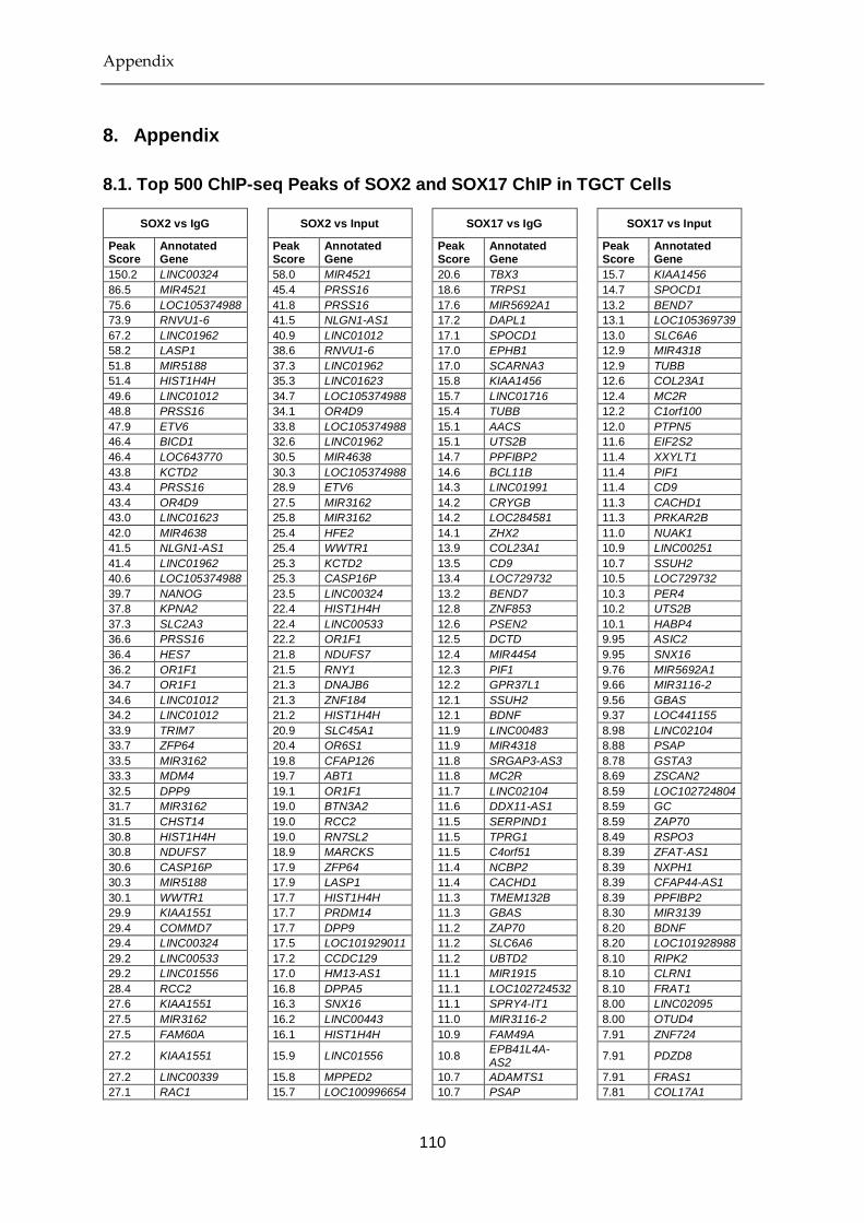
Appendix
110
8. Appendix
8.1. Top 500 ChIP-seq Peaks of SOX2 and SOX17 ChIP in TGCT Cells
SOX2 vs IgG SOX2 vs Input SOX17 vs IgG SOX17 vs Input
Peak Score
Annotated Gene
Peak Score
Annotated Gene
Peak Score
Annotated Gene
Peak Score
Annotated Gene
150.2 LINC00324 58.0 MIR4521 20.6 TBX3 15.7 KIAA1456
86.5 MIR4521 45.4 PRSS16 18.6 TRPS1 14.7 SPOCD1
75.6 LOC105374988 41.8 PRSS16 17.6 MIR5692A1 13.2 BEND7
73.9 RNVU1-6 41.5 NLGN1-AS1 17.2 DAPL1 13.1 LOC105369739
67.2 LINC01962 40.9 LINC01012 17.1 SPOCD1 13.0 SLC6A6
58.2 LASP1 38.6 RNVU1-6 17.0 EPHB1 12.9 MIR4318
51.8 MIR5188 37.3 LINC01962 17.0 SCARNA3 12.9 TUBB
51.4 HIST1H4H 35.3 LINC01623 15.8 KIAA1456 12.6 COL23A1
49.6 LINC01012 34.7 LOC105374988 15.7 LINC01716 12.4 MC2R
48.8 PRSS16 34.1 OR4D9 15.4 TUBB 12.2 C1orf100
47.9 ETV6 33.8 LOC105374988 15.1 AACS 12.0 PTPN5
46.4 BICD1 32.6 LINC01962 15.1 UTS2B 11.6 EIF2S2
46.4 LOC643770 30.5 MIR4638 14.7 PPFIBP2 11.4 XXYLT1
43.8 KCTD2 30.3 LOC105374988 14.6 BCL11B 11.4 PIF1
43.4 PRSS16 28.9 ETV6 14.3 LINC01991 11.4 CD9
43.4 OR4D9 27.5 MIR3162 14.2 CRYGB 11.3 CACHD1
43.0 LINC01623 25.8 MIR3162 14.2 LOC284581 11.3 PRKAR2B
42.0 MIR4638 25.4 HFE2 14.1 ZHX2 11.0 NUAK1
41.5 NLGN1-AS1 25.4 WWTR1 13.9 COL23A1 10.9 LINC00251
41.4 LINC01962 25.3 KCTD2 13.5 CD9 10.7 SSUH2
40.6 LOC105374988 25.3 CASP16P 13.4 LOC729732 10.5 LOC729732
39.7 NANOG 23.5 LINC00324 13.2 BEND7 10.3 PER4
37.8 KPNA2 22.4 HIST1H4H 12.8 ZNF853 10.2 UTS2B
37.3 SLC2A3 22.4 LINC00533 12.6 PSEN2 10.1 HABP4
36.6 PRSS16 22.2 OR1F1 12.5 DCTD 9.95 ASIC2
36.4 HES7 21.8 NDUFS7 12.4 MIR4454 9.95 SNX16
36.2 OR1F1 21.5 RNY1 12.3 PIF1 9.76 MIR5692A1
34.7 OR1F1 21.3 DNAJB6 12.2 GPR37L1 9.66 MIR3116-2
34.6 LINC01012 21.3 ZNF184 12.1 SSUH2 9.56 GBAS
34.2 LINC01012 21.2 HIST1H4H 12.1 BDNF 9.37 LOC441155
33.9 TRIM7 20.9 SLC45A1 11.9 LINC00483 8.98 LINC02104
33.7 ZFP64 20.4 OR6S1 11.9 MIR4318 8.88 PSAP
33.5 MIR3162 19.8 CFAP126 11.8 SRGAP3-AS3 8.78 GSTA3
33.3 MDM4 19.7 ABT1 11.8 MC2R 8.69 ZSCAN2
32.5 DPP9 19.1 OR1F1 11.7 LINC02104 8.59 LOC102724804
31.7 MIR3162 19.0 BTN3A2 11.6 DDX11-AS1 8.59 GC
31.5 CHST14 19.0 RCC2 11.5 SERPIND1 8.59 ZAP70
30.8 HIST1H4H 19.0 RN7SL2 11.5 TPRG1 8.49 RSPO3
30.8 NDUFS7 18.9 MARCKS 11.5 C4orf51 8.39 ZFAT-AS1
30.6 CASP16P 17.9 ZFP64 11.4 NCBP2 8.39 NXPH1
30.3 MIR5188 17.9 LASP1 11.4 CACHD1 8.39 CFAP44-AS1
30.1 WWTR1 17.7 HIST1H4H 11.3 TMEM132B 8.39 PPFIBP2
29.9 KIAA1551 17.7 PRDM14 11.3 GBAS 8.30 MIR3139
29.4 COMMD7 17.7 DPP9 11.2 ZAP70 8.20 BDNF
29.4 LINC00324 17.5 LOC101929011 11.2 SLC6A6 8.20 LOC101928988
29.2 LINC00533 17.2 CCDC129 11.2 UBTD2 8.10 RIPK2
29.2 LINC01556 17.0 HM13-AS1 11.1 MIR1915 8.10 CLRN1
28.4 RCC2 16.8 DPPA5 11.1 LOC102724532 8.10 FRAT1
27.6 KIAA1551 16.3 SNX16 11.1 SPRY4-IT1 8.00 LINC02095
27.5 MIR3162 16.2 LINC00443 11.0 MIR3116-2 8.00 OTUD4
27.5 FAM60A 16.1 HIST1H4H 10.9 FAM49A 7.91 ZNF724
27.2 KIAA1551 15.9 LINC01556 10.8 EPB41L4A-AS2
7.91 PDZD8
27.2 LINC00339 15.8 MPPED2 10.7 ADAMTS1 7.91 FRAS1
27.1 RAC1 15.7 LOC100996654 10.7 PSAP 7.81 COL17A1

Appendix
111
26.0 EIF5A 15.7 MIR4643 10.7 TMPRSS11E 7.81 MIMT1
25.8 PLVAP 15.2 OR6S1 10.6 PYDC2 7.81 CYP7A1
25.6 OR6S1 15.2 MIR3162 10.6 RNF130 7.71 IER2
25.3 FAM60A 15.2 PRSS16 10.5 GSTA3 7.71 CARS
25.2 OR1F1 15.2 MIR3143 10.5 SLC39A10 7.61 PRKD1
24.9 HIST1H4H 15.0 EIF5A 10.4 CCZ1B 7.52 LINC01005
24.8 MLLT1 15.0 MIR5188 10.3 NANOG 7.52 C8orf34-AS1
24.7 CTC1 14.9 LINC01012 10.3 LYST 7.52 ATL2
24.5 VDAC1 14.9 TRIM55 10.2 LOC102724579 7.42 ZBTB7C
24.4 ALOXE3 14.8 MIS18BP1 10.2 ZNF281 7.32 KHDRBS3
24.4 LOC100049716 14.8 HCG16 10.2 MIR646 7.32 LOC101927881
24.3 HM13-AS1 14.8 MIR4634 10.1 LOC105369739 7.22 YWHAZ
24.3 AMN1 14.6 LINC02016 10.1 HABP4 7.22 LINC01381
24.2 ZNF184 14.4 VN1R10P 10.1 MIR548I1 7.12 ZNF281
23.8 HCG16 14.3 DOT1L 9.95 LOC101929341 7.12 LOC101928304
23.5 LINC00533 14.3 MDM4 9.95 SNX16 7.12 ESRRB
23.5 LINC01623 14.3 ZYG11A 9.95 NFIB 7.03 AGBL4-IT1
23.1 SCAMP4 14.1 KIAA1551 9.95 CCDC179 7.03 MIR548XHG
23.0 DOT1L 14.0 RBMS3-AS1 9.95 ARTN 7.03 SUSD3
23.0 MPZL1 14.0 ZNF184 9.86 COL17A1 7.03 CNTN4-AS1
22.9 UBE2D2 13.8 LINC01623 9.76 IER2 6.93 SMG8
22.9 LINC01012 13.8 ARHGEF3 9.76 PTPN5 6.93 LINC00862
22.7 SLC45A1 13.8 SOX5 9.76 MIR141 6.93 SIAE
22.7 GNGT2 13.5 USP7 9.66 NARS2 6.83 DAPL1
22.4 OR2V2 13.5 CCNH 9.66 LOC101928988 6.73 KIT
22.4 HES7 13.4 ZBED9 9.66 EIF2S2 6.73 PRDM1
22.2 RNY1 13.2 ZNF184 9.56 TFDP2 6.73 ALDH3B2
22.1 LINC01556 13.1 LINC00339 9.56 H3F3A 6.73 GUCY1A3
22.0 HMGN4 13.1 CHST14 9.56 MIR548AG2 6.64 CXCR4
21.7 SLC2A14 13.0 LINC00533 9.37 LOC441155 6.64 MIR4454
21.7 BTN3A2 12.9 SSB 9.37 KIDINS220 6.54 CXorf21
21.6 BCAS4 12.7 PRSS16 9.37 XXYLT1 6.54 IQCF1
21.6 NANOG 12.6 CLEC3A 9.37 LOC105369739 6.54 CNGB1
21.3 DNAJB6 12.6 LINC01822 9.27 SPRY4-IT1 6.54 CCDC179
21.3 ZNF692 12.5 MIR5188 9.27 CPNE8 6.54 ENPP2
21.1 PRSS16 12.5 ZNF184 9.17 ZNF592 6.44 RBFOX1
20.6 A2ML1 12.5 ZHX2 9.08 LOC101928797 6.44 MIR181C
20.6 CFAP126 12.3 LINC01012 9.08 GSN 6.44 LOC100506885
20.4 LINC01498 12.2 LINC01012 9.08 MIMT1 6.34 DDX11-AS1
20.3 ZYG11A 12.1 ZNF483 8.98 LOC105375075 6.34 LOC101929544
20.3 NACC1 12.1 SMARCA2 8.98 KALRN 6.25 HULC
20.2 ABT1 12.1 LINC01012 8.88 POU5F1 6.25 KIDINS220
20.2 TMPO-AS1 12.0 SMARCC2 8.88 SOX5 6.25 SLC39A11
20.0 RNY3 11.8 HTR1F 8.88 LINC01356 6.15 LINC01060
19.8 LOC100996654 11.8 NOBOX 8.78 PCDH1 6.15 NR2F2
19.8 LINC00941 11.8 OR1F1 8.78 LINC01951 6.15 NDUFB6
19.8 SLC2A14 11.8 FBRS 8.78 KRT18 6.05 LOC105376360
19.7 RN7SL2 11.7 SMARCA2 8.78 PGAP1 6.05 IRX5
19.5 MIR6883 11.7 SHISA2 8.78 RNF11 6.05 LOC101928008
19.5 ZNF184 11.6 DDAH1 8.69 CARS 6.05 CDH18
19.4 RNF112 11.6 LRIG1 8.59 MIR3977 6.05 BASP1
19.4 SLC2A14 11.4 PLEKHB2 8.49 TSPAN9 5.95 LOC102725080
19.3 PRDM14 11.4 LINC01629 8.49 PUM2 5.95 PMAIP1
19.3 LINC01012 11.4 MIR5692B 8.39 LINC02114 5.95 NR2F6
19.3 GNG7 11.4 HK3 8.39 FTO 5.95 MIR8084
19.3 HIST1H4H 11.3 CNN1 8.39 ZNF552 5.95 LINC02032
19.1 OSCP1 11.3 MIR5188 8.39 BICD1 5.95 RNF130
19.1 PLXDC1 11.2 HSPA6 8.30 MACROD2-
AS1 5.95 ESRP1
19.1 MIR193BHG 11.2 NIFK-AS1 8.30 FSCN1 5.95 ZNF565
19.0 MIR4638 11.2 KPNA2 8.30 CCDC129 5.86 CBR4
19.0 OR1M1 11.2 DUSP6 8.30 CRHBP 5.86 PYDC2
19.0 MIR4472-2 11.1 MKRN1 8.20 CAND1.11 5.86 LOC101928797
19.0 DPPA5 11.1 LINC01012 8.20 SMYD3 5.86 STARD13
18.9 MARCKS 11.1 AFF1 8.20 MIR4634 5.76 NRF1

Appendix
112
18.8 PLLP 11.1 ERCC1 8.20 ZFAT-AS1 5.76 ARSJ
18.6 LOC105376805 11.1 NACC1 8.20 IL20RB 5.76 TPTE
18.5 TRIM7 11.1 MIR3690 8.20 SLC45A1 5.76 DDX18
18.4 LINC01012 10.8 MORC3 8.10 EDIL3 5.76 KIAA1456
18.4 B4GALT5 10.8 GRPR 8.10 NCEH1 5.66 DNAJB6
18.2 UBE2D2 10.8 GNGT2 8.00 SPIDR 5.66 OCM
18.2 LINC01841 10.8 PLEKHG2 8.00 MIR3191 5.66 RPA1
18.2 PCCB 10.8 ILF2 8.00 LOC100130298 5.66 MIR6085
18.1 LOC100507468 10.7 TIA1 8.00 BAMBI 5.66 FAM83A
18.0 PCDH1 10.7 CASP16P 8.00 TMEM87B 5.66 PRDM14
18.0 PSMB2 10.5 LINC01448 7.91 ZNF724 5.56 TSN
18.0 FOXK2 10.5 SLC35D3 7.91 IQCF1 5.56 C5orf66-AS1
18.0 RASSF8-AS1 10.5 APOBEC3F 7.91 LOC101927735 5.47 SDK1
17.9 USP48 10.4 RPL36 7.91 GREB1L 5.47 MIR1915
17.9 KIAA1551 10.4 MUC4 7.81 PMAIP1 5.47 HDAC2
17.7 LINC01623 10.4 LOC100131289 7.81 MIR135B 5.47 FAM136A
17.7 OAT 10.3 PIH1D1 7.81 LINGO2 5.47 TMEM261
17.6 NANOGNB 10.3 GLOD5 7.81 TIFAB 5.37 LOC100289230
17.6 SRCIN1 10.2 RCOR1 7.81 LOC100128386 5.37 MIR610
17.6 SALL4 10.2 MIR9-2 7.71 ZBTB2 5.37 LINC02003
17.5 LOC101929011 10.2 PLAGL1 7.71 CFAP44-AS1 5.37 PIN1P1
17.5 MIR3143 10.2 ZBTB5 7.71 CYP4A22 5.37 DCTD
17.3 ZBTB8OS 10.2 TERF1 7.61 MIR548AU 5.37 LOC100192426
17.3 KDM4C 10.2 GPD2 7.61 MIR548A2 5.37 BCL9
17.2 LINC01012 10.0 HIST1H4H 7.61 ZFAT-AS1 5.37 ATP6V1G3
17.2 CCDC129 10.0 GGACT 7.61 ADAMTS6 5.37 PPM1A
17.1 PMS2CL 9.89 ZC3HAV1L 7.61 TFRC 5.37 NPAS3
17.0 MIR193A 9.89 ANKRD1 7.61 TAB2 5.37 LOC102546299
17.0 ATF1 9.89 NANOG 7.52 TBCE 5.37 LOC101927056
17.0 SNORA38B 9.77 TRIP4 7.52 PDP1 5.27 SMYD3
17.0 C7orf55-LUC7L2
9.77 LOC105374988 7.52 DCD 5.27 C1orf105
16.8 UBE2G1 9.77 SIAH2 7.42 IGSF5 5.27 MIR383
16.8 PCGF6 9.77 PCLO 7.42 ZBTB7C 5.27 MGC27382
16.8 LINC00443 9.64 LINC01947 7.42 LOC105374428 5.27 LINC01375
16.8 ZNF184 9.51 UBE2D2 7.42 PDZD8 5.27 NABP1
16.8 SMARCC2 9.51 LINC00324 7.42 KIZ 5.17 MDH1
16.7 EEF2K 9.51 SLC35F3 7.32 KHDRBS3 5.17 OR6S1
16.7 PMS2P4 9.38 RIF1 7.32 GBA3 5.17 LOC101927847
16.6 LINC01447 9.38 SPATA45 7.32 ENPP2 5.17 MIR3191
16.6 VSIG10 9.38 GRIA3 7.32 LINC01267 5.17 FIS1
16.4 MLXIPL 9.25 REST 7.32 LOC101927881 5.17 ITGA9-AS1
16.4 MELK 9.25 BTN3A2 7.32 LINC02095 5.17 WDR36
16.4 LINC02016 9.25 CLPP 7.32 FSCN1 5.17 LOC730100
16.3 SNX16 9.25 LINC00533 7.32 TCERG1L 5.17 UMODL1-AS1
16.2 MTA3 9.12 RPA3 7.32 MIR6085 5.08 ABCB5
16.2 AXIN2 9.12 ZBTB8B 7.32 NUDT12 5.08 MACROD2
16.2 FOXK2 9.12 MIR3675 7.22 PLXNA2 5.08 CFHR5
16.2 ZNF815P 9.12 TTC39B 7.22 MIR3139 5.08 GBA3
16.1 ZNF184 9.00 LOC284788 7.12 CCAT2 5.08 TEAD1
16.1 RLIM 9.00 ALOXE3 7.12 LOC728715 5.08 DPPA5
15.9 BORCS7-ASMT
9.00 LOC102724623 7.12 UMODL1-AS1 5.08 LOC100129216
15.9 LOC101926933 9.00 MDM4 7.03 GPR156 5.08 GTF2I
15.8 SMU1 8.87 JAKMIP2-AS1 7.03 AGBL4-IT1 5.08 KIAA0513
15.8 MPPED2 8.74 MIR4666B 7.03 LOC102724404 5.08 KCTD3
15.8 USP7 8.74 LINC01589 7.03 SEPT9 5.08 KYNU
15.8 ZYG11A 8.74 RRAGC 7.03 AQP6 5.08 CUEDC1
15.8 OR1F1 8.74 LOC101928782 7.03 EPHA3 4.98 PTPRD-AS2
15.7 MIR4643 8.61 PMS2CL 7.03 MIR548XHG 4.98 REXO1L2P
15.7 AKAP3 8.61 COX5A 6.93 LSAMP-AS1 4.98 MIR548AD
15.7 LOC105374988 8.61 ASIC2 6.93 COL4A1 4.98 LINC01923
15.5 MIR5188 8.61 LOC101929154 6.93 MIR4266 4.98 MBNL1
15.5 RAB11B 8.61 ZNF717 6.93 C9orf72 4.98 PCGEM1
15.5 DUSP6 8.61 RIMS2 6.93 LOC102723886 4.98 FREM3
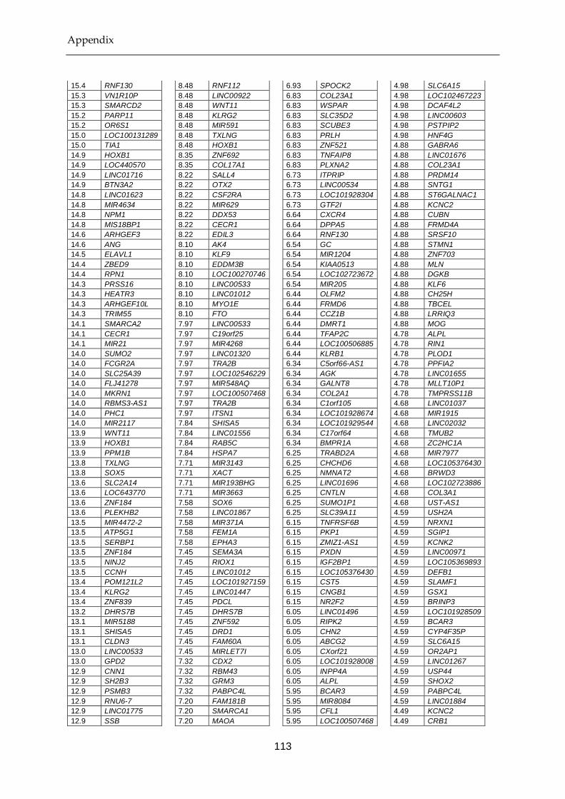
Appendix
113
15.4 RNF130 8.48 RNF112 6.93 SPOCK2 4.98 SLC6A15
15.3 VN1R10P 8.48 LINC00922 6.83 COL23A1 4.98 LOC102467223
15.3 SMARCD2 8.48 WNT11 6.83 WSPAR 4.98 DCAF4L2
15.2 PARP11 8.48 KLRG2 6.83 SLC35D2 4.98 LINC00603
15.2 OR6S1 8.48 MIR591 6.83 SCUBE3 4.98 PSTPIP2
15.0 LOC100131289 8.48 TXLNG 6.83 PRLH 4.98 HNF4G
15.0 TIA1 8.48 HOXB1 6.83 ZNF521 4.88 GABRA6
14.9 HOXB1 8.35 ZNF692 6.83 TNFAIP8 4.88 LINC01676
14.9 LOC440570 8.35 COL17A1 6.83 PLXNA2 4.88 COL23A1
14.9 LINC01716 8.22 SALL4 6.73 ITPRIP 4.88 PRDM14
14.9 BTN3A2 8.22 OTX2 6.73 LINC00534 4.88 SNTG1
14.8 LINC01623 8.22 CSF2RA 6.73 LOC101928304 4.88 ST6GALNAC1
14.8 MIR4634 8.22 MIR629 6.73 GTF2I 4.88 KCNC2
14.8 NPM1 8.22 DDX53 6.64 CXCR4 4.88 CUBN
14.8 MIS18BP1 8.22 CECR1 6.64 DPPA5 4.88 FRMD4A
14.6 ARHGEF3 8.22 EDIL3 6.64 RNF130 4.88 SRSF10
14.6 ANG 8.10 AK4 6.54 GC 4.88 STMN1
14.5 ELAVL1 8.10 KLF9 6.54 MIR1204 4.88 ZNF703
14.4 ZBED9 8.10 EDDM3B 6.54 KIAA0513 4.88 MLN
14.4 RPN1 8.10 LOC100270746 6.54 LOC102723672 4.88 DGKB
14.3 PRSS16 8.10 LINC00533 6.54 MIR205 4.88 KLF6
14.3 HEATR3 8.10 LINC01012 6.44 OLFM2 4.88 CH25H
14.3 ARHGEF10L 8.10 MYO1E 6.44 FRMD6 4.88 TBCEL
14.3 TRIM55 8.10 FTO 6.44 CCZ1B 4.88 LRRIQ3
14.1 SMARCA2 7.97 LINC00533 6.44 DMRT1 4.88 MOG
14.1 CECR1 7.97 C19orf25 6.44 TFAP2C 4.78 ALPL
14.1 MIR21 7.97 MIR4268 6.44 LOC100506885 4.78 RIN1
14.0 SUMO2 7.97 LINC01320 6.44 KLRB1 4.78 PLOD1
14.0 FCGR2A 7.97 TRA2B 6.34 C5orf66-AS1 4.78 PPFIA2
14.0 SLC25A39 7.97 LOC102546229 6.34 AGK 4.78 LINC01655
14.0 FLJ41278 7.97 MIR548AQ 6.34 GALNT8 4.78 MLLT10P1
14.0 MKRN1 7.97 LOC100507468 6.34 COL2A1 4.78 TMPRSS11B
14.0 RBMS3-AS1 7.97 TRA2B 6.34 C1orf105 4.68 LINC01037
14.0 PHC1 7.97 ITSN1 6.34 LOC101928674 4.68 MIR1915
14.0 MIR2117 7.84 SHISA5 6.34 LOC101929544 4.68 LINC02032
13.9 WNT11 7.84 LINC01556 6.34 C17orf64 4.68 TMUB2
13.9 HOXB1 7.84 RAB5C 6.34 BMPR1A 4.68 ZC2HC1A
13.9 PPM1B 7.84 HSPA7 6.25 TRABD2A 4.68 MIR7977
13.8 TXLNG 7.71 MIR3143 6.25 CHCHD6 4.68 LOC105376430
13.8 SOX5 7.71 XACT 6.25 NMNAT2 4.68 BRWD3
13.6 SLC2A14 7.71 MIR193BHG 6.25 LINC01696 4.68 LOC102723886
13.6 LOC643770 7.71 MIR3663 6.25 CNTLN 4.68 COL3A1
13.6 ZNF184 7.58 SOX6 6.25 SUMO1P1 4.68 UST-AS1
13.6 PLEKHB2 7.58 LINC01867 6.25 SLC39A11 4.59 USH2A
13.5 MIR4472-2 7.58 MIR371A 6.15 TNFRSF6B 4.59 NRXN1
13.5 ATP5G1 7.58 FEM1A 6.15 PKP1 4.59 SGIP1
13.5 SERBP1 7.58 EPHA3 6.15 ZMIZ1-AS1 4.59 KCNK2
13.5 ZNF184 7.45 SEMA3A 6.15 PXDN 4.59 LINC00971
13.5 NINJ2 7.45 RIOX1 6.15 IGF2BP1 4.59 LOC105369893
13.5 CCNH 7.45 LINC01012 6.15 LOC105376430 4.59 DEFB1
13.4 POM121L2 7.45 LOC101927159 6.15 CST5 4.59 SLAMF1
13.4 KLRG2 7.45 LINC01447 6.15 CNGB1 4.59 GSX1
13.4 ZNF839 7.45 PDCL 6.15 NR2F2 4.59 BRINP3
13.2 DHRS7B 7.45 DHRS7B 6.05 LINC01496 4.59 LOC101928509
13.1 MIR5188 7.45 ZNF592 6.05 RIPK2 4.59 BCAR3
13.1 SHISA5 7.45 DRD1 6.05 CHN2 4.59 CYP4F35P
13.1 CLDN3 7.45 FAM60A 6.05 ABCG2 4.59 SLC6A15
13.0 LINC00533 7.45 MIRLET7I 6.05 CXorf21 4.59 OR2AP1
13.0 GPD2 7.32 CDX2 6.05 LOC101928008 4.59 LINC01267
12.9 CNN1 7.32 RBM43 6.05 INPP4A 4.59 USP44
12.9 SH2B3 7.32 GRM3 6.05 ALPL 4.59 SHOX2
12.9 PSMB3 7.32 PABPC4L 5.95 BCAR3 4.59 PABPC4L
12.9 RNU6-7 7.20 FAM181B 5.95 MIR8084 4.59 LINC01884
12.9 LINC01775 7.20 SMARCA1 5.95 CFL1 4.49 KCNC2
12.9 SSB 7.20 MAOA 5.95 LOC100507468 4.49 CRB1

Appendix
114
12.7 CERS4 7.20 TLK1 5.95 ESRRB 4.49 LINC01947
12.7 KDELR2 7.20 RBM17 5.95 LINC01210 4.49 CWF19L2
12.6 CFAP126 7.20 HSPA9 5.95 SLC36A4 4.49 DOCK3
12.6 CCND2 7.07 ACTR3B 5.95 ZNF565 4.49 LINC01720
12.6 SMARCA2 7.07 DPYD-AS2 5.95 SUGCT 4.49 TYRP1
12.6 MIR589 7.07 PMS2P4 5.86 FAM222B 4.49 PRG4
12.6 CLEC3A 7.07 PTPRZ1 5.86 LMO7-AS1 4.49 LINC01815
12.6 CCND2 7.07 LINC01440 5.86 LINC01699 4.49 MKRN9P
12.5 RCOR1 7.07 LOC101928386 5.86 GRK5 4.49 PRRC2B
12.5 SUZ12P1 7.07 TPRG1-AS1 5.86 FAM188B 4.49 UNC80
12.5 SEPHS1 7.07 NICN1 5.86 ITGB1 4.49 DOK5
12.5 GACAT3 6.94 RNF113A 5.86 SCGB1A1 4.49 LRIG3
12.5 ELAVL1 6.94 TMEM214 5.86 PABPC4L 4.49 SNTG1
12.5 CFAP126 6.94 LOC101928519 5.86 CYP1B1 4.49 LINC01370
12.5 ZHX2 6.94 KCMF1 5.86 FBXW4 4.49 DCN
12.3 LINC01012 6.94 LOC101929154 5.86 UBALD2 4.39 SLC6A5
12.3 CNR2 6.94 LINC01340 5.86 SLC39A1 4.39 COMMD1
12.3 SERBP1 6.94 LINC01714 5.76 SALL1 4.39 DGKB
12.3 COX6A1 6.94 TOB2P1 5.76 DNTT 4.39 OR6N1
12.3 IER2 6.81 RNF187 5.76 ESRP1 4.39 FRAT2
12.3 BISPR 6.81 CNTN4 5.76 LOC101929011 4.39 LINC02127
12.2 ZC3HAV1L 6.81 RBFOX1 5.76 ARSJ 4.39 LINC01142
12.2 LINC01819 6.81 TMPRSS11E 5.76 DDX18 4.39 LRBA
12.2 FBRS 6.81 LOC646241 5.76 CABLES1 4.39 HPVC1
12.2 HDAC11-AS1 6.81 ZNF777 5.76 CAAP1 4.39 CLEC14A
12.2 UPP1 6.81 RAB11FIP2 5.76 LOC101928894 4.39 LINC01790
12.2 DNMBP 6.81 LINC00648 5.76 NMNAT3 4.39 LOC100507468
12.1 CLPP 6.81 PACRG-AS2 5.76 EPHB3 4.39 ANKS1B
12.1 EEF2 6.81 POM121L2 5.66 DNAJB6 4.39 SKIL
12.1 HSPA6 6.68 GLRA4 5.66 FRAT2 4.39 DPP4
12.1 HK3 6.68 KCND2 5.66 OCM 4.39 ALG10
12.1 ZNF483 6.68 BMP7-AS1 5.66 RPA1 4.39 MIR4445
12.1 ADGRL1 6.68 C9orf85 5.66 SLAMF1 4.39 LOC100507468
12.1 FXR2 6.68 SEPHS1 5.66 CFLAR 4.39 DPY19L2
12.1 GAST 6.68 EIF3E 5.66 ID1 4.39 SLC4A4
12.0 PLIN3 6.68 RHOU 5.66 THBD 4.39 MIR4735
12.0 RPL36 6.68 JARID2 5.66 FAM196B 4.29 ZBED8
12.0 SNORD118 6.68 GGA3 5.66 PPIF 4.29 SYT1
12.0 DPRXP4 6.68 POLG 5.66 BMF 4.29 MIR3919
11.8 HTR1F 6.68 MIR513C 5.66 MLN 4.29 CA10
11.8 NOBOX 6.68 BRAT1 5.66 LOC105376360 4.29 SLC24A2
11.8 METTL21A 6.68 LINC00324 5.56 LOC646730 4.29 DCLK2
11.8 MIR3690 6.55 MIR4699 5.56 AOAH-IT1 4.29 BASP1
11.7 NUP85 6.55 CCDC57 5.56 DDX11-AS1 4.29 LINC01851
11.7 TTLL6 6.55 RHBDD2 5.56 DDX3X 4.29 PKIA
11.7 SHISA2 6.55 LINC01623 5.56 FBXL19-AS1 4.29 GPR149
11.7 CERS5 6.55 LINC00269 5.56 PLPP3 4.29 SLC36A4
11.7 PINK1 6.55 VWC2 5.56 ADAMTSL4 4.29 LINC01822
11.7 ALOXE3 6.55 UBE2G1 5.56 TSPAN18 4.29 TIA1
11.6 ILF2 6.55 MIR4654 5.56 DET1 4.29 LOC102467226
11.6 FAM212B-AS1 6.55 DAPK1 5.56 KCTD15 4.29 MKRN9P
11.6 APOBEC3F 6.55 ZNF184 5.56 ASIC2 4.29 MIR3974
11.6 LINC01822 6.55 MC4R 5.56 EGFEM1P 4.29 MIR181B1
11.6 SLC2A14 6.55 PLS3 5.56 PNPT1 4.29 ZNF804A
11.6 SIAH2 6.55 ADAMTS19-AS1
5.56 ADAMTS14 4.29 PXDNL
11.4 LINC01629 6.55 LINC00533 5.47 CCDC50 4.29 ST3GAL1
11.4 TEX43 6.55 SERPINI1 5.47 NCOR2 4.29 FRMD6-AS1
11.4 LOC102723557 6.43 CREB5 5.47 TMEM170B 4.29 KITLG
11.4 YIPF4 6.43 MIR6087 5.47 LINC01696 4.29 LOC105369860
11.4 CHMP6 6.43 TERC 5.47 KRTCAP3 4.29 SPRED2
11.4 MORC3 6.43 LINC01381 5.47 MATR3 4.29 LMO7-AS1
11.4 LINC01448 6.43 HOOK1 5.47 HULC 4.29 MIR4735
11.4 CP 6.43 LINC01320 5.47 SLC2A1-AS1 4.29 SI
11.4 PWWP2A 6.43 AFF2 5.47 HNF1B 4.29 TMEFF2

Appendix
115
11.4 RFX1 6.43 MYNN 5.47 PRPF6 4.29 LINC01070
11.3 FBN3 6.43 CHSY3 5.47 STARD13 4.29 FZD4
11.3 POLR2G 6.43 SARS 5.37 ASB1 4.29 KCNQ5
11.3 TLE2 6.30 ABCA1 5.37 GPX5 4.29 MIR548AE1
11.3 MUC4 6.30 NTN1 5.37 LINC02003 4.29 YTHDC2
11.3 GGACT 6.30 SERBP1 5.37 GDF6 4.29 LINC01111
11.2 ABCA13 6.30 TBC1D32 5.37 GALNT2 4.29 PDE4B
11.2 RPUSD3 6.30 GFRA1 5.37 XRCC5 4.29 MIR149
11.2 LHFPL4 6.30 LINC00972 5.37 GRHPR 4.29 MIR4999
11.2 EIF4B 6.30 UBXN11 5.37 PPM1A 4.29 LRFN5
11.2 LINC00941 6.30 MIR3162 5.37 RARRES3 4.29 LINC01620
11.2 CAND2 6.17 RCC1 5.37 CUBN 4.29 LINC02010
11.2 NIFK-AS1 6.17 LINC01609 5.37 LOC101927056 4.29 XIRP2
11.2 TTC39B 6.17 TMEM106B 5.37 ARID1B 4.29 FUNDC2P2
11.1 UIMC1 6.17 MORF4L1 5.37 TMUB2 4.20 MIR8065
11.1 RIF1 6.17 METTL21A 5.27 PKDCC 4.20
11.1 LINC01783 6.17 PTPRG 5.27 FAM86DP 4.20 LINC01419
11.1 AFF1 6.17 TYRP1 5.27 DDX11-AS1 4.20 PTPRR
11.1 GADD45GIP1 6.17 SHOX2 5.27 LRRC4B 4.20 IZUMO3
11.1 ERCC1 6.17 FBXW2 5.27 UXS1 4.20 PTCHD1
10.9 WDR66 6.17 PFKFB4 5.27 IRX6 4.20 C1orf140
10.9 CROCC 6.17 LEFTY2 5.27 IER2 4.20 NKAIN3
10.9 MOG 6.17 RNASE12 5.27 PAG1 4.20 TRIM2
10.9 DDAH1 6.04 LOC101927139 5.27 PDE2A 4.20 NR4A3
10.9 CLEC4C 6.04 MYLIP 5.27 PSORS1C2 4.20 MGAT4C
10.9 MGC12916 6.04 KDM4B 5.27 PRRC2B 4.20 DUSP10
10.9 LRIG1 6.04 LINC01716 5.27 LOC101928540 4.20 LOC440982
10.9 MIR3675 6.04 POLR2G 5.27 MIR3664 4.20 CASP8
10.8 ZMYND8 6.04 EOMES 5.27 NEAT1 4.20 CYP1B1
10.8 GRPR 6.04 DAP 5.27 LOC102724933 4.20 ADAMTS20
10.8 PLEKHG2 6.04 LINC00906 5.27 CASC18 4.20 MAP4K5
10.8 UBXN11 6.04 NAALADL2 5.27 WI2-2373I1.2 4.20 LINC01170
10.8 MSH6 6.04 ID3 5.27 RARG 4.20 LINC01322
10.8 VDAC1 6.04 LARP1B 5.27 ALX1 4.20 ZNF335
10.8 ATPIF1 6.04 EYA1 5.17 AKAP1 4.20 LINC01239
10.8 UNC13B 6.04 COMMD7 5.17 MIR569 4.10 NRP1
10.8 MIR4472-2 6.04 MYOF 5.17 ALDH7A1 4.10 MIR4735
10.7 IGFBP4 6.04 DDX43 5.17 ATP6V1C2 4.10 GXYLT1
10.7 FCHO1 5.91 CRLF2 5.17 DKK1 4.10 LINC01609
10.7 APTR 5.91 SLTM 5.17 GREB1L 4.10 FGF20
10.7 LINC01857 5.91 DLGAP1-AS2 5.17 ST3GAL1 4.10 DGKB
10.7 LINC00922 5.91 PABPC1P2 5.17 JUP 4.10 IGSF5
10.5 ARID3A 5.91 SNX16 5.17 ZNF302 4.10 BACH1-IT2
10.5 TMEM214 5.91 LINC01036 5.17 C11orf58 4.10 RCC2
10.5 MTMR14 5.91 DPP10 5.17 CDH10 4.10 SEMA6D
10.5 VAMP2 5.91 MIR5188 5.17 LOC100507468 4.10 FAM19A2
10.5 SLC35D3 5.91 SGIP1 5.17 WBSCR17 4.10 FGFR3
10.4 MAN1C1 5.91 LOC101929555 5.17 CDH4 4.10 KCNIP4-IT1
10.4 RPL6 5.91 LOC101929570 5.17 RRM1 4.10 CDCA7L
10.4 OR2B2 5.91 MIR1263 5.17 RFK 4.10 DIRC1
10.4 TNRC18 5.91 TRPC7 5.17 OPRM1 4.10 LINC00524
10.4 LOC100507468 5.91 MIR4426 5.17 LINC01070 4.10 LINC01031
10.4 LINC01947 5.91 SRSF10 5.17 MIR4427 4.10 LINC01935
10.4 ZNF184 5.91 TDGF1 5.08 SLC28A3 4.10 NFATC1
10.4 MELK 5.91 LOC101928909 5.08 CSF1 4.10 NFIB
10.4 LINC01267 5.91 SMU1 5.08 CHCHD6 4.10 LINC01239
10.4 WSB1 5.78 RN7SL1 5.08 LINGO2 4.10 LINC01554
10.4 RAF1 5.78 CD99L2 5.08 CFAP126 4.10 PELO
10.4 RNF112 5.78 MIR4459 5.08 HSPA8 4.10 LOC102723895
10.4 ANG 5.78 ETV1 5.08 USP32 4.10 MROH9
10.4 UHRF1BP1L 5.78 RPS24 5.08 LOC102724890 4.10 LOC100129138
10.4 RRAGC 5.78 LMX1B 5.08 NRP1 4.10 PLSCR2
10.4 LHX5 5.78 RMND5B 5.08 HNRNPAB 4.10 MTAP
10.3 CASP16P 5.78 STK10 5.08 LOC100130298 4.10 SP3
10.3 TIMP4 5.78 SAMD5 5.08 ECE1 4.10 DNAH7

Appendix
116
10.3 GLOD5 5.78 EPHA3 5.08 LINC01063 4.10 MIR2053
10.3 PFKFB4 5.78 RIPK1 5.08 RIMS1 4.00 PTPRD
10.3 PIH1D1 5.78 YWHAE 5.08 KYNU 4.00 TMEM178A
10.3 MIR648 5.78 LINGO2 4.98 OTUD4 4.00 P2RY1
10.3 RPEL1 5.78 MTF2 4.98 TTC25 4.00 FAM46A
10.3 HNF1A-AS1 5.78 SNUPN 4.98 KDELR2 4.00 OR2L5
10.3 SPATA45 5.78 WDR59 4.98 NUDCD1 4.00 KCNB2
10.2 LINC01164 5.78 LINC01623 4.98 ZFAT 4.00 FGF13
10.2 RCC2 5.78 MIR371A 4.98 PLAGL1 4.00 APBB2
10.2 SRCIN1 5.65 KLF4 4.98 WWP2 4.00 NUDT12
10.2 LOC105369723 5.65 SLC44A1 4.98 ETV4 4.00 LINC01343
10.2 ZBTB5 5.65 PLOD2 4.98 LINC01139 4.00 LOC102723833
10.2 TERF1 5.65 OR1F1 4.98 C20orf85 4.00 FRG1BP
10.2 RPN1 5.65 SSTR2 4.98 GNPDA2 4.00 LOC105377143
10.2 CERS4 5.65 FAM212B-AS1 4.98 TRIM24 4.00 LINC01467
10.2 WWTR1-AS1 5.65 SLC2A3 4.98 KIF15 4.00 CEBPZ
10.2 ADGRL1 5.65 PRUNE1 4.88 ZNF608 4.00 MIR5688
10.2 NANOG 5.65 RPL34-AS1 4.88 GC 4.00 LOC100506474
10.2 MIR9-2 5.65 AACSP1 4.88 SLC36A4 4.00 SI
10.2 PLAGL1 5.65 NRXN3 4.88 MIR3917 4.00 LRRC4B
10.2 STYK1 5.65 LAMA2 4.88 RSPO3 4.00 SOX2-OT
10.2 BCAT1 5.65 TMEM106B 4.88 PDK1 4.00 DKK1
10.0 ZNF184 5.65 OSCP1 4.88 RBFOX1 4.00 LOC101927849
10.0 ZNF335 5.65 NXPH1 4.88 FAM19A5 4.00 LINC01324
10.0 SLC44A1 5.65 TMEM14EP 4.88 PSKH2 4.00 LINGO2
10.0 ZNF675 5.65 NATD1 4.88 LMX1B 4.00 ELAVL2
10.0 CASC21 5.65 LOC100996251 4.88 LINC01090 4.00 LOC105376633
10.0 ZMYND8 5.65 SOX2-OT 4.88 CUEDC1 4.00 ADGRB3
10.0 RAB5C 5.65 OR5AR1 4.88 NSD1 4.00 RELN
10.0 LYZL1 5.65 PIM2 4.88 COL4A1 4.00 FOXN3-AS2
10.0 BASP1 5.53 LOC100132831 4.88 KLF6 4.00 MIR1-1HG
10.0 FGD4 5.53 ZMYND8 4.88 ISG20 4.00 LINC01473
10.0 LOC284788 5.53 TRAPPC8 4.88 NBPF3 4.00 SATB2
10.0 WNT5B 5.53 TMEM2 4.88 TBCEL 4.00 MIR205
10.0 DENND2A 5.53 LINC01012 4.88 CTSO 4.00 NLN
10.0 ATF7IP 5.53 ASMT 4.78 LINC00964 4.00 PRG4
9.89 FAM60A 5.53 LRP1B 4.78 SP4 4.00 LOC105373782
9.89 MIR5188 5.53 YIPF5 4.78 RHOB 3.90 LRP12
9.89 TNFAIP8L1 5.53 FOXN3 4.78 ZBTB10 3.90 UGT2A3
9.89 TERC 5.53 LINC01242 4.78 LINGO2 3.90 CHRM3
9.89 SPNS3 5.53 LOC105369860 4.78 TADA3 3.90 LINC01948
9.89 SRCIN1 5.53 PCLO 4.78 TMEM132D 3.90 LINC01923
9.89 MACF1 5.53 C17orf112 4.78 NDUFS5 3.90 BRINP3
9.89 ANKRD1 5.53 SPEN 4.78 TENM3 3.90 PRDM14
9.89 LOC105369632 5.53 SNORD12 4.78 CNDP1 3.90 LINC01995
9.89 RPH3A 5.53 OTUD4 4.78 KIAA1456 3.90 PSAT1
9.89 ZNF184 5.53 CCDC62 4.78 CENPN 3.90 STARD7
9.77 KIF2C 5.53 ANKS1B 4.78 LINC00551 3.90 LINC01467
9.77 TGFBR2 5.40 SFRP2 4.78 PTPRS 3.90 LOC102723831
9.77 PXN 5.40 CWC27 4.78 EEF1A1 3.90 SFRP2
9.77 OR6S1 5.40 LINC01756 4.78 MIR4792 3.90 MIR6827
9.77 PCLO 5.40 CASP16P 4.78 TYRO3 3.90 MIR4735
9.77 GRIA3 5.40 DHX15 4.78 LOC101927043 3.90 CDAN1
9.77 UBE2G1 5.40 NEGR1 4.78 SLAMF7 3.90 TUSC1
9.77 LOC105374988 5.40 PHF12 4.78 MIR2053 3.90 LOC102724053
9.77 RAD52 5.40 ASGR2 4.78 PRSS36 3.90 TOX
9.77 TRIP4 5.40 LOC101928834 4.78 SPATA6 3.90 LOC100129434
9.77 RP9P 5.40 LINC01320 4.78 FUZ 3.90 OR4A5
9.77 MORF4L1 5.40 CDKL3 4.78 LINC01291 3.90 LOC105375972
9.77 ZFP36L2 5.40 TMEM261 4.78 USP32 3.90 LOC100506474
9.77 RHBDD2 5.40 SKAP2 4.78 BMPR1A 3.90 KCNC2
9.77 SPEN 5.40 SPATA16 4.78 DEFB131 3.90 GOLIM4
9.77 EIF2AK1 5.40 IAPP 4.78 KIAA1614 3.90 PRRX1
9.64 SLC2A14 5.40 FAT4 4.78 LOC646241 3.90 LINC01239
9.64 C19orf25 5.40 LOC100506474 4.68 FAM84B 3.90 HDAC9

Appendix
117
9.64 SYT6 5.40 MIR3686 4.68 MIR4454 3.90 RAB17
9.64 DNM1L 5.40 GPRIN3 4.68 PYGL 3.90 BSPRY
9.64 RIOX1 5.40 PER4 4.68 MIR1915 3.90 GLRA3
9.64 SLC2A14 5.40 PRRC2C 4.68 UBQLN4 3.90 LGR5
9.64 MIR1303 5.40 ATF6 4.68 LINC00303 3.90 STMN2
9.64 BRDT 5.40 LOC101929926 4.68 ITGB5 3.90 UBE2N
9.64 PIK3CB 5.40 PTBP2 4.68 SRD5A2 3.90 TRHR
9.64 GDF3 5.40 LRRC7 4.68 C8orf58 3.90 DOK5
9.64 GCNT2 5.40 LOC101927434 4.68 APP 3.90 LINC01014
9.51 ZBED9 5.40 LINC01242 4.68 FGF13 3.90 LINGO2
9.51 RPA3 5.40 CASC21 4.68 MIR548AQ 3.90 RANBP17
9.51 YWHAE 5.40 ATR 4.68 PHTF2 3.90 LOC102546299
9.51 ASIC2 5.40 MIR4521 4.68 HEG1 3.90 EGFEM1P
9.51 SLC35F3 5.40 LINC00276 4.68 TSPAN18 3.90 FAM196B
9.51 HCG27 5.40 LINC01947 4.68 BRWD3 3.90 CADM2-AS2
9.51 NTN1 5.40 HNF1A-AS1 4.68 MIR4463 3.90 PLCB1
9.51 TRIM71 5.40 PRSS16 4.59 SERBP1 3.90 MAML3
9.51 BRAT1 5.27 PRKACB 4.59 TFDP2 3.90 THADA
9.51 LOC102546229 5.27 VPS41 4.59 AGO3 3.90 TNFSF18
9.51 MYNN 5.27 TYRP1 4.59 LOC101927847 3.90 SLC16A7
9.51 LINC01267 5.27 ZNF184 4.59 POMC 3.90 LINC02141
9.38 LINC01381 5.27 SLC29A2 4.59 LOC101928254 3.90 LINC01239
9.38 MIR23A 5.27 ZNRF1 4.59 SNAPC1 3.90 EEF1A1
9.38 PEMT 5.27 SEMA3E 4.59 AMOTL2 3.90 MMP16
9.38 VAMP1 5.27 PRR16 4.59 FGF18 3.90 PDPN
9.25 SNURF 5.27 LOC101927356 4.59 LINC01005 3.90 P2RY1
9.25 NANOG 5.27 MUC19 4.59 ZBTB5 3.90 LOC440982
9.25 PGPEP1 5.27 LINC02032 4.59 MIR548AG2 3.90 CNGB3
9.25 LINC00620 5.27 HIST1H4H 4.59 FGFR1 3.90 MIR548AD
9.25 LINC00533 5.27 SGK1 4.59 FAM86EP 3.90 GBE1
9.25 CLEC7A 5.27 MIR4699 4.59 NKAIN1 3.90 KCNN2
9.25 LOC100507468 5.27 DCN 4.49 YWHAZ 3.90 RNF133
9.25 CLINT1 5.27 DOCK3 4.49 LINC01962 3.90 EPDR1
9.25 BMPR2 5.27 CACHD1 4.49 LRRN3 3.90 TMEM139
9.25 PRSS16 5.27 TOX 4.49 LINC00552 3.90 ISG20
9.25 GTF2I 5.27 TTC27 4.49 CWF19L2 3.90 LOC100507377
9.25 LINC00533 5.27 LINC01947 4.49 TRERF1 3.90 FYB
9.25 FLNB 5.27 MSH6 4.49 LINC01170 3.90 NTS
9.25 REST 5.27 PRICKLE1 4.49 PSAP 3.90 SLC24A2
9.25 SENP2 5.27 CFD 4.49 PBX3 3.90 CDC14C
9.25 CDH1 5.27 PPM1B 4.49 ZNF826P 3.90 LINC00971
9.25 LOC100270746 5.27 FGF12 4.49 LINC01488 3.90 MIR2053
9.25 KDM4B 5.27 SPRED2 4.49 SSUH2 3.90 TFCP2L1
9.25 VWF 5.27 LINC00299 4.49 SSUH2 3.90 LINC01222
9.12 AP2A2 5.27 MIR7157 4.49 MIR205HG 3.90 LOC401134
9.12 OLFM2 5.27 XK 4.49 THSD4-AS2 3.90 MIR7641-2
9.12 TP53 5.27 LINC01322 4.49 MAP4K4 3.90 FAM188B
9.12 SLC2A14 5.27 CCDC171 4.49 HUS1 3.81 KCNC2
9.12 CASC17 5.27 MIR548AD 4.49 HUS1 3.81 RPS29
9.12 HSPB1 5.27 HES7 4.49 KPNA2 3.81 PKIA
9.12 DFNA5 5.27 DCUN1D1 4.49 TMCC1 3.81 CNTNAP4
9.12 PRR13 5.27 JAK2 4.39 FOLR1 3.81 PITPNM2
9.12 CCDC9 5.27 LINC00693 4.39 SPARC 3.81 ZIC4
9.12 PUM1 5.27 SNORA38B 4.39 LINC01101 3.81 HDGF
9.12 LOC101927637 5.27 LINC00533 4.39 ESCO1 3.81 MIR4300
9.12 FOXJ2 5.27 KANK1 4.39 LOC105369739 3.81 LOC102723883
9.12 TRAPPC8 5.27 ZNF714 4.39 TMEM72 3.81 KCNV1
9.12 LRP6 5.27 CTR9 4.39 PLCD4 3.81 GRM5
9.12 DAZL 5.27 SULT6B1 4.39 SSBP3 3.81 MIR4735
Genes are annotated according to distance of ChIP-seq peak to the nearest transcription start
site (TSS)

Publications
118
9. Publications
2019 Epigenetic drugs and their molecular targets in testicular germ cell
tumours
Jostes S, Nettersheim D, Schorle H
Nature Reviews Urology
2018 Deciphering the molecular effects of romidepsin on germ cell
tumours: DHRS2 is involved in cell cycle arrest but not apoptosis or
induction of romidepsin effectors.
Nettersheim D, Berger D, Jostes S, Skowron M, Schorle H
J Cell Mol Med. 2018 Nov 20
2018 Signals and Transcription Factors for Specification of Human Germ
Cells
Jostes S, Schorle H
Stem Cell Investigations 2018; 5:13
2017 Xenografting of Cancer Cell Lines for In Vivo Screening of the
Therapeutic Potential of HDAC Inhibitors
Nettersheim D, Jostes S, Schorle H
Methods Mol Biol. 2017
2016 Re-visiting the Protamine-2 locus: deletion, but not
haploinsufficiency, renders male mice infertile.
Schneider S, Balbach M, Jikeli J, Fietz D, Nettersheim D, Jostes S,
Schmidt R, Kressin M, Bergmann M, Wachten D, Steger K, Schorle H
Sci Rep. 2016 Nov 11;6:36764
2016 The bromodomain inhibitor JQ1 triggers growth arrest and
apoptosis in testicular germ cell tumours in vitro and in vivo
Jostes S, Nettersheim D, Fellermeyer M, Schneider S, Hafezi F,
Honecker F, Schumacher V, Geyer M, Kristiansen G, Schorle H
J Cell Mol Med. 2016 Dec 27
2016 A signalling cascade including ARID1A, GADD45B and DUSP1
induces apoptosis and affects the cell cycle of germ cell cancers
after romidepsin treatment
Nettersheim D, Jostes S, Fabry M, Honecker F, Schumacher V, Kirfel J,
Kristiansen G, Schorle H
Oncotarget. 2016 Aug 27.
2016 Elucidating human male Germ Cell Development by studying
Germ Cell Cancer
Nettersheim D, Jostes S, Schneider S, Schorle H
Reproduction. 2016 Oct 152(4):R101-13

Publications
119
2016 The cancer/testis-antigen PRAME supports the pluripotency
network and represses somatic germ cell differentiation programs
in seminomas
Nettersheim D, Arndt I, Sharma R, Riesenberg S, Jostes S, Schneider S,
Hölzel M, Kristiansen G, Schorle H
Br J Cancer. 2016 Aug 9;115(4):454-64
2016 SOX2 is essential for in vivo reprogramming of seminoma-like
TCam-2 cells to an embryonal carcinoma-like fate
Nettersheim D, Heimsoeth A, Jostes S, Schneider S, Fellermeyer M,
Hofmann A, Schorle H
Oncotarget. 2016 Jul 26;7(30):47095-47110
2015 BMP Inhibition in Seminomas Initiates Acquisition of Pluripotency
via NODAL Signaling Resulting in Reprogramming to an Embryonal
Carcinoma
Nettersheim D, Jostes S, Sharma R, Schneider S, Hofmann A, Ferreira
HJ, Hoffmann P, Kristiansen G, Esteller MB, Schorle H
PLoS Genet. 2015 Jul 30;11(7):e1005415

Acknowledgements
120
10. Acknowledgements
Allen voran möchte ich mich bei meinem Doktorvater Prof. Dr. Hubert Schorle
bedanken. Bereits 2013 gab er mir die Möglichkeit seine Arbeitsgruppe und die hier
angestrebten wissenschaftlichen Projekte und Ziele im Rahmen eines Labor-
Praktikums kennen zu lernen. Gerne habe ich hier auch das Projekt meiner
anschließenden Masterarbeit begonnen, welches ich im Rahmen der Doktorarbeit
weiterführen und ausbauen konnte. Prof. Dr. Schorle hat mich vertrauensvoll auf
meinem wissenschaftlichen Weg begleitet und mir zu jeder Zeit das Gefühl vermittelt
mit Fragen oder Problemen an ihn herantreten zu können. Für seine Hilfe und seine
geleistete Vorbereitung auf meinen wissenschaftlichen Werdegang bin ich sehr
dankbar.
Außerdem gilt mein Dank Priv. Doz. Dr. Reinhard Bauer für die freundliche Bereitschaft
das 2.-Gutachten meiner Arbeit zu übernehmen. Zudem bedanke ich mich bei Prof.
Matthias Geyer für eine erfolgreiche Kollaboration, sowie die Zusage Teil meiner
Prüfungskommission zu sein und bei Prof. Dr. Ian Brock für die Bereitschaft als
fachfremdes Prüfungsmitglied der Kommission anzugehören.
Der Sander Stiftung danke ich für die finanzielle Unterstützung des SOX2/SOX17
Projekts, ohne welches die letzten beiden Jahre meiner Promotion nicht möglich
gewesen wären. Der Monika Kutzner Stiftung danke ich für die finanzielle
Unterstützung des JQ1 Projekts. Außerdem bedanke ich mich bei GlaxoSmithKline,
dem DAAD und dem Bonner Forum Biomedizin für die Erstattung von Reisekosten
und Konferenzgebühren. Mithilfe dieser Gelder war es mir möglich meine
wissenschaftlichen Ergebnisse im nationalen und internationalen Rahmen zu
diskutieren und mich mit anderen Wissenschaftlern auf diesem Gebiet auszutauschen.
Ferner gilt mein Dank allen aktuellen und ehemaligen Mitarbeitern der Pathologie.
Besonders Prof. Dr. Daniel Nettersheim hat mich während seiner Zeit als Post Doc
unter Prof. Schorle immer tatkräftig unterstützt und mir bei der Projektplanung, sowie
dem Schreiben von Publikationen hilfreich zur Seite gestanden. Ich möchte mich auch
besonders bei Gaby Beine, Franziska Kaiser, Simon Schneider, Jan Langkabel,
Blanca Randel, Anna Pehlke und Caroline Schuster bedanken. Auch dank ihnen war
die Zeit in der Pathologie ein tolles Erlebnis für mich und ich bin dankbar für so viel
Freundlichkeit und Zusammenhalt innerhalb der Arbeitsgruppe. Nicht zuletzt möchte
ich mich bei meiner Familie, meinem Partner und bei meinen Freunden bedanken,
welche auch außerhalb der Arbeitsgruppe immer ein offenes Ohr für mich hatten und
mich während dieser herausfordernden Zeit unterstützt haben.