targeted cns delivery using human minipromoters and demonstrated compatibility with adeno-associated...
TRANSCRIPT

Citation: Molecular Therapy — Methods & Clinical Development (2014) 1, 5; doi:10.1038/mtm.2013.5 © 2014 The American Society of Gene & Cell Therapy All rights reserved 2329-0501/14
www.nature.com/mtm
INTRODUCTIONSeveral research groups have focused on genome-wide expres-sion analyses in mouse brain.1–4 However, these projects are lim-ited in their ability to provide information on the location and function of specific regulatory elements that drive the expres-sion pattern. Recently, the VISTA enhancer project has gener-ated data regarding specific putative regulatory elements.5 Identification of such regulatory elements facilitates the design of compact promoters that could be used in downstream clinical applications.
Gene therapies for severe brain and eye disorders hold great therapeutic promise. Adeno-associated virus (AAV) is likely to be a key delivery mechanism due to its nonpathogenic, nonin-sertional, and low immunogenicity characteristics.6 However, because of its small size, the DNA payload is severely limited. To accommodate such space restrictions, compact promoters will need to be developed. Many gene therapy studies have employed ubiquitous promoters to drive expression; however, this strategy can be restricted by off-target side effects. To limit such effects, the development of region-specific or cell type–specific promoters will be crucial. In addition, physiological lev-els of gene expression may be most appropriate for gene-based therapeutics. The use of functional endogenous promoters,
which confer physiological levels of expression, may even result in higher expression than ubiquitous promoters in particular cell types.7 Finally, the presence of viral sequences may enhance transgene inactivation, and off-target expression may increase immunogenicity, resulting in failure of long-term therapeutic results.8
The Pleiades Promoter Project aims to overcome these biological challenges by generating small promoters (MiniPromoters (MiniPs)) of purely human DNA content, which exhibit highly specific expres-sion patterns, and that could be used in space-constrained viral vectors. We previously published the first set of such Pleiades (Ple) MiniPs, each of which was 4 kilobases or less in length and able to drive regional or cell type–specific expression from the mouse genome in the mouse brain.9 With the new work presented here, we have undertaken additional promoter development, characterize more deeply some previous designs, and, most importantly, tested a subset of MiniPs in AAV. While our primary target tissue remains adult brain for gene therapy, other central nervous system (CNS) tissues use similar transcriptional programs and are important for gene therapy. Therefore, MiniPromoter characterization has been extended to the spinal cord and retina.
Eighteen novel brain MiniPs are described herein, with 2 iden-tified as delivering near pan-neuronal expression in the adult
Received 8 October 2013; accepted 5 November 2013
2329-0501
5
Molecular Therapy — Methods & Clinical Development
10.1038/mtm.2013.5
ARTICLE
8January2014
1
8October2013
5November2013
2014
© 2014 The American Society of Gene & Cell Therapy
MiniPromoters for the CNS and AAV
CN de Leeuw et al.
Critical for human gene therapy is the availability of small promoters tools to drive gene expression in a highly specific and repro-ducible manner. We tackled this challenge by developing human DNA MiniPromoters (MiniPs) using computational biology and phylogenetic conservation. MiniPs were tested in mouse as single-copy knock-ins at the Hprt locus on the X chromosome and evaluated for lacZ reporter expression in central nervous system (CNS) and non–CNS tissue. Eighteen novel MiniPs driving expres-sion in mouse brain were identified, 2 MiniPs for driving pan-neuronal expression and 17 MiniPs for the mouse eye. Key areas of therapeutic interest were represented in this set: the cerebral cortex, embryonic hypothalamus, spinal cord, bipolar and ganglion cells of the retina, and skeletal muscle. We also demonstrated that three retinal ganglion cell MiniPs exhibit similar cell type speci-ficity when delivered via adeno-associated virus vectors intravitreally. We conclude that our methodology and characterization has resulted in desirable expression characteristics that are intrinsic to the MiniPromoter, not dictated by copy-number effects or genomic location, and results in constructs predisposed to success in adeno-associated virus. These MiniPs are immediately appli-cable for preclinical studies toward gene therapy in humans and are publicly available to facilitate basic and clinical research, and human gene therapy.
Molecular Therapy — Methods & Clinical Development (2014) 1, 5; doi:10.1038/mtm.2013.5; published online 8 January 2014
1Centre for Molecular Medicine and Therapeutics, Child and Family Research Institute, University of British Columbia, Vancouver, British Columbia, Canada; 2Department of Medical Genetics, University of British Columbia, Vancouver, British Columbia, Canada; 3Department of Ophthalmology, College of Medicine, University of Florida, Gainesville, Florida, USA; 4Canada’s Michael Smith Genome Sciences Centre, British Columbia Cancer Agency, Vancouver, British Columbia, Canada; 5Department of Molecular Biology and Biochemistry, Simon Fraser University, Burnaby, British Columbia, Canada; 6Department of Psychiatry, University of British Columbia, Vancouver, British Columbia, Canada. Correspondence: EM Simpson ([email protected])
Targeted CNS delivery using human MiniPromoters and demonstrated compatibility with adeno-associated viral vectorsCharles N de Leeuw1,2, Frank M Dyka3, Sanford L Boye3, Stéphanie Laprise1, Michelle Zhou1, Alice Y Chou1, Lisa Borretta1, Simone C McInerny1, Kathleen G Banks1, Elodie Portales-Casamar1, Magdalena I Swanson1, Cletus A D’Souza4, Shannon E Boye3, Steven JM Jones2,4,5, Robert A Holt2,4–6, Daniel Goldowitz1,2, William W Hauswirth3, Wyeth W Wasserman1,2 and Elizabeth M Simpson1,2,6
ARTICLE

2
MiniPromoters for the CNS and AAVCN de Leeuw et al
Molecular Therapy — Methods & Clinical Development (2014) 5 © 2014 The American Society of Gene & Cell Therapy
mouse brain, 13 in the spinal cord, and 6 validated for use in developmental studies. We further demonstrate 17 MiniPs with expression in the eye, mostly in the retina, but including a subset directing expression to the cornea or lens. Three MiniPs are dem-onstrated to retain the capacity to target expression to the gan-glion cell layer when delivered to the eye in AAV vectors. These novel tools will significantly improve current methodologies of gene therapy molecular medicine through increased specificity in vector constructs for key anatomical regions of therapeutic interest.
RESULTS33 MiniPs characterizedAll MiniPs contain either a promoter fragment (Prom) or a longer Prom (LongProm) segment usually spanning a known transcrip-tion start site from a mammalian endogenous gene. In general, using computational biology and phylogenetic conservation, we designed and constructed four MiniPs for each source gene. While some MiniPs only contained the minimal promoter element, most contained additional conserved putative regulatory elements, or regulatory regions (RRs), in a configuration 5′ of the minimal pro-moter. All of the sequences used in generating the MiniPs are com-posed of entirely human sequence generated by polymerase chain reaction (PCR) from the RP11 BAC library or directly synthesized using the human genome reference sequence (hg18, March 2006 reference genome).
The 33 MiniP designs studied herein cover 75,777 base pairs (bp) of the human genome (Table 1). Most importantly, we have developed 18 novel MiniPs for expression in the brain. In addi-tion, we have undertaken and expanded characterization of 12 previously described MiniPs from our group.9 Finally, we char-acterize three previously positive MiniPs using a more sensitive reporter, lacZ.
While the primary aim of promoter design was to develop tools for adult brain gene therapy, we recognized the growing impor-tance of gene therapy for the eye and made that an area of focused characterization for MiniPs (Table 1). As such, we generated a unique resource of 17 MiniPs for expression in the mouse eye, including the neural retina, lens, cornea, and optic nerve.
Novel brain MiniP expression ranged from ubiquitous to highly regionalizedNovel MiniPs were analyzed for brain expression (Figure 1), and detailed expression analysis can be found in the Supplementary Data. Positive MiniPs can be grouped by their primary site of expres-sion in the mouse brain. Four MiniPs (Ple27 (CCL27 RRs), Ple28 (CCL27 RRs), Ple122 (ICMT RRs), and Ple170 (POGZ RRs)) expressed in nearly all brain regions. Three MiniPs, Ple15 (C8ORF46 RRs), Ple16 (C8ORF46 RRs), and Ple23 (CCKBR RRs), demonstrated expression primarily limited to the cortex. Ple240 (UGT8 RRs) expression was restricted to the white matter. Several MiniPs expressed strongest in the diencephalon (Ple12 (AVP RRs), Ple29 (CCL27 RRs), Ple97 (GPX3 RRs), Ple112 (HCRT RRs), Ple146 (NTSR1 RRs), Ple155 (PCP2 RRs), and Ple232 (TNNT1 RRs)). Ple67 was expressed in regions outside of the telencephalon. Four MiniPs expressed primarily in the hindbrain—Ple22 (CCKBR RRs), Ple201 (SLC6A5 RRs), Ple235 (TRH RRs), and Ple238 (TRH RRs). This group of MiniPs represents a novel and diverse collec-tion of brain promoters.
The majority of novel MiniPs showed unique expression pat-terns. However, we found 33% (i.e., Ple12, Ple22, Ple23, Ple29, Ple155, and Ple232) of the 18 novel MiniPs delivered expression in
the zona incerta of the brain, suggestive of a common regulatory property or function in this brain region despite otherwise diverse expression.
Previously, we encountered examples of MiniPs negative with the enhanced green fluorescent protein (EGFP) reporter gene, even with antibody-based detection methods. However, subse-quent testing with lacZ found them to be positive (unpublished observation and ref. 9). This data implied that the lacZ marker is more sensitive than EGFP for evaluating MiniP expression. Thus, we set out to determine whether the use of lacZ with previous positive designs would broaden the expression pattern. We evaluated the expression of three of previously published MiniPs driving the EGFP reporter9: Ple53 (Doublecortin (DCX) RRs; expres-sion in neurons), Ple67 (FEV RRs; expression in serotonergic brain regions), and Ple112 (HCRT RRs; expression in hypothalamus), but now driving the more sensitive lacZ reporter (Figure 1; and DCX results). All three demonstrated an overlapping but expanded expression pattern when compared with their published EGFP counterparts, suggesting that the previous EGFP reporter was expressed below antibody-detection limits in some cells.
Variability in expression was observed for three of the eight novel MiniP and new reporter germline strains (Ple12, Ple232, and Ple240; n ≥ 4 for each strain). However, only one (Ple232) demonstrated extreme variability from nearly negative in the brain to strongly positive. Thus, we conclude that the in vivo expression variability of MiniPs docked at Hprt is generally low.
Ple26 and Ple170 MiniPs drive near pan-neuronal expressionEstablishing a pan-neuronal promoter for the adult brain has proven to be challenging in the field due to the highly diverse col-lection of neuronal subtypes represented with numerous nonover-lapping transcriptional programs.10 Recently, Heimer-McGinn and Young11 published a random-insertion transgenic mouse driving cre recombinase in an apparently pan-neuronal manner. However, although a great resource for lineage tracing studies, there was no promoter fragment that can be used to create other mouse strains, or be used in viral or plasmid delivery systems. We identified two MiniPs, Ple26 (CCL27 RRs), originally described in Portales-Casamar et al.,9 and Ple170 (POGZ RRs; a novel MiniP), as pan-neuronal candi-dates. In this study, we expanded our characterization and assessed the extent to which these MiniPs label neurons. These two MiniPs were assessed for neuronal costaining using NeuN and off-target glial expression using Gfap (Figure 2).
Both MiniPs were positive throughout the cortex, the hippo-campus, and the cerebellum (Figure 2a,b). The olfactory bulb was more weakly stained with Ple26 as compared with the rest of the brain and with Ple170. The general overlap with NeuN was more pronounced for Ple26 than for Ple170. This was particularly true for the cerebellum where comparatively weak labeling was observed for Ple170. Using hippocampal neurons and estimating the aver-age coverage based on co-labeling in cornus ammonis 2–3 regions, we found a significant difference of 91.34% co-labeling with NeuN for Ple26 and 82.89% for Ple170 (n = 3 animals for each, and 3 sec-tions averaged for each animal; t-test, P < 0.05). This data suggests that Ple26 would be superior to Ple170 for use in driving near pan-neuronal expression.
MiniP expression observed in spinal cord and non-CNS tissueFor 21 MiniPs, we collected adult spinal cord, heart, liver, lung, and ear-notch tissue samples to be stained for β-galactosidase (β-gal) (see Supplementary Table S1). Within this analysis, we looked at

3
MiniPromoters for the CNS and AAVCN de Leeuw et al
Molecular Therapy — Methods & Clinical Development (2014) 5© 2014 The American Society of Gene & Cell Therapy
all germline strains except for Ple25 (CCKBR RRs), which was ana-lyzed prior to the introduction of this protocol. Thirteen MiniPs stained positive in spinal cord (Figure 3), 12 in heart, 6 in liver, 8 in lung, and 14 in ear-notch samples. Nearly half (46%) of the 13 CNS-positive MiniPs were also expressed in all four additional tissues. Interestingly, only four (31%) were restricted to spinal cord, and hence specific to the CNS.
MiniPs that were positive in the spinal cord were often expressed in the nerve fibers exiting the spinal column and/or dorsal root ganglia. Overview images for spinal cord expression are shown in Figure 3a and summarized in Figure 3b. We observed expression patterns that consisted of only the central spinal cord (n = 1, Ple88 (GFAP RRs)), spinal cord with nerve fibers (n = 4; Ple17 (C8ORF46 RRs), Ple28 (CCL27 RRs), Ple32 (CLDN5 RRs), and Ple170 (POGZ RRs)), spinal cord with dorsal root ganglia (n = 4; Ple24 (CCKBR RRs), Ple26 (CCL27 RRs), Ple53 (DCX RRs), and Ple55 (DCX RRs)), and spinal cord with nerve fibers and dorsal root ganglia positive (n = 4; Ple34 (CLDN5 RRs), Ple123 (ICMT RRs), Ple167 (POGZ RRs), and Ple240 (UGT8 RRs)).
Embryonic analyses of six MiniPs revealed hypothalamus-specific and skeletal muscle expressing constructsSix MiniPs were analyzed at embryonic day (E) 12.5 and assessed by X-gal staining to gain insight into their developmental
expression (Figure 4). For each MiniP, at least two hemizygous males and one litter-matched wild-type were stained for X-gal. We selected three MiniPs with widespread adult brain expression data: Ple26 (CCL27 RRs), Ple123 (ICMT RRs), and Ple170 (POGZ RRs). We selected three further MiniPs of specific interest: Ple131 (MKI67 RRs) for the role of KI-67 in proliferation,12 Ple140 (NR2E1 RRs) because of its highly regionalized hypothalamic expression in adults,9 and Ple232 (TNNT1 RRs) for the role of TNNT1 in skel-etal muscle.13
Ple26 (CCL27 RRs) (Figure 4a), a pan-neuronal candidate, directed strong expression throughout the embryo, indicative of ubiquitous expression at this developmental stage. Ple123 (ICMT RRs) and Ple131 (MKI67 RRs) were both predominantly expressed in the heart (Figure 4b,c), with Ple131 also marking scattered cells throughout all other regions of the embryo. Our previously published highly specific adult hypothalamus promoter, Ple140 (NR2E1 RRs) (Figure 4d), demonstrated exclusive expression to the developing hypothalamus, consistent with the observation in the adult.9 This early developmental specificity suggests expression in a cell type that maintains its identity throughout development. Interestingly, Ple170 (POGZ RRs) (Figure 4e), the second pan-neu-ronal candidate, is expressed more strongly throughout the limbs and tail, with a blue-green haze of expression throughout the rest of the embryo, including the nervous system. The Ple232 (TNNT1
Table 1 Summary of MiniPromoters
Ple numbera Gene RRs Plasmid
pEMS numberbStrain
categoryBrain
expressioncEye
expression
12 AVP 1482 Novel MiniPromoter This study Negative15 C8ORF46 1485 Novel MiniPromoter This study Negatived
16 C8ORF46 1486 Novel MiniPromoter This study Negatived
17 C8ORF46 1487 Expanded characterization Portales-Casamar et al.9 Negative22 CCKBR 1493 Novel MiniPromoter This study Negatived
23 CCKBR 1494 Novel MiniPromoter This study Negatived
24 CCKBR 1495 Expanded characterization Portales-Casamar et al.9 GCL, rare INL25 CCKBR 1496 Expanded characterization Portales-Casamar et al.9 GCL, rare INL26 CCL27 1497 Expanded characterization Portales-Casamar et al.9 All layers; cornea; optic nerve27 CCL27 1498 Novel MiniPromoter This study Negatived
28 CCL27 1499 Novel MiniPromoter This study All layers; lens29 CCL27 1500 Novel MiniPromoter This study Negatived
32 CLDN5 1503 Expanded characterization Portales-Casamar et al.9 GCL, INL34 CLDN5 1505 Expanded characterization Portales-Casamar et al.9 GCL, INL, IPL, PR53 DCX 1524 Different reporter This study GCL, rare INL; optic nerve55 DCX 1526 Expanded characterization Portales-Casamar et al.9 GCL, rare INL; optic nerve67 FEV 1538 Different reporter This study GCL, rare INL88 GFAP 1559 Expanded characterization Portales-Casamar et al.9 Astrocytic layer; optic nerve97 GPX3 1568 Novel MiniPromoter This study Negatived
112 HCRT 1583 Different reporter This study Negatived
122 ICMT 1593 Novel MiniPromoter This study Negatived
123 ICMT 1594 Expanded characterization Portales-Casamar et al.9 All layers; cornea; optic nerve131 MKI67 1602 Expanded characterization Portales-Casamar et al.9 Cornea140 NR2E1 1612 Expanded characterization Portales-Casamar et al.9 Negative146 NTSR1 1617 Novel MiniPromoter This study GCL, rare INL155 PCP2 1626 Novel MiniPromoter This study INL, IPL167 POGZ 1091 Expanded characterization Portales-Casamar et al.9 All layers; cornea170 POGZ 1641 Novel MiniPromoter This study All layers; cornea201 SLC6A5 1673 Novel MiniPromoter This study Negatived
232 TNNT1 1704 Novel MiniPromoter This study Negative235 TRH 1707 Novel MiniPromoter This study Negatived
238 TRH 1710 Novel MiniPromoter This study Negative240 UGT8 1712 Novel MiniPromoter This study GCL, INL, PR; cornea; optic
nerve
GCL, ganglion cell layer; INL, inner nuclear layer; IPL, inner plexiform layer; PR, photoreceptors; RRs, regulatory regions.aPle MiniPromoters drive lacZ, except Ple167 which drives EGFP/cre. bPlasmid constructs available from AddGene (http://wwww.addgene.org/). cAll constructs were positive in the brain. dThese constructs were analyzed in chimeras and thus might be false negatives in the eye.

4
MiniPromoters for the CNS and AAVCN de Leeuw et al
Molecular Therapy — Methods & Clinical Development (2014) 5 © 2014 The American Society of Gene & Cell Therapy
Figure 1 Brain expression patterns of 18 novel MiniPromoters and 2 additional MiniPromoter strains that were remade with the lacZ reporter. Adult mice were perfused, brains postfixed, sectioned sagittally or coronally at 1 mm, and stained overnight for lacZ using the substrate X-gal. At least two germline animals were analyzed for each established strain. Ple12 (AVP RRs) expressed in amygdala and thalamus (top panel), and in the ventral hippocampus and cortex layer IV (bottom panel). Ple15 (C8ORF46 RRs) expressed throughout the cortex and throughout the hippocampal pyramidal and dentate granule neurons with strong signal in the dentate gyrus. Ple16 (C8ORF46 RRs) showed a similar but weaker expression pattern to Ple15. Ple22 (CCKBR RRs) had lacZ-positive cells located in the periaqueductal gray (top panel, arrow) and in the hypothalamus (bottom panel, arrow). Ple23 displayed cortical staining throughout the brain, colliculi staining, a strong localized area at the anterior thalamus (top panel, arrow), and in the midbrain colliculi (bottom panel, arrow head), cerebellar granular and Purkinje cells (bottom panel, arrow). Ple27 and Ple28 (CCL27 RRs) showed expression throughout most brain regions, with Ple28 being much stronger. Ple29 (CCL27 RRs) expressed weakly in small localized areas of the thalamus and hypothalamus (bottom panel, arrow and arrowhead, respectively). Ple67 (FEV RRs) expressed in cortical layers II/III, cerebellar Purkinje cells, and strongly throughout most midbrain and hindbrain areas, with a definitive exclusion of striatal tissue (top panel). Raphe nuclei are clearly positive (bottom panel). Ple97 (GPX3 RRs) stained cells localized at the anterior thalamus (top panel, arrow) and ventrolateral hippocampus (bottom panel, arrow), and also in large blood vessels (top panel, arrow head). Ple112 (HCRT RRs) expressed in the anteroventral thalamus (top panel, arrow) and lateral hypothalamic area (top panel, arrow head). The lateral geniculate complex was positive (bottom panel, arrow). Ple122 (ICMT RRs) expressed strongly throughout all major brain regions, with comparatively weaker expression found in the thalamus, and hippocampal CA2, 3, 4 and dentate gyrus (top panel). Ple146 (NTSR1 RRs) had scattered staining in the anterior thalamus (top panel, arrow) and in Purkinje cells (bottom panel, arrow). Ple155 (PCP2 RRs) expressed strongly in the anterodorsal, anteroventral, and
Ple12-lacZ(AVP RRs)
Ple15-lacZ(C8ORF46 RRs) (CH)
Ple16-lacZ(C8ORF46 RRs) (CH)
Ple22-lacZ(CCKBR RRs) (CH)
Ple23-lacZ(CCKBR RRs) (CH)
Ple27-lacZ(CCL27 RRs) (CH)
Ple112-lacZ(HCRT RRs) (CH)
Ple201-lacZ(SLC6A5 RRs) (CH)
Ple235-lacZ(TRH RRs)
Ple238-lacZ(TRH RRs)
Ple240-lacZ(UGT8 RRs)
Ple232-lacZ(TNNT1 RRs) (CH)
Ple122-lacZ(ICMT RRs) (CH)
Ple146-lacZ(NTSR1 RRs) (CH)
Ple155-lacZ(PCP2 RRs)
Ple170-lacZ(POGZ RRs)
Ple28-lacZ(CCL27 RRs) (CH)
Ple29-lacZ(CCL27 RRs) (CH)
Ple97-lacZ(GPX3 RRs) (CH)
Ple67-lacZ(FEV RRs)

5
MiniPromoters for the CNS and AAVCN de Leeuw et al
Molecular Therapy — Methods & Clinical Development (2014) 5© 2014 The American Society of Gene & Cell Therapy
RRs) (Figure 4f ) MiniP showed strong expression in the developing skeletal muscles of the embryo.
At E12.5, 50% of MiniPs (Ple123, Ple131, and Ple170) showed variability in expression, in contrast with the relative lack of variability in the adult. Figure 4 presents the most frequently observed staining pattern. For Ple123 (n = 7 embryos), expres-sion levels ranged from primarily heart only (n = 5) to ubiquitous (n = 2). Ple131 (n = 5 embryos) displayed heart only (n = 1) to weak ubiquitous (n = 3) to strong ubiquitous (n = 1) expression. Ple170 (n = 3 embryos) ranged from negative (n = 2) to ubiq-uitous (n = 1) expression. Interestingly, in contrast to the adult brain, we did not observe variability in the embryo for Ple232 expression.
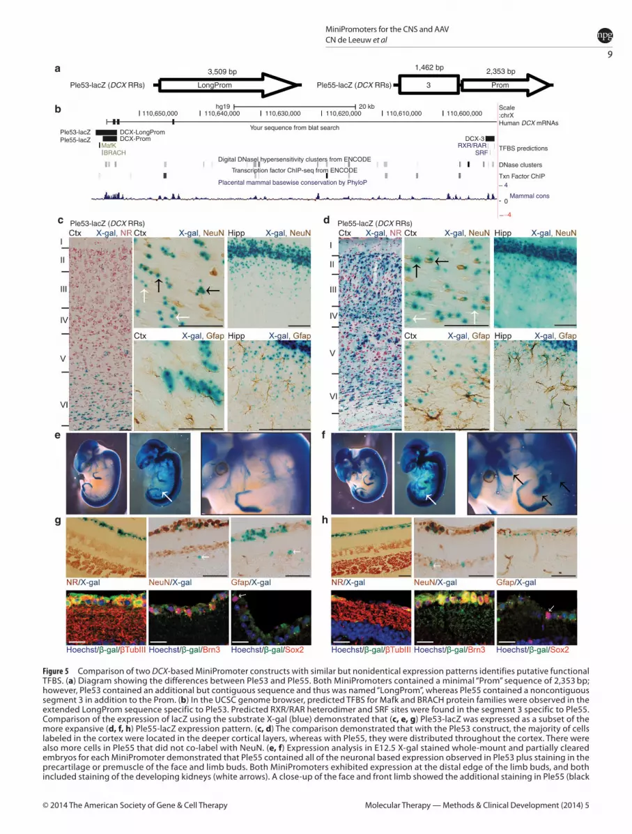
DCX-based MiniPs demonstrate expression driven by a “Prom” and a candidate regulatory elementWe compared two MiniPs, both designed using the DCX gene, to understand differences in the RRs used (Figure 5a,b). The first design, Ple53, was based on a known promoter (LongProm) used in the literature14 and found to be expressed as anticipated in neurogenic regions of adult brain. The second design, Ple55, featured a 5′ shortened promoter region (Prom) coupled to a highly conserved putative regulatory element from within intron 4.
We first looked at the cortical layer specificity of Ple53 and Ple55 (Figure 5c,d). Ple53 expression, as detected by X-gal and counterstained with neutral red, was limited to the deepest cor-tical layer VI (Figure 5c, left panel). Rare positive cells in layers II, IV, and V were observed. In contrast, Ple55 was expressed in all cortical layers, including layer I (Figure 5d, left panel). Nearly all layer II, IV, and VI neurons were labeled. Both MiniPs were expressed in the known adult neurogenic regions of the dentate subgranular zone, subventricular zone, and rostral migratory stream axis (data not shown).
To assess whether the positive cells in the cortex were pri-marily neurons, we compared the extent of NeuN and Gfap co-labeling with X-gal in the cortex of these two strains (Figure 5c,d, center and right panels). Cells were found in both strains that co-labeled (black arrows) and did not co-label (white arrows) with NeuN (Figure 5c,d). We observed some co-labeling with Gfap in the hippocampal formation, which was more extensive with Ple55 (Figure 5d, right panel). The overlapping cortical and neu-rogenic expression between the two designs suggests an expan-sion of the expression pattern with Ple55.
At E12.5 (Figure 5e,f ), we noted an overall conserved staining pattern for Ple53 and Ple55, consistent with the developing ner-vous system. Specifically, expression was observed throughout the developing brain, eye, spinal cord, and dorsal root ganglia. Labeling extends along both the dorsal and ventral ramus. Expression in the developing kidney was found in partially cleared embryos (Figure 5e,f, white arrows). Ple55 showed additional expression in the face and limbs (black arrows). Only the nervous system expression and
kidney expression have been previously documented for the mouse Dcx gene.15
In the retina (Figure 5g,h), Ple53 and Ple55 were expressed nearly exclusively in the ganglion cell layer (GCL), with rare inner nuclear layer (INL) cells positive. Both MiniPs were also expressed in the optic nerve, axonal extensions from the GCL. Studies by others in rat retina suggested horizontal cell labeling for endogenous Dcx.16 However, only the rare INL MiniP-expressing cells (white arrows in Figure 5g,h, top middle panel) may be consistent with their local-ized expression in horizontal cells. Thus, we conclude that the reti-nal expression for Ple53 and Ple55 is ectopic and unexpected.
To further the characterization of the retinal expression, we per-formed co-labeling studies. Strong overlap between NeuN-positive nuclei and X-gal (Figure 5g,h, top middle panel), and with rare exceptions, a lack of overlap with the astroglial marker Gfap (Figure 5g,h, top right panel), was observed. Further analysis by immunoflu-orescence of β-gal using co-labeling with β-Tubulin III (Figure 5g,h, bottom left panel) and Brn3 (Figure 5g,h, bottom middle panel), both markers of retinal ganglion cells, demonstrated significant overlap, while only rare cells were positive for the cholinergic ama-crine marker, Sox2 (Figure 5g,h, bottom right panel, white arrows).
To account for the expression differences observed between Ple53 and Ple55, we undertook a bioinformatics analysis (Figure 5a,b). The 5′ extension of the Prom segment found in the LongProm used in Ple53 and the additional element from intron 4 added together with the Prom segment in Ple55 were used to search for predicted transcription factor binding sites (TFBS). Ple53 was predicted to contain a MafK (a transcription factor bind-ing site based on the MafK gene family) TFBS (UCSC (University of California, Santa Cruz)/ChIP (chromatin immunoprecipitation)-seq ENCODE data) and a BRACH (a transcription factor binding site based on the brachyury gene family) TFBS (TFBIND data-base), which are located in the 5′ nonoverlapping region of the LongProm and thus missing in Ple55. Furthermore, Ple55 con-tained two predicted serum response factor (analyzed using CONSITE with profiles from the JASPAR database) and two reti-noid X receptor/retinoic acid receptor (JASPAR database) TFBSs within the conserved element 3, derived from the intron 4 region. Serum response factor is a transcription factor critical for muscle development,17 and a TFBS for it is located in element 3 of Ple55. We propose that this serum response factor TFBS in Ple55 is responsible for the additional expression pattern in embryos and that the tissue identity is premuscle. Lastly, it appeared that there was greater co-labeling with Gfap in the hippocampal for-mation of adult Ple55 mouse brain, which would be consistent with a role for serum response factor in astrocyte and oligoden-drogial development.18 This unique predicted TFBS may account for the expression differences observed.
Interestingly, in contrast to all other MiniP strains, we were unable to maintain the DCX-based MiniP strains on the C57BL/6J background past backcross N3 due to lack of allele transmission. However, we were able to restore and maintain normal allele
anteromedial nuclei (top panel, arrow). Additional staining was seen in the retrosplenial area (bottom panel, arrow). Ple170 (POGZ RRs) expressed strongly throughout all brain regions, with weaker staining in the striatal nuclei. The olfactory bulb mitral, glomerular, and granular layers are all stained (bottom panel). Ple201 (SLC6A5 RRs) expressed weakly in the anterior and retrosplenial areas of the cortex (top panel, arrow). Staining was present in the medulla also (bottom panel, arrows). Ple232 (TNNT1 RRs) expressed in the anterior cingulate and zona incerta (top panel, arrow and arrow head, respectively) and in the lateral hippocampus (bottom panel, arrow). Ple235 (TRH RRs) expressed in the pontine grey and periaqueductal grey (top and bottom panels respectively, arrows). Ple238 (TRH RRs) had scattered expression in many brain regions, including the cerebellar interposed and fastigial nuclei and in parts of the medulla (bottom panel). Ple240 (UGT8 RRs) showed strong fiber-tract associated staining in all parts of the brain, with cortex layer IV strongly positive and in thalamic intralaminar and anterior nuclei groups. In addition, the olfactory bulb glomeruli and corpus callosum were clearly labeled. Bs, brainstem; CA, cornus ammonis; Cb, cerebellum; CH, chimera; Ctx, cortex; Hipp, hippocampus; Hth, hypothalamus; Mb, midbrain; RRs, regulatory regions; Str, striatum; Th, thalamus.

6
MiniPromoters for the CNS and AAVCN de Leeuw et al
Molecular Therapy — Methods & Clinical Development (2014) 5 © 2014 The American Society of Gene & Cell Therapy
transmission after crossing the allele to the 129S1/SvImJ strain and hypothesized an unknown genotoxic effect for these alleles in C57BL/6J mice.
“Proms” typically confer the observed specificityIt has been proposed that the minimal promoter for a given gene confers the general specificity of expression but that addi-tional regulatory elements either alter the timing or fine-tune the regional specificity.19,20 To test this concept, we analyzed sets of MiniPs designed from the same gene in our combined dataset (this paper and ref. 9). For the purpose of this analysis, we classi-fied MiniPs (with respect to brain only) as: (i) related, having at least some expression similar to the endogenous gene; or (ii) unrelated, having no pattern similar to the endogenous gene. Similarly, we classified sets of MiniPs as: (i) related (i.e., all MiniPs are related to endogenous and similar in pattern to each other); (ii) unrelated (i.e.,
all MiniPs do not have expression in common with the endogenous gene but are similar in pattern to each other); or (iii) mixed (i.e., a set containing related and unrelated MiniPs, or alternatively, a set containing MiniPs that are all related to the endogenous expres-sion pattern but not similar to each other). We had 16 such sets (see Supplementary Figure S1).
Nine of these sets contain MiniPs that all have an identi-cal basal Prom (or minimal promoter) element within the set. Of these nine, six sets of MiniPs (C8ORF46, CLDN5, HCRT, ICMT, MKI67, and S100β RRs) displayed related expression, while the remaining two (POGZ and RGS16 RRs) displayed unrelated expression, and one set had one related and one unrelated MiniP (NR2E1 RRs).
An additional six sets included a combination of MiniPs that either contained the Prom element or a LongProm element (usu-ally a known promoter in the literature and contiguous but larger than the Prom element) within the given set (i.e., overlapping basal
Figure 2 Two MiniPromoters, Ple26 (CCL27 RRs) and Ple170 (POGZ RRs), drive near pan-neuronal expression throughout the mouse brain. For each, a representative 1-mm sagittal and coronal whole brain section is shown, as well as cryosections of NeuN (neuronal marker) and Gfap (glial marker) antibody co-labeling with X-gal in the cortex, hippocampus, and cerebellum. (a) Ple26 (CCL27 RRs) showed marked expression throughout the adult brain with slightly less labeling in the olfactory bulb. In the cortex, NeuN closely co-labeled many of the X-gal positive cells (black arrows). However, there were some NeuN cells that do not co-label with X-gal (red arrows), as well as many smaller punctate X-gal nuclei that do not co-label with NeuN (white arrows). Nearly all hippocampal neurons co-label with X-gal, and extensive co-labeling was also observed in the cerebellum, with X-gal only staining also present in the molecular layer. Also, rare X-gal/Gfap-positive cells were observed (black arrows). (b) Ple170 (POGZ RRs) demonstrated a similar expression pattern to that of Ple26. The CA2 and CA3 regions of the hippocampus showed less staining than the dentate gyrus and CA1 regions. Many NeuN co-labeled cells were visible (black arrows). However, some NeuN-positive cells in the cortex were not labeled with X-gal (red arrows), as well as some punctate X-gal nuclei were NeuN-negative (white arrows). Reduced NeuN co-labeling was seen in the hippocampus and cerebellum, particularly in the cerebellar granule cell layer. Occasionally, a cell was found to be double labeled with Gfap and X-gal (black arrows). CA, cornus ammonis; Cb, cerebellum; Ctx, cortex; Hipp, hippocampus; RRs, regulatory regions. (Scale bars = 100 μm).
a
b

7
MiniPromoters for the CNS and AAVCN de Leeuw et al
Molecular Therapy — Methods & Clinical Development (2014) 5© 2014 The American Society of Gene & Cell Therapy
promoters). Five of these six sets (CCKBR, DBH, DCX, GFAP, and PITX3 RRs) had 5′ extensions compared with the Prom, and one set (TRH RRs) contained a 3′ extension compared with the Prom. All six sets displayed related expression.
Lastly, the CCL27-based set contained MiniPs with two nonoverlapping Prom1 and Prom2 designs. Prom1 was located further upstream and annotated as a putative cortical expression promoter based on 5′ cap analysis of gene expres-sion data. A Prom2-based MiniP, located near the known tran-scription start site, was still related but distinctly different from those based on Prom1. Due to this difference, we classified this as a mixed set.
Seventy-five percent (12/16) of the MiniP sets displayed related expression in all positive MiniPs. Thus, we conclude that the common Prom element is generally sufficient to confer specificity, as compared with the endogenous mouse gene, and added putative regulatory elements either enhance or reduce expression levels, or alternatively expand or narrow the expres-sion pattern.
Seventeen MiniPs deliver expression to the adult mouse eyeThe gene set for MiniP design was originally chosen for region-specific and cell type–specific adult brain expression; however, since the brain and eye are both generated from diencephalon, it is likely that many designs would also be positive in the eye. Thus, we undertook a detailed investigation of eyes in otherwise brain-positive MiniPs.
We analyzed the expression pattern of 17 MiniPs found to be pos-itive in the adult mouse eye (Table 1). Within this set, we observed expression in nearly all major retinal cell types. In extra-retinal tissue, we identified seven MiniPs positive in the cornea, six in the optic nerve, and two in the lens. Fifteen eye positive MiniPs are presented in Figure 6, and two additional ones are presented in Figure 5, as part of the DCX MiniP comparison.
We found Ple24 and Ple25 (CCKBR RRs) were expressed in the GCL and occasionally in the inner part of the INL. Ple26 (CCL27 RRs) was expressed in the cornea, optic nerve, and all retinal layers. Another design for this gene, Ple28, showed expression in all layers and also in the lens but no expression in the cornea or optic nerve.
Figure 3 Thirteen MiniPromoters expressed in the mouse spinal cord. LacZ expression was detected by X-gal immunohistochemistry in whole mount thoracic spinal cord. (a) Ple17 (C8ORF46 RRs), Ple28 (CCL27 RRs), Ple32 (CLDN5 RRs), and Ple170 (POGZ RRs) showed scattered expression in the central spinal cord and in nerve bundles. Ple24 (CCKBR RRs) expression was seen primarily in the spinal cord with rare puncta in the dorsal root ganglia (data not shown). Ple26 (CCL27 RRs), Ple53 (DCX RRs), and Ple55 (DCX RRs) expressed throughout the gray matter regions and also in dorsal root ganglia; the latter particularly strong for Ple53 and Ple55. Ple34 (CLDN5 RRs) and Ple123 (ICMT RRs) expressed throughout all of the spinal cord, while Ple123 demonstrated weaker expression in nerve bundles but strong expression in dorsal root ganglia. Ple88 (GFAP RRs) was expressed only in patches along white matter regions of the central spinal cord. Ple167 (POGZ RRs) expressed in patches in the spinal cord with some nerve fibers and occasional dorsal root ganglia puncta also positive (data not shown). Ple240 (UGT8 RRs) is expressed strongly and predominantly along spinal nerve cord bundles with weaker expression in the central spinal cord tissue and in dorsal root ganglia. (b) Summary of expression pattern for each construct in the three key regions of the spinal cord. CH, chimera; DRG, dorsal root ganglion; ND, not done; RRs, regulatory regions; √, expression present; ×, expression absent.
Ple17-lacZ (C8ORF46 RRs)
Ple32-lacZ (CLDN5 RRs)
Ple88-lacZ (GFAP RRs)
Ple240-lacZ (UGT8 RRs)
Ple24-lacZ (CCKBR RRs)
Ple34-lacZ (CLDN5 RRs)
Ple123-lacZ (ICMT RRs)
Ple26-lacZ (CCL27 RRs)
Ple53-lacZ (DCX RRs)
Ple167-EGFP/cre (POGZ RRs)
Ple28-lacZ (CCL27 RRs)
Ple55-lacZ (DCX RRs)
(CH)
(CH)
Ple170-lacZ (POGZ RRs)
RegionPle17
Ple24
Ple26
Ple28
Ple32
Ple34
Ple53
Ple55
Ple88
Ple123
Ple167
Ple170
Ple240
CentralspinalcordNervefibres
DRG Rare Diffuse
ND ND NDPunctate
Weak Some
a
b

8
MiniPromoters for the CNS and AAVCN de Leeuw et al
Molecular Therapy — Methods & Clinical Development (2014) 5 © 2014 The American Society of Gene & Cell Therapy
Ple32 (CLDN5 RRs) was expressed in the GCL and INL. Ple34 (CLDN5 RRs) was positive in the INL and GCL and was expressed rarely in cells of the outer nuclear layer (photoreceptors). The morphology of cell nuclei located in the INL, with processes extending to the GCL, was suggestive of bipolar ON cells. Ple67 (FEV RRs) stained cells in the GCL and rare INL cells. Ple88, based on a known pro-moter for GFAP,21 was expressed in the astrocytic layer as expected. Ple123 (ICMT RRs) stained all retinal layers including the cornea and optic nerve. Ple131 (MKI67 RRs), based on proliferative maker KI-67, stained patches of the cornea only, consistent with other studies showing expression of KI-67 in basal epithelial cells in the cornea.22 Ple146 (NTSR1 RRs) stained rare GCL and INL cells (INL-positive cell not pictured). Ple155 (PCP2 RRs) stained predominantly in the INL and inner plexiform layer and was strongly suggestive of bipolar ON cells, which is expected expression for this gene. Ple167 and Ple 170 (POGZ RRs) stained cells in all layers of the neural retina includ-ing the cornea. Ple240 (UGT8 RRs) stained positive in the retinal inner segment (photoreceptors) and occasionally in the INL with fibers extending into the astrocytic layer, suggesting a Müller glial or bipolar cell type. The cornea and optic nerve were also positive for Ple240.
This characterization has led to a set that is unique compared with currently available eye promoters (see ref. 23 for a brief list).
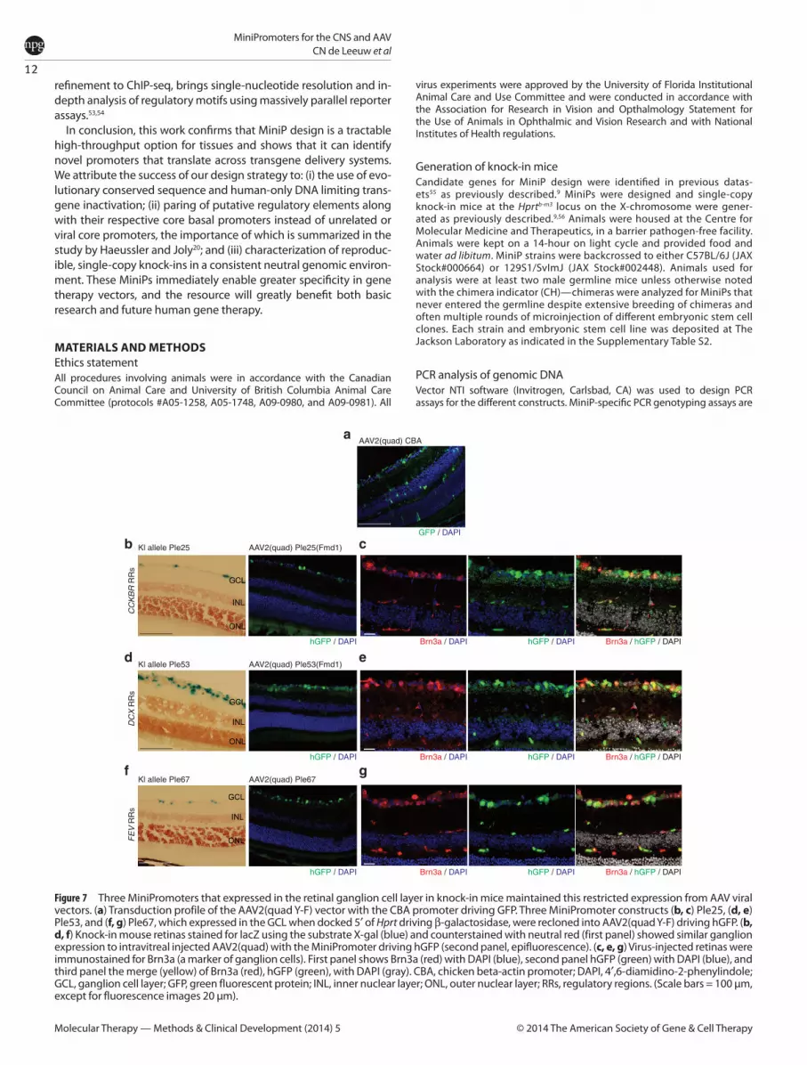
Knock-in MiniPs that express in retinal ganglion cells predict expression of MiniP-GFP using AAVTo assess the feasibility of using the MiniPs for AAV-mediated delivery, we cloned three (Ple25, Ple53, and Ple67) MiniPs that were expressed primarily in the retinal ganglion cell layer, into the AAV2-(quad Y-F) vector. This vector, containing four tyrosine to phenylalanine mutations, has previously been shown to trans-duce cells of the retina uniformly, with a transduction profile that includes photoreceptors, Müller glia, and horizontal, bipo-lar, amacrine, and ganglion cells.24 Two (Ple25 and Ple53) of the three MiniPs were 5′ trimmed by ~200–400 bp to fit into the viral vector due to limits on the carrying capacity of AAV constructs. Ple25 was trimmed from 3,740 bp (KI allele) to 3,312 bp (AAV), and Ple53 was trimmed from 3,520 bp (KI allele) to 3,310 bp (AAV), resulting in Ple25(Fmd1) and Ple53(Fmd1), respectively. Ple67 (2,202 bp) did not require trimming. Mice were injected intravitreally at postnatal day (P) 28, harvested 4 weeks later, and analyzed for green fluorescent protein (GFP) expression. The transduction profile of AAV2(quad Y-F) using the ubiquitous chicken beta-actin promoter demonstrated expression in all reti-nal layers (Figure 7a). Figure 7b,d,f show a side-by-side compari-son of the knock-in mouse strain and the AAV vector–injected mice. All three AAV vectors supported expression of the reporter gene in the retinal ganglion cell layer. For Ple53 and Ple67, we also observed rare positive cells in the innermost aspect of the INL. Importantly, the 5′ trimming of ~200–400 bp for Ple25 and Ple53 to accommodate AAV space constraints did not alter their specificity, indicating that the deleted segments were nones-sential for specificity. We conclude that for at least these three MiniPs, the expression pattern in the knock-in strain predicted that obtained using a virus-based delivery to the mouse retina. These data suggest that MiniPromoter specificity may be main-tained in AAV vectors.
The retinal ganglion cell (RGC) layer in mice contains ~40% ganglion cells and 60% amacrine cells.25 Nadal-Nicolás et al.26 estimated that Brn3a+ cells label ~92.2% of RGCs, as co-labeled with the Fluorogold tracer in rat retina. Similarly, Fluorogold was estimated to label 97.8–98.4% of RGCs in rats.27 Thus, we considered that ~90% of RGCs in the adult mouse retina should be labeled with Brn3a and could be used as a reliable marker of retinal ganglion cells. We undertook Brn3a co-labeling in virus-injected mice for Ple25, Ple53, and Ple67 to assess the extent of expression in ganglion cells, with the remainder of GFP-positive cells in the GCL assumed to be amacrine. As shown in Figure 7c,e,g, the MiniPs ranked qualitatively from most to least overlap with Brn3a: Ple53 > Ple25 > Ple67. We were also interested in how much expression was observed in amacrine (Brn3a-negative) cells in the GCL. Similarly, qualitative ranking of the same MiniPs from most to least expression in Brn3a-negative but GFP-positive cells ordered the MiniPs: Ple67 > Ple25 > Ple53. Thus, we con-clude that Ple53 supports expression in the greatest proportion of retinal ganglion cells and is most specific to this cell type, while Ple67 supports expression in a subset of retinal ganglion cells in addition to numerous amacrine cells.
DISCUSSIONInterest in the identification of small promoters for capturing the expression pattern of biologically unique genes is longstanding.
Figure 4 Expression of six MiniPromoters during development. E12.5 embryos were harvested and stained for lacZ expression using X-gal. Whole-mount (first panels) and partially cleared whole-mount embryos (second panels) were photographed. (a) Ple26 (CCL27 RRs) stained throughout the entire embryo. (b) Ple123 (ICMT RRs) displayed variability, with the most common expression pattern being limited to the developing heart as shown. (c) Ple131 (MKI67 RRs) was expressed throughout the embryo at varying levels, most prominently in the heart. (d) Ple140 (NR2E1 RRs) showed a very strongly defined pattern in the hypothalamus of the embryo. (e) Ple170 (POGZ RRs) demonstrated staining in all parts of the embryo, but generally stronger outside of the nervous system. (f) Ple232 (TNNT1 RRs) stained strongly in skeletal muscle and the tongue of the embryo but was not visible in the brain or spinal cord. RRs, regulatory regions.
Ple26-lacZ (CCL27 RRs) Ple123-lacZ (ICMT RRs)
Ple131-lacZ (MKI67 RRs) Ple140-lacZ (NR2E1 RRs)
Ple170-lacZ (POGZ RRs) Ple232-lacZ (TNNT1 RRs)
a b
c d
e f
9
MiniPromoters for the CNS and AAVCN de Leeuw et al
Molecular Therapy — Methods & Clinical Development (2014) 5© 2014 The American Society of Gene & Cell Therapy
Figure 5 Comparison of two DCX-based MiniPromoter constructs with similar but nonidentical expression patterns identifies putative functional TFBS. (a) Diagram showing the differences between Ple53 and Ple55. Both MiniPromoters contained a minimal “Prom” sequence of 2,353 bp; however, Ple53 contained an additional but contiguous sequence and thus was named “LongProm”, whereas Ple55 contained a noncontiguous segment 3 in addition to the Prom. (b) In the UCSC genome browser, predicted TFBS for Mafk and BRACH protein families were observed in the extended LongProm sequence specific to Ple53. Predicted RXR/RAR heterodimer and SRF sites were found in the segment 3 specific to Ple55. Comparison of the expression of lacZ using the substrate X-gal (blue) demonstrated that (c, e, g) Ple53-lacZ was expressed as a subset of the more expansive (d, f, h) Ple55-lacZ expression pattern. (c, d) The comparison demonstrated that with the Ple53 construct, the majority of cells labeled in the cortex were located in the deeper cortical layers, whereas with Ple55, they were distributed throughout the cortex. There were also more cells in Ple55 that did not co-label with NeuN. (e, f) Expression analysis in E12.5 X-gal stained whole-mount and partially cleared embryos for each MiniPromoter demonstrated that Ple55 contained all of the neuronal based expression observed in Ple53 plus staining in the precartilage or premuscle of the face and limb buds. Both MiniPromoters exhibited expression at the distal edge of the limb buds, and both included staining of the developing kidneys (white arrows). A close-up of the face and front limb showed the additional staining in Ple55 (black
Ple53-lacZ (DCX RRs) Ple55-lacZ (DCX RRs)
Ple53-lacZ (DCX RRs) Ple55-lacZ (DCX RRs)LongProm
3,509 bp
110,650,000 110,640,000
DCX-LongPromPle53-lacZPle55-lacZ DCX-Prom
MafKBRACH
110,630,000 110,620,00020 kbhg19
110,610,000 110,600,000Scale:chrXHuman DCX mRNAs
TFBS predictions
DCX-3
DNase clusters
Txn Factor ChIP4
−4
Mammal cons
RXR/RARSRF
Placental mammal basewise conservation by PhyloP
Digital DNasel hypersensitivity clusters from ENCODE
Your sequence from blat search
Transcription factor ChIP-seq from ENCODE
0
3
1,462 bp 2,353 bp
Prom
a
b
c
e
g
f
h
d

10
MiniPromoters for the CNS and AAVCN de Leeuw et al
Molecular Therapy — Methods & Clinical Development (2014) 5 © 2014 The American Society of Gene & Cell Therapy
The Pleiades approach, with methodology described in detail previously,9 has the ability to generate novel promoters in a high-throughput manner. Here, we present 18 additional promoters identified by this technique, and the further characterization of 15 previous designs.
Our approach focuses on physiologically relevant expression. The reasoning for this is threefold: (i) overexpression of therapeutic mol-ecules can be deleterious28; (ii) overexpression of purely research-related molecules such as cre can cause cytotoxicity19,29,30; and (iii) introduction of artificial sequences to enhance expression can pose regulatory challenges due to their unknown specificity. Our strategy of mimicking endogenous expression levels using endog-enous sequences is aimed at overcoming these challenges and thus enabling improved gene therapy designs.
However, there are some limitations to the Pleiades design pipe-line. First, a small number of MiniPs displayed highly variable expres-sion when docked at Hprt. We were unable to find previous reports of variable expression for constructs knocked-in at Hprt, which is generally a neutral docking location with consistent expression of docked constructs.31 Expression variability was also not a general feature of our previous MiniP work.9 Thus, we hypothesize that the variability observed is construct and sequence specific and reflects the large number of MiniPs tested. For the most part, the extent of variability we saw in embryos correlated with that observed for adults. It remains to be seen whether such construct-dependent variability is genome context specific or would be similarly repro-duced in other systems (e.g., virus-based delivery or knock-in at the ROSA26 locus).
Additionally, the Pleiades data was limited by the inability to attain germline animals for a small subset of MiniPs. In these cases, chimeras were studied. Although each chimera may not display the entire cellular expression profile, the observed chimeric expression has been a reliable predictor of germline expression.9 Lastly, we were limited in characterization of the specific cell types of expres-sion, particularly for the brain where hundreds or thousands of dif-ferent neuronal cell types are predicted to exist.32 However, this data on regionalized expression of MiniP constructs remains a valuable scientific and therapeutic resource.
Importantly, this data represents numerous novel and biologi-cally significant findings: first, we identified the zona incerta as a common region of expression for MiniP designs. We hypothesize that overrepresentation of the zona incerta brain region in this set is due to the diverse range of neurochemical cells located in this region, which includes neurons varying in soma size and shape (see ref. 33 for a comprehensive review on the zona incerta). In addition, extensive networking exists between cells of the zona incerta and numerous other brain regions (e.g., the cerebral cortex, diencepha-lon, basal ganglia, brainstem, and spinal cord),33 further suggesting a heterogeneous population in this region. The persistent finding of staining in this region suggests caution for therapeutic delivery where expression in the zona incerta could cause harm.
The brain-positive MiniPs provide novel tools for researchers and clinicians alike. Within our toolbox of MiniPs, we have now added
constructs that would drive expression in the cortex and hippocam-pus (Ple15 and Ple16), in oligodendroglia (Ple240), in a near pan-neuronal expression (Ple26 and Ple170), or in the developing ner-vous system (Ple53 and Ple55).
Selected designs were also validated for their use in develop-mental studies. We tested six adult brain–positive MiniPs and showed expression in the brain for five of these at E12.5. The remaining MiniP, Ple232, was expressed only outside of the embry-onic brain, in contrast to its expression in the adult. This finding is not entirely unexpected, since not all genes will be expressed in the brain at the time point used in this study (E12.5, mid-point of neurogenesis). Remarkably, we showed highly specific expression of Ple140 (NR2E1 RRs) in the hypothalamus at E12.5, with no evi-dence of its expression in other embryonic tissue. These embry-onic studies allow us to rapidly detect the functioning of MiniPs outside of the CNS, like Ple232 (TNNT1 RRs) which could prove a valuable tool for researchers studying, and developing treatments for, skeletal muscle disorders. Overall, this embryonic dataset sug-gests that many other Pleiades MiniPs are likely to be positive dur-ing development.
We were surprised by the portion of MiniPs expressed in the mouse eye. This represents a novel toolset for driving retinal gene expression, and these MiniPs offer great therapeutic potential. Bipolar cells are the next step in retinal processing after photore-ceptors are activated by light. In diseases where photoreceptors are nonfunctional or have undergone cell death, one strategy is to target the bipolar cells.34 Similarly, since retinal ganglion cells form the last step in the visual processing pathway, expression of therapeutic molecules could be a viable option here also,34 with perhaps more generic retinal disease application. Our efforts have developed at least two MiniPs with bipolar cell expression (Ple34 and Ple155) and six with ganglion cell layer expression (Ple24, Ple25, Ple53, Ple55, Ple67, and Ple146). The finding of such a large proportion (35.3%) of eye positives specific to the GCL implies that selection of candidates for MiniP design using brain expression data may be biased toward generating RGC-positive designs. We hypothesize this may be due to the simi-larities between RGCs and other CNS neurons with regards to (i) the ability to fire action potentials and (ii) similarities in gene expression between RGC-specific transcripts and other CNS neu-rons that fire action potentials.35–39 Interestingly, the ganglion cell-marking Brn3 transcription factors are also highly expressed in the sensory neurons of the dorsal root ganglia.40 Of the six GCL-positive MiniPs, we were able to assess dorsal root ganglion expression in three and found all three positive (Ple24, Ple53, and Ple55). Thus, our selection strategy has not only preferen-tially generated MiniPs positive in the GCL of the retina but also in the dorsal root ganglion (eight out of ten MiniPs tested).
Other MiniPs that expressed in the eye also demonstrate poten-tial utility: Ple88 (GFAP RRs) could be used to aid in the treat-ment of retinal disorders with increased astrogliosis (defined as increased GFAP expression), and Ple131 (MKI67 RRs) may be use-ful for corneal repair. Curiously, we found occasional discordance
arrows). (g, h) (upper panels) MiniPromoter lacZ expression as detected by X-gal reaction in the retina (left panel). Expression for the DCX-based designs was limited to the ganglion cell layer (GCL) for both Ple53 and Ple55, with rare cells observed in the inner nuclear layer. NeuN (marking all retinal ganglion and amacrine cells) co-labeled with X-gal (center panel), but Gfap (retinal astrocytes) did not (right panel). Rare cells did not co-label with NeuN (white arrows). (g, h) (lower panels) Immunofluorescence co-labeling with Brn3, βtubIII, and Sox2. Co-labeling demonstrated that most lacZ-positive cells stained with βtubIII and Brn3 markers (ganglion cells) for both MiniPromoters (left and center panels), and only rarely with Sox2-positive cholinergic amacrine cells (right panel, white arrows). Ctx, cortex; Hipp, hippocampus; NR, neutral red; RAR, retinoic acid receptor; RRs, regulatory regions; RXR, retinoid X receptor; SRF, serum response factor; TFBS, transcription factor binding sites; UCSC, University of California, Santa Cruz. (Scale bars = 100 μm, except for (g, bottom panel) fluorescence images 20 μm).

11
MiniPromoters for the CNS and AAVCN de Leeuw et al
Molecular Therapy — Methods & Clinical Development (2014) 5© 2014 The American Society of Gene & Cell Therapy
between cell type expression in the brain and eye. The Ple155 (PCP2 RRs) MiniP was expressed as expected in the mouse ret-ina (bipolar ON cells), but with seemingly unrelated expression in the brain (MiniP expression present in cortex, thalamus, and hypothalamus but absent in Purkinje cells for which PCP/L7 is well known).41,42 This data further supports the notion that cell type specificity is regulatory element driven in specific organs or tis-sues.43 Altogether, this represents a powerful set of tools for driv-ing gene expression in the eye.
One concern was whether the observed expression pattern of MiniPs would be reliably reproduced when used in viral vectors. Viral vector gene delivery is the preferred therapeutic method for the brain and eye, primarily because the number of treatment options via gene delivery is vast. We showed that a subset of three MiniPs (Ple25, Ple53, and Ple67), which expressed in the retinal ganglion cell layer in knock-in mice, exhibited a similar expression pattern in the mouse retina when delivered intravitreally and expressing from recombinant AAV2. We hypothesize that the characterization by single-copy site-specific knock-ins, and use of exclusively human DNA, may have predisposed these MiniPs to success in AAV. Using this methodology, the desirable expression characteristics are more likely to be inherent to the MiniP only and not dictated by copy-number or the genomic location. In addition, human DNA should be less prone to expression variability compared with more distantly related sequence or viral promoters. Finally, critical to the success of this work was the choice of an AAV serotype with a broad transduc-tion profile in the retina, which included the target cell types of the MiniPs. Interestingly, with this success, the MiniPs can also be used to map the specificity of novel serotypes, providing a valuable tool for researchers working on AAV capsid biology.
An interesting pattern emerged using these three MiniPs: we observed an inverse qualitative relationship between the overlap of stained retinal ganglion cells (defined here as Brn3a-positive in the adult retina) and amacrine cells (defined as Brn3a-negative) located in the GCL. This result was surprising, as we were anticipating that MiniP expression may target different subsets of ganglion or ama-crine cells, with no expression in the other cell types. However, we observed overlapping expression in both cell types, and our data implies that transcriptional regulation between amacrine and RGC cells may not be distinctly separate. Indeed, in support of this idea are the results of Kunzevitzky et al.,44 who evaluated the transcrip-tional profiles using microarrays of amacrine and retinal ganglion cells at E20, P5, and P11 in the rat. They found ~70% overlap in expression between the two cell types at all time points (see Figure 5 in the study of Kunzevitsky et al.). Both amacrine and ganglion cells are born between E14 and E18. Evidence for potential overlap-ping transcriptional regulation between these two cell types also comes from work that showed VC1.1+ progenitors can generate both VC1.1+ and VC1.1− progenitors.45,46 The former produces ama-crine and horizontal progeny, while the latter produces retinal gan-glion cells and cones.46
Two of these three Ple MiniPs were trimmed prior to use in AAV. In the future, the process of making Pleiades MiniPs even smaller will be informed by data generated by novel technologies. Such data includes that of the ENCODE project,47 FANTOM projects,48–50 and VISTA Enhancer project.5 Specifically, the ENCODE transcription fac-tor ChIP-seq and DNase I hypersensitivity data, along with FANTOM cap analysis of gene expression,50 and p300 enhancer protein bind-ing51 data will allow for anchoring of, and supportive evidence for function of, RRs. In addition, the development of ChIP-exo,52 a
Figure 6 Fifteen MiniPromoters expressed in the mouse eye. Mice were perfused, eyes harvested, and stained for lacZ using the substrate X-gal (blue). Eyes were cryoprotected, embedded, and sectioned. Ple24 (CCKBR RRs), Ple25 (CCKBR RRs), Ple32 (CLDN5 RRs), Ple67 (FEV RRs), and Ple146 (NTSR1 RRs) expressed in a subset of GCL cells. Rarely, an amacrine cell in the innermost part of the INL was positive in Ple24, or a rare positive INL cell in Ple32. Ple26 and Ple28 (both based on CCL27 RRs) expressed in the INL, and in photoreceptors as evidenced by the columnar staining of the ONL and photoreceptor segments. Ple26 showed extensive IPL and OPL fiber staining and Ple28 was also positive in the GCL. Ple34 (CLDN5 RRs) and Ple155 (PCP2 RRs) have staining characteristic of bipolar cells, with cell bodies in the INL and processes extending to the GCL (Ple34) or inner half of the IPL (Ple155). Ple88 (GFAP RRs) stained in the retinal astrocytic layer. Ple123 (ICMT RRs) expressed throughout the retina. Ple131 (MKI67 RRs) expressed in cells of the corneal epithelium. Ple167 and Ple170 (both based on POGZ RRs) demonstrated similar staining patterns in the GCL, INL, OPL and photoreceptor segments. Ple240 (UGT8 RRs) had fiber-like staining spanning the IPL and clear cell body labeling in a subset of INL cells. CH, chimera; INL, inner nuclear layer; IPL, inner plexiform layer; ONL, outer nuclear layer; OPL, outer plexiform layer; RRs, regulatory regions. (Scale bar = 100 μm).
Ple24-lacZ(CCKBR RRs)
Ple25-lacZ(CCKBR RRs)
Ple26-lacZ(CCL27 RRs)
Ple28-lacZ(CCL27 RRs)
Ple32-lacZ(CLDN5 RRs)
Ple34-lacZ(CLDN5 RRs)
Ple67-lacZ(FEV RRs)
Ple88-lacZ(GFAP RRs)
Ple123-lacZ(ICMT RRs)
Ple131-lacZ(MKI67 RRs)
Ple146-lacZ(NTSR1 RRs)
Ple155-lacZ(PCP2 RRs)
Ple167-EGFP/cre(POGZ RRs)
Ple170-lacZ(POGZ RRs)
Ple240-lacZ(UGT8 RRs)
12
MiniPromoters for the CNS and AAVCN de Leeuw et al
Molecular Therapy — Methods & Clinical Development (2014) 5 © 2014 The American Society of Gene & Cell Therapy
refinement to ChIP-seq, brings single-nucleotide resolution and in-depth analysis of regulatory motifs using massively parallel reporter assays.53,54
In conclusion, this work confirms that MiniP design is a tractable high-throughput option for tissues and shows that it can identify novel promoters that translate across transgene delivery systems. We attribute the success of our design strategy to: (i) the use of evo-lutionary conserved sequence and human-only DNA limiting trans-gene inactivation; (ii) paring of putative regulatory elements along with their respective core basal promoters instead of unrelated or viral core promoters, the importance of which is summarized in the study by Haeussler and Joly20; and (iii) characterization of reproduc-ible, single-copy knock-ins in a consistent neutral genomic environ-ment. These MiniPs immediately enable greater specificity in gene therapy vectors, and the resource will greatly benefit both basic research and future human gene therapy.
MATERIALS AND METHODSEthics statementAll procedures involving animals were in accordance with the Canadian Council on Animal Care and University of British Columbia Animal Care Committee (protocols #A05-1258, A05-1748, A09-0980, and A09-0981). All
virus experiments were approved by the University of Florida Institutional Animal Care and Use Committee and were conducted in accordance with the Association for Research in Vision and Opthalmology Statement for the Use of Animals in Ophthalmic and Vision Research and with National Institutes of Health regulations.
Generation of knock-in miceCandidate genes for MiniP design were identified in previous datas-ets55 as previously described.9 MiniPs were designed and single-copy knock-in mice at the Hprtb-m3 locus on the X-chromosome were gener-ated as previously described.9,56 Animals were housed at the Centre for Molecular Medicine and Therapeutics, in a barrier pathogen-free facility. Animals were kept on a 14-hour on light cycle and provided food and water ad libitum. MiniP strains were backcrossed to either C57BL/6J (JAX Stock#000664) or 129S1/SvImJ (JAX Stock#002448). Animals used for analysis were at least two male germline mice unless otherwise noted with the chimera indicator (CH)—chimeras were analyzed for MiniPs that never entered the germline despite extensive breeding of chimeras and often multiple rounds of microinjection of different embryonic stem cell clones. Each strain and embryonic stem cell line was deposited at The Jackson Laboratory as indicated in the Supplementary Table S2.
PCR analysis of genomic DNAVector NTI software (Invitrogen, Carlsbad, CA) was used to design PCR assays for the different constructs. MiniP-specific PCR genotyping assays are
Figure 7 Three MiniPromoters that expressed in the retinal ganglion cell layer in knock-in mice maintained this restricted expression from AAV viral vectors. (a) Transduction profile of the AAV2(quad Y-F) vector with the CBA promoter driving GFP. Three MiniPromoter constructs (b, c) Ple25, (d, e) Ple53, and (f, g) Ple67, which expressed in the GCL when docked 5′ of Hprt driving β-galactosidase, were recloned into AAV2(quad Y-F) driving hGFP. (b, d, f) Knock-in mouse retinas stained for lacZ using the substrate X-gal (blue) and counterstained with neutral red (first panel) showed similar ganglion expression to intravitreal injected AAV2(quad) with the MiniPromoter driving hGFP (second panel, epifluorescence). (c, e, g) Virus-injected retinas were immunostained for Brn3a (a marker of ganglion cells). First panel shows Brn3a (red) with DAPI (blue), second panel hGFP (green) with DAPI (blue), and third panel the merge (yellow) of Brn3a (red), hGFP (green), with DAPI (gray). CBA, chicken beta-actin promoter; DAPI, 4′,6-diamidino-2-phenylindole; GCL, ganglion cell layer; GFP, green fluorescent protein; INL, inner nuclear layer; ONL, outer nuclear layer; RRs, regulatory regions. (Scale bars = 100 μm, except for fluorescence images 20 μm).
AAV2(quad) CBA
Kl allele Ple25 AAV2(quad) Ple25(Fmd1)
Kl allele Ple53 AAV2(quad) Ple53(Fmd1)
Kl allele Ple67
FE
V R
Rs
DC
X R
Rs
CC
KB
R R
Rs
AAV2(quad) Ple67
GFP / DAPI
hGFP / DAPI hGFP / DAPIBrn3a / DAPI Brn3a / hGFP / DAPI
hGFP / DAPI hGFP / DAPIBrn3a / DAPI Brn3a / hGFP / DAPI
hGFP / DAPI hGFP / DAPIBrn3a / DAPI Brn3a / hGFP / DAPI
a
b
d
f
e
c
g

13
MiniPromoters for the CNS and AAVCN de Leeuw et al
Molecular Therapy — Methods & Clinical Development (2014) 5© 2014 The American Society of Gene & Cell Therapy
available on the http://www.pleiades.org Web site or by contacting the cor-responding author.
Immunohistochemistry of adult brainsOne millimeter sectioned tissues. The mice were anesthetized with avertin and perfused transcardially with 4% paraformaldehyde (PFA) and postfixed 2–4 hours. The brains were then removed from 4% PFA and immediately sectioned. To section, brains were placed ventral side up into the adult mouse coronal or sagittal Rodent Brain Matrix (ASI Instruments, Warren, MI). Brains were sectioned lateral through medial to lateral (for sagittal) or rostral to caudal (for coronal) using single-edge razor blades (Electron Microscopy Sciences, Hatfield, PA). All slices from one brain were placed into 1 well of a 12-well plate containing 1× phosphate-buffered saline (PBS) (Invitrogen) or 0.1% sodium azide in 1× PBS until staining. Expression of β-gal (lacZ) was detected with X-gal (5-bromo-4-chloro-3-indolyl-β-d-galactopyranoside) staining as previously described.9 Briefly, the staining was performed with 3–5 ml of X-gal staining solution (25 mg/ml X-gal, 1 mol/l MgCl2, 50 mmol/l potassium ferri-cyanide, 50 mmol/l potassium ferro-cyanide, and 1× PBS to volume) per well in a 24-well plate. The plate was wrapped in aluminum foil and incubated at 37 °C for 10–16 hours with gentle agita-tion on a rotator board. Subsequently, the sections were transferred into PBS, examined under a dissecting microscope, and photographed using a CoolSNAP-Procf color camera (Media Cybernetics, Rockville, MD) mounted on a Leica MZ12.5 stereomicroscope (Leica Microsystems, Buffalo Grove, IL) and Image-Pro Express v.4.5.1.3 software (Media Cybernetics).
Cryosectioned tissues. The mice were anesthetized with avertin and perfused transcardially with 4% PFA in 0.1 mol/l phosphate buffer (pH 7.4) for 15 minutes. Brains were dissected and postfixed in the same solu-tion for 2 hours and transferred into 25% sucrose-PBS overnight. Each brain was sectioned using a cryostat, and 20-μm sagittal sections were collected. X-gal staining and imaging was performed as described for 1-mm sections. Co-staining of X-gal and NeuN or Gfap was performed sequentially. Primary antibodies used were mouse anti-GFAP (1:1,000; Millipore, Billerica, MA) and mouse anti-NeuN (1:500; Chemicon, Billerica, MA). Bound primary antibodies were detected using the VectaStain Elite ABC Kit (Vector Labs, Burlingame, CA; PK-6100), as per manufacturer’s instructions. Slides were gradually dehydrated in ethanol and xylene se-ries and mounted with coverslips. Bright-field images were taken with an Olympus (Center Valley, PA) BX61 motorized microscope using the DP Controller software, Olympus.
Other tissues. Spinal cord, heart, liver, lung, and ear notch were collected from perfused mice and stained as described above.
Embryo harvesting and stainingFor timed pregnancies, female heterozygous MiniP mice were crowded for 7–10 days and set-up in the afternoon with C57BL/6J or 129S1/SvImJ studs. Subsequently, females were checked for copulation plugs for 5 days in the morning. The day of plug was considered E0.5. At E12.5, embryos were harvested, yolk sac tissue taken for PCR analysis, and embryos fixed in 4% PFA/PBS for 2–6 hours. Embryos were stained in X-gal solu-tion for 18 hours, postfixed overnight at 4 °C, and stored in 70% ethanol. Pictures were taken of uncleared embryos. The embryos were then par-tially cleared based on the method of Schatz et al.57 In brief, embryos were incubated in step 1 solution (20% glycerol with 0.8% KOH) for 3 days at 37 °C with swirling once daily. Further clearing was done in step 2 solu-tion (50% glycerol with 0.5% KOH) for 3 days at 37 °C. Postfixing of fragile cleared embryos was done in 4% PFA for 2 hours at room temperature. Embryos were stored until imaging in 70% ethanol, after which they were equilibrated to 100% glycerol for imaging. Imaging was performed as per 1-mm brain sections (above). All embryo expression patterns were seen in at least two animals.
Immunohistochemistry on eyesLacZ staining was detected either directly from MiniPs driving β-gal, or by driving expression of EGFP/cre resulting in recombination of the Gt(ROSA)26Sortm1Sor locus. After whole body perfusion of mice, eyes were postfixed in 4% PFA. LacZ staining was performed using the X-gal substrate for 18 hours at 37 °C. Eyes were then postfixed in 4% PFA for 2 hours prior to cryoprotection in 25% sucrose-PBS overnight. Eyes were embedded in opti-mal cutting temperature (OCT), along with positive and negative controls,
and cryosectioned at 12 μm and mounted on SuperFrost Plus slides, Fisher Scientific. Eye sections were counterstained with neutral red for 45 seconds to 1 minute. Eye expression was confirmed in at least two animals unless otherwise noted with the chimera indicator (CH). Bright-field images were taken using Olympus BX61 motorized microscope using the DP Controller software.
Immunofluorescence on eyesEyes were harvested and fixed in 4% PFA for 2 hours prior to cryoprotection in 25% sucrose-PBS overnight. Eyes were embedded in OCT, and cryosec-tioned at 12 μm and mounted on SuperFrost Plus slides prior to immuno-fluorescence. Standard immunofluorescence procedures were followed.9 Primary antibodies include: chicken anti-β-gal (Abcam Cambridge, MA; ab9361), rabbit anti-β-tubulin III (Covance, Princeton, NJ; PRB-435P), rabbit anti-Brn3 (Santa Cruz Biotechnology, Santa Cruz, CA; sc28595), and rab-bit anti-Sox2 (Abcam; ab97959). Secondary antibodies used were (all from Invitrogen): chicken anti-rabbit Alexa-594, goat anti-chicken Alexa-488, goat anti-chicken Alexa-594, goat anti-mouse Alexa-488, goat anti-mouse Alexa-594, goat anti-rabbit Alexa-488, and goat anti-rabbit Alexa-594. Nuclei were stained using 1 μg/ml of Hoechst 33342 (Sigma, St Louis, MO; 14533). Slides were mounted with Prolong Gold (Invitrogen) antifade reagent and imaged using an Olympus BX61 microscope and the InVivo software (Olympus), or alternatively using confocal laser scanning microscopy with a Leica SP5 II microscope (Leica Microsystems) and LAF software (Leica Microsystems).
Adeno-associated virusRecombinant AAV vector plasmids containing a 2,202 bp Ple67 (FEV RRs), a 3,310 bp Ple53 (DCX RRs) or 3,312 bp Ple25 (CCKBR RRs) MiniPromoter con-struct, an SV40 splice donor–acceptor site followed by the cDNA for GFP were constructed. To accommodate the packaging limit of recombinant AAV, Ple25 and Ple53 were shortened relative to the knock-in MiniP allele as follows: MiniPs were amplified by PCR, and the respective restriction sites were added via the oligonucleotides. The following oligonucleotides were used: Ple25 forward KpnI: GTCGGGTACCAGAATTTCAGGAGCCCATATG, Ple25 reverse XhoI: GTCGCTCGAGTGCCTTACTCAGCTCGACC, Ple53 for-ward KpnI: GTCGGGTACCTTATCCCATCCATTGTTGTATC, Ple53 reverse XhoI: GTCGCTCGAGGGTGGAACCTCAGAGACCTG, Ple67 forward EcoRI: GCTGGAATTCGGAGAAGATCAAGACTCAGG, and Ple67 reverse XhoI: GCTGCTCGAGGAGGCAGGCTTTGCGCTC, (restriction sites underlined). The plasmids pEMS1496, pEMS1524, and pEMS1538 were used as templates. The shorter Ple25 promoter construct is referred to as Ple25(Fmd1), and the shorter Ple53 promoter construct as Ple53(Fmd1).
Identity for all promoters was confirmed by sequencing the recom-binant AAV vector plasmids before packaging into AAV2-(quad Y-F), in which the VP3 capsid protein contains four surface exposed tyrosine to phenylalanine mutations (Y272F, Y444F, Y500F, and Y730F).24 Vectors were purified and titered according to previously published methods.58 At least 5 × 10⁹ vector genomes (vg) per eye were injected intravitreally in 4-week-old C57BL/6 mice.
GFP expression was analyzed 4 weeks postinjection. Mice were eutha-nized, and the eyes were fixed in 4% PFA for 2 hours at room temperature. Eyes were then treated with a stepwise sucrose gradient (from 4 to 20% sucrose-PBS) and embedded and frozen in Tissue-Tek OCT compound (Sakura Finetek, Torrance, CA). Ten-micron-thick sections of each eye were then washed in PBS for 15 minutes, mounted in VECTASHIELD mounting medium (Vector Labs) with 4′,6-diamidino-2-phenylindole, and analyzed for GFP expression by confocal microscopy.
Image processingImages were processed using ImageJ (http://rsbweb.nih.gov/ij/), Photoshop, and Illustrator (Adobe, San Jose, CA). Brightness, contrast, and scaling adjust-ments were performed. For images where brains were not properly aligned (Figure 2), they were traced with magnetic lasso and repositioned on a black background. The auto-tone feature in Photoshop was used to correct for color balance and exposure settings for images of eyes (Figure 6).
Data and resource distributionData on MiniPs can also be found on Pleiades Promoter Project website (http://www.pleiades.org/). All plasmids and sequence information are available from Addgene (http://www.addgene.org/). Embryonic stem cells

14
MiniPromoters for the CNS and AAVCN de Leeuw et al
Molecular Therapy — Methods & Clinical Development (2014) 5 © 2014 The American Society of Gene & Cell Therapy
and mice have been deposited at, and are available from, the Mutant Mouse Regional Resource Centers (http://www.mmrrc.org/) and The Jackson Laboratory (http://www.jax.org/).
CONFLICT OF INTERESTC.N.d.L., E.P.-C., C.A.D., S.J.M.J., R.A.H., D.G., W.W.W., E.M.S., and the University of British Columbia have filed for US patents on a subset of the MiniPs. W.W.H. and the University of Florida have a financial interest in the use of AAV therapies, and own equity in a company (AGTC) that might, in the future, commercialize some aspects of this work.
ACKNOWLEDGMENTSWe thank the entire Pleiades Promoter Project team for their pipeline work, which directly facilitated the generation of the mouse strains used in this manuscript. We thank Nazar Babyak, Shadi Khorasan-zadeh, and Tara R. Candido for their tissue culture expertise; Andrea J. Korecki and Russell J. Bonaguro for molecular biology expertise; Jacek Mis, Jing Chen, and Kristi Hatakka for microinjection; Penny Qu for assistance with imaging; Anthony Mathelier for bioinformatic discussion; Ximena Corso-Díaz for criti-cal reading of the manuscript; and Marina Campbell for aid in manuscript preparation.This work was supported as part of the Pleiades Promoter Project by grants from Genome Canada (grant number: 20R03061); Genome British Columbia (grant num-ber: 045PLE); GlaxoSmithKline R&D (grant number: 20R62517); BC Mental Health and Addiction Services (grant number: 20R62601); Child and Family Research Institute (grant number: 20R42459); University of British Columbia (UBC) Institute of Mental Health (grant number: 20R78630); and UBC Office of the Vice President Research (grant number: 20R78603) to S.J.M.J., R.A.H., D.G., W.W.W., and E.M.S. We also acknowledge partial support of this work from the National Institutes of Health (NIH) (grant number: EY021721) and grants from the Macula Vision Research Foundation, Foundation Fighting Blindness, Eldon Family Foundation, Overstreet Fund, and Research to Prevent Blindness to W.W.H. In addition, we acknowledge partial support from NIH (grant number: R01GM084875) to W.W.W. Finally, we acknowledge salary support from Canadian Research Chairs to D.G. and E.M.S., Canadian Institutes of Health Research New Investigator Award to W.W.W., Canadian Institutes of Health Research Canada Graduate Scholarships to C.N.d.L., and Michael Smith Foundation for Health Research Awards to C.N.d.L., S.J.M.J., R.A.H., and W.W.W.
REFERENCES1 Ng, L, Bernard, A, Lau, C, Overly, CC, Dong, H-W, Kuan, C, et al. (2009). An anatomic gene
expression atlas of the adult mouse brain. Nat Neurosci 12: 356–362.2 Gong, S, Zheng, C, Doughty, ML, Losos, K, Didkovsky, N, Schambra, UB et al. (2003).
A gene expression atlas of the central nervous system based on bacterial artificial chromosomes. Nature 425: 917–925.
3 Visel, A, Thaller, C and Eichele, G (2004). GenePaint.org: an atlas of gene expression patterns in the mouse embryo. Nucleic Acids Res 32 (Database issue): D552–D556.
4 Magdaleno, S, Jensen, P, Brumwell, CL, Seal, A, Lehman, K, Asbury, A et al. (2006). BGEM: an in situ hybridization database of gene expression in the embryonic and adult mouse nervous system. PLoS Biol 4: e86.
5 Visel, A, Minovitsky, S, Dubchak, I and Pennacchio, LA (2007). VISTA Enhancer Browser–a database of tissue-specific human enhancers. Nucleic Acids Res 35 (Database issue): D88–D92.
6 Snyder, RO (1999). Adeno-associated virus-mediated gene delivery. J Gene Med 1: 166–175.7 Xu, R, Janson, CG, Mastakov, M, Lawlor, P, Young, D, Mouravlev, A et al. (2001).
Quantitative comparison of expression with adeno-associated virus (AAV-2) brain-specific gene cassettes. Gene Ther 8: 1323–1332.
8 Cordier, L, Gao, GP, Hack, AA, McNally, EM, Wilson, JM, Chirmule, N et al. (2001). Muscle-specific promoters may be necessary for adeno-associated virus-mediated gene transfer in the treatment of muscular dystrophies. Hum Gene Ther 12: 205–215.
9 Portales-Casamar, E, Swanson, DJ, Liu, L, de Leeuw, CN, Banks, KG, Ho Sui, SJ et al. (2010). A regulatory toolbox of MiniPromoters to drive selective expression in the brain. Proc Natl Acad Sci USA 107: 16589–16594.
10 Molyneaux, BJ, Arlotta, P, Menezes, JR and Macklis, JD (2007). Neuronal subtype specification in the cerebral cortex. Nat Rev Neurosci 8: 427–437.
11 Heimer-McGinn, V and Young, P (2011). Efficient inducible Pan-neuronal cre-mediated recombination in SLICK-H transgenic mice. Genesis 49: 942–949.
12 Gerdes, J, Schwab, U, Lemke, H and Stein, H (1983). Production of a mouse monoclonal antibody reactive with a human nuclear antigen associated with cell proliferation. Int J Cancer 31: 13–20.
13 Novelli, G, Gennarelli, M, Zelano, G, Sangiuolo, F, Lo Cicero, S, Samson, F et al. (1992). Polymerase chain reaction in the detection of mRNA transcripts from the slow skeletal troponin T (TNNT1) gene in myotonic dystrophy and normal muscle. Cell Biochem Funct 10: 251–256.
14 Couillard-Despres, S, Winner, B, Karl, C, Lindemann, G, Schmid, P, Aigner, R et al. (2006). Targeted transgene expression in neuronal precursors: watching young neurons in the old brain. Eur J Neurosci 24: 1535–1545.
15 Reiner, O, Coquelle, FM, Peter, B, Levy, T, Kaplan, A, Sapir, T et al. (2006). The evolving doublecortin (DCX) superfamily. BMC Genomics 7: 188.
16 Wakabayashi, T, Kosaka, J, Mori, T, Takamori, Y and Yamada, H (2008). Doublecortin expression continues into adulthood in horizontal cells in the rat retina. Neurosci Lett 442: 249–252.
17 Li, S, Czubryt, MP, McAnally, J, Bassel-Duby, R, Richardson, JA, Wiebel, FF et al. (2005). Requirement for serum response factor for skeletal muscle growth and maturation revealed by tissue-specific gene deletion in mice. Proc Natl Acad Sci USA 102: 1082–1087.
18 Lu, PP and Ramanan, N (2012). A critical cell-intrinsic role for serum response factor in glial specification in the CNS. J Neurosci 32: 8012–8023.
19 Boulaire, J, Balani, P and Wang, S (2009). Transcriptional targeting to brain cells: engineering cell type-specific promoter containing cassettes for enhanced transgene expression. Adv Drug Deliv Rev 61: 589–602.
20 Haeussler, M and Joly, JS (2011). When needles look like hay: how to find tissue-specific enhancers in model organism genomes. Dev Biol 350: 239–254.
21 Brenner, M, Kisseberth, WC, Su, Y, Besnard, F and Messing, A (1994). GFAP promoter directs astrocyte-specific expression in transgenic mice. J Neurosci 14 (3 Pt 1): 1030–1037.
22 Joyce, NC, Meklir, B, Joyce, SJ and Zieske, JD (1996). Cell cycle protein expression and proliferative status in human corneal cells. Invest Ophthalmol Vis Sci 37: 645–655.
23 Hashimoto, T (2008). Development of viral vectors with optimal transgene expression for ocular gene therapies. Adv Exp Med Biol 613: 113–119.
24 Petrs-Silva, H, Dinculescu, A, Li, Q, Deng, WT, Pang, JJ, Min, SH et al. (2011). Novel properties of tyrosine-mutant AAV2 vectors in the mouse retina. Mol Ther 19: 293–301.
25 Jeon, CJ, Strettoi, E and Masland, RH (1998). The major cell populations of the mouse retina. J Neurosci 18: 8936–8946.
26 Nadal-Nicolás, FM, Jiménez-López, M, Sobrado-Calvo, P, Nieto-López, L, Cánovas-Martínez, I, Salinas-Navarro, M et al. (2009). Brn3a as a marker of retinal ganglion cells: qualitative and quantitative time course studies in naive and optic nerve-injured retinas. Invest Ophthalmol Vis Sci 50: 3860–3868.
27 Salinas-Navarro, M, Mayor-Torroglosa, S, Jiménez-López, M, Avilés-Trigueros, M, Holmes, TM, Lund, RD et al. (2009). A computerized analysis of the entire retinal ganglion cell population and its spatial distribution in adult rats. Vision Res 49: 115–126.
28 Schedl, A, Ross, A, Lee, M, Engelkamp, D, Rashbass, P, van Heyningen, V et al. (1996). Influence of PAX6 gene dosage on development: overexpression causes severe eye abnormalities. Cell 86: 71–82.
29 Buerger, A, Rozhitskaya, O, Sherwood, MC, Dorfman, AL, Bisping, E, Abel, ED et al. (2006). Dilated cardiomyopathy resulting from high-level myocardial expression of Cre-recombinase. J Card Fail 12: 392–398.
30 Forni, PE, Scuoppo, C, Imayoshi, I, Taulli, R, Dastrù, W, Sala, V et al. (2006). High levels of Cre expression in neuronal progenitors cause defects in brain development leading to microencephaly and hydrocephaly. J Neurosci 26: 9593–9602.
31 Farhadi, HF, Lepage, P, Forghani, R, Friedman, HC, Orfali, W, Jasmin, L et al. (2003). A combinatorial network of evolutionarily conserved myelin basic protein regulatory sequences confers distinct glial-specific phenotypes. J Neurosci 23: 10214–10223.
32 Masland, RH (2004). Neuronal cell types. Curr Biol 14: R497–R500.33 Mitrofanis, J (2005). Some certainty for the “zone of uncertainty”? Exploring the function
of the zona incerta. Neuroscience 130: 1–15.34 Cepko, CL (2012). Emerging gene therapies for retinal degenerations. J Neurosci 32:
6415–6420.35 Trimarchi, JM, Stadler, MB, Roska, B, Billings, N, Sun, B, Bartch, B et al. (2007). Molecular
heterogeneity of developing retinal ganglion and amacrine cells revealed through single cell gene expression profiling. J Comp Neurol 502: 1047–1065.
36 Kong, WC and Cho, EY (1999). Antibodies against neurofilament subunits label retinal ganglion cells but not displaced amacrine cells of hamsters. Life Sci 64: 1773–1778.
37 Julien, JP, Tretjakoff, I, Beaudet, L and Peterson, A (1987). Expression and assembly of a human neurofilament protein in transgenic mice provide a novel neuronal marking system. Genes Dev 1: 1085–1095.
38 Reh, TA, Tetzlaff, W, Ertlmaier, A and Zwiers, H (1993). Developmental study of the expression of B50/GAP-43 in rat retina. J Neurobiol 24: 949–958.
39 Capone, GT, Bendotti, C, Oster-Granite, ML and Coyle, JT (1991). Developmental expression of the gene encoding growth-associated protein 43 (Gap43) in the brains of normal and aneuploid mice. J Neurosci Res 29: 449–460.
40 Badea, TC, Williams, J, Smallwood, P, Shi, M, Motajo, O and Nathans, J (2012). Combinatorial expression of Brn3 transcription factors in somatosensory neurons: genetic and morphologic analysis. J Neurosci 32: 995–1007.
41 Berrebi, AS, Oberdick, J, Sangameswaran, L, Christakos, S, Morgan, JI and Mugnaini, E (1991). Cerebellar Purkinje cell markers are expressed in retinal bipolar neurons. J Comp Neurol 308: 630–649.

15
MiniPromoters for the CNS and AAVCN de Leeuw et al
Molecular Therapy — Methods & Clinical Development (2014) 5© 2014 The American Society of Gene & Cell Therapy
42 Oberdick, J, Smeyne, RJ, Mann, JR, Zackson, S and Morgan, JI (1990). A promoter that drives transgene expression in cerebellar Purkinje and retinal bipolar neurons. Science 248: 223–226.
43 Visel, A, Bristow, J and Pennacchio, LA (2007). Enhancer identification through comparative genomics. Semin Cell Dev Biol 18: 140–152.
44 Kunzevitzky, NJ, Almeida, MV and Goldberg, JL (2010). Amacrine cell gene expression and survival signaling: differences from neighboring retinal ganglion cells. Invest Ophthalmol Vis Sci 51: 3800–3812.
45 Alexiades, MR and Cepko, CL (1997). Subsets of retinal progenitors display temporally regulated and distinct biases in the fates of their progeny. Development 124: 1119–1131.
46 Cepko, CL, Austin, CP, Yang, X, Alexiades, M and Ezzeddine, D (1996). Cell fate determination in the vertebrate retina. Proc Natl Acad Sci USA 93: 589–595.
47 ENCODE Project Consortium, Bernstein, BE, Birney, E, Dunham, I, Green, ED, Gunter, C, Snyder, M. (2013). An integrated encyclopedia of DNA elements in the human genome. Nature 488: 57–74.
48 The FANTOM Consortium (2005). The transcriptional landscape of the mammalian genome. Science 309: 1559–1563.
49 Ravasi, T, Suzuki, H, Cannistraci, CV, Katayama, S, Bajic, VB, Tan, K et al. (2010). An atlas of combinatorial transcriptional regulation in mouse and man. Cell 140: 744–752.
50 Plessy, C, Bertin, N, Takahashi, H, Simone, R, Salimullah, M, Lassmann, T et al. (2010). Linking promoters to functional transcripts in small samples with nanoCAGE and CAGEscan. Nat Methods 7: 528–534.
51 Visel, A, Blow, MJ, Li, Z, Zhang, T, Akiyama, JA, Holt, A et al. (2009). ChIP-seq accurately predicts tissue-specific activity of enhancers. Nature 457: 854–858.
52 Rhee, HS and Pugh, BF (2011). Comprehensive genome-wide protein-DNA interactions detected at single-nucleotide resolution. Cell 147: 1408–1419.
53 Kheradpour, P, Ernst, J, Melnikov, A, Rogov, P, Wang, L, Zhang, X et al. (2013). Systematic dissection of regulatory motifs in 2000 predicted human enhancers using a massively parallel reporter assay. Genome Res 23: 800–811.
54 Patwardhan, RP, Hiatt, JB, Witten, DM, Kim, MJ, Smith, RP, May, D et al. (2012). Massively parallel functional dissection of mammalian enhancers in vivo. Nat Biotechnol 30: 265–270.
55 D’Souza, CA, Chopra, V, Varhol, R, Xie, YY, Bohacec, S, Zhao, Y et al. (2008). Identification of a set of genes showing regionally enriched expression in the mouse brain. BMC Neurosci 9: 66.
56 Yang, GS, Banks, KG, Bonaguro, RJ, Wilson, G, Dreolini, L, de Leeuw, CN et al. (2009). Next generation tools for high-throughput promoter and expression analysis employing single-copy knock-ins at the Hprt1 locus. Genomics 93: 196–204.
57 Schatz, O, Golenser, E and Ben-Arie, N (2005). Clearing and photography of whole mount X-gal stained mouse embryos. Biotechniques 39: 650, 652, 654 passim.
58 Jacobson, SG, Acland, GM, Aguirre, GD, Aleman, TS, Schwartz, SB, Cideciyan, AV et al. (2006). Safety of recombinant adeno-associated virus type 2-RPE65 vector delivered by ocular subretinal injection. Mol Ther 13: 1074–1084.
This work is licensed under a Creative Commons Attribution-NonCommercial-NoDerivative Works 3.0 License. To view a copy of
this license, visit http://creativecommons.org/licenses/by-nc-nd/3.0/
Supplementary Information accompanies this paper on the Molecular Therapy—Methods & Clinical Development website (http://www.nature.com/mtm)