table of content - karger publishers
TRANSCRIPT
1J Vasc Res 2017;54(suppl 1):1-80DOI: 10.1159/000471944
TABLE OF CONTENTMONDAY, 29 MAY 2017 .................................................................................................................................. 2
Young Investigator Session 1 ............................................................................................................................................................2
Traditional Chinese Medicine Symposia .......................................................................................................................................2
TUESDAY, 30 MAY 2017 ................................................................................................................................. 4
Parallel Session 1A Vascular Progenitors .......................................................................................................................................4
Parallel Session 1B Tumor Angiogenesis and Resistance to Anti-angiogenic Therapy ................................................4
Parallel Session 1C Metabolic Diseases and the Microvasculature .....................................................................................5
Parallel Session 2A Molecular Mechanisms underlying Tissue Specific ............................................................................6
Parallel Session 2B Vascular Ageing ................................................................................................................................................7
Parallel Session 2C Functional And Morphological Effects in Hypertension ...................................................................8
Parallel Session 3A Inflammation and Immunity in Artherosclerosis .................................................................................8
Parallel Session 3B Lymphatics in Health and Disease ......................................................................................................... 10
Parallel Session 3C Functional assessment and Imaging of Microvasculature in Clinical Practice ....................... 10
Poster Session A : Topic 1A-3C ....................................................................................................................................................... 12
WEDNESDAY, 31 MAY 2017 .........................................................................................................................37
Parallel Session 4A Biomechanics of Vascular Disease.......................................................................................................... 37
Parallel Session 4B Emerging Technologies in Vascular Biology ....................................................................................... 37
Parallel Session 4C Novel Angiogenic pathways and Tissue Regeneration .................................................................. 38
Parallel Session 5A Imaging the Microvasculature ................................................................................................................ 39
Parallel Session 5B Epigenetics and NonCoding RNA in Vascular Biology (A) .............................................................. 40
Parallel Session 5C Platelets and Microvaculatur Interactions ........................................................................................... 40
Parallel Session 6A Epigenetics and NonCoding RNA in Vascular Biology (B).............................................................. 41
Parallel Session 6B Novel Mechanisms of Cellular Trafficking via the Vasculature ..................................................... 42
Parallel Session 6C Management of lymphatic disorders .................................................................................................... 43
THURSDAY, 1 JUNE 2017 .............................................................................................................................45
Parallel Session 7A Vascular Development and progenitors .............................................................................................. 45
Parallel Session 7B Regulation of Vascular Permeability ...................................................................................................... 45
Parallel Session 7C Oxidative Reactions, Signaling and Stress .......................................................................................... 47
Poster Session B : Topic 4A -7C ...................................................................................................................................................... 48
AUTHOR INDEX ............................................................................................................................................74
2 J Vasc Res 2017;54(suppl 1):1-80DOI: 10.1159/000471944
MONDAY, 29 MAY 2017
YOUNG INVESTIGATOR SESSION 1
YIS1-01Nrf2 in Endothelial Cells - Focus on Angiogenesis and Premature SenescenceAnna Grochot-Przeczek1, Damian Kloska1, Aleksandra Kopacz1, Dominik Cysewski2, Aleksandra Augustyniak1, Patrycja Kaczara1, Bartosz Proniewski1, Jozef Dulak1, Alicja Jozkowicz1
1Jagiellonian University, Poland, 2Polish Academy of Science, Poland Introduction: Nrf2 transcription factor is a master regulator mediating adaptive response to cellular stress. A number of genes coding for stress-responsive proteins, detoxifying and ROS scavenging enzymes are directly regulated by Nrf2 through antioxidant response element located in their promoters. Our data show that Nrf2 deficient endothelial cells (ECs) are highly dysfunctional, but intriguingly still maintain oxidative balance. We aimed to find the molecular reason of their impairment.
Methods: Primary human endothelial cells transfected with siNrf2 and aortas isolated from Nrf2 KO mice, together with appropriate controls were used.
Results: SDF-1-induced angiogenic response of ECs is blocked in the absence of Nrf2. This effect is evidenced, however, to be independent of direct Nrf2 transcriptional activity, what is in line with poor phosphorylation, lack of acetylation and delayed nuclear translocation of Nrf2 driven by SDF-1. We observed that Nrf2 deficient ECs present p53-dependent premature senescence, accompanied by potent S-nitrosylation of proteins, occurring concomitantly with increased activation of eNOS and suppressed expression of denitrosylating enzymes. Inhibition of p53 rescues ECs from cellular senescence, even so does not reverse detrimental effect of Nrf2 deficiency on EC function. On the other hand, lack of Nrf2 affects expression of cytoskeleton-regulating proteins, what contributes to impairment of EC migration and angiogenesis.
Conclusions: A role for Nrf2 in angiogenic response of ECs is linked to its involvement in regulation of proteins responsible for actin cytoskeleton rearrangements. We suppose that S-nitrosylation of proteins in Nrf2 deficient ECs may protect them from oxidative stress, but leads to premature senescence.
TRADITIONAL CHINESE MEDICINE SYMPOSIA
TCM-01Heat Stress-induced Microcirlatory Dysfunction and the Beneficial Effect of Xuebijing InjectionQiaobing Huang1, Qiulin Xu2, Xiaohua Guo1, Hui Jin2, Lei Su2
1Southern Medical University, China, 2General Hospital of Guangzhou Military Command, China Introduction: Xuebijing injection (XBJ), a traditional Chinese medicine effectively used in sepsis treatment, has been reported to suppress inflammatory responses and restore coagulation disturbances. Little is known about the effect of XBJ in heatstroke treatment.
Methods: Animals were treated with suitable doses of XBJ before and/or after the induction of heatstroke. Serum TNF-α, IL-6, vWF, soluble E-selectin, and troponin were measured by ELISA. Liver, kidney and heart functional profiles were evaluated by UniCel DxC 800 Synchron Clinical Systems. Coagulation profiles were examined by STA Compact® Hemostasis System. Lung, and Jejunum injury were evaluated with H&E staining. The microcirculation profiles were observed with intravital microscopy using spinotrapezius preparation. The mechanisms of XBJ were explored by analyzing PAR1 signaling and oxidative stress in HUVECs.
Results: Treatment with XBJ decreased serum levels of TNF-α, IL-6, vWF and soluble E-selectin. XBJ improved the microcirculation by enhancing blood flow rate and arteriolar reactivity. XBJ was capable of suppressing the activation of PAR1 in HUVECs and protecting endothelial barrier both in vitro and in vivo heat-stressed models. XBJ could increase SOD reactivity and decrease ROS level in relative tissues. XBJ also ameliorated liver, heart, kidney, lung, and Jejunum injuries, reduced coagulation disturbances, and improved survival rates in heatstroke animals.
Conclusions: XBJ prevents organ injuries and improves survival in heatstroke animals, probably by attenuating inflammatory responses, oxidative stress, and endothelial injury. XBJ may be potentially useful in the prevention and treatment of heatstroke. ( supported by Guangdong Province Science and Technology Planning Project of China NO. 2103B021800047)
TCM-02Antitumor and Antiangiogenic Activities of Acanthus ebracteatus Vahl inSuthiluk Patumraj1, Natchaya Wongeakin2, Parvapan Bhattarakosol3, Toshiki Watanabe4
1Center of Excellence for Microcirculation, Thailand, 2Center of Excellence for Microcirculation, Department of Physiology, Faculty of Medicine, Chulalongkorn University, Bangkok, 10330, Thailand, 3Department of Microbiology, Faculty of Medicine, Chulalongkorn University, Bangkok, 10330, Thailand, 4Graduate School of Medical and Dental Sciences, Tokyo Medical and Dental University, Tokyo, 113-8510, Japan Introduction: Cervical cancer is the second most common cancer in women worldwide. There were a number of research studies indicated about antitumor potential of Thai medicinal plant, Acanthus ebracteatus Vahl. The present study aimed to investigate the mechanisms of anti-tumor and anti-angiogenic effects of AE in associated with HIF-1α/NF-kB signaling pathway.
Methods: Female nude mice were divided into 4 major groups; 1) control group, 2) control with AE (3,000 mg/kg BW by gavage daily), 3) HPV-vehicle group (CaSki cells with integrated HPV-16 DNA (HPV-Veh) implanted nude mice model), 4) HPV-treated-with AE group (HPV-AE). After tumor bud in each mouse is approximately 0.5*0.5 cm3, mice were given AE every day until the experimental dates. On day 7, 14, 21, and 28, the tumor size was measured by caliper. Capillary vascularity (CV) was determined using laser scanning confocal microscopy and then percentage of CV was calculated for each confocal image by using Image Software. Tumor tissue was collected from each mouse to examine HIF-1α and NF-κB expressions by immunohistochemistry.
3J Vasc Res 2017;54(suppl 1):1-80DOI: 10.1159/000471944
Results: The results showed that both tumor volume and % CV in AE-treated groups were decreased significantly as compared to HPV-Veh. Moreover, the HIF-1α expression in HPV-AE14D decreased significantly as compared to HPV-Veh14D. Nevertheless, there were significant differences in NF-κB expressions between HPV-Veh and HPV-AE on both 7 and 14 days. Conclusion: The novel findings of this study demonstrated that the AE inhibitory effects on tumor growth and tumor angiogenesis associated with HIF-1α/NF-kB signaling pathway.
TCM-03Xiao-Yao-San, a Compound Chinese Medicine, Ameliorates Rat Polycystic Ovary Syndrome Induced by Chronic StressHao-Yu Sun1, Yu-Ying Liu2, Quan Li2, Xiao-Hong Wei2, Chun-Shui Pan2, Jing-Yu Fan2, Jing-Yan Han1
1Department of Integration of Chinese and Western Medicine, School of Basic Medical Sciences, Peking University, China, 2Tasly Microcirculation Research Center, Peking University Health Science Center, China Introduction: Chronic stress induces endocrine disturbance, which contributes to the development of polycystic ovary syndrome (PCOS), a condition that remains a challenge for clinicians to cope with. The present study investigated the effect of Xiao-Yao-San (XYS), a traditional Chinese medicine formula used for treatment of gynecological disease, on chronic stress induced PCOS and its underlying mechanism.
Methods: Female Sprague-Dawley (SD) rats (200-240 g) underwent a 3 weeks chronic unpredictable mild stress (CUMS) to establish the PCOS model, followed by 4 weeks treatment with XYS by gavage. Granulosa cells were incubated with noradrenaline (1 mM) in complete extracellular matrix for 24 hours, then incubated with or without XYS-treated rat serum for 24 h.
Results: Post-treatment with XYS ameliorated CUMS-induced irregular estrous cycles and follicles development abnormalities as well as decrease of estradiol level in serum, reduced cystic follicles formation and the apoptosis of granulosa cells, attenuated the increase in dopamine-β-hydroxylase and c-fos level in locus coeruleus, the noradrenaline level in serum and ovary tissue, and the expression of β2-adrenergic receptor in ovary tissue. Besides, XYS alleviated the reduction of phosphorylation of S6K I and AKT, and the increase of LC3-I to LC3-II conversion both in vivo and in vitro.
Conclusions: XYS is able to ameliorate the CUMS-induced PCOS by attenuating the over activation of sympathetic nervous system and apoptosis of granulosa cells.
TCM-04Ma-Huang-Tang and Ma-Xing-Shi-Gan-Tang ameliorate rat lung microcirculatory disturbance and lung injuryJing-Yan HanDepartment of Integration of Chinese and Western Medicine, School of Basic Medical Sciences, Peking University/2.Tasly Microcirculation Research Center, Peking University Health Science Center, China Lung microcirculatory disturbance and injury may occur in response to cold and LPS stimulation. No appropriate strategy exists in clinic to cope with his condition, however. Ma-Huang-Tang (MHT) and Ma-Xing-Shi-Gan-Tang (MXSGT) are two compound Chinese medicines recorded in Treatise on Febrile Diseases. In clinic, MHT is effective in treatment of cold-induced lung injury, while MXSGT effective in treatment of LPS- caused lung injury. However, the role of these medicines are unclear so far. The present study investigated these issues for MHT using a cold-warm-cycles model, and for MXSGT using a LPS model.The results revealed the beneficial role of MHT in rats exposed to cold-warm-cycles, as shown by the decreased membrane translocation of NADPH Oxidase subunits p47phox, p67phox and p40phox, inhibition of Src phosphorylation and NF-κB nuclear translocation, lowered levels of TNF-α, IL-1β and IL-6 in lung tissue, reduced leukocyte adhesion in lung microvessels and inflammatory cells infiltration in peri microvascular tissues. MXSGT was found to inhibit the expression of TLR-4 in the proteins extracted from lung tissues of rats stimulated by LPS, as well as protect Src phosphorylation, NF-κB nuclear translocation, inflammatory cytokines release, ICAM-1 expression, leukocyte adhesion to lung microvessels, inflammatory cells infiltration in perimicrovascular tissues and lung edema.
The results above suggest that MHT is able to attenuate cold-warm-cycles induced rat lung microcirculatory disturbance and lung injury, which involves inhibition of NADPH Oxidase and Src/ NF-κB signaling, while MXSGT attenuates LPS-caused rat microcirculatory disturbance and lung injury, which is related to the inhibition of TLR-4/Src/ NF-κB pathway.
4 J Vasc Res 2017;54(suppl 1):1-80DOI: 10.1159/000471944
TUESDAY, 30 MAY 2017
PARALLEL SESSION 1A VASCULAR PROGENITORS
1A-01Bone Morphogenetic Protein 9 (BMP9) Regulates Lymphatic Endothelial Lineage Specification During In Vitro Mouse Embryonic Stem Cell DifferentiationM. Subileau, G. Merdzhanova, D. Ciais, V.e Collin-Faure, J.-J. Feige, S. Bailly, D. VittetInserm, U1036, France Introduction: The lymphatic vasculature is essential for immune surveillance and the maintenance of tissue fluid balance. While several key factors regulating lymphangiogenesis have been identified, those involved in the regulation of the initials steps of lymphatic endothelial development remain largely unknown. Here, we have examined the potential involvement of BMP9 during the first steps of lymphatic endothelial differentiation.
Methods: We have used an in vitro model based on the co-culture of vascular precursors generated by mouse embryonic stem (ES) cell differentiation with OP9 stromal cells, which recapitulates the initial features of lymphatic specification.
Results: We found that BMP9 phosphorylates Smad1,5 proteins and induces a dose dependent biphasic effect on ES cell-derived vascular precursors. At low concentrations, below 1 ng/ml, BMP9 induces early differentiation commitment into the lymphatic endothelial lineage, as reflected by an increase in the formation of LYVE-1+ cells. The activation of the calcineurin phosphatase/NFATc1 signaling pathway appears involved in this effect. In contrast, high BMP9 concentrations direct vascular precursor differentiation towards blood endothelial lineage at the expense of lymphatic commitment, probably due to an indirect action relayed by OP9 stromal cell-mediated VEGF-A secretion. RNA silencing experiments indicate specific involvement of BMP-type I receptor subtypes in these different BMP9 effects.
Conclusion: Altogether, our results provide the evidence that BMP9 constitutes an early driver of lymphatic endothelial cell fate specification during mouse ES cell in vitro differentiation. Low doses of BMP9 should then be considered to expand lymphatic endothelial cells from stem cells for cell therapy purposes.
PARALLEL SESSION 1B TUMOR ANGIOGENESIS AND RESISTANCE TO ANTI-ANGIOGENIC THERAPY
1B-01Dual Angiopoietin-2/VEGFA Inhibition Elicits Anti-Tumor Immunity That is Enhanced by PD1 BlockadeM. Schmittnaegel1, N. Rigamonti2, E. Kadioglu2, A. Cassará2, C. Wyser Rmili2, A. Kiialainen3, Y. Kienast4, H.-J. Mueller4, C.-H. Ooi4, D. Laoui2, M. De Palma2
1EPFL ISREC, Switzerland2Swiss Institute for Experimental Cancer Research (ISREC), School of Life Sciences, École Polytechnique Fédérale de Lausanne (EPFL), Switzerland, 3Roche Innovation Center Basel, Pharmaceutical Sciences, Pharma Research and Early Development, Switzerland, 4Roche Innovation Center Munich, Oncology Discovery, Pharma Research and Early Development, Germany
Pathological angiogenesis is a hallmark of cancer and therapeutic target. Vascular-endothelial growth factor-A (VEGFA) and angiopoietin-2 (ANGPT2) sustain tumor angiogenesis. Here we show that combined ANGPT2/VEGFA blockade by a bi-specific antibody (A2V) provided synergistic anti-tumoral benefits in both genetically engineered and transplant tumor models, including metastatic breast cancer (MMTV-PyMT), pancreatic neuroendocrine tumor (RIP1-Tag2), and melanoma. Mechanistically, ANGPT2/VEGFA blockade promoted vascular regression, cancer cell death, and tumor-antigen presentation by intratumoral phagocytes. A2V also “normalized” the remaining tumor blood vessels and induced the upregulation of T cell-recruiting and adhesion-associated molecules, such as CXCL10 and VCAM, on tumor endothelial cells. This facilitated the extravasation and peri-vascular accumulation of activated, interferon-γ (IFNγ)-expressing cytotoxic T lymphocytes (CTLs). In turn, CTL-derived IFNγ upregulated PDL1 expression in the vascular endothelial cells of A2V-treated tumors, possibly blunting the anti-tumoral functions of the CTLs. An anti-PD1 antibody suppressed this adaptive response and improved tumor control by A2V in MMTV-PyMT and other tumor models, while it had dismal therapeutic activity in monotherapy. These findings support the rationale for co-targeting tumor angiogenesis and immune checkpoints in cancer therapy.
1B-02Smart vascular-targeting drug combinations – an alley to modern treatment of chemoresistant tumorsP. Nowak-Sliwinska1, A. Weiss2, U. Kulsoom Abdul3, R. H. Berndsen3, J. R. van Beijnum3, A. W. Griffioen3, P. Nowak-Sliwinska2
1School of Pharmaceutical Sciences, Switzerland, 2School of Pharmaceutical Sciences, University of Geneva (UNIGE), Switzerland, 3Angiogenesis Laboratory, Department of Medical Oncology, VU Medical Center, Netherlands Introduction: Targeted anti-cancer agents have provided new clinically promising strategies for therapy. The potential of selectively targeted agents are continuously being investigated, however, their contribution to the prolongation of overall patient survival remains limited. It is believed that more efficient therapies may be achieved by optimally combining targeted agents. However, the optimization of higher-order drug combinations is not trivial due to the large parametric space presented by multiple drugs considered at various doses.
Methods: We implemented the feedback system control (FSC) technique that allows for the identification of the optimal drug combination using minimal experimental effort and data modelling. Using in vitro cell proliferation assays performed in human endothelial cells, we identified synergistic low-dose drug combinations. These drug combinations were further analysed in functional assays in vitro, as well as in two in vivo tumor models.
Results: We identified an optimal three-drug combination composed of individual drug at significantly reduced doses as compared to optimal corresponding single drugs doses. Successful translation of the combination into two preclinical in vivo tumor models showed synergistic tumor growth inhibition. Moreover, a synergistic two-drug combination identified showed effective inhibition of cis-platin-resistant tumors in vivo and the formation of chromosome bridges in vitro, suggesting the initiation of cellular senescence. Both combinations resulted in in vivo tumor angiogenesis inhibition as indicated by CD31+ staining.
5J Vasc Res 2017;54(suppl 1):1-80DOI: 10.1159/000471944
Conclusions: Our results suggest that the FSC approach allows for the rapid identification of effective, synergistic, low-dose drug combinations. It is the first study on the optimization of angiostatic drug combinations.
1B-03Targeting Endothelial Vimentin for Angiostatic Therapy and Imaging of CancerJ. van Beijnum1, E.h Huijbers1, P. Nowak-Sliwinska2, T. Wong1, A. Griffioen1
1VUMC, Netherlands, 2UNIGE, Switzerland Introduction: Angiostatic anti-cancer therapy, although already widely applied, needs thorough improvement. Direct targeting of tumor endothelial cells (EC) rather than tumor cell derived growth factors circumvents acquired resistance and broadens applicability of anti-angiogenic therapy. The emerging extracellular role of the intermediate filament protein vimentin in combination with its overexpression in the tumor vasculature offers diverse targeting opportunities. We previously demonstrated proof-of-principle of targeting vimentin as anti-tumor and anti-angiogenic treatment option, which resulted in up to 70% reduction in tumor growth. Here, we extend on this work by developing novel vimentin targeting moieties.
Methods: Using a recombinant fusion protein containing murine vimentin, we were able to overcome self-tolerance and immunize mice against vimentin. Furthermore, we used the spleens of these animals to generate hybridomas. Antibody-producing clones were tested for reactivity with mouse and human vimentin to allow broad application in preclinical testing.
Results: All mice showed high titre anti-vimentin antibodies which resulted in tumor growth and –angiogenesis inhibition by approximately 60%. We have selected a novel anti-vimentin mouse monoclonal antibody. In addition, we have obtained nanobodies reactive with vimentin. Both are being evaluated for applicability in imaging and therapy.
Conclusions: Targeting tumor endothelial vimentin is promising approach for anti-angiogenic treatment and imaging of cancer.
1B-04The Effects of Cellular Senescence in Endothelial Cells on Breast Cancer DevelopmentY. Gabai, I. Ben-PorathThe Hebrew University of Jerusalem, Israel Endothelial cells are an important component of the tumor stroma. These cells affect their environment by delivery of oxygen and nutrients, and also by secretion of growth factors and by immune cell recruitment. Modification of tumor microvasculature function can thereby affect cancer development. Senescence is a cellular response to stress, including DNA damage. Senescent cells are thought to be non-dividing, yet their presence in the tumor lesion, or in the stroma, may influence cells in their environment through cytokine secretion or other means, and thereby exert either tumor-promoting or tumor-suppressing effects. Here, we aim to uncover the consequences of senescence in endothelial cells within tumors. To do this, we generated transgenic mice that allow the induction of p16Ink4A, a main activator of the senescence program,
in the vasculature of mice that develop breast cancer. Strikingly, we found that p16Ink4A activation in blood vessels of developing tumors leads to decreased numbers of proliferating cells in the lesions, without affecting vessel numbers. This suggests that endothelial senescence causes reduced support of tumor growth by the vasculature, either by influencing vessel structure and perfusion or by altering endothelial secretion of supportive angiocrine factors. These findings lay the ground for a detailed analysis of the mechanisms by which senescent tumor endothelial cells affect tumor growth and progression.
PARALLEL SESSION 1C METABOLIC DISEASES AND THE MICROVASCULATURE
1C-01Prediabetes Is Associated With White Matter Abnormalities: The Maastricht StudyM. van Agtmaal1, A. Houben2
1Maastricht University Medical Center+, Netherlands, 2Maastricht University, Netherlands Introduction: Type 2 diabetes mellitus (T2DM) is known to be associated with cerebral small vessel disease (CSVD) and brain atrophy; however information on prediabetes is scarce. Therefore, we investigated whether both prediabetes and T2DM were associated with CSVD and brain atrophy.
Methods: We used cross-sectional data from The Maastricht Study, a population-based cohort study (n=2243, 1372 with normal glucose metabolism, 347 with prediabetes, and 524 with T2DM (oversampled), mean age 59.8 ±8.2 year, 48.4% female). We measured the volumes of white matter hyperintensities (WMH), white matter (WM), grey matter (GM) and cerebrospinal fluid (CSF) on images acquired by 3T-MRI. Diabetes status was determined by use of an oral glucose tolerance test. We used linear regression analyses to assess the association between (pre-)diabetes and brain tissue volumes, and adjusted for age, sex, intracranial volume, educational level and cardiovascular risk factors.
Results: Prediabetes and T2DM were associated with larger WMH volumes (β 0.11 [0.02;0.20], p=0.02, and 0.23 [0.15;0.31], p<0.001, respectively), and smaller WM volumes (β -0.32[-0.59;-0.06], p=0.02 and -0.66 [-0.90;-0.41], p<0.001, respectively) in fully adjusted models, while we found no association with GM volume (β 0.17 [-0.09;0.44], p=0.19 and -0.18 [-0.42;0.07], p=0.16, respectively). T2DM was associated with a larger CSF volume (β 0.78 [0.51;1.05], p<0.001), while prediabetes was not (β 0.13 [-0.16;0.42], p=0.39).
Conclusions: Both prediabetes and T2DM are independently associated with higher WMH and lower WM volumes, not with GM volumes. These data suggest that, in a middle-aged population, structural brain changes in the white matter occur before onset of T2DM.
6 J Vasc Res 2017;54(suppl 1):1-80DOI: 10.1159/000471944
1C-02Endothelial loss of Mitofusin 2 cause metabolic disturbancesI. Chivite1, A. G Gómez-Valadés2, S. Ramírez2, M. Pozo2, R. Haddad-Tovolli2, A. Zorzano3, R. Gomis4, M. Graupera5, M. Claret6
1IDIBAPS, Spain, 2Neuronal Control of Metabolism (NeuCoMe) Laboratory, Institut d’Investigacions Biomèdiques August Pi i Sunyer (IDIBAPS), 08036 Barcelona, Spain., 3Institute for Research in Biomedicine Barcelona (IRBB), 08028 Barcelona, Spain. CIBER de Diabetes y Enfermedades Metabólicas Asociadas (CIBERDEM), 08036 Barcelona, Spain, 4CIBER de Diabetes y Enfermedades Metabólicas Asociadas (CIBERDEM), 08036 Barcelona, Spain. Diabetes and Obesity Research Laboratory, Institut d’Investigacions Biomèdiques August Pi i Sunyer (IDIBAPS), 08036 Barcelona, Spain, 5Vascular Signalling Laboratory, Institut d’Investigació Biomèdica de Bellvitge (IDIBELL), 08908 L’Hospitalet de Llobregat, Barcelona, Spain, 6Neuronal Control of Metabolism (NeuCoMe) Laboratory, Institut d’Investigacions Biomèdiques August Pi i Sunyer (IDIBAPS), 08036 Barcelona, Spain. CIBER de Diabetes y Enfermedades Metabólicas Asociadas (CIBERDEM), 08036 Barcelona, Spain. Blood vessels distribute nutrients and signalling molecules to every single cell in the body. Endothelial cells define the vessel wall and thus they are in a unique location to crucially modulate nutrients and signalling molecules availability to tissues. Mitofusin 2 (Mfn2) is a GTPase-like protein implicated in external mitochondrial membrane fusion. A connection between mitochondrial fusion in the vasculature and endothelial dysfunction has been previously suggested. However, it is unknown whether primary vascular dysfunction impacts on systemic metabolism. Our hypothesis is that altered mitochondrial fusion in endothelial cells would lead to energy balance and metabolic disorders.
In order to address this hypothesis we generated mice lacking Mfn2 in adulthood by breeding a tamoxifen-inducible endothelial Cre line (PdgfbiCreERT2) with Mfn2 floxed animals (Mfn2ΔEC). Exhaustive histopathological analysis in a wide range of organs did not show obvious tissular alterations. Upon adult-induced Mfn2 ablation in endothelial cells, mice progressively reduced body mass until reaching a weight loss circa 75% of control counterparts. Concomitantly, Mfn2ΔEC mice exhibited enhanced insulin sensitivity, improved glucose tolerance and reduced hepatic glucose production. Moreover, Mfn2ΔEC animals did not show differences in appetite or intestinal nutrient absorption, suggesting increased energy expenditure. Consistently, mutant mice showed resistance to HFD-induced weight gain. The physiological underpinnings underlying this reduction in body weight are currently under investigation.
Our data suggest that mitochondrial dynamics in endothelial cells is implicated in systemic energy homeostasis control in mice.
PARALLEL SESSION 2A MOLECULAR MECHANISMS UNDERLYING TISSUE SPECIFIC
2A-01BCLAF1 is a novel gene implicated in Smooth Muscle Cell Survival and Transdifferentiation in AtherosclerosisU. Rykaczewska1, P. Saliba-Gustafsson2, K. Caidahl3, K. Lund3, M. Lengquist3, J. Skogsberg4, J. Lindeman5, E. Ehrenborg2, A. Razuvaev3, U. Hedin3, L. Matic Perisic3
1Department of Molecular Medicine and Surgery, Karolinska Institute, Sweden2Department of Medicine, Karolinska Institutet, Sweden, 3Department of Molecular Medicine and Surgery, Karolinska Institutet, Sweden, 4Department of Medical Biochemistry and Biophysics, Karolinska Institutet, Sweden, 5Department of Vascular Surgery, Leiden University Medical Center, Netherlands Background: Carotid ultrasound (US) is a clinical tool to assess degree of stenosis in selecting patients for stroke-preventive intervention. We hypothesized that US parameters related to morphological features of instability reflect global gene expression in plaques.
Results: Plaque echolucency and density measured by US were correlated to microarray profiles from lesions retrieved at surgery (n=96). Pathway analyses highlighted enrichment of apoptosis (p=8.82E-05) and BCLAF1 (BCL2 associated factor 1) as the most significantly dysregulated gene (p<0.0001). By microarrays, BCLAF1 was strongly downregulated in plaques vs. normal arteries. Immunohistochemistry showed that BCLAF1 was localized in smooth muscle cells (SMCs) and repressed early in atherogenesis, but reappeared in CD68+ cells in late-stage plaques. Proximity ligation assay demonstrated interaction between BCLAF1 and BCL2 in normal arteries and plaques. In vitro, stimulation of SMCs with pro-survival EGF and bFGF resulted in induction of BCLAF1 (p<0.001), while its expression was suppressed by macrophage-conditioned medium (p<0.001). Moreover, BCLAF1 silencing in SMCs led to downregulation of BCL2 (p=0.03) and SMC markers (ACTA2/CNN1; p<0.0001, p=0.02 respectively), and a decrease in proliferation and adhesion (p<0.0001). Transdifferentiation of SMCs using oxLDL, confirmed by CD68 upregulation and MYH11 repression, was accompanied by upregulation of BCLAF1 (all p<0.05). However, a combination of oxLDL exposure and BCLAF1 silencing resulted in preserved high expression of MYH11 (p<0.05).
Conclusions: Correlation of US parameters with gene expression, identified apoptosis and BCLAF1 as significant determinants of plaque phenotype. Our results indicate that BCLAF1 promotes SMC survival by transdifferentiation into macrophage-like phenotype, likely via interaction with BCL2.
2A-02Dynamic Regulation of Canonical TGFβ-Signalling by the Endothelial Transcription Factor ERG Protects from Liver Fibrogenesis N. Dufton1, C. Peghaire1, L. Osuna-Almagro1, C. Raimondi1, V. Kalna1, A. Chuahan2, G. Webb2, Y. Yang3, G. Birdsey1, P. Lalor2, J. Mason4, D. Adams2, A. Randi1
1Imperial College London, United Kingdom, 2University of Birmingham, United Kingdom, 3University College London, United Kingdom, [email protected], United Kingdom Introduction: Vascular dysfunction, aberrant TGFβ-SMAD signalling and elevated inflammatory mediators are hallmarks of chronic fibrotic
7J Vasc Res 2017;54(suppl 1):1-80DOI: 10.1159/000471944
disease. Endothelial cells (EC) have been shown to contribute to fibrotic diseases via endothelial-to-mesenchymal transition (EndMT).
Aim: Investigate the regulation of EndMT by the EC-specific transcription factor ERG during the pathogenesis of fibrotic liver disease.
Methods: We applied in-depth molecular techniques of gene regulation, protein interaction and signalling using siRNA, ChIP-PCR, CO-IP, PLA, phosphorylation and luciferase. EC-specific PDGF-Cre-eGFP ERG-deficient mice (ErgiEC-KO) and human liver biopsies were assessed for characteristic fibrotic markers and EndMT by pico-sirius red and confocal microscopy.
Results: We show that ERG regulates opposing pathways in canonical SMAD signalling, driving the expression of genes related to the pro-homeostatic SMAD1 pathway, while repressing genes associated with EndMT, in a SMAD2/3-dependent manner. ERG forms an inhibitor complex with SMAD2/3 to regulate DNA binding. Characterisation of ErgiEC-KO mice revealed spontaneous collagen deposition in the liver associated with marked changes in vascular architecture and increased expression of smooth muscle actin, TGFβ2 and pSMAD3. In patients with end-stage alcoholic liver disease and primary biliary cirrhosis, ERG expression negatively correlated with markers of EndMT in regions of fibrosis.
Conclusions: We identify ERG as an EC-specific regulator of TGFβ-SMAD signalling, promoting liver homeostasis. We provide evidence that loss of ERG expression is an early, causative event during murine liver fibrogenesis. This is the first study to describe EndMT in human liver disease and highlights the potential for ERG as a new sensitive biomarker to monitor vascular dysfunction in these patients.
PARALLEL SESSION 2B VASCULAR AGEING
2B-01S-nitrosylation as a double-edged sword in Nrf2-deficient endothelial cell senescence D. Kloska1, A. Kopacz1, J. Dulak1, A. Jozkowicz1, A. Grochot-Przeczek1, D. Cysewski2, P. Kaczara3, B. Proniewski3
1Jagiellonian University, Poland, 2Institute of Biochemistry and Biophysics, Polish Academy of Science, Poland, 3Jagiellonian Centre for Experimental Therapeutics, Jagiellonian University, Poland Introduction: Senescence is a death escaping mechanism in response to cellular stress or damage and may contribute to early vascular aging. Nrf2 transcription factor, a master mediator of adaptive stress response regulating anti-oxidative gene expression, was shown to maintain healthy endothelial phenotype and regulate aging. We aimed to investigate molecular mechanisms involved in Nrf2-dependent endothelial cell senescence.
Methods: We used primary human aortic endothelial cells (HAECs) with inhibited Nrf2 expression. Senescent phenotype and related mechanisms were studied by qPCR, western blotting, EPR, HPLC, immunohistochemistry and flow cytometry analysis.
Results: Lack of Nrf2 in HAECs leads to premature cellular senescence accompanied by elevated level of p53 and p21. Unexpectedly, HAEC
cells devoid of Nrf2 maintain oxidative balance. We noticed gradient distribution of nitric oxide in these cells – high intercellular storage and decreased release. Consequently, we observed enhanced proteins S-nitrosylation in Nrf2-/- cells, due to the constitutive activation of endothelial nitric oxide synthase and decreased expression of denitrosylating enzymes. Using known inhibitor of S-nitrosylation, DTT, we selectively redirected cells with silenced Nrf2 towards death.
Conclusion: S-nitrosylation protects endothelial cells against oxidative stress and apoptosis, but also leads to impairment of the numerous intracellular processes, leading to premature senescence.
2B-02Characterization of a Novel Mouse Model of Endothelial Cell Progerin ExpressionL. Rolas1, A. Barkaway1, S. Morrell1, M. Golding1, C. Gaston-Massuet1, C. López-Otin2, S. Nourshargh1
1William Harvey Research Institute, Barts and The London School of Medicine, Queen Mary University of London, United Kingdom, 2Departamento de Bioquímica y Biología Molecular, Facultad de Medicina, Instituto Universitario de Oncología, Universidad de Oviedo, Spain Premature ageing and associated cardiovascular diseases are hallmarks of Hutchinson-Gilford Progeria Syndrome. The underlying cause of this disorder is a mutation in the LMNA gene resulting in the expression of progerin, a truncated and dysfunctional form of the nuclear envelope protein Lamin A. To investigate the impact of progerin expression on microvascular endothelial cell (EC) functions we have crossed mice carrying an internal LoxP-stop-LoxP allele in the Lmna gene (LmnaLCS/
LCS) with a Tie2-Cre transgenic strain to conditionally express progerin in ECs (Tie2Cre;LmnaG609G/G609G). To monitor the cellular distribution/efficiency of Cre expression, these mice were additionally crossed with a Rosa-26-Tomato Cre reporter.
To characterize the colony, cremasteric microvessels were analyzed by confocal microscopy and a 100% correlation between Tomato and EC progerin expression was noted. Quantification of >600 ECs (n=3 mice) revealed a recombination efficiency of ~73%, with ~67% and ~78% in venular and arteriolar ECs, respectively. Having established a robust model of EC progerin expression, on-going works are aimed at analyzing the functional implications of this genetic abnormality, e.g. preliminary works have indicated altered morphology and phenotype of ECs derived from Tie2Cre;LmnaG609G/G609G mice. Furthermore, analysis of mediator generation in IL-1β-stimulated cremaster muscles of these mice showed increased tissue and plasma levels of the chemokines CXCL1 and CXCL2, in line with data obtained using physiologically aged animals.
Collectively we have developed a powerful genetic model that is amenable to analysis of the effects of EC senescence/ageing on EC functions and inflammatory responses in vivo.
Supported by Wellcome Trust, QMUL and the EU.
8 J Vasc Res 2017;54(suppl 1):1-80DOI: 10.1159/000471944
PARALLEL SESSION 2C FUNCTIONAL AND MORPHOLOGICAL EFFECTS IN HYPERTENSION
2C-01Disrupted Fluid Homeostasis in the Brain of Hypertensive RatsE. Bakker1, B. Bedussi1, D. Naessens1, J. de Vos1, R. Olde Engberink1, M. Wilhelmus2, E. Richard1, E. VanBavel1
1Academic Medical Center, Netherlands, 2VUMC, Netherlands Introduction: Hypertension is associated with cognitive decline and various forms of dementia, including Alzheimer’s disease. In animal models of hypertension, many of Alzheimer’s disease characteristics are recapitulated, including brain atrophy, cognitive decline, amyloid b accumulation and blood brain barrier dysfunction. Removal of amyloid b and other waste products depends in part on clearance via the brain interstitial fluid (ISF).
Methods: Here we studied the impact of hypertension on ISF drainage, using spontaneously hypertensive rats (SHR) and normotensive Wistar Kyoto rats (WKY). At 8 months, high (500 kD) and low (3 kD) fluorescent molecular weight tracers were released passively into the hippocampus. Tracer spreading was analyzed by confocal microscopy. Tracer removal was measured by spectrophotometric analysis of brain homogenates.
Results: Tracers showed a drastically enhanced spreading in the brain of SHR as compared to WKY. Tracer spreading was inhomogeneous, with accumulation at ISF-CSF borders, around arteries, and towards the stratum lacunosum moleculare. These locations stained positively for the astrocyte marker GFAP, and aquaporin 4. Despite enhanced dispersion, clearance of tracers was not affected in SHR.
Conclusions: These data indicate enhanced bulk flow of ISF in the hippocampus of hypertensive rats. ISF drains along astrocytes towards arteries and the cerebrospinal fluid compartment, which leads to sieving of high molecular weight solutes. Sieving may lead to a local increase in the concentration of waste products and potentially promotes the aggregation of amyloid b.
2C-02SphK2-S1P Signaling and Vascular Inflammation in Experimental HypertensionA. Meissner1, F. Miro2, F. Jimenez-Altayo3, A. Planas4
1Lund University, Sweden, 2Institut d’Investigacions Biomèdiques August Pi i Sunyer (IDIBAPS), Barcelona, Spain, 3Departament de Farmacologia, de Terapèutica i de Toxicologia, Institut de Neurociències, Universitat Autònoma de Barcelona, Bellaterra, Spain, 4Departament d’Isquèmia Cerebral i Neurodegeneració, Institut d’Investigacions Biomèdiques de Barcelona (IIBB), Consejo Superior de Investigaciones Científicas (CSIC), Barcelona, Spain Introduction: Hypertension (HTN) is a complex condition involving functional and structural alterations of the microvasculature, and an activation of the immune-system. We recently reported a critical contribution of the bioactive phospholipid sphingosine-1-phosphate (S1P) to the pathogenesis of experimental HTN, whereby the activity of its generating enzyme SphK2 crucially affects blood pressure (BP) responses to Angiotensin II (AngII) by regulating S1P plasma levels and hence, T cell mobilization from secondary lymphoid organs. Based
on these findings, we presently investigated the role of SphK2-S1P signaling in vascular inflammation during HTN.
Methods: In a murine model of AngII-induced HTN, S1P levels were determined using mass spectrometry. Using FACS approaches, we analyzed T-cells and cytokines. Standard qPCR and immune-histochemistry were applied to analyze endothelial activation and inflammation. Vessel reactivity was determined with pressure myography.
Results: In contrast to plasma, splenic S1P concentration drastically elevated with AngII treatment only when SphK2 was genetically depleted ultimately, leading to accumulation of S1P1+ T-cells. Cytokine analysis of these T-cells indicated an overall Th2 phenotype in SphK2 KO mice after AngII treatment. Thus, typical hypertension-associated accumulation of immune-cells in mesenteric arteries and vascular remodeling was devoid in SphK2 KO mice. Similarly, AngII-induced elevation of plasmatic and vascular S1P levels was accompanied by enhanced endothelial activation and impaired vasodilatory response to S1P and acetylcholine only in WT mice. Intriguingly, pharmacological inhibition of SphK2 lowered BP, and reduced vascular inflammation.
Conclusion: These results point to a critical contribution of SphK2-S1P signaling in immune-cell responses and vascular inflammation during AngII-induced HTN.
PARALLEL SESSION 3A INFLAMMATION AND IMMUNITY IN ARTHEROSCLEROSIS
3A-01Oral Metformin Profoundly Suppresses Atherosclerotic Lesion Development In Vivo Independently of Glucose Lowering in a Mild Hyperlipidemic Model Via AMPKJ. J Boyle, A.a Seneviratne, D. O Haskard, D. Carling, G. HydeImperial College London, UK Atherosclerotic vascular disease remains a cause of common morbidity and mortality. The anti-diabetic drug metformin has been cheap, effective, safe, and may reduce cardiovascular events relative to other antidiabetic drugs. It would be an extremely good candidate for ‘repurposing’ directly for risk management. We have previously shown similarities in signalling networks induced by metformin and intraplaque hemorrhage, leading to promotion of a likely beneficial macrophage phenotype, Mhem. We tested the hypothesis that metformin suppresses atherosclerosis via lesional macrophage AMPK. Hyperlipidemic mice, that were not also dysglycemic, were treated with oral metformin. Metformin profoundly suppressed lesion development. Bone-marrow-transplantation, a standard method to determine myeloid-specific effects, from mice deficient in AMPK, demonstrated that this effect of metformin required myeloid cell AMPK. Lesional macrophage expression of HO-1 was induced by metformin in an AMPK-dependent manner. At an early time-point, metformin also induced lesional macrophage pAMPK, pATF1 and HO-1, consistent with its reproduction of the Mhem signalling network and consequent phenotype. Prolonged treatment of established lesions resulted in generation of fibrous-cap-like structures, consistent with lesion stabilisation. Metformin causes similar patterns of gene expression in human and mouse cells, and in human macrophages, its effects partially matched those of heme. In conclusion, metformin is strongly preventive against atherosclerotic
9J Vasc Res 2017;54(suppl 1):1-80DOI: 10.1159/000471944
lesion development, directly via lesional macrophage AMPK, and because metformin partially reproduces the Mhem network and phenotype that we first identified in human intraplaque hemorrhage. Our data support the idea that it may be worth assessing the efficacy of metformin in non-diabetic humans at vascular risk.
3A-02Genetic and Pharmacologic Inhibition of the Neutrophil Elastase Prevents Experimental Atherosclerosis by Inhibiting Foam Cell FormationQ. XiaoQueen Mary University of London, UK Introduction: Neutrophil elastase (NE) possesses proteolytic activity on extracellular matrix proteins as well as non-matrix proteins, and has been implicated in various human diseases including human atherosclerotic plaques.
Methods and Results: NE genetic deficient mice (ApoE-/-/NE-/- mice), bone marrow transplantation, and a long-term effective, specific NE inhibitor, GW311616A, had been applied in this study to establish the causal role of NE in atherosclerosis. Plasma NE activity was significantly increased in the high-fat diet (HFD)-fed wild-type (WT) (ApoE-/-) mice, but not in the NE-deficient mice. Selective NE knockout markedly reduced HFD-induced atherosclerosis and significantly increased atherosclerotic plaque stability. Plasma lipid profiles were not affected by NE-deficiency, while decreased levels of circulating proinflammatory cytokines (hs-CRP, TNF-α, IFN-γ and IL-12) and classic monocytes (Ly6Chi/CD11b+) were observed in NE-deficient mice fed with HFD for 12 weeks. Bone marrow reconstitution of wild-type mice with NE-/- bone marrow cells significantly reduced HFD-induced atherosclerosis, while bone marrow reconstitution of NE-/- mice with wild-type bone marrow cells almost restored the pathologic features of atherosclerotic plaques induced by HFD in NE-deficient mice. Importantly, pharmacological inhibition of NE through oral administration of GW311616A (2mg/kg) into WT mice also significantly reduced atherosclerosis. Mechanistically we demonstrated for the first time that NE promotes foam cell formation by increasing ABCA1 protein degradation and inhibiting macrophage cholesterol efflux.
Conclusions: Altogether, we have unravelled a pathogenic role for NE in foam cell formation and atherosclerosis development. Inhibition of NE may represent a novel therapeutic approach to treat cardiovascular diseases.
3A-03Myeloid PHD2 deficiency stimulates macrophage collagen production and atherosclerotic plaque stability in mice J. Sluimer1, T. Theelen2, K. van Kuijk2, E. Marsch2, J. Demandt2, M. Gijbels2, L. Schurgers2, E. Biessen2, P. Carmeliet3, M. Daemen4
1Maastricht University Medical Center (MUMC), Netherlands, 2MUMC, Netherlands, 3VRC, VIB, KU Leuven, Belgium, 4AMC, Netherlands A novel link between the oxygen sensors HIF-prolyl hydroxylases (PHD) 1 and 2 and cholesterol homeostasis and atherogenesis was recently described, however the role of myeloid knockout of PHD2
is unknown. We hypothesized that myeloid PHD2-/- would advance atherosclerosis through its observed effect on increased angiogenesis.Low-density lipoprotein receptor knockout (LDLr-/-) mice were crossed with LysMCre+/- PHD2 floxed (PHD2 cKO) mice, and fed a 0.25% cholesterol diet for 6 or 12 weeks. PHD2 cKO- resulted in significantly larger atherosclerotic plaques in the aortic root and brachiocephalic artery in all stages (early +60%, intermediate +50%, advanced +40%), wihtout changes in serum cholesterol. Surprisingly, plaque and adventitial angiogenesis were similar, while PHD2 cKO mice showed a remarkable pro-fibrotic plaque phenotype (early +65%; intermediate +70%, advanced +22% Sirius red+ collagen). This collagen expansion stabilized atheromas, as cap thickness was increased in intermediate (+47%) and advanced (+29%) plaques. A net accumulation of collagen is likely attributed to reduced Mac3+ macrophage content (-33%), although in vitro macrophage collagen degradation capacity (MMP acitvity and expression) was preserved. Simultaneously, plaque SMC content (αSMA) was increased by PHD2 cKO in early (+70%, p<0.01) and intermediate (+70%, p<0.001) plaques, although in vitro murine aortic SMC collagen synthesis, proliferation, and migration were not altered by PHD2 cKO macrophage-conditioned medium. Also, in human carotid plaques (n=40) PHD2 protein expression co-localized with plaque macrophages, while PHD2 mRNA correlated with CD68 content (p<0.05). In conclusion, myeloid PHD2-/- resulted in large fibrotic plaques, independent of angiogenesis.
3A-04Extracellular S100A4 and PDGF-BB act in synergy to promote a pro-inflammatory-like smooth muscle cell phenotype: implications in atherosclerosisA. Sakic1, C. Chaabane2, M. Grigorian3, N. Ambartsumian3, M.-L. Bochaton-Piallat2
1University of Geneva, Faculty of Medicine CMU, Switzerland, 2Department of Pathology and Immunology, Faculty of Medicine, University of Geneva, Geneva, Switzerland, 3Department of Molecular Cancer Biology, Institute of Cancer Biology, Danish Cancer Society, Copenhagen, Denmark
During atherosclerosis, smooth muscle cells (SMCs) accumulate into the intima and switch from a contractile to a synthetic phenotype. We have previously isolated two distinct SMC populations from the porcine coronary artery, spindle-shaped (S) and rhomboid (R) SMCs. R-SMCs display the features of synthetic SMCs. S100A4, a calcium-binding protein, was identified as being a marker of R-SMCs in vitro and of intimal SMCs, in both pig and man.
To study the pivotal role of extracellular S100A4 on SMC phenotypic transition, S-SMCs were treated with recombinant multimeric S100A4. Treatment with S100A4 was associated with a partial transition from S- to R-phenotype, NFκB translocation into the nucleus, increased expression of S100A4 and decreased expression of α-smooth muscle actin. Remarkably, treatment of S-SMCs with S100A4 and platelet-derived growth factor-BB (PDGF-BB) together induced a complete SMC transition toward a R-phenotype, associated with enhanced proliferation, increased expression of S100A4, decreased expression and disorganization of α-smooth muscle actin and activation of NF-κB compared with multimeric S100A4 or PDGF-BB alone. Moreover S100A4 and PDGF-BB together induced a pro-inflammatory profile characterized by the upregulation of matrix metalloproteinases 1 and 3, and granulocyte-macrophage colony-stimulating factor. S100A4 and PDGF-BB-induced SMC phenotypic transition as well as activation toward a pro-inflammatory phenotype was prevented by silencing of toll like receptor 4 (TLR4).
10 J Vasc Res 2017;54(suppl 1):1-80DOI: 10.1159/000471944
Extracellular S100A4 and PDGF-BB act in synergy to promote a pro-inflammatory-like SMC phenotype, likely through TLR4. Our results suggest that the proinflammatory-like SMCs could be involved in plaque rupture and monocyte activation and foam cell formation.
PARALLEL SESSION 3B LYMPHATICS IN HEALTH AND DISEASE
3B-01TBX1 Interacts Genetically with VEGFR3 to Regulate Lymphangiogenesis in Mice S. Martucciello1, M. Giuseppina Turturo2, S. Cioffi2, E. Illingworth3, A. Baldini2
1IRCCS Neuromed, Italy2Institute of Genetics and Biophysics“ABT”,CNR, 80131, Naples, Italy, 3Institute of Genetics and Biophysics“ABT”,CNR, 80131, Naples, Italy; University of Salerno, 84084, Fisciano, Italy Introduction: Tbx1 is the major gene implicated in 22q11.2 deletion syndrome. The complex clinical phenotype includes vascular anomalies. We have shown that Tbx1 has a critical role in lymphangiogenesis and in brain angiogenesis. In mice, loss of Tbx1 is associated with a strong reduction in lymphatic vessel density in most tissues and down regulation of Vegfr3, a gene that is critical for lymphangiogenesis. Our aim is to evaluate the genetic interplay between Tbx1 and Vegfr3 in lymphatic development in mice.
Methods: To test for a genetic interaction between Tbx1 and Vegfr3 we analysed cardiac lymphangiogenesis in conditional double heterozygous (Tbx1Cre/+;Vegfr3flox/+) embryos at E18.5 by Lyve1 immunostaining. We also tested whether forced expression of Vegfr3 in Tbx1-expressing tissues, which includes lymphatic endothelial cells, is sufficient to restore normal lymphatic development. For this we generated a transgenic mouse that expresses Vegfr3 upon Cre recombination.
Results: Our data reveal a strong genetic interaction between Tbx1 and Vegfr3 in cardiac lymphatic development. In Tbx1Cre/+; Vegfr3flox/+
embryos, lymphatic vessels were completely absent on the ventral side of the heart and strongly reduced on the dorsal side. This phenotype is much more severe than that seen in Tbx1 or Vegfr3 heterozygous embryos. Surprisingly, we found that restoring Vegfr3 expression in Tbx1 mutants does not rescue the lymphatic defects in these mutants, indeed it aggravates them
Conclusions: This is the first evidence that Vegfr3 can act as an anti-lymphangiogenic factor and it recalls the situation in brain where an anti-angiogenic role for Vegfr3 has been reported.
3B-02Postnatal Remodeling of Meningeal Lymphatics is Required for the Drainage of Macromolecules from the Central Nervous SystemL. Balint, Z. Ocskay, B. Andras Deak, Z. JakusDepartment of Physiology, Semmelweis University School of Medicine and MTA-SE „Lendület” Lymphatic Physiology Research Group, Budapest, Hungary Introduction: Although the classical view is that central nervous system (CNS) lacks lymphatics, recent studies have described lymphatic structures in the dura mater. However, the developmental program and the physiological
and pathological roles of meningeal lymphatics remain not understood. In our current study we aimed to define the developmental program of meningeal lymphatics and characterize the function of these structures.
Methods: Lymphatic developmental program was followed by immunostaining. To monitor meningeal lymphatic function, 70 kDa Rhodamine-dextran was injected into the brain parenchyma followed by the assessment of the drainage to the cervical lymph nodes in lymphatic reporter animals.
Results: In our experiments, we demonstrated the presence of lymphatic structures in the meninges. Our results indicated that meningeal lymphatics develop during the postnatal period when a structural remodeling of the lymphatic structures occurs. The initiation of structural remodeling coincides with the beginning of the lymphatic mediated drainage of macromolecules from the CNS. Furthermore, we found that the drainage of macromolecules to the deep cervical lymph nodes reaches the maximum capacity two weeks past birth.
Conclusion: We revealed that postnatal structural remodeling of the developing lymphatic vessels is required for the beginning of active lymphatic drainage from the CNS to the deep cervical lymph nodes. Our results suggest that increasing lymph flow might be an important driver of the structural remodeling of meningeal lymphatics. Defining the development and function of meningeal lymphatics may lead to better understanding of the pathogenesis of diseases affecting the CNS associated with macromolecular deposition (e.g. Alzheimer’s disease).
PARALLEL SESSION 3C FUNCTIONAL ASSESSMENT AND IMAGING OF MICROVASCULATURE IN CLINICAL PRACTICE
3C-01Experiences And Opportunities In The Development Of A Clinical Microvascular Imaging FacilityJ. AllenMicrovascular Diagnostics, Northern Medical Physics and Clinical Engineering, United Kingdom
Summary: The Newcastle Microvascular Diagnostics Service (MDS) provides a comprehensive array of optical and thermal technologies for assessing micro-circulatory blood flow and function [1]. Thermography is a key modality, although the facility has capability for capillaroscopy, laser Doppler perfusion / speckle contrast imaging, as well as numerous non-imaging techniques [2]. The test portfolio covers four main areas: connective tissue disease and Raynaud’s phenomenon, specialist limb studies (i.e. amputation level, muscle compartment perfusion and venous physiology), neurovascular assessment, and burn wound depth assessment. The MDS greatly benefits from a state-of-the-art temperature and humidity-controlled clinical room, enabling thermal physiology investigations to be performed efficiently and with confidence. Extensive research and development (R&D) is also undertaken, with collaborations across a range of academic, clinical and industrial partners. The history and development of the MDS, tests performed, R&D undertaken, clinical management, and future service directions will be summarized in the talk. References: [1] Allen J. Development of a Clinical Microvascular Imaging and Vascular Optics Facility: The Newcastle Upon Tyne (UK) Experience. January
11J Vasc Res 2017;54(suppl 1):1-80DOI: 10.1159/000471944
2017. In: Innovative Research in Thermal Imaging for Biology and Medicine, Edition: 1, Chapter: 1, Publisher: IGI Global, Editors: Ricardo Vardasca, Joaquim Gabriel Mendes, pp.1-33. [2] Allen J and Howell KJ. Microvascular imaging: Techniques and opportunities for clinical physiological measurements. Physiological Measurement 35(7):R91-141.
3C-02On demand sildenafil as a treatment of Raynaud’s Phenomenon: a series of N-of-1 trialsJ.-L. Cracowski1, M. Roustit2, J. Giai3, M. Mouhib2, A. Lotito2, C. Khouri2, S. Blaise4, C. Seinturier4, F. Subtil3, B. Imbert4, P. Carpentier4, S. Vohra5
1Centre d’Investigation Clinique de Grenoble, Inserm CIC1406, France, 2Centre d’Investigation Clinique de Grenoble, Inserm CIC1406, France, 3Université de Lyon, F-69000, Lyon, France, 4Service de médecine Vasculaire, CHU Grenoble, France, 58.Complementary and Alternative Research and Education Program (CARE), Department of Pediatrics, University of Alberta, Edmonton, Alberta, Canada. Continous treatment of Raynaud’s phenomenon (RP) with Phosphodiesterase-5 inhibitors has shown moderate efficacy. Given that RP attacks are triggered by cold exposure, “as required” single doses before/during exposure may be a good alternative. Our objective was to assess the efficacy and safety of an on demand sildenafil treatment in RP.
Methods: A series of randomized, double-blind, N-of-1 trials was conducted in patients with primary or secondary RP. Each trial consisted in repeated cycles of 1-week treatment periods with placebo, sildenafil 40 mg or sildenafil 80 mg taken as required, with a maximum of two doses daily. Mixed models were used and parameters were estimated in a Bayesian framework to determine individual and aggregated probabilities of efficacy on the Raynaud’s Condition Score (RCS) and the frequency of RP attacks. Skin blood flow in response to cooling was assessed with Laser Speckle Contrast Imaging.
Results: Thirty-eight patients completed two to five treatment cycles (secondary RP, n=12). Aggregated data demonstrated that the probability that the efficacy of sildenafil 40 mg was superior to that of the placebo on the frequency of RP was 93%, and 90.6% for the RCS; and 91.5% and 62.6%, respectively, for sildenafil 80 mg. Sildenafil was associated with significantly more adverse events than placebo. We observed a dose-dependent effect of sildenafil on skin blood flow during cooling.
Conclusion: On demand sildenafil has a greater probability of efficacy than placebo in patients with RP. Individual data showed highly heterogeneous response, highlighting the need for personalized treatment for these patients.
3C-03Quantification of dynamic perfusion changes in microcirculation by a new hindlimb ischemia-based murine modelH. Silva1, H. Ferreira2, A.-P. Gadeau3, L. Monteiro Rodrigues1
1CBIOS / Universidade Lusófona, Portugal, 2IBEB, Faculty of Sciences / Universidade de Lisboa, Portugal, 3Adaptation cardiovasculaire à l’ischémie / Université de Bordeaux & INSERM, U1034, France Few animal models designed to assess microvascular dysfunction mechanisms exist, especially if continuous dynamical perfusion
assessments are necessary. Our model, based on the hindlimb ischemia (HLI) procedure also uses hyperoxia as a stressor. Two C57/BL6 mice groups were analyzed, a control group (N=16, 18±10 weeks old) and a HLI group (N=9, 16 weeks old) subjected to unilateral HLI, and both subjected to 100% oxygen breathing for 10 min under ketamine-xylazine anesthesia – for the HLI group one day before surgery (day 0) and on days 4, 6, 9, 12, 15, 21 and 35 of the experimentation period, while only once for the control group. Blood flow (BF) was measured by laser Doppler flowmetry (LDF) on both hind paws in both groups, and decomposed with the wavelet transform (WT). The control group revealed three different vascular responses – bilateral perfusion decrease, bilateral perfusion increase and mixed response. In the HLI group, BF decreased significantly in both feet on day 4 regarding day 0, and increased until day 35. No differences were found between feet on days 0 and 35, suggesting a hemodynamical readjustment process involving both feet. The ischemic foot responded consistently to hyperoxia with perfusion increase, while the non-intervened foot mainly with perfusion decrease. WT allowed the identification of several LDF frequency ranges, compatible with the cardiac, respiratory, myogenic, sympathetic and endothelial, already described for the human. The proposed model seems suitable for the study of peripheral microcirculation mechanisms, and the analysis tools adequate to differentiate the observed vascular responses.
3C-04Peripheral Blood Flow Characteristics in Colorectal Cancer I. Tikhomirova, Y. Malysheva, A. MyravyovYaroslavl State Pedagogical University, Russia There is a strong relationship between cancer and thrombotic events. Our study was designed to compare microcirculatory, hemorheological and hemostasis parameters in patients with colorectal cancer. Ensuring of normal values of tissue perfusion in patients was realized by means of hard efforts of microcirculation regulatory mechanisms; in preoperative period passive mechanisms were activated, after surgery passive as well as active regulatory mechanisms were intensified and blood flow oscillations were characterized by high regularity and self-similarity and reduced complexity. The decrease of high shear rate blood viscosity in patients was determined by the significant lowering of Hct value which exceeded the effects of plasma viscosity growth and the decrease of red blood cell deformability promoting elevation of blood viscosity. Under low shear rates the rise of the extent of erythrocyte aggregation and high resistance of aggregates to shear stress further elevated whole blood viscosity. The decrease of the efficacy of oxygen delivery to tissue by blood was determined by the notable reduction of the number of erythrocytes - the main oxygen carrier – as well as by the rise of red blood cell rigidity and aggregability. Analysis of the platelets hemostasis revealed the substantial growth of platelets activation in spite of the decreased platelet count, the extent of spontaneous platelet aggregation was high and the rate of aggregation process was increased in patients compared to control. These unfavorable changes of peripheral blood flow appear to promote thrombotic events and complications in cancer. Work was supported by Russian Science Foundation grant N 14-15-00787.
12 J Vasc Res 2017;54(suppl 1):1-80DOI: 10.1159/000471944
POSTER SESSION A : TOPIC 1A-3C
PoA-01Aspirin Pretreatment Suppressed Cord Formation, Adhesion and Migration Abilities of Human Lymphatic Endothelial Cells.O. Prangsaengtong1, K. Koizumi2
1Department of Biopharmacy, Faculty of Pharmacy, Srinakharinwirot University, Nakhonnayok 26120, Thailand, 2Department of Kampo Diagnostics, Institute of Natural Medicine, University of Toyama, Toyama 930-0194, Japan Introduction: Enhancement of lymphangiogenesis is an important process to promote cancer cell growth and metastasis via lymphatic system. Lymphangiogenesis process mainly involved in adhesion and migration of lymphatic endothelial cells. Aspirin, a nonsteroidal anti-inflammatory agent, has been reported to prevent cancers of colon, rectum, stomach, and esophagus. Aspirin also inhibited lymphangiogenesis in tumor and incision wound in the in vivo models. However, little is known how aspirin involved in the lymphangiogenesis process. Thus, the objective of this study was to determine the effects of Aspirin on lymphangiogenesis process using HMVEC-dLy cells.
Methods: HMVEC-dLy, primary human lymphatic endothelial cells, were used to determine the effect of Aspirin on cell proliferation using Proliferation assay. Moreover, Cord formation assay, Adhesion assay, and Migration assays were also used to investigate the lymphangiogenesis process.
Results: Pretreatment of Aspirin at maximum nontoxic dose of 0.3 mM for 48 hours significantly suppressed cord formation of HMVEC-dLy on Matrigel from respective control by 81.50%, 84.41%, 82.58% and 86.89% at 2, 4, 6 hours, respectively (**p<0.01). Aspirin also significantly suppressed cell adhesion and migration abilities by 75.76% and 75.53%, respectively, when compared with control (*p<0.05).
Conclusion: Here, we report, for the first time pretreatment of Aspirin inhibited lymphangiogenesis process, including cord formation, adhesion and migration abilities in primary human lymphatic endothelial cells. However, understanding of the regulatory pathways remain to be further explored. This finding may provide the role of Aspirin on lymphangiogenesis and cancer prevention.
PoA-02Adventitial fibroblasts are key regulators of neointima formation Jochen Dutzmann1, Jan-Marcus Daniel1, Laura Korte1, Kloss Frederik1, Mirja Sirisko1, Stefan Offermanns2, Kevin J. Croce3, Johann Bauersachs1, Daniel G. Sedding1
1Hannover Medical School, Germany, 2Max Planck Institute for Heart and Lung Research, Germany, 3Brigham and Women’s Hospital, USA
Background: The impact and contribution of the adventitial layer on neointima formation are still unknown.
Methods and Results: Formation of a neointimal lesion at 21 days following femoral artery dilation was preceded by high adventitial proliferation rates and adventitial thickening. Immunohistochemical characterization of the proliferating cells revealed that they most likely represent fibroblasts. Antibody-mediated leukocyte-depletion prevented adventitial cell proliferation.
Complete removal of the adventitial layer prevented neointima formation. Re-transplantation of the aortic adventitia of ubiquitously GFP expressing C57BL/6-Tg(CAG-EGFP)1Osb/J mice around the medial vascular layer of the femoral artery completely restored neointima formation. Importantly, only few GFP+ cells were present in the neointimal layer. Instead, femoral artery dilation of dual color Myh11 creER(T2)-/+ mTmG-/+ double transgenic reporter mice revealed the vast majority of neointimal SMC originated from differentiated medial SMCs.
To investigate a potential paracrine effect of the activated adventitial layer, we explanted adventitial transplants 14 days following injury and transplantation and incubated the respective samples in serum-free media. BrdU incorporation and scratch wound assays revealed increased proliferation and migration rates of SMCs in response to the supernatant of adventitial grafts compared to control supernatants. Further secretome analyses identified interleukin (IL)-6 to trigger SMC proliferation and migration. Accordingly, transplantation of an IL-6-deficient adventitia into C57BL/6J wild type mice was not sufficient to trigger neointima formation.
Conclusion: Acute vascular injury is followed by adventitial proliferation and expansion of cytokine-producing fibroblasts, whose paracrine function is essential for the subsequent induction of the proliferation and migration of local SMC and thus for neointima formation.
PoA-033D culture conditioning modulates arterial specification of human pluripotent stem cells-derived endothelial cellsGabor Foldes1, Edit Gara2, Maria Husveth-Toth2, Annamaria Nemes2, Bela Merkely2, Sian E Harding1
1National Heart and Lung Institute, Imperial College London, UK, 2Heart and Vascular Centre, Semmelweis University, Budapest, Hungary Introduction: Endothelial derivatives of human embryonic and induced pluripotent stem cells (hESC-EC, hiPSC-EC) have emerged as one of the potential sources of new vascular cells in the therapy of ischemic cardiovascular diseases. Our aim was to study how 3D culturing may influence endothelial differentiation of hPSC, their arterial-venous specification as well as endothelial function.
Methods: Human ESC-ECs (H7 line) and hiPSC-ECs (IMR90-4 line) were grown in 2D cultures in endothelial growth medium or seeded on fibronectin-coated 300 µm thick decellularised aortic segments. To quantify the extent of cell seeding, confocal imaging algorithm have been developed. Expressions of endothelial and angiogenesis-related genes and proteins were assessed by qPCR and proteome profiling. Human ESC-EC and hiPSC-EC were subcutaneously transplanted in Matrigel plugs into athymic nude rats to study in vivo conditioning.
Results: Proteome profiles of hESC-EC and hiPSC-EC in 2D cultures showed similar pattern, suggesting that cells derived from different hPSC lines may have similar functional characteristics. Levels of several angiogenesis-related proteins (e.g. angiopoietins1 and 2) were markedly higher when cells were cultured on 3D biomatrix. Cell-matrix adhesion-related proteins (collagen XVIII, MMP8, MMP9, TIMP1, MCP-1) showed robust increase in 3D culture, thereby increasing
13J Vasc Res 2017;54(suppl 1):1-80DOI: 10.1159/000471944
adhesive capacity of cells upon reseeding of matrices. Expressions of arterial (EphrinB2, Notch1-2) endothelial markers were also increased, both in 3D cultures and in vivo (all p<0.01, ANOVA, n=3).
Conclusions: Our results suggest that 3D culture conditions are prerequisite for full maturation of hPSC-EC. Beside transcriptional regulation, optimised in vitro and in vivo conditions can also modify endothelial specification.
PoA-04Differentiation and Functionality of Endothelial Cells and Pericytes from Human Induced Pluripotent Stem Cells (hiPSC) Christine Mummery, Valeria OrlovaLUMC, Netherlands Introduction: hiPSCs can be derived from adult somatic cells from healthy individuals or patients. Once established, hiPSCs can be propagated indefinitely in culture and serve as an unlimited source of cardiovascular cells for disease modelling in humans.
Methods: We have developed an efficient, serum-free protocol based on timed growth factor addition for simultaneous derivation of endothelial cells (ECs), pericytes and cardiomyocytes. Functionality has been tested in TEER assays, microfluidic devices, and in vivo in zebrafish development by injecting in early stage embryos.
Results: Purified ECs and pericytes from hiPSCs exhibited features typical for each cell type and could be propagated for multiple passages, without loss of functionality. hiPSC-derived ECs were able to incorporate into developing vasculature in zebrafish xenografts in vivo. Incorporation efficiency was much higher than adult ECs like HUVECs. Upon co-culture in vitro, ECs and pericytes formed a highly organized primary vascular plexus, and the contractile smooth muscle (SM) cell marker associated protein SM22 was upregulated by pericytes adjacent to ECs. Primary plexus formation by ECs and upregulation of the contractile phenotype by pericytes were dependent on transforming growth factor-β (TGFβ) and NOTCH signalling respectively. Under microfluidic flow, the ECs responded to inflammatory cytokines by increased leukocyte adhesion.
Conclusions: Patient-specific hiPSC could be a valuable for the dissection of the molecular mechanisms and modeling of genetic cardiovascular disorders and targeted drug discovery. Furthermore, the functionality of hiPSC-derived ECs and cardiomyocytes make them an excellent source of cells for therapeutic vascularization and tissue engineering applications in the future.
PoA-05Activation of P2X7 and P2Y11 purinergic receptors inhibits migration and normalizes tumor-derived endothelial cells via cAMP signaling.L. Munaron, D. Avanzato, T. Genova, A. Fiorio Pla, M. Bernardini, S. Bianco, B. Bussolati, D. Mancardi, E. Giraudo, F. Maione, P. Cassoni, I. CastellanoUniversity of Torino, Italy Introduction: Purinergic signaling is involved in inflammation and cancer. Extracellular ATP accumulates in tumor interstitium, reaching
hundreds micromolar concentrations, but its functional role on tumor vasculature and endothelium is unknown.
Methods: migration, tubulogenesis, permeability assays and cocultures. Immunohistochemistry. cAMP measurements. Calcium imaging.
Results: Here we show that high ATP doses (>20 µM) strongly inhibit migration of endothelial cells from human breast carcinoma (BTEC), but not of normal human microvascular EC. Lower doses (1-10 result ineffective. The anti-migratory activity is associated with cytoskeleton remodeling and is significantly prevented by hypoxia. Pharmacological and molecular evidences suggest a major role for P2X7R and P2Y11R in ATP-mediated inhibition of TEC migration: selective activation of these purinergic receptors by BzATP mimics the anti-migratory effect of ATP, which is in turn impaired by their pharmacological or molecular silencing. Downstream pathway includes calcium-dependent Adenilyl Cyclase 10 (AC10) recruitment, cAMP release and EPAC1 activation. Notably, high ATP enhances TEC-mediated attraction of human pericytes, leading to a decrease of endothelial permeability, a hallmark of vessel normalization. Finally, we provide the first evidence of in vivo P2X7R expression in blood vessels of murine and human breast carcinoma.
Conclusions: We have identified a purinergic pathway selectively acting as an antiangiogenic and normalizing signal for human tumor-derived vascular endothelium.
PoA-06OLFML3 constitue a new therapeutic target for colorectal carcinoma therapyJ. Stalin1, C. Secondini2, S. Jemelin1, C. Ruegg2, B. Imhof1, M. Licina1
1Department PATIM Centre Medical Universitaire Geneva, Switzerland, 2Faculty of Science and Medecine, University of Fribourg, Switzerland A large number of antiangiogenic drugs have been used as anticancer agents to target tumor endothelial cells or pericytes. Because of limited efficacy of the current monotherapies, there is a strong demand for the dual targeting of endothelial cells and pericytes. Our laboratory identified Olfactomedin-like 3 (Olfml3) as a novel proangiogenic factor involved in tumor angiogenesis.
Now, we found that Olfml3 is expressed in tumor biopsy at mRNA and protein levels. It is localized in both tumor endothelial cells and accompanying pericytes but also in other stromal cells. We have generated monoclonal rat and Fc chimeric mouse antibodies which have both the ability to interact with Olfml3. Blockade of Olfml3 by anti-Olfml3 antibodies is highly effective, it reduces tumor vascularization, pericyte coverage, and tumor growth in LLC1 lung tumor models and in several colorectal carcinoma models. With ex vivo and co-culture experiments between endothelial cells and pericytes we show that Olfml3 targeting is sufficient to inhibit angiogenesis and pericyte coverage. It appears that the blocking effect on angiogenesis could be due to the diminution of several proteins involved in the VEGF pathway. Our finding revealed that targeting Olfml3 decrease metastatic dissemination in a breast cancer model (4T1 cell lines).
To conclude, Olfml3 blockade is a promising novel strategy to decrease tumor growth by targeting different stromal cells such as endothelial cells and pericytes but also cancer cells.
14 J Vasc Res 2017;54(suppl 1):1-80DOI: 10.1159/000471944
PoA-07TRPM8 Inhibits Endothelial Cell Migration Via A Non-Channel Function By Trapping Small Gtpase, Rap1A. Fiorio Pla1, T. Genova2, G. Grolez3, C. Camillo4, M. Bernardini2, A. Bokhobza3, E. Richard5, M. Scianna6, L. Lemonnier3, D. Valdembri4, L. Munaron2, M. R. Philips7, V. Mattot8, G. Serini4, N. Prevarskaya3, D. Gkika3
1Dept. Life Science and Systems Biology, University of Torino and Laboratoire de Physiologie cellulaire, Inserm U1003, Laboratory of Excellence, Ion Channels Science and Therapeutics, Université de Lille, France, 2Dept. Life Science and Systems Biology, University of Torino, Italy, 3Laboratoire de Physiologie cellulaire, Inserm U1003, Laboratory of Excellence, Ion Channels Science and Therapeutics, Université de Lille, France, 45Laboratory of Cell Adhesion Dynamics, Candiolo Cancer Institute - FPO, IRCCS, Dept. of Oncology, University of Torino School of Medicine, Candiolo (TO), Italy, 54BICeL Campus Lille1, FR3688 FRABio, Université de Lille, France, 6Department of Mathematical Sciences, Politecnico di Torino, Italy, 7Cancer Institute of Biomolecular Medicine, NYU School of Medicine, USA, 8CNRS, Institut Pasteur de Lille, UMR 8161 - M3T – Mechanisms of Tumorigenesis and Target Therapies, Universite de Lille, France Introduction: Endothelial cell (EC) adhesion and migration are critical steps of the angiogenic process, whose dysfunction is associated with tumor growth and metastasis. TRPM8 channel has recently been proposed to play a protective role in prostate cancer by impairing cell motility. However, the mechanisms by which it could influence vascular behavior are unknown.
Methods: TRPM8 expression in EC was validated by RT-PCR as well western blotting and immunofluorescence; Ca2+ imaging and electrophysiology were used to validate TRPM8 function. TRPM8 role in EC was studied in vitro by means of migration, adhesion, haptotaxis and HUVEC sprouting assays, in vitro vascular network formation upregulating or downregulating TRPM8. Moreover a multiscale hybrid mathematical approach was also used. Rap1-activity was evaluated by means of GST pull down and live cell imaging using GFP-RBDRalGDS
probe. TRPM8-Rap1 interaction was evaluated by means of PLA, immunoprecipitation, and GST-pull down.
Results: Here we reveal a novel non-channel function for TRPM8 which unexpectedly acts as a Rap1 GTPase inhibitor, thereby inhibiting EC motility, independently from the pore function. TRPM8 retains Rap1 intracellularly through direct protein-protein interaction preventing thus its cytoplasm-plasma membrane trafficking. In turn, this mechanism impairs the activation of a major inside-out signaling pathway that trigger the conformational activation of integrins and, consequently, cell adhesion, migration, in vitro endothelial tube formation and spheroid sprouting.
Conclusion: Our results show a novel molecular mechanism, pore independent, by which endogenous TRPM8 expression inhibits Rap1 GTPase and thus plays a critical role in the behavior of vascular endothelial cells by inhibiting migration.
PoA-08TRP Expression Signature In Tumour-Derived Endothelial Cells: Functional Roles In Prostate Cancer AngiogenesisD. Gkika1, M. Bernardini2, G. Grolez1, A. Brossa2, G. Trimaglio1, A. Joshi2, A. DeRosa2, V. Mattot3, G. Fromont-Hankard4, F. Soncin3, B. Bussolati2, N. Prevarskaya1, A. Fiorio Pla2, D. Gkika1
1Univerity of Lille / Inserm, France, 2University of Turin, Italy, 3Institut de Biologie de Lille / CNRS, France, 4Univerity of Tours / Inserm, France Introduction: TRP channels play a key role in cancer progression, modulating cell proliferation and survival, cancer invasion of surrounding tissues and angiogenesis. TRP expression could therefore characterise the prostate cancer (PCa) cell phenotype. Another well-established concept is that TRPs deeply modulate endothelial cell (EC) biology and tumour angiogenesis. However, a specific TRP expression signature of PCa angiogenesis is still lacking. Our aim was therefore to define a TRP expression signature during PCa angiogenesis providing novel therapeutic targets.
Methods: By means of a qPCR screening and Western blotting, as well as immunohistochemistry, we fully profiled the expression of all TRPs in normal ECs and tumour endothelial cells (TECs) derived from PCa, as well as from breast and renal tumours. Moreover, we characterised the role of the ‘prostate specific’ TRPs in the modulation of EC biological processes such as cell proliferation, motility and ability to form tubules in vitro, as well as in vivo angiogenesis.
Results: We identified five ‘prostate specific’ trp genes whose expression is deregulated in PCa-derived ECs compared to their healthy counterpart. We the specifically characterised the role of each TRP channel in both in vitro and in vivo angiogenesis, EC proliferation and migration as well as their role in PCa cell attraction by TECs.
Conclusions: Taken together, our results propose novel molecular players to selectively target PCa progression and angiogenesis. Indeed, our expression profiling and functional data could explain the transition of prostate endothelial cells to their aggressive tumour phenotype.
PoA-09LRG1 is a Novel Determinant Of Dysfunctional Neovascularization In DiseaseM. O’Connor1, D. Kallenberg1, L. Dowsett1, J. Ohme2, A.d Watson2, J. George1, A. Ager2, S. Moss1, J. Greenwood1
1University College London, UK, 2Cardiff University, Wales We previously reported that the secreted glycoprotein, leucine-rich alpha-2-glycoprotein 1 (LRG1), is upregulated in mouse models of ocular disease that exhibit neovascularisation. We demonstrated that LRG1 promotes dysfunctional vessel growth and that gene deletion or antibody blockade attenuates aberrant vessel growth in ocular neovascular disease. LRG1 mediates this pro-angiogenic effect, in part, by modifying TGF-beta signalling.
Biomarker studies have repeatedly shown LRG1 to be upregulated in various cancers with accompanying evidence that it is associated with tumour growth and poor prognosis. We therefore investigated the role of LRG1 in the experimental subcutaneous tumour models B16/F0 and LL2 and found that in the absence or functional blockade of LRG1
15J Vasc Res 2017;54(suppl 1):1-80DOI: 10.1159/000471944
there is a significant reduction in tumour vascular density and tumour growth. Most notably, we found that loss of LRG1 results in improved association of pericytes and basement membrane with endothelial cells, increased blood vessel perfusion and a reduction in tumour hypoxia. Normalising tumour vasculature to enhance vessel patency, reduce hypoxia and vascular leakage and improve delivery of cytotoxic drugs has become a therapeutic objective. We therefore hypothesized that antibody blockade of LRG1 would improve the delivery of the cytotoxic agent cisplatin and cytotoxic CD8+ T cells, and showed that co-therapy indeed resulted in enhanced tumour cell death, enhanced T cell infiltration of the tumour, and a significant reduction in tumour growth.
These data suggest that LRG1 subverts physiological angiogenesis by promoting dysfunctional vessel growth, and that therapeutic targeting of LRG1 may improve vascular function in disease.
PoA-10Distinct Effects of Stem Cell-Derived Extracellular Vesicles on Tumor AngiogenesisB. Bussolati1, A. Brossa1, T. Lopatina1, G. Camussi2
1Center for Molecular Biotechnology Dept. of Molecular Biotechnology and Health Sciences, Italy, 2Dept. of Medical Sciences, Italy Introduction. Extracellular vesicles (EVs) are cell-derived extracellular signalling organelles that transmit specific information from the cell of origin to the target cells. EVs from stem cells were shown to exert anti-tumor effects in preclinical models. However, the possible anti-angiogenic activity of mesenchymal stem cell-derived EVs of different origin on tumor angiogenesis is at the moment unclear.
Methods. We isolated EVs from liver (HLSC-EVs), amniotic fluid (AFSCs) and bone marrow mesenchymal stem cells (MSCs-EVs) and assessed number, size and expression of surface receptors and angiogenic microRNAs. Functionally, we evaluated their effect on tube formation, proliferation, motility and apoptosis in vitro of tumor-derived endothelial cells (TECs) obtained from renal carcinomas. In vivo, we tested the anti-angiogenic effect SC-EVs on a model of tumor angiogenesis in SCID mice. Their molecular effect was investigated using gene array analysis.
Results. HLSC-EVs and AFSC-EVs inhibited the angiogenic potential of TEC in vitro and reduced TEC survival and organization into vascularized structures in vivo. No effect was observed for MSC-EVs. Injected EVs were localized into the vascular structures formed by EVs in vivo and showed an increased presence in respect to adjacent normal skin vessels. HLSC an SFSC-EVs contained several anti-angiogenic miRNAs and their transfer in target TECs induced down-regulation of the miRNA target genes, including HIF-1, VEGF, S1PR1, Integrin β3 and TGF-β.
Conclusion. AFSC and HLSC-EVs but not MSC-EVs displayed an anti-angiogenic effect on TECs and inhibited pathways involved in tumor angiogenesis that may contribute to the anti-tumor effects.
PoA-11Effect of the Endoglin Monoclonal Antibody (mAb) TRC105 on Renal Tumor-Derived Endothelial CellsB. Bussolati, A. Brossa, L. BuonoCenter for Molecular Biotechnology Dept. of Molecular Biotechnology and Health Sciences, Italy Introduction: Tumor vascularization is a fundamental step for tumor growth and progression. Tumor endothelial cells (TEC) may derive from recruited endothelial cells or from intra-tumor vasculogenesis due to endothelial differentiation of cancer stem cells (CSC). We previously isolated cancer stem cells and endothelial cells from renal carcinomas expressing the cell surface glycoprotein endoglin (CD105). We here evaluated the in vitro effect of TRC105, an endoglin neutralizing mAb currently being tested in a Phase 3 clinical trial, on TEC and on CSC-derived TEC. In addition, we tested the combination of TRC105 with anti-angiogenic drugs.
Methods: We isolated and characterized CD105+ CSC and TEC from kidney tumors. Endothelial differentiation of CSC was induced by hypoxia. The effect of TRC105, alone or in combination with different anti-angiogenic drugs, was tested on proliferation, apoptosis and on the ability to form tubular structures in vitro. In addition, we analyzed the pathways modulated by TRC105 on CSC-derived TEC by microarray.
Results: TRC105 inhibited tubule formation of both TEC and CSC-derived TEC in vitro, without affecting proliferation or apoptosis. When used in combination with ineffective doses of other anti-angiogenic drugs, TRC105 showed synergistic effects of promoting apoptosis and inhibiting in vitro tubulogenesis. The combination of TRC105 and sunitinib modulated tyrosine kinase receptors including ERBB, c-kit and VEGF receptors compared to single agent treatment.
Conclusions: Our results highlight a synergistic effect of TRC105 in combination with classic anti-angiogenic therapy on tumor vascularization.
PoA-12Secretion of soluble CD146 by tumors stimulates vascularization, growth and survival: targeted therapy with a specific antibodyM. Blot-Chabaud1, J. Stalin2, M. Nollet3, P. Garigue3, S. Fernandez4, R. Bachelier3, A. Foucault-Bertaud3, A. Leroyer3, N. Bardin3, B. Guillet3, F. Dignat-George3
1Inserm UMR-S 1076. Aix-Marseille University, France, 2University of Geneva, Switzerland, 3Inserm UMR-S 1076, France, 4Cerimed. Aix-Marseille University, France CD146 (MUC-18, MCAM) is a membrane protein expressed by numerous tumors and constitutes a factor of bad prognosis. As we previously described that a soluble form (sCD146) is generated by the proteolytic cleavage of membrane CD146, we hypothesized that sCD146 could be secreted by CD146-positive tumors to induce pro-tumoral effects. Our study shows that sCD146 is secreted together with VEGF by many CD146-positive cancer cells. It displays both paracrine effects on angiogenesis and autocrine effects on tumor growth and survival. These autocrine effects are mediated in part through the induction of c-myc by sCD146. In vivo, sCD146 increases vascularization and tumor growth in different models of mice xenografted with CD146-positive melanoma or pancreatic cancer cells.
16 J Vasc Res 2017;54(suppl 1):1-80DOI: 10.1159/000471944
We generated a blocking monoclonal antibody that specifically targets sCD146. This antibody was able to successfully suppress sCD146 effects, providing the proof of concept that CD146-positive tumors can be specifically and efficiently targeted.
In conclusion, sCD146 secreted by CD146-positive tumors mediates important pro-angiogenic and pro-tumoral effects, and targeting sCD146 with a specific blocking antibody could constitute an innovative therapy for the treatment of these tumors.
PoA-13Endothelial-Mesenchymal-Transition Induced By TNF-alpha: Implication In Cancer-Associated-Fibroblasts (CAFs) FormationR. Tournaire, M. Adjuto-Saccone, J. Garcia, J. IovannaInserm U1068 - Centre de Recherche en Cancérologie de Marseille (CRCM), France Endothelial-mesenchymal-transition (EndMT), a form of epithelial-mesenchymal-transition (EMT), is characterized by loss of endothelial cell markers, gain of mesenchymal markers, and acquisition of invasive and migratory properties. EndMT plays a significant role in embryonic heart formation and is involved in a variety of tissue fibrosis. We have shown previously that EndMT is present in cancer and that the Tyrosine Kinase receptor Tie1 deficiency induces EndMT in human endothelial cells (Garcia J. et coll., EMBO Rep., 2012). Tie1 is an endothelial receptor essential for development and maintenance of the vascular system. EndMT is an important source of cancer-associated fibroblasts (CAFs) which are known to facilitate tumor progression and metastasis. In parallel, chronic inflammation is directly linked to various steps involved in tumorigenesis. In the present study, we explore the hypothesis that the proinflammatory cytokine TNF-á abondantly in tumors, induces EndMT. We show that TNF-α strongly induces human endothelial cells to undergo EndMT. We find that TNF-á increases Erk1/2, Erk5 and Akt phosphorylations, promotes a motile phenotype, decreases tubule formation and LDL-Ac incorporation. Interestingly, TNF-α decrease in Tie1 expression. Furthermore, we show that the EndMT induced by TNF-á is reversible. These results allows to better understand the mechanisms involved in EndMT in tumor which remains largely unknown in contrast to the well-defined role of EMT. They have also implications for therapeutic targeting of CAFs or therapies inducing the reversible mesenchymal-endothelial transition process, in the aim of restoring normal angiogenesis and thus better access to drugs.
PoA-14Low Doses of Ionizing Radiation Induce Angiogenesis: Therapeutic ImplicationsF. G. Marques1, J. Ferreira2, C. Cardina2, E. Poli3, J. Malaquias3, F. Pina3, J. C. Mendes de Almeida4, M. Mareel5, S. Constantino Rosa Santos6
1Centro Cardiovascular da Universidade de Lisboa, Faculdade de Medicina, Universidade de Lisboa, Portugal, 2Centro Cardiovascular da Universidade de Lisboa, Faculdade de Medicina, Universidade de Lisboa, Lisboa, Portugal, 3Hospital de Santa Maria, Centro Hospitalar Lisboa Norte, Lisboa, Portugal, 4Hospital de Santa Maria, Centro Hospitalar Lisboa Norte, Lisboa, Portugal; Faculdade de Medicina, Universidade de Lisboa, Lisboa, Portugal, 5University Hospital Ghent, Ghent, Belgium, 6Centro Cardiovascular da Universidade de Lisboa, Faculdade de Medicina, Universidade de Lisboa, Lisboa, Portugal; Faculdade de Medicina, Universidade de Lisboa, Lisboa, Portugal
During radiotherapy, tissues located in the peritumoral area are exposed to low doses of ionizing radiation (LDIR). LDIR include a wide range of values and our attention will be on those that induce neovascularization. Understanding this effect is crucial, since radiotherapy is an essential tool for cancer treatment and a proangiogenic effect, even in the peritumoral tissue, would be counterproductive.
Recently, we demonstrated that LDIR enhance in vitro and in vivo angiogenesis and consequently promote tumor growth and metastasis. Our first goal is to validate these findings in humans. Although our published results are focused on endothelial cells (ECs), the effect of LDIR in other cells remain unclear, our second goal.
Peritoneal biopsies exposed or not to LDIR from patients with rectal cancer that received neoadjuvant radiotherapy were collected followed by ECs isolation and qRT-PCR analysis. Furthermore, confluent 3T3-L1 pre-adipocytes were exposed or not to LDIR and induced to differentiate. The number of mature adipocytes was determined, extracellular levels of angiogenic factors analysed and the conditioned medium used for in vitro and in vivo angiogenic assays.
Our data show that LDIR significantly activate ECs of peritumoral tissues and increase microvascular density. Moreover, LDIR significantly decrease the number of mature adipocytes. However, it is interesting to note that significantly higher levels of VEGF are produced upon LDIR exposure leading to a proangiogenic response in vitro and in vivo.
Overall, these results provide new insights into the cellular effects of LDIR and a new rationale basis to improve current radiation oncology protocols.
PoA-15Effects of Cell Wall-Short Chain Carbohydrate (CW-SCC) on Tumor Growth and Angiogenesis in Cervical Cancer Cells-Implanted Nude MiceB. Yoysungnoen1, P. Bhattarakosol2, R. Wititsuwannakul3, S. Patumraj4
1Division of Physiology, Department of Preclinical Science, Thailand, 2Department of Microbiology, Faculty of Medicine, Chulalongkorn University, Bangkok, Thailand, 3Department of Biochemistry, Prince of Songkla University, Hat-Yai, Songkla, Thailand, 4Department of Physiology, Faculty of Medicine, Chulalongkorn University, Bangkok, Thailand Introduction: Cell Wall-Short Chain Carbohydrate (CW-SCC) is a short chain polysaccharide presenting in the primary plant cell wall. CW-SCC has been reported antitumor properties. However, the effect of CW-SCC on tumor growth and tumor angiogenesis, especially, using cervical cancer (CaSki)-implanted nude mice has yet not been reported. Therefore, the present study was examined to determine the effects of CW-SCC on tumor progression and tumor angiogenesis in cervical cancer (CaSki)-implanted nude mice.
Methods: CaSki cells were inoculated in mice to establish subcutaneous tumors. One month after inoculation, vehicle, 60 or 120 mg/kg of CW-SCC was orally administered for 30 consecutive days. The tumor volume was measured every 3-4 days. The microvascular density (MVD) was evaluated using the CD31 expression. VEGF, COX-2, and EGFR expression were detected by immunohistochemistry. The cell apoptosis was also detected by TUNELS method.
17J Vasc Res 2017;54(suppl 1):1-80DOI: 10.1159/000471944
Results: CW-SCC at doses of 60 and 120 mg/kg treatments statistically retarded the tumor volume by 49.74 and 50.64%, respectively. The MVD in CaSki+vehicle group was markedly increased compared to the control group. Interestingly, after both dosage treatments with CW-SCC, the CaSki group showed a significant decrease in MVD. The VEGF, COX-2, and EGFR were also overexpressed in CaSki+vehicle group; however they were attenuated by both treatments with CW-SCC. The decreasing cell apoptosis was found in CaSki+vehicle group but CW-SCC could induction of cell apoptosis.
Conclusions: CW-SCC exhibit tumor growth retardation and tumor angiogenesis inhibition in CaSki-implanted nude mice model by inhibiting angiogenic biomarkers expression and promote cell apoptosis.
PoA-16Sunitinib Stimulates Expression of VEGFC by Tumor Cells and Promotes Lymphangiogenesis in Clear Cell Renal Cell CarcinomasG. Pagès1, M. Dufies2, S. Giuliano3, R. Grépin3
1University Cote D’azur, Nice, France, 2Université Cote d’Azur, 3Centre Scientifique de Monaco, France Introduction: Sunitinib is an antiangiogenic therapy given as a first-line treatment for renal cell carcinoma (RCC). While treatment improves progression-free survival, most patients relapse. We hypothesized that patient relapse can stem from the development of a lymphatic network driven by the production of the main growth factor for lymphatic endothelial cells, VEGFC.
Methods: VEGFC mRNA and protein were measured in RCC cell lines and in primary cells derived from surgically removed tumors.Transcriptional regulation was tested on reporter gene coupling vegfc promoter to the luciferase gene. mRNA half-life was evaluated through time courses using the polylmerase II inhibiteur DRB. The role of the VEGFC 3’UTR in controling mRNA half-life was tested on reporter gene coupling the 3’UTR of VEGFC mRNA to the luciferase gene. The relationship between sunitinib treatment, VEGFC production and the development of lymphatic vessels was tested inexperimental tumor in mice and in patients samples.
Results: We found that sunitinib can stimulate vegfc gene transcription and increase VEGFC mRNA half-life. In addition, sunitinib activated p38 MAPK, which resulted in the upregulation/activity of HuR and inactivation of tristetraprolin, two AU-rich element–binding proteins. Sunitinib stimulated a VEGFC-dependent development of lymphatic vessels in experimental tumors. This may explain our findings of increased lymph node invasion and new metastatic sites in 30% of sunitinib-treated patients and increased lymphatic vessels found in 70% of neoadjuvant treated patients.
Conclusion: A therapy dedicated to destroying tumor blood vessels induced the development of lymphatic vessels, which may have contributed to the treatment failure.
PoA-17The Kinase Inhibitor Pyrazolyl-Urea Gege3 Inhibits Angiogenesis And Reveals Dystrophia Myotonica Protein Kinase (DMPK)1 As A Novel Angiogenesis TargetA. Sidibe1, E. Meta2, B. A Imhof3, R. J. Fish4, C. Brullo2, O. Bruno5
1Université de Genève, Switzerland, 2Department of Pharmacy, Medicinal Chemistry Section, University of Genoa, Viale Benedetto XV, 3 - 16132 Genoa, Italy, 3Department of Pathology and Immunology, University of Geneva, CH-1211 Genève, Switzerland, 4Department of Genetic Medicine and Development, University of Geneva, CH -1211 Genève, Switzerland, 5Department of Pharmacy, Medicinal Chemistry Section, University of Genoa, Viale Benedetto XV, 3 - 16132 Genoa, Italy
Introduction: Activation of alternative receptor tyrosine kinases by compensatory angiogenic factors was implicated in the failure of targeting VEGF/VEGFR2 signalling in cancer therapy. Targeting MAPK and PI3K signaling pathways, commonly induced by angiogenic factors, may be an alternative approach. Herein we aimed to evaluate the antiangiogenic potential of the pyrazolyl-urea GeGe3, a kinase inhibitor, in blocking tumor growth and to identify its kinase targets as novel mediators of angiogenesis.
Materials and methods: GeGe3 was identified among 27 compounds for the inhibition of HUVECs migration in wound healing assay and was further analysed in vitro on proliferation of HUVEC and cancer cell lines, and in vivo on physiological angiogenesis in Tg(fli1a:EGFP)y1 zebrafish embryos as well as pathological angiogenesis in Lewis Lung carcinoma LLC1 tumors in C57BL/6 mice. GeGe3 targets were identified by using Pamgene®12 arrays.
Results: GeGe3 blocked ERK1/2 and AKT activation thereby inhibited the proliferation of HUVEC but showed no effect on the proliferation of different cancer cell lines. GeGe3 impaired intersegmental angiogenesis during development of zebrafish embryos. In addition, GeGe3 blocked angiogenesis in subcutaneous LLC1 tumors and tumor growth. Screening for GeGe3-targeted kinases revealed Aurora B, Aurora C, NEK10, polo-like kinase (PLK)2, PLK3, DMPK1 and CAMK1 as candidate targets. In-depth examination revealed DMPK1 as a new mediator of angiogenesis, found highly expressed in tumor endothelial cells and pericytes.
Conclusion: The pyrazolyl-urea GeGe3, a blocker of MAPK and PI3K pathways, strongly inhibits physiological and tumor angiogenesis. In addition, we identified direct targets of GeGe3 including DMPK1, a new angiogenesis target.
PoA-18Association Between Renal Function and Matrix Metalloproteinases in Type 2 DiabetesFrancesco Casanova, Damilola D. Adingupu, Kim M. Gooding, Kunihiko Aizawa, Phillip E. Gates, Salim Elyas, Angela C. Shore, David W. StrainUniversity of Exeter, UK Introduction: Nephropathy [that is increased albumin excretion rate (AER) and reduced Glomerular Filtration Rate (GFR)] is a hallmark of clinical microvascular disease in those with diabetes. We aimed to explore the associations between AER, GFR and members of the Matrix Metalloproteinase (MMP) family.
18 J Vasc Res 2017;54(suppl 1):1-80DOI: 10.1159/000471944
Methods: People with (DM=210) and without (NoDM=117) Type 2 diabetes had GFR (Cockroft-Gault) and AER (overnight urine collection) measured. Plasma biomarkers [MMP1, MMP3, MMP7, MMP10 and MMP12] were measured on OLINK proteomics platform. Results: MMP3, MMP7, MMP10 were associated with GFR in DM but not in NoDM [For DM, Adjusted Standardised Beta (StdBeta) -0.324(p<0.001), -0.251(p<0.001) and -0.178 (p=0.002) respectively], independently of potential confounders (age, sex, blood pressure, BMI, cholesterol and HbA1c). There was an interaction between presence/absence of diabetes and MMP3 (p=0.007). AER was associated with MMP7, independently of confounders, in DM but not in NoDM [For DM Adjusted StdBeta 0.266(p<0.001)]. All other biomarkers were not associate with GFR or AER. Conclusion: Decreasing GFR is associated with increasing MMPs in DM but not in NoDM. Increasing AER is associated with increasing levels MMP7 in DM but not in NoDM. These results suggest MMPs may play a role in diabetic renal complications.
PoA-19Hyperglycemia is the Main Mediator of Prediabetes- and Type 2 Diabetes-Associated Impairment of Microvascular Function: The Maastricht StudyB. M. Sörensen1, A. J.H.M. Houben2, T. T.J.M. Berendschot3, A. A. Kroon2, J. S.A.G. Schouten3, C. J.H. van der Kallen2, R. M.A. Henry2, A. Koster4, K. D. Reesink5, P. C. Dagnelie6, N. C. Schaper2, C. G. Schalkwijk2, M. T. Schram2, C. D.A. Stehouwer2
1Internal medicine, Maastricht University, Netherlands, 2Internal Medicine, Maastricht University, Netherlands, 3University Eye Clinic Maastricht, Maastricht University Medical Center+, Netherlands, 4Department of Social Medicine, Maastricht University, Netherlands, 5Department of Biomedical Engineering, Maastricht University, Netherlands, 6Department of Epidemiology, Maastricht University, Netherlands Introduction: Prediabetes- and type 2 diabetes-associated microvascular dysfunction may explain their increased risk of microvascular complications. Mechanisms underlying microvascular dysfunction in (pre)diabetes remain poorly understood. We investigated to what extent differences in retinal and skin microvascular function between individuals without and with (pre)diabetes are potentially attributable to hyperglycemia, insulin resistance, blood pressure, lipid profile and low-grade inflammation.
Methods: In The Maastricht Study, a type 2 diabetes-enriched population-based cohort study (n=1791, 51% men, aged 60±8 years), we determined flicker light-induced retinal arteriolar %-dilation (Dynamic Vessel Analyzer), heat-induced skin %-hyperemia (laser-Doppler flowmetry) and diabetes status (OGTT; normal glucose metabolism (NGM), (n=1040), prediabetes (n=276) or type 2 diabetes (n=475)). Composite indices were formed of hyperglycemia, insulin resistance, blood pressure, lipid profile, and low-grade inflammation. Mediating effects of composite indices on prediabetes- and type 2 diabetes-associated microvascular dysfunction were estimated by linear regression.
Results: Age- and sex-adjusted analyses showed lower retinal arteriolar %-dilation in prediabetes (B=-0.16, 95%CI [-0.53;0.21]), with further deterioration in type 2 diabetes (B=-0.83 [-1.15;-0.51]) versus NGM, p-trend<0.001. Skin %-hyperemia was lower in prediabetes (B=-80
[-198;38]), with further deterioration in type 2 diabetes (B=-210 [-309;-112]) versus NGM, p-trend<0.001. Type 2 diabetes-associated differences in microvascular function were explained mainly by hyperglycemia (mediating effect [bootstrapped 95%CI] 55.3% [20.4%;91.3%] and 64.8% [6.2%;122.4%], respectively). Other composite indices did not significantly contribute. Patterns of mediation were similar for prediabetes-associated microvascular dysfunction.
Conclusions: Our findings suggest early and intensive glycemic control in (pre)diabetes as a promising therapeutic target for the prevention of (pre)diabetes-associated microvascular complications.
PoA-20Type 1 Diabetes Increases Retention of Low-Density Lipoprotein at Atherosclerosis-Susceptible Sites in the Mouse Vasculature.Lasse Bach Steffensen1, Mette Kallestrup Hagensen2, Martin Bødtker Mortensen2, Mads Kjoelby3, Ninna Stillits4, Jacob Fog Bentzon2
1University of Southern Denmark, 2Atherosclerosis Research Unit, Institute of Clinical Medicine and Department of Cardiology, Aarhus University Hospital, Skejby, Denmark, 3Danish Diabetes Academy, Department of Biomedicine, Aarhus University, Aarhus, Denmark., 4Atherosclerosis Research Unit, Institute of Clinical Medicine and Department of Cardiology, Aarhus University Hospital, Skejby, Denmark. Introduction: Individuals with type 1 diabetes are at high risk of developing atherosclerotic cardiovascular disease. However, comprehensive knowledge of the underlying mechanism is lacking. Increased retention of low-density lipoprotein (LDL) at atherosclerosis-susceptible sites has been suggested to accelerate atherogenesis in type 1 diabetic individuals. The aim of the present study was to test this hypothesis by investigating whether retention of LDL is increased in atherosclerosis-susceptible sites in a mouse model of type 1 diabetes.
Methods: Fluorescently-labeled LDL from healthy non-diabetic human individuals was intravenously injected into type 1 diabetic Ins2(Akita) mice and non-diabetic wildtype littermates. The amount of retained LDL 24 hours post-injection was quantified by fluorescence microscopy of cryosections of the atherosclerosis-susceptible inner curvature of the aortic arch, and by scans of thoracic aorta en face preparations. Vascular gene expression in the inner curvature of the aortic arch was analyzed by microarray and quantitative polymerase chain reaction.
Results: LDL retention was detected at atherosclerosis-prone sites of the aortic arch and located in both intimal and medial layers. Quantitative microscopy revealed a 8.1-fold increase in retained LDL in Ins2(Akita) mice compared to wildtype littermates. These findings were confirmed in independent experiments using near-infrared scans of thoracic aorta en face preparations. However, diabetic status did not affect expression of genes known to be involved in LDL retention.
Conclusion: Experimental type 1 diabetes increases the ability of the vascular wall to retain LDL. This effect may contribute to the increase in atherosclerotic cardiovascular disease observed in type 1 diabetic patients.
19J Vasc Res 2017;54(suppl 1):1-80DOI: 10.1159/000471944
PoA-21In vitro protocols to test the effects of metabolic dysfunctions on endothelial cells Eleonora Bassino1, Ilaria Rinaldi1, Franco Gasparri2, Luca Munaron3
1Department of Life Sciences and Systems Biology, University of Turin, Italy, 2Department of Pharmacy, University of Salerno, Italy, 3Department of Life Sciences and Systems Biology, University of Turin, Italy Job Title
Introduction: Endothelial dysfunction (ED) includes both microvascular and macrovascular complications and is considered a hallmark in the patho-physiology of metabolic syndrome (MetS) and diabetes. ED is characterized by impaired endothelium-dependent relaxation, excessive cytokines production, release of inflammatory factors and oxidative stress.The aim of this work is to setup experimental protocols to study in vitro microvascular and macrovascular endothelial damages related to MetS and diabetes.
Methods: Endothelial complications associated to MetS were assessed treating human aortic endothelial cells (HAEC) with uric acids (UA), leptin and low density lipoproteins (LDL). We analyzed the modulation of some metabolic markers (lipid peroxidation, cholesterol determination, ROS production) or ICAM-I and Carnitine Palmitoyltransferase 2 (CPT2) production. Hyperglycemic ED were obtained treating human brain microvascular endothelial cells (HBMVEC) with high glucose. Acute or chronic hyperglycemic complications were studied analyzing cell viability, migration, cytokines or ATP production, and caspase activation.
Results: UA (6, 9 and 12 mg/dl) treatment significantly reduced HAEC viability, increased ROS production and modified ICAM-I expression. The incubation with Leptin significantly increased CPT2 expression (24 hrs). Moreover, LDL (200 mg/ml) increased lipid peroxidation and modify cholesterol production. In HBMVEC, 30 mM glucose reduced cell viability, significantly increased IL-1a production and modified wound closure rate (24 hrs). Chronic exposure (5 days) differentially affected ATP production and caspase activation.
Conclusions: Microvascular and macrovascular endothelial dysfunction protocols in vitro shown in this work can be successfully used to test the role of natural compounds for nutraceutical application.
PoA-22Vitamin D deficiency and androgen excess causes impaired testosterone and insulin induced relaxation in coronary arterioles of fertile female ratsLeila Hadjadj1, Szabolcs Várbíró2, Anna Monori-Kiss3, Eszter M. Horváth4, Éva Pál3, Réka Sziva2, Andrea Heinzlmann5, Attila Magyar5, György L. Nádasy4, Zoltán Benyó3
1IBUSZ Utazasi Irodak Kft, Budapest, Hungary, 22nd Department of Obstetrics and Gynecology Semmelweis University, Faculty of Medicine, Budapest, Hungary, 3Institute of Human Physiology and Experimental Research, Semmelweis University, Faculty of Medicine, Budapest, Hungary, 4Department of Physiology Semmelweis University, Faculty of Medicine, Budapest, Hungary, 5Department of Anatomy, Histology and Embriology Semmelweis University, Faculty of Medicine, Budapest, Hungary Vitamin D deficiency and androgen excess causes impaired testosterone and insulin induced relaxation in coronary arterioles of fertile female rats.
Introduction: Vitamin D deficiency occurs 40% in adult population and seemed to be an emerging, new risk factor for coronary artery disease. Androgen excess affects 8% of fertile female population and elevates cardiometabolic risk factor with insulin resistance of various organs. Our aim was to investigate the combined effect of these hormonal alterations, considering insulin and testosterone induced relaxation of coronary arterioles.
Methods: 46 adolescent (21-28 day-old), 90-110 gram-weighed female Wistar rats were grouped randomly in 4 groups: vitamin D supplemented groups with (n=12) and without transdermal testosterone treatment (n=12) and vitamin D deficient groups with (n=11) and without transdermal testosterone treatment (n=11).
After 8 weeks of treatment, arterioles (in vivo diameter of 100-130 micrometre) from left anterior coronary artery were obtained and examined in physiological circumstances with pressure miography. With videomicroscopic angiometry the inner and outer radii of the arteriole was evaluated. Insulin and testosterone induced relaxation of the arterioles were obtained and statistically analysed. Immunhistology was used to detect insulin and vitamin D receptor density of coronary arterioles.
Results: Insulin induced relaxation was only in vitamin D supplemented, testosterone free group preserved. In the other groups insulin induced relaxation was significantly damaged on a dose dependent manner. Testosterone induced relaxation was reduced in testosterone treated groups regardless to vitamin D status.
Conclusions: In hyperadrogene females not only insulin resistance, but androgen access results impaired relaxation of coronary arterioles.
PoA-24Exploring the Relationship Between Retinal Micro- aneurysms With Plasma Levels of Matrix Metal-loproteinases and Growth Factors in Type 2 DiabetesKim Gooding1, Tom Mayne1, Francesco Casanova1, Claire Ball1, Danielle Cox1, Roland Ling2, Angela Shore1
1University of Exeter Medical School, UK, 2Royal Devon and Exeter NHS Foundation Trust, UK Retinal microaneuryms (MAs) are an early sign of diabetic retinopathy (DR). MA turnover (formation/disappearance) has been proposed as an early marker of DR progression. However, little is known about mechanisms underlying their turnover.
Aim: Exploring whether MA turnover is associated with plasma matrix metalloproteinases (MMPs) and growth factors (GF) in individuals with type 2 diabetes (T2DM).
Methods: MA turnover was determined over 2 years from clinical retinal photographs using RetmarkerDR automated software in 102 participants with T2DM (51% with DR at baseline, 49% developed DR). The software counts MAs at baseline, and number of stable, new and disappeared MAs at each follow-up (1 and 2 years). MA formation and disappearance rates were summed to calculate MA turnover over 2 years. MA turnover was defined as no MA, low MA (<6/2years) or high MA turnover (≥6/2years). One eye was selected to enter analysis. Plasma MMPs (MMP 1,3,7,10,12) and GFs (EGF, FGF-23,GDF-15, HBEGF,HGF,PIGF,PDGFB) levels were measured.
20 J Vasc Res 2017;54(suppl 1):1-80DOI: 10.1159/000471944
Results: MA turnover (median (range): 1.0(0-31.5)/2years) was associated with GDF-15 (Rs=0.233,p=0.019), MMP3 (Rs=0.244,p=0.014), MMP7 (Rs=0.218,p=0.028) and PIGF (Rs=0.226,p=0.022). They were also significantly different across no (n=40), low (n=55) and high MA (n=7) turnover groups (p values<0.036 Kruskal-Wallis test). Further examination demonstrated that GDF-15 was associated with MA formation rate (Rs=0.202,p=0.042), and MMP3, MMP7 and PIGF with MA disappearance rate (Rs values=213-0.233; p values<0.033).
Conclusions: MA turnover is associated with plasma GDF-15, PIGF, MMP3 and 7. Interestingly, GDF-15 was associated with MA formation whilst PGF, MMP3 and 7 were associated with MA disappearance.
PoA-25FoxO1 Deficiency in Endothelial Cells Promotes Vascular Remodeling and Prevents Metabolic Complications During ObesityMartina Rudnicki, Ghoncheh Abdifarkosh, Emmanuel Nwadozi, Emilie Roudier, Tara HaasYork University, Canada Background: In obesity, impaired angiogenesis is associated with adipose dysfunction as well as development of metabolic complications. As Forkhead Box O1 (FoxO1) transcription factor restrains angiogenesis, we tested the role of endothelial cell (EC) FoxO1 in the development of adipose dysfunction using tamoxifen-inducible, endothelial-selective Pdgfb-creERT2;FoxO1fl/fl mice.
Methods: Male mice with postnatal EC-specific deficiency of FoxO1 (FoxO1iEC-D) and littermate-matched control mice (6-8 weeks-old, n=10/group) were fed a high-fat (HF) diet (58% fat) for 16 weeks.
Results: FoxO1 mRNA was ~60% lower in EC from FoxO1iEC-D mice. After 16-weeks HF diet, FoxO1iEC-D mice displayed lower body weight gain and adiposity and decreased plasma glycerol. EC-FoxO1 deficiency also reduced fasting blood glucose levels, improved glucose tolerance and enhanced GLUT1 mRNA levels on white adipose tissue (eWAT). EC-FoxO1 deficiency increased CD31 mRNA, suggesting higher microvascular density, which was confirmed by whole-mount staining of eWAT. Gene expression analysis of eWAT indicated that EC-FoxO1 deficiency prevented diet-induced downregulation of PGC1α, eNOS and adipokine imbalance. FoxO1iEC-D mice displayed increased nitric oxide release in eWAT explants, which was associated with lower systemic blood pressure. eWAT explants from FoxO1iEC-D mice also exhibited increased phosphorylation of Akt in response to insulin. Consistent with improved adipose function, adipocytes of FoxO1iEC-D mice were smaller, yet no change in mRNA of browning markers, such as UCP-1 and PRDM16 was detected. Conclusions: Our data reveal a novel role for EC-FoxO1 in development of obesity-induced impairments in angiogenesis and adipose tissue homeostasis, which ultimately compromises whole-body metabolism.
Funding: CIHR
PoA-26Cardiovascular Evaluation Of Patients With Type 2 Diabetes MellitusCornel Cezar Tudorica1, Ana Maria Vintila2, Steluta Tudorica3, Cristina Calcan4, Adriana Gurghean2, Vlad Vintila5
1Coltea Hospital, Romania, 2Coltea Clinical Hospital, Romania, 3Masina de Paine Bucharest, Romania, 4Military Hospital Bucharest, Romania, 5Universitary Hospital Bucharest, Romania Objective: to evaluate clinical and paraclinical cv parameters in patients DM2
Method: 120 patients with DM2 were retrospectively examined: familial history, clinical exam, blood tests, ECG (the last one), echocardiography (the last one). Patients with symptomatic heart failure were excluded. LV diastolic dysfunction was considered to be present if any of the following findings were seen, as previously described: E/A < 1 or > 2, DT < 150 or > 220 ms, IVRT < 60 or > 100 ms.
Results: The mean age was 63.5 years, 55% women. According to BMI, 31.6% had overweight and 64.9% obesity. Mean duration of diabetes was 9.6 years. All patients enrolled were treated with OADs. Mean HbA1c was 7.8%, mean value of fasting plasma glucose was 142.79 mg/dl. The major DM complication were neuropathy (75.4% of patients), retinopathy (32.5%). RF associated: dyslipidemia in 72.5%, SH in 81.7% of patients.
ECG – heart rate 78 beats/min; 12 RBB, 4 LBB, 12 AV block I degree, repolarization changes in 87 patients, 16 atrial fibrillation. The echo findings were: LA – 49.3 mm, LVEDD-60.2 mm, EF 49.7%. 40.82% of patients with HbA1c<7.5% had diastolic dysfunction and 81.57% from them with HbA1c > 7.5%. Conclusion: the diabetes mellitus type 2 is associated with frequent microvascular complications but also with diastolic dysfunction (especially in uncontrolled patients) due to both diabetes per se and maybe to associated risk factors. In all these patients an early intervention is beneficial.
PoA-27The Effect of Imatinib and Selective Inhibition of Abl-related Gene on Endothelial Transport of Fatty Acids Amandeep Kaur1, Jurjan Aman2, EC Eringa2, Alexander Turaihi2
1VU medical center, Amsterdam, Netherlands, 2VU University medical center, Amsterdam, Netherlands
Introduction: Type 2 diabetes is accompanied by elevated levels of fatty acids (FA), which subsequently result in lipotoxicity in liver, pancreas and skeletal muscle. Vascular endothelial growth factor- B (VEGF-B) serves as the main pathway involved in transendothelial FA transport. Imatinib, a drug used to treat chronic myeloid leukemia, is shown to have antidiabetic effects. Imatinib also enhances the endothelial barrier integrity, predominantly via inhibition of tyrosine kinase, Abl-related gene (Arg). The aim of this study is to elucidate the role of Abl kinases on VEGF-B induced FA transport in the endothelium.
Methods: Human umbilical vein endothelial cells (HUVECs) and fluorescently labeled FA were used to study the effect of imatinib on FA transport in vitro. Western blotting was used to study the phosphorylation
21J Vasc Res 2017;54(suppl 1):1-80DOI: 10.1159/000471944
of Crk-like protein (CrkL), a downstream target of Arg. The effect of Imatinib on lipids in vivo was evaluated using the NaCLBligh and Dyer extraction method.
Results: In HUVECs, VEGF-B stimulation leads to enhanced uptake of FA, which is paralleled by time-dependent increase in the phosphorylation of CrkL. The phosphorylation of CrkL increased from 1.7 fold at 5 min stimulation with VEGF-B (P= 0.005) to 3 fold at 15 min stimulation (P=0,184), and to a 2.3 fold increase at 30min (P=0.048). VEGF-B induced phosphorylation of CrkL is inhibited by imatinib at physiologically relevant concentrations (10uM), suggesting the involvement of Abl kinases (Imatinib:P=0.08, VEGF-B timepoint:P=0,008, Interaction:P=0.04).
Conclusion: These in vitro data suggest that Abl kinases are involved in VEGF-B induced FA uptake in the endothelium.
PoA-28The Effect of Imatinib on Muscle Perfusion and Insulin SensitivityCamiel Box, Etto Eringa, Alexander Turaihi, Jurjan AmanVumc, Netherlands Introduction: Microvascular endothelial dysfunction plays a major role in the pathophysiology of type 2 diabetes and its complications by impairing muscle perfusion, and thereby decreasing access of insulin to myocytes and reducing insulin sensitivity. Imatinib has been shown to have anti-diabetic effects, and here we hypothesized that Imatinib improves insulin sensitivity by increasing muscle perfusion in mice.
Method: We used two mouse models: mice acutely treated with Imatinib and mice treated with Imatinib (25 mg/kg) or placebo daily for two weeks while being fed either a standard chow diet or a Western diet to induce insulin resistance.
Results: Acute Imatinib treatment did not affect muscle perfusion, quantified as microvascular blood volume using contrast-enhanced ultrasonography: 0.025±0.013 (mean±SD of corrected signal intensity) compared to 0.021±0.010 at baseline (P=0.38). Neither Western diet feeding nor chronic Imatinib treatment affected insulin sensitivity, quantified with a hyperinsulinemic-euglycemic clamp. Western diet and chronic Imatinib treatment both did not affect basal muscle perfusion. The effect of insulin on microvascular blood volume was attenuated by Imatinib (P=0.002), but not by Western diet (P=0.21): compared to baseline, chow/placebo mice showed an increase of 84±99% (P=0.03) in microvascular blood volume during hyperinsulinemia, chow/Imatinib mice 36±52% (P=0.31), Western diet/placebo mice 186±93% (P=0.06), and Western diet/Imatinib mice 14±45% (P=0.69).
Conclusions: Chronic Imatinib treatment decreases the effect of insulin on muscle perfusion, has no effect on basal muscle perfusion, and Imatinib also does not acutely affect muscle perfusion. Therefore, the anti-diabetic effects of Imatinib seem not to be caused by improved microvascular muscle perfusion.
PoA-29The Role of ADAMTS-5 in Extracellular Matrix Remodelling of Thoracic Aortic AneurysmsM. Fava1, J. Barallobre-Barreiro1, F. Baig1, U. Mayr1, M. Lynch1, A. Joshi1, N. Catibog1, R. Gomes1, T. Barwari1, P. Youssefi2, A. Viviano2, X. Yin1, M. Jahangiri2, M. Mayr1
1King’s College London, UK2St. George’s Hospital, University of London, UK Introduction: Thoracic aortic aneurysms (TAA) are common in patients with bicuspid aortic valve (BAV). ADAMTS-1 (a disintegrin and metalloproteinase with thrombospondin motifs) has recently been implicated in TAA formation (Oller et al, Nat Med, 2017). The contribution of other ADAMTS proteases to TAA is currently unknown.
Methods: Using proteomics, we compared the extracellular matrix (ECM) composition in the greater (i.e. the aneurysm-prone area) and lesser curvatures of TAA in BAV patients. Our findings in patients were complemented by studies in ADAMTS-5 deficient mice.
Results: In BAV patients with TAA, the large aggregating proteoglycan versican was the most differentially regulated ECM protein in the aneurysm-prone area. In mice, ADAMTS-5 is the main versican-degrading member of the ADAMTS family. Hence, a model of aortic dilatation by angiotensin II (AngII) infusion was adopted in mice lacking the catalytic domain of ADAMTS-5 (Adamts-5Δcd). AngII treatment raised blood pressure in wild-type (WT) mice; this response was attenuated and associated with increased dilation of the ascending aorta in Adamts-5Δcd mice. Concomitantly, versican accumulation and reduced versican degradation products were observed in Adamts-5Δcd
aortas compared to WT controls. The presence of other ADAMTS members, including ADAMTS-1, was not sufficient to maintain versican processing and prevent aortic dilation in Adamts-5Δcd mice.
Conclusion: Our results support the emerging role of ADAMTS proteases in TAA. ADAMTS-5 rather than ADAMTS-1 is the key protease for versican regulation in murine aortas. Further studies are needed to define the ECM substrates of the different ADAMTS proteases and their contribution to TAA formation.
PoA-30ICAM-1null C57BL/6 Mice Are Not Protected From Experimental Ischemic StrokeG. Enzmann1, M. Vaas2, S. Pavlidou3, J. Klohs4, B. Engelhardt3
1University of Bern, Switzerland 2Institute for Biomedical Engineering, ETH, and Neuroscience Center Zurich, University of Zurich and ETH Zurich, Switzerland, 3Theodor Kocher Institute, University of Bern, Switzerland, 4Institute for Biomedical Engineering, ETH, Neuroscience Center Zurich, University of Zurich and ETH Zurich, Switzerland Accumulation of neutrophils in the brain is a hallmark of cerebral ischemia and considered central in exacerbating tissue injury. Intercellular adhesion molecule (ICAM)-1 is upregulated on brain endothelial cells after ischemic stroke and considered pivotal in neutrophil recruitment as ICAM-1-deficient mouse lines were found protected from experimental stroke. Translation of therapeutic inhibition of ICAM-1 into the clinic however failed. This prompted us to investigate stroke pathogenesis in Icam1tm1Alb C57BL/6 mutants, a true ICAM-1null mouse line. Performing transient middle cerebral artery occlusion (tMCAO) we found that absence of ICAM-1 did not
22 J Vasc Res 2017;54(suppl 1):1-80DOI: 10.1159/000471944
ameliorate stroke pathology at acute time points after reperfusion. Near infrared imaging showed comparable accumulation of neutrophils in the ischemic hemispheres of ICAM-1null and wild type C57BL/6 mice. We also isolated equal numbers of neutrophils from the ischemic brains of ICAM-1null and wild type C57BL/6 mice. Immunostaining of the brains showed neutrophils to equally accumulate in the leptomeninges and brain parenchymal vessels of ICAM-1null and wild type C57BL/6 mice. In addition the lesion size was comparable in ICAM-1null and wild type mice. Extensive blood-brain barrier permeability was observed in both genotypes. Finally, we observed a higher incidence of hemorrhagic transformation in blood vessels of ICAM-1null mutants with extravasation of individual neutrophils into the brain parenchyma. Our study demonstrates that absence of ICAM-1 neither inhibits cerebral ischemia induced accumulation of neutrophils in the brain nor provides protection from ischemic stroke.
PoA-33PGC1alpha Regulates Decreased Mitochondrial Respiration in Aneurysmal Aortas of Fibulin-4 Mutant MiceI. van der Pluijm1, P. van Heijningen1, A. Ijpma1, J. Burger1, K. Schoonderwoerd1, W. Sluiter1, L.-j. Ringuette2, I. Que3, E. Kaijzel3, C. Milanese1, E. MacFarlane4, E. Davis2, H. Yanagisawa5, H. Dietz4, R. Kanaar1, J. Essers1
1ErasmusMC, Netherlands, 2McGill University, Montreal, Canada, 3Leiden University Medical Center, Netherlands, 4Johns Hopkins University School of Medicine, Baltimore, Maryland, USA, 5University of Texas Southwestern Medical Center, Dallas, USA Thoracic aortic aneurysms are life-threatening and often diagnosed too late, underscoring the need to identify the underlying molecular sequence of events. We therefore examined the molecular mechanisms involved in aneurysm formation. In Fibulin-4R/R mice, the extracellular matrix protein Fibulin-4 is reduced, resulting in progressive ascending aneurysm formation and early death around 3 months of age. We performed proteomics and genomics studies on the aortas of these mice. Intriguingly, we observed alterations in mitochondrial protein composition in Fibulin-4R/R aortas. Consistently, functional studies in Fibulin-4R/R vascular smooth muscle cells (VSMCs) revealed lower oxygen consumption rates, but increased acidification rates. Mitochondria in Fibulin-4R/R VSMCs showed a normal distribution and localization, but were reduced in size. Interestingly, we also found reduced mitochondrial respiration in Tgfbr-1M318R/+ VSMCs, a model for Loeys-Dietz syndrome. Fibulin-4R/R heart and muscle tissue similarly showed decreased oxygen consumption, whereas mitochondrial complex activities were normal. Furthermore, aortas of aneurysmal Fibulin-4R/R mice displayed increased ROS levels. Consistently, gene expression analyses revealed dysregulation of metabolic pathways. Moreover, Fibulin-4R/R blood ketone levels were reduced and liver fatty acids were decreased, while liver glycogen was increased, indicating dysregulated metabolism at the organismal level. Moreover, the activity of PGC1α, a key regulator switch between mitochondrial function and organismal metabolism, was downregulated in Fibulin-4R/R VSMCs. Taken together, our data indicate altered mitochondrial function and metabolic dysregulation, leading to increased ROS levels and altered energy production, as a novel mechanism which may contribute to thoracic aortic aneurysm formation. These findings open up novel (nutritional) intervention strategies for aneurysmal disease.
PoA-34PCSK6 is a Key Protease in the Control of Smooth Muscle Cell Function in Vascular Remodelling L. Matic1, U. Rykaczewska1, S. Röhl1, A. Razuvaev1, M. Lengquist1, M. Sabater-Lleal1, S.r W. van der Laan2, C. L. Miller3, P. Q. Rodriquez1, G. K. Hansson1, G. Paulsson-Berne1, T. Quertermous3, G. Pasterkamp2, A. Hamsten1, P. Eriksson1, U.Hedin1
1Karolinska Institute, Sweden, 2University Medical Center Utrecht, Netherlands, 3Stanford University, USA Background: Recently, we demonstrated upregulation of proprotein convertase subtilisin/kexin 6 (PCSK6) in human atherosclerotic plaques, associated with smooth muscle cells (SMCs), extracellular matrix remodelling and mitogens. Here, we applied a systems biology approach to gain deeper insights into the role of PCSK6 in the vasculature.
Results: Intronic PCSK6 gene variant rs1531817 was found to be associated with maximum progression of carotid intima-media thickness in high-cardiovascular risk subjects (p=0.002). This variant was also linked to PCSK6 expression in normal arteries (p=0.0007) and in plaques (p=0.01), as well as with plaque SMC content (p=0.045). Increased expression of PCSK6 was shown in independent human cohorts comparing atherosclerotic lesions vs. healthy arteries, using large-scale transcriptomic (p<0.0001) and proteomic datasets (p=0.001). By immunohistochemistry, PCSK6 was localised to the fibrous cap SMA+ cells and neovessels in plaques. In human, rat, and mouse intimal hyperplasia, PCSK6 was expressed by proliferating SMA+ cells and upregulated after 5 days in the rat carotid balloon injury model, with a positive correlation to PDGFB/IGF1 (r>0.7, p<0.0001) and MMP2/MMP14 (r>0.5, p<0.0001). Here, PCSK6 was shown to co-localise and co-interact with MMP14 and MMP2 by in situ proximity ligation assay. PCSK6-/- mice exhibited reduced intimal hyperplasia compared with controls after carotid ligation (p=0.01), accompanied by decreased MMP14 activation (p<0.01) and impaired SMC outgrowth from aortic ring explants in vitro. Conversely, lentiviral PCSK6 overexpression markedly increased PDGFBB-induced proliferation and migration of human SMCs (p<0.0001).
Conclusions: PCSK6 is a novel protease that induces SMC migration in response to PDGFB, mechanistically via activation of MMP14.
PoA-35VEGFR2/Notch controlled cell elongation induces polarized VE-cadherin and actin dynamics at endothelial junctions and drive angiogenesisJ. Cao1, M. Ehling2, S. März3, J. Seebach3, T. Sixta4, M. E Pitulescu2, A.-C. Werner5, B. Flach6, E. Montanez5, R. Adams2, H. Schnittler3
1University of Münster/ Institute of Anatomy and Vascular Biology, Germany, 2Max Planck Institute for Molecular Biomedicine, Germany, 3Institute of Anatomy and Vascular Biology, Westfälische Wilhelms-Universität Münster, Germany, 4Department of Cybernetics, Czech Technical University, Czech Republic, 5Walter-Brendel-Centre of Experimental Medicine, Ludwig-Maximilians University Munich, Germany, 6Department of Cybernetics, Czech Technical University, Czech Republic Angiogenesis requires polarized cell migration and junction remodelling while maintaining vessel integrity. The calcium dependent vascular endothelial cadherin (VE-cadherin) together with its coupling
23J Vasc Res 2017;54(suppl 1):1-80DOI: 10.1159/000471944
to actin filaments provides the backbone of endothelial adherens junctions but the dynamics underlying the junction remodelling in angiogenesis needs to be evaluated. We demonstrate in vitro and in vivo angiogenesis model that VEGF and the gamma-secretase inhibitor DAPT, both inductors of angiogenesis, steer cell migration and force VE-cadherin dynamics interdependently with actin dynamics through endothelial shape change (cell elongation). This process involves the activation of actin-driven and ARP2/3 complex-controlled junction-associated intermittent lamellipodia (JAIL). JAIL formation occurs at sub-junction sites with low local VE-cadherin concentration, a process that can be achieved by cell elongation. During cell elongation the total amount of VE-cadherin remains constant but it dilutes at cell junctions due to increased cell perimeter. Elongated cells exhibit a polarized VE-cadherin pattern with interruptions at cell poles where large JAIL form to drive forward migration of elongated endothelial cells. The lateral junctions of elongated cells display faint linear VE-cadherin patterns with small JAIL, allowing cells to move relative to each other. Moreover, VEGF-induced cell elongation is initiated by Rac1 activation, loss of junction contractility and depends on VEGFR2 in synergism with Nrp1. Taken together, cell elongation is the key process that enables junction remodelling while maintain vascular integrity in developing blood vessels.
PoA-36TFEB is involved in VEGFR2 Expression and Function in the Developing VasculatureG. Doronzo1, E. Astanina2, D. Corà2, G. Chiabotto3, V. Comunanza2, A. Noghero2, F. Neri4, A. Puliafito2, L. Primo2, C. Spampanato5, C. Settembre6, A. Ballabio6, G. Camussi7, S. Oliviero4, F. Bussolino2
1Fondazione Piemontese per la Ricerca Sul Cancro-Onlus, Italy, 2Department of Oncology; University of Turin; Candiolo, 10060 Italy, 3Department of Medical Sciences; University of Turin; Turin, 10129 Italy., 4Human Genetics Foundation; Turin, 10126 Italy, 5Telethon Institute of Genetics and Medicine (TIGEM); Pozzuoli (Naples), 80078; Italy, 6Telethon Institute of Genetics and Medicine (TIGEM); Pozzuoli (Naples), 80078; Italy, 7Department of Medical Sciences; University of Turin; Turin, 10129 Italy
Aim: TFEB belongs to the microphtalmia family of bHLH-leucine zipper transcription factors that are involved in the biogenesis and functions of endo-lysosomal compartment including the traffic to plasmamembrane. TFEB recognizes E-box-type DNA sequences (CLEAR) motif [Settembre et al, 2011]. Targeted inactivation of TFEB in mice impaired placental vascularization and inhibited the expression of vascular-endothelial growth factor-A (VEGF) in labyrinthine cells. The embryonic vasculature is unable to invade placenta causing a lethal hypoxia [Steingrimsson et al, 1998]. VEGFR2 plays critical roles in the biology of the vasculature, participating in angiogenesis and the control of vascular tone and permeability. Although the signal networks triggered by receptor activation are well understood, the mechanisms regulating VEGFR2 expression are largely unknown.
Results: By using endothelial cells (ECs) specific loss-of-function mice (TfebEC-/-) we investigated the effects of Tfeb knockdown in vasculature formation. Tfeb deletion in endothelium impairs embryo vascular remodelling and maturation of retinal and renal vessels. In human ECs silenced (sh-TFEB) or overexpressing (TFEBS142A) TFEB, we evaluated VEGFR2 as an indirect target of TFEB. TFEB is able to silence VEGFR2 transcript via miR 15a/16 and regulates its membrane trafficking via the motor myosin 1C (MYO1C)
Conclusions: Using loss-of-function TFEB mutants, we show defects in the developing vasculature caused by increased VEGFR2 levels and impaired signaling, resulting in reduced endothelial proliferation. Thus, this study revealed a new and unprecedented function of TFEB that expands its role beyond the regulation of autophagic flux and lysosome function.
PoA-37The AMP-Activated Protein Kinase (AMPK) Reduces Calcium Sensitivity And Causes Vasodilation By Altering Actin Filament Dynamics In Microvascular Smooth MuscleJ. Qiu1, K. Michael Schubert2, H. Schneider2, S. Blodow2, M. Wiedenmann3, U. Pohl2
1the Walter Brendel Centre of Experimental Medicine of LMU, Germany, 2Walter Brendel Centre of Experimental Medicine, Cardiovascular Physiology, Ludwig-Maximilians-Universität, Munich, Germany, 3tWalter Brendel Centre of Experimental Medicine, Cardiovascular Physiology, Ludwig-Maximilians-Universität, Munich, Germany Introduction: We reported earlier that AMPK can induce acute microvascular dilation by reducing smooth muscle (SMC) cytosolic free calcium ([Ca2+]i). Additionally, we observe that AMPK can induce long term dilation without changes of [Ca2+]i. Here we studied the capacity of AMPK to dilate microvessels in a Ca2+-independent manner and the underlying mechanism.
Methods: Mouse mesenteric arteries were isolated, cannulated and loaded with Fura2-AM. SMC [Ca2+]i and vascular diameters were continuously recorded. Microscopic analysis of the actin cytoskeleton in cultured SMC and the vascular wall in situ was performed using actin immunofluorescence staining and confocal microscopy. SiRNA was used to knockout AMPK in cultured SMC, WBs were performed in single arteries and SMC.
Results: The AMPK activator PT1 induced a slowly developing, endothelium independent dilation in arteries pre-constricted with KCl and pre-treated with SERCA and BKCa-inhibitors. This dilation occured without changes of [Ca2+]i. WB revealed an increase in G-actin after treatment with PT1 in arteries. These changes were confirmed on cultured SMC and abolished after transfection of the SMC with siRNA against the AMPK Imaging (lifact) of living-SMC in intact arteries showed central rarefaction of actin filaments,These morphological changes could be pinned down to actin filament thinning and debranching. PT1 and A76 also induced dephosphorylation of cofilin, an actin severing protein.
Conclusions: This work shows for the first time that AMPK induces actin depolymerization leading to vasodilation in VSMC, which is mediated via cofilin. This work further supports a multimodal role of AMPK as vasodilator in small arteries.
PoA-38Transdifferentiation of Human Dermal Fibroblasts into Smooth Muscle Like Cells: a Novel Method to Study the Effect of Pathogenic Variants in Aortic AneurysmsN. Bogunovic1, R. J.P. Musters1, G. Pals1, W. Wisselink1, J. D. Blankensteijn1, D. Micha1, B. Zandieh-Doulabi2, K. K. Yeung1
1VUmc, Netherlands, 2ACTA, Netherlands
24 J Vasc Res 2017;54(suppl 1):1-80DOI: 10.1159/000471944
Introduction: Research on the pathogenesis of aortic aneurysms has revealed mutations in genes encoding the smooth muscle cell (SMC) contractile proteins as key underlying causes. Mutations associated with familial aortic aneurysms have been found in MYH11 (myosin heavy chain 11) and ACTA2 (smooth muscle actin alpha 2). Currently, SMC can only be obtained by an invasive aortic biopsy. Therefore, the aim of this study is to directly convert skin fibroblasts into SMC-like cells to provide a less invasive diagnostic test to study SMC function and mutations.
Methods: Dermal fibroblasts from 7 healthy donors and 7 patients with MYH11 or ACTA2 variants were transdifferentiated into SMC-like cells within 2 weeks by using 5ng/mL TGFβ1 and a scaffold containing collagen and elastin (matriderm). SMC-specific markers were analyzed on mRNA and protein level. To investigate and classify the pathogenicity of the variants, cDNA sequencing was performed.
Results: The induced SMC-like cells were comparable to primary human aortic SMC in the expression of SMC specific markers on mRNA and protein level. Importantly, in patients with MYH11 or ACTA2 variants the effect on splicing can be demonstrated on the mRNA level in the induced SMC, allowing prediction and classification into pathogenic or non- pathogenic variants.
Conclusions: Direct conversion of human dermal fibroblasts into SMC-like cells is a highly efficient method to investigate the pathogenic effect of variants in genes encoding the proteins of the SMC contractile apparatus. Our findings suggest the possible role of these variants in disturbed SMC contractility and aortic aneurysm formation.
PoA-39Glycomimetics Reduce Vascular Calcification In Vitro via Regulation of CREB Phosphorylation G. Sidgwick, R. Weston, A. M Jones, F. L Wilkinson, Y. AlexanderManchester Metropolitan University, UK Accumulation of advanced glycated end-products (AGEs) leads to osteogenic differentiation of smooth muscle cells (SMCs) and the onset of vascular calcification. The glycocalyx plays a key role in cellular communication, therefore non-sugar glycosaminoglycan mimics may modulate aberrant disease mechanisms. We investigated how glycated-LDL (gly-LDL) and serum from patients with peripheral arterial disease (PAD) triggers SMC calcification in vitro, and whether novel glycomimetics could prevent this. This was assessed using ELISA, Alkaline phosphatase (ALP) activity, alizarin red staining and Western blotting.
Gly-LDL (10µg/ml) increased SMC calcification compared to controls after 21 days, with serum-induced calcification apparent after 10 days (p<0.05). Glycomimetics significantly inhibited both gly-LDL and PAD serum-induced mineralisation, with gly-LDL-induced ALP activity reduced at day 4 (p<0.05). Furthermore, secreted levels of the osteogenic marker osteocalcin (OCN) were reduced, whereas osteopontin (OPN) and osteoprotegerin (OPG), both inhibitors of calcification, were increased in SMC treated with glycomimetics compared to gly-LDL. Similar trends were observed in PAD serum-treated SMCs. An increase in phosphorylation of cyclic AMP response element-binding protein (CREB), TOR, and the SRC proteins LYN, YES and CHK-2 were detected in gly-LDL treated cells compared with untreated controls, which was attenuated with glycomimetics. The MEK inhibitor U0126 accelerated
calcification and increased ALP activity and RAGE expression, a key receptor implicated in vascular calcification, compared to gly-LDL alone.
Glycomimetics show potential as an anti-calcification strategy, by inhibiting both gly-LDL and PAD serum-induced SMC mineralisation in vitro. These protective effects may occur via regulation of CREB phosphorylation and subsequent modulation of downstream osteogenic markers.
PoA-40Vascular remodeling in Alzheimer’s disease Ananya Chakraborty1, A Kamermans2, B van het Hof2, P Scheltens3, W M van der Flier3, He de Vries2
1VU university medical centre, Netherlands, 2Department of Molecular Cell Biology and Immunology (MCBI); Neuroscience Campus Amsterdam; VU University Medical Center; Amsterdam, The Netherlands, 3Department of Neurology, Alzheimer Center, Neuroscience Campus Amsterdam, VU University Medical Center, Amsterdam, The Netherlands.
Introduction: Amyloid beta (Aβ) depositions around the brain vasculature is known as cerebral amyloid angiopathy (CAA). This is often in close loci with histological hallmarks of Alzheimer’s disease (AD) rendering impaired neurovascular unit (NVU) and blood brain barrier (BBB) functionality. Evidence holds that dysfunctional BBB in CAA is likely due to presence of hypoxia and neuroinflammation. In our previous work we have shown that astrocytes play a pivotal role in maintenance of the BBB. We now aim to understand how hypoxia influences astrocytes and consequently affects the NVU functioning. Methods: Human U373 astrocytoma cells were used to mimic in-vitro AD model. mRNA expression of relevant proteins were evaluated. Astrocytic expression of identified protein was confirmed in human postmortem tissues. To understand how the identified protein affects NVU, we created an over-expression cell-line via lentiviral transduction. The over-expression cell line was further used for mechanistic studies Results: We recognized Angiopoietin like-4 (ANGPTL4), a member of angiopoietin-like gene family and encodes a glycosylated, secreted protein, as a downstream marker for hypoxia in astrocytes in CAA. Functionally, astrocytes that overexpress and secrete ANGPTL4 exert a protective effect on neuronal cells that are suffering from oxidative stress to enhance their viability. Conclusions: We showed that astrocytic expression of ANGPTL4 is increased in CAA in reactive astrocytes and can be mimicked in-vitro by introducing an hypoxic microenvironment. Astrocytic ANGPTL4 is involved in maintenance of neuronal viability. Our data showed additional evidence that astrocyte functioning is a central component in the pathogenesis of the disease.
PoA-41The mega-aortic syndrome: Progression of ascending aneurysm or a disease with different origin?Ulrike Baranyi1, Christian Stern1, Bernhard Scharinger1, Adrian Türkcan1, Marie-Elisabeth Stelzmueller1, Thomas Aschacher1, Martin Andreas1, Marek Ehrlich1, David Bernhard2, Günther Laufer1, Barbara Messner1
1Medical University of Vienna, Department of Surgery, Austria, 2Innsbruck Medical University, Austria
25J Vasc Res 2017;54(suppl 1):1-80DOI: 10.1159/000471944
Introduction: Thoracic aortic aneurysm (TAA) is an often asymptomatic disease with fatal outcome, such as dissection or rupture. The mega-aortic syndrome (MAS) is an extensive dilatation of the whole aorta with low incidence but high lethal outcome with unknown pathophysiology so far. Methods: We compared aortic tissue of patients with sporadic TAAs with moderate (n=4) (diameter ≤ 6 cm) and enhanced luminal diameter (> 6cm) (n=6) and MAS (n=5) of the ascending aorta with non-aneurysmal control tissues (n=5). We performed immunohistochemistry, immunofluorescence and isolated cells from tissue.
Results: Specimens of MAS patients showed a significantly reduced thickness of the media but an increased thickness of the intima compared to control tissue and TAAs with moderate dilatation. Advanced media degeneration however was detectable in both, TAAs with enhanced luminal diameter and MAS specimens, accompanied by reduced medial smooth muscle cell-density. Further specimens of MAS were characterized by massive atherosclerotic lesions in contrast to specimens of sporadic TAA patients. Infiltrations of macrophages in atherosclerotic lesions but also in the media adjacent to the adventitia were significantly elevated in tissue of TAAs with dilatation ≤6 cm. Of note, atherosclerotic plaque-associated macrophages as well as those in the external media produce huge amounts of MMP-9 which is possibly involved in media degeneration and tissue destruction. Conclusions: Our data revealed that specimen of the mega-aortic syndrome are more related to atherosclerosis compared to specimen of TAA-TAV tissue. Further infiltrations of CD68+ macrophages were significantly elevated in TAA-TAV samples and are the main producers of MMP9.
PoA-42Age-Related Microvascular Rarefaction Could be Prevented by Exercise Training: in Situ Investigation of Brain, Bone and Skin Microvascular Networks in RatsSheepsumon Viboolvorakul1, Suthiluk Patumraj2
1Rangsit University, Thailand, 2Chulalongkorn University, Thailand Introduction: During aging, reduction of blood flow and microvascular loss contributes insufficient tissue perfusion leading to several diseases. Capillary loss in aged tissues appears to be related to downregulation of vascular endothelial growth factor (VEGF). In situ investigation provides a dynamic visualization of living tissues and it is a crucial tool for evaluation of emerging changes in microcirculation. This study aimed to investigate effect of exercise training on age-induced microvascular alterations with modulation of VEGF expressions in brain, bone and skin.
Methods: Male Wistar rats were divided into 3 groups; sedentary-young, sedentary-aged and exercised-aged (swam 60 min/day, 5 days/week for 8 weeks). In situ study of microvascular networks in brain, bone, and skin were performed to determine blood flow (BF) (by Doppler flowmetry) and capillary vascularity (CV) (using a laser scanning confocal fluorescent microscopy). Further VEGF levels in the tissues were determined by immunoassay.
Results: CV and BF of brain, bone, and skin significantly reduced in sedentary-aged rats when compared to those in young rats, however,
in exercised-aged rats, CV and BF of the tissues significantly elevated when compared to those in old rats without exercise. Further, age induced significant downregulation of VEGF expression in aged rats when compared to those in young rats. Exercise significantly elevated VEGF levels in exercised-aged rats when compared to those in non-exercise old rats.
Conclusions: Exercise training could prevent microvascular deterioration related to reduction of tissue perfusion against aging, particularly associated with its action on VEGF regulation in brain, bone and skin tissues.
PoA-43Age determines the contractile capacity of arteries, magnitude of Ang II-induced contraction, and mRNA and protein expression of AT1 receptorsAkos KollerUniversity Of Physical Education, Hungary Introduction: Aging induces substantial morphological and functional changes in vessels, which may or may not relate to changes in systemic blood pressure (SBP). We hypothesized that although arteries undergo morphological and functional remodeling there could be differential changes in the smooth muscle contractile mechanisms.
Methods: From newborn to senescent, SBP of rats, morphological, contractile and molecular characteristics of isolated carotid arteries rats were assessed.
Results: SBP of rats increased from 0.25 to the age of 6 months, and then it plateaued, which was maintained until 30 months of age. Wall lumen and thickness increased, whereas wall tension gradually reduced with age. Contractions of arteries to a non-receptor-mediated vasomotor agent (KCl, 60mM) increased in three consecutive age groups with different slopes. Contractions to Ang II increased from 8 days to 6 months and then they decreased to 30 months. Similarly, the AT1R mRNA level increased from 8 days to 12 months and then decreased to 30 months. The pattern of these changes correlated with mRNA and protein expression of AT1 receptors as a function of age.
Conclusions: During normal aging due to remodeling of the arterial wall (smooth muscle) the contractile capacity (but not contractility) of arteries increases, which seems to be independent of systemic blood pressure. Ang II-induced contractile responses show a bell curve pattern, as a function of age. Taken together, smooth muscle contractile function is determined differently by various mechanisms as a function of age, resulting in a relative maintenance of vascular resistance and systemic blood pressure.
PoA-45IGF-1 Deficiency Exacerbates Hypertension-Induced Cerebral Microhemorrhages in Mice, Mimicking the Aging PhenotypeStefano Tarantini1, M. Noa Valcarcel-Ares2, Andriy Yabluchanskiy2, Zsolt Springo2, Gabor A. Fulop2, Zoltan Ungvari2
1Stefano Tarantini, 2Reynolds Oklahoma Center on Aging, Department of Geriatric Medicine, University of Oklahoma Health Sciences Center, Oklahoma City, USA
26 J Vasc Res 2017;54(suppl 1):1-80DOI: 10.1159/000471944
Clinical and experimental studies show that aging exacerbates hypertension-induced cerebral microhemorrhages (CMHs), which progressively impair neuronal function. There is growing evidence that aging promotes IGF-1 deficiency, which compromises multiple aspects of cerebromicrovascular and brain health. To determine the role of IGF-1 deficiency in the pathogenesis of CMHs, we induced hypertension in mice with liver-specific knockdown of IGF-1 (Igf1f/f + TBG-Cre-AAV8) and control mice by angiotensin-II plus L-NAME treatment. In IGF-1 deficient mice the same level of hypertension led to significantly earlier onset and increased incidence and neurological consequences of CMHs, as compared to control mice, as shown by neurological examination, gait analysis and histological assessment of CMHs in serial brain sections. Previous studies showed that in aging increased oxidative stress-mediated MMP activation importantly contributes to the pathogenesis of CMHs. Thus, it is significant that hypertension-induced cerebrovascular oxidative stress and MMP activation were increased in IGF-1 deficient mice. We found that IGF-1 deficiency impaired hypertension-induced adaptive media hypertrophy and extracellular matrix remodeling, which together with the increased MMP activation likely also contributes to increased fragility of intracerebral arterioles. Collectively, IGF-1 deficiency promotes the pathogenesis of CMHs, mimicking the aging phenotype, which likely contribute to its deleterious effect on cognitive function. Therapeutic strategies that up-regulate IGF-1 signaling in the cerebral vessels and/or reduce microvascular oxidative stress and MMP activation may be useful for the prevention of CMHs, protecting cognitive function in high-risk elderly patients.
PoA-46Aged Vasculature Supports Enhanced and Aberrant Neutrophil-Vessel Wall Interactions in vivoAnna Barkaway, Loïc Rolas, Steven Morrell, Jennifer Bodkin, Matthew Golding, Mathieu-Benoit Voisin, Sussan NoursharghWilliam Harvey Research Institute, Barts and the London School of Medicine, Queen Mary University of London, UK Ageing is the primary risk factor for inflammatory disorders. Whilst many studies have investigated the impact of age on immune cell functions, less is known about the impact of age on vascular responses. Here we investigated the impact of ageing, and more specifically aged vasculature, on neutrophil-microvessel wall interactions.
Analysis of leukocyte trafficking in mouse cremaster muscles by intravital microscopy (IVM) revealed increased leukocyte rolling and firm adhesion in IL-1β-stimulated tissues in aged animals (>16 months) as compared to young (2-4 months). To specifically investigate the impact of aged vasculature in these responses, chimeric mice exhibiting young myeloid cells but aged tissues were generated through bone-marrow transfer. Briefly, donor animals were young LysM-EGFP-ki mice and recipients were young wild-type (WT)-C57BL/6 mice (controls; termed young chimeric mice) or aged WT-C57BL/6 mice (aged chimeric mice). IVM analysis of these mice demonstrated similar responses between young and aged WT mice as compared to young and aged chimeric mice, respectively. Collectively these results suggest that aged endothelial cells (ECs) are a key factor in promoting elevated leukocyte-vessel wall interactions. Furthermore, analysis of chimeric mice by confocal IVM demonstrated aberrant forms of neutrophil-vessel wall interactions, such as reverse transendothelial cell migration, in aged chimeric
animals. Mechanistic investigations have revealed altered molecular and functional changes in aged ECs.
Collectively, we have obtained evidence for the ability of aged ECs to support exaggerated/aberrant neutrophil-vessel wall interactions, responses that could contribute to elevated pathological inflammation noted with age.
Supported by the Wellcome Trust, QMUL and the EU.
PoA-47Loss of Netrin 4 Results in Endothelial Cell SenescenceHuayu Zhang, Dianne Vreeken, Ruben de Bruin, Wendy Sol, Eric van der Veer, Ton Rabelink, Anton Jan van Zonneveld, Janine van GilsLUMC, Netherlands Introduction: Netrin 4 (NTN4) belongs to a family of conserved axon guidance molecules that control neuronal and vascular development. We find that NTN4 is very highly expressed in endothelial cells also after development. In this study, we sought to investigate the importance of NTN4 in EC biology.
Methods and Results: First we explored the regulation of NTN4 expression in ECs, We found a 1.6-fold upregulation in NTN4 expression in ECs cultured under laminar flow conditions. In contrast, ECs stimulated with TNFα had decreased NTN4 expression. In order to know the necessity of NTN4 in ECs, we abrogated NTN4 expression in ECs using shRNA. Silencing of NTN4 in ECs resulted in more senescent associated β-galactosidase activity that could be rescued by using NTN4 protein coated culture surface. Consistent with increased senescence, NTN4 reduction is accompanied with increased expression of senescence-associated transcription factors, CDKN1A and CDKN2A, as well as decreased ability to proliferate. Importantly, ECs with reduced levels of NTN4 have also increased expression of ICAM-1 and VCAM-1, are more prone to adhesion of human monocyte and have impaired barrier function, measured in an electric cell-substrate impedance sensing system. These phenotypes are commonly seen in endothelium senescence.
Conclusion: Our results gives novel insights in the role of NTN4 contributing to EC function by preventing the ECs from senescence phenotype. As in situations like acute inflammation and unfavourable hemodynamic conditions, NTN4 expression decreases, so that there is a possible window for us to improve endothelial function by normalizing NTN4 expression.
PoA-49Impairment of Transient Receptor Potential Vanilloid 4-Mediated Dilation in the Mesenteric Arteries of Spontaneously Hypertensive RatsAmmar Boudaka, Maryam Al-Suleimani, Hajar Baomar, Intisar Al-Lawati, Fahad ZadjaliSultan Qaboos University, College of Medicine and Health Sciences, Oman Introduction: The endothelium, through the release of vasodilators, plays an important role in the control of total peripheral resistance and hence blood pressure homeostasis. Transient Receptor Potential Vanilloid type 4 (TRPV4) is a mechanosensitive non-selective cation
27J Vasc Res 2017;54(suppl 1):1-80DOI: 10.1159/000471944
channel, expressed on the endothelium and contributes to endothelium-mediated vasodilation. So far, no data are available about TRPV4 morphological and functional status of in hypertensive cases.
Methods: Young and adult Wistar-Kyoto, and spontaneously hypertensive rats (WKY-Y, WKY-A, SHR-Y & SHR-A) were used. Second order mesenteric arteries were isolated from anesthetized male rats, mounted in a wire myograph and precontracted with phenylephrine. Then the effect of 4αPDD (TRPV4 agonist) was investigated in the presence and absence of HC067047 (TRPV4 blocker), L-NAME, and endothelium. The morphological distribution of TRPV4 in the mesenteric arteries was investigated by immunostaining and the level TRPV4 mRNA expression was studied using Real-time PCR. Values are means±S.E.M., compared by student t-test.
Results: 4αPDD induced a relaxation response in the mesenteric arteries that was markedly inhibited by HC067047, endothelium removal and L-NAME. The 4αPDD-induced relaxation was significantly lower in SHR-Y compared to WKY-Y. Moreover, the 4αPDD-induced response was significantly lower in WKY-A than WKY-Y. Immunostaining showed immunofluorescent signal confined to the endothelium. The expression of TRPV4 mRNA in SHR-Y was significantly lower than in WKY-Y. Furthermore, TRPV4 mRNA expression in WKY-A was lower than its expression in WKY-Y.
Conclusion: Stimulation of endothelial TRPV4 triggers an endothelium-mediated relaxation response that markedly decreases with hypertension and growing up due to downregulation of TRPV4 expression.
PoA-50The Impact Of Hyperuricemia On The Outcome Of Patients With Long Standing HypertensionCornel Cezar Tudorica1, Ana Maria Vintila2, Steluta Tudorica3, Cristina Calcan4, Cristina Bulei5, Adriana Gurghean2
1Coltea Hospital, Romania, 2Coltea Clinical Hospital, Romania, 3Masina de Paine Bucharest, Romania, 4Military Hospital Bucharest, Romania, 5Fundeni Hospital Bucharest, Romania Hiperuricemia is an independent poor prognostic factor in cardiovascular diseases, but it‘s role in long standing hypertension is not very clearly understood. We aimed to investigate the impact of hyperuricemia in patients with SH for more than 15 years.
Method: In 192 hypertensive patients > 15 years ±DM2± ischaemic heart disease ± NYHA II were evaluated: blood pressure, heart rate, ECG, lab tests: glycemia, uric acid, creatinine, e-GRF at baseline and after 18 months.
Results: 100 pts had high uric acid (men >8.5 mg/dl, women >7.5 mg/dl) – study group (I) and 92 normal acid uric – control group (II). The patients in both groups had similar values of blood pressure; were treated with similar classes of antihypertensive drugs . In the group I: 50 pts had IHD, 36 LVF, 34 DM2. In group II 45 IHD, 32 LVF, 31 DM2. No significant ECG changes between groups. Glycemia: 105.4 mg/dl in group I, respectively 101.6mg/dl. The average creatinine was at baseline 0.98 ±0.3 mg/dl in study group, respectively 0.96±0.25. After 18 months the average values were 0.976 ±0.32 mg/dl, respectively 0.96 ± 0.28 mg/dl. 8 patients developed renal impairment [creatinine > 1.3 mg/dl], 5 of them with decreased e-GRF in study group and 3 in control group.
Conclusion: Though the creatinine level was similar in the two groups the number of patients who developed renal impairment was higher in the hyperuricemic group. Hyperuricemia in long standing hypertension seems to be associated with a greater short term risk of renal function deterioration.
PoA-51Role of Zyxin Family Proteins in Hypertension-Induced Arterial Remodelling Taslima Nahar1, Subhajit Ghosh2, Caroline Arnold1, Sibgha Tahir1, Markus Hecker1
1University of Heidelberg, Germany, 2Bayer Pharma Aktiengesellschaft, Germany Hypertension induces a shift of vascular smooth muscle cells (VSMCs) phenotype to the synthetic state to maintain vascular tone. At cellular level, stretch translocates zyxin from focal adhesions (FA) to nucleus where it regulates mechanosensitive genes. Hence, we analyzed the role of zyxin in hypertension-induced arterial remodelling. Zyxin knockout (ko) VSMCs revealed a synthetic phenotype. In contrast, only DOCA-treated very old zyxin ko animals revealed vascular phenotype, arguing for a possible functional redundancy of zyxin family members. Therefore, our aim was first to analyze the potential interchangeable role of zyxin and LPP (lipoma preferred partner a close zyxin homolog) in zyxin and LPP ko VSMCs respectively. VSMCs isolated from LPP ko mice was analyzed in vitro for migration, proliferation and contractile properties. Furthermore, in experimentally-induced hypertension, blood pressure was recorded by radio-telemetry while resistivity index of small artery was investigated by ultrasound imaging. LPP co-localised with zyxin at FA and translocated to the nucleus upon stretching. LPP levels significantly declined with age in arteries of zyxin ko mice. LPP ko VSMCs functionally mimicked the synthetic phenotype of zyxin ko VSMCs. Moreover, overexpression of either zyxin family member in other ko VSMCs almost fully compensate their loss of function. Young LPP ko mice had a greater blood pressure amplitude and a lower resistivity index. The testing of older LPP ko mice is underway. Our findings characterize zyxin as a novel regulator of the phenotypic shift of VSMCs during arterial remodelling and LPP may support zyxin in preventing this shift.
PoA-52Thrombospondin 4 Knockout in Hypertension Induces Aortic Dissection and Cardiac HypertrophyEd van Bavel1, Teresa Palao1, Lejla Medzikovic1, Shaynah Wanga1, Judith de Vos1, Catarina Rippe2, Karl Swärd2, Vivian de Waard1, Erik Bakker1
1Academic Medical Center, Netherlands, 2Lund University, Sweden Thrombospondin-4 (TSP-4) is a multidomain calcium-binding protein with diverse functions. As an extracellular matrix protein it is involved in remodeling processes after cardiac injury. Intracellularly, it mediates the endoplasmic reticulum (ER) stress response in the heart and blood vessels. TSP-4 is linked to hypertension, as it is upregulated in resistance arteries of Spontaneously Hypertensive Rats. In this study we explored the role of TSP-4 in hypertension. Wild type (WT) and thrombospondin-4 knockout (Thbs4-/-) mice were treated with angiotensin II (AngII), which is a vasoactive peptide inducing hypertension and vascular inflammation. Aortic dissections were observed in 6 out of 11 Thbs4-/- mice treated with AngII, and not in WT mice. Further studies revealed an increase in the aortic wall cross-sectional area of the AngII-treated Thbs4-/- mice, mainly attributable
28 J Vasc Res 2017;54(suppl 1):1-80DOI: 10.1159/000471944
to thickening of the adventitial layer. The mechanical properties of the aortas studied by the stress-strain relationships were not different between the groups, and similar extracellular matrix parameters were found, such as the number of elastin breaks. Inflammation was only observed in the aortas from AngII-treated mice with dissections, where macrophages infiltrated the adventitial and medial layers. Hearts from AngII-treated Thbs4-/- mice showed an exaggerated hypertrophic response, as shown by the heart/body ratio and mRNA expression levels of hypertrophic markers. In addition, perivascular fibrosis was observed in these Ang II-treated Thbs4-/- hearts. We conclude that under hypertension conditions, absence of TSP-4 induces cardiac hypertrophy and aortic dissection.
PoA-53Structure and Contractile Function of Pericardial Resistance Arteries from Cardiothoracic Surgery Patients; Effects of Anti-Hypertensive TreatmentsJo G. R. De Mey, Thomas LeurgansUniversity of Southern Denmark, Denmark In hypertensive patients, remodeling of resistance arteries can be reversed by ACE-inhibitors but not beta-blockers. We evaluated whether these treatments modulate contractile reactivity of resistance arteries independently from their structure. Biopsies of the parietal pericardium were harvested during elective cardiothoracic surgery and a small artery was isolated from them (N = 131). Then we measured in the arterial segments (n = 526) lumen diameter (D), media cross sectional area (CSA) and maximal contractile responsiveness (active wall tension, AWT). There was a positive relation between i) CSA (median (interquartile range): 2188 (1357-3115) µm2) and D (190 ± 48 µm), ii) between AWT (1.50 (0.89-2.30) N/m) and D, and iii) between active wall stress (AWT / media thickness; 438 ± 199 N/m2) and D (each P < 0.0001). After normalization to the volume of the smooth muscle layer, contractility continued to vary markedly between arteries from different patients (AWT / CSA: 722 ± 292 N/m3) independently of most classic demographic and clinical characteristics. It did not differ significantly between hypertensive non-diabetic patients prescribed an ACE-inhibitor (or AT1 antagonist), a beta-blocker or both (N = 20 – 31). Thus, in cardiothoracic surgery patients, contractile responses of resistance arteries increase with the diameter and thickness of the tunica media. Arterial contractility still varies considerably between patients after normalization to the volume of the tunica media. Blood pressure lowering with an inhibitor of the renin-angiotensin system and/or beta-blocker had no structure-independent effect on resistance artery contractile function.
PoA-55Gastrin-Releasing Peptide Induces Monocyte Adhesion to Vascular Endothelium by Upregulating Endothelial Adhesion MoleculesM.-k. Kim, H.-j. Park, Y. Kim, H. Joon Kim, S.-k. Bae, M.-k. Baepusan national university, South Korea Introduction: Gastrin-releasing peptide (GRP) is a neuropeptide that plays roles in various pathophysiological conditions including inflammatory diseases in peripheral tissues; however, little is known about whether GRP can directly regulate endothelial inflammatory processes.
Methods: The monocyte adhesion to vascular endothelium were determined using HUVECs (in vitro) and Rat aortas (ex vivo). We counted the number of fluorescently labeled (using Calcein-AM) U937 cells bound to GRP-treated vascular endothelium. The levels of ICAM-1 and VCAM-1 in GRP-treated HUVECs were measured by RT-PCR, real-time PCR and western blot analysis. Also we performed promoter assays using luciferase reporter constructs to determine whether GRP enhanced ICAM-1 and VCAM-1 transcriptional activity. The signaling pathway related with inflammatory activity of GRP in HUVECs was examined by western blot analysis and Immunocytochemistry.
Results: In this study, we showed that GRP promotes the adhesion of leukocytes to HUVECs and the aortic endothelium. GRP increased the expression of intercellular adhesion molecule-1 (ICAM-1) and vascular cell adhesion molecule-1 (VCAM-1) by activating nuclear factor-κB (NF-κB) in endothelial cells. In addition, GRP activated extracellular signal-regulated kinase 1/2 (ERK1/2), p38MAPK, and AKT, and the inhibition of these signaling pathways significantly reduced GRP-induced monocyte adhesion to the endothelium.
Conclusions: In conclusion, these results provided the first evidence that GRP increases expression of ICAM-1 and VCAM-1 through NF-κB activation in endothelial cells, and that GRP stimulates monocyte adhesion onto vascular endothelial cells. Our findings suggest a potential role for GRP in the pathogenesis of vascular diseases associated with vascular inflammatory reaction.
PoA-56Heme Oxygenase-1 Deficiency Reduces Angiotensin II-Induced Abdominal Aortic Aneurysm Appearance in Non-Hypercholesterolemic Mice A. Piechota-Polanczyk, J. Dulak, A. JozkowiczDepartment of Medical Biotechnology, Faculty of Biochemistry, Biophysics and Biotechnology, Jagiellonian University, Poland Introduction: We investigated the role of HO-1 in angiotensin-II (Ang-II)-induced abdominal aortic aneurysm (AAA) formation in mice without hypercholesterolemia.
Methods: Six-month-old male mice (C57Bl6xFVB) were introduced to high-fat-diet seven days before saline or Ang-II (1000 and 2500 ng/kg/day) infusion via osmotic minipumps. Aortas were taken 28-days after osmotic pump placement for analysis of structure (H&E), lipid deposition (Oil-O-Red), collagen content (Gomori-Trichrome), leukocytes (CD45+) and macrophages (F4/80+).
Results: HO-1-/- mice treated with Ang-II had a tendency towards lower number of deaths due to aneurysm rupture compared to HO-1+/+ mice [1/4 vs. 3/5 in Ang-II 2500 ng/kg/min (p=0.11 vs. control) and 0/3 vs. 1/3 in Ang-II 1000 ng/kg/min (p=0.39 vs. control)]. Furthermore, in HO-1-/- mice AAAs appeared later (day 21) and were localized in aortic branches. Contrarily, in the HO-1+/+ group AAAs developed between day 3-7 in the aorta and were characterized by thrombus presence. Histological examination indicated tendencies towards a dose-dependent enlargement of adventitia in HO-1+/+ compared to HO-1-/-. Moreover, there was slightly higher expression of CD45+ and F4/80+ cells in Ang-II groups. Also, we observed stronger accumulation of CD45+ cells and macrophages in HO-1+/+ mice treated with Ang-II (2500 ng/kg/min) compared to the HO-1-/-.
29J Vasc Res 2017;54(suppl 1):1-80DOI: 10.1159/000471944
Conclusion: Our results indicated possible genotype-dependent differences in abdominal aorta structure of HO-1+/+ and HO-1-/- mice. We presented lower lipid deposition and influx of inflammatory cells into aortic wall of HO-1-/- mice which may partially explained reduced susceptibility of HO-1-/- to Ang-II-induced AAA.
Supported by the Fuga-IV program of the National Science Centre (DEC-2015/16/S/NZ4/00040 to APP).
PoA-57AMP-Activated Protein Kinase-Mediated Chromatin Remodelling Redirects Activating Transcription Factor 1 From Cyclic-AMP Response Genes to Heme-Response GenesJ. J Boyle, A. Seneviratne, D. O Haskard, D. CarlingImperial College London, UK We have previously defined a heme / AMPK / ATF1 pathway leading to induction of Heme Oxygenase 1 (HO-1, HMOX1) in normal hemorrhage resolution. This underpins the Mhem macrophage phenotype that mediates normal hemorrhage resolution. ATF1 normally activates through canonical cyclic-AMP response elements (CRE). Although these are over-represented in heme-response genes, the most heme-responsive genes have variant CREs. Inflammation resolution is typically mediated by specialised pro-resolving mediators, which act via cyclic-AMP. We tested whether this switch in geneset is mediated by the atherosclerosis risk gene and chromatin remodelling enzyme SMARCA4. In human monocyte-derived macrophages (hMDM), cyclic-AMP had did not induce HMOX1, and conversely heme did not modulate cyclic-AMP response genes NR4A2 and FOS. By western blotting, heme stimulated successively, AMPK (0.25-0.5h), then MSK1 (0.5-1h), then Histone H3Ser10 phosphorylation (H3S10p) (0.5-2h), then H3K27Ac (1-4h), preceding HO-1 gene activation (HO-1 mRNA and protein were induced at 4h). By Chromatin immunoprecipitation (ChIP), or ATF1 recruitment to HO-1 at 4h, but not at 2h or earlier. By chromatin conformation capture (3C), chromatin looping also preceded ATF1 recruitment. SMARCA4 was recruited to the HO-1 enhancer before p-ATF1, but not particularly to the cyclic-AMP response genes FOS and NR4A2. When we knocked down SMARCA4, we found this redistributed p-ATF1 from heme -response genes to cyclic-AMP response genes (by ChIP) and increased expression of cyclic-AMP response genes NR4A2 and FOS. Taken together, we think that chromatin remodelling driven directly by kinases redirects pATF1 from one gene response to another, and may redirect macrophages from inflammation-resolution to hemorrhage-resolution.
PoA-59Hematoma Resolution In Vivo is Mediated by AMP-Activated Kinase (AMPK) and Activating Transcription Factor 1 (ATF1) Via Coregulation of Tissue Homeostatic GenesJ. J Boyle1, A. Seneviratne2, D. Carling1, D. O Haskard1
1Imperial College London, UK, 2Imperial College Lodnon, UK Hematoma resolution is an important homeostatic function in vertebrates, yet its regulatory mechanism has not been dissected. We have defined a heme / AMPK / ATF1 / HMOX1 pathway in human and murine macrophages. Importantly, this coregulated HO-1 and lipid homeostasis genes. We sought to define whether this pathway had an in vivo role.
Perifemoral hematomas are an important complication of vascular access under anticoagulation (eg percutaneous coronary intervention) and present a model of fundamental responses to hematoma including those in intraplaque hemorrhage. Heme and metformin both induced HO-1 and LXR and lipid homeostatic genes dependent on AMPK and ATF1. Hematomas comprising 50µL autologous erythrocytes were injected in the femoral area. These resolved completely between day 8 and day 9 in wild type mice (n=12). Hematomas were still present in mice deficient in AMPK (prkab1-/-) or ATF1 (atf1-/-) (n=6 each), accompanied by increased and delayed inflammation. Loss of either AMPK or ATF1 decreased normal HO-1 levels during erythrocyte clearance. Fluorescently-labelled lipid trafficked to large lipid laden foam cells and fluorescent iron tracer Ruthenium protoporphyrin-IX showed that the Ru iron tracer tracked to small macrophages. Taken together, these data indicate that normal hemorrhage resolution requires AMPK and ATF1, and that the normal dichotomy between large lipid-laden macrophages (foam cells) and small iron-laden macrophages (siderophages) is due to active regulation, also directed by AMPK and ATF1. This yields insights into the resolution mechanisms protecting against intraplaque hemorrhage.
PoA-60Seven Days of High Salt Diet (4% NaCl) Changes the Peripheral Blood Leukocytes’ Subpopulations Distribution in Sprague-Dawley RatsI. Drenjancevic, S. Novak, A. Ćosić, M. MihaljFaculty of Medicine Osijek, University of Osijek- Dept of Physiology and Immunology, Croatia Introduction: Impaired endothelial function, together with inflammation is hallmark of early atherosclerosis. High dietary salt intake(HS) leads to impaired endothelial function, both in macro- and micro-circulation; however, the effects of HS on immune cells’ function are unknown. Present study aimed to assess distribution of inflammatory cells and their cell adhesion molecules expression in peripheral blood of Sprague-Dawley rats in response to HS diet.
Methods: Sprague-Dawley rats (8-10 weeks) were randomly assigned to LS or HS group (4%NaCl for 7 days; n=min.3 rats/group). Peripheral blood was analyzed for CD3, CD4, CD8, CD11a, CD11b/c, CD49d, MHC II, CD43, and HIS48 expression by BD FACS Canto II cytometer.Data are represented as average±SEM.
Results: Cell frequencies of T and B lymphocytes, granulocytes and macrophages/dendritic cells were not different between groups. However, HS diet induced a significant decrease in CD49d+ expression of granulocytes, more prominently of granulocytes highly expressing CD49d (P=0.035, and P<0.001, respectively). CD49d expression on highly positive CD49d granulocytes was down-regulated in HS group compared to LS group (LS group: 335.83±4.38 and 270.21±22.88 in HSD group; P=0.048). CD3+CD4+cells with high expression of CD11a molecule had decreased CD11a expression in HS group (P=0.047) compared to LS group.
Conclusions:Decreased number of granulocytes highly expressing CD49d and decreased expression of CD3+CD4+ cells highly expressing CD11a molecules in HS group suggest that these cells extravasated through endothelial layer of blood vessels, possibly to the place of future plaque formation. Results suggest interaction of specific and innate immune cells in this process in HS intake. Funding: HRZZ#IP-2014-09-6380 (V-ELI Athero).
30 J Vasc Res 2017;54(suppl 1):1-80DOI: 10.1159/000471944
PoA-61The Role of Human Genetic Variations in Neuronal Guidance Cues in Premature AtherosclerosisDi. Vreeken1, C. S. Bruikman2, H. Zhang3, J. C. Capelleveen, van2, G. M. Dallinga-Thie2, A. J. Zonneveld, van3, G. K. Hovingh2, J. M. Gils, van3
1Department of Internal Medicine, Einthoven Laboratory of Experimental Vascular and Regenerative Medicine, Leiden University Medical Center, Netherlands, 2Department of Vascular Medicine, Academic Medical Center, Amsterdam, the Netherlands, 3Department of Internal Medicine, Einthoven Laboratory of Experimental Vascular and Regenerative Medicine, Leiden University Medical Center, Leiden, the Netherlands Introduction: Neuronal guidance cues (NGCs) are differentially expressed during development where they direct the formation of neuronal and vascular networks. In mice, studies revealed a role for NGCs in atherogenesis. However, little is known on the role of NGCs in cardiovascular disease in human. Recently, we identified a number of genetic variations in NGCs in families of patients with premature atherosclerosis. Our project aims to employ these unique cohorts to explore and validate the causality of the observed genetic variations in NGCs in human atherosclerosis.
Methods and results: A selection of potential high impact genetic variations in NGCs that were found to be expressed in human endothelial cells was identified. A particularly interesting variation in this respect was found in ephrin B2 (EFNB2). EFNB2 is a membrane bound ligand that by either forward (through the receptor) or reverse signalling (via the ligand itself) seems to be involved in several processes important for atherosclerosis, e.g. vascular stability and monocyte adhesion. The patient-associated mutation in EFNB2 leads to the loss of a phosphorylation site that may influence EFNB2 reverse signalling. Using overexpression, knockdown and patient derived blood outgrowth endothelial cells, our current studies focus on studying the role of EFNB2 and the impact of the patient-associated variation on proliferation, migration, barrier function and vascular stability.
Conclusions: We identified a genetic variation in EFNB2 that associates with premature atherosclerosis and are assessing the functional consequences of this variation. These studies will provide novel insights into the pathogenesis of premature atherosclerosis in human.
PoA-62Apelin Is A Proatherogenic Factor Promoting Vascular Smooth Muscle Cell Phenotypic TransitionC. Chaabane1, S. König2, C. Brun2, Y. Audigier3, A. Baertschi2, M.-L. Bochaton-Piallat4
1University of Geneva, Switzerland, 2Department of Neuroscience, University of Geneva, Switzerland, 3Cancer Research Center of Toulouse, UMR 1037 INSERM – Universiy of Toulouse, France, 4Department of Pathology and immunology, University of Geneva, Switzerland Introduction: Apelin is a bioactive peptide involved in vascular diseases but its role in atherogenesis remains unclear. We have investigated the role of apelin in smooth muscle cell (SMC) phenotypic changes, a process characteristic of atherosclerotic plaque formation, by using two distinct SMC populations isolated from porcine coronary artery: spindle-shaped (S) and rhomboid (R) SMCs. S-SMCs are well differentiated whereas R-SMCs display the features of a synthetic phenotype.
Methods and Results: Apelin was highly expressed in vitro in R-SMCs as well as in vivo in intimal SMCs of porcine coronary stent-induced intimal thickening. In these situations, apelin displayed a cytoplasmic and nuclear expression. To investigate the role of apelin in SMC phenotypic transition, we have produced nuclear and secreted mutated preproapelin-encoded plasmids, which were transfected in S-SMCs (devoid of apelin). Both nuclear and secreted apelin induced a SMC phenotypic change, which was associated with increased proliferative activity, downregulation of SMC differentiation markers (i.e. α-smooth muscle actin), and increased expression and release of S100A4 (marker typical of the synthetic phenotype). Unexpectedly, overexpression of nuclear apelin induced a nuclear expression of intracellular S100A4. Treatment of S-SMCs with platelet-derived growth factor-BB, known to induce SMC phenotypic changes, yielded nuclear expression of both apelin and S100A4. Overexpression of S100A4 in S-SMCs did not induce apelin expression suggesting that apelin acts upstream S100A4.
Conclusion: Our results indicate that apelin is a proatherogenic factor promoting SMC phenotypic changes toward a synthetic phenotype. Moreover nuclear apelin lead to nuclear expression of S100A4.
PoA-63Atherosclerosis is accelerated in CD146-deficient miceA. Leroyer1, M. Blin2, R. Bachelier3, S. Fernandez4, B. Guillet4, K. Fallague4, S. Robert4, C. Heymes3, N. Bardin4, M. Blot-Chabaud3, F. Dignat-George4
1Inserm U1076 Aix-Marseille Université, France, 2Aix Marseille Université, France, 3Inserm, France, 4Aix-Marseille Université, France Introduction: The progression of atherosclerosis is based on the continued recruitment of leukocytes in the vessel wall. The previously described role of CD146 in leukocyte infiltration in vitro suggests a role for this molecule in atherogenesis. However, its involvement in atherosclerotic plaque formation has never been investigated. Thus, we evaluated the role of CD146 during atherosclerosis.
Methods and Results: CD146 -/-/ApoE -/- and ApoE -/- mice were fed a Western diet for 24 weeks and were monitored for aortic wall thickness using high frequency ultrasound. The arterial wall thickness was significantly higher in CD146-deficient mice. We evidenced a significant increase of atheroma in both total aortic lesion and aortic sinus of CD146-null mice. In addition, atherosclerotic lesions were more inflammatory since CD146-deficient plaques contained more neutrophils and more macrophages. During atherosclerosis, circulating neutrophils were significantly increased in the absence of CD146. Consistent with the higher recruitment of inflammatory cells to the atheroma, we demonstrated that RANTES was up-regulated in CD146-deficient atherosclerotic arteries. In addition, CD146-deficient mice presented significant higher levels of circulating RANTES during atherosclerosis. Finally, we showed that macrophages were a source of RANTES and that its production by CD146-null macrophages was also significantly increased through a mechanism dependent of p38-MAPK signaling pathway.
Conclusions: Our data indicate that CD146 deficiency is associated with the upregulation of RANTES and increased inflammation of atheroma, which could influence the atherosclerotic plaque fate. Thus, these data identify CD146 as a potential new target for atherosclerosis treatment.

31J Vasc Res 2017;54(suppl 1):1-80DOI: 10.1159/000471944
PoA-64Organ Specific Induction of Lymphatic Growth with Nanoparticle-Encapsulated Nucleoside-Modified VEGFC mRNA (VEGFC mRNA-LNP) Complexes In VivoD. Szőke1, A. Styevkóné Dinnyés1, N. Pardi2, K.Ajtay1, D. Weissman2, Z. Jakus1
1Department of Physiology, Semmelweis University School of Medicine and MTA-SE „Lendület” Lymphatic Physiology Research Group, Budapest, Hungary, 2University of Pennsylvania, Perelman School of Medicine, Philadelphia, PA, USA Introduction: Lack or malfunction of lymphatics leads to the development of primary or secondary lymphedema. Currently, there is no efficient therapeutic approach for restoring lymphatic function in these diseases.
Aims: We aimed to develop a novel nanoparticle-encapsulated nucleoside-modified VEGFC mRNA complex (VEGFC mRNA-LNP) based system to induce lymphatic growth in vivo.
Methods: In our experiments purified nucleoside-modified mRNA (VEGFC, GFP and poly(C)) were encapsulated into lipid nanoparticles. Cell lines were transfected with mRNA-LNP complexes in vitro, and protein expression levels were monitored. Wild type and lymphatic reporter animals were injected with mRNA-LNPs locally or systemically, and lymphatic growth was assessed.
Results: We found that GFP and VEGFC mRNA-LNP complex transfection results in protein expression in cell lines in vitro. After injecting GFP mRNA-LNP complexes, significant increase of fluorescent signal was detected in isolated organs in vivo. Furthermore, we detected increased lymphatic growth after organ specific VEGFC mRNA-LNP treatment of the skin, diaphragm and skeletal muscle etc. with significant increase in the length, diameter and branch points of the lymphatic vessels.
Conclusions: Our results indicate that the mRNA-LNP system is an effective approach to trigger protein expression in vitro and in vivo. Furthermore, expression of VEGFC mRNA-LNP complexes is an efficient tool to induce lymphatic growth, which is a novel gain of function model to identify the organ specific physiological and pathophysiological roles of the lymphatic system. In addition, the system may provide the basis for the development of a new therapeutic tool for the treatment of lymphedemas in the future.
PoA-65Splice Site Mutation in VEGFC is Associated With Milroy-Like Primary LymphoedemaN. Nadarajah1, V. McConnell2, S. Mansour3, P. Ostergaard4
1St George’s University of London, UK, 2Department of Genetic Medicine, Belfast City Hospital, Ireland, 3SW Thames Regional Genetics Service, St George’s Healthcare NHS Trust, UK, 4Molecular and Clinical Sciences Institute, St George’s University of London, UK Introduction: Milroy Disease is an autosomal dominant primary lymphoedema. 70% of cases are caused by mutations in FLT4 (VEGFR3). A patient presented with congenital pedal oedema but had screened negative for VEGFR3, and was therefore screened for mutations in VEGFC. VEGFC is a ligand for VEGFR3.
Methods: Sanger sequencing the DNA of the proband identified a heterozygous c.361+5 G>A splice site variant in the VEGFC intronic region after exon 2. Further screening of the parents revealed that this variant was inherited from the mother who also has mild lower limb oedema with varicose veins and varicose eczema. To test the effects of this mutation, blood was collected using PAXgene Blood RNA Tubes and RNA extraction was carried out. cDNA was then generated via RT-PCR for the mother and the proband. Primers were designed spanning exons 1 to 3 of VEGFC and the cDNA was sequenced to understand the effect the variant has on splicing.
Results: Analysis of the Sanger sequencing trace of the cDNA identified that there was a heterozygous deletion of exon 2. The signal of the trace was very low which suggests a haploinsufficiency phenotype.
Conclusions: The variant identified in the intronic region of VEGFC in the proband results in disruption to splicing leading to a deletion of exon 2. The low signal of mutant VEGFC in Sanger sequencing trace suggests that there could be low expression of VEGFC. This low expression could lead to disruption in vessel formation causing the oedema in this patient.
PoA-66Investigating the Role of KIF11 in the Lymphatic Function of MCLID PatientsS. Martin Almedina1, G. Brice2, P. Mortimer2, S. Jeffery3, S. Mansour2, K. Gordon2, P. Ostergaard3
1St George’s University of London, UK, 2St George’s Healthcare NHS Trust, UK, 3St George’s University of London, UK Introduction: Mutations in KIF11 are causative for microcephaly with or without chorioretinopathy, lymphoedema, or intellectual disability (MCLID); a rare autosomal dominant disorder involving a variable spectrum of central nervous system, ocular developmental anomalies and lymphoedema (mostly bilateral in the lower limbs and sometimes restricted to the feet). EG5 (kinesin-5, encoded by KIF11 gene) is a motor protein that participates in various kinds of spindle dynamics during cell mitosis but nothing is known about its specific role in lymphatic endothelial cells (LECs) and how mutations in KIF11 lead to MCLID.
Methods: Lymphoscintigraphy by subcutaneous injection of a tracer was used to imaging and assess lymphatic dysfunction in MCLID patients. With KIF11 haploinsufficiency as the likely underlying key disease mechanism in these patients, LECs were treated with Ispinesib to block EG5 function. The effect of EG5 inhibition on LECs was analysed by in-vitro lymphangiogenic assays (e.g. cell proliferation, wound healing).
Results: Lymphoscintigraphy revealed the absence of radioactive isotope uptake from the web spaces between the toes, indicating the failure of initial lymphatics to absorb the tracer. LECs treated with Ispinesib, a specific inhibitor of EG5, displayed abnormal, monopolar spindles visualised by α-tubulin staining. Inhibition of EG5 function with Ispinesib decreased LECs proliferation in an in-vitro cell proliferation assay.
Conclusions: The lymphoscintigraphy results and initial functional assays suggest that EG5 clearly has a specific role in LECs. The
32 J Vasc Res 2017;54(suppl 1):1-80DOI: 10.1159/000471944
similarities in the phenotype and lymphoscintigraphy results between Milroy patients (VEGFR3 mutations) and MCLID patients (KIF11 mutations) could suggest a common mechanism for lymphatic dysfunction.
PoA-67Elucidating the Role of ACKR3 for Lymphatic Vessel Function and Immune Cell Migration Towards Draining Lymph NodesE. Caroline Sigmund1, J. Arasa1, M. Thelen2, C. Halin1
1ETH Zürich, Switzerland, 2Institute for Research in Biomedicine, Bellinzona, Switzerland Atypical chemokine receptor 3 (ACKR3, CXCR7), belongs to a group of chemokine receptors that do not signal via G proteins but instead act as decoy and scavenger receptors. Ligands of ACKR3 include CXCL12, CXCL11 and likely the vascular hormone adrenomedullin. During mouse embryonic development, ACKR3 deficiency causes lymphatic hyperplasia and lymphedema in addition to aberrant cardiac development, leading to postnatal death. A previous report showed that this phenotype could be caused by an overabundance of adrenomedullin. The role of ACKR3 in adult stage lymphatic vessels (LVs), however, is unknown. The aim of this study is to determine the importance of ACKR3 for LV function during adulthood i.e. for tissue fluid drainage and immune cell migration.
First results obtained by whole mount immunostaining in ACKR3GFP/+ reporter mice, and RNA sequencing of murine lymphatic endothelial cells (LECs) demonstrate expression of ACKR3 in adult murine lymphatic capillary and collector type vessels but not in lymph node LECs. In an in vitro uptake assay using a fluorescently labeled, ACKR3 specific chimeric chemokine CXCL11/12, we showed ACKR3 mediated chemokine scavenging activity in human LECs. To further study ACKR3 function in adult LVs in vivo, we have recently generated a conditional tamoxifen-inducible ACKR3 knockout model (Prox1-CreERT2ACKR3fl/flRedstopfl/fl) which is currently undergoing characterization. In summary, we demonstrate that ACKR3 is expressed in murine adult LVs and plan to elucidate its contribution to LV biology.
PoA-68CD155 (PVR) is Expressed in Afferent Lymphatic Vessels and Modulates Lymphatic Endothelial Cell Biology in VitroP. Schineis, J. Arasa Aparici, C. Halin WinterETH Zürich, Switzerland
Lymphatic vessels (LV) play a pivotal role in maintaining tissue fluid homeostasis by drainage of interstitial fluids and macromolecules. In addition, afferent LVs serve as the main migratory route for dendritic cells and T cells from the tissue to draining lymph nodes or back to circulation.
Transcriptional analysis of lymphatic endothelial cells (LEC) isolated from murine skin, recently indicated to us that CD155, formerly known as Poliovirus Receptor (PVR) or necl-5, is expressed in the lymphatic vasculature. CD155 is a member of the nectin family of cell adhesion molecules, which lately has gained attention for its role in a novel T cell regulatory pathway.
Performing whole mount stainings and FACS analysis of mouse ear skin and diaphragm we confirmed CD155 expression on capillary and collecting LECs of afferent LVs in vivo and on cultured human and murine LECs in vitro. In lymphatic capillaries, CD155 strongly co-localized with the lymphatic hyaluronan receptor LYVE-1 on the flaps formed by the oak leaf-shaped LECs. In functional in vitro assays, ligation of CD155 with a monoclonal antibody (clone D171), which exhibits agonistic and blocking functions, increased human LEC proliferation. Moreover, in tube formation assays, CD155 ligation enhanced tube network complexity and tube persistence.
Collectively, our findings for the first time show the expression of CD155 on LVs, and our in vitro results indicate a potential contribution of CD155 to lymphangiogenesis.
PoA-69Investigating the Role of ACKR4 on Leukocyte Trafficking Within Lymphatic Vessels and on Lymphatic Vessel BiologyM. Friess1, S. Proulx1, A. Rot2, C. Halin1
1ETH Zürich, Switzerland, 2University of York, Canada Atypical chemokine receptors (ACKRs) are chemokine receptors that are uncoupled from G-proteins. ACKRs function as scavengers, internalizing and degrading the chemokines they bind. ACKR4, which binds the chemokines CCL19, CCL21 and CCL25, was shown to be expressed by lymphatic endothelial cells (LECs) lining the subcapsular sinus (SCS) ceiling of lymph nodes (LNs). As such, ACKR4 is important for the formation of a CCL21 gradient across the SCS and for enabling DC migration into LNs. In this project, we set out to investigate ACKR4 expression by LECs in other parts of the lymphatic vasculature and to further study its function in the lymphatic system. Wholemount-stainings in ACKR4-GFP knock-in mice (ACKR4GFP/GFP and ACKR4GFP/+) revealed strong expression of ACKR4 in large lymphatic collectors, while only some of the smaller pre-collectors and no capillaries in the dermis showed GFP signal. These findings prompted us to investigate the functionality of ACKR4 in collector LECs and to study its effect on leukocyte trafficking. Performing an in vivo chemokine uptake assay with fluorescently labelled CCL19 injected into foot we found that ACKR4 expressed in LECs of the large afferent collectors is functional; i.e. it bound CCL19 in ACKR4GFP/+
mice while no CCL19 signal was detected in ACKR4GFP/GFP mice. In a next step, we are currently investigating the effect of ACKR4 expression on T cell migration through lymphatic collectors, using adoptive transfer experiments, wholemount stainings and intravital microscopy. Overall, we show that ACKR4 is expressed by lymphatic collectors and aim to further elucidate its impact on leukocyte trafficking.
PoA-703D Characterization of Spatial and Temporal Lymphatic Growth in Decolorized TissuesA. Styevkóné Dinnyés1, F. Girach2, Ti. Harkány2, Z. Jakus1
1Department of Physiology, Semmelweis University School of Medicine and MTA-SE „Lendület” Lymphatic Physiology Research Group, Budapest, Hungary, 2Department of Molecular Neurosciences, Center for Brain Research, Medical University of Vienna, Vienna, Austria Introduction: Novel and unexpected roles of lymphatics have recently been implicated in the pathogenesis of hypertension, atherosclerosis,
33J Vasc Res 2017;54(suppl 1):1-80DOI: 10.1159/000471944
myocardial infarction, obesity and metabolic diseases, but the molecular mechanisms regulating lymphatic growth remain not fully understood due to the great limitations of the available experimental systems.
Aims: We aimed to develop an effective approach to monitor spatial and temporal lymphatic growth.
Methods: In our experiments a recently described approach was optimized to make embryonic and adult tissues completely transparent in amino alcohol containing CUBIC cocktails, and lymphatic vessels were visualized in lymphatic reporter animals. Immunostaining of the decolorized organs was also performed to follow the lymphangiogenic program. Lymphatic vessels were visualized by fluorescent stereo, confocal, two-photon and light-sheet microscopy.
Results: The tissue decolorization approach allowed us to make both developing and adult organs completely transparent. We found that fluorescent reporter signals can be detected from lymphatic reporter strains after tissue decolorization. Furthermore, tissue decolorization in combination with immunostaining against reporter (GFP) or endogenous (LYVE1) proteins appeared to be a highly efficient way to visualize the lymphangiogenic program with single cell resolution in transparent organs.
Conclusions: We demonstrated that whole body tissue decolorization allows us to visualize the lymphatic vasculature in 3D with single cell resolution in developing and adult organs. 3D characterization of spatial and temporal lymphatic growth in decolorized tissues provides new perspectives for the studies focusing on the molecular mechanisms regulating the lymphangiogenic program, which are essential for the development of novel therapeutic approaches modulating lymphatic growth and function.
PoA-71Functional Studies on the EPHB4 Signalling Pathway in Patients With Generalised Lymphatic DysplasiaC. Karapouliou1, S. Martin-Almedina2, S. Jeffery2, P. Ostergaard2
1SGUL, UK, 2Molecular & Clinical Sciences Institute, St George’s University of London, UK Introduction: Lymphatic Endothelial Cells (LECs) have been shown to express EPHB4, a receptor tyrosine kinase (TK) which signals via the ephrinB2 ligand. Previous data revealed EPHB4 as a new causative gene for Lymphatic-Related (non-immune) Hydrops Fetalis (LRHF), a form of Generalized Lymphatic Dysplasia, a subgroup of Primary Lymphoedemas. Two missense mutations in EPHB4 were reported to be associated with this phenotype and the receptor was also identified as critical regulator of lymphangiogenesis. This study aims to further investigate the role of EPHB4 in lymphangiogenesis and the mechanisms by which the two specific mutations interfere with EPHB4 signalling and dysregulate lymphangiogenesis.
Methods: Transient transfection experiments in LECs in a ligand-dependent system were performed and receptor activation was analysed to functionally investigate the identified mutations. To study the effect of the two mutations on downstream signalling pathways after ligand stimulation, stable transfected cell lines expressing the wild type (wt) and the two mutant (mt) EPHB4 receptors are currently being generated. In parallel, EPHB4 expression is being silenced using siRNA with the aim to assess the effect of EPHB4 downregulation on LEC behaviour.
Results: Both mt proteins showed reduced TK activity after ephrinB2 stimulation compared to the wt receptor. Conclusion: These results identify that the specific mutations disrupt EPHB4 signalling and suggest a possible mechanism for the lymphatic phenotype. Furthermore, they give new insights into the pathophysiological role of EPHB4 in lymphangiogenesis. Our future work will provide a better understanding of human lymphatic development and the role of EPHB4 in this process.
PoA-72Vascular Effects Of Treprostinil Cutaneous Iontophoresis On The Leg, Finger And Foot J.-L. Cracowski1, F.e Gaillard-Bigot1, M. Roustit1, J.-F. Jourdil2, F. Stanke-Labesque2
1Centre d’Investigation Clinique de Grenoble, Inserm CIC1406, France, 2Laboratory of Pharmacology, Grenoble Alps University Hospital, France Introduction: Skin iontophoresis of vasodilators is an alternative to oral drug administration. Our objective was to evaluate and compare the effect of treprostinil solution at 2.5 mM delivered by cutaneous iontophoresis on the leg, the sole of the foot and the finger pad of healthy subjects. The endpoint was skin hyperaemia quantified using laser speckle contrast imaging.
Methods: This was a double-blind, randomized, clinical pharmacology study. Twelve healthy volunteers were enrolled in this study and underwent cathodal iontophoresis at 33 µA/cm² for 20 min (leg), and 120 min (finger and foot) on 3 different days.
Results: On the leg, 2.5 mM treprostinil induced a significant increase in CVC compared with NaCl (AUC0-4h was 48097 +/- 17446; versus 1583 +/- 3132 %BL.min, respectively; p=0.002). The peak flux was obtained 1 hour after the end of iontophoresis On the sole of the foot and finger pad, 2.5 mM treprostinil induced a similar significant increase in CVC compared with NaCl (AUC0-3h was 10882 +/- 13805; versus -404 +/- 9023 %BL.min, for the foot, p=0.0006 whereas AUC0-3h was 13442 +/- 14193; versus 1994 +/- 9377 %BL.min, for the finger, p=0.002). For both sites, the peak flux was obtained approximately 15 min after the end of iontophoresis.
Conclusion: Cathodal iontophoresis of 2.5 mM treprostinil induced a sustained, safe and consistent increase in skin blood flow in the leg, finger and sole of the foot. Repeated intermittent treprostinil iontophoresis should now be tested in patients with scleroderma or diabetes exhibiting skin ulcerations.
PoA-73Pilot Assessment Of Heel Perfusion Changes With Simple Tissue Loading Using Spectrophotometry Based Tissue Oxygen Saturation MeasurementsJ. Allen1, A. Groves2, E. Scott3, G. Stansby3
1Microvascular Diagnostics, Northern Medical Physics and Clinical Engineering, UK, 2Microvascular Diagnostics, Northern Medical Physics and Clinical Engineering, Freeman Hospital, UK, 3Northern Vascular Centre, Freeman Hospital, UK Introduction: Pressure ulcers (PUs) are common in healthcare settings, and represent a significant clinical and financial burden (UK
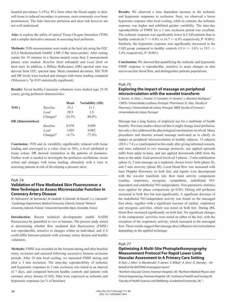
34 J Vasc Res 2017;54(suppl 1):1-80DOI: 10.1159/000471944
hospital prevalence 3-14%). PUs form when the blood supply to skin/soft tissue is reduced secondary to pressure, most commonly over bony prominences. The links between perfusion and ulcer risk however are not well understood.
Aim: to explore the utility of optical Tissue Oxygen Saturation (TOS) and a simpler derivative measure in assessing heel perfusion.
Methods: TOS measurements were made at the heel site using the O2C (LEA Medizintechnik GmbH: LMF-2 flat micro-probe). After resting supine for 10 minutes in a thermo-neutral room then 2 measurement phases were studied: Baseline (heel unloaded) and Load (heel on hard mat). In addition, a Diffuse Reflectance (DR) spectral ratio was derived from O2C spectral data. Mean (standard deviation, SD) TOS and DR levels were tracked and changes with tissue loading compared (Wilcoxon’s, *p<0.05 statistically significant).
Results: Seven healthy Caucasian volunteers were studied (age 22-56 years), giving perfusion characteristics:
Mean Variability (SD)TOS [ Baseline 53.2 11.1 Load 28.9 1.8 Changes* -24.3% -80.8%DR [dimensionless] Baseline 0.978 0.009 Load 1.026 0.002 Changes* +4.7% -77.0%
Conclusion: TOS and its variability significantly reduced with tissue loading and converged to a value close to 30%, a level attributed to tissue colour. DR showed similarities in the patterns of dynamics. Further work is needed to investigate the perfusion oscillations, tissue colour and changes with tissue loading, ultimately with a view to assessing patients at risk of developing a pressure ulcer.
PoA-74Validation of Flow Mediated Skin Fluorescence: a New Technique to Assess Microvascular Function in Coronary Artery DiseaseM. Hellmannn1, M. Tarnawska2, M. Dudziak2, K. Dorniak2, M. Roustit3, J.-L. Cracowski3
1Cardiology Department, Medical University, Gdansk, Poland, 2Medical University, Gdansk, Poland, 3Université Grenoble Alpes, Grenoble, France Introduction: Recent technical developments enable NADH fluorescence be quantified in vivo in humans. The present study aimed at determining whether flow mediated skin fluorescence (FMSF) was reproducible, sensitive to changes within an individual, and if it could differ between patients with coronary artery disease and healthy volunteers.
Methods: FMSF was recorded on the forearm during and after brachial artery occlusion and assessed following successive forearm occlusion periods. After 10 min local cooling, we measured FMSF during and after a 3 min occlusion. The inter-day reproducibility of ischemic and hyperemic responses to 3 min occlusion was tested at an interval of 7 days, and compared between healthy controls and patients with coronary artery disease (CAD). Data were expressed as ischemic and hyperemic responses (as % of baseline).
Results: We observed a time dependent increase in the ischemic and hyperemic responses to occlusion. Next, we observed a lower hyperemic response after local cooling, while in contrast, the ischemic response was higher and exhibited greater variability. The inter-day reproducibility of FMSF for a 3 min occlusion period was excellent. The ischemic response was significantly lower in CAD patients than in healthy controls (6.7 +/- 4.8% vs 14.7 +/- 6.8% respectively, P <0.001). Similarly, the hyperemic response was significantly decreased in the CAD group compared to healthy controls (11.6 +/- 3.6% vs 19.5 +/- 5.4% respectively, P <0.001).
Conclusions: We showed that quantifying the ischemic and hyperemic FMSF response is reproducible, sensitive to acute changes in skin microvascular blood flow, and distinguishes patients populations.
PoA-75Exploring the impact of massage on peripheral microcirculation with the wavelet transformC. Rocha1, H. Silva1, I. Frazão2, V. Castelão2, H. Ferreira3, L. Monteiro Rodrigues1
1CBIOS / Universidade Lusófona, Portugal, 2Pharmacol. Sc. Dep., Faculty of Pharmacy / Universidade de Lisboa, Portugal, 3IBEB, Faculty of Sciences / Universidade de Lisboa, Portugal Massage has a long history of empirical use for a multitude of health benefits. Previous studies observed that it might change local perfusion, but only a few addressed the physiological mechanisms involved. Many procedures and theories around massage motivated us to clarify its impact on peripheral microcirculation in healthy subjects. 15 subjects (28.0 ± 7.4 y.o.) participated in this study after giving informed consent, and were subjected to two massage protocols, one applied upwards (uM) from ankle to knee, and one applied downwards (dM) from the knee to the ankle. Each protocol involved 3 phases - 5 min stabilization (phase I), 5 min massage on a randomly chosen lower limb (phase II), and 5 min recovery (phase III). Local blood flow was measured with laser Doppler flowmetry on both feet, and signals were decomposed with the wavelet transform into their main activity components (cardiac, respiratory, myogenic, sympathetic, endothelial NO-dependent and endothelial NO-independent). Non-parametric statistics were applied for phase comparisons (p<0.05). During uM perfusion increased in both feet but non-significantly. A significant decrease of the endothelial NO-independent activity was found on the massaged foot alone, together with a significant increase of cardiac, respiratory and myogenic activities, which was noted on both feet. During dM, blood flow increased significantly on both feet. No significant changes in the components’ activities were noted on either of the feet, with the exception of the respiratory activity, which increased in the massaged foot. These results suggest that massage does influence microcirculation depending on the applied technique.
PoA-77Optimising A Multi-Site Photoplethsmyography Measurement Protocol For Rapid Lower Limb Vascular Assessment In A Primary Care SettingD. Kyle1, J. Allen2, A. Macdonald2, P. James3, S. Wilkes4, A. Sims2, G. Stansby1, - On behalf of the NOTEPAD investigator teams5
1Northern Vascular Centre, Freeman Hospital, UK, 2Northern Medical Physics and Clinical Engineering, Freeman Hospital, UK, 3Institute of Health and Society,UK, 4Faculty of Health Sciences and Wellbeing, Sunderland University, UK, 5-
35J Vasc Res 2017;54(suppl 1):1-80DOI: 10.1159/000471944
Introduction: Peripheral arterial disease (PAD) is common and can cause pain on walking or when more severe, gangrene and limb loss. Unfortunately, it is often under-diagnosed in primary care and the current standard test for PAD, the ankle brachial pressure index (ABPI), can be time consuming and technically challenging to perform. We have developed a novel low-cost optical pulse device for PAD diagnosis utilising multi-site photoplethsmyography (MPPG), which detects composite macro- and micro-circulatory blood volume changes and may provide a fast and easy-to-do assessment of PAD.
Methods: Forty-three PAD patients were recruited from secondary care. Measurements were performed in a thermally neutral vascular measurement facility at 2, 5 and 10 minutes; both in the sitting and supine position. MPPG provided two pulse measures for the toes – The Shape Index (SI) and Timing Index (TI). Mixed linear models were used for statistical analysis.
Results: Results revealed that posture significantly affected SI at each time point but was not significantly affected when resting time was shortened from 10 minutes to either 5 or 2 minutes in the supine position. The TI measure and absolute differences between left and right were not significantly affected by resting time or posture.
Conclusion: An innovative photoplethysmography device for PAD assessment demonstrates that a resting time of 2 minutes can be used which is much less than the standard 10 minute period for ABPI. By optimizing the measurement protocol the MPPG pulse technology can potentially make an accessible and workable solution to vascular diagnostics in primary care.
PoA-78About the in vivo effects of hyperoxia in humanH. Silva1, I. Frazão2, R. Miranda2, L. Monteiro Rodrigues1
1CBIOS / Universidade Lusófona, Portugal, 2Pharmacol. Sc. Dep., Faculty of Pharmacy / Universidade de Lisboa, Portugal Skin is widely regarded as a useful model for studying microvascular function in vivo, allowing noninvasive assessments. The normobaric oxygen breathing (NOB) test is particularly useful to dynamically assess peripheral microcirculation, although there is a clear need for clarification regarding the mechanisms involved in this response. Our aim was to look deeper into the peripheral microvascular response to hyperoxia using a combination of several noninvasive techniques. 12 healthy subjects (20.2 ± 2.9 y.o.) were included in this study after informed consent. Subjects performed a NOB test while sitting, which consisted of three phases - 10 min breathing room atmosphere (I), 10 min breathing 100% oxygen through a facemask (II), and again breathing room atmosphere (III). Measured variables included blood flow with laser Doppler flowmetry (LDF) and photoplethysmography (PPG) on the toes; electrodermal activity (EDA), transepidermal water loss (TEWL), and transcutaneous (tc) O2 pressure. Additionally, heart rate (HR) was given by electrocardiography (ECG), respiratory rate (RR) and depth by pneumography and blood pressure (BP) with a digital device. Nonparametric statistics were applied (p<0.05). During phase II, tcpO2 increased significantly, revealing two distinct increment velocities, creating hyperoxia. This, in turn, caused a significant decrease in blood flow in both feet, as noted with LDF and PPG. EDA and TEWL showed consistent bilateral increases, but without significance. RR decreased while RD increased, both significantly.
HR and BP increased also without significance. These results suggest the complemented utility of these techniques for the noninvasive assessment of the “integrated” microvascular response to hyperoxia.
PoA-79Pilot Assessment Of The Utility Of Tissue Oxygenation Spectrophotometry Measurements In Patients With Systemic SclerosisJ. Allen1, C. Di Maria2, S. Urwin2, A. Murray3, L. Ottewell4, B. Griffiths4
1Microvascular Diagnostics, Northern Medical Physics and Clinical Engineering, UK, 2Microvascular Diagnostics, Northern Medical Physics and Clinical Engineering, Freeman Hospital, UK, 3Institute of Cellular Medicine, Faculty of Medical Sciences, Newcastle University, UK, 4Department of Rheumatology, Musculoskeletal Unit, Freeman Hospital, UK
Introduction: Systemic sclerosis (SSc) patients can experience significant morbidity, including interstitial lung disease (ILD). The development of tests to aid their assessment and determine prognosis is very important. The aim of this study was to assess the utility of non-invasive skin Tissue Oxygen Spectrophotometry (TOS) measurements in patients with established SSc.
Methods: Superficial tissue depth TOS measurements were collected by a single operator (JA) at 3 skin sites on the forearm, upper chest and calf using the O2C device (LEA Medizintechnik GmbH): in healthy controls (HCs) and SSc patients, including some with ILD diagnosed by high-resolution computed tomography chest scans. Non-parametric statistics were employed (p<0.05 statistically significant).
Results: Nine healthy controls (median [IQR]) (age 52 [47-59] years)] and 14 SSc patients (61 [53-65] years) were studied. Median SSc disease duration was 6 [3-8] years; 8 had ILD; and 7 had diffuse cutaneous SSc. Median TOS levels using the mean of the 3 sites were significantly reduced in SSc (42 [35-45] %) compared to HC (52 [52-67] %) (p<0.001). SSc ILD patients had relatively low median TOS levels (37 [31-46] %) (not statistically significant).
Conclusion: Significant TOS reductions were found in SSc patients compared to HCs at all sites, and there was a tendency for TOS to be lower in ILD patients. The results demonstrate the potential utility of TOS for this patient group. Further work is needed to validate this technique prospectively and in a larger group of SSc patients.
PoA-80Time-Dependent Behaviour of Microvascular Blood Flow and Oxygenation: a Predictor of Functional OutcomesG. Clough1, K. Kuliga2, R. Gush3, A. Chipperfield4
1University of Southampton, UK, 2Faculty of Medicine, University of Southampton, UK, 3Moor Instruments Ltd., Axminster, UK, 4Faculty of Engineering and the Environment, University of Southampton, UK Objective: This study investigates the time-dependent behaviour and algorithmic complexity of low frequency, periodic oscillations in blood flux and oxygenation signals from the microvasculature. Recent findings in animal models suggest that this may provide a novel understanding of the flexibility/predictability of network perfusion and is investigated in humans here.
36 J Vasc Res 2017;54(suppl 1):1-80DOI: 10.1159/000471944
Methods: Microvascular blood flux (BF) and oxygenation (oxyHb, deoxyHb, totalHb and SO2) was recorded from healthy males using combined laser Doppler fluximetry and white light spectroscopy with local skin temperature clamped to 33oC and during local thermal hyperaemia (LTH) at 43oC. Power spectral density (PSD) of the BF and OXY signals was evaluated within the frequency range (0.0095-1.6Hz). Signal complexity was determined using the Lempel-Ziv (LZ) algorithm.Results: Fold-increase in BF during LTH was 15.6 (10.3,22.8) and in OxyHb 4.8 (3.5,5.9) (median, IQR). All BF and OXY signals exhibited multiple oscillatory components with clear differences in signal power distribution across frequency bands at 33oC and 43oC. Significant reduction in the intrinsic variability and complexity of the microvascular signals during LTH was found, with mean LZ complexity of BF and OxyHb falling by 25% and 49%, respectively (p<0.001).
Conclusions: These results provide corroboration that in human skin microvascular blood flow and oxygenation are influenced by multiple, time-varying oscillators that adapt to local influences and become more predictable during increased haemodynamic flow. Time series analysis offers an extended assessment of microvascular network behaviour to inform how a loss of system flexibility may prevent microvascular networks from effectively adapting to an imposed stressor.
PoA-81Comparing the in vivo spectral components of Laser Doppler Flowmetry and Photoplethysmography by the wavelet transform to assess microcirculationH. Silva1, I. Frazão2, R. Miranda2, L. Monteiro Rodrigues1
1CBIOS / Universidade Lusófona, Portugal, 2Pharmacol. Sc. Dep., Faculty of Pharmacy / Universidade de Lisboa, Portugal Laser Doppler flowmetry (LDF) is regarded as the most employed technology for microcirculation quantification. However, photoplethysmography (PPG) has received a renewed interest, much due to its recent technological evolution. Although both quantify local blood flow, LDF and PPG are technically very different, giving signals with distinct morphologies and characteristics. Our aim was to perform a spectral comparison between LDF and PPG signals with the wavelet transform. Signals from 12 healthy subjects (20.2±2.9 y.o.) were recorded for 30 min on the second and third toes with LDF and PPG, respectively. The electrocardiography (ECG, lead I) was recorded from both hands, while a respiration signal was obtained with pneumography (PNG). A wavelet-derived periodogram was created for all signals, and their range and frequency of maximal amplitude compared (p<0.05). LDF and PPG periodograms showed a well-defined cardiac band, consistent with the ECG periodogram, from 0.8 to 1.6 Hz, with no significant differences found for the range between the signals. A respiratory band was well defined in all signals from 0.4 to 0.2 Hz. The PPG signal further showed lower frequency bands in the 0.1-0.05, 0.05-0.01 and 0.01-0.005 Hz regions, also observed with LDF, and which are consistent with the myogenic, sympathetic and endothelial activities. No statistical significant differences were found for the range or for the frequency of maximal amplitude between the LDF and PPG signals. These results show the spectral coherence between the assessed signals, further suggesting PPG as a useful alternative to LDF for exploring the oscillatory components of skin microcirculation.
PoA-82Paraoxonase-1 Reduces Vasodilation in the Microcirculation by Regulating Arachidonic Acid-Derived Lactone MetabolitesAndrea Szuchman-Sapir1, Reut Levy2, Dan Gilad2, Ziv Mozes2, Shahar Atiya2, Rotem Ben Shushan2, Elana Slutsky-Smith2, Snait Tamir2
1MIGAL-Galilee Research Institute, Israel, 2MIGAL - Galilee Research Institute, Israel Endothelial cells control the tone of the underlying vascular smooth muscle cells by releasing various relaxing and contracting factors, including endothelial-derived hyperpolarizing factors (EDHF). Among the EDHFs are the cytochrome P450 - derived arachidonic acid metabolites - the epoxyeicosatrienoic acids (EETs), which are known as vasodilators.
Paraoxonase 1 (PON1) is an antiatherogenic high density lipoprotein-associated lactonase. Previous findings revealed that PON1 knockout (PON1KO) mice have low blood pressure, which is inversely correlated with the level of 5,6-EET. 5,6-EET is an EDHF that induces arterial dilation. Formerly, we demonstrated that 5,6-EET forms a more stable lactone (5,6- δ-DHTL) isomer, that can be hydrolyzed by PON1. Our studies revealed that 5,6-δ-DHTL is a potential EDHF, since it mediates vasodilation of human resistance arteries and evokes endothelial hyperpolarization, with increased cytosolic Ca2+ influx and Ca2+-dependent potassium efflux. Using immunohistochemistry assay and confocal microscopy, we demonstrated that PON1 can penetrate the endothelial cell membrane in mice arteries and had lactonase activity in the cells. Vascular reactivity studies using pressurized myograph revealed that PON1KO mice mesenteric arteries had a significantly higher relaxation response to cumulative concentrations of 5,6-DHTL than those of WT mice, while administration of rePON1 reduced the dilation effect mediated by 5,6-DHTL.
Taken together, our results indicate that PON1 has a regulatory role in microcirculation vasodilation by controlling the levels and function of 5,6-DHTL, which is a potential EDHF. This study may point out for additional lactone-containing metabolites of oxygenated PUFA that are potent dilators of resistant vessels, and are regulated by PON1.
37J Vasc Res 2017;54(suppl 1):1-80DOI: 10.1159/000471944
WEDNESDAY, 31 MAY 2017
PARALLEL SESSION 4A BIOMECHANICS OF VASCULAR DISEASE
4A-01Aquaporin 1 is critical in adaptation of endothelial cells to shear stress.O. Bondareva1, M. Odenthal-Schnittler2, B. Kemper3, H.-J. Schnittler2
1Institute of Ananomy and Vascular Biology, Münster, Germany, 2Institute of Anatomy and Vascular Biology, Münster, Germany, 3Biomedical Technology Center of the Medical Faculty, University of Münster, Germany Aqp1 is a membrane protein which is expressed in the apical and basolateral plasma membranes of epithelial and endothelial cells, and functions as a water channel, activated via osmotic pressure gradient. Aqp1 in endothelial cells was reported to be involved in directed cell migration, wound healing processes, tubulogenesis and cancer angiogenesis. Here we demonstrate a novel role of Aqp1 being essential in physiological adaptation of the endothelium to shear stress.
We investigated adaptation of endothelial cells to arterial levels of shear stress (30 hours; 1,2 Pa) by means of label-free digital holographic microscopy. This process was accompanied by an increase in cell volume that follows a saturation function as well as promoted Aqp1 upregulation. Inhibition of cell swelling was blocked by the sodium- proton exchange inhibitor cariporid. Importantly, siRNA mediated knock-down of Aqp1 in HUVEC disintegrated the endothelial monolayer under shear stress conditions visible by ubiquitous intercellular gap formation due to actin- and VE-cadherin remodeling. Furthermore Aqp1 is essential in maintenance of endothelial integrity as determined by impedance spectroscopy measurements.
Our data for the first time demonstrate the critical impact of cell swelling and importance of Aqp1 in basic endothelial cell adaptation to shear stress regarding maintenance of cell monolayer integrity and barrier function via volume regulation.
PARALLEL SESSION 4B EMERGING TECHNOLOGIES IN VASCULAR BIOLOGY
4B-01Lentiviral Magnetic Targeting of SHP-2 Locally Governs HIF-1α Dependent Wound Healing Angiogenesis in vivo Y. Heun1, M. Anton2, A. Pfeifer3, C. Plank2, U. Pohl1, H. Mannell1
1Cardiovascular Physiology, LMU, Germany, 2Institut für Molekulare Immunologie/Experimentelle Onkologie und Therapieforschung, Technical University Munich, Germany, 3Institut für Pharmakologie und Toxikologie, Bonn University, Germany Introduction: HIF-1α is essential for angiogenesis and wound healing. Here, we studied a gene therapeutic approach to modify HIF-1α by site specific targeting of the phosphatase SHP-2 in wounds in vivo.
Methods: Wound healing angiogenesis was assessed in the murine (C57BL/6) dorsal skin fold chamber (n=3-5 per experimental group). SHP-2 wild-type and mutant constructs were expressed by lentiviral transduction of human microvascular endothelial cells and in wounds
in vivo by magnetic targeting of MNP-LV complexes. VEGF expression was measured by qRT-PCR. HIF-1α degradation was detected by western blot and HIF-1α-ODD-Luciferase reporter construct expression. HIF-1α activity was measured by a luciferase reporter construct sensing HIF1 transcriptional activity and HIF1 DNA-binding assay.
Results: Expression of inactive SHP-2 (CS) reduced endothelial cell and aortic vessel sprouting, and VEGF secretion under hypoxia (pO2: 8±2mmHg) compared to SHP-2 WT, whereas expression of constitutively active SHP-2 (E76A) enhanced these responses (all p<0.05, n=4-5). Interestingly, SHP-2 CS expression prevented hypoxic endothelial HIF-1α stabilization, activity and DNA-binding by enhancing HIF-1α degradation (all p<0.05, n=3-6). Using magnetic targeting of MNP-LV, we achieved local transgene expression in separate wounds of the same dorsal skin fold chamber. Whereas SHP-2 CS expression impaired angiogenesis and HIF-1α activity in wounds in vivo, SHP-2 E76A enhanced this compared to SHP-2 WT (all p<0.05, n=4-5). Finally, SHP-2 CS reduced VEGF expression in these wounds (p<0.05, n=5).
Conclusion: SHP-2 augments HIF-1α dependent wound healing angiogenesis in vivo by promoting HIF-1α stabilization. Magnetic targeting of SHP-2 may be a useful approach achieving local therapeutic angiogenic responses.
4B-02Implantable Oxygen Biosensors Reveals Post-occlusion Tissue Reactive HyperoxiaJ. Chien1, M. Mohammed1, H. Eldik1, M. Ibrahim1, S. Nichols2, B. Klitzman1
1Duke Univ Med Ctr, United States, 2Profusa, United States Introduction: The purpose of this study was to implant biosensors quantifying tissue oxygenation before, during, and after transient ischemia.
Methods: Rats had unilateral femoral artery and vein ligation and received bilateral subcutaneous hindlimb biosensors composed of porphyrin embedded into a porous poly-hydroxyethylmethacrylate scaffold. The biosensor’s phosphorescence decay changed inversely with oxygenation. Near infrared spectroscopy (NIR) quantified percent O2 saturation of capillary hemoglobin. Laser Doppler flowmetry quantified blood flow. On days 28 and 84, rats underwent systemic hypoxia as well as bilateral hindlimb tourniquet application. Response magnitudes, response times and normalized changes were calculated. Statistical significance was assessed using ANOVA and paired t-test.
Results: Laser Doppler flowmetry confirmed a reactive hyperemia following tourniquet release. In addition, both the implantable biosensors and NIR methods suggested that the tissue O2 content transiently exceeded baseline following tourniquet release (p<0.05). We term this phenomenon reactive hyperoxia, indicating that the increased consumption post-occlusion is met with an even greater elevated O2 supply. The NIR spectroscopy and phosphorescence lifetime data are consistent. The phosphorescence lifetime biosensor also demonstrates a more prominent response magnitude in general and a faster response over time (71 secs vs. 143 secs in NIR; p<0.01), suggesting microvascular ingrowth throughout the porous scaffold.
38 J Vasc Res 2017;54(suppl 1):1-80DOI: 10.1159/000471944
Conclusions: Implantable biosensors provide real-time monitoring of tissue oxygenation with rapid responses, excellent biocompatibility and localized measurements. Both the biosensors and NIR spectroscopy demonstrated reactive hyperoxia post ischemia. Monitoring tissue oxygenation may provide diagnostic and prognostic information on wound healing, re-vascularization, 3D scaffold design and efficacy of therapeutics promoting tissue oxygenation.
PARALLEL SESSION 4C NOVEL ANGIOGENIC PATHWAYS AND TISSUE REGENERATION
4C-01Lateral Induction of Dll4 Expression Initiates non Sprouting Angiogenesis by VEGFR. Gianni Barrera1, A. Uccelli2, K. Bentley3, H. Gerhardt4, A. Banfi2
1University Hospital, DBM, Switzerland, 2Department of Biomedicine and of Surgery, Basel University Hospital, Switzerland, 3Pathology, Beth Israel Deaconess Medical Center, Harvard Medical School, Boston, USA, 4Max-Delbrück Center for Molecular Medicine, Helmholtz Association, Berlin, Germany Dll4/Notch1 signaling regulates sprouting angiogenesis by determining the balanced formation of tip and stalk endothelial cells. However, we have found that VEGF over-expression in skeletal muscle induces microvascular growth by causing circumferential vascular enlargement followed by longitudinal splitting (intussusception), rather than sprouting. Here we investigated whether and how Dll4/Notch1 signaling regulates VEGF-induced intussusceptive angiogenesis. Monoclonal populations of transduced myoblasts, expressing specific VEGF levels that induce normal or aberrant angiogenesis, were implanted in mouse muscles. By 4 days, vascular enlargements displayed simultaneous Notch1 activation and Dll4 expression in long stretches of adjacent endothelial cells, whereas after intussusceptive remodeling (7 days) the Dll4/Notch1 axis was no longer active in both normal and aberrant vessels. Early Notch inhibition disrupted the enlargements into disorderly endothelial aggregates. Testable scenarios were generated by a computational model for the dynamics of Dll4 expression and Notch1 activation in non-sprouting angiogenesis. Experimental validation showed that VEGF leads to simultaneous expression of Dll4 and Notch1 activation in adjacent ECs by a mechanism of lateral induction of Dll4 by Notch1, opposite to the lateral inhibition regulating sprouting. Also contrary to the effect in sprouting, sustained Notch inhibition during therapeutic VEGF delivery not only did not disrupt normal angiogenesis, but actually increased it, with efficient physiological perfusion. In conclusion: 1) VEGF determines circumferential enlargement and intussusception by a spatially continuous and stable pattern of Dll4/Notch1 activation sustained by lateral induction; and 2) in the setting of therapeutic VEGF delivery and intussusceptive angiogenesis, Notch inhibition increases both vascular growth and functional perfusion.
4C-02Von Willebrand Factor modulates blood vessel formation and function via Angiopoietin-2K. Smith, R. Starke, N. Dufton, C. Millar, T. McKinnon, J. Mason, M. Laffan, A. RandiImperial College London, United Kingdom Introduction: Von Willebrand Factor (VWF) is a large glycoprotein synthesised by endothelial cells (EC) and megakaryocytes. Defects in VWF cause Von Willebrand disease (VWD), the most common
bleeding disorder. Some patients with VWD present with vascular malformations in the gastrointestinal (GI) tract (angiodysplasia), which can lead to severe bleeding. Vascular malformations may be linked to the ability of VWF to regulate blood vessel formation. VWF drives the formation of Weibel Palade Bodies (WPB) which store a key regulator of angiogenesis and permeability, Angiopoietin 2 (Ang-2).
Methods: In vitro, inhibition of VWF expression was performed using siRNA in HUVEC. Expression was measured by qRT-PCR, ELISA or immunoblotting. EC sprouting was assessed using in vitro fibrin bead assay. In vivo permeability in VWF KO mice and controls was assessed using intravenous injection of high (2x10⁶) and low (4.4x10³) molecular weight dextrans (MWD) then confocal microscopy.
Results: Enhanced EC sprouting of VWF-deficient HUVEC in vitro was normalised by inhibition of Ang-2 activity. Inhibition of VWF expression in vitro causes enhanced synthesis of Ang-2 in an Akt-FOXO1 dependent manner in EC. In vivo, Ang-2 levels are increased in the hearts but not lungs nor liver of VWF KO mice, suggesting that VWF regulates Ang-2 expression in a tissue-specific manner. Isolectin staining showed enhanced vascularization in the heart of VWF KO mice. Vessels from VWF KO mice were dysfunctional as measured by increase extravasation of low MWD.
Conclusion: VWF regulates Ang-2 expression via Akt-FOXO1 dependent pathway and VWF modulates angiogenesis and vascular function through Ang-2.
4C-03Targeting Connexin40 Expression or Function Reduces Angiogenesis in the Developing Mouse Retina F. Alonso1, J.-A. Haefliger2, F. Allagnat2, L. Hamard2, E. Génot3
1INSERM1045, Université de Bordeaux, France, 2Department of Medicine, Lausanne University Hospital, Switzerland, 3INSERM1045, Université de Bordeaux, France Introduction: Connexins (Cxs) constitute a large family of trans-membrane proteins that form gap junction channels enabling direct transfer of small signaling molecules from cell-to-cell. We recently found that altering Cx40 expression or function in endothelial cells (ECs) impairs their ability to vascularize tumors in mice. However, how Cx40 contributes to angiogenesis has not yet been investigated.
Methods: We used the mouse neonatal retina model and Cx40-deficient mice to explore Cx40 expression and function in developmental angiogenesis. We used immortalized mouse brain microvascular ECs to explore the contribution of Cx40 channels on cell properties and behavior in vitro.
Results: Our data show that Cx40 is expressed in the immature vascular plexus of the developing retina. Genetic ablation of Cx40 causes retinal hypovascularization in the postnatal mouse eye, with reduced radial vessel growth and decreased capillary density as compared to WT mice. At the angiogenic front, Cx40-deficiency attenuates vessel sprouting and the mural cells recruited on the neovessels display an altered phenotype. These alterations can be attributed to disturbed EC functions as selective re-expression of Cx40 in these cells restores normal angiogenesis. In vitro, targeting Cx40 by silencing its expression or by
39J Vasc Res 2017;54(suppl 1):1-80DOI: 10.1159/000471944
blocking gap junction channels, decreases EC proliferation, modifies their secretome and promotes mural cell chemoattraction. Finally, a Cx40 inhibitory peptide (40Gap27) phenocopies the loss of endothelial Cx40 in vitro and in vivo.
Conclusions: Our data show that endothelial Cx40 regulates retinal vessel growth and maturation. Cx40 thus represents a novel potential target for treating pathological ocular angiogenesis.
PARALLEL SESSION 5A IMAGING THE MICROVASCULATURE
5A-01Imaging Of Leukocyte And Stem Cell Trafficking In Vivo In The Beating Mouse Heart Microcirculation Following Myocardial Ischaemia-Reperfusion InjuryN. Kalia, A. Lokman, D. KavanaghUniversity of Birmingham, United Kingdom Background: The clinical success of stem cell (SC) therapy for myocardial infarction is compromised by poor cardiac homing following systemic delivery. Given that such therapy may depend on beneficial paracrine effects, it is a further hindrance that little is known about the inflammatory response dynamics within myocardial microcirculation in vivo.
Methods: 3D-printed stabilisers were bonded to the anaesthetised (ketamine/xylazine) mouse beating heart to enable confocal intravital imaging of ventricular microcirculation. PE-anti-Gr-1 and APC-anti-CD41 antibodies were injected to label neutrophils and platelets respectively with FITC-BSA enabling blood flow visualisation. In some mice, haematopoietic SCs (HSCs; HPC-7s) were introduced intra-arterially. IR injury was induced by 45min (reversible) ligation of the LAD artery.
Results: Neutrophil adhesion and platelet accumulation were both significantly (p<0.001) and rapidly increased in injured microvessels with platelet accumulation increasing with time. No difference in number or velocity of free-flowing neutrophils was observed. A significant (p<0.05) decrease in functional capillary density was also observed in injured hearts. Although HSC adhesion was not significantly enhanced as a result of injury, a time-dependent increase in adhesion was observed in sham and injured hearts. No significant change in number or velocity of free-flowing HSCs was observed following injury. Interestingly, despite reduced capillary perfusion, approximately 10-20 HSCs were observed trafficking though the heart at each time point throughout reperfusion.
Discussion: Intravital microscopy has allowed successful visualisation of the microvascular inflammatory response and HSC homing events in the beating mouse heart post-reperfusion. Subsequent experiments will allow us to assess whether HSC administration can confer vasculoprotection.
5A-02Novel non-destructive microangio-CT for visualization and quantification of the entire organ vasculatureV. Djonov, R. HlushchukInstitute of Anatomy, Switzerland
Introduction: In most animal models the non-destructive investigation of the entire vasculature down to the capillary level was not feasible. The lack of such 3-dimensional visualization and proper vascular quantification in combination with inappropriate animal models contributes to the high discrepancy between the results obtained in preclinical and clinical studies.
Methods and Results: Recently we developed conceptually new non-destructive, ex vivo microangio- CT(µaCT ) and are able to visualize and quantify the entire vasculature of a whole organ down to the capillary level (in a range of 4-6µm in diameter). In addition the solidification and autofluorescence properties of the new contrast agent µAngiofil permit a correlative approach, i.e., after the µaCT-visualization and definition of the sites of interest further morphological analyses by histology, immunohistochemistry and transmission-electron microscopy can be performed on the same samples. The introduced µaCT-based vascular quantification protocols are very rapid and superior to the time-consuming classical morphometry. Validation based on lectin-stained histological sections reveled excellent perfusion efficiency: 98±0.5% of the blood vessels were µAngiofil®-positive, whereas 93±2.6% were lectin-positive. By applying this approach we analyzed pro- and anti-angiogenic treatment strategies in murine brain, kidney, muscle, eye, heart and bones.
Conclusions: The introduced correlative and quantitative imaging approach is highly reproducible and allows for the first time a detailed 3D characterization of the entire vasculature down to the single capillary vessels. Alone, or as a correlative approach in combination with histology and electron microscopy this opened a new horizons for broad range investigations in the field of vascular biology.
5A-03Red Blood Cells Properties in Human MicrocirculationC. Saldanha1, I. Oliveira2, P.a Napoleão2, A. Messias3, A. Silva-Herdade2
1Instituto de Bioquímica, Instituto de Medicina Molecular, Faculdade de Medicina, Portugal, 2Instituto de Bioquímica, IMM, FMUL, Portugal, 3Cuidados Intensivos, Hospital Beatriz Ângelo, Loures, Portugal The physiological role of Red Blood cells (RBCs) in microcirculation will be highlight in this minireview. RBCs can elongate and can undergo aggregation in dependence of several factors like glucose metabolism and several signal transduction mechanisms. RBCs is a component of blood and has ability to scavenger and delivery oxygen and nitric oxide (NO) to all tissues. For the estimation of the oxygenation of tissue’s status it is necessary the quantification of the affinity of haemoglobin to oxygen. It was found increase of P50 in hypertensive patients with diastolic pressure higher or equal to 130mm and high degree of retinopathy. Hg. In diabetic patients high plasma glucose levels and decrease values of P50 were obtained. It was verified higher NO release from erythrocyte samples obtained from patients with hypercholesterolemia and hypertension, besides the fact
40 J Vasc Res 2017;54(suppl 1):1-80DOI: 10.1159/000471944
that they sustain impaired erythrocyte deformability. In patients with amyotrophic lateral sclerosis erythrocyte NO content is preserved and was verified an inverse association between respiratory function and NO efflux from the erythrocyte. Sepsis patient before dead at 24 in IUC showed higher efflux of NO from erythrocytes that worsening the blood circulation as verified by high unequal blood flow and high microvascular flow index observed in sub lingual microcirculation. The in vivo mice model of acute inflammation evidenced that the NO efflux from erythrocyte decrease with acute phase response development. NO normal values were obtained with inflammation recovering besides the maintenance of decreased RBCs deformability
PARALLEL SESSION 5B EPIGENETICS AND NONCODING RNA IN VASCULAR BIOLOGY (A)
5B-01Smooth Muscle Cells Differentiated from Human Umbilical Cord Mesenchymal Stem Cells Regulated by miR-503 Are Potent for Vascular Tissue EngineeringW. Gu, X. Hong, Z. Zhang, Y. Hu, Q. XuKing’s College London, United Kingdom Introduction: Tissue engineered vascular grafts with long-term patency are in great need in the clinic. Human mesenchymal stem cells from the umbilical cord (UCMSCs) exhibit multi-lineage differentiation abilities, such as smooth muscle cell (SMC) differentiation. Micro RNAs are short non-coding regulatory RNAs. They widely participate in stem cell differentiation and may play an important role in SMC differentiation.
Objective: We hypothesize that vascular grafts could be generated with SMCs differentiated from human UCMSCs and explore the role of miRNAs in the differentiation process.
Methods and Results: We utilized TGFbeta1 to stimulate UCMSCs differentiation towards SMCs. SMC markers including α-SMA, SM22, Calponin and SMMHC were highly upregulated both at the gene expression and the protein level after TGFbeta1 treatment. Micro-RNA (MiR) array analysis showed the miR-503 upregulation in differentiation, which was confirmed by TaqMan assay. We further demonstrated that miR-503 mimics promoted SMC differentiation and miR-503 inhibitors downregulated SMC markers. Smad7, a negative regulator of TGFbeta1 related signaling pathways, was shown to be the direct target of miR-503 by luciferase reporter experiments. Furthermore, upon TGFbeta1 stimulation, smad4 could bind to the promoter region of miR-503 and upregulate its expression. Finally, SMCs differentiated from UCMSCs exhibited the ability to migrate into decellularized aorta grafts.
Conclusions: We developed a protocol to generate SMCs from UCMSCs and miR-503 was proved to regulate the differentiation process. These findings implicate that UCMSC as a new cell source to obtain SMCs for tissue engineering is promising and present miR-503 as a potent regulator in SMC differentiation mechanism.
5B-02miRNA-22 Is A Novel Mediator Of Vascular Smooth Muscle Cell Phenotypic Modulation And Neointima FormationQ. XiaoQueen Mary University of London, United Kingdom Introduction: microRNA-22 (miR-22) has recently been reported to play a role during vascular smooth muscle cell (VSMC) differentiation from stem cells. However, its importance in mature VSMC functions as well as neointima formation following vascular injury remains to be elucidated. In this study, we aimed to investigate the potential involvement of miR-22 in mature VSMC phenotypic modulation and neointima formation, and unravel its underlying molecular mechanisms.
Methods and Results: Expression level of miR-22 was closely modulated during VSMC phenotypic modulation, and in the injured arteries. miR-22 over-expression significantly increased VSMC contractile gene expression and inhibited VSMC proliferation and migration, whilst the opposite effect was observed when endogenous miR-22 was knocked down in VSMCs. Apart from previously identified miR-22’s target gene, methyl-CpG binding protein 2 (MECP2), the transcriptional regulator and oncoprotein, Ecotropic Virus Integration Site 1 Protein Homolog (EVI1), has been identified as a novel miR-22 target gene in miR-22-mediated VSMC phenotypic modulation. Of note, perivascular enforced expression of miR-22 in the injured vessels significantly reduced the expression levels of MECP2 and EVI1, decreased VSMC proliferation, and inhibited neointima formation in wire-injured femoral arteries. Finally, miR-22 expression was significantly reduced, while MECP2 and EVI1 expression levels were dramatically increased in diseased human arteries; and an inverse relationship could be observed between miR-22 and MECP2 and/or EVI1 in human femoral arteries with or without atherosclerotic lesions.
Conclusions: Our data demonstrates that miR-22 is a novel regulator of VSMC function specifically during neointima hyperplasia, offering a novel therapeutic opportunity for treating vascular diseases.
PARALLEL SESSION 5C PLATELETS AND MICROVACULATUR INTERACTIONS
5C-01CD40L Dependent Platelet String Formation In The Mouse Microcirculation In VivoS. Tahir, M. HeckerHeidelberg University, Germany Introduction: CD40 ligation on endothelial cells (ECs) promotes the release of von Willebrand factor (vWF) multimers (ULVWF) which are cleaved by the zinc metalloproteinase ADAMTS13. Platelets adhere to these multimers and start recruiting circulating monocytes. It is not known whether CD40-CD40 ligand (CD40L) interactions play a role in thrombus formation and/or leukocyte diapedesis in microvessels in vivo, and whether the presence of ADAMTS13 limits these effects.
Methods: Two-photon excitation intravital microscopy was used to study ULVWF multimer-platelet string formation (carboxy fluorescein diacetate stained donor platelets) and leukocyte extravasation in the murine cremaster microcirculation of both wild-type and ADAMTS13
41J Vasc Res 2017;54(suppl 1):1-80DOI: 10.1159/000471944
ko mice. Leukocytes were labeled using PE-conjugated CD45 antibody. vWF and CD40 visualization was done with primary antibodies conjugated to Qdot-565 and Qdot-525, respectively.
Results: CD40L treatment induced platelet string formation (PSF) was observed both in arterioles and venules but was found predominantly in the latter. PSF was increased by about 3-fold in ADAMTS13 ko mice. PSF in knockout arterioles showed enhanced string length. Leukocyte extravasation after CD40L treatment was found to be more abundant in ADAMTS13 ko venules and prevailed in areas with visible ULVWF multimer-platelet strings.
Conclusion: CD40L stimulation augments vWF mediated PSF predominantly in venules. The absence of ADAMTS13 enhances the string formation in venules and elongates platelet strings in arterioles. CD40L also enhances leukocyte extravasation in venules starting predominantly in the areas of platelet strings.
5C-02Differential roles of NADPH oxidase 1 and 2 in collagen, collagen-related peptide and thrombin induced platelet activationD. Vara, L. Caggiano, G. PulaUniversity of Bath, United Kingdom Introduction: The role of NADPH oxidases in haemostasis and thrombosis have become important subjects in cardiovascular research; NOX1 and NOX2 appear to be main regulators of platelet function in both haemostasis and thrombosis. This study defines the molecular mechanism of action of NADPH oxidases in the platelet context, their relative importance in response to different stimuli and the possibility to modulate platelet responses by targeting these enzymes.
Methods: Superoxide anion was measured using electron spin resonance and quantified in platelets of human, WT and NOX1-/- and NOX2-/- mice following activation with thrombin, collagen and the GPVI-specific agonist, CRP. NADPH activity was confirmed in the presence of pharmacological inhibitors specific for NOX1, 2-acetylphenothiazine (2-APT) and NOX2, gp91TAT peptide. Aggregometry, whole blood thrombus formation and cell signalling were performed to clarify NOX signalling.
Results: We demonstrated that platelet aggregation, degranulation and thrombus formation under physiological shear stress in response to collagen require NOX1 activity and superoxide anion generation. Within the context of collagen-dependent stimulation of platelets, NOX1 drives the cell signalling triggered by collagen via inhibition of Src Homology Phosphatase 2 (SHP2/PTPN11), which dephosphorylates Src Tyrosine Kinase (SYK) and negatively regulates GPVI signalling pathway. NOX1-dependent inactivation of SHP2/PTPN11 is caused by cysteine oxidation within the phosphatase catalytic domain, as demonstrated by oxidation-specific immunoblotting.
Conclusion: This study confirms that NOX1 is critical player in collagen-dependent platelet activation, characterises the molecular mechanism of action of NOX1 within the GPVI signalling pathway and provides novel pharmacological tools for the inhibition of NOX1 in human platelets.
PARALLEL SESSION 6A EPIGENETICS AND NONCODING RNA IN VASCULAR BIOLOGY (B)
6A-01Heme Oxygenase-1 Controls Post-Ischemic InflammationA. Jazwa1, M. Tomczyk2, I. Kraszewska2, K. Szade2, K. Bukowska-Strakova2, A. Jozkowicz2, J. Dulak2
1Jagellonian University, Faculty of Biochemistry, Biophysics and Biotechnology, Poland, 2Department of Medical Biotechnology Faculty of Biochemistry, Biophysics and Biotechnology Jagiellonian University, Poland Overactive and prolonged immune responses can be responsible for heart failure after myocardial infarction (MI). Heme oxygenase-1 (Hmox1) is a heme-degrading enzyme that exerts important cytoprotective effects including anti-inflammatory action. However, its importance in suppression of post-ischemic inflammation remains incompletely understood. Thus, the objective of the present study was to investigate the molecular and cellular responses after MI in the presence and absence of Hmox1.
Hmox1 knockout (Hmox1-/-) and wild type (WT, Hmox1+/+) mice were subjected to a permanent ligation of left anterior descending (LAD) coronary artery. In the ischemic myocardium of WT mice Hmox1 was significantly upregulated shortly after LAD ligation, whereas no changes were observed in expression of other genes involved in iron metabolism (Hmox2, ferritin). During the 3-week follow-up a more potent deterioration of heart function and cardiomyocyte hypertrophy were accompanied by higher numbers of classical inflammatory Ly6Chi monocytes in the peripheral blood and prolonged upregulation of monocyte chemoattractant protein-1 (MCP-1) in the hearts of Hmox1-/- mice. These notable post-MI changes in the absence of Hmox1 were associated with greater induction of adhesion molecules (Icam1, Vcam1, E-selectin) and increased abundance of pro-inflammatory monocyte-derived cardiac macrophages. Hmox1 deficiency also affected the expression of fibrosis-, inflammation- and cardiomyocyte hypertrophy-associated miRNAs (miR-29a, miR-146a and miR-199a) when compared with the post-infarct WT hearts.
Hmox1 provides a timely resolution of inflammation after MI by restricting inflammatory responses implicated in adverse cardiac remodeling. Hmox1 may influence the fate of cardiac and immune cells arriving into a post-ischemic myocardium via modulation of miRNA expression.
6A-02Mir-150 as a Regulator of Endothelial Function in Pulmonary Arterial HypertensionG. Russomanno, A. Osman, B. Wojciak StothardImperial College of London, United Kingdom Introduction: Pulmonary arterial hypertension (PAH) is a severe disease associated with enhanced inflammatory responses and endothelial damage, leading to pulmonary vascular remodelling, right ventricular failure and death. MicroRNAs (miRs) contribute to the pathogenesis of pulmonary arterial hypertension (PAH). Circulating levels of miR-150 are reduced in PAH and act as an independent predictor of patient survival. miR-150 expression in endothelial cells is regulated by shear-dependent transcription factor Krueppel-like factor 2 (KLF2). However, mechanistic links between changes in miR-150 levels and dysregulation of endothelial function are not fully understood.
42 J Vasc Res 2017;54(suppl 1):1-80DOI: 10.1159/000471944
Methods: Localization of miR-50 in lungs of mice with Sugen/hypoxia-induced PH was studied with in situ hybridization, while changes in intracellular miR-150 levels were studied with qPCR and SmartFlare™ RNA detection probes. Human Pulmonary Artery Endothelial Cells (HPAECs) were transfected with miR-150 mimics or inhibitors and exposed to hypoxia, growth factor depletion or inflammatory cytokines. Cell viability, proliferation, motility, barrier function and activation of NFκB were studied. Downstream mediators of miR-150 were identified by miRNA target prediction and gene sequencing analysis.
Results: Endothelial miR-150 levels were significantly reduced in lungs of PAH Sugen/hypoxia mice. Overexpression of KLF2 significantly increased intracellular levels of miR-150 in HPAECs. miR-150 significantly reduced endothelial apoptosis, proliferation and pro-inflammatory activation and enhanced endothelial repair in vitro, while miR-150 inhibitor had opposite effects. Downstream targets of miR-150 showed strong association with apoptosis/proliferation pathways.
Conclusions: miR-150 attenuates pulmonary endothelial damage induced by vascular stresses. Therapeutic supplementation of miR-150 may be considered as potential intervention to prevent vascular dysfunction in PAH.
6A-03Inhibition of MiRNA-125b Within the Anti-oxidative Response to Cigarette SmokingM. Musbah Almedawar, S. Giebe, M. Brux, C. Brunssen, H. MorawietzTechnische Universität Dresden, Germany Cigarette smoking imposes high oxidative stress on vascular endothelial cells and leads to endothelial dysfunction. It has been recently shown that induction of miRNA-125b expression by NRF2 is protective against oxidative stress via inhibiting AHRR expression. We aim to study the role of miRNANA-125b-5p in endothelial cells in regulating the NRF2 and AHR pathways after exposure to an aqueous cigarette smoke extract (CSEaq).
Primary human umbilical vein endothelial cells (HUVEC) were exposed to CSEaq and 30dyn/cm2 unidirectional shear stress (USS). Gene and miR expression was quantified by RTqPCR and protein with immunoblotting. Fluorescence imaging was done on USS- and CSEaq-exposed HUVEC after 24-hours. Levels of reduced glutathione (GSH) were measured using the fluorescent probe o-phthalaldehyde.
USS up-regulated miRNA-125b-5p expression (2.55±0.11;p<0.001). However, exposing HUVEC to CSEaq under USS for 24 hours significantly reduced this expression (0.89±0.22;p<0.05). AHRR gene (14.93±4.08;p<0.05) and protein (5.95±1.75;p<0.05) expression was increased after 24 and 48 hours. Whereas NRF2 gene (1.99±0.31;p<0.05) and protein (4.19±1.42;p<0.05) expression was up-regulated after 6 hours. NRF2 immunofluorescence showed a higher nuclear:cytosolic (1.33±0.05;p<0.01) in CSEaq-exposed HUVEC. Consequently, a higher gene expression of HO-1 (18.18±1.8;p<0.001) and NQO1 (16.12±1.99;p<0.001) was observed 24 hours after exposure. Similarly, the expression of CYP1A1, downstream of AHR activity and a source of H2O2, was highly induced (67.76±6.55;p<0.001). GSH levels were down-regulated 2-fold after 30 minutes, 2 hours, and 6 hours after exposure to CSEaq (p<0.05).
CSEaq reduced miRNA-125b-5p expression even under USS and increased the expression of anti-oxidative genes. In addition, it reduced GSH levels indicating an increased oxidative stress.
PARALLEL SESSION 6B NOVEL MECHANISMS OF CELLULAR TRAFFICKING VIA THE VASCULATURE
6B-01Endothelial RhoGAP DLC1 Mediates the Transition From Rolling to Adhesion of Neutrophils through recruitment of actin adaptor proteinsL. Schimmel1, M. van der Stoel2, A.-M. van Stalborch3, A. de Ligt3, M. Hoogenboezem3, S. Tol3, V. de Waard2, S. Huveneers2, J. van Buul3
1Sanquin Research and Landsteiner Laboratory, Netherlands, 2AMC, Switzerland, 3Sanquin, Netherlands During immune surveillance and inflammation, leukocytes follow specific steps to exit the vasculature. Prior to transendothelial migration (TEM) through the endothelial cell layer, leukocytes are subjected to the well-defined stages of rolling, firm adhesion and crawling. Where essential adhesion molecules as E-selectin and P-selectin for rolling, and ICAM-1 and VCAM-1 for firm adhesion are already known, we describe the role of the endothelial GTPase activating protein (GAP) DLC1 in regulating the transition of leukocyte rolling towards firm adhesion. Using physiological flow assays, we show that silencing DLC1 in endothelial cells prolongs the rolling phase of neutrophils on the endothelial surface, evidenced by increased lateral migration distance and speed prior to the actual TEM. Also, we found that neutrophils displayed a more round phenotype when adhering on DLC1-deficient endothelial cells compared to the normal spread phenotype when adhering on control endothelial cells. Surface expression of endothelial adhesion molecules (E- and P-selectin, ICAM-1 and VCAM-1) and expression of endogenous chemokines (IL-6, IL-8 and SDF1) are not affects by DLC1 depletion, measured by IF and FACS. Also ICAM-1 surface mobility measured by FRAP and ICAM-1 recruitment towards anti-ICAM-1-coated beads remains unaltered in DLC1-deficient cells. However, ICAM-1 post-translational modification is altered upon DLC1 silencing, resulting in a higher molecular mass protein, as was determined by Western blotting. In conclusion the endothelial RhoGAP DLC1 regulates ICAM-1 post-translational modification in order to provide proper adhesion anchors for neutrophils to transit from rolling to firm adhesion, an essential step for neutrophils prior to TEM.
6B-02Absence of the Junctional Adhesion Molecule (JAM)-B Ameliorates Experimental Autoimmune EncephalomyelitisS. Tietz1, T. Périnat2, G. Enzmann2, U. Deutsch2, B. Imhof3, B. Engelhardt2
1Theodor Kocher Institut, University of Bern, Switzerland, 2Theodor Kocher Institute, University of Bern, Switzerland, 3Department of Pathology and Immunology, University of Geneva, Switzerland In multiple sclerosis (MS) and its animal model experimental autoimmune encephalomyelitis (EAE) autoaggressive CD4+ T cells cross the blood-brain barrier (BBB) and cause neuroinflammation. Therapeutic targeting of CD4+ T-cell trafficking into the central nervous system (CNS) using the humanized anti-a4-integrin antibody
43J Vasc Res 2017;54(suppl 1):1-80DOI: 10.1159/000471944
natalizumab has proven beneficial for the treatment of MS, however comes with the risk of progressive multifocal leukoencephalopathy, probably caused by inhibition of CD8+ T cell entry into the CNS. We have recently shown that CD8+ T cell indeed employ a4b1-integrins to migrate across the BBB. Besides vascular cell adhesion molecule – 1 (VCAM-1) we identified junctional adhesion molecule -B (JAM-B) localized in BBB tight junctions as alternative vascular a4b1-integrin ligand mediating CD8-T cell trafficking across the BBB. Using a novel transgenic mouse model with constitutive lack of JAM-B we here investigated the role of JAM-B in mediating T-cell trafficking into the CNS during EAE. Although JAM-B-/- C57BL/6 mice developed ameliorated EAE when compared to wild-type littermates, we found higher numbers of infiltrating immune cells in the CNS of JAM-B-/-
C57BL/6 mice suffering from EAE. The majority of inflammatory cells was trapped behind the BBB in leptomeningeal and perivascular spaces. This suggests that although JAM-B is not required for T-cell diapedesis across the BBB absence of JAM-B limits inflammatory cell entry into the CNS parenchyma. The signalling events downstream of vascular JAM-B leading to amelioration of EAE are presently investigated.
PARALLEL SESSION 6C MANAGEMENT OF LYMPHATIC DISORDERS
6C-01Microvascular Lung Endothelial Cells Become Glucocorticoid Resistant upon Stimulation with Malaria Parasites and Interferon-γK. Zielinska1, L. De Cauwer2, J. B. De Souza3, G. Opdenakker4, K. De Bosscher2, P. E Van den Steen4
1Rega Institute, KU Leuven, Belgium, 2Receptor Research Laboratories, Nuclear Receptor Lab, VIB-UGent, VIB Medical Biotechnology Center, Ghent, Belgium, 3Faculty of Infectious and Tropical Diseases, Department of Immunology and Infection, London School of Hygiene and Tropical Medicine, London, UK, 4Laboratory of Immunobiology, Rega Institute for Medical Research, KU Leuven - University of Leuven, Leuven, Belgium Introduction: Endothelial cells play an important role in malaria complications such as cerebral malaria and malaria-associated acute respiratory distress syndrome (MA-ARDS). Cytoadherence of parasitized red blood cells, endothelial activation, cytokine production and leukocyte recruitment lead to overwhelming pulmonary inflammation with endothelial barrier damage and edema in MA-ARDS. Currently no sufficient treatment for this syndrome exists. Glucocorticoids are potent anti-inflammatory drugs with endothelium-protective properties. However, in a mouse model of MA-ARDS with Plasmodium berghei NK65 (PbNK65), glucocorticoid resistance impaired the therapeutic effects.
Methods: To further study molecular mechanisms of malaria-induced glucocorticoid resistance, we established an in vitro model with murine L2 microvascular lung endothelial cells stimulated with interferon (IFN)-γ and PbNK65 extract. Glucocorticoid sensitivity was determined by measuring inhibition of chemokine secretion by dexamethasone.
Results: Lung endothelial cells remained glucocorticoid sensitive upon stimulation with IFN-γ. We characterized the effects of dexamethasone on control- and IFN-γ-stimulated cells by RNA-Seq. Strikingly, stimulation with PbNK65 extract and IFN-γ induced glucocorticoid resistance. Glucocorticoid receptor levels were not changed in the resistant
condition. Furthermore, glucocorticoid receptor phosphorylation and translocation also remained functional. With the transcriptome data of the sensitive condition as a starting point, we are currently investigating the role of specific pro-inflammatory pathways in the PbNK65-induced glucocorticoid resistance in lung endothelial cells.
Conclusion: IFN-γ-stimulated lung endothelium remained glucocorticoid sensitive. However, IFN-γ in combination with PbNK65 extract induced glucocorticoid resistance. To our knowledge this is the first study to report glucocorticoid resistance in endothelial cells induced by malaria parasites.
6C-02PR-1 is the tumor endothelium specific targetP. Nowak-Sliwinska1, P. Nowak-Sliwinska2, J. R. van Beijnum3, A. W. Griffioen3
1School of Pharmaceutical Sciences, Switzerland, 2School of Pharmaceutical Sciences, University of Geneva (UNIGE), Switzerland, 3Angiogenesis Laboratory, Department of Medical Oncology, VU Medical Center, Netherlands Introduction: Markers of tumor endothelium may be attractive therapeutic targets. Angiostatic compounds have made their way into the daily clinical management of cancer, but their impact on patient survival is rather limited. A better strategy would be to target the tumor vasculature directly.
Methods: Using Next Generation Sequencing (NGS) we identified the population of genes specifically overexpressed in the tumor endothelium. Fourteen genes were found to be specific marker of tumor endothelial cells. One of these genes was PR-1, the growth factor receptor, known for binding of PR-1 ligand.
Results: We report on the role of PR-1 in angiogenesis by showing the anti-angiogenic activity both in vitro and in vivo. PR-1 antibody-mediated targeting resulted in significant inhibition of proliferation (**p<0.01) and sprouting (**p<0.01) of endothelial cells (EC). We further demonstrated that anti-PR-1 antibodies markedly suppressed angiogenesis in the chicken chorioallantoic membrane (CAM, *p<0.05). Induction of vascular PR-1 expression was observed during tumor angiogenesis, both in animal models as well as in cancer patients. We subsequently showed, using animal and patient data that treatment with angiogenesis inhibitor sunitinib significantly reduced the expression of PR-1 (**p<0.025), as well as other angiogenesis related genes.
Conclusions: Our study underscores the oncogenic impact of PR-1, thus suggesting that targeting PR-1 might be considered in therapeutic settings.
6C-03EP4 Type Prostaglandin E2 Receptors Mediate Vasodilation in Response to Spreading Depolarization in the BrainF. Bari1, D. Peter Varga2, A. Menyhart2, T. Puskas2, E. Farkas2
1University of Szeged, Faculty of Medicine, Hungary, 2University of Szeged, Faculty of Medicine, Department of Medical Physics and Informatics, Hungary Introduction: Recurrent spreading depolarizations (SDs) are associated with atypical hemodynamic response in the ischemic cortex, which is thought to contribute to the expansion of the ischemic core The significance of prostanoid signaling in mediating the CBF response to SD has remained inconclusive.
44 J Vasc Res 2017;54(suppl 1):1-80DOI: 10.1159/000471944
Methods: Cyclooxygenase (COX) enzyme inhibitors (SC-560, NS-398) or an antagonist (L161,982) of the EP4 type prostaglandin E2 receptor were applied topically to a cranial window over the parietal cortex of isoflurane-anesthetized Sprague-Dawley rats (n = 60). Cerebral ischemia was induced by common carotid artery occlusion in half of the animals. SDs were triggered by topical application of 1M KCl. SD occurrence was confirmed by the acquisition of DC potential, and CBF variations were recorded by laser-Doppler flowmetry.
Results: EP4 receptor antagonism significantly decreased the SD-coupled peak hyperemia (50 ± 21 vs. 76 ± 37%) and augmented post-SD oligemia (58 ± 13 vs. 40 ± 14%) in the intact cortex. Interestingly, inhibition of COX-1 or the blockade of EP4 receptors remarkably delayed repolarization after SD (154 ± 58 and 120 ± 79, vs. 42 ± 14 s) in the ischemic brain.
Conclusions: Our data suggest, that activation of EP4 receptors initiates vasodilation in response to SD in the intact brain. In addition, COX-1 derived prostanoids – together with the activation of EP4 receptors– shorten SD duration in the acute phase of ischemia. In summary, specific components of prostanoid signaling are suggested to play a defining role in sustaining or aborting SD in the ischemic nervous tissue.
6C-04Aminapthone®, a drug for micro-circulation dearrangement: novel aspects of an «endothelial protector drug»M. Bertini, G. Battista Gervasi, M. BaldacciLaboratori Baldacci SpA, Italy Introduction: During the last decades a lot of new developments in microcirculation basic science have been done: the role of microcirculation has becoming crucial for maintaining tissue metabolism and structural and functional changes in microcirculation have been pointed out in different relevant pathologies. A vaso-active, endhotelial protector drug, able to counteract the physiopathological changes in microcirculation, could be useful.
Methods: Aminapthone® is a synthetic drug derived from 4-aminobenzoic acid which is currently employed to treat capillary disorders. During the last years a lot of interesting new activities have disclosured a new profile of the molecule that could “protect microcirculation” from different injuries related to relevant pathologies.
Results: Aminapthone® has recently demonstrated to down-regulate a lot of molecules with key vasoactive roles such as the vasoconstrictive molecule endothelin -1 and the circulating soluble adhesion molecules (sELAM-1 and sVCAM-1). More recently aminapthone® has been analysed in vitro on whole genome-gene expression and production of different proteins: the drug was able to down-regulate a wide range of inflammatory genes. Microarray and Multiple Elisa confirmed these results and extended analysis of secretome showed a significant reduction of the release of 14 cytokines and chemokines.
Conclusion: Aminapthone® was able to modulate the espression of inflammatory molecules relevant to the pathogenesis of several conditions in which the endothelial dysfunction is the main player and the early event. Aminapthone® multiple mechanism of action seems to
open a door on different pathologies to a new “endothelial therapeutic approach” with a new class of drugs: the “endothelial protector drugs”.
45J Vasc Res 2017;54(suppl 1):1-80DOI: 10.1159/000471944
THURSDAY, 1 JUNE 2017
PARALLEL SESSION 7A VASCULAR DEVELOPMENT AND PROGENITORS
7A-01Role of parvins during vascularization of the central nervous systemE. Montanez, B. PitterLudwig-Maximilians-University Munich, Germany The vascularization of the central nervous system (CNS) is through invasive angiogenic sprouting from blood vessels outside of the CNS. Parvins are a family of adaptor proteins that localize to focal adhesion and facilitate the interaction of integrins with the actin cytoskeleton and consist of three members: α-parvin (α-pv), β-parvin (β-pv) and γ-parvin (γ-pv). Endothelial cells (ECs) express α- and β-pv. Constitutive endothelial α-pv deficiency results in late embryonic lethality associated with haemorrhages and reduced vascular density. Postnatal induced EC-specific deletion of α-pv leads to retinal hypovascularization due to reduced vessel sprouting and excessive vessel regression (Fraccaroli et al. 2015). The role of parvins on invasive angiogenesis and vascularization of the CNS is not known. To study the role of parvins in CNS vascularization, we generated mice with ECs lacking both α- and β-pv (referred to herein as α-pvΔEC;β-pv-/- mice) by intercrossing α-pvfl/+;Tie2-Cre;β-pv-/- mice with α-pvfl/fl;β-pv-/- mice. Here we show that α-pvΔEC;β-pv-/- mice exhibit haemorrhages in the head and die between embryonic day (E)11.5 and E12.5. Paralleling sites of haemorrhage, α-pvΔEC;β-pv-/- embryos displayed selective CNS-specific vascular patterning defects, with markedly reduced angiogenic sprouting into the brain, enlargement of vessel diameter, glomeruloid vascular malformations and impaired pericyte-EC integration. We observed similar angiogenic defects with characteristic haemorrhagic glomeruloid malformations in α-pvΔEC;β-pv-/- neural tubes.
7A-02Blood flow controls mural cells recruitment in vertebratesM. Santoro1, D. Gays2, X. Chen3, C. Milia3
1University of Turin, Italy, 2University of Torino, Italy, 3VIB, Netherlands Vascular mural cells (vMCs) are essential components of the vertebrate vascular system, controlling blood vessel maturation and homeostasis. Discrete molecular mechanisms have been associated with vMC development and differentiation. The function of hemodynamic forces in controlling vMC recruitment is unclear. Using transgenic lines marking developing vMCs in zebrafish embryos, we find that vMCs are recruited by arterial-fated vessels and that the process is flow dependent. We take advantage of tissue-specific CRISPR gene targeting to demonstrate that hemodynamic-dependent Notch activation and the ensuing arterial genetic program is driven by endothelial primary cilia. We also identify zebrafish foxc1b as a cilia-dependent Notch-specific target that is required within endothelial cells to drive vMC recruitment. In summary, we have identified a hemodynamic-dependent mechanism in the developing vasculature that controls vMC recruitment.
7A-03PDGF-Dependent Olfactomedin-like 3 Function in Pericyte Recruitment and Vascular RemodellingM. Miljkovic-Licina1, B. A. Imhof2
1University of Geneva, Switzerland, 2University of Geneva, CMU, Dept. of Pathology and Immunology, Switzerland Interaction between endothelial cells and pericytes in the blood vessel wall has a major impact on the regulation of vascular formation, stabilization, remodeling, and function. Failure of the interaction between the two cell types results in severe and often lethal cardiovascular defects, as observed in numerous genetic mouse models. We previously described Olfactomedin-like 3 (Olfml3), an ECM protein with proangiogenic properties that regulates vascular maturation and remodeling under normal and pathological conditions. Here we used Olfml3 knock-in mouse model to further investigate Olfml3-mediated pericyte recruitment in the maturation phase of angiogenesis. Olfml3-deficient brain and heart vessels showed ectopic endothelial proliferation and decreased number of supporting pericytes, as a result of arrested vascular maturation characterized by the failure of pericytes to migrate around endothelial cells. Brain endothelial hyperplasia and lack of pericytes was observed already at embryonic day 12.5 and persisted throughout development of a certain Olfml3-deficient mice. Furthermore, prominent dilated blood vessels with endothelial hyperplasia and lack of pericytes were observed in the brains of 4-weeks old Olfml3-deficient mice that died suddenly. The pericyte migration defect was also observed when platelet-derived growth factor-B (PDGF-B) controlled migration was studied in the pericytes isolated from Olfml3-deficient animals. In addition, Olfml3-deficient pericytes exhibited a significant growth defect in vitro, indicating that Olfml3 is required to maintain the viability of these cells. Taken together, our study demonstrated that Olfml3 is required for pericytes migration and elucidate a novel mechanism involving communication between PDGF-B and Olfml3 in pericyte recruitment and vascular maturation.
PARALLEL SESSION 7B REGULATION OF VASCULAR PERMEABILITY
7B-01VEGF165-Induced Vascular Permeability Requires NRP1 for ABL-Mediated SRC Family Kinase ActivationA. Fantin1, A. Lampropoulou1, V. Senatore1, J. T Brash1, C. Prahst2, C. A Lange3, S. E Liyanage1, C. Raimondi4, J. W Bainbridge1, H. G Augustin5, C. Ruhrberg1
1UCL Institute of Ophthalmology, UK, 2Center for Vascular Biology Research, Beth Israel Deaconess Medical Center, Boston, USA, 3Eye Center, University Hospital Freiburg, Germany, 4National Heart and Lung Institute (NHLI) Vascular Sciences, Hammersmith Hospital, Imperial College London, London W12 0NN, UK, 5Division of Vascular Oncology and Metastasis, German Cancer Research Center (DKFZ-ZMBH Alliance), Heidelberg, and Department of Vascular Biology and Tumor Angiogenesis (CBTM), Medical Faculty Mannheim, Heidelberg University, Mannheim, Germany Introduction: The vascular endothelial growth factor (VEGF) isoform VEGF165 stimulates vascular growth and hyperpermeability. Whereas blood vessel growth is essential to sustain organ health, chronic hyperpermeability causes damaging tissue oedema.
46 J Vasc Res 2017;54(suppl 1):1-80DOI: 10.1159/000471944
Methods: We have compared VEGF164-induced intradermal vascular leakage in a comprehensive range of mouse mutants and combined it with tissue culture models and a mouse model of VEGF-dependent neovascular pathology akin to exudative age-related macular degeneration, the leading cause of blindness in the elderly in the Western world.
Results: We show that VEGF165-induced vascular leakage requires both VEGFR2 and NRP1, including the VEGF164-binding site of NRP1 and the NRP1 cytoplasmic domain (NCD), but not the known NCD interactor GIPC1 (synectin). In the VEGF165-bound receptor complex, the NCD promotes ABL kinase activation, which in turn is required to activate VEGFR2-recruited SRC family kinases (SFKs). These results elucidate the receptor complex and signalling hierarchy of downstream kinases that transduce the permeability response to VEGF165. In the mouse model with choroidal neovascularisation akin to age-related macular degeneration, NCD loss attenuated vessel leakage without affecting neovascularisation.
Conclusions: These findings raise the possibility that targeting NRP1 or its NCD interactors may be a useful therapeutic strategy in neovascular disease to reduce VEGF165-induced oedema without compromising vessel growth.
7B-02Novel Mechanisms That Regulate Retinal Endothelial Barrier Function: Integrating Angiopoietin-Driven Phosphoproteomics, Bioinformatics and Functional ScreensV. Küppers1, M. Tzouros1, J. D. Zhang1, D. W. Avila1, L. Badi1, M. Ebeling1, O.Eidam1, P. Turowski2, G. Hartmann1
1F. Hoffmann-La Roche Ltd., Switzerland, 2UCL Institute of Ophthalmology, UK Introduction: Breakdown of the barrier function of endothelial cells (ECs) lining the retinal vasculature allows neovascularization and vascular leakiness, leading to sight-threatening diseases like diabetic macular edema (DME) or diabetic retinopathy (DR). Since many patients do not fully benefit from current anti-VEGF therapies, there is an important unmet need for the discovery of additional therapeutics. Identifying new targets that regulate blood-retinal barrier integrity is thus crucially important. One signaling pathway regulating EC permeability is the angiopoietin (Ang)/Tie system. Ang1 stimulation of Tie-2 leads to stabilization of vascular barriers, while Ang2 is a weak Tie-2 agonist, which acts antagonistic to Ang1, leading to barrier destabilization. Methods: Human umbilical vein ECs (HUVECs) were treated with Ang1 and Ang2 and analyzed using quantitative phosphoproteomics. The initial discovery hits were evaluated on peptide and pathway level. The data was combined with published datasets relevant for endothelial barrier integrity, resulting in an integrated barrier-influencing signaling network. Key mediators were identified and functionally analyzed using transendothelial resistance and FITC-dextran permeability.
Results: The combination of phosphoproteomics, bioinformatics and functional screens led to the identification of novel mediators that efficiently regulate endothelial barrier function. Among those mediators are various kinases, phosphatases and cytoskeleton-associated proteins that will now be analyzed in more detail to unravel the underlying signaling pathways.
Conclusions: Integrating phosphoproteomics, bioinformatics and functional screening methods is a powerful way for comprehensive analyses of interfering pathways. In our case, the most promising candidate proteins will be investigated further and might be used as targets for new DME/DR therapeutics.
7B-03The ETS Transcription Factor ERG regulates Notch Signalling and Controls the Balance of Expression between the Notch Ligands Delta-like ligand 4 and Jagged1A. Shah1, G. Birdsey1, C. Peghaire1, M. Pitulescu2, N. Dufton1, Y. Youwen1, W. Isabelle1, L. Osuna Almagro1, L. Payne1, J. Mason1, H. Gerhardt3, R. Adams2, A. Randi1
1Imperial College London, United Kingdom, 2Max Planck Institute for Molecular Biomedicine, Germany, 3Vesalius Research Center, Netherlands Introduction: The ETS related gene ERG is a member of the E-26 transformation specific (ETS) transcription factor family. In vivo and in vitro model systems have demonstrated the role of ERG in vascular development and angiogenesis. Recently, we have shown that ERG controls vascular stability through β-catenin signalling. Notch pathway is crucial for vascular maturation and stability. Here, we hypothesize that ERG could regulate this key homeostatic pathway to promote vascular stability. Methods: ERG regulation of Notch signalling was investigated 1) in vitro with multiple approaches including ERG siRNA strategy, western blotting, qPCR, luciferase assay and ChIP-Seq and 2) in vivo, using the retina postnatal neovascularization model in mice with inducible endothelial deletion of ERG (ErgiEC-KO) and the adult retina in constitutive ERG endothelial hemideficient mice (ErgcEC-Het). Results: We show that ERG activates Notch signalling in vitro by regulating the expression of multiple Notch proteins. ERG controls the balance between the Notch ligands, which exert opposite effects on vascular development and angiogenesis, by transcriptionally driving Dll4 whilst repressing Jag1 expression. In vivo, this regulation occurs in the maturing plexus of the mouse developing retina and not at the angiogenic front. ERG was found to regulate this balance also in established vasculature of adult mice retina, when blood vessels have been remodelled to form a mature network. Conclusions: ERG is a key regulator of endothelial Notch signalling. ERG transcriptionally drives Dll4 and represses Jag1 expression in vitro and controls this balance of expression selectively in maturing and established vessels in vivo.
7B-04Desert Hedgehog-induced autocrine signaling in endothelial cells is critical for endothelium integrityM.-A. Renault, C. Caradu, A.-P. GadeauUMR Inserm 1034, France Introduction: Endothelium integrity is critical to vascular homeostasis, as failure of this system results in serious consequences such as hemorrhage, edema, inflammation, and tissue ischemia. Five years ago, Hedgehog signaling has been identified as a new regulator of vessel integrity especially in the brain. The purpose of the present study was to elucidate molecular mechanisms underlying Hedgehog-regulation of endothelium integrity.
47J Vasc Res 2017;54(suppl 1):1-80DOI: 10.1159/000471944
Methods and results: First we demonstrated that Desert Hedgehog (Dhh) is expressed by endothelial cells in adults both in mice and humans. Then, by using siRNA in cell culture assays and endothelial specific Dhh KO mice, we revealed that Dhh is necessary for endothelial intercellular junction integrity as Dhh KD leads to spontaneous vascular leakage in several organs. Additionally, we found that Dhh KD leads to EC activation attested by an increased expression of VCAM-1 and an increased recruitment of neutrophils in the lung in the setting of systemic inflammation. Altogether, these data demonstrate for the first time that EC-derived Dhh is necessary for endothelium integrity. Next we perform a series of experiments to investigate Dhh downstream signaling and found that Dhh promotes Cadherin-5/β-catenin interaction via Smoothened while it limits VCAM-1 expression independently on Smo. Finally, our results revealed that Dhh is downregulated in the setting of inflammation both in vitro and in vivo and that administration the Smoothened agonist SAG limits LPS-induced disruption of adherens junctions.
Conclusion: The present studies identify EC-derived Dhh as a critical regulator of vascular integrity and as a new trigger of inflammation-induced EC dysfunction.
PARALLEL SESSION 7C OXIDATIVE REACTIONS, SIGNALING AND STRESS
7C-01Oxidized LDL, Angiotensin II and Oxidative StressH. Morawietz1, R. A. Catar2, C. Goettsch3, A. Taye3, A. Hofmann3, C. Brunssen3
1University Clinics Carl Gustav Carus, Technische Universität Dresden, Germany, 2Charité Berlin, 3Technische Universität Dresden, Germany Introduction: The interaction between oxidized low-density-lipoprotein (oxLDL) and angiotensin II (Ang II), the impact of Ang II receptor 1 (AT1) receptor blockade on lipoprotein-induced oxidative stress and endothelial function and of AT1a/AT1b double knockout on oxidative stress is not well understood.
Methods: We studied the impact of native and oxidized LDL (nLDL, oxLDL) on Ang II receptor expression and formation of reactive oxygen species in primary cultures of human umbilical arterial endothelial cells (HUAEC).
Results: Native and oxLDL induced after 1 h AT1 (nLDL: 3.3-fold, oxLDL: 3.7-fold) and AT2 (nLDL: 1.8-fold, oxLDL: 2.8-fold) receptor mRNA expression in HUAEC. Increased AT1 and AT2 mRNA and protein expression could be observed after 3 to 24 h. Both lipoproteins increased intracellular AT1 receptor immunofluorescence in HUAEC. Native LDL and oxLDL activated MEK/ERK and p38 MAPK pathways. OxLDL induced oxLDL receptor LOX-1 expression in HUAEC (1.7-fold, RT-PCR, Western blot). Induction of Ang II receptors by oxLDL was reduced by AT1 receptor antagonist candesartan. OxLDL induced superoxide anion formation in HUAEC (1.7-fold, chemiluminescence). AT1 receptor blockade prevented lipoprotein-induced oxidative stress in HUAEC like SOD. AT1 blockade prevented impaired endothelial function. In aortic rings of wild-type mice, oxLDL-induced vascular superoxide anion formation was reduced by AT1 blockade. Deletion of AT1 receptor subtypes in AT1a/AT1b double knockout mice resulted in the aorta in downregulation of protein expression of LOX-1 and Nox subunits, compared with wild-type mice.
Conclusion: Our data suggest an interaction between oxidized LDL, angiotensin II and oxidative stress after AT1 blockade and AT1a/AT1b
double knockout.
7C-02Novel Small Molecule Glycomimetics Rescue Free Fatty Acid-Induced Endothelial Dysfunction Via Akt/eNOS And Nrf2/ARE SignalingF. Wilkinson1, A. Mahmoud2, A. Jones1, J. Wilkinson3, M. Romero4, J. Duarte4, Y. Alexander1
1Manchester Metropolitan University, UK, 2Beni-Suef University, Egypt, 3Salford University, UK, 4Granada University, Spain Cell surface polysaccharides are crucial in controlling interactions with growth factors and cytokines. Therefore, novel saccharide-inspired mimics (glycomimetics) may be able to target aberrant signaling protein interactions in disease. We have synthesized a set of glycomimetics to determine whether they activate protective pathways in endothelial cells subjected to fatty acid-induced endothelial dysfunction.
Glycomimetics, C1-C4, were synthesized, incorporating key sulfate groups to mimic heparan sulphate. Human Umbilical Vein Endothelial Cells (HUVECs) were treated with glycomimetics (1µM) in the presence or absence of the free fatty acid, palmitate. DAF-2 and H2DCF-DA assays were used to determine NO and reactive oxygen species (ROS) production, respectively, along with lipid peroxidation colorimetric and enzyme activity assays. RT-PCR and western blotting measured Akt, eNOS, Nrf-2, NQO-1 and HO-1 expression. Endothelial function was determined ex vivo using acetylcholine-induced endothelium-dependent relaxation in mouse thoracic aortic rings by wire myography.
All glycomimetics protected against palmitate-induced oxidative stress and enhanced NO production in vitro via upregulation of Akt/eNOS signaling, activation of the Nrf2/ARE pathway and down-regulation of ROS-induced lipid peroxidation. Under palmitate-induced oxidative stress, ex vivo endothelium-dependent relaxation was significantly enhanced by the glycomimetics. Furthermore, the glycomimetics did not induce activation, as determined by lack of ICAM-1 protein and exhibited no hERG inhibition.
We have developed novel small molecule glycomimetics that protect against free fatty acid-induced endothelial dysfunction both in vitro and ex vivo, without activating ECs or inhibiting an essential potassium ion channel for heart function. Future work will focus on validation in an animal model of cardiovascular disease.
7C-03Mitochondria-derived H2O2 and activation of BK Channels impairs Myogenic Constriction of Cerebral Arteries after Traumatic Brain InjuryP. Toth, N. Szarka, K. Amrein, E. Czeiter, K. Pohoczky, Z. Helyes, A. Buki, A. KollerUniversity of Pecs, Hungary Introduction: Traumatic brain injury (TBI) impairs pressure-induced myogenic constriction of cerebral arteries which contributes to TBI-related autoregulatory dysfunction and the development of secondary brain injury. TBI has been shown to lead to mitochondrial oxidative stress in cerebral vessels, which likely contributes to secondary brain
48 J Vasc Res 2017;54(suppl 1):1-80DOI: 10.1159/000471944
injury, as well. Despite of these findings the TBI-induced mechanisms responsible for impaired myogenic responses of cerebral arteries are not well understood. We hypothesized that TBI-induced mitochondrial oxidative stress activates dilator mechanisms within the vascular wall which impair pressure-induced constriction.
Methods and Results: We generated brain injury in WKY rats using the weight-drop technique, and 24 hours after severe TBI we found that myogenic responses of isolated middle cerebral arteries (MCAs) of TBI rats were blunted compared to control. The impaired myogenic responses of MCAs were restored after inhibiting mitochondrial oxidative stress (mitoTEMPO), eliminating H2O2 (PEG catalase) and blocking large-conductance calcium-activated potassium (BK) channels by paxilline. Paxilline diminished H2O2-induced dilations of MCAs from TBI rats, as well.
Conclusions: Our results suggest that after TBI mitochondria-derived H2O2 activates BK channels in the cerebrovascular wall, which diminishes pressure-induced constriction of cerebral vessels. Future studies should explore the link between H2O2 and BK channels, as well as further studies should elucidate the potential of inhibiting these vascular pathways in prevention of TBI-related secondary brain damage and in the therapy of patients suffered traumatic brain injury.
POSTER SESSION B : TOPIC 4A -7C
PoB-01Integrating force and signalling for hypertension Shengpeng Wang, Shengpeng Wang, Nina Wettschureck, Stefan OffermannsMax-Planck-Institute for Heart and Lung Research, Germany
Blood vessels sense the mechanical forces of the blood stream on their inner surface which is covered by endothelial cells. The ability of endothelial cells to respond to fluid shear stress exerted by blood flow is crucial for the development and function of the vascular system. Despite the key role fluid shear stress plays in the physiology and pathophysiology of the vascular system, it is still unclear how endothelial cells sense fluid shear stress.
By using siRNA mediated loss of function screen, a novel fluid shear stress mechanosensor, the G-protein Gq/G11 coupled receptor P2Y2, was identified. We found endothelial P2Y2 and Gq/G11 is essential for flow dependent endothelial responses in vitro and regulation of vascular tone and blood pressure in vivo. Furthermore, we identified the mechanosensitive cation channel Piezo1 as upstream mechanism for fluid shear stress-induced ATP release and subsequent P2Y2 activation. In addition we demonstrate for the first time that Piezo1-dependent ATP release is mediated by Pannexin channels.
Hypertension is a primary risk factor for cardiovascular diseases including myocardial infarction and stroke. Our work has discovered the long-sought upstream mechanism linking endothelial mechanotransduction and arterial hypertension.
PoB-02Absence of pro-protein convertase subtilisin/kexin 6 increases flow-mediated outward remodeling in the mouse carotid artery.Anton Razuvaev, Samuel Röhl, Ljubica Matic PerisicKarolinska Institutet, Sweden Objective: Proprotein convertases (PCSKs) process matrix metalloproteases and cytokines but their function in vasculature is largely unknown. Previously, we described the role of PCSK6 in atherosclerotic plaques. Here, our aim was to evaluate the effects of PCSK6 on flow-mediated vascular remodeling in vivo. Materials and Methods: Increased flow was created in the right common carotid artery (CCA) of PCSK6 -/- and C57Bl/6J mice by ligation of the left CCA. All animals were subjected to high-frequency ultrasound examination prior to surgery, at 3 and 5 weeks after surgery. Upon euthanization the right CCA was harvested for myography and histological evaluation. Results: The vascular circumference at optimal stretch in myography was strongly correlated (Pearson r=0.74, p<0.001) with the diastolic circumference measured by high-frequency ultrasound. A significant increase in diastolic circumference was seen at 3 and 5 weeks after surgery in PCSK6 -/- mice with increased flow compared to PCSK6 -/- control group (1.6±0.15 mm vs 1.4±0.08 mm, p<0.05 and 1.7±0.09 mm vs 1.4±0.12 mm, p<0.01). Myography revealed a significant increase in circumference at optimal stretch (1.7±0.21 mm vs 1.4±0.08 mm, p<0.05) in PCSK6 -/- mice subjected to increased flow compared to PCSK6 -/- control group. A significant flow-mediated increase in medial contractility was identified (0.68±0.14 mN/mm vs 0.45±0.11 mN/mm, p<0.05) in C57Bl/6J mice compared to C57Bl/6J control where as an absence of flow-mediated increase in medial contractility was seen in PCSK6 -/- mice. Conclusion: Absence of PCSK6 increases outward remodeling and reduces medial contractility in response to increased blood flow.
PoB-03Combined use of β-Blocker and Inhibitor of the Renin-Angiotensin System Decreases the Stiffness of Resistance Arteries from Cardiothoracic Surgery PatientsMaria Bloksgaard1, Thomas Leurgans1, Lars Melholt Rasmussen2, Lars Peter Riber2, Akhmadjon Irmukhamedov2, Jo G. R. De Mey1
1University of Southern Denmark, Denmark, 2Odense University Hospital, Denmark In hypertensive patients, remodeling of resistance arteries can be reversed by inhibitors of the renin-angiotensin system (ACEI/ARB) but not β-blockers. We evaluated whether these treatments alone or in combination affect the stiffness of resistance arteries isolated from the parietal pericardium obtained during elective cardiothoracic surgeries.
The geometry independent measure of wall stiffness, the incremental elastic (Young’s) modulus Einc, was determined by fitting the stress-strain relationships of the individual arteries to the single-exponential function σ=σ0eβε (least squares method, R2 0.993±0.007 (n=131) range 0.956-0.999), as previously described (Dobrin 1978, Gonzalez et al 2005) followed by calculation of Einc=σβ at 100mmHg.
49J Vasc Res 2017;54(suppl 1):1-80DOI: 10.1159/000471944
Einc did not correlate to the recent level of (hyper)glycemia (HbA1c), mean arterial blood pressure, increasing age or smoking status of the patient. Hypertensive diabetic patients showed surprisingly a significantly decreased wall stiffness compared to normotensive nondiabetic patients with mean difference of log10 Einc (95% CI) 0.2504 (0.055-0.446, p=0.0082). When patients were treated with the combination of ACEI/ARB and β-blockers, wall stiffness was significantly decreased compared to patients receiving either treatment alone: ACEI/ARB vs. both 0.779 (0.606-0.951, p<0.0001); β-blocker vs. both 0.826 (0.680-0.973, p<0.0001) and ACEI/ARB vs. betablocker 0.047 (-0.213 to 0.117, p=0.771).
In conclusion, our study indicates that treatment of hypertension with the combination of ACEI/ARB and β-blocker decreases resistance artery stiffness in patients requiring cardiothoracic surgery. Biochemical analyses of the constituents of the arterial wall in combination with multivariate statistical evaluations will aid further insights into the molecular explanation for these findings.
PoB-04Dynamic Remodeling of the Cytoskeleton and Adhesion are Involved in Vascular Smooth Muscle Contraction and Relaxation Gerald Meininger, Zhe Sun, Michael Hill, Luis Martinez-LemusUniversity of Missouri, USA The actin cytoskeleton as well as cell-extracellular matrix (ECM) and cell-cell adhesions are fundamental to the structure and contractile functions of vascular smooth muscle cells (VSMC). Convincing evidence has demonstrated coordination of the activation state of VSMC with active remodeling and reorganization of the actin cytoskeleton. The remodeling appears dynamic and occurs together with VSMC contraction. We hypothesized that cytoskeletal remodeling, associated with contraction and relaxation, involves bidirectional cytoskeletal remodeling and corresponding changes in cell-ECM and cell-cell adhesions. For these investigations, VSMC isolated from arterioles were studied with atomic force microscopy (AFM) to examine cortical actin cytoskeletal remodeling, integrin-mediated cell-ECM adhesion and N-cadherin based cell-cell adhesion. AFM contact mode imaging was performed to quantify changes in VSMC cortical actin. Also, AFM probes functionalized with ECM proteins (fibronectin or collagen type I) or with N-cadherin allowed quantification of changes in adhesion. We found that contractile agonists (angiotensin II or phenylephrine) induced cortical actin polymerization and increased cell stiffness, whereas vasodilator agonists (acetylcholine-Ach, adenosine or the nitric oxide donor PANOate) caused rapid depolymerization of the VSMC cytoskeleton and reduced cell stiffness. Additionally, we found that adhesion to ECM proteins and cell-cell adhesion was enhanced with vasoconstrictors and reduced with vasodilators. Decreases in N-cadherin adhesions also occurred in intact arterioles exposed to altered intravascular pressure or Ach. Collectively, the dynamic behavior of the cytoskeleton coordinated with adhesion supports unique cytoskeletal and adhesive site behaviors associated with VSMC contraction and relaxation.
PoB-05A Non-Invasive Acquisition of Aortic Wall Parameters Using Fluid Structure Interaction (FSI) Model and In Vivo MeasurementsAndrzej Polanczyk1, Maciej Polanczyk2, Michal Podgorski3, Michal Krempski-Smejda2, Ludomir Stefanczyk4, Aleksandra Piechota-Polanczyk5
1Lodz University of Technology, Faculty of Process and Environmental Engineering, Poland, 2Lodz University of Technology, Faculty of Process and Environmental Engineering, Department of Heat and Mass Transfer, Poland, 3Polish Mother’s Memorial Hospital Research Institute, Poland, 4Department of Radiology and Diagnostic Imaging, Medical University of Lodz, Poland, 5Department of Medical Biotechnology, Faculty of Biochemistry, Biophysics and Biotechnology, Jagiellonian University, Krakow, Poland Introduction: We aimed to use Fluid Structure Interaction (FSI) model to prepare a novel non-invasive approach to measure and characterize biomechanical properties of aortic wall displacement.
Methods: Data from AngioCT and a 2D-speckle-tracking technique (2DSTT) of 10 patients [five aortas (AA) and five abdominal aortic aneurysms (AAA)] were used to reconstruct aortic wall displacement using FSI model. Wall deformation measured with 2DSTT was compared to FSI results. Because in FSI analysis we used Yeoh model where α and β parameters were used to describe wall displacement, we further wanted to set those parameters. Therefore, mechanical parameters of aorta’s wall in FSI model were automatically updated in the calculation loop until the measured and calculated displacements reached the same value, then the initially applied mechanical parameters (α and β) were assumed as correct.
Results: There was 3.5-fold increase in AAA diameter compared to AA diameter (p<0.0001). FSI results indicated 2.29±0.12mm of wall deformation for AA compared to 2.38±0.08mm measured with 2DSTT. For AAA calculated wall deformation was 4.16±0.68mm and measured with 2DSTT was 4.34±0.64mm. Therefore, mathematical model of FSI for AA and AAA reached 96% accuracy. Also, we indicated that for AA average value of α=14.65±1.87N/cm2 and β=9.56±1.55N/cm2. While for AAA models α and β average values were equal to 11.07±0.71N/cm2 and 80.59±6.20N/cm2.
Conclusion: Non-invasive acquisition of aortic wall parameters may be used to calculate wall displacement and to further monitor the progression of AAA.
This work was supported by the Polish National Centre for Research and Development (501/10-34-19-605 to AP).
PoB-07A Software Tool for Automated Analysis of in vitro Lymphatic Endothelial Cell Tube FormationPeter Runge, Javier Montoya, Philipp Schineis, Szymon Stoma, Simon Noerrelykke, Michael Detmar, Cornelia HalinETH Zurich, Switzerland Remodeling of the lymphatic vascular network plays an important role in diseases like cancer and chronic inflammatory and autoimmune disorders. The lymphatic endothelial cell (LEC) tube formation assay is one of the most frequently used in vitro models for investigating new factors and mechanisms potentially involved in lymphangiogenesis. In
50 J Vasc Res 2017;54(suppl 1):1-80DOI: 10.1159/000471944
this assay, the formation of vessel-like structures (tubes) by LECs is typically captured in 2D-microscopic images at a specific time-point, followed by manual image-based analysis of the tube-like network. However, this manual analysis method is highly time-consuming, variable and subjective. We have developed a software tool for the automated analysis of in vitro LEC tube formation over time. In particular, we aimed to improve the robustness of analysed tube formation data and to reduce the overall processing time. Our software is able to specifically recognize and detect tubes formed by fluorescently labelled LECs in a 2D-image and to quantify tube length, branching points, ring number and tube area. The software is implemented in MATLAB and has a user friendly graphical interface: Default image analysis parameters can be modified and, after the automated analysis, manual curation of segmentation results is possible. Combining this tool with time-lapse imaging (hourly image acquisition and analysis), we found that in comparison to control-treated conditions, VEGF-A not only enhanced tube formation but also stabilized the maintenance of the tube-like network over longer time periods. Overall, our software allows for a fast, reliable and more complex, longitudinal investigation of the in vitro LEC tube formation process.
PoB-08Novel Therapeutic Modalities Improve Wound Healing In Reconstructiv Plastic Surgery: Potentail Role Of MicrocirculationBéla Zoltán Debreczeni1, Róbert Tamás dr1, Nikolett Szolnoki PhD Dr2, Éva Nieszner PhD Dr3, Adrienn Péntek4, Diána Nagy dr1, Alexandra Tokaji5, Ákos Koller Prof. DSc6
1Department of Plastic and Burn Surgery, Military Health Centre, Budapest, Hungary, 2Baromedical Hyperbaric Center, Budapest, Hungary, 3Department of Cardiology, Military Health Centre, Budapest, Hungary, 4Humancell MCC Ltd., Budapest, Hungary, 5Department of Pathophysiology, Semmelweis University, Budapest, Hungary, 6Institute of Natural Sciences, University of Physical Education, Budapest, Hungary Introduction: An appropriate skin microcirculation has an essential role in the survival of transported flap in reconstructive plastic surgery. By applying novel therapeutic methods, which likely improve the microcirculation of flap and wound we aimed to enhance tissue vitality and wound healing.
Methods: In patient undergoing plastic surgery (the wounds were located on the breast or lower extremities), we have used the negative pressure wound therapy (NPWT), the hyperbaric oxygen therapy (HBOT), a systemic antioxidant therapy (SAT), and an adipose cell transfer therapy (ACTT). These therapies were shown to improve wound microcirculation, and thus we employed them during intra- and postoperative conditions. The level of microcirculation of the skin flap and wound was assessed by Laser Doppler Flowmetry (LDF, expressed in relative scale, mean unit: MU) on 1-3 postoperative days.
Results: On 1, 2 and 3 postoperative days using various therapies the following data were obtained with LDF indicating the relative level of microcirculation: NPWT (9,1 - 9,7 - 23,3 MU), HBOT (12,9 - 16,5 - 36,3 MU), SAT (2,9 - 9,4 MU), and ACTT (29,9 - 22,4 MU). The increases in LDF signal correlated with a better wound healing and flap survival.
Conclusions: The results suggest that - likely via improving the microcirculation and thus tissue oxygen supply and cell regeneration - these novel therapeutic modalities can be useful in reconstructive plastic surgery by advancing wound healing and increasing the chance of skin flap survival.
Support: Hungarian National Research, Development and Innovation Office: NKFI108444.
PoB-09PDGF-BB Prevents VEGF-Induced Aberrant Angiogenesis by Limiting Vascular Enlargement and Enabling IntussusceptionAndrea Uccelli1, Roberto Gianni Barrera1, Valentin Djonov2, Lorenz Gürke1, Brigitte Vollmar3, Andrea Banfi1
1Departments of Biomedicine and of Surgery, Basel University Hospital, Switzerland, 2Institute of Anatomy, University of Bern, Switzerland, 3Institute for Experimental Surgery, University of Rostock,Germany VEGF induces normal or aberrant angiogenesis depending on its dose in the microenvironment around each producing cell in vivo. This transition depends on the balance between VEGF-induced endothelial stimulation and PDGF-BB-mediated pericyte recruitment, and co-expression of PDGF-BB normalizes aberrant angiogenesis despite high VEGF doses. We recently found that VEGF over-expression induces angiogenesis in skeletal muscle through an initial circumferential vascular enlargement followed by longitudinal splitting (intussusception), rather than sprouting. Here we investigated the cellular mechanism by which PDGF-BB co-expression normalizes VEGF-induced aberrant angiogenesis. Monoclonal populations of transduced myoblasts, expressing similarly high levels of VEGF alone or with PDGF-BB were implanted in mouse muscles. We found that also with PDGF-BB co-expression angiogenesis took place through vascular enlargement and intussusception. However, enlargements were significantly smaller in diameter, due to a significant reduction in endothelial proliferation, and retained pericytes, which otherwise were lost with high VEGF alone. Remarkably, a time-course of histological analyses and repetitive intra-vital imaging showed that PDGF-BB co-expression anticipated the initiation of vascular enlargement and markedly accelerated the intussusceptive process. Interestingly, quantification during in vivo imaging suggested that changes in flow and shear stress are not responsible for the acceleration of intussusception. Quantification of reporter gene expression showed that the activation of VEGF-R2 signaling was significantly reduced by PDGF-BB co-expression compared to VEGF alone. In conclusion, PDGF-BB co-expression prevents VEGF-induced aberrant angiogenesis by modulating VEGF-R2 signaling, which limits the degree of circumferential enlargement, and accelerating intussusceptive remodeling, thereby allowing efficient completion of vascular splitting into normal capillary networks.
51J Vasc Res 2017;54(suppl 1):1-80DOI: 10.1159/000471944
PoB-10PDGF-BB accelerates stabilization of VEGF-induced angiogenesis by stimulating endothelial Semaphorin3A expression and recruitment of Neuropilin1+ monocytesEmmanuela Bovo, Silvia Reginato, Elena Groppa, Sime Brkic, Roberto Gianni-Barrera, Andrea BanfiUniversity Hospital Basel, Switzerland Vascular Endothelial Growth Factor (VEGF) is the master regulator of angiogenesis. However, its therapeutic potential is challenged by the need to control both the dose and duration of expression: sustained and uncontrolled levels cause the growth of angioma-like tumors, but transient delivery shorter than about 4 weeks is insufficient for stabilization and persistence of induced vessels. We previously found that: 1) increasing VEGF doses impair vessel stabilization by inhibiting the endothelial Semaphorin3A/Neuropilin1+ monocytes (NEM)/TGF-β1 paracrine axis; and 2) PDGF-BB co-expression restores normal angiogenesis despite high VEGF levels. Here we investigated whether and how PDGF-BB accelerates the stabilization of VEGF dose-dependent angiogenesis.
Mouse muscles were implanted with monoclonal myoblast populations homogeneously producing specific low, medium or high VEGF levels, with or without PDGF-BB. VEGF signaling was abrogated after 2 and 3 weeks by systemic treatment with the receptor-body Aflibercept. As expected, increasing VEGF doses progressively impaired stabilization. PDGF-BB co-expression did not change the effects of low and medium VEGF, but it greatly accelerated vascular stabilization at high VEGF (80% at 3 weeks vs 0% with VEGF alone). Aberrant vascular structures were switched to normal pericyte-covered capillaries, similar to those induced by low VEGF alone. However, stabilization was significantly greater than with low VEGF (50%), suggesting pericyte restoration may not be the only mechanism. Consistently, PDGF-BB dose-dependently stimulated endothelial Sema3A expression in vitro and restored Sema3A production, NEM recruitment and TGF-b1 levels in vivo despite high VEGF. These results suggest that PDGF-BB accelerates vascular stabilization also by regulating the Sema3A/NEM/TGF-b1 axis.
PoB-11The Viability and Pro-Arteriogenic Function of Monocytes is Maintained Following Alginate Encapsulation: Implications for Cell-Based Therapies in Limb IschaemiaFrancesca Ludwinski, Ashish Patel, Gopinath Damodaran, Alberto Smith, Bijan ModaraiKing’s College London, UK Background: Critical limb ischaemia (CLI) is a severe manifestation of peripheral arterial occlusion and is associated with high morbidity and mortality. Injection of pro-arteriogenic cells may promote limb revascularisation, but clinical trials have shown limited efficacy, perhaps because of poor of cell retention. Cell encapsulation could allow greater retention and improve trial outcomes.
Aims: To determine the effect of encapsulation on the viability, phenotype and pro-arteriogenic activity of macrophages.
Methods: Mouse pro-arteriogenic macrophages (Tie2-iBMM cell line) was encapsulated (~200cells/bead) in 150µm, 1% sodium alginate
microspheres, polymerised with 0.1M CaCl2 using a GMP-grade encapsulator. In vitro, longitudinal analysis of viability and phenotype was carried out by flow cytometry and compared with non-encapsulated cells. The capacity of cells to promote limb revascularisation was assessed in the murine hindlimb ischaemia model.
Results: Encapsulated Tie2-iBMM viability was 95% (vs 96% of naked cells), while macrophage marker expression (F4/80, CD11b and Tie2) were retained (90-94%) after 21days in vitro. Encapsulated Tie2-iBMMs were retained by 28days in the ischaemic hindlimb and promoted a similar level of perfusion to naked cells, confirmed by histological analysis of muscle vessel numbers and size.
Conclusion: Tie2-iBMM viability, phenotype and arteriogenic function are unaffected by alginate microsphere encapsulation. Capsules are retained for up 28days following implantation in the ischaemic murine hindlimb. These results suggest that cell retention may not be the only factor limiting cell-based therapies for CLI. Functional impairment of autologous cells, and the effect of encapsulation on paracrine signalling from pro-arteriogenic cells should now be investigated.
PoB-12The Protective Endothelial-Directed Actions of Resolvin D1 Require COX-1 and PPARβ/δ ActivityRobert Purcell1, Sally Latham1, Ashton Faulkner1, Kathleen Botham1, Wendy Hall2, Caroline Wheeler-Jones1
1Royal Veterinary College, UK, 2King’s College London, UK The dietary omega-3 polyunsaturated fatty acid, docosahexaenoic acid (DHA), is oxygenated to generate 17-hydroxyDHA (17-HDHA) and Resolvin D1 (RvD1). RvD1 plays an important role in the resolution of inflammation and tissue repair, but while endothelial cells (ECs) are critical in both of these processes, the direct actions of DHA-derived mediators on ECs are not defined. The in vitro angiogenic potential of ECs in response to 17-HDHA and RvD1 was assessed using an EC tube formation assay.
17-HDHA and RvD1 increased capillary-like tube formation at physiological concentrations in both HUVEC and primary microvascular ECs. The pro-angiogenic activities of RvD1, but not 17-HDHA, were attenuated by an ALX/FPR2 antagonist. RvD1 elevated intracellular Ca2+ and induced ERK1/2 phosphorylation in ECs, responses that were ALX/FPR2-dependent. RvD1-induced tube formation was attenuated by inhibition of MEK-ERK signalling and cyclooxygenase-1 (COX-1) activity, suggesting involvement of de novo prostaglandin production. Inhibition of prostacyclin synthase attenuated tubulogenesis, implicating prostacyclin as an autocrine regulator of RvD1-driven responses. 17-HDHA and RvD1 increased transcriptional activity of PPARβ/δ and augmented the expression of heme-oxygenase-1, a known PPARβ/δ target, and tubulogenesis in response to these mediators was inhibited by a PPARβ/δ antagonist and lentiviral knockdown of PPARβ/δ.
Collectively, these data support the hypothesis that DHA-derived metabolites directly target ECs to enhance pro-angiogenic potential. The endothelial-directed pro-repair effects are linked to COX-1-mediated de novo PGI2 generation, leading to activation of the nuclear transcription factor PPARβ/δ. These mechanisms likely contribute to the reported beneficial roles of RvD1 in the resolution of inflammation and tissue repair.
52 J Vasc Res 2017;54(suppl 1):1-80DOI: 10.1159/000471944
PoB-13Context-Dependent Regulation of Endothelial Cell Metabolism In Vitro: Differential Effects of VEGF-A and the PPARβ/δ Agonist GW0742Ashton Faulkner1, Robert Purcell1, Coleen Jones1, Colleen Lopez2, Carolyn Carr2, Mary Board2, David Bishop-Bailey1, Caroline Wheeler-Jones1
1Royal Veterinary College, UK, 2University of Oxford, UK During angiogenesis endothelial cells (EC) undergo profound changes in growth and motility but how ECs adapt their metabolism to sustain this activity remains unclear. We characterised the central metabolic phenotype of ECs induced to undergo dynamic behaviour with VEGF-A or with GW0742, an agonist of the ligand-activated nuclear receptor PPARβ/δ.
Metabolic flux was measured in human umbilical vein ECs (HUVEC) using radiolabelled tracers. Growth and motile behaviour was measured using assays of proliferation, scratch-wound migration and capillary-like tube-formation following exposure of ECs to VEGF-A or GW0742, alone or in the presence of selective pharmacological inhibitors or targeted siRNAs.
In EC monolayers, GW0742 reduced both glycolysis and fatty acid oxidation (FAO) whilst VEGF-A increased glycolysis but reduced FAO. Only VEGF-A promoted proliferation and migration whereas both agonists induced HUVEC tubule formation, with GW0742 being dependent on PPARβ/δ and SIRT1. During tubulogenesis VEGF-A increased glycolysis whilst GW0742 induced FAO. Pharmacological or RNAi-mediated inhibition of glycolysis or FAO significantly impaired EC growth and motility.
Our data suggest a context-dependent and differential response to VEGF-A and GW0742. In monolayers and during tubulogenesis, VEGF-A treatment led to a form of metabolic activation that supports its ability to initiate an angiogenic response. In contrast, GW0742 promotes metabolic quiescence in HUVEC monolayers, in keeping with its known cyto-protective/anti-inflammatory properties. GW0742 cannot induce migration or proliferation, but promotes PPARβ/δ- and SIRT1-dependent tubule formation and up-regulation of FAO, suggesting a potential role in facilitating neovessel maturation.
PoB-14The Therapeutic Effects of a Novel Glycomimetic on the Function of Endothelial Colony Forming Progenitor Cells Isolated From Patients With Diabetic Foot UlcersNicola Payton1, Alexander Langford-Smith2, Ria Weston2, Ahmad Hasan3, Alan Jones2, Andrew Boulton4, Frank Bowling5, S.Tawqeer Rashid6, Fiona Wilkinson2, M. Yvonne Alexander2
1Manchester Metropolitan University, UK, 2Translational Cardiovascular Science, Manchester Metropolitan University, UK, 3Diabetes Research Group, University of Manchester; Universiti Kuala Lumpur, Malaysia, 4Diabetes Research Group, University of Manchester; Manchester Academic Health Science Centre, UK, 5Diabetes Research Group, University of Manchester; Department of Vascular and Endovascular Surgery, Manchester Royal Infirmary; Manchester Academic Health Science Centre, UK, 6Diabetes Research Group, University of Manchester;Department of Vascular and Endovascular Surgery, Manchester Royal Infirmary; Manchester Academic Health Science Centre, UK
Introduction: Endothelial colony forming progenitor cells (ECFCs) promote repair and regeneration of the vascular endothelium. However, patients with diabetes have impaired ECFC function, and may develop foot ulceration alongside endothelial dysfunction. We have generated small molecule glycomimetic drugs with protective effects against endothelial dysfunction. The aim of our study was to investigate the effect of novel glycomimetic C3 on the angiogenic potential of diabetic ECFCs and whether it could improve their function.
Methods: Biochemical assays and functional analyses of migratory, angiogenic and metabolic activity were performed on ECFCs harvested from peripheral blood. This was taken from diabetic patients with neuropathic or neuroischaemic ulcers and healthy controls for in vitro analysis in the presence or absence of C3.
Results: 13 neuroischaemic (NI), 11 neuropathic patients (NP) and 4 healthy controls (HC) were analysed. Neuroischaemic compared to neuropathic ECFCs showed: 1) enhanced migratory capacity in scratch assay: 95% closure vs 42% (p<0.05); 2) improved angiogenesis in tube formation assay: increased closed loops (p<0.0001) and branch points (p<0.05); 3) lower bioavailability/generation of nitric oxide in Griess assay. The basal rate of glycolysis (extracellular acidification rate) and mitochondrial activity (oxygen consumption rate and total mitochondrial capacity) was significantly reduced in NP compared to NI or HC. The angiogenic capacity of NI ECFCs was significantly enhanced when treated with C3.
Conclusion: ECFCs from neuroischaemic patients differ from neuropathic patients; the enhanced migratory and angiogenic responses in vitro suggest C3 treatment may be more successful in these patients, offering novel therapeutic potential for diabetic wound healing.
PoB 15Pericyte-Coated Tissue Engineered Scaffold for Improved Vascular Graft EndothelializationPaola Campagnolo1, Adam J Gormley2, Lesley W Chow2, Anne Géraldine Guex2, Paresh A Parmar2, Jennifer L Puetzer2, Joseph AM Steele2, Alexandre Breant2, Paolo Madeddu3, Molly M Stevens2
1University of Surrey, UK, 2Imperial College London, UK, 3University of Bristol, UK Introduction: Vascular bypass is the most common intervention for advanced obstruction. When autologous vessels are unavailable or compromised, synthetic vascular grafts are employed. The development of efficient synthetic alternatives is challenged by insufficient endothelialization, leading to thrombosis and pathological remodelling.
Methods: Our bifunctional electrospun poly(ε-caprolactone) (PCL)-based vascular scaffold incorporated two tethered bioactive peptides: osteopontin-derived peptide (Adh) on the luminal side to improve endothelial cell adhesion, and a heparin-binding peptide (Hep) on the abluminal side to create a reservoir of growth factors. The abluminal side was seeded with saphenous vein-derived pericytes (SVPs) as a source of pro-angiogenic growth factors. Endothelial cell adhesion and migration was studied by metabolic assays and fluorescence microscopy. The amenability of the construct to cryopreservation was assessed after freezing and thawing at 14 days.
Results: The presence of the Adh peptide significantly increased endothelial cell adhesion, while the Hep peptide promoted heparin
53J Vasc Res 2017;54(suppl 1):1-80DOI: 10.1159/000471944
binding and the retention of SVP-secreted vascular endothelial growth factor (VEGF). The seeding with SVPs further increased endothelial cell adhesion, indicating a combinatory effect of the peptides and pericytes. Additionally, SVPs promoted endothelial migration both in a transwell assay and a modified scratch assay performed on the PCL scaffold. Lastly, SVP-seeded scaffolds were preserved by freezing in a xeno-free medium and showed 65% recovery of cell viability after thawing.
Conclusions: The scaffold devised displayed increased endothelial coverage, suggesting a potentially greater in vivo performance. Furthermore, its suitability for Good Manufacturing Practise (GMP)-grade preservation implies a fast translatability as an off-the-shelf graft alternative.
PoB-16Direct activation of NOX2 by 2-deoxyribose-1- phosphate triggers nuclear factor kappa B-dependent angiogenesisDina Vara1, Caroline Wheeler-Jones2, Kimberly Mace3, Giordano Pula1
1University of Bath, UK, 2Royal Veterinary College, UK, 3University of Manchester, UK Introduction: Deoxyribose-1-phosphate (dRP) is a novel pro-angiogenic paracrine stimulus released by cancer cells, platelets and macrophages and acts on endothelial cells. The objective of this study was to clarify how dRP stimulates angiogenic responses in human endothelial cells.
Method and Results: Live cell imaging, electron paramagnetic resonance (EPR), pull-down of dRP interacting proteins followed by immunoblotting, gene silencing of different NOXs and their regulatory co-subunits by siRNA transfection, and experiments with inhibitors of the sugar transporter GLUT1 were utilised to demonstrate that dRP acts intracellularly by directly activating the endothelial NADPH oxidase 2 (NOX2) complex, but not NOX4. Increased reactive oxygen species (ROS) generation in response to NOX2 activity leads to redox dependent activation of the transcription factor nuclear factor kappa B (NF-κB), which, in turn, induces VEGF receptor 2 (VEGFR2) upregulation. Using endothelial tube formation assays, gene silencing by siRNA and antibody-based receptor inhibition, we demonstrate that the activation of NF-κB and VEGFR2 is necessary for the angiogenic responses elicited by dRP. The upregulation of VEGFR2 and the NOX2-dependent stimulation of angiogenesis by dRP was confirmed in excisional wound and matrigel plug vascularisation assays in vivo using NOX2-/- mice.
Conclusion: For the first time, we demonstrate that dRP acts intracellularly and stimulates superoxide anion generation by direct binding and activation of the enzymatic complex, resulting in the sequential activation of NOX2. dRP is likely to play an important role in the stimulation of postnatal angiogenesis and tissue repair essential in regenerative medicine.
PoB-17Pericytes Accelerate the in vivo Angiogenesis in mm-Thick Engineered Tissues Anna Marsano1, Giulia Cerino1, Emanuele Gaudiello1, Manuele Giuseppe Muraro1, Ivan Martin1, Friedrich Eckstein2, Arnaud Scherberich1
1University of Basel/University Hospital of Basel, Switzerland, 2University Hospital of Basel, Switzerland Introduction: A prompt and adequate vascularization of the engineered tissues right upon implantation is crucial [Hamdi, 2011]. Human adipose tissue-derived stromal vascular fraction (SVF) cells were used for their known high vascularization potential, conferred by the presence of both mesenchymal and endothelial progenitor cells [Chen, 2014]. Our previous findings showed that compared to static condition, the unidirectional perfusion-based culture supported in vitro the uniform pre-vascularization of mm-thick sponges, the SVF enrichment for endothelial cells and remarkably for pericytes, while in vivo accelerated the construct vascularization, favoring the implanted cell survival. In this study, we investigated the specific role of pericytes in the in vitro formation of vessel-like structures and in the in vivo accelerated induction of early angiogenesis.
Methods: Human SVF cells were seeded (~108 cells/cm3) on 3-mm-thick type-I-collagen sponges either upon isolation or following pericyte (CD45-/CD34-/CD146+ [Corselli, 2013]) depletion by magnetic-activated-cell-sorting. Constructs were cultured for 5 days in perfusion (flow rate: 3ml/min).
Results: Our findings showed that pericyte depletion led in vitro to a decreased release of pro-angiogenic paracrine factors, lower cell proliferation and most important to a reduced formation of mature laminin-coated vessel-like structures. At early time point in vivo (3 days), despite the comparable implanted human cell survival, the absence of pericytes contributed to an increased inflammatory response (CD45+ CD68+ cells) and to a 2.7-fold lower vessel length density compare to the whole SVF population. Conclusions: Our findings demonstrate that pericytes are essential in order to obtain pre-vascularized engineered tissues and especially a prompt vascularization in vivo.
PoB-18Quaking is a Key Regulator of Endothelial Cell Differentiation, Neovascularization and Angiogenesis. Andriana MargaritiQueen’s University Belfast, North Ireland The capability to derive endothelial cell (ECs) from induced Pluripotent Stem (iPS) cells holds huge therapeutic potential for cardiovascular disease. Objective- This study elucidates the precise role of the RNA-binding protein Quaking isoform 5 (QKI-5) during EC differentiation from both mouse and human iPS cells and dissects how RNA-binding proteins can improve differentiation efficiency towards cell therapy for important vascular diseases. iPS cells represent an attractive cellular approach for regenerative medicine today since they can be used to generate patient-specific therapeutic cells towards autologous cell therapy. In this study, using the model of iPS cells differentiation towards ECs, the QKI-5 was found to be an important
54 J Vasc Res 2017;54(suppl 1):1-80DOI: 10.1159/000471944
regulator of STAT3 stabilisation and VEGFR2 activation during the EC differentiation process. QKI-5 was induced during EC differentiation, resulting in stabilisation of STAT3 expression and modulation of VEGFR2 transcriptional activation as well as VEGF secretion through direct binding to the 3’ UTR of STAT3. Importantly, mouse iPS-ECs overexpressing QKI-5 significantly improved angiogenesis and neovascularization and blood flow recovery in experimental hind limb ischemia. Notably, human iPS cells overexpressing QKI-5, induced angiogenesis on Matrigel plug assays in vivo only seven days after subcutaneous injection in SCID mice. These results highlight a clear functional benefit of QKI-5 in neovascularization, blood flow recovery and angiogenesis. They, thus, provide support to the growing consensus that elucidation of the molecular mechanisms underlying EC differentiation will ultimately advance stem cell regenerative therapy and eventually make the treatment of cardiovascular disease a reality.
PoB-19Podosomes Mediate Basement Membrane Collagen-IV Proteolysis During Sprouting Angiogenesis Elisabeth Génot1, Florian Alonso1, Pirjo Spuul1, Thomas Daubon1, Bettina Pitter2, Eloi Montanez2, Isabelle Frémaux1
1INSERM1045, Université de Bordeaux, France, 2Walter-Brendel Center of Experimental Medicine, Ludwig-Maximilians University, Munich, Germany Introduction: During angiogenic sprouting, endothelial ‘tip cells’ emerge from existing vessels in response to VEGF-A. These cells are invasive meaning that they are capable of breaching the basement membrane that forms a sleeve around the capillary. Although it has been known for long that metalloproteases play a key role in this process, how cells handle this step have not been characterized. In the present work, we have examined whether F-actin/cortactin/P-Src-based matrix-degrading microdomains called podosomes contribute to this step.
Methods: To explore the role of podosomes in the angiogenic process, we developed a novel asymmetric 3D-assay recapitulating sprouting angiogenesis in vitro. We used the mouse neonatal retinal angiogenesis model to detect then analyse podosome formation during sprouting angiogenesis in vivo.
Results: In vitro, VEGF-A/Notch signaling regulates the formation of functional podosomes in endothelial cells in 2D as well as in tip cell-like cells in 3D. In vivo, we demonstrate that tip cells assemble podosomes during physiological retinal angiogenesis. In the retina, podosomes are also part of an interconnected network that surrounds large microvessels and impinges on the underlying basement membrane. Consistently, Collagen-IV is scarce in podosome areas. Moreover, Notch inhibition exacerbates podosome formation and Collagen-IV loss, indicating that Notch signaling regulates this process.
Conclusion: We propose that the localized proteolytic action of podosomes on basement membrane collagen-IV facilitates endothelial cell sprouting and anastomosis within the developing vasculature. The identification of podosomes as key components of the sprouting machinery provides another opportunity to target angiogenesis therapeutically.
PoB-20EphrinB2/EphB4 Signaling Controls the Switch Between Normal and Aberrant Angiogenesis by VEGF in Skeletal MuscleSime Brkic1, Elena Groppa2, Galina Wirth3, Petra Korpisalo-Pirinen3, Veronica Sacchi2, Manuele Giuseppe Muraro2, Marianna Trani2, Silvia Reginato2, Roberto Gianni-Barrera2, Seppo Ylä-Herttuala3, Andrea Banfi2
1University Hospital Basel, Switzerland, 2Department of Biomedicine, University Hospital Basel, Basel, Switzerland, 3Department of Molecular Medicine, A. I. Virtanen Institute, University of Eastern Finland, Kuopio, Finland Vascular Endothelial Growth Factor-A (VEGF) is the master regulator of angiogenesis. We previously found that: 1) exogenous VEGF induces either normal or aberrant vessels depending on its dose in the microenvironment around each producing cell in vivo; and 2) VEGF delivery to skeletal muscle induces angiogenesis by circumferential enlargement followed by longitudinal splitting (intussusception) rather than sprouting. Further, stimulation of pericyte recruitment by Platelet Derived Growth Factor-BB co-expression prevents VEGF-induced aberrant angiogenesis. Here, we investigated the role of specific pericyte-mediated signaling pathways in regulating this switch.
Myoblasts were transduced to co-express a low VEGF dose and blockers of the TGFβ-1/TGFβ-R, Tie2/Angiopoietin, and EphrinB2/EphB4 pathways: only EphrinB2/EphB4 inhibition switched the normal angiogenesis induced by low VEGF to aberrant angioma-like structures. Conversely, EphB4 stimulation by systemic treatment with recombinant EphrinB2-Fc replaced aberrant angiogenesis by high VEGF with normal capillary networks. EphrinB2/EphB4 signaling did not affect pericyte recruitment, but regulated the degree of endothelial proliferation induced by specific VEGF doses, limiting the size of vascular enlargements and enabling successful splitting despite high VEGF. Mechanistically, EphB4 did not directly affect VEGF-R2 activation by VEGF, but it specifically regulated VEGF-R2 downstream signaling through phospho-ERK1/2. Lastly, EphrinB2-Fc treatment prevented aberrant angiogenesis after adenoviral VEGF delivery in both non-ischemic and ischemic muscle, leading to flow normalization and improved tissue regeneration.
In conclusion, EphrinB2/EphB4 signaling controls the switch between normal and aberrant angiogenesis by VEGF independently of pericyte recruitment, and is a promising molecular target to improve both safety and efficacy of therapeutic VEGF gene delivery.
PoB-21Coupling of Vascularization and Efficient Bone Formation in Osteogenic Grafts Requires VEGF Dose ControlAndrea Grosso1, Maximilian G. Burger1, Alexander Lunger1, Veronica Sacchi2, Priscilla Briquez3, Jeffrey A. Hubbell3, Dirk J. Shaefer1, Andrea Banfi1, Nunzia Di Maggio1
1University Hospital Basel, Switzerland, 2San Diego State University, USA, 3University of Chicago, USA Spontaneous vascularization of large-size engineered bone grafts is insufficient and requires stimulation for progenitor survival and efficient bone formation. Sustained over-expression of Vascular Endothelial Growth Factor (VEGF) by genetically modified BMSC improved graft vascularization, but impaired bone formation by excessive resorption. Here we sought to investigate whether short-term delivery of VEGF
55J Vasc Res 2017;54(suppl 1):1-80DOI: 10.1159/000471944
protein bound into a fibrin hydrogel may improve graft vascularization without impairing bone formation and the role of VEGF dose on coupling angiogenesis and osteogenesis.
Recombinant VEGF was engineered with a transglutaminase substrate sequence (TG-VEGF) to allow cross-linking into fibrin hydrogels. Bone marrow-derived mesenchymal stem cells (BMSC) were seeded on hydroxyapatite granules in fibrin hydrogels containing 0.1, 1, 10 or 100 µg/ml of TG-VEGF. Control grafts contained only naïve or VEGF-overexpressing BMSC. Histological analysis was performed 1, 4 and 8 weeks after ectopic implantation in nude mice.
All VEGF doses increased vessel density up to 5-fold already after 1 week and vascularization persisted through all time-points. Bone tissue development was enabled by 0.1 µg/ml of TG-VEGF as efficiently as by BMSC alone. However, higher doses progressively impaired bone formation and 100 µg/ml were similar to VEGF-expressing BMSC. Bone loss correlated with increased osteoclast recruitment and bone resorption.
VEGF effects on bone resorption are dose-dependent and a therapeutic window exists that optimally couples efficient vascularization and bone formation. This provides an applicable strategy for bone regeneration with clinically desirable features: 1) no genetic modification; 2) homogeneous and tunable factor doses; 3) limited and controllable duration of factor delivery.
PoB-22Blood Flow Can Signal During Angiogenesis Not Only Through Mechanotransduction, But Also by Affecting Growth Factor DistributionElizabeth JonesKU Leuven, Belgium Introduction: Blood flow is known to control gene activation in endothelial cells through mechanotransduction. The hemodynamic environment also controls interstitial flow, however, which affects growth factor distribution. This secondary effect of blood flow has not been studied to a significant extent because of difficulties in measuring interstitial flow patterns in vivo. We recently developed a technique to analyze flow dynamics and vascular remodeling simultaneously in avian embryos. In this study, we extend our technique to model interstitial flow through the porous matrix of the mesenchymal tissue and use this to investigate how flow in the blood vessels affects the distribution of growth factors in the mesenchyme.
Method: We inject green fluorescently labeled acetylated low-density lipoprotein (acLDL), which specifically labels endothelial cells, and red fluorescent microspheres, to follow blood flow dynamics. We then image the vessel wall and the flow dynamics for 16 hours. We analyse the data using computational fluid dynamics, assuming that the endothelium and mesenchyme act as porous matrices, with different permeabilities. We simulate VEGF production, diffusion and transport by interstitial flow in these tissues.
Results: We find that flow controls sprouting location and elongation, both through the direct action of mechanical force and through indirect effects on growth factor distribution. We found that sprout extend along VEGF gradients initially but that as they pass the midpoint, they follow the interstitial flow patterns exactly. Most importantly, we find that the
distribution of VEGF is regulated by interstitial flow, and the effect of diffusion of VEGF is negligible.
PoB-23Exercise-Induced Interleukin 15 Regulates Insulin-Dependent Muscle Blood FlowBeatriz CS Boa1, Femke PM Hoevenaars2, Zeineb Gam2, Erik van Poelgeest2, Fernanda M Ferrão3, Victor WM van Hinsbergh2, Eliete Bouskela3, Etto C Eringa2
1Vrije Universiteit Amsterdam, Netherlands, 2VUmc, Netherlands, 3Rio de Janeiro State University, Brazil Introduction: Interleukin 15 (IL-15) is a muscle-derived hormone that regulates adipose tissue metabolism. Perivascular adipose tissue (PVAT) enhances insulin-induced vasodilatation in muscle arteries, crucial to muscle insulin sensitivity. Here we investigated whether IL-15/IL-15Rα alters PVAT increase of insulin-induced vasodilatation and muscle perfusion.
Methods: Muscle perfusion was evaluated using contrast-enhanced ultrasonography (CEU) during hyperinsulinemic euglycaemic clamp in C, EX and IL-15Rα-/-, and during acute muscle contractions. In gracilis muscle PVAT we determined expression of the adipokines TNF-α, adiponectin, IL-1β and MCP-1. Insulin-induced vasodilatation was studied in gracilis arterioles of exercised (Ex) or control (C) C57Bl/6 male mice in the pressure myograph with or without their own PVAT.
Results: Microvascular blood flow was substantially reduced in IL15Rα-/- mice compared to IL15Rα+/+ during hyperinsulinemia (Baseline: IL15Rα+/+ vs. IL15Rα-/- 0.01±0.004 vs. 0.01±0.001a.u.; hyperinsulinemia: *0.03±0.01 vs. +0.01±0.001a.u.; *P=0.0412 vs. baseline and +P=0.0002 vs. IL15Rα+/+), but not during acute contractions. Chronic exercise did not increase muscle perfusion, but enhanced both expression of IL15 and IL15Rα in gastrocnemius and protein in gracilis muscle. Aortic PVAT and epididymal fat expressed IL15Rα. Ex vivo, acute pre-treatment of control arterioles with IL-15 abolished the PVAT effect on insulin-induced vasodilatation. On the other hand, exercise increased arteriolar responses to insulin [(C)0.4±3.6 vs. (EX)*29.5±13.3% of response; p<0.01]. Exercise decreased TNF-α, IL-1β and MCP-1 expression in PVAT.
Conclusions: Here we present evidence that exercise controls muscle perfusion through IL-15-stimulated blood flow and improving arteriolar sensitivity to insulin. Additionally, exercise reduces expression of inflammatory adipokines in PVAT.
PoB-24Breast Cancer Metastasis To Bone: The Perivascular NicheRussell Hughes1, Gloria Allocca2, Ingunn Holen2, Xinyue Chen3, Jamie Hobbs3, Nicola Brown2
1Microcirculation Research Group, Department of Oncology & Metabolism Faculty of Medicine, Dentistry and Health, UK, 2Microcirculation Research Group, Department of Oncology & Metabolism, Faculty of Medicine, Dentistry and Health, University of Sheffield, UK, 3Scanning Probe Microscopy Group, Department of Physics and Astronomy, University of Sheffield, UK Metastasis to bone is a significant problem in advanced breast cancer patients, as metastases are difficult to detect until clinical symptoms arise. Bone metastases rapidly result in debilitating pain and bone
56 J Vasc Res 2017;54(suppl 1):1-80DOI: 10.1159/000471944
destruction; these symptoms can be treated but there are no curative treatment strategies for advanced breast cancer.
Disseminated tumour cells (DTCs) home to three overlapping niches within the bone metaphysial regions - the vascular, stem cell and endosteal niches. The vascular niche is characterised by the recently identified H-type capillary, around which the DTCs reside. These bone metastatic niches comprise many cell types including endothelial cells, stem cells, osteoblasts, osteoclasts, and macrophages, all embedded in extracellular matrix.
The homeostasis of the bone microenvironment is co-ordinated by a complex network of soluble factors in addition to mechanical forces that arise from cell-cell and cell-matrix interactions. Growing evidence demonstrates that these bone homeostatic processes regulate both dormancy and outgrowth of DTCs. Thus additional mechanistic studies are required to determine the processes controlling DTC dormancy and reactivation.
Here, we use confocal microscopy to examine the cellular composition of the bone microenvironment by visualizing (i) the bone marrow vasculature (eg CD31 and Endomucin), (ii) cells of the osteoprogenitor/osteoblastic lineages (eg aSMA, Osterix, Runx2, and Collagen-I. Confocal microscopy, flow cytometry and qRT-PCR studies are used to examine therapy-induced changes in the vascular, endosteal and stem cell niches and the homeostatic processes linked with DTC dormancy and outgrowth. These studies may assist with preventing either bone colonization by DTCs or reactivation of dormant cells.
PoB-25Superoxide Scavenging in vivo Increases Endothelial Nitric Oxide Bioavailability in Sprague-Dawley Rats on a High Salt Diet Anita Cosic, Lidija Rasic, Ivana Jukic, Ana Stupin, Zrinka Mihaljević, Ines DrenjancevicFaculty of Medicine Osijek, Department of Physiology and Immunology, Croatia Introduction: Previously, we showed that superoxide scavenger TEMPOL in vitro increased the flow-induced dilatation (FID) of middle cerebral arteries (MCA) in rats on high salt (HS) diet which was NO-dependent response. Present study aimed to determine the effect of HS diet on flow-induced endothelial NO production.
Method: Eleven weeks old male Sprague-Dawley rats were divided in 3 groups(N=5 per group): low salt(LS) group, HS(4%NaCl) and HS+TEMPOL(4%NaCl+TEMPOL 1 mM/L in drinking water ad libidum) for 7 days. Following diet protocol, rats were anesthetized with ketamine (75mg/kg) and midazolam (2.5mg/kg) and decapitated. MCA were isolated and cannulated on pressure myograph with or without flow (at ∆80 mmHg), in the absence/presence of the NOS inhibitor L-NAME. NO production was determined by DAF-2DA to DAF-2T conversion fluorescence assay. All experimental procedures conformed to the European Guidelines for the Care and Use of Laboratory Animals (directive 86/609) and were approved by institutional Ethical Committee.Data are presented as Fluorescence Intensity (AU),average±SEM, p<0.05 was significant. Results: Basal NO production in no-flow condition was similar among groups. L-NAME blocked the production of NO in each group ((LS:35.4±3.17 vs. LS+L-NAME:22.8±3.58; HS:32.4±6.48 vs. HS+L-
NAME:14.2±1.15; HS+TEMPOL:35.4±4.29 vs. HS+TEMPOL+L-NAME:19.2±2.35, p<0.05).Flow-induced NO production was significantly lower in HS group (21.31±0.61) compared to flow-induced NO production in LS (31.26±3.22,p=0.016) and HS+TEMPOL group(31.94±2.11,p=0.001) L-NAME blocked flow-induced NO production similarly among groups (flow:LS 31.26±3.22 vs. LS+L-NAME:18.08±1.66; HS:21.31±0.61 vs. HS+L-NAME:17.46±1.25; HS+TEMPOL:31.94±2.11 vs. HS+TEMPOL+L-NAME:18.45±0.95,p>0.05).
Conclusions: Present study determined that HS diet suppresses the flow-induced NO production in MCA. Superoxide scavanging restores flow-induced NO production and bioavailability in HS+TEMPOL group.Funding: HRZZ#IP-2014-09-6380 (V-ELI Athero).
PoB-26A Novel Cervical Spinal Cord Window Preparation Allows for Two-Photon Imaging of T-Cell Interactions with the Cervical Spinal Cord Microvasculature During EAENeda Haghayegh Jahromi1, Heidi Tardent2, Gaby Enzmann2, Urban Deutsch2, Naoto Kawakami3, Stefan Bittner4, Dietmar Vestweber5, Frauke Zipp4, Jens Stein2, Britta Engelhardt2
1Theodor Kocher Institute, University of Bern, Switzerland, 2Theodor Kocher Institute, University of Bern, Bern, Switzerland, 3Max Planck Institute of Neurobiology, Department of Neuroimmunology, Martinsried, Germany, 4Focus Program Translational Neuroscience (FTN) and Immunotherapy (FZI), Rhine Main Neuroscience Network (rmn²), University Medical Center of the Johannes Gutenberg University, Mainz, Germany, 5Max-Planck-Institute of Molecular Biomedicine, Münster, Germany T-cell migration across the blood-brain barrier (BBB) is a crucial step in the pathogenesis of experimental autoimmune encephalomyelitis (EAE), an animal model of multiple sclerosis (MS). Two-photon intravital microscopy (2P-IVM) has been established as a powerful tool to study cell-cell interactions in inflammatory EAE lesions in living animals. In EAE, central nervous system (CNS) inflammation is strongly pronounced in the spinal cord, an organ in which 2P-IVM imaging is technically very challenging and has been limited to the lumbar spinal cord. In addition cervical spinal cord lesions are seen in MS. We have therefore established a novel spinal cord window preparation allowing to use 2P-IVM to image immune cell interactions with the cervical spinal cord microvascular endothelium during EAE over extended time. We observed differences in the angioarchitecture of the cervical spinal cord versus the lumbar spinal cord, which will entail different hemodynamic parameters in these different vascular beds and thus may influence T-cell trafficking to different parts of the spinal cord. We presently employ this novel window preparation to directly compare the multi-step extravasation of encephalitogenic Th1 versus Th17 across cervical spinal cord microvessels in vivo. This analysis includes investigation of the cellular pathway of T-cell diapedesis across the BBB by visualization of endothelial junctions in this vascular bed.
57J Vasc Res 2017;54(suppl 1):1-80DOI: 10.1159/000471944
PoB-27Acute salt loading affects post-occlusive reactive hyperemia in skin microcirculation independently of blood pressure changes in young healthy menAna Stupin1, Lidija Rasic2, Ema Alispahic2, Anita Cosic2, Marko Stupin3, Ivana Jukic2, Ines Drenjancevic2
1Faculty of Medicine J.J. Strossmayer University of Osijek, Croatia, 2Department of Physiology and Immunology, Faculty of Medicine, J.J. Strossmayer University of Osijek, Osijek, Croatia, 3Department for Cardiovascular Disease, Osijek University Hospital, Osijek, Croatia Introduction: We have reported that acute high-salt (HS) loading significantly impairs microvascular and macrovascular reactivity independently of blood pressure (BP) changes in young healthy women. This study aimed to assess whether 7-days HS intake affects microvascular reactivity in skin microcirculation in young healthy men and to evaluate whether these changes are independent of BP as in young healthy women.
Methods: 13 young healthy men participated in this study (age range 19-24). Subjects took 7 day low-salt diet (<3.2 g salt/day) and 7-days HS diet (~14 g salt/day). Skin microvascular post-occlusive reactive hyperemia (PORH) was measured using laser Doppler flowmetry before and after diet protocols. Body mass index (BMI), waist-to-hip ratio (WHR), BP and heart rate (HR) were measured, and venous blood and 24h urine samples were analyzed for sodium, potassium, urea and creatinine levels before and after diet protocols.
Results: There was no significant difference in BMI and WHR before and after HS diet. Changes in 24h urinary sodium confirmed subjects conformed to the diet protocol. There was no change in BP and HR before and after HS diet. HS diet provoked significant impairment of PORH in forearm skin microcirculation (PORH before 135±29% vs. after HS diet 112±27%, P=0.038).
Conclusion: The results of this study demonstrated that 7-days HS loading induced significant impairment of skin microvascular reactivity in young healthy men, without changes in BP, and confirmed our previous results obtained in young healthy women population. Furthermore, it provides evidence that HS intake - related microcirculatory function impairment is sex-independent.
PoB-28Myeloperoxidase Reduces the Endothelial Glycocalyx Thickness via Physical Interaction With Heparan Sulfate Side ChainsKashish Manchanda, Stephan Baldus, Anna KlinkeHeart Center, University Hospital Cologne, France Introduction: Myeloperoxidase (MPO) is known to mediate neutrophil recruitment and activation independent of its catalytic function. Here, we aim to investigate further the extra-catalytic role of MPO in terms of modulation of the endothelial glycocalyx (EG) structure and its underlying mechanism.
Methods and Results: The EG thickness was measured in cremaster muscle of C57BL/6J (WT) mice using FITC-dextran exclusion technique and intravital microscopy. The EG thickness reduced significantly in WT mice both with systemically-administered and neutrophil-secreted
MPO. A similar effect was also observed after treatment with mutant inactive MPO (Q91T) and poly-lysine. Syndecan-1 plasma levels were quantified; high concentrations were detected after treatment with MPO or Q91T. This affect was attenuated after in vivo neutrophil depletion, strongly suggesting that in addition to the EG collapse, MPO also leads to glycocalyx shedding via neutrophil recruitment. MPO’s binding to the EG was also studied ex vivo by alcian blue staining in cremasteric vessels, where a decrease in glycocalyx anionic charge was revealed; and in vitro with chinese hamster ovary (CHO) cells and its glycosaminoglycan mutants — pgsA and pgsD where MPO showed abundant binding with CHO cells that decreased significantly in the mutants.
Conclusions: Our results clearly demonstrate that MPO binds to the EG via ionic interaction with heparan sulfate side chains and reduces the EG thickness independent of its catalytic properties. This mechanism therefore seems to be another unique example of MPO’s chemotactic role based on its cationic surface charge that not only facilitates neutrophil recruitment but also crucially influences vascular integrity.
PoB-30Microcirculatory Disorders and Protective Role of SOD in Severe Heat StrokeHui Jin1, Lei Su2
1Southern Medical University, China, 2General Hospital of Guangzhou Military Command, China Introduction: Aims to examine microcirculation and protective role of SOD in severe heat stroke (HS).
Method: HPMEC were divided into three groups: control group, HS group and SOD+HS group. SOD activity, ROS levels, cell permeability and expression of VE-Cadherin on were measured. A total of 211 rats were divided into Control group, HS group (HS), pretreated with superoxide dismutase (SOD+HS) group, and pretreated with normal saline (NS+HS) group. Heat stress was induced by incubating the animals in certain temperatures. Blood flow and vascular reactivity were monitored dynamically with intravital microscopy. Pulmonary permeability was reflected by wet-to-dry weight ratio, the concentration of Evans Blue, and histopathology of lung. Spinotrapezius and lung single-cell suspension was prepared for detection of SOD and ROS.
Result: SOD reactivity decreased, and ROS level increased during heat stress, which induce cell permeability increase, VE-Cadherin translocation to the cytoplasm, blood flow rate reduce, water content and EB concentration in lung tissue increase, and ALI in HS group. Mean arterial pressure also decreased at early stages of heat stress. SOD exhibited could decrease level of ROS, and better protective role in cell permeability, and decrease VE-Cadherin translocation, so SOD could protect blood flow rate and attenuate lung edema an ALI. Survival time in SOD+HS group was longer than that in other groups.
Conclusion: SOD decreasing could increase level of ROS during heat stress, which induce microcirculation disturbance occur at the early stage and before systemic hemodynamic disorder. Intervention with SOD may have certain protecting effects in severe HS.
58 J Vasc Res 2017;54(suppl 1):1-80DOI: 10.1159/000471944
PoB-31Oxidant activation of PKG controls vascular tone by regulating Ca2+ spark frequencyViktória Csató1, Sharifah Zamiah2, Majid Ahmed2, Hayley Bennett2, Adam Greenstein2
1University of Manchester, Cardiovascular Research Group,UK, 2University of Manchester, Faculty of Life Sciences, UK We have recently shown that oxidant activated Protein Kinase G (PKG) contributes to small artery contractility by regulation of the Ca2+ spark/Large conductance Ca2+-activated K+(BK)channel axis.
To explore characteristics of this signalling pathway in more detail,a transgenic mouse model was used, in which PKG is resistant to oxidant induced dimerization.Vascular function was examined in vitro with pressure myography. Ca2+ sparks and caffeine-induced Ca2+ transients (indicative of sarcoplasmic reticulum (SR) Ca2+ load) were imaged with high-speed spinning-disc confocal microscopy. Western blot protocols examined phospholamban and ryanodine phosphorylation.
In wild type (WT) arteries, Ca2+ spark frequency increased with intraluminal pressure but reached a ‘Ca2+spark frequency-ceiling’ at 80 mmHg. There was no effect of pressure on the Ca2+ spark frequency in oxidant-resistant (OR) arteries. Similarly, exogenous H2O2 increased Ca2+ spark frequency in WT arteries at low pressures but did not have this effect at higher intraluminal pressures. Conversely, caffeine-induced Ca2+ transients showed very little relationship with intraluminal pressure, although at higher pressures SR Ca2+ content was reduced in WT compared with OR arteries. Both exogenous H2O2and the BK agonist NS11021 vasodilated pressure-constricted mesenteric arteries equivalently between WT and OR arteries. H2O2 caused phosphorylation of the ryanodine receptor but not phospholamban.
Oxidant activated PKG appears to target the ryanodine receptor to increase Ca2+ spark frequency and maintain BK function during pressure induced constriction. However, once activated, further increases pressure or exogenous oxidants cannot additionally increase the activity of this vasodilatory pathway, despite only low-level activity of the BK channel.
PoB-32The transcription factor ERG controls distinct TRANSCRIPTIONAL regulatory programs in the vascular ENDOTHELIUM and in prostate cancer cellsYouwen Yang1, Viktoria Kalna1, Aarti Shah1, Lourdes Osuna Almagro1, Jorge Ferrer1, Berthold Gottgens2, Anna Randi1, Graeme Birdsey1
1Imperial College London, UK, 2Cambridge University, UK Introduction: The ETS transcription factor ERG, constitutively expressed in endothelial cells (EC), is essential for lineage specification. ERG plays a crucial role in promoting angiogenesis and vascular stability during development. In the mature vasculature ERG functions to maintain endothelial homeostasis by transactivating genes involved in key endothelial functions. However, ERG over-expression in prostate cancer, as a result of chromosomal translocation, correlates with invasiveness, malignancy and overall poor prognosis.
Methods: We used an integrated genomic approach by combining a chromatin immunoprecipitation with deep sequencing (ChIP-Seq) for ERG in quiescent EC with transcriptome profiling of ERG-deficient EC
to identify directly activated versus repressed ERG targets. Generated data and publically accessible data were analyzed with Bioconductor packages in R, computational scripts and online bioinformatics tools.
Results: Comparison with genomic data from TMPRSS2:ERG fusion-positive VCaP prostate cancer cells showed limited overlap of transcriptional targets, with common binding peaks mainly located in promoter regions. Distinct distal binding peaks in EC were associated with active enhancer marks H3K27ac and H3K4me1. Large clusters of active enhancers, super-enhancers, were identified genome-wide in EC using the histone modification H3K27ac. ERG occupied more than 90% of all EC super-enhancer regions in comparison to typical enhancers and such super-enhancers positively correlated to a high endothelial gene expression. Furthermore, we demonstrate that ERG can itself serve as a marker of super-enhancer identification in EC.
Conclusion: These data support the model that ERG acts as an endothelial lineage-determining transcription factor and provide the basis to dissect ERG’s homeostatic versus oncogenic mechanism of action.
PoB-34Endothelial Cell Activation Is Regulated by Epidermal Growth Factor-Like Domain 7 (EGFL7) During InflammationFabrice Soncin1, Sébastien Pinte2, Bertrand Caetano2, Alexandra Le Bras2, Chantal Havet2, Gaëlle Villain2, Catherine Duez3, Virginie Mattot4
1Institut de Biologie de Lille, France, 2CNRS UMR8161, Institut Pasteur de Lille, Univ Lille, France, 3INSERM U1019, CNRS UMR 8204, Institut Pasteur de Lille, Univ Lille, France, 4CNRS UMR8161, Institut Pasteur de Lille, Univ Lille, Lille, France Activation of the blood vessel endothelium is a critical step during inflammation. Endothelial cells stimulated by pro-inflammatory cytokines play an essential part in the adhesion and extravasation of circulating leucocytes into inflamed tissues. The endothelial egfl7 gene (VE-statin) represses endothelial cell activation in tumors, and prior observations suggested that it could also participate in the regulation of endothelial cell activation during inflammation.
We show here that Egfl7 expression is strongly repressed in mouse lung endothelial cells during LPS- and TNFα-induced inflammation in vivo. LPS have a limited effect on Egfl7 expression by endothelial cells in vitro whereas the pro-inflammatory cytokine TNFα strongly represses Egfl7 expression in endothelial cells. TNFα regulates the egfl7 gene promoter through regions located between -7585bp and -5550bp ahead of the main transcription start site and via an NF-κB-dependent mechanism. Conversely, Egfl7 regulates the response of endothelial cells to TNFα by restraining the induced expression of ICAM-1, VCAM-1, and E-selectin, resulting in a decreased adhesion of leukocytes onto endothelial cells stimulated by TNFα. Egfl7 regulates the expression of these adhesion molecules through the NF-κB and MEK/Erk pathways, in particular by preventing the proteasome-mediated degradation of IkBα both in non-activated endothelial cells and during activation.
Egfl7 is thus an endogenous and constitutive repressor of blood vessel endothelial cell activation in normal and in inflammatory conditions and participates in a loop of regulation of activation of these cells by pro-inflammatory cytokines (published in J Biol Chem. (2016) 291(46):24017-24028)
59J Vasc Res 2017;54(suppl 1):1-80DOI: 10.1159/000471944
PoB-35The 2-arachidonoylglyercol/cannabinoid receptor 2 axis regulates bone marrow mobilization and myeloid cell recruitment after myocardial infarctionMichael Horckmans1, Maximilian Schloss1, Mariaelvy Bianchini1, Remco Megens1, Larisa Ring1, Aurelien Thomas2, Estelle Lauer3, Sebastien Lenglet3, Christian Weber1, Johan Duchene1, Sabine Steffens1
1Institute for Cardiovascular Prevention (IPEK), LMU Munich, Germany, 2Universities of Geneva and Lausanne, Switzerland, 3University of Geneva, Switzerland Introduction: Myocardial infarction (MI) leads to massive infiltration of neutrophils and monocytes to the myocardium. The mechanisms that control myeloid cell recruitment after MI are incompletely understood. We asked whether the endocannabinoid 2-arachidonoylglyercol (2-AG)/cannabinoid receptor CB2 axis is involved in this process.
Methods and results: Intravenous administration of endogenous CB2 agonist 2-AG into wildtype (WT) mice induced a rapid increase of blood neutrophil and monocyte counts. The effect was blunted in Cb2-
/- mice, suggesting that myeloid cell mobilization was CB2 dependent. We found that neutrophil blood counts per se were higher in Cb2-/- mice, which was linked to lower CXCL12 bone marrow levels and enhanced bone marrow activation. Flow cytometry and whole mount 2-photon-laser-scanning microscopy of murine femurs and sternum confirmed higher numbers of hematopoietic stem and progenitor cells and proliferation in Cb2-/- compared to WT mice, both at steady state and after MI. In response to MI, we found a significant increase in plasma 2-AG levels, due to altered expression of endocannabinoid 2-AG synthesizing enzyme diacylglycerol lipase (DAGL) and metabolizing enzyme monoacylglycerol lipase (MAGL). Bone marrow mRNA levels of DAGL decreased and MAGL increased after MI, whereas the opposite pattern was observed in infarcted hearts. Pharmacological blockade of MAGL in WT mice subjected to MI resulted in increased cardiac neutrophil and monocyte infiltration, infarct size and worsened cardiac function.
Conclusions: These findings suggest that changes in endocannabinoid gradients contribute to myeloid cell mobilization from the bone marrow and subsequent recruitment to the heart in response to MI.
PoB-36Cellular and Molecular Mechanisms Directing the Pathway of T Cell Diapedesis Across the Blood-Brain BarrierLuca Marchetti1, David Francisco2, Isabelle Gruber3, Adolfo Odriozola4, Ruth Lyck3, Benoit Zuber4, Rémy Bruggmann2, Britta Engelhardt3
1Theodor Kocher Institute, Switzerland, 2Interfaculty Bioinformatics Unit - University of Bern, Switzerland, 3Theodor Kocher Institute - University of Bern, Switzerland, 4Institute of Anatomy - University of Bern, Switzerland
The blood-brain barrier (BBB) strictly controls leukocyte trafficking into the central nervous system (CNS). During inflammatory diseases of the CNS, such as multiple sclerosis (MS), circulating immunocompetent cells cross the BBB and enter the CNS parenchyma. Two distinct pathways for T cell migration across the BBB have been observed: paracellular diapedesis through the endothelial junctions and transcellular diapedesis through the endothelial cell body. Using primary mouse brain microvascular endothelial cells (pMBMECs) as an in vitro BBB model, we have previously shown that cell surface levels of endothelial ICAM-1 direct T cells to paracellular
or transcellular sites of diapedesis across the BBB. The present study aims to identify the cellular and molecular mechanisms mediating transcellular versus paracellular T-cell diapedesis across the BBB. We have started to investigate the cellular mechanisms involved by employing serial block-face electron scanning microscopy (SBFESM) allowing to see T-cell interactions with the BBB expressing high or low levels of ICAM-1 under physiological flow at the ultrastructural level in 3D. In parallel we have performed a RNAseq analysis from pMBMECs expressing high or low levels of endothelial ICAM-1 and after a selection process we have identified 56 differentially expressed genes. Both experimental approaches are employed to define a final set of candidate genes that will be further investigated for biological validation.
PoB-37An Unexpected Role of L-selectin During Integrin Outside-In Signaling in NeutrophilsAnika Stadtmann, Andreas Margraf, Katharina Kühne, Alexander ZarbockUniversity Hospital Muenster, Germany Neutrophils are recruited from the blood to sites of inflammation, where they contribute to immune defense but may also cause tissue damage. Integrins are transmembrane receptors on the neutrophil surface mediating cell-cell and cell-extracellular matrix interactions and are involved in migration, cell polarity, and proliferation. L-selectin participates in migration, a process that is triggered by outside-in signaling. However, it is still unknown which cell surface and intracellular signaling molecules are involved in integrin-mediated outside-in signaling.
To investigate the role of L-selectin in integrin-mediated outside-in signaling, we used in vitro migration assays, measured the production of reactive oxygen species, analysed bacterial phagocytosis, performed biochemical experiments, and employed a Klebsiella pneumoniae-induced pneumonia.
The activation and phosphorylation of different intracellular signaling molecules following integrin-mediated outside-in signaling is significantly reduced in L-selectin-deficient neutrophils. The production of reactive oxygen species as a response to the ligation of different ligands to integrins is also reduced. In a Klebsiella pneumoniae-induced pneumonia model, L-selectin-deficient-mice had less neutrophils in the bronchoalveolar lavage and had a higher bacterial burden compared to WT-mice, suggesting that the leukocyte recruitment and bacterial clearance is dependent on L-selectin. Blocking experiments in combination with ELISA studies indicate that shedding of L-selectin from the neutrophil surface via matrix metalloproteinases is involved in the enhancement of integrin outside-in signaling.
For the first time, we demonstrate a role of L-selectin in integrin-mediated outside-in signaling. By showing the physiological relevance, these findings can shed light onto an important part of the signaling-complex in neutrophils during the recruitment process.
60 J Vasc Res 2017;54(suppl 1):1-80DOI: 10.1159/000471944
PoB-38Ly49Q and its Inhibitory Role in Integrin Activation and Leukocyte RecruitmentHelena Block, Andreas Margraf, Skupski Jennifer, Alexander ZarbockUniversity Hospital Muenster, Department of Anaesthesiology, Germany Neutrophil recruitment to sites of inflammation is a critical step within the host defense. Its tight regulation plays a pivotal role to enable accurate immune responses. Overwhelming activation might result in tissue damage due to the release of proteases or cytokines. The immunoreceptor tyrosine-based inhibitory motif (ITIM) – containing receptor Ly49Q belongs to the Ly49 family of inhibitory NK receptors, although it is predominantly expressed on Gr1-positive cells.
Using a Klebsiella pneumoniae-induced pneumonia model, intravital microscopy of the cremaster muscle, and flow cytometry based integrin activation studies, we investigated the altered leukocyte recruitment in Ly49Q-deficient mice in comparison to WT mice.
In the Klebsiella pneumoniae-induced pneumonia model, we observed significant fewer cells in the bronchoalveolar lavage in Ly49Q-deficient mice compared to control mice, accompanied with a higher bacterial burden. To identify the detailed contribution of Ly49Q to different steps of the leukocyte recruitment cascade we analyzed leukocyte recruitment to the inflamed cremaster muscle of Ly49Q-KO mice in comparison to WT mice. We observed a decreased rolling velocity in Ly49Q-deficient mice, an increased adhesion, reduced crawling, and decreased transmigration compared to control mice. Flow cytometry analysis revealed an increased binding capacity of the β2-integrins LFA-1 and Mac1 to their ligands ICAM1 and fibrinogen following CXCL1 stimulation of Ly49Q-deficient neutrophils in comparison to control cells.
Taken together, we showed that Ly49Q-deficiency leads to an exaggerated activation of integrins. Forced binding to adhesion molecules prevents effective transmigration, and bacterial clearance. Thus, we identified Ly49Q as inhibitory receptor involved in integrin activation and leukocyte recruitment.
PoB-39Modelling The Multi-Step Migration Of Human T Cells Across The Human Blood-Brain Barrier Under Physiological Flow In Vitro In A Novel Flow Chamber SystemAdrien Mossu1, Maria Rosito1, Hung Li Chung2, Tejas Khire2, Fabien Gosselet3, James McGrath2, Britta Engelhardt1
1Theodor Kocher Institute, Switzerland, 2Rochester Institute of Technology, USA, 3Artois University, France The endothelial blood-brain barrier (BBB) in central nervous system (CNS) microvessels establishes an important interface between the immune system and the CNS. In multiple sclerosis (MS) BBB properties change and promote the migration of pathogenic T cells into the CNS. To this end molecular mechanisms mediating T-cell entry into the CNS has largely been studied in animal models, but translation of these findings in human models is still lacking. Recently a new human BBB model has been established by coculturing endothelial cells differentiated from human CD34+ cord blood cells with bovine pericytes. This model exhibits strong BBB characteristics like a low
permeability and the expression of tight junction molecules like ZO-1 and claudin-5. Here we introduce successful adaptation of this in vitro BBB model to a flow chamber allowing for live cell imaging of T-cell migration across the BBB under physiological flow. This included adaptation of culture conditions for endothelial cell growth and BBB differentiation on nanoporous nitride membranes with high optical characteristics, as well as the replacement of the pericyte co-culture. To enable investigation of the interaction of small numbers of patient derived T cells with the BBB the flow chamber design was adapted accordingly. Using this novel in vitro model of the human BBB we started to study the multi-step extravasation of human T cells across the human BBB under flow in vitro. We introduce our novel BBB model as a powerful tool to investigate extravasation of rare patient derived T cells across the BBB under physiological flow.
PoB-40Angiogenic Factor-Driven Inflammation ADIn: Paradigm Of VEGF-Reprogramed Inflammation Of Tumor Vessels, Key For Extravasation Of Human Proangiogenic MonocytesAdama Sidibe, Patricia Ropraz, Stéphane Jemelin, Yalin Emre, Marine Poittevin, Paul F Bradfield, Beat A ImhofUniversité de Genève, Switzerland Recruitment of circulating monocytes is critical for tumor angiogenesis. However, the mechanisms of extravasation of human monocyte subpopulations to tumors is unknown. Herein, we used human tumor xenograft models and live-imaging of monocyte transmigration to decipher the extravasation mechanisms of human proangiogenic monocytes (HPMo), composed of CD14dimCD16+ patrolling and CD14+CD16+ intermediate cells. Here we show that the inflammatory cytokine IFNg-increased expression of CX3CL1 chemokine on vascular endothelium mediated retention of HPMo in the vascular lumen. Expression of the angiogenic factor VEGF and the inflammatory cytokine TNFa by tumor cells specified a molecular expression program in the endothelium that caused CX3CL1 repression and allowed extravasation of HPMo. The de novo recruited HPMo boosted tumor angiogenesis by secreting MMP-9 leading to release of matrix bound VEGF, amplifying the entry of more proangiogenic monocytes into tumors. Uncovering the luminal retention role of CX3CL1 and this cancer-related recruitment process of HPMo sets the stage for future approaches in tumor therapies.
PoB-41Extravasation of Metastatic Melanoma Cells across the Lung Endothelium: Differential Roles of Endothelial VCAM-1 and PECAM-1Ruth Lyck1, Pascale Zwicky2, Thomas Gruber2, Dietmar Vestweber3, Urban Deutsch2
1University of Bern, Switzerland, 2Theodor Kocher Institute, University of Bern, Switzerland, 3Max Planck Institute of Molecular Biomedicine, Germany Melanoma metastasis is the main cause of melanoma related death. However, metastasis prevention remains an unsolved challenge. Metastasis formation is a multi-step process that includes the invasion of melanoma cells into the local tissue, intravasation into blood or lymph vessels and extravasation at distant sites. Cell adhesion molecules critically contribute to all steps. In this study, we investigated the roles of endothelial VCAM-1 and endothelial PECAM-1 for melanoma cell
61J Vasc Res 2017;54(suppl 1):1-80DOI: 10.1159/000471944
extravasation across the lung endothelium. VCAM-1 is an important trafficking cue for activated T cells across the inflamed endothelium with important clinical impact in the treatment of multiple sclerosis. PECAM-1 is a junctional molecule with rather limited knowledge about its role in trafficking. In this study, we used mouse melanoma cells positive for the α4β1-integrin but devoid of PECAM-1. In vitro live cell imaging allowed us to elucidate an important role of the a4-integrin binding to VCAM-1 in the arrest of melanoma cells on primary mouse lung endothelial cells (pMLuECs) under physiological flow. Though, imaging over time showed that blockade of VCAM-1/a4-integrin only transiently reduced intercalation of melanoma cells into the endothelial monolayer. PECAM-1 did not play any role in shear resistant arrest of melanoma cells on pMLuECs. However, an increased permeability of PECAM-1 deficient pMLuECs correlated to increased melanoma cell intercalation and diapedesis compared to wild type pMLuECs. Taken together, our data suggest that targeting of a4-integrin could provide transient benefit whereas preserving endothelial integrity might be of superior relevance in the prevention of melanoma metastasis formation.
PoB-43Weibel-Palade Body Exocytosis Regulated by VEGF Gradient Orchestrates Pericyte Coverage During Retinal Vascular AngiogenesisMélissande Cossutta1, Mélissande Cossutta2, Marie Darche2, Gilles Carpentier2, Michel Paques3, José Courty2, Ilaria Cascone2
1UPMC - Institut de la Vision, France, 2Laboratoire CRRET, France, 3Institut de la Vision, France Endothelial cell (EC) activation is a process leading to EC loss of quiescence and Weibel-Palade Body (WPB) exocytosis. WPBs are EC specific storage organelles whose primary component is von Willebrand Factor and that contain pro-angiogenic molecules such as Angiopoietin-2 (Ang2) involved in tip-cell migration and endothelium destabilization. We aimed to characterize the role of WPB exocytosis during angiogenesis using mouse retina model.
Between the post-natal days P1-P12, the superficial vascular network forms from the optic nerve toward the periphery of the retina. At day P6, the network is divided into a peripheral remodeling plexus and a mature plexus close to the optic nerve. WPBs were tubular structure mostly elongated along the longitudinal axis of vessels. We showed a spatial correlation between WPB and PC distribution in P6 mice: vessels in the remodeling plexus – exposed to higher levels of VEGF – showed less WPBs and PCs than vessels in the mature plexus suggesting a functional link between WPB exocytosis and PC recruitment during angiogenesis. Targeting of VEGF signaling pathway reduced VEGF-induced WPB exocytosis and increased PC recruitment in vitro and in vivo. Ang2 is known to regulate PC recruitment to the vasculature and was stored in WPBs of the retina vessels. In vitro, Ang2 silencing in ECs was sufficient alone to improve PC migration while recombinant Ang2 rescued anti-VEGF-induced PC recruitment.
To conclude we suggest that WPB cellular distribution is regulated by VEGF-induced exocytosis during retinal angiogenesis. VEGF gradient may induce WPB exocytosis and Ang2 secretion that could spatially regulate PC recruitment.
PoB-44VEGFR3 Overexpression Partially Rescues The Brain Vascular Defects Of TBX1 MutantsSara Cioffi1, Maria Giuseppina Turturo2, Stefania Martucciello3, Gemma Flore2, Elizabeth Illingworth4, Antonio Baldini5
1Institute of Genetics and Biophysics «ABT», CNR, Italy, 2Institute of Genetics and Biophysics «ABT», CNR, Naples, Italy, 3IRCCS Neuromed, Pozzilli, Italy, 4University of Salerno, Fisciano, Italy, 5Institute of Genetics and Biophysics «ABT», CNR, 80131 Naples, Italy Introduction: The transcription factor TBX1 is the major gene involved in 22q11.2 deletion syndrome (22q11.2DS). Some brain-related clinical problems in 22q11.2DS patients may have vascular origins. Using mouse models of this disease, we have shown that TBX1 is critical for brain vascularization (Cioffi 2014). In particular, loss of Tbx1 causes anatomical and functional defects, comprising vessel hyperplasia, enhanced angiogenic sprouting, reduced perfusion and hypoxia. Endothelial (EC)-specific inactivation of Vegfr3, a gene that is regulated by Tbx1, causes similar defects (Tammela 2011).
Methods: We are using in vitro and in vivo approaches to study the interplay between Tbx1 and Vegfr3 in vascular development. In this study, we manipulated Vegfr3 and Tbx1 expression in cultured ECs using a Cre-inducible Vegfr3 transgene (TgVegfr3) to overexpress Vegfr3 and RNA interference to knockdown Tbx1 and Vegfr3. We then performed a functional assay based on the ability transfected ECs to form microtubules when cultured in MatrigelTM. To evaluate the brain vasculature in Tbx1 mutant embryos we performed immunostaining with endothelial-specific antibodies.
Results: In cultured ECs, the co-transfection of siRNATBX1 and TgVegfr3 partially rescued microtubule hyperbranching after TBX1 knockdown. In vivo, Vegfr3 overexpression by TgVegfr3 partially rescued brain vascular hyperplasia in Tbx1Cre+ and Tbx1Cre/Lacz embryos.
Conclusions: Our data suggest that Vegfr3 is a major effector of Tbx1 function in ECs and the modulation of its expression in vivo and in cultured ECs is able to rescue vascular anomalies caused by reduced expression of Tbx1. These findings may have direct relevance for the human disease.
62 J Vasc Res 2017;54(suppl 1):1-80DOI: 10.1159/000471944
PoB-45Human Venous Valve Disease Caused by Mutations in FOXC2 and GJC2Oliver Lyons1, Prakash Saha2, Christopher Seet2, Adam Kuchta3, Andrew Arnold3, Steven Grover4, Victoria Rashbrook2, Amelie Sabine5, Gema Vizcay-Barrena6, Ashish Patel2, Francesca Ludwinski2, Soundrie Padayachee3, Tsutomu Kume7, Brenda Kwak8, Glen Brice9, Sahar Mansour9, Pia Ostergaard10, Peter Mortimer10, Steve Jeffery10, Nigel Brown11, Taija Makinen12, Tatiana Petrova5, Bijan Modarai2, Alberto Smith2
1King’s College London, UK, 2Academic Department of Vascular Surgery, Cardiovascular Division, BHF Centre of Research Excellence, King’s College London, St Thomas’ Hospital, United Kingdom, 3Department of Ultrasonic Angiology, Guy’s & St Thomas’ NHS Foundation Trust, United Kingdom, 4Division of Hemostasis and Thrombosis, Beth Israel Deaconess Medical Center and Harvard Medical School, USA, 5Department of Fundamental Oncology, Centre Hospitalier Universitaire Vaudois and University of Lausanne, Epalinges, Switzerland, 6Center for Ultrastructural Imaging, King’s College London, United Kingdom, 7Feinberg Cardiovascular Research Institute, Northwestern University School of Medicine, USA, 8Department of Pathology and Immunology, University of Geneva, Switzerland, 9SW Thames Regional Genetics Service, St George’s Hospital, United Kingdom, 10Cardiovascular and Cell Sciences Institute, St George’s University of London, UK, 11Institute of Medical & Biomedical Education, St George’s University of London, United Kingdom, 12Rudbeck Laboratory, Department of Immunology, Genetics and Pathology, Uppsala University, Sweden Introduction: Venous valves (VVs) prevent blood reflux that can give rise to chronic venous hypertension and ulceration. Patients with mutations in the genes encoding the transcription factor FOXC2, and gap junction protein Connexin47 (GJC2) have venous reflux, and we sought to examine the VV phenotype in these patients.
Methods and Results: We found that mutations in these genes are associated with reduced valve number and length in both the upper and lower limbs. To elucidate the mechanisms underlying these defects we first investigated normal murine VV development. This revealed that initiation of VV formation occurs earlier than previously described and is marked by elongation and reorientation (termed ‘organisation’) of Prox1hi endothelial cells by postnatal day 0. We also found that the expression of transcription factors Foxc2 and Nfatc1, and the gap junction genes Gjc2 (Cx47), Gja1 (Cx43) and Gja4 (Cx37), were temporo-spatially regulated during this process. Foxc2 and NFATc1 were co-expressed at P0, and combined Foxc2 deletion with calcineurin-NFAT inhibition disrupted early Prox1hi endothelial organisation, which suggests co-operative Foxc2-NFATc1 patterning of these events. Genetic deletion of either Gjc2, Gja4 or Gja1 also disrupted early VV Prox1hi endothelial organisation at P0, and this likely underlies the VV defects seen in GJC2-mutated patients. Knockout of Gja4 or Gjc2 resulted in reduced proliferation of Prox1hi valve-forming cells. At later stages, we show that blood flow, Foxc2 and calcineurin-NFAT signalling are individually required for growth and maturation of the valve leaflets, while Foxc2 is not required for VV maintenance.
PoB-46Role of mDia1 and Src in Vascular Hyperpermeability Induced by Advanced Glycation End ProductsXiaohua Guo, Weijin Zhang, Xiaoyan Zhou, Qiaobing HuangSouthern Medical University, China
Introduction: The disruption of microvascular barrier in response to advanced glycation end products (AGEs) stimulation contributes to vasculopathy associated with diabetes mellitus. Here, to study the role of mDia1and Src and their association with RAGE, moesin, VE-cadherin and FAK in AGE-induced vascular hyperpermeability, we verified that AGEs induced RAGE-mDia1 binding, followed by phosphorylation of Src, which resulted in increased monolayer permeability in HUVECs.
Methods and Results: Transfected with either RAGE mutant plasmid which was designed to destroy the specific RAGE-mDia1 binding site, or mDia1 siRNA abolished Src phosphorylation induced by AGEs. Cells over-expressed Src displayed a higher permeability after AGE treatment, accompanied with more obvious F-actin rearrangement. Activation of Src with pcDNA3/flag-SrcY530F alone duplicated these effects. Inhibition of Src with siRNA, PP2 or pcDNA3/flag-SrcK298M abolished these AGE-induced effects. The pulmonary microvascular endothelial cells isolated from receptor for AGEs (RAGE)-knockout mice decreased the phosphorylation of Src and attenuated the barrier dysfunction after AGE-treatment. The in vivo study showed that the exudation of dextran from mesenteric venules was increased in AGE-treated mouse. And this was attenuated in RAGE knockout or PP2-pretreated mice. Up-regulation of Src activity induced the phosphorylation of moesin, as well as the activation and dissociation of VE-cadherin, while down-regulation of Src abolished these effects. FAK was also proved to interact with Src in HUVECs stimulated with AGEs.
Conclusions: Our studies demonstrated that mDia1 and Src play critical role in AGE-induced microvascular hyperpermeability by phosphorylating moesin, VE-cadherin, and FAK respectively.
PoB-47The RhoGEF Trio Controls Endothelial Cell-Cell Junction Stability Through Rap1 ActivationJos van Rijssel, Ilse Timmerman, Mark Hoogenboezem, Jaap van BuulSanquin Research and University of Amsterdam, Netherlands Endothelial cell-cell junctions maintain a restrictive barrier that is tightly regulated to allow dynamic responses to permeability-inducing angiogenic factors as well as inflammatory agents and adherent leukocytes. The ability of these stimuli to transiently remodel endothelial adherens junctions (AJs) depends on Rho- and Rap-GTPase-controlled cytoskeletal rearrangements. How activity of Rho and Rap-GTPases is spatio-temporally controlled at endothelial AJs by guanine-nucleotide exchange factors (GEFs) is incompletely understood. Here, we identify a crucial role for the Rho-GEF Trio in stabilizing VE-cadherin-based junctions. Trio interacts with VE-cadherin and locally activates Rac1 at AJs during nascent contact formation. The Rac-GEF domain of Trio is responsible for remodeling of junctional actin from radial to cortical actin bundles, a critical step for junction stabilization. We show that this domain of Trio also activates Rap1 through Rac1, RhoG and involve the RapGEFs C3G and PDZGEF2. This promotes the formation of linear AJs and increasing endothelial monolayer resistance, in addition to thick F-actin fibers that colocalized with phospho-MLC. Unexpectedly, using laser ablation we observed contrasting tension on junctional F-actin compared to peripheral F-actin fibers. Collectively, our data show the importance of spatio-temporal regulation of the actin cytoskeleton through Trio, Rac1 and Rap1 at VE-cadherin-based cell-cell junctions to maintain the endothelial barrier.

63J Vasc Res 2017;54(suppl 1):1-80DOI: 10.1159/000471944
PoB-48Claudin 3-Deficient C57BL/6 Mice Display Intact Brain BarriersMariana Dias1, Caroline Coisne1, Pascale Baden1, Ivana Lazarevic1, David Francisco2, Ruth Lyck1, Gaby Enzmann1, Urban Deutsch1, Rémy Bruggmann2, Christer Betsholtz3, Mikio Furuse4, Britta Engelhardt1
1Theodor Kocher Institute, Switzerland, 2University of Bern, Switzerland, 3karolinska Institute, Uppsala University, Sweden, 4National Institute for Physiological Sciences, Japan During neurological disorders such as multiple sclerosis (MS) or its animal correlate experimental autoimmune encephalomyelitis (EAE), loss of blood-brain barrier (BBB) integrity is observed and associated with the formation of inflammatory lesions, as visualized by gadolinium-enhanced magnetic resonance imaging. Claudin-3 has been shown to be localized to tight junctions (TJs) of the endothelial BBB and the epithelial blood-cerebrospinal fluid barrier (BCSFB). A specific contribution of claudin-3 in BBB integrity has been suggested by its selective loss in microvessels surrounded by inflammatory infiltrates in EAE. Additionally, claudin-3-/- mice on a mixed genetic background have recently been shown to develop aggravated EAE due to increased leakiness of the BCSFB. This prompted us to study the role of claudin-3 in barrier integrity in the homogenous genetic background of C57BL/6 mice. Surprisingly, we found no differences when comparing EAE development in claudin-3-/- mice and their wild-type littermates. In addition, absence of claudin-3 did not increase leakage of exogenous or endogenous tracers across both barriers. Similarly, in vitro investigations failed to demonstrate any role for claudin-3 in maintaining barrier’s properties, which was assessed by measuring transelectrical resistance and by evaluating the diffusion of small and large molecular tracers across both barriers. Compensatory upregulation of other TJs proteins (such as occludin, claudin-2 and claudin-11) in claudin-3-/- mice that might mask the absence of claudin-3 was excluded by expression analysis of the mRNA and protein level. Altogether, our results demonstrate that absence of claudin-3 in C57BL/6 mice does not impair brain barrier properties during health and neuroinflammation.
PoB-49A Large-Scale Screen Identifies TIAM2 and SYDE1 as Novel Regulators of the Endothelial Barrier FunctionJoana Amado-Azevedo1, Nathalie Reinhard2, Jan van Bezu1, Renee de Menezes3, Victor van Beusechem4, Geerten van Nieuw Amerongen1, Victor van Hinsbergh1, Peter Hordijk1
1Dept Physiology, VU University Medical Center Amsterdam, Netherlands, 2Dept Molecular Cytology, University of Amsterdam, Netherlands, 3Dept Epidemiology and Biostatistics, VU University Medical Center Amsterdam, Netherlands, 4Dept Medical Oncology, VU University Medical Center Amsterdam, Netherlands Vascular leakage is a hallmark of many diseases. Despite its importance, no specific therapies are available to prevent or reduce it. RhoGTPases, their regulators and effectors are central to many cellular processes exerting both positive and negative effects on the integrity of the endothelial barrier. Precise knowledge about these mechanisms remains fragmentary as data is limited to few family members. Using state-of-art approaches combining RNAi and electrical resistance measurements we quantified the relevance of each Rho-associated gene on the endothelial barrier function.
HUVECs of 12 donors were isolated, pooled, seeded on ECIS® arrays and transfected with a customized library of 270 siRNAs smart-pools targeting all known RhoGTPases, GAPs, GEFs, GDIs, and Effectors. The effect of loss of the respective targets on the endothelial integrity was evaluated by the quantification of the absolute endothelial barrier resistance. Statistical analysis identified 10 relevant candidate genes of which six promoted endothelial barrier function (higher resistance) and four mediated a weaker barrier (lower resistance).Two hits with significant, opposing effects on endothelial integrity were studied further and characterized by biochemical, immunofluorescence and FRET-biosensor analyses. We found that the Rac1-GEF TIAM2 is a positive regulator and the Cdc42(Rac1)-GAP SYDE1 is a negative regulator of the basal endothelial barrier. Tiam2 and SYDE1 have not been previously been implicated in the control of endothelial integrity.In conclusion, using a novel siRNA-based screen, we identified two new regulators of the endothelial barrier function.
PoB-50Cullin-3-mediated RhoB ubiquitination controls endothelial barrier function.Igor Kovacevic1, Tomohisa Sakaue Sakaue2, Jisca Majolee1, Manon Pronk1, Masashi Maekawa2, Dirk Geerts3, Mar Fernandez-Borja4, Shigeki Higashiyama2, Peter Hordijk1
1VU University Medical Center Amsterdam, Netherlands, 2Ehime University Graduate School of Medicine, Japan, 3Erasmus University Medical Center, Netherlands, 4Sanquin Research Amsterdam, Netherlands Introduction: RhoGTPases control endothelial cell (EC) migration, adhesion, and barrier formation. Whereas the relevance of RhoA for endothelial barrier function is widely accepted, the role of the RhoA homologue RhoB is poorly defined. RhoB shares 88% homology with RhoA and RhoC, but its subcellular localization and half-life are uniquely different. Here, we studied the role of ubiquitination for the function and stability of RhoB in primary human ECs. Ubiquitination is a regulated multistep process of addition of one or several ubiquitin molecules to a target protein. Depending on the type of ubiquitination, this post-translational modification can influence the localization and degradation of a protein. Methods: We used the general Cullin RING ligase inhibitor MLN4924 to inhibit ubiquitination of RhoB. To determine the role of Cullin-3 and KCTD10 in endothelial cells, we used a targeted shRNA-mediated knockdown approach. Cullin-3 and KCTD10 depleted cells were used for analysis of the endothelial barrier integrity. Immunofluorescence confocal microscopy was used to analyse the cell cytoskeleton morphology, cell adhesion and RhoB subcellular localization. Results and conclusions: We show that the poly-ubiquitination of RhoB in ECs targets the protein to lysosomes. Moreover, we identified Cullin-3 RING ligase as key modulator of endothelial barrier integrity via its regulation of the localization and activity of RhoB. In addition, we found that BTB-protein KCTD10 is the substrate receptor for RhoB in endothelial cells. In conclusion, our data show that poly-ubiquitination controls the signaling output of RhoB in endothelial cells through determination of its subcellular localization and efficient degradation.
64 J Vasc Res 2017;54(suppl 1):1-80DOI: 10.1159/000471944
PoB-51Protein farnesylation and endothelial barrier function Muhammad AslamJustus Liebig University, Giessen, Germany Background and Aims: The family of small GTPases (Rho- and Ras-GTPases) is the major regulator of the endothelial cytoskeleton and hence barrier integrity. The role of Rho subfamily of GTPases in endothelial barrier integrity is well characterised; however, little is known about the role of Ras subfamily. The activity of Ras GTPases is regulated by post-translation farnesylation and the aim of the study was whether Ras-GTPase inhibition protects against loss of endothelial barrier integrity induced by inflammatory mediators.
Methods: The study was carried out in HUVEC using a well-established model of in vitro endothelial barrier function. The activity of Ras GTPases was inhibited using protein farnesyl transferase inhibitors (FTIs); LB 42708 (LB) and tipifarnib.
Results: Treatment of ECs with LB stabilised basal endothelial barrier and abrogated thrombin-induced hyperpermeability in a concentration- and time-dependent manner. The maximum protective effect, which persisted beyond 24 h, was achieved with 2 mM LB after 12 h of incubation. This concentration of the FTI reduced farnesylation of all the three Ras-isoforms. Likewise, LB abrogated the TNFa-induced leucocyte transendothelial migration. Interestingly, LB caused an enhanced translocation of VE-cadherin to cell-cell junctions without exerting any effect on the actin cytoskeleton or endothelial contractile machinery, which is regulated mainly by Rho family of GTPases; this suggests that the endothelial barrier-protective effects are independent of inhibition of RhoA/Rock signalling. Similar results were obtained using another FTI, tipifarnib.
Conclusion: The data of the present study demonstrate that FTIs protect endothelial barrier function against inflammatory mediator-induced endothelial hyper-permeability.
PoB-52Dietary Sulforaphane from Broccoli Blocks Inerleukin-1 Beta Enhancement of the Bradykinin Permeability ResponsePaul Fraser, Mita Singh, Deep Babla, Keith Farrell-DillonKing’s College London, UK Introduction: We have previously shown that a 10 min treatment with interleukin-1β (IL-1β) results in the bradykinin-permeability dose-response in pial vessels being enhanced by x 1000 (Woodfin et al,. FRBM 2011). This was prevented by co-applying apocynin with IL-1β thereby implicating NOX2 activation and the formation of reactive oxygen species. Sulforaphane (SFN) has anti-inflammatory effects via Nrf2 activation and the resultant formation of antioxidant proteins. We have investigated whether SFN treatment will prevent the IL1-β effect.
Method: The ileal artery of a freshly killed rat was cannulated orthogradely and branches that did not lead to the cremaster muscle were ligated. The cremaster microcirculation was perfused with a PSS containing albumin (10 mg.ml-1) and FITC-albumin (5 mg.ml-1). Permeability was obtained from the decay rate constant of fluorescence
gradient across the wall of a selected venule when perfusion was stopped. Bk (100nM) was applied for 1 min, and IL-1β (30pM) for 10 min before being washed off. SFN (5mg/kg) was injected i.p. 24h beforehand.
Results: Permeability is expressed as 10-6 cm.s-1. Bradykinin resulted in permeability increasing by 0.57+/-0.12, which was no different in SFN treated animals (0.57+/-0.14). IL-1β resulted in the bradykinin response increasing to 1.40+/-016 (p<0.001), but SFN pretreatment blocked the IL-1β effect on bradykinin permeability 0.63+/-0.09, very similar to the effect of apocynin co-application with IL-1β (0.32+/-0.12).Conclusion: The ability of SFN to prevent IL-1β potentiation is likely to be related to blocking NOX2 assembly, and the possible mechanisms for this are being investigated.
PoB-53An Investigation Into the Role of Neutrophil-Derived TNF in Pathophysiological Microvascular HyperpermeabilityRoss King1, Steven Morrell2, Sergei Nedospasov3, Mathieu-Benoit Voisin2, Sussan Nourshargh2
1William Harvey Research Institute, UK, 2William Harvey Research Institute, Barts and the London School of Medicine, Queen Mary University of London, UK, 3Engelhardt Institute of Molecular Biology, Russian Academy of Sciences, Russia Tumour necrosis factor (TNF) is a pro-inflammatory cytokine that is released by numerous cell types, in particular cells of the immune system, following infection and injury. Previous studies from our laboratory have identified neutrophil-derived TNF as a key regulator of neutrophil-dependent vascular permeability in response to neutrophil chemoattractants (Finsterbusch et al., J Exp Med., 2014).
To build on these novel findings, the present work aimed to investigate the role of neutrophil-derived TNF in pathophysiological models of aberrant microvascular permeability, through the use of a novel mouse model exhibiting selective deletion of TNF in neutrophils (TNFf/f
MRP-8 Cre-/+). Initial characterisation of the TNFf/f MRP-8 Cre+ mice indicated ~60% deletion of TNF in mouse neutrophils but not in other leukocyte subtypes. In vivo, using a dorsal skin permeability assay, TNFf/f MRP-8 Cre+ mice showed significant reduction (~43% versus TNFf/f MRP-8 Cre- mice) in vascular permeability in response to intradermal leukotriene B4, but not histamine, confirming a key role for neutrophil-derived TNF in induction of vascular permeability by neutrophil chemoattractants. Furthermore, mice lacking neutrophil TNF were protected from vascular hyperpermeability post induction of ischaemia/reperfusion injury in the mouse cremaster muscle (~62% inhibition versus TNFf/f MRP-8 Cre- mice).
Collectively, these results support our hypothesis that neutrophil-derived TNF is an important pro-permeability factor that may contribute to pathophysiologies characterised by excessive oedema.
This work was supported by generous funds from The British Heart Foundation and the Wellcome Trust.
65J Vasc Res 2017;54(suppl 1):1-80DOI: 10.1159/000471944
PoB-54The Role of F-box Proteins in Regulation of the Endothelial Barrier Manon Pronk, Ugne Kazlauskaite, Peter Hordijk, Igor KovacevicVU University Medical Center, Netherlands Regulation of protein stability through the ubiquitin proteasome system is a key mechanism underlying numerous cellular processes. One group of proteins which plays an important role of substrate recognition in the process of ubiquitination are the F-box proteins. These proteins are known for controlling several processes like cell growth, development, signaling transduction and cell death. The function of F-box proteins was mostly studied in cancer cell lines and their involvement in regulation of endothelial barrier function was previously not investigated.
We screened the effect of si-RNA mediated silencing of 62 F-box proteins on endothelial barrier function by Electrical Cell-substrate Impedance Sensing (ECIS) in human umbilical vein endothelial cells (HUVECs). Positive hits were further characterized by western blot, transwell assay and immunofluorescence staining in both basal and thrombin-stimulated conditions.
Out of 62 F-box proteins tested in this screen, 3 proteins were found to be involved in maintenance of endothelial barrier function. Interestingly, depletion of Skp2 and FBXL4 enhanced basal barrier function, whereas knockdown of FBXW7 disrupted basal barrier integrity. These changes correlated with increased or decreased VE-cadherin staining at cell-cell contacts, respectively. In addition, silencing of Skp2, FBXL4 or FBXW7 altered the response to thrombin as measured by ECIS.
This study provides novel insights on the involvement of F-box proteins in regulation of endothelial barrier function.
PoB-55RhoB is an Important Negative Regulator of Endothelial Barrier Stability Manon Pronk, Jan van Bezu, Igor Kovacevic, Jurjan Aman, Victor van Hinsbergh, Peter HordijkVU University Medical Center, Netherlands Cytoskeletal changes that regulate endothelial barrier function are controlled by RhoGTPases, in particular Cdc42, Rac1 and RhoA. It is broadly accepted that activation of RhoA leads to stress fiber formation, endothelial cell contraction and vascular leakage. Although the highly homologous RhoB and RhoC GTPases are expressed in primary human endothelial cells, the differential role of RhoB and RhoC in endothelial barrier function remains poorly understood.
Human Umbilical Vein Endothelial Cells (HUVECs) were silenced for RhoA/B/C expression, either individually or in combination using si-RNA mediated depletion. Knockdown efficiency as well as expression of crucial proteins in barrier regulation were assessed by western blot and Immunofluorescence staining. Endothelial barrier function and permeability of knockdown cells under resting conditions or following Thrombin stimulation were measured by electrical cell-substrate impedance sensing (ECIS) and transwell permeability assay, respectively.
Knockdown of RhoB as well as of RhoC increased expression of RhoA and vice versa. Knockdown of RhoB was sufficient to induce an
increase in basal endothelial barrier function, by increasing VE-cadherin presence at cell-cell contacts. Thrombin-induced loss of barrier function or permeability required the expression of RhoA, with additional contributions to this effect for RhoB and/or RhoC. Finally, loss of both RhoA and RhoB impaired thrombin-induced phosphorylation of Myosin Light Chain, decreased the thrombin-induced gap formation and decreased the concomitant degradation of VE-cadherin.
This study provides evidence that RhoB is a key negative regulator of basal endothelial integrity by regulation of VE-cadherin presence at cell-cell contacts. RhoB cooperates with RhoA in agonist-induced contractility and permeability.
PoB-56The Endothelium in Control to Reduce the Risk of Transfusion Related Acute Lung InjurySofia Morsing1, Alexander Vlaar2, Jaap van Buul3
1Sanquin/AMC, Netherlands, 2Amsterdam Medical Center (AMC), Netherlands, 3Sanquin, Netherlands
Vascular Endothelial Cells (VECs) regulate the migration of leukocytes across the endothelial border and prevent leakage of fluid into tissue. In diseases such as transfusion related acute lung injury (TRALI), these mechanisms acutely fail for reasons yet to be fully elucidated. Using primary Human Umbilical Venous Endothelial Cells (HUVEC) and Lung Human Microvascular Endothelial Cells (HMVEC-L) under static and flow conditions, we tested several different inflammatory cytokines such as TNF-α and LPS at different time points, to maximize the number of transmigrating leukocytes. In addition we aimed to find correlating biomarkers of endothelial activation, which may be used for assessment of patients at risk of TRALI. LPS or TNF-α were given at 20h, 4h and 1h prior to transmigration assays with primary neutrophils isolated from whole blood of healthy donors. Supernatant of ECs was collected prior to neutrophil addition and assessed for vWF, IL-6 and IL-8 expression using ELISA, and sVE-cadherin, sICAM-1 and sVCAM-1 with Western blotting. Preliminary results show maximum transmigration of neutrophils is achieved at 4-6 h cytokine pretreatment for HUVECs, during maximal E-selectin upregulation. Both LPS and TNF-α averaged 60% transmigrated neutrophils at this time point under static conditions, as compared to 20-40% for the 20h time point. Under flow these numbers were 99% vs 70-80% for 4h and 20h respectively. We expect that the 4-6h time point will correlate with maximum levels of IL-6, IL-8 and vWF in supernatant, as well as sVE-cadherin, sICAM-1 and sVCAM-1. We expect similar results in HMVECs as in HUVECs.
PoB-57Endothelial cell dysfunction is a new potential therapeutic target for the treatment of critical limb ischemiaMarie-Ange Renault, Caroline Caradu, Thierry Couffinhal, Alain-Pierre GadeauUMR Inserm 1034, France Introduction: Therapeutic angiogenesis for ischemic diseases, including critical limb ischemia (CLI), was proposed as a supplement or alternative to surgical revascularization. Nevertheless, after promising results obtained in animals, it failed to demonstrate any significant effect in large human clinical trials. Our objective was to
66 J Vasc Res 2017;54(suppl 1):1-80DOI: 10.1159/000471944
explore the pathophysiological mechanisms associated with impaired tissue repair and perfusion in patients with CLI with the aim to identify new therapeutic targets.
Methods and Results: We found that human CLI muscles (Rutherford 5-6) are characterized by decreased Desmin positive areas and increased fibrosis but no differences in capillary density. This phenotype is associated with increased inflammation and interstitial edema which are signs evoking the presence of endothelial cell (EC) dysfunction (i.e. EC activation and endothelium permeability). To test whether EC dysfunction may be targeted to improve ischemic muscle perfusion and repair we induced chronic hindlimb ischemia in streptozotocin-induced diabetic mice by sequential ligation of femoral and common iliac arteries. Consistently with human pathology, diabetic mice showed impaired tissue perfusion associated with myopathic features, muscle fibrosis, inflammation and edema without angiogenesis impairment. We used the Hedgehog agonist SAG, which we previously found to be able to prevent EC dysfunction, and found that SAG administration decreased inflammation, edema and fibrosis and improved muscle perfusion, as attested by decreased muscle hypoxia.
Conclusion: The present study suggests for the first time that increasing capillary density may not be what is necessary to improve ischemic muscle perfusion and repair and proposes EC dysfunction as an alternative target for CLI therapy.
PoB-58Loss Of Endothelial Glycocalyx Hyaluronan Upon Glycolytic Activation Results In Reduced Angiopoietin-1 Signaling And Vascular DestabilizationBernard van den Berg1, Gangqi Wang2, Anna Rita Cantelmo3, Sarantos Kostidis4, Cristina Avramut5, Erik Jansen6, Wendy Sol7, Franck Lebrin7, Johan van der Vlag8, Anton-Jan van Zonneveld7, Eelco de Koning9, Herman-Josef Groene10, Hans Vink11, Peter Carmeliet3, Ton Rabelink7
1The Einthoven Laboratory for Vascular Medicine, Department of Internal Medicine, Division of Nephrology, Leiden University Medical Center, Leiden, Netherlands, 2The Einthoven Laboratory for Vascular Medicine, Department of Internal Medicine, Division of Nephrology, Leiden University Medical Center, the Netherlands, 3Laboratory of Angiogenesis and Vascular Metabolism, Department of Oncology, KU Leuven, Vesalius Research Center, VIB, Leuven, Belgium; Laboratory of Angiogenesis and Vascular Metabolism, Vesalius Research Center, VIB, Leuven, Belgium, 4Center for Proteomics & Metabolomics, Leiden University Medical Center, Leiden, the Netherlands, 5Department of Molecular Cell Biology, Section Electron Microscopy, Leiden University Medical Center, Leiden, The Netherlands, 6Hubrecht Institute / KNAW and University Medical Center Utrecht, Utrecht, The Netherlands, 7The Einthoven Laboratory for Vascular Medicine, Department of Internal Medicine, Division of Nephrology, Leiden University Medical Center, Leiden, The Netherlands, 8Department of Nephrology, Nijmegen Centre for Molecular Life Sciences, Radboud University Medical Centre, Nijmegen, The Netherlands, 9The Einthoven Laboratory for Vascular Medicine, Department of Internal Medicine, Division of Nephrology, Leiden University Medical Center, Leiden, The Netherlands; Hubrecht Institute / KNAW and University Medical Center Utrecht, Utrecht, The Netherlands, 10Department of Cellular and Molecular Pathology, The German Cancer Research Center, Heidelberg, Germany, 11Department of Physiology, Maastricht University Medical Center, Maastricht, the Netherlands
Angiopoietin1/Tie2 signaling is required for endothelial quiescence, pericyte recruitment and the formation of stable vessels, especially in adult glomerular fenestrated- and retinal endothelium. The endothelial glycocalyx has been postulated to be important for presentation, concentration, and the induction of conformational changes of growth factors. Here we show that Angiopoietin 1 binds specifically to the endothelial glycocalyx component hyaluronan. Endothelial deletion of hyaluronan synthesis results in loss of Angiopoietin 1 binding and leads to disrupted Tie2 signaling and vascular destabilization. In vivo this leads to the development of glomerulopathy, retinopathy and cardiomyopathy. We show that the synthesis of endothelial surface hyaluronan can be reduced by the glycolytic activator PFKFB3, which is shown to decrease the cytosolic substrates for hyaluronan synthesis, UDP-GlcNAc and UDP-GlcA. PFKFB3 activation also results in decreased endothelial Angiopoietin 1 binding and signaling. Destabilized tumor vessels where endothelial PFKFB3 is hyperactive, express reduced endothelial surface hyaluronan, while mice haplo-insufficient for PFKFB3 in endothelial cells have increased endothelial surface hyaluronan. In addition, surface hyaluronan expression in cultured endothelial cells overexpressing PFKFB3 can be restored upon addition of either UDP-GlcNAc or UDP-GlcA. These data proof that loss of the polysaccharide hyaluronan from the endothelial glycocalyx directly leads to vessel destabilization and organ damage, and support that restoration of endothelial glycocalyx function by modulating glycolysis could be a target to achieve vessel normalization.
PoB-59Splenic Ly6Chi Monocytes Contribute to Adverse Late Post-Ischemic Left Ventricular Remodeling in Heme Oxygenase-1 Deficient MiceMateusz Tomczyk, Izabela Kraszewska, Krzysztof Szade, Karolina Bukowska-Strakova, Neli Kachamakova-Trojanowska, Alicja Jozkowicz, Jozef Dulak, Agnieszka JazwaDepartment of Medical Biotechnology, Faculty of Biochemistry, Biophysics and Biotechnology, Jagiellonian University, Poland Heme oxygenase-1 (Hmox1) is a stress-inducible protein crucial in heme catabolism. It is a potential therapeutic target in cardiovascular disease due to its anti-oxidative and anti-inflammatory properties. The aim of this study was to determine the importance of Hmox1 in resolution of inflammation following myocardial infarction (MI) and macrophage-mediated cardiac remodeling.
Hmox1 knockout (Hmox1-/-) and wild type (Hmox1+/+) mice were subjected to a permanent ligation of left anterior descending coronary artery to induce MI. Better overall survival of Hmox1-/- mice between day 3 and day 5 post-MI due to significantly lower incidence of left ventricle (LV) free wall rupture was observed. However, up to day 21 greater deterioration of LV function was noted in Hmox1-/- than in the surviving Hmox1+/+ mice and it was connected to higher abundance of cardiac macrophage populations and their stronger enrichment with CD11c+ cells. The major source of monocytes is bone marrow, but spleen was identified as an important source of surplus circulating inflammatory Ly6Chi monocytes in Hmox1-/- mice. Splenectomy decreased post-MI myocardial macrophage infiltration and significantly improved LV function in Hmox1-deficient mice. Interestingly, our preliminary data indicate that the numbers of cardiac macrophages in hearts of Hmox1-/- neonates are also higher when compared to their wild-type littermates.
67J Vasc Res 2017;54(suppl 1):1-80DOI: 10.1159/000471944
Hmox1 deficiency has divergent consequences in MI. It improves early post-MI survival by decreasing the occurrence of cardiac rupture. Afterwards, however, the absence of Hmox1 is associated with increased post-MI inflammatory Ly6Chi monocyte mobilization from spleen, myocardial macrophage infiltration, adverse late cardiac remodeling and severe heart failure.
PoB-60Real-time quantification of subcellular H2O2 and glutathione redox potential in living cardiovascular tissuesMassimo Santoro1, Emiliano Panieri2
1University of Turin, Italy, 2University of Torino, Italy Detecting and measuring the dynamic redox events that occur in vivo is a prerequisite for understanding the impact of oxidants and redox events in normal and pathological conditions. These aspects are particularly relevant in cardiovascular tissues wherein alterations of the redox balance are associated with stroke, aging, and pharmacological intervention. An ambiguous aspect of redox biology is how redox events occur in subcellular organelles including mitochondria, and nuclei. Genetically-encoded Rogfp2 fluorescent probes have become powerful tools for real-time detection of redox events. These probes detect hydrogen peroxide (H2O2) levels and glutathione redox potential (EGSH), both with high spatiotemporal resolution. By generating novel transgenic (Tg) zebrafish lines that express compartment-specific Rogfp2-Orp1 and Grx1-Rogfp2 sensors we analyzed cytosolic, mitochondrial, and the nuclear redox state of endothelial cells and cardiomyocytes of living zebrafish embryos. We provide evidence for the usefulness of these Tg lines for pharmacological compounds screening by addressing the blocking of pentose phosphate pathways (PPP) and glutathione synthesis, thus altering subcellular redox state in vivo. Rogfp2-based transgenic zebrafish lines represent valuable tools to characterize the impact of redox changes in living tissues and offer new opportunities for studying metabolic driven antioxidant response in biomedical research.
PoB-61Biophysiological Role of Red Blood Cells in MicrocirculationCarlota Saldanha1, Carlota Saldanha2, Inês Oliveira2, Patricia Napoleão2
1Instituto de Bioquímica, Instituto de Medicina Molecular, Faculdade de Medicina, Portugal, 2Instituto de Bioquímica, Instituto de Medicina Molecular FMUL, Portugal Introduction: Red Blood Cells (RBCs) are blood components that scavenger and delivery oxygen and nitric oxide (NO). These physiological functions, at microcirculatory vessel network are dependent of RBCs membrane integrity and normal interaction with endothelium and white blood cells and platelets. Membrane enzyme acetylcholinesterase and CD47 function as receptors on the signal transduction pathways of NO. Our aim was to evaluate in vitro the insulin participation on NO efflux from erythrocytes of patients with shock septic (SS) and highlight in vivo that in this inflammatory situation RBCs are associated with hemodynamics parameters in microcirculation.
Methods: Blood samples are collected from 20 patients with SS and from each one, erythrocyte suspensions were done and incubated without and with insulin ( ) or acetylcholine (ACh 10-5M) or both (4 aliquots from each patients). NO efflux was quantified with sensor amino-IV coupled with inNO-Tm software, and nitrosogluthatione (GSNO) level inside RBCs by spectrophotometric analysis. Sublingual hemodynamics evaluation were made in same day of blood collection.
Results: we shown that inn SS patients insulin significantly (P<0.05) increased the RBCs [GSNO], did not influence the efflux of NO from erythrocytes. However a significantly (p= 0.03) positive correlation was showed between NO efflux amount and levels of perfused vessel density (PVD).
Conclusions: insulin maintained the ability of erythrocytes from septic shock patients to scavenger NO. NO efflux from erythrocytrs of this acute inflammatory situation are associated with the amount and density of subligual perfused microvessels.
PoB-62Self-Report:Microcirculatory Disorders and Protective Role of SOD in Severe Heat StrokeHui Jin1, Lei Su2
1Southern Medical University, China, 2General Hospital of Guangzhou Military Command, China Introduction: Aims to examine microcirculation and protective role of SOD in severe heat stroke (HS).
Method: HPMEC were divided into three groups: control group, HS group and SOD+HS group. SOD activity, ROS levels, cell permeability and expression of VE-Cadherin on were measured. A total of 211 rats were divided into Control group, HS group (HS), pretreated with superoxide dismutase (SOD+HS) group, and pretreated with normal saline (NS+HS) group. Heat stress was induced by incubating the animals in certain temperatures. Blood flow and vascular reactivity were monitored dynamically with intravital microscopy. Pulmonary permeability was reflected by wet-to-dry weight ratio, the concentration of Evans Blue, and histopathology of lung. Spinotrapezius and lung single-cell suspension was prepared for detection of SOD and ROS.
Result: SOD reactivity decreased, and ROS level increased during heat stress, which induce cell permeability increase, VE-Cadherin translocation to the cytoplasm, blood flow rate reduce, water content and EB concentration in lung tissue increase, and ALI in HS group. Mean arterial pressure also decreased at early stages of heat stress. SOD exhibited could decrease level of ROS, and better protective role in cell permeability, and decrease VE-Cadherin translocation, so SOD could protect blood flow rate and attenuate lung edema an ALI. Survival time in SOD+HS group was longer than that in other groups.
Conclusion: SOD decreasing could increase level of ROS during heat stress, which induce microcirculation disturbance occur at the early stage and before systemic hemodynamic disorder. Intervention with SOD may have certain protecting effects in severe HS.
68 J Vasc Res 2017;54(suppl 1):1-80DOI: 10.1159/000471944
PoB-63β-Catenin Mediates the Anti-Apoptotic Effects of NO in Endothelial CellsVirginia Tajadura1, Christina Warboys2, Albert Ferro1
1KCL, UK, 2ICL, UK Increased endothelial cell apoptosis is associated with the development of atherosclerotic plaques. Strategies to promote endothelial cell survival may therefore represent a novel therapeutic approach in cardiovascular disease.
Nitric oxide (NO) and β-catenin have both been shown to promote cell survival. Recently we showed that pharmacological activation of the endothelial nitric oxide synthase (eNOS), acting through cGMP, can promote nuclear translocation and transcriptional activity of β-catenin.
Using an orbital shaker system to generate shear stress, we investigated the physiological role of β-catenin as a mediator of NO-induced cell survival in endothelial cells. Human umbilical vein endothelial cells (HUVEC) exposed to flow for 72h at 150 rpm on an orbital shaker exhibited different degrees of apoptosis between the undisturbed flow (UF) zone in the periphery of the well and the central disturbed flow (DF) zone in the centre of the well. The degree of apoptosis increased in both UF and DF areas when cells were treated with inhibitors of beta-catenin transcriptional activity or eNOS activity.
We investigated the expression of several anti-apopotic genes in the UF and DF zones and have identified several genes that are flow, eNOS and beta-catenin dependent We also investigated the reciprocal effects on eNOS of activation of Wnt signalling and beta-catenin in HUVECs and have identified the specific residues on eNOS that are phosphorylated by Wnt3a and LiCl stimulation.
Together our results indicate that beta-catenin is a key mediator of flow-induced anti-apoptotic effects, both through transcriptional regulation and through activation of eNOS phosphorylation in endothelial cells.
PoB-64The Transcription Factor JUNB in Cancer and MetastasisJuliane Wutschka, Sila Appak, Marina Schorpp-KistnerDKFZ - Deutsches Krebsforschungszentrum, Germany JUNB, a member of the AP-1 family of transcription factors, is a context-dependent transcriptional regulator implicated in physiological as well as pathophysiological conditions, such as inflammation, proliferation, differentiation and wound healing. Previous findings established JUNB as an important regulator of vascular development and homeostasis as well as of angiogenesis. JUNB is essential for neovascularisation during embryonic development and maximal hypoxia-triggered induction of Vascular Endothelial Growth Factor A (VEGF-A), the predominant vascular growth factor required for vessel sprouting and growth. More recently, JUNB-regulated microRNAs have been identified and functional analyses in zebrafish confirmed the impact of JUNB and its target miR-182 on developmental lymphangiogenesis.
In tumorigenesis, JUNB can act both as oncogene and tumor suppressor predominantly depending on the type of cancer. In a model of ES-cell derived teratocarcinomas loss of JUNB attenuated tumor growth and
reduced tumor angiogenesis, however, no difference was observed when JUNB was ablated in the tumor microenvironment.
The role of JUNB in metastasis has not been addressed so far. Following loss of function approaches the role of stromal JUNB in metastatic spread as well as tumor angiogenesis and lymphangiogenesis is investigated. By applying syngeneic mouse models of spontaneous metastasis of melanoma, breast and lung cancer we aim to identify to what extend and via which target genes JUNB influences metastatic dissemination. With this study we aim to gain more insights into the mechanisms underlying metastasis in order to eventually provide new therapeutic options.
PoB-65Microvascular Dysfunction Is Associated With Late-Life Depression: A Meta-AnalysisMarnix van Agtmaal1, Alfons Houben2
1Maastricht University Medical Center+, Germany, 2Maastricht University, Germany Introduction: Recent studies suggest that cerebral microvascular dysfunction is involved in the pathophysiology of late-life depression, which may have implications for treatment. This has not been systematically assessed. Therefore, we performed a systematic review and meta-analyses to examine the association of generalized and cerebral microvascular dysfunction with depression.
Methods: We conducted a systematic literature search in Medline and Embase for original studies published from 1977 to October 2016, and included cross-sectional and longitudinal studies that assessed the association between microvascular dysfunction (plasma markers of endothelial dysfunction, and cerebral small vessel disease) and depressive symptoms and/or depressive disorder. Pooled odds ratios (OR) were calculated by the generic inverse variance method with the use of random effects models.
Results: We identified 712 studies; 48 were included in the meta-analysis, of which 8 described longitudinal data. Data of 43,600 participants, 9,203 depression cases, and 72,441 person-years (mean follow-up 3.7 years) were available. Higher levels of plasma endothelial biomarkers, white matter hyperintensities, cerebral microbleeds and cerebral (micro)infarctions were associated with depression (ORs and [95%CI] per standard deviation, 1.54[1.29-1.83]; 1.29[1.19-1.39], 1.18[1.03-1.34]; 1.30[1.21-1.39], respectively). Longitudinal data showed a significant association of white matter hyperintensities with incident depression (OR[95%CI] 1.19[1.09-1.30]).
Conclusions: The present meta-analysis shows that microvascular dysfunction is associated with higher odds of depression, and cerebral small vessel disease is associated with a 19% increased risk for the development of depression over time. These findings support the hypothesis that microvascular dysfunction is causally linked to late-life depression.
69J Vasc Res 2017;54(suppl 1):1-80DOI: 10.1159/000471944
PoB-66Inhibition of Cyclic GMP Export by Multidrug Resistance Protein 4: a New Strategy to Treat Erectile Dysfunction?Johan Van de Voorde1, Charlotte Boydens2, Bart Pauwels2, Laura Vanden Daele2
1Ghent University - Department of Pharmacology, Belgium, 2Ghent University, Belgium Elevation of cyclic guanosine monophosphate (cGMP) levels is a key event for penile erection. Intracellular cGMP concentrations are regulated by degradation enzymes (phosphodiesterases, PDEs) as well as by transport across the plasma membrane by multidrug-resistance-protein (MRP) 4 and 5. This study evaluated the functional effect of MRP 4 inhibition and the role of MRP 4-mediated cGMP export in mouse corpora cavernosa (CC). Isometric tension of mouse corpora cavernosa was measured after cumulative addition of MK-571, an inhibitor of MRP 4, or sildenafil, a PDE-5 inhibitor. In addition the effect of MRP 4 inhibition on cGMP-(in)dependent relaxations was studied. In vivo intracavernosal pressure (ICP) and mean arterial pressure (MAP) measurements were performed after intracavernosal injection of MK-571. MK-571 and sildenafil both relaxed the CC concentration dependently with sildenafil being the most potent relaxing compound. Furthermore, MK-571 enhanced relaxing responses to cGMP-dependent substances even under in vitro diabetic conditions. In contrast cGMP-independent relaxations were not altered by MRP 4 inhibition. Intracavernosal administration of MK-571 significantly increased ICP, with minimal effect on MAP. cGMP analysis revealed that MRP 4 inhibition was accompanied with increased cGMP levels. This study demonstrates that inhibition of MRP 4 increases basal and stimulated levels of cGMP leading to CC relaxation and penile erection. It is suggested that, in addition to degradation of cGMP, export of cGMP by MRP 4 substantially contributes to regulating the cGMP levels in mouse CC. As a consequence, MRP 4 might be a valuable alternative target for the treatment of (diabetic) erectile dysfunction.
PoB-67Structural Characteristics of the Human Intracranial Aneurysm Wall Prone to RuptureSandrine Morel1, Mannekomba R. Diagbouga1, Nicolas Dupuy1, Esther Sutter1, Marco Corniola2, Renato Gondar2, Max Jaegersberg2, Nathalie Isidor2, Vincent Braunersreuther1, Marie-Luce Bochaton-Piallat1, Karl Schaller2, Philippe Bijlenga2, Brenda R. Kwak1
1University of Geneva, Switzerland, 2Geneva University Hospitals. Switzerland Introduction: Intracranial aneurysm (IA) is a disease of the vascular wall resulting in abnormal enlargement of the vessel lumen. IAs are mostly quiescent and asymptomatic, but their rupture leads to severe brain damage or death. Presently, physicians lack adequate tools to safely determine whether an IA is at risk of rupture. Our goal is to use the aneurysm 3D-shape to characterize disease status.
Methods: IA domes, ruptured (RIA, N=18) or not (UIA, N=31), have been collected following the clipping of the IA at the Geneva University Hospitals. Domes have been fixed in Formol, embedded in paraffin, sectioned and stained for smooth muscle cells, macrophages, collagen and elastin. Results are expressed in percentage. Comparison have been performed using non-parametric Mann-Whitney U test.
Results: Patients and IA dome characteristics are not different between RIAs and UIAs. Histological analysis of aneurysmal wall composition
revealed that RIAs have a higher content in macrophages (3.1% vs. 0.4%, p<0.001) and a lower content of smooth muscle cells (16.2% vs. 24.0%, p<0.05). This difference in cellular composition is associated with a lower content in collagen (15.2% vs. 37.4%, p<0.05) in the vascular wall of RIAs whereas the elastin content is not affected (33.9% vs. 41.6%). Interestingly, the collagen content in the aneurysm wall of ruptured IAs was not affected by the thickness of the vascular wall.
Conclusions: Our results show that the IA wall prone to rupture presents a lower smooth muscle cell and collagen content, independently of the wall thickness, and a higher content in macrophages.
PoB-68Exploring the Molecular Basis of Hypertension: the Function of PLEKHA7 in Endothelial Calcium HomeostasisSophie Sluysmans, Isabelle Mean, Diego Guerrera, Jimit Shah, Maud Frieden, Nicolas Demaurex, Sandra CitiUniversity of Geneva, Switzerland Hypertension and related cardiovascular pathologies are a prevalent health concern worldwide. PLEKHA7 is a cytoplasmic protein of adherens junctions, which stabilizes and indirectly links cadherin/catenin and nectin complexes. Genomic and genetic studies have implicated PLEKHA7 in hypertension. PLEKHA7-KO rats have a reduced blood pressure, and zebrafish depleted for the orthologue Hadp1 shows altered cardiac morphogenesis and function. PLEKHA7-associated decrease in rat blood pressure correlates with perturbed intracellular calcium signalling in endothelial cells, and an altered calcium handling is also observed in Hadp1-depleted zebrafish cardiomyocytes. PLEKHA7 therefore appears to regulate calcium homeostasis.
We used CRISPR/Cas9 technology to generate PLEKHA7-KO brain endothelioma (bEnd.3) and aortic endothelial-derived (meEC) cells. We perform single-cell live-imaging and microplate reading to follow cytosolic calcium concentration, allowing to measure the activity of calcium transporters and the response to agonists. We also apply shear stress to the cells, to evaluate the role of PLEKHA7 in response to mechanical stress.
We observe that PLEKHA7-KO in meEC cells does not affect calcium extrusion through the plasma membrane under static conditions, although a compensatory mechanism between the transporters PMCA and NCX remains to be investigated. Moreover, we show a similar response of WT and PLEKHA7-KO cells to the calcium agonists ATP and bradykinin under these same conditions.
This suggests that shear stress may be required to highlight PLEKHA7 involvement in calcium handling. Understanding PLEKHA7 function in endothelial calcium homeostasis will provide new molecular tools to understand blood pressure regulation and hypertension.
70 J Vasc Res 2017;54(suppl 1):1-80DOI: 10.1159/000471944
PoB-69Investigating the Role of Human Monocytes in Cardiovascular Repair. In Vivo and Ex Vivo Imaging in a Murine Model of Myocardial Infarction.Janette Iking1, Andreas Borgscheiper1, Lisa Honold2, Michael Kuhlmann2, Sven Hermann2, Lars Stegger3, Evangelia Pardali1, Johannes Waltenberger1
1University Hospital Münster, Dept. of Cardiovascular Medicine, Germany, 2University of Münster, European Institute for Molecular Imaging, Germany, 3University Hospital Münster, Dept. of Nuclear Medicine, Germany Introduction: Infiltrating monocytes represent a key player in infarct healing and tissue remodelling. So far, no data is available concerning the dynamic recruitment of human monocytes into the ischaemic myocardium. Our aim was to proof the concept of a non-invasive in vivo imaging approach to visualize the accumulation of human monocytes following injection into mice with myocardial infarction.
Methods: Human monocytes were labelled with a fluorescent probe and 99mTc-HMPAO. NOD/Scid mice underwent surgery for either permanent (PI) or transient ligation of the LAD coronary artery with subsequent reperfusion (IR). Three days after surgery, labelled monocytes were injected into NOD/Scid mice and SPECT/CT scans were acquired. Subsequently, murine organs were assessed for biodistribution of human monocytes by optical imaging (FRI), autoradiography (AR), gamma counting, ex vivo SPECT/CT imaging, and histology.
Results: In PI, 99mTc-HMPAO accumulated in the ischaemic area when injected 3 days post-MI; however, the same accumulation could not be detected in IR. Similarly, gamma counter results and optical FRI suggest an enhanced homing of human monocytes in the mouse heart following PI when compared to mouse hearts following IR or sham surgery. Nevertheless, ex vivo optical FRI suggests that there is migration of human monocytes not only to the PI but also to the IR infarcted myocardium.
Conclusions: Our findings suggest proof of concept for the detection of human monocytes infiltrating the mouse heart in and ex vivo. This novel concept bears great potential for future exploration of migratory behaviour and biodistribution of human immune cells following myocardial infarction.
PoB-70Structural Basis of Tie2 Activation and Tie2/Tie1 Heterodimerization Veli-Matti Leppanen1, Pipsa Saharinen2, Kari Alitalo3
1Wihuri Research Institute, Finland, 2University of Helsinki, Finland, 3University of Helsinki and Wihuri Research Institute, Finland Introduction: The endothelial cell (EC) specific receptor tyrosine kinases Tie1 and Tie2 are necessary for the remodeling and maturation of blood and lymphatic vessels. Angiopoietin-1 (Ang1) growth factor is a Tie2 agonist, whereas Ang2 functions as a context-dependent agonist/antagonist. The orphan receptor Tie1 modulates Tie2 activation, which is induced by multimeric angiopoietins in cis, and across EC-EC junctions in trans. Methods: We have used X-ray crystallography, and structure-based mutagenesis in vitro, to study the mechanism of Tie2 activation and Tie1/Tie2 heterodimerization.
Results: We find that Ang1-induced Tie2 dimerization and activation in cis occurs via intermolecular β-sheet formation between the membrane-proximal Fibronectin-like 3 (Fn3) domains of Tie2. The structures of Tie2 and Tie1 Fn3 domains are very similar, and compatible with Tie2/Tie1 heterodimerization by the same mechanism. Mutagenesis of the key interaction residues of the Tie2 and Tie1 Fn3 domains decreased Ang1-induced Tie2 phosphorylation and increased the basal phosphorylation of Tie1, respectively. Furthermore, the Tie2 structures revealed additional interactions between the Tie2 Fn2 domains that coincide with a mutation of Tie2 in primary congenital glaucoma that leads to defective Tie2 clustering and junctional localization. Mutagenesis of the Fn2-Fn2 interface increased the basal phosphorylation of Tie2, suggesting that the Fn2 interactions are essential in preformed Tie2 oligomerization. Conclusions: We report here the structural basis of Ang1-induced Tie2 dimerization in cis, and provide mechanistic insights on Ang2 antagonism, Tie1/Tie2 heterodimerization and Tie2 clustering. The interactions of the membrane-proximal domains could provide new targets for modulation of Tie receptor activity.
PoB-71Vasorin is a Key Regulator of Smooth Muscle Cell Differentiation and Neointima Formation Laura Korte, Jochen Dutzmann, Jan-Marcus Daniel, Katrin Donde, Johann Bauersachs, Daniel SeddingHannover Medical School, Germany Introduction: De-differentiation and excessive proliferation of vascular smooth muscle cells (VSMC) is associated with various vascular diseases. VSMC phenotype switching from a quiescent to a proliferative state is among others triggered by various signaling cascades which downregulate essenential proteins.
Methods/Results: Vasorin was found to be expressed specifically in smooth muscle cells in vitro and in vivo. Vasorin expression was downregulated in VSMC following wire-induced injury of the femoral artery in vivo. Mimicking this down regulation in vitro by depleting vasorin via significantly enhanced the proliferation of VSMC. Moreover, VSMC lacking vasorin exhibited a reduced expression of marker molecules of differentiated VSCM. In contrast, overexpression of vasorin in VSMC in vitro significantly reduced the serum-induced proliferation of VSCM. Further analysis revealed that vasorin expression was downregulated on the post-transcriptional level. Subsequently, vasorin was identified to be a direct target of microRNA-146a. miR-146a expression was up regulated after serum stimulation of VSMC in vitro and after wire-induced injury in vivo. Inhibition of miRNA-146a using specific antisense nucleotides restored vasorin expression in VSMC in vivo. Following the inhibition of miR-146a, VSMC proliferation was reduced significantly. Consistently, local application of LNA-miR-146a significantly reduced neointima formation.
Conclusion: Vasorin is a cell specific key regulator of VSMC differentiation and proliferation. The expression and function of vasorin is tightly regulated by miR-146a. Restoring vasorin expression via the inhibition of miR-146a prevents de-differentiation and proliferation of VSMC following vascular injury and thus prevents neointima formation. Targeting vasorin represents a novel, effective and cell specific approach to prevent neointima formation.
71J Vasc Res 2017;54(suppl 1):1-80DOI: 10.1159/000471944
PoB-72The Shear Stress-Induced Expression of the Atheroprotective Transcription Factor KLF4 Regulates Cx40 Expression in Endothelial CellsJean-François Denis, Brenda KwakUniversity of Geneva, Switzerland Introduction: Endothelial-specific deletion of connexin40 (Cx40) in ApoE-/- mice increases their susceptibility to atherosclerosis, which suggests that this endothelial cell (EC) protein has anti-atherogenic properties. High laminar shear stress (HLSS), as observerd in straight parts of arteries, is atheroprotective through induction of Krüppel-like transcription factors (KLF2, KLF4). As the promotor of the Cx40 gene contains KLF consensus binding sites, we hypothesize that shear stress, through the modulation of KLF4, may affect Cx40 expression in ECs.
Methods: Confluent cultures of mouse ECs were exposed to HLSS (20 dynes/cm2) for 24h using an Ibidi flow-system or kept under static conditions. Expression of KLF4 and Cx40 was investigated by qPCR and immunofluorescence. Knockdown of KLF4 was performed using siRNA.
Results: ECs exposed to HLSS increased KLF4 expression by 3-fold, (n=4; p<0.001) compared to static conditions which was accompanied by a 9-fold increase in Cx40 expression (n=4; p<0.01). Knock-down of KLF4 by half in ECs resulted in a concomitant 30% decrease in Cx40 expression in static conditions (p<0.01, n=3). Furthermore, inhibition of HLSS-induced increase in KLF4 reduced the induction of Cx40 expression to 5-fold (p<0.01, n=6), suggesting that KLF4 may be involved in the transcription of Cx40.
Conclusions: HLSS increases the expression of KLF4 and of the anti-atherogenic protein Cx40 in vitro. Therefore, this effect of shear stress on Cx40 expression may participate in the overall protective effect of HLSS on the endothelium.
PoB-73The influence of photoactivated coproporphyrin at microcirculationNikolay Petrishchev, Tatyana Grishacheva, Svetlana Chefu, Nikolay PetrishchevPavlov First Saint-Petersburg State Medical University, Russia Introduction: Coproporphyrin (CP) is one of photosensitizers (PS) used in PDT, but its effect on the microcirculation has been studied insufficiently.
Materials and methods: Wistar rats (200-300 g) were used in the studies. CP (10 mg/kg) administered intravenously 3 or 24 hours before irradiation of the skin. Precursor of protoporphyrin IX 5-ALA (3.5 mg/kg) injected intravenously 3 hours prior to irradiation used as a comparator drug. Skin blood flow was measured using TLDF. Laser (λ=635 nm, P=0.5 W, W=300 j/cm2, t=8 minutes). Measurement of tissue blood flow performed immediately after irradiation, 1 and 24 hours after irradiation. The accumulation of PS in the skin was determined by fluorescence method.
Results: In the case of exposure after the administration of 5-ALA skin blood flow decreased to 4.4±0.3 p.u. immediately after irradiation and up to 3.2±0.2 p.u. 1 hour later. 24 hours later the blood flow recovered
to 4.9±0.3 p.u. Administration of CP 3 hours before irradiation caused blood flow decrease in 1 hour to 3.7±0,2 p. u. ( reference value 5.4±0,1 p. u.). 24 hours later the blood flow recovered to 4.9±0.3 p u. Animals showed no signs of photodynamic skin damage.Administration of CP 24 hours before exposure caused blood flow decrease immediately after irradiation to 4.3±0.3 p.u., 1 hour later - to 2.7±0.2 p.u. and 24 hours later – to 2.7±0.3 p.u. (reference value 5.2±0.2 p. u.).
Conclusion: Photoactivated coproporphyrin caused a significant disruption of skin microcirculation in 3 and 24 hours (most notably) after intravenous administration.
PoB-74Why RBCs migrate radially in microvessels? Mean Field Modeling and investigation of the role of viscoelasticityYannis Dimakopoulos, John TsamopoulosUniversity of Patras, Greece The current work investigates the effect of blood viscoelasticity on the stress, velocity and haematocrit fields as well as the formation of the cell-free-layer for haemodynamics in microvessels lined with glycocalyx layer. To this end, we propose an inhomogeneous constitutive model for treating properly the shear induced rbc migration mechanism and incorporating the effect of plasma viscoelasticity. The enhanced model can accurately reproduce the experimental data by Damiano-Long-Smith (2004) and provide reasonable predictions for the rbc distribution, the shear-stress along the luminal surface. It is shown that blood viscoelasticity expressed in terms of Deborah number is quite small (O(0.3)), but plays a crucial role in the formation of the cell-depletion-layer. In particular, there is a critical value of Deborah number above which the formation of the cell-depletion-layer occurs. The model can finally provide highly accurate predictions of the shear stress irrespectively of the shape of the vessel.
PoB-75Atorvastatin Enhances Hindlimb Regeneration and Arteriogenesis by Ameliorating Muscular Inflammation Status in a Peripheral Artery Disease Mouse Model. Maxime Pellegrin1, Karima Bouzourène2, Mélanie Sipion2, Jean-François Aubert2, Lucia Mazzolai2
1Lausanne University Hospital (CHUV), Switzerland, 2Division of Angiology, University Hospital of Lausanne (CHUV), Switzerland Introduction: The benefits of statins in limb outcome remain uncertain in patients with peripheral artery disease (PAD). In the present study, we used a PAD mouse model to investigate cholesterol-independent effect of atorvastatin on ischemic hindlimb function. Methods: ApoE-/- mice with hindlimb ischemia were treated orally with (ATOR) or without (CON) atorvastatin (20 mg/kg/day) for 1 week or 4 weeks. Walking capacity was assessed on a running wheel for 24h. Arteriogenic response was evaluated using laser Doppler perfusion imaging as well as alpha-smooth muscle actin immunostaining. Real-time RT-PCR analysis was performed both in ischemic gastrocnemius and spleen tissues.
72 J Vasc Res 2017;54(suppl 1):1-80DOI: 10.1159/000471944
Results: Walking capacity was improved in ATOR-treated mice in comparison with CON (5.0±2.1km vs. 2.0±1.5km at end of the study, p<0.05). Compared to CON, ATOR-treated mice showed increased hindlimb perfusion recovery (ischemic/non-ischemic paw ratio: 0.39±0.11 vs. 0.55±0.17; p<0.05), higher arteriolar density (p=0.067), and higher number of regenerating muscle fibers (p<0.05). ATOR increased ischemic gastrocnemius mRNA expression of Arg1 (anti-inflammatory and tissue repair M2-macrophage marker) (p=0.065), but not of iNOS (pro-inflammatory M1-macrophage marker). Additionally, pro-inflammatory cytokines TNF-alpha, IL-1beta, and IL-12p35 mRNA expression were decreased, whereas anti-inflammatory cytokine IL-4 mRNA expression was increased in response to ATOR (p<0.05). No significant difference in IFN-gamma, IL-4, IL-17, and IL-10 (signature cytokines for Th1, Th2, Th17, and Treg) mRNA expression in spleen tissue was observed between the 2 groups. Conclusions: Atorvastatin improves ischemic limb function via ameliorating muscle inflammatory state. Our data provide rational for the use of statin in PAD patients for improving functional status.
PoB-76Acute Radiation Exposure Damages the Intestinal Mucosa and Key Vasoregulatory MechanismsMichael Hill1, Yan Yang1, Charles Maitz1, Zhe Sun1, Zhara Nourian1, Tianyi Qiu1, Geert Schmid-Schonbien2, Gerald Meininger1
1University of Missouri, USA, 2University of California, San Diego, USA Introduction: Large dose radiation exposure leads to vascular abnormalities that impact morbidity and mortality. The mechanisms underlying such vascular dysfunction are, however, uncertain. We hypothesized that radiation exposure compromises the mucosal barrier properties of the small intestine allowing leakage of digestive enzymes into the vasculature exposing cells of the vessel wall to protease activity.
Methods: Mice were placed in a paraffin chamber and exposed to 8Gy radiation. Two and seven days after irradiation, segments of jejunum were collected for histology and vascular smooth muscle cells (VSMC) isolated for measurement of extracellular matrix protein (fibronectin) adhesion using atomic force microscopy (AFM) and K+ channel activity by patch clamp. Mice were also subjected to oral glucose tolerance tests to assess insulin sensitivity.
Results: At day 2, extensive damage to intestinal villi and crypts was evident, while repair occurred by day 7. Coincident with intestinal damage there was decreased probability of binding of fibronectin-coated AFM probes to VSMC and the voltage-gated component of K+ (Kv) flux was decreased. Mice exhibited impaired glucose tolerance at day 2 after irradiation. To simulate exposure to pancreatic enzymes, VSMC were treated with chymotrypsin (10 – 50U/ml; 5 mins). Consistent with irradiation, decreased binding of fibronectin and impaired Kv currents were evident.
Conclusions: The data are consistent with acute irradiation leading to disruption of the intestinal mucosal permeability barrier allowing access of proteases to the systemic circulation. A consequence of this is suggested to be autodigestion, and cleavage, of key vasoregulatory proteins including integrin receptors and ion channel subunits.
PoB-77Anti-Inflammatory And Anti-Apoptotic Effects Of KLF2-Induced MIRS In Pulmonary Vascular EndotheliumHebah Sindi, Giusy Russomanno, Beata Wojciak-StothardImperial College London, UK The shear stress-induced transcription factor 2 (KLF2) protects vascular endothelial homeostasis. KLF2 signaling is reduced in pulmonary hypertension (PH), a severe disorder characterised by endothelial damage, inflammation and progressive remodelling of small intrapulmonary arteries. We aimed to identify micro RNAs (miRs) that are up-regulated by KLF2 but reduced in PH and study their effect on apoptotic and inflammatory responses human pulmonary endothelial cells (HPAECs).
Methods: KLF2-induced changes in exosomal miR profile in HPAECs were characterised with universal RT microRNA PCR analysis. Anti-apoptotic, anti-inflammatory and anti-proliferative propertes of KLF2 exosomes and selected miRs were studied in HPAECs and pulmonary smooth muscle cells (HPASMCs) in vitro. Ingenutiy Pathway Analysis and gene sequencing identified potential signaling mediators.
Results: KLF2 induced differential expression of 86 microRNAs. 8 miRs predicted to have anti-apoptotic and anti-inflammatory properties, were significantly reduced in human and animal PH. Transfecting HPAECs with selected miR mimics confirmed that miR-181a-5p and miR 324-5p prevented apoptosis and NFkB activation in stress conditions induced by starvation, hypoxia or TNF-α stimulation and that the combined treatment was more effective than single treatments. KLF2-induced endothelial exosomes and exosomal miRs reduced PASMC proliferation and TNF-α-induced NFkB activity in vitro. Gene sequencing analysis identified a number of endothelium-protective protein mediators of potential therapeutic significance.
Conclusions: Exosomal delivery of KLF2-induced miR 181-5p and miR 324-5p protects endothelial homeostasis and reduces pulmonary smooth muscle cell proliferation in vitro. Therapeutic supplementation of these miRs may constitute a new strategy in treatment of pulmonary hypertension.
PoB-78Role Of Eplin During Neutrophils Adhesion And Trasnmigration Through Endothelium Under FlowMohammed Aldirawi, Muna Taha, Sigrid März, Maria Odenthal-Schnittler, Nadzeya Kramko, Hans-Joachim SchnittlerAnatomy and Vascular Biology Department, University of Muenster, Germany Leukocyte extravasation through endothelium is a critical process in inflammation and wound healing conditions. Endothelium needs to be activated that accompanies adhesion of leukocytes to endothelium. Docking structures are subsequently formed that are enriched with ICAM1 and composed also of actin filaments controlled by actin binding proteins. Although the docking complex is an essential structure to allow firm adhesion and subsequent transmigration, the detailed molecular mechanism needs to be evaluated. Here we demonstrate the impact of the actin binding protein epithelial protein lost in neoplasm (EPLIN) in a human umbilical vein endothelial cell (HUVEC) culture model under flow conditions. Human umbilical vein endothelial cells were activated
73J Vasc Res 2017;54(suppl 1):1-80DOI: 10.1159/000471944
by TNF-a and human Neutrophils adhesion and transmigration were assayed under flow conditions. Using EGFP-tagged EPLIN isoforms we demonstrate by life cell imaging and high resolution structured illumination microscopy (SIM) EPLIN recruitment to leukocyte docking complex irrespective if leukocyte transmigration occurs para or transcellular. Down regulation of EPLIN by siRNA in HUVEC significantly decrease adhesion and transmigration of neutrophils under venous levels of flow (0, 5 to 1 dyn/cm2). However down regulation of EPLIN did not modify the TNF-a induced expression of ICAM1, VCAM and E-selectin. Vice versa over-expression of a- EPLIN isoform increases the transmigration of neutrophils by about 25% under flow conditions but not the adhesion. Data demonstrate a significant impact of the actin binding protein EPLIN in neutrophil adhesion and transmigration, most probably via modulation of actin dynamics and ICAM clustering.
PoB-79The p110alpha/PI3K controls endothelial cell rearrangements during angiogenesis by stimulating MLC2 dephosphorylationMariona Graupera1, Ana Angulo-Urarte2, Piotr Kobialka2, Pau Castel3, Pedro M Casado4, Markus Affolter5, Holger Gerhardt6, Henry-Georg Belting5, Pedro R. Cutillas4
1Institut d’Investigació Biomèdca de Bellvitge, Spain, 2Vascular Signalling Laboratory, Institut d´Investigació Biomèdica de Bellvitge (IDIBELL), Gran Via de l’Hospitalet 199-203, 08907 L´Hospitalet de Llobregat, Barcelona, Spain, 3Human Oncology and Pathogenesis Program (HOPP), Memorial Sloan Kettering Cancer Center, 1275 York Avenue, New York, NY 10065, USA, 4Centre for Haemato-Oncology, Barts Cancer Institute, Queen Mary University of London, London EC1M 6BQ, United Kingdom, 5Biozentrum der Universität Basel, Klingelbergstrasse 50/70, CH-4056 Basel, Switzerland, 6Max-Delbrueck Center for Molecular Medicine (MDC) Berlin, Germany Endothelial cells express all class I PI3K isoforms, but only the p110alpha catalytic subunit is required for vessels to grow. However, little is known about the role of p110alpha/PI3K signalling in the different steps of vascular morphogenesis. By using a tamoxifen-inducible endothelial Cre mouse line together with a zebrafish model, we have found that p110alpha signalling regulates cell rearrangements. Inactivation of p110alpha results in endothelial tubes composed of single cells that show an elongated shape with multiple protrusions and no lumen. These aberrant structures fail to stabilize upon anastomosis and show increased actin contractility and enhanced phosphorylation of myosin light chain 2. In seeking to define the mechanism of excessive actin contractility upon inactivation of p110alpha, we have performed a phosphoproteomic analysis in primary endothelial cells. Our screening has identified new actin-related downstream effectors of p110alpha which explain why inhibition of PI3K in endothelial cells results in defects in actin contractility and in turn impair cell intercalation. Our data place PI3K signalling as a critical mediator of cytoskeleton organization in endothelial cell during angiogenesis.
74 J Vasc Res 2017;54(suppl 1):1-80DOI: 10.1159/000471944
AUTHOR INDEX
AG Abdifarkosh .................................. PoA-25 U Abdul ...............................................1B-02 D Adams..............................................2A-02 R Adams ............................... 7B-03, PoA-35D Adingupu ...................................... PoA-18 M Adjuto-Saccone ........................... PoA-13 M Affolter .........................................PoB-79 A Ager .............................................. PoA-09 M Ahmed ..........................................PoB-31 K Aizawa .......................................... PoA-18 K Ajtay ............................................. PoA-64 I Al-Lawati ....................................... PoA-49 M Al-Suleimani ................................ PoA-49 M Aldirawi ........................................PoB-78 M Alexander ......................................PoB-14 Y Alexander ......................... 7C-02, PoA-39 E Alispahic ........................................PoB-27 K Alitalo ............................................PoB-70 F Allagnat ............................................4C-03 J Allen .................................. 3C-01, PoA-77 G Allocca ..........................................PoB-24 M Almedawar ......................................6A-03 F Alonso ................................4C-03, PoB-19 J Amado-Azevedo .............................PoB-49 J Aman ............................... PoA-27, PoB-55 N Ambartsumian .................................3A-04 K Amrein .............................................7C-03 M Andreas ........................................ PoA-41 A Angulo-Urarte ................................PoB-79 M Anton ..............................................4B-01 S Appak .............................................PoB-64 J Arasa Aparici ...................PoA-67, PoA-68 A Arnold ............................................PoB-45 C Arnold ........................................... PoA-51 T Aschacher...................................... PoA-41 M Aslam ............................................PoB-51 E Astanina ........................................ PoA-36 S Atiya .............................................. PoA-82 J Aubert .............................................PoB-75 Y Audigier ........................................ PoA-62 H Augustin ..........................................7B-01 A Augustyniak .................................YIS1-01 D Avanzato ....................................... PoA-05 D Avila ................................................7B-02 C Avramut .........................................PoB-58
BD Babla .............................................PoB-52 L Bach Steffensen ............................ PoA-20 R Bachelier .........................PoA-63, PoA-12 P Baden .............................................PoB-48 L Badi ..................................................7B-02
M Bae ............................................... PoA-55 S Bae ................................................ PoA-55 A Baertschi ....................................... PoA-62 F Baig ............................................... PoA-29 S Bailly................................................1A-01 J Bainbridge ........................................7B-01 E Bakker ............................... 2C-01, PoA-52 M Baldacci ..........................................6C-04 A Baldini .................................3B-01PoB-44 S Baldus ............................................PoB-28 L Balint ...............................................3B-02 C Ball ............................................... PoA-24 A Ballabio ........................................ PoA-36 A Banfi ..... 4C-01, PoB-09, PoB-20, PoB-21H Baomar ......................................... PoA-49 J Barallobre-Barreiro ....................... PoA-29 U Baranyi ......................................... PoA-41 N Bardin ........................................... PoA-12 F Bari ..................................................6C-03 A Barkaway .......................... 2B-02, PoA-46T Barwari ......................................... PoA-29 E Bassino ......................................... PoA-21 J Bauersachs ....................... PoA-02, PoB-71H Beck ................................................ CLS I B Bedussi ............................................2C-01 H Belting ...........................................PoB-79 R Ben Shushan ................................. PoA-82 I Ben-Porath ........................................1B-04 H Bennett ..........................................PoB-31 K Bentley ............................................4C-01 Z Benyó ............................................ PoA-22 T Berendschot .................................. PoA-19 M Bernardini ......................PoA-05, PoA-08R Berndsen ..........................................1B-02 D Bernhard ....................................... PoA-41 M Bertini .............................................6C-04 C Betsholtz ........................................PoB-48 P Bhattarakosol ................ TCM-02, PoA-15M Bianchini ......................................PoB-35 S Bianco ........................................... PoA-05 E Biessen .............................................3A-03 P Bijlenga ..........................................PoB-67 G Birdsey ................. 2A-02, 7B-03, PoB-32D Bishop-Bailey ................................PoB-13 S Bittner ............................................PoB-26 S Blaise ...............................................3C-02 J Blankensteijn ................................. PoA-38 M Blin .............................................. PoA-63 H Block .............................................PoB-38 S Blodow .......................................... PoA-37 M Bloksgaard ....................................PoB-03 M Blot-Chabaud .................PoA-12, PoA-63 B Boa ................................................PoB-23 M Board ............................................PoB-13
M Bochaton-Piallat ... 3A-04, PoA-62, PoB-67 J Bodkin ........................................... PoA-46 M Bødtker Mortensen ...................... PoA-20 N Bogunovic .................................... PoA-38 A Bokhobza ...................................... PoA-07 O Bondareva .......................................4A-01 A Borgscheiper ..................................PoB-69 K Botham ..........................................PoB-12 A Boudaka ........................................ PoA-49 A Boulton ..........................................PoB-14 E Bouskela ........................................PoB-23 K Bouzourène ...................................PoB-75 E Bovo ..............................................PoB-10 F Bowling..........................................PoB-14 C Box ............................................... PoA-28 C Boydens .........................................PoB-66 J Boyle ...................3A-01, PoA-57, PoA-59 P Bradfield .........................................PoB-40 J Brash .................................................7B-01 V Braunersreuther .............................PoB-67 A Breant ............................................PoB-15 G Brice ............................... PoA-66, PoB-45 P Briquez ...........................................PoB-21 S Brkic ............................... PoB-10, PoB-20A Brossa .............................PoA-08, PoA-10N Brown ............................. PoB-24, PoB-45 R Bruggmann ..................... PoB-36, PoB-48 C Bruikman ...................................... PoA-61 C Brullo ............................................ PoA-17 C Brun .............................................. PoA-62 O Bruno ............................................ PoA-17 C Brunssen ..............................6A-03, 7C-01 M Brux ................................................6A-03 A Buki .................................................7C-03 K Bukowska-Strakova ......... 6A-01, PoB-59C Bulei ............................................. PoA-50 L Buono ............................................ PoA-11 J Burger ............................................ PoA-33 M Burger ...........................................PoB-21 B Bussolati .......... PoA-05, PoA-08, PoA-10F Bussolino ...................................... PoA-36
CB Caetano ..........................................PoB-34 L Caggiano ..........................................5C-02 K Caidahl ............................................2A-01 C Calcan ........................................... PoA-50 C Camillo ......................................... PoA-07 P Campagnolo ...................................PoB-15 G Camussi ..........................PoA-10, PoA-36 A Cantelmo .......................................PoB-58 J Cao................................................. PoA-35 J Capelleveen, van ........................... PoA-61
NAME ABSTRACT N0 NAME ABSTRACT N0 NAME ABSTRACT N0
75J Vasc Res 2017;54(suppl 1):1-80DOI: 10.1159/000471944
AUTHOR INDEX
C Caradu .............................................7B-04 C Cardina ......................................... PoA-14 D Carling ................3A-01, PoA-57, PoA-59P Carmeliet ........................... 3A-03, PoB-58 G Carpentier ......................................PoB-43 P Carpentier .........................................3C-02 C Carr ................................................PoB-13 P Casado ............................................PoB-79 F Casanova .........................PoA-18, PoA-24 I Cascone ...........................................PoB-43 A Cassará ............................................1B-01 P Cassoni .......................................... PoA-05 P Castel .............................................PoB-79 V Castelão ........................................ PoA-75 I Castellano ...................................... PoA-05 R Catar ................................................7C-01 N Catibog ......................................... PoA-29 G Cerino ............................................PoB-17 C Chaabane .......................... 3A-04, PoA-62A Chakraborty .................................. PoA-40 S Chefu..............................................PoB-73 X Chen ................................. 7A-02, PoB-24 G Chiabotto ...................................... PoA-36 J Chien ................................................4B-02 A Chipperfield .................................. PoA-80 I Chivite ..............................................1C-02 L Chow ..............................................PoB-15 A Chuahan ...........................................2A-02 H Chung ............................................PoB-39 D Ciais ................................................1A-01 S Cioffi ..................................3B-01, PoB-44S Citi .................................................PoB-68 M Claret ..............................................1C-02 G Clough .......................................... PoA-80 C Coisne ............................................PoB-48 V Collin-Faure ....................................1A-01 V Comunanza ................................... PoA-36 S Constantino Rosa Santos .............. PoA-14 D Corà .............................................. PoA-36 M Corniola ........................................PoB-67 A Cosic ................PoA-60, PoB-25, PoB-27, M Cossutta .......................................PoB-43, T Couffinhal ......................................PoB-57 J Courty .............................................PoB-43 D Cox ............................................... PoA-24 J Cracowski ........................PoA-72, PoA-74 K Croce ............................................ PoA-02 V Csató ..............................................PoB-31 P Cutillas ...........................................PoB-79 D Cysewski .........................YIS1-01, 2B-01 E Czeiter ..............................................7C-03
DM Daemen ...........................................3A-03 P Dagnelie ........................................ PoA-19 G Dallinga-Thie ............................... PoA-61 G Damodaran ....................................PoB-11 J Daniel .............................. PoA-02, PoB-71 M Darche ..........................................PoB-43 T Daubon...........................................PoB-19 E Davis ............................................. PoA-33 A de Ligt .............................................6B-01 E de Koning ......................................PoB-58 H de Vries......................................... PoA-40 J De Mey ............................ PoA-53, PoB-03 J De Souza ...........................................6C-01 J de Vos ................................ 2C-01, PoA-52 K De Bosscher ....................................6C-01 L De Cauwer .......................................6C-01 M De Palma ........................................1B-01 R de Bruin ........................................ PoA-47 R de Menezes ....................................PoB-49 V de Waard ........................... 6B-01, PoA-52 B Deak ................................................3B-02 B Debreczeni .....................................PoB-08 J Demandt ...........................................3A-03 N Demaurex ......................................PoB-68 J Denis ...............................................PoB-72 A DeRosa ......................................... PoA-08 M Detmar ..........................................PoB-07 U Deutsch ............... 6B-02, PoB-41, PoB-48 C Di Maria ....................................... PoA-79 N Di Maggio .....................................PoB-21 M Diagbouga ....................................PoB-67 M Dias ...............................................PoB-48 H Dietz ............................................. PoA-33 F Dignat-George .............................. PoA-12 Y Dimakopoulos ...............................PoB-74 V Djonov .............................. 5A-02, PoB-09 K Donde ............................................PoB-71 K Dorniak ......................................... PoA-74 G Doronzo ........................................ PoA-36 L Dowsett ......................................... PoA-09 I Drenjancevic ......PoA-60, PoB-25, PoB-27 J Duarte ...............................................7C-02 J Duchene ..........................................PoB-35 M Dudziak ....................................... PoA-74 C Duez ..............................................PoB-34 M Dufies........................................... PoA-16 N Dufton .....................2A-02, 4C-02, 7B-03 J Dulak ..... YIS1-01, 2B-01, PoA-56, PoB-59N Dupuy ............................................PoB-67 J Dutzmann ........................ PoA-02, PoB-71
EM Ebeling ............................................7B-02F Eckstein ..........................................PoB-17 M Ehling .......................................... PoA-35 E Ehrenborg ........................................2A-01 M Ehrlich ......................................... PoA-41 O Eidam ..............................................7B-02 H Eldik ................................................4B-02 S Elyas ............................................. PoA-18 Y Emre ..............................................PoB-40 B Engelhardt ......... 6B-02, PoA-30, PoB-26, .............................. PoB-36, PoB-39, PoB-48G Enzmann ............. 6B-02, PoA-30, PoB-48 P Eriksson ........................................ PoA-34 E Eringa ...................PoA-27, PoA-28, PoB-23 J Essers ............................................. PoA-33
FK Fallague ........................................ PoA-63 J Fan ................................................TCM-03 A Fantin ...............................................7B-01 E Farkas ..............................................6C-03 K Farrell-Dillon ................................PoB-52 A Faulkner .......................... PoB-12, PoB-13 M Fava ............................................. PoA-29 J Feige .................................................1A-01S Fernandez ........................PoA-12, PoA-63 M Fernandez-Borja ...........................PoB-50 F Ferrão .............................................PoB-23 H Ferreira ............................. 3C-03, PoA-75 J Ferreira .......................................... PoA-14 J Ferrer ..............................................PoB-32 A Ferro ..............................................PoB-63 A Fiorio Pla ......... PoA-05, PoA-07, PoA-08 R Fish ............................................... PoA-17 B Flach ............................................. PoA-35 G Flore ..............................................PoB-44 J Fog Bentzon .................................. PoA-20 G Foldes ........................................... PoA-03 A Foucault-Bertaud .......................... PoA-12 D Francisco ........................ PoB-36, PoB-48 P Fraser .............................................PoB-52 I Frazão ..............................PoA-75, PoA-78K Frederik ........................................ PoA-02 I Frémaux ..........................................PoB-19 M Frieden ..........................................PoB-68 M Friess ........................................... PoA-69 G Fromont-Hankard ......................... PoA-08 G Fulop ............................................ PoA-45 M Furuse ...........................................PoB-48
NAME ABSTRACT N0 NAME ABSTRACT N0 NAME ABSTRACT N0
76 J Vasc Res 2017;54(suppl 1):1-80DOI: 10.1159/000471944
GA G Gómez-Valadés ............................1C-02 F G. Marques ................................... PoA-14 Y Gabai ...............................................1B-04 A Gadeau ...............................7B-04, PoB-57 F Gaillard-Bigot ............................... PoA-72 Z Gam ...............................................PoB-23 E Gara .............................................. PoA-03 J Garcia ............................................ PoA-13 P Garigue .......................................... PoA-12 F Gasparri ......................................... PoA-21 C Gaston-Massuet ...............................2B-02 P Gates ............................................. PoA-18 E Gaudiello .......................................PoB-17 D Gays ................................................7A-02 D Geerts ............................................PoB-50 E Génot ................................4C-03, PoB-19, T Genova ............................PoA-07, PoA-05 J George ........................................... PoA-09 H Gerhardt ................4C-01, 7B-03, PoB-79G Gervasi ............................................6C-04 S Ghosh ............................................ PoA-51 J Giai ...................................................3C-02 R Gianni Barrera ...................4C-01, PoB-09 R Gianni-Barrera ................ PoB-10, PoB-20 S Giebe ................................................6A-03 M Gijbels ............................................3A-03 D Gilad ............................................. PoA-82 C Gils .................................................. CLS I J Gils, van......................................... PoA-61 F Girach ........................................... PoA-70 E Giraudo ......................................... PoA-05 S Giuliano ........................................ PoA-16 D Gkika ..............................PoA-07, PoA-08C Goettsch ...........................................7C-01 M Golding ............................. 2B-02,PoA-46 R Gomes........................................... PoA-29 R Gomis ..............................................1C-02 R Gondar ...........................................PoB-67 K Gooding ..........................PoA-18, PoA-24K Gordon ......................................... PoA-66 A Gormley .........................................PoB-15 F Gosselet..........................................PoB-39 B Gottgens ........................................PoB-32 M Graupera ...........................1C-02, PoB-79A Greenstein ......................................PoB-31 J Greenwood .................................... PoA-09 R Grépin ........................................... PoA-16 A Griffioen ..................1B-02, 1B-03, 6C-02B Griffiths ........................................ PoA-79 M Grigorian ........................................3A-04 T Grishacheva ...................................PoB-73 A Grochot-Przeczek ............YIS1-01, 2B-01 H Groene ...........................................PoB-58
G Grolez ........................................... PoA-07 E Groppa ............................ PoB-10, PoB-20A Grosso............................................PoB-21 S Grover ............................................PoB-45 A Groves .......................................... PoA-73 I Gruber .............................................PoB-36 T Gruber ............................................PoB-41 W Gu ...................................................5B-01 D Guerrera ........................................PoB-68 A Guex ..............................................PoB-15 B Guillet .............................PoA-12, PoA-63X Guo ................................TCM-01, PoB-46A Gurghean ........................PoA-26, PoA-50 L Gürke .............................................PoB-09 R Gush ............................................. PoA-80
HT Haas .............................................. PoA-25 R Haddad-Tovolli ................................1C-02 L Hadjadj .......................................... PoA-22 J Haefliger ...........................................4C-03 N Haghayegh Jahromi .......................PoB-26 C Halin ............................... PoA-69, PoB-07 C Halin Winter ................................. PoA-68 W Hall ...............................................PoB-12 L Hamard ............................................4C-03 A Hamsten ........................................ PoA-34 J Han ...............................TCM-03, TCM-04G Hansson ........................................ PoA-34 S Harding ......................................... PoA-03 T Harkány ........................................ PoA-70 G Hartmann .........................................7B-02 A Hasan .............................................PoB-14 D Haskard ..............3A-01, PoA-57, PoA-59 C Havet .............................................PoB-34 M Hecker ............................. 5C-01, PoA-51 U Hedin ................................ 2A-01, PoA-34 A Heinzlmann .................................. PoA-22 D Hellmannn .................................... PoA-74 Z Helyes ..............................................7C-03 R Henry ............................................ PoA-19 S Hermann ........................................PoB-69 Y Heun ................................................4B-01 C Heymes ......................................... PoA-63 S Higashiyama ..................................PoB-50 M Hill ................................ PoB-04, PoB-76, R Hlushchuk........................................5A-02 J Hobbs..............................................PoB-24 F Hoevenaars ....................................PoB-23 A Hofmann ..........................................7C-01 I Holen ..............................................PoB-24 X Hong ................................................5B-01 L Honold ...........................................PoB-69
M Hoogenboezem .................PoB-47, 6B-01 M Horckmans ....................................PoB-35 P Hordijk ...PoB-49, PoB-50, PoB-54, PoB-55E Horváth ......................................... PoA-22 A Houben ......................................... PoA-19 G Hovingh ........................................ PoA-61 Y Hu ....................................................5B-01 Q Huang ............................TCM-01, PoB-46 J Hubbell ...........................................PoB-21 R Hughes ...........................................PoB-24 E Huijbers ...........................................1B-03 M Husveth-Toth ............................... PoA-03 S Huveneers ........................................6B-01 G Hyde ................................................3A-01
IM Ibrahim ...........................................4B-02 A Ijpma ............................................. PoA-33 J Iking................................................PoB-69 E Illingworth .........................3B-01, PoB-44 B Imbert ..............................................3C-02 B Imhof ..... 7A-03, PoA-06, PoA-17, PoB-40 J Iovanna .......................................... PoA-13 A Irmukhamedov ..............................PoB-03 W Isabelle ...........................................7B-03 N Isidor .............................................PoB-67
JM Jaegersberg ...................................PoB-67 M Jahangiri ...................................... PoA-29 Z Jakus ................................. 3B-02, PoA-64 P James ............................................. PoA-77 E Jansen ............................................PoB-58 A Jazwa ................................ 6A-01, PoB-59 S Jeffery ...............PoA-66, PoA-71, PoB-45 S Jemelin .......................................... PoA-06 S Jennifer ..........................................PoB-38 L Jensen ............................................... CLS I F Jimenez-Altayo ................................2C-02 H Jin ..................................TCM-01, PoB-30A Jones ..................................7C-02, PoB-14 C Jones ..............................................PoB-13 E Jones ..............................................PoB-22 A Joshi .............................................. PoA-08 J Jourdil ............................................ PoA-72 A Jozkowicz .......................YIS1-01, 2B-01, ............................................ PoA-56, PoB-59I Jukic ................................. PoB-25, PoB-27
NAME ABSTRACT N0 NAME ABSTRACT N0 NAME ABSTRACT N0
AUTHOR INDEX
77J Vasc Res 2017;54(suppl 1):1-80DOI: 10.1159/000471944
KN Kachamakova-Trojanowska ..........PoB-59 P Kaczara ............................YIS1-01, 2B-01 E Kadioglu ..........................................1B-01 E Kaijzel ........................................... PoA-33 N Kalia ................................................5A-01 D Kallenberg .................................... PoA-09 M Kallestrup Hagensen .................... PoA-20 V Kalna ................................ 2A-02, PoB-32A Kamermans ................................... PoA-40 R Kanaar .......................................... PoA-33 C Karapouliou .................................. PoA-71 A Kaur .............................................. PoA-27 D Kavanagh ........................................5A-01 N Kawakami .....................................PoB-26 U Kazlauskaite ..................................PoB-54 B Kemper ............................................4A-01 T Khire ..............................................PoB-39 C Khouri..............................................3C-02 Y Kienast .............................................1B-01 A Kiialainen ........................................1B-01 H Kim............................................... PoA-55 M Kim .............................................. PoA-55 Y Kim ............................................... PoA-55 R King ...............................................PoB-53 M Kjoelby ........................................ PoA-20 A Klinke ............................................PoB-28 B Klitzman ..........................................4B-02 J Klohs ............................................. PoA-30 D Kloska .............................YIS1-01, 2B-01P Kobialka .........................................PoB-79 K Koizumi ........................................ PoA-01 A Koller .................. 7C-03, PoA-43, PoB-08S König ............................................ PoA-62 A Kopacz .............................YIS1-01, 2B-01P Korpisalo-Pirinen ...........................PoB-20 L Korte ............................... PoA-02, PoB-71A Koster ........................................... PoA-19 S Kostidis ..........................................PoB-58 I Kovacevic ......... PoB-50, PoB-54, PoB-55, N Kramko ..........................................PoB-78 I Kraszewska ........................ 6A-01, PoB-59M Krempski-Smejda .........................PoB-05 A Kuchta ...........................................PoB-45 M Kuhlmann .....................................PoB-69 K Kühne ............................................PoB-37 K Kuliga ........................................... PoA-80 T Kume .............................................PoB-45 V Küppers ...........................................7B-02 B Kwak ................ PoB-45, PoB-67, PoB-72D Kyle .............................................. PoA-77
LM Laffan .............................................4C-02 P Lalor .................................................2A-02 A Lampropoulou .................................7B-01 C Lange ...............................................7B-01 A Langford-Smith .............................PoB-14 D Laoui ...............................................1B-01 S Latham ...........................................PoB-12 E Lauer ..............................................PoB-35 G Laufer ........................................... PoA-41 I Lazarevic ........................................PoB-48 A Le Bras ..........................................PoB-34 F Lebrin .............................................PoB-58 L Lemonnier ..................................... PoA-07 S Lenglet ...........................................PoB-35 M Lengquist ..................................... PoA-34 V Leppanen .......................................PoB-70 A Leroyer ...........................PoA-12, PoA-63T Leurgans ........................................PoB-03 R Levy .............................................. PoA-82 Q Li .................................................TCM-03 M Licina ........................................... PoA-06 J Lindeman ..........................................2A-01 R Ling .............................................. PoA-24 Y Liu ...............................................TCM-03 S Liyanage ..........................................7B-01 A Lokman ............................................5A-01 T Lopatina ........................................ PoA-10 C Lopez .............................................PoB-13 C López-Otin ......................................2B-02 A Lotito ...............................................3C-02 F Ludwinski ....................... PoB-11, PoB-45 K Lund ................................................2A-01 A Lunger ...........................................PoB-21 R Lyck .................. PoB-36, PoB-41, PoB-48 M Lynch ........................................... PoA-29 O Lyons .............................................PoB-45
MA Macdonald .................................... PoA-77 K Mace ..............................................PoB-16 E MacFarlane ................................... PoA-33 P Madeddu ........................................PoB-15 M Maekawa ......................................PoB-50 A Magyar ......................................... PoA-22 A Mahmoud ........................................7C-02 F Maione .......................................... PoA-05 C Maitz ..............................................PoB-76 J Majolee ...........................................PoB-50 T Makinen .........................................PoB-45J Malaquias ...................................... PoA-14 Y Malysheva .......................................3C-04 D Mancardi ...................................... PoA-05
K Manchanda ....................................PoB-28 H Mannell ...........................................4B-01 S Mansour ............PoA-65, PoA-66, PoB-45 L Marchetti ........................................PoB-36 M Mareel .......................................... PoA-14 A Margariti ........................................PoB-18 A Margraf ..........................................PoB-37 A Marsano .........................................PoB-17 E Marsch .............................................3A-03 I Martin .............................................PoB-17 S Martin Almedina .............PoA-66, PoA-71 L Martinez-Lemus .............................PoB-04 S Martucciello .......................3B-01, PoB-44 S März .............................................. PoA-35 J Mason .......................2A-02, 4C-02, 7B-03L Matic ............................................. PoA-34 L Matic Perisic ..................... 2A-01, PoB-02V Mattot .............................PoA-07, PoA-08T Mayne ........................................... PoA-24 M Mayr ............................................ PoA-29 U Mayr ............................................. PoA-29 L Mazzolai ........................................PoB-75 V McConnell .................................... PoA-65 J McGrath ..........................................PoB-39 T McKinnon ........................................4C-02 I Mean ...............................................PoB-68 L Medzikovic ................................... PoA-52 R Megens ..........................................PoB-35 G Meininger ....................... PoB-04, PoB-76 A Meissner ..........................................2C-02 L Melholt Rasmussen ........................PoB-03 J Mendes de Almeida ....................... PoA-14 A Menyhart .........................................6C-03 G Merdzhanova ...................................1A-01 B Merkely ........................................ PoA-03 A Messias ............................................5A-03 B Messner ........................................ PoA-41 E Meta .............................................. PoA-17 D Micha ........................................... PoA-38 M Mihalj .......................................... PoA-60 Z Mihaljević ......................................PoB-25 C Milanese ....................................... PoA-33 C Milia ................................................7A-02 M Miljkovic-Licina .............................7A-03 C Millar ...............................................4C-02 C Miller ............................................ PoA-34 R Miranda ........................................ PoA-78 F Miro .................................................2C-02 B Modarai .........................................PoB-11 M Mohammed .....................................4B-02 J Møller ............................................... CLS I A Monori-Kiss ................................. PoA-22 E Montanez ............7A-01, PoA-35, PoB-19 L Monteiro Rodrigues ........PoA-75, PoA-78
NAME ABSTRACT N0 NAME ABSTRACT N0 NAME ABSTRACT N0
AUTHOR INDEX
78 J Vasc Res 2017;54(suppl 1):1-80DOI: 10.1159/000471944
J Montoya..........................................PoB-07 H Morawietz ...........................6A-03, 7C-01 S Morel..............................................PoB-67 S Morrell ............................ PoA-46, PoB-53S Morsing ..........................................PoB-56 P Mortimer ......................... PoA-66, PoB-45 S Moss .............................................. PoA-09 A Mossu ............................................PoB-39 M Mouhib ...........................................3C-02 Z Mozes ........................................... PoA-82 H Mueller ............................................1B-01 C Mummery ..................................... PoA-04 L Munaron ........... PoA-05, PoA-21, PoA-07 M Muraro ........................... PoB-17, PoB-20 A Murray .......................................... PoA-79 R Musters ......................................... PoA-38 A Myravyov ........................................3C-04
NN Nadarajah ..................................... PoA-65 G Nádasy .......................................... PoA-22 D Naessens ..........................................2C-01 D Nagy ..............................................PoB-08 T Nahar ............................................ PoA-51 P Napoleão ........................... 5A-03, PoB-61 S Nedospasov ....................................PoB-53 A Nemes ........................................... PoA-03 F Neri ............................................... PoA-36 S Nichols .............................................4B-02 É Nieszner .........................................PoB-08 S Noerrelykke ...................................PoB-07 A Noghero ........................................ PoA-36 M Nollet ........................................... PoA-12 Z Nourian ..........................................PoB-76 S Nourshargh...................... PoA-46, PoB-53S Novak ............................................ PoA-60 P Nowak-Sliwinska .....1B-02, 1B-03, 6C-02E Nwadozi ........................................ PoA-25
OM O’Connor ..................................... PoA-09 Z Ocskay .............................................3B-02 M Odenthal-Schnittler ......... 4A-01, PoB-78 A Odriozola .......................................PoB-36 S Offermanns ..................... PoA-02, PoB-01J Ohme ............................................. PoA-09 R Olde Engberink ...............................2C-01 I Oliveira .............................. 5A-03, PoB-61 S Oliviero ......................................... PoA-36 C Ooi ...................................................1B-01 G Opdenakker .....................................6C-01 V Orlova ........................................... PoA-04
A Osman..............................................6A-02 P Ostergaard .........PoA-66, PoA-71, PoB-45 L Osuna Almagro ..... 2A-02, 7B-03, PoB-32L Ottewell ........................................ PoA-79
PS Padayachee ....................................PoB-45 G Pagès ............................................ PoA-16 É Pál ................................................. PoA-22 T Palao ............................................. PoA-52 G Pals ............................................... PoA-38 C Pan ...............................................TCM-03 E Panieri ............................................PoB-60 M Paques ...........................................PoB-43 E Pardali ............................................PoB-69 N Pardi ............................................. PoA-64 H Park .............................................. PoA-55 P Parmar ............................................PoB-15 G Pasterkamp ................................... PoA-34 A Patel ................................ PoB-11, PoB-45 S Patumraj ..........TCM-02, PoA-42, PoA-15 G Paulsson-Berne ............................. PoA-34 B Pauwels..........................................PoB-66 S Pavlidou ........................................ PoA-30 L Payne ...............................................7B-03 N Payton............................................PoB-14 C Peghaire ...............................2A-02, 7B-03 M Pellegrin ........................................PoB-75 A Péntek ............................................PoB-08 T Périnat ..............................................6B-02 N Petrishchev ....................................PoB-73 A Pfeifer ..............................................4B-01 M Philips .......................................... PoA-07 A Piechota-Polanczyk ........ PoA-56, PoB-05 F Pina ............................................... PoA-14 S Pinte ...............................................PoB-34 B Pitter ................................. 7A-01, PoB-19 M Pitulescu .......................... 7B-03, PoA-35 A Planas ..............................................2C-02 C Plank ................................................4B-01 M Podgorski ......................................PoB-05 U Pohl .................................. 4B-01, PoA-37 K Pohoczky .........................................7C-03 M Poittevin........................................PoB-40 A Polanczyk ......................................PoB-05 M Polanczyk .....................................PoB-05 E Poli ................................................ PoA-14 M Pozo ................................................1C-02 C Prahst ...............................................7B-01 O Prangsaengtong ............................ PoA-01 N Prevarskaya ....................PoA-07, PoA-08L Primo ............................................ PoA-36 B Proniewski .......................YIS1-01, 2B-01
M Pronk ............................. PoB-50, PoB-55S Proulx ............................................ PoA-69 J Puetzer ............................................PoB-15 G Pula....................................5C-02, PoB-16 A Puliafito ........................................ PoA-36 R Purcell ............................. PoB-12, PoB-13 T Puskas ..............................................6C-03
QJ Qiu ................................................. PoA-37 T Qiu .................................................PoB-76 I Que ................................................. PoA-33 T Quertermous ................................. PoA-34
RT Rabelink .......................... PoA-47, PoB-58 C Raimondi .............................2A-02, 7B-01 S Ramírez ............................................1C-02 A Randi .................... PoB-32, 4C-02, 2A-02 V Rashbrook .....................................PoB-45 S Rashid ............................................PoB-14 L Rasic ..............................................PoB-27 L Rasmussen ....................................... CLS I A Razuvaev ............2A-01, PoA-34, PoB-02K Reesink ......................................... PoA-19 S Reginato .......................... PoB-10, PoB-20 N Reinhard ........................................PoB-49 M Renault ............................................7B-04 L Riber ..............................................PoB-03 E Richard ......................................... PoA-07 N Rigamonti ........................................1B-01 I Rinaldi ........................................... PoA-21 L Ring ...............................................PoB-35 L Ringuette ....................................... PoA-33 C Rippe ............................................ PoA-52 S Robert ........................................... PoA-63 C Rocha ............................................ PoA-75 P Rodriquez ...................................... PoA-34 S Röhl................................. PoB-02, PoA-34 L Rolas ................................. 2B-02, PoA-46 M Romero ...........................................7C-02 P Ropraz ............................................PoB-40 M Rosito............................................PoB-39 A Rot ................................................ PoA-69 E Roudier ......................................... PoA-25 M Roustit ...............3C-02, PoA-72, PoA-74 M Rudnicki ...................................... PoA-25 C Ruegg ........................................... PoA-06 C Ruhrberg ..........................................7B-01 P Runge .............................................PoB-07 G Russomanno ...................... 6A-02,PoB-77 U Rykaczewska .................... 2A-01, PoA-34
NAME ABSTRACT N0 NAME ABSTRACT N0 NAME ABSTRACT N0
AUTHOR INDEX
79J Vasc Res 2017;54(suppl 1):1-80DOI: 10.1159/000471944
SM Sabater-Lleal................................ PoA-34 A Sabine ............................................PoB-45 V Sacchi ............................. PoB-20, PoB-21P Saha ................................................PoB-45 P Saharinen .......................................PoB-70 T Sakaue ............................................PoB-50 A Sakic ................................................3A-04 C Saldanha ........................... 5A-03, PoB-61P Saliba-Gustafsson ............................2A-01 M Santoro............................................7A-02 C Schalkwijk .................................... PoA-19 K Schaller ..........................................PoB-67 N Schaper ......................................... PoA-19 B Scharinger ..................................... PoA-41 P Scheltens ....................................... PoA-40 A Scherberich ....................................PoB-17 L Schimmel .........................................6B-01 P Schineis ........................... PoA-68, PoB-07 M Schloss ..........................................PoB-35 G Schmid-Schonbien ........................PoB-76 M Schmittnaegel .................................1B-01 H Schneider ...................................... PoA-37 H Schnittler ............4A-01, PoA-35, PoB-78K Schoonderwoerd ........................... PoA-33 M Schorpp-Kistner ............................PoB-64 J Schouten ........................................ PoA-19 M Schram ......................................... PoA-19 K Schubert ....................................... PoA-37 L Schurgers .........................................3A-03 M Scianna ........................................ PoA-07 E Scott .............................................. PoA-73 C Secondini ...................................... PoA-06 D Sedding........................... PoA-02, PoB-71J Seebach .......................................... PoA-35 C Seet ................................................PoB-45 C Seinturier .........................................3C-02 V Senatore ...........................................7B-01 A Seneviratne .........3A-01, PoA-57, PoA-59 G Serini ............................................ PoA-07 C Settembre ...................................... PoA-36 D Shaefer ..........................................PoB-21 A Shah ...................................7B-03, PoB-32 J Shah ................................................PoB-68 A Shore............................................. PoA-24 G Sidgwick....................................... PoA-39 A Sidibe .............................................PoB-40 E Sigmund ........................................ PoA-67 H Silva ...............................PoA-75, PoA-78A Silva-Herdade ..................................5A-03 A Sims .............................................. PoA-77 H Sindi ..............................................PoB-77 M Singh .............................................PoB-52 M Sipion............................................PoB-75
M Sirisko .......................................... PoA-02 T Sixta .............................................. PoA-35 J Skogsberg .........................................2A-01 J Sluimer .............................................3A-03 W Sluiter .......................................... PoA-33 E Slutsky-Smith ............................... PoA-82 S Sluysmans ......................................PoB-68 A Smith .............................................PoB-11 K Smith ...............................................4C-02 W Sol.................................. PoA-47, PoB-58 F Soncin ............................. PoA-08, PoB-34B Sörensen ....................................... PoA-19 C Spampanato .................................. PoA-36 Z Springo ......................................... PoA-45 P Spuul ..............................................PoB-19 A Stadtmann ......................................PoB-37 J Stalin ................................PoA-06, PoA-12 F Stanke-Labesque ........................... PoA-72 G Stansby ...........................PoA-73, PoA-77 R Starke ...............................................4C-02 J Steele ..............................................PoB-15 L Stefanczyk .....................................PoB-05 S Steffens ..........................................PoB-35 L Stegger ...........................................PoB-69 C Stehouwer ..................................... PoA-19 J Stein ................................................PoB-26 M Stelzmueller ................................. PoA-41 C Stern ............................................. PoA-41 M Stevens..........................................PoB-15 N Stillits ........................................... PoA-20 S Stoma .............................................PoB-07 D Strain ............................................ PoA-18 A Stupin ............................. PoB-25, PoB-27M Stupin............................................PoB-27 A Styevkóné Dinnyés .........PoA-64, PoA-70L Su ...................................TCM-01, PoB-30M Subileau ..........................................1A-01 F Subtil ................................................3C-02 H Sun ..............................................TCM-03 Z Sun .................................. PoB-04, PoB-76 E Sutter ..............................................PoB-67 K Swärd ........................................... PoA-52 K Szade ................................ 6A-01, PoB-59N Szarka ..............................................7C-03 R Sziva ............................................. PoA-22 D Szőke ............................................ PoA-64 N Szolnoki ........................................PoB-08 A Szuchman-Sapir ........................... PoA-82
TM Taha ..............................................PoB-78 S Tahir .................................. 5C-01, PoA-51 V Tajadura .........................................PoB-63
R Tamás .............................................PoB-08 S Tamir ............................................. PoA-82 S Tarantini ........................................ PoA-45 H Tardent ...........................................PoB-26 M Tarnawska .................................... PoA-74 A Taye .................................................7C-01 T Theelen ............................................3A-03 M Thelen .......................................... PoA-67 A Thomas ..........................................PoB-35 S Tietz .................................................6B-02 I Tikhomirova......................................3C-04 I Timmerman .....................................PoB-47 A Tokaji .............................................PoB-08 S Tol ....................................................6B-01 M Tomczyk .......................... 6A-01, PoB-59P Toth ..................................................7C-03 R Tournaire ...................................... PoA-13 M Trani..............................................PoB-20 G Trimaglio ...................................... PoA-08 J Tsamopoulos ...................................PoB-74 C Tudorica ........................................ PoA-50 S Tudorica ........................................ PoA-50 A Turaihi ............................PoA-27, PoA-28A Türkcan ......................................... PoA-41 P Turowski ..........................................7B-02 M Turturo ..........................................PoB-44 M Tzouros ...........................................7B-02
UA Uccelli ...............................4C-01, PoB-09Z Ungvari ......................................... PoA-45 S Urwin ............................................ PoA-79
VM Vaas.............................................. PoA-30 M Valcarcel-Ares ............................. PoA-45 D Valdembri ..................................... PoA-07 A van Stalborch ...................................6B-01 A van Zonneveld ................ PoA-47, PoB-58 B van den Berg ..................................PoB-58 B van het Hof ................................... PoA-40 E van Bavel ...................................... PoA-52 E van der Veer .................................. PoA-47 E van Poelgeest .................................PoB-23 G van Nieuw Amerongen ..................PoB-49 I van der Pluijm ................................ PoA-33 J van Beijnum .............1B-02, 1B-03, 6C-02J van Bezu .......................... PoB-55, PoB-49 J van Buul .............. 6B-01, PoB-47, PoB-56 J Van de Voorde .................................PoB-66 J van der Vlag ...................................PoB-58 J van Gils.......................................... PoA-47
NAME ABSTRACT N0 NAME ABSTRACT N0 NAME ABSTRACT N0
AUTHOR INDEX
80 J Vasc Res 2017;54(suppl 1):1-80DOI: 10.1159/000471944
J van Rijssel ......................................PoB-47 K van Kuijk .........................................3A-03 M van Agtmaal ....................................1C-01 M van der Stoel ...................................6B-01 P Van den Steen ...................................6C-01 P van Heijningen .............................. PoA-33 S van der Laan ................................. PoA-34 V van Beusechem..............................PoB-49 V van Hinsbergh .... PoB-23, PoB-49, PoB-55W van der Flier ................................ PoA-40 E VanBavel ..........................................2C-01 L Vanden Daele .................................PoB-66 D Vara ...............................................PoB-16 S Várbíró .......................................... PoA-22 D Varga ...............................................6C-03 D Vestweber ....................... PoB-26, PoB-41S Viboolvorakul ............................... PoA-42 G Villain ............................................PoB-34 H Vink ...............................................PoB-58 A Vintila ........................................... PoA-50 V Vintila ........................................... PoA-26 D Vittet ................................................1A-01 A Viviano ......................................... PoA-29 G Vizcay-Barrena ..............................PoB-45 A Vlaar ..............................................PoB-56 S Vohra ................................................3C-02 M Voisin ............................. PoA-46, PoB-53B Vollmar ..........................................PoB-09 D Vreeken ..........................PoA-47, PoA-61
WJ Waltenberger ...................................PoB-69 G Wang .............................................PoB-58 S Wang ..............................................PoB-01 S Wanga ........................................... PoA-52 C Warboys .........................................PoB-63 T Watanabe ......................................TCM-02 A Watson .......................................... PoA-09 G Webb ...............................................2A-02 C Weber .............................................PoB-35 X Wei ..............................................TCM-03 A Weiss ...............................................1B-02 D Weissman ..................................... PoA-64 A Werner .......................................... PoA-35 R Weston ............................ PoA-39, PoB-14 N Wettschureck .................................PoB-01 C Wheeler-Jones ......PoB-12, PoB-13, PoB-16M Wiedenmann ................................ PoA-37 M Wilhelmus .......................................2C-01 S Wilkes ........................................... PoA-77 F Wilkinson ............ 7C-02, PoA-39, PoB-14 J Wilkinson ..........................................7C-02 G Wirth ..............................................PoB-20
W Wisselink ..................................... PoA-38 R Wititsuwannakul ........................... PoA-15 B Wojciak Stothard .............. 6A-02, PoB-77 T Wong ................................................1B-03 N Wongeakin ...................................TCM-02 J Wutschka ........................................PoB-64 C Wyser Rmili .....................................1B-01
XQ Xiao .................................................3A-02 Q Xu ....................................TCM-01, 5B-01
YA Yabluchanskiy .............................. PoA-45 H Yanagisawa ................................... PoA-33 Y Yang .................... 2A-02, PoB-32, PoB-76K Yeung............................................ PoA-38 X Yin ................................................ PoA-29 S Ylä-Herttuala .................................PoB-20 P Youssefi ......................................... PoA-29 Y Youwen ............................................7B-03 B Yoysungnoen ................................ PoA-15
ZF Zadjali ........................................... PoA-49 S Zamiah ...........................................PoB-31 B Zandieh-Doulabi ........................... PoA-38 A Zarbock ..........................................PoB-37 H Zhang .............................PoA-47, PoA-61J Zhang ................................................7B-02 W Zhang ............................................PoB-46 Z Zhang ...............................................5B-01 X Zhou ..............................................PoB-46 K Zielinska ..........................................6C-01 F Zipp ................................................PoB-26 A Zonneveld, van ............................. PoA-61 A Zorzano ............................................1C-02 B Zuber .............................................PoB-36 P Zwicky ...........................................PoB-41
NAME ABSTRACT N0 NAME ABSTRACT N0
AUTHOR INDEX