symposia chairman's lecture small vessel disease - karger
TRANSCRIPT
Fax �41 61 306 12 34E-Mail [email protected]
© 2012 S. Karger AG, Basel1015–9770/12/0347–0001/$38.00/0
Accessible online at:www.karger.com/ced
Symposia
Chairman’s Lecture
CL
Antithromobotic Therapy in Asian Patients with Ischemic Cerebrovascular DiseaseShinichiro Uchiyama
Department of Neurology, Tokyo Women’s Medical University, Japan
There is an indication of antithrombotic therapy for the secondary prevention of ischemic cerebrovascular disease. Antiplatelet therapy is indicated for non-cardioembolic stroke, while anticoagulant ther-apy is indicated for cardioembolic stroke.
Aspirin is most widely used, but efficacy and safety of aspirin is not satisfactory for secondary stroke prevention. Clopidogrel is mar-ginally more effective than aspirin, although clopidogrel resistance is argued with genetic polymorphisms, which is frequent in Asian. The new ADP receptor inhibitor prasugrel is expected to avoid resistance. Cilostazol can prevent stroke without increasing bleeding risk, and also expected to be applied in patients with symptomatic intracranial artery stenosis, which is frequent in Asian. Control of blood pressure and its variability is vital to avoid cerebral bleeding in patients on antiplatelet agents, particularly in Asian patients.
Warfarin is indicated for stroke prevention in patients with atrial fibrillation, but is underused because of many unmet needs. New oral anticoagulants such as direct thrombin inhibitor and factor Xa inhibitors may solve these problems with warfarin. Clinical trials of these drugs showed equal or superior to warfarin in preventing stroke and less risk of cerebral bleeding than warfarin in safety. Therefore, these new drugs may be indicated for patients with not only high risk (CHADS2 score �2) but also low risk (CHADS2 score 1) of atrial fibrillation. However, warfarin will still be used because of low cost as well as established coagulation monitoring and reversal of antico-agulation.
Small Vessel Disease
S1-1
Epidemiology and Clinical Implication of Cerebral MicrobleedsHee-Joon Bae
Department of Neurology, Stroke Center, Seoul National University Bundang Hospital, Korea
Black dots in the brain parenchyma diagnosed in T2*-weighted magnetic resonance imaging (MRI), known as cerebral microbleeds (CMBs), have become increasingly recognized with the widespread use of MRI techniques. Among the consequences of cerebral small vessel disease (SVD), CMBs are relatively new and their clinical implication are actively being explored now.
A recent paper from the Rotterdam study reported that the preva-lence of CMBs is 35.7% in the community-indwelling healthy persons of 80 years or older and even 6.5% in those aged 45 to 50 years. Their prevalence is much higher in patients with symptomatic intracranial hemorrhage or ischemic stroke, and in those with Asian ethnicity.
As risk factors, age, hypertension, systolic blood pressure, low serum cholesterol, smoking, and other vascular risk factors can be listed. Furthermore, the association of APOE genotypes with CMBs in lobar location has been found repeatedly.
Multiple CMBs restricted to lobar, cortical or cortico-subcortical regions have been incorporated into the diagnostic criteria for cere-bral amyloid angiopathy and suggested as a predictor of subsequent intracerebral hemorrhage (ICH).
CMBs could be regarded as the asymptomatic counterpart of ICH, and it is hypothesized that they may precede symptomatic ICH. Prevention strategies for both hypertensive and amyloid angiopathy should thus start early in life and may be aided by noninvasive imag-ing biomarkers that indicate early disease, such as CMBs.
Lastly, the role of CMBs as neuroimaging correlates of vascular cognitive impairment and in specific treatment setting such as throm-bolysis and anticoagulation will be discussed.
Cerebrovasc Dis 2012;34(suppl 1):1–138 Published online: September 4, 2012DOI: 10.1159/000341759
Cerebrovasc Dis 2012;34(suppl 1):1–1382 Abstracts of Symposia
S1-2
Small Vessel Disease and InflammationKazuo Kitagawa
Department of Neurology, Stroke Center, Osaka University Graduate School of Medicine, Japan
Cerebral small vessel diseases can be defined as silent cerebral infarction (SCI), white matter lesions and cerebral microbleeds (CMB). These findings have been shown to be of predictive value for both future stroke and cognitive decline. Inflammatory process has now been established as a key player for development, progres-sion and rupture of atheromatous plaque. Furthermore, recent clini-cal studies demonstrated that blood inflammatory marker levels are associated with incident ischemic stroke and also with MRI findings of cerebral small vessel disease. We have also shown that the level of serum interleukin 6 (IL6) is related to intracranial artery lesion, SCI and CMB (Stroke 36:768, 2005; Atherosclerosis 197:326, 2008; Stroke 42:3202, 2011). Although the causal relationship between inflammatory markers and cerebral small vessel disease remain unclear, endothelial dysfunction and vessel inflammation that are linked to hypertension (J Neurosci Res 88; 2889, 2010) could under-lie the initial step in the development of the cerebral small vessel disease. Furthermore, we recently showed that the levels of IL-6 are associated with subsequent cerebrovascular events after adjustment for risk factors and the presence of SCI. Management of hypertension and inflammatory process in endothelial cells could be important for prevention of stroke as well as dementia.
S1-3
Association of Visit-to-Visit Blood Pressure Variability with Cerebral Microbleeds and White Matter LesionsYining Huang
Department of Neurology, Peking University First Hospital, China
Brain small vessel disease is predominant in Asia stroke patients. Hypertension is an important risk factor, but the different impact of the risk factors on brain bleeding and ischemic lesions is unclear. We assessed the association of visit-to-visit blood pressure variability with cerebral microbleeds and white matter lesions development in one year follow-up.
We consecutively recruited and followed 720 ischemic stroke cases for 12 to 18 months. Blood pressure was measured monthly and controlled to a target level. The visit-to-visit blood pressure variability (BPV) was quantified by the maximum (Max), standard deviation (SD), coefficient of variation (CV), successive variation (SV), SD independent of mean (SDIM), and SV independent of mean (SVIM). Magnetic resonance imaging was performed at baseline and the end of the study. CMB and WML were rated using Microbleed Anatomical Rating Scale and Age-Related White Matter Changes scales, respectively. Multiple logistic analyses assessed BPV associa-tions with CMB and WML development.
Of 584 cases fulfilled MRI examination, CMB and WML pro-gression were found in 66(13.2%) and 281(48.1%), respectively fol-lowing for mean 14 months. SBP variability was an independent risk factor for deep (OR=1.025, 95%CI 1.005–1.046, P=0.015 for Max) and infratentorial (OR=1.040, 95%CI 1.008–1.072, P=0.013 for Max; OR=1.103, 95%CI 1.005–1.210, P=0.039 for SD; OR=1.157, 95%CI 1.010–1.325, P=0.035 for CV; and OR=1.128, 95%CI 1.022–1.244, P=0.017 for SDIM) CMB progression. While DBP variability was independently associated with CMB development in deep regions (OR=1.147, 95% CI 1.006–1.308, P=0.040 for SD and OR=1.164, 95% CI 1.000–1.356, P=0.050 for SDIM). Lobar CMB and WML progression was not significantly associated with BPV between visits.
Conclusion is BPV independently predicts CMB progression in deep and infratentorial regions.
S1-4
Antithrombotic Therapy and Blood Pressure Control in Patients with Lacunar StrokeOscar Benavente
Division of Neurology, University of British Columbia, Canada
Small subcortical strokes, also known as lacunar strokes are the most common clinical manifestation of small vessel disease and are a frequent stroke subtype. Lacunes are infarcts measured at <15 mm and are believed to be due to disease of a small deep penetrating arteries. These infarcts are often located in deep areas of the cerebral hemispheres and brainstem.
Although any etiology of brain ischemia (i.e. cardioembolic dis-ease or large artery atherosclerosis) can occasionally cause lacunar stroke, it is believed that most are due to disease of small penetrating artery disease.
Despite the frequency of lacunar strokes, no clinical trial had focused on this common disorder. The Secondary Prevention of Small Subcortical Strokes (SPS3) trial tested two interventions in a 2X2 factorial design. Patient with a recent lacunar stroke verified by MRI were enrolled and allocated to antiplatelet intervention (Aspirin vs Aspirin + Clopidogrel) and to two levels of blood pressure control (“Usual” 149–130 mmHg vs “Intensive” <130 mmHg).
Of the 3020 participants, 1503 were randomized to Aspirin and 1517 to Aspirin + Clopidogrel. After a mean follow-up of 3.4 yrs, the risk of recurrent stroke was not significantly reduced by dual anti-platelet therapy (2.5% per yr vs. 2.7% per year) (HR, 0.92;95% 0.72–1.16). The risk of major hemorrhage was doubled with combination therapy (2.1% vs 1.1% per year).
Based on the results of SPS3 antiplatelet trial and on prior data from randomized trials for secondary stroke prevention, lacunar strokes should be treated according the current guidelines of the American Heart Association.
Results from the SPS3 blood pressure trial are anticipated late 2012.
Cerebrovasc Dis 2012;34(suppl 1):1–138 3Asia Pacific Stroke Conference 2012
Intracranial Artery Disease
S2-2
Hemodynamic Compromise and Stroke Recurrence in Patients with Symptomatic Major Cerebral Artery OcclusionKuniaki Ogasawara
Department of Neurosurgery, School of Medicine, Iwate Medical University, Japan
Major cerebral arterial occlusion caused by atherosclerotic dis-ease may lead to reduced perfusion pressure in the distal cerebral circulation. Reduced perfusion pressure (hemodynamic compromise) is suspected as a risk factor for ischemic stroke. In studies in which cerebral hemodynamic status was assessed using single-photon emis-sion computed tomography (SPECT) with acetazolamide administra-tion, the following data are shown: 1) The cumulative recurrence-free survival rate in all patients with reduced cerebrovascular reactivity (CVR) to acetazolamide was significantly lower than in those with normal CVR. 2) All strokes in patients with reduced CVR developed within 12 months of the last ischemic event before entry into the study. 3) In each subgroup of patients with ICA or MCA occlusion, the cumulative recurrence-free survival rate of those with reduced CVR was also significantly lower than in those with normal CVR. 4) A low CVR and a low resting CBF at entry significantly increased stroke recurrence. 5) Most of stroke recurrence-free survivors with normal CVR at entry retained a normal CVR at follow-up; the CVR in half of the stroke recurrence-free survivors with ICA occlusion and reduced CVR at entry had returned to normal by follow-up; the CVR did not normalize during follow-up in any of the stroke recurrence-free survivors with MCA occlusion and reduced CVR at entry. In conclusion, reduced CVR as determined by SPECT is significantly associated with an increased risk of stroke recurrence in patients with symptomatic MCA or ICA occlusion.
S2-3
Epidemiology of Intracranial AtherosclerosisNijasri C. Suwanwela
Division of Neurology, Department of Medicine, Chulalongkorn University, Thailand
Intracranial atherosclerosis is the major cause of stroke among East and Far East Asians, Hispanics, and Blacks. It accounts for 20–50% of stroke among Asians and 8–10% of stroke in Caucasian. The common sites of intracranial atherosclerosis are the large arteries at the base of brain with the highest prevalence at the middle cere-bral artery followed by internal carotid, and basilar arteries. Besides racial predisposition, risk factors of intracranial atherosclerosis are mostly similar to those of atherosclerotic disease including diabetes mellitus, hypertension, dyslipidemia and smoking. Moreover, meta-bolic syndrome, but not its isolated components, has been found to
be independently associated with intracranial atherosclerosis. Genetic factors and evidence of chronic infection have also been shown to be related to the site of atherosclerosis.
The risk of recurrent events in patients with symptomatic intrac-ranial atherosclerosis is high particularly during the first year after stroke. Medical management with antiplatelets is the mainstay of therapy for symptomatic intracranial atherosclerosis. However, dual antiplatelets may be used in the acute phase. Aggressive risk factor control including blood pressure lowering, lipid (especially LDL cho-lesterol) management, and smoking cessation is also recommended for both symptomatic and asymptomatic diseases. At present, endo-vascular management is still investigational and cannot be widely recommended due to the risk of procedure related adverse effects.
S2-4
Differences and Similarities between Intracranial and Extracranial Artery DiseasesRolf Kern
Department of Neurology, Universitäts-Medizin Mannheim, University of Heidelberg, Germany
Large artery atherosclerosis is attributed to the main vascular risk factors and can be stable and asymptomatic over months or years. However, disease progression may also occur abruptly or gradually and is associated with a significant risk of plaque rupture and sub-sequent thromboembolism. While intracranial artery disease is most prevalent in Asia, extracranial stenosis is more common in Western countries. Recently, intracranial and extracranial artery disease has been linked to vascular comorbidities such as coronary artery disease and aortic plaques. In particular, patients with concomitant extra- and intracranial stenosis are at high risk of recurrent vascular events, cognitive impairment, and death. This underlines the importance of aggressive medical therapy and risk factor management for preven-tion of stroke and other vascular events.
If symptomatic, intracranial and extracranial disease often gener-ate pial infarct patters by artery-to-artery embolism, but hemodynamic or combined (‘impaired washout’) mechanisms may be responsible for ischemic lesions in hemodynamic risk zones of the brain (‘bor-derzone infarction’). Occasionally, intracranial artery disease causes small subcortical infarcts by local thrombosis of penetrating arteries originating from the MCA. A careful diagnostic workup is crucial for defining the pathomechanism of stroke in large artery disease. Such a workup should include brain imaging (preferably MRI including diffusion-weighted and perfusion imaging) and vascular imaging (preferably extra- and transcranial Doppler/duplex ultrasound and/or CT or MR angiography) of the brain supplying arteries. Follow-up vascular imaging and screening for microembolism are very helpful to distinguish between intracranial embolism and extra-/intracranial atherosclerosis.
Cerebrovasc Dis 2012;34(suppl 1):1–1384 Abstracts of Symposia
Cerebral Bleeding
S3-1
MISTIE Phase II Results: Safety, Efficacy and Surgical PerformanceDaniel F. Hanley, M Zuccarello, K Lane, WC Broaddus, I Awad, EF Aldrich, C Wijman, P Vespa, JL Caron, J Huang, B Bailey, M LaPointe, J Bederson, M Gizzi, DA Mendelow, RE Temes, K Aziz, JJ Fletcher, G Lopez, JI Lopez, F Goldenberg, I Kureshi, S Poli, T Steiner, J Muschelli
Division of Brain Injury Outcomes, Johns Hopkins University, USA
Background: We report the primary clinical outcome results (180 day mRS) for the “Minimally Invasive Surgery plus t-PA for Intracerebral Hemorrhage Evacuation” (MISTIE) trial, a NINDS-funded, two-stage, study of safety, efficacy, and surgical performance that continued to completion after a planned interim analysis showed a strong indication of safety, and efficacy.
Results: 96 subjects were randomized to minimally invasive surgery (MIS) plus t-PA (n=54) or medical therapy (n=42). This population was 66% male, average age 61 ± 11 yrs., with 66% basal ganglia/34% lobar locations. At presentation, the clot size was: ICH, 39 mL ± 21; IVH, 3mL ± 7; with functional levels of Glasgow Coma Scale 10 ± 3 and NIH Stoke Scale 22 ± 9. As of August 1, 2012, the entire cohort has reached the 180-day time point. This data demon-strates: the safety profile for the surgical group was within specified thresholds; mortality levels at 7, 30 & 180 days were 1.0%, 12.5% and 25% respectively; rebleeding was observed in 5.2% of subjects; and there were two instances of brain infections. Clot removal rates were 18%/day for subjects receiving 0.3 mg, and 19%/day for 1.0 mg. Removal rates for the treatment groups were significantly higher than in medical subjects (1.2%/ day). Logistic regression revealed baseline factors (GCS and NIHSS) and ICH/IVH clot volumes at presenta-tion and end of treatment were predictors of good functional outcome (modified Rankin 0–3) at 180 days. Surgical extraction of clot was associated with increased likelihood of mRS 0–3 (odds ratio 0.62 / 10 cc remaining; p=0.005). When surgeons produced end-of-treatment clot volumes of ≤15cc, the odds for benefit using dichotomized mRS was 2.65 (p=0.068).
Conclusions: MIS appears safe compared to medical therapy and producessurgically significant removal of clot without cran-iotomy. Treatment via MISTIE may benefit ICH patients because effective removal occurs and there appears to be limited tissue injury. Confirmation of these clinically significant benefits in a Phase III tri-alwould lead to a major change in practice, as currently the majority of ICH patients do not undergo surgical treatment.
S3-2
Intracerebral Hemorrhage During Oral Antithrombotic TherapyKazunori Toyoda
Department of Cerebrovascular Medicine, National Cerebral and Cardiovascular Center, Japan
Bleeding events are inevitable complications of antithrombotic therapy; in particular, intracranial hemorrhage (ICH) is a typical life-threatening event and is common in Asian population. To determine the incidence and severity of bleeding complications in patients with cardiovascular diseases and stroke treated with oral antithrombotic therapy in Japan, a prospective, multicenter, observational study (the Bleeding with Antithrombotic Therapy [BAT] Study) was conducted. In its initial report of the overall results, adding antiplateletsto warfa-rin or single antiplatelet therapy doubled the risk of life-threatening or major bleeding events (Toyoda K, et al: Stroke 2008;39:1740–1745). Patients with cerebrovascular diseases had higher incidence of ICH than those with cardiovascular diseases regardless of choice of anti-rhrombotic therapy. In addition, an increase in blood pressure lev-els during antithrombotic medication was positively associated with development of ICH (Toyoda K, et al: Stroke 2010;41:1440–1444). In a retrospective study from the same study group, prior medication with antiplatelet agents, warfarin, or both was predictive of hema-toma enlargement and early death in ICH patients (Toyoda K, et al: Cerebrovasc Dis 2009;27:151–159). New oral anticoagulants, includ-ing factor Xa inhibitors and dabigatran, show much lower incidence of ICH than warfarin based on clinical trials. These findings suggest importance of choosing appropriate agents and appropriate dosage in addition to adequate blood pressure control for avoiding ICH during oral antithrombotic therapy.
S3-3
Obesity Paradox in Cerebral Hemorrhage: Yin and Yang for Stroke PatientsByung-Woo Yoon
Department of Neurology, Seoul National University Hospital, Korea
For a long time, ample evidence has suggested that obesity is a risk factor for vascular diseases. In relation to stroke, obesity has been associated with incidence of stroke and mortality due to stroke. In contrast to this conventional notion, recent studies have shown unexpected results that being overweight is protective in established vascular diseases. With regard to chronic heart failure, convincing evidence has accumulated from studies including >30,000 patients over a broad spectrum of disease severity to show that being over-weight is associated with decreased mortality. This paradoxical phe-nomenon has been called the “Obesity Paradox”.
The obesity paradox has been also found in patients with stroke. Studies from Korean, Danish and Greek patients with ischemic stroke showed that overweight patients tended to live longer than the patients with normal weight, indicating an inverse relationship between body mass index (BMI) and long-term mortality. Furthermore, obesity par-
Cerebrovasc Dis 2012;34(suppl 1):1–138 5Asia Pacific Stroke Conference 2012
adox has been noted in patient with intracerebral hemorrhage - BMI was independently associated with a lower risk of long-term mortal-ity after intracerebral hemorrhage.
The most persuading explanation for this paradox is that obesity in the aged population may indicate an increased metabolic reservoir to overcome increased energy expenditure of catastrophic events or chronically debilitated conditions after such events. Although these hypothesis-generating studies were unable to provide a direct causal-relationship, it is plausible to speculate that obesity may have differ-ent aspects before and after stroke.
S3-4
Role of Endoscopic Surgery for Intracerebral Hemorrhage-Comparison with Craniotomy and Stereotactic SurgeryKoji Fujita, Naoyuki Nakao
Department of Neurological Surgery, Wakayama Medical University, Japan
Intracerebral hemorrhage (ICH) mostly leads to severe disability. The management of ICH remains controversial, particularly about the surgical indications and methods. Compered to craniotomy, there are some studies focusing on less invasive stereotactic and endoscopic surgery. We retrospectively reviewed endoscopic evacuation for ICH comparing stereotactic aspiration and craniotomy to evaluate the safety, neurological outcomes in our institute.Fifty five patients with supratentorial ICH were classified into 3 groups; group E (n=21) underwent endoscopic surgery, group S (n=19) underwent stereotac-tic aspiration, and group C (n=15) underwent craniotomy. Patient’s characteristics, waiting time of surgery, preoperative hematoma volume, operation time, evacuation rate and modified Rankin Scale (mRS) 2 months after surgery were evaluated between all groups. Group C had the longest operation time significantly (140minutes; p<.0001). Although the lowest evacuation rate was seen in group S (70.3%; p=.0018), its operation time was the shortest (46minutes; p<.0001). Group E had the better postoperative mRS than the oth-ers. For thalamic hemorrhage, the preoperative hematoma volume and operation time in group S were significantly less than those in group E.Especially for lobar hemorrhage, group E had the much shorter operation time and the better mRS than group C.These results indicate that endoscopic surgery is minimally invasive and effective procedure with good outcome. Stereotactic surgery is still effec-tive for smaller amount of basal ganglia hematomas for the elderly.Endoscopic surgery can be an appropriate substitute for craniotomy especially for lobar hemorrhage, since it produces good neurological outcomes and aids in rapid hematoma evacuation.
Cryptogenic Stroke
S4-1
Cryptogenic Stroke: Introduction with a Global PerspectivePadma S. Gunaratne
Department of Neurology, National Hospital of Sri Lanka, Sri Lanka
Ischemic stroke is mainly caused by cardioembolism, athero-thrombo embolism of large vessels or small vessel occlusive dis-ease. Ischemic stroke without a well definedaetiology is described as Cryptogenic Stroke (CS) or stroke of undetermined aetiology. According to many modern stroke registries, 30 - 40% of ischemic stroke are cryptogenic while there is a wide heterogeneity of the prev-alence as typing is determined on clinical features and the extent of investigations. Further, CS is generally diagnosed in patients less than 55 years as atherosclerosis is more common after 55 years.
In addition to truly unknown aetiology, transitory aetiology and inadequate investigations also lead patients to be classified as CS. It is more prevalent in younger and most frequently caused by cardio embolism followed by vasculopathy and coagulopathy. Common causes of cardio-embolism include paradoxical embolism from deep vessels via PFO, paroxysmal atrial fibrillation, valvular heart disease and atrial septal aneurysm. PFO is present in 50% of CS patients while only in 25% of the general public. The most frequent vascular causes are complex aortic plaques and Fabry’s disease.
Investigations of CS should include Trans-oesaphageal echocar-diography, MR or CT angiography, Holter, carotid Doppler and hae-matological studies for hypercoagulable states. If extensive diagnostic work up uncovered the aetiology, specific treatment could be imple-mented. Closure of PFO is not recommended unless the patient suffers recurrent stroke.
There is a dearth of published information of CS in Asia and high quality stroke registries and randomized controlled trials in the region would provide answers for numerous unanswered questions related to CS in Asia.
S4-2
Patent Foramen Ovale and StrokeDisya Ratanakorn
Division of Neurology, Department of Medicine, Faculty of Medicine Ramathibodi Hospital, Mahidol University, Thailand
The causes of ischemic stroke cannot be found up to 40%, the so called cryptogenic stroke. Many studies have shown the asso-ciation between PFO and cryptogenic stroke. Around one-fourth of general population have PFO. The mechanism of stroke in PFO is paradoxical emboli. The stroke risk may be higher in PFO with atrial septal aneurysm than that in PFO alone. PFO can be diagnosed by
Cerebrovasc Dis 2012;34(suppl 1):1–1386 Abstracts of Symposia
either transesophageal echocardiogram (TEE) or trancranial Doppler ultrasonography (TCD) with intravenous contrast bubble. Contrast TCD detects TEE proven right to left shunt with a high sensitivity (85–100%) and specificity (72–100%). Management of PFO includes antithrombotic drugs, transcatheter device closure, and surgical clo-sure. The PFO in Cryptogenic Stroke Study (PICSS) has shown that stroke patients with and without PFO treated with either aspirin or warfarin have the similar rate of recurrent stroke or death. Given that warfarin has more bleeding risk than aspirin, aspirin has been recom-mended for secondary stroke prevention in PFO. However, there is no enough data to recommend aspirin for primary stroke prevention. The surgical closure of PFO increases risk of postoperative stroke (2.8%) without effecting long term survival. The CLOSURE 1 study has demonstrated that transcatheter device closure with antiplatelet therapy in cryptogenic stroke with PFO has no greater benefit than medical therapy alone for the prevention of recurrent stroke or TIA. The PFO closure may be considered in recurrent strokes despite med-ical treatment. Nevertheless, more prospective studies are needed to determine the optimal management of this condition.
S4-3
Neurosonological Approaches to Cryptogenic StrokeYasuyuki Iguchi
Department of Neurology, The Jikei University School of Medicine, Japan
In unknown or undetermined causes of stroke using standard examinations, one of the most important mechanisms is paradoxi-cal brain embolism (PBE) due to right-to-left shunt (RLS). Our aim was to review whether neurosonology is handy tool for cryptogenic stroke, especially PBE. Diagnostic criteria of PBE are 1.presence of RLS, 2. presence of deep venous thrombosis or pulmonary embo-lism, and 3. exclusion of other sources of embolism. Common RLS is patent foramen ovale (PFO) which was seen in 27% of autopsied cases, atrial septal defect, and pulmonary arterio-venous fistula. PFO is a hole like a pressure valve in the heart that didn’t close the way it should after birth. Usually closing, however, PFO opens when right atrium pressure is higher than the left, such as in cough, sex, sports, and Valsalva maneuver. In order to detect RLS, transcranialdoppler (TCD) plays important role. We would like to show the scientific aspects of PBE by using TCD from clinical research and case presen-tation from Kawsasaki Medical School, Japan.
S4-4
Cerebral Venous Thrombosis in AsiaMohammad Wasay
Department of Medicine/Neurology, Aga Khan University, Pakistan
CVT is a well known but poorly reported entity. The largest data base of these patients included 624 patients. Most of the studies and
registries related to CVT are reported from European countries. No large multi-center or multi- national data base or registry has been reported from Asian countries. CVT is not uncommon in Asia espe-cially in south Asian subcontinent including India, Pakistan and Bangladesh. Pangayara reported from India that CVT accounted for half of young stroke and 40% for stroke in woman. Review of CVT cases from Asian countries is suggestive of differences in risk factors profile and outcome in these patients as compared to European stud-ies. Largest cohort of CVT patients from Europe (n=624) reported that 50% of these cases were related to OCP pills, 6% were due to pregnancy and 14% were secondary to puerperium.A study of 182 adult patients with CVT from USA reported 7% due to pregnancy and puerperium and 5% related to OCP use. A study from Pakistan (n=109) patients with CVT) reported that 17% were due to pregnancy and puerperium and 5% related to OCP use. Cantu from Mexico reported 59% cases due to Pregnancy/ puerperium. Most patients with CVT do not undergo an extensive work up to identify cause of CVT. Outcome of these patients is also different from western countries.
Randomized Clinical Trials
S5-1
The Third International Stroke Trial (IST-3) of Thrombolysis. Main Results & Implications for Clinical PracticePeter Sandercock, on behalf of the IST-3 collaborative group
University of Edinburgh, Centre for Clinical Brain Sciences and Western General Hospital, UK
Background: Thrombolysis is of net benefit among selected patients with acute ischaemic stroke aged under 80 years treated within 4.5 hours of onset. The third International Stroke Trial (IST-3) sought to determine whether a wider range of patients might benefit up to 6 hours from onset.
Methods: international, multi-centre, randomized, open treat-ment trial. Patients were allocated to 0.9 mg/kg intravenous recombinant tissue plasminogen activator (rt-PA) (n=1515) or to control (n=1520).
Findings1: All 3035 randomised patients (half our original tar-get)were included in the analyses, of whom 1617 (53%) were aged over 80 years. At 6 months, similar numbers had died in each group (408 [27%] allocated rt-PA vs 407 [27%] control); 554 (37%) vs 534 (35%) were alive and independent (OHS 0–2), adjusted odds ratio (OR) 1·13 (95% confidence interval [CI] 0·95–1·35), a non-signifi-cant absolute increase 14/1000 (95% CI 48 more to 20 fewer). A sec-ondary ordinal analysis provided evidence of a favourable shift in the distribution of OHS scores (p<0·001). Fatal or non-fatal symptomatic haemorrhage within 7 days occurred in 104 (7%) vs 16 (1%), OR 6·94 (95% CI 4·07–11·8). Subgroup analyses suggested that the ben-efit of treatment was greatest within 3 hours and was at least as large among those aged > 80 as among those ≤ 80 years.
Cerebrovasc Dis 2012;34(suppl 1):1–138 7Asia Pacific Stroke Conference 2012
Interpretation: For the types of patient recruited in IST-3, despite the early hazards, thrombolysis within six hours improved functional outcome. Benefit was greatest < 3hrs and did not appear to be diminished among elderly patients.
Reference
1. Sandercock P et al: Lancet 2012;379(9834):2352–2363.
S5-3
CSPS 2 (Cilostazol Stroke Prevention Study 2) and ThereafterYukito Shinohara
Department of Neurology, Federation of National Public Service Personnel Mutual Aid Associations Tachikawa Hospital, Japan
Introduction: Cilostazol, a phosphodiesterase inhibitor, has been used widely in Asian countries for the secondary stroke preven-tion.
The aim of this presentation is to introduce the result of CSPS 2 which compared the efficacy and safety of cilostazol and aspirin (Lancet Neurology 2010; 9: 959–968), and to show the result of a combined analysis of CSPS 2 with CASISP conducted in China.
Results: [1] CSPS 2: 2757 patients with ischemic stroke were randomly allocated to receive cilostazol or aspirin. The occurrence of stroke was 2.76%/year in the cilostazol and 3.71% in the aspirin group (HR 0.743, 95% CI 0.564 – 0.981, p=0.0357). Hemorrhagic events occurred in fewer patients on cilostazol than on aspirin (p=0.0004).
[2] Combined analysis: CSPS 2 and CASISP were multicenter, double blind, double dummy, randomized controlled trials in Japan and China, respectively. Subjects were 4 times more and mean fol-low-up period was much longer in CSPS. The occurrence rate of stroke in combined analysis showed cilostazol was significantly bet-ter than aspirin for preventing stroke (p=0.0144) and particularly in occurrence of ICH (p=0.0006).
Conclusion: [1] Cilostazol was superior to aspirin in prevent-ing stroke in Japan and China.
[2] CSPS 2 enrolled patients with higher risk than those in CASISP, which might reflect more westernized life style in Japan.
[3] The lower incidence of stroke with antiplatelets despite high risk factor in Japan may be due to more frequent use of ARB, Ca antagonist and statins in Japan in there 2 studies.
S5-4
Beneficial Effect of Extracranial-Intracranial Arterial Bypass for Symptomatic Hemodynamic Cerebral Ischemia due to Cerebrovascular Steno-Occlusive Disease: Japanese Extracranial-Intracranial Bypass TrialAkira Ogawa, for The JET Study Group
Iwate Medical University School of Medicine, Japan
Background: Hemodynamic cerebral ischemia is associated with an increased risk of stroke recurrence in patients with symptom-atic internal carotid artery (ICA) or middle cerebral artery (MCA) occlusive disease. Although a previous trial failed to demonstrate the benefits of extracranial-intracranial (EC-IC) arterial bypass in those with ICA/MCA occlusion, the utility of this strategy in patients with documented hemodynamic cerebral ischemia remains unclear.
Methods: A multicenter, randomized controlled trial was con-ducted to compare medical care plus bypass surgery (surgical arm) with medical care alone (medical arm) in patients with hemodynamic cerebral ischemia due to symptomatic ICA or MCA occlusive disease, as documented by quantitative cerebral blood flow measurement. The composite primary end point was the incidence of deaths and disability of any cause, progressing strokes or crescendo transient ischemic attacks requiring further EC-IC bypass surgery, and need for any cerebral arte-rial reconstructive surgery within 24 months after randomization.
Results: The composite primary end point occurred in 17 (16.5%) of the 103 patients assigned to the medical arm and in 7 (6.8%) of the 103 patients assigned to the surgical arm (p=0.032; RR and 95% CI, 0.41 and 0.18–0.95). The reduction in incidence of the composite end point in the surgical arm was primarily attributed to the reduction of ipsilateral ischemic strokes (10.7% and 2.9 %, p=0.028; RR and 95% CI, 0.27 and 0.08–0.93).
Conclusion: EC-IC arterial bypass surgery is beneficial for patients with symptomatic hemodynamic cerebral ischemia due to ICA or MCA occlusive disease.
New Anticoagulants in Atrial Fibrillation
S6-1
Risk Scores and New GuidelinesGregory YH Lip
Centre for Cardiovascular Sciences, University of Birmingham, UK
Stroke prevention with appropriate thromboprophylaxis still remains central to the management of atrial fibrillation(AF). Stroke risk in AF is not homogeneous. Thus, a crucial part of AF manage-ment requires the appropriate use of thromboprophylaxis.
Cerebrovasc Dis 2012;34(suppl 1):1–1388 Abstracts of Symposia
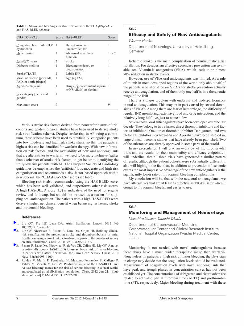
Various stroke risk factors derived from nonwarfarin arms of trial cohorts and epidemiological studies have been used to derive stroke risk stratification schema. Despite stroke risk in AF being a contin-uum, these schema have been used to ‘artificially’ categorise patients into low, moderate and high risk stroke strata, so that the patients at highest risk can be identified for warfarin therapy. With new informa-tion on risk factors, and the availability of new oral anticoagulants that are alternatives to warfarin, we need to be more inclusive (rather than exclusive) of stroke risk factors, to get better at identifying the ‘truly low risk patients’ with AF. The European Society of Cardiology guidelines de-emphasises the ‘artificial’ low, moderate and high risk categorisation and recommends a risk factor based approach with a new schema, the ‘CHA2DS2-VASc’ score (see table).
Bleeding risk is also recommended using the HAS-BLED score, which has been well validated, and outperforms other risk scores. A high HAS-BLED score (≥3) is indicative of the need for regular review and followup, but should not be used as a reason for stop-ping oral anticoagulation. The patients with a high HAS-BLED score derive a higher net clinical benefit when balancing ischaemic stroke and intracranial bleeding.
References
1. Lip GY, Tse HF, Lane DA. Atrial fibrillation. Lancet. 2012 Feb 18;379(9816):648–661.
2. Lip GY, Nieuwlaat R, Pisters R, Lane DA, Crijns HJ. Refining clinical risk stratification for predicting stroke and thromboembolism in atrial fibrillation using a novel risk factor-based approach: the euro heart survey on atrial fibrillation. Chest. 2010 Feb;137(2):263–272.
3. Pisters R, Lane DA, Nieuwlaat R, de Vos CB, Crijns HJ, Lip GY. A novel user-friendly score (HAS-BLED) to assess 1-year risk of major bleeding in patients with atrial fibrillation: the Euro Heart Survey. Chest. 2010 Nov;138(5):1093–1100.
4. Roldán V, Marín F, Fernández H, Manzano-Fernandez S, Gallego P, Valdés M, Vicente V, Lip GY. Predictive value of the HAS-BLED and ATRIA bleeding scores for the risk of serious bleeding in a ‘real world’ anticoagulated atrial fibrillation population. Chest. 2012 Jun 21. [Epub ahead of print] PubMed PMID: 22722228.
S6-2
Efficacy and Safety of New AnticoagulantsWerner Hacke
Department of Neurology, University of Heidelberg, Germany
Ischemic stroke is the main complication of nonrheumatic atrial fibrillation. For decades, an effective secondary prevention was avail-able, oral Vitamin-K antagonists (VKA), which leads to an almost 70% reduction in stroke events.
However, use of VKA oral anticoagulants was limited. As a rule of thumb in most developed regions of the world only about half of the patients who should be on VKA’s for stroke prevention actually receive anticoagulation, and of them only one half is in a therapeutic range of the INR.
There is a major problem with underuse and underperformance in oral anticoagulation. This may be in part caused by several down-sides of VKA’s. Among them are fear of hemorrhage, the difficulty of regular INR monitoring, extensive food and drug interaction, and the relatively long half live, just to name a few.
Several novel oral anticoagulants have been developed over the last decade. They belong to two classes, direct thrombin inhibitors and fac-tor xa inhibitors. One direct thrombin inhibitor Dabigatram, and two factor xa inhibitors, Rivaroxaban and Apixaban have been studied in, large clinical outcome studies that have already been published. Two of the substances are already approved in some parts of the world.
In my presentation I will give an overview of the three pivotal trials and the results for their main safety and efficacy endpoints. I will underline, that all three trials have generated a similar pattern of results, although the patient cohorts were substantially different. I also will highlight the fact that, in addition to prevention of ischemic events the most impressive advantage of the new anticoagulants is the significantly lower rate of intracranial bleeding complications.
My conclusion will be, that with the new oral anticoagulants, we have alternatives that are at least as effective as VKA’s, safer when it comes to intracranial bleeds, and easier to use.
S6-3
Monitoring and Management of HemorrhageMasahiro Yasaka, Yasushi Okada
Department of Cerebrovascular Medicine, Cerebrovascular Center and Clinical Research Institute, National Hospital Organization Kyushu Medical Center, Japan
Monitoring is not needed with novel anticoagulants because these drugs have a much wider therapeutic range than warfarin. Nonetheless, in patients at high risk of major bleeding, the physician in charge may decide that the coagulation levels should be evaluated. Measurement of coagulation levels with novel anticoagulants that have peak and trough phases in concentration curves has not been established yet. The concentrations of dabigatran and rivaroxaban are related to activated partial thrombin time (APTT) and prothrombin time (PT), respectively. Major bleeding during treatment with these
Table 1. Stroke and bleeding risk stratification with the CHA2DS2-VASc and HAS-BLED schemas
CHA2DS2–VASc Score HAS–BLED Score
Congestive heart failure/LV dysfunction
1 Hypertension ie. uncontrolled BP
1
Hypertension 1 Abnormal renal/liver function
1 or 2
Aged ≥75 years 2 Stroke 1Diabetes mellitus 1 Bleeding tendency or
predisposition1
Stroke/TIA/TE 2 Labile INR 1Vascular disease [prior MI, PAD, or aortic plaque]
1 Age (eg.>65) 1
Aged 65–74 years 1 Drugs (eg concomitant aspirin or NSAIDSs) or alcohol
1
Sex category [i.e. female gender]
1
Maximum score 9 9
Cerebrovasc Dis 2012;34(suppl 1):1–138 9Asia Pacific Stroke Conference 2012
anticoagulants may be checked by measurement of APTT or PT, respectively. Ischemic events may be checked by measurement of the plasma levels of molecular markers in the coagulation system, such as prothrombin fragment 1+2, thrombin-antithrombin complex, and soluble fibrin monomer complex.
Hemorrhagic complications are the most common adverse event associated with both anticoagulants and antiplatelet agents. Patients should be aware of the risks and physicians should know how to man-age bleeding complications. It is important to treat the bleeding as promptly and efficiently as possible. We refer to cessation of oral med-ication, bleeding stopped by mechanical compression or surgical inter-ventions, circulating blood volume and blood pressure maintained by appropriate intravenous drip infusion for induction of dieresis, blood pressure control for patients with intracranial hemorrhage, gastric lavage or oral administration of activated charcoal, supplementation of endogenous procoagulant factors, such as fresh frozen plasma, pro-thrombin complex concentrate, hemodialysis to remove dabigatran, development of antibodies capable of neutralizing dabigatran.
S6-4
Efficacy and Safety of Dabigatran Versus Warfarin in Patients with Atrial Fibrillation: Analysis in Asian Population in RE-LY TrialM Hori1, SJ Connolly2, J Zhu3, LS Liu3, C-P Lau4, P Pais5, D Xavier5, SS Kim6, R Omar7, AL Dans8, RS Tan9, J-H Chen10, S Tanomsup11, M Watanabe12, M Koyanagi12, MD Ezekowitz13, PA Reilly14, L Wallentin15, S Yusuf 2, the RE-LY Investigators1Osaka Medical Center for Cancer and Cardiovascular Diseases, Japan; 2Population Health Research Institute, McMaster University and Hamilton Health Sciences, Canada; 3Cardiovascular Institute & Fu Wai Hospital Beijing, China; 4Queen Mary Hospital, University of Hong Kong, Hong Kong; 5St. John’s Medical College and Research Institute, India; 6Yonsei University College of Medicine, Korea; 7National Heart Institute, Malaysia; 8Philippine General Hospital, Philippines; 9National Heart Centre, Singapore; 10National Cheng Kung University Hospital, Taiwan; 11Ramathibodi Hospital, Mahidol University, Thailand; 12Nippon Boehringer Ingelheim, Japan; 13Lankenau Medical Center, Thomas Jefferson Medical College, USA; 14Boehringer Ingelheim Pharmaceuticals Inc., USA; 15Uppsala Clinical Research Center, Sweden
Purpose: Intracranial hemorrhagesare reported to be higher in Asians than in non-Asians, especially in patients receiving warfarin.In the RE-LY study, dabigatranetexilate (DE) was non-inferior (110 mg bid) or superior (150 mg bid) to warfarin for the prevention of stroke and systemic embolism (SE) in patients with atrial fibrilla-tion. Major bleeding was significantly lower in DE 110 mg bid and comparable in DE 150 mg bid compared with warfarin. An analysis of RE-LY to determine the effects of DE versus warfarin comparing Asian and non-Asian countries was performed.
Methods and Results: Of 18,113 in the RE-LY, Asian patients (n=2,782 in 10 countries) were compared with non-Asian patients
(n=15,331 in 34 countries). Rates of stroke/SE in Asia were 3.06%/y on warfarin, 2.50%/y on DE 110 mg bid and 1.39%/y on DE 150 mg bid. The rates of major bleeding in Asia were significantly lower on DE (both doses) than warfarin; 3.82%/y on warfarin, 2.22%/y on DE 110 mg bid and 2.17%/y on DE 150 mg bid. The rate of hemorrhagic stroke on warfarin treated patients was more than two-fold higher in Asian than in non-Asian patients despite younger age in Asians.
Conclusion: Efficacy of DE on stroke/SE was consistent between Asians and non-Asians, but the reductions in major bleed-ing were greater with DE compared to warfarin in Asians. The rates of hemorrhagic stroke in individuals receiving warfarin were much higher in Asians compared to non-Asians, thus, with a larger risk reduction with DE in Asians.
Stroke in Asia
S7-1
Changing Burden of Stroke in East AsiaByung-Woo Yoon
Department of Neurology, Seoul National University Hospital, Korea
Stroke is the leading or major cause of death in many countries.Some countries in Asia show higher mortality and burden of dis-
ease from stroke. China has one of the highest figures of them, where there are substantial changes is the prevalence of vascular risk fac-tors.
Hypertension, smoking, and diabetes mellitus are the main risk factors for stroke in Asia which is similar to those in Western coun-tries. With the economic growth in the region, dyslipidemia, diabetes and obesity become more prevalent. Also, smoking and high salt con-sumption are important obstacles to reduce stroke.
There is significant variation in the relative burden of stroke com-pared with coronary heart disease worldwide. Contrary to Western countries, East Asia has greater mortality and morbidity from stroke than from coronary heart disease despite having overlapping risk fac-tors and disease mechanisms.
Recently atrial fibrillation (AF) and related stroke are getting more attention. AF is more common in aged people and risk of stroke in patients with AF increases with age. Strategies to improve preventing AF-related stroke are urgent because aging population raises growing problems in East Asia.
Cerebrovasc Dis 2012;34(suppl 1):1–13810 Abstracts of Symposia
S7-2
Stroke Epidemiology in Japan: The Hisayama StudyJun Hata, Yutaka Kiyohara
Department of Environmental Medicine, Graduate School of Medical Sciences, Kyushu University, Japan
The Hisayama Study is a population-based prospective study of cerebro-cardiovascular diseases. The town of Hisayama is located in the suburb of Fukuoka metropolitan area in Kyushu, Japan. The population of the town is approximately 8400 and the age and occu-pational distributions in the population are very similar to those in the whole country of Japan. Since 1961, the annual health examina-tions for the residents of Hisayama, aged 40 years or older, have been repeated by the town government and Kyushu University, and several cohorts for prospective studies have been established. These cohorts are characterized by high participation rates (≥78%), high follow-up rates (>99%), and high autopsy rate (approximately 75%). Therefore, the Hisayama Study provides high-quality epidemiological evidence on cardiovascular disease such as stroke and coronary heart disease in Japan.
During the past 50 years, the incidence of stroke has decreased greatly, which is mainly due to the improvement in blood pressure control in residents with hypertension. However, metabolic risk factors such as glucose intolerance, hypercholesterolemia, and obesity have become more prevalent and important. We have reported the associa-tion of traditional risk factors (i.e. hypertension, glucose intolerance, dyslipidemia, obesity, cigarette smoking, alcohol consumption, and physical inactivity) and some novel risk factors (i.e. metabolic syn-drome, chronic kidney disease, high-sensitive C-reactive protein, and genetic polymorphisms) with stroke or cardiovascular disease.
In the symposium, we will overview the secular trends in stroke incidence and major risk factors and the impact of risk factors on stroke incidence using the data from the Hisayama Study.
S7-3
Stroke Epidemiology in Southeast AsiaJose C. Navarro
Stroke Services, Department of Neurology & Psychiatry, University of Santo Tomas, Philippines
Southeast Asia is the region below the main countries of Asia. It consists of the countries that are geographically south of China, east of India, west of New Guinea and north of Australia. The region consists of eleven (11) countries with diverse culture, religion and language. It seems that this diversity is also seen in the epidemiologi-cal data from the different member countries. An electronic search of the medical literature through Medline, Pubmed and also some search engines were utilized. Furthermore personal communication with neurologists from ASEAN (Association of Southeast Asian Nations) countries was done which proved to be more fruitful. We reviewed published and unpublished community-based data on the following epidemiological parameters: prevalence, incidence, num-ber of disability-adjusted life years (DALY), case fatality and mor-
tality rates, risk factors and stroke subtypes from a total population of 1,399,220. Unfortunately, epidemiologic data were not available from four (4) member countries. The articles reviewed were mostly carried out in 2001–2011, except 2, which were done around 1990. The data from the available ASEAN countries were as follows: prev-alence 0.8–4.15% (from 5 countries), incidence 0.18%-4% (from 2 countries), DALY 3–13% (from 4 countries). The following risk fac-tors reported from 5 countries were as follows: hypertension 45–76%, DM 11–30%, high cholesterol 8.5–77%, smoking 15–28%, alcohol 11–20%. As to subtypes of stroke: cerebral infarction 43–74% and hemorrhagic stroke 18–40%.
S7-4
Characteristics and Diversity of Stroke in AsiaNiphon Poungvarin
Division of Neurology, Department of Medicine, Faculty of Medicine, Siriraj Hospital, Mahidol University, Thailand
Stroke is the most common causes of disability in adult and it ranks second for all causes of mortality worldwide. More than half of the world’s population lives in Asia. Incidence of stroke is defi-nitely increasing in developing countries while it is decreasing in developed world. It is no doubt that burden of stroke in Asia will increase, because of its low socioeconomic status and the aging popu-lation. It is thus vital to understand the characteristics and diversity of stroke in Asia, so that we can combat it with better options. Asian population with high incidence of stroke are East Asia (China, Japan, and Korea) and with relatively low incidence in South Asia (India, Pakistan, Bangladesh, Bhutan, Nepal and Sri Lanka). South East Asia (Thailand, Malaysia, Myanmar, Singapore, Vietnam, Loas, Cambodia, Brunei, Philippines and Indonesia) has incidence of stroke varying from high to low levels. Data of stroke epidemiology in Middle East are still limited.
Strategic planning to combat stroke in Asia are urgently required and need cooperation, understanding, and prompt multinational action of both government and nongovernment organizations. Diets which affect risk factors of stroke must be seriously emphasized, such as salty diet in East Asia, high carbohydrate intake in South Asia, heavy smoking among Chinese and high incidence of venous stroke in India. Prevention is thus the key element of success in combating stroke with most cost-benefit outcome.
Cerebrovasc Dis 2012;34(suppl 1):1–138 11Asia Pacific Stroke Conference 2012
Subarachnoid Hemorrhage
S8-1
Effect of Bypass Surgery for the Treatment of the Intracranial AneurysmsJae Sung Ahn
Department of Neurosurgery, Asan Medical Center, Korea
Purpose: The current standard treatment methods of intracra-nial aneurysms includes either endovascular coiling or microsurgical clipping. In certain situations, however, vascular reconstruction that is followed by occlusion of the parent artery is required. The authors have assessed the results from bypass surgeries for treatment of com-plex intracranial aneurysms from 2003 to 2012, retrospectively to propose its role as a treatment modality.
Materials and Methods: The outcome of 51 patients with complex aneurysms who were treated with EC-IC bypass surgery fol-lowed by trapping or proximal / distal occlusion has been reviewed.
Results: The patient group was composed of 16 male and 35 female patients, aged 14 to 76 years, and some of them were pre-sented with symptoms related to hemorrhage in 18 cases, others were presented with TIA or the compressive symptoms. Aneurysms were mainly in the anterior circulation (n=42, 82%). The types of aneu-rysms included 23 cases of large to giant size aneurysms, 9 cases were blood blister-like aneurysms, or traumatic pseudoaneurysms of the ICA, 8 cases were dissecting aneurysms, and 5 cases were fusi-form aneurysms.
The types of bypass surgeries performed were, respectively, STA-ACA bypass on 3 patients, STA-MCA bypass on 30 patients, includ-ing other 15 cases of using short radial artery interposition graft, and EC-IC high flow bypass with using the radial artery, or the saphenous vein on 9 cases, STA-SCA / PCA bypass on 3 patients, OA-PICA on 6 patients.
There has been no mortality and the overall surgery-related mor-bidity rate was 13.8% (5/36). Bypass patency rate was 92.2% (47/51) and there was one case of patency-related morbidity. There were three cases with poor outcomes (GOS III). One case involved a patient with blood blister-like aneurysm of the ICA, to whom unplanned bypass surgery was instilled after prolonged trapping in the ICA due to failed attempt at direct clipping of the aneurysm, which led to a cerebral infarction. Another case was involved with a patient with giant aneu-rysm in the ICA, which was dealt with STA-MCA bypass followed by trapping, but, due to a low perfusion, border zone infarction occurred. The last case with poor outcome was due to acute occlusion of the bypass.
Conclusion: Revascularization technique is a pivotal arma-mentarium in managing complex intracranial aneurysms and scrupu-lous prior planning is essential for improved surgical outcome.
S8-2
Obliteration of Ruptured Aneurysms – Current Complementary Role of Clipping and CoilingChang Wan Oh
Department of Neurosurgery, Seoul National University College of Medicine, Korea
The management outcome of aneurysmal subarachnoid hem-orrhage (aSAH) continued to improve in the past years. Regarding obliteration of ruptured aneurysms, reducing further damage to neural tissue may have contributed to the improved outcome. Availability of two options of aneurysmal obliteration, microsurgical clipping and endovascular coiling, has played important role in this refinement of management for patients with aSAH. These two modalities carry dif-ferent characteristics, and can be utilized complementarily for differ-ent situations.
Recent guideline by American Heart Association (AHA) rec-ommended that ‘‘Determination of aneurysm treatment, judged by experienced cerebrovascular surgeons and endovascular specialists, should be multidisciplinary decision based on characteristics of the patient and the aneurysm”. Age & neurological status of the patients, co-existent intra-parenchymal hemorrhage, and location of aneurysm are common factors determining mode of treatment. Other factors, inherent to individual patient and available resources of the institute, should also be taken into account to optimize the result.
To demonstrate such complementary role of clipping and coiling, I will present representative cases of our experiences.
S8-3
Vasospasm Following SAH, Pathogenesis and TreatmentHiroki Ohkuma
Department of Neurosurgery, Hirosaki University, Japan
Cerebral vasospasm is the major cause of mortality and morbidity after aneurysmal subarachnoid hemorrhage. Oxyhemoglobin released from subarachnoid clot produces free radicals and activates intracel-lular signalling transductions, of which the main pathway has been considered the Rho / Rho-kinasepathway. And induced sustained con-traction of smooth muscle cells leads to luminal narrowing of major cerebral arteries, followed by cerebral ischemia. Free radicals also stimulate the synthesis of endothelin-1 and inhibit the synthesis of nitric oxide (NO) in the endothelial cells, which accelerate sustained contraction of smooth muscle cells.
Prevention of cerebral vasospasm should be planned with taking such steps of the pathogeneses into the account. Diminish of suba-rachnoid clot has been tried using fibrinolytic drugs such as urokinase or tissue plasminogen activator. As a free radical scavenger, we have indicated the clinical efficacy of Edaravone. As a Rho kinase inhibi-tor, fasudilhydrochloride has widely been used in Japan. Statin, HMG-CoA inhibitor, is speculated to have the preventive effect for cerebral vasospasm via inhibition of RhoA and up-regulation of eNOS, how-ever, meta-analysis of RCTs failed to reveal the efficacy. Conscious
Cerebrovasc Dis 2012;34(suppl 1):1–13812 Abstracts of Symposia
trial using clazosentan, ET-1 receptor antagonist, also failed to reveal the improvement of outcome. Cilostazol, which induces NO produc-tion and increases cAMP levels via inhibition of phosphodiesterase III, has recently been indicated to be effective for prevention of cere-bral vasospasm by several reports including our RCT.
As a treatment of cerebral vasospasm after occurrence of ischemic symptoms, endvascular techniques including intra-arterial infusion of vasodilators and transluminal balloon angioplasty play an important role.
S8-4
Prognosis of SAH After Surgery – Japan and Asia Pacific RegionTatsuya Ishikawa, Junta Moroi, Kentaro Hikichi, Shotaro Yoshioka, Akifumi Suzuki
Department of Surgical Neurology, Research Institute for Brain & Blood Vessels – Akita, Japan
Background: Craniotomy and clipping have been robust treat-ments for ruptured cerebral aneurysm for more than 50 years. Recent advances in endovascular treatment are shifting the treatment for rup-tured cerebral aneurysm from craniotomy and clipping to intravascu-lar coil embolization. Treatment in acute stage followed by intensive neurosurgical care as well as technical advances enables safer and more efficient treatment.
Materials: We have analyzed 248 patients with SAH (ruptured aneurysm) in our institute during 2007–2011 (Age 29–87 (mean 62) y.o., WFNS grade I: 105, II: 43, III: 8, IV: 58, V: 34). Of all, 227 patients underwent surgical treatment by craniotomy (93%) or endo-vascular treatment (7%). We compared our result with the previous reports from nation-wide survey and my previous patient series.
Results: In 227 patients who received radical surgery, their overall outcome evaluated by mRK were 0 (40%), 1(17%), 2(10%), 3(8%), 4(12%), 5(9%), and 6(5%). Totally, the favorable outcome (mRK 0–2) was obtained in 67%, not showing any major improve-ments as compared to the previous experiences.
Conclusion: The meta-analysis demonstrated that overall sur-gical results have shown the decrease in case fatality. From my own experience, however, drastic improvement on functional outcome has not been identified in the last two decades. Although outcomes are mainly determined by the damage from initial bleeding, surgi-cal complications are still problematic factor to worsen functional outcomes. New treatment strategies are not always free from associ-ated complications and problems. We will also overview the surgical results in Japan, as well as countries in Asia Pacific region.
TIA as a Medical Emergency
S9-1
Urgent Diagnosis and Immediate Management in Acute Setting of TIAYasushi Okada1, Masahiro Yasaka1,Masahiro Kamouchi2, Takanari Kitazono2
1Department of Cerebrovascular Medicine, Cerebrovascular Center and Clinical Research Institute, National Hospital Organization Kyushu Medical Center, Japan, 2Department of Medicine and Clinical Science, Graduate School of Medical Sciences, Kyushu University, Japan
Since transient ischemic attack (TIA) has been viewed as a medical emergency with high risk for early stroke recurrence, it is undoubtedly important that examinations should be performed imme-diately to clarify its mechanism and to lead the definite diagnosis and early treatment. Early risk stratification by ABCD2 score have been useful to predict the risk of subsequent ischemic stroke early after TIA. Carotid ultrasonography and transesophageal echocardiography are also useful to detect the source of artery-to-artery emboli. Cardiac monitoring and blood examination are thought to play a key role for the diagnosis of cardioembolic TIA. MRI diffusion weighted imag-ing and MR angiography are also indispensable to understand TIA mechanism and intracranial circulation.
To prevent the subsequent stroke from TIA, antiplatelet and anticoagulant therapies are quite important as well as comprehen-sive management of life-style, hypertension, diabetes mellitus, dys-lipidemia, and other atherosclerotic disease. Carotid endarterectomy (CEA) and endovascular intervention is key treatment for the patients with significant stenosis of ICA.
Recently the new concept of acute cerebrovascular syndrome (ACVS) has been advocated to increase the awareness of TIA among patients and medical professionals. TIA should be recognized as the last chance to avoid the completed irreversible stroke which causes invalid conditions. In this symposium, we present the TIA intervention strategy in Japan and discuss the significant factors of high risk TIA patients from the analysis in our cases and in Fukuoka Stroke Registry.
S9-2
Microembolic Signals in Diagnosis and Management of TIAKS Lawrence Wong
Department of Medicine & Therapeutics, Chinese University of Hong Kong, Hong Kong
Microembolic signals (MES) is usually detected by monitoring of the blood flow of the middle cerebral artery by transcranial Doppler (TCD). The presence of MES in symptomatic patients indicates ongo-ing thromboemobolism. Patients with TIA are more likely to have
Cerebrovasc Dis 2012;34(suppl 1):1–138 13Asia Pacific Stroke Conference 2012
MES than those with more severe stroke. The presence and number of MES is a predictor of further ischemic stroke/TIA. MES is also a sur-rogate marker for anti-platelet drugs. The CARESS and CLAIR study have confirmed that dual anti-platelet agent is effective in reducing the number of MES and may prevent stroke in patients with MES.
S9-4
Surveillance and Guide of TIA in JapanToshiyuki Uehara1,2
1Department of Cerebrovascular Medicine, National Cerebral and Cardiovascular Center, Japan, 2The Japan TIA Research group 09–11 supported by Grants-in-Aid from the Ministry of Health, Labour and Welfare of Japan
We conducted a nation-wide survey using a questionnaire to clar-ify the current status of clinical practice of transient ischemic attack (TIA) in stroke specialized facilities in Japan. We sent a questionnaire to directors of 683 stroke teaching hospitals certified by the Japan Stroke Society. The response rate was 72.3%. According to this ques-tionnaire survey, clinical practice of TIA in stroke teaching hospitals seemed to be generally reasonable. However, the study demonstrated that new definition of TIA as duration of symptom <1 hour and the predictive scores of stroke risk after TIA such as ABCD2 score were hardly widespread (both approximately 7%).
We also carried out a multicenter retrospective study to elucidate the characteristics of inpatients with TIA. The subjects of this study were TIA patients admitted to 13 stroke centers within 7 days after onset between 2008 and 2009. Four hundred sixty-four patients (293 men, mean age of 69 years, median of ABCD2 score; 5) were reg-istered. MRI examinations were performed in 458 patients (99%), and acute ischemic lesions on diffusion weighted image (DWI) were found in 96 patients (21%). Multiple regression analysis revealed that large artery atherosclerotic lesion and time of onset to DWI of more than 24 hours were predictors of positive DWI. During hospitaliza-tion, recurrent TIA occurred in 27 patients (5.8%), ischemic stroke in 8 patients (1.7%), and ischemic heart disease in 4 patients (0.8%).
We are now drafting the manual for clinical practice of TIA.
Stroke Imaging
S10-1
Recent Progress in Stroke ImagingMakoto Sasaki
Division of Ultrahigh Field MRI, Institute for Biomedical Sciences, Iwate Medical University, Japan
Stroke imaging is widely used to assess patients with ischemic stroke. However, the imaging and postprocessing procedures remark-
ably vary among institutions and vendors, and this variation may deteriorate the accuracy of stroke imaging.
Perfusion CT/MRI is used to evaluate the extent of the area with ischemic penumbra; however, different software packages show sig-nificant differences in the sizes of perfusion abnormalities, and these differences should be minimized. Recently, some research groups have performed cross-validation studies by using digital phantoms and have elucidated the reliability of current commercial and aca-demic software packages. These research initiatives can promote further multicenter studies on reperfusion therapies by providing accurate inclusion/exclusion criteria for penumbral imaging.
MR plaque imaging techniques also vary among institutions and vendors. Recent studies have shown that intraplaque contrast can deteriorate in cardinal techniques such as ECG-gated black-blood method; however, this issue can be resolved by using spin-echo or MR angiography. Further studies using standardized protocols are required to establish the clinical significance of MR plaque imaging in the management of cervical carotid stenosis.
Introduction of ultrahigh-field 7-Tesla (7T) MRI has raised inter-est in the use of stroke imaging. High signal-to-noise ratio and T1 prolongation at 7T dramatically improve the quality of MR angiog-raphy and enable visualization of minute perforating arteries and collateral circulations. Further, the profound susceptibility effects at 7T enable noninvasive assessment of oxygen metabolism in acute or hemodynamic ischemias. Ultrahigh-field MRI is expected to have an increased clinical impact in stroke imaging in the near future.
S10-2
Advanced Imaging in Selection for Thrombolysis and RevascularizationWerner Hacke
Department of Neurology, University of Heidelberg, Germany
I.v.rt-PA is the evidenced based treatment of choice in patients with acute ischemic stroke in a time window of up to 4.5 hours. The effect of i.v.rt-PA is highest when given very early, e.g. in the first 90 minutes.
Many patients arrived later than 4.5 hours after ischemic stroke and some of them may also be candidates for thrombolytic therapy or acute interventional thrombectomy. For those patients, plain CT-scanning for the exclusion of brain hemorrhage or of major early infarct signs is not sufficient. Unfortunately, up to now, advanced imaging methods that are available now for more than 10 years such as diffusion perfusion MRI or CTA/CT perfusion have not yet been proven in clinical trials. In fact, while many people believe that such a type of selection exist, some of the randomized trials that have been performed, have been surprisingly negative.
In addition, advanced imaging may also be of use in patients with patients with severe stroke even in the first 4½ hours. In that occasion, one would look for proof of proximal vessel occlusion and indicate a combination treatment of i.v.rt-PA plus thrombectomy or thrombectomy alone.
Again, also for this apparently logical approach, clinical trials that prove that this approach is superior, are still missing. In my presenta-tion I will give an overview of the status of advance imaging in stroke right now and the clinical trials that are ongoing.
Cerebrovasc Dis 2012;34(suppl 1):1–13814 Abstracts of Symposia
S10-3
Neuroradiological Classification of Intracranial Large and Small Vessel DiseasesJong S. Kim
Department of Neurology, Asan Medical Center, University of Ulsan, Korea
Intracranial atherosclerosis (ICAS) is a major cause of stroke in Asian population, In Korea, the ratio of symptomatic ICAS and extracranial atherosclerosis is approximately 7:3. ICAS produces stroke by way of artery to artery embolism, in situ thromboocclusion and branch occlusion. Branch occlusion is an important cause of sub-cortical and brainstem infarction, and its role is greater in posterior than in anterior circulation system. Thus, although small vessel dis-ease (SVD) has been considered a major cause of single subcortical infarction (SSI). SSI can be caused by other causes such as embolic infarction or large artery disease, and the latter is an important cause of SSI in Asia.
Recent imaging techniques such as MRA or CTA allow us to detect this SSI associated with parental artery disease (SSIPAD) more easily. Another similar but confusing term ‘branch atheromatous dis-ease’ (BAD) has been introduced to emphasize atherosclerotic SSI. The similarity and differences between SSIPAD and BAD will be dis-cussed. Although the clinical and imaging characteristics of SSIPAD or BAD are similar to SSI caused by SVD, SSIPAD/BAD is more often associated with characteristics of atherosclerosis, and fluctuat-ing and a poorer clinical outcome.
More recently, imaging techniques such as high resolution vessel wall MRI (HRMRI) has identified atherosclerotic plaque and diag-nose PAD without focal stenosis in SSI patients with apparently nor-mal MRA or CTA findings. Thus, SSIPAD/BAD seems to be much more common than previously realized. The role of HRMRI in iden-tifying the nature of vascular pathology in patients with SSI will be discussed.
S10-4
Cerebral Blood Flow and Metabolism in Stroke PatientsJyoji Nakagawara
Department of Neurosurgery and Stroke Center, Nakamura Memorial Hospital, Japan
In acute stroke patients, development of cerebral infarction might depend on both time from stroke onset and residual cerebral blood flow (CBF), and cerebral tissue viability could not be preserved in severe ischemic region even within 3 hours, but could preserved in moderate ischemic region within 3–9 hours. Ischemic penumbra defined by perfusion MRI or perfusion CT for urgent recanalization therapy could be identified in moderate ischemic region without cor-tical infarction within 9 hours from stroke onset. Penumbra imaging could provide information on therapeutic time window in acute stroke patients.
In patients with progressing stroke by steno-occlusive lesion of carotid arteries, acute cerebral ischemia could be compensated
by dilatation of cerebral artery (increase of cerebral blood volume: CBV) and elevation of oxygen extraction fraction (OEF). This hemo-dynamic situation could be called as “acute misery perfusion”, and should be reversed by urgent therapeutic intervention such as CAS. The time window for therapeutic intervention could be extended up to a few days form onset.
In patients with chronic hemodynamic ischemia, Stage II hemody-namic cerebral ischemia may be a surrogate marker of stroke recurrence. This hemodynamic condition can also correspond to “chronic misery perfusion”. EC-IC bypass could be beneficial for stroke prevention in patients with Stage II hemodynamic cerebral ischemia determined by PET or quantified resting and acetazolamide-activated CBF-SPECT. Stratification of hemodynamic cerebral ischemia using quantified CBF-SPECT should be standardized with high accuracy using the DTARG method and SEE analysis for universalizing the effectiveness of EC-IC Bypass surgery and organizing future clinical trials.
Stroke Genetics
S11-1
Genetics of Stroke in IndiaMV Padma
Department of Neurology, All India Institute of Medical Sciences, India
The genetic contribution to common multifactorial stroke is poly-genic, and identification of individual causative mutations is problem-atic due to complexity of such a condition. Few attempts have been made to study the role of genetic variation in development of stroke in Indian population. Angiotensin converting enzyme gene, PDE4D gene, eNOS variants were found significant risk factors while vari-ants in the genes encoding coagulation factors like prothrombin, Pro and anti-inflammatory genes ( MMP3, IL-10 genes) showed negative association. Atleast 135 genes were found modulated and/or modulate hyperhomocyteinemia in ischemic stroke. The role of ESR1genepolymorphisms ( Pvull (rs 2234693 and Xbal (rs 9340799) with stroke in a South Indian population showed significant associa-tion in postmenopausal women. Significant association was seen with 1347 G/A polymorphism (rs 2108622) in the 11 th exon region of cyto-chrome P450 4F2 gene with hypertension, ischemic stroke and cardi-oembolic stroke subtype. IL-10-1082 G/A promoter polymorphism (rs 1800896) was again found associated with ischemic stroke occurrence. Association of the -344C/T aldosterone synthase (CYP11B2) gene variant ( TT genotype and T allele) was found positive with hyperten-sion and stroke. HindIII polymorphism of LPL was found associated with ischemic stroke, raised plasma triglycerides and reduced HDL levels. Studies also suggested that -7351C/T polymorphism of tPA and 4G/5G polymorphism of PAI-1 are not associated with stroke while the DD genotype and D allele of tPA gene are important risk factor for ischemic stroke. Aspirin resistance was found more common in patients with 3435TT genotype than in those with CC genotype of the multiple drug resistance -1 (MDR-1) gene.
Cerebrovasc Dis 2012;34(suppl 1):1–138 15Asia Pacific Stroke Conference 2012
S11-2
How to Identify Genetic Factors in Stroke PatientsShoji Tsuji
Department of Neurology, The University of Tokyo, Japan
Stroke is one of the most common diseases with the number of patients exceeding 1.37 million in Japan. From the view point of genetics, there are a number of stroke-related diseases with Mendelian inheritance. In the majority of patients with stroke, however, the molecular etiologies underling stroke remain to be elucidated. For stroke-related diseases with Mendelian inheritance, positional clon-ing strategies have achieved marvelous achievements in identifying the causative genes, including those of CARASIL (autosomal reces-sive arteriopathy with subcortical infarcts and leukoencephalopa-thy), CADASIL (autosomal dominant arteriopathy with subcortical infarcts and leukoencephalopathy) and many more diseases. On the other hand, the molecular etiologies underling stroke except for those with Mendelian inheritance remain to be elucidated. In the past decade, genome-wide association studies (GWAS) based on com-mon disease-common variants hypothesis have intensively been con-ducted to identify disease-susceptibility genes. Although GWAS has been successful, the effect sizes of the disease susceptibility genes are generally of small effect sizes, suggesting that the value for clini-cal application is limited. Recent studies have demonstrated that we need to focus on rarer variants to identify susceptibility genes with larger effect sizes (common disease-multiple rare variants hypoth-esis). Given the recent advancement of technologies of next genera-tion sequencers, identification of rare variants with large effect sizes underlying stroke is highly expected. These efforts as well as new knowledge in pharmacogenomics will soon be translated into clinical practice for prevention and treatment of stroke.
S11-3
Hereditary Cerebral Small Vessel Disease in JapanTeruyuki Hirano
Department of Internal Medicine 3, Oita University Faculty of Medicine, Japan
Cerebral small vessel diseases (SVDs) are major disease burden in Japan. SVDs manifest with lacunar infarction, intracerebral hem-orrhage, and vascular dementia. Hypertension, dyslipidemia, and diabetes mellitus are risk factors for SVDs, however, many heredi-tary SVDs without risk factors have been identified. Among these, cerebral autosomal dominant arteriopathy with subcortical infarcts and leukoencephalopathy (CADASIL) was the first to be geneti-cally identified as NOTCH3 gene mutation. Since 1998, when the first Japanese CADASIL family was discovered, 85 patients from 71 families with various NOTCH3 mutations were reported. Of these, prevalence of migraine: 41%, repeated cerebral ischemic attacks: 36%, dementia: 36% and emotional disturbance: 23%. The onset of neurological symptom was 44 years on average. Definite diagnosis requires NOTCH3 mutation involving cysteine and/or detection of
granular osmiophilic material (GOM). The second genetically identi-fied SVD, cerebral autosomal recessive arteriopathy with subcortical infarcts and leukoencephalopathy (CARASIL) was originally found in Japan. In 2009, HTRA1 was identified as the responsible gene that regulates TGF-ß signaling. By 2011, 28 patients have been identified. The unique clinical manifestations consisted with progressive demen-tia: 90%, premature baldness: 86%, and spondylosisdeformans/disk herniation: 100%. Average onset age was 32 years. Males are pre-dominantly affected. Frequent reasons to lead CADASIL/CARASIL diagnosis was typical MRI findings such as prominent leukoaraiosis extending to anterior frontal and/or temporal lobes. Recent achieve-ment of Research Committee for Hereditary Cerebral Small Vessel Disease will be reported.
S11-4
Recent Advances in Stroke GeneticsChristopher Levi
Acute Stroke Unit – John Hunter Hospital, Priority Research Centre for Translational Neuroscience and Mental Health – University of Newcastle, Australia
The genetic influences on the common forms of stroke are just beginning to be uncovered. Three genetic loci, all associating with the subtype of large artery stroke, have recently been discovered using genome wide scanning approaches and the previously discovered loci associating with atrial fibrillation (4q25 & 16q22.3) have also been confirmed as associating with ischaemic stroke. Based on these initial results, it appears likely that genetic factors play a significant role in stroke pathophysiology but that heterogeneity will exist across differ-ent stroke pathophysiologies. It also appears likely that the currently identified genetic variants will be only a small part of a richer and more complex genetic landscape where very large samples of well characterized and pathophysiologically subtyped patients will be nec-essary to advance our understanding of stroke genetics and explore the likely interactions between genetic and environmental factors.
The stroke specific variants that have been identified in large artery disease are the same chromosome 9p21.3 locus polymorphisms identified in myocardial infarction1 along with polymorphisms at the chromosome 7p21.1 locus2 and polymorphisms at chromosome 6p21.1 locus3. These variants and the candidate genes and genetic regions involved have uncertain relationships to the atherothrombotic and brain ischaemia process. The findings now raise the challenge of “reverse translation” and potentially, the discovery of new biological mechanisms underlying stroke pathophysiology.
References
1. Gschwendtner A, Bevan S, et al on behalf of the International Stroke Genetics Consortium, Annals of Neurology 2009;65:531–539.
2. International Stroke Genetics Consortium and the Wellcome Trust Case Control Consortium. Nature Genetics 2012;44:328–335.
3. Holliday E, Maguire J et al on behalf of the Australian Stroke Genetics Collaborative and the International Stroke Genetics Consortium. Nature Genetics 2012; In press.
Oral Presentations
Fax �41 61 306 12 34E-Mail [email protected]
© 2012 S. Karger AG, Basel
Accessible online at:www.karger.com/ced
Large Vessel Disease 1
OP-1
Tips for the Superficial Temporal Artery-to-Middle Cerebral Artery Target BypassRyota Kimura1, Shinichiro Kurokawa1, Hideaki Mishima1, Ichiro Nakagawa2, Hiroyuki Nakase2, Katsutoshi Takayama3, Kaoru Myouchin3
1Department of Neurosurgery, Ishinkai Yao General Hospital, Japan, 2Department of Neurosurgery, Nara Medical University, Japan, 3Departments of Radiology and Interventional Neuroradiology, Ishinkai Yao General Hospital, Japan
Purpose: To introduce our method for a superficial temporal artery (STA)-to-middle cerebral artery (MCA) anastomosis through a minimum cranial window.
Methods: 19 patients with misery perfusion due to athero-sclerotic cerebrovascular occlusive disease underwent STA MCA target bypass since August 2009 to June 2012 in our department. Single-photon emission computed tomography or perfusion image of computed tomography was performed to evaluate regional cere-brovascular reserve capacity. We reconstructed not only angiography but also three-dimensional images of the skull and scalp by helical computed tomography scan data. These images were superimposed to select the suitable STA branch and the cortical artery as the target recipient MCA, and determine the precise local relationship among the STA branches, the appropriate recipient MCA and the anatomical landmarks. We could design minimum and suitable skin incision. We marked the actual site of the target on the skull with Indigo carmine to adjust the location. The STA was anatomosed with the target through a minimum cranial window.
Results: Successful bypass to the target was confirmed in all cases. The length of the skin incision was 57.5 mm ± 9.6 mm. The major and minor axis of the cranial window were 34.4 mm ± 6.3 mm and 22.4 mm ± 3.8 mm.
Conclusion: The “target bypass” method might be effective for cases with atherosclerotic cerebrovascular occlusive disease.
OP-2
Surgical Result and Technical Trouble in STA-MCA Bypass SurgeryTakachika Aoki, Tomoya Miyagi, Yasuharu Takeuchi, Masaru Hirohata, Motohiro Morioka
Department of Neurosurgery, Kurume University School of Medicine, Japan
Although there are few serious complications with superficial temporal artery-middle cerebral artery (STA-MCA) bypass, the detailed surgical complications are still unclear. We analyzed surgi-cal complications and technical troubles in the 196 patients suffered STA-MCA bypass by one operator from 2001 to 2011. Furthermore, all surgery were equally divided into 4 stages according to time line (1st, 2nd, 3rd and 4th stages) and analyzed, to clear whether more surgical experience result in better surgical results. Overall surgi-cal morbidity was 2.0% and mortality was 0%, but there were some troubles related to STA preparation caused by the surgical technique itself. This study suggested that ischemic complication occurred at the rate of 2–4% and technical trouble of STA injury or occlusion occurred in 2–12% of surgery. These morbidity rate and technical trouble rate were almost same in the all periods, even in the last 46 cases. In STA-MCA bypass surgery, suturing technique training must be needed. Furthermore, we must pay attention for other procedures, such as STA preparation.
OP-3
Effect of STA-MCA Bypass for Chronic Ocular Ischemic Syndrome Due to the Internal Carotid Artery OcclusionShoichiro Kawaguchi, Jun-ichi Iida, Yoshitomo Uchiyama
Department of Neurosurgery, Nara Prefectural Nara Hospital, Japan
Introduction: The authors examined the effect of the STA-MCA bypass for chronic ocular ischemic syndrome (OIS) due to the internal carotid artery occlusion.
Methods: We evaluated the visual symptoms and the ocular cir-culation in fifty-four patients before and after bypass STA-MCA bypass. Visual symptoms were decline of visual acuity in 36 cases, frequent amaurosis fugax in 9 cases and both in 9 cases. The ocular circulation was evaluated by the ophthalmic artery (OphAr) flow and the central retinal artery (CRA) flow using color Doppler flow imaging (CDFI) providing the flow direction and peak systolic flow velocity (Vs).
Results: 1) Preoperatively, 50 patients showed reversed OphAr flow. The mean OphAr Vs was –0.37 m/sec, and the mean CRA Vs was 0.06 m/sec.
Cerebrovasc Dis 2012;34(suppl 1):1–138
Cerebrovasc Dis 2012;34(suppl 1):1–138 17Asia Pacific Stroke Conference 2012
2) At one month after bypass, 21 patients showed the antegrade OphAr flow. The flow direction of the OphAr flow was corrected significantly. The mean OphAr Vs increased to –0.06 m/sec signifi-cantly, and the CRA Vs also increased to 0.08 m/sec significantly.
3) At three months after surgery, 26 patients showed the antegrade flow. The mean OphAr Vs increased to 0.05 m/sec significantly, and the CRA Vs also increased to 0.11 m/sec significantly.
4) During the follow-up period (mean: 6.3 years), 21 patients (47%) showed the visual acuity improvement, and no patients com-plained of the amaurosis fugax and worsened the OIS.
Conclusions: STA-MCA bypass can improve the ocular circu-lation, recover the visual symptom in half of them, and also prevent the worsening of the OIS.
OP-4
Hemodynamics and Changes after STA-MCA Anastomosis in Moyamoya Disease and Atherosclerotic Cerebrovascular Disease Measured by Micro-Doppler UltrasonographyTakakazu Kawamata1, Hanako Morisawa2, Akitsugu Kawashima2, Masataka Hayashi2, Kohji Yamaguchi2, Taku Yoneyama2, Yoshikazu Okada2
1Department of Neurosurgery, Tokyo Women’s Medical University Yachiyo Medical Center, Japan, 2Department of Neurosurgery, Tokyo Women’s Medical University, Japan
Purpose: Moyamoya disease (MMD) has been proposed to dif-fer from atherosclerotic cerebrovascular disease (ACVD) in hemo-dynamics and effects of STA-MCA anastomosis. Furthermore, the circulatory dynamics have been also shown to differ between adult MMD (AMMD) and pediatric MMD (PMMD). We analyzed the cor-tical flow velocity and direction using micro-Doppler ultrasonogra-phy to evaluate the cortical circulation before and after anastomosis in MMD and ACVD.
Methods: Twenty-eight patients with AMMD, 7 with PMMD, 16 with ACVD, and 12 control patients were studied. A micro-Doppler probe was applied on the cortical recipient artery (A4 or M4) before and after anastomosis. Systolic maximum flow velocity (Vmax) and blood flow direction were investigated at proximal and distal parts of anastomosed sites in recipient arteries.
Results: Before anastomosis, retrograde cortical flow was recorded significantly more frequently in PMMD patients, and Vmax in cortical artery was significantly lower in AMMD patients. Pre-anastomosis flow direction was preserved more frequently in PMMD patients after anastomosis. The rate of Vmax increase after anasto-mosis was significantly higher in AMMD than in PMMD. The Vmax increase rate was significantly higher in patients with the opera-tive hemisphere showing postoperative cerebral hyperperfusion on Xe-CT.
Conclusions: In AMMD, significantly low velocity in the cortical artery was observed before anastomosis, and bypass surgery reversed the flow and significantly increased flow velocity. The data of PMMD showed unique hemodynamics before anastomosis, char-acterized by a higher frequency of retrograde flow and preserved velocity. Intraoperative micro-Doppler monitoring may be also useful to predict postoperative cerebral hyperperfusion.
OP-5
External Carotid-Internal Carotid (EC/IC) Bypass Surgery Improves Hemodynamic Parameters and Cognitive Performance in Patients with Severe Intracranial Steno-Occlusive DiseaseYohanna Kusuma, Dong Y, Hl Teoh, Venketasubramanian – Narayanaswamy, Bernard PL Chan, Vijay Kumar Sharma
Department of Medicine, National University Hospital, Singapore
Introduction: Patients with intracranial stenoses are at con-siderable risk for recurrent ischemia. Recent trials on stenting and external carotid-internal carotid (EC/IC) bypass in carotid occlusions failed to show benefit over best medical therapy despite improving cerebral hemodynamics. We evaluated whether the improved cerebral vasodilatory reserve (CVR) after EC/IC bypass for severe stenoses of intracranial internal carotid (ICA) or middle cerebral artery (MCA) influenced cognitive parameters.
Methods: Patients with severe stenoses of ICA or MCA and impaired CVR on transcranial Doppler (TCD) were evaluated with acetazolamide-challenged HMPAO-SPECT. Patients with signifi-cantly impaired CVR on SPECT were offered EC/IC bypass. TCD-breath-holding index (BHI), SPECT-CVR and cognitive performance were performed at baseline and at 6 months after surgery.
Results: Of 112 patients with severe ICA/MCA stenosis, 77 (69%) showed impaired CVR and 46 (41%) underwent bypass. Significant improvements occured in CVR-TCD-BHI in MCA improved (median 0 (IQR 0.45) to 1.10 (IQR 0.73), p < 0.001) and SPECT (P < 0.001). 9 patients underwent neuropsychological evalu-ation before and after bypass surgery. Compared to controls (n = 7), significant within improvements were noted in animal fluency (3 ± 3, p = 0.002 vs 0.6 ± 4.2, p>0.05), picture immediate recall (1.4 ± 0.9, p = 0.01 vs 0.6 ± 1.5, p>0.05) and delayed recall (1.3 ± 1.1, p = 0.007 vs –0.1 ± 2.3, p>0.05). Patients undergoing surgery had borderline significant improvement than controls in block design (4.1 ± 5.4, p = 0.052 vs 3.2 ± 10.6, p>0.05). Significantly less ischemic events were observed in the surgical group (11% vs 45% in medical group; p<0.005) during median 21 months follow up.
Conclusion: EC/IC bypass surgery in selected patients results in significant improvement in cerebral hemodynamics and cognitive performance.
Cerebrovasc Dis 2012;34(suppl 1):1–13818 Abstracts of Oral Presentations
OP-6
Release of Free Fatty Acids and Interleukins During Carotid Artery StentingYuko Abe1, Manabu Sakaguchi2, Shigetaka Furukado2, Toshiyuki Fujinaka2, Toshiki Yoshimine2, Kazuo Kitagawa2
1Cerebrovascular Division, Department of Medicine, Yodogawa Christian Hospital, Japan, 2Stroke Center, Osaka University Graduate School of Medicine, Japan
Background: The reason for higher rate of stroke in the periprocedural period for CAS than for carotid endarterectomy has not been fully understood. Previously, we reported the association between local interleukin (IL)-6 levels and new ischemic lesion after CAS. However, other soluble factors might be released during CAS. We sought to identify the lipid fractions and other cytokines that are released after CAS.
Methods: The study consisted of 24 cases that underwent CAS with distal balloon occlusion device. We obtained aortic blood samples and local blood samples just after CAS. We measured the serum lipid levels from all the samples. In 9 symptomatic subjects with >80% carotid stenosis, we analyzed the concentrations of sev-eral cytokines and growth factors by cytokine microarray. Diffusion-weighted magnetic resonance imaging was performed before and after the procedure.
Results: The local free fatty acids (FFA) level was markedly ele-vated and the local triglyceride (TG) level was drastically decreased compared to that of aortic sample. The FFA release was strongly associated with the TG decrease (R = –0.54, P<0.01). Furthermore, local FFA levels in 6 cases with new ischemic lesions (median, 1918 μEq/L) were significantly higher than in 18 cases without (median, 1338 μEq/L) (P<0.05). In cytokine array system, the local levels of IL-1α, IL-2, IL-5, IL-6, IL-10, IL-13, interferon (IFN)-γ, and tumor necrosis factor (TNF)-α were significantly higher than those of aortic samples.
Conclusion: Our study demonstrated that FFA and several interleukins were released locally during CAS, suggesting the poten-tial of soluble factors on periprocedural complications.
OP-7
Carotid Endarterectomy in Patients with Contralateral Internal Carotid Artery Occlusion: Surgical Consideration and Long-Term Outcome in Our InstituteHajime Yabuzaki1, Takahiro Ota1, Thoru Mizutani2, Tatsuya Sugiyama1, Takeshi Ogura1, Kazuaki Naemura1, Naoya Hiraiwa1, Takayuki Yasuda1, Kuzuoka Sakura1
1Department of Neurosurgery, Tokyo Metropolitan Tama Medical Center, Japan, 2Department of Neurosurgery, Showa University, Japan
Purpose: The SAPPHIRE trial defined carotid endarterectomy (CEA) as a high-risk surgery in patients with contralateral internal carotid artery (ICA) occlusion, because of the possibility of cerebral infarction due to blockage of the carotid artery during CEA. In con-
trast, many reports state that CEA is a very effective method of stroke prevention in such patients. Here, we report our consideration for this surgical procedure and preoperative assessment in order to perform it as safely as possible.
Methods: Between 2002 and October 2011, 27 patients with carotid artery stenosis and contralateral ICA occlusion underwent CEA in our institute, and the patients’ perioperative and long-term outcomes were evaluated. We routinely used single-photon emission computed tomography with N-isopropyl-p-iodoamphetamine (IMP-SPECT) and balloon occlusion test (if necessary) for evaluating cere-bral ischemic tolerance before CEA and used shunting as a routine procedure to shorten the blocking time of the carotid artery during operation.
Results: One patient developed stroke on the ipsilateral side 1 day after surgery and slight left-sided hemiparesis was observed; however, no major sequelae were found. During the long-term follow up (average, 37 months), transient amaurosis was observed once in a patient, bur no new symptomatic stroke had developed.
Conclusion: Surgical consideration and adequate preoperative assessment enabled us to safely perform CEA in patients with contral-ateral ICA occlusion.
OP-8
Surgical Strategy for Performing Carotid Endarterectomy Safely in Our HospitalKazuaki Naemura1, Takahiro Ota1, Sakura Kuzuoka1, Hajime Yabuzaki1, Takayuki Yasuda1, Naoya Hiraiwa1, Kenji Sumi1, Takesi Ogura1, Tatsuya Sugiyama1, Tohru Mizutani2
1Department of Neurosurgery, Tokyo Metropolitan Tama Medical Center, Japan, 2Department of Neurosurgery, Showa University, Japan
Purpose: According to some recent randomized controlled tri-als, carotid endarterectomy (CEA) is more preferable than carotid artery stenting (CAS) for treating internal carotid artery stenosis in selected patients. Moreover, several reports have shown that patients undergoing CAS are more susceptible to perioperative stroke than those undergoing CEA. A safe and standard procedure for CEA needs to be established, particularly for less-experienced doctors.
Methods: We retrospectively reviewed 586 cases of CEAs per-formed at our institute between January 2002 and March 2012, and on the basis of the experience of the surgeons, divided the cases into 2 groups: Expert’s (CEAs performed by surgeons with > 10 years of experience) and Beginner’s (CEAs performed by surgeons with < 10 years of experience). We assessed the complications occurring during the first 30 days after the operation.
Results: Death, stroke, and acute myocardial infarction (AMI) were the main postoperative complications occurring during the first 30 days after the surgery. In total, 1 patient (0.2%; Beginners group) died and 16 (2.8%) patients experienced stroke (including 5 [2.7%] patients from the Experts group and 11 [2.9%] from the Beginners group); however, none of the patients experienced AMI. No signifi-cant differences were identified between the characteristics of these 2 groups.
Cerebrovasc Dis 2012;34(suppl 1):1–138 19Asia Pacific Stroke Conference 2012
Conclusions: The surgical strategy for CEA used in our insti-tute is extremely safe. Moreover, by using our procedure, even young surgeons (Beginners) can perform CEA as successfully and safely as experts can.
OP-9
Post Carotid Endarterectomy Improvement in Cognition is Associated with Resolution of Crossed Cerebellar HypoperfusionKohei Chida, Kuniaki Ogasawara, Kenta Aso, Yasunori Suga, Masakazu Kobayashi, Kenji Yoshida, Akira Ogawa
Department of Neurosurgery, Iwate Medical University, Japan
Background and Objectives: The purpose of the present study was to investigate whether resolution of crossed cerebellar hypoperfusion (CCH) after carotid endarterectomy (CEA) are associ-ated with postoperative improvement of cognitive function.
Methods: Neuropsychological testing was performed preop-eratively and after 1 postoperative month in 79 patients undergoing CEA for ipsilateral internal carotid artery stenosis (≥70%). Brain per-fusion single photon emission computed tomography (SPECT) using N-isopropyl-p-123I-iodoamphetamine was also performed before and after surgery. Data were analyzed using a three-dimensional stereo-taxic region of interest template.
Results: Seven patients (9%) showed improvement in postop-erative cognitive function. All the 7 patients exhibited both postop-erative increase in blood flow in the ipsilateral cerebral cortex and resolution of CCH. Analysis by a receiver operating characteristic (ROC) curve was used to estimate the ability to discriminate between patients with and without postoperative cognitive improvement. The area under the ROC curve was significantly greater when analyz-ing the magnitude of postoperative resolution of CCH (0.991; 95% CI 0.984–1.001) when compared with the magnitude of postopera-tive increase in cerebral blood flow (0.929; 95% CI 0.886–0.971) (p < 0.05).
Conclusion: Resolution of CCH after CEA is associated with postoperative improvement in cognitive function.
OP-10
Blood Flow Metabolism Mismatch Triggers Postoperative Hyperperfusion after Carotid Endarterectomy: A Serial SPECT/PET StudyDaisuke Shimbo1, Satoshi Kuroda2, Naoki Nakayama1, Ken Kazumata1, Kenji Hirata3, Tohru Shiga3, Kiyohiro Houkin1, Nagara Tamaki3
1Department of Neurosurgery, Hokkaido University Graduate School of Medicine, Japan, 2Department of Neurosurgery Toyama University, Japan, 3Department of Nuclear Medicine Hokkaido University, Japan
Objective: There are few studies that evaluate the relationship between preoperative PET parameters and post-CEA hyperperfusion, and that denote the time course of hyperperfusion. This study was aimed to assess the relationship between the PET parameters and post-CEA hyperperfusion in patients with carotid artery stenosis and to evaluate the time course of CBF changes after CEA.
Subjects and Methods: This study included 41 patients who underwent CEA due to severe (>70%) carotid artery stenosis between Oct 2006 and Feb 2012. There were 36 men and 5 women. Prior to CEA, 15O-gas PET and 123I-IMP SPECT with acetazolamide chal-lenge were performed in all patients. All patients underwent CEA using internal shunting. CBF measurements were repeated on Day 0, Day 2, and Day 7 after CEA.
Results: Hyperperfusion was observed in 10 of 41 patients. There was no symptomatic hyperperfusion. Hyperperfusion was observed in 8 of 13 patients with impaired cerebrovascular reactiv-ity. All of 6 patients with elevated oxygen extraction fraction (OEF) demonstrated hyperperfusion and elevated OEF had a significant cor-relation with the development of hyperperfusion (p<0.001). Serial SPECT studies demonstrated that hyperperfusion was detected in 7 of 10 patients on Day 0. Almost these patients had hyperperfusion more than 7 days after CEA.
Conclusion: Preoperative mismatch between blood flow and metabolism (elevated OEF) most likely leads to post-CEA hyperper-fusion. Hyperperfusion persisted for more than 7 days in patients with hyperperfusion immediately after CEA. Comprehension of perioper-ative cerebral hemodynamics and metabolism is important to prevent hyperperfusion syndrome.
OP-11
Restenosis after Carotid Endarterectomy for Symptomatic Carotid Stenosis. Timing of Surgery and Incidence of RestenosisNorikata Kobayashi, Morio Takasaki, Kaname Ito, Daisuke Maruyama, Daizo Ishii, Kenichi Morita, Tetsu Satow, Hiroharu Kataoka, Koji Iihara
Department of Neurosurgery, National Cerebral and Cardiovascular Center, Japan
Background and Purpose: Recently, restenosis after carotid endarterectomy has been reported to be associated with timing of sur-gery and nature of the carotid plaque. The aim of the study was to
Cerebrovasc Dis 2012;34(suppl 1):1–13820 Abstracts of Oral Presentations
evaluate association between the incidence of restenosis after CEA for symptomatic carotid lesion and timing of surgery.
Methods: Between January 2007 and March 2011, a total of 196 CEA were performed with primary closure in 188 patients (male: 91.4%, symptomatic: 55%) in our institution. Postoperatively, the degree of stenosis was assessed with CT angiogram (n = 181) and/or duplex ultrasonography (n = 189). Restenosis was defined as >50% luminal narrowing at the treated site. The rate of restenosis was com-pared between symptomatic and asymptomatic lesions, and then the incidence for symptomatic cases was also analyzed based on the tim-ing of surgery which was divided to early (<30 days) and delayed (>30 days) based on the period between the last ischemic event and surgery.
Results: Overall, restenosis occurred in 14 cases (7.1%) and its incidence was not significantly different between symptomatic and asymptomatic groups (4.6% versus 10.2%, P = 0.13). In this period, early CEA was performed in 43 cases (39.8%) for symptomatic cases. In symptomatic group, restenosis (n = 5) was noted exclusively in cases with delayed (n = 65) group (P = 0.06).
Conclusion: Here we showed that the rate of restenosis was not significantly different based on the symptomatology of the carotid plaques. In symptomatic groups, restenosis occurred only in cases with delayed surgery, suggesting possible correlation between the sta-bilization of the carotid plaque and development of restenosis after CEA.
Hemorrhagic Stroke 1
OP-12
Intraoperative Findings and Selection of Treatment Technique in Patients with an Internal Carotid Artery Anterior Wall Saccular-Shape Aneurysm Associated with a Persistent Primitive Dorsal Ophthalmic ArteryMasahiro Indo, Soichi Oya, Kei Yanai, Tsuguto Takizawa, Akira Saito, Tsukasa Tsuchiya, Takumi Nakamura, Han-Soo Chang, Toru Matsui
Department of Neurosurgery, Saitama Medical University, Saitama Medical Center, Japan
Background: It is challenging to treat internal carotid artery (ICA)-anterior wall aneurysms because they are often not unsuitable for direct clipping and coiling even if they appear saccular on cerebral angiogram.
Objective: We focus on the persistent primitive dorsal ophthal-mic artery (PPDOA) and discuss the possible relationship between the PPDOA and the etiology of ICA-anterior wall aneurysms.
Methods: Among 8 patients with an saccular-shaped ICA-anterior wall aneurysm diagnosed at our medical center between 2006 and 2012, 5 patients had PPDOA and 3 patients did not have PPDOA. The aneurysms that presented with obvious characteristics of dissect-ing aneurysms, such as stenosis and dilation at the parent artery, were
excluded. Intraoperative findings of the aneurysms and postoperative results of coil embolization were also assessed.
Results: Six patients had a subarachnoid hemorrhage, and two patients with an unruptured aneurysm were conservatively treated. Among 6 SAH cases, 4 patients had PPDOA. Two of these 4 SAH patients with PPDOA aneurysms were intraoperatively confirmed to have saccular true aneurysms. They had no early rebleeding or regrowth after coil embolization. In contrast, 2 SAH cases without PPDOA were dissecting aneurysm confirmed intraoperatively. We speculate that the presence of PPDOA indicates the incomplete fusion of the ophthalmic artery to ICA, rendering the anterior wall of the ICA vulnerable to hemodynamic stress and that this results in the formation of true aneurysms, not pseudoaneurysms in this specific portion.
Conclusion: The presence of PPDOA might be a clue to judge whether IC anterior wall aneurysms are true or pseudo-aneurysm.
OP-13
Full Exposure of Ruptured Cerebral Aneurysm Preceding Clip Application: A Novel Surgical Concept and Its Advantages in 118 Case SeriesNaoki Nakayama1, Kiyohiro Houkin1, Tatsuya Ishikawa2, Masaaki Hokari1, Ken Kazumata1, Masaki Ito1, Taku Sugiyama1, Hiroshi Yasuda3
1Neurosurgery, Hokkaido University Graduate School of Medicine, Japan, 2Department of Neurosurgery, Research Institute for Brain and Blood Vessels – Akita, Japan, 3Department of Neurosurgery, National Hospital Organization Hokkaido Medical Center, Japan
Purpose: To present a novel surgical concept that is full expo-sure of ruptured cerebral aneurysm before clip insertion, and to describe its capability in a series of experiences and what advantage it has.
Methods: We retrospectively analyzed 118 patients with rup-tured aneurysms treated microsurgically since 2007 to 2011 review-ing intraoperative videos. The aneurysm was entirely denuded with a hemostatic fibrin clot of the ruptured point sticking to it before clip applying. We call this method the full exposure.
Results: In 118 patients undergoing clipping surgeries for ruptured aneurysm, the full exposure maneuver was performed in 80 patients (67.5 %). Among them, 76 patients (95.0 % of the cases undergoing full exposure maneuver) got successful completion. The direction of the final clipping (which is ideal) was largely different before full exposure, in 36 % of the cases that the full exposure com-pleted. And this method bring about capability of separation of the neck adhesion (25 %), keeping branch vessel away (42 %), and dis-secting the perforating artery off the aneurysmal wall (21 %). Totally, the full exposure method brought about one or more of the above-mentioned effects in 75 % of the cases.
Conclusion: The full exposure method is advantageous to avoid bleeding during clip closure or tearing of aneurysmal wall, and to preserve branch vessel or perforating artery. And it will realize ideal clipping closure line with free mobilization of the aneurysm. It is expected that this method leads to more safe surgery and permanent cure of ruptured aneurysm.
Cerebrovasc Dis 2012;34(suppl 1):1–138 21Asia Pacific Stroke Conference 2012
OP-14
The Application of ICG Videoangiography in Cerebrovascular SurgeryYoko Kato1, Chen Sifang2, Junpei Oda3, Ashish Kumar4, Takeya Watabe1, Shuei Imizu1, Daikichi Oguri1, Hirotoshi Sano5, Yuichi Hirose1
1Department of Neurosurgery, Fujita Health University, Japan, 2Department of Neurosurgery, The First Affiliated Hospital of Xiamen University, China, 3Department of Neurosurgery, Chita Rehabilitation Hospital, Japan, 4Department of Neurosurgery, Nizam’s Institute of Medical Sciences, India, 5Department of Neurosurgery, Synthesis Shinkawabashi Hospital, Japan
The utility of microscope-integrated visual analysis of blood flow dynamics for the rapid interpretation of fluorescence video sequences generated using ICG videoangiography, has never been evaluated. This study presents our preliminary experience with the application of intraoperative near-infrared indocyanine green videoangiography and objective analysis of blood flow dynamics in cerebrovascular sur-gery in the form of fluorescence intensity.
Results: Overall, the procedure of ICG angiography was done 158 times in 48 patients. There was no adverse effect of ICG dye. In cerebral aneurysm cases, the images obtained were of high resolu-tion. In 4 cases, incomplete clipping was detected by ICG-VA and allowed suitable adjustment to completely obliterate the aneurysm. In 3 aneurysm cases, the intensity diagram of ICG angiography provided valuable information. ICG videoangiography identified the feeding arteries, the draining veins and nidus in all 3 AVM cases, which was confirmed by an immediate analysis of fluorescence intensity.
Conclusions: ICG video angiography provides high resolution images allowing real time assessment of the blood flow in surgical field. The intensity analysis function in addition, is a useful adjunct to improve the accuracy of the clipping and decrease the complica-tion rates in cerebral aneurysm cases. In cerebral AVM cases, with the help of color map and intensity diagram function, the superficial feeders, drainers and nidus can be identified easily.
OP-15
Fluorescein Video Angiography in Aneurysm Surgery – Our Experience of 108 CasesYoshihisa Nishiyama, Kazuya Kanemaru, Hideyuki Yoshioka, Mitsuto Hanihara, Toru Horikoshi, Hiroyuki Kinouchi
Department of Neurosurgery, Interdisciplinary Graduate School of Medicine and Engineering, University of Yamanashi, Japan
Purpose: Fluorescein video angiography is simple and use-ful method to confirm complete occlusion of aneurysm lumen and preservation of blood flow in the arteries around the aneurysm. We have been performed fluorescein video angiography in more than 100 aneurysm surgery, since 2008. In this study, we analyze intraoperative findings, and clarify the usefulness and limitation of this method.
Methods: One-hundred and eight consecutive cases (122 aneu-rysms) in which fluorescein video angiography were performed were included in this study.
Results: There were 27 male and 81 female, aged 12 to 92 years, 35 ruptured and 73 unruptured aneurysm cases. Aneurysms were in the ICA in 48, MCA in 46, ACA in 20, PCA in 1, and VBA in 7. Intravenous injections of fluorescein were adopted for 64 cases (72 aneurysms), and intraarterial injections were in 44 cases (50 aneu-rysms). Blood flow in the arteries around the aneurysm was well visu-alized 82% (100 / 122 aneurysms), and intraarterial injection (92%) was superior in quality to intravenous injection (75%). The faster clearance of fluorescein after intraarterial injection made it possible to repeat imaging in a short period. The causes of poor visualization were strong arteriosclerotic changes, insufficient intensity of excita-tion light in the surgical field, and thick clot covering arteries.
Conclusions: This method is useful to visualize blood flow dur-ing aneurysm surgery. Intraarterial injection method is especially useful in cases with giant or multiple aneurysms requiring repeated imaging.
OP-16
Clinical Results of Combined Neurovascular Approach for Elderly Patients with Aneurysmal Subarachnoid Hemorrhage: A Single Comprehensive Stroke Center ExperienceMunehiro Otsuka1, Hidetoshi Ooigawa1, Ririko Takeda1, Hiroyuki Nakajima1, Shin-ichiro Yoshikawa1, Daisuke Okada1, Kaima Suzuki1, Hiroki Sato1, Taro Yanagawa1, Fumitaka Yamane2, Shoichiro Ishihara2, Hiroki Kurita1
1Departments of Cerebrovascular Surgery, International Medical Center, Saitama Medical University, Japan, 2Departments of Endovascular Neurosurgery, International Medical Center, Saitama Medical University, Japan
Objective: A team approach by cerebrovascular surgeons and endovascular interventionists is currently recommended to evaluate each patient and to tailor the best treatment plan for patients with aneurismal subarachnoid hemorrhage (SAH). The indications, how-ever, have not been elucidated well. The aim of this study was to ana-lyze the therapeutic results in elderly patients with SAH in a single comprehensive stroke center with a combined neurovascular team, where treatment modality was mainly decided on aneurismal topog-raphy.
Methods: Medical chart of consecutive 64 elderly patients (age>75 y.o) with aneurismal SAH who underwent surgical repair (clipping 34, coiling 30) during a 25-month period was retrospec-tively reviewed to access the clinical outcome.
Results: There was no significant difference in the occurrence of delayed ischemic neurological deficit, hydrocephalus and clinical outcome between clipping and coiling groups. However, coiling con-tributed to better outcome than clipping in patients aged more than 80 y.o. (mRS 3.33 vs 4.61; P = 0.017)
Conclusions: The proper selection of surgical or endovascular treatment for ruptured aneurysms on the basis of aneurismal topogra-phy can achieve acceptable efficacy in both modalities even in elderly
Cerebrovasc Dis 2012;34(suppl 1):1–13822 Abstracts of Oral Presentations
patients. Conversely, coiling is advocated in very elderly patients aged over 80 y.o.
OP-17
Characteristics and Prognostic Value of Acute Catecholamine Surge in Patients with Aneurysmal Subarachnoid HemorrhageHiroyuki Nakajima, Takeshi Ogura, Hidetoshi Ooigawa, Ririko Takeda, Shin-ichiro Yoshikawa, Munehiro Ootsuka, Daisuke Okada, Kaima Suzuki, Hiroki Sato, Taro Yanagawa, Hiroki Kurita
Department of Cerebrovascular Surgery, International Medical Center, Saitama Medical University, Japan
Objectives: The characteristics of serum catecholamine con-centration at the hyper-acute phase of aneurysmal subarachnoid hemorrhage (SAH) and its relationship between patient outcome and delayed vasospasm were investigated.
Methods: Patients with aneurysmal SAH (170) were pro-spectively studied between August 2008 and June 2011. Baseline demographic data and physiological parameters, including plasma concentrations of adrenaline (AD), noradrenaline (NA) and dopamine (DP) were evaluated for all patients.
Results: On admission, plasma AD, NA and DP levels were significantly higher in patients with a poor clinical grade on admis-sion (Hunt & Kosnik: IV–V), compared to those with a good clinical grade on admission (Hunt & Kosnik: I–III). AD showed a markedly high concentration immediately after the onset of SAH and then rap-idly decreased. NA levels peaked within 6 hours after onset, then significantly decreased. The increase of DP with time was not sig-nificant, but showed a similar trend to that of NA. The level of each catecholamine showed significant mutual correlation. Multivariate analyses demonstrated age, poor clinical grade, plasma AD and NA levels were predictors of poor patient outcome, and poor clinical grade, Fisher scale and plasma AD level were predictors of the devel-opment of delayed vasospasm.
Conclusions: The present findings suggest that sympathetic activation in patients in the acute phase of SAH reflects the severity of SAH, and is closely related to the development of delayed vasos-pasm, leading to the subsequent immune response and inflammatory reactions. Strategies for suppressing catecholamine at the hyper-acute phase may contribute to vasospasm prevention and improve patient outcome.
OP-18
Dynamic Changes in Cardiac Performance and Fluid Shift in Neurogenic Stunned Myocardium after Subarachnoid HemorrhageYuka Yokoyama1, Ken Kazumata2, Katsuyuki Asaoka1, Taku Sugiyama1, Kazuyoshi Yamazaki1, Sumire Echizenya1, Kouji Itamoto1
1Department of Neurosurgery, Teine Keijinkai Hospital, Japan, 2Department of Neurosurgery, Hokkaido University, Japan
Background: Neurogenic stunned myocardium (NSM) known as a systemic consequence early after subarachnoid hemorrhage (SAH) sometimes complicates postoperative fluid management thereby it may impact outcome. This is the first report to pursue time-course hemodynamic changes of post-SAH NSM analyzed by using an advanced bedside transpulmonary thermodilution device.
Methods: Six patients diagnosed to have NSM by echocar-diography were studied. Cardiac index (CI), global end-diastolic vol-ume index (GEDI), and extravascular lung water index (ELWI) were measured by the transpulmonary thermodilution, in conjunction with serial echocardiographic examination. Predefined goal-directed fluid management based on the hemodynamic parameters was employed.
Results: Four distinct phases were identified in cardiac out-put approximate 9.7 + 3.2 days measurements such as, 1) normal in ultra-early period, 2) subsequent decline for several days, 3) progres-sive increase, and 4) normalization. Despite the negative incremental fluid balances, progressive increase in GEDI (> 850 ml/min/m2) with hyperdynamic state (CI > 5.0 L/min/m2; n = 2) or upper normal range of the cardiac output (n = 3) was revealed after the cardiac depression in 5 patients. Resolution of wall motion abnormality and hemodynamic instability were normalized spontaneously in approximate 10 days period.
Conclusion: Assessment of hemodynamic parameters by bedside transpulmonary thermodilution device revealed sequence of quantitative measurements along its pathophysiologiocal process in TC. Unstable cardiac output as well as end diastolic ventricular volume increase demonstrated evolution and recovery process of LV dysfunction.
OP-19
Surgical Results of the Unruptured Cerebral Aneurysms Depend on the Systemic DiseasesKatsumi Matsumoto1, Kouichiurou Tsuruzono1, Manabu Sasaki1, Noriyasu Yoshimura1, Shou Hujiwara1, Satoru Oshino2
1Department of Neurosurgery, Iseikai Hospital, Japan, 2Department of Neurosurgery, Osaka University School of Medicine, Japan
Purpose: Treatment of the unruptured cerebral aneurysm is controversial because of high treatment risk. We reviewed our treat-
Cerebrovasc Dis 2012;34(suppl 1):1–138 23Asia Pacific Stroke Conference 2012
ment results. The relation between surgical results and their systemic disease was investigated.
Methods and Patients: Excluding giant aneurysm, We treated 215 patients (237 aneurysms) in the past 12 years. Surgical results were investigated using modified Rankin scale (mRS) 3 month after the treatment. Patients were grouped according to their systemic diseases, including hypertension, diabetes mellitus, stroke, ischemic heart disease, renal failure, liver diseases (Group 0; nosys-temic disease, Group I; one systemic disease, Group II; more than two systemic diseases).
Results: In Group O, 30 patients underwent clipping surgery with no complication. Three patients underwent coil embolization and one basilar aneurysm patient had thalamic infraction. Complication was 3.3%. In Group I, 63 pateints underwent clipping surgery and 2 had surgical complication of mRS 2 or 3 (3.2 %). Fifteen patients underwent coil embolizaiotn and one had combined therapy. One had complication resulting in mRS 2 (6.7 %). Complication was 3.8%. In Group II, 71 underwent clipping surgery resulting in 4 complication of mRS 2 or 3, 2 complication of mRS 4 or 6. Coil embolization had one complication (4.5 %). Complication was 8.7%. In total, morbid-ity was 5.1% and mortality was 0.9%.
Conclusion: Surgical complication increased depending on their systemic disease. When systemic disease was less than one, sur-gical results did not differ between coil embolization and clipping surgery. When systemic diseases are more than two, coil emboliza-tion is safe as compared to clipping surgery.
OP-20
Endovascular Coil Embolization for Unruptured Intracranial Aneurysms – Feasibility, Safety and the Long-Term Effect on Preventing Later Aneurysmal RuptureTomosato Yamazaki, Makoto Sonobe, Noriyuki Kato, Hiromichi Kasuya, Atsushi Ogata, Go Ikeda, Shunichiro Miki
Department of Neurosurgery, National Hospital Organization, Mito Medical Center, Japan
Objectives: Little is known about the long-term effect of endo-vascular coiling for unruptured intracranial aneurysms. The aim of this study was to clarify the long-term effect of endovascular coiling on preventing later aneurysmal rupture and safety of this procedure for unruptured intracranial aneurysms.
Materials and Methods: From March 1997 to March 2011, 309 unruptured intracranial aneurysms were treated with endovas-cular coiling (coiling group) in out institution while 326 unruptured aneurysms were followed-up without treatment (observation group). We assessed feasibility and safety of endovascular coiling as well as compared risks of rupture between the coiling and the observation groups.
Results: Of 309 unruptured aneurysms in the coiling group, 291 (94.2%) were successfully embolized with endovascular coil-ing. Symptomatic complication occurred in 7 cases (2.3%). Of these, one case had hemorrhagic and 6 had thromboembolic events. Of 291 coiled aneurysms with a total follow-up of 2125.2 aneurysm-years (range, 1.0 to 15.1 years; mean, 6.5 years), 7 ruptured. The annual
risk of rupture for coiled aneurysms < 8 mm was 0.13%/year and 1.3%/year for those > 8 mm. Incompletely coiled aneurysms > 9 mm and symptomatic cases were associated with later rupture after coil embolization. In contrast, of 326 untreated aneurysms with a total follow-up of 1354.5 aneurysm-years (range, 1.1 to 16.7 years; mean, 4.1 years), 3 ruptured (1.1%) and 7 enlarged (2.1%).
Conclusions: Endovascular coiling for unruptured intracra-nial aneurysms was safe and effective on preventing later aneurys-mal rupture. However, incompletely coiled aneurysms > 9 mm and symptomatic cases require close follow-up by radiological imaging after coiling.
OP-21
Ruptured Intracranial Aneurysms with Small Basal Outpouching: Incidence of Basal Rupture and Results of Surgical and Endovascular TreatmentsJaechan Park
Neurosurgery, Kyungpook National University School of Medicine, Korea
Background: The recognition of an aneurysmal basal rupture is crucial for optimal treatment, due to the doubtful efficacy of endo-vascular coiling and the need for an appropriate surgical clipping technique.
Objective: To evaluate the incidence of a small basal out-pouching (the most common angiographic configuration suggesting a basal rupture), the incidence of a ruptured basal outpouching, and the results of surgical and endovascular treatments.
Methods: The occurrence of small basal outpouchings was determined in the initial angiographic examinations of 471 patients with a ruptured aneurysm. Information was also obtained from patient charts, surgical and interventional reports, operative video records, and reviews of radiological investigations.
Results: A small basal outpouching was identified in 41 (8.7%) out of the 471 ruptured aneurysms. In the surgical series (n = 286), a basal rupture was identified in 8 (30.8%) of the 26 cases of a basal outpouching and successfully treated by aneurysm clip placement. In the endovascular series (n = 185), 5 (33.3%) of the 15 patients with a basal outpouching developed intraprocedural aneurysm rebleeding, which was most commonly observed with ACoA aneurysms.
Conclusion: The current surgical series included a 9% inci-dence of ruptured intracranial aneurysms with a small basal outpouch-ing, and a 31% incidence of these basal outpouchings being identified as the rupture point. The results also inferred that endovascular coil-ing a basal outpouching carries a high risk of intraprocedural aneu-rysm rebleeding, whereas surgical clipping is safer and provides more protection against rebleeding of aneurysms with a basal rupture.
Cerebrovasc Dis 2012;34(suppl 1):1–13824 Abstracts of Oral Presentations
OP-22
Stent-Assisted Coil Embolization for Dissecting Aneurysms of the Intracranial Vertebrobasilar ArteryKen-ichi Morita, Tetsu Satow, Norikata Kobayashi, Daizo Ishii, Koichi Ito, Keita Yamauchi, Eika Hamano, Shigeki Takada, Hiroharu Kataoka, Koji Iihara
Department of Neurosurgery, National Cerebral and Cardiovascular Center, Japan
Purpose: Internal trapping of the parent artery, including the dissected segment, is one of the optimal treatment to prevent bleed-ing for a dissecting aneurysm of the vertebral artery (VA-DA). For VA-DA involving posterior inferior cerebellar artery (PICA) or dis-secting aneurysm of the basilar artery (BA-DA), however, internal trapping is difficult to perform and appropriate treatment has not been established. Our experience of treatment for VABA-DAs not amenable to internal trapping by stent-assisted coil embolization is reported.
Methods: Among 26 patients of VA and BA-DA treated between 2007 and 2012, six patients (three ruptured BA-DAs, two ruptured VA-DAs, one unruptured BA-DA) were underwent stent-assisted coil embolization using ultrasoft type coils. Five patients received dual antiplatelet therapy while one received single antiplatelet drug start-ing in periprocedural period.
Results: All six dissecting aneurysms were successfully cov-ered by stents (5 Driver, 1 Enterprise), and angiograms revealed complete obliteration of VABA-DA in all cases. Intraprocedural per-foration occurred in one case, which was successfully managed with addition of the coils without neurological deterioration. No postpro-cedual rebleeding or occlusion of the stent was observed. In three cases, follow-up angiograms were obtained (mean 32 months), show-ing the patency of the stents without recanalization of the aneurysms.
Conclusions: Stent-assisted coil embolization using undersize and ultrasoft type coils seems to an effective, less invasive and maybe durable reconstructive therapy for VABA-DAs unsuitable for internal trapping of the parent artery.
Stroke Prevention and Dementia
OP-23
Cardiovascular Risk Management and Progression of Carotid Intima-Media Thickness in High-Risk Patients: 10-Year Follow-Up StudyShuhei Okazaki, Shigetaka Furukado, Kaori Miwa, Mari Matsumoto, Manabu Sakaguchi, Hideki Mochizuki, Kazuo Kitagawa
Stroke Center, Department of Neurology, Osaka University Graduate School of Medicine, Japan
Purpose: Although carotid intima-media thickness (IMT) progression is widely used as a surrogate marker for cardiovascular events, the relationship between long-term IMT progression and risk factors is only partially clarified. We prospectively investigated the association of 10-year IMT progression with management of conven-tional risk factors, and inflammatory markers in atherosclerotic high-risk patients.
Methods: Of 513 consecutive atherosclerotic high-risk patients who underwent carotid ultrasound in 2001–2003 in Osaka University Hospital, we examined 197 patients who underwent follow-up carotid ultrasound in 2010–2012. The management of risk factors was evalu-ated by mean values of annual physical examination and blood tests, including high-sensitive C-reactive protein (hsCRP), interleukin (IL)-6, and IL-18.
Results: The mean baseline age was 64 ± 8 years, and the mean follow-up period was 9.3 ± 0.9 years. The conventional risk factors were generally well-controlled; mean blood pressure was 134 ± 9/76 ± 7 mmHg, mean LDL cholesterol level was 117 ± 25 mg/dl, and mean HbA1c (JDS) was 5.6% ± 0.7%. IMT increased 0.29 ± 0.23 mm throughout the observation period. IMT progressions was positively correlated with baseline IMT, age, male sex, mean plasma glucose, mean HbA1c, mean hsCRP, mean IL-6, and mean IL-18, and negatively correlated with mean diastolic blood pres-sure. In multiple regression analyses adjusted for age and sex, mean HbA1c (r = 0.23, p = 0.003) and mean plasma IL-6 (r = 0.31, p = 0.033) were independently associated with IMT progression.
Conclusion: In atherosclerotic high-risk patients with well-controlled risk factors, mean annual HbA1c and plasma IL-6 were independent predictors of IMT progression.
Cerebrovasc Dis 2012;34(suppl 1):1–138 25Asia Pacific Stroke Conference 2012
OP-24
Cut-Off Points of Carotid Intima-Media Thickness for Prediction of Cardiovascular Disease in Japanese Urban Cohort: The Suita StudyYoshihiro Kokubo1, Masatoshi Koga2, Makoto Watanabe1, Kazunori Toyoda3, Kazuyuki Nagatsuka4, Yoshihiro Miyamoto1
1Department of Preventive Cardiology, National Cerebral and Cardiovascular Center, Japan, 2Division of Stroke Care Unit, National Cerebral and Cardiovascular Center, Japan, 3Division of Cerebrovascular Medicine, National Cerebral and Cardiovascular Center, Japan, 4Division of Neurology, National Cerebral and Cardiovascular Center, Suita, Japan
Introduction: Carotid intima-media thickness (IMT) has been increasingly a subclinical marker for cardiovascular disease (CVD). However, few studies have examined the cut-off points of IMT for predicting CVD incidence.
Methods: We studied 5,331 Japanese individuals (mean age 55.3 years) without CVD who completed a baseline survey and carotid atherosclerosis in the Suita Study, and were then followed for 8.7 years on average. Carotid atherosclerosis was evaluated by high-resolution ultrasonography with atherosclerotic indexes of IMT in the common carotid artery (CCA), carotid artery bulb (Bulb), and inter-nal and external carotid arteries. Mean IMT was defined as the mean of the IMT of the proximal and distal walls for both sides of the CCA at a point 10 mm proximal to the beginning of the dilation of each Bulb. The risks of CVD and its subtypes across carotid atherosclero-sis were compared using a multivariable-adjusted Cox proportional-hazards model.
Results: In 46,561 person-years of follow-up, we documented 124 cerebral infarctions, 49 hemorrhagic strokes, 12 unclassified strokes, and 125 ischemic heart disease (IHD) events. The adjusted hazard ratios (95% confidence intervals) for CVD, all strokes, and IHD were 5.91 (2.48–14.1), 3.31 (1.02–10.8), and 13.7 (3.90–48.3) per 0.1 mm increase of mean IMT, while they were 1.40 (1.25–1.58), 1.32 (1.13–1.56), 1.55 (1.32–1.82) per 0.1 mm increase of maximum IMT, respectively. When we sequentially changed the cutoff values of maximum IMT, an increased risk of CVD was observed in those with maximum IMT ≥1.4 mm.
Conclusions: Carotid IMT, especially maximum IMT ≥1.4 mm, is a strong predictor for CVD.
OP-25
Impact of Diabetes and Prediabetes on Stroke Subtype and Poor OutcomeRyota Tanaka1, Yuji Ueno2, Nobukazu Miyamoto1, Kazuo Yamashiro1, Yasutaka Tanaka2, Hideki Shimura2, Nobutaka Hattori1, Takao Urabe2
1Department of Neurology, Juntendo University School of Medicine, Japan, 2Department of Neurology, Juntendo University Urayasu Hospital, Japan
Purpose: Although abnormal glucose metabolism (AGM) is commonly reported in ischemic stroke patients, it is unclear whether AGM is associated with poor stroke outcome. The present study aimed to clarify AGM prevalence according to clinical stroke subtype using 75-g glucose tolerance test (OGTT) in Japanese acute stroke patients and address the association between AGM and stroke prog-nosis based on glucose metabolism type.
Methods: Of 406 ischemic stroke patients, a 75-g glucose tol-erance test was administered to 118 patients without previously diag-nosed diabetes and 241 were classified into diabetes (DM; previously diagnosed and newly diagnosed DM), prediabetes (preDM; IGT or IFG or both), and normal glucose tolerance (NGT) groups. A associa-tions between each glycemic status and stroke subtype, early neuro-logical deterioration (END), and in-hospital recurrence of stroke (RS) were evaluated.
Results: Overall AGM prevalence (including known diabetes) was highest among patients with atherothrombotic infarction, fol-lowed by those with branch atheromatous disease, lacunar infarction, and transient ischemic attack. Age and sex adjusted odds ratios for END and RS was significantly higher in the DM group [ORs = 9.931, p = 0.027] than NGT group, even after adjusting for confounding factors [ORs = 9.908, p = 0.046]. Similar but insignificant associa-tions were observed between preDM and NGT group [ORs = 6.620, p = 0.086].
Conclusions: Our findings suggest that DM, rather than preDM, is an independent risk factor for poor outcome after ischemic stroke.
OP-26
Epidemiologic Evaluation of Stroke Risk Factors between Patients with First Ever Stroke and Recurrent StrokeMohammad Saadatnia, Ahmad Chitsaz, Azadeh Tavangar, Mohammad Hadi Shamsaei, Marzieh Tajmirriahi, Sasan Haghighi
Isfahan University of Medical Sciences, Iran
Background: Stroke is a third reason of death in developed countries and is a commonest neurologic disease that makes disabil-ity. This study was designed to evaluate epidemiologic characteristics of stroke between patients with first ever stroke and recurrent stroke.
Method: This cross-sectional study was conducted from 2010 to 2011 in department of neurology at two university hospitals (Isfahan University of Medical Sciences), Isfahan, Iran. Five hun-
Cerebrovasc Dis 2012;34(suppl 1):1–13826 Abstracts of Oral Presentations
dred and ninety patients (above 16 years old) with confirmed stroke diagnosis enrolled in study. Hypertension (HTN), diabetes, dyslipi-demia, Atrial Fibrilation (AF), Myocardial infarction, valvular heart Diseases (VHD), Smoking and addiction, Ischemic heart disease, his-tory of prior stroke and seasons of the year were registered.
Results: Of the 590 patients, 287 (48.6%) were females and 303 (51.4%) were males. Ninety four patients (15.9%), 89 (15.1%), 110 (18.6%), 162 (27.5%), 124 (21%) and 11 (1.9%) were in <50, 50–59, 60–69, 70–79, 80–89 and > 90 years old (P = 0.01). HTN, AF and VHD were the commonest risk factors in patients with prior history of stroke compared with first ever strokes (P = 0.01, P = 0.01, P = 0.02).
Conclusion: This study was indicated that HTN, AF and VHD were the commonest risk factors in patients with prior his-tory of stroke compared with first ever strokes. Therefore, HTN, AF and VHD should be modified more to reduce incidence of recurrent stroke.
OP-27
Chronic Kidney Disease and Two-Year Prognosis after Ischemic StrokeMeng Lee1, Daniela Markovic2, Bruce Ovbiagele3
1Department of Neurology, Chang Gung Memorial Hospital, Taiwan, 2Department of Biomathematics, University of California, USA, 3Department of Neurosciences, University of California, USA
Objective: To assess chronic kidney disease (CKD) as a prog-nosticator of vascular events among recent ischemic stroke patients, and evaluate how its interaction with tHcy may modify this relation-ship.
Methods: We analyzed the database of a multicenter trial com-prising 3673 recent ischemic stroke patients and followed for 2 years. Subjects were divided into two groups based on presence of CKD, defined as estimated glomerular filtration rate <60 mL/min. Cox pro-portional hazards models examined the association of CKD with the primary (stroke, myocardial infarction or vascular death) and second-ary (stroke) outcomes.
Results: In unadjusted analyses, patients with CKD were more likely to experience the primary outcome (HR 1.77, 95% CI 1.48 to 2.13, p<0.001) and the secondary outcome (HR 1.35, 95% CI 1.08 to 1.70, P = 0.01). After adjusting for confounders, presence of CKD was still associated with the outcomes, but to a lesser extent with the primary outcome (HR 1.36, 95% CI 1.11 to 1.67, P = 0.003) and not with the secondary outcome (HR 1.10, 95% CI 0.84–1.43, P = 0.489). Among those with elevated mean tHcy over the study period, the pri-mary outcome occurred more frequently among patients with CKD (HR 2.34, 95% CI 1.36 to 3.76), vs. those without CKD (HR 1.38, 95% CI 1.02 to 1.88) [interaction P-value = 0.097]. Reducing tHcy with folic acid was not associated with outcome regardless of base-line CKD status.
Conclusions: CKD is associated with a higher risk of recur-rent vascular events after a recent ischemic stroke, but tHcy-lowering therapy does not influence this risk.
OP-28
Favorable Effects of Olmesartan on Cerebral Blood Flow in Elderly and Hypertensive Patients without StrokeKen Ikeda, Kiyokazu Kawabe, Takehisa Hirayama, Takanori Takazawa, Osamu Kano, Yasuo Iwasaki
Department of Neurology, Toho University Omori Medical Center, Japan
Reduction of cerebral blood flow (CBF) is suggested in hyperten-sive patients without previous history of stroke. Herein we aimed to examine whether olmesartan, an angiotensin II receptor blocker (ARB) influences CBF in elderly patients with hypertension. A total of 10 patients with the first or the second degree of essential hypertension (mean age ± SD, 70.5 ± 5.7 years) and 8 age-matched normotensive control subjects underwent brain single photon emission tomography scanning with 99 mTc-ethyl cysteinate dimer before and after olm-esartan treatment for 24 weeks. Systolic and diastolic blood pressures (mean ± SD mm Hg) were 156.2 ± 9.9 and 89.1 ± 5.5, respectively. All patients had no abnormalities on neurological examination and prior history of stroke or cardiovascular disease. Brain magnetic resonance imaging and angiography were unremarkable in all patients. CBF of whole brain was reduced approximately 15 % in hypertensive patients before olmesartan administration compared to controls. Regional CBFs were decreased by 11–20 % in the frontal, parietal, temporal and posterior lobe. Olmesartan treatment significantly decreased sys-tolic blood pressures of 130.4 ± 4.2 (p < 0.001) and diastolic pressure of 78.2 ± 7.0 (p < 0.001). At 24 weeks after olmesartan treatment, CBF of whole brain and regional CBFs of the frontal, parietal and tempo-ral lobe were restored to those levels of control subjects. The present study first indicated that olmesartan could improve brain hypoper-fusion in elderly and hypertensive patients without organic damage. This ARB might have a favorable potential for cerebrovascular circu-lation, in addition to blood pressure-lowering effect.
OP-29
Platelet Reactivity Monitoring in Neuro-Endovascular Treatment (PLANET)Hiroshi Yamagami1, Nobuyuki Sakai2, Hayato Maruoka3, Hidemitsu Adachi2, Hirotoshi Imamura2, Kenichi Todo4, Shiro Yamamoto4, Tatsuya Ishikawa2, Koji Nasu3, Natsumi Nomoto3
1Department of Stroke and Cerebrovascular Diseases, National Cerebral and Cardiovascular Center, Japan, 2Department of Neurosurgery, Kobe City Medical Center General Hospital, Japan, 3Department of Clinical Laboratory, Kobe City Medical Center General Hospital, Japan, 4Department of Neurology, Kobe City Medical Center General Hospital, Japan
Purpose: Cytochrome P450 (CYP) 2C19 genotype affects clopi-dogrel response. Platelet reactivity monitoring, such as VerifyNow P2Y12 and vasodilator-stimulated phosphoprotein (VASP), can be useful to predict thrombotic events in patients treated with clopi-
Cerebrovasc Dis 2012;34(suppl 1):1–138 27Asia Pacific Stroke Conference 2012
dogrel. We sought to clarify the relations between platelet reactivity and thrombotic complications of cerebral endovascular treatment.
Methods: Patients who were scheduled for coil embolization of cerebral aneurysm or carotid artery stenting were prospectively registered. Blood sample was collected before the treatment, P2Y12 reaction units (PRU) by VerifyNow system and VASP index by flow cytometer were measured, and CYP2C19 genotype were analyzed. Developments of thrombotic events within 30 days after the proce-dure were examined.
Results: Two hundred twenty nine patients were registered between May 2010 and March 2012. Among them, 154 patients those who were treated with clopidogrel were analyzed. CYP2C19 gene polymorphism was *1/*1 (extensive metabolizer: EM) in 24.7%, *1/*2 or *1/*3 (intermediate metabolizer: IM) in 50.7%, and *2/*2, *2/*3, *3/*3 (poor metabolizer: PM) in 17.5%. Both PRU and VASP index was higher in PM or IM group than in EM group (each P < 0.005). Seven thrombotic events (4.5%) developed, and the frequency of thrombotic events was tended to be higher in patients with PRU > 220 U than in those with PRU < 220 U (6.9% vs 0%, P = 0.096), and slightly higher in patients with VASP index > 50% than in those with VASP index < 50% (7.4% vs 2.3%, P = 0.24).
Conclusions: Platelet reactivity monitoring may be able to pre-dict thrombotic complications after cerebral endovascular treatment.
OP-30
Management of the Elderly Patients on Dabigatran for Stroke Prevention to Improve Safety and EffectivenessTakao Kanzawa1, Kentarou Suzuki1, Tomo Horikoshi1, Ban Mihara2, Takeharu Nakane3
1Department of Stroke of Medicine, Institute of Brain and Blood Vessels, Mihara Memorial Hospital, Japan, 2Department of Neurology, Institute of Brain and Blood Vessels, Mihara Memorial Hospital, Japan, 3Department of Pharmacy, Institute of Brain and Blood Vessels, Mihara Memorial Hospital, Japan
Background and Purpose: Since March, 2011, dabigatran has been available for stroke prevention in patients with atrial fibrilla-tion in Japan. However, it have been reported that patients including the elderly and those with renal impairment or low body weight has a greater risk of severe bleeding. We sought to identify clinical features of elderly patients (≥75 years) on dabigatran for stroke prevention.
Methods: In our single stroke center database from March, 2011, 147 patients treated with dabigatran were retrospectively stud-ied.
Results: In 63 elderly patients, 77.8% (49/63) were treated with 110 mg dabigatran twice daily and in 84 patients (<75 years) 39.2% (44 of 84). Elderly patients had a mean body weight 54 ± 12 vs. 64.2 ± 12.3 (P<0.05 for all the following comparisons), CHADS2 score (average ± SD) 3.7 ± 0.9 vs. 2.9 ± 1.0, HUS-BLED score 1.94 ± 0.7 vs. 1.55 ± 0.85, CCr (creatinine clearance) 53.0 ± 18.5 vs. 84.7 ± 25.3, APTT values 51.7 ± 12.1 vs. 46.0 ± 9.8, and a rate of APTT (>60) 25.7% vs. 13.6%. In 18 patients (8 of them, elderly patients), a dose of dabigatoran was reduced because of CCr<50 or APTT>60 and in 4 patients (all of them, elderly patients) discontinued due to
CCr<30 or APTT>80. Median follow-up was 8 months (IQR:3–11). Overall, stroke or systemic embolism occurred in 3 patients (2.1%), all of whom are elderly patients treated with 110 mg dabigatran twice daily, and no major bleeding was observed.
Conclusion: In an elderly patient treated with dabigatoran, CCr and APTT should be monitored periodically due to age-related decline in renal function.
OP-31
Evaluation of Plasma Level of BNP in Patients with Vascular Dementia, Stroke without Dementia and Control GroupMohammad Saadatnia, Mohammad Reza Najafi, Afshan Farshadmehr, Marzieh Tajmirriahi
Isfahan University of Medical Sciences, Iran
Background: Increase of world population, change in life style and increase of life expectancy led to rise of cardiovascular diseases, stroke and vascular dementia. Vascular dementias lead to many prob-lems that led to decrease quality of life. One of the tests that noticed for detection of vascular dementia is Beta natriurtic peptide (BNP). It may be help to early detection of vascular dementia and this study was done to evaluate serum level of BNP in vascular dementia, stroke without dementia and a control group.
Material and Methods: This is a cross sectional study that done in Alzahra hospital, Isfahan, Iran in Jan 2011 till Jan 2012. We selected 3 groups: vascular dementia, stroke without dementia and healthy persons and plasma level of BNP was measured. All demo-graphic data were collected and recorded in special check list. Finally the data entered to computer and analyzed by SPSS soft ware.
Results: Each group had 23 participants. There were not signif-icant differences in demographic data of three groups. The mean level of BNP in control, vascular dementia, and CVA was 24.71 ± 13.2, 83.08 ± 9 and 51.57 ± 12.4 respectively and according to one way ANOVA the difference between three groups was statistically signifi-cant (P<0.001).
Conclusion: According to results of this study the plasma level of BNP is a reliable marker for early detection and prediction of vascular dementia in comparison with stroke without dementia and healthy control. This finding should be confirmed with studies with larger sample size.
Cerebrovasc Dis 2012;34(suppl 1):1–13828 Abstracts of Oral Presentations
OP-32
Deep White Matter Hyperintensity as a Risk Factor for Overactive Bladder Symptom in Stroke PatientsYoshiaki Itoh1, Satoshi Yamada1, Fumie Konoeda1, Kenzo Koizumi1, Hirohiko Nagata2, Mototsugu Oya2, Norihiro Suzuki1
1Neurology, Keio University School of Medicine, Japan, 2Urology, Keio University School of Medicine, Japan
Background: Overactive bladder symptom (OAB) low-ers quality of life (QOL) in stroke victims. However, patients often hesitate to disclose their problem in front of medical staffs and little is known about prevalence and risk factors for OAB among stroke patients. We therefore conducted a questionnaire survey and analyzed the results with the image findings.
Methods: Consecutive 500 volunteer patients with chronic-phase stroke (male 311, female 189, age 71.5 ± 11.3 years old) at stroke outpatient clinics were enrolled. Overactive bladder symptom score (OABSS), SF-8 and international questionnaires on urinary dysfunction were scored.
Results: Among all, 141 patients (28%) were diagnosed as hav-ing OAB. Patients with high OAB showed low QOL scores in SF-8 (p<0.01). Modified Rankin scale (mRS) was positively related with OABSS (p<0.0001). Patients with cerebral infarction (n = 365) and those with intracerebral hemorrhage (n = 45) showed similarly high OABSS. Severity of deep white matter hyperintensity (DWMH), graded by the scoring system of Fazekas into four, was significantly associated with high OABSS (p<0.001). Among four OABSS items, nocturia, urgency and urgency incontinence increased progressively with the grading of DWMH (p<0.01) whereas daytime frequency did not differ. Severity of incontinence and urinary dysfunction, assessed with ICIQ-SF and IPSS respectively, were both positively associ-ated with Fazekas grades (p<0.001 and p<0.05). Multivariate logistic regression analysis revealed DWMH as a risk factor for OAB inde-pendent from age, sex and mRS.
Conclusions: Stroke patients with advanced DWMH are at high risk for OAB and should be carefully asked for presence of OAB.
Large Vessel Disease 2
OP-34
Treatment Strategies and Outcomes of Acute Symptomatic Internal Carotid Artery Occlusion in KoreaJeongho Hong1, Jihoon Kang1, Min Uk Jang1, Beom-Joon Kim1, Moon-Ku Han1, Hee-Joon Bae1, Tai-Hwan Park2, Kyung Bok Lee3, Byung-Chul Lee4, Kyung-Ho Yu4, Jae-Kwan Cha5, Dae-Hyun Kim5, Jun Lee6, Soo Joo Lee7, Youngchai Ko7, Jong-Moo Park8, Kyusik Kang8, Young-Jin Cho9, Keun-Sik Hong9, Ki-Hyun Cho10, Joon-Tae Kim10
1Department of Neurology, Seoul National University Bundang Hospital, Korea, 2Department of Neurology, Seoul Medical Center, Korea, 3Department of Neurology, Soonchunhyang University College of Medicine, Korea, 4Department of Neurology, Hallym University College of Medicine, Korea, 5Department of Neurology, Dong-A University Hospital, Korea, 6Department of Neurology, Yeugnam University College of Medicine, Korea, 7Department of Neurology, Eulji University Hospital, Eulji University College of Medicine, Korea, 8Department of Neurology, Eulji General Hospital, Eulji University College of Medicine, Korea, 9Department of Neurology, Ilsan Paik Hospital, Inje University College of Medicine, Korea, 10Department of Neurology, Chonnam National University Medical School, Korea
Objectives: We aimed to describe the current status of throm-bolytic treatment in patients with symptomatic internal carotid artery occlusion (ICAO) and the clinical outcome in those thrombolyzed in South Korea.
Methods: Between March 2010 and September 2011, we col-lected consecutive 2558 patients with ischemic stroke within 12 hours of symptom onset from the 10 participating centers scattered nationwidely. Of them, we identified 266 subjects (10.6%) who had symptomatic ICAO. Clinical characteristics, methods of thromboly-sis, and clinical outcomes were described based on the prospective stroke registry.
Results: Among 266 (age, 69.9 ± 12.2 years; male, 57.1%; median baseline NIHSS, 14), 43.6% had coexisting symptomatic intracranial artery occlusion. A total of 145 patients (54.5%) received thrombolysis; IV-only in 39.3%, IA-only in 20.7% and combined thrombolysis in 40%. In patients with thrombolysis, favorable out-come (mRS, 0–2 at 3 months) and mortality at 3 months were 26.9% and 24.6%. Symptomatic hemorraghic transformation (SHT) was 9%. In multivariable model, patients with IA-only and combined thrombolysis were independently associated with favorable outcome compared to those with only IV thrombolysis with adjustments by age, sex, time from onset to arrival and baseline NIHSS, intracra-nial artery occlusion, history of atrial fibrillation, premorbid mRS. No differences in mortality and SHT was observed among thrombolytic modalities with adjustments.
Cerebrovasc Dis 2012;34(suppl 1):1–138 29Asia Pacific Stroke Conference 2012
Conclusion: In Korea, a tenth of patients with acute ischemic stroke were caused by ICAO and half of them were treated by throm-bolysis. IA-only and combined thrombolysis in patients with acute symptomatic ICAO boosted good outcome and seems to be feasible.
OP-35
Significance of the Thrombus Component as a Prediction for the Malignant Middle Cerebral Artery InfarctionNobutaka Horie1, Minoru Morikawa2, Youhei Tateishi3, Akira Tsujino3, Kentaro Hayashi1, Kazuhiko Suyama1, Izumi Nagata1
1Department of Neurosurgery, Nagasaki University School of Medicine, Japan, 2Department of Radiology, Nagasaki University School of Medicine, Japan, 3Department of Neurology, Nagasaki University School of Medicine, Japan
Background and Purpose: Malignant middle cerebral artery (MCA) infarctions are still one of the most devastating forms of isch-emic stroke, and identification of predictors of a malignant course of the MCA or internal carotid artery (ICA) infarction is exceed-ingly important. Here, we investigated whether thrombus component defined with MR imaging could predict malignant change.
Methods: We retrospectively analyzed 38 patients with MCA main trunk or carotid T occlusion which failed t-PA therapy or did not undergo endovascular treatment among 112 patients. Initial MR imaging including T2 star (T2*), diffusion weighted imaging (DWI) and MR angiography were performed and susceptibility vessel sign (SVS) on T2* was evaluated in relation with the brain swelling evalu-ated 24 hours later.
Results: Twenty eight patients had ICA occlusion and 10 patients had MCA main trunk occlusion. Among them, 17 (45%) patients showed SVS. There were no significance between SVS posi-tive and SVS negative group in terms of age (75.6 vs. 72.3 years old), time to arrival (84.4 vs. 106.9 min) and NIH stroke scale (19.5 vs. 18.7). DWI area was significantly larger in SVS positive group (189.3 vs. 107.6 ml). DWI area was also significantly correlated with brain swelling 24 hour later regardless of SVS. Most SVS positive group showed MCA dense sign on CT next day (85.7% vs. 29.4%).
Conclusions: Susceptibility vessel sign on MRI, which sug-gests RBC rich thrombus, indicates early maturation of the infarct in the MCA area, but could not predict following malignant change.
OP-36
Correlation between Ischemic Lesions and Plaque Diagnosis Using Virtual Histology Intravascular Ultrasound in Carotid Artery Stenting Using Distal Filter DevicesArihito Tsurumi1, Misaki Ryuge2, Yuko Tsurumi3
1Department of Neurosurgery, Nagoya Medical Center, Japan, 2Department of Respiratory Medicine, Nagoya Medical Center, Japan, 3Department of Neurosurgery, Japanese Red Cross Nagoya Daiichi Hospital, Japan
Objective: To evaluate the relationships among filter occlusion, carotid plaque characteristics, and periprocedural cerebral infarction in carotid artery stenting (CAS).
Methods: A prospective cohort study of patients undergoing CAS with the use of an AngioguardXp distal filter embolic protec-tion device for cervical carotid artery stenosis was performed. Carotid plaque analysis at the minimum lumen site was performed using Virtual Histology Intravascular Ultrasound version 1.3. Magnetic res-onance diffusion-weighted imaging (DWI) was performed within 1 week after CAS. Fisher’s exact test was used for statistical analysis.
Results: Twenty-two stenoses in 22 patients treated with CAS were enrolled. One patient did not undergo DWI. The mean percent-age of each plaque type at the minimum lumen site was 57.8% (SD 14.1) for fibrous tissue, 30.3% (SD 16.7) for fibrofatty tissue, 2.6% (SD 6.1) for dense calcium, and 9.2% (SD 11.0) for necrotic core. Plaque with necrotic core >9.2% was defined as a necrotic core-rich plaque. The filter occlusion phenomenon (TIMI 0/1 flow restriction) occurred significantly more often in necrotic core-rich plaque (88% [7 of 8 patients] vs. 36% [5 of 14]; P = 0.031). High intensity spot lesions on DWI were observed significantly more often in patients with necrotic core-rich plaque (100% [7 of 7] vs. 43% [6 of 14]; P = 0.018) and after CAS with the filter occlusion phenomenon (91% [10 of 11] vs. 30% [3 of 10]; p = 0.0075).
Conclusions: In CAS, necrotic core-rich stenosis is a risk fac-tor for filter occlusion and cerebral infarction.
OP-37
The Features of Carotid Artery Lesions Evaluated by Carotid Ultrasonography and 3D-CT Angiography in Patients with Acute Ischemic Stroke in Yamagata Prefecture, JapanYasuaki Kokubo1, Rei Kondo2, Shinya Sato1, Takamasa Kayama1
1Department of Neurosurgery, Yamagata University Faculty of Medicine, Japan, 2Department of Neurosurgery, Yamagata City Hospital Saiseikan, Japan
Background: To clarify the features of carotid artery lesions in patients with acute ischemic stroke, we evaluated carotid artery lesions using carotid ultrasonography and 3D-CT angiography in patients with acute ischemic stroke at the hospitals participating in
Cerebrovasc Dis 2012;34(suppl 1):1–13830 Abstracts of Oral Presentations
registration of stroke patients to the Yamagata Society on Treatment for Cerebral Stroke (YSTCS).
Methods: From June 2005 through December 2006, The 510 consecutive patients who underwent evaluation of carotid artery lesions by carotid ultrasonography and 3D-CT angiography within 2 weeks of the onset were enrolled in this study.
Results: Forty-eight of 510 (9.4%) presented a stenosis of 50% or more and 32/510 (6.3%) presented a stenosis of 70% or more, in the neck internal carotid artery (ICA). The common carotid artery (CCA) mean-intima-media thickness (mean-IMT) in each stroke subtypes was 1.09 ± 0.51 mm in atherothrombotic infarction (AT), 1.06 ± 0.42 mm in cardiogenic embilism (CE), and 1.03 ± 0.39 mm in lacunar infarction (LI), respectively. Multivariable logistic regression analysis demonstrated that age (OR 1.029, 95% CI 1.005–1.052), and total cholesterol / HDL value ratio (OR 1.450, 95% CI 1.172–1.794) were significantly associated with a CCA mean-IMT of 1.1 mm or more.
Conclusions: Since a stenosis of 50% or more of the neck ICA was present in 48/510 (9.4%), evaluation of carotid artery lesions is indispensable in the diagnosis and treatment of cerebral infarction. Since CCA mean-IMT tends to be raised in AT and CE, combating an increase in IMT may play a key role in the prevention of ischemic stroke due to AT and CE.
OP-38
Assessment of the Vulnerable Plaque in the Management of Carotid StenosisIzumi Nagata1, Nobutaka Horie1, Minoru Morikawa2, Youhei Tateishi3, Akira Tsujino3, Kentaro Hayashi1, Kazuhiko Suyama1
1Department of Neurosurgery, Nagasaki University School of Medicine, Japan, 2Department of Radiology, Nagasaki University School of Medicine, Japan, 3Department of Neurology, Nagasaki University School of Medicine, Japan
Background and Purpose: Carotid atherosclerosis is one of the major causes of cerebral stroke. This can contribute to hemody-namic impairment in the intracranial circulation, as well as artery-to-artery embolisms. Recently, the concept of the vulnerable or high risk plaque has been increasingly shown to be applicable in the carotid circulation. Here, we investigate the efficacy of multimodal assess-ment to detect vulnerable plaques.
Methods: Since 2003, we have established MR plaque imaging to assess plaque morphology including lipid rich, intra-plaque hem-orrhage as well as inflammation. Carotid ultrasound has also been used to assess motion of the plaque. Recently dynamic assessment of the plaque has been performed using multi-detector CT angiography (MDCTA).
Results: High-resolution MRI was able to detect various sig-nal patterns related to the plaque components (82.7–93.8% sensitiv-ity and specificity). Carotid ultrasound detected 2 cases with erosion, which showed normal findings with other modalities. MDCTA was able to show dynamic change of contrast media in the plaque compo-nents. An increase in Hounsfield units from the early to delayed phase indicated plaque stability with more fibrous tissue and less LRNC, IPH, and neovascularization.
Conclusions: Multi-modal assessment is recommended to evaluate plaque vulnerability in carotid stenosis. This helps decision-making process when selecting optimal therapeutic strategies to treat with carotid plaques.
OP-39
A Novel Monitoring System of Cerebral Blood Flow on Neurosurgical Operation: Clinical Experiences of Laser Speckle FlowmetryHajime Nakamura1, Toshiyuki Fujinaka1, Andrew K Dunn2, Toshiki Yoshimine1
1Department of Neurosurgery, Osaka University Graduate School of Medicine, Japan, 2Department of BioMedical Engineering, University of Texas at Austin, USA
Purpose: ICG videoangiography has been used to confirm the patency of arteries in cerebrovascular surgery. However, it is difficult to estimate regional cerebral blood flow (CBF). We induced Laser speckle flowmetry (LSF), a novel noninvasive method for visual-ization of surface blood flow, for intraoperative monitoring of CBF. With this method, CBF image of the exposed area can be acquired in the rate of 1 image per second. We present some clinical experiences of LSF monitoring in bypass surgery.
Methods and Results: LSF was induced in the operation of STA-MCA anastomosis. CCD camera was positioned above the operative field, and a laser diode (780 nm) was used to illuminate the cortex. The penetration depth of the laser is 500 mm. Raw speckle images were used to compute speckle contrast, which is a measure of speckle visibility related to the velocity of the scattering particles, and therefore CBF. Laser speckle perfusion images were obtained every second. CBF data before and after anastomosis was analyzed and increase of CBF can be confirmed immediately after anastomosis.
Discussion: The data acquired with LSF is not absolute value but semi-quantified “indicative” value, however, there are several advantages as following; 1) no drug is needed, 2) no material will be touched on the brain surface, 3) images are visualized on time, 4) the monitoring can be performed repeatedly. LSF might be a power-ful method to monitor regional CBF change and distribution of the amount of CBF in the exposed brain.
Cerebrovasc Dis 2012;34(suppl 1):1–138 31Asia Pacific Stroke Conference 2012
OP-40
Quantitative Electrical Correlates of Cerebral Vasodilatory Reserve in Symptomatic Carotid or Middle Cerebral Artery Steno-Occlusive DiseaseYohanna Kusuma, Rahul Rathakrishnan, Hl Teoh, Venketasubramanian Narayanaswamy, Bernard PL Chan, Vijay Kumar Sharma
Department of Medicine, National University Hospital, Singapore
Background: In severe intracranial stenosis, perfusion is main-tained by cerebral autoregulation (CA). CA may be impaired due to inadequate cerebral vasodilatory reserve (CVR). Assessing CVR with transcranial Doppler (TCD) and acetazolamide-challenged HMPAO-SPECT may not be reliable in some patients. Quantitative electro-encephalography (QEEG) monitors electrical brain activity with excellent spatial and temporal resolution. We evaluated the utility of QEEG in assessing CVR in patients with severe stenosis of carotid (ICA) or middle cerebral arteries (MCA).
Methods: Symptomatic patients with severe steno-occlusive disease of ICA or MCA were evaluated for CVR with TCD moni-toring during voluntary breath-holding. Breath holding index (BHI) of <0.69 represented impaired CVR. QEEG was performed simul-taneously with TCD for quantitative analysis performed. Impaired CVR was further evaluated with acetazolamide-challenged HMPAO-SPECT.
Results: 21 patients (16 males, mean age 67 yrs) with severe intracranial stenoses and impaired CVR on TCD were included. 3 patients suffered from bilateral disease. 7/21 patients, with BHI<0.3, were found to have significantly impaired perfusion and CVR on SPECT imaging. All 7 patients showed significant abnormalities on QEEG. Of the 3 patients with bilateral severe stenosis, 2 had BHI<0.3 in bilateral MCAs but, only 1 demonstrated abnormality on SPECT. QEEG demonstrated abnormal results in both the patients.
Conclusion: Impaired CVR might influence cerebral electri-cal activity and the dynamic changes can be observed reliably with QEEG. Our preliminary pilot data supports this hypothesis. QEEG might help in evaluating CVR even in patients with insufficient tem-poral acoustic windows or bilateral severe steno-occlusive disease and help in identifying a target group of patients for possible revas-cularization.
OP-41
Intracranial Steal Phenomenon in Patients with Severe Steno-Occlusive Disease of Intracranial Carotid or Middle Cerebral ArteryYohanna Kusuma, Arvind K Sinha, Vincent F Chong, Ning Chou, Tseng Tsai Yeo, Hl Teoh, Venketasubramanian Narayanaswamy, Bernard PL Chan, Vijay Kumar Sharma
Department of Medicine, National University Hospital, Singapore
Background: In severe intracranial stenosis, perfusion is maintained by collaterals and cerebral autoregulation (CA). CA may be impaired due to inadequate cerebral vasodilatory reserve (CVR) & intracranial steal phenomenon (reversed-Robin Hood syndrome-RRHS). Identification of patients with inadequate CVR and RRHS may help in selecting high-risk patients.
Methods: We prospectively included patients with symptomatic and severe stenosis of intracranial carotid (ICA) & middle cerebral artery (MCA). CVR was evaluated with transcranial Doppler (TCD) & breath-holding index (BHI) <0.69 determined inadequate CVR. RRHS was detected as transient velocity reduction in affected artery when flow increased in reference artery. Patients with RRHS were further evaluated with acetazolamide-challenged HMPAO-SPECT.
Results: 112 patients (79 males, mean age 57 yrs; range 23–79 yrs) with severe intracranial stenosis fulfilled our criteria of inadequate CVR. 35 (31%) patients demonstrated RRHS with a median steal magnitude of 17% (interquartile-range-IQR 10). HMPAO-SPECT demonstrated perfusion deficit (median 8%; IQR 13%) in 33/35 cases (sensitivity 78%, specificity 96%, positive pre-dictive value 96%). Strong relationship between RRHS and SPECT was noted on ROC curve analysis (area under curve 0.93; 95% confi-dence interval 0.88–0.98; p<0.00001). Linear relationship was noted between TCD steal-magnitude and SPECT (Pearson correlation coef-ficient, r = 0.643; p<0.0001). Patients with RRHS had higher risk of recurrent cerebral ischemia (p = 0.04; RR 1.7, 95% CI 1.2–3.6).
Conclusions: Reversed Robin Hood syndrome in patients with severe intracranial stenosis is associated with high risk of cerebral ischemic events. Identification of RRHS might help in identifying a target group of patients for possible revascularization.
Cerebrovasc Dis 2012;34(suppl 1):1–13832 Abstracts of Oral Presentations
OP-42
Validation of Bag Re-Breathing Method Against Voluntary Breath Holding for Assessment of Cerebral Vasodilatory ReserveYohanna Kusuma, Aftab Ahmad, Phillip Andrew Seow-Ping, Chiew S Hong, Hock Luen Teoh, Bernard PL Chan, Vijay Kumar Sharma
Department of Medicine, National University Hospital, Singapore
Background: Patients with severe stenoses of intracranial arteries develop hypoperfusion due to failed vasodilatory reserve (CVR). Assessment of CVR with transcranial Doppler (TCD) using Breath-Holding Index (BHI) is established but, many patients may not hold breath sufficiently. We tested CVR with rebreathing in a standard bag.
Methods: Using Spencer’s frame, we monitored mean flow velocities (MFV) of both middle cerebral arteries (MCA) during breath-holding in patients with severe intracranial stenoses to cal-culate BHI. They were asked to rebreathe in a standard HDPE/W/ADH 8X12 bag for 1 minute. End-tidal carbon-dioxide levels were monitored to ensure adequate hypercapnea. Receiver-operating char-acteristic curve was used to determine the best cut-off to predict BHI of less than 0.4. Tests were performed twice in each patient. Patients with exhausted CVR were further evaluated with acetazolamide-chal-lenged HMPAO-SPECT.
Results: Of a total of 58 patients, 39 (67%) were male, 43 (74%) Chinese, mean age 46 years (range 25–62). No untoward effects were reported. Intracranial stenoses were 42 (72%) in one MCA, 11 (19%) in both MCAs and 7 (10%) in intracranial ICA. Intracranial steal phenomenon was seen in 10 (17%) cases. Median BHI (interquartile range, IQR) in affected MCA was 0.13 (0.34) vs. 1.1 (0.43) in the control. Median (IQR) relative change in MFV in affected MCA dur-ing bag rebreathing was 19% (17). Relative change of 15% in MFV of affected MCA was the best predictor of exhausted BHI value (sen-sitivity 97.5%, specificity 96.2%, area under the curve 0.971, 95% confidence intervals 0.919–1.0; p<0.005).
Conclusions: Standardized bag rebreathing test is reliable for the assessment of CVR in patients with severe stenoses of intracranial ICA and MCA.
OP-44
Topography of Hemorrhage and Microbleeds in Adult Moyamoya DiseaseKen Kazumata, Kiyohiro Houkin, Naoki Nakayama, Hideo Shichinohe, Toshiya Osanai, Daisuke Shinpo, Hisayasu Saito
Department of Neurosurgery, Hokkaido University School of Medicine, Japan
Background: Intracranial hemorrhage is predominant in adult moyamoya disease. The aim of our study is to categorize location of the hemorrhage based on the vascular territory, which may insight
into optimum region for the vascular reconstruction surgery. We con-ducted retrospective study in a largest series of cohort published pre-viously.
Subjects and Methods: One hundred forty seven cases with adult moyamoya disease referred Hokkaido university hospital between 1982–2011 were included in this study (average 50.5 + 14.4 yrs). Of these 121 cases were eligible for the study.
Results: T2*-weighted MR images identified the culprit of bleeding site in pure intraventricular hemorrhage. In addition, the topography of the asymptomatic microbleeds on T2*-weighted MR images were consistent with that of the intracranial hemorrhage. We identified 4 principal topographic patterns of hemorrhage; 1) caudo-putamen (lateral lenticulostriate artery), 2) posterolateral thala-mus (Lateral posterior choroidal artery) and inferomedial thalamus (medial posterior choroidal artery), 3) pure IVH (choroidal plexus), 4) subarachnoid hemorrhage.
Conclusion: Lateral posterior choroidal artery is the most com-mon culprit for the bleeding in adult moyamoya disease. Based on this study, revascularization of temporoparietal region in later stage of the disease process may be revisited for the possible reduction of the hypertrophied choroidal arteries.
Acute Management 1
OP-45
Predictors of Futile Recanalization of Acute Cerebral Major Vessel Occlusion Treated with Endovascular TreatmentYujiro Tanaka, Ichiro Nakahara, Yutaka Fukushima, Yoshikiyo Urabe, Takeshi Uwatoko, Ryota Ishibashi, Masanori Gomi, Tetuya Hashimoto, Haruka Miyaya, Sadakatu Watanabe
Department of Neurosurgery, Stroke Center, Kokura Memorial Hospital, Japan
Background: Recanalizaiton rate of cerebral major vessel occlusion is rising steadly due to introduction of mechanical throm-bectomy device such as Merci Retrieval System and Penumbra System. In the fact of increase in recanalization rate, that is not always conducive to good clinical outcome. We report our therapeutic result of endovascular treatment (EVT) and discuss predictors of poor clini-cal outcome after successful recanalization.
Method: 92 patients with acute cerebral major vessel occlu-sion, who showed diffusion-perfusion mismatch and contraindicated / failed with IV rt-PA, underwent EVT between May 2006 and January 2012. Good clinical outcome was defined by modified Rankin score (mRS) �2 or no aggravation of mRS if patient’s initial mRS was more than 3, at 3 months’ evaluation.
Results: 53 of 92 patients were treated with EVT. TICI≥2A recanalization was achieved in 39 patients (73.6%). Good clini-cal outcome was obtained in 19 patients. Procedural complication is 6 patients (11.3%) and symptomatic internal cranial hemorrhage
Cerebrovasc Dis 2012;34(suppl 1):1–138 33Asia Pacific Stroke Conference 2012
is 5 patients (9.4%). In recanalized patients, 18/39 patients (46.1%) were good clinical outcome and other 21 patients (53.8%) were poor outcome. Median time from onset to puncture was 183 minutes in patients with good clinical outcome and 253 minutes in other patients. Predictors of futile recanalization were baseline ASPECTs<7, base-line mRS≥3, diabetes.
Conclusion: In order to decrease futile recanalization, proper selection of patients, ealier recanalization and fewer complications are necessary. We suggest it is important to select appropriate strategy based on quick evaluation of collateral and thrombus propagation.
OP-46
Metabolic Syndrome is Associated with Functional Outcome in Patients with Acute Ischemic StrokeMiyoung Oh, Seung-Hoon Lee, Sang-Bae Ko, Wi-Sun Ryu, Byung-Woo Yoon
Neurology, Seoul National University Hospital, Korea
Background: Although, numerous studies demonstrated that increased risk for ischemic stroke associated with metabolic syn-drome, there is a paucity of information about the role of metabolic syndrome as a predictor after ischemic stroke. We investigated the association between the metabolic syndrome and functional outcome in patients with acute ischemic stroke.
Methods: We retrospectively evaluated 691 consecutive patients with acute stroke who were admitted to Seoul National University Hospital between January 2007 and June 2011. We defined the metabolic syndrome as having 3 or more of the following 5 car-diovascular risk factors: 1) central obesity (waist circumference: men >90 cm, women >80 cm); 2) elevated triglycerides (>150 mg/dl); 3) diminished high-density lipoprotein (HDL) cholesterol (men <40 mg/dl; women <50 mg/dl); 4) systemic hypertension (>130/85 mm Hg); and 5) elevated fasting glucose (>100 mg/dl) using the guideline of revised national cholesterol education program (NCEP). Unfavorable functional outcome using responder analysis. Multivariable logistic regression analysis was used to evaluate the relationship with the metabolic syndrome and unfavorable functional outcome.
Results: Among 691 patients, 277 patients were classified to the unfavorable outcome. The association between metabolic syn-drome and unfavorable outcome remained significant after adjust-ment of possible confounders; adjusted odds ratio (95% confidential interval), 1.57 (1.13–2.19).
Conclusion: In addition, the prevalence of unfavorable out-come was positively associated with the number of metabolic syn-drome components. Our results demonstrate that metabolic syndrome is associated with unfavorable functional outcome in stroke patients. Metabolic syndrome may be a potent predictor of functional outcome after ischemic stroke.
OP-47
Effects of Acute Statin Therapy on Inflammatory Biomarkers and Progressing Stroke: A Randomized StudyYoshiaki Tokuyama, Satoshi Takaishi, Bunta Katou, Kenzo Sakurai, Kenji Isahaya, Kanako Shimizu, Kana Shimomura, Yuta Hagiwara, Takeshi Imai, Chihiro Atsumi, Atsushi Tsuruoka, Heisuke Mizukami, Yasuhiro Hasegawa
Department of Neurology, St Marianna University School of Medicine, Japan
Purpose: We investigated whether the anti-inflammatory effects of acute statin therapy within 48 h after the onset of ischemic stroke can protect against early neurological deterioration.
Methods: We prospectively studied 179 patients admitted within 48 h after onset of ischemic stroke. Patients were divided into 4 groups, as follows. 1) Patients who had received statin treatment prior to admission were continued on statins (n = 35). Patients who had abnormal lipid profiles and were statin-naive on admission were divided into 2 groups at random: 2) a group to which statins were given immediately (n = 22); and 3) a group to which statins were given after 2 weeks (n = 22). 4) Patients showing no hyperlipidemia on admission were not given statins (n = 95). Serum concentrations of interleukin (IL)-6, IL-10, IL-18, matrix metalloproteinase (MMP)-2, MMP-9, and high-sensitivity C-reactive protein were measured on days 1, 3, 7, and 14.
Results: In Groups 1 and 2, the rates of increase in IL-6 level on days 7 and 14 were lower than in Groups 3 and 4 (P = 0.007). A similar result was provided on day 7 in a comparison between Groups 2 and 3 (P = 0.006). No significant difference between Groups 2 and 3 was seen in terms of frequency of neurological deterioration episodes (NIHSS score >1) during the 14 days after admission.
Conclusion: Acute statin therapy decreases IL-6 within 14 days after ischemic stroke. However, randomization study in statin-naive patients with atorvastatin 10 mg daily failed to demonstrate the effects of acute statin therapy on biomarkers and progressing stroke.
Cerebrovasc Dis 2012;34(suppl 1):1–13834 Abstracts of Oral Presentations
OP-48
Analysis of BP Over Time Revealed their True Relationship to Subsequent Event in Patient with Hemodynamic FailureJihoon Kang1, Hee-Joon Bae1, Cho Myung-Su2, Seo Jinwook2, June-Young Lee3, Jisung Lee3, Han Moon-Ku1, Jang Minwook1, Beom Joon Kim1, Hong Jungho1
1Department of Neurology, Stroke Center, Seoul National University Bundang Hospital, Seoul National University, Korea, 2Department of Computer Science and Engineering, College of Engineering, Seoul National University, Korea, 3Department of Biostatistics, Korea University College of Medicine, Korea
Background: The patients with hemodynamic failure after ischemic stroke demonstrate the impairment of cerebral autoregu-lation and are substantially affected by systemic BP. We developed BP visualization and analysis software (Stroscope) and evaluated the impact of BP in terms of temporal course on subsequent ischemic event (SIE) in those with hemodynamic failure.
Method: We enrolled the consecutive 385 patients who had more than 50% of stenosis and occlusion with relevant supratentorial ischemic lesion within 24 hours after symptoms onset and had perfu-sion image. After measuring the perfusion lesion volume (PLV) of visually segmented area, we selected those who represented highest one third PLV for hemodynamic failure. Using Stroscope, we ana-lyzed the relationship between BP and subsequent ischemic progres-sion and recurrence (SIE).
Result: A total 126 patients became study subjects and 44 of them (34.9%) experienced the SIE. With visual cluster-ing method, we observed that patients with stationary low SBP (112.5 ± 14.1 mmHg) and about 20 mmHg decrease from initial high SBP (145.4 ± 30.0 mmHg) within 1 day discharged without events. Patients with stationary high (144.5 ± 23.0 mmHg) had high risk of SIE. Same results were deducted when we applied the case control study (patient with and without SIE) using program.
Conclusion: Using Stroscope, we found out that SBP at acute stage were associated with SIE in patient with high PLV. Interestingly, rapid stabilization to normotensive state was safe rather than station-ary high state.
OP-49
Intensive Blood Pressure-Lowering Treatment in Patients with Acute Lacunar InfarctionYasumasa Yamamoto, Yoshinari Nagakane, Masahiro Makino, Hidesato Takezawa, Takashi Koizumi, Naoki Makita
Department of Neurology, Kyoto Second Red Cross Hospital, Japan
The optimal management of high blood pressure (BP) during acute stage has yet to be established. To test the extent to which BP
can be lowered without causing adverse effects and to determine the safety or efficacy of administration of antihypertensive agents in acute ischemic stroke, we performed ambulatory BP monitor-ing (ABPM) before and after administration of angiotensin receptor blocker (ARB) with and without diuretics to monitor the ABPM pro-file after acute lacunar infarction. Patients with lacunar infarcts are presumed to be less vulnerable to reduced cerebral perfusion pressure in the ischemic tissue by BP lowering.
Methods: We prospectively performed ABPM during the acute stage and around 3 weeks after ictus for 59 patients with lacunar infarction. As a historical control group, we selected 60 consecutive patients with acute lacunar infarction who were admitted during the period of one year before the present study and treated according to the guideline.
Results: Baseline data, prevalence of progressive motor defi-cits and modified Rankin Scores 3 months after ictus were not dif-ferent between both groups. ARB with or without diuretics lowered 24-h SBP and DBP by 27.8/12.7 mmHg, daytime SBP and DBP by 26.8/12.0 mmHg, and nighttime SBP and DBP by 30.2/12.0 mmHg. The incidence of dippers tended to increase in the second measure-ment from 11 (18.6%) to 20 (33.8%) (p = 0.093).
Conclusions: Considerable reduction in 24-h BP levels was attained around day 21. The limit of BP level to which BP can be safely lowered appeared to be lower than that previously considered.
Hemorrhagic Stroke 2
OP-50
Effect of Endoscopic Evacuation of Intracerebral HemorrhageHiroyuki Kon, Atsushi Saito, Mizuho Inoue, Shinya Koyama, Hiroki Uchida, Tatsuya Sasaki, Michiharu Nishijima
Department of Neurosurgery, Aomori Prefectural Central Hospital, Japan
We analyzed the effect of endoscopic evacuation of spontane-ous intracerebral hemorrhage (ICH). Between April 2009 and March 2012, we performed endoscopic hematoma evacuation in 40 patients with ICH (putaminal-hemorrhage 25, subcortical-12 and cerebellar-3, respectively). We retrospectively analyzed clinical and radiographic data. In all patients preoperatively 3D-CTA and reconstructed CT images were performed to exclude vascular abnormality or brain tumor, and to make plan of the location of trephination points and the appropriate surgical trajectory. Under local anesthesia one burr hole was made and evacuation was performed using a transparent sheath which provided a good visualization of the surgical field. Operative time was 32–194 mins (mean 62 mins, putaminal-55 mins, subcorti-cal-69 mins and cerebellar-50 mins). Hematoma evacuation rate was 12–100% (mean 80%, putaminal-78%, subcortical-80% and cerebel-lar-96%). Postoperative bleeding was not observed. Consciousness level was improved postoperatively in most of the cases within one
Cerebrovasc Dis 2012;34(suppl 1):1–138 35Asia Pacific Stroke Conference 2012
week. Endoscopic hematoma evacuation is minimally invasive and effective in quick hematoma evacuation and may improve the con-sciousness disturbance in patients with ICH.
OP-51
Evaluation of the Outcome after Neuroendoscopic Surgery for Spontaneous Intracerebral HemorrhageKazuo Tokushige1, Takayuki Kuroyanagi1, Takashi Unoki1, Jun Tsuyuzaki1, Naoki Ezawa1, Masanobu Hokama2, Toshihiko Miyashita2, Toshiki Takemae3, Kazuhiro Hongo4
1Neurosurgery, Komoro Kosei General Hospital, Japan, 2Shinonoi General Hospital, Neurosurgery, Japan, 3Nagano Municipal Hospital, Neurosurgery, Japan, 4Shinshu University, Neurosurgery, Japan
Introduction: Neuroendoscopic surgery for intracerebral hemorrhage (ICH) is known as one of the less invasive treatments. However it is not easy to verify how it can be reflected on the result.
Materials and Method: Eighty-two cases of ICH were treated endoscopically. Seventy-five of all are discussed except those associ-ated with arteriovenous malformation, moyamoya disease, malignant tumor (lung cancer) and severe liver cirrhosis. The following factors and subsequent results were retrospectively investigated; hematoma location (supratentorial right / left, infratentorial), hematoma vol-ume (ml), consciousness level (Glasgow Coma Scale: GCS), motor function (NIHSS-motor factor; sum of upper / lower extremities) and treatment modality (microscope, neuroendoscope and medical treat-ment).
Results: Suitable candidates of neuroendoscopic surgery for ICH are suggested to be the following groups; smaller than 50 ml in hematoma volume, consciousness level better than 8 in total GCS on arrival, and mild to moderate hemiparesis.
Conclusion: The outcome after neuroendoscopic surgery is not inferior and is almost equal to microsurgery. Actually neuroendo-scopic surgery tends to have less blood loss and the operation time is shorter compared with microscopic surgery. It is necessary to further analyze the elements which influence the result to verify the avail-ability and candidates of neuroendoscopic surgery for spontaneous ICH.
OP-52
Retrospective Analysis of the Effects of Endoscopic Hematoma Evacuation of Spontaneous Intracerebral HemorrhageKimihiko Yokosuka, Masaaki Uno, Kazuhiro Hirano, Satoshi Hirai, Hiroki Takai, Tadashi Yamaguchi, Hiroyuki Toi, Kazuyuki Kuwayama, Shunji Matubara
Department of Neurosurgery, Kawasaki Medical School, Japan
Endosopic hematoma evacuation of spontaneous intracerebral hemorhage (ICH) is a minimally invasive and safe technique. The clinical outcomes of neuroendoscopic surgery, craniotomy, and ste-reotactic neurosurgery for spontaneous ICH were compared. The clinical and radiographic data of 246 patients treated with 124 neu-roendoscopic procedures (endoscopic group), 61 microsurgical procedures (craniotomy group), and 60 stereotactic neurosurgical procedures (stereotactic group) were retrospectively analyzed. The re-bleeding rate, surgical complications, and the mortality rate were identified as the primary clinical endpoints during the one-month postoperative follow-up period. The evacuation rate and modified Rankin Scale (mRS) score were also compared. The re-bleeding rate and the mortality rate were not significantly different among the three groups. However, the re-bleeding rate was significantly higher in the stereotactic group with moderate consciousness disturbance (1.4% vs 12.5%; P<.001), and the mortality rate was significantly higher in the endoscopic group with severe consciousness disturbance and putami-nal hemorrhage (20.0% vs 3.6%; P<.05). Past illness will be related to the mortality rate in patients with putaminal hemorrhage. No re-bleeding and no mortality were seen in the endoscopic group with subcortical hemorrhage and cerebellar hemorrhage. In patients with thalamic hemorrhage in the endoscopic group, the re-bleeding rate and the mortality rate were high. In all cases in the endoscopic group, the mRS score was 4 or 5 at the one-month postoperative follow-up. The present data indicate that, in patients with ICH, endoscopic sur-gery is a safe and feasible technique. However, long-term outcomes need to be investigated.
OP-53
The Endoscopic Evacuation for Intracerebral Hemorrhage in Our InstituteHiroshige Tsuda1, Masafumi Ohtaki1, Yukinori Akiyama2, Yusuke Kimura1, Takahisa Kaneko1
1Department of Neurosurgery, Obihirokousei Hospital, Japan, 2Department of Neurosurgery, Sapporo Medical College Hospital, Japan
Objectives: Endoscopic evacuation for intracerebral hemor-rhage (ICH) is less invasive than those of craniotomy. We retrospec-tively evaluate the outcome of the endoscopic evacuation for ICH and for its good indication.
Methods: From January 2008 to May 2012, total 236 ICH patients came to our institute and we underwent 32 endoscopic evacuation for ICH. Our indication for endoscopic evacuation was
Cerebrovasc Dis 2012;34(suppl 1):1–13836 Abstracts of Oral Presentations
ICH volume 20 to 40 ml for putaminal hemorrhage and more than 20 ml for thalamic hemorrhage. For subcortical hemorrhage, we did not undergo endoscopic evacuation. In all surgery, we used neuro-navigation to aspirate the hematoma accurately. We evaluated the patient’s pre and postoperative Manual Muscle Testing (MMT) and the Modified Rankin Scale (mRs) when discharge.
Results: Contents of 32 patients underwent endoscopic evacu-ation was 16 patients for putaminal hemorrhage, 10 patients for thal-amic hemorrhage, 2 patients for combined (putaminal and thalamus), and 2 patients for caudate hemorrhage, and 1 patient for cerebellar hemorrhage. Mean preoperative score of mRs was 4.90 and MMT was 1.75. Mean postoperative mRs was 4.07 and MMT was 2.62.
Conclusions: By using neuronavigation, we could accurately aspirate the hematoma avoiding the pyramidal tract. In patients whose pyramidal tract was disrobed by hematoma, pre and postop-erative score was not improved, but just compressed by hematoma, postoperative score was remarkably improved.
OP-54
The Efficacy and Safety of Cerebrolysin in Acute Hemorrhagic Stroke: A Meta AnalysisLeah L Shiong Shu, Christina Z San Jose, Paul P Pasco
Department of Neurosciences, University of the Philippines – Philippine General Hospital, Philippines
Objectives: To determine the efficacy and safety of Cerebrolysin in acute intracerebral hemorrhage.
Methods: Randomized Double Blind Placebo Controlled Trials were searched using PUBMED (1990s to August 2010), Cochrane Central Register of Controlled Trials (CENTRAL), MEDLINE, and FREEMEDICALJOURNALS. The authors also searched the LILACS (Latin American and Caribbean of Health Sciences Information System), OPENSIGLE (System for information of grey literature in Europe), googlescholar.com and ongoing clinical trials. Full articles and data of the identified studies were obtained through individual authors. English translations of studies originally published in foreign languages were also obtained. Correspondence with Russian authors were done through the EBEWE Pharmaceutical Company.
Results: Two RCTs on the use of 30 to 50 mL of Cerebrolysin daily for 10 to 14 days were evaluated. There was a trend towards improvement in functional outcome as measured by the Barthel’s Index in the Cerebrolysin group compared to the Placebo group (Odds Ratio 1.65; C.I. –5.11, 8.41). However, this was not signifi-cant. In terms of efficacy, the 2 studies were heterogenous (Chi2 7.75, P<0.005). The incidence of adverse effects was not significant between the 2 treatment groups.
Conclusion: Cerebrolysin at 30 to 50 mL IV per day in patients with hemorrhagic stroke is safe and well tolerated. No definite conclusions can be made on the efficacy of Cerebrolysin in acute intracerebral hemorrhage because of heterogeneity of the included studies. Large scale studies matched for size and location of hema-toma and longer follow up period is recommended for future trials on Cerebrolysin.
OP-55
Use of Kampo Medicine to Facilitate Absorption of Brain Hemorrhage and Functional Recovery of PatientsNobuhiko Yokoyama, Noriko Hagiwara, Yoko Yokoyama, Tomoko Kagiyama, Yoshimasa Watanabe, Kenji Kusuda, Yoshihiro Ishimatsu, Kaoru Onoyama, Setsuro Ibayashi
Department of Neurorehabilitation, Seiai Rehabilitation Hospital, Japan
Purpose: Intraparenchymal hematoma cause severe secondary brain damage due to its mass effect and secondary edema. In order to gain maximal recovery from neurological deficits of brain hemor-rhage patients, it is desirable for intraparenchymal hematoma to be absorbed as quickly as possible in order to minimize secondary brain damage. Historically some Kampo medicine (Keishi-bukuryou-gan, Tokakujyoki-to, Tudo-san and others) have been used to facilitate healing from fresh bruise or strain. We thought that these Kampo medicine could be used to facilitate absorption of intraparenchymal hematoma.
Methods: Twenty-two patients who suffered from intraparen-chymal hemorrhage admitted to our convalescent rehabilitation ward 2 to 4 weeks after the onset. They were orally given Keishi-bukuryou-gan, Tokaku-jyoki-to or Tudo-san for 4 to 8 weeks. Functional reco very of each patient was measured by use of Functional Independence Measure (FIM) every one month after admission until discharge.
�
FIM and FIM efficiency was also calcu-lated. Their final destination after discharge was categolized as home, other hospital, nursing home, and so on. Their head CT find-ing was followed to analyze the volume change of intraparenchymal hematoma.
Results: Patients underwent Kampo treatment showed quick recovery in FIM. They showed 50%, 75% reach of maximum FIM gain within within the first and second month, respectively. Ninety percent of patients went home when at discharge. Estimated size of intraparechymal hematoma become less than 10 % of admission in volume within 3–4 weeks after Kampo treatment.
Conclusions: Kampo medicine could be useful treatment means to facilitate neurological recovery of brain hemorrhage patients.
Cerebrovasc Dis 2012;34(suppl 1):1–138 37Asia Pacific Stroke Conference 2012
OP-56
Antihypertensive Treatment of Acute Cerebral Hemorrhage (ATACH)-II at Japan Site: Study Design and Advance Construction of Domestic Research NetworkShoichiro Sato1, Haruko Yamamoto2, Adnan I Qureshi3, Yoko Y Palesch4, Masatoshi Koga5, Shoji Arihiro5, Kazunori Toyoda1 and ATACH-II Japanese Study Group1Department of Cerebrovascular Medicine, National Cerebral and Cardiovascular Center, Japan, 2Department of Advanced Medical Technology Development, Research and Development Initiative Center, National Cerebral and Cardiovascular Center, Japan, 3Department of Neurology, University of Minnesota, USA, 4Department of Biostatistics, Medical University of South Carolina, USA, 5Division of Stroke Care Unit, National Cerebral and Cardiovascular Center, Japan
The Antihypertensive Treatment for Acute Cerebral Hemorrhage (ATACH)-II Trial (ClinicalTrials.gov no. NCT01176565; UMIN 000006526) is an international, multicenter, randomized, con-currently-controlled, parallel arm, Phase III trial to determine the therapeutic benefit of early intensive systolic blood pressure (SBP) lowering compared with standard SBP lowering for acute hyperten-sion in patients with spontaneous intracerebral hemorrhage (ICH). The Trial is funded by the National Institutes of Health in the United States and led by Dr. Adnan Qureshi at the University of Minnesota. Sixteen Japanese institutions participate in this Trial. This article describes the latest version of the study design and our endeavors to develop the Japanese research network for stroke clinical research. The ATACH-II Trial plans to randomize a maximum of 1,280 (approxi-mately 400 from Japan) subjects who have supratentorial ICH (hema-toma volume <60 cc) and SBP of >180 mmHg. Subjects undergo a follow-up assessment for functional and quality of life assessment at 90 days post-randomization. The primary research hypothesis of the trial is that intensive SBP reduction (to <140 mmHg) using intrave-nous nicardipine infusion for 24 hours post-randomization reduces the proportion of death and disability at 90 days by >10% (absolute) compared to the standard SBP reduction (to 140–180 mmHg range) among subjects with ICH whose treatment is initiated within 3.5 hours of symptom onset. The ATACH-II Trial could be the seminal research project for stroke researchers in Japan to demonstrate themselves as effective contributing members of investigator-initiated international clinical trials.
OP-57
Hemorrhagic Risk of Cerebral Arteriovenous Malformations after Gamma Knife SurgeryHisae Mori, Koji Iihara, Hiroharu Kataoka, Tetsu Satow, Ken-ichi Morita
Department of Neurosurgery, National Cerebral and Cardiovascular Center, Japan
Purpose: The aim of the present study was to evaluate the hemorrhagic risk factor of arteriovenous malformations (AVM) after Gamma Knife surgery (GKS).
Methods: Eighty consecutive patients with cerebral AVMs were treated with GKS for ten years between 2002 and 2012, and followed more than three years. The mean patients age at GKS was 40 years, range 10 to 78 years. The Spetzler-Martin grade was I in 19, II in 31, III in 23, and IV in 10. The mean follow-up period was 64 months, range 17 to 120 months. In the patients with hemor-rhagic event after GKS, age at GKS, bleeding history before GKS, target volume, AVM score, and optimal dose were evaluated.
Results: Six (7.2%) of 80 patients had hemorrhagic events after GKS. The mean period was 27 months, range 6 to 64 months, between hemorrhagic event and GKS. In patients with hemorrhagic events, the mean age at GKS was 48.6 years, 7.9% had bleeding history before GKS, the mean target volume was 13.0 mL, the mean AVM score were 2.67, and the mean optimal dose was 15.2 Gy. Patients tended to present with hemorrhage were those with AVM score 2.0 or more, and there was a significant difference between patients with hemor-rhagic events and those without events.
Conclusions: AVM score showed a significant correlation about the hemorrhagic events after GKS, and was seemed to be an effective hemorrhagic predictor before GKS. AVM score more than 2.0 should be determined carefully about the indication of GKS.
OP-58
Cilostazol Prevents Cerebral Vasospasm after Aneurysmal Subarachnoid Hemorrhage: A Multicenter Prospective, Randomized, Open-Label Blinded Endpoint TrialNobuo Senbokuya1,2, Hiroyuki Kinouchi1, Kazuya Kanemaru1, Yoshihisa Nishiyama1, Hideyuki Yoshioka1, Toru Horikoshi1
1Department of Neurosurgery, University of Yamanashi, Japan, 2University of Yamanashi, Japan
Purpose: Cilostazol, a selective inhibitor of phosphodiesterase 3, may attenuate cerebral vasospasm after SAH because of its anti-platelet and vasodilatory effects. A multicenter prospective random-ized trial was conducted to investigate the effect of cilostazol on cerebral vasospasm.
Methods: Patients who admitted with SAH caused by ruptured anterior circulation aneurysm in Hunt and Kosnik grades I to IV and treated by clipping were enrolled at 7 neurosurgical sites in Japan,
Cerebrovasc Dis 2012;34(suppl 1):1–13838 Abstracts of Oral Presentations
and allocated to the usual therapy group (control group) or the add-on cilostazol 100 mg twice daily group (cilostazol group). Primary end-point was the onset of symptomatic vasospasm. Secondary endpoints were the onset of angiographic vasospasm as well as new cerebral infarctions, clinical outcome and length of hospitalization.
Results: 109 patients were randomly allocated to the cilosta-zol (n = 54) or the control groups (n = 55). Symptomatic vasospasm occurred in 13% of the cilostazol group and in 40% of the control group (p = 0.0021). The incidence of angiographic vasospasm was significantly lower in the cilostazol group than the control group. The incidence of new cerebral infarctions was also significantly lower in the cilostazol group than the control group. Clinical outcomes of the cilostazol group was better than that of the control group, although significant difference was not shown. There was also no significant difference in the length of hospitalization between the groups. No severe adverse event occurred during the study period.
Conclusions: Oral administration of cilostazol is effective to prevent cerebral vasospasm with a low risk of severe adverse events.
OP-59
Is Neck Size Useful as a Determinant for Single or Dual Antiplatelet Therapy? – A Prospective StudyTetsu Satow, Shigeki Takada, Ken-ichi Morita, Daizo Ishii, Eika Hamano, Koichi Ito, Norikata Kobayashi, Hiroharu Kataoka, Koji Iihara
Department of Neurosurgery, National Cerebral and Cardiovascular Center, Japan
Background and Purpose: Although the efficacy of anti-platelet therapy (APT) for coiling of unruptured aneurysms (UAs) has been reported, its regimen is not yet well established. Recently, our retrospective study demonstrated that dual APT better prevented ischemic complications for wide-necked aneurysms compared with single APT. The aim of this prospective study is to assess the safety and efficacy of using the neck width to determine the mode of APT (single or dual) for coiling of UAs.
Methods: 72 consecutive patients harboring UAs treated by coiling between September 2009 and March 2012 were included. APT was initiated 4 days prior to the procedure, and UAs with narrow neck (<4 mm, 23 cases) received single APT (aspirin 100 mg; group 1) while wide-necked UAs (>4 mm, 49 cases) received dual APT (aspirin 100 mg and clopidogrel 75 mg) for one month. The incidence of ischemic complications and abnormality in postprocedural diffu-sion weighted imaging (DWI) in each group were assessed.
Results: Symptomatic ischemic complications occurred in 3 (6.1%) of group 2. DWI abnormalities were observed in 4 (17.4 %) in group 1 and 19 (38.8%) in group 2. In cases with adjunctive tech-nique, there was no significant difference in the incidence of DWI positive cases between group 1 (25%) and 2 (39%).
Conclusions: In this series, single APT for coiling of narrow-necked aneurysms had favorable results in preventing both ischemic events and DWI positive lesions. This protocol, which determines the mode of APT by neck width, is easy to use and could be feasible.
TIA and Minor Stroke
OP-60
Neurological Tests to Detect a Slight Motor Impairment Due to Minor StrokeTomoaki Kameda, Kyohei Misawa, Kumiko Miura, Kunihiko Ikeguchi, Imaharu Nakano
Department of Neurology, Jichi Medical University, Japan
Background and Purpose: Hemiparesis is the most com-mon symptom in acute stroke patients. Manual muscle testing, tendon reflexes, Babinski sign, and pronator drift (hold arms outstretched with eyes closed) are widely used to detect it, but frequently fail to identify its subtle impairment. Recently, forearm rolling test and index finger rolling test (forearms or index fingers being rapidly rotated around each other) were reported to be useful. The aim of this study was to evaluate the sensitivity and specificity of these neurological exams in patients with acute minor stroke.
Methods: Subjects were 18 MRI-confirmed stroke patients with complaints of slight unilateral weakness (NIHSS motor arm score of “0” = arm holds 90 degrees for full 10 seconds). Stroke patients with confusion, aphasia, or hemispatial neglect were excluded. Controls were 39 non-stroke outpatients.
Results: Index finger rolling test was unmistakably posi-tive (almost immobile on the affected side) in all the stroke patients (100%) and had a good specificity (92%). Pronator drift also had a high sensitivity (83%) and specificity (97%). False positives in the two maneuvers were seen in some patients with Parkinson’s dis-ease. EDC weakness (61%), positive forearm rolling test (61%), grip strength differences between hands (50%), positive Babinski sign (22%), positive fifth finger sign (15%), and hyperreflexia (6%) were noted in stroke patients.
Conclusion: Index finger rolling test and pronator drift, simple and easy to perform, have a high sensitivity and specificity for detec-tion of pyramidal tract dysfunction from acute minor stroke, indicat-ing more usefulness than the well-known exams.
OP-61
Usefulness of Manual Muscle Testing of Pronator Teres and Supinator Muscles in Differential Diagnosis of Monoplegia or Hemiparesis Due to Cerebrovascular Disease (CVD)Hirokazu Furuya1, Akihiro Watanabe1, Hajime Arahata1, Yuji Kawano1, Hiroyuki Kumazoe2, Naoki Fujii1
1Department of Neurology, Neuro-Muscular Center, NHO Omuta Hospital, Japan, 2Department of Radiology, National Omuta Hospital, Japan
Purpose: Diagnosis of monoplegia (MP) or hemiparesis (HP) due to CVD is sometimes difficult and the beginner diagnoses fine
Cerebrovasc Dis 2012;34(suppl 1):1–138 39Asia Pacific Stroke Conference 2012
cervical lesion as CVD in some cases at the bedside. We have previ-ously reported the usefulness of manual muscle testing of detecting laterality of muscle weakness between the pronator teres (PT) and supinator (SP) muscles (LPS) in assessing cervical lesion [1]. This time, we confirm the reliability of this method for the diagnosis of mild MP or HP due to CVD.
Methods: We evaluated 27 cases of CVD with MP or HP by checking the LPS retrospectively. That is, laterality between the both sides of PT was examined first and SP muscle power next. If the examiner detects weakness of muscle in one side of both of these two muscles, we classified it as positive laterality of PT and SP muscle power (LPS) and diagnosed it due to CVD. If the examiner detects weakness of muscle in only one of those four muscles (right and left PT and SP), we classified it due to cervical lesion.
Results: Among 27 cases of CVD, LPS was observed in 19 cases and weakness of one muscle was in 3 cases. No muscle weak-ness was detected in 5 cases.
Conclusion: Detecting LPS is a simple but useful test for the detection of mild MP or HP due to CVD as well as detection of fine cervical lesion at the bed side for the beginner of neurologist.
Reference
1. Furuya H, Yamada T, Murai H, et al: Fukuoka Acta Med 2005;96(8):319–325.
OP-62
Intracranial Stenting for TIA – A Promising ToolBobby Jose Puthisserry, Ipe Sojan Ipe
Department of Neurosciences, Mosc Medical College Hospital, India
There is lot of controversy regarding intracranial stenting. Some people advocate strongly for it and others dead against it, arguing for medical treatment alone.
The authors present their experience with very critical intracra-nial stenosis with recurrent TIA who had dramatic recovery follow-ing angioplasty and stenting. The presentation will be illustrative with representative cases in each vascular territory.
To conclude we believe that intracranial stenting has a very prom-ising role in future in properly selected cases.
OP-63
Independent Association between Interleukin-6 Levels and Risk of First-Ever Cerebrovascular Events in High-Risk PatientsKaori Miwa1, Makiko Tanaka2, Shuhei Okazaki1, Shigetaka Furukado1, Manabu Sakaguchi1, Hideki Mochizuki1, Kazuo Kitagawa1
1Department of Neurology and Stroke Center, Osaka University Graduate School of Medicine, Japan, 2Hoshigaoka Koseinenkin Hosipital, Japan
Background and Purpose: Relations between inflamma-tory marker levels and future stroke have been recently shown, but it remains unclear how these measurements could be combined with established surrogate-markers for stroke, carotid intima-media thick-ness (IMT) and evidence of small-vessel disease such as silent lacu-nar infarction (SLI) for prediction. The purpose of this study is to clarify whether inflammatory marker levels are associated with cere-brovascular events (CVEs) independent of IMT and SLI.
Methods: We enrolled 464 outpatients who had atherosclerotic risk factors without any preexisting cardiovascular disease (CVD). We examined the presence of SLI by MRI; evaluated IMT by ultra-sound; and measured high-sensitivity C-reactive protein (hsCRP), interleukin (IL)-6, and IL-18 at baseline and assessed their associa-tions with CVEs.
Results: During 4.8 ± 2.6 years of follow-up, CVEs occurred in 25 patients. In age- and sex-adjusted analysis, IL-6, but nei-ther hsCRP nor IL-18, was associated with CVEs. The association remained significant after adjustment for conventional risk factors, IMT, and SLI (HR:1.80, 95% CI: 1.06–3.08 per 1-SD increase in log IL-6). Compared to the patients with below median IL-6 without SLI, those with above median IL-6 and SLI had a higher risk of CVEs (HR:4.14, 95% CI: 1.31–15.73). Overall, IL-6 showed a modest abil-ity to add discrimination to conventional risk factors, IMT, and SLI (C-statistic: + 0.057, p = 0.014).
Conclusion: Baseline IL-6 levels are independently associ-ated with CVEs in high-risk patients without prior CVD, although the association is more apparent in those with SLI. IL-6 may be indepen-dent integrals in being burdened with the elevated risk for CVEs.
OP-64
Clinical Manifestations of Acute Cerebral MicrobleedsMasao Nagayama1, Sunghoon Yang1, Tsunemi Kitagawa2, Zenji Shiozawa1
1Department of Neurology and the Center for Stroke and Neurocritical Care, International University of Health and Welfare Atami Hospital, Japan, 2The Center for Stroke and Neurocritical Care, International University of Health and Welfare Atami Hospital, Japan
Purpose: In the clinical practice, physicians sometimes encoun-ter those patients with acute stroke syndrome but without identifiable responsible lesion on neuroimaging. We tried to identify the clinical
Cerebrovasc Dis 2012;34(suppl 1):1–13840 Abstracts of Oral Presentations
manifestations of acute cerebral microbleed (CMB) which might be expected to cause focal symptoms with such clinical characteristics.
Methods: In the work-up of all patients suspected of acute stroke, we included GRE T2* sequences/1.5 Tesla MRI which are sensitive to CMBs in the routine neuroimaging. Clinical manifesta-tions of acute CMBs were analyzed together with the ones reported in the literature.
Results: Among 243 patients admitted to our department from April 1, 2010 to March 31, 2012, we found two patients with acute CMBs. Patient 1; 88 y/o female with acute vertigo and dizziness showed unidirectional horizontal nystagmus and multiple CMBs at cerebellum and pons with further development of CMBs during admission. Patient 2; 84 y/o male with acute vertigo showed CMBs at bilateral cerebellum. In the literature, we found three patients with CMBs. Patient 1; 69 y/o male with acute dizziness and circumoral paresthesia showed peripheral facial palsy, ataxia, and CMB at pons. Patient 2; 78 y/o male with acute paresthesia at arm and cheek showed CMB at thalamus. Patient 3; 72 y/o male with acute horizontal gaze palsy showed CMB at pons.
Conclusions: In patients with acute stroke syndrome but with-out identifiable responsible lesion, GRE T2* MRI sequences should be included to avoid tentative diagnosis of transient ischemic attack and cerebral infarction which might mislead to the treatment of oppo-site direction.
Neuroimaging
OP-65
Posterior Cerebral Artery Laterality Sign on MRA Predicts Long-Term Functional Outcome in Patients with Middle Cerebral Artery OcclusionMasahiko Ichijo1, Kazunori Miki2, Satoru Ishibashi1, Tomoyuki Kamata3, Hiroto Fujigasaki4, Hidehiro Mizusawa1
1Department of Neurology and Neurological Science, Tokyo Medical and Dental University, Japan, 2Department of Endovascular Surgery, Tokyo Medical and Dental University, Japan, 3Department of Neurology, Musashino Red Cross Hospital, Japan, 4Department of Internal Medicine, Bokutoh Metropolitan Hospital, Japan
Background and Purpose: Prominent posterior cerebral artery (PCA) laterality at time-of-flight magnetic resonance angiog-raphy (MRA), indicating collateral flow from PCA to middle cerebral artery (MCA) territory via leptomeningeal anastomoses, has often been encountered in acute ischemic patients. We hypothesized that this sign correlates with improved functional outcomes in patients with MCA occlusion patients treated with intravenous (IV) recombi-nant tissue plasminogen activator (rtPA).
Methods: Fifty acute ischemic stroke patients with MCA occlusion were treated with IV rtPA from April 2007 to October 2009. All patients routinely underwent initial (first 3 hours) MR scans and
additional follow-up CT scans. Two film-readers blinded to all clini-cal information assessed the presence or absence of posterior cerebral artery (PCA) laterality sign on MRA. We retrospectively analyzed the clinical and radiologic data of the patients with or without PCA lat-erality sign.
Results: Twenty of 50 patients had PCA laterality sign on MRA. National Institute of Health Stroke Scale (NIHSS) score 7 days after stroke onset was significantly lower (P = 0.007), and infarct vol-ume on follow-up CT was significantly smaller (P = 0.009) in patients with PCA laterality sign compared with patients without the sign. Multivariate logistic regression analyses showed an adjusted odds ratio of 7.78 for a favorable outcome (mRS score 0–1 at 6 months) in patients with PCA laterality sign (95% CI, 1.6–52.4; P = 0.009).
Conclusion: The presence of PCA laterality sign on MRA before IV thrombolysis can be used as a predictor of functional out-come in patients with MCA occlusion.
OP-66
Dual Imaging with MRI and 18F-FDG PET Can Highly Predict Lipid and Hematoma in Carotid PlaqueHisayasu Saito1, Satoshi Kuroda2, Naoki Nakayama1, Kenji Hirata3, Keiichi Magota3, Tohru Shiga3, Nagara Tamaki3, Daisuke Yoshida4, Satoshi Terae4, Ken Kazumata1, Kiyohiro Houkin1
1Department of Neurosurgery, Hokkaido University Hospital, Japan, 2Department of Neurosurgery, Toyama University Hospital, Japan, 3Department of Nuclear Medicine, Hokkaido University Hospital, Japan, 4Department of Radiology, Hokkaido University Hospital, Japan
Objective: Recent studies have disclosed that vulnerable plaques in the carotid artery are at high risk for ischemic stroke, sug-gesting the importance of diagnostic modalities with high accuracy to detect them in patients with carotid stenosis. This study was aimed to evaluate the validity of MR imaging and 18F-FDG PET to predict the components of carotid plaques.
Subjects and Methods: Twenty-five patients were included in this study. Prior to carotid endarterectomy (CEA), 18F-FDG PET, black-blood T1-weighted (BB-T1) imaging, and 3-dimensional time-of-flight (TOF) imaging were performed in all of them. During CEA, macroscopic observation of carotid plaque was performed under sur-gical microscope. The specimens were stained with primary antibod-ies against CD68 and MMP9.
Results: 18F-FDG PET revealed that 13 of 25 patients had the carotid plaque with high 18F-FDG uptake. All of them had lipid-rich plaque with strong immunoreactivity against CD68 and MMP9. Its sensitivity, specificity, positive predictive value (PPV), and negative predictive value (NPV) were all 100%. Seven patients had the carotid plaque with high signal on both BB-T1 and TOF imaging. All of them had intraplaque hemorrhage (IPH). Its sensitivity, specificity, PPV, and NPV were 78%, 100%, 100%, and 89%, respectively.
Conclusions: These findings suggest that MRI and 18F-FDG PET are complementary to predict the components of carotid plaque. The former is valuable to identify IPH, and the latter is valuable to
Cerebrovasc Dis 2012;34(suppl 1):1–138 41Asia Pacific Stroke Conference 2012
identify lipid-rich component with inflammation. The combination of these two modalities may be valuable to predict the carotid plaque at higher risk for ischemic stroke.
OP-67
Prediction of Carotid Plaque Characteristics Using Non-Gated Magnetic Resonance Imaging: Correlation with Endarterectomy SpecimensShinsuke Narumi1, Makoto Sasaki2, Hideki Ohba1, Kuniaki Ogasawara3, Jiro Hitomi4, Kiyofumi Mori1, Kazumasa Ohura1, Mao Yamaguchi1, Yasuo Terayama1
1Department of Neurology and Gerontology, Iwate Medical University, Japan, 2Institute for Biomedical Sciences, Iwate Medical University, Japan, 3Department of Neurosurgery, Iwate Medical University, Japan, 4Department of Anatomy, Iwate Medical University, Japan
Background and Purpose: Electrocardiographic gating, commonly used in magnetic resonance (MR) carotid plaque imag-ing, can negatively affect intraplaque contrast if the repetition time (TR) is inappropriate. The present study aimed to determine whether a non-gated technique with appropriate TRs can accurately evaluate intraplaque characteristics in specimens excised by carotid endart-erectomy (CEA).
Materials and Methods: We prospectively examined 40 consecutive patients who underwent CEA (age, 59–82 years) using a 1.5T scanner. Axial T1-weighted images (T1WI) with TR of 500 ms, and proton density- and T2-weighted images (PDWI and T2WI) with TR of 3000 ms with a self-navigated rotating-blade scan instead of cardiac gating were obtained. Signal intensities of the plaque and adjacent muscle were measured, and the contrast ratio (CR) on T1WI, PDWI, and T2WI as well as the gray scale median (GSM) on ultra-sonography (US) were correlated with the pathological findings of the CEA specimens.
Results: On T1WI, the CRs of the carotid plaques differed sig-nificantly between groups in which the main components were histo-logically confirmed as fibrous tissue, lipid/necrosis, and hemorrhage (0.54–1.17, 1.16–1.53, and 1.40–2.29, respectively). Sensitivity and specificity for discriminating lipid/necrosis/hemorrhage from fibrous tissue was 96% and 100%, respectively. On T2WI, the CRs of plaques with lipid/necrosis were significantly higher than those of other groups, but the CRs on PDWI and GSM on US were not signifi-cantly different between the groups.
Conclusion: Non-gated T1WI can readily predict the intra-plaque main components of the carotid artery with high sensitivity and specificity.
OP-68
Detecting Intracranial Atherosclerotic Lesions in Acute Stroke Patients by Using Magnetic Resonance Three-Dimensional Vessel Wall Imaging (3D-VWI)Tatsunori Natori1, Makoto Sasaki2, Mitsuharu Miyoshi3, Hideki Ohba1, Noriyuki Katsura1, Mao Yamaguchi1, Shinsuke Narumi1, Kohsuke Kudo2, Yasuo Terayama1
1Department of Neurology and Gerontology, Iwate Medical University, Japan, 2Institute for Biomedical Sciences, Iwate Medical University, Japan, 3GE Healthcare Japan, Japan
Background: Atherosclerotic plaque in major intracranial arteries are one of the main causes of ischemic stroke. Magnetic reso-nance angiography (MRA) is usually used to assess these changes by detecting luminal narrowing, but cannot visualize them directly. Hence, we attempted to detect intracranial atherosclerotic changes in acute stroke patients by using MR 3D vessel wall imaging (3D-VWI).
Methods: We prospectively examined 19 consecutive patients with non-cardioembolic acute stroke (11 in the middle cerebral artery [MCA], 8 in the vertebrobasilar artery [VBA] territory) utilizing a 1.5-T scanner. T1-weighted isotropic volume images were obtained as 3D-VWI with a flow-sensitized 3D fast spin-echo technique. The thickening of the walls of the MCA and VBA was visually evalu-ated on multiplanar reformatted images and the contrast ratio of sig-nal intensity of the lesions to corpus callosum was calculated. These changes were then compared with the stenotic changes observed in the MCA and VBA on MRA. We performed the same examinations also on control.
Results: In all patients, wall thickening, indicating atheroscle-rotic plaque, was observed in 18 of 19 patients (94.7%) of related arteries and control on 3D-VWI. In MCA territory infarction, the signal intensity of the wall lesions in related arteries showed high signal, suggesting intraplaque hemorrhage, compared with control (p=0.037). While, in all patients, significant stenosis was observed only 1 artery of control.
Conclusion: The above data suggests that 3D-VWI is useful tool for visualizing intracranial atherosclerotic lesions in acute stroke patients, which are rarely detected by MRA. The method also detect hemorrhage within the lesions.
Cerebrovasc Dis 2012;34(suppl 1):1–13842 Abstracts of Oral Presentations
OP-69
Detection of Hyperperfusion Using Pulsed Arterial Spin Labeling after Revascularization Surgery in Cases with Carotid StenosisMasafumi Ohtaki, Hiroshige Tsuda, Sounen Kin, Yusuke Kimura, Takahisa Kaneko
Department of Neurosurgery, Obihiro Kosei General Hospital, Japan
Arterial spin labeling (ASL), in which perfusion images are obtained using labeling blood as an endogenous tracer, is expected for wide clinical application in cerebrovascular diseases after the intro-duction of 3.0T MRI machines to clinical practice. In cases undergo-ing revascularization for carotid stenosis, perfusion images of pulsed ASL (pASL) were compared with cerebral blood flow (CBF) images of SPECT before and after operation. Also, detectability of hyperper-fusion after revascularization was evaluated with pASL.
Eighteen cases with carotid stenosis underwent revasculariza-tion (CEA or CAS), in which 2D-multiphase pASL and SPECT were obtained before and 1 day after operation. The imaging machine of 3.0T MRI (Philips Achieva) showed perfusion images of 6 slices and 8 phases with 250 msec interval after single labeling. Look-Locker readout of pASL was employed.
We evaluated preoperatively as normal flow in 5 cases, normal flow with transit delay in 11 cases by a high-grade stenotic lesion, and reduced flow with perfusion delay in 2 cases. After revascularization, 2 cases of the latter showed an increase of regional CBF more than 30% in the MCA area compared to the contralateral side in SPECT. In these cases, early transit of intravascular arterial spin and regional increase in signal intensity were pathognomonic in pASL. In other 5 cases showing a focal 5–20% increase of regional CBF, however, pASL did not show specific findings in perfusion pattern.
pASL can depict the dynamic changes of cerebral circulation after revascularization for carotid stenosis and is sensitive for detection of postoperative hyperperfusion.
OP-70
Susceptibility Weighted Phase Imaging in Anesthetized and Non-Anesthetized Subjects Demonstrates Regional Differences in Oxygen Extraction Fraction in the BrainJonathan Goodwin1, Kohsuke Kudo1, Yutaka Shinohe2, Ikuko Uwano1, Fumio Yamashita1, Yutaka Matsumura1, Tsuyoshi Metoki1, Kuniaki Ogasawara3, Akira Ogawa3, Makoto Sasaki1
1Division of Ultrahigh Field MRI, Iwate Medical University, Japan, 2Department of Dental Anesthesiology, Iwate Medical University, Japan, 3Department of Neurosurgery, Iwate Medical University, Japan
Introduction: Oxygen extraction fraction (OEF) represents a critical relationship between blood supply and tissue oxygen
consumption in the brain. Disruption in this relationship is known to occur in a range of cerebrovascular disorders, and is therefore considered to be an important parameter for the assessment of cere-brovascular health. Traditionally, the gold standard approach to OEF measurement is PET, however more recently two MRI based methods have been developed.
Aim: In this work, we assess the use of susceptibility weighted phase imaging in three subject groups: normal (n = 5), sedation group 1 (Propofol, n = 5) and sedation group 2 (Midazolam, n = 5), and com-pare post processed OEF maps obtained before sedation, with OEF maps obtained during three periods of sedation recovery (5 mins, 10 mins and 30 mins post injection).
Results: Three different image slices were averaged for each subject, with 23 ROI’s selected from each slice. Statistical analysis with ANOVA revealed significant main effect differences (p<0.05) in delta OEF between groups in a number of ROI’s. Furthermore, a number of regions also demonstrated significant interaction between groups and sedation recovery periods.
Conclusion: Preliminary results assessing differences in delta OEF between anesthesia and control groups suggest that this method may be a useful tool for measuring altered OEF in the human brain. In future work we plan to further develop a method of quantitative sus-ceptibility weighted imaging for OEF mapping, and apply the tech-nique to the study of misery perfusion in a clinical population.
OP-71
The Use of Susceptibility-Weighted Imaging as an Indicator of Retrograde Leptomeningeal Venous Drainage with Dural Arteriovenous FistulaIchiro Nakagawa1, Fumihiko Nishimura1, Yasuo Hironaka1, Shuichi Yamada1, Yasushi Motoyama1, Young-Su Park1, Toshiaki Taoka2, Takeshi Wada2, Hiroyuki Nakagawa2, Kimihiko Kichikawa2, Hiroyuki Nakase1
1Department of Neurosurgery, Nara Medical University, Japan, 2Department of Radiology, Nara Medical University, Japan
Background: Retrograde leptomeningeal venous drainage (RLVD) in dural arteriovenous fistulas (DAVFs) is associated with intracerebral hemorrhage and nonhemorrhagic neurological deficits or death. Angiographic evidence of RLVD is a definite indication for treatment, but less-invasive methods of identifying RLVD are required.
Objective: To evaluate the efficacy of susceptibility-weighted magnetic resonance imaging (SWI) in detecting RLVD in DAVFs.
Methods: We retrospectively identified 17 DAVF patients who had angiographic evidence of RLVD and received treatment. Conventional angiography and SWI were assessed at pre- and post-treatment time points. The presence of RLVD on SWI was defined as cortical venous hyperintensity, and the presence of venous congestion on SWI venograms was defined as increased caliber of cortical or medullary veins.
Results: Cortical venous hyperintensity was identified in pre-treatment SWI of 15 patients. Cortical venous hyperintensity was absent in early post-treatment SWI, consistent with the absence of
Cerebrovasc Dis 2012;34(suppl 1):1–138 43Asia Pacific Stroke Conference 2012
RLVD in post-treatment angiography, in all but one of these patients. In two patients, cortical venous hyperintensity was identified during follow-up, indicating recurrence of RLVD. Cortical venous hyper-intensity was not identified in pre-treatment SWI of two patients, despite angiographic evidence of RLVD. Venous congestion was identified in pre-treatment SWI venograms of 11 patients, and was of similar appearance to that identified from angiography. Venous congestive signs improved over the follow-up period.
Conclusion: The presence of SWI hyperintensity within the venous structure is an accurate indicator of RLVD in DAVF patients. Therefore, SWI offers a noninvasive alternative to angiog-raphy for identification of RLVD in pre- and post-treated DAVF patients.
OP-72
DWMRI Study Reveals Late-Onset Cytotoxic Edema in the Pulvinar and Medial Nuclei of Thalami in Human Brains after Hypoglycemic Injury: Possible Remote Effects of Axonal and Transsynaptic MechanismMasayuki Fujioka1, Shingo Ito2, Yusuke Tada2, Asami Iwamura2, Yasuyuki Kawai2, Tadahiko Seki2, Yasuyuki Urizono2, Satoshi Suzuki3, Yasuhide Kitazawa3, Michiaki Hata2, Toshiaki Taoka4, Kazuo Okuchi2
1Neuroscience Unit, Department of Emergency and Critical Care Medicine, Nara Medical University, Japan, 2Department of Emergency and Critical Care Medicine, Nara Medical University, Japan, 3Department of Emergency and Critical Care Medicine, Kansai Medical University Hirakata Hospital, Japan, 4Department of Radiology, Nara Medical University, Japan
Background: Experimental studies have shown that hypogly-cemia induces a purely neuronal lesion of the neocortex (layers 2 and 3), the hippocampus (CA1 region and dentate gyrus), and dorsolateral crescent of the caudoputamen in rat brains in the acute stage. Our MRI study in humans suggested that hypoglycemic coma produces slowly-progressive neuronal death and glial proliferation (astrocyte and microglia) with paramagnetic effect (Ann Neurol 2003;54:732–47) in the striatum, neocortex, hippocampus, and/or substantia nigra, but not in the thalamus (Stroke 1997;28:584–87). These studies have shown a particular resistance of the thalamus against hypoglycemic injury. Here we investigated if diffusion-weighted (DW) MRI can depict a specific change with time in the thalamus of humans after hypoglycemic coma.
Methods: We repeatedly studied MRI in three adult patients in a vegetative state for two months after profound hypoglycemia.
Results: In all patients, serial MR images revealed symmetrical lesions of hyperintensity and hypointensity on T1- and T2-weighted images, respectively, in the striatum, neocortex, and/or hippocampus from 17 days after onset, but not in the thalamus. In these patients, DWI in the early stage showed hyperintensity in the neocortex and basal ganglia except for thalamus. However, with the DWI hyper-intensity in these areas subsiding over time, the pulvinar and medial nuclei of thalami bilaterally appeared bright on DWI study from one month after the severe hypoglycemic stress.
Conclusion: The thalamic lesion after hypoglycemia may rep-resent a delayed cytotoxic edema caused by secondary remote effects from the neocortex/striatum via axonal and/or transsynaptic mecha-nisms.
OP-73
Associations of Durations of Antiplatelet Use and Vascular Risk Factors with the Presence of Cerebral MicrobleedsKazuo Yamashiro1, Ryota Tanaka1, Yasuyuki Okuma2, Yuji Ueno3, Yasutaka Tanaka3, Nobutaka Hattori1, Takao Urabe3
1Department of Neurology, Juntendo University School of Medicine, Japan, 2Department of Neurology, Juntendo Shizuoka Hospital, Japan, 3Department of Neurology, Juntendo Urayasu Hospital, Japan
Background and Purpose: The relationship of the presence of cerebral microbleeds with antiplatelet use remains controversial. This study was conducted to determine whether durations of anti-platelet use and vascular risk factors are associated with the presence of cerebral microbleeds.
Methods: This study consisted of 220 outpatients with cere-brovascular disease detected by MRI. We analyzed the associations of antiplatelet use, vascular risk factors, history of ischemic stroke, durations of antiplatelet use and vascular risk factors, and radiologic findings with the presence and location of microbleeds.
Results: Multivariate logistic regression analysis showed that hypertension and history of lacunar infarction were independently associated with the presence of deep or infratentorial microbleeds. A long duration (20 years or longer) of hypertension was independently associated with the presence of deep or infratentorial microbleeds after adjustment for age, sex, duration of aspirin use, and history of lacunar infarction. Although the association between the presence of deep or infratentorial microbleeds and aspirin use increased with the duration of aspirin use, this association did not reach statistical sig-nificance after adjustment for age, sex, duration of hypertension, and history of lacunar infarction. The prevalence of deep or infratentorial microbleeds significantly increased with the grade of white matter lesion and in the presence of lacunar infarction detected by MRI.
Conclusions: Our findings suggest that deep or infratentorial microbleeds reflect the severity of hypertensive vasculopathy. Further studies are required to confirm the safety of long-term use of anti-platelets for cerebral microbleeds.
Cerebrovasc Dis 2012;34(suppl 1):1–13844 Abstracts of Oral Presentations
OP-74
Cystatin C is Associated with MicrobleedsMiyoung Oh, Seung-Hoon Lee, Sang-Bae Ko, Wi-Sun Ryu, Byung-Woo Yoon
Neurology, Seoul National University Hospital, Korea
Background: Chronic kidney disease (CKD) was associated with cerebral small vessel disease (SVD), e.g. lacunar infarction, white mater lesions and cerebral microbleeds (CMBs). Cystatin C, an inhibitor of lysosomal cysteine protease, was suggested to be a more accurate indicator of kidney function, compared to creatinine based estimated glomerular filtration rate (GFR). We investigated whether cystatin C is associated with the severity of CMBs.
Methods: Patients with ischemic stroke admitted to a tertiary were enrolled between January 2008 and May 2011. The severity of CMBswas graded according to their number: none, mild, 1–4, moder-ate, 5–9, severe, �10. Patients were classified into quartiles of cystatin C and four categories of estimated GFR. Ordinal logistic regression analysis was performed to examine the association between the cysta-tin C, estimated GFR, and CMBs.
Results: A total of 683 subjects were included. The propor-tion of higher number of CMBs was increased according to quartiles of cystatin C. The highest cystatin C quartiles tended to have more CMBs (adjusted OR, 1.90, 95% confidential interval [CI], 1.08, 3.33), compared to the lowest one. Estimated GFR did not show a significant association with number of CMBs. The number of CMBs was increased according to elevated 1-SD of log-transformed cystatin C levels (OR 1.30 95% CI 1.04, 1.61).
Conclusion: Cystatin C may be a more accurate indicator of kidney function than estimated GFR. In this study, cystatin C reflects severity of CMBs, a surrogate marker of SVD, independent of esti-mated GFR.
OP-75
Cortical Activation Pattern in Patients with Stroke: A Cross-Sectional Study with Simultaneous Near-Infrared Spectroscopy (NIRS) and Electroencephalography (EEG)Mari Ito1, Naoki Ota2, Keiichiro Shindo3, Kaori Muraoka1, Junichi Ushiba2, Meigen Liu4
1Department of Rehabilitation Medicine, Kawasaki Municipal Hospital, Japan, 2Graduated School of Science and Technology, Keio University, Japan, 3Saiseikai Kanagawa Prefecture Hospital, Japan, 4Department of Rehabilitation Medicine, Keio University School of Medicine, Japan
Purpose: Brain-computer interface (BCI) has recently been proposed as a neurorehabilitative tool for stroke patients. Event-related desynchronization (ERD) in EEG is often used as a signal for BCI, but few reports are available studying activation pattern of ERD in stroke patients. The purpose of this study is to evaluate the relation-ship among neural activity, cerebral blood flow, and the severity of motor impairment in patients with stroke.
Method: We recruited 17 hemiparetic stroke patients who received standard inpatient rehabilitation. The average time from stroke onset was 91.5 days. ERD and changes in oxy-Hb concen-tration were measured with simultaneous NIRS-EEG during motor execution (ME) and motor imagery (MI) of extending the fingers. The hand subitem of the Fugl-Meyer Assessment (FMA) was used for evaluating motor impairment.
Results: During ME of the paretic hand, contralateral ERD was positively correlated with the FMA (p<0.05), and ipsilateral ERD was negatively correlated with the FMA except score 0 (p<0.05). Ipsilateral ERD during ME of the paretic hand was larger than that of the nonparetic hand. No significant relationship was shown between the FMA and activation pattern during MI. Some patients showed dif-ferent activation pattern between ERD and Oxy-Hb.
Conclusion: These findings suggest that the ipsilateral neu-ral activation may contribute to the movement of the paretic hand in patients with severe paresis. The neural activity does not necessarily correspond to the changes of cerebral blood flow.
Uncommon Stroke
OP-76
Middle Cerebral Artery Dissection and Reversible Cervicocephalic Artery Vasoconstriction in a Pediatric Stroke – A Sequential MRI and MRA StudyHirokazu Ueda, Junji Takasugi, Kanako Kiyohara, Haruhiko Terakawa
Department of Neurology, Osaka Kosei Nenkin Hospital, Japan
Serial diffusion-weighted imaging (DWI) and MRA studies were performed in a patient with right middle cerebral artery (MCA) dis-section. A ten-year old girl suffered a severe thundercup headache and complained left side hemiparesis soon after enjoying an attrac-tion ride in the amusement park. An initial MRI study revealed DW high area in the right basal ganglia, and segmental stenosis and an intimal-flap in the right M1 segment on MRA. After admission, the symptoms fluctuated for several days during which infusion therapy with anti-oxidant regimen (edaravone) was continued. On day 5, DWI showed extended area of fresh infarction and MRA revealed diffuse narrowing of right internal carotid artery (ICA). She was prescribed a Ca-antagonist (verapamil), and hemiparesis gradually improved with rehabilitation. On day 11, she complained repeated attacks of left side numbness, and DWI showed fresh infarctions in the right frontal and parietal subcortical areas. MRA showed further diffuse narrowing of the vessel caliber with segmental no flow sig-nals in the right MCA and ICA through bifurcation. She was pre-scribed increased-dose of Ca-antagonist and started anti-platelet therapy (cilostazol). Follow-up MRA studies showed slight resump-tion of the ICA and MCA flow signals on day 14, and, progressive resumption continued thereafter. She was discharged on day 46 and
Cerebrovasc Dis 2012;34(suppl 1):1–138 45Asia Pacific Stroke Conference 2012
MRA image on day 61 showed no laterality in ICA. She has spent independent school life.
We suppose that reversible vasoconstriction evolved in the dis-sected MCA and its proximal ICA, and resulted in delayed hemody-namic infarctions.
OP-77
Preoperative Cerebral MRI Characteristics in Infective Endocarditis: Incidence, Risk Factors, and OutcomeShuhei Okazaki1, Daisuke Yoshioka2, Manabu Sakaguchi1, Yoshiki Sawa2, Hideki Mochizuki1, Kazuo Kitagawa1
1Stroke Center, Department of Neurology, Osaka University Graduate School of Medicine, Japan, 2Department of Cardiovascular Surgery, Osaka University Graduate School of Medicine, Japan
Purpose: We aimed to identify radiological characteristics, incidence, risk factors, and influence on outcome of cerebral MRI lesions in patients with infective endocarditis (IE).
Methods: We retrospectively reviewed consecutive patients with a definite IE who underwent a cardiac surgery in 6 hospitals between January 2004 and November 2011. Preoperative brain MRIs were performed to all patients without contraindications, and patients were included if MRIs (T1, T2, FLAIR, DWI) were performed within 14 days after the diagnosis.
Results: Eighty-five consecutive patients with IE who under-went a cardiac surgery were evaluated. Preoperative MRI revealed at least 1 cerebral abnormality in 55 patients (65%), including acute ischemic lesions in 47, and hemorrhagic lesions in 8. Among 47 patients with ischemic lesions, 19 (40%) developed with neurological symptoms; 24 (60%) had only small ischemic lesions (<10 mm); 36 (77%) had multiple ischemic lesions; 30 (64%) had ischemic lesions in multiple vascular territories. In multivariable logistic regression analysis, white blood cell counts and plasma CRP level were indepen-dently associated with acute ischemic lesions (adjusted OR per 1-SD increase was 2.21; 95% CI, 1.23–4.35, and 2.33; 95% CI, 1.27–4.96, respectively). Three patients suffered a postoperative neurological complication, but we found no association between preoperative MRI lesion and postoperative complication.
Conclusion: Preoperative MRI detected a high incidence of asymptomatic cerebral abnormality in patients with IE. Acute isch-emic lesions were often small, multiple, and located in multiple arte-rial territories. Inflammatory reactions may play an important role in the development of ischemic stroke in patients with IE.
OP-78
Clinical and Genetic Feature of Japanese CADASILToshiki Mizuno1, Ikuko Mizuta1, Akiko Hosomi-Watanabe1, Ai Hamano1, Yumiko Azuma1, Masaki Kondo1, Osamu Onodera2, Masanori Nakagawa1
1Department of Neurology, Kyoto Prefectural University of Medicine, Japan, 2Department of Molecular Neuroscience, Resource Branch for Brain Disease, Brain Research Institute, Niigata University, Japan
Purpose: To investigate the clinical and genetic features of Japanese CADASIL, we analyzed our Japanese cases, and proposed new criteria for Japanese CADASIL based on the features in Japan.
Subjects: 37 Japanese CADASIL cases in 33 families (21 male and 16 female). The average age was 52.1 ± 10.3 years old.
Results: We found 14 mutations of NOTCH3 related to cysteine and one mutation not related to cysteine within EGF like repeats. We also obtained three clinical features of Japanese CADASIL. One is the wide distribution of onset age for clinical symptoms other than migraine, with the onset of symptoms being later than age 60 in 22% of cases. Second, the majority (65%) of Japanese CADASIL cases have stroke risk factors, such as hypertension, hyperlipi-demia, or smoking. Third, in 22% cases there was no definite fam-ily history of stroke. The previous diagnostic criteria proposed by Dabous excluded several definite cases in our cohort. Therefore, to avoid missing undiagnosed cases of CADASIL, we have generated new diagnostic criteria for Japanese CADASIL. In our diagnosed Japanese CADASIL cases, the sensitivity of the new criteria was 19% and 78% for probable and possible cases, respectively, and only one case was missed.
Conclusion: Using our new criteria, diagnosis of CADASIL can be made even in cases with elderly onset, stroke risk factors, and obscure family history.
OP-79
Non-Invasive Intracranial Pressure Monitoring with Transcranial Doppler in a Patient with Progressive Cerebral Venous Sinus ThrombosisYohanna Kusuma, Benjamin Robert Wakerley, Leonard LL Yeo, Clement Tan, Hl Teoh, Bernard PL Chan, Vijay Kumar Sharma
Department of Medicine, National University Hospital, Singapore
Background: Assessment and management of intracranial pressure (ICP) in neurosurgical and neurological conditions often determines clinical outcomes. Continuous ICP monitoring is pos-sible via invasive methods only. Ophthalmological examination of optic disc can assess ICP but cannot show real-time ICP changes. Transcranial Doppler (TCD) is non-invasive and bedside test and TCD derived pulsatility-index (PI) correlates strongly with ICP for non-invasive monitoring.
Cerebrovasc Dis 2012;34(suppl 1):1–13846 Abstracts of Oral Presentations
Methods: We performed daily TCD to monitor ICP in a case of progressive cerebral venous sinus thrombosis. Serial PI changes, clinical and ophthalmological finds were recorded.
Results: A 49 year old Chinese-male presented with two days’ history of acute and severe generalized headache. On examination, he was alert, afebrile, normotensive without any focal neurologi-cal deficit. Early bilateral papilloedema was seen. Brain magnetic resonance (MR) showed left occipital acute hemorrhagic infarction and MR venography confirmed thrombosis of sagittal, transverse and sigmoid sinuses. Despite early intravenous anticoagulation, his headache and papilloedema worsened. By day 4, his visual acuity reduced to 6/24. Repeat MR venogram revealed new venous infarc-tions with surrounding edema and extension of thrombosis into both internal jugular veins. Urgent endovascular thrombolysis resulted in significant clot lysis and partial recanalization of right sigmoid and transverse sinuses. Over next 5 days, his level of consciousness improved and headache dissipated. He was switched to oral antico-agulation and discharged on day 33 with improved vision and papil-loedema.
Conclusion: Indirect assessment of ICP with TCD may be used as a non-invasive tool to monitor patients in whom invasive monitor-ing is inappropriate or undesirable.
Rehabilitation
OP-81
Coordination of Bimanual Movement after Stroke Dependents on Interhemispheric Inhibition and Motor ImpairmentNaoyuki Takeuchi, Shin-Ichi Izumi
Department of Physical Medicine and Rehabilitation, Tohoku University Graduates School of Medicine, Japan
Purpose: It has been reported that the interhemispheric inhi-bition (IHI) controls the bimanual movement. Stroke alters the IHI due to the unbalancing between both hemispheres. In this study, we investigated the correlation between the IHI and the coordination of bimanual movement after stroke.
Methods: Twenty-two patients with chronic subcortical stroke participated in this study. The IHI was assessed using single transcra-nial magnetic stimulation. The coordination of bimanual movement was measured during the bimanual antiphase tapping task. The motor impairment of paretic hand was evaluated by Fugl-Meyer scale.
Results: The IHI from the unaffected to the affected hemisphere (IHI unaffected-affected) was stronger than IHI from the affected to the unaffected hemisphere (IHI affected-unaffected) in stroke patients. The coordination of bimanual coordination did not correlate with the IHI unaffected-affected or IHI affected-unaffected. However, the motor impairment after stroke had a negative correlation with the accuracy in coordination of bimanual movement. Moreover, the motor impairment modified by both IHI had a more negative correla-tion with the accuracy in coordination of bimanual movement.
Conclusions: The motor impairment of the paretic hand after stroke deteriorates the coordination of bimanual movement. Moreover, the change of both IHI after stroke also influences the coordination of bimanual movement.
OP-82
Early Functional Recovery of Ischaemic Stroke Patients after 3 Months of Transfer of Care from Hospital to the Community: A Prospective Observational StudyM Fairuz Ali1, Na Aziz1, Afa Aziz1, Am Rizal2, S Azmin3
1Department of Family Medicine, Universiti Kebangsaan Malaysia Medical Centre, Malaysia, 2Department of Community Health, Universiti Kebangsaan Malaysia Medical Centre, Malaysia, 3Department of Medicine (Neurology), Universiti Kebangsaan Malaysia Medical Centre, Malaysia
Background: Stroke management is shifting from inpatient care towards organized outpatient care, putting primary care clini-cians into highlight. Information on early outcomes of stroke patients is pertinent towards planning of post discharge community stroke care.
Aims: To assess early outcomes of ischaemic stroke patients attending teaching hospital in terms of risk factors, functional recov-ery and mood status.
Methods: A 3-months prospective observational study look-ing into Modified Barthel’s Index (MBI) for functional recovery and Patient Heath Questionnaire (PHQ 9) for depression. Data on demo-graphic status and clinical profile were also obtained.
Results: A total of 46 patients were recruited between December 2011 to February 2012. At 3-months follow-up, 39 patients were alive (84.7%), 7 died (15.2%); with 2 patients (4.3%) defaulted the follow-up. Mean age was 67.2 (SD 11.0) years with Malays the majority group (50%). Hypertension (89.1%) was the highest risk factor, fol-lowed by dyslipidaemia (65.2%) and diabetes (63.0%). The mean MBI different was 45.2 (SD 27.0); with median MBI 17.0 (IQR 33.0) at baseline compared to 85.0 (IQR 42.0) at follow-up (p<0.001, CI 35.98, 55.45). Prevalence of depression was 21.6%. Lower functional recovery was found among depressed patients (p<0.026) and among patients with recurrent stroke (p<0.035).
Conclusion: Significant functional recovery for stroke patients was seen even at 3 months post stroke. Factors such as recurrent stroke and depression affected functional recovery; hence should be screened at the early stage of stroke.
Cerebrovasc Dis 2012;34(suppl 1):1–138 47Asia Pacific Stroke Conference 2012
OP-83
Use of Botulinum Toxin A in Post-Stroke Patients with Severe Upper and Lower Limbs SpasticityAkiyoshi Nagatomi, Hiroaki Kimura
Department of Rehabilitation, Hiroshima University Hospital, Japan
Objectives: This is a preliminary evaluation of botulinum toxin A treatment on disability caused by upper and lower limbs spasticity after stroke.
Methods: Eight chronic post-stroke patients with severe spasticity were treated with intramuscular botulinum A neurotoxin (median age at treatment 48.8 years; median time between onset of stroke and treatment 4.1 years). Baseline and assessments four weeks and 3 months after treatment were compared to assess efficacy. The duration of improvement in disability was documented. Outcome measures used were; passive range of movement at the shoulder, elbow, wrist, fingers, hip and knee; modified Ashworth scale to assess spasticity of elbow, wrist, knee and ankle joints; walking speed. Some muscles of upper and lower muscles were treated with intramuscular botulinum toxin. Up to a total dose of 240–360 mouse units (MU) of BOTOX (GlaxoSmithKline) was injected in each patient.
Results: Passive range of movement at elbow, wrist, knee and ankle improved after treatment.
Modified Ashworth Scale score in upper and lower limbs were decreased from 3.0 at baseline to 2.5 at four weeks. Walking balance improved in four patients and 10-meter walking speed improved in two patients in two cases at 4 weeks. Benefit was noted within four weeks and lasted one to 3 months. No adverse effects occurred.
Conclusions: This study may suggest that intramuscular botu-linum toxin is a safe and effective treatment for reducing spasticity and disability in post-stroke patients with severe spasticity.
OP-84
Terson Syndrome as a Constrained Factor for the Rehabilitation Practice of Patients with Subarachnoid HemorrhageTetsuya Shiraishi1, Takatsugu Murakawa1, Motoyuki Abe2
1Department of Rehabilitation Medicine, Gifu Central Hospital, Japan, 2Faculty of Rehabilitation and Care, Seijoh University, Japan
Terson syndrome (TS) is a vitreous hemorrhage caused by reti-nal capillary disruption developed after a subarachnoid hemorrhage (SAH). We report three cases of TS whose rehabilitation practice and activities of daily living (ADL) increased remarkably after a vitrec-tomy in our rehabilitation unit.
A 44-yr-old man suffered from a brain stem infarction four months after clipping surgery to dissecting aneurysm of the vertebral artery. Under an accurate diagnosis of TS and after receiving a vitrectomy and surgery to correct strabismus, his rehabilitation for ataxic hemipa-resis improved. A 52-yr-old woman underwent coil embolization sur-gery on a ruptured anterior communicating artery aneurysm (AcomA)
and was transferred to our rehabilitation unit in a semicomatose state. After two months, she regained consciousness and appealed against visual impairment. Under a diagnosis of TS and with a significant improvement in her vision following a bilateral vitrectomy, her reha-bilitation practice was accelerated, and she finally was independent for her ADL. A 57-yr-old woman was comatose due to a SAH from a ruptured AcomA. Her visual impairment was strongly suspected at the time of the initial rehabilitation assessment. She was diagnosed with TS, and her rehabilitation practice progressed remarkably after a vitrectomy.
Visual loss as a major hindrance factor to rehabilitation practice is often noted only after recovering a patient’s consciousness and easily removed by ophthalmologic treatment. Therefore, physiatrists should pay attention to a patient’s syndrome in order to develop a rehabili-tation practice that incorporates visual impairment along with any physical and cognitive impairment caused by a SAH.
OP-85
Perception and Actual Performance of Walking, Balance and Functional Status: The First Comparison Study among Stroke Survivors in the Community in MalaysiaNoor Azah Aziz, Li Cheng Chang, Mohd Radzniwan Rashid, Sharifah Ezatt Alkaff, Nurlinah Ibrahim
Department of Family Medicine/Stroke Rehabilitation Clinic, Medical Faculty, University Kebangsaan Malaysia Medical Centre (UKM MC), Malaysia
Background: Problem with ambulation is a stroke complica-tion, varying from balance instability to lower limbs weakness. Actual ambulation is influenced by patient’s perception in which evidence was lacking.
Aim: To compare patients’ perceptions and actual performance of walking, balance and functional status among ambulating stroke survivors in community.
Methods: Cross-sectional study using universal sampling among post-stroke survivors of 3-months and above in outpatient clinics managing stroke patients. Assessments were Patient Self-Perception Questionnaire, 10-Meter Walking Test, Tandem Balance Test, Barthel Index and anthropometric measurements. Demographic and clinical characteristics were also recorded.
Results: A total of 105 patients were recruited, with mean age of 65.4 (SD10.3) years and mean duration of stroke of 28.6 (SD 25.5) months. Prevalence of patients’ perception of good ambulation, bal-ance and functional status were recorded as 81.9%, 68.6% and 56.2% respectively. Prevalence of actual performance of walking, balance and health status were 72.3%, 62.9% and 94.3%. Only 14.4% had agreement in terms of functional status (p = 0.004), with reported agreement in walking and stability were 52.0% (p≤0.001) and 53.7% (p≤0.001) respectively. Factors of age and income significantly asso-ciated with wrong perception of balance; working status, having dia-betes or hypertension was found to influence wrong perception of functional status.
Conclusion: Perception and actual patients’ performance were different especially in functional status. Factors of increasing age
Cerebrovasc Dis 2012;34(suppl 1):1–13848 Abstracts of Oral Presentations
and low income influenced balance and having medical co-morbid-ities influence health status. Intervention should be directed towards improving overall health status as it also influenced balance and ambulation.
OP-86
Effectiveness of the Arthrokinematic Approach-Hakata Method on Arm Motor Recovery in Chronic Stroke PatientsShoichi Tsuchida
Department of Physical Medicine and Rehabilitation, Rehabilitation Clinic, REHAB Tsuchida, Japan
Stretching, passive exercises and mobilization have limited ben-efit for the hemiplegic upper limb following stroke. The arthrokine-matic approach-Hakata method and articular neurological therapy are effective in chronic stroke patients with poor arm functions.
Purpose: To demonstrate the effectiveness of 2 new procedures (the arthrokinematic approach-Hakata method and articular neuro-logical therapy) for improving the affected upper limb function in chronic stroke survivors.
Subjects: Chronic stroke survivors who (1) had their first stroke, (2) had stroke more than 8 months ago, and received tradi-tional physical therapy since the acute phase, (3) had no severe cogni-tive impairment, and (4) were unable to bring their arm to shoulder level while sitting were included.
Method: The approach joints were the sacroiliac, costovertbral, sternoclavicular, gleno-humeral, and seventh cervical-first throracic inter-vertebral joints. The procedures were performed for 20 min-utes, monthly or twice a month. All subjects were evaluated at 3, 6,12 months after.
Results: All subjects received muscle relaxation at the affected site. The recovery of upper limb function is closely correlated with the mobility capacity of the patients; Patients who could walk inde-pendently were able to move their arm voluntarily; patients who could walk with cane and braces were able to elevate their arm over the shoulder level in the sitting position; and patients who could not walk were unable to move their arm.
Conclusion: Our results demonstrate the effectiveness of the arthrokinematic approach-Hakata method and articular neurological therapy for improving the affected upper limb function in chronic stroke survivors.
OP-87
Predictors of Changes in Activities of Daily Living on Stroke Patients after Convalescent RehabilitationDaisuke Nishio1, Shinichiro Maeshima2, Aiko Osawa2, Yoshitake Hirano1, Tomoya Minakawa1, Megumi Baba1, Hiroshi Kigawa1, Hitoshi Maruyama3
1Rehabilitation Center, Hanno-Seiwa Hospital, Japan, 2Department of Rehabilitation Medicine, Saitama Medical University International Medical Center, Japan, 3Health and Welfare Sciences Course, Graduate School of International University of Health and Welfare, Japan
Purpose: To investigate changes and identify predictors in activities of daily living (ADL) of stroke patients after discharge from the convalescent rehabilitation word.
Methods: Subjects were 73 stroke patients (45 males and 28 females) who were discharged from our convalescent rehabilitation ward to their home. The age of the patients ranged from 16 to 87, and out of the patients, 30 had cerebral hemorrhages, 37 had cerebral infarctions, and 6 had subarachnoid hemorrhages. The demography (age, gender, family members), neurological severity, family support, and length of hospitalization were compared with their ADL level at 6 months after discharge.
Results: Forteen patients worsened their ADL level after 6 months. Thirty-seven patients improved their ADL level after 6 months. The presence of family support training, neurological sever-ity and ADL level at discharge influenced the changes in ADL level at 6 months after discharge.
Conclusion: The conclusion is that the family support train-ing during hospitalization is useful to improve their ADL and for its maintenance even after discharge.
OP-88
The Functional Independent Measure Affecting the Outcome of Stroke Patients Whether Going Home or Going to Nursing HomesShunsuke Matsumoto, Hitoshi Hayashida, Kenichi Uemura
Department of Rehabilitation Medicine, Higashi-Matsudo Municipal Hospital, Matsudo City Welfare & Medical Center, Japan
145 stroke patients who completed the rehabilitation program at Higashi-Matsudo Municipal Hospital from 2009 to 2011 were retrospectively studied as for their functional independence mea-sures (FIM) and the outcome whether they went home or to nursing homes. The mean age of patients were 72 ± 10 (SD = standard devia-tion) years; including 75 women and 70 men; 82 ischemic and 63 hemorrhagic strokes; 125 supratentorial, 16 infratentorial and 4 both supra- and infratentorial lesions; and 66 left, 60 right and 19 bilateral hemipareses. The mean time to hospitalization was 51 ± 32 days and
Cerebrovasc Dis 2012;34(suppl 1):1–138 49Asia Pacific Stroke Conference 2012
the mean length of hospital stay was 82 ± 46 days. The mean FIM was (57 ± 28)/126 on admission and (72 ± 33)/126 on discharge. 98 patients went home while 47 went to nursing homes. None of the age, the sex, the stroke type, the site of lesion and side of paresis affected their outcome of home or nursing homes. The FIM was (68 ± 27)/126 on admission and (85 ± 29)/126 on discharge in the home group while it was (36 ± 16)/126 and (45 ± 23)/126 respectively in the nursing home group, the difference being statically significant. In the nursing home group, the lower the FIM on admission the longer the time to hospitalization. In home group, the greater the improvement rate of FIM, the shorter the time to hospitalization and shorter the length of hospital stay. We conclude that early initiation rehabilitation of stroke patients is quite important to facilitate their home return.
OP-89
Voluntary Training with Family Members Has an Effect for Stroke Patients with Severe HemiplegiaYoshitake Hirano1, Shinichiro Maeshima2, Aiko Osawa2, Daisuke Nishio1, Makoto Ikeda3, Megumi Baba1, Tomoya Minakawa1, Kazunari Hata1, Ryota Kaneko1, Hiroshi Kigawa1
1International Rehabilitation Center, Hanno-Seiwa Hospital, Japan, 2Department of Rehabilitation Medicine, Saitama Medical University International Medical Center, Japan, 3Graduate School of Human Health Sciences, Tokyo Metropolitan University, Japan
Background: To clarify the effects of voluntary training with family participation outside of regular physiotherapy on stroke patients with hemiplegia in a convalescence rehabilitation ward.
Methods/Design: The subjects were 38 first-ever stroke patients presenting with severe hemiplegia who were discharged from our rehabilitation hospital (mean age: 62.5 ± 10.4 years old). We divided subjects into two groups: “Family participation group (17 patients)” and “Non-family participation group (21 patients)”. We compared back ground factors, cognitive, physical function, ADL and destination in two groups. Both groups took part in a regular rehabili-tation program (PT, OT, ST) for 150 min each day.
Result: There were no significant differences between two groups in back ground factors, cognitive, physical function and ADL. There were shorter lengths of hospitalization and higher rate of home discharge in family participation group.
Conclusions: Voluntary training with family participation was effective on shorter hospitalization and home discharge for severe stroke patient.
OP-90
Water Swallowing Test: Screen and Detection for Aspiration in Acute Stroke PatientsAiko Osawa1, Shinichiro Maeshima1, Norio Tanahashi2
1Department of Rehabilitation Medicine, Saitama Medical University International Medical Center, Japan, 2Department of Neurology, Saitama Medical University International Medical Center, Japan
Water-swallowing test (WST) is the usual method for dysphagia screening in patients with acute stroke. This study compared amount of water to investigate the reliability of WST to exclude aspiration fol-lowing acute stroke. Subjects were 111 patients with cerebral strokes aged between 20–98 years (65.6 ± 13.4 years) including 65 males and 46 females and for whom videofluorography (VF) was performed for swallowing difficulty or suspected swallowing difficulty. They con-sisted of 64 patients with cerebral infarction, 26 patients with cere-bral hemorrhage, 13 patients with subarachnoid hemorrhage, and 8 patients with other cerebral strokes. WSTs using 5, 10, 30 and 60 ml and modified water swallowing test were (MWST) evaluated during VF. As results, numbers of choking, cough, wet voice and aspiration increased with an increasing amount of water. The sensitivity and specificity of WSTs for aspiration ranged from 34.8% to 55.7% and 78.9% to 93.2%, respectively. The MWST which was used only 3 ml water gave a sensitivity of 55.3%, specificity of 80.8% for aspiration. Drinking speed and bolus did not related to aspiration. In conclu-sion, water-swallowing tests are not enough to be used as a screening instrument in acute stroke as VF. WSTs with more water had more reliable in sensitivity for aspiration, but there is no justification for overconfidence to investigate aspirations. We recommend that we do not use only the WST but also VF to investigate correct findings of swallowing.
Cerebrovasc Dis 2012;34(suppl 1):1–13850 Abstracts of Oral Presentations
Experimental Stroke
OP-91
Clinical Significance of the Renin-Angiotensin System in Acute Phase of Ischemic StrokeTetsuro Ago1, Masahiro Kamouchi 1, Ryu Matsuo1, Kuroda Junya1, Kenji Fukuda2, Seiji Gotoh1, Tetsu Isomura3, Hideto Awano3, Kazuo Suzuki3, Yasushi Okada4, Yutaka Kiyohara5, Takanari Kitazono1
1Department of Medicine and Clinical Science, Graduate School of Medical Sciences, Kyushu University, Japan, 2Kurume University, Japan, 3Molecuence, Japan, 4Department of Cerebrovascular Medicine, Clinical Research Institute, National Hospital Organization Kyushu Medical Center, Japan, 5Environmental Medicine, Graduate School of Medical Sciences, Kyushu University, Japan
Purpose: Activation of the renin-angiotensin system (RAS) is an important factor participating in the development of hypertension and atherosclerosis, thereby causing brain infarction. On the other hand, it has been suggested that angiotensin II (Ang II) may play ben-eficial roles in some experimental stroke models. Our goal was to elucidate the clinical significance of RAS during the acute phase of ischemic stroke.
Methods: We prospectively enrolled 171 patients with ischemic stroke and age- and gender-matched healthy subjects. Blood samples and clinical information were obtained at five time points (days 0, 3, 7, 14 and 90) after the stroke onset. We measured plasma concen-tration of angiotensinogen (AGTN) and angiotensinogen-converting enzyme (ACE), and compared with those of controls. We also exam-ined the association between their temporal profiles and neurological severity.
Results: Plasma AGTN values were increased in all stroke sub-types with the different expression profile in each subtype: they were continuously high in atherothrombotic brain infarction while fluctu-ated in cardioembolic and lacunar infarction. In contrast, plasma ACE values remained decreased until the 90 day after the onset in all sub-types. Neurological severity assessed by NIHSS at day 0 was associ-ated negatively with the AGTN values, while positively with ACE.
Conclusions: Ang II production may be increased until the 90 day after stroke onset with decreased ACE in all subtypes and differ-ent patterns of AGTN according to each subtype. Increased produc-tion of Ang II after acute ischemic stroke may play a neuroprotective role in a clinical setting.
OP-92
Lipopolysaccharide-Induced Activation of Astroglial Pentose-Phosphate Pathway Under HypoxiaTakuya Iizumi1, Shinichi Takahashi1, Yoshikane Izawa1, Takato Abe2, Norihiro Suzuki1
1Department of Neurology, Keio University School of Medicine, Japan, 2Department of Internal Medicine, Ichikawa General Hospital, Tokyo Dental College, Japan
Purpose: Endogenous ligands of toll-like receptor 4 (TLR4) released from ischemic brain induce reactive oxygen species (ROS) and nitric oxide (NO). TLR4 signals stimulate the anaerobic metabo-lism of glucose in ischemic tissue with inflammation. The present study examined the effects of lipopolysaccharide (LPS; a classical TLR4 ligand) on the pentose-phosphate pathway (PPP; a shunt path-way of glycolysis) and the production of ROS/NO in cultured astro-glia.
Methods: Primary neurons and secondary astroglia were pre-pared from SD rats or C57BL6 mice. LPS was applied to cultured cells under normoxic (21% O2) or hypoxic (1% O2) conditions for 12–24 h and the PPP activity was measured by [14C]glucose method. ROS and NO production were measured by H2DCFDA and DAF-FM DA, respectively. To explore the molecular regulation of PPP activ-ity, Bip, Nrf2 and ERK activation were assessed immunohistochemi-cally.
Results: LPS (10 ng/mL) enhanced PPP flux by 20% in astro-glia but not in neurons. Hypoxia additively augmented LPS-induced PPP activation in astroglia. LPS induced Nrf2 translocation to the nucleus without Bip expression, indicating no involvement of ER stress. The phosphorylation of ERK1/2 was observed 6–12 h after LPS application in astroglia. U0126, an inhibitor of ERK phospho-rylation eliminated LPS-induced PPP activation, suggesting ERK dependency. Sulforaphane, a potent activator of Nrf2 reduced LPS-induced ROS/NO production in astroglia.
Conclusion: Astroglia responded to LPS by enhancing PPP flux and reducing ROS/NO production. These responses were depen-dent on the TLR4-induced ERK signal and the resultant activation of Nrf2. The activation of Nrf2 may reduce ischemic cell damage after stroke.
Cerebrovasc Dis 2012;34(suppl 1):1–138 51Asia Pacific Stroke Conference 2012
OP-93
Role of PARL/HtrA2 Pathway in Striatal Neuronal Injury after Global IschemiaHideyuki Yoshioka1, Takashi Yagi1, Takuma Wakai1, Yuichiro Fukumoto1, Pak H Chan2, Hiroyuki Kinouchi1
1Department of Neurosurgery, University of Yamanashi, Japan, 2Department of Neurosurgery, Stanford University, USA
PARL and HtrA2 play a pivotal role in mitochondrial dysfunc-tion, a key step in ischemic neuronal injury. Mature form of HtrA2, which is processed with PARL in mitochondria, maintains mito-chondrial integrity in physiological condition. Upon apoptotic stim-uli, HtrA2 is released into the cytosol, where it induces cell death. However, the role of PARL and HtrA2 after cerebral ischemia has not been elucidated well. To clarify the role of PARL and HtrA2 after ischemia, mice were subjected to 17 or 22 minutes of bilateral common carotid artery occlusion, and neuronal injury was assessed in the striatum. Western blot and coimmunoprecipitation analyses revealed that in sham animals PARL and processed HtrA2 local-ized to mitochondria, and PARL was bound to HtrA2. Expression of PARL and processed HtrA2 in mitochondria significantly decreased 6–72 hours after 22 minutes ischemia, and the binding of PARL and HtrA2 was disappeared 24 hours after ischemia. On the other hand, processed HtrA2 was released into the cytosol, and was bound to X-linked inhibitor of apoptosis protein (XIAP) 24 hours after ischemia. Administration of PARL-siRNA inhibited processing of HtrA2, and treatment with PARL-siRNA worsened injury. The num-ber of TUNEL-positive cells in the PARL-siRNA group increased 5.2- and 1.1-fold compared to those of the control-siRNA group in the 17 and 22 minutes group, respectively. Our results indicate that down-regulation of PARL after ischemia inhibits processing of HtrA2, which increases cell vulnerability. In addition, processed HtrA2 released into the cytosol after ischemia contributes injury via inhibiting XIAP.
OP-94
Small Molecule-Induced Cytosolic Activation of Akt Rescues Ischemic Neuronal Cell DeathEiichiro Nagata1, Hakryul Jo2, Subhanjan Mondal2, Natsuko Fujii1, Hongbo Robert Luo2, Fumihito Yoshii1, Shunya Takizawa1
1Department of Neurology, Tokai University School of Medicine, Japan, 2Department of Pathology, Harvard Medical School, Dana-Farber/ Harvard Cancer Center, Department of Lab Medicine, Children’s Hospital Boston, USA
Purpose: Elevating Akt activation is an obvious clinical strategy to prevent progressive neuronal cell death in neurologi-cal disorders. However, this endeavor has been hindered due to the lack of specific Akt activators. Here from a cell-based high
throughput chemical genetic screening, we identified a novel small molecule, SC79 that inhibits Akt membrane translocation, but para-doxically activates Akt in the cytosol. SC79 specifically binds to the PH domain of Akt. SC79 bound Akt adopts a conformation favorable for phosphorylation by upstream protein kinases. We investigated the effect of SC79 on ischemic neuronal cell death using permanent middle cerebral artery occlusion (MCAO) mouse model.
Materials and Methods: This study was conducted in accordance with the Animal Welfare Guidelines of Tokai University School of Medicine, Japan. The permanent focal cerebral ischemia was induced by MCAO. SC79 was injected intraperitoneally once per hour for 6 hours after MCAO. We evaluated infarct volume and his-tological examination of around infarct area between SC79-injected and sham-operated mice.
Results: The effect of SC79 was potent with reducing infarct volume by around 45% 24 hours after MCAO. Moreover, SC79-injected mice effectively prevented stroke-induced Akt deactivation in the immunohistological analysis.
Conclusions: SC79, which is the first specific Akt activator, enhanced Akt activity in ischemic stroke and prevented neuronal cell death.
OP-95
Effect of Cilostazol on Intramicrovascular Behavior of Platelets in Murine Brain after Bilateral Common Carotid Artery Occlusion and ReperfusionTakuya Fukuoka, Kimihiko Hattori, Hajime Maruyama, Makiko Hirayama, Norio Tanahashi
Department of Neurology, Saitama Medical University International Medical Center, Japan
Introduction: The purpose of this study is to investigate the effect of cilostazol on platelet behavior (rolling and adhesion) in murine cerebral microvessels following bilateral carotid artery occlu-sion and reperfusion.
Methods: We used 18 C57BL/6 J mice. 10 mice were used as control. In 8 mice, 10 mg/kg of cilostazol was administered orally for 30 minutes before the experiment (cilostazol group). We induced bilateral carotid artery occlusion for 15 minutes using clip and reperfusion. A cranial window was prepared in the right parietal region. Platelets obtained from donor mice were labeled with a fluorescent dye (carboxyfluorescein iodoacetate succinimi-dyl ester) in vitro. Labeled platelets were intravenously adminis-tered at 3 and 6 hours after reperfusion (3H, 6H) and then platelet behavior in the brain microvessels was observed. The number of platelet rolling and adhesion in the pial artery and vein were cal-culated.
Results: In cilistazol group, the numbers of platelet rolling were significantly inhibited in 3 or 6 hours after reperfusion com-pared to control (P<0.05), in pial veins; (3H; 880 ± 971/mm2/30 sec, 6H; 1001 ± 768, 3H Cilo; 85.6 ± 185, 6H Cilo; 93.6 ± 79.5) and in pial arteries; (3H; 105 ± 98, 6H; 110 ± 119, 3H Cilo; 45.0 ± 82.5, 6H Cilo; 43.5 ± 53.2). Similarly, the numbers of platelet adhesion were significantly inhibited in 3 or 6 hours after reperfusion compared to
Cerebrovasc Dis 2012;34(suppl 1):1–13852 Abstracts of Oral Presentations
control (P<0.05), in pial veins; (3H; 421 ± 350, 6H; 804 ± 685, 3H Cilo; 33.4 ± 63.1, 6H Cilo; 69.3 ± 57.0) and arteries (3H; 96 ± 71, 6H; 113 ± 132, 3H Cilo; 12.1 ± 36.3, 6H Cilo; 14.6 ± 34). More plate-lets rolled and adhered to pial veins than to those in pial arteries (P<0.05).
Conclusions: Cilostazol inhibited platelet-endothelial intertac-tion following cerebral ischemia and reperfusion.
OP-96
Comparison of Bare Metal and Statin-Coated Coils on Rates of Intra-Aneurysmal Tissue Organization in a Rat Model of AneurysmTomonobu Kodama, Hiroo Iwata
Department of Reparative Materials, Institute for Frontier Medical Sciences, Kyoto University, Japan
Endovascular treatment of intracranial aneurysms with detach-able coils has been accepted widely. Problems of coil compaction, recanalization and rare endothelialization at the aneurysm orifice are not yet solved. We investigated the efficacy of a simvastatin coating applied without any additional matrix to coils to acceler-ate thrombus organization in the cavity in a rat model of aneurysm. Twelve metal coils coated with simvastatin and twelve bare coils were inserted into the ligated external carotid arterial (ECA) sacs of rats. The ECA sacs were removed 2 or 4 weeks after the coils were implanted and examined by histology and immunohistochemi-cal assay. The organized areas in the ECA sacs in the simvastatin group (73.6 ± 19.4%, 2 wk; 83.4 ± 11.1%, 4 wk) was significantly higher p = 0.003, 2 wk; p = 0.0004, 4 wk than the bare metal group at 2 and 4 weeks (20.5 ± 10.7%, 2 wk, p<0.003; 37.4 ± 20.6%, 4 wk, p<0.0004). Organized tissues that formed around the coils coated with simvastatin were characterized by an accumulation of cells positive for αSMA and collagen connective matrix. Tissues also were accompanied by marked formation of endothelium at the ori-fice of the ECA sac. We suggest that coating coils with simvasta-tin effectively accelerated organization within the aneurysms and endothelialization over the coil.
Acute Management 2
OP-97
NIHSS-Time Score Predicts Outcomes in rt-PA Patients: SAMURAI rt-PA RegistryJunya Aoki1, Kazumi Kimura1, Masatoshi Koga2, Kazuomi Kario3, Jyoji Nakagawara4, Eisuke Furui5, Yoshiaki Shiokawa6, Yasuhiro Hasegawa7, Satoshi Okuda8, Hiroshi Yamagami9, Yasushi Okada10, Kensaku Shibazaki1, Yuki Sakamoto1, Kazunori Toyoda11
1Department of Stroke Medicine, Kawasaki Medical School, Japan, 2Division of Stroke Care Unit, National Cerebral and Cardiovascular Center, Japan, 3Division of Cardiovascular Medicine, Department of Medicine, Jichi Medical University School of Medicine, Japan, 4Department of Neurosurgery and Stroke Center, Nakamura Memorial Hospital, Japan, 5Department of Stroke Neurology, Kohnan Hospital, Japan, 6Departments of Neurosurgery and Stroke Center, Kyorin University School of Medicine, Japan, 7Department of Neurology, St Marianna University School of Medicine, Japan, 8Department of Neurology, National Hospital Organization Nagoya Medical Center, Japan, 9Stroke Center, Kobe City Medical Center General Hospital, Japan, 10Department of Cerebrovascular Medicine and Clinical Research Institute, National Hospital Organization Kyushu Medical Center, Japan, 11Department of Cerebrovascular Medicine, National Cerebral and Cardiovascular Center, Japan
Purpose: Patient outcomes after recombinant tissue plasmi-nogen activator (rt-PA) therapy are affected by the initial National Institutes of Health Stroke Scale (NIHSS) score and onset-to-treat-ment time (OTT). We aimed to devise an original scale comprising a simple multiplication of initial NIHSS score and OTT as a scale for predicting outcomes after recombinant tissue plasminogen activator (rt-PA) therapy.
Methods: Data from rt-PA patients in 10 stroke centers in Japan were investigated. NIHSS-time score was calculated as initial NIHSS score × OTT.
Results: Subjects comprised 526 patients. Median NIHSS score was 12 (7–18), and median OTT was 2.42 h (2.00–2.75 h). Median NIHSS-time score was 27.7 (16.9–41.7). Good (modified Rankin Scale [mRS] 0–1) and poor (mRS 4-6) outcome rates at 3 months for patients with NIHSS-time scores ≤10 were 71.1% and 7.8%, com-pared to 54.7% and 16.5% for scores >10 and ≤20, 38.9% and 31.9% for scores >20 and ≤30, 25.0% and 44.6% for scores >30 and ≤40, and 17.4% and 61.8% for scores >40, respectively. Cut-off NIHSS-time scores to predict good and poor outcomes with 50% probability were defined as 20 and 40, respectively. Multivariate logistic regres-sion analysis revealed NIHSS-time score as an independent predic-tor of good (odds ratio (OR), 0.587; 95% confidence interval (CI), 0.422–0.818, p = 0.002) and poor (OR, 1.756; 95% CI, 1.227–2.514, p = 0.002) outcomes after adjusting for age, sex, NIHSS score, OTT, Alberta Stroke Programme Early CT Score, internal carotid artery occlusion, and glucose level.
Cerebrovasc Dis 2012;34(suppl 1):1–138 53Asia Pacific Stroke Conference 2012
Conclusions: NIHSS-time score predicts clinical outcomes in rt-PA patients.
OP-98
Intravenous t-PA Therapy for Acute Ischemic Stroke in the Bay Area of Metropolitan Osaka, JapanHitoshi Niki1, Yoshinobu Okuda1, Joe Matsuzaki1, Tomoko Yoshida1, Shintaro Sugiyama1, Namiko Shichi1, Naoki Iwasa2, Yoko Ooka1, Shoichi Shiraishi2, Yuri Kuroda1, Tomoko Okazaki2, Takehiko Yanagihara3
1Neurology, Tane General Hospital, Japan, 2Neurology, Osaka Police Hospital, Japan, 3Osaka Neurological Research Institute, Japan
Purpose: We analyzed clinical characteristics and outcomes of patients with acute ischemic stroke (AIS), who received intravenous t-PA therapy during the first 6 years after approval at a regional refer-ral center in metropolitan Osaka, Japan.
Method: Clinical information including age, NIHSS and Japan Coma Scale (JCS) as well as outcome measures including NIHSS and modified Rankin Scale (mRS) at discharge and death during hospi-talization were extracted for all AIS patients who received intrave-nous t-PA therapy at our institution between Oct. 10, 2005 and Oct. 9, 2011.
Results: Among 1934 hospitalized AIS patients, t-PA therapy was given to 123 patients (6.4%) with mean age of 69.4 ± 13.7 (SD) and pretreatment median NIHSS of 14. They had median NIHSS of 3 and mRS of 2 at discharge with 16 death (13.0%). Among them, patients �75 years (n = 46) had NIHSS of 9.5 at discharge with 11 death (23.9%) and patients with NIHSS �23 (n = 33) had NIHSS of 17 at discharge with 9 death (27.3%), whereas patients with JCS �100 (n = 16) had NIHSS of 32 at discharge with 6 death (37.5%). Between the first and second 3 years, the number of t-PA therapy increased from 51 to 72, and the time from arrival to hospital to t-PA administration was reduced from 80.9 min to 67.4 min.
Conclusion: There was an increase in the number of patients for t-PA therapy and a decrease in the time necessary to start t-PA during 6 years. However, the number of patients with high risk for complication after t-PA therapy remained high.
OP-99
Efficacy of Intravenous Thrombolysis with Tissue Plasminogen Activator in Elderly Patients with Acute Ischemic Stroke: Fukuoka Stroke Registry (FSR)Ryu Matsuo, Masahiro Kamouchi, Tetsuro Ago, Junya Kuroda, Takahiro Kuwashiro, Hiroshi Sugimori, Takanari Kitazono
Department of Medicine and Clinical Science, Graduate School of Medical Sciences, Kyushu University, Japan
Background and Purposes: Intravenous tissue plasminogen activator (t-PA) therapy is efficacious to improve clinical outcomes in patients with acute ischemic stroke. However, the benefit of its use for elderly patients remains unclear. The aim of this study is to elucidate the safety and efficacy of t-PA therapy for Japanese elderly patients.
Methods: Using Fukuoka Stroke Registry, a prospective multi-centered study for acute stroke in Japan, we analyzed data of 303 patients with acute ischemic stroke treated with t-PA therapy (73.7 ± 12.6, mean ± SD). The patients were categorized into two groups according to their age: those equal or older than 75 years (elderly group, n = 170) and the others (non-elderly group, n = 133). Neurological severity was assessed by NIH stroke scale (NIHSS) score. Good recovery was defined as decrease in NIHSS more than 3 during hospitalization or NIHSS 0 at discharge.
Results: NIHSS at admission in elderly group (median 13, IQR 8–18) was more severe than that in non-elderly group (median 10, IQR 6–16, p<0.001). There was no significant difference in the frequency of good recovery (elderly 71% vs. non-elderly 71%) and intracranial hemorrhage (elderly 9.0% vs. non-elderly 7.6%) between both groups. The prevalence of in-hospital mortality (elderly 6.6% vs. non-elderly 0.8%, P = 0.01) and poor outcome (modified Rankin Scale 2–6) at 3 months (elderly 79% vs. non-elderly 49%, P<0.001) was significantly higher in elderly group.
Conclusions: Elderly patients may be treated efficiently and safely with intravenous t-PA therapy. However, clinical outcomes were poorer in elderly patients.
OP-100
Impact of Pretreatment MRI on Decision-Making of Intravenous Thrombolysis for Acute Stroke PatientsEijirou Tanaka1, Tomoyuki Ohara1, Kazunori Toyoda1, Fumio Miyashita1, Kaoru Endo1, Jun Fujinami1, Takuya Okata1, Kazuyuki Nagatsuka2, Kazuo Minematu1
1Department of Cerebrovascular Medicine, National Cerebral and Cardiovascular Center, Japan, 2Department of Neurology, National Cerebral and Cardiovascular Center, Japan
Purpose: MRI/MRA is often performed during emergent stroke management in Japan. While pretreatment MR findings were use-ful in predicting outcomes after intravenous thrombolysis, its role in
Cerebrovasc Dis 2012;34(suppl 1):1–13854 Abstracts of Oral Presentations
judging eligibility for thrombolysis remains unclear. The purpose of this study was to examine the role of MRI/MRA on decision-making of thrombolysis.
Methods: Among patients visiting our institution within 2.5 hours after onset of acute neurological deficits, corresponding to NIHSS >4, from January 2009 to June 2011, we studied those who would be eligible for thrombolysis based on clinical and pretreatment CT findings (e.g. absence of extended early ischemic change [EIC] or intracranial hemorrhage) and then underwent MRI/MRA before thrombolysis. We examined influences of MRI/MRA on decision-making of thrombolysis in these patients.
Results: Of total 131 patients studied, 24 patients (18%) did not receive thrombolysis based on MRI (DWI) or MRA findings. Of these, possible indicators for post-thrombolysis bleeding were identi-fied in 12 patients; CT-undetectable extensive EIC in 5, incidentally-found cerebral aneurysm in 3, possible vertebrobasilar dissection in 2, a recent infarct in 1, and cervical epidural hematoma in 1. In 5 patients, small vessel diseases were probable mechanism based on EIC location and MRA finding, and neurologists in charge preferred acute antithrombotic therapy other than thrombolysis. In the remain-ing 7 patients, EIC was absent on DWI and cerebral arteries were intact; and their final diagnosis was epilepsy.
Conclusions: In 18% of patients eligible for thrombolysis based on clinical and CT findings, thrombolysis was withdrawn based on pretreatment MRI/MRA findings.
OP-101
Absence of Deep White Matter Lesions on Diffusion-Weighted Imaging is a Predictor of Good Outcome after Intravenous Tissue Plasminogen Activator ThrombolysisHiroyuki Kawano1, Teruyuki Hirano2, Yuichiro Inatomi1, Tadashi Terasaki3, Toshiro Yonehara1
1Department of Neurology, Stroke Center, Saiseikai Kumamoto Hospital, Japan, 2Department of Internal Medicine 3, Faculty of Medicine, Oita University, Japan, 3Department of Neurology, Japanese Red Cross Kumamoto Hospital, Japan
Background: The impact of deep white matter lesions observed at the corona radiata on diffusion-weighted MRI (DWI-W lesions) on patients’ outcome after IV tPA is unclear. Our goal was to elucidate whether DWI findings before tPA could predict outcome.
Methods: Eighty-three consecutive patients with hyperacute anterior circulation ischemic stroke were enrolled. All patients underwent MRI within 3 hours and received intravenous tPA. The relationships among the Alberta Stroke Programme Early CT Score (ASPECTS) on DWI (DWI-ASPECTS), DWI-W lesions, 3-month modified Rankin Scale (mRS) were assessed. Good outcome was defined as 3-month mRS 0–1. Patients with a premorbid mRS 2–5 were excluded in the present analysis.
Results: Seventy-two patients were studied. Median NIHSS score decreased from 12 before tPA to 7 at 24 hours later. The median (range) of the baseline DWI-ASPECTS value was 9 (range, 6–10), and DWI-W lesion was found in 34 (47%) patients. Twenty-nine (40.3%) patients had good outcome. Patients with good outcome had
lower NIHSS score before tPA (median, 10 vs 15, p = 0.0598), shorter time from onset to tPA (median, 112 vs 132 minutes, p = 0.153), and younger (70.3 ± 13.6 vs 74.6 ± 11.2 years old, p = 0.1505) than those without good outcome. DWI-W lesion was seen less frequently in patients with than without good outcome (31% vs 58%, p = 0.0313). Multivariate regression analysis using a forward selection method indicated that absence of DWI-W lesion was independently related to a 3-month mRS 0–1 (OR 1.76, 95% CI 1.07–2.99, p = 0.03).
Conclusions: Absence of DWI-W lesion can predict good out-come in patients with tPA.
OP-102
Clot Dissolution is Better with Ultrasound Assisted Thrombolysis for Fresh Clots with Higher Cholesterol ContentYohanna Kusuma1, S Murugappan2, Yufeng Zhou2, Prakash R Palliwall1, Hl Teoh1, Venketasubramanian Narayanaswamy1, Bernard PL Chan1, Vijay Kumar Sharma1
1Department of Medicine, National University Hospital, Singapore, 2Department of Aviation Engineering, Nanyang Technologicdal University, Singapore
Background: Intravenous tissue plasminogen activator (IV-tPA) remains the only drug for arterial recanalization in acute ischemic stroke (AIS). Various clot characteristics and continuous exposure to ultrasound might help in clot dissolution. We evaluated impact of clot composition on its dissolution with tPA and ultrasound in in-vitro model.
Methods: Clot was prepared using horse blood. Cholesterin was added to increase cholesterol levels. 355 IU/ml of tPA was used for clot-lysis. Clot was exposed to continuous 1-MHz transcranial Doppler ultrasound (intensity 340 mW/cm2). Efficiency of thrombol-ysis was calculated as percentage of clot weight-loss after 60 minutes. Clot composition after lysis was evaluated with surface emission microscope (SEM).
Results: Exposure to ultrasound and tPA resulted in significant reduction in the clot-weight at 1 hour (from 35.50 (SD 3.23%) to 24.71 (SD2.11%); p<0.005). Although, we did not see any significant difference between clot-cholesterol levels and clot-lysis with ultra-sound or tPA alone, combination of these modalities induced signifi-cant lysis in the clots with cholesterol levels of more than 50 mg/dL (clot-weight reduced by 41.68 (SD2.3%) as compared to clots with normal cholesterol (30.60 (SD4.10%); p<0.005). SEM demonstrated that fibrin network became thicker and denser in aged clots while higher number of activated platelets were noted on the surface of cholesterol-rich clots.
Conclusion: Compared to the fresh clots, the aged thrombi lyse less with thrombolysis. Furthermore, ultrasound-assisted throm-bolysis with tPA appears to work better in fresh thrombi with higher cholesterol levels. Our findings might provide some insights into the poor recanalization rates in patients with cardioembolism.
Cerebrovasc Dis 2012;34(suppl 1):1–138 55Asia Pacific Stroke Conference 2012
OP-103
Effective Intravenous Administration of Tissue Plasminogen Activator in Patients with Top of the Basilar Syndrome by Cardiogenic Embolism: A Report of Four CasesTerushige Toyooka, Hiroshi Kageyama, Nobusuke Tsuzuki, Kazunari Oka
Department of Neurosurgery, Kuki General Hospital, Japan
Background: “Top of the basilar (TOB)” syndrome is most often due to a cardiogenic embolus, for which hyperacute intravenous thlombolysis is expected to be an effective treatment.
Objective: To investigate the effectiveness of intravenous administration of tissue plasminogen activator (t-PA) in patients with TOB syndrome caused by a cardiogenic embolus.
Methods: We studied four consecutive patients (3 males, 1 female) with a mean age of 75.0 years (range 50–91 years) who had TOB syndrome associated with a cardiogenic embolic occlusion. All 4 patients were diagnosed by magnetic resonance (MR) study and underwent intravenous administration of t-PA within 3 hours of show-ing signs of TOB syndrome. The transition of neurological symptoms in the acute stage and the mRS score 2 months posttreatment were assessed.
Results: The neurological disturbances apparent upon admis-sion were acute loss of consciousness (4/4) with a fluctuating level of consciousness within minutes (3/4), oculomotor nerve palsy (2/4), hemiplegia (3/4); and quadriplegia (1/4). In all 4 cases, diffusion-weighted MR imaging revealed acute ischemia in the bilateral thala-mus. The average NIHSS score at admission was 26.5 and that 24 hours after intravenous thlombolysis was 12.3. After 2 months, 2 patients showed an excellent recovery in their mRS scores (<2), and the remaining 2 patients had mRS scores of 3 and 5.
Conclusions: Hyperacute intravenous tPA administration can be highly effective for embolic TOB occlusion. Prompt diagnosis of TOB syndrome is critical, requiring a focused assessment of pathog-nomonic signs, such as fluctuating consciousness, and/or radiological findings such as bithalamic infarction.
OP-104
Timing of Recanalization after Intravenous Thrombolysis Determines the Functional Outcomes in Acute Ischemic StrokeYohanna Kusuma, Prakash R Palliwall, Leonard LL Yeo, Girish Battla, Hl Teoh, Venketasubramanian Narayanaswamy, Bernard PL Chan, Vijay Kumar Sharma
Department of Medicine, National University Hospital, Singapore
Background: Recanalization of occluded intracranial artery remains the major aim of intravenous tissue plasminogen activator (IV-TPA) in acute ischemic stroke (AIS). We evaluated this relation-
ship between timing of recanalization and clinical outcome in our AIS cohort.
Methods: Data for consecutive AIS patients treated with IV-TPA were prospectively entered in our registry. Data collected were demography, risk-factors, stroke-subtypes, blood pressure and NIH Stroke Scale (NIHSS) scores before IV-TPA, at 2 hours and 24 hours. Continuous transcranial Doppler (TCD) monitoring was performed for 2 hours for early recanalization (ER). On day 2, CT or MR angiography was performed to diagnose as delayed recanaliza-tion (DR). Absence of recanalization on TCD or imaging on day-2 was called persistent arterial occlusion (PAO). Favorable outcome at 3 months was determined by modified Rankin scale (mRS) of 0–1.
Results: Of the 2238 AIS patients, 240 (11%) received IV-TPA; median age 65 yrs (range 19–92), 63% males, median NIHSS 17 points (range 3–35). Overall, 122 (50.8%) patients achieved favor-able outcome at 3 months. ER, DR and PAO were evaluated in 160 patients-ER 55 (34.4%), DR 44 (27.5%) and PAO in 61 (38.1%). Timing of recanalization was associated with favorable outcome (ER 72.7%, DR 63.6% and PAO 31.1%; p<0.005). Multivariable analy-sis showed NIHSS at onset (OR per 1-point increase 0.907, 95% CI 0.848–0.969), ER (OR 3.32, 95% CI 1.295–9.474) and DR (OR 3.021 95% CI 1.197–7.634) as independent predictors of favorable outcome at 3 months.
Conclusions: Timing of arterial recanalization induced by IV-TPA in acute ischemic stroke is a strong predictor of favorable outcome at 3 months.
OP-105
Factors Associated with Early Recanalization Failure Following Intravenous rt-PA Therapy in Ischemic StrokeMasatoshi Koga1, Arihiro Shoji1, Fumio Miyashita2, Haruko Yamamoto3, Naoaki Yamada4, Kazuyuki Nagatsuka5, Kazuo Minematsu2, Kazunori Toyoda2
1Division of Stroke Care Unit, National Cerebral and Cardiovascular Center, Japan, 2Department of Cerebrovascular Medicine, National Cerebral and Cardiovascular Center, Japan, 3Department of Advanced Medical Technology Development, Research and Development Initiative Center, National Cerebral and Cardiovascular Center, Japan, 4Department of Radiology, National Cerebral and Cardiovascular Center, Japan, 5Department of Neurology, National Cerebral and Cardiovascular Center, Japan
Aim: To investigate factors associated with early recanalization failure following rt-PA therapy.
Methods: We enrolled acute ischemic stroke patients with responsible arterial occlusion on initial MRA who were treated with IV rt-PA within 3 hours of onset. Early recanalization on the follow-up MRA within a day was evaluated according to the modified Mori grade (Mori E, et al: Stroke 2010;41:461–465): Grade 0, no reperfu-sion; Grade 1, movement of thrombus; 2, partial recanalization; and Grade 3, complete recanalization.
Results: Of 73 patients (25 women, 74.3 ± 11.3 years) who were enrolled, 28 showed the modified Mori Grade 0, 17 showed Grade
Cerebrovasc Dis 2012;34(suppl 1):1–13856 Abstracts of Oral Presentations
1–2, and the remaining 28 showed Grade 3. Patients without reca-nalization (Grade 0) had ICA/M1 origin (length <5 mm) occlusion more frequently (57.1% vs. 31.1%, p = 0.028) and higher LDLC level (117.8 ± 32.1 mg/dl vs. 99.3 ± 24.9 mg/dl, p = 0.008) than those with Grade 1–3. On multivariate logistic regression analysis with back-ward selection, ICA/M1 origin occlusion (OR 0.088, 95% CI 0.015–0.391), hypertension (OR 0.288, 95% CI 0.072–0.992) and LDLC (per 10-mg/dl, OR 0.702, 95% CI 0.542–0.873) were independently associated with no reperfusion. Patients without complete recanali-zation (Grade 0–2) had ICA/M1 origin occlusion more commonly (53.3% vs. 21.4%, p = 0.007) and higher FDP level (7.2 ± 3.9 μg/ml vs. 5.3 ± 2.9 μg/ml, p = 0.034) than those with Grade 3. ICA/M1 occlusion (OR 0.288, 95% CI 0.087–0.860) and FDP (per 1-μg/ml, OR 0.846, 95% CI 0.688–0.999) were inversely associated with com-plete recanalization.
Conclusion: The presence of ICA/M1 origin occlusion, higher LDLC level and higher FDP level were associated with failure of early movement of thrombus or recanalization.
OP-106
Persistently Reduced Venous Drainage after Intravenous Thrombolysis is Associated with Poor Outcome in Acute Anterior Circulation Ischemic StrokeYohanna Kusuma, Prakash R Palliwal, Aftab Ahmad, Leonard LL Yeo, Hl Teoh, Venketasubramanian Narayanaswamy, Bernard PL Chan, Vijay Kumar Sharma
Department of Medicine, National University Hospital, Singapore
Background: Identifying early predictors of functional out-come after acute ischemic stroke (AIS) is important for planning rehabilitation strategies. We hypothesized that venous drainage would be impaired in patients with acute occlusion of internal carotid (ICA) or middle cerebral artery (MCA). Internal cerebral veins (ICV) drain deep parts of brain, run parallel and close to each other and con-sistently seen on CT angiography (CTA). Even minor asymmetry in their filling can be identified. Since systemic thrombolysis can alter the vascular findings, we evaluated the relationship between ICV asymmetry on follow-up CTA and functional outcome.
Methods: Consecutive AIS patients treated with intravenous thrombolysis between 2007 and 2010 were included. ICV asymmetry was assessed by 2 independent blinded stroke neurologists/ neuroradi-ologists. Functional outcome was assessed by modified Rankin Scale (mRS) at 3-months, dichotomized as good (0–1) and poor (2–6). Data were analyzed for predictors of functional outcome.
Results: Of 2238 patients with AIS, 226 (10.1%) anterior circu-lation AIS patients received intravenous thrombolytic therapy. Median age 65 yrs (range 19–92), 44% males and median National Institute of Health Stroke Scale (NIHSS) 16-points (range 4–32). Hypertension was the commonest risk factor in 173 (76.5%) while 78 (34.5%) patients had atrial fibrillation. Overall, 108 (47.8%) patients achieved poor functional outcome at 3-months. ICV asymmetry on follow-up CTA was assessed in 103 (45.5%) patients. Admission NIHSS score (OR 1.08;95% CI 1.001–1.157, p = 0.048) and ICV asymmetry on
follow-up CTA (OR 23.9;95% CI 5.15–63.99, p <0.0001) predicted poor outcome at 3-months.
Conclusion: Asymmetry of internal cerebral veins on follow-up CT angiography after intravenous thrombolysis is an early predic-tor of poor functional outcome.
Heart and Brain
OP-107
Impact of Trans-Esophageal Echocardiography (TEE) in Patients with Silent Cerebral InfarctionKazuhiko Hanzawa1, Atsushi Morishita2, Terutaka Nishimatsu3, Masanori Tsuchida1
1Thoracic and Cardiovascular Surgery, Niigata University Graduate School of Medicine and Dental Science, Japan, 2Cardiovascular Surgery, Numata Neurosurgical and Cardiovascular Hospital, Japan, 3Neurosurgery, Numata Neurosurgical and Cardiovascular Hospital, Japan
Purpose: Recently, silent cerebral infarction is often detected in brain MRI. However, treatment of silent cerebral infarction is contro-vercial. The purpose of this study is whether silent cerebral infarction needs to treat or not.
Methods: Subjected were 118 patients (male 75, 68.5 ± 12.2 y.o. (22–88)) with silent cerebral infarction by MRI. All the subjects came to hospital due to headache, vertigo or faintness. TEE was per-formed by 2.0 MHz multi-plane probe.
Results: Findings or diseases which determined by TEE were 78 patent foramen ovale (PFO), 33 strands in aortic or mitral valve, 14 papillary fibroelastoma (PFE) in aortic or mitral valve, 1 left atrial appendage (LAA) thrombus, 45 abnormal flow velocity pattern in LAA estimated paroxysmal atrial fibrillation, 3 aortic valve stenosis (AS), 53 aortic lesion (>4 mm). 3 patients with PFE had cardiac sur-gery to remove it. One patient with AS had aortic valve replacement. Most patients with PFO or PFE had anticoagulant therapy and symp-toms were improved.
Comments: The present study indicated that silent cerebral infarction may be caused by embolism due to cardiac or aortic lesion. Some silent cerebral infarction may indicate impending state. When silent cerebral infarction is recognised, TEE is recommended to detect embolic source and to decide how to treat of it.
Cerebrovasc Dis 2012;34(suppl 1):1–138 57Asia Pacific Stroke Conference 2012
OP-108
Predictive Value of CT Angiography for Hemorrhagic Transformation and Outcome of Acute Cardioembolic StrokeTakayuki Kato, Bunta Kato, Kenzo Sakurai, Yuta Hagiwara, Takeshi Imai, Satoru Takaishi, Koji Yamada, Toshikazu Hirayama, Yasuhiro Hasegawa
Department of Neurology, St. Marianna University School of Medicine, Japan
Background and Objective: Evidence has suggested the utility of CT angiography (CTA) source images (CTASI) for deter-mining the extent of brain ischemia. The objective of this study was to determine the clinical utility of Alberta Stroke Program Early CT score on CTA source image (CTASI-ASPECTS) and the Clot Burden Score (CBS) in emergency stroke care.
Materials and Methods: This study investigated consecutive patients who presented to our hospital within 24 h of onset of symp-toms, underwent CTA before or immediately after treatment, and were diagnosed with cardioembolic stroke. CTA results were evalu-ated on the basis of CTASI-ASPECTS and CBS.
Results: A total of 120 patients, including 69 males and 30 patients treated with t-PA, with a mean age of 76.0 ± 12.7 years, were identified as eligible. In the t-PA group, a significant correlation was found between CBS on admission and a decrease in NIHSS score dur-ing the first 3 days after onset of symptoms in a subset of patients with lesions in major arteries (p = 0.025). Symptomatic intracranial hemor-rhage correlated significantly with NCCT on admission (p = 0.001), CTA-ASPECTS (p = 0.001), and CBS on admission (p = 0.013). A significant correlation was also observed between mRS after 1 month of treatment and CBS (P = 0.000).
Conclusion: In patients with cardioembolic stroke, CTASI-ASPECTS can be used as a predictive factor for risk of hemorrhage and prognosis. The present findings also suggest the predictive value of CTASI for the improvement of symptoms in patients treated with t-PA.
OP-109
Admission Plasma D-Dimer Level is a Predictive Factor for Cerebral Infarction Volume and Functional Outcome in Patients with Nonvalvular Atrial FibrillationMari Matsumoto1, Manabu Sakaguchi1, Shuhei Okazaki1, Shigetaka Furukado1, Masafumi Tagaya2, Hideki Etani2, Takeshi Shimazu3, Toshiki Yoshimine4, Kazuo Kitagawa1
1Neurology, Osaka University Graduate School of Medicine, Japan, 2Division of Stroke Medicine, Osaka National Hospital, Japan, 3Department of Traumatology and Acute Critical Medicine, Osaka University Graduate School of Medicine, Japan, 4Department of Neurosurgery, Osaka University Graduate School of Medicine, Japan
Purpose: Plasma D-dimer level could reflect the activity of thrombus formation at the left atrium in patients with nonvalvular atrial fibrillation (NVAF). The purpose of this study is to investigate the association with plasma D-dimer level on admission, infarct size and functional outcome in patients with NVAF.
Methods: Total 259 consecutive ischemic stroke patients with NVAF who admitted within 48 hours from the onset were identi-fied. We excluded patients who received thrombolytic therapy, with possibility of other stroke etiology and without admission D-dimer measurement. Finally, 120 patients were included. We measured the infarction volume in the CT underwent in 2–5 days from the onset. Relationship between infarct volume and risk factors were analyzed. Influence of D-dimer on functional outcome was also analyzed in patients with preadmission mRS score 0–1.
Results: Infarct volume was significantly correlated with D-dimer (r = 0.34, p<0.001), systolic blood pressure (r = 0.20, p = 0.031), diastolic blood pressure (r = 0.29, p = 0.001), NIHSS score on admission (r = 0.60, p<0.001) and mRS score at discharge (r = 0.60, p<0.001). Multivariate regression analysis showed that plasma D-dimer level was significantly associated with the infarct volume after adjustment for age and sex, and other risk factors. In the outcome analysis of patients with preadmission mRS score 0–1 (n = 100), plasma D-dimer level was significantly associated with NIHSS score at the admission (r = 0.37, p<0.001) and mRS score at discharge (r = 0.34, p<0.001).
Conclusions: Plasma D-dimer level on admission is signifi-cantly associated with infarction volume and functional outcome in cardioembolic stroke of patients with NVAF.
Cerebrovasc Dis 2012;34(suppl 1):1–13858 Abstracts of Oral Presentations
OP-110
Brain Natriuretic Peptide in Ischemic StrokeKenji Maruyama1, Tsuyoshi Shiga2, Mutsumi Iijima1, Shinichiro Uchiyama1
1Department of Neruology, Tokyo Women’s Medical University School of Medicine, Japan, 2Departments of Cardiology, Tokyo Women’s Medical University School of Medicine, Japan
Background and Purpose: Elevated serum levels of brain natriuretic peptide (BNP) have been associated with cardioembolic stroke. We studied factors for increased BNP levels.
Methods: We measured BNP in consecutive patients with acute ischemic stroke, who admitted to our stroke unit between 2010 to 2012. Stroke subtypes were assigned using Trial of ORG 10172 in Acute Stroke Treatment criteria. We studied correlation of BNP with ischemic stroke subtype, size of infarction, and modified Rankin Scale score at discharge (“good outcome” = 0–2 versus “poor out-come” 3–6).
Results: Of 243 patients with ischemic stroke, 36% were female and mean age was 71 ± 12 years. Univariate regression analy-sis showed that elevated BNP was associated with cardioembolism (p<0.001), lower ejection fraction (p<0.001), a history of coronary heart disease (p = 0.006) and atrial fibrillation (p<0.001). In multi-variate analysis, elevated BNP decreased the odds of good functional outcome (OR, 0.59; 95% CI, 0.49–0.70). Addition of BNP to multi-variate models increased their predictive performance for functional outcome (P = 0.03) after ischemic stroke.
Conclusions: Serum BNP levels are strongly associated with cardioembolic stroke and functional outcome after ischemic stroke.
OP-111
Prognosis of Initial Cardioembolic Stroke Patients with Non-Valvular Atrial Fibrillation was Poor with Increase of CHADS2 ScoreNorio Tanahashi, Ichiro Deguchi, Takeshi Hayashi, Yuji Kato
Department of Neurology, Saitama Medical University, International Medical Center, Japan
Objective: To investigated the prognosis of patients with ini-tial cardiogenic cerebral embolism associated with non-valvular atrial fibrillation (NVAF) according to the CHADS2 score.
Methods: A total of 234 patients (135 men, 99 women; mean ± SD age, 76 ± 11 years) of initial cardiogenic cerebral embo-lism with NVAF who were admitted to our hospital between April 2007 and March 2011 were retrospectively investigated the CHADS2 score, use of warfarin, and clinical outcome.
Results: CHADS2 scores were as follows: 0 points, n = 21 (9%); 1 point, n = 72 (31%); 2 points, n = 92, 39%; 3 points, n = 47 (20%); and, 4 points, n = 2 (1%). The overall warfarin use rate was low (14.1%, n = 33), and it was significantly (p = 0.024) lower for paroxysmal atrial fibrillation (8%) than for chronic atrial fibrillation (18.5%). The clinical outcome evaluated by the modified Rankin
Scale (mRS) score after 3 months were as follows: CHADS2 score 0 points, mRS 0–2 81%, 3–6 19%; 1 points, mRS 0–2 46%, 3–6 54%; 2 points, mRS 0–2 46%, 3–6 54%; and �3 points, mRS 0–2 29%, 3–6 71%. Clinical outcome was poor with increase of CHADS2 score (p = 0.002). Logistic regression analysis revealed that age �75 years was related to poor outcome (p<0.001).
Conclusion: The overall warfarin use rate was low in initial cardioembolic stroke patients with NVAF. Clinical outcome was poor with increase of CHADS2 score. The age �75 years was related to clinical poor outcome.
OP-112
CHADS2 and CHA2DS2-VASc Scores as Bleeding Risk Indices for Patients Having Atrial Fibrillation: The Bleeding with Antithrombotic Therapy (BAT) StudyKazunori Toyoda1, Masahiro Yasaka2, Shinichiro Uchiyama3, Kazunori Iwade4, Yukihiro Koretsune5, Ken Nagata6, Tomohiro Sakamoto7, Takehiko Nagao8, Masahiro Yamamoto9, Jun Gotoh10, Jun C Takahashi11, Kazuo Minematsu1
1Department of Cerebrovascular Medicine, National Cerebral and Cardiovascular Center, Japan, 2National Hospital Organization Kyushu Medical Center, Japan, 3Tokyo Women’s Medical University School of Medicine, Japan, 4Hospital Organization Yokohama Medical Center, Japan, 5National Hospital Organization Osaka National Hospital, Japan, 6Research Institute for Brain and Blood Vessels (Akita), Japan, 7Graduate School of Medical Sciences, Kumamoto University, Japan, 8Tokyo Metropolitan HMTC Ebara Hospital, Japan, 9Yokohama City Brain and Stroke Center, Japan, 10National Hospital Organization Saitama Hospital, Japan, 11Department of Neurosurgery, National Cerebral and Cardiovascular Center, Japan
Background: The CHADS2 and CHA2DS2-VASc scores, isch-emic stroke risk indices for patients having atrial fibrillation (AF), may also be useful as a bleeding-risk index.
Methods: 1221 Japanese AF patients (376 women, 70 ± 10 years old) who routinely took oral antithrombotic agents were enrolled from the BAT register, a prospective multicenter study. The CHADS2 and CHA2DS2-VASc were assessed based on information at entry. The scores of 0, 1, and >1 were defined as the low, intermedi-ate, and high ischemic risk category, respectively, for either index. A median follow-up duration was 19.4 months.
Results: Of 1221 patients, 873 took warfarin, 114 took anti-platelets, and 234 took both. The annual incidence of ischemia was 0.76% in the low risk category (186 patients), 1.46% in the inter-mediate risk category (283 patients), and 2.90% in the high risk cat-egory (752 patients) by the CHADS2, and 1.44%, 0.42%, and 2.50%, respectively, by the CHA2DS2-VASc (53, 163, and 1005 patients). The annual incidence of bleeding in each category was 1.52%, 2.19%, and 2.25%, respectively, by the CHADS2, and 1.44%, 1.69%, and 2.24%, respectively, by the CHA2DS2-VASc. After adjustment for antithrombotic use, the CHADS2 was associated with ischemia (OR
Cerebrovasc Dis 2012;34(suppl 1):1–138 59Asia Pacific Stroke Conference 2012
1.76, 95% CI 1.04–3.38, per 1-category increase), and the CHA2DS2-VASc tended to be (2.20, 0.91–8.46). Contrarily, associations of the indices with bleeding were weak (1.11, 0.71–1.82 for CHADS2; 1.21, 0.63–2.88 for CHA2DS2-VASc).
Conclusions: Bleeding risk gradually increased as the CHADS2 and CHA2DS2-VASc scores increased, although the statisti-cal impact was rather weak as compared to their predictive power of ischemic stroke.
OP-113
Atrial Fibrillation is Associated with Poor Stroke Outcome: A Hong Kong Regional Hospital ExperienceRichard Li, May Yuet Ngor Mok, Stephen Cheng, Eric Yeung, Chi Nam Lee, Mandy Au Yeung, Chun Ming Cheung, Tak Hong Tsoi
Department of Medicine, Pamela Youde Nethersole Eastern Hospital, Hong Kong
Background: Information on epidemiology, severity of stroke, treatment and clinical outcome of ischemic stroke patients with atrial fibrillation (AF) is limited in Chinese.
Methods: All stroke patients enlisted in the stroke registry of a Hong Kong regional hospital over a 2-year period (2010–2011) were analyzed on demographics, prevalence of known and newly diag-nosed AF, anticoagulation therapy, CHADS2 score, National Institute of Health Stroke Scale (NIHSS) on presentation, mortality and modi-fied Rankin Scale (mRS) score upon discharge.
Results: 1607 ischemic stroke patients were identified in our study. 23.9% (384) had ischemic stroke associated with AF, 16.1% (259) of which were known AF and 7.8% (125) were newly diag-nosed. The mean CHADS2 score of patients with AF prior to stroke admission was 2.6. Patients with AF had a higher mean NIHSS score on admission (14.2 vs 7.5, p<0.05); higher mortality (10.7% vs 4.7%, p<0.05) and higher proportion with poor outcome (mRS >2) (75% vs 51.4%, p<0.05) compared with non-AF patients. Among AF patients, those with CHADS2 score ≥2 had higher mean NIHSS (14.9 vs 12.9, p<0.05) and more had poor outcome (79.7% vs 58.3%, p<0.01) than those with CHADS2 score 0–1. Among known AF patients with CHADS2 score ≥2, only 20% were on warfarin; 65% were on anti-platelet, and 15% on neither.
Conclusion: Ischemic stroke patients with AF in Hong Kong had more severe stroke and poorer outcomes. They were undertreated with anticoagulation for secondary prophylaxis of ischemic stroke.
OP-114
Basilar Arterial Dilatation as a Surrogate Marker of Cardiovascular Risk FactorMakiko Tanaka, Manabu Sakaguchi, Kaori Miwa, Shuhei Okazaki, Shigetaka Furukado, Hideki Mochizuki, Kazuo Kitagawa
Department of Neurology, Osaka University Graduate School of Medicine, Japan
Background and Purpose: Basilar arterial dolichoectasia, which is extreme dilatation and elongation of the basilar artery (BA), is associated with subsequent stroke. The association between moder-ate dilatation of BA and cardiovascular risk is not clear. We prospec-tively investigated the association with subsequent cardiovascular events in patients with atherosclerotic risk factors.
Methods: The study subjects were 493 outpatients aged 50 or over who underwent brain magnetic resonance imaging (MRI) and had one or more atherosclerotic risk factors. The short axis of the BA diameter was measured on the T2-weighted brain MRI. We followed the subsequent cardiovascular events such as cerebrovascular events, coronary events, and peripheral arterial events and investigated the relation with BA diameter. The Kaplan-Meier method was used to analyze survival, and Cox regression models were used to examine prognostic variables.
Results: The BA diameter was 1.1–5.2 mm. In the mean follow-up periods of 6.0 years, 91 patients developed cardiovascular events. Age, male gender, diabetes, history of cardiovascular disease, carotid intima-media thickness, the severity of the deep white matter lesions, and larger BA diameter were the significant risk factors for the car-diovascular events in the univariate analysis. Patients with large BA diameter (> 2.7 mm) had higher risk of cardiovascular events after adjusting for other risk factors (hazard ratio 1.8). Patients with large BA diameter developed more cardiovascular events compared with those with small BA diameter.
Conclusion: Larger BA diameter is an independent risk of car-diovascular events in high-risk patients.
OP-115
Totally Endoscopic Stand-Alone Left Atrial Appendectomy: A Novel Surgical Method for Stroke Prevention in Lone Atrial FibrillationToshiya Ohtsuka1, Mikio Ninomiya1, Takahiro Nonaka1, Motoyuki Hisagi1, Takahiro Ota2, Toru Mizutani2
1Department of Cardiovascular Surgery, Tokyo Metropolitan Tama Medical Center, Japan, 2Department of Neurosurgery, Tokyo Metropolitan Tama Medical Center, Japan
Introduction: We retrospectively evaluated efficacy of thora-coscopic stand-alone left-atrial appendectomy conducted as second-ary stroke prevention in ablation-resistant lone atrial fibrillation.
Methods: Since 2009, 27 patients (18 men, nine women, 76.3 ± 10.5 years old) were treated: every case had the remodeled

Cerebrovasc Dis 2012;34(suppl 1):1–13860 Abstracts of Oral Presentations
heart with ablation-resistant atrial fibrillation and had suffered from stroke. Modified Rankin scale was 0 for seven, I for 11, II for eight and III for one case, and the mean CHADS2 score was 3.02 (2–6). Twenty-one (77.8%) patients had been at high risk of the imminent stroke recurrence, because, despite the stroke in the near past (within a month in 10, within three months in 11 cases), INR had already reached a therapeutic range in nine and warfarin had to remain underdosed in 12 cases due to hemorrhagic complications – cerebral bleeding in three, gastrointestinal bleeding in eight and symptomatic unidentified anemia in one. The left atrial appendage was thoraco-scopically removed employing an endoscopic stapling-cutter (EZ-45, Ethicon Endo-Surgery, USA).
Results: There was no mortality or major complications. Two cases (7.41%) were converted to mini-thoracotomy. The opera-tion took 34.0 (18–91) minutes on average. One-year postoperative enhanced computed tomography obtained from 15 cases revealed no residual appendage stumps. The mean follow-up is 14.8 (1–38) months; although warfarin was discontinued immediately after sur-gery, thrombo-embolism has hitherto recurred in no patients.
Conclusions: Although a longer follow-up is required, we conclude that thoracoscopic stand-alone appendectomy is safe and simple, and could be a viable surgical option for secondary stroke prevention in ablation-resistant lone atrial fibrillation.
OP-116
Synchronous Carotid Endarterectomy and Coronary Revascularization – Report of 4 CasesYaoki Nakao, Nobuyuki Shimokawa, Yuji Tsukazaki, Toshiyuki Sugino, Hiroki Morisako, Yoshihiko Fu
Department of Neurosurgery, Tsukazaki Hospital, Japan
Purpose: Asymptomatic coronary artery disease has been diag-nosed in 25% to 70% of patients with cerebrovascular disease. Many surgeons advocate staged or synchronous coronary artery revascular-ization to reduce the high perioperative and long-term risk of stroke associated with multivessel disease. However, no randomized trial has assessed whether a combined synchronous or staged coronary revas-cularization confers any benefit in these patients. This study reports results of synchronous carotid endarterectomy (CEA) and coronary artery bypass grafting (CABG) in further support of the hypothesis that carotid and coronary artery revascularization can be safely per-formed in most patients.
Methods: The series includes 4 consecutive patients underwent synchronous CEA and CABG from April 2008 through April 2012 (4 males, mean age of 72 years, consisting of 2 CEA and off-pump CABG cases and 2 cases of CEA and on-pump CABG).
Results: No critical complication was observed these patients except one who died of pancreas cancer one year after the operation. Asymptomatic ipsilateral minor stroke occurred in one patient.
Conclusions: Regarding treatments for patients who actually require combined carotid and coronary revascularization, surgical interventions should be considered carefully. Indeed, we should per-form carotid artery stenting (CAS) or percutaneous coronary interven-tion (PCI) as long as possible in such cases. However, synchronous
CEA and CABG may reduce the high surgical risk of complication in patients who are not applicable to minimally invasive interventions.
OP-117
Global Brain Ischemia-Reperfusion Induces Striatal T1-Hyperintensity of Neuronal Death without Microbleeds in Human Brains: SWI Study on Cardiac Arrest SurvivorsMasayuki Fujioka1, Keisuke Tsuruta1, Asami Iwamura2, Tomoo Watanabe2, Hidetada Fukushima2, Yasuyuki Urizono2, Michiaki Hata2, Masahiko Sakamoto3, Toshiaki Taoka3, Kazuo Okuchi2
1Neuroscience Unit, Department of Emergency and Critical Care Medicine, Nara Medical University, Japan, 2Department of Emergency and Critical Care Medicine, Nara Medical University, Japan, 3Department of Radiology, Nara Medical University, Japan
Background: Global brain ischemia-reperfusion leads to selec-tive neuronal death in the hippocampal CA1 area, cerebellar cortex, dorsolateral striatum, and/or neocortical layers 3, 5, and 6 in animal models and in humans. Our previous MRI studies on patients resus-citated from cardiac arrest showed 1) bilateral neurodegeneration with hyperintensity on T1-weighted MRI in the striatum, thalamus, and/or substantia nigra (Stroke. 1994;25:2091–5., Neuroradiology. 1994;36:605–7.), and 2) specific hippocampal atrophy in the chronic stage (MRI volumetry) (Cerebrovascular Dis. 2000;10:2–7.). In the current study with susceptibility-weighted MRI (SWMRI), we inves-tigated if the delayed T1-hyperintensity in the dorsolateral striatum consistently observed in cardiac arrest survivors represents minor hemorrhage (methemoglobin) or signifies selective neuronal death without bleeding reported as a specific type of ischemic neurodegen-eration (Ann Neurol. 2003;54:732–47.).
Methods: We repeatedly studied eight vegetative patients resus-citated from unexpected out-of-hospital cardiac arrest using magnetic resonance (MR) imaging. We performed SWI study on the late-onset striatal T1-hyperintensity to investigate if the specific change repre-sents iron accumulation derived from hemoglobin degradation prod-ucts.
Results: In the eight patients, serial MR images demonstrated delayed T1-hyperintesity in the bilateral striatum from one to two weeks after the onset. The SWI study showed no hypointense change in the striatal T1-hyperintensity.
Conclusion: The SWI study in patients after cardiopulmonary resuscitation suggests that global brain ischemia-reperfusion induces delayed striatal injury with T1-hyperintensity without erythrocyte-extravasation in humans.
Cerebrovasc Dis 2012;34(suppl 1):1–138 61Asia Pacific Stroke Conference 2012
Stroke Care and Awareness
OP-118
Nationwide Survey of Acute Stroke Care Capacity and 42,323 Interventions for Proper Designation of Stroke Centers in JapanKoji Iihara1, Kunihiro Nishimura2, Akiko Kada2, Jyoji Nakagawara3, Kuniaki Ogasawara4, Junichi Ono5, Yoshiaki Shiokawa6, Toru Aruga7, Shigeru Miyachi8, Kazunori Toyoda9, Izumi Nagata10, Sinya Matsuda11, Hisae Mori1, Fumiaki Nakamura12, Hiroharu Kataoka1
1Department of Neurosurgery, National Cerebral and Cardiovascular Center, Japan, 2Department of Preventative Medicine, National Cerebral and Cardiovascular Center, Japan, 3Department of Neurosurgery, Nakamura Memorial Hospital, Japan, 4Department of Neurosurgery, Iwate Medical University, Japan, 5Department of Neurosurgery, Chiba Cardiovascular Center, Japan, 6Department of Neurosurgery, Kyorin University, Japan, 7Department of Emergency Medicine, Showa University, Japan, 8Department of Neurosurgery, Nagoya University, Japan, 9Department of Cerebrovascular Medicine, National Cerebral and Cardiovascular Center, Japan10Department of Neurosurgery, Nagasaki University, Japan, 11Department of Public Health, University of Occupational and Environmental Health, Japan, 12Department of Clinical Epidemiology, Kyoto University, Japan
Purpose: For proper designation of comprehensive stroke cen-ter (CSC) in Japan, nationwide survey was conducted to assess its key components recommended by the Brain Attack Coalition.
Methods: The 49-question survey regarding personnel, sur-gical/endovascular expertise, infrastructure, diagnostic programs, and educational components was mailed to the 1381 certified train-ing institutions of the Japan Neurosurgical Society and/or the Japan Stroke Society. Locations of the hospitals were classified based on the urban employment area.
Results: Of 751 respondent hospitals, 724 were geographically classified to metropolitan-central (MtC) (n = 382), metropolitan-outly-ing (n = 240), micropolitan-central (n = 90) and micropolitan -outlying (n = 12). As infrastructure, stroke care unit (SCU), acute stroke team, and t-PA protocol were established in 17%, 26% and 85%, respec-tively, and these features except SCU were in place at a significantly higher proportion in hospitals in MtC (P<0.0001). Diagnostic tech-niques including magnetic resonance imaging/angiography (MRI/A), computed tomography angiography (CTA) and digital subtraction angiography (DSA), however, were available in 24 hours in 86%, 84% and 80%, respectively, showing no significant geographical differ-ence. Personnels with 7 special expertise including vascular neurolo-gists, neurosurgeons and endovascular physicians were significantly more in place in MtC (P<0.0001). Surgical and interventional thera-pies including carotid endarterectomy and stenting, aneurismal clip-ping and coiling, bypass and i.v. t-PA infusions (total 42,323 cases) were significantly more common per hospital in MtC (P <0.0001).
Conclusion: This survey represents the first nationwide assess-ment of the key components of CSC in Japan, demonstrating striking rural-urban disparities except for diagnostic components. This study was supported by Grants-in-Aid from the Ministry of Health, Labour and Welfare of Japan (principal investigator: K.I.).
OP-119
Collaboration for Stroke Care (Medical Treatment/Nursing Service/Welfare) in the Remote Place, Such as Hida Area, in Northern Parts of GifuKatsunobu Takenaka1, Katsuhiko Hayashi1, Masayasu Katoh1, Toru Iwama2, Shinya Banno1, Hatasu Kobayashi3, Toshiaki Hitomi3, Kouji Harada3, Akio Koizumi3
1Department of Neurosurgery, Takayama Red Cross Hospital, Japan, 2Department of Neurosurgery, Gifu University Graduate School of Medicine, Japan, 3Department of Health and Environmental Sciences, Kyoto University Graduate School of Medicine, Japan
Grouping system for stroke care (medical treatment/nursing ser-vice/welfare) has been developing for the regional healthcare coop-eration in the remote place, such as Hida area, for several years. This area is a wide and a rural area which has many elder people. Population is 150,000 and aging rate (over 65-years-old) is 29%, and wideness is almost same size of metropolitan Tokyo area. Our hospi-tal is a center hospital which has emergency department and critical care center in regional medicine of this area. It has also building acute disease, skilled nursing facilities (SNF), healthcare facility for the aged, and home nursing visit to perform in-home nursing care. Five hundreds forty seven new in-patients were admitted to our depart-ment in 2010. Of all, number of stroke patients is 303 cases in admis-sion, which contain subarachnoid hemorrhage (22 cases), intracranial cerebral hemorrhage (86 cases), and cerebral infarction (195 cases). Intravenous tissue plasminogen activator (rt-PA) was administered in 12 patients in a year. Due to high incidence of stroke and the elder patients in this area, we need to construct grouping system and con-sortium to achieve seamless medical service. They involve in faculties of the hospital, primary care physician, the administration, specialist of university service and professor as pro bono publico, Hida regional health authority, and public heath department to communicate with each others about many issues (e.g. prevention of stroke and comfort study). Collaboration of national health, medical service and nursing care insurance system is most important in the remote place.
Cerebrovasc Dis 2012;34(suppl 1):1–13862 Abstracts of Oral Presentations
OP-120
The Nationwide Survey of Quality of Life and Depression Among Japanese Surgeons and Neurologists of Stroke Care, J-ASPECT StudyKunihiro Nishimura1, Kazunori Toyoda2, Izumi Nagata3, Jyoji Nakagawara4, Kuniaki Ogasawara5, Junichi Ono6, Yoshiaki Shiokawa7, Aruga Toru8, Shigeru Miyachi9, Akifumi Suzuki10, Yoshinori Miyamoto1, Misa Takegami1, Shinya Matsuda11, Koji Iihara2
1Department of Preventive Medicine, National Cerebral and Cardiovascular Center, Japan, 2National Cerebral and Cardiovascular Center, Japan, 3Nagasaki University, Japan, 4Nakamura Memorial Hospital, Japan, 5Iwate Medical University, Japan, 6Chiba Cardiovascular Center, Japan, 7Kyorin University, Japan, 8Showa University, Japan, 9Nagoya University, Japan, 10Research Institute for Brain and Blood Vessels-Akita, Japan, 11University of Occupational and Enviromental Health, Japan
Background: Burnout and depression is common among phy-sicians. No information exists about the relationship between specific demographic and practice characteristics with quality of life and depression among surgeons and neurologists and no previous survey were conducted for Japanese physicians. The objective of this study is to determine the incidence of depression among Japanese surgeons and neurologists.
Methods: Members of the Japanese Neurosurgical Society and Japanese Society of Neurology were sent an anonymous, cross-sectional survey in March 2011. The survey evaluated demographic variables, practice characteristics, career satisfaction, and quality of life (QOL). QOL were measured using validated instruments, SF-36. Depression were screened by MHI-5 of SF-36 (mental QOL score)1).
Results: Of total 10741 neurosurgeons and neurologists exclud-ing three prefectures in the Tohoku earthquake area, 2724 (25.3%) returned surveys. Responders had been in practice 21.9 years, worked 65.9 hours per week, and were on duty 2.95 nights/month and on call 2.02 nights/ week (mean values). Overall, 27.8 % had a mental QOL score > 1 standard deviation below the population norm and 58% screened positive for mild symptoms of depression, 26.6% showed sever depressive symptoms. Factors associated with severe depres-sion included longer working time (OR = 1.31/10 hour increase, 95% CI 1.24–1.31), number of patients (OR = 1.07, 95% CI 1.01–1.12), number of on duty nights per month (OR = 1.05, 95% CI 1.02–1.09), number of on call nights per week (OR = 1.1, 95% CI 1.07–1.13).
Conclusion: Depression is common among Japanese surgeons and neurologists. One hour increase of sleeping time reduced the 33% of depression risk and high income level was also protective for burn-out.
OP-121
Expedited Stroke Protocol Reduces Door to Needle Time in Acute Ischemic StrokeArvind Sharma, Maher Saqqur, Muhammad Ibrahimi, Khurshid Khan, Ashfaq Shuaib
Department of Neurology, University of Alberta, Canada
Objective: Benefit of intravenous thrombolytic therapy in acute ischemic stroke (AIS) is time dependent. Our study sought to determine the effect of Expedited Stroke Protocol (ESP) on door to needle time in AIS treatment.
Method: Prospective study data collected on all the patients who were brought by ambulance with suspected acute stroke symp-toms within 4.5 hours from February to June 2010 using ESP. The protocol mandated pre-hospital notification of the stroke team by Emergency Medical Service (EMS) utilizing critical care line. Each member of the stroke team was simultaneously paged on designated pager. A detailed demographic data were collected. We compared in hospital processing times (Door to CT time, Door to needle) amongst patients receiving (iv tPA) with and without ESP.
Results: 98 consecutive patients with stroke like symptoms were analyzed. Mean age-70 (23–99) and 55 (56.1%) male. 71/98 (72.4%) ESP was triggered. Twenty-one (21.4%) received iv tPA. Remainder 77 (78.5%) either had a non-vascular etiology, improved rapidly or were outside the therapeutic window. Among those who were thrombolysed-17 (81%) were assessed through ESP. Door to CT time for ESP-33.3 min and for non-ESP 40 min (P = 0.89). Door to needle time for ESP was 69.7 min and non-ESP 96 min (P = 0.0259). Mean onset to needle time for ESP was 143 min and for non-ESP 164 min (P = 0.0895). Mean pre-thrombolysis NIHSS for ESP was 15 and for non-ESP was 9 (P = 0.152). Post thrombolysis NIHSS at the time of discharge for ESP was 10 (13/17 patients – 4 died) and for non-ESP was 5.
Conclusion: Our findings show encouraging trend of early processing time with expedited stroke protocol.
OP-122
Knowledge, Attitude and Practice of Physicians Regarding TIA Diagnosis and Management in a Low – Middle Income CountryQuratulain Shaikh1, Ayeesha Kamran Kamal1, Shaista Anwar Siddiqui1, Faisal Wadiwala1, Farzin Majeed1, Maria Khan1, Muhammad Jan1, Uroba Faheem1, Humna Abid1, Farzana Khalid2, Aziz Tank2, Junaid Razzak3, Nadeemullah Khan3, Abdul Malik4, Quratulain Shaikh1
1Stroke Service, Section of Neurology, Aga Khan University, Pakistan, 2College of Family Medicine, Pakistan, 3Department of Emergency Medicine, Aga Khan University, Pakistan, 4Neurology Awareness and Research Foundation, Pakistan
Introduction: TIA represents a medical emergency and an opportunity to prevent stroke. It is important in a resource poor region
Cerebrovasc Dis 2012;34(suppl 1):1–138 63Asia Pacific Stroke Conference 2012
like Pakistan to focus on this high risk group. We studied the adher-ence to the best evidence based practices for TIA among the primary caregivers in our setting.
Methods: We performed an electronic and paper based survey of 1088 physicians practicing in the country. They included emer-gency physicians (ER), neurologists, family physicians and general practitioners. The website stayed live for 6 months with reminders. Case based scenarios tested clinical recognition, triage skills, investi-gations management on a Likert’s Scale.
Results: We collected 200 responses. In response to a clinical scenario of TIA more than 2/3rd rightly diagnosed it as TIA. However 39% also thought it was depression. Among diagnostics 1/3rd dis-agreed with an ECG and neuroimaging. Among those who agreed with neuroimaging only 40% would request an MRI Brain without contrast with Diffusion Weighted Images and MRA. Regarding the choice of therapy only 2/3rd agreed with Asprin 75 mg while half of the physicians disagreed with antiplatelets in combination. Also rec-ommended by 30% were futile and dangerous treatment modalities like B12 injections, antidepressants, dexamethasone, and neuropro-tective agents. Compared to subspecialists results indicated that inap-propriate practices were more prevalent at the first stop providers, namely General practitioners and ER doctors.
Conclusion: The survey shows that although recognition of the symptoms and risk factors associated with TIA is excellent, there is still considerable room for improvement in triage, management and investigation of TIA.
OP-123
The Recognition of Stroke in the Emergency Room (ROSIER) Scale: A Validation of a Stroke Recognition Instrument in a Tertiary HospitalMarc Laurence Lorica Fernandez, Natasha Locop Fabiana, Jose Leonard V Rivera Pascual
Department of Neurosciences, University of the Philippines-Philippine General Hospital, Philippines
The study aims to validate the ROSIER scale as a tool in differen-tiating acute stroke events from stroke mimics in the emergency room. ROSIER scale forms were filled up at the Emergency Department whenever a stroke is suspected. Blood glucose, blood pressure, and Glasgow Coma Scale were determined. Facial, arm or leg weakness, speech disturbance and visual field defects were also assessed. Each property was given +1 for every positive finding. The cumulative scores per subject were obtained. It was compared with neuroimaging findings. Sensitivity and specificity of each cutoff point were deter-mined.
About 185 patients were diagnosed as stroke. The mean time of arrival after symptom onset was 12 hours. Arm weakness was seen in 89% of the cases, facial asymmetry (82%) leg weakness (81.0%), and slurred speech (68.1%). Five patients (2.7%) had a score less than +1, and were diagnosed as non-stroke. Majority of the patients have a score of +4 (42.16%).
About 99.30% of the cases were classified by the ROSIER scale as positive. The overall accuracy of the test was 78.92%. Using a receiver operator characteristic curve, the optimum cutoff score was
+4. However, the sensitivity and specificity values are still modest (84.93% and 23.07%, respectively).
The ROSIER scale may be used as a tool in recognizing stroke for its high sensitivity rate and simplicity in design. However, it may not be used as a tool to confidently rule out acute onset stroke due to its low specificity.
OP-124
Analysis of Physicians Awareness Level to Preventive Stroke in ChinaJianping Jia
The Neurological Institute of Capital Medical University, China
Purpose: To investigate the awareness level of Chinese neu-rologists and general practitioners.
Methods: A cross-sectional questionnaire survey about the physicians’ awareness level of stroke. With the stratified non-random sampling survey, 5000 neurologists or general practitioners in 1300 hospitals were investigated.
Results: It received a total of 4947 valid questionnaires. For acute stage: 93.1% physicians would send patients to hospitals within 24-hour CT immediately if the original hospitals hadn’t, of which the awareness level of the non-chief physicians was significantly higher than chief physicians (93.4% vs 89.1%, p = 0.011); 75.0% physicians thought it was necessary for patients with ischemic stroke to have thrombolytic therapy within 3–6 h and 96.5% physicians thought it was necessary for patients with ischemic stroke to have an antiplate-let therapy, of which neurologists was significantly higher than gen-eral practitioners (77.5% vs 71.1%, p<0.001) and (97.8% vs 94.4%, p<0.001). For secondary prevention:45.1% physicians thought the level of blood pressure control was130/80 mmHg for patients with ischemic stroke and hypertension, in which the awareness level of physicians who worked <5 years was significantly higher than those who worked ≥ 5 years (40.0% vs 36.2%, p = 0.010). For primary pre-vention: 53.9% physicians thought patients with non-valvular atrial fibrillation who had a high risk of stroke should take warfarin, of which the awareness level of neurologists was significantly higher than general practitioners (63.4% vs 43.5%, p<0.001).
Conclusions: Chinese neurologists and general practitioners are poor in knowledge of the guidelines. It is extremely important to take national strategies for raising the awareness level of guidelines for the treatment of stroke.
Cerebrovasc Dis 2012;34(suppl 1):1–13864 Abstracts of Oral Presentations
OP-125
Predictors of Dysphagia in Patients with Acute Intracerebral HemorrhageHideaki Takahata, Ichiro Kawahara, Tomonori Ono, Keisuke Toda, Keisuke Tsutsumi, Hiroshi Baba
Department of Neurosurgery, National Hospital Organization, Nagasaki Medical Center, Japan
Background and Purpose: Stroke is a major cause of dys-phagia, but difficulties in swallowing and oral feeding in patients with hemorrhagic stroke have not been fully identified. This study sought to reveal prognostic factors for severe dysphagia in patients with acute intracerebral hemorrhage (ICH).
Methods: We retrospectively analyzed a total of 244 patients with spontaneous ICH who were admitted to our institution from 2004 to 2008. Swallowing function was measured by functional oral intake scale (FOIS), and tube-dependency (FOIS 1–3) was defined as severe dysphagia.
Results: Four weeks after the onset of ICH, 69 (28.3%) patients were tube-dependent (FOIS 1–3); 119 (48.8%) were dysphagic (FOIS 4–6); and only 56 (23.0%) were on normal diet (FOIS 7). Among 69 tube-dependent patients, 48 (19.7%) were fully dependent on supplemental nutrition (FOIS 1; nil-by-mouth). A logistic regression analysis was applied and infratentorial hematoma location, hematoma volume, impaired consciousness on admission and chest infection were revealed as independent predictors for severe dysphagia.
Conclusions: Our study suggests that as many as three-quar-ters of acute ICH patients have difficulties in swallowing and oral feeding during the acute phase of their illness. More studies should examine dysphagia in severe stroke patients to solve the eating prob-lems following stroke.
OP-126
Mortality in Stroke Patients Post PEG Insertion: A Study of Important Determining Variables and the Development of a Dysphagic Stroke PEG Mortality ScoreYuelong Jiang1, Xinsheng (Frank) Liu2, Huong Van Nguyen2, Fintan O’Rourke2, Vincent Ngian2, Ying Xu2, Daniel KY Chan2
1Department of Gastroenterology and Hepatology, Beijing Hospital of Health Ministry, China, 2Department of Aged Care and Rehabilitation, Bankstown-Lidcombe Hospital, Australia
Background: Percutaneous endoscopic gastrostomy (PEG) feeding is widely used in stroke patients suffering persistent dys-phagia. However, predicting risks and benefits of PEG insertion is difficult.
Aims: To assess factors associated with mortality in stroke patients after PEG insertion and create scoring system to predict mor-tality risk.
Methods: Retrospective study of consecutive acute stroke patients admitted to Bankstown-Lidcombe Hospital 2005 to 2010,
who underwent PEG insertion for feeding due to dysphagia. Based on collected data, Dysphagic Stroke PEG Mortality Score (DSPMS) derived from seven factors: age, Glasgow Coma Scale, admission and pre-PEG albumin level, American Society of Anaesthesia (ASA) score; and presence of cerebral haemorrhage and ischaemic heart dis-ease. Minimum score zero, maximum score seven.
Results: 52 patients included in study. 16 deaths within 6 months of acute stroke. Compared to non-survivors, survivors had signifi-cantly lower mean age (78.3 ± 9.0 vs 82.9 ± 5.3 years, p = 0.026), lower mean ASA score (3.11 ± 0.52 vs 3.75 ± 0.58, p = 0.001) and shorter length of hospital stay (45.6 ± 12.5 days vs 60.9 ± 38.3, p = 0.036). Pre-PEG insertion albumin level higher in survivors compared to non-survivors (33.6 g/L vs 30.9 g/L, p = 0.077). Mean DSPMS in surviving group significantly lower than non-survivors (2.39 ± 1.27 vs 4.12 ± 1.54, p < 0.001). DSPMS ≥4 associated with significantly greater risk of death at 6 months compared to DSPMS <4 (RR 4.27; p = 0.001).
Conclusion: Prior to PEG insertion, the Dysphagic Stroke PEG Mortality Score can assist selection of patients likely to survive more than 6 months.
OP-127
Determinants of Caregiver Burden in Caregivers of Stroke Patients in MongoliaEnkhzaya Chuluunbaatar, Yiing-Jenq Chou, Christy Pu
Institute of Public Health, National Yang Ming University, Taiwan
Background: Since stroke is a long-term and highly disabled disease, the caregivers are more likely to be informal rather than for-mal, especially in middle and low-income countries. Caregiver train-ing is essential to improve patient’s outcome, however, there is no formal training program in Mongolia.
Aims: To identify the caregiver burden (CGB) in informal care-givers of stroke patients in Mongolia during the first year of stroke care.
Methods: Caregivers of stroke patients completed a question-naire that included socioeconomic status, care giving characteristics, Montgomery Caregiver Burden Scale, relationship with the care-recipient, monthly income and self-rated health. Care-recipient’s socio-demographic and stroke characteristics, comorbidity and stroke outcomes are also measured. Changes of HRQOL were calculated for before discharge and after 1 year follow-up. One-sample t-tests were performed to compare the CGB for objective, relationship and stress burden. Multiple linear regressions were performed to identify the factors associated with these changes.
Results: 79 caregivers were successfully followed-up for com-plete CGB scale and other measurements after one year. The changes of the mean scores improved in objective domain (p<.05), however, declined in relationship domain and overall CGB scores (p<.05). There were no statistical differences in stress domain. Factors associ-ated with changes of CGB were female caregivers, older age, more hours spend with the patient a day, care-recipient’s activities of daily living score, and self-rated health.
Cerebrovasc Dis 2012;34(suppl 1):1–138 65Asia Pacific Stroke Conference 2012
Conclusions: Findings indicate that increased burden relates to care-recipient’s high dependency and care giving characteristics. Caregiver training may be reduced these burden for stroke caregivers.
OP-128
The Symbiosis of Neuro-Interventionists: From Parallel Practice to Interdisciplinary Patient CareAnnie LC Chan1, Simon CH Yu2, George Wong3, Kwok Tung Lee2, Cecilia SF Leung4, Lawrence KS Wong1, Thomas W Leung1
1Division of Neurology, Department of Medicine & Therapeutics, The Chinese University of Hong Kong, Hong Kong, 2Department of Diagnostic Radiology and Organ Imaging, Faculty of Medicine, The Chinese University of Hong Kong, Prince of Wales Hospital, Hong Kong, 3Department of Neurosurgery, The Chinese University of Hong Kong, Prince of Wales Hospital, Hong Kong, 4Division of Neurology, Department of Medicine and Therapeutics, Prince of Wales Hospital, Hong Kong
Backgrounds: We have delivered neuro-interventional ser-vice through a team-oriented approach comprising of specialists from radiology, neurosurgery and neurology since 2006. The impact and optimal framework of this collaboration remain unclear.
Methods: Institutional Review Board approved the study. We aimed to evaluate this collaborative model by a quantitative frame-work construct survey on 4 major aspects: philosophy (believes in benefits of interprofessional collaboration, knowledge of each other’s treatment approach), trust, process (knowledge exchange, physician centrality, conflicts) and outcomes (job satisfaction, personal growth, intention to leave, research output). Doctors, nurses and radiographers were invited by mail to complete the survey. One-way ANOVA test or Kruskal-Wallis test were used for statistical analyses.
Results: Thirty-eight team-members (38/53, 71.7%) completed the questionnaire.
Within the continuum from parallel practice to integrative model, 24 responders (63.2%) defined the current mode of collaboration as multidisciplinary, integrative or interdisciplinary. Majority of doctors (66.7%) and radiographers (62.5%) expressed a strong believe in ben-efits of interprofessional collaboration but only 28.6% nurses agreed. Doctors showed a stronger believe that the collaboration enable a better clinical outcome after endovascular intervention (p = 0.013). While most doctors enjoyed a high degree of autonomy and more opportunities for personal growth and research through the collabora-tion, most nurses and radiographers had less autonomy and described the collaboration as physician oriented (all p<0.05).
Conclusions: A collaborative paradigm for neuro-interven-tional service may improve the clinical outcome of patients. However, the protocol-driven model may diminish autonomy and job satisfac-tion of nurses and radiographers.
Stroke in Asia
OP-129
Epidemiologic Evaluation of Stroke Risk Factors and Prognosis Among Rural-Urban Patients in Isfahan, IranMarzieh Tajmirriahi, Mohammad Saadatnia, Mohammad Hadi Shamsaei, Azadeh Tavangar, Ahmad Chitsaz
Isfahan University of Medical Sciences, Iran
Introduction: This study was designed to evaluate epidemio-logic characteristics of stroke risk factors between rural and urban population.
Methods: In this prospective cross-sectional study, five hundred and ninety consecutive adult stroke patients were studied in depart-ment of neurology at two university hospitals (Isfahan University of Medical Sciences) in 2010. Epidemiologic data and stroke risk fac-tors (Hypertension (HTN), diabetes, dyslipidemia, Atrial Fibrilation, Myocardial infarction, Valvular diseases, Smoking, Ischemic heart disease (IHD), history of prior stroke) and prognosis of stroke patients were recorded. Also we asked the patients if they are from rural or urban places.
Results: Of the 590 patients, 464 (78.6%) and 126 (21.4%) were urban and rural. According to analysis HTN, IHD and diabetes were the commonest risk factors in patients of both groups. There was no significant difference between stroke risk factors in rural-urban patients in intracranial hemorrhage groups however in ischemic stroke groups, diabetes (p = 0.02) and dyslipidemia (p = 0.04) were more common in urban stroke patients. Large vessels thrombosis (LVT) and small vessels thrombosis (SVT) were more common in rural and urban stroke patients respectively (p = 0.03). There was no difference in hospital death between groups.
Conclusions: This study showed urban stroke patients has a higher probability of having diabetes and dyslipidemia in comparison with rural stroke patients. Indeed, in urban stroke patients SVT was more common than rural patients. It may be due to different diet and mobility in urban and rural area. More population base study will be needed to confirm this theory.
Cerebrovasc Dis 2012;34(suppl 1):1–13866 Abstracts of Oral Presentations
OP-130
Validation of Acute Ischemic Stroke Diagnosis in the National Health Insurance Research Database Using Taiwan Stroke Registry Database as a ReferenceCheng-Yang Hsieh1, Ching-Lan Cheng2, Chih-Hung Cheng3, Ming-Liang Lai3, Yea-Huei Yang Kao2
1Neurology, Tainan Sin Lau Hospital, Taiwan, 2Institute of Clinical Pharmacy and Pharmaceutical Sciences, National Cheng Kung University, Taiwan, 3Stroke Center and Department of Neurology, National Cheng Kung University Hospital, Taiwan
Purpose: The National Health Insurance Research Database (NHIRD) has been used to study acute ischemic stroke (AIS). The Taiwan Stroke Registry (TSR) database is a well-validated national database, representing 18% of all acute stroke admission in 2006–8. This study evaluated the validity of the AIS diagnosis in NHIRD using TSR database as a reference.
Methods: This validation study used data from a single medi-cal center participating TSR in 2006–8. Patients with AIS diagnoses were identified from the registry data as a gold standard. Patients who had ICD-9 code (International Disease Classification code, 9th ver-sion) 433.xx or 434.xx in any of their five discharge diagnoses or just principal discharge diagnosis were identified from the NHIRD. We matched the two groups of patients using birth date, admission date, and discharge date to check the sensitivity, specificity, positive- and negative-predictive value (PPV and NPV). Cohen’s Kappa with 95% CI (confidence interval) was calculated to check the diagnosis agree-ment.
Results: Using discharge diagnosis code of 433.xx or 434.xx in any of their five discharge diagnoses, the sensitivity, specificity, PPV and NPV were 81.6%, 99.9%, 67.0%, and 99.9%, respectively. The Cohen’s Kappa was 0.74 (95% CI: 0.71–0.75). When restricting principle diagnosis code 433.xx or 434.xx, the sensitivity, specificity, PPV and NPV were 77.2%, 99.9%, 78.2%, and 99.9%, respectively. The Cohen’s Kappa was 0.78 (95% CI: 0.76–0.79).
Conclusions: The accuracy of NHIRD in recording AIS admis-sion was acceptable. The PPV of AIS diagnosis in NHIRD increased if we identified patients using only principle discharge diagnosis.
OP-131
Frequency and Determinants of Intracranial Atherosclerotic Stroke in Native South AsiansAyeesha Kamran Kamal1,2, Asif Rasheed3, Khalid Mahmood4, Muhammad Murtaza2, Moazzam Zaidi2, Maria Khan2, Nabi Shah2
1Stroke Service Aga Khan University, Aga Khan University, Pakistan, 2Stroke Service, Karachi Intracranial Stenosis Study Group (KISS), Aga Khan University, Pakistan, 3KISS Study Group, Center For Non Communicable Diseases, Pakistan, 4KISS Study Group, Dow University of Health Sciences, Pakistan
Background: Although intra-cranial atherosclerotic disease (ICAD) is a common etiology of stroke in South Asians, there is little evidence in these populations on the determinants of ICAD and its prevalence.
Methods: Between 2007 and 2010, all patients (n = 1604) pre-senting with imaging confirmed acute ischemic stroke were screened for ICAD in four major hospitals in Karachi. 314 cases with ICAD and 331 controls (frequency matched with cases for age and gender) were enrolled and information on demography, past medical history, family history, parental consanguinity, socio-economic status, and dietary factors were recorded. All analyses were adjusted for age, gender, recruitment center, history of hypertension and diabetes, tobacco use and dyslipidemia.
Results: ICAD was the underlying cause of disease in 81.1% cases with large artery atherosclerosis and accounted for 19.5% of all stroke events. Significant factors associated with ICAD were his-tory of hypertension (OR:3.33; CI:2.31–4.78); history of diabetes (OR:2.29; CI:1.56–3.35); ever use of tobacco (OR: 1.49; CI:1.03–2.16), waist-to-hip ratio (OR: 1.58; CI:1.04–2.41), family history of stroke (OR:1.89; CI:1.21–2.95), monthly income (OR:1.59; CI:1.01–2.51); unemployment (OR:2.15; CI:1.21–3.83); and stress at home (OR:3.67; CI:2.13–6.34).
Conclusions: ICAD accounts for one fifth of all strokes in native South Asian Pakistanis making it the most common ischemic stroke mechanism. In addition to aggressive risk factor control, these data also indicate that efforts on ameliorating inequity and unemploy-ment and stress reduction may help reduce stroke due to ICAD. Future research should focus on prevention, encompassing risks beyond the traditional risk factors for ICAD.
Cerebrovasc Dis 2012;34(suppl 1):1–138 67Asia Pacific Stroke Conference 2012
OP-132
Stroke Radiology and Distinguishing Characteristics of Intracranial Atherosclerotic Disease in Native South Asian PakistanisAyeesha Kamran Kamal1,3, Maria Khan1, Asif Rasheed2, Saman Hashmi1, Moazzam Zaidi1, Muhammad Murtaza1, Saba Akhtar2, Lajpat Bansari2, Nabi Shah2, Maria Samuel2
1Stroke Service Aga Khan University, Aga Khan University, Pakistan, 2KISS Study Group, Center For Non Communicable Disease, Pakistan, 3Stroke Service, Karachi Intracranial Stenosis Study Group, Aga Khan University, Pakistan
Background: There are no descriptions of stroke mechanisms from intracranial atherosclerotic disease (ICAD) in native South Asian Pakistanis.
Methods: Men and women aged ≥ 18 years with acute stroke presenting to four tertiary care hospitals in Karachi, Pakistan were screened using Magnetic Resonance Angiography/ Transcranial Doppler scans. TOAST criteria were applied to identify strokes from ICAD.
Results: 245 patients with acute stroke due to ICAD were studied. 230 scans were reviewed. 206/230 (89.0%) showed acute ischemia. The most frequent presentation was with cortically based strokes in 42.2% (87/206) followed by border zone infarcts (52/206, 25.2%). Increasing degrees of stenosis correlated with the develop-ment of both cortical and border zone strokes (p = .002). Important associated findings were frequent atrophy (166/230, 72.2%) silent brain infarcts (66/230, 28%) and a marked lack of severe leukoaraio-sis identified in only 68 /230 (29.6 %). A total of 1870 arteries were studied individually. MCA was the symptomatic stroke vessel in half, presenting with complete occlusion in 66%. Evidence of biological disease, symptomatic or asymptomatic was identified in 753 (40.2%) vessels of which 543 (72%) were significantly (>50%) stenosed at presentation.
Conclusion: ICAD is a diffuse process in Pakistani South Asians, with involvement of multiple vessels in addition to the symp-tomatic vessel. The MCA is the most frequent symptomatic vessel presenting with cortical embolic infarcts. There is a relative lack of leukoaraiosis. Concomitant atrophy, silent brain infarcts and recent ischemia in the symptomatic territory are all frequently associated findings.
OP-133
Pattern of Risk Factors and Clinical Features of Stroke at a Community Based Hospital Catering to the Healthcare Needs of the Lower Socio-Economic StrataVivek Misra1, Lavanya Vijayaraghavan2, ND Ramanujam2, Krishnamoorthy Srinivas2
1Department of Anatomy, University of Madras, India, 2The Institute of Neurological Sciences, Voluntary Health Services Multi-speciality Hospital, India
Our objective was to describe the frequency of risk factors, clini-cal features and pattern of management in a hospital based study conducted at Voluntary Health Services Multi-Specialty Hospital, a community based hospital.
Methods: Using guidelines from the World Health Organization STEPS Stroke Manual, data were collected retrospectively from con-secutive patients admitted to a community funded hospital with a diagnosis of intra-parenchymal ischemic or hemorrhagic stroke con-firmed by clinical examination or imaging modality from January 2008 to March 2012. Data were archived using an in-house print and electronic medical report system NeuroDBase, developed by The Institute of Neurological Sciences, VHS.
Results: We evaluated 421 consecutive patients (mean age 61.20, ± 13; females 35.62%). Ischemic stroke was the most frequent subtype (90.35%) followed by intraparenchymal hemorrhagic (7.52%) and undetermined stroke (2.13%). The median time from symptoms onset to hospital admission was 3.59 days (1 to 720 hrs). Age was the common non-modifiable risk factor. Hypertension (51.4%) was the most common risk factor, followed by Diabetes Mellitus Type 2 (48.4%) and Alcohol consumption (32.06%). None of the patients with ischemic stroke received thrombolysis. The treatment of stroke included anti-platelet, anti-edema and neuroprotective therapy com-bined with management of the risk factors with anti-hypertensive, anti-diabetic and statin drugs.
Conclusion: The prevalence of stroke risk factors and clini-cal presentation in our study were similar to developing economies. Nearly 85% of stroke victims had one or more modifiable risk fac-tors. We also found long delays in hospital admission influence the choice of treatment for acute stroke.
Cerebrovasc Dis 2012;34(suppl 1):1–13868 Abstracts of Oral Presentations
OP-134
Secular Trends in the Incidence of and Risk Factors for Stroke and Coronary Heart Disease in Japan: Half Century Data from the Hisayama StudyJun Hata1, Yasufumi Doi2, Toshiharu Ninomiya2, Yoichiro Hirakawa1, Masayo Fukuhara1, Naoko Mukai1, Masaharu Nagata1, Seiji Gotoh1, Masahiro Kamouchi2, Takanari Kitazono2, Yutaka Kiyohara1
1Department of Environmental Medicine, Graduate School of Medical Sciences, Kyushu University, Japan, 2Department of Medicine and Clinical Science, Graduate School of Medical Sciences, Kyushu University, Japan
Background: Changes in lifestyle and advances in medical technology during the past half century have been likely to affect the prevalence of cardiovascular risk factors and the incidence of cardio-vascular diseases in the general Japanese population.
Methods: We established 5 cohorts of residents in 1961, 1974, 1983, 1993 and 2002 in Hisayama Town, Fukuoka, Japan, aged 40 years or older without a history of cardiovascular disease. Each cohort was followed up for 7 years. Baseline characteristics of cardio-vascular risk factors and the incidence of stroke and coronary heart disease during the follow-up were estimated and compared among the cohorts with adjustment for age.
Results: During the past half century, the age-adjusted preva-lence of hypertension was almost stable in both sexes, whereas the proportion of individuals with antihypertensive treatment increased consistently, and the age-adjusted mean values of systolic blood pres-sure among hypertensive men and women decreased significantly. The prevalence of metabolic risk factors (glucose intolerance, hyper-cholesterolemia and obesity) increased greatly in both sexes. The age-adjusted incidence of ischemic stroke in men and women and intracerebral hemorrhage in men decreased greatly. The incidence of coronary heart disease did not change significantly in men and slightly decreased in women.
Conclusions: Our findings suggest that the incidence of stroke has declined significantly over the past half century, probably owing to better management of hypertension in Japanese population. There is a need for greater primary prevention efforts in the treatment of increasing metabolic risk factors as well as hypertension.
OP-135
Angiographic Distinctions and Collateralization in Occlusive Radiation Vasculopathy: A Case-Referent StudyThomas W Leung1,2, Winnie XY Zou2, Simon CH Yu2, Edward HC Wong2, KT Lee2, Lawrence KS Wong2
1Department of Medicine and Therapeutics, The Chinese University of Hong Kong, Hong Kong, 2Prince of Wales Hospital, The Chinese University of Hong Kong, Hong Kong
Objective: Occlusive radiation vasculopathy (ORV) pre-disposes survivors of head-and-neck cancers to ischemic strokes. Understanding the angiographic attributes and collateral circulations of ORV may help elucidate stroke mechanism.
Methods: We prospectively performed digital subtraction angiography (DSA) in 92 adult patients who had first-ever ischemic strokes attributed to ORV. Another 112 patients who had no radio-therapy (RT) but symptomatic high-grade (>70%) carotid stenoses were enrolled within the same period as referent subjects. DSA was performed within 2 months from stroke onset and delineated carotid and vertebro-basilar circulations from aortic arch up to intracranial branches. Two reviewers blind to group assignment recorded all vas-cular lesions and collateral status.
Results: ORV patients were younger and had less atheroscle-rotic risks. Mean interval between RT and stroke was 15 years. In referent patients, high-grade stenoses were mostly focal at proximal internal carotid artery. In contrast, high-grade ORV lesions diffusely involved common carotid artery and internal carotid artery, and were more frequently bilateral (53% vs 23%), tandem (23% vs 10%), asso-ciated with complete occlusion in one or both carotid arteries (28% vs 11%), vertebral artery steno-occlusions (28% vs 16%) and ECA stenosis (19% vs 5%) (all p<0.05). With comparable rates of anomaly over the Circle of Willis, ORV patients showed more established col-lateral circulations through leptomeningeal arteries, anterior commu-nicating artery, posterior communicating artery and retrograde flow in ophthalmic artery.
Interpretation: ORV patients had more steno-occlusions over carotid and vertebral arteries amid mature collateral circulations at initial stroke presentation. Decompensation of collateral flows may precipitate stroke in ORV.
Cerebrovasc Dis 2012;34(suppl 1):1–138 69Asia Pacific Stroke Conference 2012
OP-136
Lymphoma Increases the Incidence of Stroke: A Nationwide Population-Based StudyYuan-Chun Hsiao1, Pi-Shan Sung1, Chih-Hung Chen1, Pang-Yao Tsai2
1Department of Neurology and Stroke Center, National Cheng Kung University Hospital, Taiwan, 2Department of Public Health, China Medical University, Taiwan
Purpose: Stroke is the second most common central nervous system (CNS) complication among cancer patients except for brain metastasis. However, there were few studies to directly discuss about the relationship between lymphoma and stroke occurrence.
Methods: This study used population-based claims data from National Health Insurance database from 1997–2007. A case-control cohort study was conducted. Cases were patients coded as lymphoma and controls were sex- and age-matched non-lymphoma patients by 1:4 ratio. Both case and control group excluded patients who were coded as any other cancer or stroke before entering the cohort. The total cases were 16,741, and the control number was 66,964. The association between lymphoma and the hazard ratio (HR) of stroke was estimated by Cox proportional hazard model.
Results: Lymphoma increased the incidence of all strokes by 18% (95% CI, 1.09–1.29) after adjusting for sex, age, hypertension, diabetes, hyperlipidemia, coronary artery disease and atrial fibrilla-tion. Furthermore, lymphoma independently raises the incidence of hemorrhagic stroke (HR: 2.35, 95% CI, 1.98–2.79). Among them, patients who are diagnosed to lymphoma less than one year are 1.54 times (95% CI, 1.33–1.78) higher to stroke than patients without lym-phoma, and the trend seems to decrease over time. Both Hodgkin and Non-Hodgkin lymphoma raises the risk of stroke, especially in hem-orrhagic stroke.
Conclusion: Lymphoma is associated with increasing the risk of stroke, especially hemorrhagic stroke. Patients with newly diag-nosed lymphoma less than one year have much higher risk to suf-fering stroke than patients who survive to lymphoma more than five years.
Poster Presentations
Fax �41 61 306 12 34E-Mail [email protected]
© 2012 S. Karger AG, Basel
Accessible online at:www.karger.com/ced
Acute Management 1
PP-1
The Clinical Implications of Arterial Status in Acute Ischemic StrokeJoon-Tae Kim, Bang-Hoon Cho, Kang-Ho Choi, Seong-Min Choi, Seung-Han Lee, Man-Seok Park, Ki-Hyun Cho, Myeong-Kyu Kim, Byeong-Chae Kim
Department of Neurololgy, Cerebrovascular Center, Chonnam National University Hospital, Korea
Background: Arterial status after acute ischemic stroke may influence outcomes after thrombolysis. We sought to investigate whether imaging characteristics on bridging MRI after IV-tPA treat-ment could predict clinical outcomes. In addition, we investigated whether arterial status after thrombolysis (or revascularization) was associated with clinical outcomes after thrombolytic treatment.
Methods: This was a retrospective study of acute ischemic stroke patients who were consecutively admitted to our tertiary stroke center. Patients were enrolled if they (1) had an acute ischemic stroke within 3 hours of symptom onset, (2) had a National Institute of Health Stroke Scale (NIHSS) score of >4, (3) had acute ischemic lesions in anterior circulation, and (3) were treated with IV-tPA. A multiple logistic regression model was used to evaluate the indepen-dent factors for favorable outcomes. We compared clinical outcomes according to initial MRA findings and recanalization statuses after treatment.
Results: A total of 206 patients (122 men and 84 women) were included in this study. Eighty-one of the 206 patients had favorable outcomes at 3 months. IAR was performed on 38 patients after IV-tPA treatment. The recanalization status after thrombolysis was an inde-pendent factor associated with favorable outcomes by multivariate logistic regression analysis.
Conclusions: Our results suggest that angiographic find-ings after thrombolysis could help predict clinical outcomes and that bridging MRI findings could select patients for additional IAR. Although proximal MCA and ICA occlusions are independent predic-tors of unfavorable outcomes, combined IV and IAR can avert such outcomes in selective patients with small DWI lesions.
PP-2
tPA Thrombolytic Therapy for Acute Ischemic StrokeSyoichiro Kono1, Kentaro Deguchi1, Nobutoshi Morimoto1, Tomoko Kurata1, Tohru Yamashita1, Yasuhiro Manabe2, Yoshiki Takao3, Kenichi Kashihara4, Koji Abe1
1Department of Neurology, Okayama University, Japan, 2Department of Neurology, Okayama National Hospital Medical Center, Japan, 3Department of Neurology, Kurashiki Heisei Hospital, Japan, 4Department of Neurology, Okayama Kyokuto Hospital, Japan
Background and Aims: A recombinant tissue plasminogen activator (tPA), alteplase, was approved for patients with acute isch-emic stroke within 3 h of onset in Japan in October 2005 at a dose of 0.6 mg/kg. The aim was to assess safety and efficacy of alteplase in Japan.
Methods: The 114 consecutive patients who admitted to our 4 hospital groups received intravenous tPA within 3 h of onset from October 2005 to December 2009. Clinical backgrounds and outcomes were investigated.
Results: When total patients were chronologically divided into 2 groups, the mean time from arriving at hospital to start of treat-ment was significantly reduced in latter group from 82.6 to 70.9 min compared to former group. Intracerebral hemorrhage (ICH) group involved in 26 patients (22.8%) had significantly greater proportion of cardiogenic embolism (CE; 88.5% vs. 58.0%), had taken warfarin (26.8% vs. 6.8%) than non-ICH group, showed higher score of NIHSS on admission (16 vs. 10), 3 days (14 vs. 5) and 7 days (13.5 vs. 3) after onset and showed lower DWI-ASPECTS (7.8 vs. 9.1). Patients with edaravone showed larger proportion of CE than non-edaravone group (70.9% vs. 36.4%), higher recanalization rate (77.7% vs. 36.4%), and lower NIHSS on admission, 3 and 7 days after onset.
Conclusions: These data suggest that intravenous alteplase (0.6 mg/kg) within 3 h of onset was safe and effective, that DWI-ASPECTS and NIHSS were useful predictors of ICH after tPA admin-istration, that warfarin-treated patients are more likely to develop symptomatic ICH despite INR less than 1.7.
Cerebrovasc Dis 2012;34(suppl 1):1–138

Cerebrovasc Dis 2012;34(suppl 1):1–138 71Asia Pacific Stroke Conference 2012
PP-3
Outcomes of Intravenous Thrombolysis in Posterior Versus Anterior Circulation Stroke in Our HospitalYuko Kozai, Yoshiaki Takahashi, Hisashi Narai, Yasuhiro Manabe
Department of Neurology, National Hospital Organization Okayama Medical Center, Japan
Background and Purpose: We aimed to compare the safety and clinical outcome of intravenous thrombolysis applied to patients with posterior (PCS) and anterior (ACS) circulation stroke.
Methods: Retrospectively collected data of 79 consecutive patients with acute ischemic stroke (64 ACS, 15 PCS) treated with intravenous thrombolysis in our hospital were analyzed. Presenting characteristics, symptomatic intracranial hemorrhage, mortality, and favorable outcome (modified Rankin scale 0 or 1) at 3 months were compared between patients with PCS and ACS.
Results: As compared with patients with ACS, those with PCS were younger (mean age, 66 versus 73 years) and had a lower mean baseline National Institutes of Health Stroke score (7 versus 11). Patients with PCS less often had symptomatic intracranial hemor-rhage (0% versus 6%) and had less often a favorable outcome (60% versus 67%). Mortality was similar in the 2 groups (PCS, 7%; ACS, 6%).
Conclusion: Our study suggests that PCS is associated with a lower risk of symptomatic intracranial hemorrhage after intravenous thrombolysis as compared with ACS, whereas favorable outcome and mortality were similar in the 2 stroke territories.
PP-4
Intravenous Injection of rt-PA for Elderly Stroke PatientsYoichi Ohnuki, Tsuyoshi Uesugi, Atsushi Mizuma, Shunya Takizawa
Department of Neurology, Tokai University School of Medicine, Japan
Purpose: In the Japanese guideline for stroke 2009, intrave-nous injection of rt-PA (iv rt-PA) is not recommended for the stroke patients aged more than 75 years old because of its adverse effect. However, it is controversial whether iv rt-PA can be expected better outcome even in the elderly patients, because a few studies pointed out its effectiveness. We, therefore, re-evaluated the safety and effi-cacy in iv rt-PA elderly patients in our hospital.
Method: We investigated the outcome (mRS) in 52 elderly stroke patients at the discharge from Tokai university hospital. We further divided 52 patients into 2 groups; the middle elderly group (75 to 79 years old, n = 25) and the higher elderly group (morethan 80 years old, n = 27), and then analyzed mRS in 2 groups.
Result: In the middle elderly group, mRS(0–1), mRS(2–5), mRS6 was 5, 14 and 6, respectively. On the other hand, in the higher elderly group, mRS(0–1), mRS(2–5), mRS6 was 6, 16 and 5, respec-
tively. There was no significant difference in distribution of mRS between the 2 groups.
Conclusion: Since there are no difference in outcome between the middle and higher elderly groups, we can conclude that iv rt-PA should be given in higher elderly patients, because we can expect the better outcome even in a few patients.
PP-5
Thrombolytic Therapy and Functional Outcome in Wakeup-StrokeYong-Jin Cho1, Keun-Sik Hong1, Hee-Joon Bae2
1Department of Neurology, Inje University Ilsan Paik Hospital, Korea, 2Department of Neurology, Seoul National University College of Medicine, Seoul National University Bundang Hospital, Korea
Background and Objectives: Wakeup-stroke has been chal-lenging issue to increase the number of patients who are eligible to thrombolytic therapy. However, clinical characteristics and outcome of wakeup-stroke were not well known.
Methods: In this retrospective observational study, all consecu-tive patients, who admitted via emergency room of a tertiary univer-sity hospital from 2008/04 to 2010/09, were identified based on a prospective stroke registry. All patients were presented within 7 days from symptom onset and had relevant ischemic lesions on brain imag-ing. TIA patients were excluded. Age, Sex, initial NIHSS, admission date and time, thrombolytic therapy, and modified Rankin Scale at 3 months were identified.
Results: Among 711 patients included, 141 (19.8%) had wakeup stroke. In patients with wakeup-stroke, male was 64.5% (vs. 55.8%, p = 0.06), mean age was 66.9 ± 12.5 (vs. 68.8 ± 13.1, p<0.01), median initial NIHSS was 4 (IQR 2–11) [vs. 3 (IQR 1-8), p<0.01], the rate of all thrombolytic therapy was 5.0% (vs. 16.3%, p<0.01), of iv thrombolysis 2.1% (vs. 9.3%, p<0.01), of ia thrombolysis 1.4% (vs. 2.3%, p = 0.52), of combined iv and ia 1.4% (vs. 4.2%, p = 0.11), and the proportion of modified Rankin Scale<2 was 53.7% (vs. 55.8%, p = 0.66).
Conclusion: In patients with wakeup-stroke, the proportion of thrombolytic therapy was low, however functional outcome at 3 months were not significantly different.
Cerebrovasc Dis 2012;34(suppl 1):1–13872 Abstracts of Poster Presentations
PP-6
The Efficacy of Intravenous t-PA for Acute Ischemic Stroke in Taiwan Stroke RegistryHou-Chang Chiu1, Lien Li-Ming1, Hsieh Fang-I2, Chiou Hung-Yi2, Hsu Chung Y3, the Taiwan Stroke Registry Investigators1
1Department of Neurology, Shin Kong WHS Memorial Hospital, Taiwan, 2School of Public Health, Taipei Medical University, Taiwan, 3Graduate Institute of Clinical Medical Science, China Medical University, Taiwan
Purpose: Data of estimating the efficacy of intravenous t-PA for acute ischemic stroke were mainly from Caucasians and rarely from Asians. We aimed to investigate the efficacy of intravenous t-PA for acute ischemic stroke in Taiwanese using the data from Taiwan Stroke Registry.
Methods: 220 acute ischemic stroke cases admitted within 2 hours of stroke onset and received IV tPA were retrospectively col-lected and compared to those admitted within 2 hours of stroke onset but did not receive IV tPA (n = 220) from Taiwan Stroke Registry between 2006 and 2009. Cases and controls were frequency matched with age, sex, initial NIHSS and past history of stroke. All study subjects with contraindication for IV tPA were excluded. Functional outcomes were measured by mRS at 1, 3, and 6 months post-stroke. Study subjects with mRS�1 were defined as good outcome.
Results: The distributions of basic characteristics were similar in both groups. The proportions of good outcome in tPA and non-tPA groups at 1, 3, and 6 months post-stroke were 18.2% vs. 12.9% (p = 0.149), 24.1% vs. 15.0% (p = 0.0294) and 27.2% vs. 17.5% (p = 0.0282), respectively. The symptomatic intracerebral hemorrhage rate in tPA group was 6.8%.
Conclusions: This is the first time in Asia using a stroke cohort-based case-control study to investigate the efficacy of IV t-PA in ischemic stroke. We found that receiving IV tPA in acute isch-emic stroke cases admitted within 2 hours of stroke onset has a sig-nificantly higher probability of good functional outcomes at 3 and 6 months post-stroke.
Acute Management 2
PP-7
Current Status of Thrombolysis for Acute Occlusion of Intracranial Cerebral Arteries in KoreaMin Uk Jang1, Jihoon Kang1, Byung-Chul Lee2, Moon-Ku Han1, Keun-Sik Hong3, Yong-Jin Cho3, Jong-Moo Park4, Kyusik Kang4, Jae-Kwan Cha5, Dae-Hyun Kim5, Tae Hwan Park6, Kyung Bok Lee7, Soo Joo Lee8, Youngchai Ko8, Jun Lee9, Joon-Tae Kim10, Ki-Hyun Cho10, Kyung-Ho Yu2, Jeong-Ho Hong1, Beom-Joon Kim1, Hee-Joon Bae1
1Department of Neurology, Seoul National University Bundang Hospital, Korea, 2Department of Neurology, Hallym University Sacred Heart Hospital, Korea, 3Department of Neurology, Ilsan Paik Hospital, Inje University College of Medicine, Korea, 4Department of Neurology, Eulji General Hospital, Eulji University School of Medicine, Korea, 5Medical Science Research Center, College of Medicine, Dong-A University, Korea, 6Department of Neurology, Seoul Medical Center, Korea, 7Department of Neurology, College of Medicine, Soonchunhyang University, Korea, 8Department of Neurology, Eulji University Hospital, Eulji University, Korea, 9Department of Neurology, Yeungnam University College of Medicine, Korea, 10Department of Neurology, Chonnam National University Medical School, Korea
Purpose: Despite the relative importance of intracranial artery disease in the Koreans, the current status recanalization strategies in acute occlusion have not been reported yet.
Methods: Despite the relative importance of intracranial artery disease in Koreans, the current status recanalization strategies in acute occlusion have not been reported yet (n = 104).
Results: The baseline NIHSS score of subjects was 12 (IQR, 5–18). MCA was most affected vessel (58.9%) and followed by intracranial ICA (15.7%), PCA (11.9%), basilar artery (11.3%) and ACA (2.2%). Recanalization therapy was applied in 269 patients (49.2%); intravenous only (IV-only) in 121 (45.0%), intraarterial only (IA-only) in 44 (16.4%), and combined thrombolysis (CT) in 104 (38.7%). The rates of thrombolysis treatment were 64.7% in ≤4.5 h and 27.1% in 4.5–6 h and 18.6% in 6–12 h, respectively. The propor-tions of IV-only, IA-only and CT in each time period were 87.6%, 8.3% and 4.1% in ≤4.5 h; 59.1%, 0.0% and 40.9% in 4.5–6 h; and 93.3%, 2.9% and 3.8% in 6–12 h (p<0.001). Symptomatic hemor-rhagic transformation occurred in 9.9% of those treated by IV-only, 11.4% in IA-only, and 12.5% in CT (p = 0.756, adjusted by age, diabe-tes, BP, time from onset to arrival (OTA) and baseline NIHSS score). Proportions of favorable outcome (mRS at 3 months ≤2) were 34.7% for IV only, 27.3% for IA only, and 32.7% for CT (P = 0.836). Center disparities in IV, IA, Combined, adjusted by age, sex, OTA, NIHSS, vessel location was not observed (P = 0.616).
Cerebrovasc Dis 2012;34(suppl 1):1–138 73Asia Pacific Stroke Conference 2012
Conclusions: A substantial proportion of patients with acute symptomatic occlusion of cerebral arteries are treated with various thrombolytic strategies.
PP-8
Influence of Antiplatelet Pretreatment on the Outcome and Safety after Intravenous Thrombolysis for Acute Ischemic StrokeSunghoon Yang1, Youichi Kanatsuka2, Hideyuki Kikyo2, Ryoko Imazeki2, Kazumitsu Amari2, Masahiro Yamamoto2, Masao Nagayama3
1Department of Neurology and the Center for Stroke and Neurocritical Care, International University of Health and Welfare Atami Hospital, Yokohama Stroke and Brain Center, Japan, 2Department of Neurology, Yokohama Brain and Stroke Center, Japan, 3Department of Neurology and the Center for Stroke and Neurocritical Care, International University of Health and Welfare Atami Hospital, Japan
Purpose: Acute ischemic stroke is increasingly often observed in patients taking antiplatelet agent for secondary prevention of atherothrombotic diseases. We investigated the effectiveness and safety of intravenous thrombolysis in patients receiving antiplatelet therapy at stroke onset.
Methods: We retrospectively analyzed the antithrombotic pre-treatment, sequential changes of NIH stroke scale (NIHSS) scores, and intracranial hemorrhagic complications in 78 patients with acute ischemic stroke undergoing intravenous recombinant t-PA from April 1, 2006 to May 30, 2011. Neurological improvement was defined as a decrease of 2 points in the NIHSS scores at 24 h after t-PA administration. Head CT and MRI findings were assessed 6 h and 24–36 h after t-PA administration.
Results: Among the subjects, antithrombotic pretreatment was anticoagulant in 8 patients, antiplatelet in 20 patients with aspirin in 14, ticlopidine in 2, clopidogrel in 1, cilostazol in 1, cilostasol and clopidogrel in 1, aspirin and ticlopidine in 1 patient. Neurological improvement was observed in 18 among 20 (90%) patients receiv-ing antiplatelet pretreatment (14 male patients, 72 ± 14 years) and 28 among 50 (56%) patients without antithrombotic pretreatment (38 male patients, 72 ± 11 years) (p = 0.005). No patients suffered from symptomatic intracranial hemorrhage. Asymptomatic intracranial hemorrhage was observed in 4 among 20 (20%) patients receiving antiplatelet pretreatment and 19 among 50 (38%) patients without antithrombotic pretreatment (p = 0.12).
Conclusions: In this study, intravenous thrombolysis was effective and safe in acute ischemic stroke patients receiving anti-platelet therapy.
PP-9
Imaging-Based Selection for Stroke Thrombolysis rather than Time-Based – A Practical AlternativeBobby Jose Puthisserry, Ipe Sojan Ipe
Department of Neurosciences, Mosc Medical College Hospital, India
The authors present their experience with stroke thrombolysis based on imaging findings rather than blindly on the time frame alone.
Those patients with a large vessel occlusion like ICA, ACA, MCA or VA or Basilar were taken up for intraarterial thrombolysis if they have a favourable CT perfusion findings even if they present late. Good results were obtained with careful and individualised selection. The authors believe this could be obtained due to individual varia-tion in the collateral circulation. Those who presented early but with already an established volume reduction fared badly.
Those patients with small perforator occlusions who presented within the stipulated 4.5 hrs time frame were treated with IV throm-bolysis.
The authors wish to present the imaging protocols used and the selection criteria for intraarterial thrombolysis and intravenous thrombolysis.
We also wish to emphasis that the time frame method of selection will deny treatment to a large section of patients, as the brain perfu-sion may still be retained by collateral circulation.
PP-10
Lower rCBV on Perfusion MRI Determined the Outcome after Intra-Arterial Revascularization in Acute Ischemic StrokeJae Kwan Cha, Dae Hyun Kim, Hye Suk Kim
Department of Neurology, Dong-A University of College of Medicine, Korea
Intra-arterial (IA) thrombolysis has been increasingly used in patients with acute ischemic stroke (AIS). Recently, the extent of CBV map on perfusion MRI might be able to delineate the real tissue at risk from the regions of tolerable low blood flow. In this study, we evaluated the usefulness of rCBV ratio on perfusion MRI to predict the poor outcome after intra-arterial (IA) thrombolysis in AIS. We investigated the data of pretreatment multi-modal MRI, included dif-fusion weighted and perfusion weighted images on 61 patients with anterior circulation infarctions from Jan. 2007 to Dec. 2010. Among the subjected patients, 28 patients (45.9%) showed a poor outcome (mRS 3–6) at 90 days after AIS. Compared to 33 patients with good outcome, those patients with poor outcome had longer interval from onset to puncture time (p<0.01), lower recanalization (p = 0.02), higher serum glucose level (p = 0.02), larger DWI size (p = 0.02) and lower rCBV ratio on PWI (p<0.01) than those with good outcome after IA thrombolysis. However, there was no any significant differ-ence of the presence of DPM on PWI in AIS patients between good and poor outcome. In multivariate analysis for the occurrence of poor
Cerebrovasc Dis 2012;34(suppl 1):1–13874 Abstracts of Poster Presentations
outcome after IA thrombolysis, non-recanalization (p<0.01), reduced rCBV ratio on PWI (p = 0.03) and shorter interval of onset to puncture time (p = 0.02) had an independent significance. In this study, rCBV ratio on perfusion images might be an important tool to estimate the prognosis the fate after IA thrombolysis in AIS. Further studies will be needed to verify this notion in the future.
PP-11
The Relationship between Stroke and Chronic Kidney DiseaseMitsunori Ozaki, Tatsuya Ogino, Daisuke Mori, Kenji Kamiyama, Toshiaki Osato, Jyoji Nakagawara, Hirohiko Nakamura
Department of Neurosurgery, Nakamura Memorial Hospital, Japan
Purpose: The purpose of this study was to evaluate the rela-tionship between stroke and chronic kidney disease (CKD), one of the most serious risk factors for vascular diseases.
Methods: We reviewed 294 stroke patients (221 ischemic stroke patients: 88 cardiac embolism, 75 atherothrombosis and 58 lacunar, 73 cerebral hemorrhage) from January to April 2010 and evaluated statistically their risk factors (age, sex, hypertension, diabetes, LDL, HDL and CKD stage) among each subtypes of stroke.
Results: Patients with cardiac embolism tended to be at the higher CKD stage than patients with atherothrombosis (p = 0.03), although the two subgroups have no significant difference of their risk factors (Student’s t-test, Chi-square test and Mann Whitney test, significant level 5%).
Conclusion: The results might suggest we should think of renal protection for patients with cardiac embolism as one of parts of the acute stroke management.
PP-12
Clinical Implications of Plasma Pentosidine in Acute Ischemic StrokeChiaki Yokota1, Mayumi Fukuda1, Yoshihiro Kokubo2, Yoshihiro Miyamoto2, Kazunori Toyoda1, Kazuo Minematsu1
1Department of Cerebrovascular Medicine, National Cerebral and Cardiovascular Center, Japan, 2Department of Preventive Cardiology, National Cerebral and Cardiovascular Center, Japan
Purpose: We aimed to examine clinical implications of plasma pentosidine, an advanced glycation end product, in acute stroke patients.
Methods: We prospectively enrolled 291 patients (169 men, 74 years in average) admitted within 3 days of ischemic stroke onset from August 2008 to September 2010. Clinical features on admis-sion including risk factors and stroke severity were assessed. Blood samples for measurement of pentosidine levels were obtained within
3 days after the stroke onset. Patients were divided into three catego-ries based on tertiles.
Results: Median values of pentosidine were 123 pg/mol in atherothrombotic (n = 40), 155 in lacunar (n = 51), 195 in cardioembo-lic (n = 98), and 175 in other type infarction (n = 102). Pentosidine lev-els correlated significantly with age, smoking habit, history of stroke, ischemic heart disease (IHD), body mass index, estimated glomerular filtration rate (eGFR), blood pressure, LDL cholesterol, and NIHSS scores on admission. The highest tertile of pentosidine was associ-ated with eGFR (B –0.483; p<0.001), LDL-cholesterrol (–0.003; 0.014), atherothrombotic infarction (–0.469; 0.001), past history of stroke (0.213; 0.025), and IHD (0.357; 0.005) by multiple regression analysis. At 3 month after stroke, 21 patients had recurrence, 8 died, 4 received neurosurgical operation, and 29 had good outcome (modi-fied Rankin Scale < 2). Levels of pentosidine had no association with patient outcome at 3 month.
Conclusions: High levels of plasma pentosidine associated with cerebro- and cardiovascular diseases but did not predict the short-term outcome after acute stroke.
Acute Management 3
PP-13
Association between ABI and NIHSS Score in Patients with Acute Non-Cardiogenic Ischemic StrokeDaisuke Kuzume1, Ichiro Miyano2, Kanako Komatsu1, Keiko Hashimoto1, Masahiro Yamasaki1, Nobufumi Yasuda2, Naohisa Hamashige3
1Department of Neurology, Chikamori Hospital, Japan, 2Department of Public Health, Kochi Medical School, Japan, 3Department of Internal Medicine, Chikamori Hospital, Japan
Background: The purpose of this study is to clarify the asso-ciation between ankle-brachial index (ABI) and National Institute of Health Stroke scale (NIHSS) score of acute phase in non-cardiogenic ischemic stroke.
Subject: 258 patients with acute onset of non-cardiogenic isch-emic stroke (age; 73.7 ± 11.5 years old, men 153, women 105) who were admitted to acute hospital from October 2009 to May 2011.
Methods: All subjects were classified into two subgroups. Subjects with NIHSS score < 4 were defined as “Mild group (MG)” (n = 172), and subjects with NIHH score > 5 were defined as “Non-mild group (NMG)” (n = 86). In all subjects, Total-cholesterol (T-Cho), Triacylglycerol (TG), HDL – cholesterol (HDL), LDL – cholesterol (LDL) were examined on admission.
The measurements of ABI and Cardio ankle vascular index (CAVI) were performed (FUKUDA DENSHI VaSera VS-1000 or VS-1500) within 72 hours of admission. Subjects with ABI < 0.9 was defined as “Lower ABI (LABI)”.
Cerebrovasc Dis 2012;34(suppl 1):1–138 75Asia Pacific Stroke Conference 2012
Results: Compared to NMG, MG was younger (71.8 ± 11.5 vs 77.5 ± 10.4 p<0.001), had lower proportion of LABI (14 (8.1%) vs 26 (30.2%) p<0.001) and higher TG (140.8 ± 92.6 vs 105.0 ± 68.2 mg/dL p<0.001). Multiple logistic regression analysis showed that LABI (OR 2.995 95% CI 1.388–6.703 p = 0.008) and age (OR 1.035 95% CI 1.003–1.068 p = 0.031) had significant association with NMG. Multiple regression analysis showed that CAVI had significant asso-ciation with NIHSS score (β = 0.202 p = 0.002).
Conclusions: ABI < 0.9 and high levels of CAVI on acute non-cardiogenic ischemic stroke was suggested to be associated with severe symptom of stroke.
PP-14
Design and Methods of the SAMURAI-NVAF Study: A Prospective, Multicenter, Observational Study on Anticoagulant Therapy for Japanese Stroke Patients with Nonvalvular Atrial FibrillationShoji Arihiro1, Masatoshi Koga1, Kazunori Toyoda2 and SAMURAI-NVAF Study Group1Division of Stroke Care Unit, National Cerebral and Cardiovascular Center, Japan, 2Department of Cerebrovascular Medicine, National Cerebral and Cardiovascular Center, Japan
Warfarin had been an only oral anticoagulant for prevention of thromboembolic events including stroke in patients with atrial fibril-lation (AF) for many years. Several anticoagulants that inhibit throm-bin or factor Xa have been developed recently. In Japan, dabigatran was approved in clinical use in 2011 and rivaroxaban was approved in 2012. We formed a multicenter study group (Stroke Acute Management with Urgent Risk-factor Assessment and Improvement [SAMURAI] Study group) to elucidate problems on acute stroke management for Japanese patients. The aim of this SAMURAI-NVAF study is to determine choice of anticoagulant therapy during acute and chronic stages of ischemic stroke or transient ischemic attack (TIA) and short- and long-term outcomes, including stroke recurrence and bleeding complications, in patients having nonvalvular AF (NVAF). This is a prospective, multicenter, observational study supported by a Grant-in-Aid (H23-Junkanki-Ippan-010) from the Ministry of Health, Labour and Welfare, Japan. Consecutive patients with acute ischemic stroke/TIA having NVAF are registered in 16 domestic institutions. Over a thousand patients are estimated to be registered. Underlying characteristics are recorded for assessing ischemic-risk and bleeding-risk indices, including the CHADS2, CHA2DS2-VASc, and HAS-BLED scores. Contents and dosage of antithrombotic therapy after index stroke/TIA are also documented; most of the registered patients are expected to initiate or continue receiving anticoagulant therapy. Primary outcomes are ischemic events and major bleedings within two years after onset. We set the registration period from September, 2011 to December, 2013.
PP-15
A Meta-Analysis on the Efficacy and Safety of Cilostazol Against AspirinPatricia Ann Diy Canto1, Cristina San Jose1, Cesar Cabanela De Guzman2
1Department of Neurosciences, University of the Philippines Manila-Philippine General Hospital, Philippines, 2University of the East Ramon Magsaysay Memorial Medical Center, Philippines
Stroke is a leading cause of adult disability and death worldwide. A key aim to management of patients with ischemic stroke is to reduce the risk of recurrent stroke and serious fatal and nonfatal vas-cular events. Aspirin remains the cornerstone antithrombotic therapy, and has been demonstrated to have a proportional reduction in vascu-lar events: 19% with 500 to 1500 mg daily, 26% with 160 to 325 mg daily, and 32% with 75 to 150 mg daily. However, Aspirin was also associated with a 22% increase in the odds of symptomatic intracra-nial hemorrhage and 69% increase in major extracranial hemorrhage. In a study of 1095 patients with recent ischemic stroke on Cilostazol, the annual relative risk of serious vascular events was reduced by 41.7%. In this meta-analysis, we analyzed the effect of Cilostazol versus Aspirin in preventing stroke occurrence and the rate of hemor-rhagic events. Analysis shows a significant reduction in the overall stroke occurrence, and hemorrhagic events. These results show an advantage in using Cilostazol for secondary stroke prevention due to a better risk reduction in stroke occurrence and safety compared to Aspirin.
PP-16
Usefulness of Rapid Saturation of Cropidogrel to Non-Cardiogenic Cerebral Infarction – Pilot StudyNaoto Kimura1, Masayuki Ezura1, Miki Fujimura1, Akira Ito1, Misaki Aizawa1, Kenichi Tsukita2, Koichi Narikawa2, Yasushi Suzuki2, Hiroshi Uenohara1
1Department of Neurosurgery, National Hospital Organization Sendai Medical Center, Japan, 2Department of Neurology, National Hospital Organization Sendai Medical Center, Japan
Objective: Medical therapy is main treatment for the non-cardiogenic cerebral infarction. In Japan, intravenous injection of Ozagrel or Argatroban is mainly used for the acute stroke. But regret-fully, 20% of patientsʼ symptom progress while treatment. For this reason, these days immediate antiplatelet therapy is reported for preventing the aggravation. We focused on the rapid saturation of Cropidogrel and observed effectiveness.
Material and Methods: Twenty eight patients with acute non cardiac infarction were enrolled in this study (present group). Patient was divided to the 3 types (athero-thrombotic infarction (ATI), Lacunar infarction (LI) and Branch atheromatous disease (BAD). After admission Ozagrel or Argatroban was given and 300 mg Cropidogrel was added immediately (Chro-300 group). NIHSS on
Cerebrovasc Dis 2012;34(suppl 1):1–13876 Abstracts of Poster Presentations
admission, progression of the symptoms, mRS at discharge was com-pared with previous patients in whom Ozagrel or Argatroban was give without any Cropidogrel in acute phase (IV group).
Results: Of 54 patients 24 ATI 20, LI 10, BAD were seen in IV group. NIHSS on admission was 1–25 (median 3) and average mRS at discharge was 2.6. 11 patients (20%) aggravate there symptoms while under hospitalization (4 ATI, 3 LI, 4 BAD). 3 patients had hem-orrhagic infarction. Of 28 patients, 9 ATI, 12 LI, 7 BAD were seen in Cro-300 group. NIHSS on admission was 1–22 (median 3) and aver-age mRS at discharge was 1.3. 3 (11%) patient aggravate there symp-toms (1 ATI, 1 LI, 1 BAD). 1 patient had hemorrhagic infarction.
Conclusion: Rapid saturation of Cropidogrel did not increase bleeding complications, and tends to reduce condition advance.
PP-17
ADAMTS13: A Novel Therapeutic Drug Candidate for Acute Ischemic StrokeMasayuki Fujioka1, Kazuhide Hayakawa2, Carl Muroi2, Keiichi Irie2, Kenichi Mishima2, Yoshihiro Fujimura3, Michihiro Fujiwara2, Kazuo Okuchi4, Bo K Siesjo5
1Neuroscience Unit, Department of Emergency and Critical Care Medicine, Nara Medical University, Japan, 2Department of Neuropharmacology, Faculty of Pharmaceutical Sciences, Fukuoka University, Japan, 3Department of Blood Transfusion Medicine, Nara Medical University, Japan, 4Department of Emergency and Critical Care Medicine, Nara Medical University, Japan, 5Laboratory for Experimental Brain Research, Lund University, Sweden
Stroke research has highlighted basic mechanisms that damage brain cells under ischemic stress. Especially, early studies have inves-tigated why a specific group of neurons die easily compared to other type of brain cells after stroke and how such neurons could become resistant against ischemic injury by pharmacological or genetical manipulation. In the clinical settings, a main treatment proved effec-tive for acute ischemic stroke includes thrombolysis that enables early brain reperfusion to improve clinical outcomes. Recently, with a concept of “neurovascular unit” emerging, stroke research has emphasized an importance to study how blood vessels and brain cells communicate with each other. In this context, ADAMTS13(A disin-tegrin and metalloproteinase with thrombospondin type-1 motifs 13) may become a new therapeutic option against ischemic brain injury. ADAMTS13 cleaves von Willebrand factor (VWF) depending on increased shear stress related to blood flow. VWF is released upon stimuli from Weibel-Palade-body in vascular endothelial cells. VWF contributes to platelet thrombus formation and leukocytes tethering, rolling and transmigration/extravasation on the activated/injured vas-cular endothelium. Therefore, ADAMTS13 is expected to protect brain from ischemia by reducing microthrombosis and systemic-/neu-ronal-inflammation. We have been working on this subject (Blood. 2010;115:1650–3., Neurol Sci. 2012 Jan 3.). In a mouse focal cere-bral ischemia model, ADAMTS13-gene-deletion aggravates ischemic brain injury. The administration of ADAMTS13 enzyme reduces the size of brain infarction. Herein, we review the possible neuroprotec-tive effects and mechanisms of ADAMTS13 in acute ischemic stroke.
Further, based on our on-going study, we discuss a hypothesis that ADAMTS13 may contribute to improve delayed/secondary ischemic brain injury caused by hemorrhagic stroke.
PP-18
Clinical Manifestations of Nonconvulsive Status Epileptics in Stroke PatientsTsunemi Kitagawa1, Masao Nagayama2, Sunghoon Yang2, Zenji Shiozawa2
1The Center for Stroke and Neurocritical Care and Department of Rehabilitation Medicine, International University of Health and Welfare Atami Hospital, Japan, 2The Center for Stroke and Neurocritical Care and Department of Neurology, International University of Health and Welfare Atami Hospital, Japan
Purpose: Since the continuous electroencephalography (EEG) monitoring has become available in 1990s, clinical manifestations of nonconvulsive status epileptics (NCSE) has been increasingly extended from simple staring, repetitive blinking, chewing or swal-lowing to coma, prolonged apnea, and dementia and so on. However, especially symptomatic NCSE is still underdiagnosed even by neu-rologists. We tried to assess the frequency and manifestations of NCSE in stroke patients.
Methods: Among 243 patients who were admitted to our department from April 1, 2010 to March 31, 2012, patients with par-oxysmal or fluctuating neurological manifestations underwent contin-uous EEG monitoring as far as possible. Those patients with possible NCSE were observed for the response to antiepileptics.
Results: NCSE was diagnosed in six patients. Among them, underlying diseases were cerebral infarction in the chronic stage in 4 patients, and bilateral carotid stenoses, vascular dementia, acute encephalopathy, frontotemporal lobe degeneration in one patient, respectively. Manifestations of NCSE were aphasia in 2 patients, and Kluever-Bucy syndrome, facial automatism, recurrent loss of consciousness attacks, amnestic attack in one patient, respectively. All patients with NCSE showed paroxysmal spikes, slow waves, or repetitive simultaneous grouping discharges and responded to anti-epileptics well.
Conclusions: In the clinical practice of stroke, NCSE is not rare and physicians should be aware of broad spectrum of manifesta-tions of NCSE which warrant rapid EEG screening.
Cerebrovasc Dis 2012;34(suppl 1):1–138 77Asia Pacific Stroke Conference 2012
Acute Management 4
PP-19
Use of the Merci Retrieval System in Our Medical Center: An Initial ProgressArata Abe1, Gaku Matsumoto2, Takuya Kanamaru1, Nobuo Kamiya1, Satoshi Suda1, Seiji Ookubo1, Masahiro Mishina1, Kenichiro Katsura1, Hiroyuki Yokota2, Yasuo Katayama1
1Department of Internal Medicine, Divisions of Neurology, Nippon Medical School, Japan, 2Department of Emergency Medicine, Nippon Medical School, Japan
Background: The present paper reports our initial progress in using MRS in comparison with the result shown by a proceeding, independent group.
Methods: The 15 cases, which we followed up for 3–10 months between April 2011 and March 2012, were retrospectively investigated by prognostic evaluation of good outcomes (mRS:0–2) and poor outcomes (mRS:5–6) results as well as by that of successful recanalization (TICI sale 2A or 2B or 3).
Results and Discussion: We applied our MRS to 15 patients, M/F ratio fo 8:7, whose average year was 73.2 and average time of progress from stroke onset to admission was 156 minutes. At admis-sion, they showed an average NIHSS point of 21.7, having occluded regions in ICA, MCA and VABA with average occlusive degrees of 55%, 45% and 0%, respectively. Using the devices of 1.6 on average in each treatment, we performed 2.7 runs of MRS application to each vascular target. As a result, successful recanalization was attained in 9 cases (60%) and also good outcome was observed in 6 cases (40%) for 90 days, with no complication occurring during the prognosis in association with MRS treatment. However, there were 8 poor out-come cases including 2 cases of death and a case of non-symptomatic intracranial haemorrhage. All these results were almost consistent with the outcomes from a preceeding study (Sakai et al., 2011). The good outcomes in our present study were characterized by success-ful recanalization in younger patients with lower NIHSS points and patients with higher pre-treatment ASPECTS score.
PP-20
Combined Use of Solitaire and Balloon in Acute StrokeSang Kyu Park, Young Sup Park, Dong Sup Chung, Young Min Han, Jong Tae Kim, Kyung Sool Jang, Wan Soo Yoon, Dong Kyu Jang, Ki Yeol Lee, Hyong Sup Kim
Department of Neurosurgery, Incheon St. Mary’s Hospital, Catholic University of Medicine, Korea
Background: Sudden major cerebral artery occlusion often resists recanalization with currently available technique or result
huge intracerebral hemorrhage (ICH) after thrombolytic therapy. The purpose of this study was to demonstrate experience of mechanical recanalization with a retrievable self-expanding stent and balloon in acute intracranial artery occlusion.
Methods: Twenty eight consecutive patients with acute intracranial artery occlusions were treated with a Solitaire stent. If successful recanalization was not achieved after stent retrieval, addi-tional balloon angioplasty was done. The angiographic outcome was assessed by Thrombolysis in Cerebral Infarction (TICI) and clinical outcome was assessed by National Institute of Health Stroke Scale (NIHSS) change and modified Rankin scale (mRS).
Results: A recanalization of TICI 2 or 3 was achieved in 22 patients (78%) after stent retrieval. Successful recanalization was achieved after additional balloon angioplasty in 5 patients (17%) although recanalization failed after stent retrieval. At 90-day fol-low up, 24 patients (85%) had a NIHSS improvement of >4 and 17 patients (60%) had a good outcome (mRS<2). Mortality was occurred in two patients. None of the patients developed symptomatic ICH, except one case of arterial perforation. Marked improvement of NIHSS score was correlated with good TICI grade after initial throm-boembolectomy with stent and favorable outcome was correlated sig-nificantly with short time interval from symptom to recanalization, low initial NIHSS score and collateral flow.
Conclusions: Mechanical thromboembolectomy with retriev-able stent and following balloon angioplasty when needed is a fast, safe and effective 1st line therapy in acute intracranial artery occlu-sion with low complication rate.
PP-21
Periprocedural Result of Carotid Artery Stenting for Recently Symptomatic LesionYoji Kuramoto1, Nobuyuki Sakai2, Yasushi Ueno1, Hiroshi Yamagami2, Hidemitsu Adachi2, Chiaki Sakai3, Hirotoshi Imamura2, Kennichi Todo2, Shiro Yamamoto2, Tatsuya Ishikawa2, Haruhiko Kikuchi2
1Department of Neurosurgery, Shinko Hosipital, Japan, 2Stroke Center, Kobe City Medical Center General Hospital, Japan, 3Division of Therapeutic Neuroradiology, Institute of Biomedical Research and Innovation Hospital, Japan
Purpose: To evaluate carotid artery stenting (CAS) for recently symptomatic carotid stenosis.
Materials and Method: We performed 275 CASs for sym-potomatic lesion since April 2001 to April 2012. Early CAS (ECAS), within 14 days of onset, was 94 (34.2%) lesions. Patient’s background of ECAS is male: female = 85; 9, mean age 71.9 ± 6.78 years old, % stenosis before treatment = 81.0 ± 18.0%. Complete occlusion was 12 cases. Self-expanding stent and embolic protection device were used in all cases.
Result: In ECAS group, perioperative events occurred in 16 (17.0%) CASs, mortality in 0, embolic complications in 3 (3.2%), and intracranial hemorrhage (ICH) with hyperperfusion syndrome in 3 (3.2%). In non-ECAS group, perioperative events occurred in 17 (9.3%) CASs, mortality in 3 (1.6%), embolic complications in 8 (4.4%). No significant difference in perioperative events between
Cerebrovasc Dis 2012;34(suppl 1):1–13878 Abstracts of Poster Presentations
ECAS and non-ECAS group, however, ICH and hyperperfusion syn-drome occurred only in ECAS group. Factors related with events in the ECAS on the left.
Conclusion: Periprocedural complications were not signifi-cantly increase in CAS within 14 days of symptomatic lesions, but slightly higher. It is necessary to pay attention to hyperperfusion syn-drome and left side lesions in early stage.
PP-22
Pure Ipsilateral Hypoglossal Nerve Paralysis Caused by Internal Carotid Artery DissectionHiroki Fujii, Toshiho Ohtsuki, Ikuko Takeda, Shiro Aoki, Hiroki Ueno, Kazuhide Ochi, Takeshi Nakamura, Naohisa Hosomi, Takemori Yamawaki, Masayasu Matsumoto
Department of Neurology, Hiroshima University Hospital, Japan
Case: A 42-year-old man complained of a retro-orbital pain fol-lowed by difficulty enough to move his tongue 4 days after dental treatment. Cervical bruits, Horner’s sign, and any other lower cranial nerve palsy were not detected, but the left hypoglossal nerve palsy was observed. T1 and T2-weighed axial MRI showed an intimal flap and crescent high signal suggesting intramural hematoma between the distal part of the foramen lacerum and the level of the C2 verte-bra. MR angiography showed the presence of a pearl and string sign located approximately 6 cm distal from the bifurcation. The dilated pseudoaneurysm beneath the foramen lacerum could directly com-press the hypoglossal nerve. Eleven months after the 1st visit, the left hypoglossal nerve palsy disappeared and the aneurysm was reduced in size.
Comment: Carotid artery dissection should be considered as a differential diagnosis of ipsilateral pure hypoglossal nerve paralysis following cephalic pain.
PP-23
Difference in Clinical Features between Patients Having Vertebral Artery Dissections with Ischemic Stroke and Those Without Any Types of StrokeKazuhito Kokuba, Eiichi Ishikawa, Takeshi Uwadoko, Yoshinobu Wakisaka, Yoshiyuki Wakugawa, Masahiro Yasaka, Yasushi Okada
Department of Cerebrovasscular Medicine, Cerebrovascular Center, National Hospital Organization, Kyushu Medical Center, Japan
Background and Purpose: Brain infarction associated with cerebral artery dissection occupies 2% of stroke, and 6.2% of stroke in young adult. Intracranical vertebral artery is the most popular tar-get artery in Japanese people. Recent progression of imaging tech-nology has enabled us to find vertebral artery dissection without any
ischemic or hemorrhagic lesions. Then we investigated the clinical features of vertebral artery dissection with ischemic stroke and those without any stroke.
Methods: We studied clinical features of 63 patients (45 males, mean age 52 ± 11 years) with vertebral artery dissections, 37 patient had ischemic stroke (Ischemic group) and 26 patient did not any stroke (Non-stroke group), from July 2004 to October 2011.
Result: In the Ischemic group were higher systolic blood pres-sure (mean 153 mmHg vs. 134 mmHg, P = 0.01), serum glucose (116.7 mg/dl vs. 96.7 mg/dl, P<0.01), plasma D-dimer (0.94 μg/ml vs. 0.50 μg/ml), incidence of dyslipidemia (59.4 % vs. 30%, P = 0.02), and less frequently seen posterior cervical pain (62% vs. 96%, P<0.01) than non-stroke group. The Ischemic group had occlusion of vertebral artery (51% vs. 7.7%, P<0.01) on non-dominant side more frequently than the non-stroke group. There were no difference of laterality inci-dence of vertebral artery occlusion. In non-stroke group, pearl sign (27% vs. 2.7%, P<0.01), and pearl and strings sign (46% vs. 11%, P<0.01) were more frequently seen than in the ischemic group.
Conclusion: It seems that ischemic stroke with vertebral dis-section is associated with high blood pressure, high serum glucose, high plasma level of D-dimer and vertebral arterial occlusion on non-dominant side.
PP-24
Thalamic Stroke and Connectivity-Based Seed Classification Analysis of Probabilistic TractographyMi Sun Oh1, Byung-Chul Lee1, Kyung-Cho Yu1, Woo-Kyung Yoo2, Suck-Hun Ohn2
1Department of Neurology, Hallym University Sacred Heart Hospital, Hallym University College of Medicine, Korea, 2Department of Rehabilitation, Hallym University Sacred Heart Hospital, Hallym University College of Medicine, Korea
Background and Objective: The anatomical connectiv-ity patterns between specific brain regions could be correlated with the functional neurological deficit in patients with brain lesions. Especially, the parcellation of the thalamus into meaningful subdivi-sions would have significant implications for normal functions and for the exploration of neurological deficits associated with thalamic lesion. We examined functional and anatomical relations of thalamic lesions by analyzing data from diffusion tractography and clinical findings of patients with acute thalamic infarctions.
Case Report: We acquired diffusion tensor imaging in four patients with acute thalamic infarction within 7 days after stroke onset and performed probabilistic tractography. Cortical connectivity infor-mation was used to divide the thalamus into sub-regions with highest probability of connectivity to distinct cortical areas. A connectivity-based seed classification analysis identified abnormal connectivity between the thalamic nuclei and the cortical areas corresponding to the clinical symptoms and signs of each patient.
Discussion: The connectivity patterns between thalamus and cortex using a novel probabilistic tractography algorithm with dif-fusion imaging are well correlated with the functional neurological deficits in patients with thalamic stroke.
Cerebrovasc Dis 2012;34(suppl 1):1–138 79Asia Pacific Stroke Conference 2012
Neuroimaging 1
PP-25
“DWI-SWI” Mismatch May Represent an Acute Ischemic Penumbra with Misery PerfusionMasayuki Fujioka1, Masato Takahashi1, Yusuke Tada2, Hideki Asai2, Asami Iwamura2, Shingo Ito2, Tomoo Watanabe2, Yasuyuki Kawai2, Tadahiko Seki2, Hidetada Fukushima2, Yasuyuki Urizono2, Michiaki Hata2, Toshiaki Akashi3, Toshiaki Taoka3, Kazuo Okuchi2
1Neuroscience Unit, Department of Emergency and Critical Care Medicine, Nara Medical University, Japan, 2Department of Emergency and Critical Care Medicine, Nara Medical University, Japan, 3Department of Radiology, Nara Medical University, Kashihara, Nara, Japan
Background: Susceptibility-weighted MRI (SWI) is useful to investigate acute stroke pathophysiology. The paramagnetic property of deoxyhemoglobin induces SWI signal change related to intra-vascular blood oxygen level. Thus, SWI detects the increased ratio of deoxyhemoglobin to oxyhemoglobin in cerebral venous com-partments, and is considered to illustrate cerebral misery perfusion (hypoperfusion with compensatory increase of oxygen extraction fraction). Here, we describe the neuroradiologically-illustrative case of acute stroke with “DWI-SWI” mismatch in the ischemic human brain.
Case Report: A 54-year-old man suffered from mild disorien-tation after left neck compression by a machine tool. On arrival at a local hospital one hour later, neurological examination showed con-sciousness level of GCS14 without any other deficit. Brain CT scans revealed no abnormality. However, six hours after the injury, right hemiparesis developed. DWI showed a laminar hyperintensity in a small portion of the left cerebral cortex. MRA revealed left cervical internal carotid artery (ICA) occlusion. On admission at our hospital 8 hours after trauma, he showed GCS12, moderate aphasia and right hemiparesis. The cerebral angiography (DSA) showed an extension of the occlusion into the ICA top. MRI study at 10 hours showed a smaller DWI-hyperintensity in the cerebral cortex and a larger area of prominent SWI-hypointense cerebral veins in the left hemisphere. The SWI-lesion exceeding DWI-hyperintensity matured into infarc-tion 20 hours after trauma with neurological deterioration.
Conclusion: The DWI-SWI mismatch may signify a discrep-ancy between smaller cytotoxic edema and larger misery perfusion, and provide information about viability of the brain tissue at risk of potential infarction.
PP-26
Low-Powered MR Images for Screening of 1647 Patients. A Clinical AnalysisKotaro Akaishi1, Kazuhiro Hongo2
1Neurosurgery, Akaishi Neurosurgical Clinic, Japan, 2Department of Neurosurgery, Shinshu University School of Medicine, Japan
Background: Recently, low-powered magnetic resonance imaging (MRI) has been introduced even in the clinics in Japan. Compared with computed tomography (CT), MRI has many advan-tages despite longer examination time for screening in on outpatient clinic. Low-powered MRI could find early staged ischemic lesions in our clinic. An MRI might be useful for the clinic rather than a CT.
Materials and Methods: From January 2010 to December 2011, sixteen hundreds and forty-seven patients underwent an examination MRI (HITACHI, 0.4T) who visited our clinic because of symptoms such as numbness, headache, speech disturbance, gait disturbance, hand weakness. Fluid attenuated inversion recovery (FLAIR) and diffusion weighted imaging (DWI) were taken as rou-tine sequences. This program took 10 minutes for one patient.
Result: Thirty patients were found to have ischemic lesion. One patient had embolic shower, and others had lacunar stroke. Ozagrel was immediately administrated for them, and they were transferred to neurosurgical institutions by ambulance. Eight patients were found to have an intracranial hemorrhage; two were found to have a subarach-noid hemorrhage, and six were intracerebral hemorrhage.
Discussion: Patients, visiting to a clinic have relatively mild symptoms. Even the symptoms were mild, they have vascular lesions: some of them can be detected even by low-powered MRI. With these sequences, FLAIR and DWI, hemorrhagic lesions can also be detected. These lesions may grow larger if appreciate therapies are not applied.
Conclusion: MRI seems to be suitable for the first examination for the patient, who might have stroke, visiting to a clinic with mild symptoms.
PP-27
Clinical Use of 3D Arterial Spin-Labeled Perfusion Imaging Using 1.5-Tesla MRI in Acute Ischemic Cerebrovascular DiseaseKazunori Okada1, Hiroyuki Takayoshi1, Masanori Hashimoto2, Tomoko Hashimoto3, Shingo Yamagata4, Shuhei Yamaguchi5
1Department of Neurology, Oda Municipal Hospital, Japan, 2Department of General Medicine, Oda Municipal Hospital, Japan, 3Department of Internal Medicine, Oda Municipal Hospital, Japan, 4Department of Oda General Medicine Education Center, Faculty of Medicine, Shimane University, Japan, 5Department of Internal Medicine III, Faculty of Medicine, Shimane University, Japan
Arterial spin-labeled (ASL) perfusion MRI is expected to eluci-date the ischemic penumbra in acute ischemic cerebrovascular disease
Cerebrovasc Dis 2012;34(suppl 1):1–13880 Abstracts of Poster Presentations
(ICD). We examined the clinical value of 3D-ASL using 1.5-Tesla MRI (GE HDxt ver15) in ICD patients within 24 hours from onset.
Subjects and Methods: Fifty consecutive ICD patients (21 male and 29 female, mean age 81.4 years) who underwent emergency MRI, including diffusion weighted imaging (DWI), fluid attenuated inversion recovery (FLAIR) imaging, MR angiography, and pulsed continuous ASL perfusion imaging were analyzed according to stroke subtype, size of ASL hypoperfusion area (none, focal, and dif-fuse), and severity of neurological deficits measured by the National Institutes of Health Stroke Scale (NIHSS).
Results: Positive ASL hypoperfusion was observed two of five patients with transient ischemic attack, one of eight with lacunar infarction, 10 of 21 with atherothrombotic infarction, and 14 of 16 with cardiogenic embolic infarction. About half the patients in the atherothrombotic and cardiogenic infarction groups showed diffuse hypoperfusion that extended to the all-perfusion area in the main arteries. The severity of neurological deficits was positively corre-lated with the ratio of ASL hypoperfusion. In two cases that under-went thrombolytic therapy, improvement in ASL hypoperfusion was observed according to recanalization of obstructed arteries.
Conclusions: Despite some problems, ASL in 1.5-Tesla MRI may provide useful clinical information for detection of blood flow abnormalities in acute ICD patients.
PP-28
Predicting the Progressing Stroke of Deep White Matter Infarction with Diffusion and Perfusion Magnetic Resonance ImagingSeiji Okubo, Yasuo Katayama
Neurology, Nippon Medical School, Japan
Purpose: To asses hypothesis, that compromised perfusion around ischemic lesion can play a role in the deterioration of acute deep white matter infarction, we studied perfusion MR imaging (PI) and diffusion weighted MR imaging (DWI) of deep white matter infarction in the acute stage, and evaluated the correlation between the existence of perfusion disturbances and the progression of neuro-logical symptoms.
Methods: Thirty-one patients with deep white matter infarction in lentate-striatum artery region were examined using PI and DWI within twenty-four hours of onset. A time to peak (TTP) map was assessed in a blinded manner and cases were divided into two groups according to the existence of delayed TTP area around DWI positive area. National institute of health stroke scores (NIHSS) were obtained on the first, third, seventh and thirtieth day, and a modified Rankin scale (mRS) on the thirtieth day was compared in the two groups. Patient whose motor symptom deteriorated more than two points in NIHSS within first seven days were classified as progressing stroke.
Results: In all 31 patients, 8 patients showed abnormal TTP map surround DWI positive area. In this delayed TTP group, 7 patients (87.5%) were suffered from progressing stroke. But in 23 patients of normal TTP group, neurological symptoms of only 2 patients (8.7%) deteriorated.
Conclusions: Our data suggests compromised perfusion can play a role in the worsening of deep white matter infarction.
Combined study of PI and DWI in the acute stage can guide us to predict the extent of deterioration.
PP-29
Magnetic Resonance Imaging Investigation of Secondary Degeneration of the Mesencephalic Substantia Nigra after Cerebral InfarctionYasuko Ohe
Department of Neurology and Cerebrovascular Medicine, Saitama Medical University International Center, Japan
Objective: Secondary degeneration of the substantia nigra after cerebral infarction is widely known to occur in animal experiments, but has yet to be sufficiently investigated in human cerebral infarc-tion. This study investigated the background and features of patients showing secondary degeneration of the substantia nigra.
Patients and Measures: Subjects comprised 43 patients, and showing secondary degeneration of the substantia nigra on cra-nial MRI. We investigated clinical disease type, location of vascular occlusion, lesion site, and time from onset of symptoms to positive lesion diagnosis by MRI.
Results: Clinical disease type was cardiogenic embolism in 29, atheromatous embolism in 8, embolism (origin unknown) in 2, infarction following coil embolization for internal carotid aneurysm in 1, arterial dissection in 2, and vasculitis in 1. MRA identified the occluded vessel as the internal carotid artery in 19 patients, the mid-dle cerebral artery (M1) in 20, and the middle cerebral artery (M2) in 3, not performed in 1 patient. Cerebral infarctions were striatal in 7 patients and striatal and cortical in 36. Hyperintense regions in the substantia nigra were observed in all patients after 7–28 days on MRI.
Conclusions: Most patients with secondary degeneration of the substantia nigra showed clinical disease comprising vascular occlusion of the internal carotid artery or the middle cerebral artery, which was envisaged to cause a sudden drop in brain circulation across a wide area. Secondary degeneration of the substantia nigra appeared within around 2 weeks on average, disappearing after sev-eral months.
Cerebrovasc Dis 2012;34(suppl 1):1–138 81Asia Pacific Stroke Conference 2012
PP-30
Potential Serum Biomarker Associated with Intracranial Plaque Enhancement in Stroke PatientsJeong-Min Kim1, Keun-Hwa Jung1, Chul-Ho Sohn2, Jangsub Moon1, Moon Hee Han2, Soon-Tae Lee1, Kon Chu1, Jae-Kyu Roh1
1Department of Neurology, Seoul National University Hospital, Korea, 2Department of Neuroradiology, Seoul National University Hospital, Korea
Background: High resolution MR imaging can provide in vivo information about intracranial atheroma including plaque volume and activity. Gadolinium enhanced carotid plaque has been known to rep-resent inflamed atheroma, and several serum biomarkers have been reported to represent plaque inflammation. However, its association in intracranial atherosclerosis has not been studied.
Methods: Between July 2009 and August 2011, the patients who were admitted with infarction or transient ischemic attack due presumably to intracranial atherosclerotic disease were enrolled in the study. 3-T high resolution MR imaging was performed in the relevant artery and plaque enhancement was defined when the plaque or the vessel wall had gadolinium enhancement. We reviewed demographic variables, vascular risk factors, and laboratory data.
Results: Total of 64 patients were included in the study, with mean age of 65.45 ± 11.67 years and 27 female patients. Intracranial plaque enhancement was detected in 34 stroke patients including 18 basilar arteries, 15 middle cerebral arteries, and one vertebral artery. Clinical factors and laboratory data were compared between the patients with enhanced plaque and those without it, and high sensitive C-reactive protein (hsCRP, 0.282 ± 0.387 vs. 0.090 ± 0.073; p = 0.041) and fibrinogen (333.7 ± 73.48 vs. 298.1 ± 52.79; p = 0.044) were ele-vated in patients with enhanced plaque, and hsCRP and fibrinogen levels were correlated each other (r = 0.567, p = 0.0001). Multiple logistic regression analysis including age, hsCRP, and history of hypertension revealed that hsCRP level was independently associated with plaque enhancement (p = 0.030).
Conclusion: Intracranial plaque enhancement is associated with high level hsCRP, suggesting plaque enhancement stands for active inflammatory process of atherosclerotic disease.
Neuroimaging 2
PP-31
Diffusion Kurtosis Image of the Cerebral Infarction: Increased Axial Diffusion KurtosisToshiaki Taoka1, Masayuki Fujioka2, Masahiko Sakamoto1, Toshiaki Akashi1, Toshiteru Miyasaka1, Tomoko Ochi1, Saeke Hori1, Takeshi Wada1, Masato Uchikoshi3, Kimihiko Kichikawa1
1Department of Radiology, Nara Medical University, Japan, 2Department of Emergency and Critical Care Medicine, Japan, 3Siemens Japan KK, Japan
Purpose: Diffusion kurtosis is a dimensionless statistical mea-sure to quantifying the deviation of the water diffusion profile from Gaussian distribution. The purpose of the current study is to evalu-ate the changes in diffusion kurtosis in the cases with early cerebral infarction.
Materials and Methods: The subjects of this study con-sist of the 10 cases with cerebral infarction. The duration after the onset ranged from 3 hours to 4 weeks. Diffusion kurtosis images were acquired by using an echo-planar diffusion weighted sequence (Siemens AG, work in progress), including axial kurtosis to the eigenvector (Kax images) and radial kurtosis to the eigenvector (Krad images). We also evaluated diffusion weighted trace images (Dtrace images).
Result: There were 6 lesions of cerebral infarction within one week. In which, all 6 lesions showed high signal on Kax images, 2 lesion showed high signal on Krad images and all 6 lesions showed high signal on Dtrace images. Among the 4 lesions between one to 3 weeks, 2 lesions showed high signal on Kax images, no lesion showed high signal on Krad images and 3 lesions showed high signal on Dtrace images. Two lesions after 2 weeks of onset did not show high signal on three images.
Conclusion: Diffusion kurtosis value which is axial to the eigenvector showed increase in the lesions of the early infarction and showed earlier decrease compared to the diffusion weighted images. Additional information for the tissue with very early infarction will be provided by diffusion kurtosis imaging.
Cerebrovasc Dis 2012;34(suppl 1):1–13882 Abstracts of Poster Presentations
PP-32
Effect of Cilostazol on Reducing Carotid Arterial Plaques and Improvement of Lipid Metabolism, Assessed by Three Dimensional (3D) UltrasonographyKazumasa Ohura, Hideki Ohba, Kiyofumi Mori, Tatsunori Natori, Masanori Mizuno, Masato Kin, Yasuhiro Ishibashi, Yasuo Terayama
Department of Neurology and Gerontology, Iwate Medical University, Japan
Background: We are using 3D ultrasonography, which gives us a detailed 3D morphology of carotid arterial plaques, to assess the efficacy of medication for those plaques. Cilostazol is an antiplatelet agent with a pleiotropic effects on the carotid arterial endothelium and plaque in addition to its antiplatelet effects.
Purpose: To evaluate the effect of cilostazol on reducing carotid arterial plaques using 3D ultrasonography.
Subjects and Methods: The subjects were 21 patients with cerebral infarctions with carotid arterial plaques (M:F = 16:5, mean age: 67 ± 7) treated with cilostazol (200 mg daily). They had regular follow-ups at our out-patient clinic. Medications were unchanged for over 6 months before cilostazol administration. Plaque volume mea-surement and serological analyses of all subjects were performed 3 months after cilostazol administration and compared with them with those at the baseline. The 3D plaque images were acquired using a Voluson 730 Expert (GE Health Care) with a 3D/4D probe.
Results: Among all subjects taking cilostazol, the carotid arte-rial plaque volume was reduced from 0.44 ± 0.39 to 0.39 ± 0.40 cm3 (p = 0.005). Among the 19 subjects who did not show increase in plaque volumes, serum high-density lipoprotein-cholesterol (HDL-C) was significantly increased from 59 ± 22 to 65 ± 23 mg/dl in 3 months (p = 0.017).
Conclusions: The present study using 3D ultrasonography showed that cilostazol reduces the volume of atherosclerotic carotid plaques within 3 months after administration. The above data indi-cates that cilostazol reduces the volume of carotid arterial plaques and improves the lipid metabolism of the carotid arterial endothelium.
PP-33
Ultrasonic Evaluation of Change in Dynamic Property of Vessel Wall, Dynamic DeformabilityKenji Kagechika1, Fumio Nogata2, Minori Nakata1, Misao Tsubokawa1, Hirotaka Kawasaki1
1Department of Rehabilitation Medicine, Kanazawa Medical University School of Medicine, Japan, 2Department of Mechanical Engineering, Gifu University, Japan
Purpose of this study is establishment of metrology that detects progress of arteriosclerosis and new diagnostic method of stiffness parameter of carotid artery by ultrasound.
Materials and Methods: Measurement principle is that arteriosclerotic and the ruptured blood vessel appear as a change in the dynamic property of the vessel wall (dynamic deformability and strength). Then, the change in the vessel diameter according to the ictus is measured from the echo view, and stiffness (Eth) and blood vessel rupture strength of the vessel wall are evaluated by biomechan-ics. The feature of this method: Stiffness (Eth) is evaluated according to the several echo dynamic scenes and the blood pressure. The vessel diameter can be measured from the echo view. On the other hand, the distorsion depends on the speed of the external force because the blood vessel has both elasticity and the viscosity characters.
Result and Conclusions: It was admitted that one axis stretch condition necessary to measure strength of the material, equivalent Eth notation to the tensile test result was appropriate. As a result, the thing that a blood vessel of lower extremity was about as much as seven times harder than the carotid artery was shown. In the future it is possible to apply it to other blood vessels such as aorta abdominalis, and artery of lower extremities to be able to evaluate arteriosclerosis by ultrasound.
PP-34
Usefulness of Acceleration Time Ratio Using Carotid Ultrasonography in the Assessment of Extracranial Internal Carotid Artery StenosisHidehiro Takekawa1, Keisuke Suzuki2, Madoka Okamura1, Yohei Asakawa1, Masanari Yamamoto1, Yuko Ishii1, Yasuhisa Daimon1, Koichi Hirata2
1Stroke Division, Department of Neurology, Dokkyo Medical University, Japan, 2Department of Neurology, Dokkyo Medical University, Japan
Purpose: Calcification of internal carotid artery (ICA), leads to acoustic shadow, causes diagnostic problems in accurate evaluation of the ICA stenosis by conventional sonography. The aim of this study was to investigate the usefulness of acceleration time (AcT) as deter-mined by carotid artery sonography in evaluation of ICA stenosis.
Methods: We evaluated 78 ICA and common carotid arteries (CCA). The linear-array probe was set in the CCA at 2 cm below the carotid sinus and the convex-array probe was set in the ICA at 3 cm above the origin of ICA to measure the Doppler waveform. The AcT ratio was calculated as AcT of ICA/AcT of the ipsilateral CCA. We also performed cerebral angiography and evaluated the degree of ICA stenosis according to the North American Symptomatic Carotid Endarterectomy Trial (NASCET) criteria. Simple linear regression analysis was used to evaluate the relationship between the AcT ratio and the NASCET stenosis. The receiver operating characteristic (ROC) curve analysis was performed to calculate the optimal cutoff values of the AcT ratio for the NASCET stenosis > 50%.
Results: There was a significant correlation between the AcT ratio and the NASCET stenosis (p < 0.01). Based on the ROC curve, the sensitivity and specificity of the AcT ratio using a cutoff level of 1.5 were 100% and 96.6%, respectively.
Conclusions: The AcT ratio determined by carotid artery sonography can be an additional reliable method to estimate the ICA stenosis, possibly even when the vessels are severely calcified.
Cerebrovasc Dis 2012;34(suppl 1):1–138 83Asia Pacific Stroke Conference 2012
PP-35
Carotid Ultrasonographic Findings in Patients with Atherothrombotic Brain Infarction and Acute Coronary SyndromeYohei Asakawa1, Hidehiro Takekawa1, Keisuke Suzuki2, Madoka Okamura1, Koichi Hirata2
1Stroke Division, Department of Neurology, Dokkyo Medical University, Japan, 2Department of Neurology, Dokkyo Medical University, Japan
Purpose: Although risk factors for atherothrombotic brain infarction (ATBI) and acute coronary syndrome (ACS) are often overlapped as the common atherogenic factors, the details remain unclear. The aim of our study was to determine the difference in ath-erosclerotic parameters assessed by carotid ultrasonography between patients with ATBI and ACS.
Methods: The study included first-episode patients with ATBI (n = 33, ATBI group) and ACS (n = 61, ACS group). We performed carotid artery ultrasonography to evaluate plaque score (PS), maxi-mal intima media thickness of common carotid artery and carotid bulb (IMT-Cmax, IMT-Bmax), unstable plaque (UP) and severe stenosis (SS) of internal carotid artery as calculated by peak systolic velocity of 200 cm/s or more. Mann-Whitney U test was performed to compare differences between the two groups. Multiple regression analysis was used to assess the relationship between both ACS and ATBI groups and findings of ultrasonography.
Results: The median values of PS (7.5 vs. 6.1), the IMT-Cmax (1.3 vs. 1.3) and the IMT-Bmax (2.5 vs. 1.9) did not statistically differ between the groups of ATBI and ACS. UP (45.5% vs. 23%) and SS (21.2% vs. 3.3%) were more frequently observed in ATBI group than in ACS group (P < 0.05, Mann-Whitney U test); however, after adjust-ment for covariates in multiple regression analysis, these differences were no longer significant.
Conclusions: Common changes in atherosclerotic parameters found by carotid ultrasonography in patients with ACS and ATBI sug-gest that ACS patients need to be carefully monitored with optimal treatment to prevent ATBI and vice versa.
PP-36
Usefulness of Acceleration Time Ratio Using Carotid Ultrasonography in the Assessment of Vertebral Artery at Its OriginRyuta Okabe1, Hidehiro Takekawa1, Madoka Okamura1, Keisuke Suzuki1, Yohei Asakawa1, Masanari Yamamoto1, Yuko Ishii1, Yasuhisa Daimon1, Koichi Hirata2
1Stroke Division, Department of Neurology, Dokkyo Medical University, Japan, 2Department of Neurology, Dokkyo Medical University, Japan
Purpose: The accurate evaluation of the vertebral artery (VA) stenosis at its origin (V0) is often technically difficult by conven-tional sonography. The aim of this study was to assess the usefulness of acceleration time (AcT) determined by carotid artery sonography in V0 stenosis evaluation.
Methods: We evaluated 74 VA and common carotid arteries (CCA). The linear-array probe was set in the CCA at 2 cm below the carotid sinus and was set at the C 5/6 or 6/7 vertebral body to measure the Doppler waveform. The VA-AcT ratio was calculated as ipsilat-eral AcT of VA/AcT of CCA. We also performed cerebral angiogra-phy to evaluate the severity of V0 stenosis based on European Carotid Surgery Trial criteria (diameter stenosis). Simple linear regression analysis was performed to evaluate the relationship between VA-AcT ratio and the diameter stenosis. The receiver operating characteristic (ROC) curve was used to determine the cut-off points of the VA-AcT ratio for predicting the diameter stenosis >50%.
Results: There was a significant relationship between VA-AcT ratio and the ECST stenosis (p<0.01). Based on ROC curve, the sen-sitivity and specificity of AcT ratio using a cutoff level of 1.5 were 100% and 96.6%, respectively. The ROC curve showed that the AcT ratio cutoff level of 1.71 had 96.9% sensitivity and 80.0% specificity for VA stenosis, respectively.
Conclusions: The VA-AcT ratio determined by carotid artery sonography can be an additional reliable method to evaluate stenosis of the V0.
Hemorrhagic Stroke 1
PP-37
Comparison of Decompressive Hemicraniectomy and Craniotomy in Large Supratentorial Hypertensive Intracerebral HemorrhageSang Kyu Park, Young Sup Park, Dong Sup Chung, Young Min Han, Jong Tae Kim, Kyung Sool Jang, Wan Soo Yoon, Dong Kyu Jang, Ki Yeol Lee, Hyong Sup Kim
Department of Neurosurgery, Incheon St. Mary’s Hospital, Catholic University of Medicine, Korea
Objective: Decompressive craniectomy (DC) is used regularly in large amount of supratentorial spontaneous ICH. However, con-sensus on DC surgery for ICH has not been reached. We conducted a retrospective study to assess the effect of DC on outcome of patients with spontaneous basal ganglia hemorrhage.
Methods: We evaluated the clinical, radiologic finding and out-come of large amount of supratentorial spontaneous ICH who per-formed hematoma evacuation. Supratentorial ICHs that exhibited a hematoma volume of over 50 mL according to the xyz/2 method were included in this study. We compared a hematoma removal plus DC group and a hematoma removal group (HR) with regard to GCS, pre-operative hematoma volume, shift from the midline, time from ictus to surgery, post-surgical hematoma volume, brain swelling, hospital-ization period and mRS after 3 months. Statistical analysis was done using the t-test or x2 test, and the odds ratio was calculated.
Results: 00 patients participated in this study. Mean age of DC and HR group was 00 and 00 years, respectively. GCS, preop-
Cerebrovasc Dis 2012;34(suppl 1):1–13884 Abstracts of Poster Presentations
erative hematoma volume, midline shifting, time from the ictus to surgery and postoperative hematoma volume were similar between both groups. Hospitalization periods increased in the DC group. The mRS after 3 months was similar for both groups. The factor relevant for mRS were age, postoperative hematoma volume, and GCS at 24 h after surgery.
Conclusion: DC is not necessary for spontaneous supratento-rial ICH if the hematoma can be removed properly.
PP-38
The Prognostic Value of Copeptin in Acute Intracerebral Hemorrhage PatientsAi-Mei Zhang, Xian-Zhang Li, Jun Li
Affiliated Hospital of Ji Ning Medical College, China
Purpose: To examine the prognostic value of copeptin in acute intracerebral hemorrhage (ICH) patients.
Methods: 60 consecutive patients were recruited who were admitted to the hospital within 24 hours after a spontaneous ICH. The plasma copeptin level was measured by ELISA upon admission. The hematoma volume, the Glasgow Coma Scale (GCS) and the ICH Score according to Hemphill were assessed. 90 days functional outcomes were measured with the Modified Rankin Scale (mRS). Evaluating the predictive value of copeptin Comparing the predictive value of copeptin with ICH volume, GCS and Hemphill scores.
Results: Copeptin level correlated positively with hematoma volume (r = 0.57, p<0.05) and Hemphill scores (r = 0.63, p<0.05), and negatively with GCS (r = –0.69, p<0.05) on admission. Copeptin lev-els were higher in patients who died within 30 days than in 30-day survivors (4.35 ± 0.49 ng/ml vs 3.47 ± 0.42 ng/ml, p<0.05). Copeptin levels were also higher in the patients with an unfavorable functional outcome at 90 days compared to the patients with a favorable outcome (3.89 ± 0.54 ng/ml vs 3.28 ± 0.35 ng/ml, p<0.05). Univariate Logistic regression analysis showed that plasma copeptin level, hematoma volume, Hemphill scores were prognostic factors of functional out-comes after 90 days in patients with ICH, and plasma copeptin level, hematoma volume, Hemphill scores, GCS were prognostic factors of mortality in 30 days. Multivariate Logistic regression analysis showed that plasma copeptin level, Hemphill scores and GCS were indepen-dent prognostic factors of of functional outcomes after 90 days.
Conclusions: Copeptin may reflect the severity of ICH and it is a new prognostic marker in patients with ICH.
PP-39
Endoscopic Hematoma Evacuation for Subcortical HemorrhageMunetoshi Yasuda1, Shinya Oshiro1, Masani Nonaka2, Naoki Wakuta2, Masakazu Okawa2, Mitsutoshi Iwaasa2, Tetsuya Ueba2, Toru Inoue2, Hiroyasu Ishikura3
1Department of Cerebrovascular and Neurology Center, National Fukuoka-Higashi Medical Center, Japan, 2Department of Neurosurgery, Fukuoka University, Fukuoka, Japan, 3Department of Emergency and Critical Care Medicine of Fukuoka University, Japan
Endoscopic evacuation of intracerebral hemorrhage (ICH) has the advantage of being less invasive than craniotomy. But endoscopic evacuation of subcortical hemorrhage limited visualization and dif-ficulties in hematoma are still a concern. We reported the results of endoscopic hematoma evacuation for subcortical hemorrhage.
Between April 2007 and January 2012, we retrospectively ana-lyzed the clitical and radiographic data obtained in 11 patients treated with endoscopic subcortical hematoma evacuation. Rebleeding, mor-bidity, and mortality were identified as primary clinical end points. Hematoma evacutation rate was estimated by comparing the pre-and postoperative CT scan. Glasgow Coma Scale (GCS), Glasgow Outocome Scale (GOS), the modified Rankin Scale (mRS) were recorded at the 6-month postoperative follow up. The pertinent litera-ture was reviewed and summarized.
6 men and 5 women (mean age 72.4 years, range 55–88 years). All surgeries were performed within 48 hours of the onset. The mean duration surgery was 76.7 minutes. The mean hematoma volume was 63.4 ml, the mean hematoma reduction rate was 91.8%. The rebleed-ing rate was 0%. The mortality rate was 20% (2 of 11 patients), and the morbidity rate was 0%. The mean mRS score was 3.2 at 6-month follow up.
The data demonstrate that endoscopic evacuation of subcortical hemorrhage is effective. The operative time is faster in this surgical procedure than in open craniotomy. The rebleeding and morbidity rate were low or equal compared with rates reported in the literature for craniotomy.
PP-40
A Study Regarding Safety of Prothrombin Complex Concentrate to Reverse PT-INR at an Acute Phase of Intracranial Hemorrhage During Warfarin TherapyMasahiro Shijo, Yoshiyuki Wakugawa, Eichi Ishikawa, Yoshinobu Wakisaka, Takeshi Uwatoko, Sei Haga, Masahiro Yasaka, Shinji Nagata, Yasushi Okada
Cerebrovascular Center, Kyushu Medical Center, Japan
Prothrombin complex concentrates (PCCs) have been reported to be useful in reversing prolonged PT-INR in patients with acute intrac-ranial hemorrhage (ICH) during warfarin therapy. However, risk with its use hasn’t been fully elucidated yet. Then, in order to evaluate the safety of PCCs in reversing PT-INR, we compared incidence
Cerebrovasc Dis 2012;34(suppl 1):1–138 85Asia Pacific Stroke Conference 2012
of thromboembolic events, bleeding complication in perioperative period, and hepatic or renal dysfunction, in 67 patients who developed acute ICH during warfarin therapy and received PCCs (PCC group) with those in 915 acute ICH patients who didn’t receive irrespec-tive of warfarin therapy (control group) from 2005 to 2011. PT-INR was reversed immediately by PCCs from median 2.56 (1.14–10.39) to 1.46 (0.92–3.50) (p<0.01). There weren’t any excess bleedings or other hemorrhagic complications during perioperative period in 33 patients receiving surgery after using PCCs. We had no patients with hepatic disorder, but a patient developing renal disorder within 48 hours after receiving PCCs. We had two patients developed ischemic stroke (2.99%) in the PCC group and 10 patients (1.09%) in control group (p = 0.17), and one patients with deep venous thrombosis in the PCC group and 13 in the control group (1.59%, vs. 1.43% p = 0.92) within 4 weeks after using PCCs. No patients had ischemic heart disease or DIC after receiving PCCs, and all thromboembolic events were occurred beyond 48 hours after administration. There were no significant differences between the frequency of thromboembolic events between the PCC and control groups. It seems that reversal of PT-INR with PCCs is safe in the acute phase of ICH during warfarin therapy.
PP-41
Concomitant Subdural Hemorrhage and Intracerebral Hemorrhage Due to Brain Metastasis of the Hepatocellular Carcinoma: A Case ReportSe Youn Jang1, Kwan Young Song2, Jung Hee Kim2
1Department of Neurosurgery, Seoul Medical Center, Korea, 2Seoul Medical Center, Korea
Purpose: Prognosis of hepatocellular carcinoma (HCC) is poor. Despite a high incidence of extrahepatic metastases, brain metastates were rare and only 1.2% of all the metastatic brain tumor were from HCC. We report a case with metastatic brain tumor, which occurred to subdural hemorrhage (SDH) due to brain metastasis of the hepato-cellular carcinoma.
Methods and Results: A 51-year-old male patient admitted to department of neurosurgery via emergency room, chief complaint of sudden altered level of consciousness. He suffered hepatocellular carcinoma since 2010 and transarterial chemoembolization was per-formed three times for HCC. He took brain computed tomography (CT) emergently.
Results: The brain CT revealed that SDH was in the right fronto-temporal area and 6.0x3.5 cm sized intracerebral hemorrhage (ICH) was in the right parieto-occipital lobe. Brain angiographic CT scan was revealed that enhacing lesion and vascular abnormality was not seen on hemorrhagic lesion. He undertook an emergency decom-pressive craniectomy and evacuation of the acute SDH and ICH. During evacuation of ICH, the yellowish, hard mass was seen around the hemorrhage. Pathological examination displayed the findings of metastatic brain tumor from HCC.
Conclusion: We report a rare case of spontaneous subdural hemorrhage with intracerebral hemorrhage originating from a meta-static hepatocellular carcinoma. Metastatic brain tumors should be
considered in the differential diagnosis as a cause of spontaneous SDH with ICH.
PP-42
Predictive Factors for Percutaneous Endoscopic Gastrostomy in Patients with Intracranial HemorrhageKotaro Morita, Hiroshi Abe
Department of Neurosurgery, Tachikawa General Hospital, Japan
Objective: Dysphagia is one of the main functional disorders for cerebrovascular diseases (CVD), which prevents rehabilitations of patients, and those often need installation of percutaneous endo-scopic gastrostomy (PEG). If any predictors become clear for neces-sity of PEG, adequate introduction of PEG based on these results may help for early establishment of nutrition and improve prognosis of the patients.
Methods: Total 782 patients admitted in our ward through last year, 368 patients had acute CVD (intracranial hemorrhage (ICH) = 98, cerebral infarction = 246, subarachnoid hemorrhage = 24). This retro-spective study included all patients with ICH admitted to our hospital through a year (from Janualy 2010 to December 2010).
Results: Fourteen percent of patients received PEG. PEG was placed within 4 weeks (mean 24 days), and the period tended to shorten compared to previous years. Those patients had 1) older, 2) worse on Glasgow Coma Scale (GCS), 3) worse NIHSS, 4) higher rate of lobar hemorrhages, 5) higher rate of over 30 mL amounts of hematoma, 6) higher ICH scores, 7) worse modified Rankin Scale (mRS) at discharge from hospital, compared to non PEG patients significantly. The presence of intraventricular hemorrhage had no differences between PEG and non PEG group. Mortality rate was cor-related to ICH score similarly as reported before.
Conclusion: When the patients have higher ICH score, older and severe neurological state, earlier PEG initiation should be con-sidered.
Cerebrovasc Dis 2012;34(suppl 1):1–13886 Abstracts of Poster Presentations
Hemorrhagic Stroke 2
PP-43
Natural Histories of Unruptured Intracranial Aneurysm in Our HospitalMitsuo Sato, Masahiro Oinuma, Masayuki Nakano, Jun Asari, Kazuo Watanabe
Department of Neurosurgery, Southern Tohoku Research Institute for Neuroscience, Southern Tohoku Fukushima Hospital, Japan
Objectives: We report the natural histories of unruptured intracranial aneurysm (UIA) followed up in our hospital.
Patients and Methods: We included 164 patients who were diagnosed as having UIA with a size of 2 mm or more on MRA and then followed up in our hospital. The total number of UIA lesions was 184: 72 lesions in the ICA, 62 in the MCA, 36 in the ACA, and 13 in the VA-BA. The size was less than 4.9 mm for 145 lesions and equal to or more than 5.0 mm for 39 lesions; the mean value was 3.7 mm. We investigated the enlargement of UIA and development of SAH during a follow-up period of 12 to 154 months (mean: 70 months).
Results: 1) Twenty-five lesions (13.6%) showed enlargement by 1 mm or above; nine patients showed enlargement of lesions by 3 mm or above, five of whom underwent clipping surgery, and all were discharged from our hospital because they were regarded as showing GR (good recovery). 2) Seven patients (3.8%) developed SAH, with an annual rupture rate of 0.65%. These patients were all female (mean age: 68 years). The UIA lesion detected measured 5.1 mm on average. During the follow-up, three patients showed lesion enlargement. At discharge from our hospital, the Glasgow Outcome Scale was GR & MD (moderate disability) for six patients, and one patient died.
Conclusions: The rupture rate of UIA during follow-up was relatively low. If enlargement is identified, the therapeutic strategy should be reconsidered.
PP-44
Role of Clot Volume in Subarachnoid Hemorrhage-Associated Cerebral Vasospasm between Surgical Clipping and Coil EmbolizationSatoshi Shirao1, Hiroshi Yoneda1, Katsuhiko Ueda2, Hideyuki Ishihara1, Yoshihiko Maeda1, Hiroyasu Koizumi1, Fumiaki Oka1, Hirokazu Sadahiro1, Eiichi Suehiro1, Sadahiro Nomura1, Masami Fujii1, Michiyasu Suzuki1
1Department of Neurosurgery and Clinical Neuroscience, Yamaguchi University School of Medicine, Japan, 2Department of Radiology, Yamaguchi University School of Medicine, Japan
Purpose: Subarachnoid clots are important in development of vasospasm after SAH. The purpose of this study was to define
the association between subarachnoid clot volume and incidence of symptomatic vasospasm (SV) after surgical clipping and GDCs embolization.
Methods: The subjects were 150 patients with aneurysmal SAHs that were detected with a CT scan at admission and were treated by surgical clipping (Clip group, n = 109) or GDC embolization (Coil group, n = 41) within 72 hours of ictus between January 22, 2002 and November 22, 2009. Software-based volumetric quantification of the subarachnoid clot was performed using the voxel count method.
Results: SV occurred in 24.7% and 17.0% of the patients in the Clip group and the Coil group, respectively. A χ2 test identified that subarachnoid clots (>21.0 ml) were associated with the devel-opment of SV compared to lower blood volumes in the Clip group (<21.0 ml, p<0.01) and that subarachnoid clots (>34.0 ml) were asso-ciated with development of SV compared to lower blood volumes in the Coil group (<34.0 ml, p = 0.01). The hemorrhage volumes of the patients with SV in the Clip group were lower than those in the Coil group at admission, on the day after the operation, and on Day 3–10 after SAH. The hemorrhage volume at admission did not differ sig-nificantly between the groups (Clip group: 32.7 ± 22.5 ml, Coil group: 32.0 ± 22.8 ml).
Conclusions: A threshold of cisternal hemorrhage volume (>21.0 ml in the Clip group and >34.0 ml in the Coil group) may exist above which patients are very likely to develop SV.
PP-45
Predictors of Clinical Outcome in Patients with Ruptured Internal Cerebral Artery Aneurysm: A Single Comprehensive Stroke Center ExperienceShinichiro Yoshikawa1, Takeshi Ogura1, Hidetoshi Ooigawa1, Ririko Takeda1, Hiroyuki Nakajima1, Munehiro Ootsuka1, Daisuke Okada1, Kaima Suzuki1, Hiroki Sato1, Taro Yanagawa1, Fumitaka Yamane2, Shoichiro Ishihara2, Hiroki Kurita1
1Departments of Cerebrovascular Surgery, International Medical Center, Saitama Medical University, Japan, 2Departments of Endovascular Neurosurgery, International Medical Center, Saitama Medical University, Japan
Objective: The aim of this study was to analyze the trend on therapeutic decision-making and outcome in patients with ruptured internal cerebral artery (ICA) aneurysm treated in our comprehensive stroke center, where two therapeutic modalities (clipping and coiling) were available in independent department service.
Methods: Medical records of 100 consecutive patients who underwent ruptured ICA aneurysm repair during the past 3 years were retrospectively reviewed. All patients were evaluated by cerebrovas-cular surgeons and endovascular interventionists before treatment. The treatment modality was selected on the basis of aneurysm topog-raphy and patients’ condition.
Results: The ruptured ICA aneurysms were repaired by surgical clipping in 47 patients and by endovascular coiling in 53. In multi-variate analysis, independent predictors of poor patient outcome at discharge (mRS4~6) were age of the patients and poor clinical grade
Cerebrovasc Dis 2012;34(suppl 1):1–138 87Asia Pacific Stroke Conference 2012
(H&K III–V) at admission, and wider aneurysmal neck. The treat-ment modarity didn’t influence on the outcome.
Conclusions: The considered selection of surgical or endovas-cular repair of ruptured aneurysm achieved excellent radiographic efficacy with low morbidity. Proper treatment of elderly patients and patients with wider aneurysmal neck remains to be elucidated.
PP-46
A Case of Simultaneously Ruptured Intracranial, Intrathoracic and Intraperitoneal AneurysmsRyuge Misaki1, Arihito Tsurumi2
1Department of Respiratory Medicine, National Hospital Organization, Nagoya Medical Center, Japan, 2Department of Neurosurgery, Nagoya Medical Center, Japan
Introduction: We report the successful treatment of ruptured intracranial, intrathorasicic and intraperitoneal aneurysms.
Case Report: A 42-year-old male patient had a 2-year history of left vertebral artery aneurysm. Although it was followed up by imag-ing at another hospital, he suddenly fell into a deep coma and was taken to our hospital in an ambulance. He was in critical condition and a head computed tomography (CT) revealed diffuse subarachnoid hemorrhage with enlarged fourth ventricle. A subsequent 3-dimen-sional CT angiogram confirmed a left vertebral artery fusiform aneu-rysm, 5 mm in diameter, 15 mm in length, including the left posterior inferior cerebellar artery. At the same time, the chest-abdominal contrast CT showed an aortic arch aneurysm with left intrathoracic bleeding and a splenic artery aneurysm with intraperitoneal bleeding around the spleen. We diagnosed was ruptured aneurysms in 3 dif-ferent areas. After hemodynamic and respiratory control, we embo-lized the left vertebral artery aneurysm with bare platinum coils under proximal protection with balloon occlusion. On day 4, we performed an embolization of the splenic artery aneurysm by coils and n-butyl-2-cyanoacrylate. The aortic arch aneurysm healed naturally. Finally he experienced neurologic recovery to a modified Rankin Scale score of 3 in 51 days.
Conclusion: There has been no report so far of successful treatment of simultaneous ruptured aneurysms in 3 areas. This rare case was in severe condition when he was admitted to the hospital, but endovascular repair effectively rescued him.
PP-47
Injuries of the Cingulum and Fornix after Rupture of an Anterior Communicating Artery Aneurysm Using a Diffusion Tensor Tractography Study (DTTS)Chul Hoon Chang1, Byung Yeon Choi1, Seong Ho Kim1, Young Jin Jung1, Sung Ho Jang2, Heon Hing Ji3
1Department of Neurosurgery, Yeungnam University Hospital, Korea, 2Department of Physical Medicine and Rehabilitation, Yeungnam University Hospital, Korea, 3Department of Physical Therapy, Sun Moon University, Korea
Background: After rupture of anterior communicating artery (ACoA) aneurysm, the anterior cingulum and fornix can be vulner-able to injury. However, very little is known about this topic.
Object: To investigate injuries of the cingulum and fornix in patients with ruptured ACoA aneurysm using DTTS.
Methods: Eleven patients with ruptured ACoA aneurysm and eleven age- and sex-matched normal control subjects were recruited. DTTS was scanned at an average of 54.1 days after ictus.
Results: Six (54.5%) and seven (63.6%) of 11 patients revealed no trajectory of anterior cingulum and fornical body on DTTS respec-tively. Interms of diffusion tensor imaging parameters, we found out that the fractional anisotrophy value and tract volume of the cingulum and fornix were decreased.
Conclusion: We found that injuries of the cingulum and fornix in the patients with ruptured ACoA aneurysm. It is our belief that sustained memory impairment in these patients might be related to injuries of the the cingulum and fornix. Therefore, we recommed evaluation of the cingulum and fornix using DTTS and neuropsy-chological test for the patients with ruptured ACoA aneurysm and memory impairment.
Hemorrhagic Stroke 3
PP-48
Subarachnoid Hemorrhage Caused by a Dissecting Aneurysm of the Internal Carotid Artery – Report of Two CasesNobusuke Tsuzuki, Hiroshi Kageyama, Terushige Toyooka, Kazunari Oka
Department of Neurosurgery, Kuki General Hospital, Japan
Introduction: We report two cases of subarachnoid hemor-rhage (SAH) caused by a dissecting aneurysm of the internal carotid artery (ICA).
Clinical Presentation: Case 1: A 41-year-old woman pre-sented with severe headache. A three-dimensional rotational angiog-raphy (3D-RA) demonstrated aneurysm bulge at the C1 segment of
Cerebrovasc Dis 2012;34(suppl 1):1–13888 Abstracts of Poster Presentations
the right ICA. She underwent trapping of the right ICA between the posterior communicating artery and the anterior choroidal artery with STA-MCA bypass. The postoperative 3D-RA revealed retrograde blood flow of the right anterior chroidal artery via an anastomosis with the lateral posterior choroidal artery. Case 2: A 40-year-old man presented with severe headache. A 3D-RA dem-onstrated an aneurysm bulge at the C1 segment of the right ICA involving the anterior choroidal artery. He underwent trapping of the dissected segment of the right ICA, including the origin of the anterior choroidal artery following STA-MCA anastomosis. After the trapping, the lumen of the trapped ICA was investigated and retrograde blood oozing from orifice of the anterior choroidal artery was observed. We had no choice but to occlude the anterior choroi-dal artery at its origin.
Conclusion: At the present time, trapping of the parent artery with bypass surgery may be the best surgical procedure for dissecting aneurysms of the ICA. When the anterior choroidal artery is involved within the lesion, occlusion at the origin of the artery should be also considered in addition to trapping the ICA, as this artery may have an anastomosis with the lateral posterior choroidal artery in which described in case 1.
PP-49
Clinical Features of the Nontraumatic Dissecting Aneurysm of A2 PortionShuichiro Asano, Tadayoshi Nakagomi
Department of Neurosurgery, Teikyo University School of Medicine, Japan
Purpose: The natural history of nontraumatic A2 dissecting aneurysm is still unclear.
Methods: By reviewing the relevant literatures, we elucidated the clinical features of A2 dissecting aneurysm.
Results: We extracted 41 detailed case reports in 35 clinical articles from 1984 to 2011. Forty cases were from Japan, and only one case was from Taiwan. Mean age was 47 years (SD 6.4 years), and female: male ratio was approximately 1/5 (seven women and 34 men). There were 32 cases of cerebral infarction alone, five cases of hemorrhage alone, and four cases of both infarction and hemorrhage. Seventeen cases underwent surgical intervention (A3-A3 bypass in eight cases, wrapping in four cases, trapping in three cases, and STA-ACA in two cases), while 24 cases had conservative treatment (in eight cases antiplatelet drugs were administered in chronic stage, in the other 16 cases they were not administered). In the conservative treatment group, only six cases cured the dissection, but between the observation period and the cured A2 dissection or not there was statistical significance (median period with cured A2: seven months, that with not cured: two months, p = 0.017). In the conservative cured group, half of the cases used antiplatelet drugs in chronic stage, and there was no statistical significance between using antiplatelet drugs or not in the conservative treatment.
Conclusions: In A2 dissection, male: female ratio and infarc-tion: hemorrhage ratio were high. If we consider the conservative treatment of A2 dissection, we need the long-term observation period at least one year.
PP-50
Clinical Features of Intracranial Vertebral Dissection without Hemorrhage and InfarctionAtsushi Saito, Hiroyuki Kon, Mizuho Inoue, Shinya Koyama, Hiroki Uchida, Tatsuya Sasaki, Michiharu Nishijima
Department of Neurosurgery, Aomori Prefectural Central Hospital, Japan
Noninvasive neuroimaging techniques are increasingly identifying unruptured intracranial vertebral artery dissection. Non-hemorrhagic vertebral artery dissection without ischemic lesion is also increas-ingly diagnosed, but the natural history is not known.
This study included 15 consecutive patients presented with symp-tomatic intracranial vertebral artery dissection without intracranial hemorrhage and infarction on computed tomography (CT) and mag-netic resonance (MR) image between 2000 and 2010. The diagnosis was suggested by characteristic geometry including irregular stenosis, dilatation, Pearl & string sign and double lumen on three-dimentional CT angiography or MR angiography. The number of male to female was 10 to 5, and the mean age was 57.0 years of age. Retrospective analysis was performed in the follow-up interval of 2.7 years.
Initial symptoms at admission were headache in 11, vertigo in 3, numbness in 1 and transient hemiparesis in 1. Lesion of dissection localized in vertebral artery in 12 and others in 3. Initial geometry was irregular stenosis in 3, dilatation in 6, Pearl & string in 5, dou-ble lumen in 1. Geometrical changes were observed in 4 cases. All changes were detected within 1 month from diagnosis. Two cases of Pearl & string presented with subsequent subarachnoid hemorrhage, which were in less management of hypertension and frequent radio-logical follow-up.
Geometrical changes of non-hemorrhagic cases without infarction were observed within 1 month after initial symptom in accordance with the previous report. Our data suggests the importance of man-agement of stroke risk and cautious follow-up after the initial symp-tom.
PP-51
Bilateral Carotid Cavernous AV Fistula Presenting DiplopiaChang-Hun Kim1, Ko Sang-Bae1, Lee Seung-Hoon1, Yoon Byung-Woo1, Chung Chul-Kyu2
1Department of Neurology, Seoul National University Hospital, Korea, 2Department of Radiology, College of Medicine, Seoul National University Hospital, Korea
Introduction: Carotid cavernous fistula (CCF) is a common vascular malformation affecting the orbit. We experienced a case of bilateral CCF presenting diplopia associated with ophthalmic vein engorgement.
Case: A 63-year-old female patient visited our hospital com-plaining of diplopia and headache. Her headache was represented with throbbing left hemicranial character with nausea and vomiting.
Cerebrovasc Dis 2012;34(suppl 1):1–138 89Asia Pacific Stroke Conference 2012
Then, she was aware of diplopia and left periorbital swelling since the headache had aggravated. Her diplopia was pure horizontal crossed type. Interestingly, diplopia was aggavated at neutral position, and relieved when her eyeball bidirectionally gazed. There were no other neurological sign except diplopia. Magnetic resonance angiogra-phy revealed an abnormal high-signal intensity extravascular lesion around bilateral cavernous portion of internal carotid artery with bilateral prominent superior ophthalmic veins, which were suspected of compressing medial rectus muscles. 4-vessel angiography showed bilateral indirect CCF (Right: type D, Left: type C). Coil-associated embolization was performed, and we confirmed adequate obliteration of the fistula by post-operative angiography. The patient’s clinical symptoms were improved.
Conclusion: There are many possible mechanisms which could cause the cranial nerve paresis in CCF patient. The patient’s atypical pattern of dipolpia was associated with medial rectus muscle compression caused by ophthalmic vein engorgement, which is an uncommon mechanism of diplopia in CCF patient.
PP-52
Surgical Strategy for Ruptured Blood Blister-Like AneurysmsYasushi Ueno1, Youji Kuramoto1, Narihide Shinoda1, Masato Matsumoto1, Osamu Hirai1, Nobuyuki Sakai2, Haruhiko Kikuchi2
1Department of Neurosurgery and Stroke Center, Shinko Hospital, Japan, 2Department of Neurosurgery, Kobe City Medical Center General Hospital, Japan
Object: Internal carotid artery (ICA) blood blister-like aneu-rysms (BBAs) have fragile aneurysm walls, poorly defined necks and located at the supraclinoid ICA with remarkable tendency of pre-operative rebleeding, premature rupture during surgery and enlarge-ment of aneurysmal dome in the acute stage, so surgical treatment is extremely challenging. The authors describe the clinical course of patients with subarachnoid hemorrhage (SAH) caused by ruptured BBAs and emphasize the usefulness of parent artery occlusion (PAO) with or without extracranial-intracranial (EC-IC) bypass in the acute SAH period.
Methods: We analyzed the clinical records of 16 consecutive patients (8 male and 8 female) with a mean age of 56 years (range 29–88 years) treated between January 2005 and December 2010.
Results: All 16 patients presented with SAHs corresponding to World Federation of Neurosurgical Societies Grades I, II, III, IV, and V in 4, 3, 3, 2 and 4 patient, respectively. All surgery was performed in the acute stage but in 4 of 16 cases we cannot identified BBAs imme-diately after onset. 3 of the 16 experienced preoperative rebleeding, and repeated angiography revealed remarkable enlargement of the aneurysm. 6 patients underwent PAO with bypass, 6 without bypass and 4 underwent interventional aneurysmal coil embolization. The outcome was excellent and postoperative angiography demonstrated complete obliteration of the BBA in 6 patients, good in 4 and dead in 6. Intraoperative premature bleeding from the BBAs occurred in 2 of 7 patients who underwent surgical trapping.
Conclusions: Ruptured BBAs were successfully treated with PAO during the acute SAH period.
PP-53
Surgical Management of Unruptured Internal Carotid Artery Bifurcation AneurysmsTomohiro Araki
Department of Neurosurgery, Suzuka Kaisei Hospital, Japan
Purpose: Internal carotid artery bifurcation aneurysms (ICB-An) are rare, but are difficult to surgically treat because they are located at the highest point of the ICA, and are overlaid by the frontal lobe and surrounded by perforators and deep veins. Seven cases of ICB-An treated by clipping are presented, and ICB-An are discussed.
Methods: Seven patients with 4–12 mm diameter ICB-An (two males and five females aged 31–81 years) were treated by clipping under an operating microscope by a single surgeon (T.A.) between 2009 and 2012. All cases were asymptomatic unruptured aneurysms. The pterional approach was employed for all cases. Outcomes were evaluated at discharge with the modified Rankin Scale (mRS).
Results: A favorable outcome (mRS0) was obtained in all cases. Five cases underwent complete neck clipping, and two cases had residual neck due to a perforator-originated aneurismal neck.
Conclusion: Three-dimensional (3D) angiograms and fusion images of magnetic resonance (MR) imaging and MR angiography are the best imaging modalities to use to obtain 3D knowledge of angioarchitecture and proper orientation during microsurgical dissec-tion. Visualization of the ICA bifurcation complex is very important. In order to dissect the middle cerebral artery, anterior cerebral artery, perforators, deep veins, and optic nerve it is recommend to widely open the sylvian fissure and/or use the anterior polar approach. Treatment outcomes of unruptured aneurysms are excellent, so ICB-An should be treated by clipping.
Hemorrhagic Stroke 4
PP-54
STA-ACA Bypass Using Contralateral STA as Interposition Graft in the Treatment of Complex ACA AneurysmsJae Sung Ahn1, Eun Suk Park2, Su Hee Cho1, Ku Hyun Yang1, Jung Cheol Park1, Do Hoon Kwon1, Byung Duk Kwun1, Chang Jin Kim1
1Department of Neurological Surgery, Asan Medical Center, University of Ulsan College of Medicine, Korea, 2Department of Neurosurgery, Ulsan University Hospital, University of Ulsan College of Medicine, Ulsan, Korea
Bypass surgery has been used as a remedy for the complex cere-bral aneurysm, which unsolved with clipping method. But, little has been reported about bypass options for anterior cerebral artery
Cerebrovasc Dis 2012;34(suppl 1):1–13890 Abstracts of Poster Presentations
(ACA) aneurysms. The authors experienced three patients with com-plex ACA aneurysms, large fusiform and large thrombosed aneurysm involving the distal A1 and proximal A2 segments, and large calcified distal ACA aneurysm, respectively. To achieve complete obliteration of aneurysm, we performed superficial temporal artery (STA)-ACA bypass using contralateral STA as interposition grafts with surgical and endovascular trapping without ischemic event. These cases show that STA-ACA bypass using contralateral STA interposition graft is a feasible option to maintain blood supply to the ACA territory if a proximal ACA lesion requires trapping.
PP-55
Application of Vein Graft Bypass for Treatment of Complicated Carotid Aneurysmal LesionsYoshikazu Okada, Takakazu Kawamata
Department of Neurosurgery, Tokyo Women’s Medical University, Japan
Complicated carotid aneurysms such as giant aneurysms and dis-secting aneurysms are hard to treat by simple clipping or trapping. We have experienced the treatment for complicated carotid aneurysmal lesions with a vein graft bypass using a prosthesis tunnel.
Material and Method: Sixty giant or large aneurysms and 10 dissecting- or pseudo-aneurysms of the carotid system were treated by our strategies. First a vein graft bypass was established between the ECA and M2 with supporting STA-MCA bypass. The vein graft was harvested from the saphenous vein. A Dacron or a ring supported Gore-Tex artificial vessel (5 or 6 mmI.D.) was used as a subcutane-ous tunnel. Then the ICA proximal ligation was basically used for giant and large aneurysms, and trapping of the lesion for dissecting- or pseudo-aneurysms.
Results and Conclusion: No surgical mortality and per-manent neurologic complications were observed in 4 cases. Bypass patency was 93% at one year. Only 2 giant aneurysms could not be completely obliterated by proximal ligation. From these results our strategies would be useful for complicated ICA aneurysmal lesions.
PP-56
Decreased Cutaneous Electroconductivity in Patients with Symptomatic Cerebral Vasospasm Following Subarachnoid HemorrhageKeishi Fujita, Wataru Katayama, Masayuki Sato, Go Ikeda, Shinya Watanabe, Kuniyuki Onuma, Takao Kamezaki
Department of Neurosurgery, Ibaraki Seinan Medical Center Hospital, Japan
Purpose: We observed postoperative changes in cutaneous electroconductivity, which reflects sympathetic activity, in patients with subarachnoid hemorrhage. In this paper, we discuss the differ-
ence in sympathetic activity between the patients with symptomatic cerebral vasospasm (SCV) and the patients without SCV.
Methods: Patients without postoperative paralysis and aphasia, who underwent surgical clipping within 72 hours from the onset of subarachnoid hemorrhage, were enrolled in this study after provid-ing written informed consent for study inclusion. To assess sympa-thetic activity, we recorded the cutaneous electroconductivity of 24 representative measuring points, with averages used as data, based on the Ryodoraku method. The cutaneous electroconductivity was measured daily from postoperative day 1 to day 14 of subarachnoid hemorrhage.
Results: Twenty-seven patients were enrolled in this study. SCV occurred in ten patients. The average of the cutaneous electro-conductivity of the patients with SCV was significantly lower than that of the patients without SCV on postoperative day 4, 5, 7, 9, 10, 11 and 13. Eight patients suffered from SCV after postoperative day 4.
Conclusions: Although further study with a large series of cases is needed, our data indicate that sympathetic activity of the patients with SCV was lower than that of the patients without SCV and suggest that the cutaneous electroconductivity may be a potential predictor for SCV following subarachnoid hemorrhage.
PP-57
Transcranial Electric Motor-Evoked Potentials Monitoring Under Sevoflurane and Remifentanil Anesthesia During the Intracranial Aneurysm SurgeryKazuhito Nakamura1, Mayumi Kitabayashi1, Katsumi Shimotake1, Takaho Murata1, Hidetoshi Ikeda2, Kenichi Ishibashi2, Toshihiro Takami2, Kenji Ohata2
1Department of Neurosurgery, Suisyoukai Murata Hospital, Japan, 2Department of Neurosurgery, Osaka City University, Japan
Objective: Intraoperative transcranial electric motor-evoked potentials (TC-MEPs) monitoring is widely used to protect the motor pathways during neurosurgical operation. Aim of this study was to investigate the usefulness of intraoperative TC-MEPs monitoring under sevoflurane and remifentanil anesthesia to detect a compromise of motor pathways during the intracranial aneurysm surgery.
Material and Methods: Consecutive 44 patients undergoing intracranial aneurysm surgery were examined from October 2010 to March 2012. Thirty-five patients were performed TC-MEPs, aged between 43 to 88 years, female/male ratio 23:12, were performed TC-MPEs monitoring. Anesthesia was induced with/without fentanyl 100 μg, propofol 1.5 mg/kg and rocuronium 0.6 mg/kg. Anesthesia was maintained with 50% air in oxygen, sevoflurane at 0.25 or 0.5 MAC (minimum alveolar concentration) and remifentanil. No addi-tional muscle relaxant agents used. Success rate of the intraoperative TC-MEPs recording and surgical result were evaluated.
Results: MEP recording was successful in 32 of 35 patients (91.4%). Two patients who had hemiparesis due to severe cervical disc hernia were failure to record TC-MEPs. One patient with ante-rior communicating artery aneurysm was insufficient recording from lower extremities. In one coiled-aneurysm case, MEPs was disap-peared after clipping, it was soon improving by re-applying the clip.
Cerebrovasc Dis 2012;34(suppl 1):1–138 91Asia Pacific Stroke Conference 2012
In spite of no intraoperative change of MEPs, two patients had tran-sient hemiparesis after surgery. These false-negative results were accounted for hydrocephalus, venous congestion and edema.
Conclusions: Transcranial electric motor-evoked potentials monitoring under sevoflurane and remifentanil anesthesia without muscle relaxants is simple and safety method for the protection of the motor pathways during intracranial aneurysm surgery.
PP-58
Continuous Monitoring of Cerebrovascular Autoregulation in the Treatment of Poor Grade Subarachnoid Hemorrhage by Pressure Reactivity Index Calculation During the Acute StageHiroyasu Koizumi, Satoshi Shirao, Eiichi Suehiro, Hideyuki Ishihara, Fumiaki Oka, Hiroshi Yoneda, Sadahiro Nomura, Masami Fujii, Michiyasu Suzuki
Department of Neurosurgery, Yamaguchi University School of Medicine, Japan
Impaired cerebrovascular autoregulation is correlated with poor outcome in aneurysmal subarachnoid hemorrhage (SAH). This study performed continuous monitoring of vascular reactivity by calculat-ing the pressure reactivity index (PRx) during the acute stage.
After the treatment of ruptured cerebral aneurysms, catheters for monitoring intracranial pressure (ICP) were placed in the cerebral parenchyma of patients with severe SAH. Continuous monitoring of ICP and mean blood pressure was performed during the acute stage. For assessment of vascular autoregulation, PRx was calculated as a moving correlation coefficient between mean blood pressure and ICP.
Case 1: A 55-year-old woman was admitted with SAH WFNS grade 5. Digital subtraction angiography demonstrated Rt. vertebral artery dissection. The ruptured dissecting right vertebral artery was successfully coiled. Sampling for PRx calculation was conducted during the acute stage. Mean values of PRx were 0.15 ± 0.25 and this patient survived with an outcome of severe disability.
Case 2: An 81-year-old woman suffered WFNS grade 5 SAH with hematoma in the right temporal lobe. Clipping surgery for the aneu-rysm at right middle cerebral artery bifurcation and ipsilateral hemi-spheric decompressive craniectomy were performed. Mean values of PRx during the acute stage were 0.24 ± 0.1. The consequent neuro-logical outcome of this patient was severe disability.
Positive values of PRx indicate disturbed cerebrovascular auto-regulation which is considered to be poor prognosis for SAH patients. This study verified the correlation between PRx values and progress of treatment for the patients described above with literary consider-ation.
Hemorrhagic Stroke 5
PP-59
Extravasation of Contrast Media During Diagnostic CT AngiographyHitoshi Kobata, Akira Sugie, Taichirou Toho
Department of Neurosurgery, Osaka Mishima Emergency Critical Care Center, Japan
Object: To indentify the clinical characteristics of patients with aneurismal subarachnoid hemorrhage (SAH) who showed active con-trast extravasation from a ruptured aneurysm.
Methods: Since induction of multi detector-row CT in October 2003, 356 SAH patients arrived at our tertiary critical care center. Except for those transferred in cardiac arrest (CA) without return of spontaneous circulation, 336 patients had three-dimensional CT angiography (CTA) immediately after diagnosis of SAH under deep sedation and strict blood pressure control.
Results: Extravasation was seen in 16 patients (4.8% of all CTA cases). The WFNS Grade was V in all of them including 1 who resus-citated from CA. The location of aneurysms were anterior communi-cation artery in 6, internal carotid artery in 4, middle cerebral artery in 3, vertebral artery in 2, and anterior cerebral artery in 1. The mean time from onset of SAH to arrival was 44 minutes. Ten of them had episodes suggesting aneurysm rerupture before CTA. An emergency clipping surgery with wide craniectomy was conducted in 6 and coil embolization was done in 1 patients. Fourteen died and 2 survived with 1 in moderate disability and 1 in good recovery.
Conclusions: Active contrast extravasation means continuous or intermittent bleeding from a ruptured aneurysm and indicates dev-astating outcome. Nevertheless, favorable results could be obtained with immediate surgery and neurocritical care in a few patients.
PP-60
Coil Embolization for Basilar Artery Tip AneurysmsTakashi Higa1, Naoto Takeda2, Koichi Kato3, Shinsuke Sato1, Yoshikazu Okada1
1Department of Neurosurgery, Tokyo Women’s Medical University, Japan, 2Department of Neurosurgery, Okubo Hospital, Japan, 3Department of Neurosurgery, Saiseikai Kurihashi Hospital, Japan
Background: The purpose of this retrospective study was to report the results of coil embolization of BA tip aneurysms.
Materials: Between 1999 and 2011, 53 patients with 53 BA tip aneurysms underwent endosaccular coil embolization. There were 39 women (73.4%) and 14 men, with a mean age of 64.1 years. Twenty seven patients presented SAH, 2 patients associated with other aneu-rysm which developed SAH, and 24 patients were incidentally dis-covered. Pre-treatment WFNS grade of the ruptured cases were I in
Cerebrovasc Dis 2012;34(suppl 1):1–13892 Abstracts of Poster Presentations
4 cases, II in 11 cases, III in 2 cases, IV in 9 cases, and V in 1 case. Mean size of the aneurysm were 6.9 mm in ruptured cases and 7.1 mm in unruptured cases, respectively.
Results: Result of initial embolization showed complete oblit-eration (CO) in 16 cases, neck remnant (NR) was 8 cases, and dome filling (DF) was 3 cases in ruptured cases. CO was 17, NR was 5, DR was 3 in unruptured cases. Four of 27 ruptured cases and 1 of 26 unruptured cases demanded subsequent embolization because of coil compaction and/or aneurysm regrowth. One hemorrhagic complica-tion and 3 embolic complications occurred during the procedure.
Conclusion: The result of coil embolization for BA tip aneu-rysm in this series is associated with low morbidity and mortality rates, and is recognized to be acceptable compared with the previous reports.
PP-61
Endovascular Surgery for Basilar Artery Trunk Saccular AneurysmsTakashi Higa1, Koichi Kato2, Hiroshi Ujiie2, Shinsuke Sato1, Yoshikazu Okada1
1Department of Neurosurgery, Tokyo Women’s Medical University, Japan, 2Department of Neurosurgery, Tokyo Rosai Hospital, Japan
Background and Purpose: Basilar artery (BA) trunk aneu-rysms are rare and still remain a formidable surgical challenge. The purpose of this retrospective study was to report the clinical entities and results of endovascular surgery of BA trunk saccular aneurysms.
Materials: Between 1995 and 2011, 16 patients with 16 BA trunk saccular aneurysms underwent endovascular surgery. Six patients presented subarachnoid hemorrhage (SAH), 3 patients associ-ated with other aneurysm which developed SAH, 1 patient presented with mass effect to the brain stem, and 6 patients were incidentally discovered.
Results: Five ruptured and nine unruptured aneurysms were successfully treated by endovascular surgery. Another one incom-pletely embolized aneurysm had grown to huge size 5 years later and the patient underwent a Hunterian ligation with a radial artery graft between the extracranial vertebral artery and the posterior cere-bral artery. In one ruptured case, we attempted neck clipping, but abandoned because of concern for neck tearing by clipping. It was embolized using detachable coils later. BA trunk aneurysms showed characteristic features such as so called lateral aneurysm (44%), mul-tiple aneurysms (50%) including 1 de novo aneurysms, and 4 BA fen-estration (25%).
Conclusion: The unusual high incidence of associated various vascular anomalies suggests that focal wall weakness must be based on the mechanism of aneurysm initiation on the BA trunk. Most patients presented with SAH. Pre-treatment neurological state was predictive for clinical outcome. Endovascular surgery is an effective therapeutic alternative that is associated with low morbidity and mor-tality rates, and should be considered as a first choice.
PP-62
Endovascular Treatment of Ruptured Arterial Dissection of the Vertebral ArteryToshiya Osanai1, Satoshi Ushikoshi2, Kazumata Ken1, Naoki Nakayama1, Kiyohiro Houkin1
1Department of Neurosurgery, Hokkaido University Graduate School of Medicine, Japan, 2National Hospital Organization Hokkaido Medical Center, Japan
Endovascular treatment has been established as principal treat-ment for ruptured arterial dissection of the vertebral artery. However, strategy for this disorder should be determined as considering some factors such as relationship between aneurysm and PICA, and so on.
We retrospectively analyzed the cases of endovascular treatment for ruptured arterial dissection of the vertebral artery.
We evaluated the cases of 28 consecutive patients with ruptured arterial dissection of the vertebral artery treated by interventional in our institution and satellite hospital from 1999 to 2010.
In this study, we invested WFNS grading, rebleedig, location between dissection and PICA, the kind of interventional method, complication and prognosis.
Clinical condition on admission was WFNS grade 1 in four, grade 2 in six, grade 3 in 1, grade 4 in four and grade 5 in twelve. 6 cases rerupture before treatment, though all cases were treated as soon as possible. There were PICA distal type in twelve, PICA proximal type in three, no PICA in eight, PICA involved in two, double PICA in one. In 28 cases, most cases were treated by interanal trapping (25 cases) and other treatment consisted of proximal occlusion by detachable coil in 2 patients and stent assist coil embolization in only 1 patient. In this study no case rebleed after treatment.
In our experience, internal trapping may prevent rebleeding after treatment enough.
The other treatment need to discuss the efficacy and safety more so that we experienced few cases with proximal occlusion and stent assist coil embolization.
PP-63
Spontaneous Epidural Hematoma after Stent-Assisted Coiling for Ruptured Posterior Communicating Artery AneurysmChun-Sung Cho1, Sun Hee Oh2, Kyeong-Wook Yoon2, Jung-Ho Ko1, Young Jin Kim1, Sang Koo Lee1, Seong Bum Hong2
1Department of Neurosurgery, Dankook University College of Medicine, Korea, 2Department of Neurosurgery Dankook University Hospital, Korea
Background: Intracranial epidural hematoma (EDH) is usu-ally a consequence of head injury. But, spontaneous epidural hema-toma can occur in patients with paracranial infections, bleeding or coagulation disorder, vascular malformation, and neoplastic disorder. We present first case report demonstrates delayed spontaneous EDH occurring as a complication of stent -assisted coiling of a ruptured intracranial aneurysm.
Cerebrovasc Dis 2012;34(suppl 1):1–138 93Asia Pacific Stroke Conference 2012
Case Report: A 60-year-old woman presented with subarach-noid hemorrhage (SAH) was admitted in Hunt and Hess Grade III. Cerebral angiography showed an aneurysm of left posterior com-municating artery aneurysm and another region revealed a negative nonspecific finding. Brain computed tomography (CT) revealed large amount of acute EDH with mild midline shifting. An emergent craniotomy and evacuation of EDH was performed. The patient was discharged from the hospital 21 days after admission without any neurologic deficit.
Conclusion: Stent-assisted coiling are important additions to the devices used in endovascular treatment of wide-necked aneu-rysms. In most cases, this phenomenon represents a very rare event and is not predictable. However, inteventionalists should always bear in mind in this situation and also there is need to long term follow up data.
PP-64
Endovascular Treatment of an Isolated Lateral Spinal Artery Aneurysm Causing Subarachnoid HemorrhageJunkyeung Ko1, Taehong Lee2, Jaeil Lee1, Changhwa Choi3
1Department of Neurosurgery, Pusan National University Hospital, Korea, 2Department of Diagnostic Radiology, Pusan National University Hospital, Korea, 3Department of Neurosurgery Pusan National University Yangsan Hospital, Korea
This case report describes an aneurysm arising from the lateral spinal artery. Spinal artery aneurysms that are not associated with other vascular abnormalities or other entities are exceptionally rare. Especially isolated lateral spinal artery (LSA) aneurysm is extremely rare with only one case of isolated LSA aneurysm rupture reported to date. We report a case of LSA aneurysm presenting with subarach-noid hemorrhage (SAH).
A 67-year-old man presented with sudden onset of headache and neck pain. A computed tomography (CT) scan showed perimesen-cephalic and perimedullary SAH and subdural hematoma extending caudally to upper cervical spinal cord. A conventional angiogram demonstrated a right LSA aneurysm. Onyx embolization of the aneu-rysm was performed. During procedure, the patient developed sudden cardiac arrest. After resuscitation, the patient developed “lock-in-syn-drome”. Retrospective angiography review revealed Onyx migration to distal posterior inferior cerebellar artery and contralateral LSA. We describe the first treatment example of an isolated LSA aneurysm using Onyx with a catastrophic complication. This extremely rare case illustrates how knowledge of the angiography and super-selec-tive microangiography aids the correct diagnosis, choice of treatment modality and the prevention of endovascular or surgical treatment complications.
Hemorrhagic Stroke 6
PP-65
The Effect of Intra-Arterial Nicardipine Infusion for the Treatment of Vasospasm after Aneurysmal Subarachnoid HemorrhageHyeon-Song Koh1, Chang-Woo Kang2, Hyon-Jo Kwon2
1Department of Neurosurgery, Chungnam National University School of Medicine, Korea, 2Department of Neurosurgery, Chungnam National University Hospital, Korea
Delayed ischemic neurologic deficit (DIND) caused by cerebral vasospasm after aneurysmal subarachnoid hemorrhage (SAH), is a major cause of morbidity and mortality. Many studies have described the intra-arterial injection of a calcium antagonist to treat vasospasm; among these, positive results regarding the safety and effect of nicar-dipine have been reported. We report the safety and usefulness of low-dose nicardipine in the treatment of vasospasm were examined based on the outcomes of 32 patients treated with intra-arterial nicar-dipine angioplasty for vasospasm after aneurysmal SAH. Prognosis was evaluated using the modified Rankin Scale (mRS) at discharge.
After the procedure, angiographic improvement was observed, with improvements in the GCS score in all patients (range: 1–5). Neurologic deficits that had developed before angioplasty were pare-sis (five patients) and aphasia (seven patients). The four patients who had paresis and in the seven patients with aphasia, an optimal postop-erative condition was eventually recovered. In 15 (46.9%) of the 32 patients, after one session of angioplasty and active triple H therapy, clinical symptoms improved, which made additional angioplasty unnecessary. In the other 17 (53.1%) patients, however, despite triple H therapy after angioplasty, additional angioplasty was required.
Angioplasty consisting only of the intra-arterial administration of low-dose nicardipine was performed for the treatment of vasospasm refractory to medical treatment. The outcomes were better than those reported in other studies using balloon and papaverine angioplasty. Additionally, the relatively low-dose of nicardipine shortened the procedure time, with faster recovery of systolic BP and fewer proce-dure-related complications.
Cerebrovasc Dis 2012;34(suppl 1):1–13894 Abstracts of Poster Presentations
PP-66
The Experience of Fusiform-Like Medium to Large-Sized Intracranial Aneurysm: Report of 13 CasesHirofumi Oyama, Kentaro Wada, Akira Kito, Hideki Maki, Tomoyuki Noda
Department of Neurosurgery, Ogaki Municipal Hospital, Japan
Introduction: We have treated 282 cases of intracranial aneu-rysms for the past 6 years. Among these cases, 13 cases were fusi-form-like medium to large-sized aneurysm. We summarize the feature of these 13 cases.
Cases: Range of age was 61 to 92 years old (average age: 74 years old). Gender was male in 2 cases and female in 11 cases. They had past history of hypertension in 8 cases, hyperlipidemia in 3 cases and diabetes mellitus in 1 case.
Results: 11 cases presented SAH, 1 case showed oculomotor nerve palsy, and aneurysm was found during examination of ICH in 1 case. As for the cases of SAH, Hunt and Kosnik grade was II in 1 case, III in 1 case, IV in 2 cases and V in 7 cases. Fisher group was III in 8 cases and IV in 3 cases. 3 cases suffered re-rupture. Location of aneurysm was ICA in 16 cases, MCA in 3 cases, BA in 3 cases and Acom A in 1 case. Multiple aneurysms were found in 6 cases. The maximum diameter of aneurysms were 8.2 to 21.1 mm (mean values: 13.6 mm). Operation was done in 8 cases as followings; clipping in 4 cases, trapping after bypass-surgery in 3 cases and simple trapping in 1 case. 5 cases were treated conservatively. The mRS at discharge was VI in 7 cases, V in 4 cases, IV in 1 case and zero in 1 case.
Discussion: Prognosis of fusiform-like medium to large-sized intracranial aneurysm is very poor.
PP-67
Grading of Intracerebral Hematoma in Ruptured MCA AneurysmsChang Taek Moon, Young Cho Koh, Young Il Chun
Neurosurgery, Konkuk University Medical Center, Korea
Objective: We would like to propose grading of ICH in rup-tured MCA aneurysms, which helps to predict the prognosis more accurately.
Materials and Methods: From August 2005 to December 2010, 27 cases of emergent hematoma evacuation and aneurysm clip-ping for MCA aneurysms were done in the author’s clinic. Three vari-ables were considered in grading the ICH, which were 1) hematoma volume, 2) diffuse subarachnoid hemorrhage (SAH) that extends to the contralateral Sylvian cistern, and 3) the presence of midline shift-ing from computed tomography (CT) findings. For hematoma vol-ume of greater than 25 mL, we assigned 2 points whereas 1 point for less than 25 cc. We also assigned 1 point for the presence of diffuse SAH whereas 0 point for the absence of it. Then, 1 point was assigned for midline shifting of greater than 5 mm whereas 0 point for less than 5 mm.
Results: According to the grading system, the numbers of patients from grade 1 to 4 were 4, 6, 8 and 9 respectively and 5, 7, 8, 4 and 3 patients belonged to Glasgow outcome scale (GOS) 5 to 1 respectively. It was found that the patients with higher GOS had lower ICH grade which were confirmed to be statistically significant (P < 0.01). Preoperative Hunt and Hess grade and absence of midline shifting were the factors to predict favorable outcome.
Conclusion: The ICH grading system composed of above three variables was helpful in predicting the patient’s outcome more accurately.
PP-68
Combined Approach for the Patients with Cerebral Aneurysms by Single Surgeon Using Both Microsurgical and Endovascular TherapiesDong-Jun Lim, Sung-Kon Ha, Sang-Dae Kim, Se-Hoon Kim, Jung-Yul Park, Youn-Kwan Park
Department of Neurosurgery, Korea University School of Medicine, Korea
A combined neurovascular approach that uses both surgical and endovascular therapies for the intracranial aneurysms could maximize efficacy and outcomes while minimizing risks. We retrospectively reviewed our experience of combined therapy for the same patients with intracranial aneurysm.
Between July 2008 and December 2011, twenty-one patients with intracranial aneurysms were treated using both microsurgical and endovascular therapies. Fourteen patients (66.7%) had multiple aneurysms, one of which was treated with microsurgical technique and the other was with endovascular one. Two patients had remnant aneurysms after clipping and another two patients showed regrowing ones after clipping. These four patients were managed with endovas-cular coil embolization. Two patients failed microsurgery and they underwent endovascular coiling. One patient failed endovascular coil embolization, so microsurgical clipping was performed.
Overall management outcome was favorable. In cases of multiple aneurysms, aneurysms located at proximal internal carotid artery or the posterior circulation prefer endovascular therapy and ruptured ones at anterior circulation prefer surgical clipping. Remnant or regrowing aneurysms after surgical clipping were treated success-fully using endovascular technique without procedural morbidity. Very small aneurysm less than 3 mm in diameter was difficult to treat with coils and it was treated with clipping without difficulty.
Microsurgical clipping and endovascular coiling has its own pros and cons respectively. We can achieve favorable outcome with com-bined approach using both treatment modalities for the complicated patients with some tricky intracranial aneurysms.
Cerebrovasc Dis 2012;34(suppl 1):1–138 95Asia Pacific Stroke Conference 2012
PP-69
Neck Dissection and Carotid Access for Neuroendovascular TherapyBum Tae Kim, Dong Sung Shin
Department Neurosurgery, Soonchunhyang University School of Medicine, Korea
To present the experiences of neuroendovascualr therapy (NET) through the neck dissection and carotid access (NDCA).
NDCA is performed in a angiographic/operating room under gen-eral anesthesia. After skin drapes, 5 cm incision is made along the skin crease of lower neck. Vessel loops are placed on the proximal and distal part of the exposed CCA, and two anchoring sutures of 5–0 prolene are placed in the central portion. CCA is punctured with an 18G angio needle in the area of the anchoring suture and a 0.035 inch wire is inserted. Introducer sheath is advanced into the vessel over the wire and the contrast is injected through the sheath to con-firm the position of the distal end of the sheath. Silk suture is used to anchor the sheath to the skin edge. The sheath is removed after the NET anchoring sutures is tied tightly to secure the rent.
Seven NDCAs were performed. Four of them were coil embo-lization, and the others were tumor embolization, traumatic CCF, and carotid angioplasty and stenting (CAS). All patients underwent trans-femoral catheter angiography (TFCA) before NET and they had a tortuosity of aorta and aortic arch, which means it is not possible to access to internal carotid. The mean age was 69.4 years old. Three aneurysms and CCF were completely occluded and the MCAB aneu-rysm was partially occluded. Carotid dissection was performed dur-ing CAS and it was changed to carotid endarterectomy.
NDCA is a favorable option for patients who have a tortuous vas-culature access in the TFCA.
TIA and Minor Stroke
PP-71
Stroke without Acute Ischemic Lesions on Diffusion Weighted Image – Fukuoka Stroke RegistryShigeru Fujimoto1, Juro Jinnouchi1, Takayuki Matsuki1, Satomi Mezuki1, Takao Ishitsuka1, Takanari Kitazono2
1Stroke Center, Steel Memorial Yawata Hospital, Japan, 2Department of Medicine and Clinical Science, Graduate School of Medical Sciences, Kyushu University, Japan
Background and Purpose: Diffusion weighted image (DWI) is indispensable in the early diagnosis of acute ischemic stroke. However, DWI cannot always detect an acute ischemic lesion. We investigated the clinical features of stroke patients without an isch-emic lesion on DWI.
Subjects and Methods: Among the consecutive 3062 stroke patients who were admitted within 24 hours after the symptom onset, 1731 patients with sudden neurological deficits (duration was 24 hours or more) and pre-stroke modified Rankin scale (mRS) of 0 or 1 were included in the present study. All patients underwent MRI stud-ies including DWI. We investigated clinical features and stroke recur-rence during 3 months.
Results: Among 1731 patients, 102 (5.9%) had no ischemic lesions on DWI (DWI-negative Group), and other 1629 had ischemic lesions on DWI (DWI-positive Group). Atrial fibrillation (16.7% vs 29.2%, p = 0.0064) and intracranial or extracranial cerebral artery stenosis of 50% or more in diameter (16.7% vs 36.3%, p<0.0001) were less frequent, and the initial NIH stroke scale score was lower (3.2 ± 2.8 vs 5.8 ± 5.9, p<0.0001) in the DWI-negative than the DWI-positive Group. Analysis on 285 patients who underwent transesoph-ageal echocardiography (TEE), there was no significant differences on the frequency of complicated aortic arch lesions or patent foramen ovale between both groups. Stroke recurrence during 3 months after the onset was relatively more frequent in the DWI-negative than the DWI-positive Group (7.8% vs 6.2%).
Conclusions: In the DWI-negative Group, a stroke recurrence was not rare and studies for the etiology of stroke, such as TEE, were required.
PP-72
Transient Neurological Attack before Vertbrobasilar StrokeTakao Hoshino1, Takehiko Nagao1, Satoko Mizuno1, Satoru Shimizu2, Shinichiro Uchiyama1
1Department of Neurology, Tokyo Women’s Medical University, Japan, 2Medical Research Institute, Tokyo Women’s Medical University, Japan
Background: Symptoms in patients with vertebrobasilar (VB) circulation ischemia are sometimes nonspecific, and it can complicate the distinction of transient ischemic attack (TIA) from other benign disorders. According to previous accepted classification, typical TIA does not occur with one of some VB symptoms like vertigo, diplo-pia, or dysarthria alone. However, there has not been evident data to certify it.
Methods: This hospital-based study included 208 consecutive patients with acute ischemic stroke in VB system. We defined tran-sient neurological attack (TNA) as a temporary (<24 hours) episode with neurological symptoms, and divided them into TIA, nonspecific TNA, or other specific disorders based on the clinical and radiologi-cal findings. We investigated the incidence and clinical symptoms of TNAs within 3 months prior to the stroke episode. Furthermore, comparisons were made between patients with and without history of previous TNAs with respect to their background and stroke profiles.
Results: Of 208 patients with VB stroke, 56 (26.9%) had his-tory of any TNAs. Among them, 33 (58.9%) and 17 (30.4%) were diagnosed with TIA and nonspecific TNA, respectively. 21 (37.5%) had attacks with an nonfocal symptom in isolation, but acute infarc-tion in neuroimaging was confirmed in 4 of them. Vertigo was the most frequent symptom of the nonspecific TNAs. Patients with prior
Cerebrovasc Dis 2012;34(suppl 1):1–13896 Abstracts of Poster Presentations
TNA had significantly higher percentages of atherothrombotic stroke than those without TNA (40.0% vs. 21.5%, P = 0.0094).
Conclusions: A considerable fraction of TIAs due to VB circu-lation ischemia are potentially overlooked among clinically-nonfocal TNAs.
PP-73
Lateropulsion of the Eyes in Medullary Infarction Revealed by Brain ImagingTaro Okunomiya, Kanta Tanaka, Daisuke Kanbe, Akiyo Shinde, Takashi Kageyama, Toshihiko Suenaga
Department of Neurology, Tenri Hospital, Japan
Purpose: Lateropulsion of the eyes behind closed lids is one of the ocular motor findings in medullary infarction, but has been little studied from the radiological aspects. In this retrospective case series, we investigated the prevalence of this sign in cases of medul-lary infarction and its correlation with radiological features.
Methods: We identified 20 cases of lateral medullary infarction (LMI) and 4 of medial medullary infarction (MMI) seen at our insti-tute from January 2006 to April 2012. To investigate lateropulsion of the eyes, we measured the degree of ocular conjugate deviation in the axial plane on CT or MRI. We defined lateropulsion as deviation of the ocular axis by >15 degrees from the midline axis. The upper, middle, and lower portions of the medulla oblongata were defined according to previous reports.
Results: Fifteen cases (75%) of LMI showed ipsilateral latero-pulsion, and 2 (50%) of MMI showed contralateral lateropulsion. In relation to the rostrocaudal classification of LMI, none of 2 cases in the upper medulla, 7 of 9 (78%) in the middle medulla, 4 of 5 (80%) in the upper and middle medulla, 3 of 4 (75%) in the middle and lower medulla, and all of 3 (100%) in the upper, middle and lower medulla showed ipsilateral lateropulsion. MMI was located in the upper medulla in all cases.
Conclusion: Brain imaging in acute medullary infarction fre-quently reveals ipsilateral lateropulsion of the eyes in cases of LMI and contralateral lateropulsion of the eyes in cases of MMI.
PP-74
Lacunar Infarction and Diabetes Mellitus – Fukuoka Stroke RegistryYuka Kanazawa1, Shuji Arakawa1, Ryu Matsuo1, Yoshihiko Furuta1, Masahiro Kamouchi2, Takanari Kitazono2
1Department of Cerebrovascular Disease, Kyushu Rosai Hospital, Japan, 2Department of Medicine and Clinical Science, Graduate School of Medical Sciences, Kyushu University, Japan
Purpose: Although diabetes mellitus (DM) has been recog-nized as a risk factor for atherothrombotic brain infarction (ATBI), evidence is limited for lacunar infarction (LI). The aim of this study was to investigate the association between DM and LI.
Methods: From the Fukuoka Stroke Registry (FSR), a multi-center stroke registry in Japan, patients with DM who had suffered from their first-ever acute (within 7 days after onset) ischemic stroke of thrombotic etiology (LI or ATBI) and occurring in the anterior cir-culation were included in the present study. Patients were divided into LI (n = 107) and ATBI groups (n = 168), and the clinical characteris-tics were compared.
Results: Compared to the ATBI group, the median age of the LI group was less (LI: 68 ± 11 years, ATBI: 72 ± 11 years, P = 0.004). There were no significant differences in the prevalence of hyperten-sion, dyslipidemia, atrial fibrillation and obesity between groups. Although blood glucose levels at admission were not different between the groups, poor glycemic control (HbA1c ≥ 8.0% at admis-sion) was less frequently observed in patients from the LI group (LI: 22%, ATBI: 34%, p = 0.049). Diabetic retinopathy (LI: 33%, ATBI: 23%, p = 0.08) and nephropathy (LI: 31%, ATBI: 19%, p = 0.03) were a more frequent complication in the LI group. Multivariate analysis showed a significant association between diabetic retinopathy/neph-ropathy and the LI group (p = 0.03).
Conclusion: Diabetic microangiopathy was more common in patients with LI and may reflect brain arteriolosclerosis.
PP-75
Deep Subcortical Infarction: Two Different Clinical EntitiesJoon-Tae Kim, Bang-Hoon Cho, Seung-Han Lee, Seong-Min Choi, Myeong-Kyu Kim, Tai-Seung Nam, Kang-Ho Choi, Hyun-Jung Jung
Department of Neurololgy, Cerebrovascular Center, Chonnam National University Hospital, Korea
Background: Small deep infarcts in the subcortical area might be clasified into 2 types, lacunar and branchatheromatous infarcts. We hypothesized that the 2 types of small deep infarcts would be dis-tinct clinical entities.
Methods: We included patients with small deep infarcts in the subcortical area (lesions with a longest diameter of <20 mm on axial DWI). The patients were divided into 2 groups as follows: (1) those with isolated lesions (ILs) and (2) those with linear lesions (LLs) on coronal DWI. Early neurological deterioration (END) was defined when the NIHSS scores increased by more than 2 points from the baseline NIHSS scores during the 5 days after symptom onset.
Results: This study analyzed a total of 248 patients. END, white matter hyperintensities and microbleeds were observed more frequently in the IL group than in the LL group (p<0.05), whereas relevant arterial diseases were more frequently found in the LL group than in the IL group. An independent factor for END was lesion diam-eter on coronal DWI (OR, 1.79; 95% CI, 1.012–1.151; p = 0.021) by multivariate logistic regression.
Conclusion: This study demonstrated that the patterns of IL and LL from small deep infarcts had different imaging and clinical findings. The results of this study suggests that the IL patterns of small deep infarcts may have similar characteristics to lacunar infarcts and the LL patterns may have a higher risk of END than their IL patterns.
Cerebrovasc Dis 2012;34(suppl 1):1–138 97Asia Pacific Stroke Conference 2012
PP-76
Odor Identification Test Differentiates Vascular Parkinsonism from Parkinson’s DiseaseMutsumi Iijima, Mikio Osawa, Shinichiro Uchiyama
Department of Neurology, Tokyo Women’s Medical University, School of Medicine, Japan
Objective: Vascular parkinsonism (VP) remains difficult to diagnose based on clinical features or brain MRI. Olfactory dysfunc-tion is a non-motor symptom in idiopathic Parkinson’s disease (PD). We investigated whether olfactory function distinguished VP from PD.
Methods: Participants were comprised of 12 patients with VP, 40 non-demented patients with akinetic-type PD, and 40 age-matched controls. Patients with VP were defined as follows: symmetrical gait difficulties and cerebrovascular lesions on MRI. Olfactory function was evaluated using the Odor Stick Identification Test for Japanese, which evaluates the detection of 12 odorants familiar to Japanese par-ticipants. Subjective symptoms of olfactory disturbance were evalu-ated with a 5-point scale ranging from normosmia to anosmia.
Results: Smell identification scores were 8.4 ± 1.4 (mean ± SD) in patients with VP, 3.2 ± 2.5 in PD patients, and 7.1 ± 3.0 in normal subjects. Smell identification scores in VP were significantly higher than that in PD patients (P < 0.001), but were not any different com-pared with normal subjects. The scores of subjective symptoms were 2.5 points in PD patients and 1.2 points in VP patients, and it was significantly higher in PD than in VP patients (P < 0.05).
Conclusion: These results suggest that olfactory function may distinguish VP from PD.
Large Vessel Disease 1
PP-77
Clinical and Hemodynamic Feature of Chronic Isolated Unilateral MCA Occlusion: Classification According of Angiographic PatternChang-Woo Ryu, Sun Mi Kim, So Yeon Shin
Department of Radiology, Kyung Hee University Hospital at Gangdong, Korea
Purpose of this study is to classify the angiographic find-ing of chronic isolated unilateral middle cerebral artery occlusion (ChMCAO) and to assess the clinical characteristics of each type. We retrospectively reviewed consecutive patients showed ChMCAO on cerebral angiography. We excluded patients who suspected to moy-amoya disease, vasculitis or acute thromboembolic occlusion. We assessed the basic demographics and atherosclerotic risk factors. According to relation with occluded MCA, clinical presentation was
classified as ischemic stroke, hemorrhagic stroke and asymptomatic subjects. The brain Diamox SPECT was reviewed for evaluation of perfusion reserve capacity. According to the degree of the antegrade flow of MCA, ChMCAO was classified as poor and good antegrade flow groups. We compared the clinical presentation, atheroscle-rotic risk factors, and the finding of SPECT between two groups of ChMCAO.
Results: Total 45 patients (mean 55 years, 25 males) with ChMCAO were enrolled in this study. 27 (60%) were classified into the group with poor antegrade flow, and 18 (40%) were classified into the good antegrade group. In statistical analysis, the prevalence of ischemic stroke was significantly higher in poor antegrade group (p<0.05). Three intracrebral hematomas were presented only good antegrade group. In these three patients, aneurysms at collaterals were detected on angiography. When survey the finding of SPECT, the decreased reserve capacity was significantly dominant in poor ante-grade group.
In conclusion, according to the angiographic pattern of ChMCAO, clinical characteristics would be different. Therefore, based on con-ventional angiography, the hemodynamic pattern evaluation is useful to manage the patient with ChMCAO.
PP-78
Two Different Morphologies of Chronic Unilateral Middle Cerebral Artery Occlusion: Evaluation Using High-Resolution Vessel Wall MRIChang-Woo Ryu1, Sun Mi Kim1, Eui Jong Kim2, Woo Suk Choi2
1Department of Radiology, Kyung Hee University Hospital at Gangdong, Korea, 2Department of Radiology, Kyung Hee University Hosptial, Korea
Background and Purpose: Characterizing the morpholo-gies of occluded artery segments may help elucidate the etiology of chronic intracranial artery occlusion. We acquired high-resolution vessel wall MRI of the middle cerebral artery (MCA) in patients with chronic unilateral MCA occlusion and evaluated the MRI and clinical findings.
Methods: We selected 20 consecutive patients (M:F = 8:12; mean age = 62.2 years) who presented with unilateral MCA occlusion, as documented by MR angiography. High-resolution proton-density weighted MRI of the occluded MCA was acquired using a 3.0 Tesla MRI. We surveyed the morphology of the MCA at the occluded seg-ment. Symptoms, the presence of other stenotic arteries, atheroscle-rosis risk factors, and the relating brain lesion were compared for patients grouped by different findings on high-resolution vessel wall MRI.
Results: MCA occlusions were classified into the following two groups: plugged MCA (13/20) with a clear view of the MCA or atrophic MCA (7/20) with no MCA trunk visible in the Sylvian fissure. The presence of other stenotic arteries (69.2% vs. 0%), ath-erosclerotic risk factor (100% vs. 42.9%) and ipsilateral preexisting infarcts (69.2% vs. 28.6 %) were more frequent in the plugged MCA group than in the atrophic MCA group.
Cerebrovasc Dis 2012;34(suppl 1):1–13898 Abstracts of Poster Presentations
Conclusions: The plugged MCA may be caused by atheroscle-rosis, but atrophic MCA may not be. When a patient has complex risk factors for multiple diseases or insufficient risk factors for atheroscle-rosis, morphological analysis using vessel wall MRI may help evalu-ate the etiology of the disease.
PP-79
Cerebral Blood Flow Evaluation before and after Carotid Endarterectomy (CEA) Using Perfusion CTNorifumi Okii
Department of Neuorsurgery, JA Yoshida General Hospital, Japan
Purpose: Cerebral blood flow evaluation before and after carotid endarterectomy (CEA) using perfusion CT.
Object: Five sides in four cases which had been done CEA in our hospital from February 2010 to August 2011 (one case has both sides).
Method: Perfusion CT is performed before CEA, just after CEA and chronic stage after CEA. Cerebral blood flow (CBF), cere-bral blood volume (CBV) and mean transit time (MTT) are calculated on workstation from DICOM data obtained by CT scanner.
Result: On the affected side, all these five sides in four cases showed CBF and CBV are diminished, and MTT is long before CEA. Just after CEA cerebral flow dynamics showed the excessive increase in CBF and the excessive shortening of MTT, but the change in CBV was slight. In the chronic period, these changes were not so much. These changes were seen not only in the affected side but also in the non-affected side.
Consideration and Conclusion: It is easy to perform per-fusion CT, because we can perform perfusion CT following normal CT, just after the operations. Perfusion CT depicted cerebral blood flow dynamic change caused by the CEA. Also, I was able to con-firm the cerebral hyperperfusion after the CEA, too. After CEA, the reason why the change in CBV were slight is that CBV was already increased to compensate for decreased CBF before CEA.
PP-80
Frequency of Carotid Artery Disease in Ischaemic Strokes Using Carotid Duplex UltrasonographyAjini Arasalingam, Padma Gunaratne
Neurology Unit 2, Stroke Unit, National Hospital of Sri Lanka, Sri Lanka
Objective: To investigate the frequency and extent of extra-cranial carotid artery stenosis in ischaemic strokes using carotid ultra-sonography.
Methods: A descriptive study was conducted at the Stroke Unit, National Hospital of Sri Lanka where 150 consecutive patients with ischaemic stroke were recruited to the study. Carotid artery dis-
ease was defined as the presence of more than 50% stenosis of the symptomatic extracranial carotid artery. In addition to the relevant history, physical examination and laboratory investigations done as per the Performa all patients underwent a duplex scan of the extracra-nial carotid arteries. Patients with significant occlusion underwent a magnetic resonance angiogram to confirm stenosis.
Results: 130 of 150 patients had anterior circulation strokes. Of the 130, 82 (63.1%) were males and 48 (36.9%) were females with mean age for males and females being 54.9 ± 12.7 and 56.3 ± 15.3 years respectively. 23 (17.69%) subjects had symptomatic carotid artery disease. Out of the 23 with extra cranial carotid artery disease 3 (12%) were less than 45 years, 19 (82.6%) had total occlusion, 2 (8.69%) had 70 to 99% occlusion and 2 (8.69%) had 50 to 70% occlu-sion. Of the 2 with 70 to 99% occlusion only one fulfilled the criteria for carotidendarterectomy and underwent the procedure successfully.
Conclusions: Extracranial carotid disease in the Sri Lankan population appears to be similar to the West. Carotid stenosis is being detected mostly after a stroke as a complete occlusion. Early detection may be useful in stroke prevention. A larger study would be needed.
PP-81
Clinical Factors and Carotid Arterial Plaques in Acute Stroke PatientsJun Hong Lee1, Sung Hee Hwang2, Jae Hyun Park3
1Department of Neurology, National Health Insurance Corporation Ilsan Hospital, Korea, 2Department of Neurology, Kangnam Sacred Hospital, Korea, 3Department of Neurology, Sanggyepaik Hospital, Korea
Objectives: Carotid arterial diseases become more common risk factor for stroke patients in Asian area. We reviewed stroke data-base to investigate clinical factors related to carotid arterial stenosis, including intracranial arterial stenosis and peripheral arterial disease.
Methods: Acute stroke patients whose stroke onset were within 1 week when admitted at the National Health Insurance Corporation Ilsan Hospital from January 2006 to December 2011 with avail-able carotid ultrasound study, transcranial Doppler (TCD) examina-tion and ankle-brachial indexes (ABI) formed the analysis cohorts. Retrospective review was performed.
Results: A total of 304 patients were included during that period. By duplex ultrasound, common/internal carotid arteries are examined and the greatest diameter of plaques are recorded. 3 groups of carotid arterial plaques are defined: diameter is less than 2 mm (112 patients, 37%), 2–4 mm (174 patients, 57%) and greater than 4 mm (18 patients, 6%). As the size of carotid arterial plaques increased, ABI is decreased (P = 0.000) and the number of intracranial arterial stenosis is increased (P = 0.008). Among the risk factors, Age, diabe-tes, male patients are increased (P = 0.000, P = 0.047, P = 0.004) and smoking history showed tendency of increase (P = 0.057) as diam-eter of carotid arterial plaque increase. However hypertension, total cholesterol, LDL cholesterol, HDL cholesterol, triglyceride and past stroke history are not correlated with carotid arterial stenosis.
Conclusions: Among the acute stroke patients, more than a half of them have carotid arterial plaque which diameters are greater than 2 mm and these patients tend to have higher burden of advanced
Cerebrovasc Dis 2012;34(suppl 1):1–138 99Asia Pacific Stroke Conference 2012
atherosclerosis as evidenced by a higher prevalence of diabetes, intracranial arterial stenosis and peripheral arterial occlusive disease.
PP-82
Characteristics and Risk Factors of Patients with Carotid Artery Disease and Ischaemic Strokes. A Preliminary StudyAjini Arasalingam, Padma Gunaratne
Neurology Unit 2, Stroke Unit, National Hospital of Sri Lanka, Sri Lanka
Objective: To determine the characteristics and the common risk factors of patients with symptomatic carotid artery disease.
Method: A descriptive study was carried out. Relevant history, physical examination and laboratory investigations were done as per the Performa in one hundred and fifty (150) consecutive patients with acute ischaemic stroke admitted to the stroke unit, NHSL. All patients were subjected to a Doppler ultrasound study of the extra-cranial carotid arteries.
Results: 23 (15.33%) patients of the 150 had symptomatic carotid artery disease. Of the 23 patients with carotid artery disease 14 (60.87%) were males and 9 (39.13%) were females with a mean age of 61.14 ± 13.12 years and 59.33 ± 15.83 years respectively. 3 were young strokes. 13 (56.52%) were total anterior circulation strokes, 04 (17.39%) partial anterior circulation strokes and 6 (26.08%) were lacunar strokes. Out of 17 anterior circulation syndrome 15 (65.22%) were greater than one third the hemisphere. 13 (56.53%) had more than 3 risk factors and 9 (39.13%) had 2 or 3 risk factors while 1 had no identifiable risk factors. 16 (69.57%) had hypertension, 16 (69.57%) had hyperlipdaemia, 12 (52.17%) had diabetes mellitus while 7 (30.43%) had a past history of a stroke or TIA. According to TOAST classification 17 (73.91%) had large artery disease while 5 (21.74%) were stroke of undetermined aetiology.
Conclusion: The presence of multiple cardiovascular risk fac-tors and severe form of strokes may be associated with carotid steno-sis. Screening patients with multiple cardiovascular risk factors may be useful in detecting high risk patients. Further large studies are needed.
Large Vessel Disease 2
PP-83
The Change of Flow-Mediated Vasodilatation and the View of Carotid ArteriesUtako Adachi, Chisako Yano, Yukiko Tsutsumi, Mutsumi Iijima, Shinichiro Uchiyama
Department of Neurology, Tokyo Women’s Medical University School of Medicine, Japan
Purpose: In order to clarify the relationship between endoth-erial dysfunction & development of atherosclerosis, we studied the correlation of the endotherial function measured by flow medi-ated dilatation (FMD) & carotid atherosclerosis evaluated by using ultrasonography.
Methods: Subjects were 17 patients suspected of cerebrovascu-lar disease (15 males & 2 females, average age: 70.7 ± 9.4 years) were examined. Endothelial function was determined by the percentage of FMD (%FMD). Carotid atherosclerosis was evaluated by maximum of carotid intima-media thickness (maxIMT) and plaque score (PS). We compared %FMD, maxIMT, and plaque score at entry and after year.
Results: Five patients had normal %FMD both at entry and after one year, maxIMT & PS decreased or remained unchanged during one year. Eight patients had low %FMD in both at entry and after one year, and three patients had normal %FMD at entry but low %FMD after one year, in whom max IMT & PS increased or remained unchanged. One patient had low %FMD at entry but normal %FMD after one year while max IMT reduced & PS increased.
Conclusion: These results indicate that %FMD is associated with and preceded to the development of atherosclerosis.
PP-84
Serum Fetuin-A Levels are Inversely Associated with Carotid Plaque in a Population of Community-Dwelling JapaneseAya Obuchi1, Hisashi Adachi2, Yuji Hirai1, Mika Enomoto1, Ako Fukami1, Maki Otsuka1, Kuniko Yoshikawa1, Eishi Esaki1, Kanako Yokoi1, Kinuka Ogata1, Eri Tsukagawa1, Akiko Kasahara1, Ayako Yoshimura1, Sachiko Nakamura1, Tsutomu Imaizumi1
1Department of Internal Medicine, Division of Cardio-Vascular Medicine, Kurume University School of Medicine, Japan, 2Department of Community Medicine, Kurume University School of Medicine, Japan
Background: Fetuin-A is a hepatic secretory protein that inhibits arterial calcification in vitro. However, high fetuin-A levels are positively associated with atherosclerotic diseases such as cere-bral infarction. It is still controversial whether serum fetuin-A levels
Cerebrovasc Dis 2012;34(suppl 1):1–138100 Abstracts of Poster Presentations
are associated with carotid plaque which is known as a cause of cere-bral infarction in a population of community-dwelling Japanese. We therefore investigated whether serum fetuin-A levels are associated with carotid plaque.
Methods: We studied 447 healthy Japanese adults who under-went health examinations in Uku, Nagasaki prefecture in 2009 and 2010. Multiple regression analysis was used to estimate an associa-tion between fetuin-A and carotid plaque. The presence of plaque was defined in the case of intima-media thickness >0.9 mm by the guideline of ESH-ESC 2004. Fetuin-A levels were determined by an immunoturbidimetric method.
Results: Serum fetuin-A levels are associated with female gen-der (p<0.05), insulin (p<0.01), HOMA-IR (p<0.01), LDL-cholesterol (p<0.05), triglycerides (p<0.05), GPT (p<0.05) and carotid plaque (p<0.01; inversely). We analyzed using multiple stepwise regression analyses. The significances of carotid plaque (p<0.01; inversely) and HOMA-IR (p<0.01) were still remained. Furthermore, when we com-pared the mean of serum fetuin-A levels in two groups stratified by existence of carotid plaque, fetuin-A levels in the group of existence of carotid plaque were much lower (p<0.01) than in the group of no carotid plaque after adjustments for confounders.
Conclusions: Fetuin-A is strongly and inversely associated with carotid plaque. This epidemiological study is the first demon-stration an inverse relationship between fetuin-A and carotid plaque in a healthy population of community-dwelling Japanese.
PP-85
Effects of Cilostazole on Serum Lipid Profiles and Carotid Atherosclerosis in Stroke PatientsHiromi Terashi, Yukiko Tsutsumi, Shinichiro Uchiyama
Department of Neurology, Tokyo Women’s Medical University of Medicine, Japan
Purpose: Cilostazole, a phosphodiesterase 3 inhibitor, is used as an antiplatelet for secondary stroke prevention in Japan. Recently some studies have reported affects serum lipid profiles.
We therefore investigated the effect of cilostazole on serum lipid profiles as well as atherosclerosis in stroke patients.
Methods: A total of 49 consecutive stroke patients (33 men and 16 women) treated with cilostazole were enrolled. We investigated the changes in serum lipid profiles before and after administration of cilostazol. We also evaluated the changes in intima-media thick-ness of common carotid artery (CCA-IMT) using ultrasonography as well as the influence of concomitant use of statin prior to cilostazol administration.
Results: High-density lipoprotein cholesterol (HDL-C) levels increased from 54.9 + 17.1 mg/dl to 64.4 + 17.0 mg/dl (p<0.001), and triglyceride (TG) levels decreased significantly from 137.5 + 70.0 mg/dl to 104.3 + 57.4 mg/dl (p<0.001) after the treatment, while low-den-sity lipoprotein cholesterol (LDL-C) levels and CCA-IMT remained unchange. However, in patients who showed regression or no change in IMT, HDL level increased (55.7 + 19.1 mg/dl to 65.8 + 20.3 mg/dl, p = 0.003) and TG decreased (106.9 + 54.7 mg/dl to 83.8 + 40.0 mg/dl, p = 0.049), while in patients with progression in IMT, only TG decreased (187.9 + 96.5 mg/dl to 127.6 + 62.1 mg/dl, p = 0.047). Use
of statin had no influence on the increase in HDL-C and decrease in TG.
Conclusions: These results suggest that cilostazol can improve serum lipid profiles and inhibit the progression or accelerate of carotid atherosclerosis by increasing HDL.
PP-86
Effects of Cilostazol on the Markers of Inflammation and Endothelial Cell Damage in Patients Who Underwent Carotid EndoarterectomyMasako Yamazaki1, Yoshikazu Okada2, Akitsugu Kawashima2, Koji Yamaguchi2, Taku Yoneyama2, Shinichiro Uchiyama1
1Department of Neurology, Tokyo Women’s Medical University, Japan, 2Department of Neurosurgery, Tokyo Women’s Medical University, Japan
Purpose: In order to investigate whether cilostazol have a protective effect on endothelial cells in patients with severe carotid disease, the levels of inflammatory and endothelial injury markers were measured in patients who underwent carotid endoarterectomy (CEA).
Methods: Inflammatory markers (high sensitivity CRP [hsCRP] and soluble CD40 ligand) and endothelial injury markers (soluble vascular cellular adhesion molecule [VCAM]-1, soluble E-selectin, soluble intercellular adhesion molecule-1 and von Willebrand Factor antigen [vWF]) were measured in 45 patients who underwent CEA (38 males and 7 females, mean age was 69 years). Venous blood was taken from each patient before (Pre) and 3 days (Day3), 10 days (Day10), 4 weeks (Day28) and 1 year (Post) after CEA. Each marker was compared between patients who were treated with aspirin alone (aspirin group, 18 cases) and those treated with cilostazol alone or cilostazol plus aspirin (cilostazol group, 17 cases).
Results: The levels of hsCRP, VCAM-1, E-selectin and vWF at Day3 and the levels of hsCRP and VCAM-1 on Day10 were signifi-cantly higher than those at PreCEA. The alterations of those markers were not significantly different between aspirin and cilotazol group. The levels of hsCRP in the cilostazol group were significantly lower than those in aspirin group at both PreCEA and PostCEA. The lev-els of VCAM-1 at PreCEA in the cilostazol group were significantly lower than those in the aspirin group.
Conclusions: It was suggested that cilostazol inhibits the inflammation and endothelial cell damage in patients with severe carotid disease.
Cerebrovasc Dis 2012;34(suppl 1):1–138 101Asia Pacific Stroke Conference 2012
PP-87
The Long-Term Result of Early STA-MCA Anastomosis at Acute Phase of Hemodynamic Ischemic StrokePil-Woo Huh, Sang-Bock Lee, Do-Sung Yoo, Jong-Yeon Kim, Tae-Gye Lee, Kyoung-Suok Cho
Department of Neurosurgery, Uijeongbu St. Mary’s Hospital, The Catholic University of Korea School of Medicine, Korea
Background: To evaluate the effect and safety of superficial temporal artery-middle cerebral artery (STA-MCA) anastomosis in the early stage after acute ischemic event in patients with intracranial atherosclerotic occlusive disease.
Methods: From 2006 to 2010, 20 patients (15 males and five females) with atherosclerotic ICA or MCA stenosis or occlusion were treated by STA-MCA bypass. All patients presented with acute hemodynamic insufficiency and stroke in progress despite maximal medical therapy. STA-MCA bypass were performed within 2 weeks from symptom onset. Clinical outcome and hemodynamic study were preoperatively and postoperatively investigated and we compared our results with those of other studies on delayed STA-MCA bypass.
Results: Among 20 patients who underwent early STA-MCA bypass, fourteen (70%) patients achieved a good functional out-come (mRS 0, n = 2; mRS 1, n = 8; mRS 2, n = 4). Before surgery, the mean rCBF and CVR in the symptomatic hemisphere were 37.3 mL/100 g/min and 57.8%. The mean basal rCBF and CVR signifi-cantly increased postoperatively. No reperfusion-induced hemor-rhage occurred. In pooled analysis, no statistical differences were observed in clinical outcome (P = 0.328) and incidence of postopera-tive complication (P = 0.516) between patients who underwent early STA-MCA bypass and patients who underwent delayed STA-MCA bypass in other studies.
Conclusions: In this series of carefully selected 20 patients with acute ischemic stroke, early STA-MCA bypass appears to be both safe and effective, and in some cases resulted in rapid neurologi-cal improvement. Early STA-MCA bypass appears to be beneficial for the selected patients who have acute hemodynamic ischemic stroke.
PP-88
Keys to Minimize the Time of MCA Occlusion in STA-MCA Anastomosis and Its Potential and Wide Application in Cerebral Vascular SurgerySoichi Oya, Naoaki Fujisawa, Tsukasa Tsuchiya, Masahiro Indo, Takumi Nakamura, Han-Soo Chang, Toru Matsui
Department of Neurosurgery, Saitama Medical School/Center, Japan
Background: Anastomosis is undoubtedly one of critical tech-niques in cerebral vascular surgery. While it is true that the occlu-sion time of recipient arteries does not have to be extremely short,
cerebrovascular specialists are required to do their best to minimize it. Attempts to make the clumping time as short as possible are criti-cal not only for the prevention of ischemic complications but also for the safe and quick vascular reconstruction in a variety of emergency situations.
Methods: At Saitama Medical School/Center, residents per-form superficial temporal artery – middle cerebral artery (STA-MCA) bypass under the supervision of senior vascular specialists. We reviewed our surgical procedure and compare the differences noted during surgery between residents and experts at our institution to delineate how to achieve the stable and speedy anastomosis.
Results: The keys for successful and speedy anastomosis include the steady manipulation of forceps, tactics of setting the oper-ative field before starting actual suturing, and confidence obtained by continuous practice and experience. We believe that stable skills for anastomosis even in difficult situations enable surgeons to avoid unfavorable results in various emergency situations such as intraop-erative injury to arteries by erroneous manipulation. We also present some advanced cases including the anastomosis in the posterior fossa and the STA-MCA bypass in pediatric moyamoya disease.
Conclusions: Logical understanding of anastomosis strategy and continuous daily practice lead to minimize the clumping time, which we believe results in the improvement of surgical outcome in cerebrovascular surgery.
Large Vessel Disease 3
PP-89
Endovascular Revascularization of Chronic Complete Occlusion of the Internal Carotid ArteryShigeru Nemoto1, Yoshikazu Yoshino1, Kazunori Miki1, Toshiki Toumori1, Koichi Arimori1, Masato Inoue1, Katsunari Namba2, Masaaki Shojima3
1Department of Endovascular Surgery, Tokyo Medical and Dental University, Japan, 2Department of Endovascular Surgery, Jichi Medical University, Japan, 3Department of Neurosurgery, University of Tokyo, Japan
Purpose: Complete occlusion of the internal carotid artery is not a good candidate for CEA or CAS. No definite treatment is avail-able except for acute occlusion. Our experience with endovascular revascularization of these lesions is presented.
Material and Method: Endovascular revascularization was performed in 26 patients with chronic complete occlusion. A guide wire was advanced through the occluded internal carotid artery. With proximal and distal protection sequential angioplasty was performed. Carotid stents and coronary stents were deployed to the residual stenosis. Patients were followed up with angiography or 3DCT.
Results: Technical success was obtained in 22 out of 26. Complications were observed in three. One with retinal ischemia and two with transient hemiparesis. In the follow-up, patency of the ves-
Cerebrovasc Dis 2012;34(suppl 1):1–138102 Abstracts of Poster Presentations
sel was confirmed in 21 of 22. Ischemic stroke occurred in one with reocclusion. Other patients were free from stroke after treatment.
Conclusion: Endovascular revascularization is promising for chronic complete occlusion of the internal carotid artery.
PP-90
Irregular Neointimal Lining with Prominent Proliferative Activity in a Patient Underwent Patch Angioplasty for Carotid Endarterectomy – An Autopsy Case ReportMasaki Ito1, Yoshimasa Niiya1, Haruto Uchino1, Hiroyuki Itosaka1, Hideo Shichinohe2, Ken Kazumata2, Naoki Nakayama2, Syoji Mabuchi1, Kiyohiro Houkin2
1Department of Neurosurgery, Otaru Municipal Medical Center, Japan, 2Department of Neurosurgery, Hokkaido University Graduate School of Medicine, Japan
Background: Although the healing response after carotid arterial injury are well documented using animal model of balloon-injured carotid artery and carotid patch angioplasty, there is limited information regarding the healing process after carotid endarterec-tomy (CEA) in human patients.
Case Description: We describe an autopsy case of 79-year-old man with symptomatic carotid artery stenosis. He underwent patch angioplasty for CEA using Hemashield patch. For 2 weeks, his postoperative course was uneventful with gradual functional recovery. However, acute colonic pseudo-obstruction has developed at 18 days after surgery and he died from sepsis. Autopsy was performed and the specimen of ipsilateral carotid artery was obtained. Morphological and immunohistochemical analysis was performed. As a result, Hematoxylin-Eosin stain and Elastica-Masson stain revealed asym-metrical neointimal coverage. Thus, the treated carotid artery was lined with neointima but patch graft was exposed to the arterial lumen at 18 days after CEA. Immunohistochemistry against alpha-smooth muscle actin (αSMA), von-Willebrand factor and vascular endothe-rial growth factor receptor-2 revealed that the neointima was mainly composed of αSMA-positive vascular smooth muscle cells (VSMC). Moreover, the neointima was composed of more Ki-67 positive cells when compared with the intima of the distal internal carotid artery beyond the limit of CEA.
Conclusions: These results suggested that carotid artery was covered by VSMC-rich neointima with prominent proliferative activ-ity indicated by Ki-67 positive cells, but patch graft was not at 18 days after patch angioplasty for CEA. This report gives novel find-ings regarding the healing process after carotid endarterectomy in human patients.
PP-91
Preliminary Angiographical Study of the Posterior Auricular Artery; Potential as a Donor Artery for Middle Cerebral Artery Territory RevascularizationJoji Tokugawa1, Narisumi Cho2, Hiroharu Suzuki1, Natsuki Sugiyama1, Mario Suzuki1, Hiroshi Hasegawa1, Osamu Akiyama1, Takanori Esaki1, Yasuaki Nakao1, Takuji Yamamoto1, Kentaro Mori1
1Department of Neurosurgery, Juntendo University Shizuoka Hospital, Japan, 2Department of Radiology, Juntendo University Shizuoka Hospital, Japan
Objective: We treated a patient with a rare case of posterior auricular artery (PAA)-middle cerebral artery (MCA) anastomosis instead of using superior temporal artery. Consequently, we investi-gated the angiographical anatomy of the PAA, in particular to assess the potential as a donor artery.
Patients and Methods: We have analyzed 187 consecutive patients who had undergone conventional angiography. We excluded PAAs such as ipsilateral to previous major head injury or history of craniotomy. The PAA was observed on either external carotid or com-mon carotid arteriography. The PAA was classified in four groups based on the length of the artery. Proposed classification is as fol-lows: Group A, ends between the origin and the center of the external auditory canal: Group B, ends between the origin and the top of the helix: Group C, ends between the top of the helix and the vertex: Group D, the artery reaches up to the vertex.
Results: A total of 324 (right, 163; left, 161) PAAs were ana-lyzed in 89 men and 89 women aged 12 to 89 years (mean 61.0 years) examined for aneurysms in 62 cases, occlusive vascular diseases in 42, hemorrhagic disease in 35, tumors in 22, and so on. The percent-age of Group A, B, C and D were 14.8%, 35.2%, 48.5% and 1.5%, respectively. The arteries in Group D seem to be a possible candidate as a donor artery.
Conclusion: The present study indicates that PAA large enough to provide a donor vessel for PAA-MCA anastomosis is fairly rare.
PP-92
Clinical Features of Three Patients with Bilateral Carotid Artery Occlusion Which Have Developed from Unilateral OcclusionMasayuki Izumi, Kouji Nakashima, Akihiro Yasumoto, Tomoko Yuasa, Soutarou Taguchi, Tomoko Nishikawa, Nachi Tanabe, Yuka Tsunoda, Akifumi Fujikake, Taka-aki Fukuoka, Keisuke Tokui, Jun-ichi Niwa, Naoki Nakao, Doyu Manabu
Department of Neurology, Aichi Medical University School of Medicine, Japan
The circulation via the carotid arteries is essential to the mainte-nance of the brain activity, and its acute insufficiency is very critical indeed. Then, how should be about the chronic or stepwise insuf-
Cerebrovasc Dis 2012;34(suppl 1):1–138 103Asia Pacific Stroke Conference 2012
ficiency with it? In order to clarify the clinical features of bilateral carotid artery occlusion (CAO), we investigated three patients who suffered from bilateral CAO, each of which had developed from either unilateral pathology during twenty years. Case 1 was a man who died at 68-year-old. Although he resulted to be supported in all points of activity of daily living (ADL) at the time he had suffered from bilateral CAO, he got a long-lived prognosis for 11.5 years. Case 2 was a man who died at 84-year-old. Immediately following that he had suffered from bilateral CAO, his left carotid artery made a natural recanalization. Nevertheless, he also resulted to be supported in all points of ADL, and his prognosis was only one year because of his infectious disease. Case 3 was a 75-year-old man who was getting a good clinical prognosis after his suffering from bilateral CAO, and now he is under observation. All of these three bilateral CAO, that were thought to be caused by atherothrombotic mechanism, showed various clinical features, potently because of each presenting differ-ent collateral circulation resulting in different clinical course during the development from unilateral CAO to bilateral pathology.
PP-93
Vascular Risk Factors, Etiologies in Patients with Central Retinal Artery OcclusionJeong Ho Hong1, Jung Won Shin1, Jin Heon Jeong1, Han Yeong Jeong1, Ji Hoon Kang1, Cheol Kyu Jung2, O Ki Kwon3, Hee-Joon Bae1, Moon-Ku Han1
1Department of Neurology, Seoul National University Bundang Hospital, Korea, 2Department of Radiology, Seoul National University Bundang Hospital, Korea, 3Department of Neurosurgery, Seoul National University Bundang Hospital, Korea
Background: Central retinal artery occlusion (CRAO) is a visually disabling, ocular vascular occlusive disorder resulting in ischemia of the inner retina and shares important risk factors with cerebrovascular diseases. We investigated vascular risk factors, etiol-ogy and estimated efficacy of intra-arterial thrombolysis in central retinal artery occlusion (CRAO).
Methods: Consecutive patients with acute spontaneous non-arteritic CRAO who underwent intra-arterial thrombolysis and sys-temic evaluation for vascular risk factors between 2008 and 2011 in a single tertiary hospital were identified retrospectively. We reviewed medical records and checked improvement state of visual acuity to estimate efficacy of intra-arterial thrombolysis.
Results: Thirty-six patients with acute non-arteritic CRAO were identified. Thirty-one patients (86%) had at least one vascular risk factor with hypertension being the most common vascular risk factor (64.9%). Large artery disease (55.5%) including ophthalmic artery stenosis (19.4%) was most common cause in CRAO. Patients with etiology of undetermined-negative also occupied some portion (25%). Only 2 patients (5%) among the all patients who have treated with intra-arterial thrombolysis had significant improvement in visual acuity.
Conclusions: This study show that patients with CRAO has various vascular risk factors like them of cerebrovascular disease and large artery disease is the most common cause. Vascular risk fac-
tor modification is important to prevent cerebrovascular disease in patient with CRAO as those who have a stroke.
Large Vessel Disease 4
PP-94
Brain Injury Pattern in Subclavian Steal SyndromeKeun-Hwa Jung, Jeong-Min Kim, Soon-Tae Lee, Kon Chu, Jae-Kyu Roh
Department of Neurology, Seoul National University Hospital, Korea
Objectives: Chronic, repetitive, and sublethal hypoperfusion by intra- or extra-cranial artery stenosis promotes collateral develop-ment, and conditions the brain towards preventing subsequent lethal ischemia, although these latent properties have rarely been dem-onstrated in the clinical setting. This study assessed the previously unexplored role of subclavian steal syndrome (SSS) on inciting and protecting brain injury.
Methods: We enrolled patients diagnosed with SSS associated with subclavian artery stenosis. Subclavian steal was determined by transcranial Doppler (TCD) or digital subtraction angiography (DSA). We analyzed the prevalences and predictors of posterior isch-emic symptoms and infarcts in SSS patients, and also investigated individual cases to demonstrate a clinical evidence of brain condi-tioning, focusing on cytotoxic and vasogenic edema.
Results: Of 54 SSS patients, posterior ischemic symptoms were present in 18 (33.3%), of whom the majority (72.2%) was diagnosed as transient ischemic attack. Four patients with cytotoxic edema had mild neurological deficits, and rapid and complete recovery, while one patient had prolonged, severe vasogenic edema following acute hypertension. Symptoms and infarcts including old silent lesions occurred more frequently as unstable hemodynamics of the anterior circulation were combined.
Conclusions: Contemporary understanding of ischemic condi-tioning, collateral circulation, and concomitant vascular disease may greatly enhance therapeutic and prognostic applications in SSS.
Cerebrovasc Dis 2012;34(suppl 1):1–138104 Abstracts of Poster Presentations
PP-95
Cerebrovascular Disorder by Pulmonary Arteriovenous Fistula without Rendu-Osler-Weber Syndrome – A Case ReportToshiki Fukuoka, Arihito Tsurumi
Department of Neurosurgery, Nagoya Medical Center, Japan
A 63-year-old, previously healthy man presented with aphasia, dysarthria, and numbness in the right arm, which was diagnosed as a transient cerebral ischemic attack. Magnetic resonance angiography of the head, neck and chest revealed no atherosclerotic lesions, nor did he have hypertention or arrhythmia. Chest computed tomography (CT) revealed nodular shadows in the right S1, S5 and S8 bronchopul-monary segments. In addition, contrast enhanced CT revealed pulmo-nary arteriovenous fistulae. The patient underwent endovascular coil embolization of the pulmonary arteriovenous fistulae, which resulted in their complete occulusion. The patient’s perioperative course was uneventful and no cerebral ischemic attack occured after the surgery.
Pulmonary arteriovenous fistulae, which can cause paradoxical cerebral embolisms, are usually associated with Rendu-Osler-Weber syndrome. Multiple pulmonary arteriovenous fistulae without Rendu-Osler-Weber syndrome is a rare disease. However, 0.5% of ischemic cerebral disorders are associated with idiopathic pulmonary arterio-venous fistulae and some of them may have multiple lesions, as in the present case. Therefore, it is important to examine all lung fields during coil embolization of pulmonary arteriovenous fistula to avoid overlooking other lesions.
PP-96
Review of Past Researches and Contemporary Study of Moyamoay DiseaseKiyohiro Houkin
Neurosurgery, Hokkaido University, Japan
Research on moyamoya disease has progressed remarkably in the past several decades. Indeed, many new facts concerning the epide-miology of the disease have been revealed and surgical treatments have been drastically improved. However, despite extensive research, the mechanism of moyamoya disease is still unknown. Consequently, the cardinal treatment of this disease has not yet been developed. For further clarification of its etiology, innovative studies are therefore indispensable. The aim of this paper is to review research on the pathogenesis of moyamoya disease as a milestone in the direction of its true solution.
Many hypotheses of the pathogenesis of moyamoya disease have been proposed in the past half a century, including infection (viral and bacterial), autoimmune disorders, proteins abnormality and gene abnormality. Some of these are now considered to be historical achievements. Others, however, can be still subjected to contempo-rary research. Currently, several genetic abnormalities are consid-ered to offer the most probable hypothesis. In addition, interesting papers have been presented on the role of the endothelial progenitor
cell on the pathogenesis of moyamoya disease. Intuitively, however, it appears that a single theory cannot always explain the pathogenesis of this disease adequately. In other words, the complex mechanism of several factors may comprehensively explain the formation of moy-amoya disease. “Double hit hypothesis” is probably the best explana-tion the complicated pathology and epidemiology of this disease.
PP-97
Headache in Moyamoya Disease in Japan – A Study Using a QuestionnaireShigeru Nogawa1, Koichi Oki2, Takato Abe1, Satoshi Okada1, Norihiro Suzuki2
1Department of Neurology, Tokyo Dental College Ichikawa General Hospital, Japan, 2Department of Neurology, School of Medicine, Keio University, Japan
Purpose: Recently MRI has enabled us to find occult patients with moyamoya disease (MMD). Some of these asymptomatic or the other symptomatic patients may complain of headache frequently. The purpose of the present study was to clarify characteristics of the headache in MMD and to evaluate effectiveness of medical and surgi-cal treatments.
Methods: We sent a questionnaire to all of the members of Japanese Headache Society (n = 1530). Of 580 doctors who replied the questionnaire, 111 (19.1%) had seen 229 MMD patients complaining of headache. In the following study, we evaluated 174 patients whose demographics were identified (age: 35.0 ± 17.3 year, male to female ratio: 1 to 3.1).
Results: The painful part of the head varied, and the charac-teristics of the headache depended on the patients. As regards medi-cal treatments, NSAIDs (56.9%) including aspirin, medication for tension-type headache such as muscle relaxants and benzodiazepines, and medication for migraine such as triptans (4.6%), calcium chan-nel blockers, valproic acid were prescribed. However, use of trip-tans was associated with occurrence of ischemic stroke in 2 patients. Revascularization surgery (10.3%), especially the STA-MCA direct bypass with or without indirect bypass seemed to be effective for the headache.
Conclusions: A number of patients are suffering from the dis-ease-specific headache. Because direct bypass surgery improved the headache in some patients, dilatation of the collateral vessels may be related with its pathogenesis. However, use of triptans should be con-traindicated because it could cause ischemic attacks. Further exami-nation should be warranted to elucidate the mechanism involved.
Cerebrovasc Dis 2012;34(suppl 1):1–138 105Asia Pacific Stroke Conference 2012
PP-98
The Relationship between Champagne Bottle Neck Sign and Symptomatic Cerebrovascular Disease in Moyamoya and Akin-Moyamoya DiseaseEmi Tabata1, Yukihiro Wakugawa1, Yoshiki Sanbongi1, Kunihito Kokuba1, Masahiro Yasaka1, Toshiyuki Tumoto2, Shinji Nagata3, Yasushi Okada1
1Department of Cerebrovascular Disease, National Hospital Organization Kyushu Medical Center, Japan, 2Department of Neuroendovascular Surgery National Hospital Organization Kyushu Medical Center, Japan, 3Department of Neurosurgery, National Hospital Organization Kyushu Medical Center, Japan
Purpose: Champagne bottle neck sign is the characteristic vas-cular finding at the origin of cervical internal carotid artery (ICA) often seen in patients with Moyamoya disease. We investigated whether this sign affect the following ipsilateral cerebrovascular events in patients with moyamoya or akin-moyamoya disease.
Methods: We investigated the vascular characteristics of 106 ICAs in 53 moyamoya or akin-moyamoya patients by MRA, CTA, angiography or conventional carotid ultrasonography and medical history and vascular risk factors. The Champagne bottle neck sign was defined that the diameter of the ICA was less than 50% of that of the common carotid artery.
Result: The Champagne bottle neck sign was found in 35 in the 106 ICAs (33%). Were noted in the side of ipsilateral to the Champagne bottle neck sign cerebral hemorrhage in 2 vessels (5.6 %), TIA in 16 vessels (45.7%), cerebral infarction in 8 vessels (22.9%), both cerebral infarction and hemorrhage in 2 vessels (5.7%). On the other hand, TIA in 9 vessels (12.6%) and cerebral infarction in 14 vessels (19.7%) were noted in the ICA axis where the Champagne bottle neck sign was negative. In total cerebrovascular disease were more frequently demonstrated in the ICA axis ipsilateral to the Champagne bottle neck sign (80.0% vs. 32.3%, p <0.01). The rel-evance of cerebrovascular disease with other probable risk factors for cerebrovascular disease such as, hypertension, dyslipidemia, smok-ing, and diabetes, were not found.
Conclusion: The Champagne bottle neck sign may be a predic-tive finding for the ipsilateral cerebrovascular events in moyamoya and aki-moyamoya disease patients.
PP-99
Diagnosis of Moyamoya Disease on MRI; Flow Voids in the Basal Ganglia are an Essential Criterion for Definitive Diagnosis?Takeshi Mikami1, Toshiya Sugino1, Shunya Ohtaki1, Houkin Kiyohiro2, Nobuhiro Mikuni1
1Neurosurgery, Sapporo Medical University, Japan, 2Neurosurgery, Hokkaido University Graduate School of Medicine, Japan
Background and Purpose: Flow voids in the basal ganglia cannot always be recognized on MRI, even in patients with typical moyamoya disease. In this report, flow voids in the basal ganglia and the basal cistern were compared in patients with moyamoya disease and control subjects, and their diagnostic value was verified.
Methods: A total of 41 consecutive patients with moyamoya disease were included in this analysis. The number of flow voids in the basal ganglia and the basal cistern were evaluated on each side by 3 observers. Then, the numbers of flow voids in the patients with moyamoya disease were compared with those in the control group.
Results: The mean numbers of flow voids in the basal ganglia and the basal cistern were significantly higher in the moyamoya dis-ease group than those in the control group (p < .0001). However, the number of flow voids in the basal ganglia was under one in 69 sides (28.0%) in patients with moyamoya disease. An AUC comparative analysis using the ROC curve indicated that the evaluation of flow voids in the basal cistern was significantly superior to that in the basal ganglia (p < .0001). The cuttoff value of the number of flow voids in the basal cistern for the diagnosis of moyamoya disease was 6.
Conclusions: Based on this study, it is emphasized that for a definitive diagnosis of moyamoya disease, abnormal vessels should be looked for around the terminal portions of the internal carotid arteries.
Heart and Brain 1
PP-100
Brain Natriuretic Peptide Levels as a Predictor for New Atrial Fibrillation During Hospitalization in Patients with Acute Ischemic StrokeKensaku Shibazaki, Kazumi Kimura, Junya Aoki
Department of Stroke Medicine, Kawasaki Medical School, Japan
Background: We investigated the relationship between brain natriuretic peptide (BNP) levels and the detection rate of new docu-mented atrial fibrillation (AF) after ischemic stroke.
Cerebrovasc Dis 2012;34(suppl 1):1–138106 Abstracts of Poster Presentations
Methods: We prospectively enrolled consecutive patients with ischemic stroke within 24 hours of onset. Patients with AF on admis-sion electrocardiography or with a history of AF were excluded. The plasma BNP level was measured on admission and the factors asso-ciated with new documented AF were investigated by multivariate logistic regression analysis. Furthermore, we evaluated the detection rates of AF according to BNP level.
Results: A total of 584 patients were enrolled. AF was detected in 40 patients (new AF group: 6.8%). The median (interquartile range) BNP level of the new AF group was significantly higher than that of the non-AF group (186.6 (68.7–386.3) vs. 35.2 (15.9–80.1) pg/ml, p<0.0001). The cutoff level, sensitivity, and specificity of BNP levels to distinguish the new AF group from the non-AF group were 65.0 pg/ml, 80%, and 70%, respectively. Multivariate logistic regression analysis demonstrated that an NIHSS score >7 (odds ratio [OR], 3.4; 95% confidence interval [CI], 1.685–7.006, p = 0.0007), and a plasma BNP of >65.0 pg/ml (OR, 6.8; 95% CI, 2.975–15.359, p<0.0001) were independently associated with new AF. The detection rates of AF according to the BNP level were as follows: 2% of patients with <50 pg/ml, 4% with 50 to <100 pg/ml, 12% with 100 to <200 pg/ml, 26% with 200 to <400 pg/ml, and 38% with >400 pg/ml.
Conclusions: BNP levels can predict new AF in acute isch-emic stroke patients. Elevated BNP levels result in an increase in the frequency of detection of new AF.
PP-101
Is Measurement of Plasma Levels of BNP Useful for Prediction of Stroke Patients with Paroxysmal Atrial Fibrillation Who Had Sinus Rhythm at Admission?Hiromi Ishikawa, Yoshiyuki Wakugawa, Eiichi Ishikawa, Takeshi Uwatoko, Yoshinobu Wakisaka, Masahiro Yasaka, Yasushi Okada
Cerebrovascular Medicine, Cerebrovascular Center, Kyushu Medical Center, Japan
Background and Purpose: Elevated plasma levels of brain natriuretic peptide (BNP) have been reported to be associated with cardioembolic stroke. We investigated whether or not the measure-ment of plasma BNP levels was useful to predict patients with parox-ysmal atrial fibrillation (PAF) who had sinus rhythm at admission.
Methods: We measured plasma levels of BNP in consecutive patients with acute ischemic stroke within 48 hours of onset who were admitted to our stroke unit between January 2010 to August 2011. We excluded patients with stroke based on large artery atherosclero-sis, small artery disease, cardioembolic stroke associated with atrial fibrillation confirmed at admission, arterial dissection or associated with dialysis. We assigned patients included into three groups, PAF group, in whom PAF was detected thereafter, ARCH group, in whom complicated atherosclerotic lesion at the aortic arch was noted by transesophageal echocardiography but PAF was not, and Unknown group without atrial fibrillation and aortic arch lesions. The plasma levels of BNP were compared among them.
Results: We included 105 ischemic stroke patients, in whom 21 were in PAF group, 38 were in ARCH group and the other 46 were in Unknown group. The plasma BNP level of PAF group was sig-
nificantly higher than those in other two groups (144 pg/ml, 38 pg/ml, and 36 pg/ml, respectively, p<0.01 ). The optimal cut-off value of the BNP concentration to predict PAF group was 111 pg/ml with sensitiv-ity of 67% and specificity of 85%.
Conclusion: Measurement of plasma BNP levels seems useful for prediction of stroke patients who have PAF.
PP-102
Correlation between PT-INR at Admission and Stroke Severity or Infarct Size in Patients with Cardioembolic StrokeShuji Arakawa, Yuka Kanazawa, Kayo Wakisaka, Yoshihiko Furuta, Chiharu Yasuda
Department of Cerebrovascular Disease, Kyushu Rosai Hospital, Japan
Purpose: The purpose of this study was to investigate the cor-relation between PT-INR at admission and stroke severity or infarct size in patients with acute cardioembolic stroke.
Methods: Of the consecutive patients with acute cardioembo-lic stroke, 70 patients who did not receive intravenous tissue plasmi-nogen activator were enrolled in this study. The correlation between PT-INR at admission and stroke severity (NIH stroke scale and modi-fied Rankin Scale at admission and discharge) or infarct size (high-intensity lesion volume on initial DWI) was examined.
Results: Twenty-one of 70 patients received warfarin at stroke onset. Compared with 57 patients with a PT-INR of <1.5, 13 patients with a PT-INR of >1.5 showed a lower NIHSS score at admission (9.7 ± 7.4 vs 5.0 ± 6.0, p < 0.05), lower NIHSS score at discharge (9.9 ± 13.2 vs 1.8 ± 11.2, p < 0.05), and lower mRS at dis-charge (2.7 ± 2.1 vs 1.3 ± 1.4, p < 0.05). The average infarct size was 60.7 ± 105.3 cm3 and 17.4 ± 37.4 cm3, respectively (p = 0.1). Patients with an infarct size of <10 cm3 comprised 40.0% and 76.9%, respec-tively (p < 0.05).
Conclusion: Among patients with cardioembolic stroke, those with a PT-INR of >1.5 at admission showed smaller infarctions and better outcomes compared with patients with a PT-INR of <1.5 at admission.
Cerebrovasc Dis 2012;34(suppl 1):1–138 107Asia Pacific Stroke Conference 2012
PP-103
CHADS2 Score is Related to Outcome at Discharge in Patients with Cardioembolic Stroke Caused by Non-Valvular Atrial FibrillationYoshiharu Taguchi, Shutaro Takashima, Nobuhiro Dougu, Takamasa Nukui, Koji Hirano, Hirofumi Konishi, Koji Yoshida, Tomohiro Hayashi, Mamoru Yamamoto, Kortaro Tanaka
Department of Neurology, Toyama University Hospital, Japan
Purpose: The CHADS2 score is useful to stratify embolic risks in patients with non-valvular atrial fibrillation (NVAF). However, the association between the CHADS2 score and outcome after cardi-oembolic stroke (CE) has not yet been elucidated. The present study aimed to evaluate whether the CHADS2 score is associated with stroke severity and clinical outcome after CE in patients with NVAF.
Methods: We studied 104 consecutive CE patients with NVAF (61 men, 43 women; aged 73 ± 10 years). The CHADS2 score was calculated as follows: 2 points for prior ischemic stroke and 1 point for each of the following: age >75 years, hypertension, diabetes, and congestive heart failure.
Results: Pre-admission CHADS2 score of 0, 1, 2, 3, 4, 5, and 6 were present in 8.7%, 26.0%, 27.9%, 17.3%, 16.3%, 3.8% and 0% of patients, respectively. The median NIHSS score on admis-sion were 3, 5, 9, 5, 13 and 9 in 6 groups (CHADS2 score of 0, 1, 2, 3, 4, 5). Although there were no significant correlations between the CHADS2 score and the NIHSS score on admission, a significant positive correlation was observed between the CHADS2 score and modified Rankin Scale (mRS) score at discharge (P<0.001). Age >75 years, female sex, hypertension, congestive heart failure, D-dimer value, NIHSS score on admission, and CHADS2 score were signifi-cantly associated with unfavorite outcome. On multivariated analysis, CHADS2 score, D-dimer value and NIHSS score were independently associated with unfavorite outcome.
Conclusion: The CHADS2 score is related to outcome at dis-charge in patients with CE caused by NVAF.
PP-104
Transesophageal Echocardiographic Findings are Independent and Relevant Predictors of Ischemic Stroke in Patients with Non-Valvular Atrial FibrillationShutaro Takashima1, Keiko Nakagawa2, Tadakazu Hirai2, Nobuhiro Dougu1, Yoshiharu Taguchi1, Etsuko Sasahara2, Kazumasa Ohara2, Nobuyuki Fukuda2, Hiroshi Inoue2, Kortaro Tanaka1
1Department of Neurology, Toyama University Hospital, Japan, 2Second Department of Internal Medicine, Toyama University Hospital, Japan
Purpose: Not only the CHADS2 score, but also echocardio-graphic findings have been reported to be useful for predicting the risk of ischemic stroke in patients with non-valvular atrial fibrillation (NVAF). However, it remains to be determined which of these factors might be more relevant for evaluation of the risk of stroke in each patient.
Methods: In 490 patients with NVAF who underwent transesophageal echocardiography (TEE), we examined the long-term incidence of ischemic stroke events (mean follow-up time, 5.7 ± 3.2 years). For each patient, the predictive values of gender, the CHADS2 risk factors (congestive heart failure, hypertension, age ≥ 75 years, diabetes mellitus, history of cerebral ischemia), the CHADS2 score, and the findings on echocardiography, including TEE risk markers, were assessed.
Results: According to the results of univariate analyses, age ≥ 75 years, history of cerebral ischemia, CHADS2 score ≥ 2, and pres-ence of TEE risk were significantly correlated with the incidence of ischemic stroke. Cox proportional hazards regression analyses identi-fied age ≥ 75 years and presence of TEE risk as significant predic-tors of subsequent ischemic stroke events in patients with NVAF. As compared with that in persons below 75 years of age without TEE risk, the ischemic stroke rate was significantly higher in persons who were ≥ 75 years of age with TEE risk (4.3 vs. 0.56 %/year, HR = 8.94, P < 0.001).
Conclusions: TEE findings might be more relevant predictors of ischemic stroke than the CHADS2 score in patients with NVAF.
Cerebrovasc Dis 2012;34(suppl 1):1–138108 Abstracts of Poster Presentations
PP-105
Is Left Atrial Volume Index Associated with a Specific Subtype of Ischemic Stroke?Ayeesha Kamran Kamal, Quratulain Shaikh, Farzin Majeed, Bilal Ahmed, Maria Khan, Jamal Mahar, Masood Ahmad, Ayesha Ahmed, Maryam Ahmed
Stroke Service, Aga Khan University, Pakistan
Introduction: Two major etiologic mechanisms of stroke are cardioembolism and atherosclerosis. Data investigating associations between echocardiogram and stroke mechanisms is limited.
Methods: The study is a review of 633 patients admitted to the stroke service of a tertiary care hospital. All patients follow a stan-dardized stroke pathway. Stroke subtype was classified by certified investigators using the Causative Classification System. Data was analyzed comparatively within the two major ischemic stroke sub-types.
Results: Patients with cardioembolic stroke had a higher prevalence of atrial fibrillation (p < .00), acute myocardial infarction (p < .00) and ischemic heart disease (p < .00). On Electrocardiogram (ECG) and Transthoracic Echo (TTE), patients with cardioembo-lic stroke had a higher prevalence of atrial fibrillation (p<.00), left ventricular thrombus (p<.00), left ventricular ejection fraction <30% (p<.00) and global hypokinesia (p<.00) Patients with cardioembolic stroke had higher mean left atrial volume indices (LAVi) (p<.00), mean left ventricular mass indices (LVMi) (p<.05) and mean left atrial diameters (LAD) (p<.05). The risk of cardioembolic stroke increased with LAVi of 34 ml/m2 and above. At LAVi of 29 ml/m2, the risk of atherothrombotic stroke increased.
Conclusion: This is the first study that directly links advanced cardiac investigations to specific stroke phenotype. At mild increase in left atrial dimensions the risks of atherosclerotic stroke is high and with further increase in left atrial dimensions the risk is shifted towards cardioembolism as the predominant mechanism of ischemic stroke. This continuum of increasing atrial dimensions may explain why some patients with atherosclerotic stroke may have cardioem-bolism.
Heart and Brain 2
PP-106
Atherosclerotic Risk Factor is a Predictor of Aortic Plaque Lesion in Patients with Ischemic StrokeAtsushi Mizuma1, Chikage Kijima1, Kazuyuki Iijima2, Yoshiaki Goto2, Eri Sakabe1, Kazunari Honma1, Takashi Yasuda2, Kentaro Tokuoka2, Shunya Takizawa1, Yasuhisa Kitagawa2
1Tokai University School of Medicine, Japan, 2Departments of Neurology, Tokai University Hachioji Hospital, Japan
Purpose: Aortic plaque is considered a risk factor of ischemic stroke, and it is important of not only ulceration but also plaque thick-ness. However, there are a few reports of the predictors with aortic plaque compared to carotid plaque. The purpose of this study is to clarify the predictors of aortic plaque, evaluating the relation between aortic plaque lesion and atherosclerotic risk factor including carotid plaque lesion, in patients with acute ischemic stroke.
Subjects and Methods: We enrolled 66 first-ever isch-emic stroke patients undergoing transesophageal echocardiography, whose etiology of ischemic stroke (National Institute of Neurological Disorders and Stroke-III) was unknown. We observed aortic plaque from ascending aorta to aortic arch and divided patients into two groups by aortic plaque thickness; it was high-risk group (over 4 mm) and low-risk group (less than 4 mm). We examined body mass index, smoking history (Brinkman index), hypertension, CAD his-tory, drug history, serum lipid levels (triglyceride, total cholesterol, HDL-cholesterol, LDL-cholesterol, non-HDL-cholesterol, LDL-cholesterol/HDL-cholesterol ratio (L/H)), glucose tolerance (HbA1c), renal function (eGFR, except for dialysis) and carotid lesion (mean IMT, max IMT).
Results: Mean age was significantly different between low-risk group and high-risk group (P<0.01). HDL-C (P<0.05), L/H ratio (P<0.05), and non-HDL-C (P<0.01), HbA1c (P<0.05) and eGFR (P<0.001) were also significantly different between two groups. Max IMT was correlated with aortic plaque lesion.
Conclusion: Atherosclerotic risk factor can be considered to a predictor of aortic plaque lesion. Aortic plaque is important of not only embolic source but also one of atherosclerotic marker.
Cerebrovasc Dis 2012;34(suppl 1):1–138 109Asia Pacific Stroke Conference 2012
PP-107
Does Alberta Stroke Program Early CT Score (ASPECTS) at Admission Predict Outcome in Acute Cardioembolic Stroke? A Prospective Study from Tertiary Care Hospital in North IndiaParesh Zanzmera1, MV Padma Srivastava1, Rohit Bhatia1, MB Singh1, Manjari Tripathi1, Kameshwar Prasad1, Ajay Garg2
1Department of Neurology, All India Institute of Medical Sciences, India, 2Department of Neuroradiology, All India Institute of Medical Sciences, India
Purpose: To evaluate prediction of ASPECTS in terms of early and delay outcome measures among acute cardioembolic stroke patients who presented within 48 hours of stroke onset
Materials and Methods: In a prospective cohort study, we recruited consecutive patients with acute cardioembolic stroke along with equal number of ischaemic stroke, who presented within 48 hours of stroke onset. All the patients were evaluated at admission (GCS and NIHSS) and at 3 months (BI and mRS). CT ASPECTS was calculated by two observers independently to evaluate primary and secondary stroke outcome measures.
Results: Among 32 patients with acute ischaemic [16 cardioem-bolic (mean age 39 years, males 9) and 16 non cardioembolic (mean age 56 years, males 8)] stroke patients, median ASPECTS scores did not differ significantly between two groups (7 vs. 8, p = 0.8). Significantly lesser number of deaths (mortality) was recorded in cardi-oembolic stroke patients (0 vs. 2) at discharge, although GCS, NIHSS and ASPECTS at admission did not vary significantly. At 3 months, mRS (2.18 ± 0.72 vs. 1.46 ± 0.91, p = 0.04) and BI (68.13 ± 14.28 vs. 84.33 ± 13.90, p = 0.02) were significantly worse among cardioembolic compared to non-cardioembolic ischaemic stroke patients. The hospi-tal stay was statistically significantly shorter in patients with non car-dioembolic stroke (9.04 ± 6.32 vs. 11.88 ± 7.06, p = 0.03).
Conclusion: ASPECTS may not be equally relied upon in acute cardioembolic stroke to predict early and delayed outcome.
PP-108
Does Extracorporeal Circulation During Open Heart Surgery Develop Regional Brain Ischemia in Patients with Cerebral Artery Stenosis?Michiyuki Maruyama1, Naoko Ogura1, Atsuko Maruyama1, Yosuke Kasai1, Yasuhiro Nagashima1, Takahiko Misumi2, Tsutomu Ito2, Toshiya Muramatsu3
1Department of Strokology, Saiseikai Yokohamashi Tobu Hospital, Japan, 2Department of Cardiovascular Surgery, Saiseikai Yokohamashi Tobu Hospital, Japan, 3Department of Cardiology, Saiseikai Yokohamashi Tobu Hospital, Japan
Patients undergoing open heart surgery, chiefly coronary artery bypass graft (CABG), often have cerebral artery stenosis as systemic
atherothrombotic syndrome. Particularly in the cases using extracor-poreal circulation (ECC) during the surgery, it is concerned whether the artificial changes of cerebral perfusion contributes to the develop-ment of focal brain ischemia or not.
We studied 30 cases of cerebral artery stenosis (median age 72.5, 14 female) which underwent surgery using ECC, and retrospectively investigated the assessment of brain circulation, the degree of steno-sis, and the clinical outcome after the surgery.
In the 30 cases, CABG was performed in 17; the other procedures in 13. Before the surgery, MRA was carried out in 28 cases, carotid ultrasound in 28, three-dimentional CT angiography in 14, digital subtraction angiography (DSA) in 14, and IMP-SPECT in 12 (includ-ing 9 acetazolamide loaded cases). In 2 cases, the maximum percent-age of luminal stenosis was 100% of internal carotid artery, >90% in 3, 50–90% in 11, <50% in 5; 100% of middle cerebral artery (MCA) in 1, >90% in 1, 50–90% in 4, <50% in 1; and 100% of posterior circulation in 2, respectively. Loaded IMP-SPECT revealed compro-mised perfusion reserve only in one patient, who had 90% stenosis of distal MCA. It was the unique case which developped infarction after the surgery.
Severe stenosis alone is not a risk factor for regional brain isch-emia in patients undergoing ECC. Dynamic assessment for collat-eral circulation by DSA and perfusion reserve by loaded SPECT are needed individually as clinical judgement before the surgery.
PP-109
Cerebrovascular Accident in Patients with Ventricular Assist DeviceYusuke Shimoda1, Naoki Nakayama1, Masaaki Hokari1, Hideo Shitinohe1, Satoshi Kuroda1, Kiyohiro Hokin1, Mamoru Sakakibara2, Hiroyuki Tsutsui2, Tomonori Ooka3, Yoshiro Matsui3
1Department of Neurosurgery, Hokkaido University School of Medicine, Japan, 2Department of Cardiovascular Internal Medicine, Hokkaido University, Japan, 3Department of Cardiovascular Surgery, Hokkaido University, Japan
In Japan the number of the heart transplant candidates is about 500 per year and when a transplant is not carried out, the one-year probability of survival is 50% and a bad prognosis.
Some candidates get Ventricular Assist Device (VAD) during transplant standby, then antiplatelet or anticoagulant therapy start. During this treatment the risk of the intracerebral hemorrhage or brain embolism is high.
We have some cases in which required the craniotomy to remove the intracerebral hemorrhage and report the medical treatment method recommended.
Case 1: 51-year-old female diagnosed as having fulminant myo-carditis undergone an operation of VAD implantation. During the heparinization, she suffered from a disturbance of consciousness because of the cerebral hemorrhage. After the emergent operation, she got better and discharged without the VAD.
Case 2: 23-year-old male with VAD because of the fulminant myocarditis got the occlusion of internal carotid artery or the cerebral hemorrhage in the course of the anticoagulant therapy. He got better
Cerebrovasc Dis 2012;34(suppl 1):1–138110 Abstracts of Poster Presentations
after the conservative medical management, but several months later the cerebellum hemorrhage caused a progressing unconsciousness. He remains in the state of the candidate of the heart transplantation thanks to the emergent removal of the hemorrhage.
Stroke happened repeatedly in the patients with VAD. Neurosurgeons and cardiovascular specialists should judge appropri-ately and quickly whether the craniotomy is safety or not after antico-agulants or blood products are administered.
These judgments are very difficult. We should take the operation into consideration positively since the postoperative progress isn’t necessarily bad when normalization of the blood coagulation ability is obtained.
PP-110
Carotid Intima-Media Thickness and Homocysteine Predict Cardiovascular and All-Cause Death: A Population-Based Cohort StudyJin-Man Jung1, Do Young Kwon1, Changsu Han2, Inho Jo3, Sangmee Ahn Jo4, Moon Ho Park1
1Department of Neurology, Korea University Ansan Hospital, Korea, 2Department of Psychiatry, Korea University Ansan Hospital, Korea, 3Department of Molecular Medicine, Ewha Womans University College of Medicine, Korea, 4Department of Pharmacy, Dankook University School of Pharmacy, Korea
Background: There is still debate over the utility of C-IMT or carotid plaque in predicting future cardiovascular events and death. Additionally, the importance of serum homocysteine level was raised as a predictor of cardiovascular events and death. We investigated the significance of C-IMT, carotid plaque, and serum homocysteine in determining the risk of cardiovascular and all-cause mortality in a population-based cohort.
Methods: We exammed 1,391 subjects recruited from the Ansan Geriatric cohort. We used B-mode carotid ultrasonography to assess C-IMT and plaque, measuring average maximal IMT and aver-age mean IMT through six to eight measurements of far-wall IMT in both common carotid arteries. We evaluated the presence of plaque in 8 carotid segments, including the common, internal, and external carotid arteries, as well as the carotid bulbs. After selecting variables with p < 0.1 by univariate analysis, we used multivariate Cox regres-sion analysis to predict both cardiovascular and all-cause mortality.
Results: During a mean follow-up of 62.4 ± 12.4 months, 71 subjects (5.12%) died and 23 (1.66%) died of cardiovascular causes. Multivariate Cox regression analysis found the predictors of car-diovascular mortality to be average maximal IMT (hazard ratio [HR] = 3.709; 95% confidence interval [CI] 1.202–11.446) and serum homocysteine level (HR = 1.057; 95% CI 1.012–1.103). All-cause mortality was independently associated with C-IMT (average maxi-mal and average mean IMT) and serum homocysteine level.
Conclusions: C-IMT and serum homocysteine level were found to predict cardiovascular and all-cause mortality, indepen-dently of the presence of carotid plaque and other cardiovascular risk factors.
PP-111
Role of Hyperhomocysteinemia in the Regulation of Membrane Microviscosity of Red Blood Cells in Hypertensive and Normotensive Men: An Electron Spin Resonance StudyKazushi Tsuda
Cardiovascular and Metabolic Research Center, Kansai University of Health Sciences, Japan
Hyperhomocysteinemia may be strongly linked to hypertension, obesity, diabetes mellitus, and cerebrovascular diseases. The present study was performed to assess possible relationships between plasma homocysteine levels and membrane fluidity (a reciprocal value of membrane microviscosity) of red blood cells (RBCs) in hyperten-sive and normotensive men using an electron spin resonance (ESR)-method. The order parameter (S) for the spin label agent (5-nitroxide stearate) in the ESR spectra was significantly higher in hypertensive men than in normotensive men (HT 0.728 ± 0.002, mean ± SEM, n = 27, NT 0.718 ± 0.002, n = 19, P<0.01), indicating that membrane fluidity of RBCs was decreased in hypertension. Plasma total homo-cysteine levels were significantly increased in hypertensive men compared with normotensive men (HT: 12.9 ± 1.0 μmol/L, n = 27, NT: 9.9 ± 0.4 μmol/L, n = 19, P<0.05). In contrast, plasma nitric oxide (NO)-metabolite concentration was lower in hypertensive men than in normotensive men. Plasma total homocysteine levels were inversely correlated with plasma NO-metabolites. Furthermore, the order parameter (S) of RBCs was correlated with plasma total homocysteine (r = 0.331, n = 46, P<0.05) and inversely correlated with NO-metabolite levels, suggesting that decreased membrane fluidity of RBCs was associated with hyperhomocysteinemia and endothe-lial dysfunction. Multivariate regression analysis showed that plasma homocysteine might be a significant determinant of membrane flu-idity of RBCs. These results suggest that hyperhomocysteinemia and endothelial dysfunction might have a close correlation with the rheologic behavior of RBCs and the microcirculation in hypertensive men.
Cerebrovasc Dis 2012;34(suppl 1):1–138 111Asia Pacific Stroke Conference 2012
Heart and Brain 3
PP-112
Electron Spin Resonance Study on the Modulatory Effect of Benidipine, A Ca-Channel Blocker, on Membrane Microviscosity of Erythrocytes in Hypertensive SubjectsKazushi Tsuda
Cardiovascular and Metabolic Research Center, Kansai University of Health Sciences, Japan
Evidence indicates that benidipine, a long-acting Ca-channel blocker, may exert its protective effect against cardiovascular and cerebrovascular diseases by increasing nitric oxide (NO)-production. The purpose of the present study was to investigate the effects of beni-dipine and NO on the membrane function in hypertensive subjects. We measured the membrane fluidity (a reciprocal value of membrane microviscosity) of erythrocytes by using an electron spin resonance (ESR)-method. In an in vitro study, benidipine decreased the order parameter (S) for 5-nitroxide stearate (5-NS) and the peak height ratio (ho/h-1) for 16-NS obtained from ESR spectra of erythrocyte membranes in a dose-dependent manner (S:control 0.717 ± 0.002, mean ± SEM, n = 38, benidipine 10–7 mol/L 0.696 ± 0.002, n = 38, P<0.05). The finding indicated that benidipine increased membrane fluidity and improved microviscosity of erythrocytes. The effect of benidipine was significantly potentiated by the NO-donors, L-arginine and S-nitroso-N-acetylpenicillamine. In contrast, the change evoked by benidipine was counteracted by the NO-synthase inhibitors, NG-nitro-L-arginine-methylester and asymmetric dimethylarginine. The effect of benidipine on the membrane fluidity of erythrocytes was more pronounced in hypertensive subjects than in normoten-sive subjects. In the separate series of the study, we observed that orally administered benidipine significantly increased the membrane fluidity of erythrocytes with a concomitant increase in plasma NO metabolite levels in hypertensive subjects. These results suggest that benidipine might have a beneficial effect on the rheologic behavior of erythrocytes and the improvement of the microcirculation in hyper-tensive subjects.
PP-113
Adiponectin and Oxidative Stress in the Regulation of Membrane Fluidity of Red Blood Cells in Hypertensive and Normotensive Men: An Electron Spin Resonance StudyKazushi Tsuda
Cardiovascular and Metabolic Research Center, Kansai University of Health Sciences, Japan
Evidence indicates that decreased levels of adiponectin and increased levels of oxidative stress might be associated with hyper-tension, stroke and other cerebrovascular diseases. This study was undertaken to investigate possible relationships among plasma adi-ponectin, 8-iso-prostaglandin F2α (8-isoPGF2α:an index of oxida-tive stress) and membrane fluidity (a reciprocal value of membrane microviscosity) in hypertensive and normotensive men using an elec-tron spin resonance-method. The order parameter (S) for the spin-label agent (5-nitroxide stearate) in red blood cell (RBC) membranes was significantly higher in hypertensive men than in normotensive men, indicating that membrane fluidity was decreased in hyperten-sion. Plasma adiponectin concentration was lower in hypertensive men than in normotensive men (HT 7.0 ± 0.3 μg/ml, mean ± SEM, n = 26, NT 8.3 ± 0.4 μg/ml, n = 17, P<0.05). In contrast, plasma 8-isoPGF2α levels were increased in hypertensive men compared with normotensive men. Plasma adiponectin concentration was cor-related with plasma NO-metabolites, and inversely correlated with plasma 8-isoPGF2α. Furthermore, the order parameter (S) of RBCs was inversely correlated with plasma adiponectin (r = –0.405, n = 43, P<0.01), and positively correlated with plasma 8-isoPGF2α (r = 0.318, n = 43, P<0.05), suggesting that reduced membrane fluidity of RBCs might be associated with hypoadiponectinemia and increased oxida-tive stress. In a multivariate regression analysis, both of plasma adi-ponectin and 8-isoPGF2α were significant determinants of membrane fluidity of RBCs. These results suggest that adiponectin and oxidative stress might have a close correlation with the rheologic behavior of RBCs and the microcirculation in hypertension.
PP-114
Cases of Hypoxic/Ischemic Encephalopathy with Long ProgressionTakeshi Hayashi1, Takamasa Shirayoshi2, Kimihiko Hattori3, Yosuke Horiuchi1, Hajime Maruyama1, Yasuko Ohe1, Ichiro Deguchi1, Takuya Fukuoka1, Yuji Kato1, Harumitsu Nagoya1, Norio Tanahashi1
1Department of Neurology, Saitama Medical University International Medical Center, Japan, 2Department of Neurology, Ota General Hospital, Japan, 3Hattori Ebisusama Clinic, Japan
Purposes: Recent medical progress has led to an increase in cardiac arrest survivors, but clinical and radiological characteristics of hypoxic/ischemic encephalopathy (HIE) have not been fully eluci-
Cerebrovasc Dis 2012;34(suppl 1):1–138112 Abstracts of Poster Presentations
dated. Some patients with HIE may show gradual cognitive decline, but reports of following-up for long time are quite scarce. We here show clinical and radiological features of three HIE cases.
Methods: Three cases who underwent successful resuscitation after in-hospital cardiac arrest and followed up by neurologists were included. One case was followed as long as twelve years, and oth-ers were for several months. Radiological investigation included CT, MRI, and SPECT.
Results: After initial comatose state for several hours, case 1 gradually regained her consciousness. However, recent memory impairment as well as motor clumsiness and tremor continued. Of note was that her mental state gradually exacerbated since then, which was accompanied by radiological progression of brain atrophy. Initial MRI showed hyperintense lesions in cerebral cortex and stria-tum by T1-weighd image, that are features of HIE, but those of years later revealed marked atrophy and hyperintense lesions of cerebral white matter by T2-weighd image. Case 2 and 3 remained in akinetic mutism after initial comatose state. Although neither neurologi-cal improvements nor exacerbations were recognized, CT and MRI revealed progressive brain atrophy. Serial SPECT showed progres-sive cerebral blood flow reduction taking for several months.
Conclusions: Although hypoxic/ischemic insult is only tran-sient, HIE may take the course of long progression both clinically and radiologically.
PP-115
Coefficient of Variation of Electrocardiographic R-R Intervals in Healthy Subjects and Stroke PatientsHung-Yi Hsu1, Chia-Chi Chang2, Tzu-Chien Hsiao2, Yi-Ying Chiang3
1Department of Internal Medicine, Tungs’ Taichung MetroHarbor Hospital and Chung-Shan Medical University, Taiwan, 2Department of Computer Science, National Chiao-Tung University, Taiwan, 3Department of Internal Medicine, Tungs’ Taichung MetroHarbor Hospital, Taiwan
Coefficient of variation of the time intervals between two con-secutive R-waves (CV of RRI) is calculated by division the standard deviation of RRI with its mean. That is, the CV of RRI has similar characteristic of heart rate variability and presents a kind of the indi-vidual normalization. The purpose of this study was to evaluate the validity of CV of RRI as an index of cardiovascular health status. The study compared CV of RRI, standard deviation of RRI (SDNN), and baroreflex sensitivity (BRS) in 31 healthy controls and 25 patients with ischemic stroke. Resting beat-to-beat RRI, arterial blood pres-sure were recorded for 10 minutes in supine position in all recruited subjects, and in head-up tilt positions in controls. BRS was mea-sured by the spontaneous sequence analysis method. The CV of RRI, SDNN and BRS in healthy controls were negatively correlated with age in both supine (r = –0.526, –0.731, and –0.637, respectively; all p <0.01) and tilt-up positions (r = –0.785, –0.697, and –0.703, respec-tively, all p <0.01). In contrast, these indexes did not correlation with age in stroke patients. The CV of RRI, SDNN and BRS significantly correlated with each other in healthy controls and stroke patients (all p <0.01). In conclusion, the SDNN and CV of RRI measured by sim-
ple medical devices could be potential indicators for cardiovascular health status in both healthy subjects and stroke patients. Aging is associated with decline in SDNN, BRS and CV of RRI in healthy subjects.
PP-116
A Case of Trigemino-Cardiac Reflex in Pterional ApproachMayumi Kitabayashi, Kazuhito Nakamura, Takaho Murata
Department of Neurosurgery, Suisyoukai Murata Hospital, Japan
Trigemino-cardiac reflex (TCR) is a type of vasovagal reflex which happens in stimulating trigeminal nerve. We present a case of TCR in pterional approach for clipping of an unruptured anterior communicating artery (Acom) aneurysm. A 69-year-old female was evaluated because of syncope. MRI showed incidentally Acom aneu-rysm. During operation for clipping this aneurysm, only when we manipulate the dura attached to sphenoid ridge, a total of three times asystole appeared temporarily. In this case, a branch of trigeminal nerve accompanying middle meningeal artery was stimulated, then TCR may be induced and lead to asystole. Furthermore, it seemed that remifentanil used for anesthesia is related to TCR.
Uncommon Disease 1
PP-117
Usefulness of CEA and CA19-9 for Detecting a Previously Undiagnosed Cancer in Patients with Acute Ischemic StrokeAkiyuki Hiraga, Saiko Nasu, Ikuo Kamitsukasa
Department of Neurology, Chiba Rosai Hospital, Japan
Background: Ischemic stroke (IS) can occur in patients with an underlying or undiagnosed malignancy. We aim to report the clini-cal features of IS patients in whom a previously undiagnosed cancer was detected after stroke onset.
Methods: Clinical records of 28 IS patients with cancer were reviewed retrospectively. The analysis was made focused on the dif-ferences between patients who were already diagnosed as having cancer before IS onset (group A) and those in whom a previously undiagnosed cancer was detected after IS onset (group B).
Results: There were 18 patients in the group A and 10 in the group B. In group B patients, the indicators that led to the detection of cancer were as follows: ascites (n = 2), liver enzyme elevation (n = 2), anemia (n = 2), hematemesis (n = 1), hematochezia (n = 1), and sore throat (n = 1), and autopsy (n = 1). Nine of the 10 patients (90%) in the group B, and 6 of the 18 (33%) in the group A had a gastrointestinal

Cerebrovasc Dis 2012;34(suppl 1):1–138 113Asia Pacific Stroke Conference 2012
cancer. In the group B, 8 of the 9 patients showed elevated serum CEA and/or CA19–9. Stroke relapse, prognosis, diffusion-weighted imaging patterns and laboratory findings were not different between the 2 groups.
Conclusions: Gastrointestinal cancer was frequent in IS patients with newly diagnosed malignancy after stroke onset. Physicians should be aware that underlying cancer may be present particularly in IS patients whose stroke etiology is unclear or who have anemia or liver dysfunction. In such cases, measurements of CEA and CA19–9 levels are easy and useful screening for the detec-tion of occult malignancies.
PP-118
Uncovered Cancer During Post-Stroke Hospitalized Rehabilitation: A Case Suggestive of Trousseau’s SyndromeHiroshi Fujioka, Seiji Uesugi, Akifumi Izumihara, Katsuhiro Yamashita
Department of Neurosurgery, Kanmon Medical Center, National Hospital Organization, Japan
We report a case suggestive of Trousseau’s syndrome during the course of hospitalized post-stroke rehabilitation. A 67 years old male, with a history of right hemiparesis and Broca’s aphasia through the left middle cerebral artery infarct, was admitted due to a sudden worsening of his paralyzed limbs. Diffusion-weighted images (DWIs) in MRI showed atypical multiple infarcts both in cortices and sub-cortical white matter over both hemispheres (TOAST classification was ‘unknown cause’). Although in-hospital rehabilitation following antithrombotic therapy made in good progress, anemia was gradu-ally deteriorated. Since detailed examination by abdominal CT and ultrasonic echo showed a distinct mass in the ascending colon, he was subsequently transferred to the department of digestive surgery, and diagnosed as ascending colon cancer (adenocarcinoma, stage IIIa). Surgical operation was successfully performed, and the patient was able to continue hospitalized rehabilitation. To the best of the authors’ knowledge, this is the first report of Trousseau’s syndrome during hospitalized rehabilitation. Given that ischemic stroke in aged people is likely to be associated with occult cancer, the present case emphasizes the importance of MRI-based screening of Trousseau’s syndrome in post-stroke rehabilitation patients.
PP-119
Resolved Hemichorea after Carotid EndarterectomyJin-Man Jung1, Won-Min Jo2, Mun-Ho Park1, Kwon Do-Young1
1Department of Neurology, Korea University Ansan Hospital, Korea, 2Department of Chest Surgery, Korea University Ansan Hospital, Korea
Background: Hemichorea can be found with hypoperfusion of basal ganglia in patient with significant contralateral carotid stenosis. However, we report a case of hemichorea resolved by carotid endart-erectomy in moderate stenosis of the right internal carotid artery with no hypoperfusion lesions on CT perfusion imaging.
Case: A 72-year old man with a history of hypertension and past smoking presented with choreathetoid movement in his left arm and leg lasting 3 years. His symptom sustained all day except sleeping time, to a more or less extent. He had no history of diabetes mel-litus or prior neuroleptic exposure. There were no abnormal labo-ratory findings including full blood count, inflammatory markers, coagulation studies, renal, glucose, liver, serum/urine cupper level and thyroid biochemistry. Neurologic examination revealed left-sided hemichorea without other neurologic signs. Radiologic evidence of infarction in both basal ganglia and thalami was not observed on brain MRI. MR angiography showed moderate stenosis in the proximal right internal carotid artery (ICA). Carotid duplex ultrasonography revealed 54% stenosis and surface ulceration of the proximal right ICA and no significant stenosis of the left ICA. Dynamic CT per-fusion before and after intravenous administration of acetazolamide showed no abnormality in the both hemispheres including basal gan-glia without impaired vascular reserve. After carotid revasculariza-tion, his hemichorea gradually subsided and completely disappeared three months later.
Conclusion: This case report showed carotid endarterectomy resolved contralateral hemichorea irrespective of vascular perfusion status.
PP-120
Fibrocartilaginous Embolism as an Uncommon Cause of Spinal Cord InfarctionRichard Li1, W Leung2, Stephen Cheng1, Eric Yeung1, Chi Nam Lee1, Mandy Au Yeung1, Chun Ming Cheung1, Tak Hong Tsoi1
1Department of Medicine, Pamela Youde Nethersole Eastern Hospital, Hong Kong, 2Department of Radiology, Pamela Youde Nethersole Eastern Hospital, Hong Kong
Background: Fibrocartilaginous embolism is a rare cause of acute spinal cord infarction. There were scarce reports of the disease from Asia.
Case Report: A previously healthy 51-year-old plumber pre-sented with abrupt onset of bilateral lower-limb paraplegia, associ-ated with severe low back and pelvic pain. The symptoms followed shortly after bowel opening, and were preceded by heavy-lifting
Cerebrovasc Dis 2012;34(suppl 1):1–138114 Abstracts of Poster Presentations
24 hours before. Magnetic resonance imaging (MRI) of the whole spine on admission showed no cord compression, and there were no T2-weighted hyperintense intramedullary signals. Cerebrospinal fluid studies were negative, and autoimmune markers including anti-aquaporin-4 were negative. Empirical pulse steroid was given, but had no improvement on the symptoms. Follow-up MRI whole spine 2 weeks later revealed new T2-weighted hyperintense signals in the anterior aspect of spinal cord from T3 level to the conus medullaris, showing diffusion restriction. A new T12 vertebral body bone mar-row infarction was also seen. Such imaging changes were suggestive of fibrocartilaginous embolism. We postulated that acute vertical disc herniation was induced during heavy-lifting, and Valsalva maneuver during defecation resulted in embolism of nucleus pulposus mate-rial into small caliber spinal vessels, causing anterior cord infarct. Although the gold standard for diagnosis is post-mortem autopsy, a history of heavy-lifting, severe back pain and sudden onset parapare-sis, as well as typical MRI findings would be helpful in differentiating fibrocartilaginous embolism from other causes of myelopathy.
Uncommon Disease 2
PP-121
A Case of Spontaneous Dissection of the Extracranial Internal Carotid ArteryEiji Abe1, Hisatoshi Nakano1, Makoto Gouda1, Atushi Tashima1, Takeshi Kubo2, Minoru Fujiki2
1Department of Neurosurgery, Almeida Memorial Hospital, Japan, 2Department of Neurosurgry, Oita University, Japan
We herein report a rare case in which carotid stenting was per-formed for spontaneous dissection of the internal carotid artery (IC). A 35-year-old male developed right hemiparesis and aphasia and came to us for examination. Magnetic resonance imaging (MRI) depicted fresh multiple infarctions of the left cerebral hemisphere with left IC occlusion. We initiated bed rest and medical therapy. However, 6 weeks after admission, MRI and CT angiography demonstrated the extracranial carotid artery aneurysm to have progressively enlarged. Stenting was performed under local anesthesia and distal protection. Implementation of one Wallstent improved the IC morphology and cerebral blood flow. Progressive thrombosis gradually occurred, and the aneurysm disappeared on the CT -angiogram one month after stenting.
The flow change caused by the stent might have led to thrombosis and healing of the aneurysm in this case. We report this rare case, along with a literature review of the clinical profile and related neu-roimaging findings, as well as discussing the treatment and outcomes of similar cases.
PP-122
Clinical Characteristics of Internal Carotid Artery DissectionChiaki Arakawa1, Mayu Sekine1, Junko Yoneda1, Koichi Oki1, Hirokazu Kobayashi1, Tomohide Adachi1, Haruhiko Hoshino1, Makoto Takagi1, Hajime Izumoto2
1Department of Neurology, Tokyo Saiseikai Central Hospital, Japan, 2Department of Neuroendovascular Therapy, Tokyo Saiseikai Central Hospital, Japan
Purpose: To investigate the clinical characteristics of internal carotid artery dissection
Method: We examined 8 cases of internal carotid artery dissec-tion; these cases fulfilled the diagnostic imaging criteria for cerebral artery dissection. The patients were admitted from January 2006 to December 2011.
Result: The mean age of the patients in this study (6 men and 2 women) was 41.9 ± 7.6 years.
Only 1 patient had head trauma, and the others had no clear trig-ger events. In the 8 cases of internal carotid artery dissection, 3 dis-section sites on the right and 5 on the left were presented. There were 6 dissection sites in the extracranial internal carotid artery and 2 in the intracranial carotid artery. Seven cerebral infarctions and 1 cerebral infarction with subarachnoid hemorrhage were presented. One patient had hypertension, 2 had hyperlipidemia, and 3 had an ongoing smok-ing habit. Precedence of headache was recognized in 7 patients. MRA showed 5 intraluminal hematoma, 2 string signs, 1 pearl and string sign, 2 tapered occlusions, and 1 double lumen. ADL at discharge was mRS 0–2 in 3 patients and mRS 3–4 in 3 patients. In the aforemen-tioned 12 cases of vertebral artery dissection, ADL at discharge were mRS 0–2 in 10 patients and mRS 3–4 in 2 patients.
Conclusion: Internal carotid artery dissection occurred more frequently in the extracranial internal carotid artery. In most patients, the cause of dissection could not be determined. Furthermore, patients with internal carotid artery dissection tended to have poorer recov-ery.
PP-123
Clinical and Radiological Features of Ischemic Type of Isolated Spontaneous Posterior Inferior Cerebellar Artery Dissection in Young AdultsKyung-Pil Park1, Ji-Ho Bae2, Min-Gyu Park1, Jae-Hwan Choi1, Seung-Kug Baik3
1Department of Neurology, Pusan National University School of Medicine, Korea, 2Department of Neurology, Daedong Hospital, Korea, 3Department of Radiology, Pusan National University School of Medicine, Korea
Objective: Isolated spontaneous dissection of posterior infe-rior cerebellar artery (PICA) is very rare, with several cases has been reported. Thus, clinical, radiolgical features and outcomes of isch-emic type of PICA dissection are not well known in literature.
Cerebrovasc Dis 2012;34(suppl 1):1–138 115Asia Pacific Stroke Conference 2012
Methods: We consecutively enrolled the patients of the isolated spontanous PICA dissection with cerebellar ischemia in our institute. Careful history and neurological examination were obtained. Digital subtraction angiography (DSA) was used by confirmation. Diffusion-and susceptibilty-weighted imaging (DWI and SWI), MRA were also studied. Follow-up MRA was done in selective patients.
Results: Four patients (2 male, range of age: 38–54) were diag-nosed as ischemic type of PICA dissection. All patients experienced sudden or fluctuating vertigo, various degree of occipital/occipito-temporal headache and gait disturbance. No conventional vascular risk factors were identified in two patients. Three patients showed unilateral or bilateral cerebellar infarctions on DWI. One patient pre-sented no DWI abnormality but SWI hypointense vessel sign was noticed. Thrombus signal along the PICA on SWI were shown in two. DSA demonstrated dialated/tapered narrowing/stenosis or dissecting aneurysm. Follow-up MRA in two patients showed complete or par-tial recanlalization of PICA. All patients were treated with antiplatelet agents and showed 0 of modified Rankin Score after three months.
Conclusion: Clinical presentation of ischemic type of isolated PICA dissection was relatively charteristic; vertigo, headache and/or gait disturbance. Clinical courses were benign with antiplatelet medi-cation in all of our patients. Charicteristic clinical features, DSA and follow-up MRA are helpful for diagnostic confirmation in isolated PICA dissection.
PP-124
A Novel Case of Intracranial Arterial Dissection Associate with Obstructive Sleep Apnea SyndromeYoshiomi Shimizu, Makiko Tanaka, Shiro Sugiura, Kenji Yoshikawa, Tsutomu Takahashi, Taiji Ito, Ryuzo Fukunaga
Department of Neurology, Hoshigaoka Koseinenkin Hospital, Japan
Purpose: In this report, we experienced a case of ischemic basi-lar artery dissection which may be associated with obstructive sleep apnea syndrome (OSAS) and we also showed a new concept for the mechanism of stroke due to cervicocephalic arterial dissection.
Case: 42 years old man transferred into our stroke center. He had right facial sensory disturbance and right semi-body weakness. Brain MRI imaging revealed high intensity area on DWI imaging in left upper pons and T1WI/MRA imaging revealed apparent depict of basilar artery dissection. Initial clinical history had no traumatic epi-sode suggestive of cause for cervico-cephalic arterial dissection. For screening of OSAS, PSG revealed that nocturnal dipping of Oxygen concentration and RDI was 67 time per hour and respiratory pattern was strongly supportive for OSAS. Finally, we diagnosed that present ischemic event was associated with OSAS.
Conclusion: Apnea syndrome is consider to play a trigger isch-emic stroke patient. In this report, we are firstly reported that OSAS would be associate with intracranial arterial dissection. OSAS may be as a new risk factor for intracranial cerebral arterial dissection.
Uncommon Disease 3
PP-125
Agenesis of Bilateral Internal Carotid Arteries Led to the Development of VertigoYasushi Shibata, Kiyoshi Endo, Ryota Mashiko
Department of Neurosurgery, University of Tsukuba, Japan
Agenesis of bilateral internal carotid arteries is a rare disease. There have been only a few case reports of subjects who developed cerebral infarction, intracranial hemorrhage or subarachnoid hemor-rhage. We experienced a rare case of agenesis of bilateral internal carotid arteries who developed vertigo. The patient, a 63-year-old female, had been treated for hypertension and hyperlipidemia for 1 year. She developed breast pain and visited the Department of Surgery at our hospital. She was diagnosed with breast cancer, and breast surgery was scheduled. One morning, when she woke up, she was suddenly dizzy, so she admitted to our hospital. Her physical and neurological findings were almost normal. Brain CT and MR imaging and angiography showed enlargement and invagination of the basi-lar artery into the pons. A skull CT showed complete occlusion of bilateral carotid canals. Cerebral angiography and carotid ultrasound confirmed agenesis of bilateral internal carotid arteries. There was no collateral flow from extra cranial arteries into the intracranial brain region. No aneurysm or major stenosis was found. Single photon emission CT showed mild cerebral low perfusion at the left parietal lobe. Her dizziness disappeared with conservative therapy. Our case was very rare, because the patient was diagnosed with agenesis of bilateral internal carotid arteries before developing major cerebro-vascular disease. We will closely follow up this patient and discuss therapeutic intervention to prevent the future development of major cerebrovascular disease.
PP-126
Ischemic Stroke as the Initial Symptom of Neurosyphilis in a Young Adult Patient Positive for Human Immunodeficiency VirusAyaka Numao, Keisuke Suzuki, Hidehiro Takekawa, Hisatake Iwanami, Naoki Izawa, Koichi Hirata
Department of Neurology, Dokkyo Medical University, Japan
Purpose: To describe a human immunodeficiency virus (HIV)-positive patient presenting with ischemic stroke as the initial manifes-tation of meningovascular neurosyphilis
Methods: Case report.Patient: A 31-year-old man developed acute left occipital head-
ache for 3 days. A week later he developed left facial pain lasted for 3 days followed by dysarthria and right hemiparesis which persisted for a week. Brain MRI showed acute brainstem infarction in the left pons extending to the midbrain. Subsequently, antiplatelet therapy
Cerebrovasc Dis 2012;34(suppl 1):1–138116 Abstracts of Poster Presentations
with aspirin 100 mg /day and cilostazol 200 mg/day was started. The patient was referred to our hospital for further evaluation and treat-ment.
Results: On admission, his blood pressure was 124/74 mmHg, pulse was regular at 110/min and body temperature was 37.1°C. The laboratory data showed elevated levels of erythrocyte sedimenta-tion rate (40 mm/h) and D-dimer (0.7 μg/ml) but negative C-reactive protein levels. Treponema pallidum hemagglutination test and rapid plasma regain test and fluorescent treponemal antibody absorption test of immunoglobulin G were positive in both serum and cerebro-spinal fluid (CSF). CSF analysis showed lymphocytic pleocytosis (30/mm3), with glucose of 30 mg/dL and protein of 146 mg/dL. The patient was found to be HIV positive with a CD4 count of 417 cells/μl and a viral load of 51000 copies/ml. The risk factor for HIV infec-tion was male to male sexual contacts. The patient was treated with intravenous penicillin G (24 million U per day) for 2 weeks.
Conclusion: Acute ischemic stroke may be the first manifesta-tion of neurosyphilis in a young adult especially with HIV infection.
PP-127
Cerebral Venous Thrombosis (CVT): Prognostic Factors and OutcomeJiraporn Sujjanunt, Niphon Poungvarin
Department of Medicine, Siriraj, Thailand
Objective: To study clinical presentation, risk factors, prognos-tic factors and outcomes in patients with CVT.
Methods: Retrospective charts review during a 7-year period (2006–2012) at Siriraj Hospital, Thailand.
Results: Forty-two patients (35 females, 7 males) with mean age of 39 ± 14.5 years were identified. The common symptoms were head-ache (81%), weakness (40%), seizure (38%) and visual loss (23%). Probable cause of CVT in 32 (76%) patients were oral contraceptive use in eighteen (42.9%), hypercoagulable state in ten (23.8%), neph-rotic syndrome in five (11.9%), infection in three (7.1%), pregnancy in two (4.8%) and malignancy in two (4.8%). All patients were treated with anticoagulant (heparin 54%, LMWH31%, warfarin11.9%). Two patients had craniectomy and clot removal. Four patients (16.7%) had poor outcome (mRS >2). Predictors of a poor outcome from univari-ate analysis were mental status change (P = 0.005), diabetes mellitus (DM) (P = 0.026) and malignancy (P = 0.026).
Conclusions: Prognosis of CVT in this study is better than previous reports. Identifying the underlying causes of CVT is essen-tial. Mental status change, DM and presence of malignancy were the predictors of poor outcome.
PP-128
Cerebral Venous Sinus Thrombosis Associated with Immune Thrombocytopenic Purpura: A Case ReportKaren De Villa Hernandez, Ma. Cristina San Jose, Ma. Angelina Mirasol, Jhaphet Agunias
Department of Neurosciences, Philippine General Hospital, Philippines
Cerebral venous sinus thrombosis (CVST) is an uncommon type of stroke mechanism associated with a hypercoaguable state. CVST on the background of significant thrombocytopenia could be consid-ered a clinical paradox which potentially poses dilemmas due to the co-existence of hyper- and hypocoaguable states. This rare combina-tion has been reported in only two patients, both of whom were of the pediatric population (below 4 years). We present here a case of an 18-year-old-male hospitalized for severe headache and seizures after a minor head injury. Thrombosis of the sagittal sinus was confirmed with angiography. Work up of the incidental finding of persistently low platelet count revealed immune thrombocytopenic purpura (ITP). Due to risk of bleeding, anticoagulation was not initiated. Patient was maintained on steroid therapy and eventually showed clinical improvement. The paradoxical thrombotic event in ITP is attributed to the augmented platelet microparticle thrombogenicity following peripheral autoimmune destruction.
PP-129
Papilledema in Cerebral Venous and Sinus ThrombosisMarzieh Tajmirriahi1, Azadeh Tajmirriahi2, Homa Ebrahimi3, Mohammad Saadatnia1, Sasan Haghighi1
1Isfahan University of Medical Sciences, Iran, 2Department of Ophtalmology, Medical University of Vienna, Austria, 3Islamic Azad University Najafabad, Iran
Papilledema (PE) is one of the most frequent and initial symptoms of Cerebral venous and sinus thrombosis (CVST).
The impairment of CSF absorbation, with resulting increase of intracerebral pressure (ICP) is the principal mechanism in the pathophysiology of PE in CVST. Some of the cases with CVST develop PE, due to expulsion of venous infarction or hemorrhage. PE occurs in 30 to 75 percent of the presented cases according to different studies. The onset may be acute, however, most patients presenting PE, develop symptoms over days or weeks. Usually PE is associated with symptoms such as headache, ophtalmoplegia, blurred vision, seizures and general weakness. In rare cases; PE appears to be the sol-itary presentation of CVST. PE is more often associated with involve-ment of multiple sinus and venous. The sole Lateral sinus thrombosis is not associated with PE in most of CVST cases. Physicians should always consider the possibility of CVST in patients with recent PE and focal neurological signs, especially seizures. History of oral con-traceptive use and PE should raise the attention of the physician for a possible CVST. The current initial management of papilledema in
Cerebrovasc Dis 2012;34(suppl 1):1–138 117Asia Pacific Stroke Conference 2012
CVST includes anticoagulant therapy, thrombolytic therapy (in spe-cial cases) and medical therapy (e.g., acetazolamide or furosemide).
Patients, who fail to respond, are intolerant to the side effects of applied medication, or non compliant with maximum tolerated medi-cal therapy might require optic nerve sheath fenestration or cerebro-spinal fluid diversion (i.e., shunting) procedures.
Experimental Stroke 1
PP-130
Dual Transplantation of Allogeneic Bone Marrow Mononuclear Cells Shows Further Neuroprotection in Rat Transient Focal Ischemia ModelFumio Kamiya, Masayuki Ueda, Chikako Nito, Nobuo Kamiya, Toshiki Inaba, Satoshi Suda, Tomonari Saito, Yasuo Katayama
Department of Internal Medicine, Nippon Medical School, Japan
Background and Objectives: Transplantation of bone mar-row cells exerts neuroprotection against cerebral ischemia. We have previously shown that both autologous and allogeneic bone marrow mononuclear cells (BMMCs) are neuroprotective in a rat transient focal ischemia model. The present study was designed to examine therapeutic time window of allogeneic BMMC transplantation in the model, and to verify the effects of additional transplantation out of the time window.
Methods: Male Sprague-Dawley rats were subjected to 90 minutes focal ischemia, followed by administration of 1x10 7 alloge-neic BMMCs via the femoral vein at 0, 3 or 6 hours after reperfusion. Neurological deficit score was evaluated at 24 or 72 hours after reper-fusion, and then animals were perfusion-fixed. Infarct volumes were assessed using HE-stained sections. Another set of animals, adminis-tered BMMCs at 6 hours after reperfusion, were received additional BMMC transplantation of the same dose at 9 hours after reperfusion. Statistical significance was set at p < 0.05.
Results: Infarct volumes at 24 and 72 hours were significantly decreased in transplantation at 0 and 3 hours, but not 6 hours, after reperfusion, compared to vehicle-treatment. Additional transplanta-tion at 9 hours after reperfusion reduced infarct volumes, and neuro-logical deficit score at 72 hours, but not 24 hours, was significantly improved in the additional transplantation group.
Conclusion: Intravenous administration of allogeneic BMMCs showed neuroprotection in rat transient focal ischemic model, and therapeutic time window of single transplantation was between 3 and 6 hours after reperfusion. Additional transplantation at 9 hours after reperfusion showed further neuroprotection.
PP-131
Protective Effects of Aspirin Plus Cilostazol Combination TherapyToda Yusuke, Ken-ichiro Katsura, Makoto Sakurazawa, Takuya Kanemaru, Syuntou Inaba, Moeko Saito, Yasuo Katayama
Nippon Medical School, Neurology, Nephrology, and Rheumatology, Japan
Objectives: Cilostazol has various effects besides anti-platelet effect. We examined the effect of combination therapy with aspirin. We made brain ischemia model which have pre-medicated anti-plate-let drugs, and then examined the effect on infarct volume and neu-rological symptoms. We also explored the relative cortical cerecral blood flow using MRI, and the protective mechanisms by immunos-taining.
Methods: We used 8 weeks male SD rats weighting 250 to 300 g. After oral administration of anti-platelets for 7 days, we made transient MCA occlusion for 90 minutes, and used MRI 75 min after embolization, and examined infarct volume and neurological symp-toms at 24 hours after ischemia. The dosage of drugs were as follow-ing: aspirin (30 mg/kg/day), cilostazol (50 mg/kg/day), and Vehicle, 0.5% CMC (carboxymethlcellulose). We stained Bcl-2, Bax, TUNEL, 8-OHDG, 4-HNE, and Cox-2.
Results: Compared with aspirin group, aspirin plus cilostazol combination therapy group showed significant decrease of infarct volume, and inhibit of the cerebral hypoperfusion. In immunostaining result, Bax, TUNEL, 8-OHDG, and 4-HNE expressions were signifi-cantly decreased.
Conclusions: In secondary stroke prevention therapy, single anti-platelet therapy is generally acceptable. However, there exist high risk patients to whom we want to treat with several anti-platelet drugs. From the results of CSPS2, cilostazol has fewer hemorrhagic complication than other anti-platelet. So it is clinically important to investigate the protective effects of aspirin plus cilostazol combina-tion therapy. This time, we showed asprin plus cilostazol combination therapy has more neuroprotective effects than asprin mono therapy in acute stroke.
PP-132
Post-Treatment with Atorvastatin Reduces Inflammatory Responses and Protects Rat Brain after Transient Focal IschemiaTomonari Saito, Chikako Nito, Masayuki Ueda, Toshiki Inaba, Fumio Kamiya, Yasuo Katayama
Department of Neurology, Nippon Medical School, Japan
Purpose: Pre-treatment with statins has been shown to amelio-rate ischemic brain damage after experimental stroke, that was inde-pendent of cholesterol-lowering mechanism. In the present study, we investigated the effects of post-treatment with atorvastatin orally in a transient middle cerebral artery occlusion (MCAO).
Methods: Sprague-Dawley rats were subjected to 90 min-focal ischemia using an intraluminal suture technique. Atorvastatin (20 mg/
Cerebrovasc Dis 2012;34(suppl 1):1–138118 Abstracts of Poster Presentations
kg/day) or vehicle was orally administered and animals were devided into vehicle-treated control (VV) group, atorvastatin pre-treated (AV) group, atorvastatin post-treated (VA) group and atorvastatin continu-ous-treated (AA) group. In pre-treated groups, rats were given with atorvastatin or vehicle for 7 days before MCAO, and in post-treated groups, animals underwent MCAO and then were received with ator-vastatin or vehicle at 24 hours or for 7 days after the induction of the injury. Infarct volume evaluated at 24 hours and 7 days after reperfu-sion and neurological examination and immunohistochemical analy-sis were performed at 7 days after reperfusion.
Results: Each atorvastatin-treated group demonstrated sig-nificant reduction in infarct volumes compared to vehicle-treated group 24 hours after reperfusion. At 7 days after reperfusion, infarct volumes in the VA group and AA group but not AV group were sig-nificantly smaller than the VV group. Only AA group significantly improved neurological scores 7 days after reperfusion compared to VV group. Post-treatment groups significantly decreased microglial activation and TNF-α expression in the ischemic boundary zone of cerebral cortex after 7 days of reperfusion.
Conclusions: These results demonstrate that post-treatment with atorvastatin may reduce the extent of brain damage by inhibition of postischemic inflammation.
PP-133
Experimental Study of the Suppressive Effect of Edaravone on Hemorrhagic Infarction in the Hyperglycemia RatKoichi Okamura1, Tamiji Tsubokawa2, Hiroo Joshita2, Yoshiaki Shiokawa1
1Department of Neurosurgery, Kyorin University Faculty of Medicine, Japan, 2Department of Neurosurgery, Saitama Cardiovascular and Respiratory Center, Japan
The recombinant tissue plasminogen activator (t-PA) is a use-ful therapy for acute ischemic stroke. But it is always accompanied by the risk of hemorrhagic infarction, which is the most significant complication after reperfusion. Recently, in experimental research the breakdown of the neurovascular unit is becoming elucidated as the mechanism leading to hemorrhagic infarction. Oxidative stress is primarily under consideration as the causative substance of the break-down of the neurovascular unit. Edaravone, which is a free radical scavenger, acts to reduce oxidative stress and prevents the break-down of neurovascular units during ischemia and reperfusion. We have previously reported on our success in the stable production of a hyperglycemia rat model of hemorrhagic infarction, which can be observed by macro scope. In this study, Edaravone was administered to the rat model to conduct an experimental investigation to ascer-tain whether hemorrhagic infarcts are reduced. edaravon treated rats (n = 11) or control rats (n = 11) underwent 1.5 hours of middle cerebral artery (MCA) occlusion and 24 h reperfusion followed by evalua-tion of infarct size and hemorrhage volumes. As a result, we observed that edaravone attenuates Infarct volume, and reduces hemorrhagic transformation. Hemorrhage severity was significantly reduced by the administration of edaravon when compared with untreated rats. The oxidative stress is known to increase more at the time of a reper-fusion. And the thrombolysis in the hyperglycemia is known to exag-
gerate vascular damage following transient focal cerebral ischemia and frequently induces hemorrhagic transformation. We suggested that hemorrhagic transformation might be controlled by edaravone reducing oxidative stress.
PP-134
Pretreatment with Ethyl Eicosapentaenoic Acid Prevents Endothelial Rho-Kinase Activation and Infarct Expansion Following Transient Focal Ischemia in Rat BrainMasayuki Ueda, Toshiki Inaba, Chikako Nito, Fumio Kamiya, Tomonari Saito, Nobuo Kamiya, Yasuo Katayama
Department of Neurology, Nippon Medical School, Japan
Purpose: Pretreatment with ethyl eicosapentaenoic acid (EPA-E) has been shown to ameliorate ischemic brain damage following transient focal ischemia in rats. However mechanisms underlying the neuroprotection remain uncertain. The present study examined effects of EPA-E pretreatment on endothelial Rho-kinase activation follow-ing transient focal ischemia.
Methods: Under halothane anesthesia, male Sprague-Dawley rats were subjected to 90 min focal ischemia using an intralumi-nal suture technique. EPA-E (100 mg/kg/day) or vehicle was orally administered once a day for 7 days prior to ischemia. Cerebral blood flow (CBF) and apparent diffusion coefficient (ADC) images were obtained just prior to reperfusion using a 7T MRI system. Neurological scores were assessed at 24 hrs after reperfusion, and animals were decapitated or perfusion-fixed. Infarct volumes were determined using TTC-stained sections (n = 5, each), and immuno-histochemistry using antibodies against p-adducin or von Willebrand factor (vWF) was performed to examine Rho-kinase activation or endothelial expression (n = 5, each). Statistical significance was set at p < 0.05.
Results: Decreased ADC areas were significantly smaller in EPA-E group compared to vehicle group, although reduced CBF areas were not different between the groups. EPA-E group demon-strated significant reduction in infarct volumes, as well as signifi-cantly improved neurological scores, compared to vehicle group. Immunohistochemistry showed significant decrease in p-adducin and vWF positive vessel densities in EPA-E group compared to vehicle group.
Conclusions: Pretreatment with EPA-E prevents infarct expan-sion following transient focal ischemia in rat brain, and inhibition of Rho-kinase activation may be involved in the neuroprotective mecha-nisms of EPA-E.
Cerebrovasc Dis 2012;34(suppl 1):1–138 119Asia Pacific Stroke Conference 2012
Experimental Stroke 2
PP-135
Modifying Neurorepair and Neuroregenerative Factors with tPA and Edaravone after Transient Middle Cerebral Artery Occlusion in Rat BrainKentaro Deguchi, FengFeng Tian, Ning Liu, Wentao Liu, Yoshio Omote, Syoichiro Kono, Taijun Yunoki, Shoko Deguchi, Koji Abe
Department of Neurology, Okayama University, Japan
Background and Purpose: Expression changes of neurore-pair and neuroregenerative factors were examined after transient cere-bral ischemia in relation to the effects of tissue plasminogen activator (tPA) and a free radical scavenger edaravone.
Methods: Physiological saline or edaravone was injected twice during 90 min of transient middle cerebral artery occlusion (tMCAO) in rats, followed by the same saline or tPA at reperfusion. Sizes of the infarct and protein factors relating to neruorepair and neuroregen-eration were examined at 4 d after tMCAO on a chondroitin sulfate proteoglycan neurocan, semaphorin type 3A (Sema3A), a myelin-associated glycoprotein receptor (Nogo receptor, Nogo-R), a synap-tic regenerative factor (growth associated protein-43, GAP43), and a chemotropic factor netrin receptor (deleted in colorectal cancer, DCC).
Results: Two groups treated by edaravone only or edaravone plus tPA showed a reduction in infarct volume compared to the two groups treated by vehicle only or vehicle plus tPA. Immunohistochemistry and Western blot analyses indicated that protein expressions of neuro-can, Sema3A, Nogo-R, GAP43, and DCC were decreased with tPA, but were recovered with edaravone. Additive edaravone prevented such reductions of 5 proteins induced by tPA.
Conclusions: The present study newly demonstrated that exogenous tPA reduced protein factors for inhibiting and promot-ing axonal growth, but that edaravone ameliorated such damages for acute ischemic brain repair.
PP-136
RANTES is a Key Chemokine Produced in the Brain During Ischemic StrokeHimiko Tokami1, Tetsuro Ago1, Masahiro Kamouchi1, Ryu Matsuo1, Junya Kuroda1, Kenji Fukuda2, Seiji Gotoh1, Tetsu Isomura3, Hideto Awano3, Kazuo Suzuki3, Yasushi Okada4, Yutaka Kiyohara5, Takanari Kitazono1
1Department of Medicine and Clinical Science, Graduate School of Medical Sciences, Kyushu University, Japan, 2Kurume University, Japan, 3Molecuence, Japan, 4Department of Cerebrovascular Medicine, Clinical Research Institute, National Hospital Organization Kyushu Medical Center, Japan, 5Environmental Medicine, Graduate School of Medical Sciences, Kyushu University, Japan
Purpose: Various chemokines may be produced within the brain and be involved in the pathophysiology during ischemic stroke. Experimental stroke has suggested that RANTES is a chemokine par-ticipating in stroke pathophysiology. Our goal was to elucidate the roles of RANTES in clinical and experimental ischemic stroke.
Methods: We enrolled 171 patients with ischemic stroke and age- and gender-matched healthy subjects. Blood samples and clini-cal information were obtained at five time points (days 0, 3, 7, 14 and 90). We measured plasma concentrations of 9 chemokines, including RANTES, and others. We examined the expression of RANTES and its receptors (CCR1, 3, 5) in a MCA occlusion stroke model. We also examined the effects of RANTES on signal transduction and cell sur-vival in PC12, a neuronal cell line.
Results: RANTES was the unique chemokine that was increased immediately after stroke onset in all stroke subtypes. Plasma RANTES values were closely associated with BDNF and EGF in stroke patients. Immunohistochemistry demonstrated that RANTES was strongly expressed in neurons in peri-infarct areas at day 1. Similarly, CCR3 and CCR5 were expressed in neurons, while CCR1 was in vascular cells. Treatment of PC12 with RANTES induced the activation of Akt and attenuated the cleavage of caspase-3, an inducer of apoptosis. RANTES maintained cell number of PC12 under serum free conditions. It increased the expression of BDNF and EGF in the cells.
Conclusions: RANTES is a key chemokine induced immedi-ately after the stroke onset. It may have a potential to protect neurons in an autocrine/paracrine mechanism.
Cerebrovasc Dis 2012;34(suppl 1):1–138120 Abstracts of Poster Presentations
PP-137
Dynamic Changes of Mitochondrial Fusion and Fission Proteins after Transient Cerebral Ischemia in MiceWentao Liu, Fengfeng Tian, Tomoko Kurata, Nobutoshi Morimoto, Koji Abe
Department of Neurology, Graduate School of Medicine, Dentistry and Pharmaceutical Sciences, Okayama University, Japan
With fusion or fission, mitochondria alter their morphology in response to various physiological and pathological stimuli result-ing in either elongated, tubular, interconnected or fragmented form, respectively. Immunohistochemistry and Western blot analysis were performed at 2 d, 7 d, 14 d and 28 d after 90 min of transient middle cerebral artery occlusion (tMCAO) in mice. This study first showed that mitochondrial fission protein Dynamin-related protein 1 (Drp1) and fusion protein optic atrophy 1 (Opa1) were both upregulated in the ischemic penumbra with the peak at 2 d after tMCAO, while phosphorylated-Drp1 (P-Drp1) progressively increased with a peak at 14 d after tMCAO. Double immunofluorescent analysis showed many Drp1/cytochrome c oxidase subunit l (COX1) double positive cells and Opa1/COX1 double positive cells in the ischemic penum-bra, and also showed some double positive cells with Drp1/terminal deoxynucleotidyl transferase-mediated dUTP-digoxigenin nick end labeling (TUNEL) and Opa1/TUNEL in the ischemic penumbra. In contrast, both Drp1 and Opa1 showed progressive decreases until 2 d after tMCAO in the ischemic core due to necrotic brain damage. The present study suggests that there happened a continuous mito-chondrial fission and fusion during these periods in the ischemic pen-umbra after tMCAO probably in an effort for mitophagy and cellular survival.
PP-138
Circulating MicroRNA as a Potential Biomarker of Acute StrokeJeong-Min Kim1, Kon Chu1, Keun-Hwa Jung1, Soon-Tae Lee1, Jae-Jun Ban2, Ji-Young Huh2, So-Yoon Kim2, Jang-Sub Moon1, Jae-Kyu Roh1
1Department of Neurology, Seoul National University Hospital, Korea, 2Stroke & Stem Cell Laboratory in Clinical Research Institute, Seoul National University College of Medicine, Korea
Background: MicroRNAs (miRNA) are short sequence non-coding RNAs that control gene expression, and can be detected in various disease models including neoplasm and vascular disease. In this study we examined the regulation of circulating miRNA in acute ischemic stroke patients.
Methods: Between August 2011 and March 2012, those patients who admitted due to acute stroke were included, and patients without acute stroke were compared as control group. We determined to measure five miRNA candidates including miR-17, 21, 106a, 126, and 200b, which are related with vascular biology and atherosclerosis
pathogenesis after extensive literature review. We obtained 5 ml of venous sample from a patient after informed consent and collected demographic variable, vascular risk factors, and laboratory data. The expression level of miRNA was calculated by quantitative real-time PCR using delta CT method with a reference of miR-16.
Results: Total of 81 patients was included in the study and 55 patients were diagnosed as acute ischemic stroke. Systolic blood pressure (p = 0.017), white blood cell count (p = 0.015), and the level of miR-17 expression (p = 0.041) were significantly higher in acute ischemic stroke patients than in control patients from univariate anal-ysis. Multiple logistic regression model including age, systolic blood pressure, white blood cell count, and miR-17 level were constructed to predict acute ischemic stroke, which revealed that systolic blood pressure (p = 0.032) and miR-17 level (p = 0.032) were independent predictors.
Conclusion: This study shows that the expression of miR-17 is increased after acute ischemic stroke, and different miRNA expres-sion pattern among stroke subtypes.
PP-139
Hydrogen Sulfide Protects Brain from Oxidative Stress in Cerebral Ischemic Reperfusion Injury Model via Nrf-2 Signalling PathwayWi-Sun Ryu1, Chi Kyung Kim2, Beom Joon Kim3, Seung-Hoon Lee2, Byung-Woo Yoon2
1Department of Neurology, Seoul National University Hospital, Korea, 2Seoul National University Hospital, Korea, 3Seoul National University Bundang Hospital, Korea
Backgoround: Several lines of evidence have demonstrated that hydrogen sulfide (H2S) has a protective role in ischeic-reperfu-sion injury of several organs, including heart, liver and kidney. We examined the protective effect of H2S in rodent cerebral ischemia-reperfusion injury model.
Methods: Ischemia-reperfusion model was induced by intralu-minal middle cerebral artery occlusion for 120 min followed by rep-erfusion. Sodium hydrosulfide (NaHS, 1 mg/kg and 5 mg/kg), the donor of H2S, and normal saline (1 mL) was infused via tail vein, immediately after reperfusion. We evaluated Nrf-2 expression using western blot. In addition, GSH/GSSG ratio and lipid peroxidase were measured to assess the levels of oxidative stress.
Results: Injection of H2S significantly reduced infarct size (5 mg/kg: 43 ± 10%; 1 mg/kg: 48 ± 12% vs. Control: 58 ± 11%, p < 0.05, n = 9–11). Administration of NaHS (5 mg/Kg) significantly increased Nrf2, an important molecule in anti-oxidative response, expression in the experiment group at 24 and 48 horus after ischemia compared with control (n = 4, p<0.01). The levels of GSH/GSSG ratio and lipid perixidase were significantly lower in H2S treated gorup compared with control (p < 0.05, n = 4). In addition, we found that cleaved cas-pase 3 expression and TUNEL positive cell significantly decreased after NaHS injection.
Conclusion: In the present study, we showed that administra-tion of NaHS, the donor of H2S, protects brain from ischemia-reper-fusion injury. This effect may be driven from anti-oxidative effects of H2S.
Cerebrovasc Dis 2012;34(suppl 1):1–138 121Asia Pacific Stroke Conference 2012
Experimental Stroke 3
PP-140
Effects of Pioglitazone on NO Production, Hydroxyl Radical Metabolism During Cerebral Ischemia and Reperfusion in db/db MiceYasuo Ito, Masamizu Yamazato, Ryouji Nishioka, Akifumi Miyake, Nobuo Araki
Department of Neurology, Saitama Medical University, Japan
Methods: db/db mice [n = 16] were used. Pioglitazone 20 mg/kg/day was given in 8 mice for 4 days, and others were contol group. Both NO production and hydroxyl radical metabolism were continu-ously monitored by in vivo microdialysis. Microdialysis probes were inserted into the bilateral striatum. The in vivo salicylate trapping method was applied for monitoring hydroxyl radical formation via 2,3 dihydroxybenzoic acid (DHBA), and 2,5-DHBA. A Laser doppler probe was placed on the skull surface. Forebrain cerebral ischemia was produced by occlusion of both common carotid arteries for 10 minutes. Levels of NO metabolites, nitrite (NO2
–) and nitrate (NO3–),
in the dialysate were determined using the Griess reaction. Results: (1) Blood pressure: Pioglitazone group (65.8 ± 12.9
mmHg; mean ± SD) showed significantly lower than that of the con-trol group (104.7 ± 31.7) (p<0.05). (2) Cerebral blood flow (CBF): Pioglitazone group (82.4 ± 22.9 %; mean ± SD) showed significantly higher than that of the control group (59.9 ± 11.0), 50, 90–100 min-utes after the start of reperfusion. (3) NO2
–; There were no signifi-cant differences between the groups. (4) NO3
–; pioglitazone group (121.7 ± 15.9 %; mean ± SD) showed significantly higher than that of the control group (104.0 ± 10.7). (5) 2,3-DHBA; Pioglitazone group (98.6 ± 1.45) showed significantly lower than that of the control group (106.1 ± 4.3).
Conclusion: These in vivo data suggest that pioglitazone influ-ences on the CBF and hydroxyl radical production in db/db mice, and may protect against cerebral ischemic injury following ischemia and repurfusion.
PP-141
Comparison of Nitric Oxide Production between Angiotensin II Type 1a Receptor Knockout Mice and Type 2 Receptor Knockout Mice During Global Cerebral Ischemia and ReperfusionRyoji Nishioka1, Masahiko Sawada1, Norio Asano1, Masamizu Yamazato1, Yasuo Ito1, Akifumi Miyake1, Makiko Hirayama1, Keisuke Ishizawa2, Nobuo Araki1, 1Department of Neurology, Saitama Medical University, Japan, 2Department of Pathology, Saitama Medical University, Japan
Purpose: Nitric oxide (NO) and angiotensin II play an impor-tant role in the regulation of cerebral blood vessels. We analyzed NO metabolites during global ischemia and reperfusion in angiotensin II type 1a receptor knockout mice (AT1a-KO) and in angiotensin II type 2 receptor knockout mice (AT2-KO).
Methods: Male AT1a-KO group (n = 8), AT2-KO group (n = 5) and C57BL/6 (n = 6: control group) were used. A microdialysis probe was inserted into striatum. Global ischemia was produced by clip-ping both common carotid arteries. The levels of nitrite (NO2
–) and nitrate (NO3
–) in the dialysate samples were measured by the Griess reaction. The brains were immunostained using a primary antibody directed against nNOS in an immunoperoxidase method.
Results: (1) MABP: MABP in AT1a-KO (50 ± 8 mmHg, mean ± SD) was significantly lower than that of controls (71 ± 8). MABP in AT2-KO (82 ± 10) was significantly higher than that of controls. (2) CBF: There were no significant differences. (3) NO Metabolites: Baseline NO3
– levels in the dialysate in AT1a-KO group (1.09 ± 0.14 μmol/L) was significantly lower than that of the control group (1.61 ± 0.61) (p<0.05). After reperfusion, NO3
– levels in the dialysate in AT1a-KO group were significantly lower than those of control group. No significant differences were observed in NO3
– lev-els between AT2-KO group and control group. (4) nNOS-positive neuron. There were no significant differences between the AT1a-KO group and control group in the number of nNOS-positive neurons.
Conclusion: These data suggest that angiotensin II may be an important factor for keeping NOS activity not only in baseline but also in ischemia/reperfusion.
PP-142
Laser-Induced Thrombus Formation in Angiotensin II Type 2 Receptor-Knockout Murine Brain Microvasculature Observed on Intravital Fluorescence MicroscopyHajime Maruyama, Takuya Fukuoka, Norio Tanahashi
Department of Neurology and Cerebrovascular Medicine, Saitama Medical University International Medical Center, Japan
Objective: Using a laser, we developed a technique to instanta-neously induce thrombus formation in murine brain microvasculature.
Cerebrovasc Dis 2012;34(suppl 1):1–138122 Abstracts of Poster Presentations
The purpose of this study was to observe the effect of angiotensin II type 2 (AT2) receptor deficient on the process of laser-induced throm-bus formation and platelet behavior in the brain microvasculature of mice using intravital fluorescence microscopy.
Design and Method: C57BL/6J mice (control group, N = 7) and AT2 receptor-knockout mice (AT2 knockout group, N = 10) were anesthetized with chloral hydrate and inserted a catheter in their cer-vical vein. Their head was fixed with a head holder, and a cranial window was prepared in the parietal region. Platelets were labeled in vivo by intravenous administration of carboxylfluorescein suc-cinimidylester (CFSE). Laser irradiation (1000 mA, DPSS laser 532 nm, TS-KL/S2; Sankei) was spotted for 4 seconds on pial arteries to induce thrombus formation. Labeled platelets and thrombus were observed continuously with a fluorescence microscope.
Results and Conclusion: After laser irradiation to the pial artery, the complete occlusion rate was not significant between in the control group (60%, 12/20 vessels, vessel diameter 28.3 ± 5.4 μm) and in the AT2 knockout group (48%, 12/25 vessels, vessel diam-eter 27.5 ± 3.4 μm). Thirty minutes after laser irradiation, the area of platelet thrombus was significantly higher in the AT2 knockout group (555 ± 488 μm2) than in the control group (358 ± 256 μm2; P = 0.028). The present study suggested that the deficient of AT2 receptor pro-moted the laser-induced thrombus formation in murine pial arteries.
PP-143
In Vivo Optical Imaging of StrokeFeng Feng Tian, Kentaro Deguchi, Toru Yamashita, Wen Tao Liu, Syoichiro Kono, Koji Abe
Okayama University, China
Recent studies have suggested that autophagy is involved in a neural death pathway following cerebral ischemia. In vivo detection of autophagy could be important for evaluating ischemic neural cell damage for human stroke patients. Using novel green fluorescent protein (GFP)-fused microtubule-associated protein 1 light chain 3 (LC3) transgenic (Tg) mice, in vivo imaging of autophagy was per-formed at 1, 3, and 6 d after 60-min transient middle cerebral artery occlusion (tMCAO). Ex vivo imaging of autophagy, testing of the autophagy inhibitor 3-methyladenine (3-MA), Western blot analysis, immunohistochemistry, terminal deoxynucleotidyl transferase-medi-ated dUTP-digoxigenin nick end labeling (TUNEL), and fluores-cent analyses were performed on brain sections following tMCAO. In vivo fluorescent signals were detected above the ischemic hemi-sphere through the skull bone at 1, 3, and 6 d after tMCAO, with a peak at 1 d. Similar results were obtained with ex vivo fluorescence imaging. Western blot analysis revealed maximum LC3-I and LC3-II expression at 1 d after tMCAO, and fluorescence immunohistochem-istry demonstrated that GFP-LC3-positive cells were primarily neu-ronal, not astroglial or microglial, cells. The number of GFP-LC3/TUNEL double-positive cells was greater in the peri-ischemic area than in the core. These results provided evidence of in vivo autophagy detection, with a peak at 1 d, in a live animal model following cere-bral ischemia. This novel technique could be valuable for monitoring autophagic processes in vivo in live stroke patients, as well as for clarifying the detailed role of autophagy in the ischemic brain, as well as in other neurological diseases.
PP-144
In Vivo Optical Imaging of Early Stage Apoptosis in Mice Brain after Transient Cerebral IschemiaNing Liu, Kentaro Deguchi, Jingwei Shang, Xuemei Zhang, Fengfeng Tian, Toru Yamashita, Yasuyuki Ohta, Yoshio Ikeda, Tohru Matsuura, Koji Abe
Department of Neurology, Okayama University Graduate School of Medicine, Dentistry and Pharmaceutical Sciences, Japan
Apoptosis is one of the mechanisms contributing to neuronal degeneration in ischemic stroke. In vivo imaging of annexin V (A5) was performed at 12 h, 24 h, 48 h and 4 d after 90 min transient middle cerebral artery occlusion (tMCAO) in mice with a fluorescent protein Cy5.5. Immunohistochemistry for heat shock protein 70 (HSP70), A5 and TUNEL were also performed with brain sections after the tMCAO. In vivo fluorescence was strongly observed at 48 h over the head especially with removing both head skin and skull bone. Zonal ex vivo fluorescent signals were surrounding the ischemic core and double positive cells with Cy5.5/exogenous A5 antibody were found in the area. While HSP70 was observed at the peak time of 24 h, A5 became detectable at 12 h with increasing numbers until 48 h. The number of TUNEL positive cells increased at 24 h and kept the level until 4 d, showing dissociating temporal pattern with A5. Double positive cells for A5/TUNEL became the peak at 48 h. This study shows that in vivo Cy5.5 fluorescence represents A5 signal spatially surrounding the ischemic core, and that the A5 fluorescence tempo-rally detects an early stage apoptosis after cerebral ischemia.
Stroke in Asia 1
PP-145
Impact of Life and Family Background on the Delayed Presentation to Hospital in Acute StrokeYurta Hagiwara, Takahiro Shimizu, Koji Yamada, Takeshi Imai, Kenzo Sakurai, Bunta Kato, Yasuhiro Hasegawa
Department of Neurology, St. Marianna University School of Medicine, Japan
Background and Objectives: Over-65 population stands at 29 million, more than 20 percent of the total in Japan. This rate is the highest in the world and will continue to expand. We aimed to identify whether the life and family background is a significant factor for the delayed presentation to a hospital after ischemic stroke onset.
Materials and Methods: A total of 251 patients (157 males, 94 females; mean age, 74.6 years) with cerebral infarction were
Cerebrovasc Dis 2012;34(suppl 1):1–138 123Asia Pacific Stroke Conference 2012
examined. Life and family background were reviewed using their medical record.
Results: Patients who presented to hospital within 3 h of onset (early presentation group) showed a significantly higher frequency of cardioembolic stroke (p<0.0005) and a significantly higher mean NIHSS score (p<0.0001) compared with those who presented after 3 h of onset (delayed presentation group). Of the examined life and family background factors, a significantly higher number of those in elderly households and solitary elderly people was noted in the delayed presentation group than in the early presentation group (p<0.001). Logistic regression analysis showed that a higher NIHSS score (fourth quartile: 8–40, odds ratio [OR]: 4.50), time of onset (8:00–20:00, OR: 3.23) and life and family background (not being in an elderly household or not being a solitary elderly person, OR: 2.73) were significantly associated with early presentation to hospital.
Conclusions: Being in an elderly household or being a solitary elderly person could be significant factors associated with delayed presentation to hospital in the aging society.
PP-146
The Analysis of the Qualitative of Lifestyle the Patients Who Had Stroke on Seoul Medical Center (Old Kangnam Hospital) During 10 Years – Analysis Jung Hee Kim, DuSoo Kim, Song KwanYoung, Jang SeYoun
Department of Neurosurgery, Seoul Medical Center, Korea
Objective: The awareness of stroke with hypertension and dia-betes control, based on a lot of metabolic control due to lower risk of stroke, and various promotional efforts being done by mobilizing the media are doing their utmost. Through this study, the control quality of life, especially marriage or divorce, the presence of family history, past history, insurance-related issues must be earned to the statistical correlation between the prevalence of strokes. Furthermore, we want to be contributed to the quality of life for everyone.
Materials and Methods: In this study, the last ten years from 2001 to 2010, Seoul Medical Center (formerly the Kangnam General Hospital – Renamed ago) who admitted to the department of neuro-surgery of the disease should target patients I code out of classifica-tion of disease. The number of full data, the behavior of the lives of 2,000 patient as being entitled to marriage or divorce status, family relationships, job, insurance relationships, focuses on the health sta-tus. Chart review and retrospective study in a format that was used for statistical analysis.
Result: The stroke patients were characterized by instability of everyday life, especially the role of the family was very important. Singles is all without of patients was very high prevalence of stroke.
Conclusion: This abstract problem showing a statistically sig-nificant level, so could be more firmly based. Policies that promote the health policy sector can contribute to the research results and to the citizens of Seoul to give a sense of awareness, is expected.
PP-147
The Time Trend of Recurrence Following Acute Ischemic StrokeNayoung Kim1, Tai Hwan Park2, Soo Joo Lee3, Youngchai Ko3, Jae-Kwan Cha1, Kyung Bok Lee4, Jun Lee5, Dong-Eog Kim6, Yong-Jin Cho7, Joon-Tae Kim8, Byung-Chul Lee9, Kyung-Ho Yu9, Jong-Moo Park10, Dae-Hyun Kim1, JiSung Lee11, Juneyoung Lee11, Moon-Ku Han12, Hee-Joon Bae12
1Department of Neurology, Dong-A University Medical Center, Korea, 2Department of Neurology, Seoul Medical Center, Korea, 3Department of Neurology, Eulji University Hospital, Korea, 4Department of Neurology, Soonchunhyang University Hospital, Korea, 5Department of Neurology, Yeugnam University Hospital, Korea, 6Department of Neurology, Dongguk University Hospital, Korea, 7Department of Neurology, Ilsan Paik Hospital, Korea, 8Department of Neurology, Chonnam National University Hospital, Korea, 9Department of Neurology, Hallym University Sacred Heart Hospital, Korea, 10Department of Neurology, Eulji General Hospital, Korea, 11Department of Biostatistics, Korea University College of Medicine, Korea, 12Departments of Neurology, Seoul National University Bundang Hospital, Korea
Background: Most of previous studies defined recurrence as stroke occurrence after 3 weeks or 1 month after the index stroke. The purpose of this study is to determine the time trend of recurrence immediately after stroke onset.
Methods: A consecutive series of patients, who were hospi-talized due to ischemic stroke to 10 university hospitals in Korea within 7 days of onset from January 2011 through July 2011, were enrolled. Each patient’s clinical status was prospectively followed up to 3 months. Recurrence was categorized into early (<21 days from onset) and late (>21 days from onset). Early recurrence was defined as 1) any new neurologic deficits, which occurred after a period neu-rologic stability or improvement lasting at least 24 hours, and 2) those deficits were believed to be caused by discrete lesions outside the index lesions on brain imaging. Late recurrence was defined as the WHO definition of stroke. The cumulative risk was calculated using the Kaplan-Meier method.
Results: Among 2559 patients (age, 67.6 ± 12.7 years; male, 58.8%), 110 (4.3%) had experienced a recurrent stroke. The cumula-tive risk of recurrence was 1.7% at 7 days, 2.9% at 21 days, 3.8% at 60 days, and 4.3% at 90 days. About a half of recurrence developed within 11 days from onset. Daily recurrence rate was highest in the 3 day of onset.
Conclusions: The risk of recurrent stroke is highest in the first few days and rapidly declines after then. Therefore, the risk of recur-rent stroke should be accurately determined immediately after stroke onset.
Cerebrovasc Dis 2012;34(suppl 1):1–138124 Abstracts of Poster Presentations
PP-148
Demographic of Patients Admitted into the Acute Stroke Unit, Brunei DarussalamGuan Choon Chan1, Uta Meyding-Lamade2
1Department of Neurology, Neuroscience, Stroke and Rehabilitation Centre, Jerudong Park Medical Centre, Brunei, 2Department of Neurology, Krankenhaus Nordwest, Germany
Background: Stroke is a major public health problem for Brunei Darussalam. The Neuroscience, Stroke and Rehabilitation Centre (NSRC) has recently been established and acted as a tertiary referral centre for all stroke patients in Brunei Darussalam.
Objective: To assess the demographic of stroke patients admit-ted into NSRC.
Methods: Medical case-notes of patients admitted with stroke (ischemic or haemorrhagic) from July 2010 to November 2011 were reviewed. Data collected included age, race and gender.
Results: 254 patients were studied (mean age of 60 ± 14, range 22 to 95). There were 105 (41%) female (mean age of 63 ± 14, range 33 to 95) and 149 (59%) male (mean age of 58 ± 15, range 22 to 90). There were 218 Malays (86%), 25 Chinese (10%) and 11 others (4%). There were 1 male and no female in age group 20 to 30, 14 male and 5 female in group 31 to 40, 36 male and 18 female in group 41 to 50, 36 male and 24 female in group 51 to 60, 24 male and 22 female in group 61 to 70, 25 male and 22 female in group 71 to 80, 12 male and 13 female in group 81 to 90, no male and 1 female in group over 90 years.
Conclusion: Our preliminary study showed stroke disease hap-pens more commonly in male especially in age group 31–40 (M:F; 3:1) and 41–50 (M:F; 2:1). The first onset of stroke disease is earlier in male patients with our youngest stroke being age 22.
Stroke in Asia 2
PP-149
Stroke in the Elderly-Demographic and Risk Factors of the Acute Stroke Unit in Brunei DarussalamGuan Choon Chan1, Uta Meyding-Lamade2
1Department of Neurology, Neuroscience, Stroke and Rehabilitation Centre, Jerudong Park Medical Centre, Brunei, 2Department of Neurology, Krankenhaus Nordwest, Germany
Background: Stroke is emerging as a major public health problem for Brunei Darussalam. The Neuroscience, Stroke and Rehabilitation Centre (NSRC) has recently been established and acted as a tertiary referral centre for all stroke patients in Brunei Darussalam.
Objective: To assess the demographic of elderly patients admit-ted into NSRC with Stroke.
Methods: Medical case-notes of patients over age of 65 admit-ted from July 2010 to June 2011 were reviewed. Data collected included age, gender, length of stay, type of stroke and risk factors.
Results: Overall there were 62 elderly patients (over age 65). There were 41 i.e. 66% ischemic stroke, 7 (11%) haemorrhagic stroke, 6 (10%) transient ischemic attack and 8 (13%) other neuro-logical conditions. Of those with confirm stroke disease 85% were ischemic stroke and 15% haemorrhagic stroke. Of those patients with ischemic stroke there were 26 male (63%) and 15 female (37%). 85% of them has hypertension, 46% hyperlipidemia, 32% diabetes mellitus and 22% atrial fibrillation. Overall 66% of patients have at least 2 combined risk factors. 78% of all ischemic stroke involved the middle cerebral artery territory, 22% the posterior circulation and 5% the anterior cerebral artery territory. The average length of stay of patients with ischemic stroke is 20 days.
Conclusion: In our preliminary study stroke disease occurs commonly in the elderly with majority 85% having ischemic stroke. The most common site of infarction is the middle cerebral artery terri-tory. Hypertension remains the most important risk factors with 66% patients having at least 2 risk factors.
PP-150
Situation of Care Management in Persons with Stroke at Emergency Department Bumrungrad International HospitalSomhanwong Patcharaporn
Nursing Faculty, Rangsit University, Thailand
Background and Aims: Situation of care management in persons’ suspected having stroke at ED of Bumrungrad International Hospital by using tools to screen patients for appropriate treatments based on clinical practice guidelines for acute stroke.
Method: By retrospectively reviewed from patient’s medical records that stroke code were activated from January – December 2010 in a total of 105 patients. Patients are categorized into 2 groups 1) request an ambulance to pick-up and 2) walk-in patient. Tools analyzed are Los Angeles Pre-hospital Stroke Screen (LAPSS), and F.A.S.T, door-to-CT, and door-to-rtPA.
Results: In 1st group, by applying LAPSS, from total number of 18 patients, 94.4% of patients’ call-to-respond time is within 8 minutes. 94.4% of patients’ door-to-CT result is within 45 minutes. 66.7% of 18 patients were diagnosed with stroke (Ischemic stroke 44.4%, TIA and Hemorrhagic stroke 11.1% each). In 2nd group, by applying F.A.S.T, from total number of 87 patients was assessed by ED physician within 10 minutes. 98.9% of patients’ door-to-CT result is within 45 minutes. 65.4% of 87 patients were diagnosed with stroke (Ischemic stroke 35.6%, TIA 21.8%, and Hemorrhagic stroke 8%). 15.38% of total patients with ischemic stroke received a treatment of rt-PA, whereas 84.62% didn’t received a treatment (onset >4.5 hours 66.67%, significant improvement of symptoms 18.18%, and medical contraindication 15.15%).
Conclusions: Lack of self-awareness regards to prognosis of the disease.
Cerebrovasc Dis 2012;34(suppl 1):1–138 125Asia Pacific Stroke Conference 2012
PP-151
Prevalence of Depression, Severity and Related Factors Among Acute Stroke Thai PatientsPapon Laohawiraphab1, Solapat Hemrunrojn2, Nijasri Channarong3
1Department of Psychiatry, Phyathai2 Hospital, Thailand, 2Department of Psychiatry, Faculty of Medicine, Chulalongkorn University, Thailand, 3Department of Medicine, Faculty of Medicine, Chulalongkorn University, Thailand
Objective: Cerebrovascular disease is one of the world leading causes of death and depression is an important sequalae of stroke. The objective of this study was to assess the prevalence of poststroke depression (PSD) and its severity and to identify related factors in acute stroke patients at the stroke unit, King Chulalongkorn Memorial Hospital, Thailand.
Method: A cross-sectional descriptive study was conducted from April 2009 to January 2010. The 60 participants were consecu-tively recruited from acute stroke unit with informed consent. The patients who had aphasia or prominent cognitive impairment whose Thai Mental State Examination (TMSE) score<18 were excluded. Depression was determined by using Hamilton Rating Scale for Depression (HAM-D) -Thai version. And the general information questionnaire, the stressful life events questionnaire and the Personal Resource Questionnaire (PRQ) were administered.
Results: Among 60 participants with the mean age of 61.65 ± 11.88 years old, 61.7% were male and 95% were right handed patients. The prevalence of PSD among the patients was 26.7%. While 6.7% and 20% of these patients had major depression and mild to moderate depression respectively. The comorbid cognitive impair-ment (TMSE score 18–23) was found in 11.7% of the patients. The CT and/or MRI brain demonstrated 40% left-sided lesions, 33.3% right-sided lesions, 1.7% bilateral lesions. PSD was significantly associated with unemployment (p<0.05).
Conclusion: The prevalence of PSD among the acute stroke patients at the stroke unit was 26.7%. The majority had mild to mod-erate depression. Unemployment was significantly associated with PSD.
PP-152
Prevalence of Extracranial Internal Carotid and Intracranial Large Arteries Stenosis in Patients with Suspected Coronary Artery Disease Who Underwent Coronary Angiography in Chulalongkorn HospitalChumpita Sutthapas, Supot Srimahachota, Nijasri Charnnarong
Department of Medicine, King Chulalongkorn Memorial Hospital/Chulalongkorn University, Thailand
Background and Purpose: Atherosclerosis is a systemic process. Patients with cervicocerebral arteries atherosclerosis fre-quently have atherosclerosis elsewhere, especially coronary arteries; however prevalence of coexistence of these diseases has not been reported in Thai.
Methods: We studied prevalence of extracranial internal carotid arteries stenosis (ECAS) and intracranial large arteries steno-sis (ICAS) in 320 consecutive patients with angiographically docu-mented CAD in Chulalongkorn hospital during May and December 2011. Carotid duplex ultrasonography and trancranial Doppler ultra-sound were performed to identify ECAS and ICAS, respectively. We defined significant vascular stenosis as the lesion diameter stenosis more than 50%.
Results: Of our patients, 57 (17.8%) had significant extracra-nial internal carotid artery stenosis, 59 (18.4%) had intracranial large arteries stenosis, and 26 (8.1%) had both arteries stenosis. Thirty-one patients (9.7%) were absent temporal acoustic bone window. The presence of carotid bruit and left main coronary artery disease were significantly associated with extracranial internal carotid stenosis (p = 0.001). The extent of coronary artery lesions were also indepen-dent predictors of extracranial internal carotid stenosis, intracranial large vessels stenosis and both arteries stenosis. (p = 0.001, 0.02 and 0.02, respectively)
Conclusions: Prevalence of cervicocerebral arteries steno-sis is not uncommon in patients with CAD in Thai. We recommend screening of extracranial internal carotid artery stenosis in patients with multivessel CAD diseases, left main disease and presenting with carotid bruit.
PP-153
The Study of Anatomical Variation of the Circle of Willis in Patients Who Underwent Magnetic Resonance Angiography of Brain at King Chulalongkorn Memorial HospitalUdom Suthiponpaisan1, Nijasri Charnnarong2
1Medicine, Chulalongkorn University, Thailand, 2Chulalongkorn University, Thailand
Background: Anatomical varations of the circle of Willis can be found in normal population. We aimed to study the anatomical variations of the circle of Willis in Thai population and search for relationship between type of variation and ischemic stroke.
Cerebrovasc Dis 2012;34(suppl 1):1–138126 Abstracts of Poster Presentations
Methods: In 559 patients during January 2010 to December 2010 who underwent Magnetic Resonance Angiography of the brain at King Chulalongkorn Memorial Hospital by MRI 3.0 Tesla, Achieva Philips were studied.
Results: We found 17 types of CoW variations. The prevalence of CoW variations is 88.9%. The most common type of variation is bilateral hypoplastic PCoAs (36.1%), followed by unilateral hyp-oplastic PCoA (13.4%), complete CoW (11.1%), hypoplastic A1 and bilateral hypoplastic PCoAs (9.8%), hypoplastic A1 and bilateral hyp-oplastic PCoAs (8.9%), PCA arise from ipsilateral ICA (8.8%), uni-lateral hypoplastic PCoA and hypoplastic ACoA (3.6%), hypoplastic ACoA (2.1%), hypoplastic A1 and contralateral PCoA (1.4%), hyp-oplastic A1 (1.3%), hypoplastic P1 and contralateral PCoA and ipsi-lateral A1 (1.1%), Others (0.9%), all hypoplastic segments (0.4%), bilateral hypoplastic P1s (0.4%), hypoplastic P1 and contralateral hypoplastic PCoA and ipsilateral hypoplastic A1 (0.4%), hypoplastic P1 and contralateral hypoplastic A1 (0.2%) and hypoplastic ACoA and P1 (0.2%), respectively. They were association between the CoW variations and ischemic stroke. Unilateral/ bilateral PCoAs, bilateral hypoplastic PCoAs with ACoA and male type were the risk factor of ischemic stroke.
Conclusion: The prevalence of CoW variations in Thai popu-lation is 88.9%. Unilateral/ bilateral PCoAs and bilateral hypoplastic PCoAs with ACoA of CoW variations are the risk factor of ischemic stroke.
Prevention 1
PP-154
Prevention of Stroke by Eicosaentaenoic Acid (EPA) via Plaque StabilizationKentaro Suzuki1, Takao Kanzawa2, Satoshi Suda3, Nobuhito Nakajima4, Koichi Nomura4, Seiji Okubo3, Yasuo Katayama3, Ban Mihara1
1Department of Neurology, Institute of Brain and Blood Vessels Mihara Memorial Hospital, Japan, 2Department of Neurosurgery Institute of Brain and Blood Vessels Mihara Memorial Hospital, Japan, 3Department of Neurology, Nippon Medical School Hospital, Japan, 4Department of Neurology, Kitamurayama Public Hospital, Japan
Background and Purpose: Based on subanalysis of the JELIS Trial, eicosapentaenoic acid (EPA) reduced the recurrence of stroke in a Japanese population. However, docosahexaenoic (DHA) but not EPA had an inverse association with carotid intima-media thickness (IMT). It is still unclear about the mechanism underlying the prevention of stroke by EPA. To study a role of EPA, we analyzed the association of carotid plaque features by carotid sonography and serum EPA in patients with acute ischemic stroke (AIS).
Methods: The serum levels of fatty acid compositions, inflam-matory markers (MDA-LDL, high sensitivity CRP) and other risk factors, and carotid plaque features in 200 consecutive patients
(age:79.4 ± 13.3, male:52%) from July 2010 to June 2011 with AIS admitted to our hospital and no intake of EPA, were retrospectively analyzed.
Results: Whereas there was no significant association between the proportion of EPA (EPA/AA) and IMT in patients with AIS, how-ever, EPA/AA was significantly lower in patients who had unstable plaques such as ulcer (ulcer plaques: EPA/AA 0.26 ± 0.11, unulcer plaques: EPA/AA0.40 ± 0.27, p<0.01) or hypo echoic plaque (hypo echoic plaques: EPA/AA 0.40 ± 0.27, iso and hyper echoic plaques: EPA/AA0.53 ± 0.35, p<0.01). The association of EPA and unstable plaques remained significant after adjusting for other risk factors (ulcer plaques: p<0.01, hypo echoic plaques: p<0.05).
Conclusions: EPA appears to prevent stroke through an ability to promote plaque stabilization.
PP-155
Relationship between Polyunsaturated Fatty Acids Level and Acute Ischemic Stroke in a Japanese Farming AreaMasahiro Mishina1, Kyongsong Kim2, Shiro Kobayashi2, Yasuo Katayama1
1The Second Department of Internal Medicine, Nippon Medical School, Japan, 2Department of Neurosurgery, Nippon Medical School Chiba Hokusoh Hospital, Japan
Introduction: Japanese have higher levels of eicosapentaenoic acid (EPA) and docosahexaenoic acid (DHA) in their diets than the Europeans and Americans. We have already reported that high EPA/AA ratio was related with severity in the acute ischemic stroke (Mishina M et al. International Stroke Conference 2012). The data contained blood examinations two days after admission, however, and might be affected by the diet during the hospitalization. In this study, we selected the patients who were measured serum fatty acid fractions in fasting state next morning of hospitalization, and reevalu-ated the relationship between severity and fatty acid levels.
Methods: We studied 75 patients with lacunar infarction (LI; n = 25), atherothrombotic infarction (AT; n = 32) and cardiogenic embolism (CE; n = 18). The patients underwent blood examinations in a fasting state next morning of hospitalization, including exami-nation of low-density lipoprotein cholesterol (LDL), high-density lipoprotein cholesterol (HDL), triglyceride (TG), blood glucose, hemoglobin A1c (HbA1c), uric acid, and fatty acid fractions of EPA, DHA, dihomo-γ-linolenic acid (DGLA) and arachidonic acid (AA). We used the modified Rankin scale (mRS) to assess clinical severity at discharge.
Results: The standard least squares model showed that the mRS at discharge was negatively correlated with EPA/AA, and positively correlated with age, DHA/AA and blood glucose. Stepwise logistic regression analysis showed that the mild severity (mRS score <3) was influenced by diagnosis of LI, male gender, higher EPA/AA, and lower DHA/AA.
Conclusions: Our study suggests that EPA/AA level affect the severity in patients with acute ischemic stroke.
Cerebrovasc Dis 2012;34(suppl 1):1–138 127Asia Pacific Stroke Conference 2012
PP-156
Combined Elevations of Asymmetric Dimethylarginine and Homocysteine Have Big Impact on Carotid Atherosclerosis: The Tanushimaru StudyHisashi Adachi, Kumiko Yoshida, Yuji Hirai, Mika Enomoto, Ako Fukami, Aya Ohbuchi, Ayako Yoshimura, Sachiko Nakamura, Tsutomu Imaizumi
Department of Internal Medicine, Division of Cardio-Vascular Medicine, Kurume University School of Medicine, Japan
Background: It is shown that elevated serum level of ADMA (asymmetric dimethylarginine) or homocystein (Hcy) is associated with atherosclerosis in humans. However, the impact of both eleva-tions is not known. We investigated the combined elevations on the intima-media thickness (IMT) by using carotid ultrasonography in a general population.
Methods: In 517 subjects (224 men, 293 women; mean age, 62.8 years) recruited from a population-based survey, we measured fasting plasma ADMA and Hcy levels, and performed bilateral carotid B-mode ultrasonography. The participants underwent measurements of other blood chemistries. The presence of plaque was defined in the case of IMT>0.9 mm by the guideline of ESH-ESC 2004.
Results: With multiple linear regression analysis after adjust-ments for age and sex, ADMA levels were significantly related to Hcy (p<0.001), IMT (p<0.01), and systolic blood pressure (p<0.05). The IMT of high ADMA and high Hcy (n = 147) was 0.76 ± 0.22 mm versus 0.63 ± 0.15 mm of low ADMA and low Hcy (n = 157). Age and sex adjusted means of IMT were significantly higher (p<0.05) in the group of higher levels of ADMA and Hcy than in the group of lower levels of ADMA and Hcy. The relative risk of presence of plaque in the highest ADMA-Hcy group versus the lowest ADMA-Hcy group was 2.5 (p<0.01:95% C.I.; 1.066–5.709) by multiple logistic regres-sion analysis adjusted for age, sex, and other confounders.
Conclusion: This is the first report that the synergic effect on carotid atherosclerosis was demonstrated in the highest ADMA-Hcy group in an epidemiological study.
PP-157
Study of Phosphatidylserine-Dependent Anti-Prothrombin Antibody in Cerebral InfarctionHirohisa Okuma1, Kazuyuki Iijima1, Yoshiaki Goto1, Takashi Yasuda1, Kentaro Tokuoka1, Shunya Takizawa2, Yasuhisa Kitagawa1
1Department of Neurology, Tokai University Hachioji Hospital, Japan, 2Department of Neurology, Tokai University School of Medicine, Japan
Background and Purpose: Antiphospholipid syndrome (APS), an autoimmune disease defined by the presence of antiphos-pholipid autoantibodies, is clinically characterized by a predisposi-
tion to thrombosis and pregnancy complications, and is the leading cause of thrombosis in patients with no acquired risk factors. This study was carried out to clarify the relationship of with IgG phos-phatidylserine-dependent anti-prothrombin antibody (aPS/PT), IgG beta 2 glycoprotein I -dependent anticardiolipin antibody (beta 2-GPI aCL), and lupus anticoagulant (LA) to cerebral infarction, using data from 93 patients who visited our hospitals.
Materials and Methods: We computed the positive rates for each of IgG aPS/PT, beta 2-GPI aCL, and LA in the 93 patients with cerebral infarction, and carried out logistic regression analysis with IgG aPS/PT as the outcome variable and with beta 2-GPI aCL, LA and each risk factor as predictor variables in order to assess the rela-tionship of IgG aPS/PT with each factor.
Results: IgG aPS/PT was more highly correlated with LA than beta 2-GPI aCL in IgG aPS/PT-positive patients with cerebral infarc-tion. IgG aPS/PT itself appears to have high specificity by itself as a marker for APS, because there were patients who were IgG aPS/PT-positive but both beta 2-GPI aCL- and LA-negative.
Conclusions: In IgG aPS/PT-positive patients with cerebral infarction, IgG aPS/PT is more highly correlated with LA than beta 2-GPI aCL. It is also strongly associated with APS. Measurement of IgG aPS/PT in patients with cerebral infarction could be of diagnostic value.
PP-158
Carotid Artery Stenting for High-Risk Cases of Cerebral Hyperperfusion Syndrome: CAS Under General Anesthesia vs. Staged CASTetsuya Hashimoto, Ichiro Nakahara, Yoshihiko Watanabe, Yutaka Fukushima, Yoshikiyo Urabe, Ryota Ishibashi, Takeshi Uwatoko, Masanori Gomi, Yujiro Tanaka, Haruka Miyata, Sadayoshi Watanabe
Department of Neurosurgery, Kokura Memorial Hospital, Japan
Objective: Cerebral hyperperfusion syndrome (CHS) is a potentially devastating complication after carotid artery stenting (CAS). General anesthesia (GA) has a potential for prevention of CHS by decreasing cerebral perfusion pressure and cerebral metabo-lism for prevention of CHS. Staged CAS has been reported to avoid CHS after CAS in high-risk case of CHS.
Methods: Twelve cases considered at high risk of CHS were treated with CAS under GA. GA was stopped from the next day onward according to the postoperative evaluation. For high-risk cases of GA, we tried staged CAS for 4 patients.
Results: For 12 cases of CAS under GA, average age was 73.6 years. Two patients showed CHS after CAS and GA was continued for 5 days and 9 days. Average period of GA was 3.5 days. All cases treated under GA for more than four days presented pneumonia. The other complications of GA were higher brain dysfunction, disuse syn-drome, renal dysfunction and acute coronary syndrome. For 4 cases of staged CAS, average age was 75.8 years. We succeeded in staging for 2 cases. In one case, significant arterial dissection appeared after PTA and stent was deployed in one stage. In another case, stenosis was recoiled after PTA and stent was deployed on the next day. No patients showed CHS.
Cerebrovasc Dis 2012;34(suppl 1):1–138128 Abstracts of Poster Presentations
Conclusion: Both CAS under GA and staged CAS are effec-tive for prevention of CHS, though each has its specific problems. Either strategy should be applied according profile of each case, as superiority of whichever strategy has not apparent yet.
PP-159
Risk Factors for Stroke in Ischemic Stroke Patients with Plaque Located from Brachiocephalic Artery to Common Carotid ArteriesYoshiaki Goto1, Kazunari Honma1, Kazuyuki Iijima1, Kouichi Shimono2, Sachiko Yutani1, Takashi, Yasuda1, Kentaro Tokuoka1, Hirohisa Okuma1, Shunya Takizawa3, Yasuhisa Kitagawa1
1Neurology, Tokai University Hachioji Hospital, Japan, 2Department of Clinical Laboratory Tokai University Hachioji Hospital, Japan, 3Department of Neurology Tokai University School of Medicine, Japan
Purpose: Carotid ultrasonography is an essential tool to exam-ine the degree of carotid artery atherosclerosis, and it is now possible to detect atheromatous changes ranging from the brachiocephalic artery to common carotid arteries. The purpose of the present study was to investigate the relationship between risk factors for stroke and plaque in this region in acute ischemic stoke patients.
Subjects: Twenty-nine ischemic stroke patients underwent carotid ultrasonography (mean age 73 years old, male 18, female 11). Maximal intima-media thickness (max IMT), plaque, and risk factors for ischemic stroke, such as LDL-C, HDL-C, TG, HbA1C, and eGFR, were examined. Plaque was evaluated according to the Japanese classification.
Results: Eleven of 29 patients showed plaque in the region from the brarchiocephalic artery to common carotid arteries (plaque group). Type 2b plaque was the most common, followed by types 2a,1b and 1a. There was no significant difference in max IMT or in risk factors for stroke between the plaque group and non-plaque group. As regards renal function, there was a significant different in mean eGFR, which was 45.4+20.1 ml/min/1.73 m2 in the plaque group and 61.6+11.1 ml/min/1.73 m2 in the non-plaque group.
Conclusion: Plaque located in the region from the brachio-cephalic artery to common carotid arteries is commonly found in patients with chronic kidney disease (CKD). Decrease of eGFR appears to be a risk factor for atheromatous changes in large vessels proximal to the common carotid arteries.
Prevention 2
PP-160
Favorable Effect of Physical Activity on IMT is Offset by SmokingHiroyuki Katano1, Kazuo Yamada1, Masahiro Ohno2
1Neurosurgery and Medical Informatics & Integrative Medicine, Nagoya City University Graduate School of Medical Sciences, Japan, 2Health Management Center, Chunichi Hospital, Japan
Background: Though the favorable effect of physical activity and the hazardous effect of smoking on the progression of atheroscle-rosis have been well studied, the interactive effect of the two factors has seldom been investigated.
Methods: A total of 1,090 subjects from among examinees of screening for brain diseases (an annual medical checkup of the brain) from April 2007 to March 2008 were studied to clarify the effect of physical activity on current, former and never smokers in relation to maximum carotid intima-media thickness (IMT). Univariate and multivariate analyses were performed to investigate the relation-ship between maximum IMT and independent variables such as age, gender, coexisting disease, physical activity, alcohol drinking, fam-ily history, symptoms, BMI, systolic blood pressure, diastolic blood pressure, blood sugar, total cholesterol, HDL cholesterol, triglyceride, Bathel index and time since smoking cessation.
Results: Univariate analysis revealed that only never smok-ers exhibited a significant relationship between physical activity and maximum IMT. Multiple regression analyses performed for three age specific groups (<49, 50–59, 60<), demonstrated that physical activ-ity was not a significant predictor of maximum IMT in current and former smokers 49 years old and younger, and current smokers 50–59 years old, though the other multivariate analyses showed no meaning-ful results, including for never smokers.
Conclusions: The present study revealed that for young and middle-aged current smokers, the favorable effect of physical activity on the prevention of carotid atherosclerosis might be attenuated by detrimental effect of smoking.
PP-161
Risk of Stroke in SmokersHiroko Toshima1, Kozue Tanaka2
1Department of Cell Physiology, Jikei University School of Medicine, Japan, 2Tokyo Metropolitan Cancer and Infectious Diseases Center Komagome Hospital, Japan
Purpose: It is well known that smoking is a risk factor of thrombotic disease such as ischemic heart disease and stroke. This paper aims to report the tendency to thrombosis measured by platelet function and blood fluidity.
Cerebrovasc Dis 2012;34(suppl 1):1–138 129Asia Pacific Stroke Conference 2012
Method: The subjects were ten young healthy smokers (25.0 ± 2.1 year old) and the control subjects were twelve young healthy non-smokers (24.3 ± 1.7 year old). Before and five minutes after smoking a cigarette of their favorite brand, we collected blood from the subjects’ cubital vein. We measured concentration of nico-tine, cotinine, catecholamine, glucose and vasopressin (ADH) in blood. And we measured spontaneous platelet agglutination (SPA) by a laser light scattering method and whole blood fluidity (MC) by MC-FAM in vitro. Autonomic nervous function was evaluated by fre-quency analysis of heart rate variability during smoking.
Results: No significant difference in catecholamine concen-tration was observed. However, glucose, ADH, logSPA, MC inflec-tion point, MC blockage ratio, and LF/HF ratio increased compared with their values before smoking shown in parentheses as follows: 89.8 ± 9.7 (85.7 ± 13.6) mg/dl, 5.7 ± 22.9 (4.9 ± 3.4) pg/ml, 6.4 (6.2), 4.3 ± 3.1 (5.4 ± 2.7), 255.4 ± 150.3 (160.2 ± 131.5), and 14.9 ± 13.5 (3.7 ± 1.2), respectively.
Conclusion: It was suggested that, due to the increase of LF/HF and glucose, catecholamine increased in the range where no direct measurement can be done after smoking. We suggested that the slight increase in catecholamine concentration activated platelet aggrega-tion in vitro. And it might occur vaso-constriction in vivo after smok-ing. Furthermore, it was suspected that thrombogenicity increased when platelet agglutination increased and blood fluidity decreased owing to smoking.
PP-162
Follow-Up Study of Incidence of Stroke in Brain Dock ExamineesMasahiro Sasaki1, Akifumi Suzuki2, Taizen Nakase1, Tatsuya Ishikawa3
1Department of Stroke Science, Research Institute for Brain and Blood Vessels-AKITA, Japan, 2Research Institute for Brain and Blood Vessels-AKITA, Japan, 3Department of Surgical Neurology, Research Institute for Brain and Blood Vessels-AKITA, Japan
Purpose: In 1991, we started the medical examination in accor-dance with examinees’ wishes of stroke prevention by brain dock. In 2012, we studied the clinical outcome of brain dock examinees by using the method of questionnaire survey in the aspect of stroke incidence. In this conference, we report the incidence rate of stroke in examinees and consider the clinical significance of brain dock.
Subjects and Methods: The questionnaire survey was car-ried out for consecutive 2000 examinees who underwent brain dock between 2002 and 2004. In the questionnaire, examinees were asked questions about the history of illness suffered after the check up by brain dock.
Results: 750 of 2000 examinees replied for the survey. The present status of 750 examinees was modified Rankin Scale (mRS) 0 or 1 in 663 examinees, 2–4 in 75, 5 in 1, and 6 in 11. No history of illness was replied from 354 of 750 examinees, and any history of illness was replied from 396 examinees. In the aspect of stroke incidence, 2 examinees suffered from subarachnoid hemorrhage, 26 examinees suffered from cerebral infarction and 3 examinees suffered from intracerebral hemorrhage. Causes of death in 11 examinees
(mRS 6) were cerebral infarction in 1, any malignant tumor in 6, heart attack in 1 and others in 3.
Conclusion: The follow-up study of brain dock examinees showed some incidence of stroke. In the conference, the analysis of relationship between findings of brain dock, treatment of abnormali-ties and clinical outcomes is to be reported.
PP-163
Present Status of J-STARS and SubstudiesMasayasu Matsumoto1, Takemori Yamawaki1, Toshiho Ohtsuki1, Naohisa Hosomi1, Masanori Fukushima2, Yoji Nagai2, Kazuo Minematsu3, Chiaki Yokota3, Hideki Origasa4, Shinichiro Uchiyama5, Setsuro Ibayashi6
1Department of Clinical Neuroscience and Therapeutics, Hiroshima University Graduate School of Biomedical Sciences, Japan, 2Translational Research Informatics Center, Japan, 3National Cerebral and Cardiovascular Center, Japan, 4University of Toyama Graduate School of Medicine and Pharmaceutical Sciences, Japan, 5Tokyo Women’s Medical University School of Medicine, Japan, 6Seiai Rehabilitation Hospital, Japan
Background and Aims: MEGA study (Lancet 368: 1155, 2006) clearly showed that treatment with a low dose of pravastatin reduced the risk of coronary heart disease in Japan by much the same amount as higher doses and tended to reduce the risk of ischemic stroke. The clinical trial, Japan Statin Treatment Against Recurrent Stroke (J-STARS) was planned to overcome the lack of evidence con-cerning secondary prevention of stroke with statin in our population.
Methods: In a multicenter, prospective, randomized, open labelled, blinded-endpoint trial, a half of patients presenting non-car-diogenic ischemic stroke 1–36 months before entry were randomly assigned to standard dose (10 mg/day) of pravastatin, who had to be 45–80 years old and have serum total cholesterol elevated 180–240 mg/dl. A follow-up period is 5 years.
Outcome Endpoints: Primary endpoint: any cerebrovascular events, including TIA.
Status: A total of 1578 patients were included by 2009, and we are now in the process of follow-up for a mean duration of 4.1 years. Mean age 66.2 years; male 68.9%, hypertension 75.9%, diabetes 23.3%, mean total cholesterol 210.0 mg/dl, LDL 129.5, HDL 53.5. The latest status will be presented at the conference.
Analysis: We conducted an interim analysis in 2011, and the committee decided to continue the trial. The final analysis will be applied using Kaplan-Meier survival method, log-rank test and Cox proportional hazard model.
Conclusions: J-STARS is already running for pravastatin pre-venting ischemic stroke patients from any recurrent stroke with safety. Clinical Trial. Gov NCT00221104. URL: http://jstars.umin.ne.jp/
Cerebrovasc Dis 2012;34(suppl 1):1–138130 Abstracts of Poster Presentations
PP-164
Design and Baseline Characteristics of the Multicenter Prospective Cohort Study of Blood Pressure Management after Acute Ischemic Stroke and its Prognostic Significance (BOSS)Beom Joon Kim1, Moon-Ku Han1, Jong-Moo Park2, Yong-Jin Cho3, Keun-Sik Hong3, Kyung Bok Lee4, Soo Joo Lee5, Youngchai Ko5, Tai Hwan Park6, Jun Lee7, Jae-Kwan Cha8, Ki-Hyun Cho9, Joon-Tae Kim9, Byung-Chul Lee10, Kyung-Ho Yu10, Juneyoung Lee11, JiSung Lee11, Jihoon Kang1, Jeong Ho Hong1, Min Uk Jang1, Hee-Joon Bae1
1Department of Neurology, Seoul National University Bundang Hospital, Korea, 2Department of Neurology, Eulji General Hospital, Korea, 3Department of Neurology, Ilsan Paik Hospital, Korea, 4Department of Neurology, Soonchunhyang University College of Medicine, Korea, 5Department of Neurology, Eulji University Hospital, Eulji University, Korea, 6Department of Neurology, Seoul Medical Center, Korea, 7Department of Neurology, Yeungnam University College of Medicine, Korea, 8Department of Neurology, College of Medicine, Dong-A University, Korea, 9Department of Neurology, Chonnam National University Hospital, Korea, 10Department of Neurology, Hallym University College of Medicine, Korea, 11Department of Biostatistics, Korea University College of Medicine, Korea
Purpose: Although hypertension is one of the critical risk fac-tors for recurrence of ischemic stroke, limited data exist on the prog-nostic significance of blood pressure (BP) level and management of high BP after stroke.
Methods: Ten academic hospitals participated in the multi-center prospective cohort study of Blood pressure management after acute ischemic Stroke and its prognostic Significance (BOSS) and recruited consecutive ischemic stroke patients within 7 days after stroke between January 2010 and December 2011. Estimated number of included subjects is around 5500. Information on the clinical out-comes including death, stroke recurrence and cardiovascular event, BP levels after stroke and usage of BP-lowering medication will be prospectively collected until December 2012. Treatment intensifica-tion score will be calculated based on medication changes and BP levels at each visit after discharge.
Results: Until November 2011, a total of 5284 acute ischemic stroke patients were enrolled to BOSS study. Mean (± standard devia-tion) age of included subjects was 67.4 ± 12.8 and 3115 (59.0%) were male sex. Median [interquartile range] NIHSS score at presentation was 4 [2, 8], and composition of stroke subtypes was as follows; 2046 (38.7%) large artery atherosclerosis, 943 (17.8%) small vessel occlu-sions, 1105 (20.9%) cardioembolisms, 117 (2.2%) other determined etiologies, and 1073 (20.3%) undetermined etiologies. Among them, 3734 (70.7%) subjects had hypertension, and mean systolic BP at pre-sentation was 147.8 ± 27.3 mm Hg.
Conclusions: BOSS is the first prospective study of BP man-agement and recurrent vascular events in real-world setting and its results will be announced in 2014.
Stroke Care & Dementia
PP-165
Specific Needs for a Telestroke Network in Japan: A Questionnaire Survey in Kanagawa Prefecture, JapanTakeshi Imai, Kenzo Sakurai, Heisuke Mizukami, Yasuhiro Hasegawa
Department of Neurology, School of Medicine, St. Marianna University, Japan
Background: There are remarkable regional disparities in stroke treatment in Japan, especially in the thrombolytic therapy. To eliminate such disparities, a telestroke network has been suggested. With the purpose of investigating the need for a telestroke network in Japan, this study conducted a questionnaire survey with hospitals that admit acute stroke patients.
Methods: We distributed questionnaires to 79 facilities endorsed by Kanagawa prefecture, Japan, that admit acute stroke patients. The survey investigated the number of days when telestroke network support might have been needed in a month., which was defined as number of days per month when physicians who can per-form tPA treatment were absent from work, including weekend duty time slots.
Results: We received responses from 48 facilities (response rate: 60.8%). Results demonstrated that the number of days when telestroke support might have been needed varied depending on the number of patients per year admitted within 3 hours of stroke. We determined that facilities with more than 200 such patients did not need telestroke support. For lower volume facilities handling 10 to 199 patients, however, they had more days when telestroke support might have been needed (Spearman’s Ro = –0.349, p = 0.027). The lowest volume facilities, with 10 to 30 patients, had significantly more such days (mean: 8.87 days per month) than larger-sized facili-ties with 150 to 199 patients (mean: 3.5 days per month).
Conclusion: Smaller-sized facilities admitting fewer than 30 patients with acute stroke per year showed a strong need for tele-stroke network support.
PP-166
Immediate Stroke Life Support Course in YokohamaHisato Ikeda1, Noriko Sakota2, Hiroaki Matsumoto1
1Department of Neurosurgery, Showa University Northern Yokohama Hospital, Japan, 2Showa University, School of Nursing and Rehabilitation Science, Japan
Background: Immediate Stroke Life Support (ISLS) is a simu-lation training system for acute stroke management courses provided in Japan. This course is modified as a simulated training not only for medical doctor but also medical staff. Eleven courses were held from
Cerebrovasc Dis 2012;34(suppl 1):1–138 131Asia Pacific Stroke Conference 2012
2008 to 2011 in Yokohama. We shortly reviewed our course and pres-ent problems and development were discussed.
Materials and Methods: The nurses and medical doctor who attend to Yokohama ISLS course form October, 2008 to August, 2011 were subjected to this study. A survey was done to participant after the course which asked for their purpose and accomplishment of the course.
Results: Sixty-six nurses and forty-one doctors have attended to Yokohama ISLS course. Most of the both participants were satis-fied with the course. Both nurses and doctors felt NIHSS is difficult. Nurse felt difficult to check CT findings. However, doctor thinks it difficult to diagnose infarction using CT.
Conclusions: A simulated training system for the cardiopul-monary resuscitation is already popular in Japan. However, ISLS is the only simulated training system for neuroresuscitation. The major purpose of taking ISLS course is to get use to stroke management. However, ISLS is four hours course; there is not enough time for learning details of diagnosis during the course. Pre-test may be an answer for this problem.
PP-167
Incidence of and Factors Associated with Pneumonia Among Patients Hospitalized for Acute StrokeVanessa Calimag Valera, Paul Matthew Dimagiba Pasco
Neurology, Philippine General Hospital, Philippines
Background and Objective: The frequency of pneumonia must be recognized so that preventive and timely therapeutic strat-egies are employed and improve clinical outcomes. We sought to determine the incidence of and factors associated with pneumonia among acute stroke patients.
Methods: We reviewed medical records of all adult patients admitted for acute stroke at the Philippine General Hospital from January 2010 to December 2010. Pneumonia was diagnosed by set criteria. A simple logistic regression analysis was performed to deter-mine which variables were associated with pneumonia.
Results: 217 patients were included in the study. 24 (11.06%) patients (15 women (62.50%) and 9 men (37.50%)) met the crite-ria for pneumonia. Most of the pneumonia patients had pre-existing hypertension (n = 19, 79.71%) and had lower Glasgow coma scores compared to those patients who had no pneumonia. 18 (75%) patients with pneumonia had NIHSS>13. Majority had a hemispheric stroke localization (n = 19, 79.17%). Almost all patients with pneumonia were tube fed (n = 22, 91.67%). Patients with pneumonia had a longer total hospital length of stay than those patients who did not develop pneumonia (17.5 days versus 6 days) and the difference was statisti-cally significant (p-value<0.00001).
Conclusion: Pneumonia occurred in 11.06% acute stroke patients. Factors significantly associated with the occurrence of pneumonia were age>70, NIHSS>13, NGT feeding, a hemispheric stroke location and longer length of hospital stay. Assessment of these variables may be used to identify patients at risk of pneumonia after stroke and employ preventive strategies.
PP-168
Regional Differences in Cortical Benzodiazepine Receptors of Vascular, Alzheimer, and Mixed Dementia PatientsHaruo Hanyu, Kazumasa Kume, Tomohiko Sato, Kentaro Hirao, Hidekazu Kanetaka, Soichiro Shimizu, Hirofumi Sakurai, Toshihiko Iwamoto
Department of Geriatric Medicine, Tokyo Medical University, Japan
Background and Purpose: Determining the clinical dis-tinction between Alzheimer disease (AD), vascular dementia (VaD), and mixed AD/VaD dementia (MD) can be difficult. We examined regional benzodiazepine receptors (rBZR) using single photon emis-sion CT (SPECT) in patients with AD, VaD, and MD and compared the changes in the availability of rBZR with those of regional cerebral blood flow (rCBF).
Methods: A total of 5 patients with AD, 6 with MD, and 9 with VaD underwent SPECT studies with N-isopropyl-p-[123I] iodo-amphetamine and 123I-iomazenil to measure rCBF and rBZR. The ratios of rCBF and rBZR uptake in brain subregions to the average global activity were compared among these diseases. In addition, we acquired z-score maps using 3-dimensional stereotactic surface pro-jections of SPECT data.
Results: Compared with AD, VaD and MD showed rCBF and rBZR reduction predominantly in the frontal lobe, but rBZR images revealed more extensive and severe defects than rCBF images. In contrast, AD showed rCBF and rBZR reduction predominantly in the parietotemporal lobe compared with VaD and MD, but rCBF images revealed more extensive defects than rBZR images.
Conclusion: rCBF imaging can detect parietotemporal abnor-malities in AD, while rBZR imaging may enable the demonstration of underlying pathophysiological differences in the frontal lobe between VaD, MD and AD, reflecting neuronal integrity in the cerebral cor-tex.
PP-169
The Impact of Sjogren Syndrome as a Cause of Vascular Cognitive ImpairmentKenji Yoshikawa, Makiko Tanaka, Shiro Sugiura, Yoshiomi Shimizu, Tsutomu Takahashi, Taiji Ito, Ryuzou Fukunaga
Department of Neurology, Hoshigaoka Koseinenkin Hospital, Japan
Introduction: Sjogren syndrome (Sjs) is an autoimmune disorder involving the exocrine glands, which affects 0.2–3.0% of the population. Approximately 20% of patients with Sjs have CNS involvement as a result of angiitis of the small vessels.
Aims: The aim of the study was to clarify the prevalence and impact of Sjs among patients in a memory clinic.
Methods: We prospectively recruited patients with cognitive dysfunction in a memory clinic from 2007 to 2010. In addition to the examinations for dementia, we measured the patients’ levels of
Cerebrovasc Dis 2012;34(suppl 1):1–138132 Abstracts of Poster Presentations
anti-SS-A and SS-B antibodies. Schirmer’s test and/or a lip biopsy were added if required. Sjs was diagnosed based on the American European consensus criteria.
Results: Among 276 patients who completed the examinations (102/174 males/females, mean age: 77.9, median MMSE score: 23), 265 had cognitive dysfunction. Sixteen (6.3%) and seven (2.7%) were positive for anti-SS-A and SS-B antibodies, respectively. Twenty (7.5%) were diagnosed with primary Sjs (mean age: 77.2 yo, median MMSE: 21). Seven of these patients had previously been diagnosed with MCI (VCI-ND: 5, aMCI: 2), and 13 had been diag-nosed with dementia. All had asymmetrical focal hypoperfusion on SPECT, and eighteen had subcortical lesions on MRI. Twelve were treated for dementia (median time: 2.1 y), and their MMSE signifi-cantly improved (median MMSE: 26, p = 0.0019), while the non-Sjs subjects’ MMSE declined (n = 126).
Conclusion: Sjs with dementia is present in > 5% of those with cognitive decline, and is characterized by subcortical white matter lesions and a preferable response to medication.
PP-170
Stroke Care in BangladeshMuzharul Mannan
Neurology, Bangabandhu Sheikh Mujib Medical University, Bangladesh
Patients with stroke are the commonest admission in most of the medical wards in different hospital and Neurology ward. In the devel-oping countries management of such Patients in specialized stroke unit has made considerable impact on the morbidity and mortality. Stroke Units may not be possible every where but following guide-lines may improve overall management and out come measures.
Among the non communicable diseases stroke is an important cause of morbidity & mortality which can be minimized largely by adopting the following important measures:1. Meticulous control of risk factors (prohibition of smoking and
minimizing alcohol intake, control of DM, HTN & dyslipidae-mia).
2. Proper evaluation & correction of cardiac lesion (like valvular heart disease, ischaemic heart disease, cardiomyopathy & arrhyth-mia).
3. Modification of life styles (adequate physical exercise, avoidance of excessive fat intake, avoidance of excessive worriness & anxi-ety).
4. Proper & prompt treatment of an acute attack..5. Stroke awareness programme should be conducted at various lev-
els like mass media.
Rehabilitation 1
PP-171
Combined Robotic Therapy with Transcranial Direct Current StimulationJunichiro Shiraishi1, Satoru Saeki1, Mitsuhiro Ochi2, Taiji Oda2, Yasuyuki Matsushima2, Kosei Yoshikawa1, Kenji Hachisuka2
1Wakamatsu Hospital of University of Occupational and Environmental Health, Japan, 2Department of Rehabilitation Medicine, University of Occupational and Environmental Health, Japan
Objective: Although robot-assisted arm training (AT) and transcranial direct stimulation (tDCS) are regarded as efficacious for rehabilitating impairments in the upper extremities, the features of combined robotic therapy (AT and tDCS) are unknown. Therefore, we examined the differences in the functional recovery of the upper extremities of hemiplegic patients by comparing the findings of anodal tDCS on the affected hemisphere with those of cathodal tDCS on the unaffected hemisphere during AT.
Methods: This study was a randomized double-blind crossover trial. Subjects consisted of 18 patients with hemiplegia, who were randomly assigned to one of two groups. Group A underwent AT combined with anodal tDCS on the affected hemisphere (CTA) for 5 days and then AT combined with cathodal tDCS on the unaffected hemisphere (CTC) for 5 days, whereas group B underwent CTC and then CTA. For CTA, the anode and cathode electrodes were placed over M1 of the affected hemisphere, and a supraorbital area on the contralateral side, respectively, and tDCS was performed at 1 mA for the first 10 minutes of AT (1,000 repetitions of wrist dorsi/palmar flexion and pronation/supination by an Arm Trainer). The functions of the upper extremities were evaluated with Fugl-Meyer Assessment (FMA), modified Ashworth scale (MAS) and Motor Activity Log before and after the intervention.
Results: The FMA and MAS significantly improved by CTA and CTC, and the MAS of the fingers improved more by CTC than by CTA.
Conclusion: CTC more effectively improved impairments in the upper extremities of hemiplegic patients than CTA.
Cerebrovasc Dis 2012;34(suppl 1):1–138 133Asia Pacific Stroke Conference 2012
PP-172
Does Venlafaxine Facilitate Recovery and/or Modulate Agitation in Patient with Hemorrhagic Stroke?Myung Jun Shin1, Young Sun Cha1, Jae Hyeok Chang1, Yong Beom Shin1, Sung Nyun Kim2, Jae Heung Park3
1Department of Rehabilitation Medicine, Pusan National University Hospital and Pusan National University School of Medicine, Korea, 2Department of Rehabilitation, Clinical Aging Research Institute (CARI), Busan Medical Center, Korea, 3Department of Rehabilitation Medicine, Hakjang Keunsol Hospital, Korea
Intraventricular hemorrhage (IVH) occurs in up to 50% of patients with primary intracerebral hemorrhage (ICH). IVH resulted in brain iron accumulation, bilateral enlargement of the lateral ventricles, and surrounding brain tissue loss.
Sometimes, a cognitive deficit observed in IVH appears an inability to inhibit or suppress inappropriate thoughts and behaviors. Generally, two regions of the brain that have been implicated in inap-propriate behaviors are the frontal lobes and the basal ganglia. In the basal ganglia thalamocortical circuitry, an important neuromodulator is dopamine. Amantadine, a dopamine agonist, is applied in the treat-ment of cognitive deficit and agitation.
Three month later in a patient with IVH, a 41-year-old man was hospitalized for rehabilitation. Motor weakness is not apparent, but dysphagia such as tracheal aspiration was observed. He had uncon-trolled behaviors, poor arousal and memory dysfunction. We tried amantadine for 2 weeks. However, we could not find the improve-ment. So we modulated non-dopaminergic pathway, which affects cognition and emotion. After administration of venlafaxine for 1 week, two step commands become a possibility and agitation was dis-appeared. Also swallowing difficulty was improved.
Venlafaxine, a nonclassical antidepressant agent, produces its therapeutic effects through inhibiting the neural uptake of serotonin, norepinephrine and low-grade dopamine and improves working mem-ory and spatial memory. We suggest that venlafaxine ameliorated the symptoms by normalizing the serotonergic and/or noradrenergic neu-rotransmission. Further studies combining velafaxine treatment with an objective approach such as brain imaging are necessary for provid-ing more insight on mechanism of velafaxine action.
PP-173
Relationship between Diffusion-Tensor FA Values and Long-Term Outcome in Patients with Hemiparesis after Intracerebral HemorrhageTetsuo Koyama1, Kohei Marumoto2, Kazuhisa Domen2
1Department of Rehabilitation Medicine, Nishinomiya Kyoritsu Neurosurgical Hospital, Japan, 2Department of Physical Medicine and Rehabilitation, Hyogo College of Medicine, Japan
Objective: To assessed the relationship between fractional anisotropy (FA) values of magnetic resonance-diffusion tensor imag-ing (DTI) and long-term outcome in patients with hemiparesis after intracerebral hemorrhage.
Methods: DTI data were obtained on days 14–18 from 16 patients. FA values within the cerebral peduncle were analyzed using a computer-automated method. Motor outcome of hemiparesis was evaluated using Brunnstrom (Br.) stage (1 to 6: severe to normal) for separate shoulder/elbow/forearm, wrist/hand, and lower extrem-ity functions when patients were discharged from a long-term reha-bilitation facility 2–7 months after onset. Using logistic regression analyses, the ratios between FA values in the affected and unaffected hemisphere (rFA) were modeled in relation to Br. stage.
Results: Values for rFA ranged between 0.628 and 1.043 (median, 0.881). Analyses revealed that relationships between rFA and Br matched logistic probabilities for shoulder/elbow/forearm (R2 = 0.291, p < 0.001), wrist/hand (R2 = 0.321, p < 0.001), and lower extremity (R2 = 0.224, p < 0.001) functions. When estimated rFA val-ues were smaller than 0.7, estimated probability of Br. 1–2 was close to 85% for shoulder/elbow/forearm, 95% for wrist/hand, and 30% for lower extremity functions. Meanwhile, when estimated rFA val-ues were greater than 0.9, estimated probability for Br. 4–6 nearly equaled 70% for shoulder/elbow/forearm, 65% for wrist/hand, and 85% for lower extremity functions.
Conclusions: FA values within the cerebral peduncle are tightly associated with long-term outcomes of upper extremity func-tion.
PP-174
The Latest Concept of Spasticity – The Evaluation of Muscle Hardness in Hemiplegic StrokeMasaru Seki
Department of Rehabilitation Medicine, Kanagawa University of Human Services, Japan
The mechanism of muscle hardness in the latest concept of spas-ticity, that occurs during passive movement of the ankle in stroke patients with spastic hemiplegia, was investigated using the isoki-netic machine. The muscle hardness in paralyzed ankles significantly increased in the extended knee position, and in both knee positions, the muscle hardness in paralyzed ankles was significantly greater than in
Cerebrovasc Dis 2012;34(suppl 1):1–138134 Abstracts of Poster Presentations
the non-paralyzed side and healthy subjects during passive movement at 10 degrees per second. Those subjects with paralyzed ankles were divided into two groups according to the presence of the stretch reflex in the triceps surae muscles during passive movement at 50 and 100 degrees per second, results indicated that there was no significant dif-ference in the muscle hardness between the two. The muscle hardness in paralyzed ankles significantly correlated with the grade of spastic-ity (the Modified Ashworth Scale) and palsy (the Stroke Impairment Assessment Set). These results suggest the involvement of increased resistance as a component that counters the muscle and tendon series elastic component and a fascia / cellulite parallel elastic component, and infer that changes in mechanical viscoelasticity originating from the muscle, tendon, joint, and soft tissue play the most important role in the muscle hardness of spastic hemiplegic patients.
PP-175
The Clinical Effects of SSRIs for Poststroke Depression in the Convalescent Rehabilitation Ward: A Randomized Control StudyMasanori Takahashi1, Hideaki Ito1, Hiromi Chisaka2, Shigeo Oono3, Manabu Hashimoto4, Kenji Hachisuka1
1Department of Rehabilitation Medicine, University of Occupational and Environmental Health, Japan, 2Department of Rehabilitation, Yoshino Hospital, Japan, 3Department of Rehabilitation, Kokura Rehabilitation Hospital, Japan, 4Center for Emotional and Behavioral Disorders, National Hospital Organization Hizen Psychiatric Center, Japan
Depression is a common occurrence after stroke. Some selective serotonin reuptake inhibitors (SSRIs) have been shown to result in better depression outcomes compared to placebo for the treatment of poststroke depression (PSD). There have been few reports that have investigated the effectiveness of paroxetine, one of the SSRIs, improving ADL and QOL. We administered paroxetine to patients with PSD, and investigated the effects of paroxetine on the severity of depression and functional outcomes.
Stroke inpatients who had poststroke depression in the convales-cent rehabilitation ward were studied. Subjects were randomly given either paroxetine (paroxetine group, n = 10) or nicergoline (control group, n = 8) for eight weeks. We evaluated the severity of depression, functional outcomes, and QOL for eight months from the start of drug administration.
At four weeks post-enrollment, the severity of depression was significantly improved in the paroxetine group compared to the con-trol group, but there were no significant differences in the functional outcomes. The length of hospitalization tended to be shorter in the paroxetine group. At eight months post-enrollment, there were no sig-nificant differences in the severity of depression, ambulation, ADL and QOL between the paroxetine group and the control group.
Administration of paroxetine for PSD significantly improved the depression during treatment with the drug, but did not affect the functional outcomes. The duration of hospitalization tended to be slightly shorter in the paroxetine group. Accurately diagnosing PSD during the subacute stage after stroke and administering a SSRI may
improve depression within a short time, and can shorten the hospital-ization period.
Rehabilitation 2
PP-177
Cognitive Function may be Critical in Early Recovery from Unilateral Spatial Neglect in Stroke PatientsChisako Koeder Kamakura1, Yuzuru Sakai1, Yuji Ueno2, Akito Hayashi1, Nobutaka Hattori3, Takao Urabe2
1Department of Rehabilitation, Juntendo University Urayasu Hospital, Japan, 2Department of Neurology, Juntendo University Urayasu Hospital, Japan, 3Department of Neurology, Juntendo University School of Medicine, Japan
Purpose: Unilateral spatial neglect (USN), which is inhibiting factor for rehabilitation and associated with poor functional recovery, is found in about 40% of ischemic stroke patients. Previously, recov-ery from left-sided USN has depended on the scale of cognitive func-tion test in stroke patients. However, it has not been fully studied in acute ischemic stroke patients. Here, we investigated the relationship between recovery from the left sided USN and cognitive function in acute stroke patients.
Methods: Five patients (male: 2, female: 3, age: 67–79 years) with right-sided acute ischemic stroke were enrolled. All patients were undergoing two months-rehabilitation training including exercise of activities of daily living, and tasks on the desk. At the same time, all patients were performed cognitive function test including mini-mental status examination (MMSE) and revised Hasegawa dementia scale (HDS-R), and Behavioral Inattention Test (BIT) to evaluate for USN.
Results: Four patients whose scores of HDS-R and MMSE were greater than 26 points, showed rapid recovery of BIT (recovery rate: 38 ± 28), although some of them had large lesions in the right hemispheres on MRI. On the contrary, one patient whose scores of HDS-R and MMSE were 21 and 21 points, respectively, showed less recovery.
Conclusions: Rehabilitation training for acute ischemic stroke patients exhibited well recovery from left-sided USN when their cog-nitive functions were preserved.
Cerebrovasc Dis 2012;34(suppl 1):1–138 135Asia Pacific Stroke Conference 2012
PP-178
Intensify Secondary Stroke Prevention with Quality of Life Wellbeing and Functions Restoration in the Active Lifestyle Therapeutic Exercise ProgramChui Mei Kwok1, Hui Tung Lui2, Kwok Fai Hui2, Ka Yan Wong1, Hoi Yee Lam1, Chin Yee Yick1
1Physiotherapy Department, Tseung Kwan O Hospital, Hong Kong, 2Department of Medicine, Tseung Kwan O Hospital, Hong Kong
Background: Stroke survivors commonly have cardiovascular deficits predisposing them to sedentary lifestyle, increased risk for additional secondary complications and subsequent stroke recurrent events. An action ‘Intensify Secondary Stroke Prevention Strategy’ with Active Lifestyle Therapeutic Exercise Program (ALTEP) was implemented in the Physiotherapy Stroke Clinic.
Objectives: 1) To determine the effect of ALTEP on various health related outcomes; 2) To rigorously compare the vascular risk factors’ control and one year’s recurrent rate among the ‘ALTEP’ and ‘Usual Care’ groups.
Methodology: Single blind, cohort clinical trial was con-ducted. 142 Patients were recruited in the ‘ALTEP’ group. 68 patients did not comply in the ALTEP were treated as ‘Usual Care’ group. Both received standard medical specialty outpatient services.
Results: The total energy expenditure in moderate intensity physical activities was significantly increased, from 1217 to 2584 kcal/wk. The endurance (distance of 6-minutes walk test), ambulatory function (comfort gait speed), Quality of Life (QoL) (SF 36), blood pressure, fasting blood glucose and lipid profile were significantly improved after ALTEP (p<0.005 with Bonferoni adjustment, repeated measured ANOVA). The stroke recurrent rate was significantly lower in the ALTEP group, 4.2% (n = 6) (p = 0.000, Mann-Whiney U test). 26.4% (n = 18) in the ‘Usual Care’ group. The fasting blood glu-cose and lipid profile were significantly better in the ALTEP group (p < 0.01, Bonferoni adjustment, Independent-t test).
Conclusion: The patients in ALTEP actively increased their physical activity level. They achieved good restoration in functions and QoL. Together with pharmacologic treatment, it enhanced better vascular risk factors’ control and lower recurrent rate.
PP-179
The Consistency Among Rehabilitation Courses for Stroke Patients Predicted in Acute Phase and Undergone in Convalescent PhaseTadashi Terasaki
Department of Neurology, Japanese Red Cross Kumamoto Hospital, Japan
Background and Aims: We have operated stroke liaison crit-ical pathway in Kumamoto area since 2007 (K-STREAM: Kumamoto Seamless sTroke REfferal Associates for CVD amelioration), that had
been drawn up to realize continuance of therapy and rehabilitation. Standardization of therapies and sharing of information among the hospitals were the key points. In this pathway, rehabilitation program in convalescent phase was consisted of three courses (mild, moderate, severe) of different length according to patient’s mobility capability. The aim was to clarify the consistency among the courses predicted in acute phase by acute stroke physicians and actually evaluated in convalescent phase by physiatrist.
Methods: We analyzed the data of 1344 cases those were entered for both of rehabilitation courses predicted in acute phase and evaluated in convalescent phase in digitalized stroke liaison critical pathway. Rates of concordance, underestimate and overestimate were evaluated, comparing courses in acute phase with those in convales-cent phase.
Results: Overall, rate of concordance, underestimate and over-estimate in acute phase hospitals were 73.2%, 21.7% and 5.1%, respectively. About 443 cases predicted as mild course in acute phase hospitals, concordance rate and underestimate rate were 72.5% and 27.5%. About 444 cases predicted as moderate course, concordance rate and underestimate rate were 53.4% and 38.3%. About 457 cases predicted as severe course, concordance rate was 93.2%.
Conclusions: Rehabilitation courses tended to be underesti-mated in acute phase hospitals for moderate cases compared to mild case.
PP-180
The Survey of Outcome Measures in Rehabilitation in KoreaYong-Il Shin1, Soo Yeon Kim2, Hyun Yoon Ko2
1Department of Rehabilitation Medicine, Pusan National University School of Medicine, Korea, 2Pusan National University School of Medicine, Korea
Objectives: The objective of present study was to identify the rehabilitation outcome measures currently used in Korea.
Methods: The survey was conducted by e-mail questionnaire to 165 rehabilitation centers in Korea (75 training hospitals, 20 rehabili-tation hospitals, 70 care hospitals). Non-responders were sent a sec-ond copy of the questionnaire if they did not answer within 1 week. Data from the returned questionnaires were entered into a Microsoft Excel and subjected to descriptive and simple quantitative analysis.
Results: A total of 99 (60%) responses were received. Of these, 95% units collected some outcome assessment measure as part of routine clinical practice. K-MBI (Korean Modified Barthel Index) (80%) and FIM (Functional Assessment Measure) (61%) were the most popular global outcome measures. The K-BBS (Korean Berg Balance Scale) (53%) were used most frequently for balance assessment. Upper extremity function was checked with hand grip strength test (70%) and Jebsen hand function test (63%) most com-monly. K-MMSE (Korean Mini Mental Status Exam) or MMSE-K were most popular cognitive function test (78%). K-WAB (Korean Western Aphasia Battery) was the most popular language test (67%). Sixty-three (67%) units used outcome results for discussion and goal setting. Seventy-eight (78%) units responded that they would use a standardized outcome measures if there is an agreed standardized out-come measures lists (80%) and support of money and time (43%).
Cerebrovasc Dis 2012;34(suppl 1):1–138136 Abstracts of Poster Presentations
Conclusions: The survey demonstrated that quite widespread use of outcome assessments in routine clinical rehabilitation within Korea. There is also an agreement for need of standardized outcome measure for rehabilitation.
PP-181
Disc Prolapses and Herniation Treatment by Integrated MedicineKiron Mozammel Nure
Integrated Medicine, Bangladesh Research Institute for Integrated Medicine, Bangladesh
Some patients come to doctor for treatment of pain at both legs and backache with weakness. Some doctors diagnose for Lumbago Sciatica and some for PLID. Accordingly patients are treated with Physiotherapy & Medicine at the primary stage. Due to application of pain killer Medicines, patients feel pain free at some extent but it is revived again when Medicine & Physiotherapy are stopped. As a result, then patients search for specialized doctors for correct diag-nosis and after examination with Myllogram or MRI it ascertained that the case is of Disc Prolapses & Herniation. Disc Prolapses or displacement mainly observed at the vertebra at the cervical region. It occurs due to degenerative changes of bones at the old stage. Also it may occur due to change of normal stage of Cartilage for short-age of fluids or increase of protein in bone cells. In spite of that, causes due to hit or carrying of heavy loads may be considered. If displacement occurs at vertebra of cervical region patient feels pain at upper limbs and weakness. Similarly PLID patients suffer for pain and weakness at lower limbs. gradually, the case converted into Paraplegia. Most of Orthopedics & Neuro Surgeons advised to the patients for Leminectomy to relief the instant pain. But due to adoption of surgery, local muscles and ligaments become strength less and failure of muscle tolerance takes place. As a result, the case may relapse again and even several times of surgery shows the same result.
Public Awareness & Nursing
PP-182
Effects of Television Advertisement on Knowledge about Early Stroke Symptoms by AC JAPAN: A Survey in a Japanese General PopulationAkiko Morimoto1, Akihiro Toyota2, Tomonori Okamura3, Hirofumi Nakayama4, Naomi Miyamatsu1, Kazunori Toyoda5, Makoto Watanabe5, Miho Morinaga1, Yoshihiro Miyamoto5, Takenori Yamaguchi5
1Department of Clinical Nursing, Shiga University of Medical Science, Japan, 2Rehabilitation Centre, Chugoku Rosai Hospital, Japan, 3Department of Public Health, Keio University, Japan, 4Japan Stroke Association, Japan, 5National Cerebral and Cardiovascular Center, Japan
Purpose: To clarify the effects of television (TV) advertisement by AC JAPAN on knowledge about early stroke symptoms.
Methods: A TV advertisement providing information about early stroke symptoms was distributed by the Japan Stroke Association and AC JAPAN. The TV advertisement included infor-mation about major stroke symptoms, which went on air from July 2010 to June 2011 all over Japan. After the campaign, 980 randomly selected residents answered a telephone survey regarding exposure to the TV advertisement and knowledge about early stroke symptoms in a city (Kure, Hiroshima, Japan). The survey asked participants to select all items that are relevant to early stroke symptoms from 10 including 5 dummy symptoms.
Results: When participants were divided into 4 groups accord-ing to the frequency of exposure to TV advertisement (0, 1 to 4 times, 5 to 9 times and 10 or more times), proportions of participants who correctly chose 5 symptoms were 36.0% (71 of 197 participants), 45.5% (70 of 154), 48.4% (75 of 155), and 51.4% (232 of 451), respectively. The multivariable-adjusted ORs and 95% CIs for correct choice of 5 early symptoms were 1.56 (1.01–2.41) in group with 1 to 4 times exposure, 1.58 (1.02–2.45) in group with 5 to 9 times expo-sure and 1.72 (1.21–2.46) in group with 10 or more times exposure compared to those who was not exposed.
Conclusion: We observed a dose-response relationship between exposure to TV advertisement and accuracy in knowledge about early stroke symptoms; but, a cross-sectional survey did not prove causality.
Cerebrovasc Dis 2012;34(suppl 1):1–138 137Asia Pacific Stroke Conference 2012
PP-183
Cerebral Microbleeds Predicts Future Stroke: A Longitudinal Study in Brain Check-Up SystemKatsuhiko Kadota, Tomonori Nakagawa, Masaki Ishihara, Mitaki Shingo, Takuya Yamaguchi, Hiroaki Oguro, Shuhei Yamaguchi
Department of Neurology, Shimane University Faculty of Medicine, Japan
Background and Aims: We have reported that microbleeds (MBs) on MRI is a highly significant risk factor for future cerebral hemorrhage in addition to cerebral infarction. Now we aimed to con-firm this finding in a longitudinal brain check-up system with longer follow-up periods.
Method: We performed a longitudinal study on 2,965 neuro-logically normal subjects without a history of stroke (mean age; 67 y.o.). They were followed up with the mean interval of 73 months to obtain information about their stroke event with a questionnaire by mail or telephone interview and inquiry to the relevant medical facili-ties. We assessed MBs, asymptomatic brain infarction (SBI), periven-tricular hyper-intensity, and subcortical white matter lesion (SWML) according to the brain dock guideline. The statistical analysis was performed with a multivariate analysis using the Cox’s proportion hazard model.
Result: MBs were observed in 154 subjects (3.9%) at the initial assessment. During the follow-up period stroke occurred in 78 sub-jects (1.8%) in total, including 45 cerebral infarction and 15 cerebral hemorrhage. The multivariate analysis revealed that only MBs was a significant risk factor for cerebral hemorrhage, whereas age, SWML, and MBs were risk factors for cerebral infarction.
Conclusion: Consistent with the previous study of a smaller sample size, MBs is a strong risk factor for both future cerebral hem-orrhage and infarction even in healthy subjects.
PP-184
SEIQoL-DW Scores in Convalescent Patients with Cerebrovascular Disease at Hospital Admission and Discharge: Comparison with and without a Narrative ApproachAkemi Fujita1, Keiko Ishinabe2, Reiko Nakamura3
1Department of Nursing, Aomori University of Health and Welfare, Japan, 2Ibaraki Christian University Graduate School of Nursing, Japan, 3Tohoki Fukushi University Faculty of Health Science Department of Nursing, Japan
Objectives: To elucidate the influence of a narrative approach to convalescent patients with cerebrovascular disease on subjective QOL, and to investigate the need for nursing support using the narra-tive approach.
Methods: The subjects were patients with cerebrovascular disease but no apparent higher brain dysfunction in a hospital con-valescent rehabilitation ward. They provided consent for study par-
ticipation. Trained nurses conducted the SEIQoL-DW and applied the narrative approach at hospital admission and discharge. The study was approved by the institutional review board.
Results and Discussion: Twenty-four patients were random-ized to the narrative approach (intervention) group and 23 to the no intervention (control) group (males 26 and females 21; average age 63.6 years). Comparison of the SEIQoL-indices at hospital admission and discharge revealed a significant increase in the control group but low scores in the intervention group at discharge, suggesting that nar-rative intervention had increased the uncertainty of patients about life after discharge. Analysis of SEIQoL-DW scores revealed that inter-vention group patients considered their relationships with family and friends important from hospital admission through the rehabilitation period; at the time of hospital discharge they considered their future lives, including their family relationships, important. Patients in the control group considered disease and health important at admis-sion and their health and family important at the time of discharge. Although patients in both groups regarded “work” and “hobbies” as important, the SEIQoL-indices for these items were low, suggesting that nursing support to increase these indices could improve subjec-tive QOL in convalescent patients with cerebrovascular disease.
PP-185
Why Dose Nurse Attend Immediate Stroke Life Support (ISLS)Noriko Sakoda1,2, Hisato Ikeda3, Hiroaki Matumoto3
1Department of Nursing & Rehabilitation Sciences, Showa University, Japan, 2Showa University, School of Nursing & Rehabilitation Sciences, Japan, 3Department of Neurosurgery, Showa University Northern Yokohama Hospital, Japan
Background: Immediate Stroke Life Support (ISLS) is a simu-lation training system for acute stroke management courses provided in Japan on 2006. This course is modified as a simulated training not only for medical doctor but also medical staff. However, many nurse attend to our course in Yokohama, we evaluated their purposes of attending ISLS.
Material and Methods: The nurses who attend to Yokohama ISLS course form October, 2008 to August, 2011 were subjected to this study. A survey was done to participant after the course which asked for their purpose and accomplishment.
Result: Sixty-six nurses have attended to Yokohama ISLS course. The purpose of Nurse attending to ISLS were 1) wanted to involve the care of stroke patients 2) felt necessary because working in ICU or SCU, 3) wanted to understand more about stroke, 4) learn-ing how to teach, 5) wanted to be instructor. Most of the participants were satisfied with the course.
Discussion: Many nurses take ISLS course for the purpose of their skill up managing stroke patients. And also, most of the nurses think the necessities of systematic intervention. Simulated training system for the cardiopulmonary resuscitation is already popular in Japan. However, ISLS is the only simulated training system for neu-roresuscitation. The other major purpose of taking ISLS course is teaching skills which they wanted to use at their hospital.