synthesis and triplex-forming properties of cyclic oligonucleotides with (g,a)-antiparallel strands
TRANSCRIPT

1
“Synthesis and triplex-forming properties of cyclic oligonucleotides with G,A-antiparallel strands.” Grimau, M.G., Gargallo, R., Aviñó, A., Eritja, R. Chem. Biodiv., 2(2), 275-285 (2005). doi: 10.1002/cbdv.200590010
Synthesis and Triplex-Forming Properties of Cyclic Oligonucleotides with G,A-
Antiparallel Strands.
by Marta G. Grimaua), Anna Aviñóa), Raimundo Gargallob) and Ramon Eritja*a).
a) Department of Structural Biology. Institut de Biologia Molecular de Barcelona. C.S.I.C., Jordi
Girona 18-26, E-08034 Barcelona. Spain. (phone: +34(93)4006145; fax: +34(93)2045904; e-
mail : [email protected])
b) Department of Analytical Chemistry. Universitat de Barcelona. Martí i Franqués 1-11, E-
08028 Barcelona, Spain.
2
Cyclic oligonucleotides carrying an oligopurine Watson-Crick sequence linked to the corresponding
(G,A)-and (G,T)-antiparallel strands were prepared by nonenzymatic template-assisted cyclization
of phosphorylated precursors. Cyclization was attempted using 3’-phosphate and 5’-phosphate
linear precursors with carbodiimide or BrCN activation. The best results were obtained with the 5’-
phosphorylated precursors and carbodiimide activation. Cyclic oligonucleotides bind
polypyrimidine target sequence by formation of antiparallel triplexes. We have used UV and
circular dichroism (CD) spectroscopy to analyze triplexes formed by cyclic oligonucleotides
carrying G and A in the reverse Hoogsteen strand. The relative stability of the triplexes formed by
cyclic and linear oligonucleotides with a common polypyrimidine target was determined using
melting experiments. The most stable triplexes were formed by the cyclic oligonucleotide followed
by the unphosphorylated and phosphorylated oligonucleotide precursors and, finally, the
corresponding hairpin. Although the differences in binding affinity between cyclics and their
corresponding linear precursors are small, the use of cyclics offers a clear advantage over
conventional duplex recognition.
Introduction.- Currently, there is increasing interest in the design of sequence-specific
DNA- and RNA-binding molecules that may have diagnostic or therapeutic uses. Thus, certain
oligonucleotides have been shown to bind oligopurine-oligopyrimidine sequences of double-
stranded DNA by forming triple-helical structures (triplexes, Scheme 1) [1-3]. Depending on the
orientation of the third strand with respect to the central oligopurine Watson-Crick strand, triplexes
are classified into two main categories: (i) parallel and (ii) antiparallel. The formation of parallel
triplexes usually requires protonation of N3 of cytosine to undergo correct Hoogsteen bonding with

3
N7 of guanine; for this reason they are most stable under acidic conditions. In contrast, antiparallel
triplexes do not require protonation to form the reverse Hoogsteen H-bonds [1-3].
An alternative approach to the generation of oligonucleotide-derived DNA-, or RNA-
binding molecules is based on triplex formation via the linkage of one Watson-Crick strand with the
third strand or triplex forming oligonucleotide (TFO) (Scheme 1). Such DNA hairpins bind single-
stranded nucleic acids targets by triplex formation [4-5]. This strategy has been further developed to
bind double-stranded DNA by strand displacement using PNA derivatives. In this case, the driving
force is the high stability of PNA-PNA-DNA triplexes [6-7].
Cyclic oligonucleotides designed to form triplexes have also been shown to bind target
single-stranded DNA and RNA targets with affinities and sequence selectivities that are
considerably higher than those seen for the corresponding hairpins [8-9]. However, most of this
work has been undertaken using T-/C-rich cyclic oligonucleotides designed to bind the polypurine
Watson-Crick strand [8, 9]. In contrast, there is little information available on purine-rich cyclic
oligonucleotides designed to bind the polypyrimidine Watson-Crick strand, and available data has
been obtained with cyclic oligonucleotides carrying the (G,T)-antiparallel strand [10-12].
Here we describe the synthesis and properties of oligonucleotides containing the oligopurine
Watson-Crick strand linked to the (G,A)-antiparallel strand by one or two loops. The results
obtained show that cyclic oligonucleotides carrying (G,A)-antiparallel strands can be obtained in
good yields via template-assisted cyclization of 5’-phosphorylated linear precursors. Moreover,
triplex formation between these oligonucleotides and their target can be monitored by UV and CD-
spectroscopy. The binding affinity of the cyclic oligonucleotide carrying (G,A)-reverse antiparallel
strands is 8 kcal/mol more stable than conventional duplex recognition.
Results. - 1. Synthesis of 3’-phosphate and 5’-phosphate precursors. The target polypyrimidine
sequence was selected from a triplex studied by Xodo et al [13, 14]. The same sequence has been

4
used to characterize the binding properties of both parallel [15] and antiparallel hairpins [16]
(Scheme 2). We selected one of the phosphates in the middle of the Watson-Crick oligopurine
sequence to disconnect the cyclic oligonucleotide (indicated in bold in Scheme 2) and design the
sequence of the linear precursors. Oligonucleotides 1-4 (Table 1) with phosphate groups at either
the 5’- (1, 2) or 3’-end (3, 4), were prepared using phosphoramidite and a solid support derived
from (4,4’-dimethoxytrityloxyethyl) hydroxyethyl sulfone [17]. 5’-Phosphorylated oligonucleotides
were purified by HPLC using XTerra® columns. These columns allow the separation of 5’-
phosphorylated oligonucleotides from non-phosphorylated sequences 5 and 6 (Figure 1). During the
preparation of the 3’-phosphorylated oligonucleotide, we observed lower yields than expected. We
suspect that the ammonia treatment was not efficient in breaking the bond between the 3’-phosphate
and the solid support. Several treatments were assayed including longer times, use of 1,8-
diazabicyclo[5.4.0]undec-7-ene (DBU) and use of a 1: 1 mixture of 40% methylamine and conc.
ammonia. The best results were obtained by overnight treatment with the methylamine/ammonia
mixture. Although the use of methylamine is not compatible with the use of the cytidine protected
with the benzoyl group [18, 19], in our case this was not a problem, because the sequence did not
contain cytidine. The 3’-phosphorylated oligonucleotide was purified by reverse-phase HPLC using
a trityl-on protocol. The resulting product displayed a single peak in HPLC with XTerra® columns
(data not shown).
2. Cyclization of phosphorylated precursors. Templated cyclization of phosphorylated precursors
was studied using 1-(3-dimethylaminopropyl)-3-ethylcarbodimide (EDC) [20] or BrCN [10, 11, 20]
as condensing agents. In our hands, 3’-phosphorylated sequences yielded very low amounts of
cyclic oligonucleotides (less than 5%) using the conditions described in the literature. This lack of
success may be due to the shorter length of the duplex part of the oligonucleotide and the fact that
phosphate formation between purines is more difficult than between pyrimidines [20]. In contrast,

5
cyclization of 5’-phosphorylated sequences by EDC or BrCN generated a new product that
migrated at a rate 0.9 times that of the linear precursor in polyacrylamide gel electrophoresis
(PAGE). This product was resistant to digestion with a mixture of snake venom phosphodiesterase
and alkaline phosphatase, and displayed the expected molecular weight in mass spectrometry
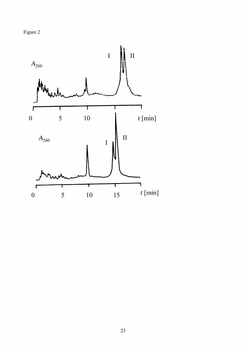
(MALDI-TOF). Cyclization of oligonucleotide 2 with EDC represented the best reaction (70-90%
conversion to cyclic oligonucleotide, as estimated by PAGE and HPLC using XTerra®; Figure 2).
Conversion of sequence 1 to cyclic oligonucleotide 7 by EDC resulted in lower, but still good,
yields (50-70% yield). The recovery of cyclic oligonucleotides from BrCN-catalyzed reactions was
poor, due to the formation of a precipitate. This precipitation is explained by the formation of an
insoluble complex with Ni2+ ions present in the BrCN reaction. Consequently, we isolated the
reaction products from BrCN reactions by centrifugation of the reaction mixtures and dissolution of
the pellets in a 0.1M EDTA solution. In this way, Ni2+ ions were complexed by EDTA and purine-
containing oligonucleotides became soluble. Analysis of the BrCN-reactions show less than 50%
conversion of linear to cyclic oligonucleotides. EDC-mediated cyclization did not produce any
precipitate, and yields were better, consequently, this method was preferred. The desired cyclic
oligonucleotides 7 and 8 were purified by either gel electrophoresis or HPLC using an XTerra®
column (Figure 2).
3. Melting experiments. The binding properties of cyclic oligonucleotides 7 and 8 as well their
phosphorylated and unphosphorylated linear sequences were analyzed by thermal denaturation with
UV spectroscopy at pH 7.2. Hairpin sequences 9 and 10 were included for comparison as well as
control oligonucleotides 11 and 12 with scrambled antiparallel strands [16]. Also, the
complementary purine sequence 13, without any antiparallel strand, was prepared. Melting studies
of oligonucleotides 1-14 alone did not show any clear transition. In some cases, small changes
could be observed at around 30-40 degrees, but they were not cooperative. This is due either to a

6
lack of secondary structure (oligonucleotides 11-14) or to the absence of hyperchromicity observed
during the dissociation of triplex forming strand in these sequences (oligonucleotides 1-10) [16, 21,
22].
Melting experiments with complexes of oligonucleotides 1-13 and the target pyrimidine
sequence 14 showed a clear transition, demonstrating that oligonucleotides 1-10 bind their target
with a higher efficiency than their complementary and control sequences 11-13 (Table 2). Cyclic
oligonucleotide 8 bound the target with the highest stability (ΔG37 –19.1 Kcal/mol; Table 2). This
represents an enhancement of 8 Kcal/mol over the simple Watson-Crick recognition (13 ΔG37 –11.2
Kcal/mol, 12 ΔG37 -10.8 Kcal/mol; Table 2). Non-phosphorylated linear sequence 6 is slightly less
stable (ΔG37 –17.5 Kcal/mol; Table 2). The presence of the phosphate in the linear sequence
induces little or not destabilization of the triplex (4 ΔG37 -17.5 and 2 ΔG37 –15.2 Kcal/mol; Table 2),
and the triplex formed by hairpin 10 is approx. 3 Kcal/mol less stable (10 ΔG37 –15.5 Kcal/mol;
Table 2). It is important to note that the melting temperature of the hairpin is the highest of this
series, but that the free energy inverts the order of relative stability. It has already been pointed out
that melting temperatures are not always indicative of relative stability [23]. In some cases,
differences in the shape of the melting curves means that a structure with a higher Tm will exhibit a
lower stability [23]. In the present case, the melting curves of the hairpins and cyclic
oligonucleotides have a similar Tm but the differences in the shape of the curves are responsible for
the relative stability of the triplexes.
Similar results were obtained in the series carrying G,T-reverse Hoogsteen strand (Table 3).
However, unfortunately, cyclic oligonucleotides 7 gave a broad curve with low hyperchromicity,
and curve fitting was not possible in this particular case. Although thermodynamic parameters could
not be obtained, cyclic oligonucleotide 7, as well as linear and hairpin sequences 5, 3, 1, and 9
displayed the highest Tm (54.9-58.5) and were at least 3.5-6.4 kcal/mol more stable than the simple
Watson-Crick recognition (5, 3, 9, and 5 ΔG37 –17.6, –15.7, -14.7, and -15.4 Kcal/mol respectively

7
Table 3; 11 ΔG37 -11.2 Kcal/mol, Table 3; 13 ΔG37 –11.2 Kcal/mol, Table 2). These results are in
agreement with previous work on cyclic oligonucleotides carrying a (G,T)-reverse Hoogsteen strand
[10, 11].
We have also run melting experiments in buffers containing NaCl and KCl instead of MgCl2
in order to check whether G-quartet structures may inhibit triplex formation [24] but only a 8ºC
decrease on Tm was observed (similar to parallel triplexes [15]) indicating no interference of these
structures on triplex formation (data not shown).
The circular dichroism spectra of cyclic oligonucleotide 8 with and without target sequence
14 are shown in Figure 3. The CD spectrum of the triplex formed by an equimolar mixture of
oligonucleotides 8 and 14 shows a positive band at 270 nm and a negative band at 242 nm. This
spectrum is in agreement with the CD spectra of (G,A)-antiparallel triplexes described in literature
[25]. Thermal denaturation of triplexes was also followed by CD spectroscopy. Figure 4 shows the
changes in the CD spectrum for the (G,A)-antiparallel triplex formed by oligonucleotides 8 and 14.
Analysis of the complete data set by multivariate curve resolution (MCR, [22, 26, 27]) revealed the
presence of two species with a melting temperature of 51.5ºC (Table 2). The pure spectra of folded
and unfolded structures resolved by MCR and the concentration profiles of both species are shown
in Figure 4. The same methodology was applied to the analysis of the thermal denaturation of the
(G,A)-antiparallel triplex formed by oligonucleotides 3 and 14, giving a melting temperature of 52
ºC (Table 2). Although the melting temperatures obtained by UV- and CD-spectroscopy are in
agreement, there is a difference of 5-7 degrees between the two methods. These differences are
probably due to differences in the heating process employed; whilst heating is continuous in UV
melting curves, the CD method uses steps of temperature with a waiting time for equilibration.
Thermal denaturation of oligonucleotide 3 alone was also assessed by CD spectroscopy (data not
shown). Here, a transition between two species was also observed, with a melting temperature of 33
ºC. This indicates that oligonucleotide 3 is partially folded, most probably forming reverse

8
Hoogsteen bonds, although UV-spectroscopy did not show changes during thermal denaturation of
this oligonucleotide [16, 21, 22].
Discussion. The ability of cyclic oligonucleotides to bind single-stranded nucleic acid sequences
by triplex formation has attracted a widespread interest due to the higher binding affinity and
selectivity, as well as the enhanced resistance to exonuclease degradation [8, 9]. Here, we present
for first time the synthesis and binding properties of a cyclic oligonucleotide designed to form
(G,A)-antiparallel triplexes. These results complement other studies undertaken using cyclic
oligonucleotides designed to form (G,T)-antiparallel triplexes [10, 11] and (C,T)-parallel triplexes
[8, 9]. We show that cyclic oligonucleotides carrying (G,A)-antiparallel strands are obtained in
good yields, probably due to a higher affinity to the template pyrimidine strand. The cyclic
oligonucleotides obtained in this study have a high affinity to their polypyrimidine targets at neutral
pH, making them interesting tools for antisense inhibition of gene expression.
It is important to note that previous work with antiparallel triplexes using (G,A)- and (G,T)-
oligonucleotides and duplex DNA as targets has shown a low stability of these triplexes, especially
for those formed by (G,T)-oligonucleotides [22, 25]. This situation changes dramatically when the
(G,A)- and (G,T)-triplex forming sequences are connected to the appropriate oligopurine Watson-
Crick sequences to form hairpins or cyclic oligonucleotides designed to bind single-stranded
polypyrimidide targets. In this situation, the differences observed with (G,T)- antiparallel triplexes
disappear and (G,A)- and (G,T)-antiparallel triplexes have equally high stability. Although this
behaviour has been described previously [10, 11, 16, 28], very little biological data has been
generated using this potentially powerful strategy.
Most of the prior work using cyclic oligonucleotides for triplex formation has been
performed with C-/T-rich oligonucleotides [8, 9]. These oligonucleotides target the polypurine
sequence by forming parallel triplexes that are stable at acidic pH. We chose to target the

9
complementary polypyrimidine sequence as a result of recent developments using 8-aminopurines.
Recently, we have shown that substitution of guanine with 8-aminoguanine results in strong
stabilization of antiparallel triplexes [16]. We believe that the introduction of 8-aminoguanine in
cyclic oligonucleotides will be of interest in order to further increase the binding affinities.
Nevertheless, even with the use of natural nucleosides we demonstrate that by using cyclic and
hairpin oligonucleotides it is possible to achieve much stronger binding than with regular duplex
recognition at neutral pH.
This study was supported by the Dirección General de Investigación Científica (BQU2003-00397
and BFU2004-02048/BMC), the Generalitat de Catalunya (2001-SGR-0049) and Fundació La
Caixa (BM04-52-0). We thank Drs Jordi Robles, Anna Grandas, Enrique Pedroso and Vicent
Marchán for their help in the preparation of the oligonucleotides and the use of the Meltwin
program. We thank Waters for their support in the use of XTerra® HPLC columns.
Experimental Part
General. Phosphoramidites and ancillary reagents used during oligonucleotide synthesis were from
Applied Biosystems (USA), Transgenomic (Scotland), Link Technologies (Scotland) and Glen
Research (USA). The rest of the chemicals were purchased from Aldrich, Sigma or Fluka. Long
chain amino-controlled pore glass (LCAA-CPG) was purchased from CPG (USA). Solvents were
from S.D.S. (France). NAP-10 columns (Sephadex G-25) were purchased from Amersham. Solid
support for the preparation of 3’-phosphorylated precursors was synthesized as described elsewhere
[17].
10
Instrumentation. Oligonucleotide sequences were synthesized on a Applied Biosystems DNA
synthesizer model 392. UV-Visible spectra were recorded on a Shimadzu UV-2101PC and CD
spectra on a Jasco J810.
Oligonucleotide Synthesis. Oligonucleotides were prepared using standard (benzoyl- or isobutyryl-
protected) 3’-[(2’-cyanoethyl)phosphoramidites] and the phosphoramidite of (4,4’-
dimethoxytrityloxyethyl) hydroxyethyl sulfone [17]. Supports were obtained from commercial
sources or prepared as described elsewhere [17].
After the assembly of the sequences in batches of 1 μmol or 200 nmols, the resulting supports were
treated with 1ml of concentrated ammonia (overnight at 55ºC). Oligonucleotide supports carrying a
phosphate group at the 3’- end were treated overnight with 1ml of 1:1 mixture of 40% methylamine
and 32% NH3 in aqueous solutions.
The products arising from ammonia treatment were dissolved in water and purified by HPLC using
a PRP-1 10 μm column (305 x 7mm, Hamilton, USA) at a flow rate of 3 ml/min. A 20min linear
gradient from 15 to 50% acetonitrile over 100mM aqueous triethylammonium acetate was used for
oligonucleotides carrying the (MeO)2-Tr group. After removal of the (MeO)2-Tr group with 80%
acetic acid (30min) the resulting oligonucleotides were purified on the same column using a 20min
linear gradient from 5 to 25% acetonitrile over 100 mM aqueous triethylammonium acetate.
5’-Phosphorylated and cyclic oligonucleotides were purified by HPLC using an XTerra® MS C18
2.5 μm (Waters, USA) column: flow rate 1 ml/min; column temperature, 60 ºC. A 20 min gradient
from 5 to 15% acetonitrile over 100 mM aqueous triethylammonium acetate was used. Yields after
purification: 1-4 (1 μmols), 20-40 OD units. MS (MALDI-TOF) of oligonucleotide 5, 9412.7 (M)
expected for C300H374N111O186P29 9410.2; oligonucleotide 6: 9459.6 (M) expected for
C300H369N126O176P29 9455.3; oligonucleotide 1, 9492.8 (M) expected for C300H375N111O189P30
9490.2; oligonucleotide 2, 9537.8 (M) expected for C300H370N126O179P30 9535.3.

11
Cyclization of oligonucleotides.
Carbodiimide activation. Phosphorylated linear precursors 1-4 (4 OD units) and template 14 (1.3
OD units) were dissolved on 0.14 ml of the following buffer : 50 mM morpholinoethansulfonic acid
(MES) pH 6.0, 20 mM MgCl2. The solution was heated at 80ºC followed by slow cooling at room
temperature. Solid water-soluble carbodiimide (EDC) was added to obtain a 0.25 M EDC solution
and the resulting mixture was kept at 4ºC for 16-20 hours. The resulting solution was dialyzed
against water and then concentrated and purified by HPLC using an XTerra® column (see
conditions above, Figure 2). Yield: 7, 0.3-0.6 OD units; 8, 2-3 OD units. MS (MALDI-TOF) of
sequence 8: 9525.6 (M) expected for C300H368N126O178P30 9517.3.
BrCN activation. Phosphorylated linear precursors 1-4 (4 O.D. units) and template 14 (1.3 OD
units) were dissolved in0.5 ml of the following buffer: 200 mM imidazol-HCl pH 7.0, 100 mM
NiCl2. BrCN (0.062 ml of a 5M solution in acetonitrile) was added to obtain a 125 mM BrCN
solution and the resulting mixture was kept 4 ºC for 16-20 hours. The solution was then centrifuged
and the supernatant dialyzed against water. Finally, the supernatant was concentrated and analyzed
by gel electrophoresis indicating that contain only the 11bases template 14, so it was discarded. The
pellet was treated with 0.25 ml of 0.1 M EDTA and 0.25 ml of water. The resulting solution was
dialyzed against water and then concentrated and purified by PAGE yielding 0.5 O.D. units for 7
and 0.6 O.D. units for 8.
Melting experiments. Melting experiments were performed by mixing equimolar amounts of the
appropriate oligonucleotides dissolved in a soln. containing 50 mM MgCl2, 10 mM sodium
cacodylate (pH 7.2), and 0.1 mM EDTA. Oligonucleotides were annealed by slow cooling from 90
ºC to room temperature. UV Absorption spectra and melting curves (absorbance vs temperature)
were recorded in 1-cm path-length cells using a Shimadzu UV2101PC UV/VIS spectrophotometer

12
(Japan) with a temperature controller using a programmed temperature increase of 0.5˚/min.
Melting curves were obtained by monitoring the absorbance at 260 nm of triplex concentrations of
approx. 2-4�M. All of the absorbance vs. temperature plots showed sigmoidal curves, indicating
cooperative transition, and the data were fitted to a two-state model using MeltWin 3.5 software
[29] in order to determine Tm and ΔG values. Thermodynamic data were calculated as mean values
of three independent melting experiments. Results: see Table 2 and 3.
CD melting experiments
The same solutions used for UV experiments were used for CD spectroscopy. All CD
measurements were performed on a Jasco Spectropolarimeter (model J810) equipped with a
thermostat-controlled cell holder with a cell path length of 1 cm. Melting experiments were
determined using steps of 5 oC followed by 5 min equilibration time, and a temperature ramp of 1
oC per minute.
Data treatment
The complete CD spectra recorded during melting experiments were analyzed by means of the
multivariate curve resolution (MCR) method [22, 26]. This method, which is based on factor
analysis, has been widely applied to the analysis of spectroscopic data recorded from
conformational studies of nucleic acids and proteins [27], and only a brief description is given here.
The reader is referred to references [22, 26, 27] for more information about the procedure and its
applications. The goal of this method is the recovery of information about the number of species,
and their concentration and spectral profiles. This is performed using the experimental spectra
without the postulation of a physico-chemical model. For this purpose, the complete experimental
CD spectra recorded during a melting experiment are collected in a table or matrix D. The
dimensions of D were Nr rows x Nm columns, where Nr are the spectra recorded at successive

13
temperature values and Nm the number of wavelengths measured in each spectrum. The matrix D is
analyzed according to the MCR procedure to obtain information about the thermodynamic behavior
of the system. For melting experiments, the information consisted of the number of conformations,
their concentration profiles (and the corresponding Tm values), and the pure spectrum for each one
of them. Mathematically, the experimental data model can be written as:
D = C ST + E (equation 1)
where C is the data matrix containing the concentration profiles, ST is the pure spectrum and E is
the residual noise not explained by the species or conformations in C and ST.
All MCR-ALS calculations were performed using in-house MATLAB® (version 6; The Mathworks
Inc, Natick, MA) routines.
REFERENCES
[1] N.T. Thuong, C. Hélène Angew. Chem. Int. Ed. Engl., 1993, 32, 666-690.
[2] P.P. Chan, P.M. Glazer J. Mol. Med., 1997, 75, 267-282.
[3] V.N. Soyter, V.N. Potoman, Triple-helical Nucleic Acids. Springer-Verlag, New York, N.Y.
1996.
[4] C. Giovannangeli, T. Montenay-Garestier, M. Rouge, M. Chassignol, N.T. Thuong, C. Hélène,
J. Am. Chem. Soc., 1991, 113, 7775-7776.
[5] G. Prakash, E.T. Kool, J. Chem. Soc. Chem. Commun., 1991, 646, 1161-1162.
[6] P.E. Nielsen, M. Egholm, R. Berg, O. Buchardt, Science 1991, 254, 1497-1500.
[7] H. Kuhn, V.V. Demidov, P.E. Nielsen, M.D. Frank-Kamenetskii, J. Mol. Biol., 1999, 286, 1337-
1345.
[8] E.T. Kool, Chem Rev., 1997, 97, 1473-1478

14
[9] E. T. Kool, Annu. Rev. Biophys. Biomol. Struct., 1996, 25, 1-28.
[10] S. Wang, E.T. Kool, J. Am. Chem. Soc., 1994, 116, 8857-8858.
[11] T. Vo, S. Wang, E.T. Kool, Nucleic Acids Res., 1995, 23, 2937-2944.
[12] A. Miksimenko, E.V. Volkov, J.R. Bertrand, H. Porumb, C. Malvy, Z.A. Shabarova, M. B.
Gottlikh, Eur. J. Biochem., 2000, 267, 3592-3603.
[13] L.E. Xodo, G. Manzini, F. Quadrifolio, G.A. van der Marel, J.H. van Boom, Nucleic Acids
Res., 1991, 19, 5625-5631.
[14] G. Manzini, L.E. Xodo, D. Gasparotto, J. Mol. Biol., 1990, 213, 833-843.
[15] A. Aviñó, M. Frieden, J.C. Morales, B. G. de la Torre, R. Güimil-García, F. Azorín, J.L. Gelpí,
M. Orozco, C. González, R. Eritja. Nucleic Acids Res., 2002, 30, 2609-2619.
[16] A. Aviñó, E. Cubero, C. González, R. Eritja, M. Orozco, J. Am. Chem. Soc., 2003, 125, 16127-
16138.
[17] T. Horn, M.S. Urdea, Tetrahedron Lett. 1986, 27, 4705-4708.
[18] A. MacMillan, G.L. Verdine, Tetrahedron 1991, 47, 2603-2616.
[19] M.P. Reddy, F. Farooqui, N.B. Hanna, Tetrahedron Lett., 1996, 37, 8691-8694.
[20] N.G. Dolinnaya, M. Blumenfeld, I.N. Merenkova, T. S. Oretskaya, N.F. Krynetskaya, M.G.
Ivanovskaya, M. Vasseur, Z.A. Shabarova, Nucleic Acids Res., 1993, 21, 5403-5407.
[21] D. Murphy, R. Eritja, G. Redmond, Nucleic Acids Res., 2004, 32, e65.
[22] J. Jaumot, A. Aviñó, R. Eritja, R. Tauler, R. Gargallo, J. Biomol. Str. Dyn., 2003, 21, 267-278.
[23] J.L. Mergny, L. Lacroix, Oligonucleotides, 2003, 13, 515-537.
[24] V. Dapic, V. Abdomerovic, R. Marrington, J. Peberdy, A. Rodger, J.O. Trent, P. J. Bates,
Nucleic Acids Res., 2003, 31, 2097-2107.
[25] Y. He, P.V. Scaria, R.H. Shafer, Biopolymers, 1997, 41, 431-441.
[26] MCR routines for Matlab® and tutorials are freely available at the web site:
www.ub.es/gesq/mcr/mcr.htm

15
[27] J. Jaumot, M. Vives, R. Gargallo, Anal. Biochem. 2004, 327, 1-13.
[28] M. Mills, P.B. Arimodo, L. Lacroix, T. Garestier, C. Hélène, H. Klump, J.L. Mergny, J. Mol.
Biol.. 1999, 291, 1035-1054.
[29] J.A. McDowell, D.H. Turner, Biochemistry, 1996, 35, 14077-14089.

16
Table 1: Oligonucleotides Prepared in this Study
Number Sequence
1 5’-phosphate-AGGAGA-T4-TGTGGTGGTTG-T4-GAAGG-3’
2 5’-phosphate-AGGAGA-T4-AGAGGAGGAAG-T4-GAAGG-3’
3 5’-AGGAGA-T4-TGTGGTGGTTG-T4-GAAGG-phosphate-3’
4 5’-AGGAGA-T4-AGAGGAGGAAG-T4-GAAGG-phosphate-3’
5 5’-AGGAGA-T4-TGTGGTGGTTG-T4-GAAGG-3’
6 5’-AGGAGA-T4-AGAGGAGGAAG-T4-GAAGG-3’
7 Cyclo-(5’-AGGAGA-T4-TGTGGTGGTTG-T4-GAAGG-3’)
8 Cyclo-(5’-AGGAGA-T4-AGAGGAGGAAG-T4-GAAGG-3’)
9 5’-GAAGGAGGAGA-T4-TGTGGTGGTTG-3’
10 5’-GAAGGAGGAGA-T4-AGAGGAGGAAG-3’
11 5’-GAAGGAGGAGA-T4-GTGTGGTTTGT-3’
12 5’-GAAGGAGGAGA-T4-GAGAGGAAAGA-3’
13 5’-GAAGGAGGAGA-3’
14 5’-TCTCCTCCTTC-3’

17
Table 2: Melting Temperatures (Tm, ºC) and Free Energies (ΔG37, Kcal/mol) for Complexes
Formed by Oligonucleotides Carrying (G,A)-Hoogsteen Strand (2, 4, 6, 8, 10) and Control
Oligonucleotides (12, 13) with the Pyrimidine Target Sequence 14.
Ligand Complex Tm [ºC], UVa,b) ΔG37, UVa,b) Tm [ºC], CDa,b)
8
59.0 -19.1 51.5
6
57.3 -17.5 52
4
58.1 -17.5 n.d.
2
55.6 -15.2 n.d.
10
60.9 -15.5 n.d.
12
49.4 -10.8 n.d.
13
48.5 -11.2 n.d.
AGAGGAGGAAG
AGAGGAGGAAG
TCTCCTCCTTC
AGAGGA GGAAG
AGAGGAGGAAG
TCTCCTCCTTC
AGAGGA pGGAAG
AGAGGAGGAAG
TCTCCTCCTTC
AGAGGAp GGAAG
AGAGGAGGAAG
TCTCCTCCTTC
AGAGGAGGAAG
AGAGGAGGAAG
TCTCCTCCTTC
AGAGGAGGAAG
GAGAGGAAAGA
TCTCCTCCTTC
AGAGGAGGAAGTCTCCTCCTTC

18
a) 0.050M MgCl2, 0.01M sodium cacodylate pH 7.2, 0.1mM EDTA. b) Uncertainties in Tm values
and in free energies are estimated at 1 ºC, and 15%. n.d.: not determined.

19
Table 3: Melting Temperatures and Free Energies of Complexes Formed by Oligonucleotides
Carrying (G,T)-Hoogsteen Strand (3, 5, 7, 9) and Control Oligonucleotide 11 with the Pyrimidine
Target Sequence 14.
Ligand Complex Tm [ºC], UVa,b) ΔG37º, UV a,b)
7
56.5 n.a.
5
55.9 -17.6
3
54.9 -15.7
1
51.6 -14.7
9
58.5 -15.4
11
49.5 -11.2
a) 50mM MgCl2, 10 mM sodium cacodylate pH 7.2, 0.1mM EDTA. b) Uncertainties in Tm values
and in free energies are estimated at 1 ºC, and 15 %. n.a.: not possible to analyze.
AGAGGAGGAAG
TGTGGTGGTTG
TCTCCTCCTTC
AGAGGA GGAAG
TGTGGTGGTTG
TCTCCTCCTTC
AGAGGA pGGAAG
TGTGGTGGTTG
TCTCCTCCTTC
AGAGGAGGAAG
TGTGGTGGTTG
TCTCCTCCTTC
AGAGGAGGAAG
GTGTGGTTTGT
TCTCCTCCTTC
AGAGGAp GGAAG
TGTGGTGGTTG
TCTCCTCCTTC

20
LEGENDS
Scheme 1. Strategies for binding nucleic acids by triplex formation. (left) Strategy for targeting
duplexes using triplex-forming oligonucleotides (TFO). (center and right) Targeting single-stranded
polypyrimidine nucleic acid sequences by triplex formation using hairpins (centre) or cyclic
oligonucleotides (right) made by linking the polypurine strand with the triplex forming strand
(TFO). A similar approach has been used for targeting the polypurine sequence (see ref. [8,9]).
Scheme 2. Triplexes analyzed in this study made of the pyrimidine target sequence with cyclic
oligonucleotides formed by linking the Watson-Crick purine strand and the (G,A)- or (G,T)-
antiparallel strand. The phosphate bond that was selected to disconnect the cyclic oligonucleotide
occurs between the G and the A shown in bold. Sequences of the linear precursors are shown in
Table 1.
Figure 1: HPLC Analysis of 5’-phosphorylated oligonucleotide 2 using an XTerra® column. The
figure shows the HPLC profile of the crude product obtained after ammonia treatment. Fractions I
and II contained the desired phosphorylated oligonucleotide and the unphosphorylated
oligonucleotide respectively.
Figure 2: HPLC profile of the product obtained in template-assisted cyclization of 2, analyzed
using an XTerra® column. (a) cyclization of sequence 1 and (b) cyclization of sequence 2.
Fractions I and II contained the starting phosphorylated oligonucleotide (1 or 2) and the desired
cyclic oligonucleotide (7 or 8), respectively. The peak eluting close to 10 min correspond to
template sequence 14.

21
Figure 3. CD-spectra of cyclic oligonucleotide 8 with and without target sequence 14 (0.050M
MgCl2, 0.01M sodium cacodylate pH 7.2, 25 ºC, 3 μM triplex concentration).
Figure 4. Thermal denaturation of triplex formed by cyclic oligonucleotide 8 with target sequence
14 followed by CD-spectroscopy. (0.050M MgCl2, 0.01M sodium cacodylate pH 7.2, 0.1mM EDTA,
3 μM triplex concentration). A) Experimental spectra recorded from 20 to 80 oC, at 5 oC intervals
(matrix D). B) MCR-resolved concentration profiles according to Equation 1 (matrix C). C) MCR-
resolved pure spectra (matrix ST). (a) triplex, (b) unfolded species.
Scheme 1
Scheme 2
polypurine
polypyrimidine
TFO
polypurine
polypyrimidine
TFO
polypurine
polypyrimidine
TFO
TGTGGTGGTTGT
TT
T
T
TT
3' 5'TCTCCTCCTTC5' 3'
AGAGGAGGAAG
T
T
TT
T
T
TT
3' 5'TCTCCTCCTTC5' 3'
5' 3' 5' 3'
AGAGGAGGAAG AGAGGAGGAAG

22
Figure 1
23
Figure 2
t [min]
t [min]
A260
A260
10
10
I II
III
0 5
0 5 15

24
Figure 3

25
Figure 4
220 240 260 280 300 320-2
-1.5
-1
-0.5
0
0.5
1
Wavelength (nm)
CD
sig
nal (
mde
g)
20 oC
80 oC
20 30 40 50 60 70 800
0.2
0.4
0.6
0.8
1
Temperature (oC)
Rel
ativ
e co
ncen
tratio
n
Tm = 51.5 oC
a b
220 240 260 280 300 320-2
-1.5
-1
-0.5
0
0.5
1
1.5
Wavelength (nm)
CD
sig
nal (
arbi
trary
uni
ts)
a
b
A) B)
C)