syndrome douloureux régional complexe: apport de la
TRANSCRIPT
© Fannie Allen Demers, 2021
Syndrome douloureux régional complexe: apport de la neurostimulation périphérique - Plasticité cérébrale et
amélioration cliniques
Mémoire
Fannie Allen Demers
Maîtrise en médecine expérimentale - avec mémoire
Maître ès sciences (M. Sc.)
Québec, Canada
ii
RÉSUMÉ
Malgré des traitements spécialisés et multidisciplinaires, les personnes souffrant du
syndrome douloureux régional complexe (SDRC) peuvent conserver de la douleur et des
limitations fonctionnelles qui s’expliqueraient par des changements cérébraux persistants,
entre autres dans le cortex moteur primaire (M1). Étudier les changements de fonctionnement
du M1 permettrait de mieux comprendre comment utiliser la neurostimulation non invasive,
comme les stimulations magnétiques répétées en périphérie (rPMS des muscles, connues
pour influencer la plasticité cérébrale), pour normaliser la fonction motrice corticale, réduire
la douleur et augmenter les gains cliniques.
Les objectifs de ce projet de maîtrise étaient donc de mieux comprendre la place dans la
littérature de la neurostimulation non invasive en SDRC, de tester le fonctionnement de M1
en parallèle à la fonction sensorimotrice d’adultes avec SDRC au membre supérieur, ainsi
que de mesurer l’effet d’une séance rPMS sur ces mesures et les symptômes de douleur de
cette même population.
Il a été observé que, indépendamment du côté atteint, l’excitabilité du M1 était asymétrique
en SDRC avec une association avec la douleur et les troubles du mouvement. Les participants
avec SDRC présentaient également une diminution et une latéralisation altérée des mesures
de fonction sensorimotrice.
Les rPMS ont permis de moduler bilatéralement l’excitabilité des M1 (diminution du
débalancement) et, chez les personnes présentant avant la séance rPMS une hyperexcitabilité
du M1 controlatéral au membre atteint, de diminuer leur douleur. Les rPMS ont également
permis une amélioration de la fonction sensorimotrice et des changements centraux reliés à
la plasticité cérébrale ont été mesurés dans l’hémisphère ipsilatéral au membre avec SDRC.
Les rPMS seules ou comme adjuvant aux thérapies conventionnelles de réadaptation
représentent donc une approche prometteuse pour dépasser les gains cliniques en SDRC.
iii
ABSTRACT
Despite specialized and multidisciplinary treatments, people suffering from complex regional
pain syndrome (CRPS) can present with persistent pain and functional limitations likely due
to brain changes such as in the primary motor cortex (M1). Studying the changes of M1
functioning would permit to better understand how to use noninvasive neurostimulation, as
repetitive peripheral magnetic stimulation (rPMS of muscles, known to influence brain
plasticity) in CRPS to enable the normalization of cortical motor function, the reduction of
pain and to go beyond gains already reached.
The objectives of this master's project were thus to better understand the place in the
literature of the noninvasive neurostimulation in SDRC, to test the functioning of M1
concurrent with the sensorimotor function of adults with CRPS of the upper limb, and to
measure the effect of one rPMS session on these measures and pain symptoms of this same
population.
It has been measured that M1 excitability was asymmetrical in CRPS, regardless of the
impaired side, with an association to pain and movement disorders. Participants with CRPS
also exhibited a decreased and an altered lateralization of the measures of sensorimotor
function.
rPMS influenced bilateral M1 excitability (decrease of the imbalance) and, with people
presenting before the rPMS session hyperactivity of M1 contralateral to the impaired limb,
reduced pain. rPMS also improved sensorimotor function and central changes related to brain
plasticity were measured in the hemisphere ipsilateral to the CRPS limb.
rPMS alone or as adjuvant to conventional rehabilitation therapies thus represent a promising
approach to overcome clinical gains in CRPS.
iv
TABLE DES MATIÈRES
RÉSUMÉ ............................................................................................................................... II
ABSTRACT ........................................................................................................................ III
TABLE DES MATIÈRES ................................................................................................... IV
LISTE DES TABLEAUX .................................................................................................... VI
LISTE DES FIGURES ...................................................................................................... VII
LISTE DES ABRÉVIATIONS ........................................................................................ VIII
REMERCIEMENTS ............................................................................................................ X
AVANT-PROPOS ............................................................................................................. XII
INTRODUCTION ................................................................................................................ 1
SECTION 1 : ARTICLE 1 - « Complex Regional Pain Syndrome (CRPS). A
Comprehensive Review on Plastic Changes supporting the Use of Noninvasive
Neurostimulation » ........................................................................................................... 1
RÉSUMÉ ........................................................................................................................... 3
ABSTRACT ....................................................................................................................... 4
TABLE OF CONTENTS ...................................................................................................... 5
PHYSIOPATHOLOGY OF CRPS ....................................................................................... 8
BRAIN CHANGES AND MALADAPTIVE NEURONAL PLASTICITY .................................. 11
CONVENTIONAL TREATMENT IN CRPS AND LIMITATIONS OWING TO BRAIN CHANGES 17
PRINCIPLES SUPPORTING THE USE OF NONINVASIVE NEUROSTIMULATION IN CRPS ..... 21
CONCLUSION ................................................................................................................. 29
ACKNOWLEDGEMENTS ................................................................................................. 29
REFERENCES ................................................................................................................. 30
FIGURE CAPTION .......................................................................................................... 38
SECTION 2 : HYPOTHÈSES ET OBJECTIFS DU PROJET DE MAÎTRISE ......... 45
Rationnel du projet suivant les recommandations de la revue de littérature ............. 45
Hypothèses de travail.................................................................................................... 46
Objectifs principaux ..................................................................................................... 47
Approches méthodologiques ........................................................................................ 48
CHAPITRE 1 : MÉTHODOLOGIE ................................................................................. 50
1.1 CARACTÉRISTIQUES DES PARTICIPANTS ..................................................................... 50
1.2 RECRUTEMENT ........................................................................................................... 51
1.3 DEVIS EXPÉRIMENTAL ................................................................................................ 54
1.4 QUESTIONNAIRES ....................................................................................................... 55
1.5 MESURES CLINIQUES .................................................................................................. 57
1.6 MESURES NEUROPHYSIOLOGIQUES ............................................................................ 60
1.7 TRAITEMENT EXPÉRIMENTAL ..................................................................................... 67
1.8 RÉSUMÉ DES RÉDUCTIONS DE DONNÉES ET ANALYSES STATISTIQUES ........................ 68
v
CHAPITRE 2 : ARTICLE 2 « LIVING WITH A COMPLEX REGIONAL PAIN SYNDROME
(CRPS): CLINICAL AND CORTICOMOTOR CHANGES AS COMPARED TO PAIN-FREE PEOPLE » .... 70
RÉSUMÉ ......................................................................................................................... 72
ABSTRACT ..................................................................................................................... 73
INTRODUCTION ........................................................................................................... 74
METHODS ...................................................................................................................... 76
RESULTS ........................................................................................................................ 83
DISCUSSION ................................................................................................................. 86
CONCLUSION ............................................................................................................... 91
ACKNOWLEDGEMENTS ............................................................................................. 91
REFERENCES ............................................................................................................... 92
FIGURES CAPTIONS ................................................................................................... 98
CHAPITRE 3 : ARTICLE 3 « THETA BURST STIMULATION OVER FOREARM MUSCLES IN
COMPLEX REGIONAL PAIN SYNDROME (CRPS): INFLUENCE ON BRAIN AND CLINICAL OUTCOMES » 106
RÉSUMÉ ....................................................................................................................... 108
ABSTRACT ................................................................................................................... 109
INTRODUCTION ......................................................................................................... 110
MATERIALS AND METHODS .................................................................................. 113
RESULTS ...................................................................................................................... 120
DISCUSSION ............................................................................................................... 123
CONCLUSION ............................................................................................................. 127
ACKNOWLEDGEMENTS ........................................................................................... 127
REFERENCES ............................................................................................................. 128
FIGURES CAPTIONS ................................................................................................. 134
CHAPITRE 4 : DISCUSSION GÉNÉRALE ................................................................. 142
4.1 RETOUR SUR LES HYPOTHÈSES RELATIVES À L’ARTICLE 2 ....................................... 142
4.2 RETOUR SUR LES HYPOTHÈSES RELATIVES À L’ARTICLE 3 ........................................ 143
4.3 FORCES ET LIMITES DES ÉTUDES .............................................................................. 146
4.4 PERSPECTIVES CLINIQUES ET DE RECHERCHE ......................................................... 147
CONCLUSIONS ............................................................................................................... 150
BIBLIOGRAPHIE ........................................................................................................... 151
ANNEXES ......................................................................................................................... 165
vi
LISTE DES TABLEAUX
DE L’INTRODUCTION – ARTICLE 1
Table 1. The “Budapest Criteria” for Complex Regional Pain Syndrome (CRPS) Diagnosis
Table 2. Brain Changes Reported in Complex Regional Pain Syndrome (CRPS)
Table 3. Studies with Noninvasive Neurostimulation in Complex Regional Pain Syndrome
(CRPS)
DU CHAPITRE 1 – MÉTHODOLOGIE
Tableau 1 : Caractéristiques générales des participants avec SDRC et sans douleur
Tableau 2 : Critères d’inclusion et d’exclusion du projet de recherche
Tableau 3 : Résumé des protocoles de stimulations magnétiques transcrâniennes doubles
Tableau 4 : Résumé des variables et mesures à l’étude
DU CHAPITRE 2 – ARTICLE 2
Table 1: General Characteristics of Participants
Table 2: The “Budapest Criteria” for Complex Regional Pain Syndrome (CRPS) Diagnosis
Table 3: Raw clinical and TMS data in CRPS and pain-free groups
DU CHAPITRE 3 – ARTICLE 3
Table 1: General Characteristics of Participants
Table 2: The “Budapest Criteria” for Complex Regional Pain Syndrome (CRPS) Diagnostic
Table 3: Raw data in CRPS at pre- and post-rPMS and in pain-free participant
vii
LISTE DES FIGURES
DE L’INTRODUCTION – ARTICLE 1
Figure 1. Brain Changes in People with a Complex Regional Pain Syndrome
DU CHAPITRE 1 - MÉTHODOLOGIE
Figure 1 : Devis expérimental
Figure 2. Échelle visuelle analogue
Figure 3. Test au monofilament de Semmes et Weinstein
Figure 4. Goniomètre manuel à doigt et articulations mesurées
Figure 5. Dynamomètre JAMAR
Figure 6. Muscle premier interosseux dorsal de la main (FDI)
Figure 7. Stimulateur électromagnétique MagStim 200 et
bobine de stimulation en forme de 8
Figure 8. Électrode d’EMG de surface, collant double-face et unité préamplificatrice
Figure 9. Système international EEG 10-20
Figure 10. Mécanismes d’inhibition et de facilitation intracorticales
Figure 11. Bobine de stimulation avec ventilateur intégré appliquée sur le FDS
Figure 12. Stimulateur Magstim Rapid-Rate2
DU CHAPITRE 2 – ARTICLE 2
Figure 1. Resting Motor Threshold (RMT)
Figure 2: Test and Conditioned MEP Amplitudes
Figure 3. Clinical Outcomes (Group Means)
Figure 4: Group Correlations
DU CHAPITRE 3 – ARTICLE 3
Figure 1 : Clinical Outcomes
Figure 2. Resting Motor Threshold (RMT)
Figure 3. TMS Outcomes
Figure 4. Correlation
viii
LISTE DES ABRÉVIATIONS
AMPA (récepteurs) : α-amino-3-hydroxy-5-méthylisoazol-4-propionate (récepteurs)
ANOVA : analyses de variance, de l’anglais « analysis of variance »
EMG : électromyographie
EVA : échelle visuelle analogue
FDI : muscle premier interosseux dorsal de la main, de l’anglais « first dorsal interosseus »
FDS : muscle fléchisseur superficiel des doigts, du latin « flexor digitorum superficialis »
FIFC : formulaire d’information et de consentement
HC : hémisphère controlatéral ou contrôlant le membre avec SDRC
HI : hémisphère ipsilatéral ou contrôlant le membre sans SDRC
ICF : facilitation intracorticale, de l’anglais « intracortical facilitation »
iTBS : blocs de stimulations intermittentes de fréquence thêta, de l’anglais « intermittent
theta-burst stimulations »
M1 : cortex moteur primaire
MEP : potentiel moteur évoqué dans le muscle, de l’anglais « motor evoked potentials »
MSO : intensité maximale du stimulateur, de l’anglais : « maximal stimulator output »
NMDA (récepteurs) : N-methyl-D-aspartate (récepteurs)
NOS (ou SDRC de type NOS) : qui n’est pas mieux expliqué par tout autre condition, de
l’anglais « not otherwise specified »
RMT : seuil moteur de repos, de l’anglais « resting motor threshold »
SICF : facilitation intracorticale avec conditionnement à courte latence, de l’anglais « short-
interval intracortical facilitation »
SICI : inhibition intracorticale avec conditionnement à courte latence, de l’anglais « short-
interval intracortical inhibition »
rPMS : stimulations magnétiques répétées en périphérie, de l’anglais « repetitive Peripheral
Magnetic Stimulations »
rTMS : stimulations magnétiques transcrâniennes répétitives, de l’anglais « repetitive
transcranial magnetic stimulation »
SDRC : syndrome douloureux régional complexe
SENIAM : électromyographie de surface pour l’évaluation non-invasive des muscles, de
l’anglais « surface electromyography for the non-invasive assessment of muscles »
SNC : système nerveux central
TMS : stimulations magnétiques transcrâniennes, de l’anglais « transcranial magnetic
stimulations »

ix
À mon fils Costa, pour avoir été une boussole lorsque j’avais perdu le nord,
tu nous as permis de retrouver la berge, merci…
x
REMERCIEMENTS
La réalisation de cette maîtrise a été un périple rempli de virages, de hauts et de bas. Je suis
fière qu’elle se termine avec le dépôt de mon mémoire recueillant trois articles. Elle n’aurait
toutefois pas été possible sans l’appui et le soutien de plusieurs personnes durant ce long
parcours.
D’abord, merci à mon directeur, Cyril Schneider. J’ai eu la chance de t’avoir comme
professeur dans le continuum baccalauréat-maîtrise en physiothérapie où j’ai vu ta passion
pour l’enseignement, pour la complexité du système nerveux central et pour les stimulations
magnétiques. Tu m’as donné le goût d’en connaître plus et déjà en 2013 je me joignais à
l’équipe du laboratoire pour un stage de recherche. Merci pour ton accompagnement et ton
partage de connaissances à travers les deux stages de recherche, la maîtrise de physiothérapie
option recherche et maintenant la maîtrise de recherche. Merci pour ta vision novatrice et ta
stimulation (sans mauvais jeu de mot) : tu m’as permis de sortir de ma zone de confort et de
dépasser mes limites. En espérant que notre collaboration n’en soit qu’à ses débuts!
Merci à tous mes collègues du laboratoire! Merci beaucoup Nicolas pour ta disponibilité et
ton accompagnement quand j’étais dans des impasses! Merci Véronique, Hugo, Louis-David
et Annabelle de m’avoir si bien accueillie au laboratoire et initiée au monde de la recherche
de 2013 à 2015! Je garde d’excellent souvenir de nos pauses-café réflexions ou -chansons
(#Tristitude), de la jonglerie, des soirées d’analyse ou de fête, des sorties de course le midi,
de vos explications, … Merci aussi à Samuel, Audrey, Pier-Luc, Aymeric, Nao, Noémie,
Élyse, Kim, Alicia, Isabella, William : vous m’avez apporter d’autres perspectives et alléger
les périodes plus dures avec nos discussions, nos rires, nos dîners … :D
Merci à tout le personnel du centre de gestion de la douleur chronique du CHUL qui m’a fait
découvrir comment s’y déroulaient les traitements : cela m’a aidé dans le développement du
protocole de recherche. Merci également à tous les participants des projets de recherche, sans
votre temps et collaboration, rien de tout cela n’aurait pu être possible.
xi
Merci à mes employeurs en clinique privée! L’allègement de mon horaire de travail m’a
permis d’accomplir cette maîtrise! Votre compréhension a été grandement appréciée et votre
accompagnement sur le plan physio a fait de moi une meilleure professionnelle en clinique
comme en recherche.
Merci à ma famille, particulièrement mes parents, Johane et Clermont, pour votre soutien,
vos encouragements et tous nos bons moments ensemble. Au travers les dernières années,
j’ai eu des périodes de doute et de difficultés où vous avez été de vrais piliers.
Finalement, merci à mes trois hommes! Costa et Arthur, mes garçons, vous me remettez les
pieds sur terre, vous me rappelez que la vie est précieuse et qu’il est bon de profiter de tous
les petits bonheurs de la vie. Pierre-Yves, mon amour, merci d’avoir palier aux
responsabilités lorsque j’étais en fin de course, pour ton soutien constant, tes
encouragements, ta compréhension, ton écoute face à ma maîtrise. Par moment tu as cru
davantage en moi, que moi-même. Je suis heureuse et très chanceuse de partager ma vie avec
vous! Merci!
xii
AVANT-PROPOS
Ce mémoire est le résultat de trois ans de travail et a mené à la rédaction de trois articles. Je
tiens à remercier chaudement mon directeur, Cyril Schneider, PhD avec qui j’ai travaillé sur
les articles qu’il a relus et corrigés ainsi que pour sa révision de mon mémoire. Comme j’ai
écrit la version initiale et que j’ai été directement impliquée dans toutes les étapes de la
réalisation des articles (revue de littérature, élaborations des études, recrutement des
participants, prise de mesures, analyses brutes et statistiques, création des tableaux et figures,
rédaction et correction), j’en suis l’autrice principale. Toutefois, j’ai eu le plaisir de collaborer
avec des co-auteurs nommés ci-après que je remercie grandement pour leur travail et
rétroaction.
Le premier article est une revue de littérature accepté avec corrections par le journal
« Frontiers in Pain Research ». Il brosse le portrait de la problématique à l’étude et explique
le rationnel du projet de maîtrise. Le deuxième co-auteur, Andrea Zangrandi, a collaboré au
développement de la version finale de l’article, notamment par la mise à jour de la revue de
littérature entre sa rédaction et sa soumission.
Les articles deux et trois présentent des résultats originaux provenant des études cliniques
réalisées dans le cadre de ma maîtrise et sont respectivement en préparation pour soumission
au journal « Pain » et accepté avec corrections par le journal « Frontiers in Pain Research ».
Le deuxième article présente les différences entre des participants vivant avec un syndrome
douloureux régional complexe et des participants sans douleur. La deuxième co-autrice,
Alicia Delisle, a participé à la prise de données et aux analyses de deux des participants. Le
troisième article présente l’effet d’un traitement expérimental chez les participants avec un
syndrome douloureux régional complexe (différences entre les temps avant- et après-
traitement et comparés aux sujets sans douleur). Le deuxième co-auteur Andrea Zangrandi a
participé à la correction de l’article.
La réalisation de la maîtrise et du mémoire a été possibles grâce aux bourses du Fond de
recherche en santé du Québec (FRSQ), de la Fondation du CHU de Québec et de la bourse
de leadership et développement durable de l’Université Laval – secteur scientifique. Les

xiii
études ont été financées par le Canadian Pain Network (Dr Schneider, fonds d’opération) des
Instituts de recherche en santé du Canada – Stratégie de recherche axée sur le patient (SRAP)
et par la Fondation canadienne pour l’innovation (Dr Schneider, équipements).
1
INTRODUCTION
L’introduction du mémoire comporte deux sections.
La première est une revue de littérature narrative sur le syndrome régional douloureux
complexe (SDRC) exposant la problématique, mais également la littérature et les
recommandations supportant la réalisation des deux études expérimentales effectuées au
cours de cette maîtrise (présentées dans les chapitres 2 et 3). Cet article de revue en anglais
est en préparation pour soumission au journal Neurophysiologique Clinique / Clinical
Neurophysiology.
La deuxième section de l’introduction expose le rationnel, les objectifs, les hypothèses et
l’approche de ces deux études expérimentales.
SECTION 1 : ARTICLE 1 - « Complex Regional Pain Syndrome (CRPS). A
Comprehensive Review on Plastic Changes supporting the Use of Noninvasive
Neurostimulation »
« Syndrome douloureux régional complexe (SDRC). Une revue de littérature
narrative sur les changements plastiques supportant l’utilisation de la
neurostimulation noninvasive »
2
Accepté avec corrections par le journal « Frontiers in Pain Research »
COMPLEX REGIONAL PAIN SYNDROME (CRPS). A COMPREHENSIVE REVIEW ON PLASTIC
CHANGES SUPPORTING THE USE OF NONINVASIVE NEUROSTIMULATION
Authors: ALLEN DEMERS Fannie, MPht 1,2,3; ZANGRANDI Andrea, MSc,2,3;
SCHNEIDER Cyril, PhD 1,2,4,*
Affiliations:
1 Noninvasive Stimulation laboratory (NovaStim)
2 Neuroscience Division of Centre de recherche du CHU of Québec – Université Laval
3 Faculty of Medicine, Université Laval, Quebec City, Canada
4 Dept Rehabilitation, Faculty of Medicine, Université Laval, Quebec City, Canada
Text : 7298 words; Abstract: 186 words; 3 Tables; 1 Figure
*Corresponding author: Cyril Schneider, Noninvasive Stimulation laboratory (NovaStim),
Neuroscience Division of Centre de recherche du CHU of Québec – Université Laval,
CHUL-2705 boul. Laurier, RC-9800, Québec QC, Canada, G1V 4G2, Tel. 418-525-4444
(47648), Fax. 418-654-2753, Email: [email protected]
3
RÉSUMÉ
Contexte : Le syndrome douloureux régional complexe (SDRC) est une pathologie rare très
invalidante en raison, entre autres, des douleurs importantes affectant typiquement un
(parfois plusieurs) membre(s). La physiopathologie du SDRC est complexe et
multifactorielle. Les anomalies périphériques et somatosensorimotrices peuvent refléter des
changements inadaptés du système nerveux central. Ces changements de volume,
connectivité, activation, métabolisme, etc. pourraient être des marqueurs aidant à comprendre
la chronicisation et le caractère réfractaire aux traitements conventionnels et contribuant à
développer des traitements plus efficaces.
Objectif : Cette revue vise une meilleure compréhension de la physiopathologie du SDRC et
des changements cérébraux associés et expliquant la chronicisation, rappelle les approches
thérapeutiques conventionnelles et leurs limites et discute des écrits scientifiques récents
soutenant l’utilisation de techniques de neurostimulation non invasives et non
pharmacologiques en SDRC.
Conclusion : Malgré des perspectives prometteuses, plus d’études sont nécessaires pour
supporter l’efficacité de la neurostimulation non invasive en SDRC. L’intégrité de la voie
corticospinale et les changements cérébraux devront être documentés afin de personnaliser
les protocoles de neurostimulation. La neurostimulation non invasive du cerveau ou des
nerfs/ muscles/ racines spinales, seule ou combinée aux traitements conventionnels,
représente un terrain fertile pour des recherches plus approfondies en vue de développer des
approches plus efficaces dans la gestion de la douleur en SDRC.
Mots-clés : Syndrome douloureux régional complexe (SDRC); neurostimulation non
invasive; rTMS; rPMS; tDCS; TENS; plasticité neuronale mal-adaptée; douleur chronique
4
ABSTRACT
Background: The complex regional pain syndrome (CRPS) is a rare debilitating disorder
characterized by severe pain affecting one or more limbs. CRPS presents a complex
multifactorial physiopathology. The peripheral and somatosensorimotor abnormalities may
reflect maladaptive changes of the central nervous system. These changes of volume,
connectivity, activation, metabolism, etc. could be keys to understand chronicization,
refractoriness to conventional treatment and how to develop more efficient treatments.
Objective: This review focused on better understanding CRPS physiopathology and the brain
changes explaining chronicization, recalling the conventional therapeutic approaches and
their limitations, and discussing the up-to-date literature supporting the use of non-
pharmacological noninvasive neurostimulation techniques in CRPS.
Conclusion: Future work is warranted to foster the evidence of the efficacy of noninvasive
neurostimulation in CRPS. The integrity of corticospinal pathways and neuronal status (brain
changes) will have to be documented in order to individualize protocols of neurostimulation
adapted to each person. Noninvasive neurostimulation of brain or of nerve / muscles / spinal
roots, alone or in combination with conventional therapy, represents a fertile ground for
further investigations with a view of developing more efficient approaches for pain
management in CRPS.
Keywords: Complex regional pain syndrome (CRPS); noninvasive neurostimulation; rTMS;
rPMS; tDCS; TENS; maladaptive neuronal plasticity; chronic pain
5
TABLE OF CONTENTS
RÉSUMÉ………………………………………………………………………………….......3
ABSTRACT…………………………………………………………………………………...4
INTRODUCTION ...................................................................................................................... 6
Complex regional pain syndrome (CRPS): an overview…………………..……………..6
Objectives of the review…………………………………………………………………………..7
PHYSIOPATHOLOGY OF CRPS............................................................................................... 8
Peripheral changes and central sensitization………………………...…….….….…..……...8
Dysregulation of the autonomic nervous system .…………………………………...…...….9
Immune dysfunction…………………..…………………………….………………..…….……10
BRAIN CHANGES AND MALADAPTIVE NEURONAL PLASTICITY............................................ 11
Brain volume changes………………………………...……….……..……………..….11
Brain mapping and functional changes…………………………..……………...……11
Primary somatosensory cortex (S1)…………………………………….…………………...11
Motor areas…………………………………….……………………………………………….12
Transcranial magnetic stimulation (TMS) studies………………………………………….13
Non-motor areas……………………………………………….……………………………….14
Functional connectivity…………………………………………….……………………...….15
NMDA receptors……………………………………………….…………………..…………..15
Discussion on brain changes …………………...…………………………..…………16
CONVENTIONAL TREATMENT IN CRPS AND LIMITATIONS OWING TO BRAIN CHANGES…..17
Medical intervention…………..………...…………………...………………………....17
Combination of medication……………………………………………………….……….….17
Anesthesia…………………………………………………….………….……………………..18
Ketamine injection………………………………………………………………...…………...18
Rehabilitation…………….…………………………………………….……………….18
Psychological therapy……………………………..…………...………...…………..…19
Conventional treatment evidence……………………………………...………...……..19
PRINCIPLES SUPPORTING THE USE OF NONINVASIVE NEUROSTIMULATION IN CRPS.......... 21
rTMS and rPMS…………………………………………………….………………….21
tDCS………………………………………..………………………………………..….22
TENS………………………………...………………………………………………….22
Current evidence for noninvasive neurostimulation in CRPS………………....……..23
rTMS…………………………………….……………………………………..………………..23
rPMS…………………………………………….……………………………..………………..24
tDCS…………………………………………….….…………………………..………………..24
TENS…………………………………………….……………………………..………………..24
Conclusion…………………………….……………………………………………..………....25
Why use noninvasive neurostimulation in CRPS……………………...………..…….26
Insights from other chronic pain conditions………………………………...……………..26
Chronic CPRS and brain changes………………………………….………….…………….26
Cerebral plasticity and noninvasive neurostimulation…………………….….…………..27
Perspectives………………………………………………….…………………………………27
CONCLUSION ........................................................................................................................ 29
ACKNOWLEDGEMENTS ........................................................................................................ 29
REFERENCES ........................................................................................................................ 30
6
INTRODUCTION
Complex Regional Pain Syndrome (CRPS): an Overview
Complex Regional Pain Syndrome (CRPS) is a rare debilitating disorder characterized by
severe and persisting pain affecting one (sometimes more) limb(s). Signs and symptoms are
disproportionate owing to the inciting event, and include spontaneous and/or movement-
induced pain, sensory impairment (allodynia, hyperesthesia), autonomic dysregulation
(changes in skin temperature and/or colour, abnormal sweating) and motor abnormalities
(joint stiffness, tremor, dystonia and muscle weakness) The inciting event is usually
traumatic such as fracture, surgery, sprain or contusion, but in around 10% of cases the
precipitating cause remains unknown. CRPS is divided in two main categories, based on the
absence (type I, 90% of all CRPS cases) or presence (type II) of nerve lesion at the periphery
(Borchers and Gershwin, 2014). A third type (“Not Otherwise Specified, or “NOS”) includes
patients who do not fulfil the diagnosis criteria, but whose signs and symptoms cannot be
better explained by another diagnosis (Harden et al., 2010). Individuals who were diagnosed
only at a later stage when some of the symptoms were resolved can also enter the NOS
category (although retrospective inspection of medical history shows that they would have
fulfilled all criteria for CRPS diagnosis if only they had been assessed at the acute stage).
The upper limb is more often affected (almost 60% of cases) than the lower limb and many
cases resolve within the first year (Bean et al., 2014, Bruehl, 2015). However, more than half
of people diagnosed with CRPS still present deficiencies as diminution of strength or stiffness
one year after onset (Bean et al., 2014), 62% of diagnosed people still have activities of daily
living limitations 3 to 9 years after onset (Geertzen et al., 1998) and most are unable to come
back to their previous occupation, need workplace adjustments or are declared officially
disabled (Borchers and Gershwin, 2014). CRPS mostly occurs at the age range of 40-70 years
old (median of 46 years old), three to four times more frequently in women (Veldman et al.,
1993) and rarely in children (less than 10% of all cases, usually in early adolescence) (Abu-
Arafeh and Abu-Arafeh, 2016). Worldwide, CRPS incidences vary from 5.5 to 26.2 per
100.000 person per year (de Mos et al., 2008, Sandroni et al., 2003). Although some
psychological factors are often concomitant with the CRPS, no link has yet been drawn
7
between development nor aggravation of CRPS and psychological factors (Bean et al., 2015,
Beerthuizen et al., 2009, de Mos et al., 2008), except for the association reported between
traits of anxious personality and CRPS development (Dilek et al., 2012).
Due to the variety and complexity of its symptoms and a lack of recognition as a disease,
CRPS was historically referred to by means of different names (e.g., reflex sympathetic
dystrophy, algodystrophy, causalgia, shoulder-hand syndrome, etc.; see (Merskey, 1986).
The 1994 International Association for the Study of Pain (IASP) adopted the appellation of
CRPS and affined the diagnosis by establishing specific descriptive criteria. The latter were
then improved by the “Budapest Criteria” (see Table 1) (Harden et al., 2010), still used to
diagnose CRPS (Merskey and Bogduk, 1994; Harden et al., 2013). Today, CRPS
physiopathology remains not well understood, especially concerning the brain mechanisms
responsible for its deterioration into a chronic condition. The steps of conventional treatments
vary a lot depending to the culture and local practices from one establishment to another, are
not well supported by evidence-based data and do not usually address the underlying
maladaptive changes of brain function (O'Connell et al., 2013).
(Insert Table 1 near here)
Objectives of the Review
Therefore, the present review aims to: 1) better understand the complex physiopathology of
CRPS; 2) give an overview of the brain-related changes in CRPS that could play a role in
chronicization; 3) recall the conventional therapeutic approaches and their limitations; and
4) review the up-to-date literature supporting the use of non-pharmacological noninvasive
neurostimulation techniques to treat CRPS*.
*Invasive stimulation has been excluded due to the impact of surgery and related pain on
neural plasticity.
8
PHYSIOPATHOLOGY OF CRPS
To date, the pathophysiology of CRPS remains largely discussed as multifactorial (Birklein
and Dimova, 2017, Bruehl, 2010b). The prevalence and intensity of each mechanism
involved can vary between patients and over time, thus laying the stress on the difficulty to
treat CRPS and the need for individualization of treatments (Bruehl, 2010; Marinus et al.,
2011). Peripheral sensitization, dysregulation of the autonomic nervous system and immune
dysfunction are known to contribute to the occurrence and development of the syndrome.
However, a growing line of
research points out that autonomic
and sensorimotor disturbances
should be viewed as a
manifestation of underlying
plastic changes that occur in the
central nervous system (CNS)
(Janig and Baron, 2002,
Maihofner et al., 2003, Maihofner
et al., 2010). Therefore, brain
changes and maladaptive neuronal
plasticity will be discussed in a
distinct section below.
Peripheral Changes and Central Sensitization
The inciting trauma of CRPS is usually responsible for the inflammation and the immune
cascade that trigger the proliferation of connective tissue cells associated with contracture
and of keratinocytes that produce inflammatory cytokines; the inflammatory cytokines
activate osteoblasts and osteoclasts responsible for the formation and resorption of the bones
(Russo et al., 2018). This results in less bone density and a sensitization of peripheral
nociceptors in CRPS, i.e. a pain threshold now reached by stimuli of lower intensity
(decreased pain threshold). Precisely, some C-fibers (nociceptive afferents), which usually
transmit mostly nociceptive information from periphery to spinal cord, begin to produce
Neuronal plasticity is the capacity of neurons to modulate
the efficacy of their synaptic connections with other
elements of CNS (neurons, glial cells). Long-term
potentiation (LTP) and long-term depression (LTD)
characterize respectively the increase and decrease of
synaptic strength. LTP and LTD act via, among others,
receptors of glutaminergic N-methyl-D-aspartate
(NMDA) and neuromodulators (dopamine, serotonin,
acetylcholine, norepinephrine) can influence the duration
of the changes undergone (Hasselmo, 1995; Massé-Alarie
and Schneider, 2011; Pell, Roth and Zangen, 2011). The
more often synaptic circuits are used, the higher will be
LTP. Thus, the more often pain pathways are activated, the
lower will be the threshold to trigger pain messages.
9
inflammatory neuropeptides (e.g., substance P); these neuropeptides activate mast cells that
release in turn chemical mediators associated with the symptoms observed in acute phase,
such as the edema, skin red coloring and warmth or the hair growth (Birklein and Schmelz,
2008, Birklein et al., 2001). It follows an oxidative stress for the patient in the acute phase,
as denoted by a higher number of oxygen-free and hydroxyl radicals in the saliva and serum
(Eisenberg et al., 2005). At the chronic stage (symptoms present for 6 months and more),
pro-inflammatory factors are still present, but the inflammatory profile (presence among
others of interleukins 1 and 6 in the cerebrospinal liquid and 1, 2 ,4 and 7 in blood samples)
is different than during the acute phase (symptoms from less than 6 months; presence of
interleukins 8 and TNFα receptors I and II in blood) (Parkitny et al., 2013). Neurogenic
inflammation is also reported in parallel with CNS changes and reciprocal influences are
suspected, likely the former influencing the latter in the acute phase and the reverse in the
chronic phase (Russo et al., 2018). It is noteworthy that cutaneous innervation seems affected
even in type-1 CRPS (no nerve lesion) as reflected by lower axonal density (Oaklander et al.,
2006), lower C-fiber and Aδ-fiber density and changes in hair follicles and sweat glands
innervation (Albrecht et al., 2006). It was also suggested that minimal distal nerve injury
could be the initial trigger for the cascade of events leading to CRPS (Oaklander et al., 2006;
Bruehl, 2010b), thus likely explaining why some people do not recall any inciting trauma
having led to their CRPS.
Dysregulation of the Autonomic Nervous System
CRPS has been considered for a long time as a hyperactivity of the autonomic nervous
system. This was because of the changes of color, temperature and sweating of the skin, and
people were diagnosed CRPS only if symptoms were reduced by a stellate ganglion block or
by a sympathetic block of the lumbar chain (Harden et al., 2013; Borchers and Gershwin,
2014; Schlereth, Drummond and Birklein, 2014). Whether the autonomic nervous system is
involved in CRPS pathophysiology is controversial, some authors having reported
sympathetic dysfunction in the acute phase and its normalization over three months (Wasner
et al., 1999; Gradl and Schürmann, 2005), others having denoted a normal activity or an
increase (in both early and late stages) (Casale and Elam, 1992; Drummond, Finch and
Gibbins, 1996; Goldstein, Tack and Li, 2000; Borchers and Gershwin, 2014). This warrants
10
other studies on that topic because dysregulation of the autonomic nervous system may at
least contribute to state changes (warm vs. cold limb) that cannot be only due to local
inflammation (Schlereth, Drummond and Birklein, 2014; Knudsen et al., 2019)
Immune Dysfunction
The last decade research has revealed that antibodies (e.g., of adrenergic and cholinergic
receptors) could be present in the serum samples of people with CRPS (likewise
inflammatory markers as cytokines). This suggests that the immune system could play a role
in chronicization (long-term duration) of CRPS (Kohr et al., 2011; Dubuis et al., 2014;
Birklein and Dimova, 2017). Research in this field is booming and the upcoming advent of
knowledge ought to be considered in future reviews.
11
BRAIN CHANGES AND MALADAPTIVE NEURONAL PLASTICITY
The brain changes mostly reported in CRPS are presented in Table 2 and illustrated in Figure
1 for grey matter volume, extent or shift of cortical maps (either sensory or motor),
connectivity, activation or metabolism of brain structures and alterations of
neurophysiological outcomes commonly collected by means of Transcranial magnetic
stimulations (TMS) of M1.
Brain Volume Changes
Neuroimaging studies widely showed brain changes in CRPS (which are not all in
agreement). This includes a decrease of the grey matter volume in the right anterior insula,
orbitofrontal cortex (OFC), right ventromedial prefrontal cortex (vmPFC), cingulate cortex,
putamen, sensorimotor cortices and parietal areas (Azqueta-Gavaldon et al., 2020, Geha et
al., 2008, Shokouhi et al., 2018), and an increase of the grey matter volume in the two dorsal
putamen, right hypothalamus, dorsomedial prefrontal cortex, primary motor cortex
contralateral to the CRPS limb and choroid plexus (Barad et al., 2014, Zhou et al., 2015).
These neuroanatomical changes were observed with functional significance owing to
emotion, somatosensory processing and control of movement.
Brain Mapping and Functional Changes
Primary Somatosensory Cortex (S1)
Except for one study (Mancini et al., 2019), most neuroimaging studies in CRPS ̶performed
with magnetoencephalography (MEG), encephalography (EEG) or functional magnetic
resonance imaging (fMRI) ̶ confirmed a significant shrinking of the S1 hand representation
in the hemisphere contralateral to the painful side, as compared to the unaffected hand or to
pain-free subjects (Di Pietro et al., 2013, Juottonen et al., 2002, Maihofner et al., 2003,
Pfannmoller et al., 2019, Pleger et al., 2004b, Vartiainen et al., 2008). One study denoted that
the center of gravity of S1 hand area was shifted to the lip area (Maihofner et al., 2003). EEG
recordings of the higher amplitudes of somatosensory evoked potentials (SSEP) following
median / ulnar nerve stimulation on the CRPS side showed that the S1 CRPS-hand area was
12
more responsive to peripheral signal than on the unaffected side or in pain-free people
(Juottonen et al., 2002) but peak timing was unchanged (Juottonen et al., 2002, Maihofner et
al., 2003, Pleger et al., 2004b, Vartiainen et al., 2008). The results of these studies (shrinking
of the S1 vs higher amplitudes of SSEP) could appear contradictory but implicate a lot of
mechanisms central (cortical and spinal) as peripheral which make their interpretation
difficult. Some fMRI studies reported a smaller activation and weaker blood-oxygen level-
depended signal (BOLD) in the CRPS-related S1 area as compared to the other side (Pleger
et al., 2005, Pleger et al., 2004b) or to pain-free subjects (Pleger et al., 2006), but other studies
did not find any between-hemisphere difference (Freund et al., 2010). Somatosensory
excitability was assessed by SSEP using paired-pulse evoked suppression paradigm. This
technique requires the application of two asynchronous stimulation of the median nerve at
the level of the wrist, with the expectation that the amplitude of the second SSEP in S1 is
significantly smaller than the first. Results showed a marked bilateral reduction of cortical
disinhibition in specific tasks, as compared to pain-free subjects, thus suggesting an
impairment of somatosensory circuits (Galea, 2012, Lenz et al., 2011).
Motor Areas
Neuroimaging Studies. The activation of the primary motor cortex (M1) and supplementary
motor area (SMA) recorded by fMRI during finger tapping with the CRPS-affected limb was
shown to be increased bilaterally but more markedly on the ipsilateral side (Maihofner et al.,
2007). The technique of arterial spin labelling was used to test the motor resting neural
activity and it was found that blood perfusion in M1 and SMA was increased in people with
chronic CRPS (Shokouhi et al., 2018). Also, the technique of positron emission tomography
with F-Fluorodésoxyglucose (FDG-PET) tested that the metabolisms of M1 and dorsal
prefrontal cortex contralateral to the CRPS-affected side was decreased as compared to pain-
free people (Shiraishi et al., 2006). Two MEG studies investigated the 20-Hz rebound of M1
in response to somatosensory stimulation, which reflects the increase of M1 excitability after
a period of suppressed activity due to the somatosensory stimulation. In people with CRPS,
20-Hz oscillations (associated with M1 inhibition mechanisms) did not adapt properly in
response to tactile (Juottonen et al., 2002) and noxious (Kirveskari et al., 2010) stimuli, thus
suggesting the alteration of M1 inhibition processes.
13
Transcranial Magnetic Stimulation (TMS) Studies. TMS is a reliable tool widely used to
study M1 (function and mapping) and to characterize markers of M1 and corticospinal
excitability in CRPS (Nardone et al., 2018). Some TMS outcomes were reported to be
different in CRPS and others unchanged as compared to pain-free people. Mapping of M1
representation by single-pulse TMS in people with type-1 CRPS showed that the affected
hand had a smaller M1 representation than the unaffected with a centre of gravity more
variable but not significantly different between sides or compared to pain-free people (Krause
et al., 2006).
TMS paradigms enable to investigate the different mechanisms of M1 inhibition and CRPS
studies showed that some inhibitory processing could be altered and others not. Paired-pulse
TMS of M1 at inter-stimulus intervals below 4 msec showed that the short-interval
intracortical motor inhibition (SICI, depending on GABAA receptors activity) (Di Lazzaro et
al., 2017) was reduced, as compared to pain-free individuals, either in both hemispheres
(Krause et al., 2004, Schwenkreis et al., 2003) or only in M1 contralateral to CRSP-side
(Eisenberg et al., 2005, Lefaucheur et al., 2006, Pfannmoller et al., 2019). Also, the long-
afferent inhibition (LAI), which investigates sensorimotor integration, i.e. the cholinergic
inhibition of TMS-evoked motor potentials (MEP) by sensory afferents volley triggered by
an electrical stimulation of a peripheral nerve (Di Lazzaro et al., 2017), was shown to be
reduced in M1 contralateral to the affected hand (Morgante et al., 2017). However, at shorter
inter-stimulus intervals aiming at testing the short-afferent inhibition (SAI), the MEP
reduction by median nerve stimulation was unchanged as compared to pain-free subjects
(Morgante et al., 2017, Turton et al., 2007). In addition, the paired associative stimulation,
e.g., 180 pairs of nerve electric stimulation and TMS of M1, induced the same capacity of
sensorimotor plasticity (MEP increase) in CRPS as in pain-free (Morgante et al., 2017). Thus,
circuits connecting S1 and M1 seem to work properly in CRPS and may not explain
differences in M1 inhibitory function and plasticity. Of note, the cortical silent period
following a MEP (superimposed on background isometric contraction) is a different
mechanism of M1 inhibition which depends on GABAB-receptors (Rossini et al., 2015) and
14
which was shown to be comparable between sides in CRPS and to pain-free subjects (Krause
et al., 2005, Morgante et al., 2017).
TMS studies further showed controversial findings in CRPS for M1 facilitation. Indeed,
paired-pulse TMS of M1 at inter-stimulus intervals over 10 msec showed that the intracortical
motor facilitation (ICF depending on NMDA glutamatergic receptors, Reis et al., 2008) is
either comparable between sides in CRPS and to pain-free subjects (Schwenkreis et al.,
2003), or significantly increased in the hemisphere contralateral to CRPS side (Morgante et
al., 2017).
Among other TMS-of-M1 outcomes in CRPS that were unchanged between hemispheres or
as compared to pain-free people is the resting motor threshold (RMT) and the amplitude of
the MEP tested at 120% RMT (Krause et al., 2004, Morgante et al., 2017, Schwenkreis et al.,
2003, van Velzen et al., 2015). RMT is the minimal TMS intensity required to evoke five
MEP ≥ 50uV out of 10 successive trials in the target muscle at rest and it represents the basic
M1 excitability (Rossi et al., 2009). The MEP amplitude informs on the corticospinal
excitability and depends on the extent of M1 tissue responding to TMS, the motoneuronal
excitability and on the synchronicity of descending volleys to excite the alpha-motoneurons
in the spinal cord. Of note, the same authors showed that MEP amplitudes were either
unchanged in CRPS (Krause et al., 2004) or bilaterally decreased (Krause et al., 2005), as
compared to pain-free subjects. Results of these studies are summarized in the Table 2.
Non-Motor Areas
FMRI recordings during painful stimulation in people with CRPS denoted an increase (either
contralateral to the site of stimulation or bilateral) of the responses in the posterior cingulate
cortex (PPC), in parallel with a decrease of posterior opercular cortex (Freund et al., 2010).
Several studies found a bilateral increase of the responses in SII, cingulate cortex, parietal
cortex, cerebellum, as well as in the right insula and the right thalamus (Shiraishi et al., 2006).
Altered thalamic perfusion was also found contralateral to the affected limb (Fukui, 2003,
Fukumoto et al., 1999).
15
Functional Connectivity
Changes of brain activation (neuroimaging data) and M1 excitability (TMS data) reported
above in CRPS could be related to a substantial reorganization of functional connectivity
between brain structures. Diffusion-tensor imaging (DTI, a technique using MRI-recorded
direction of water within the myelinated fibres to reconstruct brain tractography) helped
detect on one side the increase of connectivity between VMPFC and insula, between putamen
and pre/postcentral gyri (Azqueta-Gavaldon et al., 2020) and, on the other side, a decrease
of connectivity between VMPFC and basal ganglia (BG) (Geha et al., 2008) and between
putamen and cerebellum (Azqueta-Gavaldon et al., 2020). Precisely, the involvement of BG
in the physiopathology of CRPS was recently hypothesized (Azqueta-Gavaldon et al., 2017).
In support of this hypothesis are the increase of BG activation to nociceptive stimuli in
children (Lebel et al., 2008, Linnman et al., 2013) and adults with CRPS (Freund et al., 2010)
and the bilateral alteration of the functional linking between the intraparietal sulcus and
caudate nuclei in people with CRPS (Bolwerk et al., 2013). Resting-state fMRI (rsfMRI)
studies also brought about evidence of the alteration of the default mode network in CRPS
(Bolwerk et al., 2013, Kucyi et al., 2014, Zang et al., 2014).
NMDA Receptors
Studies in chronic pain also suggested that glutamate NMDA receptors could play a pivotal
role in the synaptic plastic changes (Puchalski and Zyluk, 2016). Precisely, isotropic NMDA
receptors work as gates for massive inflows of calcium ions in the postsynaptic neuron when
previously depolarized (Purves et al., 2011), thus substantially contributing to the LTP-like
phenomenon of neuroplasticity. However, in chronic pain, the NMDA receptors lead to the
activation of sensory and nociceptive pathways at lower threshold of peripheral stimuli (a
change referred to as “central sensitization”) (Woolf, 2011). In CRPS, some studies showed
positive results after the administration of an NMDA-antagonist such as ketamine, either
alone (Azari et al., 2012, Correll et al., 2004, Ushida et al., 2002) or in combination with
other medications (Gustin et al., 2010).
(Insert Table 2 and Figure 1 near here)
16
Discussion on Brain Changes
Neuroanatomical and functional brain modifications in CRPS (see Table 2) rely on
somatosensory, motor and emotional networks, in one or both hemispheres but are still not
consensual in the literature, perhaps because studies did not test the same time-intervals after
the onset of the symptoms (acute vs. chronic stage). Controversy of findings reported may
be also due to the fact that it is not clear whether brain changes were related to prolonged
pain, acute symptoms, non-use of the CRPS limb, or whether they were specific to CRPS or
common to other chronic pain conditions. Another explanation could be that CRPS
influences brain in different ways from one person to another. This different influence on
brain between individuals could explain the chronicization (symptoms not resolved by
treatment) and the different alteration of body perception (representation of one’s body on
the basis of proprioceptive, somatosensory, cutaneous and vestibular information) (Kuttikat
et al., 2016) present in some people but not others. S1 and M1 map distortion in CRPS
(Bruehl, 2010a) can alter sensorimotor integration e.g., people with CRPS take longer to
recognize their affected hand laterality (Moseley, 2004) leading to a mismatch between
sensory information and movement, thus hindering motor control and generating pain. One
question is whether conventional treatments in CRPS that influence sensory integration, such
as rehabilitation, are sufficient to normalize these brain changes? Another question is whether
brain changes in CRPS could be related to a significant reduction of sensory information and
integration, as already shown in other pain conditions (e.g., phantom limb pain) for map
reorganization (Bruehl, 2010a). This may lead to the new hypothesis that nurturing brain with
sensory information which is coherent with painless movement of the CRPS limb could
positively influence brain plasticity and decrease pain.
17
CONVENTIONAL TREATMENT IN CRPS AND LIMITATIONS OWING TO BRAIN CHANGES
Multidisciplinary Management
Treatment in CRPS which is conventionally thought as optimal includes pharmacotherapy,
rehabilitation (physical and/or occupational therapy) and psychotherapy. Indeed, it is
generally acknowledged that multidisciplinary care and follow-up in CRPS would be the
most efficient to improve the condition, but no randomized controlled trial yet supports this
view (Ballantyne et al., 2012; Gatchel et al., 2014; Bruehl, 2015). A multi-facetted treatment
should address the multi-dimension aspects of chronic pain related to the brain web
interconnectivity between circuits of pain, stress, emotions, even addiction and memory.
Stress or negative emotions experienced by people render pain intolerable, more suffering
(Burgess et al., 2019). Thus, health practitioners should avoid words or sentences that can be
easily misinterpreted, stressful and that can lead to pain catastrophization (e.g., “your limb's
control is screwed up”, “your vertebra is displaced”, “you have a tear in the intervertebral
disc”, etc.), thus worsening the patient’s condition. Rather, and in parallel to treatment
presented below, health professionals should explain and normalize deficiencies and
symptoms in relation to the pain condition (e.g., by acknowledging the presence of depressed
mood), teach pain (understanding the reason why one suffers reduces anxiety and
interferences in daily activities; learn to manage their energy and daily activities, etc.), break
misconceptions, teach relaxation or breathing techniques, etc. (Moseley and Butler, 2015).
Medical Intervention
Combination of Medication
In acute stage particularly, pharmacological treatment includes steroids, bisphosphonates,
and dimethylsulfoxide cream (Birklein and Dimova, 2017). Guidelines for neuropathic pain
treatment (including CRPS) advise, if simple medication does not reduce pain in the first
three to four weeks, the use of one drug among this four: amitriptyline, duloxetine,
gabapentin or pregabalin and to try one of the other three medications if the first one did not
help (National Institute for Health and Care Excellence (NICE), 2013; Royal College of
Physicians, 2018). Combining medication usually aims at reducing pain intensity with
18
limited side effects. In general, people diagnosed with CRPS are prescribed with one pain
killer (acetaminophen) or nonsteroidal anti-inflammatory drug (AINS) and/or a selective
inhibitor of cyclooxygenase-2 (COX-2) with one serotonin-noradrenaline reuptake inhibitor
(SNRI) or tricyclic antidepressants (TCA antidepressants); if not yet relieved, they add one
gabapentinoid and/or opioid (Truchon and Marquette, 2015).
Anaesthesia
Two Cochrane systematic reviews in CRPS reported, respectively, no evidence of reduction
of pain with local anesthetic sympathetic blockade and adverse effects in almost half of the
studies analysed (Stanton et al., 2013), and moderate evidence of no efficacy of intravenous
regional blockage with guanethidine with risks of significant adverse events and low quality
evidence of no efficacy of local anesthetic sympathetic blockade (O’Connell et al., 2015). It
seems however that topic creams, even local anesthetic patches, could be useful clinically
even if not supported by data-based evidence (Palmer, 2015).
Ketamine Injection
A few studies reported that pain was reduced after ketamine injections in people with CRPS
(Kiefer et al., 2008; Schwartzman et al., 2009; Azari et al., 2012; Puchalski and Zyluk, 2016;
Sorel et al., 2018; Zhao, Wang and Wang, 2018) but only low-quality evidence supports its
use (O'Connell et al., 2013). There is recent evidence however that ketamine, which is an
antagonist of the NMDA receptors (via the blockade of the receptors canals pores), can
decrease central sensitization (Puchalski and Zyluk, 2016) but with few side effects (systemic
effect).
Rehabilitation
Physical or occupational therapy usually combines different modalities which are specific to
the person’s deficits and symptoms. Indeed, the CRPS treatment in rehabilitation includes
teaching the patient how to monitor the mechanical stress on the painful joint, how to manage
energy, sleep or working postures, etc., proposes personalised physical exercises, manual
therapy (movement driven by the therapist, soft tissue and myofascial techniques, etc.),
electrotherapy or thermotherapy (transcutaneous electrical nerve stimulation or TENS;
19
ultrasound, interference current, etc.) and CRPS-specific sensorimotor strategies:
desensitization, graded motor imagery (GMI, with its three phases: discriminating laterality,
mental motor imagery and mirror therapy), tactile sensory discrimination training, etc. (Smart
et al., 2016) Given all the cortical sensory and motor changes reported in CRPS (see previous
section), the sensorimotor strategies should be efficient to resolve CRPS, as already
supported by a few original works (Maihöfner et al., 2010; Lagueux et al., 2012; Cossins et
al., 2013; de Souza et al., 2015). However, two systematic reviews reported low-quality
evidence for the efficacy of GMI or mirror therapy for pain reduction and function
improvement in CRPS (O’Connell et al., 2015) and very low-quality evidence for long-term
efficacy of GMI, multimodal physiotherapy and mirror therapy (Smart et al., 2016). The two
reviews further noticed that the lack of high-quality data-based evidence challenges the
development of guidelines to treat CRPS. The present work suggests that the lack of evidence
could be due to the fact that one size does not fit all, i.e. people experiencing pain differently
and responding to treatment differently, thus a same treatment being not efficient for
everyone. That lays the stress on the fact that a better understanding of individual brain
changes will allow the development of personalized rehabilitation protocols to be tested in
research.
Psychological Therapy
As introduced above, pain is multi-facetted and CRPS has been considered by some as a
global and excessive body's response to the perception of a threat (via an injury) to tissues
integrity (Bean et al., 2015). Psychological therapies can help solve CRPS by intervening on
the predisposing factors: stress, anxiety, depression, fear of movement, addictions (drugs,
alcohol, video games, etc.), self-esteem, social participation, etc. Psychotherapy usually also
encourages patients to be involved actively in their rehabilitation and treatment plan (Bean
et al., 2015; Goh, Chidambaram and Ma, 2017).
Conventional Treatment Evidence
CRPS remains difficult to cure and there is almost no evidence to support therapies currently
used (Bruehl, 2015, O'Connell et al., 2013, Truchon and Marquette, 2015). This lack of
efficacy may rely on two limitations. First, most people with CRPS experience pain only by
20
the thoughts of moving the painful part, thus the conventional treatments usually create
temporary additional pain that can maintain neural maladaptive plasticity of pathways,
connectivity and structures, and reduce treatment after-effects. Then, despite the knowledge
of brain changes in CRPS, literature is scarce about maladaptive plasticity normalization with
clinical significance. The way CRPS is treated should be revisited by implementing
approaches able to influence sufficiently the neuroplasticity at the origin of functional
improvement. That is why, noninvasive painless neurostimulation techniques are such a
promising avenue in CRPS.
21
PRINCIPLES SUPPORTING THE USE OF NONINVASIVE NEUROSTIMULATION IN CRPS
The noninvasive neurostimulation techniques for pain management aim at influencing the
neuronal plasticity that enables clinical improvement. These techniques include cortical and
peripheral repetitive magnetic stimulation (rTMS/rPMS), transcranial direct current
stimulation (tDCS) and transcutaneous electrical nerve stimulation (TENS).
rTMS and rPMS
Repetitive magnetic stimulation approaches consist of painless magnetic pulse trains
administrated either transcranially (rTMS) over M1 or dorsal prefrontal cortex, or
peripherally (rPMS) over nerve or muscles. The manipulation of the stimulation parameters,
such as frequency (from 0.1 Hz up to 50 Hz), train duration, inter-train interval, coil
positioning and cortical/peripheral target enables to modify the actual net after-effects in the
neural tissues beneath the coil, i.e. facilitating or inhibiting effect (for details, see Pell et al.,
2011; Beaulieu and Schneider, 2015). In clinical pain studies, rTMS is usually applied over
M1 at subthreshold intensity (below the intensity eliciting a muscle response via the
corticospinal pathway). The after-effects (LTP-like excitation or LTD-like inhibition) can
last from minutes to several hours, depending on the protocol and the task tested, and can
induce changes of excitability and function in remote areas (Lefaucheur et al., 2008). Long-
lasting rTMS-induced analgesic effects likely rely on LTP-like mechanisms (see the previous
section on central sensitization) via an influence on glutamatergic networks (Moisset et al.,
2016). rPMS is commonly applied over a spinal root, nerve or muscle belly at a
suprathreshold intensity to trigger muscle contraction (Massé-Alarie and Schneider, 2011).
It is hypothesized that it may recruit proprioceptive afferents directly by the depolarization
of sensory fiber terminals and indirectly via the induction of repeated contractions and joint
movements (Beaulieu and Schneider, 2015). Also, due to minimal recruitment of nociceptive
receptors (the magnetic pulse bypasses skin without resistance), it is painless and the
proprioceptive message mediated to brain is not “contaminated” by cutaneous information
(Beaulieu and Schneider, 2015). Thus, rPMS nicely mimics the contraction / relaxation
process of one muscle or a group of muscles and the pure proprioceptive information
generated is coherent with the appropriate motor control to influence sensorimotor plasticity
at the origin of motor improvement or pain reduction (Beaulieu and Schneider, 2015). In
22
support, it is shown in motor disorders or in chronic pain that rPMS influence the cortical
markers with clinical significance (Krause and Straube, 2008).
tDCS
tDCS is administrated by means of two electrodes (the anode and the cathode) fixed on the
scalp. Many studies have shown a greater reduction of pain when the anode is positioned
above M1, as compared to S1 or the dorsolateral prefrontal cortex (O'Connell et al., 2018,
Plow et al., 2012). The cathode is usually positioned on the forehead, over the supraorbital
area, contralateral to M1 stimulated. M1 stimulation by tDCS may activate corticospinal and
corticothalamic projections which in turn influence the activity of regions of the
diencephalon, brain stem and spinal cord involved in pain modulation mechanisms (Cuypers
et al., 2013, Martel et al., 2017). Specifically, studies show that the effectiveness of tDCS in
relieving chronic pain and maintaining effects depends on key stimulation parameters such
as electrode position (anodal M1 montage), stimulation intensity (2 mA), and number of
weekly sessions (O'Connell et al., 2018, Plow et al., 2012).
TENS
TENS can be applied at high frequency (HF>50 Hz) with subthreshold intensity (no muscle
contraction) or at low frequency (LF<10 Hz) with suprathreshold intensity (producing
muscle contraction) (Sluka and Walsh, 2003). In humans, both protocols can reduce chronic
pain by the addition of somatosensory inputs and the release of endogenous opioids (Leonard
et al., 2010), but their respective mechanisms of action seem different owing to different
after-effects related to the frequencies used (Peng et al., 2019). Also, low-intensity
conventional TENS can have maximal analgesic effects homotopically, i.e. on the stimulated
side, whereas high-intensity TENS can induce spatially diffused analgesic effects. It has also
been shown that only high-intensity TENS produced long-lasting changes in S1 and M1 areas
and in their connectivity to vmPFC, which is part of the pain inhibition descending system
(Peng et al., 2019). This activation of the pain inhibition systems promotes the release of
endogenous opioids, thus explaining the diffuse analgesic effects (Choi et al., 2016,
DeSantana et al., 2008, Ottoson and Lundeberg, 1988).
23
Current Evidence for Noninvasive Neurostimulation in CRPS
Fourteen studies have been published to date on the use of noninvasive neurostimulation in
CRPS, either alone and combined with other therapies. The details of these rTMS, tDCS,
rPMS and TENS studies are reported in Table 3.
rTMS
Only three studies to date have tested the after-effects of rTMS in people with CRPS: two
studies focused on type I (Pleger et al., 2004a) and the third on mixed types I and II CRPS
(Gaertner et al., 2018). All three administrated rTMS over M1 contralateral to the CRPS
hand. The first two studies used 10-Hz rTMS in a one single session with 10 patients (Pleger
et al., 2004) or in 10 sessions with 12 patients, once a day for 10 days in a row (Pleger et al.,
2004a). Precisely, Pleger et al. (2004) reported that pain intensity could be reduced after one
rTMS session (as measured on the visual analogue scale or VAS), as compared to sham
stimulation, with the VAS scores being the lowest at 15 min after the end stimulation, but
back to baseline at 45 min. Picarelli et al., (2010) applied rTMS as an add-on intervention of
a standard pharmacological and rehabilitation treatment for 10 consecutive sessions (10 days
in a row). Of note, the pharmacological and rehabilitation treatment was first followed during
a month before adding on rTMS. The authors reported a reduction of pain (scores of VAS
and McGill Pain Questionnaire or MPQ) and improvements of affective and emotional scores
(SF-36 and Hamilton Depression Scale) during the period of rTMS treatment, but the effects
had vanished at follow-ups of one week and three months. The third study (Gaertner et al,
2018) was conducted in a mixed cohort (CRPS types I and II). The authors used an open-
label and nonrandomized design to investigate the after-effects of the priming of 10-Hz rTMS
by intermittent theta burst (iTBS). The protocol of iTBS (5-Hz bursts of three pulses
delivered at 50 Hz) was delivered at an intensity of 70% the resting motor threshold (RMT)
and was followed immediately by the 10-Hz rTMS (10-sec trains with 30 sec inter-train
interval) delivered at 80% RMT with the coil guided by online neuronavigation. A decrease
of the pain VAS scores was reported immediately after the end of the stimulation and two
weeks after. Of note, the magnitude of pain reduction was similar between patients having
undergone a single session (n=6) and those having been enrolled in five sessions once a day
(n=15).
24
rPMS
To date, only one study used rPMS in CRPS (Krause et al., 2005). Ten series of 10 sec of 20-
Hz rPMS at 120% of spinal RMT were applied over the cervical nerve roots innervating
muscles of the painful area. The after-effects of rPMS in this study were limited to the
lengthening, in pain-free participants only, of the duration of the contralateral and ipsilateral
cortical silent periods which informs on the level of M1 and interhemispheric inhibition,
respectively. Unfortunately, this study did not collect any clinical outcomes.
tDCS
Three studies investigated the after-effects of anodal tDCS applied over M1 contralateral to
the CRPS hand, two case studies (Schmid et al., 2011) and one randomized parallel single
blind study (Lagueux et al., 2018). Of note, these studies used tDCS as an add-on of
sensorimotor training (ST) (Schmid et al., 2011), TENS over the painful area (Houde et al.,
2020) and graded motor imagery (GMI) (Lagueux et al., 2018). Schmid et al. (2011) reported
that anodal tDCS + ST reduced pain intensity and improved the pattern identification during
ST, as compared to sham tDCS + ST. Houde et al. (2020) reported that anodal tDCS + TENS
once a day for five consecutive days slightly reduced pain intensity and unpleasantness, as
compared to tDCS alone. Lagueux et al. (2018) tested tDCS + GMI in 11 people with CRPS
type I. Precisely, the participants underwent six weeks of GMI and anodal tDCS of M1 was
added once a day for five days in a row in the first 2 weeks of GMI, then once a week for the
remaining four weeks of GMI. The authors reported that this did not reduce pain more than
in the control group of 11 other patients having undergone sham tDCS + GMI with same
parameters (Lagueux et al., 2018). Of note, it has never been reported yet that protocols of
tDCS alone could improve on pain management in CRPS (O'Connell et al., 2018).
TENS
TENS after-effects in CRPS pain management have been described in numerous interesting
case reports and case series since the late ’70s, both in children and adults. However, robust
data-evidence based studies are missing and the efficacy of TENS has not yet been
25
established. Current evidence is limited by case report designs, a large variety of protocols
employed or missing details, heterogeneous cohorts of patients, and lack of appropriate
control condition in most cases. However, given the high acceptance and safety of this device,
it is almost always worthwhile to consider TENS as part of a multi-disciplinary approach
(Wilder, 2006). Precisely, three case studies in children aged 10, 6 and 3.5 years old,
respectively (Leo, 1983) and one case report in a 43-year old woman (Bodenheim and
Bennett, 1983) reported that TENS applied over acupuncture points or painful areas, at low
or high frequencies and for one or several sessions, decreased pain, hyperesthesia,
hyperalgesia, oedema, cyanosis if any, and, in parallel, improved the range of motion at the
painful joint. Two other series of cases used various stimulation protocols between children
and with limited details provided in the articles: TENS coupled with home-based physical
therapy reduced pain symptoms in 9/10 cases with complete remission within 2 months in
7/10 cases (Kesler et al., 1988) and TENS reduced pain in 20/29 cases (Robaina et al., 1989).
More recently, a randomized clinical trial tested 100-Hz TENS as add-on to a standard
physical therapy program (SPT: contrast bath, whirlpool bath and physical exercise) in 15
people with CRPS type 1. The authors showed that 15 sessions of TENS + SPT reduced pain
scores and oedema and increased the 2nd–3rd fingers range of motion more than in a group
of 15 other patients who underwent sham TENS + SPT. It was concluded that the addition of
TENS to SPT significantly contributed to clinical recovery in CRPS (Bilgili et al., 2016).
Conclusion
Two systematic reviews rated the therapeutic effects of noninvasive neurostimulation
techniques in CRPS on pain intensity with very low quality of evidence, and this may be
mainly due to small sample size, short follow-up, and small/short-term analgesic effect
(Cossins et al., 2013, O'Connell et al., 2013).
(Insert Table 3 near here)
26
Why Use Noninvasive Neurostimulation in CRPS?
Insights from Other Chronic Pain Conditions
Noninvasive neurostimulation techniques have already been reported to influence
neurophysiological markers in various chronic pain conditions, such as fybromyalgia
(Mhalla et al., 2011, Passard et al., 2007), neuropathic pain (Gibson et al., 2017, Hirayama
et al., 2006, Khedr et al., 2005, Kumru et al., 2017, Lefaucheur, 2006, Lefaucheur et al.,
2004), low back pain (Ambriz‐Tututi et al., 2016, Binny et al., 2019, Masse-Alarie et al.,
2013, Masse-Alarie et al., 2017, Jin et al., 2015; Galhardoni et al., 2015; Moisset et al., 2016),
phantom limb pain (Töpper et al., 2003), etc. Evidence is slowly piling up in parallel with a
better understanding of the pathologies. The review from Moisset et al. (2016) related the
analgesic effects of rTMS (over M1 or dorsolateral prefrontal cortex in patients with chronic
pain) with the changes of corticospinal excitability that can last for weeks. In a recent
systematic review (Baptista et al., 2019), the use of high-frequency rTMS over M1 was
acknowledged level A of evidence in neuropathic pain, level B in CRPS and the use of high-
frequency rTMS over the left dorsolateral prefrontal cortex was acknowledged level B of
evidence in the control of pain.
Surprisingly, despite promising data from noninvasive neurostimulation in other chronic pain
conditions, only a few randomized clinical studies tested these techniques in CRPS
(O’Connell et al., 2018). A possible explanation is that CRPS is a rare syndrome charaterized
by a large variabilty of clinical profile, making it difficult to run larger studies with
randomized placebo-controlled designs.
Chronic CPRS and Brain Changes
Maladaptive neuronal plasticity has been described as a primary cause of symptoms
chronicization in CRPS (Costigan et al., 2009, Marinus et al., 2011). Pain intensity was found
to be positively correlated with volume changes of the left posterior hippocampus and left
amygdala (Barad et al., 2014), and negatively correlated with volume changes of bilateral
dorsolateral prefrontal cortex, putamen, and other areas associated with pain processing
(Barad et al., 2014, Shokouhi et al., 2018; Azqueta-Gavaldon et al., 2020). Also, on the CRPS
27
side, the shrinkage of the S1 hand representation was associated with the intensity of pain
and the presence of hyperalgesia (Maihofner et al., 2004), and motor threshold to TMS of
M1 hand area was found significantly lower in the presence of allodynia (Krause et al., 2004).
Most importantly, brain changes could reverse, even normalize, concomitantly to clinical
improvements (Maihofner et al., 2004, Marinus et al., 2011, Sorel et al., 2018).
Cerebral Plasticity and Noninvasive Neurostimulation
Noninvasive neurostimulation can influence cerebral plasticity and reverse maladaptive
neural changes (Bolognini et al., 2009, Engineer et al., 2011, Massé-Alarie and Schneider,
2011, Massé-Alarie and Schneider, 2016, Naro et al., 2016); thus, these techniques can be
potentially implemented into interdisciplinary approaches that precisely aimed at promoting
the central reorganization at the origin of the pain condition improvement. Compared to other
invasive treatments, neurostimulation techniques offer multiple advantage. They are painless,
particularly with the use of magnetic stimulation (rTMS, rPMS) and do not have side-effects
(or limited ones such as transient headaches for rTMS and tDCS). Especially, they can be
used as adds-on of rehabilitation exercises. Indeed, neurostimulation can normalize the
maladaptive brain plasticity in few chronic pain pathologies (Massé-Alarie et al., 2017),
which primes and potentiates the effects of the task-oriented rehabilitation, thus making it
possible to go beyond the gains already reached and plateaued. It is hypothesized that
neurostimulation would do the same in chronic CRPS. It is known furthermore that patients
with CRPS often experience kinesiophobia or fear of movement (Marinus et al., 2013). This
said, rPMS that mimics the contraction / relaxation mechanisms and triggers movements of
the CRPS limb with any pain (base on other population with pain) could help reduce
kinesiophobia and all the psychological stress surrounding the attempt to move, thus easing
at the end the compliance to therapy and its success.
Perspective
Future work is warranted to foster the evidence of the efficacy of noninvasive
neurostimulation in CRPS, alone or combined to other treatments, and understand if there
are responders and non-responders to one or all techniques, what could be the predicting
factors and if they are specific to a technique. Beyond, due to high variability of CRPS

28
profiles, and to the fact that not all people respond to treatment or with the same extent, it
will be crucial to customize the individual approach in neurostimulation. Precisely, the
integrity of corticospinal pathways and neural status (brain changes) will have to be
documented in order to customize individualized protocols of neurostimulation adapted to
each case.
29
CONCLUSION
Knowledge of CRPS physiopathology has evolved rapidly in the last decades, with more
evidence of neural changes involvement in the chronicization, the symptoms and the
resistance to conventional treatment. Despite some promising data, literature on
neurostimulation trials in CRPS remains scarce. Insights from other pain conditions suggest
however that noninvasive neurostimulation techniques can substantially decrease pain
intensity, improve the other symptoms and influence the maladaptive neural plasticity, which
could sustain the changes in the long term. The current review lays the stress on the fact that
noninvasive neurostimulation of brain or of nerve / muscles / spinal roots, alone or in
combination with conventional therapy, represents a fertile ground for further investigations
with a view of developing more efficient approaches for pain management in CRPS.
ACKNOWLEDGEMENTS
The authors declare no conflict of interest. The authors acknowledge Estelle Gouriou, PhD
candidate, for the conception and revision of Figure 1. The work was supported by the
Chronic Pain Network funded by the strategy for patient-oriented research (SPOR) of the
Canadian Institutes of Health Research (CS, #358108). FAD was supported by a master
studentship for health professionals from the Fonds de Recherche du Québec-Santé (FRQS).
AZ was supported by a doctorate studentship from the Centre Thématique de Recherche en
Neurosciences (CTRN-Université Laval, Québec).
30
REFERENCES
Abu-Arafeh H, Abu-Arafeh I. Complex regional pain syndrome in children: incidence and
clinical characteristics. Archives of disease in childhood 2016;101(8):719-23.
Ambriz‐Tututi M, Alvarado‐Reynoso B, Drucker‐Colín R. Analgesic effect of repetitive
transcranial magnetic stimulation (rTMS) in patients with chronic low back pain.
Bioelectromagnetics 2016;37(8):527-35.
Azari P, Lindsay DR, Briones D, Clarke C, Buchheit T, Pyati S. Efficacy and safety of
ketamine in patients with complex regional pain syndrome. CNS drugs 2012;26(3):215-
28.
Azqueta-Gavaldon M, Schulte-Gocking H, Storz C, Azad S, Reiners A, Borsook D, et al.
Basal ganglia dysfunction in complex regional pain syndrome - A valid hypothesis? Eur
J Pain 2017;21(3):415-24.
Azqueta-Gavaldon M, Youssef AM, Storz C, Lemme J, Schulte-Göcking H, Becerra L, et
al. Implications of the putamen in pain and motor deficits in complex regional pain
syndrome. Pain 2020;161(3):595-608.
Barad MJ, Ueno T, Younger J, Chatterjee N, Mackey S. Complex regional pain syndrome
is associated with structural abnormalities in pain-related regions of the human brain. J
Pain 2014;15(2):197-203.
Bean D, Johnson MH, Heiss-Dunlop W, Lee AC, Kydd RR. Do psychological factors
influence recovery from complex regional pain syndrome type 1? A prospective study.
Pain 2015;156(11):2310-8.
Bean DJ, Johnson MH, Kydd RR. Relationships between psychological factors, pain, and
disability in complex regional pain syndrome and low back pain. Clin J Pain
2014;30(8):647-53.
Beaulieu LD, Schneider C. Repetitive peripheral magnetic stimulation to reduce pain or
improve sensorimotor impairments: A literature review on parameters of application and
afferents recruitment. Neurophysiol Clin 2015;45(3):223-37.
Beerthuizen A, van‘t Spijker A, Huygen FJ, Klein J, de Wit R. Is there an association
between psychological factors and the Complex Regional Pain Syndrome type 1
(CRPS1) in adults? A systematic review. PAIN® 2009;145(1-2):52-9.
Bilgili A, Cakir T, Dogan SK, Ercalik T, Filiz MB, Toraman F. The effectiveness of
transcutaneous electrical nerve stimulation in the management of patients with complex
regional pain syndrome: A randomized, double-blinded, placebo-controlled prospective
study. J Back Musculoskelet Rehabil 2016;29(4):661-71.
Binny J, Wong NLJ, Garga S, Lin C-WC, Maher CG, McLachlan AJ, et al. Transcutaneous
electric nerve stimulation (TENS) for acute low back pain: systematic review.
Scandinavian journal of pain 2019;19(2):225-33.
Birklein F, Schmelz M. Neuropeptides, neurogenic inflammation and complex regional
pain syndrome (CRPS). Neurosci Lett 2008;437(3):199-202.
Birklein F, Schmelz M, Schifter S, Weber M. The important role of neuropeptides in
complex regional pain syndrome. Neurology 2001;57(12):2179-84.
Bodenheim R, Bennett JH. Reversal of a Sudeck's atrophy by the adjunctive use of
transcutaneous electrical nerve stimulation. A case report. Phys Ther 1983;63(8):1287-8.
Bolognini N, Pascual-Leone A, Fregni F. Using non-invasive brain stimulation to augment
motor training-induced plasticity. J Neuroeng Rehabil 2009;6:8.
31
Bolwerk A, Seifert F, Maihofner C. Altered resting-state functional connectivity in
complex regional pain syndrome. J Pain 2013;14(10):1107-15 e8.
Bruehl S. Complex regional pain syndrome. BMJ 2015;351:h2730.
Burgess HJ, Burns JW, Buvanendran A, Gupta R, Chont M, Kennedy M, et al.
Associations Between Sleep Disturbance and Chronic Pain Intensity and Function. Clin
J Pain 2019;35:569–76.
Choi JC, Kim J, Kang E, Lee JM, Cha J, Kim YJ, et al. Brain mechanisms of pain relief by
transcutaneous electrical nerve stimulation: A functional magnetic resonance imaging
study. European Journal of Pain 2016;20(1):92-105.
Correll GE, Maleki J, Gracely EJ, Muir JJ, Harbut RE. Subanesthetic ketamine infusion
therapy: a retrospective analysis of a novel therapeutic approach to complex regional
pain syndrome. Pain Medicine 2004;5(3):263-75.
Cossins L, Okell RW, Cameron H, Simpson B, Poole HM, Goebel A. Treatment of
complex regional pain syndrome in adults: A systematic review of randomized
controlled trials published from June 2000 to February 2012. European Journal of Pain
2013;17(2):158-73.
Costigan M, Scholz J, Woolf CJ. Neuropathic pain: a maladaptive response of the nervous
system to damage. Annual review of neuroscience 2009;32:1-32.
Cuypers K, Leenus DJ, Van Wijmeersch B, Thijs H, Levin O, Swinnen SP, et al. Anodal
tDCS increases corticospinal output and projection strength in multiple sclerosis.
Neuroscience letters 2013;554:151-5.
de Mos M, de Bruijn AG, Huygen FJ, Dieleman JP, Stricker BH, Sturkenboom MC. The
incidence of complex regional pain syndrome: a population-based study. Pain
2007;129(1-2):12-20.
de Mos M, Huygen FJ, Dieleman JP, Koopman JS, Stricker BH, Sturkenboom MC.
Medical history and the onset of complex regional pain syndrome (CRPS). Pain
2008;139(2):458-66.
DeSantana JM, Walsh DM, Vance C, Rakel BA, Sluka KA. Effectiveness of
transcutaneous electrical nerve stimulation for treatment of hyperalgesia and pain.
Current rheumatology reports 2008;10(6):492.
Di Lazzaro V, Rothwell J, Capogna M. Noninvasive Stimulation of the Human Brain:
Activation of Multiple Cortical Circuits. Neuroscientist 2017:1073858417717660.
Di Pietro F, McAuley JH, Parkitny L, Lotze M, Wand BM, Moseley GL, et al. Primary
somatosensory cortex function in complex regional pain syndrome: a systematic review
and meta-analysis. J Pain 2013;14(10):1001-18.
Dilek B, Yemez B, Kizil R, Kartal E, Gulbahar S, Sari O, et al. Anxious personality is a
risk factor for developing complex regional pain syndrome type I. Rheumatology
international 2012;32(4):915-20.
Eisenberg E, Chistyakov AV, Yudashkin M, Kaplan B, Hafner H, Feinsod M. Evidence for
cortical hyperexcitability of the affected limb representation area in CRPS: a
psychophysical and transcranial magnetic stimulation study. Pain 2005;113(1-2):99-105.
Engineer ND, Riley JR, Seale JD, Vrana WA, Shetake JA, Sudanagunta SP, et al.
Reversing pathological neural activity using targeted plasticity. Nature
2011;470(7332):101-4.
Freund W, Wunderlich AP, Stuber G, Mayer F, Steffen P, Mentzel M, et al. Different
activation of opercular and posterior cingulate cortex (PCC) in patients with complex
32
regional pain syndrome (CRPS I) compared with healthy controls during perception of
electrically induced pain: a functional MRI study. Clin J Pain 2010;26(4):339-47.
Fukui S. Evaluation of thalamic neural activity in CRPS type 1 patients by proton MR
spectroscopy: a correlative study with rCBF. J Anesth 2003;17(2):142-4.
Fukumoto M, Ushida T, Zinchuk VS, Yamamoto H, Yoshida S. Contralateral thalamic
perfusion in patients with reflex sympathetic dystrophy syndrome. The Lancet
1999;354(9192):1790-1.
Gaertner M, Kong J-T, Scherrer KH, Foote A, Mackey S, Johnson KA. Advancing
Transcranial Magnetic Stimulation Methods for Complex Regional Pain Syndrome: An
Open-Label Study of Paired Theta Burst and High-Frequency Stimulation.
Neuromodulation: Technology at the Neural Interface 2018;21(4):409-16.
Galea V. Bilateral somatosensory cortex disinhibition in complex regional pain syndrome
type I. Yearbook of Sports Medicine 2012;2012:308-10.
Geertzen JH, Dijkstra PU, Sonderen ELv, Groothoff JW, Duis HJt, Eisma WH.
Relationship between impairments, disability and handicap in reflex sympathetic
dystrophy patients: a long-term follow-up study. Clinical rehabilitation 1998;12(5):402-
12.
Geha PY, Baliki MN, Harden RN, Bauer WR, Parrish TB, Apkarian AV. The brain in
chronic CRPS pain: abnormal gray-white matter interactions in emotional and
autonomic regions. Neuron 2008;60(4):570-81.
Gibson W, Wand BM, O'Connell NE. Transcutaneous electrical nerve stimulation (TENS)
for neuropathic pain in adults. Cochrane Database of Systematic Reviews 2017(9).
Gustin SM, Schwarz A, Birbaumer N, Sines N, Schmidt AC, Veit R, et al. NMDA-receptor
antagonist and morphine decrease CRPS-pain and cerebral pain representation. Pain
2010;151(1):69-76.
Harden RN, Bruehl S, Perez RS, Birklein F, Marinus J, Maihofner C, et al. Validation of
proposed diagnostic criteria (the "Budapest Criteria") for Complex Regional Pain
Syndrome. Pain 2010;150(2):268-74.
Hirayama A, Saitoh Y, Kishima H, Shimokawa T, Oshino S, Hirata M, et al. Reduction of
intractable deafferentation pain by navigation-guided repetitive transcranial magnetic
stimulation of the primary motor cortex. Pain 2006;122(1-2):22-7.
Houde F, Harvey MP, Tremblay Labrecque PF, Lamarche F, Lefebvre A, Leonard G.
Combining Transcranial Direct Current Stimulation and Transcutaneous Electrical
Nerve Stimulation to Relieve Persistent Pain in a Patient Suffering from Complex
Regional Pain Syndrome: A Case Report. J Pain Res 2020;13:467-73.
Janig W, Baron R. Complex regional pain syndrome is a disease of the central nervous
system. Clin Auton Res 2002;12(3):150-64.
Juottonen K, Gockel M, Silen T, Hurri H, Hari R, Forss N. Altered central sensorimotor
processing in patients with complex regional pain syndrome. Pain 2002;98(3):315-23.
Kesler RW, Saulsbury FT, Miller LT, Rowlingson JC. Reflex sympathetic dystrophy in
children: treatment with transcutaneous electric nerve stimulation. Pediatrics
1988;82(5):728-32.
Khedr E, Kotb H, Kamel N, Ahmed M, Sadek R, Rothwell J. Longlasting antalgic effects
of daily sessions of repetitive transcranial magnetic stimulation in central and peripheral
neuropathic pain. Journal of Neurology, Neurosurgery & Psychiatry 2005;76(6):833-8.
Kirveskari E, Vartiainen NV, Gockel M, Forss N. Motor cortex dysfunction in complex
regional pain syndrome. Clin Neurophysiol 2010;121(7):1085-91.
33
Krause P, Foerderreuther S, Straube A. Bilateral motor cortex disinhibition in complex
regional pain syndrome (CRPS) type I of the hand. Neurology 2004;62(9):1654; author
reply -5.
Krause P, Foerderreuther S, Straube A. Effects of conditioning peripheral repetitive
magnetic stimulation in patients with complex regional pain syndrome. Neurol Res
2005;27(4):412-7.
Krause P, Forderreuther S, Straube A. TMS motor cortical brain mapping in patients with
complex regional pain syndrome type I. Clin Neurophysiol 2006;117(1):169-76.
Krause P, Straube A. Peripheral repetitive magnetic stimulation induces intracortical
inhibition in healthy subjects. Neurological research 2008;30(7):690-4.
Kucyi A, Moayedi M, Weissman-Fogel I, Goldberg MB, Freeman BV, Tenenbaum HC, et
al. Enhanced medial prefrontal-default mode network functional connectivity in chronic
pain and its association with pain rumination. J Neurosci 2014;34(11):3969-75.
Kumru H, Albu S, Vidal J, Tormos JM. Effectiveness of repetitive trancranial or peripheral
magnetic stimulation in neuropathic pain. Disability and rehabilitation 2017;39(9):856-
66.
Lagueux E, Bernier M, Bourgault P, Whittingstall K, Mercier C, Leonard G, et al. The
Effectiveness of Transcranial Direct Current Stimulation as an Add-on Modality to
Graded Motor Imagery for Treatment of Complex Regional Pain Syndrome: A
Randomized Proof of Concept Study. Clin J Pain 2018;34(2):145-54.
Lebel A, Becerra L, Wallin D, Moulton EA, Morris S, Pendse G, et al. fMRI reveals
distinct CNS processing during symptomatic and recovered complex regional pain
syndrome in children. Brain 2008;131(7):1854-79.
Lefaucheur J. The use of repetitive transcranial magnetic stimulation (rTMS) in chronic
neuropathic pain. Neurophysiologie Clinique/Clinical Neurophysiology 2006;36(3):117-
24.
Lefaucheur J-P, Antal A, Ahdab R, Ciampi de Andrade D, Fregni F, Khedr EM, et al. The
use of repetitive transcranial magnetic stimulation (rTMS) and transcranial direct current
stimulation (tDCS) to relieve pain. Brain Stimul 2008;1:337–44.
Lefaucheur J-P, Drouot X, Menard-Lefaucheur I, Nguyen J. Neuropathic pain controlled
for more than a year by monthly sessions of repetitive transcranial magnetic stimulation
of the motor cortex. Neurophysiologie Clinique/Clinical Neurophysiology
2004;34(2):91-5.
Lefaucheur JP, Drouot X, Menard-Lefaucheur I, Keravel Y, Nguyen JP. Motor cortex
rTMS restores defective intracortical inhibition in chronic neuropathic pain. Neurology
2006;67(9):1568-74.
Lenz M, Hoffken O, Stude P, Lissek S, Schwenkreis P, Reinersmann A, et al. Bilateral
somatosensory cortex disinhibition in complex regional pain syndrome type I.
Neurology 2011;77(11):1096-101.
Leo KC. Use of electrical stimulation at acupuncture points for the treatment of reflex
sympathetic dystrophy in a child. A case report. Phys Ther 1983;63(6):957-9.
Leonard G, Goffaux P, Marchand S. Deciphering the role of endogenous opioids in high-
frequency TENS using low and high doses of naloxone. Pain. 2010 Oct;151(1):215-219.
Linnman C, Becerra L, Lebel A, Berde C, Grant PE, Borsook D. Transient and persistent
pain induced connectivity alterations in pediatric complex regional pain syndrome. PLoS
One 2013;8(3):e57205.
34
Maihofner C, Baron R, DeCol R, Binder A, Birklein F, Deuschl G, et al. The motor system
shows adaptive changes in complex regional pain syndrome. Brain 2007;130(Pt
10):2671-87.
Maihofner C, Handwerker HO, Neundorfer B, Birklein F. Patterns of cortical
reorganization in complex regional pain syndrome. Neurology 2003;61(12):1707-15.
Maihofner C, Handwerker HO, Neundorfer B, Birklein F. Cortical reorganization during
recovery from complex regional pain syndrome. Neurology 2004;63(4):693-701.
Maihofner C, Seifert F, Markovic K. Complex regional pain syndromes: new
pathophysiological concepts and therapies. Eur J Neurol 2010;17(5):649-60.
Mancini F, Wang AP, Schira MM, Isherwood ZJ, McAuley JH, Iannetti GD, et al. Fine-
Grained Mapping of Cortical Somatotopies in Chronic Complex Regional Pain
Syndrome. The Journal of Neuroscience 2019;39(46):9185-96.
Marinus J, Moseley GL, Birklein F, Baron R, Maihöfner C, Kingery WS, et al. Clinical
features and pathophysiology of complex regional pain syndrome. The Lancet
Neurology 2011;10(7):637-48.
Marinus J, Perez RS, van Eijs F, van Gestel MA, Geurts JW, Huygen FJ, et al. The role of
pain coping and kinesiophobia in patients with complex regional pain syndrome type 1
of the legs. The Clinical journal of pain 2013;29(7):563-9.
Martel M, Harvey M-P, Houde F, Balg F, Goffaux P, Léonard G. Unravelling the effect of
experimental pain on the corticomotor system using transcranial magnetic stimulation
and electroencephalography. Experimental brain research 2017;235(4):1223-31.
Masse-Alarie H, Beaulieu L-D, Preuss R, Schneider C. Repetitive peripheral magnetic
neurostimulation of multifidus muscles combined with motor training influences spine
motor control and chronic low back pain. Clinical Neurophysiology 2017;128(3):442-
53.
Massé-Alarie H, Schneider C. Cerebral reorganization in chronic low back pain and
neurostimulation to improve motor control. Neurophysiologie clinique= Clinical
neurophysiology 2011;41(2):51-60.
Massé-Alarie H, Schneider C. Revisiting the corticomotor plasticity in low back pain:
challenges and perspectives. Healthcare: Multidisciplinary Digital Publishing Institute;
2016. p. 67.
Merskey HE. Classification of chronic pain: Descriptions of chronic pain syndromés and
definitions of pain terms. Pain 1986.
Mhalla A, Baudic S, Ciampi de Andrade D, Gautron M, Perrot S, Teixeira MJ, et al. Long-
term maintenance of the analgesic effects of transcranial magnetic stimulation in
fibromyalgia. Pain 2011;152(7):1478-85.
Moisset X, de Andrade DC, Bouhassira D. From pulses to pain relief: an update on the
mechanisms of rTMS-induced analgesic effects. Eur J Pain 2016;20(5):689-700.
Morgante F, Naro A, Terranova C, Russo M, Rizzo V, Risitano G, et al. Normal
sensorimotor plasticity in complex regional pain syndrome with fixed posture of the
hand. Mov Disord 2017;32(1):149-57.
Nardone R, Brigo F, Holler Y, Sebastianelli L, Versace V, Saltuari L, et al. Transcranial
magnetic stimulation studies in complex regional pain syndrome type I: A review. Acta
Neurol Scand 2018;137(2):158-64.
Naro A, Milardi D, Russo M, Terranova C, Rizzo V, Cacciola A, et al. Non-invasive brain
stimulation, a tool to revert maladaptive plasticity in neuropathic pain. Frontiers in
human neuroscience 2016;10:376.
35
O'Connell NE, Marston L, Spencer S, DeSouza LH, Wand BM. Non-invasive brain
stimulation techniques for chronic pain. Cochrane Database Syst Rev
2018;4:CD008208.
O'Connell NE, Wand BM, McAuley J, Marston L, Moseley GL. Interventions for treating
pain and disability in adults with complex regional pain syndrome. Cochrane Database
Syst Rev 2013(4):CD009416.
Ottoson D, Lundeberg T. Transcutaneous Electrical Nerve Stimulation. 1988:17-39.
Parkitny L, McAuley JH, Di Pietro F, Stanton TR, O’Connell NE, Marinus J, et al.
Inflammation in complex regional pain syndrome: a systematic review and meta-
analysis. Neurology 2013;80(1):106-17.
Passard A, Attal N, Benadhira R, Brasseur L, Saba G, Sichere P, et al. Effects of unilateral
repetitive transcranial magnetic stimulation of the motor cortex on chronic widespread
pain in fibromyalgia. Brain 2007;130(Pt 10):2661-70.
Peng WW, Tang ZY, Zhang FR, Li H, Kong YZ, Iannetti GD, et al. Neurobiological
mechanisms of TENS-induced analgesia. Neuroimage 2019;195:396-408.
Pfannmoller J, Strauss S, Langner I, Usichenko T, Lotze M. Investigations on maladaptive
plasticity in the sensorimotor cortex of unilateral upper limb CRPS I patients. Restor
Neurol Neurosci 2019;37(2):143-53.
Picarelli H, Teixeira MJ, de Andrade DC, Myczkowski ML, Luvisotto TB, Yeng LT, et al.
Repetitive transcranial magnetic stimulation is efficacious as an add-on to
pharmacological therapy in complex regional pain syndrome (CRPS) type I. J Pain
2010;11(11):1203-10.
Pleger B, Janssen F, Schwenkreis P, Volker B, Maier C, Tegenthoff M. Repetitive
transcranial magnetic stimulation of the motor cortex attenuates pain perception in
complex regional pain syndrome type I. Neurosci Lett 2004a;356(2):87-90.
Pleger B, Ragert P, Schwenkreis P, Forster AF, Wilimzig C, Dinse H, et al. Patterns of
cortical reorganization parallel impaired tactile discrimination and pain intensity in
complex regional pain syndrome. Neuroimage 2006;32(2):503-10.
Pleger B, Tegenthoff M, Ragert P, Forster AF, Dinse HR, Schwenkreis P, et al.
Sensorimotor retuning [corrected] in complex regional pain syndrome parallels pain
reduction. Ann Neurol 2005;57(3):425-9.
Pleger B, Tegenthoff M, Schwenkreis P, Janssen F, Ragert P, Dinse HR, et al. Mean
sustained pain levels are linked to hemispherical side-to-side differences of primary
somatosensory cortex in the complex regional pain syndrome I. Exp Brain Res
2004b;155(1):115-9.
Plow EB, Pascual-Leone A, Machado A. Brain stimulation in the treatment of chronic
neuropathic and non-cancerous pain. The Journal of Pain 2012;13(5):411-24.
Reis J, Swayne OB, Vandermeeren Y, Camus M, Dimyan MA, Harris-love M, et al.
Contribution of transcranial magnetic stimulation to the understanding of cortical
mechanisms involved in motor control. Physiol Soc 2008:325–51.
Robaina FJ, Rodriguez JL, de Vera JA, Martin MA. Transcutaneous electrical nerve
stimulation and spinal cord stimulation for pain relief in reflex sympathetic dystrophy.
Stereotact Funct Neurosurg 1989;52(1):53-62.
Rodham K, Gavin J, Coulson N, Watts L. Co-creation of information leaflets to meet the
support needs of people living with complex regional pain syndrome (CRPS) through
innovative use of wiki technology. Informatics for Health and Social Care
2016;41(3):325-39.
36
Rossi S, Hallett M, Rossini PM, Pascual-Leone A. Safety, ethical considerations, and
application guidelines for the use of transcranial magnetic stimulation in clinical practice
and research. Clin Neurophysiol 2009;120:2008–39.
Rossini PM, Burke D, Chen R, Cohen LG, Daskalakis Z, Di Iorio R, et al. Non-invasive
electrical and magnetic stimulation of the brain, spinal cord, roots and peripheral nerves:
Basic principles and procedures for routine clinical and research application: An updated
report from an I.F.C.N. Committee. Clin Neurophysiol 2015;126:1071–107.
Russo M, Georgius P, Santarelli DM. A new hypothesis for the pathophysiology of
complex regional pain syndrome. Med Hypotheses 2018;119:41–53.
Sandroni P, Benrud-Larson LM, McClelland RL, Low PA. Complex regional pain
syndrome type I: incidence and prevalence in Olmsted county, a population-based study.
Pain 2003;103(1):199-207.
Schmid AC, Hummel FC, Birbaumer N. P19.13 Pain reduction in a CRPS patient due to
tDCS and sensorimotor training. A single case study. Clinical Neurophysiology
2011;122:S144.
Schwenkreis P, Janssen F, Rommel O, Pleger B, Volker B, Hosbach I, et al. Bilateral motor
cortex disinhibition in complex regional pain syndrome (CRPS) type I of the hand.
Neurology 2003;61(4):515-9.
Shiraishi S, Kobayashi H, Nihashi T, Kato K, Iwano S, Nishino M, et al. Cerebral glucose
metabolism change in patients with complex regional pain syndrome: a PET study.
Radiat Med 2006;24(5):335-44.
Shokouhi M, Clarke C, Morley-Forster P, Moulin DE, Davis KD, St Lawrence K.
Structural and Functional Brain Changes at Early and Late Stages of Complex Regional
Pain Syndrome. J Pain 2018;19(2):146-57.
Sluka KA, Walsh D. Transcutaneous electrical nerve stimulation: basic science
mechanisms and clinical effectiveness. The Journal of pain 2003;4(3):109-21.
Smart KM, Wand BM, O’Connell NE. Physiotherapy for pain and disability in adults with
complex regional pain syndrome (CRPS) types I and II, Cochrane Database Syst Rev
2016: 2, 1-101.
Sorel M, Zrek N, Locko B, Armessen C, Ayache SS, Lefaucheur J-P. A reappraisal of the
mechanisms of action of ketamine to treat complex regional pain syndrome in the light
of cortical excitability changes. Clinical Neurophysiology 2018;129(5):990-1000.
Töpper R, Foltys H, Meister IG, Sparing R, Boroojerdi B. Repetitive transcranial magnetic
stimulation of the parietal cortex transiently ameliorates phantom limb pain-like
syndrome. Clinical Neurophysiology 2003;114(8):1521-30.
Truchon R, Marquette M. Algorithme de prise en charge interdisciplinaire du syndrome de
douleur régionale complexe (SDRC). Ministère de la Santé et des Services sociaux
2015.
Turton AJ, McCabe CS, Harris N, Filipovic SR. Sensorimotor integration in Complex
Regional Pain Syndrome: a transcranial magnetic stimulation study. Pain
2007;127(3):270-5.
Ushida T, Tani T, Kanbara T, Zinchuk VS, Kawasaki M, Yamamoto H. Analgesic effects
of ketamine ointment in patients with complex regional pain syndrome type 1. Regional
Anesthesia & Pain Medicine 2002;27(5):524-8.
van Velzen GA, Marinus J, van Dijk JG, van Zwet EW, Schipper IB, van Hilten JJ. Motor
cortical activity during motor tasks is normal in patients with complex regional pain
syndrome. J Pain 2015;16(1):87-94.
37
van Velzen GA, Perez RS, van Gestel MA, Huygen FJ, van Kleef M, van Eijs F, et al.
Health-related quality of life in 975 patients with complex regional pain syndrome type
1. PAIN® 2014;155(3):629-34.
Vartiainen NV, Kirveskari E, Forss N. Central processing of tactile and nociceptive stimuli
in complex regional pain syndrome. Clin Neurophysiol 2008;119(10):2380-8.
Veldman PH, Reynen HM, Arntz IE, Goris RJA. Signs and symptoms of reflex
sympathetic dystrophy: prospective study of 829 patients. The Lancet
1993;342(8878):1012-6.
Wilder RT. Management of pediatric patients with complex regional pain syndrome. The
Clinical journal of pain 2006;22(5):443-8.
Woolf CJ. Central sensitization: implications for the diagnosis and treatment of pain. Pain
2011;152(3):S2-S15.
Zang Y-F, Baliki MN, Mansour AR, Baria AT, Apkarian AV. Functional Reorganization of
the Default Mode Network across Chronic Pain Conditions. PLoS ONE
2014;9(9):e106133.
Zhou G, Hotta J, Lehtinen MK, Forss N, Hari R. Enlargement of choroid plexus in complex
regional pain syndrome. Sci Rep 2015;5:14329.
38
FIGURE CAPTION
Figure 1. Brain Changes in People with a Complex Regional Pain Syndrome (CRPS)
This figure illustrates the main data reported in Table 2 owing to the increase or decrease of
grey matter volume (GMV), connectivity, activity and metabolism and the alteration of
maps related to hand sensorimotor function.
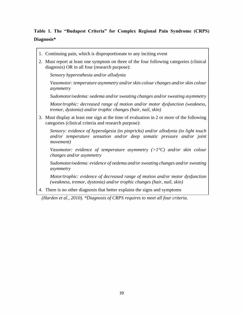
39
Table 1. The “Budapest Criteria” for Complex Regional Pain Syndrome (CRPS)
Diagnosis*
1. Continuing pain, which is disproportionate to any inciting event
2. Must report at least one symptom on three of the four following categories (clinical
diagnosis) OR in all four (research purpose):
Sensory hyperesthesia and/or allodynia
Vasomotor: temperature asymmetry and/or skin colour changes and/or skin colour
asymmetry
Sudomotor/oedema: oedema and/or sweating changes and/or sweating asymmetry
Motor/trophic: decreased range of motion and/or motor dysfunction (weakness,
tremor, dystonia) and/or trophic changes (hair, nail, skin)
3. Must display at least one sign at the time of evaluation in 2 or more of the following
categories (clinical criteria and research purpose):
Sensory: evidence of hyperalgesia (to pinpricks) and/or allodynia (to light touch
and/or temperature sensation and/or deep somatic pressure and/or joint
movement)
Vasomotor: evidence of temperature asymmetry (>1°C) and/or skin colour
changes and/or asymmetry
Sudomotor/oedema: evidence of oedema and/or sweating changes and/or sweating
asymmetry
Motor/trophic: evidence of decreased range of motion and/or motor dysfunction
(weakness, tremor, dystonia) and/or trophic changes (hair, nail, skin)
4. There is no other diagnosis that better explains the signs and symptoms
(Harden et al., 2010). *Diagnosis of CRPS requires to meet all four criteria.
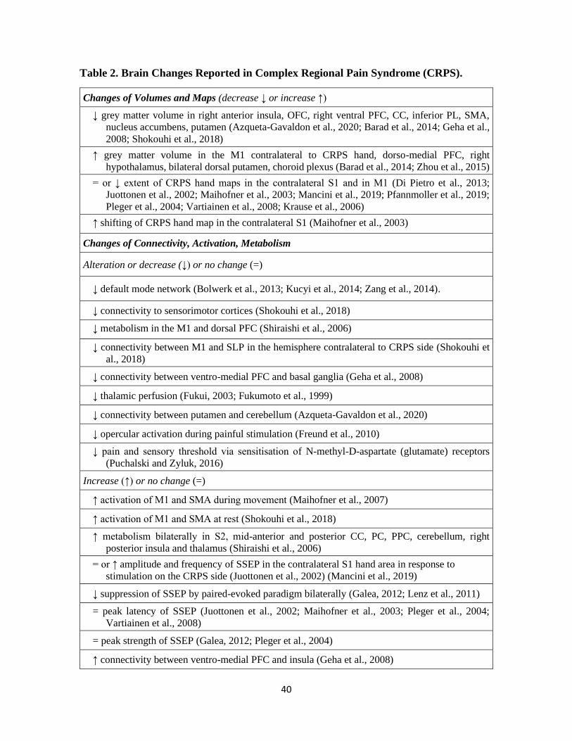
40
Table 2. Brain Changes Reported in Complex Regional Pain Syndrome (CRPS).
Changes of Volumes and Maps (decrease ↓ or increase ↑)
↓ grey matter volume in right anterior insula, OFC, right ventral PFC, CC, inferior PL, SMA,
nucleus accumbens, putamen (Azqueta-Gavaldon et al., 2020; Barad et al., 2014; Geha et al.,
2008; Shokouhi et al., 2018)
↑ grey matter volume in the M1 contralateral to CRPS hand, dorso-medial PFC, right
hypothalamus, bilateral dorsal putamen, choroid plexus (Barad et al., 2014; Zhou et al., 2015)
= or ↓ extent of CRPS hand maps in the contralateral S1 and in M1 (Di Pietro et al., 2013;
Juottonen et al., 2002; Maihofner et al., 2003; Mancini et al., 2019; Pfannmoller et al., 2019;
Pleger et al., 2004; Vartiainen et al., 2008; Krause et al., 2006)
↑ shifting of CRPS hand map in the contralateral S1 (Maihofner et al., 2003)
Changes of Connectivity, Activation, Metabolism
Alteration or decrease (↓) or no change (=)
↓ default mode network (Bolwerk et al., 2013; Kucyi et al., 2014; Zang et al., 2014).
↓ connectivity to sensorimotor cortices (Shokouhi et al., 2018)
↓ metabolism in the M1 and dorsal PFC (Shiraishi et al., 2006)
↓ connectivity between M1 and SLP in the hemisphere contralateral to CRPS side (Shokouhi et
al., 2018)
↓ connectivity between ventro-medial PFC and basal ganglia (Geha et al., 2008)
↓ thalamic perfusion (Fukui, 2003; Fukumoto et al., 1999)
↓ connectivity between putamen and cerebellum (Azqueta-Gavaldon et al., 2020)
↓ opercular activation during painful stimulation (Freund et al., 2010)
↓ pain and sensory threshold via sensitisation of N-methyl-D-aspartate (glutamate) receptors
(Puchalski and Zyluk, 2016)
Increase (↑) or no change (=)
↑ activation of M1 and SMA during movement (Maihofner et al., 2007)
↑ activation of M1 and SMA at rest (Shokouhi et al., 2018)
↑ metabolism bilaterally in S2, mid-anterior and posterior CC, PC, PPC, cerebellum, right
posterior insula and thalamus (Shiraishi et al., 2006)
= or ↑ amplitude and frequency of SSEP in the contralateral S1 hand area in response to
stimulation on the CRPS side (Juottonen et al., 2002) (Mancini et al., 2019)
↓ suppression of SSEP by paired-evoked paradigm bilaterally (Galea, 2012; Lenz et al., 2011)
= peak latency of SSEP (Juottonen et al., 2002; Maihofner et al., 2003; Pleger et al., 2004;
Vartiainen et al., 2008)
= peak strength of SSEP (Galea, 2012; Pleger et al., 2004)
↑ connectivity between ventro-medial PFC and insula (Geha et al., 2008)
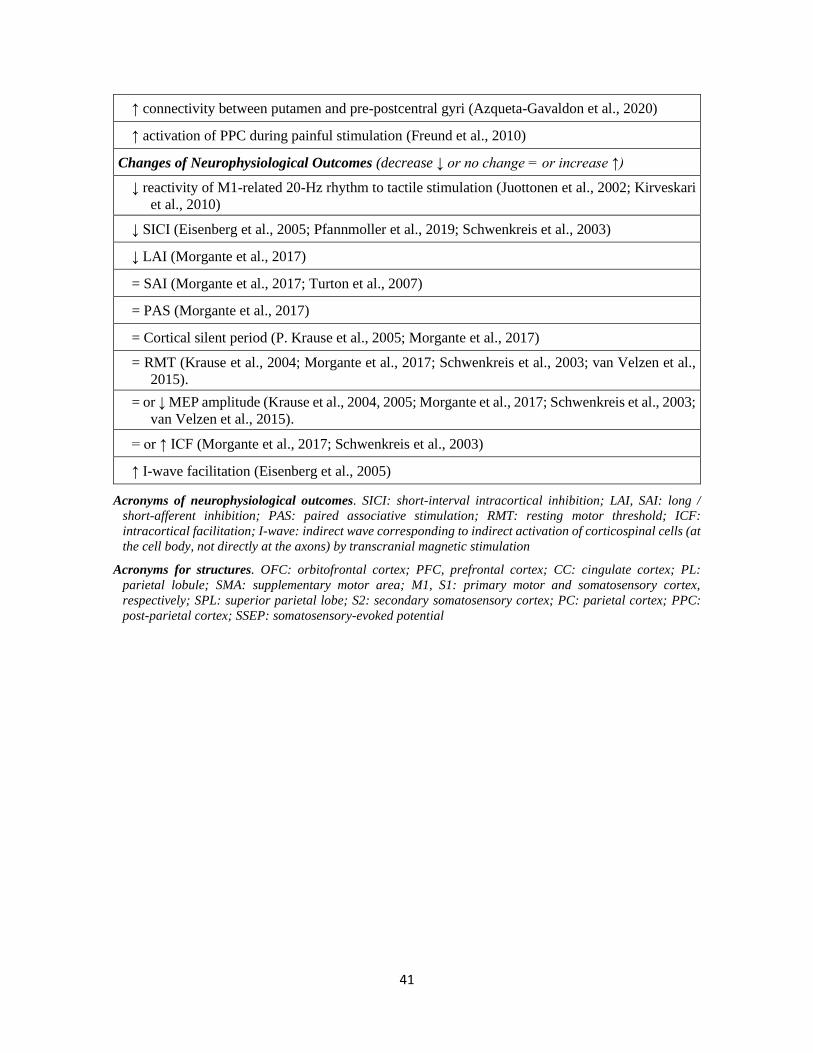
41
↑ connectivity between putamen and pre-postcentral gyri (Azqueta-Gavaldon et al., 2020)
↑ activation of PPC during painful stimulation (Freund et al., 2010)
Changes of Neurophysiological Outcomes (decrease ↓ or no change = or increase ↑)
↓ reactivity of M1-related 20-Hz rhythm to tactile stimulation (Juottonen et al., 2002; Kirveskari
et al., 2010)
↓ SICI (Eisenberg et al., 2005; Pfannmoller et al., 2019; Schwenkreis et al., 2003)
↓ LAI (Morgante et al., 2017)
= SAI (Morgante et al., 2017; Turton et al., 2007)
= PAS (Morgante et al., 2017)
= Cortical silent period (P. Krause et al., 2005; Morgante et al., 2017)
= RMT (Krause et al., 2004; Morgante et al., 2017; Schwenkreis et al., 2003; van Velzen et al.,
2015).
= or ↓ MEP amplitude (Krause et al., 2004, 2005; Morgante et al., 2017; Schwenkreis et al., 2003;
van Velzen et al., 2015).
= or ↑ ICF (Morgante et al., 2017; Schwenkreis et al., 2003)
↑ I-wave facilitation (Eisenberg et al., 2005)
Acronyms of neurophysiological outcomes. SICI: short-interval intracortical inhibition; LAI, SAI: long /
short-afferent inhibition; PAS: paired associative stimulation; RMT: resting motor threshold; ICF:
intracortical facilitation; I-wave: indirect wave corresponding to indirect activation of corticospinal cells (at
the cell body, not directly at the axons) by transcranial magnetic stimulation
Acronyms for structures. OFC: orbitofrontal cortex; PFC, prefrontal cortex; CC: cingulate cortex; PL:
parietal lobule; SMA: supplementary motor area; M1, S1: primary motor and somatosensory cortex,
respectively; SPL: superior parietal lobe; S2: secondary somatosensory cortex; PC: parietal cortex; PPC:
post-parietal cortex; SSEP: somatosensory-evoked potential
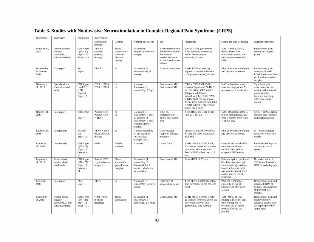
42
Table 3. Studies with Noninvasive Neurostimulation in Complex Regional Pain Syndrome (CRPS).
References Study type Population Intervention
Stimulation
protocol
Control Number of sessions Site Parameters Scales and time of testing Outcomes reported
Bilgili et al.,
2016
Double-blinded,
placebo-
controlled,
randomized trial
CRPS type
I (N = 30)
Exp = 15
Sham = 15
TENS +
standard
physical
therapy
Sham
stimulation +
standard
physical
therapy
15 sessions,
frequency/week not
reported
Active electrode on
the dorsal aspect of
the forearm,
passive electrode
on the dorsal aspect
of hand
100-Hz TENS (50–100 ms
pulse duration) at intensity
below the discomfort
threshold, 20 min
VAS, LANSS, DN-4,
ROM, edema size,
functional capacity with
hand dynamometer and
DHI;
Reduction of pain,
edema and fingers
ROM
Bodenheim
& Bennett,
1983
Case report SA
Exp = 1
TENS na 24 sessions (3
sessions/week, 8
weeks)
Acupuncture points 20-Hz TENS at intensity
adjusted to patient tolerance
(100-μs pulse width), 60 min
Clinical evaluation of pain
and physical outcomes
Reduction of pain,
recovery of ankle
ROM, increase of bone
stock and reversal of
atrophy
Gaertner et
al., 2018
Open-label and
nonrandomized
study
CRPS type
I and II (N
= 21)
Exp = 6
Exp = 15
iTBS + rTMS
iTBS + rTMS
na
na
1 session
5 sessions (1
session/day, 5 days)
Contralateral M1
Contralateral M1
iTBS at 70% RMT (5-Hz
bursts of 3 pulses at 50 Hz, 2
sec ON / 8 sec OFF, total =
600 pulses) followed
immediately by 10-Hz rTMS
at 80% RMT (10-sec trains,
30-sec inter-train interval; total
= 2000 pulses). Total = 2600
pulses per session
VAS, at baseline, then
after the single or the 5
sessions and 2 weeks after
Significant pain
reduction after one
session and one-week
posttreatment;
however, no group
differences were
present
Houde et al.,
2020
Case report CRPS type
I
Exp = 1
Anodal tDCS
Anodal tDCS
+ TENS
na
na
5 sessions (1
session/day, 5 days)
10 sessions (1
session/day, 5 days,
repeated after 6
months)
tDCS on
contralateral M1,
TENS over painful
area
2-mA tDCS and 3-Hz TENS
(400 μs), 25 min
VAS; at baseline, after 15
min of each intervantion,
after 6 months from tDCS
+ TENS only
tDCS + TENS slightly
reduced pain intensity
and unpleasantness
Kesler et al.,
1988
Cohort study RSD (N =
10)
Exp = 10
TENS + home-
based physical
therapy
na Various depending
on the patient (4
sessions/day,
multiple days)
Over vascular
supply of affected
extremity
Intensity adjusted to comfort,
60 min. No other information
provided
Clinical evaluation of pain
and physical outcomes
N=7 with complete
remission within two
months
Krause et
al., 2005
Cohort study CRPS type
I (N = 22)
Exp = 12
Control =
10
rPMS Healthy
subjects
1 session Over C7/C8 20-Hz rPMS at 120% RMT;
10 trains of 10 sec each, inter-
train interval not reported;
Total = 2000 pulses over ~10
min
Cortical and spinal MEP,
contra-and-ipsilateral
cortical silent period;
pre/post-rPMS testing
Less effective input to
the motor cortical
system
Lagueux et
al., 2018
Randomized
parallel single
blind study
CRPS type
I (N = 22)
Exp = 11
Control =
11
Anodal tDCS
+ graded motor
imagery
Sham
stimulation +
graded motor
imagery
14 sessions (1
session/day, 5
days/week for 2
weeks, 1 day/week
for 4 weeks)
Contralateral M1 2-mA tDCS of 20 min Pain perception, quality of
life, kinesiophobia, pain
catastrophizing, anxiety,
mood; at baseline, at 6
weeks of treatment and 1
month after th end of
treatment
No added value of
tDCS combined with
GMI for reducing pain
Leo at al.,
1983
Case report RSD
Exp = 1
TENS na 2 sessions (1
session/day, 22 days
apart)
Bilaterally at
acupuncture points
4-Hz TENS at intensity below
pain threhsold, 30 sec for each
point
Pain and right upper
extremity ROM; at
baseline and after each
session
Reduction of pain and
increased ROM at
painful, improvements
still present at 3
months
Picarelli et
al. 2010
Double-blind,
placebo-
controlled, 2-arm,
randomized trial
CRPS type
I (N = 23)
Exp = 12
Sham = 11
rTMS + best
medical
treatment
Sham
stimulation
10 sessions (1
session/day, 5
days/week, 2 weeks)
Contralateral M1 10-Hz rTMS at 100% RMT;
25 trains of 10 sec each, 60-sec
inter-train interval; total =
2500 pulses over ~29 min
VAS, MPQ, SF-36,
HDRS; at baseline, then
daily during the 10
sessions and 1 week/3
months after the last
session
Reduction of pain and
improvement of
affective aspects only
during the period of
stimulation
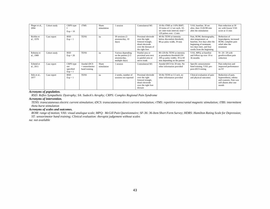
43
Pleger et al.,
2004
Cohort study CRPS type
I
Exp = 10
rTMS Sham
stimulation
1 session Contralateral M1 10-Hz rTMS at 110% RMT;
10 trains of 1.2 sec each, 10-
sec inter-train interval; total =
120 pulses over ~2 min
VAS; baseline, 30 sec
after, then 15/45/90 min
after the stimulation
Pain reduction at 30
sec with lowest VAS
score at 15 min
Richlin et
al., 1978
Case report RSD
Exp = 1
TENS na 30 sessions (3
sessions/day, 10
days)
Proximal electrode
over the right
femoral triangle,
distal electrode
over the dorsum of
the right foot
40-Hz TENS at intensity
below discomfort threshold,
80-μs pulse width, 30 min
Pain, ROM, thermography,
skin temperature; at
baseline, five days after the
beginning of treatment,
two days later, and four
weeks from the beginning
Reduction of
hyperalgesia, increased
ROM, complete pain
relief after the
treatment
Robaina et
al., 1989
Cohort study RSD
Exp = 26
TENS na Various depending
on the patient (2-5
sessions/day,
multiple days)
Painful area or
proximal area next
to painful area or
nerve trunk
80-120-Hz TENS at intensity
at parasthesia threshold (50-
200 μs pulse width), 30 to 60
min depending on the patient
VAS, MPQ; at baseline
and follow-up over 10 to
36 months
N= 20 / 29 with
good/excellent pain
reduction
Schmid et
al., 2011
Case report CRPS type
not
specified
Exp = 1
Anodal tDCS
+ sensorimotor
hand training
Sham
stimulation
1 session Contralateral M1 Anodal tDCS for 20 min. No
other information provided
Specific sensorymotor
hand training, VAS; pre-
post-tDCS testing
Pain reduction and
improved performance
on ST
Stilz et al.,
1977
Case report RSD
Exp = 1
TENS na 2 weeks, number of
sessions not reported
Proximal electrode
over the right
femoral triangle,
distal electrode
over the right foot
dorsum
50-Hz TENS at 3.5 mA, no
other information provided
Clinical evaluation of pain
and physical outcomes
Reduction of pain,
hyperesthesia, edema
and cyanosis. Pain was
still absent after one
month
Acronyms of population.
RSD: Reflex Sympathetic Dystrophy; SA: Sudeck's Atrophy; CRPS: Complex Regional Pain Syndrome
Acronyms of intervention.
TENS: transcutaneous electric current stimulation; tDCS: transcutaneous direct current stimulation; rTMS: repetitive transcranial magnetic stimulation; iTBS: intermittent
theta burst stimulation
Acronyms of scales and outcomes.
ROM: range of motion; VAS: visual analogue scale; MPQ: McGill Pain Questionnaire; SF-36: 36-Item Short Form Survey; HDRS: Hamilton Rating Scale for Depression;
ST: sensorimotor hand training; Clinical evaluation: therapist judgement without scales
na: not available
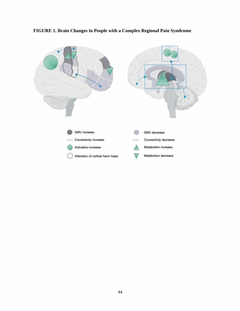
44
FIGURE 1. Brain Changes in People with a Complex Regional Pain Syndrome
45
SECTION 2 : HYPOTHÈSES ET OBJECTIFS DU PROJET DE MAÎTRISE
Rationnel du projet suivant les recommandations de la revue de littérature
Étant donné :
- que le SDRC induit des changements au niveau des cortex moteur primaire (M1) des deux
hémisphères pour le contrôle des membres atteint et non-atteint (Allen Demers et al., in
preparation);
- que cette réorganisation centrale pourrait expliquer le maintien dans le temps des
incapacités résiduelles liées au SDRC;
- que malgré les traitements conventionnels : 62% sont encore restreints dans la réalisation
de leurs activités de la vie quotidienne trois à neuf ans après le début du SDRC (Geertzen
et al., 1998), plus de la moitié des gens diagnostiqués avec un SDRC présente encore des
déficiences (comme une diminution de force ou de la raideur) un an après le début du
syndrome (Bean et al., 2014) et la majorité ne sont pas en mesure de retourner à leur
occupation, ont besoin d’adaptations sur leur lieu de travail ou sont déclarés officiellement
invalides (Borchers and Gershwin, 2014).
- qu’il devient impératif de développer des méthodes d’interventions moins coûteuses, moins
douloureuses, moins invasives et plus efficaces pour traiter les personnes atteintes de
SDRC résistant aux traitements conventionnels (Bruehl 2010);
- que les changements dans le fonctionnement des M1 rapportés dans la littérature ne sont
pas tous cohérents entre eux (Allen Demers et al., in preparation);
- qu’il est possible de tester le fonctionnement du M1 et de la voie corticospinale* (par
stimulations magnétiques transcrâniennes (TMS, de l’anglais « transcranial magnetic
stimulations », Beaulieu et al. 2017a; Beaulieu et al. 2017c; Malcolm et al. 2006);
- que les stimulations magnétiques répétées en périphérie (rPMS des muscles, de l’anglais
« repetitive peripheral magnetic stimulations ») génèrent un flux massif d’informations
sensorielles (non-douloureuses et cohérentes avec le mouvement produit) qui atteignent
*Le système corticospinal ou voie corticospinale inclut les neurones et
interneurones du cortex moteur, soit les cellules corticospinales ou pyramidales
qui envoient les commandes motrices aux motoneurones alpha de la moelle
épinière qui innervent les fibres musculaires.
46
le cerveau par les voies somatosensorielles via le thalamus (Beaulieu & Schneider 2015;
Beaulieu et al. 2017b);
- que ce flux d’informations somatosensorielles permettrait d’influencer la plasticité des
circuits sensorimoteurs du cortex fronto-pariétal (possiblement par des mécanismes de
potentialisation à long terme impliquant les récepteurs glutamatergiques N-methyl-D-
aspartate ou NMDA) et permettent, dans les cas de douleur chronique lombaire,
d’influencer les M1, dont leur excitabilité motrice corticale (Massé-Alarie et al. 2013);
- que, dans ces cas, les rPMS permettent de diminuer les douleurs vécues (Massé-Alarie et
al. 2013);
- que les rPMS n’ont pas encore été testées avec les gens souffrant de SDRC :
Il est raisonnable de tenter de :
- tester le fonctionnement des M1 (notamment l’excitabilité motrice corticale et
corticospinale) grâce aux TMS de M1 chez des adultes vivant avec un SDRC au membre
supérieur (étude 1)
- influencer le fonctionnement des M1 avec des rPMS au niveau des muscles de la zone
douloureuse* et par le fait même, de diminuer la douleur et améliorer la fonction manuelle
(étude 2).
Hypothèses de travail
Il sera possible de mesurer des changements de fonctionnement d’un ou des M1 chez les
participants adultes avec SDRC au membre supérieur par rapport à des participants sans
douleur (étude 1).
*Le fléchisseur superficiel des doigts (FDS, du latin « Flexor Digitorum
Superficialis ») a été choisi comme muscle cible au traitement puisque le SDRC est
plus prévalent au membre supérieur qu’inférieur (Bruehl, 2015), qu’on retrouve
fréquemment une diminution d’amplitude articulaire de flexion des doigts et du
poignet chez les patients atteints d’un SDRC au membre supérieur et que
l’amélioration de son contrôle permettrait par la même occasion l’amélioration de
plusieurs tâches fonctionnelles (notamment celles impliquant la fermeture de la main).
47
Une session de rPMS sur le muscle FDS du côté atteint permettra de diminuer la douleur et
sera accompagné d’une réduction des différences de fonctionnement du/des M1, soit des
mesures se rapprochant de celles d’un groupe d’adultes sans douleur (étude 2).
Objectifs principaux
À la suite du premier article de revue de littérature (en SDRC et neurostimulation) qui
compose l’introduction et soutient le rationnel de la maîtrise, ce mémoire présente deux
études expérimentales développées pour tester les hypothèses de travail et adresser les
objectifs suivants :
L’objectif principal de la première étude était de tester le fonctionnement des M1 chez un
groupe d’adultes vivant avec un SDRC au membre supérieur par rapport à celle d’un groupe
d’adultes sans douleur. L’objectif principal de la seconde étude était de tester, chez les
adultes avec SDRC, si une seule séance de rPMS permettait de diminuer la douleur et si cela
était en relation avec une normalisation des différences de fonctionnement du/des M1, en
comparaison avec le groupe de participants sans douleur.
Spécifiquement, chez une population adulte vivant avec un SDRC au membre supérieur, les
objectifs étaient :
Pour l’étude 1, par rapport à un groupe d’adultes sans douleur :
1) déterminer les différences de fonctionnement des zones motrices corticales du muscle
premier interosseux dorsal (FDI)* par TMS du M1 et enregistrement
*Le fonctionnement du M1 (en pré et post-rPMS du muscle FDS) a été testé par TMS de la
zone motrice corticale contrôlant le muscle FDI, puisque sa stimulation par TMS autant que
sa captation par EMG de surface est plus précise que celle du FDS (l’EMG de surface captent
les muscles voisins/superposés qui sont nombreux dans le cas du FDS), qu’il est grandement
étudié dans la littérature, parce que la mesure de la zone motrice du FDI est un bon
indicateur du fonctionnement de la zone motrice du FDS (bien que précises, les zones
motrices se chevauchent afin d’améliorer la précision des gestes moteurs complexes en
groupes de muscles synergiques) (Gagné & Schneider 2007, 2008; Melgari et al. 2008) et
parce que la zone douloureuse en SDRC est souvent grande et non limité à un seul muscle.
48
d’électromyographie (EMG) de surface des réponses du muscle FDI, en évaluant
notamment l’excitabilité motrice corticale et corticospinale via le seuil moteur de repos
(RMT, de l’anglais « resting motor threshold ») et d’autres variables décrites dans la
section 1.6 Mesures neurophysiologiques;
2) mesurer la fonction manuelle des deux mains via les mesures d’amplitude articulaire
active des doigts, de force musculaire de préhension, de proprioception des membres
supérieurs et de sensibilité au toucher léger;
3) quantifier, via des questionnaires, les symptômes vécus;
4) déterminer s’il existe des corrélations entre mesures de fonction manuelle ou symptômes
et mesures TMS de fonctionnement du M1.
Pour l’étude 2, entre les temps de mesure pré- et post-rPMS
5) mesurer l’effet immédiat (en aigu) d’une seule session de rPMS du muscle FDS du
membre supérieur atteint sur :
6.1) la douleur (variable principale) et ses interférences (autres symptômes vécus);
6.2) le fonctionnement (dont l’excitabilité avec la mesure du RMT) des zones motrices
corticales du FDI par TMS;
6.3) la fonction manuelle des deux mains via les mesures d’amplitude articulaire active
des doigts, de force musculaire de préhension et de sensibilité au toucher léger;
6) déterminer la présence de facteurs prédictifs du succès de l’intervention parmi les mesures
initiales, les caractéristiques descriptives ou les relations entre changements de variables,
afin d’identifier les meilleurs répondants et donc mieux cibler la population qui
bénéficierait du traitement expérimental rPMS.
Approches méthodologiques
Tester les hypothèses de travail et atteindre les objectifs de la maîtrise a demandé le
recrutement d’adultes avec SDRC. La clientèle en phase chronique (soit ayant atteint un
plateau clinique avec les traitements conventionnels) a été choisie pour tester l’effet net des
rPMS et non pas celui des traitements conventionnels. Il a été choisi de recruter des
participants avec un diagnostic initial de SDRC type I (sans lésion nerveuse) puisque pour
envoyer les informations somatosensorielles au cerveau et permettre la plasticité cérébrale
49
recherchée, les rPMS doivent stimuler un muscle qui a un réseau nerveux intact. Étant en
phase chronique, les participants ayant reçu initialement un diagnostic de SDRC type I (parce
qu’ils répondaient alors à tous les critères diagnostiques) correspondaient davantage à des
type NOS (qui n’est pas mieux expliqué par tout autre condition, de l’anglais « not otherwise
specified ») lors de leur participation à l’étude, c’est-à-dire qu’ils ne répondaient plus à
l’ensemble des critères diagnostiques puisque certains symptômes étaient alors résolus
(l’œdème et les changements sudomoteurs étaient notamment réglés). Les participants du
groupe Sans-douleur ont été recrutés en les appariant, autant que possible, au sexe et à l’âge
du groupe SDRC pour permettre la comparaison des variables mesurées.
Ces expérimentations ont permis la rédaction de deux articles. Le premier article porte sur
les différences de fonctionnement des M1 (débalancement interhémisphérique d’excitabilité)
et de fonction manuelle entre un groupe d’adultes vivant avec un SDRC et un groupe Sans-
douleur (chapitre 2). Le second article traite des effets de l’ajout des rPMS chez la clientèle
avec SDRC sur le fonctionnement du M1 (plasticité induite), la fonction manuelle et les
symptômes (chapitre 3).
La méthodologie qui a permis de tester les participants et de rédiger les deux articles est
exposée dans le chapitre suivant (chapitre 1). Chaque personne a participé à une seule session
en laboratoire et les données ont permis les deux études : le premier article porte sur les
mesures avant traitement (comparaison des participants avec SDRC avec les personnes sans
douleur) et le deuxième article porte sur la comparaison pré/post-traitement expérimental
chez les participants avec SDRC, soit la réduction des écarts (entre les mesures des
participants avec SDRC et les valeurs normatives des participants sans douleur) en post-
rPMS de ceux mesurés en pré-rPMS.
50
CHAPITRE 1 : MÉTHODOLOGIE
1.1 CARACTÉRISTIQUES DES PARTICIPANTS
Groupe avec SDRC
Huit adultes souffrant de SDRC type I au membre supérieur ayant atteint un plateau clinique
(aucun changement de leur condition minimalement trois mois précédant la participation à
l’étude et ayant terminé leur suivi en traitement conventionnel) ont été recrutés par liste de
diffusion ou directement au Centre d’expertise en gestion de la douleur chronique du CHU
de Québec – Université Laval (pavillon CHUL). Le groupe SDRC (six femmes et deux
hommes) était âgé en moyenne de 52 ans (étendu : 35 à 65 ans). Deux des participants avait
le membre supérieur non-dominant atteint (les autres ayant le membre supérieur dominant
atteint). Un seul des participants était gaucher, les 7 autres droitiers. Le temps depuis
l’apparition du SDRC (7 à 168 mois, moyenne de 55.9 mois), l’intensité de leur douleur (38
± 20mm sur l’échelle visuelle analogue, EVA) et l’intensité du SDRC (2 à 12 au
questionnaire « CRPS Severity Score », Annexe 1) variaient d’un participant à l’autre en
début d’étude. L’ensemble des caractéristiques générales des participants sont présentées
dans le Tableau 1.
Groupe Sans-douleur
Huit adultes sans douleur (appariés pour l’âge) ont été recrutés à des fin de comparaison des
mesures cliniques et neurophysiologiques. Le groupe Sans-douleur (quatre femmes et quatre
hommes) était âgé en moyenne de 50,8 ans (étendu : 40 à 61 ans). Un seul des participants
était gaucher, les 7 autres droitiers.
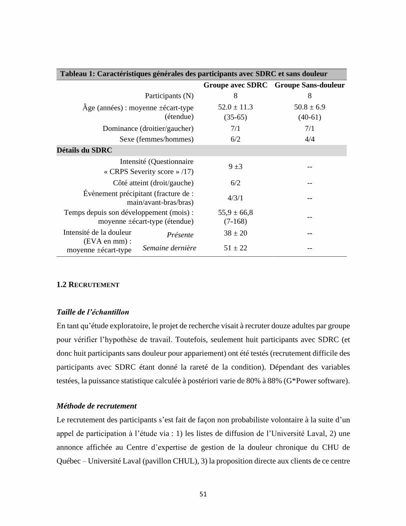
51
1.2 RECRUTEMENT
Taille de l’échantillon
En tant qu’étude exploratoire, le projet de recherche visait à recruter douze adultes par groupe
pour vérifier l’hypothèse de travail. Toutefois, seulement huit participants avec SDRC (et
donc huit participants sans douleur pour appariement) ont été testés (recrutement difficile des
participants avec SDRC étant donné la rareté de la condition). Dépendant des variables
testées, la puissance statistique calculée à postériori varie de 80% à 88% (G*Power software).
Méthode de recrutement
Le recrutement des participants s’est fait de façon non probabiliste volontaire à la suite d’un
appel de participation à l’étude via : 1) les listes de diffusion de l’Université Laval, 2) une
annonce affichée au Centre d’expertise de gestion de la douleur chronique du CHU de
Québec – Université Laval (pavillon CHUL), 3) la proposition directe aux clients de ce centre
Tableau 1: Caractéristiques générales des participants avec SDRC et sans douleur
Groupe avec SDRC Groupe Sans-douleur
Participants (N) 8 8
Âge (années) : moyenne ±écart-type
(étendue)
52.0 ± 11.3
(35-65)
50.8 ± 6.9
(40-61)
Dominance (droitier/gaucher) 7/1 7/1
Sexe (femmes/hommes) 6/2 4/4
Détails du SDRC
Intensité (Questionnaire
« CRPS Severity score » /17) 9 ±3 --
Côté atteint (droit/gauche) 6/2 --
Évènement précipitant (fracture de :
main/avant-bras/bras) 4/3/1 --
Temps depuis son développement (mois) :
moyenne ±écart-type (étendue)
55,9 ± 66,8
(7-168) --
Intensité de la douleur
(EVA en mm) :
moyenne ±écart-type
Présente 38 ± 20 --
Semaine dernière 51 ± 22 --
52
par le médecin traitant. Les personnes intéressées par le projet contactaient l’étudiante-
physiothérapeute en charge qui (après leur avoir résumé le projet de recherche), validait avec
eux les critères de participation (Tableau 2), puis leur envoyait le formulaire d’information
et de consentement (FIFC). Les participants devaient signer et faire signer l’annexe du FIFC
par leur médecin (pour valider l’éligibilité), puis une rencontre au laboratoire était planifiée.
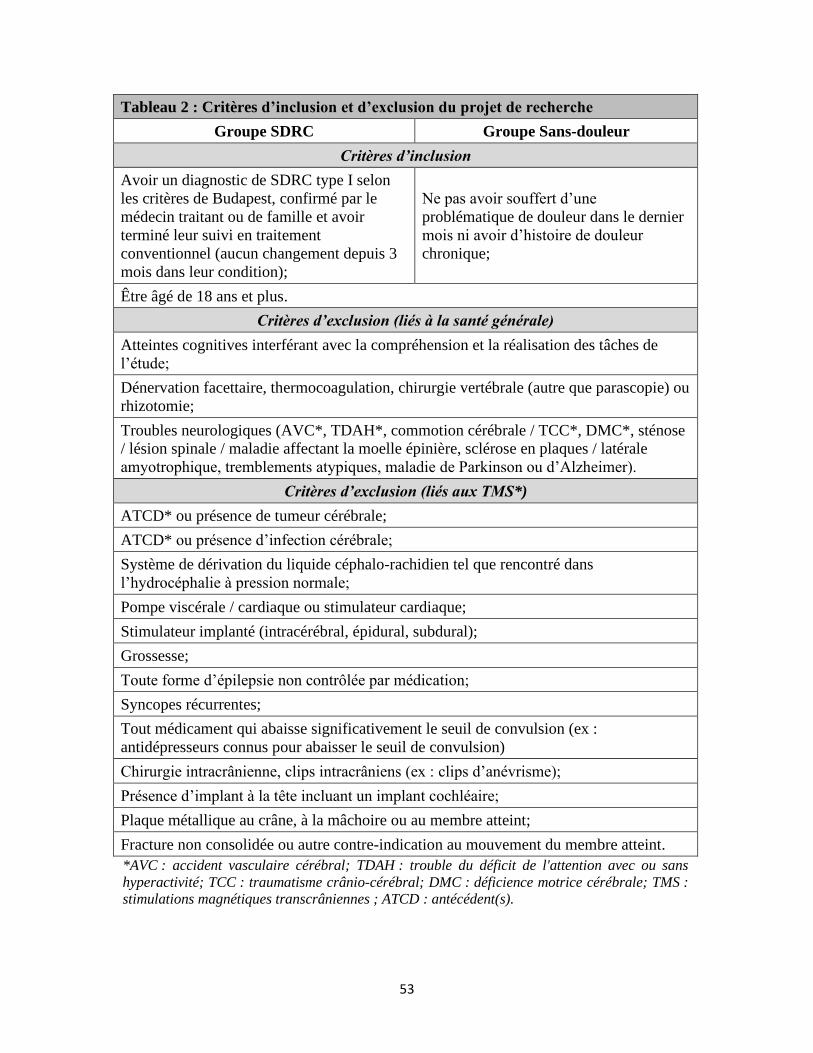
53
Tableau 2 : Critères d’inclusion et d’exclusion du projet de recherche
Groupe SDRC Groupe Sans-douleur
Critères d’inclusion
Avoir un diagnostic de SDRC type I selon
les critères de Budapest, confirmé par le
médecin traitant ou de famille et avoir
terminé leur suivi en traitement
conventionnel (aucun changement depuis 3
mois dans leur condition);
Ne pas avoir souffert d’une
problématique de douleur dans le dernier
mois ni avoir d’histoire de douleur
chronique;
Être âgé de 18 ans et plus.
Critères d’exclusion (liés à la santé générale)
Atteintes cognitives interférant avec la compréhension et la réalisation des tâches de
l’étude;
Dénervation facettaire, thermocoagulation, chirurgie vertébrale (autre que parascopie) ou
rhizotomie;
Troubles neurologiques (AVC*, TDAH*, commotion cérébrale / TCC*, DMC*, sténose
/ lésion spinale / maladie affectant la moelle épinière, sclérose en plaques / latérale
amyotrophique, tremblements atypiques, maladie de Parkinson ou d’Alzheimer).
Critères d’exclusion (liés aux TMS*)
ATCD* ou présence de tumeur cérébrale;
ATCD* ou présence d’infection cérébrale;
Système de dérivation du liquide céphalo-rachidien tel que rencontré dans
l’hydrocéphalie à pression normale;
Pompe viscérale / cardiaque ou stimulateur cardiaque;
Stimulateur implanté (intracérébral, épidural, subdural);
Grossesse;
Toute forme d’épilepsie non contrôlée par médication;
Syncopes récurrentes;
Tout médicament qui abaisse significativement le seuil de convulsion (ex :
antidépresseurs connus pour abaisser le seuil de convulsion)
Chirurgie intracrânienne, clips intracrâniens (ex : clips d’anévrisme);
Présence d’implant à la tête incluant un implant cochléaire;
Plaque métallique au crâne, à la mâchoire ou au membre atteint;
Fracture non consolidée ou autre contre-indication au mouvement du membre atteint.
*AVC : accident vasculaire cérébral; TDAH : trouble du déficit de l'attention avec ou sans
hyperactivité; TCC : traumatisme crânio-cérébral; DMC : déficience motrice cérébrale; TMS :
stimulations magnétiques transcrâniennes ; ATCD : antécédent(s).
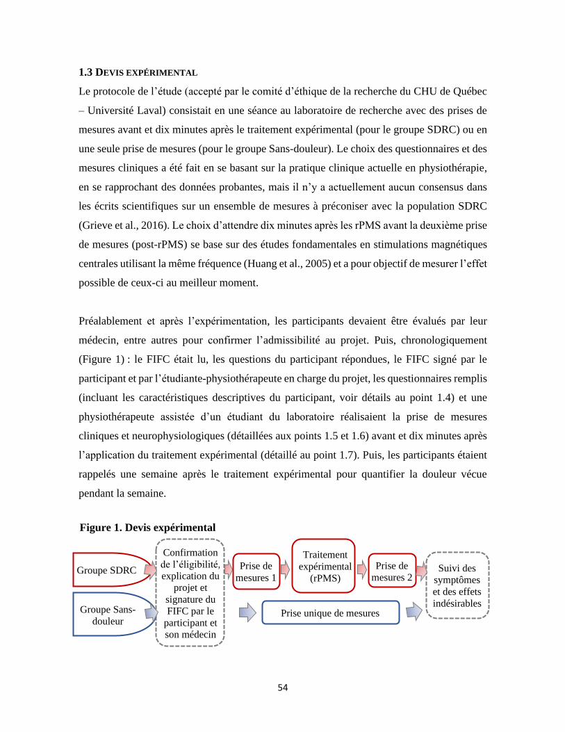
54
1.3 DEVIS EXPÉRIMENTAL
Le protocole de l’étude (accepté par le comité d’éthique de la recherche du CHU de Québec
– Université Laval) consistait en une séance au laboratoire de recherche avec des prises de
mesures avant et dix minutes après le traitement expérimental (pour le groupe SDRC) ou en
une seule prise de mesures (pour le groupe Sans-douleur). Le choix des questionnaires et des
mesures cliniques a été fait en se basant sur la pratique clinique actuelle en physiothérapie,
en se rapprochant des données probantes, mais il n’y a actuellement aucun consensus dans
les écrits scientifiques sur un ensemble de mesures à préconiser avec la population SDRC
(Grieve et al., 2016). Le choix d’attendre dix minutes après les rPMS avant la deuxième prise
de mesures (post-rPMS) se base sur des études fondamentales en stimulations magnétiques
centrales utilisant la même fréquence (Huang et al., 2005) et a pour objectif de mesurer l’effet
possible de ceux-ci au meilleur moment.
Préalablement et après l’expérimentation, les participants devaient être évalués par leur
médecin, entre autres pour confirmer l’admissibilité au projet. Puis, chronologiquement
(Figure 1) : le FIFC était lu, les questions du participant répondues, le FIFC signé par le
participant et par l’étudiante-physiothérapeute en charge du projet, les questionnaires remplis
(incluant les caractéristiques descriptives du participant, voir détails au point 1.4) et une
physiothérapeute assistée d’un étudiant du laboratoire réalisaient la prise de mesures
cliniques et neurophysiologiques (détaillées aux points 1.5 et 1.6) avant et dix minutes après
l’application du traitement expérimental (détaillé au point 1.7). Puis, les participants étaient
rappelés une semaine après le traitement expérimental pour quantifier la douleur vécue
pendant la semaine.
Confirmation
de l’éligibilité,
explication du
projet et
signature du
FIFC par le
participant et
son médecin
Suivi des
symptômes
et des effets
indésirables
Prise de
mesures 1
Prise de
mesures 2
Prise unique de mesures
Traitement
expérimental
(rPMS)
Groupe Sans-
douleur
Groupe SDRC
Figure 1. Devis expérimental
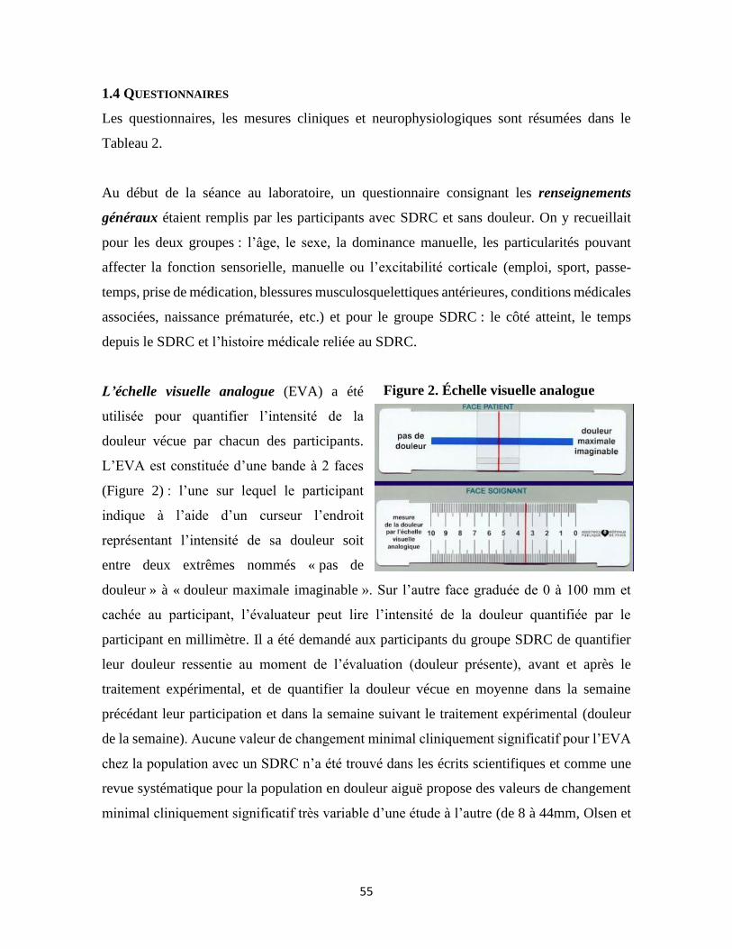
55
1.4 QUESTIONNAIRES
Les questionnaires, les mesures cliniques et neurophysiologiques sont résumées dans le
Tableau 2.
Au début de la séance au laboratoire, un questionnaire consignant les renseignements
généraux étaient remplis par les participants avec SDRC et sans douleur. On y recueillait
pour les deux groupes : l’âge, le sexe, la dominance manuelle, les particularités pouvant
affecter la fonction sensorielle, manuelle ou l’excitabilité corticale (emploi, sport, passe-
temps, prise de médication, blessures musculosquelettiques antérieures, conditions médicales
associées, naissance prématurée, etc.) et pour le groupe SDRC : le côté atteint, le temps
depuis le SDRC et l’histoire médicale reliée au SDRC.
L’échelle visuelle analogue (EVA) a été
utilisée pour quantifier l’intensité de la
douleur vécue par chacun des participants.
L’EVA est constituée d’une bande à 2 faces
(Figure 2) : l’une sur lequel le participant
indique à l’aide d’un curseur l’endroit
représentant l’intensité de sa douleur soit
entre deux extrêmes nommés « pas de
douleur » à « douleur maximale imaginable ». Sur l’autre face graduée de 0 à 100 mm et
cachée au participant, l’évaluateur peut lire l’intensité de la douleur quantifiée par le
participant en millimètre. Il a été demandé aux participants du groupe SDRC de quantifier
leur douleur ressentie au moment de l’évaluation (douleur présente), avant et après le
traitement expérimental, et de quantifier la douleur vécue en moyenne dans la semaine
précédant leur participation et dans la semaine suivant le traitement expérimental (douleur
de la semaine). Aucune valeur de changement minimal cliniquement significatif pour l’EVA
chez la population avec un SDRC n’a été trouvé dans les écrits scientifiques et comme une
revue systématique pour la population en douleur aiguë propose des valeurs de changement
minimal cliniquement significatif très variable d’une étude à l’autre (de 8 à 44mm, Olsen et
Figure 2. Échelle visuelle analogue
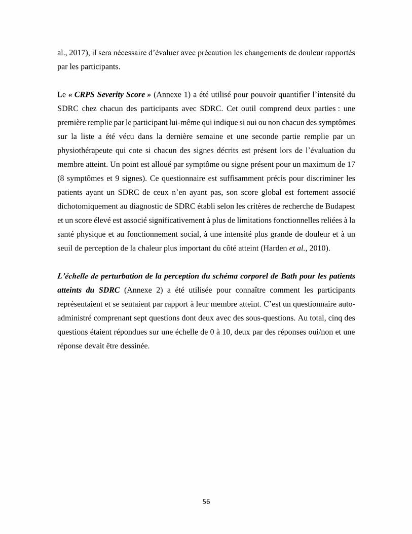
56
al., 2017), il sera nécessaire d’évaluer avec précaution les changements de douleur rapportés
par les participants.
Le « CRPS Severity Score » (Annexe 1) a été utilisé pour pouvoir quantifier l’intensité du
SDRC chez chacun des participants avec SDRC. Cet outil comprend deux parties : une
première remplie par le participant lui-même qui indique si oui ou non chacun des symptômes
sur la liste a été vécu dans la dernière semaine et une seconde partie remplie par un
physiothérapeute qui cote si chacun des signes décrits est présent lors de l’évaluation du
membre atteint. Un point est alloué par symptôme ou signe présent pour un maximum de 17
(8 symptômes et 9 signes). Ce questionnaire est suffisamment précis pour discriminer les
patients ayant un SDRC de ceux n’en ayant pas, son score global est fortement associé
dichotomiquement au diagnostic de SDRC établi selon les critères de recherche de Budapest
et un score élevé est associé significativement à plus de limitations fonctionnelles reliées à la
santé physique et au fonctionnement social, à une intensité plus grande de douleur et à un
seuil de perception de la chaleur plus important du côté atteint (Harden et al., 2010).
L’échelle de perturbation de la perception du schéma corporel de Bath pour les patients
atteints du SDRC (Annexe 2) a été utilisée pour connaître comment les participants
représentaient et se sentaient par rapport à leur membre atteint. C’est un questionnaire auto-
administré comprenant sept questions dont deux avec des sous-questions. Au total, cinq des
questions étaient répondues sur une échelle de 0 à 10, deux par des réponses oui/non et une
réponse devait être dessinée.
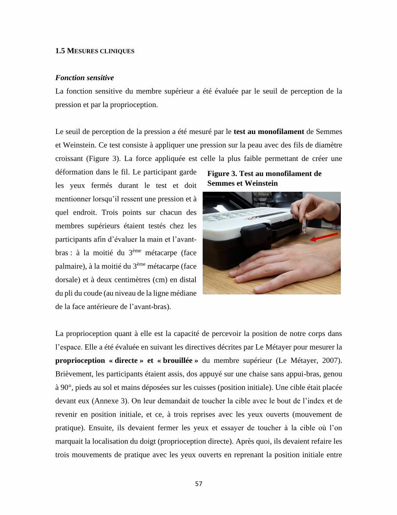
57
1.5 MESURES CLINIQUES
Fonction sensitive
La fonction sensitive du membre supérieur a été évaluée par le seuil de perception de la
pression et par la proprioception.
Le seuil de perception de la pression a été mesuré par le test au monofilament de Semmes
et Weinstein. Ce test consiste à appliquer une pression sur la peau avec des fils de diamètre
croissant (Figure 3). La force appliquée est celle la plus faible permettant de créer une
déformation dans le fil. Le participant garde
les yeux fermés durant le test et doit
mentionner lorsqu’il ressent une pression et à
quel endroit. Trois points sur chacun des
membres supérieurs étaient testés chez les
participants afin d’évaluer la main et l’avant-
bras : à la moitié du 3ème métacarpe (face
palmaire), à la moitié du 3ème métacarpe (face
dorsale) et à deux centimètres (cm) en distal
du pli du coude (au niveau de la ligne médiane
de la face antérieure de l’avant-bras).
La proprioception quant à elle est la capacité de percevoir la position de notre corps dans
l’espace. Elle a été évaluée en suivant les directives décrites par Le Métayer pour mesurer la
proprioception « directe » et « brouillée » du membre supérieur (Le Métayer, 2007).
Brièvement, les participants étaient assis, dos appuyé sur une chaise sans appui-bras, genou
à 90°, pieds au sol et mains déposées sur les cuisses (position initiale). Une cible était placée
devant eux (Annexe 3). On leur demandait de toucher la cible avec le bout de l’index et de
revenir en position initiale, et ce, à trois reprises avec les yeux ouverts (mouvement de
pratique). Ensuite, ils devaient fermer les yeux et essayer de toucher à la cible où l’on
marquait la localisation du doigt (proprioception directe). Après quoi, ils devaient refaire les
trois mouvements de pratique avec les yeux ouverts en reprenant la position initiale entre
Figure 3. Test au monofilament de
Semmes et Weinstein
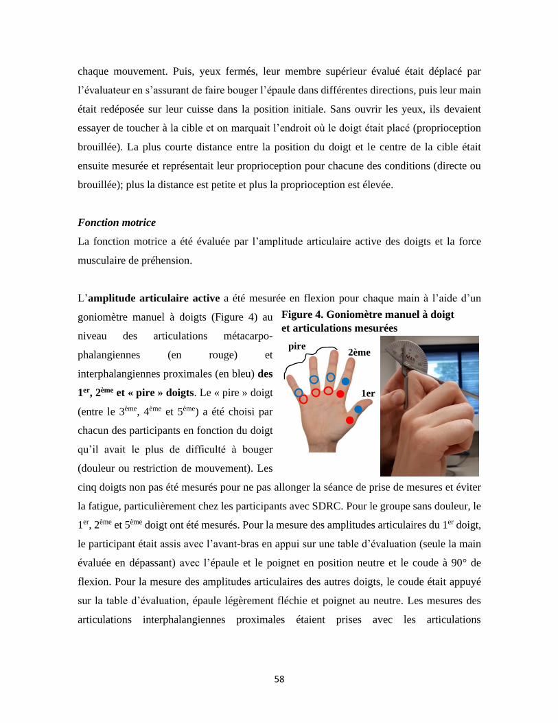
58
chaque mouvement. Puis, yeux fermés, leur membre supérieur évalué était déplacé par
l’évaluateur en s’assurant de faire bouger l’épaule dans différentes directions, puis leur main
était redéposée sur leur cuisse dans la position initiale. Sans ouvrir les yeux, ils devaient
essayer de toucher à la cible et on marquait l’endroit où le doigt était placé (proprioception
brouillée). La plus courte distance entre la position du doigt et le centre de la cible était
ensuite mesurée et représentait leur proprioception pour chacune des conditions (directe ou
brouillée); plus la distance est petite et plus la proprioception est élevée.
Fonction motrice
La fonction motrice a été évaluée par l’amplitude articulaire active des doigts et la force
musculaire de préhension.
L’amplitude articulaire active a été mesurée en flexion pour chaque main à l’aide d’un
goniomètre manuel à doigts (Figure 4) au
niveau des articulations métacarpo-
phalangiennes (en rouge) et
interphalangiennes proximales (en bleu) des
1er, 2ème et « pire » doigts. Le « pire » doigt
(entre le 3ème, 4ème et 5ème) a été choisi par
chacun des participants en fonction du doigt
qu’il avait le plus de difficulté à bouger
(douleur ou restriction de mouvement). Les
cinq doigts non pas été mesurés pour ne pas allonger la séance de prise de mesures et éviter
la fatigue, particulièrement chez les participants avec SDRC. Pour le groupe sans douleur, le
1er, 2ème et 5ème doigt ont été mesurés. Pour la mesure des amplitudes articulaires du 1er doigt,
le participant était assis avec l’avant-bras en appui sur une table d’évaluation (seule la main
évaluée en dépassant) avec l’épaule et le poignet en position neutre et le coude à 90° de
flexion. Pour la mesure des amplitudes articulaires des autres doigts, le coude était appuyé
sur la table d’évaluation, épaule légèrement fléchie et poignet au neutre. Les mesures des
articulations interphalangiennes proximales étaient prises avec les articulations
Figure 4. Goniomètre manuel à doigt
et articulations mesurées
pire
1er
2ème
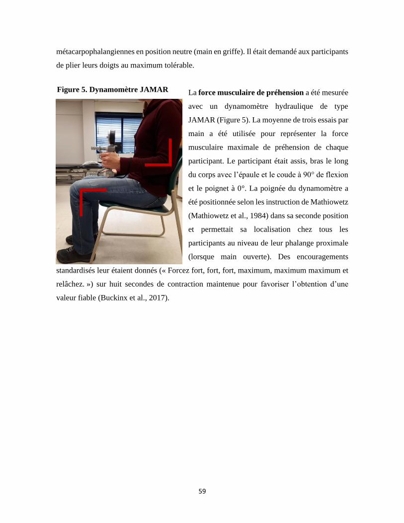
59
métacarpophalangiennes en position neutre (main en griffe). Il était demandé aux participants
de plier leurs doigts au maximum tolérable.
La force musculaire de préhension a été mesurée
avec un dynamomètre hydraulique de type
JAMAR (Figure 5). La moyenne de trois essais par
main a été utilisée pour représenter la force
musculaire maximale de préhension de chaque
participant. Le participant était assis, bras le long
du corps avec l’épaule et le coude à 90° de flexion
et le poignet à 0°. La poignée du dynamomètre a
été positionnée selon les instruction de Mathiowetz
(Mathiowetz et al., 1984) dans sa seconde position
et permettait sa localisation chez tous les
participants au niveau de leur phalange proximale
(lorsque main ouverte). Des encouragements
standardisés leur étaient donnés (« Forcez fort, fort, fort, maximum, maximum maximum et
relâchez. ») sur huit secondes de contraction maintenue pour favoriser l’obtention d’une
valeur fiable (Buckinx et al., 2017).
Figure 5. Dynamomètre JAMAR
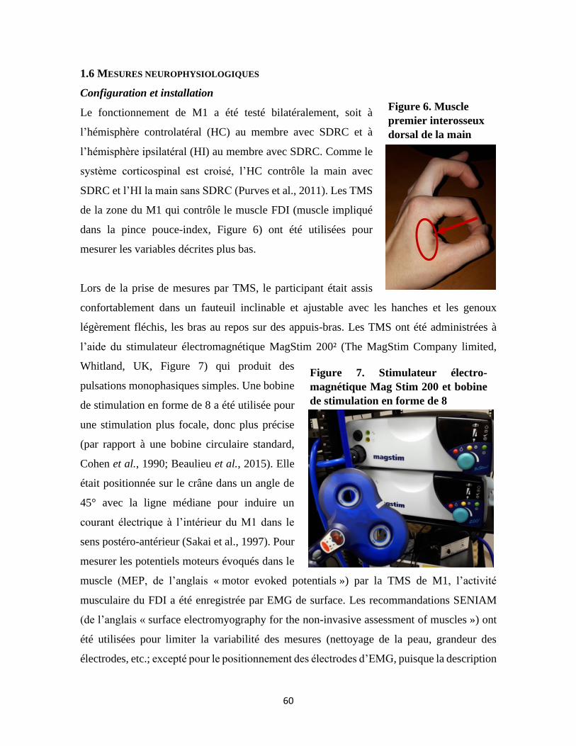
60
1.6 MESURES NEUROPHYSIOLOGIQUES
Configuration et installation
Le fonctionnement de M1 a été testé bilatéralement, soit à
l’hémisphère controlatéral (HC) au membre avec SDRC et à
l’hémisphère ipsilatéral (HI) au membre avec SDRC. Comme le
système corticospinal est croisé, l’HC contrôle la main avec
SDRC et l’HI la main sans SDRC (Purves et al., 2011). Les TMS
de la zone du M1 qui contrôle le muscle FDI (muscle impliqué
dans la pince pouce-index, Figure 6) ont été utilisées pour
mesurer les variables décrites plus bas.
Lors de la prise de mesures par TMS, le participant était assis
confortablement dans un fauteuil inclinable et ajustable avec les hanches et les genoux
légèrement fléchis, les bras au repos sur des appuis-bras. Les TMS ont été administrées à
l’aide du stimulateur électromagnétique MagStim 200² (The MagStim Company limited,
Whitland, UK, Figure 7) qui produit des
pulsations monophasiques simples. Une bobine
de stimulation en forme de 8 a été utilisée pour
une stimulation plus focale, donc plus précise
(par rapport à une bobine circulaire standard,
Cohen et al., 1990; Beaulieu et al., 2015). Elle
était positionnée sur le crâne dans un angle de
45° avec la ligne médiane pour induire un
courant électrique à l’intérieur du M1 dans le
sens postéro-antérieur (Sakai et al., 1997). Pour
mesurer les potentiels moteurs évoqués dans le
muscle (MEP, de l’anglais « motor evoked potentials ») par la TMS de M1, l’activité
musculaire du FDI a été enregistrée par EMG de surface. Les recommandations SENIAM
(de l’anglais « surface electromyography for the non-invasive assessment of muscles ») ont
été utilisées pour limiter la variabilité des mesures (nettoyage de la peau, grandeur des
électrodes, etc.; excepté pour le positionnement des électrodes d’EMG, puisque la description
Figure 7. Stimulateur électro-
magnétique Mag Stim 200 et bobine
de stimulation en forme de 8
Figure 6. Muscle
premier interosseux
dorsal de la main
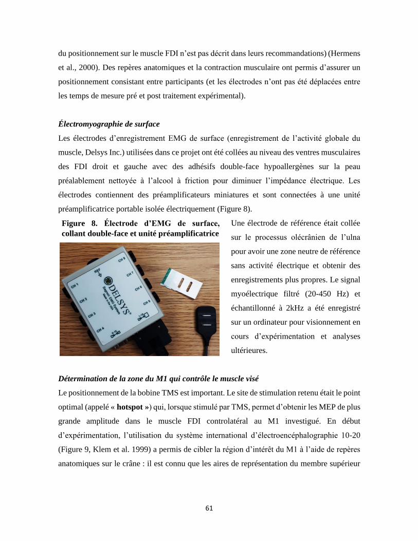
61
du positionnement sur le muscle FDI n’est pas décrit dans leurs recommandations) (Hermens
et al., 2000). Des repères anatomiques et la contraction musculaire ont permis d’assurer un
positionnement consistant entre participants (et les électrodes n’ont pas été déplacées entre
les temps de mesure pré et post traitement expérimental).
Électromyographie de surface
Les électrodes d’enregistrement EMG de surface (enregistrement de l’activité globale du
muscle, Delsys Inc.) utilisées dans ce projet ont été collées au niveau des ventres musculaires
des FDI droit et gauche avec des adhésifs double-face hypoallergènes sur la peau
préalablement nettoyée à l’alcool à friction pour diminuer l’impédance électrique. Les
électrodes contiennent des préamplificateurs miniatures et sont connectées à une unité
préamplificatrice portable isolée électriquement (Figure 8).
Une électrode de référence était collée
sur le processus olécrânien de l’ulna
pour avoir une zone neutre de référence
sans activité électrique et obtenir des
enregistrements plus propres. Le signal
myoélectrique filtré (20-450 Hz) et
échantillonné à 2kHz a été enregistré
sur un ordinateur pour visionnement en
cours d’expérimentation et analyses
ultérieures.
Détermination de la zone du M1 qui contrôle le muscle visé
Le positionnement de la bobine TMS est important. Le site de stimulation retenu était le point
optimal (appelé « hotspot ») qui, lorsque stimulé par TMS, permet d’obtenir les MEP de plus
grande amplitude dans le muscle FDI controlatéral au M1 investigué. En début
d’expérimentation, l’utilisation du système international d’électroencéphalographie 10-20
(Figure 9, Klem et al. 1999) a permis de cibler la région d’intérêt du M1 à l’aide de repères
anatomiques sur le crâne : il est connu que les aires de représentation du membre supérieur
Figure 8. Électrode d’EMG de surface,
collant double-face et unité préamplificatrice
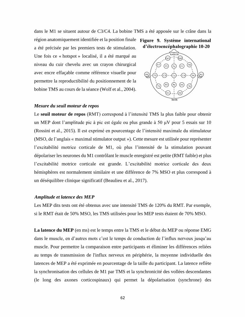
62
dans le M1 se situent autour de C3/C4. La bobine TMS a été apposée sur le crâne dans la
région anatomiquement identifiée et la position finale
a été précisée par les premiers tests de stimulation.
Une fois ce « hotspot » localisé, il a été marqué au
niveau du cuir chevelu avec un crayon chirurgical
avec encre effaçable comme référence visuelle pour
permettre la reproductibilité du positionnement de la
bobine TMS au cours de la séance (Wolf et al., 2004).
Mesure du seuil moteur de repos
Le seuil moteur de repos (RMT) correspond à l’intensité TMS la plus faible pour obtenir
un MEP dont l’amplitude pic à pic est égale ou plus grande à 50 μV pour 5 essais sur 10
(Rossini et al., 2015). Il est exprimé en pourcentage de l’intensité maximale du stimulateur
(MSO, de l’anglais « maximal stimulator output »). Cette mesure est utilisée pour représenter
l’excitabilité motrice corticale de M1, où plus l’intensité de la stimulation pouvant
dépolariser les neurones du M1 contrôlant le muscle enregistré est petite (RMT faible) et plus
l’excitabilité motrice corticale est grande. L’excitabilité motrice corticale des deux
hémisphères est normalement similaire et une différence de 7% MSO et plus correspond à
un déséquilibre clinique significatif (Beaulieu et al., 2017).
Amplitude et latence des MEP
Les MEP dits tests ont été obtenus avec une intensité TMS de 120% du RMT. Par exemple,
si le RMT était de 50% MSO, les TMS utilisées pour les MEP tests étaient de 70% MSO.
La latence du MEP (en ms) est le temps entre la TMS et le début du MEP ou réponse EMG
dans le muscle, en d’autres mots c’est le temps de conduction de l’influx nerveux jusqu’au
muscle. Pour permettre la comparaison entre participants et éliminer les différences reliées
au temps de transmission de l'influx nerveux en périphérie, la moyenne individuelle des
latences de MEP a été exprimée en pourcentage de la taille du participant. La latence reflète
la synchronisation des cellules de M1 par TMS et la synchronicité des vollées descendantes
(le long des axones corticospinaux) qui permet la dépolarisation (synchrone) des
Figure 9. Système international
d’électroencéphalographie 10-20
63
motoneurones alpha de la moelle épinière (Kobayashi & Pascual-Leone, 2003; Groppa et al.,
2012).
L’amplitude du MEP (mV) est l’amplitude de la réponse biphasique mesurée pic à pic. Elle
reflète le volume des cellules de M1 activé par la TMS (c’est-à-dire les neurones
corticospinaux et les interneurones associés), mais également l’excitabilité des motoneurones
et la synchronicité des vollées descendantes pour une réponse synchrone des motoneurones
et des fibres musculaires innervées (Kobayashi & Pascual-Leone, 2003; Rossini et al., 2015).
Il est à noter que le RMT ainsi que la latence des MEP sont les mesures TMS avec les
meilleures propriétés métrologiques chez des participants en santé ou avec lésion du système
nerveux central (Beaulieu et al., 2017a.c; Malcolm et al., 2006). On peut donc dire avec
assurance qu’un changement (significatif et supérieur à l’erreur de mesure) dans les valeurs
de ces mesures, reflèterait un réel changement d’excitabilité du M1 ou de synchronicité des
volées corticospinales.
Mécanismes d’inhibition et de facilitation intracorticale - MEP conditionnés
Les paradigmes de double TMS ont été utilisés pour tester les mécanismes d’inhibition et de
facilitation de M1 (Figure 10) à l’aide d’une bobine TMS reliée à deux stimulateurs MagStim
2002. Plus précisément, ces paradigmes testent le niveau d’excitabilité des circuits neuronaux
de M1, régis soit par les récepteurs GABAA pour les circuits inhibiteurs (inhibition
intracorticale de court-intervalle, SICI, de l’anglais « short-interval intracortical inhibition »)
soit par les récepteurs glutamatergiques NMDA et AMPA (α-amino-3-hydroxy-5-
méthylisoazol-4-propionate) pour les circuits excitateurs (facilitation intracorticale, ICF, de
l’anglais « intracortical facilitation » et facilitation intracorticale de court intervalle, SICF,
de l’anglais « short-interval intracortical facilitation ») (résumés au Tableau 3.) (Nielsen et
al., 2007; Gracies 2005; Rogasch et al., 2014).

64
Pour tester ces circuits, une TMS dite conditionnante est envoyée avant la TMS test (SICI,
ICF) ou après la TMS test (SICF). La TMS conditionnante a une intensité inférieure au
RMT : utilisée seule elle ne permet pas de dépolariser assez de cellules corticospinales pour
déclencher une réponse MEP, mais elle est suffisante pour activer les interneurones de M1
(cellules inhibitrices ou facilitatrices) au voisinage des corps cellulaires des cellules
corticospinales. En double TMS, lorsque la TMS test dépolarise les cellules corticospinales,
celles-ci sont déjà en partie inhibées (paradigme SICI) ou facilitées (paradigme ICF) par la
TMS conditionnante. Dans des conditions normales (sujets en santé et sans douleur), le MEP
conditionné en SICI a une amplitude inférieure à celle du MEP test et le MEP conditionné
en ICF ou SICF a une amplitude plus grande. Les paramètres des paradigmes SICI, ICF et
SICF sont détaillés dans le Tableau 3 ci-après. Ces paramètres ont été testés dans de
nombreuses études TMS et pharmacologiques et ils dépendent notamment de la nature des
récepteurs et neuromédiateurs impliqués et de la distance des interneurones par rapport aux
corps cellulaires des cellules corticospinales (Rossini et al., 2015).
L’intensité des TMS tests a été ajustée en post-intervention dans l’étude 2 pour que les
amplitudes des MEP tests soient comparables avec celles prises en pré-intervention et que la
comparaison pré/post-intervention des amplitudes des MEP conditionnés soit valide
(Beaulieu et al. 2017a). Des moyennes de douze MEP tests et de douze MEP conditionnés
SICI : de l’anglais « short-interval intracortical inhibition »); CS : TMS conditionnante; TS :
TMS test; ms : millisecondes; ICF de l’anglais « intracortical facilitation » (Rogasch, Daskalakis,
and Fitzgerald 2014)
Figure 10. Mécanismes d’inhibition et de facilitation
intracorticales En bleu, tracée EMG obtenu avec un MEP test.
En rouge, tracée EMG obtenu avec un MEP
conditionné en inhibition ou facilitation : donc MEP
conditionné plus petit en inhibition (SICI) ou plus
grand en facilitation (ICF ou SICF) comparé au
MEP test.
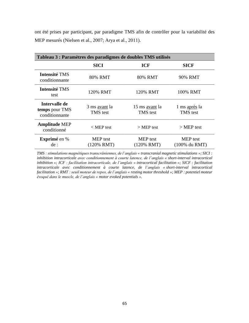
65
ont été prises par participant, par paradigme TMS afin de contrôler pour la variabilité des
MEP mesurés (Nielsen et al., 2007; Arya et al., 2011).
Tableau 3 : Paramètres des paradigmes de doubles TMS utilisés
SICI ICF SICF
Intensité TMS
conditionnante 80% RMT 80% RMT 90% RMT
Intensité TMS
test 120% RMT 120% RMT 100% RMT
Intervalle de
temps pour TMS
conditionnante
3 ms avant la
TMS test
15 ms avant la
TMS test
1 ms après la
TMS test
Amplitude MEP
conditionné < MEP test > MEP test > MEP test
Exprimé en %
de :
MEP test
(120% RMT)
MEP test
(120% RMT)
MEP test
(100% du RMT)
TMS : stimulations magnétiques transcrâniennes, de l’anglais « transcranial magnetic stimulations »; SICI :
inhibition intracorticale avec conditionnement à courte latence, de l’anglais « short-interval intracortical
inhibition »; ICF : facilitation intracorticale, de l’anglais « intracortical facilitation »; SICF : facilitation
intracorticale avec conditionnement à courte latence, de l’anglais « short-interval intracortical
facilitation »; RMT : seuil moteur de repos, de l’anglais « resting motor threshold »; MEP : potentiel moteur
évoqué dans le muscle, de l’anglais « motor evoked potentials ».
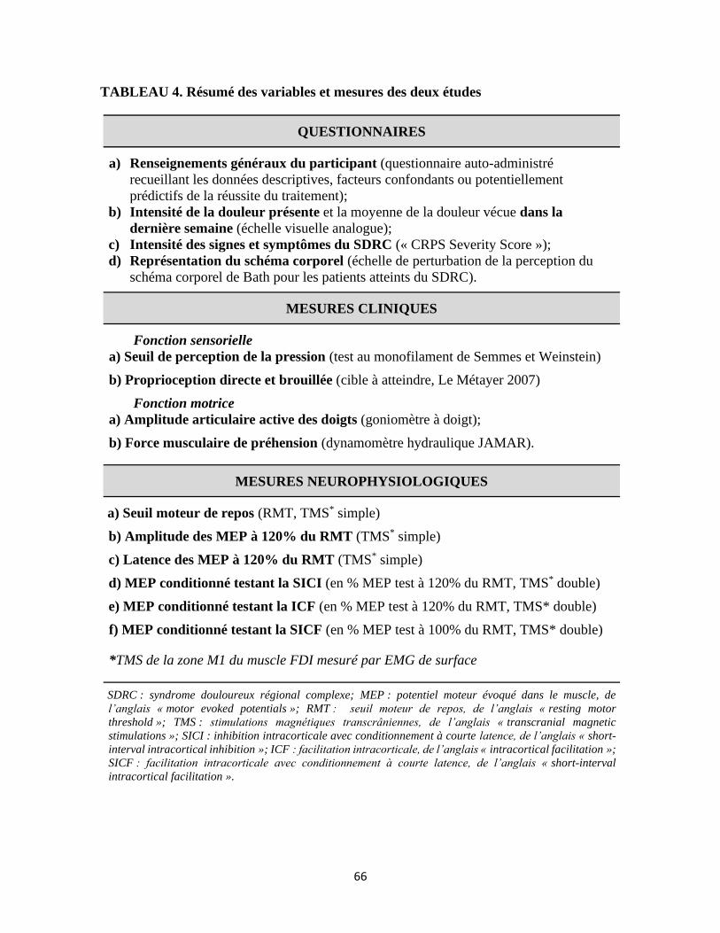
66
TABLEAU 4. Résumé des variables et mesures des deux études
QUESTIONNAIRES
a) Renseignements généraux du participant (questionnaire auto-administré
recueillant les données descriptives, facteurs confondants ou potentiellement
prédictifs de la réussite du traitement);
b) Intensité de la douleur présente et la moyenne de la douleur vécue dans la
dernière semaine (échelle visuelle analogue);
c) Intensité des signes et symptômes du SDRC (« CRPS Severity Score »);
d) Représentation du schéma corporel (échelle de perturbation de la perception du
schéma corporel de Bath pour les patients atteints du SDRC).
MESURES CLINIQUES
Fonction sensorielle
a) Seuil de perception de la pression (test au monofilament de Semmes et Weinstein)
b) Proprioception directe et brouillée (cible à atteindre, Le Métayer 2007)
Fonction motrice
a) Amplitude articulaire active des doigts (goniomètre à doigt);
b) Force musculaire de préhension (dynamomètre hydraulique JAMAR).
MESURES NEUROPHYSIOLOGIQUES
a) Seuil moteur de repos (RMT, TMS* simple)
b) Amplitude des MEP à 120% du RMT (TMS* simple)
c) Latence des MEP à 120% du RMT (TMS* simple)
d) MEP conditionné testant la SICI (en % MEP test à 120% du RMT, TMS* double)
e) MEP conditionné testant la ICF (en % MEP test à 120% du RMT, TMS* double)
f) MEP conditionné testant la SICF (en % MEP test à 100% du RMT, TMS* double)
*TMS de la zone M1 du muscle FDI mesuré par EMG de surface
SDRC : syndrome douloureux régional complexe; MEP : potentiel moteur évoqué dans le muscle, de
l’anglais « motor evoked potentials »; RMT : seuil moteur de repos, de l’anglais « resting motor
threshold »; TMS : stimulations magnétiques transcrâniennes, de l’anglais « transcranial magnetic
stimulations »; SICI : inhibition intracorticale avec conditionnement à courte latence, de l’anglais « short-
interval intracortical inhibition »; ICF : facilitation intracorticale, de l’anglais « intracortical facilitation »;
SICF : facilitation intracorticale avec conditionnement à courte latence, de l’anglais « short-interval
intracortical facilitation ».
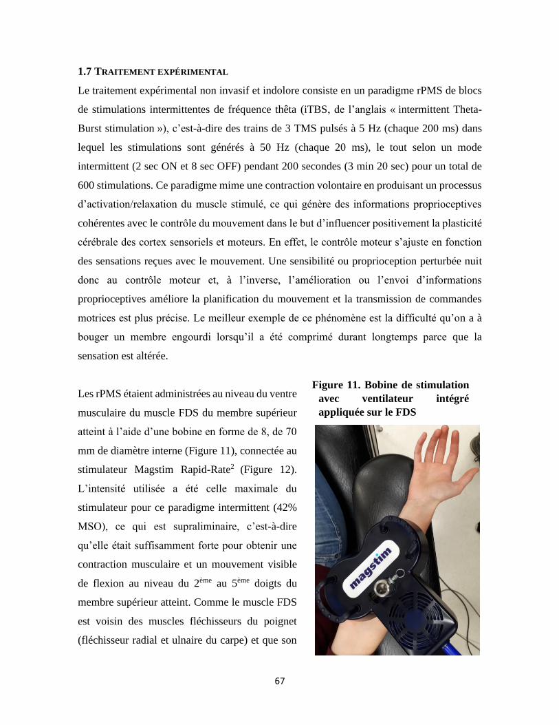
67
1.7 TRAITEMENT EXPÉRIMENTAL
Le traitement expérimental non invasif et indolore consiste en un paradigme rPMS de blocs
de stimulations intermittentes de fréquence thêta (iTBS, de l’anglais « intermittent Theta-
Burst stimulation »), c’est-à-dire des trains de 3 TMS pulsés à 5 Hz (chaque 200 ms) dans
lequel les stimulations sont générés à 50 Hz (chaque 20 ms), le tout selon un mode
intermittent (2 sec ON et 8 sec OFF) pendant 200 secondes (3 min 20 sec) pour un total de
600 stimulations. Ce paradigme mime une contraction volontaire en produisant un processus
d’activation/relaxation du muscle stimulé, ce qui génère des informations proprioceptives
cohérentes avec le contrôle du mouvement dans le but d’influencer positivement la plasticité
cérébrale des cortex sensoriels et moteurs. En effet, le contrôle moteur s’ajuste en fonction
des sensations reçues avec le mouvement. Une sensibilité ou proprioception perturbée nuit
donc au contrôle moteur et, à l’inverse, l’amélioration ou l’envoi d’informations
proprioceptives améliore la planification du mouvement et la transmission de commandes
motrices est plus précise. Le meilleur exemple de ce phénomène est la difficulté qu’on a à
bouger un membre engourdi lorsqu’il a été comprimé durant longtemps parce que la
sensation est altérée.
Les rPMS étaient administrées au niveau du ventre
musculaire du muscle FDS du membre supérieur
atteint à l’aide d’une bobine en forme de 8, de 70
mm de diamètre interne (Figure 11), connectée au
stimulateur Magstim Rapid-Rate2 (Figure 12).
L’intensité utilisée a été celle maximale du
stimulateur pour ce paradigme intermittent (42%
MSO), ce qui est supraliminaire, c’est-à-dire
qu’elle était suffisamment forte pour obtenir une
contraction musculaire et un mouvement visible
de flexion au niveau du 2ème au 5ème doigts du
membre supérieur atteint. Comme le muscle FDS
est voisin des muscles fléchisseurs du poignet
(fléchisseur radial et ulnaire du carpe) et que son
Figure 11. Bobine de stimulation
avec ventilateur intégré
appliquée sur le FDS
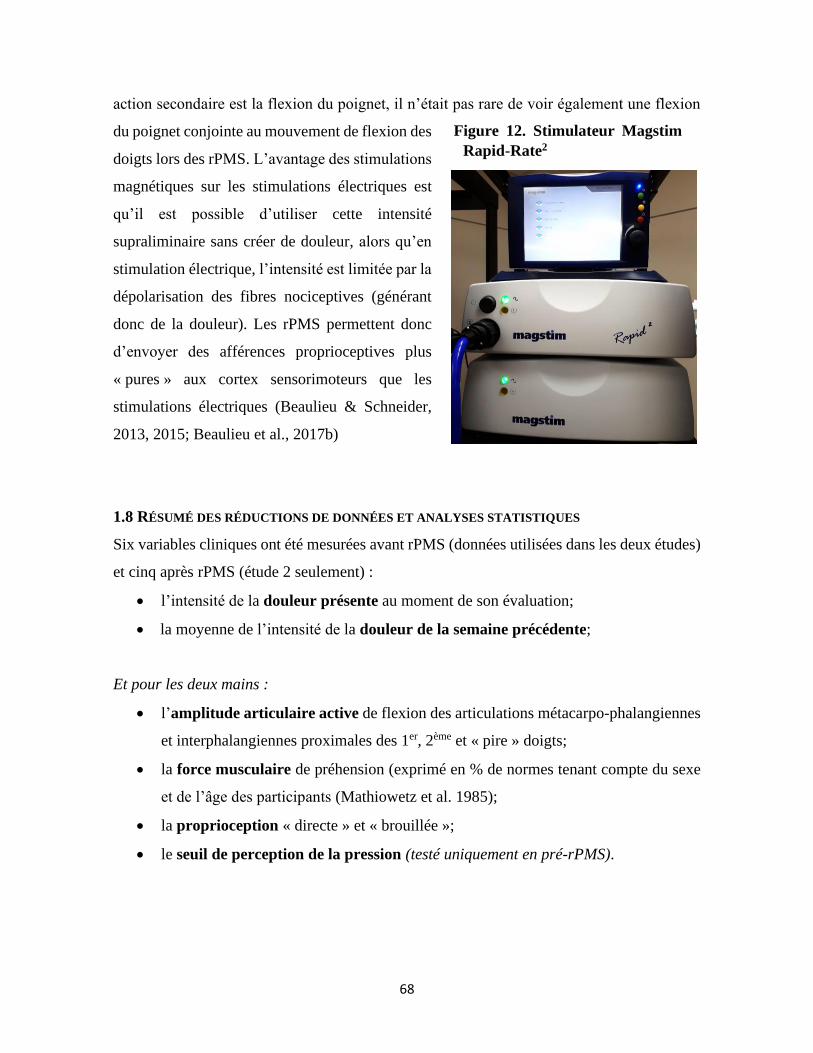
68
action secondaire est la flexion du poignet, il n’était pas rare de voir également une flexion
du poignet conjointe au mouvement de flexion des
doigts lors des rPMS. L’avantage des stimulations
magnétiques sur les stimulations électriques est
qu’il est possible d’utiliser cette intensité
supraliminaire sans créer de douleur, alors qu’en
stimulation électrique, l’intensité est limitée par la
dépolarisation des fibres nociceptives (générant
donc de la douleur). Les rPMS permettent donc
d’envoyer des afférences proprioceptives plus
« pures » aux cortex sensorimoteurs que les
stimulations électriques (Beaulieu & Schneider,
2013, 2015; Beaulieu et al., 2017b)
1.8 RÉSUMÉ DES RÉDUCTIONS DE DONNÉES ET ANALYSES STATISTIQUES
Six variables cliniques ont été mesurées avant rPMS (données utilisées dans les deux études)
et cinq après rPMS (étude 2 seulement) :
• l’intensité de la douleur présente au moment de son évaluation;
• la moyenne de l’intensité de la douleur de la semaine précédente;
Et pour les deux mains :
• l’amplitude articulaire active de flexion des articulations métacarpo-phalangiennes
et interphalangiennes proximales des 1er, 2ème et « pire » doigts;
• la force musculaire de préhension (exprimé en % de normes tenant compte du sexe
et de l’âge des participants (Mathiowetz et al. 1985);
• la proprioception « directe » et « brouillée »;
• le seuil de perception de la pression (testé uniquement en pré-rPMS).
Figure 12. Stimulateur Magstim
Rapid-Rate2
69
Six variables TMS par hémisphère (mesures neurophysiologiques) ont été mesurées avant et
10 minutes après l’application des rPMS pour tester le fonctionnement et la plasticité du M1 :
• le RMT reflétant l’excitabilité de base du M1 (exprimé en % MSO ou en % du RMT
pré-traitement du M1 contralatéral au côté atteint);
• la moyenne de l’amplitude pic-à-pic des MEP test obtenus à 120% RMT (en mV) et
leur latence moyenne (en % de la taille), le tout reflétant la quantité de tissu de M1
répondant aux TMS et la synchronicité des vollées descendantes au niveau des
motoneurones alpha spinaux;
• la moyenne de l’amplitude pic-à-pic des MEP conditionnés pour les mesures des
mécanismes moteurs intracorticaux de SICI, ICF et SICF.
Le programme de statistiques Jamovi a permis de tester les données et leur significativité
(p<0.05). Des statistiques descriptives (moyennes et écart-types) ont permis de comparer les
groupes aux différents temps de prise de mesures. Dépendant des études (1 ou 2) et des
variables, les analyses de variance (ANOVA) ont été appliquées avec les facteurs GROUPE
(groupe SDRC vs groupe Sans-douleur) X CÔTÉ (HC vs HI, ou main atteinte vs non-atteinte
ou vs moyenne des mains/hémisphères du groupe Sans-douleur) ou CÔTÉ X TEMPS (pré-
vs post-rPMS). Les analyses post-hoc bilatérales et plans de comparaison ont permis de tester
l’origine des interactions détectées. Pour les variables où il n’y avait pas de côté à tester ni
de données pour le groupe Sans-douleur, comme pour l’intensité de la douleur, le test t de
Student pairé bilatéral a été utilisé entre les temps pré et post-rPMS. Les matrices de
corrélation de Pearson ont été utilisées pour tester les liens entre les variables présentant des
changements statistiquement significatifs post-rPMS et pour détecter l’existence de facteurs
prédictifs de l’efficacité du traitement, soit la présence d’un lien entre les changements
mesurés chez certains participants et la valeur de leurs variables en ligne de base (avant
traitement).
70
CHAPITRE 2 : ARTICLE 2 « Living with a Complex Regional Pain Syndrome (CRPS):
Clinical and Corticomotor Changes as Compared to Pain-Free People »
« Vivre avec un syndrome douloureux régional complexe (SDRC): changements
cliniques et corticomoteurs par rapport aux personnes sans douleur »
Cet article décrit les différences mesurées entre les participants adultes vivant avec un SDRC
de type I au membre supérieur en stade chronique et les participants sans douleur.
Les participants avec SDRC ont présenté une force de préhension et une amplitude articulaire
active inférieures du côté avec SDRC comparativement au côté sans SDRC ou au groupe
Sans-douleur et une altération de la différence normale entre les deux côtés du corps de la
sensibilité à la pression et de la proprioception. En parallèle, le débalancement d’excitabilité
motrice corticale était significativement plus important chez les participants avec SDRC que
chez le groupe Sans-douleur. Le sens du débalancement (hémisphère contralatéral ou
ipsilatéral au SDRC) variait d’un participant à l’autre, indépendamment du côté atteint. Ceux
avec l’hémisphère ipsilatéral moins excitable étaient ceux avec le moins d’amplitudes
articulaires, mais le moins de douleur également.
Ces résultats originaux ouvrent la voie à la nécessité de comprendre les changements
individuels de fonctionnement moteur cortical en lien avec la fonction sensorimotrice chez
des participants avec SDRC pour personnaliser l’approche en neurostimulation et influencer
la plasticité cérébrale qui pourrait permettre l’amélioration de leur condition (voir Chapitre
3 : Article 3).
71
En préparation pour soumission au journal PAIN
TITLE: LIVING WITH A COMPLEX REGIONAL PAIN SYNDROME (CRPS):
CLINICAL AND CORTICOMOTOR CHANGES AS COMPARED TO PAIN-FREE
PEOPLE
AUTHORS: Fannie Allen Demers 1-3, Alicia Delisle 1-4, Cyril Schneider 1-3,5
AFFILIATIONS:
1 Noninvasive Stimulation laboratory (NovaStim)
2 Neuroscience Division of Centre de recherche du CHU of Québec – Université Laval
3 Faculty of Medicine, Université Laval, Quebec City, Canada
4 Université d’Agens, France
5 Dept Rehabilitation, Faculty of Medicine, Université Laval, Quebec City, Canada
Abstract = 224 words; Main text = 5875 words; 4 Figures; 3 Tables
*Corresponding author: Cyril Schneider, Noninvasive Stimulation laboratory (NovaStim),
Neuroscience Division of Centre de recherche du CHU of Québec – Université Laval,
CHUL-2705 boul. Laurier, RC-9800, Québec QC, Canada, G1V 4G2, Tel. 418-525-4444
(47648), Fax. 418-654-2753, Email: [email protected]
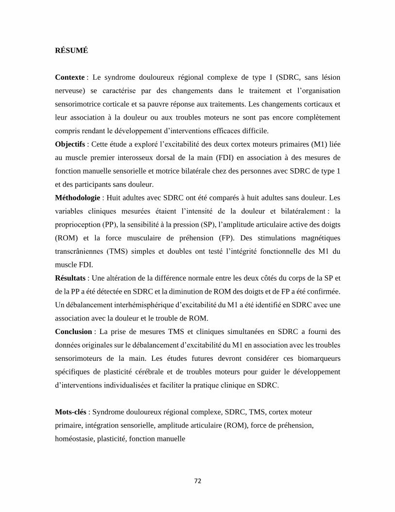
72
RÉSUMÉ
Contexte : Le syndrome douloureux régional complexe de type I (SDRC, sans lésion
nerveuse) se caractérise par des changements dans le traitement et l’organisation
sensorimotrice corticale et sa pauvre réponse aux traitements. Les changements corticaux et
leur association à la douleur ou aux troubles moteurs ne sont pas encore complètement
compris rendant le développement d’interventions efficaces difficile.
Objectifs : Cette étude a exploré l’excitabilité des deux cortex moteurs primaires (M1) liée
au muscle premier interosseux dorsal de la main (FDI) en association à des mesures de
fonction manuelle sensorielle et motrice bilatérale chez des personnes avec SDRC de type 1
et des participants sans douleur.
Méthodologie : Huit adultes avec SDRC ont été comparés à huit adultes sans douleur. Les
variables cliniques mesurées étaient l’intensité de la douleur et bilatéralement : la
proprioception (PP), la sensibilité à la pression (SP), l’amplitude articulaire active des doigts
(ROM) et la force musculaire de préhension (FP). Des stimulations magnétiques
transcrâniennes (TMS) simples et doubles ont testé l’intégrité fonctionnelle des M1 du
muscle FDI.
Résultats : Une altération de la différence normale entre les deux côtés du corps de la SP et
de la PP a été détectée en SDRC et la diminution de ROM des doigts et de FP a été confirmée.
Un débalancement interhémisphérique d’excitabilité du M1 a été identifié en SDRC avec une
association avec la douleur et le trouble de ROM.
Conclusion : La prise de mesures TMS et cliniques simultanées en SDRC a fourni des
données originales sur le débalancement d’excitabilité du M1 en association avec les troubles
sensorimoteurs de la main. Les études futures devront considérer ces biomarqueurs
spécifiques de plasticité cérébrale et de troubles moteurs pour guider le développement
d’interventions individualisées et faciliter la pratique clinique en SDRC.
Mots-clés : Syndrome douloureux régional complexe, SDRC, TMS, cortex moteur
primaire, intégration sensorielle, amplitude articulaire (ROM), force de préhension,
homéostasie, plasticité, fonction manuelle
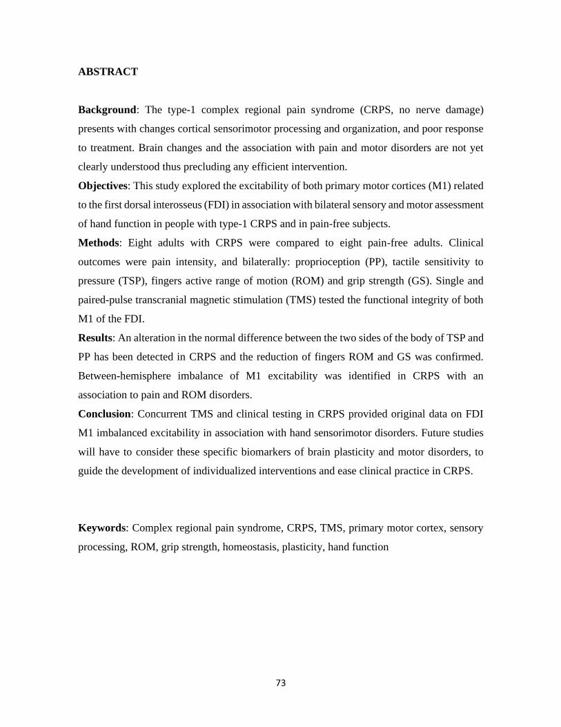
73
ABSTRACT
Background: The type-1 complex regional pain syndrome (CRPS, no nerve damage)
presents with changes cortical sensorimotor processing and organization, and poor response
to treatment. Brain changes and the association with pain and motor disorders are not yet
clearly understood thus precluding any efficient intervention.
Objectives: This study explored the excitability of both primary motor cortices (M1) related
to the first dorsal interosseus (FDI) in association with bilateral sensory and motor assessment
of hand function in people with type-1 CRPS and in pain-free subjects.
Methods: Eight adults with CRPS were compared to eight pain-free adults. Clinical
outcomes were pain intensity, and bilaterally: proprioception (PP), tactile sensitivity to
pressure (TSP), fingers active range of motion (ROM) and grip strength (GS). Single and
paired-pulse transcranial magnetic stimulation (TMS) tested the functional integrity of both
M1 of the FDI.
Results: An alteration in the normal difference between the two sides of the body of TSP and
PP has been detected in CRPS and the reduction of fingers ROM and GS was confirmed.
Between-hemisphere imbalance of M1 excitability was identified in CRPS with an
association to pain and ROM disorders.
Conclusion: Concurrent TMS and clinical testing in CRPS provided original data on FDI
M1 imbalanced excitability in association with hand sensorimotor disorders. Future studies
will have to consider these specific biomarkers of brain plasticity and motor disorders, to
guide the development of individualized interventions and ease clinical practice in CRPS.
Keywords: Complex regional pain syndrome, CRPS, TMS, primary motor cortex, sensory
processing, ROM, grip strength, homeostasis, plasticity, hand function
74
INTRODUCTION
Complex Regional Pain Syndrome (CRPS), formerly known among others as reflex
sympathetic dystrophy or algoneurodystrophy, is a rare chronic pain condition. It is
characterized by pain to one (unilateral CRPS) or more limbs (bilateral or generalized) which
is disproportionate to the inciting focal event (fracture, sprain, surgery, or no traumatism
identified) combined with trophic changes and sensory, motor and autonomic disorders
(Borchers & Gershwin, 2014; Marinus et al., 2011). The upper limb is affected in almost
60% of cases (Bruehl, 2015) and kinesiophobia, i.e. the fear of movement, is responsible for
learned non-use of the CRPS hand at the chronic stage, which worsens the condition due to
reduced proprioceptive information for motor control (Marinus et al., 2013). CRPS is divided
in two main categories, based on the absence (type I, 90% of all CRPS cases) or presence
(type II) of nerve lesion at the periphery (Borchers & Gershwin, 2014). The therapeutic
approach is multidisciplinary, including medical interventions (medications, infiltrations,
etc.), rehabilitation (physical and/or occupational therapies), psychology and clinical
guidelines to follow-up treatment progression. However, after discharge from pain clinics
and in the long term, pain and severe impairments of daily life activities persist in almost
20% of people and 31% remain unable to be back at work two years after the onset of
symptoms (Bean et al., 2014; Geertzen et al., 1998; de Mos et al., 2007; O’Connell et al.,
2013; Rodham et al., 2015).
It is possible to think that refractoriness to treatment, chronicization of symptoms and
generalization to other limbs than the one with the inciting event could be due to central
changes not wholly understood yet and not appropriately treated. Indeed, the etiology
remains unclear but the physiopathology is acknowledged as multifactorial (Bruehl, 2010)
with peripheral changes and sensitization, dysregulation of the sympathetic nervous system
and plastic changes within the central nervous system (Allen Demers et al., review in
preparation; Borchers & Gershwin, 2014; Goh et al., 2017). Our recent comprehensive
review on the topic, among others, denoted that functional limitations (beyond ankylosis or
kinesiophobia) could be related to brain changes (Allen Demers et al., in preparation).
75
Neuroimaging and transcranial magnetic stimulation (TMS of M1) studies have contributed
to knowledge of changes of volumes, connectivity, activation and excitability of different
parts of the brain in CRPS, especially the primary sensory (S1) and motor (M1) cortices with
clinical significance in pain control, perception and motor planning (Allen Demers et al.,
review in preparation; Barad et al., 2014; Geha et al., 2008; Kim et al., 2013; Kirveskari et
al., 2010; Maihöfner et al., 2003; Pleger et al., 2006; Schwenkreis et al., 2009; Vartiainen et
al., 2008; Walton et al., 2010; Zhou et al., 2015). The alteration of the homeostasis of M1
excitability between hemispheres (referred to as imbalanced hemispheric activity) has also
been widely associated with pain and motor disorders in CRPS of the hand (Barad et al.,
2014; Eisenberg et al., 2005; Kirveskari et al., 2010; Krause et al., 2006; Maihöfner et al.,
2007, 2003; Di Pietro et al., 2013; Schwenkreis et al., 2003). However, the direction of
imbalance remains controversial at the chronic stage (Di Pietro et al. 2013a), i.e. hyper- or
hypoexcitability of M1 contralateral to the CRPS hand as compared to M1 ipsilateral. It is
thus crucial to understand if imbalanced hemispheric activity is actually different between
people with CRPS and if the direction of imbalance is related to the severity of the symptoms,
including pain intensity and sensorimotor disorders. This will encourage the development of
an individual approach in CRPS relying on M1 imbalance direction and its normalization to
improve the symptoms.
Therefore, the present study aimed at investigating in people with hand CRPS type 1 (no
nerve lesion) the between-hemisphere differences of corticospinal and intracortical
excitability related to M1, by means of TMS of M1, concurrently with each hand function
assessed by means of standardized sensory and motor testing (pain intensity, proprioception,
tactile sensitivity, fingers active range of flexion, grip strength). Both the CRPS and non-
CRPS sides will be compared, and the CRPS group will be compared to a group of pain-free
participants.
76
METHODS
Participants and Procedures
Eight adults with hand type-1 CRPS (52 ±11 years old, see Table 1) were enrolled in a single
session of TMS and clinical testing and they were compared to a group of eight pain-free
adults (50.8 ±6.9 years old). All participants had signed the informed consent form approved
by the local ethics committees. The participants with CRPS presented with a unilateral upper
limb CRPS type 1 diagnosed with the Budapest clinical criteria (see Table 2) and were
discharged from pain clinics follow-up. The pain-free participants did not experience pain in
the last month before enrolment nor have medical history of chronic pain (pain persisting for
more than three consecutive months). All participants met the exclusion criteria related to
general health (absence of : spine surgery, major circulatory/respiratory/cardiac disease,
other neurological condition for participants with CRPS, severe upper limb orthopaedic
condition, cognitive disorder interfering with the tasks of the study) and to TMS safety
guidelines (Lefaucheur et al., 2011; Rossi et al., 2011, 2009). A physician performed medical
evaluation at pre- and post-enrolment to confirm eligibility and monitor adverse effects. The
same physical therapist collected TMS and clinical data in both groups and data were codified
post-hoc for group (CRPS vs. pain-free) until completion of analyses. After questionnaires
on CRPS severity and perception, and pain evaluation (see next section), all outcomes were
collected on both sides in all participants. Everyone was phoned 2 and 7 days later to
document any adverse effects (Rossi et al. 2009).
Questionnaires on CRPS and Pain Intensity
CRPS severity. The Complex Regional Pain Syndrome Severity Scale (CSS) and the Bath
CRPS Body Perception Disturbance Scale (Bath) were the two questionnaires used to
characterize CRPS. The CSS (Harden et al., 2010) includes a section fulfilled by the
participant on the presence or absence of eight symptoms and a section fulfilled by the
therapist for the presence or absence of nine signs, with each symptom or sign presence
counting for one point on a maximal score of 17. The CSS global score contrasts between
people with and without CRPS, in strong association with the Budapest research criteria and
77
with, among others, the severity of physical and social limitations, pain intensity and thermal
perception abnormalities (Harden et al., 2010).
Self-perception and representation of CRPS limb. The Bath CRPS Body Perception
Disturbance Scale (Lewis et al., 2010) includes seven questions (two with sub-questions, five
on a scale of 0-10, two with Yes/No and one drawing of the CRPS limb) and it was used to
describe and quantify how the participants perceived and represented the CRPS limb.
Pain intensity. Participants’ sitting position was standardized against a chair’s backrest with
the shoulder in neutral rotation and 0-30° abduction, elbow in 90-100° flexion, and forearm
resting on a height-adjustable therapeutic table. The visual analogue scale (VAS, Heller et
al., 2016) was used to quantify the pain intensity experienced by each participant with CRPS.
The VAS consists of a two-sided band: on one side, the participant uses a cursor to rate the
level of pain intensity between two extremes called "No pain" and "Maximal pain
imaginable"; on the other side of VAS, hidden from the participant, a graduated scale from
0 (no pain) to 100 millimeters (maximal pain imaginable) is used by the evaluator to quantify
the participant’s pain intensity. VAS was used to quantify “instantaneous pain” and the mean
pain in the last week.
Mean scores of CCS, Bath item 4 (the extent to which feelings regarding the CRPS limb are
negative) and VAS are reported in Table 1.
(Insert Tables 1-2 near here)
Surface Electromyographic (EMG) Recordings
Painless EMG procedures were followed. After standard skin preparation (i.e. cleaning the
skin with alcohol and shaving whenever necessary, Hermens et al. 2000), the parallel-bar
EMG sensors with adhesive skin interfaces were installed on each participant (fixed 1cm
distance between electrodes; 16-channel Bagnoli EMG System, Delsys Inc., Boston, MA).
EMG sensors were placed bilaterally on the belly of the first dorsal interosseous muscle
(FDI). A common ground electrode was placed on the ulna olecranon of the tested limb.
78
EMG signals were band-pass filtered (20-450 Hz), amplified before digitization (2 kHz) and
computer-stored for offline analysis (PowerLab acquisition system, LabChart,
ADInstruments, Colorado Springs-CO, US).
TMS Testing for Both Hemispheres
TMS procedures were strictly replicated for each hemisphere, following the guidelines from
the International Federation of Clinical Neurophysiology (Rossini et al., 2015). Participants
were comfortably in the reclining chair, arms and legs supported, hips and knees slightly bent
(20° flexion).
Hotspot. Magnetic stimuli were applied over the M1 area of the FDI (index finger abductor
and involved in precision grip) by a figure-of-eight coil (7-cm outer diameter each wing;
Magstim Company Limited, Whitland-UK) positioned on the scalp with the long axis of the
two- wing intersection pointing antero-posteriorly at a 45° angle from the medial line to
active M1 cells in the postero-anterior direction (Sakai et al., 1997). The 10–20 EEG system
was used to first approximate FDI M1 area (Klem, 1999). The position was then adjusted
slightly to determine the ‘hotspot’, namely the M1 location where the MEP obtained in the
contralateral FDI are of the highest amplitude at the lowest TMS intensity (Wolf et al., 2004).
This hotspot was marked on the scalp (surgical pen) to ensure reliable coil positioning during
the session and visual inspection of EMG recordings from both sides monitored the complete
relaxation of the hands during TMS testing.
Resting Motor Threshold (RMT). RMT was determined at the TMS intensity that elicited at
least five FDI MEP with an amplitude of 50 μV or higher out of ten trials. RMT has good
validity and reliability (Beaulieu et al., 2017a, 2017c; Malcolm et al., 2006) and it informs
on M1 basic excitability (Rossini et al., 2015; Ziemann et al., 2015) which allowed to probe
the hemispheric activity balance.
79
MEP Amplitude and Latency. A suprathreshold test (unconditioned) TMS at an intensity of
120% RMT enabled to collect and measure the test MEP amplitude and latency. Test MEP
amplitude (in mV) was measured peak-to-peak and it reflects the volume of M1 cells
activated by TMS and the excitability of the corticospinal pathway (Devanne et al., 1997;
Kobayashi & Pascual-Leone, 2003; Rossini et al., 2015). Test MEP latency (in %
participant’s height for validity of interindividual comparison) was measured from
stimulation time to MEP onset and it reflects conduction time and, indirectly, the
synchronicity of descending volleys to depolarize spinal motoneurons (Groppa et al., 2012;
Kobayashi & Pascual-Leone, 2003).
SICI, ICF and SICF. Paired-pulse TMS (coil connected to two Magstim 2002 monophasic
stimulators) was used to test the short-interval intracortical inhibition (SICI), the intracortical
facilitation (ICF) and the short-interval intracortical facilitation (SICF) (Arya et al., 2011;
Ilić et al., 2002; Kujirai et al., 1993). In SICI, a subthreshold conditioning TMS (80 % RMT
eliciting no MEP on its own) was delivered 3 msec before the test TMS at 120 % RMT
(Kujirai et al., 1993). In ICF, the same parameters were used but at an inter-stimulus interval
of 15 ms (Kujirai et al., 1993). In SICI, the conditioned MEP expressed post-hoc relative to
the mean test MEP amplitude is usually of lower amplitude than the test, and in ICF it is of
higher amplitude than the test (Kujirai et al., 1993). SICI probes the function of GABAA
interneurons within M1 and ICF the function of oligosynaptic glutamatergic circuits (Rossini
et al., 2015; Ziemann et al., 2015). In SICF, two stimuli were delivered 1 ms apart, the first
TMS at 100 % RMT and the second at 90 % RMT (Ortu et al. 2008). MEP amplitude was
then expressed relative to the amplitude of test MEP at 100 % RMT, which likely reflects I-
waves summation following the depolarisation of M1 interneurons by TMS (Ilic et al., 2002;
Rossini et al., 2015; Ziemann et al., 2015). When test MEP amplitudes were different
between sides, the test TMS intensity was adjusted to match this amplitude between sides
and be able to compare the conditioned MEP data between sides. Twelve test MEP and 12
conditioned MEP were collected per participant at a frequency of 0.2-0.3 Hz. Resting breaks
were given on request.
80
Clinical Testing on Both Sides
Upper Limb Proprioception
Following Le Métayer’s guidelines, the paradigm of the blurred proprioception of the upper
limb was used to assess the ability to perceive limb position in space (Le Métayer, 2007).
Briefly, the participants were seated, leaning against a chair without armrests, knees bent at
90° with feet flat on the floor and hands on the thighs (initial position). They had first to reach
with the index a target (large red dot) on a graduated screen at gaze height and arm distance
in front of them and return to the initial position and to repeat this practice three times. Then,
they closed the eyes and the evaluator cautiously (without pain) moved their tested arm in
the air with shoulder movements and elbow flexion / extension and put the arm back in the
initial position, and the participants, without opening the eyes, had to reach back the target
with the index (one trial only). The score of blurred proprioception was the distance in
centimeter between the finger contact on the graduated screen (with eyes closed) and the
target.
Cutaneous Pressure Threshold
This superficial tactile sensitivity was probed by means of the Semmes and Weinstein
monofilaments test (Weinstein, 1993). This test consists of applying pressure on the skin at
different regions of hand and forearm with wired monofilaments of increasing diameter. The
participant was seated against the chair’s backrest, the shoulder in neutral rotation and 0°
abduction, elbow in 90-100° flexion, and forearm resting on the height-adjustable therapeutic
table. With the eyes kept closed during testing, the participant had to tell where a pressure
was felt. The pressure applied to the skin by the experimenter was the weakest to create the
first bending of the wire. When nothing was felt, the experimenter tested the monofilament
with very next higher diameter. The higher the first wire diameter felt, the higher the
cutaneous pressure threshold, thus the lower the superficial tactile perception. Three points
of pressure were tested on each side: at half of the third metacarpal in the palmar and on the
dorsal sides of the hand and at two centimeters down to the antecubital fossae (at the midline
of the forearm). The same experimenter tested all participants.
81
Active Range of Motion (ROM)
Active ROM in flexion was measured using a manual finger goniometer at the
metacarpophalangeal (MCP) and proximal interphalangeal (PIP) joints of the 1st and 2nd
fingers and also of the “worst” finger, i.e. the finger (between the third, fourth and fifth)
harder or more painful to move for each participant. For active ROM of the 1st finger, the
participant was seated with the forearm positioned at rest on the table, shoulder and wrist in
neutral position and the elbow flexed at 90°, and the hand protruding on the edge of the table.
For active ROM of the other fingers, the elbow and shoulder were slightly bent, with the
elbow at rest on the therapeutic table. The measurements of the PIP joints were taken with
the wrist and MCP joints in neutral position (claw hand). Three trials were tested and
averaged per MCP and PIP. Participants rested 60sec between trials.
Grip Strength
Participants were sitting with no arm support, the forearm in neutral position and the wrist
between 0-30° extension and 0-15° ulnar deviation. The maximal grip strength per hand was
measured using a JAMAR hydraulic hand dynamometer (Sammons Preston Rolyan,
Bolingbrook-IL, USA). Following Mathiowetz’s instructions (Mathiowetz et al., 1984)
JAMAR dynamometer was set to the second handle position (placed in hand with help of the
evaluator if needed). The average of three trials per hand was used to represent the maximum
grip strength of each participant. Standardized cheers were given ("Squeeze hard, hard, hard,
the hardest and release") over 8 sec s of sustained contraction to promote a reliable value
(Buckinx et al., 2017). Participants rested 30s between trials.
Data Reduction and Statistical Analyses
Six TMS outcomes were collected per hemisphere per participant: RMT (expressed in % of
maximal stimulator output-MSO), test MEP amplitude (mV) and latency (% participant’s
height), and conditioned MEP amplitudes for SICI, ICF, and SICF (% test MEP amplitude).
In addition, the between-hemisphere difference of RMT was calculated for each group
(DRMT, % MSO).
82
Eleven clinical outcomes were measured per hand per participant: blurred proprioception
(cm), monofilament pressure (tenth mg) for cutaneous pressure thresholds (three points: at
forearm, and palmar and dorsal metacarpus sides), active ROM of MCP and PIP joints of the
1st, 2nd and worst fingers (°), and isometric muscle grip strength (%). To ensure validity of
interindividual comparison, grip strength was expressed in % of grip strength norms stratified
by age and sex (Mathiowetz et al., 1985).
Shapiro-Wilk’s test for Normality revealed that data were normally distributed (p>0.05), thus
parametric tests were used. Two-way analyses of variance (ANOVA, repeated measures)
were applied on TMS and clinical data with factors Group (CRPS vs. Pain-free) X
Hemisphere for TMS data and the factors Group X Hand for clinical outcomes. Hemispheres
were defined as contralateral (CH) and ipsilateral (IH) to CRPS hand and as Dominant (D)
and Non-Dominant (ND) for pain-free participants. Planned comparisons (two-sided,
Tukey’s post-hoc tests for multiple comparisons) were used to detect where differences lay.
Bilateral unpaired Student’s t-tests were applied between groups on between-hemisphere
difference of RMT. Pearson’s correlation matrices were produced to examine the
relationships between the outcomes and especially between the primary outcome (RMT
between hemispheres) and clinical outcomes (including CSS and Bath scores and pain
intensity). The maximum normed residual test (Grubbs' test) helped detect an outlier from a
data set approximated by normal distribution (Prism 8.0, GraphPad Software Inc, La Jolla-
CA, USA): data set is analyzed and an outlier would typically be expunged when it falls
beyond a critical deviation from the sample mean (chance probability to obtain the outlying
value < 0.05). Significance was set at p < 0.05 (https://www.jamovi.org).
83
RESULTS
The two groups were not statistically different for age and manual dominance (one left-
hander per group, p>0.05). A few TMS data was not collected due to head discomfort during
the application of the paired-pulse TMS: out of the N=8+8 participants in the CRPS+pain-
free groups, SICI is presented for 7+7 participants, ICF for 6+7 participants and SICF for
7+8 participants. Also, in the CRPS group, fingers movement were not possible for one
participant (no active ROM nor grip strength collected), ROM measures were painful for
another one (no ROM collected), and one was nauseous with eyes closed (no blurred
proprioception nor tactile sensitivity collected); in the pain-free group, one participant was
detected as outlier for the tactile sensitivity on the palmar face of the hand; thus, blurred
proprioception is presented for 7+8 participants, tactile sensitivity for 7+8 on dorsal side and
7+7 on palmar side, ROM for 6+8 and grip strength for 7+8 participants. No adverse effect
was reported at phone follow-ups. Table 3 presents all clinical and TMS outcomes per group.
(Insert Table 3 near here)
TMS Outcomes
Resting motor threshold. Fig 1A presents that the significant relation linking both M1 RMT
in the pain-free subjects (right graph, r=0.87, p=0.005) with narrow dispersion of individual
data around the perfect line. This relation was not significant in the CRPS group (r=0.62,
p=0.11) and the individual data were more dispersed around the perfect line. Of note, 6/8
participants in the CRPS group presented with the hemisphere ipsilateral to CRPS hand with
higher RMT as compared to the contralateral Fig. 1A). The bilateral unpaired t-test detected
that the between-hemisphere difference of RMT (DRMT, Fig. 1B) was close to be significantly
larger in CRPS group (8±4 % MSO) than in the pain-free group (4.5 ±2.7 % MSO, p=0.06).
(Insert Figure 1 near here)
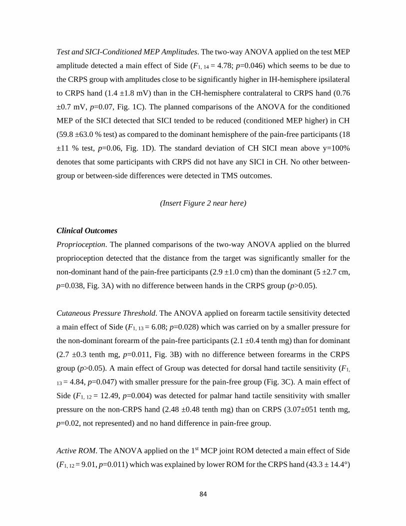
84
Test and SICI-Conditioned MEP Amplitudes. The two-way ANOVA applied on the test MEP
amplitude detected a main effect of Side (F1, 14 = 4.78; p=0.046) which seems to be due to
the CRPS group with amplitudes close to be significantly higher in IH-hemisphere ipsilateral
to CRPS hand (1.4 ±1.8 mV) than in the CH-hemisphere contralateral to CRPS hand (0.76
±0.7 mV, p=0.07, Fig. 1C). The planned comparisons of the ANOVA for the conditioned
MEP of the SICI detected that SICI tended to be reduced (conditioned MEP higher) in CH
(59.8 ±63.0 % test) as compared to the dominant hemisphere of the pain-free participants (18
±11 % test, p=0.06, Fig. 1D). The standard deviation of CH SICI mean above y=100%
denotes that some participants with CRPS did not have any SICI in CH. No other between-
group or between-side differences were detected in TMS outcomes.
(Insert Figure 2 near here)
Clinical Outcomes
Proprioception. The planned comparisons of the two-way ANOVA applied on the blurred
proprioception detected that the distance from the target was significantly smaller for the
non-dominant hand of the pain-free participants (2.9 ±1.0 cm) than the dominant (5 ±2.7 cm,
p=0.038, Fig. 3A) with no difference between hands in the CRPS group (p>0.05).
Cutaneous Pressure Threshold. The ANOVA applied on forearm tactile sensitivity detected
a main effect of Side (F1, 13 = 6.08; p=0.028) which was carried on by a smaller pressure for
the non-dominant forearm of the pain-free participants (2.1 ±0.4 tenth mg) than for dominant
(2.7 ±0.3 tenth mg, p=0.011, Fig. 3B) with no difference between forearms in the CRPS
group (p>0.05). A main effect of Group was detected for dorsal hand tactile sensitivity (F1,
13 = 4.84, p=0.047) with smaller pressure for the pain-free group (Fig. 3C). A main effect of
Side (F1, 12 = 12.49, p=0.004) was detected for palmar hand tactile sensitivity with smaller
pressure on the non-CRPS hand (2.48 ±0.48 tenth mg) than on CRPS (3.07±051 tenth mg,
p=0.02, not represented) and no hand difference in pain-free group.
Active ROM. The ANOVA applied on the 1st MCP joint ROM detected a main effect of Side
(F1, 12 = 9.01, p=0.011) which was explained by lower ROM for the CRPS hand (43.3 ± 14.4°)
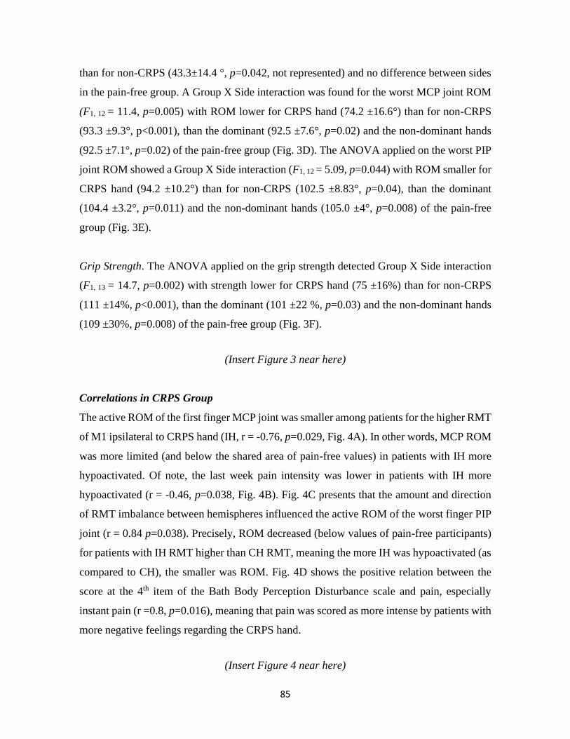
85
than for non-CRPS (43.3±14.4 °, p=0.042, not represented) and no difference between sides
in the pain-free group. A Group X Side interaction was found for the worst MCP joint ROM
(F1, 12 = 11.4, p=0.005) with ROM lower for CRPS hand (74.2 ±16.6°) than for non-CRPS
(93.3 ±9.3°, p<0.001), than the dominant (92.5 ±7.6°, p=0.02) and the non-dominant hands
(92.5 ±7.1°, p=0.02) of the pain-free group (Fig. 3D). The ANOVA applied on the worst PIP
joint ROM showed a Group X Side interaction (F1, 12 = 5.09, p=0.044) with ROM smaller for
CRPS hand (94.2 ±10.2°) than for non-CRPS (102.5 ±8.83°, p=0.04), than the dominant
(104.4 ±3.2°, p=0.011) and the non-dominant hands (105.0 ±4°, p=0.008) of the pain-free
group (Fig. 3E).
Grip Strength. The ANOVA applied on the grip strength detected Group X Side interaction
(F1, 13 = 14.7, p=0.002) with strength lower for CRPS hand (75 ±16%) than for non-CRPS
(111 ±14%, p<0.001), than the dominant (101 ±22 %, p=0.03) and the non-dominant hands
(109 ±30%, p=0.008) of the pain-free group (Fig. 3F).
(Insert Figure 3 near here)
Correlations in CRPS Group
The active ROM of the first finger MCP joint was smaller among patients for the higher RMT
of M1 ipsilateral to CRPS hand (IH, r = -0.76, p=0.029, Fig. 4A). In other words, MCP ROM
was more limited (and below the shared area of pain-free values) in patients with IH more
hypoactivated. Of note, the last week pain intensity was lower in patients with IH more
hypoactivated (r = -0.46, p=0.038, Fig. 4B). Fig. 4C presents that the amount and direction
of RMT imbalance between hemispheres influenced the active ROM of the worst finger PIP
joint (r = 0.84 p=0.038). Precisely, ROM decreased (below values of pain-free participants)
for patients with IH RMT higher than CH RMT, meaning the more IH was hypoactivated (as
compared to CH), the smaller was ROM. Fig. 4D shows the positive relation between the
score at the 4th item of the Bath Body Perception Disturbance scale and pain, especially
instant pain (r =0.8, p=0.016), meaning that pain was scored as more intense by patients with
more negative feelings regarding the CRPS hand.
(Insert Figure 4 near here)
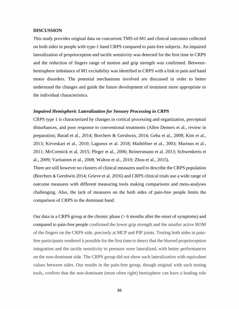
86
DISCUSSION
This study provides original data on concurrent TMS-of-M1 and clinical outcomes collected
on both sides in people with type-1 hand CRPS compared to pain-free subjects. An impaired
lateralization of proprioception and tactile sensitivity was detected for the first time in CRPS
and the reduction of fingers range of motion and grip strength was confirmed. Between-
hemisphere imbalance of M1 excitability was identified in CRPS with a link to pain and hand
motor disorders. The potential mechanisms involved are discussed in order to better
understand the changes and guide the future development of treatment more appropriate to
the individual characteristics.
Impaired Hemispheric Lateralization for Sensory Processing in CRPS
CRPS type 1 is characterized by changes in cortical processing and organization, perceptual
disturbances, and poor response to conventional treatments (Allen Demers et al., review in
preparation; Barad et al., 2014; Borchers & Gershwin, 2014; Geha et al., 2008; Kim et al.,
2013; Kirveskari et al., 2010; Lagueux et al. 2018; Maihöfner et al., 2003; Marinus et al.,
2011; McCormick et al. 2015; Pleger et al., 2006; Reinersmann et al. 2013; Schwenkreis et
al., 2009; Vartiainen et al., 2008; Walton et al., 2010; Zhou et al., 2015).
There are still however no clusters of clinical measures used to describe the CRPS population
(Borchers & Gershwin 2014; Grieve et al. 2016) and CRPS clinical trials use a wide range of
outcome measures with different measuring tools making comparisons and meta-analyses
challenging. Also, the lack of measures on the both sides of pain-free people limits the
comparison of CRPS to the dominant hand.
Our data in a CRPS group at the chronic phase (> 6 months after the onset of symptoms) and
compared to pain-free people confirmed the lower grip strength and the smaller active ROM
of the fingers on the CRPS side, precisely at MCP and PIP joints. Testing both sides in pain-
free participants rendered it possible for the first time to detect that the blurred proprioception
integration and the tactile sensitivity to pressure were lateralized, with better performances
on the non-dominant side. The CRPS group did not show such lateralization with equivalent
values between sides. Our results in the pain-free group, though original with such testing
tools, confirm that the non-dominant (most often right) hemisphere can have a leading role
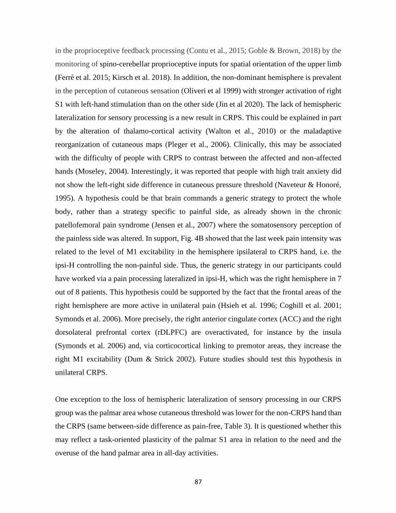
87
in the proprioceptive feedback processing (Contu et al., 2015; Goble & Brown, 2018) by the
monitoring of spino-cerebellar proprioceptive inputs for spatial orientation of the upper limb
(Ferrè et al. 2015; Kirsch et al. 2018). In addition, the non-dominant hemisphere is prevalent
in the perception of cutaneous sensation (Oliveri et al 1999) with stronger activation of right
S1 with left-hand stimulation than on the other side (Jin et al 2020). The lack of hemispheric
lateralization for sensory processing is a new result in CRPS. This could be explained in part
by the alteration of thalamo-cortical activity (Walton et al., 2010) or the maladaptive
reorganization of cutaneous maps (Pleger et al., 2006). Clinically, this may be associated
with the difficulty of people with CRPS to contrast between the affected and non-affected
hands (Moseley, 2004). Interestingly, it was reported that people with high trait anxiety did
not show the left-right side difference in cutaneous pressure threshold (Naveteur & Honoré,
1995). A hypothesis could be that brain commands a generic strategy to protect the whole
body, rather than a strategy specific to painful side, as already shown in the chronic
patellofemoral pain syndrome (Jensen et al., 2007) where the somatosensory perception of
the painless side was altered. In support, Fig. 4B showed that the last week pain intensity was
related to the level of M1 excitability in the hemisphere ipsilateral to CRPS hand, i.e. the
ipsi-H controlling the non-painful side. Thus, the generic strategy in our participants could
have worked via a pain processing lateralized in ipsi-H, which was the right hemisphere in 7
out of 8 patients. This hypothesis could be supported by the fact that the frontal areas of the
right hemisphere are more active in unilateral pain (Hsieh et al. 1996; Coghill et al. 2001;
Symonds et al. 2006). More precisely, the right anterior cingulate cortex (ACC) and the right
dorsolateral prefrontal cortex (rDLPFC) are overactivated, for instance by the insula
(Symonds et al. 2006) and, via corticocortical linking to premotor areas, they increase the
right M1 excitability (Dum & Strick 2002). Future studies should test this hypothesis in
unilateral CRPS.
One exception to the loss of hemispheric lateralization of sensory processing in our CRPS
group was the palmar area whose cutaneous threshold was lower for the non-CRPS hand than
the CRPS (same between-side difference as pain-free, Table 3). It is questioned whether this
may reflect a task-oriented plasticity of the palmar S1 area in relation to the need and the
overuse of the hand palmar area in all-day activities.
88
Cortical Motor Changes and Relation to Pain and Sensorimotor Impairment
TMS of M1 provides outcomes that reflect the integrity of the cortical and corticospinal
components of the motor systems. We thus used TMS of M1 hand area with clinical scores
to better understand the abnormal control of the CRPS hand. Literature about TMS outcomes
in CRPS is controversial with MEP unchanged between hemispheres or as compared to pain-
free people (Krause et al., 2004; Morgante et al., 2017; Schwenkreis et al., 2003; van Velzen
et al., 2015), or, for the same authors, MEP was either unchanged (Krause et al., 2004) or
bilaterally decreased as compared to pain-free subjects (Krause et al., 2005). SICI was
reported as reduced in both hemispheres as compared to pain-free subjects (Krause, et al.,
2006) or reduced only in M1 contralateral to CRPS hand (Eisenberg et al., 2005; Lefaucheur
et al., 2006; Pfannmoller et al., 2019). We did not obtain significant changes in CRPS, but
only trends of MEP increase in ipsi-H and SICI decrease in M1 contralateral. One explanation
could be that our CRPS group was at the chronic stage; participants were at acute stage in
the previous studies and it is known that changes of TMS outcomes can resolve in the long
term due to homeostatic plasticity, i.e. brain mechanisms to balance M1 activity (Manganotti
et al., 2008). The trending increase of MEP could however reflect global increase of
corticospinal excitability, including a larger volume of M1 cells responding to TMS and more
synchronicity of descending volleys to excite the alpha-motoneurons in the spinal cord
(Devanne et al., 1997, Kobayashi & Pascual-Leone, 2003, Rossini et al., 2015). The trending
decrease of SICI could reflect M1 disinhibition still acting on the modification of
sensorimotor maps (Ziemann et al., 2001) to learn alternate motor strategies to recover motor
control in the presence of persistent pain (Massé-Alarie & Schneider, 2017). The net effects
were an imbalance of M1 excitability between hemispheres, as detected by RMT.
RMT-related data are the main TMS findings of our study. RMT informs on the transsynaptic
excitability of M1 tissues (Ziemann et al., 2014) and the absence of RMT correlation between
hemispheres in CRPS (Fig. 1A, left graph) could reflect an alteration of interhemispheric
homeostasis. Homeostasis maintains a balanced M1 excitability between hemispheres
(Abraham, 2008; Abraham & Bear, 1996; Hassanzahraee et al., 2018; Hulme et al., 2013;
Karabanov et al., 2015; Mansour et al., 2014; Nijs & Van Houdenhove, 2009; Ziemann &
Siebner, 2008). A potential cause of homeostasis alteration could be the smudging or the
89
overlap of sensorimotor maps contralateral to CRPS, as already denoted (Di Pietro et al.,
2013b, 2013a; Pleger et al., 2006). The modulation of excitatory influences to M1 or the
changes of connectivity to M1 could have imbalanced M1 excitability between hemispheres.
This is supported by the fact that S1 ipsilateral to CRPS is larger than S1 in pain-free subjects
(Di Pietro et al., 2015), thus increasing the S1-M1 activation. Also, the connectivity from the
prefrontal cortex to the frontal lobe is augmented in CRPS at the chronic stage (Shokouhi et
al., 2018), thus potentially overactivating M1.
Correlations of Fig. 4 showed the association between the excitability of ipsi-H (right-H in
7/8 patients) and CRPS fingers ROM and pain intensity. Precisely, among patients, the higher
the ipsi-H excitability, the larger the active ROM (Fig.4A,C) but the more intense the last
week pain (Fig.4C). Thus, the ipsi-H excitability as compared to the contralateral seems an
interesting biomarker to guide treatment. For example, any approach capable to down-
regulating ipsi-H activity could decrease pain intensity. Then, rehabilitation could be
painlessly administrated. However, the portrait is not that simple and the association between
the item 4 of the Bath Body Perception Disturbance Scale and the intensity of pain (Fig. 4D)
revealed that the more were the negative feelings regarding the CRPS limb the higher the
pain intensity was. This emotional aspect of CRPS may be related to connectivity changes of
the DLPFC networks including the limbic system (Shokouhi et al., 2018). In addition,
kinesiophobia may have created a vicious circle that worsened the condition: the CRPS hand
was less or no more used and this resulted in substantial learned non-use maladaptive changes
in sensorimotor areas (Hotta et al., 2015) thus potentially enlarging and exciting further S1-
M1 in ipsi-H .
Methodological Considerations
The small number of participants and missing data due to discomfort to paired-pulse TMS or
clinical testing (ROM, eyes closed) precludes from any generalization of our results and the
findings need to be replicated in larger samples with better effect size. The study offers the
advantages of testing sensory function (proprioception, tactile sensitivity) and hand motor
function (fingers ROM, grip strength) in both the CRPS and non-CRPS hand, in comparison
to the both sides of pain-free participants and concurrently to TMS outcomes from both
90
hemispheres. This provides a valuable foundation for future work to decipher in parallel
different mechanisms of action in CRPS that could be relevant to guide the appropriate
therapies against refractoriness to treatment: lateralized sensory processing, hemispheric
balanced activity (direction, amount), central sensitization, emotional aspects, neuroimaging
and TMS investigation. psychosocial evaluation.
91
CONCLUSION
Concurrent TMS and clinical testing in type-1 CRPS hand provided original data on the
impairment of hemispheric balanced activity of the primary motor cortex and the
lateralization of sensory processing along with an association with pain and hand/finger
function outcomes. Future studies will have to consider these specific biomarkers of brain
plasticity and motor disorders, to guide the development of individualized interventions and
ease clinical practice in CRPS.
ACKNOWLEDGEMENTS
The authors declare no conflict of interest. This work was supported by the Chronic Pain
Network (CPN) funded by the strategy for patient-oriented research (SPOR) of the Canadian
Institutes of Health Research (CS co-author: IE116731). FAD was supported by a master
studentship for health professionals from the Fonds de Recherche du Québec-Santé (FRQS)
and a grant research from CPN. AD was supported by a master studentship for France-
Québec research training exchanges and a grant from CPN.
92
REFERENCES
Abraham, Wickliffe C. 2008. “Metaplasticity: Tuning Synapses and Networks for
Plasticity.” Nature Reviews Neuroscience 9(5): 387–99.
Abraham, Wickliffe C, and Mark F Bear. 1996. “Metaplasticity : Plasticity of Synaptic.”
Trends Neurosci 19: 126–30.
Allen Demers, Fannie, Zangrandi, Andrea, Schneider, Cyril. In preparation “Noninvasive
Neurostimulation in Complex Regional Pain Syndrome (CRPS). A Comprehensive
Review on Brain and Clinical Outcomes.” (Neurophysiologie Clinique / Clinical
Neurophysiology)
Arya, Kamal Narayan, Shanta Pandian, Rajesh Verma, and R K Garg. 2011. “Movement
Therapy Induced Neural Reorganization and Motor Recovery in Stroke: A Review.”
Journal of bodywork and movement therapies 15(4): 528–37.
Barad, Meredith J et al. 2014. “Complex Regional Pain Syndrome Is Associated with
Structural Abnormalities in Pain-Related Regions of the Human Brain.” The journal of
pain : official journal of the American Pain Society 15(2): 197–203.
Bean, Debbie J., Malcolm H. Johnson, and Robert R. Kydd. 2014. “The Outcome of
Complex Regional Pain Syndrome Type 1: A Systematic Review.” Journal of Pain
15(7): 677–90.
Beaulieu, Louis-David, H. Flamand, Véronique, Massé-Alarie, Hugo and Schneider, Cyril.
2017a. “Reliability and Minimal Detectable Change of Transcranial Magnetic
Stimulation Outcomes in Healthy Adults: A Systematic Review.” Brain Stimulation
10(2): 196–213.
Beaulieu, Louis-David, Masse-Alarie, Hugo, Brouwer, Brenda, and Schneider, Cyril. 2015.
“Noninvasive Neurostimulation in Chronic Stroke: A Double-Blind Randomized
Sham-Controlled Testing of Clinical and Corticomotor Effects.” Topics in Stroke
Rehabilitation 22(1): 8–17.
Beaulieu, Louis-David, Massé-Alarie, Hugo, Ribot-Ciscar, Edith, and Schneider, Cyril.
2017c. “Reliability of Lower Limb Transcranial Magnetic Stimulation Outcomes in
the Ipsi- and Contralesional Hemispheres of Adults with Chronic Stroke.” Clinical
Neurophysiology 128(7): 1290–98.
Borchers, A. T., and M. E. Gershwin. 2014. “Complex Regional Pain Syndrome: A
Comprehensive and Critical Review.” Autoimmunity Reviews 13(3): 242–65.
Bruehl, Stephen. 2015. “Complex Regional Pain Syndrome.” bmj (351): 825–38.
Buckinx, Fanny et al. 2017. “Reliability of Muscle Strength Measures Obtained with a
Hand-Held Dynamometer in an Elderly Population.” Clinical Physiology and
Functional Imaging 37(3): 332–40.
Coghill RC, Gilron I, Iadarola MJ. 2001 "Hemispheric lateralization of somatosensory
processing". J Neurophysiol 85:2602–2612
Cohen, Leonardo G. et al. 1990. “Effects of Coil Design on Delivery of Focal Magnetic
Stimulation. Technical Considerations.” Electroencephalography and Clinical
Neurophysiology 75(4): 350–57.
Contu S, Cappello L, Konczak J, Masia L. 2015 "Preliminary analysis of non-dominant
proprioceptive acuity and interlimb asymmetry in the human wrist". Annu Int Conf
IEEE Eng Med Biol Soc. 2015:3598-601.
Devanne H, Lavoie BA, Capaday C. 1997 "Input-output properties and gain changes in the
human corticospinal pathway". Exp Brain Res;114:329–38.
93
Di Pietro, Flavia et al. 2013a. “Primary Motor Cortex Function in Complex Regional Pain
Syndrome: A Systematic Review and Meta-Analysis.” Journal of Pain 14(11): 1270–
88.
Di Pietro, Flavia et al. 2013b. “Primary Somatosensory Cortex Function in Complex
Regional Pain Syndrome: A Systematic Review and Meta-Analysis.” The journal of
pain : official journal of the American Pain Society 14(11): 1270–88.
Di Pietro, Flavia et al. 2015. “Interhemispheric Somatosensory Differences in Chronic Pain
Reflect Abnormality of the Healthy Side.” Human Brain Mapping 36(2): 508–18.
Dum RP, Li C, Strick PL. 2002 "Motor and nonmotor domains in the monkey dentate".
Ann N Y Acad Sci 978:289–301
Eisenberg, Elon et al. 2005. “Evidence for Cortical Hyperexcitability of the Affected Limb
Representation Area in CRPS: A Psychophysical and Transcranial Magnetic
Stimulation Study.” Pain 113(1): 99–105.
Ferrè, Elisa Raffaella, Berlot, Eva, Haggard, Patrick. 2015 Vestibular contributions to a
right-hemisphere network for bodily awareness: Combining galvanic vestibular
stimulation and the “Rubber Hand Illusion”, Neuropsychologia, Volume 69, Pages
140-147
Geertzen, J H B et al. 1998. “Relationship between Impairments, Disability and Handicap
in Reflex Sympathetic Dystrophy Patients: A Long-Term Follow-up Study.” Clinical
rehabilitation 12(5): 402–12.
Geha, Paul Y et al. 2008. “The Brain in Chronic CRPS Pain: Abnormal Gray-White Matter
Interactions in Emotional and Autonomic Regions.” Neuron 60(4): 570–81.
Goble DJ, Brown SH. 2008 "Upper limb asymmetries in the matching of proprioceptive
versus visual targets". J Neurophysiol 99: 3063–3074.
Goh, En Lin, Swathikan Chidambaram, and Daqing Ma. 2017. “Complex Regional Pain
Syndrome: A Recent Update.” Burns & Trauma 5(1): 1–11.
Gouvernement du Canada. 2018. “Faire Face à La Crise Des Opioïdes Au Canada.”
https://www.canada.ca/fr/sante-canada/services/dependance-aux-
drogues/consommation-problematique-medicaments-ordonnance/opioides/repondre-
crise-opioides-canada.html (March 11, 2019).
Gracies, Jean-Michel. 2005. “Pathophysiology of Spastic Paresis. II: Emergence of Muscle
Overactivity.” Muscle & nerve 31(5): 552–71.
Grieve, S, L Jones, N Walsh, and C McCabe. 2016. “What Outcome Measures Are
Commonly Used for Complex Regional Pain Syndrome Clinical Trials? A Systematic
Review of the Literature.” European journal of pain (London, England) 20(3): 331–
40.
Harden, R. Norman et al. 2010. “Development of a Severity Score for CRPS.” Pain 151(3):
870–76. http://dx.doi.org/10.1016/j.pain.2010.09.031.
Hassanzahraee, Maryam, Maryam Zoghi, and Shapour Jaberzadeh. 2018. “How Different
Priming Stimulations Affect the Corticospinal Excitability Induced by Noninvasive
Brain Stimulation Techniques: A Systematic Review and Meta-Analysis.” Reviews in
the Neurosciences 29(8): 883–99.
Heller GZ, Manuguerra M, Chow R. 2016 "How to analyze the Visual Analogue Scale:
Myths, truths and clinical relevance". Scand J Pain;13:67–75.
Hermens, Hermie J., Bart Freriks, Catherine Disselhorst-Klug, and Rau Günter. 2000.
“Development of Recommendations for SEMG Sensors and Sensor Placement
Procedures.” Journal of Electromyography and Kinesiology 10: 361–74.
94
Hotta, J. et al. 2015. “Patients with Complex Regional Pain Syndrome Overestimate
Applied Force in Observed Hand Actions.” European Journal of Pain (United
Kingdom) 19(9): 1372–81.
Hsieh JC, Hannerz J, Ingvar M, 1996 "Right-lateralised central processing for pain of
nitroglycerin-induced cluster headache". Pain 67:59–68
Hulme, Sarah R., Owen D. Jones, and Wickliffe C. Abraham. 2013. “Emerging Roles of
Metaplasticity in Behaviour and Disease.” Trends in Neurosciences 36(6): 353–62.
Jensen R, Hystad T, Kvale A, Baerheim A, 2007 "Quantitative sensory testing of patients
with long lasting Patellofemoral pain syndrome". Eur J Pain 11:665–676.
Jin SH, Lee SH, Yang ST, An J. 2020 "Hemispheric asymmetry in hand preference of
right-handers for passive vibrotactile perception: an fNIRS study". Sci Rep. Aug
7;10(1):13423.
Karabanov, Anke et al. 2015. “Consensus Paper: Probing Homeostatic Plasticity of Human
Cortex With Non-Invasive Transcranial Brain Stimulation.” Brain Stimulation 8(3):
442–54.
Kim, Jieun et al. 2013. “Sustained Deep-Tissue Pain Alters Functional Brain Connectivity.”
Pain 154(8): 1343–51.
Kirsch, V., Boegle, R., Keeser, D., Kierig, E., Ertl-Wagner, B., Brandt, T., Dieterich, M.
2018 "Handedness-dependent functional organizational patterns within the bilateral
vestibular cortical network revealed by fMRI connectivity based parcellation"
NeuroImage, Volume 178, ,Pages 224-237.
Kirveskari, Erika, Nuutti V. Vartiainen, Maarit Gockel, and Nina Forss. 2010. “Motor
Cortex Dysfunction in Complex Regional Pain Syndrome.” Clinical Neurophysiology
121(7): 1085–91.
Kobayashi M, Pascual-Leone A. 2003 "Transcranial magnetic stimulation in neurology".
Lancet Neurol;2:145–56.
Krause, P., Foerderreuther, S., & Straube, A. (2004). Bilateral motor cortex disinhibition in
complex regional pain syndrome (CRPS) type I of the hand. Neurology, 62(9), 1654;
author reply 1654-1655. doi:10.1212/wnl.62.9.1654
Krause P, Foerderreuther S, Straube A. 2005 "Effects of conditioning peripheral repetitive
magnetic stimulation in patients with complex regional pain syndrome". Neurol
Res;27:412–7.
Krause, Phillip, Stefanie Förderreuther, and Andreas Straube. 2006. “TMS Motor Cortical
Brain Mapping in Patients with Complex Regional Pain Syndrome Type I.” Clinical
Neurophysiology 117(1): 169–76.
Lagueux, Emilie et al. 2012. “Modified Graded Motor Imagery for Complex Regional Pain
Syndrome Type 1 of the Upper Extremity in the Acute Phase: A Patient Series.”
International Journal of Rehabilitation Research 35(2): 138–45.
Lagueux, E., Bernier, M., Bourgault, P., Whittingstall, K., Mercier, C., Leonard, G.,
Tousignant-Laflamme, Y. 2018. The Effectiveness of Transcranial Direct Current
Stimulation as an Add-on Modality to Graded Motor Imagery for Treatment of
Complex Regional Pain Syndrome: A Randomized Proof of Concept Study. Clin J
Pain, 34(2), 145-154.
Lefaucheur, J-P et al. 2011. “[French Guidelines on the Use of Repetitive Transcranial
Magnetic Stimulation (RTMS): Safety and Therapeutic Indications].”
Neurophysiologie clinique = Clinical neurophysiology 41(5–6): 221–95.
Lefaucheur JP. 2006 "The use of repetitive transcranial magnetic stimulation (rTMS) in
95
chronic neuropathic pain." Neurophysiol Clin Neurophysiol;36:117–24.
Lewis Jenny, McCabe Candy, Lewis J., McCabe C. 2010 "Body Perception Disturbance
(BPD) in CRPS". Pract Pain Manag:60–6.
Maihöfner, Christian et al. 2007. “The Motor System Shows Adaptive Changes in Complex
Regional Pain Syndrome.” Brain 130(10): 2671–87.
Maihöfner, Christian, Hermann O Handwerker, Bernhard Neundörfer, and Frank Birklein.
2003. “Patterns of Cortical Reorganization in Complex Regional Pain Syndrome.”
Neurology 61(12): 1707–15.
Malcolm, M. P. et al. 2006. “Reliability of Motor Cortex Transcranial Magnetic
Stimulation in Four Muscle Representations.” Clinical Neurophysiology 117(5):
1037–46.
Manganotti P, Acler M, Zanette GP, Smania N, Fiaschi A. 2008 "Motor cortical
disinhibition during early and late recovery after stroke". Neurorehabil Neural Repair.
Jul-Aug;22(4):396-403.
Mansour, A R, M A Farmer, M N Baliki, and A Vania Apkarian. 2014. “Chronic Pain : The
Role of Learning and Brain Plasticity.” 32: 129–39.
Marinus, Johan et al. 2011. “Clinical Features and Pathophysiology of Complex Regional
Pain Syndrome.” The Lancet Neurology 10(7): 637–48.
Massé-Alarie, Hugo, Beaulieu, Louis-David, Preuss, Richard, Schneider, Cyril. 2017
"Repetitive peripheral magnetic neurostimulation of multifidus muscles combined
with motor training influences spine motor control and chronic low back pain",
Clinical Neurophysiology, Volume 128, Issue 3, Pages 442-453
Massé-Alarie, Hugo, and Schneider, Cyril. 2016. “Revisiting the Corticomotor Plasticity in
Low Back Pain: Challenges and Perspectives.” Healthcare (Basel, Switzerland) 4(3):
67.
Mathiowetz, Virgil et al. 1985. “Grip and Pinch Strength: Normative Data for Adults.”
Archives of Physical Medicine and Rehabilitation 66: 69–74.
McCormick, Zachary L. et al. 2015. “Short-Term Functional, Emotional, and Pain
Outcomes of Patients with Complex Regional Pain Syndrome Treated in a
Comprehensive Interdisciplinary Pain Management Program.” Pain Medicine: 2357–
67.
Le Métayer, M. 2007. “L’évaluation Clinique Du Sens de La Position de l’axe Du Corps et
Des Membres Supérieurs Par «l’épreuve Nez et Doigt».” Motricite Cerebrale 28(1):
25–31.
de Mos, M. et al. 2007. “The Incidence of Complex Regional Pain Syndrome: A
Population-Based Study.” Pain 129: 12–20.
Moseley GL. 2004 "Why do people with complex regional pain syndrome take longer to
recognize their affected hand?" Neurology; 62:2182–6.
Naveteur J, Honoré J. 1995 "Cutaneous Thresholds and Anxiety: Influence of Eye
Orientation and Lateral Asymmetry." Perceptual and Motor Skills 80(3):707-716.
Nielsen, J B, C Crone, and H Hultborn. 2007. “The Spinal Pathophysiology of Spasticity--
from a Basic Science Point of View.” Acta physiologica (Oxford, England) 189(2):
171–80.
Nijs, Jo, and Boudewijn Van Houdenhove. 2009. “From Acute Musculoskeletal Pain to
Chronic Widespread Pain and Fibromyalgia: Application of Pain Neurophysiology in
Manual Therapy Practice.” Manual Therapy 14(1): 3–12.
O’Connell, Neil E et al. 2015. “Interventions for Treating Pain and Disability in Adults
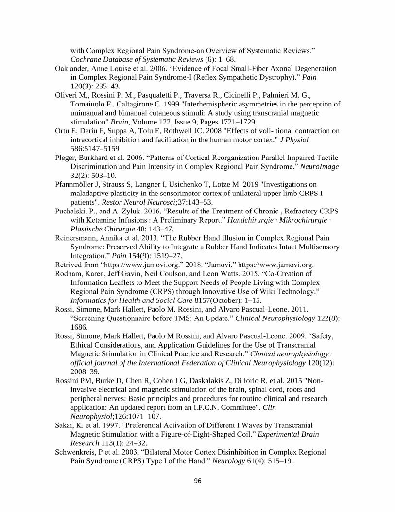
96
with Complex Regional Pain Syndrome‐an Overview of Systematic Reviews.”
Cochrane Database of Systematic Reviews (6): 1–68.
Oaklander, Anne Louise et al. 2006. “Evidence of Focal Small-Fiber Axonal Degeneration
in Complex Regional Pain Syndrome-I (Reflex Sympathetic Dystrophy).” Pain
120(3): 235–43.
Oliveri M., Rossini P. M., Pasqualetti P., Traversa R., Cicinelli P., Palmieri M. G.,
Tomaiuolo F., Caltagirone C. 1999 "Interhemispheric asymmetries in the perception of
unimanual and bimanual cutaneous stimuli: A study using transcranial magnetic
stimulation" Brain, Volume 122, Issue 9, Pages 1721–1729.
Ortu E, Deriu F, Suppa A, Tolu E, Rothwell JC. 2008 "Effects of voli- tional contraction on
intracortical inhibition and facilitation in the human motor cortex." J Physiol
586:5147–5159
Pleger, Burkhard et al. 2006. “Patterns of Cortical Reorganization Parallel Impaired Tactile
Discrimination and Pain Intensity in Complex Regional Pain Syndrome.” NeuroImage
32(2): 503–10.
Pfannmöller J, Strauss S, Langner I, Usichenko T, Lotze M. 2019 "Investigations on
maladaptive plasticity in the sensorimotor cortex of unilateral upper limb CRPS I
patients". Restor Neurol Neurosci;37:143–53.
Puchalski, P., and A. Zyluk. 2016. “Results of the Treatment of Chronic , Refractory CRPS
with Ketamine Infusions : A Preliminary Report.” Handchirurgie · Mikrochirurgie ·
Plastische Chirurgie 48: 143–47.
Reinersmann, Annika et al. 2013. “The Rubber Hand Illusion in Complex Regional Pain
Syndrome: Preserved Ability to Integrate a Rubber Hand Indicates Intact Multisensory
Integration.” Pain 154(9): 1519–27.
Retrived from “https://www.jamovi.org.” 2018. “Jamovi.” https://www.jamovi.org.
Rodham, Karen, Jeff Gavin, Neil Coulson, and Leon Watts. 2015. “Co-Creation of
Information Leaflets to Meet the Support Needs of People Living with Complex
Regional Pain Syndrome (CRPS) through Innovative Use of Wiki Technology.”
Informatics for Health and Social Care 8157(October): 1–15.
Rossi, Simone, Mark Hallett, Paolo M. Rossini, and Alvaro Pascual-Leone. 2011.
“Screening Questionnaire before TMS: An Update.” Clinical Neurophysiology 122(8):
1686.
Rossi, Simone, Mark Hallett, Paolo M Rossini, and Alvaro Pascual-Leone. 2009. “Safety,
Ethical Considerations, and Application Guidelines for the Use of Transcranial
Magnetic Stimulation in Clinical Practice and Research.” Clinical neurophysiology :
official journal of the International Federation of Clinical Neurophysiology 120(12):
2008–39.
Rossini PM, Burke D, Chen R, Cohen LG, Daskalakis Z, Di Iorio R, et al. 2015 "Non-
invasive electrical and magnetic stimulation of the brain, spinal cord, roots and
peripheral nerves: Basic principles and procedures for routine clinical and research
application: An updated report from an I.F.C.N. Committee". Clin
Neurophysiol;126:1071–107.
Sakai, K. et al. 1997. “Preferential Activation of Different I Waves by Transcranial
Magnetic Stimulation with a Figure-of-Eight-Shaped Coil.” Experimental Brain
Research 113(1): 24–32.
Schwenkreis, P et al. 2003. “Bilateral Motor Cortex Disinhibition in Complex Regional
Pain Syndrome (CRPS) Type I of the Hand.” Neurology 61(4): 515–19.
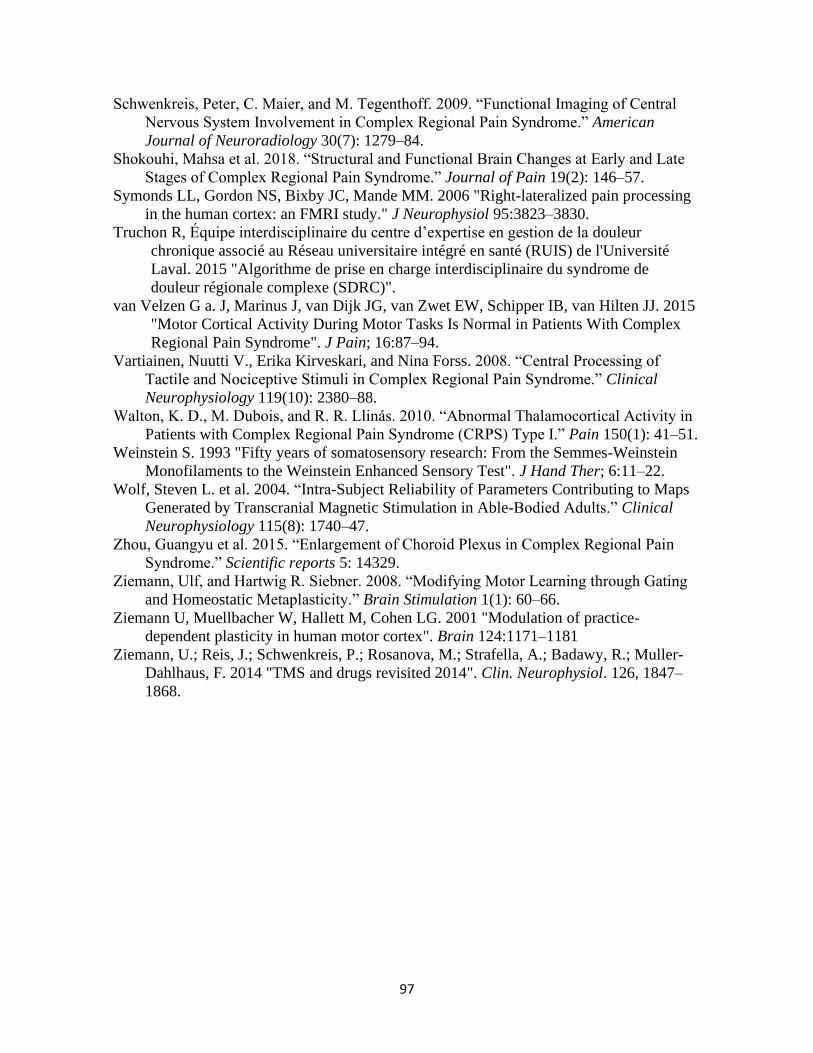
97
Schwenkreis, Peter, C. Maier, and M. Tegenthoff. 2009. “Functional Imaging of Central
Nervous System Involvement in Complex Regional Pain Syndrome.” American
Journal of Neuroradiology 30(7): 1279–84.
Shokouhi, Mahsa et al. 2018. “Structural and Functional Brain Changes at Early and Late
Stages of Complex Regional Pain Syndrome.” Journal of Pain 19(2): 146–57.
Symonds LL, Gordon NS, Bixby JC, Mande MM. 2006 "Right-lateralized pain processing
in the human cortex: an FMRI study." J Neurophysiol 95:3823–3830.
Truchon R, Équipe interdisciplinaire du centre d’expertise en gestion de la douleur
chronique associé au Réseau universitaire intégré en santé (RUIS) de l'Université
Laval. 2015 "Algorithme de prise en charge interdisciplinaire du syndrome de
douleur régionale complexe (SDRC)".
van Velzen G a. J, Marinus J, van Dijk JG, van Zwet EW, Schipper IB, van Hilten JJ. 2015
"Motor Cortical Activity During Motor Tasks Is Normal in Patients With Complex
Regional Pain Syndrome". J Pain; 16:87–94.
Vartiainen, Nuutti V., Erika Kirveskari, and Nina Forss. 2008. “Central Processing of
Tactile and Nociceptive Stimuli in Complex Regional Pain Syndrome.” Clinical
Neurophysiology 119(10): 2380–88.
Walton, K. D., M. Dubois, and R. R. Llinás. 2010. “Abnormal Thalamocortical Activity in
Patients with Complex Regional Pain Syndrome (CRPS) Type I.” Pain 150(1): 41–51.
Weinstein S. 1993 "Fifty years of somatosensory research: From the Semmes-Weinstein
Monofilaments to the Weinstein Enhanced Sensory Test". J Hand Ther; 6:11–22.
Wolf, Steven L. et al. 2004. “Intra-Subject Reliability of Parameters Contributing to Maps
Generated by Transcranial Magnetic Stimulation in Able-Bodied Adults.” Clinical
Neurophysiology 115(8): 1740–47.
Zhou, Guangyu et al. 2015. “Enlargement of Choroid Plexus in Complex Regional Pain
Syndrome.” Scientific reports 5: 14329.
Ziemann, Ulf, and Hartwig R. Siebner. 2008. “Modifying Motor Learning through Gating
and Homeostatic Metaplasticity.” Brain Stimulation 1(1): 60–66.
Ziemann U, Muellbacher W, Hallett M, Cohen LG. 2001 "Modulation of practice-
dependent plasticity in human motor cortex". Brain 124:1171–1181
Ziemann, U.; Reis, J.; Schwenkreis, P.; Rosanova, M.; Strafella, A.; Badawy, R.; Muller-
Dahlhaus, F. 2014 "TMS and drugs revisited 2014". Clin. Neurophysiol. 126, 1847–
1868.
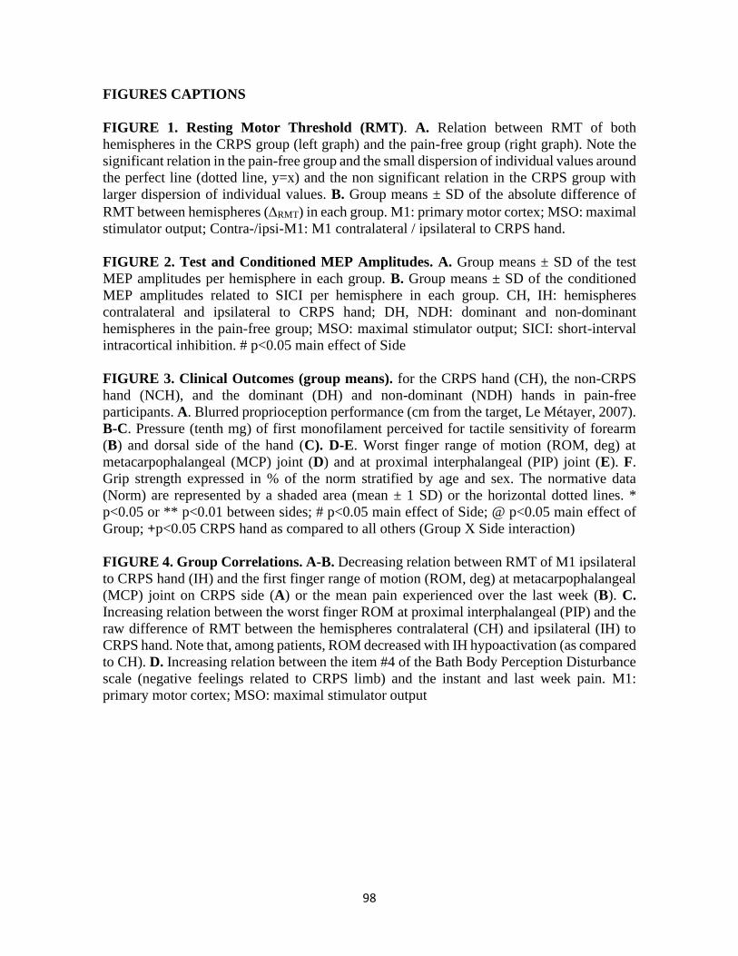
98
FIGURES CAPTIONS
FIGURE 1. Resting Motor Threshold (RMT). A. Relation between RMT of both
hemispheres in the CRPS group (left graph) and the pain-free group (right graph). Note the
significant relation in the pain-free group and the small dispersion of individual values around
the perfect line (dotted line, y=x) and the non significant relation in the CRPS group with
larger dispersion of individual values. B. Group means ± SD of the absolute difference of
RMT between hemispheres (RMT) in each group. M1: primary motor cortex; MSO: maximal
stimulator output; Contra-/ipsi-M1: M1 contralateral / ipsilateral to CRPS hand.
FIGURE 2. Test and Conditioned MEP Amplitudes. A. Group means ± SD of the test
MEP amplitudes per hemisphere in each group. B. Group means ± SD of the conditioned
MEP amplitudes related to SICI per hemisphere in each group. CH, IH: hemispheres
contralateral and ipsilateral to CRPS hand; DH, NDH: dominant and non-dominant
hemispheres in the pain-free group; MSO: maximal stimulator output; SICI: short-interval
intracortical inhibition. # p<0.05 main effect of Side
FIGURE 3. Clinical Outcomes (group means). for the CRPS hand (CH), the non-CRPS
hand (NCH), and the dominant (DH) and non-dominant (NDH) hands in pain-free
participants. A. Blurred proprioception performance (cm from the target, Le Métayer, 2007).
B-C. Pressure (tenth mg) of first monofilament perceived for tactile sensitivity of forearm
(B) and dorsal side of the hand (C). D-E. Worst finger range of motion (ROM, deg) at
metacarpophalangeal (MCP) joint (D) and at proximal interphalangeal (PIP) joint (E). F.
Grip strength expressed in % of the norm stratified by age and sex. The normative data
(Norm) are represented by a shaded area (mean ± 1 SD) or the horizontal dotted lines. *
p<0.05 or ** p<0.01 between sides; # p<0.05 main effect of Side; @ p<0.05 main effect of
Group; +p<0.05 CRPS hand as compared to all others (Group X Side interaction)
FIGURE 4. Group Correlations. A-B. Decreasing relation between RMT of M1 ipsilateral
to CRPS hand (IH) and the first finger range of motion (ROM, deg) at metacarpophalangeal
(MCP) joint on CRPS side (A) or the mean pain experienced over the last week (B). C.
Increasing relation between the worst finger ROM at proximal interphalangeal (PIP) and the
raw difference of RMT between the hemispheres contralateral (CH) and ipsilateral (IH) to
CRPS hand. Note that, among patients, ROM decreased with IH hypoactivation (as compared
to CH). D. Increasing relation between the item #4 of the Bath Body Perception Disturbance
scale (negative feelings related to CRPS limb) and the instant and last week pain. M1:
primary motor cortex; MSO: maximal stimulator output
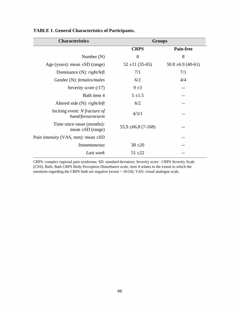
99
TABLE 1. General Characteristics of Participants.
CRPS: complex regional pain syndrome; SD: standard deviation; Severity score : CRPS Severity Scale
(CSS); Bath: Bath CRPS Body Perception Disturbance scale, item 4 relates to the extent to which the
emotions regarding the CRPS limb are negative (worst = 10/10); VAS: visual analogue scale.
Characteristics Groups
CRPS Pain-free
Number (N) 8 8
Age (years): mean ±SD (range) 52 ±11 (35-65) 50.8 ±6.9 (40-61)
Dominance (N): right/left 7/1 7/1
Gender (N): females/males 6/2 4/4
Severity score (/17) 9 ±3 --
Bath item 4 5 ±1.5 --
Altered side (N): right/left 6/2 --
Inciting event: N fracture of
hand/forearm/arm 4/3/1 --
Time since onset (months):
mean ±SD (range) 55,9 ±66,8 (7-168) --
Pain intensity (VAS, mm): mean ±SD --
Instantaneous 38 ±20 --
Last week 51 ±22 --
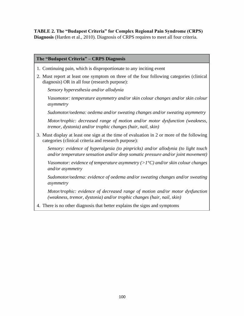
100
TABLE 2. The “Budapest Criteria” for Complex Regional Pain Syndrome (CRPS)
Diagnosis (Harden et al., 2010). Diagnosis of CRPS requires to meet all four criteria.
The “Budapest Criteria” – CRPS Diagnosis
1. Continuing pain, which is disproportionate to any inciting event
2. Must report at least one symptom on three of the four following categories (clinical
diagnosis) OR in all four (research purpose):
Sensory hyperesthesia and/or allodynia
Vasomotor: temperature asymmetry and/or skin colour changes and/or skin colour
asymmetry
Sudomotor/oedema: oedema and/or sweating changes and/or sweating asymmetry
Motor/trophic: decreased range of motion and/or motor dysfunction (weakness,
tremor, dystonia) and/or trophic changes (hair, nail, skin)
3. Must display at least one sign at the time of evaluation in 2 or more of the following
categories (clinical criteria and research purpose):
Sensory: evidence of hyperalgesia (to pinpricks) and/or allodynia (to light touch
and/or temperature sensation and/or deep somatic pressure and/or joint movement)
Vasomotor: evidence of temperature asymmetry (>1°C) and/or skin colour changes
and/or asymmetry
Sudomotor/oedema: evidence of oedema and/or sweating changes and/or sweating
asymmetry
Motor/trophic: evidence of decreased range of motion and/or motor dysfunction
(weakness, tremor, dystonia) and/or trophic changes (hair, nail, skin)
4. There is no other diagnosis that better explains the signs and symptoms
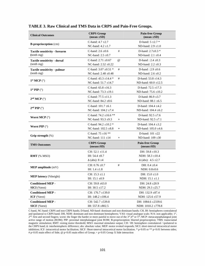
101
TABLE 3. Raw Clinical and TMS Data in CRPS and Pain-Free Groups.
Clinical Outcomes CRPS Group
(mean ±SD)
Pain-free Group
(mean ±SD)
B-proprioception (cm) C-hand: 4.7 ±2.7
NC-hand: 4.2 ±1.7
D-hand: 5 ±2.7 *
ND-hand: 2.9 ±1.0
Tactile sensitivity - forearm
(tenth mg)
C-hand: 2.6 ±0.6 #
NC-hand: 2.5 ±0.7
D-hand: 2.7±0.3 *
ND-hand: 2.1 ±0.4
Tactile sensitivity - dorsal
(tenth mg)
C-hand: 2.71 ±0.67 @
NC-hand: 2.52 ±0.22
D-hand : 2.4 ±0.3
ND-hand: 2.2 ±0.3
Tactile sensitivity - palmar
(tenth mg)
C-hand: 3.07 ±0.51 * #
NC-hand: 2.48 ±0.48
D-hand : 2.9 ±0.6
ND-hand: 2.6 ±0.2
1st MCP (°) C-hand: 43.3 ±14.4 * #
NC-hand: 51.7 ±14.7
D-hand: 53.8 ±14.3
ND-hand: 60.0 ±12.5
1st PIP (°) C-hand: 65.8 ±16.3
NC-hand: 73.3 ±19.1
D-hand: 72.5 ±17.3
ND-hand: 75.6 ±10.2
2nd MCP (°) C-hand: 77.5 ±11.3
NC-hand: 84.2 ±8.6
D-hand: 86.9 ±3.7
ND-hand: 88.1 ±6.5
2nd PIP (°) C-hand: 101.7 ±6.1
NC-hand: 104.2 ±7.4
D-hand: 104.4 ±4.2
ND-hand: 104.4 ±6.2
Worst MCP (°) C-hand: 74.2 ±16.6 **
NC-hand: 93.3 ±9.3 +
D-hand: 92.5 ±7.6
ND-hand: 92.5 ±7.1
Worst PIP (°) C-hand: 94.2 ±10.2 *
NC-hand: 102.5 ±8.8 +
D-hand: 104.4 ±3.2
ND-hand: 105.0 ±4.6
Grip strength (%) C-hand: 75 ±16 **
NC-hand: 111 ±14 +
D-hand: 101 ±22
ND-hand: 109 ±30
TMS Outcomes CRPS Group
(mean±SD)
Pain-free Group
(mean±SD)
RMT (% MSO)
CH: 52.1 ±11.4
IH: 54.4 ±8.7
Δ (abs): 8 ±4
DH: 59.8 ±10.3
NDH: 58.3 ±10.4
Δ (abs): 4.5 ±2.7
MEP amplitude (mV) CH: 0.76 ±0.7 #
IH: 1.4 ±1.8
DH: 0.4 ±0.4
NDH: 0.8±0.6
MEP latency (%height) CH: 15.3 ±1.1
IH: 15.1 ±0.9
DH: 15.0 ±1.0
NDH: 15.1 ±1.1
Conditioned MEP -
SICI (%test)
CH: 59.8 ±63.0
IH: 30.5 ±17.2
DH: 24.8 ±20.9
NDH: 29.3 ±25.7
Conditioned MEP -
ICF (%test)
CH: 176.7 ±139.0
IH: 148.2 ±106.4
DH: 132.9 ±87.4
NDH: 125.6 ±57.9
Conditioned MEP -
SICF (%test)
CH: 542.7 ±539.8
IH: 557.8 ±802.5
DH: 1084.6 ±2339.6
NDH: 1010.2 ±778.8
C-hand, NC-hand: CRPS and non-CRPS hands; D-hand, ND-hand: dominant and non-dominant hands; CH, IH: hemispheres contralateral
and ipsilateral to CRPS hand; DH, NDH: dominant and non-dominant hemispheres; VAS: visual analogue scale; N/A: non applicable; 1st, 2nd: first and second fingers; worst: the finger the harder or more painful to move out of the 3rd,4th or 5th; MCP: metacarpophalangeal joint
active range of motion (ROM); PIP: proximal interphalangeal joint ROM; B-proprioception: blurred proprioception; TMS: transcranial
magnetic stimulation; RMT: resting motor threshold; MSO: maximal stimulator output; CH / IH: hemisphere contralateral / ipsilateral to
the CRPS hand; Δ: interhemispheric difference; abs: absolute value; MEP: motor evoked responds; SICI: short-interval intracortical motor
inhibition; ICF: intracortical motor facilitation; SICF: Short-interval intracortical motor facilitation. * p<0.05 or ** p<0.01 between sides; # p<0.05 main effect of Side; @ p<0.05 main effect of Group; + p<0.05 Group X Side interaction
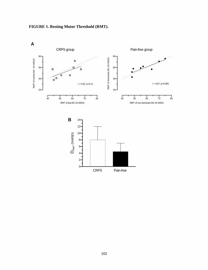
102
FIGURE 1. Resting Motor Threshold (RMT).
A
40 50 60 70 80
20
40
60
80
RMT of ipsi-M1 (% MSO)
RM
T o
f contr
a-M
1 (%
MS
O)
r = 0.62, p=0.11
40 50 60 70 80
20
40
60
80
RMT of non-dominant M1 (% MSO)
RM
T o
f dom
inant M
1 (
% M
SO
)
r = 0.87, p=0.005
CRPS group Pain-free group
0
2
4
6
8
10
12
14
D R
MT (
% M
SO
)
CRPS Pain-free
B
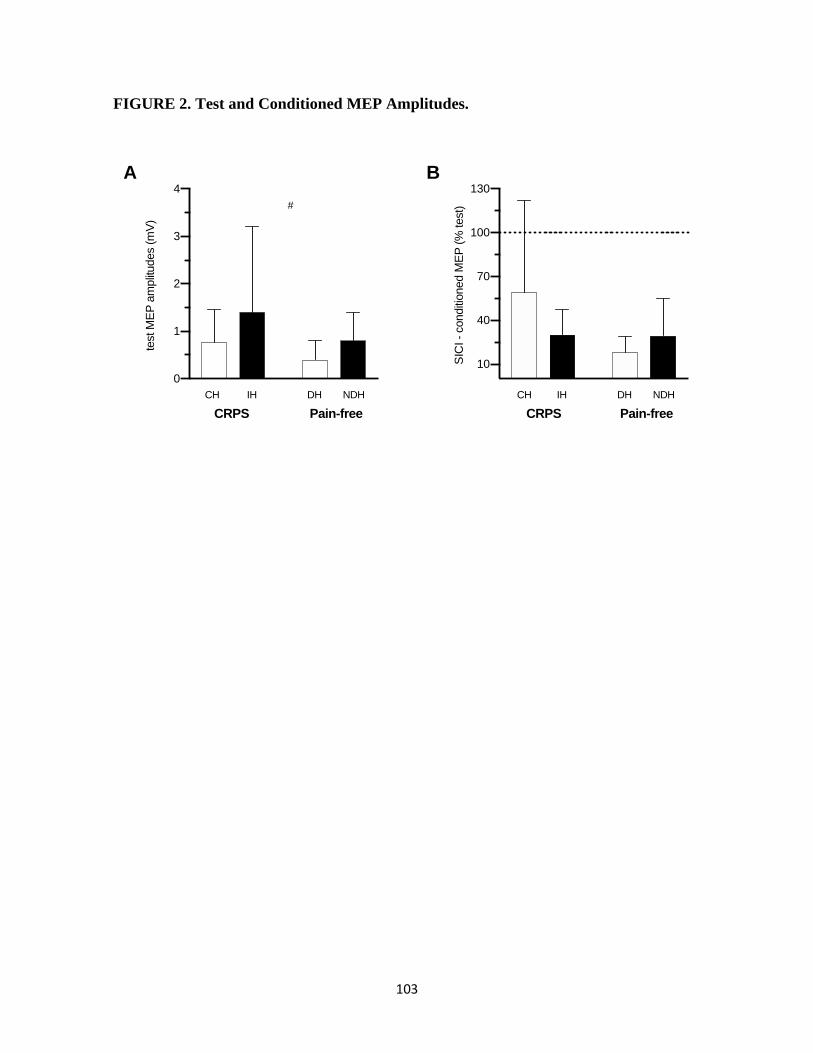
103
FIGURE 2. Test and Conditioned MEP Amplitudes.
0
1
2
3
4
test M
EP
am
plit
udes (
mV
)
#
CH IH DH NDH
10
40
70
100
130
SIC
I -
conditi
oned M
EP
(%
test)
CH IH DH NDH
A B
CRPS Pain-free CRPS Pain-free
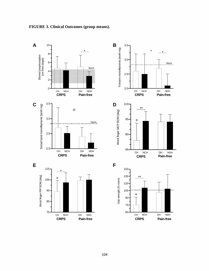
104
FIGURE 3. Clinical Outcomes (group means).
75
85
95
105
115
Wors
t fin
ger
PIP
RO
M (
deg)
+
CH NCH DH NDH
*
2.0
2.5
3.0
3.5
Fore
arm
monofil
am
ents
(te
nth
mg)
Norm
CH NCH DH NDH
*#
60
75
90
105
120
135
150
Grip s
trength
(%
norm
)
+
**
CH NCH DH NDH
2.0
2.5
3.0
3.5
Dors
al h
and m
onofil
am
ents
(te
nth
mg)
@
CH NCH DH NDH
Norm
A B
C
0
2
4
6
8
10
Blu
rred p
roprioceptio
n (
cm
fro
m targ
et)
*
CH NCH DH NDH
Norm
D
CRPS Pain-free CRPS Pain-free
E
65
80
95
110
Wors
t fin
ger
MC
P R
OM
(deg)
+
**
CH NCH DH NDH
F
CRPS Pain-free CRPS Pain-free
CRPS Pain-free CRPS Pain-free
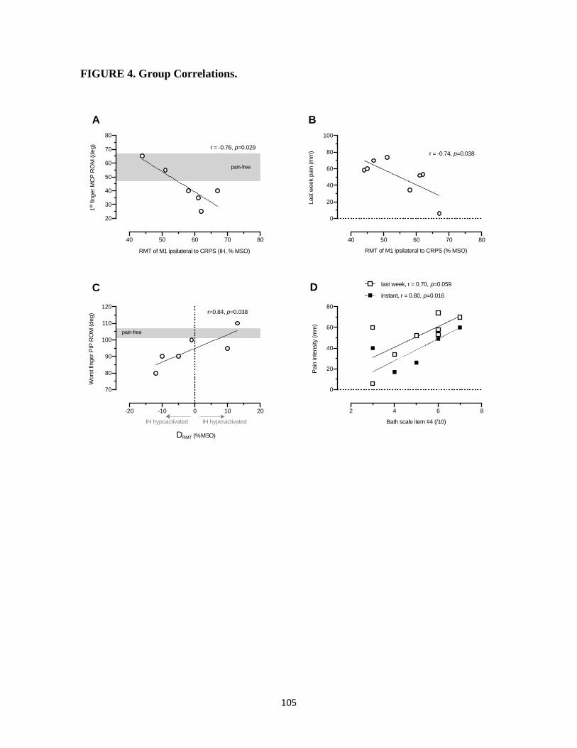
105
FIGURE 4. Group Correlations.
40 50 60 70 80
0
20
40
60
80
100
RMT of M1 ipsilateral to CRPS (% MSO)
Last w
eek p
ain
(m
m) r = -0.74, p=0.038
40 50 60 70 80
20
30
40
50
60
70
80
RMT of M1 ipsilateral to CRPS (IH, % MSO)
1st f
inger
MC
P R
OM
(deg)
r = -0.76, p=0.029
pain-free
-20 -10 0 10 20
70
80
90
100
110
120
D RMT (% MSO)
Wors
t fin
ger
PIP
RO
M (
deg) r=0.84, p=0.038
pain-free
IH hypoactivated IH hyperactivated
A B
C
2 4 6 8
0
20
40
60
80
Bath scale item #4 (/10)
Pain
inte
nsity
(m
m)
last week, r = 0.70, p=0.059
instant, r = 0.80, p=0.016D
106
CHAPITRE 3 : ARTICLE 3 « Theta Burst Stimulation Over Forearm Muscles in
Complex Regional Pain Syndrome (CRPS): Influence on Brain and Clinical Outcomes »
« Stimulations magnétiques intermittentes de fréquence thêta sur les muscles de
l'avant-bras en syndrome douloureux régional complexe (SDRC)- Son influence sur
les résultats cliniques et neurophysiologiques »
L’étude expérimentale 1 (Chapitre 2 : Article 2) a permis de montrer une altération de la
fonction sensorimotrice chez les participants avec SDRC de type I au membre supérieur et le
besoin de considérer la présence d’un débalancement d’excitabilité motrice corticale et le
sens de ce débalancement. Comme il est connu que les rPMS peuvent influencer positivement
la plasticité cérébrale, l’étude expérimentale 2 (article 3) a testé l’apport d’une séance de
rPMS sur la douleur, la fonction et l’excitabilité motrice corticale en SDRC.
Il a été possible de mesurer des changements à la suite d’une seule séance de rPMS : une
diminution du débalancement d’excitabilité motrice corticale, une diminution de l’intensité
de la douleur, une amélioration d’amplitude articulaire active des doigts et de proprioception
du membre supérieur, ainsi qu’une levée d’inhibition et une augmentation de facilitation
motrices corticales (signes de plasticité induite).
Ces résultats originaux supportent l’efficacité et la faisabilité des rPMS en SDRC et le lien
entre rebalancement interhémisphérique d’excitabilité motrice corticale et baisse de la
douleur. Ceci renforce le besoin de mieux comprendre les changements du fonctionnement
sensorimoteur cortical pour personnaliser les traitements de neurostimulation en SDRC.
107
Accepté avec corrections par le journal « Frontiers in Pain Research »
THETA BURST STIMULATION OVER FOREARM MUSCLES IN COMPLEX
REGIONAL PAIN SYNDROME (CRPS): INFLUENCE ON BRAIN AND CLINICAL
OUTCOMES.
ALLEN DEMERS Fannie, MPht1-3; ZANGRANDI Andrea, MSc1-3; SCHNEIDER Cyril,
PhD 1-4*
1 Noninvasive Stimulation laboratory (NovaStim)
2 Neuroscience Division of Centre de recherche du CHU of Québec – Université Laval
3 Faculty of Medicine, Université Laval, Quebec City, Canada
4 Dept Rehabilitation, Faculty of Medicine, Université Laval, Quebec City, Canada
*Corresponding author: Prof Cyril Schneider, Noninvasive Stimulation laboratory,
Neuroscience Division, Research Center of CHU of Québec – Université Laval, CHUL-2705
boul. Laurier, RC-9800, Québec QC, Canada, G1V 4G2, Tel. 418-525-4444 (47648), Fax.
418-654-2753, Email: [email protected]
Sources of support. The authors acknowledge the Canadian Foundation for Innovation (CS
equipment), the Canadian Pain Network (CPN) funded by the strategy for patient-oriented
research (SPOR) of the Canadian Institutes of Health Research (CS funds), the Fonds de
Recherche du Québec-Santé (FRQS, FAD studentship) and the Centre Thématique de
Recherche en Neurosciences (CTRN-Université Laval, Québec, AZ studentship).
108
RÉSUMÉ
Contexte et objectifs : Le syndrome douloureux régional complexe (SDRC) est caractérisé
par des changements cérébraux qui ne sont actuellement que peu adressés par les traitements
conventionnels. Les stimulations magnétiques répétées en périphérie (rPMS des muscles)
sont indolores et non invasives et peuvent influencer ces changements (induire une plasticité
cérébrale) pour réduire la douleur et améliorer la motricité. Cette étude originale a testé leurs
effets immédiats en SDRC sur l’intensité de la douleur, le contrôle sensorimoteur du membre
supérieur et les changements d’excitabilité du cortex moteur primaire (M1).
Méthodologie : Quatorze adultes ont été recrutés (six avec un SDRC, huit sans douleur). Le
groupe avec SDRC a été testé en pré- et post-rPMS du muscle fléchisseur superficiel des
doigts atteint et comparé aux valeurs normatives du groupe Sans-douleur (sans rPMS). Les
variables cliniques mesurées étaient : l’intensité de la douleur, la proprioception, l’amplitude
articulaire active et la force musculaire de préhension. L’excitabilité du M1 a été testée par
stimulations magnétiques transcrâniennes (TMS) simple et double du M1.
Résultats : Les rPMS ont réduit l’intensité de la douleur instantanée et de la semaine,
normalisé le débalancement d’activité hémisphérique, amélioré la proprioception et
l’amplitude articulaire et induit des changements plastiques que dans le M1 de l’hémisphère
ipsilatéral (HI) au SDRC. Plus l’HI était hypoactivé au départ, plus la douleur était réduite.
Conclusion : Cette étude original fournit des résultats prometteurs pour l’utilisation des
rPMS en SDRC en mettant l’accent sur les changements plastiques de l’HI. Des études
futures devront confirmer l’existence d’une relation de cause à effet entre les variables TMS
et la diminution de douleur post-rPMS pour favoriser le développement de traitements
personnalisés de neurostimulation non invasive en SDRC.
Mots-clés : Syndrome douloureux régional complexe, SDRC, rPMS, TMS, douleur
chronique, plasticité, neurostimulation
109
ABSTRACT
Background and Objective. The complex regional pain syndrome (CRPS) is characterised
by brain changes that are currently only rarely addressed by conventional treatment regimens.
Repetitive peripheral magnetic stimulation (rPMS of muscles) is painless and noninvasive
and it can influence these changes (induction of brain plasticity) to reduce pain and improve
motricity. This original study in CRPS tested rPMS after-effects in CRPS on pain intensity
and sensorimotor control of the upper limb, along with changes of the primary motor cortex
(M1) excitability.
Methods. Fourteen adults were enrolled (six with CRPS, eight pain-free) in a single session.
The CRPS group was tested at pre- and post-rPMS over the flexor digitorum superficialis
muscle on the CRPS side and was compared to normative pain-free group values (no rPMS).
The clinical outcomes were pain intensity, proprioception, active range of motion, grip
strength. M1 excitability was tested using single and paired-pulse transcranial magnetic
stimulation (TMS) of M1.
Results. rPMS reduced instant and week pain, normalized the hemispheric activity
imbalance, improved proprioception and range of motion, and induced plastic changes only
in M1 ipsilateral to CRPS side (IH). The more IH was hypoactivated at baseline, the more
pain was reduced.
Discussion. This original study provided promising findings for the use of rPMS in CRPS
with a focus on the plastic changes of IH. Future studies should confirm the existence of a
causal relationship between TMS outcomes and post-rPMS decrease of pain. This will favor
the development of personalized treatments of noninvasive neurostimulation in CRPS.
Keywords: CRPS, rPMS, TMS, chronic pain, plasticity, neurostimulation
110
INTRODUCTION
Complex Regional Pain Syndrome (CRPS), formerly known among others as reflex
sympathetic dystrophy or algoneurodystrophy, is a chronic pain condition characterized by
pain to one or more limbs which is disproportionate to the inciting event (fracture, sprain,
surgery, or no traumatism identified) combined with trophic changes and sensory, motor and
autonomic disorders (Borchers & Gershwin, 2014; Marinus et al., 2011). All causes leading
to CRPS remain unclear but evidence proposes three mechanisms explaining the symptoms:
peripheral changes and sensitization, dysregulation of the sympathetic nervous system and
maladaptive neuroplasticity (Allen Demers et al., in preparation, February, 2021; Borchers
& Gershwin, 2014; Goh et al., 2017). Interindividual variability of the contribution of each
mechanism over time makes it difficult to administer an efficient treatment for all people
with CRPS.
Conventional treatments include medical interventions (medications, topical cream,
injections, etc.), rehabilitation (physical and/or occupational therapies) and, ideally,
psychological therapy and follow-up. But even with clinical guidelines and medical follow-
up of response to treatment, there is almost no evidence to support therapies currently used
in CRPS (Bruehl, 2015; Truchon, 2015; O’Connell et al., 2015). Actually, more than half of
people diagnosed with CRPS still present deficiencies as diminution of strength or stiffness
one year after onset (Bean et al., 2014), 62% of diagnosed people still have activities of daily
living limitations 3 to 9 years after onset (Geertzen et al., 1998) and most are unable to come
back to their previous occupation, need workplace adjustments or are declared officially
disabled (Borchers and Gershwin, 2014).
The persistence of symptoms and refractoriness to treatment could be due to central changes
that are not sufficiently influenced by conventional approaches, such as changes of volumes,
connectivity, activation and excitability of different parts of the brain (Allen Demers et al.,
in preparation, February, 2021; Barad et al., 2014; Geha et al., 2008; Kim et al., 2013;
Kirveskari et al., 2010; Maihöfner et al., 2003; Pleger et al., 2006; Schwenkreis et al., 2009;
Vartiainen et al., 2008; Walton et al., 2010; Zhou et al., 2015). Among the changes, the
111
imbalanced hemispheric activity between motor cortices (M1) has been pointed out (Barad
et al., 2014; Eisenberg et al., 2005; Kirveskari et al., 2010; Krause et al., 2006; Maihöfner et
al., 2007, 2003; Di Pietro et al., 2013; Schwenkreis et al., 2003). However, the direction of
the imbalance remains controversial: is the hemisphere contralateral to the CRPS hand (CH
controlling the limb with CRPS) hyperexcitable or hypoexcitable as compared to the
hemisphere ipsilateral (IH controlling the non-CRPS hand)? Our recent original study (Allen
Demers et al., in preparation, February, 2021) tested M1 excitability of adults with CRPS
type I by means of transcranial magnetic stimulation (TMS). The results showed that one
size does not fit all, i.e. that pain did not influence M1 excitability in the same way between
the participants, thus providing a clue for the controversy in the literature on the direction of
imbalanced hemispheric activity and likely contributing to the misunderstanding of how to
appropriately / efficiently adapt CRPS treatments.
One new technology currently proposed in clinical research on CRPS is noninvasive
magnetic neurostimulation, which is painless and can sustainedly inhibit or excite
sensorimotor areas (depending on the parameters used), thus influencing neuroplasticity
related to pain and motor improvement. However, the possibility that imbalance between CH
and IH excitability can be different between people with hand CRPS (Allen Demers et al.,
unpublished data, February, 2021) questions the rationale of studies that all administrated
excitatory neurostimulation over the M1 of CH. It is indeed legitimate to think that if M1
excitability is related to CRPS pain intensity (Allen Demers et al., in preparation, February,
2021) then the protocols chosen to reduce the interhemispheric excitability imbalance and
impact pain should be personalized according to the imbalance direction. Another point of
importance is kinesiophobia (Marinus et al., 2013), i.e. the fear of movement in CRPS due
to pain induced, thus the installation of a vicious circle of non-use of the CRPS limb that
worsens further the impaired motor control due to reduced proprioceptive afferents (Allen
Demers et al., unpublished data, February, 2021).
Repetitive peripheral magnetic stimulation (rPMS) is applied with a coil positioned on skin
over the muscles (Struppler et al., 2003b). The intensity is suprathreshold to trigger muscle
contraction and mimic the mechanisms of muscle contraction/relaxation: this generates flows
112
of proprioceptive information to brain from either the structures stimulated (afferents, muscle
fibers, nerve terminals) and the muscles stretched by joint movement (Beaulieu & Schneider,
2013, 2015; Massé-Alarie & Schneider, 2011). rPMS in physiopathology (coil over painful
area or spastic paretic muscles) have been shown to induce changes of fronto-parietal
networks activity (Struppler et al., 2007) and of M1 excitability (Massé-Alarie et al., 2017;
Beaulieu et al. 2015, 2017). This was reported with clinical significance, i.e. pain reduction
and motor improvement in people with chronic low back pain (Massé-Alarie et al., 2013,
2017) or with brain lesion (Beaulieu et al., 2015, Beaulieu et al., 2017, Krewer et al., 2014,
Struppler et al., 2003b), and enhancement of perceptual-cognitive function (Struppler et al.,
2003a). It is noteworthy furthermore that electroencephalography studies have shown that
suprathreshold peripheral magnetic stimulation did elicit somatosensory evoked potentials in
the primary sensory areas that are of almost pure proprioceptive origin, i.e. with negligible
cutaneous and nociceptive inflow as compared to electrical stimulation (Kunesch et al., 1993,
Zhu & Starr, 1991). Thus, rPMS administration is painless and with minimal cutaneous
contamination of the proprioceptive signals to sensorimotor areas (Beaulieu & Schneider,
2015). The mechanisms of action of rPMS could be related to long-term potentiation (LTP)
and depression (LTD) which are associated with neuroplasticity (Hasselmo, 1995; Pell et al.,
2011). In CRPS, these mechanisms are involved in central sensitization (Puchalski & Zyluk,
2016). Thus, rPMS in people with CRPS could potentially have a triple influence leading to
reduce pain intensity and improve motor control, i.e. central desensitization (by influence on
LTP/LTD), rebalance of hemispheric activity (by influence on M1 excitability), and
suppliance of the reduced proprioceptive information.
Therefore, the present study aimed at testing in people with hand CRPS type 1 (no nerve
lesion) if a single session of rPMS could reduce pain (primary outcome) and improve
function of the affected limb. rPMS was administrated over the flexor digitorum superficialis
muscle (FDS), largely involved in hand function, and the acute after-effects were assessed
by means of clinical outcomes (pain intensity, fingers active range of motion, grip strength,
proprioception) and TMS outcomes (M1 function, including corticospinal and intracortical
excitability). Data collected before and after rPMS were compared to data of a group of pain-
free participants.
113
MATERIALS AND METHODS
Participants and Procedures
Six adults with hand type-1 CRPS (54.3 ±10.6 years old, see Table 1) were enrolled in an
open-label single session of rPMS (plus pain intensity follow-up at one week) and were
compared at pre- and post-rPMS to a group of eight pain-free adults (50.8 ±6.9 years old)
tested at one time point only (no rPMS). All participants had signed the informed consent
form approved by the local ethics committees. The participants with CRPS presented with a
unilateral upper limb CRPS type 1 diagnosed with the Budapest clinical criteria (see Table
2) and were discharged from pain clinics follow-up. The pain-free participants did not
experience pain in the last month before enrolment nor have medical history of chronic pain
(pain persisting for more than three consecutive months). All participants met the exclusion
criteria related to general health (spine surgery, major circulatory/respiratory/cardiac disease,
other neurological condition for participants with CRPS, severe upper limb orthopaedic
condition, cognitive disorder interfering with the tasks of the study) and to TMS safety
guidelines (Lefaucheur et al., 2011; Rossi et al., 2011, 2009). A physician performed medical
evaluation at pre- and post-enrolment to confirm eligibility and monitor adverse effects. None
of the participants had history of specific repetitive motor activity (as a musician or
professional level athlete). The physical therapist collected clinical and TMS outcomes also
applied rPMS but data were then codified for group (CRPS vs. pain-free) and time of testing
(pre- vs. post-rPMS for CRPS group) until completion of analyses. The fact that the same
physical therapist ensured clinical testing for all participants reduced the inter-evaluator
variability. All outcomes were collected in a single 3-hour session (including rest breaks), as
follows: clinical outcomes, TMS outcomes, then rPMS intervention over FDS, and 10min
later TMS outcomes and clinical outcomes. All outcomes were collected on both sides (when
appropriate) for all participants. Everyone was phoned 2 and 7 days later to document any
adverse effect (Rossi et al. 2009). Participants with CRPS were questioned on the mean
intensity of week pain at 7 days (one-week follow-up).
(Insert Tables 1-2 near here)
114
Intervention
Participants were comfortably seated in a reclining-adjustable chair with limb supports and
the forearms in supination. rPMS was administered on CRPS side with an air-film cooled
figure-of-eight coil (7 cm outer diameter per wing, biphasic waveform, 400-µs pulse width,
Rapid2 Magstim, Magstim Company, England) held tangentially on the skin overlying the
FDS muscle belly with the long axis of coil junction perpendicular to muscle fibres
orientation (for a review of parameters of rPMS application, see Beaulieu and Schneider,
2015). The FDS was the target muscle because of the decrease of fingers/wrist flexion
AROM commonly reported in CRPS with functional significance, such as grasping
impairment (Bean et al., 2014; Geertzen et al. 1998; de Mos et al. 2007; Rodham et al. 2015).
rPMS was delivered at theta-burst frequency (5-Hz trains of three pulses at 50 Hz during
200s, 2s ON / 8s OFF, 600 pulses total). This intermittent mode of rPMS was used to elicit
cyclic muscle activation/relaxation as already reported by our research group (Beaulieu et
al., 2015, 2017; Flamand et al., 2012; Flamand & Schneider, 2014, Schneider et al. 2016;
Massé-Alarie et al., 2013, Massé-Alarie et al., 2017). rPMS intensity was set at 42% of
maximal stimulator output (the maximal possible on TBS mode with Rapid2 Magstim
equipment) to produce palpable FDS contractions with visible wrist/fingers flexion which
was painless for the CRPS participants.
Clinical Testing
All following outcomes (except pain and proprioception) were collected on both sides (either
hand for clinical or hemisphere for TMS of M1), before and ten minutes after rPMS for the
CRPS group, or once for the pain-free group.
Pain Intensity. Participants’ sitting position was standardized against a chair’s backrest with
the shoulder in neutral rotation and 0° abduction, elbow in 90-100° flexion, and forearm
resting on a height-adjustable therapeutic table. The visual analogue scale (VAS) was used
to quantify the pain intensity experienced by each participant with CRPS. The VAS consists
of a two-sided band: on one side, the participant uses a cursor to rate the level of pain intensity
between two extremes called "No pain" and "Maximal pain imaginable"; on the other side of
VAS, hidden from the participant, a graduated scale from 0 (no pain) to 100 millimeters
115
(maximal pain imaginable) is used by the evaluator to quantify the participant’s pain
intensity. VAS was used to quantify instantaneous pain at pre- and post-rPMS and the mean
pain in the last week (pre-rPMS week pain) and at follow-up one week later (post-rPMS week
pain).
Active Range of Motion (ROM). Active ROM in flexion was measured using a manual finger
goniometer at the proximal interphalangeal (PIP) joints of the first, second and “worst”
finger, i.e. the finger (between the third, fourth and fifth) harder or more painful to move for
each participant. For active ROM of the second and worst fingers PIP, the elbow was bent
(±130°) at rest on the therapeutic table, the shoulder slightly bent (±45°), the wrist and
metacarpophalangeal joints in the neutral position (claw hand). For active ROM of the first
finger PIP, the forearm was positioned at rest on the table with the hand protruding on the
edge. Three trials were tested and averaged per PIP. Participants rested 60sec between trials.
Grip Strength. Participants were sitting with no arm support, the forearm in neutral position
and the wrist between 0-30° extension and 0-15° ulnar deviation. The maximal grip strength
per hand was measured using a JAMAR hydraulic hand dynamometer (Sammons Preston
Rolyan, Bolingbrook-IL, USA). Following Mathiowetz’s instructions (Mathiowetz et al.,
1984), JAMAR dynamometer was set to the second handle position (placed in hand with help
of the evaluator if needed). The average of three trials per hand was used to represent the
maximum grip strength of each participant. Standardized cheers were given ("Squeeze hard,
hard, hard, the hardest and release") over 8 sec of sustained contraction to promote a reliable
value (Buckinx et al., 2017). Participants rested 30s between trials.
Upper Limb Proprioception. Following Le Métayer’s guidelines, the paradigm of the blurred
proprioception of the upper limb was used to assess the ability to perceive limb position in
space (Le Métayer, 2007). Briefly, the participants were seated, leaning against a chair
without armrests, knees bent at 90° with feet flat on the floor and hands on the thighs (initial
position). They had first to reach with the index a target (large red dot) on a graduated screen
at gaze height and arm distance in front of them and return to the initial position and to repeat
this practice three times. Then, they closed the eyes and the evaluator cautiously (without
116
pain) moved their tested arm in the air with shoulder movements and elbow flexion /
extension and put the arm back in the initial position, and the participants, without opening
the eyes, had to reach back the target with the index (one trial only). The score of blurred
proprioception is the distance in centimeter between the finger contact on the graduated
screen (with eyes closed) and the target.
Surface Electromyographic (EMG) Recordings
EMG procedures were followed without induced pain in participants with CRPS. After
standard skin preparation (i.e. cleaning the skin with alcohol and shaving whenever
necessary) (Hermens et al. 2000), the parallel-bar EMG sensors with adhesive skin interfaces
were installed on each participant (fixed 1cm distance between electrodes; 16-channel
Bagnoli EMG System, Delsys Inc., Boston, MA). EMG sensors were placed bilaterally on
the belly of the first dorsal interosseous muscle (FDI). A common ground electrode was
placed on the ulna olecranon of the tested limb. EMG signals were band-pass filtered (20-
450 Hz), amplified before digitization (2 kHz) and computer-stored for offline analysis
(PowerLab acquisition system, LabChart, ADInstruments, Colorado Springs-CO, US).
TMS Testing
M1 function at pre-rPMS and 10 min post-rPMS of FDS was tested by TMS of FDI M1 area.
FDI M1 excitability is easier to test as compared to FDS and is known to be increased by
upper extremity flexion owing to a proximo-distal synergy in M1 (Gagné & Schneider, 2007,
2008). TMS procedures were strictly replicated for each hemisphere pre- and post-
intervention, following the guidelines from the International Federation of Clinical
Neurophysiology (Rossini et al., 2015). Participants were comfortably in the reclining chair
with arms and legs supported, hips and knees slightly bent (20° flexion).
Hotspot. Magnetic stimuli were applied over FDI M1 area by a figure-of-eight coil (7-cm
outer diameter each wing; Magstim Company Limited, Whitland-UK) positioned on the
scalp with the long axis of the two- wing intersection pointing antero-posteriorly at a 45°
angle from the medial line to active M1 cells in the postero-anterior direction (Sakai et al.,
117
1997). The 10–20 EEG system was used to first approximate FDI M1 area (Klem, 1999).
The position was then adjusted slightly to determine the ‘hotspot’, namely the M1 location
where the MEP obtained in the contralateral FDI are of the highest amplitude at the lowest
TMS intensity (Wolf et al., 2004).This hotspot was marked on the scalp (surgical pen) to
ensure reliable coil positioning across time of testing (Wolf et al., 2004) and visual inspection
of EMG recordings from both sides monitored the complete relaxation of the hands during
TMS testing.
Resting Motor Threshold (RMT). RMT was determined at the TMS intensity that elicited at
least five FDI MEP with an amplitude of 50 μV or higher out of ten trials. RMT has good
validity and reliability (Beaulieu et al., 2017a, 2017c; Malcolm et al., 2006) and it informs
on M1 basic excitability (Rossini et al., 2015, Ziemann et al., 2015).
MEP Amplitude and Latency. A suprathreshold test (unconditioned) TMS at an intensity of
120% RMT enabled to collect and measure the test MEP amplitude and latency. Test MEP
amplitude (in mV) was measured peak-to-peak and it reflects the volume of M1 cells
activated by TMS and the excitability of the corticospinal pathway (Devanne et al., 1997,
Kobayashi & Pascual-Leone, 2003, Rossini et al., 2015). Test MEP latency (in %
participant’s height) was measured from stimulation time to MEP onset and it reflects
conduction time and indirectly the synchronicity of descending volleys to depolarize spinal
motoneurons (Groppa et al., 2012, Kobayashi & Pascual-Leone, 2003).
SICI, ICF and SICF. Paired-pulse TMS (coil connected to two Magstim 2002 monophasic
stimulators) was used to test the short-interval intracortical inhibition (SICI), the intracortical
facilitation (ICF) and the short-interval intracortical facilitation (SICF) (Arya et al., 2011,
Ilić et al., 2002, Kujirai et al., 1993). In SICI, a subthreshold conditioning TMS (80 % RMT
eliciting no MEP on its own) was delivered 3 msec before the test TMS at 120 % RMT
(Kujirai et al., 1993). In ICF, the same parameters were used but at an inter-stimulus interval
of 15 ms (Kujirai et al., 1993). The conditioned MEP expressed post-hoc relative to the mean
test MEP amplitude is usually of lower amplitude than the test in SICI and of higher
amplitude in ICF (Kujirai et al., 1993). SICI probes the function of GABAA interneurons
118
within M1 and ICF the function of oligosynaptic glutamatergic circuits (Rossini et al., 2015;
Ziemann et al., 2015). In SICF, two stimuli were delivered 1 ms apart, the first TMS at 100
% RMT and the second at 90 % RMT. MEP amplitude was then expressed relative to the
amplitude of test MEP at 100 % RMT, which likely reflects I-waves summation following
the depolarisation of M1 interneurons by TMS (Ilic et al., 2002; Rossini et al., 2015; Ziemann
et al., 2015). The test TMS intensity was adjusted at post-rPMS to match the amplitudes of
test MEP obtained at pre-rPMS for the validity of comparisons of conditioned MEP
amplitudes between the two times of testing (Beaulieu et al., 2015). Twelve test MEP and 12
conditioned MEP were collected per participant at a frequency of 0.2-0.3 Hz with 10%
variations between trials. Resting breaks were given on request.
Data Reduction and Statistical Analysis
Five clinical outcomes were measured at pre- and post-rPMS: instant pain (mm), week pain
(mm), blurred proprioception (cm), active ROM (degree), and grip strength. To ensure
validity of interindividual comparison, grip strength was expressed in percentage of grip
strength norms stratified by age and sex (Mathiowetz et al., 1985). Pain at post-rPMS was
also expressed in % pre-rPMS. Six TMS outcomes were collected at pre- and post-rPMS:
RMT (% of maximal stimulator output) also expressed in % of RMT at pre-rPMS, between-
hemisphere difference of RMT (% MSO), test MEP amplitude (mV), MEP latency (%
participant’s height), and conditioned MEP amplitudes for SICI, ICF, and SICF (%
respective test MEP amplitude).
Shapiro-Wilk’s test for Normality revealed that data were normally distributed, thus
parametric tests were used. Bilateral paired Student’s t-tests between pre- and post-rPMS
values were applied to instant and week pain, blurred proprioception and the between-
hemisphere difference of RMT. The clinical and TMS outcomes were averaged between
sides in the pain-free group (p>0.05). One-way analysis of variance (Fisher’s ANOVA) was
applied to all outcomes (except pain) between CRPS side (hand or hemisphere), non-CRPS
side and the mean of the pain-free group. In the CRPS group, two-way ANOVA with
repeated measures were applied with factors Time (pre- vs. post-rPMS) X Hand (CRPS vs.
non-CRPS) to all clinical outcomes (except pain and proprioception) and with factors Time
119
X Hemisphere (Contralateral-CH vs. Ipsilateral-IH) to all TMS outcome (except between-
hemisphere difference of RMT). Planned comparisons (two-sided, Tukey’s post-hoc tests for
multiple comparisons) were used to detect where differences detected by ANOVA lay.
Pearson’s correlation matrices were produced to examine the relationships between the
change of primary outcome (pain intensity) and the amount of change or baseline values of
other variables. The maximum normed residual test (Grubbs' test) helped detect an outlier
from a data set approximated by normal distribution (Prism 8.0, GraphPad Software Inc, La
Jolla-CA, USA): data set is analyzed and an outlier would typically be expunged when it falls
beyond a critical deviation from the sample mean (chance probability to obtain the outlying
value < 0.05). Significance was set at p < 0.05. Statistical analysis was conducted with Jamovi
(https://www.jamovi.org).
120
RESULTS
TMS testing was truncated for one participant per group due to head discomfort during the
application of the paired-pulse TMS for SICI and ICF in both hemispheres, and another in
the CRPS group for CH. Thus, SICI and ICF data in IH are presented for N=5 participants
with CRPS and 7 pain-free subjects and in CH for N=4 participants with CRPS and 7 pain-
free subjects. All other outcomes under study were collected with no problem and no adverse
effect was reported at follow-ups. Table 3 presents all clinical and TMS outcomes per group.
(Insert Table 3 near here)
Clinical Outcomes
Four participants with CRPS (out of six) reported instant pain reduction after rPMS (mean
decrease of 8 mm on the VAS corresponding to 26% reduction, range of 20-100% reduction)
and all but one did report week pain reduction at the one-week follow-up (mean decrease of
8 mm on the VAS corresponding to 17% reduction, range of 15-25%, see Fig. 1A, left graph).
Pre- vs. post-rPMS reduction of instant pain was close to significance (t-test p=0.06) whereas
week pain reduction was significant (p=0.03, Fig. 1A right graph with post-rPMS pain
expressed as % pre-rPMS).
Blurred proprioception of the CRPS upper limb was improved in all participants but one
(group mean of 2 cm closer to the target, Fig. 1B, left graph). The one participant with no
improvement was already better than the pain-free participants (shaded area). Pre- vs. post-
rPMS improvement of blurred proprioception on the CRPS side was on the edge of
significance (p=0.05, Fig. 1B, right graph).
The Side X Time ANOVA applied on the active ROM did not detect any effect. The one-
way ANOVA (comparison to pain-free subjects) was significant for the worst finger PIP
ROM at pre-rPMS (F2, 17 = 3.61, p=0.049, Fig. 1C, right graph) and not significant at post-
rPMS (F2, 16 = 0.61, p= 0.56). Post-hoc test denoted that at pre-rPMS values of the CRPS
hand (95.0 ±5.0 degrees) were smaller than in pain-free subjects (104.7 ±3.1, p=0.045).
121
Individual data (Fig. 1C, left graph) showed that only two participants had post-rPMS
increase of the worst PIP ROM.
The Side X Time ANOVA applied on the grip strength detected a main effect of SIDE (F1, 5
= 18.2, p=0.008), but no effect of rPMS, with the CRPS side weaker than the non CRPS at
pre-rPMS (p=0.023) and at post-rPMS (p=0.027, see Table 3). The one-way ANOVA was
significant (F2,17 = 5.51, p=0.014) with the grip strength higher in pain-free subjects than on
the CRPS side (p=0.034, see Table 3).
(Insert Fig. 1 near here)
TMS Outcomes
Individual CRPS data of Fig. 2A shows that at pre-rPMS the RMT of IH (M1 ipsilateral to
CRPS hand) was higher than RMT of CH (lower M1 excitability in IH) in four participants
and lower in two participants (higher M1 excitability in IH). The absolute value of this
imbalance was higher than 7% MSO at pre-rPMS for four participants and for only two at
post-rPMS. The bilateral paired t-test applied on the absolute difference of RMT between
hemispheres (whatever the direction of difference) detected a close to significant decrease of
imbalance at post-rPMS (5.0 ±4.3 MSO) as compared to pre-rPMS (8.5 ±4.6 MSO, p=0.06,
Fig. 2B). Of note, at post-rPMS, the mean of hemispheric difference of RMT of the CRPS
group is within values of the pain-free group (shaded area).
The Side X Time ANOVA applied to MEP latency detected a main effect of TIME (F1,5 =
6.81, p=0.048, Fig. 3A) with longer latencies at post-rPMS, significantly for IH (p=0.028).
The Side X Time ANOVA applied on the conditioned MEP (SICI, ICF, SICF) did not detect
any effect. However, for SICI (Fig. 3B), the one-way ANOVA revealed that, as compared to
pain-free participants (27.0 ±11.9 % test MEP), the conditioned MEP amplitudes were
significantly higher in IH at post-rPMS (159.7 ±245.2 % test MEP, p=0.03). In CH, they
were close to be significantly higher than in pain-free group at pre-rPMS (67.2 ±65.5,
p=0.051). The conditioned MEP for ICF (Fig. 3C) were higher in IH at post-rPMS (564.9
122
±795.8 % test MEP) as compared to pain-free group (129.2 ±53.5 % test MEP, p=0.02). No
other between-side or between-time or between-group differences were detected (p>0.05).
Correlations
Fig. 4 presents the negative relationship (r = -0.84, p=0.04) between the amount of post-
rPMS instant pain reduction (in % pre-rPMS) and the RMT at baseline (pre-rPMS) of IH
(expressed in % of CH). In other words, instant pain was more reduced after rPMS in patients
with IH RMT higher (lower M1 excitability) than in CH at baseline. More precisely, the more
CH was hyperactivated as compared to IH (larger imbalance), the larger was pain decrease
after rPMS.
(Insert Figs. 2-3-4 near here)
123
DISCUSSION
This original CRPS study provides new evidence of one single rPMS session influence on
pain and hand sensorimotor function with dynamic changes of M1 excitability. The potential
mechanisms involved are discussed in order to better understand rPMS-driven changes.
Reduction of Pain and Improvement of Function
Only one study used rPMS in literature in CRPS but collected only TMS outcomes with no
clinical data and did not report any significant effect in patients (Krause et al. 2005). The
authors used a single session as we did but rPMS was applied over the cervical spine (C7-
C8) at 20 Hz for a total of 2000 pulses over 10 min whereas we used iTBS (50 Hz) over the
FDS on the painful side for a total of 600 pulses over 3min 20 sec. The protocols are thus not
comparable. However, iTBS over muscles has already been used successively by our
research group to decrease pain and improve posture-motor control in people with other pain
conditions, such as chronic low back pain (Massé-Alarie et al., 2013). Our present results of
a decrease of instant pain at post-rPMS and persistence over one week denotes the promising
potential of rPMS in CRPS. Pain changes obtained in one single session of rPMS in parallel
with improvement of worst finger PIP active range of motion as compared to pain-free
participants resemble changes reported in studies having used TENS (transcutaneous
electrical neurostimulation) but in 15 to 30 sessions (Bilgili et al., 2016, Bodenheim &
Bennett, 1983, Kesler et al., 1988, Richlin et al., 1978) or in 5 sessions of TENS combined
with brain stimulation (Houde et al., 2020). However, rPMS increased the proprioceptive
signals to brain with minimal contamination of proprioceptive-motor transduction by
cutaneous responses, unlike with TENS (Beaulieu & Schneider, 2015; Kunesch et al., 1993,
Zhu & Starr, 1991). The improvement of performance after rPMS in the blurred
proprioception paradigm (Le Métayer, 2007) provided first ever evidence that rPMS-induced
flows of ascending signals could promote proprioceptive integration and sensorimotor
control in CRPS and could have changed the perception of the CRPS upper limb in space.
This was first suggested by the rPMS-induced and PET-scanned activation of the frontal-
parietal pathways involved in perception and sensorimotor planning (Struppler et al., 2007).
In further support, the decrease of week pain after rPMS could reflect an intertwined action
of rPMS on sensorimotor control and pain modulation mechanisms, as already suggested in
124
chronic low back pain (Massé-Alarie et al., 2017). Thus, the increased availability of
proprioceptive inputs following rPMS (which are decreased in CRPS due to non-use of the
limb) may have contribute to the selection of more efficient strategies of pain management
and motor control of the fingers.
Resting Motor Threshold in CRPS: a Predictive Factor of Responsiveness to rPMS ?
The intermittent theta-burst frequency used for rPMS and that improved proprioceptive
integration in coherence with voluntary muscle contraction did likely influence sensorimotor
plastic mechanisms with no spinal saturation or fatigue, as already shown in chronic low
back pain and chronic stroke (Massé-Alarie et al., 2013; Beaulieu et al., 2017b). The inner
mechanisms may be an action on glutamate and GABA receptors synthesis thus leading to
LTP or LTD in M1 (Huang et al., 2005). It is known that these receptors influence M1
homeostasis (M1 basic excitability) which is tested by the resting motor threshold to TMS.
One new result in CRPS is the association between the imbalance of hemispheric activity at
pre-rPMS (tested by RMT) and pain reduction, as shown by Fig. 4, but also by individual
data in Fig. 1A. Precisely, the higher the RMT was in M1 ipsilateral to CRPS hand at
baseline, i.e. IH hypoactivated (or CH hyperactivated) at pre-rPMS as compared to the other
side, the larger was pain decrease at post-rPMS. We thus question whether people with CRPS
who will benefit more from rPMS after-effects present with a suboptimal function of IH
(hemispheric imbalance in the direction of an hypoactivation of M1 ipsilateral to painful
side).
Also, pain decrease at post-rPMS concurred with the reduction of RMT hemispheric
difference from values that were at pre-rPMS higher than 7% MSO, a cut-off for significant
imbalance (Beaulieu et al., 2017). This rPMS-induced normalization (clinically but not
significant) of imbalanced hemispheric activity in CRPS may have contributed to improve
proprioceptive integration and motor planning, thus explaining further the clinical changes.
125
Clinical Changes Driven by Plasticity of M1 Ipsilateral to CRPS Hand ?
Unexpectedly, rPMS over the painful area did not induce any significant TMS changes in the
contralateral M1 (CH). Not even the significant release from SICI reported in the literature
(Krause et al., 2004, Schwenkreis et al., 2003). It is acknowledged however that in normal
conditions (pain-free subjects) the massive flows of somatosensory signals mediated to brain
drive M1 plasticity (Miles, 2005). Thus, the absence of TMS changes in CH questions
whether one single rPMS session was sufficient and whether the after-effects in CH could
have been mitigated by substantial learned non-use plastic changes of CH sensorimotor areas,
though improvement of blurred proprioception performance. Of note, the higher variability
of SICI in CH, as compared to IH, could be due to cortical factors such as an unstable volume
of M1 cells recruited or a faulty synchronization of corticospinal cells by TMS rather than
any spinal phenomena (Amassian et al., 1987).
Rather, changes of TMS outcomes were observed post-rPMS in M1 ipsilateral to CRPS hand
(IH). The influence of rPMS on M1 ipsilateral to rPMS side was already reported by our
research group in chronic stroke and with clinical significance (Beaulieu et al., 2017b). Our
data showed in IH post-rPMS decrease of SICI (increased conditioned MEP) and increase of
ICF as compared to pain-free participants (Fig. 3). M1 disinhibition (SICI release) favors the
induction of plasticity by unmasking of synapses and the up-regulation of synaptic
glutamatergic facilitation (Hess & Donoghue, 1994, Jacobs & Donoghue, 1991, Liepert,
2006). These ongoing synaptic changes may in turn desynchronize M1 cells response to TMS
or decrease the synchronicity of the descending volleys onto alpha motoneurons. This
corresponds actually to the lengthening of MEP latency that we detected in IH at post-rPMS.
All these changes suggest that the hemisphere ipsilateral to CRPS hand has adapted
dynamically to rPMS and was involved in the pain and sensorimotor control of this hand.
Methodological Considerations
The diagnosis of each participant with type-1 CRPS was not validated at the onset of study
given resorption of some symptoms before enrolment. Thus, even if participant met the
clinical criteria at the time of diagnosis, they may have been CRPS type NOS (for Not
Otherwise Specified) at enrolment. This explains why the study referred to the Budapest
126
clinical diagnostic criteria and not the research criteria. The small number of participants
affects the strength of the results, especially for TMS outcomes with missing data. However,
each participant was compared to oneself (% pre-rPMS) and this could have improved data
reliability. The relatively small sample size of this study did not allow the allocation of some
participants to a control group, which is essential to demonstrate the effectiveness of the
intervention. However, larger recruitment can be challenging given CRPS rarity. Also, in
previous rPMS randomized double blind placebo controlled studies, placebo did not yield
any effect on the level of pain (Beaulieu et al., 2015; Khedr et al., 2012; Massé-Alarie et al.,
2013), suggesting that rPMS intervention induced a real after-effect. In support, the
improvement of proprioception integration could not have been due to placebo and neither
could the long-term reduction of pain (week pain post-rPMS). The experimenter applying
the interventions also collected data and was not blinded to objectives and hypotheses. Thus,
to avoid any bias, all pre/post-rPMS measures were strictly replicated between participants
and all data were codified until completion of analyses. The study offers the advantages of
testing sensory deficits (proprioception) and post-rPMS improvement, plus outcomes on both
sides so that changes in IH could have been detected. Pain decrease and MEP latency
lengthening in IH could have been related to the activation of descending anti-nociceptive
pathways and spinal inhibition mechanisms. However, hemispheric activity balance,
improvement of blurred proprioception and paired-pulse TMS paradigms rely on M1
transsynaptic mechanisms (Paulus et al., 2008, Ziemann et al., 2015).
127
CONCLUSION
Our study provided promising findings for the potential of rPMS to promote the condition in
CRPS of the upper limb. One single rPMS session normalized the imbalanced hemispheric
activity, influenced the excitability of M1 ipsilateral to the CRPS hand (IH) in parallel with
clinical significance, especially pain reduction and improvement of proprioceptive system
function. Importantly, pain decreased against the level of IH hypoactivity between patients.
Basic studies are thus needed to explore M1 function in CRPS when IH is hypo- or
hyperactivated as compared to the other hemisphere. These studies should confirm the
existence of a causal relationship between TMS outcomes denoted herein and post-rPMS
decrease of pain. This will favor the development of personalized treatments of noninvasive
neurostimulation in CRPS.
ACKNOWLEDGEMENTS
This work was supported by the Canadian Foundation for Innovation (CS equipment,
#10071), the Canadian Pain Network (CPN) funded by the strategy for patient-oriented
research (SPOR) of the Canadian Institutes of Health Research (CS operating funds,
#358108). FAD was supported by a master studentship for health professionals from the
Fonds de Recherche du Québec-Santé (FRQS) and a grant research from CPN. AZ was
supported by a doctorate studentship from the Centre Thématique de Recherche en
Neurosciences (CTRN-Université Laval, Québec).
CONFLICTS OF INTEREST. None declared.
128
REFERENCES
Amassian V, Stewart M, Quirk G, Rosenthal J. Physiological basis of motor effects of a
transient stimulus to cerebral cortex. Neurosurgery 1987;20(1):74-93.
Allen Demers F, Delisle A, Schneider C. Living with a Complex Regional Pain Syndrome
(CRPS): Clinical and Corticomotor Modifications as Compared to Pain-free People,
in preparation (Neurophysiol Clin / Clin Neurophysiol).
Allen Demers F, Zangrandi A, Schneider C. Noninvasive Neurostimulation in Complex
Regional Pain Syndrome (CRPS). A Comprehensive Review on Brain and Clinical
Outcomes, in preparation (Clin J Pain).
Arya KN, Pandian S, Verma R, Garg RK. Movement therapy induced neural reorganization
and motor recovery in stroke: a review. J Bodyw Mov Ther 2011;15:528–37.
Barad MJ, Ueno T, Younger J, Chatterjee N, Mackey S. Complex regional pain syndrome
is associated with structural abnormalities in pain-related regions of the human brain.
J Pain 2014;15:197–203.
Bean DJ, Johnson MH, Kydd RR. The outcome of complex regional pain syndrome type 1:
A systematic review. J Pain 2014;15:677–90.
Beaulieu L-D, Flamand VH, Massé-Alarie H, Schneider C. Reliability and minimal
detectable change of transcranial magnetic stimulation outcomes in healthy adults: A
systematic review. Brain Stimul 2017a;10:196–213.
Beaulieu L-D, Masse-Alarie H, Brouwer B, Schneider C. Noninvasive neurostimulation in
chronic stroke: A double-blind randomized sham-controlled testing of clinical and
corticomotor effects. Top Stroke Rehabil 2015;22:8–17.
Beaulieu L-D, Massé-Alarie H, Camiré-Bernier S, Ribot-Cisar É, Schneider C. After-
effects of peripheral neurostimulation on brain plasticity and ankle function in
chronic stroke: The role of afferents recruited. Neurophysiol Clin 2017b;47:275–91.
Beaulieu L-D, Massé-Alarie H, Ribot-Ciscar E, Schneider C. Reliability of lower limb
transcranial magnetic stimulation outcomes in the ipsi- and contralesional
hemispheres of adults with chronic stroke. Clin Neurophysiol 2017c;128:1290–8.
Beaulieu L-D, Schneider C. Repetitive peripheral magnetic stimulation to reduce pain or
improve sensorimotor impairments: A literature review on parameters of application
and afferents recruitment. Neurophysiol Clin Neurophysiol 2015;45:223–37.
Beaulieu L-D, Schneider C. Effects of repetitive peripheral magnetic stimulation on normal
or impaired motor control. A review. Neurophysiol Clin Neurophysiol 2013;43:251–
60.
Bilgili A, Cakir T, Dogan SK, Ercalik T, Filiz MB, Toraman F. The effectiveness of
transcutaneous electrical nerve stimulation in the management of patients with
complex regional pain syndrome: A randomized, double-blinded, placebo-controlled
prospective study. J Back Musculoskelet Rehabil 2016;29(4):661-71.
Bodenheim R, Bennett JH. Reversal of a Sudeck's atrophy by the adjunctive use of
transcutaneous electrical nerve stimulation. A case report. Phys Ther
1983;63(8):1287-8.
Borchers AT, Gershwin ME. Complex regional pain syndrome: A comprehensive and
critical review. Autoimmun Rev 2014;13:242–65.
Bruehl S. Complex regional pain syndrome. Bmj 2015:825–38.
Buckinx F, Croisier J-L, Reginster J-Y, Dardenne N, Beaudart C, Slomian J, et al.
Reliability of muscle strength measures obtained with a hand-held dynamometer in

129
an elderly population. Clin Physiol Funct Imaging 2017;37:332–40.
Cohen LG, Roth BJ, Nilsson J, Dang N, Panizza M, Bandinelli S, et al. Effects of coil
design on delivery of focal magnetic stimulation. Technical considerations.
Electroencephalogr Clin Neurophysiol 1990;75:350–7.
Devanne H, Lavoie B, Capaday C. Input-output properties and gain changes in the human
corticospinal pathway. Experimental brain research 1997;114(2):329-38.
Di Pietro F, McAuley JH, Parkitny L, Lotze M, Wand BM, Moseley GL, et al. Primary
motor cortex function in complex regional pain syndrome: A systematic review and
meta-analysis. J Pain 2013;14:1270–88.
Eisenberg E, Chistyakov A V., Yudashkin M, Kaplan B, Hafner H, Feinsod M. Evidence
for cortical hyperexcitability of the affected limb representation area in CRPS: a
psychophysical and transcranial magnetic stimulation study. Pain 2005;113:99–105..
Gagné M, Schneider C. Dynamic influence of wrist flexion and extension on the
intracortical inhibition of the first dorsal interosseus muscle during precision grip.
Brain Res 2008;1195:77–88.
Gagné M, Schneider C. Dynamic changes in corticospinal control of precision grip during
wrist movements. Brain Res 2007;1164:32–43.
Geertzen JHB, Dijkstra PU, van Sonderen ELP, Groothoff JW, ten Duis HJ, Eisma WH.
Relationship between impairments, disability and handicap in reflex sympathetic
dystrophy patients: a long-term follow-up study. Clin Rehabil 1998;12:402–12.
Geha PY, Baliki MN, Harden RN, Bauer WR, Todd B, Apkarian AV. The brain in chronic
CRPS pain: Abnormal gray-white matter interactions in emotional and autonomic
regions. Neuron 2008;60:570–81.
Goh EL, Chidambaram S, Ma D. Complex regional pain syndrome: a recent update. Burn
Trauma 2017;5:1–11.
Gracies J-M. Pathophysiology of spastic paresis. II: Emergence of muscle overactivity.
Muscle Nerve 2005;31:552–71.
Groppa S, Oliviero A, Eisen A, Quartarone A, Cohen L, Mall V, et al. A practical guide to
diagnostic transcranial magnetic stimulation: report of an IFCN committee. Clinical
Neurophysiology 2012;123(5):858-82.
Flamand VH, Beaulieu LD, Nadeau L, Schneider C. Peripheral magnetic stimulation to
decrease spasticity in cerebral palsy. Pediatr Neurol 2012;47(5):345-8.
Flamand VH, Schneider C. Noninvasive and painless magnetic stimulation of nerves
improved brain motor function and mobility in a cerebral palsy case. Arch Phys Med
Rehabil 2014;95(10):1984-90.
Harden, R. N., Bruehl, S., Perez, R. S., Birklein, F., Marinus, J., Maihofner, C. Vatine, J. J.
(2010). Validation of proposed diagnostic criteria (the "Budapest Criteria") for
Complex Regional Pain Syndrome. Pain, 150(2), 268-274.
Hasselmo ME. Neuromodulation and cortical function: modeling the physiological basis of
behavior. Behav Brain Res 1995;67:1–27.
Hermens HJ, Freriks B, Disselhorst-Klug C, Günter R. Development of recommendations
for SEMG sensors and sensor placement procedures. J Electromyogr Kinesiol
2000;10:361–74.
Hess G, Donoghue J. Long-term potentiation and long-term depression of horizontal
connections in rat motor cortex. Acta Neurobiol Exp 1996;56:397–405.
Hess G, Donoghue J. Long-term potentiation of horizontal connections provides a
mechanism to reorganize cortical motor maps. Journal of neurophysiology
130
1994;71(6):2543-7.
Houde F, Harvey MP, Tremblay Labrecque PF, Lamarche F, Lefebvre A, Leonard G.
Combining Transcranial Direct Current Stimulation and Transcutaneous Electrical
Nerve Stimulation to Relieve Persistent Pain in a Patient Suffering from Complex
Regional Pain Syndrome: A Case Report. J Pain Res 2020;13:467-73.
Ilić TV, Meintzschel F, Cleff U, Ruge D, Kessler KR, Ziemann U. Short‐interval paired‐pulse inhibition and facilitation of human motor cortex: the dimension of stimulus
intensity. The Journal of physiology 2002;545(1):153-67.
Jacobs KM, Donoghue JP. Reshaping the cortical motor map by unmasking latent
intracortical connections. Science 1991;251(4996):944-7.
Kesler RW, Saulsbury FT, Miller LT, Rowlingson JC. Reflex sympathetic dystrophy in
children: treatment with transcutaneous electric nerve stimulation. Pediatrics
1988;82(5):728-32.
Khedr EM, Ahmed M a, Alkady E a M, Mostafa MG, Said HG. Therapeutic effects of
peripheral magnetic stimulation on traumatic brachial plexopathy: clinical and
neurophysiological study. Neurophysiol Clin 2012;42:111–8.
Kirveskari E, Vartiainen N V., Gockel M, Forss N. Motor cortex dysfunction in complex
regional pain syndrome. Clin Neurophysiol 2010;121:1085–91.
Kim J, Loggia ML, Edwards RR, Wasan AD, Gollub RL, Napadow V. Sustained deep-
tissue pain alters functional brain connectivity. Pain 2013;154:1343–51.
Kirveskari E, Vartiainen N V., Gockel M, Forss N. Motor cortex dysfunction in complex
regional pain syndrome. Clin Neurophysiol 2010;121:1085–91.
Klem GH. The ten-twenty electrode system of the international federation. the
internanional federation of clinical nenrophysiology. Electroencephalogr Clin
Neurophysiol Suppl 1999;52:3-6.
Kobayashi M, Pascual-Leone A. Transcranial magnetic stimulation in neurology. The
Lancet Neurology 2003;2(3):145-56.
Krause P, Foerderreuther S, Straube A. Bilateral motor cortex disinhibition in complex
regional pain syndrome (CRPS) type I of the hand. Neurology 2004;62(9):1654;
author reply -5.
Krause P, Foerderreuther S, Straube A. Effects of conditioning peripheral repetitive
magnetic stimulation in patients with complex regional pain syndrome. Neurol Res
2005;27(4):412-7.
Krause P, Förderreuther S, Straube A. TMS motor cortical brain mapping in patients with
complex regional pain syndrome type I. Clin Neurophysiol 2006;117:169–76.
Krewer C, Hartl S, Muller F, Koenig E. Effects of repetitive peripheral magnetic
stimulation on upper-limb spasticity and impairment in patients with spastic
hemiparesis: a randomized, double-blind, sham-controlled study. Arch Phys Med
Rehabil 2014;95(6):1039-47.
Kujirai T, Caramia M, Rothwell JC, Day B, Thompson P, Ferbert A, et al. Corticocortical
inhibition in human motor cortex. The Journal of physiology 1993;471(1):501-19.
Kunesch E, Knecht S, Classen J, Roick H, Tyercha C, Benecke R. Somatosensory evoked
potentials (SEPs) elicited by magnetic nerve stimulation. Electroencephalography and
Clinical Neurophysiology/Evoked Potentials Section 1993;88(6):459-67.
Lefaucheur J-P, André-Obadia N, Poulet E, Devanne H, Haffen E, Londero a, et al.
[French guidelines on the use of repetitive transcranial magnetic stimulation (rTMS):
safety and therapeutic indications]. Neurophysiol Clin 2011;41:221–95.
131
Liepert J. Motor cortex excitability in stroke before and after constraint-induced movement
therapy. Cognitive and behavioral neurology 2006;19(1):41-7.
Maihöfner C, Baron R, DeCol R, Binder A, Birklein F, Deuschl G, et al. The motor system
shows adaptive changes in complex regional pain syndrome. Brain 2007;130:2671–
87.
Maihöfner C, Handwerker HO, Neundörfer B, Birklein F. Patterns of cortical
reorganization in complex regional pain syndrome. Neurology 2003;61:1707–15.
Malcolm MP, Triggs WJ, Light KE, Shechtman O, Khandekar G, Gonzalez Rothi LJ.
Reliability of motor cortex transcranial magnetic stimulation in four muscle
representations. Clin Neurophysiol 2006;117:1037–46.
Marinus J, Moseley GL, Birklein F, Baron R, Maih??fner C, Kingery WS, et al. Clinical
features and pathophysiology of complex regional pain syndrome. Lancet Neurol
2011;10:637–48.
Marinus, J., Perez, R. S., van Eijs, F., van Gestel, M. A., Geurts, J. W., Huygen, F. J., ... &
van Hilten, J. J. (2013). The role of pain coping and kinesiophobia in patients with
complex regional pain syndrome type 1 of the legs. The Clinical journal of
pain, 29(7), 563-569.
Massé-Alarie H, Beaulieu L-D, Preuss R, Schneider C. Repetitive peripheral magnetic
neurostimulation of multifidus muscles combined with motor training influences
spine motor control and chronic low back pain. Clin Neurophysiol 2017.
Masse-Alarie H, Flamand VH, Moffet H, Schneider C. Peripheral Neurostimulation and
Specific Motor Training of Deep Abdominal Muscles Improve Posturomotor Control
in Chronic Low Back Pain 2013;29:814–23.
Masse-Alarie H, Schneider C. Cerebral reorganization in chronic low back pain and
neurostimulation to improve motor control. Neurophysiol Clin 2011;41(2):51-60.
Le Métayer M. L’évaluation clinique du sens de la position de l’axe du corps et des
membres supérieurs par «l’épreuve nez et doigt». Mot Cerebrale 2007;28:25–31.
Miles TS. Reorganization of the human motor cortex by sensory signals: A selective
review. Clin Exp Pharmacol Physiol 2005;32:128–31.
de Mos M, de Bruijn AGJ, Huygen FJPM, Dieleman JP, Stricker BHC, Sturkenboom
MCJM. The incidence of complex regional pain syndrome: A population-based
study. Pain 2007;129:12–20.
Nielsen JB, Crone C, Hultborn H. The spinal pathophysiology of spasticity--from a basic
science point of view. Acta Physiol (Oxf) 2007;189:171–80.
O’Connell NE, Wand BM, McAuley J, Marston L, Moseley GL. Interventions for treating
pain and disability in adults with complex regional pain syndrome‐an overview of
systematic reviews. Cochrane Database Syst Rev 2015:1–68.
Pell GS, Roth Y, Zangen A. Modulation of cortical excitability induced by repetitive
transcranial magnetic stimulation: Influence of timing and geometrical parameters
and underlying mechanisms. Prog Neurobiol 2011;93:59–98.
Paulus W, Classen J, Cohen LG, Large CH, Di Lazzaro V, Nitsche M, et al. State of the art:
pharmacologic effects on cortical excitability measures tested by transcranial
magnetic stimulation. Brain stimulation 2008;1(3):151-63.
Pleger B, Ragert P, Schwenkreis P, Förster A-F, Wilimzig C, Dinse H, et al. Patterns of
cortical reorganization parallel impaired tactile discrimination and pain intensity in
complex regional pain syndrome. Neuroimage 2006;32:503–10.
Puchalski P, Zyluk A. Results of the Treatment of Chronic , Refractory CRPS with
132
Ketamine Infusions : a Preliminary Report. Handchirurgie · Mikrochirurgie · Plast
Chir 2016;48:143–7.
Richlin DM, Carron H, Rowlingson JC, Sussman MD, Baugher WH, Goldner RD. Reflex
sympathetic dystrophy: Successful treatment by transcutaneous nerve stimulation.
The Journal of Pediatrics 1978;93(1):84-6.
Rodham K, Gavin J, Coulson N, Watts L. Co-creation of information leaflets to meet the
support needs of people living with complex regional pain syndrome (CRPS) through
innovative use of wiki technology. Informatics Heal Soc Care 2015;8157:1–15.
Rossi S, Hallett M, Rossini PM, Pascual-Leone A. Screening questionnaire before TMS:
An update. Clin Neurophysiol 2011;122:1686.
Rossi S, Hallett M, Rossini PM, Pascual-Leone A. Safety, ethical considerations, and
application guidelines for the use of transcranial magnetic stimulation in clinical
practice and research. Clin Neurophysiol 2009;120:2008–39.
Rossini PM, Burke D, Chen R, Cohen LG, Daskalakis Z, Di Iorio R, et al. Non-invasive
electrical and magnetic stimulation of the brain, spinal cord, roots and peripheral
nerves: Basic principles and procedures for routine clinical and research application.
An updated report from an I.F.C.N. Committee. Clin Neurophysiol
2015;126(6):1071-107.
Sakai K, Ugawa Y, Terao Y, Hanajima R, Furubayashi T, Kanazawa I. Preferential
activation of different I waves by transcranial magnetic stimulation with a figure-of-
eight-shaped coil. Exp Brain Res 1997;113:24–32.
Sanes JN, Donoghue JP. Plasticity and Primary Motor Cortex. Annu Rev Neurosci
2000;23:393–415.
Schneider C, Allen Demers F, Flamand VH, Massé-Alarie H, Beaulieu LD.
Neurostimulation to influence the brain and improve motor and cognitive function in
aging. Gerontechnology 2016;15(suppl):164s.
Schwenkreis P, Janssen F, Rommel O, Pleger B, Völker B, Hosbach I, et al. Bilateral motor
cortex disinhibition in complex regional pain syndrome (CRPS) type I of the hand.
Neurology 2003;61:515–9.
Schwenkreis P, Maier C, Tegenthoff M. Functional Imaging of Central Nervous System
Involvement in Complex Regional Pain Syndrome. Am J Neuroradiol 2009;30:1279–
84.
Struppler A, Angerer B, Havel P. Modulation of sensorimotor performances and cognition
abilities induced by RPMS: clinical and experimental investigations. Suppl Clin
Neurophysiol 2003a;56:358-67.
Struppler A, Havel P, Muller-Barna P. Facilitation of skilled finger movements by
repetitive peripheral magnetic stimulation (RPMS) - a new approach in central
paresis. NeuroRehabilitation 2003b;18(1):69-82.
Truchon R, Équipe interdisciplinaire du centre d’expertise en gestion de la douleur
chronique associé au Réseau universitaire intégré en santé (RUIS) de l'Université
Laval., "Algorithme de prise en charge interdisciplinaire du syndrome de douleur
régionale complexe ( SDRC )". 2015.
Vartiainen N V., Kirveskari E, Forss N. Central processing of tactile and nociceptive
stimuli in complex regional pain syndrome. Clin Neurophysiol 2008;119:2380–8.
Walton KD, Dubois M, Llinás RR. Abnormal thalamocortical activity in patients with
Complex Regional Pain Syndrome (CRPS) Type I. Pain 2010;150:41–51.
Wolf SL, Butler AJ, Campana GI, Parris TA, Struys DM, Weinstein SR, et al. Intra-subject

133
reliability of parameters contributing to maps generated by transcranial magnetic
stimulation in able-bodied adults. Clin Neurophysiol 2004;115:1740–7.
Zhu Y, Starr A. Magnetic stimulation of muscle evokes cerebral potentials. Muscle Nerve
1991;14(8):721-32.
Zhou G, Hotta J, Lehtinen MK, Forss N, Hari R. Enlargement of choroid plexus in complex
regional pain syndrome. Sci Rep 2015;5:14329.
Ziemann U, Reis J, Schwenkreis P, Rosanova M, Strafella A, Badawy R, et al. TMS and
drugs revisited 2014. Clinical neurophysiology 2015;126(10):1847-68.
134
FIGURES CAPTIONS
FIGURE 1: Clinical Outcomes. A. Individual values (left graph) and group means ± SD
(right graph) of instant and week pain at pre- and post-rPMS and expressed in percent of
pain at pre-rPMS. B. Individual values (left graph) and group means ± SD (right graph) of
blurred proprioception performance for the CRPS hand (CH) and Non-CRPS (NCH) and
expressed in cm from the target (Le Métayer, 2007). C. Individual values (left graph) and
group means ± SD (right graph) of the worst finger PIP ROM (proximal inter-phalyngeal
range of motion) for the CRPS hand (CH) and Non-CRPS (NCH) and expressed in degrees.
The shaded area represents the mean ± 1 SD of the pain-free group. *p<0.05 between pre-
and post-rPMS; +p<0.05 with pain-free group
FIGURE 2: Resting Motor Threshold (RMT). A. Individual values of RMT at pre- and
post-rPMS (empty and filled circles, respectively) in M1 contralateral to CRPS hand (CH)
and M1 ipsilateral (IH) and expressed in % CH RMT at baseline (pre-rPMS). The two
participants with hyperexcitability of IH (as compared to CH) are represented by greyed
circles. B. Group means ± SD of the absolute difference of RMT between CH and IH at pre-
and post-rPMS and expressed in % maximal stimulator output. The shaded area represents
the mean ± 1 SD of the pain-free group.
FIGURE 3: TMS Outcomes. Group means ± SD at pre- and post-rPMS in M1 contralateral
to CRPS hand (CH) and M1 ipsilateral (IH)) A. FDI MEP latency expressed in % of
participants height. B-C. Amplitude of conditioned motor evoked potentials for short-interval
intracortical inhibition (SICI in B) and intracortical facilitation (ICF in C) expressed in %
test MEP. The shaded area represents the mean ± 1 SD of the pain-free group. *p<0.05
between pre- and post-rPMS; +p<0.05 with pain-free group
FIGURE 4: Correlation. Pain reduction (amount of change expressed in % pain at pre-
rPMS) against the hemispheric balance of resting motor threshold (RMT) at pre-rPMS (r = -
0.84, p=0.04). Note that RMT of M1 ipsilateral to CRPS hand (IH) is expressed in % RMT
of M1 contralateral, thus the more IH was hypoexcitable as compared to CH (ratio > 100)
the more pain was reduced.
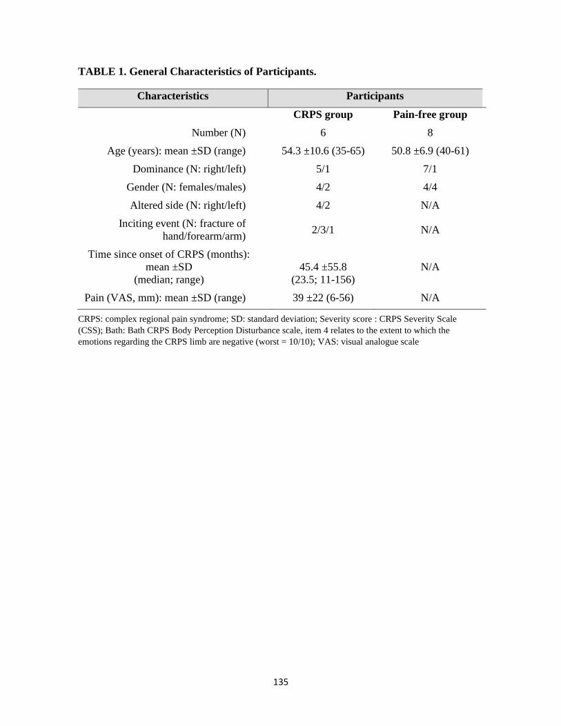
135
TABLE 1. General Characteristics of Participants.
CRPS: complex regional pain syndrome; SD: standard deviation; Severity score : CRPS Severity Scale
(CSS); Bath: Bath CRPS Body Perception Disturbance scale, item 4 relates to the extent to which the
emotions regarding the CRPS limb are negative (worst = 10/10); VAS: visual analogue scale
Characteristics Participants
CRPS group Pain-free group
Number (N) 6 8
Age (years): mean ±SD (range) 54.3 ±10.6 (35-65) 50.8 ±6.9 (40-61)
Dominance (N: right/left) 5/1 7/1
Gender (N: females/males) 4/2 4/4
Altered side (N: right/left) 4/2 N/A
Inciting event (N: fracture of
hand/forearm/arm) 2/3/1 N/A
Time since onset of CRPS (months):
mean ±SD
(median; range)
45.4 ±55.8
(23.5; 11-156)
N/A
Pain (VAS, mm): mean ±SD (range) 39 ±22 (6-56) N/A
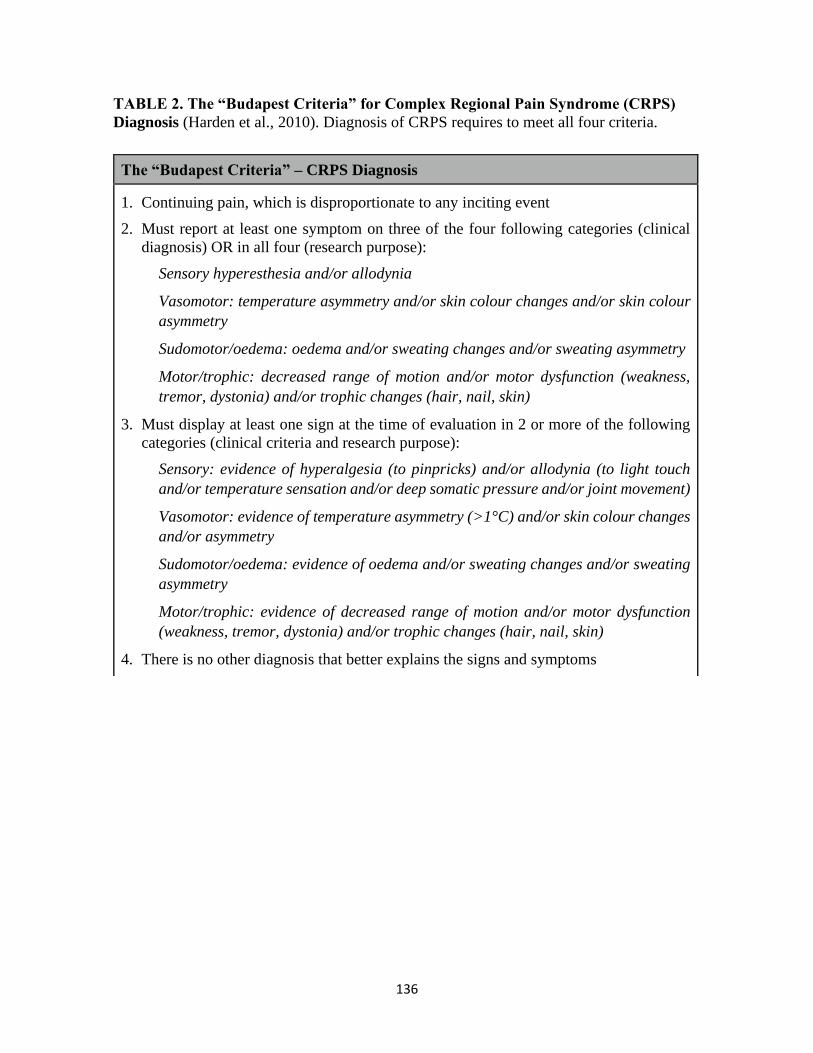
136
TABLE 2. The “Budapest Criteria” for Complex Regional Pain Syndrome (CRPS)
Diagnosis (Harden et al., 2010). Diagnosis of CRPS requires to meet all four criteria.
The “Budapest Criteria” – CRPS Diagnosis
1. Continuing pain, which is disproportionate to any inciting event
2. Must report at least one symptom on three of the four following categories (clinical
diagnosis) OR in all four (research purpose):
Sensory hyperesthesia and/or allodynia
Vasomotor: temperature asymmetry and/or skin colour changes and/or skin colour
asymmetry
Sudomotor/oedema: oedema and/or sweating changes and/or sweating asymmetry
Motor/trophic: decreased range of motion and/or motor dysfunction (weakness,
tremor, dystonia) and/or trophic changes (hair, nail, skin)
3. Must display at least one sign at the time of evaluation in 2 or more of the following
categories (clinical criteria and research purpose):
Sensory: evidence of hyperalgesia (to pinpricks) and/or allodynia (to light touch
and/or temperature sensation and/or deep somatic pressure and/or joint movement)
Vasomotor: evidence of temperature asymmetry (>1°C) and/or skin colour changes
and/or asymmetry
Sudomotor/oedema: evidence of oedema and/or sweating changes and/or sweating
asymmetry
Motor/trophic: evidence of decreased range of motion and/or motor dysfunction
(weakness, tremor, dystonia) and/or trophic changes (hair, nail, skin)
4. There is no other diagnosis that better explains the signs and symptoms
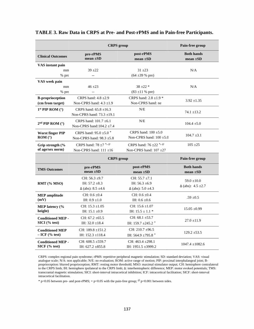
137
TABLE 3. Raw Data in CRPS at Pre- and Post-rPMS and in Pain-free Participants.
CRPS group Pain-free group
Clinical Outcomes pre-rPMS
mean ±SD
post-rPMS
mean ±SD
Both hands
mean ±SD
VAS instant pain
mm
% pre
39 ±22
--
31 ±23
(64 ±39 % pre)
N/A
VAS week pain
mm
% pre
46 ±23
--
38 ±22 *
(83 ±11 % pre)
N/A
B-proprioception
(cm from target)
CRPS hand: 4.8 ±2.9
Non-CPRS hand: 4.3 ±1.9
CRPS hand: 2.8 ±1.9 *
Non-CPRS hand: ne 3.92 ±1.35
1st PIP ROM (°)
CRPS hand: 65.8 ±16.3
Non-CPRS hand: 73.3 ±19.1
N/E 74.1 ±13.2
2nd PIP ROM (°) CRPS hand: 101.7 ±6.1
Non-CPRS hand:104.2 ±7.4
N/E 104.4 ±5.0
Worst finger PIP
ROM (°)
CRPS hand: 95.0 ±5.0 +
Non-CPRS hand: 98.3 ±5.8
CRPS hand: 100 ±5.0
Non-CPRS hand: 100 ±5.0 104.7 ±3.1
Grip strength (%
of age/sex norm)
CRPS hand: 78 ±7 +, @
Non-CPRS hand: 111 ±16
CRPS hand: 76 ±22 +, @
Non-CPRS hand: 107 ±27
105 ±25
CRPS group Pain-free group
TMS Outcomes pre-rPMS
mean ±SD
post-rPMS
mean ±SD
Both hands
mean ±SD
RMT (% MSO)
CH: 56.3 ±9.7
IH: 57.2 ±8.3
Δ (abs): 8.5 ±4.6
CH: 55.7 ±7.1
IH: 56.3 ±6.9
Δ (abs): 5.0 ±4.3
59.0 ±10.0
Δ (abs): 4.5 ±2.7
MEP amplitude
(mV)
CH: 0.6 ±0.4
IH: 0.9 ±1.0
CH: 0.6 ±0.4
IH: 0.6 ±0.6 .59 ±0.5
MEP latency (%
height)
CH: 15.3 ±1.05
IH: 15.1 ±0.9
CH: 15.6 ±1.07
IH: 15.5 ± 1.1 * 15.05 ±0.99
Conditioned MEP -
SICI (% test)
CH: 67.2 ±65.5
IH: 32.0 ±18.4
CH: 68.1 ±53.7
IH: 159.7 ±245.2 + 27.0 ±11.9
Conditioned MEP
– ICF (% test)
CH: 189.8 ±151.2
IH: 152.3 ±118.4
CH: 210.7 ±96.5
IH: 564.9 ±795.8 + 129.2 ±53.5
Conditioned MEP -
SICF (% test)
CH: 608.5 ±559.7
IH: 627.2 ±855.8
CH: 463.4 ±298.1
IH: 1951.5 ±3999.2 1047.4 ±1082.6
CRPS: complex regional pain syndrome; rPMS: repetitive peripheral magnetic stimulation; SD: standard deviation; VAS: visual
analogue scale; N/A: non applicable; N/E: no evaluation; ROM: active range of motion; PIP: proximal interphalangeal joint; B-
proprioception: blurred proprioception; RMT: resting motor threshold; MSO: maximal stimulator output; CH: hemisphere contralateral
to the CRPS limb; IH: hemisphere ipsilateral to the CRPS limb; Δ: interhemispheric difference; MEP: motor evoked potentials; TMS:
transcranial magnetic stimulation; SICI: short-interval intracortical inhibition; ICF: intracortical facilitation; SICF: short-interval intracortical facilitation.
* p<0.05 between pre- and post-rPMS; + p<0.05 with the pain-free group; @ p<0.001 between sides.
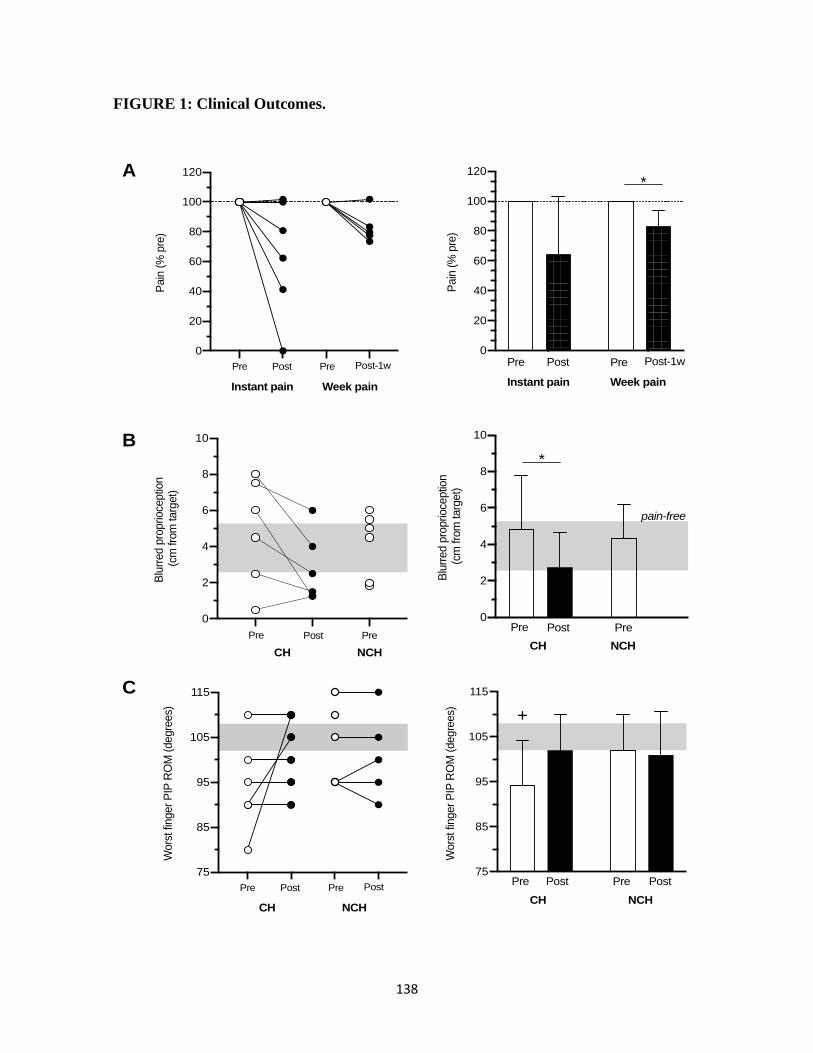
138
FIGURE 1: Clinical Outcomes.
75
85
95
105
115
Wors
t fin
ger
PIP
RO
M (
degre
es)
Pre PrePost Post
+
CH NCH
0
20
40
60
80
100
120
Pain
(%
pre
)
Pre Post Pre Post-1w
Instant pain Week pain
*
0
20
40
60
80
100
120P
ain
(%
pre
)
Pre Post Post-1wPre
Instant pain Week pain
A
B
C
0
2
4
6
8
10B
lurr
ed p
roprioce
ptio
n (
cm fro
m targ
et)
Pre PrePost
*
CH NCH
pain-free
0
2
4
6
8
10
Blu
rred p
roprioce
ptio
n (
cm fro
m targ
et)
Pre Post
CH NCH
Pre
75
85
95
105
115
Wors
t fin
ger
PIP
RO
M (
degre
es)
Pre Post PostPre
CH NCH
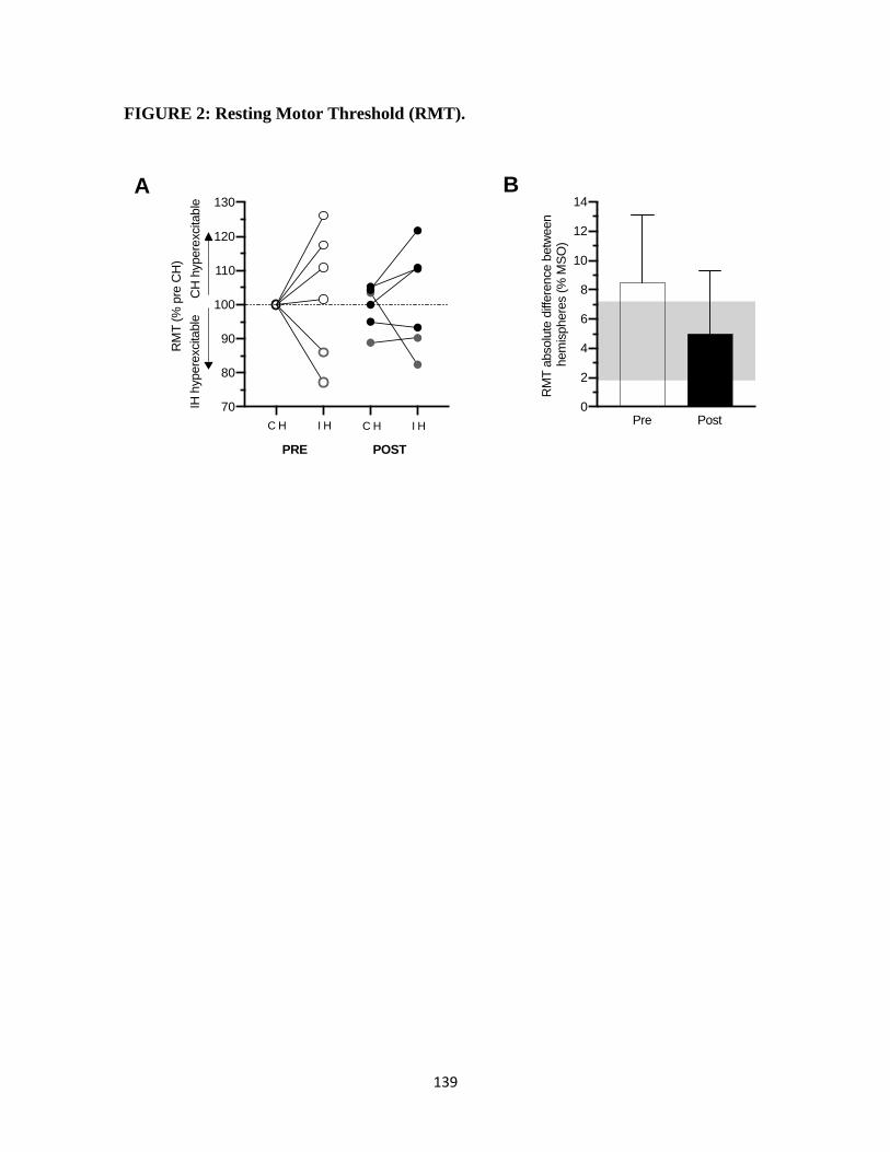
139
FIGURE 2: Resting Motor Threshold (RMT).
A
70
80
90
100
110
120
130
RM
T (
% p
re C
H)
PRE POST
C H I H C H I H
IH h
ypere
xcita
ble
CH
hypere
xcita
ble
0
2
4
6
8
10
12
14
RM
T a
bsolu
te d
iffere
nce b
etw
een
hem
isphere
s (
% M
SO
)
Pre Post
B
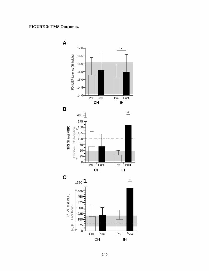
140
FIGURE 3: TMS Outcomes.
CH IH
14.0
14.5
15.0
15.5
16.0
16.5
17.0
FD
I M
EP
Late
ncy (
% h
eig
ht)
Pre PrePost Post
*
CH IH
0
25
50
75
100
125
150
175
400
SIC
I (%
test M
EP
)
Pre PrePost Post
+
No Inhib
ition
Inhib
ition
CH IH
0
75
150
225
300
375
450
525
1350
ICF
(%
test M
EP
)
Pre PrePost Post
Facili
tatio
nN
o F
.
+
A
B
C
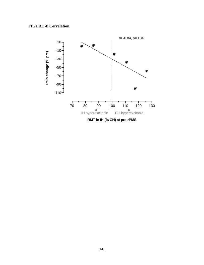
141
FIGURE 4: Correlation.
70 80 90 100 110 120 130
-110
-90
-70
-50
-30
-10
10
RMT in IH (% CH) at pre-rPMS
Pain
ch
an
ge (
% p
re)
r= -0.84, p=0.04
IH hyperexcitable CH hyperexcitable
142
CHAPITRE 4 : DISCUSSION GÉNÉRALE
Les objectifs principaux du projet de maîtrise étaient la compréhension de la place dans la
littérature de la neurostimulation non invasive en SDRC et la justification de son utilisation
(revue, article 1), de comparer les évaluations cliniques et le fonctionnement des M1
(notamment les différences interhémisphériques d’excitabilité motrice corticale) en SDRC
du membre supérieur par rapport à un groupe Sans-douleur (étude 1, article 2), et de tester si
l’utilisation des rPMS permettait de diminuer la douleur et de normaliser cette différence
d’excitabilité motrice interhémisphérique en SDRC (étude 2, article 3). Les objectifs ont été
atteints et les résultats présentés dans les chapitres 2 et 3 sont discutés dans les prochains
paragraphes, avec les forces et les limites du projet de maîtrise, en proposant par la suite les
perspectives des études futures.
4.1 RETOUR SUR LES HYPOTHÈSES RELATIVES À L’ARTICLE 2
L’hypothèse de départ de l’étude expérimentale 1 (article 2) était que les participants avec un
SDRC auraient une différence d’excitabilité du M1 plus grande entre leurs hémisphères que
les participants sans douleur. Il a été possible de mesurer l’excitabilité du M1 chez tous les
participants et l’hypothèse de départ a été validée (différence clinique mesurée). Toutefois,
la douleur ne semble pas influencer le sens de ce débalancement de la même façon chez tout
le monde. Ce résultat a influencé notre décision de poursuivre la mesure du débalancement
dans l’étude 2 (article 3) pour comprendre si le sens du débalancement influençait la réponse
au traitement.
Les résultats cliniques moteurs de l’étude expérimentale 1 (Article 2) correspondaient aux
hypothèses initiales (force et amplitude articulaire active moindre pour la main avec SDRC),
mais les différences des mesures cliniques sensorielles (test des monofilaments et
proprioception brouillée) au membre sans SDRC par rapport au groupe Sans-douleur
n’étaient pas celles attendues. Ces mesures cliniques et leurs corrélations avec l’excitabilité
du M1 de l’HI sont difficiles à interpréter : est-ce qu’elles révèlent des causes et mécanismes
pouvant expliquer en partie la chronicisation du SDRC (plasticité mal-adaptée du HI discutée
dans l’article 2) ou peuvent-elles être dues à un petit échantillon de participants très
hétérogènes. Les mesures de SICI, ICF et SICF n’ont pas permis d’améliorer l’interprétation
143
des changements cliniques. Les écarts-types très élevés des MEP tests et conditionnés
peuvent expliquer en partie pourquoi si peu de différence a été détectée entre les groupes.
Dans des études futures, la prise de davantage de MEP par condition pourrait favoriser une
plus grande diminution de la variabilité des mesures TMS, ce qui concorde avec les
recommandations de Beaulieu et al 2017 (Beaulieu et al. 2017c). Il est également possible
que les changements de fonctionnement du M1 en SDRC impliquent d’autres variables que
celles mesurées (par exemple, d’autres circuits neuronaux que ceux testés). Les études futures
devront tester davantage le fonctionnement du M1 et la voie corticospinale pour éclaircir les
hypothèses relevées par l’étude expérimentale 1 et, si leur puissance statistique leur permet,
il serait intéressant de tester l’impact de plusieurs cofacteurs sur les mesures TMS prises en
pré et post-rPMS comme le sexe des participants, leur dominance, leur côté atteint en
fonction de leur dominance, etc. pour relever de possibles différences initiales entre sous-
groupes et/ou des réactions différentes aux rPMS.
4.2 RETOUR SUR LES HYPOTHÈSES RELATIVES À L’ARTICLE 3
L’hypothèse de départ de l’étude exploratoire 2 (Article 3) était qu’il serait possible de
mesurer des changements à la suite d’une seule séance de rPMS chez des participants avec
SDRC au membre supérieur en stade chronique comparativement à leurs mesures pré-rPMS
(cliniques et TMS) et que leurs mesures se rapprocheraient en post-rPMS des valeurs de
celles d’un groupe Sans-douleur. Cette hypothèse a été vérifiée. Toutefois, d’autres
hypothèses auraient pu être émises, entre autres à la suite de la réalisation de l’étude
expérimentale 1 (Article 2), et seront discutées en lien avec les résultats de l’étude 2 (Article
3).
À la suite de l’étude 1 qui montrait un débalancement d’excitabilité du M1 avec un sens
variable d’un participant à l’autre, indépendamment du côté atteint, il a été anticipé que les
rPMS appliquées au membre avec SDRC auraient un impact sur un des deux sous-groupes
de participants seulement : ceux avec l’HC ou l’HI plus excitable. En effet, les rPMS ont
permis une diminution de la douleur présente que dans le sous-groupe avec l’HC plus
excitable que l’HI (malgré une baisse de douleur à une semaine retrouvée dans les deux sous-
groupes), puis plus le débalancement favorisait l’excitabilité de l’HC sur celui de l’HI en pré-
144
rPMS et plus la diminution de douleur était grande (article 3, Figure 4). Ces résultats devront
être répliqués avant d’en tirer une conclusion définitive, mais le RMT semble donc être un
bon indicateur d’efficacité au traitement (lorsque HC plus excitable).
La plus grande diminution de douleur chez les participants avec le HC plus excitable que le
HI (étude 1/article 2, Figure 4B) a généré plusieurs questionnements : comme ces participants
étaient également ceux avec le moins de douleur moyenne dans la semaine précédant les
rPMS, la plus grande diminution de douleur était-elle donc vraiment en lien avec le sens du
débalancement de RMT ou d’autres facteurs? Leur intensité de douleur moindre, la rend-elle
plus difficile à réduire (car moins de possibilités de réduction de douleur) ou plus facile car
moins intense? À ma connaissance, la facilité ou difficulté à réduire une douleur en fonction
de son intensité n’a pas été testée en SDRC. Benedetti et al. (1997) rapportent une réduction
de douleur avec une stimulation électrique transcutanée seulement chez les personnes ayant
une douleur légère à modérée suite à une opération thoracique alors que ces stimulations
étaient inefficaces chez les personnes dont la douleur était élevée (Benedetti et al., 1997). Il
serait alors intéressant de tester et rapporter les différences d’effets des stimulations
magnétiques entre différents niveaux de douleur des personnes avec SDRC. Ces études
devraient avoir de plus grands échantillons, être randomisées et contrôlées et tester les stades
aigus et chroniques pour décrire suffisamment bien la population et ainsi permettre de mieux
personnaliser les traitements.
On aurait pu penser que seul l’HC aurait été influencé par les rPMS (retour sensoriel
lemniscal croisé), mais il semble que les deux hémisphères cérébraux aient présenté des
changements plastiques, ce qui suggère une communication transcallosale efficace ou des
afférences sensorielles ipsilatérales pour le membre supérieur atteint de SDRC. En effet, les
rPMS ont eu non seulement un impact bilatéral sur l’excitabilité des M1 (ce qui a permis de
diminuer le débalancement d’excitabilité des M1), mais également sur plusieurs autres
variables TMS mesurées dans l’HI. Une levée d’inhibition (diminution du mécanisme SICI),
une augmentation de la facilitation (augmentation du mécanisme ICF) et une synchronisation
moins bonne des volées descendantes (latence plus lente) ont été mesurées dans l’HI, le tout
suggérant l’induction de mécanismes de plasticité motrice corticale. Comme discuté dans
145
l’article 3, ces changements sont vraisemblablement dus à la modification de la force des
synapses corticospinales et à l’activation de synapses latentes (Jacobs & Donoghue 1991;
Hess & Donoghue 1996; Sanes & Donoghue 2000), mais également à l’inhibition
interhémisphérique (en provenance de HC) de la SICI de HI (Reis et al., 2008). Comme ces
mécanismes sont reliés à la plasticité cérébrale, il est possible d’émettre l’hypothèse que les
rPMS activent des mécanismes de plasticité dans l’HI ce qui permettrait, s’ils étaient utilisés
avant des exercices spécifiques de réadaptation, de potentialiser les effets de la réadaptation.
Les rPMS représenteraient donc potentiellement un adjuvant thérapeutique aux traitements
conventionnels en réadaptation.
Les mesures de proprioception brouillée et d’amplitude articulaire active de la main avec
SDRC se sont améliorées après rPMS. Ces améliorations en une seule séance rPMS de 3
minutes 20 secondes ne peuvent être dues à une diminution des restrictions mécaniques
(capsule articulaire raccourcie, adhérences cicatricielles…). Il est probable que les
changements dans l’HI, ainsi que la diminution du débalancement d’excitabilité des M1 ont
permis d’améliorer l’intégration sensorielle et la planification motrice dans l’HC permettant
l’envoi de commandes motrices plus efficaces et influençant l’amplitude articulaire active et
la proprioception de la main avec SDRC. Toutefois, qu’en est-il de la main sans SDRC?
Selon l’étude 1 (article 2), l’intégration sensorielle de l’HI (selon les tests de monofilaments
et de proprioception) était touchée dans le groupe avec SDRC. Or, le test de monofilaments
n’a été réalisé qu’en pré-rPMS et celui de proprioception de façon bilatérale en pré-rPMS,
mais qu’à la main avec SDRC en post-rPMS. Il aurait été intéressant d’avoir ces résultats
pour statuer si les rPMS permettaient, en parallèle des mesures de plasticité détectées dans
cet hémisphère, une meilleure intégration sensorielle dans l’HI.
Deux questionnaires ont été utilisés dans les études exploratoires pour décrire la population
avec SDRC ayant participé aux études. L’objectif de leur utilisation aurait pu être d’y mesurer
des changements à la suite des rPMS. Or, malgré une amélioration de la fonction manuelle à
la suite du traitement, très peu de modifications des scores globaux à l’échelle de sévérité du
SDRC ont été mesurées à une semaine de suivi (0 à 3 points). Cela peut s’expliquer parce
que l’échelle de sévérité du SDRC, contenant des questions dichotomiques plutôt que
146
quantitatives, est peu sensible au changement. Selon Harden et al. 2017, une différence de
≥4.9 sur l’échelle indiquerait une vraie différence dans la symptomatologie des participants
(Harden et al., 2017). De la même façon, peu de changements ont été relevés sur l’échelle de
perturbation de la perception du schéma corporel de Bath pour les patients atteints du SDRC,
mais les participants des études (en stade chronique) y avaient en pré-rPMS noté peu de
perturbations (laissant donc peu de place au changement). Ces deux questionnaires seraient
possiblement plus utiles en stade aigu ou subaigu pour noter l’impact de la répétition de
séances de rPMS ou l’impact des rPMS combinées à des techniques de visualisation motrice
(qui influencent également la perception corporelle, Moseley, 2006).
4.3 FORCES ET LIMITES DES ÉTUDES
La portée des résultats des deux études exploratoires (articles 2 et 3) est limitée par la taille
des échantillons. Toutefois, la comparaison des mesures pré/post-rPMS chez les mêmes
participants augmente la force des résultats. Aussi, ces études exploratoires permettent
d’émettre de nouvelles hypothèses et de soutenir la continuation d’études en SDRC, que ce
soit pour mieux comprendre les changements de fonctionnement des M1 afin d’adapter les
approches au débalancement fonctionnel, ou pour tester l’effet de la neurostimulation non
invasive en fonction de ce débalancement et des autres variables TMS.
Le diagnostic de SDRC type I des participants n’a pas été validé au départ des études étant
donné la résolution de certains de leurs symptômes avant leur participation à l’étude. Bref,
même si les participants répondaient initialement aux critères diagnostiques cliniques de
Budapest, ils auraient probablement été diagnostiqués SDRC de type NOS lors de leur
participation. C’est pour cette raison que les critères diagnostiques de Budapest cliniques ont
été utilisés plutôt que ceux de recherche.
Il est possible qu’un biais de recrutement ait influencé nos résultats. En effet, étant donné le
mode de recrutement non probabiliste volontaire des participants, il est probable que ceux
ayant décidé de participer aux études soient ceux qui étaient les plus motivés à s’améliorer
et par le fait même les plus à risque de vivre un effet placebo. Ce qui peut s’appliquer à la
majorité des études avec un même mode de recrutement. Par contre, aucun effet placebo n’a
147
été mesuré jusqu’à maintenant dans les études randomisées et contrôlées en rPMS en
physiopathologie dans notre laboratoire (Massé-Alarie et al., 2013, 2016; Beaulieu et al.,
2015). Également, un effet placebo est par définition un effet qui ne dure pas. Or, l’étude 2
montre des effets sur la douleur à une semaine post-rPMS, ce qui rassure face au risque de
placebo, bien que des études randomisées contrôlées permettraient d’en écarter le risque.
Finalement, la prise des mesures cliniques a été réalisée par la même physiothérapeute pour
éliminer les différences possibles reliées à la fidélité interévaluateur. Il n’était donc pas
possible qu’elle soit à l’aveugle par rapport au temps de mesure (pré vs. post-rPMS) ni au
groupe (SDRC vs. Sans-douleur) auquel faisait partie le participant qu’elle évaluait.
Toutefois, toutes les données TMS ont été codifiées pour chacun des participants et leur
analyse a donc pu être réalisée à l’aveugle des facteurs testés (groupe, temps de mesure, côté).
4.4 PERSPECTIVES CLINIQUES ET DE RECHERCHE
Les résultats obtenus dans le cadre de cette maitrise sont prometteurs et soutiennent la
réalisation d’études de plus grande envergure, car plusieurs questions demeurent.
Études longitudinales
D’abord, il serait intéressant d’effectuer une étude longitudinale pour vérifier si les
différences de fonctionnement du M1 sont présentes avant le SDRC et pourraient expliquer
pourquoi un individu développe ce syndrome ou si, à l’inverse, elles sont la conséquence du
SDRC sur le système sensorimoteur. Une meilleure connaissance des différences de
fonctionnement du M1 permettrait également de mieux adapter les interventions chez la
population avec SDRC pour influencer positivement la plasticité cérébrale et optimiser le
fonctionnement des cortex sensorimoteurs. Le gain serait énorme pour les individus qui
souffrent du SDRC, mais également sur le plan sociétal puisque le SDRC est rattaché à une
restriction de participation au travail et un coût de santé important.
Répétition des séances rPMS
La réalisation d’études comprenant une répétition du nombre de séances de rPMS permettrait
vraisemblablement d’optimiser et de cristalliser les gains obtenus en une seule séance. Peut-
148
être même que l’influence répétée sur la plasticité cérébrale des deux hémisphères permettrait
de diminuer la douleur de façon équivalente entre personnes avec hyperexcitabilité de
l’hémisphère controlatéral au SDRC et personnes avec hyperexcitabilité de l’hémisphère
ipsilatéral.
Neurostimulation cérébrale et combinaison avec rPMS
La neurostimulation cérébrale est connue pour influencer le système sensorimoteur (telle que
présenté dans la revue de l’article 1) et pourrait possiblement, lorsqu’utilisée avant les rPMS,
permettre d’optimiser l’impact de ces derniers. Le projet de maitrise présenté dans ce
mémoire expose les résultats qui ont servi d’assise au développement d’une étude de plus
grande envergure, actuellement en cours au laboratoire, qui vise à tester ces hypothèses.
En effet, la connaissance de la présence d’un débalancement d’excitabilité du M1 de sens
différent selon chaque participant (étude 1) a rendu l’application d’un même protocole de
stimulation magnétique du cerveau insensée. À nos yeux, il est effectivement non logique et
non éthique d’appliquer un protocole de stimulation qui augmente l’excitabilité de M1 sur
un hémisphère déjà plus excitable ou un protocole inhibiteur sur un hémisphère moins
excitable. Le protocole de l’étude en cours a donc été développé pour que les rPMS soient
testées, dans un premier temps, seules (étude expérimentale 2) et, dans un deuxième temps,
combinées à des protocoles de stimulations magnétiques centrales personnalisés au
participant (selon ses propres mesures TMS de ligne de base). Les participants reçoivent alors
au laboratoire sur quatre séances distinctes un protocole : excitateur sur l’hémisphère le
moins excitable (pour certains sur l’HC, d’autres sur l’HI), inhibiteur sur l’hémisphère le plus
excitable (HC ou HI), puis ces mêmes protocoles sont combinés aux rPMS. Ainsi,
l’hémisphère stimulé est parfois le dominant ou non-dominant, le droit ou gauche, contrôlant
ou non le membre avec SDRC… Un échantillon de très grande taille est donc nécessaire pour
atteindre la puissance statistique suffisante pour la comparaison des différents protocoles.
Combinaison des rPMS à la réadaptation (adjuvant)
La combinaison de la neurostimulation à des exercices spécifiques de physiothérapie ou
d’ergothérapie n’a pas été testée au travers des études de cette maîtrise. L’influence de la
149
neurostimulation sur la plasticité cérébrale permettrait vraisemblablement, lorsqu’utilisée
avant des exercices, de préparer M1 et le système corticospinal pour que les exercices
spécifiques aient un meilleur impact sur les circuits sensorimoteurs. Il serait donc très
intéressant de développer des études combinant l’application de neurostimulation non
invasive avant la pratique d’exercices spécifiques chez la population avec SDRC. Des études
similaires ont été réalisées chez d’autres populations souffrant de douleur chronique et les
résultats sont probants (Massé-Alarie et al. 2013, 2017; Schneider et al., en cours). Si les
rPMS étaient accessibles en pratique publique et/ou privé en physiothérapie, leur
applicabilité serait assez facile puisque les physiothérapeutes ont une excellente connaissance
de l’anatomie (il leur serait aisé de localiser un muscle précis, positionner et maintenir la
bobine sur un muscle ou groupe de muscles spécifiques et valider leur positionnement par le
mouvement créé par la stimulation). Par leur formation académique, ils seraient également
en mesure d’évaluer et choisir le ou les muscles dont le contrôle moteur déficient serait à
améliorer par rPMS pour que de nombreuses tâches soient moins, voire non, douloureuses.
Le temps de stimulation par muscle est très court (3min20sec) donc facilement intégrable
même dans un contexte de soins réels avec une limite de temps par client. Par contre, d’autres
évidences doivent encore être collectées et rapportées pour que de telles applications
cliniques des rPMS puissent être recommandées par les ordres professionnels.
150
CONCLUSIONS
Les résultats des présentes études exploratoires sont novateurs, mais également très
prometteurs. Les participants du groupe SDRC avaient déjà eu accès à des traitements
multidisciplinaires et étaient passés au travers de l’algorithme de traitements conventionnels
avec un suivi médical spécialisé d’une clinique de gestion de la douleur chronique
(médication, blocs intraveineux, injections de kétamine, etc.), de l’ergothérapie (modalités
actives et passives, exercices spécifiques, conseils, modalités analgésiques telle que chaleur,
imagerie motrice, etc.) et un suivi psychologique. Malgré tout, comme chez 62% des
participants de l’étude de Geertzen et al., ils vivaient encore avec des restrictions aux activités
de la vie quotidienne et de la douleur au début de leur participation à l’étude (Geertzen et al.,
1998). Il est probable que leurs difficultés résiduelles soient dues, du moins en partie, aux
changements centraux qui ont pu être mesurés (notamment la différence d’excitabilité
motrice corticale entre les deux hémisphères cérébraux). Or, malgré tout cela, une seule
séance de rPMS de 3 minutes 20 secondes sur le muscle FDS a pu normaliser le
débalancement d’excitabilité interhémisphérique et améliorer douleur et fonction.
En une seule séance rPMS, l’objectif n’était pas de résoudre complètement la douleur et les
incapacités des participants, mais d’explorer l’effet des rPMS sur les symptômes de douleur,
le fonctionnement de M1 et la fonction manuelle. Ces études ouvrent la porte à un grand
nombre d’autres études et de perspectives de recherche et d’essais cliniques.
Comme physiothérapeute, il m’apparaît important et intéressant de contribuer aux
développements de nouvelles connaissances en santé et de promouvoir la profession. Il me
semble réaliste dans un proche avenir de voir les physiothérapeutes utiliser de nouvelles
technologies de neurostimulation, comme les rPMS, pour continuer d’améliorer la qualité
des soins et les services prodigués à la population.
151
BIBLIOGRAPHIE
Abraham, Wickliffe C. 2008. “Metaplasticity: Tuning Synapses and Networks for
Plasticity.” Nature Reviews Neuroscience 9(5): 387–99.
Abraham, Wickliffe C, and Mark F Bear. 1996. “Metaplasticity : Plasticity of Synaptic.”
Trends Neurosci 19: 126–30.
Abu-Arafeh, H., & Abu-Arafeh, I. 2016. Complex regional pain syndrome in children:
incidence and clinical characteristics. Archives of disease in childhood, 101(8), 719-723.
Allen Demers F, Delisle A, Schneider C. Living with a Complex Regional Pain Syndrome
(CRPS): Clinical and Corticomotor Modifications as Compared to Pain-free People, in
preparation (Neurophysiol Clin / Clin Neurophysiol).
Allen Demers, Fannie, Zangrandi, Andrea, Schneider, Cyril. In preparation “Noninvasive
Neurostimulation in Complex Regional Pain Syndrome (CRPS). A Comprehensive
Review on Brain and Clinical Outcomes.” (Neurophysiologie Clinique / Clinical
Neurophysiology)
Amassian V, Stewart M, Quirk G, Rosenthal J. Physiological basis of motor effects of a
transient stimulus to cerebral cortex. Neurosurgery 1987;20(1):74-93.
Ambriz‐Tututi, M., Alvarado‐Reynoso, B., & Drucker‐Colín, R. 2016 Analgesic effect of
repetitive transcranial magnetic stimulation (rTMS) in patients with chronic low back
pain. Bioelectromagnetics, 37(8), 527-535.
Arya, Kamal Narayan, Shanta Pandian, Rajesh Verma, and R K Garg. 2011. “Movement
Therapy Induced Neural Reorganization and Motor Recovery in Stroke: A Review.”
Journal of Bodywork and Movement Therapies 15 (4): 528–37.
Azqueta-Gavaldon, M., Schulte-Gocking, H., Storz, C., Azad, S., Reiners, A., Borsook, D.,
.Kraft, E. 2017. Basal ganglia dysfunction in complex regional pain syndrome - A valid
hypothesis? Eur J Pain, 21(3), 415-424. doi:10.1002/ejp.975
Azqueta-Gavaldon, M., Youssef, A. M., Storz, C., Lemme, J., Schulte-Göcking, H.,
Becerra, L., Kraft, E. 2020. Implications of the putamen in pain and motor deficits in
complex regional pain syndrome. Pain, 161(3), 595-608.
Barad, Meredith J, Takefumi Ueno, Jarred Younger, Neil Chatterjee, and Sean Mackey.
2014. “Complex Regional Pain Syndrome Is Associated with Structural Abnormalities
in Pain-Related Regions of the Human Brain.” The Journal of Pain : Official Journal of
the American Pain Society 15 (2): 197–203.
Bean, Debbie J., Malcolm H. Johnson, and Robert R. Kydd. 2014. “The Outcome of
Complex Regional Pain Syndrome Type 1: A Systematic Review.” Journal of Pain 15
(7): 677–90.
Bean, D., Johnson, M. H., Heiss-Dunlop, W., Lee, A. C., & Kydd, R. R. (2015). Do
psychological factors influence recovery from complex regional pain syndrome type 1?
A prospective study. Pain, 156(11), 2310-2318.
Bean, D. J., Johnson, M. H., & Kydd, R. R. (2014). Relationships between psychological
factors, pain, and disability in complex regional pain syndrome and low back pain. Clin
J Pain, 30(8), 647-653.
Beaulieu, Louis-David, Véronique H. Flamand, Hugo Massé-Alarie, and Cyril Schneider.
2017a. “Reliability and Minimal Detectable Change of Transcranial Magnetic
Stimulation Outcomes in Healthy Adults: A Systematic Review.” Brain Stimulation 10
(2): 196–213.
Beaulieu, Louis-David, Hugo Massé-Alarie, Samuel Camiré-Bernier, Édith Ribot-Cisar,
152
and Cyril Schneider. 2017b. “After-Effects of Peripheral Neurostimulation on Brain
Plasticity and Ankle Function in Chronic Stroke: The Role of Afferents Recruited.”
Neurophysiologie Clinique 47 (4): 275–91.
Beaulieu, Louis-David, Hugo Massé-Alarie, Edith Ribot-Ciscar, and Cyril Schneider.
2017c. “Reliability of Lower Limb Transcranial Magnetic Stimulation Outcomes in the
Ipsi- and Contralesional Hemispheres of Adults with Chronic Stroke.” Clinical
Neurophysiology 128 (7): 1290–98.
Beaulieu, Louis-David, Hugo Masse-Alarie, Brenda Brouwer, and Cyril Schneider. 2015
“Noninvasive Neurostimulation in Chronic Stroke: A Double-Blind Randomized Sham-
Controlled Testing of Clinical and Corticomotor Effects.” Topics in Stroke
Rehabilitation 22 (1): 8–17.
Beaulieu, Louis-David, and Cyril Schneider. 2015 “Repetitive Peripheral Magnetic
Stimulation to Reduce Pain or Improve Sensorimotor Impairments: A Literature Review
on Parameters of Application and Afferents Recruitment.” Neurophysiologie
Clinique/Clinical Neurophysiology 45 (3): 223–37.
Beaulieu L-D, Schneider C. Effects of repetitive peripheral magnetic stimulation on normal
or impaired motor control. A review. Neurophysiol Clin Neurophysiol 2013;43:251–60
Beerthuizen, A., van‘t Spijker, A., Huygen, F. J., Klein, J., & de Wit, R. (2009). Is there an
association between psychological factors and the Complex Regional Pain Syndrome
type 1 (CRPS1) in adults? A systematic review. PAIN®, 145(1-2), 52-59.
Benedetti F, Amanzio M, Casadio C, Cavallo A, Cianci R, Giobbe R, Mancuso M, Ruffini
E, Maggi G. Control of postoperative pain by transcutaneous electrical nerve stimulation
after thoracic operations. Ann Thorac Surg. 1997, 63(3):773-6
Bilgili, A., Cakir, T., Dogan, S. K., Ercalik, T., Filiz, M. B., & Toraman, F. (2016). The
effectiveness of transcutaneous electrical nerve stimulation in the management of
patients with complex regional pain syndrome: A randomized, double-blinded, placebo-
controlled prospective study. J Back Musculoskelet Rehabil, 29(4), 661-671.
Binny, J., Wong, N. L. J., Garga, S., Lin, C.-W. C., Maher, C. G., McLachlan, A. J., . . .
Shaheed, C. A. (2019). Transcutaneous electric nerve stimulation (TENS) for acute low
back pain: systematic review. Scandinavian journal of pain, 19(2), 225-233.
Birklein, F., & Schmelz, M. (2008). Neuropeptides, neurogenic inflammation and complex
regional pain syndrome (CRPS). Neurosci Lett, 437(3), 199-202.
Birklein, F., Schmelz, M., Schifter, S., & Weber, M. (2001). The important role of
neuropeptides in complex regional pain syndrome. Neurology, 57(12), 2179-2184.
Bodenheim, R., & Bennett, J. H. (1983). Reversal of a Sudeck's atrophy by the adjunctive
use of transcutaneous electrical nerve stimulation. A case report. Phys Ther, 63(8),
1287-1288.
Bolognini, N., Pascual-Leone, A., & Fregni, F. (2009). Using non-invasive brain
stimulation to augment motor training-induced plasticity. J Neuroeng Rehabil, 6, 8.
Bolwerk, A., Seifert, F., & Maihofner, C. (2013). Altered resting-state functional
connectivity in complex regional pain syndrome. J Pain, 14(10), 1107-1115 e1108.
Borchers, A. T., and M. E. Gershwin. 2014. “Complex Regional Pain Syndrome: A
Comprehensive and Critical Review.” Autoimmunity Reviews 13(3): 242–65.
Bruehl, S. (2015). Complex regional pain syndrome. BMJ, 351, h2730.
Bruehl, Stephen. 2010. “An Update on the Pathophysiology of Complex Regional Pain
Syndrome.” The American Society of Anesthesiologists 113 (3): 713–25.
Buckinx, Fanny, Jean-Louis Croisier, Jean-Yves Reginster, Nadia Dardenne, Charlotte
153
Beaudart, Justine Slomian, Sylvain Leonard, and Olivier Bruyère. 2017. “Reliability of
Muscle Strength Measures Obtained with a Hand-Held Dynamometer in an Elderly
Population.” Clinical Physiology and Functional Imaging 37 (3): 332–40.
Burgess HJ, Burns JW, Buvanendran A, Gupta R, Chont M, Kennedy M, et al.
Associations Between Sleep Disturbance and Chronic Pain Intensity and Function. Clin
J Pain 2019;35:569–76.
Coghill RC, Gilron I, Iadarola MJ. 2001 "Hemispheric lateralization of somatosensory
processing". J Neurophysiol 85:2602–2612
Choi, J. C., Kim, J., Kang, E., Lee, J. M., Cha, J., Kim, Y. J., . . . Yi, D. J. (2016). Brain
mechanisms of pain relief by transcutaneous electrical nerve stimulation: A functional
magnetic resonance imaging study. European Journal of Pain, 20(1), 92-105.
Cohen, Leonardo G., Bradley J. Roth, Jan Nilsson, Nguyet Dang, Marcela Panizza, Stefania
Bandinelli, Walter Friauf, and Mark Hallett. 1990. “Effects of Coil Design on Delivery
of Focal Magnetic Stimulation. Technical Considerations.” Electroencephalography and
Clinical Neurophysiology 75 (4): 350–57.
Contu S, Cappello L, Konczak J, Masia L. 2015 "Preliminary analysis of non-dominant
proprioceptive acuity and interlimb asymmetry in the human wrist". Annu Int Conf
IEEE Eng Med Biol Soc. 2015:3598-601.
Cossins, L., Okell, R. W., Cameron, H., Simpson, B., Poole, H. M., & Goebel, A. (2013).
Treatment of complex regional pain syndrome in adults: A systematic review of
randomized controlled trials published from June 2000 to February 2012. European
Journal of Pain, 17(2), 158-173.
Costigan, M., Scholz, J., & Woolf, C. J. (2009). Neuropathic pain: a maladaptive response
of the nervous system to damage. Annual review of neuroscience, 32, 1-32.
Cuypers, K., Leenus, D. J., Van Wijmeersch, B., Thijs, H., Levin, O., Swinnen, S. P., &
Meesen, R. L. (2013). Anodal tDCS increases corticospinal output and projection
strength in multiple sclerosis. Neuroscience letters, 554, 151-155.
de Mos, M., De Bruijn, A., Huygen, F., Dieleman, J., Stricker, B. C., & Sturkenboom, M.
(2007). The incidence of complex regional pain syndrome: a population-based study.
Pain, 129(1-2), 12-20.
de Mos, M., Huygen, F. J., Dieleman, J. P., Koopman, J. S., Stricker, B. H., &
Sturkenboom, M. C. (2008). Medical history and the onset of complex regional pain
syndrome (CRPS). Pain, 139(2), 458-466.
DeSantana, J. M., Walsh, D. M., Vance, C., Rakel, B. A., & Sluka, K. A. (2008).
Effectiveness of transcutaneous electrical nerve stimulation for treatment of hyperalgesia
and pain. Current rheumatology reports, 10(6), 492.
Devanne H, Lavoie BA, Capaday C. 1997 "Input-output properties and gain changes in the
human corticospinal pathway". Exp Brain Res;114:329–38.
Di Pietro, Flavia, James H. McAuley, Luke Parkitny, Martin Lotze, Benedict M. Wand, G.
Lorimer Moseley, and Tasha R. Stanton. 2013a. “Primary Motor Cortex Function in
Complex Regional Pain Syndrome: A Systematic Review and Meta-Analysis.” Journal
of Pain 14 (11): 1270–88.
Di Pietro, Flavia et al. 2013b. “Primary Somatosensory Cortex Function in Complex
Regional Pain Syndrome: A Systematic Review and Meta-Analysis.” The journal of
pain : official journal of the American Pain Society 14(11): 1270–88.
Di Pietro, Flavia et al. 2015. “Interhemispheric Somatosensory Differences in Chronic Pain
Reflect Abnormality of the Healthy Side.” Human Brain Mapping 36(2): 508–18.
154
Dilek, B., Yemez, B., Kizil, R., Kartal, E., Gulbahar, S., Sari, O., & Akalin, E. (2012).
Anxious personality is a risk factor for developing complex regional pain syndrome type
I. Rheumatology international, 32(4), 915-920.
Dum RP, Li C, Strick PL. 2002 "Motor and nonmotor domains in the monkey dentate".
Ann N Y Acad Sci 978:289–301
Engineer, N. D., Riley, J. R., Seale, J. D., Vrana, W. A., Shetake, J. A., Sudanagunta, S. P.,
. . . Kilgard, M. P. (2011). Reversing pathological neural activity using targeted
plasticity. Nature, 470(7332), 101-104.
Eisenberg, Elon et al. 2005. “Evidence for Cortical Hyperexcitability of the Affected Limb
Representation Area in CRPS: A Psychophysical and Transcranial Magnetic Stimulation
Study.” Pain 113(1): 99–105.
Ferrè, Elisa Raffaella, Berlot, Eva, Haggard, Patrick. 2015 Vestibular contributions to a
right-hemisphere network for bodily awareness: Combining galvanic vestibular
stimulation and the “Rubber Hand Illusion”, Neuropsychologia, Volume 69, Pages 140-
147
Flamand VH, Beaulieu LD, Nadeau L, Schneider C. Peripheral magnetic stimulation to
decrease spasticity in cerebral palsy. Pediatr Neurol 2012;47(5):345-8.
Flamand VH, Schneider C. Noninvasive and painless magnetic stimulation of nerves
improved brain motor function and mobility in a cerebral palsy case. Arch Phys Med
Rehabil 2014;95(10):1984-90.
Freund, W., Wunderlich, A. P., Stuber, G., Mayer, F., Steffen, P., Mentzel, M., . . .
Schmitz, B. (2010). Different activation of opercular and posterior cingulate cortex
(PCC) in patients with complex regional pain syndrome (CRPS I) compared with
healthy controls during perception of electrically induced pain: a functional MRI study.
Clin J Pain, 26(4), 339-347.
Fukui, S. (2003). Evaluation of thalamic neural activity in CRPS type 1 patients by proton
MR spectroscopy: a correlative study with rCBF. J Anesth, 17(2), 142-144.
Fukumoto, M., Ushida, T., Zinchuk, V. S., Yamamoto, H., & Yoshida, S. (1999).
Contralateral thalamic perfusion in patients with reflex sympathetic dystrophy
syndrome. The Lancet, 354(9192), 1790-1791.
Gagné, Martin; Schneider, Cyril 2007. “Dynamic Changes in Corticospinal Control of
Precision Grip during Wrist Movements.” Brain Research 1164 (1998): 32–43.
Gagné, Martin, Schneider, Cyril 2008. “Dynamic influence of wrist flexion and extension
on the intracortical inhibition of the first dorsal interosseus muscle during precision grip.
” Brain Research 1195 (2008) 77-88.
Galea, V. (2012). Bilateral somatosensory cortex disinhibition in complex regional pain
syndrome type I. Yearbook of Sports Medicine, 2012, 308-310.
Geertzen, J H B, P U Dijkstra, E L P van Sonderen, J W Groothoff, H J ten Duis, and W H
Eisma. 1998. “Relationship between Impairments, Disability and Handicap in Reflex
Sympathetic Dystrophy Patients: A Long-Term Follow-up Study.” Clinical
Rehabilitation 12 (5): 402–12.
Geha, P. Y., Baliki, M. N., Harden, R. N., Bauer, W. R., Parrish, T. B., & Apkarian, A. V.
(2008). The Brain in Chronic CRPS Pain: Abnormal Gray-White Matter Interactions in
Emotional and Autonomic Regions. Neuron, 60(4), 570-581.
Gibson, W., Wand, B. M., & O'Connell, N. E. (2017). Transcutaneous electrical nerve
stimulation (TENS) for neuropathic pain in adults. Cochrane Database of Systematic
Reviews(9).
155
Goble DJ, Brown SH. 2008 "Upper limb asymmetries in the matching of proprioceptive
versus visual targets". J Neurophysiol 99: 3063–3074.
Goh, En Lin, Swathikan Chidambaram, and Daqing Ma. 2017. “Complex Regional Pain
Syndrome: A Recent Update.” Burns & Trauma 5(1): 1–11.
Gouvernement du Canada. 2018. “Faire Face à La Crise Des Opioïdes Au Canada.”
https://www.canada.ca/fr/sante-canada/services/dependance-aux-
drogues/consommation-problematique-medicaments-ordonnance/opioides/repondre-
crise-opioides-canada.html (March 11, 2019).
Gracies, Jean-Michel. 2005. “Pathophysiology of Spastic Paresis. II: Emergence of Muscle
Overactivity.” Muscle & Nerve 31 (5): 552–71.
Grieve, S, L Jones, N Walsh, and C McCabe. 2016. “What Outcome Measures Are
Commonly Used for Complex Regional Pain Syndrome Clinical Trials? A Systematic
Review of the Literature.” European Journal of Pain (London, England) 20 (3): 331–40.
Groppa, S., A. Oliviero, A. Eisen, A. Quartarone, L. G. Cohen, V. Mall, A. Kaelin-Lang, et
al. 2012. “A Practical Guide to Diagnostic Transcranial Magnetic Stimulation: Report of
an IFCN Committee.” Clinical Neurophysiology 123 (5): 858–82.
Harden, R Norman, Maihofner, Christian, Abousaad, Elias, Vatine, Jean-Jacques, Kirsling,
Amy, S G M Perez, Roberto, Kuroda, Maxine, Brunner, Florian, Stanton-Hicks,
Michael, Marinus, Johan, J van Hilten, Jacobus, Mackey, Sean, Birklein, Frank,
Schlereth, Tanja, Mailis-Gagnon, Angela, Graciosa, Joe, B Connoly, Sara, Dayanim,
David, Massey, Michael, Frank, Hadas, Livshitz, Anatoly, Bruehl, Stephen, 2017. “A
prospective, multisite, international validation of the Complex Regional Pain Syndrome
Severity Score” Pain 158 (8): 1420–1436
Harden, R. Norman, Stephen Bruehl, Roberto S G M Perez, Frank Birklein, Johan Marinus,
Christian Maihofner, Timothy Lubenow, et al. 2010. “Development of a Severity Score
for CRPS.” Pain 151 (3): 870–76.
Hassanzahraee, Maryam, Maryam Zoghi, and Shapour Jaberzadeh. 2018. “How Different
Priming Stimulations Affect the Corticospinal Excitability Induced by Noninvasive
Brain Stimulation Techniques: A Systematic Review and Meta-Analysis.” Reviews in
the Neurosciences 29(8): 883–99
Hasselmo ME. Neuromodulation and cortical function: modeling the physiological basis of
behavior. Behav Brain Res 1995;67:1–27.
Heller GZ, Manuguerra M, Chow R. 2016 "How to analyze the Visual Analogue Scale:
Myths, truths and clinical relevance". Scand J Pain;13:67–75.
Hermens, Hermie J., Bart Freriks, Catherine Disselhorst-Klug, and Rau Günter. 2000.
“Development of Recommendations for SEMG Sensors and Sensor Placement
Procedures.” Journal of Electromyography and Kinesiology 10: 361–74.
Hess, G, and JP Donoghue. 1996. “Long-Term Potentiation and Long-Term Depression of
Horizontal Connections in Rat Motor Cortex.” Acta Neurobiologia Experimentalis 56:
397–405.
Hirayama, A., Saitoh, Y., Kishima, H., Shimokawa, T., Oshino, S., Hirata, M., . . .
Yoshimine, T. (2006). Reduction of intractable deafferentation pain by navigation-
guided repetitive transcranial magnetic stimulation of the primary motor cortex. Pain,
122(1-2), 22-27.
Hotta, J. et al. 2015. “Patients with Complex Regional Pain Syndrome Overestimate
Applied Force in Observed Hand Actions.” European Journal of Pain (United Kingdom)
19(9): 1372–81.
156
Houde, F., Harvey, M. P., Tremblay Labrecque, P. F., Lamarche, F., Lefebvre, A., &
Leonard, G. (2020). Combining Transcranial Direct Current Stimulation and
Transcutaneous Electrical Nerve Stimulation to Relieve Persistent Pain in a Patient
Suffering from Complex Regional Pain Syndrome: A Case Report. J Pain Res, 13, 467-
473.
Hsieh JC, Hannerz J, Ingvar M, 1996 "Right-lateralised central processing for pain of
nitroglycerin-induced cluster headache". Pain 67:59–68
Huang, Ying Zu, Mark J. Edwards, Elisabeth Rounis, Kailash P. Bhatia, and John C.
Rothwell. 2005. “Theta Burst Stimulation of the Human Motor Cortex.” Neuron 45 (2):
201–6.
Hulme, Sarah R., Owen D. Jones, and Wickliffe C. Abraham. 2013. “Emerging Roles of
Metaplasticity in Behaviour and Disease.” Trends in Neurosciences 36(6): 353–62.
Ilić TV, Meintzschel F, Cleff U, Ruge D, Kessler KR, Ziemann U. Short‐interval paired‐
pulse inhibition and facilitation of human motor cortex: the dimension of stimulus
intensity. The Journal of physiology 2002;545(1):153-67.
Jacobs, Kimberle M., and John P. Donoghue. 1991. “Reshaping the Cortical Motor Map by
Unmasking Latent Intracortical Connections.” American Association for the
Advancement of Science, 251 (4996): 944–47.
Janig, W., & Baron, R. (2002). Complex regional pain syndrome is a disease of the central
nervous system. Clin Auton Res, 12(3), 150-164.
Jensen R, Hystad T, Kvale A, Baerheim A, 2007 "Quantitative sensory testing of patients
with long lasting Patellofemoral pain syndrome". Eur J Pain 11:665–676.
Jin SH, Lee SH, Yang ST, An J. 2020 "Hemispheric asymmetry in hand preference of
right-handers for passive vibrotactile perception: an fNIRS study". Sci Rep. Aug
7;10(1):13423.
Juottonen, K., Gockel, M., Silen, T., Hurri, H., Hari, R., & Forss, N. (2002). Altered central
sensorimotor processing in patients with complex regional pain syndrome. Pain, 98(3),
315-323.
Karabanov, Anke et al. 2015. “Consensus Paper: Probing Homeostatic Plasticity of Human
Cortex With Non-Invasive Transcranial Brain Stimulation.” Brain Stimulation 8(3):
442–54.
Kesler, R. W., Saulsbury, F. T., Miller, L. T., & Rowlingson, J. C. (1988). Reflex
sympathetic dystrophy in children: treatment with transcutaneous electric nerve
stimulation. Pediatrics, 82(5), 728-732.
Khedr, E., Kotb, H., Kamel, N., Ahmed, M., Sadek, R., & Rothwell, J. (2005). Longlasting
antalgic effects of daily sessions of repetitive transcranial magnetic stimulation in central
and peripheral neuropathic pain. Journal of Neurology, Neurosurgery & Psychiatry,
76(6), 833-838.
Kim, Jieun et al. 2013. “Sustained Deep-Tissue Pain Alters Functional Brain Connectivity.”
Pain 154(8): 1343–51.
Kirsch, V., Boegle, R., Keeser, D., Kierig, E., Ertl-Wagner, B., Brandt, T., Dieterich, M.
2018 "Handedness-dependent functional organizational patterns within the bilateral
vestibular cortical network revealed by fMRI connectivity based parcellation"
NeuroImage, Volume 178, ,Pages 224-237.
Kirveskari, Erika, Nuutti V. Vartiainen, Maarit Gockel, and Nina Forss. 2010. “Motor
Cortex Dysfunction in Complex Regional Pain Syndrome.” Clinical Neurophysiology
121 (7): 1085–91.
157
Klem, George H., Hans Otto Lüders, H.H. Jasper, and C. Elger. 1999. “The Ten-Twenty
Electrode System of the International Federation.” Electroencephalography and Clinical
Neurophysiology.
Kobayashi, Masahito, and Alvaro Pascual-Leone. 2003. “Transcranial Magnetic
Stimulation in Neurology.” The Lancet Neurology 2 (3): 145–56.
Krause, P., Foerderreuther, S., & Straube, A. (2004). Bilateral motor cortex disinhibition in
complex regional pain syndrome (CRPS) type I of the hand. Neurology, 62(9), 1654;
author reply 1654-1655.
Krause, P., Foerderreuther, S., & Straube, A. (2005). Effects of conditioning peripheral
repetitive magnetic stimulation in patients with complex regional pain syndrome. Neurol
Res, 27(4), 412-417.
Krause, Phillip, Stefanie Förderreuther, and Andreas Straube. 2006. “TMS Motor Cortical
Brain Mapping in Patients with Complex Regional Pain Syndrome Type I.” Clinical
Neurophysiology 117 (1): 169–76.
Krause, P., & Straube, A. (2008). Peripheral repetitive magnetic stimulation induces
intracortical inhibition in healthy subjects. Neurological research, 30(7), 690-694.
Krewer C, Hartl S, Muller F, Koenig E. Effects of repetitive peripheral magnetic
stimulation on upper-limb spasticity and impairment in patients with spastic
hemiparesis: a randomized, double-blind, sham-controlled study. Arch Phys Med
Rehabil 2014;95(6):1039-47.
Kucyi, A., Moayedi, M., Weissman-Fogel, I., Goldberg, M. B., Freeman, B. V.,
Tenenbaum, H. C., & Davis, K. D. (2014). Enhanced medial prefrontal-default mode
network functional connectivity in chronic pain and its association with pain rumination.
J Neurosci, 34(11), 3969-3975.
Kujirai T, Caramia M, Rothwell JC, Day B, Thompson P, Ferbert A, et al. Corticocortical
inhibition in human motor cortex. The Journal of physiology 1993;471(1):501-19.
Kumru, H., Albu, S., Vidal, J., & Tormos, J. M. (2017). Effectiveness of repetitive
trancranial or peripheral magnetic stimulation in neuropathic pain. Disability and
rehabilitation, 39(9), 856-866.
Kunesch E, Knecht S, Classen J, Roick H, Tyercha C, Benecke R. Somatosensory evoked
potentials (SEPs) elicited by magnetic nerve stimulation. Electroencephalography and
Clinical Neurophysiology/Evoked Potentials Section 1993;88(6):459-67.
Lagueux, Emilie et al. 2012. “Modified Graded Motor Imagery for Complex Regional Pain
Syndrome Type 1 of the Upper Extremity in the Acute Phase: A Patient Series.”
International Journal of Rehabilitation Research 35(2): 138–45.
Lagueux, E., Bernier, M., Bourgault, P., Whittingstall, K., Mercier, C., Leonard, G.,
Tousignant-Laflamme, Y. (2018). The Effectiveness of Transcranial Direct Current
Stimulation as an Add-on Modality to Graded Motor Imagery for Treatment of Complex
Regional Pain Syndrome: A Randomized Proof of Concept Study. Clin J Pain, 34(2),
145-154.
Lebel, A., Becerra, L., Wallin, D., Moulton, E. A., Morris, S., Pendse, G., . . . Borsook, D.
(2008). fMRI reveals distinct CNS processing during symptomatic and recovered
complex regional pain syndrome in children. Brain, 131(7), 1854-1879.
Lefaucheur, J-P et al. (2011). “[French Guidelines on the Use of Repetitive Transcranial
Magnetic Stimulation (RTMS): Safety and Therapeutic Indications].” Neurophysiologie
clinique = Clinical neurophysiology 41(5–6): 221–95
158
Lefaucheur, J. (2006). The use of repetitive transcranial magnetic stimulation (rTMS) in
chronic neuropathic pain. Neurophysiologie Clinique/Clinical Neurophysiology, 36(3),
117-124.
Lefaucheur J-P, Antal A, Ahdab R, Ciampi de Andrade D, Fregni F, Khedr EM, et al.
(2008) The use of repetitive transcranial magnetic stimulation (rTMS) and transcranial
direct current stimulation (tDCS) to relieve pain. Brain Stimul, 1:337–44.
Lefaucheur, J.-P., Drouot, X., Menard-Lefaucheur, I., & Nguyen, J. (2004). Neuropathic
pain controlled for more than a year by monthly sessions of repetitive transcranial
magnetic stimulation of the motor cortex. Neurophysiologie Clinique/Clinical
Neurophysiology, 34(2), 91-95.
Lefaucheur, J. P., Drouot, X., Menard-Lefaucheur, I., Keravel, Y., & Nguyen, J. P. (2006).
Motor cortex rTMS restores defective intracortical inhibition in chronic neuropathic
pain. Neurology, 67(9), 1568-1574.
Lenz, M., Hoffken, O., Stude, P., Lissek, S., Schwenkreis, P., Reinersmann, A., Maier, C.
(2011). Bilateral somatosensory cortex disinhibition in complex regional pain syndrome
type I. Neurology, 77(11), 1096-1101.
Leo, K. C. (1983). Use of electrical stimulation at acupuncture points for the treatment of
reflex sympathetic dystrophy in a child. A case report. Phys Ther, 63(6), 957-959.
Leonard G, Goffaux P, Marchand S. Deciphering the role of endogenous opioids in high-
frequency TENS using low and high doses of naloxone. Pain. 2010 Oct;151(1):215-219.
Lewis Jenny, McCabe Candy, Lewis J., McCabe C. 2010 "Body Perception Disturbance
(BPD) in CRPS". Pract Pain Manag:60–6.
Liepert J. Motor cortex excitability in stroke before and after constraint-induced movement
therapy. Cognitive and behavioral neurology 2006;19(1):41-7.
Linnman, C., Becerra, L., Lebel, A., Berde, C., Grant, P. E., & Borsook, D. (2013).
Transient and persistent pain induced connectivity alterations in pediatric complex
regional pain syndrome. PLoS One, 8(3), e57205.
Maihöfner, Christian, Ralf Baron, Roberto DeCol, Andreas Binder, Frank Birklein,
Günther Deuschl, Hermann O. Handwerker, and Jörn Schattschneider. 2007. “The
Motor System Shows Adaptive Changes in Complex Regional Pain Syndrome.” Brain
130 (10): 2671–87.
Maihofner, C., Handwerker, H. O., Neundorfer, B., & Birklein, F. (2003). Patterns of
cortical reorganization in complex regional pain syndrome. Neurology, 61(12), 1707-
1715.
Maihofner, C., Handwerker, H. O., Neundorfer, B., & Birklein, F. (2004). Cortical
reorganization during recovery from complex regional pain syndrome. Neurology, 63(4),
693-701.
Maihofner, C., Seifert, F., & Markovic, K. (2010). Complex regional pain syndromes: new
pathophysiological concepts and therapies. Eur J Neurol, 17(5), 649-660.
Malcolm, M. P., W. J. Triggs, K. E. Light, O. Shechtman, G. Khandekar, and L. J.
Gonzalez Rothi. 2006. “Reliability of Motor Cortex Transcranial Magnetic Stimulation
in Four Muscle Representations.” Clinical Neurophysiology 117 (5): 1037–46.
Mancini, F., Wang, A. P., Schira, M. M., Isherwood, Z. J., McAuley, J. H., Iannetti, G. D.,
Rae, C. D. (2019). Fine-Grained Mapping of Cortical Somatotopies in Chronic Complex
Regional Pain Syndrome. The Journal of Neuroscience, 39(46), 9185-9196.
159
Manganotti P, Acler M, Zanette GP, Smania N, Fiaschi A. 2008 "Motor cortical
disinhibition during early and late recovery after stroke". Neurorehabil Neural Repair.
Jul-Aug;22(4):396-403.
Mansour, A R, M A Farmer, M N Baliki, and A Vania Apkarian. 2014. “Chronic Pain : The
Role of Learning and Brain Plasticity.” 32: 129–39.
Marinus, J., Moseley, G. L., Birklein, F., Baron, R., Maihöfner, C., Kingery, W. S., & van
Hilten, J. J. (2011). Clinical features and pathophysiology of complex regional pain
syndrome. The Lancet Neurology, 10(7), 637-648.
Marinus, J., Perez, R. S., van Eijs, F., van Gestel, M. A., Geurts, J. W., Huygen, F. J., van
Hilten, J. J. (2013). The role of pain coping and kinesiophobia in patients with complex
regional pain syndrome type 1 of the legs. The Clinical journal of pain, 29(7), 563-569.
Martel, M., Harvey, M.-P., Houde, F., Balg, F., Goffaux, P., & Léonard, G. (2017).
Unravelling the effect of experimental pain on the corticomotor system using
transcranial magnetic stimulation and electroencephalography. Experimental Brain
Research, 235(4), 1223-1231.
Masse-Alarie, H., Beaulieu, L.-D., Preuss, R., & Schneider, C. (2017). Repetitive
peripheral magnetic neurostimulation of multifidus muscles combined with motor
training influences spine motor control and chronic low back pain. Clinical
Neurophysiology, 128(3), 442-453.
Massé-Alarie, Hugo, Véronique H Flamand, Hélène Moffet, and Cyril Schneider. 2013.
“Peripheral Neurostimulation and Specific Motor Training of Deep Abdominal Muscles
Improve Posturomotor Control in Chronic Low Back Pain” 29 (9): 814–23.
Massé-Alarie, H., & Schneider, C. (2011). Cerebral reorganization in chronic low back pain
and neurostimulation to improve motor control. Neurophysiologie clinique= Clinical
neurophysiology, 41(2), 51-60.
Massé-Alarie, H., & Schneider, C. (2016). Revisiting the corticomotor plasticity in low
back pain: challenges and perspectives. Paper presented at the Healthcare.
Mathiowetz, Virgil et al. 1985. “Grip and Pinch Strength: Normative Data for Adults.”
Archives of Physical Medicine and Rehabilitation 66: 69–74.
McCormick, Zachary L. et al. 2015. “Short-Term Functional, Emotional, and Pain
Outcomes of Patients with Complex Regional Pain Syndrome Treated in a
Comprehensive Interdisciplinary Pain Management Program.” Pain Medicine: 2357–67.
Merskey, H. E. (1986). Classification of chronic pain: Descriptions of chronic pain
syndromés and definitions of pain terms. Pain.
Métayer, M. Le. 2007. “L’évaluation Clinique Du Sens de La Position de l’axe Du Corps et
Des Membres Supérieurs Par «l’épreuve Nez et Doigt».” Motricite Cerebrale 28 (1):
25–31.
Mhalla, A., Baudic, S., Ciampi de Andrade, D., Gautron, M., Perrot, S., Teixeira, M. J.,
Bouhassira, D. (2011). Long-term maintenance of the analgesic effects of transcranial
magnetic stimulation in fibromyalgia. Pain, 152(7), 1478-1485.
Miles TS. Reorganization of the human motor cortex by sensory signals: A selective
review. Clin Exp Pharmacol Physiol 2005;32:128–31.
Moisset, X., de Andrade, D. C., & Bouhassira, D. (2016). From pulses to pain relief: an
update on the mechanisms of rTMS-induced analgesic effects. Eur J Pain, 20(5),
Morgante, F., Naro, A., Terranova, C., Russo, M., Rizzo, V., Risitano, G., Quartarone, A.
(2017). Normal sensorimotor plasticity in complex regional pain syndrome with fixed
posture of the hand. Mov Disord, 32(1), 149-157.
160
Mos, M. de, A. G.J. de Bruijn, F. J.P.M. Huygen, J. P. Dieleman, B. H.Ch Stricker, and M.
C.J.M. Sturkenboom. 2007. “The Incidence of Complex Regional Pain Syndrome: A
Population-Based Study.” Pain 129: 12–20.
Moseley GL. Graded motor imagery for pathologic pain: A randomized controlled trial.
Neurology 2006;67:2129–34.
Moseley GL. Why do people with complex regional pain syndrome take longer to
recognize their affected hand? Neurology 2004;62:2182–6.
Nardone, R., Brigo, F., Holler, Y., Sebastianelli, L., Versace, V., Saltuari, L., . . . Trinka, E.
(2018). Transcranial magnetic stimulation studies in complex regional pain syndrome
type I: A review. Acta Neurol Scand, 137(2), 158-164.
Naro, A., Milardi, D., Russo, M., Terranova, C., Rizzo, V., Cacciola, A., . . . Quartarone, A.
(2016). Non-invasive brain stimulation, a tool to revert maladaptive plasticity in
neuropathic pain. Frontiers in human neuroscience, 10, 376.
Naveteur J, Honoré J. 1995 "Cutaneous Thresholds and Anxiety: Influence of Eye
Orientation and Lateral Asymmetry." Perceptual and Motor Skills 80(3):707-716.
Nielsen, J B, C Crone, and H Hultborn. 2007. “The Spinal Pathophysiology of Spasticity--
from a Basic Science Point of View.” Acta Physiologica (Oxford, England) 189 (2):
171–80
Nijs, Jo, and Boudewijn Van Houdenhove. 2009. “From Acute Musculoskeletal Pain to
Chronic Widespread Pain and Fibromyalgia: Application of Pain Neurophysiology in
Manual Therapy Practice.” Manual Therapy 14(1): 3–12.
O'Connell, N. E., Marston, L., Spencer, S., DeSouza, L. H., & Wand, B. M. (2018). Non-
invasive brain stimulation techniques for chronic pain. Cochrane Database Syst Rev, 4,
CD008208.
O'Connell, N. E., Wand, B. M., McAuley, J., Marston, L., & Moseley, G. L. (2013).
Interventions for treating pain and disability in adults with complex regional pain
syndrome. Cochrane Database Syst Rev(4), CD009416.
Oaklander, Anne Louise et al. 2006. “Evidence of Focal Small-Fiber Axonal Degeneration
in Complex Regional Pain Syndrome-I (Reflex Sympathetic Dystrophy).” Pain 120(3):
235–43.
Oliveri M., Rossini P. M., Pasqualetti P., Traversa R., Cicinelli P., Palmieri M. G.,
Tomaiuolo F., Caltagirone C. 1999 "Interhemispheric asymmetries in the perception of
unimanual and bimanual cutaneous stimuli: A study using transcranial magnetic
stimulation" Brain, Volume 122, Issue 9, Pages 1721–1729.
Olsen, Mette Frahm, Eik Bjerre, Maria Damkjær Hansen, Jørgen Hilden, Nino Emanuel
Landler, Britta Tendal, and Asbjørn Hróbjartsson. 2017. “Pain Relief That Matters to
Patients: Systematic Review of Empirical Studies Assessing the Minimum Clinically
Important Difference in Acute Pain.” BMC Medicine 15 (1): 1–18.
Ortu E, Deriu F, Suppa A, Tolu E, Rothwell JC (2008). Effects of voli- tional contraction
on intracortical inhibition and facilitation in the human motor cortex. J Physiol
586:5147–5159
Ottoson, D., & Lundeberg, T. (1988). Transcutaneous Electrical Nerve Stimulation. 17-39.
Parkitny, L., McAuley, J. H., Di Pietro, F., Stanton, T. R., O’Connell, N. E., Marinus, J.
Moseley, G. L. (2013). Inflammation in complex regional pain syndrome: a systematic
review and meta-analysis. Neurology, 80(1), 106-117.
161
Passard, A., Attal, N., Benadhira, R., Brasseur, L., Saba, G., Sichere, P., Bouhassira, D.
(2007). Effects of unilateral repetitive transcranial magnetic stimulation of the motor
cortex on chronic widespread pain in fibromyalgia. Brain, 130(Pt 10), 2661-
Paulus W, Classen J, Cohen LG, Large CH, Di Lazzaro V, Nitsche M, et al. State of the art:
pharmacologic effects on cortical excitability measures tested by transcranial magnetic
stimulation. Brain stimulation 2008;1(3):151-63.
Pell GS, Roth Y, Zangen A. Modulation of cortical excitability induced by repetitive
transcranial magnetic stimulation: Influence of timing and geometrical parameters and
underlying mechanisms. Prog Neurobiol 2011;93:59–98.
Peng, W. W., Tang, Z. Y., Zhang, F. R., Li, H., Kong, Y. Z., Iannetti, G. D., & Hu, L.
(2019). Neurobiological mechanisms of TENS-induced analgesia. Neuroimage, 195,
396-408.
Pfannmoller, J., Strauss, S., Langner, I., Usichenko, T., & Lotze, M. (2019). Investigations
on maladaptive plasticity in the sensorimotor cortex of unilateral upper limb CRPS I
patients. Restor Neurol Neurosci, 37(2), 143-153.
Pleger, B., Janssen, F., Schwenkreis, P., Volker, B., Maier, C., & Tegenthoff, M. (2004).
Repetitive transcranial magnetic stimulation of the motor cortex attenuates pain
perception in complex regional pain syndrome type I. Neurosci Lett, 356(2), 87-90.
Pleger, B., Ragert, P., Schwenkreis, P., Forster, A. F., Wilimzig, C., Dinse, H.,Tegenthoff,
M. (2006). Patterns of cortical reorganization parallel impaired tactile discrimination and
pain intensity in complex regional pain syndrome. Neuroimage, 32(2), 503-510.
Pleger, B., Tegenthoff, M., Ragert, P., Forster, A. F., Dinse, H. R., Schwenkreis, P., . . .
Maier, C. (2005). Sensorimotor retuning [corrected] in complex regional pain syndrome
parallels pain reduction. Ann Neurol, 57(3), 425-429.
Pleger, B., Tegenthoff, M., Schwenkreis, P., Janssen, F., Ragert, P., Dinse, H. R., . . .
Maier, C. (2004). Mean sustained pain levels are linked to hemispherical side-to-side
differences of primary somatosensory cortex in the complex regional pain syndrome I.
Exp Brain Res, 155(1), 115-119.
Plow, E. B., Pascual-Leone, A., & Machado, A. (2012). Brain stimulation in the treatment
of chronic neuropathic and non-cancerous pain. The Journal of Pain, 13(5), 411-424.
Puchalski, P., and A. Zyluk. 2016. “Results of the Treatment of Chronic , Refractory CRPS
with Ketamine Infusions : A Preliminary Report.” Handchirurgie · Mikrochirurgie ·
Plastische Chirurgie 48: 143–47.
Purves D, Augustine G, Fitzpatrick D, Hall W, Lamentia A-S, McNamara JO, et al.
Neurosciences. 4e édition. Bruxelles: 2011.
Reinersmann, Annika et al. 2013. “The Rubber Hand Illusion in Complex Regional Pain
Syndrome: Preserved Ability to Integrate a Rubber Hand Indicates Intact Multisensory
Integration.” Pain 154(9): 1519–27.
Reis, Janine, Orlando B Swayne, Yves Vandermeeren, Mickael Camus, Michael A
Dimyan, Michelle Harris-love, Monica A Perez, Patrick Ragert, John C Rothwell, and
Leonardo G Cohen. 2008. “Contribution of Transcranial Magnetic Stimulation to the
Understanding of Cortical Mechanisms Involved in Motor Control.” The Physiological
Society, 325–51.
Richlin DM, Carron H, Rowlingson JC, Sussman MD, Baugher WH, Goldner RD. Reflex
sympathetic dystrophy: Successful treatment by transcutaneous nerve stimulation. The
Journal of Pediatrics 1978;93(1):84-6.
Robaina, F. J., Rodriguez, J. L., de Vera, J. A., & Martin, M. A. (1989). Transcutaneous
162
electrical nerve stimulation and spinal cord stimulation for pain relief in reflex
sympathetic dystrophy. Stereotact Funct Neurosurg, 52(1), 53-62.
Rodham, Karen, Jeff Gavin, Neil Coulson, and Leon Watts. 2015. “Co-Creation of
Information Leaflets to Meet the Support Needs of People Living with Complex
Regional Pain Syndrome (CRPS) through Innovative Use of Wiki Technology.”
Informatics for Health and Social Care 8157 (October): 1–15.
Rogasch, Nigel C., Zafiris J. Daskalakis, and Paul B. Fitzgerald. 2014. “Cortical Inhibition,
Excitation, and Connectivity in Schizophrenia: A Review of Insights from Transcranial
Magnetic Stimulation.” Schizophrenia Bulletin 40 (3): 685–96.
Rossi, Simone, Mark Hallett, Paolo M. Rossini, and Alvaro Pascual-Leone. 2011.
“Screening Questionnaire before TMS: An Update.” Clinical Neurophysiology 122(8):
1686.
Rossi, Simone, Mark Hallett, Paolo M Rossini, and Alvaro Pascual-Leone. 2009. “Safety,
Ethical Considerations, and Application Guidelines for the Use of Transcranial Magnetic
Stimulation in Clinical Practice and Research.” Clinical neurophysiology : official
journal of the International Federation of Clinical Neurophysiology 120(12): 2008–39.
Rossini, P. M., D. Burke, R. Chen, L. G. Cohen, Z. Daskalakis, R. Di Iorio, V. Di Lazzaro,
et al. 2015. “Non-Invasive Electrical and Magnetic Stimulation of the Brain, Spinal
Cord, Roots and Peripheral Nerves: Basic Principles and Procedures for Routine Clinical
and Research Application: An Updated Report from an I.F.C.N. Committee.” Clinical
Neurophysiology 126 (6): 1071–1107.
Sakai, K., Y. Ugawa, Y. Terao, R. Hanajima, T. Furubayashi, and I. Kanazawa. 1997.
“Preferential Activation of Different I Waves by Transcranial Magnetic Stimulation with
a Figure-of-Eight-Shaped Coil.” Experimental Brain Research 113 (1): 24–32.
Sandroni, P., Benrud-Larson, L. M., McClelland, R. L., & Low, P. A. (2003). Complex
regional pain syndrome type I: incidence and prevalence in Olmsted county, a
population-based study. Pain, 103(1), 199-207.
Sanes, Jerome N, and John P Donoghue. 2000. “Plasticity and Primary Motor Cortex.”
Annual Review of Neuroscience 23: 393–415.
Schmid, A. C., Hummel, F. C., & Birbaumer, N. (2011). P19.13 Pain reduction in a CRPS
patient due to tDCS and sensorimotor training. A single case study. Clinical
Neurophysiology, 122, S144.
Schneider C, Allen Demers F, Flamand VH, Massé-Alarie H, Beaulieu LD.
Neurostimulation to influence the brain and improve motor and cognitive function in
aging. Gerontechnology 2016;15(suppl):164s.
Schwenkreis, P, F Janssen, O Rommel, B Pleger, B Völker, I Hosbach, R Dertwinkel, C
Maier, and M Tegenthoff. 2003. “Bilateral Motor Cortex Disinhibition in Complex
Regional Pain Syndrome (CRPS) Type I of the Hand.” Neurology 61 (4): 515–19.
Schwenkreis, Peter, C. Maier, and M. Tegenthoff. 2009. “Functional Imaging of Central
Nervous System Involvement in Complex Regional Pain Syndrome.” American Journal
of Neuroradiology 30(7): 1279–84.
Shiraishi, S., Kobayashi, H., Nihashi, T., Kato, K., Iwano, S., Nishino, M., . . . Kimura, T.
(2006). Cerebral glucose metabolism change in patients with complex regional pain
syndrome: a PET study. Radiat Med, 24(5), 335-344.
Shokouhi, M., Clarke, C., Morley-Forster, P., Moulin, D. E., Davis, K. D., & St Lawrence,
K. (2018). Structural and Functional Brain Changes at Early and Late Stages of
Complex Regional Pain Syndrome. J Pain, 19(2), 146-157.
163
Sluka, K. A., & Walsh, D. (2003). Transcutaneous electrical nerve stimulation: basic
science mechanisms and clinical effectiveness. The Journal of Pain, 4(3), 109-121.
Smart KM, Wand BM, O’Connell NE. (2016) Physiotherapy for pain and disability in
adults with complex regional pain syndrome (CRPS) types I and II, Cochrane Database
Syst Rev, 2, 1-101.
Sorel, M., Zrek, N., Locko, B., Armessen, C., Ayache, S. S., & Lefaucheur, J.-P. (2018). A
reappraisal of the mechanisms of action of ketamine to treat complex regional pain
syndrome in the light of cortical excitability changes. Clinical Neurophysiology, 129(5),
990-1000.
Struppler A, Angerer B, Havel P. Modulation of sensorimotor performances and cognition
abilities induced by RPMS: clinical and experimental investigations. Suppl Clin
Neurophysiol 2003a;56:358-67.
Struppler A, Havel P, Muller-Barna P. Facilitation of skilled finger movements by
repetitive peripheral magnetic stimulation (RPMS) - a new approach in central paresis.
NeuroRehabilitation 2003b;18(1):69-82.
Symonds LL, Gordon NS, Bixby JC, Mande MM. 2006 "Right-lateralized pain processing
in the human cortex: an FMRI study." J Neurophysiol 95:3823–3830.
Töpper, R., Foltys, H., Meister, I. G., Sparing, R., & Boroojerdi, B. (2003). Repetitive
transcranial magnetic stimulation of the parietal cortex transiently ameliorates phantom
limb pain-like syndrome. Clinical Neurophysiology, 114(8), 1521-1530.
Truchon R, & M., M. (2015). Algorithme de prise en charge interdisciplinaire du syndrome
de douleur régionale complexe (SDRC). Ministère de la Santé et des Services sociaux.
Turton, A. J., McCabe, C. S., Harris, N., & Filipovic, S. R. (2007). Sensorimotor
integration in Complex Regional Pain Syndrome: a transcranial magnetic stimulation
study. Pain, 127(3), 270-275.
van Velzen, G. A., Marinus, J., van Dijk, J. G., van Zwet, E. W., Schipper, I. B., & van
Hilten, J. J. (2015). Motor cortical activity during motor tasks is normal in patients with
complex regional pain syndrome. J Pain, 16(1), 87-94.
van Velzen, G. A., Perez, R. S., van Gestel, M. A., Huygen, F. J., van Kleef, M., van Eijs,
F.,Marinus, J. (2014). Health-related quality of life in 975 patients with complex
regional pain syndrome type 1. PAIN®, 155(3), 629-634.
Vartiainen, N. V., Kirveskari, E., & Forss, N. (2008). Central processing of tactile and
nociceptive stimuli in complex regional pain syndrome. Clinical Neurophysiology,
119(10), 2380-2388.
Veldman, P. H., Reynen, H. M., Arntz, I. E., & Goris, R. J. A. (1993). Signs and symptoms
of reflex sympathetic dystrophy: prospective study of 829 patients. The Lancet,
342(8878), 1012-1016.
Walton, K. D., M. Dubois, and R. R. Llinás. 2010. “Abnormal Thalamocortical Activity in
Patients with Complex Regional Pain Syndrome (CRPS) Type I.” Pain 150(1): 41–51.
Weinstein S. 1993 "Fifty years of somatosensory research: From the Semmes-Weinstein
Monofilaments to the Weinstein Enhanced Sensory Test". J Hand Ther; 6:11–22.
Wilder, R. T. (2006). Management of pediatric patients with complex regional pain
syndrome. The Clinical journal of pain, 22(5), 443-448.
Wolf, Steven L., Andrew J. Butler, Georgette I. Campana, Trinity A. Parris, Danielle M.
Struys, Sarah R. Weinstein, and Paul Weiss. 2004. “Intra-Subject Reliability of
Parameters Contributing to Maps Generated by Transcranial Magnetic Stimulation in
Able-Bodied Adults.” Clinical Neurophysiology 115 (8): 1740–47.
164
Zang, Y.-F., Baliki, M. N., Mansour, A. R., Baria, A. T., & Apkarian, A. V. (2014).
Functional Reorganization of the Default Mode Network across Chronic Pain
Conditions. PLoS One, 9(9), e106133.
Zhou, G., Hotta, J., Lehtinen, M. K., Forss, N., & Hari, R. (2015). Enlargement of choroid
plexus in complex regional pain syndrome. Sci Rep, 5, 14329.
Zhu Y, Starr A. Magnetic stimulation of muscle evokes cerebral potentials. Muscle Nerve
1991;14(8):721-32.
Ziemann, Ulf, and Hartwig R. Siebner. 2008. “Modifying Motor Learning through Gating
and Homeostatic Metaplasticity.” Brain Stimulation 1(1): 60–66.
Ziemann U, Muellbacher W, Hallett M, Cohen LG. 2001 "Modulation of practice-
dependent plasticity in human motor cortex". Brain 124:1171–1181
Ziemann, U.; Reis, J.; Schwenkreis, P.; Rosanova, M.; Strafella, A.; Badawy, R.; Muller-
Dahlhaus, F. 2014 "TMS and drugs revisited 2014". Clin. Neurophysiol. 126, 1847–
1868.
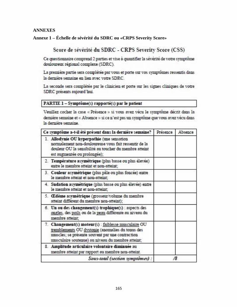
165
ANNEXES
Annexe 1 – Échelle de sévérité du SDRC ou «CRPS Severity Score»
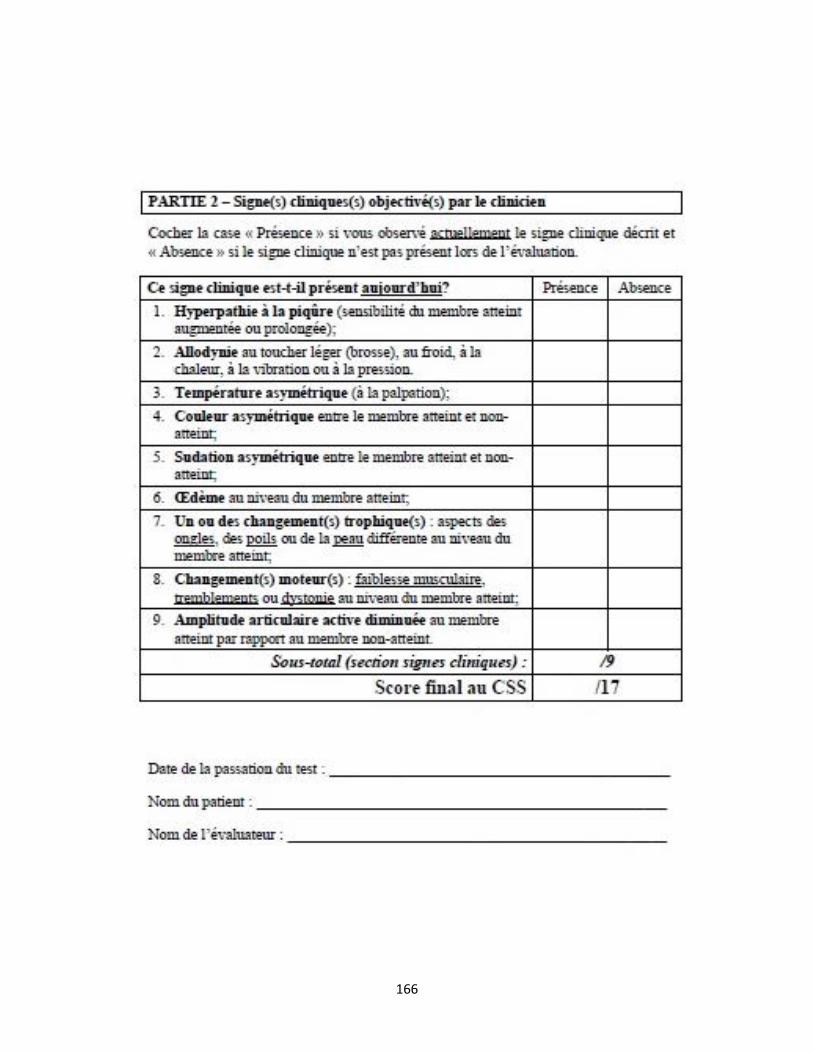
166
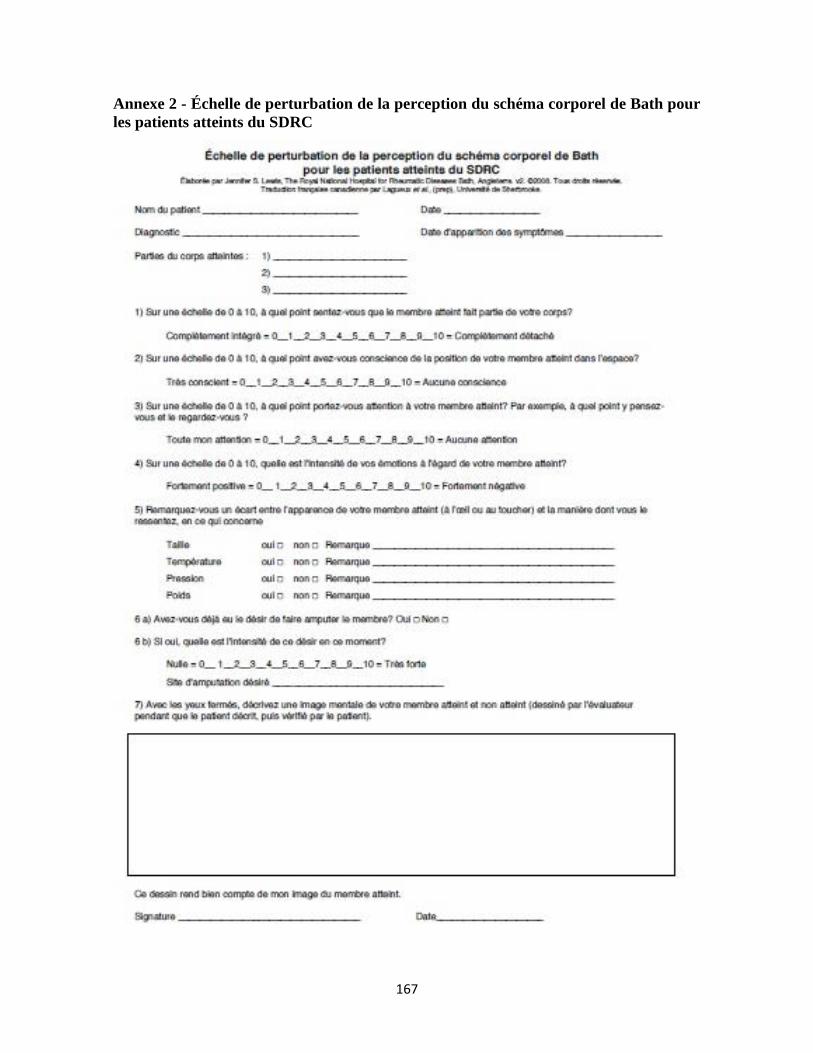
167
Annexe 2 - Échelle de perturbation de la perception du schéma corporel de Bath pour
les patients atteints du SDRC
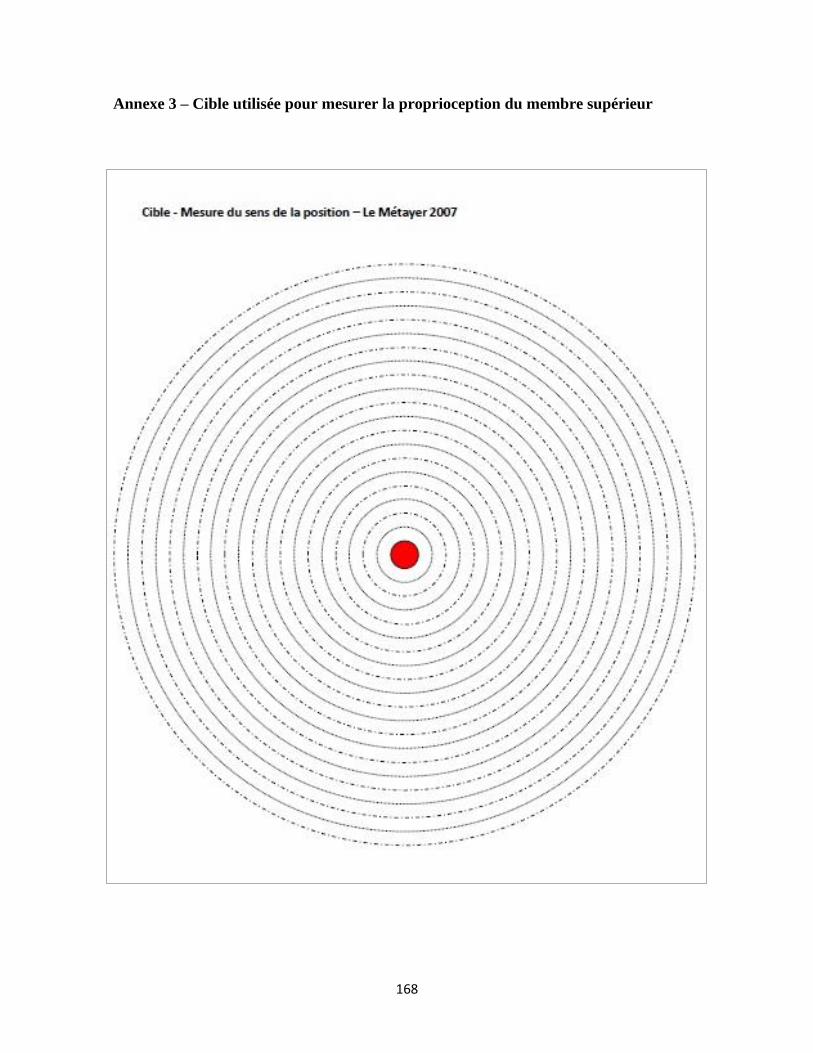
168
Annexe 3 – Cible utilisée pour mesurer la proprioception du membre supérieur