le complexe mili/mhen1 et études fonctionnelles des
TRANSCRIPT
HAL Id: tel-00601225https://tel.archives-ouvertes.fr/tel-00601225
Submitted on 17 Jun 2011
HAL is a multi-disciplinary open accessarchive for the deposit and dissemination of sci-entific research documents, whether they are pub-lished or not. The documents may come fromteaching and research institutions in France orabroad, or from public or private research centers.
L’archive ouverte pluridisciplinaire HAL, estdestinée au dépôt et à la diffusion de documentsscientifiques de niveau recherche, publiés ou non,émanant des établissements d’enseignement et derecherche français ou étrangers, des laboratoirespublics ou privés.
Le complexe MILI/mHEN1 et études fonctionnelles desprotéines DrTDRD1 et DrMOV10L
Stephanie Eckhardt
To cite this version:Stephanie Eckhardt. Le complexe MILI/mHEN1 et études fonctionnelles des protéines DrTDRD1 etDrMOV10L. Biologie cellulaire. Université de Grenoble, 2011. Français. �NNT : 2011GRENV010�.�tel-00601225�

THÈSE
Pour obtenir le grade de
DOCTEUR DE L’UNIVERSITÉ DE GRENOBLE
Spécialité: Biologie cellulaire
Arrêté ministériel: 7 août 2006
Présentée par
Stephanie ECKHARDT
Thèse dirigée par Ramesh PILLAI
Préparée au sein du Laboratoire de Régulation de l’expression des
gènes, Laboratoire Européen de Biologie Moléculaire (EMBL) et dans
l'École Doctorale Chimie et Science du vivant
Le complexe MILI/mHEN1 et études
fonctionnelles des protéines DrTDRD1 et
DrMOV10L
Thèse soutenue publiquement le 12.04.2011 devant le jury composé
de :
Dr Winfried WEISSENHORN
Professeur d'Université Joseph Fourier, Président
Dr Oliver MÜHLEMANN
Professeur d'Université de Berne, Rapporteur
Dr Marc BILLAUD
Directeur de recherche IAB, Rapporteur
Dr Donal O’CARROLL
Chef d‟équipe, EMBL Monterotondo, Examinateur
Dr Stephen CUSACK
Directeur de l‟antenne EMBL Grenoble, Examinateur
Dr Ramesh PILLAI
Chef d‟équipe, EMBL Grenoble, Directeur de thèse


THESIS
for obtaining the degree
DOCTOR OF THE UNIVERSITY GRENOBLE
Field: Cell biology
Ministerial Decree: 7 august 2006
Submitted by
Stephanie ECKHARDT
Thesis directed by Ramesh PILLAI
Prepared in the Regulation of Gene Expression Laboratory, European
Molecular Biology Laboratory (EMBL) and the Graduate School
Chemistry and Life Sciences
The MILI/mHEN1 complex and functional
studies of DrTDRD1 and DrMOV10L
Thesis publicly defended on the 12.04.2011 in front of the following
committee:
Dr Winfried WEISSENHORN
Professor at the University Joseph Fourier, Chair
Dr Oliver MÜHLEMANN
Professor at the University Bern, Reviewer
Dr Marc BILLAUD
Research Director IAB, Reviewer
Dr Donal O’CARROLL
Groupleader, EMBL Monterotondo, Examiner
Dr Stephen CUSACK
Head of Outstation EMBL Grenoble, Examiner
Dr Ramesh PILLAI
Groupleader, EMBL Grenoble, Thesis Director


___________________________________________________________________________
I
Mots clés
piARNs, protéines Piwi, mHEN1, HEN1 body, DrTDRD1, DrMOV10L
Key words
piRNAs, Piwi proteins, mHEN1, HEN1 body, DrTDRD1, DrMOV10L

___________________________________________________________________________
II
“There are in fact two things, science and opinion; the former begets knowledge,
the latter ignorance.”
Hippocrates
Law, Bk IV, circa 395 B.C
“Il y a en effet deux choses, la science et l’opinion ; celle-là conduit au savoir,
celle-ci à l’ignorance.”
Hippocrate
La Loi, Livre IV, environ 395 AJC

Abstract
___________________________________________________________________________
___________________________________________________________________________
III
Argonaute proteins associate with small RNAs to participate in gene regulatory processes.
Piwi proteins are a sub-clade of Argonaute that are mainly expressed in the animal germline.
They bind to Piwi-interacting RNAs (piRNAs) and contribute to genome integrity through
transposon silencing. To investigate factors involved in the piRNA pathway, mice and Danio
rerio (zebrafish) were exploited as model systems to investigate the piRNA methyltransferase
mHEN1, the Tudor domain-containing protein TDRD1 and the helicase MOV10L.
One defining feature of piRNAs is the presence of a 2`-O-methyl group on the 3`
terminal nucleotide which is catalyzed by the animal homologs of the plant RNA
methyltransferase HEN1. The mouse homolog, mHEN1, was shown to methylate RNA
substrates in vitro. The connection of mHEN1 to the piRNA pathway was shown in this study
by identifying an interaction between the C-terminal region of mHEN1 and the N-terminal
region of MILI adjacent to the PAZ domain, but distinct from that used for methylation-
dependent interaction with Tudor proteins. These results suggest the presence of a composite
interaction domain formed by the PAZ domain and the N-terminal region proximal to it. In
purified mouse germ cells and testis sections mHEN1 was detected, using specific antisera, in
a cytoplasmic granule distinct from the chromatoid body that contains the Piwi proteins. This
study implicates the HEN1 body as a potential site of piRNA biogenesis.
As a result of challenges encountered with mice for manipulation of the germline,
zebrafish embryos were developed as an alternate model system for studying piRNA pathway
components in our laboratory. The role of two piRNA pathway factors, DrTDRD1 and
DrMOV10L were investigated, in vivo. In mice MOV10L is thought to be involved in the
stabilization or loading of piRNAs into Piwi proteins. The role of TDRD1 was found to be an
essential component of the piRNA silencing complex. The expression profile of zebrafish
DrTDRD1 and DrMOV10L was determined from the one-cell egg stage to the adult fish
along with the role of the 3`UTR of DrTDRD1 in restricting expression to the germ cells.
Using morpholino knockdowns, DrMOV10L was functionally linked to the transposon
repression pathway. Taken together, these studies establish the mechanism of recruitment of
the piRNA methyltransferase and take the first steps towards providing a manipulatable in
vivo system for studying piRNA factors.
Résumé
_________________________________________________________________________
___________________________________________________________________________
IV
Les protéines argonautes, en s‟associant a des petits ARNs, participent à la régulation
génique. Les protéines Piwi appartenant à la sous-classe des argonautes sont principalement
exprimées dans les cellules de lignée germinale. Elles se lient aux petits ARNs interagissant
avec les protéines Piwi (piARN) et contribuent au maintien de l‟intégrité génique grâce à des
transposons responsables de la mise sous silence génique. Afin d‟étudier les différents
facteurs impliqués dans la voie de piARN, les modèles souris (Mus musculus) et danio zébré
ou poisson zèbre (Danio rerio) ont été utilisés pour explorer les méthyltransférases de
piARN, mHEN1, la protéine contenant le domaine Tudor 1, TDRD1 et l‟hélicase MOV10L.
Les piARN se caractérisent par la présence d‟un groupe 2`-O-methyl sur le dernier nucléotide
en extrémité 3`, ajouté par un homologue mammifère de la méthyltransférase d‟ARN de
plantes HEN1. Il a été montré que l‟homologue chez la souris, mHEN1, pouvait méthyler des
ARNs in vitro. Dans cette étude il a été démontré en particulier que cette mHEN1 était
impliquée dans la voie de piARN grâce à l‟identification d‟une interaction entre la région C-
terminale de mHEN1 et la région N-terminale de MILI adjacente au domaine PAZ, région
d‟interaction différente de celle observée pour les protéines Tudor. Ces résultats suggèrent la
présence d‟un domaine composite d‟interaction formé par le domaine PAZ et la région
proximale de son extrémité N-terminale. La protéine mHEN1 a été détectée dans des granules
cytoplasmiques à partir de cellules germinales purifiées et de sections de testicules en
utilisant des anti-sera spécifiques, alors que les protéines Piwi ont été détectées dans des
corps chromatoïdes. Les résultats de cette étude impliquent que HEN1 serait un site potentiel
pour la biogenèse des piARN.
La manipulation de lignées germinales de souris s‟avère particulièrement difficile. Un
système modèle alternatif, à partir d‟embryons de poisson zèbre a été développé dans le
laboratoire pour l‟étude de la voie des piARN. Le rôle de deux facteurs, DrTDRD1 et
DrMOV10L, impliqués dans cette voie ont été étudiés in vivo. Dans les souris, MOV10L
serait impliquée dans la stabilisation ou dans le recrutement des piARN vers les protéines
Piwi. Le rôle de TDRD1 s‟est révélé essentiel pour le complexe de silence des piARN. Le
profile d‟expression des protéines DrTDRD1 et DrMOV10L de danio zèbre a été déterminé à
différents phases de développement, du stade œuf unicellulaire au stade adulte. La spécificité
du profile d‟expression dans les cellules germinales est dépendante de la région 3` non
traduite de DrTDRD1. En outre il a été montré, grâce à l‟utilisation de morpholinos, que

Résumé
___________________________________________________________________________
___________________________________________________________________________
V
DrMOV10L était fonctionnellement impliquée dans la voie de répression des transposons.
L‟ensemble de ces études a permis de comprendre le mécanisme de recrutement d‟une
méthyltransférase de piARN et de mettre au point un système modèle permettant leur étude in
vivo.

Acknowledgments
___________________________________________________________________________
___________________________________________________________________________
VI
This thesis would not have been possible without the support of the following people. I am
eternally grateful to my parents, grandparents, future parents in law and lastly my fiancé
Louis Hutin for their loving help.
I would like to thank Prof. Dr. Wolfgang Nellen from the University Kassel who has
had a big influence in my career and who introduced to me the world of research. Dr. Kriton
Kalantidis, from the IMBB FORTH, Heraklion, Greece, Dr. Fredrik Söderbom, from the
University Uppsala and BMC, Sweden, both formed my view of science in a way that
encouraged me to initiate PhD studies.
To my thesis advisor, Dr. Ramesh Pillai, who accepted me as his first PhD student. I
thank him for giving me the opportunity to work independently on many different projects.
Thanks to my Thesis advisory committee: Dr. Stephen Cusack, Dr. Saadi Khochbin
and Dr. Donal O’Carroll for their good advice and support.
Special thanks to the members of our laboratory Dr. Michael Reuter, Jordi Xiol,
and Elisa Cora for all our scientific discussions and helpful advice.
I am deeply grateful to my wonderful lab-mates who helped me not only with fruitful
scientific discussions, but also kept my motivation going. Special thanks to my close
colleagues, Dr. Sebastien Muller, Dr. Rodrigo Louro, Dr. Philipp Berninger and Dr.
Radha Raman Pandey for keeping my spirit focused on the true meaning of life. In the
same breath, I have to thank Dr. Adam Round.
While at the EMBL many trainees joined the lab. I would like to thank Anisa,
Aurélien, Elsa, Emerens, Emilia, Hivin, Kirsten, Maartje, Magdalena, Ricardo, Sylvain,
Zhaolin, Anna and Margorzata.
I have not forgotten Jérôme Boudin a former technician in the lab for his strength
and power to do well. To the EMBL administration staff Sylviane Troger, Dominique
Lancon, Virginie Bertholet, Franscois Tronel, and Mary-Jane Villot for making daily life
so much easier.
A big thanks to Nicolas Martinelli, Nicolas Martinez, Dr. Danielle Desravines and
Philippe Mas for helping with the translations in the thesis and Dr. Andrew McCarthy, Dr.
Max Nanao, Dr. Chloe Zubieta and Dr. Simon Conn for proofreading of the manuscript.
Last but not least thanks to all members of the EMBL community, whose paths I
have crossed the last four years.

Remerciements
___________________________________________________________________________
___________________________________________________________________________
VII
Ce travail de thèse n‟aurait pas aboutit sans le support des personnes suivantes : je serais
éternellement reconnaissante a mes parents, grands parents, futurs beaux parents et mon
fiance Dr Louis Hutin pour leur aide bienveillante.
Je souhaiterais remercier le Dr Wolfgang Nellen de l‟Université de Kassel qui a
représenté dans ma carrière une grande influence et aussi m‟a initiée à l‟univers de la
Recherche. Ensemble, les Dr Kriton Kalantidis, du IMBB FORTH, Heraklion, Grèce, et Dr
Fredrik Söderbom, de l‟Université d‟ Uppsala et BMC, Suède, m‟ont incitée à poursuivre
une thèse par leur vision respective du domaine scientifique.
A mon responsable de thèse, le Dr Ramesh Pillai, qui m‟a accueillie comme premier
étudiant en thèse au sein de son laboratoire. Je le remercie pour l‟opportunité de travailler de
façon indépendante sur des projets varies.
Je remercie mon jury de thèse : Dr Stephen Cusack, Dr Saadi Khochbin and Dr
Donal O’Carroll pour leurs conseils pertinents et leurs encouragements.
Je remercie particulièrement les membres de notre laboratoire : Dr Michael Reuter,
Jordi Xiol et Elisa Cora pour toutes les discussions scientifiques et les conseils justes que
nous avons partagés.
Je voudrais également exprimer ma profonde gratitude a mes formidables collègues
qui ne m‟ont pas seulement aidée a travers de fructueuses discussions scientifiques, mais
aussi ont su trouver les mots pour garder ma motivation intacte.Des remerciements
particuliers a mes collègues proches: Dr Sebastien Muller, Dr Rodrigo Louro, Dr Philipp
Berninger et Dr Radha Raman Pandey qui m‟ont aidée à garder l‟esprit concentré sur les
véritables valeurs de l‟existence. Dans la même veine, je pense également au Dr Adam
Round.
Pendant ce travail de thèse, beaucoup de stagiaires ont intégré les activités du
laboratoire. Je voudrais donc remercier Anisa, Aurélien, Elsa, Emerens, Emilia, Hivin,
Kirsten, Maartje, Magdalena, Ricardo, Sylvain, Zhaolin, Anna et Margorzata.
Je n‟ai pas oublié Jérôme Boudin, technicien de l‟équipe pour son énergie et sa
disponibilité. A l‟équipe administrative de l‟EMBL : Sylviane Troger, Dominique Lancon,
Virginie Bertholet, Franscois Tronel, et Mary-Jane Villot pour rendre mes activités
quotidienne beaucoup plus aisées.

Remerciements
___________________________________________________________________________
___________________________________________________________________________
VIII
Un grand merci a Nicolas Martinelli, Nicolas Martinez, Dr Danielle Desravines et
Philippe Mas pour leur aide sur les traductions inhérentes a mon mémoire de thèse; et Dr
Andrew McCarthy, Dr Max Nanao, Dr Chloe Zubieta et Dr Simon Conn pour les
corrections qu‟ils ont apportes a celui ci.
Pour finir, merci a tous les membres de la communauté EMBL de Grenoble avec qui
nos chemins se sont croises durant ces quartes dernières années.

Abbreviations
___________________________________________________________________________
___________________________________________________________________________
IX
3` 3 prime
5` 5 prime
7mG 7-methylguanine
A Adenine
AGO Argonaute protein
Ala (A) Alanine
Arg (R) Arginine
Asn (N) Asparagine
Asp (D) Aspartic acid
ATP Adenosinetriphosphate
AUB Aubergine
c Control
casiRNA Cis-acting siRNAs
CB Chromatoid body
cDNA Copy DNA
C. elegans Caenorhabditis elegans
CTD C-terminal domain
C-terminus Carboxyl-terminus
Cys (C) Cysteine
DCL Dicer-like
Dcr Dicer
D. melanogaster Drosophila melanogaster
DNA Deoxyribonucleic acid
DNase Deoxyribonuclease
DND1 Dead end protein1
Dnmt de novo DNA methyltransferase
dpf Days post fertilization
D. rerio (Dr) Danio rerio
Drosha The Hebrew and Yiddish word for sermon in Judaism.
dsDNA Double stranded DNA
dsRBD Double stranded RNA binding domain
dsRNA Double-stranded RNA
E. coli Escherichia coli
e. g. Exempli gratia
endo-siRNA Endogenous siRNAs
et al. And co-workers
G Guanine
Gcl germ cell less
GFP Green fluorescent protein
Gln (Q) Glutamine
Glu (E) Glutamic acid
Gly (G) Glycine
GST Glutathione-S-transferase
GW Glycine (G)-tryptophan (W)
HeLa cells Henrietta Lacks cells
HEN HUA ENHANCER
HILI human Piwi-like 2
His (H) Histidine
HIV Human immunodeficiency virus
HIWI Human Piwi-like 1
HIWI2 Human Piwi-like 4
hPIWIl3 Human Piwi-like 3
H. sapiens Homo sapiens
IAP Intracisternal-A particle
i. e. id est
Ile (I) Isoleucine
Abbriviations
___________________________________________________________________________
___________________________________________________________________________
X
Leu (L) Leucine
LINE Long interspersed nuclear elements
lsiRNA Long siRNAs
LTR Long terminal repeats
Lys (K) Lysine
Met (M) Methionine
MILI Piwi-like protein 2 (Mus musculus)
MIWI Piwi-like protein 1 (Mus musculus)
MIWI2 Piwi-like protein 4 (Mus musculus)
miRNA microRNA
miRNP(s) miRNA-containing effector complex
Mirtron miRNA from intron
MMA Monomethylated Arginine
M. musculus (m) Mus musculus
MOV Moloney leukemia virus homolog
mRNA Messenger RNA
MTR1 Mouse Tudor repeat 1
MVH Mouse Vasa homolog
natsiRNA Natural antisense siRNAs
ncRNA Non-coding RNA
N-terminus Amino terminus
nt Nucleotide(s)
OD Optical density
OH Hydroxyl
ORF Open reading frame
Pasha Partner of Drosha
PAZ PIWI-Argonaute-Zwille domain
P-body Processing-body
PCR Polymerase chain reaction
PGC Primordial germ cells
pH Pondus Hydrogenii or potentia Hydrogenii
Phe (F) Phenylalanine
piRNA Piwi-interacting RNAs
Piwi P-element induced wimpy testis
Pol Polymerase
pre-miRNA Precursor miRNA
pri-miRNA Primary miRNA
PRMT Protein arginine methyltransferase
Pro (P) Proline
R2D2 Two dsRNA-binding domains (R2), associated with DCR-2
RanBPM Ran-GTP binding protein
RanGTP Ran guanosine triphosphate
rasiRNA Repeat-associated short interfering RNA
RBD RNA binding domain
RDE RNAi defective family member
RDR RNA-dependent polymerase
RdRP RNA-dependent polymerase
RecQ ATP-dependent DNA helicase
RISC RNA-induced silencing complex
RIWI Rat PIWI
RLC RISC-loading complex
RNA Ribonucleic acid
RNAi RNA interference
RNase Ribonuclease
RNP Ribonucleoprotein particle
rRNA Ribosomal RNA
RT-PCR Reverse transcription polymerase chain reaction
Abbreviations
___________________________________________________________________________
___________________________________________________________________________
XI
SAH S-adenosylhomocysteine
SAM S-Adenosyl methionine
scanRNA Scanning RNA
SD Standard deviation
sDMA Symmetrically di-methylated arginines
Ser (S) Serine
Sf21 (IPLB-Sf21AE) Spodoptera frugiperda cell line
SINE short intersperse nuclear element
siRNA Small interfering RNA
SIWI Silk worm Aubergine protein
Sm proteins Smith antigene proteins
snoRNA Small nucleolar RNA
snRNPs Small nuclear ribonucleoproteins
SPR Surface Plasmon Resonance
ssDNA Single-stranded DNA
ssRNA Single-stranded RNA
Ste Stellate
Su(ste) Suppressor of Stellate
T Thymine
TAC Thesis advisory committee
tasiRNA Trans-acting siRNAs
TDRD Tudor domain-containing
Thr (T) Threonine
TNP Transition protein
tRNA Transfer RNA
Trp (W) Tryptophan
Tyr (Y) Tyrosine
U Uracil
UTR Untranslated region
UV Ultraviolet
Val (V) Valine
Vls Valois
Wago Worm-specific Argonaute subfamily
WDR77 WD repeat domain 77
WT Wild type
Yb helicase female sterile (1) helicase
ZILI Zebrafish Piwi-like 2
ZIWI Zebrafish Piwi-like 1
Zuc Zucchini

Units of measure
___________________________________________________________________________
___________________________________________________________________________
XII
% Percent
Å Ångström
bp Base pair(s)
°C Degrees centigrade
dpc Days post coitum
dpp Days post partum
fmol Femtomole
g Gramm(s)
h Hour(s)
hpf Hours post fertilization
kb Kilo base(s)
kDa Kilo Dalton(s)
L Liter(s)
M Molar
mA Milliampere(s)
mg Microgramme(s)
min Minute(s)
mJ Millijoule(s)
mL Millilitre(s)
mm Millimetre(s)
mM Millimolar
µCi Microcurie(s)
µg Microgramme(s)
µJ Microjoule(s)
µL Microlitre(s)
ng Nanogramme(s)
pmol Picomole(s)
s Second(s)
U Unit(s)
V Volt(s)
w/v Weight/volume
W Watt(s)
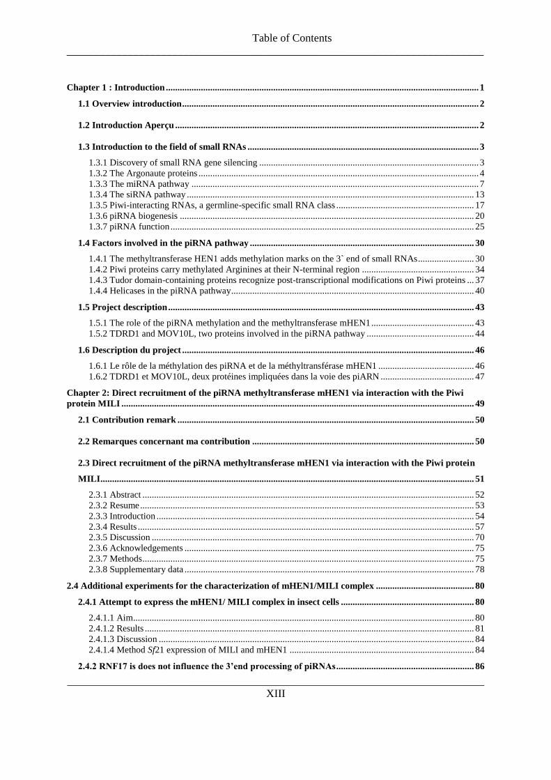
Table of Contents
___________________________________________________________________________
___________________________________________________________________________
XIII
Chapter 1 : Introduction ...................................................................................................................................... 1
1.1 Overview introduction ............................................................................................................................... 2
1.2 Introduction Aperçu .................................................................................................................................. 2
1.3 Introduction to the field of small RNAs ................................................................................................... 3
1.3.1 Discovery of small RNA gene silencing .............................................................................................. 3 1.3.2 The Argonaute proteins ........................................................................................................................ 4 1.3.3 The miRNA pathway ........................................................................................................................... 7 1.3.4 The siRNA pathway ........................................................................................................................... 13 1.3.5 Piwi-interacting RNAs, a germline-specific small RNA class ........................................................... 17 1.3.6 piRNA biogenesis .............................................................................................................................. 20 1.3.7 piRNA function .................................................................................................................................. 25
1.4 Factors involved in the piRNA pathway ................................................................................................ 30
1.4.1 The methyltransferase HEN1 adds methylation marks on the 3` end of small RNAs ........................ 30 1.4.2 Piwi proteins carry methylated Arginines at their N-terminal region ................................................ 34 1.4.3 Tudor domain-containing proteins recognize post-transcriptional modifications on Piwi proteins ... 37 1.4.4 Helicases in the piRNA pathway ........................................................................................................ 40
1.5 Project description ................................................................................................................................... 43
1.5.1 The role of the piRNA methylation and the methyltransferase mHEN1 ............................................ 43 1.5.2 TDRD1 and MOV10L, two proteins involved in the piRNA pathway .............................................. 44
1.6 Description du project ............................................................................................................................. 46
1.6.1 Le rôle de la méthylation des piRNA et de la méthyltransférase mHEN1 ......................................... 46 1.6.2 TDRD1 et MOV10L, deux protéines impliquées dans la voie des piARN ........................................ 47
Chapter 2: Direct recruitment of the piRNA methyltransferase mHEN1 via interaction with the Piwi
protein MILI ....................................................................................................................................................... 49
2.1 Contribution remark ............................................................................................................................... 50
2.2 Remarques concernant ma contribution ............................................................................................... 50
2.3 Direct recruitment of the piRNA methyltransferase mHEN1 via interaction with the Piwi protein
MILI ................................................................................................................................................................ 51
2.3.1 Abstract .............................................................................................................................................. 52 2.3.2 Resume ............................................................................................................................................... 53 2.3.3 Introduction ........................................................................................................................................ 54 2.3.4 Results ................................................................................................................................................ 57 2.3.5 Discussion .......................................................................................................................................... 70 2.3.6 Acknowledgements ............................................................................................................................ 75 2.3.7 Methods .............................................................................................................................................. 75 2.3.8 Supplementary data ............................................................................................................................ 78
2.4 Additional experiments for the characterization of mHEN1/MILI complex .......................................... 80
2.4.1 Attempt to express the mHEN1/ MILI complex in insect cells ......................................................... 80
2.4.1.1 Aim .................................................................................................................................................. 80 2.4.1.2 Results ............................................................................................................................................. 81 2.4.1.3 Discussion ....................................................................................................................................... 84 2.4.1.4 Method Sf21 expression of MILI and mHEN1 ............................................................................... 84
2.4.2 RNF17 is does not influence the 3’end processing of piRNAs ........................................................... 86

Table of Contents
___________________________________________________________________________
___________________________________________________________________________
XIV
2.4.2.1 Aim .................................................................................................................................................. 86 2.4.2.2 Results ............................................................................................................................................. 87 2.4.2.3 Discussion ....................................................................................................................................... 89 2.4.2.4 Methods ........................................................................................................................................... 89
2.4.3 Structural insight of binding of the methylated piRNAs by the MIWI PAZ domain ..................... 90
2.4.3.1 Contribution remarks ....................................................................................................................... 90 2.4.3.2 Remarques concernant ma contribution .......................................................................................... 90 2.4.3.3 Aim .................................................................................................................................................. 91 2.4.3.4 Results ............................................................................................................................................. 92 2.4.3.5 Discussion ....................................................................................................................................... 97 2.4.3.6 Methods ........................................................................................................................................... 99
Chapter 3: Analysis of the MOV10L and TDRD1 homologs in zebrafish ................................................... 101
3.1 Contribution remark ............................................................................................................................. 102
3.2 Remarques concernant ma contribution ............................................................................................. 102
3.3 Analysis of the MOV10L and TDRD1 homologs in zebrafish ........................................................... 103
3.3.1 Abstract ............................................................................................................................................ 104 3.3.2 Resume ............................................................................................................................................. 104 3.3.3 Introduction ...................................................................................................................................... 105 3.3.4 Results .............................................................................................................................................. 112 3.3.5 Discussion DrMOV10L and DrTDRD1 .......................................................................................... 127 3.3.6 Methods ............................................................................................................................................ 130
3.4 Additional experiments .............................................................................................................................. 133
3.4.1 Development of a reporter assay for functional studies of piRNAs ................................................ 133
3.4.1.1 Aim ................................................................................................................................................ 133 3.4.1.2 Results ........................................................................................................................................... 134 3.4.1.3 Discussion ..................................................................................................................................... 138 3.4.1.4 Method .......................................................................................................................................... 139
3.4.2 Identification of possible interaction partners of the MYND domain of TDRD1 using a yeast two
hybrid ............................................................................................................................................................ 140
3.4.2.1 Aim ................................................................................................................................................ 140 3.4.2.2 Results ........................................................................................................................................... 141 3.4.2.3 Discussion ..................................................................................................................................... 142 3.4.2.4 Method .......................................................................................................................................... 144
Chapter 4: General conclusions and future perspectives .............................................................................. 145
4.1 General conclusions and future perspectives ...................................................................................... 146
4.2 Conclusions générales et perspectives .................................................................................................. 151
Supplements ...................................................................................................................................................... 157
References ......................................................................................................................................................... 186

Figures
________________________________________________________________________
XV
Figure 1: Domains of the Argonaute proteins ....................................................................................................... 6
Figure 2: miRNA pathways ................................................................................................................................ 12
Figure 3: siRNA pathway ................................................................................................................................... 16
Figure 4: Germ cell development and spermatogenesis in mouse ...................................................................... 19
Figure 5: The Ping-pong cycle in Drosophila .................................................................................................... 24
Figure 6: The methyltransferase of HEN1 is highly conserved. ......................................................................... 33
Figure 7: Symmetrical dimethylation at the N-termini of mouse and Drosophila Piwi proteins ....................... 36
Figure 8: Structure of the Tudor domain 11 of Drosophila TUDOR ................................................................. 39
Figure 9:Endogenous mHEN1 in testes extracts is an RNA methyltransferase.................................................. 58
Figure 10: mHEN1 interacts with MILI ............................................................................................................. 60
Figure 11: mHEN1 interacts with MILI independent of RNAs and Piwi methylation status ............................. 62
Figure 12: The mHEN1 interaction site on MILI is close to the PAZ domain ................................................... 64
Figure 13: MILI interacts with the N-terminus of mHEN1 close to methyltransferase domain ......................... 66
Figure 14: Cytoplasmic localization of mHEN1 ................................................................................................ 68
Figure 15: Model ................................................................................................................................................ 74
Figure 16: MILI/mHEN1 complex studies in Sf21 ............................................................................................. 82
Figure 17: RNF17 and mHEN1 show a similar localization pattern. ................................................................. 86
Figure 18: RNF17 does not affect the 2‟O methylation of the 3‟end of piRNAs ............................................... 88
Figure 19: Purified PAZ domains and mutants................................................................................................... 93
Figure 20: UV-cross-linking experiments of PAZ domain and mutants to RNAs. ............................................ 96
Figure 21: Piwi expression during zebrafish development ............................................................................... 107
Figure 22: DrMOV10L alignment .................................................................................................................... 113
Figure 23: DrTDRD1 alignment ...................................................................................................................... 114
Figure 24: RT-PCRs to determine the expression of fish piRNA-pathway related mRNAs. ........................... 115
Figure 25: Tests of α-DrTDRD1 and α-DrMOV10L antibodies ...................................................................... 117
Figure 26: Immunoprecipitations of zebrafish piRNA pathway componments................................................ 119
Figure 27: Binding studies of DrTDRD1 and DrMOV10L ............................................................................. 121
Figure 28: Localization of DrTDRD1 .............................................................................................................. 123
Figure 29: Morpholino knockdowns................................................................................................................. 126
Figure 30: Reporter constructs and image processing ...................................................................................... 136
Figure 31: Analysis of the reporter constructs .................................................................................................. 137
Figure 32: Structure of the ETO MYND domain ............................................................................................. 140
Figure 33: Yeast-two-hybrid............................................................................................................................. 141
Figure S 1: Alignment of HEN1 from different animals .................................................................................... 78
Figure S 2: Endogenous mHEN1 from testes extracts is devoid of any associated small RNAs ....................... 79


Chapter 1 : Introduction

Introduction
___________________________________________________________________________
___________________________________________________________________________
2
1.1 Overview introduction
The introductory part of this thesis first concerns the discovery of small RNA mediated gene
silencing. To date, three main groups of small non-coding RNAs (ncRNAs) are the focus of
interest: microRNAs (miRNAs), small interfering RNAs (siRNAs) and Piwi-interacting
RNAs (piRNAs). The enzymatic core in all small ncRNA mechanisms is the Argonaute
protein family, which is summarized before the miRNA and siRNA pathways are discussed.
The focus then turns to piRNAs, their biogenesis and functions. Finally, some known factors
of the piRNA pathway are highlighted, in particular the RNA methyltransferase mHEN1, the
Tudor domain-containing family and Helicases.
1.2 Introduction Aperçu
L‟introduction de cette thèse concerne tout d‟abord la répression de gènes médiée par les
petits ARNs. A ce jour, trois groupes d‟ARNs non-codants (ncARNs) sont privilégiés : les
microARNs (miARNs), les petits ARNs interférents (siARNs) et les ARNs interagissant avec
Piwi (piARNs). Les enzymes appartenant à la famille des protéines Argonautes sont à
l‟origine de tous les mécanismes incluant les petits ncARNs. La famille des protéines
Argonautes sera introduite, puis nous discuterons des voies de signalisation des miARNs et
siARNs. Nous aborderons ensuite le sujet des piARNs, leur biogénèse ainsi que leurs
fonctions. Enfin, quelques facteurs connus de la voie des piARNs seront développés, en
particulier la ARN methyltransferase mHEN1, la famille des protéines à domaine Tudor et les
hélicases.

Introduction
___________________________________________________________________________
___________________________________________________________________________
3
1.3 Introduction to the field of small RNAs
1.3.1 Discovery of small RNA gene silencing
Genomes contain the entire hereditary information of an organism and are encoded in either
DNA or for many types of virus in RNA. Human DNA is packed in 22 autosomal
chromosome pairs and one sex-determining pair. The human genome contains about 6 billion
DNA base pairs. Remarkably, this only codes for 23,000 proteins, which is approximately 1.5
% of the genome, the remaining 98.5% consists of non-coding (nc)RNA genes, regulatory
sequences, introns and regions of unknown function which contain evolutionary artifacts that
have no present-day usage (Abdellah 2004; Lander et al. 2001).
The huge amount of genomic information in a cell has to be controlled and regulated.
The diverse mechanisms that cells and viruses use to control the flow of genetic information
or gene production are collectively referred to as gene regulation. Known mechanisms
include regulation of the DNA-RNA transcription up to and including post-translational
modification of a protein. However, not only the sequence and regulatory processes dictate
how the cell functions, changes in the phenotype or gene expression can be caused by other
factors. The study of these factors and underlying mechanisms is called epigenetics, one part
of which concerns the study of small non-coding RNAs that regulate gene expression.
In 1998, Fire and Mello demonstrated that double stranded RNA (dsRNA) triggers
gene silencing in C. elegans much better than single stranded RNA (Fire et al. 1998). The
Nobel Prize for Physiology or Medicine in 2006 was awarded for their discovery that small
interfering RNAs (siRNAs) caused the suppression of gene activity in a homology-dependent
manner, the RNA interference (RNAi) pathway.
In 1993, the first microRNA (miRNA, or small temporal RNA: stRNAs), named lin-4
(abnormal cell linage 4), was discovered as a negative regulator of LIN14, a protein which
builds a temporal gradient by its decreasing expression during the larvae development in the
nematode C. elegans. This 22 nt miRNA is partially complementary to the 3`untranslated
region (UTR) of its target mRNA, repressing lin-14 translation and influencing its stability
(Lee et al. 1993; Wightman et al. 1993). After several years of research a second miRNA, let-

Introduction
___________________________________________________________________________
___________________________________________________________________________
4
7, was discovered in C. elegans (Reinhart et al. 2000). Let-7 was found to be highly
conserved (Pasquinelli et al. 2000) and a search for more regulatory small RNA sequences
started.
To date, three main groups of small RNAs are in the main focus of interest:
microRNAs, small interfering RNAs (siRNAs) and since 2006 the Piwi-interacting RNAs
(piRNAs). They all have the ability to regulate a broad variety of biological processed
(Choudhuri 2009).
1.3.2 The Argonaute proteins
At the core of the small ncRNA pathways is the Argonaute protein family. These proteins
were first discovered due to their function for stem cell self-renewal and germ cell
development and maintenance (Bohmert et al. 1998; Cox et al. 1998). This protein family is
highly conserved evolutionarily and several members are found in many species: humans 8,
mice 7, Drosophila melanogaster 5 and Arabidopsis thaliana 10 Argonaute genes (Hock &
Meister 2008) (see Figure 1 A).
All Argonautes contain four domains: The N-terminus, a PIWI/Argonaut/Zwille
(PAZ) domain, a MID domain and the PIWI domain (see Figure 1 B and C) (Hutvagner &
Simard 2008). The PAZ domain shows similarities to an oligonucleotide and oligosaccharide
binding (OB) -fold and is responsible for the specific recognition of the 3` end of small
ncRNAs (Lingel et al. 2003, 2004; Ma et al. 2004; Song et al. 2003; Yan et al. 2003). The
MID domain adopts a Rossmann-like fold, and contains a highly conserved 5`-phosphate-
binding pocket for small RNAs (Boland et al. 2010; Frank et al. 2010; Wang et al. 2008b;
Yuan et al. 2005). The Piwi domain displays an RNaseH fold and contains a conserved
DDH/L motif, which is responsible for RNA cleavage activity of some Argonaute members
(Parker et al. 2004; Song et al. 2003; Song et al. 2004; Wang et al. 2008a; Wang et al.
2009b; Yan et al. 2003; Yuan et al. 2005). It is presently unclear why other Argonautes with
the conserved PIWI domain and DDH motifs are inactive.
In the Argonaute protein family three distinct subfamilies can be distinguished: the
AGO, the Piwi and a worm-specific subclade of the Argonautes (Wago) (Sasaki et al. 2003;
Tolia & Joshua-Tor 2007). The ubiquitously expressed AGO proteins bind to miRNAs and

Introduction
___________________________________________________________________________
___________________________________________________________________________
5
siRNAs, and are involved in gene silencing events at both transcriptional and post-
transcriptional levels (Buhler & Moazed 2007; Filipowicz 2005; Tomari & Zamore 2005).
The mainly germline-expressed Piwi proteins bind to piRNAs, and are responsible for the
germline development, gametogenesis and transposon silencing (Aravin et al. 2006; Girard et
al. 2006; Grivna et al. 2006a; Lau et al. 2006; Watanabe et al. 2006) (see Figure 1 A, red
indicated Piwi proteins).
The differentiation between the AGO and the Piwi subfamily is based on the
similarity of the proteins to either the AGO proteins of Arabidopsis or to Drosophila PIWI.
While the AGO proteins have been found in nearly all eukaryotes, Piwi proteins are restricted
to animals and ciliates; organisms which have a sexual reproduction cycle (Cox et al. 1998).
While the PAZ, MID and PIWI domains of Piwi and AGO are conserved, they sequentially
differ mainly at their N-termini (Cox et al. 1998).
In the next part, the most studied miRNA and siRNA pathway shall be highlighted
before the main focus of this thesis, the piRNA pathway, is described.

Introduction
___________________________________________________________________________
___________________________________________________________________________
6
Figure 1: Domains of the Argonaute proteins
(A) Argonaute proteins of Arabidopsis thaliana, mouse, human, zebrafish and Drosophila
melanogaster were aligned and used to create a phylogram with ClustalW (Chenna et al. 2003).
(B)The Argonaute proteins contain an N-terminal domain followed by the first linker (L1). In the
centre is the PAZ domain, which binds the 3` end of small RNAs. The PAZ domain and the MID domain are
separated by the second linker region (L2). The MID domain binds the 5` end of the small RNAs and orients
it towards the PIWI domain, which contains catalytic residues (DDH) for target RNA cleavage.
(C) The 2.6 Å crystal structure of the Ago ternary complex of Thermus thermophilus Ago by Wang
and colleagues. The N-terminus is indicated in sky-blue, the PAZ domain in cyan, the Mid domain in green-
blue and the Piwi domain in dark blue. The linkers (L1 and L2) are displayed in grey. The Ago protein was
co-crystallized with a bound 21-nucleotide guide DNA (orange) and a 19-nucleotide target RNA (red)
(Wang et al. 2009b)

Introduction
___________________________________________________________________________
___________________________________________________________________________
7
1.3.3 The miRNA pathway
Most miRNAs are evolutionally conserved and encoded as isolated genes or in closely spaced
clusters of 2-7 genes, from intergenic or exonic or intronic regions (Lagos-Quintana et al.
2001; Lau et al. 2001; Lee & Ambros 2001; Mourelatos et al. 2002). The canonical
biogenesis of miRNAs in animals starts in the nucleus, where POL II transcribes a precursor,
containing a 5` terminal 7-methyl guanylate cap and a 3` poly(A) tail, as for mRNAs (Cai et
al. 2004; Lee et al. 2004a) (see Figure 2 A). This primary miRNA (pri-miRNA) (Lee et al.
2002) can encode several miRNAs (Lagos-Quintana et al. 2001; Lau et al. 2001; Mourelatos
et al. 2002). The pri-miRNAs are cut into an intermediate form, the 60-70 nt pre-miRNA
(Lagos-Quintana et al. 2001; Lee & Ambros 2001; Lee et al. 1993; Mourelatos et al. 2002),
by the microprocessor protein complex. This contains the RNaseIII endonuclease Drosha
(Lee et al. 2003) and the dsRNA binding protein Pascha (partner of Drosha) in flies and C.
elegans (Denli et al. 2004) or DGCR8 (human DiGeorge Syndrome Critical Region Gene 8)
in mammals (Gregory et al. 2004; Han et al. 2004a; Landthaler et al. 2004; Wang et al.
2007). Pasha/DGCR8 directly interacts with the stem of the pri-miRNA and serves as
molecular anchor and positioning guide for Drosha (Han et al. 2006). Drosha creates the so
called pre-miRNA, a hairpin structure, where the loop is flanked by base-paired arms that
form a stem, which contain some unpaired nucleotides (Han et al. 2006; Lagos-Quintana et
al. 2001; Lau et al. 2001; Mourelatos et al. 2002). The pre-miRNA stem carries a two
nucleotide overhang at their 3` end, typical after the action of an RNase III enzyme, and a
phosphate group at their 5` end (Lee et al. 2003).
The canonical miRNA pathway gives rise to most of the known miRNAs, but some
miRNAs are produced by nuclear pre-mRNA splicing. This gives rise to pre-miRNA introns,
which mimic the structure of pre-miRNAs, and are called mirtrons (Babiarz et al. 2008;
Berezikov et al. 2007; Glazov et al. 2008; Okamura et al. 2007; Ruby et al. 2007) (see Figure
2 b). The spliced introns accumulate first as lariat shaped products and are then 2`-5`
debranched by the lariat debranching enzyme (Ldbr). These products contain the 2 nucleotide
3` overhangs, depending on the splicing site and can enter the standard miRNA biogenesis
pathway, where the 3` arm of the hairpin gives rise to mature miRNAs (Okamura et al. 2007;
Ruby et al. 2007). Not all mirtrons define their 3` end by splicing and carry a longer 3` tail
Introduction
___________________________________________________________________________
___________________________________________________________________________
8
(Ruby et al. 2007). In such cases the 3` end undergoes a trimming event in the nucleus that is
mediated by the exosome 11 complex via Rrp6 (Flynt et al. 2010).
Another group of miRNAs in mouse embryonic stem cells derive from endogenously
expressed short hairpins (shRNAs). These shRNAs are probably transcribed by POL III and
are DGCR8 independent (Babiarz et al. 2008) (see Figure 2 c). Finally, some other sources of
miRNAs are the small nucleolar RNAs (snoRNAs), the transfer RNAs (tRNAs), as well as
the terminal hairpins of endo-siRNAs long stem loop precursors that can bypass the Drosha
step (Cole et al. 2009; Ender et al. 2008; Miyoshi et al. 2010; Saraiya & Wang 2008).
The pre-miRNAs from the different pathways are exported from the nucleus to the
cytoplasm by the Ran guanosine triphosphate (RanGTP)-dependent dsRNA-binding protein
Exportin5 shuttle system (Bohnsack et al. 2004; Lund et al. 2004; Yi et al. 2003). Exportin5
recognizes the 3` overhangs of the pre-miRNAs and binds directly to the stem of at least
16 nt. Additionally, the binding of the RanGTP-Exportin5 complex to the pre-miRNA
protects the RNA from exonucleolytic degradation in the nucleus (Okada et al. 2009; Zeng &
Cullen 2004).
Once in the cytoplasm, the pre-miRNAs are cleaved by Dicer into dsRNA duplexes of
21-25 nt with 2nt overhangs at the 3` end (Bernstein et al. 2001; Grishok et al. 2001;
Hutvagner et al. 2001; Ketting et al. 2001; Knight & Bass 2001; Lee & Ambros 2001)
(Figure 2). Dicer prefers to cleave with an Uridine at the 5` position, which results in an 5`U
preference in mature miRNAs (Aravin et al. 2003). Dicer is accompanied by its dsRNA
binding protein TRBP (human immunodeficiency virus (HIV-1) trans-activating response
(TAR) RNA-binding protein) in mammals or the BP isoforms of Loqs in flies (Forstemann et
al. 2005; Haase et al. 2005; Jiang et al. 2005; Saito et al. 2005). TRBP and Loqs enhance the
processing of pre-miRNAs to miRNA duplexes by increasing the substrate affinity of Dicer
and stimulating the association of the complex with the Argonaute proteins (Chendrimada et
al. 2005; Haase et al. 2005; Jiang et al. 2005; Saito et al. 2005).
Recently it was shown by two groups that a miRNA pathway exists, which requires
Drosha, but not Dicer. Here the pre-miRNAs are directly loaded into AGO2. The AGO
protein itself then cleaves the pre-miRNAs into miRNA (Cheloufi et al. 2010; Cifuentes et al.
2010) with a defined 5`end. The 3` end remains variable, because it is then uridilated and
afterwards trimmed to create a functional miRNA (Cifuentes et al. 2010).

Introduction
___________________________________________________________________________
___________________________________________________________________________
9
The miRNA duplex, created by Dicer, contains one miRNA (guide strand) and one
passenger strand (miRNA*), corresponding to the two sides of the pre-miRNA stem. Only
one strand of the duplex is incorporated into an Argonaute protein (Grishok et al. 2001;
Mourelatos et al. 2002). The passenger strand is thought to be degraded. Only in some cases
are both strands used to produce mature miRNAs (Ruby et al. 2006). The sorting is carried
out by sensing the relative thermodynamic stability of the 5` ends of each strand to determine
the guide strand, or the strand which directs the 5` silencing. The 5‟ end of the actual miRNA
strand is less tightly paired to the complementary strand (Khvorova et al. 2003; Schwarz et
al. 2003).
In most organisms, miRNAs are sorted to associate with specific Argonautes. For
example, although that Drosophila contains two members of the Ago protein clade - AGO1
and AGO2-, the miRNAs are actively sorted into AGO1. This sorting of miRNAs into AGO1
requires DCR-1 and Loqs. In contrast, the association of siRNAs into AGO2 requires DCR-
2/R2D2, which are components of the siRNA pathway. If the miRNA duplex has central
mismatches, their binding to the DCR-2/R2D2 heterodimer is reduced and the small RNA is
loaded in AGO1 (Forstemann et al. 2007; Tomari et al. 2007). Additionally, the different
AGO associations of small RNAs itself is dependent on specific mismatches of the duplex.
While miRNAs often show a bulge in the duplex at the positions 9 and 10, Watson-Crick
base paring at position 9 and 10 promote miRNA* binding to AGO2. In Drosophila, the
miRNA* loaded into AGO2 has a 3` end modified by the RNA methyltransferase HUA
ENHANCER1 and behaves similar to siRNAs. The identity of the 5` nucleotide affects the
sorting as well, but is not sufficient for the recognition by specific Ago proteins (Czech et al.
2009; Okamura et al. 2009).
In mammals the sorting of the miRNA/miRNA* is less clear. All four AGO proteins
(hAGO1-4) in human show similar a preference towards the structure of the RNA duplexes in
requiring a central mismatch for RISC loading (Yoda et al. 2010). In different tissues at
different developmental stages, the use of miRNA and miRNA* strands can vary to a large
degree (Hu et al. 2009; Landgraf et al. 2007). In mice AGO2 is the dominant miRNA partner.
Although, mutant analysis showed, that the different AGO proteins (mouse AGO 1, 3 and 4)
in the organism can substitute for AGO2 (Liu et al. 2004).

Introduction
___________________________________________________________________________
___________________________________________________________________________
10
Plants lack a Drosha homolog, hence production of miRNAs in plants require
cleavage by the nuclear Dicer-like 1 (DCL1) (see Figure 2). After POL II transcription of the
pri-miRNA, DAWDLE (DDL), a FHA domain-containing RNA-binding protein, binds to the
pri-miRNA and facilitates the binding of DCL1 (Yu et al. 2008). DCL1 is accompanied by
the dsRNA binding protein HYL1 (Han et al. 2004b; Vazquez et al. 2004a), which binds to
the Cys2His2 zinc finger protein SERRATE (SE). Together these proteins position DCL1 on
the miRNA transcript precursor for correct processing (Kurihara et al. 2006; Yang et al.
2006a). Here, the pri-miRNA is first cleaved into the pre-miRNA (70-190 nt), and is then cut
into the miRNA duplex (Kurihara & Watanabe 2004; Papp et al. 2003; Park et al. 2002;
Reinhart et al. 2002; Xie et al. 2004). A putative mirtron discovered in rice grains, indicates
that plants may use spliced introns to produce miRNAs similar to animals (Zhu et al. 2008).
Unlike animal miRNAs, both miRNA strands in plants are 2`-O-methylated at their 3`
end by the RNA methyltransferase HEN1 (Park et al. 2002; Yang et al. 2006b; Yu et al.
2005). This protects the miRNAs from oligo-uridylation, a signal for degradation (Li et al.
2005). The miRNAs are exported by the Exportin5 homolog HASTY into the cytoplasm
(Park et al. 2005; Peragine et al. 2004). The addition of the methyl group on the miRNAs
could happen before loading into AGO1, as both strands are methylated (Yu et al. 2005).
However it is not yet clear, if the methylation of the miRNAs and the binding to AGO1 is
localized in the nucleus or in the cytoplasm, given that HEN1 and AGO1 localize in both
compartments (Fang & Spector 2007).
In animals and plants the mechanism by which miRNAs regulate their targets is
dependent on the Argonaute proteins they are bound to and the degree of complementarity
the miRNA have to their target mRNA (Hutvagner & Zamore 2002; Liu et al. 2004; Meister
et al. 2004). Although rare cases of miRNA target sites with nearly full complementarity to
miRNA exist in animals (Davis et al. 2005; Hutvagner & Zamore 2002; Mansfield et al.
2004; Pfeffer et al. 2004; Song et al. 2004; Sullivan et al. 2005; Yekta et al. 2004), the target
recognition by partial complementarity is more common in metazoan (Bartel 2009;
Brennecke et al. 2005; Lai 2004; Lewis et al. 2005; Lewis et al. 2003; Stark et al. 2005; Xie
et al. 2005) (see Figure 2). Therefore, the nucleotides 2-8 of miRNA, called the miRNA
„seed‟, guide the miRNA and Argonaut-containing miRISC to its targets by Watson-Crick
base pairing. The relatively small size of the seed sequence provokes the possibility that one
miRNA can address many different targets (Baek et al. 2008; Brennecke et al. 2005; Krek et

Introduction
___________________________________________________________________________
___________________________________________________________________________
11
al. 2005; Lai 2004; Lewis et al. 2005; Lewis et al. 2003; Lim et al. 2005; Selbach et al. 2008;
Stark et al. 2005; Xie et al. 2005). The „seed‟ sequence contributes the most to the
thermodynamic stability of the target binding (Ameres et al. 2007; Haley & Zamore 2004).
So if mismatches occur in the seed sequence, then the intermolecular interaction between the
miRNA and the AGO protein is disturbed (Parker et al. 2009; Wang et al. 2008a). The target
sites are preferentially located in an AU-rich region and tend to be located towards the
beginning or end of long 3‟ UTRs (Gaidatzis et al. 2007; Grimson et al. 2007). When several
target sites are within one transcript, the miRNA guided miRISC acts independently (Doench
et al. 2003), but if the target sites are within a short range they can act synergetically
(Grimson et al. 2007). In general, the binding of the miRISC to a target site with partial
complementarity leads to a translational block, a mRNA destabilization or both (Lee et al.
1993; Lim et al. 2005; Pillai et al. 2005; Wightman et al. 1993).
In plants a perfect or near-perfect complementarity to the mRNA target site is the
norm. In most cases this leads to a cleavage of the target sequence, the so called Slicing
reaction (Llave et al. 2002; Rhoades et al. 2002; Tang et al. 2003) (see Figure 2). It was
shown that AGO1 has slicer activity (Baumberger & Baulcombe 2005). But AGO1 or
AGO10 bound to miRNAs can also block translation, similar to animal miRNAs (Brodersen
et al. 2008).
In general, miRISCs associated with translational blocked RNAs, are guided by the
Glycine-tryptophan-rich 182 (GW182) protein to small cytoplasmic foci called processing
(P)- bodies. Here the mRNA can be degraded by the decapping and deadenylation enzymes
or stored for later release at a specific time point depending on cellular requirements (Behm-
Ansmant et al. 2006; Eulalio et al. 2008; Liu et al. 2005a; Liu et al. 2005b; Pillai et al. 2005;
Sen & Blau 2005).
This mechanism of miRNA-guided gene expression regulation is important in a broad
variety of biological processes including development (Brennecke et al. 2003; Giraldez et al.
2005; Giraldez et al. 2006; Lee et al. 1993; Reinhart et al. 2000), cell cycle regulation
(Linsley et al. 2007), metabolism (Krutzfeldt et al. 2005; Poy et al. 2004; Xu et al. 2003),
immunoresponse effects (Azuma-Mukai et al. 2008; Yeung et al. 2009) and diseases such as
cancer (Esquela-Kerscher & Slack 2006; Lu et al. 2005).

Introduction
___________________________________________________________________________
___________________________________________________________________________
12
Figure 2: miRNA pathways
(A) miRNAs of animals can derive from (a) pri-miRNA which is processed into pre-miRNAs by the
Drosha/Pasha (green/yellow) complex, (b) mitrons, which are processed by debranching enzymes and cleaved
by Rrp6 (red) if they have a 3‟ tail, (c) hairpins, which are Pasha dependent, (d) snoRNA precursors or (e) tRNA
precursors or virus induced tRNA like structures requiring specific processing enzymes. The pre-miRNA is
exported into the nucleus by Exportin5 (pink) and directly associated with an Ago protein (blue), or cleaved by
the Dcr-1/Loqs (purple/green) complex or the Dcr-2/R2D2 (purple/lilac) complex and then bound to an Ago
protein. The miRISC can then perform translational inhibition and RNA destabilization (left side) or mRNA
cleavage (right side). The main path of miRNAs is indicated with black arrows.
(B) The plant miRNA pathway. The pri-miRNA is bound by DDL1 (dark red), which recruits DCL1
(purple), HYL1 (yellow-green) and SE (green) to free the miRNA duplex. miRNAs may derive from mirtron in
plants, too. However, it is not yet clear if the methylation of the miRNAs by HEN1 (red) and the binding of to
AGO1 (blue) are located in the nucleus or cytoplasm. The miRNAs are exported by HASTY (pink). The most
common effect by the plant miRISC is the cleavage of its target mRNA (right side) but translation inhibition has
also been reported.

Introduction
___________________________________________________________________________
___________________________________________________________________________
13
1.3.4 The siRNA pathway
Initially, the siRNA pathway was thought only to act as a defense mechanism for the cell to
protect against selfish and invasive RNA elements. This was mainly due to the fact that
disruptions of the pathway in D. melanogaster and C. elegans did not cause an obvious
phenotype, except for a higher sensitivity to viral infections and increased activity of
repetitive elements (Ding & Voinnet 2007; Lee et al. 2004b; Okamura et al. 2004; Tabara et
al. 1999; Tabara et al. 2002).
A broad variety of endo-siRNAs have been described, deriving from hairpins formed
by inverted repeats, natural antisense siRNAs (natsiRNAs) from complementary transcripts
and secondary siRNAs, which are generated by RNA-dependent RNA polymerases (RdRPs).
In plants, the predominant endo-siRNAs are cis-acting siRNAs (casiRNAs; 24 nt). They are
transcribed from transposons, repetitive elements and tandem repeats (Chan et al. 2004; El-
Shami et al. 2007; Herr et al. 2005; Kanoh et al. 2005; Onodera et al. 2005; Pontier et al.
2005; Xie et al. 2004; Zilberman et al. 2003). They function in directing DNA methylation
and histone modification to silence the loci they originate from (Chan 2008; Chan et al. 2004;
Llave et al. 2002; Mette et al. 2000; Tran et al. 2005; Zilberman et al. 2003).
Trans-acting siRNAs (tasiRNAs) are also an example of endo-siRNAs in plants, and
of how the mi- and siRNA pathway can overlap: After miRNAs have directed the cleavage of
certain transcripts, the cleaved single stranded 3‟ RNA fragment (and a lesser extent the 5‟
fragment) can be used to produce siRNAs (Allen et al. 2005; Peragine et al. 2004; Vazquez et
al. 2004b; Williams et al. 2005; Yoshikawa et al. 2005).
Endo-siRNAs are also produced in response to stress, for example from salt stress
(Borsani et al. 2005) and in response to bacterial pathogen effectors (Katiyar-Agarwal et al.
2006). Natural antisense transcript-derived siRNAs (natsiRNA) are produced from pairs of
RNAs, were one strand is expressed constitutively and the antisense transcript in stress
conditions only (Borsani et al. 2005; Katiyar-Agarwal et al. 2006).
While endo-siRNAs are well described in plants (Vazquez 2006) and fungi
(Catalanotto et al. 2002; Reinhart & Bartel 2002), they were only recently discovered in
animals. While plant and worm endo-siRNAs are typically produced depending on the RNA-

Introduction
___________________________________________________________________________
___________________________________________________________________________
14
dependent RNA polymerases action, D. melanogaster, mouse and human lack this kind of
RdRP (Ghildiyal & Zamore 2009). In cultured human cells, it was discovered that the full-
length LINE1 (long interspersed nuclear element1) contain a sense and antisense promoter in
its 5` untranslated region (5`UTR). This leads to a bidirectional transcription of an
overlapping region, which could be detected by Dicer and processed into siRNAs to silence
retrotransposable activity. However, the exact mechanism remains undetermined (Yang &
Kazazian 2006). More recently endo-siRNAs in human have been shown to be required for
chromatin remodeling in the vicinity of the original siRNA target site, such as H3K9me2 and
H3K27me3 (Malecova & Morris 2010).
In flies endogenous siRNAs have been detected in somatic and germ cells. They are
21 nt in length, present in sense and antisense orientations and have 2` O methylated 3`ends.
One group of endogenous siRNAs derive from transposons and another from heterochromatic
and intergenic sequences, building long extensive structured transcripts like hairpins and
mRNA (Chung et al. 2008; Czech et al. 2008; Ghildiyal et al. 2008; Hartig et al. 2009;
Kawamura et al. 2008; Okamura et al. 2008a; Okamura et al. 2008b).
In mice, endo-siRNAs have been identified in oocytes. As for flies, they are 21 nt in
length and originate from a variety of genomic clusters, forming dsRNAs by spliced
transcripts of protein-coding genes to homologous antisense transcripts of pseudogenes or an
inverted repeat of pseudogenes (Sasidharan & Gerstein 2008; Tam et al. 2008; Watanabe et
al. 2008). Additionally, in embryonic stem cells endo-siRNAs are produced via Dicer-
independent cleavage of long hairpins, which mainly derive from genomic loci with tandem
inverted short intersperse nuclear elements (SINEs) (Babiarz et al. 2008).
Whether the siRNAs originate from exogenous substrates or endogenous transcripts,
the siRNA biogenesis begins with a double stranded long RNA. This is cleaved by the
dsRNA-specific Ribonuclease III (RNaseIII) Dicer (Bernstein et al. 2001; Elbashir et al.
2001b; Hammond et al. 2000) into a 21 to 24 nt long RNA duplex (Elbashir et al. 2001b;
Zamore et al. 2000), with a two nt overhang at the 3` ends (Elbashir et al. 2001b; Elbashir et
al. 2001c) (see Figure 3). Each strand has a 5` phosphate and a 3` hydroxyl group (Elbashir et
al. 2001b). The siRNA duplex is then incorporated into the RNA-induced silencing complex
(RISC). This occurs via a RISC loading complex (RLC) (see Figure 3), where the relative
thermodynamic stability of the 5` ends of each strand is sensed to select the guide strand
(Aza-Blanc et al. 2003; Khvorova et al. 2003; Schwarz et al. 2003). In Drosophila, the

Introduction
___________________________________________________________________________
___________________________________________________________________________
15
dsRNA-binding protein R2D2 (two dsRNA-binding domains, associated with Dicer 2), a
component of the RLC and a partner of Dicer-2 (DCR-2), is responsible for the recognition of
the thermodynamically more stable 5` end of the passenger strand (Liu et al. 2003; Tomari et
al. 2004). The RLC then recruits an AGO protein, for example Agonaute2 (AGO2) in
Drosophila, which cleaves the passenger strand and releases it (Kim et al. 2007; Leuschner et
al. 2006; Matranga et al. 2005; Miyoshi et al. 2005; Rand et al. 2005). This siRNA mediated
cleavage (like the target cleavage) always happens across the phosphodiester bond between
the 10th
and 11th
nucleotide of the guide strand (Elbashir et al. 2001c). The cleavage is Mg2+
dependent and leads to a 3` hydroxyl and a 5` phosphate group (Martinez & Tuschl 2004;
Schwarz et al. 2004; Wang et al. 2009b). The RNA is now mature in higher animals, but not
in flies and plants, where the methyltransferase HEN1 methylates the 3` end of the siRNAs to
stabilize the siRNAs and protect them from degradation (Boutet et al. 2003; Chen 2007;
Ebhardt et al. 2005; Li et al. 2005; Yang et al. 2006b). Following this, the siRNA can guide
the RISC to target mRNAs with perfect complementarity, cleaving the mRNA and leading to
their degradation because they lack either a cap or poly(A) tail (Elbashir et al. 2001a;
Elbashir et al. 2001b; Elbashir et al. 2001c; Hammond et al. 2000; Zamore et al. 2000).
In plants and flies, the siRNA pathway is partly a defense mechanism against viral
infections, but as well a gene regulatory element (Ghildiyal & Zamore 2009; Obbard et al.
2006). However, there is yet no evidence that mammals use the RNAi pathway for viral
defense, instead they have developed an interferon induced protein based immune-system
(Vilcek 2006; Williams 1999).

Introduction
___________________________________________________________________________
___________________________________________________________________________
16
Figure 3: siRNA pathway
(A) siRNAs in insects (the Drosophila melanogaster pathway is shown here as an example) starts with
a long double stranded RNA, which is cleaved by a Dicer (purple) into ~21 nt siRNA duplexes. The R2D2
protein (lilac) binds to the thermodynamically stable 5` end and together with Dicer the RNA is guided to
AGO2 (blue) to form the RISC. The passenger strand (black) is cleaved by the Argonaute enzyme and the guide
strand (red) is 2`-O-methylated at the 3` end. The RISC complex cleaves its target mRNA, guided by the
siRNA. The mRNA fragments are degraded afterwards.
(B) Amplification of siRNAs in plants: A plant Dicer cuts the primary siRNAs out of a long dsRNA.
This siRNAs bind to an Argonaute protein, which cleaves the target mRNA. This can recruit RDR6 with the
RNA-binding protein SGS3 (yellow), which leads to a synthesis of a dsRNA. This dsRNA is then cleaved by
DCL4 to create secondary siRNAs. These are again bound by AGO. All siRNAs in plants are 2‟O methylated.
Adapted from (Ghildiyal & Zamore 2009)

Introduction
___________________________________________________________________________
___________________________________________________________________________
17
1.3.5 Piwi-interacting RNAs, a germline-specific small RNA class
Piwi-interacting RNAs (piRNAs) are the largest class of small RNA molecules expressed in
the animal cell (Seto et al. 2007), but they were not widely recognized as such until 2006,
when five independent research groups simultaneously re-discovered these germline-specific
ncRNAs in mice and Drosophila (Aravin et al. 2006; Girard et al. 2006; Grivna et al. 2006a;
Lau et al. 2006; Watanabe et al. 2006). Actually, similar ncRNAs were described in
Drosophila as repeat-associated RNAs (rasiRNAs) (Aravin et al. 2003) already in 2003.
RasiRNAs have the same features as piRNAs (Saito et al. 2006; Vagin et al. 2006) and will
therefore be referred to as piRNAs hereafter.
piRNAs derive from discrete gene-poor genomic loci (Aravin et al. 2006; Girard et al.
2006; Grivna et al. 2006a; Lau et al. 2006; Watanabe et al. 2006). Different from miRNAs,
whose sequences are conserved across species, no sequence conservation was found for
piRNAs (Aravin et al. 2006; Girard et al. 2006; Lau et al. 2006). Many of the larger piRNA
clusters are syntenic and range in size from 0.9 to 127 kb (Aravin et al. 2006), as well as
encoding many homologous piRNAs with overlapping sequences (Ro et al. 2007). Most of
the piRNAs in mammals derive from uni-directional clusters, but a few bidirectional clusters
have been identified (Aravin et al. 2006; Girard et al. 2006; Lau et al. 2006; Ro et al. 2007;
Watanabe et al. 2006).
In Drosophila these piRNA clusters derive mainly from transposons, transposon
remnants and repeat sequences (Brennecke et al. 2007; Gunawardane et al. 2007; Saito et al.
2006; Yin & Lin 2007). In mammals piRNAs contain exonic, intronic, intergenic and repeat
sequences, but the majority are derived from intronic regions (Grivna et al. 2006a; Ro et al.
2007) and only 17 % of the adult mouse piRNAs map to repeated elements (Aravin et al.
2006; Girard et al. 2006; Ro et al. 2007). Mice piRNAs clusters expressed before birth can
generate piRNAs from both strands, as in Drosophila, while postnatal piRNAs do not show
this feature (single-strand clusters) (Aravin et al. 2008).
Interestingly, Piwi proteins show a clear preference for different originating piRNAs
during germline development. Drosophila contains three Piwi proteins; AGO3, PIWI and
AUBERGINE (AUB). AGO3 show a bias for piRNAs encoded on the sense strand of

Introduction
___________________________________________________________________________
___________________________________________________________________________
18
transposons, while PIWI and AUB preferentially bind piRNAs from the antisense strand
(Brennecke et al. 2007; Gunawardane et al. 2007).
In mice, three Piwi proteins -MIWI, MILI and MIWI2- are present exclusively in
testes. MILI is expressed from 12.5 days post coitum (dpc) until round spermatids (20 days
post partum, dpp). MIWI2 is found from 15.5 dpc until 3 days after birth and MIWI is
expressed from 14 dpp until the round spermatids stage (see Figure 4). MILI binds ~26 nt
piRNAs (Aravin et al. 2006; Aravin et al. 2007b), MIWI2 ~28 nt piRNAs (Aravin et al.
2008) and MIWI associates with ~30 nt piRNAs (Girard et al. 2006; Grivna et al. 2006a;
Grivna et al. 2006b). At 16.5 dpc most (70%) of the piRNAs are derived from transposon
sequences. Between 16.5 dpc and 10 dpp the amount of transposon derived piRNAs is ~35%
in MILI. The piRNA profile changes during development, with those derived from long
interspersed nuclear elements (LINE) and long terminal repeats (LTR) decreasing as the
small interspersed nuclear elements (SINE) and exon originating ones increase. MILI and
MIWI2 associate with repeat derived piRNAs during the embryonic development, whereas
the piRNA profile of MILI changes towards post-natal stages (Aravin et al. 2008;
Kuramochi-Miyagawa et al. 2008). Prenatal piRNAs show complementarities between MILI-
and MIWI2-associated piRNAs. MILI piRNAs have a bias for an U at the first nt (Aravin et
al. 2006; Aravin et al. 2008; Kuramochi-Miyagawa et al. 2008; Watanabe et al. 2006) and
derive from the sense strand of clusters and/or transposons. This suggests they arise from
cleavage of a genomically encoded piRNA cluster. MIWI2 has the signature of a preference
for an A at the 10th
nt and a strong bias for antisense strand transposon sequences. A small
proportion of the MILI- and MIWI2-associated repeat sequences also show a 10-nt overlap
(Aravin et al. 2008; Kuramochi-Miyagawa et al. 2008).
From the pachytene stage onwards, MIWI and MILI bind to non-repetitive piRNAs of
intergenic origin and of unknown function (Aravin et al. 2006; Girard et al. 2006). The
MIWI-associated piRNAs show a bias for Uracil at the 5` end like MILI piRNAs (Girard et
al. 2006; Watanabe et al. 2006).
Introduction
___________________________________________________________________________
___________________________________________________________________________
19
Figure 4: Germ cell development and spermatogenesis in mouse
7.25 dpc the primordial germ cells appear. Between 7.5 and 13.5 dpc genome wide reprogramming takes
place before the cells enter mitotic arrest. The expression of piRNAs and Piwi proteins are indicated in blue
lines. MILI is expressed from 12.5 dpc until round spermatids, MIWI2 from 15.5 dpc until 3 days after birth
and MIWI from 14 dpp until the round spermatids stage.

Introduction
___________________________________________________________________________
___________________________________________________________________________
20
In the zebrafish (Danio rerio) ZIWI and ZILI-associated piRNAs (26-27 nt) are found
in the male and female germline (Houwing et al. 2008; Houwing et al. 2007). The fish
piRNAs originate mainly from transposons (mostly LTR elements) and repetitive sequences,
the rest are from non-repetitive sequences that lie in intergenic regions. Unlike mammalian,
zebrafish piRNAs are derived from discrete genomic loci where they display a strong strand
asymmetry. The piRNA density is much higher in ovaries than in testis (Houwing et al. 2008;
Houwing et al. 2007). Another difference is that piRNAs in fish can be maternally passed to
the embryo (Houwing et al. 2007). Similar to mice, the piRNAs of ZIWI have the 5` U bias
while those in ZILI has predominantly A at the 10 nt (Houwing et al. 2008)
So far, all the piRNAs analyzed from different species have a 5` monophosphate and a
3` hydroxyl group (Houwing et al. 2007; Saito et al. 2006; Watanabe et al. 2006) that is
modified (Gunawardane et al. 2007; Houwing et al. 2007; Kawaoka et al. 2009; Vagin et al.
2006). This modification was shown to be a 2`-O-methylation (Horwich et al. 2007; Kirino &
Mourelatos 2007c; Ohara et al. 2007a; Saito et al. 2007a; Zhou et al. 2010). The piRNAs
share this 3‟ end modification with plant mi- and siRNAs (Akbergenov et al. 2006; Katiyar-
Agarwal et al. 2007; Li et al. 2005; Yang et al. 2006a; Yang et al. 2006b; Yu et al. 2005) as
well as the siRNAs and hairpin RNAs (hpRNAs) from Drosophila (Horwich et al. 2007;
Okamura et al. 2008b; Pelisson et al. 2007; Saito et al. 2007a; Vagin et al. 2006). Similar
modification of animal miRNAs or siRNAs in other organisms is not yet described..
To summarize, piRNAs (24 to 31 nt in length) are not conserved between species and
are modified at their 3` end with a methyl group. Many piRNAs have a U-bias at the 5` end
or an A at the 10th
position and an overlap between these piRNAs is suggested. These
features of piRNAs may be due to their biogenesis and are discussed in the next part.
1.3.6 piRNA biogenesis
The piRNA pathway is evolutionary ancient (Grimson et al. 2008) and there is a great interest
in knowing the exact molecular pathway that leads to piRNA biogenesis. piRNAs are not
equally distributed over the genome, but rather clustered at defined loci (Aravin et al. 2006;
Grivna et al. 2006a), where 96 % of the piRNA sequences are present in a few hundred
genomic sites (Aravin et al. 2006; Girard et al. 2006). It is suggested that a long single
Introduction
___________________________________________________________________________
___________________________________________________________________________
21
stranded RNA or a pair of divergently transcribed large single stranded RNAs, from one
cluster, serve as a precursor for piRNAs (Aravin et al. 2006; Lau et al. 2006; Watanabe et al.
2006). Given that many piRNAs are generated from a single cluster simultaneously, an
individual transcription for each piRNA is unlikely. This idea is supported by the observation
that P-element insertions into putative promoter of piRNA loci disrupt the production of all
piRNAs from a 180 kb window (Brennecke et al. 2007; Kawaoka et al. 2008; Malone et al.
2009).
The primary processing of piRNAs is not well understood. Factors of this process are only
just starting to be discovered. This is mainly from research in flies due to the ease of genetic
manipulation. Biochemical studies in mouse characterized the orthologues of the fly proteins
and identified new factors.
One factor in Drosophila is the putative nuclease Zucchini, which mainly localizes in
a perinuclear region of the cytoplasm, called the nuage, in gonad-associated somatic and
germline cells (Pane et al. 2007; Saito et al. 2009). Zucchini was found to interact physically
with AUB (Pane et al. 2007).The loss of zucchini function leads to a reduction of processed
piRNAs in somatic as well as germline cells, but does not affect the signatures of the
secondary piRNA pathway (Malone et al. 2009; Olivieri et al. 2010; Pane et al. 2007; Saito
et al. 2009).
Among the factors of the primary pathway identified so far are several putative
helicases, Vasa, Spindle-E and Armitage in Drosophila and their mouse homologs Vasa
(MVH) and MOV10l (Frost et al. 2010; Kuramochi-Miyagawa et al. 2004; Kuramochi-
Miyagawa et al. 2010; Malone et al. 2009; Olivieri et al. 2010; Wang et al. 2010; Zheng et
al. 2010)
Another source of primary piRNAs could be those that are maternally deposited in the
Drosophila egg. These are mainly bound by AUB and PIWI, and to a lesser extent by AGO3
to prevent transposon activity in the offspring (Brennecke et al. 2008). Once the primary
piRNAs are present, they are suggested to be used in an amplification loop. This so called
“ping-pong” model for piRNA biogenesis is based on sequence analyses of fly piRNA
(Brennecke et al. 2007; Gunawardane et al. 2007) (see Figure 5). The Piwi protein Aubergine
(AUB)-containing complex is guided by the primary piRNA to actively transcribed
transposons. The transposon sequence is cleaved by AUB, defining the 5` end of a new sense

Introduction
___________________________________________________________________________
___________________________________________________________________________
22
strand piRNA. This model points out how the 5` ends of new piRNAs are generated from
long single-stranded precursors by the slicer action of Piwi proteins, but the mechanism of 3`
end formation is less clear (Brennecke et al. 2007; Gunawardane et al. 2007). It is assumed
that an exonuclease or an undefined endonuclease acts on the RNA. Potential candidates were
identified in a Drosophila genetic screen (Pane et al. 2007). The new sense piRNA, a so
termed secondary piRNA, because it is generated by Piwi-mediated cleavage, is incorporated
into AGO3 RNPs, which target the antisense precursors and lead to the production of more
antisense piRNAs to associate with AUB. This mechanism results in an amplification loop of
secondary piRNAs (Brennecke et al. 2007). PIWI-associated piRNAs show no pairing with
AGO3 bound piRNAs (Gunawardane et al. 2007), maybe because it is mainly nuclear, while
AUB and AGO3 are cytoplasmic localized in the nuage (Brennecke et al. 2007;
Gunawardane et al. 2007; Harris & Macdonald 2001). Therefore it could be speculated that
the nuage is the localization center of the ping-pong cycle (Klattenhoff & Theurkauf 2008).
Additionally, loss of PIWI has no impact on ping-pong signals, indicating that PIWI does not
participate in the amplification cycle, while AUB and AGO3 mutants lack this pathway
(Malone et al. 2009).
Mutations in spindle-E, vasa, the Tudor domain-containing protein krimper and
armitage strongly impact on the ping-pong cycle and delocalize AUB and AGO3 from the
nuage. Additionally, the piRNA size shifts towards the piRNAs from PIWI. Thus, the Piwi
functions in the germline depends on the RNA helicase Armitage, Spindle-E and Vasa, and
the Tudor domain protein Krimper. Comparison between Spindle-E and Armitage reveal that
Spindle-E is crucially required for the ping-pong cycle. Armitage is only needed for a
minority of elements to enter the cycle, so it may not be required for the ping-pong cycle
(Malone et al. 2009; Olivieri et al. 2010). The Heterochromatin protein 1 (HP1) homolog
Rhino is not needed for primary piRNA processing but is required for the ping-pong cycle
(Olivieri et al. 2010). Rhino associates with the 42AB cluster and promotes the production of
the RNAs leading to piRNAs from dual-strand heterochromatic clusters, indicating that it
could be involved in the processing of primary piRNAs. In rhino mutants the ping-pong cycle
is impaired and transposon silencing is inefficient. Rhino localizes in the nucleus of germ
cells and is responsible for the proper localization of AUB, AGO3 and Vasa to the nuage
(Klattenhoff et al. 2009). The nuclease Squash also seems to be required for piRNA
production, but its phenotype, in terms of the piRNA biogenesis is rather weak, although the

Introduction
___________________________________________________________________________
___________________________________________________________________________
23
piRNA level decreases overall (Malone et al. 2009; Pane et al. 2007). Also, there is no
identifiable Squash homolog in other species.
A similar post-transcriptional amplification loop has been suggested for vertebrates as
well. In mice, MILI mainly binds sense while MIWI2 shows an antisense bias for piRNAs.
Prenatal piRNAs show a strong enrichment of secondary piRNA signatures (A at the tenth
nucleotide). MIWI2 is enriched two fold with secondary piRNAs compared to MILI. Thus,
MILI is primarily biased towards piRNAs with a U as the 1st nt, while MIWI2 binds
secondary piRNAs with an A at the 10th
position (Aravin et al. 2008; Kuramochi-Miyagawa
et al. 2008). Additionally, in mili mutants MIWI2 is not loaded with piRNAs (Aravin et al.
2008). Recently it was discovered that the ping-pong cycle in mouse is dependent on MHV,
which is probably needed to load piRNAs in MIWI2 (Kuramochi-Miyagawa et al. 2010). The
Ping-pong signatures have been detected in zebrafish as well. Additionally, piRNAs binding
to ZIWI are preferentially antisense while those binding to ZILI have a sense orientation
(Houwing et al. 2008). At the moment the ping-pong model is an attractive hypothesis for
secondary piRNA production based on bioinformatic analysis of deep sequencing, but it is
still lacking more experimental support.
Unlike miRNAs in animals, the piRNAs in various organisms have a 2`-O-methyl
modification as a universal feature on their 3` terminal nucleotide (Horwich et al. 2007;
Kirino & Mourelatos 2007c; Ohara et al. 2007a; Saito et al. 2007a). This modification is
performed by the RNA methyltransferase HEN1 proteins (Horwich et al. 2007; Kirino &
Mourelatos 2007a, b; Okamura et al. 2008c; Pelisson et al. 2007; Saito et al. 2007a; Vagin et
al. 2006). It is not yet known at which point of the piRNA biogenesis the methylation occurs
or what impact the modification has on the binding of piRNAs to Piwi proteins.
In conclusion, piRNAs are produced by at least by two different pathways. Primary
piRNAs are generated from long single stranded transcripts of piRNA clusters that are often
highly enriched for fragmented, diverged transposon remnants. The primary piRNAs in
complex with Piwi proteins can target complementary transcripts, cleaving these and
producing a new 5` end of a secondary piRNA. This means that piRNAs can amplify the
silencing signal and at the same time silence the transposon transcript at the same time.

Introduction
___________________________________________________________________________
___________________________________________________________________________
24
Figure 5: The Ping-pong cycle in Drosophila
Primary processed and maternal contributed piRNAs antisense to transposon transcripts are loaded into AUB
and lead the complex to its target transposon transcript, whereby the slicer activity of AUB defines the 5`end
of a secondary sense piRNA. The 3` end processing is unknown, but this end gets 2`-O-methylated by
DmHEN1. The new piRNA is loaded into an AGO3-containing complex to target a piRNA cluster transcript
and generate a piRNA, which is antisense to the transposon transcript and incorporated into AUB again. This
amplification cycle leads to transposon silencing. (Adapted from (Brennecke et al. 2007; Gunawardane et al.
2007))

Introduction
___________________________________________________________________________
___________________________________________________________________________
25
1.3.7 piRNA function
The protection of the genome from transposable elements is crucial to its integrity. Thus,
mobile elements have to be distinguished and silenced (Girard & Hannon 2008; Saito &
Siomi 2010). Transposable elements can be divided in class I and class II. Class I transpose
via an RNA element while class II use DNA elements and are characterized by a terminal-
inverted repeat (TIR). The class I can again be subdivided into long terminal repeats (LTR)
retrotransposons and non-LTR transposons, which are also called long-interspersed nuclear
elements (LINE) or poly(A)-type retrotransposons. An ancient family of transposable
elements is built from sequences deriving from small interspersed nuclear elements (SINEs)
(Biemont & Vieira 2006).
Studies in mice, Drosophila and zebrafish show that piRNAs and Piwi proteins are
essential for germline development and the control of transposable elements (Saito & Siomi
2010). An example for gene regulation via Piwi proteins is the AUB regulation of the Stellate
locus by piRNAs, which are encoded by the Suppressor of Stellate (Su(ste)) (Aravin et al.
2001; Schmidt et al. 1999; Vagin et al. 2006). Drosophila males lacking the Y-chromosome
are sterile, form cytoplasmic needles and/or nuclear star-shaped crystalline structures in
spermatocytes (Bozzetti et al. 1995; Hardy et al. 1984) and show defects in meiosis,
specifically in chromosome pairing (Palumbo et al. 1994). The crystal formation can be
suppressed if the flies lack the Stellate (Ste) locus on the X-chromosome (Livak 1984). On
the Y-chromosome the deletion of the Suppressor of Stellate su(ste) also leads to male
sterility (Hardy et al. 1984) because Ste is overexpressed (Livak 1990). The euchromatic Ste
locus and the heterochromatic Su(ste) region are homologous (Kalmykova et al. 1998). Both
strands of the Su(ste) region are transcribed and repress the Ste locus (Aravin et al. 2001). If
the Drosophila spindle-E, or aub is mutated, the Ste locus is not suppressed (Aravin et al.
2004; Aravin et al. 2001; Schmidt et al. 1999). piRNAs from this locus are only found to be
antisense to Su(ste) (Aravin et al. 2004; Vagin et al. 2006). In piwi mutants, piRNAs from the
Su(Ste) locus hyper-accumulate but Ste is silenced. In aub mutants the sense transcript
accumulates but not the antisense piRNAs, suggesting that the antisense piRNA silence not
only Ste mRNA but also target the sense piRNA (Vagin et al. 2006). The Su(ste) piRNAs are

Introduction
___________________________________________________________________________
___________________________________________________________________________
26
strongly reduced in ago3 mutant males as well, suggesting that AGO3 is needed to produce
the piRNAs bound to AUB (Li et al. 2009).
Another example of regulation via piRNAs and Piwi proteins is the flamenco locus.
The flamenco locus is a heterochromatic region on the X-chromosome, containing numerous
fragments of transposable elements, which encode a large piRNA cluster and is required to
repress the transposon activity of Gypsy, Idefix and ZAM (Brennecke et al. 2007; Sarot et al.
2004). If this region is disrupted, the piRNAs from here are reduced and the transposon
activity increases (Brennecke et al. 2007). These results show that piRNAs act in the
transposable element regulation. The disruption of aub has the same effect (Pelisson et al.
2007; Savitsky et al. 2006; Shpiz et al. 2007; Shpiz & Kalmykova 2007; Vagin et al. 2004).
In somatic cells, the first insight of the epigenetic role of Piwi proteins was found in
Drosophila, where AUB and PIWI were shown to regulate position-effect variegation
(PEV)(Pal-Bhadra et al. 2002). This is a mechanism in which euchromatin genes are silenced
if they are in close proximity to a heterochromatic region, for example in chromosomal
rearrangement (Dorer & Henikoff 1994, 1997; Fanti et al. 1998; Martin-Morris et al. 1997).
PIWI is present in the nucleus and can be found directly associated with chromosomes and
interacting with the Heterochromatin protein 1a (HP1a), a marker for heterochromatin,
indicating that the piRNA pathway is directly connected to the heterochromatin-forming
mechanism (Brower-Toland et al. 2007).
piRNAs are also able to activate transcription. Specific sub-telomeric heterochromatin
(telomere-associated sequence, or TAS) piRNAs bound to PIWI promote euchromatic
histone modifications and piRNA transcription in TAS on the right arm of chromosome 3
(3R-TAS). PIWI associates to this region and promotes transcription activity. In piwi
mutants, this 3R-TAS accumulates heterochromatin histone modifications and
heterochromatin protein 1 a (HP1a), which silence this locus and lead to a loss of germline
stem-cell maintenance (Yin & Lin 2007).
In zebrafish, many piRNAs derive from transposable elements, preferentially from
LTR and mainly in the antisense orientation to transposon transcripts. Examples are
GypsyDr1, GypsyDr2 and Ngaro. In zili knockout mutants, the activity of several LTR, non-
LTR and DNA elements is increased (Houwing et al. 2007). piRNAs are differently
expressed in testes and ovaries and play also a role in zebrafish sex differentiation (Houwing

Introduction
___________________________________________________________________________
___________________________________________________________________________
27
et al. 2007; Zhou et al. 2010). These sex-specific piRNAs derive from separate loci. Ovarian
piRNAs are more often associated in groups (33%), when compared to testes piRNAs (20%).
Mammalian piRNAs can be divided in two different classes: The pre-pachytene and
the pachytene piRNAs, according to the stage of spermatogenesis in which they appear (see
Figure 4). Like fly piRNAs, pre-pachytene piRNAs which consists of embryonic and post-
natal populations before P10, derive primarily from repetitive sources (35% in MILI or 60%
in MIWI2), mainly SINE, LINE and LTR transposons, and are implicated in retrotransposon
silencing (Aravin et al. 2007a; Aravin et al. 2007b; Carmell et al. 2007; Kuramochi-
Miyagawa et al. 2008).
In mammals, piRNAs are implicated in establishment of de novo methylation during
gametogenesis (Aravin & Bourc'his 2008; Aravin et al. 2008). During development, the
global methylation patterns are extensively remodeled in waves of demethylation and de novo
methylation (Hajkova et al. 2002). At 8 dpc the DNA methylation decreases together with a
transient loss of the DNA de novo methyltransferases Dnmt1, Dmnt3a and Dmnt3b. To
prevent deregulated transcription, POL II is suppressed as well (Seki et al. 2007). During the
migration of the primordial germ cells (PGCs), the transcriptional repressor B-lymphocyte
maturation-induced protein 1 (BLIMP1, also known as Prdm1) is associated with the histone
arginine methyltransferase PRMT5 in the nucleus and mediates dimethylation of H2AR3and
H4R3 to suppress their target genes (Ancelin et al. 2006). At 11.5 dpc BLIMP1/PRMT5
translocate from the nucleus into the cytoplasm in post-migrated PGCs and their targets are
activated (Ancelin et al. 2006). Interestingly, H4R3me is also associated with activation of
other genes by facilitating the acetylation of H4 tails (Strahl et al. 2001; Wang et al. 2001a)
while H3R8me, which is also mediated by PRMT5, is a transcription repression mark (Pal et
al. 2004).
It would appear that during 12.5 and 14.5 dpc the genome is not protected, but that is
not the case. Post migratory-germ cell-specific genes such as the mouse Vasa homolog
(MVH or also known as DEAD box polypeptide 4, Ddx4), Sycp3 (synaptonemal complex
protein 3) and Dazl (deleted in azoospermia-like) carry methylation marks in their flanking
regions and become hypomethylated when they are expressed at 13.5 dpc (Maatouk et al.
2006). Some transposable elements like LINE1 and some intracisternal A particles (IAPs)
remain methylated even at 13.5 dpc (Hajkova et al. 2002). The male germ cells go into

Introduction
___________________________________________________________________________
___________________________________________________________________________
28
mitotic arrest between 13.5 and 14.5 dpc. The imprinting marks are erased by demethylation
during the PGC migration and new marks are established according to the gender of the
animal. The parental methylation imprints (H19, Dlk1/Gtl2 and Rasgrf1) are established
between 14.5 dpc and birth (20 dpc), with the paternal are imprinted before the maternal ones
in the male germline (Davis et al. 1999; Davis et al. 2000; Kato et al. 2007; Li et al. 2004;
Ueda et al. 2000). Dnmt3a plays a central role in this mechanism for all loci, but Rasgrf1
(RAS protein-specific guanine nucleotide-releasing factor1) additionally requires the de novo
methyltransferase Dnmt3b (Kaneda et al. 2004; Kato et al. 2007), most likely due to the fact
that several retrotransposons are encoded in this region. Both methyltransferases build a
complex with Dnmt3l, which is highly expressed in prospermatogonia but has no
methyltransferase activity (Bourc'his & Bestor 2004; Kaneda et al. 2004; Kato et al. 2007;
Webster et al. 2005). The imprinting marks stay throughout the rest of the germ cell
development.
Together with the imprinting marks retrotransposable elements undergo de novo
methylation. While SINEB1 is mainly methylated by Dnmt3a, LINE1 and IAP are
methylated by Dnmt3a and Dnmt3b, the methylation of all these sequences requires Dnmt3l
(Kato et al. 2007). In cases of failure of the de novo methylation, retroelements are highly
transcribed in spermatogonia and spermatocytes, the meiosis fails and germ cells are
progressively lost by the mid-pachytene stage (Aravin et al. 2007b; Bourc'his & Bestor 2004;
Hata et al. 2006; Webster et al. 2005). Current evidence would indicate a role for piRNAs
bound to Piwi proteins in ensuring the genomic stability by silencing the transposable and
repetitive elements (Aravin et al. 2003; Brennecke et al. 2007; Gunawardane et al. 2007;
Houwing et al. 2007; Saito et al. 2006; Vagin et al. 2006). In mili or miwi2 deficient males,
LTR and non-LTR transposons are overexpressed due to a loss of DNA-methylation marks
(Aravin et al. 2007b; Carmell et al. 2007; Kuramochi-Miyagawa et al. 2008). Apart from
piwi mutants, the deletion of small piRNA clusters, such as Nct1 and 2 in mice, lead to an
increased activity of transposable elements, like LINE1 and open reading frame 1 (ORF1).
This demonstrates that piRNAs themselves have roles in the transposon control. The
mutation of the Nct1 and 2 does not affect gene expression, fertility, or spermatogenesis (Xu
et al. 2008).
How piRNAs perform silencing of the transposable elements and which factors are
involved is not known. However, it is possible that silencing by piRNA is similar to the

Introduction
___________________________________________________________________________
___________________________________________________________________________
29
mechanism of heterochromatin formation at peri-centromers induced by siRNAs in
Schizosaccaromyces pombe. Here, the siRNA recruits the RNA-induced transcriptional
silencing (RITS) complex to nascent transcripts from peri-centromers, which in turn recruits
the HP1a homolog SWI6, triggering heterochromatin formation (Buhler et al. 2006; Verdel et
al. 2004; Volpe et al. 2002).
Piwi proteins also play a role in dsDNA break-repair. Isolation of rat PIWI (RIWI)
complexes identified the adenosine triphosphate (ATP)–dependent DNA helicase rRECQ1
(Lau et al. 2006), the homolog of the Neurospora QDE-3, which together with an AGO
protein has a role in gene silencing (Catalanotto et al. 2002).The presence of RecQ in the
RIWI complexes (Lau et al. 2006) is a indication for the function of the piRNA pathway in
dsDNA-break repair (Hunter 2008). On the other hand, in absence of Piwi proteins, dsDNA
breaks are caused by transposon accumulation (Klattenhoff et al. 2007).
Pachytene piRNAs are bound to MILI and MIWI, but their function is still not clear.
Isolation of RNAs from polysome and RNP fractions containing MIWI shows that MIWI and
piRNAs interact in polysome and are associated with mRNAs (Grivna et al. 2006a; Grivna et
al. 2006b). MIWI is also associated with eIF4E, an mRNA cap binding protein that is
responsible for translational control, and Dicer, the endonuclease needed for si- and miRNA
production. The quantity of certain miRNAs is significantly reduced in MIWI mutants,
meaning MIWI could be involved in miRNA-mediated translational control. The target
mRNAs are also down-regulated, too, supporting a MIWI involvement in mRNA stability
(Deng & Lin 2002). MILI seems to be involved in translational control as well, where it
forms a stable complex with eIF3a and associates with eIF4E and eIF4G containing the m7G
cap-binding complex (Unhavaithaya et al. 2009). However, the data supporting relationship
between Piwi proteins and translation control needs to be obtained from other model systems
to become accepted as one of the modes by which piRNA pathway regulates gene expression.
In mice, as in other mammals, piRNAs are only found in significant amounts in the
male germline. While the male germline contains lifelong regenerating stem cells, the female
gender relies on meiotically arrested oocytes stored from prenatal development. Nevertheless,
the female germ cells have to be protected from transposable elements too. Perhaps this
function in oocytes is carried out by endogenous small interfering RNAs deriving from
Introduction
___________________________________________________________________________
___________________________________________________________________________
30
retroelements including LINE, SINE and LTR (Tam et al. 2008; Watanabe et al. 2006;
Watanabe et al. 2008), as Piwi mutants display normal fertility in females.
Although a lot of knowledge was acquired since piRNAs were discovered in 2006,
many questions still remain unanswered. While the ping-pong model explains secondary
piRNAs, the biogenesis of primary piRNA remains unclear. Factors involved in the
biogenesis and function of piRNAs are only just starting to be discovered. Although the pre-
pachytene piRNAs are associated with transposable element silencing, many of these piRNAs
do not fit this scheme. Also, there is still no function for pachytene piRNAs.
1.4 Factors involved in the piRNA pathway
1.4.1 The methyltransferase HEN1 adds methylation marks on the 3` end of
small RNAs
Hen1 was identified in Arabidopsis thaliana as a gene involved in the specification of the
stamen and carpel identities during the flower development, while searching for factors
which enhances HUA1 and HUA2, leading to the name HUA ENHANCER 1 (HEN1) (Chen
et al. 2002). Plants carrying hen1 mutations are smaller, their fertility is reduced and their leaf
shape alters (Chen et al. 2002). HEN1 is indirectly involved in stress response (Zhang et al.
2008), acquisition and resolution of copper (Abdel-Ghany & Pilon 2008), and the long
distance transport of information via small RNAs (Buhtz et al. 2010). It S-adenosyl-L-
methionine dependent methylates miRNA and siRNA duplexes with a preferential length of
21-24 nt after Dicer cleavage (Akbergenov et al. 2006; Katiyar-Agarwal et al. 2007; Li et al.
2005; Yang et al. 2006a; Yang et al. 2006b; Yu et al. 2005). The methylation of the small
RNAs by HEN1 protects them from the addition of extra nucleotides, mostly U-residues, by
an unknown terminal transferase or polymerase which leads to their destabilization and
degradation (Chen 2005; Ebhardt et al. 2005; Li et al. 2005; Yang et al. 2006b). The
miRNAs and the siRNA levels are greatly reduced in hen1 mutants (Akbergenov et al. 2006;
Boutet et al. 2003; Katiyar-Agarwal et al. 2006; Park et al. 2002; Vazquez et al. 2004a;
Vazquez et al. 2004b).

Introduction
___________________________________________________________________________
___________________________________________________________________________
31
The Arabidopsis thaliana HEN1 (942 aa) is a multi-domain protein. It contains two
dsRNA-binding domains (dsRBD 1 and 2) separated by a LA-motif (another RNA binding
site) containing domain at the N-terminus (Tkaczuk et al. 2006). This is followed by a PPIase
(peptidyl-prolyl cis-trans isomerase)-like domain (PLD), lacking a typical PPIase active site
(Tkaczuk et al. 2006). The C-terminus contains the catalytic domain, a typical Rossmann-
fold MTase fold with an S-adenosyl methionine (SAM)-binding motif (Asp-Phe-Gly-Cys-
Gly) and a nuclear localization motif (Anantharaman et al. 2002; Park et al. 2002; Vazquez et
al. 2004a; Yu et al. 2005). Four of the five domains directly interact with the RNA substrate
in an A-form confirmation (the dsRBD1 and 2, the MTase domain and the La domain).
HEN1 binds to the small RNAs as a monomer and specifically recognizes duplexes with 2 nt
overhangs at the 3` end (Huang et al. 2009) (see Figure 6A).
Based on homology and structure prediction, members of the HEN1-family were
identified in metazoan, fungi, Viridiplantae and bacteria. Evolutionary analyses of this
protein family suggest that the protein appeared first in the common ancestor of eukaryotes,
before they separated into Viridiplantae and Metazoan/Fungi and was subsequently
horizontal transferred to the bacteria.
The bacterial HEN1 is more similar to animal than plant, the only common feature of
all being the MTase domain. Only the plant HEN1 contains the double stranded RNA binding
domains and PPIase domain (Jain & Shuman 2010). The crystal structure of the MTase
domain of bacteria HEN1 revealed a core seven-stranded β-sheet (β1-7), which is flanked on
two sides by α-helices (αZ, αA-E), a common fold of Ado Met-dependent MTases.
Additionally the MTase of HEN1 contains a short helix between β4 and αD and support
domain between β5 and αE (see Figure 6 B). This is a conserved structure and unique to
HEN1 proteins (Mui Chan et al. 2009) (see Figure 6 C). Recently it was shown, that different
to former suggestions, the methyltransferase activity of bacteria HEN1 is manganese, and not
magnesium dependent (Jain & Shuman 2011).
In bacteria HEN1 is essential for RNA repair. For example, in the bacterium
Anabaena variabilis, HEN1 (ca. 460 aa) interacts with the polynucleotide Kinase-
phosphatase PNKP, a protein involved in RNA repair, and forms a stabile heterotetramer in
vitro. PNKP alone cannot repair RNAs, which were cleaved by ribotoxins. Together with
HEN1 the RNAs are not only repaired but a methyl group at the cleavage site is added during

Introduction
___________________________________________________________________________
___________________________________________________________________________
32
the progress. This protects the RNA from being cut again (Chan et al. 2009; Jain & Shuman
2010).
In Tetrahymena, Piwi-associated scanRNAs (26-31 nt) are 2`-O-methylated (Kurth &
Mochizuki 2009). scanRNA are expressed only during the sexual reproduction (Mochizuki et
al. 2002). This modification is catalyzed by HEN1p and protects the small RNAs from
degradation. HEN1p and the Piwi proteinTwi1p interact where the single-stranded scanRNA
are methylated after loading and exclusion of one RNA strand. This modification ensures the
function of scanRNAs in DNA-elimination, influencing the efficient of the production of
sexually functional progeny (Kurth & Mochizuki 2009).
Studies of the HEN1 Drosophila homolog (DmHEN1), a 391 aa protein, shows that it
is essential for piRNA, siRNA and hpRNAs methylation (Horwich et al. 2007; Okamura et
al. 2008b; Pelisson et al. 2007; Saito et al. 2007a; Vagin et al. 2006). DmHEN1 methylates
single-stranded RNA (ssRNA) substrates (Horwich et al. 2007; Saito et al. 2007a) with a size
range between 22 and 38 nt (Saito et al. 2007a). Loss of activity results in reduced length and
abundance of piRNAs and siRNAs (Horwich et al. 2007; Saito et al. 2007a). Analysis of
DmHEN1 and the zebrafish DrHEN1 revealed that the methylation of the small RNAs is
needed for their stability. This occurs by protecting them from transferases preferentially
adding Us at their 3` end, which subsequently attract 3`-5` exonucleases that trim the small
RNAs (Ameres et al. 2010; Kamminga et al. 2010).
The mouse mHEN1 (395 aa) is expressed exclusively in the male germline and the
recombinant protein expressed in E. coli is reported to have methylation activity, suggesting
that this could potentially be the piRNA methyltransferase. This protein was able to
methylate 20-40 nt long ssRNAs on their 3` ends (Kirino & Mourelatos 2007a, b). Until now
it is not clear how this modification affects the piRNA pathway in mice and how the
methyltransferase is recruited to this pathway. Further, it is not known how the methylation
mark on small RNAs in animals is recognized by other components of the piRNA pathway,
for example the PAZ domain of the murine Piwi proteins.

Introduction
___________________________________________________________________________
___________________________________________________________________________
33
Figure 6: The methyltransferase of HEN1 is highly conserved.
(A) Ribbon diagram of A. thaliana HEN1 structure. The dsRBD1 is colored in blue, the La motif in
magenta, the LCD in bordeaux, the dsRBD2 in sand, the PPIase-like domain in yellow and the MTase domain
in red. The linkers are colored in gray. Indicated as a green sphere is the Mg2+ and the AdoHcy is indicated in
green sticks. The RNA strands are indicated in blue and cyan (adopted from (Huang et al. 2009) and analyzed
by Pymol)
(B) Ribbon diagram of the Anabaena variabilis HEN methyltransferase domain (purple). The bound
AdoHcy is indicated in green (adopted from (Mui Chan et al. 2009) and analyzed by Pymol)
(C) Superimposed AtHEN1 and AvHEN1(analyzed by Pymol), indicating the high similarity of the
structures.
(D) Domain architecture of different HEN1. At, Arabidopsis thaliana; m, mouse; h, human; Gg, Gallus
gallus (chicken); Dr, Danio rerio (zebrafish); Dm, Drosophila melanogaster (fruit fly). The methyltransferase
domain is indicated in red and the LAM domain in green. The figure is based on (Chen 2007).

Introduction
___________________________________________________________________________
___________________________________________________________________________
34
1.4.2 Piwi proteins carry methylated Arginines at their N-terminal region
Recently it was discovered that Piwi proteins carry post-translational modifications in form of
symmetrically di-methylated arginines (sDMA) at their N-termini (Kirino et al. 2010a; Kirino
et al. 2010b; Nishida et al. 2009; Reuter et al. 2009; Vagin et al. 2009). The first identified
sDMA carrying residue was R74 in MILI (Reuter et al. 2009) (see Figure 7). R100, R146,
R163 and R549 in MILI were identified as dimethylated and R95 as monomethylated by
Vagin and colleagues (Vagin et al. 2009), while R74 was found to carry both types of
methylation in the latter study. In contrast to these findings, Chen et al. published that R74,
R83, R95 and R100 can be mono- and dimethylated, while R45, R146, R156 and R163 are
only dimethylated (Chen et al. 2009). MIWI was first described to carry R49 and R371
monomethylated and R14 mono-or dimethylated (Vagin et al. 2009). Later Chen and
colleagues published that R53 in MIWI can be both mono and dimethylated (Chen et al.
2009). In Drosophila the AUB protein showed sDMAs at R11, R13 and R15, while AGO3
contains sDMAs at R4, R68 and R70 (Nishida et al. 2009). These different findings for the
methylation status maybe due to the source of material used for analysis, suggesting that Piwi
methylation is a dynamic process.
sDMAs are known to modify the ability of proteins to carry out their biological
functions. For example, Sm proteins, which are splicing machinery factors, contain sDMAs
that are recognized by the single Tudor domain of the SMN (survival motor neuron) proteins
to recruit U snRNAs (Buhler et al. 1999; Meister et al. 2002; Selenko et al. 2001). The
methylation of the Sm proteins and Piwi proteins is catalyzed by protein methyltransferase 5
(PRMT5) in Drosophila as well in mouse (Kirino et al. 2009; Nishida et al. 2009; Vagin et
al. 2009). In Drosophila dprmt5 null-mutants, AGO3 and AUB are shown to be significantly
reduced and PIWI only mildly, the mRNA level of these proteins remains unaffected (Kirino
et al. 2009). In contrast, Nishida et al. could not detect the reduction of AUB (Nishida et al.
2009). piRNAs are also reduced, while the retrotransposon activity increases in dprmt5 null-
mutants, while the binding capacity of Piwi proteins to piRNAs is unaltered (Kirino et al.
2009). Interestingly, the AUB localization to the nuage is disturbed in dprmt5 mutants
(Nishida et al. 2009). A mouse mutant for Prmt5 is not yet available (likely to be embryonic
lethal given its role in transcription regulation during development, as described above), but it
Introduction
___________________________________________________________________________
___________________________________________________________________________
35
was biochemically shown that the co-factor of PRMT5, WDR77, associates with MIWI,
MILI and MIWI2 (Vagin et al. 2009). The Drosophila homolog to WDR77 is Valois, a nuage
component whose genetic disruption leads to the grandchild-less phenotype (Anne &
Mechler 2005; Cavey et al. 2005; Harris & Macdonald 2001; Schupbach & Wieschaus 1991)
In Drosophila and mice the methylation of the arginines at the N-termini of the Piwi
proteins is essential for protein interactions with Tudor domain-containing proteins (Chen et
al. 2009; Kirino et al. 2010a; Kirino et al. 2010b; Nishida et al. 2009; Reuter et al. 2009;
Vagin et al. 2009; Vasileva et al. 2009; Wang et al. 2009a). Without these interactions the
piRNA pathway is affected, display transposon activation, and the animals are male-sterile
(Kirino et al. 2010a; Kirino et al. 2010b; Reuter et al. 2009; Vagin et al. 2009).
In zebrafish, two Piwi proteins are described; zebrafish PIWI (ZIWI) and zebrafish
PIWI like (ZILI). Considering sequence homology, ZIWI seems to be the ortholog of MIWI
and ZILI of MILI (Houwing et al. 2008; Houwing et al. 2007). ZILI and ZIWI display RG
repeats in their N-termini similar to Drosophila and mouse Piwi proteins, but their
methylation status has not been examined yet.

Introduction
___________________________________________________________________________
___________________________________________________________________________
36
Figure 7: Symmetrical dimethylation at the N-termini of mouse and Drosophila Piwi proteins
The N-terminal region of Drosophila and mouse Piwi proteins (blue line) carry RG/GR repeats (R in red, G in
green). Putative sequences for symmetrical dimethylation are RG (Kirino et al. 2009). Putative methylation sites
are marked with grey balls and confirmed arginine methylations with red balls (Symmetrically dimethylation
sites (two balls) and monometylation sites (one ball). Figure adapted from (Siomi et al. 2010)).

Introduction
___________________________________________________________________________
___________________________________________________________________________
37
1.4.3 Tudor domain-containing proteins recognize post-transcriptional
modifications on Piwi proteins
Tudor domains are found in many proteins across species and are named after the Drosophila
TUDOR protein, containing 11 Tudor domains (Boswell & Mahowald 1985; Ponting 1997;
Talbot et al. 1998). The PIWI protein and piRNAs levels in tudor mutants are not affected in
the female germline (Kirino et al. 2010b), but the transposon derived level of piRNAs is
altered (Nishida et al. 2009). But most importantly, the localization of AUB in the pole plasm
is strongly reduced. Thus, the arginine methylation of AUB by dPRMT5 is required for an
interaction with TUDOR, which in return is needed for the correct localization of AUB
(Kirino et al. 2010b; Nishida et al. 2009). Transposon silencing is only faintly affected in
tudor mutants, suggesting that the piRNA pathway in PGC specification is independent from
transposon control (Kirino et al. 2010b). TUDOR can also associate with AGO3 through R68
and R72 methylation (Nishida et al. 2009). TUDOR associates with AGO3 and/or AUB is
found in heteromeric complexes in different combinations, which contain pre-piRNA-like
molecules of a few hundred nts in length. It was speculated that TUDOR provides a kind of
quality control of piRNAs, which associate with AUB or AGO3, and is involved in loading
(Nishida et al. 2009). Recently, the structure of an extended Tudor domain (Tudor 11) of
TUDOR with methylated peptides of AUB was solved (Liu et al. 2010). The structure
contains a typical Tudor domain, which is comprised of four β-strands, and an
oligonucleotide binding (OB) fold. The Tudor domain is an insertion between β-strands 2 and
7 of the OB domain and is connected to the OB domain via linker helices. The structure also
revealed that the interaction with methylated peptides is dependent on a conserved aromatic
cage (Liu et al. 2010) (see Figure 8).
A total of 26 Tudor domain-containing proteins are expressed in mice, but only 10 are
considered to be members of the Tudor domain-containing (TDRD) family (Siomi et al.
2010). TDRD1, containing a MYND domain and four Tudor domains, interacts with MILI
and MIWI, where it was shown that the Tudor domains recognize the sDMAs R74 at the N-
termini of MILI (Chen et al. 2009; Kojima et al. 2009; Reuter et al. 2009; Vagin et al. 2009;
Wang et al. 2009a). It may also interact with MIWI2, but the data is controversial (Kojima et
al. 2009; Reuter et al. 2009; Vagin et al. 2009; Wang et al. 2009a). Not only TDRD1

Introduction
___________________________________________________________________________
___________________________________________________________________________
38
interacts with Piwi proteins in mouse testes, seven TDRDs (TDRD1-TDRD9 except for
TDRD 3 and 5) are associated with these proteins. For example, TDRD6, the mouse homolog
of Drosophila TUDOR (Hosokawa et al. 2007), interacts with MILI and MIWI (Chen et al.
2009; Vagin et al. 2009; Vasileva et al. 2009). TDRD9, the homolog of Drosophila Spindle-
E, interacts with MIWI2 and MIWI (Aravin et al. 2009; Shoji et al. 2009; Vagin et al. 2009).
However, it is not clear if TDRD interactions with MIWI2 require sDMAs, as Spindle-E was
not found to associate with AUB peptides (Nishida et al. 2009). MIWI2 and MIWI can also
interact with TDRD2 (Chen et al. 2009; Vagin et al. 2009), as well as MILI and MIWI with
TDRD7 (Kirino et al. 2010b; Vagin et al. 2009; Vasileva et al. 2009). In conclusion, the
methylation of the RG repeats is needed for the interaction of Piwi proteins with the Tudor
domain-containing protein family, but most probably is not sufficient for the binding (Siomi
et al. 2010). Taking into account that many TDRDs interact with Piwi proteins, it might be
that the piRNA pathway is a central function of TDRDs.
Functional evidence of the involvement of the TDRDs in the piRNA pathway was
provided by studies of TDRD1 and TDRD9. Mutations in tdrd1 and tdrd9 lead to male
sterility (Chuma et al. 2006; Shoji et al. 2009).
The loss of these TDRDs activate transposable elements (Reuter et al. 2009; Shoji et
al. 2009; Vagin et al. 2009), and, while the amount of piRNAs bound to MILI is not affected
in tdrd1 mutants, the small RNA profile changes (Reuter et al. 2009; Vagin et al. 2009).
Interestingly, TDRD1 and TDRD9 are not involved in the loading of piRNAs into the Piwi
proteins (Reuter et al. 2009; Shoji et al. 2009). Additionally, the nuclear localization of
MIWI2 is disturbed in tdrd1 mutants as in a mili-mutant (Aravin et al. 2008; Reuter et al.
2009; Vagin et al. 2009). All these studies suggest that the TDRDs are involved in the piRNA
pathway, but that the mechanism of their association with transposable elements silencing is
not known.

Introduction
___________________________________________________________________________
___________________________________________________________________________
39
Figure 8: Structure of the Tudor domain 11 of Drosophila TUDOR
Ribbon display of the structure of the extended Tudor 11 domain (yellow) of Drosophila TUDOR with a
Aubergine peptide (red) containing a sDMA as a in stick model. The canonical Tudor domain is presented in the
green oval. The sDMA binding pocket residues are also shown as stick (adapted from (Liu et al. 2010) using
PyMol)

Introduction
___________________________________________________________________________
___________________________________________________________________________
40
1.4.4 Helicases in the piRNA pathway
Many Piwi-associated factors and genes genetically linked to the piRNA pathway have been
described, but their direct involvement has not been shown yet. Among those factors are
several putative helicases such as Vasa, Spindle-E and Armitage in Drosophila and the
mouse homologs of Vasa (MVH) and Armitage (MOV10L) (Frost et al. 2010; Kuramochi-
Miyagawa et al. 2004; Kuramochi-Miyagawa et al. 2010; Malone et al. 2009; Olivieri et al.
2010; Wang et al. 2010; Zheng et al. 2010).
The DEAD-box RNA helicase Vasa is an evolutionarily conserved RNA helicase and
essential for germ cell development (Noce et al. 2001). In Drosophila Vasa is responsible to
regulate target mRNAs such as nanos (Becalska & Gavis 2009; Noce et al. 2001), a
component of the germ cell nuage (Hay et al. 1990; Lasko & Ashburner 1990). In MVH-
deficient mice there is a block of the spermatogenesis at the first meiotic division (Tanaka et
al. 2000). In both organisms, Vasa, and accordingly MVH are localized in the nuage in
Drosophila (Liang et al. 1994) and chromatoid body in round spermatids in mice (Fujiwara et
al. 1994; Meikar et al. 2010; Raz 2000).
MVH is expressed in the PGCs as they arrive at the gonad from 10.5 dpc onwards,
and in the germ cells undergoing gametogenic processes until the post-meiotic stage in both
sexes (Toyooka et al. 2000). It is shown that MVH associates with MILI and MIWI
(Kuramochi-Miyagawa et al. 2004), connecting the protein to the piRNA pathway. In vasa
mutants the spermatogenesis is disrupted between the leptotene to zygotene stage due to
apoptosis of the premeiotic germ cells and decreased differentiation and proliferation activity,
resulting in male sterility, while the female fertility is unaffected (Tanaka et al. 2000). In
male germ cells of the mvh-deficit mice, the DNA methylation is significantly impaired at the
postnatal stage and consequently the activity of retrotransposable elements is up regulated
(Kuramochi-Miyagawa et al. 2010). While piRNAs are severely reduced, some do remain.
This phenotype is similar to those of mili- and miwi2-deficient mice (Kuramochi-Miyagawa
et al. 2010; Kuramochi-Miyagawa et al. 2008). Interestingly, while MILI is still loaded with
piRNAs in mhv-deficit mice, MIWI2 is not associated with piRNAs (Kuramochi-Miyagawa
et al. 2010), indicating a defective ping-pong cycle. Additionally, the localization of MIWI2
is shifted from the nucleus to the cytoplasm and out of the P-bodies, and MILI is not

Introduction
___________________________________________________________________________
___________________________________________________________________________
41
localized in the intermitochondrial cement, a form of germ granule in fetal post-
spermatogonia (Kuramochi-Miyagawa et al. 2010). These results indicate an involvement of
MVH in the piRNA biogenesis, presumably during the early ping-pong cycle and the creation
of the intermitochondrial cement (Kuramochi-Miyagawa et al. 2010), but it is not clear what
the molecular function of this protein is.
The putative helicase Armitage in Drosophila, which is mainly expressed in germline
cells (Cook et al. 2004), but also detected in somatic follicle cells (Olivieri et al. 2010),
seems to be involved in the primary piRNA processing (Olivieri et al. 2010). Although that it
was reported to be only required for the secondary pathway (Malone et al. 2009). The
differing observations are probably due to the fact that the Armitage locus contains two
promoters, transcribing a longer and shorter mRNA, and leading to two different isoforms of
the protein. The longer isoform is found in somatic and germline cells, while the shorter form
is only present in the somatic cells. armitage knockdown flies lose primary/somatic piRNAs
and show increased activity of some retro-transposable elements (Olivieri et al. 2010).
Similar, in mov10l-deficient mice neither MILI nor MIWI2 are loaded with piRNAs, as
piRNAs are not produced in these knockout mice. Due to this loss, retrotransposons are not
DNA-methylated and are thus activated (Zheng et al. 2010).
In the follicle cells Armitage is found in 1-3 perinuclear foci and co-localizes with the
Tudor domain-containing putative helicase Yb (Zheng et al. 2010), with which it interacts
(Olivieri et al. 2010). Yb accumulates in somatic support cells of male and female gonads in
the cytoplasm, the so called Yb bodies, which are in close proximity to mitochondria. It is
required for regulation of PIWI expression and for female fertility (Szakmary et al. 2009). In
yb knockdowns the Armitage localization is lost, while in armitage mutants the Yb bodies are
not even detectable (Olivieri et al. 2010). As with armitage, yb knockdowns increase the
transposon activity (Olivieri et al. 2010).
Loss of armitage and yb lead to a loss of the nuclear localization of PIWI.
Interestingly, in knockdowns of the putative nuclease zucchini PIWI localizes in the
perinuclear granule with and dependent on Armitage. This finding suggests that PIWI also
transiently localizes to the cytoplasm and may explain why PIWI and Armitage are found to
interact. Similar results were obtained in the germ cells, where Armitage localizes to the
nuage (Olivieri et al. 2010). The direct interaction of Armitage and PIWI is supported by the
Introduction
___________________________________________________________________________
___________________________________________________________________________
42
result of Zheng et al. that shows that the mouse Armitage homolog MOV10L interacts with
all three mouse Piwi proteins (Zheng et al. 2010). The level of PIWI is reduced in armitage,
and less affected in the yb mutants, indicating that Yb is only required in the somatic piRNA
pathway. The level of Armitage is neither affected in piwi, nor yb mutants. These results
suggest that a failure in the loading of PIWI with piRNAs destabilizes the protein resulting in
mis-localization and that the PIWI loading occurs in the cytoplasm (Olivieri et al. 2010).
Similar results were obtained in mov10l knockout mice, where the predominantly nuclear
localization of MIWI2 is disturbed and observed in the cytoplasm (Zheng et al. 2010).
Unfortunately, the mechanism by which Armitage or MOV10L regulate the loading or the
stability of piRNAs during the primary piRNA pathway, is not known yet.

Introduction
___________________________________________________________________________
___________________________________________________________________________
43
1.5 Project description
This thesis aims to study piRNA pathway factors and to understand their contributions to this
pathway. The project can be divided broadly into two parts: the studies on the piRNA
methyltransferase mHEN1 and the characterization of DrTDRD1 and DrMOV10L in
zebrafish embryos.
1.5.1 The role of the piRNA methylation and the methyltransferase mHEN1
piRNAs carry a 2`-O-methyl group on their 3` terminal nucleotide (Horwich et al. 2007;
Kirino & Mourelatos 2007c; Ohara et al. 2007a; Saito et al. 2007a). When the project was
started the methyltransferase of piRNAs was not known, but soon, homologues of the plant
HEN1 RNA methyltransferase were identified in Drosophila, mice and other organisms
(Horwich et al. 2007; Kamminga et al. 2010; Kirino & Mourelatos 2007b; Saito et al. 2009).
While it was directly shown in Drosophila that DmHEN1 acts on si-and piRNAs (Horwich et
al. 2007; Saito et al. 2007a), the mouse homolog of HEN1 was suggested to be a piRNA
methyltransferase due to its expression in the germline and because the recombinant protein
is able to methylate 20-40 nt long ssRNAs on their 3` ends (Kirino & Mourelatos 2007a, b).
The aim of this project was to show that endogenous mHEN1 is the actual piRNA
methyltransferase and to answer the following questions: How is mHEN1 recruited to the
piRNAs? Is there an association between the Piwi proteins and mHEN1 and which domains
are involved in this interaction? Do post-translational marks on the Piwi proteins play a
potential interaction role with mHEN1, as they do with Tudor domain-containing proteins?
What is the sub-cellular localization of mHEN1 in the context of Piwi proteins? And finally,
when does mHEN1 catalyzes the methylation of piRNAs?
Further, it is not clear how the methylation marks on small RNAs in animals is
recognized by other components of the piRNA pathway, for example the PAZ domain of the
murine Piwi proteins. Another aim was therefore to characterize the potential 3` RNA

Introduction
___________________________________________________________________________
___________________________________________________________________________
44
binding of PAZ domain and to perform structural and biophysical studies of the association
with methylated and non-methylated substrates.
To answer these questions, immunoprecipitations of the endogenous mHEN1 protein
were needed and analysis of the methyltransferase activity of the protein. Interaction studies
of mHEN1 with different piRNA pathway components and subsequent deletion constructs
were therefore required to link the methyltransferase directly to the pathway. In order to gain
more insights into mHEN1 biological functionality, localizations of the different components
in purified germ cell populations were considered necessary to place the protein and complex
in time and space. Furthermore, the structure of a Piwi PAZ domain could reveal the
recognition mechanism of methylated piRNAs.
Chapter 2 addresses the direct recruitment of the piRNA methyltransferase mHEN1
via interaction with the Piwi protein MILI.
1.5.2 TDRD1 and MOV10L, two proteins involved in the piRNA pathway
piRNAs are responsible for silencing transposable elements in the germline (Saito & Siomi
2010), but while this function is known, their biogenesis factors are poorly understood. Two
piRNA pathway components, TDRD1 and MOV10L from mouse, were studied in our
laboratory (Reuter et al. 2009; Zheng et al. 2010). TDRD1 is a component of the transposon
silencing machinery, interacting with the mouse Piwi proteins (Kojima et al. 2009; Lau et al.
2006; Reuter et al. 2009; Vagin et al. 2009; Wang et al. 2009a). MOV10L is a putative
helicase which is thought to be involved in primary piRNA biogenesis given that it associates
with all three murine Piwi proteins and that in its absence, neither MILI nor MIWI2 are
loaded with piRNAs (Frost et al. 2010; Zheng et al. 2010). The third chapter aims to identify
the homologs of the mouse TDRD1 (DrTDRD1) and MOV10L (DrMOV10L) in zebrafish
and to characterize the function of these proteins using Danio rerio embryos as a model
system. Other questions include; how does DrTDRD1 perform its function in transposon
silencing? What is the function of DrMOV10L in the primary piRNA pathway? When and
where are these proteins expressed and localized in the zebrafish? Are they associated with
the zebrafish Piwi proteins, as shown for mouse homologs, or is there interplay between

Introduction
___________________________________________________________________________
___________________________________________________________________________
45
DrTDRD1 and DrMOV10L? Are DrMOV10L and DrTDRD1 involved in transposon
silencing?
Besides biochemical studies using RT-PCRs, immunoprecipitations, binding assays and
localization experiments, another aim in this project was to develop a new method to gain
rapid functional data of piRNA-associated components. This was carried out by developing a
fluorescence reporter assay to detect changes in the piRNA content in the zebrafish embryo
system. A zebrafish facility at the EMBL Grenoble had to be established for this purpose.

Introduction
___________________________________________________________________________
___________________________________________________________________________
46
1.6 Description du project
Cette thèse a pour objectif l‟étude des facteurs de la voie des piRNA et la compréhension de
leur contribution à cette voie. Le projet peut être divisé en deux grandes parties: les études sur
la piRNA-méthyltransférase mHEN1, et la caractérisation de DrTDRD1 et DrMOV10L dans
les embryons de poissons zèbres.
1.6.1 Le rôle de la méthylation des piRNA et de la méthyltransférase mHEN1
Les piARN portent un groupement 2‟-O-méthyle sur leur nucléotide 3‟terminal (Horwich et
al. 2007; Kirino & Mourelatos 2007c; Ohara et al. 2007a; Saito et al. 2007a). Lorsque le
projet a débuté, la méthyltransférase des piARN n'était pas connue, mais rapidement les
homologues de HEN1, ARN-méthyltransférase de plante, ont été identifiés chez la
drosophile, la souris et d'autres organismes (Horwich et al. 2007; Kamminga et al. 2010;
Kirino & Mourelatos 2007b; Saito et al. 2009). Bien qu'il ait été directement démontré chez
la drosophile que DmHEN1 agit sur les si- et piARNs (Horwich et al. 2007; Saito et al.
2007a), il a été suggéré que l'homologue de HEN1 chez la souris soit une piARN-
méthyltransférase du fait de son expression dans la lignée germinale et aussi parce que la
protéine recombinante est capable de méthyler les ARN simple brin de 20-40 nt de long au
niveau de leur extrémité 3„(Kirino & Mourelatos 2007a, b).
L'objectif de ce projet était de montrer que la protéine mHEN1 endogène est la
véritable piARN-méthyltransférase et de répondre aux questions suivantes: comment mHEN1
est recrutée par les piARN ? Y a-t-il une association entre les protéines Piwi et mHEN1 et
quels domaines sont impliqués dans cette interaction ? Est-ce que les marques post-
traductionnelles des protéines Piwi jouent un rôle potentiel d'interaction avec mHEN1,
comme c‟est le cas avec les protéines contenant des domaines Tudor ? Quelle est la
localisation sub-cellulaire de mHEN1 dans l‟environnement des protéines Piwi ? Et enfin,
quand la protéine mHEN1 catalyse-t-elle la méthylation des piARN ?
En outre, la façon dont les marques de méthylation des petits ARN chez les animaux
sont reconnues par d'autres composants de la voie de piARN n‟est pas claire, c‟est le cas par

Introduction
___________________________________________________________________________
___________________________________________________________________________
47
exemple du domaine PAZ des protéines Piwi murin. Un autre objectif était donc de
caractériser le domaine de liaison potentiel de PAZ avec l‟extrémité 3‟ de l‟ARN et
d'effectuer des études structurales et biophysiques de l'association avec des substrats méthylés
et non méthylés.
Pour répondre à ces questions, des immunoprécipitations de la protéine endogène
mHEN1 ainsi que l'analyse de son activité méthyltransférase étaient nécessaires. Les études
d'interaction de mHEN1 avec différentes composantes de la voie des piARN et l‟utilisation
de constructions permettant leur délétion ont donc été nécessaires pour relier la
méthyltransférase directement à la voie des piARN. Afin d‟en savoir plus sur la fonction
biologique de mHEN1, des expériences de localisation des différents composantes dans des
populations de cellules germinales purifiées ont été nécessaires pour replacer la protéine et le
complexe dans le temps et l'espace. De plus, la structure d'un domaine PAZ Piwi a pu révéler
le mécanisme de reconnaissance des piARN méthylés.
Le chapitre 2 présente le recrutement direct de la piARN-méthyltransférase mHEN1
via l‟interaction avec la protéine Piwi MILI.
1.6.2 TDRD1 et MOV10L, deux protéines impliquées dans la voie des piARN
Les piARNs sont responsables du mécanisme de répression des elements transposables dans
les lignées germinales (Saito & Siomi 2010). Alors que leur fonction est connue, ce n‟est pas
le cas de leur biogénèse. Deux éléments de la biogénèse des piARNs, TDRD1 et MOV10L,
ont été étudiés dans notre laboratoire (Reuter et al. 2009; Zheng et al. 2010). TDRD1 est une
composante du transposon de la machinerie de répression, et intéragit avec les les protéines
Piwi chez la souris (Kojima et al. 2009; Lau et al. 2006; Reuter et al. 2009; Vagin et al.
2009; Wang et al. 2009a). MOV10L est quant a elle une hélicase putative qui est supposée
être impliqué dans les premières étapes de la biogénèse des piARNs puisqu‟elle s‟associe
avec les trois protéines Piwi murines et, lorsque celle-ci est absente, MILI et MIWI2 sont
dépourvues de piARNs (Frost et al. 2010; Zheng et al. 2010). Le troisième chapitre à pour
but de d‟identifier les homologues des versions murines de TDRD1 (DrTDRD1) and

Introduction
___________________________________________________________________________
___________________________________________________________________________
48
MOV10L (DrMOV10L) chez le poisson-zèbre et de caractériser la fonction de ces protéines
en utilisant Danio rerio comme modèle. Les autres questions que nous nous sommes posées
sont : par quel mécanisme est-ce que DrTDRD1 inactive le transposon ? Quel est la fonction
de DrMOV10L dans la biogénèse des piARNs? Où et quand ces protéines sont-elles
exprimées chez le poisson-zèbre ? Sont-elles associées aux protéines Piwi du poisson-zèbre,
comme il l‟a été montré chez la souris, ou est-ce qu‟elles interagissent entre elles ?
DrMOV10L et DrTDRD1 sont-elles impliquées dans le mécanisme de répression du
transposon?
En marge des études biochimiques utilisant les techniques de RT-PCR,
immunoprécipitation, de tests d‟intéraction et d‟expériences de localisation, nous avions aussi
pour but de développer une nouvelle méthode nous permettant d‟obtenir rapidement des
données fonctionnelles sur les protéines associées aux piARNs. Dans cette optique, nous
avons développé un test de fluorescence pouvant détecter les changements de quantités de
piARN chez l‟embryon du poisson-zèbre. Une plateforme à été crée à l‟EMBL Grenoble dans
ce but.
Chapter 2: Direct recruitment of the piRNA methyltransferase
mHEN1 via interaction with the Piwi protein MILI
The piRNA methyltransferase mHEN1
___________________________________________________________________________
___________________________________________________________________________
50
2.1 Contribution remark
This chapter describes studies aimed at understanding the role of the mouse piRNA
methyltransferase, mHEN1, in piRNA biogenesis. In writing this chapter as a journal
manuscript I have assembled data generated mainly by myself and some data of a postdoc
(Dr. Michael Reuter) in the laboratory. Michael‟s contribution to this manuscript is the
piRNA methylation assays with endogenous mHEN1, described in Figure 9, and the
immunoprecipitation to detect the endogenous mHEN1 associated with MILI or co-
purification of piRNAs, shown in Figure S 2. Both experiments relied on an anti-mHEN1
antibody that I prepared and purified myself. All the rest of the data is from my own work.
2.2 Remarques concernant ma contribution
Ce chapitre décrit des études visant à comprendre le rôle de la piARN-méthyltransférase
mHEN1 chez la Souris, dans la biogenèse des piARN. En écrivant ce chapitre comme un
manuscrit de journal, j'ai rassemblé les données générées principalement par moi-même ainsi
que celles d'un post-doctorant (Dr Michael Reuter) du laboratoire. La contribution de Michael
à ce manuscrit est un essai de méthylation des piARN utilisant la protein mHEN1 endogène
(décrit dans la figure 9), l'immunoprécipitation permettant de détecter la proteine mHEN1
endogène associée à MILI et la co-purification de piARNs (représentée sur la figure S 2). Les
deux expériences sont basées sur un anticorps de mHEN1 que j‟ai préparé et purifié. Le reste
des données provient uniquement de mon travail.
The piRNA methyltransferase mHEN1
___________________________________________________________________________
___________________________________________________________________________
51
2.3 Direct recruitment of the piRNA methyltransferase mHEN1 via
interaction with the Piwi protein MILI
Stephanie Eckhardt1, Michael Reuter
1, Emerald Perlas
2, Sophie Pison-Rousseaux
3, Saadi Khochbin
3
and Ramesh S. Pillai1
1European Molecular Biology Laboratory, 6 Rue Jules Horowitz, BP 181, 38042 Grenoble,
France.
2Mouse Biology Unit, EMBL Monterotondo Outstation, via Ramarini 32, 00016,
Monterotondo, Rome, Italy.
3Institut National de la Santé et de la Recherche Médicale (INSERM), U823, Institut Albert
Bonniot, Grenoble F-38706, France.
Correspondence to:
Ramesh Pillai
Phone: xx-33-4-76 20 7446
Fax: xx-33-4-76 20 7199
E-mail: [email protected]

The piRNA methyltransferase mHEN1
___________________________________________________________________________
___________________________________________________________________________
52
2.3.1 Abstract
Piwi-interacting RNAs (piRNAs) are gonad-specific small RNAs that associate with the Piwi
subfamily of Argonaute proteins. Together they are implicated in silencing of transposons
and other selfish genetic elements within the animal germline. One defining feature of all
characterized piRNAs to date is the presence of a 2`-O-methyl group at their 3` terminal
nucleotide. This modification is recognized by the PAZ domain of Piwi proteins. The methyl
modification is catalyzed by the RNA methyltransferase HEN1. Herein we present the
methyltransferase activity of the endogenous mouse homolog of HEN1 (mHEN1) from testes
extracts. Although all three murine Piwi proteins, MIWI, MILI and MIWI2, in testes
incorporate methylated piRNAs, only MILI shows detectable interaction in both testes
extracts and HEK293 cell cultures. The interaction site was mapped to the N-terminal part of
MILI, to a region proximal to the PAZ domain, but distinct from the region mediating
methylation-dependent interaction with Piwi-associated factors like Tudor proteins. mHEN1
recognizes this site with a C-terminal region close to the methyltransferase domain, bringing
its active site in close proximity to the 3`end of the piRNA. Piwi proteins and other piRNA
processing machinery components are all localized in cytoplasmic granules called nuage in
germinal cells. Strikingly, mHEN1 is localized to a singular spot, distinct from the nuage, in
haploid round spermatids. This distinct localization suggests a temporal assembly of the
mHEN1/MILI complex. Considered together, this study uncovers the physical basis for
recruitment of the piRNA methyltransferase to the piRNA pathway.

The piRNA methyltransferase mHEN1
___________________________________________________________________________
___________________________________________________________________________
53
2.3.2 Resume
Les piARNs sont des petits ARNs spécifiques des gonades qui s‟associent avec la sous-
famille Piwi, appartenant à la famille des protéines Argonaute. Ensemble, ils sont impliqués
dans la répression des transposons et autres éléments génétiques égoïstes dans la lignée
germinale animale. Une propriété fondamentale de tous les piARNs caractérisés à ce jour est
la présence d‟un groupement 2‟-O-methyl sur le nucléotide terminal de l‟extrémité 3‟. Cette
modification est reconnue par le domaine PAZ des protéines Piwi. La modification du
groupement méthyl est catalysée par l‟ARN méthyltransferase HEN1. Ici, nous présentons
l‟activité méthyltransferase de l‟homologue de souris endogène (mHEN1) obtenu à partir
d‟extraits testiculaires. Bien que les trois protéines Piwi murines, MIWI, MILI et MIWI2,
incorporent les piARNs méthylés dans les testicules, seul MILI montre une interaction
détectable à la fois dans les extraits testiculaires et les cellules HEK293. Le site d‟interaction
a été identifié sur la partie N-terminale de MILI, dans une région proche du domaine PAZ,
mais distincte de la région médiant l‟interaction méthylation-dépendante avec les facteurs
associés à Piwi, tels que les protéines Tudor. mHEN1 reconnait ce site avec une région C-
terminale proche du domaine méthyltransferase, de sorte que le site actif vienne à proximité
de l‟extrémité 3‟ du piARN. Les protéines Piwi, ainsi que les autres composants de la
machinerie traitant les piARNs, sont tous localisés dans des granules cytoplasmiques appelés
« nuage » dans les cellules germinales. De facon surprenante, mHEN1 se concentre au niveau
d‟un seul point distinct du nuage, dans les spermatides haploïdes ronds. Cette localisation
distincte suggère donc un assemblage temporel du complexe mHEN1 / MILI. Mis en
commun, ces résultats suggèrent donc une base physique pour le recrutement des piARN
methyltransferases dans la voie de signalisation des piARNs.

The piRNA methyltransferase mHEN1
___________________________________________________________________________
___________________________________________________________________________
54
2.3.3 Introduction
A wide variety of non-coding RNAs are involved in gene regulation in both prokaryotes and
eukaryotes (Birney et al. 2007; Mattick 2009). A class of non-coding RNAs of the size range
19-31 nucleotides (nt) form a distinct group and are generally called the small RNAs
(Ghildiyal & Zamore 2009). One characteristic feature of small RNAs is their association
with RNA binding proteins called Argonautes. The Argonaute family is highly conserved in
both prokaryotes and eukaryotes (Cox et al. 1998; Hock & Meister 2008), however, small
RNAs are currently described only in eukaryotes. Argonaute proteins are defined by the
presence of two conserved domains - PAZ and PIWI (Hock & Meister 2008). Structural
studies of prokaryotic full-length Argonautes and eukaryotic Argonaute domains have
revealed the PAZ domain to resemble an oligonucleotide-binding (OB) fold with a
hydrophobic cavity that binds the 3` end of a small RNA. The PIWI domain comprises two
distinct regions: the MID domain which has a 5` phosphorylated nucleotide binding pocket
and an RNase-H-like fold domain, which contains an endonuclease activity site present in
some Argonautes (slicers) (Parker et al. 2004; Song et al. 2004; Wang et al. 2008a; Wang et
al. 2008b; Yuan et al. 2005).
Argonaute proteins can be broadly classified into two subfamilies: Ago and Piwi
(Sasaki et al. 2003; Tolia & Joshua-Tor 2007). The ubiquitously expressed Ago subfamily
members bind small RNAs which are ~21-24 nt in length. These small RNAs are called
microRNAs (miRNAs) and small interfering RNAs (siRNAs). These RNAs guide the Ago
proteins to participate in gene silencing events at both transcriptional and post-transcriptional
levels (Buhler & Moazed 2007; Filipowicz 2005; Tomari & Zamore 2005). Piwi proteins, on
the other hand, are found only in animals and exclusively expressed in the gonads. They bind
longer (~26-31 nt) small RNAs called Piwi-interacting RNAs (piRNAs). The mouse genome
expresses three Piwi proteins named MIWI (mPIWIL1), MILI (mPIWIL2) and MIWI2
(mPIWIL4) (Aravin et al. 2007a; Hartig et al. 2007; Kim 2006; Peters & Meister 2007; Seto
et al. 2007). MILI and MIWI2 are implicated in silencing transposons and repetitive elements
in the germline (Aravin et al. 2003; Brennecke et al. 2007; Gunawardane et al. 2007;
Houwing et al. 2007; Saito et al. 2006; Vagin et al. 2006). The function of MIWI is presently
unknown.

The piRNA methyltransferase mHEN1
___________________________________________________________________________
___________________________________________________________________________
55
The biogenesis of piRNAs is different from that of mi- and siRNAs. While the latter
originate from double stranded RNAs, which are processed by the enzyme Dicer (Ghildiyal
& Zamore 2009), piRNAs are believed to be transcribed from discrete gene-poor clusters in
intergenic repetitive element or transposon-rich regions (Aravin et al. 2006; Girard et al.
2006; Grivna et al. 2006a; Watanabe et al. 2006) in the genome as a single-stranded long
precursor RNA (Brennecke et al. 2007) in a Dicer-independent manner (Houwing et al. 2007;
Vagin et al. 2006). Activity of unknown nucleases is implicated in cleaving this precursor
into mature piRNAs. Most factors involved in the piRNA biogenesis are currently unknown,
but recent research has begun to identify Tudor domain-containing proteins and RNA
helicases as key factors in the piRNA pathway (Frost et al. 2010; Reuter et al. 2009; Vagin et
al. 2009; Wang et al. 2009a; Zheng et al. 2010)
While, the exact mechanism by which piRNAs are generated is unknown, the
resulting piRNAs carry a 5` monophosphate and a 3` hydroxyl group similar to all known
small RNAs (Houwing et al. 2007; Saito et al. 2006; Watanabe et al. 2006). Additionally,
piRNAs are 2`-O-methylated at their 3`ends (Horwich et al. 2007; Kirino & Mourelatos
2007c; Ohara et al. 2007a; Saito et al. 2007a; Zhou et al. 2010). This modification is reported
for example, for fly small RNAs entering AGO2 (mainly siRNAs), as well as for both
miRNAs and siRNAs in plants (Boutet et al. 2003; Chen 2007; Ebhardt et al. 2005; Horwich
et al. 2007; Li et al. 2005; Yang et al. 2006b). The 2`-O-methyl group is now regarded as a
defining feature of piRNAs in all systems analyzed (Horwich et al. 2007; Kirino &
Mourelatos 2007c; Ohara et al. 2007a; Saito et al. 2007a; Zhou et al. 2010).
In plants, the 2`-O-methyl group at the 3` end of miRNA and siRNA duplexes is
added by the RNA methyltransferase HUA ENHANCER 1 (HEN1). Apart from the
Rosmann fold methyltransferase domain, the plant HEN1 has two double-stranded RNA
binding domains (dsRBD) and an additional RNA recognition motif. Based on predicted
sequence and structural homology, members of the HEN1-family were identified in
metazoan, fungi, Viridiplantae and in bacteria. In contrast to the plant HEN1, the animal
homologs contain similarity only to the methyltransferase domain (see Figure S 1) (Chen
2007; Tkaczuk et al. 2006). Based on genetic studies, the Drosophila and fish homologs of
HEN1 have been shown to be essential for piRNA methylation (Horwich et al. 2007;
Kamminga et al. 2010; Saito et al. 2007a). While the plant HEN1 acts on dsRNAs
(Akbergenov et al. 2006; Li et al. 2005; Yang et al. 2006a; Yang et al. 2006b; Yu et al.

The piRNA methyltransferase mHEN1
___________________________________________________________________________
___________________________________________________________________________
56
2005), recombinant Drosophila melanogaster (Dm)HEN1 methylates only single-stranded
RNA (ssRNA) substrates. This is consistent with the observed absence of dsRNA precursors
and Dicer-independency in the piRNA pathway (Horwich et al. 2007; Saito et al. 2007a). The
in vitro RNA methylation activity of recombinant mouse homolog of HEN1 (mHEN1)
protein was investigated, and is shown to be capable of methylating single-stranded -but not
double-stranded- small RNAs (Kirino & Mourelatos 2007b).
The 3` terminal modification of small RNAs is shown to enhance their stability and is
not necessary for their recognition. In hen1 mutants of Arabidopsis thaliana the miRNA and
siRNA levels are greatly reduced (Akbergenov et al. 2006; Boutet et al. 2003; Katiyar-
Agarwal et al. 2006; Park et al. 2002; Vazquez et al. 2004a; Vazquez et al. 2004b; Xie et al.
2003). In fact, the modification protects the 3` termini from an uridylation activity, which
usually acts as a signal for destabilization (Chen 2005; Ebhardt et al. 2005; Li et al. 2005;
Yang et al. 2006b). Similarly, the loss of Dmhen1 activity results in reduced length and
abundance of piRNAs and siRNAs while miRNAs are not affected (Horwich et al. 2007;
Saito et al. 2007a). Methylation by DmHEN1 also protects fly small RNAs from uridylation
and 3` trimming activities that are recruited to small RNAs with extensive complementary
targets (Ameres et al. 2010). Loss of HEN1 in zebrafish and Tetrahymena is shown to
destabilize piRNAs and scanRNAs, respectively (Kamminga et al. 2010; Kurth & Mochizuki
2009). In fly and fish hen1 mutants, transposons are slightly activated due to defective
piRNA pathway. Importantly, methylation appears not to play any role in loading of small
RNAs into an RNP, as in all the mutants described above, small RNAs correctly sort into
their respective Argonautes (Horwich et al. 2007; Kamminga et al. 2010; Saito et al. 2007a).
One important question that remains to be addressed is how HEN1 is recruited to
specific Argonaute proteins for catalyzing methylation of their associated small RNAs. Fly
HEN1 is shown to interact with Piwi proteins but not with DmAGO1 that carries
microRNAs, which are not modified by methylation (Saito et al. 2007a). However, the nature
of the interface mediating this interaction with Piwi proteins is not known. In addition, little
is known about the biology of the endogenous HEN1 protein in mouse. Here, the
characterization of the RNA methylation activity of endogenous mHEN1 is described. Of the
three Piwi proteins, only MILI interacts with mHEN1 in both testes extracts and in HEK293
cells. This interaction surface was mapped to the N-terminus of MILI, to a region proximal to
the PAZ domain. Finally, immunofluorescence analysis of mHEN1 localizes the protein to a
The piRNA methyltransferase mHEN1
___________________________________________________________________________
___________________________________________________________________________
57
cytoplasmic granule distinct from that accumulating other piRNA pathway components.
Taken together, these studies identify a physical interaction that recruits mHEN1 to the
mouse piRNA pathway and identify compartmentalization of the protein in a distinct
cytoplasmic domain.
2.3.4 Results
2.3.4.1 The endogenous mHEN1 is an RNA methyltransferase in mouse testes
Endogenous murine piRNAs (28 nt and 30 nt) carry a 2`-O-methyl group on their 3` terminal
nucleotide (Kirino & Mourelatos 2007c; Ohara et al. 2007a). A similar 3` terminal
modification of plant small RNAs is catalyzed by the RNA methyltransferase HEN1 (Yu et
al. 2005). Its homologs in fly and fish have been shown to be responsible for modifying
piRNAs (Horwich et al. 2007; Kamminga et al. 2010; Saito et al. 2007a). The recombinant
version of the mouse homolog of HEN1 (mHEN1) (see Figure S 1) is demonstrated to have
methylation activity for small single-stranded RNAs (Kirino & Mourelatos 2007b), but the
activity of the endogenous protein is not reported. To characterize the piRNA methylation
role of the endogenous mHEN1, antisera was raised against the C-terminal part of the protein
in rabbits (GQG and GOY). Western blotting analysis with the affinity-purified antibodies
detects a strong band at ~45 kDa corresponding to the expected size of the protein in mouse
testes extracts, and recombinant 6XHis-mHEN1 (see Figure 9 A and B Western blot). No
signal was detected with pre-immune sera, purified in a similar manner (see Figure 9 A).
Next, the endogenous mHEN1 complexes was immunopurified from adult mouse
testes extract and in vitro methylation assays were performed, as previously described (Yu et
al. 2005). Briefly, a mixture of 21 and 30 nt RNAs were incubated with immunopurified
HEN1 and 14
C-SAM (methyl donor). Subsequently, an aliquot of the reaction mixture was
removed at the end of the incubation for 5`-end-labelling to confirm the integrity of RNAs
during the experiment. The remaining reaction mixture was processed for detection of 14
C
signal due to any RNA methylation activity. The 5` end-labeling experiment reveals that both
RNA substrates were intact at the end of the reaction (see Figure 9 B, middle panel). A signal
indicating RNA methylation is observed only in the presence of immunoprecipitated
mHEN1, whereas the control samples without any added protein, or purifications with an

The piRNA methyltransferase mHEN1
___________________________________________________________________________
___________________________________________________________________________
58
unrelated antibody remained negative (Figure 9 B, top panel; 14
C). Interestingly, only the 30
nt RNA and not the 21 nt RNA substrate is methylated by endogenous mHEN1 (see Figure 9
B, 14
C). Similar assays performed with other 21 nt RNA oligonucleotides also showed no 3`
terminal methylation in the RNA methylation assay (unpublished data). These data are in
contrast to activity assays on the recombinant protein, which was able to methylate RNAs in
the range of 20 nt to 40 nt (Kirino & Mourelatos 2007b). It is likely that specific associated
factors confer specificity to endogenous mHEN1 complex isolated from testes extracts for 30
nt piRNA-sized RNA substrates. Taken together, these results provide direct evidence that
mHEN1 is the piRNA methyltransferase in mouse testis.
Figure 9:Endogenous mHEN1 in testes extracts is an RNA methyltransferase
(A) Affinity purified mHEN1 antibodies and the pre-immune serum was tested with adult testes extract.
The protein markers are in kDa. The antibody specifically detects a band of ~45 kDa (indicated with an arrow).
(B) In vitro methylation assays with mHEN-1 immunopurified (IP) (using two independent antibodies
GQG, GOY) from adult testes extracts. All reactions contain 21 nt and 30 nt RNAs as substrates and [14
C] SAM
as methyl donor. In the presence of mHEN1, only the 30 nt RNA is labeled (top panel, 14
C) in these reactions.
RNA substrates in the reactions remain intact as shown by 5‟-end labeling (middle panel). Bottom panel shows
Western analysis for mHEN1 (using GQG antibody) with 20 % of the IP reaction. Control Ab, IP using an
unrelated antibody; ―, reaction with no protein added. Size (in nucleotides) of 32
P-endlabelled DNA markers
and 30 nt RNA (piR-1 RNA) are indicated.
The piRNA methyltransferase mHEN1
___________________________________________________________________________
___________________________________________________________________________
59
2.3.4.2 mHEN1 interacts with MILI in mouse testes extract and in the HEK 293 cell line
In Drosophila and zebrafish hen1 mutants, piRNAs are loaded into Piwi proteins, but remain
unmethylated (Kamminga et al. 2010; Saito et al. 2007a). This suggests that recognition of
the substrate occurs in the context of the Argonaute proteins. Recombinant DmHEN1
associates with three Piwi proteins (AUB, PIWI and AGO3) in hen1 mutant extract, and
methylates the bound unmodified piRNAs (Saito et al. 2007a). In contrast the miRNA-
binding AGO1 does not interact with DmHEN1. To directly test interactions of the
endogenous mHEN1 to murine Piwi proteins, MILI and MIWI, two of the mouse Piwi
proteins present in adult mouse testis, were immunoprecipitated and presence of mHEN1
probed by Western blotting. As shown in Figure 10 A, mHEN1 is robustly detected only
when MILI in present. A possible reason for this could be that the interaction with MIWI,
which also carries methylated piRNAs, is rather weak and undetectable in the assay
conditions. Alternatively, since most of the piRNAs in the endogenous Piwis are already
methylated, there might be structural changes in MIWI that hinders re-binding of the
methyltransferase.
To further confirm the interaction with MILI in a heterologous system, HA-tagged Piwi
proteins (MILI, MIWI, and MIWI2) and hAGO2, a member of the Ago subfamily of
Argonaute proteins, that does not bind to methylated small RNAs and should therefore not
interact with mHEN1 (negative control), were co-transfected with Myc-tagged mHEN1 in
HEK293 cell cultures. The tagged Piwi proteins/hAGO2 were purified with α-HA- beads
from lysates prepared 48 hours post-transfection and subjected to Western analyses.
Expressions of all the tagged proteins were detected in the input fraction of the lysate. No
interaction was detected between mHEN1 and hAGO2, MIWI or MIWI2 (see Figure 10 C).
Confirming the detected association between endogenous proteins, Myc-mHEN1 interacts
specifically with HA-MILI (see Figure 10 C). The interaction was further confirmed with
reciprocal tagging and immunoprecipitations (see Figure 10 D).

The piRNA methyltransferase mHEN1
___________________________________________________________________________
___________________________________________________________________________
60
Figure 10: mHEN1 interacts with MILI
(A) Immunoprecipitations with testes extract using α-MILI and α-MIWI antibodies reveals that
endogenous mHEN1 interacts only with MILI. Western blot analysis for mHEN1 is with GQG antibody.
(B) Myc-tagged mHEN1 interacts with HA-tagged MILI in HEK293 cell cultures, but not with HA-
hAGO2 or HA-MIWI. Indicated HA-tagged fusions of different Argonaute proteins were co-expressed with a
Myc-tagged fusion of mHEN1 in HEK293 cells. Western analysis with indicated antibodies of α-HA
purifications and input extracts is shown. Input is 5% of the extract used in IPs.
(C) Myc-tagged mHEN1 interacts only with HA-tagged MILI in HEK293 cell cultures and not with
HA-MIWI or HA-MIWI2.
(D) Reciprocal interaction of Myc-tagged MILI with HA-mHEN1.

The piRNA methyltransferase mHEN1
___________________________________________________________________________
___________________________________________________________________________
61
2.3.4.3 Interaction of mHEN1 with MILI is independent of bound RNA and Piwi methylation
status
To rule out the possibility that the MILI/mHEN1 interaction is mediated by RNA, the
purified HA-MILI/Myc-mHEN1 complex was subjected to treatment with RNase A. This
treatment did not affect retention of Myc-mHEN1 on the HA-beads when HA-MILI was
present. Again, MIWI failed to interact with mHEN1. This result suggests that complex
formation between MILI and mHEN1 is direct and independent of bound RNA. In fact, in
immunoprecipitations of mHEN1 from mouse testes extracts, piRNAs were never detected,
suggesting an interaction with „empty‟ unloaded MILI (see Figure S 2).
Tudor domain-containing proteins interact with Piwi proteins via recognition of
symmetrical dimethylated arginines mediated by the Tudor domains. Tudor domain–
containing protein 1 (TDRD1) is one such factor which has been shown to interact with
modified RG/GR dipeptides present on the N-terminus of MILI (Reuter et al. 2009; Vagin et
al. 2009; Wang et al. 2009a). Since in tdrd1 mutants, MILI is still loaded with piRNAs,
TDRD1 might act subsequent to/or during piRNA loading, which is the same step when
mHEN1 interacts with piRNA-loaded Piwi proteins. To determine if TDRD1 interacts as well
with mHEN1, HEK293 were co-transfected cells with Myc-TDRD1 and HA-mHEN1, and
performed immunoprecipitation with anti-HA-beads. As shown in Figure 11 B, the two
proteins do not interact.
Next, we investigated, whether the post-transcriptional modification of MILI could be
important for the interaction with mHEN1. HA-MILI/Myc-mHEN1 co-transfections were
performed in the presence of the methylation-inhibitor 5`Deoxy-5` (methylthio) adenosine
(MTA) or the solvent, DMSO. The HA-MILI/Myc-TDRD1 pair was used as a positive
control for methylation-dependent interaction in these experiments. While MILI/TDRD1
interaction is affected by MTA treatment, MILI/mHEN1 interaction was not (see Figure 11
C). Thus, we demonstrated that mHEN1 interacts with unloaded MILI and this interaction is
independent of Piwi methylation status.

The piRNA methyltransferase mHEN1
___________________________________________________________________________
___________________________________________________________________________
62
Figure 11: mHEN1 interacts with MILI independent of RNAs and Piwi methylation status (A) mHEN1/MILI interaction is RNA-independent. HEK293 cells were co-transfected as indicated and
subject to α-HA immunoprecipitation, followed by RNaseA treatment where mentioned. Immunoprecipitates
were then analyzed by anti-Myc Western blotting.
(B) mHEN1 andTDRD1 do not interact with each other
(C) HA-MIWI and -MILI are co-expressed with Myc-mHEN-1 and treated with 5`Deoxy-5`
(methylthio) adenosine (MTA) or DMSO when indicated. After α-HA-purification HA-MIWI and Myc-mHEN1
do not show any interaction. For controlling the detection sensitivity a HA-MILI gradient is marked. MTA
treated samples show still the interaction of MILI and mHEN1 (middle panel) but no methylation (Y12 panel).

The piRNA methyltransferase mHEN1
___________________________________________________________________________
___________________________________________________________________________
63
2.3.4.4 The mHEN1/MILI interaction surface brings the methyltransferase domain in close
proximity to the 3` end of the piRNA
The N-terminus of MILI is shown to be an interaction site for other proteins, for example
TDRD1 (Reuter et al. 2009; Vagin et al. 2009; Wang et al. 2009a). To gain insight into
whether the mHEN1 interaction with MILI is mutually exclusive with TDRD1, full-length
HA-MILI or an N-terminal deletion construct of MILI were co-expressed with Myc-tagged
mHEN1 in HEK 293 cells. The HA-tagged proteins were purified with α-HA-beads from
lysates prepared 48 hours post-transfection and subjected to Western analyses. While the full-
length HA-MILI co-immunoprecipitates Myc-mHEN1, the N-terminus deletion did not show
any interaction with the methyltransferase nor did the HA-tagged MIWI pull down mHEN1
(see Figure 12 A). From this experiment, we can conclude that mHEN1 interacts with the N-
terminal region of MILI.
To precisely define the binding surface of mHEN1 and MILI, deletion constructs of
HA-tagged MILI carrying increasingly shortened N-termini (see Figure 12 B) were co-
expressed with full-length Myc-mHEN1 in HEK 293 cells. Western blot analysis of the HA-
beads purified cell extract indicated that deletions of the first 221 aa of HA-MILI does not
affect the binding of Myc-tagged mHEN1, while deletions starting with amino acid position
236 or more abolish the interaction (see Figure 12 C). Thus, the interaction surface for
mHEN1 is in a region proximal to the PAZ domain of MILI. The binding site could be a
composite surface generated by the PAZ domain and N-terminal region proximal to the PAZ
domain.
Then the minimal N-terminal deletion fragment that retains interaction with mHEN1
was examined by comparing known HEN1 binders (PIWI, DmAGO3, AUBERGINE,) and
hypothetical binding proteins (DmAGO2, the zebrafish ZILI and ZIWI, the Bombyx mori
BmAGO3 and SIWI) to non-binders (DmAGO1) in a sequence alignment to investigate
possible motif conservation (see Figure 12 D). The minimal N-terminal region of MILI
reveals 7 amino acids that are strictly conserved in all Argonaute proteins that are known to
bind methylated small RNAs. Argonautes that bind unmethylated small RNAs lack this
consensus sequence. The significance, if any, of this sequence motif is presently unknown as
they are widely separated, but perhaps in the folded protein these amino acids might form a
defined interaction surface. Structural studies of the MILI/mHEN1 complex should shed light
on this question.

The piRNA methyltransferase mHEN1
___________________________________________________________________________
___________________________________________________________________________
64
Figure 12: The mHEN1 interaction site on MILI is close to the PAZ domain
(A) mHEN1 interacts with the N-terminus of MILI. HEK293 cells were co-transfected as indicated
and subject to anti-HA immunoprecipitation, followed by analysis by anti-Myc Western blotting.
(B) Schematic view of the N-terminal deletion constructs of MILI. On the right side interactions with
mHEN1 are indicated (+ as positive, - as negative)
(C) Indicated HA-tagged deletion constructs of MILI were co-transfected with Myc-mHEN1 and HA-
purified. The interaction between mHEN1 and the MILI deletions were analyzed by anti-Myc Western blot
staining. MILI deletions of more than the first 221 aa do not interact with mHEN1 any longer.
(D) Alignment of the potential mHEN1 binding site in the N-terminus of MILI with the same region of
HEN1 interacting Piwi proteins (PIWI, DmAGO3, AUBERGINE), potential interacting Argonaute proteins
(DmAGO2, ZILI and ZIWI, BmAGO3 and SIWI) and the N-terminus of DmAGO1 as an example for an
Argonaute protein which does not interact with HEN1 aligned using ClustalW. The Alignment was analyzed
using ESPript. In this region seven amino acids (in blue) are conserved in all Argonaute proteins that are known
to bind methylated small RNAs (indicated in red boxes; for MILI: V243, R255, I302, I304, N322, L372 and
L374). Argonautes that bind unmethylated small RNAs lack them.

The piRNA methyltransferase mHEN1
___________________________________________________________________________
___________________________________________________________________________
65
The region of mHEN1 important for the interaction with MILI was also investigated.
It was shown that the C-terminal domain of the zebrafish DrHEN1 is involved in the correct
localization of the protein to cytoplasmic RNA processing centers, called germ granules. This
suggests that this region is bound by other factors, probably a Piwi protein for proper
localization (Kamminga et al. 2010). It was speculated that the recruitment of the DrHEN1
could be due to a Piwi protein, although it was not shown. To examine this hypothesis,
several C-terminal deletion constructs of Myc-tagged mHEN1 were constructed (see Figure
13 A) and co-transfected with HA-tagged MILI in HEK293 cell. After HA-beads purification
and Western blot analysis with α-Myc antibodies, we demonstrated that constructs with C-
terminal deletions longer than 303 aa did not affect the interaction of mHEN1 to MILI (see
Figure 13 B). However, mHEN1 constructs with shorter C-termini (1-234 aa) did not show
interactions with the co-expressed HA-MILI. In conclusion, the far end of the C-terminus of
mHEN1 (303- 395 aa) is not involved in the interaction to MILI. In assumption that the C-
terminus of DrHEN1 is needed to bind to the fish Piwi proteins for correct localization and
that the methyltransferase domain is not needed for this interaction, the region of the
DrHEN1 minimal localization construct (Kamminga et al. 2010) was aligned with the
minimal C-terminal mouse mHEN1, which still was able to bind to MILI. These data suggest
that the MILI binding region on mHEN1 is between 234-303 aa. The close proximity to the
methyltransferase domain might facilitate the methylation of the 3` end of the piRNAs bound
in the PAZ domain of MILI.
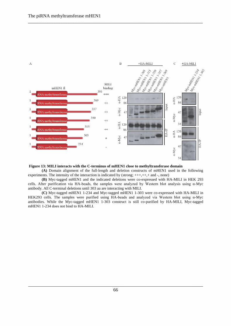
The piRNA methyltransferase mHEN1
___________________________________________________________________________
___________________________________________________________________________
66
Figure 13: MILI interacts with the C-terminus of mHEN1 close to methyltransferase domain
(A) Domain alignment of the full-length and deletion constructs of mHEN1 used in the following
experiments. The intensity of the interaction is indicated by (strong; +++,++,+ and -, none)
(B) Myc-tagged mHEN1 and the indicated deletions were co-expressed with HA-MILI in HEK 293
cells. After purification via HA-beads, the samples were analyzed by Western blot analysis using α-Myc
antibody. All C-terminal deletions until 303 aa are interacting with MILI.
(C) Myc-tagged mHEN1 1-234 and Myc-tagged mHEN1 1-303 were co-expressed with HA-MILI in
HEK293 cells. The samples were purified using HA-beads and analyzed via Western blot using α-Myc
antibodies. While the Myc-tagged mHEN1 1-303 construct is still co-purified by HA-MILI, Myc-tagged
mHEN1 1-234 does not bind to HA-MILI.
The piRNA methyltransferase mHEN1
___________________________________________________________________________
___________________________________________________________________________
67
2.3.4.5 mHEN1 is a cytosolic protein with a localization distinct from other piRNA pathway
components
Since mHEN1 is involved in 3` end maturation of piRNAs and directly interacts with MILI,
their expression pattern was examined in mouse testes sections by indirect
immunofluorescence. In seminiferous tubules of the male gonads, spermatogenesis proceeds
through mitotic and meiotic divisions of the germ cells to give rise to the haploid round
spermatids (RS), which undergo further development to form mature sperm. mHEN1 is
detected in both meiotic pachytene spermatocytes (PS) and RS. In both cell types, the enzyme
is cytosolic, with RS showing a single punctuate spot (see Figure 14 A). To investigate this
pattern in more detail, the localization of mHEN1 and different components of the piRNA
machinery were performed using specific antibodies in purified germ cells (Pivot-Pajot et al.
2003). The murine Piwi proteins MILI and MIWI are reported to be cytoplasmic with
maximal expression seen in PS and RS respectively (Deng & Lin 2002; Kuramochi-
Miyagawa et al. 2008). In isolated meiotic PS mHEN1 was detected together with the
pachytene cell marker SCP3, a component of the axial element of the synaptonemal complex
which becomes associated with the two sister chromatides of each homolog (Lammers et al.
1994; Moens et al. 1987) (see Figure 14 B). mHEN1 is diffusely distributed in the cytoplasm,
with occasional granules being present. In RS, the mouse VASA homolog (MVH) (Kotaja et
al. 2006; Tanaka et al. 2000) is reported to be present in a single large perinuclear structure
called the chromatoid body (CB) (see Figure 14 C). MIWI and MILI are also reported CB
components (Aravin & Bourc'his 2008; Aravin et al. 2008; Wang et al. 2009a), which was
confirmed by co-staining experiments. Detection of mHEN1 in RS indicates its localization
in a single cytoplasmic focus, which is slightly smaller than that observed for the Piwi
proteins (see Figure 14 C). Surprisingly, co-staining with MILI reveals that the focus stained
by anti-mHEN1 antibody is distinct from that stained by MILI (see Figure 14 D third row).
This separate sub-structure was called the “HEN body”. Examination of multiple cells and in
different experiments confirms this distinct localization pattern for mHEN1, but also
indicates a general tendency of the HEN body to remain in close proximity to the CB. The
functional relevance of this sub-cellular compartmentalization is presently unknown, but
serves probably temporal regulation of the interaction. Immunoprecipitations of MILI
revealed in Western blot analysis with α-mHEN1 antibody that the interaction of MILI and

The piRNA methyltransferase mHEN1
___________________________________________________________________________
___________________________________________________________________________
68
HEN1 is present in PS and in RS, although the localizations of the two proteins are distinct
(see Figure 14 F).
Figure 14: Cytoplasmic localization of mHEN1
mHEN1 localizes in a singular granule distinct from the chromatoid body in round spermatids
(A) Adult mouse testes section stained with α-mHEN1 (red) and DAPI (blue). White arrow indicates
round spermatids (RS) and red arrow marks a pachytene spermatocyte (PS)
(B) Pachytene spermatocytes stained with mHEN-1 (red) and SCP3 (green)
(C) Purified RS stained with indicated antibodies. Overlay shows merged channels with DAPI staining
(blue) for DNA.
(D) MIWI and MILI are in the chromatoid body (CB) while mHEN1 forms a distinct granule. Double
staining with anti-MILI mouse monoclonal antibody (green) and rabbit polyclonal antibodies for indicated
proteins (red). Overlay shows merged channels with DAPI staining (blue) for DNA.
(E) A few more representative examples of MILI/mHEN1 overlay images are shown. One of the panels
shows two adjacent RS, each with a CB and a HEN body.
(F) Immunoprecipitations using α-MILI antibodies and as a control empty beads revealed that in RS as
well as in PS MILI co-precipitates mHEN1.

The piRNA methyltransferase mHEN1
___________________________________________________________________________
___________________________________________________________________________
69

The piRNA methyltransferase mHEN1
___________________________________________________________________________
___________________________________________________________________________
70
2.3.5 Discussion
HEN1 homologs catalyze addition of a methyl group to the 2`OH at the 3` end of small
RNAs. Here, evidence was presented that the endogenous mouse homolog of HEN1
(mHEN1) from testis is an RNA methyl transferase. Its interaction with one of the Piwi
proteins, MILI, was shown both in testis extracts and in a heterologous cell culture system.
This interaction surface was mapped on the N-terminus of MILI to a region proximal to the
PAZ domain and to the C-terminus of mHEN1 in vicinity of the methyltransferase domain.
Finally, the endogenous protein was localized in seminiferous tubules and isolated germ cells
to show that mHEN1 is a component of a nuage, distinct to the chromatoid body where other
piRNA biogenesis factors accumulate. Together, these results provided evidence implicating
the endogenous mouse homolog of HEN1 in the murine piRNA pathway.
In Arabidopsis, both ~21 nt siRNAs and miRNAs carry a 3` terminal methyl group.
Since these RNAs use double-stranded RNA (dsRNA) precursors, the plant HEN1 has at its
N-terminus dsRNA-binding domains for recognizing small RNA duplexes for methylation. In
agreement with this, the enzyme is inactive on single-stranded RNA substrates (Yu et al.
2005). Animal homologs are much smaller than the plant protein, and contain only the
conserved SAM-domain required for RNA methylation (Chen 2007; Tkaczuk et al. 2006).
Consistent with the lack of dsRNA precursors in the piRNA pathway, animal HEN1 proteins
do not act on dsRNA substrates (Horwich et al. 2007; Kirino & Mourelatos 2007a, b; Saito et
al. 2007a). The question is how the animal protein specifically picks its substrates among
other single-stranded RNAs in the cell. One possibility is the variable sizes of piRNA
substrates HEN1 encounters in animal systems.
In mouse for example, all Piwi proteins recognize piRNAs of a specific length (MILI
binds to ~26 nt, MIWI2 to ~28 nt and for MIWI associates with ~29 nt piRNAs) (Aravin et
al. 2006; Aravin & Bourc'his 2008; Girard et al. 2006; Grivna et al. 2006a; Grivna et al.
2006b). Since the Piwi proteins are the only factors capable of discriminating these minor
size differences in piRNAs, it is reasonable to expect that methylation of piRNAs might
occur in the context of the RNA being bound to the Piwi protein. Supporting this idea is the
finding that in fly Dmhen1 mutants, piRNAs remain unmethylated, but are loaded correctly
into Piwi proteins. In fact, supplementing purified Piwi complexes from such mutants with

The piRNA methyltransferase mHEN1
___________________________________________________________________________
___________________________________________________________________________
71
recombinant fly DmHEN1 allowed piRNA methylation to proceed. Modification of siRNAs
also can be rescued in the Dmhen1 mutant ovary extracts by addition of recombinant
DmHEN1, but only after unwinding of the siRNA duplex intermediate and loading of the
single-stranded siRNA into fly DmAgo2 (Horwich et al. 2007). Additionally, DmHEN1 did
not modify Drosophila Ago1 bound miRNAs, suggesting specific requirements for protein-
mediated recruitment of HEN1 or accessibility of the enzyme to the small RNA 3`end.
In this study, the interaction of mHEN1 and MILI was demonstrated in both testes
extracts and also in a heterologous cell culture system. Similarly, recombinant fly DmHEN1
interacts with endogenous Piwi proteins in Dmhen1 mutant extracts, indicating that it is likely
that the Piwi proteins recruit the enzyme to the associated piRNAs (Saito et al. 2007b).
Surprisingly, our results did not show an interaction between mHEN1 and MIWI or MIWI2.
Very likely such an interaction might occur only with a piRNA loaded Piwi protein,
containing an unmethylated RNA, similar to that used in the fly Dmhen1 mutant study.
Similar experiments in mouse will have to wait for the availability of a mouse mhen1 mutant.
Accessibility of the small RNA 3` end for methylation can be influenced by at least
three factors: first, the affinity of the PAZ domain for the 3` end in its modified or
unmodified form. Recently it was shown that the MIWI PAZ domain has a slightly increased
(2-fold) affinity for a methylated 3` end. Further it was revealed by structural studies that the
methyl-group takes the position of the last nucleotide in the MIWI PAZ domain compared to
a hAGO1 PAZ domain (Simon et al. 2011), suggesting that the methyl group could bind the
piRNA deeper into the binding pocket. Because an unmethylated RNA binds more weakly
than a methylated piRNA, the unmodified piRNA might have its 3` end exposed and
accessible to HEN1 for methylation. A second factor contributing to the accessibility of the
3`end of the piRNA could be the extent of base-pairing of the small RNA uses to recognize
its targets. Crystal structures of Argonaute-guide RNA-target RNA ternary complex reveal
that extensive complementarity forces the PAZ domain to let go of the small RNA 3` end to
facilitate the formation of the required guide-target A-helix (Filipowicz 2005). This
possibility is supported by the intricate link between piRNA function in transposon mRNA
destruction and (secondary) piRNA biogenesis. In this context, the mapping of the interaction
surface on MILI to a region close to the PAZ domain is interesting and might offer yet a third
possibility. This interaction might force a conformational change near the PAZ domain,
enabling the release of the unmethylated and weakly-bound small RNA 3` end. After

The piRNA methyltransferase mHEN1
___________________________________________________________________________
___________________________________________________________________________
72
methylation, a stronger interaction with the PAZ domain might result in the release of the
Piwi protein. All these possibilities will need to be tested by future structural and biophysical
analyses.
The localization studies of mHEN1 indicate a cytoplasmic localization of the piRNA
during the maturation process. All currently known processing factors and most Piwis are
components of the cytoplasmic perinuclear granules (called nuage in worms, flies and mice)
(Kotaja & Sassone-Corsi 2007). In mouse, only MIWI2 is mostly nuclear, but it is also
observed in nuage structures, called pi-P-bodies in the cytoplasm of gonocytes (Aravin &
Bourc'his 2008; Aravin et al. 2009). In fact, interfering with MIWI2 loading results in a
cytoplasmic localization (Aravin et al. 2009; Reuter et al. 2009; Vagin et al. 2009; Wang et
al. 2009a), reinforcing the hypothesis that piRNA biogenesis is a cytoplasmic process and
only when MIWI2 is properly loaded can it enter the nucleus. In flies too, where only PIWI is
nuclear, depletion of piRNA biogenesis factors like Armitage, Yb or Zucchini lead to a
cytoplasmic localization (Olivieri et al. 2010; Saito et al. 2009). Taken together, germline
nuage appears to be the site of actual piRNA production.
It was observed that mHEN1 shares the same localization pattern as MILI in
pachytene stage cells. This result is similar to the localization pattern of ZIWI and DrHEN1
in zebrafish, which are both localized in the nuage (Kamminga et al. 2010). However, here it
is shown that in later stages of the spermatogenesis in mouse, in round spermatids, mHEN1 is
in a separate body in the vicinity of the chromatoid body. The novel granule structure was
called a HEN-body. The spatial separation from the CB raises the question whether or not
there is a functional role for this body in piRNA biogenesis.
Regardless of the different localizations of MILI and mHEN1 in haploid round
spermatids, the interaction of the two proteins that was reported here remains unchanged both
in pachytene spermatocytes and round spermatids. Modification of piRNAs is also not
different in these cells as all piRNAs in mouse total testis RNA are methylated (Kirino &
Mourelatos 2007c; Ohara et al. 2007a). Thus, accumulation of mHEN1 in a distinct body is
unlikely to have a direct role in piRNA biogenesis. One possible function of the HEN body is
to sequester mHEN1 away from the CB which contains miRNAs and other stored
developmentally important mRNAs (Kotaja & Sassone-Corsi 2007). Any promiscuous
methylation of its RNA components might be disastrous for developmental processes, as it
can alter the regulated half-lives of these RNAs. Another possible explanation is that given

The piRNA methyltransferase mHEN1
___________________________________________________________________________
___________________________________________________________________________
73
that mHEN1 is a strong interactor with MILI, the protein has to be locked in a separate body
in order to not interfere with the silencing function of the effector MILI complex. Further
characterization of mHEN1-interacting factors will shed light on the contents of the HEN
body and the role of the HEN1 localization.
2.3.5.1 Model of mHEN1 activity
We propose a model of mHEN1 activity based on our results and currently available
published data (see Figure 15): Our results have shown that post-translational modifications
in the form of symmetrically dimethylated arginines in the N-terminus of MILI, catalyzed by
the methyltransferase PRMT5 and its associate Piwi binding protein WDR77, are not needed
for the interaction with mHEN1. In Drosophila dprmt5 null mutants, piRNAs are still
methylated (Kirino et al. 2009), confirming that HEN1 performs its activity independent of
the post-translational modifications of the PIWI proteins. In fly and fish hen1 mutants the
piRNA pathway is only slightly impaired in its function to silence transposable elements
(Horwich et al. 2007; Kamminga et al. 2010; Saito et al. 2007a). This presumes that the Piwi
proteins must be methylated on their N-terminal arginines in the hen1 mutant, because
otherwise a stronger activity of the transposons would be expected as shown for fly dprmt5
mutants (Kirino et al. 2009) or in mutants of proteins which bind dependent on the
symmetrical dimethylated arginines of Piwi proteins like for example TDRD1 in mice
(Reuter et al. 2009; Wang et al. 2009a). Immunoprecipitations using an antibody against
symmetrical dimethylated arginines or against TDRD1 co-purified matured piRNAs (Reuter
et al. 2009). However, piRNAs were never observed in immunoprecipitations of mHEN1.
Additionally, mHEN1 interacts with MILI in an RNA-independent manner, suggesting that
the mHEN1/MILI interaction can occur before the complex contains RNAs and before MILI
is post-translationally modified. Evidence shows that HEN1 is not required for piRNA
loading onto Piwi proteins (Kamminga et al. 2010; Saito et al. 2007a). These data suggest a
model where the methylation of piRNAs occurs before the post-transcriptional modification
of Piwi and the subsequent complex arrangement including TDRD1.
We found that mHEN1 is localized in a distinct granule in close proximity to the
chromatoid body where MILI is localized. We speculate that mHEN1 leaves the MILI
complex after the methylation of the piRNAs in order to allow formation of MILI-containing

The piRNA methyltransferase mHEN1
___________________________________________________________________________
___________________________________________________________________________
74
multi-protein complexes. One component of this complex would be TDRD1, with which
MILI associates and co-localizes throughout development (Reuter et al. 2009). This
localization could as well serve to separate mHEN1from other small RNAs, like miRNAs
which are present in the chromatoid body (Kotaja et al. 2006; Meikar et al. 2010).
Thus, the action of mHEN1 can be placed after the loading of the piRNAs onto MILI
and before or at the same time as the post-translational modification of MILI. After piRNA
methylation, mHEN1 likely leaves the complex and separates into a distinct cytoplasmic
granule.
Figure 15: Model
mHEN1 (red) is not needed for the loading of MILI (blue), but can interact with the unloaded protein. After
loading of MILI with piRNAs mHEN1 methylates the bound RNA and leaves the complex. Then PRMT5
(green) guided by WDR77 (olive) methylates the arginines in the N-term of MILI. This methylation is essential
for binding of TDRD1 (yellow).

The piRNA methyltransferase mHEN1
___________________________________________________________________________
___________________________________________________________________________
75
2.3.6 Acknowledgements
We thank T. Noce for anti-MVH antibody. We thank EMBL Monoclonal Antibody Core
Facility and Protein Expression and Purification Facility for production of antibodies. Work
in RP laboratory is supported by the EMBL.
2.3.7 Methods
2.6.1 Antibodies
For the production of antibodies, GST-fusions of specific protein fragments were produced in
E. coli strain BL21 and injected into rabbits. The fragments used were anti-mHEN1 (GQG
and GOY, 185-395 aa) and anti-MIWI (BVI, 1-200 aa; kindly provided by Dr Michael
Reuter). The anti-MILI monoclonal antibody (mAb, 13E-3; kindly provided by Dr Michael
Reuter) recognizes the peptide corresponding to 107-122 aa of MILI and was raised at the
Monoclonal Antibody Facility, EMBL. Control antibodies used in IPs were rabbit polyclonal
antibodies directed against the Glutathione-S-Transferase (GST) moiety. All antibodies were
affinity-purified.
2.6.2 Mouse testes extracts and RNA analysis
CD1 mouse testes extract preparation, immunoprecipitation and 5`-end-labelling of piRNAs
were performed as described previously (Aravin et al. 2006; Girard et al. 2006). Dried gels
were exposed to a storage phosphor screen and scanned with a Typhoon scanner
(Amersham).
The in vitro methylation assays was performed as described (Yu et al. 2005).
Endogenous mHEN1 from mouse testes extract was immunopurified (using antibodies
chemically-cross linked to Protein G sepharose) and bead-bound protein was used in the
assay. After the incubation period, an aliquot (1/40th) of the in vitro methylation reaction was
5'-end labeled with 32
P to verify the presence of both substrate RNAs.
The following RNAs were used in the in vitro methylation assay:
TGACATGAACACAGGTGCTCA (21 nt)

The piRNA methyltransferase mHEN1
___________________________________________________________________________
___________________________________________________________________________
76
TGACATGAACACAGGTGCTCAGATAGCTTT
(30 nt, the same sequence as piR-1 piRNA). The 21 nt RNA is a 3' end-truncated version of
the 30 nt RNA.
2.6.3 Clones and cell culture
For mammalian cell transfections, full-length ORFs of Argonaute proteins (MILI, MIWI and
MIWI2) or mHEN1 (Acc. no. AK038994, RIKEN), or deletion constructs were inserted into
pCIneo-HA vector (Pillai et al. 2004) or into pcDNA3-Myc (Invitrogen), as required.
For HEK293 transfections, 2 µg of each construct of interest (pCIneo-HA-Piwi or
mHEN1 plasmids and/or pcDNA3-Myc-mHEN1 or Piwi protein vectors) were used and
transfected into 6 cm dishes at 80% confluence using Lipofectamine/Plus (Invitrogen)
following the manufacturer‟s protocol, and harvested 48 hours post-transfection. When
mentioned, transfected cells were treated with methyltransferase inhibitor 5`-deoxy-5`-
(methylthio) adenosine (MTA; Sigma) at a final concentration of 750 µM or the solvent
DMSO alone. Cells from one 6 cm plate were lysed and mixed with 10 µL α-HA-beads (anti-
HA affinity matrix beads (Roche) following the purification in the manufacturer‟s protocol.
When mentioned, purified samples were treated with RNase A (20 µg) for 15 min at 37 °C,
followed by three washes.
Western analysis was performed with affinity-purified antibodies to specific proteins at a
dilution of 1:250. Other antibodies used were: anti-HA antibodies (Santa-Cruz), anti-Myc and
Y12 at 1:250 dilutions. An aliquot of 5% input material was probed with anti-HA antibody to
control for mHEN1 expression in all reactions.
2.6.4 Immunofluorescence
For tissue sections, testes from 20-day old CD10 mice were fixed in 4% paraformaldehyde.
Staining with anti-mHEN1 (used at 1:50) was performed on 7 µm sections using microwave
antigen retrieval in 10 mM citrate buffer pH 6.0 and detected by immunofluorescence with
anti-rabbit Alexa 488 (Invitrogen). The tissues were mounted with Vectrashield (Reactolab)
containing DAPI and visualized using 100x objective of a Zeiss Axiovision Z1 wide-field
microscope.
Preparation of purified cell populations by sedimentation (Pivot-Pajot et al. 2003) and
fixation to glass slides as well as the indirect immunofluorescence detections were performed

The piRNA methyltransferase mHEN1
___________________________________________________________________________
___________________________________________________________________________
77
as described (Kotaja et al. 2006). The following affinity-purified primary antibodies were
used at 1:4 dilutions: anti-MIWI (BVI), anti-mHEN1 (GQG), anti-MILI (13E-3). Anti-MVH
was used as a dilution of 1:1000 (kindly provided by Toshiaki Noce, Mitsubishi Kagaku
Institute of Life Sciences, Tokyo, Japan). The secondary antibodies anti-rabbit IgG Alexa
Fluor 594 and anti-mouse-IgG Alexa Fluor 488 (Invitrogen) were both used at 1:10,000
dilutions. Cells were mounted with Vectrashield (Reactolab) containing DAPI and visualized
using 100x objective of a Zeiss Axiovision Z1 wide-field microscope. Images were taken
with Axiocam camera, processed with manufacturer‟s software and mounted with Photoshop.

The piRNA methyltransferase mHEN1
___________________________________________________________________________
___________________________________________________________________________
78
2.3.8 Supplementary data
Methyltransferase domain
LAM domain
DrHEN1 C-term construct (Kamminga et al. 2010)
mHEN1region needed for binding to MILI
Figure S 1: Alignment of HEN1 from different animals
Sequence alignment of full-length animal HEN1 homologs aligned using ClustalW. The Alignment was
analyzed using ESPript. The figure is based on (Huang et al. 2009; Kamminga et al. 2010)

The piRNA methyltransferase mHEN1
___________________________________________________________________________
___________________________________________________________________________
79
Figure S 2: Endogenous mHEN1 from testes extracts is devoid of any associated small RNAs
mHEN1 was immunoprecipitated (IP) using antibodies (GQG or GOY) and analyzed by 5‟-end-labelling and
Western (GQG). Control, IP with an unrelated antibody; beads, pull down without any antibodies; asterisk,
heavy chain of antibodies. Size (in nucleotides) of RNA markers is indicated.
The piRNA methyltransferase mHEN1
___________________________________________________________________________
___________________________________________________________________________
80
2.4 Additional experiments for the characterization of
mHEN1/MILI complex
2.4.1 Attempt to express the mHEN1/ MILI complex in insect cells
2.4.1.1 Aim
RIWI, a Piwi protein in rat (Lau et al. 2006), as well as the Drosophila Piwi proteins are
described as endonucleases, which are able to slice their target RNAs guided by the loaded
piRNAs (Gunawardane et al. 2007; Nishida et al. 2007; Saito et al. 2006). Based on the
assumption that MILI and MIWI2 are slicers, the piRNA biogenesis was described as similar
to the ping-pong model of Drosophila piRNAs (Aravin & Bourc'his 2008; Kuramochi-
Miyagawa et al. 2010; Kuramochi-Miyagawa et al. 2008). In this model, the Piwi proteins
create, guided by their bound RNAs, the 5` end of a secondary piRNA via their slicer
activity. The 3`end formation remains unknown. The novel piRNA is subsequently
incorporated into one of the Piwi orthologs and performs a similar reaction (Brennecke et al.
2007; Gunawardane et al. 2007). It was shown in the previous section that mHEN1 and MILI
interact in both testis extract and in HEK 293 cells. Analysis of the slicer activity of MILI and
determination if the slicing event of the 5` end of the secondary piRNAs and the methylation
of the 3`ends by mHEN1 are functional connected are still missing in the literature.
In order to study the mechanism of methylation of piRNAs in the context of Piwi
proteins, mHEN1 and MILI were required to be produced recombinantly. Bacterial
expressions of full-length recombinant proteins was unsuccessful: neither mHEN1 nor MILI
are soluble as full-length constructs. Therefore, co-expressions of mHEN1 and MILI as a
recombinant complex in insect cells using a dual ORF expression construct were tried. Insect
cell expression in Sf9 or Sf21 cells is also attractive because the reported lack of recombinant
Sm and Piwi protein methylation (Brahms et al. 2000; Kirino et al. 2009), which enables to
confirm the previous described results that the interaction of MILI and mHEN1 is
independent of the post-translational methylation marks in the N-terminus of MILI.

The piRNA methyltransferase mHEN1
___________________________________________________________________________
___________________________________________________________________________
81
2.4.1.2 Results
A fusion vector was designed encoding 6XHis-tagged MILI and CBP-tagged mHEN1 with
independent promoters, which was inserted into the baculovirus genome by Tn7
transposition. The baculovirus was used to transfect Sf21 cells. The expression was monitored
via YFP co-expressed by the bacmid (Berger et al. 2004; Bieniossek et al. 2008; Fitzgerald et
al. 2006) (see Figure 16 A and B). Insect cells were harvested at the highest possible YFP
expression. As indicated in Figure 16 B, the expression of both proteins was analyzed via
Western blot of the total cell extract and the soluble fraction after lysis, using specific
purified antibodies against MILI and mHEN1. Both proteins were detected in both fractions.
Nickel-beads were used to purify the 6XHis-MILI and stained via affinity purified anti-
mHEN1 antibody in a Western blot analysis, demonstrating that the CPB-tagged mHEN1
was co-purified (see Figure 16 C), suggesting that the two proteins are expressed and form a
complex in the insect cells.
The 6XHis-MILI/CBP-mHEN1 complex was poorly expressed and purifications did
not yield homogenous proteins, as judged by SDS-PAGE (see Figure 16 D first gel). Again,
Western blot analysis of one of the purified elutions was performed, which showed the most
abundant protein concentration (Elution 3). The Western blot indicated that MILI and
mHEN1 are present in the nickel column elution (see Figure 16 D second and third column).
Six bands in the proximity of the expected molecular weight of mHEN1 and MILI were
excised and subjected to mass spectrometry. Indeed, MILI was identified in band #2 and
mHEN1 in bands #4 and #5. The other bands are likely to be endogenous proteins from the
insect Spodoptera fugiperda and unidentifiable as the genome has not been fully sequenced.
To validate the findings that mHEN1 interacts with MILI independent of the post-
translational modifications of the arginines in the N-terminus of the Piwi protein, the Western
blot membrane was re-stained with Y12 antibody. However, contrary to the published data
(Brahms et al. 2000; Kirino et al. 2009) MILI was at least partially methylated (see Figure 16
C third row). Based on this result it is impossible neither to confirm nor to disprove the
independence of the interaction of mHEN1 with MILI on the methylation state of the Piwi
protein. Also, data from the laboratory show that low level expression of Bombyx mori Piwi
proteins can lead to their methylation in Sf9 cells (personal communication from Zhaolin

The piRNA methyltransferase mHEN1
___________________________________________________________________________
___________________________________________________________________________
82
Yang). Due to these reasons, although the original goal was to use the insect expression
system to produce proteins lacking post-translational modifications, it is not possible to
conclude that this is the case.
In the laboratory it was shown before (personal communication from Zhaolin Yang),
that Sf21 cell contain piRNAs. Knowing that the MILI-mHEN1 complex is present, although
in small amounts, whether bound Sf21 piRNAs are present in the complex should be
determined. Therefore, RNA were isolated from the Ni-column elution fractions, 32
P end-
labelled and seperated on a 15% denaturating polyacrylamid gel. No specific enrichment of a
specific RNA species around 30 nt in length were identified (see Figure 16 E), indicating that
the complex did not bind Sf21 piRNAs, likely due to the failure of MILI-mHEN1 complex in
entering the piRNA pathway.
Given the difficulties in expressing the mHEN1/MILI complex and the impurities of
the elutions we did not proceed further to functional analysis of the complex.
Figure 16: MILI/mHEN1 complex studies in Sf21
(A) 6XHis-MILI and CBP-mHEN1 were co-expressed in a fusion vector in Sf21. The expression was
monitored by YFP-co-expression by the bacmid (red bars), analyzed against a laboratory internal standard at
different time points (blue bars) after the cells growth arrested. As indicated, the expression was the highest after
84 hours after growth stop of the insect cells.
(B) The aliquots taken at each time point mentioned above were analyzed via Western blot using
affinity purified anti-mHEN1 and anti-MILI antibodies, indicating that both proteins are expressed from 24 h
after cell growth stagnation.
(C)The Sf21 cell extract of the transfected cells as well as non-transfected (WT) cell were purified via
nickel-column. As a control Glutathione beads were used which are not supposed to bind the tagged-proteins.
The purified samples were analyzed by Western blot using affinity purified anti-mHEN1 and anti-MILI
antibodies. Nickel-column purifications of 6XHis-MILI co-purified CBP-mHEN1. The WT sample as well as
the beads control remained negative. In difference to published data, MILI can be detected carrying
symmetrically dimethylated arginines indicated by Y12 staining of the Western blot.
(D) Attempted nickel-column purification of the complex was analyzed via a coomassie stained gel,
indicating that the complex is not significantly overexpressed. Although the 6XHis-MILI/CBP-mHEN1
complex was expressed, the coomassie stained gel revealed multiple other bands. Several bands of the
approximate size of 6XHis-MILI and CBP-mHEN1 were excised from the coomassie stained gel and analyzed
by mass spectrometry. In the adjusted table, the results are summarized, which indicate that CBP-mHEN1 and
6XHis-MILI are present but mixed with other Sf21 proteins like Tubulin.
(E) RNAs of nickel-column purified 6XHis- MILI/CBP-mHEN1 and WT samples exposed to the same
experimental set up were extracted and analyzed on a 15% denaturating RNA gel. A significant enrichment of
small RNAs in the size of 30 nt (which could indicate bound piRNAs) was not detected.

The piRNA methyltransferase mHEN1
___________________________________________________________________________
___________________________________________________________________________
83
The piRNA methyltransferase mHEN1
___________________________________________________________________________
___________________________________________________________________________
84
2.4.1.3 Discussion
In an attempt to express the recombinant MILI-mHEN1 complex in the insect cell line Sf21,
analysis of the expression by Western blot analysis using affinity purified antibodies
indicated that the complex can be detected by this sensitive method, but Coomassie stained
gels show that the complex is not abundantly expressed in insect cells.
In contrast to the published data indicating that recombinant human and mouse
proteins do not carry symmetrically dimethylated arginines in this expression system
(Brahms et al. 2000; Kirino et al. 2009), it was shown that the 6XHis-MILI is at least partial
methylated. This result makes it impossible to confirm or disprove the suggested
independency of post-translational modification on MILI for the mHEN1 binding.
It was determined if the Sf21 expressed 6XHis-MILI/CBP-mHEN1 complex is bound
to small RNAs in the size of 30 nt, which could indicate a loading of the complex with Sf21
piRNAs. A specific band of piRNA size was not observed. This is probably due to the fact
that the recombinant mouse complex could not enter the piRNA pathway of the insect cells.
2.4.1.4 Method Sf21 expression of MILI and mHEN1
A fusion vector of pFastBac-6XHis-MILI and pUCS-CPB-mHEN1 was inserted into the
baculovirus genome by Tn7 transposition and used to express 6XHis-tagged MILI and CBP-
tagged mHEN1 in Sf21 cells. The expression was monitored by the co-expression of the
bacmid internal YFP (Berger et al. 2004; Bieniossek et al. 2008; Fitzgerald et al. 2006).
Aliquots of 1x 10⁶ cells of the transfected Sf21 cells were harvested at each time point. After
lysis, 1/10th of the total cell extract (SNP) were removed and the soluble part (SN) and pellet
separated. 1/10th of the cell lysis was removed and the rest analyzed at a photo-spectrometer
for YFP expression.
The cells were purified via the 6XHis-tag: small scale for immunoprecipitation: Lyses
of 1 x 10⁷ cells via hypotonic buffer (50 mM Tris pH=7.5, 15 mM KCl, 25 mM MgCl2) using
sonication, binding to a Nickel column, 8 times washing (50 mM Tris, 50 mM Imidazole,
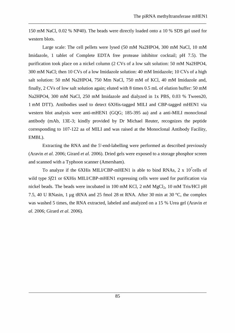
The piRNA methyltransferase mHEN1
___________________________________________________________________________
___________________________________________________________________________
85
150 mM NaCl, 0.02 % NP40). The beads were directly loaded onto a 10 % SDS gel used for
western blots.
Large scale: The cell pellets were lysed (50 mM Na2HPO4, 300 mM NaCl, 10 mM
Imidazole, 1 tablet of Complete EDTA free protease inhibitor cocktail; pH 7.5). The
purification took place on a nickel column (2 CVs of a low salt solution: 50 mM Na2HPO4,
300 mM NaCl; then 10 CVs of a low Imidazole solution: 40 mM Imidazole; 10 CVs of a high
salt solution: 50 mM Na2HPO4, 750 Mm NaCl, 750 mM of KCl, 40 mM Imidazole and,
finally, 2 CVs of low salt solution again; eluted with 8 times 0.5 mL of elution buffer: 50 mM
Na2HPO4, 300 mM NaCl, 250 mM Imidazole and dialyzed in 1x PBS, 0.03 % Tween20,
1 mM DTT). Antibodies used to detect 6XHis-tagged MILI and CBP-tagged mHEN1 via
western blot analysis were anti-mHEN1 (GQG; 185-395 aa) and a anti-MILI monoclonal
antibody (mAb, 13E-3; kindly provided by Dr Michael Reuter, recognizes the peptide
corresponding to 107-122 aa of MILI and was raised at the Monoclonal Antibody Facility,
EMBL).
Extracting the RNA and the 5'-end-labelling were performed as described previously
(Aravin et al. 2006; Girard et al. 2006). Dried gels were exposed to a storage phosphor screen
and scanned with a Typhoon scanner (Amersham).
To analyze if the 6XHis MILI/CBP-mHEN1 is able to bind RNAs, 2 x 10⁷cells of
wild type Sf21 or 6XHis MILI/CBP-mHEN1 expressing cells were used for purification via
nickel beads. The beads were incubated in 100 mM KCl, 2 mM MgCl2, 10 mM Tris/HCl pH
7.5, 40 U RNasin, 1 µg tRNA and 25 fmol 28 nt RNA. After 30 min at 30 °C, the complex
was washed 5 times, the RNA extracted, labeled and analyzed on a 15 % Urea gel (Aravin et
al. 2006; Girard et al. 2006).
The piRNA methyltransferase mHEN1
___________________________________________________________________________
___________________________________________________________________________
86
2.4.2 RNF17 is does not influence the 3’end processing of piRNAs
2.4.2.1 Aim
The piRNA methyltransferase mHEN1 interacts with the murine Piwi protein MILI in testes
extract as well as co-expressed in HEK293 cells. Here, it was attempted to identify other
binding partners of mHEN1, which might be involved in the 3`end processing and
modification of piRNAs. Immunoprecipitations using the affinity purified mHEN1 antibody
were performed and analyzed via mass spectrometry, searching for co-purified protein
(personal communication from Michael Reuter). Unfortunately, interesting candidates which
could be involved in the 3` end formation and modification of piRNAs were not detected.
Based on the findings that mHEN1 localizes in a perinuclear granule distinct from the
chromatoid body, proteins with similar localization and expression pattern were searched.
One candidate was the Tudor domain and RING finger domain-containing RNF17/TDRD4
(Pan et al. 2005). The two isoforms of the protein are exclusively expressed in testis. The
short isoform (RNF17S) contains three and the long isoform (RNF17L) five Tudor domains.
RNF17S localizes diffusely in the cytoplasm of all germ cells, while RNF17L builds a
structure distinct from the chromatoid body in late pachytene spermatocytes, round
spermatids and elongating spermatids (Pan et al. 2005), similar to the HEN1-body (see
Figure 17) . RNF17 is essential for the spermatogenesis. Like miwi mutants, Rnf17 mutant
mice are male sterile, due to a block of the spermatogenesis after round spermatids (Pan et al.
2005), indicating that the protein could be connected to the piRNA pathway due to its
knockout phenotype. It was investigated if RNF17 and mHEN1, due to their similar
localization pattern, are functionally connected.
Figure 17: RNF17 and mHEN1 show a similar localization pattern.
Pan et al. localized RNF17 (red) and TDRD1 (green) a protein known to be localized in the chromatoid body in
round spermatids (left picture). In the right image the mHEN1 granule (red) distinct from the chromatoid body
(green) is indicated by co-staining of MILI in round spermatids. Despite the fact that in the testes section image
of Pan et al the resolution is not that good as in purified germ cells, the localization pattern of mHEN1 and
RNF17 (red arrows) in comparison to the chromatoid body (green arrows) is similar in round spermatids.

The piRNA methyltransferase mHEN1
___________________________________________________________________________
___________________________________________________________________________
87
2.4.2.2 Results
RNF17 and mHEN1 show similar localization pattern. In round spermatids both proteins are
found to form granules distinct from the chromatoid body (Pan et al. 2005). The antibodies to
both proteins were produced in rabbits, making it rather difficult to perform co-localization
experiments using fluorescent secondary anti-rabbit antibodies. To avoid inconclusive
localization experiments, first a possible interaction between both proteins was investigated.
The anti-mHEN1 antibody was used for immunoprecipitations from adult mouse testes
extracts and the proteins were analyzed via western blot. Using an anti-RNF17 antibody a co-
purification of RNF17 could not be detected in the anti-mHEN1 immunoprecipitate (see
Figure 18 A).
Then the influence of RNF17 in the piRNA pathway, especially in the 3` end
formation, was investigated. rnf17 mutant mouse testis were obtained from Dr. Jeremy Wang,
University of Pennsylvania. Immunoprecipitations of MILI and MIWI were performed from
adult mouse testes extract of WT, RNF-/+
and RNF-/-
and the RNAs were isolated. After
radioactive labeling of the RNAs, they were analyzed on a denaturating gel. In all samples
piRNA size and abundance were not affected (see Figure 18 B). To analyze if RNF17
influences the 3` end modification of piRNAs, the piRNAs of MIWI and MILI
immunoprecipitations of WT, RNF-/+
and RNF-/-
samples were subjected to β-eliminations.
The piRNA mobility of all testes extract samples did not change after the treatment,
indicating that the RNAs were modified at their 3‟ end, while artificial unmodified RNAs run
faster due to a loss of the last nucleotide (see Figure 18 C).

The piRNA methyltransferase mHEN1
___________________________________________________________________________
___________________________________________________________________________
88
Figure 18: RNF17 does not affect the 2’O methylation of the 3’end of piRNAs
(A) mHEN1 was immunopurified using an affinity purified antibody and analyzed on a Western blot.
Staining with anti-RNF17 identified the protein in the input sample but not as a co-purified protein in the
mHEN1 IP.
(B) MIWI and MILI bound RNAs were immunoprecipitated from of WT, RNF17+/-
and RNF17-/-
adult
testes extracts. The RNAs were 32
P labeled and analyzed on a denaturating gel. No significant difference in the
samples could be detected.
(C) The RNA of MIWI and MILI IPs were oxidized and a β-elimination was performed. In contrast to
the synthetic control RNA oligos of 30nt, 28nt and 20nt the β-elimination did not affect the mobility of the
immunopurified RNA on the gel indicating that they are 3` end modified.

The piRNA methyltransferase mHEN1
___________________________________________________________________________
___________________________________________________________________________
89
2.4.2.3 Discussion
Many different types of perinuclear electron-dense structures, called nuages, are identified in
the germ cells in different species and have been described as important for germ cell
specification and differentiation. One of these nuages in mice is the chromatoid body, thought
to be an active center for the piRNA pathway (Kotaja & Sassone-Corsi 2007). Another
example in mice is the RNF17 granule. RNF17 is a testes-specific protein with a RING
domain and three or five Tudor domains, according to the isoform. The long isoform
RNF17L forms dimers or polymers, indicating that it may be responsible for assembly of the
RNF17 granules (Pan et al. 2005). These granules are distinct from the chromatoid body, like
the HEN-body. Although RNF17 shows a similar localization pattern to mHEN1, it does not
interact with the methyltransferase nor does it seem to be involved into the 3` end processing
of piRNAs. These results indicate that RNF17 is probably not needed at the same step of the
piRNA biogenesis as mHEN1. It is possible that the protein is involved further downstream
in piRNA pathway, potentially in DNA methylation, because it contains Tudor domains,
which could bind to histones or Piwi proteins carrying methylated arginines. Until now there
is no reason to believe that RNF17 functions in the 3`end processing of piRNAs.
2.4.2.4 Methods
Testes extracts were prepared from adult CD1 WT mice, +/- RNF17 (heterozygous) and -/-
RNF17 (knockout) testes kindly provided from the Wang laboratory (Pan et al. 2005).
Endogenous mHEN1 or Piwi proteins from mouse testes extract were immunopurified using
the following antibodies linked to Protein G sepharose: anti-mHEN1 (GQG and GOY, 185-
395 aa), anti-MIWI (BVI, 1-200 aa) and anti-MILI monoclonal antibody (mAb, 13E-3). For
Western blot analysis purified rabbit anti-RNF17 (Wang laboratory) was used in a dilution of
1:500 and affinity purified anti-mHEN1 in a dilution of 1:250. Mouse testes extract
preparation and immunoprecipitation and 5'-end-labelling of piRNAs were performed as
described previously (Aravin et al. 2006; Girard et al. 2006). Dried 15% Urea gels were
exposed to a storage phosphor screen and scanned with a Typhoon scanner (Amersham). The
-eliminations were performed as described (Vagin et al. 2006).

The piRNA methyltransferase mHEN1
___________________________________________________________________________
___________________________________________________________________________
90
2.4.3 Structural insight of binding of the methylated piRNAs by the
MIWI PAZ domain
2.4.3.1 Contribution remarks
The biophysical characterization of the PAZ domain is based on collaboration between the
Carlomagno group from the Structural and Computational Biology unit at the European
Molecular Biology Laboratory Heidelberg, Germany, and the Pillai group of the European
Molecular Biology Laboratory, Grenoble Outstation Grenoble, France. While the NMR
structure of the MIWI PAZ domain was solved in the Carlomagno group, I focused on the
expression, optimization of the expression and the purification of the PAZ domain for
biochemical and biophysical experiments. In our lab, a trainee student, Elsa Rocha, designed
and established the first purification protocols for the soluble MIWI PAZ construct. Two
point mutant constructs were prepared by Jerome Bourdin, a research associate in the lab.
The biophysical measurements using SPR were performed by Dr. Peter Sehr at the Chemical
Biology Facility, EMBL Heidelberg. My contribution consisted in following up the project in
the lab by optimization and purification of the PAZ domain and the mutants, as well as the
design of new mutant constructs for Biacore experiments in Heidelberg. I also performed
several UV-crosslinking experiments that were not included in the final paper.
2.4.3.2 Remarques concernant ma contribution
La caractérisation du domaine PAZ est basée sur une collaboration avec le groupe
Carlomagno de l‟unité de biologie structurale et bioinformatique d‟EMBL Heidelberg et le
groupe Pillai de l‟antenne EMBL de Grenoble, France. Alors que la structure NMR du
domaine PAZ de MIWI a été résolue dans le groupe Carlomagno, je me suis concentrée sur
l‟expression et la purification du domaine PAZ pour des expériences biochimiques et
biophysiques. Dans notre laboratoire, une stagiaire Elsa Rocha a conçu et rédigé les
protocoles de purification préliminaires pour la construction Miwi PAZ. Deux mutants
ponctuels ont été préparés par Jerome Bourdin, notre assistant de recherche. Les mesures

The piRNA methyltransferase mHEN1
___________________________________________________________________________
___________________________________________________________________________
91
biophysiques utilisant la SPR (Surface Plasmon Resonance) ont été réalisées par le Dr Peter
Sehr (Chemical Biology Facility, EMBL Heidelberg). Mon rôle a été de purifier le domaine
PAZ et ses mutants ainsi que de créer de nouvelles constructions pour des analyses
complémentaires Biacore à Heidelberg. J‟ai également réalisé plusieurs expériences de
pontage covalent par UV non inclus dans le papier final.
2.4.3.3 Aim
All piRNAs in total mouse testes RNA are demonstrated to carry the 2`-O-methyl
modification (Kirino & Mourelatos 2007c; Ohara et al. 2007b). The role of this modification
in RNA binding to the PAZ domain has not yet been established. To test the possibility of a
methylation-requirement of the piRNAs in the binding to the MIWI PAZ domain, the NMR
structure of the MIWI PAZ domain in complex with a 2`O methylated ssRNA (8 nt) was
solved in collaboration with the Carlomagno group at the EMBL Heidelberg based on a
construct by Elsa Roch. Like the PAZ domain of Dicer and AGO proteins, the MIWI PAZ
domain shows an OB-fold. The short barrel is capped by 2 -helices on one side and an
helix and hairpin on the other. The RNA binds in the cavity, built by the central β-barrel
and the α-helix/β-hairpin motif. The 2`-O-methyl group of the 3` end is recognized by a
hydrophobic cleft consisting of F333 on the -hairpin and M382 and A381 on strand 7 of
the core -sheet. The carbonyl and amide of M382 of strand β7 forms hydrogen bonding
interactions with the 3`hydroxyl and the 2`methoxy groups of the RNA 3` end. Based on
chemical shifts, the protons of the 3`nt ribose are in close proximity to residues of the
βbarrel and the αhelix/βhairpin insert (A381, L383, F333). The second to last nucleotide of
the ssRNA does not show interactions with the protein. Several contacts were observed
between the ribose of the third to the sixth nucleotides and strand β2 of the central barrel
(T317, Y318, R319) and the C-terminus (L393). That leads to a stretched conformation of the
phosphate backbone between the 3`end nt and the second to last nt. In the MIWI PAZ
domain, it seems that the 2`-O-methyl group of the RNA occupies the space which is taken
by the 3` end nucleotide of unmethylated RNAs bound to the hAGO1 PAZ domain. This
indicates that the RNA in the MIWI PAZ/RNA complex is shifted out of the binding pocket
The piRNA methyltransferase mHEN1
___________________________________________________________________________
___________________________________________________________________________
92
by one nucleotide, while the last nucleotide takes the position close to the second to last
nucleotide in the hAGO1 complex with unmethylated 3` end RNA (Simon et al. 2011).
Based on the NMR structure of our collaborators and alignments of different PAZ
domains, the biophysical nature of the interaction of the different amino acids of the PAZ
domain to the piRNAs was characterized. The goal was to understand the impact of the
methyl group on the 3` end of piRNAs on the recognition and binding by the MIWI PAZ
domain. To this end Surface Plasmon Resonance (SPR) experiments were performed in
collaboration with Dr Peter Sehr at the EMBL Core facility in Heidelberg, as well as UV
cross linking experiments of purified proteins and mutants with synthetic RNAs.
2.4.3.4 Results
2.8.3.4.1 PAZ mutant preparation and protein expression
From the NMR structure, three PAZ domain residues T332, F343 and M382 were determined
to be important for 3`end piRNA binding in the RNA binding pocket of the PAZ domain.
These residues were replaced by alanines (cloned by Jerome Boudin) to analyze their impact
in the interaction. Further, two residues potentially involved in RNA backbone binding
(K319A and R316A) were also mutated. The constructs were cloned in the 6XHis-GST-tag
encoding plasmid pET-M-30. WT constructs of the MIWI PAZ domain (constructed by Elsa
Rocha) and the PAZ domain of hAGO2 (constructed by Maartje Luteijn) were used.
The proteins were expressed in RosettaTM
2 E. coli cells and purified via a Ni-column.
After optimization of the purification protocol, all constructs were soluble and homogeneous
by SDS-PAGE (see Figure 19). As a negative control for further experiments, GST alone was
expressed. The negative control protein GST migrates as higher molecular weight construct
than the GST-tagged PAZ domains and mutants, because it contains a 6XHis-tag, a TEV-
cleavage site and a DCoH (an unrelated protein).

The piRNA methyltransferase mHEN1
___________________________________________________________________________
___________________________________________________________________________
93
Figure 19: Purified PAZ domains and mutants
6XHis-GST, 6XHis-GST-hAGO2 PAZ, 6XHis=GST-MIWI PAZ and the 6XHis-GST-MIWI PAZ mutants
were purified and run on a 15 % SDS gel, which was coomassie stained afterwards.
The piRNA methyltransferase mHEN1
___________________________________________________________________________
___________________________________________________________________________
94
2.4.3.4.2 Biophysical investigation of the influence of the 3`O methylation of RNAs in the
PAZ binding
The purified 6XHis-GST-hAGO2 PAZ, the 6XHis-GST-MIWI PAZ and the 6XHis-GST-
MIWI PAZ mutant domains were used for Surface Plasmon Resonance (SPR) experiments
against methylated and unmethylated single stranded RNAs to determine the affinity of each
construct. The KD of the binding to unmethylated RNAs is 2 µM and for methylated RNAs
0.9 µM so the ratio is KD,meth/KD,unmeth=0.5. Despite the fact that the NMR structure suggests
that the M382 is a key residue for the recognition of the 2`O methyl-group on the RNA, the
mutation M382A just increases the KD of the MIWI PAZ M382A domain binding for the
methylated RNA complex by a factor of two (KD=2 µM). The KD for the MIWI PAZ to
unmethylated RNA complex increased by 5 fold (KD=9.7 µM) indicating that the M382A
mutation probably has a destabilizing effect on the hydrophobic core of the PAZ domain.
Mutations of other residues predicted to influence the binding of the methylated RNA, like
MIWI PAZ T332A, MIWI PAZ F343A and MIWI PAZ M382A did not change the binding.
Although it is not yet known how the biogenesis of piRNAs works, it is assumed that
piRNAs arise from single stranded precursors, independent of Dicer activity. In this context it
is expected that Piwi proteins bind preferentially ssRNAs and the sliding of the methylated
RNA by one nucleotide in the MIWI PAZ domain should be accepted, because the ssRNA
has no structural limitations. In Dicer dependent pathways, like the plant si- and miRNAs and
the siRNAs in Drosophila, the RNAs carry a 3` 2nt overhang, where the last nucleotide can
be methylated (Horwich et al. 2007; Yu et al. 2005). Due to the similarities of the structure
and recognition of RNAs of the PAZ domains in the different pathways it was proposed that
the PAZ domain has a similar affinity to methylated RNA with a 1 nt overhang analogous to
unmethylated RNAs with a 2 nt overhang. To follow this idea, an SPR experiment was
performed in which the MIWI PAZ domain was exposed to an RNA with a 10 nt overhang,
simulating ssRNA (KDunmethylated=0.9 µM, KDmethylated=2 µM), to an RNA with a 1 nt overhang
(KDunmethylated=134 µM, KDmethylated=105 µM) and an RNA with 2 nt overhang
(KDunmethylated=55 µM, KDmethylated=27 µM). The interaction of the MIWI PAZ domain is 30
times more stabile with ssRNAs than with 2 nt overhang dsRNAs. This indicates that the
domain was optimized during evolution to bind ssRNAs. Comparing RNAs with 1 nt
overhang to 2 nt overhang RNAs shows that the affinity of the MIWI-PAZ domain to the
RNA with 1 nt overhang is 4 times lower, irrespectively of the methylation. In conclusion,

The piRNA methyltransferase mHEN1
___________________________________________________________________________
___________________________________________________________________________
95
the binding of the methyl-group on the 3‟end of the RNA does not allow enough flexibility to
compensate for another unpaired nucleotide.
2.4.3.4.3 RNA binding to the PAZ domains
In parallel to the SPR experiments, the influence of different amino acids of the MIWI PAZ
domain on the RNA-binding stability via UV cross-linking experiments was investigated. UV
crosslinking is a common method to identify direct binding of RNAs to RNA-binding
proteins. Upon exposure to ultraviolet light (UV cross-linking), proteins cross-link nucleic
acids at their contact points.
In order to use this approach the purified 6XHis-GST-hAGO2 PAZ, the 6XHis-GST-
MIWI PAZ and the 6XHis-GST-MIWI PAZ mutant domains were incubated with 32
P labeled
methylated (RNA 5) and unmethylated (RNA 1) single stranded synthetic RNAs (see Figure
20 A, table) and UV-cross-linked. The samples were then separated by a 15% SDS-PAGE
(see Figure 20 B right column) and the intensity of the radioactive bands on the dried gel (see
Figure 20 B left column) were analyzed via the Typhoon™ 9410 Variable Mode Image (see
Figure 20 C and D).
The 6XHis-GST-hAGO2 PAZ domain binding to non-methylated RNAs is slightly
higher than to methylated targets. In contrast, the binding of the 6XHis-GST-MIWI PAZ
domain and the 6XHis-GST-MIWI PAZ mutants has higher affinity for the methylated RNAs
than for unmethylated RNAs. A significant difference in MIWI PAZ and MIWI PAZ mutant
binding was not observed, possibly due to the sensitivity of the assay.
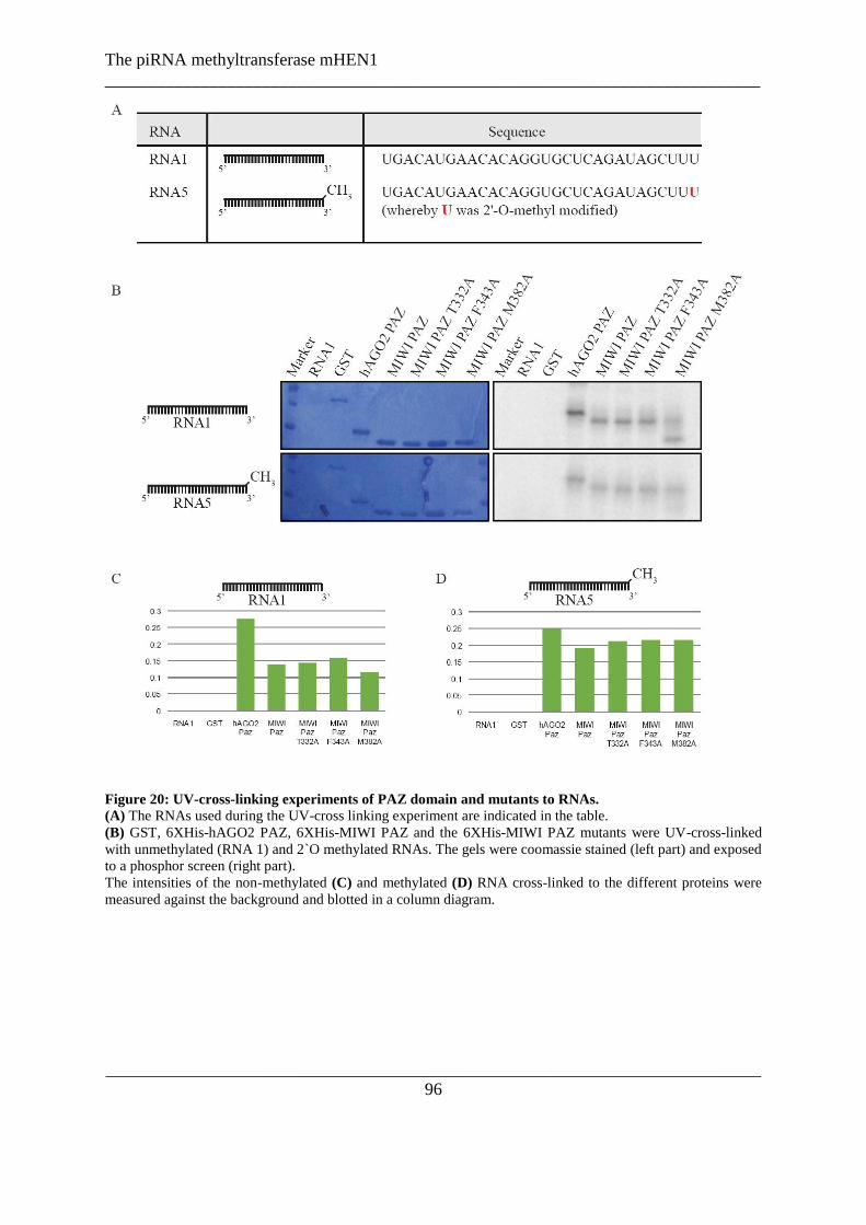
The piRNA methyltransferase mHEN1
___________________________________________________________________________
___________________________________________________________________________
96
Figure 20: UV-cross-linking experiments of PAZ domain and mutants to RNAs.
(A) The RNAs used during the UV-cross linking experiment are indicated in the table.
(B) GST, 6XHis-hAGO2 PAZ, 6XHis-MIWI PAZ and the 6XHis-MIWI PAZ mutants were UV-cross-linked
with unmethylated (RNA 1) and 2`O methylated RNAs. The gels were coomassie stained (left part) and exposed
to a phosphor screen (right part).
The intensities of the non-methylated (C) and methylated (D) RNA cross-linked to the different proteins were
measured against the background and blotted in a column diagram.

The piRNA methyltransferase mHEN1
___________________________________________________________________________
___________________________________________________________________________
97
2.4.3.5 Discussion
Our collaborators of the Carlomagno group at the EMBL Heidelberg solved the NMR
structure of the MIWI PAZ domain bound to a methylated small RNA. The structure is
highly similar to the known PAZ domains (Lingel et al. 2003, 2004; Ma et al. 2004; Song et
al. 2003; Yan et al. 2003). The interaction of the MIWI PAZ domain with RNAs were
biophysically analyzed, indicating that the MIWI PAZ domain prefers single stranded
methylated small RNAs.
piRNAs carry a 2`-O-methylation mark at their 3` terminal nucleotide (Horwich et al.
2007; Kirino & Mourelatos 2007c; Ohara et al. 2007a; Saito et al. 2007a; Zhou et al. 2010).
It is not clear yet, what role this modification has. In fly and zebrafish mutants of the RNA
methyltransferase hen1, Piwi proteins still correctly incorporate the unmethylated piRNAs,
suggesting that the modification of the RNA is not needed for the loading process (Horwich
et al. 2007; Kamminga et al. 2010). On the other hand, piRNAs in zebrafish, siRNAs in flies
and si- and miRNAs in plants lacking the modification are exposed to an unknown enzyme
adding a short Poly U-tail which consequently leads to trimming by an unidentified nuclease
(Ameres et al. 2010; Kamminga et al. 2010; Li et al. 2005). This indicates that the methyl
group at the 3`end is needed for stabilization of small RNA.
In mouse, the function of the post-transcriptional modification of the piRNAs is
unknown, but even if it is assumed that the methylation protects the piRNAs from
destabilizing events, the methylated 3` end of the piRNAs has to be recognized and to fit into
the PAZ domain of the Piwi proteins. Here, the influence of the 2`-O-methylation of the
3‟end of piRNAs on the binding to the MIWI PAZ domain was investigated.
In comparison to the hAGO1 PAZ structure, the binding of the methylated 3`end of
the RNA to the MIWI PAZ domain shifts the RNA by one nucleotide out of the binding
pocket, so that the first nucleotide takes the space, which is normally taken by the second
nucleotide bound in an hAGO1 PAZ. Taken this shift by one nucleotide in the MIWI PAZ
domain into account it could be that the methylation is needed for a tight fit into the protein.
The PAZ domain itself can recognize the RNA and bind it, but without the methylation mark
on the RNA the binding is loose and there is a risk of losing the piRNA due to enzymatic
activities. To avoid this, the methylation mark is recognized and fits the RNA in the correct

The piRNA methyltransferase mHEN1
___________________________________________________________________________
___________________________________________________________________________
98
tight position in the domain. The PAZ domain of the human Piwi protein HIWI shows as well
that the RNA binding pocket is able to accommodate the methyl group of the RNA while
AGO1 is unable to bind the modified RNAs due to steric clashes with the surfaces of amino
acid side chains (Tian et al. 2010).
The key-residue of the specific interaction of the MIWI PAZ domain with methylated
RNAs is the long side-chain of M382, which bends around from the β7 on the opposite side
of the RNA binding surface to contact the RNA. Surprisingly, this methionine is not
conserved in all Piwi proteins, but in MIWI, HIWI and Aubergine. For the HIWI PAZ
domain it was shown that this methionine contacts the methyl group at the 3` end of RNAs as
well (Tian et al. 2010). Despite these contacts, the methionine seems not to be required for
the binding to methylated RNAs per se, but has perhaps evolved to optimize the interaction.
However, taken the cross-linking and Surface Plasmon Resonance (SPR) results into
account, it was shown here that the recognition of methylated and unmethylated RNAs by the
MIWI PAZ domain is highly similar. The KD of the binding to unmethylated RNAs is 2 µM
and for methylated RNAs 0.9 µM. Recently, it was published that the PAZ domain human
Piwi proteins HIWI, HIWI2 and HILI bind to a methylated 12 nt self-complementary duplex
with 2 nt overhangs 2.5 to 6-fold higher affinity than to unmethylated RNAs measured by
isothermal titration calorimetry (Tian et al. 2010), confirming the observed data. The only
significant change in the affinity to methylated RNAs was determined in M382 mutants,
arguing again for an important role of this residue in the methyl group recognition of the
MIWI PAZ domain. In contrast, double mutants of the methionine (M381Y) and a Proline
(P379H) in HIWI PAZ revealed no significant difference in the binding to methylated or
unmethylated RNA substrate (Tian et al. 2010).
In conclusion, the MIWI PAZ domain binds preferentially to single stranded RNAs
over double stranded RNAs with 3` overhangs. The methyl-group on the 3` end of ssRNAs is
not essential for the binding, but buries the RNA deep into the binding pocket. All data
suggest that the PAZ domain has a high flexibility to adapt to RNAs with different sequences,
chemical modifications and structures.

The piRNA methyltransferase mHEN1
___________________________________________________________________________
___________________________________________________________________________
99
2.4.3.6 Methods
2.4.3.6.1 Protein expression and purification
Generation of the expression constructs and preliminary protein expression of the PAZ
domain of MIWI (276-425 aa) and hAGO2 (228-371 aa) were carried out by Elsa Rocha and
Maartje Luteijn, respectively. The mutations of the MIWI PAZ expression construct T332A,
F343A and M382A (performed by Jerome Bourdin) and additional mutation constructs
(K316A and R319A) were designed by directed point mutagenesis.
Samples for structural work were prepared in Dr. Teresa Carlomagno‟s lab at EMBL
Heidelberg essentially based on the protocol set up by Elsa Rocha. Protein samples used for
UV-crosslinking and some of the SPR measurements were prepared by myself as follows: the
proteins were expressed in RosettaTM
2 in 4 L TB medium and induced by 1 mM IPTG. The
cell pellets were lysed (50 mM Na2HPO4, 300 mM NaCl, 10 mM Imidazole, 4 U/mL DNase
I, 0,3 μg/mL RNase A, 1 tablet of Complete EDTA free protease inhibitor cocktail; pH 7.5)
by using a microfluidizer with a chamber pressure of 15-18 kPSI. The purification took place
on a nickel column (2 CVs of a low salt solution: 50 mM Na2HPO4, 300 mM NaCl; then 10
CVs of a low Imidazole solution: 40 mM Imidazole; 10 CVs of a high salt solution: 50 mM
Na2HPO4, 750 Mm NaCl, 750 mM of KCl, 40 mM Imidazole and, finally, 2 CVs of low salt
solution again; eluted with 8 times 0.5 mL of elution buffer: 50 mM Na2HPO4, 300 mM
NaCl, 250 mM Imidazole and dialyzed in 1x PBS, 0.03 % Tween20, 1 mM DTT).
2.4.3.6.2 Surface Plasmon Resonance (SPR) experiments
The experiments were performed at 25 °C on a Biacore T100 machine (Biacore AB, Uppsala,
Sweden) by Dr. Peter Sehr at the Chemical Biology Facility, EMBL Heidelberg.
The RNAs were design to consist of an anchor strand with a coupled biotin at the 3`
end (5` - GAGCACCUGUGUUCAUGUCA - 3` biotin, 100 µM) and a complementary
strand, which was annealed to it, leading to 1, 2 or 10 nt overhangs. Used RNAs were RP
RNA 1 (5`- UGACAUGAACACAGGUGCUCAGAUAGCUUU-3`, 100 µM), RP RNA 5
(5`-UGACAUGAACACAGGUGCUCAGAUAGCUUU-3` where U was 2`-O-methyl
modified, 2nt overhang, 5` UGA CAU GAA CAC AGG UGC GC UU 3`; 1 nt overhang, 5`-
UGA CAU GAA CAC AGG UGC GC U-3`. 25 µM biotinylated RNA and 25 µM of the

The piRNA methyltransferase mHEN1
___________________________________________________________________________
___________________________________________________________________________
100
experimental required complementary RNA were incubated 2 min at 90 °C and cooled down
to RT within 45 min in 20 mM Tris pH 7.5, 100 mM NaCl, 2 mM EDTA to form duplexes.
The annealed duplex mixture was diluted to 25 nM in PBS pH 7.4, 0.05% Tween20, 1 mM
DTT and bound to a streptavidin-coated biosensor chip. As a reference, the sensor surface
without modification was used. The Biotin Capture Template was run without ligand with 50
mM NaOH + 1 M NaCl as conditioner. A manual run with 30 µl/min was performed to
stabilize the baseline. The Biotin Capture Template was then run without conditioning, with
the aim for a target level of 120 RU. The wild type and mutant PAZ proteins were twofold
ranging from 100-0.098 µM. The proteins were injected as analytes at a flow rate of 30
µL/min and the equilibrium dissociation constants determined by steady state affinity
evaluation.
2.4.3.6.3 Binding assay via UV-cross-linking
These studies were carried out at the EMBL Grenoble: 5 µM protein (6XHis-GST, 6XHis
hAGO2 PAZ, 6XHis MIWI PAZ or 6XHis MIWI PAZ mutants) and 100 µM 30 nt
P32
-labelled ssRNA (5`-UGACAUGAACACAGGUGCUCAGAUAGCUUU-3`) with (RP
RNA5) or without (RP RNA1) 2`-O-methylation on the 3‟ end were mixed in 1 x PBS (10 µL
final volume) and incubated at 30 ˚C for 10 min. The samples were exposed to ultraviolet
light (300 mJ). The reactions were stopped by adding 10 µL 2x SDS loading dye and heated
to 94 ˚C for 5 min (Adapted from (Lingel et al. 2003)). Then the samples were resolved on
15 % SDS gels, coomassie stained, dried and exposed to a storage phosphor screen. The
screen was read out at the Typhoon™ 9410 Variable Mode Image, or Typhoon Trio imager
GE healthcare, reference: 63-0055-87.

Chapter 3: Analysis of the MOV10L and TDRD1 homologs in
zebrafish
Analysis of DrTDRD1 and DrMOV10L
___________________________________________________________________________
___________________________________________________________________________
102
3.1 Contribution remark
The results in this chapter are entirely my work. Help for the initial experimental set up was
provided by the Gilmour group at the European Molecular Biology Laboratory, Heidelberg,
Germany.
3.2 Remarques concernant ma contribution
Les résultats de ce chapitre proviennent uniquement de mon travail. L‟équipe de Darren
Gilmour, EMBL Heidelberg, m‟a aidée pour la mise au point des expériences de base.

Analysis of DrTDRD1 and DrMOV10L
___________________________________________________________________________
___________________________________________________________________________
103
3.3 Analysis of the MOV10L and TDRD1 homologs in zebrafish
Stephanie Eckhardt1, Andreea Gruia
2, Darren Gilmour
2 and Ramesh S. Pillai
1
1European Molecular Biology Laboratory, 6 Rue Jules Horowitz, BP 181, 38042 Grenoble,
France.
2European Molecular Biology Laboratory, Meyerhofstraße 1, 69117 Heidelberg, Germany.
Correspondence to:
Ramesh Pillai
Phone: xx-33-4-76 20 7446
Fax: xx-33-4-76 20 7199
E-mail: [email protected]
Analysis of DrTDRD1 and DrMOV10L
___________________________________________________________________________
___________________________________________________________________________
104
3.3.1 Abstract
Piwi-interacting RNAs (piRNAs) are responsible for silencing of transposons in the germline,
but their biogenesis and the factors involved are, as yet, not fully understood. The
inaccessibility of the germ cells is a major hindrance to study piRNAs. However, Tudor
domain-containing protein 1 (TDRD1) and the putative RNA helicase MOV10L have been
implicated in the mouse piRNA pathway, while their exact mechanism of action is not
known. Homologs of these proteins are found in many other organisms ranging from flies to
humans, indicating their conserved roles in the piRNA pathway. To analyze germline
pathway components zebrafish (Danio rerio) embryos were utilized as a model system.
DrTDRD1 and DrMOV10L were identified based on sequence analysis and their co-
expression with fish Piwi proteins was determined. In localization experiments, the testes-
specific DrTDRD1 was found to be in primordial germ cells. It was shown by reporter
studies that the tissue-specific localization of DrTDRD1 is dependent on its 3‟UTR.
Knockdowns of DrMOV10L desilences transposons, implicating a role in the fish piRNA
pathway.
3.3.2 Resume
Les ARNs interagissant avec Piwi (piARNs) sont responsables de la répression des
transposons dans la lignée germinale, mais leur biogénèse et les facteurs impliqués sont, à ce
jour, mal connus. Limpossibilité d‟étudier directement les cellules germinales constitue un
obstacle majeur à l‟étude des piARNs. En revanche, la protéine à domaine Tudor TDRD1 et
la potentielle ARN hélicase MOV10L ont toutes deux été impliquées dans la voie des
piARNs chez la souris, alors que leur mode d‟action n‟est pas connu. Les homologues de ces
protéines sont retrouvés dans de nombreux autres organismes, des mouches aux humains, ce
qui montre leur rôle conservé dans la voie des piARNs. Afin d‟analyser les différents
composants de la voie de signalisation dans les cellules germinales, des embryons de poisson
zèbre (Danio rerio) ont été utilisé comme modèle. DrTDRD1 et DrMOV10L ont été identifié
à partir de l‟analyse de leur séquence primaire et leur coexpression avec les protéines Piwi de
Analysis of DrTDRD1 and DrMOV10L
___________________________________________________________________________
___________________________________________________________________________
105
poisson zèbre a été établie. Dans des expériences de localisation, la protéine DrTDRD1,
spécifique des testicules, a été retrouvée dans les cellules germinales primordiales. Il a encore
été montré, par gène rapporteur, que cette localisation tissu-spécifique de DrTDRD1 est
dépendante de son extrémité 3‟UTR. Des KO de DrMOV10L dérépriment les transposons, ce
qui suggère un rôle dans la voie des piARNs chez le poisson.
3.3.3 Introduction
The gonad-specific Piwi proteins of the Argonaute family associate with 26-31 nt RNAs,
called piRNAs, to protect the genome of the germline from transposable elements in all
animals studied so far (Ghildiyal & Zamore 2009) (see section 1.3.5-1.3.7).
Mice and Drosophila are the favored model systems for investigating the piRNA
pathway components. However, both of these systems have their limitations with a suitable
alternative model system being Danio rerio (zebrafish). The advantages of the zebrafish are
rapid generation time (12-16 weeks), prolific egg-layers, visibility of the cells during larval
stages and simplicity and high throughput nature of manipulations. The accessibility of the
germline facilitates rapid work on the piRNA pathway. Only one other group uses the
zebrafish system to investigate questions concerning the piRNA biogenesis, factors
associated with this pathway and the functions of these components.
To date it has been shown that in zebrafish, the Piwi clade consists of two members:
ZILI and ZIWI (Houwing et al. 2008; Houwing et al. 2007). ZIWI is initially maternally
provided and later expressed strongly in mitotic and early meiotic stages of the germ cell
differentiation (Houwing et al. 2007) (see Figure 21). ZILI was first described as expressed
from 3 days post fertilization (dpf) onwards and not detectable as a maternally contributed
product (Houwing et al. 2008). However, Sun and colleagues (2010) reported that ZILI can
be detected at the one-cell stage of the embryo (see Figure 21). At 3 dpf, the ZILI protein is
localized in the nucleus as well as in the cytoplasm. In adult zebrafish, ZILI is present in
mitotic and early meiotic cells in the cytoplasm in male and female gonads like ZIWI
(Houwing et al. 2008). ziwi or zili knockout fish show a loss of germ cells. While this loss is
due to excessive apoptosis in ziwi mutants (Houwing et al. 2007), zili mutants show an arrest

Analysis of DrTDRD1 and DrMOV10L
___________________________________________________________________________
___________________________________________________________________________
106
in the germ cell development (Houwing et al. 2008). In both cases the fish are phenotypical
male (Houwing et al. 2008).
ZIWI and ZILI-associated piRNAs (26-27 nt) are found in both the male and female
germline, with greater abundance in ovaries compared to testis. The zebrafish piRNAs
originate mainly from transposons (mainly from LTR elements) and repetitive sequences,
while most of the non-repetitive sequences are in intergenic regions (Houwing et al. 2008;
Houwing et al. 2007). As with flies, zebrafish piRNAs can be passed maternally to the
embryo (Houwing et al. 2007). piRNAs binding to ZIWI have a 5` U bias and are
preferentially antisense regarding the transcript. ZILI- associated piRNAs show a bias for an
A at the 10 nt position and have sense orientation (Houwing et al. 2008). These are typical
signatures required for the ping-pong amplification cycle. This model was analyzed in
Drosophila and describes how piRNAs guide a Piwi protein-containing complex to a target
RNA, which is cleaved, defining a 5`end of a new piRNA, while silencing transcripts of
transposable elements (Brennecke et al. 2007; Gunawardane et al. 2007) (see section 1.3.6).
To gain deeper insights into the piRNA pathway and to identify associated factors,
zebrafish was used as a model system. New tools had to be developed and methods used in
other organisms adapted to the requirements of the zebrafish system. Screening methods
involving fluorescence reporter or analysis of transposon activity via qPCRs could shed light
on piRNA pathway components. To test the reliability of the methods and to gain first
insights into piRNA-associated components, the homologs of two Piwi-interacting factors
analyzed in mouse, MOV10L and TDRD1 (Reuter et al. 2009; Zheng et al. 2010), were
chosen to be investigated.

Analysis of DrTDRD1 and DrMOV10L
___________________________________________________________________________
___________________________________________________________________________
107
Figure 21: Piwi expression during zebrafish development
The embryonic development of zebrafish takes 48-72 hours post fertilization (hpf) and can be roughly divided in
six periods: zygote, cleavage, blastula, gastrula, segmentation, pharyngula and hatching. After leaving the egg,
the larva develops to sexual maturity after 12-16 weeks (green box, 1:10 scale compared to previous images).
The Piwi protein ZIWI can be observed throughout development (Houwing et al. 2007). ZILI was found to be
expressed from 72 hpf onwards (Houwing et al. 2008) (dark blue line), but Sun and described ZILI already in
the one-cell stage of the embryo (Sun et al. 2010) (light blue dashed line). However, Sun describes a cap of
expression of ZILI around 24 hpf. The image was adapted from (Kimmel et al. 1995).

Analysis of DrTDRD1 and DrMOV10L
___________________________________________________________________________
___________________________________________________________________________
108
3.3.3.1 The putative helicase MOV10L is involved in the primary piRNA biogenesis
One factor shown to be associated with the primary piRNA biogenesis is the fly helicase
Armitage (Olivieri et al. 2010; Saito et al. 2010) and its mouse homolog MOV10L (named
after its homology to protein Moloney Leukaemia Virus 10 (MOV10)) (Frost et al. 2010;
Zheng et al. 2010), which are both expressed in the gonads (see section 1.4.4).
The testes-specific MOV10L contains a helicase domain and two highly conserved
regions of unknown function (Frost et al. 2010; Vagin et al. 2009; Zheng et al. 2010). The
same domain architecture is shown for ubiquitously expressed helicase MOV10, with which
MOV10L shares 45 % amino acid identity (Zheng et al. 2010). MOV10 localizes in the
cytoplasmic P-bodies (Meister et al. 2005; Wulczyn et al. 2007), where it associates with the
mature-miRNA-containing RISC by interacting directly with Argonaute proteins. This
interaction is required for miRNA-guided mRNA cleavage (Chendrimada et al. 2007; Meister
et al. 2005).
Armitage is expressed in germline cells (Cook et al. 2004) and can also be detected in
somatic follicle cells (Olivieri et al. 2010). MOV10L is only expressed in testis with low
abundance in oocytes of mice (Frost et al. 2010; Lovasco et al. 2010; Wang et al. 2001b;
Zheng et al. 2010). While armitage mutants are sterile in both genders, mov10l mutants
impairs only the male fertility (Cook et al. 2004; Frost et al. 2010; Zheng et al. 2010),
indicating their importance for the germline development. A direct interaction of Armitage
and Piwis is supported by the result of Zheng et al. that show the mouse Armitage homolog
MOV10L interacts with all three mouse Piwi proteins (Zheng et al. 2010) linking the
helicases to the piRNA pathway.
piRNAs analyzed from MOV10L immunoprecipitation reveal that most of the
piRNAs were associated with MILI and less were bound in MIWI complexes. This is
confirmed by the co-localization of MOV10L with MILI in different stages of testes
development (Zheng et al. 2010). It is suggested that MOV10L is required for piRNA
biogenesis (Zheng et al. 2010), their stability or for the loading of them into Piwi proteins, as
in the mov10l knockout neither MILI nor MIWI2 are loaded with piRNAs (Frost et al. 2010;
Zheng et al. 2010). The loss of piRNAs results in activation of retrotransposons like LINE
and IAP elements; this is accompanied by a loss of DNA methylation marks on their
promoter elements. Additionally it was observed that the predominantly nuclear localization
of MIWI2 is disturbed in mov10l knockout mice, in which it localizes in the cytoplasm
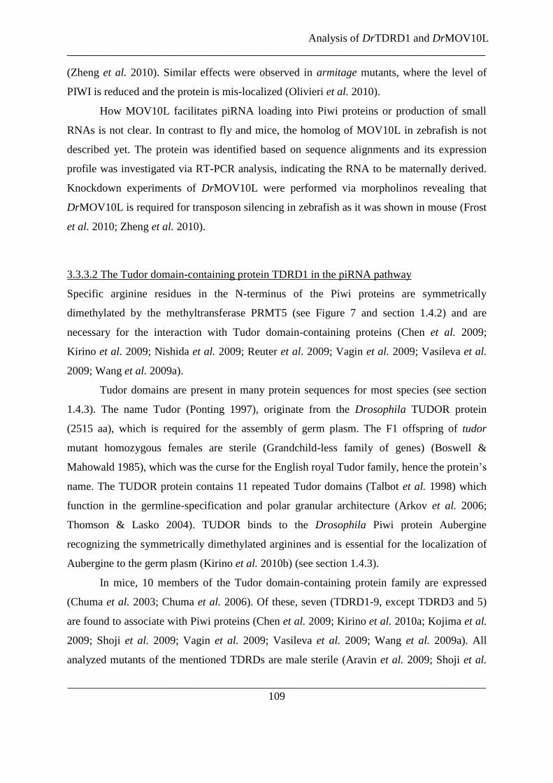
Analysis of DrTDRD1 and DrMOV10L
___________________________________________________________________________
___________________________________________________________________________
109
(Zheng et al. 2010). Similar effects were observed in armitage mutants, where the level of
PIWI is reduced and the protein is mis-localized (Olivieri et al. 2010).
How MOV10L facilitates piRNA loading into Piwi proteins or production of small
RNAs is not clear. In contrast to fly and mice, the homolog of MOV10L in zebrafish is not
described yet. The protein was identified based on sequence alignments and its expression
profile was investigated via RT-PCR analysis, indicating the RNA to be maternally derived.
Knockdown experiments of DrMOV10L were performed via morpholinos revealing that
DrMOV10L is required for transposon silencing in zebrafish as it was shown in mouse (Frost
et al. 2010; Zheng et al. 2010).
3.3.3.2 The Tudor domain-containing protein TDRD1 in the piRNA pathway
Specific arginine residues in the N-terminus of the Piwi proteins are symmetrically
dimethylated by the methyltransferase PRMT5 (see Figure 7 and section 1.4.2) and are
necessary for the interaction with Tudor domain-containing proteins (Chen et al. 2009;
Kirino et al. 2009; Nishida et al. 2009; Reuter et al. 2009; Vagin et al. 2009; Vasileva et al.
2009; Wang et al. 2009a).
Tudor domains are present in many protein sequences for most species (see section
1.4.3). The name Tudor (Ponting 1997), originate from the Drosophila TUDOR protein
(2515 aa), which is required for the assembly of germ plasm. The F1 offspring of tudor
mutant homozygous females are sterile (Grandchild-less family of genes) (Boswell &
Mahowald 1985), which was the curse for the English royal Tudor family, hence the protein‟s
name. The TUDOR protein contains 11 repeated Tudor domains (Talbot et al. 1998) which
function in the germline-specification and polar granular architecture (Arkov et al. 2006;
Thomson & Lasko 2004). TUDOR binds to the Drosophila Piwi protein Aubergine
recognizing the symmetrically dimethylated arginines and is essential for the localization of
Aubergine to the germ plasm (Kirino et al. 2010b) (see section 1.4.3).
In mice, 10 members of the Tudor domain-containing protein family are expressed
(Chuma et al. 2003; Chuma et al. 2006). Of these, seven (TDRD1-9, except TDRD3 and 5)
are found to associate with Piwi proteins (Chen et al. 2009; Kirino et al. 2010a; Kojima et al.
2009; Shoji et al. 2009; Vagin et al. 2009; Vasileva et al. 2009; Wang et al. 2009a). All
analyzed mutants of the mentioned TDRDs are male sterile (Aravin et al. 2009; Shoji et al.

Analysis of DrTDRD1 and DrMOV10L
___________________________________________________________________________
___________________________________________________________________________
110
2009; Vasileva et al. 2009). TDRD1 (129 kDa) contains a N-terminal myeloid–nervy–DEAF-
1 (MYND) domain (163-199 aa) and four Tudor domains (307-361 aa, 536-589 aa, 756-
809 aa and 982-1034 aa) (Chuma et al. 2003). These Tudor domains are responsible for
protein interactions, recognizing symmetrically dimethylated arginines (Chen et al. 2009;
Kirino et al. 2009; Nishida et al. 2009; Reuter et al. 2009; Vagin et al. 2009; Vasileva et al.
2009; Wang et al. 2009a).
TDRD1 interacts with MILI in an RNA-independent manner throughout the germ cell
development; from the primordial germ cells through to haploid post meiotic germ cells
(Reuter et al. 2009; Vagin et al. 2009; Wang et al. 2009a). It recognizes also the post-
translational modifications of MILI and MIWI (and perhaps MIWI2) (Kojima et al. 2009;
Reuter et al. 2009; Vagin et al. 2009).
tdrd1 mutant testes are smaller, because the spermatogenesis is blocked after the
pachytene-diplotene stage leading to apoptosis (Chuma et al. 2006). The levels of the MILI
protein and the piRNAs are not modified in tdrd1-null spermatocytes (Reuter et al. 2009;
Wang et al. 2009a), but the identity of the MILI bound piRNA is changed towards genic
piRNAs, showing an overrepresentation of sense-stranded exonic reads and a lack of MIWI2
bound piRNAs (Reuter et al. 2009; Vagin et al. 2009). MIWI2 is not only unloaded in tdrd1
mutants, its nuclear localization is shifted to the cytoplasm (Reuter et al. 2009; Vagin et al.
2009), as it is in mili mutants (Aravin et al. 2007b). Furthermore, the loss of TDRD1
activates LINE1 elements due to a decrease of DNA methylation during the developmental
window when MILI is required to silence transposons by de novo DNA methylation (Aravin
& Bourc'his 2008; Kuramochi-Miyagawa et al. 2008; Reuter et al. 2009; Vagin et al. 2009).
TDRD1 is a cytosolic protein localized to the intermitochondrial cement in male and
female germ cells and to the chromatoid bodies in the male mice. It interacts with the
chromatoid body marker mouse VASA homolog (MHV) (Chuma et al. 2003; Chuma et al.
2006). The medaka TDRD1 localizes to granules in the cytoplasm in primordial germ cells in
the male and female germline. When the primordial germ cells arrive in the primordium, the
medaka TDRD1-containing granules expand, creating a hollow area, which stays visible
during the sex determination. The oocytes undergo cystic division and enter then meiosis.
During this time the granules shrink and localize in close proximity to the nucleus (Aoki et
al. 2008), similar to the chromatoid body in mice.

Analysis of DrTDRD1 and DrMOV10L
___________________________________________________________________________
___________________________________________________________________________
111
Interestingly, the localizations of proteins specifically expressed in primordial germ
cells in zebrafish, like TDRD7, Vasa and Nanos1, are dependent on the 3‟UTR in their
mRNAs. These 3`UTRs of testes-specific mRNAs contain a miRNA binding site which is
flanked by two Dead end protein 1 (Dnd1) binding motifs. The Dnd1 protein is only
expressed in the germline, while in somatic cells the miRNA can bind to the mRNA binding
site of the 3`UTR recruiting the post-transcriptional silencing complex, which leads to
repression. In the germline the Dnd1 binds to the flanking regions, leading to an
inaccessibility of the miRNA binding site and the proteins are expressed (Kedde et al. 2007).
In contrast to TDRD1 in mice, the zebrafish homolog is not described in the literature
yet. Also unknown is whether ZILI and ZIWI carry symmetrically methylated
arginine/glycine repeats in their N-termini like Piwi proteins in Drosophila and mouse.
Within this study, the zebrafish homolog of TDRD1 (DrTDRD1) was identified via
sequence alignments. Expression profiling of DrTDRD1 indicated that it is maternally
contributed and persists throughout the lifecycle. In localization experiments, DrTDRD1 was
identified in the primordial germ cells, whereby this localization is dependent on its 3`UTR.
Although, DrTDRD1 is a testes-specific protein, expressed similar to Piwi proteins, it was
not observed to interact with ZILI or ZIWI, or associate with piRNAs.

Analysis of DrTDRD1 and DrMOV10L
___________________________________________________________________________
___________________________________________________________________________
112
3.3.4 Results
3.3.4.1.1 Identification of the homolog of MOV10L in zebrafish
MOV10L is a putative helicase involved in the primary piRNA biogenesis in mice (Frost et
al. 2010; Vagin et al. 2009; Zheng et al. 2010) (see section 1.4.4 and 3.3.3.1). To identify the
zebrafish homologs, a pBLAST search was performed and a single sequence was found. The
putative zebrafish DrMOV10L comprises 1106 aa with a predicted molecular weight of
122.6 kDa. It is 47.9 % identical and 68.6 % similar at the amino acid level to the mouse
MOV10L. The helicase domain of the zebrafish DrMOV10L (668-1106 aa) accommodates
an ATP binding site (701-705 aa) and a putative Mg2+
binding site (814-816 aa) (see Figure
22). DrMOV10L contains two domains of unknown functions (~34-212 aa and ~222-512 aa),
which are highly conserved in homologs of other species.
3.3.4.1.2 Identification of DrTDRD1
The mouse TDRD1 is a Tudor domain-containing protein which is likely to interact with all
three murine Piwi proteins and is needed for transposon silencing in the mouse germline
(Kojima et al. 2009; Reuter et al. 2009; Vagin et al. 2009; Wang et al. 2009a) (see section
1.4.3 and 3.3.3.2). The zebrafish homolog of DrTDRD1 consists of 1176 aa and has a
calculated molecular weight of 129.2 kDa. It showed 33.7 % identity (63.7% similarity) at the
amino acid level to the mouse TDRD1 and was identified to carry the same domains (see
Figure 23): These domains include the MYND domain (83-119 aa) with highly conserved
cysteine repeats (see Figure 23) and four predicted Tudor domains (173-293 aa, 405-523 aa,
627-745 aa and 876-995 aa, respectively). Furthermore, the binding pocket responsible the
recognition of symmetrically dimethylated arginines on other proteins (e. g. Piwis, Vasa-like
proteins) is also present in DrTDRD1 (see Figure 23).
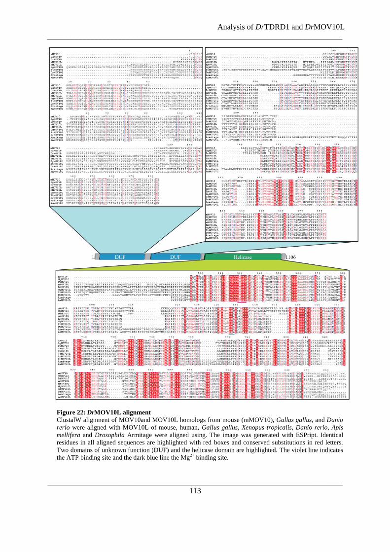
Analysis of DrTDRD1 and DrMOV10L
___________________________________________________________________________
___________________________________________________________________________
113
Figure 22: DrMOV10L alignment
ClustalW alignment of MOV10and MOV10L homologs from mouse (mMOV10), Gallus gallus, and Danio
rerio were aligned with MOV10L of mouse, human, Gallus gallus, Xenopus tropicalis, Danio rerio, Apis
mellifera and Drosophila Armitage were aligned using. The image was generated with ESPript. Identical
residues in all aligned sequences are highlighted with red boxes and conserved substitutions in red letters.
Two domains of unknown function (DUF) and the helicase domain are highlighted. The violet line indicates
the ATP binding site and the dark blue line the Mg2+
binding site.

Analysis of DrTDRD1 and DrMOV10L
___________________________________________________________________________
___________________________________________________________________________
114
Figure 23: DrTDRD1 alignment
TDRD1 from mouse, Rattus norvegicus, human, Macaca mulatta, Equus caballus, Canis familiaris,
Bos taurus, Monodelphis domestica, Ornithorhynchus anatinus, Gallus gallus, Danio rerio and Oryzias latipes
were aligned and analyzed as in (A). The MYND domain is marked with a red box and the conserved Cysteine
repeats are indicated with red stars (*). The Tudor domains are indicated with yellow (Tudor domain 1), dark
orange (Tudor domain 2), light orange (Tudor domain 3) and orange-red (Tudor domain 4). The putative
aromatic cage building residues, based on similarity to the Tudor domain 11 of TUDOR of Drosophila (Liu et
al. 2010) indicated with blue stars (*).

Analysis of DrTDRD1 and DrMOV10L
___________________________________________________________________________
___________________________________________________________________________
115
3.3.4.2 Expression profiles of Zili, Drtdrd1 and Drmov10l during embryonic development
Due to the contradictory literature regarding the onset of ZILI expression as well as
the open question of the expression profiles of the putative helicase DrMOV10L and the
DrTDRD1, RT-PCRs were carried out across the lifecycle. Therefore, RNA was extracted
from eggs (0 hpf) and embryos at different developmental stages (2, 4, 6, 8, 12, 24, 48, 72, 96
hpf). RT-PCRs with specific primers for Ziwi, Zili, Drmov10l, Drtdrd1 and actin (as a
normalization control) were performed and the expression of these genes analyzed by agarose
gel electrophoresis (see Figure 24). As expected the Actin control was present throughout the
embryonic development, indicating that similar amounts of RNAs were used at each time
point (see Figure 24). All remaining transcripts (Ziwi, Zili, Drmov10l, and Drtdrd1) were
detected at 0 hpf, indicating a maternal contribution of these mRNAs. Interestingly, the
amount of Zili continually decreased from this point, reaching the minimum at 48 hpf,
followed by a slight increase. Drmov10l, Drtdrd1 and Ziwi (see Figure 24) are expressed
throughout our experimental time points. The RT-PCR signal of Drtdrd1 and Drmov10l
decreases slightly with time, but to a lesser extent than Ziwi, while the Actin signal increases
slightly.
Figure 24: RT-PCRs to determine the expression of fish piRNA-pathway related mRNAs.
RT-PCRs using specific primers for Drmov10L, Drtdrd1, Zili, Ziwi and Actin mRNAs from unfertilized eggs
and embryos of different developmental age (time in hours post fertilization; hpf). All Drmov10l, Drtdrd1, Ziwi,
Zili and Actin are expressed from the unfertilized egg onwards throughout development. The Zili expression gets
weaker between 12 and 48 hpf to increase at 72 hpf again. Drmov10l, Drtdrd1and Ziwi appear to decease during
the time frame. However, this is not normalized for cell differentiation and is therefore an artifact.

Analysis of DrTDRD1 and DrMOV10L
___________________________________________________________________________
___________________________________________________________________________
116
3.3.4.3 Development and testing of anti- DrTDRD1 and DrMOV10L antibodies
The biochemical characterization of the endogenous zebrafish homologs of TDRD1 and
MOV10L required tools to detect and isolate these proteins. Antisera against the C-terminal
region of DrTDRD1 (1011-1176 aa) and the N-terminus of DrMOV10L (1-143) (see Figure
25 A) were raised in rabbits. Western blotting analyses with the affinity-purified antibodies
detected the respective tagged proteins expressed in HEK293 cell (Myc-tagged DrMOV10L:
ca. 125 kDa and HA-DrTDRD1: ca. 130 kDa) (see Figure 25 B).
To verify if DrTDRD1 and DrMOV10L are expressed in the gonads as their mouse
homologs are, fish testes extract was obtained from frozen zebrafish, testing if working with
frozen tissues would allow samples to be stored. The proteins were blotted to a membrane
and analyzed using the affinity-purified antibodies against DrTDRD1 and DrMOV10L. It
was not possible to detect good signal from these extracts (see Figure 25 B).
Therefore, the experiment was repeated with fresh ovary, testes and muscle extracts.
In this experimental set up α-DrTDRD1 detects a strong band of about 130 kDa in testes
extract, but not from ovaries or muscles (see Figure 25 C), suggesting that the expression of
DrTDRD1 is testes-specific and sensitive to freeze-thaw treatment. Thus, all further
experiments were performed on fresh material. α-MOV10L was not able to detect a specific
band in the extracts (see Figure 25 C). The membrane was re-stained with an α-mouse
Tubulin antibody and observed that the amount of the zebrafish Tubulin (ca. 50 kDa) in the
different extracts is comparable.
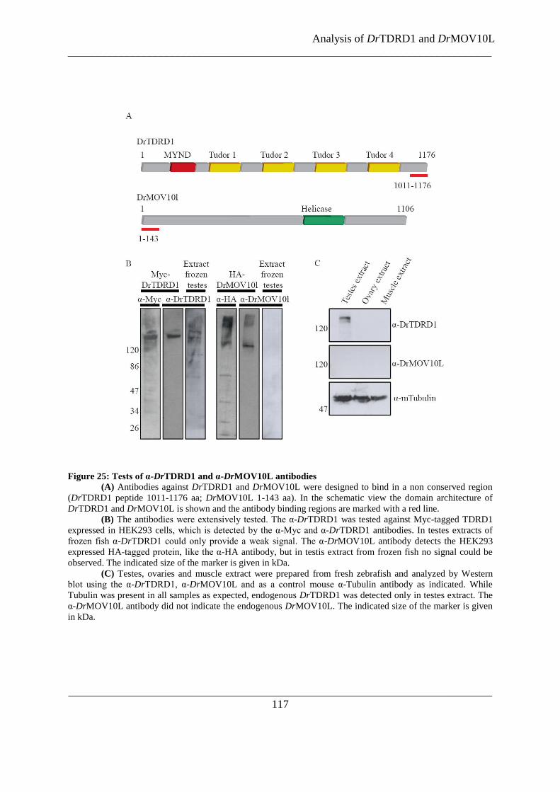
Analysis of DrTDRD1 and DrMOV10L
___________________________________________________________________________
___________________________________________________________________________
117
Figure 25: Tests of α-DrTDRD1 and α-DrMOV10L antibodies
(A) Antibodies against DrTDRD1 and DrMOV10L were designed to bind in a non conserved region
(DrTDRD1 peptide 1011-1176 aa; DrMOV10L 1-143 aa). In the schematic view the domain architecture of
DrTDRD1 and DrMOV10L is shown and the antibody binding regions are marked with a red line.
(B) The antibodies were extensively tested. The α-DrTDRD1 was tested against Myc-tagged TDRD1
expressed in HEK293 cells, which is detected by the α-Myc and α-DrTDRD1 antibodies. In testes extracts of
frozen fish α-DrTDRD1 could only provide a weak signal. The α-DrMOV10L antibody detects the HEK293
expressed HA-tagged protein, like the α-HA antibody, but in testis extract from frozen fish no signal could be
observed. The indicated size of the marker is given in kDa.
(C) Testes, ovaries and muscle extract were prepared from fresh zebrafish and analyzed by Western
blot using the α-DrTDRD1, α-DrMOV10L and as a control mouse α-Tubulin antibody as indicated. While
Tubulin was present in all samples as expected, endogenous DrTDRD1 was detected only in testes extract. The
α-DrMOV10L antibody did not indicate the endogenous DrMOV10L. The indicated size of the marker is given
in kDa.

Analysis of DrTDRD1 and DrMOV10L
___________________________________________________________________________
___________________________________________________________________________
118
3.3.4.4 Attempt to link the endogenous DrMOV10L and DrTDRD1 directly to the piRNA
pathway in zebrafish
DrTDRD1 is a testes-specific protein (see section 3.3.4.3), which could be involved in the
piRNA pathway by recognizing potential symmetrically dimethylated arginines on the
zebrafish Piwi proteins by its Tudor domains. It is unknown whether the zebrafish ZIWI and
ZILI carry similar post-translational modifications.
To analyze the piRNA profile of the zebrafish Piwi proteins, endogenous ZILI and
ZIWI complexes were immunopurified from adult zebrafish ovary extract using specific
affinity purified antibodies. To investigate if the Piwi proteins carry symmetrically di-
methylated arginines a specific antibody recognizing the symmetrically dimethylated
arginines but not the Piwi protein itself, called Y12 (Reuter et al. 2009), was used for
immunoprecipitations as well. To control the experiment, endogenous MILI complexes were
immunoprecipitated from mouse testes extract. The complex-associated RNAs were isolated
and after 5`-end-labelling separated on a denaturating gel.
In comparison to the MILI immunoprecipitation (see Figure 26 A lane 6), the amount
of ZILI and ZILI bound piRNAs is significant lower (see Figure 26 A lane 3 and 4). No
RNAs were observed using beads without specific antibodies for the immunoprecipitation
(see Figure 26 A second panel). Immunoprecipitations of ZIWI and ZILI co-purified RNAs
of 27 nt and 26 nt, respectively (see Figure 26 A lane 3 and 4). Immunoprecipitations using
the Y12 antibody indicated that at least one zebrafish protein must carry symmetrically
dimethylated arginines, due to the fact that RNAs of 26 nt were present (see Figure 26 A). In
comparison to the immunoprecipitations of ZIWI (27 nt) and ZILI-bound RNAs (26 nt), the
size of the Y12 pull down RNAs are 26 nt, which indicates that they originate from ZILI-
containing complexes. However, some contribution from ZIWI cannot be excluded.
Assuming that DrTDRD1 is an interaction partner for the zebrafish Piwi proteins,
piRNAs bound in this complex could be co-purified. Similarly, if DrMOV10L interacts with
ZIWI or ZILI bound piRNAs this could indicate complex formation. Therefore, the
DrTDRD1 complex was immunoprecipitated from adult zebrafish testes and ovaries using
affinity purified antibodies. Complex bound RNAs were extracted and, after 5` labeling,
analyzed on a denaturating gel. In the DrTDRD1 immunoprecipitations no specific small
RNAs were enriched, indicating that no piRNAs were co-purified (see Figure 26 B 32
P gel).

Analysis of DrTDRD1 and DrMOV10L
___________________________________________________________________________
___________________________________________________________________________
119
DrMOV10L immunoprecipitations also did not indicate any bound piRNAs (see
Figure 26 C). In summary, at least one of the zebrafish Piwi proteins is potentially post-
translational modified via symmetrically dimethylated arginines as indicated by
immunoprecipitations using an antibody recognizing these modifications pulling down RNAs
in the size range of piRNAs. Immunoprecipitations with α-DrTDRD1 and α-DrMOV10L did
not indicate any complex formation with piRNAs.
Figure 26: Immunoprecipitations of zebrafish piRNA pathway componments
(A) Analysis of co-purified RNAs from immunoprecipitations using α-ZIWI, α-ZILI and Y12
antibodies on adult zebrafish ovary extract (OE) and monoclonal α-MILI on adult mouse testes extract.. As
controls beads without coupled antibodies were used, incubated with the fish ovary extract (left lane) or mouse
testes extract (right lane). Lane 2 and 7 are empty
(B) Immunopurified and extracted RNAs from the DrTDRD1 complex from adult zebrafish testes
extracts (TE). No RNAs in the size range of 26-27 nt, indicating bound piRNAs, were detected.
(C) Immunopurified and extracted RNAs from the DrTDRD1 and DrMOV10l complexes treated as in
the other experiments of the figure. No piRNAs were detected.
(*) denotes empty lanes deleated for display purpose, intensities can be directly compaired in sub-
panels as they are taken from the same image without manipulation.

Analysis of DrTDRD1 and DrMOV10L
___________________________________________________________________________
___________________________________________________________________________
120
3.3.4.5 DrTDRD1 and DrMOV10L interaction studies
Interaction studies of DrMOV10L and DrTDRD1 with the zebrafish Piwi proteins
ZIWI and ZILI were performed, to link the Tudor domain-containing protein and the helicase
directly to the piRNA pathway. In a heterologous system using HEK293 cells, HA-tagged
constructs of full-length DrTDRD1 and DrMOV10L were co-expressed with Myc-ZIWI and
Myc-ZILI. The HA-tagged proteins were purified with α-HA beads from HEK293 cell
lysates prepared 48 hours post-transfection and subjected to Western analyses using α-Myc
antibodies to detect the possible co-purified Piwi proteins. Unfortunately, the HA-tagged
DrTDRD1 and DrMOV10L were lowly expressed (see Figure 27 A, α-HA). The Myc-tagged
ZILI and ZIWI proteins are present in the input sample, but they were not co-
immunoprecipitated by HA-DrTDRD1 or HA-DrMOV10L (see Figure 27 A). Mutual
interaction experiments of Myc-DrTDRD1 and HA-DrMOV10L did not indicate any
association (see Figure 27 B).
MILI and the mouse TDRD1 are known to interact via a symmetrical dimethylation
on the argenine 74 in the N-terminus of MILI (Reuter et al. 2009; Vagin et al. 2009; Wang et
al. 2009a) (see section 1.4.3). ZILI shows similar arginine/glycine repeats in the N-terminal
region like many orthologs in different species (see Figure 27 C). To test the possible
recognition of a homologous region containing arginine/glycine in the N-terminus of ZILI by
DrTDRD1, in vitro translated DrTDRD1 were incubated with biotinylated synthetic peptides
corresponding to the amino acids 158-168 of ZILI, where R163 was either unmodified or
symmetrically dimethylated (see Figure 27 green mark). Precipitations with Streptavidin
beads revealed that DrTDRD1 was only co-purified when the peptide carried a methylation
mark (see Figure 27 D), but the achived signal was weak. However, in comparison with in
vitro translated mouse TDRD1 binding to the methylated MILI peptide, the signal of
DrTDRD1 recognizing the methylated ZILI peptide is hardly detectable. Even the signal of
mouse TDRD1 interacting with methylated ZILI peptides is higher than the DrTDRD1/ZILI
peptide association (see Figure 27 E), but the signal of input sample of TDRD1 is much
stronger than the one for DrTDRD1 as well.

Analysis of DrTDRD1 and DrMOV10L
___________________________________________________________________________
___________________________________________________________________________
121
Figure 27: Binding studies of
DrTDRD1 and DrMOV10L
(A) HA-DrTDRD1 and
HA-DrMOV10L were co-expressed
with Myc-ZILI and Myc-ZIWI in
HEK293 cells. The HA-tagged
proteins were immunoprecipitated
using an α-HA matrix, after 1/20th
of the soluble part of the cell extract
was taken as a input sample. The
Myc-tagged zebrafish Piwi proteins
were not co-purified by HA-
DrTDRD1 or HA-DrMOV10L, as
shown in the Western blot analysis
using α-Myc antibodies.
(B) Co-expressions of
Myc-DrTDRD1 and HA-
DrMOV10L using the same method
as before did not reveal any
association of the two proteins.
(C) Comparison of the N-
terminal region of MILI orthologs.
Marked in red are RG/GR repeats.
In green highlighted is the sequence
of the peptide used in the binding
assay.
(D) In vitro translated full-
length DrTDRD1 was incubated
with biotinylated peptides, which
were unmodified or carried a
symmetrical dimethylation at R163.
After purification, the radioactive
marked DrTDRD1 could only be
detected in context with the
methylated peptide and not with the
unmodified one.
(E) The binding assay
shown in (D) was repeated in
comparison with mTDRD1.
mTDRD1 associated with a MILI
peptide containing the
symmetrically di-methylated R74
and recognized even the methylated
ZILI peptide, while the interaction
of DrTDRD1 with the ZILI peptides
was too weak to detect.

Analysis of DrTDRD1 and DrMOV10L
___________________________________________________________________________
___________________________________________________________________________
122
3.3.4.6 Localization of DrTDRD1
The medaka and the mouse TDRD1 are mainly expressed in the male germline. In both
species, the TDRD1 was found to localize in cytoplasmic granules (Aoki et al. 2008; Chuma
et al. 2003) (see section 3.3.3.2). The localization of DrTDRD1 was investigated using
fluorescent reporter constructs. Therefore, one-cell stage zebrafish embryos were injected
with RNA of GFP-tagged full-length DrTDRD1. After 24 h the embryos were examined
using fluorescence microscopy. The fluorescence signal of GFP-DrTDRD1 was detected in
the primordial germ cells, which are distinguishable in the tail region close to the yolk (see
Figure 28 A).
The reason why DrTDRD1 is just expressed in the primordial germline and not in the
other tissues of the embryo was intriguing. Based on the findings of Kedde et al. in 2007, that
the 3‟UTRs of testes-specific mRNAs of proteins are responsible for germline-specific
expression due to a miRNA-guided silencing event in somatic cells (see section 3.3.3.2), the
sequence of the 3`UTR of DrTDRD1 was examined to identify potential miRNA target sites.
The online search machine microcosm provided by the EBI identified 6 potential binding
sites for miRNAs (dre-miR-145, dre-miR-182, dre-let-7e, dre-miR-29a, dre-miR-146a and
dre-miR-193a). Additionally, based on sequence comparison of the 3`UTR of DrTDRD1
with the 3`UTR of Nanos and p27 revealed potential Dnd1 binding sites (Figure 28 B).
To determine if the 3`UTR of Drtdrd1 alone is sufficient to localize the protein
expression in the germline, the sequence was isolated and cloned downstream of a GFP
coding region. As controls, a GFP-construct which contained the 3`UTR of Vasa (Kedde et
al. 2007) was used as positive germline expression marker and a GFP construct without
3`UTR was created to serve as negative control (see Figure 28 C, cartoon).
The RNAs of these construct were injected into zebrafish one-cell stage embryos. 24
hpf the embryos were analyzed using a fluorescent microscope: While the GFP-construct
without 3`UTR expressed the fluorescence protein throughout the embryo, embryos injected
with GFP constructs containing the 3`UTR of Vasa or the 3`UTR DrTDRD1 expressed the
fluorescence protein mainly in the primordial germ cells (see Figure 28 C), indicating that the
3` UTR of DrTDRD1 is responsible for the specific expression of the proteins in the
germline.
Analysis of DrTDRD1 and DrMOV10L
___________________________________________________________________________
___________________________________________________________________________
123
(C) 24 hpf embryos injected with RNAs encoding GFP (left schema), GFP with the 3`UTR of Vasa
(middle schema) and GFP with the 3`UTR of DrTDRD1. GFP without 3`UTR showed expression in the
entire embryo (right image). The DrVasa 3`UTR shuttles the expression of the GFP to the germline (left
image) as shown by (Kedde et al. 2007). The same effect can be achieved with a construct containing GFP
with the 3` UTR of DrTDRD1. Below the primordial germ cells the GFP expression of the construct with
the 3` UTR of DrTDRD1 is show in higher resolution.
Figure 28: Localization of DrTDRD1
(A) Localization of GFP-DrTDRD1 at 24 hpf. The
left column shows the fluorescence signal, the middle
column the phase contrast image and the right column shows
the overlay.
(B) The 3`UTR of DrTDRD1 contains six miRNA
binding sites (red letters). Potential Dnd1 binding sites are
boxed in yellow.

Analysis of DrTDRD1 and DrMOV10L
___________________________________________________________________________
___________________________________________________________________________
124
3.3.4.7 Knockdowns of DrTDRD1 and DrMOV10L
DrTDRD1 is a testes-specific protein, as indicated by western blot analysis of fish tissue
extracts and localization experiments (see section 3.3.4.3 and 3.3.4.4), but its implication in
the zebrafish piRNA pathway was not yet shown. Furthermore, a proof for a functional
involvement of DrMOV10L as a factor in the piRNA biogenesis was still missing in
zebrafish.
To investigate if DrTDRD1 and DrMOV10L are involved in piRNA mediated gene
silencing, morpholinos for knockdown experiments in zebrafish were designed. For
DrTDRD1 knockdowns, two morpholinos were used: Morpholino 1 (MO1) was constructed
to lead to an inclusion of intron1 in DrTDRD1, leading to a pre-mature stop codon in the
translation (see Figure 29 A). The second morpholino (MO2) is targeting the exon3-intron3
junction and should result in exon3 deletion (see Figure 29 B). To knockdown the expression
of DrMOV10L, morpholino 3 (MO3) was designed to bind the translational start site and
block the translation of the protein (see Figure 29 C). All morpholinos were injected into one-
cell stage zebrafish embryos. After 24 h the embryos were harvested and total RNA was
extracted to be analyzed in RT-PCRs with specific primers. To determine the MO1 activity
specific primers complementary to the exon 1 and 2 of DrTDRD1 were used. In case of
active MO1 a larger band was expected than in the uninjected wild type (WT). For MO2
primers before and after exon 3 were designed, which would lead to a shorter RT-PCR
product if the morpholino is active. Unfortunately, both morpholinos against DrTDRD1,
MO1 and 2, showed mainly RT-PCR products of the WT sequence and an insufficient
amount of the knockdown construct (see Figure 29 D), at all volumes of morpholinos
injected.
Given that MO3 against DrMOV10L is a translation inhibitor, it was not possible to
use this method for the activity control. Lacking an active antibody it was impossible to
control the activity of Morpholino 3 directly. So, the activity of MO3 was tested indirectly by
analyzing a potential change in transposon activity, cause by the reduced amount of
DrMOV10L. Hundreds of one-cell stage embryos were injected with MO3 to knockdown the
expression of DrMOV10L. Already after 24h the injected embryos are phenotypically
different to WT or embryos injected with a control morpholino (MOC) (see Figure 29 E).
MO3 embryos display a shorter tail and develop slower than the WT. The survival rate of
WT embryos was 85 %, for embryos injected with a control morpholino (MOC) 75 %, while

Analysis of DrTDRD1 and DrMOV10L
___________________________________________________________________________
___________________________________________________________________________
125
only approximately 10 % of the MO3 injected embryos survived the first 24 hpf. These
results indicated already the importance of DrMOV10L in the embryonic development.
Equal numbers of the surviving embryos were collected and the RNAs extracted.
After DNase treatment equal amounts of total RNA were used to perform RT-PCRs using
random primers. This cDNAs were applied to preliminary qPCR experiments, using primers
to determine the activity of the transposable element DrGypsy2. The values of the qPCR
were normalized against DrActin and the normalized values of RNAs of the MO3 injected
embryos were compared against RNAs of WT embryos. The activity of DrGypsy2 was just
slightly higher than the WT control (0.2 fold difference, see Figure 29 E). The qPCR
experiments were repeated in a bigger screen using primers against the transposons
EnSPmN1, Ngaro and DIRS1a. To ensure the effect is due to the morpholino and not caused
by an overall increase of the gene activity in injected embryos, the activity of Vasa was
measured as well. While DrVasa (see Figure 29 G yellow bar) and DrActin (see Figure 29 G
red bar) did not indicate any change in their activity, the expression of EnSPmN1 was 5 fold
higher than in WT embryos (see Figure 29 G light blue bar), the activity of Ngaro (see Figure
29 G royal blue) increased by 18 fold and DIRS1a (see Figure 29 G dark blue bar) was even
24 fold higher expressed than in the WT.
In conclusion, Drmov10l knockdowns display increased activity of transposable
elements, indicating that the piRNA silencing mechanism is disturbed.

Analysis of DrTDRD1 and DrMOV10L
___________________________________________________________________________
___________________________________________________________________________
126
Figure 29: Morpholino knockdowns
(A) Morpholino 1 (MO1) binds at the exon 1 intron junction of DrTDRD1 and leads to an inclusion of
the intron 1 into the mRNA of DrTDRD1
(B) Morpholino 2 (MO2) coresponds to the exon3 intron junktion and results in an exon3 deletion in
the mRNA of DrTDRD1
(C) Morpholino 3 (MO3) recognizes the translational start site of DrMOV10L and inhibit the
translation of the mRNA.
(D) RT-PCRs to reveal the knockdown capasity of MO1 and MO2. Primers flanking the regions of
interest were used. For active MO1 a larger band was expected (red arrow) than in the WT (green arrow left side
of the gel). For MO2 a shorter RT-PCR product (orange arrow) indicates the morpholino activity in difference
to the longer RT-PCR fragments of the WT (blue arrow).
(E) WT embryos (upper row) and embryos injected with a morpholino control (MOC) display a normal
development in size. Embryos injected with MO3 are shorter at the same developmental stage.
(F) Preliminary qPCR result of DrGypsy2 activation in MO3 treated embryos and in WT normalized
against the expression of DrActin. The expression is given in fold differnce.
(G) qPCRs of MO3 treated embryos analysed against the WT and normalized against DrActin, showed
that in difference to DrVasa, the activity of transposable elements is increased. Data presented as mean ± SD.

Analysis of DrTDRD1 and DrMOV10L
___________________________________________________________________________
___________________________________________________________________________
127
3.3.5 Discussion DrMOV10L and DrTDRD1
In this chapter, zebrafish homologs of the mouse piRNA pathway components TDRD1
(DrTDRD1) and MOV10L (DrMOV10L) were identified and analyzed. Transcripts of
Drtdrd1 and Drmov10l are detected from the unfertilized egg stage onwards throughout
development, as shown by RT-PCR experiments (see section 3.3.4.2). DrTDRD1 is a testes-
specific protein, shown via Western blot analysis (see section 3.3.4.3) and localizes in
primordial germ cells of zebrafish embryos, where this tissue-specific expression is
dependent upon its 3`UTR (see section 3.3.4.6). Unfortunately, interactions of DrTDRD1 and
DrMOV10L with Piwi proteins could not be shown. However, to identify a possible link
between DrTDRD1, DrMOV10L and the piRNA pathway, knockdown experiments were
performed. While the Drtdrd1 knockdown was ineffective to elucidate its function, Drmov10l
(see section 3.3.4.7) knockdowns showed an increased activity of piRNA repressable
transposable elements (Houwing et al. 2008), linking DrMOV10L function to the piRNA
pathway.
RT-PCR experiments indicated that the RNA of Drtdrd1, Drmov10l, as well as from
the zebrafish Piwi genes Zili and Ziwi are all expressed from the one-cell stage onwards
throughout embryonic development. The identified Ziwi expression is in agreement with
earlier studies, showing that the Ziwi is maternally distributed (Houwing et al. 2007). The
RT-PCR results indicate that Zili is already present in the one-cell stage, verifying studies of
Sun and colleagues (Sun et al. 2010). Interestingly, the amount of Zili decreased in the RT-
PCRs from 12 to 48 hpf with a significant recovery from 72 hpf onwards. Similar results
were obtained by Sun et al. showing the decrease of Zili around 24 hpf, when the protein is
no longer expressed in the axis of the embryo but changes towards expression in the
primordial germ cells (Sun et al. 2010). The decreasing effect of Zili is may be caused by the
reduction of maternally contributed and stored Zili mRNAs until the embryo itself starts to
transcribe it in the primordial germ cells. The RT-PCR results of Drtdrd1 and Drmov10l
display two clues. First, both mRNAs are maternally contributed and second, the expression
pattern of Drtdrd1 and Drmov10l is similar to the ones of Zili and Ziwi. The observed
decrease of the Drtdrd1 and Drmov10l signal is probably due to the cell differentiation
occurring in the embryo: while in the early stage embryos, a few cells express the genes of

Analysis of DrTDRD1 and DrMOV10L
___________________________________________________________________________
___________________________________________________________________________
128
interest, in later stages the expression is probably localized in specific tissues (primordial
germ cells as shown for Zili and Ziwi (Houwing et al. 2008; Houwing et al. 2007)). The
RNAs were extracted from the entire embryo or larva, leading to the impression of a
decreasing signal in the total RNA amount.
To be able to perform biochemical experiments with the endogenous DrMOV10L and
DrTDRD1, antibodies against these proteins were developed and extensively tested. Both
antibodies were able to detect full-length DrMOV10L and DrTDRD1 expressed in HEK293
cells, respectively. The endogenous DrTDRD1 was mainly detected in fresh testes extracts,
not in ovary extract and never in muscle extracts. These results are in agreement with the
studies of Aoki and colleagues, who showed that the medaka TDRD1 is primarily expressed
in testes and to a much smaller extent in ovaries (Aoki et al. 2008). DrTDRD1 was only
detected in freshly harvested material and not in extracts prepared from frozen zebrafish,
attributed due to oxygen-dependent degradation of the material and/or damage by repetitive
freeze-thaw cycles. Unfortunately, DrMOV10L was not detected with the specifically
designed antibody in Western blots regardless of the origin of the extracts.
Using the α-DrTDRD1 and α-DrMOV10L antibodies immunoprecipitations were
performed to determine if the two proteins co-immunoprecipitate piRNAs. In both cases no
small RNAs were identified. In terms of the α-DrMOV10L immunoprecipitation it became
clear that the antibody was inactive. The absence of piRNAs in the α-DrTDRD1
immunoprecipitation, (although that it was shown that TDRD1 in mice co-purify piRNAs
(Reuter et al. 2009)) could be due to the small amounts of biological material. Another
possibility is that DrTDRD1 interacts with mainly unloaded zebrafish Piwi proteins. In case
of such an interaction, it is likely that the Tudor domains of DrTDRD1 would recognize
potentially symmetrically dimethylated arginines at ZILI or ZIWI, as the mouse homolog
TDRD1 recognizes R74 at the N-terminus of MILI (Reuter et al. 2009). To test if ZIWI or
ZILI carrying similar post-translational marks, immunoprecipitations recognizing the
symmetrically dimethylated arginines were performed and the RNAs analyzed. The presence
of piRNAs indicates that at least one Piwi protein is modified with methylation marks. The
size of the piRNAs indicates ZILI as a primary modified protein. To analyze potential
interactions of DrTDRD1 and DrMOV10L with ZILI and ZIWI, epitopic tagged proteins
were co-expressed in HEK 293 cells. DrMOV10L did not express well and no interactions
with the ZILI and ZIWI were detected. DrTDRD1 did not interact with Piwi proteins nor

Analysis of DrTDRD1 and DrMOV10L
___________________________________________________________________________
___________________________________________________________________________
129
with DrMOV10L. It was speculated that either the proteins are not folded correctly due to a
lack of the suitable chaperone proteins in the expression system or that ZILI or ZIWI are not
recognized by the PRMT5 protein in the human cells. This would result in Piwi proteins
lacking the symmetric dimethylation moiety recognized by the Tudor domains of DrTDRD1.
GFP-DrTDRD1 was found in the migrating primordial germ cells. A GFP-construct
containing only the 3`UTR of Drtdrd1 injected into Danio rerio embryos localized the GFP
expression into the primordial germ cells as expected. In most of the localization experiments
a background in other tissues, mainly in the back bones was visible. This effect is probably
caused by the high amounts of RNA constructs, which were injected. The inhibition of the
expression of germ cell-specific proteins in vegetative tissues by miRNAs, which recognize
the miRNA binding site in the 3`UTR of these proteins, is efficient, but the artificial
constructs overwhelms the capacity of regulation. However, the signals of the primordial
germ cells have higher intensity, which leads to the conclusion that the expression in testes of
DrTDRD1 is guided by its 3`UTR.
To determine the function of Drtdrd1 and Drmov10l, knockdown experiments using
morpholinos against Drtdrd1 and Drmov10l were performed. The morpholinos against
Drtdrd1 disturbed the splicing, but the observed silencing of the mRNA was inefficient.
More than 50% of the Drtdrd1 mRNA was still wild type, while only a minority showed the
knockdown variant. It might be possible that the WT construct was still abundant enough to
silence the transposable elements, so that the change of the transposon activity was not
measurable by qPCR. In knockdowns of Drmov10l transposable elements like EnSPmN1,
Ngaro and DIRS1a display a significantly higher abundance than in the wild type. In contrast,
the activity of the germ cell-specific expressed Vasa and Actin is not affected. The values
obtained for the transposon activities in the experiments showed a comparable activity pattern
like in knockouts of zili (Houwing et al. 2008) and is consistent with studies of mouse
MOV10L (Zheng et al. 2010). In conclusion, DrMOV10L is needed for piRNA-guided
transposon silencing.

Analysis of DrTDRD1 and DrMOV10L
___________________________________________________________________________
___________________________________________________________________________
130
3.3.6 Methods
3.3.6.1 Identification of DrTDRD1 and DrMOV10L
The known mouse protein sequences of TDRD1 and MOV10L were used in a BLAST search
to identify the zebrafish homologs. The sequences identified in different species were aligned
using ClustalW2 (Multiple sequence alignment with the Clustal series of programs. (Chenna
et al. 2003)). The Alignments were analyzed with ESPript (Gouet et al. 1999).
3.3.6.2 DrTDRD1 and DrMOV10L expression test by RT-PCR
To analyze the expression of DrTDRD1 and DrMOV10L in compared to zebrafish Piwi
protein, RT-PCRs were performed. DNA fragments of ~300 nt in length of
DrMOV10L (primers: 5`-GCGAATTCATGGTGAGTGTGGTGCAGTGTTTAATGTCG-3`,
5`-CCGCTCCACCCGTATGGCTCTCC-3`);
DrTDRD1 (primers: 5`-CTCTCGAGACACCATGAATCCAGCCTTCGCTCAGCC-3`,
5`-CACCGAGTGCACCTTAAGTTGCCC-3`);
ZILI (primers: 5`-GCGAATTCATGGATCCAAAACGACCAACC-3`,
5`-CCT CCC TCT TCC ATA GGT AAC CTC-3`);
ZIWI (primers: 5`-GAATTCATGACAGGACGAGCAAGAGC-3`,
5`-GGATTGTTTCACGTGTCCATAAG-3`),
DrActin (primers: 5`-ATGTGTGACGACGACGAGACTAC-3`,
5`-GGGGCAACACGGAGCTCATTG-3`)
were analyzed on a 2% agarose gel containing ethidium bromide.
3.3.6.3 Antibodies
For the production of antibodies, GST-fusions of specific protein fragments were produced in
E. coli strain BL21 and injected into rabbits. The fragments used were anti-DrTDRD1 (1011-
1176 aa) and anti-DrMOV10L (1-143 aa). All antibodies were affinity-purified and used at a
dilution of 1:200. Anti-ZILI and anti-ZIWI antibodies were kindly provided by the Ketting
lab (Houwing et al. 2008) or produced based on peptides:
ZILI: MDPKRPTFPSPPGVI+C
ZIWI: MTGRARAR+C

Analysis of DrTDRD1 and DrMOV10L
___________________________________________________________________________
___________________________________________________________________________
131
Other antibodies used are: anti-HA antibodies (Santa-Cruz) at 1:250 dilutions and anti-Myc at
1:250 dilutions.
3.3.6.4 Mouse and zebrafish tissue extracts and RNA analysis
CD1 mouse testes extract preparation, immunoprecipitation and 5`-end-labelling of piRNAs
were performed as described in the literature (Aravin et al. 2006; Girard et al. 2006). The
same method was used for D. rerio AB testes, ovaries and muscle extract preparation and
RNA analysis. Dried gels were exposed to a storage phosphor screen and scanned with a
Typhoon scanner (Amersham).
3.3.6.5 Clones and cell culture
For mammalian cell transfections, full-length ORFs of Piwi proteins (ZIWI and ZILI) or
DrMOV10L or DrTDRD1 were obtained by RT-PCR and were inserted into a pCIneo-NHA
vector (Pillai et al. 2004), into pcDNA3-Myc (Invitrogen), or in a pSP64T-mGFP or pSP64T-
dsRedEX (Kedde et al. 2007) vector as required.
For the HEK293 Ebna interaction experiments, 2 µg each of each construct were
transfected into 6 cm dishes at 80% confluence using Lipofectamine (Invitrogen) following
manufacturer‟s protocol, and harvested 48 hours post-transfection. Cells from one 6 cm plate
were lysed and mixed with 10 µL α-HA-beads (anti-HA affinity matrix beads (Roche))
following the purification in the manufacturer‟s protocol. Western analysis was performed
with affinity-purified antibodies to specific proteins. An aliquot of 5% input material was
taken to control for expression in all reactions.
3.3.6.6 Localization studies in zebrafish
The reporter construct pSP64T-mGFP-Vasa-3`UTR (Kedde et al. 2007) was kindly provided
by the laboratory of Dr Erez Raz. We cloned the full-length DrTDRD1 sequence after the
mGFP sequence. We removed or replaced the 3`UTR of Vasa by the 3` UTR of DrTDRD1.
The constructs were used to synthesis capped sense RNA using the
mMessageMachine kit from Ambion. 210pg/embryo RNAs were injected into one-cell stage
D. rerio embryos.

Analysis of DrTDRD1 and DrMOV10L
___________________________________________________________________________
___________________________________________________________________________
132
The primordial germ cells were imaged 24 hpf with a Zeiss Axiovision Z1 wide-field
microscope. Images were taken with Axiocam camera, processed with manufacturer‟s
software. To analyze potential miRNA binding sides MicroCosm Targets V5 was used.
3.3.6.7 Knockdowns of DrTDRD1 and DrMOV10L
0.1 mM dilutions of Morpholinos were injected into one-cell stage of D. rerio embryos.
Morpholinos: MO1: 5‟-GTATATCATTCATACCATCCAGAAC-3‟
MO2: 5‟-AATGCAGCAGCATATCTTACCCTGG-3‟
MO3: 5‟-ACACTGCACCACACTCACCATCCTC-3‟
MOC: 5‟-CCTCTTACCTCAGTTACAATTTATA-3‟
The analysis of the Morpholino injected embryos were done after 24 h. MO1 and MO2 were
controlled by RT-PCR.
The qPCR was performed using specific primers for the transposable elements, Vasa
(Houwing et al. 2008) and Actin (see above) and the Brillian®II SYBER®Green QPCR
Master Mix kit from (Stratagene) in the Stratagene Mx3005P QPCR System (Program:
10 min 95 °C; 40 cycles: 30 s 95°C, 30 s 51°C, 30 s 72°C; 1 min 95°C; 30 s 55°C; 30 s 95°C)

Analysis of DrTDRD1 and DrMOV10L
___________________________________________________________________________
___________________________________________________________________________
133
3.4 Additional experiments
3.4.1 Development of a reporter assay for functional studies of
piRNAs
3.4.1.1 Aim
Piwi-interacting RNAs are the largest class of small RNA molecules expressed in animals,
specifically in the gonads. Together with the Piwi protein they form complexes linked to
transcriptional gene silencing of transposable elements in the animal germline (Lau 2010)
(see sections 1.3.5-1.3.7). Most of the factors involved in the piRNA field and mechanistic
studies were performed in flies due to the possibility to manipulate the genome easily.
Biochemical analyses of the mouse homologs of these fly proteins and the discovery of new
factors in this system has led research to a basic understanding of the piRNAs. However, a
fast tool to identify and manipulate piRNA pathway components is still missing.
D. rerio is relatively easy and fast to manipulate (see section 3.3.3). The accessibility
of the germline and the embryonic development of the fish in a Petri dish provide the
possibility to use D. rerio embryos to develop a screening method involving fluorescence
reporter for a fast functional studies of piRNA pathway components. Therefore, a suitable
construct to be injected into one-cell stage embryos, which would then expresses a
fluorescence protein only in the germline was needed. In 2007 Kedde et al. reported that the
3`UTRs of germline-specific proteins are responsible for their specific localized expression
due to the inaccessibility of a miRNA binding site, which is blocked by the germline-specific
expressed Dnd1 protein (Kedde et al. 2007) (see section 3.3.3.2). The design of a
fluorescence based reporter assay using D. rerio embryos was based on a construct encoding
GFP followed by the 3`UTR of Vasa, which was kindly provided by Kedde and colleagues
(see Figure 30 A).
Based on the assumption that piRNAs (like miRNAs and siRNAs) guide the piRNA
silencing complex to a target mRNA and silence the translation either by slicing or a
translational block (Hutvagner & Zamore 2002), the fluorescent signal of a construct
containing piRNA target sites, should decrease. In case that the piRNA pathway is disturbed
by knockdowns of piRNA pathway components, the signal intensity should increase.

Analysis of DrTDRD1 and DrMOV10L
___________________________________________________________________________
___________________________________________________________________________
134
3.4.1.2 Results
3.4.1.2.1 Design and analysis of fluorescence piRNA reporters
The construct encoding GFP followed by the 3`UTR of Vasa (Kedde et al. 2007) was
modified to serve the purpose of a fluorescence reporter for piRNA activity. Therefore, a
cloning site (linker) was inserted between the stop codon of the GFP and the 3`UTR of Vasa
to be able to insert different piRNA target sites (see Figure 30 B, first construct). In this
linker construct perfect complementary sequences to piRNAs which are known to be
abundant were cloned (piRNA30, piRNA31 and piRNA80). It is still unknown if piRNAs
bind in perfect complementarity to their target mRNAs and silence them by cleavage like
siRNAs, or if they bind only partial complementary and inhibit the translation like most of
the animal miRNAs (Jackson & Standart 2007). To answer this question, mismatched
(bulges) binding sites of piRNAs were inserted into the linker construct as well. miRNAs are
shown to perform translational silencing better on target mRNAs which contain more than
one miRNA binding site (Doench et al. 2003). Therefore bulge constructs containing three
piRNA target sites were created (see Figure 30 B, construct 5 and 6). To be able to quantify
the fluorescence intensity of the different constructs and to compare them, independent of the
actual amount of injected RNAs (because that is technically difficult to control accurately), a
normalization factor was needed. A second construct encoding another fluorescent protein,
Cherry, followed by the 3`UTR of Nanos without piRNA binding site was designed (see
Figure 30 B, construct 7). This construct co-injected with the GFP-reporter enables the
experimentator to normalize the obtained values for the intensity of the GFP signal against
the intensity of the Cherry signal to quantitatively compare different biological samples
injected simultaneously with both constructs. To test if the constructs are expressed, equal
amounts of RNA of the GFP-linker-3`UTR Vasa construct and the normalization construct
Cherry-3`UTR Nanos were co-injected into one-cell stage embryos. 24 hpf, using
fluorescence microscopy, the fluorescence of both constructs was obtained from primordial
germ cells of dechorinated embryos. This confirmed the result of Kedde et al. showing that
the 3`UTRs lead the expression of the fluorescence marker into the germ cells (see Figure 30
C). To analyze quantitatively changes in the GFP expression, the microscopy settings, once

Analysis of DrTDRD1 and DrMOV10L
___________________________________________________________________________
___________________________________________________________________________
135
optimized, were unchanged for each sample throughout the experiment and the images were
analyzed with the image processing program ImageJ from the EMBL (see Figure 30).
One of the difficulties in analyzing the images is the unbiased determination of the
background (noise). Therefore, selecting only the signal of the region of interest on the image
was necessary by applying the automated ImageJ threshold in one of the images (the signals
in the Cherry image were easier to determine, so it was used for this procedure, see Figure 30
D step 1). Thereby, a grayscale Cherry image was converted to a binary image by defining a
grayscale cutoff point (values below the cutoff become black and those above become red
(see Figure 30 D step 2). From this image a mask (see Figure 30 D step 3) was created, given
the value 1 to the red (over cutoff) pixels of the image and 0 to the black pixels (below
cutoff). The mask was then inverted (see Figure 30 D step 4); so that the signals below the
cutoff take the value 1, while the signals above the cutoff are 0 (this step is necessary,
because the ImageJ program is not able to create an inverted mask directly). The original
GFP and Cherry images were multiplied with the inverted mask (pixel by pixel), giving
images of the noise without the signals (see Figure 30 D step 5). The average noise intensity
of each image was calculated with ImageJ. Then this calculated value of the background
noise was subtracted from the original GFP and Cherry images (see Figure 30 D step6). To
be able to compare the signal intensity of different cells, embryos and fluorescent signal of
the different constructs, the GFP signals had to be normalized by the Cherry signals. The
ImageJ automated threshold was determined for both images with background subtracted to
define the region of interest (cells where the fluorescence signals are expressed) (see Figure
30 D step 7). Then the signal intensity for the GFP was divided by the Cherry signal (pixel by
pixel). In this normalized image (see Figure 30 D step8) the cells were determine by eye.
Therefore the cell shape was analyzed in the original images and the cell edges transferred to
the normalized image. To analyze in an unbiased manner, the entire cell was taken into
account, although some regions in the cell, for example where the nucleus is located have a
weaker signal then just cytoplasmic regions. Given that these regions are comparable in each
cell, they should not lead to an increase in the standard deviation. The mean of GFP/Cherry
intensity was determined for each cell by ImageJ, excluding background signals and the
intracellular space. Values were blotted in an excel file.
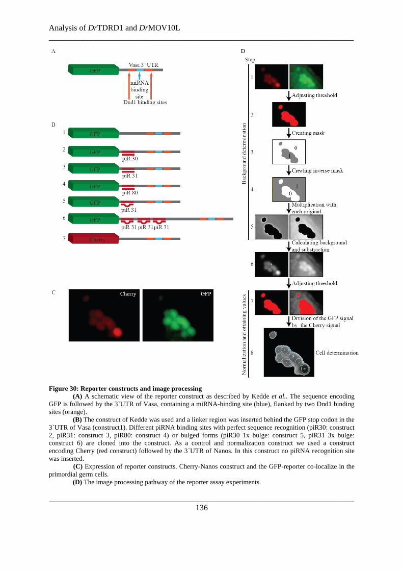
Analysis of DrTDRD1 and DrMOV10L
___________________________________________________________________________
___________________________________________________________________________
136
Figure 30: Reporter constructs and image processing
(A) A schematic view of the reporter construct as described by Kedde et al.. The sequence encoding
GFP is followed by the 3`UTR of Vasa, containing a miRNA-binding site (blue), flanked by two Dnd1 binding
sites (orange).
(B) The construct of Kedde was used and a linker region was inserted behind the GFP stop codon in the
3`UTR of Vasa (construct1). Different piRNA binding sites with perfect sequence recognition (piR30: construct
2, piR31: construct 3, piR80: construct 4) or bulged forms (piR30 1x bulge: construct 5, piR31 3x bulge:
construct 6) are cloned into the construct. As a control and normalization construct we used a construct
encoding Cherry (red construct) followed by the 3`UTR of Nanos. In this construct no piRNA recognition site
was inserted.
(C) Expression of reporter constructs. Cherry-Nanos construct and the GFP-reporter co-localize in the
primordial germ cells.
(D) The image processing pathway of the reporter assay experiments.
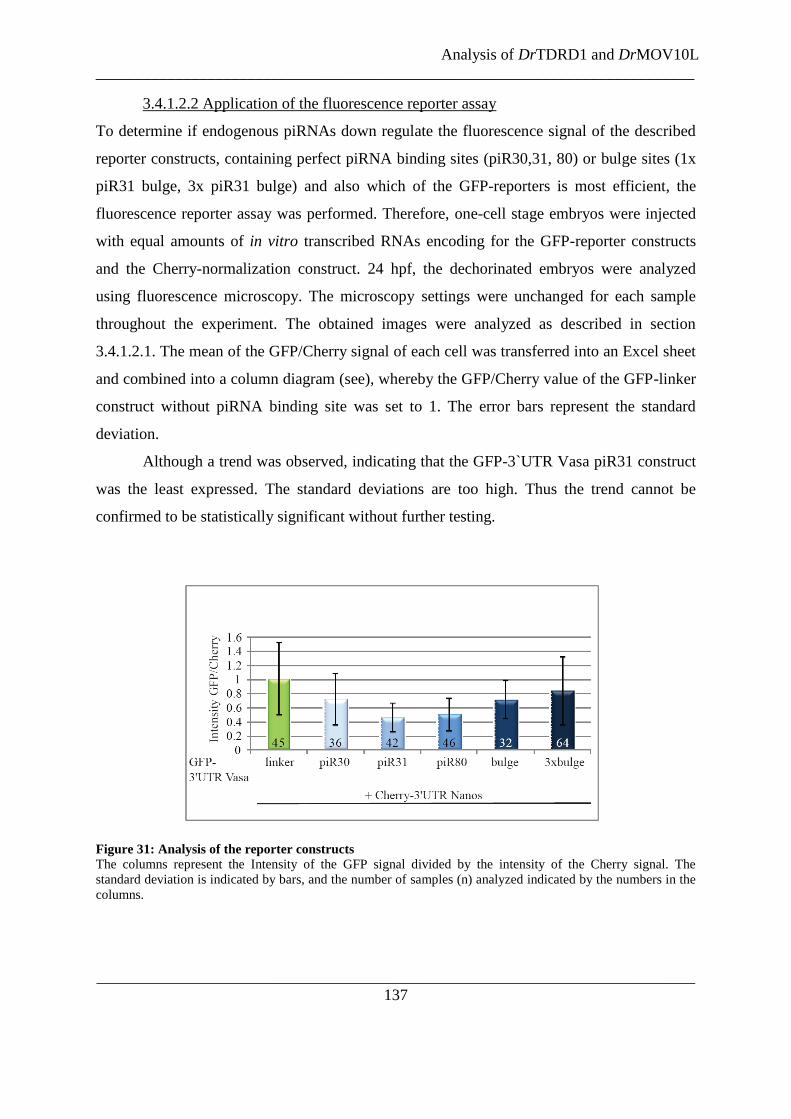
Analysis of DrTDRD1 and DrMOV10L
___________________________________________________________________________
___________________________________________________________________________
137
3.4.1.2.2 Application of the fluorescence reporter assay
To determine if endogenous piRNAs down regulate the fluorescence signal of the described
reporter constructs, containing perfect piRNA binding sites (piR30,31, 80) or bulge sites (1x
piR31 bulge, 3x piR31 bulge) and also which of the GFP-reporters is most efficient, the
fluorescence reporter assay was performed. Therefore, one-cell stage embryos were injected
with equal amounts of in vitro transcribed RNAs encoding for the GFP-reporter constructs
and the Cherry-normalization construct. 24 hpf, the dechorinated embryos were analyzed
using fluorescence microscopy. The microscopy settings were unchanged for each sample
throughout the experiment. The obtained images were analyzed as described in section
3.4.1.2.1. The mean of the GFP/Cherry signal of each cell was transferred into an Excel sheet
and combined into a column diagram (see), whereby the GFP/Cherry value of the GFP-linker
construct without piRNA binding site was set to 1. The error bars represent the standard
deviation.
Although a trend was observed, indicating that the GFP-3`UTR Vasa piR31 construct
was the least expressed. The standard deviations are too high. Thus the trend cannot be
confirmed to be statistically significant without further testing.
Figure 31: Analysis of the reporter constructs
The columns represent the Intensity of the GFP signal divided by the intensity of the Cherry signal. The
standard deviation is indicated by bars, and the number of samples (n) analyzed indicated by the numbers in the
columns.

Analysis of DrTDRD1 and DrMOV10L
___________________________________________________________________________
___________________________________________________________________________
138
3.4.1.3 Discussion
In this study D. rerio embryos were used as a system to develop a screening method based on
fluorescence reporter for a fast functional insight into the piRNA pathway. Therefore,
constructs were created encoding a fluorescence protein followed by the 3`UTR of Vasa or
Nanos, containing piRNA binding sites and the RNAs of these constructs were injected into
one-cell stage embryos. In all cases fluorescence in primordial germ cells was observed,
indicating that the reporter constructs with and without piRNA binding site are tissue-specific
expressed, as expected. It is known that the 3`UTR of Vasa or Nanos guide the expression of
the proteins to the germ cells (Kedde et al. 2007). Adding the piRNA binding sites into the
constructs did not disrupt this feature of the 3`UTR.
The intensity of the fluorescence proteins from constructs with piRNA binding sites,
or empty cloning site was measured and the values normalized against the intensity of co-
injected constructs containing another fluorescence protein, without the piRNA binding sites.
Therefore, the noise in the image was determined from the whole image and subtracted. The
normalization was performed by dividing the fluorescent intensity of the construct of interest
(GFP) by the signal of co-injected normalization construct without piRNA binding site
(Cherry). By this method the biased definition of the background was minimized and the
normalized values are comparable, independent of the actual amount of injected RNAs. In
default of a program defining the cells in the image, they had to be distinct by eye. Although,
in most cases the definition what is cell and what is background is rather easy, it is admitted
that an automatic recognition would reduce biased observations by the human eye.
In the design of this assay it was assumed that the RNA translation into a fluorescent
protein of constructs containing piRNA binding sites would be down regulated by the
endogenous piRNAs in the primordial germ cells of the zebrafish embryo. Unfortunately,
constructs containing piRNA binding sites (with perfect complementarity or one or 3 bulge
sites) did not down regulate the fluorescence signal with statistical significance and the
calculated standard deviation is too high to even consider the values of the performed piRNA
reporter assay. The variability of the values is possibly caused by the fluorescence
background due to auto-fluorescence of the cells and the leaking expression of the GFP
construct (outside of primordial germ cells). Second, the depth of field of the confocal plane

Analysis of DrTDRD1 and DrMOV10L
___________________________________________________________________________
___________________________________________________________________________
139
using multi-color TIRF microscope is small giving well defined images. Unfortunately, the
fluorescence signals measured are from the entire sample thickness, which gives rise to
variations in the signals between samples. A possible solution could be the use of confocal
microscopy, although this technique is more time consuming and inhibits fast high through-
put measurements.
However, a preliminary impression of the observed fluorescence intensity changes
due to piRNA binding indicated that binding sites for the recognition by the zebrafish
piRNAs 31 and 80 represses the expression of the fluorescence protein more than piR30 or
bulge sites. The results using the assay show a high variability and require confocal
microscopy to improve the statistical significance for identification of piRNA pathway
components.
3.4.1.4 Method
The reporter constructs pSP64T-mGFP-Vasa-3`UTR and pSP64T-dsRedEX-nos1-3`UTR
(Kedde et al. 2007) were kindly provided by the laboratory of Erez Raz. The pSP64T-mGFP-
Vasa-3`UTR was equipped with perfect complementary binding sites for piR-30, piR-31, or
piR-80. Additionally, constructs with one bulge binding site or three bulge binding sites for
piR-31 were created. The constructs were used to synthesize capped sense RNA using the
mMessageMachine kit from Ambion. 210pg/embryo RNA were injected into one-cell stage
D. rerio embryos.
As a normalization factor the same amount of RNA (210pg/embryo) of a Cherry-
Nanos construct was always mixed with the RNAs reporter constructs in the master mixture
to be co-injected. The primordial germ cells were imaged with an Olympus Biosystems
Cell^R multi-color TIRF microscope with a 40x water objective. Images were analyzed using
ImageJ (EMBL).
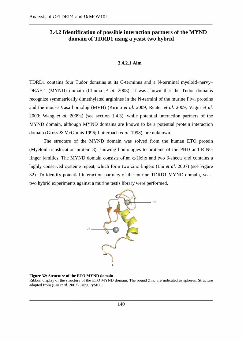
Analysis of DrTDRD1 and DrMOV10L
___________________________________________________________________________
___________________________________________________________________________
140
3.4.2 Identification of possible interaction partners of the MYND
domain of TDRD1 using a yeast two hybrid
3.4.2.1 Aim
TDRD1 contains four Tudor domains at its C-terminus and a N-terminal myeloid–nervy–
DEAF-1 (MYND) domain (Chuma et al. 2003). It was shown that the Tudor domains
recognize symmetrically dimethylated arginines in the N-termini of the murine Piwi proteins
and the mouse Vasa homolog (MVH) (Kirino et al. 2009; Reuter et al. 2009; Vagin et al.
2009; Wang et al. 2009a) (see section 1.4.3), while potential interaction partners of the
MYND domain, although MYND domains are known to be a potential protein interaction
domain (Gross & McGinnis 1996; Lutterbach et al. 1998), are unknown.
The structure of the MYND domain was solved from the human ETO protein
(Myeloid translocation protein 8), showing homologies to proteins of the PHD and RING
finger families. The MYND domain consists of an α-Helix and two β-sheets and contains a
highly conserved cysteine repeat, which form two zinc fingers (Liu et al. 2007) (see Figure
32). To identify potential interaction partners of the murine TDRD1 MYND domain, yeast
two hybrid experiments against a murine testis library were performed.
Figure 32: Structure of the ETO MYND domain
Ribbon display of the structure of the ETO MYND domain. The bound Zinc are indicated as spheres. Structure
adapted from (Liu et al. 2007) using PyMOL
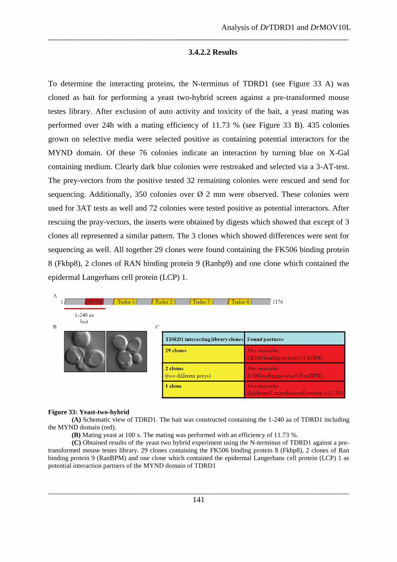
Analysis of DrTDRD1 and DrMOV10L
___________________________________________________________________________
___________________________________________________________________________
141
3.4.2.2 Results
To determine the interacting proteins, the N-terminus of TDRD1 (see Figure 33 A) was
cloned as bait for performing a yeast two-hybrid screen against a pre-transformed mouse
testes library. After exclusion of auto activity and toxicity of the bait, a yeast mating was
performed over 24h with a mating efficiency of 11.73 % (see Figure 33 B). 435 colonies
grown on selective media were selected positive as containing potential interactors for the
MYND domain. Of these 76 colonies indicate an interaction by turning blue on X-Gal
containing medium. Clearly dark blue colonies were restreaked and selected via a 3-AT-test.
The prey-vectors from the positive tested 32 remaining colonies were rescued and send for
sequencing. Additionally, 350 colonies over Ø 2 mm were observed. These colonies were
used for 3AT tests as well and 72 colonies were tested positive as potential interactors. After
rescuing the pray-vectors, the inserts were obtained by digests which showed that except of 3
clones all represented a similar pattern. The 3 clones which showed differences were sent for
sequencing as well. All together 29 clones were found containing the FK506 binding protein
8 (Fkbp8), 2 clones of RAN binding protein 9 (Ranbp9) and one clone which contained the
epidermal Langerhans cell protein (LCP) 1.
Figure 33: Yeast-two-hybrid
(A) Schematic view of TDRD1. The bait was constructed containing the 1-240 aa of TDRD1 including
the MYND domain (red).
(B) Mating yeast at 100 x. The mating was performed with an efficiency of 11.73 %.
(C) Obtained results of the yeast two hybrid experiment using the N-terminus of TDRD1 against a pre-
transformed mouse testes library. 29 clones containing the FK506 binding protein 8 (Fkbp8), 2 clones of Ran
binding protein 9 (RanBPM) and one clone which contained the epidermal Langerhans cell protein (LCP) 1 as
potential interaction partners of the MYND domain of TDRD1

Analysis of DrTDRD1 and DrMOV10L
___________________________________________________________________________
___________________________________________________________________________
142
3.4.2.3 Discussion
Potential interaction partners of the MYND domain were analyzed in a two hybrid screen
using the TDRD1 N-term as bait and a pre-transformed testis library. The results indicated
that the MYND domain potentially interacts with FK506 binding protein 8 (Fkbp8), RanBPM
(RAN binding protein 9, Ranbp9) and/or the epidermal Langerhans cell protein (LCP) 1.
A direct functional relevant interaction of the TDRD1 MYND domain with LCP1 is
unlikely given that LCP1 is expressed in the nucleus of mainly epidermal Langerhans cells,
while TDRD1 is a cytoplasmic protein in germ cells. Furthermore, the piRNA pathway is
restricted to germline as well and a functional connection of LCP1 is unlikely.
RanBPM was initially described to bind to the human RAN, a small GTP binding
protein belonging to the RAS superfamily that is essential for the translocation of RNA and
proteins through the nuclear pore complex and microtubule assembly (Nakamura et al. 1998).
RanBPM localizes in the cytoplasm as well as in the nucleus surrounding the centromers,
where it is required for correct nucleation of microtubules (Nakamura et al. 1998; Nishitani et
al. 2001). In mouse RanBPM is ubiquitously expressed with a high expression in testes from
late spermatocytes to round spermatids with a peak at the pachytene stage (Shibata et al.
2004), overlaying with the expression profile of TDRD1. RanBPM is mainly localized in the
cytoplasm and a component of the chromatoid body in round spermatids (Shibata et al. 2004)
like TDRD1. In agreement to the similar expression patterns and the localizations, it was
found that RanBPM interacts with the N-terminal of MVH (Fujiwara et al. 1994; Shibata et
al. 2004). The role of RanBPM/MVH interaction during spermatogenesis could be the
transfer of MVH proteins to an anchoring complex during the chromatoid body arrangement,
mediating microtubule assembly. Another possibility is that the complex associates with
RanGTP and could be involved in catching and transferring RNAs from the nucleus into the
chromatoid body (Shibata et al. 2004). Another indication of a possible involvement of
RanBPM in the piRNA pathway is that it was found to interact with MAEL, another
component of the chromatoid body, which interacts with mi- and piRNA pathway
components (Costa et al. 2006). Further, it interacts with the chromatoid body member
GASZ. Gasz knockout mice are male sterile due to a developmental block after the zygotene

Analysis of DrTDRD1 and DrMOV10L
___________________________________________________________________________
___________________________________________________________________________
143
stage. These mutants show retrotransposon activation due to a decrease of the piRNA amount
(Ma et al. 2009).
Taken the similarities of the expression, localization and known interaction partners of
RanBPM to TDRD1 into account, it is possible that the MYND domain interacts directly
with RanBPM, maybe to guide the localization of TDRD1 into the chromatoid body. It would
be interesting to confirm the association of RanBPM and TDRD1 endogenously and to
analyze the function of this complex in the future.
The search for interaction partners of the MYND domain of TDRD1 revealed FKBP8
as a potential candidate. FKBP8 is involved in the regulation of apoptosis in neuron cells but
also in other cell types (Bulgakov et al. 2004; Okamoto et al. 2006; Rosner et al. 2003; Wang
et al. 2006). It is a member of the FK506-binding, immunophilin protein family. It contains a
peptidyl-prolyl cis-trans isomerase domain that may be involved in protein folding or as a
scaffold to facilitate protein-protein interactions. Additionally, it contains a C-terminal
tripartite tetratricopeptide repeat protein-protein interaction (TPR) domain,
calcium/calmodulin-binding motif and a transmembrane motif (TM) (Pedersen et al. 1999;
Shirane & Nakayama 2003). FKPB8 is mainly expressed in prefrontal cortex and early
erythroids. It is also expressed in testis but not at a significantly level. Given the expression
pattern of Fkbp8, it is probably not a direct interaction partner of the TDRD1 MYND domain
and not of high interest concerning the piRNA pathway studies. On the other hand, members
of the Fkbp class (FKBP6, 5 and 3) were recently uncovered as components of the MIWI,
MILI and MIWI2 complexes (Vagin et al. 2009). FKBP3 and FKBP5 are cis-trans prolyl
isomerases that bind to the immunosuppressants FK506 and rapamycin (Jin et al. 1992; Nair
et al. 1997). FKBP3 is mainly expressed in brain and immune system cell. It has a higher
affinity for rapamycin than for FK506 and is maybe an important target molecule for
immunosuppression by rapamycin (Jin et al. 1992). Interestingly, FKBP3 interacts with the
two Histone deacetylases HDAC1, HDAC2, the HDAC-binding transcriptional regulator
YY1(Yang et al. 2001) and the E3 ubiquitin Ligase, mouse double minute 2 (MDM2)
(Ochocka et al. 2009), indicating a role in chromatin remodeling and gene expression
regulation. Given FKBP3s weak expression in the germline, it is unlikely but not excluded to
be an interaction partner of the TDRD1 MYND domain and component of the piRNA
pathway. FKPB5 is involved in post-traumatic stress disorder, depression and anxiety
(Gillespie et al. 2009). It is expressed in a wide range of tissues but mainly in the skeletal
Analysis of DrTDRD1 and DrMOV10L
___________________________________________________________________________
___________________________________________________________________________
144
muscles and adipocytes (Nair et al. 1997). FKPB5 is as well not a primary candidate for an
association with TDRD1.
An interesting interaction partner for the MYND domain of TDRD1 would be prolyl
isomerase and TPR domain-containing protein FKBP6 (Meng et al. 1998; Pedersen et al.
1999; Shirane & Nakayama 2003). The protein is expressed in male and females germlines,
but the loss of FKBP6 causes an arrest in the spermatogenesis during the meiotic prophase in
mice and rats, leading to male sterility. The female fertility is not affected (Crackower et al.
2003; Noguchi et al. 1993). In spermatocytes FKBP6 localizes in the cytoplasm and in the
nucleus. In the pachytene stage the protein was found to be in the synaptonemal complex. In
fkbp6 knockouts the autosomes and sex-chromosomes mis-align or show non-homologous
paring in the pachytene stage (Crackower et al. 2003; Noguchi et al. 2008). It would be
interesting to investigate, if the piRNA pathway is influencing the alignment of chromosomes
by regulating and guiding FKBP6.
In conclusion, the identified interaction partners of the TDRD1 MYND domain may
be directly involved or are members of protein families, which could be associated to the
piRNA pathway (except LCP1). Based on the yeast two hybrid results and the literature
search, it would be interesting to confirm direct interactions and to perform functional studies
of the potential complexes to gain deeper insights into the piRNA mechanism.
3.4.2.4 Method
The MYND domain of mTDRD1 (1-240 aa) was cloned as a bait into pGBKT7. After auto-
activation and toxicity tests, the bait was used with a MatchmakerTM
mTestis in Yeast library
(638872) following the manufacturer‟s protocol. Used yeast strains were AH109 (bait) and
Y187 (library). Preliminary positive tested colonies were restreaked and used for a 3-AT-test
according to the manufacturer‟s protocol, to select strong interactions. Positive clones were
digested and unique digests sizes were sent for sequencing.

Chapter 4: General conclusions and future perspectives
General conclusions and future perspectives
___________________________________________________________________________
___________________________________________________________________________
146
4.1 General conclusions and future perspectives
Metazoan germlines express germline-specific non-coding RNAs, the so called piRNAs.
These RNAs associate with Piwi proteins and ensure maintenance of germline stability by
silencing transposable elements (Aravin et al. 2003; Brennecke et al. 2007; Gunawardane et
al. 2007; Houwing et al. 2007; Saito et al. 2006; Vagin et al. 2006). piRNA biogenesis and
mechanisms of their action are poorly understood. piRNAs originate from a single-stranded
precursor transcribed from genomic piRNA clusters (Aravin et al. 2006; Lau et al. 2006;
Watanabe et al. 2006) and the piRNA biogenesis was found to be Dicer-independent in flies
and fish (Houwing et al. 2007; Vagin et al. 2006). Genetic screens in Drosophila and
biochemical purifications of macromolecular complexes in mice are just beginning to
uncover factors involved in the pathway. This thesis focused on the analysis of three such
factors: the methyltransferase mHEN1 in the mouse system and the Tudor domain-containing
protein DrTDRD1 and the putative helicase DrMOV10L in the Danio rerio model system.
All small RNAs in plants are methylated at the 3` terminal nucleotide. Some miRNAs
and siRNAs in Drosophila carry similar modifications. piRNAs in all animals analyzed to
date are modified by a 2`-O-methyl mark. This modification protects the small RNA 3`
termini from uridylation, which usually acts as a signal for destabilization (Ameres et al.
2010; Chen 2005; Ebhardt et al. 2005; Horwich et al. 2007; Kamminga et al. 2010; Li et al.
2005; Saito et al. 2007a; Yang et al. 2006b). The methylation mark on the small RNAs is
catalyzed by HEN1 and its homologs in different species (Akbergenov et al. 2006; Horwich
et al. 2007; Kamminga et al. 2010; Katiyar-Agarwal et al. 2007; Kirino & Mourelatos 2007c;
Li et al. 2005; Ohara et al. 2007a; Saito et al. 2007a; Yang et al. 2006a; Yang et al. 2006b;
Yu et al. 2005; Zhou et al. 2010). Although it was shown that the DmHEN1 is able to interact
with the Drosophila Piwi proteins (Saito et al. 2007a), it was not clear how HEN1 is recruited
to the piRNA pathway. Despite the fact that the mechanism was not understood, in mice only
the recombinant mHEN1 was shown to methylate RNAs in vitro (Kirino & Mourelatos
2007a, b). Therefore, in this thesis the mechanism by which the enzyme is recruited to
participate in the mouse piRNA pathway was investigated. The results demonstrate that the
endogenous mHEN1 in mouse testes extracts is an active methyltransferase capable of
methylating small RNAs in the size range of piRNAs. In addition, it shows that mHEN1

General conclusions and future perspectives
___________________________________________________________________________
___________________________________________________________________________
147
interacts with one of the murine Piwi proteins, MILI. These findings link the enzyme directly
to the piRNA pathway.
Mapping studies indicated that the N-terminal region of MILI interacts with the C-
terminal region of mHEN1. This region on MILI was mapped close to the PAZ domain,
which binds the methylated 3` end of piRNAs. It was shown that the N-terminus of MILI is
required, but not sufficient to mediate interaction with mHEN1. Therefore, it is believed that
a composite interaction site formed from the N-terminus and PAZ facilitates this interaction.
This suggests that the 3` RNA binding site of MILI, the PAZ domain, and the catalytic
domain of mHEN1 are in close proximity to facilitate the methylation of the piRNAs and its
transfer to the PAZ domain. Furthermore, it was shown here that this interaction occurs in an
RNA-independent manner and is not influenced by the methylation status of the arginines of
MILI, which are recognized by Tudor domain-containing proteins. This suggests that MILI
need not to be loaded to interact with mHEN1 and that this interaction can occur early in
piRNA biogenesis, before MILI is post-translationally modified. Further investigations
towards the structure of the MILI/mHEN1 complex together with bound RNAs could lead to
a definition of the binding surface and could address the specific determinant of mHEN1 for
Piwi proteins. Dynamic studies using NMR techniques could reveal how the recognition of
the methylated 3` end by the PAZ domain alters this interaction surface, as mHEN1 is never
found in complex with MILI loaded with piRNAs. This could be a mechanism for separation
of the enzyme from the product after the methylation reaction.
In mice, spermatogenesis proceeds through different stages of mitotic and meiotic
divisions. A strong MILI expression is observed in pachytene stage spermatocytes during
meiosis (Aravin et al. 2008; Kuramochi-Miyagawa et al. 2001). The round spermatids are
characterized by a dense peri-nuclear structure called the chromatoid body that was shown to
contain MILI (Aravin et al. 2008; Wang et al. 2009a). mHEN1 was localized to a separate
perinuclear granule (HEN-body), suggesting either that mHEN1 leaves the MILI-containing
piRNA complex after performing the methylation of the RNA, or that the piRNA formation
(Piwi slicer activity) and methylation by mHEN1 are spatially separated in the chromatoid
and HEN bodies, respectively. Recombinant mHEN1 is shown to methylate small RNAs both
in the size range of miRNAs and piRNAs (Kirino & Mourelatos 2007b). So it is believed that
the distinct localization of mHEN1 might be a mechanism to spatially restrain a potentially
promiscuous enzyme that might methylate miRNAs and other mRNAs present in the CBs.

General conclusions and future perspectives
___________________________________________________________________________
___________________________________________________________________________
148
Future research should reveal the importance of this unique localization of mHEN1 and the
identity of other mHEN1-interacting factors that dynamically links it to the piRISC. Future
research should reveal the exact site of the small RNA methylation; either at the site of
piRNA biogenesis (which is currently unclear) or in the HEN bodies where Piwi-bound
RNAs transiently reside.
In contrast to mammalian Ago proteins which bind miRNAs and siRNAs, the fact that
piRNAs carry a modified 3` end requires the Piwi proteins to accommodate the methyl group
in their PAZ domain. To further understand this recognition process, biophysical studies were
performed in the laboratory which demonstrated that the MIWI PAZ domain affinity for
ssRNAs which is thirty times higher than for double stranded RNAs, consistent with the
absence of dsRNA intermediates in the piRNA pathway. However, MIWI PAZ showed only
a 2-fold higher affinity for a methylated 3` end compared to an unmethylated RNA. It is
possible that further interactions in the context of the full-length protein (that can
accommodate longer small RNAs) might facilitate a stronger interaction with the methylated
piRNAs. To obtain molecular insight of this interaction, in collaboration with the group Dr.
Teresa Carlomagno (EMBL Heidelberg), the NMR structure of the MIWI PAZ domain in
complex with a piRNA mimic was solved (Simon et al. 2011). The solution structure reveals
that the methyl-group is inserted deeply into a pre-formed hydrophobic pocket of conserved
residues. The binding pocket contains a hydrophobic cleft consisting of F333, A381 and
M382. In the complex, the long hydrophobic side-chain of the M382 at the back of the cavity
makes contact with the methyl group. Mutation of this residue results in proportional
reduction in the affinity for methylated 3‟ ends. However, the methionine at this position is
not conserved across phyla, indicating that this contact is not necessary for the methylation
mark recognition. What is interesting in this structure is the fact that although the MIWI PAZ
structure resembles very much those of Ago proteins, subtle differences in the amino acid
composition of the pocket allows the recognition of the extra methyl group on the 3` end of
piRNAs. These differences were not evident in simple sequence alignments. Similar results
were obtained for the human HILI protein as well (Tian et al. 2010). It would be interesting
to identify the binding capacity and the structures of PAZ domains without the conserved
methionine and measure the affinity to single- and double-stranded, methylated and non-
methylated RNAs.

General conclusions and future perspectives
___________________________________________________________________________
___________________________________________________________________________
149
Coming back to the task of describing piRNA pathway components, the restrictions
using mouse as a model system had to be overcome by establishing a new, more flexible
model system using Danio rerio. Two piRNA pathway factors in mice are the putative
helicase MOV10L, which is thought to be involved in the stabilization or loading of piRNAs
into Piwi proteins (Frost et al. 2010; Lovasco et al. 2010; Wang et al. 2001b; Zheng et al.
2010) and the Tudor-domain-containing protein TDRD1, which is an essential component of
the piRNA silencing complex (Kojima et al. 2009; Reuter et al. 2009; Vagin et al. 2009;
Wang et al. 2009a). Here, this study was aimed to identify and functionally characterize the
mechanism of the two proteins in the zebrafish system. Once the proteins were identified in
the fish, the expression pattern of these proteins revealed that the mRNAs of Drtdrd1 and
Drmov10l are expressed from the egg stage throughout development of the zebrafish similar
to zebrafish Piwi proteins, suggesting the mRNAs for all these proteins are maternally
contributed. Using an affinity purified antibody against DrTDRD1, it was shown that the
protein is expressed in testes. This result is in agreement with the expression pattern of the
medaka TDRD1 (Aoki et al. 2008). The expression and localization of DrTDRD1 in this
specific tissue is similar to other germ cell-specific proteins (Kedde et al. 2007) and
dependent on its 3`UTR. Future research could localize DrTDRD1 as well as DrMOV10L in
the PGCs with high resolution to determine the sub-cellular targeting.
In contrast to the mouse TDRD1 protein, no bound piRNAs were detected in
immunoprecipitations of DrTDRD1. Either, the amount of piRNAs was insufficient for
detection, or DrTDRD1 binds to unloaded Piwi proteins, or it does not bind Piwi proteins at
all. The mouse TDRD1 is shown to detect symmetrically dimethylated arginines in the N-
termini of at least MILI and MIWI (it is not clear for MIWI2) (Kojima et al. 2009; Reuter et
al. 2009; Vagin et al. 2009; Wang et al. 2009a). Unfortunately, an interaction of the zebrafish
Piwis and DrTDRD1 was not observed in endogenous tissue extracts or in mammalian cell
culture experiments. Immunoprecipitations using better antibodies will answer the question
of the interaction in the future.
To examine the function of DrMOV10L, knockdown experiments were performed
using morpholino against the translational start site of DrMOV10L, which were injected into
the one-cell stage embryo. Like in mouse MOV10L (Zheng et al. 2010) and zebrafish zili
knockouts (Houwing et al. 2008), the activity of transposable elements increased as a
consequence of morpholino injection. The similarity to the other phenotypes indicates that

General conclusions and future perspectives
___________________________________________________________________________
___________________________________________________________________________
150
DrMOV10L is directly involved in the piRNA pathway and required for silencing of
retrotransposable elements. The knockdown method via morpholinos could be a powerful
tool to screen for piRNA biogenesis components which influence the activity of transposable
elements. Future experiments involving complementation of the knockdown phenotype with
either full-length or mutagenised (deletion, point-mutant) versions of the protein should
reveal the functional role of the different domains and its putative helicase activity.
Overall, my thesis work has identified a biochemical basis for recruitment of the
piRNA methyltransferase to the mouse piRNA pathway. This work is now being expanded
into a structural analysis of the interaction that was described here. Together, these studies
will be of great interest to the field. Regarding the development of the zebrafish embryos as a
model system to study piRNA factors in the lab, the tools necessary for addressing these
questions were set up and it was demonstrated their utility by studying two factors.
Morpholino knock-downs followed by detection of endogenous transposons could be used as
a rapid functional assay to validate candidate vertebrate piRNA factors identified in
biochemical purifications elsewhere.

Conclusions générales et perspectives
___________________________________________________________________________
___________________________________________________________________________
151
4.2 Conclusions générales et perspectives
Les lignées germinales des Métazoaires expriment des ARN non codant spécifiques aux
lignées germinales appelés piARN. Ces ARN s‟associent à des protéines Piwi et assurent le
maintien de la stabilité des cellules germinales en inhibant les éléments transposables (Aravin
et al. 2003; Brennecke et al. 2007; Gunawardane et al. 2007; Houwing et al. 2007; Saito et
al. 2006; Vagin et al. 2006). La biogenèse des piARN et leur mécanisme d‟action sont mal
connus. Les piARN proviennent d'un précurseur ARN simple brin transcrit à partir de
clusters génomiques de piARN (Aravin et al. 2006; Lau et al. 2006; Watanabe et al. 2006).
Leur biogenèse est indépendante de Dicer chez la drosophile et les poissons (Houwing et al.
2007; Vagin et al. 2006). Des criblages génétiques chez la drosophile et des purifications
biochimiques des complexes macromoléculaires chez la souris commencent à peine à
identifier les facteurs impliqués dans cette voie. Cette thèse s‟est concentrée sur l'analyse de
trois facteurs de ce type: la méthyltransférase mHEN1 dans le système de la souris et les deux
protéines comportant des domaines Tudor, DrTDRD1 et l‟hélicase putative DrMOV10L dans
le système Danio rerio.
Tous les petits ARN des plantes sont méthylés au niveau du nucléotide 3‟ terminal.
Certains miRNA et siRNA chez la drosophile portent des modifications similaires. Les
piARN de tous les animaux analysés à ce jour sont modifiées par une marque 2‟-O-méthyl.
Cette modification protège l‟extrémité 3` des petit ARN de l‟uridylation qui agit
habituellement comme un signal de déstabilisation (Ameres et al. 2010; Chen 2005; Ebhardt
et al. 2005; Horwich et al. 2007; Kamminga et al. 2010; Li et al. 2005; Saito et al. 2007a;
Yang et al. 2006b). La marque de méthylation sur les petits ARN est catalysée par HEN1 et
ses homologues dans différentes espèces (Akbergenov et al. 2006; Horwich et al. 2007;
Kamminga et al. 2010; Katiyar-Agarwal et al. 2007; Kirino & Mourelatos 2007c; Li et al.
2005; Ohara et al. 2007a; Saito et al. 2007a; Yang et al. 2006a; Yang et al. 2006b; Yu et al.
2005; Zhou et al. 2010). Bien qu'il ait été démontré que DmHEN1 est capable d'interagir avec
les protéines Piwi de drosophile (Saito et al. 2007a), le recrutement de HEN1 dans la voie
des piARN n‟était pas clair. En dépit du fait que le mécanisme d‟action n'était pas compris,
chez la souris uniquement la protéine recombinante mHEN1 était connue pour méthyler les
ARN in vitro (Kirino & Mourelatos 2007a, b). Par conséquent, dans cette thèse le mécanisme

Conclusions générales et perspectives
___________________________________________________________________________
___________________________________________________________________________
152
par lequel l'enzyme est recrutée pour participer à la voie des piARN a été étudiée chez la
souris. Les résultats démontrent que la protéine mHEN1 endogène dans les extraits de
testicules de souris est une méthyltransférase active, capable de méthylation des petits ARN
dans la gamme de taille des piARN. En outre, ceci montre que mHEN1 interagit avec l'une
des protéines Piwi murin, MILI. Ces résultats lient directement l'enzyme mHEN1 à la voie
des piARN.
Des études de cartographie ont indiqué que la région N-terminale de MILI interagit
avec la région C-terminale de mHEN1. Cette région de MILI a été cartographiée à proximité
du domaine PAZ qui lie le groupement méthyle des extrémités 3` des piARN. Il a été
démontré que la partie N-terminale de MILI est requise, mais ne suffit pas à relayer
l‟interaction avec mHEN1. Par conséquent, il est admis qu'un site d'interaction composite
formée à partir du domaine N-terminal et de PAZ facilite cette interaction. Ceci suggère que
le site 3` de liaison à l'ARN de MILI, le domaine PAZ, et le domaine catalytique de mHEN1
sont localisés à proximité les uns des autres pour faciliter la méthylation des piARN et leur
transfert au domaine PAZ. En outre, il a été montré ici que cette interaction ne repose pas sur
la présence d‟ARN et n'est pas influencée par la méthylation des résidus arginines de MILI,
qui sont reconnus par les protéines contenant des domaines Tudor. Ceci suggère que MILI ne
nécessite pas d‟être chargée pour interagir avec mHEN1 et que cette interaction peut se
produire au début de la biogenèse des piARN, avant que MILI ne soit modifiée post-
traductionnellement. D'autres investigations vers la structure du complexe MILI/mHEN1
associé à l‟ARN pourraient conduire à une définition de la surface d‟interaction et pourraient
identifier les déterminants spécifiques de mHEN1 pour les protéines Piwi. Des études
dynamiques reposant sur des techniques de RMN pourraient révéler la façon dont la
reconnaissance, par le domaine PAZ, de l‟extrémité 3` méthylée modifie cette surface
d'interaction, étant donné que mHEN1 ne se trouve jamais dans un complexe avec MILI
chargé en piARN. Ce pourrait être un mécanisme permettant l‟élimination du produit par
l'enzyme après la réaction de méthylation.
Chez la souris, la spermatogenèse se fait par différentes étapes de divisions mitotiques
et méiotiques. Une forte expression de MILI est observée dans les spermatocytes au stade
pachytène de la méiose (Aravin et al. 2008; Kuramochi-Miyagawa et al. 2001). Les
spermatides rondes sont caractérisées par une structure dense péri-nucléaire appelée le corps
chromatoïde qui est connu pour contenir MILI (Aravin et al. 2008; Wang et al. 2009a).

Conclusions générales et perspectives
___________________________________________________________________________
___________________________________________________________________________
153
mHEN1 a été localisé dans un granule périnucléaire distinct (corps HEN), ce qui suggère soit
que mHEN1 quitte le complexe MILI chargé en piARN après avoir effectué la méthylation
de l'ARN, ou que la formation des piARN (activité « Slicer » de Piwi) et la méthylation par
mHEN1 sont séparés spatialement dans le corps chromatoïde et les corps HEN
respectivement. La protéine recombinante mHEN1 est connue pour méthyler de petits ARN
dans la gamme de taille des miARN et des piARN (Kirino & Mourelatos 2007b). Donc, on
peut imaginer que la localisation distincte de mHEN1 pourrait être un mécanisme permettant
d‟éviter la promiscuité d'une enzyme qui pourrait potentiellement méthyler des miARN et
autres ARNm présent dans les corps chromatoïdes. Les futures recherches devraient révéler
l'importance de cette localisation unique de mHEN1 ainsi que l'identité des autres facteurs
interagissant avec mHEN1, les reliant de manière dynamique au piRISC. Les futures
recherches devraient révéler le site exact de la méthylation de petits ARN; soit sur le site de
la biogenèse des piARN (encore peu connu) ou dans les corps HEN où les ARN liés à Piwi
résident transitoirement.
Au contraire des protéines Ago de mammifères qui s‟associent aux miRNA et siRNA,
le fait que les piARN portent une modification en 3` nécessite des protéines Piwi qu‟elles
accommodent le groupement méthyle dans leur domaine PAZ. Pour mieux comprendre ce
processus de reconnaissance, des études biophysiques ont été réalisées dans le laboratoire qui
a démontré que l'affinité du domaine PAZ de MIWI pour les ARN simple brin qui est trente
fois supérieur à celle des ARN double brin, est en accord avec l'absence d'intermédiaires
d‟ARN double brin dans la voie des piARN. Toutefois, le domaine PAZ de MIWI n‟a montré
qu'une affinité deux fois plus élevée pour une extrémité 3`méthylée par rapport à un ARN
non méthylé. Il est possible que d'autres interactions dans le contexte de la protéine non
raccourcie (qui peut accueillir plus de petits ARN) pourraient faciliter une interaction plus
forte avec le piARN méthylé. Pour obtenir un aperçu de cette interaction moléculaire, en
collaboration avec le groupe du Dr Teresa Carlomagno (EMBL Heidelberg), la structure
RMN du domaine PAZ de MIWI en complexe avec un ARN mimant un piARN a été résolue
(Simon et al. 2011). La structure en solution révèle que le groupement méthyle est
profondément inséré dans une poche hydrophobe préformée comportant des résidus
conservés. La poche de liaison hydrophobe contient une fente composée de F333, A381 et
M382. Dans le complexe, la longue chaîne latérale hydrophobe de M382 à l'arrière de la
cavité, entre en contact avec le groupement méthyle. La mutation de ce résidu résulte en une

Conclusions générales et perspectives
___________________________________________________________________________
___________________________________________________________________________
154
réduction proportionnelle de l'affinité pour les extrémités 3` méthylées. Cependant, la
méthionine à cette position n'est pas conservée à travers les embranchements, ce qui indique
que ce contact n'est pas nécessaire pour la reconnaissance de la marque de méthylation. Ce
qui est intéressant dans cette structure réside dans le fait que, bien que la structure du
domaine PAZ de MIWI ressemble beaucoup à celles des protéines Ago, de subtiles
différences dans la composition en acides aminés de la poche permet la reconnaissance du
groupement méthyle supplémentaire présent à l‟extrémité 3` des piARN. Ces différences
n'étaient pas évidentes dans les simples alignements de séquences. Des résultats similaires ont
aussi été obtenus pour la protéine humaine HILI (Tian et al. 2010). Il serait intéressant
d'identifier la capacité de liaison et les structures des domaines PAZ sans le résidu
méthionine conservé et de mesurer l'affinité pour les ARN simple et double brin, méthylés et
non méthylés.
Pour en revenir à la description des composants de la voie des piARN, les restrictions
dues à l‟utilisation de la souris comme modèle devaient être dépassées en établissant un
nouveau modèle plus flexible, ce qui a été fait en utilisant Danio rerio. Deux facteurs de la
voie des piARN chez la souris sont l‟hélicase putative MOV10L, que l‟on pense être
impliquée dans la stabilisation ou le chargement des piARN dans les protéines Piwi (Frost et
al. 2010; Lovasco et al. 2010; Wang et al. 2001b; Zheng et al. 2010) et la protéine Tudor-
domain-containing TDRD1, qui est une composante essentielle du complexe d‟inhibition
impliquant les piARN (Kojima et al. 2009; Reuter et al. 2009; Vagin et al. 2009; Wang et al.
2009a). Ici cette étude visait à identifier et à caractériser fonctionnellement le mécanisme
d‟action des deux protéines en utilisant le modèle de poisson zèbre. Une fois que les protéines
ont été identifiées dans le poisson, le profil d'expression de ces protéines a révélé que les
ARNm de Drtdrd1 et Drmov10l sont exprimés à partir du stade de l‟oeuf tout au long du
développement du poisson zèbre poisson de façon similaire aux protéines Piwi du poisson
zèbre, suggérant que les ARNm de toutes ces protéines sont héritées de la mère. En utilisant
un anticorps purifié par affinité contre DrTDRD1, il a été montré que la protéine est exprimée
dans les testicules. Ce résultat est en accord avec le profil d'expression de TDRD1 chez le
poisson médaka (Aoki et al. 2008). L'expression et la localisation de DrTDRD1 dans ce tissu
spécifique est similaire à d'autres protéines spécifiques des cellules germinales (Kedde et al.
2007) et dépend de sa partie 3` non traduite. Les futures recherches pourraient localiser

Conclusions générales et perspectives
___________________________________________________________________________
___________________________________________________________________________
155
DrTDRD1 ainsi que DrMOV10L dans les cellules germinales primordiales avec une haute
résolution pour déterminer le ciblage sub-cellulaire.
Contrairement aux protéines TDRD1de souris, aucun piARN lié n‟a été détecté au
cours d‟immunoprécipitations de DrTDRD1. Soit la quantité de piARN était insuffisante
pour la détection, soit DrTDRD1 se lie à des protéines Piwi non chargées, ou soit elle ne
s‟associe pas du tout aux protéines Piwi. Il a été montré que la protéine TDRD1 de souris
permet la détection des arginines symétriquement diméthylées dans le domaine N-terminal
d'au moins MILI et MIWI (ce n'est pas clair pour MIWI2) (Kojima et al. 2009; Reuter et al.
2009; Vagin et al. 2009; Wang et al. 2009a). Malheureusement, aucune interaction entre
protéines PIWI du poisson-zèbre et DrTDRD1 n‟a pas été observée dans les extraits
tissulaires endogènes ou dans des expériences reposant sur des cultures cellulaires de
mammifère. Des immunoprécipitations qui utiliseraient de meilleurs anticorps pourraient
répondre à la question de l'interaction dans le futur.
Pour examiner la fonction de DrMOV10L, des expériences de knockdown ont été
réalisées en utilisant des morpholinos dirigés contre le site d‟initiation de la traduction de
DrMOV10L et qui ont été injectés dans un embryon au stade d'une cellule. Comme chez la
protéine MOV10L de souris (Zheng et al. 2010) et le poisson zèbre knockout pour zili
(Houwing et al. 2008), l'activité des éléments transposables a augmenté à la suite de
l'injection de morpholinos. La similitude avec les autres phénotypes indique que DrMOV10L
est directement impliquée dans la voie des piARN et est nécessaire pour inhiber les
rétrotransposons. La méthode de knockdown via l‟utilisation de morpholinos pourrait être un
outil puissant pour identifier les facteurs impliqués dans la biogenèse des piARN qui
influencent l'activité des éléments transposables. Les futures expériences impliquant des
complémentations des phénotypes de knockdown avec soit les protéines intégrales ou mutées
(délétion, mutation ponctuel) devraient révéler le rôle fonctionnel des différents domaines et
leur activité d‟hélicase putative.
Dans l'ensemble, mon travail de thèse a identifié une base biochimique pour le
recrutement de la piARN-méthyltransférase dans la voie des piARN de souris. Ce travail est
maintenant étendu à une analyse structurale de l'interaction qui a été décrite ici. Ensemble,
ces études seront de grand intérêt pour le domaine. En ce qui concerne le développement des
embryons de poisson zèbre comme modèle pour étudier les facteurs piARN dans le
laboratoire, les outils nécessaires pour répondre à ces questions ont été mis en place et il a été

Conclusions générales et perspectives
___________________________________________________________________________
___________________________________________________________________________
156
démontré leur utilité en étudiant deux facteurs. Des expériences de knockdown par
morpholinos suivies par la détection des transposons endogènes pourraient être utilisées
comme un rapide test fonctionnel pour valider les facteurs candidats identifiés comme
associés aux piARN vertébrés par purifications biochimiques.

___________________________________________________________________________
157
Supplements
5.1 Eckhardt, S., Szostak, E., Yang, Z., Pillai, R. S. (2010). Artificial tethering of Argonaute proteins for
studying their role in translational repression of target mRNAs. Argonaute Proteins: Methods in Molecular
Biology; series by Human Press.
5.2 Bernd Simon, John P. Kirkpatrick, Stephanie Eckhardt, Michael Reuter, Elsa A. Rocha, Miguel A. Andrade-
Navarro, Peter Sehr, Ramesh S. Pillai, Teresa Carlomagno (2011). Specific Recognition of 3’-end 2’-O-
methylated piRNA by the PAZ domain of a piwi protein. Structure

Supplements
___________________________________________________________________________
___________________________________________________________________________
158
5.1 Artificial tethering of Argonaute proteins for studying their role in translational
repression of target mRNAs
Stephanie Eckhardt1,2
, Emilia Szostak1 , Zhaolin Yang
1 and Ramesh Pillai
1
1European Molecular Biology Laboratory, 6 Rue Jules Horowitz, BP 181, 38042 Grenoble, France.
2EMBL International PhD Programme.
Correspondence to:
Ramesh Pillai
Phone: 00-33-4-76 20 7446
Fax: 00-33-4-76 20 7199
E-mail: [email protected]
i. Abstract
Small RNAs like microRNAs (miRNAs) and small interfering RNAs (siRNAs) associate
with a member of the RNA-binding Argonaute family proteins. Together they participate in
transcriptional and post-transcriptional gene silencing mechanisms. The fate of the target
mRNA is determined, in part, by the degree of complementarity with the small RNA. To
examine the exact role of the Argonaute protein in the silencing complex, human Argonautes
were artificially recruited to reporter mRNAs in a small RNA-independent manner by the
BoxB-N-peptide tethering system. Tethering of Argonaute proteins to a reporter mRNA leads
to inhibition of translation, mimicking the repression seen with miRNAs. Similar tethering
experiments were performed with fly and fission yeast Argonaute proteins and other
components of the small RNP (ribonucleoprotein) complex, uncovering their specific roles in
the silencing complexes containing them.
ii. Key Words: Tethering; BoxB; N-peptide; Argonaute; translational repression; miRNA

Supplements
___________________________________________________________________________
___________________________________________________________________________
159
1. Introduction
Small RNAs like microRNAs (miRNAs) and small interfering RNAs (siRNAs) participate in
a variety of gene regulatory pathways in most eukaryotes studied (1,2). Both classes of RNAs
function as part of RNA-protein complexes which contain a member of the Argonaute family
as a central component (3). The Argonaute (Ago) proteins recruit additional interacting
factors to assemble a silencing complex called RNA-induced silencing complex (RISC)-
containing siRNAs or a miRNA ribonucleoprotein (miRNP), when miRNA is the guide RNA
(4). In most eukaryotes, the role of small RNAs in post-transcriptional silencing by either
target mRNA degradation or repression of translation is well described. In the fission yeast,
S. pombe, siRNAs associate with Ago1 to form a RNA-induced transcriptional silencing
(RITS) complex that participates in assembly of heterochromatin at various genomic loci (5).
Although transcriptional silencing by small RNAs in higher eukaryotes is reported, it is
presently unclear if this is a wide-spread phenomenon (6).
Initial biochemical studies indicated that the RISC and miRNP are distinct complexes,
yet were shown to share several key components (7). Also, it was revealed that the degree of
complementarity between the small RNA in these complexes and the target has a decisive
role in determining the fate of the target mRNA. Perfectly complementary targets are
degraded by the endonuclolytic activity (slicer activity) of the Ago protein (8,9). In contrast,
partially complementary targets are silenced by translational repression, with very little
change in target RNA levels. In other instances, partially complementary targets are also
subject to mRNA decay but via a pathway that is mechanistically distinct from slicer-
mediated RNA cleavage (10). To appreciate the contribution of Ago proteins in the silencing
process, they were artificially tethered to a reporter mRNA by a N-peptide- BoxB interaction
system (11). This allows a small RNA-independent recruitment of Ago proteins to a target
mRNA.
The first 22 amino acids (aa) of the transcriptional anti-termination protein N of the
bacteriophage lambda (λN peptide) specifically recognize a short 19-nucleotide BoxB hairpin
(12). N-peptide fusions of a protein of interest can be artificially recruited to a target RNA
bearing one or more BoxB hairpins, allowing an independent examination of its functions
(13). Similarly, human Argonaute proteins were modified by addition of the N-peptide

Supplements
___________________________________________________________________________
___________________________________________________________________________
160
followed by a haemaglutinin (HA) tag at the N-terminus of the protein (Fig. 1). The HA-tag
allows tracking the expression of the fusion protein by Western blot analysis. A luciferase
reporter mRNA was modified by insertion of five BoxB hairpins in the 3'-untranslated region
(UTR) of the RNA. Co-transfection of plasmids expressing the 5BoxB reporter mRNA and
N-HA-Ago2 fusion protein into HeLa cell cultures results in repression of the reporter
mRNA (Fig. 2). Presence of multiple BoxB hairpins leads to increased repression, which is
very similar to the observation that multiple miRNA-binding sites on a target mRNA
contributes to efficient repression. The experimental setup also allows mapping of functional
domains in the Ago2 protein and the determination that all Ago proteins mediate repression
of the reporter mRNA (Fig. 3). The observation that silencing is without any change in
reporter mRNA levels leads to the conclusion that the default effect of Argonaute proteins is
repression of translation of the target mRNA (Fig. 4). Sucrose gradient analysis indicates that
translational inhibition of the RL-5BoxB reporter mRNA is at the initiation step of translation
(14).
Similar approach was used by others in tethering the S. pombe Ago protein to nascent
transcripts of a reporter, leading to recruitment of the RITS complex, and formation of
heterochromatin on the target loci (15). Tethering experiments with Ago-interacting GW182
protein in fly and human cells revealed the essential role of Ago proteins in recruiting
GW182 to mediate translational repression and mRNA decay (16,17). Thus, the RNA
tethering assay can be used to dissect the molecular mechanism of individual components in
the multiprotein small RNA complex that acts on RNA targets.
This chapter describes the protocols used in the tethering experiments with human
Ago proteins as reported (11).
2. Materials
2.1. Preparation of reporter and protein expression constructs
1. The mammalian expression vector pCIneo (Promega) was modified by
insertion of annealed oligonucleotides coding for the 22 aa λN-peptide, followed by
Supplements
___________________________________________________________________________
___________________________________________________________________________
161
the 14 aa HA tag or carrying HA tag alone, giving rise to pCIneo-NHA and pCIneo-
HA constructs (Fig. 1b).
2. The full-length human Argonaute proteins or their deletion versions, are
cloned downstream of the N-HA tag in the EcoRI/NotI sites of pCIneo-N-HA
construct. This results in the expression of N-HA-tagged Argonaute protein from the
cytomegalovirus (CMV) promoter.
3. Vectors expressing the R. reniformis (phRL-TK; expressing the “humanized”
Renilla luciferase) and P. pyralis or fire fly (pGL3 Promoter) luciferases are
commercially available (Promega). Annealed oligonucleotides encoding five separate
units of the BoxB hairpin sequences (5BoxB) were inserted downstream of the stop
codon in the XbaI site of phRL-TK vector resulting in the phRL-TK-5BoxB construct
(Fig. 1c). To achieve higher expression levels of the reporter required for Northern
blot analysis, cassettes encoding the RL-5BoxB were subcloned into the pCIneo
backbone to obtain pCMV-RL-5BoxB construct. All modified constructs are available
from the authors (11).
2.2. Mammalian cell culture, transfections and luciferase assays
1. Dulbecco‟s Modified Eagle‟s medium (DMEM; Invitrogen) and DMEM
supplemented with L-Glutamine (Invitrogen), pencillin/streptamycin (Invitrogen) and
10 % fetal bovine serum (Invitrogen). Media is warmed up to 37 ˚C before use.
2. Exponentially growing Hela cell cultures is seeded into 6-well tissue culture
plates (Costar) and incubated at 37 ˚C.
3. Transfection reagents- Plus reagent (Invitrogen) and Lipofectamine
(Invitrogen).
4. Sterile, autoclaved 1x PBS (phosphate-buffered saline) (137 mM NaCl, 2.7
mM KCl, 4.3 mM Na2HPO4•7H2O, 1.4 mM KH2PO4, pH 7.3).
5. Dual-Luciferase Reporter Assay kit (Promega) and luminometer (Centro LB
960, Berthold Technologies).

Supplements
___________________________________________________________________________
___________________________________________________________________________
162
2.3. SDS-Polyacrylamide Gel Electrophoresis (SDS-PAGE)
1. Hoeffer SE-260 gel system (GE Health) and Hamilton needle (Hamilton)
2. Buffer A (4x) (resolving gel buffer): For 1 L, dissolve 181.64 g Tris base (MW
121.1) in water, adjust to pH 8.8. Add 20 mL 20% SDS (Sodium Dodecyl Sulfate).
3. Buffer B (4x) (stacking gel buffer): For 500 ml, dissolve 30.28 g Tris base
(MW 121.1) in water, adjust to pH 6.8. Add 10 mL 20% SDS.
4. Running buffer (1x): 0.025 M Tris base, 0.19 M Glycine and 0.1% SDS (make
a 10x stock).
5. SDS-PAGE sample loading buffer (2x): 100 mM Tris-HCl pH 6.8, 4 % (w/v)
SDS, 0.2 % (w/v) bromophenol blue, 20 % (v/v) glycerol, 10 % (v/v) 2-
mercaptoethanol.
6. Thirty percent (30 %) acrylamide/bis-acrylamide solution (37.5:1) (National
diagnostics) (see Note 1).
7. Ammonium persulfate: prepare a 10 % solution in water, keep at 4˚C and
TEMED (see Note 2).
8. Pre-stained protein molecular weight markers (Fermentas).
2.4. Western blot analysis
1. Transfer buffer (1x): 25 mM Tris, 190 mM Glycine, 20 % Methanol.
2. Reinforced nitrocellulose membrane (Whatman OPTITRAN BA-S 85) and
Whatman 3MM chromatography paper (Whatman).
3. Semidry transfer apparatus from (Biorad; Transblot apparatus)
4. Blocking buffer (1x): 5 % non-fat dry milk powder in PBS-Tween (1x PBS
with 0.1 % Tween-20).
5. Primary antibodies: Anti-HA rabbit polyclonal (Y-11, Santa Cruz, sc-805);
rabbit IgG horse radish peroxidise-conjugate (HRP) (GE Health).
6. ECL Plus Western Blotting Detection system (GE Health). Hyperfilm ECL
(GE Health) and film exposure Hypercassette (GE Health).
7. Developing machine (AGFA Curix 60).

Supplements
___________________________________________________________________________
___________________________________________________________________________
163
2.5. Northern blot analysis
1. Absolutely RNA RT-PCR miniprep kit (Stratagene).
2. NanoDrop spectrophotometer (NanoDrop Technologies, Inc)
3. DEPC-treated water: mix 1 mL of DEPC with 1 L of double-distilled water
and incubate overnight by vigorous shaking. Autoclave to remove DEPC and aliquote.
4. Water-saturated phenol (acidic phenol), chloroform and absolute ethanol.
5. Reagents for denature agarose gel: agarose, 37 % formaldehyde.
6. Running buffer for denaturing gel (10x): 0.2 M MOPS, pH 7.0, 20 mM sodium
acetate and 10 mM EDTA) (see Note 3).
7. RNA sample loading buffer (2x): To obtain 3 mL, mix 1.5 mL formamide, 300
µL of 10x MOPS, 525 µL of 37% formaldehyde, 15 µL of ethidium bromide (10
mg/mL stock) and a speck each of bromophenol blue and xylene cyanol. Make up
with water. Freeze in aliquots at -20 ˚C.
8. Nylon membrane (Hybond N+; GE Health), UV crosslinker (Stratalinker with
254 nm UV bulbs, Stratagene) and hybridization oven.
9. Random Primed DNA labeling kit (Roche) and microspin G-25 spin columns
-32P] dCTP, 3000 Ci/mmol
(Perkin Elmer).
10. Hybridization buffer (1x; Church buffer): 0.25 M NaHPO4, 0.25 M
NaH2PO4, 1 mM EDTA, 1 % BSA (Bovine Serum Albumin), 7 % SDS.
11. Prepare 20x SSC (Sodium chloride/sodium citrate): 3 M NaCl, 0.3 M sodium
citrate, adjust pH to 7.0 with 1M HCl.
12. Northern wash buffer: 2x SSC with 0.1 % SDS (low stringency wash) and 0.2x
SSC with 0.1 % SDS (high stringency wash). Buffers are warmed up to 65 ˚C before
use.

Supplements
___________________________________________________________________________
___________________________________________________________________________
164
3. Methods
3.1. Transfection of HeLa cell cultures with reporters
1. Seed HeLa cells into required number of 6-well plates at a dilution so that they
are 60 - 70 % confluent at the time of use the next day.
2. For each well prepare a mixture of the following plasmids in a 1.5 mL
eppendorf tube: 100 ng phRL-TK-5BoxB, 100 ng of pGL3 Promoter (for
normalization of transfections) and 500 ng of plasmid expressing either HA or N-HA
Argonaute protein fusions. When analysis by Northern blotting is required, prepare
the following: 100 ng pCMV-RL-5BoxB, 100 ng of pGL3 Promoter (for
normalization of transfections), 500 ng of plasmid expressing either HA or N-HA
Argonaute protein fusions and 75 ng of a GFP expression plasmid for normalization
of RNA levels. The strong CMV promoter gives considerably higher expression levels
than the weaker Herpes Simplex Virus Thymidine Kinase (HSV-TK) promoter,
required for detection of the RL-5BoxB mRNA by Northern blotting.
3. The DNA mixture is diluted in DMEM to a final volume of 100 µL.
4. Add 4 µL of Plus Reagent and mix by pipetting, incubate for 15 minutes (min)
at room temperature.
5. In the meantime, mix 4 µL of Lipofectamine Reagent with 96 µL of DMEM
media. Incubate for 15 min at room temperature.
6. Combine the DNA-Plus mix with the Lipofectamine solution and mix well by
pipetting. Incubate a further 20 min at room temperature.
7. Each transfection in an experimental series is performed in triplicates, so a
master-mix for three transfections can be prepared (steps 2-6). The experiment series
is also reproduced at least three times.
8. During the incubation period, wash the cells sitting in the 6-well plates:
remove culture media by aspiration and wash once with 2 mL of DMEM media.
Remove the wash and replace with 800 µL of DMEM. Leave cells in the 37 ˚C
incubator till required.

Supplements
___________________________________________________________________________
___________________________________________________________________________
165
9. Remove cells from the incubator and gently pipette the transfection mix from
step 5 into the wells. Return cells to incubator.
10. Replace the media after 4 hours (h) of incubation with fresh culture media
containing supplements.
11. Change media once more after 24 h.
3.2. Luciferase assays
1. Around 24 h post-transfection, remove media by aspiration and wash cells
with 2 mL of 1x PBS. Remove as much of the 1x PBS as possible to prevent dilution
of the cell extract.
2. Add 300 µL of passive lysis buffer (1x PLB) provided in the Dual-Luciferase
Reporter Assay kit (Promega) to the cells in each well of the 6-well plate. Incubate for
15 min at room temperature with gentle shaking on a tilting platform.
3. Luciferase measurements are made with a luminometer using 10 µL of the
lysate from each well. The following amounts of the luciferase assay reagents are
used: 50 µL of LAR II and 25 µL of Stop and Glo. A detailed protocol is provided by
the manufacturer of the kit (Promega). Due to the high level of expression of hRL
from the CMV promoter, the lysate might need to be diluted with 1x PLB to stay
within the detection range of the luminometer.
4. Analysis of the luciferase measurements: the value obtained from RL
measurements is divided by the fire fly measurements from the same sample. This
normalized value for the triplicates of each transfection is then used to calculate the
mean and the standard deviation (SD). The mean obtained in transfections with
plasmid expressing HA-Argonaute fusion is set to 1 (Fig. 2a).
3.3. SDS-PAGE
To detect the expression of the HA or N-HA-tagged Argonaute proteins or their
deletions in the transfection experiments, Western analysis is performed using anti-

Supplements
___________________________________________________________________________
___________________________________________________________________________
166
HA antibodies with the cell lysate. The samples are first resolved by 10 % SDS-
PAGE and then analysed by Western blotting.
1. The 10 % SDS-PAGE is performed with the Hoeffer SE-260 gel system (GE
Health).
2. Glass plates are scrubbed clean with soap and water, and allowed to drain in a
vertical position. Clean them with 70 % ethanol applied to a tissue paper to remove
any eventual trace of contaminants that can trap air bubbles while pouring the gel.
3. Set up the glass plate and alumina plate separated by 1.5 mm thick spacers on
the gel caster. If doing it the first time, check whether the set up is water-tight by
filling the space between the plates with water. When satisfied, pour off the water into
the sink.
4. Insert the comb into the space between the plates, and using a water-proof
marker pen mark a level indicating 1 cm below the end of the teeth of the comb. This
will be the level to which the resolving gel will be poured. Remove the comb.
5. Prepare the following solutions without TEMED. Resolving gel solution: 2.5
mL Buffer 4x A, 3.3 mL 30 % acrylamide solution, 4.2 mL water, 66 µL 10 % APS.
Stacking gel solution: 1.9 mL Buffer 4x B, 1.5 mL 30 % acrylamide solution, 2.25 mL
water, 50 µL 10 % APS.
6. Add 20 µL of TEMED to the resolving gel solution and pour into the space
between the plates till the level marked in step 4. This procedure has to be done as
rapidly as possible to avoid the solution gelling before it can be poured. Since
polymerization is accelerated by higher temperatures, it is helpful to briefly place the
solution (before adding TEMED) on ice. Overlay the gel solution with 70 % ethanol
to a height of about 1 mm. This removes any potential air bubbles on the surface and
also accelerates the polymerization process. Allow to set for 10 min.
7. Pour off the 70 % ethanol and soak up any remaining liquid with a tissue
paper.
8. Add 20 µL TEMED to the stacking gel solution and fill up the space on top of
the resolving gel. Quickly insert the comb to form the wells. Let the gel sit for another
10 min (see Note 4).

Supplements
___________________________________________________________________________
___________________________________________________________________________
167
9. Remove the comb from the gel, fix it to the gel apparatus and fill up the top
and bottom tanks with 1x SDS-PAGE running buffer.
10. Rinse the wells by pipetting running buffer into them using 18-guage needle
attached to a 5 mL syringe.
11. Mix 10 µL of the lysate prepared for the luciferase assays with sample loading
dye. Prepare a similar mixture for the pre-stained marker. Heat samples for 1 min at
95 ˚C and load into the wells using a Hamilton needle (Hamilton) or a pipette tip.
12. Connect the cables to a power supply source and run the gel at 15 mA into the
stacking gel and then at 30 mA till the dye front is at the bottom of the gel. The
migration of the gel can also be monitored by the separation of the pre-stained marker.
13. When finished, switch off the power supply and disconnect the cables before
opening the gel apparatus.
3.4. Western blotting
1. After the samples have been resolved by SDS-PAGE, the plates are removed
and the stacking gel is cut off. The resolving gel is then transferred to a container and
soaked in transfer buffer.
2. During this time, cut four pieces of Whatman 3MM chromatography paper
having same dimensions as the resolving gel and prepare a piece of reinforced
nitrocellulose membrane of same size and cut on one corner to mark orientation of the
gel.
3. Soak two 3MM paper sections in transfer buffer and place on the semi-dry
transfer blotting apparatus. Use a 10 mL plastic pipette to roll out the air bubbles from
underneath. Place the nitrocellulose membrane and then the gel on top, followed by
two more sheets of soaked 3MM paper. Each time roll out any air bubbles that can
cause inefficient transfer (Fig. 5a). Place the lid of the apparatus and attach cables.
Run the transfer overnight at 5 V (see Note 5).
4. Switch off the power supply and disconnect the cables. The stack is slowly
disassembled and the membrane removed. The transfer of the pre-stained marker will
serve to indicate that the transfer worked properly. Since the Argonaute proteins are
Supplements
___________________________________________________________________________
___________________________________________________________________________
168
~90 kDa, observe whether the molecular weight marker larger than 90 kDa is clearly
visible.
5. Block the membrane in blocking buffer on a tilting shaker for 30 min at room
temperature.
6. Discard the blocking buffer and replace with the minimum amount of blocking
buffer required to cover the membrane (approximately 1-5 mL depending on the
container used to hold the membrane).
7. Add the primary antibody at the required dilution and continue incubation with
shaking for 1 h. Remove antibody solution and wash five times 5 min each with
abundant amounts of 1x PBS-Tween.
8. Incubate the membrane with blocking buffer containing diluted secondary
antibody- anti-rabbit IgG-HRP conjugate for 1 h. Wash as in step 7.
9. Prepare the ECL detection reagent as per manufacturer‟s instructions. Remove
the membrane from the wash buffer, place on a sheet of SaranWrap and uniformly
apply the ECL detection solution for 1 min. Quickly absorb the excess ECL liquid
with a tissue paper and wrap the membrane with the SaranWrap.
10. Place the membrane in the exposure cassette and proceed to the dark room.
Remove one X-ray Hyperfilm, cut or fold a corner to indicate orientation and place on
the membrane. After about 2 min, develop in the developer machine. During this time
replace a new film in the cassette to evaluate a second, longer exposure. The anti-HA
reactive proteins are visible as dark bands on the film. Overlay on the membrane to
mark out the pre-stained marker positions.
3.5. RNA extraction and Northern blotting
1. Total RNA from transfected HeLa cells can be prepared using the Absolutely
RNA RT-PCR miniprep kit following the manufacturer‟s instructions. This kit allows
DNase-treatment of the RNA bound on the purification columns, which is an
advantage. Efficient removal of the transfected plasmid DNA is very important for the
subsequent detection of the expressed reporter mRNAs. Measure the concentration of

Supplements
___________________________________________________________________________
___________________________________________________________________________
169
the purified RNA using a NanoDrop spectrophotometer. Take extreme precaution
while working with RNA (see Note 6).
2. The RNA samples are resolved in a formaldehyde denaturing gel and then
transferred to a positively-charged Nylon membrane (Hybond N+).
3. Clean the agarose gel tray, comb and gel running apparatus with soap and
water. Allow them to dry and then set them up in the casting stand in the fume hood.
Prepare formaldehyde denaturing 1 % agarose gel by boiling 1 g agarose in 72 mL of
DEPC-treated water double-distilled water. Allow to cool to 60 ˚C in a waterbath (see
Note 7).
4. In the fume hood, add 10 mL of 10x MOPS and 18 mL of 37 % formaldehyde.
Mix and pour into the setup for agarose gel. Place the comb and allow gel to solidify
for approximately 30 min.
5. In the meantime, prepare 1x MOPS running buffer by diluting the 10x buffer
in autoclaved double distilled water. Pour sufficient buffer into the gel tank. Remove
the comb from the gel and place the gel into the apparatus, submerged in the buffer.
6. Mix the RNA samples with equal volume of 2x RNA loading dye. Heat the
mixture for 5 min at 65 ˚C (see Note 8).
7. Load the samples into the gel with a pipette tip and run at a constant 5 V/cm of
gel length. When the dye front is 2-3 cm away from the bottom of the gel, stop the
gel. Disconnect all cables and remove gel for examination on a UV-transilluminator.
The 28S and 18S ribosomal RNA (rRNA) bands should be clearly visible on all the
lanes. Observe whether the intensity of the 28S rRNA band is approximately twice
that of the 18S rRNA band. This should indicate that the RNA is of good quality.
Degraded RNA samples will show either equal intensities for the two bands or a
smeary pattern. To prevent RNase contamination during the imaging process, always
place the gel on the transilluminator with a clean SaranWrap plastic sheet between
them.
8. Place the gel in a container and wash with abundant amounts of autoclaved
double distilled water to remove the formaldehyde. Change the solution twice.
Dispose formaldehyde-containing solutions into dedicated waste containers.

Supplements
___________________________________________________________________________
___________________________________________________________________________
170
9. Prepare a transfer stack by placing a long strip of Whatman 3MM paper of the
same width as the gel on a raised platform. The two ends of the strip should be in
contact with the transfer buffer (20x SSC) contained in a large tray at the bottom. The
strip is soaked with 20x SSC and air bubbles rolled out with a 10-mL plastic pipette.
Build the stack with four sheets of 3MM paper soaked in 20x SSC, place the gel, the
nylon membrane, followed by four more sheets of 3MM paper. Each of these is
soaked in transfer buffer and any air bubbles are rolled out. Place a stack of dry tissue
paper and top it off with a glass plate. Weight down the stack with something
modestly heavy (~300g; for example, a bottle of water with 250 mL water). Let the
RNA transfer to the membrane by passive transfer overnight (Fig. 5b).
10. On the next day, remove the stack and carefully extract the membrane. Place
the membrane on a dry tissue paper and fix the RNA to the membrane by a short UV-
crosslinking exposure in a UV-crosslinker (Stratalinker). Push the AUTO
CROSSLINK button on a Model 1800 UV crosslinker from Stratagene. This will
deliver energy of 120,000 microjoules from the 254 nm UV lamps to form covalent
bonds between uracils of the RNA and amino groups of the nylon membrane (see
Note 9).
3.6. Northern analysis using radioactive probes
The expression from the transfected luciferase plasmids can be studied by probing the
RNA bound to the nylon membrane with specific radioactive probes complementary
to the mRNA of interest. Working with radioactivity requires specialized laboratory
equipment and specialist training. Please consult the authorities at your institution
before embarking on experiments involving radioactivity.
1. The coding region for the Renilla luciferase is released from the phRL-TK
plasmid by restriction digestion, gel eluted, purified using a gel-extraction kit
(QIAgen). The purified RL DNA fragment is then used for preparation of radioactive
probes using the random-primed DNA labelling kit (Roche) using [γ-32P] dCTP as
per manufacturer‟s instructions.
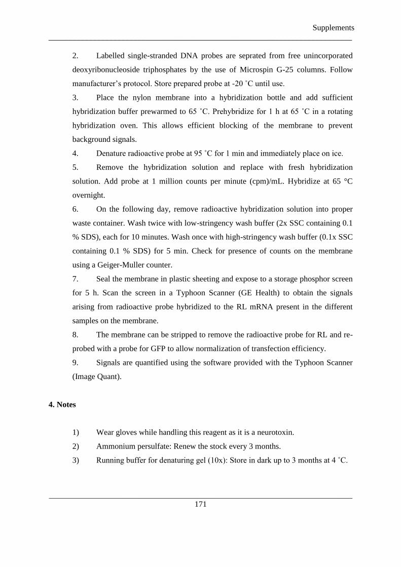
Supplements
___________________________________________________________________________
___________________________________________________________________________
171
2. Labelled single-stranded DNA probes are seprated from free unincorporated
deoxyribonucleoside triphosphates by the use of Microspin G-25 columns. Follow
manufacturer‟s protocol. Store prepared probe at -20 ˚C until use.
3. Place the nylon membrane into a hybridization bottle and add sufficient
hybridization buffer prewarmed to 65 ˚C. Prehybridize for 1 h at 65 ˚C in a rotating
hybridization oven. This allows efficient blocking of the membrane to prevent
background signals.
4. Denature radioactive probe at 95 ˚C for 1 min and immediately place on ice.
5. Remove the hybridization solution and replace with fresh hybridization
solution. Add probe at 1 million counts per minute (cpm)/mL. Hybridize at 65 °C
overnight.
6. On the following day, remove radioactive hybridization solution into proper
waste container. Wash twice with low-stringency wash buffer (2x SSC containing 0.1
% SDS), each for 10 minutes. Wash once with high-stringency wash buffer (0.1x SSC
containing 0.1 % SDS) for 5 min. Check for presence of counts on the membrane
using a Geiger-Muller counter.
7. Seal the membrane in plastic sheeting and expose to a storage phosphor screen
for 5 h. Scan the screen in a Typhoon Scanner (GE Health) to obtain the signals
arising from radioactive probe hybridized to the RL mRNA present in the different
samples on the membrane.
8. The membrane can be stripped to remove the radioactive probe for RL and re-
probed with a probe for GFP to allow normalization of transfection efficiency.
9. Signals are quantified using the software provided with the Typhoon Scanner
(Image Quant).
4. Notes
1) Wear gloves while handling this reagent as it is a neurotoxin.
2) Ammonium persulfate: Renew the stock every 3 months.
3) Running buffer for denaturing gel (10x): Store in dark up to 3 months at 4 ˚C.

Supplements
___________________________________________________________________________
___________________________________________________________________________
172
4) At this step the gel can be stored for later use by wrapping in tissue paper
soaked with 1x running buffer. Then place the gel into a plastic packet or wrap with
SaranWrap. Store at 4 ˚C.
5) Increasing the voltage can allow reduced transfer times. However, better
transfer efficiency is observed with longer duration.
6) Always wear gloves and use RNase-free plasticware (tips, reaction tubes etc).
7) Formaldehyde is toxic and has to be handled with gloves and only under the
fume hood.
8) Be carefully as the loading buffer contains ethidium bromide, which is a
carcinogen and has to be handled with proper gloves. Do not dilute the loading buffer
more than 50 %, to maintain the required denaturing conditions for the sample.
9) The UV-crosslinked membrane can be stored dry between two Whatman
3MM sheets until further use.
5. References
1. Filipowicz, W., Bhattacharyya, S.N. and Sonenberg, N. (2008) Mechanisms of post-transcriptional
regulation by microRNAs: are the answers in sight? Nat Rev Genet, 9, 102-114.
2. Bartel, D.P. (2004) MicroRNAs: genomics, biogenesis, mechanism, and function. Cell, 116, 281-297.
3. Carmell, M.A., Xuan, Z., Zhang, M.Q. and Hannon, G.J. (2002) The Argonaute family: tentacles that
reach into RNAi, developmental control, stem cell maintenance, and tumorigenesis. Genes Dev, 16,
2733-2742.
4. Meister, G. and Tuschl, T. (2004) Mechanisms of gene silencing by double-stranded RNA. Nature,
431, 343-349.
5. Verdel, A., Jia, S., Gerber, S., Sugiyama, T., Gygi, S., Grewal, S.I. and Moazed, D. (2004) RNAi-
mediated targeting of heterochromatin by the RITS complex. Science, 303, 672-676.
6. Kim, D.H., Villeneuve, L.M., Morris, K.V. and Rossi, J.J. (2006) Argonaute-1 directs siRNA-mediated
transcriptional gene silencing in human cells. Nat Struct Mol Biol, 13, 793-797.
7. Siomi, H. and Siomi, M.C. (2009) On the road to reading the RNA-interference code. Nature, 457, 396-
404.
8. Meister, G., Landthaler, M., Patkaniowska, A., Dorsett, Y., Teng, G. and Tuschl, T. (2004) Human
Argonaute2 mediates RNA cleavage targeted by miRNAs and siRNAs. Mol Cell, 15, 185-197.
9. Liu, J., Carmell, M.A., Rivas, F.V., Marsden, C.G., Thomson, J.M., Song, J.J., Hammond, S.M.,
Joshua-Tor, L. and Hannon, G.J. (2004) Argonaute2 is the catalytic engine of mammalian RNAi.
Science, 305, 1437-1441.
10. Eulalio, A., Huntzinger, E. and Izaurralde, E. (2008) Getting to the root of miRNA-mediated gene
silencing. Cell, 132, 9-14.
11. Pillai, R.S., Artus, C.G. and Filipowicz, W. (2004) Tethering of human Ago proteins to mRNA mimics
the miRNA-mediated repression of protein synthesis. Rna, 10, 1518-1525.
12. Legault, P., Li, J., Mogridge, J., Kay, L.E. and Greenblatt, J. (1998) NMR structure of the
bacteriophage lambda N peptide/boxB RNA complex: recognition of a GNRA fold by an arginine-rich
motif. Cell, 93, 289-299.
13. Baron-Benhamou, J., Gehring, N.H., Kulozik, A.E. and Hentze, M.W. (2004) Using the lambdaN
peptide to tether proteins to RNAs. Methods Mol Biol, 257, 135-154.

Supplements
___________________________________________________________________________
___________________________________________________________________________
173
14. Pillai, R.S., Bhattacharyya, S.N., Artus, C.G., Zoller, T., Cougot, N., Basyuk, E., Bertrand, E. and
Filipowicz, W. (2005) Inhibition of translational initiation by Let-7 MicroRNA in human cells.
Science, 309, 1573-1576.
15. Buhler, M., Verdel, A. and Moazed, D. (2006) Tethering RITS to a nascent transcript initiates RNAi-
and heterochromatin-dependent gene silencing. Cell, 125, 873-886.
16. Eulalio, A., Huntzinger, E. and Izaurralde, E. (2008) GW182 interaction with Argonaute is essential for
miRNA-mediated translational repression and mRNA decay. Nat Struct Mol Biol, 15, 346-353.
17. Chekulaeva, M., Filipowicz, W. and Parker, R. (2009) Multiple independent domains of dGW182
function in miRNA-mediated repression in Drosophila. Rna, 15, 794-803.
Acknowledgments
The protocols were originally developed with the active help and advice of Caroline G. Artus
and Witold Filipowicz at the Friedrich Miescher Institute for Biomedical Research, Basel,
Switzerland. Research in R.S.P‟s group is supported by the European Molecular Biology
Laboratory.

Supplements
___________________________________________________________________________
___________________________________________________________________________
174
Fig. 1
The Argonaute tethering experiment.
(a) A schematic representation of the Renilla luciferase reporter mRNA with 5BoxB hairpins inserted
into the 3'- untranslated region. The N-HA-tagged Argonaute protein is depicted as binding the BoxB hairpin;
several of the five hairpins might be bound by the protein in the actual experiment.
(b) The coding sequence for the N-HA peptides in the pCIneo-N-HA vector (Pillai et al. 2004) and the
EcoRI site used for cloning the Argonaute proteins is shown.
(c) Sequence context of the five BoxB hairpins (highlighted) in the 3‟-UTR of the RL-5BoxB mRNA.
The stop codon (TAA) of Renilla luciferase is indicated.

Supplements
___________________________________________________________________________
___________________________________________________________________________
175
Fig. 2
(a) RL activity detected in extracts from HeLa cells expressing the indicated fusion proteins. Cells were
co-transfected with constructs expressing the RL-5BoxB reporter, fire fly (FL) reference, and indicated fusion
proteins. Histograms represent normalized mean values (±SD) of RL/FL activities from a minimum of three
experiments. RL activity values seen in the presence of HA-hAgo2 were set as 1. Expression levels of fusion
proteins, as determined by Western analysis using anti-HA antibody, are shown below the histogram. The
aliquot of the N-HA-LacZ-expressing extract applied to the gel was 10 times smaller than aliquots of other
extracts. Generally, the N-HA-LacZ protein is expressed at a ~10-fold higher level than N-HA-hAgo2 or HA-
hAgo2. Increased RL activity in extracts expressing N-HA-LacZ is likely due to the effect of the protein on the
stability of mRNA reporters containing BoxB sequences.
(b) Activity of reporter RL mRNAs containing different numbers of BoxB hairpins.
(Reproduced from ref. 11 with permission from Cold Spring Harbour Laboratory Press.)
Fig. 3
Tethered hAgo2 and hAgo4, but not hAgo2 mutants and Hiwi, induce the repression.
(a) RL activity in extracts from HeLa cells cotransfected with plasmids expressing RL-5BoxB reporter
and N-HA-tagged mutant hAgo2 fusions. Western analysis of fusion protein expression is shown below the
histograms. Schematic representation of hAgo2 and its deletion mutants is shown in lower panel.
(b) Tethering of hAgo4 but not the Piwi protein Hiwi represses translation. (Reproduced from ref. 11
with permission from Cold Spring Harbour Laboratory Press.)
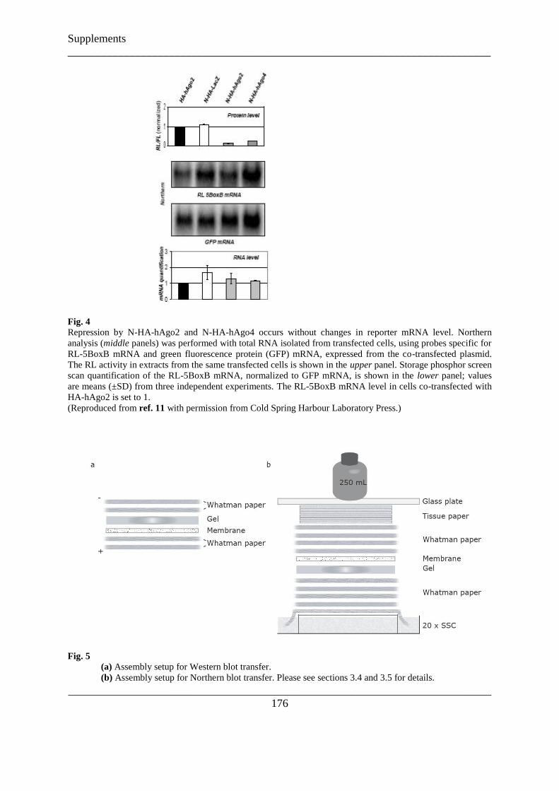
Supplements
___________________________________________________________________________
___________________________________________________________________________
176
Fig. 4
Repression by N-HA-hAgo2 and N-HA-hAgo4 occurs without changes in reporter mRNA level. Northern
analysis (middle panels) was performed with total RNA isolated from transfected cells, using probes specific for
RL-5BoxB mRNA and green fluorescence protein (GFP) mRNA, expressed from the co-transfected plasmid.
The RL activity in extracts from the same transfected cells is shown in the upper panel. Storage phosphor screen
scan quantification of the RL-5BoxB mRNA, normalized to GFP mRNA, is shown in the lower panel; values
are means (±SD) from three independent experiments. The RL-5BoxB mRNA level in cells co-transfected with
HA-hAgo2 is set to 1.
(Reproduced from ref. 11 with permission from Cold Spring Harbour Laboratory Press.)
Fig. 5
(a) Assembly setup for Western blot transfer.
(b) Assembly setup for Northern blot transfer. Please see sections 3.4 and 3.5 for details.

Supplements
___________________________________________________________________________
___________________________________________________________________________
177
5.2

Supplements
___________________________________________________________________________
___________________________________________________________________________
178

Supplements
___________________________________________________________________________
___________________________________________________________________________
179

Supplements
___________________________________________________________________________
___________________________________________________________________________
180

Supplements
___________________________________________________________________________
___________________________________________________________________________
181

Supplements
___________________________________________________________________________
___________________________________________________________________________
182

Supplements
___________________________________________________________________________
___________________________________________________________________________
183

Supplements
___________________________________________________________________________
___________________________________________________________________________
184
Supplements
___________________________________________________________________________
___________________________________________________________________________
185

___________________________________________________________________________
186
References
Abdel-Ghany, S.E. & Pilon, M. (2008) MicroRNA-mediated systemic down-regulation of copper protein
expression in response to low copper availability in Arabidopsis. J Biol Chem 283, 15932-15945.
Abdellah (2004) Finishing the euchromatic sequence of the human genome. Nature 431, 931-945.
Akbergenov, R., Si-Ammour, A., Blevins, T., et al. (2006) Molecular characterization of geminivirus-derived
small RNAs in different plant species. Nucleic acids research 34, 462-471.
Allen, E., Xie, Z., Gustafson, A.M. & Carrington, J.C. (2005) microRNA-directed phasing during trans-acting
siRNA biogenesis in plants. Cell 121, 207-221.
Ameres, S.L., Horwich, M.D., Hung, J.H., et al. (2010) Target RNA-directed trimming and tailing of small
silencing RNAs. Science 328, 1534-1539.
Ameres, S.L., Martinez, J. & Schroeder, R. (2007) Molecular basis for target RNA recognition and cleavage by
human RISC. Cell 130, 101-112.
Anantharaman, V., Koonin, E.V. & Aravind, L. (2002) Comparative genomics and evolution of proteins
involved in RNA metabolism. Nucleic acids research 30, 1427-1464.
Ancelin, K., Lange, U.C., Hajkova, P., et al. (2006) Blimp1 associates with Prmt5 and directs histone arginine
methylation in mouse germ cells. Nat Cell Biol 8, 623-630.
Anne, J. & Mechler, B.M. (2005) Valois, a component of the nuage and pole plasm, is involved in assembly of
these structures, and binds to Tudor and the methyltransferase Capsuleen. Development 132, 2167-2177.
Aoki, Y., Nagao, I., Saito, D., Ebe, Y., Kinjo, M. & Tanaka, M. (2008) Temporal and spatial localization of
three germline-specific proteins in medaka. Dev Dyn 237, 800-807.
Aravin, A., Gaidatzis, D., Pfeffer, S., et al. (2006) A novel class of small RNAs bind to MILI protein in mouse
testes. Nature 442, 203-207.
Aravin, A.A. & Bourc'his, D. (2008) Small RNA guides for de novo DNA methylation in mammalian germ
cells. Genes Dev 22, 970-975.
Aravin, A.A., Hannon, G.J. & Brennecke, J. (2007a) The Piwi-piRNA pathway provides an adaptive defense in
the transposon arms race. Science 318, 761-764.
Aravin, A.A., Klenov, M.S., Vagin, V.V., Bantignies, F., Cavalli, G. & Gvozdev, V.A. (2004) Dissection of a
natural RNA silencing process in the Drosophila melanogaster germ line. Mol Cell Biol 24, 6742-6750.
Aravin, A.A., Lagos-Quintana, M., Yalcin, A., et al. (2003) The small RNA profile during Drosophila
melanogaster development. Dev Cell 5, 337-350.
Aravin, A.A., Naumova, N.M., Tulin, A.V., Vagin, V.V., Rozovsky, Y.M. & Gvozdev, V.A. (2001) Double-
stranded RNA-mediated silencing of genomic tandem repeats and transposable elements in the D.
melanogaster germline. Curr Biol 11, 1017-1027.
Aravin, A.A., Sachidanandam, R., Bourc'his, D., et al. (2008) A piRNA pathway primed by individual
transposons is linked to de novo DNA methylation in mice. Mol Cell 31, 785-799.
Aravin, A.A., Sachidanandam, R., Girard, A., Fejes-Toth, K. & Hannon, G.J. (2007b) Developmentally
regulated piRNA clusters implicate MILI in transposon control. Science 316, 744-747.
Aravin, A.A., van der Heijden, G.W., Castaneda, J., Vagin, V.V., Hannon, G.J. & Bortvin, A. (2009)
Cytoplasmic compartmentalization of the fetal piRNA pathway in mice. PLoS Genet 5, e1000764.
Arkov, A.L., Wang, J.Y., Ramos, A. & Lehmann, R. (2006) The role of Tudor domains in germline
development and polar granule architecture. Development 133, 4053-4062.
Aza-Blanc, P., Cooper, C.L., Wagner, K., Batalov, S., Deveraux, Q.L. & Cooke, M.P. (2003) Identification of
modulators of TRAIL-induced apoptosis via RNAi-based phenotypic screening. Mol Cell 12, 627-637.
Azuma-Mukai, A., Oguri, H., Mituyama, T., et al. (2008) Characterization of endogenous human Argonautes
and their miRNA partners in RNA silencing. Proc Natl Acad Sci U S A 105, 7964-7969.
Babiarz, J.E., Ruby, J.G., Wang, Y., Bartel, D.P. & Blelloch, R. (2008) Mouse ES cells express endogenous
shRNAs, siRNAs, and other Microprocessor-independent, Dicer-dependent small RNAs. Genes Dev 22,
2773-2785.
Baek, D., Villen, J., Shin, C., Camargo, F.D., Gygi, S.P. & Bartel, D.P. (2008) The impact of microRNAs on
protein output. Nature 455, 64-71.
Bartel, D.P. (2009) MicroRNAs: target recognition and regulatory functions. Cell 136, 215-233.
Baumberger, N. & Baulcombe, D.C. (2005) Arabidopsis ARGONAUTE1 is an RNA Slicer that selectively
recruits microRNAs and short interfering RNAs. Proc Natl Acad Sci U S A 102, 11928-11933.

References
___________________________________________________________________________
___________________________________________________________________________
187
Becalska, A.N. & Gavis, E.R. (2009) Lighting up mRNA localization in Drosophila oogenesis. Development
136, 2493-2503.
Behm-Ansmant, I., Rehwinkel, J., Doerks, T., Stark, A., Bork, P. & Izaurralde, E. (2006) mRNA degradation by
miRNAs and GW182 requires both CCR4:NOT deadenylase and DCP1:DCP2 decapping complexes.
Genes Dev 20, 1885-1898.
Berezikov, E., Chung, W.J., Willis, J., Cuppen, E. & Lai, E.C. (2007) Mammalian mirtron genes. Mol Cell 28,
328-336.
Berger, I., Fitzgerald, D.J. & Richmond, T.J. (2004) Baculovirus expression system for heterologous
multiprotein complexes. Nature biotechnology 22, 1583-1587.
Bernstein, E., Caudy, A.A., Hammond, S.M. & Hannon, G.J. (2001) Role for a bidentate ribonuclease in the
initiation step of RNA interference. Nature 409, 363-366.
Biemont, C. & Vieira, C. (2006) Genetics: junk DNA as an evolutionary force. Nature 443, 521-524.
Bieniossek, C., Richmond, T.J. & Berger, I. (2008) MultiBac: multigene baculovirus-based eukaryotic protein
complex production. Current protocols in protein science / editorial board, John E. Coligan ... [et al
Chapter 5, Unit 5 20.
Birney, E., Stamatoyannopoulos, J.A., Dutta, A., et al. (2007) Identification and analysis of functional elements
in 1% of the human genome by the ENCODE pilot project. Nature 447, 799-816.
Bohmert, K., Camus, I., Bellini, C., Bouchez, D., Caboche, M. & Benning, C. (1998) AGO1 defines a novel
locus of Arabidopsis controlling leaf development. Embo J 17, 170-180.
Bohnsack, M.T., Czaplinski, K. & Gorlich, D. (2004) Exportin 5 is a RanGTP-dependent dsRNA-binding
protein that mediates nuclear export of pre-miRNAs. Rna 10, 185-191.
Boland, A., Tritschler, F., Heimstadt, S., Izaurralde, E. & Weichenrieder, O. (2010) Crystal structure and ligand
binding of the MID domain of a eukaryotic Argonaute protein. EMBO Rep 11, 522-527.
Borsani, O., Zhu, J., Verslues, P.E., Sunkar, R. & Zhu, J.K. (2005) Endogenous siRNAs derived from a pair of
natural cis-antisense transcripts regulate salt tolerance in Arabidopsis. Cell 123, 1279-1291.
Boswell, R.E. & Mahowald, A.P. (1985) tudor, a gene required for assembly of the germ plasm in Drosophila
melanogaster. Cell 43, 97-104.
Bourc'his, D. & Bestor, T.H. (2004) Meiotic catastrophe and retrotransposon reactivation in male germ cells
lacking Dnmt3L. Nature 431, 96-99.
Boutet, S., Vazquez, F., Liu, J., et al. (2003) Arabidopsis HEN1: a genetic link between endogenous miRNA
controlling development and siRNA controlling transgene silencing and virus resistance. Curr Biol 13,
843-848.
Bozzetti, M.P., Massari, S., Finelli, P., et al. (1995) The Ste locus, a component of the parasitic cry-Ste system
of Drosophila melanogaster, encodes a protein that forms crystals in primary spermatocytes and mimics
properties of the beta subunit of casein kinase 2. Proc Natl Acad Sci U S A 92, 6067-6071.
Brahms, H., Raymackers, J., Union, A., de Keyser, F., Meheus, L. & Luhrmann, R. (2000) The C-terminal RG
dipeptide repeats of the spliceosomal Sm proteins D1 and D3 contain symmetrical dimethylarginines,
which form a major B-cell epitope for anti-Sm autoantibodies. J Biol Chem 275, 17122-17129.
Brennecke, J., Aravin, A.A., Stark, A., et al. (2007) Discrete small RNA-generating loci as master regulators of
transposon activity in Drosophila. Cell 128, 1089-1103.
Brennecke, J., Hipfner, D.R., Stark, A., Russell, R.B. & Cohen, S.M. (2003) bantam encodes a developmentally
regulated microRNA that controls cell proliferation and regulates the proapoptotic gene hid in
Drosophila. Cell 113, 25-36.
Brennecke, J., Malone, C.D., Aravin, A.A., Sachidanandam, R., Stark, A. & Hannon, G.J. (2008) An epigenetic
role for maternally inherited piRNAs in transposon silencing. Science 322, 1387-1392.
Brennecke, J., Stark, A., Russell, R.B. & Cohen, S.M. (2005) Principles of microRNA-target recognition. PLoS
biology 3, e85.
Brodersen, P., Sakvarelidze-Achard, L., Bruun-Rasmussen, M., et al. (2008) Widespread translational inhibition
by plant miRNAs and siRNAs. Science 320, 1185-1190.
Brower-Toland, B., Findley, S.D., Jiang, L., et al. (2007) Drosophila PIWI associates with chromatin and
interacts directly with HP1a. Genes Dev 21, 2300-2311.
Buhler, D., Raker, V., Luhrmann, R. & Fischer, U. (1999) Essential role for the tudor domain of SMN in
spliceosomal U snRNP assembly: implications for spinal muscular atrophy. Hum Mol Genet 8, 2351-
2357.
Buhler, M. & Moazed, D. (2007) Transcription and RNAi in heterochromatic gene silencing. Nat Struct Mol
Biol 14, 1041-1048.

References
___________________________________________________________________________
___________________________________________________________________________
188
Buhler, M., Verdel, A. & Moazed, D. (2006) Tethering RITS to a nascent transcript initiates RNAi- and
heterochromatin-dependent gene silencing. Cell 125, 873-886.
Buhtz, A., Pieritz, J., Springer, F. & Kehr, J. (2010) Phloem small RNAs, nutrient stress responses, and systemic
mobility. BMC plant biology 10, 64.
Bulgakov, O.V., Eggenschwiler, J.T., Hong, D.H., Anderson, K.V. & Li, T. (2004) FKBP8 is a negative
regulator of mouse sonic hedgehog signaling in neural tissues. Development 131, 2149-2159.
Cai, X., Hagedorn, C.H. & Cullen, B.R. (2004) Human microRNAs are processed from capped, polyadenylated
transcripts that can also function as mRNAs. Rna 10, 1957-1966.
Carmell, M.A., Girard, A., van de Kant, H.J., et al. (2007) MIWI2 is essential for spermatogenesis and
repression of transposons in the mouse male germline. Dev Cell 12, 503-514.
Catalanotto, C., Azzalin, G., Macino, G. & Cogoni, C. (2002) Involvement of small RNAs and role of the qde
genes in the gene silencing pathway in Neurospora. Genes Dev 16, 790-795.
Cavey, M., Hijal, S., Zhang, X. & Suter, B. (2005) Drosophila valois encodes a divergent WD protein that is
required for Vasa localization and Oskar protein accumulation. Development 132, 459-468.
Chan, C.M., Zhou, C. & Huang, R.H. (2009) Reconstituting bacterial RNA repair and modification in vitro.
Science 326, 247.
Chan, S.W. (2008) Inputs and outputs for chromatin-targeted RNAi. Trends in plant science 13, 383-389.
Chan, S.W., Zilberman, D., Xie, Z., Johansen, L.K., Carrington, J.C. & Jacobsen, S.E. (2004) RNA silencing
genes control de novo DNA methylation. Science 303, 1336.
Cheloufi, S., Dos Santos, C.O., Chong, M.M. & Hannon, G.J. (2010) A dicer-independent miRNA biogenesis
pathway that requires Ago catalysis. Nature 465, 584-589.
Chen, C., Jin, J., James, D.A., et al. (2009) Mouse Piwi interactome identifies binding mechanism of Tdrkh
Tudor domain to arginine methylated Miwi. Proc Natl Acad Sci U S A 106, 20336-20341.
Chen, X. (2005) MicroRNA biogenesis and function in plants. FEBS letters 579, 5923-5931.
Chen, X. (2007) A marked end. Nat Struct Mol Biol 14, 259-260.
Chen, X., Liu, J., Cheng, Y. & Jia, D. (2002) HEN1 functions pleiotropically in Arabidopsis development and
acts in C function in the flower. Development 129, 1085-1094.
Chendrimada, T.P., Finn, K.J., Ji, X., et al. (2007) MicroRNA silencing through RISC recruitment of eIF6.
Nature 447, 823-828.
Chendrimada, T.P., Gregory, R.I., Kumaraswamy, E., et al. (2005) TRBP recruits the Dicer complex to Ago2
for microRNA processing and gene silencing. Nature 436, 740-744.
Chenna, R., Sugawara, H., Koike, T., et al. (2003) Multiple sequence alignment with the Clustal series of
programs. Nucleic acids research 31, 3497-3500.
Choudhuri, S. (2009) Lesser known relatives of miRNA. Biochem Biophys Res Commun 388, 177-180.
Chuma, S., Hiyoshi, M., Yamamoto, A., Hosokawa, M., Takamune, K. & Nakatsuji, N. (2003) Mouse Tudor
Repeat-1 (MTR-1) is a novel component of chromatoid bodies/nuages in male germ cells and forms a
complex with snRNPs. Mech Dev 120, 979-990.
Chuma, S., Hosokawa, M., Kitamura, K., et al. (2006) Tdrd1/Mtr-1, a tudor-related gene, is essential for male
germ-cell differentiation and nuage/germinal granule formation in mice. Proc Natl Acad Sci U S A 103,
15894-15899.
Chung, W.J., Okamura, K., Martin, R. & Lai, E.C. (2008) Endogenous RNA interference provides a somatic
defense against Drosophila transposons. Curr Biol 18, 795-802.
Cifuentes, D., Xue, H., Taylor, D.W., et al. (2010) A novel miRNA processing pathway independent of Dicer
requires Argonaute2 catalytic activity. Science 328, 1694-1698.
Cole, C., Sobala, A., Lu, C., et al. (2009) Filtering of deep sequencing data reveals the existence of abundant
Dicer-dependent small RNAs derived from tRNAs. Rna 15, 2147-2160.
Cook, H.A., Koppetsch, B.S., Wu, J. & Theurkauf, W.E. (2004) The Drosophila SDE3 homolog armitage is
required for oskar mRNA silencing and embryonic axis specification. Cell 116, 817-829.
Costa, Y., Speed, R.M., Gautier, P., et al. (2006) Mouse MAELSTROM: the link between meiotic silencing of
unsynapsed chromatin and microRNA pathway? Human molecular genetics 15, 2324-2334.
Cox, D.N., Chao, A., Baker, J., Chang, L., Qiao, D. & Lin, H. (1998) A novel class of evolutionarily conserved
genes defined by piwi are essential for stem cell self-renewal. Genes Dev 12, 3715-3727.
Crackower, M.A., Kolas, N.K., Noguchi, J., et al. (2003) Essential role of Fkbp6 in male fertility and
homologous chromosome pairing in meiosis. Science 300, 1291-1295.
Czech, B., Malone, C.D., Zhou, R., et al. (2008) An endogenous small interfering RNA pathway in Drosophila.
Nature 453, 798-802.

References
___________________________________________________________________________
___________________________________________________________________________
189
Czech, B., Zhou, R., Erlich, Y., et al. (2009) Hierarchical rules for Argonaute loading in Drosophila. Mol Cell
36, 445-456.
Davis, E., Caiment, F., Tordoir, X., et al. (2005) RNAi-mediated allelic trans-interaction at the imprinted
Rtl1/Peg11 locus. Curr Biol 15, 743-749.
Davis, T.L., Trasler, J.M., Moss, S.B., Yang, G.J. & Bartolomei, M.S. (1999) Acquisition of the H19
methylation imprint occurs differentially on the parental alleles during spermatogenesis. Genomics 58,
18-28.
Davis, T.L., Yang, G.J., McCarrey, J.R. & Bartolomei, M.S. (2000) The H19 methylation imprint is erased and
re-established differentially on the parental alleles during male germ cell development. Hum Mol Genet
9, 2885-2894.
Deng, W. & Lin, H. (2002) miwi, a murine homolog of piwi, encodes a cytoplasmic protein essential for
spermatogenesis. Dev Cell 2, 819-830.
Denli, A.M., Tops, B.B., Plasterk, R.H., Ketting, R.F. & Hannon, G.J. (2004) Processing of primary
microRNAs by the Microprocessor complex. Nature 432, 231-235.
Ding, S.W. & Voinnet, O. (2007) Antiviral immunity directed by small RNAs. Cell 130, 413-426.
Doench, J.G., Petersen, C.P. & Sharp, P.A. (2003) siRNAs can function as miRNAs. Genes Dev 17, 438-442.
Dorer, D.R. & Henikoff, S. (1994) Expansions of transgene repeats cause heterochromatin formation and gene
silencing in Drosophila. Cell 77, 993-1002.
Dorer, D.R. & Henikoff, S. (1997) Transgene repeat arrays interact with distant heterochromatin and cause
silencing in cis and trans. Genetics 147, 1181-1190.
Ebhardt, H.A., Thi, E.P., Wang, M.B. & Unrau, P.J. (2005) Extensive 3' modification of plant small RNAs is
modulated by helper component-proteinase expression. Proc Natl Acad Sci U S A 102, 13398-13403.
El-Shami, M., Pontier, D., Lahmy, S., et al. (2007) Reiterated WG/GW motifs form functionally and
evolutionarily conserved ARGONAUTE-binding platforms in RNAi-related components. Genes Dev
21, 2539-2544.
Elbashir, S.M., Harborth, J., Lendeckel, W., Yalcin, A., Weber, K. & Tuschl, T. (2001a) Duplexes of 21-
nucleotide RNAs mediate RNA interference in cultured mammalian cells. Nature 411, 494-498.
Elbashir, S.M., Lendeckel, W. & Tuschl, T. (2001b) RNA interference is mediated by 21- and 22-nucleotide
RNAs. Genes Dev 15, 188-200.
Elbashir, S.M., Martinez, J., Patkaniowska, A., Lendeckel, W. & Tuschl, T. (2001c) Functional anatomy of
siRNAs for mediating efficient RNAi in Drosophila melanogaster embryo lysate. Embo J 20, 6877-
6888.
Ender, C., Krek, A., Friedlander, M.R., et al. (2008) A human snoRNA with microRNA-like functions. Mol Cell
32, 519-528.
Esquela-Kerscher, A. & Slack, F.J. (2006) Oncomirs - microRNAs with a role in cancer. Nature reviews 6, 259-
269.
Eulalio, A., Huntzinger, E. & Izaurralde, E. (2008) GW182 interaction with Argonaute is essential for miRNA-
mediated translational repression and mRNA decay. Nat Struct Mol Biol 15, 346-353.
Fang, Y. & Spector, D.L. (2007) Identification of nuclear dicing bodies containing proteins for microRNA
biogenesis in living Arabidopsis plants. Curr Biol 17, 818-823.
Fanti, L., Dorer, D.R., Berloco, M., Henikoff, S. & Pimpinelli, S. (1998) Heterochromatin protein 1 binds
transgene arrays. Chromosoma 107, 286-292.
Filipowicz, W. (2005) RNAi: the nuts and bolts of the RISC machine. Cell 122, 17-20.
Fire, A., Xu, S., Montgomery, M.K., Kostas, S.A., Driver, S.E. & Mello, C.C. (1998) Potent and specific
genetic interference by double-stranded RNA in Caenorhabditis elegans. Nature 391, 806-811.
Fitzgerald, D.J., Berger, P., Schaffitzel, C., Yamada, K., Richmond, T.J. & Berger, I. (2006) Protein complex
expression by using multigene baculoviral vectors. Nature methods 3, 1021-1032.
Flynt, A.S., Greimann, J.C., Chung, W.J., Lima, C.D. & Lai, E.C. (2010) MicroRNA biogenesis via splicing and
exosome-mediated trimming in Drosophila. Mol Cell 38, 900-907.
Forstemann, K., Horwich, M.D., Wee, L., Tomari, Y. & Zamore, P.D. (2007) Drosophila microRNAs are sorted
into functionally distinct argonaute complexes after production by dicer-1. Cell 130, 287-297.
Forstemann, K., Tomari, Y., Du, T., et al. (2005) Normal microRNA maturation and germ-line stem cell
maintenance requires Loquacious, a double-stranded RNA-binding domain protein. PLoS biology 3,
e236.
Frank, F., Sonenberg, N. & Nagar, B. (2010) Structural basis for 5'-nucleotide base-specific recognition of guide
RNA by human AGO2. Nature 465, 818-822.

References
___________________________________________________________________________
___________________________________________________________________________
190
Frost, R.J., Hamra, F.K., Richardson, J.A., Qi, X., Bassel-Duby, R. & Olson, E.N. (2010) MOV10L1 is
necessary for protection of spermatocytes against retrotransposons by Piwi-interacting RNAs. Proc Natl
Acad Sci U S A.
Fujiwara, Y., Komiya, T., Kawabata, H., et al. (1994) Isolation of a DEAD-family protein gene that encodes a
murine homolog of Drosophila vasa and its specific expression in germ cell lineage. Proc Natl Acad Sci
U S A 91, 12258-12262.
Gaidatzis, D., van Nimwegen, E., Hausser, J. & Zavolan, M. (2007) Inference of miRNA targets using
evolutionary conservation and pathway analysis. BMC bioinformatics 8, 69.
Ghildiyal, M., Seitz, H., Horwich, M.D., et al. (2008) Endogenous siRNAs derived from transposons and
mRNAs in Drosophila somatic cells. Science 320, 1077-1081.
Ghildiyal, M. & Zamore, P.D. (2009) Small silencing RNAs: an expanding universe. Nat Rev Genet 10, 94-108.
Gillespie, C.F., Phifer, J., Bradley, B. & Ressler, K.J. (2009) Risk and resilience: genetic and environmental
influences on development of the stress response. Depression and anxiety 26, 984-992.
Giraldez, A.J., Cinalli, R.M., Glasner, M.E., et al. (2005) MicroRNAs regulate brain morphogenesis in
zebrafish. Science 308, 833-838.
Giraldez, A.J., Mishima, Y., Rihel, J., et al. (2006) Zebrafish MiR-430 promotes deadenylation and clearance of
maternal mRNAs. Science 312, 75-79.
Girard, A. & Hannon, G.J. (2008) Conserved themes in small-RNA-mediated transposon control. Trends in cell
biology 18, 136-148.
Girard, A., Sachidanandam, R., Hannon, G.J. & Carmell, M.A. (2006) A germline-specific class of small RNAs
binds mammalian Piwi proteins. Nature 442, 199-202.
Glazov, E.A., Cottee, P.A., Barris, W.C., Moore, R.J., Dalrymple, B.P. & Tizard, M.L. (2008) A microRNA
catalog of the developing chicken embryo identified by a deep sequencing approach. Genome Res 18,
957-964.
Gouet, P., Courcelle, E., Stuart, D.I. & Metoz, F. (1999) ESPript: analysis of multiple sequence alignments in
PostScript. Bioinformatics (Oxford, England) 15, 305-308.
Gregory, R.I., Yan, K.P., Amuthan, G., et al. (2004) The Microprocessor complex mediates the genesis of
microRNAs. Nature 432, 235-240.
Grimson, A., Farh, K.K., Johnston, W.K., Garrett-Engele, P., Lim, L.P. & Bartel, D.P. (2007) MicroRNA
targeting specificity in mammals: determinants beyond seed pairing. Mol Cell 27, 91-105.
Grimson, A., Srivastava, M., Fahey, B., et al. (2008) Early origins and evolution of microRNAs and Piwi-
interacting RNAs in animals. Nature 455, 1193-1197.
Grishok, A., Pasquinelli, A.E., Conte, D., et al. (2001) Genes and mechanisms related to RNA interference
regulate expression of the small temporal RNAs that control C. elegans developmental timing. Cell 106,
23-34.
Grivna, S.T., Beyret, E., Wang, Z. & Lin, H. (2006a) A novel class of small RNAs in mouse spermatogenic
cells. Genes Dev 20, 1709-1714.
Grivna, S.T., Pyhtila, B. & Lin, H. (2006b) MIWI associates with translational machinery and PIWI-interacting
RNAs (piRNAs) in regulating spermatogenesis. Proc Natl Acad Sci U S A 103, 13415-13420.
Gross, C.T. & McGinnis, W. (1996) DEAF-1, a novel protein that binds an essential region in a Deformed
response element. Embo J 15, 1961-1970.
Gunawardane, L.S., Saito, K., Nishida, K.M., et al. (2007) A slicer-mediated mechanism for repeat-associated
siRNA 5' end formation in Drosophila. Science 315, 1587-1590.
Haase, A.D., Jaskiewicz, L., Zhang, H., et al. (2005) TRBP, a regulator of cellular PKR and HIV-1 virus
expression, interacts with Dicer and functions in RNA silencing. EMBO Rep 6, 961-967.
Hajkova, P., Erhardt, S., Lane, N., et al. (2002) Epigenetic reprogramming in mouse primordial germ cells.
Mech Dev 117, 15-23.
Haley, B. & Zamore, P.D. (2004) Kinetic analysis of the RNAi enzyme complex. Nat Struct Mol Biol 11, 599-
606.
Hammond, S.M., Bernstein, E., Beach, D. & Hannon, G.J. (2000) An RNA-directed nuclease mediates post-
transcriptional gene silencing in Drosophila cells. Nature 404, 293-296.
Han, J., Lee, Y., Yeom, K.H., Kim, Y.K., Jin, H. & Kim, V.N. (2004a) The Drosha-DGCR8 complex in primary
microRNA processing. Genes Dev 18, 3016-3027.
Han, J., Lee, Y., Yeom, K.H., et al. (2006) Molecular basis for the recognition of primary microRNAs by the
Drosha-DGCR8 complex. Cell 125, 887-901.
References
___________________________________________________________________________
___________________________________________________________________________
191
Han, M.H., Goud, S., Song, L. & Fedoroff, N. (2004b) The Arabidopsis double-stranded RNA-binding protein
HYL1 plays a role in microRNA-mediated gene regulation. Proc Natl Acad Sci U S A 101, 1093-1098.
Hardy, R.W., Lindsley, D.L., Livak, K.J., et al. (1984) Cytogenetic analysis of a segment of the Y chromosome
of Drosophila melanogaster. Genetics 107, 591-610.
Harris, A.N. & Macdonald, P.M. (2001) Aubergine encodes a Drosophila polar granule component required for
pole cell formation and related to eIF2C. Development 128, 2823-2832.
Hartig, J.V., Esslinger, S., Bottcher, R., Saito, K. & Forstemann, K. (2009) Endo-siRNAs depend on a new
isoform of loquacious and target artificially introduced, high-copy sequences. Embo J 28, 2932-2944.
Hartig, J.V., Tomari, Y. & Forstemann, K. (2007) piRNAs--the ancient hunters of genome invaders. Genes Dev
21, 1707-1713.
Hata, K., Kusumi, M., Yokomine, T., Li, E. & Sasaki, H. (2006) Meiotic and epigenetic aberrations in Dnmt3L-
deficient male germ cells. Molecular reproduction and development 73, 116-122.
Hay, B., Jan, L.Y. & Jan, Y.N. (1990) Localization of vasa, a component of Drosophila polar granules, in
maternal-effect mutants that alter embryonic anteroposterior polarity. Development 109, 425-433.
Herr, A.J., Jensen, M.B., Dalmay, T. & Baulcombe, D.C. (2005) RNA polymerase IV directs silencing of
endogenous DNA. Science 308, 118-120.
Hock, J. & Meister, G. (2008) The Argonaute protein family. Genome Biol 9, 210.
Horwich, M.D., Li, C., Matranga, C., et al. (2007) The Drosophila RNA methyltransferase, DmHen1, modifies
germline piRNAs and single-stranded siRNAs in RISC. Curr Biol 17, 1265-1272.
Hosokawa, M., Shoji, M., Kitamura, K., et al. (2007) Tudor-related proteins TDRD1/MTR-1, TDRD6 and
TDRD7/TRAP: domain composition, intracellular localization, and function in male germ cells in mice.
Developmental biology 301, 38-52.
Houwing, S., Berezikov, E. & Ketting, R.F. (2008) Zili is required for germ cell differentiation and meiosis in
zebrafish. Embo J 27, 2702-2711.
Houwing, S., Kamminga, L.M., Berezikov, E., et al. (2007) A role for Piwi and piRNAs in germ cell
maintenance and transposon silencing in Zebrafish. Cell 129, 69-82.
Hu, H.Y., Yan, Z., Xu, Y., et al. (2009) Sequence features associated with microRNA strand selection in
humans and flies. BMC genomics 10, 413.
Huang, Y., Ji, L., Huang, Q., Vassylyev, D.G., Chen, X. & Ma, J.B. (2009) Structural insights into mechanisms
of the small RNA methyltransferase HEN1. Nature 461, 823-827.
Hunter, N. (2008) The RecQ DNA helicases: Jacks-of-all-trades or master-tradesmen? Cell Res 18, 328-330.
Hutvagner, G., McLachlan, J., Pasquinelli, A.E., Balint, E., Tuschl, T. & Zamore, P.D. (2001) A cellular
function for the RNA-interference enzyme Dicer in the maturation of the let-7 small temporal RNA.
Science 293, 834-838.
Hutvagner, G. & Simard, M.J. (2008) Argonaute proteins: key players in RNA silencing. Nat Rev Mol Cell Biol
9, 22-32.
Hutvagner, G. & Zamore, P.D. (2002) A microRNA in a multiple-turnover RNAi enzyme complex. Science
297, 2056-2060.
Jackson, R.J. & Standart, N. (2007) How do microRNAs regulate gene expression? Sci STKE 2007, re1.
Jain, R. & Shuman, S. (2010) Bacterial Hen1 is a 3' terminal RNA ribose 2'-O-methyltransferase component of
a bacterial RNA repair cassette. Rna 16, 316-323.
Jain, R. & Shuman, S. (2011) Active site mapping and substrate specificity of bacterial Hen1, a manganese-
dependent 3' terminal RNA ribose 2'O-methyltransferase. Rna.
Jiang, F., Ye, X., Liu, X., Fincher, L., McKearin, D. & Liu, Q. (2005) Dicer-1 and R3D1-L catalyze microRNA
maturation in Drosophila. Genes Dev 19, 1674-1679.
Jin, Y.J., Burakoff, S.J. & Bierer, B.E. (1992) Molecular cloning of a 25-kDa high affinity rapamycin binding
protein, FKBP25. J Biol Chem 267, 10942-10945.
Kalmykova, A.I., Dobritsa, A.A. & Gvozdev, V.A. (1998) Su(Ste) diverged tandem repeats in a Y chromosome
of Drosophila melanogaster are transcribed and variously processed. Genetics 148, 243-249.
Kamminga, L.M., Luteijn, M.J., den Broeder, M.J., et al. (2010) Hen1 is required for oocyte development and
piRNA stability in zebrafish. Embo J.
Kaneda, M., Okano, M., Hata, K., et al. (2004) Essential role for de novo DNA methyltransferase Dnmt3a in
paternal and maternal imprinting. Nature 429, 900-903.
Kanoh, J., Sadaie, M., Urano, T. & Ishikawa, F. (2005) Telomere binding protein Taz1 establishes Swi6
heterochromatin independently of RNAi at telomeres. Curr Biol 15, 1808-1819.

References
___________________________________________________________________________
___________________________________________________________________________
192
Katiyar-Agarwal, S., Gao, S., Vivian-Smith, A. & Jin, H. (2007) A novel class of bacteria-induced small RNAs
in Arabidopsis. Genes & development 21, 3123-3134.
Katiyar-Agarwal, S., Morgan, R., Dahlbeck, D., et al. (2006) A pathogen-inducible endogenous siRNA in plant
immunity. Proc Natl Acad Sci U S A 103, 18002-18007.
Kato, Y., Kaneda, M., Hata, K., et al. (2007) Role of the Dnmt3 family in de novo methylation of imprinted and
repetitive sequences during male germ cell development in the mouse. Hum Mol Genet 16, 2272-2280.
Kawamura, Y., Saito, K., Kin, T., et al. (2008) Drosophila endogenous small RNAs bind to Argonaute 2 in
somatic cells. Nature 453, 793-797.
Kawaoka, S., Hayashi, N., Katsuma, S., et al. (2008) Bombyx small RNAs: genomic defense system against
transposons in the silkworm, Bombyx mori. Insect Biochem Mol Biol 38, 1058-1065.
Kawaoka, S., Hayashi, N., Suzuki, Y., et al. (2009) The Bombyx ovary-derived cell line endogenously
expresses PIWI/PIWI-interacting RNA complexes. Rna 15, 1258-1264.
Kedde, M., Strasser, M.J., Boldajipour, B., et al. (2007) RNA-binding protein Dnd1 inhibits microRNA access
to target mRNA. Cell 131, 1273-1286.
Ketting, R.F., Fischer, S.E., Bernstein, E., Sijen, T., Hannon, G.J. & Plasterk, R.H. (2001) Dicer functions in
RNA interference and in synthesis of small RNA involved in developmental timing in C. elegans. Genes
Dev 15, 2654-2659.
Khvorova, A., Reynolds, A. & Jayasena, S.D. (2003) Functional siRNAs and miRNAs exhibit strand bias. Cell
115, 209-216.
Kim, K., Lee, Y.S. & Carthew, R.W. (2007) Conversion of pre-RISC to holo-RISC by Ago2 during assembly of
RNAi complexes. Rna 13, 22-29.
Kim, V.N. (2006) Small RNAs just got bigger: Piwi-interacting RNAs (piRNAs) in mammalian testes. Genes
Dev 20, 1993-1997.
Kimmel, C.B., Ballard, W.W., Kimmel, S.R., Ullmann, B. & Schilling, T.F. (1995) Stages of embryonic
development of the zebrafish. Dev Dyn 203, 253-310.
Kirino, Y., Kim, N., de Planell-Saguer, M., et al. (2009) Arginine methylation of Piwi proteins catalysed by
dPRMT5 is required for Ago3 and Aub stability. Nat Cell Biol 11, 652-658.
Kirino, Y. & Mourelatos, Z. (2007a) 2'-O-methyl modification in mouse piRNAs and its methylase. Nucleic
acids symposium series (2004), 417-418.
Kirino, Y. & Mourelatos, Z. (2007b) The mouse homolog of HEN1 is a potential methylase for Piwi-interacting
RNAs. Rna 13, 1397-1401.
Kirino, Y. & Mourelatos, Z. (2007c) Mouse Piwi-interacting RNAs are 2'-O-methylated at their 3' termini. Nat
Struct Mol Biol 14, 347-348.
Kirino, Y., Vourekas, A., Kim, N., et al. (2010a) Arginine methylation of vasa protein is conserved across
phyla. J Biol Chem.
Kirino, Y., Vourekas, A., Sayed, N., et al. (2010b) Arginine methylation of Aubergine mediates Tudor binding
and germ plasm localization. Rna 16, 70-78.
Klattenhoff, C., Bratu, D.P., McGinnis-Schultz, N., Koppetsch, B.S., Cook, H.A. & Theurkauf, W.E. (2007)
Drosophila rasiRNA pathway mutations disrupt embryonic axis specification through activation of an
ATR/Chk2 DNA damage response. Dev Cell 12, 45-55.
Klattenhoff, C. & Theurkauf, W. (2008) Biogenesis and germline functions of piRNAs. Development 135, 3-9.
Klattenhoff, C., Xi, H., Li, C., et al. (2009) The Drosophila HP1 homolog Rhino is required for transposon
silencing and piRNA production by dual-strand clusters. Cell 138, 1137-1149.
Knight, S.W. & Bass, B.L. (2001) A role for the RNase III enzyme DCR-1 in RNA interference and germ line
development in Caenorhabditis elegans. Science 293, 2269-2271.
Kojima, K., Kuramochi-Miyagawa, S., Chuma, S., et al. (2009) Associations between PIWI proteins and
TDRD1/MTR-1 are critical for integrated subcellular localization in murine male germ cells. Genes
Cells 14, 1155-1165.
Kotaja, N., Bhattacharyya, S.N., Jaskiewicz, L., et al. (2006) The chromatoid body of male germ cells:
similarity with processing bodies and presence of Dicer and microRNA pathway components. Proc Natl
Acad Sci U S A 103, 2647-2652.
Kotaja, N. & Sassone-Corsi, P. (2007) The chromatoid body: a germ-cell-specific RNA-processing centre. Nat
Rev Mol Cell Biol 8, 85-90.
Krek, A., Grun, D., Poy, M.N., et al. (2005) Combinatorial microRNA target predictions. Nat Genet 37, 495-
500.

References
___________________________________________________________________________
___________________________________________________________________________
193
Krutzfeldt, J., Rajewsky, N., Braich, R., et al. (2005) Silencing of microRNAs in vivo with 'antagomirs'. Nature
438, 685-689.
Kuramochi-Miyagawa, S., Kimura, T., Ijiri, T.W., et al. (2004) Mili, a mammalian member of piwi family gene,
is essential for spermatogenesis. Development 131, 839-849.
Kuramochi-Miyagawa, S., Kimura, T., Yomogida, K., et al. (2001) Two mouse piwi-related genes: miwi and
mili. Mech Dev 108, 121-133.
Kuramochi-Miyagawa, S., Watanabe, T., Gotoh, K., et al. (2010) MVH in piRNA processing and gene silencing
of retrotransposons. Genes Dev 24, 887-892.
Kuramochi-Miyagawa, S., Watanabe, T., Gotoh, K., et al. (2008) DNA methylation of retrotransposon genes is
regulated by Piwi family members MILI and MIWI2 in murine fetal testes. Genes Dev 22, 908-917.
Kurihara, Y., Takashi, Y. & Watanabe, Y. (2006) The interaction between DCL1 and HYL1 is important for
efficient and precise processing of pri-miRNA in plant microRNA biogenesis. Rna 12, 206-212.
Kurihara, Y. & Watanabe, Y. (2004) Arabidopsis micro-RNA biogenesis through Dicer-like 1 protein functions.
Proc Natl Acad Sci U S A 101, 12753-12758.
Kurth, H.M. & Mochizuki, K. (2009) 2'-O-methylation stabilizes Piwi-associated small RNAs and ensures DNA
elimination in Tetrahymena. Rna 15, 675-685.
Lagos-Quintana, M., Rauhut, R., Lendeckel, W. & Tuschl, T. (2001) Identification of novel genes coding for
small expressed RNAs. Science 294, 853-858.
Lai, E.C. (2004) Predicting and validating microRNA targets. Genome Biol 5, 115.
Lammers, J.H., Offenberg, H.H., van Aalderen, M., Vink, A.C., Dietrich, A.J. & Heyting, C. (1994) The gene
encoding a major component of the lateral elements of synaptonemal complexes of the rat is related to
X-linked lymphocyte-regulated genes. Mol Cell Biol 14, 1137-1146.
Lander, E.S., Linton, L.M., Birren, B., et al. (2001) Initial sequencing and analysis of the human genome.
Nature 409, 860-921.
Landgraf, P., Rusu, M., Sheridan, R., et al. (2007) A mammalian microRNA expression atlas based on small
RNA library sequencing. Cell 129, 1401-1414.
Landthaler, M., Yalcin, A. & Tuschl, T. (2004) The human DiGeorge syndrome critical region gene 8 and Its D.
melanogaster homolog are required for miRNA biogenesis. Curr Biol 14, 2162-2167.
Lasko, P.F. & Ashburner, M. (1990) Posterior localization of vasa protein correlates with, but is not sufficient
for, pole cell development. Genes Dev 4, 905-921.
Lau, N.C. (2010) Small RNAs in the animal gonad: guarding genomes and guiding development. The
international journal of biochemistry & cell biology 42, 1334-1347.
Lau, N.C., Lim, L.P., Weinstein, E.G. & Bartel, D.P. (2001) An abundant class of tiny RNAs with probable
regulatory roles in Caenorhabditis elegans. Science 294, 858-862.
Lau, N.C., Seto, A.G., Kim, J., et al. (2006) Characterization of the piRNA complex from rat testes. Science
313, 363-367.
Lee, R.C. & Ambros, V. (2001) An extensive class of small RNAs in Caenorhabditis elegans. Science 294, 862-
864.
Lee, R.C., Feinbaum, R.L. & Ambros, V. (1993) The C. elegans heterochronic gene lin-4 encodes small RNAs
with antisense complementarity to lin-14. Cell 75, 843-854.
Lee, Y., Ahn, C., Han, J., et al. (2003) The nuclear RNase III Drosha initiates microRNA processing. Nature
425, 415-419.
Lee, Y., Jeon, K., Lee, J.T., Kim, S. & Kim, V.N. (2002) MicroRNA maturation: stepwise processing and
subcellular localization. Embo J 21, 4663-4670.
Lee, Y., Kim, M., Han, J., et al. (2004a) MicroRNA genes are transcribed by RNA polymerase II. Embo J 23,
4051-4060.
Lee, Y.S., Nakahara, K., Pham, J.W., et al. (2004b) Distinct roles for Drosophila Dicer-1 and Dicer-2 in the
siRNA/miRNA silencing pathways. Cell 117, 69-81.
Leuschner, P.J., Ameres, S.L., Kueng, S. & Martinez, J. (2006) Cleavage of the siRNA passenger strand during
RISC assembly in human cells. EMBO Rep 7, 314-320.
Lewis, B.P., Burge, C.B. & Bartel, D.P. (2005) Conserved seed pairing, often flanked by adenosines, indicates
that thousands of human genes are microRNA targets. Cell 120, 15-20.
Lewis, B.P., Shih, I.H., Jones-Rhoades, M.W., Bartel, D.P. & Burge, C.B. (2003) Prediction of mammalian
microRNA targets. Cell 115, 787-798.
Li, C., Vagin, V.V., Lee, S., et al. (2009) Collapse of germline piRNAs in the absence of Argonaute3 reveals
somatic piRNAs in flies. Cell 137, 509-521.

References
___________________________________________________________________________
___________________________________________________________________________
194
Li, J., Yang, Z., Yu, B., Liu, J. & Chen, X. (2005) Methylation protects miRNAs and siRNAs from a 3'-end
uridylation activity in Arabidopsis. Curr Biol 15, 1501-1507.
Li, J.Y., Lees-Murdock, D.J., Xu, G.L. & Walsh, C.P. (2004) Timing of establishment of paternal methylation
imprints in the mouse. Genomics 84, 952-960.
Liang, L., Diehl-Jones, W. & Lasko, P. (1994) Localization of vasa protein to the Drosophila pole plasm is
independent of its RNA-binding and helicase activities. Development 120, 1201-1211.
Lim, L.P., Lau, N.C., Garrett-Engele, P., et al. (2005) Microarray analysis shows that some microRNAs
downregulate large numbers of target mRNAs. Nature 433, 769-773.
Lingel, A., Simon, B., Izaurralde, E. & Sattler, M. (2003) Structure and nucleic-acid binding of the Drosophila
Argonaute 2 PAZ domain. Nature 426, 465-469.
Lingel, A., Simon, B., Izaurralde, E. & Sattler, M. (2004) Nucleic acid 3'-end recognition by the Argonaute2
PAZ domain. Nat Struct Mol Biol 11, 576-577.
Linsley, P.S., Schelter, J., Burchard, J., et al. (2007) Transcripts targeted by the microRNA-16 family
cooperatively regulate cell cycle progression. Mol Cell Biol 27, 2240-2252.
Liu, H., Wang, J.Y., Huang, Y., et al. (2010) Structural basis for methylarginine-dependent recognition of
Aubergine by Tudor. Genes Dev 24, 1876-1881.
Liu, J., Carmell, M.A., Rivas, F.V., et al. (2004) Argonaute2 is the catalytic engine of mammalian RNAi.
Science 305, 1437-1441.
Liu, J., Rivas, F.V., Wohlschlegel, J., Yates, J.R., 3rd, Parker, R. & Hannon, G.J. (2005a) A role for the P-body
component GW182 in microRNA function. Nat Cell Biol 7, 1261-1266.
Liu, J., Valencia-Sanchez, M.A., Hannon, G.J. & Parker, R. (2005b) MicroRNA-dependent localization of
targeted mRNAs to mammalian P-bodies. Nat Cell Biol 7, 719-723.
Liu, Q., Rand, T.A., Kalidas, S., et al. (2003) R2D2, a bridge between the initiation and effector steps of the
Drosophila RNAi pathway. Science 301, 1921-1925.
Liu, Y., Chen, W., Gaudet, J., et al. (2007) Structural basis for recognition of SMRT/N-CoR by the MYND
domain and its contribution to AML1/ETO's activity. Cancer cell 11, 483-497.
Livak, K.J. (1984) Organization and mapping of a sequence on the Drosophila melanogaster X and Y
chromosomes that is transcribed during spermatogenesis. Genetics 107, 611-634.
Livak, K.J. (1990) Detailed structure of the Drosophila melanogaster stellate genes and their transcripts.
Genetics 124, 303-316.
Llave, C., Kasschau, K.D., Rector, M.A. & Carrington, J.C. (2002) Endogenous and silencing-associated small
RNAs in plants. The Plant cell 14, 1605-1619.
Lovasco, L.A., Seymour, K.A., Zafra, K., O'Brien, C.W., Schorl, C. & Freiman, R.N. (2010) Accelerated
ovarian aging in the absence of the transcription regulator TAF4B in mice. Biol Reprod 82, 23-34.
Lu, J., Getz, G., Miska, E.A., et al. (2005) MicroRNA expression profiles classify human cancers. Nature 435,
834-838.
Lund, E., Guttinger, S., Calado, A., Dahlberg, J.E. & Kutay, U. (2004) Nuclear export of microRNA precursors.
Science 303, 95-98.
Lutterbach, B., Sun, D., Schuetz, J. & Hiebert, S.W. (1998) The MYND motif is required for repression of basal
transcription from the multidrug resistance 1 promoter by the t(8;21) fusion protein. Mol Cell Biol 18,
3604-3611.
Ma, J.B., Ye, K. & Patel, D.J. (2004) Structural basis for overhang-specific small interfering RNA recognition
by the PAZ domain. Nature 429, 318-322.
Ma, L., Buchold, G.M., Greenbaum, M.P., et al. (2009) GASZ is essential for male meiosis and suppression of
retrotransposon expression in the male germline. PLoS Genet 5, e1000635.
Maatouk, D.M., Kellam, L.D., Mann, M.R., et al. (2006) DNA methylation is a primary mechanism for
silencing postmigratory primordial germ cell genes in both germ cell and somatic cell lineages.
Development 133, 3411-3418.
Malecova, B. & Morris, K.V. (2010) Transcriptional gene silencing through epigenetic changes mediated by
non-coding RNAs. Current opinion in molecular therapeutics 12, 214-222.
Malone, C.D., Brennecke, J., Dus, M., et al. (2009) Specialized piRNA pathways act in germline and somatic
tissues of the Drosophila ovary. Cell 137, 522-535.
Mansfield, J.H., Harfe, B.D., Nissen, R., et al. (2004) MicroRNA-responsive 'sensor' transgenes uncover Hox-
like and other developmentally regulated patterns of vertebrate microRNA expression. Nat Genet 36,
1079-1083.

References
___________________________________________________________________________
___________________________________________________________________________
195
Martin-Morris, L.E., Csink, A.K., Dorer, D.R., Talbert, P.B. & Henikoff, S. (1997) Heterochromatic trans-
inactivation of Drosophila white transgenes. Genetics 147, 671-677.
Martinez, J. & Tuschl, T. (2004) RISC is a 5' phosphomonoester-producing RNA endonuclease. Genes Dev 18,
975-980.
Matranga, C., Tomari, Y., Shin, C., Bartel, D.P. & Zamore, P.D. (2005) Passenger-strand cleavage facilitates
assembly of siRNA into Ago2-containing RNAi enzyme complexes. Cell 123, 607-620.
Mattick, J.S. (2009) The genetic signatures of noncoding RNAs. PLoS Genet 5, e1000459.
Meikar, O., Da Ros, M., Liljenback, H., Toppari, J. & Kotaja, N. (2010) Accumulation of piRNAs in the
chromatoid bodies purified by a novel isolation protocol. Experimental cell research 316, 1567-1575.
Meister, G., Eggert, C. & Fischer, U. (2002) SMN-mediated assembly of RNPs: a complex story. Trends in cell
biology 12, 472-478.
Meister, G., Landthaler, M., Patkaniowska, A., Dorsett, Y., Teng, G. & Tuschl, T. (2004) Human Argonaute2
mediates RNA cleavage targeted by miRNAs and siRNAs. Mol Cell 15, 185-197.
Meister, G., Landthaler, M., Peters, L., et al. (2005) Identification of novel argonaute-associated proteins. Curr
Biol 15, 2149-2155.
Meng, X., Lu, X., Morris, C.A. & Keating, M.T. (1998) A novel human gene FKBP6 is deleted in Williams
syndrome. Genomics 52, 130-137.
Mette, M.F., Aufsatz, W., van der Winden, J., Matzke, M.A. & Matzke, A.J. (2000) Transcriptional silencing
and promoter methylation triggered by double-stranded RNA. Embo J 19, 5194-5201.
Miyoshi, K., Miyoshi, T., Hartig, J.V., Siomi, H. & Siomi, M.C. (2010) Molecular mechanisms that funnel
RNA precursors into endogenous small-interfering RNA and microRNA biogenesis pathways in
Drosophila. Rna 16, 506-515.
Miyoshi, K., Tsukumo, H., Nagami, T., Siomi, H. & Siomi, M.C. (2005) Slicer function of Drosophila
Argonautes and its involvement in RISC formation. Genes Dev 19, 2837-2848.
Mochizuki, K., Fine, N.A., Fujisawa, T. & Gorovsky, M.A. (2002) Analysis of a piwi-related gene implicates
small RNAs in genome rearrangement in tetrahymena. Cell 110, 689-699.
Moens, P.B., Heyting, C., Dietrich, A.J., van Raamsdonk, W. & Chen, Q. (1987) Synaptonemal complex
antigen location and conservation. J Cell Biol 105, 93-103.
Mourelatos, Z., Dostie, J., Paushkin, S., et al. (2002) miRNPs: a novel class of ribonucleoproteins containing
numerous microRNAs. Genes Dev 16, 720-728.
Mui Chan, C., Zhou, C., Brunzelle, J.S. & Huang, R.H. (2009) Structural and biochemical insights into 2'-O-
methylation at the 3'-terminal nucleotide of RNA by Hen1. Proc Natl Acad Sci U S A 106, 17699-17704.
Nair, S.C., Rimerman, R.A., Toran, E.J., et al. (1997) Molecular cloning of human FKBP51 and comparisons of
immunophilin interactions with Hsp90 and progesterone receptor. Mol Cell Biol 17, 594-603.
Nakamura, M., Masuda, H., Horii, J., et al. (1998) When overexpressed, a novel centrosomal protein, RanBPM,
causes ectopic microtubule nucleation similar to gamma-tubulin. J Cell Biol 143, 1041-1052.
Nishida, K.M., Okada, T.N., Kawamura, T., et al. (2009) Functional involvement of Tudor and dPRMT5 in the
piRNA processing pathway in Drosophila germlines. Embo J 28, 3820-3831.
Nishida, K.M., Saito, K., Mori, T., et al. (2007) Gene silencing mechanisms mediated by Aubergine piRNA
complexes in Drosophila male gonad. Rna 13, 1911-1922.
Nishitani, H., Hirose, E., Uchimura, Y., et al. (2001) Full-sized RanBPM cDNA encodes a protein possessing a
long stretch of proline and glutamine within the N-terminal region, comprising a large protein complex.
Gene 272, 25-33.
Noce, T., Okamoto-Ito, S. & Tsunekawa, N. (2001) Vasa homolog genes in mammalian germ cell development.
Cell structure and function 26, 131-136.
Noguchi, J., Ozawa, M., Nakai, M., et al. (2008) Affected homologous chromosome pairing and
phosphorylation of testis specific histone, H2AX, in male meiosis under FKBP6 deficiency. The Journal
of reproduction and development 54, 203-207.
Noguchi, J., Yoshida, M., Ikadai, H., Imamichi, T., Watanabe, G. & Taya, K. (1993) Age-related changes in
blood concentrations of FSH, LH and testosterone and testicular morphology in a new rat sterile mutant
with hereditary aspermia. Journal of reproduction and fertility 97, 433-439.
Obbard, D.J., Jiggins, F.M., Halligan, D.L. & Little, T.J. (2006) Natural selection drives extremely rapid
evolution in antiviral RNAi genes. Curr Biol 16, 580-585.
Ochocka, A.M., Kampanis, P., Nicol, S., et al. (2009) FKBP25, a novel regulator of the p53 pathway, induces
the degradation of MDM2 and activation of p53. FEBS letters 583, 621-626.

References
___________________________________________________________________________
___________________________________________________________________________
196
Ohara, T., Sakaguchi, Y., Suzuki, T., Ueda, H. & Miyauchi, K. (2007a) The 3' termini of mouse Piwi-interacting
RNAs are 2'-O-methylated. Nat Struct Mol Biol 14, 349-350.
Ohara, T., Sakaguchi, Y., Suzuki, T., Ueda, H., Miyauchi, K. & Suzuki, T. (2007b) The 3' termini of mouse
Piwi-interacting RNAs are 2'-O-methylated. Nat Struct Mol Biol 14, 349-350.
Okada, C., Yamashita, E., Lee, S.J., et al. (2009) A high-resolution structure of the pre-microRNA nuclear
export machinery. Science 326, 1275-1279.
Okamoto, T., Nishimura, Y., Ichimura, T., et al. (2006) Hepatitis C virus RNA replication is regulated by
FKBP8 and Hsp90. Embo J 25, 5015-5025.
Okamura, K., Balla, S., Martin, R., Liu, N. & Lai, E.C. (2008a) Two distinct mechanisms generate endogenous
siRNAs from bidirectional transcription in Drosophila melanogaster. Nat Struct Mol Biol 15, 998.
Okamura, K., Chung, W.J., Ruby, J.G., Guo, H., Bartel, D.P. & Lai, E.C. (2008b) The Drosophila hairpin RNA
pathway generates endogenous short interfering RNAs. Nature 453, 803-806.
Okamura, K., Hagen, J.W., Duan, H., Tyler, D.M. & Lai, E.C. (2007) The mirtron pathway generates
microRNA-class regulatory RNAs in Drosophila. Cell 130, 89-100.
Okamura, K., Ishizuka, A., Siomi, H. & Siomi, M.C. (2004) Distinct roles for Argonaute proteins in small
RNA-directed RNA cleavage pathways. Genes Dev 18, 1655-1666.
Okamura, K., Liu, N. & Lai, E.C. (2009) Distinct mechanisms for microRNA strand selection by Drosophila
Argonautes. Mol Cell 36, 431-444.
Okamura, K., Phillips, M.D., Tyler, D.M., Duan, H., Chou, Y.T. & Lai, E.C. (2008c) The regulatory activity of
microRNA* species has substantial influence on microRNA and 3' UTR evolution. Nat Struct Mol Biol
15, 354-363.
Olivieri, D., Sykora, M.M., Sachidanandam, R., Mechtler, K. & Brennecke, J. (2010) An in vivo RNAi assay
identifies major genetic and cellular requirements for primary piRNA biogenesis in Drosophila. Embo J.
Onodera, Y., Haag, J.R., Ream, T., Nunes, P.C., Pontes, O. & Pikaard, C.S. (2005) Plant nuclear RNA
polymerase IV mediates siRNA and DNA methylation-dependent heterochromatin formation. Cell 120,
613-622.
Pal-Bhadra, M., Bhadra, U. & Birchler, J.A. (2002) RNAi related mechanisms affect both transcriptional and
posttranscriptional transgene silencing in Drosophila. Mol Cell 9, 315-327.
Pal, S., Vishwanath, S.N., Erdjument-Bromage, H., Tempst, P. & Sif, S. (2004) Human SWI/SNF-associated
PRMT5 methylates histone H3 arginine 8 and negatively regulates expression of ST7 and NM23 tumor
suppressor genes. Mol Cell Biol 24, 9630-9645.
Palumbo, G., Bonaccorsi, S., Robbins, L.G. & Pimpinelli, S. (1994) Genetic analysis of Stellate elements of
Drosophila melanogaster. Genetics 138, 1181-1197.
Pan, J., Goodheart, M., Chuma, S., Nakatsuji, N., Page, D.C. & Wang, P.J. (2005) RNF17, a component of the
mammalian germ cell nuage, is essential for spermiogenesis. Development 132, 4029-4039.
Pane, A., Wehr, K. & Schupbach, T. (2007) zucchini and squash encode two putative nucleases required for
rasiRNA production in the Drosophila germline. Dev Cell 12, 851-862.
Papp, I., Mette, M.F., Aufsatz, W., et al. (2003) Evidence for nuclear processing of plant micro RNA and short
interfering RNA precursors. Plant physiology 132, 1382-1390.
Park, M.Y., Wu, G., Gonzalez-Sulser, A., Vaucheret, H. & Poethig, R.S. (2005) Nuclear processing and export
of microRNAs in Arabidopsis. Proc Natl Acad Sci U S A 102, 3691-3696.
Park, W., Li, J., Song, R., Messing, J. & Chen, X. (2002) CARPEL FACTORY, a Dicer homolog, and HEN1, a
novel protein, act in microRNA metabolism in Arabidopsis thaliana. Curr Biol 12, 1484-1495.
Parker, J.S., Parizotto, E.A., Wang, M., Roe, S.M. & Barford, D. (2009) Enhancement of the seed-target
recognition step in RNA silencing by a PIWI/MID domain protein. Mol Cell 33, 204-214.
Parker, J.S., Roe, S.M. & Barford, D. (2004) Crystal structure of a PIWI protein suggests mechanisms for
siRNA recognition and slicer activity. Embo J 23, 4727-4737.
Pasquinelli, A.E., Reinhart, B.J., Slack, F., et al. (2000) Conservation of the sequence and temporal expression
of let-7 heterochronic regulatory RNA. Nature 408, 86-89.
Pedersen, K.M., Finsen, B., Celis, J.E. & Jensen, N.A. (1999) muFKBP38: a novel murine immunophilin
homolog differentially expressed in Schwannoma cells and central nervous system neurons in vivo.
Electrophoresis 20, 249-255.
Pelisson, A., Sarot, E., Payen-Groschene, G. & Bucheton, A. (2007) A novel repeat-associated small interfering
RNA-mediated silencing pathway downregulates complementary sense gypsy transcripts in somatic
cells of the Drosophila ovary. J Virol 81, 1951-1960.

References
___________________________________________________________________________
___________________________________________________________________________
197
Peragine, A., Yoshikawa, M., Wu, G., Albrecht, H.L. & Poethig, R.S. (2004) SGS3 and SGS2/SDE1/RDR6 are
required for juvenile development and the production of trans-acting siRNAs in Arabidopsis. Genes Dev
18, 2368-2379.
Peters, L. & Meister, G. (2007) Argonaute proteins: mediators of RNA silencing. Mol Cell 26, 611-623.
Pfeffer, S., Zavolan, M., Grasser, F.A., et al. (2004) Identification of virus-encoded microRNAs. Science 304,
734-736.
Pillai, R.S., Artus, C.G. & Filipowicz, W. (2004) Tethering of human Ago proteins to mRNA mimics the
miRNA-mediated repression of protein synthesis. Rna 10, 1518-1525.
Pillai, R.S., Bhattacharyya, S.N., Artus, C.G., et al. (2005) Inhibition of translational initiation by Let-7
MicroRNA in human cells. Science 309, 1573-1576.
Pivot-Pajot, C., Caron, C., Govin, J., Vion, A., Rousseaux, S. & Khochbin, S. (2003) Acetylation-dependent
chromatin reorganization by BRDT, a testis-specific bromodomain-containing protein. Mol Cell Biol 23,
5354-5365.
Pontier, D., Yahubyan, G., Vega, D., et al. (2005) Reinforcement of silencing at transposons and highly
repeated sequences requires the concerted action of two distinct RNA polymerases IV in Arabidopsis.
Genes Dev 19, 2030-2040.
Ponting, C.P. (1997) Tudor domains in proteins that interact with RNA. Trends Biochem Sci 22, 51-52.
Poy, M.N., Eliasson, L., Krutzfeldt, J., et al. (2004) A pancreatic islet-specific microRNA regulates insulin
secretion. Nature 432, 226-230.
Rand, T.A., Petersen, S., Du, F. & Wang, X. (2005) Argonaute2 cleaves the anti-guide strand of siRNA during
RISC activation. Cell 123, 621-629.
Raz, E. (2000) The function and regulation of vasa-like genes in germ-cell development. Genome Biol 1,
REVIEWS1017.
Reinhart, B.J. & Bartel, D.P. (2002) Small RNAs correspond to centromere heterochromatic repeats. Science
297, 1831.
Reinhart, B.J., Slack, F.J., Basson, M., et al. (2000) The 21-nucleotide let-7 RNA regulates developmental
timing in Caenorhabditis elegans. Nature 403, 901-906.
Reinhart, B.J., Weinstein, E.G., Rhoades, M.W., Bartel, B. & Bartel, D.P. (2002) MicroRNAs in plants. Genes
Dev 16, 1616-1626.
Reuter, M., Chuma, S., Tanaka, T., Franz, T., Stark, A. & Pillai, R.S. (2009) Loss of the Mili-interacting Tudor
domain-containing protein-1 activates transposons and alters the Mili-associated small RNA profile. Nat
Struct Mol Biol 16, 639-646.
Rhoades, M.W., Reinhart, B.J., Lim, L.P., Burge, C.B., Bartel, B. & Bartel, D.P. (2002) Prediction of plant
microRNA targets. Cell 110, 513-520.
Ro, S., Park, C., Song, R., et al. (2007) Cloning and expression profiling of testis-expressed piRNA-like RNAs.
Rna 13, 1693-1702.
Rosner, M., Hofer, K., Kubista, M. & Hengstschlager, M. (2003) Cell size regulation by the human TSC tumor
suppressor proteins depends on PI3K and FKBP38. Oncogene 22, 4786-4798.
Ruby, J.G., Jan, C., Player, C., et al. (2006) Large-scale sequencing reveals 21U-RNAs and additional
microRNAs and endogenous siRNAs in C. elegans. Cell 127, 1193-1207.
Ruby, J.G., Jan, C.H. & Bartel, D.P. (2007) Intronic microRNA precursors that bypass Drosha processing.
Nature 448, 83-86.
Saito, K., Inagaki, S., Mituyama, T., et al. (2009) A regulatory circuit for piwi by the large Maf gene traffic jam
in Drosophila. Nature 461, 1296-1299.
Saito, K., Ishizu, H., Komai, M., et al. (2010) Roles for the Yb body components Armitage and Yb in primary
piRNA biogenesis in Drosophila. Genes Dev 24, 2493-2498.
Saito, K., Ishizuka, A., Siomi, H. & Siomi, M.C. (2005) Processing of pre-microRNAs by the Dicer-1-
Loquacious complex in Drosophila cells. PLoS biology 3, e235.
Saito, K., Nishida, K.M., Mori, T., et al. (2006) Specific association of Piwi with rasiRNAs derived from
retrotransposon and heterochromatic regions in the Drosophila genome. Genes Dev 20, 2214-2222.
Saito, K., Sakaguchi, Y., Suzuki, T., Siomi, H. & Siomi, M.C. (2007a) Pimet, the Drosophila homolog of
HEN1, mediates 2'-O-methylation of Piwi- interacting RNAs at their 3' ends. Genes Dev 21, 1603-1608.
Saito, K., Sakaguchi, Y., Suzuki, T., Suzuki, T., Siomi, H. & Siomi, M.C. (2007b) Pimet, the Drosophila
homolog of HEN1, mediates 2'-O-methylation of Piwi- interacting RNAs at their 3' ends. Genes Dev 21,
1603-1608.
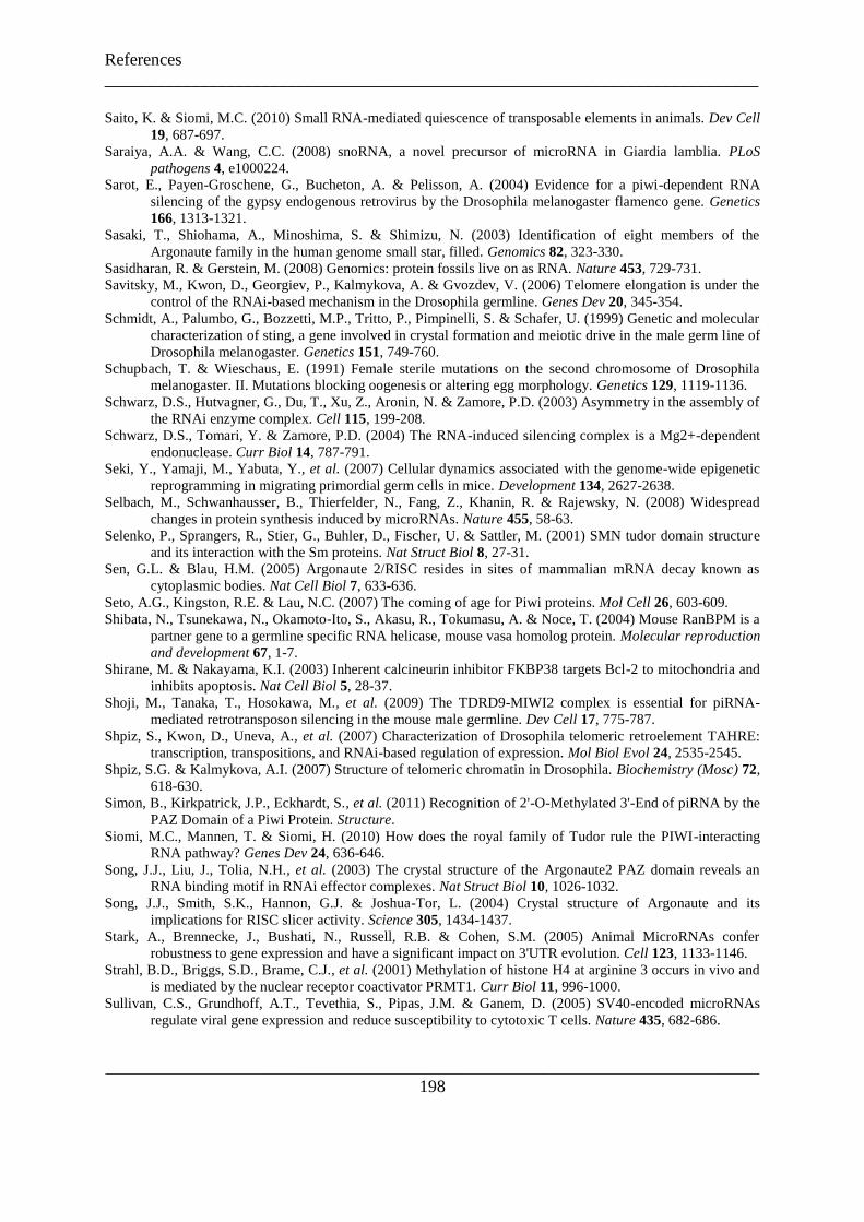
References
___________________________________________________________________________
___________________________________________________________________________
198
Saito, K. & Siomi, M.C. (2010) Small RNA-mediated quiescence of transposable elements in animals. Dev Cell
19, 687-697.
Saraiya, A.A. & Wang, C.C. (2008) snoRNA, a novel precursor of microRNA in Giardia lamblia. PLoS
pathogens 4, e1000224.
Sarot, E., Payen-Groschene, G., Bucheton, A. & Pelisson, A. (2004) Evidence for a piwi-dependent RNA
silencing of the gypsy endogenous retrovirus by the Drosophila melanogaster flamenco gene. Genetics
166, 1313-1321.
Sasaki, T., Shiohama, A., Minoshima, S. & Shimizu, N. (2003) Identification of eight members of the
Argonaute family in the human genome small star, filled. Genomics 82, 323-330.
Sasidharan, R. & Gerstein, M. (2008) Genomics: protein fossils live on as RNA. Nature 453, 729-731.
Savitsky, M., Kwon, D., Georgiev, P., Kalmykova, A. & Gvozdev, V. (2006) Telomere elongation is under the
control of the RNAi-based mechanism in the Drosophila germline. Genes Dev 20, 345-354.
Schmidt, A., Palumbo, G., Bozzetti, M.P., Tritto, P., Pimpinelli, S. & Schafer, U. (1999) Genetic and molecular
characterization of sting, a gene involved in crystal formation and meiotic drive in the male germ line of
Drosophila melanogaster. Genetics 151, 749-760.
Schupbach, T. & Wieschaus, E. (1991) Female sterile mutations on the second chromosome of Drosophila
melanogaster. II. Mutations blocking oogenesis or altering egg morphology. Genetics 129, 1119-1136.
Schwarz, D.S., Hutvagner, G., Du, T., Xu, Z., Aronin, N. & Zamore, P.D. (2003) Asymmetry in the assembly of
the RNAi enzyme complex. Cell 115, 199-208.
Schwarz, D.S., Tomari, Y. & Zamore, P.D. (2004) The RNA-induced silencing complex is a Mg2+-dependent
endonuclease. Curr Biol 14, 787-791.
Seki, Y., Yamaji, M., Yabuta, Y., et al. (2007) Cellular dynamics associated with the genome-wide epigenetic
reprogramming in migrating primordial germ cells in mice. Development 134, 2627-2638.
Selbach, M., Schwanhausser, B., Thierfelder, N., Fang, Z., Khanin, R. & Rajewsky, N. (2008) Widespread
changes in protein synthesis induced by microRNAs. Nature 455, 58-63.
Selenko, P., Sprangers, R., Stier, G., Buhler, D., Fischer, U. & Sattler, M. (2001) SMN tudor domain structure
and its interaction with the Sm proteins. Nat Struct Biol 8, 27-31.
Sen, G.L. & Blau, H.M. (2005) Argonaute 2/RISC resides in sites of mammalian mRNA decay known as
cytoplasmic bodies. Nat Cell Biol 7, 633-636.
Seto, A.G., Kingston, R.E. & Lau, N.C. (2007) The coming of age for Piwi proteins. Mol Cell 26, 603-609.
Shibata, N., Tsunekawa, N., Okamoto-Ito, S., Akasu, R., Tokumasu, A. & Noce, T. (2004) Mouse RanBPM is a
partner gene to a germline specific RNA helicase, mouse vasa homolog protein. Molecular reproduction
and development 67, 1-7.
Shirane, M. & Nakayama, K.I. (2003) Inherent calcineurin inhibitor FKBP38 targets Bcl-2 to mitochondria and
inhibits apoptosis. Nat Cell Biol 5, 28-37.
Shoji, M., Tanaka, T., Hosokawa, M., et al. (2009) The TDRD9-MIWI2 complex is essential for piRNA-
mediated retrotransposon silencing in the mouse male germline. Dev Cell 17, 775-787.
Shpiz, S., Kwon, D., Uneva, A., et al. (2007) Characterization of Drosophila telomeric retroelement TAHRE:
transcription, transpositions, and RNAi-based regulation of expression. Mol Biol Evol 24, 2535-2545.
Shpiz, S.G. & Kalmykova, A.I. (2007) Structure of telomeric chromatin in Drosophila. Biochemistry (Mosc) 72,
618-630.
Simon, B., Kirkpatrick, J.P., Eckhardt, S., et al. (2011) Recognition of 2'-O-Methylated 3'-End of piRNA by the
PAZ Domain of a Piwi Protein. Structure.
Siomi, M.C., Mannen, T. & Siomi, H. (2010) How does the royal family of Tudor rule the PIWI-interacting
RNA pathway? Genes Dev 24, 636-646.
Song, J.J., Liu, J., Tolia, N.H., et al. (2003) The crystal structure of the Argonaute2 PAZ domain reveals an
RNA binding motif in RNAi effector complexes. Nat Struct Biol 10, 1026-1032.
Song, J.J., Smith, S.K., Hannon, G.J. & Joshua-Tor, L. (2004) Crystal structure of Argonaute and its
implications for RISC slicer activity. Science 305, 1434-1437.
Stark, A., Brennecke, J., Bushati, N., Russell, R.B. & Cohen, S.M. (2005) Animal MicroRNAs confer
robustness to gene expression and have a significant impact on 3'UTR evolution. Cell 123, 1133-1146.
Strahl, B.D., Briggs, S.D., Brame, C.J., et al. (2001) Methylation of histone H4 at arginine 3 occurs in vivo and
is mediated by the nuclear receptor coactivator PRMT1. Curr Biol 11, 996-1000.
Sullivan, C.S., Grundhoff, A.T., Tevethia, S., Pipas, J.M. & Ganem, D. (2005) SV40-encoded microRNAs
regulate viral gene expression and reduce susceptibility to cytotoxic T cells. Nature 435, 682-686.

References
___________________________________________________________________________
___________________________________________________________________________
199
Sun, H., Li, D., Chen, S., et al. (2010) Zili inhibits transforming growth factor-beta signaling by interacting with
Smad4. J Biol Chem 285, 4243-4250.
Szakmary, A., Reedy, M., Qi, H. & Lin, H. (2009) The Yb protein defines a novel organelle and regulates male
germline stem cell self-renewal in Drosophila melanogaster. J Cell Biol 185, 613-627.
Tabara, H., Sarkissian, M., Kelly, W.G., et al. (1999) The rde-1 gene, RNA interference, and transposon
silencing in C. elegans. Cell 99, 123-132.
Tabara, H., Yigit, E., Siomi, H. & Mello, C.C. (2002) The dsRNA binding protein RDE-4 interacts with RDE-1,
DCR-1, and a DExH-box helicase to direct RNAi in C. elegans. Cell 109, 861-871.
Talbot, K., Miguel-Aliaga, I., Mohaghegh, P., Ponting, C.P. & Davies, K.E. (1998) Characterization of a gene
encoding survival motor neuron (SMN)-related protein, a constituent of the spliceosome complex. Hum
Mol Genet 7, 2149-2156.
Tam, O.H., Aravin, A.A., Stein, P., et al. (2008) Pseudogene-derived small interfering RNAs regulate gene
expression in mouse oocytes. Nature 453, 534-538.
Tanaka, S.S., Toyooka, Y., Akasu, R., et al. (2000) The mouse homolog of Drosophila Vasa is required for the
development of male germ cells. Genes Dev 14, 841-853.
Tang, G., Reinhart, B.J., Bartel, D.P. & Zamore, P.D. (2003) A biochemical framework for RNA silencing in
plants. Genes Dev 17, 49-63.
Thomson, T. & Lasko, P. (2004) Drosophila tudor is essential for polar granule assembly and pole cell
specification, but not for posterior patterning. Genesis 40, 164-170.
Tian, Y., Simanshu, D.K., Ma, J.B. & Patel, D.J. (2010) Structural basis for piRNA 2'-O-methylated 3'-end
recognition by Piwi PAZ (Piwi/Argonaute/Zwille) domains. Proc Natl Acad Sci U S A.
Tkaczuk, K.L., Obarska, A. & Bujnicki, J.M. (2006) Molecular phylogenetics and comparative modeling of
HEN1, a methyltransferase involved in plant microRNA biogenesis. BMC evolutionary biology 6, 6.
Tolia, N.H. & Joshua-Tor, L. (2007) Slicer and the argonautes. Nature chemical biology 3, 36-43.
Tomari, Y., Du, T. & Zamore, P.D. (2007) Sorting of Drosophila small silencing RNAs. Cell 130, 299-308.
Tomari, Y., Matranga, C., Haley, B., Martinez, N. & Zamore, P.D. (2004) A protein sensor for siRNA
asymmetry. Science 306, 1377-1380.
Tomari, Y. & Zamore, P.D. (2005) Perspective: machines for RNAi. Genes Dev 19, 517-529.
Toyooka, Y., Tsunekawa, N., Takahashi, Y., Matsui, Y., Satoh, M. & Noce, T. (2000) Expression and
intracellular localization of mouse Vasa-homologue protein during germ cell development. Mech Dev
93, 139-149.
Tran, R.K., Henikoff, J.G., Zilberman, D., Ditt, R.F., Jacobsen, S.E. & Henikoff, S. (2005) DNA methylation
profiling identifies CG methylation clusters in Arabidopsis genes. Curr Biol 15, 154-159.
Ueda, T., Abe, K., Miura, A., et al. (2000) The paternal methylation imprint of the mouse H19 locus is acquired
in the gonocyte stage during foetal testis development. Genes Cells 5, 649-659.
Unhavaithaya, Y., Hao, Y., Beyret, E., et al. (2009) MILI, a PIWI-interacting RNA-binding protein, is required
for germ line stem cell self-renewal and appears to positively regulate translation. J Biol Chem 284,
6507-6519.
Vagin, V.V., Klenov, M.S., Kalmykova, A.I., Stolyarenko, A.D., Kotelnikov, R.N. & Gvozdev, V.A. (2004)
The RNA interference proteins and vasa locus are involved in the silencing of retrotransposons in the
female germline of Drosophila melanogaster. RNA Biol 1, 54-58.
Vagin, V.V., Sigova, A., Li, C., Seitz, H., Gvozdev, V. & Zamore, P.D. (2006) A distinct small RNA pathway
silences selfish genetic elements in the germline. Science 313, 320-324.
Vagin, V.V., Wohlschlegel, J., Qu, J., et al. (2009) Proteomic analysis of murine Piwi proteins reveals a role for
arginine methylation in specifying interaction with Tudor family members. Genes Dev 23, 1749-1762.
Vasileva, A., Tiedau, D., Firooznia, A., Muller-Reichert, T. & Jessberger, R. (2009) Tdrd6 is required for
spermiogenesis, chromatoid body architecture, and regulation of miRNA expression. Curr Biol 19, 630-
639.
Vazquez, F. (2006) Arabidopsis endogenous small RNAs: highways and byways. Trends in plant science 11,
460-468.
Vazquez, F., Gasciolli, V., Crete, P. & Vaucheret, H. (2004a) The nuclear dsRNA binding protein HYL1 is
required for microRNA accumulation and plant development, but not posttranscriptional transgene
silencing. Curr Biol 14, 346-351.
Vazquez, F., Vaucheret, H., Rajagopalan, R., et al. (2004b) Endogenous trans-acting siRNAs regulate the
accumulation of Arabidopsis mRNAs. Mol Cell 16, 69-79.
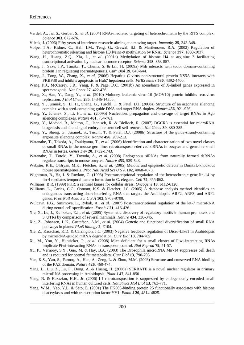
References
___________________________________________________________________________
___________________________________________________________________________
200
Verdel, A., Jia, S., Gerber, S., et al. (2004) RNAi-mediated targeting of heterochromatin by the RITS complex.
Science 303, 672-676.
Vilcek, J. (2006) Fifty years of interferon research: aiming at a moving target. Immunity 25, 343-348.
Volpe, T.A., Kidner, C., Hall, I.M., Teng, G., Grewal, S.I. & Martienssen, R.A. (2002) Regulation of
heterochromatic silencing and histone H3 lysine-9 methylation by RNAi. Science 297, 1833-1837.
Wang, H., Huang, Z.Q., Xia, L., et al. (2001a) Methylation of histone H4 at arginine 3 facilitating
transcriptional activation by nuclear hormone receptor. Science 293, 853-857.
Wang, J., Saxe, J.P., Tanaka, T., Chuma, S. & Lin, H. (2009a) Mili interacts with tudor domain-containing
protein 1 in regulating spermatogenesis. Curr Biol 19, 640-644.
Wang, J., Tong, W., Zhang, X., et al. (2006) Hepatitis C virus non-structural protein NS5A interacts with
FKBP38 and inhibits apoptosis in Huh7 hepatoma cells. FEBS letters 580, 4392-4400.
Wang, P.J., McCarrey, J.R., Yang, F. & Page, D.C. (2001b) An abundance of X-linked genes expressed in
spermatogonia. Nat Genet 27, 422-426.
Wang, X., Han, Y., Dang, Y., et al. (2010) Moloney leukemia virus 10 (MOV10) protein inhibits retrovirus
replication. J Biol Chem 285, 14346-14355.
Wang, Y., Juranek, S., Li, H., Sheng, G., Tuschl, T. & Patel, D.J. (2008a) Structure of an argonaute silencing
complex with a seed-containing guide DNA and target RNA duplex. Nature 456, 921-926.
Wang, Y., Juranek, S., Li, H., et al. (2009b) Nucleation, propagation and cleavage of target RNAs in Ago
silencing complexes. Nature 461, 754-761.
Wang, Y., Medvid, R., Melton, C., Jaenisch, R. & Blelloch, R. (2007) DGCR8 is essential for microRNA
biogenesis and silencing of embryonic stem cell self-renewal. Nat Genet 39, 380-385.
Wang, Y., Sheng, G., Juranek, S., Tuschl, T. & Patel, D.J. (2008b) Structure of the guide-strand-containing
argonaute silencing complex. Nature 456, 209-213.
Watanabe, T., Takeda, A., Tsukiyama, T., et al. (2006) Identification and characterization of two novel classes
of small RNAs in the mouse germline: retrotransposon-derived siRNAs in oocytes and germline small
RNAs in testes. Genes Dev 20, 1732-1743.
Watanabe, T., Totoki, Y., Toyoda, A., et al. (2008) Endogenous siRNAs from naturally formed dsRNAs
regulate transcripts in mouse oocytes. Nature 453, 539-543.
Webster, K.E., O'Bryan, M.K., Fletcher, S., et al. (2005) Meiotic and epigenetic defects in Dnmt3L-knockout
mouse spermatogenesis. Proc Natl Acad Sci U S A 102, 4068-4073.
Wightman, B., Ha, I. & Ruvkun, G. (1993) Posttranscriptional regulation of the heterochronic gene lin-14 by
lin-4 mediates temporal pattern formation in C. elegans. Cell 75, 855-862.
Williams, B.R. (1999) PKR; a sentinel kinase for cellular stress. Oncogene 18, 6112-6120.
Williams, L., Carles, C.C., Osmont, K.S. & Fletcher, J.C. (2005) A database analysis method identifies an
endogenous trans-acting short-interfering RNA that targets the Arabidopsis ARF2, ARF3, and ARF4
genes. Proc Natl Acad Sci U S A 102, 9703-9708.
Wulczyn, F.G., Smirnova, L., Rybak, A., et al. (2007) Post-transcriptional regulation of the let-7 microRNA
during neural cell specification. Faseb J 21, 415-426.
Xie, X., Lu, J., Kulbokas, E.J., et al. (2005) Systematic discovery of regulatory motifs in human promoters and
3' UTRs by comparison of several mammals. Nature 434, 338-345.
Xie, Z., Johansen, L.K., Gustafson, A.M., et al. (2004) Genetic and functional diversification of small RNA
pathways in plants. PLoS biology 2, E104.
Xie, Z., Kasschau, K.D. & Carrington, J.C. (2003) Negative feedback regulation of Dicer-Like1 in Arabidopsis
by microRNA-guided mRNA degradation. Curr Biol 13, 784-789.
Xu, M., You, Y., Hunsicker, P., et al. (2008) Mice deficient for a small cluster of Piwi-interacting RNAs
implicate Piwi-interacting RNAs in transposon control. Biol Reprod 79, 51-57.
Xu, P., Vernooy, S.Y., Guo, M. & Hay, B.A. (2003) The Drosophila microRNA Mir-14 suppresses cell death
and is required for normal fat metabolism. Curr Biol 13, 790-795.
Yan, K.S., Yan, S., Farooq, A., Han, A., Zeng, L. & Zhou, M.M. (2003) Structure and conserved RNA binding
of the PAZ domain. Nature 426, 468-474.
Yang, L., Liu, Z., Lu, F., Dong, A. & Huang, H. (2006a) SERRATE is a novel nuclear regulator in primary
microRNA processing in Arabidopsis. Plant J 47, 841-850.
Yang, N. & Kazazian, H.H., Jr. (2006) L1 retrotransposition is suppressed by endogenously encoded small
interfering RNAs in human cultured cells. Nat Struct Mol Biol 13, 763-771.
Yang, W.M., Yao, Y.L. & Seto, E. (2001) The FK506-binding protein 25 functionally associates with histone
deacetylases and with transcription factor YY1. Embo J 20, 4814-4825.

References
___________________________________________________________________________
___________________________________________________________________________
201
Yang, Z., Ebright, Y.W., Yu, B. & Chen, X. (2006b) HEN1 recognizes 21-24 nt small RNA duplexes and
deposits a methyl group onto the 2' OH of the 3' terminal nucleotide. Nucleic acids research 34, 667-
675.
Yekta, S., Shih, I.H. & Bartel, D.P. (2004) MicroRNA-directed cleavage of HOXB8 mRNA. Science 304, 594-
596.
Yeung, M.L., Bennasser, Y., Watashi, K., Le, S.Y., Houzet, L. & Jeang, K.T. (2009) Pyrosequencing of small
non-coding RNAs in HIV-1 infected cells: evidence for the processing of a viral-cellular double-
stranded RNA hybrid. Nucleic acids research 37, 6575-6586.
Yi, R., Qin, Y., Macara, I.G. & Cullen, B.R. (2003) Exportin-5 mediates the nuclear export of pre-microRNAs
and short hairpin RNAs. Genes Dev 17, 3011-3016.
Yin, H. & Lin, H. (2007) An epigenetic activation role of Piwi and a Piwi-associated piRNA in Drosophila
melanogaster. Nature 450, 304-308.
Yoda, M., Kawamata, T., Paroo, Z., et al. (2010) ATP-dependent human RISC assembly pathways. Nat Struct
Mol Biol 17, 17-23.
Yoshikawa, M., Peragine, A., Park, M.Y. & Poethig, R.S. (2005) A pathway for the biogenesis of trans-acting
siRNAs in Arabidopsis. Genes Dev 19, 2164-2175.
Yu, B., Bi, L., Zheng, B., et al. (2008) The FHA domain proteins DAWDLE in Arabidopsis and SNIP1 in
humans act in small RNA biogenesis. Proc Natl Acad Sci U S A 105, 10073-10078.
Yu, B., Yang, Z., Li, J., et al. (2005) Methylation as a crucial step in plant microRNA biogenesis. Science 307,
932-935.
Yuan, Y.R., Pei, Y., Ma, J.B., et al. (2005) Crystal structure of A. aeolicus argonaute, a site-specific DNA-
guided endoribonuclease, provides insights into RISC-mediated mRNA cleavage. Mol Cell 19, 405-419.
Zamore, P.D., Tuschl, T., Sharp, P.A. & Bartel, D.P. (2000) RNAi: double-stranded RNA directs the ATP-
dependent cleavage of mRNA at 21 to 23 nucleotide intervals. Cell 101, 25-33.
Zeng, Y. & Cullen, B.R. (2004) Structural requirements for pre-microRNA binding and nuclear export by
Exportin 5. Nucleic acids research 32, 4776-4785.
Zhang, J.F., Yuan, L.J., Shao, Y., Du, W., Yan, D.W. & Lu, Y.T. (2008) The disturbance of small RNA
pathways enhanced abscisic acid response and multiple stress responses in Arabidopsis. Plant, cell &
environment 31, 562-574.
Zheng, K., Xiol, J., Reuter, M., et al. (2010) Mouse MOV10L1 associates with Piwi proteins and is an essential
component of the Piwi-interacting RNA (piRNA) pathway. Proc Natl Acad Sci U S A.
Zhou, X., Zuo, Z., Zhou, F., et al. (2010) Profiling Sex-specific piRNAs in Zebrafish. Genetics.
Zhu, Q.H., Spriggs, A., Matthew, L., et al. (2008) A diverse set of microRNAs and microRNA-like small RNAs
in developing rice grains. Genome Res 18, 1456-1465.
Zilberman, D., Cao, X. & Jacobsen, S.E. (2003) ARGONAUTE4 control of locus-specific siRNA accumulation
and DNA and histone methylation. Science 299, 716-719.