swine influenza (h1n1) pneumonia: clinical considerations
TRANSCRIPT
Swine Influenza(H1N1) Pneumonia:Clinical Considerations
Burke A. Cunha, MD, MACPa,b,*
KEYWORDS
� Pandemic influenza � CPK � Relative lymphopenia� Thrombocytopenia � CA-MRSA CAP
HISTORY
Influenza is a viral zoonosis of birds and mammals that has probably existed sinceantiquity. In antiquity, populations were small and less concentrated, limiting influenzapotential. As animals became domesticated and lived in close proximity to humanpopulations, populations concentrated in urban centers, setting the stage for the intro-duction of zoonotic influenza into the human population. Influenza can maintain itself inrural populations because of ongoing changes in immunity mediated by influenza viralantigenic drift and shift. Because influenza immunity is strain-specific, influenza maybe maintained and may result in epidemics in large, closed urban populations. Attackrates of influenza are relatively high but mortality is relatively eg w 1 % low. Influenzamortality is highest in the very young, the very old, and the immunosuppressed. Influ-enza (human seasonal) has the potential for rapid spread and may involve large pop-ulations. Even though the mortality associated with epidemics is relatively low, thetotal number of fatalities involved may be huge. In the 1918–1919 influenza A(H1N1) pandemic, the mortality rate was 1%, but because massive numbers of peoplewere infected, there were 50 million fatalities.1–3
Clinical descriptions of influenza were first clearly described by Caus in 1551. TheEnglish physician described a ‘‘sweating disease’’ characterized by fever, headache,and myalgias that killed some patients rapidly but lasted only a few days in those thatsurvived. Although the English sweating sickness might have been due to influenza,the findings were nonspecific and did not resemble the first recognized influenzaepidemic, which occurred in Germany, England, and Italy in 1173. Subsequent influ-enza epidemics occurred in France and Italy in 1323 and 1387. Villaini and Seguiwere the first to use the term ‘‘una influenza,’’ referring to some ‘‘celestial influence’’that was thought to be responsible for the infection. The French described influenza
a Infectious Disease Division, Winthrop-University Hospital, 259 First Street, Mineola, LongIsland, NY 11501, USAb State University of New York School of Medicine, Stony Brook, NY, USA* Infectious Disease Division, Winthrop-University Hospital, Mineola, NY 11501.
Infect Dis Clin N Am 24 (2010) 203–228doi:10.1016/j.idc.2009.10.001 id.theclinics.com0891-5520/10/$ – see front matter ª 2010 Elsevier Inc. All rights reserved.
Cunha204
as the grip, because patients appeared to be gripped or seized by influenza. Thousandsof people were affected by these early epidemics but the first pandemic spread throughEurope in 1510. In 1679, Sydenem provided the first accurate clinical description ofinfluenza. In 1933, Smith isolated influenza A virus. Three years later, Burnette wasthe first to culture the influenza virus in embryonated eggs. In 1941, Hearst was the firstto describe the hemagglutination reactions of influenza. Frances, in 1939, was the firstto isolate influenza B and later in 1950, Taylor was the first to isolate influenza C.2–4
The great influenza pandemic of 1918 to 1919 was due to an H1N1 strain. In 1997,and again recently, outbreaks of avian influenza (H5N1) virus have occurred, affectinghumans as well as poultry. Since 1997, avian influenza (H5N1) emerged in terms oflethality with a high mortality rate ie, 60% among young healthy adults, and has asyet unrealized pandemic potential. Avian influenza (H5N1) strain resembles the 1918to 1919 pandemic strain. Fortunately, avian influenza (H5N1) outbreaks over thepast decade have been limited with minimal person-to-person spread. However, avianinfluenza (H5N1), like the influenza A (H1N1) strain of 1918 to 1919, has pandemicpotential if the virus mutates, permitting efficient person-to-person transmission.The influenza A (H1N1) pandemic of 1918 to 1919 was remarkable in many respects.The pandemic was unprecedented in its mortality and virulence, and was accompa-nied by 2 unusual sequelae, namely, encephalitis and postinfluenza Parkinsondisease.5–7 The 1918 to 1919 influenza A (H1N1) pandemic was noteworthy in thatthe death rate was highest in young healthy adults.6,8–10 The majority of the earlydeaths in young healthy individuals were due to influenza pneumonia alone and notsimultaneous/subsequent bacterial pneumonia.4,11–16
The swine influenza A (H1N1) pandemic began in 2009.17 Swine influenza (H1N1) isa novel influenza A virus comprising a reassortment of 4 distinct genetic elements,namely, swine, human, avian, and Eurasian swine genetic components, whichcombine into a single influenza virus, swine influenza (H1N1). The swine influenza(H1N1) pandemic rapidly spread from Mexico to the United States and the world inthe spring of 2009. The initial spread from Mexico was via tourists/travelers returninghome from Mexico. Foci of swine influenza (H1N1) strains became established acrosswide geographic areas.17–20 As with other influenza A pandemics, the initial ‘‘heraldwave’’ (in the late spring/summer) involved large numbers of individuals with relativelyfew deaths. Lethal swine influenza (H1N1) pneumonia affected primarily young andhealthy adults. The epidemiologic hallmark of pandemic influenza is its, ‘‘pandemicsignature,’’ ie, most early mortalities are among young healthy adults. Humanseasonal influenza affects primarily the very young, elderly, and those with comorbid-ities. Repeated passages through hosts, usually results in increased viral virulence.Excluding the effect of the (H1N1) vaccine, there is concern that subsequent wavesof swine influenza (H1N1) may be more lethal than the first.8,11,21
MICROBIOLOGY
Members of the family Orthomyxoviridae have a segmented negative strand RNAgenome. In the Orthomyxoviridae family are the influenza A, B, and C viruses. Thesingle-stranded RNA influenza viruses differ in the length of their RNA segments,that is, influenza A and B have 8 RNA segments whereas influenza C has 7 segments.Influenza A, B, and C viruses are spherical and covered by a lipid envelope from hostcell membrane. Influenza B and C have no subtypes. Subtypes are characteristic ofthe influenza A virus. Influenza A viruses are classified on the basis of their surfaceglycoproteins of the hemagglutinin (HA) and neuraminidase agglutinating (NA)proteins. Influenza A viruses have 16 different HA and 9 different NA subtypes.
Swine Influenza (H1N1) Pneumonia 205
However, only 3 HA subtypes and 2 NA subtypes have been implicated in human influ-enza epidemics. The 3 HA subtypes in human outbreaks are H1, H2, and H3, and the 2NA subtypes are N1 and N2. The surface HA proteins are HA glycoproteins in a rod-shaped spike with a hydrophobic carboxy terminal based in the viral envelope. Thehydrophobic end of the rod-shaped spike projects from the virus and is the bindingsite for sialic acid residues in host receptor cells. The HA surface glycoprotein isresponsible for viral adherence to the host cell, whereas the HA glycoprotein isresponsible for attachment of the virus and penetration of the influenza A virus intothe host cell. The NA surface glycoprotein is mushroom-shaped and is also implantedin the lipid envelope at the amino acid end of the glycoprotein. NA glycoproteincleaves terminal sialic acid residues, which are responsible for release of virus frominfected host cells and spread of the influenza virus to other cells in the respiratorytract. Influenza viruses also have M2 proteins, which are important in uncodingthe virus. M2 proteins, which are the matrix proteins, are present only in influenza Aviruses and appear on the surface of infected host cells. M1 proteins are surfacestructural proteins important in viral budding.22–24
Influenza A and B viruses bind to sialic acid receptors on the host cell surface andbegin the infectious process. Human influenza viruses attach preferentially to the a2, 6linkage to galactose containing oligosaccharides. In contrast, avian influenza virusespreferentially bind to a23 linkages. Influenza C viruses bind to 9-O-acetyl-N-acetyl-neuraminic acid receptors. Influenza A viruses are widely distributed in nature. Avianspecies are the primary reservoir for all 16 HA and 9 NA influenza A subtypes. Othernatural hosts of influenza A viruses are humans, horses, and swine. Human influenzaviruses replicate in respiratory epithelial cells, whereas avian influenza A virus isreplicated in respiratory and gastrointestinal epithelial cells.22–25
Swine influenza is an influenza A virus that consists of distinctive genetic influenza Aelements from swine, human, avian, and Eurasian swine strains of influenza (H1N1).The swine influenza (H1N1) pandemic was totally unpredicted. As with the ‘‘heraldwaves’’ of previous influenza A pandemics, large numbers of people were infectedbut relatively few died. The virulence of avian influenza (H5N1) remains worrisome.21
To date, avian influenza (H5N1) has been highly lethal in relatively small numbers ofpatients. It is thought that avian influenza (H5N1) is relatively inefficient in bird-to-human and human-to-human transmission, thus far limiting its human pandemicpotential. With the swine influenza (H1N1) pandemic, although mortality rates arelow, the potential for pandemic spread has been demonstrated during the spring/summer initial ‘‘herald wave’’. Lethal, swine influenza (H1N1) affects primarily younghealthy adults.5,6,22–25
EPIDEMIOLOGY
Influenza viruses are spread by airborne aerosols/droplets from infected individualswhile speaking, coughing, or sneezing. Fomites are also implicated in the transmissionof influenza. Nonhuman influenza infections are spread from respiratory/gastrointes-tinal infections of the host animal.2,3,26 Influenza viruses remain viable at cool temper-atures under conditions of low humidity on nonporous surfaces.
Influenza viruses may be detected in the general population throughout the year, butpeak in the winter months. In the northern hemisphere, peak human seasonal influenzaseason is in February whereas in the southern hemisphere, where winter months occurbetween May and August, flu usually peaks in June or July. Epidemics of influenza Aare characterized by a sudden increase in acute respiratory illnesses. In epidemics,usually influenza A or influenza B predominates, although both influenza A and B
Cunha206
may occur together in epidemics. Avian influenza (H5N1) may be transmitted fromdirect contact with infected birds or humans.2,5,22,23
Influenza has a short incubation period of approximately 2 days (range, 1–5 days).During the initial phase of the illness, there are high concentrations of influenza virusin respiratory secretions responsible for its spread via airborne transmission. One ofthe characteristics of influenza viruses is their changing antigenicity. Minor changesin surface glycoproteins resulting from stepwise point mutations in HA/NA glycopro-teins are called ‘‘antigenic drift.’’ Sequential amino acid changes occurring overa period of years resulting in HA/NA glycoprotein changes occur every 2 to 3 yearsand are termed ‘‘antigenic shift.’’ Influenza epidemics occur every 6 to 10 years andare due to antigenic shifts exposing the population to strains to which it has notbeen exposed previously. For this reason, pandemic influenza A has a high attackrate in individuals of all age groups who are not susceptible, particularly children.Unlike epidemics of influenza that occur during the winter months, pandemic influenzamay occur at any time and spread rapidly from country to country, facilitated by travel.The twentieth century has experienced 3 influenza pandemics, 2 of which haveemerged from China. The highest late excess fatalities occur are among the elderly,who succumb to influenza because of comorbid conditions/bacterial complications,whereas early deaths among young healthy patients are most often due to severeinfluenza pneumonia alone.5,22–26
Influenza has also been responsible for nosocomial outbreaks. Nosocomial influ-enza is usually due to influenza A and, less commonly, influenza B strains. In contrastto community-acquired infections, nosocomial outbreaks are brief, lasting 1 to 3weeks versus months in community-acquired outbreaks. Health care workers maybe important in initiating or spreading influenza in nursing homes, chronic care facili-ties, or hospitals. Nosocomial influenza has occurred in immunized hosts, suggestingthat there is suboptimal protection in elderly nursing home residents.23–26
Swine influenza (H1N1) is spread primarily via aerosols/droplets and to a lesserextent via hand-to-face transmission. Avian (H5N1) and swine influenza (H1N1) maybe transmitted in aircraft (unrecirculated cabin air) via aerosol/droplet transmission.Because swine influenza (H1N1), like avian influenza (H5N1), is often accompaniedby gastrointestinal symptoms (ie, diarrhea), there may be potential for viral spreadfrom feces. As with seasonal human influenza A, swine influenza (H1N1) may bespread nosocomially.27
CLINICAL PRESENTATION
Mild to moderate influenza is clinically indistinguishable from illnesses caused by otherrespiratory viruses that present as influenzalike illnesses (ILIs). Peak incidence of influ-enza historically is in February in the northern hemisphere, but the influenza season maystart earlier and last into early spring. Avian influenza (H5N1) has a nonseasonal distri-bution, and swine influenza (H1N1) has occurred in summer and winter. Excludingpandemics and epidemics, influenza is a relatively mild 3-day illness, that is, a mildrespiratory infection with a high attack rate and low mortality. Pandemic influenzaalso has a high attack rate, but due to highly virulent strains has a high mortality.22–25,28
In adults, severe influenza A has a distinctive clinical presentation, which is noteasily confused with other causes of viral pneumonia.23,29,30 Like severe acute respi-ratory syndrome (SARS), hantavirus pulmonary syndrome (HPS), and adenovirus,severe influenza A pneumonia begins abruptly, but influenza A patients often can recallthe exact time of onset of the illness. Severe human seasonal influenza A is accompa-nied by fever higher than 39�C/102�F and chills, accompanied by severe myalgias and
Swine Influenza (H1N1) Pneumonia 207
dry cough with or without hemoptysis. Purulent sputum suggests superimposedbacterial community-acquired pneumonia (CAP) due to MSSA/CA-MRSA. Anothernoteworthy feature of human seasonal influenza is severe debilitating fatigue rapidlymaking victims bedridden. Prostration is profound and is accompanied by headacheand prominent myalgias. Some infections present with myalgias, but the myalgias ofinfluenza are peculiarly severe and are localized to the neck and back. Retro-orbitalpain is common, and conjunctival suffusion may be present in severe seasonal humaninfluenza A. Conjunctival suffusion may be present in avian influenza (H5N1) but isuncommon with swine influenza (H1N1). The clinical constellation of an ILI with feverhigher than 39�C/102�F and severe myalgias especially of the neck/back, with profound prostration, clinically differentiates severe influenza A in adultsfrom ILIs (Table 1).22–25,28,30
The clinical presentation of avian influenza (H5N1) resembles severe human influ-enza A in adults and affects primarily young healthy adults, whereas nonepidemicpandemic/epidemic human influenza A primarily affects the very young, elderly, anddebilitated. Pandemic influenza A affects primarily young healthy adults (ie,‘‘pandemic signature’’) rather than the very young, elderly, and immunocompromised.Swine influenza (H1N1) has demonstrated its ‘‘pandemic signature,’’ that is, most fatalcases have occurred in young healthy adults.28,31
Several nonspecific laboratory test abnormalities are associated with human influ-enza A in adults. The severity of human influenza A is related to the degree/durationof leukopenia and relative lymphopenia.31,32 The nonspecific laboratory test hallmarkof influenza A (human, avian, and swine) is otherwise unexplained relative lymphope-nia. Relative lymphopenia is an inconsistent feature of swine influenza (H1N1) in chil-dren; lymphocytosis is more common. In adults, relative lymphopenia is usuallyaccompanied by thrombocytopenia. In adults with human and avian (H5N1) influenza,leukopenia if present may be an indicator of severity. Leukopenia in swine influenza(H1N1) when present usually occurs with relative lymphopenia and thrombocyto-penia.33 Atypical lymphocytes are not a usual feature of swine influenza (H1N1) inadults but may be present in children.
From a radiographic perspective, influenza (human, avian, swine) has no distinctivefeatures. Initially, the chest radiograph (CXR) shows no or minimal infiltrates. Later,(>48 hours) bilateral patchy interstitial infiltrates appear as influenza pneumonia prog-resses. The presence of focal segmental/lobar infiltrates on the admission CXR ofadults with influenza indicates simultaneous bacterial CAP, due to Staphylococcusaureus (MSSA/CA-MRSA) CAP.30 Patients with influenza/ILI and simultaneousS aureus CAP present with purulent sputum, high spiking fevers, often withcyanosis/hypotension. S aureus CAP, whether due to methicillin-sensitive S aureus(MSSA) or methicillin-resistant S aureus (MRSA), presents in the same way clinicallyand on CXR. The CXR of S aureus (MSSA/MRSA) CAP in patients with influenza pneu-monia are characterized by rapid cavitation in 72 hours or less. In adults, S aureus(MSSA/MRSA) CAP occurs only in patients with influenza/ILI (Table 2).30,31,34–38
Gastrointestinal symptoms (ie, nausea, vomiting, or diarrhea) are more commonwith avian and swine influenza (H1N1) than with human influenza. Serum aspartateand alanine transaminases (AST/ALT) are often mildly/transiently elevated in avian(H5N1) and swine influenza (H1N1). Another key laboratory finding in swine influenza(H1N1) patients is an elevated creatine phosphokinase (CPK). Some patients withhighly elevated CPKs may also have rhabdomyolysis. Seasonal human influenza Aand avian influenza A (H5N1) may have mild to moderate elevations of the serumlactate dehydrogenase (LDH). Whereas certain nonspecific laboratory abnormalitiesare typical of swine influenza (H1N1), for example, otherwise unexplained relative
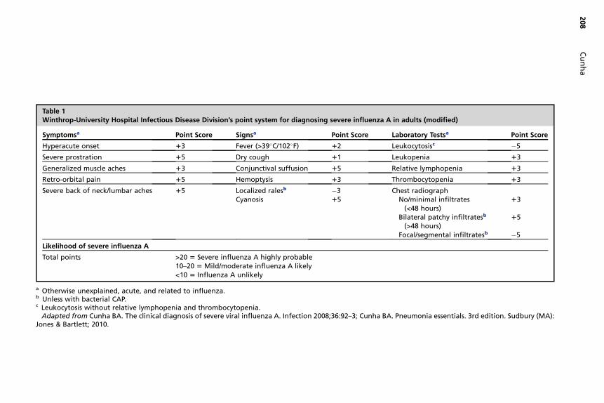
Table 1Winthrop-University Hospital Infectious Disease Division’s point system for diagnosing severe influenza A in adults (modified)
Symptomsa Point Score Signsa Point Score Laboratory Testsa Point Score
Hyperacute onset 13 Fever (>39�C/102�F) 12 Leukocytosisc �5
Severe prostration 15 Dry cough 11 Leukopenia 13
Generalized muscle aches 13 Conjunctival suffusion 15 Relative lymphopenia 13
Retro-orbital pain 15 Hemoptysis 13 Thrombocytopenia 13
Severe back of neck/lumbar aches 15 Localized ralesb �3 Chest radiographCyanosis 15 No/minimal infiltrates
(<48 hours)13
Bilateral patchy infiltratesb
(>48 hours)15
Focal/segmental infiltratesb �5
Likelihood of severe influenza A
Total points >20 5 Severe influenza A highly probable10–20 5 Mild/moderate influenza A likely<10 5 Influenza A unlikely
a Otherwise unexplained, acute, and related to influenza.b Unless with bacterial CAP.c Leukocytosis without relative lymphopenia and thrombocytopenia.
Adapted from Cunha BA. The clinical diagnosis of severe viral influenza A. Infection 2008;36:92–3; Cunha BA. Pneumonia essentials. 3rd edition. Sudbury (MA):Jones & Bartlett; 2010.
Cu
nh
a208
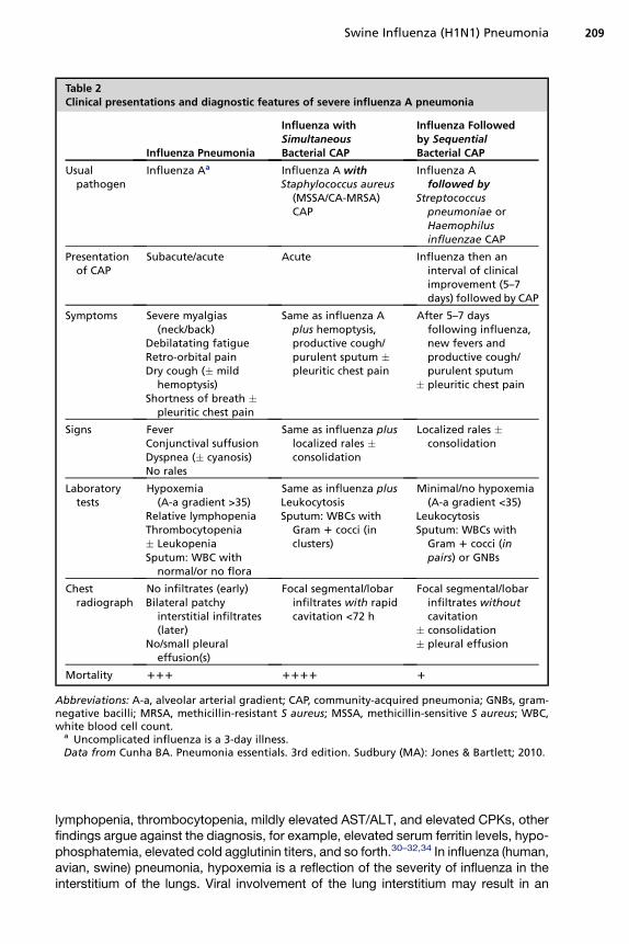
Table 2Clinical presentations and diagnostic features of severe influenza A pneumonia
Influenza Pneumonia
Influenza withSimultaneousBacterial CAP
Influenza Followedby SequentialBacterial CAP
Usualpathogen
Influenza Aa Influenza A withStaphylococcus aureus
(MSSA/CA-MRSA)CAP
Influenza Afollowed by
Streptococcuspneumoniae orHaemophilusinfluenzae CAP
Presentationof CAP
Subacute/acute Acute Influenza then aninterval of clinicalimprovement (5–7days) followed by CAP
Symptoms Severe myalgias(neck/back)
Debilatating fatigueRetro-orbital painDry cough (� mild
hemoptysis)Shortness of breath �
pleuritic chest pain
Same as influenza Aplus hemoptysis,productive cough/purulent sputum �pleuritic chest pain
After 5–7 daysfollowing influenza,new fevers andproductive cough/purulent sputum
� pleuritic chest pain
Signs FeverConjunctival suffusionDyspnea (� cyanosis)No rales
Same as influenza pluslocalized rales �consolidation
Localized rales �consolidation
Laboratorytests
Hypoxemia(A-a gradient >35)
Relative lymphopeniaThrombocytopenia� LeukopeniaSputum: WBC with
normal/or no flora
Same as influenza plusLeukocytosisSputum: WBCs with
Gram 1 cocci (inclusters)
Minimal/no hypoxemia(A-a gradient <35)
LeukocytosisSputum: WBCs with
Gram 1 cocci (inpairs) or GNBs
Chestradiograph
No infiltrates (early)Bilateral patchy
interstitial infiltrates(later)
No/small pleuraleffusion(s)
Focal segmental/lobarinfiltrates with rapidcavitation <72 h
Focal segmental/lobarinfiltrates withoutcavitation
� consolidation� pleural effusion
Mortality 111 1111 1
Abbreviations: A-a, alveolar arterial gradient; CAP, community-acquired pneumonia; GNBs, gram-negative bacilli; MRSA, methicillin-resistant S aureus; MSSA, methicillin-sensitive S aureus; WBC,white blood cell count.
a Uncomplicated influenza is a 3-day illness.Data from Cunha BA. Pneumonia essentials. 3rd edition. Sudbury (MA): Jones & Bartlett; 2010.
Swine Influenza (H1N1) Pneumonia 209
lymphopenia, thrombocytopenia, mildly elevated AST/ALT, and elevated CPKs, otherfindings argue against the diagnosis, for example, elevated serum ferritin levels, hypo-phosphatemia, elevated cold agglutinin titers, and so forth.30–32,34 In influenza (human,avian, swine) pneumonia, hypoxemia is a reflection of the severity of influenza in theinterstitium of the lungs. Viral involvement of the lung interstitium may result in an
Cunha210
oxygen diffusion defect manifested as hypoxemia and an increased A-a gradient. Thegreater the degree/duration of hypoxemia (A-a gradient >35), the more severe andpotentially fatal is influenza (human, avian, swine) pneumonia.30,31
As with avian influenza (H5N1), hospitalized adults with swine influenza (H1N1)pneumonia rarely present with or subsequently develop bacterial pneumoniaCAP.33,36 During the 1957 to 1958 Asian influenza A pandemic, some patients pre-sented with influenza A pneumonia and simultaneous S aureus CAP. Still others,especially the elderly, later developed Streptococcus pneumoniae or Haemophilusinfluenzae CAP 1 to 2 weeks into their recovery. As with avian influenza (H5N1),young healthy adults with swine influenza (H1N1) pneumonia, have only rarely pre-sented with superimposed S aureus (MSSA/CA-MRSA) CAP. Adult swine influenza(H1N1), like avian influenza (H5N1) pneumonia simultaneous or subsequent bacterialCAP remains relatively rare.36–40
DIFFERENTIAL DIAGNOSIS
The differential diagnosis of severe viral CAPs in normal hosts includes cytomegalo-virus (CMV), and adenovirus, and in compromised hosts RSV and CMV. Viral pneumo-nias presenting as severe CAPs have a similar radiological appearance, that is, no/minimal infiltrates early (<48 hours), followed later (>48 hours) by bilateral diffusepatchy interstitial infiltrates. Adult influenza A pneumonia (human, avian, swine) aswell as other severe viral CAPs are accompanied by variable degrees of hypoxemiaand a high A-a gradient >35. Bacterial CAP rarely, if ever, complicates severe CMVor adenoviral CAP.30
In general, viral pneumonias are not accompanied by pleuritic chest pain becausepathophysiologically they are interstitial and not pleurally based but influenza A maybe accompanied by bilateral ‘‘pleuritic’’ chest pain. The ‘‘pleuritic chest pain’’ of influ-enza A pneumonia is due to direct viral involvement of the intercostal musclesmimicking pleuritic chest pain. The pleuritic chest pain of bacterial CAPs is unilateraland related to the location of the underlying infiltrate.30 Influenza/ILI complicated bysimultaneous CA-MRSA/MSSA CAP is characterized by a necrotizing pneumoniawith rapidly cavitating infiltrates on CXR with or without pleuritic chest pain. MSSA/CA-MRSA (PVL1 strains) with influenza/ILI is often accompanied by cyanosis/hypo-tension (see Table 2).36–40
Swine influenza (H1N1) resembles avian influenza (H5N1) in its predilection foryoung healthy adults.39–50 The epidemiologic clue to avian influenza (H5N1) is recentclose contact with infected poultry or people in Europe or Asia. The diagnosis of avianinfluenza (H5/N1) may be missed if the hemagglutinin inhibition (HI) test is used todiagnose influenza A because this test is insensitive to avian hemagglutinins.30,39
The diagnosis of avian influenza (H5N1) is by hemagglutinin-specific reverse transcrip-tion-polymerase chain reaction (RT-PCR) for avian influenza (H5N1).
The other illness that does not clinically resemble influenza A is SARS. Unlike adulthuman seasonal influenza A, loose stools/diarrhea occurs in some and the illness lastslonger than 3 days. In contrast to influenza/avian influenza, the white blood cell (WBC)count and platelet counts in SARS are usually normal but may be slightly decreased.Relative lymphopenia is present, as are mild increases in the serum transaminases(AST/ALT). Like seasonal human influenza A, elevations of the CPK and LDH are notuncommon. Diagnosis of SARS is by specific serology or viral isolation.30,51
The clinical clue to SARS is the recent contact history with infected poultry orsomeone with SARS from Europe or Asia. Unlike seasonal human influenza A,SARS is a biphasic infection. At onset, fever decreases after a few days and the
Swine Influenza (H1N1) Pneumonia 211
patient improves, but unlike seasonal human influenza A, fever recurs in a few dayswhen the patient re-presents with signs and symptoms of viral pneumonia ie, CXRshows bilateral patchy interstitial infiltrates. Like avian influenza (H5N1), SARS hasnot been complicated by bacterial CAP.30,50,51
In the differential diagnosis of influenza/ILIs, HPS should be considered if there hasbeen close contact usually after about 2 weeks postexposure to a rodent.52 LikeSARS, HPS is a biphasic illness, that is, the initial phase is the febrile phase, followedby the cardiopulmonary phase. Later, HPS patients progress through an oliguric/diuretic phase and finally to a convalescent phase. The distinguishing clinical featureof HPS is noncardiogenic pulmonary edema. HPS is not a 3-day illness like influenza.In HPS leukocytosis (up to 90,000/mm3) rather than leukopenia is the rule. The nonspe-cific laboratory hallmark of HPS is the presence of immunoblasts.30,52 Diagnosis of HPSis by IgM enzyme-linked immunosorbent assay or RT-PCR (Table 3).
CLINICAL DIAGNOSIS
Sporadic seasonal human influenza A cases may occur throughout the year, but influ-enza activity in the northern hemisphere peaks in February (November–March), and inthe southern hemisphere in July (May–September). Influenza occurring during thepeak influenza season is often more severe than that which occurs sporadicallythroughout the year.22–25
In adults with swine influenza (H1N1), pneumonia presents in adults as an ILI witha temperature higher than 39�C (102�F) accompanied by prominent myalgias; theseare the key clinical findings. Many patients also have headache, sore throat, or drycough, with or without loose stools/diarrhea. Conjunctival suffusion is rare. The onsetof swine influenza (H1N1) pneumonia is often abrupt but a preceeding ILI prodrome iscommon. Mild cases of swine influenza (H1N1) are indistinguishable from ILIs. Adultpatients who require hospitalization have characteristic clinical nonspecific laboratoryabnormalities.17–20
In adults hospitalized with swine influenza (H1N1) pneumonia, otherwise unex-plained relative lymphopenia is uniformly present.34 However, clinicians must be care-ful to not ascribe relative lymphopenia in patients to swine influenza (H1N1)pneumonia until the patient’s medical history is reviewed for other disorders associ-ated with relative lymphopenia. Besides human seasonal and avian influenza otherinfectious causes of relative lymphopenia include CMV, human herpesvirus (HHV)-6,HHV-8, human immunodeficiency virus (HIV), miliary tuberculosis (TB), Legionnaire’sdisease, typhoid fever, Q fever, brucellosis, SARS, malaria, babesiosis, Rocky Moun-tain spotted fever (RMSF), histoplasmosis, dengue fever, Chickungunya fever, ehrli-chiosis, parvovirus B19, HPS, West Nile encephalitis (WNE), and viral hepatitis(early). Noninfectious causes of relative lymphopenia include cytotoxic drugs,steroids, sarcoidosis, systemic lupus erythematosus (SLE), lymphoma, rheumatoidarthritis (RA), radiation therapy, Wiskott-Aldrich syndrome, Whipple’s disease, severecombined immunodeficiency disease (SCID), common variable immune deficiency(CVID), Di George’s syndrome, Nezelof’s syndrome, intestinal lymphangiectasia,constrictive pericarditis, tricuspid regurgitation, Kawasaki’s disease, idiopathic CD4
cytopenia, Wegener’s granulomatosis, acute/chronic renal failure, hemodialysis;myasthenia gravis, celiac disease, alcoholic cirrhosis, coronary bypass, congestiveheart failure (CHF), acute pancreatitis, and carcinomas.30
Relative lymphopenia with swine influenza (H1N1) pneumonia may be profound andprolonged. The degree/duration of relative lymphopenia also has prognostic implica-tions. Typically, an increase in the percentage of lymphocytes precedes clinical
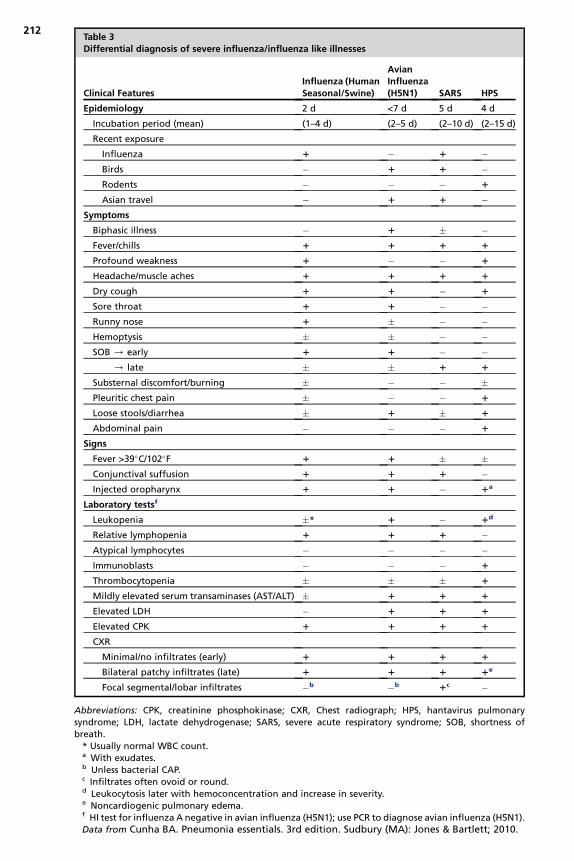
Table 3Differential diagnosis of severe influenza/influenza like illnesses
Clinical FeaturesInfluenza (HumanSeasonal/Swine)
AvianInfluenza(H5N1) SARS HPS
Epidemiology 2 d <7 d 5 d 4 d
Incubation period (mean) (1–4 d) (2–5 d) (2–10 d) (2–15 d)
Recent exposure
Influenza 1 � 1 �Birds � 1 1 �Rodents � � � 1
Asian travel � 1 1 �Symptoms
Biphasic illness � 1 � �Fever/chills 1 1 1 1
Profound weakness 1 � � 1
Headache/muscle aches 1 1 1 1
Dry cough 1 1 � 1
Sore throat 1 1 � �Runny nose 1 � � �Hemoptysis � � � �SOB / early 1 1 � �
/ late � � 1 1
Substernal discomfort/burning � � � �Pleuritic chest pain � � � 1
Loose stools/diarrhea � 1 � 1
Abdominal pain � � � 1
Signs
Fever >39�C/102�F 1 1 � �Conjunctival suffusion 1 1 1 �Injected oropharynx 1 1 � 1a
Laboratory testsf
Leukopenia �* 1 � 1d
Relative lymphopenia 1 1 1 �Atypical lymphocytes � � � �Immunoblasts � � � 1
Thrombocytopenia � � � 1
Mildly elevated serum transaminases (AST/ALT) � 1 1 1
Elevated LDH � 1 1 1
Elevated CPK 1 1 1 1
CXR
Minimal/no infiltrates (early) 1 1 1 1
Bilateral patchy infiltrates (late) 1 1 1 1e
Focal segmental/lobar infiltrates �b �b 1c �
Abbreviations: CPK, creatinine phosphokinase; CXR, Chest radiograph; HPS, hantavirus pulmonarysyndrome; LDH, lactate dehydrogenase; SARS, severe acute respiratory syndrome; SOB, shortness ofbreath.
* Usually normal WBC count.a With exudates.b Unless bacterial CAP.c Infiltrates often ovoid or round.d Leukocytosis later with hemoconcentration and increase in severity.e Noncardiogenic pulmonary edema.f HI test for influenza A negative in avian influenza (H5N1); use PCR to diagnose avian influenza (H5N1).Data from Cunha BA. Pneumonia essentials. 3rd edition. Sudbury (MA): Jones & Bartlett; 2010.
212
Swine Influenza (H1N1) Pneumonia 213
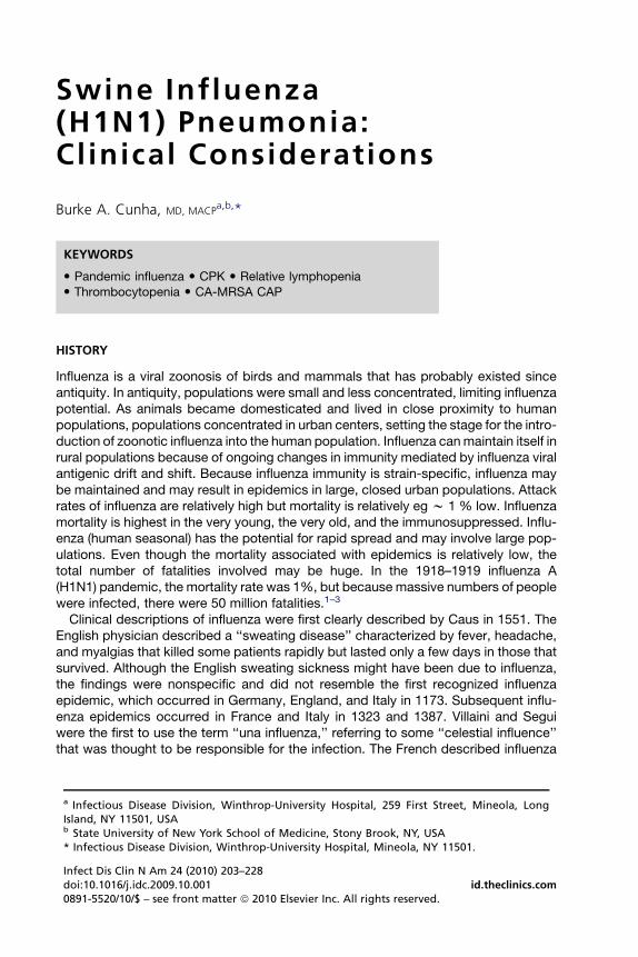
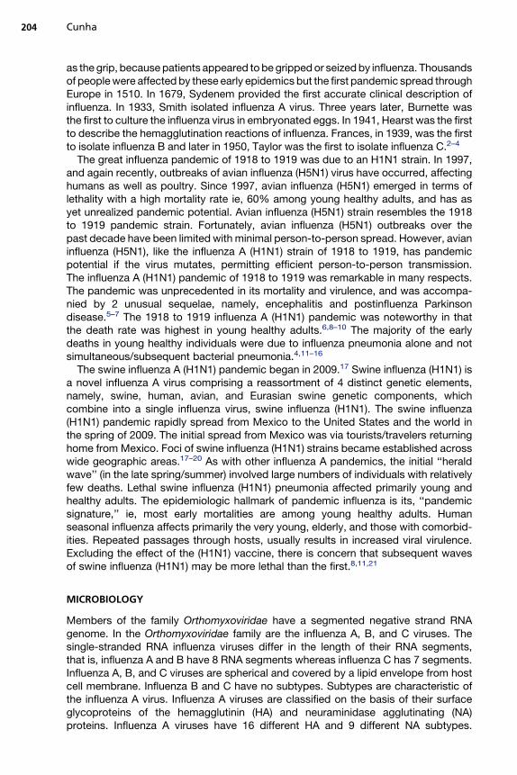
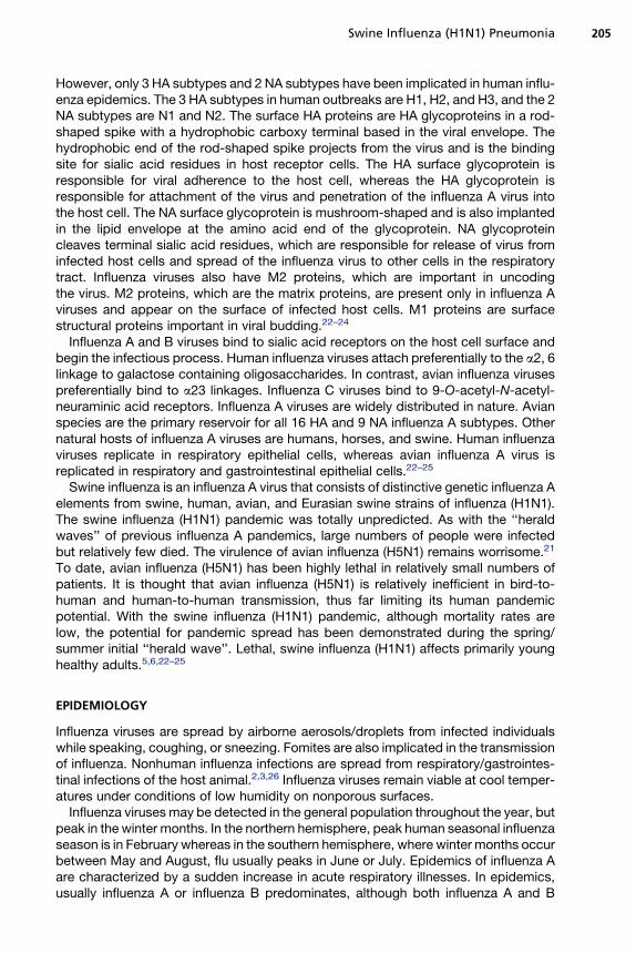
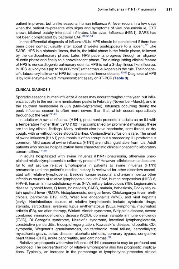
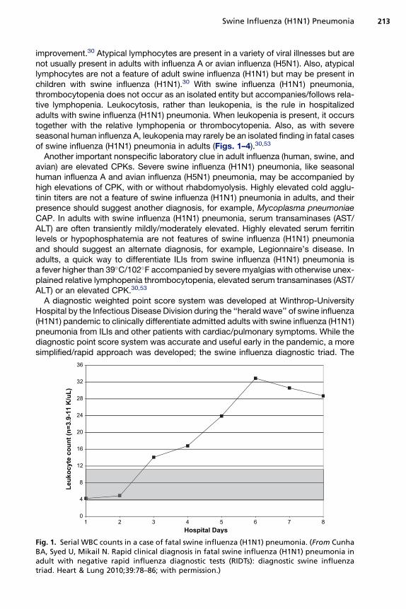
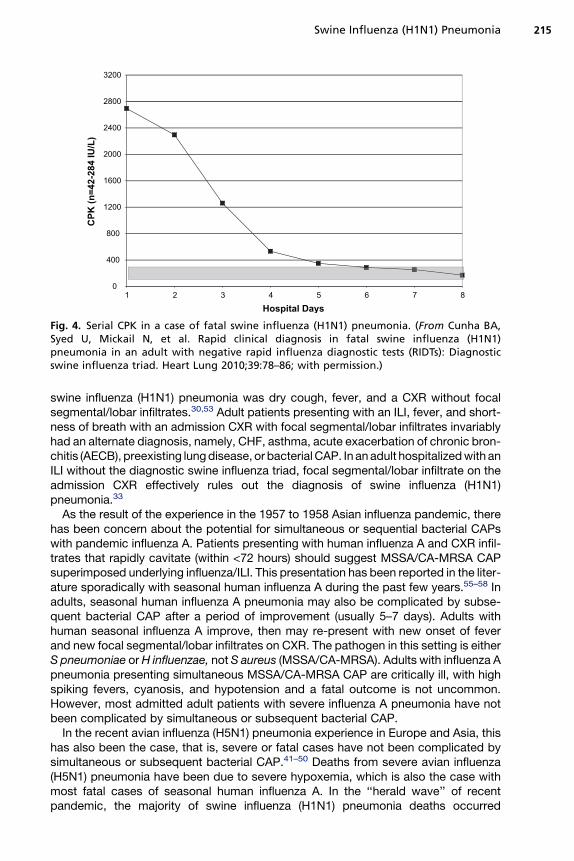
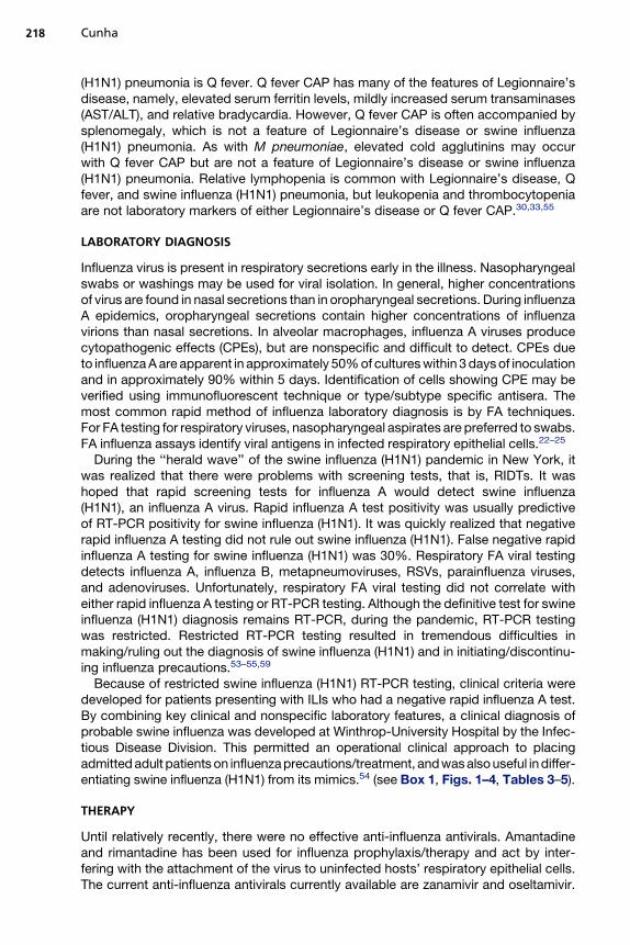
improvement.30 Atypical lymphocytes are present in a variety of viral illnesses but arenot usually present in adults with influenza A or avian influenza (H5N1). Also, atypicallymphocytes are not a feature of adult swine influenza (H1N1) but may be present inchildren with swine influenza (H1N1).30 With swine influenza (H1N1) pneumonia,thrombocytopenia does not occur as an isolated entity but accompanies/follows rela-tive lymphopenia. Leukocytosis, rather than leukopenia, is the rule in hospitalizedadults with swine influenza (H1N1) pneumonia. When leukopenia is present, it occurstogether with the relative lymphopenia or thrombocytopenia. Also, as with severeseasonal human influenza A, leukopenia may rarely be an isolated finding in fatal casesof swine influenza (H1N1) pneumonia in adults (Figs. 1–4).30,53
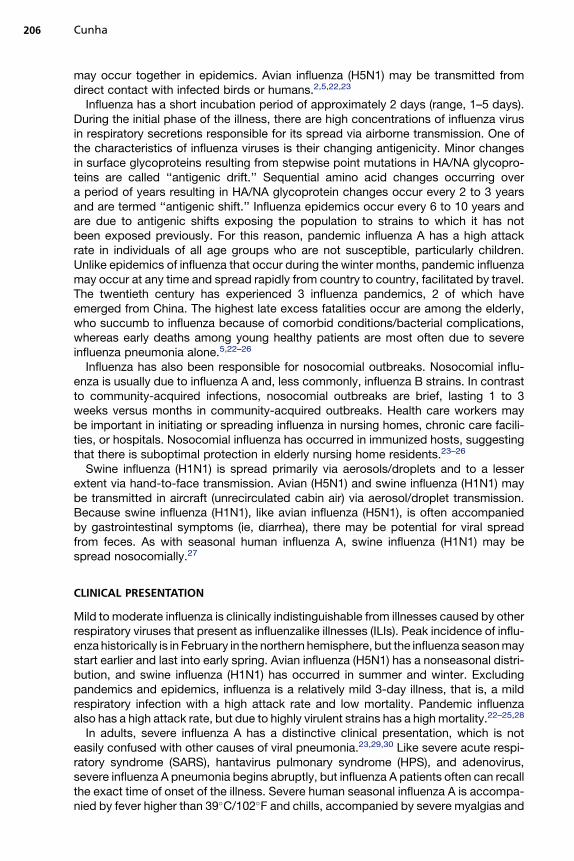
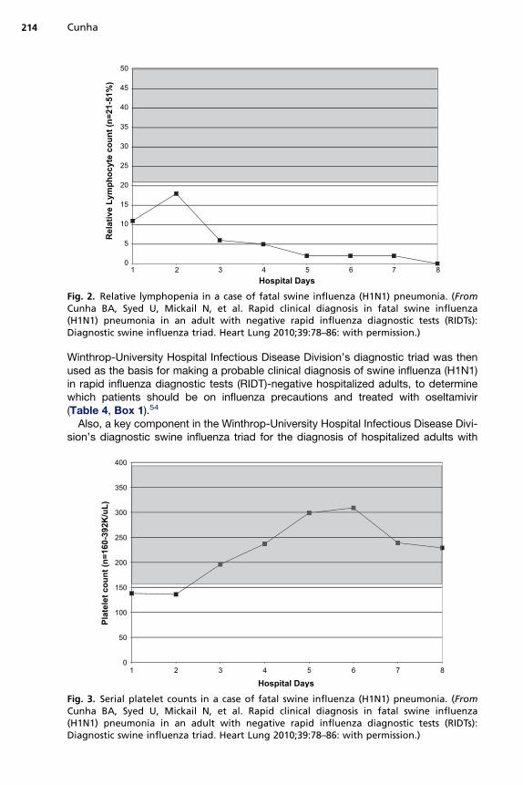
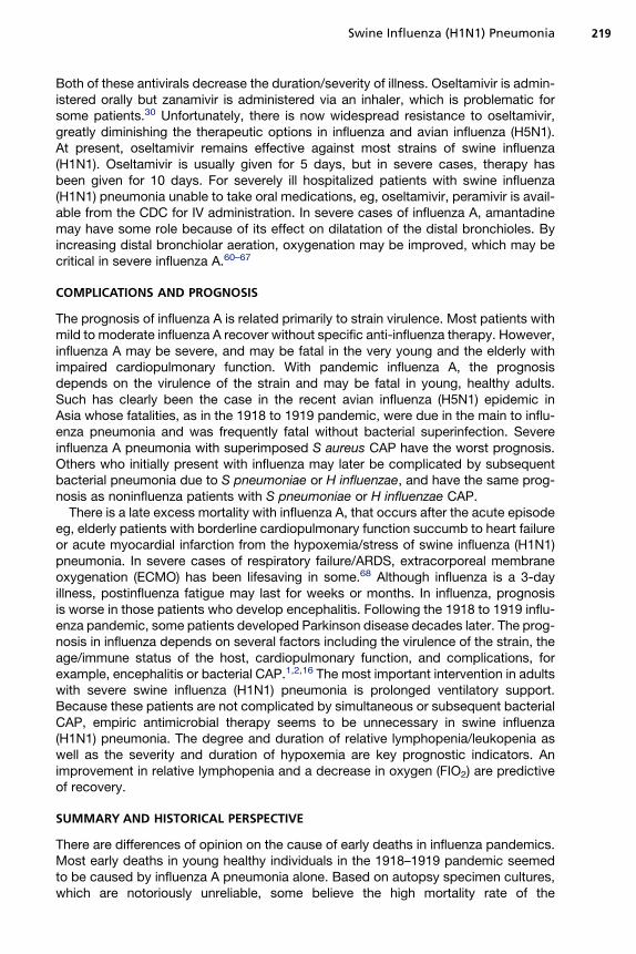
Another important nonspecific laboratory clue in adult influenza (human, swine, andavian) are elevated CPKs. Severe swine influenza (H1N1) pneumonia, like seasonalhuman influenza A and avian influenza (H5N1) pneumonia, may be accompanied byhigh elevations of CPK, with or without rhabdomyolysis. Highly elevated cold agglu-tinin titers are not a feature of swine influenza (H1N1) pneumonia in adults, and theirpresence should suggest another diagnosis, for example, Mycoplasma pneumoniaeCAP. In adults with swine influenza (H1N1) pneumonia, serum transaminases (AST/ALT) are often transiently mildly/moderately elevated. Highly elevated serum ferritinlevels or hypophosphatemia are not features of swine influenza (H1N1) pneumoniaand should suggest an alternate diagnosis, for example, Legionnaire’s disease. Inadults, a quick way to differentiate ILIs from swine influenza (H1N1) pneumonia isa fever higher than 39�C/102�F accompanied by severe myalgias with otherwise unex-plained relative lymphopenia thrombocytopenia, elevated serum transaminases (AST/ALT) or an elevated CPK.30,53
A diagnostic weighted point score system was developed at Winthrop-UniversityHospital by the Infectious Disease Division during the ‘‘herald wave’’ of swine influenza(H1N1) pandemic to clinically differentiate admitted adults with swine influenza (H1N1)pneumonia from ILIs and other patients with cardiac/pulmonary symptoms. While thediagnostic point score system was accurate and useful early in the pandemic, a moresimplified/rapid approach was developed; the swine influenza diagnostic triad. The
0
4
8
12
16
20
24
28
32
36
87654321Hospital Days
Leu
ko
cyte co
un
t (n
=3.9-11 K
/u
L)
Fig. 1. Serial WBC counts in a case of fatal swine influenza (H1N1) pneumonia. (From CunhaBA, Syed U, Mikail N. Rapid clinical diagnosis in fatal swine influenza (H1N1) pneumonia inadult with negative rapid influenza diagnostic tests (RIDTs): diagnostic swine influenzatriad. Heart & Lung 2010;39:78–86; with permission.)
0
5
10
15
20
25
30
35
40
45
50
1 2 3 4 5 6 7 8
Relative L
ym
ph
ocyte co
un
t (n
=21-51%
)
Hospital Days
Fig. 2. Relative lymphopenia in a case of fatal swine influenza (H1N1) pneumonia. (FromCunha BA, Syed U, Mickail N, et al. Rapid clinical diagnosis in fatal swine influenza(H1N1) pneumonia in an adult with negative rapid influenza diagnostic tests (RIDTs):Diagnostic swine influenza triad. Heart Lung 2010;39:78–86: with permission.)
Cunha214
Winthrop-University Hospital Infectious Disease Division’s diagnostic triad was thenused as the basis for making a probable clinical diagnosis of swine influenza (H1N1)in rapid influenza diagnostic tests (RIDT)-negative hospitalized adults, to determinewhich patients should be on influenza precautions and treated with oseltamivir(Table 4, Box 1).54
Also, a key component in the Winthrop-University Hospital Infectious Disease Divi-sion’s diagnostic swine influenza triad for the diagnosis of hospitalized adults with
0
50
100
150
200
250
300
350
400
87654321
Platelet co
un
t (n
=160-392K
/u
L)
Hospital Days
Fig. 3. Serial platelet counts in a case of fatal swine influenza (H1N1) pneumonia. (FromCunha BA, Syed U, Mickail N, et al. Rapid clinical diagnosis in fatal swine influenza(H1N1) pneumonia in an adult with negative rapid influenza diagnostic tests (RIDTs):Diagnostic swine influenza triad. Heart Lung 2010;39:78–86: with permission.)
0
400
800
1200
1600
2000
2400
2800
3200
87654321
CP
K (n
=4
2-2
84
IU
/L
)
Hospital Days
Fig. 4. Serial CPK in a case of fatal swine influenza (H1N1) pneumonia. (From Cunha BA,Syed U, Mickail N, et al. Rapid clinical diagnosis in fatal swine influenza (H1N1)pneumonia in an adult with negative rapid influenza diagnostic tests (RIDTs): Diagnosticswine influenza triad. Heart Lung 2010;39:78–86; with permission.)
Swine Influenza (H1N1) Pneumonia 215
swine influenza (H1N1) pneumonia was dry cough, fever, and a CXR without focalsegmental/lobar infiltrates.30,53 Adult patients presenting with an ILI, fever, and short-ness of breath with an admission CXR with focal segmental/lobar infiltrates invariablyhad an alternate diagnosis, namely, CHF, asthma, acute exacerbation of chronic bron-chitis (AECB), preexisting lung disease, or bacterial CAP. In an adult hospitalized with anILI without the diagnostic swine influenza triad, focal segmental/lobar infiltrate on theadmission CXR effectively rules out the diagnosis of swine influenza (H1N1)pneumonia.33
As the result of the experience in the 1957 to 1958 Asian influenza pandemic, therehas been concern about the potential for simultaneous or sequential bacterial CAPswith pandemic influenza A. Patients presenting with human influenza A and CXR infil-trates that rapidly cavitate (within <72 hours) should suggest MSSA/CA-MRSA CAPsuperimposed underlying influenza/ILI. This presentation has been reported in the liter-ature sporadically with seasonal human influenza A during the past few years.55–58 Inadults, seasonal human influenza A pneumonia may also be complicated by subse-quent bacterial CAP after a period of improvement (usually 5–7 days). Adults withhuman seasonal influenza A improve, then may re-present with new onset of feverand new focal segmental/lobar infiltrates on CXR. The pathogen in this setting is eitherS pneumoniae or H influenzae, not S aureus (MSSA/CA-MRSA). Adults with influenza Apneumonia presenting simultaneous MSSA/CA-MRSA CAP are critically ill, with highspiking fevers, cyanosis, and hypotension and a fatal outcome is not uncommon.However, most admitted adult patients with severe influenza A pneumonia have notbeen complicated by simultaneous or subsequent bacterial CAP.
In the recent avian influenza (H5N1) pneumonia experience in Europe and Asia, thishas also been the case, that is, severe or fatal cases have not been complicated bysimultaneous or subsequent bacterial CAP.41–50 Deaths from severe avian influenza(H5N1) pneumonia have been due to severe hypoxemia, which is also the case withmost fatal cases of seasonal human influenza A. In the ‘‘herald wave’’ of recentpandemic, the majority of swine influenza (H1N1) pneumonia deaths occurred
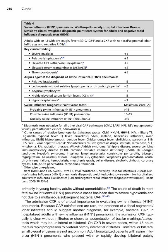
Table 4Swine influenza (H1N1) pneumonia: Winthrop-University Hospital Infectious DiseaseDivision’s clinical weighted diagnostic point score system for adults and negative rapidinfluenza diagnostic tests (RIDTs)
Adults with an ILI with dry cough, fever >39�C/102�F and a CXR with no focal/segmental lobarinfiltrates and negative RIDTsa:
Key clinical finding:
� Severe myalgias 15
� Relative lymphopeniab,c 15
� Elevated CPK (otherwise unexplained)c 13
� Elevated serum transaminases (AST/ALT)c 12
� Thrombocytopeniac 15
Argues against the diagnosis of swine influenza (H1N1) pneumonia:
� Relative bradycardia �5
� Leukopenia without relative lymphopenia or thrombocytopeniac �2
� Atypical lymphocytes �1
� Highly elevated serum ferritin levels (>2 � n)c �5
� Hypophosphatemiac �3
Swine influenza Diagnostic Point Score totals: Maximum score: 20
Probable swine influenza (H1N1) pneumonia >15
Possible swine influenza (H1N1) pneumonia 10–15
Unlikely swine influenza (H1N1) pneumonia <10
a Diagnostic tests negative for all other viral CAP pathogens (CMV, SARS, HPS, RSV metapneumo-viruses, parainfluenza viruses, adnoviruses).b Other causes of relative lymphopenia: Infectious causes: CMV, HHV-6, HHV-8, HIV, military TB,Legionella, typhoid fever, Q fever, brucellosis, SARS, malaria, babesiosis, influenza, avianinfluenza, RMSF, histoplasmosis, dengue fever, Chickungunya fever, ehrlichiosis, parvovirus B19,HPS, WNE, viral hepatitis (early); Noninfectious causes: cytotoxic drugs, steroids, sarcoidosis, SLE,lymphoma, RA, radiation therapy, Wiskott-Aldrich syndrome, Whipple disease, severe combineimmunodeficiency disease (SCID), common variable immune deficiency (CVID), Di George’ssyndrome, Nezelof’s syndrome, intestinal lymphangiectasia, constrictive pericarditis, tricuspidregurgitation, Kawasaki’s disease, idiopathic CD4 cytopenia, Wegener’s granulomatosis, acute/chronic renal failure, hemodialysis; myasthenia gravis, celiac disease, alcoholic cirrhosis, coronarybypass, CHF, acute pancreatitis, carcinomas (terminal).c Otherwise unexplained.
Data from Cunha BA, Syed U, Stroll S, et al. Winthrop-University Hospital Infectious Disease Divi-sion’s swine influenza (H1N1) pneumonia diagnostic weighted point score system for hospitalizedadults with influenza-like illnesses (ILIs) and negative rapid influenza diagnostic tests (RIDTs). HeartLung 2009;38:534–8.
Cunha216
primarily in young healthy adults without comorbidities.56 The cause of death in mostfatal swine influenza (H1N1) pneumonia cases has been due to severe hypoxemia andnot due to simultaneous/subsequent bacterial CAP.35–40
The admission CXR is of critical importance in evaluating swine influenza (H1N1)pneumonia. Because CAP coinfections are rare, the presence of a focal segmental/lobar infiltrates should suggest another diagnosis, for example, bacterial CAP. Inhospitalized adults with swine influenza (H1N1) pneumonia, the admission CXR typi-cally is clear without infiltrates or shows an accentuation of basilar markings/atelec-tasis which may be unilateral or bilateral. On CXR in severe cases, within 48 hoursthere is rapid progression to bilateral patchy interstitial infiltrates. Unilateral or bilateralsmall pleural effusions are not uncommon. Adult hospitalized patients with swine influ-enza (H1N1) pneumonia who present with, or rapidly develop bilateral patchy
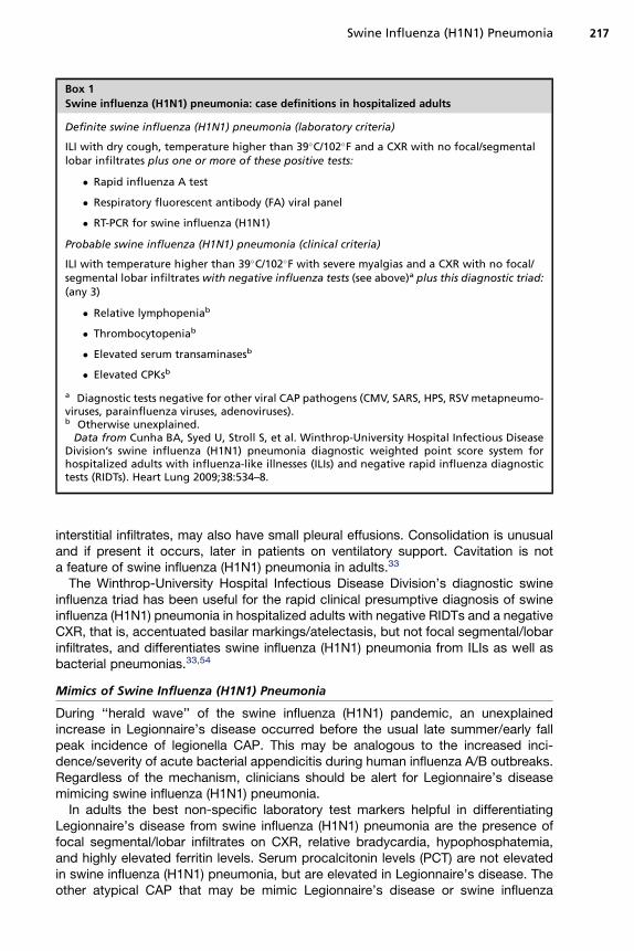
Box 1
Swine influenza (H1N1) pneumonia: case definitions in hospitalized adults
Definite swine influenza (H1N1) pneumonia (laboratory criteria)
ILI with dry cough, temperature higher than 39�C/102�F and a CXR with no focal/segmentallobar infiltrates plus one or more of these positive tests:
� Rapid influenza A test
� Respiratory fluorescent antibody (FA) viral panel
� RT-PCR for swine influenza (H1N1)
Probable swine influenza (H1N1) pneumonia (clinical criteria)
ILI with temperature higher than 39�C/102�F with severe myalgias and a CXR with no focal/segmental lobar infiltrates with negative influenza tests (see above)a plus this diagnostic triad:(any 3)
� Relative lymphopeniab
� Thrombocytopeniab
� Elevated serum transaminasesb
� Elevated CPKsb
a Diagnostic tests negative for other viral CAP pathogens (CMV, SARS, HPS, RSV metapneumo-viruses, parainfluenza viruses, adenoviruses).b Otherwise unexplained.
Data from Cunha BA, Syed U, Stroll S, et al. Winthrop-University Hospital Infectious DiseaseDivision’s swine influenza (H1N1) pneumonia diagnostic weighted point score system forhospitalized adults with influenza-like illnesses (ILIs) and negative rapid influenza diagnostictests (RIDTs). Heart Lung 2009;38:534–8.
Swine Influenza (H1N1) Pneumonia 217
interstitial infiltrates, may also have small pleural effusions. Consolidation is unusualand if present it occurs, later in patients on ventilatory support. Cavitation is nota feature of swine influenza (H1N1) pneumonia in adults.33
The Winthrop-University Hospital Infectious Disease Division’s diagnostic swineinfluenza triad has been useful for the rapid clinical presumptive diagnosis of swineinfluenza (H1N1) pneumonia in hospitalized adults with negative RIDTs and a negativeCXR, that is, accentuated basilar markings/atelectasis, but not focal segmental/lobarinfiltrates, and differentiates swine influenza (H1N1) pneumonia from ILIs as well asbacterial pneumonias.33,54
Mimics of Swine Influenza (H1N1) Pneumonia
During ‘‘herald wave’’ of the swine influenza (H1N1) pandemic, an unexplainedincrease in Legionnaire’s disease occurred before the usual late summer/early fallpeak incidence of legionella CAP. This may be analogous to the increased inci-dence/severity of acute bacterial appendicitis during human influenza A/B outbreaks.Regardless of the mechanism, clinicians should be alert for Legionnaire’s diseasemimicing swine influenza (H1N1) pneumonia.
In adults the best non-specific laboratory test markers helpful in differentiatingLegionnaire’s disease from swine influenza (H1N1) pneumonia are the presence offocal segmental/lobar infiltrates on CXR, relative bradycardia, hypophosphatemia,and highly elevated ferritin levels. Serum procalcitonin levels (PCT) are not elevatedin swine influenza (H1N1) pneumonia, but are elevated in Legionnaire’s disease. Theother atypical CAP that may be mimic Legionnaire’s disease or swine influenza
Cunha218
(H1N1) pneumonia is Q fever. Q fever CAP has many of the features of Legionnaire’sdisease, namely, elevated serum ferritin levels, mildly increased serum transaminases(AST/ALT), and relative bradycardia. However, Q fever CAP is often accompanied bysplenomegaly, which is not a feature of Legionnaire’s disease or swine influenza(H1N1) pneumonia. As with M pneumoniae, elevated cold agglutinins may occurwith Q fever CAP but are not a feature of Legionnaire’s disease or swine influenza(H1N1) pneumonia. Relative lymphopenia is common with Legionnaire’s disease, Qfever, and swine influenza (H1N1) pneumonia, but leukopenia and thrombocytopeniaare not laboratory markers of either Legionnaire’s disease or Q fever CAP.30,33,55
LABORATORY DIAGNOSIS
Influenza virus is present in respiratory secretions early in the illness. Nasopharyngealswabs or washings may be used for viral isolation. In general, higher concentrationsof virus are found in nasal secretions than in oropharyngeal secretions. During influenzaA epidemics, oropharyngeal secretions contain higher concentrations of influenzavirions than nasal secretions. In alveolar macrophages, influenza A viruses producecytopathogenic effects (CPEs), but are nonspecific and difficult to detect. CPEs dueto influenza A are apparent in approximately 50% of cultures within 3 days of inoculationand in approximately 90% within 5 days. Identification of cells showing CPE may beverified using immunofluorescent technique or type/subtype specific antisera. Themost common rapid method of influenza laboratory diagnosis is by FA techniques.For FA testing for respiratory viruses, nasopharyngeal aspirates are preferred to swabs.FA influenza assays identify viral antigens in infected respiratory epithelial cells.22–25
During the ‘‘herald wave’’ of the swine influenza (H1N1) pandemic in New York, itwas realized that there were problems with screening tests, that is, RIDTs. It washoped that rapid screening tests for influenza A would detect swine influenza(H1N1), an influenza A virus. Rapid influenza A test positivity was usually predictiveof RT-PCR positivity for swine influenza (H1N1). It was quickly realized that negativerapid influenza A testing did not rule out swine influenza (H1N1). False negative rapidinfluenza A testing for swine influenza (H1N1) was 30%. Respiratory FA viral testingdetects influenza A, influenza B, metapneumoviruses, RSVs, parainfluenza viruses,and adenoviruses. Unfortunately, respiratory FA viral testing did not correlate witheither rapid influenza A testing or RT-PCR testing. Although the definitive test for swineinfluenza (H1N1) diagnosis remains RT-PCR, during the pandemic, RT-PCR testingwas restricted. Restricted RT-PCR testing resulted in tremendous difficulties inmaking/ruling out the diagnosis of swine influenza (H1N1) and in initiating/discontinu-ing influenza precautions.53–55,59
Because of restricted swine influenza (H1N1) RT-PCR testing, clinical criteria weredeveloped for patients presenting with ILIs who had a negative rapid influenza A test.By combining key clinical and nonspecific laboratory features, a clinical diagnosis ofprobable swine influenza was developed at Winthrop-University Hospital by the Infec-tious Disease Division. This permitted an operational clinical approach to placingadmitted adult patients on influenza precautions/treatment, and was also useful in differ-entiating swine influenza (H1N1) from its mimics.54 (see Box 1, Figs. 1–4, Tables 3–5).
THERAPY
Until relatively recently, there were no effective anti-influenza antivirals. Amantadineand rimantadine has been used for influenza prophylaxis/therapy and act by inter-fering with the attachment of the virus to uninfected hosts’ respiratory epithelial cells.The current anti-influenza antivirals currently available are zanamivir and oseltamivir.
Swine Influenza (H1N1) Pneumonia 219
Both of these antivirals decrease the duration/severity of illness. Oseltamivir is admin-istered orally but zanamivir is administered via an inhaler, which is problematic forsome patients.30 Unfortunately, there is now widespread resistance to oseltamivir,greatly diminishing the therapeutic options in influenza and avian influenza (H5N1).At present, oseltamivir remains effective against most strains of swine influenza(H1N1). Oseltamivir is usually given for 5 days, but in severe cases, therapy hasbeen given for 10 days. For severely ill hospitalized patients with swine influenza(H1N1) pneumonia unable to take oral medications, eg, oseltamivir, peramivir is avail-able from the CDC for IV administration. In severe cases of influenza A, amantadinemay have some role because of its effect on dilatation of the distal bronchioles. Byincreasing distal bronchiolar aeration, oxygenation may be improved, which may becritical in severe influenza A.60–67
COMPLICATIONS AND PROGNOSIS
The prognosis of influenza A is related primarily to strain virulence. Most patients withmild to moderate influenza A recover without specific anti-influenza therapy. However,influenza A may be severe, and may be fatal in the very young and the elderly withimpaired cardiopulmonary function. With pandemic influenza A, the prognosisdepends on the virulence of the strain and may be fatal in young, healthy adults.Such has clearly been the case in the recent avian influenza (H5N1) epidemic inAsia whose fatalities, as in the 1918 to 1919 pandemic, were due in the main to influ-enza pneumonia and was frequently fatal without bacterial superinfection. Severeinfluenza A pneumonia with superimposed S aureus CAP have the worst prognosis.Others who initially present with influenza may later be complicated by subsequentbacterial pneumonia due to S pneumoniae or H influenzae, and have the same prog-nosis as noninfluenza patients with S pneumoniae or H influenzae CAP.
There is a late excess mortality with influenza A, that occurs after the acute episodeeg, elderly patients with borderline cardiopulmonary function succumb to heart failureor acute myocardial infarction from the hypoxemia/stress of swine influenza (H1N1)pneumonia. In severe cases of respiratory failure/ARDS, extracorporeal membraneoxygenation (ECMO) has been lifesaving in some.68 Although influenza is a 3-dayillness, postinfluenza fatigue may last for weeks or months. In influenza, prognosisis worse in those patients who develop encephalitis. Following the 1918 to 1919 influ-enza pandemic, some patients developed Parkinson disease decades later. The prog-nosis in influenza depends on several factors including the virulence of the strain, theage/immune status of the host, cardiopulmonary function, and complications, forexample, encephalitis or bacterial CAP.1,2,16 The most important intervention in adultswith severe swine influenza (H1N1) pneumonia is prolonged ventilatory support.Because these patients are not complicated by simultaneous or subsequent bacterialCAP, empiric antimicrobial therapy seems to be unnecessary in swine influenza(H1N1) pneumonia. The degree and duration of relative lymphopenia/leukopenia aswell as the severity and duration of hypoxemia are key prognostic indicators. Animprovement in relative lymphopenia and a decrease in oxygen (FIO2) are predictiveof recovery.
SUMMARY AND HISTORICAL PERSPECTIVE
There are differences of opinion on the cause of early deaths in influenza pandemics.Most early deaths in young healthy individuals in the 1918–1919 pandemic seemedto be caused by influenza A pneumonia alone. Based on autopsy specimen cultures,which are notoriously unreliable, some believe the high mortality rate of the
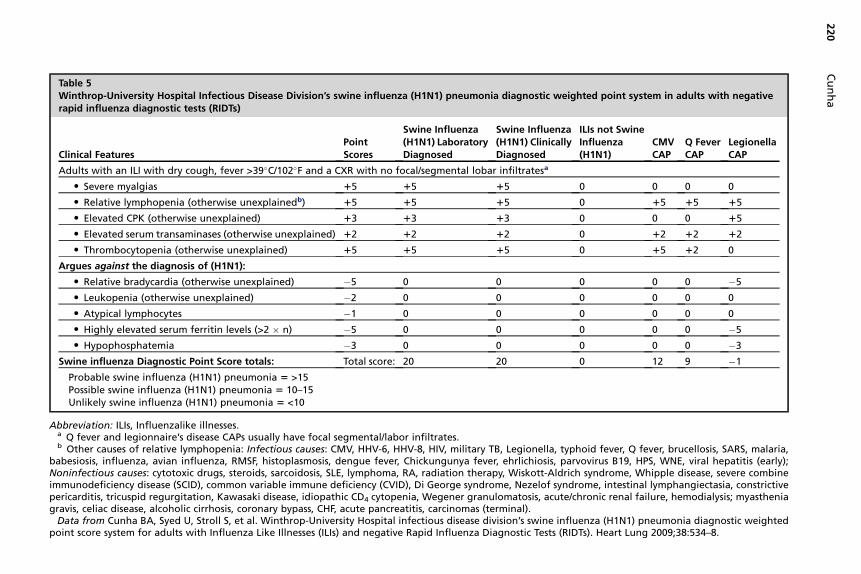
Table 5Winthrop-University Hospital Infectious Disease Division’s swine influenza (H1N1) pneumonia diagnostic weighted point system in adults with negativerapid influenza diagnostic tests (RIDTs)
Clinical FeaturesPointScores
Swine Influenza(H1N1) LaboratoryDiagnosed
Swine Influenza(H1N1) ClinicallyDiagnosed
ILIs not SwineInfluenza(H1N1)
CMVCAP
Q FeverCAP
LegionellaCAP
Adults with an ILI with dry cough, fever >39�C/102�F and a CXR with no focal/segmental lobar infiltratesa
� Severe myalgias 15 15 15 0 0 0 0
� Relative lymphopenia (otherwise unexplainedb) 15 15 15 0 15 15 15
� Elevated CPK (otherwise unexplained) 13 13 13 0 0 0 15
� Elevated serum transaminases (otherwise unexplained) 12 12 12 0 12 12 12
� Thrombocytopenia (otherwise unexplained) 15 15 15 0 15 12 0
Argues against the diagnosis of (H1N1):
� Relative bradycardia (otherwise unexplained) �5 0 0 0 0 0 �5
� Leukopenia (otherwise unexplained) �2 0 0 0 0 0 0
� Atypical lymphocytes �1 0 0 0 0 0 0
� Highly elevated serum ferritin levels (>2 � n) �5 0 0 0 0 0 �5
� Hypophosphatemia �3 0 0 0 0 0 �3
Swine influenza Diagnostic Point Score totals: Total score: 20 20 0 12 9 �1
Probable swine influenza (H1N1) pneumonia 5 >15Possible swine influenza (H1N1) pneumonia 5 10–15Unlikely swine influenza (H1N1) pneumonia 5 <10
Abbreviation: ILIs, Influenzalike illnesses.a Q fever and legionnaire’s disease CAPs usually have focal segmental/labor infiltrates.b Other causes of relative lymphopenia: Infectious causes: CMV, HHV-6, HHV-8, HIV, military TB, Legionella, typhoid fever, Q fever, brucellosis, SARS, malaria,
babesiosis, influenza, avian influenza, RMSF, histoplasmosis, dengue fever, Chickungunya fever, ehrlichiosis, parvovirus B19, HPS, WNE, viral hepatitis (early);Noninfectious causes: cytotoxic drugs, steroids, sarcoidosis, SLE, lymphoma, RA, radiation therapy, Wiskott-Aldrich syndrome, Whipple disease, severe combineimmunodeficiency disease (SCID), common variable immune deficiency (CVID), Di George syndrome, Nezelof syndrome, intestinal lymphangiectasia, constrictivepericarditis, tricuspid regurgitation, Kawasaki disease, idiopathic CD4 cytopenia, Wegener granulomatosis, acute/chronic renal failure, hemodialysis; myastheniagravis, celiac disease, alcoholic cirrhosis, coronary bypass, CHF, acute pancreatitis, carcinomas (terminal).
Data from Cunha BA, Syed U, Stroll S, et al. Winthrop-University Hospital infectious disease division’s swine influenza (H1N1) pneumonia diagnostic weightedpoint score system for adults with Influenza Like Illnesses (ILIs) and negative Rapid Influenza Diagnostic Tests (RIDTs). Heart Lung 2009;38:534–8.
Cu
nh
a220
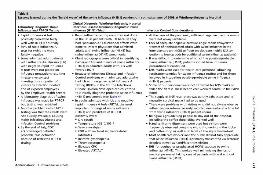
Table 6Lessons learned during the ‘‘herald wave’’ of the swine influenza (H1N1) pandemic in spring/summer of 2009 at Winthrop-University Hospital
Laboratory Diagnosis: RapidInfluenza and RT-PCR Testing
Clinical Diagnosis: Winthrop-University HospitalInfectious Disease Division’s Diagnostic SwineInfluenza (H1N1) Triad Infection Control Considerations
� Rapid influenza A testpositivity correlated fairlywell with RT-PCR positivity
� 30% of rapid influenza Atests for swine flu werefalsely negative
� Some admitted adult patientswith influenzalike illnesses (ILIs)with negative rapid influenzaA tests were not placed oninfluenza precautions resultingin extensive contactinvestigations of patients/visitors by Infection Controland of exposed employeesby the Employee Health Service
� A laboratory diagnosis of swineinfluenza was made by RT-PCRbut testing was restricted
� Another problem with RT-PCRtesting was that the results werenot quickly available. Causingmajor Infectious Disease andInfection Control problems
� By the end of July, CDCacknowledged definite/probable case definitionbecause of restricted RT-PCRtesting
� Rapid influenza testing was often not donein the ED in patients with ILIs because theyhad ‘‘pneumonia.’’ Educational efforts weredone to inform physicians that admittedadults with swine influenza (H1N1) hadswine influenza (H1N1) pneumonia
� Chest radiographs were critical in identifyingbacterial CAPs and mimics of swine influenza(H1N1) in admitted adults with ILIs withfevers >102�F
� Because of Infectious Disease and InfectionControl problems with admitted adults whohad ILIs with negative rapid influenza Atesting (RIDTs) in the ED, the InfectiousDisease Division developed clinical criteriato clinically diagnose probable swine influenza(H1N1) pneumonia (see Table 4)
� In adults admitted with ILIs and negativerapid influenza A tests (RIDTs), the mostimportant findings of swine influenza(H1N1) and predictive of RT-PCRpositivity were:� Dry cough� Temperature >39�C/102�F� Severe myalgias� CXR with no focal segmental/lobar
infiltrates� Relative lymphopenia� Thrombocytopenia� Elevated CPK� Elevated AST/ALT
� At the peak of the pandemic, sufficient negative pressure roomswere not always available
� Lack of adequate negative pressure single rooms delayed thetransfer of nonintubated adults with swine influenza in theintensive care unit (ICU) to floors (to decrease mobile ICU con-gestion to free up beds for additional swine influenza patients)
� It was difficult to determine which of the possible/probableswine influenza (H1N1) patients should have influenzaprecautions discontinued
� N95 masks were used for health care personnel obtainingrespiratory samples for swine influenza testing and for thoseinvolved in intubating possible/probable swine influenza(H1N1) patients
� Some of our personnel were not fit tested for N95 masks orfailed the fit test. These health care workers could use the PAPRhood
� The supply of N95 respirators was quickly exhausted and, ofnecessity, surgical masks had to be used
� There were problems with visitors who did not always observeinfluenza precautions. Security escorted one visitor at a time to/from swine influenza (H1N1) patient rooms
� Bilingual signs advising people to stay out of the hospital,including the coffee shop/lobby, worked well
� Hand sanitizing dispensers were used but visitors werefrequently observed coughing without covering in the lobbyand coffee shop as well as in front of the signs themselves!
� Most health care workers and the public did not fully appreciatethat swine influenza (H1N1) is primarily transmitted via aerosols/droplets as well as hand/face transmission
� EHS furloughed or prophylaxed HCWS exposed to swineinfluenza (H1N1). This worked well minimizing the loss ofmedical personnel taking care of patients with and withoutswine influenza (H1N1)
Abbreviation: ILI, influenzalike illness.
Swin
eIn
fluen
za(H
1N
1)
Pn
eu
mo
nia
221
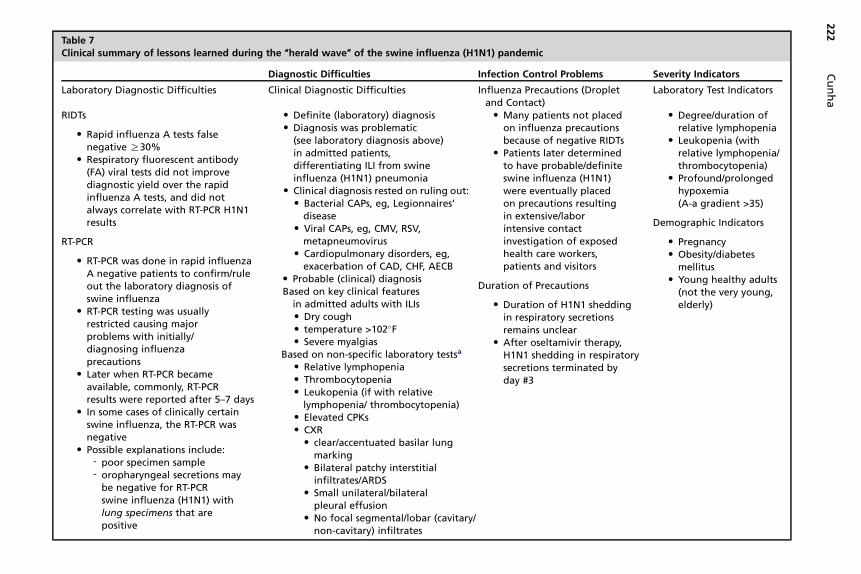
Table 7Clinical summary of lessons learned during the ‘‘herald wave’’ of the swine influenza (H1N1) pandemic
Diagnostic Difficulties Infection Control Problems Severity Indicators
Laboratory Diagnostic Difficulties Clinical Diagnostic Difficulties Influenza Precautions (Dropletand Contact)
Laboratory Test Indicators
RIDTs
� Rapid influenza A tests falsenegative R30%
� Respiratory fluorescent antibody(FA) viral tests did not improvediagnostic yield over the rapidinfluenza A tests, and did notalways correlate with RT-PCR H1N1results
RT-PCR
� RT-PCR was done in rapid influenzaA negative patients to confirm/ruleout the laboratory diagnosis ofswine influenza
� RT-PCR testing was usuallyrestricted causing majorproblems with initially/diagnosing influenzaprecautions
� Later when RT-PCR becameavailable, commonly, RT-PCRresults were reported after 5–7 days
� In some cases of clinically certainswine influenza, the RT-PCR wasnegative
� Possible explanations include:- poor specimen sample- oropharyngeal secretions may
be negative for RT-PCRswine influenza (H1N1) withlung specimens that arepositive
� Definite (laboratory) diagnosis� Diagnosis was problematic
(see laboratory diagnosis above)in admitted patients,differentiating ILI from swineinfluenza (H1N1) pneumonia
� Clinical diagnosis rested on ruling out:� Bacterial CAPs, eg, Legionnaires’
disease� Viral CAPs, eg, CMV, RSV,
metapneumovirus� Cardiopulmonary disorders, eg,
exacerbation of CAD, CHF, AECB� Probable (clinical) diagnosisBased on key clinical features
in admitted adults with ILIs� Dry cough� temperature >102�F� Severe myalgias
Based on non-specific laboratory testsa
� Relative lymphopenia� Thrombocytopenia� Leukopenia (if with relative
lymphopenia/ thrombocytopenia)� Elevated CPKs� CXR� clear/accentuated basilar lung
marking� Bilateral patchy interstitial
infiltrates/ARDS� Small unilateral/bilateral
pleural effusion� No focal segmental/lobar (cavitary/
non-cavitary) infiltrates
� Many patients not placedon influenza precautionsbecause of negative RIDTs
� Patients later determinedto have probable/definiteswine influenza (H1N1)were eventually placedon precautions resultingin extensive/laborintensive contactinvestigation of exposedhealth care workers,patients and visitors
Duration of Precautions
� Duration of H1N1 sheddingin respiratory secretionsremains unclear
� After oseltamivir therapy,H1N1 shedding in respiratorysecretions terminated byday #3
� Degree/duration ofrelative lymphopenia
� Leukopenia (withrelative lymphopenia/thrombocytopenia)
� Profound/prolongedhypoxemia(A-a gradient >35)
Demographic Indicators
� Pregnancy� Obesity/diabetes
mellitus� Young healthy adults
(not the very young,elderly)
222
Cu
nh
a
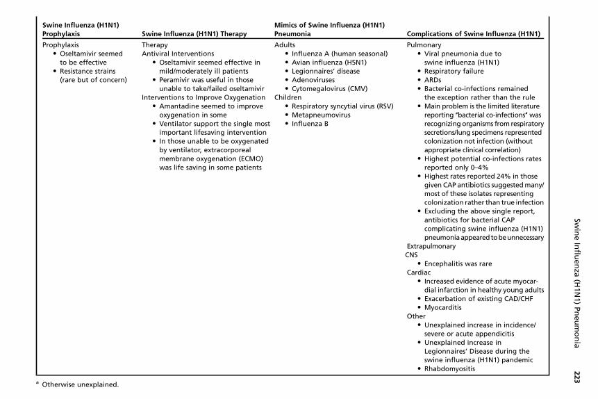
Swine Influenza (H1N1)Prophylaxis Swine Influenza (H1N1) Therapy
Mimics of Swine Influenza (H1N1)Pneumonia Complications of Swine Influenza (H1N1)
Prophylaxis Therapy Adults Pulmonary� Oseltamivir seemed
to be effective� Resistance strains
(rare but of concern)
Antiviral Interventions� Oseltamivir seemed effective in
mild/moderately ill patients� Peramivir was useful in those
unable to take/failed oseltamivirInterventions to Improve Oxygenation� Amantadine seemed to improve
oxygenation in some� Ventilator support the single most
important lifesaving intervention� In those unable to be oxygenated
by ventilator, extracorporealmembrane oxygenation (ECMO)was life saving in some patients
� Influenza A (human seasonal)� Avian influenza (H5N1)� Legionnaires’ disease� Adenoviruses� Cytomegalovirus (CMV)
Children� Respiratory syncytial virus (RSV)� Metapneumovirus� Influenza B
� Viral pneumonia due toswine influenza (H1N1)
� Respiratory failure� ARDs� Bacterial co-infections remained
the exception rather than the rule� Main problem is the limited literature
reporting ‘‘bacterial co-infections’’ wasrecognizing organisms from respiratorysecretions/lung specimens representedcolonization not infection (withoutappropriate clinical correlation)
� Highest potential co-infections ratesreported only 0–4%
� Highest rates reported 24% in thosegiven CAP antibiotics suggested many/most of these isolates representingcolonization rather than true infection
� Excluding the above single report,antibiotics for bacterial CAPcomplicating swine influenza (H1N1)pneumoniaappearedtobeunnecessary
ExtrapulmonaryCNS� Encephalitis was rare
Cardiac� Increased evidence of acute myocar-
dial infarction in healthy young adults� Exacerbation of existing CAD/CHF� Myocarditis
Other� Unexplained increase in incidence/
severe or acute appendicitis� Unexplained increase in
Legionnaires’ Disease during theswine influenza (H1N1) pandemic
� Rhabdomyositis
a Otherwise unexplained.
223
Swin
eIn
fluen
za(H
1N
1)
Pn
eu
mo
nia
Cunha224
1918–1919 influenza pandemic was due to superimposed bacterial pneumonia, forexample, S pneumoniae. Most lung specimens from autopsies of young healthy mili-tary recruits clearly show the pathologic changes of influenza alone. Based on thepathologic comparisons of viral and bacterial pneumonia, it seems that the majorityof early deaths in young healthy military recruits was due to severe hypoxemia. Lateexcess mortality seems to have been due to influenza precipitated decompensationin those with antecedent borderline cardiopulmonary function/reserve.
Most recently, S aureus was recognized as an important pathogen in the 1957– 1958Asian influenza A pandemic. In 1957–1958 Asian influenza A pandemic, influenza pre-sented as pneumonia alone or simultaneously with S aureus or subsequently with Spneumoniae or H influenzae CAP. Because the 1957–1958 influenza A pandemic clini-cians had modern bacteriologic and virologic diagnostic methods, clinicians attributedthe excess mortality to late bacterial CAP. The unusual severity of the 1918–1919 influ-enza A pandemic seems to have been related to a particularly virulent strain of influenzaA (H1N1), not unlike the virulence of avian influenza (H5N1). In the recent experiencewith avian influenza (H5N1), it is interesting to note that despite high mortality ofw 60% in young adults, avian influenza deaths have been due to avian influenza(H5N1) pneumonia alone and not simultaneous/subsequent bacterial CAP.69–70
A summary of lessons learned during the ‘‘herald wave’’ of the swine influenza(H1N1) pandemic is presented here in tabular form (Tables 6 and 7). The importanttake-home lessons for clinicians in the summary relate to laboratory and clinical diag-nosis, infection control concerns, and empiric antibiotic use.71–85
REFERENCES
1. Ewald PW. Influenza. In: Evolution of infectious disease. New York: Oxford Univer-sity Press; 1994. p. 10–116.
2. Crosby AW. Influenza. In: Kiple KF, editor. The Cambridge world history of humandisease. New York: Cambridge University Press; 1993. p. 807–11.
3. Aufderheide AC, Rodriguez-Martin C. Influenza. In: Aufderheide AC, Rodriguez-Martin C, editors. The Cambridge encyclopedia of human paleopathology. NewYork: Cambridge University Press; 1998. p. 210–2.
4. Sydenham T. Influenza. In: Major RH, editor. Classic descriptions of disease.Springfield (MA): Charles C. Thomas, Publisher; 1978. p. 201–2.
5. Douglas RG Jr. Influenza in man. In: Kilbourne ED, editor. The influenza virusesand influenza. Orlando (FL): Academic Press; 1975. p. 395.
6. Cunha BA. Influenza: historical aspects of epidemics and pandemics. Infect DisClin North Am 2004;18:141–56.
7. Ravenholt RT, Foege WH. Before our time: 1918 influenza, encephalitis lethargi-ca, parkinsonism. Lancet 1982;2:860–4.
8. Conner LA. The symptomatology and complications of influenza. JAMA 1919;73:321–5.
9. Kolte IV, Skinhoj P, Keiding N, et al. The Spanish flu in Denmark. Scand J InfectDis 2008;40:538–46.
10. Sheretz RJ, Sheretz HJ. Influenza in the preantibiotic era. Infect Dis Clin Pract2006;14:127.
11. Winternitz MC, Wason IM, McNamara FP. The pathology of influenza. New Haven(CT): Yale University Press; 1920.
12. Wolbach SB. Comments on the pathology and bacteriology of fatal influenza casesas observed at Camp Devens, Mass. Bull Johns Hopkins Hosp 1919;30:104–5.
Swine Influenza (H1N1) Pneumonia 225
13. Stevens KM. The pathophysiology of influenzal pneumonia in 1918. Perspect BiolMed 1918;25:115–25.
14. Mulder J, Hers JF. Influenza. Groningen (The Netherlands): Wolters-Noordhoff;1979.
15. Klotz O. The pathology of epidemic influenza. In: Studies on epidemic influenza,comprising clinical and laboratory investigations by members of the faculty of theschool of medicine. Pittsburgh (PA): University of Pittsburgh Press; 1919. p.255–61.
16. Andrewes FW. The bacteriology of influenza. In: Great Britain Ministry of Health.Reports on public health and medical subjects, no. 4: report on the pandemicof influenza, 1918–1919. London: His Majesty’s Stationery Office; 1920. p.110–26.
17. Centers for Disease Control and Prevention (CDC). Outbreak of swine-origin influ-enza A (H1N1) virus infection—Mexico, March-April 2009. MMWR Morb MortalWkly Rep 2009;58:467–70.
18. Centers for Disease Control and Prevention (CDC). Swine-origin influenza A(H1N1) virus infections in a school—New York City, April 2009. MMWR MorbMortal Wkly Rep 2009;58:470–2.
19. Gallaher WR. Towards a sane and rational approach to management of InfluenzaH1N1 2009. Virol J 2009;6:51.
20. Rezza G. Swine-origin influenza virus A (H1N1)v: lessons learnt from the earlyphase of the epidemic. Eur J Public Health 2009;19:572–3.
21. Cohen J, Enserink M. Infectious disease. As swine flu circles globe, scientistsgrapple with basic questions. Science 2009;324:572–3.
22. Kilbourne ED, editor. The influenza viruses and influenza. Orlando (FL):Academic Press; 1975.
23. Debr�e R, Couvreur J. Influenza: clinical features. In: Debr�e R, Celers J, editors.Clinical virology: the evaluation and management of human viral infections. Phil-adelphia: WB Saunders; 1970. p. 507–15.
24. Nicholson KG, Webster RG, Hay AJ, editors. Textbook of influenza. Oxford (UK):Blackwell Science; 1998.
25. Van Voris LP, Young JF, Bernstein JM, et al. Influenza viruses. In: Belshe RB, editor.Textbook of human virology. Littleton (MA): PSG Publishing Company; 1984. p.267–81.
26. Atmar RL. Influenza viruses. In: Murray PR, Baron EJ, Jorgensen JH, et al,editors. Manual of clinical microbiology. 9th edition. Washington, DC: ASM Press;2007. p. 1340–51.
27. Hayden FG, Palese P. Influenza virus. In: Richman DD, Whitley RJ, Hayden FG,editors. Clinical virology. 3rd edition. Washington, DC: ASM Press; 2009. p. 943–76.
28. Sym D, Patel PM, El-Chaar GM. Seasonal, avain and novel H1N1 influenza:prevention and treatment modalities. Ann Pharmacother 2009;43:2001–11.
29. Harper SA, Bradley JS, Englund JA, et al. Seasonal influenza in adults and chil-dren: diagnosis, treatment, chemoprophylaxis and institutional outbreak manage-ment: clinical practice guidelines of the Infectious Diseases Society of America.Clin Infect Dis 2009;48:1003–32.
30. Cunha BA. Pneumonia essentials. 3rd edition. Sudbury (MA): Jones & Bartlett;2010.
31. Cunha BA. The clinical diagnosis of severe viral influenza A. Infection 2008;36:92–3.
32. Kim HM, Lee YW, Lee KJ, et al. Alveolar macrophages are indispensable forcontrolling influenza viruses in lungs of pigs. J Virol 2008;82:4265–74.
Cunha226
33. Mollura DJ, Asnis DS, Crupi RS, et al. Imaging findings in a fatal case of pandemicswine-origin influenza A (H1N1). AJR Am J Roentgenol 2009;193:1500–3.
34. Agarwal PP, Cinti S, Kazerooni EA, et al. Chest radiographic and CT findings innovel swine-origin influenza A (H1N1) virus (S-OIV) infection. AJR Am J Roent-genol 2009;193:1488–93.
35. Cunha BA. A useful clinical approach to community-acquired methicillin-resistantStaphylococcus aureus (CA-MRSA) infections. J Hosp Infect 2008;68:271–73.
36. Cunha BA. Methicillin-resistant Staphylococcus aureus: clinical manifestationsand antimicrobial therapy. Clin Microbiol Infect 2005;11:33–42.
37. Tacconelli E, De Angelis G. Pneumonia due to methicillin-resistant Staphylo-coccus aureus: clinical features, diagnosis and management. Curr Opin PulmMed 2009;15:218–48.
38. Kallen AJ, Brunkard J, Moore Z, et al. Staphylococcus aureus community-acquired pneumonia during the 2006 to 2007 influenza season. Ann EmergMed 2009;53:358–65.
39. Cheng VC, Lau YK, Lee KL, et al. Fatal co-infection with swine origin influenzavirus A/H1N1 and community-acquired methicillin-resistant Staphylococcusaureus. J Infection 2009;259:1–5.
40. Cunha BA, Syed U, Strollo S. Bacterial pneumonia rare with fatal swine influenza(H1N1) pneumonia: if chest films have no focal segmental/lobar infiltrates,empiric antibiotic therapy is unnecessary. J Chemotherapy 2010;21:584–5.
41. Hien ND, Ha NH, Van NT, et al. Human infection with highly pathogenic avian influ-enza virus (H5N1) in northern Vietnam, 2004–2005. Emerg Infect Dis 2009;15:19–23.
42. Writing Committee of the Second World Health Organization consultation on clin-ical aspects of human infection with avian influenza A (H5N1) virus. Update onavian influenza A (H5N1) virus infection in humans. N Engl J Med 2008;358:261–73.
43. Thomas JK, Noppenberger J. Avian influenza: a review. Am J Health Syst Pharm2007;64:149–65.
44. Ozbay B, Sertogullarindan B, Tekin M, et al. Influenza-associated pneumonia ina Turkish area with endemic avian influenza. Respirology 2008;13:444–6.
45. Sandrock C, Kelly T. Clinical review: update of avian influenza A infections inhumans. Crit Care 2007;11:209–18.
46. To KF, Chan PK, Chan KF, et al. Pathology of fatal human infection associated withavian influenza A H5N1 virus. J Med Virol 2001;63:242–6.
47. Tran TH, Nguyen TL, Nguyen TD, et al. Avian influenza A (H5N1) in 10 patients inVietnam. N Engl J Med 2004;350:1179–88.
48. Uyeki TM. Human infection with highly pathogenic avian influenza A (H5N1) virus:review of clinical issues. Clin Infect Dis 2009;49:279–90.
49. Gambotto A, Barratt-Boyes SM, de Jong MD, et al. Human infection with highlypathogenic H5N1 influenza virus. Lancet 2008;371:1464–75.
50. Yuen KY, Chan PKS, Peiris M, et al. Clinical features and rapid viral diagnosis ofhuman disease associated with avian influenza A H5N1 virus. Lancet 1998;351:467–71.
51. Rainer TH. Severe acute respiratory syndrome: clinical features, diagnosis, andmanagement. Curr Opin Pulm Med 2004;10:159–65.
52. Rhodes LV, Huang C, Sanchez AJ, et al. Hantavirus pulmonary associated withMonongahela virus, Pennsylvania. Emerg Infect Dis 2000;6:616–21.
53. Cunha BA, Pherez FM, Strollo S. Swine influenza H1N1: diagnostic dilemma earlyin the pandemic. Scand J of Infect 2009;41:900–2.
Swine Influenza (H1N1) Pneumonia 227
54. Cunha BA, Syed U, Strollo S, et al. Winthrop-University Hospital infectiousdisease division’s swine influenza (H1N1) pneumonia diagnostic weighted pointscore system for adults with Influenza Like Illnesses (ILIs) and negative RapidInfluenza Diagnostic Tests (RIDTs). Heart Lung 2009;38:534–8.
55. Cunha BA, Pherez FM, Schoch PE. The diagnostic importance of relative lympho-penia as a marker of swine influenza (H1N1) in adults. Clin Infect Dis 2009;49:1454–6.
56. Louria DB, Blumenfeld HL, Ellis JT, et al. Studies on influenza in the pandemicof 1957–1958. Pulmonary complications of influenza. J Clin Invest 1959;38:213–65.
57. Robertson L, Caley JP, Moore J. Importance of Staphylococcus aureus in pneu-monia in the 1957 epidemic of influenza A. Lancet 1958;2:233–6.
58. Petersdorf RG, Fusco JJ, Harter DH, et al. Pulmonary infections complicatingAsian influenza. Arch Intern Med 1959;103:262–72.
59. Vasoo S, Stevens J, Singh K. Rapid antigen tests for diagnosis of pandemic(Swine) influenzae A/H1N1. Clin Infect Dis 2009;49:1090–3.
60. Cunha BA. Amantadine may be lifesaving in severe influenza A. Clin Infect Dis2006;43:1574–5.
61. Centers for Disease Control and Prevention (CDC). Update: drug susceptibility ofswine-origin influenza A (H1N1) viruses, April 2009. MMWR Morb Mortal WklyRep 2009;58:433–5.
62. Influenza Project Team. Oseltamivir resistance in human seasonal influenza viruses(A/H1N1) in EU and EFTA countries: an update. Euro Surveill 2008;13:8032.
63. Kawai N, Ikematsu H, Iwaki N, et al. A change in the effectiveness of amantadinefor the treatment of influenza over the 2003–2004, 2004–2005, and 2005–2006influenza seasons in Japan. J Infect Chemother 2007;13:314–9.
64. Meijer A, Lackenby A, Hungnes O, et al. Oseltamivir-resistant influenza virus A(H1N1), Europe, 2007-08 season. Emerg Infect Dis 2009;15:552–60.
65. Van der Vries E, van den Berg B, Schutten M. Fatal oseltamivir-resistant influenzavirus infection. N Engl J Med 2008;359:1074–6.
66. Couzin-Frankel J. Swine flu outbreak. What role for antiviral drugs? Science 2009;324:705.
67. Hurt AC, Selleck P, Komadina N, et al. Susceptibility of highly pathogenic A(H5N1)Avian influenza viruses to the neuraminidase inhibitors and adamantanes.Antiviral Res 2007;73:228–31.
68. Davies A, Jones D, Bailey M, et al. Extracorporeal Membrane oxygenation for2009 influenza A (H1N1) acute respiratory distress syndrome. JAMA 2009;302:1888–95.
69. Andreasen V, Viboud C, Simonsen L. Epidemiologic characterization of the 1918influenza pandemic summer wave in Copenhagen: implications for pandemiccontrol strategies. J Infect Dis 2008;197:270–8.
70. Steel J, Palese P. The 1918 Influenza pandemic: lessons from the past raise ques-tions for the future. In: Klenk HD, Matrosovich MN, Stech J, editors. Avian Influ-enza. Monogr Virol, vol 27. Basel: Karger; 2008. p. 272–86.
71. Cunha BA, Thekkel V, Cohan C. Swine influenza (H1N1): contact investigationburden because of failure to institute influenza precautions with negative rapidinfluenza A diagnostic test results. Infect Control Hosp Epidemiol 2010;31:102–4.
72. Cunha BA, Thekkel V, Krilov L. Nosocomial swine influenza (H1N1) pneumonia:lessons learned from an illustrative case. J Hosp Infect 2009; 72, in press.
73. Charlier C, Enouf V, Lanternier F, et al. Kinetics of nasopharyngeal shedding ofnovel H1N1 (swine-like) influenza A virus in an immunocompetent adult underoseltamivir therapy. Clin Microbiol Infect 2009;15:1189–91.
Cunha228
74. Dominguez-Cherit G, Lapinsky SE, Macias AE, et al. Critically ill patients with2009 influenza A (H1N1) in Mexico. JAMA 2009;302:1872–9.
75. MMWR. Bacterial coinfections in lung tissue specimens from fatal cases of 2009pandemic influenza A (H1N1)—United States, May–August 2009, 77.
76. MMWR. Hospitalized patients with novel influenza A (H1N1) infection—California,April–May, 2009. MMWR Morb Mortal Wkly Rep 2009;58:536–41.
77. Kumar A, Zarychanski R, Pinto R, et al. Critically ill patients with 2009 influenzaA (H1N1) in Canada. JAMA 2009;302:1880–7.
78. MMWR. Intensive-care patients with severe novel influenza A (H1N1) virus infec-tion—Michigan, June 2009. MMWR Morb Mortal Wkly Rep 2009;58:749–52.
79. Bin C, Xingwang L, Yeulong S, et al. Clinical & epidemiologic characteristics of3 early cases of influenza A pandemic (H1N1) 2009 virus infection, People’sRepublic of China, 2009. Emerging Infect Dis 2009;15:1418–22.
80. Cunha BA. Swine influenza (H1N1) pneumonia: bacterial airway colonizationcommon but fatalities due to bacterial pneumonia remain relatively rare. J ClinVirol 2009. [Epub ahead of print].
81. Barlow GD, on behalf of the BSAC Council. Swine flu and antibiotics. J AntimicrobChemother 2009;10:1–6.
82. Mauad I, Hajjar LA, Callegari CA, et al. Lung pathology in fatal novel human influ-enza A (H1N1) infection. Am J Respir Crit Care Med 2009. [Epub ahead of print].
83. Cunha BA, Klein NC, Strollo S, et al. Legionnaire’s disease mimicking swine influ-enza (H1N1) pneumonia. Heart & Lung 2010;39, in press.
84. Klein N, Chak A, Chengot A, et al. Case report: a fatal case of severe H1N1 influ-enza A (Swine flu) pneumonia in an HIV positive patient. Emerg Infect Dis 2010;16:149–50.
85. Cunha BA, Syed U, Mikail N. Rapid clinical diagnosis in fatal swine influenza(H1N1) pneumonia in adult with negative rapid influenza diagnostic tests (RIDTs):diagnostic swine influenza triad. Heart & Lung 2010;39:78–86.