swine erysipelas infection in man - core
TRANSCRIPT
University of Nebraska - LincolnDigitalCommons@University of Nebraska - LincolnHistorical Research Bulletins of the NebraskaAgricultural Experiment Station (1913-1993) Agricultural Research Division of IANR
8-1942
Swine Erysipelas Infection in ManL. Van Es
Follow this and additional works at: http://digitalcommons.unl.edu/ardhistrb
Part of the Animal Diseases Commons, Other Animal Sciences Commons, and the VeterinaryInfectious Diseases Commons
This Article is brought to you for free and open access by the Agricultural Research Division of IANR at DigitalCommons@University of Nebraska -Lincoln. It has been accepted for inclusion in Historical Research Bulletins of the Nebraska Agricultural Experiment Station (1913-1993) by anauthorized administrator of DigitalCommons@University of Nebraska - Lincoln.
Van Es, L., "Swine Erysipelas Infection in Man" (1942). Historical Research Bulletins of the Nebraska Agricultural Experiment Station(1913-1993). 159.http://digitalcommons.unl.edu/ardhistrb/159

, '
> ' -0 COLLEGE OF AGRICULTURE
't'lWUINIA POLYTECHNIC INSTITUl t AGRICULTURAL BRANCH LIBRARY
rfLACKSRIJRG, VIRfW,i UNIVERSITY OF NEBRASKA
AGRICULTURAL EXPERIMENT STATION
RESEARCH BULLETIN 130
Swine Erysipelas Infection in Man L. Van Es
LINCOLN,NEBRASKA
AUGUST 1942


COLLEGE OF AGRICULTURE UNIVERSITY OF NEBRASKA
AGRICULTURAL EXPERIMENT ST A TION
RESEARCH BULLETIN 130
Swine Erysipelas Infection in Man L. Van Es
LINCOLN, NEBRASKA
AUGUST 1942

Infection Sources
Modes of Infection
Epidemiology
Clinical Manifestations
Pathologic Anatomy
Diagnosis
Prognosis
Therapy
Prophylaxis
Bibliography
CONTENTS
.. . . . .. ..
. . . . . . . . .
Page
3
4
6
8
18
21
22
23
28
30
[5M]
Swine Erysipelas Infection in Man By L. VAN ES
The preceding decade witnessed the introduction and spread within this state of a hitherto uncommon disease of swine. This disorder, swine erysipelas, has gradually become disseminated throughout Nebraska's swine population until it has developed into a major problem of preventive veterinary medicine. Its appearance, furthermore, added another to our list of animal maladies which are communicable to man. The latter consideration may serve as warrant to review what is known about the part played by swine erysipelas as a human disease, to describe its more salient features and to examine them from epidemiological, clinical, pathological and prophylactic viewpoints.
Swine erysipelas in man in its most common form may be defined as an inflammatory disease of the skin, usually observed on the hands and fingers. It is characterized by a more or less progressive erythema, a slightly raised cutaneous surface, and marked by swelling and redness of the areas involved. The color of the latter ranges between scarlet-red, dark red and bluish-violet. In the majority of the cases the malady is not accompanied by systemic disturbances, but is often marked by intense itching, a sensation of burning and of tension of the affected parts . In medical terminology the disease is referred to as "Erysipeloid," a name first applied to it by Rosenbach in 1884.
INFECTION SOURCES
The specific cause of swine erysipelas and erysipeloid is a microorganism belonging to the order of Actinomycetales; the swine erysipelas bacillus; Erysipelothrix rhusiopathiae. For some years three varieties of this microbe were recognized and were designated as the swine, human and mouse strains .. These strains are reputed to differ somewhat in morphology and pathogenecity, variations which probably are more apparent than real.
At present there is agreement that E. rhusiopathiae is a specific microbe entity, capable or not of causing disease in accordance with unknown influences, which may determine its behavior as a real or potential pathogen.
In a number of European countries in which swine erysipelas has prevai led for ages, and probably also in some of our own erysipelas areas, its microbic cause is to a high degree ubiquitous. There it may be found as a saprophyte inhabiting the soil or wherever else nitrogenous substances are decomposing. Under favorable conditions the bacillus is capable of multiplying in the soil. In Nebraska most outbreaks of swine erysipelas are soil borne.
Thus endowed, it is not surprising that the erysipelas bacillus may be found as a contaminant of a considerable variety of substances. According to Pawlowski (80) the microbe shows a predilection for meat, game, poultry, fish, etc. He further ( 81) observes that persons engaged in the handling of slaughtered animals are most apt to infect themselves. He also mentions cheese, herrings, etc. as possible harborers of the bacillus. McGinnes and Spindle (59) reported that in a bone button factory 210 cases of erysipeloid developed during the first 10 months of operation.
Callomon (12) calls attention to contaminated earth as an intermediary source of infection for persons who clean potatoes, roots and such like.

4 AGRICULTURAL EXPERIMENT STATION B U LLETI N 130
H e cites a case of his own in which a cook became infected during the cleaning of raw carrots.
It is to be doubted that there are areas in Nebraska in which the erysipelas bacillus may be encountered in such an assortment of substances as the ones mentioned above. Apparently the local bacillary population has not reached the degree of density which may prevail in regions where stock growing and farming have been practiced for centuries.
The swine erysipelas microbe may be encountered in many animal species, in mammals, in birds as well as in fishes and crustacea. The latter two are a particularly prolific source of infection for humans. The data submitted by Gilchrist (33) and Klauder (47) and by Klauder, Righter and Harkins ( 48) reveal infections by swine erysipelas bacillus contracted while handling crabs and fishes to such an extent that erysipeloid may be regarded as an occupational disease. To the latter authors ( 48) it appeared that the disease is uncommon among fishermen engaged in catching fresh water fish . However, they point out that this industry is relatively small in comparison with the salt water fisheries.
Ritchie and Becker (88) reported a case of erysipeloid which followed an injury caused by the handling of fish from the G reat Lakes. Stefansky and Griinfeld ( 100) record an epidemic outbreak of erysipeloid in which around 200 persons were involved. The disease was caused by the dressing of a species of fresh water fish at Odessa.
Schoop (95) recovered the swine erysipelas bacillus from the spines of Selastes norvegicus and other sea fishes, and states that this microorganism is considered to be the cause of erysipeloid frequently observed among fish handlers. The investigations of Brunner ( 10) revealed that fish fed with infective material may have to be reckoned with as infection carriers. H e could determine that the longevity of the swine erysipelas bacillus amounted to five weeks on an average, but that during this period it became notably attenuated. Although no data are available to indicate that this source of infection played a part in any Nebraska cases there can be no doubt that fish and shell fish constitute one of the two chief sources of erysipeloid. The other source of erysipeloid is to be found especially in swine and to a far less extent in some other animals. This fountain head of infection is of pertinent importance in this state, where the disease has attained a manifest morbidity in its rather dense porcine population.
MODES OF INFECTION
In the light of our present knowledge erysipeloid must be regarded as a wound infection by the swine erysipelas bacillus. Any solution of the continuity of the common integument, however slight, seems to be sufficient for the ad mission of this microbe. As Bierbaum and Gottron (7) express it, erysipeloid comes about when the person involved, coming in contact with infective material, has an unsuspected small abrasion of the skin or receives such an injury in the course of the manipulation of virulent material. To Arnholz ( 3) it appeared that no matter how slight the skin damage, it may become a port of entry for erysipeloid infection.
In a case reported by Romer (89) the penetration of a prickly plant hair
SWINE ERYSIPELAS IN MAN 5
into a finger tip, soiled in the course of an autopsy was sufficient to introduce the erysipelas microbe. Statements by Klauder ( 47) , Jungmann ( 45), Callomon (12), Pawlowski (81) and Albrecht (2) support the accepted conception of the traumatic origin of erysipeloid.
In the preponderating number of erysipeloid cases the primary infection site is to be found on the fingers, the hands or the forearms. Only in comparatively rare cases was the infection introduced in other regions of the body, such as the face, the external ear, the nares, the eye or the feet, Jungmann (45).
Among the penetrating objects which played a part in the induction of erysipeloid infection the hypodermic needles used in vaccination occupy a conspicuous place. The biting parts of fish and shellfish, bone splinters, a fish hook and the ragged edges of broken culture ampoules have also been instrumental as inoculating objects.
Thus far no evidence has been presented to indicate that the development of erysipeloid could be traced to the ingestion of infective food . On the European continent, the consumption of meat from swine affected by erysipelas is a common practice among certain classes of people. At the time when swine erysipelas was still regarded as a form of anthrax, the apparent impunity with which the peasants consumed erysipelas pork began to attract attention and caused doubt on the prevailing views regarding its relation to anthrax.
As long ago as 1897 Ostertag (77) in his capacity as a court expert declared that even if the hogs concerned in the case were so badly affected with swine erysipelas that their impending deaths necessitated emergency slaughter and even if the disease was accompanied by a high fever, the meat was not likely to be injurious to human health. It seems probable that this author was only concerned about infection of alimentary origin, to the exclusion of the cutaneous infection-erysipeloid. However this may be, there can be no doubt about erysipelas as induced by ingested infective material being exceedingly rare as far as evidence presented in medical and veterinary literature is considered. Rahm ( 67) Sieben (97) and Albrecht (2) stated that nothing has become known about the alimentary source of the infection under consideration.
That under exceptional circumstances an alimentary infection may come about is indicated by a case described by Habersang ( 40) . This case pertained to a butcher who had developed a rather severe generalized swine erysipelas urticaria ( diamond skin disease) which could be attributed to the ingestion of a considerable amount of raw sausage meat coming from a hog subjected to emergency slaughter because of swine erysipelas. This patient showed nothig to indicate wound infection or the usual manifestation of erysipeloid near a possible infection site. His skin lesions showed a striking resemblance to the classic swine erysipelas urticaria met with in hogs. The patient described by Fiesinger and Brouet (27) also suggests the possibility of swine erysipelas of alimentary origin. The history of this case revealed that the patient had partaken of some salted pork at the midday meal and that in the course of the evening of the same day the man had become indisposed. That this patient suffered from swine erysipelas infection was fully established. The
6 AGRICULTURAL EXPERIMENT STATION BULLETIN 130
apparent very short incubation period, however, renders the part played by the pork a very uncertain one.
EPIDEMIOLOGY
The epidemiology of erysipeloid, as well as its incidence is, to a considerable extent, determined by a number of factors. These include the available infection sources, the occupation of exposed individuals and local customs pertaining to the disposal of animals affected with swine erysipelas.
In areas in which swine erysipelas exists enzootically a seasonal influence on the incidence of erysipeloid may be apparent. This is correlated to the density of the swine population and the prevalence of the malady at a given· time. Thus during the months from May to November erysipeloid morbidity may keep step with the incidence of swine erysipelas among hogs. Consideration must also be given to the fact that during this period most vaccinations are practiced with the hazards associated with the use of virulent culture-vaccines.
In this connection, Bierbaum and Gottron ( 6) ascertained that of the 261 cases of erysipeloid which occurred in the course of a series of years 233 cases came under observation between May and November. The morbidity was highest between July and October. On the other hand, only 28 cases were recorded during the period extending from December to April.
On the whole the occurrence of erysipeloid has a tendency to be more or less sporadic. However, wherever the disorder becomes associated with certain industries, cases are apt to become so numerous that morbidity may assume epidemic allures. This phenomenon has from time to time been observed in connection with occupations exposed to the risks provided by the manipulation of fish and crustacea.
In other industries in which the handling of pork may furnish the occasion for infection, erysipeloid may also be marked by a relatively higher incidence. Klauder and Harkins ( 49) mentioned that among 6000 employees handling the flesh of swine in a large abattoir about twenty cases developed annually and that in another establishment the same number of cases were recorded among 1000 workers.
Klauder (52) reported a similar morbidity among 400 men employed inanother abattoir. He analyzed the source of infection and the occupation of 100 erysipeloid cases with the following results :
Abattoir .......... . . 58 Furrier, unfinished skin 1 Fish, retail . 11 Rabbit, removing skin . . l Tallow, grease, fertilizer ........ 7 Weaver . . . . . . . . . . . 1 Veterinary Students . . . . . . . . . . . . 6 Opossum 1 Butchers, retail . . . . . . . . . . . . 3 Dressmaker 1 Fishermen, pleasure 3 Housewife, cleaning fish 1 Bakers, lard . . . . . . . . . . . 2 Fish handling 1 Clam opener . 1 Kitchen worker . 1 Food handler . 1
The influence of occupation on the incidence of erysipeloid is also revealed by an analysis of 97 cases by Rahm (67). He reported the following distribution of the cases:

Veterinarians Butchers Meat Inspectors . Knackers Abattoir director .
Sw1NE ERYSIPELAS IN MAN
63 ... 8
2 2 2
Vaccination he! per . Chauffeur Farmers Cabinet maker Other persons .
7
1 1 1 1
15
That erysipeloid may also assume a more or less epidemic character was shown by Beneze, cited by Callomon ( 12). This author reported on the occurrence of 61 cases within three months (November 1920-February 1921) associated with an outbreak of erysipelas among swine. Three years later Beneze observed another epidemic of erysipeloid involving 64 persons. The latter infected themselves while preparing the fat of swine carcasses for the purpose of soap making.
Doubtless local customs exercise a potent influence on the incidence of erysipeloid. On the continent of Europe, the use of pork derived from hogs affected with swine erysipelas, which had to be subjected to emergency slaughter has been practiced for ages, openly or clandestinely. Hence, there may be observed a certain parallelism between the incidence of swine erysipelas and erysipeloid as pointed out by Rahm ( 68). This author found that most patients had provided themselves with "cheap pork" or had prepared the flesh of animals affected with swine erysipelas.
Local practices to a large extent determine the epidemiologic differences between the occurrence of erysipeloid on the European continent and in this country . In the latter, the utilization of meat derived from diseased animals as food is, to say the least, extremely uncommon. Owing to this factor, it is quite probable that erysipeloid epidemics will remain quite rare among our rural population.
It is to be expected that in Nebraska the occurrence of erysipeloid will be marked by scattered and sporadic cases. Owing to the food habits of its people, the cases of erysipeloid will to a large extent remain confined to veterinarians and their helpers, engaged in anti-swine erysipelas vaccination.
However, it cannot readily be denied that the erysipeloid hazard among this group is worthy of consideration. Available records show that since April, 1938, 27 Nebraska veterinarians and 29 other persons accidently inoculated themselves, either with the culture vaccine or in the course of autopsies. Of the 27 veterinarians five met with the same accident twice; four, three times; and one, four times.
Data pertaining to the incidence of erysipeloid as it affects the two sexes are rarely encountered in literature. Only Pawlowski (80) supplied some information on this epidemiologic detail. He relates that an analysis of 1113 cases of erysipeloid which came to the U rii versity Clinic and Polyclinic at Berlin, during a period of 25 years, revealed that 827 were furnished by women and 286 cases by men. From this evidence it appears that in a presumably urban population, the women who prepare the food face an erysipelas hazard almost three times greater than the men face.
The same author (82) states that erysipeloid is rare among children less than 10 years of age. No doubt the lack of contact opportunity must be responsible for this statistical detail. The only case of erysipeloid in juveniles

8 AGR ICULTURAL EXPERIMENT STATION BULLETIN 130
which came to the present rev iewer's attention occurred in a child four years old referred to by Callomon (12).
Statistical evidence that the epidemiologic behavior of erysipeloid may simulate that of swine erysipelas is supplied by Everts (24) who observed the following incidence of erysipeloid among 550 employees of a packing plant in a succession of years:
Year No. cases
1933 1934 1935 1936 1937
15 15 9 8
10
1938 1939 1940 (?mos.)
CLINICAL MANIFESTATIONS
7 6 2
In most cases of erysipeloid the incubation periods are rather short. In 31 cases included in this analysis, the incubation periods could be more or less accurately established. In 16 of these, incubation was completed in 24 hours, and in 13 others of the same group incubation was terminated infrom one to four days .
Of the 31 cases mentioned 16 were caused by accidental culture inoculations and in 15 the infected contacts were incidental to meat handling or autopsy. It did not appear that the infection source had a manifest influence on the length of incubation.
It seemed that unusually short incubation periods are more frequently observed than uncommonly long ones. Rahm ( 67) mentioned periods of from 12 to 15 hours and also refe rred to one as short as six hours. The latter probably constituted a rare exception. The same author (68) believes that the length of the incubation period, as fa r as it pertains to a culture or meat infection, is irrelevant. H e attributes the relatively short incubation periods in man to the fact that the bacilli reach susceptible tissues in a more direct manner and do it more promptly whereas in swine the route through the digestive tract is necessarily a more circuitous one.
As a rule erysipeloid declares itself first at the primary si te of infection. The nature of its lesions and their degree of intensity, no doubt, are determined by the virulence of the bacilli , the number that enter the wound, and the nature of the latter, as well as the susceptibility of the person concerned.
The manifestations of the disease are essentially those of an acute dermatitis, marked by an erythema, the color tone of which may range from light red to dark red and even to purple. As already pointed out the earliest symptoms of erysipeloid is observed in the parts adjacent to the port of entrance of the infection. Circumscribed at first it soon spreads involving areas more or less remote from the inoculation site.
The initial phases are usually accompanied by certain subjective symptoms, some of which are quite typical or even pathogonomic of the disease. Among these may be mentained a sensation of heat, burning, prickling and not uncommonly by an intense and extremely annoying pruritus of the affected parts. Patients are apt to complain of a feeling of local tension of the skin and there may be throbbing in the affected member. The primary skin lesions
SWINE ERYSIPELAS IN M AN 9
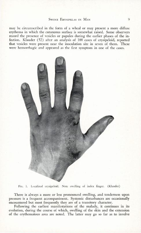
may be circumscribed in the form of a wheal or may present a more diffuse erythema in which the cutaneous surface is somewhat raised. Some observers record the presence of vesicles or papules during the earlier phases of the infection. Klauder (52) after an analysis of 100 cases of erysipeloid, reported that vesicles were present near the inoculation site in seven of them. These were hemorrhagic and appeared as the first symptom in one of the cases.
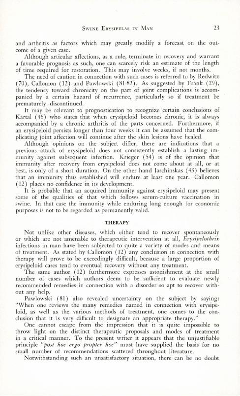
FIG. 1. Localized erysipeloid. Note swelling of index finger. (Klauder)
There is always a more or less pronounced swelling, and tenderness upon pressure is a frequent accompaniment. Systemic disturbances are occasionally encountered but most frequently they are of a transitory character.
Following the earliest manifestations of the malady, it continues in its evolution, during the course of which, swelling of the skin and the extension of the erythematous area are noted. The latter may go so far as to involve
10 AGRICULTURAL EXPERIMENT STATION BULLETIN 130
the dorsum of the hand concerned and even adjacent fingers may participate in the inflammatory process. Involvement of the palm of the hand has been observed but only in exceptional cases.
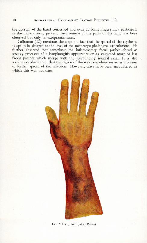
Callomon ( 12) mentions the apparent fact that the spread of the erythema is apt to be delayed at the level of the metacarpo-phalangeal articulations. He further observed that sometimes the inflammatory focus pushes ahead as streaky processes of a lymphangitis appearance or as staggered more or less faded patches which merge with the surrounding normal skin. It is also a common observation that the region of the wrist somehow serves as a barrier to further spread of the infection. However, cases have been encountered in which this was not true.
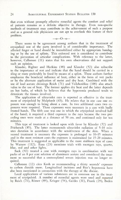
FIG. 2. Erysipeloid (After Rahm)
SWI NE ERYSIPELAS I N M AN 11
The sharply defined, slightly raised zone which develops as the erythem.a slowly advances is mentioned by Klauder (52) as a distinctive feature. As this zone extends peripherally, the more centrally situated areas of the erythematous skin tend to assume a lighter tint and gradually lose their redness .
Pawlowski (81) as well as Klauder ( 47) mention the migratory tendencies of erysipeloid infection. New erythematous patches are apt to make their appearance in areas remote from the original infection site. These patches also heal centrifugally; the center becomes paler, perhaps shows a slightly bluish color, which gradually advances to the deeper colored periphery. Thus there may come about a ring-like arrangement, which may or may not exhibit a tendency to grow peripherally. H eymann ( 42) also mentions purple elevations rhomboid in outline, which fade away somewhat, only to reappear at some other place.
Another di stinctive fea ture pointed out by Klauder (50) is the purplish red color of the affected skin, to which he attaches a diagnostic importance. H e rega rds this color as unique among the cutaneous diseases and as quite dissimilar to the red color that characterizes inflammation caused by other infections.
In erysipeloid the absence of suppuration is quite characteristic, even if some pus may be present in the punctured wound which admitted the specific baci llus. This, however, is most likely to be due to any of the common pyogenic microorganisms. As fa r as available evidence reveals, the swine erysipelas bacillus is not pyogenic per se.
Lymphatic involvement is not constantly observed in erysipeloid cases, yet frequently enough to warrant consideration. In Klauder 's (52) analysis of 100 cases, lymphangitis and lymphadenitis was manifested in 21 of them. In seven additional cases enlargement and tenderness of the regional lymphnodes were noted in the absence of lymphangitis. Callomon ( 12) likewise mentions painful enlargements of the cubital lymphnodes, but observes that many cases run their course without such lesions.
In the more benign cases of erysipeloid, which apparently constitute a conspicuous majority, fever is oniy occasionally observed. Klauder (52) recorded temperatures ranging between 100 and 102 ° F. in six of the 100 patients which came to his attention.
Among the subjective symptoms of which patients complain the most as being extremely annoying are pruritus, and stinging or burning sensations. Pain in the affected fingers is of common occurrence and the feeling of tightness induced by the swelling of the parts to a considerable extent may impair the usefulness of the hand.
Most cases of simple uncomplicated erysipeloid run their course within a period of from two to four weeks. Relapses may, however, be experienced. In Klauder's (52) series six per cent of his cases were thus involved.
Articular or periarticular disturbances not uncommonly develop along with the cutaneous manifestations of erysipeloid or present themselves as a later complication. Friedman (30) observed them in 75 per cent of his cases as the chief cause of complaint on that part of his patients.
Bierbaum and Gottron (7) and Pawlowski (80) regard articular complications as evidence that erysipeloid is not merely a simple superficial inflam-
12 AGRIC U LTU RAL EXPERIMENT STATION BULLETIN 130
matory disturbance. Callomon (12) points to joint involvement as a result of the tendency on the part of the causative microbe to penetrate into the deeper tissue layers and into parts adjoining primary infection sites.
In connection with the incidence of erysipeloid arthritis, it is the opinion of Kartal ( 46) that in any case of erysipeloid which shows no evidence of recovery within ten to 14 days, an inflammation of one or more joints must be reckoned with.
Although the articular involvements of erysipeloid are often referred to or classified as arthritis, there exists doubt in the mind of observers, that inflammatory reactions in articular structures per se are accompaniments of erysipeloid. At least such authorities as Callomen ( 12 ), Rahm ( 68) and Pawlowski (82) are of the opinion that of many cases designated as arthritis, this pathologic entity is only simulated by periarthritic swelling and cutaneous edema.
These opinions find support in the negative results of roentgenologic examinations reported by Axhausen ( 4 ), Klauder (50) and Pawlowski (81-82). Similar observations were made by Kartal ( 46) in connection with apparent involvements of an acute nature and the ones seen in relapses. On the other hand the latter author ( 46) could roentgenologically determine slight abnormalities in cases of the chronic stationary type of arthritis and in those which may be classified as arthritis deformans.
That symtoms suggesting arthritic disturbances cannot altogether be attributed to peri-articular cutaneous tension and swelling is pointed out by Klauder (52) who observed the persistence of pain after swelling had disappeared. Kartal ( 46) is of the opinion that in patients who have in some way or other become sensitized, a mere handling of the flesh of swine is sufficient to precipitate a flare-up of arthritis.
Whatever the precise nature may be, there can be no doubt that articular, peri-articular or merely arthralgic pains connected with erysipeloid are a definite source of suffering and annoyance to the patients concerned. In addition the joint involvements not uncommonly impair the usefulness of their hands.
Stiffness and pain in the affected fingers are the most frequently observed symptoms. They are usually accompanied by swelling and discoloration of the skin overlying the articular area. Movements of the affected fingers cause pain. The joints are sensitive to pressure and as mentioned by Kartal ( 46) may be immobilized in a flexed position which cannot be overcome passively. Without doubt, the thickening of the capsular ligament indicated by Rahm ( 68) may contribute to such a condition as described by Bierbaum and Gottron ( 6) in which the affected articulations could neither be extended nor flexed.
Thickening and induration of periarticular structures are apt to give rise to deformity of the fingers affected, which as a result tend to become fusiform.
In its more acute form articular involvement as an accompaniment of, or as a sequel to, erysipeloid is often marked by the characteristic discolorations in which red, purple or livid tints predominate. Kartal ( 46) describes as characteristic of the chronic stationary form, a finger with a doughy swelling covered by a tense skin showing a waxy yellowish-blue discoloration.
Pain is a prominent symptom and may become particularly disturbing at night or when movements are attempted. Erysipeloid arthritic complications
SWINE ERYSIPELAS I N MA N 13
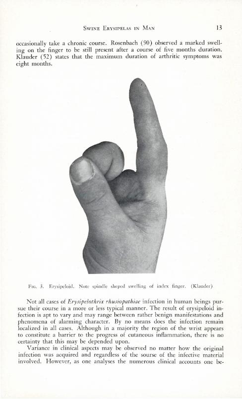
occasionally take a chronic course. Rosenbach (90) observed a marked swelling on the finger to be still present after a course of fi ve m onths duration. Klauder ( 52) states that the maximum duration of arthritic symptoms was eight months.
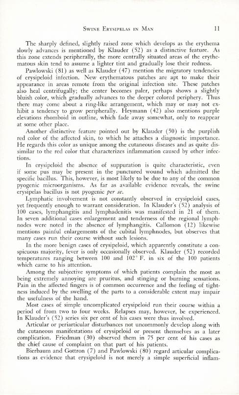
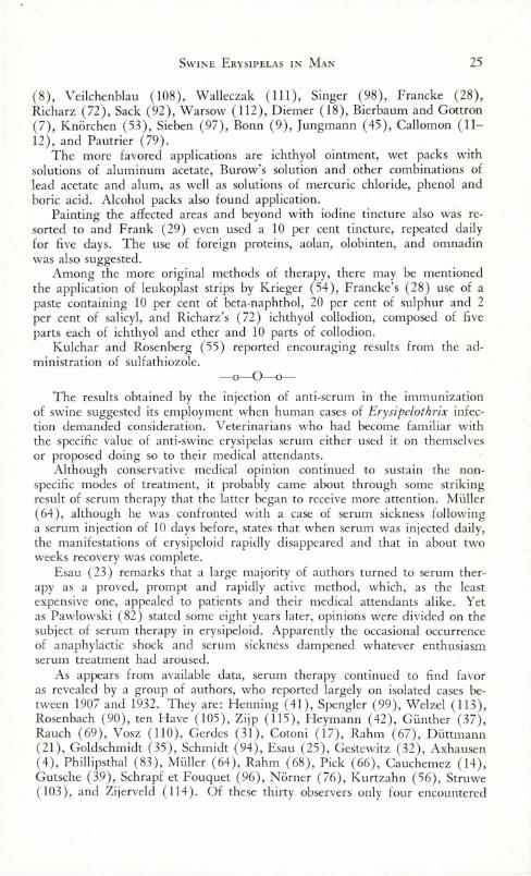
FIG. 3. Erysipcloid. Note spindle sheped swelli ng of index finger. (Klauder )
Not all cases of Erysipelothrix rhusiopathiae infection in human beings pursue their course in a more or less typical manner. The result of erysipeloid infection is apt to vary and may range between rather benign manifestations and phenomena of alarming character. By no means does the infection remain localized in all cases. Although in a majority the region of the wrist appears to constitute a barrier to the progress of cutaneous inflammation, there is no certainty that this may be depended upon.
Variance in clinical aspects may be observed no matter how the original infection was acquired and regardless of the sourse of the infective material involved. However, as one analyses the numerous clinical accounts one be-
14 AGRICULT U RAL EXPERIMENT STATION BULLETIN 130
comes impressed by the fact that accidental inoculation with culture material appears to result in a more prompt and a more severe cutaneous infection. As noted by Callomon (12) there occur from time to time human cases of chronic swine erysipelas and of acute swine erysipelas septicemia . Although rare, such cases cc,ntinue to be recorded in literature.
Hence clinicia ns may expect that now and then cases will come to their attention which are unusual or atypical and even some which run their course to a lethal termination. Some of such cases may therefore be briefly reviewed.
As reported by Krieger (54), he received through the awkwardness of a helper, a puncture by a culture-filled needle in the upper part of one of his thighs. On the following day some redness and pruritus declared themselves, to which but little attention was given. The redness had developed into an erythematous area (l ½ x 3 cm.) on the third day. Other similar lesions appeared around this urticarious area with a certain degree of suddenness, so that within a period of about ten days the entire anterior surface of the thigh from the inguinal region down to the knee became the site of smaller well defined wheals, which tended to become confluent. Their original bright red color slowly changed to a dark grayish red. The skin was hot and knee movements were interfered with. A burning pruritus was manifest and new infection foci were apt to appear after a long walk . The case eventually terminated in recovery.
Another atypical case of apparent erysipeloid is also reported by Chevallier and associates (15-16). It pertained to a young pork butcher who developed on the forearm, the legs and the upper part of the back a more or less undefined eruption which disappeared after an injection of anti-staphylococcus vaccine. However, the temperature rose to 102 ° F. and the patient revealed another breaking out of very large patches, which occupied the inferior parts of the trunk and the thighs. A day or two later, the arms and legs became invaded by extensive patches with raised, irregular margins. Some of these areas showed vesicles, ranging in size from that of a pin head to that of a small pea . Other patches developed later but did not show vesicles. They had a violet, iilac or pinkish-violet color, which conveyed the impression of a redness on a blue base. On or about the fifth day the patches became paler and desquamation began. The latter continued intensely while the skin was assuming a normal appearance; systemic disturbances ceased and the case recovered.
According to Sieben (97) one of his three erysipeloid patients who had become simultaneously infected, while engaged in dressing the carcass of a hog, presented certain unusual features. Three days after the infective contact this patient presented himself with a temperature of 104 ° F . conveyed the impression of sepsis and complained of severe headaches. He had pains in his limbs, manifested general weakness as well as a degree of stupor.
On the second day of fever, red spots with a papule in the center appeared on the back. These areas enlarged rapidly and became confluent. The center of these patches was scarlet red, whereas the remaining portion had a pale red color. The boundary between the darker portion in the center and the lighter colored one was sharply defined.
More patches developed on the back, the gluteal region, the upper portion
SWINE ERYSIPELAS I N MAN 15
of the thighs, the arms, the forearms and the hands, while two spots were noted on the forehead . These spots presented a deep bluish-red color at first but later the discoloration assumed a more brownish tint. They were completely covered by small vesicles.
After a course of illness lasting fi ve days the temperature subsided and the skin lesions slcwly disappeared. The patient made an uneventful recovery.
A case report by H abersang ( 40) indicates that a by no means uncommon form of swine erysipelas may also present itself in man when transmission possibilities are particularly propitious. A hog owner had subjected two of his swine to emergency slaughter on account of swine erysipelas. In some manner he contracted erysipeloid, from which he apparently recovered.
Soon thereafter the custom butcher who had functioned in dressing the carcass and in preparing the meat presented himself to H abersa ng with the request that his own hogs be vaccinated, because he himself had swine erysipelas urticaria ( diamond skin disease). Upon examination it was fou nd that this man's body was covered over and over wi th typical rhomboid wheals measuring 1-2 cm.
Part of these wheals were already scaling whereas others were of a bluish red color and more or less hemorrhagic. It was further ascertained that a few days after this patient had prepared sausage meat from the two hogs mentioned above, he had become ill , suffered from headache, had a temperature of 104 ° F. and attacks of dizziness and had to take to his bed .
With the completion of the eruption, systemic symptoms improved and in the course of a few days the exanthema healed with a marked degree of desquamation. Anamnestic in formation disclosed that wound infection could be excluded. On the other hand the patient while preparing the sausage meat had consumed a considerable amount of this raw material and he frankly attributed his misfortune to this infec tion source.
As reported by Spitzer ( 102) a veterinar ian engaged in the vaccinat ion of swine against erysipelas, slightly cut a finger on the broken edge of a culture vial. H e soon was compelled to seek medical aid. The purple swollen finger was treated by cold applications and the use of aluminum acetate compresses. As no benefi t followed this treatment, the affected area was deeply incised and curetted, followed by antiseptic dressing. Subsequently there appeared two fluctuating enlargements on the median face of the forearm, a marked lymphangitis, a very high temperature and great unrest on the part of the patient. The fluctuati ng enlargements were opened and a bloody watery fluid evacuated . Pus was not found to be present. While the patient was being removed to a clin ic, he died on the way. It was noted that there was a post-mortem purplish discoloration and swelling of the face, the ears and the neck of the cadaver.
Gunther (38) makes mention of two fatal cases in veteri narians due to swi ne erysipelas infection. One of these acquired the infection in his thumb on October 30 and died as a result on the following February 8. It seemed that the local lesion of the hand healed but that later symptoms of cardiac impa irment became apparent. The autoposy brought to light myocardial degeneration, degenerative changes in the liver and spleen and a recent thrombu s-like deposit on the tip of one of the mitral valves.
16 AGRICULTURAL EXPERIMENT STATION BULLETI N 130
In the other case reported by Gunther (38) cardiac disturbances were the most conspicuous clinical manifestations. Within a short space of time evidence of sepsis and of an acute endocarditis developed. The cause of death was recorded as ulcerative endocarditis.
Prausnitz ( 84) rendered an account of a fatal case in a ten year old girl suffering from a congenital hea rt defect. This patient sickened acutely with a febrile disturbance. After a few days peculiar, slightly-elevated erythematous eruptions presented themselves at various places on the body surface and simultaneously arthralgia developed. The fever remained high and the clinical picture assumed features of chronic sepsis, complicated with endocarditis. The child died after an illness of approximately six months. During the illness the swine erysipelas bacillus could be repeatedly recovered in pure culture from the blood stream.
A most unusual case was observed by Klauder (50-5 1). The patient concerned had, four months prior to the time he presented himself, pricked the index finger of his right hand on the tooth of a fishhead used as bait while crabbing. Two days later the injured finger became swollen and a sensation of burning developed.
An erythema involved the entire fin ge r, spread to the dorsum of the hand, to the other fingers and eventually to the palm of the hand also. There was pain, a sense of tension and stiffness which impaired the usefulness of the hand.
About five months after the onset, the infection spread beyond the wrist to the forearm . Ev idence of joint involvement presented itself and accounted for stiffness and pain in the fingers, the wrist and elbow. The infection became further extended until at the end of a yea r it covered the entire integument from head to feet.
However, at no time was the entire surface simultaneously involved . Klauder described its advance as wave-like with the progressing border always sharply delineated as a strip of purplish erythema. Behind the advancing margin the skin gradually became normal. The patient experienced many relapses, with purplish circinate areas of erythema appearing and reappearing over the entire body at different times. The relapses were not accompanied by subjective symptoms, although articular involvement of varying degrees of intensity added to the patient's discomfort.
A final account of this pat ient is provided by Klauder's (51) subsequent publication. The case is designated as one of generalized erysipeloid from which after a duration of 15 months, the specific bacilli could still be recovered from the skin. After the disease had pursued a course of 29 rn.onths, the patient became psychically unbalanced and committed suicide.
A case is described by Feiss inger and Brouet (27) in which the patient presented purpuric spots at the level of the malar regions and the ears. They were accompanied by a general disturbed condition, the cause of which was not determined. His condition became worse and a general pruritus declared itself. An eruption made its appearance on the trunk and the limbs. It consisted of a large patch, having a few intervals of normal skin. There developed a swelling about the left knee, which receded after 24-48 hours. A slightly elevated temperature was noted . The eruption persisted for about
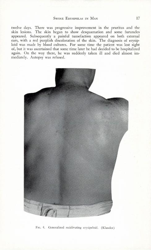
SwINE ERYSIPELAS IN MAN 17
twelve days. There was progressive improvement in the pruritus and the skin lesions. The skin began to show desquamation and some furuncles appeared. Subsequently a painful tumefaction appeared on both external ears, with a red purplish discoloration of the skin. The diagnosis of erysiploid was made by blood cultures. For some time the patient was lost sight of, but it was ascertained that some time later he had decided to be hospitalized again. On the way there, he was suddenly taken ill and died almost immediately. Autopsy was refused.
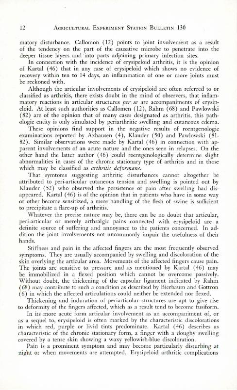
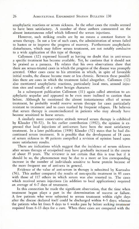
FIG. 4. Generalized rccidivating er ys ipeloid. (Klauder)
18 AGRICULTURAL EXPERIMENT STATION BULLETIN 130
The case reported by Russell and Lamb (91) pertained to a lobster fisherman who was hospitalized with a history indicating that two months before admission, there had been progressive weakness, malaise, occasionaly chilly sensations, and a loss of twenty pounds in body weight.
One month before entry a non-supperative ulceration of about 1 cm. diameter appeared on the upper lip, accompanied by moderate swelling and erythema. This cleared up in a week. No other cutaneous lesions had been observed. Previous history was apparently obscure.
Death occurred on the 26th day of hospitalization. The organisms recovered from six blood cultures, the heart's blood and endocardial vegetations were identical, morphologically, culturally and serologically with a known strain of Erysipelothrix rhusiopathiae.
PATHOLOGIC ANATOMY
Whereas the clinical and bacteriologic details connected with erysipeloid have received full consideration in literature, comparatively little attention has been given to its pathology and pathologic anatomy. This must, no doubt, be attributed to the fact that its gross pathology, with very few exceptions, is so closely allied to its clinical manifestations that purely pathologic consideration came to be regarded as of subsidiary importance ..
With reference to the patho-histology or erysipeloid, the low mortality rate of the disease always tends to restrict the supply of material suitable for study and thus to restrict investigational efforts. Furthermore, the occasional biopsies made in pracice were most commonly done for the purpose of bacteriologic diagnosis, rather than for that of research in pathology.
Hence it should not be surprising that but little attention was given to the subject since the studies of Delbanco (19) in 1898 up to the present. The material studied by Delbanco was obtained from a finger of a woman who had injured herself on a fishbone. The excision was made from the marginal portion of the inflammatory area, there where the somewhat elevated, reddened portion is sharply delimited from the healthy surrounding skin.
Histologically examined, the epidermis did not reveal conspicuous changes; there was widening of the interepithelial spaces in which a few leucocytes could be detected. The prickle0 cells were well filled out and their protoplasm not well stained.
The dominating aspect of the picture is a cuticular edema, which became Jess marked toward the papillae. The latter had retained their normal appearance. The vessels were sheathed with cells rich in spongioplasm. In their lumen mononuclear and a few polymorphonuclear leucocytes were observed. Proliferation of the perithelia appeared to be evenly distributed in the layers of the cutis. The collagenic bundles were somewhat swollen but had retained their affinity for dyes.
The elastic tissues were rarified. Conspicuously numerous were the mast cells, which dominated the histologic picture. Many of these cells showed their specific coloring only at the tip of their apparently fusiform body. In some of them the cells looked as if exploded, so that liberated granul es poured out into the lymph spaces.

SWINE E RY SIPELAS IN M AN 19
Delbanco's descriptions are approvingly cited by Callomon (12) who also agreed with Bazzoli 's interpretation of the histologic picture of erysipeloid, a disease which he defined as: "A serous inflammation, above all of the corium, with dilatation of the lymph spaces, a moderate perivascular infiltration, which may also invade the subcutis." To this Collomon ( 12 ) adds the qualification "with numerous mast cells, especially in the enlarged lymph spaces and the turgid capillaries."
On the whole one cannot be certain that aside from the finding of Erysipelothrix in the tissues, these changes are pathognomonic per se.
The gross and microscopic pathology of the extremely chronic case reported by Kl auder (5 1) may likewise not be pathognomonic for erysipeloid, even if of sufficient interest to deserve attention. The autoposy of this case disclosed aside from some irrelevant findings that the aorta was atheromatous, that plaques were observed in the coronary arteries but that no evidence of endocarditis came to light.
The liver was not enlarged but showed yellowish foci of pinhead size and a necrotic nodule which subsequently proved to be carcinomatous. Although the kidneys appeared to be pale, they were apparently normal as seen macroscopically. The spleen was enlarged, showed a rough surface and the cut surface was mushy. The structure pattern was effaced and the regional lymphnodes were hyperplastic.
Histologic examinations disclosed the following changes: Sections of skin material taken from the median line of the abdomen, showed that the connective tissue of the papillary layer was denser than normal, in agreement with the preceding inflammatory developments. A few lymphocytes surrounded the hair follicles.
Liver sections brought to light small, irregularly distributed, necrotic foci in the external and median zone of the lobuli and around the central vein . A sparse mononuclear cell infiltration was apparent about the lobular periphery. The capillaries contained polymorphonuclear lymphocytes and histiocytes. Many of the latter were phagocytes, containing pyknotic nuclei or nuclear fragments. In the kidney sections the cells of the convoluted tubules were granular. One dilated tubule contained a pink colored coagulum. There was no ev idence of nephritis and the kidneys were not found to be markedly changed.
Lymphnode structure was disturbed by an increase in the reticular cells and in the connective tissue, as well as by a pronounced reduction in the lymphocytes. A few large cells were phagocytic and contained cell fragments and pigment. Some parts showed cell destruction and hyaline degeneration.
The spleen showed pathologic changes. The malpighian corpusles were structurally altered and were not as conspicuous as under normal conditions while most of the lymphocytes had been replaced by large cells. Within the lymph follicles the vessel walls were thickened and contained a large number of degenerated or destroyed cells which were taken in by numerous phagocytes. Such cell s were found in the interstitious spaces of the pulp.
The case described by Russell and Lamb (91) when it became the subject of post mortem examination, revealed that the lungs, digestive tract, pancreas, kidneys and adrenal s, bladder, aorta and bone marrow had apparently

20 AGRICULTURAL EXPERIMENT STATION BULLETIN 130
remained normal. The pleural cavity contained approximately 1000 cc. of a straw colored, clear fluid. The visceral and parietal pericardia! structures were loosely adherent.
The heart was hypertrophic, weighing 500 gm. Three small deeply pinkish-yellow, finely granulated and firmly adherent vegetations were seen on the free margin of the anteriar leaflet of the mitral valve. Two of the chordea tendineae showed similar small vegetations. The mitral cusps were not thickened or distorted and the chordea were delicate and thread-like.
FIG.5. Endocarditis verrucosa of aortic valves. (Russell and Lamb)
There appeared to be only two aortic cusps and these were largely displaced by slightly friable and coarsely granular vegetations of the same color as the ones mentioned above. The right commissure was obscured by excrescences, that extended into each aortic sinus. In this area the vegetations were particularly bulky, invading the sinus wall, the ring and the adjoining aortic sinus, valve cusp and myocardium. Two small abscesses were revealed, one in the subpericardial fat and the other one partly in the myocardium and partly in the bulky vegetation of the cusp. The endocardium was in part covered by coarsely granular, pinkish yellow vegetation which appeared to penetrate the subjacent myocardium.
The spleen was twice its normal size and contained a moderately sized fresh infarct, covered by a thin layer of yellowish fibrin.
Histological examination of a section through the aortic ring and adjacent cusp revealed a marked thickening by layers of organized fibrin. A section
S W INE E RYSIPELAS IN M AN 21
of the m yoca rdium showed the pericardia! surface to be covered by a thin layer of fibrin , containing neutrophiles and macrophages. Several small foci of neutrophiles were scattered throughout the m yocardium.
Spleen sections disclosed the necrotic infa rct to be demarcated from the normal tissues by a broad zone of fibrin and polymorphonuclear leucocytes. T he centers of many of the malpighian corpuscles had large deposits of fibrin which contained sca ttered neutrophiles.
The liver lobules showed a marked central necrosis and only a peripheral fringe of viable cells. In the kidney sections several glomeruli were observed to contain thrombi of fi brin . In some instances exudation of fibrin and neutrophiles into the glomerular space and adjoining tubules could also be recorded .
One of the fa tal cases of erysipeloid reported by Gunther (38) revealed upon post mortem examinat ion, lymphatic hyperplasia, myocardial, hepatic and splenic degeneration, and a thrombus-like deposit on the tip of one of the mit ral valves .
DIAGNOSIS
It does not appear in an analysis of available references that the diagnosis of erysipeloid or its diffe rent iation from other cutaneous maladies constitutes an ever returning problem. As such authors as Reich (71), Klauder ( 52) and Callomon (12) have pointed out, the anamnesis and manifestations of the average case are of such a character that the recognition of the disease sca rcely presents any difficulty.
T he occupation of patients constitutes a detail in anamnesis to which conside rable weight must be attached. There is a considerable volume of evidence showing that veterinarians or their helpers when vaccinating swine by the use of live-culture vaccine or in the performance of autopsies of swine are particularly exposed to the specific in fec tion . T o a less extent thi s also pertains to persons whose occupation implies the handling of pork, fish , crustacea, game, ski ns and other products of animal origin.
If the original infection site is to be found on the hands, this fac t also should not escape attention and should be given considerat ion as mo.re or less characteristic in erysipeloid . T hi s detail assumes add itional importa nce when the erythema is not accompan ied or preceded by systemic reactions such as feve r, ch ills, etc.
Such ind ications are most frequently complemented by the bluish or purple tint of the erythema, of which Klauder ( 52) states that, " this color is unique among skin d iseases and is unlike the redness that characterizes inflammation caused by other in fections." Likewise it may be kept in m ind that suppuration or abscess formation is not a feature belonging to uncomplicated erysipeloid . The swine erysipelas bacillus is not pyogenic.
Only when an apparent erysipeloid declares itself as a primary erythema in parts of the body other than the hands, or when a cutaneous disease presents phenomena which di ffe r fro m the ones accepted as characteristic of Erysipelothrix infec tion, may difficulties arise in the establishment of a definite diagnosis.
Thus it may become necessa ry to diffe rentiate between erysipeloid and erysipelas, when the anamnesis fa ils to supply defin ite data or when no history
22 AGRICULTURAL ExPERIME. STATION BuLLETIN 130
of skin wounds or abrasions is obtainable. In doubtful cases of this sort it may be remembered that the element of the blue or purple of erysipeloid erythema as compared with the intense redness shown by that of erysipelas, constitutes a most important factor in differential diagnosis.
Erysipelas erythema spreads more rapidly and is not uncommonly accompanied by the appearance of blebs. In cases of true pyogenic erysipelas involving the hand, it is to be noted that the latter participates in the inflammatory process in its entirety.
In erysipelas the onset is commonly one of a systemic disturbance indicated by rigors and a rapid rise of temperature, which may or may not be accompanied by albuminuria. Such manifestations do not belong to the clinical picture of erysipeloid.
Suppuration is frequently observed in facial erysipelas and may be expressed by small cutaneous abscesses. As seen by Reich (71) the erythema of erysipelas shows an uninterrupted continuity in its progress whereas in erysipeloid there is a tendency on the part of the skin lesions to advance in the form of isolated patches.
Klauder (52) mentions that commonplace pyogenic infect ions may occasionally be confused with erysipeloid. In this connection he points out that the color of the erythema should serve as a distinctive feature and that pitting of the skin by pressure does not belong to the erysipeloid syndrome.
With reference to the differentation between erysipeloid and phlegmonous inflammation Reich (71) states that given a comparable extent of swelling or infiltration of the skin, as a rule in cases of phlegmonous dermatitis there are distinct suppurative foci, whereas in uncomplicated erysipeloid these are never seen. This author further observes that in erysipeloid the site of inoculation is without or almost without an immediate reaction and that such reactions, on the other hand, are conspicuous phenomena in cases of phlegmonous inflammation.
Of particular value in the recognition of Erysipelothrix infection is the demonstration of the specific microbe in the circulating blood, in the skin lesions, or in material obtained by biopsy from the advancing margin of the erythematous areas.
PROGNOSIS
There is agreement among authors that uncomplicated erysipeloid may be regarded as a benign disease, permitting a favorable prognosis. Callomon (12 ) ventures the opinion that such a prognosis may even be hazarded in cases which present such more or less alarming aspects as fever, extensive erythematous areas, etc.
Klauder (52) notes that in 79 cases the duration of illness ranged between 10 and 30 days, which is nearly in accordance with Pawlowski's (80-81) data. The latter show that the malady has no definite duration, heals after two or three weeks, although the healing process may be drawn out to four to five weeks or even longer.
In view of the possibility of unpredictable complications, a prognosis cannot always be hastily expressed. Esau (23) as well as Callomon (1 2) call attention to such eventualities as swine erysipelas septicemia, endocarditis
SWINE ERYSIPELAS I N M AN 23
and arthritis as factors which may greatly modify a forecast on the outcome of a given case.
Although articular affections, as a rule, terminate in recovery and warrant a favorable prognosis as such, one can scarcely risk an estimate of the length of time required for restoration. This may involve weeks, if not months.
The need of caution in connection with such cases is referred to by Redwitz (70), Callomon ( 12) and Pawlowski (81-82). As suggested by Frank (29), the tendency toward chronicity on the part of joint complications is accompanied by a certain hazard of recurrence, particularly so if treatment be prematurely discontinued.
It may be relevant to prognostication to recognize certain conclusions of Kartal ( 46) who states that when erysipeloid becomes chronic, it is always accompanied by a chronic arthritis of the parts concerned. Furthermore, if an erysipeloid persists longer than four weeks it can be assumed that the complicating joint affection will continue after the skin lesions have healed.
Although opinions on the subject differ, there are indications that a previous attack of erysipeloid does not consistently establish a lasting immunity agai nst subsequent infection. Krieger (54) is of the opinion that immunity after recovery from erysipeloid does not come about at all, or at best, is only of a short durat ion. On the other hand Jaschinskas ( 43) believes that an immunity thus established will endure at least one year. Callomon ( 12) places no confidence in its development.
It is probable that an acquired immunity against erysipeloid may present some of the qualities of that which follows serum-culture vaccination in swine. In that case the immunity while enduring long enough for economic purposes is not to be rega rded as permanently valid.
THERAPY
Not unlike other diseases, which either tend to recover spontaneously or which are not amenable to therapeutic intervention at all , Erysipelothrix infections in man have been subjected to quite a variety of modes and means of treatment. As stated by Callomon ( 12) any conclusion in connection with therapy will prove to be exceedingly difficult, because a large proportion of erysipeloid cases tend to eventual recovery without any treatment.
The same author ( 12) furthermore expresses astonishment at the small number of cases which authors deem to be sufficient to evaluate newly recommended remedies in connection with a disorder so apt to recover without any help.
Pawlowski (8 1) also revealed uncertainty on the subject by saying: "When one rev iews the many remedies named in connection with erysipeloid, as well as the various methods of treatment, one comes to the conclusion that it is very difficult to designate an appropriate therapy ."
One cannot escape from the impression that it is quite impossible to throw light on the distinct therapeutic proposals and modes of treatment in a critical manner. To the present writer it appears that the unjustifiable principle "post hoc ergo propter hoc" must have supplied the basis for no small number of recommendations scattered throughout literature.
Notwithstanding such an unsatisfactory situation, there can be no doubt
24 AGRICULTURAL EXPERIMENT STATION BULLETIN 130
that even without promptly effective remedial agents the comfort and relief of patients remains as a definite objective in therapy. Even non-specific methods of treatment may contribute to the achievement of such a purpose and as a general rule physicians are not apt to overlook this feature of their problem.
-o-O-o-
There seems to be agreement among authors that in the treatment of erysipeloid rest of the parts involved is of considerable importance. The affected finger or hand should be immobilized either by appropriate bandaging or by the use of splints. This enforced rest is considered to contribute to the prevention of articular complications. With reference to the latter, however, Callomon ( 11) states that his own observations did not support such an opinion.
Klauder, Righter and H arkins ( 48) and Klauder (52) also subscribe to the importance of rest and indicate that the hand should be carried in a sling or more preferably be fixed by means of a splint. These authors further emphasize the beneficial influence of heat, either in the form of wet packs or by the alternate application of moist and dry heat. Combined with the use of lead acetate dressings Reich (71) and Veilchenblau (108) also attach value to the use of heat. The former applies dry heat and the latter depends on hot baths, of which he believes that the hyperemia produced tends to detoxicate the tissues involved.
The application of ultraviolet rays is reported as effective in the t reatment of erysipeloid by Mulpfordt (63) . He relates that in one case one exposure was enough to bring about a cure. In two additional cases two exposures were required. Three exposures were necessary in a case with badly frosted hands. The fifth case was one in which the erysipeloid involved half of the hand; it required a third exposure to the rays because the two preceding ones were made at a distance of 50 cm. and continued only for ten minutes.
This type of treatment is looked upon with favor by Klauder (52) and Priebatsch ( 85). The latter recommends ultraviolet radiation of 5-10 minutes duration in accordance with the sensitiveness of the skin. When a second treatment is necessary the exposure is prolonged to 10- 15 minutes and in the most resistant cases the exposure is maintained for 15-20 m inutes. This treatment is suggested as applicable to the more stubborn type of cases by Warsow ( 112 ) . Esau (23) mentions trials with roentgen rays, quartz, blue, red and other lights.
Sack (92) treated a case with roentgen rays in combination with wet packs of an 8 per cent solution of aluminum acetate and considered the treatment so successful that a contemplated serum injection was no longer required .
Callomon ( 12) cites Kroh as recommending a thirty seconds' exposure to carbon dioxide snow. Long itudinal incisions and Bier's hyperemia have also been mentioned in connection with the therapy of the disease.
Local applications of various substances are in common use in the treatment of erysipeloid. A number of remedial agents were used and reported by Reich (71), Romer (89), Krieger (54), Gerdes (31), Frank (29), Boden
SWINE ERYSIPELAS I N MAN 25
(8), Veilchenblau ( 108), W alleczak ( 111), Singer (98), Francke (28), Richarz (72), Sack (92), Warsow ( 112), Diemer ( 18 ), Bierbaum and Gottron (7), Knorchen (53), Sieben (97), Bonn (9), Jungmann (45), Callomon ( 11-12), and Pautrier (79).
The more favored applications are ichthyol ointment, wet packs with solutions of aluminum acetate, Burow's solution and other combinations of lead acetate and alum, as well as solutions of mercuric chloride, phenol and boric acid. Alcohol packs also found application.
Painting the affected areas and beyond with iodine tincture also was resorted to and Frank (29) even used a 10 per cent tincture, repeated daily for five days . The use of foreign proteins, aolan , olobinten, and omnadin was also suggested.
Among the more original methods of therapy, there may be mentioned the application of leukoplast strips by Krieger (54 ), Francke's (28) use of a paste containing 10 per cent of beta-naphthol, 20 per cent of sulphur and 2 per cent of salicyl, and Richarz's (72) ichthyol collodion , com posed of five parts each of ichthyol and ether and 10 parts of collodion.
Kulchar and Rosenberg (55) reported encouraging results from the administration of sulfa thiozole.
-o-0 -o-
The results obtained by the injection of anti-serum in the immunization of swine suggested its employment when human cases of Erysipelothrix infection demanded consideration . Veteri narians who had become fam iliar with the specific value of anti-swine erysipelas serum either used it on themselves or proposed doing so to their medical attendants .
Al though conservative medical opinion continued to sustain the nonspecific modes of treatment, it probably came about through some striking result of serum therapy that the latter began to receive more attention. Muller ( 64), although he was confronted with a case of serum sickness following a serum injection of 10 days before, states that when serum was injected daily, the manifestations of erysipeloid rapidly disappeared and that in about two weeks recovery was complete.
Esau (23) remarks that a large majority of authors turned to serum therapy as a proved, prompt and rapidly active method, which, as the least expensive one, appealed to patients and their medical attendants alike. Yet as Pawlowski (82) stated some eight years later, opinions were divided on the subj ect of serum therapy in erysipeloid. Apparently the occasional occurrence of anaphylactic shock and serum sickness dampened whatever enthusiasm serum treatment had aroused.
As appears from ava ilable data, serum therapy continued to find favor as revealed by a group of authors, who reported largely on isolated cases between 1907 and 1932. They are: Henning (4 1), Spengler (99), Welzel (113), Rosenbach (90), ten Have (105), Zijp (115), H eymann (42), Gunther (37), Rauch (69), Vosz ( 110), Gerdes (3 1), Cotoni ( 17), Rahm (67), Duttmann (21), Goldschmidt (35), Schmidt (94), Esau (25), Gestewitz (32), Axhausen (4), Phillipsthal (83), Muller (64), Rahm (68), Pick (66), Cauchemez ( 14), G utsche (39), Schrapf et Fouquet (96), Norner (76), Kurtzahn (56), Struwe (103), and Zijerveld ( 114). Of these thirty observers only four encountered
26 AGRICULTURAL EXPERIMENT STATION BULLETIN 130
anaphylactic reactions or serum sickness. In the other cases the results seemed to have been satisfactory. A number of these authors commented on the almost instantaneous relief which followed the serum injections.
However, such striking results are by no means a constant feature in serum therapy. In not a few of such cases, the administration of serum fa iled to hasten or to improve the progress of recovery. Furthermore anaphylactic disturbances, which may follow serum treatment, are not notably conducive to a wide application of this type of therapy.
Callomon (12) expressed himself as having no doubt that in anti-serum a specific treatment has become available. Yet, he cautions that it should not be praised as a panacea. He relates that his own observations show that there are serum-treated cases in which the progress of the disease was promptly arrested. Other cases came to his attention in which, in spite of favorable initial results, the disease became more or less chronic. Between these possibilities there are cases in which the treatment failed altogether. Callomon (12) also mentioned anaphylactic reactions, particularly local ones, around injection sites and usually of a rather benign character.
In a subsequent publication Callomon ( 11) again called attention to anaphylactic sequelae and appeared to be even more inclined to caution than before. Although he mentioned the almost instantaneous effect of serum treatment, he probably would reserve serum therapy for cases particularly resistant to treatment and to cases marked by frequent relapses. He believes that serum therapy is counterindicated in patients who may already have become sensitized to horse serum.
A similarly more conservative attitude toward serum therapy is exhibited by Klauder (50-52). In his earlier contribution ( 1932), the opinion is expressed that local injections of anti-serum have been the most satisfactory treatment. In a later publication ( 1938) Klauder (52) states that he had discontinued serum treatment. It is possible that the development of 18 cases of serum sickness in 48 patients compelled a revision of opinion based upon more satisfactory results.
There are indications which suggest that the incidence of serum sickness after serum therapy of erysipeloid may have gradually increased in the course of about 35 years. The reviewer is not certain that this is true but if it should be so, the phenomenon may be due to a more or less corresponding increase in the number of individuals sensitive to horse protein because of the more frequent use of anti-sera in general.
Evidence of the value of anti-serum in therapy is submitted by Kurtzahn (56). This author compared the results of non-specific treatment in 83 cases with those of 117 others in which serum was also resorted to. The cases which received serum injections ( in addition to local applications) required an average of 6-7 days of treatment.
In this connection he made the significant observation, that the time when treatment began plays a part in the determination of success or failure. A few patients who were subjected to serum treatment within eight days after the disease declared itself could be discharged within 4- 5 days; whereas the patients who let from 8 days to 6 weeks pass by before seeking treatmen t required from 6-13 days for a cure. When these cases are compared with the
SWINE E RYSIPELAS IN M AN 27
ones which did not receive serum treatment and which required from 12-30 days for recovery, thi s author concluded that the value of serum therapy cannot be questioned .
In none of Kurtzahn's cases could undesirable complications be recorded. H e came to believe that early serum treatment, fixation of the affected parts by means of splints, and a dressing of 10 per cent ichthyol ointment constitutes the most appropriate treatment of erysipeloid .
The observations of Rahm ( 68) also throw light on what may come about in connection with serum therapy. This author, during a period of approximately six years, used serum in all recent erysipeloid cases. H e conveyed the impression that by this practice the incidence of joint complications had been reduced. Of his 36 cases, four were treated conservatively. T wo of them developed arthritis and the other two withdrew from further observation.
Rahm's other 32 cases had received injections of 10 cc. each of anti-se rum during the earliest stages of the d isease . Afte r 28 serum injections he observed recidivation 8 times. This group included 3 cases of typical erysipeloid without joint involvement, 2 cases of typical erysipeloid arthritis, and 3 cases in which arthralgia was accompanied by dermatitis.
In six of his cases, the healing process was tardy; they developed relapses of erysipeloid associated with phalangeal movements. Only in 15 cases coul d prompt recovery after serum treatment be recorded .
Rahm ( 68) did not encounter manifes t disadvantages associated with serum treatment. Only in one case did an extensive serum exanthema present itself eight days after the injection. This was promptly relieved by an inj ection of 1 cc. of a 1:1000 solution of adrenalin.
In a case reported by Esau ( 22) a relapse of erysipeloid declared itself in spite of a previous inj ection of anti-serum. H ence 13 days after the injection another dose was administered. The latter brought about a reaction denoted by a marked infiltration around the site of injection, subsequent to rigors, nausea and malaise . All these phenomena disappeared in the course of a few days. Apparently the period which elapsed between the two serum injections was too long for a safe procedure.
-o-0-o-
In connection with serum therapy in swine erysipelas, a great volume of evidence has accumulated which most decisively shows that the specific action of anti-serum cannot be doubted. H ence any particular disappointment which may follow the use of a potent serum cannot be attributed to a lack of antibodies with specific affinity for the antigen concerned. In all probability the lack pertains to an understanding of when and where the serum treatment may be reasonably ex pected to be used with uncomplicated benefits.
Disappointments following serum treatment usually arise in connection with cases which fa iled to respond to the injection and with cases in which the serum precipitated anaphylactic shock or serum sickness . Tt seems possible th at at least a part of such di sadva ntages may be avoided.
With reference to the " when" phase of serum therapy attention is called to suggestions by Kurtzahn (56), Muller (64) , Rahm ( 68) and others, who believe that the best results may be expected from the earliest possible injec-
28 A GRI CU LTURAL EXPERIMENT STATION B U LLETI N 130
tions. It is by no means impossible that delay in the administration of antiserum may account for failure of anticipated achievement.
Such an opinion derives a measure of support from what has become common knowledge among veterinarians. In their practice, it has become more or less firmly established that if swine affected with acute septicemic swine erysipelas are injected with adequate amounts of anti-serum of good potency their chances of recovery are fairly good, provided that the injections be made during the first 24- 36 hours of illness ( preferably determined by thermometry) .
If after this period, treatment is delayed the expectancy of good results progressively declines as time advances . As having a further bearing on the problem, it may also be mentioned that in swine erysipelas arthritis, quite common in young pigs, only mediocre results can be obtained from serum treatment.
It is quite probable that some, if not most, of the disappointments arising from undesirable serum reactions reported in literature, may have been avoided, if the patient 's sensitivity to horse serum had been challenged by an appropriate skin or eye test. The results of such tests should provide guidancein devising the therapeutic procedure in any given case. In the event of positive skin or eye reactions, the patient concerned should be desensitized before the curative serum dose be administered.
On the whole it seems best, that in cases which are eligible to serum treatment, preference be given to the general accepted dose of 1 cc. per 10 kilograms body weight. If for any reason serum injections are to be repeated, the amount of serum should not be more than that of prev ious doses. The interim between inj ecti ons should not exceed 4-5 days.
PROPHYLAXIS
Because of its epidemiologic and clinical peculiarities, the prevention of erysipeloid is to a considerable extent a matter of individual care with or without professional guidance.
When larger groups of workers are exposed to specific in fec tion in industries which prepare va rious products of animal origin, such as abattoirs, fisheries, etc. , additional measures may be possible. However, as yet, group protection in general has thus far remained as an unsolved problem. An exception to this was supplied by McGinnis and Spindle (59) who recommended, as a mea ns of prevention among the workers in a bone button factory, that all bones entering the plant be heated to a temperature of 144 ° F. for not less than two hours while in the soaking tanks.
The protection of the hands against injuries which either instantaneously or more remotely are apt to admit the causative microbes constitutes an effective measure of prophylaxis. This pertains especially to veterinarians engaged in the routine vaccination of swine. Their hands should always be protected by rubber gloves, not the thinnest, and of a quality which keeps them from becoming slippery when in use. It is likewise of prophylactic value to so organize vaccination operations, that the veterinarian can devote himself exclusively to the manipulation of his syringes. If circumstances compel him to attend to other details, the way is open to accidental needle punctures.

SWINE ERYSIPELAS IN MAN 29
Whenever materials apt to contain the swine erysipelas bacillus must be handled without the protection of gloves, the hands should be challenged previously for the presence of abrasions and other cutaneous infractions. Such avenues of infection can usually be detected by the sting induced by an application of strong alcohol. Skin defects disclosed in this manner should be sealed by a thin collodion-cotton dressing before the hands are exposed to suspected material.
Suction applied to the parts immediately after the needle puncture has always been a popular manner of procedure. Owing to the speed by which the microbes apparently find their way into the intercellular lymph spaces such attempts are probably of a very problematic value. To this Callomon (12) adds the objection that the practice may not be entirely safe because small abrasions or fissures of the lips may open the way to facial erysipeloid.
Immediate applications of alcohol or anti-swine erysipelas dressings have also been proposed. As there is evidence that promptly applied antiseptics, even of tincture of iodine, failed to prevent infection, it seems that the bacilli once within or under the skin can no longer be molested.
Cultures of Erysipelothrix to be used in sero-vaccination should be provided in strong vials, closed by the type of rubber stoppers now in common use in this country. They should always be preferred to fused ampoules the point of which must be broken in order to withdraw the fluid.
Acknowledgements. The author gratefully acknowledges the courtesy of Dr. J. V . Klauder of Philadelphia and of Dr. W. 0. Russell of St. Louis for permitting the use of their illustrative material accredited to them in the text.
30 AGRICULTURAL EXPERIMENT STATION B ULLETI N 130
BIBLIOGRAPHY
1. Acel. Zur Aetiologie des Erysipeloids. D. Med. Wochenschrift. 50-988-1924.
2. Albrecht. Neuere Beobachtungen uber Schweinerotlauferkrankungen des Menschen und deren Infektionsquellen. D. T. W. 42-71-1934.
3. Arnholz. Zur Pathologie und Therapie des Erysipeloids. Archiv. f. Klin. Chirurgie. 135-736-1925.
4. Axhausen. Uber Rotlaufgenelenke. Zentralbl. f. Chirurgie. 50-1333-1923.
5. Belgodere. L' ersyipeloide transmission a l' homme du Rouge t du Pore. Annales de Dermatologie et de Syphiligraphie. 7me. Serie. T . 2 1241- 193 1.
6. Bierbaum und Gottron. Zur Kenntnis des Erysipeloids Rosenbach. unter besonderer Beriicksichtigung seiner Beziehungen zum Schweinerotlauf. Dermatologische Zeitschrift. 57-5-1930.
7. Bierbaum und Gottron. Beitrag zur Erysipeloidfrage. Dermatologische Zeitschrift. 43-52-1 925.
8. Boden. Verlauf und Therapie der Rotlaufinfektion beim Menschen. T. Rundschau. 27-555-192 1.
9. Bonn. Ueber einen Fall von Rotlauf beim Menschen. T. Rundschau. 33-93-1927.
10. Brunner. Experimentelle Unte rsuchungen uber Schweinerotlaufbakterien bei Fischen. Zentralbl. f. Bakt. (2•• Abt) 97-457-1938.
1 I. Callomon. Abstract Dermatologische Wochenschrift. 94-209-1932.
12 . Callomon. Pseudoerysipel Handb. Haut-Geschlechtskrankheiten. 9-pt 1-93-1929.
13. Casper. Uebertragung des Schweineinerotlaufs auf den Menschen. D. T. W. 7-445-1899.
14. Cauchemez. La Transmission a l' homme du Rouget du Pore. Bui. Soc. Centrale de Med. Vet. 78-172-1925.
15. Chevallier, Colin , Levy, Bruh/, Ely et Moricard. Fin de l' observation de rouget du pore generalise chez l' homme, presentie a la derniere seance. Bui de la Soc. Francaise de Dermatologie et de Syphiligraphie. 39-106-1932.
16. Chavallier, Colin, Levy-Bruhl, Moricard et Ely. Un cas de Rouget du Pore Generalise chez l' homme. B ul. Soc. Francaise de Dermatologie et de Syphiligraphie. 38-1477-1931.
17. Catani. Etude sur le Bacille du Rouget. Annales de l'Institut Pasteur. 33-634-1919.
18. Diemer. Zur Frage des Erysipeloids. Kl. Wochenschrift. 2-1022- 1923 .
19. Delbanco. Uber das Erysipeloid. Deutsche Medizinal Zeitung. 19-781-1898.
20. Dumont et Catani. Bacille semblable au Bacille due Rouget du Pore rencontre dansle liquide cephalo--rachidien d 'un Meningitique. Annales de l'Instut. Pasteur. 35- 625-1921.
21. Diittmann. Schweinerotlauf und Eyrsipeloid. Beitrage z. Klin . Chirurgie. 123-461-192 1.
22. Esau. Die Serumbehandlung des Schweinerorlauf beim Menschen. Deutsche Med. Wochenschrift. 50-278-1924.
23. Esau. Die Schweinerotlauf beim Menschen. Deutsch Med . Wochenschrift. 51-734-1925.
24. Everts. The occurrence of Swine Erysipelas in the Human Family. J. A. V. M. A. 98-213-194 1.
25. Esau. Schweinerot\aufiibertragung durch Kadaververwertung D. Med. Wochenschrift. 48-489-1922.

SWINE ERYSIPELAS IN MAN 31
26. Fabre. Erysipeliiid de la main droite chez un charcutier. Bul. de la Soc. Francaise de Dermatologie et de Syphiligraphie. 40-1222-1933.
27. Fiessinger et Brouet. Rouget du Pore chez I' homme Presse Medicale 42-889-1934.
28. Francke. zur Behandlung des Fingererysipels (Erysipeloids) Miinchener Med. Wochenschrift. 71- 1094- 1924.
29. Frank. Ueber Erysipeloid und seine Behandlung D . Med. Wochenschrift. 43-1231-1917.
30. Friedman. Sur un cas de transmission a I' homme du Rouget du Mouton. Bul Meicale. 47-623-1933.
31. Gerdes. Heilung einer Rotlauf- Wundinfektion durch Injection von Roth laufserum B. T. W. Jahrgan 1914-p. 216.
32. Gestewitz. Beitrag zur Behandlung des Schweinerotlaufes beim Menschen. Med. Klinik 18-729-1922.
33. Gilchrist. Erysipeloid with a record of 329 cases of which 323 were caused by crab bites, or lesions produced by crabs. J. of Cutaneous Diseases inc. Syphilis. 22-507-1904.
34. Gleich. Ubertragung der Urticaria des Schweines auf Menschen. B. T. W . Jahrgang 1909-p. 576.
35. Goldschmidt. Uber Rotlaufinfektion beim Menschen. Med. Klinik. 17-722-1921.
36. Gottron. 1st Rotlauf der T iere auf den Menschen iibertragbar Z f. Arztl. Fortbildung 28-190-1931.
37 . Gunther. Zur Behandlung des Schweinerotlauf beim Menschen. B. T. W. 28-561-1912.
38. Gunther. Schweinerotlauf beim Menschen. Wiener Kl. Wochenschrift 25-1318- 1912.
39. Gutsche. Rorlauf beim Menschen und seine Heilung mit Rotlauf-serum. D . T . W . Jahrgang. 1826 p. 43.
40. Habersang. Zum Rotlauf der Schweine. D. T. W . Jahrg. 1926 p . 243.
41. Hennig. Ein interessan ter Fall aus der Praxis. D. T. W. Jahrgang 1907-p. 542.
42. Heymann. Infektion mit Rotlaufkulturen . D. T. W. Jahrgang. 1919-p. 112.
43. Jaschinskas. Zwei seltener Falle aus der Praxis. T. Rundschau. 35-733-1929.
44. Joss. Obituary of Dr. Franz Kilchenmann. Schweizer Archiv. fur Tierheilkunde. 76-494-1934. .
45. Jungmann. Zur Frage der In fe ktion des Menschen mit Rotlaufbazillen. T. Rundschau. 33-93-1927.
46. Kartal. Die Chronishche Erysipeloidarthritis beim Menschlichen Schweinerot!auf. Deutsche Z. f. Chirurgie. 244-332- 1935.
47. Klauder. Erysipeloid and swine erysipelas in Man . J. A. M. A. 86-536-1926.
48. Klauder, Righter and Harkins. A distincti ve and severe fo rm of Erysipeloid among Fihs Handlers-Archi ves of Dermatology and Syphilogy. 14-662- 1926.
49. Klauder and Harkins. Erysipeloid in the United States. J. A. M. A. 96-1205-1931.
50. Klauder. Erysipeloid ; Bacillus of swine edysipelas infection. A disease of Industry. J. of Industrial H ygiene. 14-222-1932 .
51. Klauder. Generalisiertes Erysipeloid. (Bericht iiber einen 29 Monate dauernden Fall mit Sektionsbefund) Dermatologische Wochenschrift. 98-613- 1934.
52 . Klauder. Erysipeloid as an Occupational Disease. J. A. M. A. 11 1-1345-1938 .

32 AGRICULTURAL EXPERIMENT STATION B U LLETIN 130
53. Knorchen Wiederraufflammen einer Rotlaufinfektion beim Menschen D. T. W . 41-3 11- 1925.
54. Krieger. Ein weiterer Beitrag zur Infection mit -Schweinerotlauf-Kulturen beim Menschen. B. T. W. Jahrang. 1913. p. 289.
55. Kulcharand Rosenberg. Sulfa thiazole in the treatment of Erysipeloid . Arch. Dermat. and Syph. 43-846- 1941.
56. Kurtzahn. Erfahrungen an 200 Erysipeloidfallen. Medizinische Welt. 2-97-1928.
57. Lemierre. Transmission du rouget du mouton a I' homme Valeur Diagnostique de I' intraclerm-reaction. Bui. de I' academie de Medecine. 109-59-1933.
58. Lubowski. Befuncl von Schweinerotlaufbazil len im Stuhle eines ikterischen Kincles D. Med. Wochenschrift. 27-11 6-1901.
59. McGinnis and Spindle. Erysipeloicl connection among workers in a Bone Button Factory clue to the Bacil lus of swine erysipelas. Am. J. Pub. Health. 24- 32- 1934.
60. Mauclaire. Panaris superficiel par le Bacille du Rouget du Pore. Archives. generales de Chirurgie. 9-30-1913.
61. Mayer. Zur Uebertragung des Schweinerotlaufs auf den Menschen (Miin Med. Wochenschrift I 908- S. 121 ) Ref. Central bl. f. Bakr. 42-4 76-1909 ( Ref)
62. Mayer. Selbstinfektion durch Rotlaufkultu r mit nachfolgender Serulllkrankheit. T. Rundschau. 33-534- 1927.
63. Muhlpfordt. Zur T herapie des Erysipeloids. Miinchener. Med. Wochenschrift. 71- 649- 1924.
64. Muller. Serulllkrankheit beim Menschen nach Heilimpfung mit Schweinerotlaufseru111. D. T. W. Jahrgang. 1924-p. 613.
65. Natusch. Beitrage zur Kenntnissdes Schweinerotlaufs. (I ) Ubertragbarkeit des Schweinerotlaufs suf den Menschen. Inaugural-Dissertation. Giessen. I 910.
66. Pick. Zur Atiologie und Therapie des Erysipeloids. Der111atologische Wochenschrift. 79- 1627-1924.
67. Rahm. Die Schweinerotlauf beim Menschen. Beitrage Z. Klin. Chirurgie. 115-664-1919.
68. Rahm. Zur Schweinerotlaufarthritis der Fingergelenke Kl. Wochenschrift 3-224-1924.
69. Rauch. Ein Fall von Schweinerotlauf beim Menschen. D. T. W. Jahrgang. 19 12 p. 805.
70. Redwitz. Das Erysipeloid. Miinchener Med. Wochenschrift. 7 J-438-1924.
71. Reich. Ueber das Erysipeloid. Wiener Kl. Wochcnschrift. 21-352-1908 . 72. Richarz. Die Behand lung des Erysipels und des Erysipeloids mit Ichthyolkollodium.
Miinschener. M. Wochenschrift. 71- 867- 1924.
73. Richter. Uber seltene Formen von Erysipeloid. Dermatologische Wochenschrift. 94-45- 1932.
74. Rupprecht. Das Erysipeloicl. Miinchener Med. Wochcnschrift. 71- 590-1924.
75. Nuszbag. Review of Erysipeloid u. Rothlaufinfewtion des Menschen by Bierbaum u. Gottron. (original in Der111atol. Z[ 43-1925 ) Runrschau 1925-577.
76. Norner. Rotlauferkrankung beim Menschen. D . T . W. 35-5 19-1927.
77. Ostertag. Zur Sanitatspolizielichen Beurteilung des F leisches rotlau fk ranker Schweine. Z. f. F leisch und Milchhygiene Jahrg. 7- 211- 1897.
78. Panisset . Les Maladies des Animaux transmissibles a I' homme. Paris J 933.
Sw1NE ERYS IPELAS I N MAN 33
79. Pautrier. Erysipeloide consecutif a une piqure d 'hamecon. Bui. de la. Soc. Francaise de Dermatologie et de Syphiligraphie. 40-192-1933 .
80. Pawlowski. Uber das Vorkomrnen van Erysipeloid wahrend eines Zeitraurnes van 25 Jahren.- Deutsche Z. f. Chirurgie . 235-7 11- 1932.
81. Pawlowski. Die Klinik und Therapie des Erysipeloids. Deutsche Med. Wochenschrift. 58-1366-1 932.
82. Pawlowski. Ueber den gegenwartigen Stand des Menschlichen Schweinerotl aufs. B. T. W. Jahg rang 1933 . p . 408.
83 . Phillipsthal. Wundinfektion mit Schweinerotl aufbacillus beim Menschen. D . Med. Wochenschrift. 49-1090-1923 .
84. Prausnitz . Bakteriologische Un tersuchungen iiber Schweinerot lauf beim Menschen. Ccntralbl. f. Bake. (0 ) 85-362-192 1.
85. Priebatsclz. Rotl auf-lnfektion beim Memchen und deren gefahrlose Behand lung. T. Rundschau 33-681- 1927.
86. Prussia. Das Gesundheitswescn des Preuszischen Staates im Tahre 1912. Centralbl. f. Bakt. (Ref) 60-390-19 14.
87. Reinhardt . Beobachtungen iiber Rotlaufinfektionen beim Menschen und ihre Behandlung. B. T. W. 1928-- p. 469.
88 . Ritclzie and Becker. Erysipeloid. Report of a case of in fect ion acq uired from Fish of Great Lakes. J. A. M. A. 95-26-1930 .
89. Romer. Eine Uebe rtragung von Rotlauf der Schweine auf den Menschen. (Centralblatt f . Bake. (Ref) 42-476-1909.
90. Rosenbacl,. Experimentelle morphologische und Klinische Srudie iiber die Krankheitserregenden Mikroorganismen des Schweinerotlaufs . des. Erysiploids und der Mausesepsis Z. £. H ygiene u. Inf. Khr. 63-343-1909.
91. Russel and Lamb. Erysipelothri x Endocardidis; a Complication of Erysipeloid . J. A. M. A. 114-1045-1940.
92. Sack. Zur Behandlung des Schweinerotlaufes (Ersipeloid ) beim Mcnschen (Miinchener Med . W ochenschrift. 7 1- 1225- 1924.
93 . Salinger. Ueber Rothlaufi nfe ktion beim Menschen. Med. Klinik. 17-439-1921.
94. Schmidt. Der Schweinerotlauf beim Menschen. Beitrage z. Klin . Chirurgie. 123-47 1- 192 1.
95. Sclzoop. Rotlaufbaktercin auf Seefi schen. D. T. W . 44-37 1- 1936 .
96. Schrapf et Fouquet. Le Rouget de I' H omme etc. Presse Medicale 34-1126-1926.
97. Sieben. Generalisierter Schweinerotlauf beim Menschen Med . Klinik 2 /- 129-1925 .
98. Singer. Schweinerotl auf beim Mcnschen. Med. Klinik. 18-113-1 922.
99. Spengler. Rotl aufinfektion bei Ausfiihrung der Rotlaufimpfung. Z. f . F leisch u. Milchh ygiene. 17- 444- 1907.
100. Stefansky und Griinfeld . Eine Epidemic des E rys ipeloids in Odessa . Zentra lbl. f. Bakt. (orig) 117-376-1930 .
101. Stickdorn. Ueber Rotl auf-infektionen beim Menschen T. Rundschau 34-208-1928 . 102. Spitzer. T od liche lnfektion durch die Erreger. des Schweierotl aufs beim Menschen
Z. f. Fleisch u . Milchhygiene. 16-66-1906.
103 . Struwe. Rot lauf-ln fek tion beim Menschen. T. Rundschau. 34-192- 1928.
104. Tavel. Das Erys ipcloid- Deutschc Z. f. Chirurg ie 61-528-190 1.
105 . ten Have. Genezing eener vlekziekre-infec tie door Vlekziekte se rum Tijdschrift voor Veea rtsnijkunde 36-828-1909 .

34 AGRICULTURAL EXPERIMENT STATION B U LLETI N 130
106. Turnheim. Ueber die Rothlaufbehandlung durch Milchinjektionen Wiener Kl. W ochenschrift. 30-1621-1 9 I 7.
107. Van Es and McGrath. Swine erypiselas. Res. Bui. 128-Neb. Ag. Exp. Station-1942.
108. Veilchenblau. Zur Uebertragung des Schweinerotlaufs auf den Menschen D. Med. Wochenschrift. 47-1030-1921.
109. Verge. Les Maladies communes a I' homme er aux Animaux V. L'Erysipeloide ou rouget de I' homme Rev. gen. de. Med. Vet. 42-65-1933.
110. Vosz. Ein Fall von Schweinerotlauf beirn Menschen behandelt rnit Rotlaufserurn. Med. Klinik. 8-1910- 1912.
111. Walleczek. 1st der Schweinerotlauf auf dern Menschen ubertragbar ? Med. Klinik I 7-1146-1921.
112. Warsaw. Ueber Erysipeloid. D. Med. Wochenschrift. 50-1266-1924.
113. Welzel. Ein Fall von Schweinerotlauf beirn Menschen und dessen Heilung duch Schweinerotlaufserurn. Munchener Med. Wochenschrift. 54-2482-1907.
11 4. Zijerveld. De snelle werking van serum by gevallen van varkensv lekziekte by den mensch. Ned. T. V. Geneeskunde. 76-141-1932.
115. Zijp . lnfectie can den Mensch door vlekziekte-bacillen en genezing daarvan door Vlekziekteserum. Tijdschr v.Veeartsenijkunde. 36-98-1909.