experimental infection with a thai reassortant swine influenza virus of pandemic h1n1 origin induced...
TRANSCRIPT
Charoenvisal et al. Virology Journal 2013, 10:88http://www.virologyj.com/content/10/1/88
RESEARCH Open Access
Experimental infection with a Thai reassortantswine influenza virus of pandemic H1N1 origininduced diseaseNataya Charoenvisal1, Juthatip Keawcharoen2, Donruethai Sreta3, Siriporn Tantawet4, Suphattra Jittimanee1,Jirapat Arunorat1, Alongkorn Amonsin5,6 and Roongroje Thanawongnuwech1,6*
Abstract
Background: Following the emergence of the pandemic H1N1 influenza A virus in 2009 in humans, this novelvirus spread into the swine population. Pigs represent a potential host for this virus and can serve as a mixingvessel for genetic mutations of the influenza virus. Reassortant viruses eventually emerged from the 2009 pandemicand were reported in swine populations worldwide including Thailand. As a result of the discovery of thisemergent disease, pathogenesis studies of this novel virus were conducted in order that future disease protectionand control measures in swine and human populations could be enacted.
Methods: The pandemic H1N1 2009 virus (pH1N1) and its reassortant virus (rH1N1) isolated from pigs in Thailandwere inoculated into 2 separate cohorts of 9, 3-week-old pigs. Cohorts were consisted of one group experimentallyinfected with pH1N1 and one group with rH1N1. A negative control group consisting of 3 pigs was also included.Clinical signs, viral shedding and pathological lesions were investigated and compared. Later, 3 pigs from viralinoculated groups and 1 pig from the control group were necropsied at 2, 4, and 12 days post inoculation (DPI).
Results: The results indicated that pigs infected with both viruses demonstrated typical flu-like clinical signs andhistopathological lesions of varying severity. Influenza infected-pigs of both groups had mild to moderatepulmonary signs on 1-4 DPI. Interestingly, pigs in both groups demonstrated viral RNA detection in the nasal swabsuntil the end of the experiment (12 DPI).
Conclusion: The present study demonstrated that both the pH1N1 and rH1N1 influenza viruses, isolated fromnaturally infected pigs, induced acute respiratory disease in experimentally inoculated nursery pigs. Althoughanimals in the rH1N1-infected cohort demonstrated more severe clinical signs, had higher numbers of pigsshedding the virus, were noted to have increased histopathological severity of lung lesions and increased viralantigen in lung tissue, the findings were not statistically significant in comparison with the pH1N1-infected group.Interestingly, viral genetic material of both viruses could be detected from the nasal swabs until the end of theexperiment. Similar to other swine influenza viruses, the clinical signs and pathological lesions in both rH1N1 andpH1N1 were limited to the respiratory tract.
Keywords: Influenza, Pandemic H1N1 2009, Pathogenesis, Reassortant, Swine, Thailand
* Correspondence: [email protected] of Pathology, Faculty of Veterinary Science, ChulalongkornUniversity, Henri-Dunant Rd, Bangkok 10330, Thailand6Emerging and re-emerging infectious diseases in animals, Research unit,Faculty of Veterinary Science, Chulalongkorn University, Henri-Dunant Rd,Bangkok 10330, ThailandFull list of author information is available at the end of the article
© 2013 Charoenvisal et al.; licensee BioMed Central Ltd. This is an Open Access article distributed under the terms of theCreative Commons Attribution License (http://creativecommons.org/licenses/by/2.0), which permits unrestricted use,distribution, and reproduction in any medium, provided the original work is properly cited.
Charoenvisal et al. Virology Journal 2013, 10:88 Page 2 of 8http://www.virologyj.com/content/10/1/88
BackgroundInfluenza A viruses are highly contagious respiratory path-ogens capable of transmission between various avian andmammalian species including swine and humans. Twospecific receptors: sialic acid (SA) α2,3 commonly foundin the epithelial cells of gastrointestinal tract of wildaquatic birds and SA α2,6 found in the epithelial cells ofthe respiratory tract of humans, are recognized. Pigs areknown as a “mixing vessel” as they express receptorswhich can bind both avian and human influenza viruseswithin the respiratory tract. As a result, interspecies trans-mission from pigs to humans or vice versa is possible. Astudy of pig-to-human influenza virus transmission onThai swine farms proved that swine-exposed workers hadantibodies against the circulating swine influenza viruses(SIV) [1]. Cross-species transmission becomes an import-ant factor in monitoring for future human influenza out-breaks. Pandemic H1N1 (pH1N1) virus emerged in April2009 and rapidly spread among human populations glo-bally. The pH1N1 virus was also called Swine-origin 2009A (H1N1) due to all of its gene segments closely related toSIV. The pH1N1 virus is a reassortant virus of theEuropean avian-like swine virus (M and NA genes), theclassic swine H1N1 virus (HA gene) and the NorthAmerican triple reassortant H3N2 virus (PB2, PB1, PA,NP and NS genes) [2]. Following the epidemic outbreakswithin the human population, the virus was also isolatedfrom pigs in Canada, Norway, Italy, Hong Kong, SouthKorea and Thailand [3-8]. It should be noted that theNorth American triple reassortant internal gene (TRIG)virus might influence antigenic drift and shift in mamma-lian species [9]. As a result, the reassortant variants ofpH1N1 containing TRIG cassette were occasionally foundin swine and other animals including turkeys [8,10-15].The recent Thai reassortant pH1N1 (rH1N1) virus has 7genes derived from the pH1N1 virus and has onlythe Neuraminidase (NA) gene from an endemic Thaiswine H1N1 virus [12]. Thus, amino acid sequences ofHemagglutinin (HA) gene of the pH1N1 and rH1N1 are98.4% identical and most antigenic sites are quite similar.Previous pathological studies comparing the pH1N1hu-
man isolate and a seasonal human H1N1 influenza virus inpigs found that those pigs showed none of the clinical signsassociated with SIV [16]. Microscopic lesions revealed onlymild bronchitis and bronchiolitis with peribronchiolarlymphocytic cuffing and a mild interstitial pneumonia [17].The pathology of the virus having undergone reassortantin pigs demonstrated in the present experiment may reflectseverity of disease not only in pigs but also in humans. Inaddition, individuals working closely with infected swinemay facilitate a human-animal interface, thereby promot-ing viral transmission between humans and pigs [1]. Inter-estingly, the genetics of SIV circulating in North Americain 1997-1998 were not considered to be stable when the
triple reassortant H3N2 virus was introduced resulting in-significant febrile disease, severe influenza-like illness, mor-tality in piglets and abortion in sows. As a result,surveillance and pathogenesis studies are considered to beessential due to this highly evolved genetic variation of SIVin North America [18,19].In the present experiment, a pathogenesis study of
pH1N1 and its reassortant pH1N1 (rH1N1) following ex-perimental infection of three week old piglets has demon-strated that acute respiratory disease in nursery pigs isinduced by both viruses. Pigs in the rH1N1-infected groupshowed prominent clinical signs, with higher numbers ofanimals shedding the virus, increased severity of pulmon-ary lesions and evidence of viral antigen in lung tissue.The information gained from the present study confirmedthe increased virulence of the reassortant influenza virusin comparison with the pandemic virus.
ResultsClinical examinationClinical signs were noted daily at 1-7, 10 and 12 days postinfection (DPI) in both cohorts. One pig from the pH1N1-infected group (group 1) was found dead due to stress fol-lowing restraint and findings associated with this animalwere excluded from our evaluation. The pH1N1-infectedpigs developed sneezing (3 of 8) and had ocular discharge(1 of 8) beginning at 1-2 DPI, and subsequently showedmild (2 of 8) to moderate (3 of 8) serous nasal dischargeand conjunctivitis (5 of 8) at 2 DPI. In contrast, the rH1N1-infected pigs showed increased severity of clinical signs,with moderate to severe serous nasal discharge (8 of 9),sneezing (5 of 9) and conjunctivitis (9 of 9) at 1-2 DPI withresolution of the former two clinical signs and ameliorationof the discharge at 3-4 DPI in 5 of 6 animals. Only mild ser-ous nasal discharge was observed in two pigs in both co-horts at the end of the experiment (12 DPI). Pigs in thecontrol group had no signs of disease throughout thecourse of the experiment.
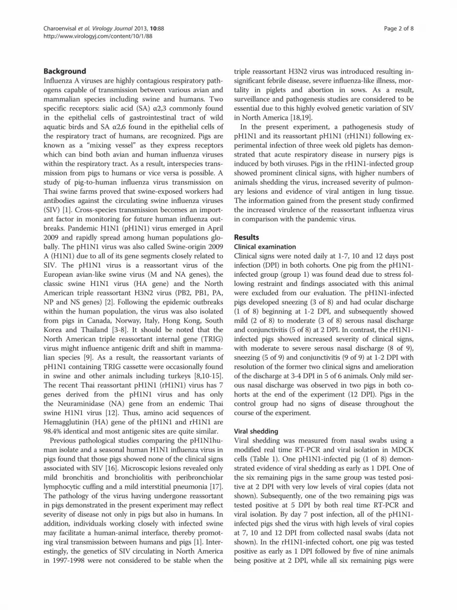
Viral sheddingViral shedding was measured from nasal swabs using amodified real time RT-PCR and viral isolation in MDCKcells (Table 1). One pH1N1-infected pig (1 of 8) demon-strated evidence of viral shedding as early as 1 DPI. One ofthe six remaining pigs in the same group was tested posi-tive at 2 DPI with very low levels of viral copies (data notshown). Subsequently, one of the two remaining pigs wastested positive at 5 DPI by both real time RT-PCR andviral isolation. By day 7 post infection, all of the pH1N1-infected pigs shed the virus with high levels of viral copiesat 7, 10 and 12 DPI from collected nasal swabs (data notshown). In the rH1N1-infected cohort, one pig was testedpositive as early as 1 DPI followed by five of nine animalsbeing positive at 2 DPI, while all six remaining pigs were
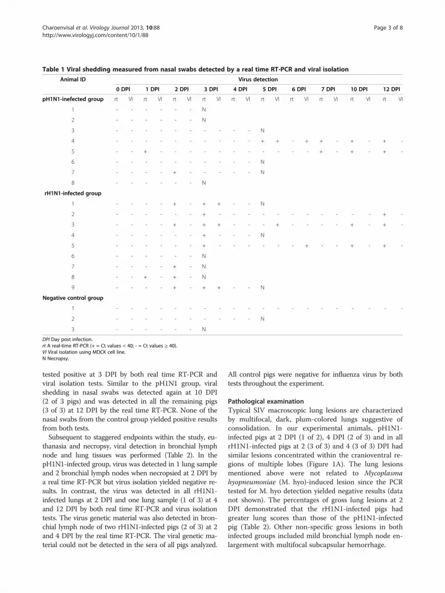
Table 1 Viral shedding measured from nasal swabs detected by a real time RT-PCR and viral isolation
Animal ID Virus detection
0 DPI 1 DPI 2 DPI 3 DPI 4 DPI 5 DPI 6 DPI 7 DPI 10 DPI 12 DPI
pH1N1-inefected group rt VI rt VI rt VI rt VI rt VI rt VI rt VI rt VI rt VI rt VI
1 - - - - - - N
2 - - - - - - N
3 - - - - - - - - - - N
4 - - - - - - - - - - + + - + + - + - + -
5 - - + - - - - - - - - - - - + - + - + -
6 - - - - - - - - - - N
7 - - - - + - - - - - N
8 - - - - - - N
rH1N1-infected group
1 - - - - + - + + - - N
2 - - - - - - + - - - - - - - - - - - + -
3 - - - - + - + + - - - + - - - - + - + -
4 - - - - - - + - - - N
5 - - - - - - + - - - - - - + - - + - + -
6 - - - - - - N
7 - - - - + - N
8 - - + - + - N
9 - - - - + - + + - - N
Negative control group
1 - - - - - - - - - - - - - - - - - - - -
2 - - - - - - - - - - N
3 - - - - - - N
DPI Day post infection.rt A real-time RT-PCR (+ = Ct values < 40; - = Ct values ≥ 40).VI Viral isolation using MDCK cell line.N Necropsy.
Charoenvisal et al. Virology Journal 2013, 10:88 Page 3 of 8http://www.virologyj.com/content/10/1/88
tested positive at 3 DPI by both real time RT-PCR andviral isolation tests. Similar to the pH1N1 group, viralshedding in nasal swabs was detected again at 10 DPI(2 of 3 pigs) and was detected in all the remaining pigs(3 of 3) at 12 DPI by the real time RT-PCR. None of thenasal swabs from the control group yielded positive resultsfrom both tests.Subsequent to staggered endpoints within the study, eu-
thanasia and necropsy, viral detection in bronchial lymphnode and lung tissues was performed (Table 2). In thepH1N1-infected group, virus was detected in 1 lung sampleand 2 bronchial lymph nodes when necropsied at 2 DPI bya real time RT-PCR but virus isolation yielded negative re-sults. In contrast, the virus was detected in all rH1N1-infected lungs at 2 DPI and one lung sample (1 of 3) at 4and 12 DPI by both real time RT-PCR and virus isolationtests. The virus genetic material was also detected in bron-chial lymph node of two rH1N1-infected pigs (2 of 3) at 2and 4 DPI by the real time RT-PCR. The viral genetic ma-terial could not be detected in the sera of all pigs analyzed.
All control pigs were negative for influenza virus by bothtests throughout the experiment.
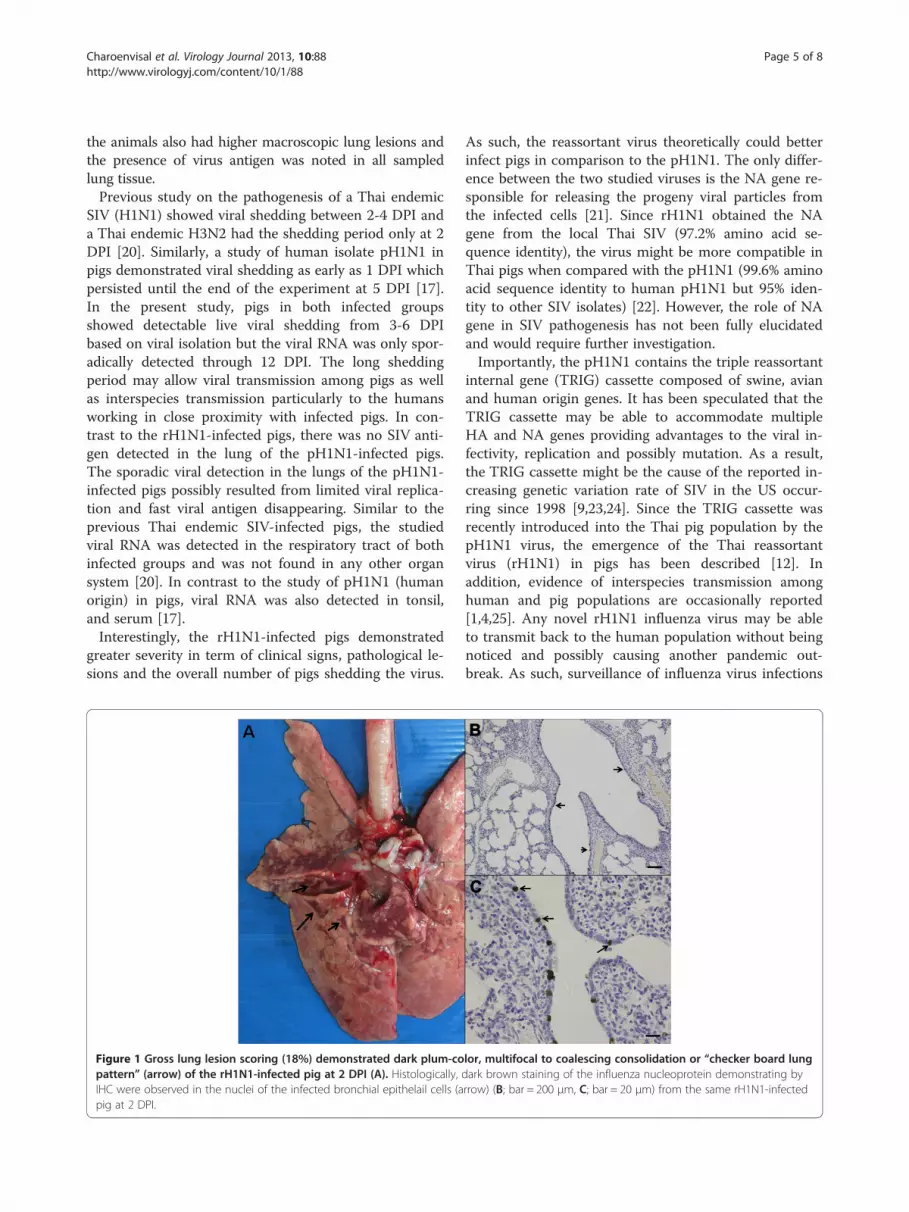
Pathological examinationTypical SIV macroscopic lung lesions are characterizedby multifocal, dark, plum-colored lungs suggestive ofconsolidation. In our experimental animals, pH1N1-infected pigs at 2 DPI (1 of 2), 4 DPI (2 of 3) and in allrH1N1-infected pigs at 2 (3 of 3) and 4 (3 of 3) DPI hadsimilar lesions concentrated within the cranioventral re-gions of multiple lobes (Figure 1A). The lung lesionsmentioned above were not related to Mycoplasmahyopneumoniae (M. hyo)-induced lesion since the PCRtested for M. hyo detection yielded negative results (datanot shown). The percentages of gross lung lesions at 2DPI demonstrated that the rH1N1-infected pigs hadgreater lung scores than those of the pH1N1-infectedpig (Table 2). Other non-specific gross lesions in bothinfected groups included mild bronchial lymph node en-largement with multifocal subcapsular hemorrhage.
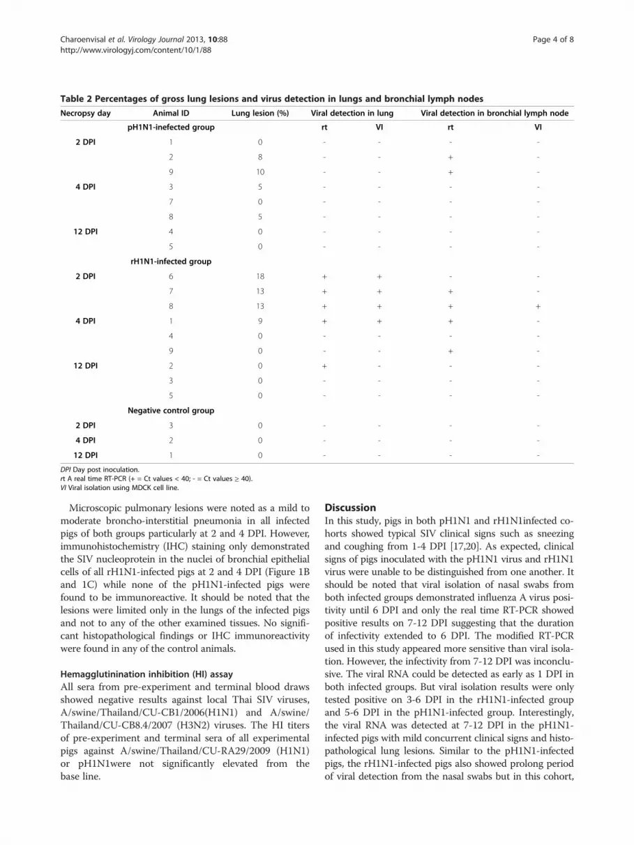
Table 2 Percentages of gross lung lesions and virus detection in lungs and bronchial lymph nodes
Necropsy day Animal ID Lung lesion (%) Viral detection in lung Viral detection in bronchial lymph node
pH1N1-inefected group rt VI rt VI
2 DPI 1 0 - - - -
2 8 - - + -
9 10 - - + -
4 DPI 3 5 - - - -
7 0 - - - -
8 5 - - - -
12 DPI 4 0 - - - -
5 0 - - - -
rH1N1-infected group
2 DPI 6 18 + + - -
7 13 + + + -
8 13 + + + +
4 DPI 1 9 + + + -
4 0 - - - -
9 0 - - + -
12 DPI 2 0 + - - -
3 0 - - - -
5 0 - - - -
Negative control group
2 DPI 3 0 - - - -
4 DPI 2 0 - - - -
12 DPI 1 0 - - - -
DPI Day post inoculation.rt A real time RT-PCR (+ = Ct values < 40; - = Ct values ≥ 40).VI Viral isolation using MDCK cell line.
Charoenvisal et al. Virology Journal 2013, 10:88 Page 4 of 8http://www.virologyj.com/content/10/1/88
Microscopic pulmonary lesions were noted as a mild tomoderate broncho-interstitial pneumonia in all infectedpigs of both groups particularly at 2 and 4 DPI. However,immunohistochemistry (IHC) staining only demonstratedthe SIV nucleoprotein in the nuclei of bronchial epithelialcells of all rH1N1-infected pigs at 2 and 4 DPI (Figure 1Band 1C) while none of the pH1N1-infected pigs werefound to be immunoreactive. It should be noted that thelesions were limited only in the lungs of the infected pigsand not to any of the other examined tissues. No signifi-cant histopathological findings or IHC immunoreactivitywere found in any of the control animals.
Hemagglutinination inhibition (HI) assayAll sera from pre-experiment and terminal blood drawsshowed negative results against local Thai SIV viruses,A/swine/Thailand/CU-CB1/2006(H1N1) and A/swine/Thailand/CU-CB8.4/2007 (H3N2) viruses. The HI titersof pre-experiment and terminal sera of all experimentalpigs against A/swine/Thailand/CU-RA29/2009 (H1N1)or pH1N1were not significantly elevated from thebase line.
DiscussionIn this study, pigs in both pH1N1 and rH1N1infected co-horts showed typical SIV clinical signs such as sneezingand coughing from 1-4 DPI [17,20]. As expected, clinicalsigns of pigs inoculated with the pH1N1 virus and rH1N1virus were unable to be distinguished from one another. Itshould be noted that viral isolation of nasal swabs fromboth infected groups demonstrated influenza A virus posi-tivity until 6 DPI and only the real time RT-PCR showedpositive results on 7-12 DPI suggesting that the durationof infectivity extended to 6 DPI. The modified RT-PCRused in this study appeared more sensitive than viral isola-tion. However, the infectivity from 7-12 DPI was inconclu-sive. The viral RNA could be detected as early as 1 DPI inboth infected groups. But viral isolation results were onlytested positive on 3-6 DPI in the rH1N1-infected groupand 5-6 DPI in the pH1N1-infected group. Interestingly,the viral RNA was detected at 7-12 DPI in the pH1N1-infected pigs with mild concurrent clinical signs and histo-pathological lung lesions. Similar to the pH1N1-infectedpigs, the rH1N1-infected pigs also showed prolong periodof viral detection from the nasal swabs but in this cohort,
Charoenvisal et al. Virology Journal 2013, 10:88 Page 5 of 8http://www.virologyj.com/content/10/1/88
the animals also had higher macroscopic lung lesions andthe presence of virus antigen was noted in all sampledlung tissue.Previous study on the pathogenesis of a Thai endemic
SIV (H1N1) showed viral shedding between 2-4 DPI anda Thai endemic H3N2 had the shedding period only at 2DPI [20]. Similarly, a study of human isolate pH1N1 inpigs demonstrated viral shedding as early as 1 DPI whichpersisted until the end of the experiment at 5 DPI [17].In the present study, pigs in both infected groupsshowed detectable live viral shedding from 3-6 DPIbased on viral isolation but the viral RNA was only spor-adically detected through 12 DPI. The long sheddingperiod may allow viral transmission among pigs as wellas interspecies transmission particularly to the humansworking in close proximity with infected pigs. In con-trast to the rH1N1-infected pigs, there was no SIV anti-gen detected in the lung of the pH1N1-infected pigs.The sporadic viral detection in the lungs of the pH1N1-infected pigs possibly resulted from limited viral replica-tion and fast viral antigen disappearing. Similar to theprevious Thai endemic SIV-infected pigs, the studiedviral RNA was detected in the respiratory tract of bothinfected groups and was not found in any other organsystem [20]. In contrast to the study of pH1N1 (humanorigin) in pigs, viral RNA was also detected in tonsil,and serum [17].Interestingly, the rH1N1-infected pigs demonstrated
greater severity in term of clinical signs, pathological le-sions and the overall number of pigs shedding the virus.
Figure 1 Gross lung lesion scoring (18%) demonstrated dark plum-copattern” (arrow) of the rH1N1-infected pig at 2 DPI (A). Histologically,IHC were observed in the nuclei of the infected bronchial epithelail cells (apig at 2 DPI.
As such, the reassortant virus theoretically could betterinfect pigs in comparison to the pH1N1. The only differ-ence between the two studied viruses is the NA gene re-sponsible for releasing the progeny viral particles fromthe infected cells [21]. Since rH1N1 obtained the NAgene from the local Thai SIV (97.2% amino acid se-quence identity), the virus might be more compatible inThai pigs when compared with the pH1N1 (99.6% aminoacid sequence identity to human pH1N1 but 95% iden-tity to other SIV isolates) [22]. However, the role of NAgene in SIV pathogenesis has not been fully elucidatedand would require further investigation.Importantly, the pH1N1 contains the triple reassortant
internal gene (TRIG) cassette composed of swine, avianand human origin genes. It has been speculated that theTRIG cassette may be able to accommodate multipleHA and NA genes providing advantages to the viral in-fectivity, replication and possibly mutation. As a result,the TRIG cassette might be the cause of the reported in-creasing genetic variation rate of SIV in the US occur-ring since 1998 [9,23,24]. Since the TRIG cassette wasrecently introduced into the Thai pig population by thepH1N1 virus, the emergence of the Thai reassortantvirus (rH1N1) in pigs has been described [12]. Inaddition, evidence of interspecies transmission amonghuman and pig populations are occasionally reported[1,4,25]. Any novel rH1N1 influenza virus may be ableto transmit back to the human population without beingnoticed and possibly causing another pandemic out-break. As such, surveillance of influenza virus infections
lor, multifocal to coalescing consolidation or “checker board lungdark brown staining of the influenza nucleoprotein demonstrating byrrow) (B; bar = 200 μm, C; bar = 20 μm) from the same rH1N1-infected
Charoenvisal et al. Virology Journal 2013, 10:88 Page 6 of 8http://www.virologyj.com/content/10/1/88
in both pigs and humans is critical for early recognitionand prevention of a potential epidemic or pandemicoutbreak.
ConclusionIn summary, clinical manifestations and pathological le-sions of both pH1N1 and rH1N1-infected pigs in thisstudy were most evident during the early stages of infec-tion (1-4 DPI), consistent with studies of the pathogen-esis of other SIV infections. The rH1N1-infected pigsdemonstrated prominent clinical signs and pathologicallesions typical of SIV infection and nasal swab testsnoted that the reassortant virus had higher numbers ofpigs shedding the infective virus based on the viral isola-tion. While result is not statistically significant, the trendobserved suggests both cohorts demonstrated some ani-mals shedding virus through the end of the study at12DPI. Similar to other SIV studies, the studied virusesreplicated well in the lung tissues and the viral antigenwas only detected within the respiratory tract.
Materials and methodsVirusesA/swine/Thailand/CU-RA29/2009(H1N1) [7], a pandemicH1N1 of pig origin (pH1N1) and A/swine/Thailand/CU-SA43/2010 (H1N1) [12], a novel reassortant virus of pigorigin (rH1N1) were individually propagated 3 times in 9-day-old embryonated chicken eggs. Allantoic fluids werecollected after 72 hours incubation. The virus concentra-tions were calculated using 50% tissue culture infectiousdose (TCID50) in Madin-Darby canine kidney (MDCK)cell using Reed and Muench method. Concentrations ofboth viruses were adjusted to 104 TCID50/ml and kept inthe -80°C until used.
Experimental pigsTwenty one, 3-week-old pigs from a local SIV, porcinecircovirus type 2 (PCV2) and porcine reproductive and re-spiratory syndrome virus (PRRSV)-free herd (kindly pro-vided by the Charoen Pokphand Food public companylimited, Thailand) were divided into 3 groups. Group 1and 2 containing 9 pigs each were intratracheally inocu-lated with 5 ml containing 104 TCID50/ml of pH1N1 andrH1N1, respectively. A negative control group containing3 pigs received mock cell culture media intratracheally.Clinical signs such as fever, coughing, sneezing, nasal dis-charge and conjunctivitis were blindly recorded daily bythe same veterinarian for a week and at 10 and 12 dayspost infection (DPI). All pigs tested serologically negativefor PRRSV and PCV2 using commercial ELISA kits(IDEXX laboratories, USA and Synbiotics, USA, respect-ively). All animals were housed in the animal facility bio-safety level 2 with appropriated food and clean waterproviding adequately throughout the experiment. The
animal usage and procedures were approved byChulalongkorn University-Faculty of Veterinary Scienceanimal care and use committee (protocol No. 11310052).
Viral detectionNasal swab were collected at 1-7, 10 and 12 DPI. TotalRNA was extracted from nasal swabs, sera, fresh bronchiallymph node and lung tissue collected at necropsy by usinga commercial kit (NucleoSpin Extract Viral RNA Kit,Macherey-Nagel, Germany). A modified real time reversetranscriptase polymerase chain reaction (real time RT-PCR)was performed using Superscript III platinum one-stepquantitative RT-PCR system (Invitrogen, USA). Primersspecific to Matrix (M) gene containingforward primer(MF3; 5’ TGATCTTCTTGAAAATTTGCAG 3’), rewardprimer (MR1+; 5’ CCGTAGMAGGCCCTCTTTTCA 3’)and M-probe (FAM-TTGTGGATTCTTGATCG-MGB)were used in this study. The cycling conditions started at48°C for 45 min, 95°C for 10 min and followed by 40 cyclesof denaturation (94°C for 15 s), annealing (55°C for 30 s)and extension (72°C for 40 s) [26].Nasal swabs, lung and bronchial lymph node homogen-
ate samples were filtrated and inoculated onto MDCKcells using ten-fold serial dilutions. The inoculated cellcultures were incubated for 72 hours. Virus was identifiedusing anti-influenza A nucleoprotein monoclonal antibodyas a primary antibody and rabbit anti-mouse IgG conju-gated horseradish peroxidase as a secondary antibody(DakoCytomation, Carpinteria, California). Then, colorwas developed using a chromogen aminoethylcarbazolesubstrate (Sigma, St. Louis, Missouri) [20].
Pathological examinationThree pigs from each viral inoculated group and 1 pigfrom the negative control group were randomly selectedfor euthanasia and necropsied at 2, 4 and 12 DPI. Atnecropsy, percentages of gross lung lesion scores charac-terized by multifocal mottled tan and consolidation inconsistency were recorded and scored as previously de-scribed [20]. Lung, bronchial lymph nodes, ileum, tonsil,liver, kidney and spleen were collected from each animalat necropsy, immersed and fixed in 10% buffered forma-lin for subsequent histopathological analysis.Formalin-fixed tissues were embedded in paraffin and
processed routinely. Sections were cut approximately4-6 μm thick for histopathological and immunohisto-chemistry (IHC) staining for Influenza A virus antigendetection. The IHC staining was performed using a la-beled streptavidin-biotin (LSAB) method. Primary anti-body using anti-influenza A (H5N1) nucleoproteinmonoclonal mouse antibodies (EVS238, B.V.EURO-PEAN VETERINARY LABORATORY, the Netherlands)and secondary antibody using Biotinylated rabbit anti-mouse IgG antibody and envision polymer (Envision
Charoenvisal et al. Virology Journal 2013, 10:88 Page 7 of 8http://www.virologyj.com/content/10/1/88
Polymer DAKOW, Denmark) were concurrently performedwith a negative control slide. The sections were developedwith 3, 3’-diaminobenzidine tetrahydrochloride (DAB) andcounterstained with Mayer’s hematoxylin. A positive con-trol slide was also included using the SIV-infected lung sec-tion from our previous experiment [7].
Hemagglutinination inhibition (HI) assaySera were collected from all pigs before starting the experi-ment and at each necropsy. All sera were pretreated with20% kaolin and receptor destroying enzyme (Denka SeikenCo. Ltd., Japan). The antibody detection was performedused standard HI assay [1]. Virus antigens used in thisexperiment were representatives of Thai endemic swineviruses; A/swine/Thailand/CU-CB1/2006(H1N1) and A/swine/Thailand/CU-CB8.4/2007 (H3N2) and pH1N1 virus(A/swine/Thailand/CU-RA29/2009(H1N1)). Samples withHI titers ≥ 40 were considered as previously exposed to thespecific tested antigen.
AbbreviationspH1N1: Pandemic H1N1 2009 virus; rH1N1: Pandemic H1N1 2009 reassortantvirus; DPI: Days post infection; SA: Sialic acid; SIV: Swine influenza virus;TRIG: Triple reassortment internal gene cassette; M: Matrix gene;NA: Neuraminidase gene; HA: Hemagglutinin gene; PB2: Polymerase basic 2gene; PB1: Polymerase basic 1 gene; PA: Polymerase acidic gene;NP: Nucleoprotein gene; NS: Non-structural protein gene; RT-PCR: Reversetranscriptase – polymerase chain reaction; IHC: Immunohistochemistrystaining; HI: Hemagglutination-inhibition test; RNA: Ribonucleic acid;TCID50: 50% Tissue culture infective dose; MDCK: Madin-darby canine kidneycell line, ml, milliliter; C: Degree celsius; PCV2: Porcine circovirus type 2;PRRSV: Porcine reproductive and respiratory syndrome virus; ELISA: Enzyme-linked immunosorbent assay; s: Second; LSAB: Labeled streptavidin-biotinmethod; DAB: 3, 3’-diaminobenzidine tetrahydrochloride.
Competing interestsThe authors declare that they have no competing interests.
Authors’ contributionsNC carried out virology, pathology, molecular genetic study and animalexperiment, analysis the data and drafting the manuscript, JK carried out animalwork, virology and molecular genetic study, DS carried out animal work andvirology, ST, SJ, JA carried out animal work, AA carried out molecular geneticstudy, RT carried out experimental design, pathology, and drafting and revisingthe manuscript. All authors have read and approved the final manuscript.
AcknowledgementsAnimal work, virology and molecular genetic study were funded by the HigherEducation Research Promotion and National Research University Project ofThailand, Office of the Higher Education Commission (HR1160A-55). Pathologystudy was supported by the 90th Anniversary of Chulalongkorn University Fund(Ratchadaphiseksomphot Endowment Fund). In addition, the authors would liketo express our sincere thanks to Dr. Eric D. Lombardini for editing the revisedmanuscript and the Charoen Pokphand Food public company limited andAHTSO Lab, Thailand for providing the animals and animal facilities.
Author details1Department of Pathology, Faculty of Veterinary Science, ChulalongkornUniversity, Henri-Dunant Rd, Bangkok 10330, Thailand. 2Department ofVeterinary Microbiology, Faculty of Veterinary Science, ChulalongkornUniversity, Henri-Dunant Rd, Bangkok 10330, Thailand. 3Faculty of VeterinaryMedicine, Rajamangala University of Technology Tawan-ok, Bangpra,Chonburi 20110, Thailand. 4Department of Clinical Science and Public Health,Faculty of Veterinary Science, Mahidol University, Salaya, Nakhon Pathom73170, Thailand. 5Department of Veterinary Public Health, Faculty of
Veterinary Science, Chulalongkorn University, Henri-Dunant Rd, Bangkok10330, Thailand. 6Emerging and re-emerging infectious diseases in animals,Research unit, Faculty of Veterinary Science, Chulalongkorn University,Henri-Dunant Rd, Bangkok 10330, Thailand.
Received: 6 August 2012 Accepted: 12 March 2013Published: 16 March 2013
References1. Kitikoon P, Sreta D, Tuanudom R, Amonsin A, Suradhat S, Oraveerakul K,
Poovorawan Y, Thanawongnuwech R: Serological evidence of pig-to-humaninfluenza virus transmission on Thai swine farms. Vet Microbiol 2011,148:413–418.
2. Garten RJ, Davis CT, Russell CA, Shu B, Lindstrom S, Balish A, Sessions WM,Xu X, Skepner E, Deyde V, Okomo-Adhiambo M, Gubareva L, Barnes J, SmithCB, Emery SL, Hillman MJ, Rivailler P, Smagala J, de Graaf M, Burke DF,Fouchier RA, Pappas C, Alpuche-Aranda CM, López-Gatell H, Olivera H,López I, Myers CA, Faix D, Blair PJ, Yu C, et al: Antigenic and geneticcharacteristics of swine-origin 2009 A (H1N1) influenza virusescirculating in humans. Science 2009, 325:197.
3. Hofshagen M, Gjerset B, Er C, Tarpai A, Brun E, Dannevig B, Bruheim T,Fostad IG, Iversen B, Hungnes O, Lium B: Pandemic influenza A(H1N1)v:human to pig transmission in Norway? Euro Surveill 2009, 14:687–689.
4. Howden KJ, Brockhoff EJ, Caya FD, McLeod LJ, Lavoie M, Ing JD, BystromJM, Alexandersen S, Pasick JM, Berhane Y, Morrison ME, Keenliside JM,Laurendeau S, Rohonczy EB: An investigation into human pandemicinfluenza virus (H1N1) 2009 on an Alberta swine farm. Can Vet J 2009,50:1153–1161.
5. Song MS, Lee JH, Pascua PN, Baek YH, Kwon HI, Park KJ, Choi HW, Shin YK,Song JY, Kim CJ, Choi YK: Evidence of human-to-swine transmission ofthe pandemic (H1N1) 2009 influenza virus in South Korea. J ClinMicrobiol2009, 48:3204–3211.
6. Moreno A, Di Trani L, Alborali L, Vaccari G, Barbieri I, Falcone E, Sozzi E,Puzelli S, Ferri G, Cordioli P: First pandemic H1N1 outbreak from a Pigfarm in Italy. Open Virol J 2010, 4:52–56.
7. Sreta D, Tantawet S, Na Ayudhya SN, Thontiravong A, WongphatcharachaiM, Lapkuntod J, Bunpapong N, Tuanudom R, Suradhat S, Vimolket L,Poovorawan Y, Thanawongnuwech R, Amonsin A, Kitikoon P: Pandemic(H1N1) 2009 virus on commercial swine farm, Thailand. Emerg Infect Dis2010, 16:1587–1590.
8. Vijaykrishna D, Poon LL, Zhu HC, Ma SK, Li OT, Cheung CL, Smith GJ, PeirisJS, Guan Y: Reassortment of pandemic H1N1/2009 influenza A virus inswine. Science 2010, 328:1529.
9. Vincent AL, Swenson SL, Lager KM, Gauger PC, Loiacono C, Zhang Y:Characterization of an influenza A virus isolated from pigs during anoutbreak of respiratory disease in swine and people during a county fairin the United States. Vet Microbiol 2009, 137:51–59.
10. Berhane Y, Kehler H, Handel K, Hisanaga T, Xu W, Ojkic D, Pasick J:Molecular and antigenic characterization of reassortant H3N2 virusesfrom Turkeys with a unique constellation of pandemic H1N1 internalgenes. PLoS One 2012, 7:1–11.
11. Ducatez MF, Hause B, Stigger-Rosser E, Darnell D, Corzo C, Juleen K,Simonson R, Brockwell-Staats C, Rubrum A, Wang D, Webb A, Crumpton JC,Lowe J, Gramer M, Webby RJ: Multiple reassortment between pandemic(H1N1) 2009 and endemic influenza viruses in pigs, United States.Emerg Infect Dis 2011, 17:1624–1629.
12. Kitikoon P, Sreta D, Nuntawan Na Ayudhya S, Wongphatcharachai M,Lapkuntod J, Prakairungnamthip D, Bunpapong N, Suradhat S,Thanawongnuwech R, Amonsin A: Brief report: molecular characterizationof a novel reassorted pandemic H1N1 2009 in Thai pigs. Virus Genes 2011,43:1–5.
13. Moreno A, Di Trani L, Faccini S, Vaccari G, Nigrelli D, Boniotti MB, Falcone E,Boni A, Chiapponi C, Sozzi E, Cordioli P: Novel H1N2 swine influenzareassortantstrain in pigs derived from the pandemic H1N1/2009 virus.Vet Microbiol 2011, 149:472–477.
14. Starick E, Lange E, Fereidouni S, Bunzenthal C, Hoveler R, Kuczka A, grosseBeilage E, Hamann HP, Klingelhofer I, Steinhauer D, Vahlenkamp T, Beer M,Harder T: Reassorted pandemic (H1N1) 2009 influenza A virus discoveredfrom pigs in Germany. J Gen Virol 2011, 92:1184–1188.
15. Zhu H, Zhou B, Fan X, Lam TT, Wang J, Chen A, Chen X, Chen H, WebsterRG, Webby R, Peiris JS, Smith DK, Guan Y: Novel reassortment of Eurasian
Charoenvisal et al. Virology Journal 2013, 10:88 Page 8 of 8http://www.virologyj.com/content/10/1/88
avian-like and pandemic/2009 influenza viruses in swine: infectiouspotential for humans. J Virol 2011, 85:10432–10439.
16. Itoh Y, Shinya K, Kiso M, Watanabe T, Sakoda Y, Hatta M, Muramoto Y,Tamura D, Sakai-Tagawa Y, Noda T, Sakabe S, Imai M, Hatta Y, Watanabe S,Li C, Yamada S, Fujii K, Murakami S, Imai H, Kakugawa S, Ito M, Takano R,Iwatsuki-Horimoto K, Shimojima M, Horimoto T, Goto H, Takahashi K,Makino A, Ishigaki H, Nakayama M, et al: In vitro and in vivocharacterization of new swine-origin H1N1 influenza viruses. Nature 2009,460:1021–1025.
17. Vincent AL, Lager KM, Faaberg KS, Harland M, Zanella EL, Ciacci-Zanella JR,Kehrli ME Jr, Janke BH, Klimov A: Experimental inoculation of pigs withpandemic H1N1 2009 virus and HI cross-reactivity with contemporaryswine influenza virus antisera. Influenza Other Respi Viruses 2010, 4:53–60.
18. Zhou NN, Senne DA, Landgraf JS, Swenson SL, Erickson G, Rossow K, Liu L,Yoon KJ, Krauss S, Webster RG: Emergence of H3N2 reassortant influenzaA viruses in North American pigs. Vet Microbiol 2000, 74:47–58.
19. Olsen CW: The emergence of novel swine influenza viruses in NorthAmerica. Virus Res 2002, 85:199–210.
20. Sreta D, Kedkovid R, Tuamsang S, Kitikoon P, Thanawongnuwech R:Pathogenesis of swine influenza virus (Thai isolates) in weanling pigs: anexperimental trial. Virol J 2009, 6:34.
21. Cheung TK, Poon LL: Biology of influenza a virus. Ann N Y Acad Sci 2007,1102:1–25.
22. Chutinimitkul S, Thippamom N, Damrongwatanapokin S, Payungporn S,Thanawongnuwech R, Amonsin A, Boonsuk P, Sreta D, Bunpong N,Tantilertcharoen R, Chamnanpood P, Parchariyanon S, Theamboonlers A,Poovorawan Y: Genetic characterization of H1N1, H1N2 and H3N2 swineinfluenza virus in Thailand. Arch Virol 2008, 153:1049–1056.
23. Vincent AL, Ma W, Lager KM, Janke BH, Richt JA: Swine influenza viruses aNorth American perspective. Adv Virus Res 2008, 72:127–154.
24. Ma W, Vincent AL, Lager KM, Janke BH, Henry SC, Rowland RR, Hesse RA,Richt JA: Identification and characterization of a highly virulent triplereassortant H1N1 swine influenza virus in the United States. Virus Genes2010, 40:28–36.
25. Komadina N, Roque V, Thawatsupha P, Rimando-Magalong J, Waicharoen S,Bomasang E, Sawanpanyalert P, Rivera M, Iannello P, Hurt AC, Barr IG:Genetic analysis of two influenza A (H1) swine viruses isolated fromhumans in Thailand and the Philippines. Virus Genes 2007, 35:161–165.
26. Payungporn S, Chutinimitkul S, Chaisingh A, Damrongwantanapokin S,Buranathai C, Amonsin A, Theamboonlers A, Poovorawan Y: Single stepmultiplex real-time RT-PCR for H5N1 influenza A virus detection. J VirolMethods 2006, 131:143–147.
doi:10.1186/1743-422X-10-88Cite this article as: Charoenvisal et al.: Experimental infection with a Thaireassortant swine influenza virus of pandemic H1N1 origin induceddisease. Virology Journal 2013 10:88.
Submit your next manuscript to BioMed Centraland take full advantage of:
• Convenient online submission
• Thorough peer review
• No space constraints or color figure charges
• Immediate publication on acceptance
• Inclusion in PubMed, CAS, Scopus and Google Scholar
• Research which is freely available for redistribution
Submit your manuscript at www.biomedcentral.com/submit