lung histopathological findings in fatal pandemic influenza a (h1n1)
TRANSCRIPT
Med Intensiva. 2011;xxx(xx):xxx---xxx
www.elsevier.es/medintensiva
ORIGINAL
Lung histopathological findings in fatal pandemic influenzaA (H1N1)
N. Nina,b, C. Sánchez-Rodrígueza,b, L.S. Verb,c, P. Cardinald, A. Ferrueloa,b,L. Sotoe, A. Deicasd, N. Camposf, O. Rochag, D.H. Cerasoh, M. El-Assara,b,J. Ortínb,c, P. Fernández-Segovianoa,b, A. Estebana,b, J.A. Lorentea,b,∗
a Hospital Universitario de Getafe, Madrid, Spainb CIBER de Enfermedades Respiratorias, Instituto de Salud Carlos III, Madrid, Spainc Centro Nacional de Biotecnología, CSIC, Madrid, Spaind Sanatorio Casmu, Montevideo, Uruguaye Instituto Nacional de Tórax, Santiago de Chile, Chilef Hospital Regional de Salto, Salto, Uruguayg Sanatorio GREMEDA, Artigas, Uruguayh Hospital Juan A. Fernández, Buenos Aires, Argentina
Received 18 July 2011; accepted 19 October 2011
KEYWORDSH1N1;ARDS;Lung pathology;Mechanicalventilation
AbstractObjective: To describe the lung pathological changes in influenza A (H1N1) viral pneumonia.We studied morphological changes, nitro-oxidative stress and the presence of viral proteins inlung tissue.Methods and patients: Light microscopy was used to examine lung tissue from 6 fatal casesof pandemic influenza A (H1N1) viral pneumonia. Fluorescence for oxidized dihydroethy-dium, nitrotyrosine, inducible NO synthase (NOS2) and human influenza A nucleoprotein (NP)(for analysis under confocal microscopy) was also studied in lung tissue specimens.Results: Age ranged from 15 to 50 years. Three patients were women, and 5 had preexistingmedical conditions. Diffuse alveolar damage (DAD) was present in 5 cases (as evidenced by hya-line membrane formation, alveolo-capillary wall thickening and PMN infiltrates), and interstitialfibrosis in one case. In the fluorescence studies there were signs of oxygen radical generation,increased NOS2 protein and protein nitration in lung tissue samples, regardless of the durationof ICU admission. Viral NP was found in lung tissue samples from three patients. Type I pneu-mocytes and macrophages harbored viral NP, as evidenced by confocal immunofluorescencemicroscopy.Conclusions: Lung tissue from patients with pandemic influenza A (H1N1) viral pneumonia showshistological findings consistent with DAD. Prolonged nitro-oxidative stress is present despiteantiviral treatment. Viral proteins may remain in lung tissue for prolonged periods of time,lodged in macrophages and type I pneumocytes.© 2011 Elsevier España, S.L. and SEMICYUC. All rights reserved.
∗ Corresponding author.E-mail address: [email protected] (J.A. Lorente).
0210-5691/$ – see front matter © 2011 Elsevier España, S.L. and SEMICYUC. All rights reserved.2173-5727
Med Intensiva. 2011;36(1):24–31
25Lung histopathological findings in fatal pandemic influenza A (H1N1)
PALABRAS CLAVEH1N1;SDRA;Histopatología depulmón;Ventilación mecánica
Resultados histopatológicos pulmonares en la gripe a (H1N1) pandémica letal
ResumenObjetivo: Describir la histopatología pulmonar de pacientes que fallecieron con neumonía porvirus de la influenza A (H1N1), el tipo celular infectado por el virus y la presencia de stressoxidativo y nitrosativo.Métodos: Hemos examinado tejido pulmonar de 6 pacientes fallecidos en la UCI con el diagnós-tico de infección por el virus influenza A (H1N1) (15---50 anos de edad) mediante (i) microscopíaóptica, (ii) microscopia confocal con tinciones específicas para diferentes tipos celulares (aquo-porina 5, factor Von Willebrand, proteína D del surfactante), (iii) inmunofluorescencia (IF) parasonda de dihidroetidio oxidado, óxido nítrico sintasa inducible (NOS2), anti-3-nitrotirosina ynucleoproteína (NP) del virus de la influenza A (H1N1).Resultados: (1) En 5 casos se encontró dano alveolar difuso (DAD), evidenciado mediante laobservación de membranas hialinas, engrosamiento de la pared alveolo-capilar e infiltración dePMN, asociado con hemorragia intensa en un paciente. Un caso presentó fibrosis intersticial.(2) Se demostró en todos los casos aumento de la inmuno-reactividad para DHE oxidado, NOS2y 3-nitrotirosina independientemente de la duración de la estancia en la UCI. (3) Se encontróNP viral en tres pacientes. (4) El virus se localiza en los neumocitos tipo I y en macrófagosalveolares.Conclusiones: El tejido pulmonar de pacientes fallecidos con neumonía por virus de la influenzaA (H1N1) evidencia hallazgos histológicos compatibles con DAD. El estrés nitro-oxidativo prolon-gado está presente a pesar del tratamiento antiviral. Las proteínas virales pueden permaneceren el tejido pulmonar durante períodos prolongados de tiempo, albergándose en los macrófagosy neumocitos tipo I.© 2011 Elsevier Espana, S.L. y SEMICYUC. Todos los derechos reservados.
Introduction
Previous reports have analyzed the clinical profile, organdysfunction and mortality risk factors in patients with H1N1influenza virus infection.1---6 These patients were usuallyyoung, often developed acute respiratory distress syndrome(ARDS) and shock, and had a high associated mortality rate,posing a management challenge for physicians.7---10
Lung histopathology in influenza A (H1N1) virus wasfirst reported in autopsy studies during the 1918 and 1957pandemics.10 More recently, in the recent 2009 pandemics,several studies have confirmed the presence of diffusealveolar damage with thick hyaline membranes formation,accompanied in some cases by necrotizing bronchiolitis andextensive hemorrhage.1,7---9,11
The specific cell type targeted by the virus has beenreported by Gill et al.11 who showed in 34 patients who diedfollowing confirmed H1N1 infection that influenza viral anti-gen was observed most commonly in the epithelium of thetracheobronchial tree, in type I and type II alveolar epithe-lial cells and in macrophages. In line with these findings,Mauad et al.7 found small vesicles containing one viral par-ticle enveloped by the pneumocyte II membrane.
It has been shown that increased nitric oxide (NO) andreactive oxygen species formation plays a critical role inlung dysfunction in acute lung injury and ARDS.12---14 In thepresent study we analyzed light microscopy findings as wellas changes in the nitro-oxidative stress in lung tissue samplesfrom fatal cases diagnosed of pandemic 2009 influenza A(H1N1) viral pneumonia and ARDS.
Methods
Patients
With our local Ethics’ Committee, 6 patients (3 fromUruguay, 1 from Chile, 1 from Argentine and 1 from Spain)who died in the Intensive Care Unit (ICU) with H1N1 influenzaviral pneumonia were studied. Each center’s institutionalreview board approved the study. The Ethics Committeeof the Hospital Universitario de Getafe classified this asan exempt study. Lung tissue samples were examined inthe Department of Pathology and in the Research Unit ofHospital Universitario de Getafe. Lung tissue samples from2 patients who died in the ICU without lung clinical diagnosisand normal lungs under light microscopy served as controls.
Clinical information was obtained from medical charts.The diagnosis of H1N1 influenza virus infection was made byreal-time reverse transcription polymerase chain reaction(PCR) in accordance with the guidelines from the Centersfor Disease Control and Prevention (CDC).
Tissue processing
The lung was carefully removed from the thorax (the tra-cheobronchial tree was not fixed), and then instilled with10% formalin. Within 36---48 h after death lung tissue wassliced sagitally in 1.0---1.5 cm thickness sections, and blockswere taken for paraffin inclusion and microscopic study afterhematoxylin---eosin (HE), Masson trichromic and PAS staining.
26 N. Nin et al.
Fluorescence for oxidized dihydroethidium
Formalin (10%) fixed, paraffin-embedded transverse lungtissue sections (5 �m thick) were mounted on poly-l-lysine-coated glass slides. After dewaxing, sections were incubatedfor 90 min at 37 ◦C with the fluorescent probe dihydroethid-ium ([DHE], 4 �mol/l; Calbiochem, Darmstadt, Germany).In the presence of reactive oxygen species, DHE is oxi-dized to ethidium, which intercalates with DNA, staining thenucleus with a bright red fluorescence. Then sections wereincubated with 4′,6-diamidino-2-phenylindol dihydrochlo-ride (300 nM) (DAPI; Sigma Chemical, St Louis, MO, USA)for 5 min at 37 ◦C reactive with fluorescent blue, markingthe interlayer between DNA pair bases in the cell nuclei.After washing with PBS plus 0.1% Triton X-100, sectionswere mounted and visualized by microscopy (Olympus BX51,Japan).15
Immunofluorescence staining for viralnucleoprotein, NOS2 and nitrotyrosine
Five-�m sections of formalin (10%) paraffin-fixed lung tis-sues were dewaxed, and irradiated with microwave in 0.01 Msodium citrate buffer (pH 6.0) and blocked with 5% bovineserum albumin (BSA) 0.1% Triton X-100 in PBS for 2 h at37 ◦C, and incubated overnight at 4 ◦C with several antibod-ies: rabbit polyclonal anti-nitrotyrosine antibody (dilution1:100; Chemicon International, USA); rabbit polyclonal NOS2antibody (dilution: 1:50) (Santa Cruz Biotechnology, Inc.Santa Cruz, CA, USA); and rabbit polyclonal antibody againstnucleoprotein (NP) of human influenza A (H3N2) (dilution1:5000) as the three primary antibodies. Human epithelialcells A540 were infected with H1N5 for positive controls(data not shown). Following washes in PBS plus 0.1% Tri-ton X-100, sections were incubated with secondary AlexaFluor® 546-conjugated goat anti-rabbit antibody (dilution1:100; Invitrogen, Molecular Probes, Inc. Oregon, USA) for45 min at 37 ◦C. Next, sections were incubated with DAPI(300 nM) for 5 min at 37 ◦C. After further washing in PBS,sections were mounted and visualized by microscopy. Thespecificity of the immunostaining was evaluated by omissionof the primary antibody (negative control).16
Dual labeling for fluorescence microscopy was performedaccording to the double indirect immunofluorescencemethod.17 For double immunofluorescence labeling, the rab-bit or rat polyclonal antibody against influenza A virus NPwas used in conjunction with the following primary anti-bodies: (1) goat polyclonal IgG antibody against aquoporin 5(AQP5) (Santa Cruz Biotechnology, Inc., Santa Cruz, CA, USA)used at a dilution of 1:50; (2) polyclonal rabbit anti-humanfactor VIII (DAKO Diagnostika, Hamburg, Germany) used ata dilution of 1:100; (3) monoclonal mouse antibody againstsurfactant protein D (Abcam, Cambridge, UK) used at a dilu-tion of 1:10; and (4) monoclonal mouse anti-human CD-68(for macrophages) (DAKO Diagnostika, Hamburg, Germany)used at a dilution of 1:100. Paraffin sections of human lungswere incubated overnight at 4 ◦C with a combination of thetwo primary antibodies. After thorough washing, sectionswere incubated with a mixture of two secondary antibodies,which consisted of Alexa Fluor® 488-conjugated donkey anti-goat antibody (dilution 1:100; Invitrogen, Molecular Probes,
Inc., Oregon, USA), FITC-labeled goat anti-mouse IgG (dilu-tion 1:100; Santa Cruz Biotechnology, Inc., Santa Cruz, CA,USA), Alexa Fluor® 546-conjugated goat anti-rabbit antibody(dilution 1:100; Invitrogen, Molecular Probes, Inc., Oregon,USA), donkey anti-rabbit 594, goat anti-rabbit 488 and goatanti-rat 594 for 45 min at 37 ◦C in the dark. In all samples,these reactions were followed by nuclear counterstainingfor 5 min at 37 ◦C with DAPI (300 nM). Sections were studiedunder confocal microscopy (Leica SP5, IL, USA). Immunoflu-orescence negative controls were preparations lacking theprimary antibody from the staining procedure.
Cross-reactivity of H3N2 viral NP with H1N1viral NP
A consensus sequence was originated from 2439 humanH1N1 reported in the Influenza virus Resource at NCBI serverin the period 2009---2010.18 The alignment of this consensussequence and the NP sequence from A/Victoria/3/75 (Vic)strain (H3N2), the protein used to generate the antibodyfor our immunofluorescence assays, was then generatedwith the BioEdit Sequence Alignment Editor program19
and a NP protein consensus sequence from H1N1 isolates(http://www.ncbi.nlm.nih.gov/genomes/FLU/FLU.html)showed a 90% identity and 96% similarity, indicating agreat conservation of the protein. Further, our IF experi-mental data using H3N2 antibody to identify seasonal andpandemic H1N1 virus-infected cells indicate a completecross-reactivity.
The alignment of the H1N1 consensus sequence andNP/Vic strain sequence strongly suggested that the poly-clonal antibody against the latter could recognize the NPprotein from pandemic H1N1 virus. This was confirmed byinfections with A/WSN (H1N1, positive control) and withpandemic H1N1 isolated (unpublished data) in A549 cells.
Results
Patients
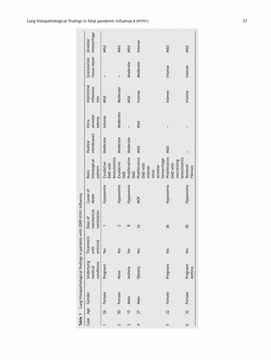
All patients were admitted to the ICU and required mechan-ical ventilation, and all were diagnosed of ARDS20 (Table 1).Three patients died within the first 8 days of mechanical ven-tilation. Five patients received treatment with oseltamivir(for 10 days or less if death occurred earlier) and allreceived empiric treatment for bacterial infection. Threepatients (cases #3, #4 and #6) received corticosteroids. H1N1influenza virus infection was confirmed by real time reversetranscription PCR, either in nasopharyngeal swab (5 cases)or in lung tissue (case #1).
Lung pathology
Five patients showed evidence of acute interstitial lesionscharacteristic of diffuse alveolar damage (DAD) (cases #1to #5, Table 1). In these five patients hyaline membranes,type II pneumocyte hyperplasia and interstitial inflammationwere observed, as well as alveolar septal edema in three ofthem (Fig. 1 panels A and B). Of note, extensive exudate offibrin-rich edema fluid in the alveolar space was observed
27Lung histopathological findings in fatal pandemic influenza A (H1N1)
Tabl
e
1
Lung
hist
opat
holo
gica
l find
ings
in
pati
ents
wit
h
2009
H1N
1
infl
uenz
a.
Case
Age
Gen
der
Und
erly
ing
med
ical
cond
itio
ns
Trea
tmen
tw
ith
anti
vira
l
Day
s
ofm
echa
nica
lve
ntila
tion
Caus
e
ofde
ath
Basi
chi
stol
ogic
alpa
tter
n
Hya
line
mem
bran
esIn
tra-
alve
olar
edem
a
Inte
rsti
tial
infl
amm
a-ti
on
Gra
nula
tion
tiss
ue
repa
irAl
veol
arhe
mor
rhag
e
1
26
Fem
ale
Preg
nant
Yes
1
Hyp
oxem
ia
Exud
ativ
eD
AD
wit
hbr
onch
iolit
is
Mod
erat
e
Inte
nse
Mild
---
Mild
2
50
Fem
ale
Non
e
Yes
2
Hyp
oxem
ia
Exud
ativ
eD
ADM
oder
ate
Mod
erat
e
Mod
erat
e
---
Mild
315
Mal
e
Asth
ma
Yes
8
Hyp
oxem
ia
Prol
ifer
ativ
eD
ADM
oder
ate
---
Mild
Mod
erat
e
Mild
4
37
Mal
e
Obe
sity
Yes
16
MO
F
Prol
ifer
ativ
eD
AD
wit
hin
tens
ein
tra-
alve
olar
hem
orrh
age
Mild
Mild
Inte
nse
Mod
erat
e
Inte
nse
5
22
Fem
ale
Preg
nant
Yes
36
Hyp
oxem
ia
Prol
ifer
ativ
eD
AD
wit
hne
crot
izin
gbr
onch
iolit
is
Mild
---
Inte
nse
Inte
nse
Mild
6
32
Fem
ale
Preg
nant
Asth
ma
Yes
45
Hyp
oxem
ia
Resi
dual
Fibr
osis
---
---
Inte
nse
Inte
nse
Mild
28 N. Nin et al.
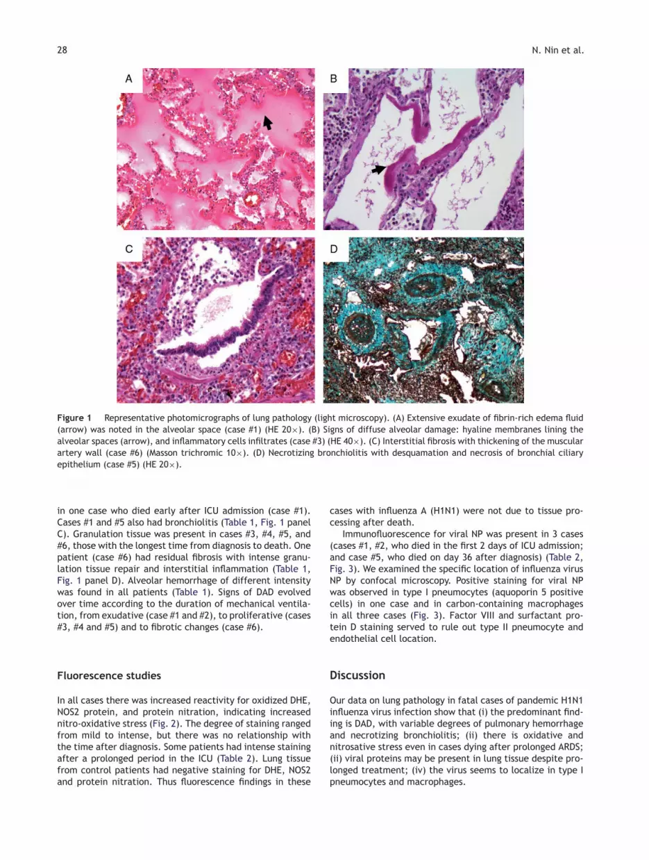
Figure 1 Representative photomicrographs of lung pathology (light microscopy). (A) Extensive exudate of fibrin-rich edema fluid(arrow) was noted in the alveolar space (case #1) (HE 20×). (B) Signs of diffuse alveolar damage: hyaline membranes lining thealveolar spaces (arrow), and inflammatory cells infiltrates (case #3) (HE 40×). (C) Interstitial fibrosis with thickening of the muscularartery wall (case #6) (Masson trichromic 10×). (D) Necrotizing bronchiolitis with desquamation and necrosis of bronchial ciliaryepithelium (case #5) (HE 20×).
in one case who died early after ICU admission (case #1).Cases #1 and #5 also had bronchiolitis (Table 1, Fig. 1 panelC). Granulation tissue was present in cases #3, #4, #5, and#6, those with the longest time from diagnosis to death. Onepatient (case #6) had residual fibrosis with intense granu-lation tissue repair and interstitial inflammation (Table 1,Fig. 1 panel D). Alveolar hemorrhage of different intensitywas found in all patients (Table 1). Signs of DAD evolvedover time according to the duration of mechanical ventila-tion, from exudative (case #1 and #2), to proliferative (cases#3, #4 and #5) and to fibrotic changes (case #6).
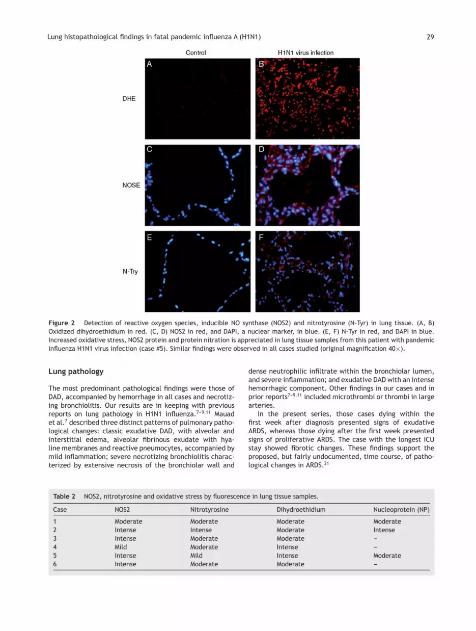
Fluorescence studies
In all cases there was increased reactivity for oxidized DHE,NOS2 protein, and protein nitration, indicating increasednitro-oxidative stress (Fig. 2). The degree of staining rangedfrom mild to intense, but there was no relationship withthe time after diagnosis. Some patients had intense stainingafter a prolonged period in the ICU (Table 2). Lung tissuefrom control patients had negative staining for DHE, NOS2and protein nitration. Thus fluorescence findings in these
cases with influenza A (H1N1) were not due to tissue pro-cessing after death.
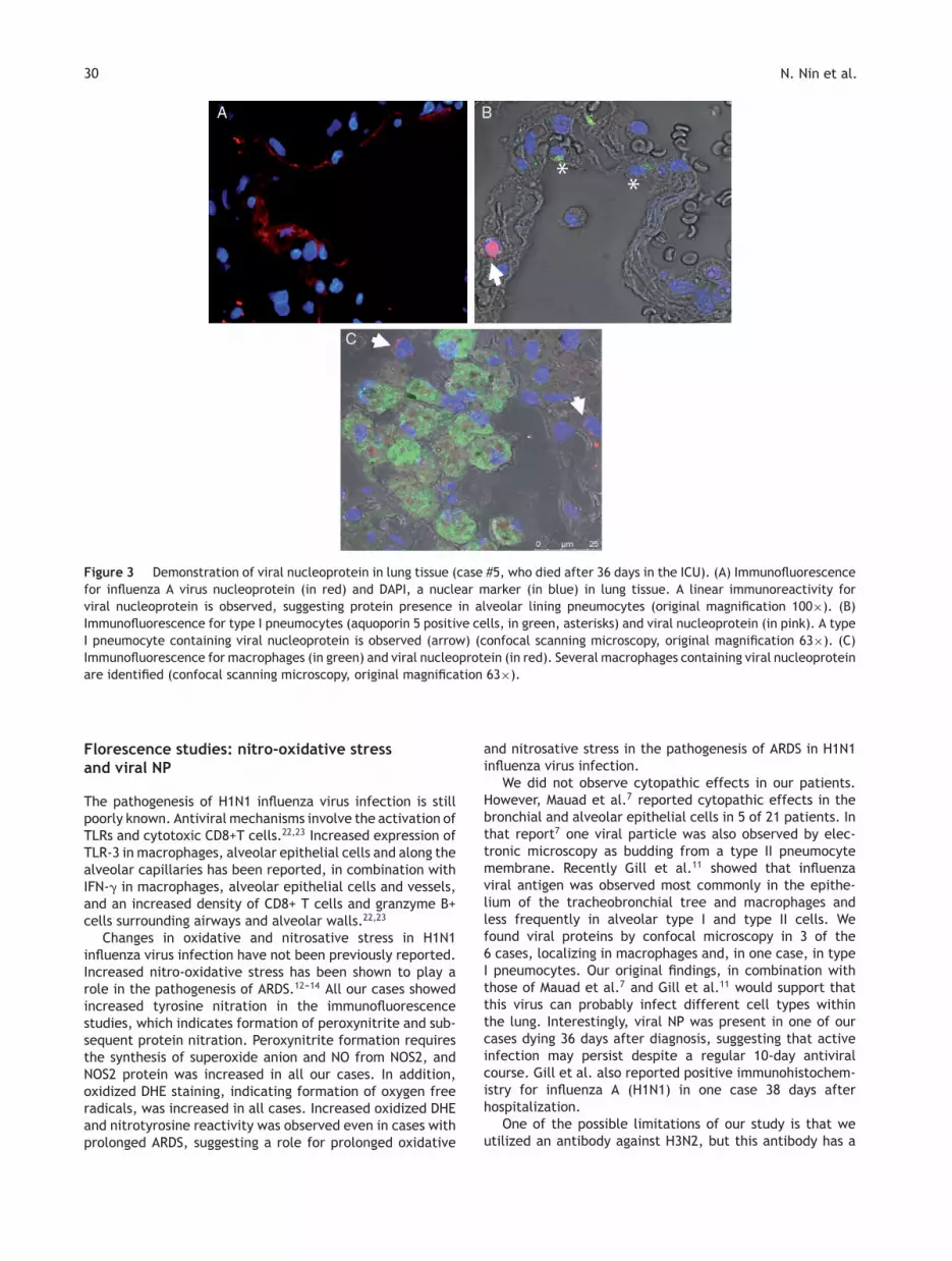
Immunofluorescence for viral NP was present in 3 cases(cases #1, #2, who died in the first 2 days of ICU admission;and case #5, who died on day 36 after diagnosis) (Table 2,Fig. 3). We examined the specific location of influenza virusNP by confocal microscopy. Positive staining for viral NPwas observed in type I pneumocytes (aquoporin 5 positivecells) in one case and in carbon-containing macrophagesin all three cases (Fig. 3). Factor VIII and surfactant pro-tein D staining served to rule out type II pneumocyte andendothelial cell location.
Discussion
Our data on lung pathology in fatal cases of pandemic H1N1influenza virus infection show that (i) the predominant find-ing is DAD, with variable degrees of pulmonary hemorrhageand necrotizing bronchiolitis; (ii) there is oxidative andnitrosative stress even in cases dying after prolonged ARDS;(ii) viral proteins may be present in lung tissue despite pro-longed treatment; (iv) the virus seems to localize in type Ipneumocytes and macrophages.
29Lung histopathological findings in fatal pandemic influenza A (H1N1)
Figure 2 Detection of reactive oxygen species, inducible NO synthase (NOS2) and nitrotyrosine (N-Tyr) in lung tissue. (A, B)Oxidized dihydroethidium in red. (C, D) NOS2 in red, and DAPI, a nuclear marker, in blue. (E, F) N-Tyr in red, and DAPI in blue.Increased oxidative stress, NOS2 protein and protein nitration is appreciated in lung tissue samples from this patient with pandemicinfluenza H1N1 virus infection (case #5). Similar findings were observed in all cases studied (original magnification 40×).
Lung pathology
The most predominant pathological findings were those ofDAD, accompanied by hemorrhage in all cases and necrotiz-ing bronchiolitis. Our results are in keeping with previousreports on lung pathology in H1N1 influenza.7---9,11 Mauadet al.7 described three distinct patterns of pulmonary patho-logical changes: classic exudative DAD, with alveolar andinterstitial edema, alveolar fibrinous exudate with hya-line membranes and reactive pneumocytes, accompanied bymild inflammation; severe necrotizing bronchiolitis charac-terized by extensive necrosis of the bronchiolar wall and
dense neutrophilic infiltrate within the bronchiolar lumen,and severe inflammation; and exudative DAD with an intensehemorrhagic component. Other findings in our cases and inprior reports7---9,11 included microthrombi or thrombi in largearteries.
In the present series, those cases dying within thefirst week after diagnosis presented signs of exudativeARDS, whereas those dying after the first week presentedsigns of proliferative ARDS. The case with the longest ICUstay showed fibrotic changes. These findings support theproposed, but fairly undocumented, time course, of patho-logical changes in ARDS.21
Table 2 NOS2, nitrotyrosine and oxidative stress by fluorescence in lung tissue samples.
Case NOS2 Nitrotyrosine Dihydroethidium Nucleoprotein (NP)
1 Moderate Moderate Moderate Moderate2 Intense Intense Moderate Intense3 Intense Moderate Moderate ---4 Mild Moderate Intense ---5 Intense Mild Intense Moderate6 Intense Moderate Moderate ---
30 N. Nin et al.
Figure 3 Demonstration of viral nucleoprotein in lung tissue (case #5, who died after 36 days in the ICU). (A) Immunofluorescencefor influenza A virus nucleoprotein (in red) and DAPI, a nuclear marker (in blue) in lung tissue. A linear immunoreactivity forviral nucleoprotein is observed, suggesting protein presence in alveolar lining pneumocytes (original magnification 100×). (B)Immunofluorescence for type I pneumocytes (aquoporin 5 positive cells, in green, asterisks) and viral nucleoprotein (in pink). A typeI pneumocyte containing viral nucleoprotein is observed (arrow) (confocal scanning microscopy, original magnification 63×). (C)Immunofluorescence for macrophages (in green) and viral nucleoprotein (in red). Several macrophages containing viral nucleoproteinare identified (confocal scanning microscopy, original magnification 63×).
Florescence studies: nitro-oxidative stressand viral NP
The pathogenesis of H1N1 influenza virus infection is stillpoorly known. Antiviral mechanisms involve the activation ofTLRs and cytotoxic CD8+T cells.22,23 Increased expression ofTLR-3 in macrophages, alveolar epithelial cells and along thealveolar capillaries has been reported, in combination withIFN-� in macrophages, alveolar epithelial cells and vessels,and an increased density of CD8+ T cells and granzyme B+cells surrounding airways and alveolar walls.22,23
Changes in oxidative and nitrosative stress in H1N1influenza virus infection have not been previously reported.Increased nitro-oxidative stress has been shown to play arole in the pathogenesis of ARDS.12---14 All our cases showedincreased tyrosine nitration in the immunofluorescencestudies, which indicates formation of peroxynitrite and sub-sequent protein nitration. Peroxynitrite formation requiresthe synthesis of superoxide anion and NO from NOS2, andNOS2 protein was increased in all our cases. In addition,oxidized DHE staining, indicating formation of oxygen freeradicals, was increased in all cases. Increased oxidized DHEand nitrotyrosine reactivity was observed even in cases withprolonged ARDS, suggesting a role for prolonged oxidative
and nitrosative stress in the pathogenesis of ARDS in H1N1influenza virus infection.
We did not observe cytopathic effects in our patients.However, Mauad et al.7 reported cytopathic effects in thebronchial and alveolar epithelial cells in 5 of 21 patients. Inthat report7 one viral particle was also observed by elec-tronic microscopy as budding from a type II pneumocytemembrane. Recently Gill et al.11 showed that influenzaviral antigen was observed most commonly in the epithe-lium of the tracheobronchial tree and macrophages andless frequently in alveolar type I and type II cells. Wefound viral proteins by confocal microscopy in 3 of the6 cases, localizing in macrophages and, in one case, in typeI pneumocytes. Our original findings, in combination withthose of Mauad et al.7 and Gill et al.11 would support thatthis virus can probably infect different cell types withinthe lung. Interestingly, viral NP was present in one of ourcases dying 36 days after diagnosis, suggesting that activeinfection may persist despite a regular 10-day antiviralcourse. Gill et al. also reported positive immunohistochem-istry for influenza A (H1N1) in one case 38 days afterhospitalization.
One of the possible limitations of our study is that weutilized an antibody against H3N2, but this antibody has a
31Lung histopathological findings in fatal pandemic influenza A (H1N1)
complete cross-reactivity with seasonal and pandemic H1N1virus-infected cells.
In summary we report lung pathological changes in fatalcases of H1N1 influenza virus infection. DAD is the predomi-nant finding, accompanied by alveolar hemorrhage and insome cases by bronchiolitis and microthrombi. Prolongednitro-oxidative stress can be present. Viral antigens can befound in type I pneumocytes.
Funding
FIS GR09/0001 and CIBER de Enfermedades Respiratorias,Instituto de Salud Carlos III, Madrid, Spain.
Conflict of interest
No potential conflict of interest relevant to this article wasreported by any of the authors.
Acknowledgements
We thank Mrs. Mar Granados from the Pathology Depart-ment of Hospital Universitario de Getafe for her technicalassistance and Eduardo Andrade M.D. from the PathologyDepartment of Hospital Regional de Salto, Uruguay.
References
1. Perez-Padilla R, de la Rosa-Zamboni D, Ponce de Leon S,Hernandez M, Quinones-Falconi F, Bautista E, et al. Pneumo-nia and respiratory failure from swine-origin influenza A (H1N1)in Mexico. N Engl J Med. 2009;361:680---9.
2. González-Vélez AE, Díaz-Agero-Pérez C, Robustillo-Rodela A,Cornejo-Gutiérrez AM, Pita-López MJ, Oliva-Iniguez L, et al.Factors associated to admission to intensive care in patientshospitalized due to pandemic Influenza A/H1N1 2009. MedIntensiva. 2011 Apr;30 [Epub ahead of print].
3. Rello J, Rodríguez A, Ibanez P, Socias L, Cebrian J, Marques A,et al. H1N1 SEMICYUC Working Group Intensive care adultpatients with severe respiratory failure caused by influenza A(H1N1) in Spain. Crit Care. 2009;13:R148.
4. Rodríguez A, Martin-Loeches I, Bonastre J, Olaechea P,Alvarez-Lerma F, Zaragoza R, et al. SEMICYUC-CIBERES-REIPIworking group. First influenza season after the 2009 pandemicinfluenza: report of the first 300 ICU admissions in Spain. MedIntensiva. 2011;35:208---16.
5. Rodríguez A, Socías L, Guerrero JE, Figueira JC, González N,Maraví-Poma E, et al. Pandemic influenza A in the ICU: expe-rience in Spain and Latin America GETGAG/SEMICYUC/(SpanishWork Group on Severe Pandemic Influenza A/SEMICYUC). MedIntensiva. 2010;34:87---94.
6. Domínguez-Cherit G, Lapinsky SE, Macias AE. Critically illpatients with 2009 influenza A (H1N1) in Mexico. JAMA.2009;302:E1---8.
7. Mauad T, Hajjar LA, Callegari GD, da Silva LF, Schout D, Galas FR,et al. Lung pathology in fatal novel human influenza A (H1N1)infection. Am J Respir Crit Care Med. 2010;181:72---9.
8. Mukhopadhyay S, Philip AT, Stoppacher R. Pathologic findingsin novel influenza A (H1N1) virus (‘‘swine flu’’) infection: con-trasting clinical manifestations and lung pathology in two fatalcases. Am J Clin Pathol. 2010;133:380---7.
9. Soto-Abraham MV, Soriano-Rosas J, Díaz-Quinónez A,Silva-Pereyra J, Vazquez-Hernandez P, Torres-López O,et al. Pathological changes associated with the 2009 H1N1virus. N Engl J Med. 2009;361:2001---3.
10. Taubenberger JK, Morens DM. The pathology of influenza virusinfections. Annu Rev Pathol. 2008;3:499---522.
11. Gill JR, Sheng ZM, Ely SF, Guinee DG, Beasley MB, Suh J, et al.Pulmonary pathologic findings of fatal 2009 pandemic influenzaA/H1N1 viral infections. Arch Pathol Lab Med. 2010;134:235---43.
12. Gole MD, Souza JM, Choi I, Hertkorn C, Malcolm S, Foust RF,3rd, et al. Plasma proteins modified by tyrosine nitration inacute respiratory distress syndrome. Am J Physiol Lung Cell MolPhysiol. 2000;278:961---7.
13. Lamb NJ, Gutteridge JM, Baker C, Evans TW, Quinlan GJ. Oxida-tive damage to proteins of bronchoalveolar lavage fluid inpatients with acute respiratory distress syndrome: evidence forneutrophil-mediated hydroxylation, nitration, and chlorination.Crit Care Med. 1999;27:1738---44.
14. Martínez-Caro L, Lorente JA, Marín-Corral J, Sánchez-Rodríguez C, Sánchez-Ferrer A, Nin N, et al. Role of free radicalsin vascular dysfunction induced by high tidal volume ventilation.Intensive Care Med. 2009;35:1110---9.
15. Peiró C, Lafuente N, Matesanz N, Cercas E, Llergo JL, Vallejo S,et al. High glucose induces cell death of cultured human aorticsmooth muscle cells through the formation of hydrogen perox-ide. Br J Pharmacol. 2001;133(August):967---74.
16. Peiro C, Matesanz N, Nevado J, Lafuente N, Cercas E, Azcutia V,et al. Glycosylated human oxyhaemoglobin activates nuclearfactor-kappaB and activator protein-1 in cultured humanaortic smooth muscle. Br J Pharmacol. 2003;140(October):681---90.
17. Nakajima M, Kawanami O, Jin EJ, Ghazizadeh M, Honda M,Asano G, et al. Immunohistochemical and ultrastructural stud-ies of basal cells, Clara cells and bronchiolar cuboidal cells innormal human airways. Pathol Int. 1998;48:944---53.
18. Hall TA. BioEdit: a user-friendly biological sequence alignmenteditor and analysis program for Windows 95/98/NT. NucleicAcids Symp Ser. 1999;41:95---8.
19. Jorba N, Juarez S, Torreira E, Gastaminza P, Zamarreno N,Albar JP, et al. Analysis of the interaction of influenza viruspolymerase complex with human cell factors. Proteomics.2008;8:2077---88.
20. Bernard GR, Artigas A, Brigham KL, Carlet J, Falke K,Hudson L, et al. The American-European Consensus Conferenceon ARDS. Definitions, mechanisms, relevant outcomes, and clin-ical trial coordination. Am J Respir Crit Care Med. 1994;149:818---24.
21. Katzenstein AL, Myers JL. Nonspecific interstitial pneumoniaand the other idiopathic interstitial pneumonias: classificationand diagnostic criteria. Am J Surg Pathol. 2000;24:1---3.
22. Bruder D, Srikiatkhachorn A, Enelow RL. Cellular immunityand lung injury in respiratory virus infection. Viral Immunol.2006;19:147---55.
23. Wong JP, Christopher ME, Viswanathan S, Karpoff N, Dai X,Das D, et al. Activation of toll like receptor signaling path-way for protection against influenza virus infection. Vaccine.2009;27:3481---3.