surgical management of lymph node compartments in
TRANSCRIPT
Surgical Management ofLymph Node
Compartments in Papil laryThyroid CancerCord Sturgeon, MD*, Anthony Yang, MD, Dina Elaraj, MD
KEYWORDS
� Papillary thyroid cancer � Lymph node dissection � Recurrent thyroid cancer� Lymph node metastases � Central neck lymph node dissection
KEY POINTS
� When central or lateral compartment cervical lymph node metastases are clinicallyevident at the time of the index thyroid operation for PTC, formal surgical clearance ofthe affected nodal basin is the optimal management.
� Prophylactic central neck dissection for PTC is practiced by some high-volume surgeonswith low complication rates, but is considered controversial because there appears to bea higher risk of complications with an uncertain clinical benefit.
� When a clinically significant recurrence is detected in a previously undissected central orlateral cervical compartment, a comprehensive surgical clearance of the lateral compart-ment is the preferred treatment.
� When a nodal recurrence is found in a previously dissected central or lateral neck field, thereoperation may focus on the areas where recurrence is demonstrated.
INTRODUCTION
In endocrine surgery, controversy abounds. It is difficult, in fact, to find a topic insurgical endocrinology for which there is little or no controversy. The managementof cervical nodal metastases from papillary thyroid cancer (PTC) is no exception.Fortunately, there is widespread agreement regarding the management of clinicallyevident nodal metastases. It seems clear, based on the risks of persistent or recurrentdisease, that the optimal management is formal surgical clearance of the affectednodal basin or basins when cervical nodal metastases are clinically evident at the
The authors have nothing to disclose.Division of Endocrine Surgery, Department of Surgery, Northwestern University, 676 NorthSaint Clair Street, Suite 650, Chicago, IL 60611, USA* Corresponding author.E-mail address: [email protected]
Surg Oncol Clin N Am 25 (2016) 17–40http://dx.doi.org/10.1016/j.soc.2015.08.013 surgonc.theclinics.com1055-3207/16/$ – see front matter � 2016 Elsevier Inc. All rights reserved.
Sturgeon et al18
time of the index thyroid operation. On the other hand, because of the high frequencyand uncertain clinical significance of occult nodal metastases from PTC, considerablecontroversy surrounds the management of the clinically negative central compart-ment, and the performance of so-called prophylactic central neck dissection (CND).Likewise, there is uncertainty regarding the thresholds for recommending remedialCND, given the attendant risks of the procedure and uncertain benefits. Within thiscontribution to the Surgical Oncology Clinics of North America, the data relevant tothe surgical management of the lymph nodes of the central and lateral compartmentsof the neck in PTC are reviewed and discussed.
NOMENCLATURE: PROPHYLACTIC VERSUS THERAPEUTIC
As defined in the American Thyroid Association (ATA) consensus statement on the ter-minology and classification of CND for thyroid cancer,1 a therapeutic neck dissectionis one that is performed for clinically apparent nodal metastases, whether they arerecognized before or during an operation, and regardless of the methodology usedto detect the nodal metastases (eg, imaging, physical examination, frozen section).A prophylactic neck dissection is one that is performed on a nodal basin for whichthere is no clinical or imaging study evidence of nodal metastases. Prophylacticneck dissection is also synonymous with elective neck dissection.
EPIDEMIOLOGY OF CENTRAL NECK METASTASES
Metastases from PTC are frequently found in the central compartment lymph nodes.Nodal metastases from PTC are found in the central compartment in 12% to 81% ofcases, depending on the completeness of the nodal dissection by the surgeon and thelevel of scrutiny to identify lymph nodes by the pathologist.2 In surgical series ofpatients with PTC treated with prophylactic CND, occult positive central compartmentnodes are found in at least one-third, and up to two-thirds of cases.2,3 Given the highfrequency of nodal metastases in the central compartment, some experts routinelyclear the central compartment in a prophylactic fashion.
CONTROVERSY REGARDING PROPHYLACTIC CENTRAL NECK DISSECTION
Routine prophylactic CND for patients with clinically node-negative PTC is controver-sial. The controversy is centered on the fact that there is risk associated with theperformance of a prophylactic CND, and that it is unclear if there is any survival orquality-of-life benefit. Furthermore, the finding of occult nodal disease will upstage pa-tients older than 45 and may influence the usage of radioiodine.Given the high rate of occult nodal metastases, some experts recommend that a
thyroidectomy for PTC be accompanied by at least an ipsilateral central compartmentnodal dissection. Proponents of prophylactic CND argue that because of the high rateof occult central nodal metastasis, prophylactic CND should decrease the need forreoperative neck surgery by reducing locoregional recurrence and simplify follow-upby lowering postoperative serum thyroglobulin.4–6 In a study of 134 patients withPTC wherein all patients underwent a CND, the authors found that 29% of patients un-dergoing primary surgery for PTC had ipsilateral central neck metastases and also29% had ipsilateral lateral neck metastases.3 These authors recommended routinecentral and ipsilateral lateral nodal compartment dissection for patients undergoingprimary surgery for PTC with a T1b or larger primary tumor. Other experts cite thehigher complication rate when thyroidectomy is combined with CND, with no apparentimprovement in survival, as rationale against prophylactic CND.4 They maintain that
Lymph Nodes in Papillary Thyroid Cancer 19
prophylactic CND exposes patients to the additional risks of recurrent laryngeal nerve(RLN) injury and hypoparathyroidism without proven benefit.Several recent retrospective analyses have compared outcomes of PTC patients
who underwent total thyroidectomy with and without CND.5,7–9 In one study, 20 pro-phylactic CNDs were required to prevent one central compartment reoperation.5 Ameta-analysis performed on studies that evaluated recurrence and complicationsassociated with prophylactic CND found that the number needed to treat to preventone recurrence was 31.10 Two meta-analyses have demonstrated an increased rateof transient hypocalcemia following prophylactic CND without showing a differencein permanent hypoparathyroidism or RLN injury.7,9 In most studies, prophylacticCND is associated with a higher rate of temporary hypocalcemia and parathyroidgland removal.11 In most formal cost-effectiveness analysis studies, prophylacticCND has not been found to be cost-effective for PTC.12,13
The presence of nodal metastases may have a significant impact on the stage ofdisease based on the current American Joint Commission on Cancer (AJCC) TNMstaging for thyroid cancer.14 Patients 45 years of age or older are upstaged to stageIII for any central neck nodal metastases regardless of the size and number of metas-tases found. Prophylactic neck dissection is therefore likely to upstage many patientsto stage III for subclinical disease. Because the AJCC staging system for differentiatedthyroid cancer was not derived or validated during an era of widespread CND, it is notclear if patients upstaged for subclinical metastases found during prophylactic dissec-tion will have the same prognosis as patients with clinically evident central neckmetastases.It is also not certain how the practice of prophylactic nodal dissection might affect
the likelihood to receive therapeutic doses of radioactive iodine (RAI). Some studiesindicate that patients are more likely to receive RAI after total thyroidectomy withCND.11 One hypothesis is that a greater frequency of detection of nodal metastases,and the associated upstaging of disease, leads to a greater utilization of RAI. Otherstudies show that prophylactic CND is associated with a lower probability of receivingRAI.15 This finding, in turn, may be due to the fact that a more complete surgical clear-ance of the central compartment may lead to lower preablation thyroglobulin levels,and consequently, to a lower utilization of RAI. In a study by Lang and colleagues,16
51% of the patients who underwent prophylactic CND had undetectable preablativethyroglobulin.A recent single-institution randomized controlled trial of total thyroidectomy with or
without prophylactic CND in patients with PTC without evidence of preoperative orintraoperative lymph node metastases has been reported by Viola and colleagues15
from Pisa, Italy. Their study of 181 patients with a median follow-up of 5 years foundthat clinically node-negative patients treated with prophylactic CND had a reduced ne-cessity for repeat RAI treatments compared with those patients who did not undergoCND. However, the prophylactic CND patients also had a significantly higher preva-lence of permanent postoperative hypoparathyroidism (19.4 vs 8.0%).
PREOPERATIVE ASSESSMENT OF THE CERVICAL NODAL COMPARTMENTS
The assessment of lymph node status before an operation for PTC is necessarybecause the presence and location of metastatic lymph nodes may not be clinicallyapparent, and the identification of nodal metastases will frequently alter the plannedprocedure. All patients should undergo a comprehensive history and physical exam-ination focused on determining the extent of disease. Unfortunately, physical exami-nation is notoriously unreliable for the exclusion of cervical nodal metastases.
Sturgeon et al20
Imaging studies, however, can detect abnormal lymph nodes in patients who have nopalpable lymphadenopathy and may also yield information that alters the extent of theoperation, even when overt nodal metastases are present.
Imaging Studies
Routine preoperative sonographic imaging of the cervical nodal basins changes theextent of surgery in as many as 41% of patients.17,18 In a retrospective cohort studyof 486 patients who underwent neck ultrasound before initial operation for PTC, ultra-sound detected abnormal lymph nodes in 16% of patients who had nonpalpablelymph nodes on physical examination and changed the extent of surgery in 15 of 37(41%) patients who had palpable lymph nodes on physical examination.18 Similarly,in a retrospective study of 151 patients who underwent neck ultrasound before initialoperation for differentiated thyroid cancer, ultrasound detected abnormal lymphnodes in 52 (34%) patients who had nonpalpable lymph nodes on physical examina-tion.17 Most patients with PTC who have lymph node metastases will have them in theinferior aspect of the neck. In a retrospective study of 578 lymph nodes that underwentfine needle aspiration (FNA) biopsy in 588 patients with differentiated thyroid cancer,67% of malignant lymph nodes were found in the inferior third of the neck, whereas46% of lymph nodes biopsied in the superior third of the neck were benign.19
Multiple imaging modalities have been studied for the preoperative staging ofpatients with PTC. Computed tomography (CT) is the preferred imaging modality forthe preoperative staging of patients with head and neck squamous cell cancers, butis not used routinely for the staging of PTC. Nonetheless, CT can characterize thesize, shape, appearance, and contrast enhancement pattern of lymph nodes andmay be helpful in the prognostication of cervical lymph nodes in patients with PTC.CT features of malignant lymph nodes include size greater than 1.5 cm in levels I orII, greater than 0.8 in the retropharyngeal space, and greater than 1.0 cm in otherlocations; spherical shape; cystic change; presence of calcifications; and abnormalcontrast enhancement.20–22 In the setting of a known head and neck squamous cellcancer, CT has approximately 80% sensitivity and specificity for the detection oflymph node metastases.20 There is no established size criterion for metastatic lymphnodes in PTC, however, and the sensitivity of CT may be lower in patients with PTCdue to the higher rates of micrometastatic disease, which may not cause a changein the appearance of these lymph nodes on CT. In large retrospective studies evalu-ating the accuracy of CT in detecting metastatic lymphadenopathy in patients withPTC, CT has a sensitivity of 50% to 67% and specificity of 76% to 91% for detectingmetastatic lymph nodes in the central compartment of the neck. Likewise, CT has asensitivity of 59% to 82% and a specificity of 71% to 100% for detecting metastaticlymph nodes in the lateral compartment in PTC.21,23,24
The ATA guidelines on the management of thyroid cancer recommend againstthe routine use of preoperative imaging studies, such as CT, MRI, or PET, for theinitial staging of PTC.6 In some cases, however, these imaging modalities may playan important role in the preoperative evaluation of the patient. CT has been themost widely studied cross-sectional imaging modality, and its main advantages arethat it is not operator dependent and that it generates high-resolution images withthe ability to perform multiplanar image reconstructions. Its main limitation is thatthe iodine load associated with the intravenous contrast given during the scan mayreduce RAI uptake for several weeks after administration.25,26 CT can be particularlyhelpful in the evaluation and surgical planning for patients with advanced PTC, orthose with large, fixed, or substernal cancers. In such cases, CT may accuratelydemonstrate extension of disease into the mediastinum, invasion into adjacent
Lymph Nodes in Papillary Thyroid Cancer 21
structures such as the aerodigestive tract, or reveal lymph node metastases in areasthat are poorly assessed by ultrasound (retropharyngeal/retrotracheal space, low-level VI/superior mediastinum).21,23,27 MRI has not been found to be particularly help-ful in the identification of cervical nodal metastases from thyroid cancer due to poorsensitivity and only moderate interobserver agreement.28 PET scanning is useful forimaging patients with poorly differentiated thyroid cancers, but adds little to the im-aging of well-differentiated thyroid cancers.The ATA and National Comprehensive Cancer Network (NCCN) guidelines recom-
mend comprehensive neck ultrasound for the preoperative staging of PTC.6,27
High-resolution neck ultrasound has been reported to have sensitivity of 52% to93% and specificity of 79% to 100% for the detection of abnormal lymph nodes in pa-tients with PTC, in both index and reoperative settings.17,18,29 Sensitivities vary widelybetween studies, while specificities are consistently high. Sensitivity is higher for thedetection of abnormal lymph nodes in the lateral compartment of the neck comparedwith the central compartment of the neck, as the assessment of the central neck maybe affected by surrounding structures, such as the tracheal air shadow, clavicle, andsternum.17,29,30 Cervical sonography has several advantages over other imaging mo-dalities. Compared with CT, ultrasound is less costly, does not expose the patient toradiation, can be performed repeatedly in children and pregnant women, is painlessand noninvasive, does not require intravenous access, does not generally precipitateclaustrophobic reactions, and does not have a maximum weight limit. Limitations ofultrasound include that it is operator dependent and that it may be limited in evaluatinglymph nodes in patients with high body mass index or in certain locations, such as theretropharyngeal, paratracheal, or retrotracheal spaces, level VI, and the superiormediastinum.21,23
Ultrasound can help characterize a lymph node as benign or malignant based onsize, shape, and appearance. Sonographic features of benign lymph nodes includeflattened elongated shape, smooth border, and hyperechoic hilum.19 Sonographicfeatures of malignant lymph nodes include enlarged size, rounded shape, loss of hilararchitecture, cystic change, hyperechoic punctate microcalcifications, and hypervas-cularity.19,31,32 No single sonographic feature, however, has adequate sensitivity andspecificity for the detection of metastatic disease. In a prospective study of 103 sus-picious lymph nodes detected on ultrasound in 18 patients who underwent operationfor recurrence of differentiated thyroid cancer, Leboulleux and colleagues32 found thatthe criterion of long axis greater than 1 cm had only 68% sensitivity and 75% speci-ficity for the detection of a lymph nodemetastasis. It should be noted that benign reac-tive lymphadenopathy is frequently encountered in the submental, submandibular,subdigastric, and high jugular regions; consequently, under normal conditions, lymphnodes in these areas may be considerably larger than lymph nodes in the remainder ofthe lateral neck. In Leboulleux’s study as well as another similar prospective study of350 lymph nodes evaluated in 112 patients with PTC by Rosario and colleagues,31 lossof fatty hilum had 88% to 100% sensitivity and 29% to 90% specificity, and cysticappearance and hyperechoic punctate calcifications each had 100% specificity butonly 11% to 20% and 46% to 50% sensitivity, respectively, for the detection of lymphnode metastases.32
Image-Guided Needle Biopsy
A definitive diagnosis of malignancy in a cervical lymph node is best obtained byultrasound-guided FNA biopsy. Cytology from FNA has high sensitivity (73%–86%)and specificity (100%) for the detection of metastatic PTC, but can be limited by non-diagnostic or inadequate samples.33,34 Measurement of thyroglobulin in the FNA
Sturgeon et al22
biopsy aspirate fluid has been developed as an adjunct to the cytologic evaluation ofsuspicious-appearing cervical lymph nodes. This technique is semiquantitative in thatit involves rinsing the needle used for the aspirate into 1 mL of saline and assaying thethyroglobulin level in that fluid by immunoradiometric or chemiluminescent assay.33
Some reports describe rinsing the needle in thyroglobulin-free serum; however, astudy by Frasoldati and colleagues33 demonstrated that using normal saline as thewashout fluid yielded equivalent results. The addition of the aspirate thyroglobulinlevel to the cytologic assessment can improve the diagnostic sensitivity of FNA to86% to 100%, and can be diagnostic even when the number of cells is insufficientfor standard cytologic analysis.33,34 Several authors have proposed threshold valueswhich, when exceeded, indicate metastatic thyroid cancer; however, universal agree-ment has not been achieved. Because it has been recognized that the aspirate thyro-globulin level is correlated to the serum TSH and serum thyroglobulin levels, it maybe prudent to obtain all 3 samples in the same setting.35,36 In one report, an aspiratethyroglobulin greater than 36 ng/mL in a patient with a thyroid gland and greater than1.7 ng/mL in a patient without a thyroid gland was defined as being indicative ofmetastasis.37 However, many studies suggest that much lower thresholds might bemore sensitive. In one study, the threshold of 1.0 ng/mL was optimal for making thediagnosis of metastatic PTC with a sensitivity of 93.2% and a specificity of95.9%.35 Another investigator used the same thresholds of 1 ng/mL for patientswith serum thyroglobulin less than 1 ng/mL and found that the sensitivity and speci-ficity were 93% and 100%, respectively. The same study demonstrated that an aspi-rate to serum thyroglobulin ratio threshold of 0.5 for patients with a serumthyroglobulin greater than 1 ng/mL had a sensitivity of 98% and specificity of 98%for the detection of metastatic PTC.36 In another study, an aspirate thyroglobulinthreshold value of 0.2 ng/mL had a sensitivity of 100% and specificity of 87.5%.38
Although approximately 25% of patients with thyroid cancer have circulating anti-bodies that interfere with the assays used to detect thyroglobulin, these antithyroglo-bulin antibodies have not been found to interfere with the detection of thyroglobulin inFNA aspirate specimens.37 Clearly, thyroglobulin only has utility as a tumor marker inpatients with differentiated thyroid cancers of follicular cell origin. Consequently, animportant limitation of this technique is in the evaluation of lymph node metastasesfrom medullary thyroid cancer, or poorly differentiated or undifferentiated thyroid can-cer. Thyroglobulin assay also has no role in the prognostication of thyroid nodules.
Implications for Surgical Planning
Systematic compartment-oriented dissection is indicated when cervical nodal metas-tases are identified. Formal clearance of the nodal basin has been shown to improveboth recurrence rates and survival.39 Selective lymph node removal (“berry picking”) isnot recommended, because this leaves behind lymph nodes that are at risk for devel-oping recurrent disease, which would then be more difficult to remove in a reoperativefield.
INTRAOPERATIVE ASSESSMENT OF LYMPH NODE STATUS
The role of prophylactic CND during thyroidectomy for PTC remains controversial.Some surgeons perform prophylactic CND routinely; some surgeons perform prophy-lactic CND only if there is metastatic disease in the lateral compartment, and somesurgeons rely on intraoperative assessment of the lymph nodes in the centralcompartment and perform CND selectively. In this section, the methods of intraoper-ative assessment of the central and lateral compartments are described.
Lymph Nodes in Papillary Thyroid Cancer 23
Intraoperative Inspection, Palpation, and Frozen Section
Many strategies exist for the intraoperative assessment of central compartment lymphnodes, including inspection and palpation and intraoperative frozen section. Theaccuracy of intraoperative inspection and palpation to assess the status of the centralcompartment lymph nodes was examined in a prospective study of 47 patients withPTC who underwent thyroidectomy with routine prophylactic CND. This study demon-strated poor sensitivity (49%–59%) and specificity (67%–83%) of inspection andpalpation in identifying metastatic central neck lymph nodes regardless of level of sur-geon experience (senior surgeon, fellow, or resident).40 Some surgeons use the resultsof intraoperative frozen section of a central neck lymph node to decide on the neces-sity or extent of formal CND. The most common lymph nodes evaluated in this mannerare the prelaryngeal, precricoid, or pretracheal nodes.The Delphian lymph node is the eponymous term for a precricoid lymph node with
potential for harboring metastatic PTC. The precricoid lymph nodes have beenstudied as predictors of advanced disease in thyroid cancer. Although the signifi-cance of a positive node is controversial, large, retrospective studies examiningthis issue have found that 20% to 25% of patients with PTC who had precricoidlymph nodes examined had a positive Delphian lymph node. Furthermore, a positiveDelphian lymph node was associated with larger primary tumor size, multicentricPTC, extrathyroidal extension, lymphovascular invasion, and more nodal dis-ease.41–43 A positive Delphian node has also been found to predict additional cen-tral neck lymph node metastases with a sensitivity and specificity of 41% to 100%and 37% to 95%, respectively, and predicts lateral neck lymph node metastaseswith a sensitivity and specificity of 50% to 85% and 76% to 88%, respectively.41–43
Because of the relationship between the Delphian lymph node and the status of theremainder of the central compartment, some surgeons will perform a CND when-ever a Delphian node is identified. Caution must be exercised in interpreting thesedata, however, because not all studies examining this issue performed CNDroutinely, and therefore, the true negative predictive value of a benign precricoidlymph node is not known.
Intraoperative Assessment of the Lateral Compartments
Although the performance of routine prophylactic CND is controversial, there is littlecontroversy regarding prophylactic lateral compartment lymph node dissectionfor PTC. Prophylactic dissection of the lateral compartments is not indicated forPTC. The sensitivity and specificity of high-resolution ultrasound for the evaluationof the lateral compartment of the neck are sufficient to rule out clinically significantnodal disease in the lateral compartment, and therefore, it is not necessary toexplore the lateral compartment of the neck intraoperatively if the preoperative ul-trasound is normal. Lateral compartment lymph node dissection is recommendedonly for therapeutic purposes, such as clinically apparent or biopsy-proven lymphnode metastases.6,27
As an adjunct, some surgeons advocate intraoperative ultrasound after the comple-tion of the lateral neck dissection to assess for residual disease. A prospective study ofintraoperative ultrasound after lateral neck dissection in 25 patients with thyroid can-cer (23 PTC, 2 medullary thyroid cancer) demonstrated that intraoperative ultrasoundidentified residual abnormal lymph nodes in 16% of patients.44 Moreover, a retrospec-tive study of 101 patients with PTC showed a statistically significant difference in therate of residual/recurrent tumor in patients who underwent surgery with or without ul-trasound guidance (2 vs 12%, P<.05).45
Sturgeon et al24
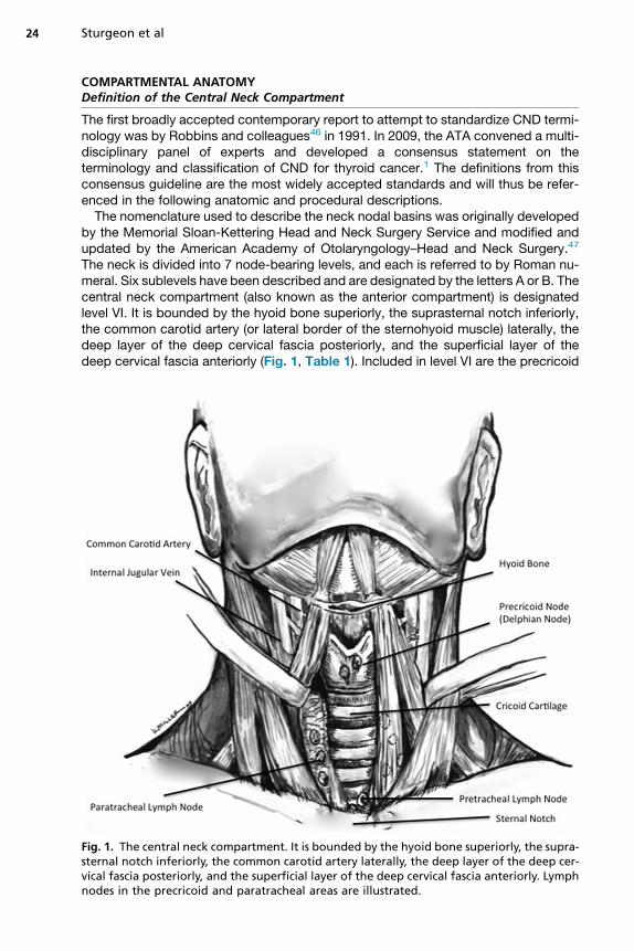
COMPARTMENTAL ANATOMYDefinition of the Central Neck Compartment
The first broadly accepted contemporary report to attempt to standardize CND termi-nology was by Robbins and colleagues46 in 1991. In 2009, the ATA convened a multi-disciplinary panel of experts and developed a consensus statement on theterminology and classification of CND for thyroid cancer.1 The definitions from thisconsensus guideline are the most widely accepted standards and will thus be refer-enced in the following anatomic and procedural descriptions.The nomenclature used to describe the neck nodal basins was originally developed
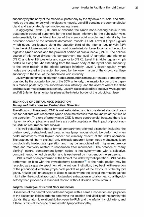
by the Memorial Sloan-Kettering Head and Neck Surgery Service and modified andupdated by the American Academy of Otolaryngology–Head and Neck Surgery.47
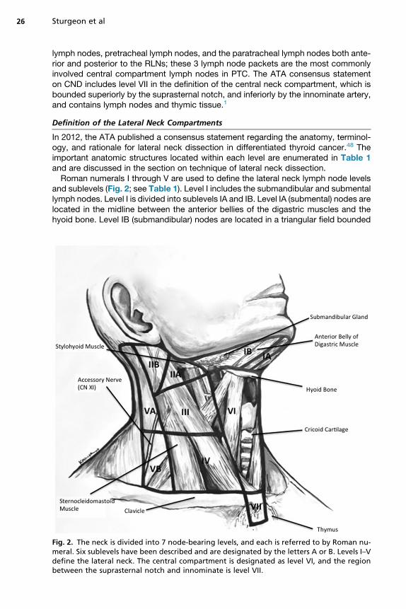
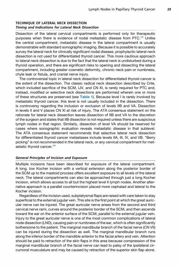
The neck is divided into 7 node-bearing levels, and each is referred to by Roman nu-meral. Six sublevels have been described and are designated by the letters A or B. Thecentral neck compartment (also known as the anterior compartment) is designatedlevel VI. It is bounded by the hyoid bone superiorly, the suprasternal notch inferiorly,the common carotid artery (or lateral border of the sternohyoid muscle) laterally, thedeep layer of the deep cervical fascia posteriorly, and the superficial layer of thedeep cervical fascia anteriorly (Fig. 1, Table 1). Included in level VI are the precricoid
Fig. 1. The central neck compartment. It is bounded by the hyoid bone superiorly, the supra-sternal notch inferiorly, the common carotid artery laterally, the deep layer of the deep cer-vical fascia posteriorly, and the superficial layer of the deep cervical fascia anteriorly. Lymphnodes in the precricoid and paratracheal areas are illustrated.
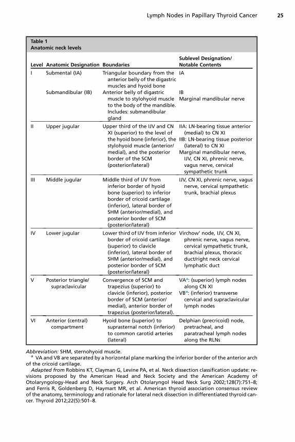
Table 1Anatomic neck levels
Level Anatomic Designation BoundariesSublevel Designation/Notable Contents
I Submental (IA) Triangular boundary from theanterior belly of the digastricmuscles and hyoid bone
IA
Submandibular (IB) Anterior belly of digastricmuscle to stylohyoid muscleto the body of the mandible.Includes: submandibulargland
IBMarginal mandibular nerve
II Upper jugular Upper third of the IJV and CNXI (superior) to the level ofthe hyoid bone (inferior), thestylohyoid muscle (anterior/medial), and the posteriorborder of the SCM(posterior/lateral)
IIA: LN-bearing tissue anterior(medial) to CN XI
IIB: LN-bearing tissue posterior(lateral) to CN XI
Marginal mandibular nerve,IJV, CN XI, phrenic nerve,vagus nerve, cervicalsympathetic trunk
III Middle jugular Middle third of IJV frominferior border of hyoidbone (superior) to inferiorborder of cricoid cartilage(inferior), lateral border ofSHM (anterior/medial), andposterior border of SCM(posterior/lateral)
IJV, CN XI, phrenic nerve, vagusnerve, cervical sympathetictrunk, brachial plexus
IV Lower jugular Lower third of IJV from inferiorborder of cricoid cartilage(superior) to clavicle(inferior), lateral border ofSHM (anterior/medial), andposterior border of SCM(posterior/lateral)
Virchow’ node, IJV, CN XI,phrenic nerve, vagus nerve,cervical sympathetic trunk,brachial plexus, thoracicduct/right neck cervicallymphatic duct
V Posterior triangle/supraclavicular
Convergence of SCM andtrapezius (superior) toclavicle (inferior), posteriorborder of SCM (anterior/medial), anterior border oftrapezius (posterior/lateral).
VAa: (superior) lymph nodesalong CN XI
VBa: (inferior) transversecervical and supraclavicularlymph nodes
VI Anterior (central)compartment
Hyoid bone (superior) tosuprasternal notch (inferior)to common carotid arteries(lateral)
Delphian (precricoid) node,pretracheal, andparatracheal lymph nodesalong the RLNs
Abbreviation: SHM, sternohyoid muscle.a VA and VB are separated by a horizontal plane marking the inferior border of the anterior arch
of the cricoid cartilage.Adapted from Robbins KT, Clayman G, Levine PA, et al. Neck dissection classification update: re-
visions proposed by the American Head and Neck Society and the American Academy ofOtolaryngology-Head and Neck Surgery. Arch Otolaryngol Head Neck Surg 2002;128(7):751–8;and Ferris R, Goldenberg D, Haymart MR, et al. American thyroid association consensus reviewof the anatomy, terminology and rationale for lateral neck dissection in differentiated thyroid can-cer. Thyroid 2012;22(5):501–8.
Lymph Nodes in Papillary Thyroid Cancer 25
Sturgeon et al26
lymph nodes, pretracheal lymph nodes, and the paratracheal lymph nodes both ante-rior and posterior to the RLNs; these 3 lymph node packets are the most commonlyinvolved central compartment lymph nodes in PTC. The ATA consensus statementon CND includes level VII in the definition of the central neck compartment, which isbounded superiorly by the suprasternal notch, and inferiorly by the innominate artery,and contains lymph nodes and thymic tissue.1
Definition of the Lateral Neck Compartments
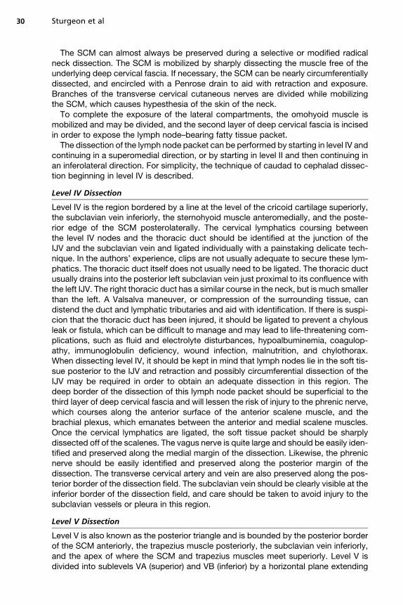
In 2012, the ATA published a consensus statement regarding the anatomy, terminol-ogy, and rationale for lateral neck dissection in differentiated thyroid cancer.48 Theimportant anatomic structures located within each level are enumerated in Table 1and are discussed in the section on technique of lateral neck dissection.Roman numerals I through V are used to define the lateral neck lymph node levels
and sublevels (Fig. 2; see Table 1). Level I includes the submandibular and submentallymph nodes. Level I is divided into sublevels IA and IB. Level IA (submental) nodes arelocated in the midline between the anterior bellies of the digastric muscles and thehyoid bone. Level IB (submandibular) nodes are located in a triangular field bounded
Fig. 2. The neck is divided into 7 node-bearing levels, and each is referred to by Roman nu-meral. Six sublevels have been described and are designated by the letters A or B. Levels I–Vdefine the lateral neck. The central compartment is designated as level VI, and the regionbetween the suprasternal notch and innominate is level VII.
Lymph Nodes in Papillary Thyroid Cancer 27
superiorly by the body of the mandible, posteriorly by the stylohyoid muscle, and ante-riorly by the anterior belly of the digastric muscle. Level IB contains the submandibulargland and associated lymph node–bearing tissue.In aggregate, levels II, III, and IV describe the lymph node–bearing tissue in the
quadrangle bounded superiorly by the skull base, inferiorly by the subclavian vein,anteromedially by the lateral border of the sternohyoid muscle, and laterally by theposterior border of the sternocleidomastoid muscle (SCM). Level II (upper jugular)lymph nodes are located along the superior third of the internal jugular vein (IJV)from the skull base superiorly to the hyoid bone inferiorly. Level II contains the jugulo-digastric lymph nodes and the proximal portion of cranial nerve (CN) XI. The obliquecourse of this nerve divides this compartment into level IIA (anterior and inferior toCN XI) and level IIB (posterior and superior to CN XI). Level III (middle jugular) lymphnodes lie along the IJV extending from the lower body of the hyoid bone superiorlyto the lower margin of the cricoid cartilage inferiorly. Level IV (lower jugular) lymphnodes are located in the region bordered by the lower margin of the cricoid cartilagesuperiorly to the level of the subclavian vein inferiorly.Level V (posterior triangle) lymphnodesare found in a triangular-shapedcompartment
bounded by the posterior border of the SCM anteriorly, the anterior border of the trape-zius muscle posteriorly, the subclavian vein inferiorly, and the apex of where the SCMand trapeziusmusclesmeet superiorly. Level V is also divided into sublevel VA (superior)and VB (inferior) by a horizontal plane at the inferior border of the cricoid cartilage.
TECHNIQUE OF CENTRAL NECK DISSECTIONTiming and Indications for Central Neck Dissection
The efficacy of therapeutic CND is well established and is considered standard prac-tice for patients with resectable lymph node metastases that are known at the time ofthe operation. The role of prophylactic CND is more controversial because there is ahigher risk of complications and there are conflicting data on the impact of prophylac-tic CND on recurrence and survival.It is well-established that a formal compartment-oriented dissection including the
prelaryngeal, pretracheal, and paratracheal lymph nodes should be performed whennodal metastases from thyroid cancer are clinically evident at the index operation.The practice of “berry picking” only clinically apparent lymph node metastases is anoncologically inadequate operation and may be associated with higher recurrencerates and morbidity related to reoperation after recurrence.1 The practice of “berrypicking” central compartment lymph nodes is not synonymous with a selective,compartment-oriented dissection and is eschewed by most endocrine surgeons.CND is most often performed at the time of the index thyroid operation. CND can be
performed en bloc with the thyroidectomy specimen49 or the nodal packet may beresected as a separate specimen. At the authors’ institution, they begin with resectionof the precricoid (Delphian) lymph node packet as part of the exposure of the thyroidgland. Frozen section analysis is used in cases where the clinical information gainedmight alter the surgical approach. A standard extracapsular total or near-total thyroid-ectomy then proceeds in standard fashion without division of the isthmus.
Surgical Technique of Central Neck Dissection
Dissection of the central compartment begins with a careful inspection and palpationof the dissection field in order to determine the location and viability of the parathyroidglands, the anatomic relationship between the RLN and the inferior thyroid artery, andif there is clinical evidence of metastatic lymphadenopathy.
Sturgeon et al28
Before the compartmental dissection, any parathyroid gland that has been devas-cularized should be autotransplanted. Because inspection of the parathyroid glandis notoriously inaccurate in determining viability, many experts recommend piercingor cutting the capsule of the gland to determine if there is continued perfusion, as evi-denced by the presence of brisk bleeding. The authors advocate liberal use of auto-transplantation of any parathyroid gland in which ischemia is suspected, becausethis may prevent or significantly mitigate the risk of permanent postoperative hypo-parathyroidism. Following the CND, parathyroid glands remaining in situ are againexamined for evidence of ischemia and autotransplanted if necessary. The inferiorglands are at highest risk for devascularization and are routinely autotransplantedby some experts.The RLN must always be identified and traced from its insertion through its entire
course as it passes caudally through the central neck compartment. Full visualizationof the RLN is maintained throughout the dissection. Atraumatic technique with onlygentle manipulation and minimal traction of the nearby tissues should be used duringdissection along the RLN. When bulky lymphadenopathy exists, the position of theRLN may be displaced by the adenopathy; this is particularly true on the right side,where adenopathy may be located deep to the RLN.The paratracheal lymph node packet is removed as one specimen. The dissection
is usually performed in a cephalad to caudad manner, removing all the node-bearing fibrofatty tissue from the level of the hyoid bone superiorly down to theinnominate artery inferiorly. The width of the dissection field extends from thecarotid artery laterally to the anterior midpoint of the trachea medially. The pretra-cheal lymph nodes inferior to the thyroid gland should always be included withinthe dissection specimen. Thymectomy is not necessary during CND.50 The dissec-tion specimen should be clearly labeled and oriented to site and side. The operativenote should clearly describe the levels included in the dissection and their borders,the disposition of the parathyroid glands, and if any anatomic variants wereidentified.Some investigators have reported that very little node-bearing tissue is ever found
above the level of the insertion of the RLN and have questioned the necessity ofextending the paratracheal dissection field above the level RLN insertion. In one studyof 31 paratracheal neck dissections for thyroid cancer in 27 patients, no lymph nodes,lymphatic tissue, or metastatic disease was identified in any upper paratracheal spec-imens retrieved above the level of the cricoid cartilage.51 All benign and metastaticlymph nodes were located in the lower paratracheal specimens. Accordingly, it maybe reasonable to use the level of the cricoid cartilage as the superior border of theCND in the absence of clinically apparent or biopsy-proven metastatic disease inthe region between the cricoid and the hyoid bone. Furthermore, limiting the superiorextent of the CND to the level of the cricoid might decrease the likelihood of devascu-larizing the superior parathyroid glands while not compromising the oncologicoutcome of the operation.
Complications of Central Neck Dissection
Complications of CND are mainly related to injury to the RLN and external branch ofthe superior laryngeal nerve, and devascularization of the parathyroid glands. Asdescribed above, permanent postoperative hypoparathyroidism rates from CNDcan be as high as nearly 20%.15 In experienced hands, the incidence of permanentRLN injury should be low, on the order of 1% to 6%.52 Meticulous hemostatic dissec-tion technique and operative experience are significant contributors to minimizingthese complications.
Lymph Nodes in Papillary Thyroid Cancer 29
TECHNIQUE OF LATERAL NECK DISSECTIONTiming and Indications for Lateral Neck Dissection
Dissection of the lateral cervical compartments is performed only for therapeuticpurposes when there is evidence of nodal metastatic disease from PTC.48 Unlikethe central compartment, metastatic disease in the lateral compartment is usuallydemonstrable with standard sonographic imaging. Because it is possible to accuratelysurvey the lateral neck for clinically significant nodal disease, prophylactic lateral neckdissection is not used for differentiated thyroid cancer. This more cautious approachto lateral neck dissection is due to the fact that the lateral neck is undisturbed during athyroid operation, and there are significant risks to opening and dissecting the lateralcompartment, including greater cosmetic deformity, chronic neck pain or numbness,chyle leak or fistula, and cranial nerve injury.The controversial topic in lateral neck dissection for differentiated thyroid cancer is
the extent of the dissection. The classic radical neck dissection described by Crile,which included sacrifice of the SCM, IJV, and CN XI, is rarely required for PTC and,instead, modified or selective neck dissections are performed wherein one or moreof these structures are preserved (see Table 1). Because level I is rarely involved bymetastatic thyroid cancer, this level is not usually included in the dissection. Thereis controversy regarding the inclusion or exclusion of levels IIB and VA. Dissectionin levels II and V places CN XI at risk of injury. The ATA consensus statement on therationale for lateral neck dissection leaves dissection of IIB and VA to the discretionof the surgeon and states that IIB dissection is not required unless there are suspiciouslymph nodes in that region. Similarly, dissection of level VA should be reserved forcases where sonographic evaluation reveals metastatic disease in that sublevel.The ATA consensus statement recommends that selective lateral neck dissectionfor differentiated thyroid cancer metastases include levels IIA, III, IV, and VB. “Berrypicking” is not recommended in the lateral neck, or any cervical compartment for met-astatic thyroid cancer.48
General Principles of Incision and Exposure
Multiple incisions have been described for exposure of the lateral compartment.A long, low Kocher incision with a vertical extension along the posterior border ofthe SCM up to the mastoid process offers excellent exposure to all levels of the lateralneck. The lateral compartments can also be approached through just a long Kocherincision, which allows access to all but the highest level II lymph nodes. Another alter-native approach is a parallel counterincision placed more cephalad and lateral to theKocher incision.Regardless of the incision used, subplatysmal flaps are raisedwith care taken to stay
superficial to the external jugular vein. This site is the first point at which the great auric-ular nerve can be injured. The great auricular nerve arises from the second and thirdcervical nerve rami, curves around the posterior border of the SCM, and then ascendstoward the ear on the anterior surface of the SCM, parallel to the external jugular vein.Injury to the great auricular nerve is one of the most common complications of lateralnode dissection (LND), causing pain or numbness of the ear, which is often significantlybothersome to the patient. The marginal mandibular branch of the facial nerve (CN VII)can be injured during the dissection as well. The marginal mandibular branch runsalong the inferior border of the mandible anterior to the facial artery and vein. Attentionshould be paid to retraction of the skin flaps in this area because compression of themarginal mandibular branch of the facial nerve can lead to palsy of the ipsilateral cir-cumoral musculature and may be caused by retraction of the superior skin flap alone.
Sturgeon et al30
The SCM can almost always be preserved during a selective or modified radicalneck dissection. The SCM is mobilized by sharply dissecting the muscle free of theunderlying deep cervical fascia. If necessary, the SCM can be nearly circumferentiallydissected, and encircled with a Penrose drain to aid with retraction and exposure.Branches of the transverse cervical cutaneous nerves are divided while mobilizingthe SCM, which causes hypesthesia of the skin of the neck.To complete the exposure of the lateral compartments, the omohyoid muscle is
mobilized and may be divided, and the second layer of deep cervical fascia is incisedin order to expose the lymph node–bearing fatty tissue packet.The dissection of the lymph node packet can be performed by starting in level IV and
continuing in a superomedial direction, or by starting in level II and then continuing inan inferolateral direction. For simplicity, the technique of caudad to cephalad dissec-tion beginning in level IV is described.
Level IV Dissection
Level IV is the region bordered by a line at the level of the cricoid cartilage superiorly,the subclavian vein inferiorly, the sternohyoid muscle anteromedially, and the poste-rior edge of the SCM posterolaterally. The cervical lymphatics coursing betweenthe level IV nodes and the thoracic duct should be identified at the junction of theIJV and the subclavian vein and ligated individually with a painstaking delicate tech-nique. In the authors’ experience, clips are not usually adequate to secure these lym-phatics. The thoracic duct itself does not usually need to be ligated. The thoracic ductusually drains into the posterior left subclavian vein just proximal to its confluence withthe left IJV. The right thoracic duct has a similar course in the neck, but is much smallerthan the left. A Valsalva maneuver, or compression of the surrounding tissue, candistend the duct and lymphatic tributaries and aid with identification. If there is suspi-cion that the thoracic duct has been injured, it should be ligated to prevent a chylousleak or fistula, which can be difficult to manage and may lead to life-threatening com-plications, such as fluid and electrolyte disturbances, hypoalbuminemia, coagulop-athy, immunoglobulin deficiency, wound infection, malnutrition, and chylothorax.When dissecting level IV, it should be kept in mind that lymph nodes lie in the soft tis-sue posterior to the IJV and retraction and possibly circumferential dissection of theIJV may be required in order to obtain an adequate dissection in this region. Thedeep border of the dissection of this lymph node packet should be superficial to thethird layer of deep cervical fascia and will lessen the risk of injury to the phrenic nerve,which courses along the anterior surface of the anterior scalene muscle, and thebrachial plexus, which emanates between the anterior and medial scalene muscles.Once the cervical lymphatics are ligated, the soft tissue packet should be sharplydissected off of the scalenes. The vagus nerve is quite large and should be easily iden-tified and preserved along the medial margin of the dissection. Likewise, the phrenicnerve should be easily identified and preserved along the posterior margin of thedissection. The transverse cervical artery and vein are also preserved along the pos-terior border of the dissection field. The subclavian vein should be clearly visible at theinferior border of the dissection field, and care should be taken to avoid injury to thesubclavian vessels or pleura in this region.
Level V Dissection
Level V is also known as the posterior triangle and is bounded by the posterior borderof the SCM anteriorly, the trapezius muscle posteriorly, the subclavian vein inferiorly,and the apex of where the SCM and trapezius muscles meet superiorly. Level V isdivided into sublevels VA (superior) and VB (inferior) by a horizontal plane extending
Lymph Nodes in Papillary Thyroid Cancer 31
laterally from the cricoid cartilage. Level VA contains the lymph nodes along the distalportion of the spinal accessory nerve (CN XI). Level VB contains the transverse cervicalvessels and associated lymph nodes as well as the supraclavicular lymph nodes.If only level VB is to be dissected, then the lymph node–bearing fatty tissue along the
transverse cervical vessels and in the supraclavicular region should be removed enbloc with the specimen. Care should be taken to identify CN XI and avoid injury to iteven if the level VA lymph node packet is not included in the dissection.If level VA is included, the anterior border of the trapezius muscle (the posterior-
lateral border of level V) is identified and incised approximately 1 cm anterior to theborder of the muscle. This incision will allow for easier and safer identification of CNXI as it inserts into the trapezius muscle. CN XI will be found coursing over the levatorscapulae muscle and will be approximately parallel to the trapezius muscle itself.Much of the lymph node–bearing tissue in level VB (and level II) will be around CNXI, and thus, it must be clearly identified and carefully manipulated. As CN XI isdissected in a superomedial direction toward the upper posterior border of theSCM, a plexus of cervical nerve branches will be encountered at the posterior borderof the SCM. The great auricular nerve can be found here just inferior to CN XI as itcourses underneath the SCM. Often referred to as Erb point,53 this plexus is a usefullandmark to identify CN XI in a more superior and medial location.
Level III Dissection
Level III extends from the superior border of level IV to the hyoid bone. The medial andlateral borders are the same as level IV. Dissection of the node-bearing fibrofattytissue packet should continue sharply over the scalene muscles and along the IJVthrough level III, with continued care taken to directly visualize and avoid injury tothe phrenic and vagus nerves. Cervical sensory nerve fibers should be preserved ifpreservation will not compromise the oncologic outcome of the operation. The com-mon trunk of the supraclavicular nerve can be identified beneath the SCM and shouldbe preserved to avoid lateral neck, shoulder, and anterior chest numbness postoper-atively. Staying superficial to the third layer of deep cervical fascia will lessen the likeli-hood of injury to all of the nerve structures except for the vagus nerve. The cervicalsensory plexus can be used as a landmark that the superior portion of the dissectionhas almost been completed.
Level II Dissection
Level II extends from the superior border of level III to the skull base superiorly. Theanteromedial border is the stylohyoid muscle and the lateral border is the posterioredge of the SCM. CN XI divides level II into level IIA and level IIB. If only level IIA isto be dissected, then identification of CN XI will represent the posterosuperior limitof the dissection, and the dissection specimen is sharply dissected from the IJVand CN XI and truncated at the apex of these 2 structures. If level IIB is to be included,the dissection should continue to the level of the posterior belly of the digastricmuscle.Once the dissection has been completed, the dissection specimen should be clearly
labeled and oriented. The operative procedure note should clearly describe the levelsincluded in the dissection and the borders. Drain placement is at the discretion of thesurgeon.
Complications of Lateral Neck Dissection
The common and major complications of lateral neck dissection have been describedabove in relation to the steps of the procedure. In summary, the most common
Sturgeon et al32
complications are sensory deficits related to injury to the great auricular nerve or cer-vical sensory nerve fibers. Major, avoidable complications include chylous leak or chy-lothorax related to unrecognized injuries of the thoracic duct or other major lymphaticbranches, shoulder weakness resulting from injury to CN XI, lip droop related to mar-ginal mandibular nerve injury, and symptoms related to injuries to the other relevantnerves (phrenic, vagus, hypoglossal). Rough handling of major vascular structuressuch as the carotid artery and IJV can lead to vascular injuries, bleeding, embolicstroke (including air embolism), and carotid dissection. Clear visualization of the ner-vous, lymphatic, and vascular structures, delicate dissection technique, and meticu-lous hemostasis will minimize complications.
IMPACT OF NODAL CLEARANCE ON RECURRENCE AND SURVIVALImpact of Nodal Basin Clearance on Recurrence and Survival
Regional lymph node metastases at the time of presentation are relatively common inPTC.17,54 There has been a long-standing controversy over the clinical significance oflymph node metastases in PTC in low-risk patients. However, the controversy over thesignificance of lymph node metastases in high-risk patients is less questioned. Multi-ple studies have shown that lymph node metastases are an independent predictor ofpoor outcome. Although the overall survival differences may be slight (82% for node-negative disease vs 79% for node-positive disease at 14 years), they are statisticallysignificant,1,55 and there is also an increased risk of recurrence in patients with lymphnode metastases in PTC. Although there are fewer patients presenting with lateralneck metastases, there is evidence that lateral neck metastases are associated witha poorer outcome, namely increased risk of recurrence and possibly decreasedsurvival.17,56,57
Clearance of the affected nodal basins of metastatic disease may offer other ben-efits beyond a small improvement in survival. First of all is the reduction in recur-rence after CND or LND. Although there is a paucity of randomized controlled orother matched control studies in the literature, multiple studies have shown recur-rence rates on the order of 10% after total thyroidectomy with CND in patientswith or without known preoperative central neck lymph node metastases.58,59
Multiple studies indicate that CND or LND can successfully control persistent orrecurrent disease as reflected by meaningful decreases in serum thyroglobulinlevels, with some studies finding that a substantial number of patients can have un-detectable preablative thyroglobulin levels after surgery.16,60 Recurrence after CND(and in patients who have not undergone CND) tends to occur in the lateral necklymph node basins, particularly levels III and IV, rather than in the central neckcompartment.58,59 Prognostic factors for local recurrence include increasing num-ber of metastatic lymph nodes and extracapsular lymph node extension on patho-logic analysis.1
Prophylactic CND remains controversial because multiple studies indicate that theincidence of both temporary61–63 and permanent15,58 hypoparathyroidism isincreased with CND. Furthermore, CND is associated with a higher incidence ofRLN injury, most of which is temporary.58,61,63 Multiple studies have shown thatcomplication rates are lower in operations for thyroid cancer, including CND andLND, when the procedure is performed by a high-volume thyroid surgeon.6 There-fore, the ATA consensus statements recommend that the surgeon’s skill level beconsidered when making the decision whether a patient who requires CND or LNDshould remain in that surgeon’s hands or be referred to a high-volume thyroid surgerycenter.1,6,64
Lymph Nodes in Papillary Thyroid Cancer 33
RECURRENT NODAL METASTASESA Comparison of Surveillance Strategies Recommended by the American ThyroidAssociation and National Comprehensive Cancer Network
Most patients who have been treated for PTC undergo long-term surveillance. Theprimary goals of long-term follow-up are the early and accurate detection of recurrentdisease and the longitudinal monitoring of thyroxine replacement and TSH.6 The ATArecommends that following total thyroidectomy, serum thyroglobulin andanti-thyroglobulin antibodies should bemeasured every 6 to 12months in the same lab-oratory using the same assay. Rising thyroglobulin values over time are considered sus-picious for recurrence. Periodic neck ultrasound is also recommended. In low-riskpatients with no detectable tumor, the ATA recommends that a TSH-stimulated thyro-globulin bemeasured12months after radioiodine ablation to verify that there is no recur-rent or persistent disease. For those patients who have no detectable stimulatedthyroglobulin, the ATA recommends yearly clinical examination and unstimulatedthyroglobulin.6
The NCCN guidelines recommend physical examination, TSH, thyroglobulin, andantithyroglobulin antibodies at 6 and 12 months and then annually thereafter if the pa-tient is considered disease-free. Similar to the ATA recommendations, neck ultra-sound is also recommended on a periodic basis by the NCCN.27 The NCCNrecommends that stimulated thyroglobulin be measured in patients who underwentradioiodine ablation and in those who have undetectable unstimulated thyroglobulinand no antithyroglobulin antibodies. Furthermore, TSH-stimulated radioiodine scan-ning should be considered for patients with detectable stimulated or unstimulatedthyroglobulin, sonographic evidence of persistent or recurrent disease, rising antithyr-oglobulin antibody titer, or with radioiodine avid metastases, or in patients consideredhigh risk. In patients with high-risk radioiodine avid tumors, especially those withdetectable thyroglobulin, distant metastases, or extension into the soft tissues, theNCCN recommends radioiodine imaging every 1 to 2 years.
Ultrasound and Serum Thyroglobulin Measurement Are Highly Accurate at DetectingRecurrence
Sonographiccervical imaginghasbeen found tobemoreaccurate thanradioiodinescan-ning for the detection of recurrent or persistent disease. In addition, the measurement ofstimulated thyroglobulin has been found to be complementary to cervical sonography,increasing the accuracy of the surveillance strategy. In a series of 340 consecutive pa-tients with differentiated thyroid cancer who underwent near-total thyroidectomy andradioiodine ablation, the combination of stimulated thyroglobulin and cervical sonogra-phy had the greatest accuracy at detecting persistent disease (sensitivity 96.3% andnegative predictive value of 99.5%).65 In a series of 80 consecutive patients with PTCmicrocarcinoma who underwent near-total thyroidectomy but did not undergo radioio-dine ablation, cervical ultrasoundwas found to bemore accurate than radioiodine scan-ning for the detection of persistent disease.66 Although radioiodine scanning showed nopathologic uptake in any patient, cervical sonography identified nodal metastases in pa-tientswith orwithoutdetectable thyroglobulin. Theauthors concluded that in this low-riskpopulation, ultrasound should be the primary screening modality, stimulated thyroglob-ulin was indicative of remnant normal thyroid tissue, and radioiodine scanning was notvaluable for the detection of persistent disease. These findings support the recommen-dations from theATA andNCCN that surveillance be conducted primarily via cervical so-nography and measurement of serum thyroglobulin, and that radioiodine scanning bereserved for higher-risk patients with radioiodine avid disease.
Sturgeon et al34
Surveillance Frequently Reveals Recurrent or Persistent Disease
TheATAdefines absence of diseaseusing 3 criteria: no clinically detectable tumor recur-rence, no evidence of recurrence on imaging, and undetectable stimulated and unstimu-lated thyroglobulin (when interfering antibodies are absent).6 Unfortunately, because ofthe stringency of these criteria and the high sensitivity of current imagingmodalities andmethods for detection of thyroglobulin,manypatientswill not satisfy these criteria, espe-cially if they have not undergone radioiodine ablation of the thyroid remnant.The recurrence rate is relatively high in PTC and is related to the sensitivity of the
tests used to conduct surveillance, the biology of PTC, and the initial treatment.Biologic variables that impact recurrence include the number of positive nodes, tumorburden, and the presence of extranodal extension.2,67–70 Patients who are patholog-ically N0 are at lowest risk of recurrence. Patients with microscopic N1a disease are atan intermediate risk.71 Patients with clinically positive or macroscopic N1 disease areat high risk. Patients with extranodal extension are at the highest risk. A recent meta-analysis stratified patients into risk groups based on the number of positive nodes.They found that the risk of recurrence was 4% with fewer than 5 positive nodes,19% with more than 5 positive nodes, and 24% with clinically apparent nodes withextranodal extension.2
For patients with stable, low levels of detectable thyroglobulin whose diseasecannot be detected on imaging, it is considered acceptable to continue active surveil-lance. For patients with recurrent or persistent disease that can be imaged, surgicalresection is the preferred treatment.
Recommendations for Surveillance of the Lateral Neck
Metastases to the lateral compartment are common and are therefore one of the focalpoints of surveillance for patients with PTC. Ultrasound is the imaging modality ofchoice for the detection of lateral neck nodal metastases when performing surveil-lance. Sonographic surveillance of the lateral neck should be performed periodicallydepending on the likelihood of recurrent or persistent disease. The entire neck shouldbe imaged (levels I through VII) by a sonographer experienced in surveillance of PTC.Special attention should be paid to the nodal basins ipsilateral to the primary tumor,because they are the most likely a location for recurrent or persistent disease.
Establishing the Diagnosis of Recurrent or Persistent Disease
The ATA published a consensus statement on the anatomy, terminology, and indica-tions for lateral neck dissection in PTC.64 They concluded that the initial modalityshould be ultrasound and that FNA of suspicious lymph nodes is the best way toestablish the diagnosis of nodal metastases. In addition, the ATA Statement on Preop-erative Imaging for Thyroid Cancer Surgery recommends that suspicious masses orlymph nodes be biopsied before a reoperation for persistent or recurrent disease.72
Ultrasound has been shown to accurately detect the presence of central and lateralnodal metastases and has been found to alter the extent of surgery in up to 40% ofindex and reoperative cases.18 Therefore, in the course of surveillance for PTC, sono-graphically suspicious lymph nodes should be interrogated with FNA biopsy. The aspi-rate may be assayed for thyroglobulin in order to increase the sensitivity andspecificity for metastatic disease.
Is Positive Imaging Alone Ever Sufficient?
The 2009 ATA guidelines state that lateral neck dissection should not be performedprophylactically, but should be performed as a “therapeutic intervention for known
Lymph Nodes in Papillary Thyroid Cancer 35
disease.”6 This statement raises the question of whether imaging alone would be suf-ficient to confirm the presence of metastatic disease. In some cases, the location ofthe lymph node may not be amenable to needle biopsy or patients may refuse needlebiopsy. The ATA Subcommittee on Lateral Neck Dissection indicated in their recentreview that lateral neck lymph nodes not amenable to FNA biopsy may be followedfor growth by serial imaging. If the inaccessible node grows during follow-up, anopen biopsy should be performed with conversion to formal lateral neck dissectionif the frozen section is positive. The ATA Statement on Preoperative Imaging forThyroid Cancer Surgery states that preoperative FNA may not be required underthe following 2 conditions: “(a) abnormal lymph nodes are inaccessible or anatomicallyrisky to biopsy, usually due to their location with respect to major vessels, and (b)unequivocally abnormal lymph nodes are found on imaging and surgery would be rec-ommended regardless of FNA results.”72
The Treatment of Recurrent or Persistent Nodal Disease
The management of low-volume recurrent or persistent nodal metastases is contro-versial. The 2most reasonable treatment options are surgical resection and active sur-veillance.73 The preferred treatment for locoregional recurrence, according to theNCCN, is surgical resection. Radioiodine therapy and external beam radiotherapyare alternative treatment options for tumors that either do or do not concentrate radio-iodine, respectively. The NCCN recommends that clinically significant nodal metasta-ses in a previously undissected nodal basin be treated with a formal compartmentalclearance of the nodal basin involved. For recurrence in the central neck in a patientwith no prior CND, a complete dissection of that ipsilateral central compartment isindicated. Likewise, recurrence discovered in a previously undissected lateralcompartment should be treated with a formal modified radical neck dissection,including levels II, III, IV, and VB. Conversely, recurrence in a previously dissected cen-tral or lateral compartment should be adequately treated with a focused dissection ofthe region containing the nodal metastasis.27
The 2009 ATA guidelines for DTC state that the “preferred hierarchy of treatment formetastatic disease” is surgical resection for potentially curable patients with locore-gional disease, radioiodine therapy for patients with iodine-avid disease, externalbeam radiation therapy, active surveillance in patients with stable or slowly progres-sive asymptomatic disease, and experimental trials.6 They also indicate that morenontraditional management may also benefit selected patients, such as ethanol abla-tion, radiofrequency ablation, or chemoembolization. Furthermore, active surveillanceis presented as a reasonable option for patients with stable asymptomatic diseasethat does not involve the central nervous system. Similar to NCCN, the ATA guidelinessupport a comprehensive dissection of previously unexplored cervical compartmentsharboring metastatic disease, and a more limited or focused dissection of previouslyoperated compartments. Clinically significant nodal metastases are defined as thosegreater than 0.8 cm in size. The treatment of cervical nodal metastases in patients withuntreatable distant metastases may be considered for palliation or avoidance of aero-digestive tract invasion.
SUMMARY
When central or lateral compartment cervical lymph node metastases are clinicallyevident at the time of the index thyroid operation for PTC, formal surgical clearance ofthe affected nodal basin is the optimal management. Prophylactic CND for PTC is prac-ticed by some high-volume surgeons with low complication rates, but is considered
Sturgeon et al36
controversial because there appears to be higher risk of complicationswith an uncertainclinical benefit. Long-term surveillance strategies are performed for most PTC patientsafter their initial treatment, and due to the high sensitivity of both the imaging modalitiesused and thyroglobulin measurement, approximately 20% of patients will be found tohave persistent or recurrent disease. It is unclearwhat exactly constitutes a clinically sig-nificant recurrence, but themost commonly used definition is a lymph node greater than0.8 cm. Surgical resection and active surveillance strategiesmay both be used depend-ing on the clinical context. When a clinically significant recurrence is detected in a pre-viously undissected central or lateral cervical compartment, a comprehensive surgicalclearance of the lateral compartment is the preferred treatment. When a nodal recur-rence is found in a previously dissected central or lateral neck field, the reoperationshould focus on the areas where recurrence is demonstrated.
ACKNOWLEDGMENTS
The authors thank Kyle Miller, MD for the original illustrations that accompany thetext.
REFERENCES
1. Carty SE, Cooper DS, Doherty GM, et al. Consensus statement on the terminol-ogy and classification of central neck dissection for thyroid cancer. Thyroid2009;19(11):1153–8.
2. Randolph GW, Duh QY, Heller KS, et al. The prognostic significance of nodalmetastases from papillary thyroid carcinoma can be stratified based on thesize and number of metastatic lymph nodes, as well as the presence of extrano-dal extension. Thyroid 2012;22(11):1144–52.
3. Machens A, Hinze R, Thomusch O, et al. Pattern of nodal metastasis for primaryand reoperative thyroid cancer. World J Surg 2002;26(1):22–8.
4. Carling T, Carty SE, Ciarleglio MM, et al. American Thyroid Association designand feasibility of a prospective randomized controlled trial of prophylactic centrallymph node dissection for papillary thyroid carcinoma. Thyroid 2012;22(3):237–44.
5. Popadich A, Levin O, Lee JC, et al. A multicenter cohort study of total thyroidec-tomy and routine central lymph node dissection for cN0 papillary thyroid cancer.Surgery 2011;150(6):1048–57.
6. Cooper DS, Doherty GM, Haugen BR, et al. Revised American Thyroid Associa-tion management guidelines for patients with thyroid nodules and differentiatedthyroid cancer. Thyroid 2009;19(11):1167–214.
7. Chisholm EJ, Kulinskaya E, Tolley NS. Systematic review and meta-analysis of theadverse effects of thyroidectomy combined with central neck dissection ascompared with thyroidectomy alone. Laryngoscope 2009;119(6):1135–9.
8. Hughes DT, White ML, Miller BS, et al. Influence of prophylactic central lymphnode dissection on postoperative thyroglobulin levels and radioiodine treatmentin papillary thyroid cancer. Surgery 2010;148(6):1100–6 [discussion: 1006–7].
9. Zetoune T, Keutgen X, Buitrago D, et al. Prophylactic central neck dissection andlocal recurrence in papillary thyroid cancer: a meta-analysis. Ann Surg Oncol2010;17(12):3287–93.
10. Wang TS, Cheung K, Farrokhyar F, et al. A meta-analysis of the effect of prophy-lactic central compartment neck dissection on locoregional recurrence rates inpatients with papillary thyroid cancer. Ann Surg Oncol 2013;20(11):3477–83.
Lymph Nodes in Papillary Thyroid Cancer 37
11. Moo TA, McGill J, Allendorf J, et al. Impact of prophylactic central neck lymphnode dissection on early recurrence in papillary thyroid carcinoma. World JSurg 2010;34(6):1187–91.
12. Zanocco K, Elaraj D, Sturgeon C. Routine prophylactic central neck dissection forlow-risk papillary thyroid cancer: a cost-effectiveness analysis. Surgery 2013;154(6):1148–55 [discussion: 1154–5].
13. Garcia A, Palmer BJ, Parks NA, et al. Routine prophylactic central neck dissec-tion for low-risk papillary thyroid cancer is not cost-effective. Clin Endocrinol (Oxf)2014;81(5):754–61.
14. Edge SB, American Joint Committee on Cancer. AJCC cancer staging manual.7th edition. New York: Springer; 2010.
15. Viola D, Materazzi G, Valerio L, et al. Prophylactic central compartment lymphnode dissection in papillary thyroid carcinoma: clinical implications derivedfrom the first prospective randomized controlled single institution study. J ClinEndocrinol Metab 2015;100(4):1316–24.
16. Lang BH, Wong KP, Wan KY, et al. Impact of routine unilateral central neckdissection on preablative and postablative stimulated thyroglobulin levels aftertotal thyroidectomy in papillary thyroid carcinoma. Ann Surg Oncol 2012;19(1):60–7.
17. Kouvaraki MA, Shapiro SE, Fornage BD, et al. Role of preoperative ultrasonogra-phy in the surgical management of patients with thyroid cancer. Surgery 2003;134(6):946–54 [discussion: 954–5].
18. Stulak JM, Grant CS, Farley DR, et al. Value of preoperative ultrasonography inthe surgical management of initial and reoperative papillary thyroid cancer.Arch Surg 2006;141(5):489–94 [discussion: 494–6].
19. Kuna SK, Bracic I, Tesic V, et al. Ultrasonographic differentiation of benign frommalignant neck lymphadenopathy in thyroid cancer. J Ultrasound Med 2006;25(12):1531–7 [quiz: 1538–40].
20. Eisenmenger LB, Wiggins RH 3rd. Imaging of head and neck lymph nodes.Radiol Clin North Am 2015;53(1):115–32.
21. Soler ZM, Hamilton BE, Schuff KG, et al. Utility of computed tomography in thedetection of subclinical nodal disease in papillary thyroid carcinoma. Arch Otolar-yngol Head Neck Surg 2008;134(9):973–8.
22. Som PM, Brandwein M, Lidov M, et al. The varied presentations of papillary thy-roid carcinoma cervical nodal disease: CT and MR findings. AJNR Am J Neuro-radiol 1994;15(6):1123–8.
23. Kim E, Park JS, Son KR, et al. Preoperative diagnosis of cervical metastatic lymphnodes in papillary thyroid carcinoma: comparison of ultrasound, computed to-mography, and combined ultrasound with computed tomography. Thyroid2008;18(4):411–8.
24. Choi JS, Kim J, Kwak JY, et al. Preoperative staging of papillary thyroid carcinoma:comparison of ultrasound imaging and CT. AJR Am J Roentgenol 2009;193(3):871–8.
25. Nygaard B, Nygaard T, Jensen LI, et al. Iohexol: effects on uptake of radioactiveiodine in the thyroid and on thyroid function. Acad Radiol 1998;5(6):409–14.
26. Laurie AJ, Lyon SG, Lasser EC. Contrast material iodides: potential effects onradioactive iodine thyroid uptake. J Nucl Med 1992;33(2):237–8.
27. Tuttle R, Haddad R, Ball D, et al. National Comprehensive Cancer Network ClinicalPractice Guidelines in Oncology: Thyroid Carcinoma v.2.2014. 2014. Available at:http://www.nccn.org/professionals/physician_gls/pdf/thyroid.pdf. Accessed April25, 2015.
Sturgeon et al38
28. Chen Q, Raghavan P, Mukherjee S, et al. Accuracy of MRI for the diagnosis ofmetastatic cervical lymphadenopathy in patients with thyroid cancer. RadiolMed 2015. [Epub ahead of print].
29. Roh JL, Park JY, Kim JM, et al. Use of preoperative ultrasonography as guidancefor neck dissection in patients with papillary thyroid carcinoma. J Surg Oncol2009;99(1):28–31.
30. Choi JS, Chung WY, Kwak JY, et al. Staging of papillary thyroid carcinoma withultrasonography: performance in a large series. Ann Surg Oncol 2011;18(13):3572–8.
31. Rosario PW, de Faria S, Bicalho L, et al. Ultrasonographic differentiation betweenmetastatic and benign lymph nodes in patients with papillary thyroid carcinoma.J Ultrasound Med 2005;24(10):1385–9.
32. Leboulleux S, Girard E, Rose M, et al. Ultrasound criteria of malignancy forcervical lymph nodes in patients followed up for differentiated thyroid cancer.J Clin Endocrinol Metab 2007;92(9):3590–4.
33. Frasoldati A, Toschi E, Zini M, et al. Role of thyroglobulin measurement in fine-needle aspiration biopsies of cervical lymph nodes in patients with differentiatedthyroid cancer. Thyroid 1999;9(2):105–11.
34. Pacini F, Fugazzola L, Lippi F, et al. Detection of thyroglobulin in fine needle as-pirates of nonthyroidal neck masses: a clue to the diagnosis of metastatic differ-entiated thyroid cancer. J Clin Endocrinol Metab 1992;74(6):1401–4.
35. Moon JH, Kim YI, Lim JA, et al. Thyroglobulin in washout fluid from lymph nodefine-needle aspiration biopsy in papillary thyroid cancer: large-scale validationof the cutoff value to determine malignancy and evaluation of discrepant results.J Clin Endocrinol Metab 2013;98(3):1061–8.
36. Jeon MJ, Kim WG, Jang EK, et al. Thyroglobulin level in fine-needle aspirates forpreoperative diagnosis of cervical lymph node metastasis in patients with papillarythyroid carcinoma: two different cutoff values according to serum thyroglobulinlevel. Thyroid 2015;25(4):410–6.
37. Boi F, Baghino G, Atzeni F, et al. The diagnostic value for differentiated thyroidcarcinoma metastases of thyroglobulin (Tg) measurement in washout fluidfrom fine-needle aspiration biopsy of neck lymph nodes is maintained in thepresence of circulating anti-Tg antibodies. J Clin Endocrinol Metab 2006;91(4):1364–9.
38. Holmes BJ, Sokoll LJ, Li QK. Measurement of fine-needle aspiration thyroglobulinlevels increases the detection of metastatic papillary thyroid carcinoma in cysticneck lesions. Cancer Cytopathol 2014;122(7):521–6.
39. Scheumann GF, Gimm O, Wegener G, et al. Prognostic significance and surgicalmanagement of locoregional lymph node metastases in papillary thyroid cancer.World J Surg 1994;18(4):559–67 [discussion: 567–8].
40. Scherl S, Mehra S, Clain J, et al. The effect of surgeon experience on the detec-tion of metastatic lymph nodes in the central compartment and the pathologicfeatures of clinically unapparent metastatic lymph nodes: what are we missingwhen we don’t perform a prophylactic dissection of central compartment lymphnodes in papillary thyroid cancer? Thyroid 2014;24(8):1282–8.
41. Isaacs JD, Lundgren CI, Sidhu SB, et al. The Delphian lymph node in thyroidcancer. Ann Surg 2008;247(3):477–82.
42. Iyer NG, Kumar A, Nixon IJ, et al. Incidence and significance of Delphian nodemetastasis in papillary thyroid cancer. Ann Surg 2011;253(5):988–91.
43. Oh EM, Chung YS, Lee YD. Clinical significance of Delphian lymph node metas-tasis in papillary thyroid carcinoma. World J Surg 2013;37(11):2594–9.
Lymph Nodes in Papillary Thyroid Cancer 39
44. Agcaoglu O, Aliyev S, Taskin HE, et al. The utility of intraoperative ultrasound inmodified radical neck dissection: a pilot study. Surg Innov 2014;21(2):166–9.
45. Ertas B, Kaya H, Kurtulmus N, et al. Intraoperative ultrasonography is useful insurgical management of neck metastases in differentiated thyroid cancers. Endo-crine 2015;48(1):248–53.
46. Robbins KT, Medina JE, Wolfe GT, et al. Standardizing neck dissection terminol-ogy. Official report of the Academy’s Committee for Head and Neck Surgery andOncology. Arch Otolaryngol Head Neck Surg 1991;117(6):601–5.
47. Robbins KT, Clayman G, Levine PA, et al. Neck dissection classification update:revisions proposed by the American Head and Neck Society and the AmericanAcademy of Otolaryngology-Head and Neck Surgery. Arch Otolaryngol HeadNeck Surg 2002;128(7):751–8.
48. Ferris R, Goldenberg D, Haymart MR, et al. American Thyroid Associationconsensus review of the anatomy, terminology and rationale for lateral neckdissection in differentiated thyroid cancer. American Thyroid Association SurgicalAffairs Committee. Thyroid 2012;22(5):501–8.
49. Hamming JF, Roukema JA. Management of regional lymph nodes in papillary,follicular, and medullary thyroid cancer. In: Clark OH, Duh Q, Kebebew E, editors.Textbook of endocrine surgery. 2nd edition. Philadelphia: Elsevier Saunders;2005. p. 195–206.
50. El Khatib Z, Lamblin J, Aubert S, et al. Is thymectomy worthwhile in central lymphnode dissection for differentiated thyroid cancer? World J Surg 2010;34(6):1181–6.
51. Holostenco V, Khafif A. The upper limits of central neck dissection. JAMA Otolar-yngol Head Neck Surg 2014;140(8):731–5.
52. Carling T, Long WD 3rd, Udelsman R. Controversy surrounding the role for routinecentral lymph node dissection for differentiated thyroid cancer. Curr Opin Oncol2010;22(1):30–4.
53. Landers JT, Maino K. Clarifying Erb’s point as an anatomic landmark in the pos-terior cervical triangle. Dermatol Surg 2012;38(6):954–7.
54. Grebe SK, Hay ID. Thyroid cancer nodal metastases: biologic significance andtherapeutic considerations. Surg Oncol Clin N Am 1996;5(1):43–63.
55. Podnos YD, Smith D, Wagman LD, et al. The implication of lymph node metas-tasis on survival in patients with well-differentiated thyroid cancer. Am Surg2005;71(9):731–4.
56. Gemsenjager E, Perren A, Seifert B, et al. Lymph node surgery in papillary thyroidcarcinoma. J Am Coll Surg 2003;197(2):182–90.
57. Ito Y, Tomoda C, Uruno T, et al. Preoperative ultrasonographic examination forlymph node metastasis: usefulness when designing lymph node dissection forpapillary microcarcinoma of the thyroid. World J Surg 2004;28(5):498–501.
58. Ahn D, Sohn JH, Park JY. Surgical complications and recurrence after centralneck dissection in cN0 papillary thyroid carcinoma. Auris Nasus Larynx 2014;41(1):63–8.
59. Forest VI, Clark JR, Ebrahimi A, et al. Central compartment dissection in thyroidpapillary carcinoma. Ann Surg 2011;253(1):123–30.
60. Hughes DT, Laird AM, Miller BS, et al. Reoperative lymph node dissection forrecurrent papillary thyroid cancer and effect on serum thyroglobulin. Ann SurgOncol 2012;19(9):2951–7.
61. Cavicchi O, Piccin O, Caliceti U, et al. Transient hypoparathyroidism followingthyroidectomy: a prospective study and multivariate analysis of 604 consecutivepatients. Otolaryngol Head Neck Surg 2007;137(4):654–8.
Sturgeon et al40
62. Lee YS, Kim SW, Kim SW, et al. Extent of routine central lymph node dissectionwith small papillary thyroid carcinoma. World J Surg 2007;31(10):1954–9.
63. Roh JL, Park JY, Park CI. Total thyroidectomy plus neck dissection in differenti-ated papillary thyroid carcinoma patients: pattern of nodal metastasis, morbidity,recurrence, and postoperative levels of serum parathyroid hormone. Ann Surg2007;245(4):604–10.
64. Stack BC Jr, Ferris RL, Goldenberg D, et al. American Thyroid Associationconsensus review and statement regarding the anatomy, terminology, and ratio-nale for lateral neck dissection in differentiated thyroid cancer. Thyroid 2012;22(5):501–8.
65. Pacini F, Molinaro E, Castagna MG, et al. Recombinant human thyrotropin-stimulated serum thyroglobulin combined with neck ultrasonography has thehighest sensitivity in monitoring differentiated thyroid carcinoma. J Clin Endocri-nol Metab 2003;88(8):3668–73.
66. Torlontano M, Crocetti U, Augello G, et al. Comparative evaluation of recombinanthuman thyrotropin-stimulated thyroglobulin levels, 131I whole-body scintigraphy,and neck ultrasonography in the follow-up of patients with papillary thyroid micro-carcinoma who have not undergone radioiodine therapy. J Clin Endocrinol Metab2006;91(1):60–3.
67. Wang LY, Palmer FL, Nixon IJ, et al. Lateral neck lymph node characteristicsprognostic of outcome in patients with clinically evident n1b papillary thyroid can-cer. Ann Surg Oncol 2015. [Epub ahead of print].
68. Wang LY, Palmer FL, Nixon IJ, et al. Central lymph node characteristics predictiveof outcome in patients with differentiated thyroid cancer. Thyroid 2014;24(12):1790–5.
69. Wu MH, Shen WT, Gosnell J, et al. Prognostic significance of extranodal exten-sion of regional lymph node metastasis in papillary thyroid cancer. Head Neck2015;37(9):1336–43.
70. Shah PK, Shah KK, Karakousis GC, et al. Regional recurrence after lymphade-nectomy for clinically evident lymph node metastases from papillary thyroid can-cer: a cohort study. Ann Surg Oncol 2012;19(5):1453–9.
71. Bardet S, Ciappuccini R, Quak E, et al. Prognostic value of microscopic lymphnode involvement in patients with papillary thyroid cancer. J Clin EndocrinolMetab 2015;100(1):132–40.
72. Yeh MW, Bauer AJ, Bernet VA, et al. American Thyroid Association statement onpreoperative imaging for thyroid cancer surgery. Thyroid 2015;25(1):3–14.
73. Tufano RP, Clayman G, Heller KS, et al. Management of recurrent/persistent nodaldisease in patients with differentiated thyroid cancer: a critical review of the risksand benefits of surgical intervention versus active surveillance. Thyroid 2015;25(1):15–27.