supplementary appendix 3: evidence report 2021
TRANSCRIPT
1
SUPPLEMENTARY APPENDIX 3: Evidence Report 2021 American College of Rheumatology Guideline for the Treatment of Juvenile Idiopathic Arthritis (JIA): Therapeutic
Approaches for Oligoarthritis, Temporomandibular Joint (TMJ) Arthritis and Systemic JIA
Prepared for: American College of Rheumatology
Literature review team:
James T. Reston PhD, MPH
Fatima Barbar-Smiley, MD, MPH
Ashley Cooper, MD
Barbara Edelheit, MD
Elaine Flanagan, MD
Miriah Gillespie-Taylor, MD
Kimberly Hays, MD
Melissa L. Mannion, MD, MPH
Rosemary Peterson, MD
Nadine Saad, MD
Nancy Sullivan, BA
Ann Marie Szymanski, MD
Rebecca Trachtman, MD, MPH
Marat Turgunbaev MD, MPH
Keila Veiga, MD
2
Introduction
Critical outcomes
• Each table reports the summary of findings from randomized trials and/or observational studies reporting the critical outcomes. The
critical outcomes, as chosen by the Core Team, varied among the different subgroups of pediatric patients with JIA (oligoarticular JIA,
active TMJ arthritis, and systemic JIA with or without macrophage activation syndrome [MAS]).
• For oligoarticular JIA and TMJ arthritis, critical outcomes included quality of life measures, disease activity measures (pediatric ACR
response, JADAS, active joint count, ESR/CRP, patient/parent global, MD global), ACR provisional criteria for clinical inactive disease,
functional ability (CHAQ, PROMIS), joint damage requiring surgical intervention, significant limb length discrepancy, and significant or
life-threatening adverse events (e.g. hospitalization, infection, malignancy). An additional critical outcome for TMJ arthritis was
resolution of MRI findings consistent with active TMJ arthritis.
• For systemic JIA with or without MAS, critical outcomes included achievement of inactive disease, avoiding emergence of MAS,
resolution of subclinical MAS, prevention or re-emergence/progression to overt MAS, resolution of overt MAS, mortality, ICU admission,
hospital admission, prediction of persistent systemic disease activity at 6 months, response to treatment/inactive disease, sustained
response to medication (no development of tolerance/antibodies), growth, ability to taper/discontinue steroids, prevention of
exacerbation, minimizing side effects/medication toxicity (steroids), prediction of ability to wean treatment without disease flare, and
proportion of durable inactive disease off therapy.
• Note that serious adverse events are rare, and thus it is quite difficult to achieve a statistically significant difference between groups for
this outcome in randomized trials powered for efficacy outcomes that occur much more often.
• Not every study identified examined all critical outcomes. Each outcome was analyzed separately.
Interventions
• The following interventions were within the scope of this guideline:
o NSAIDs
o Glucocorticoids (oral and intra-articular injections for oligo JIA and TMJ arthritis; oral and intravenous for systemic JIA)
o Non-biologic disease modifying anti-rheumatic drugs (DMARDs): this includes methotrexate, sulfasalazine, hydroxychloroquine
and leflunomide for oligo JIA and TMJ arthritis, and methotrexate and calcineurin inhibitors for systemic JIA.
o TNF inhibitors – only for oligo JIA and TMJ arthritis (adalimumab, etanercept, infliximab, golimumab, and certolizumab pegol).
o Other biological response modifiers (OBRM) for oligo JIA and TMJ arthritis: abatacept, tocilizumab, rituximab, tofacitinib, and
secukinumab).
3
o OBRM for systemic JIA: IL-1 inhibitors, IL-6 inhibitors, IL-18 inhibitors, JAK inhibitors, interferon gamma inhibitors, B cell
inhibitors, and abatacept.
o Non-medical interventions for oligo JIA and TMJ arthritis: physical therapy, occupational therapy (oligo JIA only), dietary
changes, and herbal supplements
Systematic Literature Review
• While randomized controlled trials (RCTs) were the preferred source of evidence, observational studies that directly or indirectly
addressed PICO questions with little or no RCT evidence were also included.
Quality Assessment
• Quality assessment was performed separately for each outcome using the GRADE system, which results in one of four possible evidence
grades that reflect level of confidence in the effect estimate: high, moderate, low, and very low.
• Study design is the starting point for quality assessment: randomized controlled trials (RCTs) start at high quality and observational
studies start at low quality.
• Five factors can lower the quality of evidence grade: risk of bias, inconsistency, indirectness, imprecision, and publication bias.
• Risk of bias refers to limitations in study design or execution (e.g. lack of allocation concealment or blinding).
• Inconsistency refers to unexplained heterogeneity in results of studies evaluating the same outcome.
• Indirectness refers to lack of direct comparisons of interventions of interest (e.g. studies comparing drug A vs. placebo and drug B vs.
placebo when the comparison of interest is drug A vs. drug B), lack of applicability in the interventions or populations being evaluated, or
use of indirect (surrogate) outcome measures.
• Imprecision refers to uncertainty in the estimate of effect due to very low numbers of patients or events and/or wide 95% confidence
intervals that cross a clinical decision threshold (i.e. between recommending and not recommending treatment).
• Publication bias refers to selective publication of studies that show greater treatment effects (i.e. negative studies are suppressed).
• Quality of evidence can vary from outcome to outcome. The final quality assessment for the PICO question is based on the critical
outcome with the lowest quality assessment.
• The level of evidence listed in this report for either an individual paper or a group of papers is not meant to be an absolute statement
about the quality of the study (or studies) under consideration. Rather, the intention is to rate the paper(s) in relation to the question
being asked in this guideline. Because of this, a very well conducted study might actually be rated down in this evidence report, possible
reasons including that the population or intervention being studied does not completely match the population or intervention being
examined by the PICO question in this guideline (in other words, downgrading for indirectness). The level of evidence may also be
4
downgraded due to imprecision in the effect estimate (wide confidence intervals that cross the line of no effect, or a low number of
patients or events). A combination of these factors may result in quality of evidence from a well-conducted study being rated as low.
Presentation of effects
• The treatment effects from binary (yes or no) outcomes are presented as relative effects and absolute effects.
• Relative effects capture the difference between intervention and control in relative terms. For example, a 10% event rate in controls
and a 5% event rate in the intervention represents a 50% relative risk reduction (10% - 5%/ 10%)
• The same difference represents a 5% absolute risk reduction (10% - 5% = 5%). In general, for patients, the absolute effect is the most
important.
• Relative effects for dichotomous outcomes in the tables are expressed as relative risk (RR) or odds ratio (OR). RR is the default effect size
because it is more easily interpretable, but under some circumstances RRs can lead to impossible numbers when calculating absolute
risk differences. In such instances ORs were used instead of RRs.
• In the tables, when RR or OR is specified, the first drug (e.g. tocilizumab vs methotrexate, or methotrexate vs placebo) is the reference
drug.
Evidence Summaries including Summary of Findings (= Tables under each PICO question, except some PICO questions for which no evidence
was available)
• Direct comparisons are situations where trials directly compare drug A to drug B within one of the patient subgroups covered in this
guideline.
• Indirect comparisons: Some studies do not include a direct comparison of drugs or interventions specified in a given PICO question. An
example of this is trials that compare drug A to placebo, or an observational study where all patients received drug A and a pre-post
comparison is made.
Interpreting the evidence
• It is important to take into account the information presented specifically as it relates to the question of interest. For example, when
the only evidence for a given PICO question is indirect due to the comparison or patient population, it appropriately gets downgraded
for indirectness as shown under the column labeled “indirectness.” Also, if the 95% confidence interval around an effect size is wide and
crosses the line of no difference between treatments, the evidence for that outcome is downgraded due to imprecision. Study design
and risk of bias also may result in downgrades in the quality of evidence. The overall quality of evidence takes all these factors into
account, and is appropriately rated as high, moderate, low or very low. This quality of evidence is key to your decisions.
5
Moving from evidence to recommendations
• In GRADE, recommendations can be either strong or conditional. Generally, strong recommendations are restricted to high or moderate
quality evidence. Low quality evidence almost invariably mandates a weak recommendation.
• There are, however, situations in which low quality evidence can lead to strong recommendations. For instance, if there is low quality
evidence favoring an intervention but high quality evidence of important harm then a strong recommendation against the intervention
may be appropriate.
Bibliography of included studies
• Separate reference lists of studies included for each PICO question with an evidence base appear at the end of the summaries for each
question. For two questions with a very large evidence base (PICO 23 and 55), we have placed reference lists after specific subsections
rather than a single overall reference list for each question.
6
Oligoarticular JIA
PICO 1: In children with oligoarticular JIA, should a trial of consistent NSAIDs be recommended?
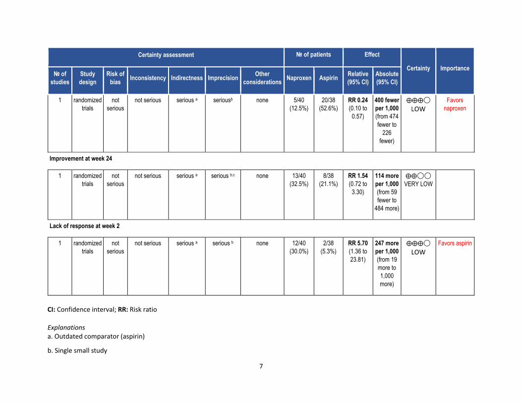
Summary: The literature search revealed one randomized controlled trial (RCT)[1] and 2 observational studies[2, 3] that addressed this PICO
question. The small, single center RCT provided indirect evidence by comparing scheduled naproxen (10mg/kg/day) to scheduled aspirin in 80
patients with JIA (65% oligoarticular). Aspirin as the comparator is not representative of current clinical practice. More subjects in the naproxen
arm (30% compared to 5.3%) discontinued medication by week 24 for lack of response (RR 5.7); however, less in the naproxen group (12.5%
compared to 52.6%) discontinued for side effects (RR 0.24). (Table 1). Definitions of improvement and lack of response were not described in
detail and appear inconsistent with measures used in contemporary trials. Absolute change in patient global, physician global and active joint
count at weeks 12 and 24 were reported, but no additional statistical analysis of these results was available (Table 2). There was no sub-analysis
of oligoarticular patients.
One observational study provided direct comparison of three NSAIDs (ibuprofen, naproxen, indomethacin) as monotherapy in JIA, reporting no
significant differences in “success” of NSAID trials (defined as attainment of inactive disease) among the three medications (52.6%, 54.1%, 54%,
respectively).[3] 52% of these NSAID trials were in patients with oligoarticular JIA- sub-analysis comparing the three medications was not
performed for oligoarticular JIA specifically. The overall success rate of NSAID monotherapy in patients with less than 5 affected joints was
59.5%. In a small prospective cohort of oligoarticular JIA patients, only 10.5% achieved clinical remission on NSAID (naproxen 20mg/kg/day or
ibuprofen 30 mg/kg/day) monotherapy.[2]
Quality of evidence across all critical outcomes: Very low
Table 1. Data from RCT - Naproxen compared to Aspirin for Oligoarticular JIA [1]
Certainty assessment № of patients Effect
Certainty Importance № of
studies
Study
design
Risk of
bias Inconsistency Indirectness Imprecision
Other
considerations Naproxen Aspirin
Relative
(95% CI)
Absolute
(95% CI)
Withdrawals due to side effects at week 24
7
Certainty assessment № of patients Effect
Certainty Importance № of
studies
Study
design
Risk of
bias Inconsistency Indirectness Imprecision
Other
considerations Naproxen Aspirin
Relative
(95% CI)
Absolute
(95% CI)
1 randomized
trials
not
serious
not serious serious a seriousb none 5/40
(12.5%)
20/38
(52.6%)
RR 0.24
(0.10 to
0.57)
400 fewer
per 1,000
(from 474
fewer to
226
fewer)
⨁⨁⨁◯
LOW
Favors
naproxen
Improvement at week 24
1 randomized
trials
not
serious
not serious serious a serious b,c none 13/40
(32.5%)
8/38
(21.1%)
RR 1.54
(0.72 to
3.30)
114 more
per 1,000
(from 59
fewer to
484 more)
⨁⨁◯◯
VERY LOW
Lack of response at week 2
1 randomized
trials
not
serious
not serious serious a serious b none 12/40
(30.0%)
2/38
(5.3%)
RR 5.70
(1.36 to
23.81)
247 more
per 1,000
(from 19
more to
1,000
more)
⨁⨁⨁◯
LOW
Favors aspirin
CI: Confidence interval; RR: Risk ratio
Explanations a. Outdated comparator (aspirin)
b. Single small study
8
c. Wide CI crosses significant effect and no-effect lines
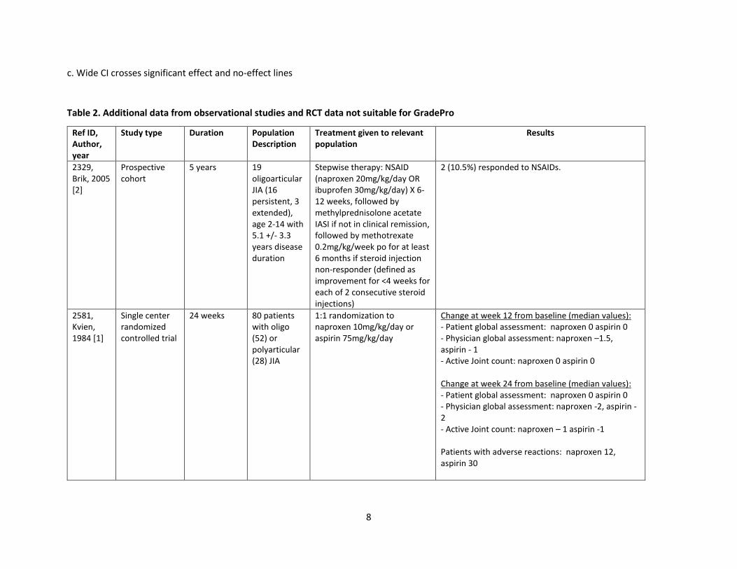
Table 2. Additional data from observational studies and RCT data not suitable for GradePro
Ref ID, Author, year
Study type Duration Population Description
Treatment given to relevant population
Results
2329, Brik, 2005 [2]
Prospective cohort
5 years 19 oligoarticular JIA (16 persistent, 3 extended), age 2-14 with 5.1 +/- 3.3 years disease duration
Stepwise therapy: NSAID (naproxen 20mg/kg/day OR ibuprofen 30mg/kg/day) X 6-12 weeks, followed by methylprednisolone acetate IASI if not in clinical remission, followed by methotrexate 0.2mg/kg/week po for at least 6 months if steroid injection non-responder (defined as improvement for <4 weeks for each of 2 consecutive steroid injections)
2 (10.5%) responded to NSAIDs.
2581, Kvien, 1984 [1]
Single center randomized controlled trial
24 weeks 80 patients with oligo (52) or polyarticular (28) JIA
1:1 randomization to naproxen 10mg/kg/day or aspirin 75mg/kg/day
Change at week 12 from baseline (median values): - Patient global assessment: naproxen 0 aspirin 0 - Physician global assessment: naproxen –1.5, aspirin - 1 - Active Joint count: naproxen 0 aspirin 0 Change at week 24 from baseline (median values): - Patient global assessment: naproxen 0 aspirin 0 - Physician global assessment: naproxen -2, aspirin - 2 - Active Joint count: naproxen – 1 aspirin -1 Patients with adverse reactions: naproxen 12, aspirin 30
9
Ref ID, Author, year
Study type Duration Population Description
Treatment given to relevant population
Results
* No additional statistical analysis was provided for these results.
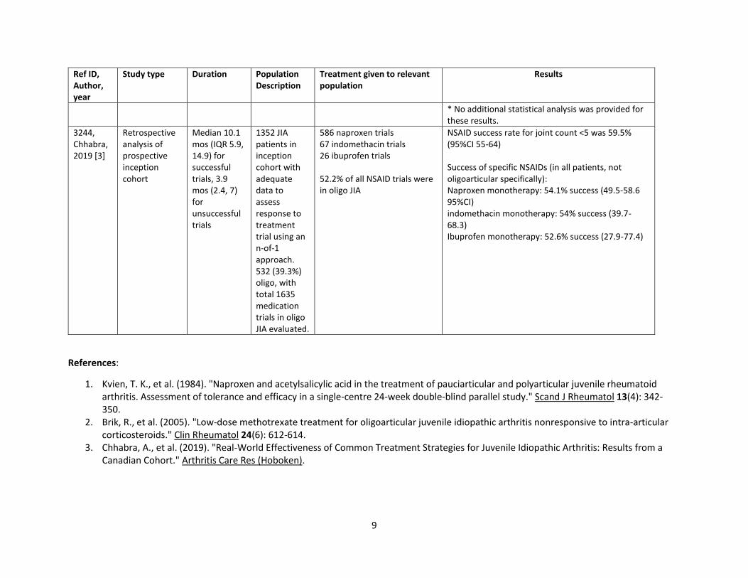
3244, Chhabra, 2019 [3]
Retrospective analysis of prospective inception cohort
Median 10.1 mos (IQR 5.9, 14.9) for successful trials, 3.9 mos (2.4, 7) for unsuccessful trials
1352 JIA patients in inception cohort with adequate data to assess response to treatment trial using an n-of-1 approach. 532 (39.3%) oligo, with total 1635 medication trials in oligo JIA evaluated.
586 naproxen trials 67 indomethacin trials 26 ibuprofen trials 52.2% of all NSAID trials were in oligo JIA
NSAID success rate for joint count <5 was 59.5% (95%CI 55-64) Success of specific NSAIDs (in all patients, not oligoarticular specifically): Naproxen monotherapy: 54.1% success (49.5-58.6 95%CI) indomethacin monotherapy: 54% success (39.7-68.3) Ibuprofen monotherapy: 52.6% success (27.9-77.4)
References:
1. Kvien, T. K., et al. (1984). "Naproxen and acetylsalicylic acid in the treatment of pauciarticular and polyarticular juvenile rheumatoid arthritis. Assessment of tolerance and efficacy in a single-centre 24-week double-blind parallel study." Scand J Rheumatol 13(4): 342-350.
2. Brik, R., et al. (2005). "Low-dose methotrexate treatment for oligoarticular juvenile idiopathic arthritis nonresponsive to intra-articular corticosteroids." Clin Rheumatol 24(6): 612-614.
3. Chhabra, A., et al. (2019). "Real-World Effectiveness of Common Treatment Strategies for Juvenile Idiopathic Arthritis: Results from a Canadian Cohort." Arthritis Care Res (Hoboken).
10
PICO 2: In children with oligoarticular JIA, should adding intraarticular glucocorticoids to initial therapy be recommended?
Summary: The literature search identified 14 observational studies that evaluated outcomes of intraarticular (IA) steroid injections in patients
with oligo JIA; most studies used triamcinolone hexacetonide (THA) or triamcinolone acetonide (TA).
Two studies compared the use of THA versus TA in patients with oligo JIA and both found significantly better outcomes with THA[1, 2].
Breit et al.[3] compared IA THA in early onset oligo JIA to late onset oligo JIA and showed a longer duration of improvement in early onset; for
early onset, the effect of IA steroids lasted a median of 121 weeks with late onset only lasting a median of 47 weeks. Another study[4] using IA
THA that did not specify length of follow-up did not find a significant correlation between age of onset and treatment outcome.
Lanni et al.[5] evaluated freedom from synovitis flare at 1, 2 and 3 years respectively following IA THA injections and divided into those having 1
joint injected, 2 joints injected or 3 or more joints injected. At one year 70% of those with 1 joint injected remained free of synovitis flare, 61% at
two years and 37% at three. The patients who had 2 or 3 or more joints injected had lower rates of freedom from synovitis flare, with lasting
results decreased to 45/32/22% for 2 joints at years 1, 2 and 3 and 44/30/19% for those with 3 or more injected joints. Another study[6] that
used both IA THA and TA injections reported that 41% of patients with persistent oligo JIA remained in remission (on and off medication) at a
mean follow-up of 4 years.
Five studies looked at sustained remission at 6 months and all showed favorable and lasting benefit of IA steroid injections: one study[7] showed
69% lasting remission with IA THA, while another study[8] using IA THA reported remission in 81.6% of injected joints. A study[9] using IA TA
found 65% overall but 81% for oligo JIA and only 59% for the other JIA types. The remaining study[10] used IA TA and showed 70% lasting
remission at 6 months. One study[11] used IA THA for large joints and IA methylprednisolone for small or difficult to access joints. This study
reported a remission rate of 59% for patients with oligo JIA.
Two studies evaluated different aspects of growth and development. Padeh et al.[12] conducted a retrospective cohort study comparing rates of
growth retardation in patients with persistent oligo JIA who received either intraarticular TA (group I) or intraarticular TA plus DMARDs (group II;
75% of patients in this group received methotrexate) during a mean follow-up of 6 years. In group I, 30.6% had any growth retardation and 6.5%
had severe growth retardation, while in group II, 44.4% had any growth retardation and 21.2% had severe growth retardation. However, a
retrospective chart review comparing patients who did and did not receive IA THA showed a significant decrease in leg length discrepancies with
the use of IA THA [13].
A prospective cohort study by Brik et al.[14] used IA methylprednisolone acetate and reported that 64% (11/17 patients) did not respond to this
treatment. Nine of the non-responders were treated with low-dose MTX for a median duration of 15±3.8 months. Except for one patient with an
extended disease course, all responded very well to treatment and went into remission after a median of 6.4±2.9 months, and none required
additional IA injections after initiation of MTX treatment.
11
Overall these studies support benefit for the use of IA THA or TA in early onset oligo JIA compared to other types of JIA and all studies showed
minimal side effects. However, the evidence is very low quality due to the lack of control groups in most studies, confounding due to
concomitant treatments (usually MTX) and because these studies do not specifically address the comparison of using IA steroids first to using
other treatment regimens first.
Quality of evidence across all critical outcomes: Very low
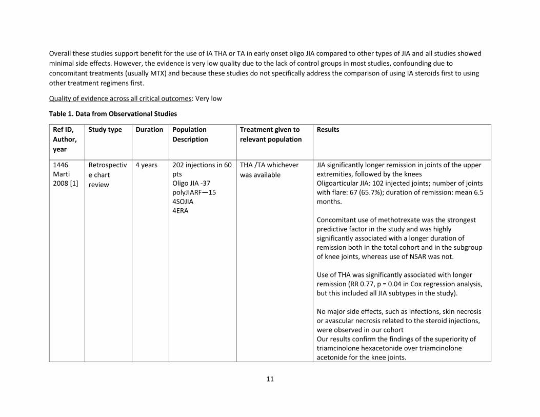
Table 1. Data from Observational Studies
Ref ID,
Author,
year
Study type Duration Population
Description
Treatment given to
relevant population
Results
1446 Marti 2008 [1]
Retrospectiv
e chart
review
4 years 202 injections in 60 pts Oligo JIA -37 polyJIARF—15 4SOJIA 4ERA
THA /TA whichever
was available
JIA significantly longer remission in joints of the upper extremities, followed by the knees Oligoarticular JIA: 102 injected joints; number of joints with flare: 67 (65.7%); duration of remission: mean 6.5 months. Concomitant use of methotrexate was the strongest predictive factor in the study and was highly significantly associated with a longer duration of remission both in the total cohort and in the subgroup of knee joints, whereas use of NSAR was not. Use of THA was significantly associated with longer remission (RR 0.77, p = 0.04 in Cox regression analysis, but this included all JIA subtypes in the study). No major side effects, such as infections, skin necrosis or avascular necrosis related to the steroid injections, were observed in our cohort Our results confirm the findings of the superiority of triamcinolone hexacetonide over triamcinolone acetonide for the knee joints.
12
Ref ID,
Author,
year
Study type Duration Population
Description
Treatment given to
relevant population
Results
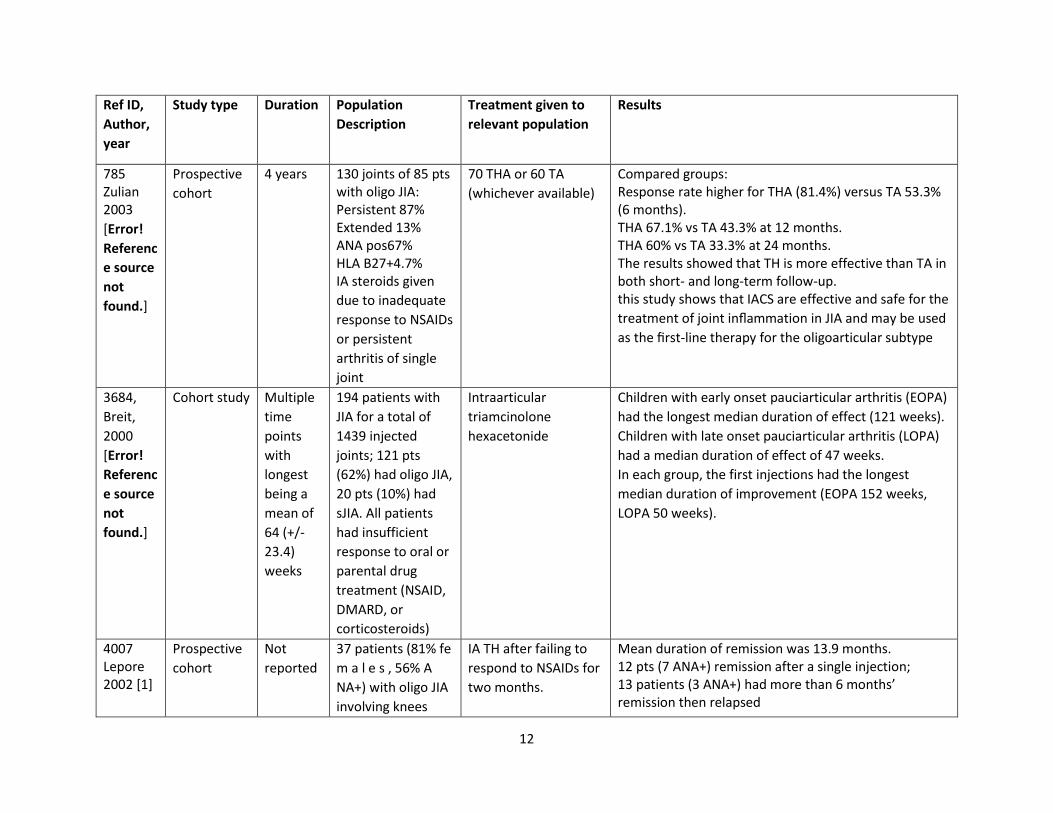
785 Zulian 2003
[Error!
Referenc
e source
not
found.]
Prospective
cohort
4 years 130 joints of 85 pts with oligo JIA: Persistent 87% Extended 13% ANA pos67% HLA B27+4.7% IA steroids given
due to inadequate
response to NSAIDs
or persistent
arthritis of single
joint
70 THA or 60 TA
(whichever available)
Compared groups: Response rate higher for THA (81.4%) versus TA 53.3% (6 months). THA 67.1% vs TA 43.3% at 12 months. THA 60% vs TA 33.3% at 24 months. The results showed that TH is more effective than TA in both short- and long-term follow-up. this study shows that IACS are effective and safe for the
treatment of joint inflammation in JIA and may be used
as the first-line therapy for the oligoarticular subtype
3684,
Breit,
2000
[Error!
Referenc
e source
not
found.]
Cohort study Multiple
time
points
with
longest
being a
mean of
64 (+/-
23.4)
weeks
194 patients with
JIA for a total of
1439 injected
joints; 121 pts
(62%) had oligo JIA,
20 pts (10%) had
sJIA. All patients
had insufficient
response to oral or
parental drug
treatment (NSAID,
DMARD, or
corticosteroids)
Intraarticular
triamcinolone
hexacetonide
Children with early onset pauciarticular arthritis (EOPA)
had the longest median duration of effect (121 weeks).
Children with late onset pauciarticular arthritis (LOPA)
had a median duration of effect of 47 weeks.
In each group, the first injections had the longest
median duration of improvement (EOPA 152 weeks,
LOPA 50 weeks).
4007 Lepore 2002 [1]
Prospective
cohort
Not
reported
37 patients (81% fe
m a l e s , 56% A
NA+) with oligo JIA
involving knees
IA TH after failing to
respond to NSAIDs for
two months.
Mean duration of remission was 13.9 months. 12 pts (7 ANA+) remission after a single injection; 13 patients (3 ANA+) had more than 6 months’ remission then relapsed
13
Ref ID,
Author,
year
Study type Duration Population
Description
Treatment given to
relevant population
Results
were treated. 18
pts were treated
within 6 months of
onset, 19 were
treated more than
6 months after
onset
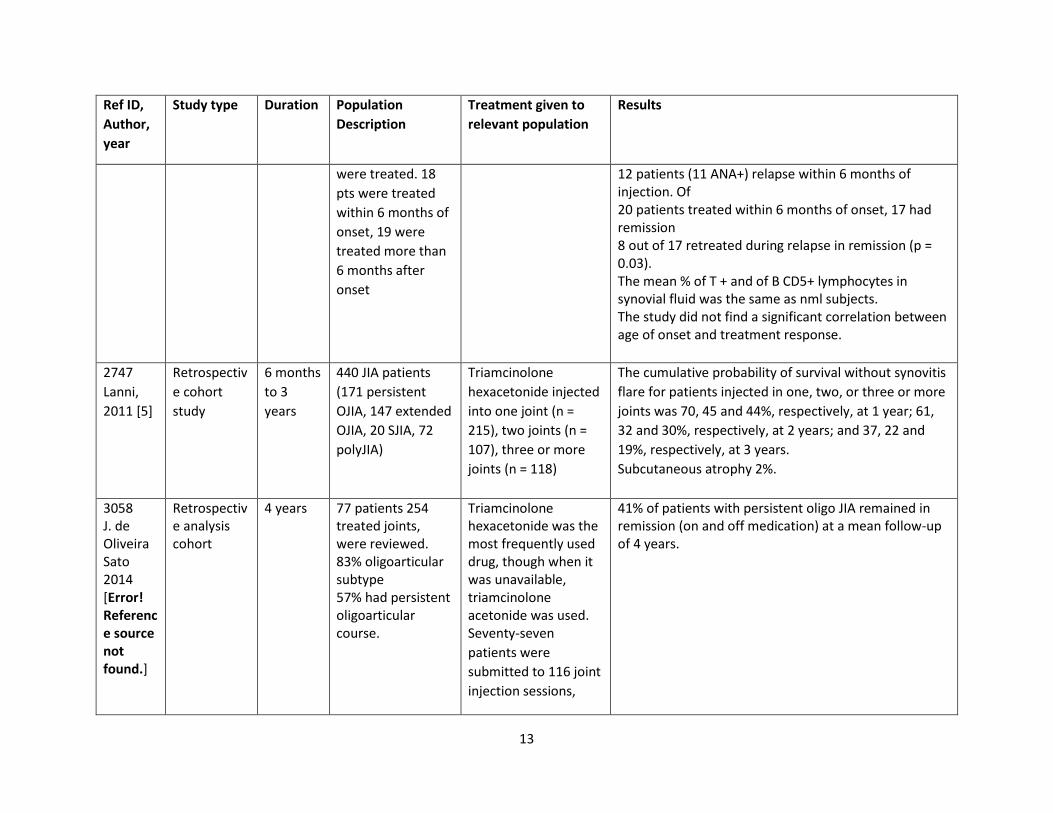
12 patients (11 ANA+) relapse within 6 months of injection. Of 20 patients treated within 6 months of onset, 17 had remission 8 out of 17 retreated during relapse in remission (p = 0.03). The mean % of T + and of B CD5+ lymphocytes in synovial fluid was the same as nml subjects. The study did not find a significant correlation between age of onset and treatment response.
2747
Lanni,
2011 [5]
Retrospectiv
e cohort
study
6 months
to 3
years
440 JIA patients
(171 persistent
OJIA, 147 extended
OJIA, 20 SJIA, 72
polyJIA)
Triamcinolone
hexacetonide injected
into one joint (n =
215), two joints (n =
107), three or more
joints (n = 118)
The cumulative probability of survival without synovitis
flare for patients injected in one, two, or three or more
joints was 70, 45 and 44%, respectively, at 1 year; 61,
32 and 30%, respectively, at 2 years; and 37, 22 and
19%, respectively, at 3 years.
Subcutaneous atrophy 2%.
3058 J. de Oliveira Sato 2014 [Error! Reference source not found.]
Retrospective analysis cohort
4 years 77 patients 254 treated joints, were reviewed. 83% oligoarticular subtype 57% had persistent oligoarticular course.
Triamcinolone hexacetonide was the most frequently used drug, though when it was unavailable, triamcinolone acetonide was used. Seventy-seven
patients were
submitted to 116 joint
injection sessions,
41% of patients with persistent oligo JIA remained in remission (on and off medication) at a mean follow-up of 4 years.
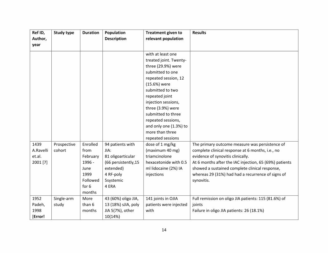
14
Ref ID,
Author,
year
Study type Duration Population
Description
Treatment given to
relevant population
Results
with at least one
treated joint. Twenty-
three (29.9%) were
submitted to one
repeated session, 12
(15.6%) were
submitted to two
repeated joint
injection sessions,
three (3.9%) were
submitted to three
repeated sessions,
and only one (1.3%) to
more than three
repeated sessions
1439
A.Ravelli
et.al.
2001 [7]
Prospective
cohort
Enrolled
from
February
1996 -
June
1999
Followed
for 6
months
94 patients with
JIA:
81 oligoarticular
(66 persistently,15
extended)
4 RF-poly
5systemic
4 ERA
dose of 1 mg/kg
(maximum 40 mg)
triamcinolone
hexacetonide with 0.5
ml lidocaine (2%) IA
injections
The primary outcome measure was persistence of
complete clinical response at 6 months, i.e., no
evidence of synovitis clinically.
At 6 months after the IAC injection, 65 (69%) patients
showed a sustained complete clinical response,
whereas 29 (31%) had had a recurrence of signs of
synovitis.
1952
Padeh,
1998
[Error!
Single-arm
study
More
than 6
months
43 (60%) oligo JIA,
13 (18%) sJIA, poly
JIA 5(7%), other
10(14%)
141 joints in OJIA
patients were injected
with
Full remission on oligo JIA patients: 115 (81.6%) of
joints
Failure in oligo JIA patients: 26 (18.1%)
15
Ref ID,
Author,
year
Study type Duration Population
Description
Treatment given to
relevant population
Results
Referenc
e source
not
found.]
triamcinolone
hexacetonide.
Discontinuation of oral medication in 32 (74.4%) oligo
JIA patients
No infection or other serious complications occurred in
any
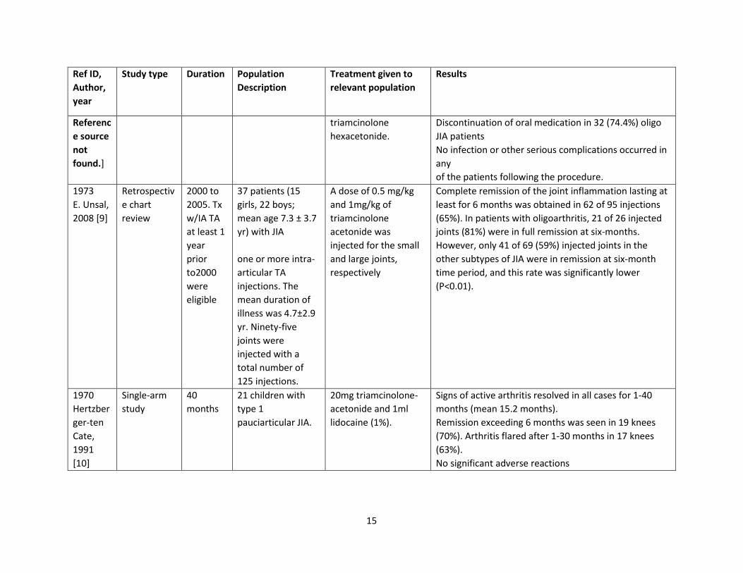
of the patients following the procedure.
1973
E. Unsal,
2008 [9]
Retrospectiv
e chart
review
2000 to
2005. Tx
w/IA TA
at least 1
year
prior
to2000
were
eligible
37 patients (15
girls, 22 boys;
mean age 7.3 ± 3.7
yr) with JIA
one or more intra-
articular TA
injections. The
mean duration of
illness was 4.7±2.9
yr. Ninety-five
joints were
injected with a
total number of
125 injections.
A dose of 0.5 mg/kg
and 1mg/kg of
triamcinolone
acetonide was
injected for the small
and large joints,
respectively
Complete remission of the joint inflammation lasting at
least for 6 months was obtained in 62 of 95 injections
(65%). In patients with oligoarthritis, 21 of 26 injected
joints (81%) were in full remission at six-months.
However, only 41 of 69 (59%) injected joints in the
other subtypes of JIA were in remission at six-month
time period, and this rate was significantly lower
(P<0.01).
1970
Hertzber
ger-ten
Cate,
1991
[10]
Single-arm
study
40
months
21 children with
type 1
pauciarticular JIA.
20mg triamcinolone-
acetonide and 1ml
lidocaine (1%).
Signs of active arthritis resolved in all cases for 1-40
months (mean 15.2 months).
Remission exceeding 6 months was seen in 19 knees
(70%). Arthritis flared after 1-30 months in 17 knees
(63%).
No significant adverse reactions
16
Ref ID,
Author,
year
Study type Duration Population
Description
Treatment given to
relevant population
Results
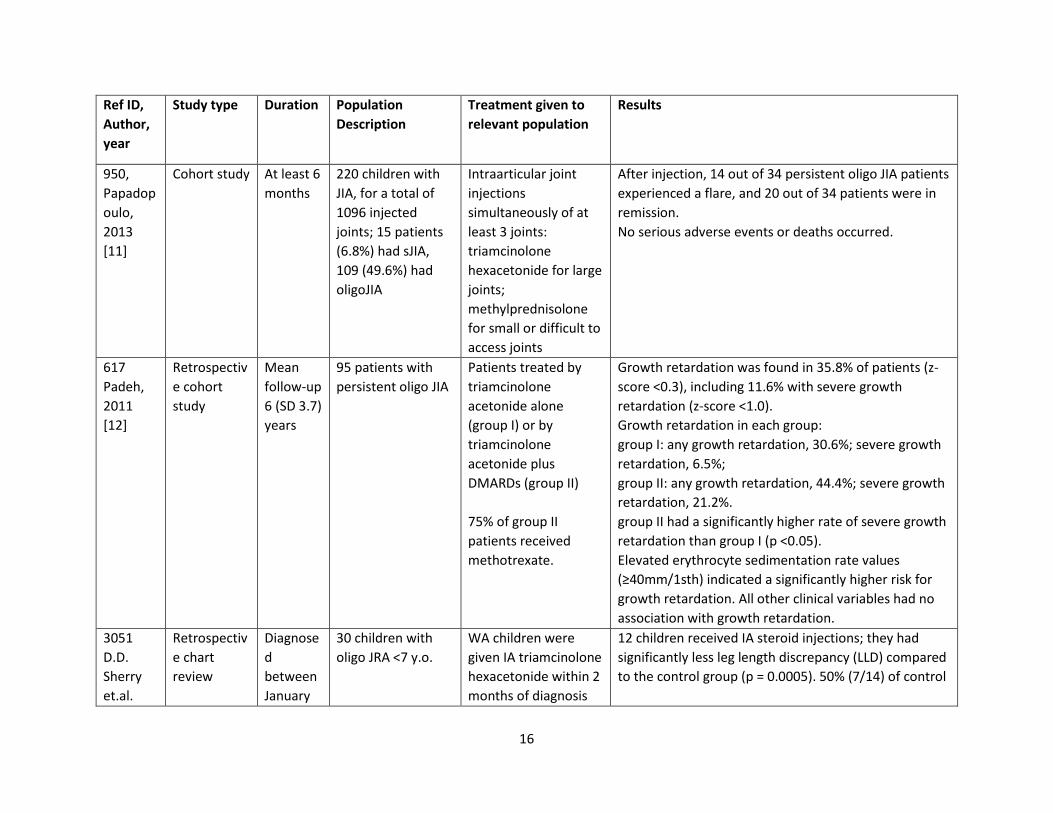
950,
Papadop
oulo,
2013
[11]
Cohort study At least 6
months
220 children with
JIA, for a total of
1096 injected
joints; 15 patients
(6.8%) had sJIA,
109 (49.6%) had
oligoJIA
Intraarticular joint
injections
simultaneously of at
least 3 joints:
triamcinolone
hexacetonide for large
joints;
methylprednisolone
for small or difficult to
access joints
After injection, 14 out of 34 persistent oligo JIA patients
experienced a flare, and 20 out of 34 patients were in
remission.
No serious adverse events or deaths occurred.
617
Padeh,
2011
[12]
Retrospectiv
e cohort
study
Mean
follow-up
6 (SD 3.7)
years
95 patients with
persistent oligo JIA
Patients treated by
triamcinolone
acetonide alone
(group I) or by
triamcinolone
acetonide plus
DMARDs (group II)
75% of group II
patients received
methotrexate.
Growth retardation was found in 35.8% of patients (z-
score <0.3), including 11.6% with severe growth
retardation (z-score <1.0).
Growth retardation in each group:
group I: any growth retardation, 30.6%; severe growth
retardation, 6.5%;
group II: any growth retardation, 44.4%; severe growth
retardation, 21.2%.
group II had a significantly higher rate of severe growth
retardation than group I (p <0.05).
Elevated erythrocyte sedimentation rate values
(≥40mm/1sth) indicated a significantly higher risk for
growth retardation. All other clinical variables had no
association with growth retardation.
3051
D.D.
Sherry
et.al.
Retrospectiv
e chart
review
Diagnose
d
between
January
30 children with
oligo JRA <7 y.o.
WA children were
given IA triamcinolone
hexacetonide within 2
months of diagnosis
12 children received IA steroid injections; they had
significantly less leg length discrepancy (LLD) compared
to the control group (p = 0.0005). 50% (7/14) of control
17
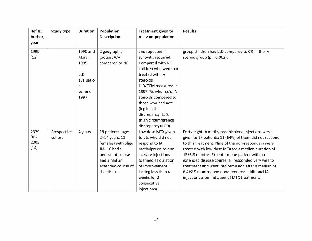
Ref ID,
Author,
year
Study type Duration Population
Description
Treatment given to
relevant population
Results
1999
[13]
1990 and
March
1995
LLD
evaluatio
n
summer
1997
2 geographic
groups: WA
compared to NC
and repeated if
synovitis recurred.
Compared with NC
children who were not
treated with IA
steroids
LLD/TCM measured in
1997 Pts who rec’d IA
steroids compared to
those who had not:
(leg length
discrepancy=LLD,
thigh circumference
discrepancy=TCD)
group children had LLD compared to 0% in the IA
steroid group (p = 0.002).
2329 Brik 2005 [14]
Prospective
cohort
4 years 19 patients (age:
2–14 years, 18
females) with oligo
JIA, 16 had a
persistent course
and 3 had an
extended course of
the disease
Low dose MTX given
to pts who did not
respond to IA
methylprednisolone
acetate injections
(defined as duration
of improvement
lasting less than 4
weeks for 2
consecutive
injections)
Forty-eight IA methylprednisolone injections were
given to 17 patients; 11 (64%) of them did not respond
to this treatment. Nine of the non-responders were
treated with low-dose MTX for a median duration of
15±3.8 months. Except for one patient with an
extended disease course, all responded very well to
treatment and went into remission after a median of
6.4±2.9 months, and none required additional IA
injections after initiation of MTX treatment.
18
References:
1. Marti, P., Molinari, L., Bolt, I. B., Seger, R., & Saurenmann, R. K. (2008). Factors influencing the efficacy of intra-articular steroid injections
in patients with juvenile idiopathic arthritis. Eur J Pediatr, 167(4), 425-430. doi:10.1007/s00431-007-0525-9
2. Zulian, F., Martini, G., Gobber, D., Agosto, C., Gigante, C., & Zacchello, F. (2003). Comparison of intra-articular triamcinolone
hexacetonide and triamcinolone acetonide in oligoarticular juvenile idiopathic arthritis. Rheumatology (Oxford), 42(10), 1254-1259.
doi:10.1093/rheumatology/keg358
3. Breit, W., Frosch, M., Meyer, U., Heinecke, A., & Ganser, G. (2000). A subgroup-specific evaluation of the efficacy of intraarticular
triamcinolone hexacetonide in juvenile chronic arthritis. J Rheumatol, 27(11), 2696-2702.
4. Lepore, L., Del Santo, M., Malorgio, C., Presani, G., Perticarari, S., Prodan, M., . . . Tommasini, A. (2002). Treatment of juvenile idiopathic
arthritis with intra-articular triamcinolone hexacetonide: evaluation of clinical effectiveness correlated with circulating ANA and T
gamma/delta + and B CD5+ lymphocyte populations of synovial fluid. Clin Exp Rheumatol, 20(5), 719-722.
5. Lanni, S., Bertamino, M., Consolaro, A., Pistorio, A., Magni-Manzoni, S., Galasso, R., . . . Ravelli, A. (2011). Outcome and predicting factors
of single and multiple intra-articular corticosteroid injections in children with juvenile idiopathic arthritis. Rheumatology (Oxford), 50(9),
1627-1634. doi:10.1093/rheumatology/ker165
6. de Oliveira Sato, J., Albuquerque Pedrosa Fernandes, T., Bicalho do Nascimento, C., Corrente, J. E., & Saad-Magalhaes, C. (2014).
Probability of remission of juvenile idiopathic arthritis following treatment with steroid joint injection. Clin Exp Rheumatol, 32(2), 291-
296.
7. Ravelli, A., Manzoni, S. M., Viola, S., Pistorio, A., Ruperto, N., & Martini, A. (2001). Factors affecting the efficacy of intraarticular
corticosteroid injection of knees in juvenile idiopathic arthritis. J Rheumatol, 28(9), 2100-2102.
8. Padeh, S., & Passwell, J. H. (1998). Intraarticular corticosteroid injection in the management of children with chronic arthritis. Arthritis
Rheum, 41(7), 1210-1214.
9. N., Günay, T., . . . Unsal, E. (2011). Performance of tuberculin skin test and interferon gamma assay for the diagnosis of latent
tuberculosis infection in juvenile idiopathic arthritis. Clin Rheumatol, 30(9), 1189-1193.
10. Hertzberger-ten Cate, R., de Vries-van der Vlugt, B. C., van Suijlekom-Smit, L. W., & Cats, A. (1991). Intra-articular steroids in
pauciarticular juvenile chronic arthritis, type 1. Eur J Pediatr, 150(3), 170-172. doi:10.1007/bf01963559
19
11. Papadopoulou, C., Kostik, M., Gonzalez-Fernandez, M. I., Bohm, M., Nieto-Gonzalez, J. C., Pistorio, A., . . . Ravelli, A. (2013). Delineating
the role of multiple intraarticular corticosteroid injections in the management of juvenile idiopathic arthritis in the biologic era. Arthritis
Care Res (Hoboken), 65(7), 1112-1120.
12. Padeh, S., Pinhas-Hamiel, O., Zimmermann-Sloutskis, D., & Berkun, Y. (2011). Children with oligoarticular juvenile idiopathic arthritis are
at considerable risk for growth retardation. J Pediatr, 159(5), 832-837.e831-832.Camlar, S. A., Makay, B., Appak, O., Appak, Y. C., Esen,
13. Sherry, D. D., Stein, L. D., Reed, A. M., Schanberg, L. E., & Kredich, D. W. (1999). Prevention of leg length discrepancy in young children
with pauciarticular juvenile rheumatoid arthritis by treatment with intraarticular steroids. Arthritis Rheum, 42(11), 2330-2334.
14. Brik, R., Gepstein, V., & Berkovitz, D. (2005). Low-dose methotrexate treatment for oligoarticular juvenile idiopathic arthritis
nonresponsive to intra-articular corticosteroids. Clin Rheumatol, 24(6), 612-614. doi:10.1007/s10067-005-1116-7
PICO 3: In children with oligoarticular JIA, should adding oral steroids to initial therapy be recommended?
Summary: The literature search identified one retrospective, cross-sectional study that indirectly addressed this PICO question.1 This study
showed that the development of adrenal insufficiency (AI) in JIA patients was a rare occurrence when low dose glucocorticoids (<7.5mg
prednisolone) was used. Signs of AI occurred in only 4/61 patients. Those who had AI all had oligo-articular JIA, were female, and were younger
at the time of their JIA diagnosis and at the time of this study. All patients were treated for at least 6 months with low dose steroids and were off
steroids for 3 months when AI was assessed. There was no statistical difference in the development of AI in relationship to steroid duration or
period of cessation.
Quality of evidence across all critical outcomes: Very Low
Table 1. Data from Observational Study
Ref ID,
Author,
year
Study type Duration Population
Description
Treatment given
to relevant
population
Results
87,
Sadeghi et
al.
2019
Cross
sectional
study
2 year study
period 2014-
2016
36 patients
with JIA
25 females
(69.4%)
Study designed
to assess
adrenal
The AI diagnosis was made if the first level of plasma
cortisol was less than 3 micrograms/dL and the second
level was less than 20 micrograms/dL.
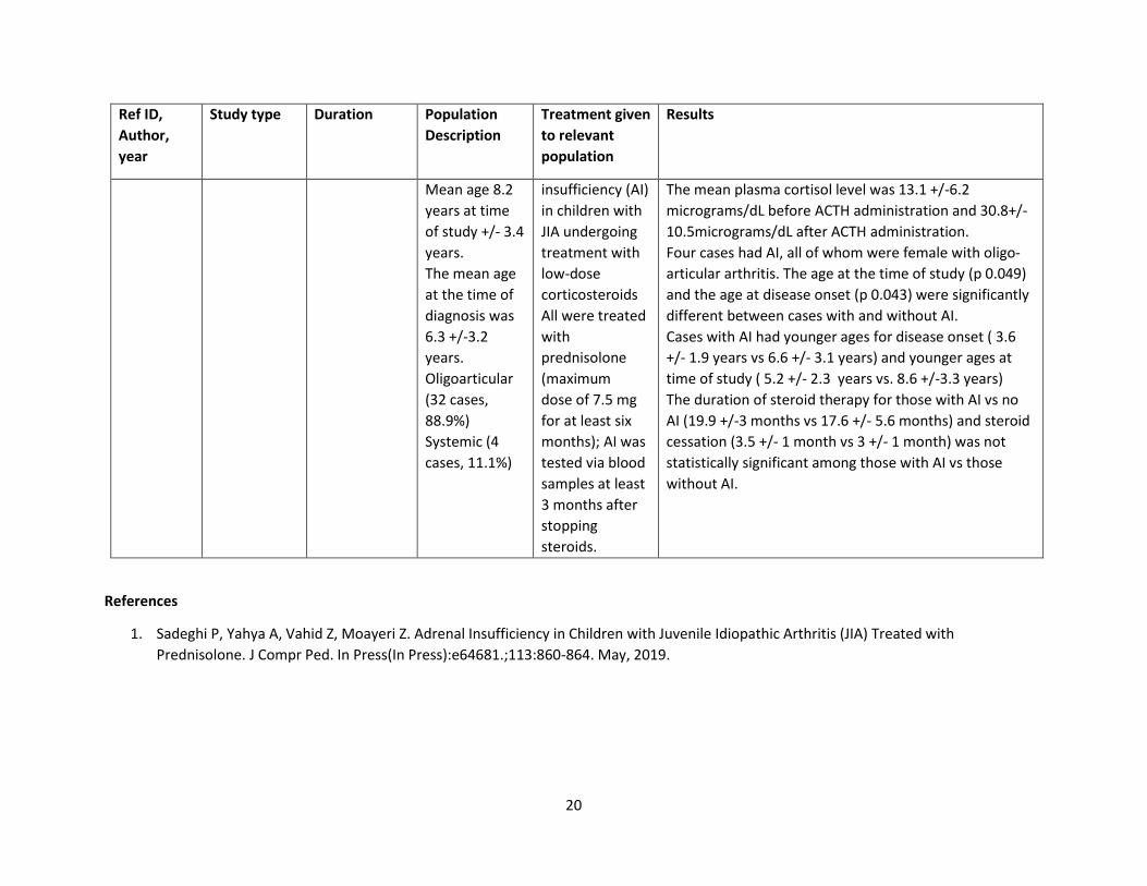
20
Ref ID,
Author,
year
Study type Duration Population
Description
Treatment given
to relevant
population
Results
Mean age 8.2
years at time
of study +/- 3.4
years.
The mean age
at the time of
diagnosis was
6.3 +/-3.2
years.
Oligoarticular
(32 cases,
88.9%)
Systemic (4
cases, 11.1%)
insufficiency (AI)
in children with
JIA undergoing
treatment with
low-dose
corticosteroids
All were treated
with
prednisolone
(maximum
dose of 7.5 mg
for at least six
months); AI was
tested via blood
samples at least
3 months after
stopping
steroids.
The mean plasma cortisol level was 13.1 +/-6.2
micrograms/dL before ACTH administration and 30.8+/-
10.5micrograms/dL after ACTH administration.
Four cases had AI, all of whom were female with oligo-
articular arthritis. The age at the time of study (p 0.049)
and the age at disease onset (p 0.043) were significantly
different between cases with and without AI.
Cases with AI had younger ages for disease onset ( 3.6
+/- 1.9 years vs 6.6 +/- 3.1 years) and younger ages at
time of study ( 5.2 +/- 2.3 years vs. 8.6 +/-3.3 years)
The duration of steroid therapy for those with AI vs no
AI (19.9 +/-3 months vs 17.6 +/- 5.6 months) and steroid
cessation (3.5 +/- 1 month vs 3 +/- 1 month) was not
statistically significant among those with AI vs those
without AI.
References
1. Sadeghi P, Yahya A, Vahid Z, Moayeri Z. Adrenal Insufficiency in Children with Juvenile Idiopathic Arthritis (JIA) Treated with
Prednisolone. J Compr Ped. In Press(In Press):e64681.;113:860-864. May, 2019.
21
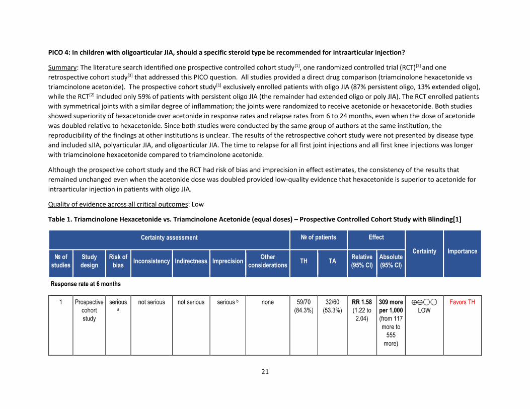
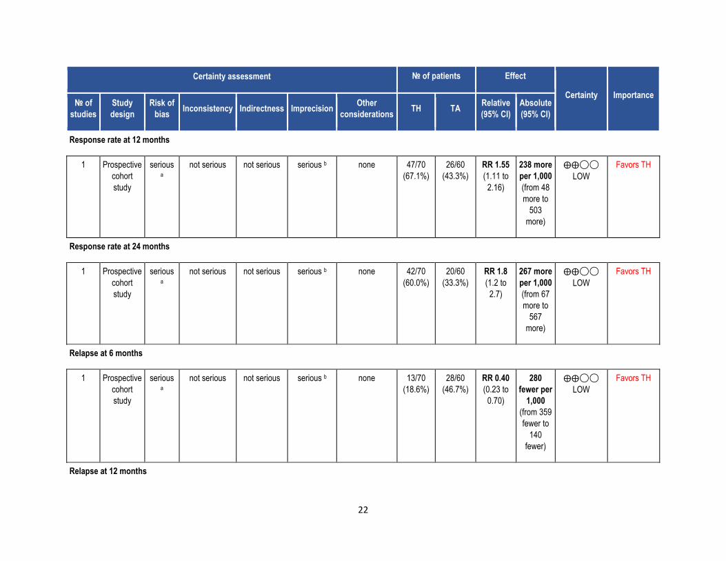
PICO 4: In children with oligoarticular JIA, should a specific steroid type be recommended for intraarticular injection?
Summary: The literature search identified one prospective controlled cohort study[1], one randomized controlled trial (RCT)[2] and one
retrospective cohort study[3] that addressed this PICO question. All studies provided a direct drug comparison (triamcinolone hexacetonide vs
triamcinolone acetonide). The prospective cohort study[1] exclusively enrolled patients with oligo JIA (87% persistent oligo, 13% extended oligo),
while the RCT[2] included only 59% of patients with persistent oligo JIA (the remainder had extended oligo or poly JIA). The RCT enrolled patients
with symmetrical joints with a similar degree of inflammation; the joints were randomized to receive acetonide or hexacetonide. Both studies
showed superiority of hexacetonide over acetonide in response rates and relapse rates from 6 to 24 months, even when the dose of acetonide
was doubled relative to hexacetonide. Since both studies were conducted by the same group of authors at the same institution, the
reproducibility of the findings at other institutions is unclear. The results of the retrospective cohort study were not presented by disease type
and included sJIA, polyarticular JIA, and oligoarticular JIA. The time to relapse for all first joint injections and all first knee injections was longer
with triamcinolone hexacetonide compared to triamcinolone acetonide.
Although the prospective cohort study and the RCT had risk of bias and imprecision in effect estimates, the consistency of the results that
remained unchanged even when the acetonide dose was doubled provided low-quality evidence that hexacetonide is superior to acetonide for
intraarticular injection in patients with oligo JIA.
Quality of evidence across all critical outcomes: Low
Table 1. Triamcinolone Hexacetonide vs. Triamcinolone Acetonide (equal doses) – Prospective Controlled Cohort Study with Blinding[1]
Certainty assessment № of patients Effect
Certainty Importance № of
studies
Study
design
Risk of
bias Inconsistency Indirectness Imprecision
Other
considerations TH TA
Relative
(95% CI)
Absolute
(95% CI)
Response rate at 6 months
1 Prospective
cohort
study
serious a
not serious not serious serious b none 59/70
(84.3%)
32/60
(53.3%)
RR 1.58
(1.22 to
2.04)
309 more
per 1,000
(from 117
more to
555
more)
⨁⨁◯◯
LOW
Favors TH
22
Certainty assessment № of patients Effect
Certainty Importance № of
studies
Study
design
Risk of
bias Inconsistency Indirectness Imprecision
Other
considerations TH TA
Relative
(95% CI)
Absolute
(95% CI)
Response rate at 12 months
1 Prospective
cohort
study
serious a
not serious not serious serious b none 47/70
(67.1%)
26/60
(43.3%)
RR 1.55
(1.11 to
2.16)
238 more
per 1,000
(from 48
more to
503
more)
⨁⨁◯◯
LOW
Favors TH
Response rate at 24 months
1 Prospective
cohort
study
serious a
not serious not serious serious b none 42/70
(60.0%)
20/60
(33.3%)
RR 1.8
(1.2 to
2.7)
267 more
per 1,000
(from 67
more to
567
more)
⨁⨁◯◯
LOW
Favors TH
Relapse at 6 months
1 Prospective
cohort
study
serious a
not serious not serious serious b none 13/70
(18.6%)
28/60
(46.7%)
RR 0.40
(0.23 to
0.70)
280
fewer per
1,000
(from 359
fewer to
140
fewer)
⨁⨁◯◯
LOW
Favors TH
Relapse at 12 months
23
Certainty assessment № of patients Effect
Certainty Importance № of
studies
Study
design
Risk of
bias Inconsistency Indirectness Imprecision
Other
considerations TH TA
Relative
(95% CI)
Absolute
(95% CI)
1 Prospective
cohort
study
serious a
not serious not serious serious b none 23/70
(32.9%)
34/60
(56.7%)
RR 0.58
(0.39 to
0.87)
238
fewer per
1,000
(from 346
fewer to
74 fewer)
⨁⨁◯◯
LOW
Favors TH
Relapse at 24 months
1 Prospective
cohort
study
serious a
not serious not serious serious b none 28/70
(40.0%)
40/60
(66.7%)
RR 0.60
(0.43 to
0.84)
267
fewer per
1,000
(from 380
fewer to
107
fewer)
⨁⨁◯◯
LOW
Favors TH
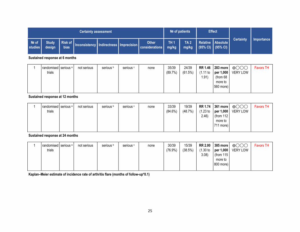
Kaplan–Meier estimate of incidence rate of arthritis flare (months of follow-up*0.1)
1 Prospective
cohort
study
serious a
not serious not serious serious b none 14/1000
(1.4%)
42/1000
(4.2%)
RR 0.33
(0.18 to
0.61)
28 fewer
per 1,000
(from 34
fewer to
16 fewer)
⨁⨁◯◯
LOW
Favors TH
Survival rate at 24 months
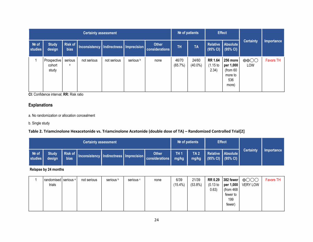
24
Certainty assessment № of patients Effect
Certainty Importance № of
studies
Study
design
Risk of
bias Inconsistency Indirectness Imprecision
Other
considerations TH TA
Relative
(95% CI)
Absolute
(95% CI)
1 Prospective
cohort
study
serious a
not serious not serious serious b none 46/70
(65.7%)
24/60
(40.0%)
RR 1.64
(1.15 to
2.34)
256 more
per 1,000
(from 60
more to
536
more)
⨁⨁◯◯
LOW
Favors TH
CI: Confidence interval; RR: Risk ratio
Explanations
a. No randomization or allocation concealment
b. Single study
Table 2. Triamcinolone Hexacetonide vs. Triamcinolone Acetonide (double dose of TA) – Randomized Controlled Trial[2]
Certainty assessment № of patients Effect
Certainty Importance № of
studies
Study
design
Risk of
bias Inconsistency Indirectness Imprecision
Other
considerations
TH 1
mg/kg
TA 2
mg/kg
Relative
(95% CI)
Absolute
(95% CI)
Relapse by 24 months
1 randomised
trials
serious a not serious serious b serious c none 6/39
(15.4%)
21/39
(53.8%)
RR 0.29
(0.13 to
0.63)
382 fewer
per 1,000
(from 468
fewer to
199
fewer)
⨁◯◯◯
VERY LOW
Favors TH
25
Certainty assessment № of patients Effect
Certainty Importance № of
studies
Study
design
Risk of
bias Inconsistency Indirectness Imprecision
Other
considerations
TH 1
mg/kg
TA 2
mg/kg
Relative
(95% CI)
Absolute
(95% CI)
Sustained response at 6 months
1 randomised
trials
serious a not serious serious b serious c none 35/39
(89.7%)
24/39
(61.5%)
RR 1.46
(1.11 to
1.91)
283 more
per 1,000
(from 68
more to
560 more)
⨁◯◯◯
VERY LOW
Favors TH
Sustained response at 12 months
1 randomised
trials
serious a not serious serious b serious c none 33/39
(84.6%)
19/39
(48.7%)
RR 1.74
(1.23 to
2.46)
361 more
per 1,000
(from 112
more to
711 more)
⨁◯◯◯
VERY LOW
Favors TH
Sustained response at 24 months
1 randomised
trials
serious a not serious serious b serious c none 30/39
(76.9%)
15/39
(38.5%)
RR 2.00
(1.30 to
3.08)
385 more
per 1,000
(from 115
more to
800 more)
⨁◯◯◯
VERY LOW
Favors TH
Kaplan–Meier estimate of incidence rate of arthritis flare (months of follow-up*0.1)
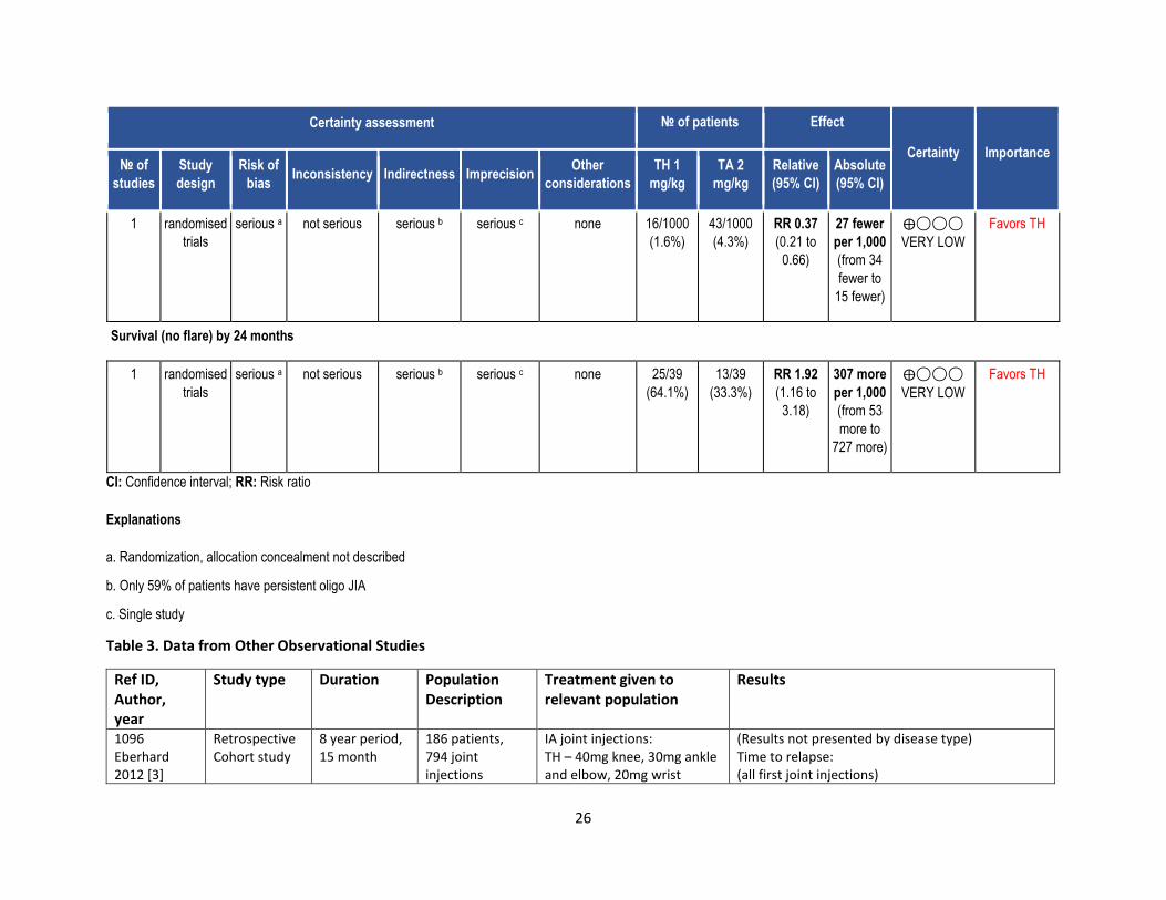
26
Certainty assessment № of patients Effect
Certainty Importance № of
studies
Study
design
Risk of
bias Inconsistency Indirectness Imprecision
Other
considerations
TH 1
mg/kg
TA 2
mg/kg
Relative
(95% CI)
Absolute
(95% CI)
1 randomised
trials
serious a not serious serious b serious c none 16/1000
(1.6%)
43/1000
(4.3%)
RR 0.37
(0.21 to
0.66)
27 fewer
per 1,000
(from 34
fewer to
15 fewer)
⨁◯◯◯
VERY LOW
Favors TH
Survival (no flare) by 24 months
1 randomised
trials
serious a not serious serious b serious c none 25/39
(64.1%)
13/39
(33.3%)
RR 1.92
(1.16 to
3.18)
307 more
per 1,000
(from 53
more to
727 more)
⨁◯◯◯
VERY LOW
Favors TH
CI: Confidence interval; RR: Risk ratio
Explanations
a. Randomization, allocation concealment not described
b. Only 59% of patients have persistent oligo JIA
c. Single study
Table 3. Data from Other Observational Studies
Ref ID, Author, year
Study type Duration Population Description
Treatment given to relevant population
Results
1096 Eberhard 2012 [3]
Retrospective Cohort study
8 year period, 15 month
186 patients, 794 joint injections
IA joint injections: TH – 40mg knee, 30mg ankle and elbow, 20mg wrist
(Results not presented by disease type) Time to relapse: (all first joint injections)
27
minimum follow up
(15 sJIA, 49 poly, 179 oligo)
TA – 80mg knee, 60mg ankle and elbow, 40mg wrist
TH (n=111) = 10.47 + 0.42 months TA (n=70) = 8.66 + 0.59 months, p<0.001 (first knee injection only) TH (n=89) = 11.04 + 0.44 months TA (n=56) = 8.99 + 0.65 months, p <0.001
References:
1. Zulian, F., et al. Comparison of intra-articular triamcinolone hexacetonide and triamcinolone acetonide in oligoarticular juvenile idiopathic arthritis. Rheumatology 2003;42:1254–1259.
2. Zulian, F. et al. Triamcinolone acetonide and hexacetonide intra-articular treatment of symmetrical joints in juvenile idiopathic arthritis: a double-blind trial. Rheumatology 2004 Oct;43(10):1288-91.
3. Eberhard, B. A., et al. (2011). A dose schedule for intraarticular steroids in juvenile arthritis. J Rheumatol 2011;39(2): 374-376.
28
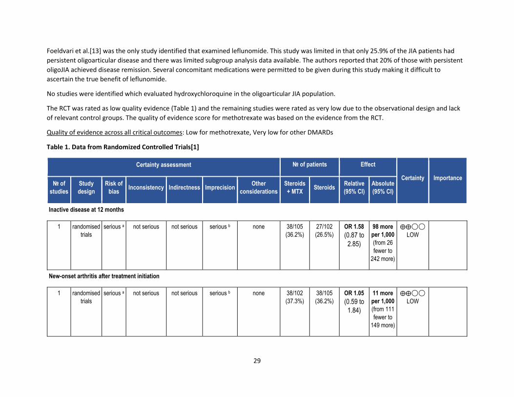
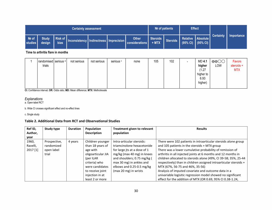
PICO 5. In children with oligoarticular JIA, should DMARD therapies be recommended, and should there be any preferred order of treatment:
methotrexatate (subcutaneous or oral), leflunomide, sulfasalazine, and/or hydroxychloroquine?
Summary: Literature searches identified one randomized controlled trial (RCT)[1] and 12 observational studies addressing this PICO question.
There were 4 prospective cohorts [3, 6, 11, 12] and 8 retrospective cohorts [2, 4, 5, 7, 8, 9, 10, 13].
Ravelli et al.[1] prospectively randomized children at 10 hospital units in Italy with oligoarticular JIA to receive either intraarticular corticosteroid
joint injections alone or injections in combination with oral methotrexate (MTX) therapy. The primary outcome was remission of disease at 12
months. Multivariable analysis (that accounted for treatment effect and ESR) showed that the addition of MTX was protective against arthritis
flare (OR 0.53, 95% CI 0.27-1.01; p=0.05). In the intention to treat analysis for remission of all joints at 12 months, there was no significant
difference (p=0.48) between those that received steroid injections alone compared to those who also received MTX in addition to injections.
Time to arthritis flare was found to be longer for those who had the addition of MTX (median 10.1 months, 95% CI 7.6 to > 16) vs those with
injections alone (median 6 months, 95% CI 4.6-8.2).
Collectively, 8 cohort studies [2, 3, 4, 5, 6, 7, 8, 9] evaluated MTX in the setting of JIA. Bava et al.[2] found that those with oligoarticular JIA had a
higher proportion of patients achieve inactive disease on MTX compared to their systemic and ERA counterparts. In total, 54.8% of oligoJIA
patients were able to achieve inactive disease. van Dijkhuizen et al.[3] did not study efficacy but looked at MTX tolerance. They found that 54.5%
of oligoJIA patients tolerated MTX, whether oral or subcutaneous. No subanalysis was done in this population to ascertain which formulation (SC
or PO) was more tolerated in those with oligo JIA. Albarouni et al.[4] reported that 70% of oligo JIA patients achieved an ACR Pedi 70 response to
MTX at 12 months. Klein et al.[5] reported no significant difference between oral and subcutaneous administration of methotrexate on the ACR
Pedi 50 and 70 scores at 6 and 12 months. Franova et al.[6] aimed to ascertain the benefit of adding MTX to achieve an inactive disease state,
however, limited oligo JIA data makes this study less relevant to the PICO question. Two other cohort studies[7, 8] also had a small percentage of
oligo JIA patients but reported remission rates of 54% to 58% following MTX treatment. Finally, Padeh et al.[9] conducted a retrospective cohort
study comparing rates of growth retardation in patients with persistent oligo JIA who received either intraarticular triamcinolone acetonide (TA)
(group I) or intraarticular TA plus DMARDs (group II; 75% of patients in this group received methotrexate) during a mean follow-up of 6 years. In
group I, 30.6% had any growth retardation and 6.5% had severe growth retardation, while in group II, 44.4% had any growth retardation and
21.2% had severe growth retardation.
Three cohort studies [10, 11, 12] focused on sulfasalazine treatment. Chen et al. [10] reported that 100% of oligoarticular JRA patients
demonstrated clinical improvement and 90.9% achieved clinical remission by a mean of 4.7 months on sulfasalazine. Varbanova et al. [11]
concluded that children with pauciarticular JRA showed the highest prevalence of responders (90%) compared to the systemic and polyarticular
subgroups. In the study by Imundo et al.[12], most of the pauciarticular JRA patients showed clinical improvement (77%) and a small number
achieved remission (26%). When examining ANA positive female patients in this subgroup, 88% showed significant improvement and 44%
achieved remission.
29
Foeldvari et al.[13] was the only study identified that examined leflunomide. This study was limited in that only 25.9% of the JIA patients had
persistent oligoarticular disease and there was limited subgroup analysis data available. The authors reported that 20% of those with persistent
oligoJIA achieved disease remission. Several concomitant medications were permitted to be given during this study making it difficult to
ascertain the true benefit of leflunomide.
No studies were identified which evaluated hydroxychloroquine in the oligoarticular JIA population.
The RCT was rated as low quality evidence (Table 1) and the remaining studies were rated as very low due to the observational design and lack
of relevant control groups. The quality of evidence score for methotrexate was based on the evidence from the RCT.
Quality of evidence across all critical outcomes: Low for methotrexate, Very low for other DMARDs
Table 1. Data from Randomized Controlled Trials[1]
Certainty assessment № of patients Effect
Certainty Importance № of
studies
Study
design
Risk of
bias Inconsistency Indirectness Imprecision
Other
considerations
Steroids
+ MTX Steroids
Relative
(95% CI)
Absolute
(95% CI)
Inactive disease at 12 months
1 randomised
trials
serious a not serious not serious serious b none 38/105
(36.2%)
27/102
(26.5%)
OR 1.58
(0.87 to
2.85)
98 more
per 1,000
(from 26
fewer to
242 more)
⨁⨁◯◯
LOW
New-onset arthritis after treatment initiation
1 randomised
trials
serious a not serious not serious serious b none 38/102
(37.3%)
38/105
(36.2%)
OR 1.05
(0.59 to
1.84)
11 more
per 1,000
(from 111
fewer to
149 more)
⨁⨁◯◯
LOW
30
Certainty assessment № of patients Effect
Certainty Importance № of
studies
Study
design
Risk of
bias Inconsistency Indirectness Imprecision
Other
considerations
Steroids
+ MTX Steroids
Relative
(95% CI)
Absolute
(95% CI)
Time to arthritis flare in months
1 randomised
trials
serious a not serious not serious serious c none 105 102 - MD 4.1
higher
(1.27
higher to
6.93
higher)
⨁⨁◯◯
LOW
Favors
steroids +
MTX
CI: Confidence interval; OR: Odds ratio; MD: Mean difference; MTX: Methotrexate
Explanations a. Open-label RCT
b. Wide CI crosses significant effect and no-effect lines
c. Single study
Table 2. Additional Data from RCT and Observational Studies
Ref ID, Author, year
Study type Duration Population Description
Treatment given to relevant population
Results
1960, Ravelli, 2017 [1]
Prospective, randomized open label trial
4 years Children younger than 18 years of age with oligoarticular JIA (per ILAR criteria) who were candidates to receive joint injection in at least 2 or more
Intra-articular steroids: triamcinolone hexacetonide for large jts at a dose of 1 mg/kg (max 40 mg) in knees and shoulders; 0.75 mg/kg ( max 30 mg) in ankles and elbows and 0.25-0.5 mg/kg (max 20 mg) in wrists
There were 102 patients in intraarticular steroids alone group and 105 patients in the steroids + MTX group There was a lower cumulative probability of remission of arthritis in all injected joints at 6 months and 12 months in children allocated to steroids alone (49%, CI 39-58; 35%, 25-44 respectively) than in children assigned intraarticular steroids + MTX (67%, 56-75 and 46%, 35-56) Analysis of imputed covariate and outcome data in a univariable logisitic regression model showed no significant effect for the addition of MTX (OR 0.69, 95% CI 0.38-1.24,
31
Ref ID, Author, year
Study type Duration Population Description
Treatment given to relevant population
Results
joints or in 1 joint if they had had a prior joint injection in the previous 12 months; patients who received an intraarticular steroid injection in 1 knee in the previous 12 months were excluded
Exclusion criteria included: prior treatment w/ MTX or a biologic, administration of systemic or intraarticular steroids in the 3 months before enrollment
Patients could be on concomitant NSAIDs at outset, but these had to be discontinued at enrollment
Methylprednisolone acetate for smaller jts at a dose of 5-10 mg for small hand and foot joints and 20-40 mg for subtalar and intertarsal jts
Methotrexate was given orally at a dose of 15 mg/m2 (max 20 mg) once a week plus folinic acid (25-50% of the methotrexate dose in mg)
MTX was started w/in 1 week of jt injections
Clinical assessments performed at 1, 3, 6, 12 mos
p=0.22). However, multivariable analysis that included treatment effect and ESR showed that concomitant administration of MTX was protective against arthritis flare (OR 0.53, 95% CI 0.27-1.01, P=0.05) Hazard ratio of flare of arthritis in injected joints for intra articular steroids plus MTX vs steroids alone in univariable analysis was 0.67 (95% CI 0.46-0.97, p=0.0321). In multivariable models, the HR adjusted for ESR was 0.55 (95% CI 0.37-0.81, p=0.003). A higher ESR was a/w greater risk of flare HR 1.02 (95% CI 1.01-1.02, p=0.0002)
32
Ref ID, Author, year
Study type Duration Population Description
Treatment given to relevant population
Results
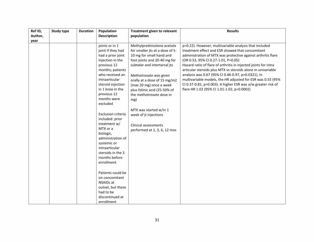
2991, Bava, 2019 [2]
Retrospective cohort, single arm
13 years JIA patients (defined via ILAR criteria) that were using MTX as sole DMARD were included. Patients previously treated with any biologic DMARD were excluded. Previous treatment with other synthetic DMARDs or concomitant or previous administration of nonsteroidal anti-inflammatory drugs and systemic or intra-articular corticosteroids was allowed. In total, there were 375 patients included of which 24% had persistent oligoarthritis
Median MTX dose was 12.8 mg/m2 for all patients State of inactive disease was evaluated per Wallace criteria In patients with oligoarthritis at presentation, all active joints were injected with corticosteroid
Methotrexate was more commonly administered subcutaneously. Of the 174 total oligoarthritis patients studied (includes persistent and extended), 49 (33.8%) did not achieve inactive disease while 125 (54.8%) did. 61% of patients achieved ID after a median of 1.7 years from the start of MTX therapy. The relative frequency of favorable outcome was higher among patients with oligoarthritis than in those with ERA or systemic arthritis; an equal proportion of patients with polyarthritis reached or did not reach ID. It appears that persistent and extended oligo were investigated together as they studied patients according to ‘functional phenotypes.’
33
Ref ID, Author, year
Study type Duration Population Description
Treatment given to relevant population
Results
patients were grouped in the functional phenotypes of oligoarthritis (4 or fewer affected joints), polyarthritis (5 or more affected joints), systemic arthritis, and ERA
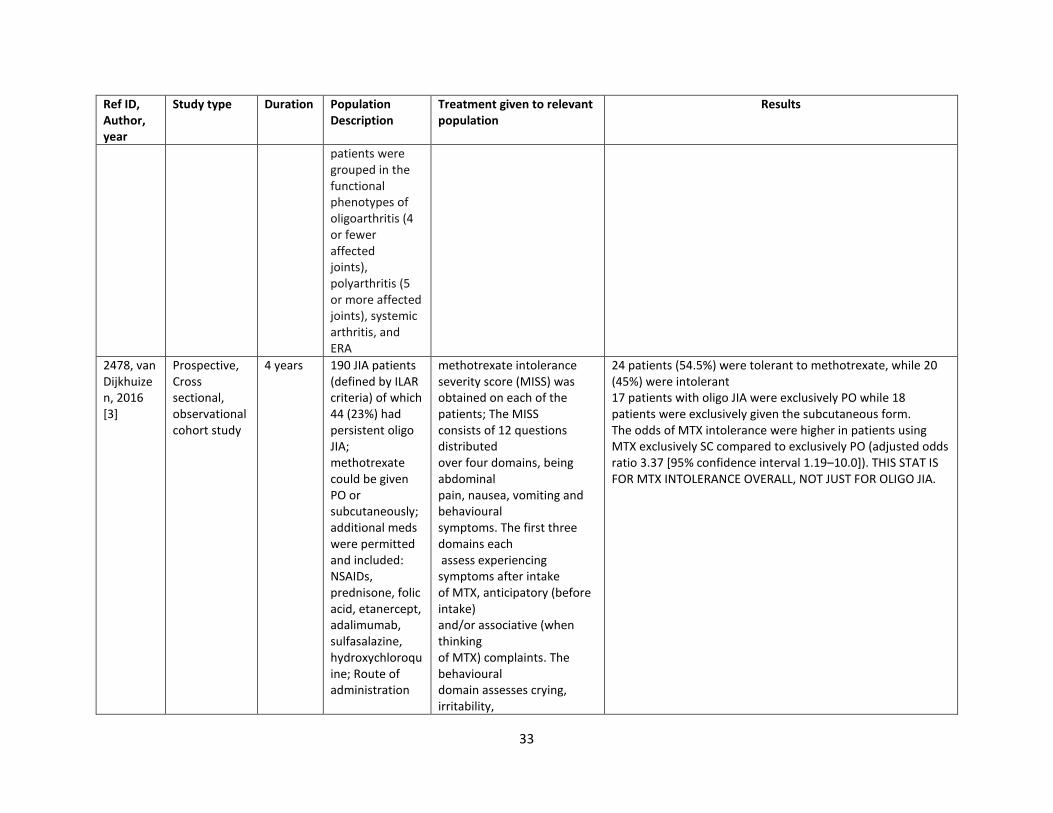
2478, van Dijkhuizen, 2016 [3]
Prospective, Cross sectional, observational cohort study
4 years 190 JIA patients (defined by ILAR criteria) of which 44 (23%) had persistent oligo JIA; methotrexate could be given PO or subcutaneously; additional meds were permitted and included: NSAIDs, prednisone, folic acid, etanercept, adalimumab, sulfasalazine, hydroxychloroquine; Route of administration
methotrexate intolerance severity score (MISS) was obtained on each of the patients; The MISS consists of 12 questions distributed over four domains, being abdominal pain, nausea, vomiting and behavioural symptoms. The first three domains each assess experiencing symptoms after intake of MTX, anticipatory (before intake) and/or associative (when thinking of MTX) complaints. The behavioural domain assesses crying, irritability,
24 patients (54.5%) were tolerant to methotrexate, while 20 (45%) were intolerant 17 patients with oligo JIA were exclusively PO while 18 patients were exclusively given the subcutaneous form. The odds of MTX intolerance were higher in patients using MTX exclusively SC compared to exclusively PO (adjusted odds ratio 3.37 [95% confidence interval 1.19–10.0]). THIS STAT IS FOR MTX INTOLERANCE OVERALL, NOT JUST FOR OLIGO JIA.
34
Ref ID, Author, year
Study type Duration Population Description
Treatment given to relevant population
Results
was categorised as exclusively PO, exclusively SC, switch from PO to SC and switch from SC to PO
restlessness and refusal to take MTX. The items can be assigned 0 (no symptoms), 1 (mild), 2 (moderate) or 3 (severe) points. The MISS was calculated as the sum of the questionnaire, while blank questions were assigned 0 points. The score could range from 0 to 36. A patient was considered intolerant if she had a score above the validated cut point of 6 points in concert with at least one associative, anticipatory or behavioural symptom
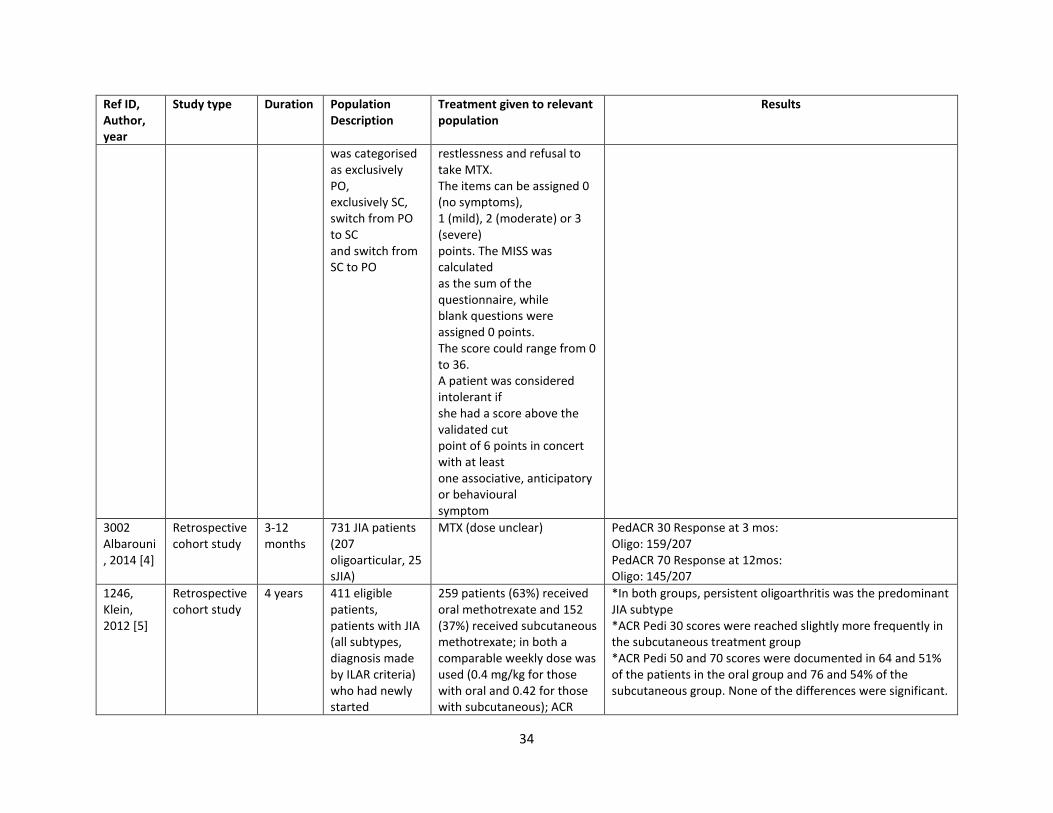
3002 Albarouni, 2014 [4]
Retrospective cohort study
3-12 months
731 JIA patients (207 oligoarticular, 25 sJIA)
MTX (dose unclear) PedACR 30 Response at 3 mos: Oligo: 159/207 PedACR 70 Response at 12mos: Oligo: 145/207
1246, Klein, 2012 [5]
Retrospective cohort study
4 years 411 eligible patients, patients with JIA (all subtypes, diagnosis made by ILAR criteria) who had newly started
259 patients (63%) received oral methotrexate and 152 (37%) received subcutaneous methotrexate; in both a comparable weekly dose was used (0.4 mg/kg for those with oral and 0.42 for those with subcutaneous); ACR
*In both groups, persistent oligoarthritis was the predominant JIA subtype *ACR Pedi 30 scores were reached slightly more frequently in the subcutaneous treatment group *ACR Pedi 50 and 70 scores were documented in 64 and 51% of the patients in the oral group and 76 and 54% of the subcutaneous group. None of the differences were significant.
35
Ref ID, Author, year
Study type Duration Population Description
Treatment given to relevant population
Results
methotrexate and were documented in the registry, consecutively studied; 94 (36%) had persistent oligoarthritis and were on oral methotrexate, 42 patients (28%) were taking subcutaneous methotrexate Patients had no previous or concomitant treatment with biologic agents. Steroids (either oral or intraarticular) were permitted.
Pedi 30/50/70 was assessed after 6 and 12 months of therapy
**OF NOTE STUDY HAS FEWER THAN 50% (ONLY 33%) PERSISTENT OLIGOARTHRITIS PATIENTS OF THE TOTAL # OF PATIENTS STUDIED
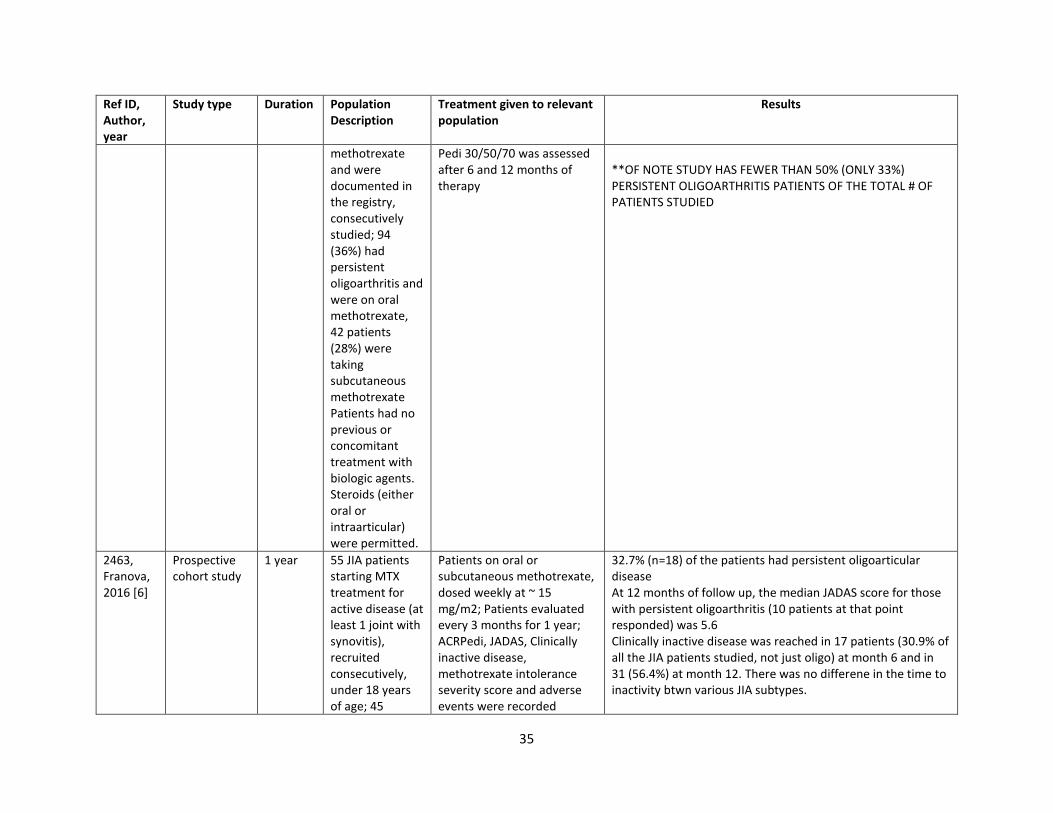
2463, Franova, 2016 [6]
Prospective cohort study
1 year 55 JIA patients starting MTX treatment for active disease (at least 1 joint with synovitis), recruited consecutively, under 18 years of age; 45
Patients on oral or subcutaneous methotrexate, dosed weekly at ~ 15 mg/m2; Patients evaluated every 3 months for 1 year; ACRPedi, JADAS, Clinically inactive disease, methotrexate intolerance severity score and adverse events were recorded
32.7% (n=18) of the patients had persistent oligoarticular disease At 12 months of follow up, the median JADAS score for those with persistent oligoarthritis (10 patients at that point responded) was 5.6 Clinically inactive disease was reached in 17 patients (30.9% of all the JIA patients studied, not just oligo) at month 6 and in 31 (56.4%) at month 12. There was no differene in the time to inactivity btwn various JIA subtypes.
36
Ref ID, Author, year
Study type Duration Population Description
Treatment given to relevant population
Results
patients received subcutaneous MTX, 10 received oral Concomitant medications included: oral corticosteroids and intraarticular corticosteroids
Neither the rate nor the extent of therapeutic response was influenced by JIA subtype or the route of MTX administration. Persistent decrease in JADAS seen over a 12 monthtime frame for those with persistent oligoarthritis.
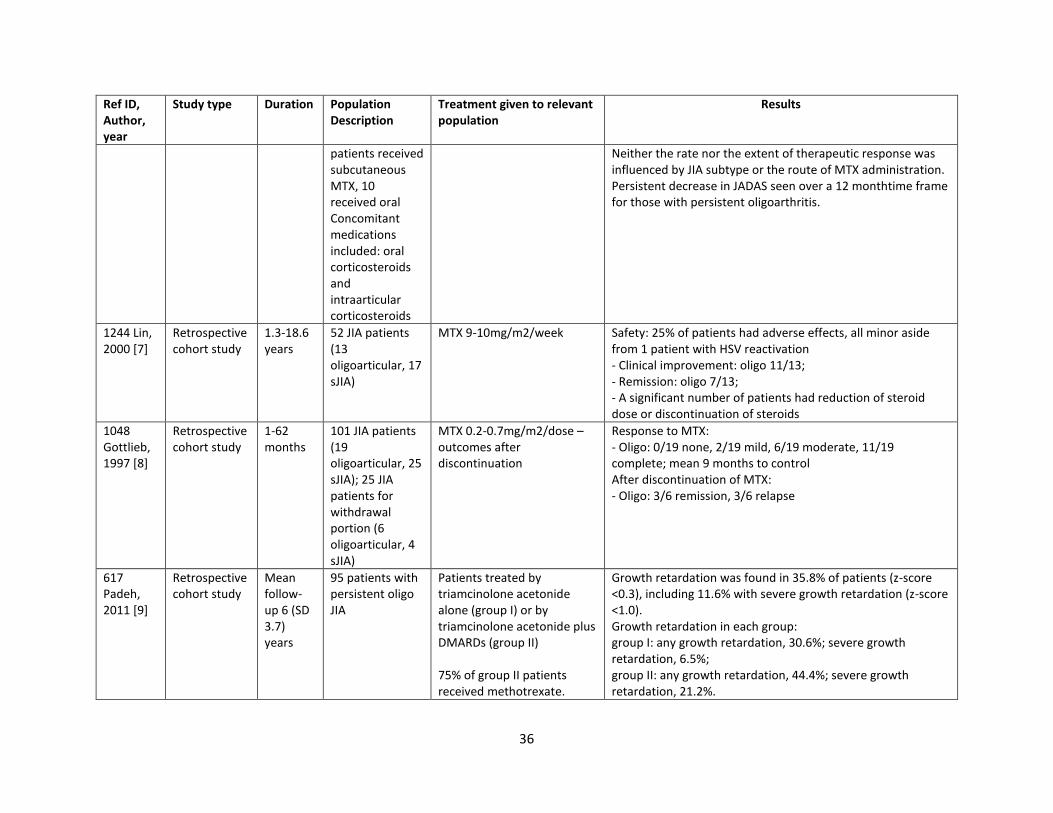
1244 Lin, 2000 [7]
Retrospective cohort study
1.3-18.6 years
52 JIA patients (13 oligoarticular, 17 sJIA)
MTX 9-10mg/m2/week Safety: 25% of patients had adverse effects, all minor aside from 1 patient with HSV reactivation - Clinical improvement: oligo 11/13; - Remission: oligo 7/13; - A significant number of patients had reduction of steroid dose or discontinuation of steroids
1048 Gottlieb, 1997 [8]
Retrospective cohort study
1-62 months
101 JIA patients (19 oligoarticular, 25 sJIA); 25 JIA patients for withdrawal portion (6 oligoarticular, 4 sJIA)
MTX 0.2-0.7mg/m2/dose – outcomes after discontinuation
Response to MTX: - Oligo: 0/19 none, 2/19 mild, 6/19 moderate, 11/19 complete; mean 9 months to control After discontinuation of MTX: - Oligo: 3/6 remission, 3/6 relapse
617 Padeh, 2011 [9]
Retrospective cohort study
Mean follow-up 6 (SD 3.7) years
95 patients with persistent oligo JIA
Patients treated by triamcinolone acetonide alone (group I) or by triamcinolone acetonide plus DMARDs (group II) 75% of group II patients received methotrexate.
Growth retardation was found in 35.8% of patients (z-score <0.3), including 11.6% with severe growth retardation (z-score <1.0). Growth retardation in each group: group I: any growth retardation, 30.6%; severe growth retardation, 6.5%; group II: any growth retardation, 44.4%; severe growth retardation, 21.2%.
37
Ref ID, Author, year
Study type Duration Population Description
Treatment given to relevant population
Results
group II had a significantly higher rate of severe growth retardation than group I (p <0.05). Elevated erythrocyte sedimentation rate values (≥40mm/1sth) indicated a significantly higher risk for growth retardation. All other clinical variables had no association with growth retardation.
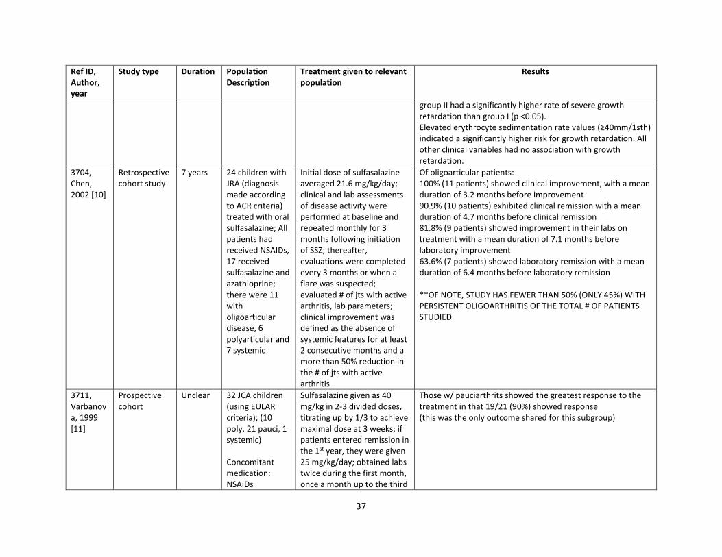
3704, Chen, 2002 [10]
Retrospective cohort study
7 years 24 children with JRA (diagnosis made according to ACR criteria) treated with oral sulfasalazine; All patients had received NSAIDs, 17 received sulfasalazine and azathioprine; there were 11 with oligoarticular disease, 6 polyarticular and 7 systemic
Initial dose of sulfasalazine averaged 21.6 mg/kg/day; clinical and lab assessments of disease activity were performed at baseline and repeated monthly for 3 months following initiation of SSZ; thereafter, evaluations were completed every 3 months or when a flare was suspected; evaluated # of jts with active arthritis, lab parameters; clinical improvement was defined as the absence of systemic features for at least 2 consecutive months and a more than 50% reduction in the # of jts with active arthritis
Of oligoarticular patients: 100% (11 patients) showed clinical improvement, with a mean duration of 3.2 months before improvement 90.9% (10 patients) exhibited clinical remission with a mean duration of 4.7 months before clinical remission 81.8% (9 patients) showed improvement in their labs on treatment with a mean duration of 7.1 months before laboratory improvement 63.6% (7 patients) showed laboratory remission with a mean duration of 6.4 months before laboratory remission **OF NOTE, STUDY HAS FEWER THAN 50% (ONLY 45%) WITH PERSISTENT OLIGOARTHRITIS OF THE TOTAL # OF PATIENTS STUDIED
3711, Varbanova, 1999 [11]
Prospective cohort
Unclear 32 JCA children (using EULAR criteria); (10 poly, 21 pauci, 1 systemic) Concomitant medication: NSAIDs
Sulfasalazine given as 40 mg/kg in 2-3 divided doses, titrating up by 1/3 to achieve maximal dose at 3 weeks; if patients entered remission in the 1st year, they were given 25 mg/kg/day; obtained labs twice during the first month, once a month up to the third
Those w/ pauciarthrits showed the greatest response to the treatment in that 19/21 (90%) showed response (this was the only outcome shared for this subgroup)
38
Ref ID, Author, year
Study type Duration Population Description
Treatment given to relevant population
Results
month, and then every 3 months following that Disease severity was assessed based on 1) # jts with active synovitis, 2) # jts with limited ROM, 3) ESR, 4) pain, 5) physician’s global, 6) patient/parent global; assessment was conducted q3 months; a point was given for each parameter if it improved > 50% Nonresponders: < 50% or fewer than 3 pts (within this were insignificant responders which showed > 30% but < 50% improvement; unchanged < 30% improvement; deterioration > 30% worsening of the indices) Complete Remission: if 5 of the following signs were absent for at least 2 months: 1) symptoms of inflammatory joint pain, 2) morning stiffness, 3) fatigue, 4) synovitis on jt exam, 5) progression of radiographic damage on sequential radiographs, 6) elevated ESR or CRP
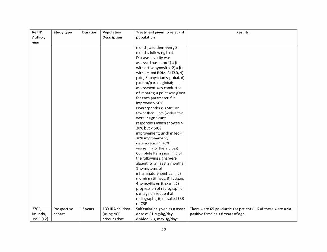
3705, Imundo, 1996 [12]
Prospective cohort
3 years 139 JRA children (using ACR criteria) that
Sulfasalazine given as a mean dose of 31 mg/kg/day divided BID, max 3g/day;
There were 69 pauciarticular patients. 16 of these were ANA positive females < 8 years of age.
39
Ref ID, Author, year
Study type Duration Population Description
Treatment given to relevant population
Results
demonstrated active arthritis (persistent effusion, limited ROM, pain; patients were allowed to be on other agents concomitantly including: NSAIDs, prednisone, hydroxychloroquine, auranofin, penicillamine, methotrexate, aspirin
blood tests were performed monthly x 3 months and then every 3 months thereafter Significant improvement defined as achievement of 1 or more of the following in the 1st year of tx: 1) 50% or more decrease in number of active joints (defined as pain, limited ROM or effusion), 2) 50% or more decrease in total number of joints with effusion, 3) 50% or more decrease in total degree of flexion contracture, 4) decrease in ESR to < 20 mm/hr (of those that had an abnormal one to begin with)
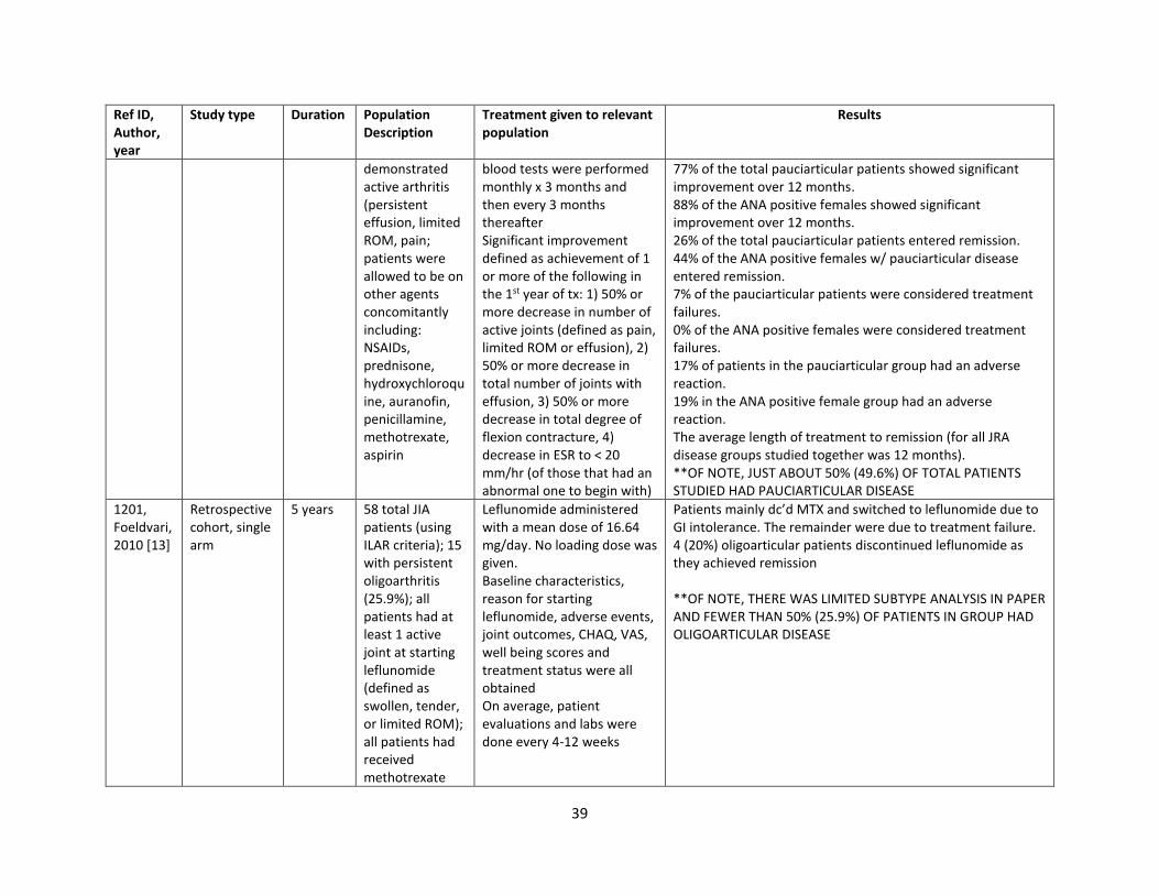
77% of the total pauciarticular patients showed significant improvement over 12 months. 88% of the ANA positive females showed significant improvement over 12 months. 26% of the total pauciarticular patients entered remission. 44% of the ANA positive females w/ pauciarticular disease entered remission. 7% of the pauciarticular patients were considered treatment failures. 0% of the ANA positive females were considered treatment failures. 17% of patients in the pauciarticular group had an adverse reaction. 19% in the ANA positive female group had an adverse reaction. The average length of treatment to remission (for all JRA disease groups studied together was 12 months). **OF NOTE, JUST ABOUT 50% (49.6%) OF TOTAL PATIENTS STUDIED HAD PAUCIARTICULAR DISEASE
1201, Foeldvari, 2010 [13]
Retrospective cohort, single arm
5 years 58 total JIA patients (using ILAR criteria); 15 with persistent oligoarthritis (25.9%); all patients had at least 1 active joint at starting leflunomide (defined as swollen, tender, or limited ROM); all patients had received methotrexate
Leflunomide administered with a mean dose of 16.64 mg/day. No loading dose was given. Baseline characteristics, reason for starting leflunomide, adverse events, joint outcomes, CHAQ, VAS, well being scores and treatment status were all obtained On average, patient evaluations and labs were done every 4-12 weeks
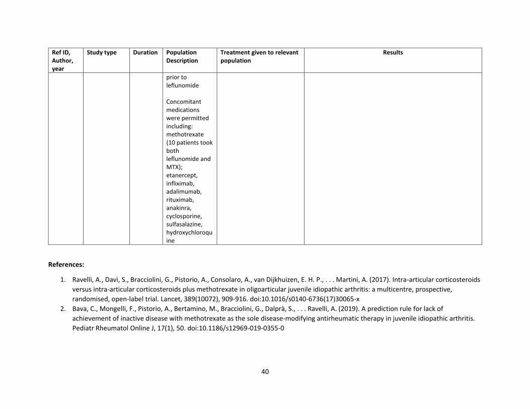
Patients mainly dc’d MTX and switched to leflunomide due to GI intolerance. The remainder were due to treatment failure. 4 (20%) oligoarticular patients discontinued leflunomide as they achieved remission **OF NOTE, THERE WAS LIMITED SUBTYPE ANALYSIS IN PAPER AND FEWER THAN 50% (25.9%) OF PATIENTS IN GROUP HAD OLIGOARTICULAR DISEASE
40
Ref ID, Author, year
Study type Duration Population Description
Treatment given to relevant population
Results
prior to leflunomide Concomitant medications were permitted including: methotrexate (10 patients took both leflunomide and MTX); etanercept, infliximab, adalimumab, rituximab, anakinra, cyclosporine, sulfasalazine, hydroxychloroquine
References:
1. Ravelli, A., Davì, S., Bracciolini, G., Pistorio, A., Consolaro, A., van Dijkhuizen, E. H. P., . . . Martini, A. (2017). Intra-articular corticosteroids
versus intra-articular corticosteroids plus methotrexate in oligoarticular juvenile idiopathic arthritis: a multicentre, prospective,
randomised, open-label trial. Lancet, 389(10072), 909-916. doi:10.1016/s0140-6736(17)30065-x
2. Bava, C., Mongelli, F., Pistorio, A., Bertamino, M., Bracciolini, G., Dalprà, S., . . . Ravelli, A. (2019). A prediction rule for lack of
achievement of inactive disease with methotrexate as the sole disease-modifying antirheumatic therapy in juvenile idiopathic arthritis.
Pediatr Rheumatol Online J, 17(1), 50. doi:10.1186/s12969-019-0355-0
41
3. van Dijkhuizen, E. H., Pouw, J. N., Scheuern, A., Hügle, B., Hardt, S., Ganser, G., . . . Wulffraat, N. M. (2016). Methotrexate intolerance in
oral and subcutaneous administration in patients with juvenile idiopathic arthritis: a cross-sectional, observational study. Clin Exp
Rheumatol, 34(1), 148-154.
4. Albarouni, M., Becker, I., & Horneff, G. (2014). Predictors of response to methotrexate in juvenile idiopathic arthritis. Pediatr Rheumatol
Online J, 12, 35. doi:10.1186/1546-0096-12-35
5. Klein, A., Kaul, I., Foeldvari, I., Ganser, G., Urban, A., & Horneff, G. (2012). Efficacy and safety of oral and parenteral methotrexate
therapy in children with juvenile idiopathic arthritis: An observational study with patients from the German Methotrexate Registry.
Arthritis Care Res (Hoboken), 64(9), 1349-1356. doi:10.1002/acr.21697
6. Fráňová, J., Fingerhutová, Š., Kobrová, K., Srp, R., Němcová, D., Hoza, J., . . . Doležalová, P. (2016). Methotrexate efficacy, but not its
intolerance, is associated with the dose and route of administration. Pediatric rheumatology, 14(1), 36. doi:10.1186/s12969-016-0099-z
7. Lin, Y. T., Tsai, M. J., Wang, L. H., Huang, M. T., Yang, Y. H., & Chiang, B. L. (2000). Efficacy and safety of methotrexate therapy for juvenile
rheumatoid arthritis. J Formos Med Assoc, 99(8), 623-629.
8. Gottlieb, B. S., Keenan, G. F., Lu, T., & Ilowite, N. T. (1997). Discontinuation of methotrexate treatment in juvenile rheumatoid arthritis.
Pediatrics, 100(6), 994-997. doi:10.1542/peds.100.6.994
9. Padeh, S., Pinhas-Hamiel, O., Zimmermann-Sloutskis, D., & Berkun, Y. (2011). Children with oligoarticular juvenile idiopathic arthritis are
at considerable risk for growth retardation. J Pediatr, 159(5), 832-837.e831-832. doi:10.1016/j.jpeds.2011.04.012
10. Chen, C.-C., Lin, Y.-T., Yang, Y.-H., & Chiang, B.-L. (2002). Sulfasalazine therapy for juvenile rheumatoid arthritis. Journal of the Formosan
Medical Association, 101(2), 110-116.
11. Varbanova, B. B., & Dyankov, E. D. (1999). Sulphasalazine. An alternative drug for second-line treatment of juvenile chronic arthritis. Adv
Exp Med Biol, 455, 331-336.
12. Imundo, L. F., & Jacobs, J. C. (1996). Sulfasalazine therapy for juvenile rheumatoid arthritis. J Rheumatol, 23(2), 360-366.
13. Foeldvari, I., & Wierk, A. (2010). Effectiveness of leflunomide in patients with juvenile idiopathic arthritis in clinical practice. J Rheumatol,
37(8), 1763-1767. doi:10.3899/jrheum.090874
42
PICO 6. In children with oligoarticular JIA, should biologic therapies be recommended, and should there be any preferred order of treatment: anti-TNF treatment, biologic treatments with other mechanisms of action?
Summary: A literature review search identified 8 cohort studies (most single arm)[1-8] and one case series[9] addressing the treatment of JIA
patients with biologic therapy. Minden et al.[1] reported outcomes of bDMARDs (missed with csDMARDs) in JIA patients over a 10-year period.
Symptom relief and disease activity appeared to be good in patients with persistent oligo JIA. An analysis that combined all JIA subtypes found
that patients who began bDMARD treatment within 2 years of symptom onset (G1 0 to 2 years) were significantly more likely to be in drug-free
remission than those patients who began bDMARD treatment later (G2 >2 to 5 years, G3 >5 years). G1 patients also had lower disease activity,
higher functional status, overall well-being, and lower rates of arthroplasty than other groups. Anink et al.[9] looked at 16 persistent oligo
patients all previously treated with MTX with 14 being treated with Enbrel and 2 with Humira. Looking at both 3- and 15-month outcomes, there
were decreases in active count joints, CHAQ scores, ESR levels, as well pain. Ten out of 16 achieved inactive disease at 3 months, 9/10 at 15
months. Etanercept was the most commonly used TNF-inhibitor in the cohort studies. Alexeeva et al.[2] evaluated 32 oligoarticular patients who
received etanercept 0.4 mg/kg twice a week with ACR 30/50/70/90s of 97%/97%/94%/88%. Inactive disease in the persistent oligo JIA patients
was 88% according to Wallace criteria and 50% according to JADAS71 criteria. Stable remission at the end of follow up was 56%. In another
study by Alexeeva et al.[3], 84 patients with persistent Oligo JIA given etanercept twice a week had a reduction in active joint count, physician
global, CHAQ and JADAS-71 with ACR Pedi 30/50/70/90 of 90.5%/90.5%/88.1%/77.4% with 86.9% achieving inactive disease. Kearsley-Fleet et
al.[4] evaluated 496 JIA patients, 12 with oligo JIA on Etanercept and found JADAS-71 decreased from median 7.2 (IQR 3.8-10.2) to 3.1 at 1 year
(IQR 0.7-5.6), which was not statistically significant. ACR Pedi’s of 30/50/70/90 were 80%/78%/70%/62% with minimal disease activity at 1 year
of 74%. Minden et al.[5] conducted a prospective cohort study evaluating 346 patients with JIA, 11 with oligo JIA, treated with etanercept who
achieved HAQ disability indices of 60% 0, 20% >0 to 0.5, 10% >0.5 to 1.0, 10% >1.0 to 3.0. Zuber et al.[7] evaluated 27 patients with oligo JIA who
achieved ACR 30/50/70/90/100 scores of 80%/80%/50%/35%/35% at 12 months. In Horneff et al.[8], 10 patients with persistent oligo JIA
receiving etanercept 0.4 mg/kg twice weekly with complete remission in 62% of patients. Donnithorne et al.[6] evaluated 125 patients with JIA,
26 with persistent oligo JIA, treated with etanercept (83%), infliximab (6%) and adalimumab (11%) with 64% of patients achieving inactive
disease at 1 year and 62% achieving inactive disease ever. Given that all of the studies lacked a comparison group, the risk of bias was high and
the quality of evidence very low.
Quality of evidence across all critical outcomes: Very low
43
Table 1. Data from Observational Studies
Ref ID, Author, year
Study type Duration Population Description Treatment given to relevant population
Results
3889 Minden, 2019[1]
Prospective cohort study
mean ± SD 9.1 ± 3.7 years
40 SJIA patients, 43 with persistent OJIA, 128 with extended OJIA
bDMARDS (mixed with csDMARDs)
PhGA of disease activity (mean ± SD): Persistent OJIA 1.5 ± 1.9 PhGA CID, n (%): Persistent OJIA 15 (36.6%) cJADAS-10: Persistent OJIA 4.3 ± 5.0 cJADAS-10 remission off drugs, no. (%): Persistent OJIA 1 (2.4%) HAQ total: Persistent OJIA 0.15 ± 0.40 Patient reported pain: Persistent OJIA 1.8 ± 2.3 At the 10-year time point, patients who began bDMARD treatment within 2 years of symptom onset (G1 0 to 2 years) were significantly more likely to be in drug-free remission than those patients who began treatment later (G2 >2 to 5 years, G3 >5 years). G1 patients also had lower disease activity, higher functional status, overall well-being, and lower rates of arthroplasty than other groups. However, this data combines different JIA subtypes (persistent OJIA, extended OJIA, sJIA, poly JIA, enthesitis, psoriatic arthritis)
763 Alexeeva, 2017[2]
Retrospective cohort study, propensity score matching
Unclear 49 patients with JIA (17 polyarticular, the rest oligoarticular)
Etanercept SQ 0.4mg/kg (max single dose 25mg), twice a week
- ACR 30/50/70/90 at the end of follow-up, n (%): Persistent oligo: 31/31/30/27 (97%/97%/94%/88%) - Inactive disease (according to the Wallace criteria) at the end of follow-up, n (%): Persistent oligo: 28 (88%); - Inactive disease (according to the JADAS71 cut-off point), n (%): Persistent oligo: 16 (50%); - Stable remission at the end of follow-up, n (%): Persistent oligo: 18 (56%);
3003, E. I. Alexeeva et al., 2017[3]
Single-arm cohort study
12 months 197 patients with non-systemic JIA (n = 84
Etanercept via subcutaneous injection at a dose of 0.4 mg per kg
Oligoarticular JIA subgroup: Active joint count decreased from median of 2 (IQR 2-4) to 0 (IQR 0-0), painful joint count
44
Ref ID, Author, year
Study type Duration Population Description Treatment given to relevant population
Results
persistent oligoarticular JIA)
body weight (maximum single dose, 25 mg) twice a week
decreased from 2 (IQR 1.5-3) to 0 (IQR 0-0), physician global decreased from 50 (IQR 42-70) to 0 (IQR 0-4), CHAQ decreased from 0.78 (IQR 0.5-1.5) to 0 (IQR 0-0.25), JADAS-71 decreased from 14.9 (11.7 – 18.9) to 0.5 (0-1.1) 90.5% of patients reached ACR Pedi 30 response, 90.5% ACR Pedi 50, 88.1% ACR Pedi 70, 77.4% ACR Pedi 90, 86.9% inactive disease
1442, L Kearsley-Fleet et al., 2016[4]
Single-arm cohort study
1 year 496 patients with JIA (n = 12 with oligoarticular JIA)
Etanercept Oligoarticular JIA outcomes: JADAS-71 decreased from median 7.2 (IQR 3.8-10.2) to 3.1 at 1 year (IQR 0.7-5.6) (not statistically significant) ACR Pedi 30 at 1 year: 80% ACR Pedi 50 at 1 year: 78% ACR Pedi 70 at 1 year: 70% ACR Pedi 90 at 1 year: 62% MDA at 1 year: 74% AEs (all patients): 9 (2%) stopped due to adverse events
4076, J. Anink et al., 2013[9]
Case series Median follow-up 13.7 months (IQR 8.3-16.7 months)
16 persistent oligoarticular JIA patients
Etanercept (n = 14), adalimumab (n = 2; both with arthritis + uveitis). All previously treated with MTX
3 month outcomes (n = 16) Active joint count decreased from median of 2 (IQR 1-3) to 0 (IQR 0-1) Pain decreased from 51 on VAS (IQR 0-71) to 6 (IQR 0-75) CHAQ decreased from 0.3 (IQR 0-0.9) to 0.1 (0-1.0) 10/16 with inactive disease at 3 months ESR decreased from 10 (IQR 2-60) to 3 (IQR 2-30); 11/15 normalized
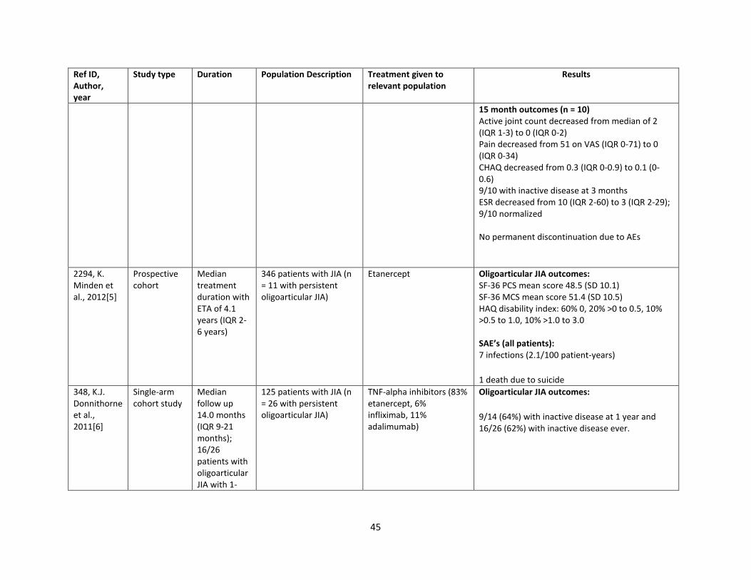
45
Ref ID, Author, year
Study type Duration Population Description Treatment given to relevant population
Results
15 month outcomes (n = 10) Active joint count decreased from median of 2 (IQR 1-3) to 0 (IQR 0-2) Pain decreased from 51 on VAS (IQR 0-71) to 0 (IQR 0-34) CHAQ decreased from 0.3 (IQR 0-0.9) to 0.1 (0-0.6) 9/10 with inactive disease at 3 months ESR decreased from 10 (IQR 2-60) to 3 (IQR 2-29); 9/10 normalized No permanent discontinuation due to AEs
2294, K. Minden et al., 2012[5]
Prospective cohort
Median treatment duration with ETA of 4.1 years (IQR 2-6 years)
346 patients with JIA (n = 11 with persistent oligoarticular JIA)
Etanercept Oligoarticular JIA outcomes: SF-36 PCS mean score 48.5 (SD 10.1) SF-36 MCS mean score 51.4 (SD 10.5) HAQ disability index: 60% 0, 20% >0 to 0.5, 10% >0.5 to 1.0, 10% >1.0 to 3.0 SAE’s (all patients): 7 infections (2.1/100 patient-years)
1 death due to suicide
348, K.J. Donnithorne et al., 2011[6]
Single-arm cohort study
Median follow up 14.0 months (IQR 9-21 months); 16/26 patients with oligoarticular JIA with 1-
125 patients with JIA (n = 26 with persistent oligoarticular JIA)
TNF-alpha inhibitors (83% etanercept, 6% infliximab, 11% adalimumab)
Oligoarticular JIA outcomes:
9/14 (64%) with inactive disease at 1 year and 16/26 (62%) with inactive disease ever.
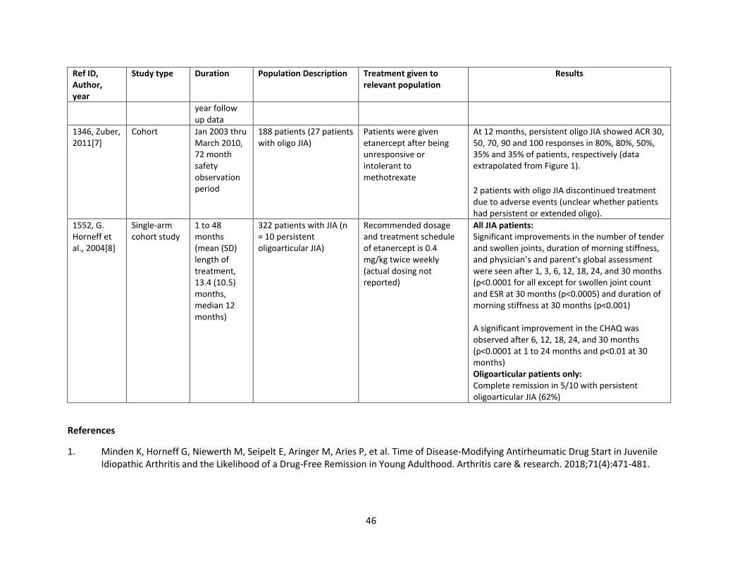
46
Ref ID, Author, year
Study type Duration Population Description Treatment given to relevant population
Results
year follow up data
1346, Zuber, 2011[7]
Cohort Jan 2003 thru March 2010, 72 month safety observation period
188 patients (27 patients with oligo JIA)
Patients were given etanercept after being unresponsive or intolerant to methotrexate
At 12 months, persistent oligo JIA showed ACR 30, 50, 70, 90 and 100 responses in 80%, 80%, 50%, 35% and 35% of patients, respectively (data extrapolated from Figure 1).
2 patients with oligo JIA discontinued treatment due to adverse events (unclear whether patients had persistent or extended oligo).
1552, G. Horneff et al., 2004[8]
Single-arm cohort study
1 to 48 months (mean (SD) length of treatment, 13.4 (10.5) months, median 12 months)
322 patients with JIA (n = 10 persistent oligoarticular JIA)
Recommended dosage and treatment schedule of etanercept is 0.4 mg/kg twice weekly (actual dosing not reported)
All JIA patients: Significant improvements in the number of tender and swollen joints, duration of morning stiffness, and physician’s and parent’s global assessment were seen after 1, 3, 6, 12, 18, 24, and 30 months (p<0.0001 for all except for swollen joint count and ESR at 30 months (p<0.0005) and duration of morning stiffness at 30 months (p<0.001) A significant improvement in the CHAQ was observed after 6, 12, 18, 24, and 30 months (p<0.0001 at 1 to 24 months and p<0.01 at 30 months) Oligoarticular patients only: Complete remission in 5/10 with persistent oligoarticular JIA (62%)
References
1. Minden K, Horneff G, Niewerth M, Seipelt E, Aringer M, Aries P, et al. Time of Disease-Modifying Antirheumatic Drug Start in Juvenile Idiopathic Arthritis and the Likelihood of a Drug-Free Remission in Young Adulthood. Arthritis care & research. 2018;71(4):471-481.
47
2. Alexeeva E, Dvoryakovskaya T, Denisova R, Sleptsova T, Isaeva K, Chomahidze A, et al. Comparative Analysis of the Etanercept Efficacy in Children with Juvenile Idiopathic Arthritis Under the Age of 4 Years and Children of Older Age Groups Using the Propensity Score Matching Method. Modern rheumatology. 2018:1-20.
3. Alexeeva EI, Namazova-Baranova LS, Bzarova TM, Valieva SI, Denisova RV, Sleptsova TV, et al. Predictors of the response to etanercept in patients with juvenile idiopathic arthritis without systemic manifestations within 12 months: results of an open-label, prospective study conducted at the National Scientific and Practical Center of Children's Health, Russia. Pediatric rheumatology online journal. 2017;15(1):51.
4. Kearsley-Fleet L, Davies R, Lunt M, Southwood TR, Hyrich KL. Factors associated with improvement in disease activity following initiation of etanercept in children and young people with Juvenile Idiopathic Arthritis: results from the British Society for Paediatric and Adolescent Rheumatology Etanercept Cohort Study. Rheumatology (Oxford, England). 2016;55(5):840-847.
5. Minden K, Niewerth M, Zink A, Seipelt E, Foeldvari I, Girschick H, et al. Long-term outcome of patients with JIA treated with etanercept, results of the biologic register JuMBO. Rheumatology (Oxford, England). 2012;51(8):1407-1415.
6. Donnithorne KJ, Cron RQ, Beukelman T. Attainment of inactive disease status following initiation of TNF-alpha inhibitor therapy for juvenile idiopathic arthritis: enthesitis-related arthritis predicts persistent active disease. The Journal of rheumatology. 2011;38(12):2675-2681.
7. Zuber Z, Rutkowska-Sak L, Postepski J, Dobrzyniecka B, Opoka-Winiarska V, Kobusinska K, et al. Etanercept treatment in juvenile idiopathic arthritis: the Polish registry. Medical science monitor : international medical journal of experimental and clinical research. 2011;17(12):Sr35-42.
8. Horneff G, Schmeling H, Biedermann T, Foeldvari I, Ganser G, Girschick HJ, et al. The German etanercept registry for treatment of juvenile idiopathic arthritis. Annals of the rheumatic diseases. 2004;63(12):1638-1644.
9. Anink J, Otten MH, Prince FH, Hoppenreijs EP, Wulffraat NM, Swart JF, et al. Tumour necrosis factor-blocking agents in persistent oligoarticular juvenile idiopathic arthritis: results from the Dutch Arthritis and Biologicals in Children Register. Rheumatology (Oxford, England). 2012;52(4):712-717.
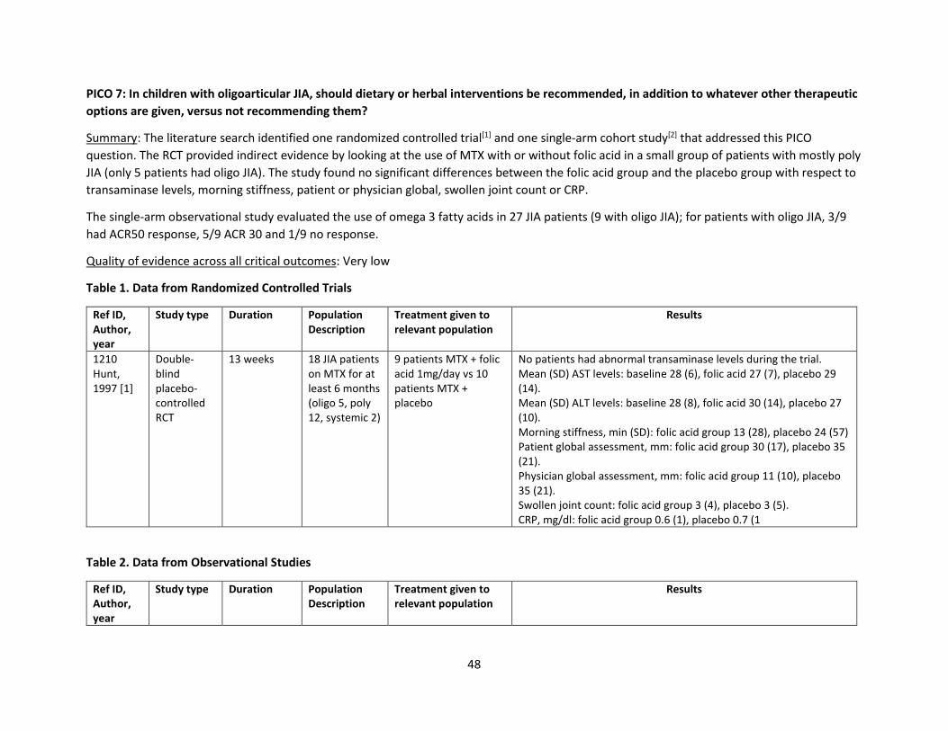
48
PICO 7: In children with oligoarticular JIA, should dietary or herbal interventions be recommended, in addition to whatever other therapeutic
options are given, versus not recommending them?
Summary: The literature search identified one randomized controlled trial[1] and one single-arm cohort study[2] that addressed this PICO
question. The RCT provided indirect evidence by looking at the use of MTX with or without folic acid in a small group of patients with mostly poly
JIA (only 5 patients had oligo JIA). The study found no significant differences between the folic acid group and the placebo group with respect to
transaminase levels, morning stiffness, patient or physician global, swollen joint count or CRP.
The single-arm observational study evaluated the use of omega 3 fatty acids in 27 JIA patients (9 with oligo JIA); for patients with oligo JIA, 3/9
had ACR50 response, 5/9 ACR 30 and 1/9 no response.
Quality of evidence across all critical outcomes: Very low
Table 1. Data from Randomized Controlled Trials
Ref ID, Author, year
Study type Duration Population Description
Treatment given to relevant population
Results
1210 Hunt, 1997 [1]
Double-blind placebo-controlled RCT
13 weeks 18 JIA patients on MTX for at least 6 months (oligo 5, poly 12, systemic 2)
9 patients MTX + folic acid 1mg/day vs 10 patients MTX + placebo
No patients had abnormal transaminase levels during the trial. Mean (SD) AST levels: baseline 28 (6), folic acid 27 (7), placebo 29 (14). Mean (SD) ALT levels: baseline 28 (8), folic acid 30 (14), placebo 27 (10). Morning stiffness, min (SD): folic acid group 13 (28), placebo 24 (57) Patient global assessment, mm: folic acid group 30 (17), placebo 35 (21). Physician global assessment, mm: folic acid group 11 (10), placebo 35 (21). Swollen joint count: folic acid group 3 (4), placebo 3 (5). CRP, mg/dl: folic acid group 0.6 (1), placebo 0.7 (1
Table 2. Data from Observational Studies
Ref ID, Author, year
Study type Duration Population Description
Treatment given to relevant population
Results
49
2696, Gheita et al, 2012 [2]
Single-arm cohort study
12 weeks 27 patients with JIA (n = 9 oligoarticular JIA)
Omega-3 fatty acids 2 g/day
Oligoarticular outcomes (extrapolated from Fig 3): 3/9 patients (33.3%) Pediatric ACR50 response 5/9 patients (55.56%) Pediatric ACR30 response 1/9 patients (11.11%) No response
References:
1. Hunt PG, Rose CD, McIlvain-Simpson G, Tejani S. The effects of daily intake of folic acid on the efficacy of methotrexate therapy in
children with juvenile rheumatoid arthritis. A controlled study. J Rheumatol. 1997 Nov;24(11):2230-2.
2. Gheita T, Kamel S, Helmy N, El-Laithy N, Monir A. Omega-3 fatty acids in juvenile idiopathic arthritis: effect on cytokines (IL-1 and TNF-
alpha), disease activity and response criteria. Clin Rheumatol. 2012 Feb;31(2):363-6. doi: 10.1007/s10067-011-1848-5. Epub 2011 Sep
16.
PICO 8. In children with oligoarticular JIA, regardless of disease activity and risk factors, should PT/OT versus no PT/OT (regardless of
concomitant medical therapy) be recommended?
Summary: Literature searches led to one randomized controlled trial [4482] which indirectly answered the PICO question. This study included
patients with JIA as well as those with cerebral palsy and brachial plexus birth injury, although data for JIA was reported separately. The aim was
to include all patients with at least one distal upper extremity joint involved. Patients were randomly allocated to either complete 8 weeks of
leap motion controller based training (LMCBT) or a conventional rehabilitation program. The LMCBT program employed elements of virtual
reality. The system could purportedly detect finger movements with submillimeter accuracy. The program included 2 games that were
developed for the purposes of this study. Conventional therapy included “re-education of muscles using a sensorimotor approach to control
motor output.” In looking strictly at the data for JIA patients, it appeared that both the LMCBT based rehabilitation and traditional rehabilitation
both led to statistically significant improvements in CHAQ total, CHAQ pain, CHAQ well-being, hand grip, tip grip, lateral grip and triple grip.
LCMBT showed a statistically significant improvement in Duruoz Hand Index (DHI) scoring compared to traditional rehabilitation. There was no
significant difference in the outcomes of the therapy methods in the remaining categories (Jebson Taylor Hand Function Test, Nine-Hole Peg
Test, CHAQ), grip strength). Regarding JIA specifically, there was no evidence to suggest that LMCBT was more effective than conventional
therapy.
This paper very indirectly addresses this PICO question. It suggests that rehabilitation is beneficial, though there was no negative control group
(no one without therapy) with which to compare. This paper also considered a virtual reality method which was not considered in this particular
50
PICO question. Furthermore, this PICO asks specifically about oligoarticular JIA and this paper considered JIA as a whole. There is no reported
subgroup analysis for oligo JIA. Nonetheless, there is a significant improvement in patients’ CHAQ scores before and after 8 weeks of traditional
rehabilitation engagement.
This study had several limitations in addition to indirectness of the patient population and comparison. Allocation was unconcealed and patients
and practitioners were not blinded (although the outcome assessor was blinded). Since this is a single small study there is also serious
imprecision in effect estimates.
Quality of evidence across all critical outcomes: Very low
Table 1. Data from randomized controlled trials
Ref ID, Author, year
Study type Duration Population Description
Monitoring given to relevant population
Results
4482, Tarakci 2019 [1]
Randomized controlled trial
1 year of study, 8 week program conducted for participants
Pediatric patients between 5 and 17 with JIA, cerebral palsy or brachial plexus birth injury with at least one affected distal joint in upper extremity (wrist and/or finger joints) Patients were excluded if they had undergone botox injections, intraarticular injection, surgery or hand rehabilitation within the previous 1 year
This was a randomized parallel group trial. Patients either underwent a leap motion controller based training program (LMCBT) or conventional rehabilitation for 8 weeks. Each program took place for 1 hour sessions 3 times weekly for 8 weeks. *LMCBT is a method which uses virtual reality to influence movement outcomes. In this case, a leap motion controller device was employed which could track hand and finger movements with purported submillimeter accuracy. Two games were “academically developed” for the purposes of the study *The conventional rehabilitation program aimed to re-educate muscles using a sensorimotor approach to control motor output
18 patients with JIA underwent LMCBT and 25 patients with JIA underwent traditional rehab There were significantly improved scores for CHAQ total, CHAQ pain, CHAQ well being and Duruoz Hand Index (DHI) in both LMCBT and traditional rehab groups when comparing pre and post-treatment scores for patients. There was a statistically significant improvement in hand grip, tip grip, lateral grip and triple grip in both LCMBT and traditional rehab pre and post treatment groups. There was a statistically significant difference in the DHI between the LMCBT and traditional rehabilitation group in favor of LMCBT. There was no statistically significant difference in the remainder of the outcome measures when comparing the two methods of rehabilitation.
51
References:
1. Tarakci E, Arman N, Tarakci D, Kasapcopur O. Leap Motion Controller-based training for upper extremity rehabilitation in children and
adolescents with physical disabilities: A randomized controlled trial. J Hand Ther. Apr-Jun 2020;33(2):220-228.
PICO 9. In children with oligoarticular JIA, should risk factors alter the treatment paradigm?
Summary: The literature search identified one observational study[1] that addressed this PICO question. The observational study was
retrospective but included 205 pts. Results were significant showing that symmetric disease was a predictor of extension to >10 joints, the need
to use DMARDs, erosive disease on radiographs, continued active disease, lack of remission and disability based on CHAQ. Ankle or wrist
involvement was a predictor of extension and erosions. Wrist involvement was a predictor of need for DMARDs and continued active disease.
Elevated ESR was a predictor of extension, need for DMARDs and lack of remission.
Quality of evidence across all critical outcomes: Very low
Table 1. Data from Observational Studies
Ref ID, Author, year
Study type Duration Population Description
Treatment given to relevant population
Results
1135, Al-Matar et a., 2002 [1]
Single-arm retrospective cohort study
At least 5 years Median 10.8 years (range 5-26.6 years)
205 patients with oligoarticular JIA
N/A All below features present in first 6 months of disease; results taken from multivariate logistic regression model (disease duration included as confounder) Symmetric disease predictor of extension to >= 10 joints (OR 19.2, 95% CI [5.46-67.8], p = 0.000), need to use DMARDs (OR 11.5, 95% CI [4.22-31.33], p = 0.000), erosive disease on radiographs (OR 4.73, 95% CI [1.47-15.2], p = 0.009), inflammatory activity at last follow up visit (OR 3.23, 95% CI [1.45-7.2]), no remission of disease (OR 4.73, 95% CI [2.15-10.4]), disability as measured by C-HAQ>0.12 (OR 2.95, 95% CI [1.01-8.6]) Ankle and/or wrist disease predictor of extension (OR 6.61, 95% CI [1.97-22.1]) and erosions (OR 3.59, 95% CI [1.15-11.2], p = 0.027)
52
Wrist disease predictor of need to use DMARDs (OR 5.87, 95% CI [1.51-22.8]) and inflammatory disease activity at last follow up visit (OR 4.01, 95% CI [1.16-13.8], p = 0.004) Elevated ESR predictive of extension (OR 3.76, 95% CI [1.09-12.9], p = 0.036), need to use DMARDs (OR 6.47, 95% CI [2.2-18.9], p = 0.001), and no remission of disease (OR 2.30, 95% CI [1.04-5.08], p = 0.039)
References:
1. Al-Matar MJ, Petty RE, Tucker LB, Malleson PN, Schroeder ML, Cabral DA. The early pattern of joint involvement predicts disease
progression in children with oligoarticular (pauciarticular) juvenile rheumatoid arthritis. Arthritis Rheum. 2002 Oct;46(10):2708-15.
PICO 10. In children with oligoarticular JIA, should disease activity measures alter the treatment paradigm?
Summary: The literature searches did not identify any studies that addressed this PICO question.
Quality of evidence across all critical outcomes: Very low
53
Active TMJ Arthritis
PICO 11. In children with JIA with active TMJ arthritis, should a trial of consistent NSAIDs be recommended and should there be any preferred
NSAID treatment?
Summary: The literature searches did not identify any studies that addressed this PICO question.
Quality of evidence across all critical outcomes: Very low
PICO 12. In children with JIA with active TMJ arthritis, should adding intraarticular glucocorticoids to initial therapy be recommended?
Summary: The literature search identified seven observational studies[1-7] that addressed this PICO question, including one prospective pilot
study, 2 prospective cohort studies, 2 retrospective cohort studies and 2 retrospective reviews. Some of the observational studies looked at
radiographic benefit, others looked at clinical markers including improvement in pain or improved MIO. Three studies showed radiographic
benefit with improvement in MRI findings.[1,2,6] Improvements in pain were more varied. In two studies[1,3] pain was significantly decreased. One
study[5] showed initial improvement in pain-frequency, pain-intensity and pain-index, but by long term follow-up only pain frequency was still
improved. One study[2] showed all groups had a decrease in pain intensity (measured by VAS) but there was no statistically significant difference
between groups including control group. Another study[7] showed mean increase in MIO (maximal incisal opening) of 6.9 mm (p = 0.002; 95% CI
3, 10.7), but with subsequent injections the increase was only 0.4 mm (p = 0.8; 95% CI –3.5, 4.4).
Two studies reported potential serious adverse events of IA steroid injections of TMJs. In one[4], 33/238 patients developed heterotopic bone
formation (HBF), bilateral in 36%. The study found a 25% increase in hazard of HBF for every additional injection (steroid and infliximab
injections analyzed together) (HR 1.254, 95%CI 1.04-1.512, p= 0.0184) and a 56% decrease in hazard for each year increase in delay between JIA
diagnosis and first injection. (HR 0.44, 95% CI 0.296-0.655, p not reported). In the other study[7], one patient developed subcutaneous atrophy at
the injection site. Two patients developed small, asymptomatic intraarticular calcifications.
Since all studies were observational and all but one lacked a control group, the risk of bias was high.
Quality of evidence across all critical outcomes: Very low
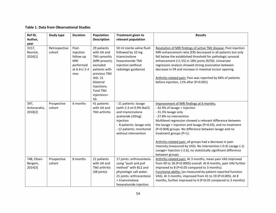
54
Table 1. Data from Observational Studies
Ref ID, Author, year
Study type Duration Population Description
Treatment given to relevant population
Results
3157, Resnick, 2016[1]
Retrospective cohort
Post-injection follow-up MRI performed at 6.4+/-2.4 mos
29 patients with JIA and TMJ synovitis (MRI proven); excluded patients with previous TMJ IASI. 21 bilateral injections. Total TMJ injections= 50.
50 ml sterile saline flush followed by 10 mg triamcinolone hexacetonide TMJ injection (without radiologic guidance)
Resolution of MRI findings of active TMJ disease: Post injection MRI enhancement ratio (ER) decreased in all patients but only fell below the established threshold for pathologic synovial enhancement (<1.55) in 18% joints (9/50). Univariate regression analysis showed strong association between decrease in ER and increase in maximal incisor opening. Arthritis-related pain: Pain was reported by 66% of patients before injection, 11% after (P<0.001)
397, Antonarakis, 2018[2]
Prospective cohort
6 months 41 patients with JIA and TMJ arthritis
- 21 patients: lavage (with 2-3 ml 0.9% NaCl) and triamcinolone acetonide (20mg) injection - 8 patients: lavage only - 12 patients: monitored without intervention
Improvement of MRI findings at 6 months: - 42.9% of lavage + injection - 31.3% lavage only - 27.8% no intervention Multilevel regression showed a relevant difference between the lavage + injection and lavage (P=0.03), and no treatment (P=0.004) groups. No difference between lavage and no treatment groups (P=1). Arthritis-related pain: all groups had a decrease in pain intensity (measured by VAS). No intervention (-0.4) Lavage (-1) Lavage+ Injection (-2.6), no statistically significant difference between groups.
748, Olsen-Bergem, 2014[3]
Prospective cohort
8 months 21 patients with JIA and TMJ arthritis (38 joints)
17 joints: arthrocentesis using “push and pull method” with B12 and physiologic salt water. 21 joints: arthrocentesis + triamcinolone hexacetonide injection
Arthritis-related pain: At 3 months, mean pain VAS improved from 49 to 18 (P<0.0005) overall. At 8 months, pain VAS further improved to 8 (P=0.05 compared to 3 months). Functional ability: (as measured by patient-reported function VAS). At 3 months, improved from 41 to 19 (P<0.005). At 8 months, further improved to 4 (P<0.05 compared to 3 months).
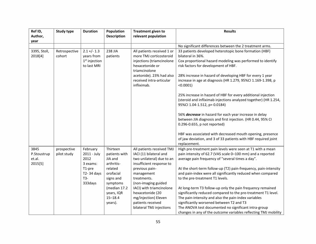
55
Ref ID, Author, year
Study type Duration Population Description
Treatment given to relevant population
Results
No significant differences between the 2 treatment arms.
3395, Stoll, 2018[4]
Retrospective cohort
2.1 +/- 1.3 years from 1st injection to last MRI
238 JIA patients
All patients received 1 or more TMJ corticosteroid injections (triamcinolone hexacetonide or triamcinolone acetonide). 23% had also received intra-articular infliximab.
33 patients developed heterotopic bone formation (HBF) bilateral in 36%. Cox proportional hazard modeling was performed to identify risk factors for development of HBF. 28% increase in hazard of developing HBF for every 1 year increase in age at diagnosis (HR 1.279, 95%CI 1.169-1.398, p <0.0001) 25% increase in hazard of HBF for every additional injection (steroid and infliximab injections analyzed together) (HR 1.254, 95%CI 1.04-1.512, p= 0.0184) 56% decrease in hazard for each year increase in delay between JIA diagnosis and first injection. (HR 0.44, 95% CI 0.296-0.655, p not reported) HBF was associated with decreased mouth opening, presence of jaw deviation, and 3 of 33 patients with HBF required joint replacement.
3845 P.Stoustrup et.al. 2015[5]
prospective pilot study
February 2011 - July 2012 3 exams: T1-pre T2- 34 days T3- 333days
Thirteen patients with JIA and arthritis-related orofacial signs and symptoms (median 17.2 years, IQR 15–18.4 years).
All patients received TMJ IACI (11 bilateral and two unilateral) due to an insufficient response to previous pain-management treatments. (non-imaging guided IACI) with triamcinolone hexacetonide (20 mg/injection) Eleven patients received bilateral TMJ injections
High pre-treatment pain levels were seen at T1 with a mean pain intensity of 62.7 (VAS scale 0–100 mm) and a reported average pain frequency of “several times a day”. At the short-term follow-up (T2) pain-frequency, pain-intensity and pain-index were all significantly reduced when compared to the pre-treatment T1 levels. At long-term T3 follow-up only the pain frequency remained significantly reduced compared to the pre-treatment T1 level. The pain-intensity and also the pain-index variables significantly worsened between T2 and T3 The ANOVA test documented no significant intra-group changes in any of the outcome variables reflecting TMJ mobility
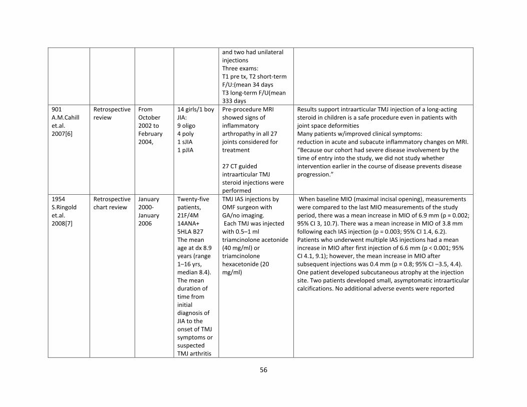
56
and two had unilateral injections Three exams: T1 pre tx, T2 short-term F/U:(mean 34 days T3 long-term F/U(mean 333 days
901 A.M.Cahill et.al. 2007[6]
Retrospective review
From October 2002 to February 2004,
14 girls/1 boy JIA: 9 oligo 4 poly 1 sJIA 1 pJIA
Pre-procedure MRI showed signs of inflammatory arthropathy in all 27 joints considered for treatment 27 CT guided intraarticular TMJ steroid injections were performed
Results support intraarticular TMJ injection of a long-acting steroid in children is a safe procedure even in patients with joint space deformities Many patients w/improved clinical symptoms: reduction in acute and subacute inflammatory changes on MRI. “Because our cohort had severe disease involvement by the time of entry into the study, we did not study whether intervention earlier in the course of disease prevents disease progression.”
1954 S.Ringold et.al. 2008[7]
Retrospective chart review
January 2000-January 2006
Twenty-five patients, 21F/4M 14ANA+ 5HLA B27 The mean age at dx 8.9 years (range 1–16 yrs, median 8.4). The mean duration of time from initial diagnosis of JIA to the onset of TMJ symptoms or suspected TMJ arthritis
TMJ IAS injections by OMF surgeon with GA/no imaging. Each TMJ was injected with 0.5–1 ml triamcinolone acetonide (40 mg/ml) or triamcinolone hexacetonide (20 mg/ml)
When baseline MIO (maximal incisal opening), measurements were compared to the last MIO measurements of the study period, there was a mean increase in MIO of 6.9 mm (p = 0.002; 95% CI 3, 10.7). There was a mean increase in MIO of 3.8 mm following each IAS injection (p = 0.003; 95% CI 1.4, 6.2). Patients who underwent multiple IAS injections had a mean increase in MIO after first injection of 6.6 mm (p < 0.001; 95% CI 4.1, 9.1); however, the mean increase in MIO after subsequent injections was 0.4 mm (p = 0.8; 95% CI –3.5, 4.4). One patient developed subcutaneous atrophy at the injection site. Two patients developed small, asymptomatic intraarticular calcifications. No additional adverse events were reported
57
was 11 months (range 0–55 mo, median 2). Ten patients (40%) had TMJ complaints or suspected TMJ arthritis at their first visit
References:
1. Resnick CM, Vakilian PM, Kaban LB, Peacock ZS. Quantifying the Effect of Temporomandibular Joint Intra-Articular Steroid Injection on Synovial Enhancement in Juvenile Idiopathic Arthritis. Journal of oral and maxillofacial surgery : official journal of the American Association of Oral and Maxillofacial Surgeons. 2016;74(12):2363-2369.
2. Antonarakis GS, Courvoisier DS, Hanquinet S, Dhouib A, Carlomagno R, Hofer M, et al. Benefit of Temporomandibular Joint Lavage With Intra-Articular Steroids Versus Lavage Alone in the Management of Temporomandibular Joint Involvement in Juvenile Idiopathic Arthritis. Journal of oral and maxillofacial surgery : official journal of the American Association of Oral and Maxillofacial Surgeons. 2018;76(6):1200-1206.
3. Olsen-Bergem H, Bjornland T. A cohort study of patients with juvenile idiopathic arthritis and arthritis of the temporomandibular joint: outcome of arthrocentesis with and without the use of steroids. International journal of oral and maxillofacial surgery. 2014;43(8):990-995.
4. Stoll ML, Amin D, Powell KK, Poholek CH, Strait RH, Aban I, et al. Risk Factors for Intraarticular Heterotopic Bone Formation in the Temporomandibular Joint in Juvenile Idiopathic Arthritis. The Journal of rheumatology. 2018;45(9):1301-1307.
5. Stoustrup P, Kristensen KD, Kuseler A, Pedersen TK, Herlin T. Temporomandibular joint steroid injections in patients with juvenile idiopathic arthritis: an observational pilot study on the long-term effect on signs and symptoms. Pediatric rheumatology online journal. 2015;13:62.
6. Cahill AM, Baskin KM, Kaye RD, Arabshahi B, Cron RQ, Dewitt EM, et al. CT-guided percutaneous steroid injection for management of inflammatory arthropathy of the temporomandibular joint in children. AJR. American journal of roentgenology. 2006;188(1):182-186.
7. Ringold S, Torgerson TR, Egbert MA, Wallace CA. Intraarticular corticosteroid injections of the temporomandibular joint in juvenile idiopathic arthritis. The Journal of rheumatology. 2008;35(6):1157-1164.
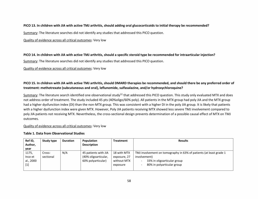
58
PICO 13. In children with JIA with active TMJ arthritis, should adding oral glucocorticoids to initial therapy be recommended?
Summary: The literature searches did not identify any studies that addressed this PICO question.
Quality of evidence across all critical outcomes: Very low
PICO 14. In children with JIA with active TMJ arthritis, should a specific steroid type be recommended for intraarticular injection?
Summary: The literature searches did not identify any studies that addressed this PICO question.
Quality of evidence across all critical outcomes: Very low
PICO 15. In children with JIA with active TMJ arthritis, should DMARD therapies be recommended, and should there be any preferred order of
treatment: methotrexate (subcutaneous and oral), leflunomide, sulfasalazine, and/or hydroxychloroquine?
Summary: The literature search identified one observational study[1] that addressed this PICO question. This study only evaluated MTX and does
not address order of treatment. The study included 45 pts (40%oligo/60% poly). All patients in the MTX group had poly JIA and the MTX group
had a higher dysfunction index (DI) than the non-MTX group. This was consistent with a higher DI in the poly JIA group. It is likely that patients
with a higher dysfunction index were given MTX. However, Poly JIA patients receiving MTX showed less severe TMJ involvement compared to
poly JIA patients not receiving MTX. Nevertheless, the cross-sectional design prevents determination of a possible causal effect of MTX on TMJ
outcomes.
Quality of evidence across all critical outcomes: Very low
Table 1. Data from Observational Studies
Ref ID, Author, year
Study type Duration Population Description
Treatment Results
1175, Ince et al., 2000 [1]
Cross-sectional
N/A 45 patients with JIA (40% oligoarticular, 60% polyarticular)
18 with MTX exposure, 27 without MTX exposure
TMJ involvement on tomography in 63% of patients (at least grade 1 involvement)
- 33% in oligoarticular group - 80% in polyarticular group
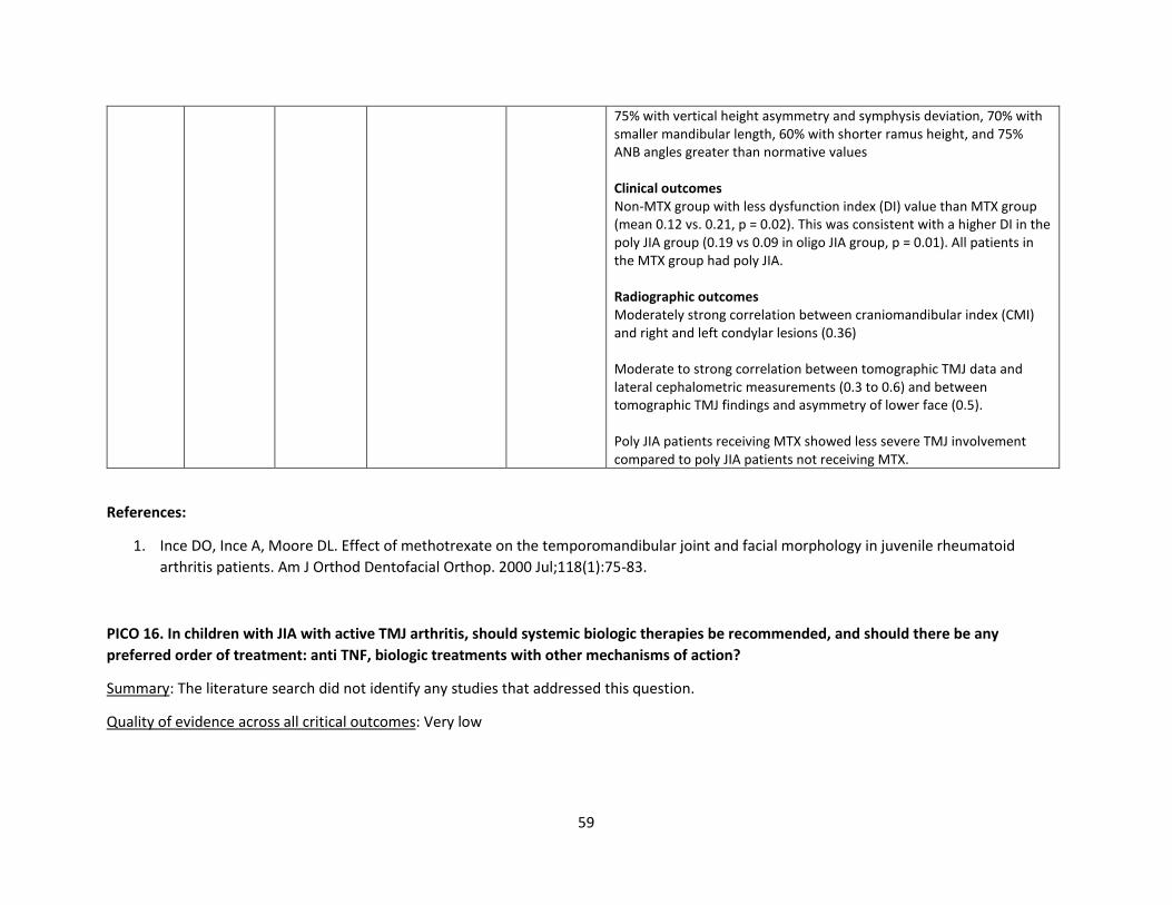
59
75% with vertical height asymmetry and symphysis deviation, 70% with smaller mandibular length, 60% with shorter ramus height, and 75% ANB angles greater than normative values Clinical outcomes Non-MTX group with less dysfunction index (DI) value than MTX group (mean 0.12 vs. 0.21, p = 0.02). This was consistent with a higher DI in the poly JIA group (0.19 vs 0.09 in oligo JIA group, p = 0.01). All patients in the MTX group had poly JIA. Radiographic outcomes Moderately strong correlation between craniomandibular index (CMI) and right and left condylar lesions (0.36) Moderate to strong correlation between tomographic TMJ data and lateral cephalometric measurements (0.3 to 0.6) and between tomographic TMJ findings and asymmetry of lower face (0.5). Poly JIA patients receiving MTX showed less severe TMJ involvement compared to poly JIA patients not receiving MTX.
References:
1. Ince DO, Ince A, Moore DL. Effect of methotrexate on the temporomandibular joint and facial morphology in juvenile rheumatoid
arthritis patients. Am J Orthod Dentofacial Orthop. 2000 Jul;118(1):75-83.
PICO 16. In children with JIA with active TMJ arthritis, should systemic biologic therapies be recommended, and should there be any
preferred order of treatment: anti TNF, biologic treatments with other mechanisms of action?
Summary: The literature search did not identify any studies that addressed this question.
Quality of evidence across all critical outcomes: Very low
60
PICO 17. In children with JIA with active TMJ arthritis, should dietary or herbal interventions be recommended, in addition to whatever other
therapeutic options are given, versus not recommending them?
Summary: The literature searches did not identify any studies that addressed this PICO question.
Quality of evidence across all critical outcomes: Very low
PICO 18. In children with JIA with active TMJ arthritis, regardless of disease activity and risk factors, should PT versus no PT (regardless of
concomitant medical therapy) be recommended?
Summary: The literature searches did not identify any studies that addressed this PICO question.
Quality of evidence across all critical outcomes: Very low
PICO 19. In children with JIA with active TMJ arthritis, should risk factors alter the treatment paradigm?
Summary: The literature searches did not identify any studies that addressed this PICO question.
Quality of evidence across all critical outcomes: Very low
61
Systemic JIA (sJIA) with and without Macrophage Activation Syndrome (MAS)
PICO 20: In patients with treatment naïve, newly diagnosed sJIA without MAS, should non-DMARD treatment (NSAIDs, glucocorticoids) be
used as initial therapy?
Summary: The literature searches identified 11 observational studies (Table 1) that directly or indirectly addressed the question of whether non-
DMARD treatment (NSAIDS, glucocorticoids) should be used as initial therapy in treatment naïve, newly diagnosed sJIA without MAS. Only one
of these studies involved children with sJIA[1], the remaining studies involved patients with Adult Onset Still’s Disease (AOSD)[2, 3, 4, 5, 6, 7, 8, 9,
10]. Studies were limited by retrospective treatment comparisons or lack of any control groups.
Sura et al.[1] observed 87 children newly diagnosed with sJIA between 2000-2014; 51 children received a trial of NSAID monotherapy with only
13 (25.5%) achieving clinically inactive disease [CID] for the duration of the study. Initial joint count was the only statistically significant predictor
(p = 0.01) of CID on NSAIDs alone.
Most of the studies in AOSD highlighted poor response to NSAID monotherapy, although Wouters’ study in 1986 highlighted variable effect of
different NSAIDS on fever with best response noted with naproxen use [fever improved in 29 % with aspirin, 70% with indomethacin and 86%
with naproxen][3]. In Kim et al.’s 2012 study, the authors concluded that NSAID monotherapy was not effective when used as monotherapy in
AOSD[4]. In Franchini et al.[5], Gerfand-Valentin et al.[3] and Kalyoncu et al.[2], only 16% to 25% of patients with AOSD went into clinical
remission following use of NSAID monotherapy.
Corticosteroids showed varying response rates across studies in patients with AOSD. Ruscitti et al.[7] assessed the role of high dose
corticosteroid therapy in AOSD. They concluded that first-line treatment with high dosage of corticosteroids was a significant predictor of the
achievement of clinical remission during the first 6 months of observation (P <.001). In contrast, Hu et al.[8] assessed a Chinese cohort of 517
AOSD patients in whom glucocorticoids were used most frequently (498/517, 96.3%) for disease control, followed by methotrexate (273/517,
52.8%) and hydroxychloroquine (174/517, 33.7%). 357/423 (84.4%) of AOSD cases were able to achieve initial remission with different regimens,
mostly including glucocorticoids, methotrexate or hydroxychloroquine. Patients who required higher dose of glucocorticoids to induce remission
had a poor response to treatment. Disease features associated with need for high dose steroids to induce remission: presence of skin rash,
pericarditis, splenomegaly and delayed diagnosis (both more than 3 months and more than 6 months). Kong et al.[10] reported that 50% of 104
AOSD patients achieved remission after 2 weeks of corticosteroid treatment. In 1-month treatment, partial remission and complete remission
rates were 92% and 71%, respectively. In the same cohort, cumulative relapse rate of 46.9% was observed after treatment with corticosteroids.
In contrast, Kalyoncu et al.[2] found that 87% of AOSD patients receiving moderate-high dose corticosteroids went into remission with initial
treatment compared to 26% patients receiving NSAIDs alone who went into remission with initial treatment.
62
A prospective cohort study by Ter Haar et al.[111] assessed the use of rIL-1Ra (Anakinra) in sJIA at 2 mg/kg which was escalated for incomplete
response to 4 mg/kg or additional prednisolone or switched to alternative therapy. The authors found that 76% of patients had inactive disease
1 year after the initiation of rIL‐1Ra; 96% patients with inactive disease at 1 month had sustained inactive disease at 1 year, compared to only
47% of patients with active disease at 1 month (OR 27.0 [95% CI 4.17, 539.74], P = 0.003). Only 33% of patients required systemic glucocorticoids
to achieve or sustain inactive disease. Patients with persistent arthritis after 1 month of rIL‐1Ra treatment were at risk of prolonged disease
activity). Among 6 patients who did not respond to initial treatment with rIL‐1Ra, Inactive disease was ultimately achieved with tocilizumab (n =
2), canakinumab (n = 2), or the combination of MTX and prednisolone (n = 2). The ability to achieve inactive disease without glucocorticoids was
also associated with sustained inactive disease at 1 year.
Quality of evidence across all critical outcomes: Very low
Table 1. Data from Observational Studies
Ref ID, Author, year
Study type Duration Population Description
Treatment given to relevant population
Results
2822
Sura et al.
2018 [1]
Cohort study
14 years (2000-2014)
87 children were newly diagnosed with sJIA 2000-2014
NSAID monotherapy
51 children received a trial of NSAID monotherapy and 13 (25.5%) achieved CID. Initial joint count was the only statistically significant predictor (p = 0.01) of CID on NSAIDs alone. Age at presentation, ferritin, and CRP were trending towards significance (p = 0.10, p = 0.08, p = 0.14 respectively).
3323
Kalyoncu et al.
2016 [2]
Multicenter cohort
0-180 mo median f/u time 22 mo
356 AOSD Mostly females 59 % median age 32 years
NSAID, moderate-high dose corticosteroids
52 (86.7%) of 60 pts receiving moderate-high dose corticosteroids went into remission with initial treatment. 5 (26.3%) of 19 patients receiving NSAIDs alone went into remission with initial treatment.
63
Ref ID, Author, year
Study type Duration Population Description
Treatment given to relevant population
Results
114, Gerfand-Valentin, 2014 [3]
Retrospective cohort
Mean 8.4 years
57 patients with AOSD
NSAIDs 28 patients received NSAIDs (6 indomethacin, 9 ketoprofen, 7 aspirin, 3 diclofenac, 3 other) Corticosteroids 51 patients received CS (49 as first- or second-line treatment).
NSAIDs Only 5/28 (18%) were controlled with NSAID monotherapy. 6 had gastrointestinal AE, 4 requiring PPIs Corticosteroids 23/51 (45%) has “steroid-dependent disease”/unable to discontinue steroids 75% had CS-related AEs
3869
Kim et al.
2012 [4]
Cohort/ retrospective analysis
Unclear 54 Korean patients with AOSD (met Yamaguchi’s criteria)
NSAID monotherapy, high-dose corticosteroids
NSAID monotherapy was tried in 42 patients without any efficacy. Of the 50 patients treated with high-dose corticosteroids, 21 patients (42%) were resistant and 29 showed a response. Elevated ESR and corticosteroids refractoriness were associated with poor prognosis (P = 0.023 and P = 0.009, respectively).
1270, Franchini, 2010 [5]
Retrospective cohort
Mean 56 months
45 patients with AOSD
NSAIDs 25 trials of NSAID monotherapy Corticosteroids 56 trials of steroid monotherapy
NSAIDs Inactive disease at least 2 months in 4/25 (all responders were patients without chronic articular involvement) Corticosteroids Inactive disease at least 2 months in 35/56 (63%), most responders had systemic disease without chronic articular disease.
144 Wouters, 1986 [6]
Cohort Study
6 months 45 pts with Adult-Onset Still's disease (AOSD)
NSAIDs, corticosteroids Fever improved in 29 % with aspirin, 70% with indomethacin and 86% with naproxen. In 76% of patients, glucocorticoids reduced the systemic and/or joint symptoms. 3 patients febrile on glucocorticoids, indomethacin reduced temperature to normal. 8 patients who improved on steroids later developed severe joint destruction.
64
Ref ID, Author, year
Study type Duration Population Description
Treatment given to relevant population
Results
13 patients received one or several slow-acting antirheumatic drugs.
2411
Ruscitti et al.
2019 [7]
Cohort study
18 months 80 pts with AOSD
High dosages of corticosteroids (0.8–1 mg/kg/day of prednisone-equivalent) versus low dosage of CCSs (0.2–0.3 mg/kg/day of prednisone-equivalent) at disease onset
25 (64.79%) patients treated with the first-line treatment with high dosage of corticosteroids reached the primary endpoint of clinical remission at 6 months, a significantly higher percentage when compared with 8 (22.85%) patients treated with the first-line treatment with low dosage of corticosteroids (P <.001). First-line treatment with high dosage of corticosteroids was a significant predictor of the achievement of clinical remission during the first 6 months of observation. At 18 months follow-up, 17 (44.73%) pts treated with high dosage of corticosteroids maintained clinical remission, being classified as monocyclic pattern, a significant higher percentage when compared with 3 (8.57%) patients treated with the first-line treatment with low dosage of corticosteroids (P: .001), Side effects: 7% of enrolled patients experienced minor adverse events. No severe adverse events or deaths were observed.
4371
Hu et al, 2019 [8]
Retrospective Cohort study
Unclear 517 AOSD patients (Chinese Cohort)
Corticosteroids, methotrexate, hydroxychloroquine
Glucocorticoids were used most frequently (498/517, 96.3%) for disease control, followed by methotrexate (273/517, 52.8%) and hydroxychloroquine (174/517, 33.7%). 84.4%. 357/423 of AOSD cases were able to achieve initial remission with different regimens, mostly including glucocorticoids, methotrexate or hydroxychloroquine.
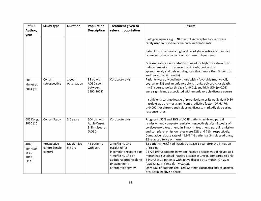
65
Ref ID, Author, year
Study type Duration Population Description
Treatment given to relevant population
Results
Biological agents e.g., TNF-α and IL-6 receptor blocker, were rarely used in first-line or second-line treatments. Patients who require a higher dose of glucocorticoids to induce remission usually had a poor response to treatment Disease features associated with need for high dose steroids to induce remission: presence of skin rash, pericarditis, splenomegaly and delayed diagnosis (both more than 3 months and more than 6 months)
681
Kim et al.
2014 [9]
Cohort, retrospective
1-year observation
82 pt with AOSD seen between 1992-2012)
Corticosteroids
Patients were divided into those with a favorable (monocyclic course; n=33) and an unfavorable (chronic, polycyclic, or death; n=49) course. polyarthralgia (p=0.01), and high LDH (p=0.03) were significantly associated with an unfavorable disease course Insufficient starting dosage of prednisolone or its equivalent (<30 mg/day) was the most significant predictive factor (OR 6.476, p=0.007) for chronic and relapsing disease, markedly decreasing response rates.
682 Kong, 2010 [10]
Cohort Study
5.6 years 104 pts with Adult-Onset Still's disease (AOSD)
Corticosteroids Prognosis: 52% and 39% of AOSD patients achieved partial remission and complete remission respectively after 2 weeks of corticosteroid treatment. In 1-month treatment, partial remission and complete remission rates were 92% and 71%, respectively. Cumulative relapse rate of 46.9% (46 patients). 34 relapsed once, 12 relapsed twice or more.
4040
Ter Haar et al.
2019 [111]
Prospective cohort (single center)
Median f/u 5.8 yrs
42 patients with sJIA
2 mg/kg rIL-1Ra escalated for incomplete response to 4 mg/kg rIL-1Ra or additional prednisolone or switched to alternative therapy.
32 patients (76%) had inactive disease 1 year after the initiation of rIL1‐Ra. 24 /25 (96%) patients in whom inactive disease was achieved at 1 month had sustained inactive disease at 1 year, compared to only 8 (47%) of 17 patients with active disease at 1 month (OR 27.0 [95% CI 4.17, 539.74], P = 0.003). Only 33% of patients required systemic glucocorticoids to achieve or sustain inactive disease.
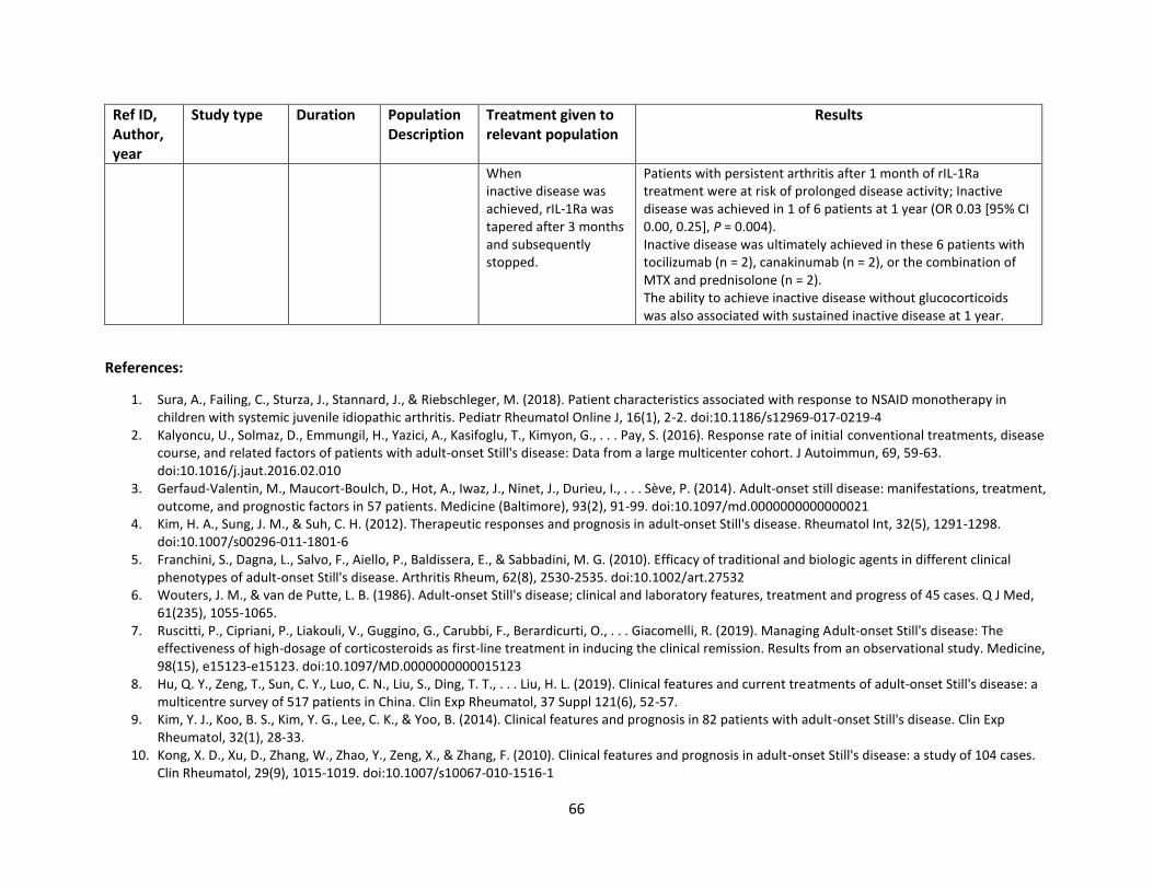
66
Ref ID, Author, year
Study type Duration Population Description
Treatment given to relevant population
Results
When inactive disease was achieved, rIL-1Ra was tapered after 3 months and subsequently stopped.
Patients with persistent arthritis after 1 month of rIL‐1Ra treatment were at risk of prolonged disease activity; Inactive disease was achieved in 1 of 6 patients at 1 year (OR 0.03 [95% CI 0.00, 0.25], P = 0.004). Inactive disease was ultimately achieved in these 6 patients with tocilizumab (n = 2), canakinumab (n = 2), or the combination of MTX and prednisolone (n = 2). The ability to achieve inactive disease without glucocorticoids was also associated with sustained inactive disease at 1 year.
References:
1. Sura, A., Failing, C., Sturza, J., Stannard, J., & Riebschleger, M. (2018). Patient characteristics associated with response to NSAID monotherapy in children with systemic juvenile idiopathic arthritis. Pediatr Rheumatol Online J, 16(1), 2-2. doi:10.1186/s12969-017-0219-4
2. Kalyoncu, U., Solmaz, D., Emmungil, H., Yazici, A., Kasifoglu, T., Kimyon, G., . . . Pay, S. (2016). Response rate of initial conventional treatments, disease course, and related factors of patients with adult-onset Still's disease: Data from a large multicenter cohort. J Autoimmun, 69, 59-63. doi:10.1016/j.jaut.2016.02.010
3. Gerfaud-Valentin, M., Maucort-Boulch, D., Hot, A., Iwaz, J., Ninet, J., Durieu, I., . . . Sève, P. (2014). Adult-onset still disease: manifestations, treatment, outcome, and prognostic factors in 57 patients. Medicine (Baltimore), 93(2), 91-99. doi:10.1097/md.0000000000000021
4. Kim, H. A., Sung, J. M., & Suh, C. H. (2012). Therapeutic responses and prognosis in adult-onset Still's disease. Rheumatol Int, 32(5), 1291-1298. doi:10.1007/s00296-011-1801-6
5. Franchini, S., Dagna, L., Salvo, F., Aiello, P., Baldissera, E., & Sabbadini, M. G. (2010). Efficacy of traditional and biologic agents in different clinical phenotypes of adult-onset Still's disease. Arthritis Rheum, 62(8), 2530-2535. doi:10.1002/art.27532
6. Wouters, J. M., & van de Putte, L. B. (1986). Adult-onset Still's disease; clinical and laboratory features, treatment and progress of 45 cases. Q J Med, 61(235), 1055-1065.
7. Ruscitti, P., Cipriani, P., Liakouli, V., Guggino, G., Carubbi, F., Berardicurti, O., . . . Giacomelli, R. (2019). Managing Adult-onset Still's disease: The effectiveness of high-dosage of corticosteroids as first-line treatment in inducing the clinical remission. Results from an observational study. Medicine, 98(15), e15123-e15123. doi:10.1097/MD.0000000000015123
8. Hu, Q. Y., Zeng, T., Sun, C. Y., Luo, C. N., Liu, S., Ding, T. T., . . . Liu, H. L. (2019). Clinical features and current treatments of adult-onset Still's disease: a multicentre survey of 517 patients in China. Clin Exp Rheumatol, 37 Suppl 121(6), 52-57.
9. Kim, Y. J., Koo, B. S., Kim, Y. G., Lee, C. K., & Yoo, B. (2014). Clinical features and prognosis in 82 patients with adult-onset Still's disease. Clin Exp Rheumatol, 32(1), 28-33.
10. Kong, X. D., Xu, D., Zhang, W., Zhao, Y., Zeng, X., & Zhang, F. (2010). Clinical features and prognosis in adult-onset Still's disease: a study of 104 cases. Clin Rheumatol, 29(9), 1015-1019. doi:10.1007/s10067-010-1516-1
67
11. Ter Haar, N. M., van Dijkhuizen, E. H. P., Swart, J. F., van Royen-Kerkhof, A., El Idrissi, A., Leek, A. P., . . . Vastert, S. J. (2019). Treatment to Target Using Recombinant Interleukin-1 Receptor Antagonist as First-Line Monotherapy in New-Onset Systemic Juvenile Idiopathic Arthritis: Results From a Five-Year Follow-Up Study. Arthritis Rheumatol, 71(7), 1163-1173. doi:10.1002/art.40865
PICO 21. In patients with treatment naïve, newly diagnosed sJIA without MAS, should DMARD treatment (methotrexate, calcineurin
inhibitor) be used as initial therapy and is there a preferred order?
Summary: The literature search revealed three observational studies [Error! Reference source not found., 2, 3] that addressed this PICO
question. Hu et al. (2019) assessed a Chinese cohort of 517 patients with adult onset stills disease (AOSD). Glucocorticoids were used most
frequently (498/517, 96.3%) for disease control, followed by methotrexate (273/517, 52.8%) and hydroxychloroquine (174/517, 33.7%). A total
of 357/423 (84.4%) patients were able to achieve initial remission with different regimens, mostly including glucocorticoids, methotrexate or
hydroxychloroquine. Biological agents (e.g. TNF-α and IL-6 receptor blocker) were rarely used in first-line or second-line treatments.[1] A
retrospective cohort study provided indirect evidence of methotrexate efficacy in AOSD, reporting that 87.6% of patients achieved clinical
remission with initial combination corticosteroid and methotrexate combination therapy.[2] Another retrospective cohort study provided
indirect evidence of methotrexate and cyclosporine efficacy in AOSD patients with steroid-refractory or steroid-dependent disease (73% and
75%, respectively).[3] No studies were identified that directly evaluated DMARD monotherapy in treatment naïve patients.
Quality of evidence across all critical outcomes: Very low
Table 1. Data from Observational Studies
Ref ID, Author, year
Study type Duration Population Description
Treatment given to relevant population
Results
4371
Hu et al, 2019 [1]
Retrospective Cohort study
Unclear 517 AOSD patients (Chinese Cohort)
Clinical and laboratory features, treatment of AOSD
Glucocorticoids were used most frequently (498/517, 96.3%) for disease control, followed by methotrexate (273/517, 52.8%) and hydroxychloroquine (174/517, 33.7%). 84.4% (357/423) of AOSD cases were able to achieve initial remission with different regimens, mostly including glucocorticoids, methotrexate or hydroxychloroquine. Biological agents, e.g., TNF-α and IL-6 receptor blocker, were rarely used in first-line or second-line treatments.
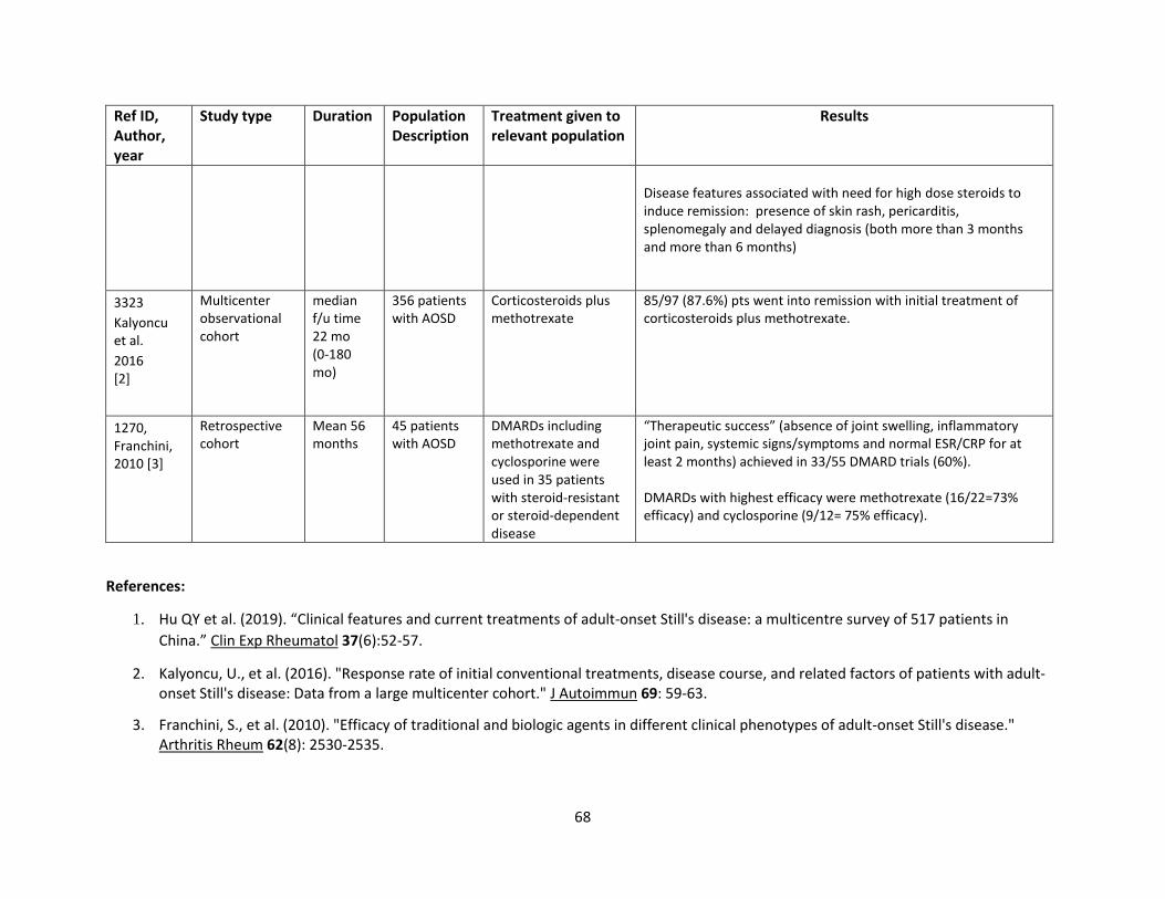
68
Ref ID, Author, year
Study type Duration Population Description
Treatment given to relevant population
Results
Disease features associated with need for high dose steroids to induce remission: presence of skin rash, pericarditis, splenomegaly and delayed diagnosis (both more than 3 months and more than 6 months)
3323
Kalyoncu et al.
2016 [2]
Multicenter observational cohort
median f/u time 22 mo (0-180 mo)
356 patients with AOSD
Corticosteroids plus methotrexate
85/97 (87.6%) pts went into remission with initial treatment of corticosteroids plus methotrexate.
1270, Franchini, 2010 [3]
Retrospective cohort
Mean 56 months
45 patients with AOSD
DMARDs including methotrexate and cyclosporine were used in 35 patients with steroid-resistant or steroid-dependent disease
“Therapeutic success” (absence of joint swelling, inflammatory joint pain, systemic signs/symptoms and normal ESR/CRP for at least 2 months) achieved in 33/55 DMARD trials (60%). DMARDs with highest efficacy were methotrexate (16/22=73% efficacy) and cyclosporine (9/12= 75% efficacy).
References:
1. Hu QY et al. (2019). “Clinical features and current treatments of adult-onset Still's disease: a multicentre survey of 517 patients in
China.” Clin Exp Rheumatol 37(6):52-57.
2. Kalyoncu, U., et al. (2016). "Response rate of initial conventional treatments, disease course, and related factors of patients with adult-onset Still's disease: Data from a large multicenter cohort." J Autoimmun 69: 59-63.
3. Franchini, S., et al. (2010). "Efficacy of traditional and biologic agents in different clinical phenotypes of adult-onset Still's disease." Arthritis Rheum 62(8): 2530-2535.
69
PICO 22. In patients with treatment naïve, newly diagnosed sJIA without MAS, should biologic treatment (Anakinra, Canakinumab,
Tocilizumab or others) be used as initial therapy and is there a preferred order?
Summary: Literature searches identified three observational cohort studies that indirectly addressed whether biologic treatment (Anakinra,
Canakinumab, Tocilizumab or others) should be used as initial therapy for sJIA and whether there is a preferred order. None of these trials included
a comparison treatment group.
A prospective cohort study by Ter Haar et al.[11] assessed the use of rIL-1Ra (Anakinra) in sJIA at 2 mg/kg which was escalated for incomplete
response to 4 mg/kg or additional prednisolone or switched to alternative therapy. The authors found that 76% of patients had inactive disease 1
year after the initiation of rIL‐1Ra; 96% patients with inactive disease at 1 month had sustained inactive disease at 1 year, compared to only 47%
of patients with active disease at 1 month (OR 27.0 [95% CI 4.17, 539.74], P = 0.003). Patients with persistent arthritis after 1 month of rIL‐1Ra
treatment were at risk of prolonged disease activity). Among 6 patients who did not respond to initial treatment with rIL‐1Ra, Inactive disease was
ultimately achieved with tocilizumab (n = 2), canakinumab (n = 2), or the combination of MTX and prednisolone (n = 2). The ability to achieve
inactive disease without glucocorticoids was also associated with sustained inactive disease at 1 year.
Vastert et al.[2] investigated IL-1Ra [Anakinra 2mg/kg] as first-line therapy in patients with sJIA. The authors observed excellent response in about
85% of patients within 3 months. Reported side effects included local skin reactions in 65% of patients, without report of serious invasive infections.
The authors noted mild cutaneous or upper airway infection and reactivation of infection with herpes simplex virus type 1 in several patients,
none of whom required hospitalization or IV antibiotic treatment.
Pacharapakornpong et al.[3] investigated the effect of Tocilizumab in 23 patients with sJIA as a first line therapy when indicated versus greater
than 6 months after indicated; 54% of patients who received early treatment with tocilizumab achieved remission whereas no patients who
received the dose late achieved remission. Patients who received early treatment had a significant difference from baseline to 12 months in joint
count (p=0.003), number of limited joints (p=0.011), patient global (p=0.003), physician global (p=0.003), ESR (p=0.003). For those who received
late treatment, there were statistically significant differences from baseline to 12 months in number of active joints (p=0.026), patient global
(p=0.014), physician global (p=0.003), ESR (p=0.002), and CHAQ (p=0.017).
The identified studies supported use of biologic treatment (Anakinra, Canakinumab, Tocilizumab) as initial therapy. Although two studies
investigated IL-1Ra as initial therapy, no studies evaluated a preferred order for starting these medications. The observational study designs and
lack of comparison groups rendered the quality of evidence as very low.
Quality of evidence across all critical outcomes: Very low
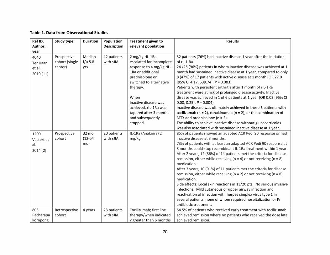
70
Table 1. Data from Observational Studies
Ref ID, Author, year
Study type Duration Population Description
Treatment given to relevant population
Results
4040
Ter Haar et al.
2019 [11]
Prospective cohort (single center)
Median f/u 5.8 yrs
42 patients with sJIA
2 mg/kg rIL-1Ra escalated for incomplete response to 4 mg/kg rIL-1Ra or additional prednisolone or switched to alternative therapy. When inactive disease was achieved, rIL-1Ra was tapered after 3 months and subsequently stopped.
32 patients (76%) had inactive disease 1 year after the initiation of rIL1‐Ra. 24 /25 (96%) patients in whom inactive disease was achieved at 1 month had sustained inactive disease at 1 year, compared to only 8 (47%) of 17 patients with active disease at 1 month (OR 27.0 [95% CI 4.17, 539.74], P = 0.003). Patients with persistent arthritis after 1 month of rIL‐1Ra treatment were at risk of prolonged disease activity; Inactive disease was achieved in 1 of 6 patients at 1 year (OR 0.03 [95% CI 0.00, 0.25], P = 0.004). Inactive disease was ultimately achieved in these 6 patients with tocilizumab (n = 2), canakinumab (n = 2), or the combination of MTX and prednisolone (n = 2). The ability to achieve inactive disease without glucocorticoids was also associated with sustained inactive disease at 1 year.
1200
Vastert et al.
2014 [2]
Prospective cohort
32 mo (12-54 mo)
20 patients with sJIA
IL-1Ra (Anakinra) 2 mg/kg
85% of patients showed an adapted ACR Pedi 90 response or had inactive disease at 3 months. 73% of patients with at least an adapted ACR Pedi 90 response at 3 months could stop recombinant IL‐1Ra treatment within 1 year. After 2 years, 12 (86%) of 14 patients met the criteria for disease remission, either while receiving (n = 4) or not receiving (n = 8) medication. After 3 years, 10 (91%) of 11 patients met the criteria for disease remission, either while receiving (n = 2) or not receiving (n = 8) medication. Side effects: Local skin reactions in 13/20 pts. No serious invasive infections. Mild cutaneous or upper airway infection and reactivation of infection with herpes simplex virus type 1 in several patients, none of whom required hospitalization or IV antibiotic treatment.
803 Pacharapakornpong
Retrospective cohort
4 years 23 patients with sJIA
Tocilizumab; first line therapy/when indicated v greater than 6 months
54.5% of patients who received early treatment with tocilizumab achieved remission where no patients who received the dose late achieved remission.
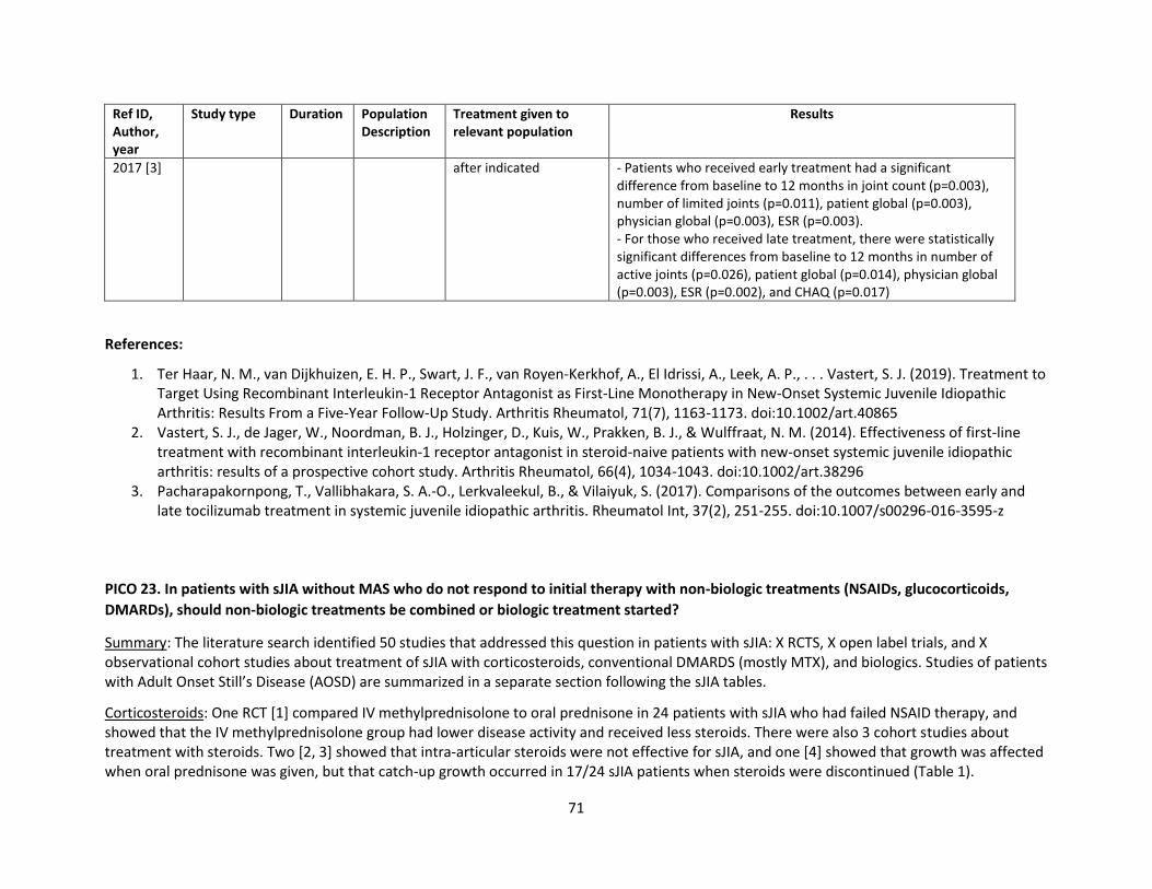
71
Ref ID, Author, year
Study type Duration Population Description
Treatment given to relevant population
Results
2017 [3]
after indicated - Patients who received early treatment had a significant difference from baseline to 12 months in joint count (p=0.003), number of limited joints (p=0.011), patient global (p=0.003), physician global (p=0.003), ESR (p=0.003). - For those who received late treatment, there were statistically significant differences from baseline to 12 months in number of active joints (p=0.026), patient global (p=0.014), physician global (p=0.003), ESR (p=0.002), and CHAQ (p=0.017)
References:
1. Ter Haar, N. M., van Dijkhuizen, E. H. P., Swart, J. F., van Royen-Kerkhof, A., El Idrissi, A., Leek, A. P., . . . Vastert, S. J. (2019). Treatment to Target Using Recombinant Interleukin-1 Receptor Antagonist as First-Line Monotherapy in New-Onset Systemic Juvenile Idiopathic Arthritis: Results From a Five-Year Follow-Up Study. Arthritis Rheumatol, 71(7), 1163-1173. doi:10.1002/art.40865
2. Vastert, S. J., de Jager, W., Noordman, B. J., Holzinger, D., Kuis, W., Prakken, B. J., & Wulffraat, N. M. (2014). Effectiveness of first-line treatment with recombinant interleukin-1 receptor antagonist in steroid-naive patients with new-onset systemic juvenile idiopathic arthritis: results of a prospective cohort study. Arthritis Rheumatol, 66(4), 1034-1043. doi:10.1002/art.38296
3. Pacharapakornpong, T., Vallibhakara, S. A.-O., Lerkvaleekul, B., & Vilaiyuk, S. (2017). Comparisons of the outcomes between early and late tocilizumab treatment in systemic juvenile idiopathic arthritis. Rheumatol Int, 37(2), 251-255. doi:10.1007/s00296-016-3595-z
PICO 23. In patients with sJIA without MAS who do not respond to initial therapy with non-biologic treatments (NSAIDs, glucocorticoids,
DMARDs), should non-biologic treatments be combined or biologic treatment started?
Summary: The literature search identified 50 studies that addressed this question in patients with sJIA: X RCTS, X open label trials, and X observational cohort studies about treatment of sJIA with corticosteroids, conventional DMARDS (mostly MTX), and biologics. Studies of patients with Adult Onset Still’s Disease (AOSD) are summarized in a separate section following the sJIA tables.
Corticosteroids: One RCT [1] compared IV methylprednisolone to oral prednisone in 24 patients with sJIA who had failed NSAID therapy, and showed that the IV methylprednisolone group had lower disease activity and received less steroids. There were also 3 cohort studies about treatment with steroids. Two [2, 3] showed that intra-articular steroids were not effective for sJIA, and one [4] showed that growth was affected when oral prednisone was given, but that catch-up growth occurred in 17/24 sJIA patients when steroids were discontinued (Table 1).
72
Conventional DMARDs: One RCT [5] found that MTX was marginally better than placebo for sJIA, but the finding was inconclusive due to serious imprecision in the effect estimate (Table 2). Five observational cohort studies [6, 7, 8, 9, 10] discussed the efficacy of MTX in sJIA, and found improvement/response/remission that ranged 63%-88% (Table 3). One study [7] stated that steroids were reduced because of MTX. One cohort study [11] showed that Cyclosporin A was effective in ~50% of 34 patients with sJIA and that steroid dose was reduced; however, was discontinued in many due to side effects. Finally, one cohort study [12] evaluated infection risk with different medications. Found that aHR was ~1.2 with anti-TNF OR anti-TNF +MTX vs. MTX. aHR was ~2 for steroids vs. MTX, and highest aHR for sJIA was 2.7 for Anakinra vs. MTX.
IL-1 inhibitors: Two RCTs[13, 14] compared IL-1 inhibitors (anakinra or canakinumab) to placebo in children with sJIA (Table 4). Il-1 inhibitors showed superiority over placebo for improvement in modified ACR Pedi 30, 50, 70, 90 and inactive disease at 1 month. Two additional RCTs[15,16] compared rilonacept to placebo and found superiority of rilonacept over placebo for ACR 30, 50, 70, and 90 at 1 month, but the difference in inactive disease did not reach statistical significance (Table 5).
Four observational studies specifically evaluated anakinra (Table 6). One retrospective cohort study[17] that used anakinra monotherapy in 10 patients reported a complete response in 80% of patients at a median follow-up of 14.5 months. Another cohort study[18] with a minimum 1-month follow-up reported a complete response to anakinra in 10/22 patients (45.5%). One study[19] found that earlier treatment with anakinra (closer to time of disease onset) led to higher likelihood of treatment response when compared to later treatment with anakinra. Another study[20] identified shorter disease duration as a factor increasing the likelihood of complete clinical response.
Three studies specifically evaluated canakinumab (Table 6). One prospective cohort study[21] reported that 69% of patients achieved remission with canakinumab and allowed tapering of corticosteroids by 48 weeks. However, serious adverse events occurred in 8/19 patients. A 5-year open label extension of two RCTs reported that 48.6% patients achieved remission at 2 years and was sustained until the study’s end, but 58% of patients discontinued treatment during the follow-up[22]. A smaller cohort study with 6 months follow-up reported an ACR pedi 90 rate of 20% after canakinumab initiation[23].
One cohort study[24] compared anakinra to canakinumab; the study found a higher rate of drug retention for canakinumab than anakinra, with a difference that was almost statistically significant (p = 0.056). Two cohort studies compared anakinra to tocilizumab; one study found no difference in treatment efficacy but did note patients were significantly more likely to continue tociluzumab than anakinra[25] The other study reported more non-responders among patients taking anakinra; some of these patients showed a treatment response when switched to tocilizumab (Table 6).[26]
IL-6 inhibitors (Tocilizumab): Two RCTs[28, 29] compared tocilizumab to placebo in patients with sJIA (Table 7); a meta-analysis of both trials showed that tocilizumab led to significantly greater improvement in ACR pedi 30, 50, 70, and 90 compared to placebo at 12 weeks, but the rate of infection with tocilizumab was also significantly higher compared to placebo. Five serious adverse events occurred in the tocilizumab group and none in the placebo group.
Eleven observational studies specifically evaluated tocilizumab (Table 8). One cohort study [30] showed that tocilizumab was safe and effective for sJIA patients, with 8/11 achieving inactive disease at 6 months. Another cohort study [31] showed that IV tocilizumab in children <2 years old
73
was equivalent to treatment of older children aged 2-17 years. Kostic et al.[32] reported that 40/48 patients with sJIA achieved inactive by approximately 4 months after tocilizumab initiation. One cohort study[33] reported that 54.5% of patients who received early treatment (within 6 months of indication for treatment) with tocilizumab achieved remission while no patients who received the dose late (>6 months after indication) achieved remission. A controlled cohort study[34] compared two different tocilizumab dosing schedules (every 2 weeks versus every 4 weeks). Patients in the 4-week group had a milder sJIA course on average and were less likely to have hepatosplenomegaly, coagulopathy, and central nervous system dysfunction. An open-label extension of an RCT[35] reported that patients who received tocilizumab had significant catch up growth when comparing pre-treatment to post treatment height velocity after one and two years. Another cohort study[36] reported that improvement in height velocity was associated with a decrease in corticosteroid dose after tocilizumab initiation. The remaining studies[37, 38, 39, 40] focused on adverse events related to tocilizumab therapy. Adverse events were mostly mild, but there were cases of serious adverse events including MAS. One study[40] of 66 patients reported serious adverse events in 32 patients, but stated that 54% of serious events were “mild.”
B cell inhibitors (Rituximab): One prospective cohort study[41] evaluated rituximab (1 IV infusion per week for 4 successive weeks) in 55 children with JIA (84% had sJIA). The study exclusively enrolled patients who had not responded to NSAIDs, corticosteroids, methotrexate, and TNF inhibitors. By 24 weeks 98% of patients achieved ACR 30 response and 25% achieved remission; by 48 weeks 52% achieved remission. At 96 weeks only 25 patients remained, and 44% were in remission (Table 8).
Nine studies evaluated multiple biologics within the same study[42, 43, 44, 45, 46, 47, 48, 49, 50] (Table 9). One cohort study evaluated predictors of need for biologic therapy, with baseline of steroids + MTX [42], and found that elevated ESR and neutrophil/lymphocyte ratio predicted need for biologic treatment. One study [43] reported that serious adverse event rates were higher with canakinumab and tocilizumab than anakinra and etanercept, although serious adverse event rates with etanercept and tocilizumab were highest when steroids were added. The remaining studies reported data that is more difficult to interpret.
Since most studies were observational designs that lacked relevant comparison groups and the few RCTs also provided only indirect evidence, the quality of evidence was very low.
Quality of evidence across all critical outcomes: Very low
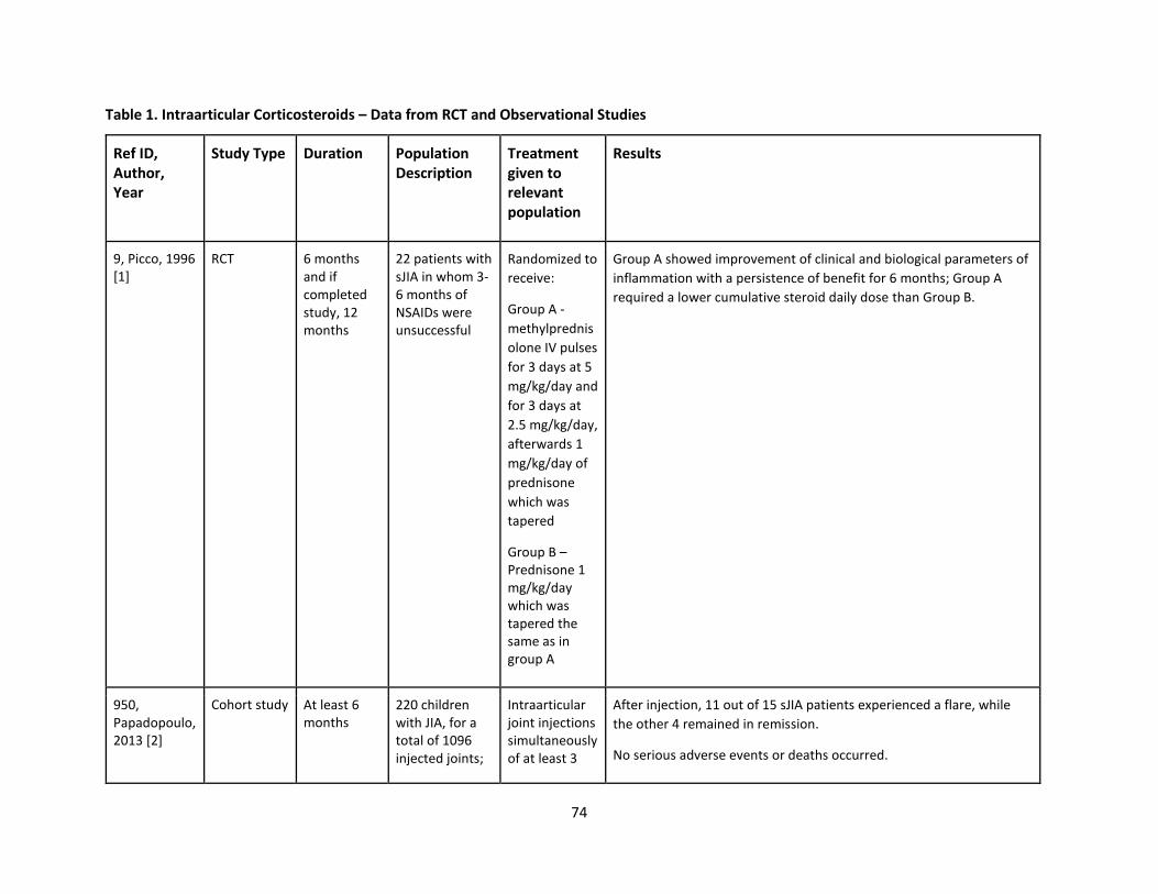
74
Table 1. Intraarticular Corticosteroids – Data from RCT and Observational Studies
Ref ID, Author, Year
Study Type Duration Population Description
Treatment given to relevant population
Results
9, Picco, 1996 [1]
RCT 6 months and if completed study, 12 months
22 patients with sJIA in whom 3-6 months of NSAIDs were unsuccessful
Randomized to
receive:
Group A -
methylprednis
olone IV pulses
for 3 days at 5
mg/kg/day and
for 3 days at
2.5 mg/kg/day,
afterwards 1
mg/kg/day of
prednisone
which was
tapered
Group B – Prednisone 1 mg/kg/day which was tapered the same as in group A
Group A showed improvement of clinical and biological parameters of
inflammation with a persistence of benefit for 6 months; Group A
required a lower cumulative steroid daily dose than Group B.
950, Papadopoulo, 2013 [2]
Cohort study At least 6 months
220 children with JIA, for a total of 1096 injected joints;
Intraarticular joint injections simultaneously of at least 3
After injection, 11 out of 15 sJIA patients experienced a flare, while
the other 4 remained in remission.
No serious adverse events or deaths occurred.
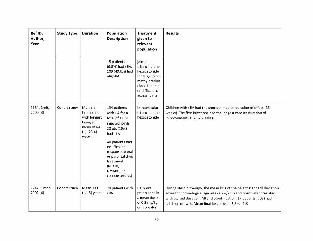
75
Ref ID, Author, Year
Study Type Duration Population Description
Treatment given to relevant population
Results
15 patients (6.8%) had sJIA, 109 (49.6%) had oligoJIA
joints: triamcinolone hexacetonide for large joints; methylprednisolone for small or difficult to access joints
3684, Breit, 2000 [3]
Cohort study Multiple time points with longest being a mean of 64 (+/- 23.4) weeks
194 patients
with JIA for a
total of 1439
injected joints;
20 pts (10%)
had sJIA
All patients had insufficient response to oral or parental drug treatment (NSAID, DMARD, or corticosteroids)
Intraarticular triamcinolone hexacetonide
Children with sJIA had the shortest median duration of effect (36
weeks). The first injections had the longest median duration of
improvement (sJIA 57 weeks).
2242, Simon, 2002 [4]
Cohort study Mean 13.6 (+/- 5) years
24 patients with
sJIA
Daily oral prednisone in a mean dose of 0.2 mg/kg or more during
During steroid therapy, the mean loss of the height standard deviation
score for chronological age was -2.7 +/- 1.5 and positively correlated
with steroid duration. After discontinuation, 17 patients (705) had
catch up growth. Mean final height was -2.8 +/- 1.8
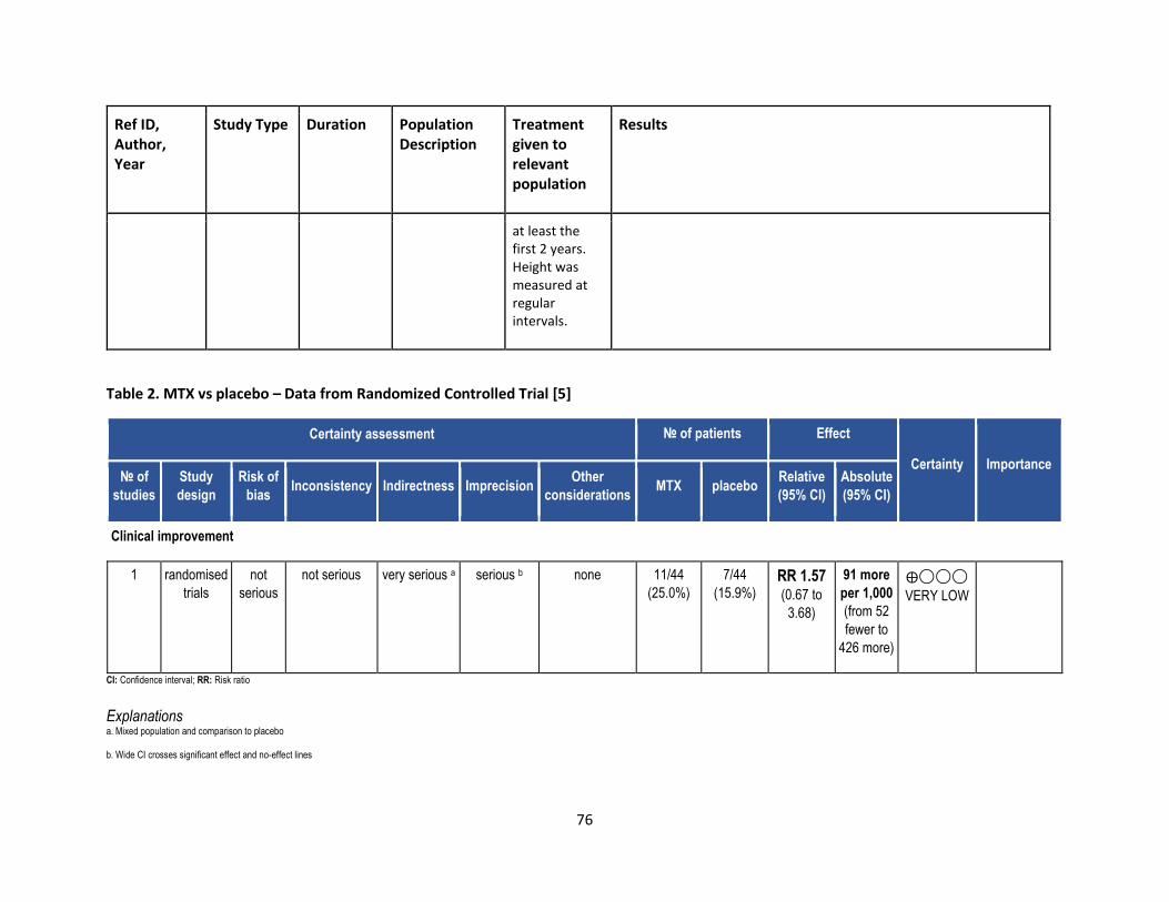
76
Ref ID, Author, Year
Study Type Duration Population Description
Treatment given to relevant population
Results
at least the first 2 years. Height was measured at regular intervals.
Table 2. MTX vs placebo – Data from Randomized Controlled Trial [5]
Certainty assessment № of patients Effect
Certainty Importance № of
studies
Study
design
Risk of
bias Inconsistency Indirectness Imprecision
Other
considerations MTX placebo
Relative
(95% CI)
Absolute
(95% CI)
Clinical improvement
1 randomised
trials
not
serious
not serious very serious a serious b none 11/44
(25.0%)
7/44
(15.9%) RR 1.57
(0.67 to
3.68)
91 more
per 1,000
(from 52
fewer to
426 more)
⨁◯◯◯
VERY LOW
CI: Confidence interval; RR: Risk ratio
Explanations a. Mixed population and comparison to placebo
b. Wide CI crosses significant effect and no-effect lines
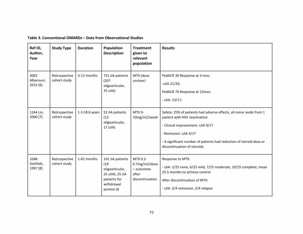
77
Table 3. Conventional DMARDs – Data from Observational Studies
Ref ID, Author, Year
Study Type Duration Population Description
Treatment given to relevant population
Results
3002 Albarouni, 2014 [6]
Retrospective cohort study
3-12 months 731 JIA patients
(207
oligoarticular,
25 sJIA)
MTX (dose
unclear)
PedACR 30 Response at 3 mos:
-sJIA 21/24;
PedACR 70 Response at 12mos:
- sJIA: 13/17;
1244 Lin, 2000 [7]
Retrospective cohort study
1.3-18.6 years 52 JIA patients
(13
oligoarticular,
17 sJIA)
MTX 9-
10mg/m2/week
Safety: 25% of patients had adverse effects, all minor aside from 1
patient with HSV reactivation
- Clinical improvement: sJIA 9/17
- Remission: sJIA 4/17
- A significant number of patients had reduction of steroid dose or
discontinuation of steroids
1048 Gottlieb, 1997 [8]
Retrospective cohort study
1-62 months 101 JIA patients
(19
oligoarticular,
25 sJIA); 25 JIA
patients for
withdrawal
portion (6
MTX 0.2-
0.7mg/m2/dose
– outcomes
after
discontinuation
Response to MTX:
- sJIA: 2/25 none, 6/25 mild, 7/25 moderate, 10/25 complete; mean
25.5 months to achieve control
After discontinuation of MTX:
- sJIA: 2/4 remission, 2/4 relapse
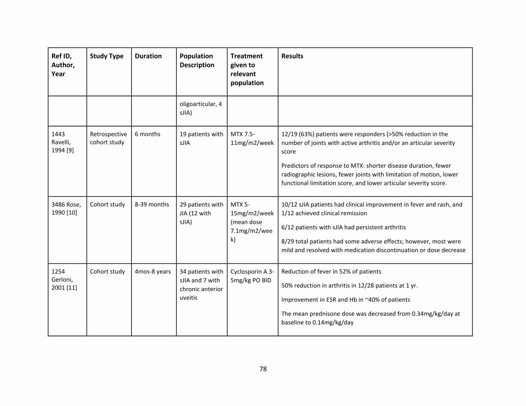
78
Ref ID, Author, Year
Study Type Duration Population Description
Treatment given to relevant population
Results
oligoarticular, 4
sJIA)
1443 Ravelli, 1994 [9]
Retrospective cohort study
6 months 19 patients with
sJIA
MTX 7.5-
11mg/m2/week
12/19 (63%) patients were responders (>50% reduction in the
number of joints with active arthritis and/or an articular severity
score
Predictors of response to MTX: shorter disease duration, fewer
radiographic lesions, fewer joints with limitation of motion, lower
functional limitation score, and lower articular severity score.
3486 Rose, 1990 [10]
Cohort study 8-39 months 29 patients with
JIA (12 with
sJIA)
MTX 5-
15mg/m2/week
(mean dose
7.1mg/m2/wee
k)
10/12 sJIA patients had clinical improvement in fever and rash, and
1/12 achieved clinical remission
6/12 patients with sJIA had persistent arthritis
8/29 total patients had some adverse effects; however, most were
mild and resolved with medication discontinuation or dose decrease
1254 Gerloni, 2001 [11]
Cohort study 4mos-8 years 34 patients with
sJIA and 7 with
chronic anterior
uveitis
Cyclosporin A 3-
5mg/kg PO BID
Reduction of fever in 52% of patients
50% reduction in arthritis in 12/28 patients at 1 yr.
Improvement in ESR and Hb in ~40% of patients
The mean prednisone dose was decreased from 0.34mg/kg/day at
baseline to 0.14mg/kg/day
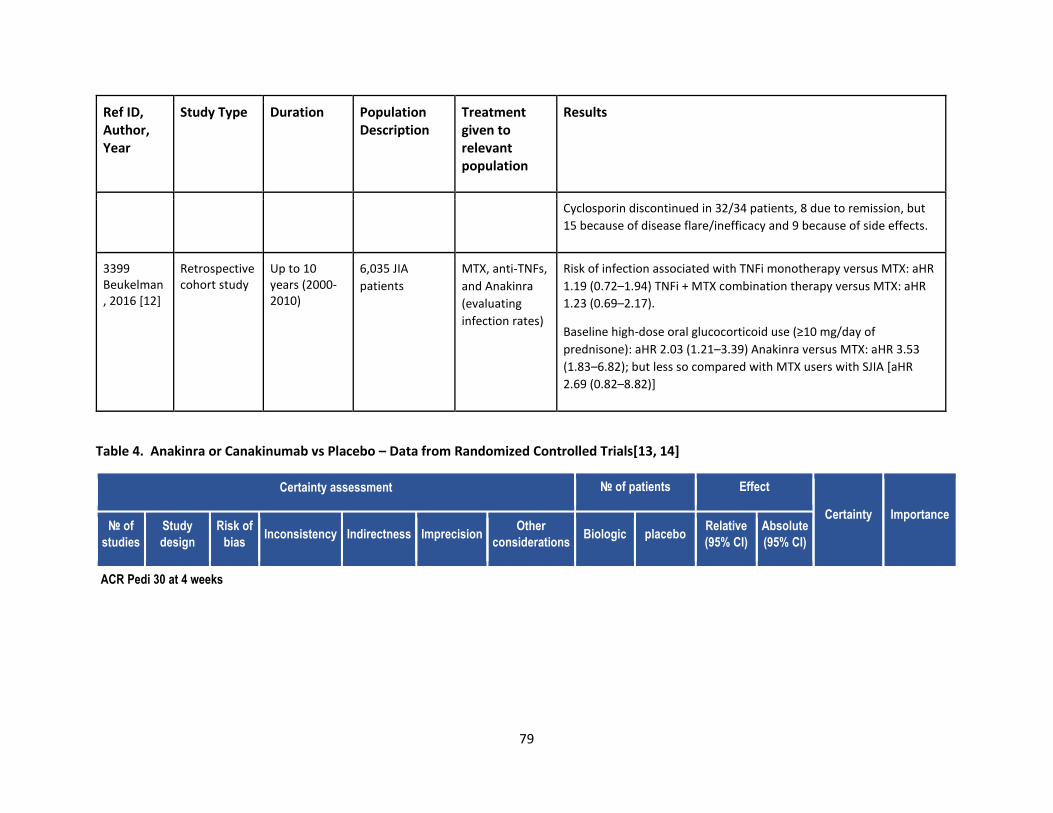
79
Ref ID, Author, Year
Study Type Duration Population Description
Treatment given to relevant population
Results
Cyclosporin discontinued in 32/34 patients, 8 due to remission, but
15 because of disease flare/inefficacy and 9 because of side effects.
3399 Beukelman, 2016 [12]
Retrospective cohort study
Up to 10 years (2000-2010)
6,035 JIA
patients
MTX, anti-TNFs,
and Anakinra
(evaluating
infection rates)
Risk of infection associated with TNFi monotherapy versus MTX: aHR
1.19 (0.72–1.94) TNFi + MTX combination therapy versus MTX: aHR
1.23 (0.69–2.17).
Baseline high-dose oral glucocorticoid use (≥10 mg/day of
prednisone): aHR 2.03 (1.21–3.39) Anakinra versus MTX: aHR 3.53
(1.83–6.82); but less so compared with MTX users with SJIA [aHR
2.69 (0.82–8.82)]
Table 4. Anakinra or Canakinumab vs Placebo – Data from Randomized Controlled Trials[13, 14]
Certainty assessment № of patients Effect
Certainty Importance № of
studies
Study
design
Risk of
bias Inconsistency Indirectness Imprecision
Other
considerations Biologic placebo
Relative
(95% CI)
Absolute
(95% CI)
ACR Pedi 30 at 4 weeks
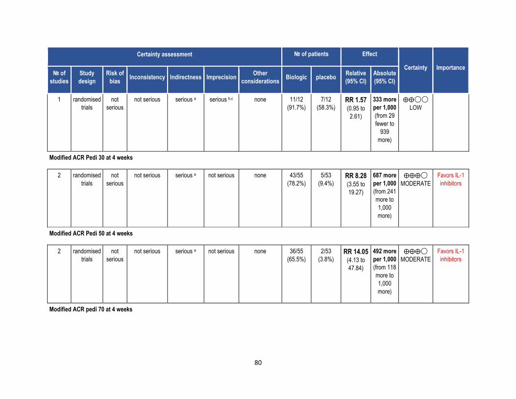
80
Certainty assessment № of patients Effect
Certainty Importance № of
studies
Study
design
Risk of
bias Inconsistency Indirectness Imprecision
Other
considerations Biologic placebo
Relative
(95% CI)
Absolute
(95% CI)
1 randomised
trials
not
serious
not serious serious a serious b,c none 11/12
(91.7%)
7/12
(58.3%) RR 1.57
(0.95 to
2.61)
333 more
per 1,000
(from 29
fewer to
939
more)
⨁⨁◯◯
LOW
Modified ACR Pedi 30 at 4 weeks
2 randomised
trials
not
serious
not serious serious a not serious none 43/55
(78.2%)
5/53
(9.4%) RR 8.28
(3.55 to
19.27)
687 more
per 1,000
(from 241
more to
1,000
more)
⨁⨁⨁◯
MODERATE
Favors IL-1
inhibitors
Modified ACR Pedi 50 at 4 weeks
2 randomised
trials
not
serious
not serious serious a not serious none 36/55
(65.5%)
2/53
(3.8%) RR 14.05
(4.13 to
47.84)
492 more
per 1,000
(from 118
more to
1,000
more)
⨁⨁⨁◯
MODERATE
Favors IL-1
inhibitors
Modified ACR pedi 70 at 4 weeks
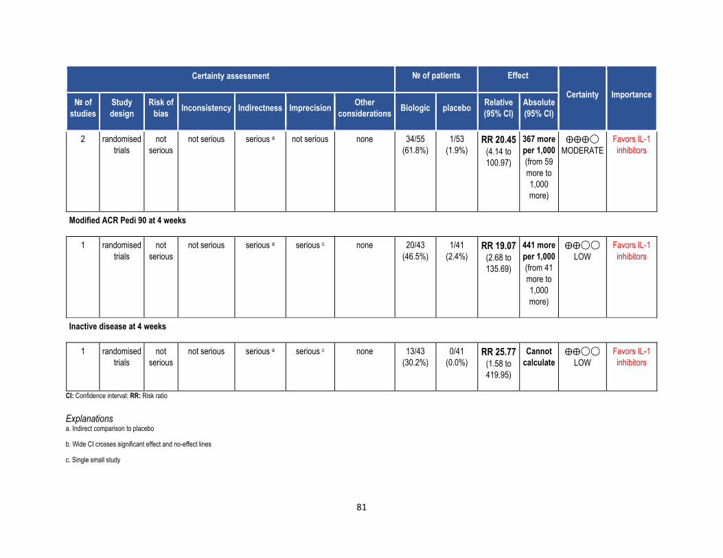
81
Certainty assessment № of patients Effect
Certainty Importance № of
studies
Study
design
Risk of
bias Inconsistency Indirectness Imprecision
Other
considerations Biologic placebo
Relative
(95% CI)
Absolute
(95% CI)
2 randomised
trials
not
serious
not serious serious a not serious none 34/55
(61.8%)
1/53
(1.9%) RR 20.45
(4.14 to
100.97)
367 more
per 1,000
(from 59
more to
1,000
more)
⨁⨁⨁◯
MODERATE
Favors IL-1
inhibitors
Modified ACR Pedi 90 at 4 weeks
1 randomised
trials
not
serious
not serious serious a serious c none 20/43
(46.5%)
1/41
(2.4%) RR 19.07
(2.68 to
135.69)
441 more
per 1,000
(from 41
more to
1,000
more)
⨁⨁◯◯
LOW
Favors IL-1
inhibitors
Inactive disease at 4 weeks
1 randomised
trials
not
serious
not serious serious a serious c none 13/43
(30.2%)
0/41
(0.0%) RR 25.77
(1.58 to
419.95)
Cannot
calculate
⨁⨁◯◯
LOW
Favors IL-1
inhibitors
CI: Confidence interval; RR: Risk ratio
Explanations a. Indirect comparison to placebo
b. Wide CI crosses significant effect and no-effect lines
c. Single small study
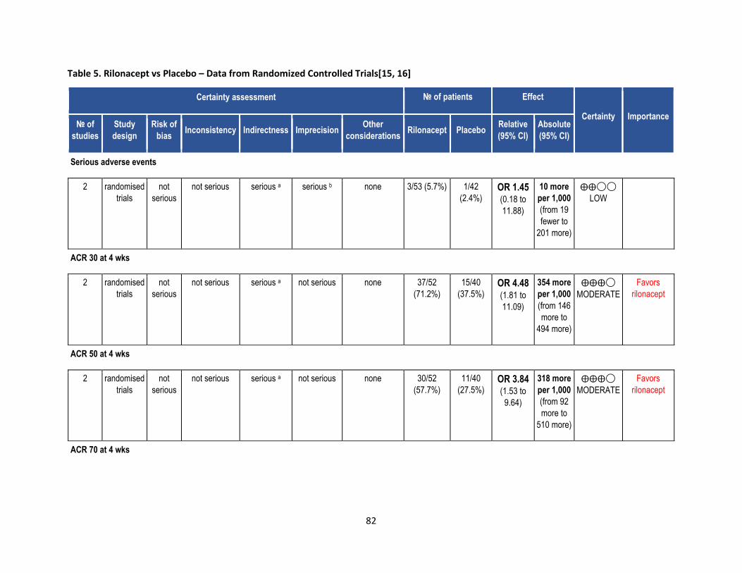
82
Table 5. Rilonacept vs Placebo – Data from Randomized Controlled Trials[15, 16]
Certainty assessment № of patients Effect
Certainty Importance № of
studies
Study
design
Risk of
bias Inconsistency Indirectness Imprecision
Other
considerations Rilonacept Placebo
Relative
(95% CI)
Absolute
(95% CI)
Serious adverse events
2 randomised
trials
not
serious
not serious serious a serious b none 3/53 (5.7%) 1/42
(2.4%) OR 1.45
(0.18 to
11.88)
10 more
per 1,000
(from 19
fewer to
201 more)
⨁⨁◯◯
LOW
ACR 30 at 4 wks
2 randomised
trials
not
serious
not serious serious a not serious none 37/52
(71.2%)
15/40
(37.5%) OR 4.48
(1.81 to
11.09)
354 more
per 1,000
(from 146
more to
494 more)
⨁⨁⨁◯
MODERATE
Favors
rilonacept
ACR 50 at 4 wks
2 randomised
trials
not
serious
not serious serious a not serious none 30/52
(57.7%)
11/40
(27.5%) OR 3.84
(1.53 to
9.64)
318 more
per 1,000
(from 92
more to
510 more)
⨁⨁⨁◯
MODERATE
Favors
rilonacept
ACR 70 at 4 wks
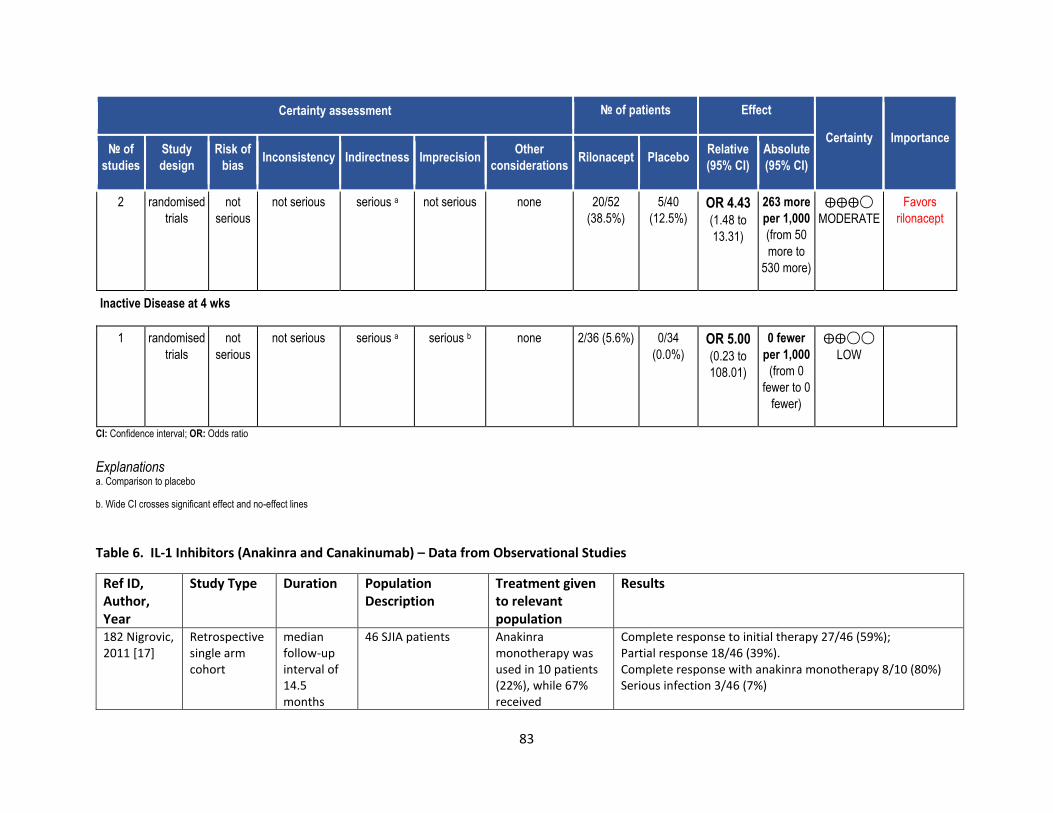
83
Certainty assessment № of patients Effect
Certainty Importance № of
studies
Study
design
Risk of
bias Inconsistency Indirectness Imprecision
Other
considerations Rilonacept Placebo
Relative
(95% CI)
Absolute
(95% CI)
2 randomised
trials
not
serious
not serious serious a not serious none 20/52
(38.5%)
5/40
(12.5%) OR 4.43
(1.48 to
13.31)
263 more
per 1,000
(from 50
more to
530 more)
⨁⨁⨁◯
MODERATE
Favors
rilonacept
Inactive Disease at 4 wks
1 randomised
trials
not
serious
not serious serious a serious b none 2/36 (5.6%) 0/34
(0.0%) OR 5.00
(0.23 to
108.01)
0 fewer
per 1,000
(from 0
fewer to 0
fewer)
⨁⨁◯◯
LOW
CI: Confidence interval; OR: Odds ratio
Explanations a. Comparison to placebo
b. Wide CI crosses significant effect and no-effect lines
Table 6. IL-1 Inhibitors (Anakinra and Canakinumab) – Data from Observational Studies
Ref ID, Author, Year
Study Type Duration Population Description
Treatment given to relevant population
Results
182 Nigrovic, 2011 [17]
Retrospective single arm cohort
median follow-up interval of 14.5 months
46 SJIA patients Anakinra monotherapy was used in 10 patients (22%), while 67% received
Complete response to initial therapy 27/46 (59%); Partial response 18/46 (39%). Complete response with anakinra monotherapy 8/10 (80%) Serious infection 3/46 (7%)
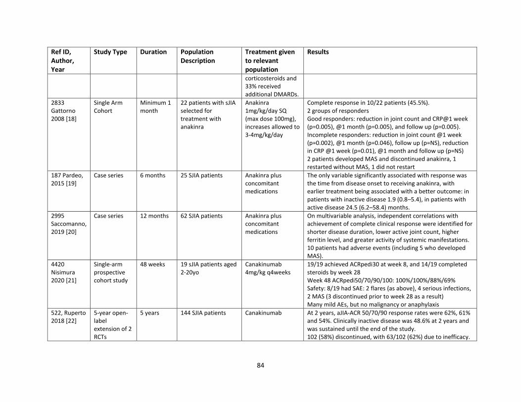
84
Ref ID, Author, Year
Study Type Duration Population Description
Treatment given to relevant population
Results
corticosteroids and 33% received additional DMARDs.
2833 Gattorno 2008 [18]
Single Arm Cohort
Minimum 1 month
22 patients with sJIA selected for treatment with anakinra
Anakinra 1mg/kg/day SQ (max dose 100mg), increases allowed to 3-4mg/kg/day
Complete response in 10/22 patients (45.5%). 2 groups of responders Good responders: reduction in joint count and CRP@1 week (p=0.005), @1 month (p=0.005), and follow up (p=0.005). Incomplete responders: reduction in joint count @1 week (p=0.002), @1 month (p=0.046), follow up (p=NS), reduction in CRP @1 week (p=0.01), @1 month and follow up (p=NS) 2 patients developed MAS and discontinued anakinra, 1 restarted without MAS, 1 did not restart
187 Pardeo, 2015 [19]
Case series 6 months 25 SJIA patients Anakinra plus concomitant medications
The only variable significantly associated with response was the time from disease onset to receiving anakinra, with earlier treatment being associated with a better outcome: in patients with inactive disease 1.9 (0.8–5.4), in patients with active disease 24.5 (6.2–58.4) months.
2995 Saccomanno, 2019 [20]
Case series 12 months 62 SJIA patients Anakinra plus concomitant medications
On multivariable analysis, independent correlations with achievement of complete clinical response were identified for shorter disease duration, lower active joint count, higher ferritin level, and greater activity of systemic manifestations. 10 patients had adverse events (including 5 who developed MAS).
4420 Nisimura 2020 [21]
Single-arm prospective cohort study
48 weeks 19 sJIA patients aged 2-20yo
Canakinumab 4mg/kg q4weeks
19/19 achieved ACRpedi30 at week 8, and 14/19 completed steroids by week 28 Week 48 ACRpedi50/70/90/100: 100%/100%/88%/69% Safety: 8/19 had SAE: 2 flares (as above), 4 serious infections, 2 MAS (3 discontinued prior to week 28 as a result) Many mild AEs, but no malignancy or anaphylaxis
522, Ruperto 2018 [22]
5-year open-label extension of 2 RCTs
5 years 144 SJIA patients Canakinumab At 2 years, aJIA-ACR 50/70/90 response rates were 62%, 61% and 54%. Clinically inactive disease was 48.6% at 2 years and was sustained until the end of the study. 102 (58%) discontinued, with 63/102 (62%) due to inefficacy.
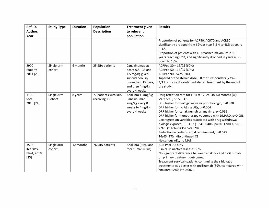
85
Ref ID, Author, Year
Study Type Duration Population Description
Treatment given to relevant population
Results
Proportion of patients for ACR50, ACR70 and ACR90 significantly dropped from 69% at year 3.5-4 to 48% at years 4-4.5. Proportion of patients with CID reached maximum in 1.5 years reaching 62%, and significantly dropped in years 4.5-5 down to 18%
2900 Ruperto, 2011 [23]
Single-arm cohort
6 months 25 SJIA patients Canakinumab at doses 0.5, 1.5 and 4.5 mg/kg given subcutaneously during first 15 days, and then 4mg/kg every 4 weeks
ACRPedi30 – 15/25 (60%) ACRPedi50 – 15/25 (60%) ACRPedi90 - 5/25 (20%) Tapered of the steroid dose – 8 of 11 responders (73%); 4/11 of those discontinued steroid treatment by the end of the study.
1105 Sota 2018 [24]
Single Arm Cohort
8 years 77 patients with sJIA receiving IL-1i
Anakinra 1-4mg/kg Canakinumab 2mg/kg every 8 weeks to 4mg/kg every 4 weeks
Drug retention rate for IL-1i at 12, 24, 48, 60 months (%): 79.9, 59.5, 53.5, 53.5 DRR higher for biologic naïve vs prior biologic, p=0.038 DRR higher for no AEs vs AEs, p=0.004 DRR higher for canakinumab vs anakinra, p=0.056 DRR higher for monotherapy vs combo with DMARD, p=0.058 Cox regression variables associated with drug withdrawal: biologic exposed (HR 3.37 (1.341-8.406) p=0.01) and AEs (HR 2.970 (1.186-7.435) p=0.020) Reduction in corticosteroid requirement, p=0.025 16/63 (27%) discontinued CS No serious AEs, no MAS
3596 Kearsley-Fleet, 2019 [25]
Single arm cohort
12 months 76 SJIA patients Anakinra (86%) and tocilizumab (63%)
ACR Pedi 90: 42% Clinically inactive disease: 39% No significant difference between anakinra and tocilizumab on primary treatment outcomes. Treatment survival (patients continuing their biologic treatment) was better with tocilizumab (89%) compared with anakinra (59%; P = 0.002).
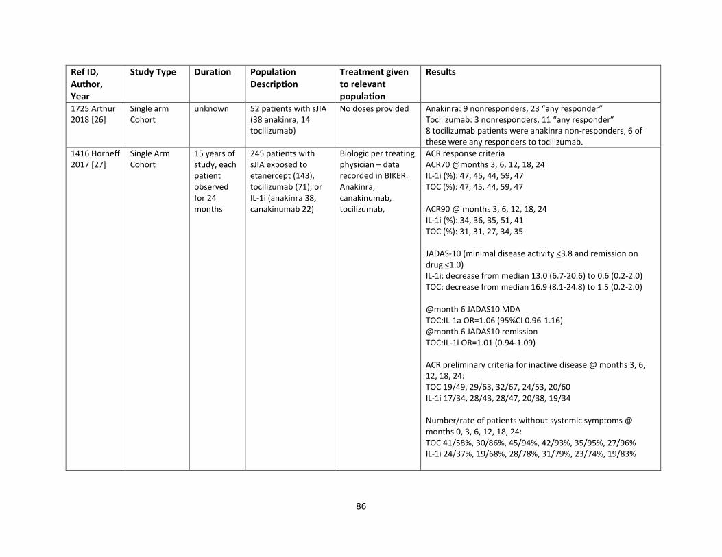
86
Ref ID, Author, Year
Study Type Duration Population Description
Treatment given to relevant population
Results
1725 Arthur 2018 [26]
Single arm Cohort
unknown 52 patients with sJIA (38 anakinra, 14 tocilizumab)
No doses provided Anakinra: 9 nonresponders, 23 “any responder” Tocilizumab: 3 nonresponders, 11 “any responder” 8 tocilizumab patients were anakinra non-responders, 6 of these were any responders to tocilizumab.
1416 Horneff 2017 [27]
Single Arm Cohort
15 years of study, each patient observed for 24 months
245 patients with sJIA exposed to etanercept (143), tocilizumab (71), or IL-1i (anakinra 38, canakinumab 22)
Biologic per treating physician – data recorded in BIKER. Anakinra, canakinumab, tocilizumab,
ACR response criteria ACR70 @months 3, 6, 12, 18, 24 IL-1i (%): 47, 45, 44, 59, 47 TOC (%): 47, 45, 44, 59, 47 ACR90 @ months 3, 6, 12, 18, 24 IL-1i (%): 34, 36, 35, 51, 41 TOC (%): 31, 31, 27, 34, 35 JADAS-10 (minimal disease activity <3.8 and remission on drug <1.0) IL-1i: decrease from median 13.0 (6.7-20.6) to 0.6 (0.2-2.0) TOC: decrease from median 16.9 (8.1-24.8) to 1.5 (0.2-2.0) @month 6 JADAS10 MDA TOC:IL-1a OR=1.06 (95%CI 0.96-1.16) @month 6 JADAS10 remission TOC:IL-1i OR=1.01 (0.94-1.09) ACR preliminary criteria for inactive disease @ months 3, 6, 12, 18, 24: TOC 19/49, 29/63, 32/67, 24/53, 20/60 IL-1i 17/34, 28/43, 28/47, 20/38, 19/34 Number/rate of patients without systemic symptoms @ months 0, 3, 6, 12, 18, 24: TOC 41/58%, 30/86%, 45/94%, 42/93%, 35/95%, 27/96% IL-1i 24/37%, 19/68%, 28/78%, 31/79%, 23/74%, 19/83%
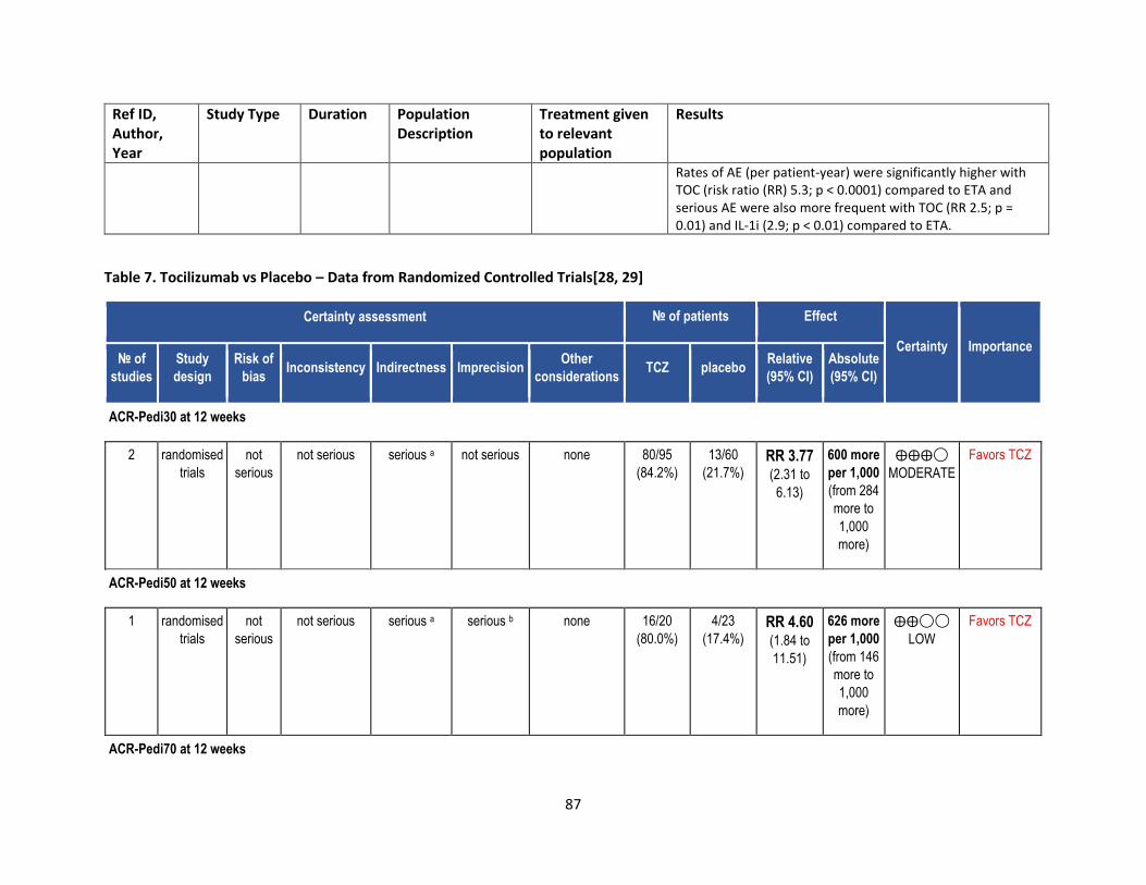
87
Ref ID, Author, Year
Study Type Duration Population Description
Treatment given to relevant population
Results
Rates of AE (per patient-year) were significantly higher with TOC (risk ratio (RR) 5.3; p < 0.0001) compared to ETA and serious AE were also more frequent with TOC (RR 2.5; p = 0.01) and IL-1i (2.9; p < 0.01) compared to ETA.
Table 7. Tocilizumab vs Placebo – Data from Randomized Controlled Trials[28, 29]
Certainty assessment № of patients Effect
Certainty Importance № of
studies
Study
design
Risk of
bias Inconsistency Indirectness Imprecision
Other
considerations TCZ placebo
Relative
(95% CI)
Absolute
(95% CI)
ACR-Pedi30 at 12 weeks
2 randomised
trials
not
serious
not serious serious a not serious none 80/95
(84.2%)
13/60
(21.7%) RR 3.77
(2.31 to
6.13)
600 more
per 1,000
(from 284
more to
1,000
more)
⨁⨁⨁◯
MODERATE
Favors TCZ
ACR-Pedi50 at 12 weeks
1 randomised
trials
not
serious
not serious serious a serious b none 16/20
(80.0%)
4/23
(17.4%) RR 4.60
(1.84 to
11.51)
626 more
per 1,000
(from 146
more to
1,000
more)
⨁⨁◯◯
LOW
Favors TCZ
ACR-Pedi70 at 12 weeks
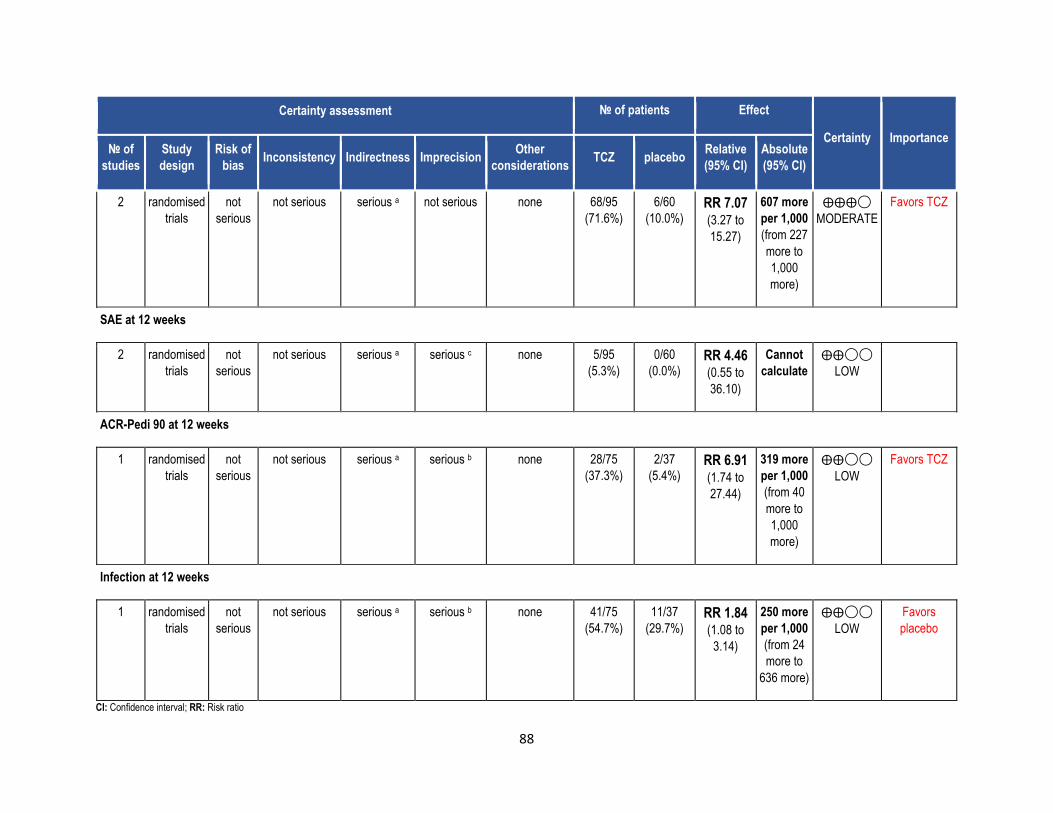
88
Certainty assessment № of patients Effect
Certainty Importance № of
studies
Study
design
Risk of
bias Inconsistency Indirectness Imprecision
Other
considerations TCZ placebo
Relative
(95% CI)
Absolute
(95% CI)
2 randomised
trials
not
serious
not serious serious a not serious none 68/95
(71.6%)
6/60
(10.0%) RR 7.07
(3.27 to
15.27)
607 more
per 1,000
(from 227
more to
1,000
more)
⨁⨁⨁◯
MODERATE
Favors TCZ
SAE at 12 weeks
2 randomised
trials
not
serious
not serious serious a serious c none 5/95
(5.3%)
0/60
(0.0%) RR 4.46
(0.55 to
36.10)
Cannot
calculate
⨁⨁◯◯
LOW
ACR-Pedi 90 at 12 weeks
1 randomised
trials
not
serious
not serious serious a serious b none 28/75
(37.3%)
2/37
(5.4%) RR 6.91
(1.74 to
27.44)
319 more
per 1,000
(from 40
more to
1,000
more)
⨁⨁◯◯
LOW
Favors TCZ
Infection at 12 weeks
1 randomised
trials
not
serious
not serious serious a serious b none 41/75
(54.7%)
11/37
(29.7%) RR 1.84
(1.08 to
3.14)
250 more
per 1,000
(from 24
more to
636 more)
⨁⨁◯◯
LOW
Favors
placebo
CI: Confidence interval; RR: Risk ratio
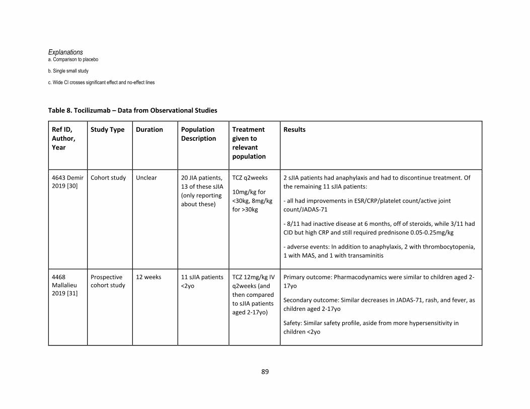
89
Explanations a. Comparison to placebo
b. Single small study
c. Wide CI crosses significant effect and no-effect lines
Table 8. Tocilizumab – Data from Observational Studies
Ref ID, Author, Year
Study Type Duration Population Description
Treatment given to relevant population
Results
4643 Demir 2019 [30]
Cohort study Unclear 20 JIA patients,
13 of these sJIA
(only reporting
about these)
TCZ q2weeks
10mg/kg for
<30kg, 8mg/kg
for >30kg
2 sJIA patients had anaphylaxis and had to discontinue treatment. Of
the remaining 11 sJIA patients:
- all had improvements in ESR/CRP/platelet count/active joint
count/JADAS-71
- 8/11 had inactive disease at 6 months, off of steroids, while 3/11 had
CID but high CRP and still required prednisone 0.05-0.25mg/kg
- adverse events: In addition to anaphylaxis, 2 with thrombocytopenia,
1 with MAS, and 1 with transaminitis
4468 Mallalieu 2019 [31]
Prospective cohort study
12 weeks 11 sJIA patients
<2yo
TCZ 12mg/kg IV
q2weeks (and
then compared
to sJIA patients
aged 2-17yo)
Primary outcome: Pharmacodynamics were similar to children aged 2-
17yo
Secondary outcome: Similar decreases in JADAS-71, rash, and fever, as
children aged 2-17yo
Safety: Similar safety profile, aside from more hypersensitivity in
children <2yo
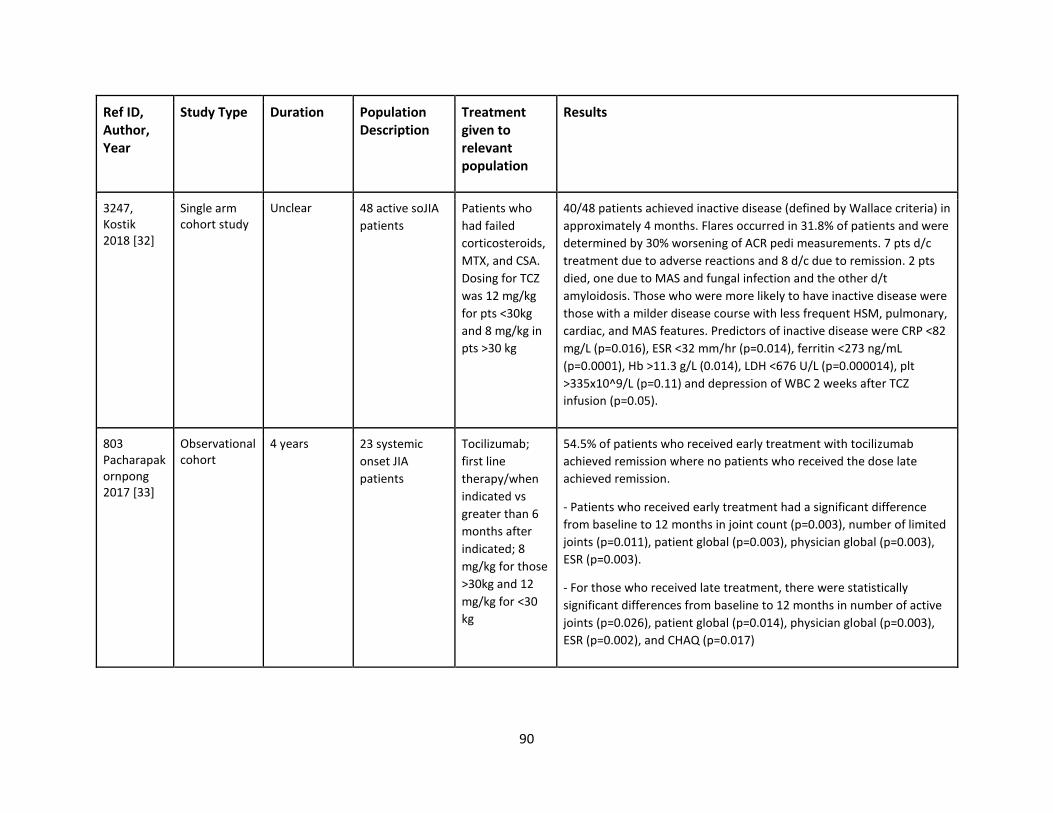
90
Ref ID, Author, Year
Study Type Duration Population Description
Treatment given to relevant population
Results
3247, Kostik 2018 [32]
Single arm cohort study
Unclear 48 active soJIA
patients
Patients who
had failed
corticosteroids,
MTX, and CSA.
Dosing for TCZ
was 12 mg/kg
for pts <30kg
and 8 mg/kg in
pts >30 kg
40/48 patients achieved inactive disease (defined by Wallace criteria) in
approximately 4 months. Flares occurred in 31.8% of patients and were
determined by 30% worsening of ACR pedi measurements. 7 pts d/c
treatment due to adverse reactions and 8 d/c due to remission. 2 pts
died, one due to MAS and fungal infection and the other d/t
amyloidosis. Those who were more likely to have inactive disease were
those with a milder disease course with less frequent HSM, pulmonary,
cardiac, and MAS features. Predictors of inactive disease were CRP <82
mg/L (p=0.016), ESR <32 mm/hr (p=0.014), ferritin <273 ng/mL
(p=0.0001), Hb >11.3 g/L (0.014), LDH <676 U/L (p=0.000014), plt
>335x10^9/L (p=0.11) and depression of WBC 2 weeks after TCZ
infusion (p=0.05).
803 Pacharapakornpong 2017 [33]
Observational cohort
4 years 23 systemic
onset JIA
patients
Tocilizumab;
first line
therapy/when
indicated vs
greater than 6
months after
indicated; 8
mg/kg for those
>30kg and 12
mg/kg for <30
kg
54.5% of patients who received early treatment with tocilizumab
achieved remission where no patients who received the dose late
achieved remission.
- Patients who received early treatment had a significant difference
from baseline to 12 months in joint count (p=0.003), number of limited
joints (p=0.011), patient global (p=0.003), physician global (p=0.003),
ESR (p=0.003).
- For those who received late treatment, there were statistically
significant differences from baseline to 12 months in number of active
joints (p=0.026), patient global (p=0.014), physician global (p=0.003),
ESR (p=0.002), and CHAQ (p=0.017)
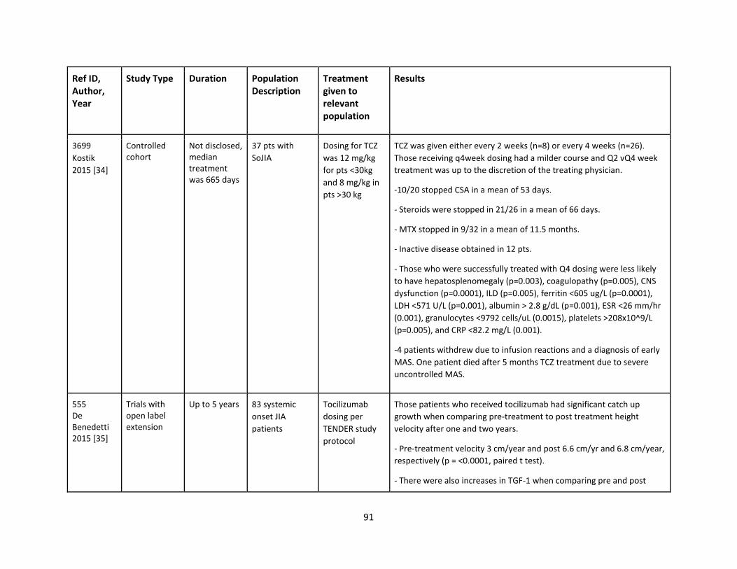
91
Ref ID, Author, Year
Study Type Duration Population Description
Treatment given to relevant population
Results
3699
Kostik
2015 [34]
Controlled cohort
Not disclosed, median treatment was 665 days
37 pts with
SoJIA
Dosing for TCZ
was 12 mg/kg
for pts <30kg
and 8 mg/kg in
pts >30 kg
TCZ was given either every 2 weeks (n=8) or every 4 weeks (n=26).
Those receiving q4week dosing had a milder course and Q2 vQ4 week
treatment was up to the discretion of the treating physician.
-10/20 stopped CSA in a mean of 53 days.
- Steroids were stopped in 21/26 in a mean of 66 days.
- MTX stopped in 9/32 in a mean of 11.5 months.
- Inactive disease obtained in 12 pts.
- Those who were successfully treated with Q4 dosing were less likely
to have hepatosplenomegaly (p=0.003), coagulopathy (p=0.005), CNS
dysfunction (p=0.0001), ILD (p=0.005), ferritin <605 ug/L (p=0.0001),
LDH <571 U/L (p=0.001), albumin > 2.8 g/dL (p=0.001), ESR <26 mm/hr
(0.001), granulocytes <9792 cells/uL (0.0015), platelets >208x10^9/L
(p=0.005), and CRP <82.2 mg/L (0.001).
-4 patients withdrew due to infusion reactions and a diagnosis of early
MAS. One patient died after 5 months TCZ treatment due to severe
uncontrolled MAS.
555 De Benedetti 2015 [35]
Trials with open label extension
Up to 5 years 83 systemic
onset JIA
patients
Tocilizumab
dosing per
TENDER study
protocol
Those patients who received tocilizumab had significant catch up
growth when comparing pre-treatment to post treatment height
velocity after one and two years.
- Pre-treatment velocity 3 cm/year and post 6.6 cm/yr and 6.8 cm/year,
respectively (p = <0.0001, paired t test).
- There were also increases in TGF-1 when comparing pre and post
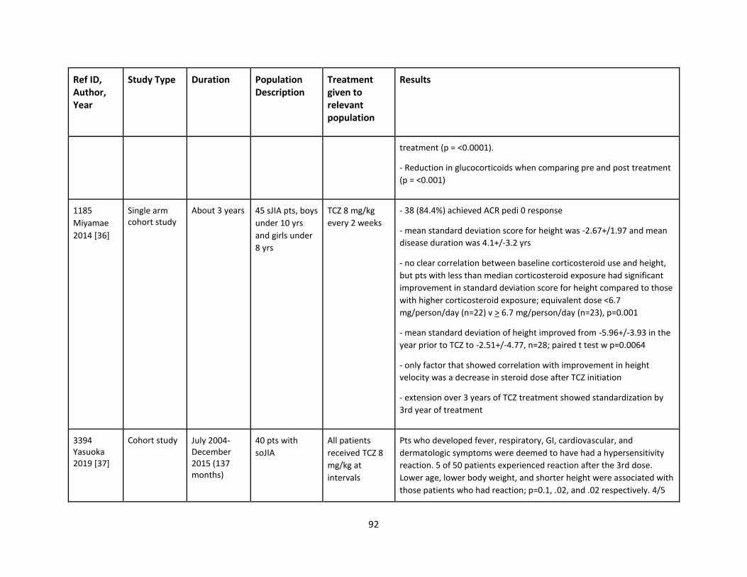
92
Ref ID, Author, Year
Study Type Duration Population Description
Treatment given to relevant population
Results
treatment (p = <0.0001).
- Reduction in glucocorticoids when comparing pre and post treatment
(p = <0.001)
1185
Miyamae
2014 [36]
Single arm cohort study
About 3 years 45 sJIA pts, boys
under 10 yrs
and girls under
8 yrs
TCZ 8 mg/kg
every 2 weeks
- 38 (84.4%) achieved ACR pedi 0 response
- mean standard deviation score for height was -2.67+/1.97 and mean
disease duration was 4.1+/-3.2 yrs
- no clear correlation between baseline corticosteroid use and height,
but pts with less than median corticosteroid exposure had significant
improvement in standard deviation score for height compared to those
with higher corticosteroid exposure; equivalent dose <6.7
mg/person/day (n=22) v > 6.7 mg/person/day (n=23), p=0.001
- mean standard deviation of height improved from -5.96+/-3.93 in the
year prior to TCZ to -2.51+/-4.77, n=28; paired t test w p=0.0064
- only factor that showed correlation with improvement in height
velocity was a decrease in steroid dose after TCZ initiation
- extension over 3 years of TCZ treatment showed standardization by
3rd year of treatment
3394 Yasuoka 2019 [37]
Cohort study July 2004-December 2015 (137 months)
40 pts with
soJIA
All patients
received TCZ 8
mg/kg at
intervals
Pts who developed fever, respiratory, GI, cardiovascular, and
dermatologic symptoms were deemed to have had a hypersensitivity
reaction. 5 of 50 patients experienced reaction after the 3rd dose.
Lower age, lower body weight, and shorter height were associated with
those patients who had reaction; p=0.1, .02, and .02 respectively. 4/5
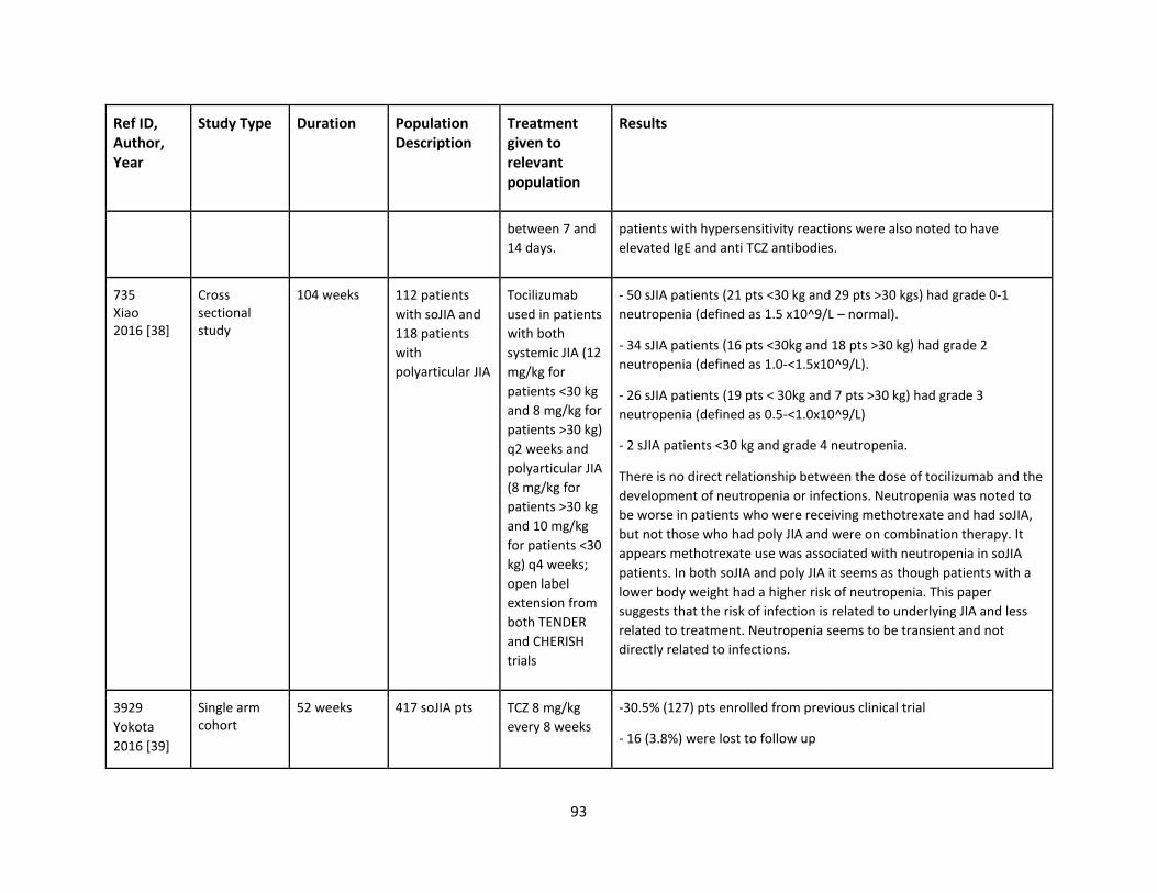
93
Ref ID, Author, Year
Study Type Duration Population Description
Treatment given to relevant population
Results
between 7 and
14 days.
patients with hypersensitivity reactions were also noted to have
elevated IgE and anti TCZ antibodies.
735 Xiao 2016 [38]
Cross sectional study
104 weeks 112 patients
with soJIA and
118 patients
with
polyarticular JIA
Tocilizumab
used in patients
with both
systemic JIA (12
mg/kg for
patients <30 kg
and 8 mg/kg for
patients >30 kg)
q2 weeks and
polyarticular JIA
(8 mg/kg for
patients >30 kg
and 10 mg/kg
for patients <30
kg) q4 weeks;
open label
extension from
both TENDER
and CHERISH
trials
- 50 sJIA patients (21 pts <30 kg and 29 pts >30 kgs) had grade 0-1
neutropenia (defined as 1.5 x10^9/L – normal).
- 34 sJIA patients (16 pts <30kg and 18 pts >30 kg) had grade 2
neutropenia (defined as 1.0-<1.5x10^9/L).
- 26 sJIA patients (19 pts < 30kg and 7 pts >30 kg) had grade 3
neutropenia (defined as 0.5-<1.0x10^9/L)
- 2 sJIA patients <30 kg and grade 4 neutropenia.
There is no direct relationship between the dose of tocilizumab and the
development of neutropenia or infections. Neutropenia was noted to
be worse in patients who were receiving methotrexate and had soJIA,
but not those who had poly JIA and were on combination therapy. It
appears methotrexate use was associated with neutropenia in soJIA
patients. In both soJIA and poly JIA it seems as though patients with a
lower body weight had a higher risk of neutropenia. This paper
suggests that the risk of infection is related to underlying JIA and less
related to treatment. Neutropenia seems to be transient and not
directly related to infections.
3929
Yokota
2016 [39]
Single arm cohort
52 weeks 417 soJIA pts TCZ 8 mg/kg
every 8 weeks
-30.5% (127) pts enrolled from previous clinical trial
- 16 (3.8%) were lost to follow up
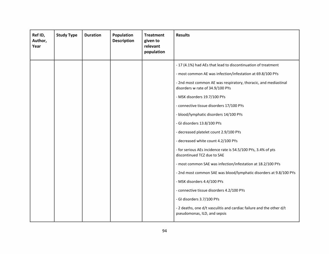
94
Ref ID, Author, Year
Study Type Duration Population Description
Treatment given to relevant population
Results
- 17 (4.1%) had AEs that lead to discontinuation of treatment
- most common AE was infection/infestation at 69.8/100 PYs
- 2nd most common AE was respiratory, thoracic, and mediastinal
disorders w rate of 34.9/100 PYs
- MSK disorders 19.7/100 PYs
- connective tissue disorders 17/100 PYs
- blood/lymphatic disorders 14/100 PYs
- GI disorders 13.8/100 PYs
- decreased platelet count 2.9/100 PYs
- decreased white count 4.2/100 PYs
- for serious AEs incidence rate is 54.5/100 PYs, 3.4% of pts
discontinued TCZ due to SAE
- most common SAE was infection/infestation at 18.2/100 PYs
- 2nd most common SAE was blood/lymphatic disorders at 9.8/100 PYs
- MSK disorders 4.4/100 PYs
- connective tissue disorders 4.2/100 PYs
- GI disorders 3.7/100 PYs
- 2 deaths, one d/t vasculitis and cardiac failure and the other d/t
pseudomonas, ILD, and sepsis
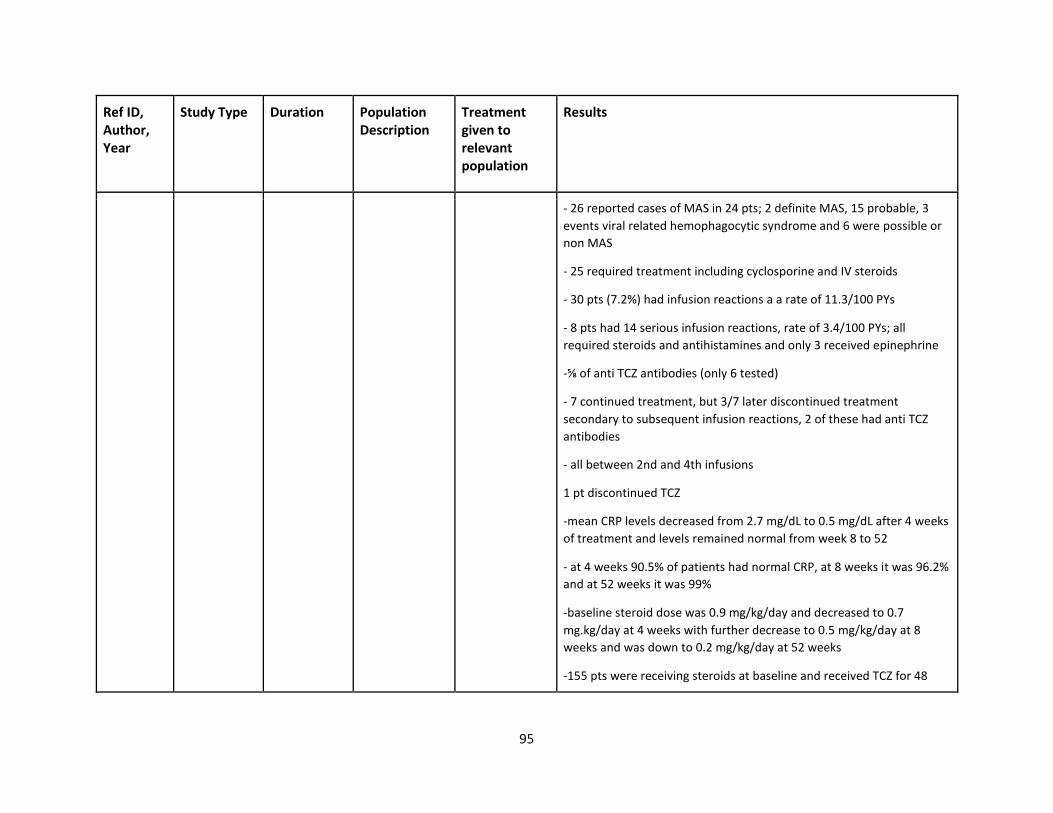
95
Ref ID, Author, Year
Study Type Duration Population Description
Treatment given to relevant population
Results
- 26 reported cases of MAS in 24 pts; 2 definite MAS, 15 probable, 3
events viral related hemophagocytic syndrome and 6 were possible or
non MAS
- 25 required treatment including cyclosporine and IV steroids
- 30 pts (7.2%) had infusion reactions a a rate of 11.3/100 PYs
- 8 pts had 14 serious infusion reactions, rate of 3.4/100 PYs; all
required steroids and antihistamines and only 3 received epinephrine
-⅝ of anti TCZ antibodies (only 6 tested)
- 7 continued treatment, but 3/7 later discontinued treatment
secondary to subsequent infusion reactions, 2 of these had anti TCZ
antibodies
- all between 2nd and 4th infusions
1 pt discontinued TCZ
-mean CRP levels decreased from 2.7 mg/dL to 0.5 mg/dL after 4 weeks
of treatment and levels remained normal from week 8 to 52
- at 4 weeks 90.5% of patients had normal CRP, at 8 weeks it was 96.2%
and at 52 weeks it was 99%
-baseline steroid dose was 0.9 mg/kg/day and decreased to 0.7
mg.kg/day at 4 weeks with further decrease to 0.5 mg/kg/day at 8
weeks and was down to 0.2 mg/kg/day at 52 weeks
-155 pts were receiving steroids at baseline and received TCZ for 48
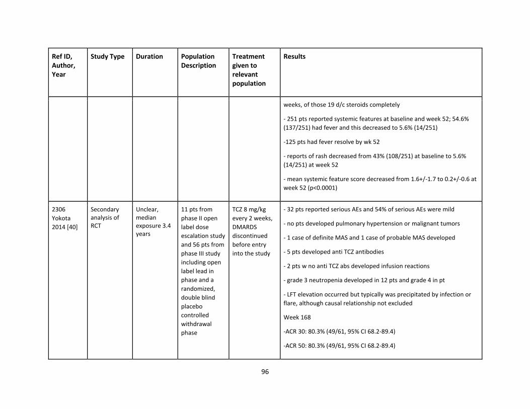
96
Ref ID, Author, Year
Study Type Duration Population Description
Treatment given to relevant population
Results
weeks, of those 19 d/c steroids completely
- 251 pts reported systemic features at baseline and week 52; 54.6%
(137/251) had fever and this decreased to 5.6% (14/251)
-125 pts had fever resolve by wk 52
- reports of rash decreased from 43% (108/251) at baseline to 5.6%
(14/251) at week 52
- mean systemic feature score decreased from 1.6+/-1.7 to 0.2+/-0.6 at
week 52 (p<0.0001)
2306
Yokota
2014 [40]
Secondary analysis of RCT
Unclear, median exposure 3.4 years
11 pts from
phase II open
label dose
escalation study
and 56 pts from
phase III study
including open
label lead in
phase and a
randomized,
double blind
placebo
controlled
withdrawal
phase
TCZ 8 mg/kg
every 2 weeks,
DMARDS
discontinued
before entry
into the study
- 32 pts reported serious AEs and 54% of serious AEs were mild
- no pts developed pulmonary hypertension or malignant tumors
- 1 case of definite MAS and 1 case of probable MAS developed
- 5 pts developed anti TCZ antibodies
- 2 pts w no anti TCZ abs developed infusion reactions
- grade 3 neutropenia developed in 12 pts and grade 4 in pt
- LFT elevation occurred but typically was precipitated by infection or
flare, although causal relationship not excluded
Week 168
-ACR 30: 80.3% (49/61, 95% CI 68.2-89.4)
-ACR 50: 80.3% (49/61, 95% CI 68.2-89.4)
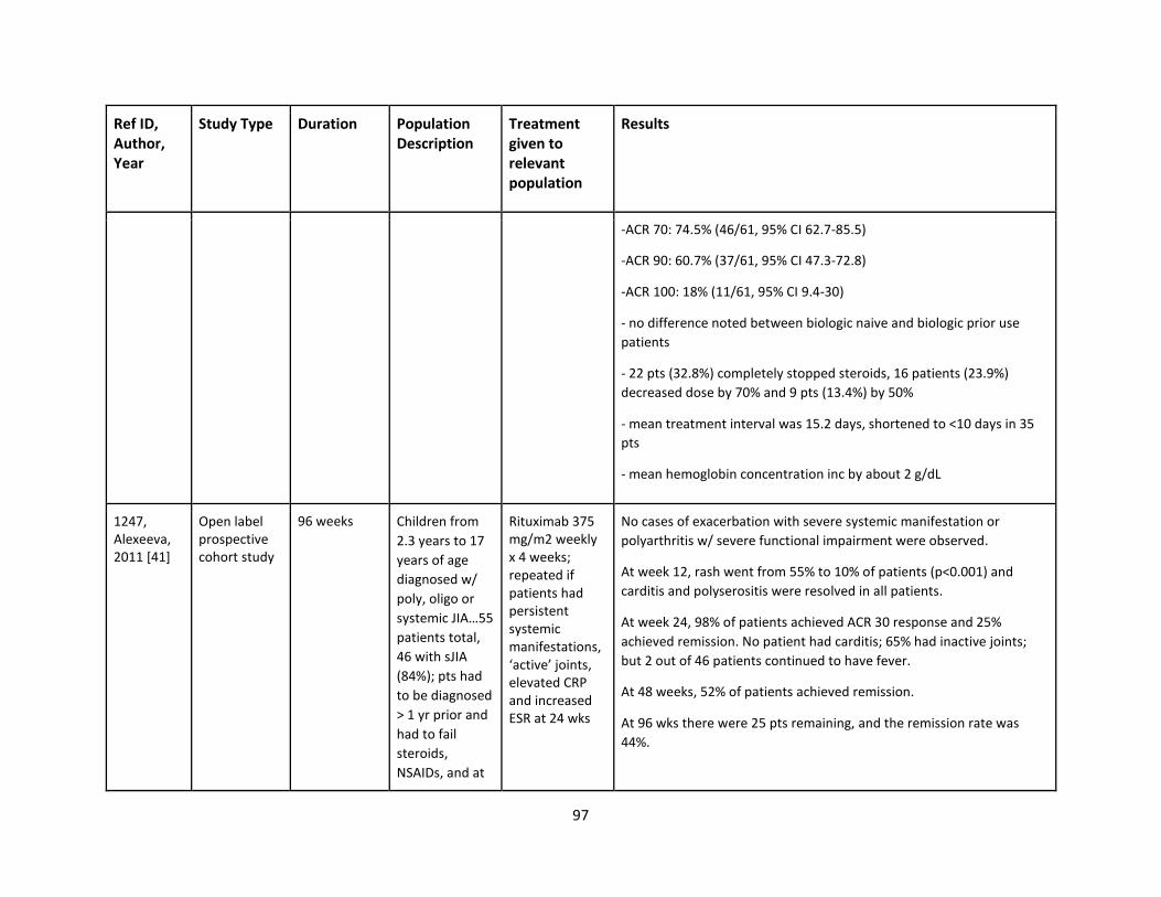
97
Ref ID, Author, Year
Study Type Duration Population Description
Treatment given to relevant population
Results
-ACR 70: 74.5% (46/61, 95% CI 62.7-85.5)
-ACR 90: 60.7% (37/61, 95% CI 47.3-72.8)
-ACR 100: 18% (11/61, 95% CI 9.4-30)
- no difference noted between biologic naive and biologic prior use
patients
- 22 pts (32.8%) completely stopped steroids, 16 patients (23.9%)
decreased dose by 70% and 9 pts (13.4%) by 50%
- mean treatment interval was 15.2 days, shortened to <10 days in 35
pts
- mean hemoglobin concentration inc by about 2 g/dL
1247, Alexeeva, 2011 [41]
Open label prospective cohort study
96 weeks Children from
2.3 years to 17
years of age
diagnosed w/
poly, oligo or
systemic JIA…55
patients total,
46 with sJIA
(84%); pts had
to be diagnosed
> 1 yr prior and
had to fail
steroids,
NSAIDs, and at
Rituximab 375 mg/m2 weekly x 4 weeks; repeated if patients had persistent systemic manifestations, ‘active’ joints, elevated CRP and increased ESR at 24 wks
No cases of exacerbation with severe systemic manifestation or
polyarthritis w/ severe functional impairment were observed.
At week 12, rash went from 55% to 10% of patients (p<0.001) and
carditis and polyserositis were resolved in all patients.
At week 24, 98% of patients achieved ACR 30 response and 25%
achieved remission. No patient had carditis; 65% had inactive joints;
but 2 out of 46 patients continued to have fever.
At 48 weeks, 52% of patients achieved remission.
At 96 wks there were 25 pts remaining, and the remission rate was
44%.
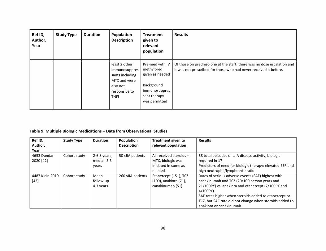
98
Ref ID, Author, Year
Study Type Duration Population Description
Treatment given to relevant population
Results
least 2 other
immunosuppres
sants including
MTX and were
also not
responsive to
TNFi
Pre-med with IV methylpred given as needed
Background
immunosuppres
sant therapy
was permitted
Of those on prednisolone at the start, there was no dose escalation and
it was not prescribed for those who had never received it before.
Table 9. Multiple Biologic Medications – Data from Observational Studies
Ref ID, Author, Year
Study Type Duration Population Description
Treatment given to relevant population
Results
4653 Dundar 2020 [42]
Cohort study 2-6.8 years, median 3.3 years
50 sJIA patients All received steroids + MTX, biologic was initiated in some as needed
58 total episodes of sJIA disease activity, biologic required in 17 Predictors of need for biologic therapy: elevated ESR and high neutrophil/lymphocyte ratio
4487 Klein 2019 [43]
Cohort study Mean follow-up 4.3 years
260 sJIA patients Etanercept (151), TCZ (109), anakinra (71), canakinumab (51)
Rates of serious adverse events (SAE) highest with canakinumab and TCZ (20/100 person years and 21/100PY) vs. anakinra and etanercept (7/100PY and 4/100PY) SAE rates higher when steroids added to etanercept or TCZ, but SAE rate did not change when steroids added to anakinra or canakinumab
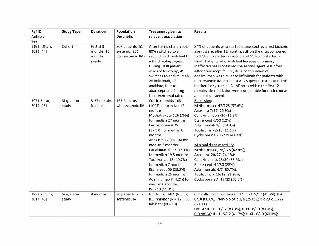
99
Ref ID, Author, Year
Study Type Duration Population Description
Treatment given to relevant population
Results
1191, Otten, 2013 [44]
Cohort F/U at 3 months, 15 months, yearly
307 patients (51 systemic, 256 non systemic JIA)
After failing etanercept, 80% switched to a second, 22% switched to a third biologic agent; During 1030 patient years of follow up, 49 switches to adalimumab, 28 infliximab, 17 anakinra, four to abatacept and 4 drug trials were evaluated.
84% of patients who started etanercept as a first biologic agent were, after 12 months, still on the drug compared to 47% who started a second and 51% who started a third. Patients who switched because of primary ineffectiveness continued the second agent less often. After etanercept failure, drug continuation of adalimumab was similar to infliximab for patients with non systemic JIA. Anakinra was superior to a second TNF blocker for systemic JIA. AE rates within the first 12 months after initiation were comparable for each course and biologic agent.
3071 Barut, 2019 [45]
Single arm study
3-27 months (median)
165 Patients with systemic JIA
Corticosteroids 168 (100%) for median 12 months; Methotrexate 126 (75%) for median 27 months; Cyclosporine A 29 (17.3%) for median 8 months; Anakinra 27 (16.1%) for median 3 months; Canakinumab 27 (16.1%) for median 19.5 months; Tocilizumab 18 (10.7%) for median 7 months; Etanercept 50 (29.8%) for median 25 months; Adalimumab 7 (4.2%) for median 6 months; IVIG 19 (11.3%)
Remission: Methotrexate 47/125 (37.6%) Anakinra 7/27 (25.9%) Canakinumab 3/30 (11.5%) Etanercept 6/50 (12%) Adalimumab 1/7 (14.3%) Tocilizumab 2/18 (11.1%) Cyclosporine A 12/29 (41.4%) Minimal disease activity: Methotrexate, 78/125 (62.4%); Anakinra, 20/27 (74.1%); Canakinumab, 23/30 (88.5%); Etanercept, 44/50 (88%); Adalimumab, 6/7 (85.7%); Tocilizumab, 16/18 (88.9%); Cyclosporine A, 17/29 (58.6%).
2933 Kimura, 2017 [46]
Single arm study
9 months 30 patients with systemic JIA
GC (N = 2), MTX (N = 6), IL1 Inhibitor (N = 12), IL6 Inhibitor (N = 10)
Clinically inactive disease (CID): IL-1i 5/12 (41.7%); IL-6i 6/10 (60.0%); Non-biologic 2/8 (25.0%); Biologic 11/22 (50.0%) Off GC: IL-1i - 10/12 (83.3%); IL-6i - 8/10 (80.0%); CID off GC: IL-1i - 5/12 (41.7%); IL-6i - 6/10 (60.0%);
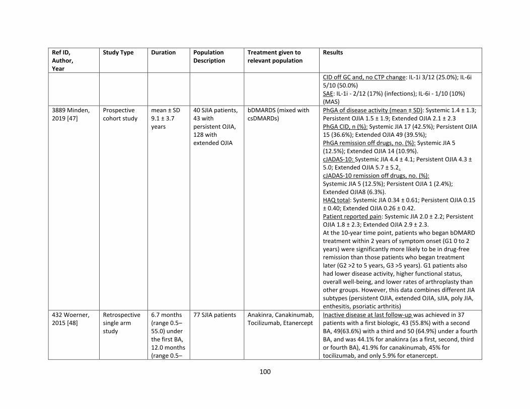
100
Ref ID, Author, Year
Study Type Duration Population Description
Treatment given to relevant population
Results
CID off GC and, no CTP change: IL-1i 3/12 (25.0%); IL-6i 5/10 (50.0%) SAE: IL-1i - 2/12 (17%) (infections); IL-6i - 1/10 (10%) (MAS)
3889 Minden, 2019 [47]
Prospective cohort study
mean ± SD 9.1 ± 3.7 years
40 SJIA patients, 43 with persistent OJIA, 128 with extended OJIA
bDMARDS (mixed with csDMARDs)
PhGA of disease activity (mean ± SD): Systemic 1.4 ± 1.3; Persistent OJIA 1.5 ± 1.9; Extended OJIA 2.1 ± 2.3 PhGA CID, n (%): Systemic JIA 17 (42.5%); Persistent OJIA 15 (36.6%); Extended OJIA 49 (39.5%); PhGA remission off drugs, no. (%): Systemic JIA 5 (12.5%); Extended OJIA 14 (10.9%). cJADAS-10: Systemic JIA 4.4 ± 4.1; Persistent OJIA 4.3 ± 5.0; Extended OJIA 5.7 ± 5.2. cJADAS-10 remission off drugs, no. (%): Systemic JIA 5 (12.5%); Persistent OJIA 1 (2.4%); Extended OJIA8 (6.3%). HAQ total: Systemic JIA 0.34 ± 0.61; Persistent OJIA 0.15 ± 0.40; Extended OJIA 0.26 ± 0.42. Patient reported pain: Systemic JIA 2.0 ± 2.2; Persistent OJIA 1.8 ± 2.3; Extended OJIA 2.9 ± 2.3. At the 10-year time point, patients who began bDMARD treatment within 2 years of symptom onset (G1 0 to 2 years) were significantly more likely to be in drug-free remission than those patients who began treatment later (G2 >2 to 5 years, G3 >5 years). G1 patients also had lower disease activity, higher functional status, overall well-being, and lower rates of arthroplasty than other groups. However, this data combines different JIA subtypes (persistent OJIA, extended OJIA, sJIA, poly JIA, enthesitis, psoriatic arthritis)
432 Woerner, 2015 [48]
Retrospective single arm study
6.7 months (range 0.5–55.0) under the first BA, 12.0 months (range 0.5–
77 SJIA patients Anakinra, Canakinumab, Tocilizumab, Etanercept
Inactive disease at last follow-up was achieved in 37 patients with a first biologic, 43 (55.8%) with a second BA, 49(63.6%) with a third and 50 (64.9%) under a fourth BA, and was 44.1% for anakinra (as a first, second, third or fourth BA), 41.9% for canakinumab, 45% for tocilizumab, and only 5.9% for etanercept.
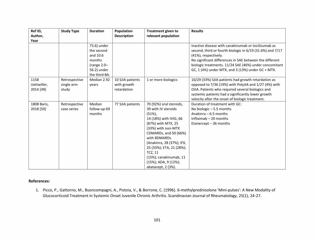
101
Ref ID, Author, Year
Study Type Duration Population Description
Treatment given to relevant population
Results
73.6) under the second and 10.6 months (range 2.0–56.2) under the third BA.
Inactive disease with canakinumab or tocilizumab as second, third or fourth biologic in 6/19 (31.6%) and 7/17 (41%), respectively. No significant differences in SAE between the different biologic treatments. 11/24 SAE (46%) under concomitant GC, 1 (4%) under MTX, and 3 (13%) under GC + MTX.
1158 Uettwiller, 2014 [49]
Retrospective single arm study
Median 2.92 years
10 SJIA patients with growth retardation
1 or more biologics 10/29 (33%) SJIA patients had growth retardation as opposed to 7/36 (19%) with PolyJIA and 1/27 (4%) with OJIA. Patients who required several biologics and systemic patients had a significantly lower growth velocity after the onset of biologic treatment.
1808 Baris, 2018 [50]
Retrospective case series
Median follow-up 69 months
77 SJIA patients 70 (92%) oral steroids, 39 with IV steroids (51%), 14 (18%) with IVIG, 66 (87%) with MTX, 25 (33%) with non-MTX CDMARDs, and 50 (66%) with BDMARDs (Anakinra, 28 (37%); IFX, 25 (33%); ETA, 21 (28%); TCZ, 11 (15%); canakinumab, 11 (15%); ADA, 9 (12%); abatacept, 2 (3%).
Duration of treatment with GC: No biologic – 5.5 months Anakinra – 6.5 months Infliximab – 20 months Etanercept – 36 months
References:
1. Picco, P., Gattorno, M., Buoncompagni, A., Pistoia, V., & Borrone, C. (1996). 6-methylprednisolone 'Mini-pulses': A New Modality of
Glucocorticoid Treatment in Systemic Onset Juvenile Chronic Arthritis. Scandinavian Journal of Rheumatology, 25(1), 24-27.
102
2. Papadopoulou, C., Kostik, M., Gonzalez-Fernandez, M. I., Bohm, M., Nieto-Gonzalez, J. C., Pistorio, A., . . . Ravelli, A. (2013). Delineating
the role of multiple intraarticular corticosteroid injections in the management of juvenile idiopathic arthritis in the biologic era. Arthritis
Care Res (Hoboken), 65(7), 1112-1120. doi:10.1002/acr.21947
3. Breit, W., Frosch, M., Meyer, U., Heinecke, A., & Ganser, G. (2000). A subgroup-specific evaluation of the efficacy of intraarticular
triamcinolone hexacetonide in juvenile chronic arthritis. J Rheumatol, 27(11), 2696-2702.
4. Simon, D., Fernando, C., Czernichow, P., & Prieur, A. M. (2002). Linear growth and final height in patients with systemic juvenile
idiopathic arthritis treated with longterm glucocorticoids. J Rheumatol, 29(6), 1296-1300.
5. Woo, P., Southwood, T. R., Prieur, A. M., Doré, C. J., Grainger, J., David, J., . . . Lemelle, I. (2000). Randomized, placebo-controlled,
crossover trial of low-dose oral methotrexate in children with extended oligoarticular or systemic arthritis. Arthritis Rheum, 43(8), 1849-
1857. doi:10.1002/1529-0131(200008)43:8<1849::aid-anr22>3.0.co;2-f
6. Albarouni, M., Becker, I., & Horneff, G. (2014). Predictors of response to methotrexate in juvenile idiopathic arthritis. Pediatr Rheumatol
Online J, 12, 35. doi:10.1186/1546-0096-12-35
7. Lin, Y. T., Tsai, M. J., Wang, L. H., Huang, M. T., Yang, Y. H., & Chiang, B. L. (2000). Efficacy and safety of methotrexate therapy for juvenile
rheumatoid arthritis. J Formos Med Assoc, 99(8), 623-629.
8. Gottlieb, B. S., Keenan, G. F., Lu, T., & Ilowite, N. T. (1997). Discontinuation of methotrexate treatment in juvenile rheumatoid arthritis.
Pediatrics, 100(6), 994-997. doi:10.1542/peds.100.6.994
9. Ravelli, A., Ramenghi, B., Di Fuccia, G., Ruperto, N., Zonta, L., & Martini, A. (1994). Factors associated with response to methotrexate in
systemic-onset juvenile chronic arthritis. Acta Paediatr, 83(4), 428-432. doi:10.1111/j.1651-2227.1994.tb18135.x
10. Rose, C. D., Singsen, B. H., Eichenfield, A. H., Goldsmith, D. P., & Athreya, B. H. (1990). Safety and efficacy of methotrexate therapy for
juvenile rheumatoid arthritis. J Pediatr, 117(4), 653-659. doi:https://doi.org/10.1016/S0022-3476(05)80709-7
11. Gerloni, V., Cimaz, R., Gattinara, M., Arnoldi, C., Pontikaki, I., & Fantini, F. (2001). Efficacy and safety profile of cyclosporin A in the
treatment of juvenile chronic (idiopathic) arthritis. Results of a 10-year prospective study. Rheumatology (Oxford), 40(8), 907-913.
doi:10.1093/rheumatology/40.8.907
12. Beukelman, T., Xie, F., Baddley, J. W., Chen, L., Mannion, M. L., Saag, K. G., . . . Curtis, J. R. (2016). The risk of hospitalized infection
following initiation of biologic agents versus methotrexate in the treatment of juvenile idiopathic arthritis. Arthritis Res Ther, 18(1), 210.
doi:10.1186/s13075-016-1109-8
13. Quartier, P., Allantaz, F., Cimaz, R., Pillet, P., Messiaen, C., Bardin, C., . . . Pascual, V. (2011). A multicentre, randomised, double-blind,
placebo-controlled trial with the interleukin-1 receptor antagonist anakinra in patients with systemic-onset juvenile idiopathic arthritis
(ANAJIS trial). Ann Rheum Dis, 70(5), 747-754. doi:10.1136/ard.2010.134254
14. Ruperto, N., Brunner, H. I., Quartier, P., Constantin, T., Wulffraat, N., Horneff, G., . . . Lovell, D. J. (2012). Two randomized trials of
canakinumab in systemic juvenile idiopathic arthritis. N Engl J Med, 367(25), 2396-2406. doi:10.1056/NEJMoa1205099
103
15. Lovell, D. J., Giannini, E. H., Reiff, A. O., Kimura, Y., Li, S., Hashkes, P. J., . . . Radin, A. R. (2013). Long-term safety and efficacy of rilonacept
in patients with systemic juvenile idiopathic arthritis. Arthritis Rheum, 65(9), 2486-2496. doi:10.1002/art.38042
16. Ilowite, N. T., Prather, K., Lokhnygina, Y., Schanberg, L. E., Elder, M., Milojevic, D., . . . Sandborg, C. I. (2014). Randomized, double-blind,
placebo-controlled trial of the efficacy and safety of rilonacept in the treatment of systemic juvenile idiopathic arthritis. Arthritis
Rheumatol, 66(9), 2570-2579. doi:10.1002/art.38699
17. Nigrovic, P. A., Mannion, M., Prince, F. H., Zeft, A., Rabinovich, C. E., van Rossum, M. A., . . . Higgins, G. C. (2011). Anakinra as first-line
disease-modifying therapy in systemic juvenile idiopathic arthritis: report of forty-six patients from an international multicenter series.
Arthritis Rheum, 63(2), 545-555. doi:10.1002/art.30128
18. Gattorno, M., Piccini, A., Lasigliè, D., Tassi, S., Brisca, G., Carta, S., . . . Rubartelli, A. (2008). The pattern of response to anti-interleukin-1
treatment distinguishes two subsets of patients with systemic-onset juvenile idiopathic arthritis. Arthritis Rheum, 58(5), 1505-1515.
doi:10.1002/art.23437
19. Pardeo, M., Pires Marafon, D., Insalaco, A., Bracaglia, C., Nicolai, R., Messia, V., & De Benedetti, F. (2015). Anakinra in Systemic Juvenile
Idiopathic Arthritis: A Single-center Experience. J Rheumatol, 42(8), 1523-1527. doi:10.3899/jrheum.141567
20. Saccomanno, B., Tibaldi, J., Minoia, F., Bagnasco, F., Pistorio, A., Guariento, A., . . . Ravelli, A. (2019). Predictors of Effectiveness of
Anakinra in Systemic Juvenile Idiopathic Arthritis. J Rheumatol, 46(4), 416-421. doi:10.3899/jrheum.180331
21. Nishimura, K., Hara, R., Umebayashi, H., Takei, S., Iwata, N., Imagawa, T., . . . Yokota, S. (2020). Efficacy and safety of canakinumab in
systemic juvenile idiopathic arthritis: 48-week results from an open-label phase III study in Japanese patients. Mod Rheumatol, 1-9.
doi:10.1080/14397595.2020.1783163
22. Ruperto, N., Brunner, H. I., Quartier, P., Constantin, T., Wulffraat, N. M., Horneff, G., . . . Lovell, D. J. (2018). Canakinumab in patients
with systemic juvenile idiopathic arthritis and active systemic features: results from the 5-year long-term extension of the phase III
pivotal trials. Ann Rheum Dis, 77(12), 1710-1719. doi:10.1136/annrheumdis-2018-213150
23. Ruperto, N., Brunner, H. I., Quartier, P., Constantin, T., Wulffraat, N., Horneff, G., . . . Lovell, D. J. (2012). Two randomized trials of
canakinumab in systemic juvenile idiopathic arthritis. N Engl J Med, 367(25), 2396-2406. doi:10.1056/NEJMoa1205099
24. Sota, J., Insalaco, A., Cimaz, R., Alessio, M., Cattalini, M., Gallizzi, R., . . . Cantarini, L. (2018). Drug Retention Rate and Predictive Factors
of Drug Survival for Interleukin-1 Inhibitors in Systemic Juvenile Idiopathic Arthritis. Front Pharmacol, 9, 1526.
doi:10.3389/fphar.2018.01526
25. Kearsley-Fleet, L., Beresford, M. W., Davies, R., De Cock, D., Baildam, E., Foster, H. E., . . . Hyrich, K. L. (2018). Short-term outcomes in
patients with systemic juvenile idiopathic arthritis treated with either tocilizumab or anakinra. Rheumatology, 58(1), 94-102.
doi:10.1093/rheumatology/key262
26. Arthur, V. L., Shuldiner, E., Remmers, E. F., Hinks, A., Grom, A. A., Foell, D., . . . Ombrello, M. J. (2018). IL1RN Variation Influences Both
Disease Susceptibility and Response to Recombinant Human Interleukin-1 Receptor Antagonist Therapy in Systemic Juvenile Idiopathic
Arthritis. Arthritis Rheumatol, 70(8), 1319-1330. doi:10.1002/art.40498
104
27. Horneff, G., Schulz, A. C., Klotsche, J., Hospach, A., Minden, K., Foeldvari, I., . . . Haas, J. P. (2017). Experience with etanercept,
tocilizumab and interleukin-1 inhibitors in systemic onset juvenile idiopathic arthritis patients from the BIKER registry. Arthritis Res Ther,
19(1), 256. doi:10.1186/s13075-017-1462-2
28. De Benedetti, F., Brunner, H. I., Ruperto, N., Kenwright, A., Wright, S., Calvo, I., . . . Martini, A. (2012). Randomized Trial of Tocilizumab in
Systemic Juvenile Idiopathic Arthritis. New England Journal of Medicine, 367(25), 2385-2395. doi:10.1056/NEJMoa1112802
29. Yokota, S., Imagawa, T., Mori, M., Miyamae, T., Aihara, Y., Takei, S., . . . Kishimoto, T. (2008). Efficacy and safety of tocilizumab in patients
with systemic-onset juvenile idiopathic arthritis: a randomised, double-blind, placebo-controlled, withdrawal phase III trial. Lancet,
371(9617), 998-1006. doi:10.1016/s0140-6736(08)60454-7
30. Demir, S., Sönmez, H. E., Arslanoğlu-Aydın, E., Özen, S., & Bilginer, Y. (2019). Tocilizumab treatment in juvenile idiopathic arthritis
patients: A single center experience. Turk J Pediatr, 61(2), 180-185. doi:10.24953/turkjped.2019.02.005
31. Mallalieu, N. L., Wimalasundera, S., Hsu, J. C., Douglass, W., Wells, C., Penades, I. C., . . . Wouters, C. (2019). Intravenous dosing of
tocilizumab in patients younger than two years of age with systemic juvenile idiopathic arthritis: results from an open-label phase 1
clinical trial. Pediatr Rheumatol Online J, 17(1), 57. doi:10.1186/s12969-019-0364-z
32. Kostik, M. M., Isupova, E. A., Chikova, I. A., Dubko, M. F., Masalova, V. V., Snegireva, L. S., . . . Chasnyk, V. G. (2018). Reasons for inactive
disease and flare in systemic onset juvenile idiopathic arthritis patients during tocilizumab treatment. Clin Exp Rheumatol, 36(2), 335-
341.
33. Pacharapakornpong, T., Vallibhakara, S. A.-O., Lerkvaleekul, B., & Vilaiyuk, S. (2017). Comparisons of the outcomes between early and
late tocilizumab treatment in systemic juvenile idiopathic arthritis. Rheumatol Int, 37(2), 251-255. doi:10.1007/s00296-016-3595-z
34. Kostik, M. M., Dubko, M. F., Masalova, V. V., Snegireva, L. S., Kornishina, T. L., Chikova, I. A., . . . Chasnyk, V. G. (2015). Successful
treatment with tocilizumab every 4 weeks of a low disease activity group who achieve a drug-free remission in patients with systemic-
onset juvenile idiopathic arthritis. Pediatric rheumatology, 13(1), 4. doi:10.1186/1546-0096-13-4
35. De Benedetti, F., Brunner, H., Ruperto, N., Schneider, R., Xavier, R., Allen, R., . . . Lovell, D. (2015). Catch-up growth during tocilizumab
therapy for systemic juvenile idiopathic arthritis: results from a phase III trial. Arthritis Rheumatol, 67(3), 840-848. doi:10.1002/art.38984
36. Miyamae, T., Yokoya, S., Yamanaka, H., & Yokota, S. (2014). Effect of tocilizumab on growth impairment in systemic juvenile idiopathic
arthritis with long-term corticosteroid therapy. Mod Rheumatol, 24(4), 567-571. doi:10.3109/14397595.2013.844404
37. Yasuoka, R., Iwata, N., Abe, N., Kohagura, T., Nakaseko, H., Shimizu, M., & Kawabe, S. (2019). Risk factors for hypersensitivity reactions to
tocilizumab introduction in systemic juvenile idiopathic arthritis. Mod Rheumatol, 29(2), 324-327. doi:10.1080/14397595.2018.1457490
38. Haijuan Xiao, Mingsheng Ma, Hongmei Song, Ji Li, Xiaoyan Tang, Chen Wang, Lejia Zhang, Yanyan He, Min Wei: Clinically inactive disease
status with tocilizumab every 4 weeks in refractory systemic-onset juvenile idiopathic arthritis. Int J Clin Exp Med 2016;9(7):14132-
14140.
105
39. Yokota, S., Itoh, Y., Morio, T., Origasa, H., Sumitomo, N., Tomobe, M., . . . Minota, S. (2016). Tocilizumab in systemic juvenile idiopathic
arthritis in a real-world clinical setting: results from 1 year of postmarketing surveillance follow-up of 417 patients in Japan. Ann Rheum
Dis, 75(9), 1654-1660. doi:10.1136/annrheumdis-2015-207818
40. Yokota, S., Imagawa, T., Mori, M., Miyamae, T., Takei, S., Iwata, N., . . . Kishimoto, T. (2014). Longterm safety and effectiveness of the
anti-interleukin 6 receptor monoclonal antibody tocilizumab in patients with systemic juvenile idiopathic arthritis in Japan. J Rheumatol,
41(4), 759-767. doi:10.3899/jrheum.130690
41. Alexeeva, E. I., Valieva, S. I., Bzarova, T. M., Semikina, E. L., Isaeva, K. B., Lisitsyn, A. O., . . . Chistyakova, E. G. (2011). Efficacy and safety
of repeat courses of rituximab treatment in patients with severe refractory juvenile idiopathic arthritis. Clin Rheumatol, 30(9), 1163-
1172. doi:10.1007/s10067-011-1720-7
42. Adiguzel Dundar, H., Acari, C., Turkucar, S., & Unsal, E. (2020). Treatment of Systemic JIA: When do we need a biologic? Real world data
of a single center. Mod Rheumatol, 1-16. doi:10.1080/14397595.2020.1761079
43. Klein, A., Klotsche, J., Hügle, B., Minden, K., Hospach, A., Weller-Heinemann, F., . . . Horneff, G. (2019). Long-term surveillance of biologic
therapies in systemic-onset juvenile idiopathic arthritis: data from the German BIKER registry. Rheumatology (Oxford).
doi:10.1093/rheumatology/kez577
44. Otten, M. H., Prince, F. H., Anink, J., Ten Cate, R., Hoppenreijs, E. P., Armbrust, W., . . . van Suijlekom-Smit, L. W. (2013). Effectiveness
and safety of a second and third biological agent after failing etanercept in juvenile idiopathic arthritis: results from the Dutch National
ABC Register. Ann Rheum Dis, 72(5), 721-727. doi:10.1136/annrheumdis-2011-201060
45. Barut, K., Adrovic, A., Sahin, S., Tarcin, G., Tahaoglu, G., Koker, O., . . . Kasapcopur, O. (2019). Prognosis, complications and treatment
response in systemic juvenile idiopathic arthritis patients: A single-center experience. Int J Rheum Dis, 22(9), 1661-1669.
doi:10.1111/1756-185x.13649
46. Kimura, Y., Grevich, S., Beukelman, T., Morgan, E., Nigrovic, P. A., Mieszkalski, K., . . . Schanberg, L. E. (2017). Pilot study comparing the
Childhood Arthritis & Rheumatology Research Alliance (CARRA) systemic Juvenile Idiopathic Arthritis Consensus Treatment Plans. Pediatr
Rheumatol Online J, 15(1), 23. doi:10.1186/s12969-017-0157-1
47. Minden, K., Horneff, G., Niewerth, M., Seipelt, E., Aringer, M., Aries, P., . . . Klotsche, J. (2019). Time of Disease-Modifying Antirheumatic
Drug Start in Juvenile Idiopathic Arthritis and the Likelihood of a Drug-Free Remission in Young Adulthood. Arthritis Care Res (Hoboken),
71(4), 471-481. doi:10.1002/acr.23709
48. Woerner, A., Uettwiller, F., Melki, I., Mouy, R., Wouters, C., Bader-Meunier, B., & Quartier, P. (2015). Biological treatment in systemic
juvenile idiopathic arthritis: achievement of inactive disease or clinical remission on a first, second or third biological agent. RMD Open,
1(1), e000036. doi:10.1136/rmdopen-2014-000036
49. Uettwiller, F., Perlbarg, J., Pinto, G., Bader-Meunier, B., Mouy, R., Compeyrot-Lacassagne, S., . . . Quartier, P. (2014). Effect of biologic
treatments on growth in children with juvenile idiopathic arthritis. J Rheumatol, 41(1), 128-135. doi:10.3899/jrheum.130311
106
50. Baris, H. E., Anderson, E., Sozeri, B., & Dedeoglu, F. (2018). Impact of biologics on disease course in systemic onset juvenile idiopathic
arthritis. Clin Rheumatol, 37(12), 3263-3273. doi:10.1007/s10067-018-4297-6.
Summary of AOSD studies: The literature search identified 26 studies that addressed this question in patient with Adult Onset Still’s Disease
(AOSD).
Combined non-biologic treatments: One observational study addressed the use of combined non-biologic DMARDS in refractory AOSD.[1] This
small retrospective study indirectly evaluated combination non-biologic therapy, reporting remission in 69% and successful discontinuation of
steroids in 42% of patients treated with methotrexate (Table 1).
IL-1 inhibitors (Anakinra): The literature search revealed one RCT [2] and 12 observational studies [4, 5, 6, 7, 8, 9, 10, 11, 12, 13, 13, 18] that
addressed the use of anakinra in AOSD. The small, open label randomized trial compared anakinra to DMARD therapy (methotrexate,
cyclosporine, azathioprine, sulfasalazine or leflunomide) in patients refractory to glucocorticoids. This study reported that the anakinra arm had
more subjects in remission at weeks 4, 8 and 24, more who were able to discontinue steroids, and fewer adverse events, but findings did not
reach statistical significance (Table 2).
Twelve additional observational studies evaluating anakinra in AOSD are outlined in Table 3. A multicenter retrospective cohort provided indirect
evidence by reporting a significant decrease in Pouchot’s score as a measure of disease activity at 3 months in 140 refractory AOSD patients
(P<.0001); 28% of patients were ultimately able to stop therapy due to inactive disease. [12] A multicenter open label study of 41 patients with
refractory AOSD provided indirect evidence, reporting a significant steroid sparing effect of anakinra at 3, 6 and 12 months (P<.001) and a 34%
rate of remission off medication at 1 year. [10] A retrospective cohort of 141 AOSD patients provided indirect evidence by evaluating drug
retention rates in long-term follow up: 14.2% withdrew due to long-term treatment induced remission (at 35.95 +/-36.05 months), 11.3% for
primary inefficacy, 7.8% for secondary inefficacy, 17.7% for side effects. [10] Seven small, retrospective cohorts provided additional indirect
evidence by reporting complete response to anakinra (inactive disease) in 70-100% of patients previously refractory to NSAIDs, glucocorticoids,
and/or DMARDs. [4, 5, 5, 8, 8, 11, 13]. Only two of these studies reported steroid-free remission rates: 28.6% [3] and 35% [8]. One study found
that patients presenting without arthritis were more likely to respond to anakinra than those presenting with arthritis (OR 10, p=0.017) [3]. An
additional retrospective cohort of 28 patients reported a lower complete response of 54% with partial response in an additional 32%; however,
half of the patients in this study had already failed another biologic DMARD. [6] A retrospective cohort of 141 patients found no significant
difference in overall response, drug retention rate or primary/secondary inefficacy based on timing of anakinra initiation with regards to disease
duration. [18] Four of these cohorts reported on adverse events: 2/28 patients stopped therapy for side effects not specified [6], 3/25 stopped
for severe urticaria and 7/25 stopped after infections [7], 2 deaths from macrophage activation syndrome (present before initiation of anakinra)
among 20 patients [11], and no serious adverse events reported among 13 patients [13].
107
Four additional observational studies provided indirect evidence by evaluating anakinra efficacy in both children with SJIA and adults with AOSD
(Table 4) [15,16,17, 17]. Patients experienced a rapid improvement in systemic features and a significant decrease in corticosteroid dose was
reported in children (p=0.05) and adults (p=0.0047) in the Lequerre study. [14] Sota, et al reported a significant reduction in corticosteroid
(p=0.033) and cDMARD (p<0.0001) requirement, an 18.2% discontinuation of anakinra for clinical remission. [17] Complete response was
reported in 42.3%-86.4% of SJIA patients and 54.3-78.2% of AOSD patients. [15,16] 88 % of SJIA and 73.5% of AOSD patients were able to taper
corticosteroids in one study. [16]
IL-1 inhibitors (Canakinumab): The literature search revealed 1 RCT and 2 observational studies that provide indirect evidence by reporting
efficacy and safety of canakinumab in both AOSD and SJIA (Table 5) [15, 16, 19]. In the RCT, compared to placebo, canakinumab was superior in
efficacy, had more adverse events but due to small sample size the results are imprecise across all outcomes [19]. (Table 4) 14.2-60% of SJIA
patients and 50-66.7% of AOSD patients had a complete response to canakinumab. One study reported a 5% medication discontinuation rate for
adverse events. [15]
IL-6 inhibitors (Tocilizumab): The literature search identified one RCT [24] and six observational studies [3, 11, 21,22,23, 25] that evaluated
tocilizumab. The RCT [20] provided indirect evidence by comparing tocilizumab to placebo and by evaluating tocilizumab efficacy in an open
label extension. In the RCT, compared to placebo, tocilizumab was superior in producing ACR 20, 50 and 70 responses at weeks 4 and 12,
decreasing systemic features at weeks 4 and 12, and decreasing prednisolone dose but due to small sample size the results are imprecise across
all outcomes. (Table 6). During the open label extension, rates of ACR20, ACR50 and ACR70 were 84.6%, 84.6% and 61.5%, respectively. Serious
adverse events were infections, aseptic necrosis in the hips, exacerbation of adult-onset Still’s disease, drug eruption and anaphylactic shock
(Table 7).
Three small observational studies provided indirect evidence, reporting complete response (clinical remission) in 82.4-100% of patients treated
with tocilizumab (Table 7) [3, 11, 21] Another cohort of 28 patients reported rapid, statistically significant improvement in fever, rash and
arthritis at 2 weeks (P<0.05) sustained at 12, 24, 36, and 48 weeks.[25] Five cohorts provided indirect evidence by reporting success in tapering
or stopping corticosteroids. [3, 21, 22, 23, 25].
IL-18 inhibitors (Tadekenig alpha): The literature search identified one phase 2, open label trial study that provided indirect evidence by
reporting safety and efficacy of the interleukin-18 inhibitor tadekenig alpha in 23 patients with AOSD (Table 8). 10 of 22 patients included in
efficacy assessment (45.5%) met pre-defined response criteria at 12 weeks (≥20% reduction in joint count AND 70% decrease in CRP (or
reduction to normal level) or normalization of ferritin. 47 drug-related AEs were reported (4 leading to drug discontinuation, 1 serious). [26]
Since most studies were observational designs that lacked relevant comparison groups and all but one RCT provided only indirect evidence, the quality of evidence was very low.
Quality of evidence across all critical outcomes: Very low
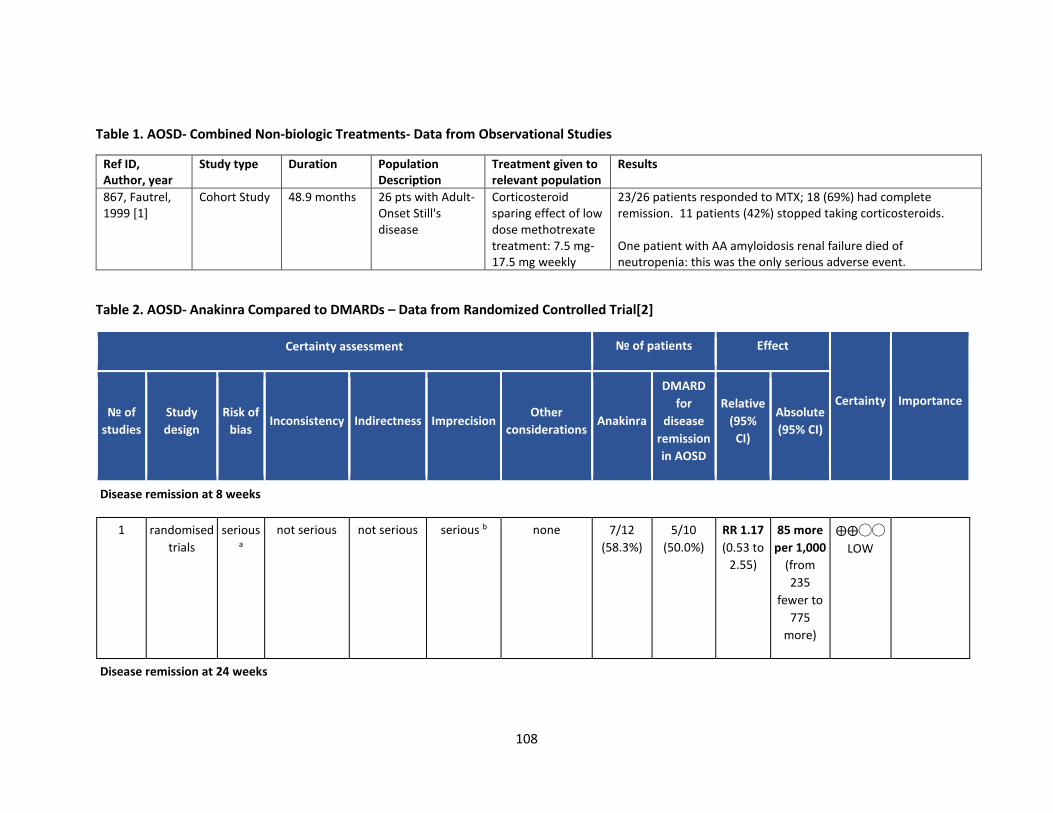
108
Table 1. AOSD- Combined Non-biologic Treatments- Data from Observational Studies
Ref ID, Author, year
Study type Duration Population Description
Treatment given to relevant population
Results
867, Fautrel, 1999 [1]
Cohort Study 48.9 months 26 pts with Adult-Onset Still's disease
Corticosteroid sparing effect of low dose methotrexate treatment: 7.5 mg-17.5 mg weekly
23/26 patients responded to MTX; 18 (69%) had complete remission. 11 patients (42%) stopped taking corticosteroids. One patient with AA amyloidosis renal failure died of neutropenia: this was the only serious adverse event.
Table 2. AOSD- Anakinra Compared to DMARDs – Data from Randomized Controlled Trial[2]
Certainty assessment № of patients Effect
Certainty Importance № of
studies
Study
design
Risk of
bias Inconsistency Indirectness Imprecision
Other
considerations Anakinra
DMARD
for
disease
remission
in AOSD
Relative
(95%
CI)
Absolute
(95% CI)
Disease remission at 8 weeks
1 randomised
trials
serious a
not serious not serious serious b none 7/12
(58.3%)
5/10
(50.0%)
RR 1.17
(0.53 to
2.55)
85 more
per 1,000
(from
235
fewer to
775
more)
⨁⨁◯◯
LOW
Disease remission at 24 weeks
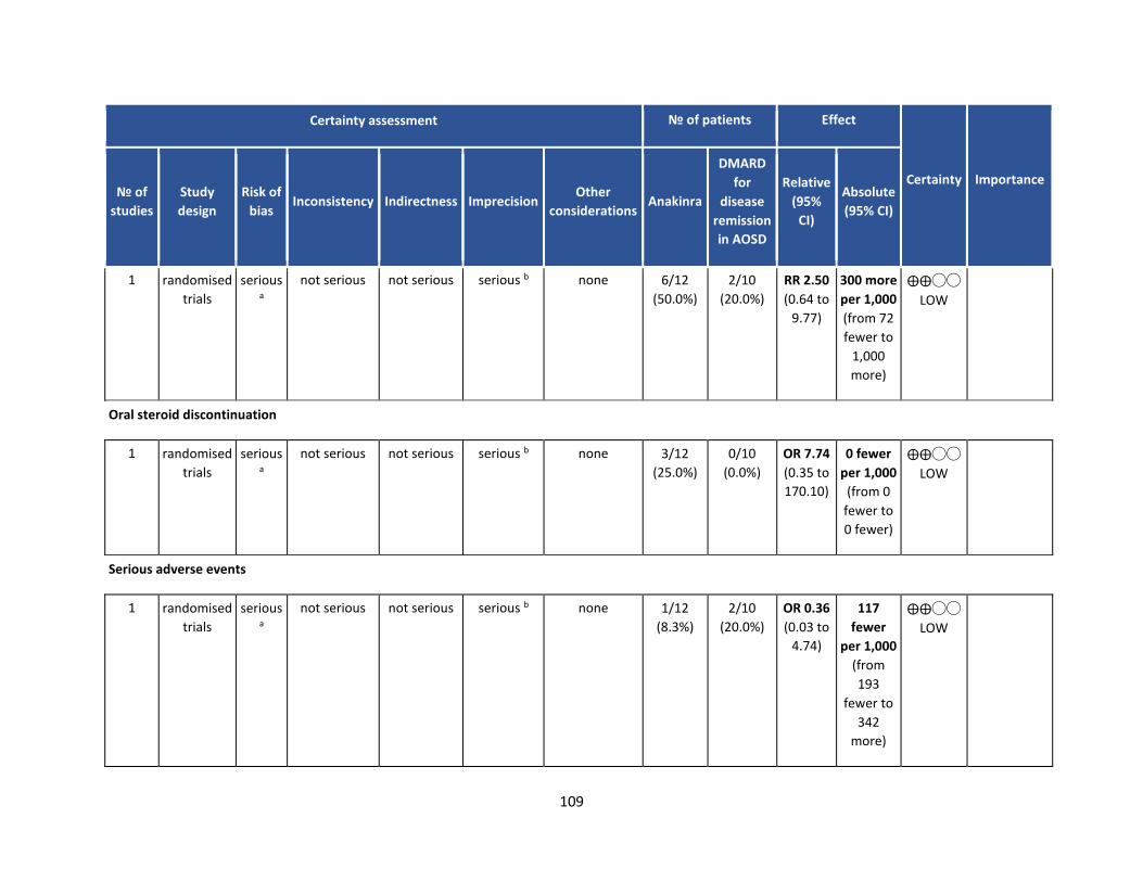
109
Certainty assessment № of patients Effect
Certainty Importance № of
studies
Study
design
Risk of
bias Inconsistency Indirectness Imprecision
Other
considerations Anakinra
DMARD
for
disease
remission
in AOSD
Relative
(95%
CI)
Absolute
(95% CI)
1 randomised
trials
serious a
not serious not serious serious b none 6/12
(50.0%)
2/10
(20.0%)
RR 2.50
(0.64 to
9.77)
300 more
per 1,000
(from 72
fewer to
1,000
more)
⨁⨁◯◯
LOW
Oral steroid discontinuation
1 randomised
trials
serious a
not serious not serious serious b none 3/12
(25.0%)
0/10
(0.0%)
OR 7.74
(0.35 to
170.10)
0 fewer
per 1,000
(from 0
fewer to
0 fewer)
⨁⨁◯◯
LOW
Serious adverse events
1 randomised
trials
serious a
not serious not serious serious b none 1/12
(8.3%)
2/10
(20.0%)
OR 0.36
(0.03 to
4.74)
117
fewer
per 1,000
(from
193
fewer to
342
more)
⨁⨁◯◯
LOW
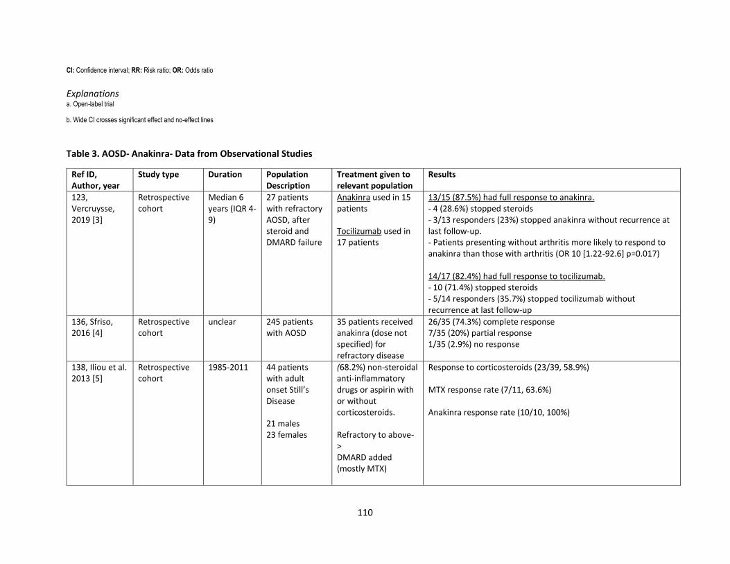
110
CI: Confidence interval; RR: Risk ratio; OR: Odds ratio
Explanations a. Open-label trial
b. Wide CI crosses significant effect and no-effect lines
Table 3. AOSD- Anakinra- Data from Observational Studies
Ref ID, Author, year
Study type Duration Population Description
Treatment given to relevant population
Results
123, Vercruysse, 2019 [3]
Retrospective cohort
Median 6 years (IQR 4-9)
27 patients with refractory AOSD, after steroid and DMARD failure
Anakinra used in 15 patients Tocilizumab used in 17 patients
13/15 (87.5%) had full response to anakinra. - 4 (28.6%) stopped steroids - 3/13 responders (23%) stopped anakinra without recurrence at last follow-up. - Patients presenting without arthritis more likely to respond to anakinra than those with arthritis (OR 10 [1.22-92.6] p=0.017) 14/17 (82.4%) had full response to tocilizumab. - 10 (71.4%) stopped steroids - 5/14 responders (35.7%) stopped tocilizumab without recurrence at last follow-up
136, Sfriso, 2016 [4]
Retrospective cohort
unclear 245 patients with AOSD
35 patients received anakinra (dose not specified) for refractory disease
26/35 (74.3%) complete response 7/35 (20%) partial response 1/35 (2.9%) no response
138, Iliou et al. 2013 [5]
Retrospective cohort
1985-2011 44 patients with adult onset Still’s Disease 21 males 23 females
(68.2%) non-steroidal anti-inflammatory drugs or aspirin with or without corticosteroids. Refractory to above-> DMARD added (mostly MTX)
Response to corticosteroids (23/39, 58.9%) MTX response rate (7/11, 63.6%) Anakinra response rate (10/10, 100%)
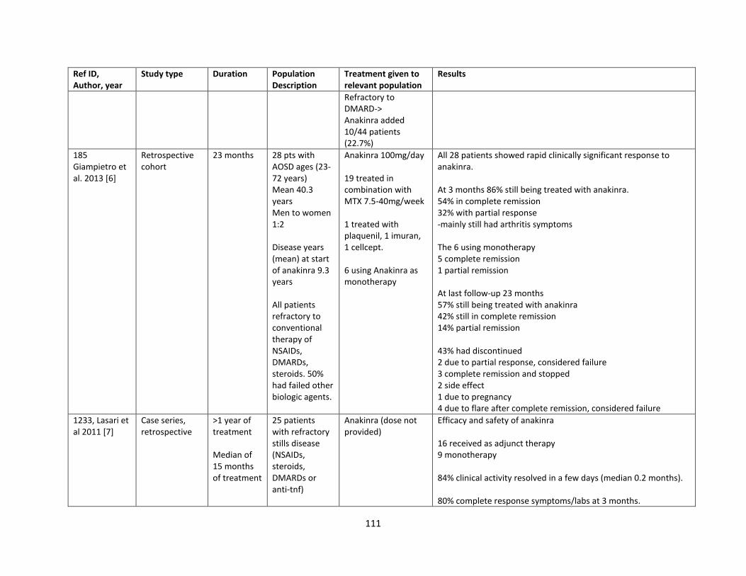
111
Ref ID, Author, year
Study type Duration Population Description
Treatment given to relevant population
Results
Refractory to DMARD-> Anakinra added 10/44 patients (22.7%)
185 Giampietro et al. 2013 [6]
Retrospective cohort
23 months 28 pts with AOSD ages (23-72 years) Mean 40.3 years Men to women 1:2 Disease years (mean) at start of anakinra 9.3 years All patients refractory to conventional therapy of NSAIDs, DMARDs, steroids. 50% had failed other biologic agents.
Anakinra 100mg/day 19 treated in combination with MTX 7.5-40mg/week 1 treated with plaquenil, 1 imuran, 1 cellcept. 6 using Anakinra as monotherapy
All 28 patients showed rapid clinically significant response to anakinra. At 3 months 86% still being treated with anakinra. 54% in complete remission 32% with partial response -mainly still had arthritis symptoms The 6 using monotherapy 5 complete remission 1 partial remission At last follow-up 23 months 57% still being treated with anakinra 42% still in complete remission 14% partial remission 43% had discontinued 2 due to partial response, considered failure 3 complete remission and stopped 2 side effect 1 due to pregnancy 4 due to flare after complete remission, considered failure
1233, Lasari et al 2011 [7]
Case series, retrospective
>1 year of treatment Median of 15 months of treatment
25 patients with refractory stills disease (NSAIDs, steroids, DMARDs or anti-tnf)
Anakinra (dose not provided)
Efficacy and safety of anakinra 16 received as adjunct therapy 9 monotherapy 84% clinical activity resolved in a few days (median 0.2 months). 80% complete response symptoms/labs at 3 months.
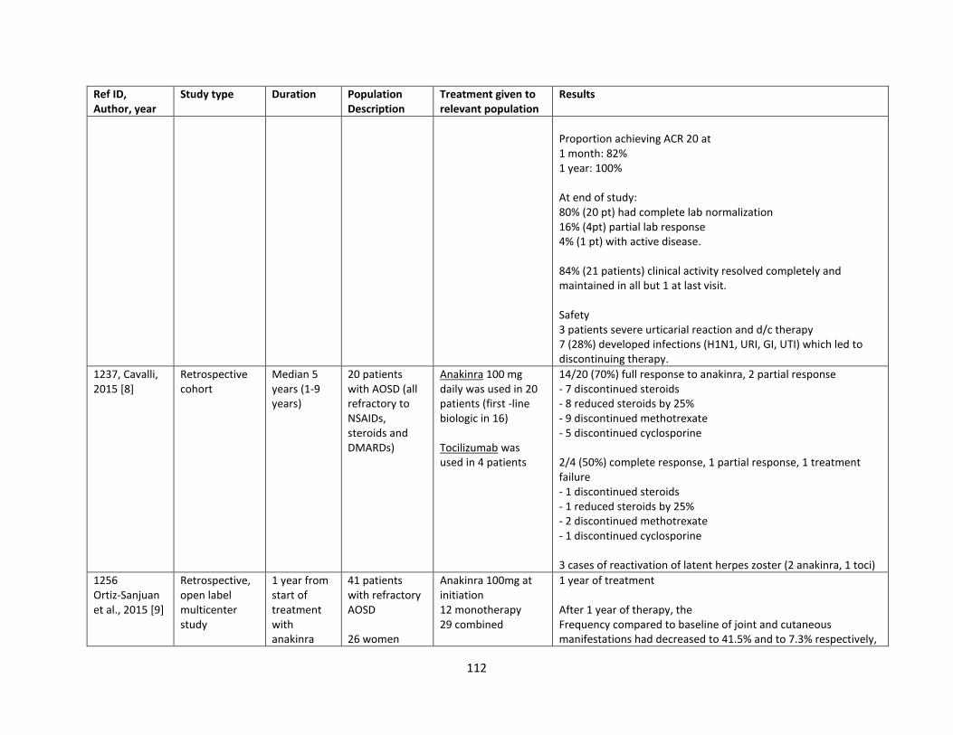
112
Ref ID, Author, year
Study type Duration Population Description
Treatment given to relevant population
Results
Proportion achieving ACR 20 at 1 month: 82% 1 year: 100% At end of study: 80% (20 pt) had complete lab normalization 16% (4pt) partial lab response 4% (1 pt) with active disease. 84% (21 patients) clinical activity resolved completely and maintained in all but 1 at last visit. Safety 3 patients severe urticarial reaction and d/c therapy 7 (28%) developed infections (H1N1, URI, GI, UTI) which led to discontinuing therapy.
1237, Cavalli, 2015 [8]
Retrospective cohort
Median 5 years (1-9 years)
20 patients with AOSD (all refractory to NSAIDs, steroids and DMARDs)
Anakinra 100 mg daily was used in 20 patients (first -line biologic in 16) Tocilizumab was used in 4 patients
14/20 (70%) full response to anakinra, 2 partial response - 7 discontinued steroids - 8 reduced steroids by 25% - 9 discontinued methotrexate - 5 discontinued cyclosporine 2/4 (50%) complete response, 1 partial response, 1 treatment failure - 1 discontinued steroids - 1 reduced steroids by 25% - 2 discontinued methotrexate - 1 discontinued cyclosporine 3 cases of reactivation of latent herpes zoster (2 anakinra, 1 toci)
1256 Ortiz-Sanjuan et al., 2015 [9]
Retrospective, open label multicenter study
1 year from start of treatment with anakinra
41 patients with refractory AOSD 26 women
Anakinra 100mg at initiation 12 monotherapy 29 combined
1 year of treatment After 1 year of therapy, the Frequency compared to baseline of joint and cutaneous manifestations had decreased to 41.5% and to 7.3% respectively,
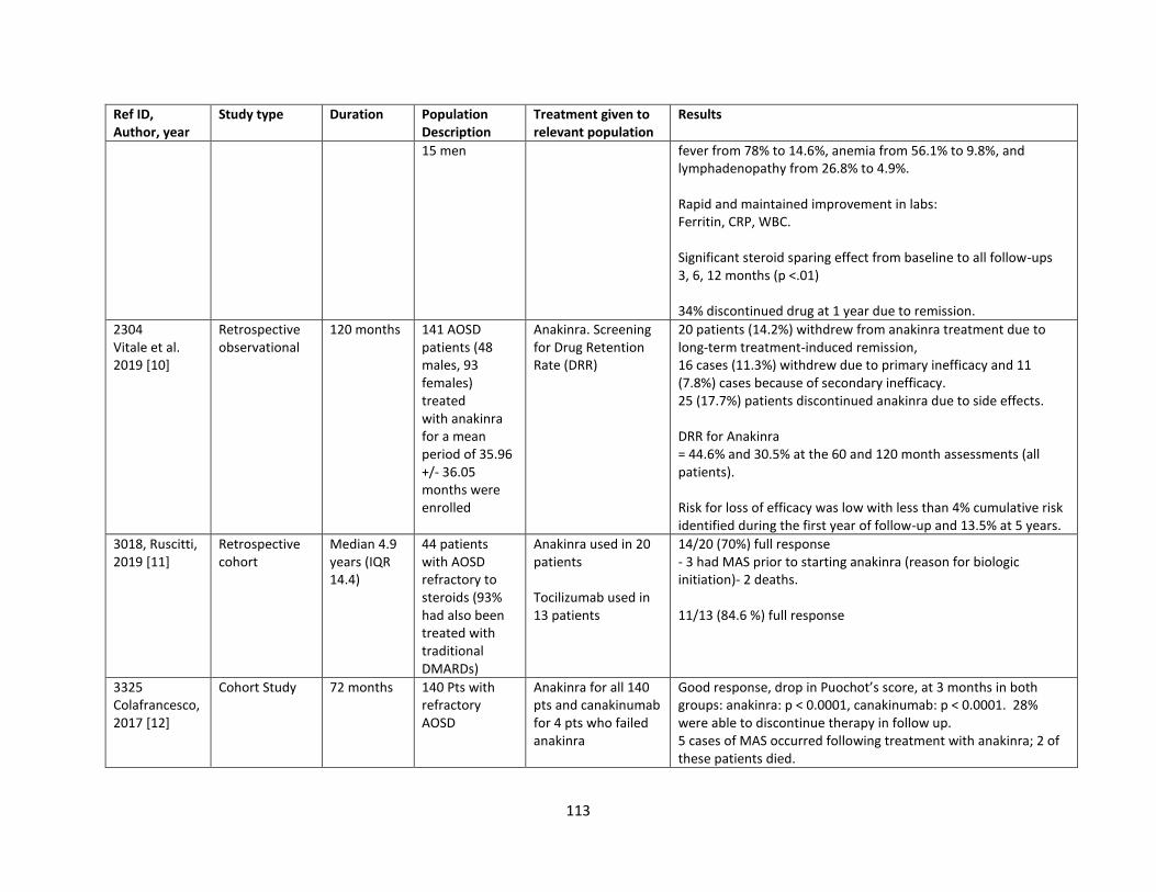
113
Ref ID, Author, year
Study type Duration Population Description
Treatment given to relevant population
Results
15 men fever from 78% to 14.6%, anemia from 56.1% to 9.8%, and lymphadenopathy from 26.8% to 4.9%. Rapid and maintained improvement in labs: Ferritin, CRP, WBC. Significant steroid sparing effect from baseline to all follow-ups 3, 6, 12 months (p <.01) 34% discontinued drug at 1 year due to remission.
2304 Vitale et al. 2019 [10]
Retrospective observational
120 months 141 AOSD patients (48 males, 93 females) treated with anakinra for a mean period of 35.96 +/- 36.05 months were enrolled
Anakinra. Screening for Drug Retention Rate (DRR)
20 patients (14.2%) withdrew from anakinra treatment due to long-term treatment-induced remission, 16 cases (11.3%) withdrew due to primary inefficacy and 11 (7.8%) cases because of secondary inefficacy. 25 (17.7%) patients discontinued anakinra due to side effects. DRR for Anakinra = 44.6% and 30.5% at the 60 and 120 month assessments (all patients). Risk for loss of efficacy was low with less than 4% cumulative risk identified during the first year of follow-up and 13.5% at 5 years.
3018, Ruscitti, 2019 [11]
Retrospective cohort
Median 4.9 years (IQR 14.4)
44 patients with AOSD refractory to steroids (93% had also been treated with traditional DMARDs)
Anakinra used in 20 patients Tocilizumab used in 13 patients
14/20 (70%) full response - 3 had MAS prior to starting anakinra (reason for biologic initiation)- 2 deaths. 11/13 (84.6 %) full response
3325 Colafrancesco, 2017 [12]
Cohort Study 72 months 140 Pts with refractory AOSD
Anakinra for all 140 pts and canakinumab for 4 pts who failed anakinra
Good response, drop in Puochot’s score, at 3 months in both groups: anakinra: p < 0.0001, canakinumab: p < 0.0001. 28% were able to discontinue therapy in follow up. 5 cases of MAS occurred following treatment with anakinra; 2 of these patients died.
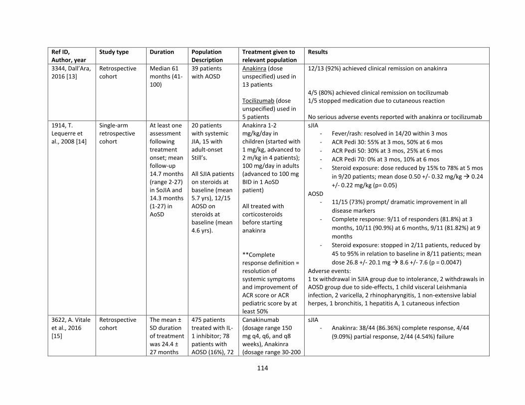
114
Ref ID, Author, year
Study type Duration Population Description
Treatment given to relevant population
Results
3344, Dall’Ara, 2016 [13]
Retrospective cohort
Median 61 months (41-100)
39 patients with AOSD
Anakinra (dose unspecified) used in 13 patients Tocilizumab (dose unspecified) used in 5 patients
12/13 (92%) achieved clinical remission on anakinra 4/5 (80%) achieved clinical remission on tocilizumab 1/5 stopped medication due to cutaneous reaction No serious adverse events reported with anakinra or tocilizumab
1914, T. Lequerre et al., 2008 [14]
Single-arm retrospective cohort
At least one assessment following treatment onset; mean follow-up 14.7 months (range 2-27) in SoJIA and 14.3 months (1-27) in AoSD
20 patients with systemic JIA, 15 with adult-onset Still’s. All SJIA patients on steroids at baseline (mean 5.7 yrs), 12/15 AOSD on steroids at baseline (mean 4.6 yrs).
Anakinra 1-2 mg/kg/day in children (started with 1 mg/kg, advanced to 2 m/kg in 4 patients); 100 mg/day in adults (advanced to 100 mg BID in 1 AoSD patient) All treated with corticosteroids before starting anakinra **Complete response definition = resolution of systemic symptoms and improvement of ACR score or ACR pediatric score by at least 50%
sJIA - Fever/rash: resolved in 14/20 within 3 mos
- ACR Pedi 30: 55% at 3 mos, 50% at 6 mos
- ACR Pedi 50: 30% at 3 mos, 25% at 6 mos
- ACR Pedi 70: 0% at 3 mos, 10% at 6 mos
- Steroid exposure: dose reduced by 15% to 78% at 5 mos
in 9/20 patients; mean dose 0.50 +/- 0.32 mg/kg → 0.24
+/- 0.22 mg/kg (p= 0.05)
AOSD - 11/15 (73%) prompt/ dramatic improvement in all
disease markers
- Complete response: 9/11 of responders (81.8%) at 3
months, 10/11 (90.9%) at 6 months, 9/11 (81.82%) at 9
months
- Steroid exposure: stopped in 2/11 patients, reduced by
45 to 95% in relation to baseline in 8/11 patients; mean
dose 26.8 +/- 20.1 mg → 8.6 +/- 7.6 (p = 0.0047)
Adverse events: 1 tx withdrawal in SJIA group due to intolerance, 2 withdrawals in AOSD group due to side-effects, 1 child visceral Leishmania infection, 2 varicella, 2 rhinopharyngitis, 1 non-extensive labial herpes, 1 bronchitis, 1 hepatitis A, 1 cutaneous infection
3622, A. Vitale et al., 2016 [15]
Retrospective cohort
The mean ± SD duration of treatment was 24.4 ± 27 months
475 patients treated with IL-1 inhibitor; 78 patients with AOSD (16%), 72
Canakinumab (dosage range 150 mg q4, q6, and q8 weeks), Anakinra (dosage range 30-200
sJIA - Anakinra: 38/44 (86.36%) complete response, 4/44
(9.09%) partial response, 2/44 (4.54%) failure
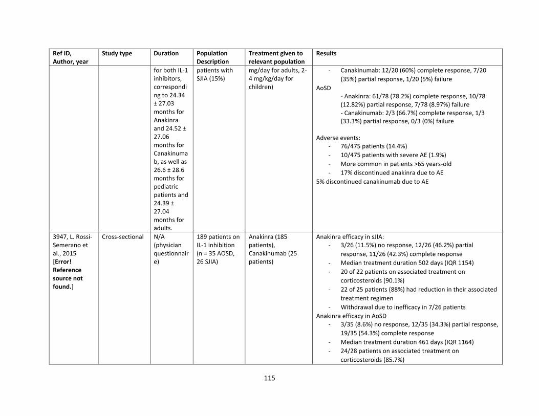
115
Ref ID, Author, year
Study type Duration Population Description
Treatment given to relevant population
Results
for both IL-1 inhibitors, corresponding to 24.34 ± 27.03 months for Anakinra and 24.52 ± 27.06 months for Canakinumab, as well as 26.6 ± 28.6 months for pediatric patients and 24.39 ± 27.04 months for adults.
patients with SJIA (15%)
mg/day for adults, 2-4 mg/kg/day for children)
- Canakinumab: 12/20 (60%) complete response, 7/20
(35%) partial response, 1/20 (5%) failure
AoSD - Anakinra: 61/78 (78.2%) complete response, 10/78 (12.82%) partial response, 7/78 (8.97%) failure - Canakinumab: 2/3 (66.7%) complete response, 1/3 (33.3%) partial response, 0/3 (0%) failure
Adverse events:
- 76/475 patients (14.4%)
- 10/475 patients with severe AE (1.9%)
- More common in patients >65 years-old
- 17% discontinued anakinra due to AE
5% discontinued canakinumab due to AE
3947, L. Rossi-Semerano et al., 2015 [Error! Reference source not found.]
Cross-sectional N/A (physician questionnaire)
189 patients on IL-1 inhibition (n = 35 AOSD, 26 SJIA)
Anakinra (185 patients), Canakinumab (25 patients)
Anakinra efficacy in sJIA: - 3/26 (11.5%) no response, 12/26 (46.2%) partial
response, 11/26 (42.3%) complete response
- Median treatment duration 502 days (IQR 1154)
- 20 of 22 patients on associated treatment on
corticosteroids (90.1%)
- 22 of 25 patients (88%) had reduction in their associated
treatment regimen
- Withdrawal due to inefficacy in 7/26 patients
Anakinra efficacy in AoSD - 3/35 (8.6%) no response, 12/35 (34.3%) partial response,
19/35 (54.3%) complete response
- Median treatment duration 461 days (IQR 1164)
- 24/28 patients on associated treatment on
corticosteroids (85.7%)
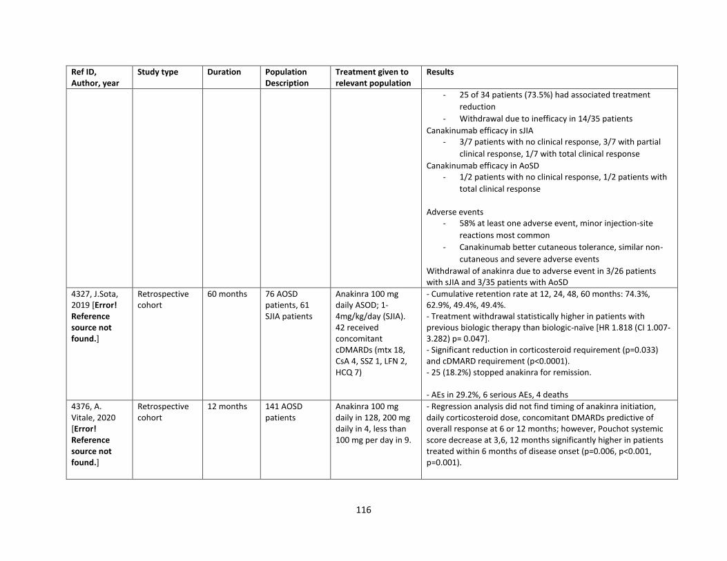
116
Ref ID, Author, year
Study type Duration Population Description
Treatment given to relevant population
Results
- 25 of 34 patients (73.5%) had associated treatment
reduction
- Withdrawal due to inefficacy in 14/35 patients
Canakinumab efficacy in sJIA - 3/7 patients with no clinical response, 3/7 with partial
clinical response, 1/7 with total clinical response
Canakinumab efficacy in AoSD - 1/2 patients with no clinical response, 1/2 patients with
total clinical response
Adverse events
- 58% at least one adverse event, minor injection-site
reactions most common
- Canakinumab better cutaneous tolerance, similar non-
cutaneous and severe adverse events
Withdrawal of anakinra due to adverse event in 3/26 patients with sJIA and 3/35 patients with AoSD
4327, J.Sota, 2019 [Error! Reference source not found.]
Retrospective cohort
60 months 76 AOSD patients, 61 SJIA patients
Anakinra 100 mg daily ASOD; 1-4mg/kg/day (SJIA). 42 received concomitant cDMARDs (mtx 18, CsA 4, SSZ 1, LFN 2, HCQ 7)
- Cumulative retention rate at 12, 24, 48, 60 months: 74.3%, 62.9%, 49.4%, 49.4%. - Treatment withdrawal statistically higher in patients with previous biologic therapy than biologic-naïve [HR 1.818 (CI 1.007-3.282) p= 0.047]. - Significant reduction in corticosteroid requirement (p=0.033) and cDMARD requirement (p<0.0001). - 25 (18.2%) stopped anakinra for remission. - AEs in 29.2%, 6 serious AEs, 4 deaths
4376, A. Vitale, 2020 [Error! Reference source not found.]
Retrospective cohort
12 months 141 AOSD patients
Anakinra 100 mg daily in 128, 200 mg daily in 4, less than 100 mg per day in 9.
- Regression analysis did not find timing of anakinra initiation, daily corticosteroid dose, concomitant DMARDs predictive of overall response at 6 or 12 months; however, Pouchot systemic score decrease at 3,6, 12 months significantly higher in patients treated within 6 months of disease onset (p=0.006, p<0.001, p=0.001).
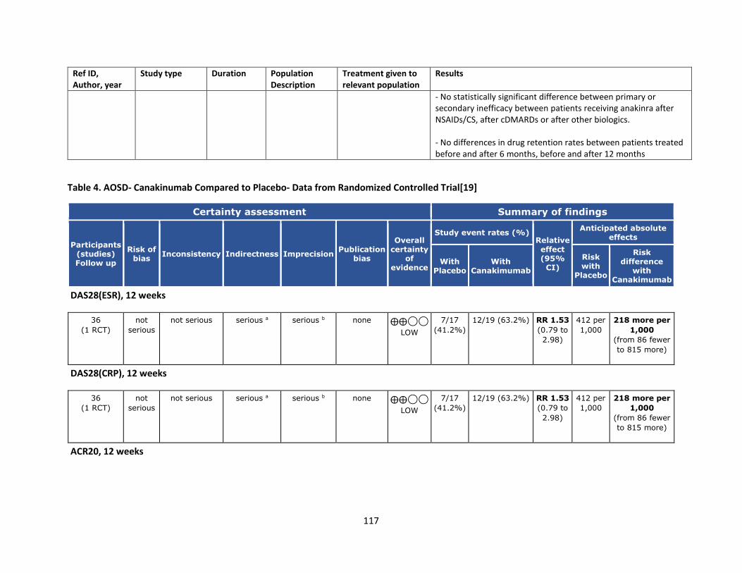
117
Ref ID, Author, year
Study type Duration Population Description
Treatment given to relevant population
Results
- No statistically significant difference between primary or secondary inefficacy between patients receiving anakinra after NSAIDs/CS, after cDMARDs or after other biologics. - No differences in drug retention rates between patients treated before and after 6 months, before and after 12 months
Table 4. AOSD- Canakinumab Compared to Placebo- Data from Randomized Controlled Trial[19]
Certainty assessment Summary of findings
Participants (studies) Follow up
Risk of bias
Inconsistency Indirectness Imprecision Publication
bias
Overall certainty
of evidence
Study event rates (%) Relative effect (95%
CI)
Anticipated absolute effects
With Placebo
With Canakimumab
Risk with
Placebo
Risk difference
with Canakimumab
DAS28(ESR), 12 weeks
36
(1 RCT)
not
serious
not serious serious a serious b none ⨁⨁◯◯
LOW
7/17
(41.2%)
12/19 (63.2%) RR 1.53
(0.79 to
2.98)
412 per
1,000
218 more per
1,000
(from 86 fewer
to 815 more)
DAS28(CRP), 12 weeks
36
(1 RCT)
not
serious
not serious serious a serious b none ⨁⨁◯◯
LOW
7/17
(41.2%)
12/19 (63.2%) RR 1.53
(0.79 to
2.98)
412 per
1,000
218 more per
1,000
(from 86 fewer
to 815 more)
ACR20, 12 weeks
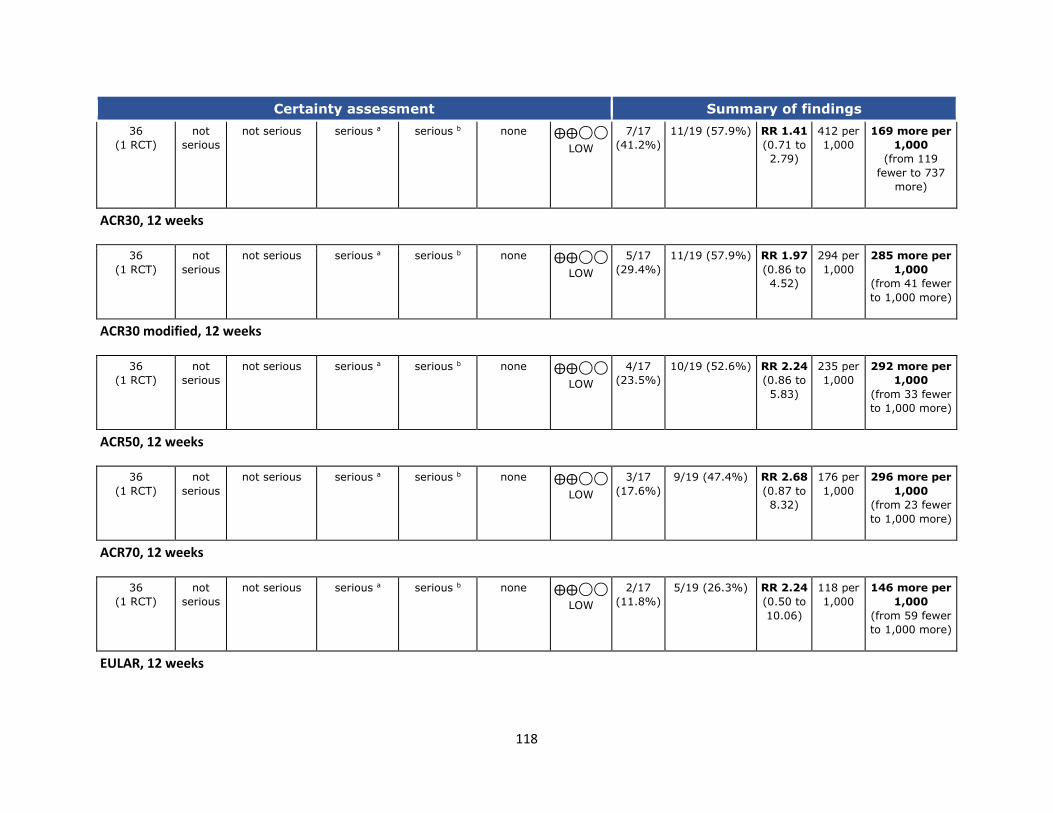
118
Certainty assessment Summary of findings
36
(1 RCT)
not
serious
not serious serious a serious b none ⨁⨁◯◯
LOW
7/17
(41.2%)
11/19 (57.9%) RR 1.41
(0.71 to
2.79)
412 per
1,000
169 more per
1,000
(from 119
fewer to 737
more)
ACR30, 12 weeks
36
(1 RCT)
not
serious
not serious serious a serious b none ⨁⨁◯◯
LOW
5/17
(29.4%)
11/19 (57.9%) RR 1.97
(0.86 to
4.52)
294 per
1,000
285 more per
1,000
(from 41 fewer
to 1,000 more)
ACR30 modified, 12 weeks
36
(1 RCT)
not
serious
not serious serious a serious b none ⨁⨁◯◯
LOW
4/17
(23.5%)
10/19 (52.6%) RR 2.24
(0.86 to
5.83)
235 per
1,000
292 more per
1,000
(from 33 fewer
to 1,000 more)
ACR50, 12 weeks
36
(1 RCT)
not
serious
not serious serious a serious b none ⨁⨁◯◯
LOW
3/17
(17.6%)
9/19 (47.4%) RR 2.68
(0.87 to
8.32)
176 per
1,000
296 more per
1,000
(from 23 fewer
to 1,000 more)
ACR70, 12 weeks
36
(1 RCT)
not
serious
not serious serious a serious b none ⨁⨁◯◯
LOW
2/17
(11.8%)
5/19 (26.3%) RR 2.24
(0.50 to
10.06)
118 per
1,000
146 more per
1,000
(from 59 fewer
to 1,000 more)
EULAR, 12 weeks
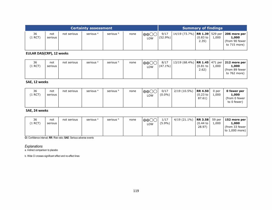
119
Certainty assessment Summary of findings
36
(1 RCT)
not
serious
not serious serious a serious b none ⨁⨁◯◯
LOW
9/17
(52.9%)
14/19 (73.7%) RR 1.39
(0.83 to
2.35)
529 per
1,000
206 more per
1,000
(from 90 fewer
to 715 more)
EULAR DAS(CRP), 12 weeks
36
(1 RCT)
not
serious
not serious serious a serious b none ⨁⨁◯◯
LOW
8/17
(47.1%)
13/19 (68.4%) RR 1.45
(0.81 to
2.62)
471 per
1,000
212 more per
1,000
(from 89 fewer
to 762 more)
SAE, 12 weeks
36
(1 RCT)
not
serious
not serious serious a serious b none ⨁⨁◯◯
LOW
0/17
(0.0%)
2/19 (10.5%) RR 4.50
(0.23 to
87.61)
0 per
1,000
0 fewer per
1,000
(from 0 fewer
to 0 fewer)
SAE, 24 weeks
36
(1 RCT)
not
serious
not serious serious a serious b none ⨁⨁◯◯
LOW
1/17
(5.9%)
4/19 (21.1%) RR 3.58
(0.44 to
28.97)
59 per
1,000
152 more per
1,000
(from 33 fewer
to 1,000 more)
CI: Confidence interval; RR: Risk ratio; SAE: Serious adverse events
Explanations a. Indirect comparison to placebo
b. Wide CI crosses significant effect and no-effect lines
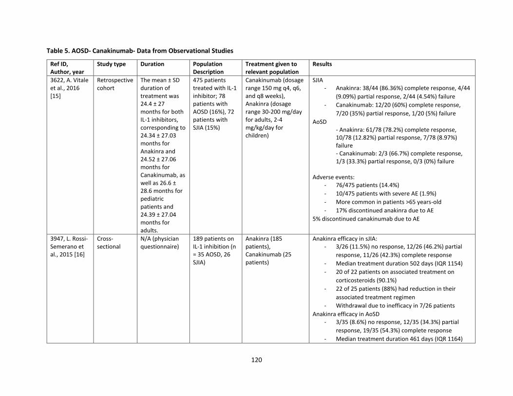
120
Table 5. AOSD- Canakinumab- Data from Observational Studies
Ref ID, Author, year
Study type Duration Population Description
Treatment given to relevant population
Results
3622, A. Vitale et al., 2016 [15]
Retrospective cohort
The mean ± SD duration of treatment was 24.4 ± 27 months for both IL-1 inhibitors, corresponding to 24.34 ± 27.03 months for Anakinra and 24.52 ± 27.06 months for Canakinumab, as well as 26.6 ± 28.6 months for pediatric patients and 24.39 ± 27.04 months for adults.
475 patients treated with IL-1 inhibitor; 78 patients with AOSD (16%), 72 patients with SJIA (15%)
Canakinumab (dosage range 150 mg q4, q6, and q8 weeks), Anakinra (dosage range 30-200 mg/day for adults, 2-4 mg/kg/day for children)
SJIA - Anakinra: 38/44 (86.36%) complete response, 4/44
(9.09%) partial response, 2/44 (4.54%) failure
- Canakinumab: 12/20 (60%) complete response,
7/20 (35%) partial response, 1/20 (5%) failure
AoSD - Anakinra: 61/78 (78.2%) complete response, 10/78 (12.82%) partial response, 7/78 (8.97%) failure - Canakinumab: 2/3 (66.7%) complete response, 1/3 (33.3%) partial response, 0/3 (0%) failure
Adverse events:
- 76/475 patients (14.4%)
- 10/475 patients with severe AE (1.9%)
- More common in patients >65 years-old
- 17% discontinued anakinra due to AE
5% discontinued canakinumab due to AE
3947, L. Rossi-Semerano et al., 2015 [16]
Cross-sectional
N/A (physician questionnaire)
189 patients on IL-1 inhibition (n = 35 AOSD, 26 SJIA)
Anakinra (185 patients), Canakinumab (25 patients)
Anakinra efficacy in sJIA: - 3/26 (11.5%) no response, 12/26 (46.2%) partial
response, 11/26 (42.3%) complete response
- Median treatment duration 502 days (IQR 1154)
- 20 of 22 patients on associated treatment on
corticosteroids (90.1%)
- 22 of 25 patients (88%) had reduction in their
associated treatment regimen
- Withdrawal due to inefficacy in 7/26 patients
Anakinra efficacy in AoSD - 3/35 (8.6%) no response, 12/35 (34.3%) partial
response, 19/35 (54.3%) complete response
- Median treatment duration 461 days (IQR 1164)
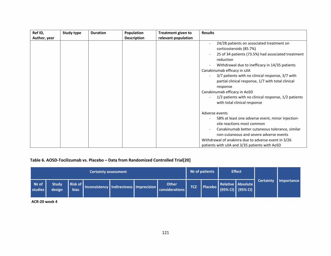
121
Ref ID, Author, year
Study type Duration Population Description
Treatment given to relevant population
Results
- 24/28 patients on associated treatment on
corticosteroids (85.7%)
- 25 of 34 patients (73.5%) had associated treatment
reduction
- Withdrawal due to inefficacy in 14/35 patients
Canakinumab efficacy in sJIA - 3/7 patients with no clinical response, 3/7 with
partial clinical response, 1/7 with total clinical
response
Canakinumab efficacy in AoSD - 1/2 patients with no clinical response, 1/2 patients
with total clinical response
Adverse events
- 58% at least one adverse event, minor injection-
site reactions most common
- Canakinumab better cutaneous tolerance, similar
non-cutaneous and severe adverse events
Withdrawal of anakinra due to adverse event in 3/26 patients with sJIA and 3/35 patients with AoSD
Table 6. AOSD-Tocilizumab vs. Placebo – Data from Randomized Controlled Trial[20]
Certainty assessment № of patients Effect
Certainty Importance № of
studies
Study
design
Risk of
bias Inconsistency Indirectness Imprecision
Other
considerations TCZ Placebo
Relative
(95% CI)
Absolute
(95% CI)
ACR-20 week 4
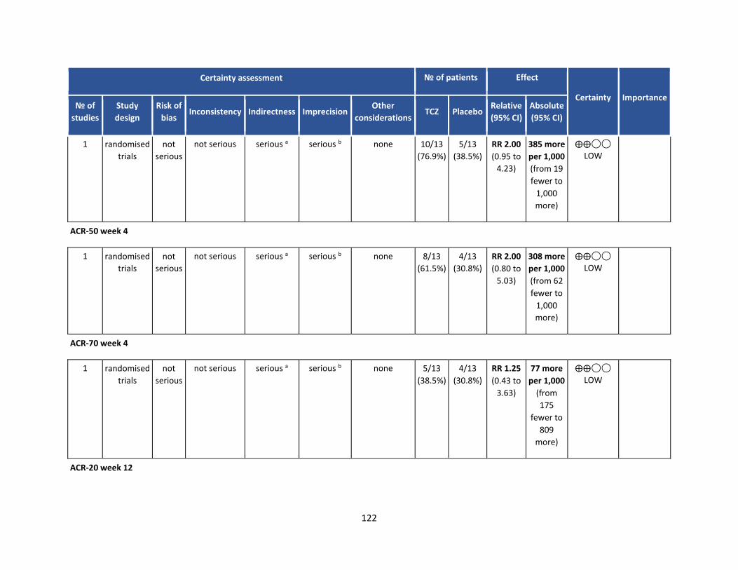
122
Certainty assessment № of patients Effect
Certainty Importance № of
studies
Study
design
Risk of
bias Inconsistency Indirectness Imprecision
Other
considerations TCZ Placebo
Relative
(95% CI)
Absolute
(95% CI)
1 randomised
trials
not
serious
not serious serious a serious b none 10/13
(76.9%)
5/13
(38.5%)
RR 2.00
(0.95 to
4.23)
385 more
per 1,000
(from 19
fewer to
1,000
more)
⨁⨁◯◯
LOW
ACR-50 week 4
1 randomised
trials
not
serious
not serious serious a serious b none 8/13
(61.5%)
4/13
(30.8%)
RR 2.00
(0.80 to
5.03)
308 more
per 1,000
(from 62
fewer to
1,000
more)
⨁⨁◯◯
LOW
ACR-70 week 4
1 randomised
trials
not
serious
not serious serious a serious b none 5/13
(38.5%)
4/13
(30.8%)
RR 1.25
(0.43 to
3.63)
77 more
per 1,000
(from
175
fewer to
809
more)
⨁⨁◯◯
LOW
ACR-20 week 12
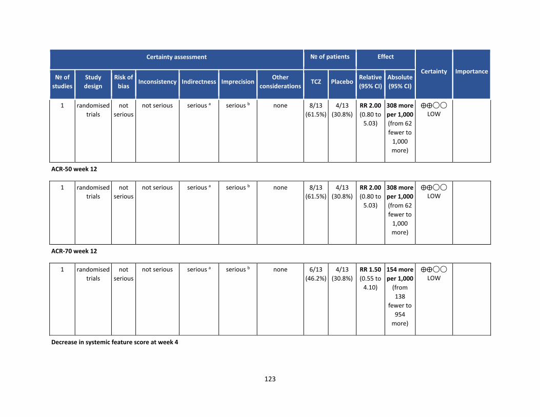
123
Certainty assessment № of patients Effect
Certainty Importance № of
studies
Study
design
Risk of
bias Inconsistency Indirectness Imprecision
Other
considerations TCZ Placebo
Relative
(95% CI)
Absolute
(95% CI)
1 randomised
trials
not
serious
not serious serious a serious b none 8/13
(61.5%)
4/13
(30.8%)
RR 2.00
(0.80 to
5.03)
308 more
per 1,000
(from 62
fewer to
1,000
more)
⨁⨁◯◯
LOW
ACR-50 week 12
1 randomised
trials
not
serious
not serious serious a serious b none 8/13
(61.5%)
4/13
(30.8%)
RR 2.00
(0.80 to
5.03)
308 more
per 1,000
(from 62
fewer to
1,000
more)
⨁⨁◯◯
LOW
ACR-70 week 12
1 randomised
trials
not
serious
not serious serious a serious b none 6/13
(46.2%)
4/13
(30.8%)
RR 1.50
(0.55 to
4.10)
154 more
per 1,000
(from
138
fewer to
954
more)
⨁⨁◯◯
LOW
Decrease in systemic feature score at week 4
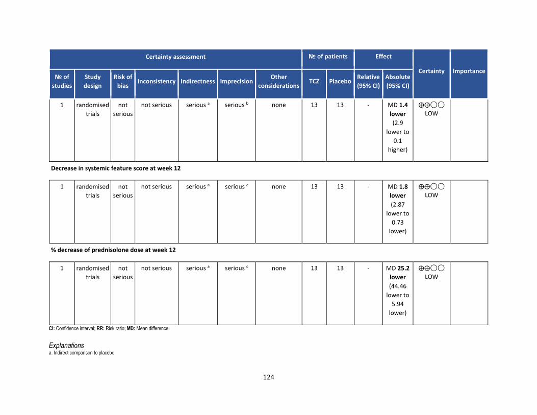
124
Certainty assessment № of patients Effect
Certainty Importance № of
studies
Study
design
Risk of
bias Inconsistency Indirectness Imprecision
Other
considerations TCZ Placebo
Relative
(95% CI)
Absolute
(95% CI)
1 randomised
trials
not
serious
not serious serious a serious b none 13 13 - MD 1.4
lower
(2.9
lower to
0.1
higher)
⨁⨁◯◯
LOW
Decrease in systemic feature score at week 12
1 randomised
trials
not
serious
not serious serious a serious c none 13 13 - MD 1.8
lower
(2.87
lower to
0.73
lower)
⨁⨁◯◯
LOW
% decrease of prednisolone dose at week 12
1 randomised
trials
not
serious
not serious serious a serious c none 13 13 - MD 25.2
lower
(44.46
lower to
5.94
lower)
⨁⨁◯◯
LOW
CI: Confidence interval; RR: Risk ratio; MD: Mean difference
Explanations a. Indirect comparison to placebo
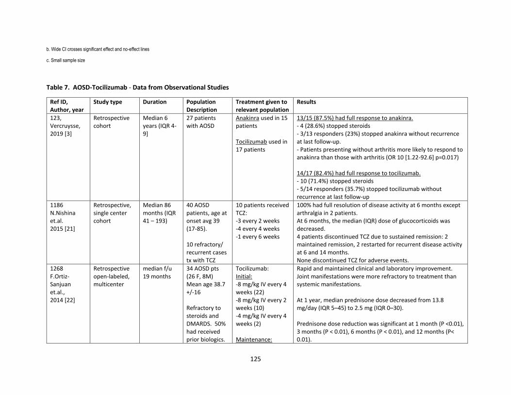
125
b. Wide CI crosses significant effect and no-effect lines
c. Small sample size
Table 7. AOSD-Tocilizumab - Data from Observational Studies
Ref ID, Author, year
Study type Duration Population Description
Treatment given to relevant population
Results
123, Vercruysse, 2019 [3]
Retrospective cohort
Median 6 years (IQR 4-9]
27 patients with AOSD
Anakinra used in 15 patients Tocilizumab used in 17 patients
13/15 (87.5%) had full response to anakinra. - 4 (28.6%) stopped steroids - 3/13 responders (23%) stopped anakinra without recurrence at last follow-up. - Patients presenting without arthritis more likely to respond to anakinra than those with arthritis (OR 10 [1.22-92.6] p=0.017) 14/17 (82.4%) had full response to tocilizumab. - 10 (71.4%) stopped steroids - 5/14 responders (35.7%) stopped tocilizumab without recurrence at last follow-up
1186 N.Nishina et.al. 2015 [21]
Retrospective, single center cohort
Median 86 months (IQR 41 – 193)
40 AOSD patients, age at onset avg 39 (17-85). 10 refractory/ recurrent cases tx with TCZ
10 patients received TCZ: -3 every 2 weeks -4 every 4 weeks -1 every 6 weeks
100% had full resolution of disease activity at 6 months except arthralgia in 2 patients. At 6 months, the median (IQR) dose of glucocorticoids was decreased. 4 patients discontinued TCZ due to sustained remission: 2 maintained remission, 2 restarted for recurrent disease activity at 6 and 14 months. None discontinued TCZ for adverse events.
1268 F.Ortiz-Sanjuan et.al., 2014 [22]
Retrospective open-labeled, multicenter
median f/u 19 months
34 AOSD pts (26 F, 8M) Mean age 38.7 +/-16 Refractory to steroids and DMARDS. 50% had received prior biologics.
Tocilizumab: Initial: -8 mg/kg IV every 4 weeks (22) -8 mg/kg IV every 2 weeks (10) -4 mg/kg IV every 4 weeks (2) Maintenance:
Rapid and maintained clinical and laboratory improvement. Joint manifestations were more refractory to treatment than systemic manifestations. At 1 year, median prednisone dose decreased from 13.8 mg/day (IQR 5–45) to 2.5 mg (IQR 0–30). Prednisone dose reduction was significant at 1 month (P <0.01), 3 months (P < 0.01), 6 months (P < 0.01), and 12 months (P< 0.01).
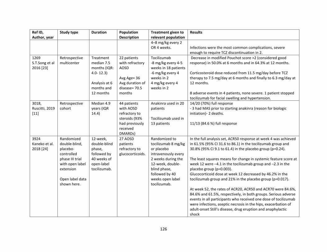
126
Ref ID, Author, year
Study type Duration Population Description
Treatment given to relevant population
Results
4–8 mg/kg every 2 OR 4 weeks.
Infections were the most common complications, severe enough to require TCZ discontinuation in 2.
1269 S.T.Song et al 2016 [23]
Retrospective multicenter
Treatment median 7.5 months (IQR: 4.0- 12.3) Analysis at 6 months and 12 months
22 patients with refractory AOSD Avg Age= 36 Avg duration of disease= 70.5 months
Tocilizumab -8 mg/kg every 4-5 weeks in 18 patients -6 mg/kg every 4 weeks in 2 4 mg/kg every 4 weeks in 2
Decrease in modified Pouchot score >2 (considered good response) in 50.0% at 6 months and in 64.3% at 12 months. Corticosteroid dose reduced from 11.5 mg/day before TCZ therapy to 7.5 mg/day at 6 months and finally to 6.3 mg/day at 12 months. 8 adverse events in 4 patients, none severe. 1 patient stopped tocilizumab for facial swelling and hypertension.
3018, Ruscitti, 2019 [11]
Retrospective cohort
Median 4.9 years (IQR 14.4)
44 patients with AOSD refractory to steroids (93% had previously received DMARDs)
Anakinra used in 20 patients Tocilizumab used in 13 patients
14/20 (70%) full response - 3 had MAS prior to starting anakinra (reason for biologic initiation)- 2 deaths. 11/13 (84.6 %) full response
3924 Kaneko et al. 2018 [24]
Randomized double-blind, placebo-controlled phase III trial with open label extension Open label data shown here.
12-week, double-blind phase, followed by 40 weeks of open-label tocilizumab.
27 AOSD patients refractory to glucocorticoids.
Randomized to tocilizumab 8 mg/kg or placebo intravenously every 2 weeks during the 12-week, double-blind phase, followed by 40 weeks open label tocilizumab.
In the full analysis set, ACR50 response at week 4 was achieved in 61.5% (95% CI 31.6 to 86.1) in the tocilizumab group and 30.8% (95% CI 9.1 to 61.4) in the placebo group (p=0.24). The least squares means for change in systemic feature score at week 12 were –4.1 in the tocilizumab group and –2.3 in the placebo group (p=0.003). Glucocorticoid dose at week 12 decreased by 46.2% in the tocilizumab group and 21% in the placebo group (p=0.017). At week 52, the rates of ACR20, ACR50 and ACR70 were 84.6%, 84.6% and 61.5%, respectively, in both groups. Serious adverse events in all participants who received one dose of tocilizumab were infections, aseptic necrosis in the hips, exacerbation of adult-onset Still’s disease, drug eruption and anaphylactic shock
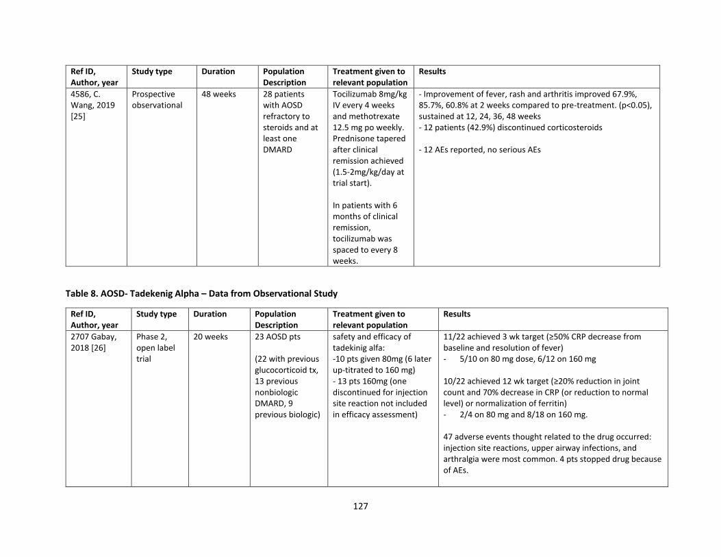
127
Ref ID, Author, year
Study type Duration Population Description
Treatment given to relevant population
Results
4586, C. Wang, 2019 [25]
Prospective observational
48 weeks 28 patients with AOSD refractory to steroids and at least one DMARD
Tocilizumab 8mg/kg IV every 4 weeks and methotrexate 12.5 mg po weekly. Prednisone tapered after clinical remission achieved (1.5-2mg/kg/day at trial start). In patients with 6 months of clinical remission, tocilizumab was spaced to every 8 weeks.
- Improvement of fever, rash and arthritis improved 67.9%, 85.7%, 60.8% at 2 weeks compared to pre-treatment. (p<0.05), sustained at 12, 24, 36, 48 weeks - 12 patients (42.9%) discontinued corticosteroids - 12 AEs reported, no serious AEs
Table 8. AOSD- Tadekenig Alpha – Data from Observational Study
Ref ID, Author, year
Study type Duration Population Description
Treatment given to relevant population
Results
2707 Gabay, 2018 [26]
Phase 2, open label trial
20 weeks 23 AOSD pts (22 with previous glucocorticoid tx, 13 previous nonbiologic DMARD, 9 previous biologic)
safety and efficacy of tadekinig alfa: -10 pts given 80mg (6 later up-titrated to 160 mg) - 13 pts 160mg (one discontinued for injection site reaction not included in efficacy assessment)
11/22 achieved 3 wk target (≥50% CRP decrease from baseline and resolution of fever) - 5/10 on 80 mg dose, 6/12 on 160 mg 10/22 achieved 12 wk target (≥20% reduction in joint count and 70% decrease in CRP (or reduction to normal level) or normalization of ferritin) - 2/4 on 80 mg and 8/18 on 160 mg. 47 adverse events thought related to the drug occurred: injection site reactions, upper airway infections, and arthralgia were most common. 4 pts stopped drug because of AEs.
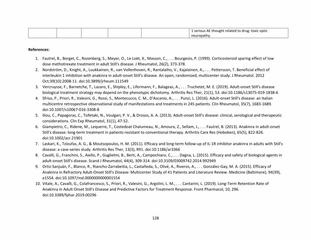
128
1 serious AE thought related to drug: toxic optic neuropathy.
References:
1. Fautrel, B., Borget, C., Rozenberg, S., Meyer, O., Le Loët, X., Masson, C., . . . Bourgeois, P. (1999). Corticosteroid sparing effect of low
dose methotrexate treatment in adult Still's disease. J Rheumatol, 26(2), 373-378.
2. Nordström, D., Knight, A., Luukkainen, R., van Vollenhoven, R., Rantalaiho, V., Kajalainen, A., . . . Pettersson, T. Beneficial effect of
interleukin 1 inhibition with anakinra in adult-onset Still's disease. An open, randomized, multicenter study. J Rheumatol. 2012
Oct;39(10):2008-11. doi:10.3899/jrheum.111549
3. Vercruysse, F., Barnetche, T., Lazaro, E., Shipley, E., Lifermann, F., Balageas, A., . . . Truchetet, M. E. (2019). Adult-onset Still's disease
biological treatment strategy may depend on the phenotypic dichotomy. Arthritis Res Ther, 21(1), 53. doi:10.1186/s13075-019-1838-6
4. Sfriso, P., Priori, R., Valesini, G., Rossi, S., Montecucco, C. M., D'Ascanio, A., . . . Punzi, L. (2016). Adult-onset Still's disease: an Italian
multicentre retrospective observational study of manifestations and treatments in 245 patients. Clin Rheumatol, 35(7), 1683-1689.
doi:10.1007/s10067-016-3308-8
5. Iliou, C., Papagoras, C., Tsifetaki, N., Voulgari, P. V., & Drosos, A. A. (2013). Adult-onset Still's disease: clinical, serological and therapeutic
considerations. Clin Exp Rheumatol, 31(1), 47-52.
6. Giampietro, C., Ridene, M., Lequerre, T., Costedoat Chalumeau, N., Amoura, Z., Sellam, J., . . . Fautrel, B. (2013). Anakinra in adult-onset
Still's disease: long-term treatment in patients resistant to conventional therapy. Arthritis Care Res (Hoboken), 65(5), 822-826.
doi:10.1002/acr.21901
7. Laskari, K., Tzioufas, A. G., & Moutsopoulos, H. M. (2011). Efficacy and long-term follow-up of IL-1R inhibitor anakinra in adults with Still's
disease: a case-series study. Arthritis Res Ther, 13(3), R91. doi:10.1186/ar3366
8. Cavalli, G., Franchini, S., Aiello, P., Guglielmi, B., Berti, A., Campochiaro, C., . . . Dagna, L. (2015). Efficacy and safety of biological agents in
adult-onset Still's disease. Scand J Rheumatol, 44(4), 309-314. doi:10.3109/03009742.2014.992949
9. Ortiz-Sanjuán, F., Blanco, R., Riancho-Zarrabeitia, L., Castañeda, S., Olivé, A., Riveros, A., . . . González-Gay, M. A. (2015). Efficacy of
Anakinra in Refractory Adult-Onset Still's Disease: Multicenter Study of 41 Patients and Literature Review. Medicine (Baltimore), 94(39),
e1554. doi:10.1097/md.0000000000001554
10. Vitale, A., Cavalli, G., Colafrancesco, S., Priori, R., Valesini, G., Argolini, L. M., . . . Cantarini, L. (2019). Long-Term Retention Rate of
Anakinra in Adult Onset Still's Disease and Predictive Factors for Treatment Response. Front Pharmacol, 10, 296.
doi:10.3389/fphar.2019.00296
129
11. Ruscitti, P., Cipriani, P., Liakouli, V., Iacono, D., Pantano, I., Caso, F., . . . Giacomelli, R. (2020). Prescribing motivations and patients’
characteristics related to the use of biologic drugs in adult-onset Still’s disease: analysis of a multicentre “real-life” cohort. Rheumatol
Int, 40(1), 107-113. doi:10.1007/s00296-019-04358-w
12. Colafrancesco, S., Priori, R., Valesini, G., Argolini, L., Baldissera, E., Bartoloni, E., . . . Sfriso, P. (2017). Response to Interleukin-1 Inhibitors
in 140 Italian Patients with Adult-Onset Still's Disease: A Multicentre Retrospective Observational Study. Front Pharmacol, 8, 369.
doi:10.3389/fphar.2017.00369
13. Dall'Ara, F., Frassi, M., Tincani, A., & Airò, P. (2016). A retrospective study of patients with adult-onset Still's disease: is pericarditis a
possible predictor for biological disease-modifying anti-rheumatic drugs need? Clin Rheumatol, 35(8), 2117-2123. doi:10.1007/s10067-
015-3164-y
14. Lequerré, T., Quartier, P., Rosellini, D., Alaoui, F., De Bandt, M., Mejjad, O., . . . Sibilia, J. (2008). Interleukin-1 receptor antagonist
(anakinra) treatment in patients with systemic-onset juvenile idiopathic arthritis or adult onset Still disease: preliminary experience in
France. Ann Rheum Dis, 67(3), 302-308. doi:10.1136/ard.2007.076034
15. Vitale, A., Insalaco, A., Sfriso, P., Lopalco, G., Emmi, G., Cattalini, M., . . . Cantarini, L. (2016). A Snapshot on the On-Label and Off-Label
Use of the Interleukin-1 Inhibitors in Italy among Rheumatologists and Pediatric Rheumatologists: A Nationwide Multi-Center
Retrospective Observational Study. Front Pharmacol, 7, 380. doi:10.3389/fphar.2016.00380
16. Rossi-Semerano, L., Fautrel, B., Wendling, D., Hachulla, E., Galeotti, C., Semerano, L., . . . Koné-Paut, I. (2015). Tolerance and efficacy of
off-label anti-interleukin-1 treatments in France: a nationwide survey. Orphanet J Rare Dis, 10, 19. doi:10.1186/s13023-015-0228-7
17. Sota, J., Rigante, D., Ruscitti, P., Insalaco, A., Sfriso, P., de Vita, S., . . . Cantarini, L. (2019). Anakinra Drug Retention Rate and Predictive
Factors of Long-Term Response in Systemic Juvenile Idiopathic Arthritis and Adult Onset Still Disease. Front Pharmacol, 10, 918.
doi:10.3389/fphar.2019.00918
18. Vitale, A., Cavalli, G., Ruscitti, P., Sota, J., Colafrancesco, S., Priori, R., . . . Cantarini, L. (2020). Comparison of Early vs. Delayed Anakinra
Treatment in Patients With Adult Onset Still's Disease and Effect on Clinical and Laboratory Outcomes. Front Med (Lausanne), 7, 42.
doi:10.3389/fmed.2020.00042
19. Kedor, C., Listing, J., Zernicke, J., Weiß, A., Behrens, F., Blank, N., . . . Feist, E. (2020). Canakinumab for Treatment of Adult-Onset Still’s
Disease to Achieve Reduction of Arthritic Manifestation (CONSIDER): phase II, randomised, double-blind, placebo-controlled,
multicentre, investigator-initiated trial. Annals of the rheumatic diseases, 79(8), 1090-1097. doi:10.1136/annrheumdis-2020-217155
20. Pham, T., Claudepierre, P., Constantin, A., de Bandt, M., Fautrel, B., Gossec, L., . . . Club Rhumatismes et, I. (2010). Tocilizumab: therapy
and safety management. Joint Bone Spine, 77 Suppl 1, S3-100. doi:10.1016/s1297-319x(10)70001-4
21. Nishina, N., Kaneko, Y., Kameda, H., & Takeuchi, T. (2015). The effect of tocilizumab on preventing relapses in adult-onset Still's disease:
A retrospective, single-center study. Mod Rheumatol, 25(3), 401-404. doi:10.3109/14397595.2014.973659
130
22. Ortiz-Sanjuán, F., Blanco, R., Calvo-Rio, V., Narvaez, J., Rubio Romero, E., Olivé, A., . . . González-Gay, M. A. (2014). Efficacy of tocilizumab
in conventional treatment-refractory adult-onset Still's disease: multicenter retrospective open-label study of thirty-four patients.
Arthritis Rheumatol, 66(6), 1659-1665. doi:10.1002/art.38398
23. Song, S. T., Kim, J. J., Lee, S., Kim, H. A., Lee, E. Y., Shin, K. C., . . . Yoo, D. H. (2016). Efficacy of tocilizumab therapy in Korean patients with
adult-onset Still's disease: a multicentre retrospective study of 22 cases. Clin Exp Rheumatol, 34(6 Suppl 102), S64-s71.
24. Kaneko, Y., Kameda, H., Ikeda, K., Ishii, T., Murakami, K., Takamatsu, H., . . . Takeuchi, T. (2018). Tocilizumab in patients with adult-onset
still's disease refractory to glucocorticoid treatment: a randomised, double-blind, placebo-controlled phase III trial. Ann Rheum Dis,
77(12), 1720-1729. doi:10.1136/annrheumdis-2018-213920
25. Wang, C. Y., Guo, S. H., Wang, L. P., & Shen, H. L. (2019). Refractory adult-onset Still disease treated by tocilizumab combined with
methotrexate: A STROBE-compliant article. Medicine (Baltimore), 98(32), e16682. doi:10.1097/md.0000000000016682
26. Gabay, C., Fautrel, B., Rech, J., Spertini, F., Feist, E., Kötter, I., . . . Schiffrin, E. J. (2018). Open-label, multicentre, dose-escalating phase II
clinical trial on the safety and efficacy of tadekinig alfa (IL-18BP) in adult-onset Still's disease. Ann Rheum Dis, 77(6), 840-847.
doi:10.1136/annrheumdis-2017-212608
PICO 24. In patients with sJIA, does the presence of subclinical MAS alter the treatment paradigm?
Summary: The literature searches did not identify any studies that addressed this PICO question.
Quality of evidence across all critical outcomes: Very low
131
PICO 25. In patients with sJIA and overt MAS, Is biologic therapy superior to calcineurin inhibitors in achievement of inactive disease and resolution of MAS?
Summary: This literature search identified two systematic reviews of MAS cases [1, 2], five single arm cohort studies [3, 4, 5, 6, 7], and one case series [2]. There were no direct comparisons made between treatment with biologics and cyclosporin. The systematic review by Grom et al.[1] included 72 patients with sJIA and possible MAS; they reported that clinical features of MAS were not modified by canakinumab compared to placebo. In contrast, Sonmez et al.[2] reported that 33/35 patients with MAS treated with anakinra achieved remission. Similarly, Sonmez et al.[2] evaluated 13 patients at their own institution with MAS secondary to sJIA; after starting anakinra, clinical symptoms resolved, steroids were able to be stopped and lab findings normalized. Two patients developed recurrence of MAS after reduction of anakinra dosage. In a cohort study by Eloseily et al.[3], sJIA patients with secondary HLH/MAS treated with anakinra within five days of hospitalization had the lowest mortality with zero deaths compared to secondary HLH/MAS due to other causes. Early administration of anakinra was associated with reduced mortality. Twenty-five percent of that cohort was treated with concomitant calcineurin inhibitors (cyclosporin A). In a cohort study by Yokota et al.[5] of 417 sJIA patients with 26 reported cases of MAS, cases treated with tocilizumab 8 mg/kg every 8 weeks showed a decrease in CRP, fever, reduction in steroid dose, and a mean reduction in systemic feature score. Another cohort study by Yokota et al.[6] evaluating sJIA patients’ treatment with IL-6 inhibition found that definite or probable MAS occurred in 3.6% of sJIA patients. Clinical and lab features appeared to be similar among patients regardless of whether IL-6 inhibition was administered. In one retrospective cohort study [7], 102 children with MAS secondary to JIA (grouped all categories together), 15 patients received IL1 inhibitors with 5 receiving cyclosporine as well.
Overall quality of evidence across all critical outcomes: Very low
Table 1. Data from Observational Studies
Ref ID, Author, Year
Study Type Duration Population Description
Treatment given to relevant population
Results
3225, Grom, 2016 [1]
Systematic review
Unclear 72 sJIA with possible MAS
Canakinumab 21 events (19 with canakinumab treatment; 2 with placebo control) in 19 patients were adjudicated as being probable MAS and 10 events in 9 patients as being possible MAS. Sojia was well controlled in majority of canakinumab treated patients at time of MAS. When rates of probable MAS events were compared between canakinumab-treated patients (2.8 per 100 patient years) and placebo treated patients (7.7 per 100 patient-years), the difference was not significant (-4.9 CI -15.6, 5.9). Three deaths due to MAS related complications (2-canakinumab treated, 1 placebo). Clinical features of MAS were not modified by
132
Ref ID, Author, Year
Study Type Duration Population Description
Treatment given to relevant population
Results
canakinumab. Infections were common trigger.
192, Sonmez, 2018 [2]
Case series/ systematic review
Jan 2015 to Jan 2017
MAS patients due to sJIA or AID (13 patients with soJIA)
Anakinra (2mg/kg/day)
Case series: 19 MAS episodes were observed. Anakinra (2mg/kg/day) was started in with a median 1 day after admission. Clinical sx resolved and lab findings normalizied within median 2 (1-4) and 6 (4-9) days after introduction of anakinra. Steroids were stopped in a median of 10 (4-13_ weeks after anakinra tx. Patients were followed up for a median of 13 (6-24 months). Two patients developed recurrence of MAS sx when anakinra was reduced, others in remission.
Systematic review: 9 articles, 35 patients with MAS associated with sJIA or AIDS, all patients except 2 reached remission.
4342,
Eloseily, 2020 [3]
Single arm cohort study
January 2008 to December 2016
All patients who presented with secondary HLH/MAS
44 patients, 13 with sJIA
Anakinra; concomitant therapies included steroids (73%), cyclosporin A (25%), IVIG (9%), etoposide (9%), tocilizumab (5%) and abatacept, rituximab, cyclophosphamide, and plasmapheresis in one patient each.
Early administration of anakinra (within 5 days of hospitalization) was associated with reduced mortality (p=0.046). Those patients with sJIA had the lowest mortality rate with no deaths among them (P=0.006)
2352, Aytac, 2016 [4]
Retrospective cohort
2009-2015 MAS patients secondary to sJIA (28) and lupus (6) 37 Mas
Multiple treatments All patients received steroids. Cyclosporine was given in 74.2% of soJIA-MAS. Intravenous immunoglobulin, anakinra, and etoposide was given during 67.7, 41,9, and 32.3% of sJIA MAS. Plasmapheresis was performed during 41.9% of sJIA MAS and was performed more frequently (p=0.021) in patients who died compared to patients who
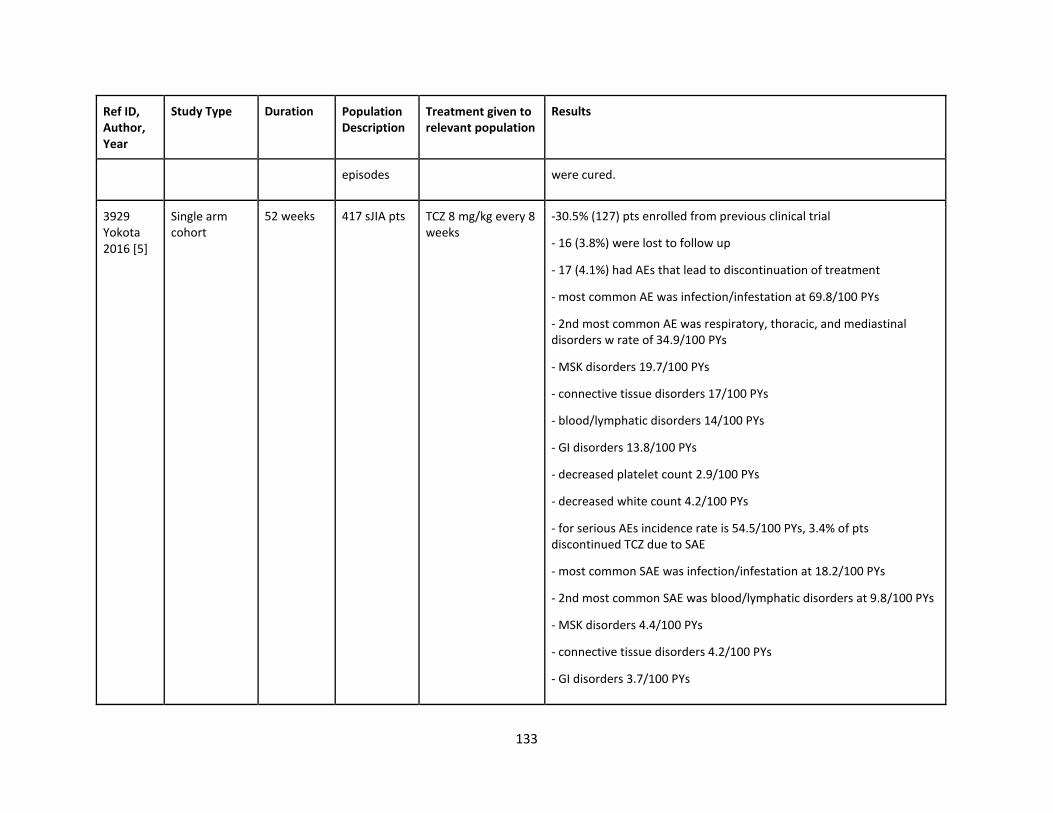
133
Ref ID, Author, Year
Study Type Duration Population Description
Treatment given to relevant population
Results
episodes were cured.
3929 Yokota 2016 [5]
Single arm cohort
52 weeks 417 sJIA pts TCZ 8 mg/kg every 8 weeks
-30.5% (127) pts enrolled from previous clinical trial
- 16 (3.8%) were lost to follow up
- 17 (4.1%) had AEs that lead to discontinuation of treatment
- most common AE was infection/infestation at 69.8/100 PYs
- 2nd most common AE was respiratory, thoracic, and mediastinal disorders w rate of 34.9/100 PYs
- MSK disorders 19.7/100 PYs
- connective tissue disorders 17/100 PYs
- blood/lymphatic disorders 14/100 PYs
- GI disorders 13.8/100 PYs
- decreased platelet count 2.9/100 PYs
- decreased white count 4.2/100 PYs
- for serious AEs incidence rate is 54.5/100 PYs, 3.4% of pts discontinued TCZ due to SAE
- most common SAE was infection/infestation at 18.2/100 PYs
- 2nd most common SAE was blood/lymphatic disorders at 9.8/100 PYs
- MSK disorders 4.4/100 PYs
- connective tissue disorders 4.2/100 PYs
- GI disorders 3.7/100 PYs
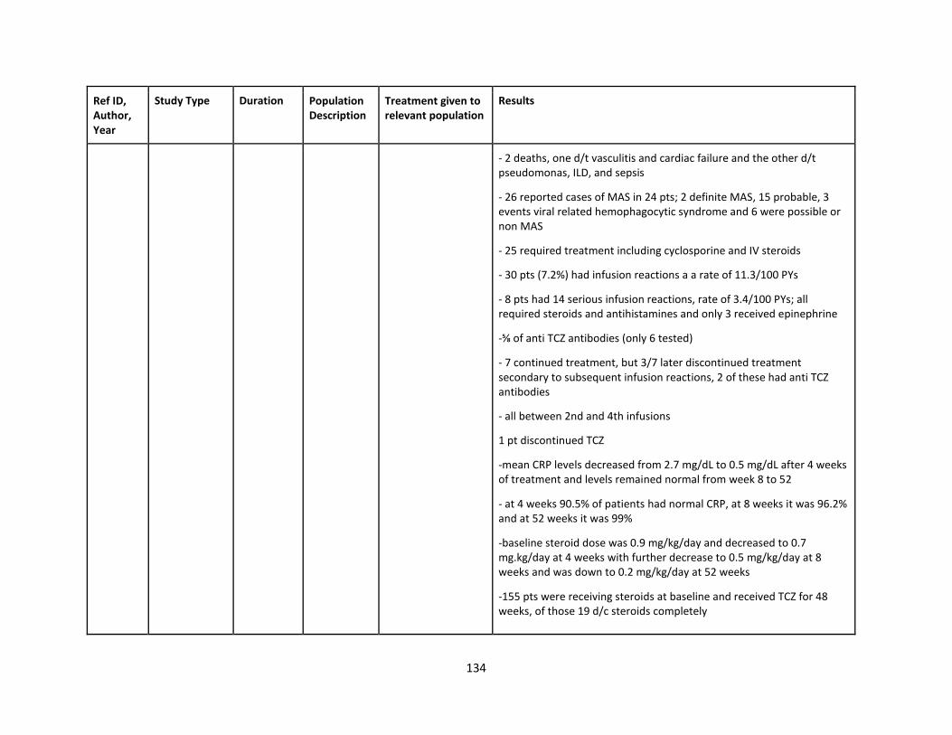
134
Ref ID, Author, Year
Study Type Duration Population Description
Treatment given to relevant population
Results
- 2 deaths, one d/t vasculitis and cardiac failure and the other d/t pseudomonas, ILD, and sepsis
- 26 reported cases of MAS in 24 pts; 2 definite MAS, 15 probable, 3 events viral related hemophagocytic syndrome and 6 were possible or non MAS
- 25 required treatment including cyclosporine and IV steroids
- 30 pts (7.2%) had infusion reactions a a rate of 11.3/100 PYs
- 8 pts had 14 serious infusion reactions, rate of 3.4/100 PYs; all required steroids and antihistamines and only 3 received epinephrine
-⅝ of anti TCZ antibodies (only 6 tested)
- 7 continued treatment, but 3/7 later discontinued treatment secondary to subsequent infusion reactions, 2 of these had anti TCZ antibodies
- all between 2nd and 4th infusions
1 pt discontinued TCZ
-mean CRP levels decreased from 2.7 mg/dL to 0.5 mg/dL after 4 weeks of treatment and levels remained normal from week 8 to 52
- at 4 weeks 90.5% of patients had normal CRP, at 8 weeks it was 96.2% and at 52 weeks it was 99%
-baseline steroid dose was 0.9 mg/kg/day and decreased to 0.7 mg.kg/day at 4 weeks with further decrease to 0.5 mg/kg/day at 8 weeks and was down to 0.2 mg/kg/day at 52 weeks
-155 pts were receiving steroids at baseline and received TCZ for 48 weeks, of those 19 d/c steroids completely
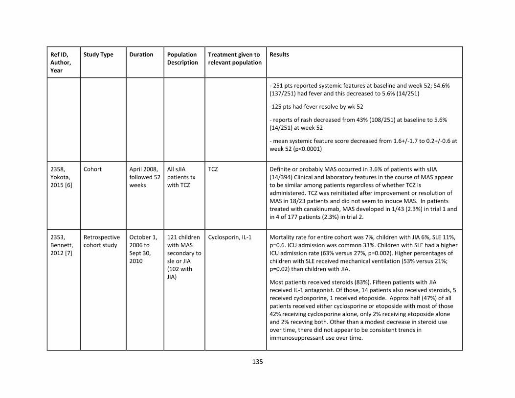
135
Ref ID, Author, Year
Study Type Duration Population Description
Treatment given to relevant population
Results
- 251 pts reported systemic features at baseline and week 52; 54.6% (137/251) had fever and this decreased to 5.6% (14/251)
-125 pts had fever resolve by wk 52
- reports of rash decreased from 43% (108/251) at baseline to 5.6% (14/251) at week 52
- mean systemic feature score decreased from 1.6+/-1.7 to 0.2+/-0.6 at week 52 (p<0.0001)
2358, Yokota, 2015 [6]
Cohort April 2008, followed 52 weeks
All sJIA patients tx with TCZ
TCZ Definite or probably MAS occurred in 3.6% of patients with sJIA (14/394) Clinical and laboratory features in the course of MAS appear to be similar among patients regardless of whether TCZ Is administered. TCZ was reinitiated after improvement or resolution of MAS in 18/23 patients and did not seem to induce MAS. In patients treated with canakinumab, MAS developed in 1/43 (2.3%) in trial 1 and in 4 of 177 patients (2.3%) in trial 2.
2353, Bennett, 2012 [7]
Retrospective cohort study
October 1, 2006 to Sept 30, 2010
121 children with MAS secondary to sle or JIA (102 with JIA)
Cyclosporin, IL-1 Mortality rate for entire cohort was 7%, children with JIA 6%, SLE 11%, p=0.6. ICU admission was common 33%. Children with SLE had a higher ICU admission rate (63% versus 27%, p=0.002). Higher percentages of children with SLE received mechanical ventilation (53% versus 21%; p=0.02) than children with JIA.
Most patients received steroids (83%). Fifteen patients with JIA received IL-1 antagonist. Of those, 14 patients also received steroids, 5 received cyclosporine, 1 received etoposide. Approx half (47%) of all patients received either cyclosporine or etoposide with most of those 42% receiving cyclosporine alone, only 2% receiving etoposide alone and 2% receving both. Other than a modest decrease in steroid use over time, there did not appear to be consistent trends in immunosuppressant use over time.
136
References:
1. Grom, A. A., Ilowite, N. T., Pascual, V., Brunner, H. I., Martini, A., Lovell, D., . . . Abrams, K. (2016). Rate and Clinical Presentation of
Macrophage Activation Syndrome in Patients With Systemic Juvenile Idiopathic Arthritis Treated With Canakinumab. Arthritis
Rheumatol, 68(1), 218-228. doi:10.1002/art.39407
2. Sönmez, H. E., Demir, S., Bilginer, Y., & Özen, S. (2018). Anakinra treatment in macrophage activation syndrome: a single center
experience and systemic review of literature. Clin Rheumatol, 37(12), 3329-3335. doi:10.1007/s10067-018-4095-1
3. Eloseily, E. M., Weiser, P., Crayne, C. B., Haines, H., Mannion, M. L., Stoll, M. L., . . . Cron, R. Q. (2020). Benefit of Anakinra in Treating
Pediatric Secondary Hemophagocytic Lymphohistiocytosis. Arthritis Rheumatol, 72(2), 326-334. doi:10.1002/art.41103
4. Aytaç, S., Batu, E. D., Ünal, Ş., Bilginer, Y., Çetin, M., Tuncer, M., . . . Özen, S. (2016). Macrophage activation syndrome in children with
systemic juvenile idiopathic arthritis and systemic lupus erythematosus. Rheumatol Int, 36(10), 1421-1429. doi:10.1007/s00296-016-
3545-9
5. Yokota, S., Itoh, Y., Morio, T., Origasa, H., Sumitomo, N., Tomobe, M., . . . Minota, S. (2016). Tocilizumab in systemic juvenile idiopathic
arthritis in a real-world clinical setting: results from 1 year of postmarketing surveillance follow-up of 417 patients in Japan. Ann Rheum
Dis, 75(9), 1654-1660. doi:10.1136/annrheumdis-2015-207818
6. Yokota, S., Itoh, Y., Morio, T., Sumitomo, N., Daimaru, K., & Minota, S. (2015). Macrophage Activation Syndrome in Patients with
Systemic Juvenile Idiopathic Arthritis under Treatment with Tocilizumab. J Rheumatol, 42(4), 712-722. doi:10.3899/jrheum.140288
7. Bennett, T. D., Fluchel, M., Hersh, A. O., Hayward, K. N., Hersh, A. L., Brogan, T. V., . . . Bratton, S. L. (2012). Macrophage activation
syndrome in children with systemic lupus erythematosus and children with juvenile idiopathic arthritis. Arthritis Rheum, 64(12), 4135-
4142. doi:10.1002/art.34661
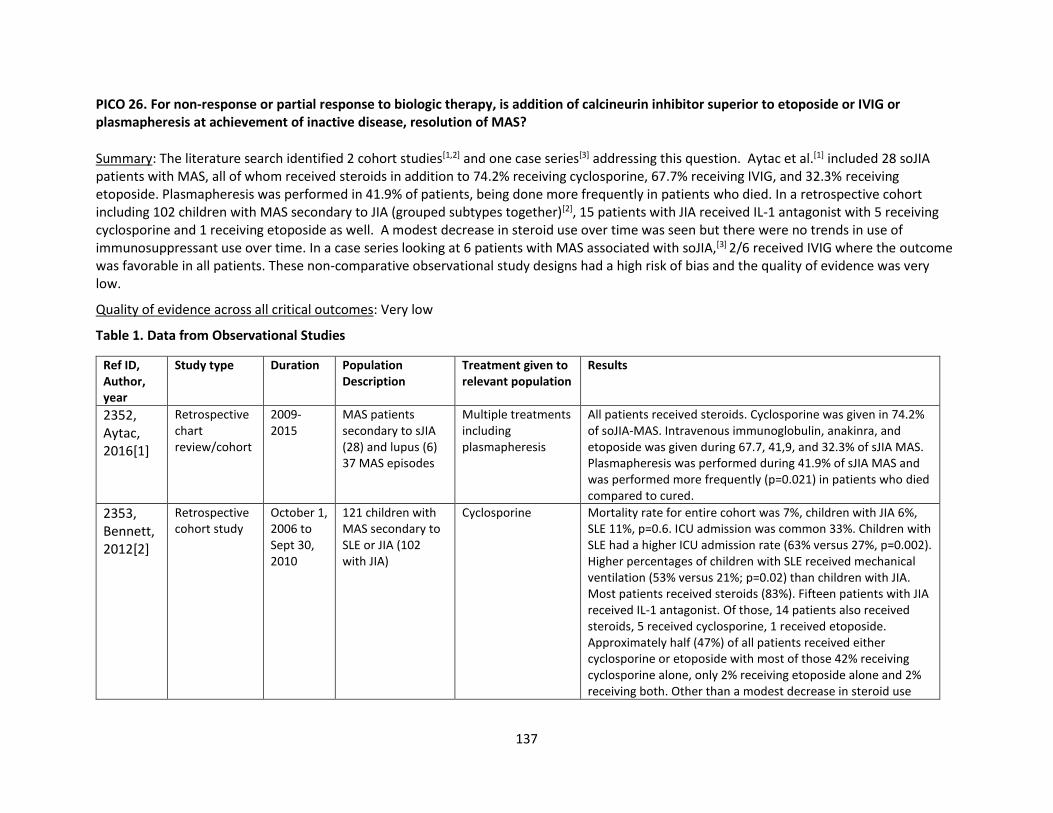
137
PICO 26. For non-response or partial response to biologic therapy, is addition of calcineurin inhibitor superior to etoposide or IVIG or plasmapheresis at achievement of inactive disease, resolution of MAS?
Summary: The literature search identified 2 cohort studies[1,2] and one case series[3] addressing this question. Aytac et al.[1] included 28 soJIA patients with MAS, all of whom received steroids in addition to 74.2% receiving cyclosporine, 67.7% receiving IVIG, and 32.3% receiving etoposide. Plasmapheresis was performed in 41.9% of patients, being done more frequently in patients who died. In a retrospective cohort including 102 children with MAS secondary to JIA (grouped subtypes together)[2], 15 patients with JIA received IL-1 antagonist with 5 receiving cyclosporine and 1 receiving etoposide as well. A modest decrease in steroid use over time was seen but there were no trends in use of immunosuppressant use over time. In a case series looking at 6 patients with MAS associated with soJIA,[3] 2/6 received IVIG where the outcome was favorable in all patients. These non-comparative observational study designs had a high risk of bias and the quality of evidence was very low.
Quality of evidence across all critical outcomes: Very low
Table 1. Data from Observational Studies
Ref ID, Author, year
Study type Duration Population Description
Treatment given to relevant population
Results
2352, Aytac, 2016[1]
Retrospective chart review/cohort
2009-2015
MAS patients secondary to sJIA (28) and lupus (6) 37 MAS episodes
Multiple treatments including plasmapheresis
All patients received steroids. Cyclosporine was given in 74.2% of soJIA-MAS. Intravenous immunoglobulin, anakinra, and etoposide was given during 67.7, 41,9, and 32.3% of sJIA MAS. Plasmapheresis was performed during 41.9% of sJIA MAS and was performed more frequently (p=0.021) in patients who died compared to cured.
2353, Bennett, 2012[2]
Retrospective cohort study
October 1, 2006 to Sept 30, 2010
121 children with MAS secondary to SLE or JIA (102 with JIA)
Cyclosporine Mortality rate for entire cohort was 7%, children with JIA 6%, SLE 11%, p=0.6. ICU admission was common 33%. Children with SLE had a higher ICU admission rate (63% versus 27%, p=0.002). Higher percentages of children with SLE received mechanical ventilation (53% versus 21%; p=0.02) than children with JIA. Most patients received steroids (83%). Fifteen patients with JIA received IL-1 antagonist. Of those, 14 patients also received steroids, 5 received cyclosporine, 1 received etoposide. Approximately half (47%) of all patients received either cyclosporine or etoposide with most of those 42% receiving cyclosporine alone, only 2% receiving etoposide alone and 2% receiving both. Other than a modest decrease in steroid use
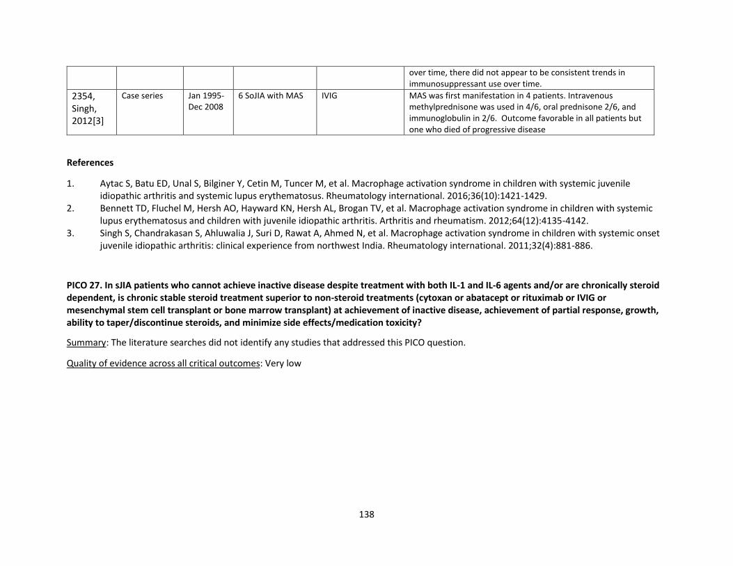
138
over time, there did not appear to be consistent trends in immunosuppressant use over time.
2354, Singh, 2012[3]
Case series Jan 1995-Dec 2008
6 SoJIA with MAS IVIG MAS was first manifestation in 4 patients. Intravenous methylprednisone was used in 4/6, oral prednisone 2/6, and immunoglobulin in 2/6. Outcome favorable in all patients but one who died of progressive disease
References
1. Aytac S, Batu ED, Unal S, Bilginer Y, Cetin M, Tuncer M, et al. Macrophage activation syndrome in children with systemic juvenile idiopathic arthritis and systemic lupus erythematosus. Rheumatology international. 2016;36(10):1421-1429.
2. Bennett TD, Fluchel M, Hersh AO, Hayward KN, Hersh AL, Brogan TV, et al. Macrophage activation syndrome in children with systemic lupus erythematosus and children with juvenile idiopathic arthritis. Arthritis and rheumatism. 2012;64(12):4135-4142.
3. Singh S, Chandrakasan S, Ahluwalia J, Suri D, Rawat A, Ahmed N, et al. Macrophage activation syndrome in children with systemic onset juvenile idiopathic arthritis: clinical experience from northwest India. Rheumatology international. 2011;32(4):881-886.
PICO 27. In sJIA patients who cannot achieve inactive disease despite treatment with both IL-1 and IL-6 agents and/or are chronically steroid dependent, is chronic stable steroid treatment superior to non-steroid treatments (cytoxan or abatacept or rituximab or IVIG or mesenchymal stem cell transplant or bone marrow transplant) at achievement of inactive disease, achievement of partial response, growth, ability to taper/discontinue steroids, and minimize side effects/medication toxicity?
Summary: The literature searches did not identify any studies that addressed this PICO question.
Quality of evidence across all critical outcomes: Very low
139
PICO 28. In sJIA patients with inactive disease treated with oral steroids, is taper to discontinuation of steroids superior to continuing long-term stable dose steroids for preventing disease flare and minimizing side effects/medication toxicity?
Summary: The literature review identified one case series[1] which included 20 patients with soJIA where symptoms of fever and rash flared up when prednisone was tapered. Five patients who had been off steroids for 4 to 24 months prior had recurrence of their disease.
Quality of evidence across all critical outcomes: Very low
Table 1. Data from Observational Studies
Ref ID, Author, year
Study type Duration Population Description
Treatment given to relevant population
Results
1472, Pelkonen, 1986
Case series 1974-1983
20 patients with sJIA
40 of 42 episodes of systemic systems in the patients were treated with steroids
Initial steroid dose ranged from 0.6-1.8 mg.kg/day. All patients had normalization of serum ferritin during the first few weeks of treatment. Fever and rash flared up when prednisone was tapered. Fifteen patients had one or several exacerbations of the disease. Five patients had been off steroids for 4 to 24 months prior to the recurrence.
References:
1. Pelkonen P, Swanljung K, Siimes MA. Ferritinemia as an indicator of systemic disease activity in children with systemic juvenile
rheumatoid arthritis. Acta Paediatr Scand. 1986 Jan;75(1):64-8.
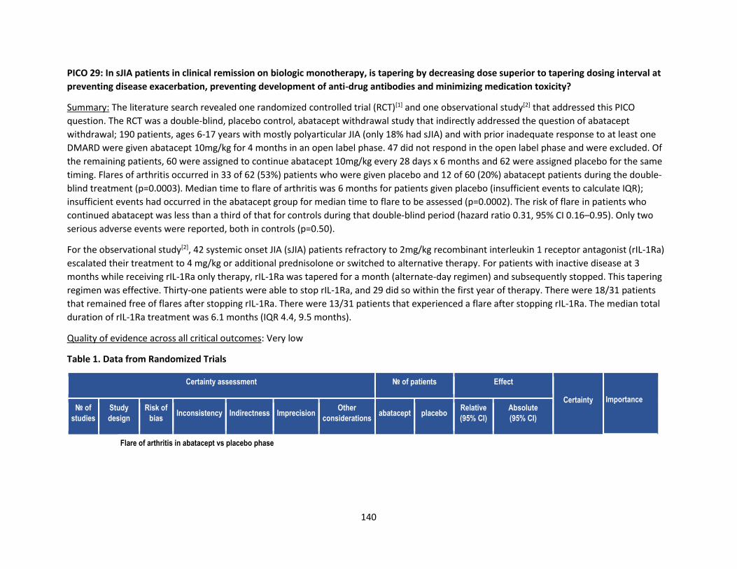
140
PICO 29: In sJIA patients in clinical remission on biologic monotherapy, is tapering by decreasing dose superior to tapering dosing interval at
preventing disease exacerbation, preventing development of anti-drug antibodies and minimizing medication toxicity?
Summary: The literature search revealed one randomized controlled trial (RCT)[1] and one observational study[2] that addressed this PICO
question. The RCT was a double-blind, placebo control, abatacept withdrawal study that indirectly addressed the question of abatacept
withdrawal; 190 patients, ages 6-17 years with mostly polyarticular JIA (only 18% had sJIA) and with prior inadequate response to at least one
DMARD were given abatacept 10mg/kg for 4 months in an open label phase. 47 did not respond in the open label phase and were excluded. Of
the remaining patients, 60 were assigned to continue abatacept 10mg/kg every 28 days x 6 months and 62 were assigned placebo for the same
timing. Flares of arthritis occurred in 33 of 62 (53%) patients who were given placebo and 12 of 60 (20%) abatacept patients during the double-
blind treatment (p=0.0003). Median time to flare of arthritis was 6 months for patients given placebo (insufficient events to calculate IQR);
insufficient events had occurred in the abatacept group for median time to flare to be assessed (p=0.0002). The risk of flare in patients who
continued abatacept was less than a third of that for controls during that double-blind period (hazard ratio 0.31, 95% CI 0.16–0.95). Only two
serious adverse events were reported, both in controls (p=0.50).
For the observational study[2], 42 systemic onset JIA (sJIA) patients refractory to 2mg/kg recombinant interleukin 1 receptor antagonist (rIL-1Ra)
escalated their treatment to 4 mg/kg or additional prednisolone or switched to alternative therapy. For patients with inactive disease at 3
months while receiving rIL-1Ra only therapy, rIL-1Ra was tapered for a month (alternate-day regimen) and subsequently stopped. This tapering
regimen was effective. Thirty-one patients were able to stop rIL-1Ra, and 29 did so within the first year of therapy. There were 18/31 patients
that remained free of flares after stopping rIL-1Ra. There were 13/31 patients that experienced a flare after stopping rIL-1Ra. The median total
duration of rIL-1Ra treatment was 6.1 months (IQR 4.4, 9.5 months).
Quality of evidence across all critical outcomes: Very low
Table 1. Data from Randomized Trials
Certainty assessment № of patients Effect
Certainty
Importance № of
studies
Study
design
Risk of
bias Inconsistency Indirectness Imprecision
Other
considerations abatacept placebo
Relative
(95% CI)
Absolute
(95% CI)
Flare of arthritis in abatacept vs placebo phase
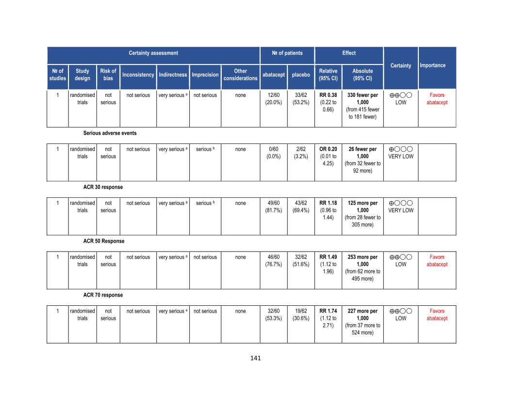
141
Certainty assessment № of patients Effect
Certainty
Importance № of
studies
Study
design
Risk of
bias Inconsistency Indirectness Imprecision
Other
considerations abatacept placebo
Relative
(95% CI)
Absolute
(95% CI)
1 randomised
trials
not
serious
not serious very serious a not serious none 12/60
(20.0%)
33/62
(53.2%)
RR 0.38
(0.22 to
0.66)
330 fewer per
1,000
(from 415 fewer
to 181 fewer)
⨁⨁◯◯
LOW
Favors
abatacept
Serious adverse events
1 randomised
trials
not
serious
not serious very serious a serious b none 0/60
(0.0%)
2/62
(3.2%)
OR 0.20
(0.01 to
4.25)
26 fewer per
1,000
(from 32 fewer to
92 more)
⨁◯◯◯
VERY LOW
ACR 30 response
1 randomised
trials
not
serious
not serious very serious a serious b none 49/60
(81.7%)
43/62
(69.4%)
RR 1.18
(0.96 to
1.44)
125 more per
1,000
(from 28 fewer to
305 more)
⨁◯◯◯
VERY LOW
ACR 50 Response
1 randomised
trials
not
serious
not serious very serious a not serious none 46/60
(76.7%)
32/62
(51.6%)
RR 1.49
(1.12 to
1.96)
253 more per
1,000
(from 62 more to
495 more)
⨁⨁◯◯
LOW
Favors
abatacept
ACR 70 response
1 randomised
trials
not
serious
not serious very serious a not serious none 32/60
(53.3%)
19/62
(30.6%)
RR 1.74
(1.12 to
2.71)
227 more per
1,000
(from 37 more to
524 more)
⨁⨁◯◯
LOW
Favors
abatacept
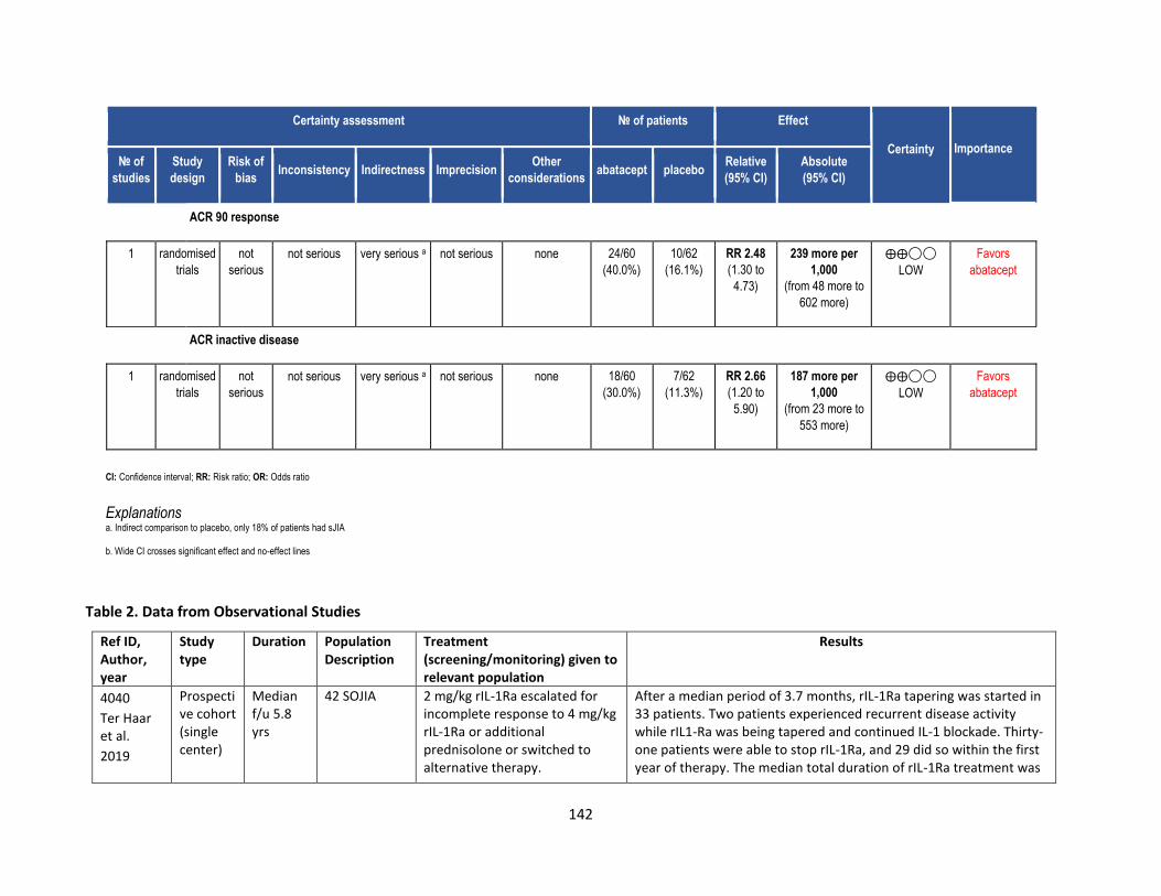
142
Certainty assessment № of patients Effect
Certainty
Importance № of
studies
Study
design
Risk of
bias Inconsistency Indirectness Imprecision
Other
considerations abatacept placebo
Relative
(95% CI)
Absolute
(95% CI)
ACR 90 response
1 randomised
trials
not
serious
not serious very serious a not serious none 24/60
(40.0%)
10/62
(16.1%)
RR 2.48
(1.30 to
4.73)
239 more per
1,000
(from 48 more to
602 more)
⨁⨁◯◯
LOW
Favors
abatacept
ACR inactive disease
1 randomised
trials
not
serious
not serious very serious a not serious none 18/60
(30.0%)
7/62
(11.3%)
RR 2.66
(1.20 to
5.90)
187 more per
1,000
(from 23 more to
553 more)
⨁⨁◯◯
LOW
Favors
abatacept
CI: Confidence interval; RR: Risk ratio; OR: Odds ratio
Explanations a. Indirect comparison to placebo, only 18% of patients had sJIA
b. Wide CI crosses significant effect and no-effect lines
Table 2. Data from Observational Studies
Ref ID, Author, year
Study type
Duration Population Description
Treatment (screening/monitoring) given to relevant population
Results
4040
Ter Haar et al.
2019
Prospective cohort (single center)
Median f/u 5.8 yrs
42 SOJIA 2 mg/kg rIL-1Ra escalated for incomplete response to 4 mg/kg rIL-1Ra or additional prednisolone or switched to alternative therapy.
After a median period of 3.7 months, rIL-1Ra tapering was started in 33 patients. Two patients experienced recurrent disease activity while rIL1-Ra was being tapered and continued IL-1 blockade. Thirty-one patients were able to stop rIL-1Ra, and 29 did so within the first year of therapy. The median total duration of rIL-1Ra treatment was
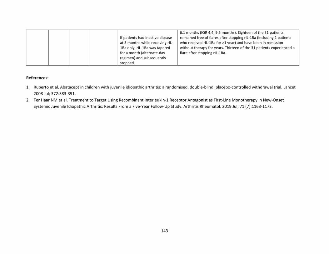
143
If patients had inactive disease at 3 months while receiving rIL-1Ra only, rIL-1Ra was tapered for a month (alternate-day regimen) and subsequently stopped.
6.1 months (IQR 4.4, 9.5 months). Eighteen of the 31 patients remained free of flares after stopping rIL-1Ra (including 2 patients who received rIL-1Ra for >1 year) and have been in remission without therapy for years. Thirteen of the 31 patients experienced a flare after stopping rIL-1Ra.
References:
1. Ruperto et al. Abatacept in children with juvenile idiopathic arthritis: a randomised, double-blind, placebo-controlled withdrawal trial. Lancet
2008 Jul; 372:383-391.
2. Ter Haar NM et al. Treatment to Target Using Recombinant Interleukin-1 Receptor Antagonist as First-Line Monotherapy in New-Onset
Systemic Juvenile Idiopathic Arthritis: Results From a Five-Year Follow-Up Study. Arthritis Rheumatol. 2019 Jul; 71 (7):1163-1173.

144
Specific Medication Screening Irrespective of Disease Subtype
PICO 30: Is there a recommended laboratory screening schedule (complete blood count, comprehensive metabolic panel and urinalysis) for
children receiving chronic daily NSAIDs?
Summary: The literature search revealed two studies that directly/indirectly addressed this question by monitoring for lab abnormalities while on treatment with NSAIDs. Neither study compared different screening schedules. A retrospective cohort study[1] most directly addressed this question of whether routine screening of hemoglobin, Transaminases, BUN, creatinine, and urinalysis was indicated in asymptomatic JIA patients. The authors reported that while 24/91 patients had lab abnormalities none of them correlated with adverse clinical signs or symptoms. Consequently, they concluded that routine monitoring may not be needed in asymptomatic patients. The second study[2] was a randomized, single center, controlled trial that indirectly addressed this PICO question by looking at lab abnormalities for patients on naproxen vs. aspirin at baseline, 12 weeks, and 24 weeks. Pathological elevations of liver enzymes occurred in the aspirin treated patients (14/30) but no clinical abnormalities occurred in the naproxen treated patients. Consequently, this study indirectly concluded that lab monitoring may vary by type of NSAID used. However, aspirin is not representative of current clinical practice, and the randomization was for comparison of different treatments rather than screening schedules. Therefore, the quality of evidence is very low. Quality of evidence across all critical outcomes: Very low Table 1. Data from Relevant Studies
Ref ID, Author, year
Study type Duration Population Description
Screening given to relevant population
Results
1393, Vora et al. 2010 [1]
Retrospective Cohort
10 years 1996-2006
91 JIA patients Oligo 62% Poly 29%
Screening for lab abnormalities or adverse events on NSAIDs for at least 1 month Median duration of tx (1.5 years) oligo 0.58 years poly
24/91 patients had lab abnormalities during the study period Nearly all were mild and not associated with adverse clinical concerns All continued treatment except for 1 patient Study proposes that routine lab monitoring in asymptomatic pts being treated with NSAIDs is of questionable utility
2581, Kvien, 1984 [2]
Single center randomized controlled trial
24 weeks 80 patients with oligo (52) or
1:1 randomization to naproxen 10mg/kg/day or aspirin 75mg/kg/day
Patients with adverse reactions: naproxen 12, aspirin 30 Discontinuation due to adverse reactions: naproxen 5, aspirin 20. Pathologically elevated liver enzymes occurred in 14/30 aspirin-treated patients. There was no elevation of ALT and AST levels of
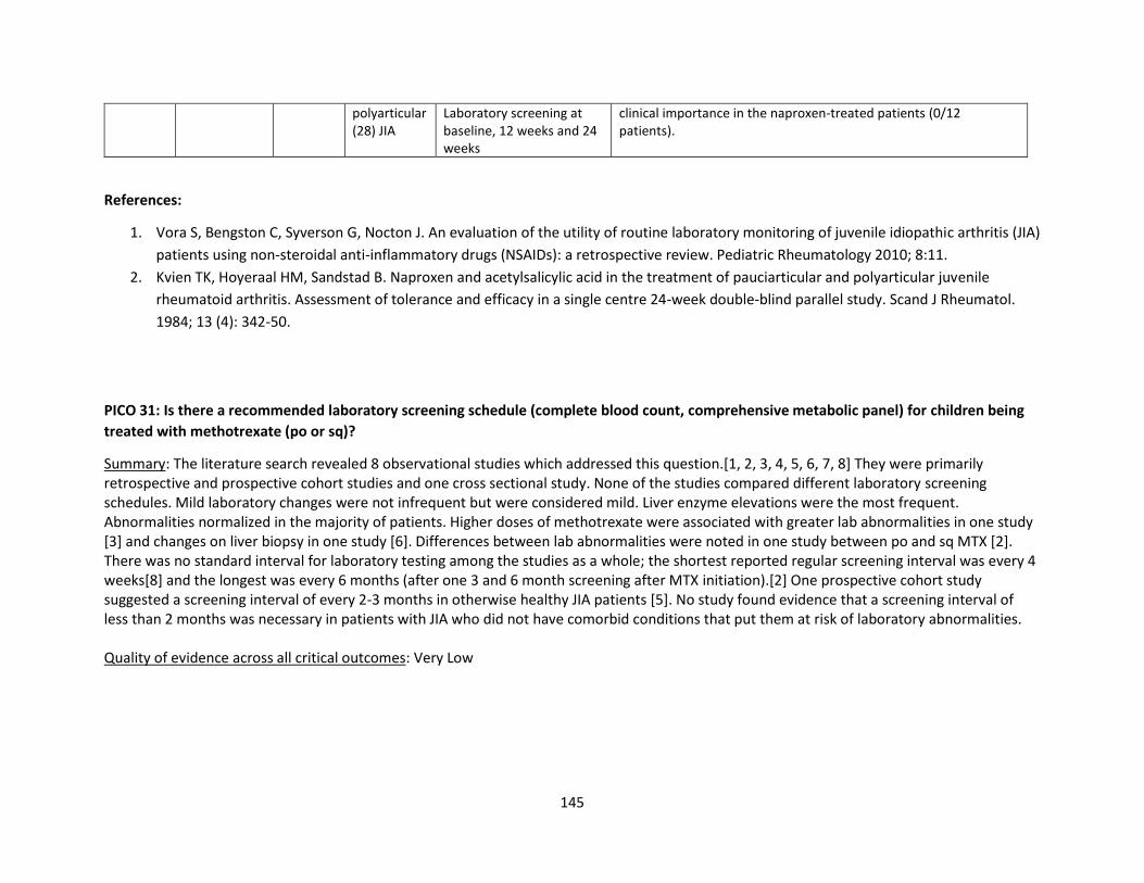
145
polyarticular (28) JIA
Laboratory screening at baseline, 12 weeks and 24 weeks
clinical importance in the naproxen-treated patients (0/12 patients).
References:
1. Vora S, Bengston C, Syverson G, Nocton J. An evaluation of the utility of routine laboratory monitoring of juvenile idiopathic arthritis (JIA)
patients using non-steroidal anti-inflammatory drugs (NSAIDs): a retrospective review. Pediatric Rheumatology 2010; 8:11.
2. Kvien TK, Hoyeraal HM, Sandstad B. Naproxen and acetylsalicylic acid in the treatment of pauciarticular and polyarticular juvenile
rheumatoid arthritis. Assessment of tolerance and efficacy in a single centre 24-week double-blind parallel study. Scand J Rheumatol.
1984; 13 (4): 342-50.
PICO 31: Is there a recommended laboratory screening schedule (complete blood count, comprehensive metabolic panel) for children being
treated with methotrexate (po or sq)?
Summary: The literature search revealed 8 observational studies which addressed this question.[1, 2, 3, 4, 5, 6, 7, 8] They were primarily retrospective and prospective cohort studies and one cross sectional study. None of the studies compared different laboratory screening schedules. Mild laboratory changes were not infrequent but were considered mild. Liver enzyme elevations were the most frequent. Abnormalities normalized in the majority of patients. Higher doses of methotrexate were associated with greater lab abnormalities in one study [3] and changes on liver biopsy in one study [6]. Differences between lab abnormalities were noted in one study between po and sq MTX [2]. There was no standard interval for laboratory testing among the studies as a whole; the shortest reported regular screening interval was every 4 weeks[8] and the longest was every 6 months (after one 3 and 6 month screening after MTX initiation).[2] One prospective cohort study suggested a screening interval of every 2-3 months in otherwise healthy JIA patients [5]. No study found evidence that a screening interval of less than 2 months was necessary in patients with JIA who did not have comorbid conditions that put them at risk of laboratory abnormalities. Quality of evidence across all critical outcomes: Very Low
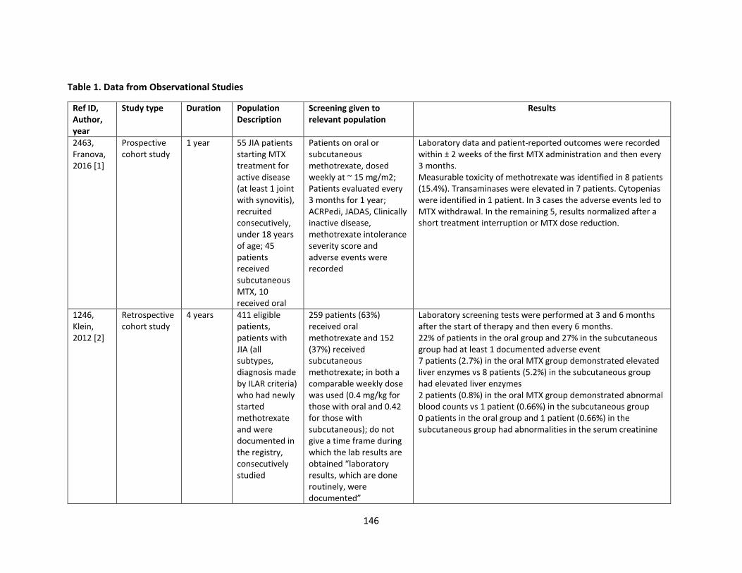
146
Table 1. Data from Observational Studies
Ref ID, Author, year
Study type Duration Population Description
Screening given to relevant population
Results
2463, Franova, 2016 [1]
Prospective cohort study
1 year 55 JIA patients starting MTX treatment for active disease (at least 1 joint with synovitis), recruited consecutively, under 18 years of age; 45 patients received subcutaneous MTX, 10 received oral
Patients on oral or subcutaneous methotrexate, dosed weekly at ~ 15 mg/m2; Patients evaluated every 3 months for 1 year; ACRPedi, JADAS, Clinically inactive disease, methotrexate intolerance severity score and adverse events were recorded
Laboratory data and patient-reported outcomes were recorded within ± 2 weeks of the first MTX administration and then every 3 months. Measurable toxicity of methotrexate was identified in 8 patients (15.4%). Transaminases were elevated in 7 patients. Cytopenias were identified in 1 patient. In 3 cases the adverse events led to MTX withdrawal. In the remaining 5, results normalized after a short treatment interruption or MTX dose reduction.
1246, Klein, 2012 [2]
Retrospective cohort study
4 years 411 eligible patients, patients with JIA (all subtypes, diagnosis made by ILAR criteria) who had newly started methotrexate and were documented in the registry, consecutively studied
259 patients (63%) received oral methotrexate and 152 (37%) received subcutaneous methotrexate; in both a comparable weekly dose was used (0.4 mg/kg for those with oral and 0.42 for those with subcutaneous); do not give a time frame during which the lab results are obtained “laboratory results, which are done routinely, were documented”
Laboratory screening tests were performed at 3 and 6 months after the start of therapy and then every 6 months. 22% of patients in the oral group and 27% in the subcutaneous group had at least 1 documented adverse event 7 patients (2.7%) in the oral MTX group demonstrated elevated liver enzymes vs 8 patients (5.2%) in the subcutaneous group had elevated liver enzymes 2 patients (0.8%) in the oral MTX group demonstrated abnormal blood counts vs 1 patient (0.66%) in the subcutaneous group 0 patients in the oral group and 1 patient (0.66%) in the subcutaneous group had abnormalities in the serum creatinine
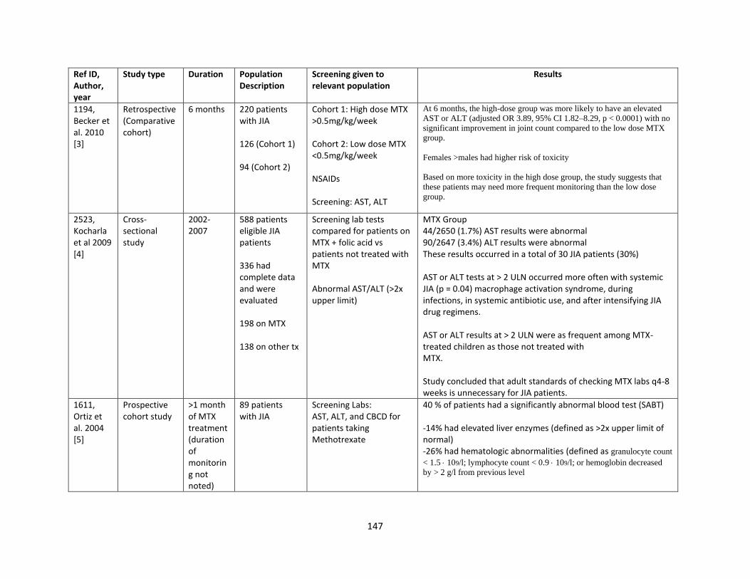
147
Ref ID, Author, year
Study type Duration Population Description
Screening given to relevant population
Results
1194, Becker et al. 2010 [3]
Retrospective (Comparative cohort)
6 months 220 patients with JIA 126 (Cohort 1) 94 (Cohort 2)
Cohort 1: High dose MTX >0.5mg/kg/week Cohort 2: Low dose MTX <0.5mg/kg/week NSAIDs Screening: AST, ALT
At 6 months, the high-dose group was more likely to have an elevated
AST or ALT (adjusted OR 3.89, 95% CI 1.82–8.29, p < 0.0001) with no
significant improvement in joint count compared to the low dose MTX
group.
Females >males had higher risk of toxicity
Based on more toxicity in the high dose group, the study suggests that
these patients may need more frequent monitoring than the low dose
group.
2523, Kocharla et al 2009 [4]
Cross- sectional study
2002-2007
588 patients eligible JIA patients 336 had complete data and were evaluated 198 on MTX 138 on other tx
Screening lab tests compared for patients on MTX + folic acid vs patients not treated with MTX Abnormal AST/ALT (>2x upper limit)
MTX Group 44/2650 (1.7%) AST results were abnormal 90/2647 (3.4%) ALT results were abnormal These results occurred in a total of 30 JIA patients (30%) AST or ALT tests at > 2 ULN occurred more often with systemic JIA (p = 0.04) macrophage activation syndrome, during infections, in systemic antibiotic use, and after intensifying JIA drug regimens. AST or ALT results at > 2 ULN were as frequent among MTX-treated children as those not treated with MTX. Study concluded that adult standards of checking MTX labs q4-8 weeks is unnecessary for JIA patients.
1611, Ortiz et al. 2004 [5]
Prospective cohort study
>1 month of MTX treatment (duration of monitoring not noted)
89 patients with JIA
Screening Labs: AST, ALT, and CBCD for patients taking Methotrexate
40 % of patients had a significantly abnormal blood test (SABT) -14% had elevated liver enzymes (defined as >2x upper limit of normal) -26% had hematologic abnormalities (defined as granulocyte count
< 1.5 109/l; lymphocyte count < 0.9 109/l; or hemoglobin decreased
by > 2 g/l from previous level
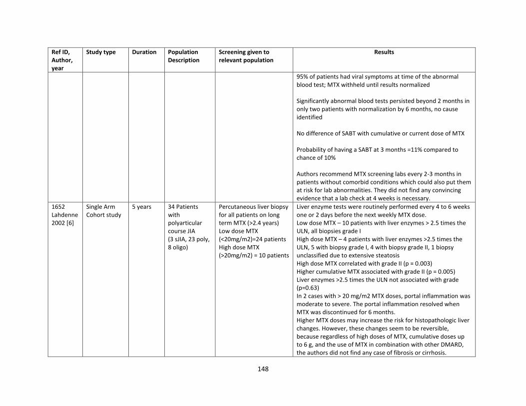
148
Ref ID, Author, year
Study type Duration Population Description
Screening given to relevant population
Results
95% of patients had viral symptoms at time of the abnormal blood test; MTX withheld until results normalized Significantly abnormal blood tests persisted beyond 2 months in only two patients with normalization by 6 months, no cause identified No difference of SABT with cumulative or current dose of MTX Probability of having a SABT at 3 months =11% compared to chance of 10% Authors recommend MTX screening labs every 2-3 months in patients without comorbid conditions which could also put them at risk for lab abnormalities. They did not find any convincing evidence that a lab check at 4 weeks is necessary.
1652 Lahdenne2002 [6]
Single Arm Cohort study
5 years 34 Patients with polyarticular course JIA (3 sJIA, 23 poly, 8 oligo)
Percutaneous liver biopsy for all patients on long term MTX (>2.4 years) Low dose MTX (<20mg/m2)=24 patients High dose MTX (>20mg/m2) = 10 patients
Liver enzyme tests were routinely performed every 4 to 6 weeks one or 2 days before the next weekly MTX dose. Low dose MTX – 10 patients with liver enzymes > 2.5 times the ULN, all biopsies grade I High dose MTX – 4 patients with liver enzymes >2.5 times the ULN, 5 with biopsy grade I, 4 with biopsy grade II, 1 biopsy unclassified due to extensive steatosis High dose MTX correlated with grade II (p = 0.003) Higher cumulative MTX associated with grade II (p = 0.005) Liver enzymes >2.5 times the ULN not associated with grade (p=0.63) In 2 cases with > 20 mg/m2 MTX doses, portal inflammation was moderate to severe. The portal inflammation resolved when MTX was discontinued for 6 months. Higher MTX doses may increase the risk for histopathologic liver changes. However, these changes seem to be reversible, because regardless of high doses of MTX, cumulative doses up to 6 g, and the use of MTX in combination with other DMARD, the authors did not find any case of fibrosis or cirrhosis.
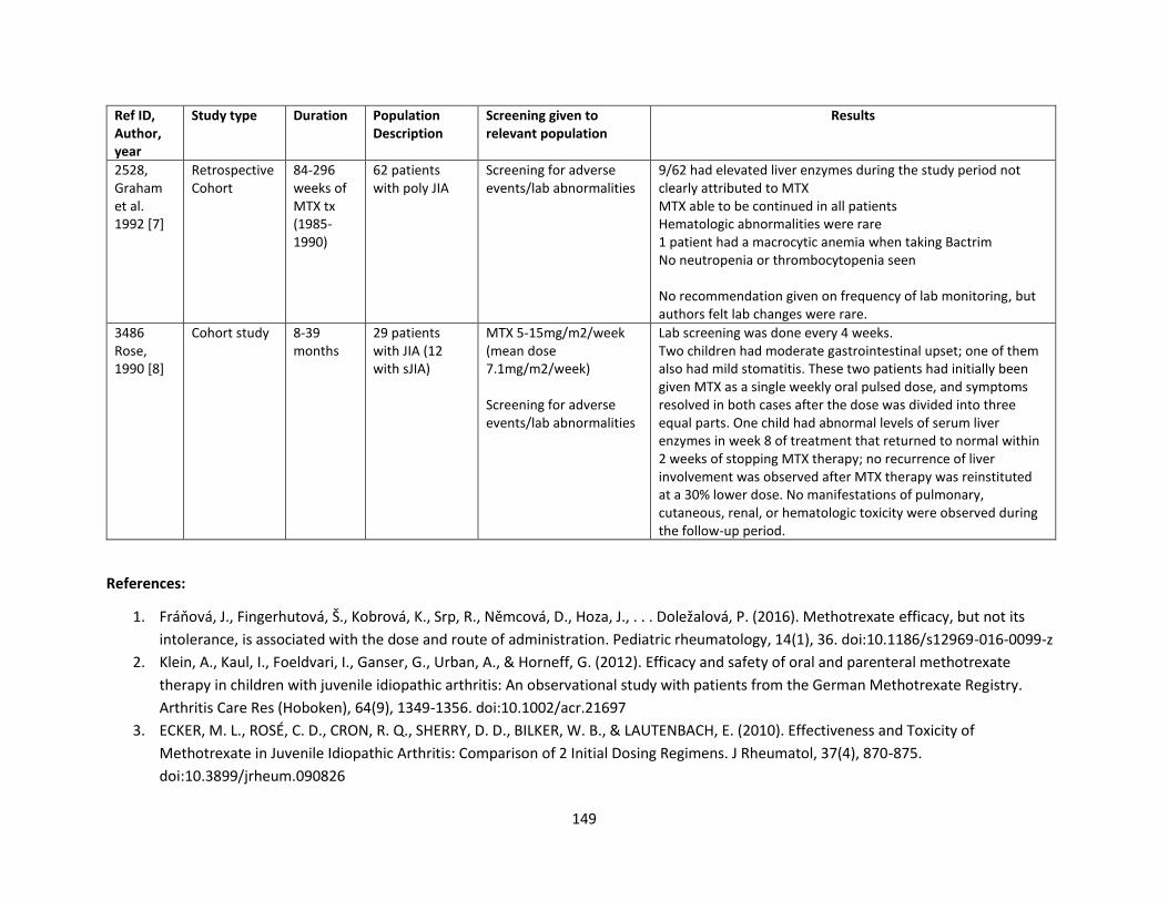
149
Ref ID, Author, year
Study type Duration Population Description
Screening given to relevant population
Results
2528, Graham et al. 1992 [7]
Retrospective Cohort
84-296 weeks of MTX tx (1985-1990)
62 patients with poly JIA
Screening for adverse events/lab abnormalities
9/62 had elevated liver enzymes during the study period not clearly attributed to MTX MTX able to be continued in all patients Hematologic abnormalities were rare 1 patient had a macrocytic anemia when taking Bactrim No neutropenia or thrombocytopenia seen No recommendation given on frequency of lab monitoring, but authors felt lab changes were rare.
3486 Rose, 1990 [8]
Cohort study 8-39 months
29 patients with JIA (12 with sJIA)
MTX 5-15mg/m2/week (mean dose 7.1mg/m2/week) Screening for adverse events/lab abnormalities
Lab screening was done every 4 weeks. Two children had moderate gastrointestinal upset; one of them also had mild stomatitis. These two patients had initially been given MTX as a single weekly oral pulsed dose, and symptoms resolved in both cases after the dose was divided into three equal parts. One child had abnormal levels of serum liver enzymes in week 8 of treatment that returned to normal within 2 weeks of stopping MTX therapy; no recurrence of liver involvement was observed after MTX therapy was reinstituted at a 30% lower dose. No manifestations of pulmonary, cutaneous, renal, or hematologic toxicity were observed during the follow-up period.
References:
1. Fráňová, J., Fingerhutová, Š., Kobrová, K., Srp, R., Němcová, D., Hoza, J., . . . Doležalová, P. (2016). Methotrexate efficacy, but not its
intolerance, is associated with the dose and route of administration. Pediatric rheumatology, 14(1), 36. doi:10.1186/s12969-016-0099-z
2. Klein, A., Kaul, I., Foeldvari, I., Ganser, G., Urban, A., & Horneff, G. (2012). Efficacy and safety of oral and parenteral methotrexate
therapy in children with juvenile idiopathic arthritis: An observational study with patients from the German Methotrexate Registry.
Arthritis Care Res (Hoboken), 64(9), 1349-1356. doi:10.1002/acr.21697
3. ECKER, M. L., ROSÉ, C. D., CRON, R. Q., SHERRY, D. D., BILKER, W. B., & LAUTENBACH, E. (2010). Effectiveness and Toxicity of
Methotrexate in Juvenile Idiopathic Arthritis: Comparison of 2 Initial Dosing Regimens. J Rheumatol, 37(4), 870-875.
doi:10.3899/jrheum.090826
150
4. KOCHARLA, L., TAYLOR, J., WEILER, T., TING, T. V., LUGGEN, M., & BRUNNER, H. I. (2009). Monitoring Methotrexate Toxicity in Juvenile
Idiopathic Arthritis. J Rheumatol, 36(12), 2813-2818. doi:10.3899/jrheum.090482
5. Ortiz-Alvarez, O., Morishita, K., Avery, G., Green, J., Petty, R. E., Tucker, L. B., . . . Cabral, D. A. (2004). Guidelines for blood test
monitoring of methotrexate toxicity in juvenile idiopathic arthritis. J Rheumatol, 31(12), 2501-2506.
6. Lahdenne, P., Rapola, J., Ylijoki, H., & Haapasaari, J. (2002). Hepatotoxicity in patients with juvenile idiopathic arthritis receiving longterm
methotrexate therapy. J Rheumatol, 29(11), 2442-2445.
7. Graham, L. D., Myones, B. L., Rivas-Chacon, R. F., & Pachman, L. M. (1992). Morbidity associated with long-term methotrexate therapy in
juvenile rheumatoid arthritis. J Pediatr, 120(3), 468-473. doi:10.1016/s0022-3476(05)80923-0
8. Rose, C. D., Singsen, B. H., Eichenfield, A. H., Goldsmith, D. P., & Athreya, B. H. (1990). Safety and efficacy of methotrexate therapy for
juvenile rheumatoid arthritis. J Pediatr, 117(4), 653-659. doi:https://doi.org/10.1016/S0022-3476(05)80709-7
PICO 32: After methotrexate (po or sq) is initiated, is there a recommended medication change secondary to elevated liver function tests and
decreased neutrophil or platelet count?
Summary: The literature search revealed 5 observational cohort studies which indirectly addressed this question.[1, 2, 3, 4, 5] Higher doses of methotrexate were associated with greater lab abnormalities in one study[2] compared to lower dose methotrexate without change in joint count. Consequently, this difference prompted the authors to suggest use of low dose methotrexate over high dose methotrexate (>20mg/m2). One study showed more changes on liver biopsy with higher doses of methotrexate[4]; however, the changes seemed to be reversible. In some instances, lab abnormalities improved after holding methotrexate temporarily [1, 3] before restarting it at the same dose or improved with methotrexate dose reduction in other cases [1, 5]. Quality of evidence across all critical outcomes: Very Low Table 1. Data from Observational Studies
Ref ID, Author, year
Study type Duration Population Description
Screening in relevant population
Results
2463, Franova, 2016 [1]
Prospective cohort study
1 year 55 JIA patients starting MTX treatment for active disease (at least 1 joint with synovitis),
Patients on oral or subcutaneous methotrexate, dosed weekly at ~ 15 mg/m2; Patients
Measurable toxicity of methotrexate was identified in 8 patients (15.4%). Transaminases were elevated in 7 patients (defined as increase of at least 1 liver transaminase above 2x the upper limit of normal). Cytopenias were identified in 1 patient. In 3 cases the adverse events led to MTX withdrawal. In the remaining 5,
151
Ref ID, Author, year
Study type Duration Population Description
Screening in relevant population
Results
recruited consecutively, under 18 years of age; 45 patients received subcutaneous MTX, 10 received oral
evaluated every 3 months for 1 year; ACRPedi, JADAS, Clinically inactive disease, methotrexate intolerance severity score and adverse events were recorded
results normalized after a short treatment interruption or MTX dose reduction. In the majority of cases, intolerance was managed to the patient and family satisfaction by treatment modifications and various other actions and their combinations: change in the route and/or timing of MTX administration, MTX dose reduction, addition of antiemetics, counseling. Methotrexate withdrawal was the ultimate solution in 2 patients only (8%).
1194, Becker et al. 2010 [2]
Retrospective (Comparative cohort)
1 year of MTX treatment
220 patients with JIA 126 (Cohort 1) 94 (Cohort 2)
Cohort 1: High dose MTX >0.5mg/kg/week Cohort 2: Low dose MTX <0.5mg/kg/week NSAIDs were allowed in each group
At 6 months, the high-dose group was more likely to have an elevated AST or ALT (adjusted OR 3.89, 95% CI 1.82–8.29, p < 0.0001) with NO statistically significant improvement in joint count compared to the low dose MTX group. Five of 126 (4%) patients in the high-dose group discontinued MTX due to hepatic toxicity. No medication substitution was mentioned.
1611, Ortiz et al. 2004 [3]
Prospective cohort study
>1 month of MTX treatment (duration of monitoring not noted)
89 patients with JIA Screening Labs: AST, ALT, and CBCD for patients taking Methotrexate
40 % of patients had a significantly abnormal blood test (SABT) -14% had elevated liver enzymes (defined as >2x upper limit of normal) -26% had hematologic abnormalities (defined as granulocyte count
< 1.5 109/l; lymphocyte count < 0.9 109/l; or hemoglobin decreased
by > 2 g/l from previous level When a SABT was identified, patients were instructed to stop taking MTX and have the blood test repeated within one to 2 weeks of the last abnormal test. 95% of patients had viral symptoms at time of the abnormal blood test; MTX withheld until results normalized and MTX was able to be restarted in most at the previous dose. In 2 patients, MTX was withheld for 6
152
Ref ID, Author, year
Study type Duration Population Description
Screening in relevant population
Results
months. No other patient had MTX withheld for longer than a month. No medication substitution was mentioned. No difference of SABT with cumulative or current dose of MTX. Probability of having a SABT at 3 months =11% compared to chance of 10%. Overall, study shows that most lab changes are minor and MTX can be continued in most patients.
1652 Lahdenne2002 [4]
Single Arm Cohort study
5 years 34 Patients with polyarticular course JIA (3 sJIA, 23 poly, 8 oligo)
Percutaneous liver biopsy for all patients on long term MTX (>2.4 years) Low dose MTX (<20mg/m2)=24 patients High dose MTX (>20mg/m2) = 10 patients
Liver enzyme tests were routinely performed every 4 to 6 weeks one or 2 days before the next weekly MTX dose. Low dose MTX – 10 patients with liver enzymes > 2.5 times the ULN, all biopsies grade I High dose MTX – 4 patients with liver enzymes >2.5 times the ULN, 5 with biopsy grade I, 4 with biopsy grade II, 1 biopsy unclassified due to extensive steatosis High dose MTX correlated with grade II (p = 0.003) Higher cumulative MTX associated with grade II (p = 0.005) Liver enzymes >2.5 times the ULN not associated with grade (p=0.63) In 2 cases with > 20 mg/m2 MTX doses, portal inflammation was moderate to severe. The portal inflammation resolved when MTX was discontinued for 6 months. Higher MTX doses may increase the risk for histopathologic liver changes. However, these changes seem to be reversible, because regardless of high doses of MTX, cumulative doses up to 6 g, and the use of MTX in combination with other DMARD, the authors did not find any case of fibrosis or cirrhosis.
3486 Rose, 1990 [5]
Cohort study 8-39 months
29 patients with JIA (12 with sJIA)
MTX 5-15mg/m2/week (mean dose 7.1mg/m2/week)
Two children had moderate gastrointestinal upset; one of them also had mild stomatitis. These two patients had initially been given MTX as a single weekly oral pulsed dose, and symptoms resolved in both cases after the dose was divided into three equal parts. One child had abnormal levels of serum liver enzymes in week 8 of treatment that returned to normal within
153
Ref ID, Author, year
Study type Duration Population Description
Screening in relevant population
Results
2 weeks of stopping MTX therapy; no recurrence of liver involvement was observed after MTX therapy was reinstituted at a 30% lower dose.
1. Franova et al. Methotrexate Efficacy, but Not Its Intolerance, Is Associated with the Dose and Route of Administration. Pediatr
Rheumatol Online J. 2016 Jun; 14 (1):36.
2. Becker ML, Rose CD, Cron RQ, Sherry DD, Bilker WB, Lautenbach E. Effectiveness and Toxicity of Methotrexate in Juvenile Idiopathic
Arthritis: Comparison of 2 Initial Dosing Regimens. J Rheumatol. 2010 Apr; 37 (4) 870-5.
3. Ortiz-Alvarez O et al. Guidelines for Blood Test Monitoring of Methotrexate Toxicity in Juvenile Idiopathic Arthritis. J Rheumatol. 2004
Dec; 31 (12): 2501-6.
4. Lahdenne P, Rapola J, Heikki Y, Haapasaari J. Hepatotoxicity in Patients with Juvenile Idiopathic Arthritis Receiving Longterm
Methotrexate Therapy. J Rheumatol.2002 Nov; 29 (11):2442-5.
5. Rose, C. D., Singsen, B. H., Eichenfield, A. H., Goldsmith, D. P., & Athreya, B. H. (1990). Safety and efficacy of methotrexate therapy for
juvenile rheumatoid arthritis. J Pediatr, 117(4), 653-659. doi:https://doi.org/10.1016/S0022-3476(05)80709-7
PICO 33. Is there a recommended laboratory screening schedule (complete blood count, comprehensive metabolic panel) for children with JIA
being treated with sulfasalazine?
Summary: Literature searches identified three observational studies (1 retrospective [1] and 2 prospective [2, 3] cohorts) which indirectly
answered the PICO question. The laboratory screening schedule was not the aim of these studies and none of them compared different
screening schedules. Each of the studies permitted for concomitant therapy administration including the use of NSAIDs [1, 3, 2], azathioprine [1,
3, 2], prednisone [3], anti-malarial [3, 2], auranofin [3], penicillamine [3], methotrexate [3] or aspirin [3]. In the study conducted by Chen et al.,
[1] labs were obtained baseline and then monthly thereafter. Each blood draw consisted of a CBC as well as renal and liver function tests. None
of the patients developed leukopenia, thrombocytopenia, or hepatitis (1). In the study by Varbanova et al. [2] labs were obtained twice during
the 1st through 3rd months and then every 3 months thereafter. Two cases of reversible neutropenia (following discontinuation of therapy) were
identified. No specific list of screening tests was identified [2]. Finally, the study performed by Imundo et al. [3] drew screening tests monthly for
3 months followed by a q3 month schedule. No specific laboratory screening measures were identified, but the authors noted that three
patients were found to have transaminitis and 5 had leukopenia with neutropenia.
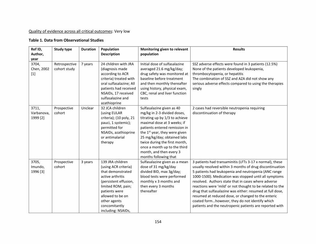
154
Quality of evidence across all critical outcomes: Very low
Table 1. Data from Observational Studies
Ref ID, Author, year
Study type Duration Population Description
Monitoring given to relevant population
Results
3704, Chen, 2002 [1]
Retrospective cohort study
7 years 24 children with JRA (diagnosis made according to ACR criteria) treated with oral sulfasalazine; All patients had received NSAIDs, 17 received sulfasalazine and azathioprine
Initial dose of sulfasalazine averaged 21.6 mg/kg/day; drug safety was monitored at baseline before treatment and then monthly thereafter using history, physical exam, CBC, renal and liver function tests
SSZ adverse effects were found in 3 patients (12.5%) None of the patients developed leukopenia, thrombocytopenia, or hepatitis The combination of SSZ and AZA did not show any serious adverse effects compared to using the therapies singly
3711, Varbanova, 1999 [2]
Prospective cohort
Unclear 32 JCA children (using EULAR criteria); (10 poly, 21 pauci, 1 systemic); permitted for NSAIDs, azathioprine or antimalarial therapy
Sulfasalazine given as 40 mg/kg in 2-3 divided doses, titrating up by 1/3 to achieve maximal dose at 3 weeks; if patients entered remission in the 1st year, they were given 25 mg/kg/day; obtained labs twice during the first month, once a month up to the third month, and then every 3 months following that
2 cases had reversible neutropenia requiring discontinuation of therapy
3705, Imundo, 1996 [3]
Prospective cohort
3 years 139 JRA children (using ACR criteria) that demonstrated active arthritis (persistent effusion, limited ROM, pain; patients were allowed to be on other agents concomitantly including: NSAIDs,
Sulfasalazine given as a mean dose of 31 mg/kg/day divided BID, max 3g/day; blood tests were performed monthly x 3 months and then every 3 months thereafter
3 patients had transaminitis (LFTs 3-17 x normal), these usually resolved within 3 months of drug discontinuation 5 patients had leukopenia and neutropenia (ANC range 1000-1500). Medication was stopped until all symptoms resolved. Authors state that in cases where adverse reactions were ‘mild’ or not thought to be related to the drug that sulfasalazine was either: resumed at full dose, resumed at reduced dose, or changed to the enteric coated form…however, they do not identify which patients and the neutropenic patients are reported with
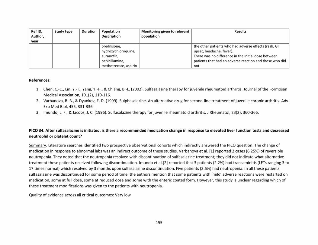
155
Ref ID, Author, year
Study type Duration Population Description
Monitoring given to relevant population
Results
prednisone, hydroxychloroquine, auranofin, penicillamine, methotrexate, aspirin
the other patients who had adverse effects (rash, GI upset, headache, fever). There was no difference in the initial dose between patients that had an adverse reaction and those who did not.
References:
1. Chen, C.-C., Lin, Y.-T., Yang, Y.-H., & Chiang, B.-L. (2002). Sulfasalazine therapy for juvenile rheumatoid arthritis. Journal of the Formosan
Medical Association, 101(2), 110-116.
2. Varbanova, B. B., & Dyankov, E. D. (1999). Sulphasalazine. An alternative drug for second-line treatment of juvenile chronic arthritis. Adv
Exp Med Biol, 455, 331-336.
3. Imundo, L. F., & Jacobs, J. C. (1996). Sulfasalazine therapy for juvenile rheumatoid arthritis. J Rheumatol, 23(2), 360-366.
PICO 34. After sulfasalazine is initiated, is there a recommended medication change in response to elevated liver function tests and decreased
neutrophil or platelet count?
Summary: Literature searches identified two prospective observational cohorts which indirectly answered the PICO question. The change of
medication in response to abnormal labs was an indirect outcome of these studies. Varbanova et al. [1] reported 2 cases (6.25%) of reversible
neutropenia. They noted that the neutropenia resolved with discontinuation of sulfasalazine treatment; they did not indicate what alternative
treatment these patients received following discontinuation. Imundo et al.[2] reported that 3 patients (2.2%) had transaminitis (LFTs ranging 3 to
17 times normal) which resolved by 3 months upon sulfasalazine discontinuation. Five patients (3.6%) had neutropenia. In all these patients
sulfasalazine was discontinued for some period of time. the authors mention that some patients with ‘mild’ adverse reactions were restarted on
medication, some at full dose, some at reduced dose and some with the enteric coated form. However, this study is unclear regarding which of
these treatment modifications was given to the patients with neutropenia.
Quality of evidence across all critical outcomes: Very low
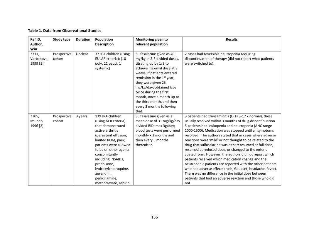
156
Table 1. Data from Observational Studies
Ref ID, Author, year
Study type Duration Population Description
Monitoring given to relevant population
Results
3711, Varbanova, 1999 [1]
Prospective cohort
Unclear 32 JCA children (using EULAR criteria); (10 poly, 21 pauci, 1 systemic)
Sulfasalazine given as 40 mg/kg in 2-3 divided doses, titrating up by 1/3 to achieve maximal dose at 3 weeks; if patients entered remission in the 1st year, they were given 25 mg/kg/day; obtained labs twice during the first month, once a month up to the third month, and then every 3 months following that.
2 cases had reversible neutropenia requiring discontinuation of therapy (did not report what patients were switched to).
3705, Imundo, 1996 [2]
Prospective cohort
3 years 139 JRA children (using ACR criteria) that demonstrated active arthritis (persistent effusion, limited ROM, pain; patients were allowed to be on other agents concomitantly including: NSAIDs, prednisone, hydroxylchloroquine, auranofin, penicillamine, methotrexate, aspirin
Sulfasalazine given as a mean dose of 31 mg/kg/day divided BID, max 3g/day; blood tests were performed monthly x 3 months and then every 3 months thereafter.
3 patients had transaminitis (LFTs 3-17 x normal), these usually resolved within 3 months of drug discontinuation 5 patients had leukopenia and neutropenia (ANC range 1000-1500). Medication was stopped until all symptoms resolved. The authors stated that in cases where adverse reactions were ‘mild’ or not thought to be related to the drug that sulfasalazine was either: resumed at full dose, resumed at reduced dose, or changed to the enteric coated form. However, the authors did not report which patients received which medication change and the neutropenic patients are reported with the other patients who had adverse effects (rash, GI upset, headache, fever). There was no difference in the initial dose between patients that had an adverse reaction and those who did not.
157
References:
1. Varbanova, B. B., & Dyankov, E. D. (1999). Sulphasalazine. An alternative drug for second-line treatment of juvenile chronic arthritis. Adv
Exp Med Biol, 455, 331-336.
2. Imundo, L. F., & Jacobs, J. C. (1996). Sulfasalazine therapy for juvenile rheumatoid arthritis. J Rheumatol, 23(2), 360-366.
PICO 35. Should children with JIA receiving leflunomide have serum creatinine, urinalysis, complete blood count and liver enzymes before
and during treatment, per manufacturer’s recommendations?
The following package insert information provided by the manufacturer regarding laboratory testing of leflunomide (ARAVA) is relevant to this
PICO question.
“Hematologic Monitoring: At minimum, patients taking ARAVA should have platelet, white blood cell count and hemoglobin or hematocrit
monitored at baseline and monthly for six months following initiation of therapy and every 6 to 8 weeks thereafter.
Bone Marrow Suppression Monitoring: If used concomitantly with immunosuppressants such as methotrexate, chronic monitoring should be
monthly.
Liver Enzyme Monitoring: At minimum, ALT (SGPT) must be performed at baseline and at least monthly for six months after starting ARAVA, and
thereafter every 6-8 weeks. In addition, if ARAVA and methotrexate are given concomitantly, ACR guidelines for monitoring methotrexate liver
toxicity must be followed with ALT, AST, and serum albumin testing every month.”
Summary: The literature search identified one study that indirectly addressed the question. The study was a retrospective, single-center cohort
study [1] of JIA patients who initiated leflunomide between April 2001 and October 2006. Labs were obtained on patients every 4-12 weeks. The
specific set of labs drawn was not reported in the study. Reporting of adverse events indicated that 9 (15.5%) of patients experienced transient
transaminitis. This resolved with decreasing the dose.
Quality of evidence across all critical outcomes: Very low
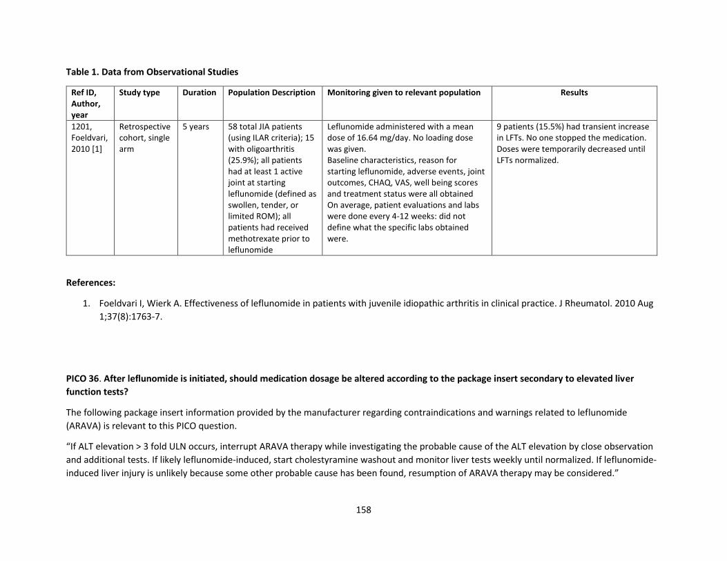
158
Table 1. Data from Observational Studies
Ref ID, Author, year
Study type Duration Population Description Monitoring given to relevant population Results
1201, Foeldvari, 2010 [1]
Retrospective cohort, single arm
5 years 58 total JIA patients (using ILAR criteria); 15 with oligoarthritis (25.9%); all patients had at least 1 active joint at starting leflunomide (defined as swollen, tender, or limited ROM); all patients had received methotrexate prior to leflunomide
Leflunomide administered with a mean dose of 16.64 mg/day. No loading dose was given. Baseline characteristics, reason for starting leflunomide, adverse events, joint outcomes, CHAQ, VAS, well being scores and treatment status were all obtained On average, patient evaluations and labs were done every 4-12 weeks: did not define what the specific labs obtained were.
9 patients (15.5%) had transient increase in LFTs. No one stopped the medication. Doses were temporarily decreased until LFTs normalized.
References:
1. Foeldvari I, Wierk A. Effectiveness of leflunomide in patients with juvenile idiopathic arthritis in clinical practice. J Rheumatol. 2010 Aug
1;37(8):1763-7.
PICO 36. After leflunomide is initiated, should medication dosage be altered according to the package insert secondary to elevated liver
function tests?
The following package insert information provided by the manufacturer regarding contraindications and warnings related to leflunomide
(ARAVA) is relevant to this PICO question.
“If ALT elevation > 3 fold ULN occurs, interrupt ARAVA therapy while investigating the probable cause of the ALT elevation by close observation
and additional tests. If likely leflunomide-induced, start cholestyramine washout and monitor liver tests weekly until normalized. If leflunomide-
induced liver injury is unlikely because some other probable cause has been found, resumption of ARAVA therapy may be considered.”
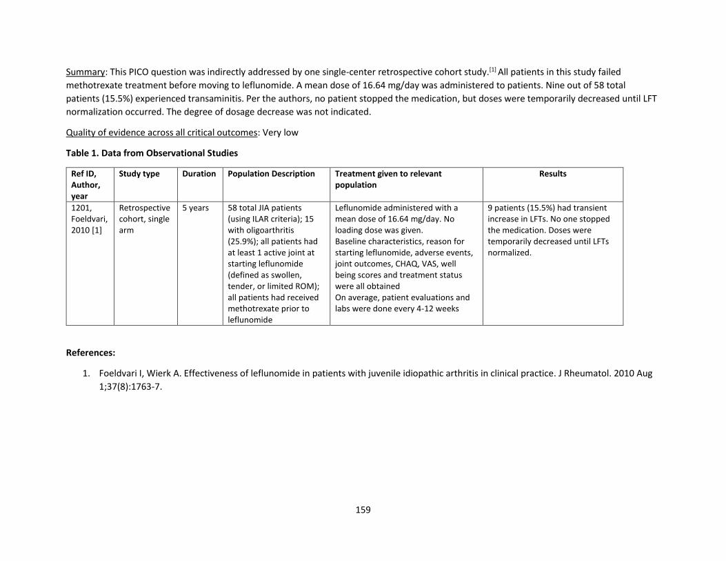
159
Summary: This PICO question was indirectly addressed by one single-center retrospective cohort study.[1] All patients in this study failed
methotrexate treatment before moving to leflunomide. A mean dose of 16.64 mg/day was administered to patients. Nine out of 58 total
patients (15.5%) experienced transaminitis. Per the authors, no patient stopped the medication, but doses were temporarily decreased until LFT
normalization occurred. The degree of dosage decrease was not indicated.
Quality of evidence across all critical outcomes: Very low
Table 1. Data from Observational Studies
Ref ID, Author, year
Study type Duration Population Description Treatment given to relevant population
Results
1201, Foeldvari, 2010 [1]
Retrospective cohort, single arm
5 years 58 total JIA patients (using ILAR criteria); 15 with oligoarthritis (25.9%); all patients had at least 1 active joint at starting leflunomide (defined as swollen, tender, or limited ROM); all patients had received methotrexate prior to leflunomide
Leflunomide administered with a mean dose of 16.64 mg/day. No loading dose was given. Baseline characteristics, reason for starting leflunomide, adverse events, joint outcomes, CHAQ, VAS, well being scores and treatment status were all obtained On average, patient evaluations and labs were done every 4-12 weeks
9 patients (15.5%) had transient increase in LFTs. No one stopped the medication. Doses were temporarily decreased until LFTs normalized.
References:
1. Foeldvari I, Wierk A. Effectiveness of leflunomide in patients with juvenile idiopathic arthritis in clinical practice. J Rheumatol. 2010 Aug
1;37(8):1763-7.
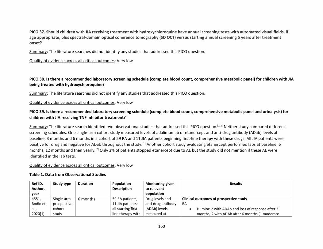
160
PICO 37. Should children with JIA receiving treatment with hydroxychloroquine have annual screening tests with automated visual fields, if age appropriate, plus spectral-domain optical coherence tomography (SD OCT) versus starting annual screening 5 years after treatment onset?
Summary: The literature searches did not identify any studies that addressed this PICO question.
Quality of evidence across all critical outcomes: Very low
PICO 38. Is there a recommended laboratory screening schedule (complete blood count, comprehensive metabolic panel) for children with JIA being treated with hydroxychloroquine?
Summary: The literature searches did not identify any studies that addressed this PICO question.
Quality of evidence across all critical outcomes: Very low
PICO 39. Is there a recommended laboratory screening schedule (complete blood count, comprehensive metabolic panel and urinalysis) for
children with JIA receiving TNF inhibitor treatment?
Summary: The literature search identified two observational studies that addressed this PICO question.[1,2] Neither study compared different
screening schedules. One single-arm cohort study measured levels of adalimumab or etanercept and anti-drug antibody (ADab) levels at
baseline, 3 months and 6 months in a cohort of 59 RA and 11 JIA patients beginning first-line therapy with these drugs. All JIA patients were
positive for drug and negative for ADab throughout the study.[1] Another cohort study evaluating etanercept performed labs at baseline, 6
months, 12 months and then yearly.[2] Only 2% of patients stopped etanercept due to AE but the study did not mention if these AE were
identified in the lab tests.
Quality of evidence across all critical outcomes: Very low
Table 1. Data from Observational Studies
Ref ID, Author, year
Study type Duration Population Description
Monitoring given to relevant population
Results
4551, Bodio et al., 2020[1]
Single-arm prospective cohort study
6 months 59 RA patients, 11 JIA patients; all starting first-line therapy with
Drug levels and anti-drug antibody (ADAb) levels measured at
Clinical outcomes of prospective study RA
• Humira: 2 with ADAb and loss of response after 3 months, 2 with ADAb after 6 months (1 moderate
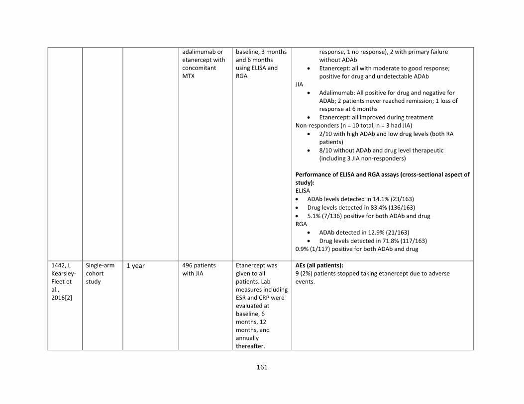
161
adalimumab or etanercept with concomitant MTX
baseline, 3 months and 6 months using ELISA and RGA
response, 1 no response), 2 with primary failure without ADAb
• Etanercept: all with moderate to good response; positive for drug and undetectable ADAb
JIA
• Adalimumab: All positive for drug and negative for ADAb; 2 patients never reached remission; 1 loss of response at 6 months
• Etanercept: all improved during treatment Non-responders (n = 10 total; n = 3 had JIA)
• 2/10 with high ADAb and low drug levels (both RA patients)
• 8/10 without ADAb and drug level therapeutic (including 3 JIA non-responders)
Performance of ELISA and RGA assays (cross-sectional aspect of study): ELISA
• ADAb levels detected in 14.1% (23/163)
• Drug levels detected in 83.4% (136/163)
• 5.1% (7/136) positive for both ADAb and drug RGA
• ADAb detected in 12.9% (21/163)
• Drug levels detected in 71.8% (117/163) 0.9% (1/117) positive for both ADAb and drug
1442, L Kearsley-Fleet et al., 2016[2]
Single-arm cohort study
1 year 496 patients with JIA
Etanercept was given to all patients. Lab measures including ESR and CRP were evaluated at baseline, 6 months, 12 months, and annually thereafter.
AEs (all patients): 9 (2%) patients stopped taking etanercept due to adverse events.
162
References:
1. Bodio C, Grossi C, Pregnolato F, Favalli EG, Biggioggero M, Marchesoni A, et al. Personalized medicine in rheumatoid arthritis: How immunogenicity impacts use of TNF inhibitors. Autoimmunity reviews. 2020;19(5):102509.
2. Kearsley-Fleet L, Davies R, Lunt M, Southwood TR, Hyrich KL. Factors associated with improvement in disease activity following initiation of etanercept in children and young people with Juvenile Idiopathic Arthritis: results from the British Society for Paediatric and Adolescent Rheumatology Etanercept Cohort Study. Rheumatology (Oxford, England). 2016;55(5):840-847.
PICO 40. Is there a recommended laboratory screening schedule (complete blood count, comprehensive metabolic panel and urinalysis) for
children with JIA receiving abatacept treatment?
Summary: The literature searches did not identify any studies that addressed this PICO question.
Quality of evidence across all critical outcomes: Very low
PICO 41. Should children with JIA receiving tocilizumab have serum creatinine, urinalysis, complete blood cell count, and liver enzymes before
and during treatment, per manufacturer’s recommendations?
Summary. Our searches identified one retrospective cohort study with 104 patients (86 with JIA) that marginally addressed this question.
Aeschlimann et al.[1] evaluated pediatric rheumatology patients who received at least 1 dose of tocilizumab. As noted in Table 1, 3 children with
sJIA developed MAS with elevated liver enzymes while on tocilizumab, and a couple children (disease not specified) had elevated liver enzymes
or febrile neutropenia while on tocilizumab.
Quality of evidence across all critical outcomes: Very low
Table 1. Data from Observational Studies
Ref ID, Author, year
Study type Duration Population Description Treatment given to relevant population
Results
4608, Aeschlimann, 2020[1]
Retrospective Cohort study
01/2007 to 06/2019
All pediatric rheumatology
patients who received at
least one dose of
tocilizumab:
At least one dose of tocilizumab
3 children with sJIA had MAS while on tocilizumab therapy, with part of the presentation including elevated liver enzymes
163
43 sJIA, 43 pJIA, 12 other
autoinflammatory disease,
6 "other"
1 child (disease not stated) had elevated liver enzymes while on tocilizumab 1 child (disease not stated) had febrile neutropenia while on tocilizumab
References
1. Aeschlimann FA, Dumaine C, Wörner A, Mouy R, Wouters C, Melki I, et al. Serious adverse events in children with juvenile idiopathic arthritis and other rheumatic diseases on tocilizumab - a real-world experience. Seminars in arthritis and rheumatism. 2020;50(4):744-748.
PICO 42. After tocilizumab is initiated, should medication dosage be altered according to the package insert secondary to elevated liver
function tests, neutropenia and/or thrombocytopenia?
Summary: The literature searches did not identify any studies that addressed this PICO question.
Quality of evidence across all critical outcomes: Very low
PICO 43. Is there a recommended laboratory screening schedule (complete blood count, comprehensive metabolic panel and urinalysis) for
children with JIA receiving anakinra treatment?
Summary. Our searches identified one retrospective cohort study with 77 patients with sJIA treated with anakinra and canakinumab that
marginally addressed this question. Sota et al.[1] evaluated drug retention rate and factors that led to stopping the drugs in some patients.
Adverse events occurred in 13/77 patients, one of whom had abnormal liver enzymes, but no patients developed MAS or serious adverse events.
Quality of evidence across all critical outcomes: Very low
164
Table 1. Data from Observational Studies
Ref ID, Author, year
Study type Duration Population Description
Monitoring given to relevant population
Results
4406 Sota, 2019[1]
Retrospective cohort
7.5 years 77 pediatric systemic JIA pts treated with anakinra and canakinumab
Evaluate drug retention rate of anakinra and canakinumab. Determine factors which led to stopping the drug.
15 centers in Italy retrospectively reviewed. 61 pts received anakinra, 25 canakinumab. 22 pts also received a DMARD concurrently (mtx, cyclosporine, sulfasalazine, leflunomide, or plaquenil). Mean ± SD treatment length 22.67 ± 19.50 months. The retention rate of both anti-IL-1 agents at 12-, 24-, 48-, and 60-months of follow-up was 79.9, 59.5, 53.5, and 53.5%, respectively. The median disease length was significantly higher in pts who stopped IL-1 blockers (5.88 years ± 6.55) vs those that were able to continue these biologic agents (3.17 years ± 3.68) (p = 0.011). 16/63 patients (27%) were able to stop corticosteroids. AEs occurred in 13 out of 77 patients (17.1%) (11 on ANA and 2 on CAN), with injection site-reactions (n = 7) most commonly, then generalized skin rashes (n = 4), respiratory problems (n = 1), and abnormal level of liver enzymes (n = 1). 10 pts stopped treatment because of AEs. No pts had MAS or serious AEs.
References:
1. Sota J, Insalaco A, Cimaz R, Alessio M, Cattalini M, Gallizzi R, et al. Drug retention rate and predictive factors of drug survival for
interleukin-1 inhibitors in systemic juvenile idiopathic arthritis. Frontiers in Pharmacology. 2019;9(JAN):1526.
PICO 44. Is there a recommended laboratory screening schedule (complete blood count, comprehensive metabolic panel and urinalysis) for
children with JIA receiving canakinumab treatment?
Summary: The literature searches did not identify any studies that addressed this PICO question.
Quality of evidence across all critical outcomes: Very low
165
PICO 45: Should all children with JIA have infection titers (measles, varicella, hepatitis B, hepatitis C) checked prior to starting
immunosuppressive medication?
Summary: The literature search identified two cohort studies[1,2] and one cross-sectional study[3] that addressed this PICO question. These
studies reported adverse events related to infectious diseases in patients with JIA receiving a biologic[1] and the incidence of zoster among
patients with JIA.[2] The rate of zoster was higher among patients with JIA compared to healthy controls (IRR 2.9 (1.8 – 4.5), p<0.001). The cross-
sectional study[3] evaluated the presence of lower genital infections in female JIA patients compared to healthy controls. The frequency of HPV
infection was higher in JIA patients compared to controls, but the difference was not statistically significant (30% vs 11%, p=0.155). There was no
difference in the frequency of Chlamydia trachomatis or Neisseria gonorrhoeae infection between JIA patients or healthy controls. Current use
of MTX (12% vs 44%, p=0.206) and biologic use (37% vs 64%, p=0.238) were not significantly different between JIA patients with abnormal and
normal cytopathology.
Quality of evidence across all critical outcomes: Very low
Table 1. Data from Observational Studies
Ref ID, Author, year
Study type
Duration Population Description
Monitoring in relevant population
Results
1858 Dumaine 2019[1]
Single Arm Cohort study
14 years 677 patients with JIA and receiving a biologic (177 poly, 137 ERA, 117 sJIA, 108 extended oligo, 87 oligo, 33 psoriatic, 18 unclassified) 90% on combination therapy (MTX, NSAIDs, corticosteroids)
Adverse event related to infectious disease as reported by investigator in the database
184 infectious adverse events, incidence rate 0.17 (95% CI 0.17 + 0.01) per biological treatment 54 IAE in sJIA (all patients on anakinra and canakinumab): ANA 6.7/100 p-y, CAN 9.6/100 p-y 37 IAE in extended oligo 14 in oligo (no treatment/disease type rates reported) 12 severe or very severe (7 in sJIA) – CAN severe 1 (1.3/100 p-y), very severe 1 (1.3/100 p-y) ANA severe 2 (1.1/100 p-y), very severe 0 24 hospitalization (15 in sJIA) – CAN 2 (2.7/100 p-y), ANA 6 (3.3/100 p-y)
1844 Nimmrich 2015[2]
Cohort Study
12 years 3,042 patients with JIA enrolled in BIKER
Incidence of zoster diagnosed by a physician
(Rate by JIA category not reported) – 17 total cases Overall rate 6/1000 patients (3.5-9.0), incidence 3.1/1000 patient-years (1.9-4.9) – compared to published rate in healthy children IRR 2.9 (1.8-4.5), p<0.001
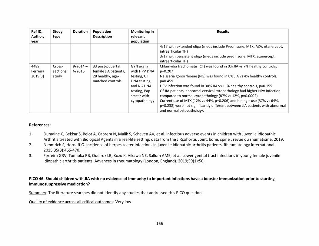
166
Ref ID, Author, year
Study type
Duration Population Description
Monitoring in relevant population
Results
4/17 with extended oligo (meds include Prednisone, MTX, AZA, etanercept, intraarticular TH) 3/17 with persistent oligo (meds include prednisone, MTX, etanercept, intraarticular TH)
4489 Ferreira 2019[3]
Cross- sectional study
9/2014 – 6/2016
33 post-pubertal female JIA patients, 28 healthy, age-matched controls
GYN exam with HPV DNA testing, CT DNA testing, and NG DNA testing, Pap smear with cytopathology
Chlamydia trachomatis (CT) was found in 0% JIA vs 7% healthy controls, p=0.207 Neisseria gonorrhoeae (NG) was found in 0% JIA vs 4% healthy controls, p=0.459 HPV infection was found in 30% JIA vs 11% healthy controls, p=0.155 Of JIA patients, abnormal cervical cytopathology had higher HPV infection compared to normal cytopathology (87% vs 12%, p=0.0002) Current use of MTX (12% vs 44%, p=0.206) and biologic use (37% vs 64%, p=0.238) were not significantly different between JIA patients with abnormal and normal cytopathology.
References:
1. Dumaine C, Bekkar S, Belot A, Cabrera N, Malik S, Scheven AV, et al. Infectious adverse events in children with Juvenile Idiopathic Arthritis treated with Biological Agents in a real-life setting: data from the JIRcohorte. Joint, bone, spine : revue du rhumatisme. 2019.
2. Nimmrich S, Horneff G. Incidence of herpes zoster infections in juvenile idiopathic arthritis patients. Rheumatology international. 2015;35(3):465-470.
3. Ferreira GRV, Tomioka RB, Queiroz LB, Kozu K, Aikawa NE, Sallum AME, et al. Lower genital tract infections in young female juvenile idiopathic arthritis patients. Advances in rheumatology (London, England). 2019;59(1):50.
PICO 46. Should children with JIA with no evidence of immunity to important infections have a booster immunization prior to starting immunosuppressive medication?
Summary: The literature searches did not identify any studies that addressed this PICO question.
Quality of evidence across all critical outcomes: Very low
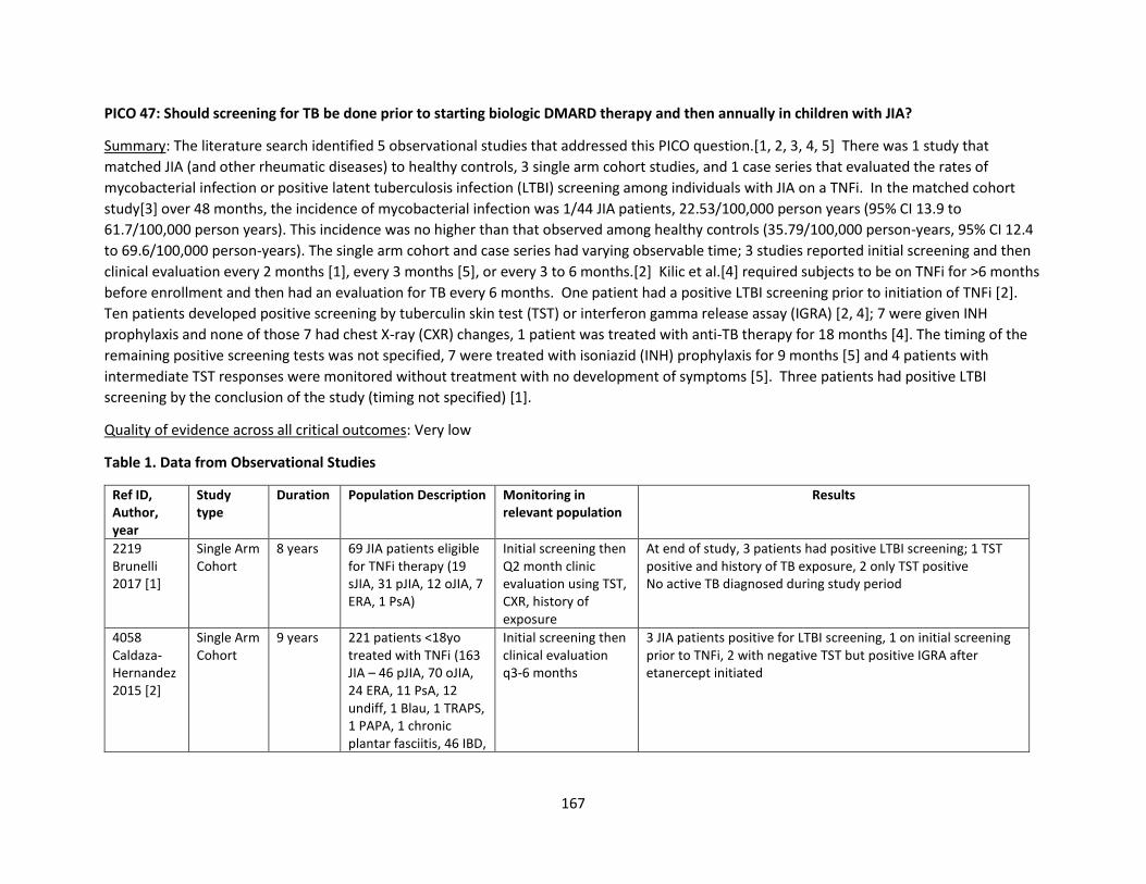
167
PICO 47: Should screening for TB be done prior to starting biologic DMARD therapy and then annually in children with JIA?
Summary: The literature search identified 5 observational studies that addressed this PICO question.[1, 2, 3, 4, 5] There was 1 study that
matched JIA (and other rheumatic diseases) to healthy controls, 3 single arm cohort studies, and 1 case series that evaluated the rates of
mycobacterial infection or positive latent tuberculosis infection (LTBI) screening among individuals with JIA on a TNFi. In the matched cohort
study[3] over 48 months, the incidence of mycobacterial infection was 1/44 JIA patients, 22.53/100,000 person years (95% CI 13.9 to
61.7/100,000 person years). This incidence was no higher than that observed among healthy controls (35.79/100,000 person-years, 95% CI 12.4
to 69.6/100,000 person-years). The single arm cohort and case series had varying observable time; 3 studies reported initial screening and then
clinical evaluation every 2 months [1], every 3 months [5], or every 3 to 6 months.[2] Kilic et al.[4] required subjects to be on TNFi for >6 months
before enrollment and then had an evaluation for TB every 6 months. One patient had a positive LTBI screening prior to initiation of TNFi [2].
Ten patients developed positive screening by tuberculin skin test (TST) or interferon gamma release assay (IGRA) [2, 4]; 7 were given INH
prophylaxis and none of those 7 had chest X-ray (CXR) changes, 1 patient was treated with anti-TB therapy for 18 months [4]. The timing of the
remaining positive screening tests was not specified, 7 were treated with isoniazid (INH) prophylaxis for 9 months [5] and 4 patients with
intermediate TST responses were monitored without treatment with no development of symptoms [5]. Three patients had positive LTBI
screening by the conclusion of the study (timing not specified) [1].
Quality of evidence across all critical outcomes: Very low
Table 1. Data from Observational Studies
Ref ID, Author, year
Study type
Duration Population Description Monitoring in relevant population
Results
2219 Brunelli 2017 [1]
Single Arm Cohort
8 years 69 JIA patients eligible for TNFi therapy (19 sJIA, 31 pJIA, 12 oJIA, 7 ERA, 1 PsA)
Initial screening then Q2 month clinic evaluation using TST, CXR, history of exposure
At end of study, 3 patients had positive LTBI screening; 1 TST positive and history of TB exposure, 2 only TST positive No active TB diagnosed during study period
4058 Caldaza-Hernandez 2015 [2]
Single Arm Cohort
9 years 221 patients <18yo treated with TNFi (163 JIA – 46 pJIA, 70 oJIA, 24 ERA, 11 PsA, 12 undiff, 1 Blau, 1 TRAPS, 1 PAPA, 1 chronic plantar fasciitis, 46 IBD,
Initial screening then clinical evaluation q3-6 months
3 JIA patients positive for LTBI screening, 1 on initial screening prior to TNFi, 2 with negative TST but positive IGRA after etanercept initiated
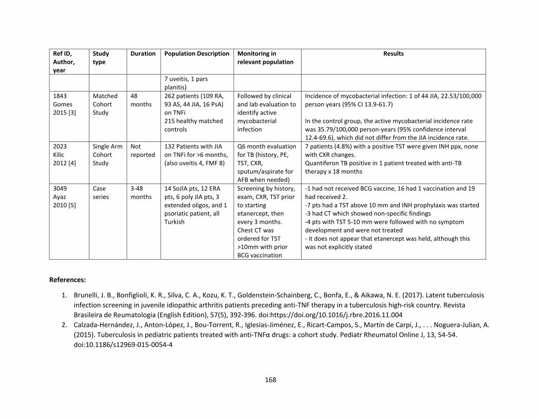
168
Ref ID, Author, year
Study type
Duration Population Description Monitoring in relevant population
Results
7 uveitis, 1 pars planitis)
1843 Gomes 2015 [3]
Matched Cohort Study
48 months
262 patients (109 RA, 93 AS, 44 JIA, 16 PsA) on TNFi 215 healthy matched controls
Followed by clinical and lab evaluation to identify active mycobacterial infection
Incidence of mycobacterial infection: 1 of 44 JIA, 22.53/100,000 person years (95% CI 13.9-61.7) In the control group, the active mycobacterial incidence rate was 35.79/100,000 person-years (95% confidence interval 12.4-69.6), which did not differ from the JIA incidence rate.
2023 Kilic 2012 [4]
Single Arm Cohort Study
Not reported
132 Patients with JIA on TNFi for >6 months, (also uveitis 4, FMF 8)
Q6 month evaluation for TB (history, PE, TST, CXR, sputum/aspirate for AFB when needed)
7 patients (4.8%) with a positive TST were given INH ppx, none with CXR changes. Quantiferon TB positive in 1 patient treated with anti-TB therapy x 18 months
3049 Ayaz 2010 [5]
Case series
3-48 months
14 SoJIA pts, 12 ERA pts, 6 poly JIA pts, 3 extended oligos, and 1 psoriatic patient, all Turkish
Screening by history, exam, CXR, TST prior to starting etanercept, then every 3 months. Chest CT was ordered for TST >10mm with prior BCG vaccination
-1 had not received BCG vaccine, 16 had 1 vaccination and 19 had received 2. -7 pts had a TST above 10 mm and INH prophylaxis was started -3 had CT which showed non-specific findings -4 pts with TST 5-10 mm were followed with no symptom development and were not treated - it does not appear that etanercept was held, although this was not explicitly stated
References:
1. Brunelli, J. B., Bonfiglioli, K. R., Silva, C. A., Kozu, K. T., Goldenstein-Schainberg, C., Bonfa, E., & Aikawa, N. E. (2017). Latent tuberculosis
infection screening in juvenile idiopathic arthritis patients preceding anti-TNF therapy in a tuberculosis high-risk country. Revista
Brasileira de Reumatologia (English Edition), 57(5), 392-396. doi:https://doi.org/10.1016/j.rbre.2016.11.004
2. Calzada-Hernández, J., Anton-López, J., Bou-Torrent, R., Iglesias-Jiménez, E., Ricart-Campos, S., Martín de Carpi, J., . . . Noguera-Julian, A.
(2015). Tuberculosis in pediatric patients treated with anti-TNFα drugs: a cohort study. Pediatr Rheumatol Online J, 13, 54-54.
doi:10.1186/s12969-015-0054-4
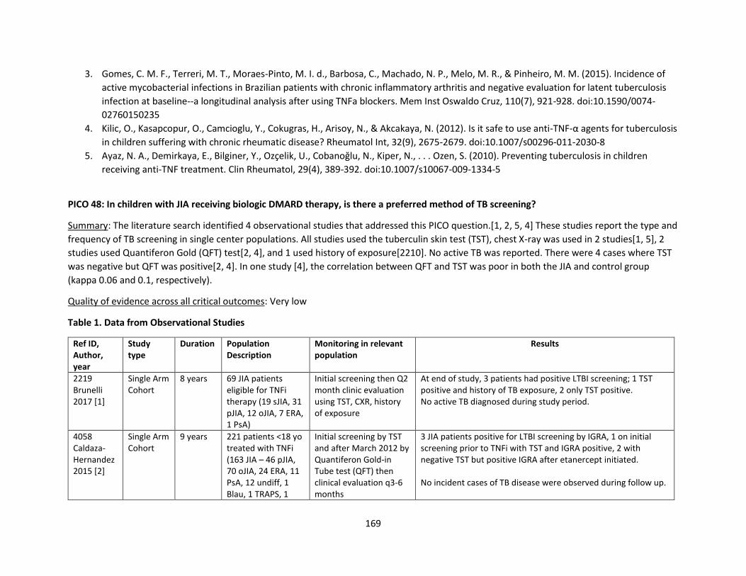
169
3. Gomes, C. M. F., Terreri, M. T., Moraes-Pinto, M. I. d., Barbosa, C., Machado, N. P., Melo, M. R., & Pinheiro, M. M. (2015). Incidence of
active mycobacterial infections in Brazilian patients with chronic inflammatory arthritis and negative evaluation for latent tuberculosis
infection at baseline--a longitudinal analysis after using TNFa blockers. Mem Inst Oswaldo Cruz, 110(7), 921-928. doi:10.1590/0074-
02760150235
4. Kilic, O., Kasapcopur, O., Camcioglu, Y., Cokugras, H., Arisoy, N., & Akcakaya, N. (2012). Is it safe to use anti-TNF-α agents for tuberculosis
in children suffering with chronic rheumatic disease? Rheumatol Int, 32(9), 2675-2679. doi:10.1007/s00296-011-2030-8
5. Ayaz, N. A., Demirkaya, E., Bilginer, Y., Ozçelik, U., Cobanoğlu, N., Kiper, N., . . . Ozen, S. (2010). Preventing tuberculosis in children
receiving anti-TNF treatment. Clin Rheumatol, 29(4), 389-392. doi:10.1007/s10067-009-1334-5
PICO 48: In children with JIA receiving biologic DMARD therapy, is there a preferred method of TB screening?
Summary: The literature search identified 4 observational studies that addressed this PICO question.[1, 2, 5, 4] These studies report the type and
frequency of TB screening in single center populations. All studies used the tuberculin skin test (TST), chest X-ray was used in 2 studies[1, 5], 2
studies used Quantiferon Gold (QFT) test[2, 4], and 1 used history of exposure[2210]. No active TB was reported. There were 4 cases where TST
was negative but QFT was positive[2, 4]. In one study [4], the correlation between QFT and TST was poor in both the JIA and control group
(kappa 0.06 and 0.1, respectively).
Quality of evidence across all critical outcomes: Very low
Table 1. Data from Observational Studies
Ref ID, Author, year
Study type
Duration Population Description
Monitoring in relevant population
Results
2219 Brunelli 2017 [1]
Single Arm Cohort
8 years 69 JIA patients eligible for TNFi therapy (19 sJIA, 31 pJIA, 12 oJIA, 7 ERA, 1 PsA)
Initial screening then Q2 month clinic evaluation using TST, CXR, history of exposure
At end of study, 3 patients had positive LTBI screening; 1 TST positive and history of TB exposure, 2 only TST positive. No active TB diagnosed during study period.
4058 Caldaza-Hernandez 2015 [2]
Single Arm Cohort
9 years 221 patients <18 yo treated with TNFi (163 JIA – 46 pJIA, 70 oJIA, 24 ERA, 11 PsA, 12 undiff, 1 Blau, 1 TRAPS, 1
Initial screening by TST and after March 2012 by Quantiferon Gold-in Tube test (QFT) then clinical evaluation q3-6 months
3 JIA patients positive for LTBI screening by IGRA, 1 on initial screening prior to TNFi with TST and IGRA positive, 2 with negative TST but positive IGRA after etanercept initiated. No incident cases of TB disease were observed during follow up.
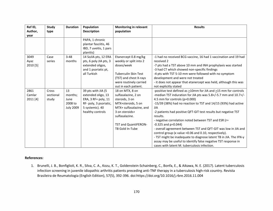
170
Ref ID, Author, year
Study type
Duration Population Description
Monitoring in relevant population
Results
PAPA, 1 chronic plantar fasciitis, 46 IBD, 7 uveitis, 1 pars planitis)
3049 Ayaz 2010 [5]
Case series
3-48 months
14 SoJIA pts, 12 ERA pts, 6 poly JIA pts, 3 extended oligos, and 1 psoriatic pt, all Turkish
Etanercept 0.8 mg/kg weekly or split into 2 doses/week Tuberculin Skin Test (TST) and chest X-rays were routinely carried out in each patient.
-1 had no received BCG vaccine, 16 had 1 vaccination and 19 had received 2 -7 pts had a TST above 10 mm and INH prophylaxis was started -3 had CT which showed non-specific findings -4 pts with TST 5-10 mm were followed with no symptom development and were not treated - it does not appear that etanercept was held, although this was not explicitly stated
2861 Camlar 2011 [4]
Cross sectional study
13 months; June 2008 to July 2009
39 pts with JIA (5 extended oligo, 13 ERA, 3 RF+ poly, 11 RF- poly, 3 psoriatic, 5 systemic). 40 healthy controls
18 on MTX, 8 on sulfasalazine, 2 on steroids, 3 on MTX+steroids, 5 on MTX+ sulfasalazine, and 3 on steroids+ sulfasalazine. TST and QuantiFERON-TB Gold In-Tube
-positive test defined as >10mm for JIA and >15 mm for controls -median TST induration for JIA pts was 5.8+/-5.7 mm and 10.7+/-4.5 mm for controls (p=0.000) -15/39 (38%) had no reaction to TST and 14/15 (93%) had active JIA -2 patients had positive QFT-GIT test results but negative TST results. - negative correlation noted between TST and ESR (r= -0.325 and p=0.044) - overall agreement between TST and QFT-GIT was low in JIA and control group (κ value =0.06 and 0.10, respectively). - TST might be inadequate to diagnose latent TB in JIA. The IFN-γ assay may be useful to identify false negative TST response in cases with latent M. tuberculosis infection.
References:
1. Brunelli, J. B., Bonfiglioli, K. R., Silva, C. A., Kozu, K. T., Goldenstein-Schainberg, C., Bonfa, E., & Aikawa, N. E. (2017). Latent tuberculosis
infection screening in juvenile idiopathic arthritis patients preceding anti-TNF therapy in a tuberculosis high-risk country. Revista
Brasileira de Reumatologia (English Edition), 57(5), 392-396. doi:https://doi.org/10.1016/j.rbre.2016.11.004
171
2. Calzada-Hernández, J., Anton-López, J., Bou-Torrent, R., Iglesias-Jiménez, E., Ricart-Campos, S., Martín de Carpi, J., . . . Noguera-Julian, A.
(2015). Tuberculosis in pediatric patients treated with anti-TNFα drugs: a cohort study. Pediatr Rheumatol Online J, 13, 54-54.
doi:10.1186/s12969-015-0054-4
3. Ayaz, N. A., Demirkaya, E., Bilginer, Y., Ozçelik, U., Cobanoğlu, N., Kiper, N., . . . Ozen, S. (2010). Preventing tuberculosis in children
receiving anti-TNF treatment. Clin Rheumatol, 29(4), 389-392. doi:10.1007/s10067-009-1334-5
4. Camlar, S. A., Makay, B., Appak, O., Appak, Y. C., Esen, N., Günay, T., . . . Unsal, E. (2011). Performance of tuberculin skin test and
interferon gamma assay for the diagnosis of latent tuberculosis infection in juvenile idiopathic arthritis. Clin Rheumatol, 30(9), 1189-
1193. doi:10.1007/s10067-011-1724-3.
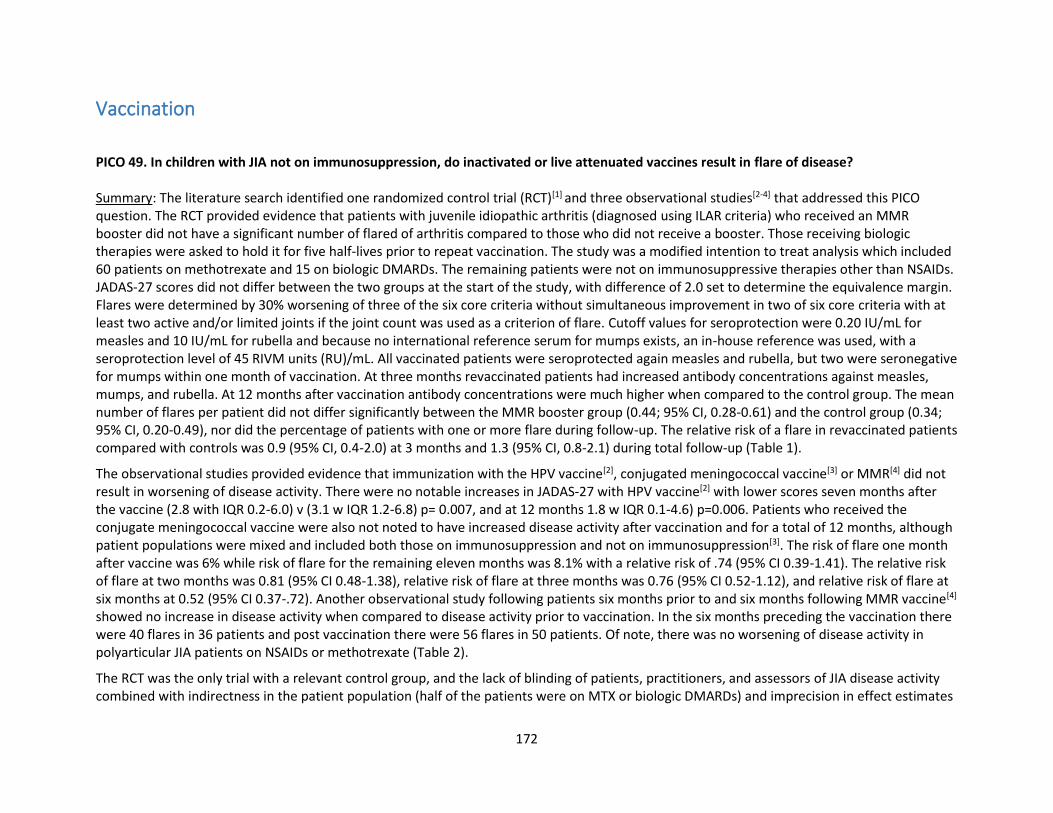
172
Vaccination
PICO 49. In children with JIA not on immunosuppression, do inactivated or live attenuated vaccines result in flare of disease? Summary: The literature search identified one randomized control trial (RCT)[1] and three observational studies[2-4] that addressed this PICO question. The RCT provided evidence that patients with juvenile idiopathic arthritis (diagnosed using ILAR criteria) who received an MMR booster did not have a significant number of flared of arthritis compared to those who did not receive a booster. Those receiving biologic therapies were asked to hold it for five half-lives prior to repeat vaccination. The study was a modified intention to treat analysis which included 60 patients on methotrexate and 15 on biologic DMARDs. The remaining patients were not on immunosuppressive therapies other than NSAIDs. JADAS-27 scores did not differ between the two groups at the start of the study, with difference of 2.0 set to determine the equivalence margin. Flares were determined by 30% worsening of three of the six core criteria without simultaneous improvement in two of six core criteria with at least two active and/or limited joints if the joint count was used as a criterion of flare. Cutoff values for seroprotection were 0.20 IU/mL for measles and 10 IU/mL for rubella and because no international reference serum for mumps exists, an in-house reference was used, with a seroprotection level of 45 RIVM units (RU)/mL. All vaccinated patients were seroprotected again measles and rubella, but two were seronegative for mumps within one month of vaccination. At three months revaccinated patients had increased antibody concentrations against measles, mumps, and rubella. At 12 months after vaccination antibody concentrations were much higher when compared to the control group. The mean number of flares per patient did not differ significantly between the MMR booster group (0.44; 95% CI, 0.28-0.61) and the control group (0.34; 95% CI, 0.20-0.49), nor did the percentage of patients with one or more flare during follow-up. The relative risk of a flare in revaccinated patients compared with controls was 0.9 (95% CI, 0.4-2.0) at 3 months and 1.3 (95% CI, 0.8-2.1) during total follow-up (Table 1).
The observational studies provided evidence that immunization with the HPV vaccine[2], conjugated meningococcal vaccine[3] or MMR[4] did not result in worsening of disease activity. There were no notable increases in JADAS-27 with HPV vaccine[2] with lower scores seven months after the vaccine (2.8 with IQR 0.2-6.0) v (3.1 w IQR 1.2-6.8) p= 0.007, and at 12 months 1.8 w IQR 0.1-4.6) p=0.006. Patients who received the conjugate meningococcal vaccine were also not noted to have increased disease activity after vaccination and for a total of 12 months, although patient populations were mixed and included both those on immunosuppression and not on immunosuppression[3]. The risk of flare one month after vaccine was 6% while risk of flare for the remaining eleven months was 8.1% with a relative risk of .74 (95% CI 0.39-1.41). The relative risk of flare at two months was 0.81 (95% CI 0.48-1.38), relative risk of flare at three months was 0.76 (95% CI 0.52-1.12), and relative risk of flare at six months at 0.52 (95% CI 0.37-.72). Another observational study following patients six months prior to and six months following MMR vaccine[4] showed no increase in disease activity when compared to disease activity prior to vaccination. In the six months preceding the vaccination there were 40 flares in 36 patients and post vaccination there were 56 flares in 50 patients. Of note, there was no worsening of disease activity in polyarticular JIA patients on NSAIDs or methotrexate (Table 2).
The RCT was the only trial with a relevant control group, and the lack of blinding of patients, practitioners, and assessors of JIA disease activity combined with indirectness in the patient population (half of the patients were on MTX or biologic DMARDs) and imprecision in effect estimates
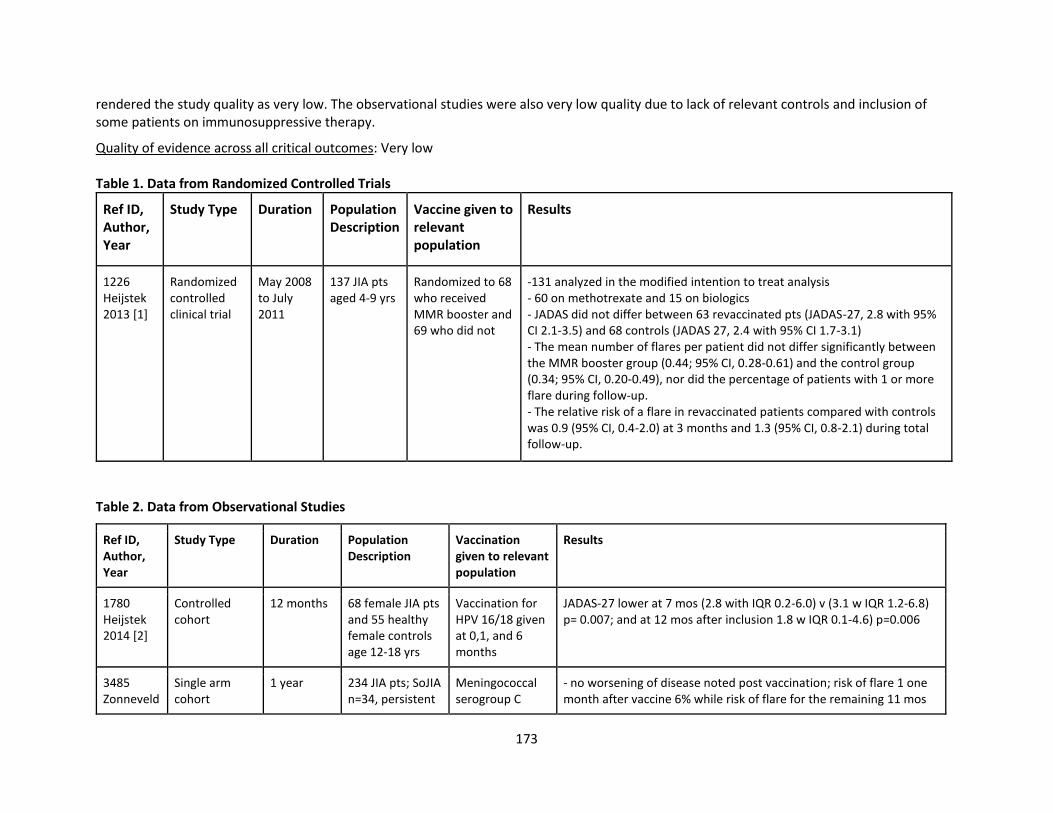
173
rendered the study quality as very low. The observational studies were also very low quality due to lack of relevant controls and inclusion of some patients on immunosuppressive therapy.
Quality of evidence across all critical outcomes: Very low Table 1. Data from Randomized Controlled Trials
Ref ID, Author, Year
Study Type Duration Population Description
Vaccine given to relevant population
Results
1226 Heijstek 2013 [1]
Randomized controlled clinical trial
May 2008 to July 2011
137 JIA pts aged 4-9 yrs
Randomized to 68 who received MMR booster and 69 who did not
-131 analyzed in the modified intention to treat analysis - 60 on methotrexate and 15 on biologics - JADAS did not differ between 63 revaccinated pts (JADAS-27, 2.8 with 95% CI 2.1-3.5) and 68 controls (JADAS 27, 2.4 with 95% CI 1.7-3.1) - The mean number of flares per patient did not differ significantly between the MMR booster group (0.44; 95% CI, 0.28-0.61) and the control group (0.34; 95% CI, 0.20-0.49), nor did the percentage of patients with 1 or more flare during follow-up. - The relative risk of a flare in revaccinated patients compared with controls was 0.9 (95% CI, 0.4-2.0) at 3 months and 1.3 (95% CI, 0.8-2.1) during total follow-up.
Table 2. Data from Observational Studies
Ref ID, Author, Year
Study Type Duration Population Description
Vaccination given to relevant population
Results
1780 Heijstek 2014 [2]
Controlled cohort
12 months 68 female JIA pts and 55 healthy female controls age 12-18 yrs
Vaccination for HPV 16/18 given at 0,1, and 6 months
JADAS-27 lower at 7 mos (2.8 with IQR 0.2-6.0) v (3.1 w IQR 1.2-6.8) p= 0.007; and at 12 mos after inclusion 1.8 w IQR 0.1-4.6) p=0.006
3485 Zonneveld
Single arm cohort
1 year 234 JIA pts; SoJIA n=34, persistent
Meningococcal serogroup C
- no worsening of disease noted post vaccination; risk of flare 1 one month after vaccine 6% while risk of flare for the remaining 11 mos
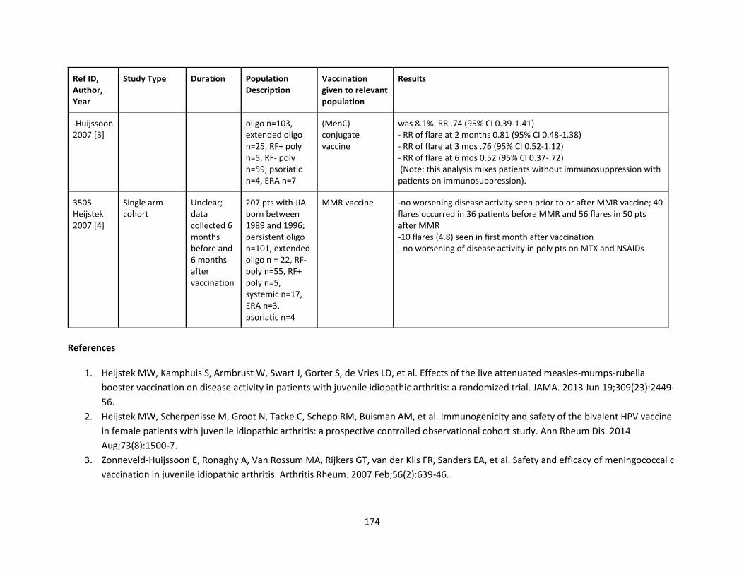
174
Ref ID, Author, Year
Study Type Duration Population Description
Vaccination given to relevant population
Results
-Huijssoon 2007 [3]
oligo n=103, extended oligo n=25, RF+ poly n=5, RF- poly n=59, psoriatic n=4, ERA n=7
(MenC) conjugate vaccine
was 8.1%. RR .74 (95% CI 0.39-1.41) - RR of flare at 2 months 0.81 (95% CI 0.48-1.38) - RR of flare at 3 mos .76 (95% CI 0.52-1.12) - RR of flare at 6 mos 0.52 (95% CI 0.37-.72) (Note: this analysis mixes patients without immunosuppression with patients on immunosuppression).
3505 Heijstek 2007 [4]
Single arm cohort
Unclear; data collected 6 months before and 6 months after vaccination
207 pts with JIA born between 1989 and 1996; persistent oligo n=101, extended oligo n = 22, RF- poly n=55, RF+ poly n=5, systemic n=17, ERA n=3, psoriatic n=4
MMR vaccine -no worsening disease activity seen prior to or after MMR vaccine; 40 flares occurred in 36 patients before MMR and 56 flares in 50 pts after MMR -10 flares (4.8) seen in first month after vaccination - no worsening of disease activity in poly pts on MTX and NSAIDs
References
1. Heijstek MW, Kamphuis S, Armbrust W, Swart J, Gorter S, de Vries LD, et al. Effects of the live attenuated measles-mumps-rubella
booster vaccination on disease activity in patients with juvenile idiopathic arthritis: a randomized trial. JAMA. 2013 Jun 19;309(23):2449-
56.
2. Heijstek MW, Scherpenisse M, Groot N, Tacke C, Schepp RM, Buisman AM, et al. Immunogenicity and safety of the bivalent HPV vaccine
in female patients with juvenile idiopathic arthritis: a prospective controlled observational cohort study. Ann Rheum Dis. 2014
Aug;73(8):1500-7.
3. Zonneveld-Huijssoon E, Ronaghy A, Van Rossum MA, Rijkers GT, van der Klis FR, Sanders EA, et al. Safety and efficacy of meningococcal c
vaccination in juvenile idiopathic arthritis. Arthritis Rheum. 2007 Feb;56(2):639-46.
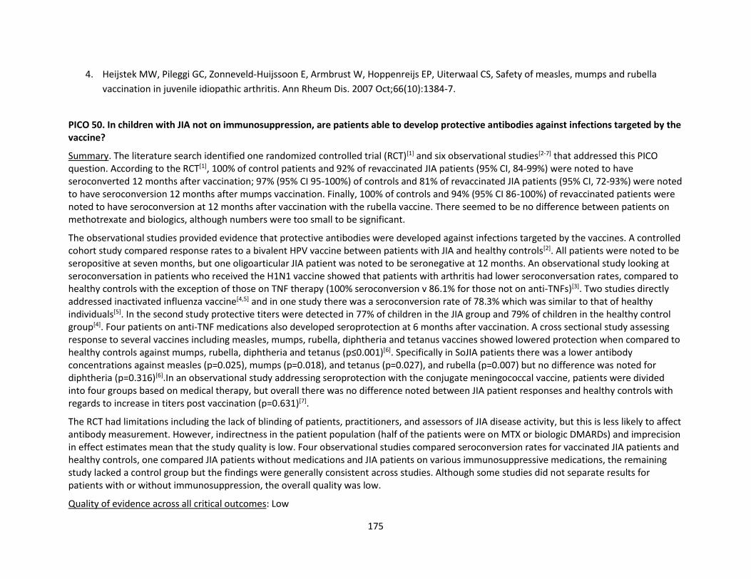
175
4. Heijstek MW, Pileggi GC, Zonneveld-Huijssoon E, Armbrust W, Hoppenreijs EP, Uiterwaal CS, Safety of measles, mumps and rubella
vaccination in juvenile idiopathic arthritis. Ann Rheum Dis. 2007 Oct;66(10):1384-7.
PICO 50. In children with JIA not on immunosuppression, are patients able to develop protective antibodies against infections targeted by the vaccine?
Summary. The literature search identified one randomized controlled trial (RCT)[1] and six observational studies[2-7] that addressed this PICO question. According to the RCT[1], 100% of control patients and 92% of revaccinated JIA patients (95% CI, 84-99%) were noted to have seroconverted 12 months after vaccination; 97% (95% CI 95-100%) of controls and 81% of revaccinated JIA patients (95% CI, 72-93%) were noted to have seroconversion 12 months after mumps vaccination. Finally, 100% of controls and 94% (95% CI 86-100%) of revaccinated patients were noted to have seroconversion at 12 months after vaccination with the rubella vaccine. There seemed to be no difference between patients on methotrexate and biologics, although numbers were too small to be significant.
The observational studies provided evidence that protective antibodies were developed against infections targeted by the vaccines. A controlled cohort study compared response rates to a bivalent HPV vaccine between patients with JIA and healthy controls[2]. All patients were noted to be seropositive at seven months, but one oligoarticular JIA patient was noted to be seronegative at 12 months. An observational study looking at seroconversation in patients who received the H1N1 vaccine showed that patients with arthritis had lower seroconversation rates, compared to healthy controls with the exception of those on TNF therapy (100% seroconversion v 86.1% for those not on anti-TNFs)[3]. Two studies directly addressed inactivated influenza vaccine[4,5] and in one study there was a seroconversion rate of 78.3% which was similar to that of healthy individuals[5]. In the second study protective titers were detected in 77% of children in the JIA group and 79% of children in the healthy control group[4]. Four patients on anti-TNF medications also developed seroprotection at 6 months after vaccination. A cross sectional study assessing response to several vaccines including measles, mumps, rubella, diphtheria and tetanus vaccines showed lowered protection when compared to healthy controls against mumps, rubella, diphtheria and tetanus (p≤0.001)[6]. Specifically in SoJIA patients there was a lower antibody concentrations against measles (p=0.025), mumps (p=0.018), and tetanus (p=0.027), and rubella (p=0.007) but no difference was noted for diphtheria (p=0.316)[6].In an observational study addressing seroprotection with the conjugate meningococcal vaccine, patients were divided into four groups based on medical therapy, but overall there was no difference noted between JIA patient responses and healthy controls with regards to increase in titers post vaccination (p=0.631)[7].
The RCT had limitations including the lack of blinding of patients, practitioners, and assessors of JIA disease activity, but this is less likely to affect antibody measurement. However, indirectness in the patient population (half of the patients were on MTX or biologic DMARDs) and imprecision in effect estimates mean that the study quality is low. Four observational studies compared seroconversion rates for vaccinated JIA patients and healthy controls, one compared JIA patients without medications and JIA patients on various immunosuppressive medications, the remaining study lacked a control group but the findings were generally consistent across studies. Although some studies did not separate results for patients with or without immunosuppression, the overall quality was low.
Quality of evidence across all critical outcomes: Low
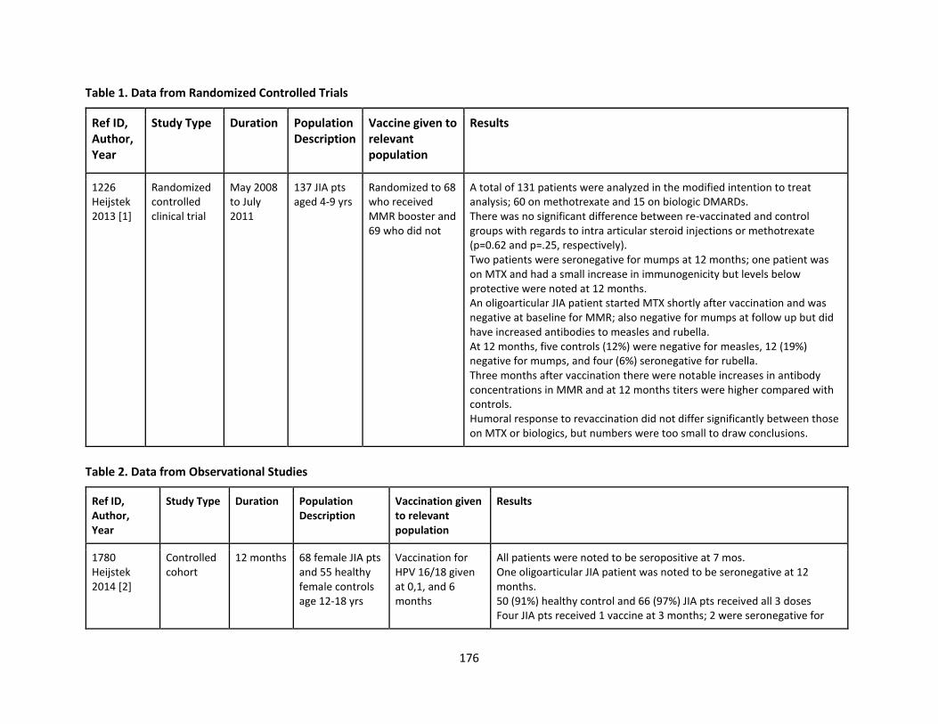
176
Table 1. Data from Randomized Controlled Trials
Ref ID, Author, Year
Study Type Duration Population Description
Vaccine given to relevant population
Results
1226 Heijstek 2013 [1]
Randomized controlled clinical trial
May 2008 to July 2011
137 JIA pts aged 4-9 yrs
Randomized to 68 who received MMR booster and 69 who did not
A total of 131 patients were analyzed in the modified intention to treat analysis; 60 on methotrexate and 15 on biologic DMARDs. There was no significant difference between re-vaccinated and control groups with regards to intra articular steroid injections or methotrexate (p=0.62 and p=.25, respectively). Two patients were seronegative for mumps at 12 months; one patient was on MTX and had a small increase in immunogenicity but levels below protective were noted at 12 months. An oligoarticular JIA patient started MTX shortly after vaccination and was negative at baseline for MMR; also negative for mumps at follow up but did have increased antibodies to measles and rubella. At 12 months, five controls (12%) were negative for measles, 12 (19%) negative for mumps, and four (6%) seronegative for rubella. Three months after vaccination there were notable increases in antibody concentrations in MMR and at 12 months titers were higher compared with controls. Humoral response to revaccination did not differ significantly between those on MTX or biologics, but numbers were too small to draw conclusions.
Table 2. Data from Observational Studies
Ref ID, Author, Year
Study Type Duration Population Description
Vaccination given to relevant population
Results
1780 Heijstek 2014 [2]
Controlled cohort
12 months 68 female JIA pts and 55 healthy female controls age 12-18 yrs
Vaccination for HPV 16/18 given at 0,1, and 6 months
All patients were noted to be seropositive at 7 mos. One oligoarticular JIA patient was noted to be seronegative at 12 months. 50 (91%) healthy control and 66 (97%) JIA pts received all 3 doses Four JIA pts received 1 vaccine at 3 months; 2 were seronegative for
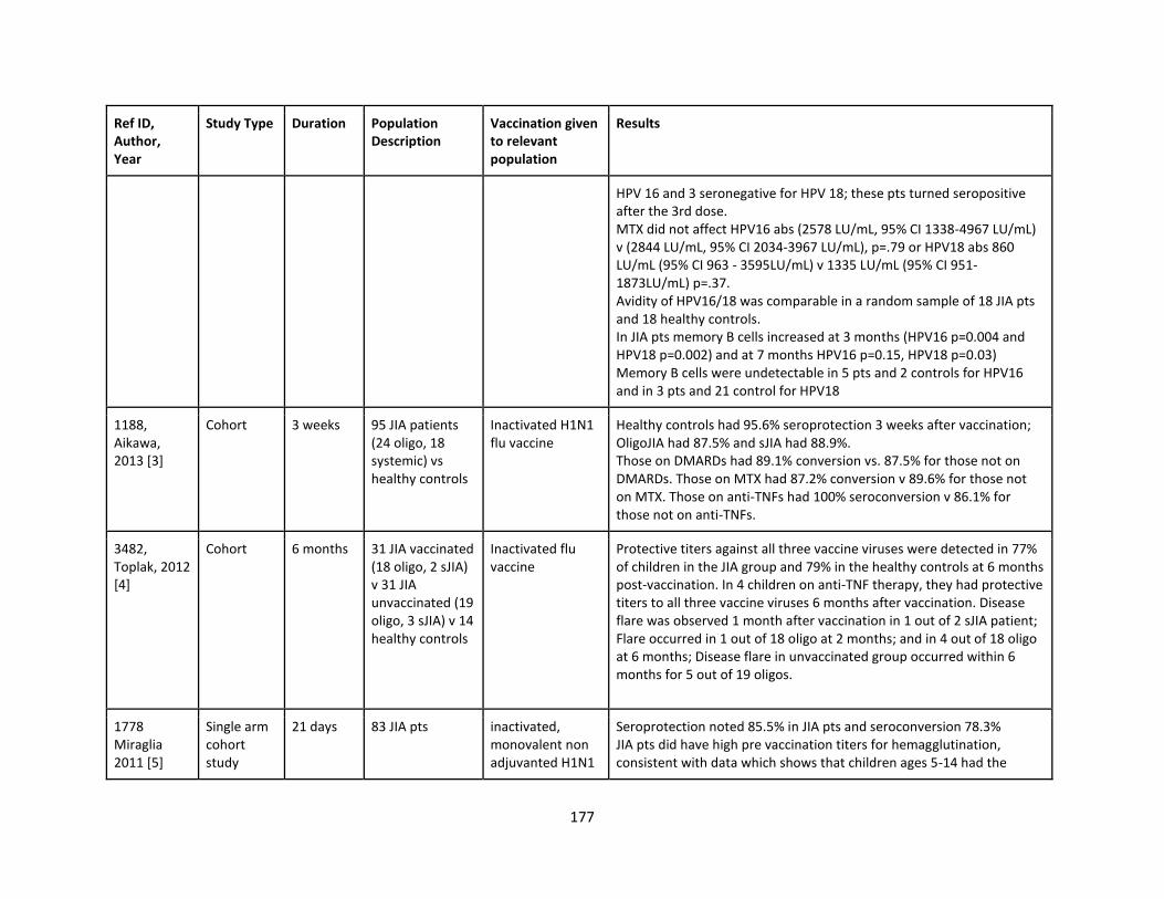
177
Ref ID, Author, Year
Study Type Duration Population Description
Vaccination given to relevant population
Results
HPV 16 and 3 seronegative for HPV 18; these pts turned seropositive after the 3rd dose. MTX did not affect HPV16 abs (2578 LU/mL, 95% CI 1338-4967 LU/mL) v (2844 LU/mL, 95% CI 2034-3967 LU/mL), p=.79 or HPV18 abs 860 LU/mL (95% CI 963 - 3595LU/mL) v 1335 LU/mL (95% CI 951-1873LU/mL) p=.37. Avidity of HPV16/18 was comparable in a random sample of 18 JIA pts and 18 healthy controls. In JIA pts memory B cells increased at 3 months (HPV16 p=0.004 and HPV18 p=0.002) and at 7 months HPV16 p=0.15, HPV18 p=0.03) Memory B cells were undetectable in 5 pts and 2 controls for HPV16 and in 3 pts and 21 control for HPV18
1188, Aikawa, 2013 [3]
Cohort 3 weeks 95 JIA patients (24 oligo, 18 systemic) vs healthy controls
Inactivated H1N1 flu vaccine
Healthy controls had 95.6% seroprotection 3 weeks after vaccination; OligoJIA had 87.5% and sJIA had 88.9%. Those on DMARDs had 89.1% conversion vs. 87.5% for those not on DMARDs. Those on MTX had 87.2% conversion v 89.6% for those not on MTX. Those on anti-TNFs had 100% seroconversion v 86.1% for those not on anti-TNFs.
3482, Toplak, 2012 [4]
Cohort 6 months 31 JIA vaccinated (18 oligo, 2 sJIA) v 31 JIA unvaccinated (19 oligo, 3 sJIA) v 14 healthy controls
Inactivated flu vaccine
Protective titers against all three vaccine viruses were detected in 77% of children in the JIA group and 79% in the healthy controls at 6 months post-vaccination. In 4 children on anti-TNF therapy, they had protective titers to all three vaccine viruses 6 months after vaccination. Disease flare was observed 1 month after vaccination in 1 out of 2 sJIA patient; Flare occurred in 1 out of 18 oligo at 2 months; and in 4 out of 18 oligo at 6 months; Disease flare in unvaccinated group occurred within 6 months for 5 out of 19 oligos.
1778 Miraglia 2011 [5]
Single arm cohort study
21 days 83 JIA pts inactivated, monovalent non adjuvanted H1N1
Seroprotection noted 85.5% in JIA pts and seroconversion 78.3% JIA pts did have high pre vaccination titers for hemagglutination, consistent with data which shows that children ages 5-14 had the
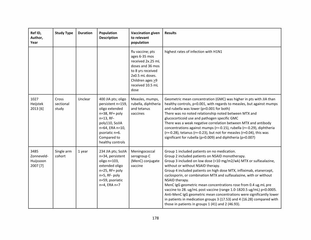
178
Ref ID, Author, Year
Study Type Duration Population Description
Vaccination given to relevant population
Results
flu vaccine; pts ages 6-35 mos received 2x.25 mL doses and 36 mos to 8 yrs received 2x0.5 mL doses. Children ages >9 received 10.5 mL dose
highest rates of infection with H1N1
1027 Heijstek 2013 [6]
Cross sectional study
Unclear 400 JIA pts; oligo persistent n=159, oligo extended n=38, RF+ poly n=13, RF-poly110, SoJIA n=64, ERA n=10, psoriatic n=6. Compared to healthy controls
Measles, mumps, rubella, diphtheria and tetanus vaccines
Geometric mean concentration (GMC) was higher in pts with JIA than healthy controls, p<0.001, with regards to measles, but against mumps and rubella was lower (p<0.001 for both) There was no noted relationship noted between MTX and glucocorticoid use and pathogen specific GMC There was a weak negative correlation between MTX and antibody concentrations against mumps (r=-0.15), rubella (r=-0.29), diphtheria (r=-0.28), tetanus (r=-0.23), but not for measles (r=0.04), this was significant for rubella (p=0.009) and diphtheria (p=0.007)
3485 Zonneveld-Huijssoon 2007 [7]
Single arm cohort
1 year 234 JIA pts; SoJIA n=34, persistent oligo n=103, extended oligo n=25, RF+ poly n=5, RF- poly n=59, psoriatic n=4, ERA n=7
Meningococcal serogroup C (MenC) conjugate vaccine
Group 1 included patients on no medication. Group 2 included patients on NSAID monotherapy. Group 3 included on low dose (<10 mg/m2/wk) MTX or sulfasalazine, without or without NSAID therapy. Group 4 included patients on high dose MTX, infliximab, etanercept, cyclosporin, or combination MTX and sulfasalazine, with or without NSAID therapy. MenC IgG geometric mean concentrations rose from 0.4 ug.mL pre vaccine to 28. ug/mL post vaccine (range 1.0-1820.5 ug/mL) p<0.0005. Anti-MenC IgG geometric mean concentrations were significantly lower in patients in medication groups 3 (17.53) and 4 (16.28) compared with those in patients in groups 1 (41) and 2 (46.93).
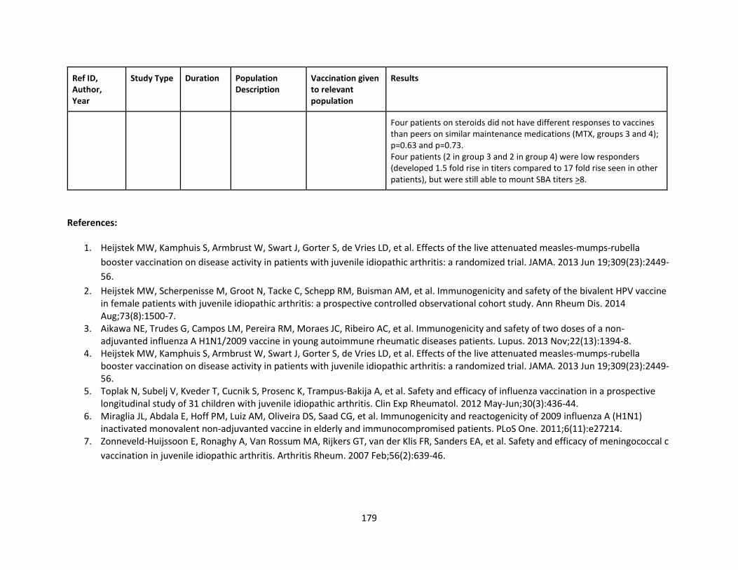
179
Ref ID, Author, Year
Study Type Duration Population Description
Vaccination given to relevant population
Results
Four patients on steroids did not have different responses to vaccines than peers on similar maintenance medications (MTX, groups 3 and 4); p=0.63 and p=0.73. Four patients (2 in group 3 and 2 in group 4) were low responders (developed 1.5 fold rise in titers compared to 17 fold rise seen in other patients), but were still able to mount SBA titers >8.
References:
1. Heijstek MW, Kamphuis S, Armbrust W, Swart J, Gorter S, de Vries LD, et al. Effects of the live attenuated measles-mumps-rubella
booster vaccination on disease activity in patients with juvenile idiopathic arthritis: a randomized trial. JAMA. 2013 Jun 19;309(23):2449-
56.
2. Heijstek MW, Scherpenisse M, Groot N, Tacke C, Schepp RM, Buisman AM, et al. Immunogenicity and safety of the bivalent HPV vaccine in female patients with juvenile idiopathic arthritis: a prospective controlled observational cohort study. Ann Rheum Dis. 2014 Aug;73(8):1500-7.
3. Aikawa NE, Trudes G, Campos LM, Pereira RM, Moraes JC, Ribeiro AC, et al. Immunogenicity and safety of two doses of a non-adjuvanted influenza A H1N1/2009 vaccine in young autoimmune rheumatic diseases patients. Lupus. 2013 Nov;22(13):1394-8.
4. Heijstek MW, Kamphuis S, Armbrust W, Swart J, Gorter S, de Vries LD, et al. Effects of the live attenuated measles-mumps-rubella booster vaccination on disease activity in patients with juvenile idiopathic arthritis: a randomized trial. JAMA. 2013 Jun 19;309(23):2449-56.
5. Toplak N, Subelj V, Kveder T, Cucnik S, Prosenc K, Trampus-Bakija A, et al. Safety and efficacy of influenza vaccination in a prospective longitudinal study of 31 children with juvenile idiopathic arthritis. Clin Exp Rheumatol. 2012 May-Jun;30(3):436-44.
6. Miraglia JL, Abdala E, Hoff PM, Luiz AM, Oliveira DS, Saad CG, et al. Immunogenicity and reactogenicity of 2009 influenza A (H1N1) inactivated monovalent non-adjuvanted vaccine in elderly and immunocompromised patients. PLoS One. 2011;6(11):e27214.
7. Zonneveld-Huijssoon E, Ronaghy A, Van Rossum MA, Rijkers GT, van der Klis FR, Sanders EA, et al. Safety and efficacy of meningococcal c
vaccination in juvenile idiopathic arthritis. Arthritis Rheum. 2007 Feb;56(2):639-46.
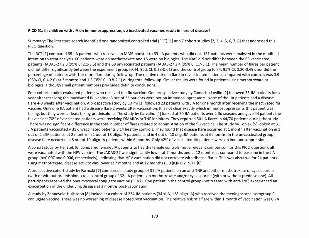
180
PICO 51: In children with JIA on immunosuppression, do inactivated vaccines result in flare of disease? Summary: The literature search identified one randomized controlled trial (RCT) [1] and 7 cohort studies [2, 3, 4, 5, 6, 7, 8] that addressed this PICO question.
The RCT [1] compared 68 JIA patients who received an MMR booster to 69 JIA patients who did not. 131 patients were analyzed in the modified intention to treat analysis. 60 patients were on methotrexate and 15 were on biologics. The JDAS did not differ between the 63 vaccinated patients (JADAS-27 2.8 (95% CI 2.1-3.5) and the 68 unvaccinated patients (JADAS-27 2.4 (95% CI 1.7-3.1). The mean number of flares per patient did not differ significantly between the experiment group (0.44; 95% CI, 0.28-0.61) and the control group (0.34; 95% CI, 0.20-0.49), nor did the percentage of patients with 1 or more flare during follow-up. The relative risk of a flare in revaccinated patients compared with controls was 0.9 (95% CI, 0.4-2.0) at 3 months and 1.3 (95% CI, 0.8-2.1) during total follow-up. Similar results were found in patients using methotrexate or biologics, although small patient numbers precluded definite conclusions.
Four cohort studies evaluated patients who received the flu vaccine. One prospective study by Camacho-Lovillo [2] followed 35 JIA patients for a year after receiving the inactivated flu vaccine; 3 out of 35 patients were not on immunosuppressants. None of the JIA patients had a disease flare 4-8 weeks after vaccination. A prospective study by Ogimi [3] followed 23 patients with JIA for one month after receiving the inactivated flu vaccine. Only one JIA patient had a disease flare 2 weeks after vaccination. It is not clear exactly which immunosuppressants this patient was taking, but they were at least taking prednisolone. The study by Carvalho [4] looked at 70 JIA patients over 2 flu seasons and gave 44 patients the flu vaccine; 70% of vaccinated patients were receiving DMARDs or TNF inhibitors. They reported 50 JIA flares in 44/70 patients during the study. There was no significant difference in the total number of flares related to administration of the flu vaccine. The study by Toplak [5] looked at 31 JIA patients vaccinated v 31 unvaccinated patients v 14 healthy controls. They found that disease flare occurred at 1 month after vaccination in 1 out of 2 sJIA patients, at 2 months in 1 out of 18 oligoJIA patients, and in 4 out of 18 oligoJIA patients at 6 months. In the unvaccinated group, disease flare occurred in 5 out of 19 oligoJIA patients within 6 months. Only 42% of vaccinated JIA patients were on immunosuppression.
A cohort study by Heijstek [6] compared female JIA patients to healthy female controls (not a relevant comparison for this PICO question); all were vaccinated with the HPV vaccine. The JADAS-27 was significantly lower at 7 months and at 12 months as compared to baseline in the JIA group (p=0.007 and 0.006, respectively), indicating that HPV vaccination did not correlate with disease flares. This was also true for 24 patients using methotrexate, disease activity was lower at 7 months and at 12 months (3.0 (IQR 0.2–5.7). [6].
A prospective cohort study by Farmaki [7] compared a study group of 31 JIA patients on an anti-TNF and either methotrexate or cyclosporine (with or without prednisolone) to a control group of 32 JIA patients on methotrexate and/or cyclosporine (with or without prednisolone). All participants received the pneumococcal conjugate vaccine (PCV7). One patient in the control group (not treated with anti-TNF) experienced an exacerbation of the underlying disease at 3 months post-vaccination.
A study by Zonneveld-Huijssoon [8] looked at a cohort of 234 JIA patients (34 sJIA, 128 oligoJIA) who received the meningococcal serogroup C conjugate vaccine. There was no worsening of disease noted post vaccination. The relative risk of a flare within 1 month of vaccination was 0.74
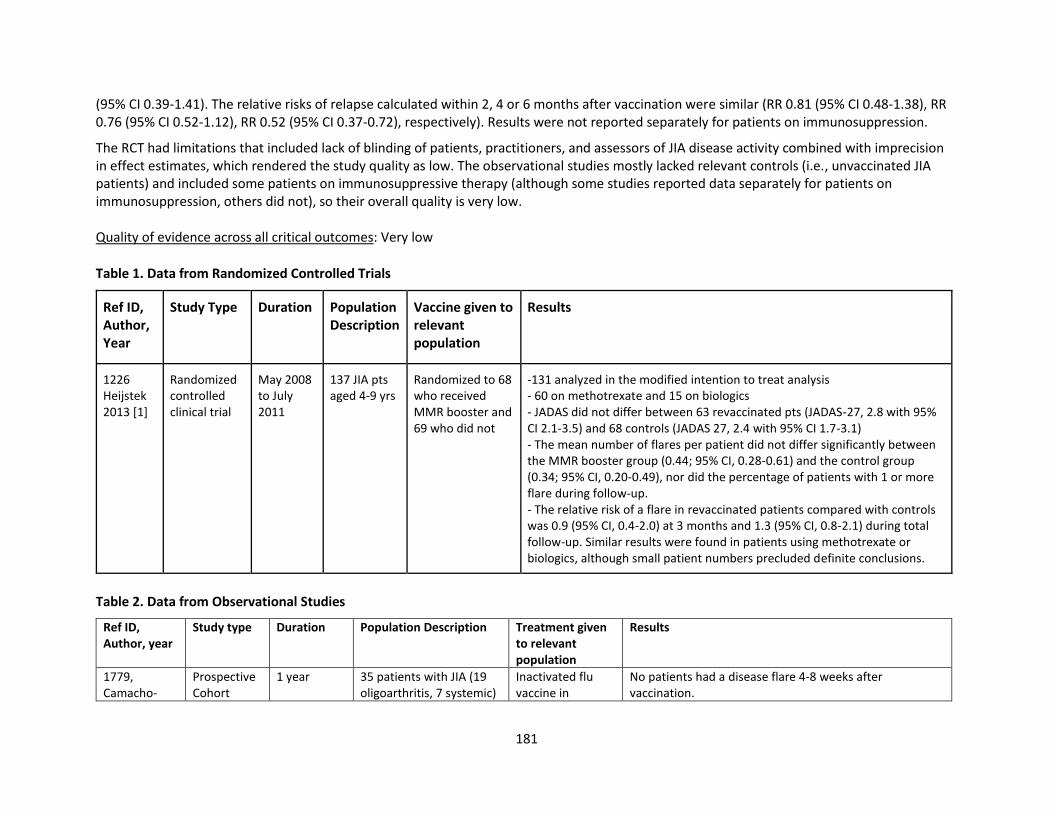
181
(95% CI 0.39-1.41). The relative risks of relapse calculated within 2, 4 or 6 months after vaccination were similar (RR 0.81 (95% CI 0.48-1.38), RR 0.76 (95% CI 0.52-1.12), RR 0.52 (95% CI 0.37-0.72), respectively). Results were not reported separately for patients on immunosuppression.
The RCT had limitations that included lack of blinding of patients, practitioners, and assessors of JIA disease activity combined with imprecision in effect estimates, which rendered the study quality as low. The observational studies mostly lacked relevant controls (i.e., unvaccinated JIA patients) and included some patients on immunosuppressive therapy (although some studies reported data separately for patients on immunosuppression, others did not), so their overall quality is very low. Quality of evidence across all critical outcomes: Very low
Table 1. Data from Randomized Controlled Trials
Ref ID, Author, Year
Study Type Duration Population Description
Vaccine given to relevant population
Results
1226 Heijstek 2013 [1]
Randomized controlled clinical trial
May 2008 to July 2011
137 JIA pts aged 4-9 yrs
Randomized to 68 who received MMR booster and 69 who did not
-131 analyzed in the modified intention to treat analysis - 60 on methotrexate and 15 on biologics - JADAS did not differ between 63 revaccinated pts (JADAS-27, 2.8 with 95% CI 2.1-3.5) and 68 controls (JADAS 27, 2.4 with 95% CI 1.7-3.1) - The mean number of flares per patient did not differ significantly between the MMR booster group (0.44; 95% CI, 0.28-0.61) and the control group (0.34; 95% CI, 0.20-0.49), nor did the percentage of patients with 1 or more flare during follow-up. - The relative risk of a flare in revaccinated patients compared with controls was 0.9 (95% CI, 0.4-2.0) at 3 months and 1.3 (95% CI, 0.8-2.1) during total follow-up. Similar results were found in patients using methotrexate or biologics, although small patient numbers precluded definite conclusions.
Table 2. Data from Observational Studies
Ref ID, Author, year
Study type Duration Population Description Treatment given to relevant population
Results
1779, Camacho-
Prospective Cohort
1 year 35 patients with JIA (19 oligoarthritis, 7 systemic)
Inactivated flu vaccine in
No patients had a disease flare 4-8 weeks after vaccination.
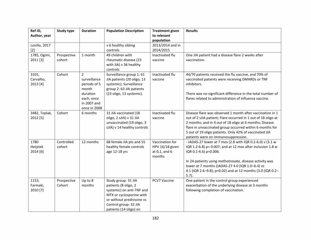
182
Ref ID, Author, year
Study type Duration Population Description Treatment given to relevant population
Results
Lovillo, 2017 [2]
v 6 healthy sibling controls
2013/2014 and in 2014/2015
1785, Ogimi, 2011 [3]
Prospective cohort
1 month 49 children with rheumatic disease (23 with JIA) v 36 healthy controls
Inactivated flu vaccine
One JIA patient had a disease flare 2 weeks after vaccination.
3101, Carvalho, 2013 [4]
Cohort 2 surveillance periods of 5 month duration each, once in 2007 and once in 2008
Surveillance group 1: 61 JIA patients (20 oligo, 13 systemic); Surveillance group 2: 63 JIA patients (23 oligo, 13 systemic).
Inactivated flu vaccine
44/70 patients received the flu vaccine, and 70% of vaccinated patients were receiving DMARDs or TNF inhibitors. There was no significant difference in the total number of flares related to administration of influenza vaccine.
3482, Toplak, 2012 [5]
Cohort 6 months 31 JIA vaccinated (18 oligo, 2 sJIA) v 31 JIA unvaccinated (19 oligo, 3 sJIA) v 14 healthy controls
Inactivated flu vaccine
Disease flare was observed 1 month after vaccination in 1 out of 2 sJIA patient; Flare occurred in 1 out of 18 oligo at 2 months; and in 4 out of 18 oligo at 6 months; Disease flare in unvaccinated group occurred within 6 months for 5 out of 19 oligo patients. Only 42% of vaccinated JIA patients were on immunosuppression.
1780 Heijstek 2014 [6]
Controlled cohort
12 months 68 female JIA pts and 55 healthy female controls age 12-18 yrs
Vaccination for HPV 16/18 given at 0,1, and 6 months
- JADAS-27 lower at 7 mos (2.8 with IQR 0.2-6.0) v (3.1 w IQR 1.2-6.8) p= 0.007; and at 12 mos after inclusion 1.8 w IQR 0.1-4.6) p=0.006. In 24 patients using methotrexate, disease activity was lower at 7 months (JADAS-27 4.0 (IQR 1.0–6.4) vs 4.1 (IQR 2.6–9.8); p=0.02) and at 12 months (3.0 (IQR 0.2–5.7).
1153, Farmaki, 2010 [7]
Prospective Cohort
Up to 8 months
Study group: 31 JIA patients (8 oligo, 2 systemic) on anti-TNF and MTX or cyclosporine with or without prednisone vs Control group: 32 JIA patients (14 oligo) on
PCV7 Vaccine One patient in the control group experienced exacerbation of the underlying disease at 3 months following completion of vaccination.
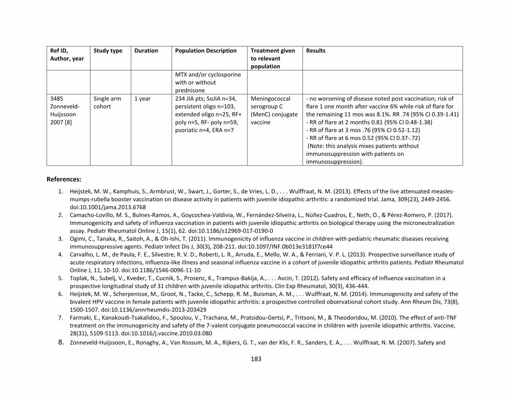
183
Ref ID, Author, year
Study type Duration Population Description Treatment given to relevant population
Results
MTX and/or cyclosporine with or without prednisone
3485 Zonneveld-Huijssoon 2007 [8]
Single arm cohort
1 year 234 JIA pts; SoJIA n=34, persistent oligo n=103, extended oligo n=25, RF+ poly n=5, RF- poly n=59, psoriatic n=4, ERA n=7
Meningococcal serogroup C (MenC) conjugate vaccine
- no worsening of disease noted post vaccination; risk of flare 1 one month after vaccine 6% while risk of flare for the remaining 11 mos was 8.1%. RR .74 (95% CI 0.39-1.41) - RR of flare at 2 months 0.81 (95% CI 0.48-1.38) - RR of flare at 3 mos .76 (95% CI 0.52-1.12) - RR of flare at 6 mos 0.52 (95% CI 0.37-.72) (Note: this analysis mixes patients without immunosuppression with patients on immunosuppression).
References:
1. Heijstek, M. W., Kamphuis, S., Armbrust, W., Swart, J., Gorter, S., de Vries, L. D., . . . Wulffraat, N. M. (2013). Effects of the live attenuated measles-mumps-rubella booster vaccination on disease activity in patients with juvenile idiopathic arthritis: a randomized trial. Jama, 309(23), 2449-2456. doi:10.1001/jama.2013.6768
2. Camacho-Lovillo, M. S., Bulnes-Ramos, A., Goycochea-Valdivia, W., Fernández-Silveira, L., Núñez-Cuadros, E., Neth, O., & Pérez-Romero, P. (2017). Immunogenicity and safety of influenza vaccination in patients with juvenile idiopathic arthritis on biological therapy using the microneutralization assay. Pediatr Rheumatol Online J, 15(1), 62. doi:10.1186/s12969-017-0190-0
3. Ogimi, C., Tanaka, R., Saitoh, A., & Oh-Ishi, T. (2011). Immunogenicity of influenza vaccine in children with pediatric rheumatic diseases receiving immunosuppressive agents. Pediatr Infect Dis J, 30(3), 208-211. doi:10.1097/INF.0b013e3181f7ce44
4. Carvalho, L. M., de Paula, F. E., Silvestre, R. V. D., Roberti, L. R., Arruda, E., Mello, W. A., & Ferriani, V. P. L. (2013). Prospective surveillance study of acute respiratory infections, influenza-like illness and seasonal influenza vaccine in a cohort of juvenile idiopathic arthritis patients. Pediatr Rheumatol Online J, 11, 10-10. doi:10.1186/1546-0096-11-10
5. Toplak, N., Subelj, V., Kveder, T., Cucnik, S., Prosenc, K., Trampus-Bakija, A., . . . Avcin, T. (2012). Safety and efficacy of influenza vaccination in a prospective longitudinal study of 31 children with juvenile idiopathic arthritis. Clin Exp Rheumatol, 30(3), 436-444.
6. Heijstek, M. W., Scherpenisse, M., Groot, N., Tacke, C., Schepp, R. M., Buisman, A. M., . . . Wulffraat, N. M. (2014). Immunogenicity and safety of the bivalent HPV vaccine in female patients with juvenile idiopathic arthritis: a prospective controlled observational cohort study. Ann Rheum Dis, 73(8), 1500-1507. doi:10.1136/annrheumdis-2013-203429
7. Farmaki, E., Kanakoudi-Tsakalidou, F., Spoulou, V., Trachana, M., Pratsidou-Gertsi, P., Tritsoni, M., & Theodoridou, M. (2010). The effect of anti-TNF treatment on the immunogenicity and safety of the 7-valent conjugate pneumococcal vaccine in children with juvenile idiopathic arthritis. Vaccine, 28(31), 5109-5113. doi:10.1016/j.vaccine.2010.03.080
8. Zonneveld-Huijssoon, E., Ronaghy, A., Van Rossum, M. A., Rijkers, G. T., van der Klis, F. R., Sanders, E. A., . . . Wulffraat, N. M. (2007). Safety and
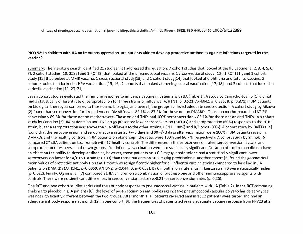
184
efficacy of meningococcal c vaccination in juvenile idiopathic arthritis. Arthritis Rheum, 56(2), 639-646. doi:10.1002/art.22399
PICO 52: In children with JIA on immunosuppression, are patients able to develop protective antibodies against infections targeted by the vaccine? Summary: The literature search identified 21 studies that addressed this question: 7 cohort studies that looked at the flu vaccine [1, 2, 3, 4, 5, 6, 7], 2 cohort studies [10, 3592] and 1 RCT [8] that looked at the pneumococcal vaccine, 1 cross-sectional study [13], 1 RCT [11], and 1 cohort study [12] that looked at MMR vaccine, 1 cross-sectional study[13] and 1 cohort study[14] that looked at diphtheria and tetanus vaccine, 2 cohort studies that looked at HPV vaccination [15, 16], 2 cohorts that looked at meningococcal vaccination [17, 18], and 3 cohorts that looked at varicella vaccination [19, 20, 21].
Seven cohort studies evaluated the immune response to influenza vaccine in patients with JIA (Table 1). A study by Camacho-Lovillo [1] did not find a statistically different rate of seroprotection for three strains of influenza (A/H1N1, p=0.521, A/H3N2, p=0.565, B, p=0.871) in JIA patients on biological therapy as compared to those on no biologics, and overall, the groups achieved adequate seroprotection. A cohort study by Aikawa [2] found that seroconversion for JIA patients on DMARDs was 89.1% vs 87.2% for those not on DMARDs. Those on methotrexate had 87.2% conversion v 89.6% for those not on methotrexate. Those on anti-TNFs had 100% seroconversion v 86.1% for those not on anti-TNFs. In a cohort study by Carvalho [3], JIA patients on anti-TNF drugs presented lower seroconversion (p=0.03) and seroprotection (60%) responses to the H1N1 strain, but the seroprotection was above the cut-off levels to the other strains, H3N2 (100%) and B/Florida (80%). A cohort study by Dell’Era [4] found that the seroconversion and seroprotective rates 28 +/- 3 days and 90 +/- 3 days after vaccination were 100% in JIA patients receiving DMARDs and the healthy controls. In JIA patients on etanercept, the rates were 100% and 96.7%, respectively. A cohort study by Shinoki [5] compared 27 sJIA patient on tocilizumab with 17 healthy controls. The differences in the seroconversion rates, seroconversion factors, and seroprotection rates between the two groups after influenza vaccination were not statistically significant. Duration of tocilizumab did not have an effect on the ability to develop antibodies, however, those patients on < 0.2 mg/kg prednisolone had a statistically significant lower seroconversion factor for A/H1N1 strain (p=0.03) than those patients on >0.2 mg/kg prednisolone. Another cohort [6] found the geometrical mean values of protective antibody titers at 1 month were significantly higher for all influenza vaccine strains compared to baseline in JIA patients on DMARDs (A/H1N1, p=0.0059, A/H3N2, p=0.044, B, p=0.032). By 6 months, only titers for influenza strain B were statistically higher (p=0.022). Finally, Ogimi et al. [7] compared 31 JIA children on a combination of prednisolone and other immunosuppressive agents with controls. There were no significant differences in seroconversion factor (p>0.21) or seroconversion rates (p>0.26).
One RCT and two cohort studies addressed the antibody response to pneumococcal vaccine in patients with JIA (Table 2). In the RCT comparing anakinra to placebo in sJIA patients [8], the level of post-vaccination antibodies against five pneumococcal capsular polysaccharide serotypes was not significantly different between the two groups. After month 1, all patients received anakinra; 12 patients were tested and had an adequate antibody response at month 12. In one cohort [9], the frequencies of patients achieving adequate vaccine response from PPV23 at 2
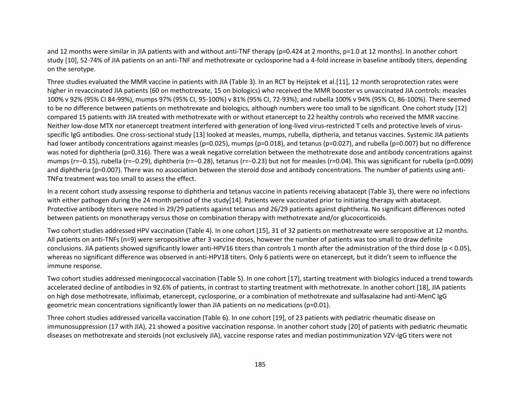
185
and 12 months were similar in JIA patients with and without anti-TNF therapy (p=0.424 at 2 months, p=1.0 at 12 months). In another cohort study [10], 52-74% of JIA patients on an anti-TNF and methotrexate or cyclosporine had a 4-fold increase in baseline antibody titers, depending on the serotype.
Three studies evaluated the MMR vaccine in patients with JIA (Table 3). In an RCT by Heijstek et al.[11], 12 month seroprotection rates were higher in revaccinated JIA patients (60 on methotrexate, 15 on biologics) who received the MMR booster vs unvaccinated JIA controls: measles 100% v 92% (95% CI 84-99%), mumps 97% (95% CI, 95-100%) v 81% (95% CI, 72-93%); and rubella 100% v 94% (95% CI, 86-100%). There seemed to be no difference between patients on methotrexate and biologics, although numbers were too small to be significant. One cohort study [12] compared 15 patients with JIA treated with methotrexate with or without etanercept to 22 healthy controls who received the MMR vaccine. Neither low-dose MTX nor etanercept treatment interfered with generation of long-lived virus-restricted T cells and protective levels of virus-specific IgG antibodies. One cross-sectional study [13] looked at measles, mumps, rubella, diptheria, and tetanus vaccines. Systemic JIA patients had lower antibody concentrations against measles (p=0.025), mumps (p=0.018), and tetanus (p=0.027), and rubella (p=0.007) but no difference was noted for diphtheria (p=0.316). There was a weak negative correlation between the methotrexate dose and antibody concentrations against mumps (r=−0.15), rubella (r=−0.29), diphtheria (r=−0.28), tetanus (r=−0.23) but not for measles (r=0.04). This was significant for rubella (p=0.009) and diphtheria (p=0.007). There was no association between the steroid dose and antibody concentrations. The number of patients using anti-TNFα treatment was too small to assess the effect.
In a recent cohort study assessing response to diphtheria and tetanus vaccine in patients receiving abatacept (Table 3), there were no infections with either pathogen during the 24 month period of the study[14]. Patients were vaccinated prior to initiating therapy with abatacept. Protective antibody titers were noted in 29/29 patients against tetanus and 26/29 patients against diphtheria. No significant differences noted between patients on monotherapy versus those on combination therapy with methotrexate and/or glucocorticoids.
Two cohort studies addressed HPV vaccination (Table 4). In one cohort [15], 31 of 32 patients on methotrexate were seropositive at 12 months. All patients on anti-TNFs (n=9) were seropositive after 3 vaccine doses, however the number of patients was too small to draw definite conclusions. JIA patients showed significantly lower anti-HPV16 titers than controls 1 month after the administration of the third dose (p < 0.05), whereas no significant difference was observed in anti-HPV18 titers. Only 6 patients were on etanercept, but it didn’t seem to influence the immune response.
Two cohort studies addressed meningococcal vaccination (Table 5). In one cohort [17], starting treatment with biologics induced a trend towards accelerated decline of antibodies in 92.6% of patients, in contrast to starting treatment with methotrexate. In another cohort [18], JIA patients on high dose methotrexate, infliximab, etanercept, cyclosporine, or a combination of methotrexate and sulfasalazine had anti-MenC IgG geometric mean concentrations significantly lower than JIA patients on no medications (p=0.01).
Three cohort studies addressed varicella vaccination (Table 6). In one cohort [19], of 23 patients with pediatric rheumatic disease on immunosuppression (17 with JIA), 21 showed a positive vaccination response. In another cohort study [20] of patients with pediatric rheumatic diseases on methotrexate and steroids (not exclusively JIA), vaccine response rates and median postimmunization VZV-IgG titers were not

186
different when patients were compared to healthy controls. In a third cohort study [21], in 6 patients with JIA treated with biologics, 5 patients produced protective antibodies against varicella virus 6 weeks after the second vaccination.
In summary, most vaccines appeared to induce protective antibodies in patients with JIA on immunosuppression. Because most studies were observational cohort designs with attendant study limitations, and the two RCTs also had limitations (single small studies with imprecision in effect estimates), the overall quality of evidence is low.
Quality of evidence across all critical outcomes: Low Table 1. Influenza Vaccine – Data from Observational Studies
Ref ID, Author, year
Study type Duration Population Description
Treatment given to relevant population
Results
1779, Camacho-Lovillo, 2017 [1]
Cohort 1 year 35 patients with JIA (19 oligoarthritis, 7 systemic) v 6 healthy sibling controls
Inactivated flu vaccine in 2013/2014 and in 2014/2015
Both groups achieved an adequate seroprotection rate. There were no differences in post-vaccination seroprotection rates or antibody response between patients receiving biological treatment and those receiving no biological treatment
1188, Aikawa, 2013 [2]
Cohort 3 weeks 95 JIA patients (24 oligo, 18 systemic) vs healthy controls
Inactivated H1N1 flu vaccine
Healthy controls had 95.6% seroprotection 3 weeks after vaccination; OligoJIA had 87.5% and systemics had 88.9%. Those on DMARDs had 89.1% conversion vs. 87.5% for those not on DMARDs. Those on MTX had 87.2% conversion v 89.6% for those not on MTX. Those on anti-TNFs had 100% seroconversion v 86.1% for those not on anti-TNFs.
3101, Carvalho, 2013 [3]
Cohort 2 surveillance periods of 5 month duration each, once in 2007 and once in 2008
Surveillance group 1: 61 JIA patients (20 oligo, 13 systemic); Surveillance group 2: 63 JIA patients (23 oligo, 13 systemic)
Inactivated flu vaccine
Patients on anti-TNF drugs presented lower seroconversion and seroprotection responses to H1N1 strain, but the seroprotection was above the cut-off levels to the other strains – H3N2 and B/Florida.
1788, Dell’Era, 2012 [4]
Cohort 3 months 30 JIA patients treated with DMARDs v 30 JIA patients treated with
2010/2011 inactivated flu vaccine
The seroconversion and seroprotective rates 28+/- 3 days and 90 +/- 3 days after vaccination were 100% in the DMARD group and healthy group. In the etanercept group, the rates were 100% at 28 +/- 3 days and 96.7% at 90 +/- 3 days.
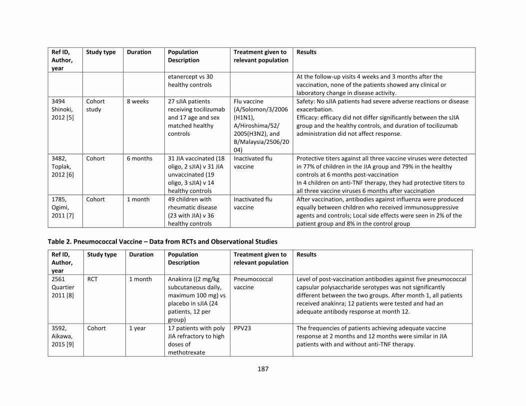
187
Ref ID, Author, year
Study type Duration Population Description
Treatment given to relevant population
Results
etanercept vs 30 healthy controls
At the follow-up visits 4 weeks and 3 months after the vaccination, none of the patients showed any clinical or laboratory change in disease activity.
3494 Shinoki, 2012 [5]
Cohort study
8 weeks 27 sJIA patients receiving tocilizumab and 17 age and sex matched healthy controls
Flu vaccine (A/Solomon/3/2006(H1N1), A/Hiroshima/52/ 2005(H3N2), and B/Malaysia/2506/2004)
Safety: No sJIA patients had severe adverse reactions or disease exacerbation. Efficacy: efficacy did not differ significantly between the sJIA group and the healthy controls, and duration of tocilizumab administration did not affect response.
3482, Toplak, 2012 [6]
Cohort 6 months 31 JIA vaccinated (18 oligo, 2 sJIA) v 31 JIA unvaccinated (19 oligo, 3 sJIA) v 14 healthy controls
Inactivated flu vaccine
Protective titers against all three vaccine viruses were detected in 77% of children in the JIA group and 79% in the healthy controls at 6 months post-vaccination In 4 children on anti-TNF therapy, they had protective titers to all three vaccine viruses 6 months after vaccination
1785, Ogimi, 2011 [7]
Cohort 1 month 49 children with rheumatic disease (23 with JIA) v 36 healthy controls
Inactivated flu vaccine
After vaccination, antibodies against influenza were produced equally between children who received immunosuppressive agents and controls; Local side effects were seen in 2% of the patient group and 8% in the control group
Table 2. Pneumococcal Vaccine – Data from RCTs and Observational Studies
Ref ID, Author, year
Study type Duration Population Description
Treatment given to relevant population
Results
2561 Quartier 2011 [8]
RCT 1 month Anakinra ((2 mg/kg subcutaneous daily, maximum 100 mg) vs placebo in sJIA (24 patients, 12 per group)
Pneumococcal vaccine
Level of post-vaccination antibodies against five pneumococcal capsular polysaccharide serotypes was not significantly different between the two groups. After month 1, all patients received anakinra; 12 patients were tested and had an adequate antibody response at month 12.
3592, Aikawa, 2015 [9]
Cohort 1 year 17 patients with poly JIA refractory to high doses of methotrexate
PPV23 The frequencies of patients achieving adequate vaccine response at 2 months and 12 months were similar in JIA patients with and without anti-TNF therapy.
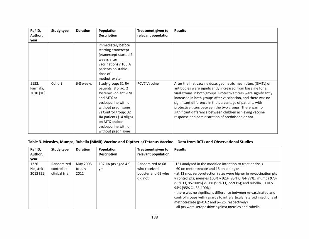
188
Ref ID, Author, year
Study type Duration Population Description
Treatment given to relevant population
Results
immediately before starting etanercept (etanercept started 2 weeks after vaccination) v 10 JIA patients on stable dose of methotrexate
1153, Farmaki, 2010 [10]
Cohort 6-8 weeks Study group: 31 JIA patients (8 oligo, 2 systemic) on anti-TNF and MTX or cyclosporine with or without prednisone vs Control group: 32 JIA patients (14 oligo) on MTX and/or cyclosporine with or without prednisone
PCV7 Vaccine After the first vaccine dose, geometric mean titers (GMTs) of antibodies were significantly increased from baseline for all viral strains in both groups. Protective titers were significantly increased in both groups after vaccination, and there was no significant difference in the percentage of patients with protective titers between the two groups. There was no significant difference between children achieving vaccine response and administration of prednisone or not.
Table 3. Measles, Mumps, Rubella (MMR) Vaccine and Diptheria/Tetanus Vaccine – Data from RCTs and Observational Studies
Ref ID, Author, year
Study type Duration Population Description
Treatment given to relevant population
Results
1226 Heijstek 2013 [11]
Randomized controlled clinical trial
May 2008 to July 2011
137 JIA pts aged 4-9 yrs
Randomized to 68 who received booster and 69 who did not
-131 analyzed in the modified intention to treat analysis - 60 on methotrexate and 15 on biologics - at 12 mos seroprotection rates were higher in revaccination pts v control pts; measles 100% v 92% (95% CI 84-99%), mumps 97% (95% CI, 95-100%) v 81% (95% CI, 72-93%); and rubella 100% v 94% (95% CI, 86-100%) - there was no significant difference between re-vaccinated and control groups with regards to intra articular steroid injections of methotrexate (p=0.62 and p=.25, respectively) - all pts were seropositive against measles and rubella
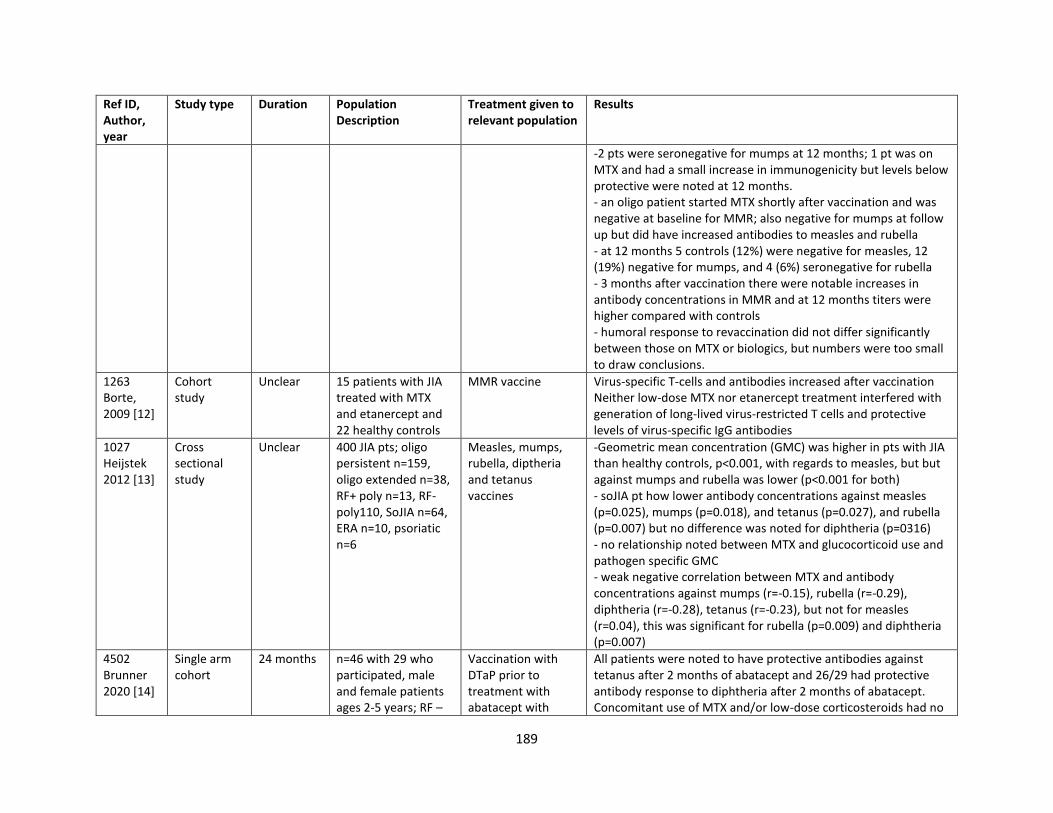
189
Ref ID, Author, year
Study type Duration Population Description
Treatment given to relevant population
Results
-2 pts were seronegative for mumps at 12 months; 1 pt was on MTX and had a small increase in immunogenicity but levels below protective were noted at 12 months. - an oligo patient started MTX shortly after vaccination and was negative at baseline for MMR; also negative for mumps at follow up but did have increased antibodies to measles and rubella - at 12 months 5 controls (12%) were negative for measles, 12 (19%) negative for mumps, and 4 (6%) seronegative for rubella - 3 months after vaccination there were notable increases in antibody concentrations in MMR and at 12 months titers were higher compared with controls - humoral response to revaccination did not differ significantly between those on MTX or biologics, but numbers were too small to draw conclusions.
1263 Borte, 2009 [12]
Cohort study
Unclear 15 patients with JIA treated with MTX and etanercept and 22 healthy controls
MMR vaccine Virus-specific T-cells and antibodies increased after vaccination Neither low-dose MTX nor etanercept treatment interfered with generation of long-lived virus-restricted T cells and protective levels of virus-specific IgG antibodies
1027 Heijstek 2012 [13]
Cross sectional study
Unclear 400 JIA pts; oligo persistent n=159, oligo extended n=38, RF+ poly n=13, RF-poly110, SoJIA n=64, ERA n=10, psoriatic n=6
Measles, mumps, rubella, diptheria and tetanus vaccines
-Geometric mean concentration (GMC) was higher in pts with JIA than healthy controls, p<0.001, with regards to measles, but but against mumps and rubella was lower (p<0.001 for both) - soJIA pt how lower antibody concentrations against measles (p=0.025), mumps (p=0.018), and tetanus (p=0.027), and rubella (p=0.007) but no difference was noted for diphtheria (p=0316) - no relationship noted between MTX and glucocorticoid use and pathogen specific GMC - weak negative correlation between MTX and antibody concentrations against mumps (r=-0.15), rubella (r=-0.29), diphtheria (r=-0.28), tetanus (r=-0.23), but not for measles (r=0.04), this was significant for rubella (p=0.009) and diphtheria (p=0.007)
4502 Brunner 2020 [14]
Single arm cohort
24 months n=46 with 29 who participated, male and female patients ages 2-5 years; RF –
Vaccination with DTaP prior to treatment with abatacept with
All patients were noted to have protective antibodies against tetanus after 2 months of abatacept and 26/29 had protective antibody response to diphtheria after 2 months of abatacept. Concomitant use of MTX and/or low-dose corticosteroids had no
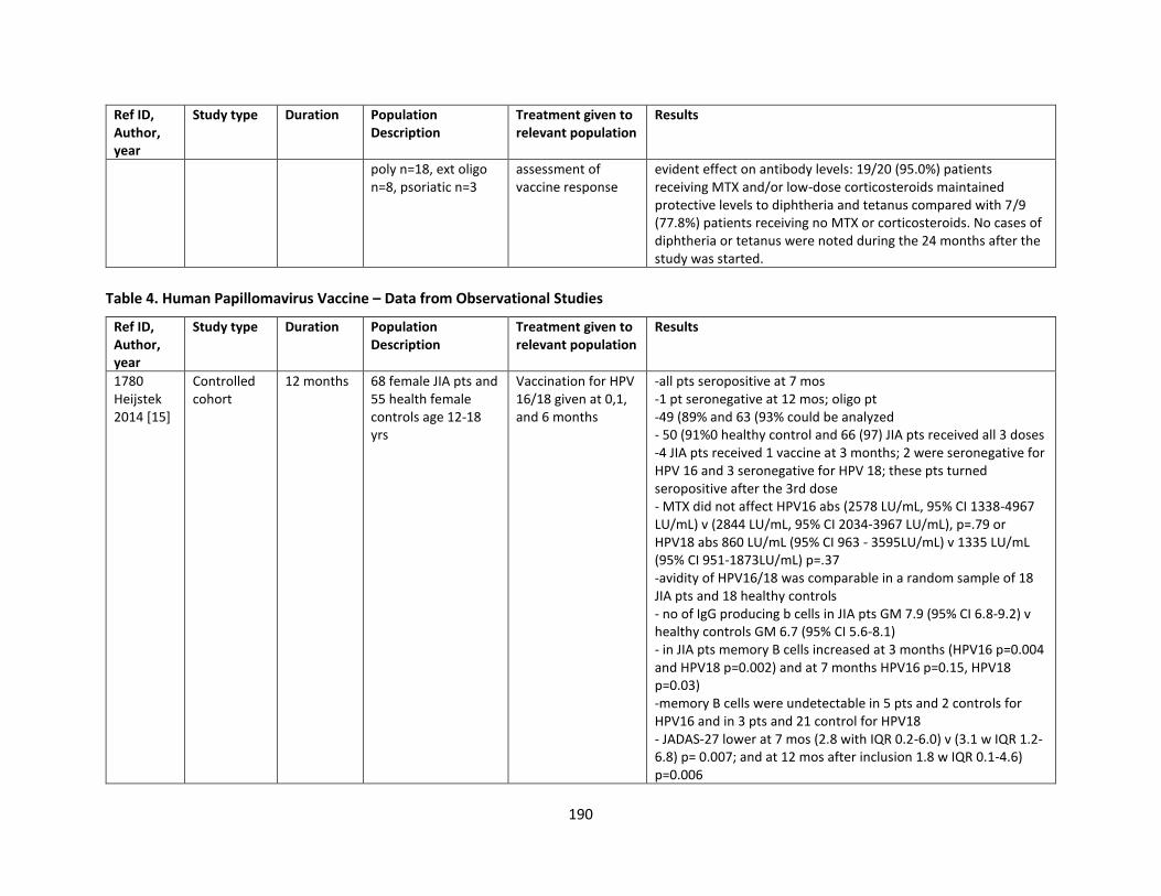
190
Ref ID, Author, year
Study type Duration Population Description
Treatment given to relevant population
Results
poly n=18, ext oligo n=8, psoriatic n=3
assessment of vaccine response
evident effect on antibody levels: 19/20 (95.0%) patients receiving MTX and/or low-dose corticosteroids maintained protective levels to diphtheria and tetanus compared with 7/9 (77.8%) patients receiving no MTX or corticosteroids. No cases of diphtheria or tetanus were noted during the 24 months after the study was started.
Table 4. Human Papillomavirus Vaccine – Data from Observational Studies
Ref ID, Author, year
Study type Duration Population Description
Treatment given to relevant population
Results
1780 Heijstek 2014 [15]
Controlled cohort
12 months 68 female JIA pts and 55 health female controls age 12-18 yrs
Vaccination for HPV 16/18 given at 0,1, and 6 months
-all pts seropositive at 7 mos -1 pt seronegative at 12 mos; oligo pt -49 (89% and 63 (93% could be analyzed - 50 (91%0 healthy control and 66 (97) JIA pts received all 3 doses -4 JIA pts received 1 vaccine at 3 months; 2 were seronegative for HPV 16 and 3 seronegative for HPV 18; these pts turned seropositive after the 3rd dose - MTX did not affect HPV16 abs (2578 LU/mL, 95% CI 1338-4967 LU/mL) v (2844 LU/mL, 95% CI 2034-3967 LU/mL), p=.79 or HPV18 abs 860 LU/mL (95% CI 963 - 3595LU/mL) v 1335 LU/mL (95% CI 951-1873LU/mL) p=.37 -avidity of HPV16/18 was comparable in a random sample of 18 JIA pts and 18 healthy controls - no of IgG producing b cells in JIA pts GM 7.9 (95% CI 6.8-9.2) v healthy controls GM 6.7 (95% CI 5.6-8.1) - in JIA pts memory B cells increased at 3 months (HPV16 p=0.004 and HPV18 p=0.002) and at 7 months HPV16 p=0.15, HPV18 p=0.03) -memory B cells were undetectable in 5 pts and 2 controls for HPV16 and in 3 pts and 21 control for HPV18 - JADAS-27 lower at 7 mos (2.8 with IQR 0.2-6.0) v (3.1 w IQR 1.2-6.8) p= 0.007; and at 12 mos after inclusion 1.8 w IQR 0.1-4.6) p=0.006
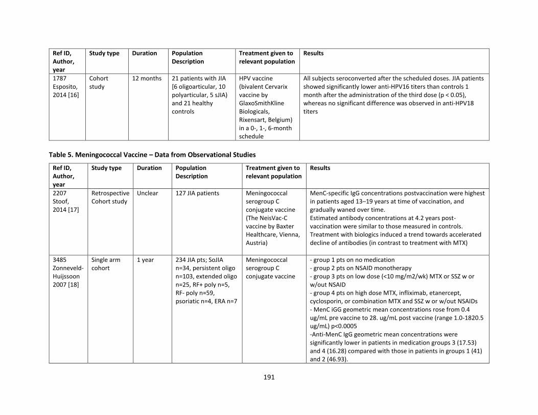
191
Ref ID, Author, year
Study type Duration Population Description
Treatment given to relevant population
Results
1787 Esposito, 2014 [16]
Cohort study
12 months 21 patients with JIA [6 oligoarticular, 10 polyarticular, 5 sJIA) and 21 healthy controls
HPV vaccine (bivalent Cervarix vaccine by GlaxoSmithKline Biologicals, Rixensart, Belgium) in a 0-, 1-, 6-month schedule
All subjects seroconverted after the scheduled doses. JIA patients showed significantly lower anti-HPV16 titers than controls 1 month after the administration of the third dose (p < 0.05), whereas no significant difference was observed in anti-HPV18 titers
Table 5. Meningococcal Vaccine – Data from Observational Studies
Ref ID, Author, year
Study type Duration Population Description
Treatment given to relevant population
Results
2207 Stoof, 2014 [17]
Retrospective Cohort study
Unclear 127 JIA patients Meningococcal serogroup C conjugate vaccine (The NeisVac-C vaccine by Baxter Healthcare, Vienna, Austria)
MenC-specific IgG concentrations postvaccination were highest in patients aged 13–19 years at time of vaccination, and gradually waned over time. Estimated antibody concentrations at 4.2 years post-vaccination were similar to those measured in controls. Treatment with biologics induced a trend towards accelerated decline of antibodies (in contrast to treatment with MTX)
3485 Zonneveld-Huijssoon 2007 [18]
Single arm cohort
1 year 234 JIA pts; SoJIA n=34, persistent oligo n=103, extended oligo n=25, RF+ poly n=5, RF- poly n=59, psoriatic n=4, ERA n=7
Meningococcal serogroup C conjugate vaccine
- group 1 pts on no medication - group 2 pts on NSAID monotherapy - group 3 pts on low dose (<10 mg/m2/wk) MTX or SSZ w or w/out NSAID - group 4 pts on high dose MTX, infliximab, etanercept, cyclosporin, or combination MTX and SSZ w or w/out NSAIDs - MenC iGG geometric mean concentrations rose from 0.4 ug/mL pre vaccine to 28. ug/mL post vaccine (range 1.0-1820.5 ug/mL) p<0.0005 -Anti-MenC IgG geometric mean concentrations were significantly lower in patients in medication groups 3 (17.53) and 4 (16.28) compared with those in patients in groups 1 (41) and 2 (46.93).
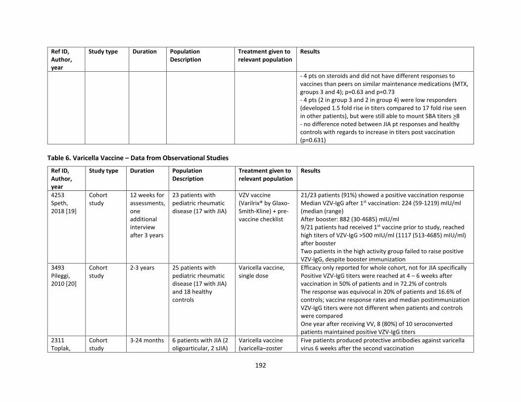
192
Ref ID, Author, year
Study type Duration Population Description
Treatment given to relevant population
Results
- 4 pts on steroids and did not have different responses to vaccines than peers on similar maintenance medications (MTX, groups 3 and 4); p=0.63 and p=0.73 - 4 pts (2 in group 3 and 2 in group 4) were low responders (developed 1.5 fold rise in titers compared to 17 fold rise seen in other patients), but were still able to mount SBA titers >8 - no difference noted between JIA pt responses and healthy controls with regards to increase in titers post vaccination (p=0.631)
Table 6. Varicella Vaccine – Data from Observational Studies
Ref ID, Author, year
Study type Duration Population Description
Treatment given to relevant population
Results
4253 Speth, 2018 [19]
Cohort study
12 weeks for assessments, one additional interview after 3 years
23 patients with pediatric rheumatic disease (17 with JIA)
VZV vaccine (Varilrix® by Glaxo-Smith-Kline) + pre-vaccine checklist
21/23 patients (91%) showed a positive vaccination response Median VZV-IgG after 1st vaccination: 224 (59-1219) mIU/ml (median (range) After booster: 882 (30-4685) mIU/ml 9/21 patients had received 1st vaccine prior to study, reached high titers of VZV-IgG >500 mIU/ml (1117 (513-4685) mIU/ml) after booster Two patients in the high activity group failed to raise positive VZV-IgG, despite booster immunization
3493 Pileggi, 2010 [20]
Cohort study
2-3 years 25 patients with pediatric rheumatic disease (17 with JIA) and 18 healthy controls
Varicella vaccine, single dose
Efficacy only reported for whole cohort, not for JIA specifically Positive VZV-IgG titers were reached at 4 – 6 weeks after vaccination in 50% of patients and in 72.2% of controls The response was equivocal in 20% of patients and 16.6% of controls; vaccine response rates and median postimmunization VZV-IgG titers were not different when patients and controls were compared One year after receiving VV, 8 (80%) of 10 seroconverted patients maintained positive VZV-IgG titers
2311 Toplak,
Cohort study
3-24 months 6 patients with JIA (2 oligoarticular, 2 sJIA)
Varicella vaccine (varicella–zoster
Five patients produced protective antibodies against varicella virus 6 weeks after the second vaccination
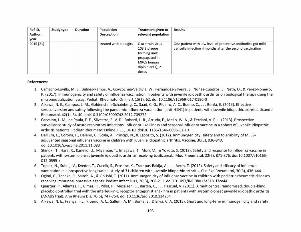
193
Ref ID, Author, year
Study type Duration Population Description
Treatment given to relevant population
Results
2015 [21] treated with biologics Oka strain virus 103.3 plaque forming units propagated in MRC5 human diploid cells), 2 doses
One patient with low level of protective antibodies got mild varicella infection 4 months after the second vaccination
References:
1. Camacho-Lovillo, M. S., Bulnes-Ramos, A., Goycochea-Valdivia, W., Fernández-Silveira, L., Núñez-Cuadros, E., Neth, O., & Pérez-Romero, P. (2017). Immunogenicity and safety of influenza vaccination in patients with juvenile idiopathic arthritis on biological therapy using the microneutralization assay. Pediatr Rheumatol Online J, 15(1), 62. doi:10.1186/s12969-017-0190-0
2. Aikawa, N. E., Campos, L. M., Goldenstein-Schainberg, C., Saad, C. G., Ribeiro, A. C., Bueno, C., . . . Bonfá, E. (2013). Effective seroconversion and safety following the pandemic influenza vaccination (anti-H1N1) in patients with juvenile idiopathic arthritis. Scand J Rheumatol, 42(1), 34-40. doi:10.3109/03009742.2012.709272
3. Carvalho, L. M., de Paula, F. E., Silvestre, R. V. D., Roberti, L. R., Arruda, E., Mello, W. A., & Ferriani, V. P. L. (2013). Prospective surveillance study of acute respiratory infections, influenza-like illness and seasonal influenza vaccine in a cohort of juvenile idiopathic arthritis patients. Pediatr Rheumatol Online J, 11, 10-10. doi:10.1186/1546-0096-11-10
4. Dell'Era, L., Corona, F., Daleno, C., Scala, A., Principi, N., & Esposito, S. (2012). Immunogenicity, safety and tolerability of MF59-adjuvanted seasonal influenza vaccine in children with juvenile idiopathic arthritis. Vaccine, 30(5), 936-940. doi:10.1016/j.vaccine.2011.11.083
5. Shinoki, T., Hara, R., Kaneko, U., Miyamae, T., Imagawa, T., Mori, M., & Yokota, S. (2012). Safety and response to influenza vaccine in patients with systemic-onset juvenile idiopathic arthritis receiving tocilizumab. Mod Rheumatol, 22(6), 871-876. doi:10.1007/s10165-012-0595-z
6. Toplak, N., Subelj, V., Kveder, T., Cucnik, S., Prosenc, K., Trampus-Bakija, A., . . . Avcin, T. (2012). Safety and efficacy of influenza vaccination in a prospective longitudinal study of 31 children with juvenile idiopathic arthritis. Clin Exp Rheumatol, 30(3), 436-444.
7. Ogimi, C., Tanaka, R., Saitoh, A., & Oh-Ishi, T. (2011). Immunogenicity of influenza vaccine in children with pediatric rheumatic diseases receiving immunosuppressive agents. Pediatr Infect Dis J, 30(3), 208-211. doi:10.1097/INF.0b013e3181f7ce44
8. Quartier, P., Allantaz, F., Cimaz, R., Pillet, P., Messiaen, C., Bardin, C., . . . Pascual, V. (2011). A multicentre, randomised, double-blind, placebo-controlled trial with the interleukin-1 receptor antagonist anakinra in patients with systemic-onset juvenile idiopathic arthritis (ANAJIS trial). Ann Rheum Dis, 70(5), 747-754. doi:10.1136/ard.2010.134254
9. Aikawa, N. E., França, I. L., Ribeiro, A. C., Sallum, A. M., Bonfa, E., & Silva, C. A. (2015). Short and long-term immunogenicity and safety
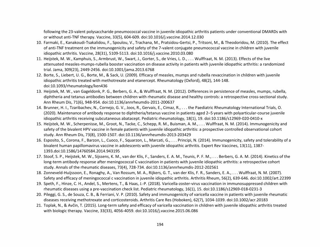
194
following the 23-valent polysaccharide pneumococcal vaccine in juvenile idiopathic arthritis patients under conventional DMARDs with or without anti-TNF therapy. Vaccine, 33(5), 604-609. doi:10.1016/j.vaccine.2014.12.030
10. Farmaki, E., Kanakoudi-Tsakalidou, F., Spoulou, V., Trachana, M., Pratsidou-Gertsi, P., Tritsoni, M., & Theodoridou, M. (2010). The effect of anti-TNF treatment on the immunogenicity and safety of the 7-valent conjugate pneumococcal vaccine in children with juvenile idiopathic arthritis. Vaccine, 28(31), 5109-5113. doi:10.1016/j.vaccine.2010.03.080
11. Heijstek, M. W., Kamphuis, S., Armbrust, W., Swart, J., Gorter, S., de Vries, L. D., . . . Wulffraat, N. M. (2013). Effects of the live attenuated measles-mumps-rubella booster vaccination on disease activity in patients with juvenile idiopathic arthritis: a randomized trial. Jama, 309(23), 2449-2456. doi:10.1001/jama.2013.6768
12. Borte, S., Liebert, U. G., Borte, M., & Sack, U. (2009). Efficacy of measles, mumps and rubella revaccination in children with juvenile idiopathic arthritis treated with methotrexate and etanercept. Rheumatology (Oxford), 48(2), 144-148. doi:10.1093/rheumatology/ken436
13. Heijstek, M. W., van Gageldonk, P. G., Berbers, G. A., & Wulffraat, N. M. (2012). Differences in persistence of measles, mumps, rubella, diphtheria and tetanus antibodies between children with rheumatic disease and healthy controls: a retrospective cross-sectional study. Ann Rheum Dis, 71(6), 948-954. doi:10.1136/annrheumdis-2011-200637
14. Brunner, H. I., Tzaribachev, N., Cornejo, G. V., Joos, R., Gervais, E., Cimaz, R., . . . the Paediatric Rheumatology International Trials, O. (2020). Maintenance of antibody response to diphtheria/tetanus vaccine in patients aged 2–5 years with polyarticular-course juvenile idiopathic arthritis receiving subcutaneous abatacept. Pediatric rheumatology, 18(1), 19. doi:10.1186/s12969-020-0410-x
15. Heijstek, M. W., Scherpenisse, M., Groot, N., Tacke, C., Schepp, R. M., Buisman, A. M., . . . Wulffraat, N. M. (2014). Immunogenicity and safety of the bivalent HPV vaccine in female patients with juvenile idiopathic arthritis: a prospective controlled observational cohort study. Ann Rheum Dis, 73(8), 1500-1507. doi:10.1136/annrheumdis-2013-203429
16. Esposito, S., Corona, F., Barzon, L., Cuoco, F., Squarzon, L., Marcati, G., . . . Principi, N. (2014). Immunogenicity, safety and tolerability of a bivalent human papillomavirus vaccine in adolescents with juvenile idiopathic arthritis. Expert Rev Vaccines, 13(11), 1387-1393.doi:10.1586/14760584.2014.943195
17. Stoof, S. P., Heijstek, M. W., Sijssens, K. M., van der Klis, F., Sanders, E. A. M., Teunis, P. F. M., . . . Berbers, G. A. M. (2014). Kinetics of the long-term antibody response after meningococcal C vaccination in patients with juvenile idiopathic arthritis: a retrospective cohort study. Annals of the rheumatic diseases, 73(4), 728-734. doi:10.1136/annrheumdis-2012-202561
18. Zonneveld-Huijssoon, E., Ronaghy, A., Van Rossum, M. A., Rijkers, G. T., van der Klis, F. R., Sanders, E. A., . . . Wulffraat, N. M. (2007). Safety and efficacy of meningococcal c vaccination in juvenile idiopathic arthritis. Arthritis Rheum, 56(2), 639-646. doi:10.1002/art.22399
19. Speth, F., Hinze, C. H., Andel, S., Mertens, T., & Haas, J.-P. (2018). Varicella-zoster-virus vaccination in immunosuppressed children with rheumatic diseases using a pre-vaccination check list. Pediatric rheumatology, 16(1), 15. doi:10.1186/s12969-018-0231-3
20. Pileggi, G. S., de Souza, C. B., & Ferriani, V. P. (2010). Safety and immunogenicity of varicella vaccine in patients with juvenile rheumatic diseases receiving methotrexate and corticosteroids. Arthritis Care Res (Hoboken), 62(7), 1034-1039. doi:10.1002/acr.20183
21. Toplak, N., & Avčin, T. (2015). Long-term safety and efficacy of varicella vaccination in children with juvenile idiopathic arthritis treated with biologic therapy. Vaccine, 33(33), 4056-4059. doi:10.1016/j.vaccine.2015.06.086
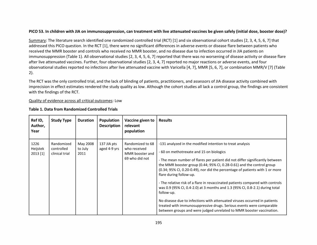
195
PICO 53. In children with JIA on immunosuppression, can treatment with live attenuated vaccines be given safely (initial dose, booster dose)?
Summary: The literature search identified one randomized controlled trial (RCT) [1] and six observational cohort studies [2, 3, 4, 5, 6, 7] that addressed this PICO question. In the RCT [1], there were no significant differences in adverse events or disease flare between patients who received the MMR booster and controls who received no MMR booster, and no disease due to infection occurred in JIA patients on immunosuppression (Table 1). All observational studies [2, 3, 4, 5, 6, 7] reported that there was no worsening of disease activity or disease flare after live attenuated vaccines. Further, four observational studies [2, 3, 4, 7] reported no major reactions or adverse events, and four observational studies reported no infections after live attenuated vaccine with Varicella [4, 7], MMR [5, 6, 7], or combination MMR/V [7] (Table 2).
The RCT was the only controlled trial, and the lack of blinding of patients, practitioners, and assessors of JIA disease activity combined with imprecision in effect estimates rendered the study quality as low. Although the cohort studies all lack a control group, the findings are consistent with the findings of the RCT.
Quality of evidence across all critical outcomes: Low
Table 1. Data from Randomized Controlled Trials
Ref ID, Author, Year
Study Type Duration Population Description
Vaccine given to relevant population
Results
1226 Heijstek 2013 [1]
Randomized controlled clinical trial
May 2008 to July 2011
137 JIA pts aged 4-9 yrs
Randomized to 68 who received MMR booster and 69 who did not
-131 analyzed in the modified intention to treat analysis
- 60 on methotrexate and 15 on biologics
- The mean number of flares per patient did not differ significantly between the MMR booster group (0.44; 95% CI, 0.28-0.61) and the control group (0.34; 95% CI, 0.20-0.49), nor did the percentage of patients with 1 or more flare during follow-up.
- The relative risk of a flare in revaccinated patients compared with controls was 0.9 (95% CI, 0.4-2.0) at 3 months and 1.3 (95% CI, 0.8-2.1) during total follow-up.
No disease due to infections with attenuated viruses occurred in patients treated with immunosuppressive drugs. Serious events were comparable between groups and were judged unrelated to MMR booster vaccination.
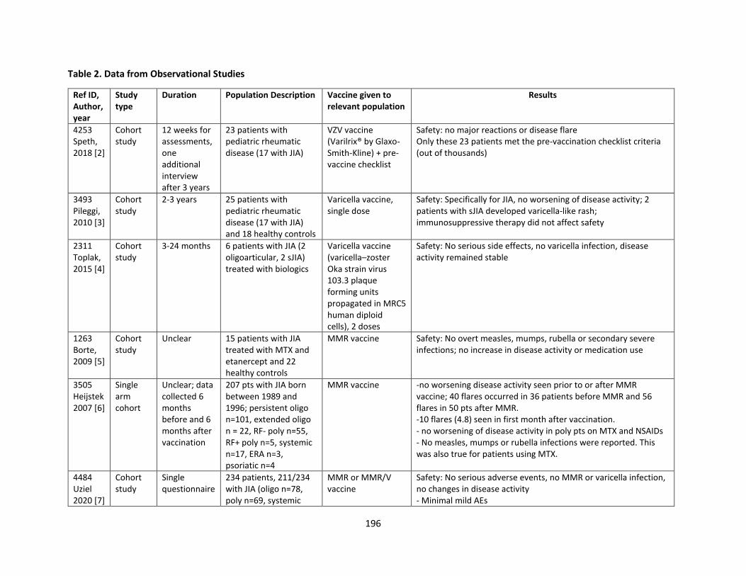
196
Table 2. Data from Observational Studies
Ref ID, Author, year
Study type
Duration Population Description Vaccine given to relevant population
Results
4253 Speth, 2018 [2]
Cohort study
12 weeks for assessments, one additional interview after 3 years
23 patients with pediatric rheumatic disease (17 with JIA)
VZV vaccine (Varilrix® by Glaxo-Smith-Kline) + pre-vaccine checklist
Safety: no major reactions or disease flare Only these 23 patients met the pre-vaccination checklist criteria (out of thousands)
3493 Pileggi, 2010 [3]
Cohort study
2-3 years 25 patients with pediatric rheumatic disease (17 with JIA) and 18 healthy controls
Varicella vaccine, single dose
Safety: Specifically for JIA, no worsening of disease activity; 2 patients with sJIA developed varicella-like rash; immunosuppressive therapy did not affect safety
2311 Toplak, 2015 [4]
Cohort study
3-24 months 6 patients with JIA (2 oligoarticular, 2 sJIA) treated with biologics
Varicella vaccine (varicella–zoster Oka strain virus 103.3 plaque forming units propagated in MRC5 human diploid cells), 2 doses
Safety: No serious side effects, no varicella infection, disease activity remained stable
1263 Borte, 2009 [5]
Cohort study
Unclear 15 patients with JIA treated with MTX and etanercept and 22 healthy controls
MMR vaccine Safety: No overt measles, mumps, rubella or secondary severe infections; no increase in disease activity or medication use
3505 Heijstek 2007 [6]
Single arm cohort
Unclear; data collected 6 months before and 6 months after vaccination
207 pts with JIA born between 1989 and 1996; persistent oligo n=101, extended oligo n = 22, RF- poly n=55, RF+ poly n=5, systemic n=17, ERA n=3, psoriatic n=4
MMR vaccine -no worsening disease activity seen prior to or after MMR vaccine; 40 flares occurred in 36 patients before MMR and 56 flares in 50 pts after MMR. -10 flares (4.8) seen in first month after vaccination. - no worsening of disease activity in poly pts on MTX and NSAIDs - No measles, mumps or rubella infections were reported. This was also true for patients using MTX.
4484 Uziel 2020 [7]
Cohort study
Single questionnaire
234 patients, 211/234 with JIA (oligo n=78, poly n=69, systemic
MMR or MMR/V vaccine
Safety: No serious adverse events, no MMR or varicella infection, no changes in disease activity - Minimal mild AEs
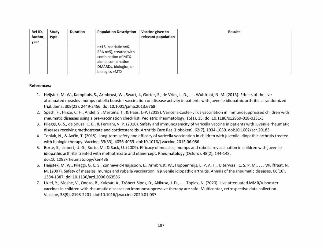
197
Ref ID, Author, year
Study type
Duration Population Description Vaccine given to relevant population
Results
n=18, psoriatic n=6, ERA n=5), treated with combination of MTX alone, combination DMARDs, biologics, or biologics +MTX
References:
1. Heijstek, M. W., Kamphuis, S., Armbrust, W., Swart, J., Gorter, S., de Vries, L. D., . . . Wulffraat, N. M. (2013). Effects of the live
attenuated measles-mumps-rubella booster vaccination on disease activity in patients with juvenile idiopathic arthritis: a randomized
trial. Jama, 309(23), 2449-2456. doi:10.1001/jama.2013.6768
2. Speth, F., Hinze, C. H., Andel, S., Mertens, T., & Haas, J.-P. (2018). Varicella-zoster-virus vaccination in immunosuppressed children with
rheumatic diseases using a pre-vaccination check list. Pediatric rheumatology, 16(1), 15. doi:10.1186/s12969-018-0231-3
3. Pileggi, G. S., de Souza, C. B., & Ferriani, V. P. (2010). Safety and immunogenicity of varicella vaccine in patients with juvenile rheumatic
diseases receiving methotrexate and corticosteroids. Arthritis Care Res (Hoboken), 62(7), 1034-1039. doi:10.1002/acr.20183
4. Toplak, N., & Avčin, T. (2015). Long-term safety and efficacy of varicella vaccination in children with juvenile idiopathic arthritis treated
with biologic therapy. Vaccine, 33(33), 4056-4059. doi:10.1016/j.vaccine.2015.06.086
5. Borte, S., Liebert, U. G., Borte, M., & Sack, U. (2009). Efficacy of measles, mumps and rubella revaccination in children with juvenile
idiopathic arthritis treated with methotrexate and etanercept. Rheumatology (Oxford), 48(2), 144-148.
doi:10.1093/rheumatology/ken436
6. Heijstek, M. W., Pileggi, G. C. S., Zonneveld-Huijssoon, E., Armbrust, W., Hoppenreijs, E. P. A. H., Uiterwaal, C. S. P. M., . . . Wulffraat, N.
M. (2007). Safety of measles, mumps and rubella vaccination in juvenile idiopathic arthritis. Annals of the rheumatic diseases, 66(10),
1384-1387. doi:10.1136/ard.2006.063586
7. Uziel, Y., Moshe, V., Onozo, B., Kulcsár, A., Tróbert-Sipos, D., Akikusa, J. D., . . . Toplak, N. (2020). Live attenuated MMR/V booster
vaccines in children with rheumatic diseases on immunosuppressive therapy are safe: Multicenter, retrospective data collection.
Vaccine, 38(9), 2198-2201. doi:10.1016/j.vaccine.2020.01.037
198
PICO 54. Can live attenuated vaccines be used safely in the households of children with JIA on immunosuppression?
Summary: The literature searches did not identify any studies that addressed this PICO question.
Quality of evidence across all critical outcomes: Very low
199
Imaging Modalities
PICO 55: In children with JIA, is any specific imaging technique recommended to best detect inflammation and damage, make a diagnosis,
predict structural damage, flare or treatment response?
Direct evidence
Summary: Literature searches identified 24 studies that provided direct evidence to address this question by comparing two imaging
technologies in patients with JIA. Five studies compared X-ray with MRI. One study showed that 83.3% of patients had erosions by MRI
compared to 34.8% by X-ray[1]. In patients without clinical complaints of cervical involvement, MRI of cervical spine showed that 65% had soft
tissue involvement, pannus formation and erosions compared to X-ray which showed only erosion in 20% of patients[2]. Cartilage loss was
visible on both MRI and X-ray[7] with more severe cartilage loss on MRI. The joint effusions were better detected by MRI than radiography in
both hips and knees[7]. One study indicated that MRI is less efficient than conventional radiography in detecting destructive changes over 1
year[19].
In a small study[3] that compared MRI with ultrasound (US) of knee and hip joints before and after IA steroid therapy, MRI was more sensitive in
detecting popliteal cyst and lymph nodes, but US was as sensitive as MRI in detecting effusion. In another study[14], the effusion of knee joints
was suspected by MRI in 76% and by radiograph in 21% joints. The MRI was more sensitive in detecting TMJ erosions, synovial hypertrophy,
synovitis and effusions[10], while US misdiagnosed 67% of patients with TMJ involvement as false-negative[11]. Another study[12] reports that
chronic TMJ arthritis was diagnosed in 69% by MRI and in 28% by US. The correlation between US capsular width and MRI assessed amount of
synovitis in TMJ was 0.483 at the subcondylar and 0.347 at the condylar level (p <0.001)[13]. In detecting imflammation, US sensitivity was 0%,
specificity 36.4%, PPV 0%, NPV 100%, when compared with MRI as the reference standard in a study where MRI detected inflammation in 64.7%
of the joints, and power Doppler US detected none [Error! Reference source not found.].
The other comparisons were (one study per each comparison): US vs GSUS vs PDUS (60% had abnormal findings in US, 60% had abnormal
findings in GSUS, and 30% had abnormal findings in PDUS)[17]; MRI vs orthopantomograms (OPG) with authors’ conclusion that MRI was
superior to OPG in following changes of the condyle over time[18]; Radiography vs US with no significant differences in joint space width (JSW)
or cartilage thickness between finger joints without or with previous arthritis[20]. The pixel by pixel DCE-MRI parameters correlated moderately
to significantly with conventional MRI scores for synovitis[Error! Reference source not found.]. Diffusion-weighted imaging (DWI) was accurate
in detecting arthritis in patients with JIA or suspected of having JIA and showed agreement with contrast-enhanced MRI [Error! Reference
source not found.].
200
The evidence base for comparative imaging in JIA is relatively diffuse in that each study is fairly unique in the combination of imaging techniques
compared, the type of JIA being evaluated, the specific joints being evaluated and the purpose of the comparison (e.g., diagnosis, detection of
inflammation, treatment response). Also, most of the studies have small patient numbers and have limited generalizability to the broader JIA
population. For these reasons, the quality of evidence is very low.
Quality of evidence across all critical outcomes: Very low
Table 1. Studies Comparing Imaging Technologies
Ref ID, Author, year
Study type Duration Population Description
Assessment given to relevant population
Results
977, Mallatia, 2011 [1]
Prospective observational
Median 1.2 years (IQR 1.0-1.4) for 39 patients with longitudinal data
66 JIA patients with wrist arthritis
Contrast-enhanced MRI of more clinically affected wrist, using 1.5T scanner. Images were scored using 1) a novel pediatric-targeted scoring system, modified from the OMERACT RAMRIS recommendations [this novel tool was a precursor to the JAMRIS system] and 2) the RAMRIS system.
- 83.3% of patients had erosions by MRI compared to 34.8% by X-ray. Assessment of novel pediatric MRI scoring tool: - MRI erosion score was significantly higher in patients with limited wrist ROM (p=0.0002), and correlated with Sharp (p<0.0001, rs= 0.61) and Poznanski scores (p<0.0001, rs= 0.61) and clinical indicators of damage (JADI-A) (p<0.0001, rs=0.49). -MRI Bone marrow edema score correlated with Sharp (p<0.0001, rs=0.66) and Poznanski scores (p=0.001, rs= -0.43) , and JADI-A (p=0.001, rs= 0.4). - MRI synovitis score significantly higher with higher wrist swelling score (p<0.0001), moderately correlated with total swollen joint count (p=0.0002, rs=0.45), JADAS-71 (p=0.0006, rs= 0.41), and physician global (p=0.001, rs= 0.41). Longitudinal data/sensitivity or scoring tool to change: - Patients meeting ACR30 at follow up had significant decrease in bone marrow edema score (p=0.04, SRM= 0.44) and synovitis score (p=0.01, SRM=0.62) - Patients without clinical improvement had significant increase in erosion score (p=0.03, SRM= 0.57), but the same was seen in patients with clinical improvement (p=0.01, SRM= 0.6)
2192 Oren et. al. 1996 [2]
Prospective cohort
1991 for 32 months
20 pts with “JRA” 7 females, 13
MRI of cervical spine, X-ray
18 pts without clinical complaints of cervical involvement, 13 (65%) had soft tissue involvement, pannus formation and erosions compared to X-ray which showed only erosion in 4 pts
201
Ref ID, Author, year
Study type Duration Population Description
Assessment given to relevant population
Results
males, age 3-16,
2071 Eich, 1994 [3]
Single-arm study
1 month 10 JIA patients
MRI and US of 15 joints before and after Intraarticular steroid therapy
Effusion before therapy: US and MRI 11 knees, 4 hips; Effusion after therapy: US 11 knees, 0 hips; MRI 4 knees, 1 hip; Popliteal cyst before therapy: US 1 knee, MRI 3 knees, clinical exam 0; Popliteal cyst before therapy: US 0 knee, MRI 1 knees, clinical exam 0; Lymph nodes before therapy: US 1 knee, MRI 9 knees; Lymph nodes after therapy: US 0 knees, MRI 6 knees.
644, Koos, 2013 [4]
Retrospective cohort study
Unclear 23 patients with JIA, 23 matched controls
Underwent contrast enhanced MRI or cone beam CT scan (CBCT)
78% of TMJs in control group were considered normal; 83% of the TMJs in the JIA group showed severe changes; difference between TMJ arthritis in control and JIA group was highly significant p<0.0001 Paper devises a scoring method for assessing TMJ arthritis that can be used with MRI and CBCT but does not compare them head to head
838, Kuseler, 1998 [5]
Prospective cohort study
3 years 30 TMJ in 15 children diagnosed with JCA (per EULAR criteria) 10 healthy children served as controls
Patients underwent clinical exam, radiographs and contrast enhanced MRI imaging
In control group, MRI showed no erosions, but 3 joints in 2 patients showed an anteriorly displaced disc and one of these discs was folded. In JCA patients, radiographs revealed small erosions (only in the cortical bone, and no changes in the shape of the condyle) in 5 joints (3 patients) and severe erosions (destruction of the trabecular bone and flattening of the condylar head) in 5 joints (3 patients). On MRI in JCA patients, there were small erosions in 5 joints (5 patients) and severe erosions in 5 joints (3 patients). Two joints with small erosions on the radiograph could not be diagnosed on the MRI. Two joints with small erosions on the MRI could not be diagnosed on radiographs.
202
Ref ID, Author, year
Study type Duration Population Description
Assessment given to relevant population
Results
Grade 2 condylar changes seen on the radiographs corresponded to the MR images. Of patients with small erosions on MRI, none reported subjective symptoms, 3 were found to have objective signs and 2 had no objective findings. Clinical signs were found in all patients with severe erosions on MRI, but only 2 of them reported subjective symptoms. Slight but clear enhancement of synovial membrane seen after injection of Gd-DTPA in 15 joints (11 patients). A strong enhancement was seen in 8 joints (6 patients). Only 1 of these patients reported subjective symptoms. Pannus was found in 7 joints (5 patients). All the joints with pannus also showed enhancement. All discs had a low signal intensity.
2208 Gylys-Morin et. al. 2001 [6]
Prospective cohort (?)
31 months (6/1996-2/1999)
30 pts with JIA: 21 F/9M Age 5-16 (sx less than a year)
MRI with contrast of more symptomatic knee compared with X-rays in 27 children
Suprapatellar joint effusions in 26/30 (87%) Meniscal hypoplasia in 11/30 (37%) Abnormal epiphyseal marrow in 8/30 (27%) 3 had irregular articular cartilage with fissures/thinning 1 knee had erosion Versus xrays which only showed suprapatellar fullness in 78%, joint space narrowing in 1 with no bony abnml
2539 Senac, 1988 [7]
Cohort Study Unclear 21 pts with JRA and 3 healthy volunteers
MRI imaging of hips and knees contrasted with X-rays in juvenile rheumatoid arthritis
13/15 hips had cartilage loss on MRI and 12/15 had joint-space narrowing on standard films. 8 hips had more severe cartilage loss on MRI than radiograph. 6 hips had joint Effusions on MRI, but one of which was radiographically visible. 2/15 hips had Avascular necrosis of the femoral head with equivocal plain film findings. 9/15 hips had bone erosions/degenerative changes on both MRI and xray-worse on MRI.
203
Ref ID, Author, year
Study type Duration Population Description
Assessment given to relevant population
Results
14/20 knees had articular and epiphyseal cartilage loss on MRI. 5/14 had focal thinning, and 9/20 had joint-space narrowing on xrays. 13/20 had meniscal changes. 15 knees
showed joint effusions on MRI vs 9 on xrays. 1 had avascular
necrosis, 3 had intraarticular fragments, and 1 had medullary infarcts on MRI, none visible on corresponding radiographs.
1040, Barendregt, 2016 [8]
Cross sectional study
Feb 2013-Dec 2014
35 patients with JIA (18 MRI active and 17 MRI inactive)
Compared dynamic contrast enhanced MRI (DCE) with diffusion weighted imaging (DWI) in quantifying synovial inflammation in JIA
ME, MIS, TTP. TIC5 and ADC were significantly different when MRI inactive and active JIA were compared. Higher percentages of TIC5 were seen in MRI inactive JIA. Lower ME, MIS, TTP and ADC were found in MRI-inactive JIA as compared to MRI active JIA. In MRI inactive JIA, ME positive correlated with ADC with r=0.49 and p=0.048. TTP positively correlated with ADC with r=0.50, p=0.043 and TIC 4 inversely correlated with ADC with r= -0.55, p= 0.022. ME, MIS and TIC5 significantly correlated to ADC (0.62, 0.45, -0.51 respectively, all p<0.05) when assessing MRI active and inactive JIA together.
546, Pradsgaard, 2015 [9]
Cohort Nov 2008 to October 2011
23 children with JIA
One knee from each of 23 children with oligo JIA were investigated by both MRI and US. Outcome measure was distal femoral cartilage thickness
High level of agreement between MRI and US measurements of mean cartilage thickness and Rho values between modalities were high (between 0.70 and 0.86, p<0.05 for all) Intercondylar notch of the distal femoral cartilage may be the best anatomical point for cartilage thickness measurements of the knee
799, Laurell, 2012 [10]
Cohort study 2007-2011 10 JIA patients with 11 clinically active joints were assessed by US and MRI
10 patients with JIA were assessed by US and MRI compared to 6 healthy controls with 8 joints
US detected synovial hypertrophy in 22 areas of 11 joints, 86% of which had synovial hyperemia, and MRI revealed synovitis in 36 areas of the same 11 joints. Erosions were identified by US in two areas of two joints and by MRI in 6 areas of four joints. Effusion was shown by US in nine areas of six joints and by MRI in 17 areas of five joints. MRI detected juxta articular bone marrow edema in 16 areas of eight joints.
1126, Muller,
Case series March and Sept 2006
30 consecutive JIA patients
Patients underwent 4 examinations: Rheum investigation, orthodontic
19/30 (63%) patients and 33/60 (55%) joints had signs of TMJ involvement on MRI. This was associated with condylar deformity in 9/19 (47%) patients and 15/33 (45%) joints.
204
Ref ID, Author, year
Study type Duration Population Description
Assessment given to relevant population
Results
2009 [11]
exam, US of TMJs, and MRI of TMJs
Rheumatological, orthodontic, and US examinations correctly diagnosed 11 (58%), 9 (47%) and 6 (33%) patients, respectively, with active arthritis but misdiagnosed 8 (42%), 10 (53%) and 12 (67%) patients, respectively, as having no signs of inflammation. Best predictor for active arthritis on MRI was a reduced maximum mouth opening
1672, Weiss, 2008 [12]
Single arm cohort
Jan 2005 to April 2007
38 newly diagnosed JIA patients
Children with newly diagnosed JIA were prospectively evaluated for TMJ arthritis. Jaw pain and disability were assessed with questionnaires and physician exam TMJs were imagined with MRI and US within 8 weeks of diagnosis
Acute TMJ arthritis was diagnosed in 75% of children by MRI and none by US. Chronic arthritis was diagnosed in 69% by MRI and in 28% by US. Findings of both acute and chronic TMJ disease were detected by MRI in 53% of the patients. Of those with acute arthritis, 71% were asymptomatic and 63% had normal PE.
3835, Kirkhus, 2016 [13]
Case series 2005-2012 55 patients; 48 with JIA; 7 with other diseases including JDM, MCTD, Scleroderma, Sjorgens and Lupus
Ultrasound and MRI of TMJs were obtained within a week for patients referred to radiology due to symptoms or clinical suspicion of TMJ arthritis; 124 ultrasounds and MRIs were done in 55 patients and were scored for subcondylar and condylar capsule width (US) and amount of synovitis (MRI)
Correlation between ultrasound capsular width and MRI assessed amount of synovitis was moderate both at the subcondylar and condylar level, spearmans rho 0.483, p <0.001 and 0.347, p<0.001 respectively. The ROC curve indicated the best discriminatory ability at the subcondylar level with an area under the curve of 0.77 (95% CI 0.69-0.85) and a cut off of 1.2 mm (sensitivity 72%, specificity 70%) for the capsular width.
4111, El-Miedany, 2001 [14]
Cohort 38 patients with JIA and clinical signs of knee joint involvement and 10 healthy controls
All patients underwent plain radiography, US and MRI examinations before and after contrast administration
Acute synovitis in at least one knee joint was present in 15/38 (39.5%) of patients while chronic synovitis was evident in the rest of the patients, 23/38 (60.5%). On radiographs, joint effusion was suspected in 6 out of 29 joints (21%). On MRI, joint effusions were seen as areas of decreased signal intensity on T1W1 in 29 out of 38 joints (76%) before and after enhancement with Gd-DTPA. Compared to control, sonographic examination was found to be of great value for evaluating joint effusion, popliteal cysts, lymph nodes, and to
205
Ref ID, Author, year
Study type Duration Population Description
Assessment given to relevant population
Results
a lesser extent, the degree of affection of the articular cartilage. MRI was superior in evaluating the extent of synovial proliferation, thinning out and erosions of articular cartilage, loculated effusions as well as hypoplastic menisci and ligaments, especially after contrast enhancement.
2383, Malattia, 2008 [15]
Cross sectional study
June 2006 to March 2007
26 JIA patients
The clinically more affected wrist was studied for erosions with MRI, radiography, and ultrasound coupled with standard clinical assessment and biochemical analysis
Of 26 JIA patients, 25 (96.1%) had 1 or more erosions as detected by MRI whereas conventional radiography and US revealed erosions in 13 (50%) and 12 (50%) of 24 patients, respectively. MRI detected erosive changes more compared to conventional radiography (p=0.002 with Bonferroni correction and US (pb= 0.0002) in the group of patients with less than 3 years disease duration. US and radiography were of equivalent value for the detection of destructive changes. Wrist MRI score correlated highly with radiographic erosion score (r=0.82) and with wrist limited range of motion score (r=0.69).
3636 Rydholm 1986 [16]
Single Arm Cohort
14 patients with JCA (16 hips examined)
Hips examined clinically, radiographically, and by US prior to joint pressure measurement and arthroscopy
Capsular distention correlated with stage of synovitis, r=0.7, p<0.01. Stage of synovitis correlated with intracapsular pressure, r=0.6, p<0.05. Stage of synovitis correlated with stage of cartilage damage, r=0.5, p<0.05. Stage of cartilage destruction correlate with radiographic score, r=0.7, p<0.01. No correlation with capsular distention and intracapsular pressure.
802 Lerkvaleekul 2017 [17]
Cross sectional
1 year 46 JIA patients Inactive group (16 patients with prior wrist arthritis now inactive on
Wrist joints examined by infrared thermography, PE, and US
Tmean and Tmax were higher in arthritis group compared to inactive and healthy (p<0.05). area under the ROC curve was 0.93, and Tmean ⩾ 31 °C was used as a cut-off point between healthy controls and the moderate to severe arthritis group, sensitivity 85.7% and specificity 80.0%. Tmax ⩾ 32.3 °C cut-off point between healthy controls and the moderate to severe arthritis group, area under the ROC curve 0.91, sensitivity and specificity of 71.4% and 93.3% respectively.
206
Ref ID, Author, year
Study type Duration Population Description
Assessment given to relevant population
Results
clinical exam) Arthritis group (30 patients with active wrist arthritis) Healthy Controls (15 patients with no arthritis)
correlation coefficients between Tmean and Tmax with the wrist examination were 0.36 (P = 0.01) and 0.31 (P = 0.04) respectively. Of 46 JIA patients, 28 (60%) had abnormal findings in US, 28 (60%) had abnormal findings in GSUS, and 14 (30%) had abnormal findings in PDUS In inactive group, 3 abnormal GSUS, 1 abnormal PDUS. US score ⩾ 1 was used as a cut-off point between the inactive group and arthritis group, the area under the ROC curve was 0.87 with sensitivity and specificity of 83.3% and 81.3% respectively The correlation coefficient between US score and wrist examination was 0.67, p < 0.01 if using either abnormal findings in GSUS or PDUS as a definition of arthritis
3099 Pedersen, 2008 [18]
Case-control study
N/A 15 JIA patients with TMJ involvement and 10 healthy children
Clinical examination, MRI-scanning, and orthopantomograms (OPG)
There was no correlation between tenderness on palpation and MRI variables or radiographic findings. Decreased translation of the condylar head was correlated to reduced mouth opening capacity. The opening capacity and condylar resorption were significantly related, as shown on MRI and OPG. MRI was superior to OPG in following changes of the condyle over time, and inflammation was detected in nearly all joints.
2554 Malattia, 2012 Malattia, 2013 [19]
Single-arm study
1 year 40 JIA patients on DMARD or biologics
Pediatric (ACRp) response criteria and conventional radiography versus MRI
MRI synovitis score (0-3): at baseline 4.0 (3.0; 6.0); at 1 year 3.0 (2.0; 3.0). The MRI synovitis score was able to discriminate between different levels of ACRp response. Relative efficiency (RE) score showing a higher responsiveness to change: RE of Physician’s Global Assessment 6.7; MRI 1.8; number of active joints 1.3; ESR 1.3; limited joint count 1.2; swollen joint count 1.2; tender joint count 1.2; CHAQ 0.7; patient global assessment 0.7. RE values of the pediatric and the RAMRIS bone erosion scores in relation to the adapted Sharp/van der Heijde score were <1, indicating that MRI is less efficient than conventional radiography in detecting destructive changes over 1 year.

207
Ref ID, Author, year
Study type Duration Population Description
Assessment given to relevant population
Results
790, Pradsgaard et al., 2019 [20]
Cross-sectional
N/A (cross-sectional)
74 patients with JIA (n = 10 systemic JIA, 4 RF+ poly, 17 RF-poly, 15 extended oligo, 28 persistent oligo
X-ray, ultrasound US measurements of cartilage thickness correlated well with radiographic joint space width (JSW) at proximal sites
- MCP: ICC 0.806 and 0.863 for right and left MCP, respectively
- PIP: ICC 0.411 and 0.392 for right and left PIP, respectively
- Knee: ICC 0.629, 0.458, 0.721, 0.534 for right knee medial condyle, lateral condyle, and left knee medial condyle and lateral condyle, respectively
No significant differences in joint space width (JSW) or cartilage thickness between finger joints without or with previous arthritis with either US or radiography Cartilage thickness measured by US smaller in knees with previous arthritis compared to knees without previous arthritis, but not statistically significant Radiographic JSW significantly less at right medial condyle in knees with previous arthritis compared to knees without previous arthritis (p = 0.04) Cartilage thickness or JSW decreased significantly with increasing age Limitations: 1 examiner (separate examiner for US and radiograph), unable to test intra-reader or inter-reader reliability
835, Hemke, 2017 [21]
Prospective, single arm, cohort study
1 year 85 patients with JIA (using ILAR criteria) and knee involvement;
Patients underwent a clinical assessment and pixel by pixel DCE-MRI time intensity curve (TIC) shape analysis method (Type 1: no enhancement, Type 2: slow enhancement, Type 3: fast enhancement,
Poor correlation observed between the relative number of TIC-shape 3 and the JAMRIS synovial hypertrophy score (R=0.328, P=0.0002). No significant correlation observed btwn the relative number of TIC-shape 2, 4 and 5 and the JAMRIS score.
208
Ref ID, Author, year
Study type Duration Population Description
Assessment given to relevant population
Results
consecutively included
Type 4: fast enhancement followed by washout, Type 5: fast enhancement followed by gradual enhancement increase, type 6: arterial enhancement); grade of synovitis was scored on conventional MR images using the JAMRIS system
Significant correlation between the JAMRIS synovial hypertrophy score and the descriptive parameters of maximal enhancement (R=0.658, p< 0.001), enhancing volume (R=0.618, p< 0.001) and initial area under the curve (R=0.639, p< 0.001). The maximal initial slope correlated moderately with the synovial hypertrophy score (r=0.453, p< 0.001). Overall concluded that the pixel by pixel DCE-MRI parameters correlated moderately to significantly with conventional MRI scores for synovitis.
4095 Fedrizzi 1997 [Error! Reference source not found.]
Controlled cohort
August - December 1993
Hip joints of 53 patients with JRA; SoJIA n=9, poly n=18, oligo n=26
Ultrasound and X-ray -21 asymptomatic joints (19.8%) had abnormal findings on ultrasound -all cases where XR was abnormal also showed abnormality on US - 26 cases with normal XR were found to have abnormalities on US - 9 pts presented with initial normal radiographs but abnormalities on US had repeat US an average of 28 months later, which showed 3 of whom to have severe radiographic abnormalities, 2 had clinical alterations initially, 3 had changes on US, and 4 still had abnormal US but normal XR - mean UJS 0.8 cm (0.7-0.9) in SoJIA; increased from expended normal in 2/2 - mean UJS 0.7 cm (0.5-1.4 cm) in poly; increased from expected normal (0.5 cm +/- 0.05 cm) in 16 (64%) - mean UJS 0.6 cm (0.4-1.0 cm) in oligo; increased from expected normal (0.5 cm +/- 0.05 cm) in 7 (27%) - in total there were abnormalities in widening with UJS >0.6 cm in 46/106 hip joints
4469 Zwir, 2020
Case-series N/A 92 JIA patients
US and MRI examinations of the TMJs
MRI detected inflammation in 119 (64.7%) of the joints, power Doppler US did not detect inflammation in any of the JIA patients.
209
Ref ID, Author, year
Study type Duration Population Description
Assessment given to relevant population
Results
[Error! Reference source not found.]
with TMJ involvement
US sensitivity was 0%, specificity 36.4%, PPV 0%, NPV 100%, when compared with MRI as the reference standard.
4479 Barendregt, 2020 [Error! Reference source not found.]
Single arm study
N/A 45 Patients with JIA or suspected JIA
Pre- and postcontrast 3.0-T MRI of the knee with
an additional Diffusion-weighted imaging (DWI)
sequence. For the clinical reference standard, a multidisciplinary team determined the presence or absence of arthritis on the basis of clinical, laboratory, and imaging findings (excluding DWI).
Detection of arthritis: Sensitivity DWI was 93% (13 of the 14 participants; 95% CI: 64%, 100%) Specificity was 81% (25 of 31; 95% CI: 62%, 92%). Scores for synovial inflammation: DWI and contrast-enhanced MRI agreed in 37 of 45 participants (82%), resulting in a sensitivity of 92% (12 of 13 participants; 95% CI: 62%, 100%) and specificity of 78% (25 of 32 participants; 95% CI: 60%, 90%) with DWI, with contrast-enhanced MRI as the reference standard.
References:
1. C. Malattia, M. B. Damasio, A. Pistorio, M. Ioseliani, I. Vilca, M. Valle, N. Ruperto, S. Viola, A. Buoncompagni, G. M. Magnano, A. Ravelli,
P. Toma, A. Martini. Development and preliminary validation of a paediatric-targeted MRI scoring system for the assessment of disease
activity and damage in juvenile idiopathic arthritis. Ann Rheum Dis. 2011. 70:440-6
2. B. Oren, H. Oren, E. Osma, N. Cevik. Juvenile rheumatoid arthritis: cervical spine involvement and MRI in early diagnosis. Turk J Pediatr.
1996. 38:189-94
3. G. F. Eich, F. Halle, J. Hodler, R. Seger, U. V. Willi. Juvenile chronic arthritis: imaging of the knees and hips before and after intraarticular
steroid injection. Pediatr Radiol. 1994. 24:558-63
4. B. Koos, N. Tzaribachev, S. Bott, R. Ciesielski, A. Godt. Classification of temporomandibular joint erosion, arthritis, and inflammation in
patients with juvenile idiopathic arthritis. J Orofac Orthop. 2013. 74:506-19
5. A. Kuseler, T. K. Pedersen, T. Herlin, J. Gelineck. Contrast enhanced magnetic resonance imaging as a method to diagnose early
inflammatory changes in the temporomandibular joint in children with juvenile chronic arthritis. J Rheumatol. 1998. 25:1406-12
210
6. V. M. Gylys-Morin, T. B. Graham, J. S. Blebea, B. J. Dardzinski, T. Laor, N. D. Johnson, A. E. Oestreich, M. H. Passo. Knee in early juvenile
rheumatoid arthritis: MR imaging findings. Radiology. 2001. 220:696-706
7. M. O. Senac, D. Deutsch, B. H. Bernstein, P. Stanley, J. V. Crues, D. W. Stoller, J. Mink. MR imaging in juvenile rheumatoid arthritis. AJR
Am J Roentgenol. 1988. 150:873-8
8. A. M. Barendregt, E. C. van Gulik, C. Lavini, C. M. Nusman, J. M. van den Berg, D. Schonenberg-Meinema, K. M. Dolman, T. W. Kuijpers, R.
Hemke, M. Maas. Diffusion-weighted imaging for assessment of synovial inflammation in juvenile idiopathic arthritis: a promising
imaging biomarker as an alternative to gadolinium-based contrast agents. Eur Radiol. 2017. 27:4889-4899
9. D. O. Pradsgaard, B. Fiirgaard, A. H. Spannow, C. Heuck, T. Herlin. Cartilage thickness of the knee joint in juvenile idiopathic arthritis:
comparative assessment by ultrasonography and magnetic resonance imaging. J Rheumatol. 2015. 42:534-40
10. L. Laurell, M. Court-Payen, S. Nielsen, M. Zak, M. Boesen, A. Fasth. Comparison of ultrasonography with Doppler and MRI for assessment
of disease activity in juvenile idiopathic arthritis: a pilot study. Pediatr Rheumatol Online J. 2012. 10:23
11. L. Muller, C. J. Kellenberger, E. Cannizzaro, D. Ettlin, T. Schraner, I. B. Bolt, T. Peltomaki, R. K. Saurenmann. Early diagnosis of
temporomandibular joint involvement in juvenile idiopathic arthritis: a pilot study comparing clinical examination and ultrasound to
magnetic resonance imaging. Rheumatology (Oxford). 2009. 48:680-5
12. P. F. Weiss, B. Arabshahi, A. Johnson, L. T. Bilaniuk, D. Zarnow, A. M. Cahill, C. Feudtner, R. Q. Cron. High prevalence of
temporomandibular joint arthritis at disease onset in children with juvenile idiopathic arthritis, as detected by magnetic resonance
imaging but not by ultrasound. Arthritis Rheum. 2008. 58:1189-96
13. E. Kirkhus, R. B. Gunderson, H. J. Smith, B. Flato, S. O. Hetlevik, T. A. Larheim, L. Z. Arvidsson. Temporomandibular joint involvement in
childhood arthritis: comparison of ultrasonography-assessed capsular width and MRI-assessed synovitis. Dentomaxillofac Radiol. 2016.
45:20160195
14. Y. M. El-Miedany, I. H. Housny, H. M. Mansour, H. G. Mourad, A. M. Mehanna, M. A. Megeed. Ultrasound versus MRI in the evaluation of
juvenile idiopathic arthritis of the knee. Joint Bone Spine. 2001. 68:222-30
15. C. Malattia, M. B. Damasio, F. Magnaguagno, A. Pistorio, M. Valle, C. Martinoli, S. Viola, A. Buoncompagni, A. Loy, A. Ravelli, P. Toma, A.
Martini. Magnetic resonance imaging, ultrasonography, and conventional radiography in the assessment of bone erosions in juvenile
idiopathic arthritis. Arthritis Rheum. 2008. 59:1764-72
16. U. Rydholm, H. Wingstrand, N. Egund, R. Elborg, L. Forsberg, L. Lidgren. Sonography, arthroscopy, and intracapsular pressure in juvenile
chronic arthritis of the hip. Acta Orthop Scand. 1986. 57:295-8
17. B. Lerkvaleekul, S. Jaovisidha, W. Sungkarat, N. Chitrapazt, P. Fuangfa, T. Ruangchaijatuporn, S. Vilaiyuk. The comparisons between
thermography and ultrasonography with physical examination for wrist joint assessment in juvenile idiopathic arthritis. Physiol Meas.
2017. 38:691-700
211
18. T. K. Pedersen, A. Kuseler, J. Gelineck, T. Herlin. A prospective study of magnetic resonance and radiographic imaging in relation to
symptoms and clinical findings of the temporomandibular joint in children with juvenile idiopathic arthritis. J Rheumatol. 2008. 35:1668-
75
19. C. Malattia, A. Consolaro, S. Pederzoli, A. Madeo, A. Pistorio, M. Mazzoni, C. Mattiuz, G. M. Magnano, S. Viola, A. Buoncompagni, E.
Palmisani, R. Hasija, N. Ruperto, A. Ravelli, A. Martini. MRI versus conventional measures of disease activity and structural damage in
evaluating treatment efficacy in juvenile idiopathic arthritis. Ann Rheum Dis. 2013. 72:363-8
20. D. O. Pradsgaard, A. Horlyck, A. H. Spannow, C. Heuck, T. Herlin. A Comparison of Radiographic Joint Space Width Measurements Versus
Ultrasonographic Assessment of Cartilage Thickness in Children with Juvenile Idiopathic Arthritis. J Rheumatol. 2019. 46:301-308
21. Hemke, R., Nusman, C. M., Berg, J. M. v. d., Lavini, C., Schonenberg-Meinema, D., Dolman, K. M., . . . Maas, M. (2017). Construct validity
of pixel-by-pixel DCE-MRI: Correlation with conventional MRI scores in juvenile idiopathic arthritis. Eur J Radiol, 94, 1-5.
doi:10.1016/j.ejrad.2017.07.00310.1016/j.ejrad.2017.07.003. Epub 2017 Jul 14.
22. Fedrizzi, M. S., Ronchezel, M. V., Hilario, M. O., Lederman, H. M., Sawaya, S., Goldenberg, J., & Sole, D. (1997). Ultrasonography in the
early diagnosis of hip joint involvement in juvenile rheumatoid arthritis. J Rheumatol, 24(9), 1820-1825.
23. Zwir, L. F., Terreri, M. T., do Amaral e Castro, A., Rodrigues, W. D. R., & Fernandes, A. R. C. (2020). Is power Doppler ultrasound useful to
evaluate temporomandibular joint inflammatory activity in juvenile idiopathic arthritis? Clin Rheumatol, 39(4), 1237-1240.
doi:10.1007/s10067-019-04731-x
24. Barendregt, A. M., Mazzoli, V., Gulik, E. C. v., Schonenberg-Meinema, D., Rashid, A. N.-S. a., Nusman, C. M., . . . Hemke, R. (2020).
Juvenile Idiopathic Arthritis: Diffusion-weighted MRI in the Assessment of Arthritis in the Knee. Radiology, 295(2), 373-380.
doi:10.1148/radiol.2020191685.
Indirect evidence
Summary: There are 116 indirect studies that may address this PICO question with varying results. These studies either presented outcomes for
only one type of imaging technology or compared an imaging technology to clinical examination.
Ultrasound: Forty-six studies evaluated ultrasound (US) for JIA joint assessment. In comparison with clinical examination, one study [1] reported
US performance for predicting risk of clinical flare in patients with inactive JIA as follows: US accuracy 64.8% (57/88), CI 54.4-73.9%; US
sensitivity: 36.6% (15/41), CI 23.6-51.9%; US specificity: 89.4% (42/47), CI 77.4-95.4%; US positive predictive value: 75.0% (15/20), CI 53.1-88.8%;
US Negative predictive value: 61.8% (42/68), CI 49.9-72.4%. Another study reported US sensitivity 52%, specificity 99% in detecting synovitis out
of all swollen joints [2]. In assessment of US of subtalar joint compared to clinical assessment as a reference test, the sensitivity was 71% and
specificity was 62% [3]. In assessment of physical examination (PE) of the knee joint and ankle with US as a reference test, the sensitivity of PE
was 64%, and specificity was 86% [14], while in other study sensitivity was 60.7%, and specificity was 99.5% [26]. The sensitivity, specificity and
212
positive predictive value for both clinical examination and US were 34.5%; 100%, and 1 for clinical, and 45.7%, 100%, and 1 for US, respectively
[20]. The ROC curve analysis of PDUS score<2 versus mild disease activity (DAS28-CRP=2.3–2.69) had 100% sensitivity and 75% specificity; the
ROC curve analysis of synovial hyperplasia score <3 versus mild disease activity had 87.6% sensitivity and 65.6% specificity; the ROC curve
analysis of total US score <3 versus mild disease activity had 75% sensitivity and 90.6% specificity [36]. Another study reported sensitivity of
PDUS 0.904 (95% CI 0.7-0.98), specificity 0.895 (0.67-0.98), PPV 0.90 (0.67-0.98), NPV 0.89 (0.67-0.98) [37]. There is a variation of both sensitivity
and specificity depending on the joints, as reported by a study, where pooled sensitivity for all joints was 48%, highest for the knee (69%) and
lowest for the carpal and tarsal joints, and the small joints of fingers and toes, the specificity varied between 92% and 100% [38]. One study
concluded that a subclinical synovitis detected by MSUS proved not to be a predictor of flares [6], while in other study the risk of flare was 5
times higher in patients with positive PD signal and 14 times higher in patients in remission on medication [27]. The correlation between total
clinical score and total US score was higher for clinically active patients than for clinically inactive patients [7, 12]; US synovitis was associated
with the presence of synovial fluid and artilage vascularization [10], the US variables were moderately correlated with clinical measures of joint
swelling, but poorly correlated with those of joint tenderness/pain on motion and restricted motion [11]. There was a positive correlation
between limitation of range of motion and US joint space in the children with JRA, but this was not consistent in every child [43]. In one study
the subclinical synovitis was detected in 42% patients by US [16], in other studies subclinical disease was around 35% [5, Error! Reference source
not found.]. Tenosynovitis was present in the absence of tibiotalar disease [4]. The agreement for each clinical and US interaction was
consistently less than moderate (k<0.4) [18, Error! Reference source not found.], about 23% of joints were clinically inactive but were active by
US [19]. The concordance rate of US with clinical examination was 89.4% [42]. Clinical tenderness and/or swelling were significantly associated
with US-PD enthesitis and was strongly associated with grade 3 vascularization by US-PD; the kappa coefficients for concordance with US-PD
enthesitis were 0.35 for clinical tenderness and 0.50 for clinical swelling; and of the 20 sites with US-PD enthesitis, 50% were normal by physical
examination [39]. One study reported that US showed changes under the treatment [21]. Patients with hip arthritis who had IA Triamcinolone
Hexacetonide and did not respond sonographically to the injection had long duration of disease and coxitis [44]. Synovial effusion detected in
the knee joints at baseline was in 29(80.5%) joints after 1 month and 22(61.1%) after 2 months of intra-articular steroid injection [28].The US of
TMJ had no correlation with ESR, CRP or ANA [33] or with peripheral joint count and disc-dislocation in closed mouth position, but had
correlation with peripheral joint count and disc dislocation in max open mouth position [34]. The cartilage in OJIA patients was thicker than in
polyarticular JIA patients and was decreased in the knee, wrist, and second PIP joint in children with JIA compared with the healthy cohort [13,
30], but no statistically significant difference was found by other study [25] in cartilage thickness of the knee between JIA patients and healthy
controls. There was no significant difference between groups for PDUS VI, SMI VI, or max synovial membrane thickness, and no correlation
observed between SMI or PDUS and the ESR, CRP, disease duration, age of the patients, sex, and JADAS‐27 [9], but positive correlation was
found between US and CRP, ESR, and number of joints in patients with active disease [Error! Reference source not found.]. 28.6% of ACR90
responders did not display complete resolution of synovial abnormalities, 29% of patients in clinical remission by cJADAS-10 at follow up had
persistence of GSUS abnormalities [29]. In other study, the discrepancy between clinically active and US active: 8.8% clinically inactive joints
were active by MSUS, 2.8% clinically active joints were inactive by MSUS, and 21.3% both clinically and MSUS active [31].
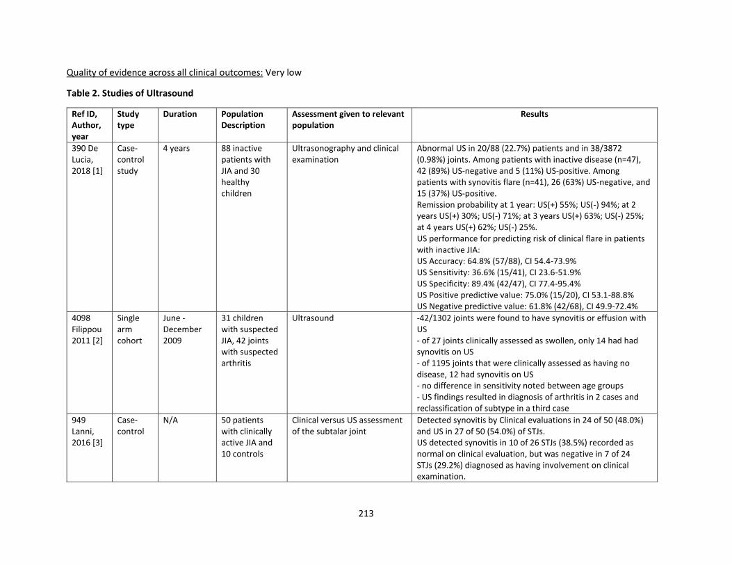
213
Quality of evidence across all clinical outcomes: Very low
Table 2. Studies of Ultrasound
Ref ID, Author, year
Study type
Duration Population Description
Assessment given to relevant population
Results
390 De Lucia, 2018 [1]
Case-control study
4 years 88 inactive patients with JIA and 30 healthy children
Ultrasonography and clinical examination
Abnormal US in 20/88 (22.7%) patients and in 38/3872 (0.98%) joints. Among patients with inactive disease (n=47), 42 (89%) US-negative and 5 (11%) US-positive. Among patients with synovitis flare (n=41), 26 (63%) US-negative, and 15 (37%) US-positive. Remission probability at 1 year: US(+) 55%; US(-) 94%; at 2 years US(+) 30%; US(-) 71%; at 3 years US(+) 63%; US(-) 25%; at 4 years US(+) 62%; US(-) 25%. US performance for predicting risk of clinical flare in patients with inactive JIA: US Accuracy: 64.8% (57/88), CI 54.4-73.9% US Sensitivity: 36.6% (15/41), CI 23.6-51.9% US Specificity: 89.4% (42/47), CI 77.4-95.4% US Positive predictive value: 75.0% (15/20), CI 53.1-88.8% US Negative predictive value: 61.8% (42/68), CI 49.9-72.4%
4098 Filippou 2011 [2]
Single arm cohort
June - December 2009
31 children with suspected JIA, 42 joints with suspected arthritis
Ultrasound -42/1302 joints were found to have synovitis or effusion with US - of 27 joints clinically assessed as swollen, only 14 had had synovitis on US - of 1195 joints that were clinically assessed as having no disease, 12 had synovitis on US - no difference in sensitivity noted between age groups - US findings resulted in diagnosis of arthritis in 2 cases and reclassification of subtype in a third case
949 Lanni, 2016 [3]
Case-control
N/A 50 patients with clinically active JIA and 10 controls
Clinical versus US assessment of the subtalar joint
Detected synovitis by Clinical evaluations in 24 of 50 (48.0%) and US in 27 of 50 (54.0%) of STJs. US detected synovitis in 10 of 26 STJs (38.5%) recorded as normal on clinical evaluation, but was negative in 7 of 24 STJs (29.2%) diagnosed as having involvement on clinical examination.
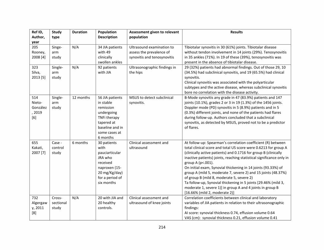
214
Ref ID, Author, year
Study type
Duration Population Description
Assessment given to relevant population
Results
205 Rooney, 2008 [4]
Singe-arm study
N/A 34 JIA patients with 49 clinically swollen ankles
Ultrasound examination to assess the prevalence of synovitis and tenosynovitis
Tibiotalar synovitis in 30 (61%) joints. Tibiotalar disease without tendon involvement in 14 joints (29%). Tenosynovitis in 35 ankles (71%). In 19 of these (39%), tenosynovitis was present in the absence of tibiotalar disease.
323 Silva, 2013 [5]
Single-arm study
N/A 92 patients with JIA
Ultrasonographic findings in the hips
29 (32%) patients had abnormal findings. Out of those 29, 10 (34.5%) had subclinical synovitis, and 19 (65.5%) had clinical synovitis. Clinical synovitis was associated with the polyarticular subtypes and the active disease, whereas subclinical synovitis bore no correlation with the disease activity.
514 Nieto-González, 2019 [6]
Single-arm study
12 months 56 JIA patients in stable remission undergoing TNFi therapy tapered at baseline and in some cases at 6 months
MSUS to detect subclinical synovitis.
B-Mode synovitis any grade in 47 (83.9%) patients and 147 joints (10.1%), grades 2 or 3 in 19 (1.3%) of the 1456 joints. Doppler mode (PD) synovitis in 5 (8.9%) patients and in 5 (0.3%) different joints, and none of the patients had flares during follow-up. Authors concluded that a subclinical synovitis, as detected by MSUS, proved not to be a predictor of flares.
655 Kakati, 2007 [7]
Case -control study
6 months 30 patients with pauciarticular JRA who received naproxen (15-20 mg/Kg/day) for a period of six months
Clinical assessment and ultrasound
At follow-up: Spearman’s correlation coefficient (R) between total clinical score and total US score were 0.6213 for group A (clinically active patients) and 0.1716 for group B (clinically inactive patients) joints, reaching statistical significance only in group A (p<.001). On initial exam, Synovial thickening in 14 joints (93.33%) of group A (mild 5, moderate 7, severe 2) and 15 joints (48.37%) of group B (mild 8, moderate 5, severe 2) Ta follow-up, Synovial thickening in 5 joints [29.46% (mild 3, moderate 1, severe 1)] in group A and 4 joints in group B [16.66% (mild 2, moderate 2)]
732 Algergawy, 2011 [8]
Cross-sectional study
N/A 20 with JIA and 20 healthy controls.
Clinical assessment and ultrasound of knee joints
Correlation coefficients between clinical and laboratory variables of JIA patients in relation to their ultrasonographic findings: AI score: synovial thickness 0.74, effusion volume 0.64 VAS (cm): synovial thickness 0.21, effusion volume 0.41
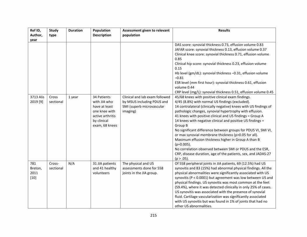
215
Ref ID, Author, year
Study type
Duration Population Description
Assessment given to relevant population
Results
DAS score: synovial thickness 0.73, effusion volume 0.83 JAFAR score: synovial thickness 0.13, effusion volume 0.37 Clinical knee score: synovial thickness 0.71, effusion volume 0.85 Clinical hip score: synovial thickness 0.23, effusion volume 0.15 Hb level (gm/dL): synovial thickness −0.31, effusion volume −0.81 ESR level (mm first hour): synovial thickness 0.61, effusion volume 0.44 CRP level (mg/L): synovial thickness 0.51, effusion volume 0.45
3713 Alis 2019 [9]
Cross sectional
1 year 34 Patients with JIA who have at least one knee with active arthritis by clinical exam, 68 knees
Clinical and lab exam followed by MSUS including PDUS and SMI (superb microvascular imaging)
45/68 knees with positive clinical exam findings. 4/45 (8.8%) with normal US findings (excluded). 14 contralateral (clinically negative) knees with US findings of pathologic changes, synovial hypertrophy with effusion. 41 knees with positive clinical and US findings = Group A 14 knees with negative clinical and positive US findings = Group B No significant difference between groups for PDUS VI, SMI VI, or max synovial membrane thickness (p>0.05 for all). Maximum effusion thickness higher in Group A than B (p=0.005). No correlation observed between SMI or PDUS and the ESR, CRP, disease duration, age of the patients, sex, and JADAS‐27 (p > .05).
781 Breton, 2011 [10]
Cross-sectional
N/A 31 JIA patients and 41 healthy volunteers
The physical and US assessments done for 558 joints in the JIA group.
Of 558 peripheral joints in JIA patients, 69 (12.5%) had US synovitis and 83 (15%) had abnormal physical findings. All the physical abnormalities were significantly associated with US synovitis (P < 0.0001) but agreement was low between US and physical findings. US synovitis was most common at the feet (59.4%), where it was detected clinically in only 25% of cases. US synovitis was associated with the presence of synovial fluid. Cartilage vascularization was significantly associated with US synovitis but was found in 1% of joints that had no other US abnormalities.
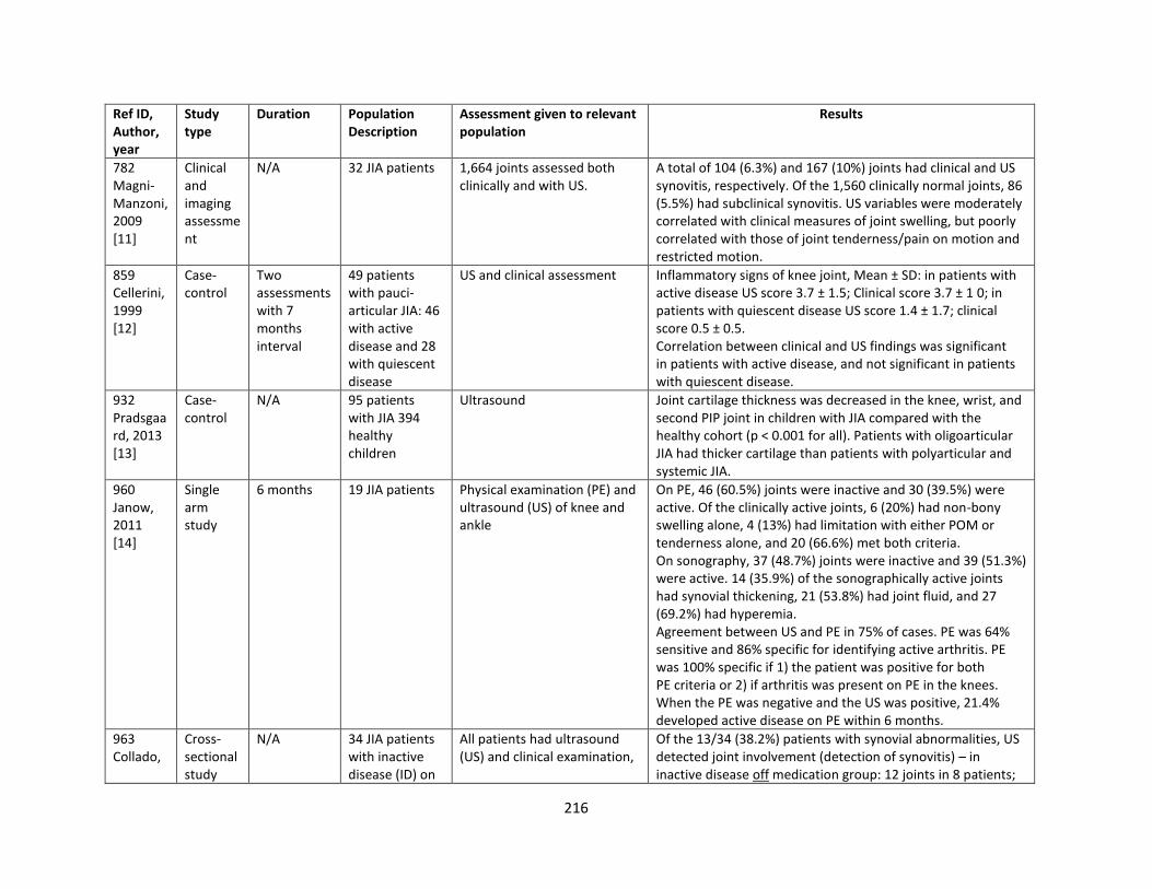
216
Ref ID, Author, year
Study type
Duration Population Description
Assessment given to relevant population
Results
782 Magni-Manzoni, 2009 [11]
Clinical and imaging assessment
N/A 32 JIA patients 1,664 joints assessed both clinically and with US.
A total of 104 (6.3%) and 167 (10%) joints had clinical and US synovitis, respectively. Of the 1,560 clinically normal joints, 86 (5.5%) had subclinical synovitis. US variables were moderately correlated with clinical measures of joint swelling, but poorly correlated with those of joint tenderness/pain on motion and restricted motion.
859 Cellerini, 1999 [12]
Case-control
Two assessments with 7 months interval
49 patients with pauci-articular JIA: 46 with active disease and 28 with quiescent disease
US and clinical assessment Inflammatory signs of knee joint, Mean ± SD: in patients with active disease US score 3.7 ± 1.5; Clinical score 3.7 ± 1 0; in patients with quiescent disease US score 1.4 ± 1.7; clinical score 0.5 ± 0.5. Correlation between clinical and US findings was significant in patients with active disease, and not significant in patients with quiescent disease.
932 Pradsgaard, 2013 [13]
Case-control
N/A 95 patients with JIA 394 healthy children
Ultrasound Joint cartilage thickness was decreased in the knee, wrist, and second PIP joint in children with JIA compared with the healthy cohort (p < 0.001 for all). Patients with oligoarticular JIA had thicker cartilage than patients with polyarticular and systemic JIA.
960 Janow, 2011 [14]
Single arm study
6 months 19 JIA patients Physical examination (PE) and ultrasound (US) of knee and ankle
On PE, 46 (60.5%) joints were inactive and 30 (39.5%) were active. Of the clinically active joints, 6 (20%) had non-bony swelling alone, 4 (13%) had limitation with either POM or tenderness alone, and 20 (66.6%) met both criteria. On sonography, 37 (48.7%) joints were inactive and 39 (51.3%) were active. 14 (35.9%) of the sonographically active joints had synovial thickening, 21 (53.8%) had joint fluid, and 27 (69.2%) had hyperemia. Agreement between US and PE in 75% of cases. PE was 64% sensitive and 86% specific for identifying active arthritis. PE was 100% specific if 1) the patient was positive for both PE criteria or 2) if arthritis was present on PE in the knees. When the PE was negative and the US was positive, 21.4% developed active disease on PE within 6 months.
963 Collado,
Cross-sectional study
N/A 34 JIA patients with inactive disease (ID) on
All patients had ultrasound (US) and clinical examination,
Of the 13/34 (38.2%) patients with synovial abnormalities, US detected joint involvement (detection of synovitis) – in inactive disease off medication group: 12 joints in 8 patients;
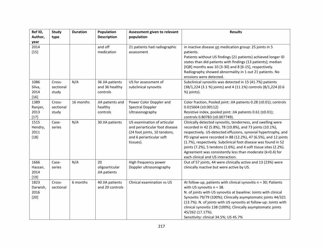
217
Ref ID, Author, year
Study type
Duration Population Description
Assessment given to relevant population
Results
2014 [15]
and off medication
21 patients had radiographic assessment
in inactive disease on medication group: 25 joints in 5 patients. Patients without US findings (21 patients) achieved longer ID states than did patients with findings (13 patients); median [IQR] months was 10 [3-30] and 8 [6-15], respectively. Radiography showed abnormality in 1 out 21 patients. No erosions were detected.
1086 Silva, 2014 [16]
Cross-sectional study
N/A 36 JIA patients and 36 healthy controls
US for assessment of subclinical synovitis
Subclinical synovitis was detected in 15 (41.7%) patients (38/1,224 (3.1 %) joints) and 4 (11.1%) controls (8/1,224 (0.6 %) joints).
1389 Ranjan, 2013 [17]
Cross-sectional study
16 months JIA patients and healthy controls
Power Color Doppler and Spectral Doppler Ultrasonography
Color fraction, Pooled joint: JIA patients 0.28 (±0.01); controls 0.015604 (±0.00112) Resistive index, pooled joint: JIA patients 0.61 (±0.01); controls 0.80783 (±0.007749).
1515 Hendry, 2011 [18]
Case-series
N/A 30 JIA patients US examination of articular and periarticular foot disease (24 foot joints, 10 tendons, and 6 periarticular soft tissues).
Clinically detected synovitis, tenderness, and swelling were recorded in 42 (5.8%), 78 (10.8%), and 73 joints (10.1%), respectively. US-detected effusions, synovial hypertrophy, and PD signal were recorded in 88 (12.2%), 47 (6.5%), and 12 joints (1.7%), respectively. Subclinical foot disease was found in 52 joints (7.2%), 5 tendons (1.6%), and 4 soft tissue sites (2.2%). Agreement was consistently less than moderate (k<0.4) for each clinical and US interaction.
1666 Hassan, 2014 [19]
Case-series
N/A 20 oligoarticular JIA patients
High frequency power Doppler ultrasonography
Out of 57 joints, 44 were clinically active and 13 (23%) were clinically inactive but were active by US.
1823 Darwish, 2016 [20]
Cross-sectional
6 months 40 JIA patients and 20 controls
Clinical examination vs US At follow-up, patients with clinical synovitis n = 30; Patients with US synovitis n = 38. N. of joints with US synovitis at baseline: Joints with clinical Synovitis 79/79 (100%); Clinically asymptomatic joints 44/321 (13.7%). N. of joints with US synovitis at follow-up: Joints with clinical synovitis 138 (100%); Clinically asymptomatic joints 45/262 (17.17%). Sensitivity: clinical 34.5%; US 45.7%
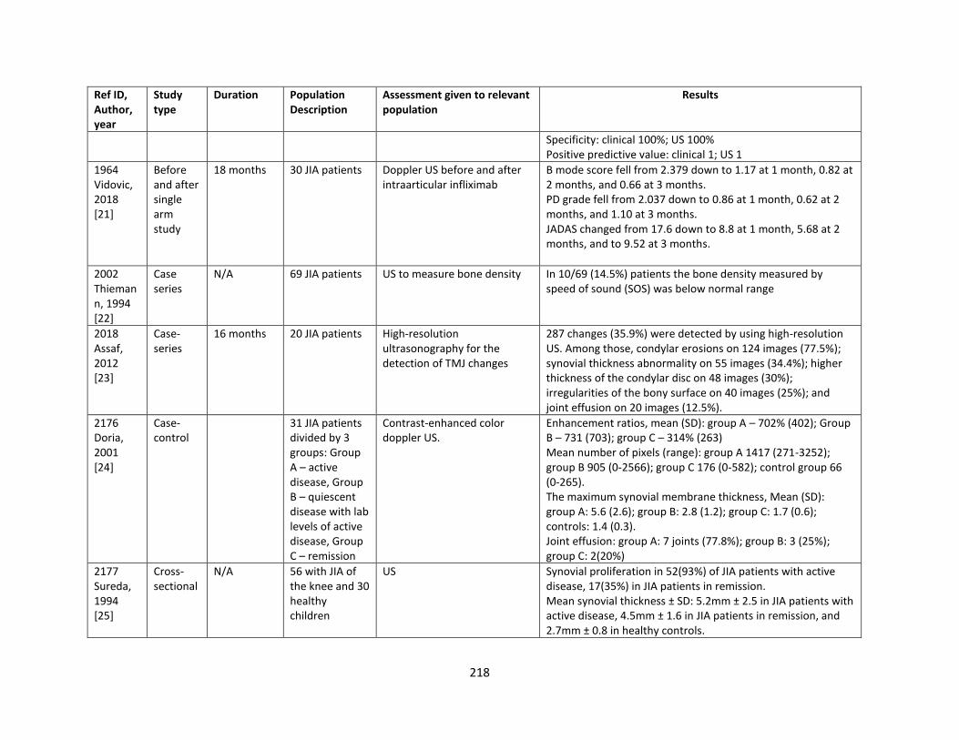
218
Ref ID, Author, year
Study type
Duration Population Description
Assessment given to relevant population
Results
Specificity: clinical 100%; US 100% Positive predictive value: clinical 1; US 1
1964 Vidovic, 2018 [21]
Before and after single arm study
18 months 30 JIA patients Doppler US before and after intraarticular infliximab
B mode score fell from 2.379 down to 1.17 at 1 month, 0.82 at 2 months, and 0.66 at 3 months. PD grade fell from 2.037 down to 0.86 at 1 month, 0.62 at 2 months, and 1.10 at 3 months. JADAS changed from 17.6 down to 8.8 at 1 month, 5.68 at 2 months, and to 9.52 at 3 months.
2002 Thiemann, 1994 [22]
Case series
N/A 69 JIA patients US to measure bone density In 10/69 (14.5%) patients the bone density measured by speed of sound (SOS) was below normal range
2018 Assaf, 2012 [23]
Case-series
16 months 20 JIA patients High-resolution ultrasonography for the detection of TMJ changes
287 changes (35.9%) were detected by using high-resolution US. Among those, condylar erosions on 124 images (77.5%); synovial thickness abnormality on 55 images (34.4%); higher thickness of the condylar disc on 48 images (30%); irregularities of the bony surface on 40 images (25%); and joint effusion on 20 images (12.5%).
2176 Doria, 2001 [24]
Case-control
31 JIA patients divided by 3 groups: Group A – active disease, Group B – quiescent disease with lab levels of active disease, Group C – remission
Contrast-enhanced color doppler US.
Enhancement ratios, mean (SD): group A – 702% (402); Group B – 731 (703); group C – 314% (263) Mean number of pixels (range): group A 1417 (271-3252); group B 905 (0-2566); group C 176 (0-582); control group 66 (0-265). The maximum synovial membrane thickness, Mean (SD): group A: 5.6 (2.6); group B: 2.8 (1.2); group C: 1.7 (0.6); controls: 1.4 (0.3). Joint effusion: group A: 7 joints (77.8%); group B: 3 (25%); group C: 2(20%)
2177 Sureda, 1994 [25]
Cross-sectional
N/A 56 with JIA of the knee and 30 healthy children
US Synovial proliferation in 52(93%) of JIA patients with active disease, 17(35%) in JIA patients in remission. Mean synovial thickness ± SD: 5.2mm ± 2.5 in JIA patients with active disease, 4.5mm ± 1.6 in JIA patients in remission, and 2.7mm ± 0.8 in healthy controls.
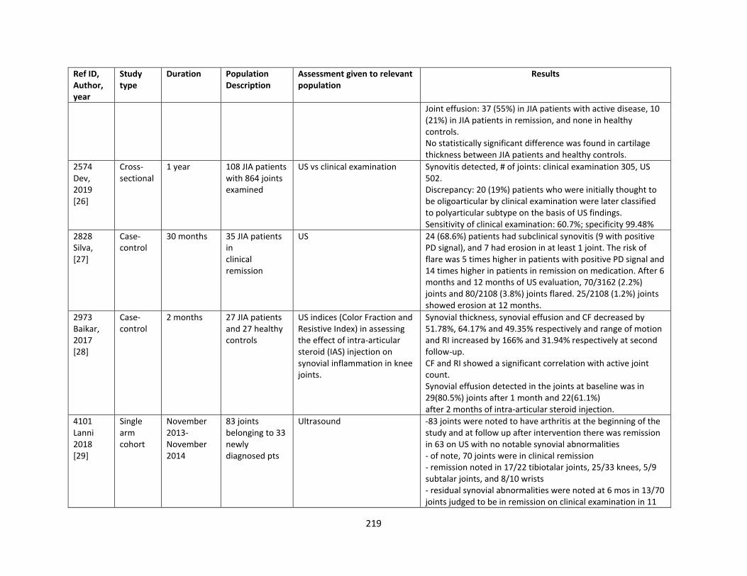
219
Ref ID, Author, year
Study type
Duration Population Description
Assessment given to relevant population
Results
Joint effusion: 37 (55%) in JIA patients with active disease, 10 (21%) in JIA patients in remission, and none in healthy controls. No statistically significant difference was found in cartilage thickness between JIA patients and healthy controls.
2574 Dev, 2019 [26]
Cross-sectional
1 year 108 JIA patients with 864 joints examined
US vs clinical examination Synovitis detected, # of joints: clinical examination 305, US 502. Discrepancy: 20 (19%) patients who were initially thought to be oligoarticular by clinical examination were later classified to polyarticular subtype on the basis of US findings. Sensitivity of clinical examination: 60.7%; specificity 99.48%
2828 Silva, [27]
Case-control
30 months 35 JIA patients in clinical remission
US 24 (68.6%) patients had subclinical synovitis (9 with positive PD signal), and 7 had erosion in at least 1 joint. The risk of flare was 5 times higher in patients with positive PD signal and 14 times higher in patients in remission on medication. After 6 months and 12 months of US evaluation, 70/3162 (2.2%) joints and 80/2108 (3.8%) joints flared. 25/2108 (1.2%) joints showed erosion at 12 months.
2973 Baikar, 2017 [28]
Case-control
2 months 27 JIA patients and 27 healthy controls
US indices (Color Fraction and Resistive Index) in assessing the effect of intra-articular steroid (IAS) injection on synovial inflammation in knee joints.
Synovial thickness, synovial effusion and CF decreased by 51.78%, 64.17% and 49.35% respectively and range of motion and RI increased by 166% and 31.94% respectively at second follow-up. CF and RI showed a significant correlation with active joint count. Synovial effusion detected in the joints at baseline was in 29(80.5%) joints after 1 month and 22(61.1%) after 2 months of intra-articular steroid injection.
4101 Lanni 2018 [29]
Single arm cohort
November 2013- November 2014
83 joints belonging to 33 newly diagnosed pts
Ultrasound -83 joints were noted to have arthritis at the beginning of the study and at follow up after intervention there was remission in 63 on US with no notable synovial abnormalities - of note, 70 joints were in clinical remission - remission noted in 17/22 tibiotalar joints, 25/33 knees, 5/9 subtalar joints, and 8/10 wrists - residual synovial abnormalities were noted at 6 mos in 13/70 joints judged to be in remission on clinical examination in 11
220
Ref ID, Author, year
Study type
Duration Population Description
Assessment given to relevant population
Results
patients - joint effusion noted in the knee in 32/33 (97%) pts at baseline, 19/22 (86.4) tibiotalar joints, 7/9 (77.8) subtalar joints, 6/10 (60) wrists, and 7/9 (77.8) elbows - synovial hypertrophy noted baseline in 32/33 (97%) knees, 21/22 (95.5) tibiotalar joints, 9/9 (100) subtalar joints, 9/10 (90) wrists, and 9/9 (100) elbows - power doppler abnormalities noted at baseline in 22/32 (66.7%) knees, 14/22 (63.6) tibiotalar joints, 8/9 (88.9) subtalar joints, 8/10 (80) wrists, and 5/9 (55.6%) elbows - at follow up there were joint effusions noted in 6/33 (18.2%) knees, 2/22 (9.1) tibiotalar joints, 1/9 (11.1) subtalar joints, no wrists, and 1/9 (11.1) elbows - synovial hypertrophy was noted at follow up in 5/32 (15.2%) of wrists, 4/22 (18.2) tibiotalar joints, 4/9 (44.4) subtalar joints, 2/10 (20) wrists, and 1/9 (11.1) elbows - power doppler abnormalities at follow up were present in 1/33 (3%) of knees, 0 tibiotalar joints, 2/9 (22.2) subtalar joints, 0 wrists and 0 elbows - decrease in grey scale US (GSUS) score and in power doppler US (PDUS) score in 16/20 (80%) - median GSUS decreased from 2.0 to 0, p<0.001 - median PDUS diminished from 2.0 to 0, p<0.001 - 21/33 (63.6%) of pts were ACR90 responders, but 6 (28.6) did not display complete resolution of synovial abnormalities -17/3 pts were in clinical remission by cJADAS-10 at follow up, but in 7 joints of 5 pts there was persistence of GSUS abnormalities
4102 Mitra 2019 [30]
Cross sectional study
June 2013-May 2014
27 JIA pts; poly n = 17, oligo n = 8, ERA n = 1, SoJIA n = 1,
Ultrasound -median cartilage thickness with (IQR) in the wrist, knee and ankle joints of the cases (n = 27) were compared re‐ spectively with the control group (n = 54) by Mann‐Whitney U test; statistically significant reduction in joint cartilage thickness in all the three joints (P < 0.01 in wrist, knee and ankle joints) - affected joints (n = 130) compared with unaffected joints (n = 32) of JIA patients without any previous history of arthritis
221
Ref ID, Author, year
Study type
Duration Population Description
Assessment given to relevant population
Results
by paired t test; mean cartilage thickness in the affected wrist (n = 44), knee (n = 42) and ankle (n = 44) joints were significantly decreased (P < 0.05) in comparison to the unaffected joints -significant difference in median cartilage thickness of the knee joint between oligoarticular versus polyarticular subtypes of JIA (right knee P = 0.028, left knee P < 0.01) except in wrist and ankle joints where no statistically significant difference in median cartilage thickness was elucidated (P > 0.05) -oligo and poly were associated with decreased median cartilage thickness in all the three joints when compared with the age‐ and sex‐matched healthy cohort (P < 0.05) - mean cartilage thicknesses of wrist, knee and ankle joints were less in boys than in girls suffering from JIA, although statistically significant thinning was found only in knee joints (P = 0.01) - no statistically significant difference between right and left sides of wrist, knee and ankle joints in cases by Wilcoxon's matched paired signed rank test (P = 0.02) - negative Spearman's coefficient of rank correlation of joint cartilage width with body weight in diseased children and it was statistically significant in the wrist, knee and ankle joints (P < 0.05)
3442 El-Banna 2019 [31]
Single Arm Cohort
Cross sectional
40 children with (30) oJIA or (10) pJIA
20 joints in each patient (knees, wrists, 2-5 MCP, 2-5 PIP)
Discrepancy between clinically active and US active (p<0.001); 70 (8.8%) clinically inactive joints were active by MSUS, 22 (2.8%) clinically active joints were inactive by MSUS, 170 (21.3%) clinically and MSUS active.
3444 Shanmugavel 2008 [32]
Single Arm Cohort
6 months 30 children with JIA and knee involvement: Group A – active
Bilateral knees on same day as clinical assessment, end of 2nd month, end of 6th month
End of month 6: Group A – 1 active joint clinically, 1 effusion US, 16 synovial thickening, 16 PDS hyperemia. Group B – 0 active joints clinically, 1 effusion US, 0 synovial thickening, 3 PDS hyperemia. All clinically assessed effusions detected on US.
222
Ref ID, Author, year
Study type
Duration Population Description
Assessment given to relevant population
Results
Group B – inactive by clinical assessment (score)
3633 Melchiorre 2010 [33]
Single Arm Cohort
68 children with JIA, never receiving treatment
US of bilateral TMJ in static and dynamic phase
46/68 (68%) with joint effusion (16 bilateral, 35%). 2/46 (4.3%) were symptomatic 62/68 (91.2%) with condylar remodeling (unilateral in 17, bilateral in 37). 18/124 (14.5%) TMJs with cortical “break” – erosion. 14/124 (11.3%) with osteophytes No disk alterations. No correlation with ESR, CRP, or ANA.
3634 Jank 2007 [34]
Single Arm Cohort
48 patients with JIA
US TMJ in closed and maximal open-mouthed position
53 (55.2%) patients with destructive changes. 41 (42.7%) disk dislocation in closed mouth position, not present in open-mouth position in 17 (17.7%). Correlation with >5 peripheral joints and TMJ destruction, p=0.002 No correlation with peripheral joint count and disc-dislocation in closed mouth position, p=0.144. Correlation with peripheral joint count and disc dislocation in max open mouth position, p=0.021. Duration of JIA >23 months associated with disc dislocation and TMJ destruction, p<0.0001 for both. Duration of JIA >60 months associated with TMJ destruction, p<0.0001, but not disc dislocation in open or closed position, p=0.070 and p=0.059.
962 Haslam 2009 [Error! Reference source not found.]
Cross sectional
17 children with oJIA <12 months and DMARD/systemic steroid naive (40 joints each)
Clinical history, physical exam, US
23/680 joints with clinical synovitis, 17 of these with US synovitis. 15/657 joints with US detected synovitis, clinically inactive. Subclinical disease was detected by US in 6/17 children assessed. US changed the joint count in 9/17 of patients assessed, with 5/17 having an increased joint count on US assessment.
223
Ref ID, Author, year
Study type
Duration Population Description
Assessment given to relevant population
Results
3429 Lofty 2018 [36]
Single arm cohort
3 years 40 patients with JIA
Clinical and lab assessment at enrollment, gray scale US and power doppler US for 28 joints
On clinical examination, 182 joints (16.3%) were swollen, 139 joints (12.4%) were tender, and 196 joints (17.5%) had clinical synovitis in the form of (swelling and/or tenderness). On US evaluation, 210 joints (18.8% of the total number of examined joints) were sonographically affected, including 192 joints (17.1% of total) with synovial hyperplasia, and 39 joints (3.5% of total) with joint effusion 142 joints (12.7% of total number of examined joints) had power Doppler (PD)signals. US of the clinically asymptomatic joints (82.5% of total number examined) had US signs of synovitis in 32 joints (3.5% of the clinically normal joints). 18 joints with clinical synovitis were normal on US. US positive predictive value is 84.7% & negative predictive value is 98%. US documented Power Doppler signals in (68.4%) of joints with clinical synovitis, (69.2%) of swollen joint, and (86.3%) of tender joints. kappa values for agreement between swollen joints and US =0.84 for swelling versus synovial hyperplasia, 0.74 for swelling versus power Doppler US, and 0.39 for swelling versus joint effusion. ROC curve analysis of PDUS score<2 versus mild disease activity (DAS28-CRP=2.3–2.69) = 100% sensitivity and 75% specificity. ROC curve analysis of synovial hyperplasia score <3 versus mild disease activity (DAS28-CRP=2.3–2.69) = 87.6% sensitivity and 65.6% specificity. ROC curve analysis of total US score <3 versus mild disease activity (DAS28-CRP=2.3–2.69) = 75% sensitivity and 90.6% specificity.
3445 Sparchez 2010 [37]
Single Arm Cohort
1 year 32 patients with JIA
Clinical and lab evaluation, US performed on all clinically active joints (current or prior) with PDUS
2 clinically inactive joints with US synovitis. Compared to clinical exam: Sensitivity of PDUS 0.904 (95% CI 0.7-0.98), specificity 0.895 (0.67-0.98), PPV 0.90 (0.67-0.98), NPV 0.89 (0.67-0.98)
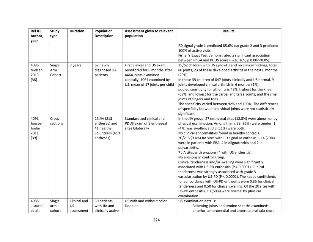
224
Ref ID, Author, year
Study type
Duration Population Description
Assessment given to relevant population
Results
PD signal grade 1 predicted 85.6% but grade 2 and 3 predicted 100% of active visits. Fisher’s Exact Test demonstrated a significant association between PhGA and PDUS score (F=26.169, p 0.00<<0.05).
4086 Nielsen 2013 [38]
Single Arm Cohort
7 years 62 newly diagnosed JIA patients
First clinical and US exam, monitored for 6 months after 4464 joints examined clinically, 1064 examined by US, mean of 17 joints per child
35/62 children with US synovitis and no clinical findings, total 80 joints, 23 of these developed arthritis in the next 6 months (29%). In these 35 children of 847 joints clinically and US normal, 9 joints developed clinical arthritis in 6 months (1%). pooled sensitivity for all joints is 48%, highest for the knee (69%) and lowest for the carpal and tarsal joints, and the small joints of fingers and toes The specificity varied between 92% and 100%. The differences of specificity between individual joints were not statistically significant.
4091 Jousse-Joulin 2011 [39]
Cross sectional
26 JIA (213 entheses) and 41 healthy volunteers (410 entheses)
Standardized clinical and PDUS exam of 5 entheseal sites bilaterally
In the JIA group, 27 entheseal sites (12.5%) were abnormal by physical examination. Among them, 23 (85%) were tender, 1 (4%) was swollen, and 3 (11%) were both. No clinical abnormalities found in healthy controls. 20/213 (9.4%) JIA sites with PD signal at enthesis -- 14 (70%) were in patients with ERA, 4 in oligoarthritis and 2 in polyarthritis. 7 JIA sites with erosions (4 with US enthesitis). No erosions in control group. Clinical tenderness and/or swelling were significantly associated with US-PD enthesitis (P < 0.0001). Clinical tenderness was strongly associated with grade 3 vascularization by US-PD (P < 0.0001). The kappa coefficients for concordance with US-PD enthesitis were 0.35 for clinical tenderness and 0.50 for clinical swelling. Of the 20 sites with US-PD enthesitis, 10 (50%) were normal by physical examination.
4088 , Laurell et al.,
Single arm cohort
Clinical and US assessment
30 patients with JIA and clinically active
US with and without color Doppler
US examination details: - Following joints and tendon sheaths examined:
anterior, anteromedial and anterolateral talo-crural
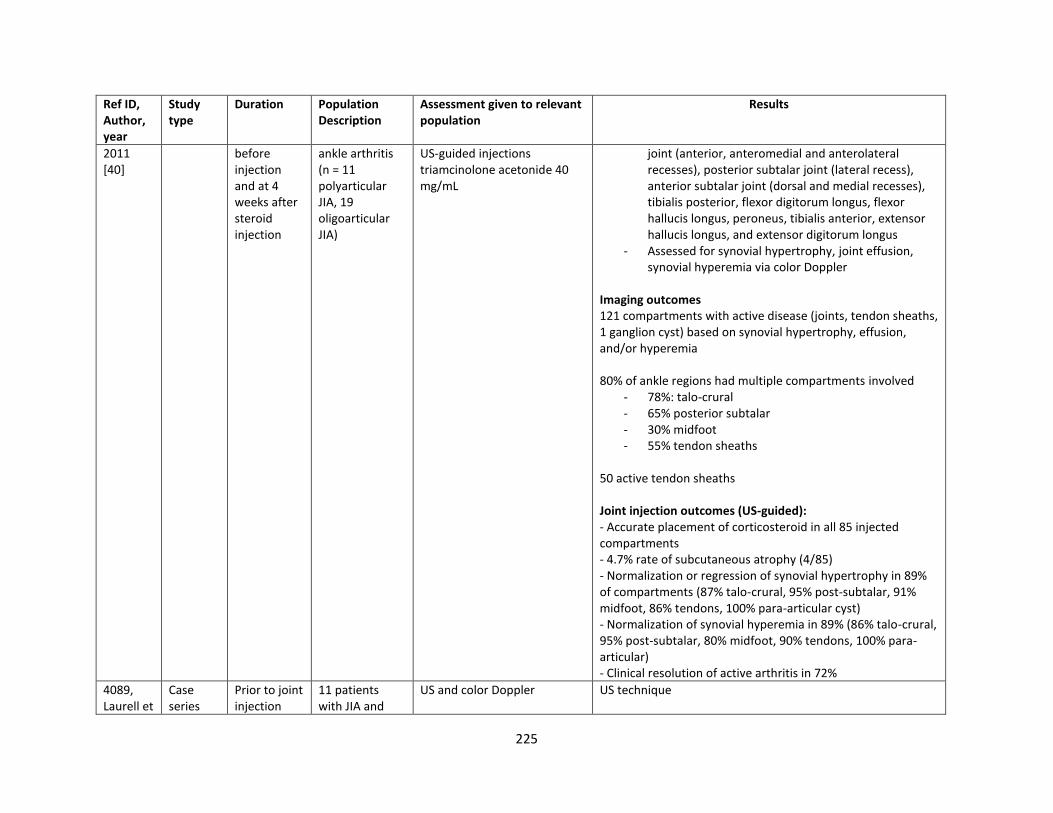
225
Ref ID, Author, year
Study type
Duration Population Description
Assessment given to relevant population
Results
2011 [40]
before injection and at 4 weeks after steroid injection
ankle arthritis (n = 11 polyarticular JIA, 19 oligoarticular JIA)
US-guided injections triamcinolone acetonide 40 mg/mL
joint (anterior, anteromedial and anterolateral recesses), posterior subtalar joint (lateral recess), anterior subtalar joint (dorsal and medial recesses), tibialis posterior, flexor digitorum longus, flexor hallucis longus, peroneus, tibialis anterior, extensor hallucis longus, and extensor digitorum longus
- Assessed for synovial hypertrophy, joint effusion, synovial hyperemia via color Doppler
Imaging outcomes 121 compartments with active disease (joints, tendon sheaths, 1 ganglion cyst) based on synovial hypertrophy, effusion, and/or hyperemia 80% of ankle regions had multiple compartments involved
- 78%: talo-crural - 65% posterior subtalar - 30% midfoot - 55% tendon sheaths
50 active tendon sheaths Joint injection outcomes (US-guided): - Accurate placement of corticosteroid in all 85 injected compartments - 4.7% rate of subcutaneous atrophy (4/85) - Normalization or regression of synovial hypertrophy in 89% of compartments (87% talo-crural, 95% post-subtalar, 91% midfoot, 86% tendons, 100% para-articular cyst) - Normalization of synovial hyperemia in 89% (86% talo-crural, 95% post-subtalar, 80% midfoot, 90% tendons, 100% para-articular) - Clinical resolution of active arthritis in 72%
4089, Laurell et
Case series
Prior to joint injection
11 patients with JIA and
US and color Doppler
US technique
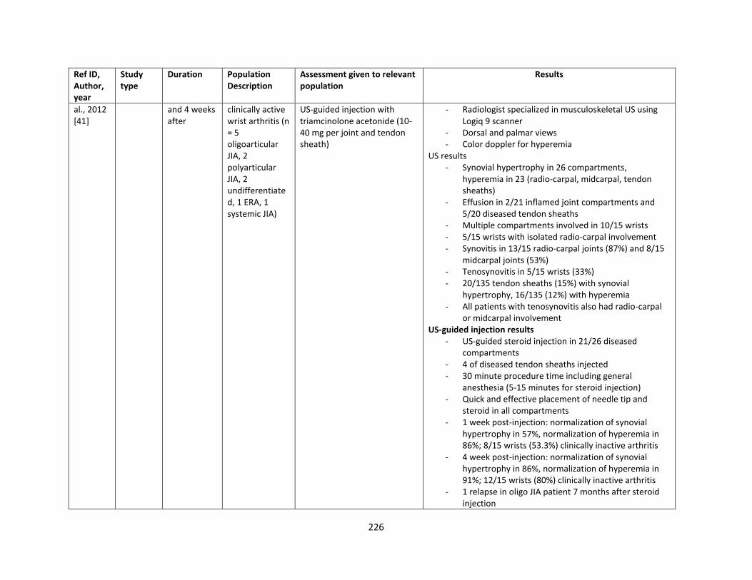
226
Ref ID, Author, year
Study type
Duration Population Description
Assessment given to relevant population
Results
al., 2012 [41]
and 4 weeks after
clinically active wrist arthritis (n = 5 oligoarticular JIA, 2 polyarticular JIA, 2 undifferentiated, 1 ERA, 1 systemic JIA)
US-guided injection with triamcinolone acetonide (10-40 mg per joint and tendon sheath)
- Radiologist specialized in musculoskeletal US using Logiq 9 scanner
- Dorsal and palmar views - Color doppler for hyperemia
US results - Synovial hypertrophy in 26 compartments,
hyperemia in 23 (radio-carpal, midcarpal, tendon sheaths)
- Effusion in 2/21 inflamed joint compartments and 5/20 diseased tendon sheaths
- Multiple compartments involved in 10/15 wrists - 5/15 wrists with isolated radio-carpal involvement - Synovitis in 13/15 radio-carpal joints (87%) and 8/15
midcarpal joints (53%) - Tenosynovitis in 5/15 wrists (33%) - 20/135 tendon sheaths (15%) with synovial
hypertrophy, 16/135 (12%) with hyperemia - All patients with tenosynovitis also had radio-carpal
or midcarpal involvement US-guided injection results
- US-guided steroid injection in 21/26 diseased compartments
- 4 of diseased tendon sheaths injected - 30 minute procedure time including general
anesthesia (5-15 minutes for steroid injection) - Quick and effective placement of needle tip and
steroid in all compartments - 1 week post-injection: normalization of synovial
hypertrophy in 57%, normalization of hyperemia in 86%; 8/15 wrists (53.3%) clinically inactive arthritis
- 4 week post-injection: normalization of synovial hypertrophy in 86%, normalization of hyperemia in 91%; 12/15 wrists (80%) clinically inactive arthritis
- 1 relapse in oligo JIA patient 7 months after steroid injection
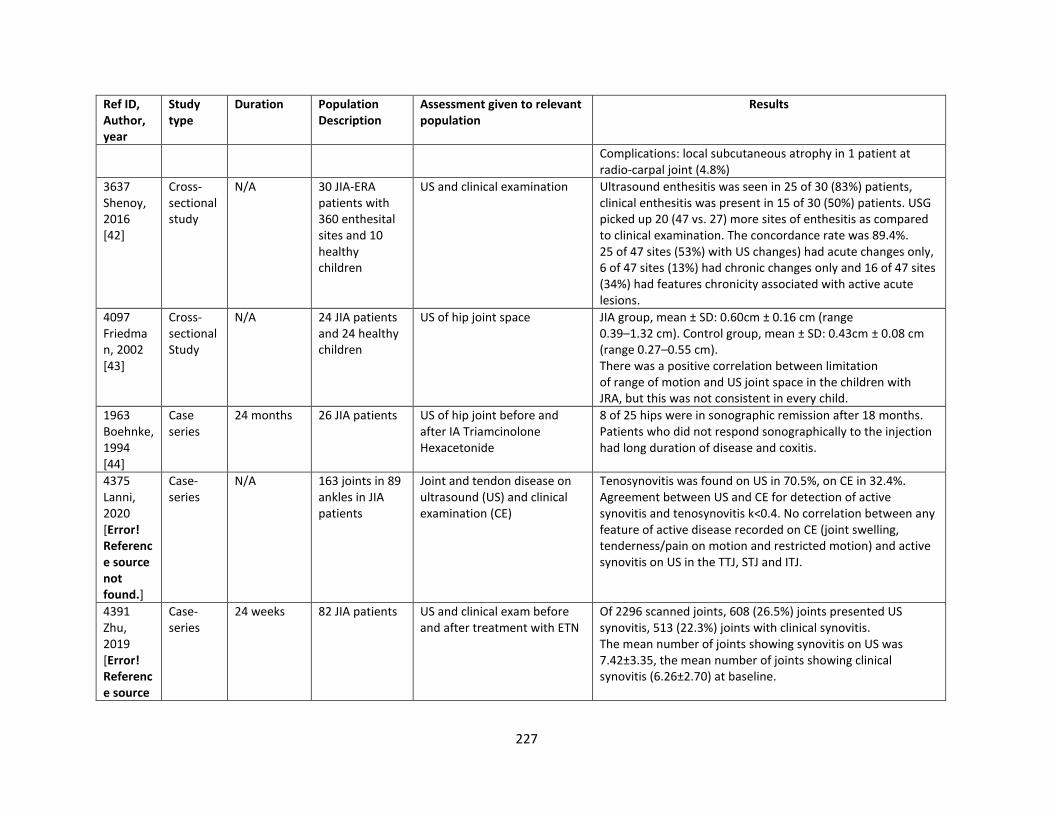
227
Ref ID, Author, year
Study type
Duration Population Description
Assessment given to relevant population
Results
Complications: local subcutaneous atrophy in 1 patient at radio-carpal joint (4.8%)
3637 Shenoy, 2016 [42]
Cross- sectional study
N/A 30 JIA-ERA patients with 360 enthesital sites and 10 healthy children
US and clinical examination Ultrasound enthesitis was seen in 25 of 30 (83%) patients, clinical enthesitis was present in 15 of 30 (50%) patients. USG picked up 20 (47 vs. 27) more sites of enthesitis as compared to clinical examination. The concordance rate was 89.4%. 25 of 47 sites (53%) with US changes) had acute changes only, 6 of 47 sites (13%) had chronic changes only and 16 of 47 sites (34%) had features chronicity associated with active acute lesions.
4097 Friedman, 2002 [43]
Cross-sectional Study
N/A 24 JIA patients and 24 healthy children
US of hip joint space JIA group, mean ± SD: 0.60cm ± 0.16 cm (range 0.39–1.32 cm). Control group, mean ± SD: 0.43cm ± 0.08 cm (range 0.27–0.55 cm). There was a positive correlation between limitation of range of motion and US joint space in the children with JRA, but this was not consistent in every child.
1963 Boehnke, 1994 [44]
Case series
24 months 26 JIA patients US of hip joint before and after IA Triamcinolone Hexacetonide
8 of 25 hips were in sonographic remission after 18 months. Patients who did not respond sonographically to the injection had long duration of disease and coxitis.
4375 Lanni, 2020 [Error! Reference source not found.]
Case-series
N/A 163 joints in 89 ankles in JIA patients
Joint and tendon disease on ultrasound (US) and clinical examination (CE)
Tenosynovitis was found on US in 70.5%, on CE in 32.4%. Agreement between US and CE for detection of active synovitis and tenosynovitis k<0.4. No correlation between any feature of active disease recorded on CE (joint swelling, tenderness/pain on motion and restricted motion) and active synovitis on US in the TTJ, STJ and ITJ.
4391 Zhu, 2019 [Error! Reference source
Case-series
24 weeks 82 JIA patients US and clinical exam before and after treatment with ETN
Of 2296 scanned joints, 608 (26.5%) joints presented US synovitis, 513 (22.3%) joints with clinical synovitis. The mean number of joints showing synovitis on US was 7.42±3.35, the mean number of joints showing clinical synovitis (6.26±2.70) at baseline.
228
Ref ID, Author, year
Study type
Duration Population Description
Assessment given to relevant population
Results
not found.]
The number of joints showing synovitis on US at baseline was positively correlated with CRP, ESR, number of joints with active disease. No correlation with age (P=0.929), gender (P=0.204), height (P=0.874), weight (P=0.806), or disease duration (P=0.664) was observed. US synovitis correlated with increased disease activity in JIA patients. The number of joints showing synovitis on US at baseline was greater in response patients compared to non-response patients, suggesting that US synovitis had potential to predict clinical response to ETN.
References:
1. Lucia, O. D., et al. (2018). "Baseline ultrasound examination as possible predictor of relapse in patients affected by juvenile idiopathic arthritis (JIA)." Ann Rheum Dis 77(10): 1426-1431.
2. Filippou, G., et al. (2011). "Ultrasonography vs. clinical examination in children with suspected arthritis. Does it make sense to use poliarticular ultrasonographic screening?" Clin Exp Rheumatol 29(2): 345-350.
3. Lanni, S., et al. (2016). "Delineating the Application of Ultrasound in Detecting Synovial Abnormalities of the Subtalar Joint in Juvenile Idiopathic Arthritis." Arthritis Care Res (Hoboken) 68(9): 1346-1353.
4. Rooney, M. E., et al. (2009). "Ankle disease in juvenile idiopathic arthritis: ultrasound findings in clinically swollen ankles." J Rheumatol 36(8): 1725-1729.
5. Silva, V. B., et al. (2013). "Association between the ultrasonographic and clinical findings in the hips of patients with juvenile idiopathic
arthritis." Rev Bras Reumatol 53(4): 322-327.
6. Nieto-Gonzalez, J. C., et al. (2019). "Can ultrasound-detected subclinical synovitis be an indicator of flare recurrence in juvenile idiopathic arthritis remission patients on tapered TNFi?" Clin Exp Rheumatol 37(4): 705-712.
7. Kakati, P., et al. (2007). "Clinical and ultrasound assessment of the knee in children with juvenile rheumatoid arthritis." Indian J Pediatr 74(9): 831-836.
8. Algergawy, S., et al. (2011). "Clinical, laboratory, and ultrasound assessment of the knee in juvenile rheumatoid arthritis." Clin Med
Insights Arthritis Musculoskelet Disord 4: 21-27.
9. Alis, D., et al. (2019). "Superb Microvascular Imaging Compared With Power Doppler Ultrasound in Assessing Synovitis of the Knee in Juvenile Idiopathic Arthritis: A Preliminary Study." J Ultrasound Med.
10. Breton, S., et al. (2011). "Comparison of clinical and ultrasonographic evaluations for peripheral synovitis in juvenile idiopathic arthritis."
229
Semin Arthritis Rheum 41(2): 272-278. 11. Magni-Manzoni, S., et al. (2009). "Comparison of clinical versus ultrasound-determined synovitis in juvenile idiopathic arthritis." Arthritis
Rheum 61(11): 1497-1504. 12. Cellerini, M., et al. (1999). "Correlation between clinical and ultrasound assessment of the knee in children with mono-articular or pauci-
articular juvenile rheumatoid arthritis." Pediatr Radiol 29(2): 117-123. 13. Pradsgaard, D. O., et al. (2013). "Decreased cartilage thickness in juvenile idiopathic arthritis assessed by ultrasonography." J Rheumatol
40(9): 1596-1603. 14. Janow, G. L., et al. (2011). "Detection of active disease in juvenile idiopathic arthritis: sensitivity and specificity of the physical
examination vs ultrasound." J Rheumatol 38(12): 2671-2674. 15. Collado, P., et al. (2014). "Detection of synovitis by ultrasonography in clinically inactive juvenile idiopathic arthritis on and off
medication." Clin Exp Rheumatol 32(4): 597-603.
16. Silva, V. B. M. e., et al. (2013). "Do patients with juvenile idiopathic arthritis in remission exhibit active synovitis on joint ultrasound?" Rheumatol Int 34(7): 937-945.
17. Ranjan, S., et al. (2013). "Evaluation of synovial inflammation in juvenile idiopathic arthritis by power color Doppler and Spectral Doppler
ultrasonography." Indian J Pediatr 81(1): 29-35.
18. Hendry, G. J., et al. (2011). "Frequent discordance between clinical and musculoskeletal ultrasound examinations of foot disease in juvenile idiopathic arthritis." Arthritis Care Res (Hoboken) 64(3): 441-447.
19. Hassan, H. A. E. S., et al. (2015). "High frequency power doppler ultrasonography in oligoarticular juvenile idiopathic arthritis: Correlation with disease severity." Egyptian Rheumatologist 37(3): 125-131.
20. Darwish, A. F., et al. (2016). "Implementation of musculoskeletal ultrasonography in detection of early juvenile idiopathic arthritis." Eur J Radiol Open 3: 264-271.
21. Vidovic, M., et al. (2018). "Intraarticular infliximab therapy in patients with juvenile idiopathic arthritis: the role of musculoskeletal ultrasound and disease activity scores in monitoring therapy response." Clin Exp Rheumatol 36(4): 676-682.
22. Thiemann, H. H., et al. (1994). "Investigations of bone density in patients with juvenile chronic arthritis by a sonographie method." Acta
Univ Carol Med (Praha) 40(1-4): 37-41.
23. Assaf, A. T., et al. (2013). "Is high-resolution ultrasonography suitable for the detection of temporomandibular joint involvement in
children with juvenile idiopathic arthritis?" Dentomaxillofac Radiol 42(3): 20110379.
24. Doria, A. S., et al. (2001). "Juvenile rheumatoid arthritis of the knee: evaluation with contrast-enhanced color Doppler ultrasound."
Pediatr Radiol 31(7): 524-531.
25. Sureda, D., et al. (1994). "Juvenile rheumatoid arthritis of the knee: evaluation with US." Radiology 190(2): 403-406. 26. Dev, S., et al. (2019). "Musculoskeletal Ultrasonography in Detecting Disease Activity in Patients of Juvenile Idiopathic Arthritis: A Cross-
Sectional Study." Indian Journal of Rheumatology 14(2): 104-108. 27. Miotto e Silva V. B., et al. (2017). "Patients with juvenile idiopathic arthritis in clinical remission with positive power Doppler signal in
joint ultrasonography have an increased rate of clinical flare: a prospective study." Pediatr Rheumatol Online J 15(1): 80.
230
28. Baikar, T., et al. (2017). "Power Color Doppler and Spectral Doppler Ultrasonography to Evaluate Response to Intra-articular Steroid Injection in Knee Joints in Juvenile Idiopathic Arthritis." Indian J Pediatr 84(11): 826-832.
29. Lanni, S., et al. (2017). "Ultrasound changes in synovial abnormalities induced by treatment in juvenile idiopathic arthritis." Clin Exp Rheumatol 36(2): 329-334.
30. Mitra, S., et al. (2019). "Ultrasound detected changes in joint cartilage thickness in juvenile idiopathic arthritis." Int J Rheum Dis 22(7): 1263-1270.
31. El-Banna, H. S., et al. (2019). "Role of musculoskeletal ultrasonography in the detection of subclinical synovitis in oligo and polyarticular juvenile idiopathic arthritis children." Egyptian Rheumatologist 41(2): 151-155.
32. Shanmugavel, C., et al. (2007). "Role of power Doppler sonography in evaluation of therapeutic response of the knee in juvenile
rheumatoid arthritis." Rheumatol Int 28(6): 573-578.
33. Melchiorre, D., et al. (2010). "Sonographic evaluation of the temporomandibular joints in juvenile idiopathic arthritis." J Ultrasound 13(1): 34-37.
34. Jank, S., et al. (2007). "Sonographic investigation of the temporomandibular joint in patients with juvenile idiopathic arthritis: a pilot study." Arthritis Rheum 57(2): 213-218.
35. Haslam, K. E., et al. (2009). "The detection of subclinical synovitis by ultrasound in oligoarticular juvenile idiopathic arthritis: a pilot study." Rheumatology (Oxford) 49(1): 123-127.
36. Lotfy, H. M., et al. (2018). "The role of Doppler Ultrasonography in evaluating disease activity in a group of juvenile idiopathic arthritis patients." Egyptian Journal of Radiology and Nuclear Medicine 49(4): 1036-1042.
37. Sparchez, M., et al. (2010). "The role of Power Doppler ultrasonography in comparison with biological markers in the evaluation of disease activity in Juvenile Idiopathic Arthritis." Med Ultrason 12(2): 97-103.
38. Nielsen, H. E., et al. (2013). "Ultrasonographic examination in juvenile idiopathic arthritis is better than clinical examination for identification of intraarticular disease." Dan Med J 60(8): A4669.
39. Jousse-Joulin, S., et al. (2011). "Ultrasonography for detecting enthesitis in juvenile idiopathic arthritis." Arthritis Care Res (Hoboken) 63(6): 849-855.
40. Laurell, L., et al. (2011). "Ultrasonography and color Doppler in juvenile idiopathic arthritis: diagnosis and follow-up of ultrasound-guided
steroid injection in the ankle region. A descriptive interventional study." Pediatr Rheumatol Online J 9(1): 4.
41. Laurell, L., et al. (2012). "Ultrasonography and color Doppler in juvenile idiopathic arthritis: diagnosis and follow-up of ultrasound-guided steroid injection in the wrist region. A descriptive interventional study." Pediatr Rheumatol Online J 10: 11.
42. Shenoy, S. and A. Aggarwal (2016). "Sonologic enthesitis in children with enthesitis-related arthritis." Clin Exp Rheumatol 34(1): 143-147. 43. Friedman, S. and M. A. Gruber (2002). "Ultrasonography of the hip in the evaluation of children with seronegative juvenile rheumatoid
arthritis." J Rheumatol 29(3): 629-632. 44. Boehnke, M., et al. (1994). "Intraarticular hip treatment with triamcinolonehexacetonide in juvenile chronic arthritis." Acta Univ Carol
Med (Praha) 40(1-4): 123-126. 45. Lanni, S., Marafon, D. P., Civino, A., Alongi, A., Proverbio, E., Agostoni, C., . . . Filocamo, G. Comparison between clinical and ultrasound
231
assessment of the ankle region in juvenile idiopathic arthritis. Arthritis Care Res (Hoboken), n/a(n/a). doi:10.1002/acr.24241. 46. Zhou, L., & Gu, X. (2019). Correlation of ultrasonography synovitis with disease activity and clinical response to etanercept treatment in
juvenile idiopathic arthritis patients. Brazilian Journal of Medical and Biological Research, 52. MRI: Fifty studies evaluated MRI for assessment of patients with JIA. In a study comparing MRI with clinical assessment, the clinical assessment
had a sensitivity of 25.7% and specificity of 91% for detecting MRI diagnosed arthritis; the agreement between MRI activity and clinical
assessment was fair in cases with arthritis less than 4 years from diagnosis (k score 0.38, P = 0.045); there was no agreement in longer standing
disease (k score 0.02, P = 0.62), and concordance between clinician and MRI result was highest in undamaged hips with agreement in 61% of
cases [1]. In another study, the combined clinical exam in comparison with MRI had sensitivity 85%, specificity 54%; the combined clinical
exam’s false-positive rate was 0.46 and the false-negative rate was 0.15 [2]. Synovial hypertrophy on MRI was correlated moderately with
physician global score (rs=0.410) [3]. 36% of active JIA patients had no synovial thickening on MRI (JAMRIS score 0) [5], and 23.6% of patients
with clinical evidence of knee arthritis and 16.3% with clinical wrist arthritis had no abnormalities on MRI [6]. However, in other studies 33% to
63% of patients in clinical remission had an abnormality on MRI [7, 8, 16, 19]. In unaffected knees of JIA patients with monoarthritis, MRI
detected abnormalities in 50% of the knees [25]. The rate of early enhancement (REE) in wrist enhancement was highly correlated with wrist
swelling (rs=0.72, p<0.02) and moderately correlated with pain intensity (rs=0.63, p<0.05) and CHAQ (rs=0.6, p<0.05). The agreement between
the automated estimation of normalized synovial volume (NSV) and the manual measurements was high (intraclass correlation coefficient 0.93
(95% CI 0.79-0.98) [32]. In hip arthritis maximal absolute enhancement (ME) was correlated with limitation of motion (rs=0.69, p<0.05), in wrist
arthritis, rate of early enhancement highly correlatted with wrist swelling (Rs =0.72) and moderately correlated with pain intensity (Rs=0.6) [9],
and the presence of MRI-based synovitis was independently and significantly associated with onset of JIA (RR 3.16, 95%CI 1.56-6.39) [4].
In TMJ assessment, MRI abnormalities revealed significant association with clinical signs of TMJ examination but not with symptoms [18], no
association found between pain on TMJ palpation or crepitation and intense contrast enhancement of TMJ, with the latter being associated with
disease activity [11, 30, 49]; and the maximal incisal opening (MIO) and deviation on opening were the only physical findings significantly
associated with synovitis on MRI [33]. An enhancement ratio threshold of 1.55 had 91% sensitivity and 96% specificity for detecting synovitis by
MRI of TMJ [34]. 25% of patients without clinical signs on TMJ examination had MRI abnormalities [38]. The MRI of TMJ’s SI ratio of 0.9 was the
best discriminating threshold, which, however, corresponded only to a moderate specificity of 0.70 and a sensitivity of 0.77 [13]. In diagnostic
assessment of the early TMJ arthritis, the contrast-enhanced MRI is a more reliable method [17]. The analysis of MRI TMJ with contrast revealed
that for abnormal condyle and flattened articular eminence, independent predictors were type of JIA, age at onset, and duration of disease
activity. Pannus was present with probability >0.5 when the disease started before 4 years of age [40]. Inclusion of MCP joints produced better
inter-reader agreement for the extended score (ICC 0.86) and accurate assessment of disease activity and treatment efficacy [14]. A preliminary
scoring system weighted for degree of acute and chronic TMJ arthritis MRI findings was found to have substantial inter- and intra-reader
reliability [15]. In MRI assessment of the hip VAS, PGA and ESR were significantly correlated with MRI score [37].
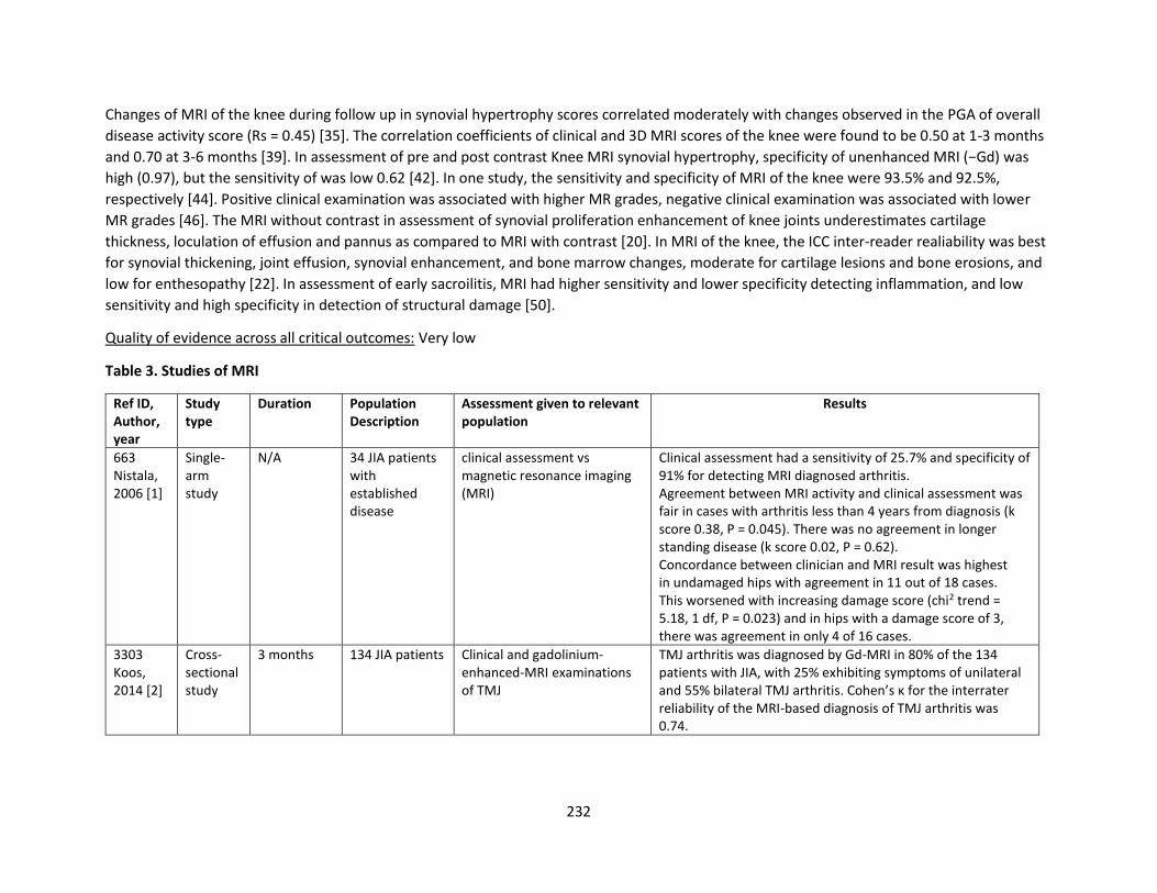
232
Changes of MRI of the knee during follow up in synovial hypertrophy scores correlated moderately with changes observed in the PGA of overall
disease activity score (Rs = 0.45) [35]. The correlation coefficients of clinical and 3D MRI scores of the knee were found to be 0.50 at 1-3 months
and 0.70 at 3-6 months [39]. In assessment of pre and post contrast Knee MRI synovial hypertrophy, specificity of unenhanced MRI (−Gd) was
high (0.97), but the sensitivity of was low 0.62 [42]. In one study, the sensitivity and specificity of MRI of the knee were 93.5% and 92.5%,
respectively [44]. Positive clinical examination was associated with higher MR grades, negative clinical examination was associated with lower
MR grades [46]. The MRI without contrast in assessment of synovial proliferation enhancement of knee joints underestimates cartilage
thickness, loculation of effusion and pannus as compared to MRI with contrast [20]. In MRI of the knee, the ICC inter-reader realiability was best
for synovial thickening, joint effusion, synovial enhancement, and bone marrow changes, moderate for cartilage lesions and bone erosions, and
low for enthesopathy [22]. In assessment of early sacroilitis, MRI had higher sensitivity and lower specificity detecting inflammation, and low
sensitivity and high specificity in detection of structural damage [50].
Quality of evidence across all critical outcomes: Very low
Table 3. Studies of MRI
Ref ID, Author, year
Study type
Duration Population Description
Assessment given to relevant population
Results
663 Nistala, 2006 [1]
Single-arm study
N/A 34 JIA patients with established disease
clinical assessment vs magnetic resonance imaging (MRI)
Clinical assessment had a sensitivity of 25.7% and specificity of 91% for detecting MRI diagnosed arthritis. Agreement between MRI activity and clinical assessment was fair in cases with arthritis less than 4 years from diagnosis (k score 0.38, P = 0.045). There was no agreement in longer standing disease (k score 0.02, P = 0.62). Concordance between clinician and MRI result was highest in undamaged hips with agreement in 11 out of 18 cases. This worsened with increasing damage score (chi2 trend = 5.18, 1 df, P = 0.023) and in hips with a damage score of 3, there was agreement in only 4 of 16 cases.
3303 Koos, 2014 [2]
Cross-sectional study
3 months 134 JIA patients Clinical and gadolinium-enhanced-MRI examinations of TMJ
TMJ arthritis was diagnosed by Gd-MRI in 80% of the 134 patients with JIA, with 25% exhibiting symptoms of unilateral and 55% bilateral TMJ arthritis. Cohen’s κ for the interrater reliability of the MRI-based diagnosis of TMJ arthritis was 0.74.
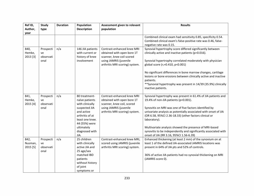
233
Ref ID, Author, year
Study type
Duration Population Description
Assessment given to relevant population
Results
Combined clinical exam had sensitivity 0.85, specificity 0.54. Combined clinical exam’s false-positive rate was 0.46, false-negative rate was 0.15.
840, Hemke, 2013 [3]
Prospective observational
n/a 146 JIA patients with current or history of knee involvement
Contrast-enhanced knee MRI obtained with open bore 1T scanner, knee coil scored using JAMRIS (juvenile arthritis MRI scoring) system.
Synovial hypertrophy score differed significantly between clinically active and inactive patients (p=0.016). Synovial hypertrophy correlated moderately with physician global score (rs=0.410, p<0.001) No significant differences in bone marrow changes, cartilage lesions or bone erosions between clinically active and inactive patients. **Synovial hypertrophy was present in 14/39 (35.9%) clinically inactive patients.
841, Hemke, 2015 [4]
Prospective observational
n/a 80 treatment-naïve patients with clinically suspected JIA and active arthritis of at least one knee. 44 (55%) were ultimately diagnosed with JIA.
Contrast-enhanced knee MRI obtained with open bore 1T scanner, knee coil, scored using JAMRIS (juvenile arthritis MRI scoring) system.
Synovial hypertrophy was present in 61.4% of JIA patients and 19.4% of non-JIA patients (p<0.001). Synovitis on MRI was one of five factors identified by univariate analysis as potentially associated with onset of JIA (OR 6.58, 95%CI 2.36-18.33) (other factors clinical or laboratory). Multivariate analysis showed the presence of MRI-based synovitis to be independently and significantly associated with onset of JIA (RR 3.16, 95%CI 1.56-6.39)
842, Nusman, 2015 [5]
Prospective observational
n/a 25 children with clinically active JIA and 25 age/sex matched IBD patients without history of joint symptoms or
Contrast-enhanced knee MRI, scored using JAMRIS (juvenile arthritis MRI scoring) system.
Enhanced thickening (at least 2 mm) of the synovium on at least 1 of the defined JIA-associated JAMRIS locations was present in 64% of JIA pts and 52% of controls. 36% of active JIA patients had no synovial thickening on MRI (JAMRIS score 0).
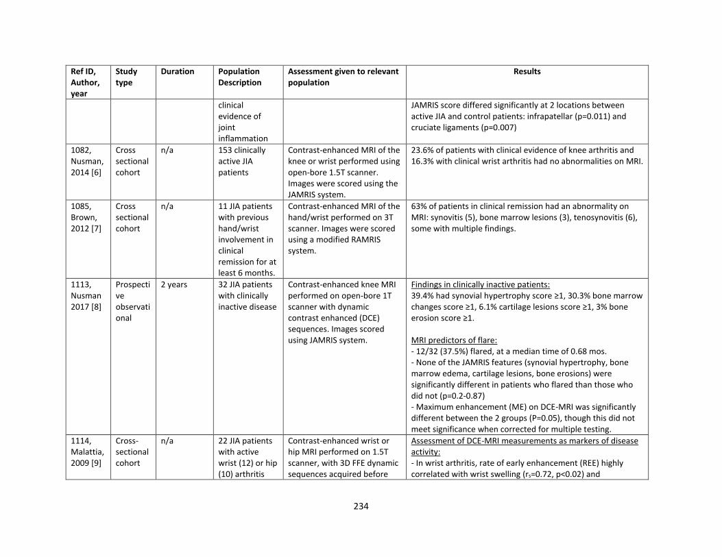
234
Ref ID, Author, year
Study type
Duration Population Description
Assessment given to relevant population
Results
clinical evidence of joint inflammation
JAMRIS score differed significantly at 2 locations between active JIA and control patients: infrapatellar (p=0.011) and cruciate ligaments (p=0.007)
1082, Nusman, 2014 [6]
Cross sectional cohort
n/a 153 clinically active JIA patients
Contrast-enhanced MRI of the knee or wrist performed using open-bore 1.5T scanner. Images were scored using the JAMRIS system.
23.6% of patients with clinical evidence of knee arthritis and 16.3% with clinical wrist arthritis had no abnormalities on MRI.
1085, Brown, 2012 [7]
Cross sectional cohort
n/a 11 JIA patients with previous hand/wrist involvement in clinical remission for at least 6 months.
Contrast-enhanced MRI of the hand/wrist performed on 3T scanner. Images were scored using a modified RAMRIS system.
63% of patients in clinical remission had an abnormality on MRI: synovitis (5), bone marrow lesions (3), tenosynovitis (6), some with multiple findings.
1113, Nusman 2017 [8]
Prospective observational
2 years 32 JIA patients with clinically inactive disease
Contrast-enhanced knee MRI performed on open-bore 1T scanner with dynamic contrast enhanced (DCE) sequences. Images scored using JAMRIS system.
Findings in clinically inactive patients: 39.4% had synovial hypertrophy score ≥1, 30.3% bone marrow changes score ≥1, 6.1% cartilage lesions score ≥1, 3% bone erosion score ≥1. MRI predictors of flare: - 12/32 (37.5%) flared, at a median time of 0.68 mos. - None of the JAMRIS features (synovial hypertrophy, bone marrow edema, cartilage lesions, bone erosions) were significantly different in patients who flared than those who did not (p=0.2-0.87) - Maximum enhancement (ME) on DCE-MRI was significantly different between the 2 groups (P=0.05), though this did not meet significance when corrected for multiple testing.
1114, Malattia, 2009 [9]
Cross-sectional cohort
n/a 22 JIA patients with active wrist (12) or hip (10) arthritis
Contrast-enhanced wrist or hip MRI performed on 1.5T scanner, with 3D FFE dynamic sequences acquired before
Assessment of DCE-MRI measurements as markers of disease activity: - In wrist arthritis, rate of early enhancement (REE) highly correlated with wrist swelling (rs=0.72, p<0.02) and
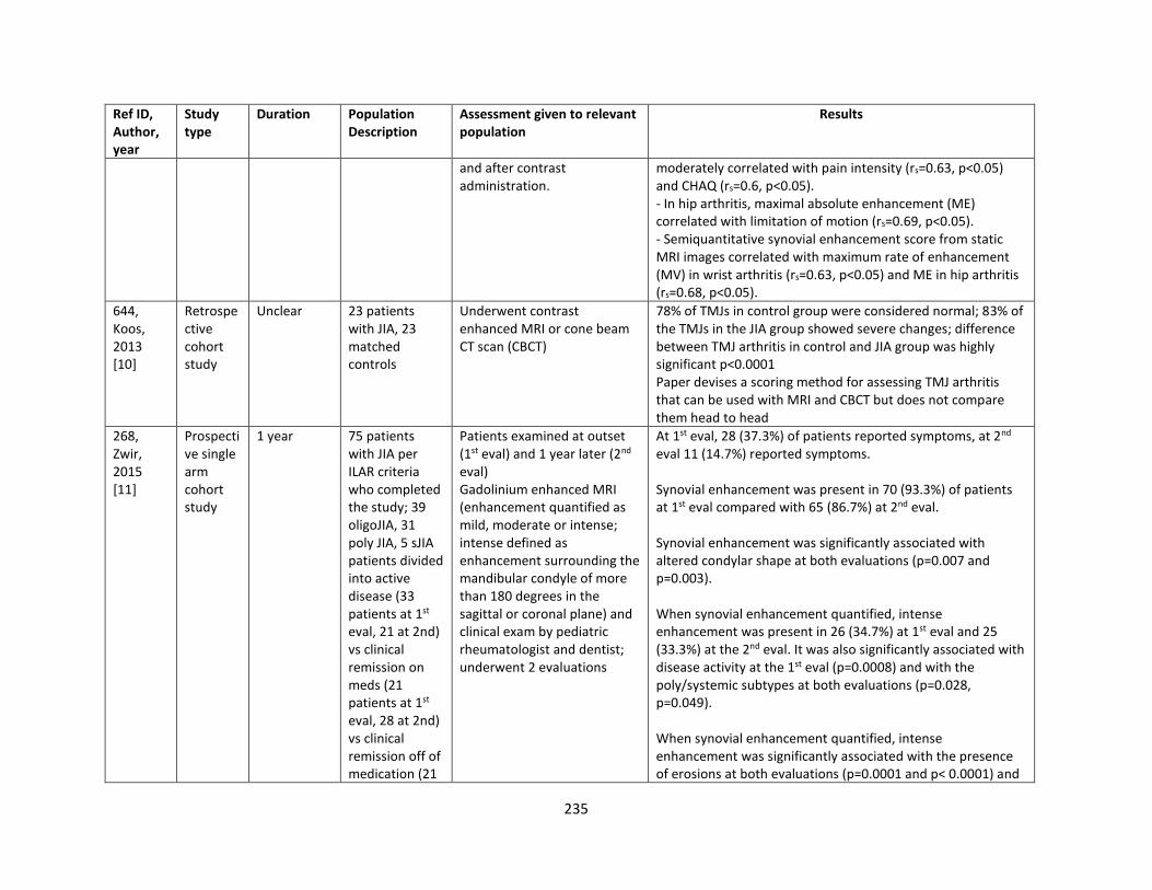
235
Ref ID, Author, year
Study type
Duration Population Description
Assessment given to relevant population
Results
and after contrast administration.
moderately correlated with pain intensity (rs=0.63, p<0.05) and CHAQ (rs=0.6, p<0.05). - In hip arthritis, maximal absolute enhancement (ME) correlated with limitation of motion (rs=0.69, p<0.05). - Semiquantitative synovial enhancement score from static MRI images correlated with maximum rate of enhancement (MV) in wrist arthritis (rs=0.63, p<0.05) and ME in hip arthritis (rs=0.68, p<0.05).
644, Koos, 2013 [10]
Retrospective cohort study
Unclear 23 patients with JIA, 23 matched controls
Underwent contrast enhanced MRI or cone beam CT scan (CBCT)
78% of TMJs in control group were considered normal; 83% of the TMJs in the JIA group showed severe changes; difference between TMJ arthritis in control and JIA group was highly significant p<0.0001 Paper devises a scoring method for assessing TMJ arthritis that can be used with MRI and CBCT but does not compare them head to head
268, Zwir, 2015 [11]
Prospective single arm cohort study
1 year 75 patients with JIA per ILAR criteria who completed the study; 39 oligoJIA, 31 poly JIA, 5 sJIA patients divided into active disease (33 patients at 1st eval, 21 at 2nd) vs clinical remission on meds (21 patients at 1st eval, 28 at 2nd) vs clinical remission off of medication (21
Patients examined at outset (1st eval) and 1 year later (2nd eval) Gadolinium enhanced MRI (enhancement quantified as mild, moderate or intense; intense defined as enhancement surrounding the mandibular condyle of more than 180 degrees in the sagittal or coronal plane) and clinical exam by pediatric rheumatologist and dentist; underwent 2 evaluations
At 1st eval, 28 (37.3%) of patients reported symptoms, at 2nd eval 11 (14.7%) reported symptoms. Synovial enhancement was present in 70 (93.3%) of patients at 1st eval compared with 65 (86.7%) at 2nd eval. Synovial enhancement was significantly associated with altered condylar shape at both evaluations (p=0.007 and p=0.003). When synovial enhancement quantified, intense enhancement was present in 26 (34.7%) at 1st eval and 25 (33.3%) at the 2nd eval. It was also significantly associated with disease activity at the 1st eval (p=0.0008) and with the poly/systemic subtypes at both evaluations (p=0.028, p=0.049). When synovial enhancement quantified, intense enhancement was significantly associated with the presence of erosions at both evaluations (p=0.0001 and p< 0.0001) and
236
Ref ID, Author, year
Study type
Duration Population Description
Assessment given to relevant population
Results
patients at 1st eval, 26 at 2nd)
with altered condylar shape at the second evaluation (p=0.0005). No association found between pain on TMJ palpation or crepitation and intense contrast enhancement. A significant association was found with a mouth opening < 40 mm and intense enhancement at both the 1st and 2nd eval (p=0.013 and p=0.0017). Intense contrast enhancement of the TMJ was significantly associated with disease activity (p< 0.001).
1115 Nusman et.al. 2016 [12]
Prospective, cohort
March 2012- July 2013,
32 children with JIA Age 8.2-17.9. 6 (19%) persistent OJIA, 4 (13%) extended OJIA, 15 (47%) polyarthritis RF (-), 2 (6%) polyarticular RF(+), 2 (6%) PsA, 2 (6%) enthesitis-related arthritis 1 (3%) undifferentiated arthritis.
Wrist MRI: All patients either had current wrist involvement (clinically active) or previous wrist involvement (clinically inactive).
The conventional descriptive measure maximum enhancement differed significantly between clinically active and inactive disease (P = 0.019), whereas time-intensity-curve shape analysis showed no differences. Juvenile Arthritis Disease Activity Score correlated moderately with enhancing volume (P= 0.484).
1124, von Kalle et.al. 2014 [13]
Retrospective review
In January 2011, so exams from 12/2005-1/2011
50 patients with JIA (40 females, 10 males) referred for MRI of TMJ
MRI of TMJ Degree of contrast enhancement alone did not allow a clear differentiation between joints with and without inflammation in dynamic contrast-enhanced MR
237
Ref ID, Author, year
Study type
Duration Population Description
Assessment given to relevant population
Results
(if multiple MRIs included the initial one) 100 dynamic contrast-enhanced MRI studies of 46 children (non-inflammatory) controls
Mean values and the number of abnormally high SI ratios of morphologically normal and abnormal joints of JIA patients were, at 6 min post-contrast, significantly higher than in the reference cohort. only 10 of the 39 (26%) joints with abnormally thick or irregular contrast enhancement had an SI ratio above 1.23, which is an enhancement above 2 SD of the reference group. 74% of the TMJs with signs of thickening of the joint tissue SI ratios remained within the range of the reference group. The ROC curve indicated the SI ratio of 0.9 as the best discriminating threshold, which, however, corresponded only to a moderate specificity of 0.70 and a sensitivity of 0.77. Signal intensity ratios of morphologically normal joints of patients with JIA were most variable. Their reference interval widely overlapped with the SI ratios of both the normal and the arthritic joints
1184 van Dijkhuizen et.al. 2018 [14]
Retrospective review
MRI examinations were performed between June 2006 and June 2008.
Seventy patients with JIA who had previously participated in observational studies. MRI after a median of 1.2 years follow-up was available for 38 patients.
Wrist MRI of 70 patients with JIA were scored by 3 independent readers and an extended score including the MCP joints
The inter-reader agreement was moderate for the original score (ICC 0.77; 95% CI 0.68–0.84) and good for the extended score (ICC 0.86; 95% CI 0.80–0.91). Using 95% LOA, the aggregate score variability was less favorable with relatively wide LOA. Weighted Cohen’s k of the individual joints indicated good agreement for the original score and good to excellent agreement for the extended score. Correlations with clinical variables reflecting disease activity improved for the extended score and its SRM was higher compared to that of the original score. Inclusion of MCP joints should be considered for a more accurate assessment of disease activity and treatment efficacy in JIA.
238
Ref ID, Author, year
Study type
Duration Population Description
Assessment given to relevant population
Results
1767 Vaid et.al. 2014 [15]
Retrospective review
N/A TMJ MRIs representative of acute and chronic TMJ arthritis in JIA (>500 TMJ MRI studies annually at Children’s of Alabama). Computed tomography scans depicting select bony changes (osteophyte formation, micrognathia).
Evaluation of MRI/CT of TMJs Compiled an atlas of radiographic images of the temporomandibular joint (TMJ) in children with juvenile idiopathic arthritis (JIA). A preliminary scoring system weighted for degree of acute and chronic TMJ arthritis MRI findings was found to have substantial inter- and intra-reader reliability.
1853 Hemke et.al. 2012 [16]
Prospective observational
11/2007- 10/2008
47 children: 94 sets of MR images from 47 children with JIA Group 1 (active JIA) 58 knees Group 2 (inactive JIA) 36 knees
MRI of knees without contrast Reproducibility of scoring method: Cohen kappa Synovial hypertrophy 0.76 Cartilage lesions 0.73 Bone erosions 0.92 Bone marrow changes 0.80 Infrapatellar fat pad heterogeneity 0.75 – Water 0.96 – Scar tissue 0.83 Effusion 0.85 Tendinopathy/internal derangement 0.49 Popliteal lymph nodes 0.85 MRI showed synovial hypertrophy suggestive of inflammation in 33% of the children with clinically inactive JIA
2017 Keller et. al.
Prospective cohort
March 2006 and October 2008
76 JIA pts I-MRI of the TMJs reviewed by two paediatric radiologists II. Rheum exams by peds rheum
Clinical findings in affected TMJs are correlated with structural damage only. Clinical assessment of TMJs does not allow accurate diagnosis of early arthritis and will still depend on contrast-enhanced MRI.
239
Ref ID, Author, year
Study type
Duration Population Description
Assessment given to relevant population
Results
2015 [17]
III.Orthodontic examination by an orthodontist
2094 Mohammed et. al. 2012 [18]
Case control (please check this – not sure)
N/A 40 pts with JIA: oligo, poly, SJIA,26 girls/14 boys). Age 8.5 to 17. 10 control subjects
clinical and post contrast magnetic resonance imaging (MRI) examinations for TMJs.
The clinical symptoms and signs of TMJ arthritis were detected in 35% and 62.5% of JIA cases, respectively. TMJ disease was observed in 80% of patients using contrast-enhanced MRI. The mean total MRI score was significantly higher in patients with active disease compared to those without activity. Patients with systemic and polyarticular JIA showed significant increase in the mean of synovial enhancement, effusion and total MRI scores compared to those with the oligoarticular type. MRI abnormalities revealed significant association with clinical signs of TMJ examination but not with symptoms. Synovial enhancement score showed significant positive correlation with disease activity score and C-reactive protein as a marker of inflammation.
2125 Van Gulik et. al. 2018 [19]
Prospective cohort
12/2008-12/2014
52 clinically inactive JIA patients (median age 13.3 years, 63.5% girls) who underwent MRI of the knee 2 groups based on MRI: 1-w/synovial thickening on MRI; 2-no synovial thickening on MRI.
MRI of knee with contrast Synovial thickening on MRI was present in 18 clinically inactive patients (group 1, 34.6%). The age was significantly lower for the patients in group 1 (median 10.7 versus 14.4, P=0.008). No significant differences were observed in any of the other patient characteristics nor the disease activity parameters tested. In more than one-third of children with clinically inactive disease: MRI of the knee showed synovial thickening.
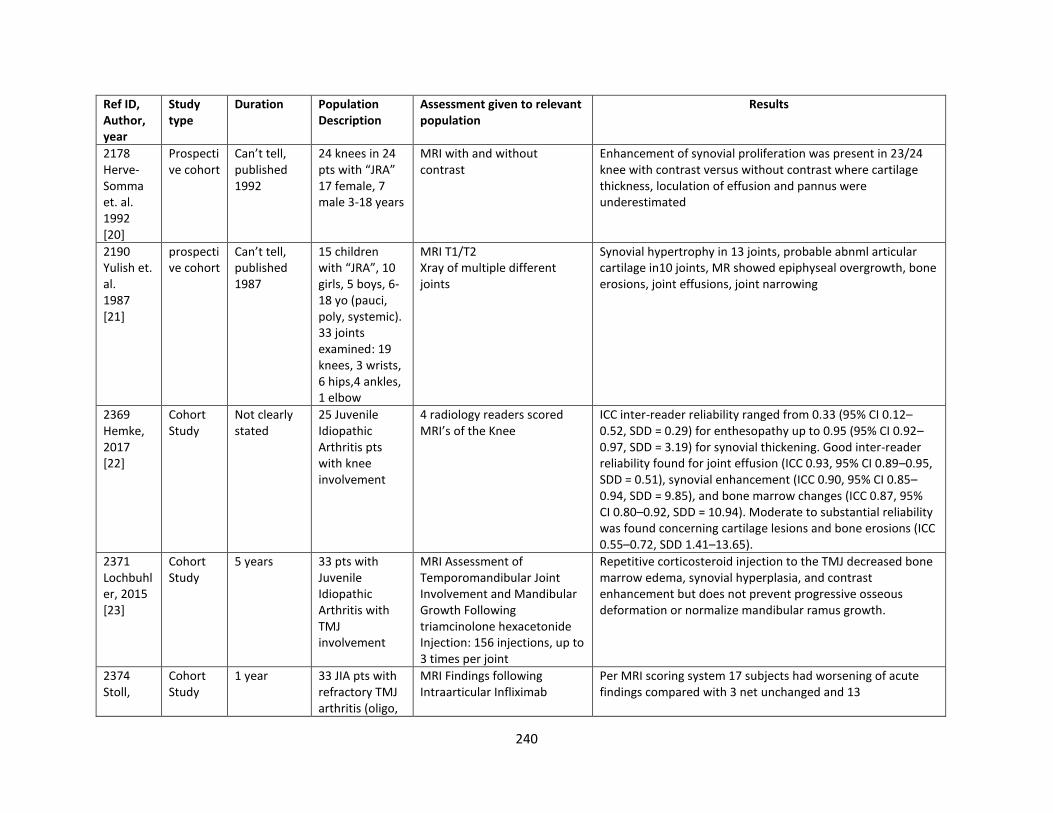
240
Ref ID, Author, year
Study type
Duration Population Description
Assessment given to relevant population
Results
2178 Herve-Somma et. al. 1992 [20]
Prospective cohort
Can’t tell, published 1992
24 knees in 24 pts with “JRA” 17 female, 7 male 3-18 years
MRI with and without contrast
Enhancement of synovial proliferation was present in 23/24 knee with contrast versus without contrast where cartilage thickness, loculation of effusion and pannus were underestimated
2190 Yulish et. al. 1987 [21]
prospective cohort
Can’t tell, published 1987
15 children with “JRA”, 10 girls, 5 boys, 6-18 yo (pauci, poly, systemic). 33 joints examined: 19 knees, 3 wrists, 6 hips,4 ankles, 1 elbow
MRI T1/T2 Xray of multiple different joints
Synovial hypertrophy in 13 joints, probable abnml articular cartilage in10 joints, MR showed epiphyseal overgrowth, bone erosions, joint effusions, joint narrowing
2369 Hemke, 2017 [22]
Cohort Study
Not clearly stated
25 Juvenile Idiopathic Arthritis pts with knee involvement
4 radiology readers scored MRI’s of the Knee
ICC inter-reader reliability ranged from 0.33 (95% CI 0.12–0.52, SDD = 0.29) for enthesopathy up to 0.95 (95% CI 0.92–0.97, SDD = 3.19) for synovial thickening. Good inter-reader reliability found for joint effusion (ICC 0.93, 95% CI 0.89–0.95, SDD = 0.51), synovial enhancement (ICC 0.90, 95% CI 0.85–0.94, SDD = 9.85), and bone marrow changes (ICC 0.87, 95% CI 0.80–0.92, SDD = 10.94). Moderate to substantial reliability was found concerning cartilage lesions and bone erosions (ICC 0.55–0.72, SDD 1.41–13.65).
2371 Lochbuhler, 2015 [23]
Cohort Study
5 years 33 pts with Juvenile Idiopathic Arthritis with TMJ involvement
MRI Assessment of Temporomandibular Joint Involvement and Mandibular Growth Following triamcinolone hexacetonide Injection: 156 injections, up to 3 times per joint
Repetitive corticosteroid injection to the TMJ decreased bone marrow edema, synovial hyperplasia, and contrast enhancement but does not prevent progressive osseous deformation or normalize mandibular ramus growth.
2374 Stoll,
Cohort Study
1 year 33 JIA pts with refractory TMJ arthritis (oligo,
MRI Findings following Intraarticular Infliximab
Per MRI scoring system 17 subjects had worsening of acute findings compared with 3 net unchanged and 13
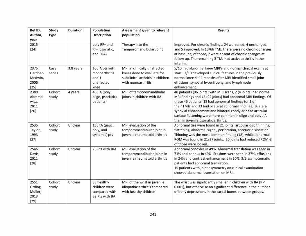
241
Ref ID, Author, year
Study type
Duration Population Description
Assessment given to relevant population
Results
2015 [24]
poly RF+ and RF-, psoriatic, and ERA)
Therapy into the Temporomandibular Joint
improved. For chronic findings: 24 worsened, 4 unchanged, and 5 improved. In 10/66 TMJ, there were no chronic changes at baseline; of those, 7 were absent of chronic changes at follow up. The remaining 3 TMJ had active arthritis in the interim.
2375 Gardner-Medwin, 2006 [25]
Case series
3.8 years 10 JIA pts with monoarthritis and 1 unaffected knee
MRI in clinically unaffected knees done to evaluate for subclinical arthritis in children with monoarthritis
5/10 had abnormal knee MRI’s and normal clinical exams at start. 3/10 developed clinical features in the previously normal knee 4–11 months after MRI identified small joint effusions, synovial hypertrophy, and lymph node enhancement.
2380 Abramowicz, 2011 [26]
Cohort study
4 years 48 JIA (poly, oligo, psoriatic) patients
MRI of temporomandibular joints in children with JIA
48 patients (96 joints) with MRI scans, 2 (4 joints) had normal MRI findings and 46 (92 joints) had abnormal MRI findings. Of these 46 patients, 13 had abnormal findings for 1 of their TMJs and 33 had bilateral abnormal findings. Bilateral synovial enhancement and bilateral condylar head articular surface flattening were more common in oligo and poly JIA than in juvenile psoriatic arthritis.
2535 Taylor, 1993 [27]
Cohort study
Unclear 15 JRA (pauci, poly, and systemic) pts
MRI evaluation of the temporomandibular joint in juvenile rheumatoid arthritis
Abnormalities were found in 21 joints: articular disc thinning, flattening, abnormal signal, perforation, anterior dislocation, Thinning was the most common finding (18), while abnormal ROM was found in 21/27 joints. 20 joints had reduced ROM-3 of those were locked.
2546 Davis, 2011 [28]
Cohort study
Unclear 26 Pts with JRA MRI evaluation of the temporomandibular joints in juvenile rheumatoid arthritis
Abnormal condyles in 49%. Abnormal translation was seen in 71% and pannus in 49%. Erosions were seen in 37%, effusions in 24% and contrast enhancement in 50%. 3/5 asymptomatic patients had abnormal translation. 15 patients with joint asymmetry on clinical examination showed abnormal translation on MRI.
2551 Ording Muller, 2013 [29]
Cohort study
Unclear 85 healthy children were compared with 68 Pts with JIA
MRI of the wrist in juvenile idiopathic arthritis compared with healthy children
The wrist was significantly smaller in children with JIA (P < 0.001), but otherwise no significant difference in the number of bony depressions in the carpal bones between groups.
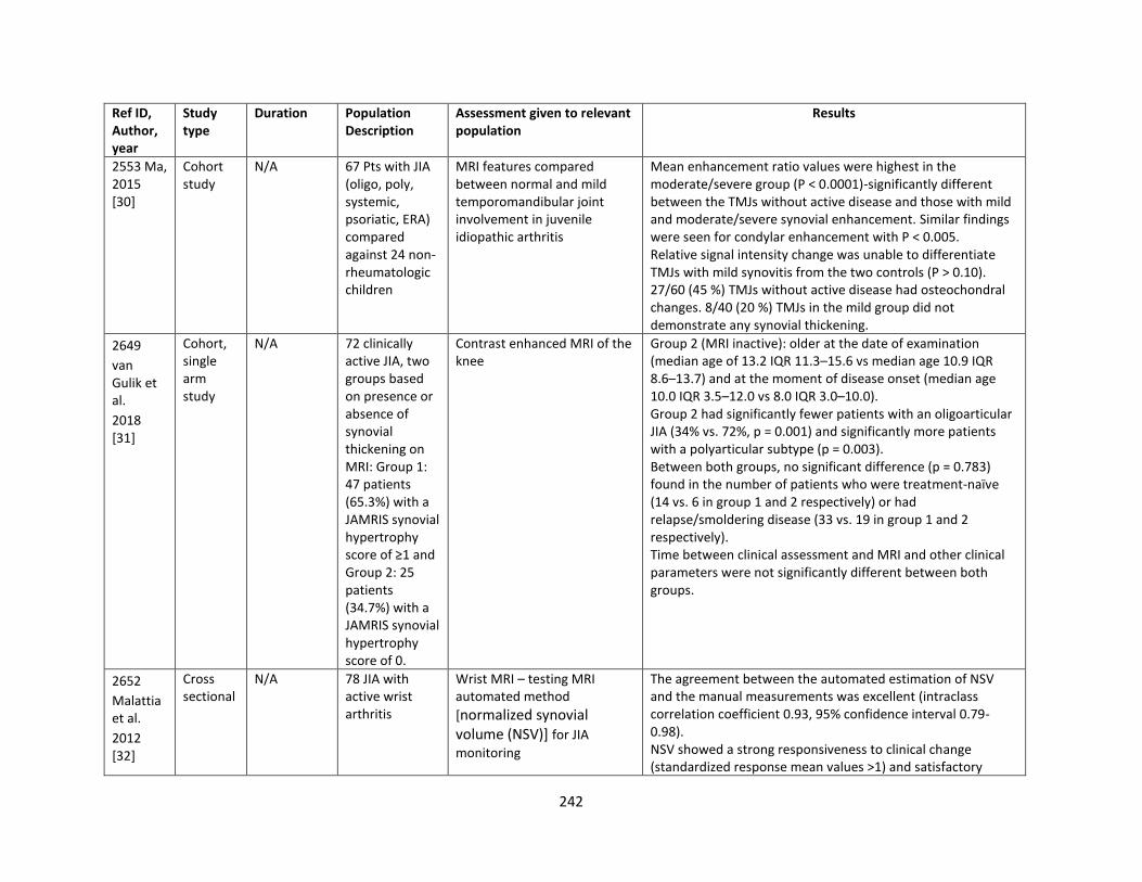
242
Ref ID, Author, year
Study type
Duration Population Description
Assessment given to relevant population
Results
2553 Ma, 2015 [30]
Cohort study
N/A 67 Pts with JIA (oligo, poly, systemic, psoriatic, ERA) compared against 24 non-rheumatologic children
MRI features compared between normal and mild temporomandibular joint involvement in juvenile idiopathic arthritis
Mean enhancement ratio values were highest in the moderate/severe group (P < 0.0001)-significantly different between the TMJs without active disease and those with mild and moderate/severe synovial enhancement. Similar findings were seen for condylar enhancement with P < 0.005. Relative signal intensity change was unable to differentiate TMJs with mild synovitis from the two controls (P > 0.10). 27/60 (45 %) TMJs without active disease had osteochondral changes. 8/40 (20 %) TMJs in the mild group did not demonstrate any synovial thickening.
2649
van Gulik et al.
2018 [31]
Cohort, single arm study
N/A 72 clinically active JIA, two groups based on presence or absence of synovial thickening on MRI: Group 1: 47 patients (65.3%) with a JAMRIS synovial hypertrophy score of ≥1 and Group 2: 25 patients (34.7%) with a JAMRIS synovial hypertrophy score of 0.
Contrast enhanced MRI of the knee
Group 2 (MRI inactive): older at the date of examination (median age of 13.2 IQR 11.3–15.6 vs median age 10.9 IQR 8.6–13.7) and at the moment of disease onset (median age 10.0 IQR 3.5–12.0 vs 8.0 IQR 3.0–10.0). Group 2 had significantly fewer patients with an oligoarticular JIA (34% vs. 72%, p = 0.001) and significantly more patients with a polyarticular subtype (p = 0.003). Between both groups, no significant difference (p = 0.783) found in the number of patients who were treatment-naïve (14 vs. 6 in group 1 and 2 respectively) or had relapse/smoldering disease (33 vs. 19 in group 1 and 2 respectively). Time between clinical assessment and MRI and other clinical parameters were not significantly different between both groups.
2652
Malattia et al.
2012 [32]
Cross sectional
N/A 78 JIA with active wrist arthritis
Wrist MRI – testing MRI automated method
[normalized synovial volume (NSV)] for JIA
monitoring
The agreement between the automated estimation of NSV and the manual measurements was excellent (intraclass correlation coefficient 0.93, 95% confidence interval 0.79-0.98). NSV showed a strong responsiveness to clinical change (standardized response mean values >1) and satisfactory
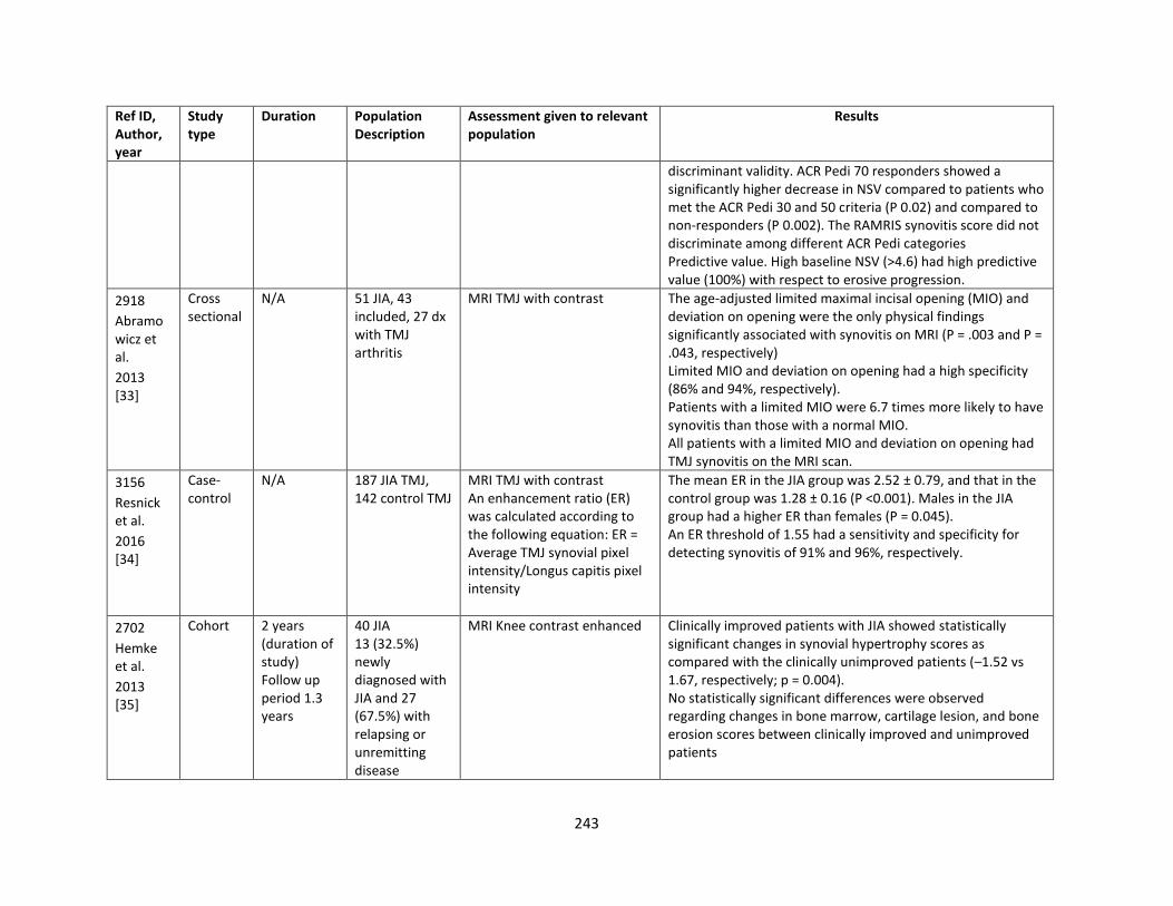
243
Ref ID, Author, year
Study type
Duration Population Description
Assessment given to relevant population
Results
discriminant validity. ACR Pedi 70 responders showed a significantly higher decrease in NSV compared to patients who met the ACR Pedi 30 and 50 criteria (P 0.02) and compared to non-responders (P 0.002). The RAMRIS synovitis score did not discriminate among different ACR Pedi categories Predictive value. High baseline NSV (>4.6) had high predictive value (100%) with respect to erosive progression.
2918
Abramowicz et al.
2013 [33]
Cross sectional
N/A 51 JIA, 43 included, 27 dx with TMJ arthritis
MRI TMJ with contrast The age-adjusted limited maximal incisal opening (MIO) and deviation on opening were the only physical findings significantly associated with synovitis on MRI (P = .003 and P = .043, respectively) Limited MIO and deviation on opening had a high specificity (86% and 94%, respectively). Patients with a limited MIO were 6.7 times more likely to have synovitis than those with a normal MIO. All patients with a limited MIO and deviation on opening had TMJ synovitis on the MRI scan.
3156
Resnick et al.
2016 [34]
Case-control
N/A 187 JIA TMJ, 142 control TMJ
MRI TMJ with contrast An enhancement ratio (ER) was calculated according to the following equation: ER = Average TMJ synovial pixel intensity/Longus capitis pixel intensity
The mean ER in the JIA group was 2.52 ± 0.79, and that in the control group was 1.28 ± 0.16 (P <0.001). Males in the JIA group had a higher ER than females (P = 0.045). An ER threshold of 1.55 had a sensitivity and specificity for detecting synovitis of 91% and 96%, respectively.
2702
Hemke et al.
2013 [35]
Cohort 2 years (duration of study) Follow up period 1.3 years
40 JIA 13 (32.5%) newly diagnosed with JIA and 27 (67.5%) with relapsing or unremitting disease
MRI Knee contrast enhanced Clinically improved patients with JIA showed statistically significant changes in synovial hypertrophy scores as compared with the clinically unimproved patients (–1.52 vs 1.67, respectively; p = 0.004). No statistically significant differences were observed regarding changes in bone marrow, cartilage lesion, and bone erosion scores between clinically improved and unimproved patients
244
Ref ID, Author, year
Study type
Duration Population Description
Assessment given to relevant population
Results
11 (27.5%) persistent oligoarthritis, 8 (20.0%) extended oligoarthritis, 17 (42.5%) rheumatoid factor (RF)-negative polyarthritis
Change or intensification of treatment after the first MRI was observed in 85.2% (23/27) of the clinically improved patients (ACR-Ped50) Changes during follow up in synovial hypertrophy scores correlated moderately with changes observed in the PGA of overall disease activity score (Rs = 0.45, p = 0.002).
3160
Workie et al.
2007 [36]
cohort 12 months 17 JIA patients dynamic contrast-enhanced MRI (DCE-MRI) of Knee
All of the clinical and laboratory measures (median values) showed a significant decrease during the study period (P<0.05), except CHAQ-DI and CRP at 3 months. The PK parameters, synovial volumes, and clinical and laboratory measures decreased from enrollment to 3 months and to 12 months in the majority of children The following parameters were correlated between baseline and 12 months: Ktrans′:PGA (rho=0.80, P=0.003), Ktrans′:TAJ (rho=0.76, P=0.006), kep:PGA (rho=0.72, P=0.012), and synovial volume:TAJ (rho=−0.51, P=0.04).
3440
El-Azeem et al.
2012 [37]
cohort 2 years (duration of study)
30 JIA patients MRI Hips bilateral with contrast
MRI of hips was abnormal in 12/30 (40%) patients: 2/8 (25%) of oligoarticular group, 4/13 (30.8%) of polyarticular group, 5/7 (71.4%) of systemic onset group and 1/2(50%) of enthesitis related group. Comparing mean values of MR score of the four clinical subsets showed significant difference (p < 0.001). Patients with active disease showed higher MR score (3.7 ± 1.5) than those with inactive disease (2.1 ± .9) [p < 0.002]. Presence of effusion and gadolinium enhancement were significantly higher in active hips (p < 0.01 and p < 0.001 respectively). VAS– PGA and ESR were significantly correlated with MRI score (p < 0.02 and <0.05 respectively).
3580 Case-control
N/A 20 JIA (80 % had TMJ
TMJ MRI and serum S100A12 level
Contrast enhanced MRI was done for 20 patients (40 TMJs): 16 patients (80%) showed MRI abnormalities while 4 patients
245
Ref ID, Author, year
Study type
Duration Population Description
Assessment given to relevant population
Results
Abdul-Aziez et al.
2010 [38]
arthritis), 10 healthy controls JIA: 3 (15%) with SoJIA ; 3 (15%) oligo-JIA and 14 (70%) poly- JIA.
had no MRI findings. The mean total score for MRI among JIA patients was 5.05±4.21.
Synovial enhancement was detected in 16 patients (80%), 31 TMJs (77.5%) with a mean score 2.60±1.60; joint effusion was present in 13 patients (65%), 19 TMJs (47.5%) with a mean score 1.40±1.46.
mean value of Maximal Interincisal Opening was significantly decreased in patients versus controls. mean serum levels of S100A12 showed significant increase among JIA patients compared to controls (p<0.001). Serum levels of S100A12 showed a significant positive correlation with disease activity score, pain score, ESR, CRP serum levels, synovial enhancement score and total MRI score.
3595
Cakmakci et al.
2001 [39]
Longitudinal cohort
6 months JIA 42 knees of 21 patients
Fat-Sat 3D MRI Knee
Correlation coefficients according to progression, improvement and equivalent findings of months 1-3 and months 3-6 comparison of clinical and MRI scores were found to be 0.50 and 0.70, respectively. Synovial hypertrophy was found to be the main determinant of pain on first- and third-month evaluations (r=o.68 and 0.74 respectively)
3838
Argyropoulou et al.
2008 [40]
Cohort N/A 46 JIA (88 TMJ) 18 oligo JIA, 17 poly JIA; 11 SoJIA with polyarticular course
MRI TMJ with contrast (contrast given for all except for 1 patient with contrast allergy)
Abnormal condyle in 32%, flattened articular eminence in 27%, flattened articular disk in 17%, intra-articular fluid in 10%, enhancing pannus in 45% restricted condylar motion in 9%. Logistic regression analysis revealed that for abnormal condyle and flattened articular eminence, independent predictors were type of JIA (P < 0.015), age at onset (P < 0.038), and duration of disease activity (P < 0.001). Pannus was present with probability >0.5 when the disease started before 4 years of age
3842 Case -control
N/A 18 adolescents (15 females,
contrast enhanced TMJ MRI
In the ADD group, 31 of 36 disks were displaced. In total, 28 of 31 displaced disks showed thickening of the bilaminar zone.
246
Ref ID, Author, year
Study type
Duration Population Description
Assessment given to relevant population
Results
Kellenberger et al.
2018 [41]
mean age 15.1 ± 1.9 years) with ADD and age- and gender-matched patients with JIA
In JIA patients, the disks were mainly flattened (19/36), centrally perforated (12/36) and/or anteriorly displaced (2/36). 19 of 31 TMJs with ADD showed various degrees of inflammation, with joint effusion, synovial thickening and joint enhancement not significantly different from JIA patients. Osseous deformity was present in 27 of 31 TMJs with ADD, with frequent erosions in both groups (ADD 25/31; JIA 32/36, P = 0.55) but lower grades of condylar and temporal bone flattening than in JIA (P ≤ 0.001). Glenoid fossa depth was preserved in 28 of 31 joints with ADD and decreased in 26 of 36 joints with JIA (P < 0.0001). Mandibular ramus height was decreased in both groups.
1013
Hemke et al.
2013 [42]
Single arm cohort
2 years 73 JIA patients: 17 (23.3 %) persistent OJIA, 13 (17.8 %) extended OJIA, 27 (37.0 %) RF- polyarthritis, 1 (1.4 %) RF+ polyarthritis, 1 (1.4 %) SJIA, 2 (2.7 %) PsA, 10 (13.7 %) enthesitis-related arthritis and 2 (2.7 %) undifferentiated JIA
Pre and post contrast Knee MRI
Agreement between Gd-enhanced (+Gd) and Gd-Unenhanced (−Gd) MRI scores of bone marrow changes, cartilage lesions and bone erosions were good concerning sensitivity, specificity, negative predictive value and positive predictive value. Inter-observer agreement was good for both −Gd and +Gd scores (ICC=0.91–1.00, 0.93–1.00, respectively). Regarding the assessment of synovial hypertrophy, specificity of −Gd was high (0.97), but the sensitivity of unenhanced MRI was only 0.62. Inter-reader agreement for +Gd MRI was ICC=0.94; however, omitting post-Gd acquisitions increased inter-reader variation (ICC=0.86) Omitting intravenous contrast medium decreases the reliability of synovial hypertrophy scores.
1129
Johnson et al.
Single arm cohort
N/A 11 JIA MRI knee (contrast enhanced in 9 patients) Obtained mean 2 months after disease onset
Synovial hypertrophy, joint effusions, popliteal lymph nodes and soft tissue swelling were present in all patients. Gadolinium DTPA enhancement improved the detection of synovial hyperplasia.
247
Ref ID, Author, year
Study type
Duration Population Description
Assessment given to relevant population
Results
2002 [43]
Metaphyseal splaying and condylar overgrowth were seen in five cases (41%) Edema of the lateral collateral ligament in two cases (18%) and superficial cartilage thinning in one case. Bony erosions and deep cartilage destruction were not demonstrated.
2209
Uhl et al.
2001 [44]
Case-control
N/A 40 JIA 40 control painful knee joints (MR diagnosis: bone bruise of the knee (7), normal knee joint (12), osteomyelitis (6), septic arthritis (2), bone tumor (7) and misc bone lesions (6)
Knee MRI: 1.5 T Receiver operating characteristic (ROC) curves evaluation was conducted by 5 independent radiologists.
The positive criteria for diagnosing JIA were joint effusions (n=.40), contrast-enhancing synovitis (n=39), cartilage lesions (n=15), subchondral erosions and bony destruction (n=1). Sensitivity and specificity were 93.5% and 92.5%, respectively. Two cases of septic arthritis were misdiagnosed as JIA by all radiologists.
2422
Rieter et al.
2016 [45]
Single arm cohort
N/A 34 children, 20 (58.8%) had oligo-arthritis, 9 (26.5%) had polyarthritis and 5 (14.7%) had systemic arthritis.
Wrist MRI with contrast Pathological contrast enhancement was seen in 100/170 (58.8%) in early postcontrast images vs. 131/170 (77.1%) based on late postcontrast images. 52/170 locations (30.6%) received a higher synovial enhancement-score based on the late post-contrast images as compared to the early post-contrast images. 60/170 (35.3%) locations received a higher total inflammation score based on the late post-contrast images. The mean scores of synovial enhancement and total inflammation were significantly higher when based on the late postcontrast images as compared to the early contrast images
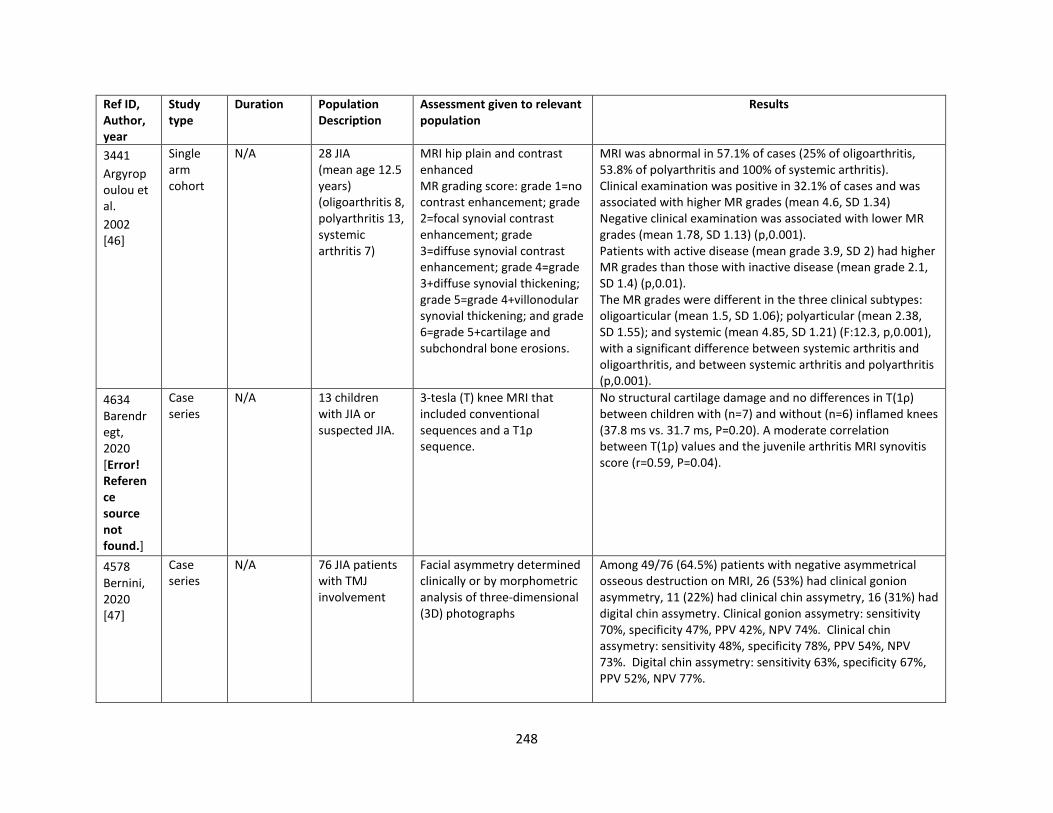
248
Ref ID, Author, year
Study type
Duration Population Description
Assessment given to relevant population
Results
3441
Argyropoulou et al.
2002 [46]
Single arm cohort
N/A 28 JIA (mean age 12.5 years) (oligoarthritis 8, polyarthritis 13, systemic arthritis 7)
MRI hip plain and contrast enhanced MR grading score: grade 1=no contrast enhancement; grade 2=focal synovial contrast enhancement; grade 3=diffuse synovial contrast enhancement; grade 4=grade 3+diffuse synovial thickening; grade 5=grade 4+villonodular synovial thickening; and grade 6=grade 5+cartilage and subchondral bone erosions.
MRI was abnormal in 57.1% of cases (25% of oligoarthritis, 53.8% of polyarthritis and 100% of systemic arthritis). Clinical examination was positive in 32.1% of cases and was associated with higher MR grades (mean 4.6, SD 1.34) Negative clinical examination was associated with lower MR grades (mean 1.78, SD 1.13) (p,0.001). Patients with active disease (mean grade 3.9, SD 2) had higher MR grades than those with inactive disease (mean grade 2.1, SD 1.4) (p,0.01). The MR grades were different in the three clinical subtypes: oligoarticular (mean 1.5, SD 1.06); polyarticular (mean 2.38, SD 1.55); and systemic (mean 4.85, SD 1.21) (F:12.3, p,0.001), with a significant difference between systemic arthritis and oligoarthritis, and between systemic arthritis and polyarthritis (p,0.001).
4634 Barendregt, 2020 [Error! Reference source not found.]
Case series
N/A 13 children with JIA or suspected JIA.
3-tesla (T) knee MRI that included conventional sequences and a T1ρ sequence.
No structural cartilage damage and no differences in T(1ρ) between children with (n=7) and without (n=6) inflamed knees (37.8 ms vs. 31.7 ms, P=0.20). A moderate correlation between T(1ρ) values and the juvenile arthritis MRI synovitis score (r=0.59, P=0.04).
4578 Bernini, 2020 [47]
Case series
N/A 76 JIA patients with TMJ involvement
Facial asymmetry determined clinically or by morphometric analysis of three-dimensional (3D) photographs
Among 49/76 (64.5%) patients with negative asymmetrical osseous destruction on MRI, 26 (53%) had clinical gonion asymmetry, 11 (22%) had clinical chin assymetry, 16 (31%) had digital chin assymetry. Clinical gonion assymetry: sensitivity 70%, specificity 47%, PPV 42%, NPV 74%. Clinical chin assymetry: sensitivity 48%, specificity 78%, PPV 54%, NPV 73%. Digital chin assymetry: sensitivity 63%, specificity 67%, PPV 52%, NPV 77%.
249
Ref ID, Author, year
Study type
Duration Population Description
Assessment given to relevant population
Results
4500 Bollhalder, 2020 [Error! Reference source not found.]
Case series
3.6 years 38 patients with JIA with TMJ involvement
Clinical and MRI findings Presence of TMJ pain did not correlate with MRI grades of inflammation or deformity. Both absolute values and centiles of MOC showed no correlation with MRI grades of inflammation or deformity.
4672 Weiss, 2020 [Error! Reference source not found.]
Case-series
N/A 120 JIA patients with sacroilitis
MRI in the assessment of early sacroiliitis
Local reports for inflammation: Sensitivity, 93.5% (95% CI: 78.6-99.2%), and specificity, 69.7% (95% CI: 59.0-79.0%), PPV 51.8% (95% CI: 38.0-65.3%). Local reports detecting structural damage: Sensitivity 45.7% (95% CI: 28.8-63.4%), specificity, 88.2% (95% CI: 79.4-94.2%); PPV 61.5% (95% CI: 40.6-79.8%).
References:
1. Nistala, K., Babar, J., Johnson, K., Campbell-Stokes, P., Foster, K., Ryder, C., & McDonagh, J. E. (2006). Clinical assessment and core
outcome variables are poor predictors of hip arthritis diagnosed by MRI in juvenile idiopathic arthritis. Rheumatology (Oxford), 46(4),
699-702. doi:10.1093/rheumatology/kel401
2. Koos, B., Twilt, M., Kyank, U., Fischer-Brandies, H., Gassling, V., & Tzaribachev, N. (2014). Reliability of clinical symptoms in diagnosing
temporomandibular joint arthritis in juvenile idiopathic arthritis. J Rheumatol, 41(9), 1871-1877. doi:10.3899/jrheum.131337
3. Hemke, R., Maas, M., Veenendaal, M. v., Dolman, K. M., Rossum, M. A. v., Berg, J. M. v. d., & Kuijpers, T. W. (2013). Contrast-enhanced
MRI compared with the physical examination in the evaluation of disease activity in juvenile idiopathic arthritis. Eur Radiol, 24(2), 327-
334. doi:10.1007/s00330-013-3036-2
4. Hemke, R., Kuijpers, T. W., Nusman, C. M., Schonenberg-Meinema, D., Rossum, M. A. v., Dolman, K. M., . . . Maas, M. (2015). Contrast-
enhanced MRI features in the early diagnosis of Juvenile Idiopathic Arthritis. Eur Radiol, 25(11), 3222-3229. doi:10.1007/s00330-015-
3752-x
250
5. Nusman, C. M., Hemke, R., Benninga, M. A., Schonenberg-Meinema, D., Kindermann, A., Rossum, M. A. v., . . . Kuijpers, T. W. (2015).
Contrast-enhanced MRI of the knee in children unaffected by clinical arthritis compared to clinically active juvenile idiopathic arthritis
patients. Eur Radiol, 26(4), 1141-1148. doi:10.1007/s00330-015-3912-z
6. Nusman, C. M., Hemke, R., Schonenberg, D., Dolman, K. M., Rossum, M. A. v., Berg, J. M. v. d., . . . Maas, M. (2014). Distribution pattern
of MRI abnormalities within the knee and wrist of juvenile idiopathic arthritis patients: signature of disease activity. AJR Am J
Roentgenol, 202(5), W439-446. doi:10.2214/ajr.13.11314
7. Brown, A., Hirsch, R., Laor, T., Hannon, M. J., Levesque, M. C., Starz, T., . . . Kwoh, C. K. (2012). Do patients with juvenile idiopathic
arthritis in clinical remission have evidence of persistent inflammation on 3T magnetic resonance imaging? Arthritis Care Res (Hoboken),
64(12), 1846-1854. doi:10.1002/acr.21774
8. Nusman, C. M., Hemke, R., Lavini, C., Schonenberg-Meinema, D., Rossum, M. A. v., Dolman, K. M., . . . Kuijpers, T. W. (2017). Dynamic
contrast-enhanced magnetic resonance imaging can play a role in predicting flare in juvenile idiopathic arthritis. Eur J Radiol, 88, 77-81.
doi:10.1016/j.ejrad.2017.01.003
9. Malattia, C., Damasio, M. B., Basso, C., Verri, A., Magnaguagno, F., Viola, S., . . . Martini, A. (2009). Dynamic contrast-enhanced magnetic
resonance imaging in the assessment of disease activity in patients with juvenile idiopathic arthritis. Rheumatology (Oxford), 49(1), 178-
185. doi:10.1093/rheumatology/kep343
10. Koos, B., Tzaribachev, N., Bott, S., Ciesielski, R., & Godt, A. (2013). Classification of temporomandibular joint erosion, arthritis, and
inflammation in patients with juvenile idiopathic arthritis. J Orofac Orthop, 74(6), 506-519. doi:10.1007/s00056-013-0166-8
11. Zwir, L. M., Terreri, M. T., Sousa, S. A., Fernandes, A. R., Guimaraes, A. S., & Hilario, M. O. (2015). Are temporomandibular joint signs and
symptoms associated with magnetic resonance imaging findings in juvenile idiopathic arthritis patients? A longitudinal study. Clin
Rheumatol, 34(12), 2057-2063. doi:10.1007/s10067-015-2925-y
12. Nusman, C. M., Lavini, C., Hemke, R., Caan, M. W., Schonenberg-Meinema, D., Dolman, K. M., . . . Maas, M. (2016). Dynamic contrast-
enhanced magnetic resonance imaging of the wrist in children with juvenile idiopathic arthritis. Pediatr Radiol, 47(2), 205-213.
doi:10.1007/s00247-016-3736-2
13. Kalle, T. v., Stuber, T., Winkler, P., Maier, J., & Hospach, T. (2014). Early detection of temporomandibular joint arthritis in children with
juvenile idiopathic arthritis - the role of contrast-enhanced MRI. Pediatr Radiol, 45(3), 402-410. doi:10.1007/s00247-014-3143-5
14. Dijkhuizen, E. H. P. v., Vanoni, F., Magnano, G. M., Magnaguagno, F., Rosendahl, K., Rossum, M. A. v., . . . Malattia, C. (2018). Effect of the
Inclusion of the Metacarpophalangeal Joints on the Wrist Magnetic Resonance Imaging Scoring System in Juvenile Idiopathic Arthritis. J
Rheumatol, 45(11), 1581-1587. doi:10.3899/jrheum.171246
15. Vaid, Y. N., Dunnavant, F. D., Royal, S. A., Beukelman, T., Stoll, M. L., & Cron, R. Q. (2013). Imaging of the temporomandibular joint in
juvenile idiopathic arthritis. Arthritis Care Res (Hoboken), 66(1), 47-54. doi:10.1002/acr.22177
16. Hemke, R., Veenendaal, M. v., Kuijpers, T. W., Rossum, M. A. v., & Maas, M. (2012). Increasing feasibility and patient comfort of MRI in
children with juvenile idiopathic arthritis. Pediatr Radiol, 42(4), 440-448. doi:10.1007/s00247-011-2306-x
251
17. Keller, H., Muller, L. M., Markic, G., Schraner, T., Kellenberger, C. J., & Saurenmann, R. K. (2015). Is early TMJ involvement in children
with juvenile idiopathic arthritis clinically detectable? Clinical examination of the TMJ in comparison with contrast enhanced MRI in
patients with juvenile idiopathic arthritis. Pediatr Rheumatol Online J, 13, 56. doi:10.1186/s12969-015-0056-2
18. Mohammed, Y., Saeed, O., Zaghloul, N., Samer, S., Mahmud, S., & Abdulah, A. (2012). Juvenile idiopathic arthritis and the
temporomandibular joint. Alexandria Journal of Medicine, 48(2), 123-129. doi:http://dx.doi.org/10.1016/j.ajme.2011.11.004
19. Gulik, E. C. v., Welsink-Karssies, M. M., Berg, J. M. v. d., Schonenberg-Meinema, D., Dolman, K. M., Barendregt, A. M., . . . Hemke, R.
(2018). Juvenile idiopathic arthritis: magnetic resonance imaging of the clinically unaffected knee. Pediatr Radiol, 48(3), 333-340.
doi:10.1007/s00247-017-4059-7
20. Herve-Somma, C. M., Sebag, G. H., Prieur, A. M., Bonnerot, V., & Lallemand, D. P. (1992). Juvenile rheumatoid arthritis of the knee: MR
evaluation with Gd-DOTA. Radiology, 182(1), 93-98. doi:10.1148/radiology.182.1.1727317
21. Yulish, B. S., Lieberman, J. M., Newman, A. J., Bryan, P. J., Mulopulos, G. P., & Modic, M. T. (1987). Juvenile rheumatoid arthritis:
assessment with MR imaging. Radiology, 165(1), 149-152. doi:10.1148/radiology.165.1.3628761
22. Hemke, R., Tzaribachev, N., Nusman, C. M., Rossum, M. A. J. v., Maas, M., & Doria, A. S. (2017). Magnetic Resonance Imaging (MRI) of
the Knee as an Outcome Measure in Juvenile Idiopathic Arthritis: An OMERACT Reliability Study on MRI Scales. J Rheumatol, 44(8), 1224-
1230. doi:10.3899/jrheum.160821
23. Lochbuhler, N., Saurenmann, R. K., Muller, L., & Kellenberger, C. J. (2015). Magnetic Resonance Imaging Assessment of
Temporomandibular Joint Involvement and Mandibular Growth Following Corticosteroid Injection in Juvenile Idiopathic Arthritis. J
Rheumatol, 42(8), 1514-1522. doi:10.3899/jrheum.141502
24. Stoll, M. L., Vaid, Y. N., Guleria, S., Beukelman, T., Waite, P. D., & Cron, R. Q. (2015). Magnetic Resonance Imaging Findings following
Intraarticular Infliximab Therapy for Refractory Temporomandibular Joint Arthritis among Children with Juvenile Idiopathic Arthritis. J
Rheumatol, 42(11), 2155-2159. doi:10.3899/jrheum.150308
25. Gardner-Medwin, J. M., Killeen, O. G., Ryder, C. A., Bradshaw, K., & Johnson, K. (2006). Magnetic resonance imaging identifies features in
clinically unaffected knees predicting extension of arthritis in children with monoarthritis. J Rheumatol, 33(11), 2337-2343.
26. Abramowicz, S., Cheon, J. E., Kim, S., Bacic, J., & Lee, E. Y. (2011). Magnetic resonance imaging of temporomandibular joints in children
with arthritis. J Oral Maxillofac Surg, 69(9), 2321-2328. doi:10.1016/j.joms.2010.12.058
27. Taylor, D. B., Babyn, P., Blaser, S., Smith, S., Shore, A., Silverman, E. D., . . . Laxer, R. M. (1993). MR evaluation of the temporomandibular
joint in juvenile rheumatoid arthritis. J Comput Assist Tomogr, 17(3), 449-454.
28. Davis, M. A., & Castillo, M. (2011). MRI evaluation of the temporomandibular joints in juvenile rheumatoid arthritis: a retrospective
review. Neuroradiol J, 24(6), 928-932. doi:10.1177/197140091102400618
29. Muller, L. S. O., Boavida, P., Avenarius, D., Damasio, B., Eldevik, O. P., Malattia, C., . . . Rosendahl, K. (2013). MRI of the wrist in juvenile
idiopathic arthritis: erosions or normal variants? A prospective case-control study. Pediatr Radiol, 43(7), 785-795. doi:10.1007/s00247-
012-2575-z
252
30. Ma, G. M., Amirabadi, A., Inarejos, E., Tolend, M., Stimec, J., Moineddin, R., . . . Doria, A. S. (2015). MRI thresholds for discrimination
between normal and mild temporomandibular joint involvement in juvenile idiopathic arthritis. Pediatr Rheumatol Online J, 13, 53.
doi:10.1186/s12969-015-0051-7
31. Gulik, E. C. v., Hemke, R., Welsink-Karssies, M. M., Schonenberg-Meinema, D., Dolman, K. M., Barendregt, A. M., . . . Berg, J. M. v. d.
(2018). Normal MRI findings of the knee in patients with clinically active juvenile idiopathic arthritis. Eur J Radiol, 102, 36-40.
doi:10.1016/j.ejrad.2018.02.027
32. Malattia, C., Damasio, M. B., Basso, C., Santoro, M., Verri, A., Pederzoli, S., . . . Martini, A. (2012). Novel automated system for magnetic
resonance imaging quantification of the inflamed synovial membrane volume in patients with juvenile idiopathic arthritis. Arthritis Care
Res (Hoboken), 64(11), 1657-1664. doi:10.1002/acr.21739
33. Abramowicz, S., Susarla, H. K., Kim, S., & Kaban, L. B. (2013). Physical findings associated with active temporomandibular joint
inflammation in children with juvenile idiopathic arthritis. J Oral Maxillofac Surg, 71(10), 1683-1687. doi:10.1016/j.joms.2013.04.009
34. Resnick, C. M., Vakilian, P. M., Breen, M., Zurakowski, D., Caruso, P., Henderson, L., . . . Peacock, Z. S. (2016). Quantifying
Temporomandibular Joint Synovitis in Children With Juvenile Idiopathic Arthritis. Arthritis Care Res (Hoboken), 68(12), 1795-1802.
doi:10.1002/acr.22911
35. Hemke, R., Veenendaal, M. v., Berg, J. M. v. d., Dolman, K. M., Rossum, M. A. v., Maas, M., & Kuijpers, T. W. (2013). One-year followup
study on clinical findings and changes in magnetic resonance imaging-based disease activity scores in juvenile idiopathic arthritis. J
Rheumatol, 41(1), 119-127. doi:10.3899/jrheum.130235
36. Workie, D. W., Graham, T. B., Laor, T., Rajagopal, A., Brien, K. J. O., Bommer, W. A., . . . Dardzinski, B. J. (2007). Quantitative MR
characterization of disease activity in the knee in children with juvenile idiopathic arthritis: a longitudinal pilot study. Pediatr Radiol,
37(6), 535-543. doi:10.1007/s00247-007-0449-6
37. El-Azeem, M. I. A., Taha, H. A., & El-Sherif, A. M. (2012). Role of MRI in evaluation of hip joint involvement in juvenile idiopathic arthritis.
Egyptian Rheumatologist, 34(2), 75-82. doi:http://dx.doi.org/10.1016/j.ejr.2012.03.001
38. Abdul-Aziez, O. A., Saber, N. Z., El-Bakry, S. A., Mohammad, A. A., Abdel-Maksud, S. S., & Ali, Y. (2010). Serum S100A12 and
temporomandibular joint magnetic resonance imaging in juvenile idiopathic arthritis Egyptian patients: a case control study. Pak J Biol
Sci, 13(3), 101-113.
39. Cakmakci, H., Kovanlikaya, A., & Unsal, E. (2001). Short-term follow-up of the juvenile rheumatoid knee with fat-saturated 3D MRI.
Pediatr Radiol, 31(3), 189-195. doi:10.1007/s002470000371
40. Argyropoulou, M. I., Margariti, P. N., Karali, A., Astrakas, L., Alfandaki, S., Kosta, P., & Siamopoulou, A. (2008). Temporomandibular joint
involvement in juvenile idiopathic arthritis: clinical predictors of magnetic resonance imaging signs. Eur Radiol, 19(3), 693-700.
doi:10.1007/s00330-008-1196-2
253
41. Kellenberger, C. J., Bucheli, J., Schroeder-Kohler, S., Saurenmann, R. K., Colombo, V., & Ettlin, D. A. (2018). Temporomandibular joint
magnetic resonance imaging findings in adolescents with anterior disk displacement compared to those with juvenile idiopathic arthritis.
J Oral Rehabil, 46(1), 14-22. doi:10.1111/joor.12720
42. Hemke, R., Kuijpers, T. W., Berg, J. M. v. d., Veenendaal, M. v., Dolman, K. M., Rossum, M. A. v., & Maas, M. (2013). The diagnostic
accuracy of unenhanced MRI in the assessment of joint abnormalities in juvenile idiopathic arthritis. Eur Radiol, 23(7), 1998-2004.
doi:10.1007/s00330-013-2770-9
43. Johnson, K., Wittkop, B., Haigh, F., Ryder, C., & Gardner-Medwin, J. M. (2002). The early magnetic resonance imaging features of the
knee in juvenile idiopathic arthritis. Clin Radiol, 57(6), 466-471. doi:10.1053/crad.2001.0876
44. Uhl, M., Krauss, M., Kern, S., Herget, G., Hauer, M. P., Altehoefer, C., . . . Langer, M. (2001). The knee joint in early juvenile idiopathic
arthritis. An ROC study for evaluating the diagnostic accuracy of contrast-enhanced MR imaging. Acta Radiol, 42(1), 6-9.
45. Rieter, J. F., Horatio, L. T. d., Nusman, C. M., Muller, L. S., Hemke, R., Avenarius, D. F., . . . Rosendahl, K. (2016). The many shades of
enhancement: timing of post-gadolinium images strongly influences the scoring of juvenile idiopathic arthritis wrist involvement on MRI.
Pediatr Radiol, 46(11), 1562-1567. doi:10.1007/s00247-016-3657-0
46. Argyropoulou, M. I., Fanis, S. L., Xenakis, T., Efremidis, S. C., & Siamopoulou, A. (2002). The role of MRI in the evaluation of hip joint
disease in clinical subtypes of juvenile idiopathic arthritis. Br J Radiol, 75(891), 229-233. doi:10.1259/bjr.75.891.750229
47. Barendregt, A. M., Mazzoli, V., van den Berg, J. M., Kuijpers, T. W., Maas, M., Nederveen, A. J., & Hemke, R. (2020). T1ρ-mapping for
assessing knee joint cartilage in children with juvenile idiopathic arthritis — feasibility and repeatability. Pediatr Radiol, 50(3), 371-379.
doi:10.1007/s00247-019-04557-4
48. Bernini, J. M., Kellenberger, C. J., Eichenberger, M., Eliades, T., Papageorgiou, S. N., & Patcas, R. (2020). Quantitative analysis of facial
asymmetry based on three-dimensional photography: a valuable indicator for asymmetrical temporomandibular joint affection in
juvenile idiopathic arthritis patients? Pediatric rheumatology, 18(1), 10. doi:10.1186/s12969-020-0401-y
49. Bollhalder, A., Patcas, R., Eichenberger, M., Müller, L., Schroeder-Kohler, S., Saurenmann, R. K., & Kellenberger, C. J. (2020). Magnetic
Resonance Imaging Followup of Temporomandibular Joint Inflammation, Deformation, and Mandibular Growth in Juvenile Idiopathic
Arthritis Patients Receiving Systemic Treatment. J Rheumatol, 47(6), 909-916. doi:10.3899/jrheum.190168
50. Weiss, P., Brandon, T., Bohnsack, J., Heshin-Bekenstein, M., Francavilla, M., Jaremko, J., . . . Chauvin, N. Variability in magnetic resonance
imaging interpretation of the pediatric sacroiliac joint. Arthritis Care Res (Hoboken), n/a(n/a). doi:10.1002/acr.24206
X-ray. Seventeen studies evaluated X-ray for JIA joint assessments. In X-ray and clinical assessment of TMJ dysfunctions and disorders, a negative
correlation was found between ANB angle (retrognatia) and maximal mouth opening [1]. Clinical exam (stiffness at mouth opening), as
compared to X-ray, sensitivity: 37%; specificity 98%, PPV 94%; NPV 71%; Maximal Voluntary Mouth opening sensitivity 61%, specificity 95%; PPV
89%, NPV 79%; Postnormal occlusion sensitivity 34%, specificity 92%; PPV 74%, NPV 69%; Anterior open bite sensitivity 24%, specificity 98%; PPV
254
91%, NPV 67%; Mandibular retrognathia sensitivity sensitivity 37%, specificity 97%; PPV 88%, NPV 70% [2]. In a study that compared Sharp,
Larsen and Poznanski methods of radiograph scoring, the correlation from baseline to the final visit and the corresponding score values at the
final visit were in the moderate-to-high range for all methods (rs 0.75 for total Sharp score, 0.83 for Larsen score, 0.87 for Poznanski score, and
0.63 for radiologist score) [3]. Positive hip exam and X-ray in patients age < 6 were similar: 38% and 37%, while in patients age > 6 positive
findings were 53% by hip exam and 26% by X-ray [4]. Less than 40% of the patients with affected mandibular condyles on X-ray displayed clinical
symptoms [5]. By JIA subtype, 41%-57% of oligoarticular and 49%-77% of polyarticular cases exhibited condylar erosion [6, 7, 10]. Radiographic
progression occurred in 38%; predictors of progression: joints with swelling/osteoporosis on X-ray, young age, and a large number of mobility-
restricted joints at baseline [11]. Among JIA patients, 13%-55% had clinical TMJ disease, while 20%-78% had changes on X-ray [12, 13, 14].
Quality of evidence across all critical outcomes: Very low
Table 4. Studies of X-ray
Ref ID, Author, year
Study type
Duration Population Description
Assessment given to relevant population
Results
3824 Gorska, 2014 [1]
Single-arm study
N/A 46 patients with JIA with low and moderate disease activity
Clinical and radiological assessment of TMJ dysfunctions and disorders
A significant negative correlation between the ANB angle (retrognathia) and maximal mouth opening (statistical power = 0.69) 2 out of 15 (13%) patients with TMJ dysfunction had no changes in the mandibular condyle and articular surface.
3807 Svensson, 2000 [2]
Single-arm study
N/A 105 JIA patients and TMJ involvement
Clinical exam vs X-ray (mandibular condylar lesions)
Clinical exam (stiffness at mouth opening) sensitivity: 37%; specificity 98%. Positive Predictive Value 94%; Negative Predictive Value 71%. Maximal Voluntary Mouth opening sensitivity 61%, specificity 95%; PPV 89%, NPV 79%. Postnormal occlusion sensitivity 34%, specificity 92%; PPV 74%, NPV 69%. Anterior open bite sensitivity 24%, specificity 98%; PPV 91%, NPV 67%. Mandibular retrognathia sensitivity sensitivity 37%, specificity 97%; PPV 88%, NPV 70%.
4203, Rossi et al., 2006 [3]
Case Series
4-5 years (baseline radiograph then annual radiographs)
25 patients with JIA (n = 12 systemic JIA, n = 8 polyarticular, n
X-ray, compared Sharp, Larsen and Poznanski methods of radiograph scoring
Results for validity of scoring measures: ICC for Sharp and Larsen methods of radiograph scoring good (> 0.9)
255
Ref ID, Author, year
Study type
Duration Population Description
Assessment given to relevant population
Results
= 5 extended oligoarticular)
Median normalized values of Sharp and Larsen scores steadily increased during study period, most significant during first year of follow up Sharp joint space narrowing (JSN) scores increased more rapidly and remained consistently higher over time than erosion scores. Poznanski score (measure of cartilage loss) captured more damage at baseline than other methods, but then became very close to Sharp JSN score Spearman correlation = 0.96 between Sharp and Larsen scores, 0.91 between Larsen score and Sharp erosion score, and 0.94 between Sharp JSN and erosion score Correlation between score changes and clinical variables: Total Sharp, Larsen, and Poznanski score changes moderately to highly correlated with number of joints with active arthritis (rs = 0.63, 0.63, and -0.41, respectively) and restricted motion (rs = 0.57, 0.61, and -0.40, respectively) and with the CHAQ score (rs = 0.80, 0.70, and -0.68, respectively). Poor correlation with ESR (rs = 0.23, 0.24, and -0.36, respectively) Radiologist score had poor correlation with all variables (rs = 0.02-0.12) Correlations between the score changes from baseline to the final visit and the corresponding score values at the final visit were in the moderate-to-high range for all methods (rs 0.75 for total Sharp score, 0.83 for Larsen score, 0.87 for Poznanski score, and 0.63 for radiologist score).
1684 Jacobsen, 1992 [4]
Single-arm study
N/A 386 JIA patients Clinical exam and x-ray of the hip
Positive hip exam in patients age < 6: 73/190 (38%) Positive hip x-ray in patients age < 6: 40/107 (37%) Positive hip exam in patients age > 6: 104/196 (53%) Positive hip x-ray in patients age > 6: 33/129 (26%)
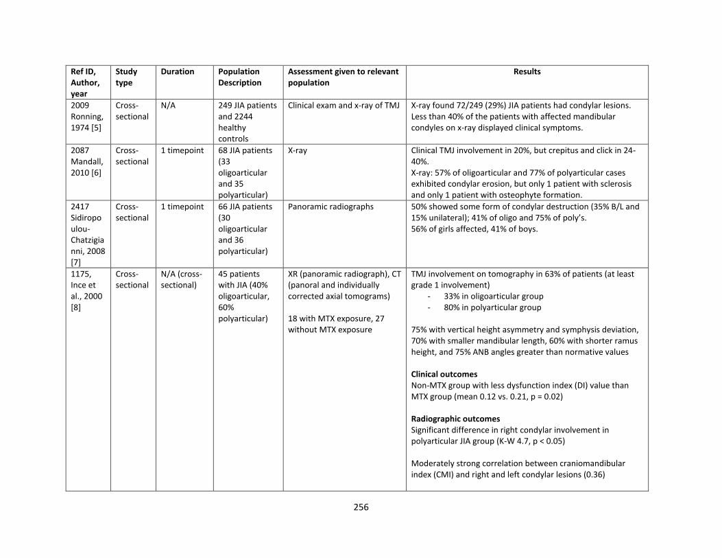
256
Ref ID, Author, year
Study type
Duration Population Description
Assessment given to relevant population
Results
2009 Ronning, 1974 [5]
Cross-sectional
N/A 249 JIA patients and 2244 healthy controls
Clinical exam and x-ray of TMJ X-ray found 72/249 (29%) JIA patients had condylar lesions. Less than 40% of the patients with affected mandibular condyles on x-ray displayed clinical symptoms.
2087 Mandall, 2010 [6]
Cross-sectional
1 timepoint 68 JIA patients (33 oligoarticular and 35 polyarticular)
X-ray Clinical TMJ involvement in 20%, but crepitus and click in 24-40%. X-ray: 57% of oligoarticular and 77% of polyarticular cases exhibited condylar erosion, but only 1 patient with sclerosis and only 1 patient with osteophyte formation.
2417 Sidiropoulou-Chatzigianni, 2008 [7]
Cross-sectional
1 timepoint 66 JIA patients (30 oligoarticular and 36 polyarticular)
Panoramic radiographs 50% showed some form of condylar destruction (35% B/L and 15% unilateral); 41% of oligo and 75% of poly’s. 56% of girls affected, 41% of boys.
1175, Ince et al., 2000 [8]
Cross-sectional
N/A (cross-sectional)
45 patients with JIA (40% oligoarticular, 60% polyarticular)
XR (panoramic radiograph), CT (panoral and individually corrected axial tomograms) 18 with MTX exposure, 27 without MTX exposure
TMJ involvement on tomography in 63% of patients (at least grade 1 involvement)
- 33% in oligoarticular group - 80% in polyarticular group
75% with vertical height asymmetry and symphysis deviation, 70% with smaller mandibular length, 60% with shorter ramus height, and 75% ANB angles greater than normative values Clinical outcomes Non-MTX group with less dysfunction index (DI) value than MTX group (mean 0.12 vs. 0.21, p = 0.02) Radiographic outcomes Significant difference in right condylar involvement in polyarticular JIA group (K-W 4.7, p < 0.05) Moderately strong correlation between craniomandibular index (CMI) and right and left condylar lesions (0.36)
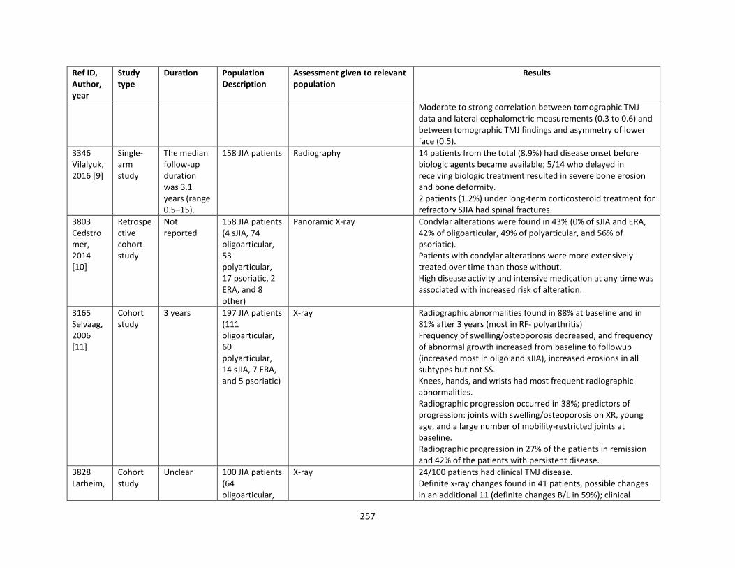
257
Ref ID, Author, year
Study type
Duration Population Description
Assessment given to relevant population
Results
Moderate to strong correlation between tomographic TMJ data and lateral cephalometric measurements (0.3 to 0.6) and between tomographic TMJ findings and asymmetry of lower face (0.5).
3346 Vilalyuk, 2016 [9]
Single-arm study
The median follow-up duration was 3.1 years (range 0.5–15).
158 JIA patients Radiography 14 patients from the total (8.9%) had disease onset before biologic agents became available; 5/14 who delayed in receiving biologic treatment resulted in severe bone erosion and bone deformity. 2 patients (1.2%) under long-term corticosteroid treatment for refractory SJIA had spinal fractures.
3803 Cedstromer, 2014 [10]
Retrospective cohort study
Not reported
158 JIA patients (4 sJIA, 74 oligoarticular, 53 polyarticular, 17 psoriatic, 2 ERA, and 8 other)
Panoramic X-ray Condylar alterations were found in 43% (0% of sJIA and ERA, 42% of oligoarticular, 49% of polyarticular, and 56% of psoriatic). Patients with condylar alterations were more extensively treated over time than those without. High disease activity and intensive medication at any time was associated with increased risk of alteration.
3165 Selvaag, 2006 [11]
Cohort study
3 years 197 JIA patients (111 oligoarticular, 60 polyarticular, 14 sJIA, 7 ERA, and 5 psoriatic)
X-ray Radiographic abnormalities found in 88% at baseline and in 81% after 3 years (most in RF- polyarthritis) Frequency of swelling/osteoporosis decreased, and frequency of abnormal growth increased from baseline to followup (increased most in oligo and sJIA), increased erosions in all subtypes but not SS. Knees, hands, and wrists had most frequent radiographic abnormalities. Radiographic progression occurred in 38%; predictors of progression: joints with swelling/osteoporosis on XR, young age, and a large number of mobility-restricted joints at baseline. Radiographic progression in 27% of the patients in remission and 42% of the patients with persistent disease.
3828 Larheim,
Cohort study
Unclear 100 JIA patients (64 oligoarticular,
X-ray 24/100 patients had clinical TMJ disease. Definite x-ray changes found in 41 patients, possible changes in an additional 11 (definite changes B/L in 59%); clinical
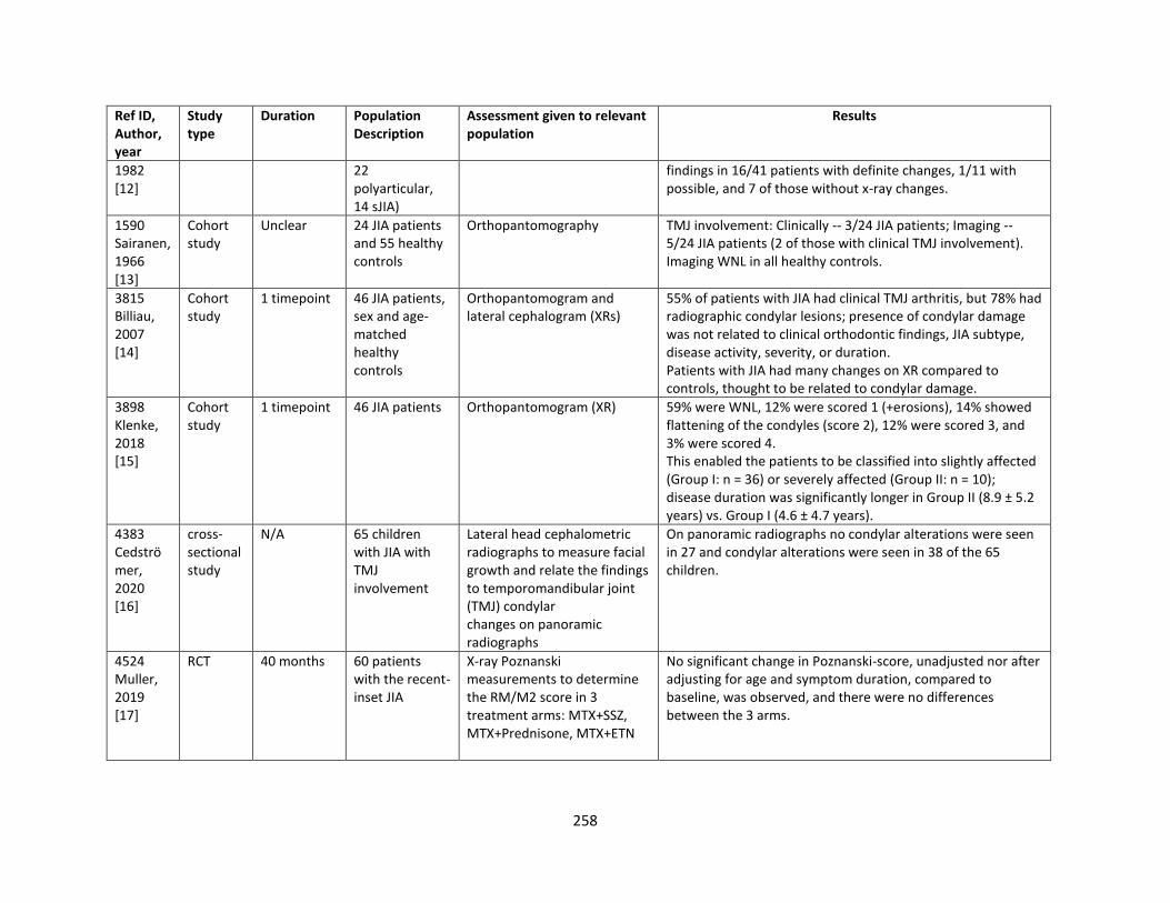
258
Ref ID, Author, year
Study type
Duration Population Description
Assessment given to relevant population
Results
1982 [12]
22 polyarticular, 14 sJIA)
findings in 16/41 patients with definite changes, 1/11 with possible, and 7 of those without x-ray changes.
1590 Sairanen, 1966 [13]
Cohort study
Unclear 24 JIA patients and 55 healthy controls
Orthopantomography TMJ involvement: Clinically -- 3/24 JIA patients; Imaging -- 5/24 JIA patients (2 of those with clinical TMJ involvement). Imaging WNL in all healthy controls.
3815 Billiau, 2007 [14]
Cohort study
1 timepoint 46 JIA patients, sex and age-matched healthy controls
Orthopantomogram and lateral cephalogram (XRs)
55% of patients with JIA had clinical TMJ arthritis, but 78% had radiographic condylar lesions; presence of condylar damage was not related to clinical orthodontic findings, JIA subtype, disease activity, severity, or duration. Patients with JIA had many changes on XR compared to controls, thought to be related to condylar damage.
3898 Klenke, 2018 [15]
Cohort study
1 timepoint 46 JIA patients Orthopantomogram (XR) 59% were WNL, 12% were scored 1 (+erosions), 14% showed flattening of the condyles (score 2), 12% were scored 3, and 3% were scored 4. This enabled the patients to be classified into slightly affected (Group I: n = 36) or severely affected (Group II: n = 10); disease duration was significantly longer in Group II (8.9 ± 5.2 years) vs. Group I (4.6 ± 4.7 years).
4383 Cedströmer, 2020 [16]
cross-sectional study
N/A 65 children with JIA with TMJ involvement
Lateral head cephalometric radiographs to measure facial growth and relate the findings to temporomandibular joint (TMJ) condylar changes on panoramic radiographs
On panoramic radiographs no condylar alterations were seen in 27 and condylar alterations were seen in 38 of the 65 children.
4524 Muller, 2019 [17]
RCT 40 months 60 patients with the recent-inset JIA
X-ray Poznanski measurements to determine the RM/M2 score in 3 treatment arms: MTX+SSZ, MTX+Prednisone, MTX+ETN
No significant change in Poznanski-score, unadjusted nor after adjusting for age and symptom duration, compared to baseline, was observed, and there were no differences between the 3 arms.
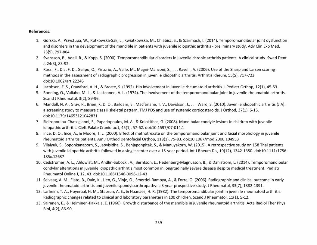
259
References:
1. Gorska, A., Przystupa, W., Rutkowska-Sak, L., Kwiatkowska, M., Chlabicz, S., & Szarmach, I. (2014). Temporomandibular joint dysfunction
and disorders in the development of the mandible in patients with juvenile idiopathic arthritis - preliminary study. Adv Clin Exp Med,
23(5), 797-804.
2. Svensson, B., Adell, R., & Kopp, S. (2000). Temporomandibular disorders in juvenile chronic arthritis patients. A clinical study. Swed Dent
J, 24(3), 83-92.
3. Rossi, F., Dia, F. D., Galipo, O., Pistorio, A., Valle, M., Magni-Manzoni, S., . . . Ravelli, A. (2006). Use of the Sharp and Larsen scoring
methods in the assessment of radiographic progression in juvenile idiopathic arthritis. Arthritis Rheum, 55(5), 717-723.
doi:10.1002/art.22246
4. Jacobsen, F. S., Crawford, A. H., & Broste, S. (1992). Hip involvement in juvenile rheumatoid arthritis. J Pediatr Orthop, 12(1), 45-53.
5. Ronning, O., Valiaho, M. L., & Laaksonen, A. L. (1974). The involvement of the temporomandibular joint in juvenile rheumatoid arthritis.
Scand J Rheumatol, 3(2), 89-96.
6. Mandall, N. A., Gray, R., Brien, K. D. O., Baildam, E., Macfarlane, T. V., Davidson, J., . . . Ward, S. (2010). Juvenile idiopathic arthritis (JIA):
a screening study to measure class II skeletal pattern, TMJ PDS and use of systemic corticosteroids. J Orthod, 37(1), 6-15.
doi:10.1179/14653121042831
7. Sidiropoulou-Chatzigianni, S., Papadopoulos, M. A., & Kolokithas, G. (2008). Mandibular condyle lesions in children with juvenile
idiopathic arthritis. Cleft Palate Craniofac J, 45(1), 57-62. doi:10.1597/07-014.1
8. Ince, D. O., Ince, A., & Moore, T. L. (2000). Effect of methotrexate on the temporomandibular joint and facial morphology in juvenile
rheumatoid arthritis patients. Am J Orthod Dentofacial Orthop, 118(1), 75-83. doi:10.1067/mod.2000.104953
9. Vilaiyuk, S., Soponkanaporn, S., Jaovisidha, S., Benjaponpitak, S., & Manuyakorn, W. (2015). A retrospective study on 158 Thai patients
with juvenile idiopathic arthritis followed in a single center over a 15-year period. Int J Rheum Dis, 19(12), 1342-1350. doi:10.1111/1756-
185x.12637
10. Cedstromer, A. L., Ahlqwist, M., Andlin-Sobocki, A., Berntson, L., Hedenberg-Magnusson, B., & Dahlstrom, L. (2014). Temporomandibular
condylar alterations in juvenile idiopathic arthritis most common in longitudinally severe disease despite medical treatment. Pediatr
Rheumatol Online J, 12, 43. doi:10.1186/1546-0096-12-43
11. Selvaag, A. M., Flato, B., Dale, K., Lien, G., Vinje, O., Smerdel-Ramoya, A., & Forre, O. (2006). Radiographic and clinical outcome in early
juvenile rheumatoid arthritis and juvenile spondyloarthropathy: a 3-year prospective study. J Rheumatol, 33(7), 1382-1391.
12. Larheim, T. A., Hoyeraal, H. M., Stabrun, A. E., & Haanaes, H. R. (1982). The temporomandibular joint in juvenile rheumatoid arthritis.
Radiographic changes related to clinical and laboratory parameters in 100 children. Scand J Rheumatol, 11(1), 5-12.
13. Sairanen, E., & Helminen-Pakkala, E. (1966). Growth disturbance of the mandible in juvenile rheumatoid arthritis. Acta Radiol Ther Phys
Biol, 4(2), 86-90.
260
14. Billiau, A. D., Hu, Y., Verdonck, A., Carels, C., & Wouters, C. (2007). Temporomandibular joint arthritis in juvenile idiopathic arthritis:
prevalence, clinical and radiological signs, and relation to dentofacial morphology. J Rheumatol, 34(9), 1925-1933.
15. Klenke, D., Quast, A., Prelog, M., Holl-Wieden, A., Riekert, M., Stellzig-Eisenhauer, A., & Meyer-Marcotty, P. (2018). TMJ
pathomorphology in patients with JIA-radiographic parameters for early diagnosis. Head Face Med, 14(1), 15. doi:10.1186/s13005-018-
0173-5
16. Cedströmer, A.-L., Andlin-Sobocki, A., Abbu, N., Hedenberg-Magnusson, B., Dahlström, L., & Berntson, L. (2020). Condylar alterations and
facial growth in children with juvenile idiopathic arthritis. Journal of Orofacial Orthopedics / Fortschritte der Kieferorthopädie, 81(3),
163-171. doi:10.1007/s00056-020-00216-8.
17. Hissink Muller, P. C. E., van Braak, W. G., Schreurs, D., Nusman, C. M., Bergstra, S. A., Hemke, R., . . . Maas, M. (2019). No radiographic
wrist damage after treatment to target in recent-onset juvenile idiopathic arthritis. Pediatric rheumatology, 17(1), 62.
doi:10.1186/s12969-019-0362-1.
CT: Eight studies evaluated CT for assessment of JIA. One study reported muscle cross sectional area (CSA) median Z-score: onset -1.94; follow-
up -1.10; Cortical thickness: onset -1.55; follow-up -0.97; Marrow area: onset 0.96; follow-up 1.05; Cortical density: onset 0.34; follow-up 0.69;
Trabecular density: onset -0.75; follow-up -0.36; Strength–strain index (SSI) onset -0.79; follow-up -0.13 [1]. There was moderate to strong
correlation between tomographic TMJ data and lateral cephalometric measurements (0.3 to 0.6) and between tomographic TMJ findings and
asymmetry of lower face (0.5) [2]. On peripheral quantitative computed tomography (PQCT) of radial diaphysis total cross-sectional area and
BMC were decreased somewhat less in the oligoarticular than in the other groups, total bone mineral density was reduced in the polyarticular
and systemic JIA population and normal in oligoarticular and connective tissue disease populations, muscle cross sectional area was decreased in
all groups [3]. In spite of the articular alterations having been frequent in the TMJs evaluated by means of cone beam CBCT in 25 patients, only 5
ATMs presented a clinical diagnosis that could lead to suspecting some articular bone alteration, thereby perhaps confirming the risk of
diagnostic underestimation [4]. On high resolution CT, TMJ lesions were more prevalent in those with polyarticular disease, and the largest AP
measurements of the mandibular condyle depth were found in those with severe TMJ lesions (9.1 +/- 1) without statistically significant
differences among patients with mild lesions (8.4+/- 0.9) and without TMJ lesions (7.9 +/- 1.2 mm) [5]. In other study, 62% of patients had
morphologic evidence of TMJ involvement on CT, with a correlation between the craniomandibular index value and the pathosis index for the
left joint as 0.376, and correlation between the vertical difference in mandibular angle regions and the average grade of TMJ as 0.303 [6].
Quality of evidence across all critical outcomes: Very low
Table 5. Studies of Computed Tomography (CT)
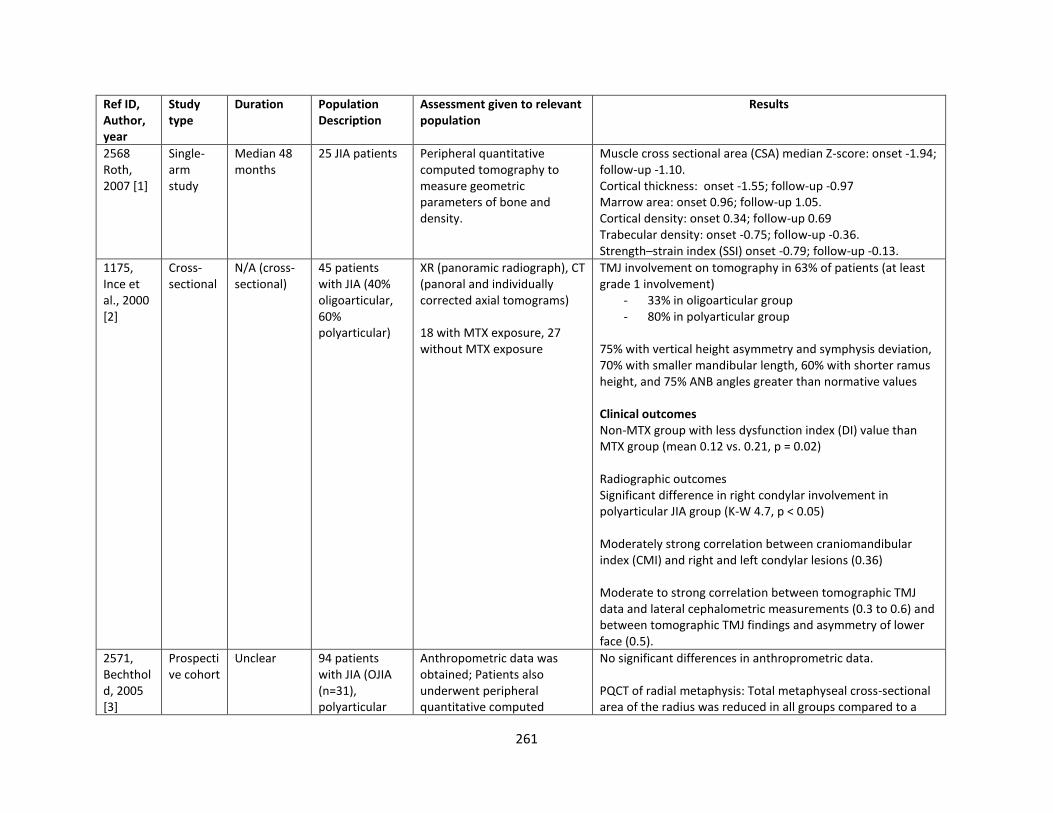
261
Ref ID, Author, year
Study type
Duration Population Description
Assessment given to relevant population
Results
2568 Roth, 2007 [1]
Single-arm study
Median 48 months
25 JIA patients Peripheral quantitative computed tomography to measure geometric parameters of bone and density.
Muscle cross sectional area (CSA) median Z-score: onset -1.94; follow-up -1.10. Cortical thickness: onset -1.55; follow-up -0.97 Marrow area: onset 0.96; follow-up 1.05. Cortical density: onset 0.34; follow-up 0.69 Trabecular density: onset -0.75; follow-up -0.36. Strength–strain index (SSI) onset -0.79; follow-up -0.13.
1175, Ince et al., 2000 [2]
Cross-sectional
N/A (cross-sectional)
45 patients with JIA (40% oligoarticular, 60% polyarticular)
XR (panoramic radiograph), CT (panoral and individually corrected axial tomograms) 18 with MTX exposure, 27 without MTX exposure
TMJ involvement on tomography in 63% of patients (at least grade 1 involvement)
- 33% in oligoarticular group - 80% in polyarticular group
75% with vertical height asymmetry and symphysis deviation, 70% with smaller mandibular length, 60% with shorter ramus height, and 75% ANB angles greater than normative values Clinical outcomes Non-MTX group with less dysfunction index (DI) value than MTX group (mean 0.12 vs. 0.21, p = 0.02) Radiographic outcomes Significant difference in right condylar involvement in polyarticular JIA group (K-W 4.7, p < 0.05) Moderately strong correlation between craniomandibular index (CMI) and right and left condylar lesions (0.36) Moderate to strong correlation between tomographic TMJ data and lateral cephalometric measurements (0.3 to 0.6) and between tomographic TMJ findings and asymmetry of lower face (0.5).
2571, Bechthold, 2005 [3]
Prospective cohort
Unclear 94 patients with JIA (OJIA (n=31), polyarticular
Anthropometric data was obtained; Patients also underwent peripheral quantitative computed
No significant differences in anthroprometric data. PQCT of radial metaphysis: Total metaphyseal cross-sectional area of the radius was reduced in all groups compared to a
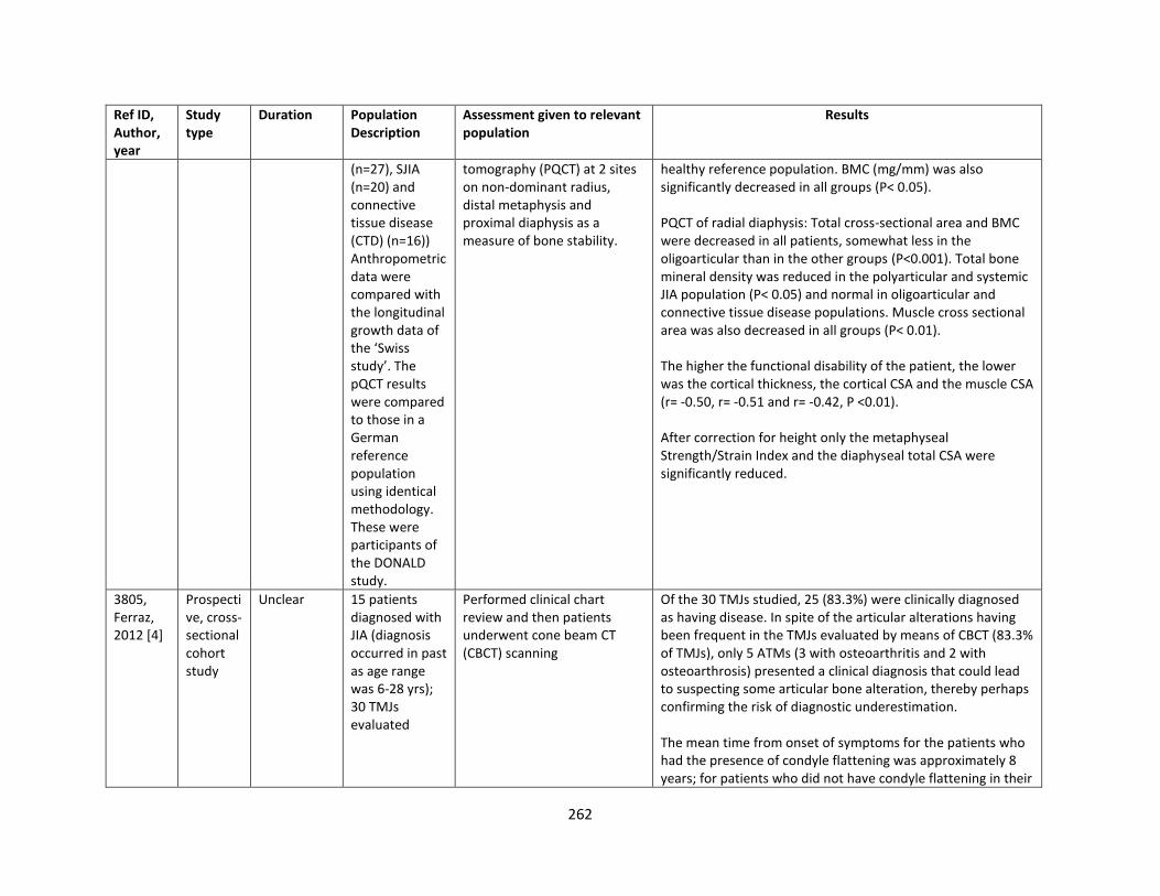
262
Ref ID, Author, year
Study type
Duration Population Description
Assessment given to relevant population
Results
(n=27), SJIA (n=20) and connective tissue disease (CTD) (n=16)) Anthropometric data were compared with the longitudinal growth data of the ‘Swiss study’. The pQCT results were compared to those in a German reference population using identical methodology. These were participants of the DONALD study.
tomography (PQCT) at 2 sites on non-dominant radius, distal metaphysis and proximal diaphysis as a measure of bone stability.
healthy reference population. BMC (mg/mm) was also significantly decreased in all groups (P< 0.05). PQCT of radial diaphysis: Total cross-sectional area and BMC were decreased in all patients, somewhat less in the oligoarticular than in the other groups (P<0.001). Total bone mineral density was reduced in the polyarticular and systemic JIA population (P< 0.05) and normal in oligoarticular and connective tissue disease populations. Muscle cross sectional area was also decreased in all groups (P< 0.01). The higher the functional disability of the patient, the lower was the cortical thickness, the cortical CSA and the muscle CSA (r= -0.50, r= -0.51 and r= -0.42, P <0.01). After correction for height only the metaphyseal Strength/Strain Index and the diaphyseal total CSA were significantly reduced.
3805, Ferraz, 2012 [4]
Prospective, cross-sectional cohort study
Unclear 15 patients diagnosed with JIA (diagnosis occurred in past as age range was 6-28 yrs); 30 TMJs evaluated
Performed clinical chart review and then patients underwent cone beam CT (CBCT) scanning
Of the 30 TMJs studied, 25 (83.3%) were clinically diagnosed as having disease. In spite of the articular alterations having been frequent in the TMJs evaluated by means of CBCT (83.3% of TMJs), only 5 ATMs (3 with osteoarthritis and 2 with osteoarthrosis) presented a clinical diagnosis that could lead to suspecting some articular bone alteration, thereby perhaps confirming the risk of diagnostic underestimation. The mean time from onset of symptoms for the patients who had the presence of condyle flattening was approximately 8 years; for patients who did not have condyle flattening in their
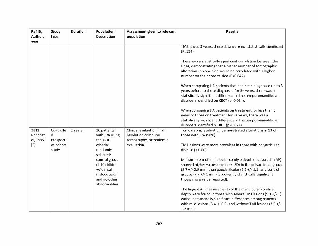
263
Ref ID, Author, year
Study type
Duration Population Description
Assessment given to relevant population
Results
TMJ, it was 3 years, these data were not statistically significant (P .334). There was a statistically significant correlation between the sides, demonstrating that a higher number of tomographic alterations on one side would be correlated with a higher number on the opposite side (P=0.047). When comparing JIA patients that had been diagnosed up to 3 years before to those diagnosed for 3+ years, there was a statistically significant difference in the temporomandibular disorders identified on CBCT (p=0.024). When comparing JIA patients on treatment for less than 3 years to those on treatment for 3+ years, there was a statistically significant difference in the temporomandibular disorders identified n CBCT (p=0.024).
3811, Ronchezel, 1995 [5]
Controlled Prospective cohort study
2 years 26 patients with JRA using the ACR criteria; randomly selected; control group of 10 children w/ dental malocclusion and no other abnormalities
Clinical evaluation, high resolution computer tomography, orthodontic evaluation
Tomographic evaluation demonstrated alterations in 13 of those with JRA (50%). TMJ lesions were more prevalent in those with polyarticular disease (71.4%). Measurement of mandibular condyle depth (measured in AP) showed higher values (mean +/- SD) in the polyarticular group (8.7 +/- 0.9 mm) than pauciarticular (7.7 +/- 1.1) and control groups (7.7 +/- 1 mm) (apparently statistically significant though no p value reported). The largest AP measurements of the mandibular condyle depth were found in those with severe TMJ lesions (9.1 +/- 1) without statistically significant differences among patients with mild lesions (8.4+/- 0.9) and without TMJ lesions (7.9 +/- 1.2 mm).
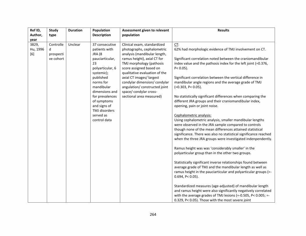
264
Ref ID, Author, year
Study type
Duration Population Description
Assessment given to relevant population
Results
3829, Hu, 1996 [6]
Controlled prospective cohort
Unclear 37 consecutive patients with JRA (8 pauciarticular, 23 polyarticular, 6 systemic); published norms for mandibular dimensions and for prevalences of symptoms and signs of TMJ disorders served as control data
Clinical exam, standardized photographs, cephalometric analysis (mandibular length, ramus height), axial CT for TMJ morphology (pathosis score assigned based on qualitative evaluation of the axial CT images/ largest condylar dimension/ condylar angulation/ constructed joint space/ condylar cross-sectional area measured)
CT: 62% had morphologic evidence of TMJ involvement on CT. Significant correlation noted between the craniomandibular index value and the pathosis index for the left joint (=0.376, P< 0.05). Significant correlation between the vertical difference in mandibular angle regions and the average grade of TMJ (=0.303, P< 0.05). No statistically significant differences when comparing the different JRA groups and their craniomandibular index, opening, pain or joint noise. Cephalometric analysis: Using cephalometric analysis, smaller mandibular lengths were observed in the JRA sample compared to controls though none of the mean differences attained statistical significance. There was also no statistical significance reached when the three JRA groups were investigated indenpendently. Ramus height was was ‘considerably smaller’ in the polyarticular group than in the other two groups. Statistically significant inverse relationships found between average grade of TMJ and the mandibular length as well as ramus height in the pauciarticular and polyarticular groups (=-0.694, P< 0.05). Standardized measures (age-adjusted) of mandibular length and ramus height were also significantly negatively correlated with the average grades of TMJ lesions (=-0.505, P< 0.005; =-0.329, P< 0.05). Those with the most severe joint
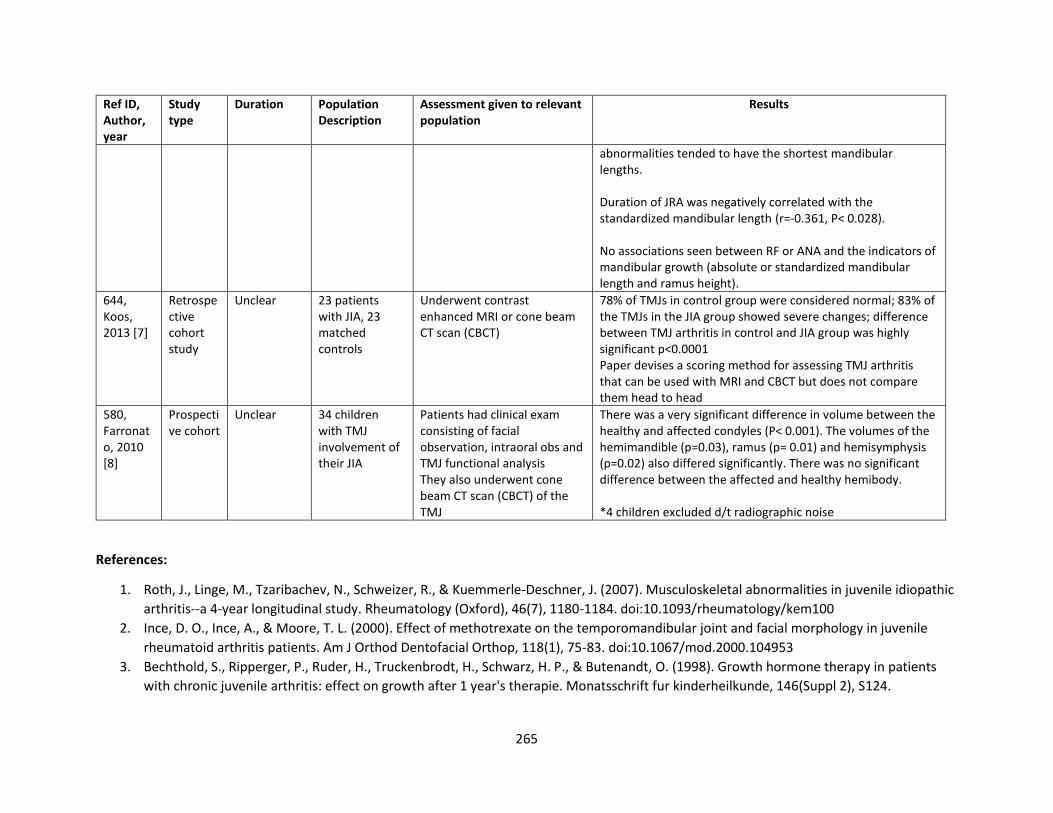
265
Ref ID, Author, year
Study type
Duration Population Description
Assessment given to relevant population
Results
abnormalities tended to have the shortest mandibular lengths. Duration of JRA was negatively correlated with the standardized mandibular length (r=-0.361, P< 0.028). No associations seen between RF or ANA and the indicators of mandibular growth (absolute or standardized mandibular length and ramus height).
644, Koos, 2013 [7]
Retrospective cohort study
Unclear 23 patients with JIA, 23 matched controls
Underwent contrast enhanced MRI or cone beam CT scan (CBCT)
78% of TMJs in control group were considered normal; 83% of the TMJs in the JIA group showed severe changes; difference between TMJ arthritis in control and JIA group was highly significant p<0.0001 Paper devises a scoring method for assessing TMJ arthritis that can be used with MRI and CBCT but does not compare them head to head
580, Farronato, 2010 [8]
Prospective cohort
Unclear 34 children with TMJ involvement of their JIA
Patients had clinical exam consisting of facial observation, intraoral obs and TMJ functional analysis They also underwent cone beam CT scan (CBCT) of the TMJ
There was a very significant difference in volume between the healthy and affected condyles (P< 0.001). The volumes of the hemimandible (p=0.03), ramus (p= 0.01) and hemisymphysis (p=0.02) also differed significantly. There was no significant difference between the affected and healthy hemibody. *4 children excluded d/t radiographic noise
References:
1. Roth, J., Linge, M., Tzaribachev, N., Schweizer, R., & Kuemmerle-Deschner, J. (2007). Musculoskeletal abnormalities in juvenile idiopathic
arthritis--a 4-year longitudinal study. Rheumatology (Oxford), 46(7), 1180-1184. doi:10.1093/rheumatology/kem100
2. Ince, D. O., Ince, A., & Moore, T. L. (2000). Effect of methotrexate on the temporomandibular joint and facial morphology in juvenile
rheumatoid arthritis patients. Am J Orthod Dentofacial Orthop, 118(1), 75-83. doi:10.1067/mod.2000.104953
3. Bechthold, S., Ripperger, P., Ruder, H., Truckenbrodt, H., Schwarz, H. P., & Butenandt, O. (1998). Growth hormone therapy in patients
with chronic juvenile arthritis: effect on growth after 1 year's therapie. Monatsschrift fur kinderheilkunde, 146(Suppl 2), S124.
266
4. Ferraz, A. M., Devito, K. L., & Guimaraes, J. P. (2012). Temporomandibular disorder in patients with juvenile idiopathic arthritis: clinical
evaluation and correlation with the findings of cone beam computed tomography. Oral Surg Oral Med Oral Pathol Oral Radiol, 114(3),
e51-57. doi:10.1016/j.oooo.2012.02.010
5. Ronchezel, M. V., Hilario, M. O., Goldenberg, J., Lederman, H. M., Faltin, K., Azevedo, M. F. d., & Naspitz, C. K. (1995).
Temporomandibular joint and mandibular growth alterations in patients with juvenile rheumatoid arthritis. J Rheumatol, 22(10), 1956-
1961.
6. Hu, Y. S., & Schneiderman, E. D. (1995). The temporomandibular joint in juvenile rheumatoid arthritis: I. Computed tomographic
findings. Pediatr Dent, 17(1), 46-53.
7. Koos, B., Tzaribachev, N., Bott, S., Ciesielski, R., & Godt, A. (2013). Classification of temporomandibular joint erosion, arthritis, and
inflammation in patients with juvenile idiopathic arthritis. J Orofac Orthop, 74(6), 506-519. doi:10.1007/s00056-013-0166-8
8. Farronato, G., Garagiola, U., Carletti, V., Cressoni, P., Mercatali, L., & Farronato, D. (2010). Change in condylar and mandibular
morphology in juvenile idiopathic arthritis: Cone Beam volumetric imaging. Minerva Stomatol, 59(10), 519-534.
18F-FDG: Three studies evaluated 18F-FDG. There was a significant association between the degree of 18F-FDG uptake and typical clinical,
radiographic and biochemical findings in JIA [1]. Correlation of 18F-FDG PET/CT with clinical disease activity of AOSD: Visual grade of lymph node
with the systemic score of AOSD (r = 0.664). SUV intensity of lymph node with the systemic score (r = 0.601). Visual grade of spleen with the
systemic score (r =0.771), ESR (r = 0.617), and ferritin (r =0.557). SUV intensity of spleen with the systemic score (r = 0.676). Visual grade of bone
marrow with the systemic score (r = 0.734), ESR (r = 0.761), leukocyte (r = 0.775), and neutrophil (r =0.711). SUV intensity of bone marrow with
the systemic score (r = 0.57), ESR (r = 0.612), leukocyte (r = 0.773), and neutrophil (r = 0.725) [2]. FDG-PET showed a diffuse distribution pattern
in inflamed joints. There was no accumulation in the bone marrow. In 12 sJIA patients it was found either in all vertebral bodies, pelvis, and
around large joints. Accumulation was not in synovia but in the bone itself or at the end of long bones and thought to be in the bone marrow.
There was greater accumulation in the spleen compared to the liver; 3 out of these 12 patients developed MAS. In 8 cases of sJIA, there was
diffuse accumulation in inflamed joints, no bone marrow involvement, no significant difference between liver and spleen, and none of these
patients had MAS. [3]
Quality of evidence across all critical outcomes: Very low
Table 6. Studies of 18F-FDG
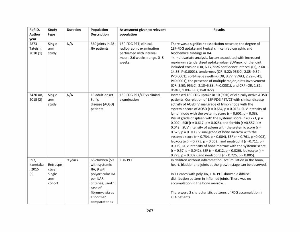
267
Ref ID, Author, year
Study type
Duration Population Description
Assessment given to relevant population
Results
2873 Tateishi, 2010 [1]
Single-arm study
N/A 560 joints in 28 JIA patients
18F-FDG PET, clinical, radiographic examination performed with interval mean, 2.6 weeks; range, 0–5 weeks.
There was a significant association between the degree of 18F-FDG uptake and typical clinical, radiographic and biochemical findings in JIA. In multivariate analysis, factors associated with increased maximum standardized uptake value (SUVmax) of the joint included erosion (OR, 6.17; 95% confidence interval (CI), 2.60–14.66; P<0.0001), tenderness (OR, 5.22; 95%CI, 2.85–9.57; P<0.0001), soft-tissue swelling (OR, 3.77; 95%CI, 2.22–6.41; P<0.0001), the presence of multiple major joints involvement (OR, 3.50; 95%CI, 2.10–5.83; P<0.0001), and CRP (OR, 1.81; 95%CI, 1.09– 3.02; P=0.022).
3420 An, 2015 [2]
Single-arm study
N/A 13 adult-onset Still’s disease (AOSD) patients
18F-FDG PET/CT vs clinical examination
Increased 18F-FDG uptake in 10 (90%) of clinically active AOSD patients. Correlation of 18F-FDG PET/CT with clinical disease activity of AOSD: Visual grade of lymph node with the systemic score of AOSD (r = 0.664, p = 0.013). SUV intensity of lymph node with the systemic score (r = 0.601, p = 0.03). Visual grade of spleen with the systemic score (r =0.771, p = 0.002), ESR (r = 0.617, p = 0.025), and ferritin (r =0.557, p = 0.048). SUV intensity of spleen with the systemic score (r = 0.676, p = 0.011). Visual grade of bone marrow with the systemic score (r = 0.734, p = 0.004), ESR (r = 0.761, p =0.003), leukocyte (r = 0.775, p = 0.002), and neutrophil (r =0.711, p = 0.006). SUV intensity of bone marrow with the systemic score (r = 0.57, p = 0.042), ESR (r = 0.612, p = 0.026), leukocyte (r = 0.773, p = 0.002), and neutrophil (r = 0.725, p = 0.005).
597, Kanetaka, 2015 [3]
Retrospective single arm cohort
9 years 68 children (59 with systemic JIA, 9 with polyarticular JIA per ILAR criteria); used 1 case of fibromyalgia as a ‘normal’ comparator as
FDG PET In children without inflammation, accumulation in the brain, heart, bladder and joints at the growth stage can be observed. In 11 cases with poly JIA, FDG PET showed a diffuse distribution pattern in inflamed joints. There was no accumulation in the bone marrow. There were 2 characteristic patterns of FDG accumulation in sJIA patients.
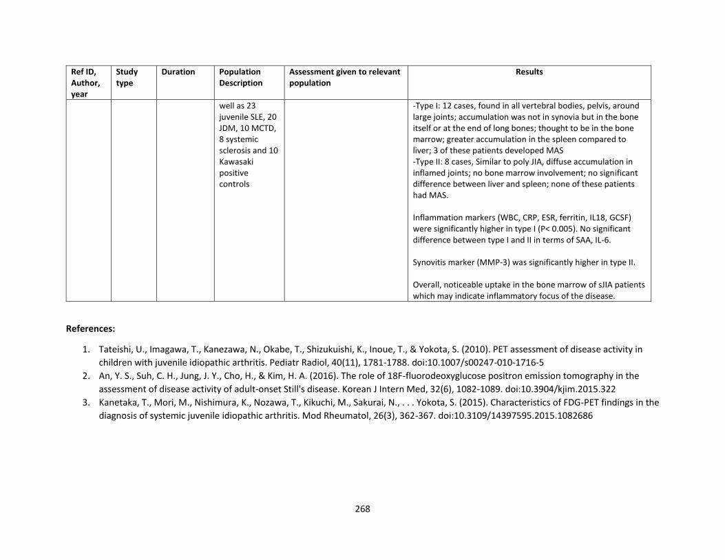
268
Ref ID, Author, year
Study type
Duration Population Description
Assessment given to relevant population
Results
well as 23 juvenile SLE, 20 JDM, 10 MCTD, 8 systemic sclerosis and 10 Kawasaki positive controls
-Type I: 12 cases, found in all vertebral bodies, pelvis, around large joints; accumulation was not in synovia but in the bone itself or at the end of long bones; thought to be in the bone marrow; greater accumulation in the spleen compared to liver; 3 of these patients developed MAS -Type II: 8 cases, Similar to poly JIA, diffuse accumulation in inflamed joints; no bone marrow involvement; no significant difference between liver and spleen; none of these patients had MAS. Inflammation markers (WBC, CRP, ESR, ferritin, IL18, GCSF) were significantly higher in type I (P< 0.005). No significant difference between type I and II in terms of SAA, IL-6. Synovitis marker (MMP-3) was significantly higher in type II. Overall, noticeable uptake in the bone marrow of sJIA patients which may indicate inflammatory focus of the disease.
References:
1. Tateishi, U., Imagawa, T., Kanezawa, N., Okabe, T., Shizukuishi, K., Inoue, T., & Yokota, S. (2010). PET assessment of disease activity in
children with juvenile idiopathic arthritis. Pediatr Radiol, 40(11), 1781-1788. doi:10.1007/s00247-010-1716-5
2. An, Y. S., Suh, C. H., Jung, J. Y., Cho, H., & Kim, H. A. (2016). The role of 18F-fluorodeoxyglucose positron emission tomography in the
assessment of disease activity of adult-onset Still's disease. Korean J Intern Med, 32(6), 1082-1089. doi:10.3904/kjim.2015.322
3. Kanetaka, T., Mori, M., Nishimura, K., Nozawa, T., Kikuchi, M., Sakurai, N., . . . Yokota, S. (2015). Characteristics of FDG-PET findings in the
diagnosis of systemic juvenile idiopathic arthritis. Mod Rheumatol, 26(3), 362-367. doi:10.3109/14397595.2015.1082686
269
PICO 56: In children with JIA who require IA corticosteroid (IAC) injections, should injections be done with imaging guidance?
Summary. The literature search identified 12 studies that addressed this question in patients with JIA. Studies included two controlled cohort
studies, with the rest being single-arm cohort studies and case series. Interventions included: IAC injections with no guidance (one study),
Ultrasound (US) guidance, and CT guided injections. Six observational studies addressed IAC guided injections in different joints including a mix of
peripheral joints and TMJ [1, 2, 3, 4, 5, 6], while six studies addressed IAC guidance for TMJ steroid injections[7, 8, 9, 10, 11, 12] (Table 1).
IAC injections – non-TMJ studies
Cunha et al.[1] published a cohort study evaluating US-guided intra-articular corticosteroid injections in 16 patients (with a longer disease duration
and previous injections) and compared them to non-US guided IAC injections (n=149). US-guided injections showed poor response to IAC (p = 0.02)
and associated with higher CHAQ values (p = 0.03), higher number of injections with poor response (p = 0.01) and a shorter time to relapse (p =
0.02) compared to patients submitted to blindly performed procedures. This study highlights a selection bias for the US guided group likely had a
more severe disease process which may have led to worse outcomes.
Young et al.[3] examined 241 US-guided 1444 corticosteroid injections (1340 joints, 104 tendon sheaths), 414 of which were repeat injections.
Reported complications were minimal and included subcutaneous tissue atrophy (29 injections, 2.1%), skin hypopigmentation (4 injections, 0.3%),
erythema and pruritis (2 injections, 0.1%), soft tissue atrophy + skin hypopigmentation (2 injections, 0.1%), 2 patients had avascular necrosis (AVN)
(hip, tibiotalar) but attributed to long-term systemic steroid therapy. Young et al., further examined US guided IAC in 2015.[2] This observational
study investigated US-guided subtalar corticosteroid injections in 122 patients. Injections were complicated in 9/241 (3.7) % with atrophy of or
skin hypopigmentation, isolated atrophy of subcutaneous soft tissues occurred in 5/241 (2.1%), skin hypopigmentation and subcutaneous atrophy
in 3/241 (1.2%), and one patient had low-grade fever (n = 1).
Laurell et al. published a case series[4] and cohort study[5] with focus on US-guided IAC injections in patients with JIA. The authors report accurate
placement of corticosteroid in all injected compartments. In their cohort study there was reported Clinical resolution of active arthritis in 72% of
the cases and in the case series, they reported 4-week post-injection normalization of synovial hypertrophy in 86%, normalization of hyperemia in
91%; 12/15 wrists (80%) clinically inactive arthritis. They reported 1 relapse in a patient with oligo JIA 7 months after steroid injection. The authors
reported 4.7% rate of subcutaneous atrophy.
A cohort study by Tynjala et al.[6] examined hip US-guided injections in 20 affected hips. They measured treatment response by absence of clinical
signs and symptoms and no effusion on US. Following injections, mean duration of efficacy 8.3 months (median 11.5 months), 14 (70%) of hip
joints had normal clinical examination and ultrasound at 3 months, 14 (70%) at 6 months and 10 (50%) of hip joints at 12 months without synovitis.
No side effects were observed for 12 months follow up period.
270
TMJ guided IAC injections:
A retrospective cohort study by Resnick et al.[7] compared Image-guided [CT or Ultrasound] TMJ IAC injections. The authors observed no
differences in resolution of pain (p = 1.00), increase in maximal incisal opening (MIO) (p = 0.975), or decrease in synovial enhancement ratio (ER)
(p = 0.492) between landmark and image guided injections. A statistically significant longer average procedure time was observed in image-guided
group (p < 0.008).
Parra et al.[8] investigated US guidance in total of 180 TMJ injections, 127 injections (70%) needle placement confirmed with CT (limited focused
CT protocol). Needle placement was acceptable in 115/127 joints (91%) of those confirmed with CT with major readjustment required in 12/127
joints (9%). As for the outcomes, 80/99 (81%) good response at follow up visit. Observed complications of US guided IAC in this study included skin
atrophy in 1 patient and transient early swelling and pain in 10 patients. In contrast, Habibi et al.[9] performed 63 US-guided steroid injections and
reported efficacy of IAC and symptom resolution in 58/63 (92.06%); 1 child developed scar at site of injection. A retrospective review by Cahill
et.al.[10] where 27 CT guided intraarticular TMJ steroid injections were performed concluded that intraarticular TMJ injection of a long-acting
steroid in children is a safe procedure even in patients with joint space deformities, with reduction in acute and subacute inflammatory changes
noted on MRI. Arabshahi et al.[11] reported that CT-guided TMJ IAC injections alleviated pain in more than two-thirds of symptomatic patients;
one-third of these patients had persistence of effusions on follow up MRI (77 % of 13 patients with pre-injection pain had pain resolution; P 0.05).
Presence of jaw pain did not significantly correlate with the presence of effusions on MRI (P=0.96). Side effects reported in this study included
facial swelling in 2 patients consistent with Cushing ’syndrome, lasting 2 days in 1 patient and 2 weeks in the other. In contrast, a retrospective
chart review by Ringold et al.[12] TMJ IAS injections were done under general anesthesia but without imaging guidance, outcomes of this study
showed that there was a mean increase in MIO of 3.8 mm following each IAS injection (p = 0.003; 95% CI 1.4, 6.2). Patients who underwent multiple
IAS injections had a mean increase in MIO after first injection of 6.6 mm (p < 0.001; 95% CI 4.1, 9.1); however, the mean increase in MIO after
subsequent injections was 0.4 mm (p = 0.8; 95% CI –3.5, 4.4). Authors reported complications that were more or less similar to imaging guided
studies. One patient developed subcutaneous atrophy at the injection site, two patients developed small, asymptomatic intraarticular
calcifications. No additional adverse events were reported.
Quality of evidence across all critical outcomes: Very low
Table 1. Data from Observational Studies
Ref ID, Author, year
Study type Duration Population Description
Intervention given to relevant population
Results
Non-TMJ injection studies
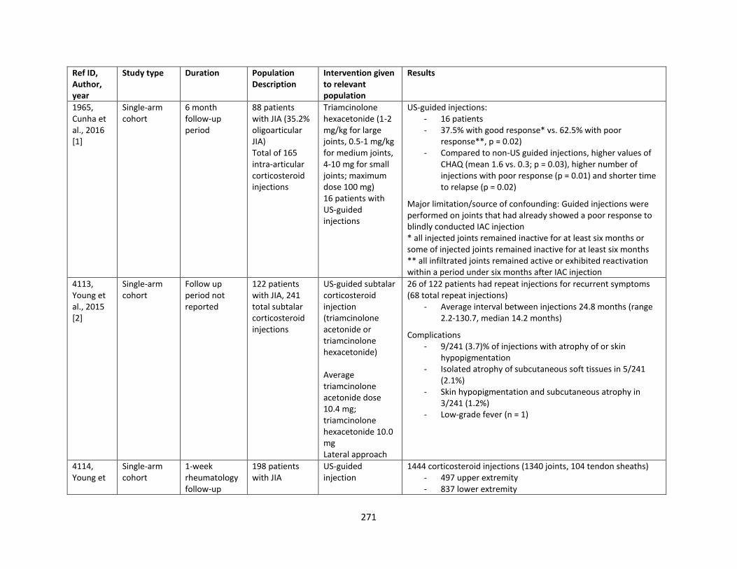
271
Ref ID, Author, year
Study type Duration Population Description
Intervention given to relevant population
Results
1965, Cunha et al., 2016 [1]
Single-arm cohort
6 month follow-up period
88 patients with JIA (35.2% oligoarticular JIA) Total of 165 intra-articular corticosteroid injections
Triamcinolone hexacetonide (1-2 mg/kg for large joints, 0.5-1 mg/kg for medium joints, 4-10 mg for small joints; maximum dose 100 mg) 16 patients with US-guided injections
US-guided injections: - 16 patients - 37.5% with good response* vs. 62.5% with poor
response**, p = 0.02) - Compared to non-US guided injections, higher values of
CHAQ (mean 1.6 vs. 0.3; p = 0.03), higher number of injections with poor response (p = 0.01) and shorter time to relapse (p = 0.02)
Major limitation/source of confounding: Guided injections were performed on joints that had already showed a poor response to blindly conducted IAC injection * all injected joints remained inactive for at least six months or some of injected joints remained inactive for at least six months ** all infiltrated joints remained active or exhibited reactivation within a period under six months after IAC injection
4113, Young et al., 2015 [2]
Single-arm cohort
Follow up period not reported
122 patients with JIA, 241 total subtalar corticosteroid injections
US-guided subtalar corticosteroid injection (triamcinolone acetonide or triamcinolone hexacetonide) Average triamcinolone acetonide dose 10.4 mg; triamcinolone hexacetonide 10.0 mg Lateral approach
26 of 122 patients had repeat injections for recurrent symptoms (68 total repeat injections)
- Average interval between injections 24.8 months (range 2.2-130.7, median 14.2 months)
Complications - 9/241 (3.7)% of injections with atrophy of or skin
hypopigmentation - Isolated atrophy of subcutaneous soft tissues in 5/241
(2.1%) - Skin hypopigmentation and subcutaneous atrophy in
3/241 (1.2%) - Low-grade fever (n = 1)
4114, Young et
Single-arm cohort
1-week rheumatology follow-up
198 patients with JIA
US-guided injection
1444 corticosteroid injections (1340 joints, 104 tendon sheaths) - 497 upper extremity - 837 lower extremity
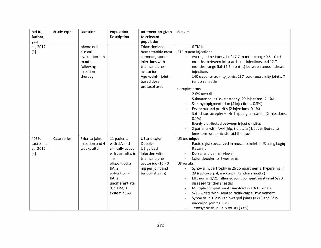
272
Ref ID, Author, year
Study type Duration Population Description
Intervention given to relevant population
Results
al., 2012 [3]
phone call, clinical evaluation 1–3 months following injection therapy
Triamcinolone hexacetonide most common, some injections with triamcinolone acetonide Age-weight-joint-based dose protocol used
- 6 TMJs 414 repeat injections
- Average time interval of 17.7 months (range 0.5-101.5 months) between intra-articular injections and 12.7 months (range 5.6-16.9 months) between tendon sheath injections
- 140 upper extremity joints, 267 lower extremity joints, 7 tendon sheaths
Complications - 2.6% overall - Subcutaneous tissue atrophy (29 injections, 2.1%) - Skin hypopigmentation (4 injections, 0.3%) - Erythema and pruritis (2 injections, 0.1%) - Soft tissue atrophy + skin hypopigmentation (2 injections,
0.1%) - Evenly distributed between injection sites - 2 patients with AVN (hip, tibiotalar) but attributed to
long-term systemic steroid therapy
4089, Laurell et al., 2012 [4]
Case series Prior to joint injection and 4 weeks after
11 patients with JIA and clinically active wrist arthritis (n = 5 oligoarticular JIA, 2 polyarticular JIA, 2 undifferentiated, 1 ERA, 1 systemic JIA)
US and color Doppler US-guided injection with triamcinolone acetonide (10-40 mg per joint and tendon sheath)
US technique - Radiologist specialized in musculoskeletal US using Logiq
9 scanner - Dorsal and palmar views - Color doppler for hyperemia
US results - Synovial hypertrophy in 26 compartments, hyperemia in
23 (radio-carpal, midcarpal, tendon sheaths) - Effusion in 2/21 inflamed joint compartments and 5/20
diseased tendon sheaths - Multiple compartments involved in 10/15 wrists - 5/15 wrists with isolated radio-carpal involvement - Synovitis in 13/15 radio-carpal joints (87%) and 8/15
midcarpal joints (53%) - Tenosynovitis in 5/15 wrists (33%)
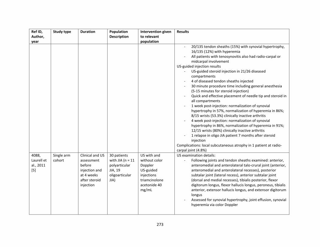
273
Ref ID, Author, year
Study type Duration Population Description
Intervention given to relevant population
Results
- 20/135 tendon sheaths (15%) with synovial hypertrophy, 16/135 (12%) with hyperemia
- All patients with tenosynovitis also had radio-carpal or midcarpal involvement
US-guided injection results - US-guided steroid injection in 21/26 diseased
compartments - 4 of diseased tendon sheaths injected - 30 minute procedure time including general anesthesia
(5-15 minutes for steroid injection) - Quick and effective placement of needle tip and steroid in
all compartments - 1 week post-injection: normalization of synovial
hypertrophy in 57%, normalization of hyperemia in 86%; 8/15 wrists (53.3%) clinically inactive arthritis
- 4 week post-injection: normalization of synovial hypertrophy in 86%, normalization of hyperemia in 91%; 12/15 wrists (80%) clinically inactive arthritis
- 1 relapse in oligo JIA patient 7 months after steroid injection
Complications: local subcutaneous atrophy in 1 patient at radio-carpal joint (4.8%)
4088, Laurell et al., 2011 [5]
Single arm cohort
Clinical and US assessment before injection and at 4 weeks after steroid injection
30 patients with JIA (n = 11 polyarticular JIA, 19 oligoarticular JIA)
US with and without color Doppler US-guided injections triamcinolone acetonide 40 mg/mL
US examination details: - Following joints and tendon sheaths examined: anterior,
anteromedial and anterolateral talo-crural joint (anterior, anteromedial and anterolateral recesses), posterior subtalar joint (lateral recess), anterior subtalar joint (dorsal and medial recesses), tibialis posterior, flexor digitorum longus, flexor hallucis longus, peroneus, tibialis anterior, extensor hallucis longus, and extensor digitorum longus
- Assessed for synovial hypertrophy, joint effusion, synovial hyperemia via color Doppler
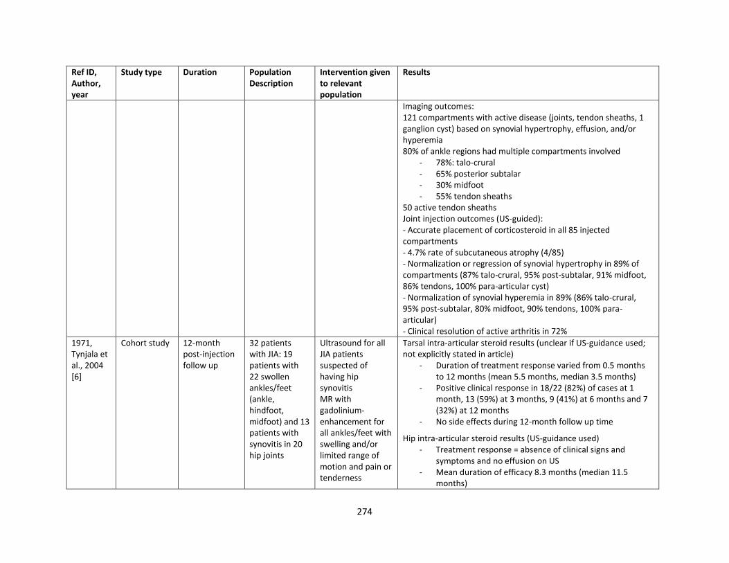
274
Ref ID, Author, year
Study type Duration Population Description
Intervention given to relevant population
Results
Imaging outcomes: 121 compartments with active disease (joints, tendon sheaths, 1 ganglion cyst) based on synovial hypertrophy, effusion, and/or hyperemia 80% of ankle regions had multiple compartments involved
- 78%: talo-crural - 65% posterior subtalar - 30% midfoot - 55% tendon sheaths
50 active tendon sheaths Joint injection outcomes (US-guided): - Accurate placement of corticosteroid in all 85 injected compartments - 4.7% rate of subcutaneous atrophy (4/85) - Normalization or regression of synovial hypertrophy in 89% of compartments (87% talo-crural, 95% post-subtalar, 91% midfoot, 86% tendons, 100% para-articular cyst) - Normalization of synovial hyperemia in 89% (86% talo-crural, 95% post-subtalar, 80% midfoot, 90% tendons, 100% para-articular) - Clinical resolution of active arthritis in 72%
1971, Tynjala et al., 2004 [6]
Cohort study 12-month post-injection follow up
32 patients with JIA: 19 patients with 22 swollen ankles/feet (ankle, hindfoot, midfoot) and 13 patients with synovitis in 20 hip joints
Ultrasound for all JIA patients suspected of having hip synovitis MR with gadolinium-enhancement for all ankles/feet with swelling and/or limited range of motion and pain or tenderness
Tarsal intra-articular steroid results (unclear if US-guidance used; not explicitly stated in article)
- Duration of treatment response varied from 0.5 months to 12 months (mean 5.5 months, median 3.5 months)
- Positive clinical response in 18/22 (82%) of cases at 1 month, 13 (59%) at 3 months, 9 (41%) at 6 months and 7 (32%) at 12 months
- No side effects during 12-month follow up time
Hip intra-articular steroid results (US-guidance used) - Treatment response = absence of clinical signs and
symptoms and no effusion on US - Mean duration of efficacy 8.3 months (median 11.5
months)
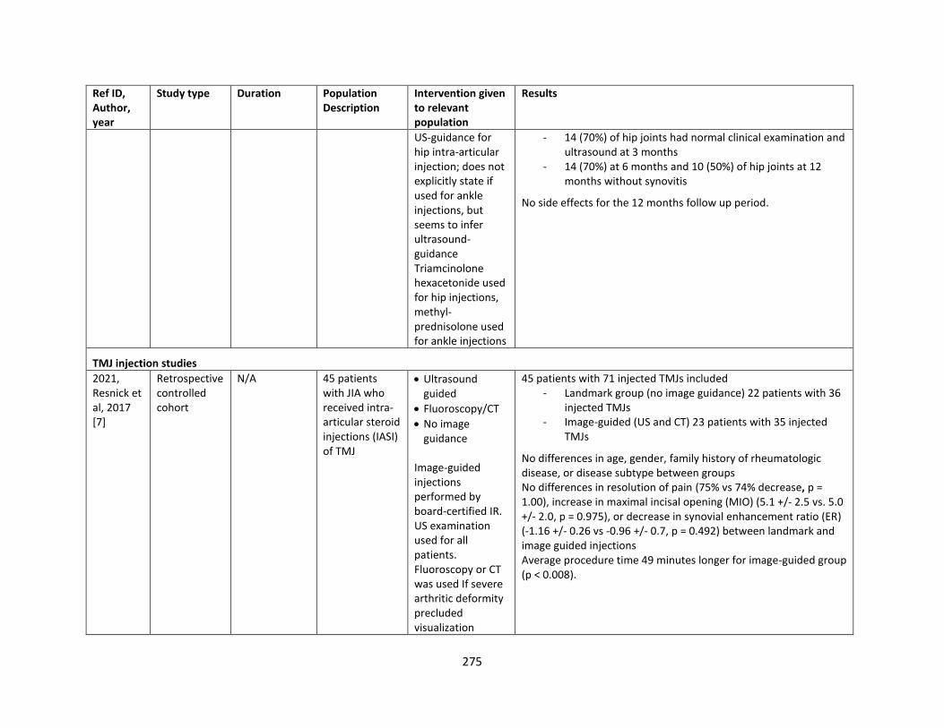
275
Ref ID, Author, year
Study type Duration Population Description
Intervention given to relevant population
Results
US-guidance for hip intra-articular injection; does not explicitly state if used for ankle injections, but seems to infer ultrasound-guidance Triamcinolone hexacetonide used for hip injections, methyl-prednisolone used for ankle injections
- 14 (70%) of hip joints had normal clinical examination and ultrasound at 3 months
- 14 (70%) at 6 months and 10 (50%) of hip joints at 12 months without synovitis
No side effects for the 12 months follow up period.
TMJ injection studies
2021, Resnick et al, 2017 [7]
Retrospective controlled cohort
N/A 45 patients with JIA who received intra-articular steroid injections (IASI) of TMJ
• Ultrasound guided
• Fluoroscopy/CT
• No image guidance
Image-guided injections performed by board-certified IR. US examination used for all patients. Fluoroscopy or CT was used If severe arthritic deformity precluded visualization
45 patients with 71 injected TMJs included - Landmark group (no image guidance) 22 patients with 36
injected TMJs - Image-guided (US and CT) 23 patients with 35 injected
TMJs
No differences in age, gender, family history of rheumatologic disease, or disease subtype between groups No differences in resolution of pain (75% vs 74% decrease, p = 1.00), increase in maximal incisal opening (MIO) (5.1 +/- 2.5 vs. 5.0 +/- 2.0, p = 0.975), or decrease in synovial enhancement ratio (ER) (-1.16 +/- 0.26 vs -0.96 +/- 0.7, p = 0.492) between landmark and image guided injections Average procedure time 49 minutes longer for image-guided group (p < 0.008).
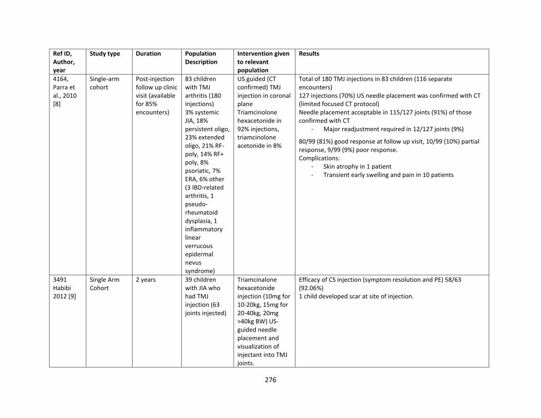
276
Ref ID, Author, year
Study type Duration Population Description
Intervention given to relevant population
Results
4164, Parra et al., 2010 [8]
Single-arm cohort
Post-injection follow up clinic visit (available for 85% encounters)
83 children with TMJ arthritis (180 injections) 3% systemic JIA, 18% persistent oligo, 23% extended oligo, 21% RF- poly, 14% RF+ poly, 8% psoriatic, 7% ERA, 6% other (3 IBD-related arthritis, 1 pseudo-rheumatoid dysplasia, 1 inflammatory linear verrucous epidermal nevus syndrome)
US guided (CT confirmed) TMJ injection in coronal plane Triamcinolone hexacetonide in 92% injections, triamcinolone acetonide in 8%
Total of 180 TMJ injections in 83 children (116 separate encounters) 127 injections (70%) US needle placement was confirmed with CT (limited focused CT protocol) Needle placement acceptable in 115/127 joints (91%) of those confirmed with CT
- Major readjustment required in 12/127 joints (9%)
80/99 (81%) good response at follow up visit, 10/99 (10%) partial response, 9/99 (9%) poor response. Complications:
- Skin atrophy in 1 patient - Transient early swelling and pain in 10 patients
3491 Habibi 2012 [9]
Single Arm Cohort
2 years 39 children with JIA who had TMJ injection (63 joints injected)
Triamcinalone hexacetonide injection (10mg for 10-20kg, 15mg for 20-40kg, 20mg >40kg BW) US-guided needle placement and visualization of injectant into TMJ joints.
Efficacy of CS injection (symptom resolution and PE) 58/63 (92.06%) 1 child developed scar at site of injection.
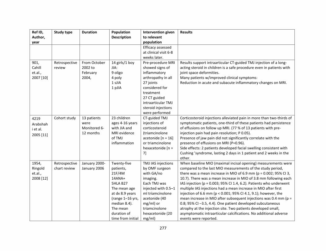
277
Ref ID, Author, year
Study type Duration Population Description
Intervention given to relevant population
Results
Efficacy assessed at clinical visit 6-8 weeks later.
901, Cahill et.al., 2007 [10]
Retrospective review
From October 2002 to February 2004,
14 girls/1 boy JIA: 9 oligo 4 poly 1 sJIA 1 pJIA
Pre-procedure MRI showed signs of inflammatory arthropathy in all 27 joints considered for treatment 27 CT guided intraarticular TMJ steroid injections were performed
Results support intraarticular CT-guided TMJ injection of a long-acting steroid in children is a safe procedure even in patients with joint space deformities. Many patients w/improved clinical symptoms: Reduction in acute and subacute inflammatory changes on MRI.
4219
Arabshahi et al.
2005 [11]
Cohort study 13 patients were Monitored 6-12 months
23 children ages 4-16 years with JIA and MRI evidence of TMJ inflammation
CT-guided TMJ injections of corticosteroid (triamcinolone acetonide [n = 16] or triamcinolone hexacetonide [n = 7]
Corticosteroid injections alleviated pain in more than two-thirds of symptomatic patients, one-third of these patients had persistence of effusions on follow up MRI. (77 % of 13 patients with pre-injection pain had pain resolution; P 0.05). Presence of jaw pain did not significantly correlate with the presence of effusions on MRI (P=0.96). Side effects: 2 patients developed facial swelling consistent with Cushing ’syndrome, lasting 2 days in 1 patient and 2 weeks in the other.
1954, Ringold et.al., 2008 [12]
Retrospective chart review
January 2000-January 2006
Twenty-five patients, 21F/4M 14ANA+ 5HLA B27 The mean age at dx 8.9 years (range 1–16 yrs, median 8.4). The mean duration of time from initial
TMJ IAS injections by OMF surgeon with GA/no imaging. Each TMJ was injected with 0.5–1 ml triamcinolone acetonide (40 mg/ml) or triamcinolone hexacetonide (20 mg/ml)
When baseline MIO (maximal incisal opening) measurements were compared to the last MIO measurements of the study period, there was a mean increase in MIO of 6.9 mm (p = 0.002; 95% CI 3, 10.7). There was a mean increase in MIO of 3.8 mm following each IAS injection (p = 0.003; 95% CI 1.4, 6.2). Patients who underwent multiple IAS injections had a mean increase in MIO after first injection of 6.6 mm (p < 0.001; 95% CI 4.1, 9.1); however, the mean increase in MIO after subsequent injections was 0.4 mm (p = 0.8; 95% CI –3.5, 4.4). One patient developed subcutaneous atrophy at the injection site. Two patients developed small, asymptomatic intraarticular calcifications. No additional adverse events were reported.
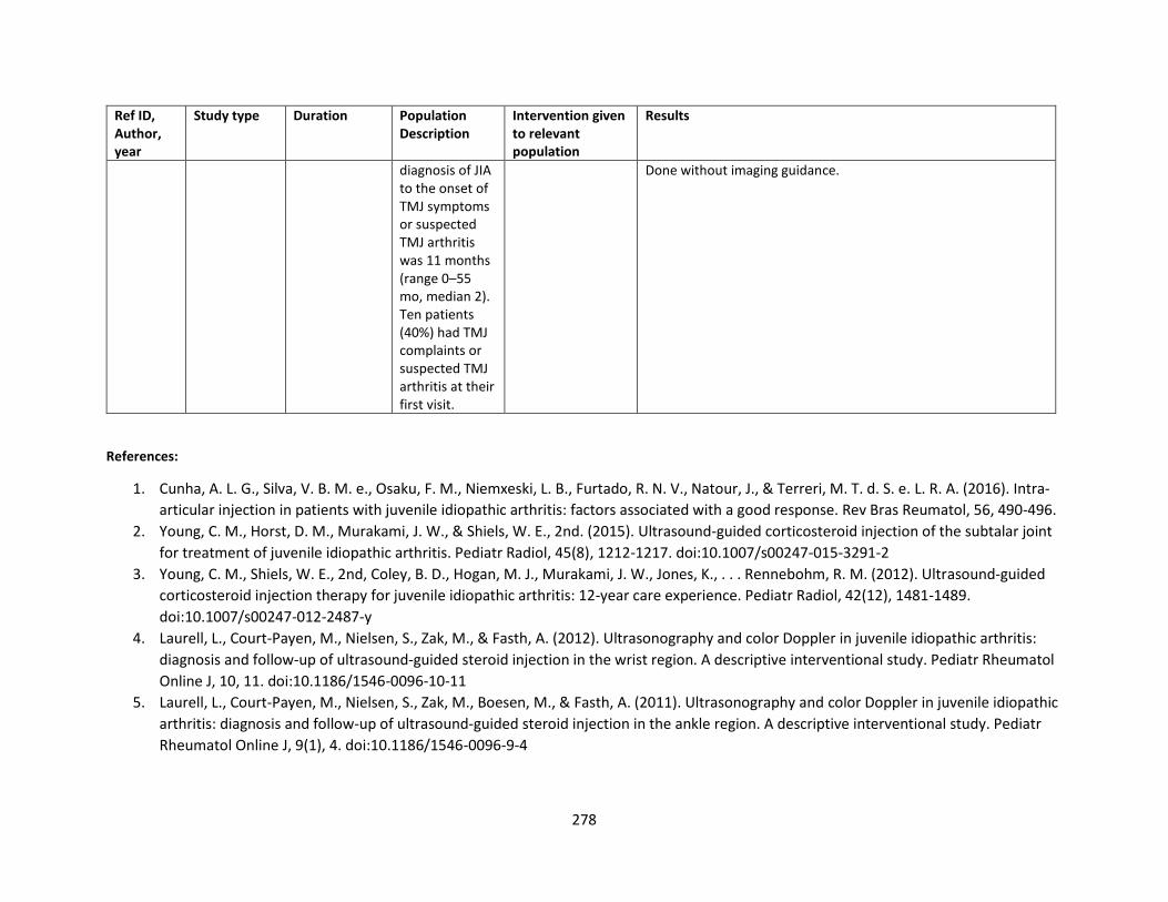
278
Ref ID, Author, year
Study type Duration Population Description
Intervention given to relevant population
Results
diagnosis of JIA to the onset of TMJ symptoms or suspected TMJ arthritis was 11 months (range 0–55 mo, median 2). Ten patients (40%) had TMJ complaints or suspected TMJ arthritis at their first visit.
Done without imaging guidance.
References:
1. Cunha, A. L. G., Silva, V. B. M. e., Osaku, F. M., Niemxeski, L. B., Furtado, R. N. V., Natour, J., & Terreri, M. T. d. S. e. L. R. A. (2016). Intra-
articular injection in patients with juvenile idiopathic arthritis: factors associated with a good response. Rev Bras Reumatol, 56, 490-496.
2. Young, C. M., Horst, D. M., Murakami, J. W., & Shiels, W. E., 2nd. (2015). Ultrasound-guided corticosteroid injection of the subtalar joint
for treatment of juvenile idiopathic arthritis. Pediatr Radiol, 45(8), 1212-1217. doi:10.1007/s00247-015-3291-2
3. Young, C. M., Shiels, W. E., 2nd, Coley, B. D., Hogan, M. J., Murakami, J. W., Jones, K., . . . Rennebohm, R. M. (2012). Ultrasound-guided
corticosteroid injection therapy for juvenile idiopathic arthritis: 12-year care experience. Pediatr Radiol, 42(12), 1481-1489.
doi:10.1007/s00247-012-2487-y
4. Laurell, L., Court-Payen, M., Nielsen, S., Zak, M., & Fasth, A. (2012). Ultrasonography and color Doppler in juvenile idiopathic arthritis:
diagnosis and follow-up of ultrasound-guided steroid injection in the wrist region. A descriptive interventional study. Pediatr Rheumatol
Online J, 10, 11. doi:10.1186/1546-0096-10-11
5. Laurell, L., Court-Payen, M., Nielsen, S., Zak, M., Boesen, M., & Fasth, A. (2011). Ultrasonography and color Doppler in juvenile idiopathic
arthritis: diagnosis and follow-up of ultrasound-guided steroid injection in the ankle region. A descriptive interventional study. Pediatr
Rheumatol Online J, 9(1), 4. doi:10.1186/1546-0096-9-4
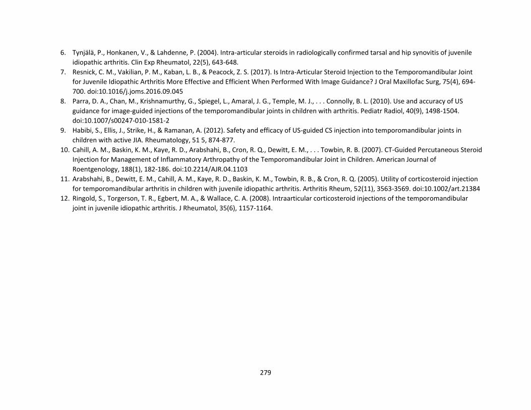
279
6. Tynjälä, P., Honkanen, V., & Lahdenne, P. (2004). Intra-articular steroids in radiologically confirmed tarsal and hip synovitis of juvenile
idiopathic arthritis. Clin Exp Rheumatol, 22(5), 643-648.
7. Resnick, C. M., Vakilian, P. M., Kaban, L. B., & Peacock, Z. S. (2017). Is Intra-Articular Steroid Injection to the Temporomandibular Joint
for Juvenile Idiopathic Arthritis More Effective and Efficient When Performed With Image Guidance? J Oral Maxillofac Surg, 75(4), 694-
700. doi:10.1016/j.joms.2016.09.045
8. Parra, D. A., Chan, M., Krishnamurthy, G., Spiegel, L., Amaral, J. G., Temple, M. J., . . . Connolly, B. L. (2010). Use and accuracy of US
guidance for image-guided injections of the temporomandibular joints in children with arthritis. Pediatr Radiol, 40(9), 1498-1504.
doi:10.1007/s00247-010-1581-2
9. Habibi, S., Ellis, J., Strike, H., & Ramanan, A. (2012). Safety and efficacy of US-guided CS injection into temporomandibular joints in
children with active JIA. Rheumatology, 51 5, 874-877.
10. Cahill, A. M., Baskin, K. M., Kaye, R. D., Arabshahi, B., Cron, R. Q., Dewitt, E. M., . . . Towbin, R. B. (2007). CT-Guided Percutaneous Steroid
Injection for Management of Inflammatory Arthropathy of the Temporomandibular Joint in Children. American Journal of
Roentgenology, 188(1), 182-186. doi:10.2214/AJR.04.1103
11. Arabshahi, B., Dewitt, E. M., Cahill, A. M., Kaye, R. D., Baskin, K. M., Towbin, R. B., & Cron, R. Q. (2005). Utility of corticosteroid injection
for temporomandibular arthritis in children with juvenile idiopathic arthritis. Arthritis Rheum, 52(11), 3563-3569. doi:10.1002/art.21384
12. Ringold, S., Torgerson, T. R., Egbert, M. A., & Wallace, C. A. (2008). Intraarticular corticosteroid injections of the temporomandibular
joint in juvenile idiopathic arthritis. J Rheumatol, 35(6), 1157-1164.