electronic supplementary information
TRANSCRIPT
1
Electronic Supplementary Information
A COVID-19 vaccine candidate composed of SARS-CoV-2 RBD dimer and Neisseria
meningitidis outer membrane vesicles
Darielys Santana-Mederos,a,† Rocmira Perez-Nicado,a,† Yanet Climent,a,† Laura Rodriguez,a,†
Belinda Sanchez Ramirez,b,† Sonia Perez-Rodriguez,c Meybi Rodriguez,a Claudia Labrada,a Tays
Hernandez,b Marianniz Diaz,b Ivette Orosa,b Ubel Ramirez,a Reynaldo Oliva,a Raine Garrido,a
Felix Cardoso,a Mario Landys,a Roselyn Martinez,a Humberto Gonzalez,a Tamara Hernandez,a
Rolando Ochoa-Azze,a Jose L. Perez,a Juliet Enriquez,d Nibaldo Gonzalez,d Yenicet Infante,d Luis
A. Espinosa,e Yassel Ramos,e Luis Javier González,e Carmen Valenzuela,f Ana Victoria
Casadesus,b Briandy Fernandez,b Gertrudis Rojas,b Beatriz Pérez-Massón,b Yaima Tundidor,b
Ernesto Bermudez,b Claudia A. Plasencia,b Tammy Boggiano,b Eduardo Ojito,b Fabrizio
Chiodo,a,g Sonsire Fernandez,a Françoise Paquet,h Cheng Fang,i Guang-Wu Chen,j,† Daniel G.
Rivera,k,† Yury Valdes-Balbin,a,†,* Dagmar Garcia-Riveraa,†,* and Vicente Verez Bencomoa,†,*
aFinlay Vaccine Institute, 200 and 21 Street, Havana 11600, Cuba.
bCenter of Molecular Immunology, P.O. Box 16040, 216 St. Havana, Cuba
cNational Toxicology Center, Havana, 11500, Cuba
dNational Civil Defense Research Laboratory, Mayabeque 32700, Cuba
eCenter for Genetic Engineering and Biotechnology, Ave 31 e/ 158 y 190, Havana 10600, Cuba
fInstitute of Cybernetics, Mathematics and Physics, Havana 10400, Cuba
Electronic Supplementary Material (ESI) for RSC Chemical Biology.This journal is © The Royal Society of Chemistry 2021

2
gDepartment of Molecular Cell Biology and Immunology, Amsterdam UMC, Vrije Universiteit
Amsterdam, Amsterdam, The Netherlands and Institute of Biomolecular Chemistry, National
Research Council (CNR), Pozzuoli, Napoli, Italy
hCentre de Biophysique Moléculaire, CNRS UPR 4301, rue Charles Sadron, F-45071, Orléans,
Cedex 2, France
iShanghai Fenglin Glycodrug Promotion Center, Shanghai 200032, China
jChengdu Olisynn Biotech. Co. Ltd., and State Key Laboratory of Biotherapy and Cancer Center,
West China Hospital, Sichuan University, Chengdu 610041, People’s Republic of China.
kLaboratory of Synthetic and Biomolecular Chemistry, Faculty of Chemistry, University of
Havana, Zapata y G, Havana 10400, Cuba
†These authors contributed equally to this work. *Corresponding authors: [email protected],
3
Materials and Methods
HPLC, gel electrophoresis and structural representation. Size-exclusion HPLC was performed
in PBS pH 7 on a Superdex 75 Increase® 10/300 GL column and Superdex 200 Increase® 5/150
GL column (GE Healthcare) at a flow rate of 0.8 ml/min and 0.25 ml/min, respectively. SDS-
polyacrylamide gel electrophoresis (SDS-PAGE) was performed in a gradient gel (4-20%)
acrylamide, loading 5 μg of RBD dimer (RBD-d) and monomer (RBD-m). Gels were stained with
Coomassie blue R250 and analyzed with Bio-Rad GS-800 densitometer and Quantity One
software. The 3D structures of RBD-d were built by combining PDB 6M0J and PDB 6WPT to
obtain the maximum amino acid coordinates for region Arg319-Phe541. The cryo-electron
microscopy structure PDB 6WPT is obtained with N-glycans linked to Asn331 and Asn343. The
structure of the RBD dimer resulted from a manual docking respecting the proximity of the two S-
S linked Cys538s and its representation was obtained using the PyMOL molecular graphics
system.1
Production of recombinant RBD (319-541) dimer2
The coding sequence for RBD (Arg319-Phe541) with a hexahistidine tag at its C-terminus
(Arg319-Phe541-(His)6) was optimized for mammalian cell expression in CHO (hamster,
Crycetulus grisesus), using the online gene optimization tools provided by Eurofins (Germany).
The resulting nucleotide sequence was assembled and amplified by PCR using synthetic gene
fragments (Eurofins, Germany) and oligonucleotides (Center for Genetic Engineering and
Biotechnology, CIGB, Cuba) and cloned into an intermediate vector containing the CMV
promoter and the mouse Ig VH signal gene. The expression cassette was re-cloned in the lentiviral
1 De Lano W.L., Pymol. South San Francisco, CA: De Lano Scientific 2002.
2 Y. Valdes-Balbin, et al. ACS Chem. Biol. 2021, 16, 1223–1233.
4
vector pL6WBlast, kindly provided by CIGB. HEK-293T cells were co-transfected with the
lentiviral vector containing the gene of interest plus the auxiliary plasmids pLPI, pLPII and
pLP/VSV-G, to produce lentiviral particles. CHO-K1 host cells were transduced with lentiviral
particles and grown in 96 well plates in the presence of the selection drug blasticidine.
Supernantants were screened by ELISA for secreted RBD), and cells showing the highest
secretion levels were adapted to grow in suspension in serum-free medium (a mixture of PFHMII
with a Center of Molecular Immunology’s proprietary medium). Secreted RBD was purified by
immobilized metal affinity chromatography (IMAC) using Ni-NTA Sepharose to get a
monomer/dimer mixture. The RBD dimer was then separated from the monomer and isolated as
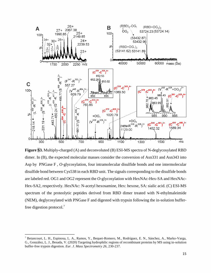
a single peak using a Superdex 200 column (Fig. S2).
Mass Spectrometry analysis. The purified dimer (RBD-d, 5 g) was treated with N-ethyl
maleimide (NEM), deglycosylated with PNGase-F to remove N-glycans, and desalted (ZipTips
C18, Millipore). The desalted protein was loaded into the metal-coated nanocapillary for ESI-MS
analysis. N-deglycosylated RBD-d and its tryptic peptides were analyzed in a hybrid orthogonal
QTof-2TM tandem mass spectrometer (Micromass) for ESI-MS analysis. The desalted samples
were loaded into the metal-coated nanocapillary and sprayed into the ion source using 1200 and
35 volts for the capillary and the entrance cone, respectively. The multiply-charged ESI-MS
spectrum (m/z 400-3000) of N-deglycosylated RBD was deconvoluted (mass 3000-70000) with
MaxtEnt 1.0 software. The ESI-MS of tryptic peptides were acquired (m/z 200-2000). To obtain
structural information in the MS/MS spectra, the multiply-charged ions were manually fragmented
by collision-induced dissociation using collision energies (20-50 eV). Argon was used as collision
gas. The ESI-MS/MS of tryptic peptides with z ≥ 3+ were deconvoluted using the MaxEnt 3.0
software. The multiply-charged ESI-MS spectrum (m/z 400-3000) of N-deglycosylated RBD was
5
deconvoluted (mass 3000-70000) with MaxtEnt1.0 software. Theoretical m/z values for tryptic
peptides and for the intact protein were calculated using the MassLynx v4.1 software (Micromass).
ESI-MS analysis of RBD dimer
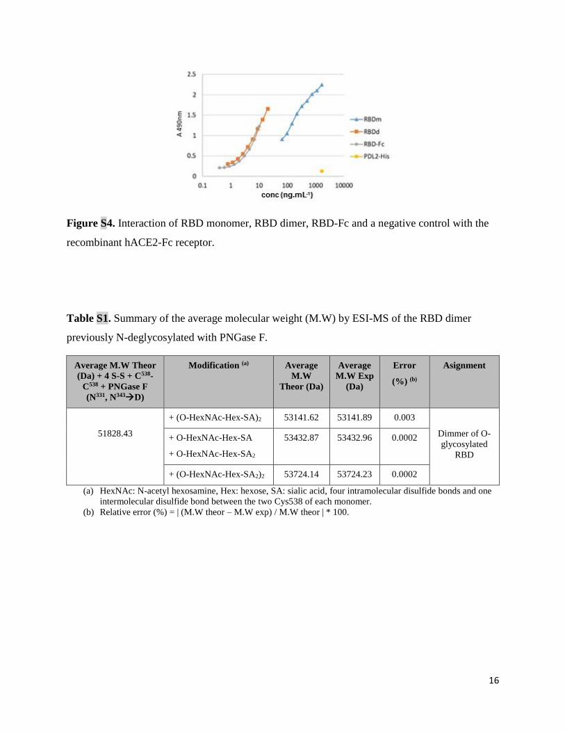
The ESI-MS spectrum showed multiply-charged ions, indicating certain heterogeneity due to the
O-glycan chains linked to the N-deglycosylated RBD dimer (Fig. S3A). Deconvoluted ESI-MS
spectrum showed (Fig. S3B) three major signals, differing in +291 Da (matching the mass value
for sialic acid residue and indicating a variable number of sialic acids residues in the RBD dimer).
The lowest abundant signal corresponds to the RBD dimer linked to two O-glycan chains
(HexNAc-Hex-SA), shown in Fig. S3B as OG1. The signal with an intermediate abundance (Fig.
S3B) corresponds to the dimer linked to a mixture of two O-glycans OG1 (HexNAc-Hex-SA) and
OG2 (HexNAc-Hex-SA2). The most intense signal corresponds to the RBD dimer linked to two
OG2 (HexNAc-Hex-SA2) (Fig. S3B).
Table S1 summarizes the structural assignments for all signals corresponding to the O-glycoforms
present in the dimer. The assignment for all tryptic peptides is summarized in Table S2. Signals
corresponding to the four intramolecular disulfide bonds in RBD dimer, and the N-terminal
peptides (R319-R328 and V320-R328) O-glycosylated at Thr323/Ser325 residues were identified.
The dimeric nature of the RBD was confirmed by signals corresponding to peptide [C538-H547]-
S-S-[C538-H547] (m/z Exp. 522.02 (5+) and m/z Exp 652.28 (4+), Fig. S3).
Dynamic Light Scattering (DLS)
DLS measurements were performed with a Malvern Zetasizer Nano ZS (Malvern) equipped with
a 633 nm He-Ne laser and operating at an angle of 173°. Scattering light detected at 173° was
automatically adjusted by laser attenuation filters. For data analysis, the viscosity and refractive
index (RI) of PBS buffer solution (at 25 °C) were used. The software used to collect and analyze
6
the data was the Zetasizer software version 7.11. The temperature was set at 25 °C. Each sample
at 500 μg/mL protein content was characterized in a single-use polystyrene microcuvette
(ZEN0040, Alfatest). The size was reported as the hydrodynamic diameter (intensity graph) of
three measurements, providing also a polydispersity index (PdI) of the size values calculated.
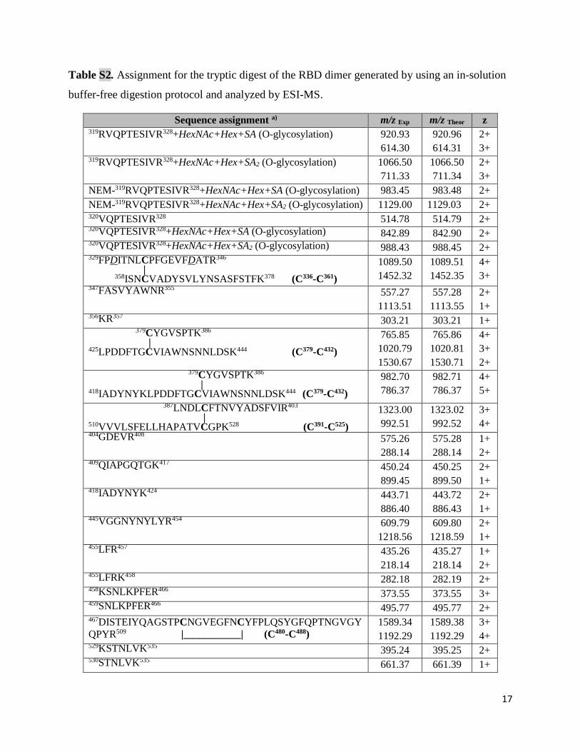
Recognition of recombinant human ACE2 by RBD-d. Microtiter plates (High binding, Costar)
were coated with ACE2-mFc (50 µL/well, 5 μg/mL) in 0.1 M carbonate-bicarbonate buffer pH
9.6 and incubated overnight at 4 °C. Plates were blocked with 200µL/well of 2% Non Fat Dry
Milk (NFDM) in PBS-0.05% Tween 20 (PBST) for 1 h at 37 °C. Serial dilutions of the RBD-d in
0.2% NFDM/PBST 50 µL were added and incubated for 2 h at 37 °C. RBD and 6×His tagged PD-
L1 were used as the positive and negative control, respectively. The bound protein was detected
with RBD-specific rabbit polyclonal antibodies (PAbs, 50 µL/well, 100 µg/mL) for 1 h at 37 °C.
Next, a peroxidase-conjugated anti-rabbit IgG monoclonal antibody (MAb, 50 µL/well, 1:10000)
was added and the plates were incubated for 1 h at 37 °C. The reaction was visualized by addition
of 3,3’,5,5’-Tetramethylbenzidine (TMB) (BDBiosciences) and stopped with 1 M H2SO4. The
absorbance at 450nm was measured using a microwell system reader (Organon Teknica). All
incubations were followed by three washing steps with PBS-T.
Reactivity cell-based ELISA. 40000 Vero cells were seeded in 96-well cell culture plates. 48 h
after seeding, the cells were fixed by adding 100 μL of 4% paraformaldehyde (PFA) followed by
incubation at room temperature for 20 min and quenching for 5 min at room temperature with 50
μL of 0.3% H2O2, in PBS. The cells were blocked with 200 μL/well of assay buffer (3% of bovine
serum albumin (BSA) in PBS) for 1 h at room temperature. Next, 50 μL of the RBD-d and RBD-
hFc were added at different concentrations and incubated for 2 h at room temperature. RBD-d,
RBD-m and RBD-hFc binding was revealed with 100 μL of RBD specific MAb S1 (10
7
µg/mL in assay buffer) for 1 h at room temperature. Next, 100 µL/well of Biotin-SP-AffiniPure
F(ab')2 Fragment Goat Anti-Mouse IgG (1:5000 in assay buffer; Jackson,115-066-071) was added
and incubated for 1 h at room temperature, followed by the addition of 100 μL of streptavidin-
conjugated peroxidase (Sigma, S5512) during 30 min at room temperature (1:25000 in assay
buffer). Finally, the 3,3′,5,5′-tetramethylbenzidine (TMB) peroxidase substrate (Sigma, T0440)
was added and plates were light-protection incubated for 15 min at room temperature. The reaction
was stopped using 1 M H2SO4. The OD at 450 nm was measured using a microwell reader
(BioTek). All incubations were followed by three washing steps with PBS.
Animal experiments
Immunogenicity of the RBD-d/OMV/alum and RBD-d/alum was evaluated in BALB/c mice (age:
6-8 weeks, 15-20 g), supplied by the National Center for Laboratory Animals Breeding
(CENPALAB), Havana, Cuba. All protocols were performed in accordance with the Guidelines
for Care and Use of Laboratory Animals of the Finlay Vaccine Institute and approved by its
Animal Ethics Committee.
Immunization schedule and sera samples. Intramuscular injection on days 0, 14 and 28; sera
were collected at days 0 (before immunization) and at days 7, 14, 21, 28, 35 and 42.
Immunogenicity experiments included groups of 10 mice injected with: a) 10 μg of RBD-d and 4
μg of OMV adjuvated with 250 μg of Al(OH)3, b) 10 μg of RBD-d adjuvated with 250 μg of
Al(OH)3 and c) 250 μg of Al(OH)3 as control.
Anti-RBD IgG ELISA
ELISA plates (96 well, NUNC) were coated with 50 µL of RBD-d at 10 µg.mL-1 in carbonate-
bicarbonate buffer pH 9.6 overnight at 4 ºC. Plates were blocked in 5% skim milk-PBS for 1 h at
37 ºC. Serum samples (diluted 1:3 v/v in PBS-1% BSA solution, pH 7.2) were added in serial
8
dilution starting from 1/50 and were then incubated for 1 h at 37 ºC. Goat anti-mouse IgG-HRP
antibody (Sigma A4416) diluted 1/5000 in PBS-1% BSA pH 7.2 were added and incubated for 1
h at 37 ºC. Then, TMB peroxidase substrate was added to the plates and incubated for 20 minutes.
Reactions were stopped with 2 N H2SO4 and the absorbance was measured at 450 nm in a
microplate reader ELISA Multiskan EX (ThermoScientific). ). All incubations were followed by
three washing steps with PBS-T. The endpoint titer was defined as the highest reciprocal dilution
of serum that gives an absorbance 4-fold greater than pre-immune serum diluted 1/50.
Avidity ELISA
To assess avidity, the method described by Antilla et al.3 was followed, based on the dissociation
of the antigen-antibody interaction by treatment with the chaotropic agent ammonium thiocyanate
(NH4SCN). The plates were covered and blocked under the same conditions as in anti-RBD IgG
ELISA, and later the sera were added in the dilution whose absorbance value was 1 in the titration
ELISA, and incubated at 37 ºC for 1 h. A fixed concentration of 2 mol.L-1 NH4SCN was added
and incubated for 15 minutes at room temperature, followed by repetition of the ELISA procedure
previously described. The Avidity Index (AI) indicates the percentage of IgG antibodies that
remain bound to the antigen after treatment with the chaotropic agent and is calculated by the
formula (IgG titer with NH4SCN/IgG titer without NH4SCN)*100. Antibodies with good avidity
are considered those with an AI greater than 50%. AI was determined only for the responding
animals (titer log> 1.70) by the anti-RBD IgG ELISA.
3 Antilla M., Esckola J., Ahman H. Avidity of IgG for Streptococcus Pneumoniae type 6B and 23F polysaccharide in
infants primed with pneumococcal conjugate and boosted with polysaccharide or conjugate vaccines. J. Infectious
Diseases 1998, 177, 1614-1621.
9
Quantification of anti-RBD IgG1 and IgG2a in immunized mice
The tests were carried out following the same steps as in the anti-RBD IgG ELISA, except that
after adding the sera and washing, the anti-IgG1 and anti-IgG2a biotin-conjugates (dilution
1:10000 and 1:5000, respectively) were added following by incubation for 1 hour at 37 °C. After
another washing step, streptavidin peroxidase conjugate is added and the mixture is incubated for
1 hour at 37 ºC. The rest of the steps and the analysis of the results were carried out according to
the anti-RBD IgG ELISA to determine the antibody titers. After determining the IgG1 and IgG2a
titers, the ratio IgG2a/IgG1 is calculated using the formula titer of IgG2a/titer of IgG1. This
relationship shows where the cellular response pattern is directed (i.e., Th1 or Th2).
Quantification of cytokines IFN-γ and IL-4
Splenocytes from immunized mice were isolated and culture in RPMI 1640 (Gibco) supplied with
10% (v/v) FBS (Hyclone), 100 U/mL penicillin, 100 μg/ml streptomycin, 1 mM pyruvate, 50 μM
β-mercaptoethanol (all from Sigma-Aldrich). These cells (3×10⁶ per well) were incubated for 72
h with RBD-d (5 µg.mL-1), concanavalin A (5 µg.mL-1) as positive control and medium alone as
negative control. Supernatants were collected and stored at -80 °C. IFN-γ and IL-4 levels were
quantified using an ELISA kit (Mabtech) following the manufacturer’s instructions.
Adoptive cell transfer
Splenic lymphocytes from immunized mice (donor) were transferred to naïve ones (recipient) as
previously described.2 Anti-RBD IgG levels were determined by the ELISA assay previously
described.
Molecular Virus Neutralization Assay
10
The ability of anti-RBD specific antibodies to inhibit the RBD-ACE2 interaction was evaluated
in a Molecular Virus Neutralization Assay. Briefly, microtiter plates (High binding, Costar) were
coated with 250 ng/well of ACE2-mFc in 0.1 M carbonate-bicarbonate buffer pH 9.6 and
incubated overnight at 4 °C. Plates were blocked with 200 µL/well of 2% of skim milk in PBS-T
during 1 h at 37 °C. Serial dilutions of sera were pre-incubated with RBD-Fc (final conc. 20
ng.mL-1) for 1 h at 37 °C. RBD-mFc was used for human samples and RBD-hFc was used for
animal samples. Mixtures were added to the plates and incubated for 2 h at 37 °C. The binding of
RBD-mFc was detected by addition of alkaline phosphatase (AP) conjugated anti-mouse IgG
antibody (1:1000) (Sigma, A9316) for 1 h at 37°C. RBD-Fc recombinant proteins, sera and
antibody conjugates were diluted in 0.2% of skim milk in PBST. In the case of RBD-hFc, the
binding was detected by incubating with anti-human IgG antibody conjugated AP (1:1800)
(Sigma, A3188). Finally, p-nitrophenylphosphate (Sigma N9389) at 1 mg.mL-1 in diethanolamine
buffer pH 9.8 was added, and plates were incubated at room temperature for 30 min. Absorbance
at 405nm was measured using a microwell reader (BioTek). In all steps other than blockade,
samples and reagents were added to get a final volume of 50 µL/well. Three washing steps with
PBST followed all incubations. Inhibition was calculated and expressed as percent according to
the next formula: Inhibition (%)= [1-(OD405nmsample/OD405nm maximal recognition)]×100.
Maximal recognition corresponds to wells incubated only with RBD-mFc or RBD-hFc (20 ng.mL-
1). To determine ID50, dilutions were log transformed and data were adjusted to a log(inhibitor) vs
normalized response with variable slope non-linear regression.
Virus neutralization assay
11
The virus neutralization assay was performed following the recommendation of Manenti et al.4
with few modifications. Animal serum samples were heat-inactivated for 30 minutes at 56 °C.
Two-fold serial dilutions of each sample serum (starting in 1:10 until 1:2560) were then mixed
with an equal volume of viral suspension containing 100 median tissue culture infectious dose
(TCID50) of SARS-CoV-2 (Strain 2025, Cuban Collection, National Laboratory of Civil Defense,
Biosafety Laboratory Level 3 facility) and incubated for 1 h at 37 °C in a humidified atmosphere
with 5% CO2. After incubation, 100 μL of each dilution was added in duplicate to plates containing
a semiconfluent VERO E6 monolayer (104 cell/well). The plates were incubated for 3 days at 37
°C in a humidified atmosphere with 5% CO2. Then, the supernatant was carefully discarded and
100 μL/well of a sterile PBS solution containing 0.02% neutral red (Sigma) was added. After 1 h
of incubation at room temperature, the neutral red solution was discarded and the cell monolayer
was washed twice with sterile PBS-T. After the second incubation, PBS-T was carefully removed;
then 100 μL/well of a lysis solution (50 parts of absolute ethanol (Sigma), 49 parts of MilliQ water
and 1 part of glacial acetic acid (Sigma) were added. Plates were incubated for 15 min at room
temperature and then read at 540 nm in a spectrophotometer. The viral neutralizing titer 50
(VNT50) is the highest serum dilution giving 50% of the average DO with respect to control cell
wells (VERO E6 monolayer without mixture of virus-sera). In sera where VNT50 could not be
calculated until 1:2560 dilution, the assay was repeated starting with a higher dilution.
Expression of phage-displayed RBDs
4 Manenti, A., Maggetti, M., Casa, E., Martinuzzi, D., Torelli, A., Trombetta, C. M., Marchi, S., Montomoli, E. (2020)
Evaluation of SARS-CoV-2 neutralizing antibodies using a CPE-based colorimetric live virus micro-neutralization
assay in human serum samples. J. Med. Virol. 92, 2096-2104.
12
The RBD coding sequence Arg328-Thr533 (EurofinsTM, Germany) was cloned in the phagemid
vector pCSM through ApaLI and NotI restriction sites.5 The original gene was modified by site-
directed mutagenesis to introduce the triplets coding for the desired replacements in RBD
sequence. Phages displaying wild-type RBD and its mutants were rescued with M13KO7 helper
phage as described.4 The three RBDs expressed with a single mutation were N501Y, L452R and
E484K, and the triple RBD mutant included mutations N501Y+ E484K+K417N, corresponding
to the variant of concern beta. Display levels were determined by ELISA on microplates coated
with 9E10 mAb, an antibody targeting the c-myc tag fused to the carboxyl-terminal end of all
proteins displayed in this system. Phages displaying the original wild-type RBD were used as
reference, assuming a display level of 100 units/mL for them, whereas relative levels of mutated
variants were subsequently calculated as reported.6 Phage-displayed RBD was shown to be
biologically active by ELISA on microplates coated with the recombinant extracellular region of
human ACE2 receptor fused to human Fc (ACE2-hFc), which confirmed their proper folding on
the viral particles assembled in E. coli periplasm.
Molecular Virus Neutralization Assay with phage-displayed RBDs
To assess the inhibition of RBD-ACE2 interaction by sera of vaccinated individuals, polyvinyl
chloride microplates were coated with ACE2-hFc at 5 µg.mL-1 in phosphate-buffered saline (PBS)
overnight at 4 ºC and blocked with PBS containing 4% skim milk powder (M-PBS) during 1h at
room temperature. Each phage preparation was diluted at 0.075 display units/mL in M-PBS
containing serial dilutions of sera (from 1/25 to 1/25 600). Phages similarly diluted in M-PBS
5 Rojas, G. Fine epitope mapping based on phage display and extensive mutagenesis of the target antigen. Methods
Mol. Biol. 2014, 1131, 447-476. 6 Rojas, G., Carmenate, T. Phagekines: screening binding properties and biological activity of functional cytokines
displayed on phages. Methods Mol. Biol. 2018, 1701, 535-560.
13
alone were used as controls for maximum binding (100%). After 1h of pre-incubation at room
temperature, diluted phages were incubated on coated/blocked microplates during an additional
hour. Plates were washed with PBS containing 0.1% Tween 20 (PBS-T) and incubated 1h at room
temperature with an anti-M13 mAb conjugated to horseradish peroxidase (GE Healthcare, USA),
properly diluted in M-PBS. After washing, the substrate ortho-phenylenediamine (500 μg.mL-1)
and 0.015 % hydrogen peroxide in citrate-phosphate buffer 0.1 mol.L-1, pH 5.0, were added. The
reaction was stopped 15 min later, with 2.5 mol.L-1 sulfuric acid. Absorbance at 490 nm was
determined with a microplate reader. Relative binding levels at each point of serum inhibition
were calculated as the ratio between absorbance at 490 nm and the maximum binding signal of
the same phage-displayed RBD variant in M-PBS. Sigmoidal inhibition curves were analyzed
with GraphPad Prism 5 software, and serum dilutions producing 50% binding inhibition (ID50)
were calculated for each serum sample against each phage-displayed RBD mutant.
14
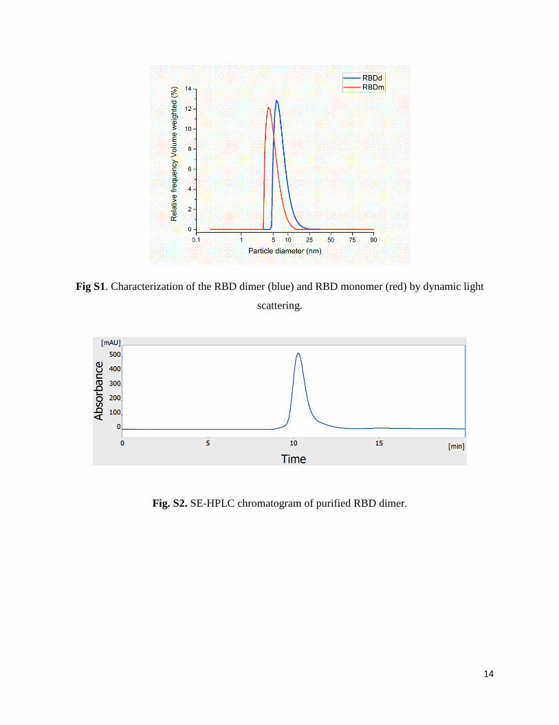
Fig S1. Characterization of the RBD dimer (blue) and RBD monomer (red) by dynamic light
scattering.
Fig. S2. SE-HPLC chromatogram of purified RBD dimer.
15
Figure S3. Multiply-charged (A) and deconvoluted (B) ESI-MS spectra of N-deglycosylated RBD
dimer. In (B), the expected molecular masses consider the conversion of Asn331 and Asn343 into
Asp by PNGase F , O-glycosylation, four intramolecular disulfide bonds and one intermolecular
disulfide bond between Cys538 in each RBD unit. The signals corresponding to the disulfide bonds
are labeled red. OG1 and OG2 represent the O-glycosylation with HexNAc-Hex-SA and HexNAc-
Hex-SA2, respectively. HexNAc: N-acetyl hexosamine, Hex: hexose, SA: sialic acid. (C) ESI-MS
spectrum of the proteolytic peptides derived from RBD dimer treated with N-ethylmaleimide
(NEM), deglycosylated with PNGase F and digested with trypsin following the in-solution buffer-
free digestion protocol.7
7 Betancourt, L. H., Espinosa, L. A., Ramos, Y., Bequet-Romero, M., Rodríguez, E. N., Sánchez, A., Marko-Varga,
G., González, L. J., Besada, V. (2020) Targeting hydrophilic regions of recombinant proteins by MS using in-solution
buffer-free trypsin digestion. Eur. J. Mass Spectrometry 26, 230-237.
16
Figure S4. Interaction of RBD monomer, RBD dimer, RBD-Fc and a negative control with the
recombinant hACE2-Fc receptor.
Table S1. Summary of the average molecular weight (M.W) by ESI-MS of the RBD dimer
previously N-deglycosylated with PNGase F.
Average M.W Theor
(Da) + 4 S-S + C538-
C538 + PNGase F
(N331, N343D)
Modification (a) Average
M.W
Theor (Da)
Average
M.W Exp
(Da)
Error
(%) (b)
Asignment
51828.43
+ (O-HexNAc-Hex-SA)2 53141.62 53141.89 0.003
Dimmer of O-
glycosylated
RBD
+ O-HexNAc-Hex-SA
+ O-HexNAc-Hex-SA2
53432.87 53432.96 0.0002
+ (O-HexNAc-Hex-SA2)2 53724.14 53724.23 0.0002
(a) HexNAc: N-acetyl hexosamine, Hex: hexose, SA: sialic acid, four intramolecular disulfide bonds and one
intermolecular disulfide bond between the two Cys538 of each monomer.
(b) Relative error (%) = | (M.W theor – M.W exp) / M.W theor | * 100.
17
Table S2. Assignment for the tryptic digest of the RBD dimer generated by using an in-solution
buffer-free digestion protocol and analyzed by ESI-MS.
Sequence assignment a) m/z Exp m/z Theor z
319RVQPTESIVR328+HexNAc+Hex+SA (O-glycosylation) 920.93
614.30
920.96
614.31
2+
3+ 319RVQPTESIVR328+HexNAc+Hex+SA2 (O-glycosylation) 1066.50
711.33
1066.50
711.34
2+
3+
NEM-319RVQPTESIVR328+HexNAc+Hex+SA (O-glycosylation) 983.45 983.48 2+
NEM-319RVQPTESIVR328+HexNAc+Hex+SA2 (O-glycosylation) 1129.00 1129.03 2+ 320VQPTESIVR328 514.78 514.79 2+ 320VQPTESIVR328+HexNAc+Hex+SA (O-glycosylation)
842.89 842.90 2+ 320VQPTESIVR328+HexNAc+Hex+SA2 (O-glycosylation)
988.43 988.45 2+ 329FPDITNLCPFGEVFDATR346 | 358ISNCVADYSVLYNSASFSTFK378 (C336-C361)
1089.50
1452.32
1089.51
1452.35
4+
3+ 347FASVYAWNR355
557.27
1113.51
557.28
1113.55
2+
1+ 356KR357
303.21 303.21 1+
379CYGVSPTK386 | 425LPDDFTGCVIAWNSNNLDSK444 (C379-C432)
765.85
1020.79
1530.67
765.86
1020.81
1530.71
4+
3+
2+ 379CYGVSPTK386 | 418IADYNYKLPDDFTGCVIAWNSNNLDSK444 (C379-C432)
982.70
786.37
982.71
786.37
4+
5+
387LNDLCFTNVYADSFVIR403
| 510VVVLSFELLHAPATVCGPK528 (C391-C525)
1323.00
992.51
1323.02
992.52
3+
4+ 404GDEVR408 575.26
288.14
575.28
288.14
1+
2+ 409QIAPGQTGK417
450.24
899.45
450.25
899.50
2+
1+ 418IADYNYK424
443.71
886.40
443.72
886.43
2+
1+ 445VGGNYNYLYR454
609.79
1218.56
609.80
1218.59
2+
1+ 455LFR457
435.26
218.14
435.27
218.14
1+
2+ 455LFRK458
282.18 282.19 2+ 458KSNLKPFER466
373.55 373.55 3+ 459SNLKPFER466 495.77 495.77 2+ 467DISTEIYQAGSTPCNGVEGFNCYFPLQSYGFQPTNGVGY
QPYR509 |___________| (C480-C488)
1589.34
1192.29
1589.38
1192.29
3+
4+ 529KSTNLVK535
395.24 395.25 2+ 530STNLVK535
661.37 661.39 1+
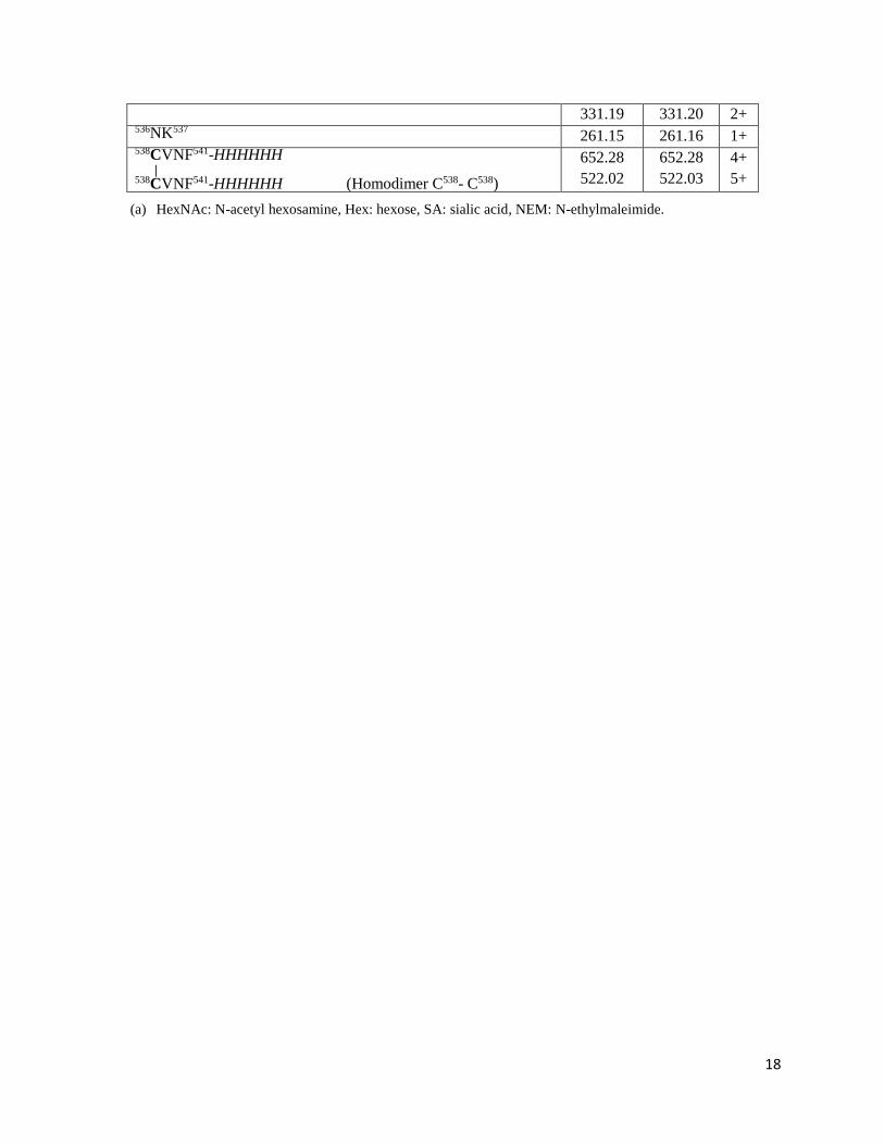
18
331.19 331.20 2+ 536NK537
261.15 261.16 1+ 538CVNF541-HHHHHH | 538CVNF541-HHHHHH (Homodimer C538- C538)
652.28
522.02
652.28
522.03
4+
5+
(a) HexNAc: N-acetyl hexosamine, Hex: hexose, SA: sialic acid, NEM: N-ethylmaleimide.