supplemental material - cjasn
TRANSCRIPT
Supplemental Material
Supplemental Table 1. Study Characteristics
Supplemental Figure 1. Overall Pain Forest Plot
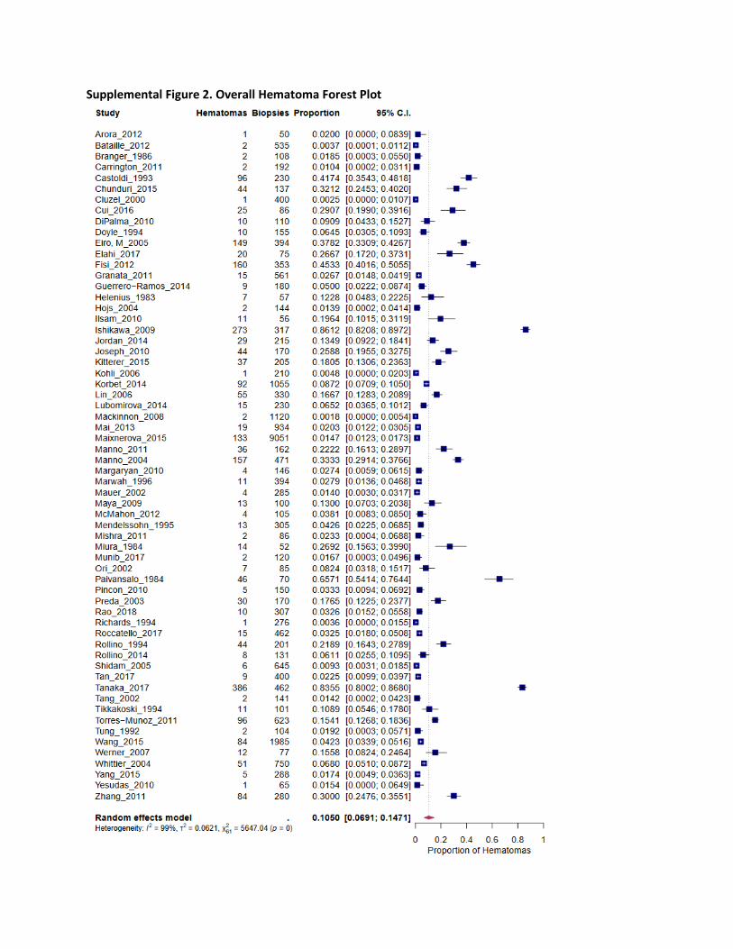
Supplemental Figure 2. Overall Hematoma Forest Plot
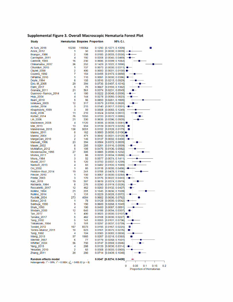
Supplemental Figure 3. Overall Macroscopic Hematuria Forest Plot
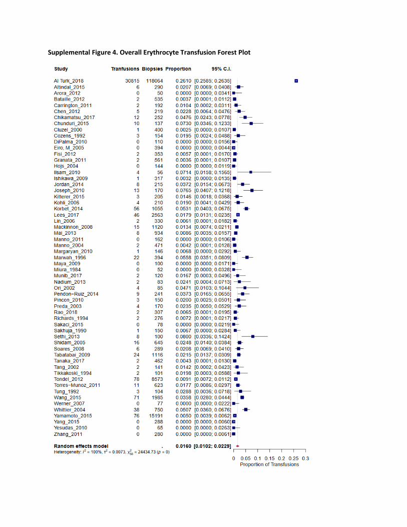
Supplemental Figure 4. Overall Erythrocyte Transfusion Forest Plot
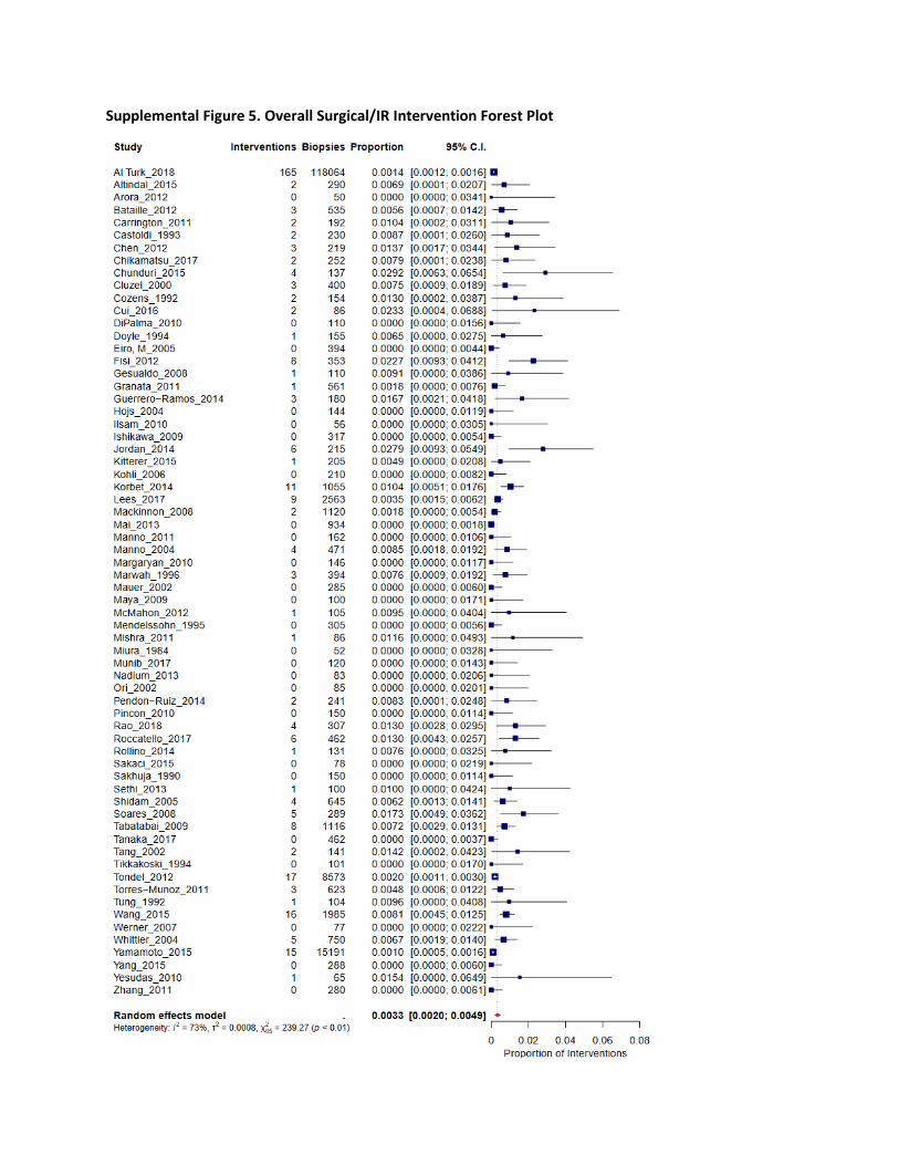
Supplemental Figure 5. Overall Surgical/IR Intervention Forest Plot
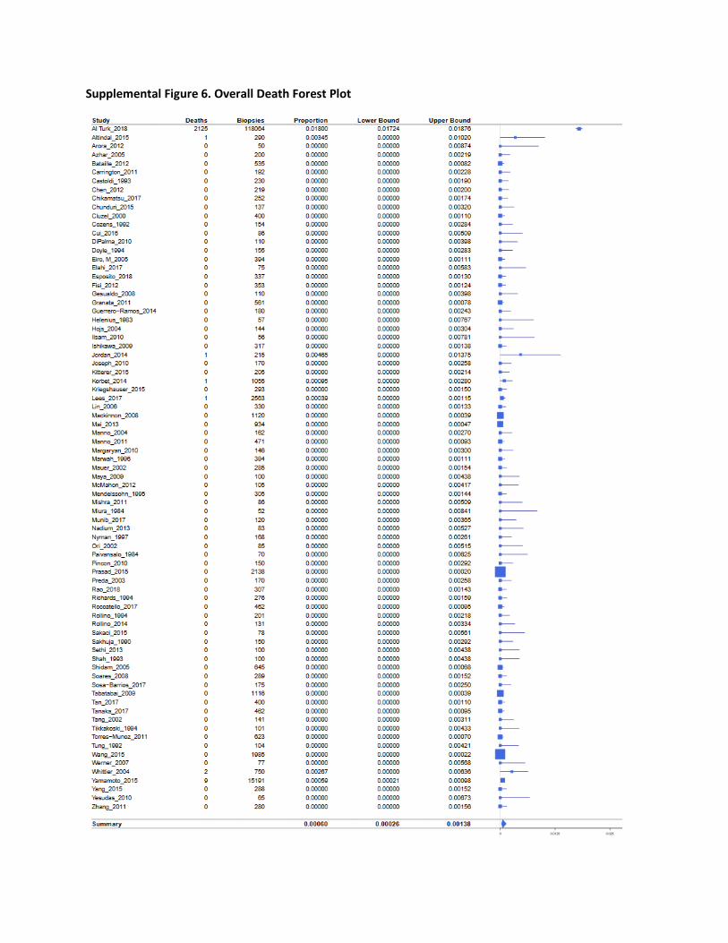
Supplemental Figure 6. Overall Death Forest Plot
Supplemental References
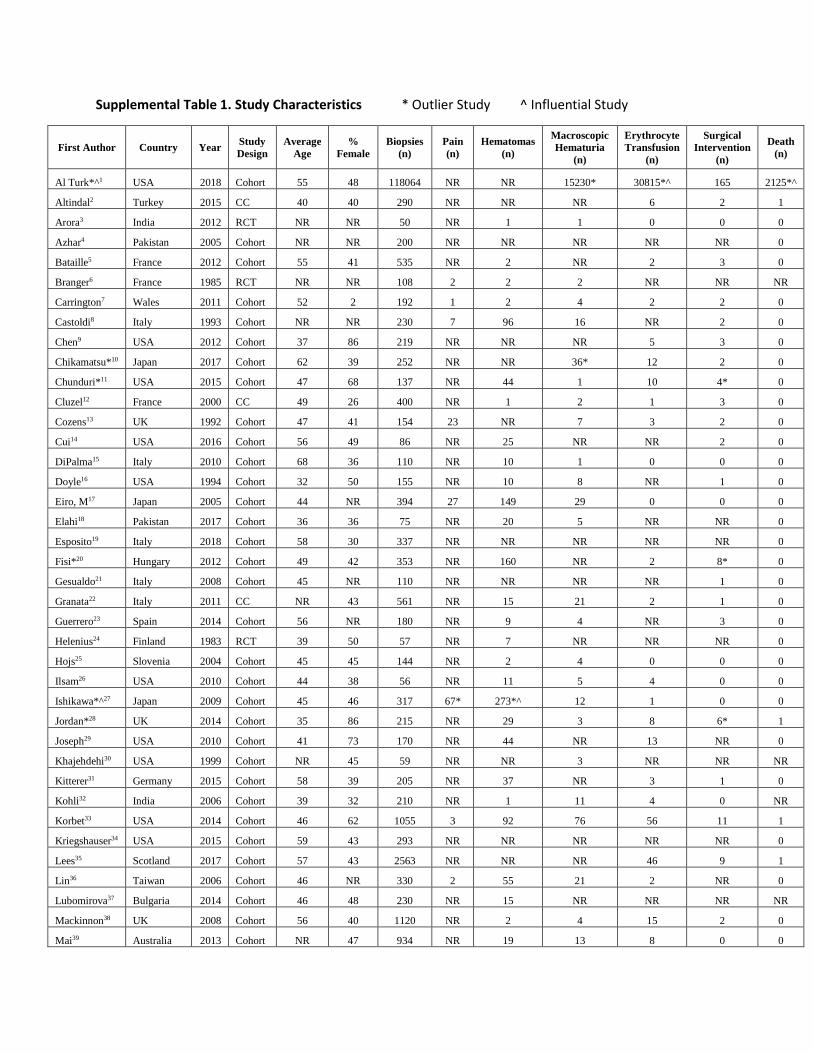
Supplemental Table 1. Study Characteristics * Outlier Study ^ Influential Study
First Author Country Year Study Design
Average Age
% Female
Biopsies (n)
Pain (n)
Hematomas (n)
Macroscopic Hematuria
(n)
Erythrocyte Transfusion
(n)
Surgical Intervention
(n)
Death (n)
Al Turk*^1 USA 2018 Cohort 55 48 118064 NR NR 15230* 30815*^ 165 2125*^
Altindal2 Turkey 2015 CC 40 40 290 NR NR NR 6 2 1
Arora3 India 2012 RCT NR NR 50 NR 1 1 0 0 0
Azhar4 Pakistan 2005 Cohort NR NR 200 NR NR NR NR NR 0
Bataille5 France 2012 Cohort 55 41 535 NR 2 NR 2 3 0
Branger6 France 1985 RCT NR NR 108 2 2 2 NR NR NR
Carrington7 Wales 2011 Cohort 52 2 192 1 2 4 2 2 0
Castoldi8 Italy 1993 Cohort NR NR 230 7 96 16 NR 2 0
Chen9 USA 2012 Cohort 37 86 219 NR NR NR 5 3 0
Chikamatsu*10 Japan 2017 Cohort 62 39 252 NR NR 36* 12 2 0
Chunduri*11 USA 2015 Cohort 47 68 137 NR 44 1 10 4* 0
Cluzel12 France 2000 CC 49 26 400 NR 1 2 1 3 0
Cozens13 UK 1992 Cohort 47 41 154 23 NR 7 3 2 0
Cui14 USA 2016 Cohort 56 49 86 NR 25 NR NR 2 0
DiPalma15 Italy 2010 Cohort 68 36 110 NR 10 1 0 0 0
Doyle16 USA 1994 Cohort 32 50 155 NR 10 8 NR 1 0
Eiro, M17 Japan 2005 Cohort 44 NR 394 27 149 29 0 0 0
Elahi18 Pakistan 2017 Cohort 36 36 75 NR 20 5 NR NR 0
Esposito19 Italy 2018 Cohort 58 30 337 NR NR NR NR NR 0
Fisi*20 Hungary 2012 Cohort 49 42 353 NR 160 NR 2 8* 0
Gesualdo21 Italy 2008 Cohort 45 NR 110 NR NR NR NR 1 0
Granata22 Italy 2011 CC NR 43 561 NR 15 21 2 1 0
Guerrero23 Spain 2014 Cohort 56 NR 180 NR 9 4 NR 3 0
Helenius24 Finland 1983 RCT 39 50 57 NR 7 NR NR NR 0
Hojs25 Slovenia 2004 Cohort 45 45 144 NR 2 4 0 0 0
Ilsam26 USA 2010 Cohort 44 38 56 NR 11 5 4 0 0
Ishikawa*^27 Japan 2009 Cohort 45 46 317 67* 273*^ 12 1 0 0
Jordan*28 UK 2014 Cohort 35 86 215 NR 29 3 8 6* 1
Joseph29 USA 2010 Cohort 41 73 170 NR 44 NR 13 NR 0
Khajehdehi30 USA 1999 Cohort NR 45 59 NR NR 3 NR NR NR
Kitterer31 Germany 2015 Cohort 58 39 205 NR 37 NR 3 1 0
Kohli32 India 2006 Cohort 39 32 210 NR 1 11 4 0 NR
Korbet33 USA 2014 Cohort 46 62 1055 3 92 76 56 11 1
Kriegshauser34 USA 2015 Cohort 59 43 293 NR NR NR NR NR 0
Lees35 Scotland 2017 Cohort 57 43 2563 NR NR NR 46 9 1
Lin36 Taiwan 2006 Cohort 46 NR 330 2 55 21 2 NR 0
Lubomirova37 Bulgaria 2014 Cohort 46 48 230 NR 15 NR NR NR NR
Mackinnon38 UK 2008 Cohort 56 40 1120 NR 2 4 15 2 0
Mai39 Australia 2013 Cohort NR 47 934 NR 19 13 8 0 0
Maixnerova40 Czech Rep 2015 Cohort 45 42 9051 NR 133 138 NR NR NR
Manno41 Italy 2011 RCT 41 NR 162 2 36 0 0 0 0
Manno42 Italy 2004 RCT 39 41 471 12 157 2 2 4 0
Margaryan43 USA 2010 Cohort 44 56 146 NR 4 2 1 0 0
Marwah44 USA 1996 Cohort 44 2 394 NR 11 23 22 3 0
Mauer45 USA 2002 RCT 30 53 285 5 4 8 NR 0 0
Maya46 USA 2009 Cohort 42 60 100 NR 13 NR 0 0 0
McMahon47 USA 2012 Cohort 49 NR 105 NR 4 5 NR 1 0
Mendelssohn48 Canada 1995 Cohort NR NR 305 NR 13 27 NR 0 0
Mishra49 Libya 2011 Cohort NR 73 86 NR 2 2 NR 1 0
Miura50 Japan 1984 Cohort 38 46 52 NR 14 3 0 0 0
Munib51 Pakistan 2017 Cohort 28 32 120 9 2 9 2 0 0
Nadium52 Sudan 2013 Cohort 34 44 83 5 NR 4 2 0 0
Nyman53 Saudi Arabia 1997 Cohort NR 57 168 NR NR NR NR NR 0
Ori54 Israel 2002 Cohort 53 47 85 NR 7 1 4 0 0
Paivansalo*55 Finland 1984 Cohort 41 44 70 12 46* NR NR NR 0
Pendon-Ruiz56 Spain 2014 Cohort 49 3 241 NR NR 19 9 2 NR
Pincon57 France 2010 Cohort 77 48 150 NR 5 1 3 0 0
Prasad58 India 2015 Cohort 34 31 2138 NR NR NR NR NR 0
Preda59 Netherlands 2003 Cohort NR NR 170 NR 30 3 4 NR 0
Rao60 India 2018 CC 37 40 307 NR 10 19 2 4 0
Richards61 UK 1994 Cohort 41 NR 276 NR 1 8 2 NR 0
Roccatello62 Italy 2017 Cohort 55 39 462 NR 15 12 NR 6 0
Rollino63 Italy 1994 RCT NR NR 201 NR 44 21 NR NR 0
Rollino64 Italy 2014 Cohort 79 45 131 NR 8 3 NR 1 0
Rychlik65 Czech Rep 2004 Cohort 42 41 4004 NR NR 273 NR NR NR
Sakaci66 Turkey 2015 Cohort 71 38 78 NR NR 1 0 0 0
Sakhuja67 India 1990 Cohort NR NR 150 NR NR 9 1 0 0
Sethi68 USA 2013 Cohort 47 59 100 2 NR NR 8 1 0
Shah69 Singapore 1993 Cohort 32 NR 100 6 NR 4 NR NR 0
Shidam70 USA 2005 Cohort 42 50 645 NR 6 12 16 4 0
Soares71 USA 2008 Cohort NR 44 289 NR NR NR 6 5 0
Sosa-Barrios72 Spain 2017 Cohort 44 58 175 NR NR NR NR NR 0
Tabatabai73 USA 2009 CC NR 61 1116 NR NR NR 24 8 0
Tan74 China 2017 Cohort 40 50 400 NR 9 1 NR NR 0
Tanaka*^75 Japan 2017 Cohort 50 47 462 NR 386*^ 5 2 0 0
Tang76 Hong Kong 2002 Cohort NR NR 141 2 2 5 2 2 0
Tikkakoski77 Finland 1994 Cohort 43 47 101 7 11 3 2 0 0
Tondel78 Norway 2012 Cohort 51 NR 8573 NR NR 167 78 17 NR Torres-Munoz79 Mexico 2011 Cohort 34 71 623 NR 96 10 11 3 0
Tung80 UK 1992 Cohort 45 38 104 NR 2 4 3 1 0
Wang81 China 2015 Cohort 40 41 1985 NR 84 57 71 16 0

Werner82 Israel 2007 Cohort 46 38 77 NR 12 6 0 0 0
Whittier83 USA 2004 Cohort NR NR 750 NR 51 56 38 5 2
Yamamoto84 Japan 2015 Cohort 45 48 15191 NR NR NR 76 15 9
Yang85 China 2015 Cohort 67 39 288 NR 5 4 0 0 0
Yesudas86 India 2010 Cohort 43 44 65 NR 1 2 0 1 0
Zhang87 China 2011 Cohort 40 44 280 NR 84 20 0 0 0
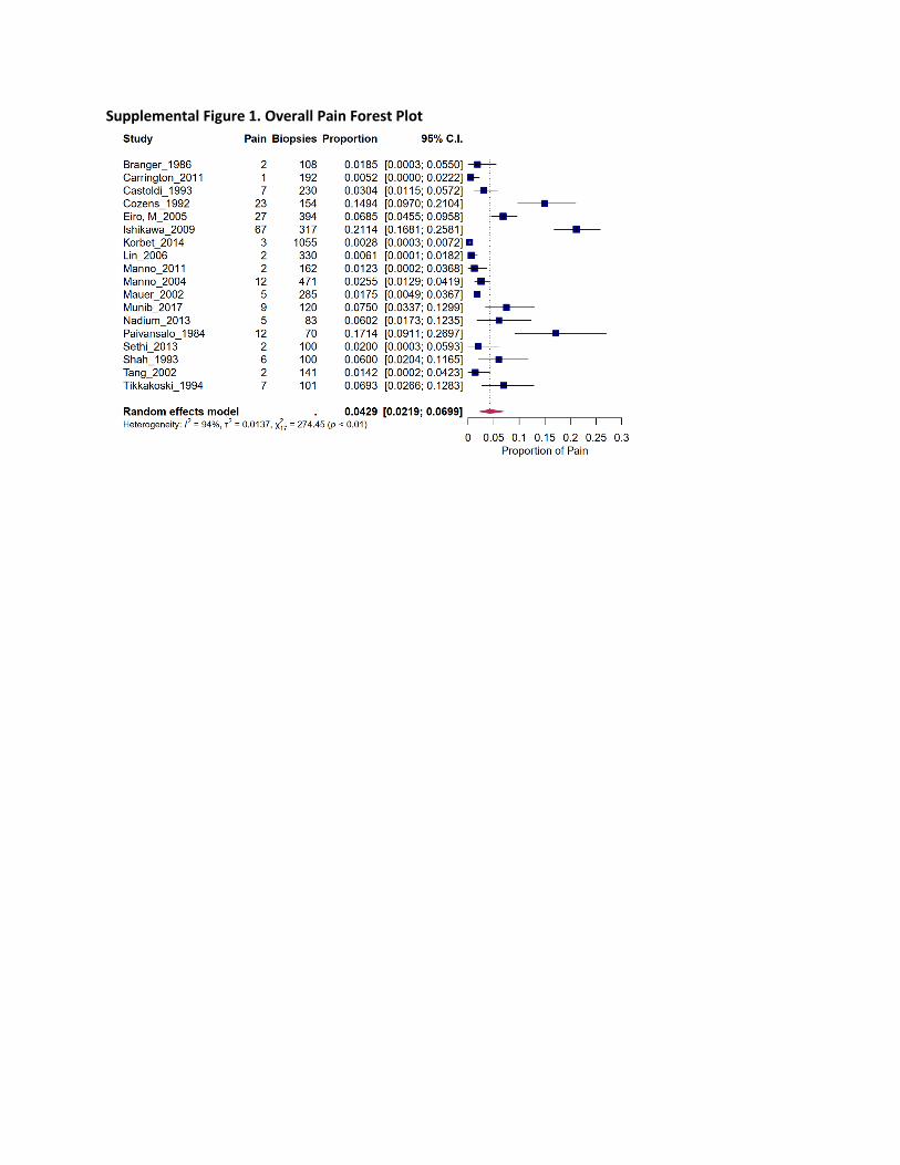
Supplemental Figure 1. Overall Pain Forest Plot
Supplemental Figure 2. Overall Hematoma Forest Plot
Supplemental Figure 3. Overall Macroscopic Hematuria Forest Plot
Supplemental Figure 4. Overall Erythrocyte Transfusion Forest Plot
Supplemental Figure 5. Overall Surgical/IR Intervention Forest Plot
Supplemental Figure 6. Overall Death Forest Plot
Supplemental References
1. Al Turk AA, Estiverne C, Agrawal PR, Michaud JM. Trends and outcomes of the use of
percutaneous native kidney biopsy in the United States: 5-year data analysis of the Nationwide
Inpatient Sample. Clin Kidney J. 2018;11(3):330-336.
2. Altindal M, Yildirim T, Turkmen E, et al. Safety of Percutaneous Ultrasound-Guided Kidney
Biopsy in Patients with AA Amyloidosis. Nephron. 2015;131(1):17-22.
3. Arora K, Punia RS, D'Cruz S. Comparison of diagnostic quality of kidney biopsy obtained using
16G and 18G needles in patients with diffuse renal disease. Saudi Journal of Kidney Diseases &
Transplantation. 2012;23(1):88-92.
4. Azhar A, Anwar N, Zeb A, Ullah A. Renal biopsy: An effective and safe diagnostic procedure.
Journal of Postgraduate Medical Institute. 2006;20(1):78-81.
5. Bataille S, Jourde N, Daniel L, et al. Comparative safety and efficiency of five percutaneous
kidney biopsy approaches of native kidneys: a multicenter study. American Journal of
Nephrology. 2012;35(5):387-393.
6. Branger B, Oules R, Balducchi JP, Fourcade J, Bourgeois JM. Ultrasonically continuously guided
renal biopsy. Uremia Investigation. 1985;9(2):297-303.
7. Carrington CP, Williams A, Griffiths DF, Riley SG, Donovan KL. Adult day-case renal biopsy: a
single-centre experience. Nephrology Dialysis Transplantation. 2011;26(5):1559-1563.
8. Castoldi MC, Del Moro RM, D'Urbano ML, et al. Sonography after renal biopsy: assessment of its
role in 230 consecutive cases. Abdominal Imaging. 1994;19(1):72-77.
9. Chen TK, Estrella MM, Fine DM. Predictors of kidney biopsy complication among patients with
systemic lupus erythematosus. Lupus. 2012;21(8):848-854.
10. Chikamatsu Y, Matsuda K, Takeuchi Y, et al. Quantification of bleeding volume using computed
tomography and clinical complications after percutaneous renal biopsy. Clinical Kidney Journal.
2017;10(1):9-15.
11. Chunduri S, Whittier WL, Korbet SM. Adequacy and complication rates with 14- vs. 16-gauge
automated needles in percutaneous renal biopsy of native kidneys. Seminars in Dialysis.
2015;28(2):E11-14.
12. Cluzel P, Martinez F, Bellin MF, et al. Transjugular versus percutaneous renal biopsy for the
diagnosis of parenchymal disease: comparison of sampling effectiveness and complications.
Radiology. 2000;215(3):689-693.
13. Cozens NJ, Murchison JT, Allan PL, Winney RJ. Conventional 15 G needle technique for renal
biopsy compared with ultrasound-guided spring-loaded 18 G needle biopsy. British Journal of
Radiology. 1992;65(775):594-597.
14. Cui S, Heller HT, Waikar SS, McMahon GM. Needle Size and the Risk of Kidney Biopsy Bleeding
Complications. KI Reports. 2016;1(4):324-326.
15. Di Palma AM, d'Apollo AM, Vendemia F, Stallone G, Infante B, Gesualdo L. Kidney biopsy in the
elderly. Journal of Nephrology. 2010;23 Suppl 15:S55-60.
16. Doyle AJ, Gregory MC, Terreros DA. Percutaneous native renal biopsy: comparison of a 1.2-mm
spring-driven system with a traditional 2-mm hand-driven system. American Journal of Kidney
Diseases. 1994;23(4):498-503.
17. Eiro M, Katoh T, Watanabe T. Risk factors for bleeding complications in percutaneous renal
biopsy. Clinical & Experimental Nephrology. 2005;9(1):40-45.
18. Elahi I, Fazal EM, Abbasi T, Maqbool S. Frequency of haemorrhagic complications of renal
biopsy. Pakistan Journal of Medical and Health Sciences. 2017;11(1):375-377.
19. Esposito V, Mazzon G, Baiardi P, et al. Safety and adequacy of percutaneous kidney biopsy
performed by nephrology trainees. BMC Nephrology. 2018;19(1):14.
20. Fisi V, Mazak I, Degrell P, et al. Histological diagnosis determines complications of percutaneous
renal biopsy: a single-center experience in 353 patients. Kidney & Blood Pressure Research.
2012;35(1):26-34.
21. Gesualdo L, Cormio L, Stallone G, et al. Percutaneous ultrasound-guided renal biopsy in supine
antero-lateral position: a new approach for obese and non-obese patients. Nephrology Dialysis
Transplantation. 2008;23(3):971-976.
22. Granata A, Floccari F, Ferrantelli A, et al. Does systematic preliminar colour Doppler study
reduce kidney biopsy complication incidence? International Journal of Nephrology.
2011;2011:419093.
23. Guerrero-Ramos F, Villacampa-Auba F, Jimenez-Alcaide E, et al. Renal biopsy with 16G needle: a
safety study. Actas Urologicas Espanolas. 2014;38(9):584-588.
24. Helenius H, Laasonen L, Forslund T, Kock B, Kuhlback B, Edgren J. Ultrasonic scanning after
percutaneous renal biopsy. Scandinavian Journal of Urology & Nephrology. 1983;17(2):213-216.
25. Hojs R. Kidney biopsy and power Doppler imaging. Clinical Nephrology. 2004;62(5):351-354.
26. Islam N, Fulop T, Zsom L, et al. Do platelet function analyzer-100 testing results correlate with
bleeding events after percutaneous renal biopsy? Clinical Nephrology. 2010;73(3):229-237.
27. Ishikawa E, Nomura S, Hamaguchi T, et al. Ultrasonography as a predictor of overt bleeding after
renal biopsy. Clinical & Experimental Nephrology. 2009;13(4):325-331.
28. Jordan N, Chaib A, Sangle S, et al. Association of thrombotic microangiopathy and intimal
hyperplasia with bleeding post-renal biopsy in antiphospholipid antibody-positive patients.
Arthritis care & research. 2014;66(5):725-731.
29. Joseph AJ, Compton SP, Holmes LH, et al. Utility of percutaneous renal biopsy in chronic kidney
disease. Nephrology. 2010;15(5):544-548.
30. Khajehdehi P, Junaid SM, Salinas-Madrigal L, Schmitz PG, Bastani B. Percutaneous renal biopsy in
the 1990s: safety, value, and implications for early hospital discharge. American Journal of
Kidney Diseases. 1999;34(1):92-97.
31. Kitterer D, Gurzing K, Segerer S, et al. Diagnostic impact of percutaneous renal biopsy. Clinical
Nephrology. 2015;84(6):311-322.
32. Kohli HS, Jairam A, Bhat A, et al. Safety of kidney biopsy in elderly: a prospective study.
International Urology & Nephrology. 2006;38(3-4):815-820.
33. Korbet SM, Volpini KC, Whittier WL. Percutaneous renal biopsy of native kidneys: a single-center
experience of 1,055 biopsies. American Journal of Nephrology. 2014;39(2):153-162.
34. Kriegshauser JS, Patel MD, Young SW, Chen F, Eversman WG, Chang YH. Risk of bleeding after
native renal biopsy as a function of preprocedural systolic and diastolic blood pressure. Journal
of Vascular & Interventional Radiology. 2015;26(2):206-212.
35. Lees JS, McQuarrie EP, Mordi N, Geddes CC, Fox JG, Mackinnon B. Risk factors for bleeding
complications after nephrologist-performed native renal biopsy. Clinical Kidney Journal.
2017;10(4):573-577.
36. Lin WC, Yang Y, Wen YK, Chang CC. Outpatient versus inpatient renal biopsy: a retrospective
study. Clinical Nephrology. 2006;66(1):17-24.
37. Lubomirova M, Tzocheva T, Hristova M, Bogov B. Complications of automated spring fired
biopsy gun technique. A retrospective analysis of 230 cases. Hippokratia. 2014;18(1):40-43.
38. Mackinnon B, Fraser E, Simpson K, Fox JG, Geddes C. Is it necessary to stop antiplatelet agents
before a native renal biopsy? Nephrology Dialysis Transplantation. 2008;23(11):3566-3570.
39. Mai J, Yong J, Dixson H, et al. Is bigger better? A retrospective analysis of native renal biopsies
with 16 Gauge versus 18 Gauge automatic needles. Nephrology. 2013;18(7):525-530.
40. Maixnerova D, Jancova E, Skibova J, et al. Nationwide biopsy survey of renal diseases in the
Czech Republic during the years 1994-2011. Journal of Nephrology. 2015;28(1):39-49.
41. Manno C, Bonifati C, Torres DD, Campobasso N, Schena FP. Desmopressin acetate in
percutaneous ultrasound-guided kidney biopsy: a randomized controlled trial. American Journal
of Kidney Diseases. 2011;57(6):850-855.
42. Manno C, Strippoli GF, Arnesano L, et al. Predictors of bleeding complications in percutaneous
ultrasound-guided renal biopsy. Kidney International. 2004;66(4):1570-1577.
43. Margaryan A, Perazella MA, Mahnensmith RL, Abu-Alfa AK. Experience with outpatient
computed tomographic-guided renal biopsy. Clinical Nephrology. 2010;74(6):440-445.
44. Marwah DS, Korbet SM. Timing of complications in percutaneous renal biopsy: what is the
optimal period of observation? American Journal of Kidney Diseases. 1996;28(1):47-52.
45. Mauer M, Zinman B, Gardiner R, et al. ACE-I and ARBs in early diabetic nephropathy. Journal of
the Renin-Angiotensin-Aldosterone System. 2002;3(4):262-269.
46. Maya ID, Allon M. Percutaneous renal biopsy: outpatient observation without hospitalization is
safe. Seminars in Dialysis. 2009;22(4):458-461.
47. McMahon GM, McGovern ME, Bijol V, et al. Development of an outpatient native kidney biopsy
service in low-risk patients: a multidisciplinary approach. American Journal of Nephrology.
2012;35(4):321-326.
48. Mendelssohn DC, Cole EH. Outcomes of percutaneous kidney biopsy, including those of solitary
native kidneys. American Journal of Kidney Diseases. 1995;26(4):580-585.
49. Mishra A, Tarsin R, Elhabbash B, et al. Percutaneous ultrasound-guided renal biopsy. Saudi
Journal of Kidney Diseases & Transplantation. 2011;22(4):746-750.
50. Miura H, Tazoe N, Hara M, Kuwahara K, Itoh J, Nakayama M. Ultrasonographic assessment of
perirenal hematoma after percutaneous renal biopsy in adult patients. Nippon Jinzo Gakkai Shi
Japanese Journal of Nephrology. 1984;26(3):337-342.
51. Munib S, Mahmood MBR, Fazli S, Uddin N. Percutaneous renal biopsy in adults: Experience of a
single center. Rawal Medical Journal. 2017;42(1):34-39.
52. Nadium WK, Abdelwahab HH, Ibrahim MA, Shigidi MM. Histological pattern of primary
glomerular diseases among adult Sudanese patients: A single center experience. Indian Journal
of Nephrology. 2013;23(3):176-179.
53. Nyman RS, Cappelen-Smith J, al Suhaibani H, Alfurayh O, Shakweer W, Akhtar M. Yield and
complications in percutaneous renal biopsy. A comparison between ultrasound-guided gun-
biopsy and manual techniques in native and transplant kidneys. Acta Radiologica.
1997;38(3):431-436.
54. Ori Y, Neuman H, Chagnac A, et al. Using the automated biopsy gun with real-time ultrasound
for native renal biopsy. Israel Medical Association Journal: Imaj. 2002;4(9):698-701.
55. Paivansalo M, Jarvi J, Suramo I. Occurrence of hematoma after renal biopsy: systematic follow-
up study by sonography. Clinical Nephrology. 1984;21(5):302-303.
56. Pendon-Ruiz de Mier MV, Espinosa-Hernandez M, Rodelo-Haad C, et al. Prospective study of the
complications associated with percutaneous renal biopsy of native kidneys: experience in a
centre. Nefrologia. 2014;34(3):383-387.
57. Pincon E, Rioux-Leclercq N, Frouget T, Le Pogamp P, Vigneau C. Renal biopsies after 70 years of
age: a retrospective longitudinal study from 2000 to 2007 on 150 patients in Western France.
Archives of Gerontology & Geriatrics. 2010;51(3):e120-124.
58. Prasad N, Kumar S, Manjunath R, et al. Real-time ultrasound-guided percutaneous renal biopsy
with needle guide by nephrologists decreases post-biopsy complications. Clinical Kidney Journal.
2015;8(2):151-156.
59. Preda A, Van Dijk LC, Van Oostaijen JA, Pattynama PM. Complication rate and diagnostic yield of
515 consecutive ultrasound-guided biopsies of renal allografts and native kidneys using a 14-
gauge Biopty gun. European Radiology. 2003;13(3):527-530.
60. Rao NS, Chandra A. Needle guides enhance tissue adequacy and safety of ultrasound-guided
renal biopsies. Kidney Research and Clinical Practice. 2018;37(1):41-48.
61. Richards NT, Darby S, Howie AJ, Adu D, Michael J. Knowledge of renal histology alters patient
management in over 40% of cases. Nephrology Dialysis Transplantation. 1994;9(9):1255-1259.
62. Roccatello D, Sciascia S, Rossi D, et al. Safety of outpatient percutaneous native renal biopsy in
patients with systemic autoimmune diseases: Results from a monocentric cohort. Annals of the
Rheumatic Diseases. 2017;76 (Supplement 2):1410.
63. Rollino C, Garofalo G, Roccatello D, et al. Colour-coded Doppler sonography in monitoring native
kidney biopsies. Nephrology Dialysis Transplantation. 1994;9(9):1260-1263.
64. Rollino C, Ferro M, Beltrame G, et al. Renal biopsy in patients over 75: 131 cases. Clinical
Nephrology. 2014;82(4):225-230.
65. Rychlik I, Jancova E, Tesar V, et al. The Czech registry of renal biopsies. Occurrence of renal
diseases in the years 1994-2000. Nephrology Dialysis Transplantation. 2004;19(12):3040-3049.
66. Sakaci T, Sahutoglu T, Ahbap E, et al. Analysis of renal biopsies in geriatric patients: Single center
experience. Nephrology Dialysis Transplantation. 2015;3):iii415.
67. Sakhuja V, Singh N, Bhalla AK, Pereira BJ, Malik N, Chugh KS. Ultrasonographic localization for
renal biopsy. Journal of the Association of Physicians of India. 1990;38(6):393-395.
68. Sethi I, Brier M, Dwyer A. Predicting post renal biopsy complications. Seminars in Dialysis.
2013;26(5):633-635.
69. Shah RP, Vathsala A, Chiang GS, Chin YM, Woo KT. The impact of percutaneous renal biopsies on
clinical management. Annals of the Academy of Medicine, Singapore. 1993;22(6):908-911.
70. Shidham GB, Siddiqi N, Beres JA, et al. Clinical risk factors associated with bleeding after native
kidney biopsy. Nephrology. 2005;10(3):305-310.
71. Soares SM, Fervenza FC, Lager DJ, Gertz MA, Cosio FG, Leung N. Bleeding complications after
transcutaneous kidney biopsy in patients with systemic amyloidosis: single-center experience in
101 patients. American Journal of Kidney Diseases. 2008;52(6):1079-1083.
72. Sosa-Barrios RH, Burguera V, Rodriguez-Mendiola N, et al. Arteriovenous fistulae after renal
biopsy: diagnosis and outcomes using Doppler ultrasound assessment. BMC Nephrology.
2017;18(1):365.
73. Tabatabai S, Sperati CJ, Atta MG, et al. Predictors of complication after percutaneous
ultrasound-guided kidney biopsy in HIV-infected individuals: possible role of hepatitis C and HIV
co-infection. Clinical Journal of The American Society of Nephrology: CJASN. 2009;4(11):1766-
1773.
74. Tan X, Chen G, Liu Y, et al. Serum D-dimer is a potential predictor for thromboembolism
complications in patients with renal biopsy. Scientific Reports. 2017;7(1):4836.
75. Tanaka K, Kitagawa M, Onishi A, et al. Arterial Stiffness is an Independent Risk Factor for Anemia
After Percutaneous Native Kidney Biopsy. Kidney & Blood Pressure Research. 2017;42(2):284-
293.
76. Tang S, Li JH, Lui SL, Chan TM, Cheng IK, Lai KN. Free-hand, ultrasound-guided percutaneous
renal biopsy: experience from a single operator. European Journal of Radiology. 2002;41(1):65-
69.
77. Tikkakoski T, Waahtera K, Makarainen H, et al. Diffuse renal disease. Diagnosis by ultrasound-
guided cutting needle biopsy. Acta Radiologica. 1994;35(1):15-18.
78. Tondel C, Vikse BE, Bostad L, Svarstad E. Safety and complications of percutaneous kidney
biopsies in 715 children and 8573 adults in Norway 1988-2010. Clinical Journal of The American
Society of Nephrology: CJASN. 2012;7(10):1591-1597.
79. Torres Munoz A, Valdez-Ortiz R, Gonzalez-Parra C, Espinoza-Davila E, Morales-Buenrostro LE,
Correa-Rotter R. Percutaneous renal biopsy of native kidneys: efficiency, safety and risk factors
associated with major complications. Archives of Medical Science. 2011;7(5):823-831.
80. Tung KT, Downes MO, O'Donnell PJ. Renal biopsy in diffuse renal disease--experience with a 14-
gauge automated biopsy gun. Clinical Radiology. 1992;46(2):111-113.
81. Wang C, Yang Y, Jin L, et al. Evaluating renal biopsy-associated hemorrhage complications by the
equation and providing an early intervention: a single-center experience. Journal of Nephrology.
2015;28(6):691-700.
82. Werner M, Osadchy A, Plotkin E, Berheim J, Rathaus V. Increased detection of early vascular
abnormalities after renal biopsies by color Doppler sonography. Journal of Ultrasound in
Medicine. 2007;26(9):1221-1226.
83. Whittier WL, Korbet SM. Timing of complications in percutaneous renal biopsy. Journal of the
American Society of Nephrology. 2004;15(1):142-147.
84. Yamamoto H, Hashimoto H, Nakamura M, Horiguchi H, Yasunaga H. Relationship between
hospital volume and hemorrhagic complication after percutaneous renal biopsy: results from
the Japanese diagnosis procedure combination database. Clinical & Experimental Nephrology.
2015;19(2):271-277.
85. Yang F, Li B, Cui W, et al. A clinicopathological study of renal biopsies from 288 elderly patients:
analysis based on 4,185 cases. International Urology & Nephrology. 2015;47(2):327-333.
86. Yesudas SS, Georgy NK, Manickam S, et al. Percutaneous real-time ultrasound-guided renal
biopsy performed solely by nephrologists: A case series. Indian Journal of Nephrology.
2010;20(3):137-141.
87. Zhang PP, Ge YC, Li SJ, Xie HL, Li LS, Liu ZH. Renal biopsy in type 2 diabetes: timing of
complications and evaluating of safety in Chinese patients. Nephrology. 2011;16(1):100-105.