study of the role of serum folic acid in atopic dermatitis: a correlation with serum ige and disease...
TRANSCRIPT

IJD
ISSN: 0019-5154
Indian Journal of
Dermatology
www.e-ijd.org Published by Medknow Publications
India
n Jo
urn
al o
f Derm
ato
log
y • V
olu
me
56
• Issue 6
• No
vem
be
r-Dec
em
be
r 2011
• Pag
es 6
13-***
Indexed with PubMed
Volume 56 Issue 6 November-December 2011
Symposium in Dermatology: Airborne Contact Dermatitis

673 Indian Journal of Dermatology 2011; 56(6)
IntroductionAtopic dermatitis (AD) is an eczematous highly pruritic chronic inflammatory relapsing skin disease. Its presentation varies from an acute eczematous relapsing eruption in early life to a characteristic lichenified dermatitis in older patients. AD often occurs in people with personal or family history of other atopic disease, i.e., bronchial asthma, rhinitis, and hay fever.[1]
Folic acid (FA) is a water-soluble vitamin B which serves as a cofactor in many enzymatic reactions related to growth and development, neuronal function, and blood cell production.[2] Impaired folate metabolism was found to reduce the intracellular methyl donor pool, associated with a higher prevalence of atopy.[3] It was found that people with the lowest folate levels had 31% higher risk of atopy, while folate administration may reduce the severity of allergic symptoms.[4]
AD results from a combined immunoglobulin E (IgE)-mediated hypersensitivity and delayed type hypersensitivity. Most individuals with AD were found to have elevated serum IgE.[5]
The aim of this work was to assess serum IgE and FA in AD patients and to correlate their levels with the disease severity, and with each other, to investigate a possible role of FA in the occurrence and/or the severity of AD for further therapeutic intervention.
Materials and MethodsThe study has been approved by the institutional ethics committee and is in accordance with the Helsinki Declaration of 1975. It included 20 AD patients and 20 age- and sex-matched healthy volunteers as controls. Patients were recruited from the outpatient dermatology clinic at Ain Shams University during the period between December 2009 and March 2010 after an informed consent was signed by their guardians.
AD patients included 11 females and 9 males with ages ranging from 2 to 14 years. All patients were subjected to full history taking, including history of asthma or hay fever in AD patients and in their first-degree relatives in the past years, and also dietary history for the patients.
Skin lesions in all cases were examined clinically and
From the Departments of Dermatology, Venereology and Andrology and 1Medical Biochemistry, Faculty of Medicine, Ain Shams University; 2Departments of Dermatology and Venereology, Dar El Shefaa Hospital, Cairo, Egypt. Address for correspondence: Dr. Enas Attia, Departments of Dermatology, Venereology and Andrology, Faculty of Medicine, Ain Shams University, Cairo, Egypt, PO: 11566. E-mail: [email protected]
Access this article onlineQuick Response Code:
Website: www.e-ijd.org
DOI: 10.4103/0019-5154.91827
Original Article
STUDY OF THE ROLE OF SERUM FOLIC ACID IN ATOPIC DERMATITIS: A CORRELATION WITH SERUM IgE AND DISEASE SEVERITY
Maha A Shaheen, Enas A S Attia, Manal L Louka1, Nashwa Bareedy2
Abstract
Background: Most atopic dermatitis (AD) patients have elevated serum immunoglobulin E (IgE). Impaired folic acid (FA) metabolism was found to reduce the intracellular methyl donor pool, associated with a higher prevalence of atopy. Aim: To assess serum IgE and FA in AD patients and to correlate their levels with the disease severity, and with each other. Materials and Methods: Twenty patients with AD were assessed for serum FA and IgE, compared with 20 age- and sex-matched controls. Patients were classified into three groups (mild, moderate, and severe AD) based on clinical severity according to Nottingham index. In both patients and controls, serum IgE was measured using Enzyme-linked immunosorbent assay technique and serum FA was measured using Microparticle Enzyme Immunoassay technique. Results: Serum FA levels were lower in AD patients compared with controls, but the difference was not statistically significant. FA levels did not show statistically significant difference among disease severity groups and did not correlate with serum IgE levels. On the other hand, serum IgE levels were significantly elevated in AD patients compared with controls, and among AD patients, its levels were significantly elevated in severe AD compared with mild and moderate disease. Conclusion: Serum IgE is useful in assessment of AD severity and activity. FA contribution to AD needs further investigations.
Key Words: Atopic dermatitis, folic acid, IgE
674Indian Journal of Dermatology 2011; 56(6)
Shaheen, et al.: Serum folic acid and IgE in atopic dermatitis
calculation of Nottingham index was done to determine severity of cases as follows: The body surface was divided into 45 different areas by a system of tick boxes. Each box on the surface area diagram was ticked, if more than 2 cm² (The size of a 10 pence coin) was involved in any given area. The number of involved areas was then calculated as a sum of the total of involved sites for assessment of extent of the disease. For assessment of disease intensity, parents were asked to report average weekly sleep loss due to itching or scratching over the last 12 months. The response was then graded from 1 to 5, with higher scores representing a greater intensity of symptoms producing sleep loss. For assessment of clinical course, parents were asked to comment on how long their child's skin condition had been present during the previous 12 months. The response was graded from 1 to 5, with higher scores reflecting longer disease duration. A final score was achieved by adding each of the three modified parameters of intensity, clinical course, and examined extent of disease score, to produce a possible range of scores from 3 to 15. Patients were then divided into the following three groups: Group 1 (G1) included patients with mild AD, Group 2 (G2) included patients with moderate disease, and Group 3 (G3) included patients with severe AD, based on the final score (maximum of 15): mild (3-8), moderate (9-11), and severe (12-15).[6]
We excluded patients with abnormal diets and use of vitamins or other supplements at the time of the study, patients with systemic illness and diseases, and patients with manifestations of other dermatological diseases (psoriasis). Patients taking drugs known to impair folate metabolism such as antirheumatic, anticonvulsant, and methotrexate therapy, or any medical treatment likely to affect the study outcome three months prior to the study, e.g., steroids and immunosuppressive drugs, were also excluded.
Samples of venous blood were drawn from each subject (patients and controls) after an overnight fast for measurement of serum FA and serum IgE. The blood samples were left to clot for up to 60 minutes at room temperature (22°C) and then centrifuged at 3 000 rpm for 10 minutes. Serum was stored in labeled Eppendorf tubes at -20°C until quantitative assay of serum FA and serum IgE was done.
Quantitative determination of serum FA was determined in duplicates by the Microparticle Enzyme Immunoassay (MEIA) technique according to the manufacturer´s instructions (AXSYM Abbot laboratories, Abbot park, IL). [7] AXSYM Folate was based on ion capture technology where a high molecular weight quaternary ammonium compound (ion capture solution) was dispensed on the glass fiber matrix of the matrix cell. This imparted a positive charge to the matrix which enabled capture of negatively charged analyte complexes. During the assay, negatively charged polyanion-analyte complexes were formed. These
complexes were captured through electrostatic interaction with the positively charged glass fiber matrix. The substrate, 4-Methylumbelliferyl phosphate, was added to the matrix cell and the fluorescent product was measured by the MEIA optical assembly. The AXSYM Folate assay generated a standard Instrumental calibration curve for measuring serum FA (ng/ml). The normal serum FA ranged from 7.2 to 15.4 ng/ml.
Serum IgE was quantitatively determined in duplicates by Enzyme-linked immunosorbent assay technique (Biocheck, USA) according to the manufacturer´s instructions. The assay system utilized one monoclonal anti-IgE antibody and goat anti-IgE antibody in the antibody-enzyme (horseradish peroxidase) conjugate solution. A450 were calculated for each set of reference standards, control, and samples. A standard curve was constructed by plotting the mean absorbance obtained from each reference standard against its concentration in IU/ml on linear graph paper, with absorbance values on the vertical or Y-axis and concentrations on the horizontal or X-axis. Using the mean absorbance value for each sample, the corresponding concentration of IgE was determined in IU/ml from a standard curve.[8] The intra-assay coefficient of variations (% CV) was 5.1% and the inter-assay % CV was 6%. The normal serum IgE ranged up to 15 IU/ml for subjects 1 to 12 months, up to 60 IU/ml for subjects 13 months to 5 years, up to 90 IU/ml for subjects 6 to 8 years, and up to 200 IU/ml for subjects 9 to 16 years.
Data analysisThe Statistical Package for the Social Sciences (SPSS) program, version 11.01 was used for data analysis. Independent student t test was used to compare between two groups with normally distributed quantitative data, while in case data were not normally distributed, Mann-Whitney U test was used. One-way ANOVA test was used when more than two groups with parametric data were to be compared, while Kruskal-Wallis test was used when nonparametric data were compared. Values were considered statistically significant if P≤0.05 and highly significant if ≤0.01.
ResultsThe current study included 20 AD patients, and 20 clinically free age- and sex-matched individuals as controls. The clinical and laboratory data of the patients are summarized in Table 1. The age of onset of the disease ranged from 1 month to 7 years with a mean of 2.7±2.1 years. The disease duration ranged from 6 months to 10 years with a mean of 3.6±2.4 years. According to Nottingham index, G1 included eight patients (40%) with mild AD (Nottingham 3-8), G2 included five patients (35%) with moderate AD (Nottingham 9-11), and G3 included seven patients (25%) with severe AD (Nottingham 12-15). Eight patients (40%) had positive history of asthma and/or allergic rhinitis, and 15 (75%) patients had positive family history of atopy.
675 Indian Journal of Dermatology 2011; 56(6)
Shaheen, et al.: Serum folic acid and IgE in atopic dermatitis
Serum FA levels of patients ranged from 3.7 to 22.4 ng/ml with a mean of 9.177±4.48 ng/ml, while the control levels ranged from 8.8 to 15.6 ng/ml with a mean of 11.57±2.29 ng/ml. Serum IgE levels of patients ranged from 8 to 375 IU/ml with a median of 70 IU/ml, while the control levels ranged from 10 to 75 IU/ml with a median of 40 IU ml.
As regards to mean serum FA level, AD patients showed lower level compared with controls; however, the difference was not statistically significant [Table 2].
Mean serum FA level was not statistically different among the different patient’s groups (P=0.06) as mean FA was 11.429±5.66 ng/ml in mild group, 5.560±1.25 ng/ml in moderate group, and 9.186±2.74 ng/ml in severe AD group.
Comparison of patients and controls as regards to serum IgE level revealed significantly elevated serum IgE in patients [Table 3]. Using Kruskal-Wallis test, there was a statistically significant difference between the three patient’s severity groups as regards to median serum IgE level (P=0.047), with statistically significant higher level in G3 (severe AD; median: 277 IU/ml) compared with G1 (mild AD; median: 46 IU/ml) and G2 (moderate AD; median: 50 IU/ml) (P=0.004 and 0.018, respectively), but there was no significant difference in serum IgE in the mild group in comparison with the moderate group (P=0.83).
There was no significant correlation between serum levels of IgE and FA among both patients and controls (r=0.09 and P=0.71 and r=0.15 and P=0.68, respectively).
No statistically significant difference was observed when comparing patients with and without history of other atopic disease(s) and patients with and without family history of atopy as regards to serum IgE levels [Table 4] as well as serum FA [Table 5].
DiscussionThe present study demonstrated that the mean levels of serum FA were lower in AD patients in comparison with healthy controls, but the difference was not statistically significant. This was in agreement with a previous study that did not support a role of impaired folate metabolism associated with MTHFR C677T polymorphism in the development of atopy.[9]
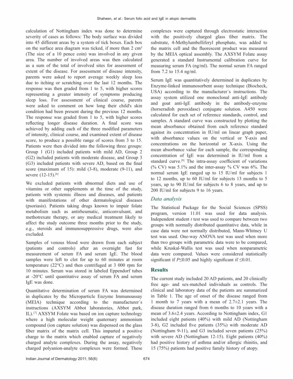
Table 1: Clinical and laboratory data of AD patientsPatient’s number
Age Sex Personal history of atopy
Family history of atopy
Disease onset
Disease duration (years)
Nottingham scoring
Serum IgE (Iu/ml)
Serum folic Acid (ng/ml)
1 3.5 y Female +ve +ve 8 m 2 10/12 Mild 80 8.32 8 y Female +ve +ve 1 y 7 y Severe 350 83 5 y Female +ve +ve 3 y 2 y Mild 65 16.64 5 y Female -ve +ve 1 m 4 11/12 Mild 30 6.335 3 y Male -ve +ve 1 m 2 11/12 Moderate 75 56 7 y Male -ve +ve 3 y 4 Mild 80 67 5 y Male +ve -ve 1 m 4 11/12 Severe 80 8.68 7 y Male -ve -ve 4 y 3 Moderate 150 79 3 y Female -ve +ve 2.5 y 6 m Mild 46 810 2.5 y Male -ve -ve 1 m 2 5/12 Mild 55 1211 3 y Female +ve +ve 1.5 y 1.5 y Mild 45 22.412 13 y Male -ve +ve 3 y 10 y Severe 277 10.113 3.5 y Female +ve +ve 6 m 3 y Moderate 45 3.714 6 y Male +ve +ve 2 m 5 10/12 Severe 14 5.915 14 y Female +ve -ve 7 y 7 Severe 100 916 6 y Male -ve +ve 4 y 2 Severe 350 817 5 y Female -ve -ve 3 y 2 y Moderate 50 16.118 7 y Female -ve +ve 6 y 1 y Moderate 35 619 2 y Female -ve +ve 1.5 y 6 m Severe 375 14.720 4 y male -ve +ve 1 m 3 11/12 Mild 8 11.8
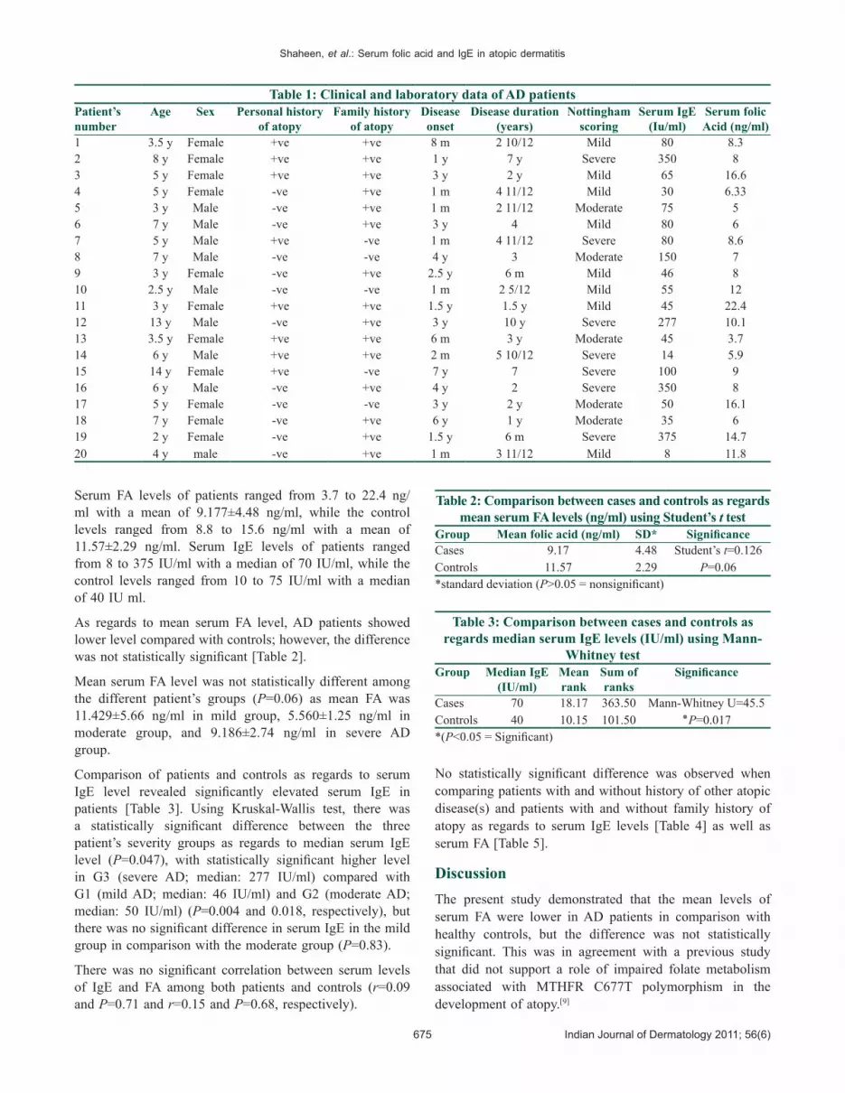
Table 2: Comparison between cases and controls as regards mean serum FA levels (ng/ml) using Student’s t test
Group Mean folic acid (ng/ml) SD* SignificanceCases 9.17 4.48 Student’s t=0.126Controls 11.57 2.29 P=0.06*standard deviation (P>0.05 = nonsignificant)
Table 3: Comparison between cases and controls as regards median serum IgE levels (IU/ml) using Mann-
Whitney testGroup Median IgE
(IU/ml)Mean rank
Sum of ranks
Significance
Cases 70 18.17 363.50 Mann-Whitney U=45.5Controls 40 10.15 101.50 P=0.017٭*(P<0.05 = Significant)

676Indian Journal of Dermatology 2011; 56(6)
Shaheen, et al.: Serum folic acid and IgE in atopic dermatitis
In contrast, a higher serum folate level was associated with lower total IgE levels and a lower risk of atopy in a US population.[10]
There was no statistically significant difference among our different patient's severity groups as regards to mean serum FA. These results indicate that there is no correlation between serum level of FA and the severity of AD.
However, a correlation between FA and severity of allergic diseases was found. In another study using a mouse model, they found that a gestational diet enriched for methyl donors, including FA, was a risk factor, rather than a protective factor for allergy and asthma. This positive association was thought to be mediated by epigenetic modification that was inherited transgenerationally.[11] Nevertheless, in contrast to gestational exposure, mice that were provided high methyl donor diets during lactation or early adulthood did not demonstrate a similar increase in disease severity, suggesting that the timing of methyl donor supplementation might play a pivotal role in determining its effect on the development of allergic disease.[11]
The possible explanation for the differences between this mouse study and our study lies in the differences between mice and human subjects.
The comparison between our patients and controls as
regards to median serum IgE levels revealed significantly elevated serum IgE in patients. These results support previously published data.[12,13]
We found that serum IgE values were statistically significantly higher in severe AD compared with mild and moderate AD. Our results were in agreement with a previous study which showed that patients with mild to moderate AD had much lower IgE levels compared with individuals with severe AD.[14]
A relationship between serum folate levels and the risk of high total IgE levels was found among non-Hispanic white and black subjects, suggesting that low serum folate levels might contribute to the risk of high total IgE levels.[4] However, there was no significant correlation between serum levels of IgE and FA in our study, probably due to the smaller number of patients.
Higher levels of IgE were found among mild AD cases with allergic rhinitis than in cases with pure AD. Also, in moderate forms of AD, coexistent bronchial asthma cases have a greater increase in the IgE values than patients with pure AD.[15] Others found that children with high IgE concentrations developed bronchial hyper-responsiveness, and children with visible dermatitis had higher IgE concentrations.[16]
In contrast, we found no significant difference between patients with only AD and patients with personal history of other atopic diseases as regards to serum IgE (although median IgE of patients with positive personal history of other atopic disease(s) > median IgE of patients with pure AD). Thus, the presence of AD seems to be a risk for the development of other atopic diseases regardless of serum IgE levels.
ConclusionsThe role of serum IgE as a marker of the development of atopy as well as the disease severity is well-established. However, FA contribution is somehow possible for further investigations. The timing of methyl donor supplementation seems to play a pivotal role in determining its effect on the development of atopy. For further studies, trials on FA supplements in AD patient with proven low serum levels are recommended with assessment of disease severity and activity, as well as the therapeutic response.
References1. Simpson EL, Hanifin JM. Atopic dermatitis. J Am Acad
Dermatol 2005;53:115-28.2. Ashwood ER. Vitamins. Tietz Fundamentals of Clinical
Chemistry. In: Burtis CA, Ashwood ER, editors. 5th ed. Philadelphia: Saunders; 2001. p. 544-63.
3. Friso S, Choi SW, Girelli D, Moson JB, Dobikowski GG, Bagley PJ, et al. A common mutation in the 5, 10-methylenetetrahydrofolate reductase gene affects genomic DNA methylation through an interaction with folate status. Proc Natl Acad Sci USA 2002;99:5606-11.
Table 4: Comparison between patients with or without personal history of other atopic disease(s), and patients
with or without family history of atopy, as regards median serum IgE levels (IU/ml) using Mann-Whitney test
Median IgE (IU/ml) SignificancePersonal history of other atopic diseases
+ve history Number=8
72 Mann-Whitney U=44
P=0.76-ve history Number=12
65
Family history of atopy +ve history Number=15
65 Mann-Whitney U=31.5 P=0.76-ve history
Number=580
(P>0.05 = nonsignificant)
Table 5: Comparison between patients with or without personal history of other atopic disease(s), and patients
with or without family history of atopy, as regards mean serum FA levels (ng/ml) using Student's t test
Mean±SD of FA (ng/ml) SignificancePersonal history of other atopic diseases
+ve history Number=8
10.31±6.13 t=0.92 P=0.37
-ve history Number=12
8.41±3.05
Family history of atopy
+ve history Number=15
9.39±5.06 t=0.35 P=0.72
-ve history Number=5
8.54±2.26
(P>0.05 = nonsignificant)

677 Indian Journal of Dermatology 2011; 56(6)
Shaheen, et al.: Serum folic acid and IgE in atopic dermatitis
4. Matsui EC, Matsui WM. High serum folate levels are associated with a lower risk of atopy and wheeze. J Allergy Clin Immunol 2009;123:1253-9.
5. Mamessier E, Magnan A. Cytokines in atopic diseases: Revisiting the Th2 dogma. Eur J Dermatol 2006;2:103-13.
6. Emerson RM, Charman CR, Williams HC. The Nottingham eczema severity score: Preliminary refinement of the Rajka and Langeland grading. Br J Dermatol 2000;142:288-97.
7. Wilson DH, Herrmann R, Hsu S, Biegalski T, Sohn L, Forsythe C, et al. Ion capture assay for folate with the Abbott IMx analyzer. Clin Chem 1995;41:1780-1.
8. Zetterström O, Johansson SG. IgE concentrations measured by PRIST in serum of healthy adults and in patients with respiratory allergy: Adiagnostic approach. Allergy 1981;36:537-47.
9. Granell R, Heron J, Lewis S, Smith GD, Sterne JA, Henderson J. The association between mother and child MTHFR C677T polymorphisms, dietary folate intake and childhood atopy in a population-based, longitudinal birth cohort. Clin Exp Allergy 2007;38:320-8.
10. Forman JP, Rimm EB, Stampfer MJ, Curhan GC. Folate intake and the risk of incident hypertention among US women. JAMA 2005;293:320-9.
11. Hollingsworth JW, Marouka S, Boon K, Tomfohr J, Bailey N, Pott EN, et al. In utero supplementation with methyl donors enhances allergic airway disease in mice. J Clin Invest
2008;118:3462-9.12. Aral M, Arican O, Gal M, Samaz S, Kactark SA, Kastal U, et al.
The relationship between serum level of total IgE, IL-18, IL-12 IFN-γ and disease severity in children with AD. Mediators Inflamm 2006;4:1-4.
13. Park DS, Youn YH. Clinical significance of serum interleukin-18 concentration in the patients with atopic dermatitis. Korean J Lab Med 2007;27:128-32.
14. Cork MJ, Robinson DA, Vasilopoulos Y, Ferguson N, Mostafa M, Mac N, et al. New prescriptive on epidermal barrier dysfunction in AD; Gene environmental interaction. J Allergy Clin Immunol 2006;118:3-21.
15. Wuthrich B. Serum IgE in atopic dermatitis: Relationship to severity of cutaneous involvement and course of disease as well as coexistence of atopic respiratory diseases. Clin Allergy 1978;8:241-8.
16. Sears MR, Burrows B, Flannery EM, Herbison GB, Hewitt CJ, Holdanay MD. Relation between airway responsiveness and serum IgE in children with asthma and in apparently normal children. N Engl J Med 1991;325:1067-71.
How to cite this article: Shaheen MA, Attia EA, Louka ML, Bareedy N. Study of the role of serum folic acid in atopic dermatitis: A correlation with serum IgE and disease severity. Indian J Dermatol 2011;56:673-7.
Received: May, 2011. Accepted: July, 2011.Source of support: Nil, Conflict of Interest: Nil.
Staying in touch with the journal
1) Table of Contents (TOC) email alert
Receive an email alert containing the TOC when a new complete issue of the journal is made available online. To register for TOC alerts go to www.e-ijd.org/signup.asp.
2) RSS feeds
Really Simple Syndication (RSS) helps you to get alerts on new publication right on your desktop without going to the journal’s website. You need a software (e.g. RSSReader, Feed Demon, FeedReader, My Yahoo!, NewsGator and NewzCrawler) to get advantage of this tool. RSS feeds can also be read through FireFox or Microsoft Outlook 2007. Once any of these small (and mostly free) software is installed, add www.e-ijd.org/rssfeed.asp as one of the feeds.