atopic dermatitis in adults: clinical and epidemiological considerations
TRANSCRIPT
r e v a s s o c m e d b r a s . 2 0 1 3;5 9(3):270–275
Revista da
ASSOCIAÇÃO MÉDICA BRASILEIRA
www.ramb.org .br
Original article
Atopic dermatitis in adults: clinical and epidemiologicalconsiderations�
Raquel Leão Orfali a, Marta M. Shimizua, Roberto Takaokaa, Mariana C. Zanibonia,Aline S. Ishizakib, Anderson A. Costab, Ana Paula L. Tibab, Maria Notomi Satoa,Valéria Aokia,∗
a Department of Dermatology, Medical School, Universidade de São Paulo (USP), São Paulo, SP, Brazilb Medical School, USP, São Paulo, SP, Brazil
a r t i c l e i n f o
Article history:
Received 14 September 2012
Accepted 3 December 2012
Available online 13 May 2013
Keywords:
Atopic dermatitis
Epidemiology
Scoring Atopic Dermatitis
IgE
a b s t r a c t
Objective: Atopic dermatitis (AD) is a chronic inflammatory disease causing intense pruritus,
and with typical clinical features. There are few epidemiological studies concerning AD in
adults, as well as little information about its prognostic. The aim of this study was to evaluate
the clinical and epidemiological course of adults with AD.
Methods: 80 patients aged above 18 years (mean age = 29 years) were selected (30 males and
50 females) and interviewed about hospitalization, systemic corticoid usage, age of AD onset,
and personal and/or familial history of atopy. Disease severity was evaluated through the
Scoring Atopic Dermatitis (SCORAD) tool. Laboratory examination included IgE serum levels
and eosinophil blood count.
Results: 71 out of 80 patients referred association with respiratory symptoms (18 had asthma,
17 had rhinitis, and 36 had both conditions); nine out of 80 patients denied any respiratory
disease. AD patients were divided in mild (n = 25), moderate (n = 30), and severe (n = 25); 56%
had one or more hospitalizations due to AD. A positive association was found between IgE
serum levels, eosinophil blood count, and disease severity.
Conclusion: Adult AD represents a clinical challenge that needs to be better characterized,
since it can be misdiagnosed and interferes with the patient’s social and personal life. The
association of skin and respiratory atopic disease is frequent, and laboratory parameters
such as circulating IgE levels and eosinophil blood count may be helpful to assess disease
severity.
© 2013 Elsevier Editora Ltda. All rights reserved.
Dermatite atópica em adultos: consideracões clínicas e epidemiológicas
Palavras-chave:
Dermatite atópica
r e s u m o
Objetivo: Dermatite atópica (DA) é uma doenca inflamatória crônica com prurido intenso
e características clínicas típicas. Há poucos estudos epidemiológicos a respeito da DA em
adultos, bem como pouca informacão disponível sobre o seu prognóstico. O objetivo do
� Study conducted at the Medical School of the Universidade de São Paulo, São Paulo, SP, Brazil.∗ Corresponding author: Av. Dr. Enéas de Carvalho Aguiar, 255/3016, São Paulo, SP, 05403-002, Brazil.
E-mail addresses: [email protected], [email protected] (V. Aoki).0104-4230/$ – see front matter © 2013 Elsevier Editora Ltda. All rights reserved.http://dx.doi.org/10.1016/j.ramb.2012.12.004

r e v a s s o c m e d b r a s . 2 0 1 3;5 9(3):270–275 271
Epidemiologia
Scoring Atopic Dermatitis
IgE
presente estudo é avaliar as características clínicas e o curso epidemiológico dos adultos
com DA.
Métodos: Foram selecionados 80 pacientes com idade acima de 18 anos (média de idade = 29
anos, 30 homens e 50 mulheres), que foram entrevistados sobre: internacões, uso de cor-
ticóide sistêmico, idade de início da DA, história pessoal e/ou familiar de atopia. A gravidade
da doenca foi avaliada de acordo com o SCORing Atopic Dermatitis (SCORAD). A avaliacão
laboratorial incluiu dosagem sérica de IgE e contagem sanguínea de eosinófilos.
Resultados: 71 dos 80 pacientes referiram associacão com sintomas respiratórios (18: asma,
17: rinite alérgica e 36: ambas as condicões); nove dos 80 indivíduos negaram qualquer
sintoma respiratório. Os pacientes com DA foram divididos em DA leve (n = 25), moderada
(n = 30) e grave (n = 25); destes, 56% tiveram uma ou mais internacões por conta da doenca.
Verificou-se uma associacão entre níveis séricos de IgE, contagem sanguínea de eosinófilos
e gravidade da doenca.
Conclusão: A DA do adulto representa um desafio clínico que necessita ser melhor caracter-
izado, uma vez que pode ser erroneamente diagnosticada, e interfere diretamente na vida
social e pessoal dos pacientes. A associacão entre manifestacão respiratória e cutânea é
frequente, e parâmetros laboratoriais como níveis de IgE circulante e contagem sanguínea
de eosinófilos podem ser úteis para acompanhar a gravidade e evolucão da doenca.
I
AdccidDm
iAh
masIrT
fmrgftasciwaatr
ntroduction
topic dermatitis (AD) is a pruritic, chronic, and inflammatoryisease, with typical clinical features. AD is one of the mostommon skin diseases, with a prevalence of 10% to 20% inhildren and 1% to 3% in adults.1,2 Diagnosis is based on clin-cal findings,3 and despite the existence of a wide number ofisease outcome measures, three scores (EASI, Scoring Atopicermatitis [SCORAD], and POEM) have been adopted by theajority of the studies.4–8
In 30% to 50% of the AD patients, the disease markedlymproves during elementary school-age or adolescence.mong those, AD patients that present the disease until adult-ood, 50% to 60% persist with a chronic, recurrent course.9
The pathogenesis of AD remains complex. Gene-environ-ent interactions in genetically predisposed individuals play
central role.10 Moreover, there is a variability of systemic andkin immune abnormalities in AD, such as increased serumgE and allergen sensitization, abnormalities in epidermal bar-ier (gene mutations encoding proteins such as filaggrin), theh1/Th2 paradigm, and microbe skin colonization.1,10,11
The skin barrier function has been considered a protectiveactor against the development of AD, since inherited abnor-
alities in critical epidermal proteins have been identified inecent studies of AD patients. One of these proteins is filag-rin (FLG), a granular cell layer key protein that is processedrom profilaggrin and facilitates terminal differentiation ofhe epidermis.12 FLG is responsible for the aggregation of ker-tin filaments, which collapse the granular cells into nuclearquames to form the cornified cell envelope of the stratumorneum (SC) and the skin barrier, protecting the organ-sm against environmental agents and preventing epidermalater loss.13–15 Skin barrier defects caused by FLG mutations
llows the penetration of allergens through the epidermis
nd their interaction with antigen-presenting cells, leading tohe development of atopic disorders, including asthma andhinitis.16 Recent studies demonstrated that FLG mutations© 2013 Elsevier Editora Ltda. Todos os direitos reservados.
are strongly associated with increased atopic eczema risk, andprobably account for approximately 10% of cases in Europe.17
AD has been classified into two groups: extrinsic form (clas-sic IgE-mediated allergic) and intrinsic form (non-allergic).18
FLG mutations have also been linked to extrinsic AD in recentstudies, indicating that high IgE serum levels may correlatewith disease severity and skin barrier defects.19
Recent literature suggests that circulating eosinophilcounts could be used as a diagnostic tool to differentiateextrinsic from intrinsic AD. Eosinophil blood count corre-lates with disease severity, similar to circulating IgE levels.Eosinophilia and eosinophil skin infiltration found in ADpatients link AD to cytokines recruitment, and chemokinesand eosinophil activation.20
In the skin, AD presents as a model of Th1/Th2 responsewith a biphasic pattern: in acute lesions, a great numberof interleukin (IL)-4, IL-5, and IL-13 (Th2-type cytokines) arefound, whereas in chronic lesions there are high levels ofIL-5, IL-12, and IFN-� (Th1 cells).1 Interleukin-4 and IL-13are implicated in the initial phase of tissue inflammation,and may mediate an isotype switching to IgE synthesis, andup-regulation expression of adhesion molecules on endothe-lial cells. IL-5 increases the survival of eosinophils, andeosinophilia with an increase of the eosinophilic cationic pro-tein (ECP) correlates to disease severity.21 Recently, regulatoryT-cells with immunosuppressant activity in AD, such as IL-17,secreting Th17 cells and Th22 (IL-22 secreting cells) have beeninvestigated in several studies.10,22,23
Staphylococcus aureus is present in 80% to 100% of ADskin, and is responsible for the disease relapsing course. S.aureus exacerbates AD by secreting toxins and superantigenswhich stimulate T cells and macrophages. Most AD patientsproduce specific IgE antibodies against staphylococcal entero-toxins that correlate with disease severity. Superantigens canalso induce corticosteroid resistance, suggesting that several
mechanisms are implied in triggering AD flares.1,10,24,25Little information is available regarding the prognosis of ADadult patients. A monthly follow-up study was conducted in

272 r e v a s s o c m e d b r a s . 2 0 1 3;5 9(3):270–275
Fig. 1 – Clinical features of AD in adults. A. Facial involvement in atopic dermatitis-extensive lichenification of the front,periorbital areas, and malar regions, sparing the central seborrheic areas. B. Severe AD, with erythema, xerosis,
and massive lichenification of the inferior limbs.adults with AD for ten years, and showed that high levels ofcirculating IgE and eosinophil counts as early as the first visitwere indicative of persistent AD.26
This study aimed to evaluate adults diagnosed with AD,with emphasis on their clinical and epidemiological course.
Methods
The patients were selected from the Atopic Dermatitis Out-patient Clinic of the Department of Dermatology from theMedical School of the Universidade de São Paulo, Brazil.Between December, 2010 and April, 2011, 329 patients diag-nosed with AD (according to Hanifin & Rajka’s criteria)3 wereevaluated; 115 were above the age of 18, and 80 of those agreedto participate in the study after signing the informed consent(approved by the Ethics Committee of the Medical School ofthe Universidade de São Paulo). They were interviewed andclassified according to the SCORAD (5) (Fig. 1A and B).
All participants answered a questionnaire that includeddata about their AD history, age of disease onset, use of sys-temic corticosteroids or other immunosuppressant drugs (e.g.methotrexate or cyclosporine), hospitalization, and personaland/or familial history of atopy. All included patients wereusing first-line therapy for disease control (e.g. emollients andtopical corticosteroids).27 IgE serum levels were detected bynephelometric method (N Latex IgE Mono, Dade Behring –Marburg, Germany). Levels up to 100 IU/mL were considerednormal. Blood tests were performed to evaluate circulatingeosinophil counts (normal levels: 0.0% to 5.0%).
Statistical analysis
Mann-Whitney and Kruskal-Wallis non-parametric testswere used to compare two or three sets of data, respec-tively. Correlation between data was established using
Spearman’s- non-parametric correlation test. Differencebetween groups was considered statistically significant whenp-value ≤ 0.05.
Results
Demographic data included: 80 adults with AD, aged 18 orolder (ranging from 18 to 79 years, mean age 29), and gen-der distribution: 30 males and 50 females. 71 of 80 AD patientsreported association with respiratory diseases (18 had asthma,17 had rhinitis, and 36 had both conditions); nine denied anyrespiratory illness. SCORAD ranged between 0 and 100, and ADpatients were classified as mild (n = 25), moderate (n = 30), andsevere (n = 25). 56% of the AD patients (44/80) had one or morehospitalizations during their life, showing the impact of the ill-ness on their quality of life. Systemic corticosteroid usage, (butnot adjuvant oral immunosuppressant) such as cyclosporineor methotrexate,27 was reported by 57 AD patients, and wasfollowed by flares after tapering initial doses (Table 1).
Mean circulating IgE levels in AD patients was 18,340 UI/mL(Fig. 2A). There was an association of IgE levels and diseaseseverity (Fig. 2B). However, no relationship was found betweenserum IgE levels and respiratory disease (Data not shown).A positive correlation between eosinophil blood count anddisease severity SCORAD (Fig. 2C) was detected.
Discussion
AD is one of the most frequent dermatoses in the pediatricpopulation; approximately 40% of the patients persist with
the disease in adulthood. Although rare, the onset of AD mayoccur in adults, usually after the third decade of life.28 AdultAD is complex, and the disease affects personal and familialdynamics.29,30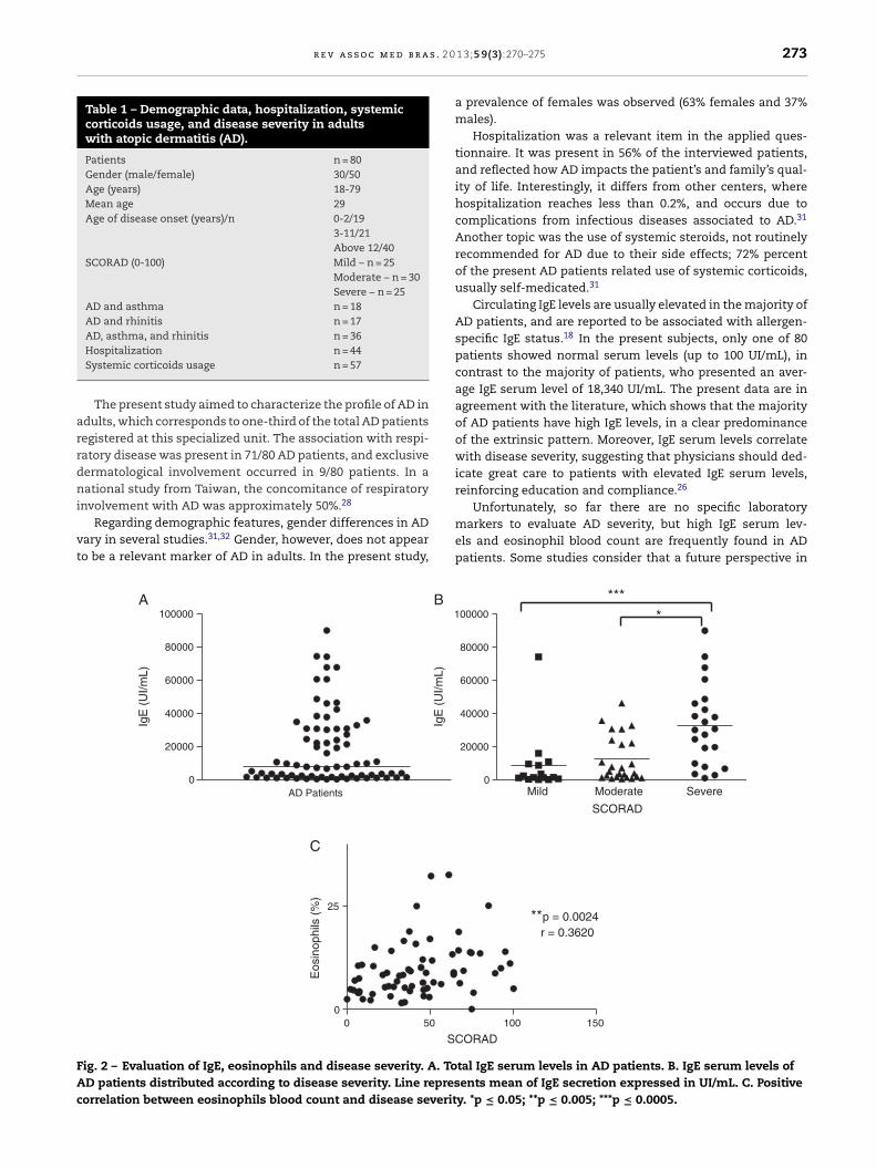
r e v a s s o c m e d b r a s . 2 0
Table 1 – Demographic data, hospitalization, systemiccorticoids usage, and disease severity in adultswith atopic dermatitis (AD).
Patients n = 80Gender (male/female) 30/50Age (years) 18-79Mean age 29Age of disease onset (years)/n 0-2/19
3-11/21Above 12/40
SCORAD (0-100) Mild – n = 25Moderate – n = 30Severe – n = 25
AD and asthma n = 18AD and rhinitis n = 17AD, asthma, and rhinitis n = 36
arrdni
vt
FAc
Hospitalization n = 44Systemic corticoids usage n = 57
The present study aimed to characterize the profile of AD indults, which corresponds to one-third of the total AD patientsegistered at this specialized unit. The association with respi-atory disease was present in 71/80 AD patients, and exclusiveermatological involvement occurred in 9/80 patients. In aational study from Taiwan, the concomitance of respiratory
nvolvement with AD was approximately 50%.28
Regarding demographic features, gender differences in ADary in several studies.31,32 Gender, however, does not appearo be a relevant marker of AD in adults. In the present study,
0
20000
40000
60000
80000
100000
AD Patients
IgE
(U
I/mL)
1
IgE
(U
I/mL)
0 50 0
25
SC
Eos
inop
hils
(%
)
A
C
B
ig. 2 – Evaluation of IgE, eosinophils and disease severity. A. ToD patients distributed according to disease severity. Line represorrelation between eosinophils blood count and disease severit
1 3;5 9(3):270–275 273
a prevalence of females was observed (63% females and 37%males).
Hospitalization was a relevant item in the applied ques-tionnaire. It was present in 56% of the interviewed patients,and reflected how AD impacts the patient’s and family’s qual-ity of life. Interestingly, it differs from other centers, wherehospitalization reaches less than 0.2%, and occurs due tocomplications from infectious diseases associated to AD.31
Another topic was the use of systemic steroids, not routinelyrecommended for AD due to their side effects; 72% percentof the present AD patients related use of systemic corticoids,usually self-medicated.31
Circulating IgE levels are usually elevated in the majority ofAD patients, and are reported to be associated with allergen-specific IgE status.18 In the present subjects, only one of 80patients showed normal serum levels (up to 100 UI/mL), incontrast to the majority of patients, who presented an aver-age IgE serum level of 18,340 UI/mL. The present data are inagreement with the literature, which shows that the majorityof AD patients have high IgE levels, in a clear predominanceof the extrinsic pattern. Moreover, IgE serum levels correlatewith disease severity, suggesting that physicians should ded-icate great care to patients with elevated IgE serum levels,reinforcing education and compliance.26
Unfortunately, so far there are no specific laboratorymarkers to evaluate AD severity, but high IgE serum lev-els and eosinophil blood count are frequently found in ADpatients. Some studies consider that a future perspective in
Mild Moderate Severe0
20000
40000
60000
80000
00000
****
SCORAD
100 150
**p = 0.0024 r = 0.3620
ORAD
tal IgE serum levels in AD patients. B. IgE serum levels ofents mean of IgE secretion expressed in UI/mL. C. Positive
y. *p ≤ 0.05; **p ≤ 0.005; ***p ≤ 0.0005.

a s . 2
r
1
1
1
1
1
1
1
1
1
1
2
22
2
2
2
2
2
274 r e v a s s o c m e d b r
the treatment of AD may be based on serological markerssuch as IL-4.21 Some studies suggest that eosinophil bloodcount may be used as an indicator to distinguish intrinsic fromextrinsic forms of AD, and others show that eosinophil bloodcount correlates with both disease severity and IgE serumlevels.24 All AD patients included in the present study showedhigh eosinophil blood counts (mean value = 9.45%; normal lev-els = 0% to 5%). A positive correlation between eosinophils anddisease severity was found (p = 0.0024; r = 0.3620), but no cor-relation between IgE serum levels and eosinophil blood countwas detected.
In conclusion, the present study presented an elevatednumber of adults with AD in this clinic, indicating that a care-ful follow-up, together with an education strategy are needed,since this is a chronic and high-cost disease with expressivesocial and economic implications. IgE serum levels and/oreosinophil blood counts present a positive correlation withdisease severity, and are relevant parameters for managingthis difficult and chronic disease, contributing to new strate-gies of treatment.
Financial support
This work was supported by FAPESP (Grant # 2011/02453-7).This work was approved by the Ethics Committee of the
Faculdade de Medicina da Universidade de São Paulo on May28, 2010.
Conflicts of interest
The authors declare no conflicts of interest.
e f e r e n c e s
1. Leung DY, Boguniewicz M, Howell MD, Nomura I, Hamid QA.New insights into atopic dermatitis. Journal of ClinicalInvestigation. 2004;113:651–7.
2. Williams H, Flohr C. How epidemiology has challenged 3prevailing concepts about atopic dermatitis. Journal of Allergyand Clinical Immunology. 2006;118:209–13.
3. Hanifin JM, Rajka G. Diagnostic features of atopic dermatitis.Acta Dermato-Venereologica Supplementum. 1980;92Suppl:44–7.
4. Hanifin JM, Thurston M, Omoto M, Cherill R, Tofte SJ, GraeberM. The eczema area and severity index (EASI): assessmentof reliability in atopic dermatitis. EASI Evaluator Group ExpDermatol. 2001;10:11–8.
5. Severity scoring of atopic dermatitis: the, SCORAD., index.Consensus report of the European Task Force on AtopicDermatitis. Dermatology. 1993;186:23–31.
6. Charman CR, Venn AJ, Williams HC. The patient-orientedeczema measure: development and initial validation of a newtool for measuring atopic eczema severity from the patients’perspective. Archives of Dermatology. 2004;140:1513–9.
7. Williams HC, Grindlay DJ. What’s new in atopic eczema? Ananalysis of systematic reviews published in 2007 and 2008
Part 1 Definitions, causes and consequences of eczema.Clinical and Experimental Dermatology. 2010;35:12–5.8. Schram ME, Spuls PI, Leeflang MM, Lindeboom R, Bos JD,Schmitt J. EASI, (objective) SCORAD and POEM for atopic
2
0 1 3;5 9(3):270–275
eczema: responsiveness and minimal clinically importantdifference. Allergy. 2012;67:99–106.
9. Pugliarello S, Cozzi A, Gisondi P, Girolomoni G. Phenotypesof atopic dermatitis. J Dtsch Dermatol Ges. 2011;9:12–20.
0. Boguniewicz M, Leung DY. Recent insights into atopicdermatitis and implications for management of infectiouscomplications. Journal of Allergy and Clinical Immunology.2010;125:4–13.
1. Leung DY, Soter NA. Cellular and immunologic mechanismsin atopic dermatitis. Journal of the American Academyof Dermatology. 2001;44 1 Suppl:S1–12.
2. Nemoto-Hasebe I, Akiyama M, Nomura T, Sandilands A,McLean WH, Shimizu H. Clinical severity correlates withimpaired barrier in filaggrin-related eczema. Journalof Investigative Dermatology. 2009;129:682–9.
3. Sugarman JL. The epidermal barrier in atopic dermatitis.Seminars in Cutaneous Medicine and Surgery. 2008;27:108–14.
4. Palmer CN, Irvine AD, Terron-Kwiatkowski A, Zhao Y, Liao H,Lee SP, et al. Common loss-of-function variants of theepidermal barrier protein filaggrin are a major predisposingfactor for atopic dermatitis. Nature Genetics. 2006;38:441–6.
5. Addor FA, Aoki V. Skin barrier in atopic dermatitis. AnaisBrasileiros de Dermatologia. 2010;85:184–94.
6. Osawa R, Akiyama M, Shimizu H. Filaggrin gene defectsand the risk of developing allergic disorders. Allergol Int.2011;60:1–9.
7. Baurecht H, Irvine AD, Novak N, Illig T, Buhler B, Ring J, et al.Toward a major risk factor for atopic eczema: meta-analysisof filaggrin polymorphism data. Journal of Allergy andClinical Immunology. 2007;120:1406–12.
8. Kulthanan K, Boochangkool K, Tuchinda P, ChularojanamontriL. Clinical features of the extrinsic and intrinsic types ofadult-onset atopic dermatitis. Asia Pac Allergy. 2011;1:80–6.
9. McGrath JA, Uitto J. The filaggrin story: novel insights intoskin-barrier function and disease. Trends in MolecularMedicine. 2008;14:20–7.
0. Liu FT, Goodarzi H, Chen HY. IgE, mast cells, and eosinophilsin atopic dermatitis. Clinical Reviews in Allergy andImmunology. 2011;41:298–310.
1. Bieber T. Atopic dermatitis. Ann Dermatol. 2010;22:125–37.2. Nograles KE, Zaba LC, Shemer A, Fuentes-Duculan J,
Cardinale I, Kikuchi T, et al. IL-22-producing “T22” T cellsaccount for upregulated IL-22 in atopic dermatitis despitereduced IL-17-producing TH17 T cells. Journal of Allergyand Clinical Immunology. 2009;123:1244–52, e2.
3. Souwer Y, Szegedi K, Kapsenberg ML, de Jong EC. IL-17and IL-22 in atopic allergic disease. Current Opinionin Immunology. 2010;22:821–6.
4. Leung DY. Atopic dermatitis and the immune system: the roleof superantigens and bacteria. Journal of the AmericanAcademy of Dermatology. 2001;45 1 Suppl:S13–6.
5. Orfali RL, Sato MN, Takaoka R, Azor MH, Rivitti EA, Hanifin JM,et al. Atopic dermatitis in adults: evaluation of peripheralblood mononuclear cells proliferation response toStaphylococcus aureus enterotoxins A and B and analysisof interleukin-18 secretion. Experimental Dermatology.2009;18:628–33.
6. Katoh N, Hirano S, Kishimoto S. Prognostic factor of adultpatients with atopic dermatitis. Journal of Dermatology.2008;35:477–83.
7. Ellis C, Luger T, Abeck D, Allen R, Graham-Brown RA, De ProstY, et al. International Consensus Conference on AtopicDermatitis II (ICCAD II): clinical update and current treatmentstrategies. British Journal of Dermatology. 2003;148 Suppl
63:3–10.8. Kulthanan K, Samutrapong P, Jiamton S, Tuchinda P.Adult-onset atopic dermatitis: a cross-sectional study of

s . 2 0
2
3
3
3Prevalence of atopic dermatitis among Korean adults visiting
r e v a s s o c m e d b r a
natural history and clinical manifestation. Asian PacificJournal of Allergy and Immunology. 2007;25:207–14.
9. Ong PY, Leung DY. Atopic dermatitis. Clin Allergy Immunol.2002;16:355–79.
0. Ong PY, Leung DY. The infectious aspects of atopic dermatitis.Immunol Allergy Clin North Am. 2010;30:309–21.
1. Hwang CY, Chen YJ, Lin MW, Chen TJ, Chu SY, Chen CC, et al.Prevalence of atopic dermatitis, allergic rhinitis and asthma
1 3;5 9(3):270–275 275
in Taiwan: a national study 2000 to 2007. ActaDermato-Venereologica. 2010;90:589–94.
2. Kim MJ, Kang TW, Cho EA, Kim HS, Min JA, Park H, et al.
health service center of the Catholic Medical Center in SeoulMetropolitan Area. Korea J Korean Med Sci. 2010;25:1828–30.