studies on ventilation of caiiz4an crocodzlus
TRANSCRIPT
Respiration Ph,vsiology ( 1976) 26. 285-301 : North-Hollund Publishing Compun,~~, Amstdum
STUDIES ON VENTILATION OF CAIIZ4AN CROCODZLUS
(CROCODILIA: REPTILIA)’
CARL CANS and BRIAN CLARK
Abstract. The ventilatory mechanics of freely moving Cui~~un crouxlilus were studied by cinefluorcscopy
and electromyography. The buccal oscillations serve only to flush the internal nares in olfactton. Ventila-
tions are coincident with abdominal oscillations. The larynx ordinarily lies adpressed to the internal nares
so that the posterior buccal chamber is excluded from the path of air flow during ventilation and does not
contribute to respiratory dead space.
The pulmonary pressures may be variably polyphasic and the tracheal flows diphasic. Exhalation
involves an anterior shift of the liver by action of the transverse abdominal muscles, while inhalation pro-
ceeds due to contraction of the diaphragmatic muscle pulling the liver caudad. The various costal muscles
facilitate air flow by shifting the position of the ribs. They also play a role in fixation of the flexible rib
cage so that it resists the aspirating and compressing actions of the hepatic piston.
The pattern of muscular activity shifts as the trunk is immersed; expiration becomes passive and
inspiration requires increased muscular effort, The ribs, instead of changing position with each breath are
comparatively fixed by the costal muscles, while changes in the volume of the pleural cavity are caused
almost exclusively by movements of the hepatic piston.
Breathing pattern
Buccal oscillations
Electromyography
Mechanics of breathing
Olfaction
Rib movements
Ventilation
The advent of simplified electromyographical techniques now permits the charac-
terization of the mechanics of breathing in lower vertebrates. A series of studies
dealing with various fishes (Ballintijn and Hughes, 1965; Ballintijn, 1969a,b; Hughes
and Ballintijn, 1965 ; Osse, 1969; McMahon, 1969) frogs (de Jongh and Gans, 1969 ;
Martin and Gans, 1972; West and Jones, 1975) turtles (Gans and Hughes, 1967;
Gaunt and Gans, 1969b; Ireland and Gans, 1973) lizards (Hadden, 1975) and snakes
r All the experiments were supported with funds from NSF BMS 71-01380
2X5
286 C. GANS AND B. CLARK
(Rosenberg, 1973) have provided a general pattern and, as a byproduct, allowed us
to modify our view of the evolution of air breathing mechanics (Gans, 197la.b).
The senior author had carried out some pilot studies on crocodilian respiration
in collaboration with A.S. Gaunt (c/1 Gaunt and Gans. 1969a). However, their
publication was delayed until after the start of the cycle of papers by Naifeh rt ul.
(1970a, b. 1971a, b,c) which contain reviews of earlier literature on crocodilian
ventilation. Analysis of the results obtained by these authors (which were of course
ultimately aiming at different questions) pointed to certain fundamental differences
from our earlier conclusions; hence, we carried out and here report on a second set
of experiments. All of these experiments attempt to record from unrestrained animals
under near normal circumstances. with the signals transmitted by loose leads and
the animals left alone as much as possible. Where our experiments confirm the results
of the earlier authors, we only note this without a further detailed report.
Methods
The experiments in the present series were carried out at approximately 23-25 C
on ten Cuinzun crocodilus (weight 400 g to 7.5 kg), out of a series of 20 used for studies
of temperature control and digestive processes. Many observations made on
instrumented animals could be confirmed by observing external indications of
respiratory movements on members of the remainder of the population.
The animals to be instrumented were anesthetized by cooling, rarely by halothane
or flaxedil therapy (Loveridge and Blake. 1972). Intratracheal pressures were moni-
tored by means of a small (90- 260 gauge). bent polyethylene tube pointing posteriorly
down the trachea. The end of the tube was closed and its side opened. ‘ventrally’
relative to the animal. The tube was fitted to each animal, brought out through the
skin, attached by sutures and adhesives to the side of the animal and capped between
experiments. Statham PM 5 pressure gauges with delay times of 0.06 msec were used
as transducers. Electrodes (0.003 inch silver wire, Teflon or enamel coated, variant
of Basnajian and Stecko. 1962). were implanted surgically, brought through the skin
adjacent to the suture, and also glued to the skin. (These non-traumatizing electrodes
provide good differentiation of muscle fiber groupings ; the insect pins used by Naifeh
et al. [ 1970b : 3411 may disturb the animal and easily pick up signals from two or
more separate muscles.) A series of wires was, in each case, collected into soft poly-
ethylene tubing leading to the preamplifiers. Up to eight muscles were monitored
simultaneously. The EMG signals were passed through Grass P- 15 AC. or Tectronix
122 and 26A2 preamplifiers and then stored with other records on a Honeywell 5600
intermediate band-pass tape system before being displayed on an oscilloscope and
Brush 481 recorder.
Mechanical movements were recorded by fixing short mercury strain gauges across
the flanks and around the buccal cavity. Other mechanical events were monitored
continuously by simultaneous display of the output of pressure gauges, as well as
VENTILATION IN CUimUn 287
of the EMG preamplifiers on one half of a closed circuit television image with a view
of the animal recorded on the other half. This remote control operation permitted
us to avoid withdrawal bradycardia (Gaunt and Gans, 1969a), and to run long-term
(24+ hours) experiments in a closed room with all manipulation proceeding by
remote control from the outside.
In order to correlate pulmonary pressures with movement, the glottis and floor
and roof of the pharyngeal chamber of two animals were painted with barium
contrast medium and filmed at The University of Michigan cinefluoroscopy unit,
utilizing a five-inch image intensifier. A few analyses of the gas composition in the
posterior pharynx were taken earlier (1967) by means of the respiratory mass
spectrometer of the Department of Physiology, State University of New York at
Buffalo.
Results
STRUCTURE
Among Recent reptiles, crocodilians (c$ Chiasson, 1962, for general morphology)
are unique in having a most elongate secondary palate (Iordanski, 1973). Two sepa-
rate channels run from the valvular nostrils (Bellairs and Shute, 1953) located on
the dorsal tip of the snout, past the olfactory chamber (Parsons, 1971) to the internal
nares that open on the roof of the posterior pharynx. This pharyngeal chamber is
separated from the buccal space proper by a double flap. The glottis is positioned
on the large hyoid plate, immediately ventral to the internal nares. From here the
trachea passes posteriorly, connecting via the bronchi to the extensive lungs within
the rib cage. The narial diameter is relatively small, though the length of the internal
tube is considerable. The pharyngeal chamber has a volume of an order of magnitude
equivalent to the resting tidal volume.
A variety of studies (see citations in Naifeh et al., 1970a, b; Gans, 1971 a) document
that in Caiman air flow is driven by three major groups of muscles : (1) various costal
groupings that may increase and decrease the volume of the pleural cavity by adjusting
the positions of the ribs; (2) various abdominal muscles that can (a) change the
volume of that portion of the visceral cavity posterior to the liver and (b) force the
liver anteriorly, thus reducing the volume of the thoracic cavity, which includes the
lungs in their pleural cavity; (3) the so-called diaphragmatic muscles that (a) attach
the liver to the ilia and epipubic elements of the pelvic girdle and (b) are capable
of pulling the liver posteriorly, thus expanding the volume of the pleural space.
These various elements are illustrated in fig. 1, based on an animal weighing more
than 4 kg. The sketches include the major costal muscles, suggesting their complex
overlap and change of fiber direction along each intercostal space. The ribs are
tripartite and their vertebrocostal, intercostal and sternocostal portions (Hoffstetter
and Gasc, 1969) are movably articulated ; the latter two portions are mainly cartilag-
inous in hatchlings, but later calcify (cf: Rathke, 1866).

288 C. GANS AND B. (‘LARK
INTfRNAl ORLIQUf\ TRANSVERSUS, ,OIAPHRAGMATlC
fXTfRNA1 OBLIOUf~ RECTUS ABOOAilNlS~
INTERMEOIATf IWIfRCOSIAl, \EXTfRNAl INlfRCOSlAL /l)LK?WAl 08tlPUE
?RANSVERSUS’ hfRNA1 INTERCOSlAl TRAMYfRSDS’ ~DJAPHRAGAIATIC
Fig. I. Cirimm m~odilur. Sketches or the major structures used in ventilation. The views represent
successively deeper dissectjons of the muscles ol’a specimen of’ approximately 4 kg total wright.
a. - b. _
Fig. 2. C’trimcln I~Y~w~~~/u.s. Sketch to show lateral costal muscles of the right side of a small (a: 560 g)
and a larger (b: 7800 g) specimen. The crossed fibers in the ventral superficial intercostal of the larger
specimen suggest increased depth and complexir_v ofthe muscle architecture, The line indicates an equiva-
lent distance to scale. DD: dorsal deep interc&al. DS: dorsal superficial intercostal. 1: intermediate
intercostal. ICS: intercostal segmenl. SC: subcostal (tronsversus). SCS: sternocostal segment. VD: ventral
deep intercostal. VS: ventral superficial intercostal,
A very major ontogenetic change invoIves the development of the costal muscula- ture (fig. 2). It is very difficult to recognize the layering of the supracostal, intercostal and subcostal muscles in small (1 kg) specimens. It is unclear whether there is a gradual ventrad shift of the margin of some of the muscles within the membranous
VENTILATION IN CUimUn 289
sheets, or if a thin layer of fibers was initially present but gradually becomes more
prominent. Certainly the relative shift of fiber pattern and the change in costal
elasticity and bending sites must have major effects on the mechanics of the croco-
dilian rib cage during ontogeny.
MOTION
Analysis confirms that Caiman shows independent pharyngeal and thoracic oscilla-
tions. Both are distinct cyclic movements that involve air flow through the nostrils.
Except in excited animals where the glottis may be partly open, the movements
of the trunk, but not those of the pharyngeal floor are reflected by pressure changes
within the lung. Pharyngeal movements induce pressure oscillations of more or less
equal magnitude, about atmospheric. Intratracheal (and presumably intrapulmo-
nary) pressures remain supra-atmospheric. except for an interval during each
thoracic oscillation. The pressure then drops to approximately 0.2 cm of water below
atmospheric for a period of from 4 to 28 set (typically 10 to 18 sec)contirming (with
the other data) that thoracic oscillations reflect ventilatory events, while pharyngeal
oscillations do not.
One of our fundamental disagreements with earlier studies concerns the relation
of pharyngeal to thoracic (pulmonary) cycles. Naifeh rt al. (1970a) suggest that the
pharyngeal cavity is ordinarily distended, remaining so during respiration. Our
fluoroscopic observations show quite clearly that the pharyngeal cavity is normally
collapsed; the glottis then lies immediately ventral to or within the cup-shaped
internal nares. Consequently buccal pumping consists of a sequence of distension-
contraction (rather than contractiondistension) cycles and always (N = 50 + )
terminates with the glottis immediately adjacent to or within the internal nares.
The larynx then remains in constant relation to the skull during ventilation. Air
flow during breathing apparently bypasses the pharynx. No gases are ordinarily
flushed through this space; consequently, the volume of the pharyngeal chamber
does not represent respiratory dead space. (The slight modifications of impendance
records across the pharynx observed by Naifeh rt al., 1970a, reflect a functionally
insignificant leakage of gas into the chamber; equivalent movements were occasion-
ally seen during fluoroscopy.) Sampling of the gas concentration in this chamber
during pulmonary ventilations confirmed that there was no correlated change.
PRESSURE AND FLOW CYCLES
Figure 3 diagrams the pressures in the trachea during a single breathing cycle of an
animal resting on land. The inter-breath resting level may either rise and then drop
or may drop immediately at the start of a ventilatory cycle. It may pass smoothly
to and then below atmospheric or show a shoulder just before this. Normally the
290 (‘. GANS AND B. CLARK
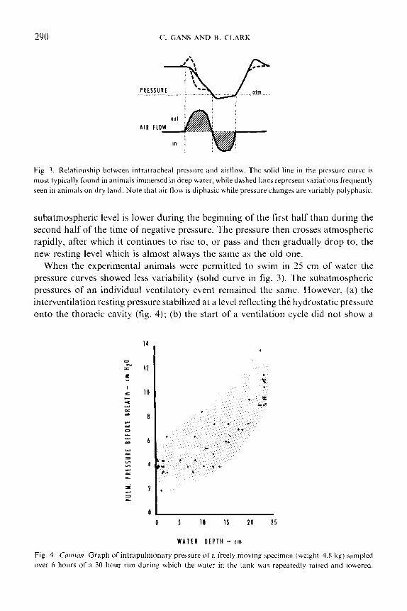
Fig. 3. Relationship between intrdtracheat pressure and airflow. The solid line in the pressure curve is
most typically found in animals immersed in deep water, while dashed lines represent variations frequently
seen in animals on dry land. Note that air flow is diphasic while pressure changes are variably polyphasic.
subatmospheric level is lower during the beginning of the first half than during the
second half of the time of negative pressure. The pressure then crosses atmospheric
rapidly, after which it continues to rise to, or pass and then gradually drop to, the
new resting level which is almost always the same as the old one.
When the experimental animals were permitted to swim in 25 cm of water the
pressure curves showed less variability (solid curve in fig. 3). The subatmospheric
pressures of an individual ventilatory event remained the same. However, (a) the
interventilation resting pressure stabilized at a level reflecting the hydrostatic pressure
onto the thoracic cavity (fig. 4); (b) the start of a ventilation cycle did not show a
lL----- 0 5 10 15 20 25
WATER DEPTH - tm
Fig. 4. Cairnun. Graph of intrapulmonary pressure of a freely moving specimen (weight 4.8 kg) sampled
over 6 hours of a 30 hour run during which the water in the tank was repeatedly raised and lowered.
VENTILATION IN C&Ian 291
0 2 4 6 8 10 12 14 16 18 20 22
WATER DEPTH-cm
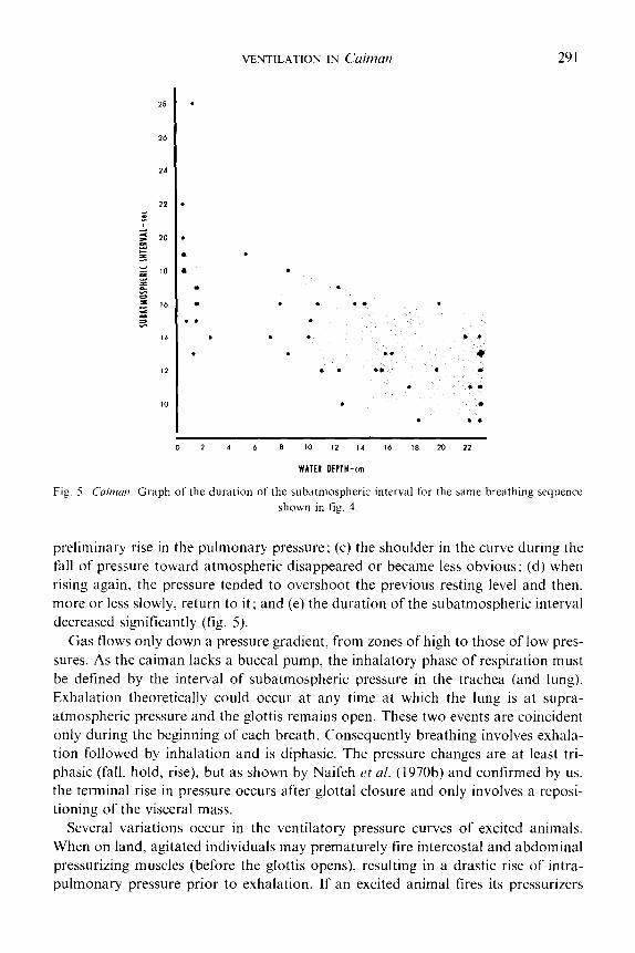
Fig. 5. Crrinzan. Graph of the duration of the subatmospheric interval for the same breathing sequence
shown in fig. 4.
preliminary rise in the pulmonary pressure; (c) the shoulder in the curve during the
fall of pressure toward atmospheric disappeared or became less obvious; (d) when
rising again, the pressure tended to overshoot the previous resting level and then,
more or less slowly, return to it; and (e) the duration of the subatmospheric interval
decreased significantly (fig. 5).
Gas flows only down a pressure gradient, from zones of high to those of low pres-
sures. As the caiman lacks a buccal pump, the inhalatory phase of respiration must
be defined by the interval of subatmospheric pressure in the trachea (and lung).
Exhalation theoretically could occur at any time at which the lung is at supra-
atmospheric pressure and the glottis remains open. These two events are coincident
only during the beginning of each breath. Consequently breathing involves exhala-
tion followed by inhalation and is diphasic. The pressure changes are at least tri-
phasic (fall, hold, rise). but as shown by Naifeh et al. (1970b) and confirmed by us,
the terminal rise in pressure occurs after glottal closure and only involves a reposi-
tioning of the visceral mass.
Several variations occur in the ventilatory pressure curves of excited animals.
When on land, agitated individuals may prematurely fire intercostal and abdominal
pressurizing muscles (before the glottis opens), resulting in a drastic rise of intra-
pulmonary pressure prior to exhalation. If an excited animal fires its pressurizers
292 (‘. GANS AND B. CLARK
strongly during exhalation, the normally smooth descent of the pressure curve is
interrupted by a brief rise. In resting animals isolated for several (S-20) hours,
ventiiatory pressure curves usually lose the initial and terminal rises above inter-
breath baseline (fig. 3).
The pharyngeal oscillations flush the narial tubes and pharyngeal space. They
can provide a continuing supply of gas to the olfactory chambers. However, they
do not contribute significantly to a flushing (or reduction) of the respiratory dead
space. Each exhalation still fills the internal narial canal with waste gdS and leaves it full at the start of inhalation. The volume of the tubes from nares to larynx conse-
quently must represent part of the respiratory dead space. On the other hand, this
tube could be bypassed should the animal manage to breathe through the open mouth.
Such a function of the gape does not seem previously to have been considered; it
remains to be seen whether gaping animals actually open the pharyngeal partition.
(A large excited C~(~~~~~i~~~.s ~~ulu,sf~~,s apparently had the pharynx distended while
gaping; however, while some air could be observed to burst forth. the buccal flaps
remained overlapping.)
MYOGRAPH1C REC‘ORDS
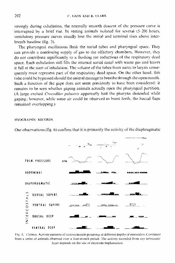
Our observations (fig. 6) confirm that it is primarily the activity ot‘the diaphragmatic
ABDOMINAL e- e
DiAPHRAGMATl( m#m_wllm
VENTRAL DEEP -d_ -a-_ -_.’
Fig. 6. (.~~i?~f[~~~. Activity patterns of various muscle groupings at different depths ~fii~rners~~n. Combined
from ;I series of animals observed over a four-month period. The activity recorded l&n any intercostal
layer depends on the site of electrode implantation.
VENTILATION IN Cffinla?? 293
muscle that is coincident with the drop of pulmonary pressure below atmospheric, though we did observe significant inhalatory activity in the section of the deep inter- costals attaching mainly to the sternocostal rib segments. Those deep intercostals attaching to the intercostal and vertebrocostal segments generally act during the exhalation and again during the repressurization phases.
The superficial intercostals fire in a more complex fashion. Activity at any site is relatively constant with respect to the phase of the breathing cycle. There is an indication of a marked shift in activity with elevation along an intercostal space, as well as between cycles that show differences in the pressure curves. The dorsal super- ficial intercostals fire mainly during periods of negative pressure, though they often continue during the rise to resting level; particularly when the animal is immersed, the muscle may also show low inte~ittent activity before inhalation. Records from theventral superficial intercostals, located near the junction between intercostal and sternocostal segments of the rib show more irregular activity, mainly during supra- atmospheric phases, but with some overlap into the action periods of the more dorsal portions of these muscles.
The abdominal wall musculature fires when the pressure in the pulmonary cavity is decreasing. It tends to cease activity at the shoulder in the descending pressure curve. The magnitude of this firing correlates well with the rate of the pressure change. Less regular activity was observed during repressurization. In general, the more active the animal the more extensive the firing. At1 of the muscles used in ventilation, including the diaphragmatic~ also fire during locomotion.
Another and major set of changes occurs as the animal enters the water. The deeper the water the less and more irregular the activity of those muscles that increase pulmonary pressure. Here again there are intercycle differences.
The ventral portion of the deep intercostal and the diaphragmatic muscles tire mainly during the period when the pulmonary contents are at subatmospheric pressure. They increase their activity when the trunk of a resting animal becomes flooded; instead of starting to fire during the first portion of the subatmospheric period, the ventral deep intercostal muscles delay onset of full firing until the middle of the subatmospheric period; they then fire strongly. After flooding, a period of lower-level, inte~ittent activity precedes the period of maximum activity. This is followed in turn by a series of short, high intensity bursts. Perhaps this shift in the activity of intercostal muscles reflects the observed reduction in rib nlovement. The intercostals may act to move the ribs during ventilation on land and to stabilize them during ventilation in water,
The diaphragmatic muscle generally starts and ceases activity earlier in the flooded animal. It tires at a constant, low level during the initial subatmospheric phase, builds up to a steady high level after about 30 2) of the subatmospheric interval. and then remains there until just prior to the rise in atmospheric pressure, when it suddenly becomes silent; very rarely does it show intermittent activity thereafter. The inhalatory muscles show only sporadic activity during the terminal phase of repressurization, as do the pressurizing muscles.
294 f’. GANS AND B. CLARK
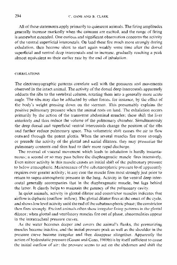
All of these statements apply primarily to quiescent animals. The firing amplitudes
generally increase markedly when the caimans are excited, and the range of firing
is somewhat extended. One curious and significant observation concerns the activity
of the ventral superficial intercostals. On land these fire much more strongly during
exhalation then become silent to start again weakly some time after the dorsal
superficial and ventral deep intercostals and to increase, gradually reaching a peak
almost equivalent to their earlier rate by the end of inhalation.
CORRELATIONS
The electromyographic patterns correlate well with the pressures and movements
observed in the intact animal. The activity of the dorsal deep intercostaIs apparently
adducts the ribs to the vertebral column, rotating them into a generally more acute
angle. The ribs may also be adducted by other forces, for instance, by the effect of
the body’s weight pressing down on the sternum. This presumably explains the
positive pulmonary pressure when the animal rests on land. The exhalation occurs
primarily by the action of the transverse abdominal muscles; these shift the liver
anteriorly and thus reduce the volume of the pulmonary chamber. Simultaneously
the deep dorsal and superficial ventral intercostals change the position of the ribs
and further reduce pulmonary space. This volumetric shift causes the air to flow
outward through the patent glottis. When the several muscles fire more strongly
or precede the activity of the glottal and narial dilators. they may pressurize the
pulmonary contents and thus lead to their more rapid discharge.
The reversal of visceral movement which leads to inhalation is hardly instanta-
neous; a second or so may pass before the diapl~ragmatjc muscle fires intensively.
Even minor activity in this muscle causes an initial shift of the pulmonary pressure
to below atmospheric. Maintenance of the subatmospheric pressure level apparently
requires ever greater activity ; in any case the muscle fires most strongly just prior to
return to supra-atmospheric pressure in the lung. Activity in the ventral deep inter-
costal generally accompanies that in the diaphragmatic muscle, but lags behind
the latter. It clearly helps to maintain the patency of the pulmonary cavity.
In quiet animals, activity in glottal dilator and constrictor muscles indicates that
airflow is diphasic (outflowPinflow). The glottal dilator tires at the onset of the cycle,
and shows low level activity until the end ofthe subatmospheric phase: the constrictor
then fires strongly. Excited animals often show irregular tiring patterns in the glottal
dilator: when glottal and ventilatory muscles fire out of phase, abnormalities appear
in the intratracheal pressure curves,
As the water becomes deeper and covers the animal’s flanks, the pressurizing
muscles become inactive, and the initial pressure peak as well as the shoulder in the
pressure curve become irregular and then disappear altogether. Apparently the
action of hydrostatic pressure (Gaunt and Gans, 1969b) is by itselfsu~cient to cause
the initial outflow of air: the pressure seems to act on the abdomen and shift the

VENTILATION IN Cairnan 295
liver further into the rib cage. The configuration of the rib cage and the ligamentous
ties of the liver keep the pleural cavity from collapsing; consequently the lung remains
patent even when the open glottis permits the pulmonary contents to drop toward
atmospheric pressure without interruption; this explains the disappearance of the
shoulder in the pressure curve of immersed animals. As the water becomes deeper.
the dorsal superficial intercostal is active earlier; it then fires intermittently during the
fall of pressure.
The firing pattern of the diaphragmatic muscle correlates well with the develop-
ment and maintenance of the subatmospheric level. The activity of the vental deep
intercostals is delayed during immersion. The increasingly antagonistic firing of the
ventral superficial intercostals during excitement suggests a stabilizing rather than
a distending function. Apparently the intercostals fix the movable frame, thus
maintaining the subatmospheric pressure against which the diaphragmatic acts in
shifting the liver.
BREATHING SEQUENCES
A number of long term (24+ hours) records of freely moving caimans, exposed to
rising and falling water levels and normal (14-10 hour) day-night cycles were recorded
from animals tethered by long cables and observed by closed-circuit television.
Isolation was necessary because some animals dive when disturbed while others,
more habituated and used to being fed by hand, rise to the surface when one of us
enters the chamber. In general, the animals would breathe in sets of two or occasion-
ally three ventilatory cycles when their body was in the air. They would shift to
single ventilatory cycles interspersed with pauses of one or more minutes when the
water became deep enough to cover their trunk. When undisturbed for long periods
the caimans showed a generally inversely weight-dependent breathing rate (table 1).
Our highest values for resting animals are less than one half those reported by Huggins
rt d. (1969) for ‘minimally stimulated’ animals of equivalent size. We only observed
(by TV) rates equivalent to theirs when our animals were moving about the tanks.
TABLE I Respiratory rates of Caiman at 23-25 C
Mass Breaths:min Period monitored
5.0 kg 0.167 24 hrs
4.8 kg 0.144 12 hrs
4.8 kg 0.250 180 min
650 g 0.574 500 min
290 g 0.633 450 min
18Og 0.580 550 min
296 C. GANS AND R. CLARK
Many of the animals dived voluntarily, staying beneath the water for 15 to 25
minutes at a time. Upon emergence they would breathe deeply, perhaps five to eight
times in a row, with the intervals between initial breaths on the order of one minute
and this period increasing to three to four minutes during the next half hour. In
general, the data confirm the observations of Gaunt and Gans (f969a) and Huggins
cl al. (1970) that each breath is accompanied by a slight tachycardia.
The pressure in the lungs did not change regularly between breaths, however.
the pulmonary pressures reflect movements and other activity of the animal. When
accidentally disturbed, the animals might inflate themselves or raise pressure by
inducing peak firing of the pressurizing muscles. As the trunk musculature is also
used in locomotion, it is inappropriate to correlate all pulmonary pressure changes
with breathing events.
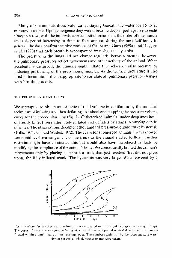
THE PRESSURE-V~~LU~~ CURVE
We attempted to obtain an estimate of tidal volume in ventilation by the standard
technique of inflating and then deflating an animal and mapping the pressure-volume
curve for the crocodilian lung (fig. 7). Catheterized animals (under deep anesthesia
or freshly killed) were a1terIlateiy inflated and deflated by stages in varying depths
of water. The observations document the standard pressure%volume curve hysteresis
(Hills, 1971; Gil and Weibel, 1972). The curve for submerged animals always showed
some mid-level rearrangement of the trunk as the animal started to float. Further
restraint might have eliminated this but would also have introduced artifacts by
modifying the compliance of the animal’s body. We consequently limited the caiman’s
movements only by placing it beneath a brick that just touched (but did not press
upon) the fully inflated trunk. The hysteresis was very large. When covered by 7
Fig. 7. Cuitnu~~. Selected pressure wlume curves measured on a freshly-killed specimen (weight 5 kg).
The cusps of the curve represent volumes at which the animal passed neutral density and the carcass
floated within a confining. but not resisting space. The numbers within or by the loops indicate water
depths (in cm) at which measurements were taken.
VENTILATION IN C&?Zclt2 297
inches of water this amounted to up to a 40 5’; increase in pressure for a particular
volume. The tidal volumes estimated from these curves are greater by almost an
order of magnitude (85 ml/kg in 3 cm of water compared with 12 ml/kg) than those
reported by Naifeh et al. (1970a) from plethysmographic techniques. Possibly our
approach changed the compliance of the rib cage; this may be more stiffly elastic in
animals that have the costal muscles in tonic condition.
A couple of incidental observations suggest some reasons for the observed hyster-
esis. When a caiman that has been partially inflated under water is then manually
squeezed, the pulmonary relations shift from the inflation to the deflation curve and
remain there. If the animal is then inflated further intermediate points are obtained
until another squeezing sequence returns things to the deflation curve. If the pressure
is then permitted to drop it proceeds along the deflation curve. The effect of sqeezing
is greatest when the pressure is applied to the thoracic rib cage rather than to the
abdominal portion of the animal. When a transient pressure peak is applied to the
inside of the lung the curve shifts only part way. The observations suggest that the
hysteresis may be due to the intrinsic frictional resistance between the shifting com-
ponents of the crocodilian body. As pressure on the rib cage is more effective in
shifting the animal between the sides of the hysteresis loop than is pressure on the
abdominal wall, one can furthermore state that it is primarily the mechanical friction
intrinsic to the rib system that is responsible for the effect. We have no evidence to
evaluate the possible occurrence of changes in alveolar configuration (c$ Weibel
rt al., 1973) in this system. The hepatic piston floats more or less freely in a fluid-
filled space and is apparently more compliant than are the costal articulations.
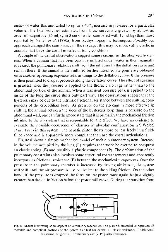
Figure 8 shows a simple mechanical model of such a pulmonary system. Increase
in the volume occupied by the lung (L) requires that work be exerted to compress
an elastic spring (E) and possibly a plastic component (P). The deformation of the
pulmonary constraints also involves some structural rearrangements and apparently
incorporates frictional resistance (F) between the mechanical components. Once the
pressure in the pulmonary chamber is increased by driving air into it, the system
will shift until the air pressure is just equivalent to the sliding friction. On the other
hand, if the pressure is dropped the force on the piston must again be just slightly
greater than the static friction before the piston will move. During the transition from
Fig. 8. Model illustrating some aspects of ventilatory mechanics. The piston is intended to represent all
movable and compliant portions of the system. See text for details. E: elastic resistance. F: frictional
resistance. G: glottis. L: pulmonary cavity. P: plastic resistance.
298 C’. GANS AND B. CLARK
just enough force to keep distending to just enough force to start collapsing the
volume remains constant (except for volumetric expansion as the pressure drops).
This explains the shift from the inhalatory to the exhalatory curve after the rib cage
has been squeezed.
The model also suggests why pressures produced by aspiration breathing may be
lower than those for pulse-pumping equivalent amounts of gas in a particular animal.
If flow is induced by movements of the pulmonary wall. thus increasing and reducing
the pressures as the result of a primary change of the volume, the process should
move along the deflation curve. However, when the gaseous medium has to serve
both as a carrier of respiratory gases and as a mechanical transmission medium,
inflation requires greater pressures to maintain forces sufficient to keep the non-
ideal piston moving. Part of the difference between the inflation and deflation curves
will then represent the cost of pulse-pumping.
Finally the model suggests an advantage for the terminal overshoot of pulmonary
pressure during repressurization prior to return to base line. A temporary overshoot
will permit the animal to store the gas at a lower apneic pressure.
Discussion
The present results confirm earlier observations (de Jongh and Gans, 1969; Gans
and Hughes, 1967; Gaunt and Cans. 1969b) that the respiratory activities of amphi-
bians and reptiles seetn closely matched to the normal environmental circumstances
of the animals. The aspiration breathing patterns seen in the several Recent reptiles
presumably arose independently from each other, and probably from the pattern
seen in mammals: consequently they must be analyzed by themselves. The key to
this basic conclusion is again confirmed by the present studies which document that
the undisturbed caiman utilizes minimal respiratory muscle activities. i.c>. does
minimal work, to move the air necessary for absorption and release of respiratory
gases. The studies also show that the cup and socket connection between Iarynx and
internal nares reduces the respiratory dead space and with this again increases the
efficiency of ventilation.
The spacing of breathing cycles furthermore reflects the animal’s capacity to dive
for prolonged periods. Post-dive emergence is accompanied by repeated breathing
movements. which discharge the accumulated lactate and gradually reoxygenate
the tissues.
The buccal oscillations in this sense are interpreted as serving one major function.
namely to flush gas along the olfactory epithelium. The observations of Naifeh ct al.
(1970a) that ventilation cycles were often preceded by one to three pharyngeal oscilla-
tions suggests a sensory function; there is no direct. association of these pharyngeal
movements with gas exchange. The possible role of gaping in ventilation remains
to be checked.
We confirm results of Naifeh rt al. (1970a, b) regarding artifacts generated when
VENTILATION IN Caimun 299
animals are exited, particularly when being handled. An excited state may well occur
in animals in the wild ; it is difficult to quantify or to compare. Consequently we
emphasize study of isolated Cuiman that are free to move but happen to be resting.
The diaphragmatic muscles are neither homologous nor analogous to a mammalian
diaphragmatic. Inward movement of the abdominal wall does not ‘serve to push the
abdominal contents up against the diaphragm, thus assisting in evacuation of air
from the lungs’ (Naifeh et al.. 1970b); it does shift the liver anteriorly, thus reducing
the space within the thoracic cavity.
We are able to confirm that the various trunk and diaphragmatic muscles tire
either to reduce the pulmonary pressure or to increase it. No muscle does both. Our
records do suggest that an insect pin would at most intercostal sites record from at
least two layers, one of them active during inhalation and another active during
exhalation and/or repressurization. There is presumably no selective advantage for
the maintenance of pulmonary contents at a particular supra-atmospheric level
between breaths. (There would be a disadvantage if the glottal closers would have
to fire continuously to keep the gas from escaping as suggested by Naifeh et al.,
1970a; however, 1970b would suggest that this was only occasionally observed; our
records do not show it.)
The various intercostal muscles span the intercostal spaces. No matter what their
angle of origin or insertion, such muscles can only act to bring a pair of adjacent ribs
more closely together. Whether this results in abduction or adduction of a particular
rib depends on the forces (active and elastic) resisting displacement and on the relative
moments imposed on the two ribs. The secondary tiring of the ventral superficial
intercostals in exited animals may then be conceived of as bringing a mechanically
ineffective muscle into action at a critical time, thus increasing the stability of the rib
cage during deep inhalation. Consequently some of the activity of the costal muscles
serves to maintain the patency of the cylinder so that shifts of the hepatic piston may
cause maximal changes of pressure. This effect is most pronounced when the trunk
is flooded. The postventilatory firing of the pressurizers presumably serves a reposi-
tioning function, particularly when the animal is on land and thus without the benefit
of hydrostatic pressure against its trunk. Such costal activity may be the inevitable
price of a ventilatory pattern that involves a major shift of the viscera with each breath.
Acknowledgments
Various pilot experiments for this study were carried out at various times by Mr. Fred
Pohl (as an undergraduate honors project), Dr. C. 0. da C. Diefenbach and Dr. A. S.
Gaunt. Dr. J. Farber assisted with gas analysis and Dr. Leon Farhi permitted the
use of his respiratory mass spectrometer. Dr. Hermann Rahn suggested the pressure-
volume experiments. We thank T. S. Parsons, H. I. Rosenberg and T. C. Scanlon for
a review of the manuscript.

300 c‘. GANS AND B. CLARK
References
Ballintijn, C. M. and G. M. Hughes (1965). The muscular basis of the respiratory pump in the trout.
,f. &Ill. Bi0i. 43: 349-362.
Ballintijn, C. M. (196%). Muscle coordination of the respiratory pump of the carp (C’~/x?nus c,rrr(,ici L.).
J. E.yp(/. Bid. 50: 569-59 I Ballintijn. C. M. (1969b). Movement pattern and efficiency of the respiratory pump of the carp (C:)71rinu.\
carpio L.). J. E.yprl. Biol. 50: 593-613.
Basmajian, J. V. and G. A. Stecko (1962). A new bipolar electrode for electromyogrdphy. J. .dp$. P!7wict/.
17: 849.
Bellairs. A. d’A. and C. C. D. Shute (1953). Observations on the narial musculature of Crocodilia and its
innervation from the sympathetic system. J. Anui. (Land.) X7: 367-378.
Chiasson, R. B. (1962). Laboratory Anatomy of the Alligator. Dubuque, Wm. C. Brown Co. Inc.
Cans. C. and G. M. Hughes (1967). The mechanism of lung ventilation in the tortoise T~,.vrurk~ ~rcrrru
Linne. J. E.x7)fl. Biol. 47 : I -~20.
Gans. C. (197la). Respiration in early tetrspods - the frog is a red herring. ~~~~/~{~i~~~? 24: 740~-751.
Gans. C. (1971b). Strategy and sequence in the evolution of the external gas exchangers of ectothermal
vertebrates. Foymu rt F~litcfit~ 3: Xl- 104.
Gaunt. A. S. and C. Cans (1969a). Diving bradycardia and withdrawal bradycardia in C‘uirnrrn c,roc,o&hr.s.
N~lrure (Land.) 223: 207 208.
Gaunt, A. S. and C. Gans (1969b). Mechanics of respiration in the snapping turtle. C’/re/J~c/~cr .vcrpcvzrinu
(Linne). J. Morph. 128: 195-228.
Gill. J. and E. R. Weibel (1972). ~lorpllologicai study ofpressure-volume hysteresis in rat lungs fixed by
vascular perfusion. Kes@. Pfr.txiof. 15 : 190-2 13. Hadden. H. D. (1975). A Functional Anatomical Study of Lizard Respiration. M.Sc. Thesis. University
of Calgary.
Hills, B. A. (1971). Geometric irreversibility and compliance hysteresis in the lung. Respir. P/~y.tiol. 13:
50.61.
Hoffstetter. R. and 3. P. Gasc (1969). Vertebrae and ribs of modern reptiles. In: Biology of the Reptilia.
Vol. I. edited by C. Gans. T. S. Parsons and A. d‘ZI. Bellairs. London, Academic Press.
Huggins. S. E., H. E. Hoff and R. V. Petia (1969). Heart and respiratory rates in crocodilian reptiles under
conditions of minimal stimulation. Plt,r,sio/. Zoo/. 42: 320-333.
Huggins. S. E.. H. E. Hoff and R. V. Peiia (1970). The respiratory--heart rate response in crocodilian
reptiles. Ph~.rio/. Zoo/. 43: I O-IX.
Hughes, G. M. and C. M. Ballintijn (1965). The muscular basis of the respiratory pumps in the do&h
(~~~,~~~~~7i~Z~.S 0z7ic&r). f. E-v/%!. B&i. 43 : 3633383.
Jordanski, N. N. (1973). The skull of the Crocodiiia. In: Biology of the Reptiha. Vol. 4. edited by C. Gans
and T. S. Parsons. London Academic Press.
Ireland, L. C. and C. Gans (1973). The adaptive significance of the flexible shell of the tortoise ,\~~r/oc+
~~/t~~r.vus torniwi. Anim. Bciwc. 20 : 77X-7X I.
Jongh, H. J. de and C’. Gans (1969). On the mechanism of respiration in the bullfrog, Ranu tztrrshrianu:
A reassessment. J. Morph. 127: ?%I?%~.
Loveridge, J. P. and D. K. Blake (1972). Techniques in the inlin~~bilization and h~lndlin~ of the Nile
crocodile, Croco~~j~Jt~.s ni/oriiu.s. .4r&& 5 : 1 14.
McMahon. B. R. (1969). A functional analysis of the aquatic and aerial respiratory movements of an
African lungfish Protoptrrus crt~rhir~picus, with reference to the evolution of the lung-ventilation
mechanism in vertebrates. J. E-1p/l. Bid. 51 : 407 430.
Martin, W. F. and C. Gans (1972). Muscular control of the vocal tract during release signaling in the toad
Br& ~~~~~~~~,p.s. J. ~~(~r~~?. 137: 1 17. Naifeh. K. H., S. E. Huggins. H. E. Hoff. T. W. Hug& and R. E. Morton jlY70a). Respiratory patterns in
crocodilian reptiles. Respir. F%~~.\in/. Y: 31-42.
VENTILATION IN Taiwan 301
Naifeh, K. H., S. E. Huggins and H. E. Hoff (1970b). The nature of the ventilatory period in crocodilian
respiration. Rrspir. PhJ’sid. 10: 338-348.
Naifeh. K. H.,S. E. Hugginsand H. E. Hoff(lY7la).Thenatureofthenonventilatoryperiod incrocodilian
respiration. Rmpir. PI~~srol. I I : 178-l 85.
Naifeh, K. H., S. E. Huggins and H. E. Hoff (1971 b). Study of the control of crocodilian respiration by
anesthetic dissection. Respir. Physiol. 12: 251-260.
Naifeh. K. H., S. E. Huggins and H. E. Hoff (1971~). Effects of brain stem section on respiratory patterns
of crocodilian reptiles, Respir. Physiol. I3 : 186197.
Osse, J. W. M. (1969). Functional morphology of the head of the perch (Pwca./luriutili.v L.): an electro-
myographic study. Neth. J. Zool. 19: 2899392.
Parsons, T. S. (1971). The nose and Jacobson’s organ. In: Biology of the Reptilia. Vol. 2, edited by C. Gans
and T. S. Parsons. London, Academic Press.
Rathke. H. (I 866). Untersuchungen iiber die Entwickelung und den Kijrperbau der Krokodile. Braun-
schweig, Vieweg und Sohn, vii + 275 pp.
Rosenberg, H. I. (1973). Functional anatomy of pulmonary ventilation in the garter snake. Thumnophi.v
rlegms. J. Morph. 140: 171. 184.
Weibel, E. R., P. Untersee. J. Gil and M. Zulauf (1973). Morphometric estimation of pulmonary diffusion
capacity. IV. Effect of varying positive pressure inflation of air spaces. Respir. Physiol. IS: 2855308.
West, N. H. and D. R. Jones (1975). Breathing movements in the frog Rana pipiens. I. The mechanical
events associated with lung and buccal ventilation. Can. J. Zoo]. 53 : 332-344.