stimulation of mitochondrial oxidative capacity in white fat independent of ucp1: a key to lean...
TRANSCRIPT

Biochimica et Biophysica Acta xxx (2013) xxx–xxx
BBAMCB-57418; No. of pages: 18; 4C: 8, 12
Contents lists available at SciVerse ScienceDirect
Biochimica et Biophysica Acta
j ourna l homepage: www.e lsev ie r .com/ locate /bba l ip
Stimulation of mitochondrial oxidative capacity in white fat independent ofUCP1: A key to lean phenotype☆
Pavel Flachs, Martin Rossmeisl, Ondrej Kuda, Jan Kopecky ⁎Department of Adipose Tissue Biology, Institute of Physiology Academy of Sciences of the Czech Republic, v.v.i., Prague, Czech Republic
Abbreviations: ACC, acetyl-CoA carboxylase; AMParachidonic acid; ATM, adipose tissue macrophages;CTRP3, adiponectin paralog C1q/TNF-related proteinfatty acid synthase; FGF-21, fibroblast growth factoracids; mtDNA, mitochondrial DNA; MCP-1, monocytekinase 4; PEPCK, phosphoenolpyruvate carboxykinNAD+-dependent deacetylase sirtuin 1; SREBP-1c, sof intracellular triacylglycerols and re-esterification15-deoxy-Δ12,14-prostaglandin J2☆ This article is part of a Special Issue entitled Brow⁎ Corresponding author at: Department of Adipose
4, Czech Republic. Tel.: +420 241063701; fax: +420E-mail address: [email protected] (J. Kopec
1388-1981/$ – see front matter © 2013 Elsevier B.V. Allhttp://dx.doi.org/10.1016/j.bbalip.2013.02.003
Please cite this article as: P. Flachs, et al., Stimtype, Biochim. Biophys. Acta (2013), http://d
a b s t r a c t
a r t i c l e i n f oArticle history:Received 19 November 2012Received in revised form 6 February 2013Accepted 9 February 2013Available online xxxx
Keywords:Fatty acid re-esterificationFutile substrate cyclePPARγAdipose tissueLipid mediatorObesity
We are facing a revival of the strategy to counteract obesity and associated metabolic disorders by inducingthermogenesis mediated by mitochondrial uncoupling protein-1 (UCP1). Thus, the main focus is on the adap-tive non-shivering thermogenesis occurring both in the typical depots of brown adipose tissue (BAT) and inUCP1-containing cells that could be induced in white adipose tissue (WAT). Because contribution of WAT toresting metabolic rate is relatively small, the possibility to reduce adiposity by enhancing energy expenditurein classical white adipocytes is largely neglected. However, several pieces of evidence support a notion thatinduction of energy expenditure based on oxidation of fatty acids (FA) in WAT may be beneficial for health,namely: (i) studies in both humans and rodents document negative association between oxidative capacityof mitochondria in WAT and obesity; (ii) pharmacological activation of AMPK in rats as well ascold-acclimation of UCP1-ablatedmice results in obesity resistance associatedwith increased oxidative capacityin WAT; and (iii) combined intervention using long-chain n-3 polyunsaturated FA (omega 3) and mild calorierestriction exerted synergism in the prevention of obesity in mice fed a high-fat diet; this was associated withstrong hypolipidemic and insulin-sensitizing effects, as well as prevention of inflammation, and synergistic in-duction of mitochondrial oxidative phosphorylation (OXPHOS) and FA oxidation, specifically in epididymalWAT. Importantly, these changes occurred without induction of UCP1 and suggested the involvement of:(i) futile substrate cycle in white adipocytes, which is based on lipolysis of intracellular triacylglycerols andre-esterification of FA, in associationwith the induction ofmitochondrial OXPHOS capacity, β-oxidation, and en-ergy expenditure; (ii) endogenous lipid mediators (namely endocannabinoids, eicosanoids, prostanoids,resolvins, and protectins) and their cognate receptors; and (iii) AMP-activated protein kinase in WAT. Quanti-tatively, the strong induction of FA oxidation inWAT in response to the combined intervention is similar to thatobserved in the transgenic mice rendered resistant to obesity by ectopic expression of UCP1 inWAT. The induc-tion of UCP1-independent FA oxidation and energy expenditure in WAT in response to the above physiologicalstimuli could underlie the amelioration of obesity and low-grade WAT inflammation, and it could reduce therelease of FA from adipose tissue and counteract harmful consequences of lipid accumulation in other tissues.In this respect, new combination treatments may be designed using naturally occurring micronutrients(e.g. omega 3), reduced calorie intake or pharmaceuticals, exerting synergism in the induction of the mitochon-drial OXPHOS capacity and stimulation of lipid catabolism in white adipocytes, and improving metabolic flexi-bility of WAT. The role of mutual interactions between adipocytes and immune cells contained inWAT in tissuemetabolism should be better characterised. This article is part of a Special Issue entitled Brown and White Fat:From Signaling to Disease.
© 2013 Elsevier B.V. All rights reserved.
K, AMP-activated protein kinase; aP2-Ucp1 mice, transgenic mice expressing UCP1 gene from the aP2 gene promoter; AA,BAT, brown adipose tissue; BCAA, branched-chain amino acids; CB1, cannabinoid type-1 receptor; CR, calorie restriction;3; DHA, docosahexaenoic acid; EPA, eicosapentaenoic acid; FAT/CD36, fatty acid translocase/CD36; FA, fatty acids; FAS,-21; HF, high-fat; HSL, hormone-sensitive lipase; LPS, lipopolysaccharide; omega 3, long-chain n-3 polyunsaturated fattychemoattractant protein-1; OXPHOS, oxidative phosphorylation; PC, pyruvate carboxylase; PDK4, pyruvate dehydrogenasease; PD1, protectin D1; PGC-1, the PPARγ coactivator 1; PPAR, peroxisome proliferator-activated receptor; SIRT1,terol regulatory element-binding protein-1c; TAG, triacylglycerols; TAG/FA cycle, futile substrate cycle based on lipolysisof fatty acids; TZD, thiazolidinedione; UCP1, mitochondrial uncoupling protein 1; WAT, white adipose tissue; 15d-PGJ2,
n and White Fat: From Signaling to Disease.Tissue Biology, Institute of Physiology of the Academy of Sciences of the Czech Republic, v.v.i., Videnska 1083, 14220 Prague241062599.
ky).
rights reserved.
ulation of mitochondrial oxidative capacity in white fat independent of UCP1: A key to lean pheno-x.doi.org/10.1016/j.bbalip.2013.02.003

2 P. Flachs et al. / Biochimica et Biophysica Acta xxx (2013) xxx–xxx
1. Introduction
White adipose tissue (WAT), the most plastic among the metaboli-cally relevant tissues in the body, is essential for storing metabolicenergy, and through its endocrine functions, it is also involved in theregulation of glucose as well as energy homeostasis. WAT has also amajor role in the control of systemic fatty acids' (FA) levels. According-ly, both hypertrophy and atrophy [1] of WAT are associated withlipotoxic damage of insulin signalling in other tissues. The key role ofWAT in glucose homeostasis [2,3] is supported by the recent experi-mental evidence of anti-diabetic effects of WAT-specific up-regulationof peroxisome proliferator-activated receptor-γ (PPARγ; [4]). Con-cerning energy homeostasis, the role of WAT metabolism is usuallyneglected. However, it is not insignificant. Thus, the contribution ofWAT to resting metabolic rate in lean human subjects is close to 5%and it doubles in obesity (reviewed in [5]), while in adult mice rearedat 20 °C the total oxidative capacity of WAT represents ~30–50% of thatin brown adipose tissue (BAT; [6]).
In both humans [7] and rodents [8], oxygen consumption in WATcells declines with age and it is negatively correlated with obesity inhumans [7]. Decreased white fat cell thermogenesis in obese humanswas also found using direct microcalorimetry [5]. Accordingly,mitochondrial content in WAT is relatively low in genetically obeseand insulin-resistant mice [9–12]. Moreover, in monozygotic twins dis-cordant for obesity, copy number of mitochondrial DNA in subcutane-ous WAT was reduced by 47% in obese co-twins, in association withreduced expression of the genes for mitochondrial proteins [13]. Inturn, induction of mitochondria and activation of FA oxidation,specifically in WAT, was observed under the conditions promotingloss of adiposity (see Section 2.4). Very recently, it has been reported[14] that systemic nonselective β-adrenergic stimulation in humansthat increases energy expenditure to the same extent as cold exposuredoes not activate brown adipose tissue (BAT; see below), and could beexplained only in part by the activation of muscle non-shiveringthermogenesis (for review on muscle thermogenesis, see [15–17]).These data support the notion that also adrenergically-stimulatedenergy expenditure in WAT may, though to a relatively small extent,influence total energy balance (see also [16] and Section 2.3).
In contrast to energy expenditure inWAT, major role of mitochon-drial uncoupling protein-1 (UCP1) in BAT with respect to cold- anddiet-induced adrenergically regulated thermogenesis is well appreci-ated [18–20], although examination of several older studies [15,21]casts some doubts on the unique role of the UCP1-mediated thermo-genesis. As described in other articles of this issue, recent discovery offunctional BAT in adult humans [22] has led to a revival of the strate-gy [23–25] to counteract obesity and associated metabolic disordersby inducing UCP1-mediated thermogenesis. In rodents, the existenceof several fat cell lineages was uncovered, which underlie the differ-entiation of precursor cells into: (i) classical multilocular brownadipocytes, which are closely related to myocytes, and which are re-sponsible for the bulk of the adaptive UCP1-mediated thermogenesis;(ii) classical unilocular white adipocytes lacking UCP1, which are fullycompetent for ATP synthesis by oxidative phosphorylation (OXPHOS;[13,26,27]); and (iii) brite adipocytes [28,29] named also beige cells(reviewed in [30]). The last cell type is of special interest, sincethese cells are interspersed in some depots of WAT, they show highinducibility of the UCP1-linked thermogenic programme, and theirabundance correlates negatively with the propensity to obesitywhile being under genetic control [31]. Besides the sympathetic stim-ulation, synthetic ligands of PPARγ (reviewed in [10,32,33]), as wellas several hormonal factors could induce brite cells in rodents ([34–38]; see Section 3.1). However, in spite of the fact that markersdiscriminating BAT from brite cells have been identified in rodents[28,30,38,39], the existence of brite cells in humans remains tobe unequivocally established [30,40], and typical white adipocytemarkers also remain to be defined. In fact, inducibility of at least
Please cite this article as: P. Flachs, et al., Stimulation of mitochondrial oxidtype, Biochim. Biophys. Acta (2013), http://dx.doi.org/10.1016/j.bbalip.20
some of the UCP1-containing cells in WAT depots probably reflectstransdifferentiation of white adipocytes [41], bringing another levelof complexity to the identification of markers characteristic of cell lin-eages engaged in adipose tissue plasticity.
Energy-dissipating function of UCP1 depends on its protonophoricactivity in the inner mitochondrial membrane, which is activated byFA in response to β-adrenergic stimulation and allows for a fullunmasking of mitochondrial oxidative capacity without concomitantsynthesis of ATP (for review, see [42]). In fact, the content of mitochon-drial ATP synthase in classical brown adipocytes is extremely low[43,44], and this feature is observed already at the time of perinatal re-cruitment of BAT thermogenesis both in rodents [45,46] and humans[47]. Adrenergic stimulation of BAT not only activates UCP1-mediatedproton leak but also leads to complex metabolic adaptations (for re-views onBATmetabolism see [20]), includingmitochondrial biogenesis,aimed at increasing fuel supply and oxidation. Thus, lipolysis of intracel-lular triacylglycerols (TAG) stores is accelerated and the uptake of FAderived from blood-born lipoproteins is increased due to the action oflipoprotein lipase (LPL) that is rapidly recruited during cold exposure[48]. Although glucose is not a major substrate for BAT thermogenesis,glucose utilisation by activated BAT increases dramatically [49] asdoes lactate production. Glycolysis supplies ATP when its productionvia OXPHOS is attenuated, and also makes pyruvate available for thesynthesis of oxaloacetate, the condensing partner for acetyl-CoA formedfrom β-oxidation of FA, thus ensuring a continuous supply of citric acidcycle intermediates (anaplerosis; see Section 2.4 and [50]). Highexpression of the muscle type carnitine palmitoyl transferase-1(CPT1; [51]) allows for rapid β-oxidation of FA [52], while high activityof mitochondrial glycerol-3-phosphate dehydrogenase supports TAGsynthesis and controls cytoplasmic NADH levels [53]. Longer exposureto cold (days) leads to increased lipogenesis, which is necessary forsustained thermogenesis. Triiodothyronine locally produced fromthyroxine in brown adipocytes [54] amplifies adrenergic stimulationof UCP1 gene transcription [55], helps to ensure an optimal balancebetween lipolysis and lipogenesis [54], and contributes to systemictriiodothyronine levels [56].
Here, reflecting the well accepted potential of UCP1-mediated ther-mogenesis to reduce obesity, we provide a complementary informationon adipose tissue biology, while focusing on: (i) the possibility toreduce body fat stores by inducing energy expenditure in white adipo-cytes based on a futile substrate cycle [57], namely lipolysis of intracel-lular triacylglycerols and FA re-esterification (TAG/FA cycle; [5,58,59]),in association with the induction of mitochondrial OXPHOS capacityand β-oxidation; (ii) the role of local mediators in WAT in the regula-tion of adipocyte metabolism; (iii) the comparison of local/systemicmetabolic effects of TAG/FA cycle vs. mitochondrial uncoupling inwhite adipocytes; and (iv) possible interventions, which couldcounteract obesity while stimulating activity of TAG/FA cycle, inducingcapacity ofmitochondrial OXPHOS and enhancing FA oxidation inwhiteadipocytes. We provide a concept, that white adipocyte endowed withhigh capacity of mitochondrial OXPHOS represents a metabolicallyflexible healthy adipocyte.
2. Physiology of mitochondria in white adipocytes
In white adipocytes, similarly as in most other cells, mitochondriarepresent the main site of ATP production. Fully coupled mitochondriacould be isolated from adipocytes liberated by the use of collagenasefrom epididymal WAT of rats [26], and OXPHOS activity could be alsodemonstrated using respirometry in isolated digitonin-permeabilizedmurine white adipocytes [27]. Especially in fully differentiated whiteadipocytes, mitochondria must generate sufficient ATP to supportvarious energy-consuming processes (see Sections 2.2 and 2.3). Theyare located in the thin cytoplasmic rim of the cells, and they showsimilar morphology as in other tissues [60]. Mitochondrial content ofmature white adipocytes is several-fold higher as compared with
ative capacity in white fat independent of UCP1: A key to lean pheno-13.02.003

3P. Flachs et al. / Biochimica et Biophysica Acta xxx (2013) xxx–xxx
preadipocytes [61], while their content depends on the anatomicallocation of fat depots. Thus, in rodents, gonadal adipocytes containmore mitochondria and have higher oxidative capacity together withhigher content of OXPHOS proteins as compared with adipocytes insubcutaneous (inguinal) depot [62,63]. Similarly, in obese humans,mitochondrial DNA (mtDNA) copy number per mg tissue is higher invisceral than in abdominal subcutaneous WAT [64]. Although mito-chondrial respiration normalized to cell number or mtDNA is lowerin visceral WAT compared to subcutaneous WAT due to smaller adipo-cytes in the former fat depot, the visceral fat is bioenergetically moreactive and more sensitive to substrates of electron transport chainthan the subcutaneous WAT [64].
It should be stressed that the interpretation of the above dataregarding mitochondrial content is not straightforward. Because ofthe complex nature of mitochondria and their biogenesis, multipleparameters need to be analysed in order to assess mitochondrial con-tent or activity in WAT. The preferred methods of ours are: (i) trans-mission electronmicroscopy (e.g. described in [65]); (ii) evaluation ofactivity of cytochrome c oxidase and cytochrome b content in crudemembrane fraction (e.g. used in [27]); (iii) quantification of selectedmitochondrial proteins by immunoblotting (e.g. described in [66]);and (iv) measurement of mitochondrial respiration using high-resolution respirometry in digitonin-permeabilized adipocytes [27].Moreover, mtDNA quantification is frequently used as a marker of mi-tochondrial content and activity, although each mitochondrion maycontain 2–10 copies of mtDNA. The direct relationship between thecychrome c oxidase activity and the mtDNA was found in WAT [62]and in skeletal muscle but not in the liver and other tissues [67].
2.1. Control of mitochondrial biogenesis in adipose tissue
In adipocytes, various properties of mitochondria including thebiogenesis, content and specific composition are controlled at differentlevels. At the transcriptional level, this control involves the keyadipogenic transcription factor, PPARγ [68–70], which is predominant-ly expressed in adipose tissue (namely the PPARγ2 isoform), especiallyin mature adipocytes [61,71], and in immune cells [72]. However, thedynamic control of biogenesis and respiratory function of mitochondriadepends in large on the PPARγ coactivator 1 (PGC-1) family of tran-scriptional regulators, consisting of PGC-1α, PGC-1β and PGC-1 relatedcoactivator (PRC; reviewed in [73]). These coactivators target multipletranscription factors including nuclear respiratory factor 1 (NRF-1) andoestrogen-related receptor α, while coordinately inducing mitochon-drial biogenesis in all metabolically relevant tissues (reviewed in[73,74]). In addition, (i) PGC-1α interacts with either PPARα [75] orfarnesoid X receptor [76] to increase mitochondrial β-oxidation; and(ii) the interaction of PGC-1α with PPARγ is required for the inductionof UCP1-mediated thermogenesis in both brown adipocytes and britecells in response to various signals including thiazolidinedione (TZD)antidiabetic drugs [32,33,77], and fibroblast growth factor-21 (FGF21;Ref. [34,36]). Importantly, the induction of UCP1-mediated thermogen-esis by TZD also requires PPARα [78] as well as the presence andaccumulation of PRMD16 protein [32], the marker of both brown andbrite adipocytes, which is absent in white adipocytes (reviewed in[28,32]). In contrast to the role of PGC-1α in the recruitment ofUCP1-mediated thermogenesis, PGC-1β plays a dominant role in thecontrol of general mitochondrial gene expression in white adipocytes[33]. Accordingly, the expression of PGC-1β but not PGC-1α increasedalong with the in vitro differentiation of preadipocytes isolated fromsubcutaneousWAT of the rat [79]. Also in the liver and skeletal muscle,PGC-1β was required for normal expression of genes encoding mito-chondrial proteins, including OXPHOS components [80]. PPARα is themain transcriptional regulator of the lipid oxidation pathway [81].Activation of PPARs by endogenous ligands, which are generated dur-ing lipolysis of TAG in adipocyte, contributes to the induction of
Please cite this article as: P. Flachs, et al., Stimulation of mitochondrial oxidtype, Biochim. Biophys. Acta (2013), http://dx.doi.org/10.1016/j.bbalip.20
mitochondrial biogenesis and lipid catabolism in response to lipolyticsignals (see Section 2.3).
Reflecting the indispensable roles of PPARγ and PGC-1 family mem-bers in adipocytes, the activity of these factors is modulated by severalmechanisms, depending also on the metabolic status. Thus, PPARγ ac-tivity is regulated by classical agonists—various FA and their metabo-lites, TZD drugs as well as by non-TZD agonists, which all bind toPPARγ and could induce its conformational change (reviewed in[68,82,83]). Furthermore, PPARγ activity is modulated by CDK5-mediated phosphorylation at serine 273 in response to obesity-linkedpro-inflammatory signals, which prevents the expression of a set ofPPARγ targets with anti-obesity effects. Agonist binding to PPARγinterferes with serine 273 phosphorylation [84]. Association of PPARγwith CDK5 is facilitated by nuclear receptor corepressor (NCoR),leading to PPARγ phosphorylation and inactivation [85]. Thesefindings: (i) triggered a search for non-agonist PPARγ ligands blockingthe serine 273 phosphorylation, which could elicit antidiabetic action(including the induction of adiponectin gene expression) withouthaving side effects of the TZD therapy [86], and (ii) partially elucidat-ed the link between low-grade WAT inflammation in obesity and in-sulin resistance [87–89]. PPARγ activity is also repressed byphosphorylation at serine 112, as well as by sumoylation at lysine107, which is, in turn prevented by FGF21 (reviewed in [90,91]). Inmice, FGF21 helps to recruit UCP1-mediated thermogenesis (seeSection 1 and [34,36]), while PPARγ-FGF21 interaction in white adi-pocytes is required for the whole-body antidiabetic effects of TZD(see Section 2.5 and [90]). Importantly, the activation of bothPPARα and PPARγ also results in potent anti-inflammatory effects,which aremediated bymultiplemechanisms, including transrepressionof transcription of inflammatory genes (reviewed in [92]; see Sections 2.5and 4).
The activity of PGC-1 family members is also regulated bypost-translational modification elicited by intracellular sensors ofenergy status such as AMP-activated protein kinase (AMPK) andNAD+-dependent deacetylase sirtuin1 (SIRT1; reviewed in [73]).AMPK is a heteromeric protein, with a dominant expression of theα1-subunit isoform in WAT. AMPK activation aims to suppress ATP-consuming processes (such as lipogenesis; see Section 2.2) and stim-ulate processes leading to the generation of ATP (e.g. β-oxidation,mitochondrial biogenesis, and glucose uptake; reviewed in [93]),while, in analogy with its role in heart [94], it could probably alsostimulate uptake of FA by increasing LPL activity in adipocytes. More-over, by a complex mechanism, AMPK modulates lipolysis of TAG inadipocytes (see Section 2.3).
SIRT1 closely interacts with AMPK [73,95], and by deacetylatingmultiple lysine residues in PGC-1α and NF-κB, it promotes lipid ca-tabolism and counteracts WAT inflammation (see Section 2.4 and[96,97]). The stimulation of mitochondrial biogenesis in response toAMPK activation probably depends on the phosphorylation of PGC-1factors on specific serine and threonine residues by NO- and p38MAPK-related mechanisms, although this mechanism remains some-how controversial (compare [77] and [98]). Furthermore, it is knownthat AMPK signalling in white adipocytes is activated by leptin andadiponectin [99]. In contrast, mitochondrial biogenesis is down-regulated by endocannabinoids that decrease AMPK activity [98]through G-protein-coupled cannabinoid type-1 (CB1) receptor inWAT [100]. By this mechanism, elevated endocannabinoid tonus inobesity may interfere with mitochondrial biogenesis [98,101] anddeteriorate the ability of mitochondria to augment oxidative capacityin face of increased lipid supply (see Section 2.4).
2.2. Major role of mitochondria in white adipocytes
Proper functioning of mitochondria in white adipocytes is essentialfor metabolism of these cells as well as for the role ofWAT in thewholeorganism. In fact, even adipocyte differentiation and maturation may
ative capacity in white fat independent of UCP1: A key to lean pheno-13.02.003
4 P. Flachs et al. / Biochimica et Biophysica Acta xxx (2013) xxx–xxx
be synchronized by initiation of de novo mitochondrial biogenesis[102], and mitochondrial production of ROS maybe a causal factor inpromoting adipocyte differentiation [103]. In mature white adipocytes,mitochondrial ATP synthesis is essential for major metabolic pathways,i.e. lipolysis, de novo FA synthesis, TAG synthesis, glyceroneogenesis,and FA re-esterification.
WAT is an important site of de novo FA synthesis from glucose,mainly in rodents, however, even in humans WAT may account forup to 40% of whole-body lipogenesis [104]. The key transcriptionfactor regulating lipogenic gene expression and thus lipid synthesisis sterol regulatory element-binding protein-1c (SREBP-1c), and theimportant enzymes are fatty acid synthase (FAS) and acetyl-CoAcarboxylase (ACC). ACC is regulated by AMPK (see Section 2.1) throughphosphorylation, which suppresses formation of malonyl-CoA, whilepromoting FA oxidation in mitochondria, reflecting the release ofmalonyl-CoA-mediated inhibition of CPT1 (reviewed in [105]).Furthermore, stearoyl-CoA desaturase 1 (SCD1) catalyses the synthesisof monounsaturated fatty acids used to build up TAG, while it also con-trols lipid partitioning between lipogenesis and FA oxidation [106]. Inthis respect, mitochondrial ATP production is required for lipogenesis,both for de novo FA synthesis as well as for esterification of FA.Thus, the formation of acetyl-CoA for FA synthesis depends on thecleavage of citrate exported from mitochondria into cytosol, where itis cleaved by ATP-citrate lyase to generate acetyl-CoA and oxalacetate.Cytosolic oxalacetate is reduced to malate by NADH, and the malate issubsequently decarboxylated to pyruvate in a reaction that is catalysedby malic enzyme and that also generates NADPH to be used during thesynthesis of FA. Pyruvate returns to the mitochondrion, where it can beconverted to oxalacetate in a reaction that requires ATP and iscatalysed by pyruvate carboxylase (PC). This enzyme is a key compo-nent of the ‘pyruvate cycle’ [107–109] and its activity is about 3-foldhigher in WAT than in the liver (reviewed in [110]), while it is also in-dispensable for the thermogenic function of BAT ([50]; see above).Depending on the activity of citric acid cycle, oxalacetate generatedby the action of PC can either enter the cycle to increase its activityand regenerate citrate or enter the pathway of gluconeogenesis (inthe liver) or glyceroneogenesis (in WAT).
TAG production requires a continuous supply of glycerol-3-phosphate to esterify FA. Glycerol-3-phosphate can be formed from:(i) glucose via glycolytic pathway; (ii) glycerol, which is converted toglycerol-3-phosphate by glycerol kinase; and (iii) precursors otherthan glucose and glycerol (such as pyruvate, lactate and amino acids),by glyceroneogenesis, which forms phosphoenolpyruvate via themitochondrial dicarboxylic shuttle and subsequently producesglycerol-3-phosphate by a partial reversion of glycolysis. While glycerolkinase is essential for glycerol-3-phosphate formation in BAT [111],glyceroneogenesis is the dominant pathway for TAG-glycerol synthesisin WAT ([112]; see Section 2.5). The key enzyme of this metabolic pro-cess is the cytosolic isoform of phosphoenolpyruvate carboxykinase(PEPCK), which catalyses decarboxylation of oxaloacetate to formphosphoenolpyruvate. Another important player in the processof glyceroneogenesis is mitochondrial pyruvate dehydrogenase thatfunctions as a metabolic switch between glucose and FA utilisation. Itsinhibition by pyruvate dehydrogenase kinase 4 (PDK4) enables pyru-vate to be used for glyceroneogenesis when the supply of glucose islow [113].
2.3. TAG/FA cycle in white adipocytes leads to energy expenditure
Lipolysis of TAG in white adipocytes is associated with re-esterification of a part (30–90% at basal state, and 10–20% at stimulatedstate) of lipolyzed FA back into adipose TAG, i.e. futile TAG/FA cycling([5,58,59,114,115]; reviewed in [116–119]). As for the first timesuggested by Newsholme and Crabtree (see Ref. [120]), substrate cyclescould provide a mechanism of variable sensitivity for controlling theflux through a metabolic pathway (see [121]). Also in WAT, an
Please cite this article as: P. Flachs, et al., Stimulation of mitochondrial oxidtype, Biochim. Biophys. Acta (2013), http://dx.doi.org/10.1016/j.bbalip.20
extensive rate of futile FA recycling probably allows for a fine and fasttuning of these opposite metabolic fluxes in response to metabolic de-mands [122,123], and it is essential for buffering of plasma FA levels[112,113,117]. Thus, TAG/FA cycle in white adipocytes has the key rolein metabolic flexibility of adipocytes, as well as the whole organism.In fact, it has been recently suggested that FA liberated from TAG intra-cellularly during lipolysis are released from the adipocyte and immedi-ately taken back by fatty acid translocase/CD36 (FAT/CD36)-mediatedmechanism to impose an additional level of regulation on lipolysis[124].
In isolated white adipocytes, the activity of TAG/FA cycle is stimu-lated by all lipolytic agents (reviewed in [125,126]), like β-adrenergicagonists, ACTH, TSH and glucagon, while insulin and triidothyroninehave no effect [127]. Injection of β-adrenergic agonist in mice couldincrease activity of TAG/FA cycle 5-fold in parametrial WAT and3-fold in interscapular BAT [128]. In accordance with its control byadrenergic system activity, TAG/FA cycle is increased in response tofasting in rats [114], while cold exposure of mice had no effect onTAG/FA cycle activity in WAT, but increased this activity 2-fold inthe BAT [128]. Importantly, the rate of TAG/FA cycling in WAT wasalso increased, when 24-hour-starved mice had eaten for 1 h, as a re-flection of increased sympathetic stimulation of the tissue underthese conditions [128].
There must be a balance between the rates of lipolysis and the gen-eration of glycerol-3-phosphate in order to support FA re-esterification(see Section 2.1). Thus, under physiological conditions such as short-term fasting, there is a striking correlation between lipolysis and FAre-esterification [114], while the expression and activity of PEPCK areup-regulated to match the rate of glyceroneogenesis with that of lipol-ysis [118,129]. Recently, both PEPCK gene expression and the content ofthe enzyme in WAT have been shown to be regulated by FAT/CD36,possibly secondary to the regulation of lipolysis [130]. In response tolong-term fasting [114] and other strong hormonal and pharmacologi-cal stimuli, the balance between the rates of lipolysis and FAre-esterification within adipocytes may be affected ([116,117]; seealso below). Moreover, this balance can be shifted in transgenic mice.Thus, overproduction of PEPCK, the key enzyme of glyceroneogenesis,in WAT (see Section 2.2) induces obesity and insulin resistance [131,132],while mice with adipose-specific PEPCK inactivation displaylipodystrophic features [133].
Energy required for TAG/FA cycle was estimated by Baldwin [134]to be 8 molecules of ATP per release and re-esterification of 3 mole-cules of FA. This demand could by covered either by β-oxidation ofFA in mitochondria during fasting and under other circumstancesleading to the stimulation of lipolysis in WAT, or by oxidation ofglucose in fed state, but also via energy equivalents produced duringde-novo lipogenesis. Thus, the synthesis of fatty acid-CoA fromglucose is associated with a net phosphorylation of ADP—as someNADPH, needed for the process, is generated in pentose phosphatepathway, the amount of ADP phosphorylation linked to oxidation ofglucose to acetyl-CoA is greater than the amount of ATP hydrolyzedduring conversion of acetyl-CoA into acyl-CoA. Therefore, excessiveamounts of ATP or other reducing equivalents produced duringde-novo lipogenesis from glucose could be a rate-limiting step forthe anabolic process [121,135,136], but the surplus of energy couldbe consumed by the TAG/FA cycling (ATP needed for activation ofFA into FA-CoA and reducing equivalents needed for glycerol phos-phate generation; [121,136]).
Higher activity of TAG/FA cycle in human visceral WAT [137] corre-sponds with relatively high mitochondrial content and OXPHOSactivity in this fat depot as compared with subcutaneous WAT (seeSection 2). Moreover, induction of lipolysis in adipocytes is linked tostimulation of mitochondrial β-oxidation ([138]; see below). In turn,a decrease in mitochondrial ATP production results in the inhibitionof both FA synthesis and lipolytic action of catecholamines (reviewedin [102,139]; see also [140]). Also the transcriptional activity of
ative capacity in white fat independent of UCP1: A key to lean pheno-13.02.003

5P. Flachs et al. / Biochimica et Biophysica Acta xxx (2013) xxx–xxx
PPARγ is inhibited in 3T3-L1 preadipocytes when mitochondrial activ-ity is impaired by OXPHOS inhibitors [141].
In the absence of the stimulation of TAG/FA cycle, oxidation of FAis relatively slow, which is consistent with the low CPT1 activity inWAT [26,52]. For instance, in normal fed rats only 0.2% of endogenousFA in white adipocytes were oxidized while 50.1% were released and49.7% re-esterified [58]. However, the amount of FA disposed throughoxidative pathway increased about 1.5-fold with the stimulation of li-polysis and TAG/FA cycle by fasting [58]. Based on these numbers, itcan be calculated that the amount ATP produced during β-oxidationof FA is fully sufficient to cover the energy costs of the process (notshown). In accordance with the data from the animal studies, directcalorimetry in human adipocytes isolated from subcutaneous WATrevealed that FA re-esterification contributed by 15% and 27% toWAT thermogenesis in the absence and presence of catecholamine,respectively [5]. Although Ball [142] has suggested that as much as15% of basal metabolism in rats can be provided by TAG/FA cycle inWAT, and based on extrapolation of these data he also assumed thatTAG/FA cycle could be responsible for 100% (!) of basal metabolismin humans [142], later experiments and calculations indicated thatWAT basal metabolism in mouse, rat and dog contributed less than1% [121]. While even the rat data seem to be unrealistically high,the extrapolation to humans is apparently skewed by much higherspecific metabolic rate in the tissues of the smaller species [143].Based on data published on re-esterification in lean men fasted for60 h [144], it could be estimated [121] that the rate of heat produc-tion by systemic re-esterification (liver, adipose tissue, skeletal mus-cle) accounts for about 1.5% of basal metabolic rate, similarly as inthe animals (see above). Reflecting these conservative estimates, aswell as the contribution of FA re-esterification to thermogenesis inwhite adipocytes and contribution of WAT to resting metabolism inhumans ([5], see also Section 1), it is to be inferred that TAG/FAcycle inWAT could be responsible for about 2–3% of resting metabolicrate in moderately obese humans.
As suggested by Gauthier et al. [145], the main role of AMPK (seeSection 2.1) activation in white adipocytes could be to restrain energydepletion and oxidative stress associated with lipolysis. Thus, it isconceivable that AMPK plays a key role in the up-regulation of mito-chondrial biogenesis and activation of β-oxidation of FA in mitochon-dria under various conditions leading to stimulation of lipolysis andTAG/FA cycle. Accordingly, activation of AMPK in white adipocyteswas observed during starvation [146], or during physical exercise[105], which was also shown to increase activity of mitochondrial en-zymes [147] as well as the expression of both PEPCK and PDK4 genesin WAT [148]. Also hyperleptinemia in rats leads to the stimulation ofmitochondrial FA oxidation [149], AMPK activation and induction ofTAG/FA cycle in white adipocytes [149]. Forced expression of PGC1αin combination with PPARα increases the activity of TAG/FA cycle inhuman white adipocytes [150]. However, similarly as in the case ofleptin (see above), and all the treatments inducing brite cells(see Section 3.1), this manipulation also resulted in the induction ofUCP1 in WAT, which makes it difficult to dissect the impact of TAG/FA cycle activation and mitochondrial uncoupling in WAT on whole-body energy balance.
AMPK also exerts complex modulation of lipolysis in white adipo-cytes [151–153] by regulating independently activities of the key en-zymes involved [125], i.e. the hormone-sensitive lipase (HSL), whichcatalyses the hydrolysis of TAG into diacylglycerols and diacylglycerolsinto monoacylglycerols, and desnutrin/adipose triglyceride lipase(ATGL), an enzyme catalysing the initial step in TAG hydrolysis[154,155] and rate-limiting hydrolysis of TAG [156]. In the interplaywith HSL, which also involves protein kinase A, activation of AMPKincreases acute lipolytic response [153], while in the long term, HSLactivity is inhibited by an AMPK-mediated mechanism [152,153]. Onthe other hand, AMPK-induced phosphorylation of ATGL leads to along-term activation of lipolysis [116,151,157]. As suggested by Langin
Please cite this article as: P. Flachs, et al., Stimulation of mitochondrial oxidtype, Biochim. Biophys. Acta (2013), http://dx.doi.org/10.1016/j.bbalip.20
et al. [116], diacylglycerols formed by ATGL-mediated lipolysis couldaugment FA re-esterification. Based on its influence on lipolysis,AMPK may exert fine control of TAG/FA cycle and it could modulatethe coupling between lipolysis and FA re-esterification.
Moreover, as shown in the heart [158], probably also in adiposetissue [159,160] ATGL-mediated lipolysis is important for the genera-tion of ligands or precursors of ligands for nuclear receptors such asPPARα (see Section 2.1) or PPARδ, which control the expression ofFA oxidation genes and mitochondrial OXPHOS activity [158,160], aswell as expression of UCP1 in BAT [78]. As suggested by the groupof Granneman [160], the induction of FA catabolism by this mecha-nism in WAT is important for limiting pro-inflammatory signallingduring chronic lipolytic stimulation, and HSL-mediated lipolysiscould be also involved [161]. The Granneman's hypothesis could beextended to suggest that the ATGL-dependent remodelling of WATis also essential for increasing the mitochondrial OXPHOS activity,as required for covering the energy cost of the activation of TAG/FAcycle in white adipocytes.
2.4. Obesity is linked with altered mitochondrial energy metabolism inwhite adipocytes
Obesity is associated with a poor performance of mitochondria inWAT (see Section 1). However, mitochondrial function in WAT is notimpaired in lean subjects with type 2 diabetes [162]. As reviewed byKusminski and Scherer [163], it is conceivable that in obese statemitochondria in WAT similarly to mitochondria in other tissues[164] are not able to cope with increasing demands for FA oxidation,resulting in incomplete β-oxidation, which is also associated with astress of endoplasmic reticulum [163,164]. Altered redox state,which is associated with increased mitochondrial ROS formationand accumulation of the products of incomplete β-oxidation, suchas partially oxidized acylcarnitines [165], leads to a deterioration ofinsulin sensitivity [164]. In addition, high anaplerosis in WAT mito-chondria in obesity may result in insufficient oxidation of metabolitesarising from degradation of branched-chain amino acids (BCAA),supporting further the development of systemic insulin resistance[166,167]. Obesity may also lead to impaired detoxification of xenobi-otics, which is linked to oxidative metabolism in WAT [168]. Impor-tantly, the redox imbalance in obesity [164] may promote furtherlipid accumulation due to an increased supply of NADPH for FAsynthesis in white adipocytes (see Section 2.2). As mentioned above(Section 2.1), pro-inflammatory signals in obese state may promoteCDK5-mediated phosphorylation of PPARγ on serine residue 273 inwhite adipocytes, resulting in unfavourable changes in gene expres-sion, including down-regulation of adiponectin [84,86]. In concertwith a down-regulation of PGC-1α [169] and increased tonus[100,170] of endocannabinoid system [171] in obesity, the changesin the activity of PPARγ signalling may impair mitochondrial biogen-esis in adipocytes. In addition, mitochondrial content and function inadipocytes in obesity could be compromised reflecting the impair-ment of AMPK-SIRT1 signalling [99]. Indeed, low-grade inflammationof WAT induced by HF diet feeding in mice results in proteolyticcleavage of both PPARγ [172] and SIRT1 [173] in adipocytes, whileSIRT1 transcription was decreased in visceral WAT of obese humanpatients [174].
In dietary-obese mice, but not in lean mice, pharmacological block-age of CB1 receptors resulted in lower accumulation of body fat [175],in accordance with the anti-obesity effect of the cannabinoid antago-nists in humans [176] and obesity resistance of the CB1 receptor knock-out mice [177]. Importantly, the amelioration of obesity in mice treatedwith cannabinoid antagonists was associated with the induction ofmitochondria, β-oxidation, lipolysis and probably also TAG/FA cyclein white adipocytes, while oxidative capacity of brown fat cells wasalso increased [175].
ative capacity in white fat independent of UCP1: A key to lean pheno-13.02.003

6 P. Flachs et al. / Biochimica et Biophysica Acta xxx (2013) xxx–xxx
2.5. Improvement of impaired energy metabolism in white adipocytes inresponse to PPARγ activation
Mitochondrial insufficiency in white adipocytes of obesemice couldbe normalized by the treatment with a PPARγ agonist, a TZD com-pound rosiglitazone [10]. In addition, TZD could also activate AMPKsignalling by a rapid, PPARγ-independent mechanism [178]. TZDsupports BCAA catabolism in WAT [179], which may contribute signif-icantly to the insulin-sensitizing effect of TZD in obesity [180]. TZD alsoinduces the expression of genes involved in glyceroneogenesis (seeSection 2.2), namely the genes encoding for PDK4 and PEPCK [113] aswell as glycerol kinase selectively in adipose tissue [181]. Ourunpublished results document similar up-regulation of PEPCK by TZDin both epididymal and subcutaneous WAT, while glycerol kinasegene was induced more in the subcutaneousWAT. Moreover, TZD sup-port both mitochondrial anaplerosis and de novo lipogenesis in WATby up-regulating PC [182], as well as FA re-uptake for re-esterificationby stimulating FAT/CD36 gene expression (see Section 2.3 and [124]).Thus, concomitantly with the induction of mitochondrial oxidativecapacity, rosiglitazone stimulates glycerol incorporation into TAG, in-duces futile FA re-esterification cycle and thus reduces FA releasefrom adipocytes [112]. The complex modulation of adipocyte metabo-lism by this PPARγ agonist documents further the tight link betweenmitochondrial oxidative capacity (and OXPHOS) and TAG/FA cycle inwhite adipocytes.
As mentioned above (see Section 2.1), activation of PPARα andPPARγ in WAT also results in a potent anti-inflammatory effect. It isto be inferred that signalling mediated by PPARs in white adipocytesis the key element in the complex regulation of tissue metabolismand inflammation, and underlies the role of WAT in the whole-bodyhomeostasis [92].
The TZD-induced remodelling of WAT is also associated with theinduction of UCP1 [10]. The capability to induce UCP1 in WAT in re-sponse to TZD rosiglitazone, that is also regarded as a classical inducerof the formation of brite cells [29], depends strongly on the anatomi-cal location of WAT, with subcutaneous WAT showing relatively highinducibility of the UCP1-harbouring adipocytes, while epididymal fatis much less affected [28]. Apparently, the control of the UCP1 contentin adipocytes depends on both the specific structure of regulatoryparts of the UCP1 gene and the interplay of intracellular transcrip-tional and translational modulators. It could be speculated that TZDactivates UCP1 gene only in brite cells but not in classical whiteadipocytes. If this was the case, and UCP1 activity in brite cells isregulated similarly as in BAT, induction of UCP1-mediated thermo-genesis in these cells would require adrenergic stimulation [183].However, it can be also speculated that the induction of UCP1 geneexpression by strong pharmacological agonists like TZD, which re-flects a direct interaction of PPARγ [184] with the regulatory regionof the UCP1 gene [185,186], overrides a fine physiological regulationof both UCP1 gene expression and mitochondrial biogenesis, andcould result in UCP1 synthesis in classical white adipocytes. It hasbeen shown that ectopic expression of UCP1 in white adipocytesresulted in mitochondrial uncoupling [187] and increased oxygenconsumption by WAT in the absence of adrenergic stimulation [6],while the protonophoric activity of the ectopic UCP1 in WAT mito-chondria retained its normal control by free FA and nucleotides[187]. Therefore, TZD-induced synthesis of UCP1 in white adipocytescould compromise mitochondrial ATP production even in the absenceof adrenergic stimulation.
3. Modulation of metabolic fluxes in white adipocytes may affectadiposity
It is conceivable that a shift in the balance among activities of themajor metabolic pathways contributing to TAG/FA cycle in whiteadipocytes, i.e. lipolysis of TAG and re-esterification of FA, as well as
Please cite this article as: P. Flachs, et al., Stimulation of mitochondrial oxidtype, Biochim. Biophys. Acta (2013), http://dx.doi.org/10.1016/j.bbalip.20
in the associated metabolic fluxes such as de novo FA synthesis andmitochondrial β-oxidation, could affect cellular lipid content ([58];see Section 2.3). Thus, in spite of the induction of mitochondrialoxidative capacity (and even UCP1 expression in WAT in the animalexperiments; see Section 2.5), pharmacological interventions usingTZD in diabetic patients lead to redistribution of body fat, with in-creased TAG accumulation in the subcutaneous fat depots and de-creased accumulation of abdominal fat [188]. Conversely, fasting inmice leads to preferential reduction of subcutaneous WAT, whichcould reflect fat-depot-specific differences in the rate of PEPCK-dependent FA re-esterification [146].
3.1. Energy expenditure in WAT could compromise accumulation ofbody fat
Some interventions,whichmodulatemetabolic fluxes in adipocytes,may result in a general decrease of accumulation of body fat. Most ofthem, e.g. pharmacological stimulation of PPARα [78], forced lipolysis[189,190], leptin treatment [149], or overexpression of PGC-1α[77,191] activate the whole programme of UCP1-mediated thermogen-esis and mitochondrial uncoupling within adipocytes, i.e. the inductionof brite cells (see Section 1). However, in most cases, the changes inlipid accumulation in WAT are also associated with the induction ofUCP1-mediated thermogenesis in classical BAT depots (reviewed in[192]), i.e. the process, which is probably much more relevant withrespect to changes of whole body energy metabolism.
Importantly, some interventions may limit accumulation of bodyfat while inducing lipid catabolism in WAT in the absence of anyinduction of UCP1. This also includes pharmacological activation ofAMPK in rats [193], which was accompanied by increased oxidationof FA [157] and, paradoxically (see Section 2.3), decreased activityof FA re-esterification in adipocytes [193]. As mentioned above(see Sections 2.1 and 2.3), AMPK is implicated in the control of activ-ity of both TAG/FA cycle andmitochondrial biogenesis in white adipo-cytes. Furthermore, mice with genetic disruption of UCP1 areresistant to obesity when reared under the conditions of a mild coldstress but not at thermoneutral temperature [194,195]. This strikinginduction of obesity resistance could involve increased energy expen-diture in WAT, as suggested by the elevated cytochrome oxidaseactivity in WAT of the UCP1-ablated but not wild type mice inresponse to cold exposure [196]. It could be speculated that underconditions of increased sympathetic stimulation and increased lipoly-sis in WAT, TAG/FA cycle could be activated and contribute to the in-duction of energy expenditure. However, shivering [19,20], as well asnon-shivering thermogenesis in skeletal muscle (for review see[15–17]) can be also involved in the obesity resistance of these mice.
3.2. UCP1-independent induction of energy dissipation in WAT bycombined intervention using omega 3 FA and mild calorie restriction
As we have shown recently in mice fed HF diet [27], combinedintervention using long-chain n-3 polyunsaturated FA (omega 3) andmild calorie restriction (CR) exerted synergism in the prevention ofobesity despite no increases in physical activity. The combined inter-vention also prevented low-grade obesity-associated inflammation ofWAT, exerted hypolipidemic and insulin-sensitizing effects, as well assynergistic induction of mitochondrial OXPHOS in epididymal WAT,which could not be detected either in other fat depots includinginterscapular BAT or in non-adipose tissues. Importantly, all these met-abolic changes occurred in the absence of any induction of UCP1 inWAT, and they were associated with a significant stimulation of palmi-tate oxidation measured ex vivo in both epididymal fat fragments andcollagenase-liberated adipocytes from epididymal WAT of micesubjected to the combined intervention. Thus, the induction of UCP1-independent energy expenditure in WAT could be involved in thelower accumulation of body fat.
ative capacity in white fat independent of UCP1: A key to lean pheno-13.02.003
7P. Flachs et al. / Biochimica et Biophysica Acta xxx (2013) xxx–xxx
Although some previous studies in rats [197–199] showed in-creased levels of UCP1 mRNA and/or protein in BAT in response to di-etary omega 3 supplemented to a diet high in fat [197,199] or sucrose[198], we did not observe any changes in UCP1 mRNA levels in BAT ofmice fed omega 3-supplemented diet when compared to HF-fed con-trols [27]. Moreover, histological analysis of BAT from HF-fed micesuggested an increased lipid deposition in the tissue, which wasunchanged by supplementing the HF diet with omega 3 (unpublisheddata). It could be only speculated whether interspecies differences or,perhaps, different dietary contents and intake of omega 3 could ex-plain differential effects of omega 3 on UCP1 gene expression in theabove studies. That the anti-obesity effect of omega 3 in mice fed HFdiet was independent of cold-induced thermogenesis is furthersupported by our recent publication [200], showing reduced accumu-lation of body fat following a long-term feeding with omega 3-supplemented HF diet in mice maintained at thermoneutral (30 °C)temperature.
Omega 3, namely eicosapentaenoic acid (EPA; 20:5 n-3) anddocosahexaenoic acid (DHA; 22:6 n-3), exert hypolipidemic effects[201,202], reduce systemic inflammation [203,204] as well as low-grade inflammation of WAT [27,205–207] and limit hepatosteatosis[27,207–210]. In humans, omega 3 lowers cardiovascular morbidityand possibly also the incidence of type 2 diabetes [211–213]. Many an-imal studies showed anti-obesity effects of omega 3 [27,66,207,208,214], while human studies documenting lower accumulation ofbody fat in response to omega 3 are scarce and they were mostlyperformed in the combination with energy restricted diets [215,216].
CR without malnutrition is an essential component in the treat-ment of obesity and associated diseases [217]. In many aspects, CRand metabolic syndrome could be opposite extremes of the samemetabolic spectrum [218]. In organisms ranging from yeast to mam-mals, CR prolongs life span while inducing SIRT1 and mitochondrialSIRT3 deacetylases, which modify activity of several transcription fac-tors, regulatory proteins and metabolic enzymes (see Sections 2.1 and2.4 and [218,219]).
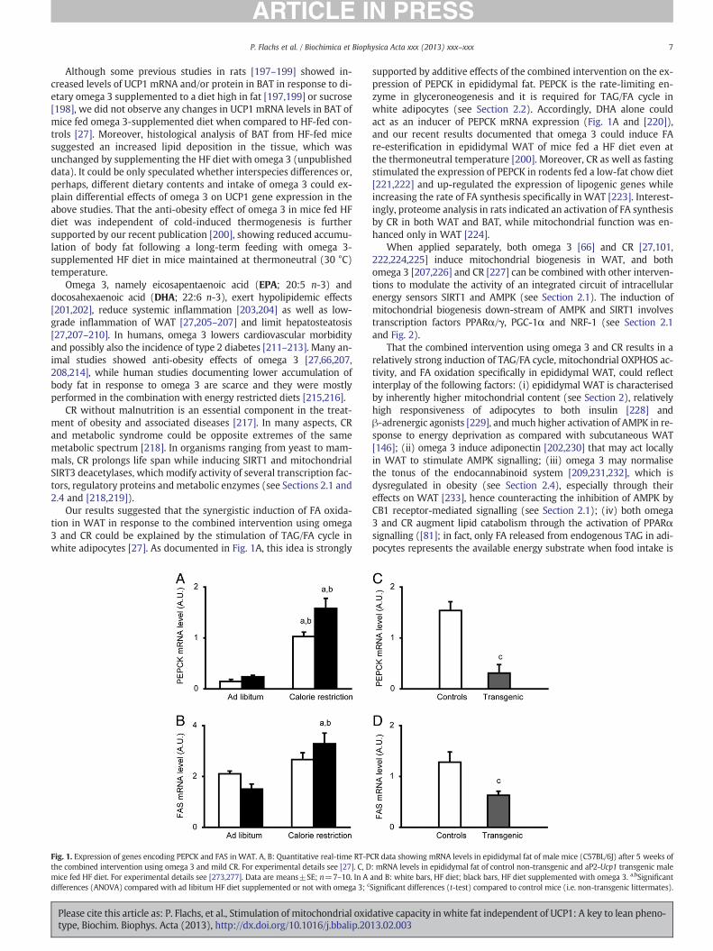
Our results suggested that the synergistic induction of FA oxida-tion in WAT in response to the combined intervention using omega3 and CR could be explained by the stimulation of TAG/FA cycle inwhite adipocytes [27]. As documented in Fig. 1A, this idea is strongly
Fig. 1. Expression of genes encoding PEPCK and FAS in WAT. A, B: Quantitative real-time RT-Pthe combined intervention using omega 3 and mild CR. For experimental details see [27]. C, Dmice fed HF diet. For experimental details see [273,277]. Data are means±SE; n=7–10. In Adifferences (ANOVA) compared with ad libitum HF diet supplemented or not with omega 3; c
Please cite this article as: P. Flachs, et al., Stimulation of mitochondrial oxidtype, Biochim. Biophys. Acta (2013), http://dx.doi.org/10.1016/j.bbalip.20
supported by additive effects of the combined intervention on the ex-pression of PEPCK in epididymal fat. PEPCK is the rate-limiting en-zyme in glyceroneogenesis and it is required for TAG/FA cycle inwhite adipocytes (see Section 2.2). Accordingly, DHA alone couldact as an inducer of PEPCK mRNA expression (Fig. 1A and [220]),and our recent results documented that omega 3 could induce FAre-esterification in epididymal WAT of mice fed a HF diet even atthe thermoneutral temperature [200]. Moreover, CR as well as fastingstimulated the expression of PEPCK in rodents fed a low-fat chow diet[221,222] and up-regulated the expression of lipogenic genes whileincreasing the rate of FA synthesis specifically in WAT [223]. Interest-ingly, proteome analysis in rats indicated an activation of FA synthesisby CR in both WAT and BAT, while mitochondrial function was en-hanced only in WAT [224].
When applied separately, both omega 3 [66] and CR [27,101,222,224,225] induce mitochondrial biogenesis in WAT, and bothomega 3 [207,226] and CR [227] can be combined with other interven-tions to modulate the activity of an integrated circuit of intracellularenergy sensors SIRT1 and AMPK (see Section 2.1). The induction ofmitochondrial biogenesis down-stream of AMPK and SIRT1 involvestranscription factors PPARα/γ, PGC-1α and NRF-1 (see Section 2.1and Fig. 2).
That the combined intervention using omega 3 and CR results in arelatively strong induction of TAG/FA cycle, mitochondrial OXPHOS ac-tivity, and FA oxidation specifically in epididymal WAT, could reflectinterplay of the following factors: (i) epididymal WAT is characterisedby inherently higher mitochondrial content (see Section 2), relativelyhigh responsiveness of adipocytes to both insulin [228] andβ-adrenergic agonists [229], andmuch higher activation of AMPK in re-sponse to energy deprivation as compared with subcutaneous WAT[146]; (ii) omega 3 induce adiponectin [202,230] that may act locallyin WAT to stimulate AMPK signalling; (iii) omega 3 may normalisethe tonus of the endocannabinoid system [209,231,232], which isdysregulated in obesity (see Section 2.4), especially through theireffects on WAT [233], hence counteracting the inhibition of AMPK byCB1 receptor-mediated signalling (see Section 2.1); (iv) both omega3 and CR augment lipid catabolism through the activation of PPARαsignalling ([81]; in fact, only FA released from endogenous TAG in adi-pocytes represents the available energy substrate when food intake is
CR data showing mRNA levels in epididymal fat of male mice (C57BL/6J) after 5 weeks of: mRNA levels in epididymal fat of control non-transgenic and aP2-Ucp1 transgenic maleand B: white bars, HF diet; black bars, HF diet supplemented with omega 3. a,bSignificantSignificant differences (t-test) compared to control mice (i.e. non-transgenic littermates).
ative capacity in white fat independent of UCP1: A key to lean pheno-13.02.003

Fig. 2. Proposed scheme of modulation of metabolic fluxes in adipocytes in the epididymal WAT in response to the combined intervention using omega 3 and CR in mice fed HF diet.As explained in the main text, the combined intervention is associated with the reduction of low-grade inflammation of WAT, induction of adiponectin, lowering of the tonus of theendocannabinoid system (lower levels of major endocannabinoids in the tissue in response to omega 3 supplementation), higher adrenergic stimulation of adipocytes (elicited byCR), and amelioration of HF diet-induced insulin resistance. Synergistic induction of the formation of lipid mediators such as 15d-PGJ2, which are derived from polyunsaturated FAreleased from membrane phospholipids by phospholipase A2 (PLA2), and decreased activity of the CDK5 in response to the anti-inflammatory effect of the intervention lead to theactivation of PPARγ signalling and up-regulation of specific genes engaged in mitochondrial biogenesis and lipid catabolism, as well as FA synthesis and glyceroneogenesis. PPARαand PGC-1 are also involved in the changes of the transcriptional programme in white adipocytes. The changes at the level of gene expression are reflected in the induction of mi-tochondrial β-oxidation, PEPCK-dependent glyceroneogenesis, and activation of the TAG/FA cycle. De novo FA synthesis is supported by the insulin-sensitizing effect of the inter-vention, depending possibly in large on the PPARγ activation. The induction of TAG/FA cycle (FA liberated from TAG during lipolysis may be released from the adipocyte andimmediately taken back by FAT/CD36-mediated mechanism—not shown) and activation of FA for β-oxidation requires ATP, leading to lowering of cellular energy status (i.e. anincreased AMP/ATP ratio) and activation of AMPK. At the early stages of adrenergic stimulation, the activity of AMPK is compromised by protein kinase A (PKA)-mediated inhibitionthat is released at later stages of the stimulation. In addition, AMPK activity is possibly further augmented, reflecting a decrease in the endocannabinoid tonus. The activation ofAMPK would support the induction of mitochondrial biogenesis, OXPHOS, and β-oxidation while limiting the lipolytic response, and hence the release of FA from adipocytes.AdipoR, adiponectin receptor; COX, cyclooxygenase; LOX, lipoxygenase.
8 P. Flachs et al. / Biochimica et Biophysica Acta xxx (2013) xxx–xxx
limited); and (v) both omega 3 [205–207,234] and CR [96] reducelow-grade inflammation of WAT, which, in turn may prevent CDK5-mediated phosphorylation of PPARγ, and therefore, secure a beneficialpattern of PPARγ-mediated changes in gene expression (seeSection 2.1).
The anti-inflammatory effects of omega 3 depend in large on theformation of their active metabolites. These lipid mediators originatefrom either targeted enzymatic synthesis, as in case of resolvins andprotectins [235,236], or from non-enzymatic oxidation reactions [234,237]. They can act as ligands for surface receptors, namely the lipidsensor GPR120 [206] or can interact with transcription factors likePPARγ and NF-κB [234]. Notably, resolvins and protectins mediatethe anti-inflammatory and protective actions of omega 3 on obesity-induced insulin resistance and hepatic steatosis [235,238]. As wehave shown recently [27], the combined intervention using omega 3and CR induced 15-deoxy-Δ12,14-prostaglandin J2 (15d-PGJ2) andprotectin D1 (PD1) with a surprising synergism observed in epididy-mal fat, but not in the liver. The induction of these lipid mediatorscould contribute to the anti-inflammatory effect of this intervention,while activation of PPARγ resulting from the supposed 15d-PGJ2binding [239,240] could impact on mitochondrial biogenesis,glyceroneogenesis, adiponectin formation and other PPARγ-mediatedeffects (see Section 2.1).
Please cite this article as: P. Flachs, et al., Stimulation of mitochondrial oxidtype, Biochim. Biophys. Acta (2013), http://dx.doi.org/10.1016/j.bbalip.20
As summarized in the following section, the levels of various lipidmediators in WAT could largely depend on the presence of variouscell types in the tissue and on the changes in the pattern of formationof these lipid mediators in response to local inflammatory status.
4. Role of local lipid mediators in regulation of WAT metabolism
In addition to differentiated adipocytes and their precursors, in-nate immune cells including macrophages, mast cells, neutrophils,or eosinophils can be found in WAT [87,88]. Among them, adipose-tissue macrophages (ATM), major antigen-presenting cells, are func-tionally and numerically dominant and exert both immune andhousekeeping functions. A state of chronic mild inflammation and dys-function of WAT present in obesity can be linked to the infiltration ofpro-inflammatory macrophages (M1 phenotype), which producepro-inflammatory cytokines [87–89,241]. ATM in leanmice show an al-tered, less-inflammatory properties (M2 phenotype; [242]). Cytokinesreleased from M1 macrophages block insulin action in adipocytes viaTNF-α pathway, promote extracellular matrix remodelling and fibrosisof WAT, recruit other immune cells such as mast cells, B-cells or T-cells,and form crown-like-structures that encircle necrotic adipocytes tophagocytose debris [243]. These sub-populations release eitheranti-inflammatory (IL-4, IL-10, IL-13) or pro-inflammatory (TNF-α,
ative capacity in white fat independent of UCP1: A key to lean pheno-13.02.003

9P. Flachs et al. / Biochimica et Biophysica Acta xxx (2013) xxx–xxx
IFN-γ) cytokines, respectively [244]. Also othermolecules like lipopoly-saccharide (LPS) or palmitic acid promote M1-polarization via TLR4pathway [245,246], and TLR antagonists including EPA or theadiponectin paralog C1q/TNF-related protein 3 (CTRP3; [247]), an en-dogenous LPS antagonist in adipose tissue, might be slowing downthe inflammation. All these mediators affect also adipocytes, whichthemselves produce all kinds of adipokines with pro- or anti-inflammatory properties (monocyte chemoattractant protein-1,MCP-1; resistin, adiponectin, etc.; [87]). Importantly, reduction ofWAT expression of SIRT1 in obesity (see Section 2.4) is causally linkedto the macrophage infiltration [96]. Recent hypothesis on WAT inflam-mation proposes that the M1 response increases fuel demand for ster-ile immune response, thus increasing hyperglycaemia and lipolysis tosupply nutrients for activated immune cells, while the M2 responsesuppresses the clonal expansion of Th1 cells and enhance nutrient stor-age by potentiating the action of insulin [248].
Besides the cytokines, a large family of endogenous lipid mediatorsderivedmainly from arachidonic acid (AA; 22:4 n-6), contributes to the
Table 1Lipid mediators and intercellular signalling in WAT.
Model Species Tissue Lipid mediator Source omediator
Adipocytes, SVF Human Subcutaneous PGE2 EndoAdipocytes, SVF Rat PGE2, PGI2 Endo+eAdipocytes, SVF,3T3-L1
Mouse Epididymal PGE2, PGI2 Endo
Adipose tissue,adipocytes
Human Subcutaneous PGE2 Endo
3T3-L1 PGE2 EndoAdipose tissue,adipocytes, 3T3-L1
Mouse Intra-abdominal cPGI2, PGE2,PGI2
Exo+en
Adipocytes, SVF, BAT Mouse Inguinal PGE2 ExoBAT Mouse Interscapular PGE2 ExoBAT, adipose tissue Mouse Interscapular,
subcutaneousPGD2-related Endo
3T3-L1 PGF2a Exo3T3-L1 PGJ2 series EndoAdipocytes, 3T3-L1 Human Subcutaneous d15-PGJ2 Exo3T3-L1 d15-PGJ2 ExoNIH-3T3 d15-PGJ2 ExoC3H10T1/2 d15-PGJ2 ExoAdipocytes, SVF Mouse Epididymal LTB4 ExoAdipopocytes,3T3-L1
Human, mouse Subcutaneous,epididymal
LTB4, CysLTs Endo
Adipocytes, SVF,3T3-L1
Rat Perigonadal 12-HETE, 5-HETE,LTB4
Exo
Adipocytes, 3T3-L1 Mouse Epididymal 12-HETE,12-HpETE
Endo+e
Adipose tissue,adipocytes
Mouse Visceral EET, DHET mix Exo
Adipocytes, SVF,macrophage
Mouse Epididymal RvD1 Endo+e
Adipose tissue,SVF
Mouse Epididymal RvD1 Exo
Adipose tissue Mouse Epididymal 17-HDHA, PD1,RvD1
Exo
Adipose tissue Mouse Epididymal PD1, 18-HEPE,17-HDHA
Endo
Adipose tissue,adipocytes
Human, mouse Epididymal RvD1, RvD2 Exo
Adipose tissue Mouse Epididymal PD1, d15-PGJ2 EndoAdiposetissue/leukocytes
Mouse Perinodal RvE1, PD1, LXA4 Exo
Adipose tissue Mouse omega 3 andomega 6 oxylipins
Endo
Adipose tissue,adipocytes, SVF
Mouse Perigonadal LXA4 Exo
Adipocytes, collagenase-liberated adipocytes from adipose tissue; SVF, stromal vascular framediator (produced by the tissue itself); exo, exogenous application of the mediator (artifi
Please cite this article as: P. Flachs, et al., Stimulation of mitochondrial oxidtype, Biochim. Biophys. Acta (2013), http://dx.doi.org/10.1016/j.bbalip.20
inflammatory state in WAT [244]. As mentioned above (seeSections 2.1, 2.4, and 3.2), endocannabinoids deteriorate AMPK signal-ling in WAT, and omega 3 supplementation results in lowering of theirtissue levels, reflecting the replacement of AA in membrane phospho-lipids by DHA and EPA. Eicosanoids, products of cyclooxygenases,lipoxygenases, and cytochromes P450, are potent local mediators ofsignal transduction and modulate the inflammatory response.Although immune cells are the main producers of eicosanoids, also ad-ipocytes synthetize prostanoids (PGE2, PGI2, PGF2α) and leukotrienesand express eicosanoid receptors. Thus, members of both adipocyteand immune cell lineages are able to communicate using lipid media-tors as membrane or nuclear receptor ligands. Selected lipid mediatorsproduced in adipose tissue are summarized in Table 1. Prostanoids areimportant for differentiation of adipocytes, and can regulate lipolysis inan autocrine and paracrine manner through PGE2 or even shift thedifferentiation of defined mesenchymal progenitors toward a brownadipocyte phenotype. PGE2 was shown to induce UCP1 in whiteadipocytes and stimulate thermogenesis in BAT. Cyclopentenone
f Effect Reference
Modulation of adipogenesis [279]xo Modulation of lipolysis [280]
Modulation of lipolysis [190]
Stimulation of leptin release [281]
Modulation of adipogenesis [282]do Shift the differentiation of mesenchymal progenitors toward a
brown adipocyte phenotype, induction of UCP1 in WAT[254,255]
Induction of UCP1 in white adipocytes, no effect in BAT [254]Stimulation of thermogenesis in BAT [283]Regulation of BAT substrate utilisation, modulation of lipolysis [256]
Increase of glucose transport [284]Modulation of adipogenesis and MCP-1 expression [285]Production of macrophage inhibitory cytokine-1 [286]Decrease of leptin production [287]Stimulation of adipogenesis [239]Promotes adipocyte differentiation [240]Secretion of MCP-1, IL-6, and TNF-a [288]Chemoattractant for macrophages in obesity [289]
Upregulation of IL-6, TNF-alpha, MCP-1 [290]
xo Induction of ER stress [291]
Modulation of adipocyte differentiation, decreased TNF-alpha,MCP-1, increase of adiponectin
[292]
xo Downregulation IL-6, MCP-1, and TNF-a, ROS; upregulation of IL-10,CD206, arginase 1, resistin-like molecule a, and chitinase-3 likeprotein, stimulated nonphlogistic phagocytosis; shift M1>M2macrophage phenotype
[249]
Increase of adiponectin production and insulin sensitivity; decreaseof IL-6 production and macrophage infiltration; shift M1>M2macrophage phenotype
[293]
Increase of adiponectin and insulin sensitivity [235]
Prevention of obesity-linked inflammation and insulin resistance [236]
Increase of adiponectin; decrease of leptin, TNF-a, MCP-1, IL-6, andIL-1b
[253]
Induction of lipid catabolism, antiinflammatory [27]Resolution of inflammation [294]
Modulation of inflammatory status in adipose tissue [295]
Decrease of IL-6 and increase of IL-10 expression; increase of insulinsensitivity, antiinflammatory
[296]
ction from adipose tissue (containing immune cells); endo, endogenous source of thecial stimulation).
ative capacity in white fat independent of UCP1: A key to lean pheno-13.02.003

10 P. Flachs et al. / Biochimica et Biophysica Acta xxx (2013) xxx–xxx
prostaglandins (e.g. 15d-PGJ2) act as PPARγ ligands and modulatetranscription of genes (see Section 2.1), including those involved inthe production of adipokines linked to inflammation. Both PGE2 and15d-PGJ2 are synthetized within cyclooxygenase pathway, but in dif-ferent branches using either prostaglandin E or D synthases, potentiallyleading to either pro- or anti-inflammatory mediators. The possibilityof modulation of these synthases adds another layer of complexity. Dis-covery of pro-resolving and anti-inflammatory omega 3-derived lipidmediators called resolvins (E-resolvins and D-resolvins), protectins,and maresins opened a new field concerning the mechanisms involvedin resolution of WAT inflammation [236,249,250].
In this context, our finding (see Section 3.2) of the synergistic in-duction in WAT of specific lipid mediators, namely 15d-PGJ2 andPD1 (or its isomer PDX with the 11E, 13Z, 15E geometry instead of11E, 13E, and 15Z in PD1), by a combined intervention using omega3 and CR is of utmost importance. The observed induction of15d-PGJ2, a metabolite derived from AA (omega 6 FA), is consistentwith the results of another study [251] showing a similar inductionof 15d-PGJ2 by dietary DHA, as well as with a non-enzymatic mecha-nism of its synthesis (see Section 3.2 and [27,251]). These resultsclearly demonstrate that dietary interventions can be used to increaselocal levels of lipid mediators in WAT, which can help to resolve in-flammation [252] and even modulate metabolic properties of adipo-cytes by interacting with the major transcriptional regulatorypathways including PPARs (see Sections 2.1 and 2.5). In fact, changesin the levels of these types of lipid mediators may exert stronger ef-fects than those exerted by pharmacological treatments. Thus, e.g.,resolvins RvD1 and RvD2 counteract both local adipokine productionand monocyte accumulation in inflamed WAT with a 300-fold higherefficiency as compared with rosiglitazone [253].
With respect to the induction of brite cells and UCP1 by PGE2[254,255], and the induction of energy-dissipating adipocytes by15d-PGJ2 and PD1 in response to the combined intervention withomega 3 and CR [27], it is unclear whether these lipid mediatorscould affect the same signalling pathway. PGE2 stimulated adipocytesthrough EP4 receptors on the plasma membrane while 15d-PGJ2,generated inside the cells, acted probably as a PPARγ ligand (seeSection 2.1). Also a precise role of enzymes involved in PGE2, PGD2and PGF2α metabolism has to be further explored [256–260]. A de-tailed characterisation of lipid mediators involved in the inductionof energy expenditure in adipocytes may contribute to better charac-terisation of the (distinct) adipocyte cell lineage(s) involved. It couldhelp to clarify, whether adipocytes, in which energy expenditure isactivated through TGA/FA cycling, belong to the brite cell lineage orwhether typical white adipocytes are involved.
It has been demonstrated that obesity-associated macrophage infil-tration of WAT in mice is one order of magnitude higher in epididymalas compared with subcutaneous WAT, and that this inflammatory re-sponse is much lower in female as compared with male mice. Further-more, female mice showed a relatively weak link between adipocytehypertrophy and various phenotypes such as impaired glucose homeo-stasis and hepatosteatosis induced by HF feeding [261]. Accordingly,the obesity-associated WAT inflammation resulted in mitochondrialdysfunction in epididymal but not in periovarian WAT in the rat[262]. Therefore, it has to be learned how mouse strain-, fat depot-and gender-specific differences in macrophage infiltration of WATcould affect metabolism of adipocytes.
In addition to various cytokines and lipid mediators, M2 macro-phages have been recently shown to produce catecholamines in ani-mals exposed to cold, with a straightforward implication for thecontrol of energy metabolism and thermogenesis in adipocytes [263].That also adipocytes and cells within stromal vascular fraction couldserve as a source of catecholamines has been recently observed[264,265].
It can be expected that the cell–cell interactions mediated by lipidmediators will be affected, reflecting not only the low-grade
Please cite this article as: P. Flachs, et al., Stimulation of mitochondrial oxidtype, Biochim. Biophys. Acta (2013), http://dx.doi.org/10.1016/j.bbalip.20
inflammation associated with obesity, but also changes in the im-mune cell population in WAT, which could be associated with cancerand, therefore, impact on metabolism of adipocytes and adiposity.Although no direct evidence linking inflammation, lipid mediators,cancer, and WAT is known, results on adipose-related breast cancerand adipocytes show that especially PGE2, its receptors, andcyclooxygenase-2 pathway are involved in increased aromatase acti-vation leading to carcinogenesis [266–269]. On the contrary, growthof breast cancer cells was decreased by omega 3 when incorporatedin plasma membranes, thus creating a pool for the synthesis ofanti-inflammatory mediators [270].
5. Testing the impact of increased lipid catabolism in whiteadipocytes—mitochondrial uncoupling induced by transgenicexpression of UCP1
As described above (see Sections 3.1 and 3.2), mitochondrialoxidative capacity in white adipocytes could be augmented, in theabsence of any changes in the UCP1 levels, e.g. by cold acclimationin UCP1-ablated mice, or more physiologically, in response to thecombined intervention using omega 3 and CR. This combined inter-vention induced a significant increase of FA oxidation inWAT. A ques-tion remains, however, how important the induction of energydissipation in bona fide white adipocytes could be with respect tothe regulation of body fat stores. Useful information can be drawnfrom the phenotype of transgenic mice expressing UCP1 gene fromthe aP2 gene promoter, with an enhanced expression of UCP1 inBAT and ectopic expression of UCP1 in WAT (aP2-Ucp1 mice; Ref.[25]). These mice are resistant to obesity while showing a modulationof various aspects of WAT metabolism as well as whole-body meta-bolic responses (see below; for review see [93]). Since UCP1over-expression in BAT of the aP2-Ucp1 mice results in BAT atrophyand the impairment of BAT thermogenic functions [6,271], presum-ably due to a toxic effect of excessive amounts of UCP1, the obesity re-sistant phenotype of these mice likely results from uncoupledmitochondrial respiration solely in mature white adipocytes.
5.1. Induction of mitochondrial oxidative capacity in WAT in differentmodels
Even the hemizygous aP2-Ucp1 transgenic mice are stronglyprotected against obesity when crossed with genetically obese Avy
mice or when fed a HF diet [25,272]. In agreement with a higherexpression of transgenic UCP1 in subcutaneous as compared withgonadal WAT [65], the transgene-induced metabolic changes weremore pronounced in the former fat depot, which also preferentiallydecreased its size [25,272]. Thus, in the subcutaneous WAT of theaP2-Ucp1 transgenic mice, 1.6-fold higher mitochondrial contentwas found as compared with wild-type mice [65], in associationwith a 3-fold increase in FA oxidation [273] and a 1.5-fold increasein oxygen consumption ([6]; as measured ex vivo in epididymalWAT).
In the UCP1-ablated mice, short-term cold exposure resulted in a2- and 3.6-fold increase in specific cytochrome oxidase activity(adjusted to protein content of tissue homogenates used for the mea-surements) detected in epididymal and subcutaneous inguinal WAT,respectively [196]. Furthermore, in epididymal WAT of inbred,wild-type C57BL/6J mice, the intervention using omega 3 combinedwith CR increased specific cytochrome oxidase activity 1.3-fold,while the specific content of mitochondrial cytochromes as well aspalmitate oxidation adjusted to tissue weight increased 1.6-fold.Moreover, both mitochondrial respiratory capacity and OXPHOS ac-tivity in digitonine-permeabilised adipocytes from epididymal WATof mice subjected to the combined intervention were 2-fold higheras compared with the control non-treated group, while mitochondriain both groups of mice were highly coupled [27].
ative capacity in white fat independent of UCP1: A key to lean pheno-13.02.003

11P. Flachs et al. / Biochimica et Biophysica Acta xxx (2013) xxx–xxx
Thus, in all the models mentioned above, obesity resistance was as-sociated with a similar induction of mitochondrial oxidative capacityand activity of FA oxidation in WAT and/or white adipocytes. At thesame time, up-regulation of mitochondrial oxidative capacity was rela-tively high in WAT as compared with skeletal muscle or BAT [27,196].Importantly, all these experiments (except for those with aP2-Ucp1transgene transferred to the genetically obese Avy mice; Ref. [25])were performed inmice having the same C57BL/6J genetic background.It is to be inferred that the induction of FA oxidation in white adipo-cytes of mice either using genetic manipulations to induce mitochon-drial uncoupling or by activating the TAG/FA cycle (or perhaps alsoby other physiological mechanisms independent of UCP1) couldcontribute to obesity resistance.
5.2. Ectopic expression of UCP1 in white adipocytes as a model of britecells
Since cellular physiology and metabolic properties of UCP1-containing adipocytes in WAT of the aP2-Ucp1 transgenic mice havebeen already well characterised (see below), the results obtainedwith this transgenic model could serve as a lead to understand thephysiology of brite adipocytes. In fact, studies of specific metabolicfeatures of brite adipocytes are at their beginnings, and they are com-plicated by problems regarding the isolation of a homogenous cellpopulation in sufficient quantities that are required for biochemicalstudies. Some of the consequences arising from the ectopic expres-sion of UCP1 in white adipocytes, which could be of interest withrespect to understanding the function of brite adipocytes, are summa-rized below.
Ectopic UCP1 lowered mitochondrial membrane potential in adi-pocytes isolated from gonadal fat of aP2-Ucp1 mice and rendered itsensitive to FA and purine nucleotides. Full uncoupling activity wasachieved in the presence of ~15-fold less UCP1 than in BAT mitochon-dria, resulting in the molar UCP1/respiratory chain ratio ~1, while thisratio is between 5 and 11 in BAT [187]. These results document thatectopic UCP1 was inserted correctly into the inner mitochondrialmembrane, and that a relatively very low amount of UCP1 is sufficientfor a full release of mitochondrial respiratory control. These data arealso consistent with the original results of Lin and Klingenberg[274], suggesting that the protonophoric activity of UCP1 exceedsseveral-fold the proton-pumping activity of the respiratory chain inthe native mitochondrial membrane. Furthermore, despite a declinein the content of transgenic UCP1 in WAT during ageing of theaP2-Ucp1 mice [65], the robust anti-obesity effect of transgenicUCP1 was observed both in the aged Avy mice expressing aP2-Ucp1transgene (3) and in the aP2-Ucp1 mice fed HF diet during thewhole life (our unpublished results). These results suggest thatindeed even minute changes in the expression of UCP1 in WAT, ascould be the case of the induction of brite adipocytes, may havesignificant impact on the respiratory control in mitochondria inthese adipocytes. With respect to possible metabolic consequencesof the induction of brite adipocytes, which takes place preferentiallyin subcutaneous WAT [28], it is important to note that the ectopicexpression of UCP1 in the transgenic mice is also much stronger insubcutaneous as compared with gonadal WAT [65].
6. Key role of AMPK in the metabolic effects associated with theinduction of energy expenditure in adipocytes
It is striking that very different mechanisms of the induction ofenergy expenditure triggered in white adipocytes in vivo, either bythe combined intervention using omega 3 and CR or transgenicexpression of UCP1, could elicit similar systemic effects, namely theprotection against obesity [25,27,272] and hypolipidemic effects[27,275]. With respect to improvement of glucose homeostasis, thecombined intervention may be even more efficient [27,272]. It can
Please cite this article as: P. Flachs, et al., Stimulation of mitochondrial oxidtype, Biochim. Biophys. Acta (2013), http://dx.doi.org/10.1016/j.bbalip.20
be speculated that the cellular energy sensor AMPK in adipocytesplays the key unifying role in these metabolic responses (Fig. 3).Thus, in case of the combined intervention, AMPK could be activatedin response to increased demands for ATP, reflecting both the induc-tion of FA re-esterification and de novo FA synthesis. Accordingly,starvation [146], omega 3 [226] and PPARγ agonists [178] couldstimulate AMPK activity in WAT. However, in the case of combinedintervention, the involvement of AMPK remains to be confirmedexperimentally. On the other hand, in the aP2-Ucp1 mice, the activa-tion of AMPK [273] associated with a significant decrease of cellularenergy status in WAT [273,276] has been unequivocally demon-strated. Thus, the activation of AMPK in response to mitochondrialuncoupling could explain fat depot-specific depression of lipogenesis[277] and lipolysis [276], as well as the increases in FA oxidation [273]and mitochondrial content [65]. Indeed, an elevated activity of α1subunit of AMPK [273], a dominant catalytic subunit in adipose tissue[93], was detected in WAT (and especially in subcutaneous WAT) ofthe aP2-Ucp1 mice. In analogy with its effects in the heart [94],AMPK activation could also explain the induction of LPL [275]. There-fore, it seems likely that AMPK represents the major regulatory intra-cellular pathway responsible for the wide spectrum of effects inducedby transgenic UCP1 in white adipocytes, while at the same time theactivation of FA oxidation and inhibition of lipogenesis in responseto AMPK activation triggered by mitochondrial uncoupling would re-duce fat accumulation.
Compared with the modulation of WAT metabolism by thetransgene-induced mitochondrial uncoupling, which could beexplained in full by the AMPK activation, the mechanistic basis ofthe metabolic effects of the combined intervention with omega 3and CR in adipocytes is more complex (see Section 3.2 and the legendto Fig. 2). These mechanisms could explain some of the differencesbetween the combined intervention and ectopic UCP1 regardingtheir effects on WAT and its secretory functions. For instance, PEPCKand FAS genes in epididymal WAT are up-regulated in response tothe combined intervention (Fig. 1A,B), and it is conceivable thatPPARγ-signalling is activated in WAT of mice subjected to this inter-vention due to both a synergistic induction of the endogenousPPARγ activator 15d-PGJ2 and the anti-inflammatory effects ofomega 3 (Fig. 2). This is reflected by the induction of adiponectin[27] and could be critical for improving whole-body insulin sensitivi-ty [88]. In contrast, a down-regulation of PPARγ and its target genesaP2 [273], PEPCK (Fig. 1C), and adiponectin (our unpublished results)has been observed in the aP2-Ucp1 mice. Also the expression of FAShas been down-regulated in response to mitochondrial uncoupling(Fig. 1D and [277]). In addition, down-regulation of PC, anotherPPARγ target [182], together with decreased mitochondrial ATP pro-duction may augment the AMPK-mediated inhibition of de novo FAsynthesis in WAT of the aP2-Ucp1 mice.
It is to be stressed that under both conditions, i.e. in response tothe combined intervention and mitochondrial uncoupling in WAT,activation of AMPK could support the metabolic effects as well as theinduction of energy expenditure by augmenting the oxidative capacityin WAT. However, activation of AMPK alone, without concomitantactivation of futile substrate cycling or mitochondrial uncoupling, re-spectively, for thermodynamic reasons, could not lead to any increasein energy expenditure [93,278]. In any case, independent of energyexpenditure, AMPK-mediated activation of FA oxidation in adipo-cytes in obesity (even at the expense of decreased glucose oxidation)could help to counteract adipose tissue inflammation, as well aslipotoxicity resulting from excessive accumulation of fat in othertissues.
7. Conclusions, future research and new therapeutic strategies
In face of the renewed interest in mitochondrial uncoupling and theinduction of UCP1-mediated thermogenesis in adipose tissue as
ative capacity in white fat independent of UCP1: A key to lean pheno-13.02.003

Fig. 3. Putative involvement of AMPK in white adipocytes in the metabolic effects either elicited by the combined intervention using omega 3 and CR or by mitochondrialuncoupling in the aP2-Ucp1 transgenic mice. For further details, see the main text and Fig. 2.
12 P. Flachs et al. / Biochimica et Biophysica Acta xxx (2013) xxx–xxx
potential therapeutic tools for obesity, increasing energy expenditurein WAT independently of UCP1 should be also considered. As demon-strated in mice, combined intervention based on the use of specificmicronutrients, e.g. omega 3, and very mild CR could result in additiveincreases of FA oxidation in WAT, which is based on the activation ofTAG/FA cycle and synergistic anti-inflammatory effects of the interven-tion. The mechanistic aspects of such changes in the phenotype ofadipocytes should be further studied. That the above metabolic effectswere observed in the epididymal but not subcutaneousWAT depot sug-gests an involvement of bona fide white adipocytes rather than newlydifferentiated cells belonging to another cell lineage, e.g. the one thatgives rise to brite adipocytes. Further studies in this respect should beperformed in order to characterise the expression of cell lineagemarkersin the cells with augmented UCP1-independent thermogenesis.
In spite of the widely neglected contribution of WAT to whole-body energy metabolism, previous studies in mice with ectopicexpression of UCP1 in WAT support the notion that modulationof WAT metabolism could contribute to reduction of adiposity. Thecomparison of phenotypic changes observed in adipocytes fromepididymal fat of mice subjected to the combined intervention withthose seen in the transgenic mice strongly suggests that by combiningphysiological stimuli it should be possible to modify metabolicproperties of white adipocytes to the extent that would elicit system-ic effects. As compared with the induction of energy expenditure bythe robust transgenic modification resulting in mitochondrialuncoupling, physiological modifications based on the combined inter-vention using omega 3 and CR may have additional health benefits,namely those, which are linked to the anti-inflammatory effects andto the suppression of the endocannabinoid system activity. With re-spect to the insulin-sensitizing effect of the combined intervention,the induction of adiponectin and stimulation of PPARγ signalling inWAT may be important for glucose homeostasis at the whole bodylevel, reflecting the key role of PPARγ signalling in adipocytes for in-sulin sensitivity of the organism. High capacity of mitochondrialOXPHOS linked to inducible TAG/FA cycling activity is essential for
Please cite this article as: P. Flachs, et al., Stimulation of mitochondrial oxidtype, Biochim. Biophys. Acta (2013), http://dx.doi.org/10.1016/j.bbalip.20
metabolic flexibility of WAT and represent the markers of healthyadipocytes.
In this review, we better characterised the intracellular handles,which could be targeted to improve metabolic properties of adipo-cytes in WAT. With respect to novel therapeutic strategies, it remainsto be established whether the mechanism triggered by the combinedintervention, which modulates metabolic fluxes specifically in epidid-ymal WAT of mice, could be also elicited in human WAT. This may bepossible given a relatively high mitochondrial content and pro-nounced activity of the TAG/FA cycle in visceral fat. New combinationtreatment strategies may be designed including naturally occurringmicronutrients like omega 3 and plant polyphenols, reduced calorie in-take, and pharmacological modulators of key signalling pathways in fatcells. Involvement of the mutual interactions between adipocytes andimmune cells contained in WAT may be essential for the induction oflipid catabolism in white adipocytes and the lean phenotype.
Acknowledgements
We thank Jon Arch and Leslie P. Kozak for the stimulating discus-sion on the role of adipose tissue metabolism in energy balance, aswell as for the help with retrieving some of the original articles,Jaap Keijer for discussing the concept of healthy adipocyte endowedwith high capacity of mitochondrial OXPHOS, and Morten Bryhn forsharing his expertise regarding biological roles of marine lipids andfor his critical reading of the manuscript. Our work was supportedby the EU FP7 project DIABAT (HEALTH-F2-2011-278373), by theCzech Science Foundation (301/11/0226) and RVO: 67985823.
References
[1] O. Gavrilova, B. Marcus-Samuels, D. Graham, J.K. Kim, G.I. Shulman, A.L. Castle, C.Vinson, M. Eckhaus, M.L. Reitman, Surgical implantation of adipose tissuereverses diabetes in lipoatrophic mice, J. Clin. Invest. 105 (2000) 271–278.
ative capacity in white fat independent of UCP1: A key to lean pheno-13.02.003

13P. Flachs et al. / Biochimica et Biophysica Acta xxx (2013) xxx–xxx
[2] L. Heilbronn, S.R. Smith, E. Ravussin, Failure of fat cell proliferation, mitochondrialfunction and fat oxidation results in ectopic fat storage, insulin resistance and typeII diabetes mellitus, Int. J. Obes. Relat. Metab. Disord. 28 (Suppl. 4) (2004) S12–S21.
[3] S. Virtue, A. Vidal-Puig, It's not how fat you are, it's what you do with it thatcounts, PLoS Biol. 6 (2008) e237.
[4] S. Sugii, P. Olson, D.D. Sears,M. Saberi, A.R. Atkins, G.D. Barish, S.H. Hong, G.L. Castro,Y.Q. Yin, M.C. Nelson, G. Hsiao, D.R. Greaves, M. Downes, R.T. Yu, J.M. Olefsky, R.M.Evans, PPARgamma activation in adipocytes is sufficient for systemic insulin sensi-tization, Proc. Natl. Acad. Sci. U. S. A. 106 (2009) 22504–22509.
[5] H. Bottcher, P. Furst, Decreased white fat cell thermogenesis in obese individuals,Int. J. Obes. 21 (1997) 439–444.
[6] J. Kopecky, M. Rossmeisl, Z. Hodny, I. Syrovy, M. Horakova, P. Kolarova, Reductionof dietary obesity in the aP2-Ucp transgenic mice: mechanism and adipose tissuemorphology, Am. J. Physiol. 270 (1996) E776–E786.
[7] P. Hallgren, L. Sjostrom, H. Hedlund, L. Lundell, L. Olbe, Influence of age, fat cellweight, and obesity on O2 consumption of human adipose tissue, Am. J. Physiol.256 (1989) E467–E474.
[8] P. Hallgren, E. Raddatz, C.H. Bergh, P. Kucera, L. Sjostrom, Oxygen consumptionin collagenase-liberated rat adipocytes in relation to cell size and age, Metabo-lism 33 (1984) 897–900.
[9] H.J. Choo, J.H. Kim, O.B. Kwon, C.S. Lee, J.Y. Mun, S.S. Han, Y.S. Yoon, G. Yoon, K.M.Choi, Y.G. Ko, Mitochondria are impaired in the adipocytes of type 2 diabeticmice, Diabetologia 49 (2006) 784–791.
[10] L. Wilson-Fritch, S. Nicoloro, M. Chouinard, M.A. Lazar, P.C. Chui, J. Leszyk, J.Straubhaar, M.P. Czech, S. Corvera, Mitochondrial remodeling in adipose tissueassociated with obesity and treatment with rosiglitazone, J. Clin. Invest. 114(2004) 1281–1289.
[11] I.W. Asterholm, D.I. Mundy, J. Weng, R.G. Anderson, P.E. Scherer, Alteredmitochondrial function and metabolic inflexibility associated with loss ofcaveolin-1, Cell Metab. 15 (2012) 171–185.
[12] A. Valerio, A. Cardile, V. Cozzi, R. Bracale, L. Tedesco, A. Pisconti, L. Palomba, O.Cantoni, E. Clementi, S. Moncada, M.O. Carruba, E. Nisoli, TNF-alphadownregulates eNOS expression and mitochondrial biogenesis in fat and muscleof obese rodents, J. Clin. Invest. 116 (2006) 2791–2798.
[13] K.H. Pietilainen, J. Naukkarinen, A. Rissanen, J. Saharinen, P. Ellonen, H. Keranen,A. Suomalainen, A. Gotz, T. Suortti, H. Yki-Jarvinen, M. Oresic, J. Kaprio, L.Peltonen, Global transcript profiles of fat in monozygotic twins discordant forBMI: pathways behind acquired obesity, PLoS Med. 5 (2008) e51.
[14] M.J. Vosselman, A.A. van der Lans, B. Brans, R. Wierts, M.A. van Baak, P.Schrauwen, W.D. van Marken Lichtenbelt, Systemic beta-adrenergic stimulationof thermogenesis is not accompanied by brown adipose tissue activity inhumans, Diabetes 61 (2012) 3106–3113.
[15] P.L. Thurlby, R.D. Ellis, Differences between the effects of noradrenaline and thebeta-adrenoceptor agonist BRL 28410 in brown adipose tissue and hind limb ofthe anaesthetized rat, Can. J. Physiol. Pharmacol. 64 (1986) 1111–1114.
[16] L. Simonsen, J. Bulow, J. Madsen, N.J. Christensen, Thermogenic response toepinephrine in the forearm and abdominal subcutaneous adipose tissue, Am. J.Physiol. 263 (1992) E850–E855.
[17] J. Mejsnar, J. Pacha, Thermogenesis due to noradrenaline in muscles underdifferent rates of perfusion, Pflugers Arch. 397 (1983) 149–151.
[18] N.J. Rothwell, M.J. Stock, Influence of noradrenaline on blood flow to brownadipose tissue in rats exhibiting diet-induced thermogenesis, Pflugers Arch.389 (1981) 237–242.
[19] J. Nedergaard, V. Golozoubova, A. Matthias, A. Asadi, A. Jacobsson, B. Cannon,UCP1: the only protein able to mediate adaptive non-shivering thermogenesisand metabolic inefficiency, Biochim. Biophys. Acta 1504 (2001) 82–106.
[20] B. Cannon, J. Nedergaard, Brown adipose tissue: function and physiologicalsignificance, Physiol. Rev. 84 (2004) 277–359.
[21] S.W. Ma, D.O. Foster, Brown adipose tissue, liver, and diet-induced thermogenesisin cafeteria diet-fed rats, Can. J. Physiol. Pharmacol. 67 (1989) 376–381.
[22] M.C. Zingaretti, F. Crosta, A. Vitali, M. Guerrieri, A. Frontini, B. Cannon, J.Nedergaard, S. Cinti, The presence of UCP1 demonstrates that metabolicallyactive adipose tissue in the neck of adult humans truly represents brownadipose tissue, FASEB J. 23 (2009) 3113–3120.
[23] N.J. Rothwell, M.J. Stock, A role for brown adipose tissue in diet-induced thermo-genesis, Nature 281 (1979) 31–35.
[24] J. Himms-Hagen, Role of thermogenesis in brown adipose tissue in the regula-tion of energy balance, in: C.H. Hollenberg, D.A.K. Roncardi (Eds.), The Adiposeand Obesity: Cellular and Molecular Mechanisms, Raven Press, New York,1983, pp. 259–270.
[25] J. Kopecky, G. Clarke, S. Enerback, B. Spiegelman, L.P. Kozak, Expression of themitochondrial uncoupling protein gene from the aP2 gene promoter preventsgenetic obesity, J. Clin. Invest. 96 (1995) 2914–2923.
[26] B.R. Martin, R.M. Denton, The intracellular localization of enzymes inwhite-adipose-tissue fat-cells and permeability properties of fat-cell mitochondria,Biochem. J. 117 (1970) 861–877.
[27] P. Flachs, R. Ruhl, M. Hensler, P. Janovská, P. Zouhar, V. Kus, J.Z. Macek, E. Papp, O.Kuda, M. Svobodova, M. Rossmeisl, G. Tsenov, V. Mohamed-Ali, J. Kopecky,Synergistic induction of lipid catabolism and anti-inflammatory lipids in whitefat of dietary obese mice in response to calorie restriction and n-3 fatty acids,Diabetologia 54 (2011) 2626–2638.
[28] T.B. Walden, I.R. Hansen, J.A. Timmons, B. Cannon, J. Nedergaard, Recruited vs.nonrecruited molecular signatures of brown, “brite,” and white adipose tissues,Am. J. Physiol. Endocrinol. Metab. 302 (2012) E19–E31.
[29] N. Petrovic, T.B. Walden, I.G. Shabalina, J.A. Timmons, B. Cannon, J. Nedergaard,Chronic peroxisome proliferator-activated receptor gamma (PPARgamma)
Please cite this article as: P. Flachs, et al., Stimulation of mitochondrial oxidtype, Biochim. Biophys. Acta (2013), http://dx.doi.org/10.1016/j.bbalip.20
activation of epididymally derived white adipocyte cultures reveals a populationof thermogenically competent, UCP1-containing adipocytes molecularly distinctfrom classic brown adipocytes, J. Biol. Chem. 285 (2010) 7153–7164.
[30] J. Wu, P. Bostrom, L.M. Sparks, L. Ye, J.H. Choi, A.H. Giang, M. Khandekar, K.A.Virtanen, P. Nuutila, G. Schaart, K. Huang, H. Tu, W.D. van Marken Lichtenbelt,J. Hoeks, S. Enerback, P. Schrauwen, B.M. Spiegelman, Beige adipocytes are adistinct type of thermogenic fat cell in mouse and human, Cell 150 (2012)366–376.
[31] C. Guerra, R.A. Koza, H. Yamashita, K.W. King, L.P. Kozak, Emergence of brownadipocytes in white fat in mice is under genetic control. Effects on body weightand adiposity, J. Clin. Invest. 102 (1998) 412–420.
[32] H. Ohno, K. Shinoda, B.M. Spiegelman, S. Kajimura, PPARgamma agonists inducea white-to-brown fat conversion through stabilization of PRDM16 protein, CellMetab. 15 (2012) 395–404.
[33] R. Pardo, N. Enguix, J. Lasheras, J.E. Feliu, A. Kralli, J.A. Villena,Rosiglitazone-induced mitochondrial biogenesis in white adipose tissue is inde-pendent of peroxisome proliferator-activated receptor gamma coactivator-1alpha, PLoS One 6 (2011) e26989.
[34] F.M. Fisher, S. Kleiner, N. Douris, E.C. Fox, R.J. Mepani, F. Verdeguer, J. Wu, A.Kharitonenkov, J.S. Flier, E. Maratos-Flier, B.M. Spiegelman, FGF21 regulatesPGC-1alpha and browning of white adipose tissues in adaptive thermogenesis,Genes Dev. 26 (2012) 271–281.
[35] M. Bordicchia, D. Liu, E.Z. Amri, G. Ailhaud, P. Dessi-Fulgheri, C. Zhang, N.Takahashi, R. Sarzani, S. Collins, Cardiac natriuretic peptides act via p38 MAPKto induce the brown fat thermogenic program in mouse and human adipocytes,J. Clin. Invest. 122 (2012) 1022–1036.
[36] E. Hondares, M. Rosell, F.J. Gonzalez, M. Giralt, R. Iglesias, F. Villarroya, HepaticFGF21 expression is induced at birth via PPARalpha in response to milk intakeand contributes to thermogenic activation of neonatal brown fat, Cell Metab.11 (2010) 206–212.
[37] J. Wu, P. Cohen, B.M. Spiegelman, Adaptive thermogenesis in adipocytes: isbeige the new brown? Genes Dev. 27 (2013) 234–250.
[38] P. Bostrom, J. Wu, M.P. Jedrychowski, A. Korde, L. Ye, J.C. Lo, K.A. Rasbach, E.A.Bostrom, J.H. Choi, J.Z. Long, S. Kajimura, M.C. Zingaretti, B.F. Vind, H. Tu, S.Cinti, K. Hojlund, S.P. Gygi, B.M. Spiegelman, A PGC1-alpha-dependent myokinethat drives brown-fat-like development of white fat and thermogenesis, Nature481 (2012) 463–468.
[39] V. Gburcik, W.P. Cawthorn, J. Nedergaard, J.A. Timmons, B. Cannon, An essentialrole for Tbx15 in the differentiation of brown and “brite” but not whiteadipocytes, Am. J. Physiol. Endocrinol. Metab. 303 (2012) E1053–E1060.
[40] F. Villarroya, Irisin, turning up the heat, Cell Metab. 15 (2012) 277–278.[41] A. Frontini, S. Cinti, Distribution and development of brown adipocytes in the
murine and human adipose organ, Cell Metab. 11 (2010) 253–256.[42] S. Krauss, C.Y. Zhang, B.B. Lowell, The mitochondrial uncoupling-protein homo-
logues, Nat. Rev. Mol. Cell Biol. 6 (2005) 248–261.[43] J. Houstek, U. Andersson, P. Tvrdik, J. Nedergaard, B. Cannon, The expression of
subunit c correlates with and thus may limit the biosynthesis of the mitochondrialF0F1-ATPase in brown adipose tissue, J. Biol. Chem. 270 (1995) 7689–7694.
[44] U. Andersson, J. Houstek, B. Cannon, ATP synthase subunit c expression: physio-logical regulation of the P1 and P2 genes, Biochem. J. 323 (1997) 379–385.
[45] J. Houstek, J. Kopecky, Z. Rychter, T. Soukup, Uncoupling protein in embryonicbrown adipose tissue—existence of nonthermogenic and thermogenicmitochondria, Biochim. Biophys. Acta 935 (1988) 19–25.
[46] J. Houstek, D. Janikova, J. Bednar, J. Kopecky, J. Sebestian, T. Soukup, Postnatalappearance of uncoupling protein and formation of thermogenic mitochondriain hamster brown adipose tissue, Biochim. Biophys. Acta 1015 (1990) 441–449.
[47] J. Houstek, K. Vizek, S. Pavelka, J. Kopecky, E. Krejcova, E. Hermanska, S.Cermakova, Type II iodothyronine 5′-deiodinase and uncoupling protein inbrown adipose tissue of human newborns, J. Clin. Endocrinol. Metab. 77(1993) 382–387.
[48] C. Carneheim, J. Nedergaard, B. Cannon, Beta-adrenergic stimulation of lipoproteinlipase in rat brown adipose tissue during acclimation to cold, Am. J. Physiol. 246(1984) E327–E333.
[49] A. Marette, L.J. Bukowiecki, Stimulation of glucose transport by insulin andnerepinephrine in isolated rat brown adipocytes, Am. J. Physiol. 257 (1989)C714–C721.
[50] B. Cannon, J. Nedergaard, The physiological role of pyruvate carboxylation inhamster brown adipose tissue, Eur. J. Biochem. 94 (1979) 419–426.
[51] N.F. Brown, J.K. Hill, V. Esser, J.L. Kirkland, B.E. Corkey, D.W. Foster, J.D. McGarry,Mouse white adipocytes and 3T3-L1 cells display an anomalous pattern of carni-tine palmitoyltransferase (CPT) I isoform expression during differentiation.Inter-tissue and inter-species expression of CPT I and CPT II enzymes, Biochem.J. 327 (1997) 225–231.
[52] J. Nedergaard, B. Cannon, Overview—preparation and properties of mitochondriafrom different sources, Methods Enzymol. 55 (1979) 3–28.
[53] T. Mracek, Z. Drahota, J. Houstek, The function and the role of the mitochondrialglycerol-3-phosphate dehydrogenase in mammalian tissues, BBA Bioenergetics1827 (2013) 401–410.
[54] M.A. Christoffolete, C.C. Linardi, L. de Jesus, K.N. Ebina, S.D. Carvalho, M.O.Ribeiro, R. Rabelo, C. Curcio, L. Martins, E.T. Kimura, A.C. Bianco, Mice withtargeted disruption of the Dio2 gene have cold-induced overexpression of theuncoupling protein 1 gene but fail to increase brown adipose tissue lipogenesisand adaptive thermogenesis, Diabetes 53 (2004) 577–584.
[55] A.C. Bianco, X.Y. Sheng, J.E. Silva, Triiodothyronine amplifies norepinephrinestimulation of uncoupling protein gene transcription by a mechanism notrequiring protein synthesis, J. Biol. Chem. 263 (1988) 18168–18175.
ative capacity in white fat independent of UCP1: A key to lean pheno-13.02.003

14 P. Flachs et al. / Biochimica et Biophysica Acta xxx (2013) xxx–xxx
[56] J.E. Silva, P. Matthews, Thyroid hormone metabolism and the source of plasmatriiodothyronine in 2-week-old rats: effect of thyroid status, Endocrinology114 (1984) 2394–2405.
[57] E.A. Newsholme, B. Crabtree, Substrate cycles: their metabolic energy andthermic consequences in man, Biochem. Soc. Symp. 43 (1976) 183–205.
[58] T. Wang, Y. Zang, W. Ling, B.E. Corkey, W. Guo, Metabolic partitioning ofendogenous fatty acid in adipocytes, Obes. Res. 11 (2003) 880–887.
[59] V.A. Hammond, D.G. Johnston, Substrate cycling between triglyceride and fattyacid in human adipocytes, Metabolism 36 (1987) 308–313.
[60] S. Cinti, The Adipose Organ, Editrice Kurtis, Milano, Italy, 1999.[61] L. Wilson-Fritch, A. Burkart, G. Bell, K. Mendelson, J. Leszyk, S. Nicoloro, M.
Czech, S. Corvera, Mitochondrial biogenesis and remodeling duringadipogenesis and in response to the insulin sensitizer rosiglitazone, Mol. Cell.Biol. 23 (2003) 1085–1094.
[62] C. Deveaud, B. Beauvoit, B. Salin, J. Schaeffer, M. Rigoulet, Regional differences inoxidative capacity of rat white adipose tissue are linked to the mitochondrialcontent of mature adipocytes, Mol. Cell. Biochem. 267 (2004) 157–166.
[63] L. Sackmann-Sala, D.E. Berryman, R.D. Munn, E.R. Lubbers, J.J. Kopchick,Heterogeneity among white adipose tissue depots in male C57BL/6J mice,Obesity (Silver Spring) 20 (2012) 101–111.
[64] R. Kraunsoe, R. Boushel, C.N. Hansen, P. Schjerling, K. Qvortrup, M. Stockel, K.J.Mikines, F. Dela, Mitochondrial respiration in subcutaneous and visceral adiposetissue from patients with morbid obesity, J. Physiol. 588 (2010) 2023–2032.
[65] M. Rossmeisl, G. Barbatelli, P. Flachs, P. Brauner, M.C. Zingaretti, M. Marelli, P.Janovska, M. Horakova, I. Syrovy, S. Cinti, J. Kopecky, Expression of theuncoupling protein 1 from the aP2 gene promoter stimulates mitochondrialbiogenesis in unilocular adipocytes in vivo, Eur. J. Biochem. 269 (2002) 19–28.
[66] P. Flachs, O. Horakova, P. Brauner, M. Rossmeisl, P. Pecina, N.L. Franssen-van Hal,J. Ruzickova, J. Sponarova, Z. Drahota, C. Vlcek, J. Keijer, J. Houstek, J. Kopecky,Polyunsaturated fatty acids of marine origin upregulate mitochondrial biogenesisand induce beta-oxidation in white fat, Diabetologia 48 (2005) 2365–2375.
[67] E. Fernandez-Vizarra, J.A. Enriquez, A. Perez-Martos, J. Montoya, P.Fernandez-Silva, Tissue-specific differences in mitochondrial activity and bio-genesis, Mitochondrion 11 (2011) 207–213.
[68] P. Tontonoz, E. Hu, B.M. Spiegelman, Stimulation of adipogenesis in fibroblastsby PPARgama2, a lipid-activated transcription factor, Cell 79 (1994) 1147–1156.
[69] R.J. Distel, H.S. Ro, B.S. Rosen, D.L. Groves, B.M. Spiegelman, Nucleoproteincomplexes that regulate gene expression in adipocyte differentiation: directparticipation of c-fos, Cell 49 (1987) 835–844.
[70] T. Imai, R. Takakuwa, S. Marchand, E. Dentz, J.M. Bornert, N. Messaddeq, O.Wendling, M. Mark, B. Desvergne, W. Wahli, P. Chambon, D. Metzger, Peroxisomeproliferator-activated receptor gamma is required in mature white and brownadipocytes for their survival in the mouse, Proc. Natl. Acad. Sci. U. S. A. 101(2004) 4543–4547.
[71] W. He, Y. Barak, A. Hevener, P. Olson, D. Liao, J. Le, M. Nelson, E. Ong, J.M. Olefsky,R.M. Evans, Adipose-specific peroxisome proliferator-activated receptor γknockout causes insulin resistance in fat and liver but not in muscle, Proc.Natl. Acad. Sci. U. S. A. 100 (2003) 15712–15717.
[72] O. Braissant, F. Foufelle, C. Scotto, M. Dauca, W. Wahli, Differential expression ofperoxisome proliferator-activated receptors (PPARs): tissue distribution ofPPARs-alfa, -beta, and -gamma in the adult rat, Endocrinology 137 (1996)354–366.
[73] R.C. Scarpulla, Metabolic control of mitochondrial biogenesis through the PGC-1family regulatory network, Biochim. Biophys. Acta 1813 (2011) 1269–1278.
[74] S. Kleiner, R.J. Mepani, D. Laznik, L. Ye, M.J. Jurczak, F.R. Jornayvaz, J.L. Estall, B.D.Chatterjee, G.I. Shulman, B.M. Spiegelman, Development of insulin resistance inmice lacking PGC-1alpha in adipose tissues, Proc. Natl. Acad. Sci. U. S. A. 109(2012) 9635–9640.
[75] R.B. Vega, J.M. Huss, D.P. Kelly, The coactivator PGC-1 cooperates with peroxi-some proliferator-activated receptor alpha in transcriptional control of nucleargenes encoding mitochondrial fatty acid oxidation enzymes, Mol. Cell. Biol. 20(2000) 1868–1876.
[76] Y. Zhang, L.W. Castellani, C.J. Sinal, F.J. Gonzalez, P.A. Edwards, Peroxisomeproliferator-activated receptor-gamma coactivator 1alpha (PGC-1alpha)regulates triglyceride metabolism by activation of the nuclear receptor FXR,Genes Dev. 18 (2004) 157–169.
[77] P. Puigserver, B.M. Spiegelman, Peroxisome proliferator-activatedreceptor-gamma coactivator 1 alpha (PGC-1 alpha): transcriptional coactivatorand metabolic regulator, Endocr. Rev. 24 (2003) 78–90.
[78] E. Hondares, M. Rosell, J. Diaz-Delfin, Y. Olmos, M. Monsalve, R. Iglesias, F.Villarroya, M. Giralt, Peroxisome proliferator-activated receptor alpha(PPARalpha) induces PPARgamma coactivator 1alpha (PGC-1alpha) geneexpression and contributes to thermogenic activation of brown fat: involvementof PRDM16, J. Biol. Chem. 286 (2011) 43112–43122.
[79] R.H. Lu, H. Ji, Z.G. Chang, S.S. Su, G.S. Yang, Mitochondrial development and theinfluence of its dysfunction during rat adipocyte differentiation, Mol. Biol. Rep.37 (2010) 2173–2182.
[80] C.R. Vianna, M. Huntgeburth, R. Coppari, C.S. Choi, J. Lin, S. Krauss, G. Barbatelli, I.Tzameli, Y.B. Kim, S. Cinti, G.I. Shulman, B.M. Spiegelman, B.B. Lowell,Hypomorphic mutation of PGC-1beta causes mitochondrial dysfunction andliver insulin resistance, Cell Metab. 4 (2006) 453–464.
[81] M. Rakhshandehroo, G. Hooiveld, M. Muller, S. Kersten, Comparative analysis ofgene regulation by the transcription factor PPARalpha between mouse andhuman, PLoS One 4 (2009) e6796.
[82] P. Tontonoz, B.M. Spiegelman, Fat and beyond: the diverse biology ofPPARgamma, Annu. Rev. Biochem. 77 (2008) 289–312.
Please cite this article as: P. Flachs, et al., Stimulation of mitochondrial oxidtype, Biochim. Biophys. Acta (2013), http://dx.doi.org/10.1016/j.bbalip.20
[83] T.S. Hughes, M.J. Chalmers, S. Novick, D.S. Kuruvilla, M.R. Chang, T.M.Kamenecka, M. Rance, B.A. Johnson, T.P. Burris, P.R. Griffin, D.J. Kojetin, Ligandand receptor dynamics contribute to the mechanism of graded PPARgammaagonism, Structure 20 (2012) 139–150.
[84] J.H. Choi, A.S. Banks, J.L. Estall, S. Kajimura, P. Bostrom, D. Laznik, J.L. Ruas, M.J.Chalmers, T.M. Kamenecka, M. Bluher, P.R. Griffin, B.M. Spiegelman,Anti-diabetic drugs inhibit obesity-linked phosphorylation of PPARgamma byCdk5, Nature 466 (2010) 451–456.
[85] P. Li, W. Fan, J. Xu, M. Lu, H. Yamamoto, J. Auwerx, D.D. Sears, S. Talukdar, D. Oh,A. Chen, G. Bandyopadhyay, M. Scadeng, J.M. Ofrecio, S. Nalbandian, J.M. Olefsky,Adipocyte NCoR knockout decreases PPARgamma phosphorylation andenhances PPARgamma activity and insulin sensitivity, Cell 147 (2011) 815–826.
[86] J.H. Choi, A.S. Banks, T.M. Kamenecka, S.A. Busby, M.J. Chalmers, N. Kumar, D.S.Kuruvilla, Y. Shin, Y. He, J.B. Bruning, D.P. Marciano, M.D. Cameron, D. Laznik,M.J. Jurczak, S.C. Schurer, D. Vidovic, G.I. Shulman, B.M. Spiegelman, P.R.Griffin, Antidiabetic actions of a non-agonist PPARgamma ligand blockingCdk5-mediated phosphorylation, Nature 477 (2011) 477–481.
[87] H.S. Schipper, B. Prakken, E. Kalkhoven, M. Boes, Adipose tissue-residentimmune cells: key players in immunometabolism, Trends Endocrinol. Metab.23 (2012) 407–415.
[88] J.M. Olefsky, C.K. Glass, Macrophages, inflammation, and insulin resistance,Annu. Rev. Physiol. 72 (2010) 219–246.
[89] C.N. Lumeng, J.L. Bodzin, A.R. Saltiel, Obesity induces a phenotypic switch inadipose tissue macrophage polarization, J. Clin. Invest. 117 (2007) 175–184.
[90] P.A. Dutchak, T. Katafuchi, A.L. Bookout, J.H. Choi, R.T. Yu, D.J. Mangelsdorf, S.A.Kliewer, Fibroblast growth factor-21 regulates PPARgamma activity and theantidiabetic actions of thiazolidinediones, Cell 148 (2012) 556–567.
[91] C. Canto, J. Auwerx, Cell biology. FGF21 takes a fat bite, Science 336 (2012)675–676.
[92] W.Wahli, L. Michalik, PPARs at the crossroads of lipid signaling and inflammation,Trends Endocrinol. Metab. 23 (2012) 351–363.
[93] S. Klaus, S. Keipert, M. Rossmeisl, J. Kopecky, Augmenting energy expenditure bymitochondrial uncoupling: a role of AMP-activated protein kinase, Genes Nutr. 7(2011) 369–386.
[94] G. Kewalramani, P. Puthanveetil, M.S. Kim, F. Wang, V. Lee, N. Hau, E. Beheshti,N. Ng, A. Abrahani, B. Rodrigues, Acute dexamethasone-induced increase incardiac lipoprotein lipase requires activation of both Akt and stress kinases,Am. J. Physiol. Endocrinol. Metab. 295 (2008) E137–E147.
[95] N.L. Price, A.P. Gomes, A.J. Ling, F.V. Duarte, A. Martin-Montalvo, B.J. North, B.Agarwal, L. Ye, G. Ramadori, J.S. Teodoro, B.P. Hubbard, A.T. Varela, J.G. Davis,B. Varamini, A. Hafner, R. Moaddel, A.P. Rolo, R. Coppari, C.M. Palmeira, R. deCabo, J.A. Baur, D.A. Sinclair, SIRT1 is required for AMPK activation and the ben-eficial effects of resveratrol on mitochondrial function, Cell Metab. 15 (2012)675–690.
[96] M.P. Gillum, M.E. Kotas, D.M. Erion, R. Kursawe, P. Chatterjee, K.T. Nead, E.S.Muise, J.J. Hsiao, D.W. Frederick, S. Yonemitsu, A.S. Banks, L. Qiang, S. Bhanot,J.M. Olefsky, D.D. Sears, S. Caprio, G.I. Shulman, SIRT1 regulates adipose tissueinflammation, Diabetes 60 (2011) 3235–3245.
[97] T. Yoshizaki, J.C. Milne, T. Imamura, S. Schenk, N. Sonoda, J.L. Babendure, J.C. Lu,J.J. Smith, M.R. Jirousek, J.M. Olefsky, SIRT1 exerts anti-inflammatory effects andimproves insulin sensitivity in adipocytes, Mol. Cell. Biol. 29 (2009) 1363–1374.
[98] L. Tedesco, A. Valerio, M. Dossena, A. Cardile, M. Ragni, C. Pagano, U. Pagotto,M.O. Carruba, R. Vettor, E. Nisoli, Cannabinoid receptor stimulation impairsmitochondrial biogenesis in mouse white adipose tissue, muscle, and liver: therole of eNOS, p38 MAPK, and AMPK pathways, Diabetes 59 (2010) 2826–2836.
[99] C.T. Lim, B. Kola, M. Korbonits, AMPK as a mediator of hormonal signalling,J. Mol. Endocrinol. 44 (2010) 87–97.
[100] T. You, B.L. Disanzo, X. Wang, R. Yang, D. Gong, Adipose tissue endocannabinoidsystem gene expression: depot differences and effects of diet and exercise,Lipids Health Dis. 10 (2011) 194.
[101] E. Nisoli, C. Tonello, A. Cardile, V. Cozzi, R. Bracale, L. Tedesco, S. Falcone, A.Valerio, O. Cantoni, E. Clementi, S. Moncada, M.O. Carruba, Calorie restrictionpromotes mitochondrial biogenesis by inducing the expression of eNOS, Science310 (2005) 314–317.
[102] A. De Pauw, S. Tejerina, M. Raes, J. Keijer, T. Arnould, Mitochondrial (dys)functionin adipocyte (de)differentiation and systemic metabolic alterations, Am. J. Pathol.175 (2009) 927–939.
[103] K.V. Tormos, E. Anso, R.B. Hamanaka, J. Eisenbart, J. Joseph, B. Kalyanaraman, N.S.Chandel, Mitochondrial complex III ROS regulate adipocyte differentiation, CellMetab. 14 (2011) 537–544.
[104] C. Chascione, D.H. Elwyn, M. Davila, K.M. Gil, J. Askanazi, J.M. Kinney, Effect ofcarbohydrate intake on de novo lipogenesis in human adipose tissue, Am. J.Physiol. 253 (1987) E664–E669.
[105] N.B. Ruderman, H. Park, V.K. Kaushik, D. Dean, S. Constant, M. Prentki, A.K. Saha,AMPK as a metabolic switch in rat muscle, liver and adipose tissue after exercise,Acta Physiol. Scand. 178 (2003) 435–442.
[106] H. Sampath, J.M. Ntambi, Stearoyl-coenzyme A desaturase 1, sterol regulatoryelement binding protein-1c and peroxisome proliferator-activated receptor-alpha: independent and interactive roles in the regulation of lipid metabolism,Curr. Opin. Clin. Nutr. Metab. Care 9 (2006) 84–88.
[107] R. Rognstad, J. Katz, The effect of 2,4-dinitrophenol on adipose-tissue metabo-lism, Biochem. J. 111 (1969) 431–444.
[108] B.R. Martin, R.M. Denton, Metabolism of pyruvate and malate by isolated fat-cellmitochondria, Biochem. J. 125 (1971) 105–113.
[109] M.S. Patel, R.W. Hanson, Carboxylation of pyruvate by isolated rat adipose tissuemitochondria, J. Biol. Chem. 245 (1970) 1302–1310.
ative capacity in white fat independent of UCP1: A key to lean pheno-13.02.003
15P. Flachs et al. / Biochimica et Biophysica Acta xxx (2013) xxx–xxx
[110] J. Kopecky, M. Rossmeisl, P. Flachs, K. Bardova, P. Brauner, Mitochondrialuncoupling and lipid metabolism in adipocytes, Biochem. Soc. Trans. 29(2001) 791–797.
[111] W.T. Festuccia, R. Guerra-Sa, N.H. Kawashita, M.A. Garofalo, E.A. Evangelista, V.Rodrigues, I.C. Kettelhut, R.H. Migliorini, Expression of glycerokinase in brownadipose tissue is stimulated by the sympathetic nervous system, Am. J. Physiol.Regul. Integr. Comp. Physiol. 284 (2003) R1536–R1541.
[112] C. Nye, J. Kim, S.C. Kalhan, R.W. Hanson, Reassessing triglyceride synthesis inadipose tissue, Trends Endocrinol. Metab. 19 (2008) 356–361.
[113] T. Cadoudal, E. Distel, S. Durant, F. Fouque, J.M. Blouin, M. Collinet, S. Bortoli, C.Forest, C. Benelli, Pyruvate dehydrogenase kinase 4: regulation by thiazolidine-diones and implication in glyceroneogenesis in adipose tissue, Diabetes 57(2008) 2272–2279.
[114] M. Vaughan, The production and release of glycerol by adipose tissue incubatedin vitro, J. Biol. Chem. 237 (1962) 3354–3358.
[115] R.L. Leibel, J. Hirsch, A radioisotopic technique for analysis of free fatty acidreesterification in human adipose tissue, Am. J. Physiol. 248 (1985) E140–E147.
[116] V. Bezaire, A. Mairal, C. Ribet, C. Lefort, A. Girousse, J. Jocken, J. Laurencikiene, R.Anesia, A.M. Rodriguez, M. Ryden, B.M. Stenson, C. Dani, G. Ailhaud, P. Arner, D.Langin, Contribution of adipose triglyceride lipase and hormone-sensitive lipaseto lipolysis in hMADS adipocytes, J. Biol. Chem. 284 (2009) 18282–18291.
[117] P.J. Campbell, M.G. Carlson, J.O. Hill, N. Nurjhan, Regulation of free fatty acid me-tabolism by insulin in humans: role of lipolysis and reesterification, Am. J. Physiol.263 (1992) E1063–E1069.
[118] R.W. Hanson, L. Reshef, Glyceroneogenesis revisited, Biochimie 85 (2003)1199–1205.
[119] L. Reshef, Y. Olswang, H. Cassuto, B. Blum, C.M. Croniger, S.C. Kalhan, S.M.Tilghman, R.W. Hanson, Glyceroneogenesis and the triglyceride/fatty acidcycle, J. Biol. Chem. 278 (2003) 30413–30416.
[120] E.A. Newsholme, B. Crabtree, The role of fructose-1,6-diphosphatase in theregulation of glycolysis in skeletal muscle, FEBS Lett. 7 (1970) 195–198.
[121] B.J. Brooks, Studies on the Triglyceride—Fatty Acid Cycle, Merton College Oxford,2013, pp. 224–299.
[122] B. Kalderon, N. Mayorek, E. Berry, N. Zevit, J. Bar-Tana, Fatty acid cycling in thefasting rat, Am. J. Physiol. Endocrinol. Metab. 279 (2000) E221–E227.
[123] V. Large, O. Peroni, D. Letexier, H. Ray, M. Beylot, Metabolism of lipids in humanwhite adipocyte, Diabetes Metab. 30 (2004) 294–309.
[124] D. Zhou, D. Samovski, A.L. Okunade, P.D. Stahl, N.A. Abumrad, X. Su, CD36 leveland trafficking are determinants of lipolysis in adipocytes, FASEB J. 26 (2012)4733–4742.
[125] M. Lafontan, D. Langin, Lipolysis and lipid mobilization in human adipose tissue,Prog. Lipid Res. 48 (2009) 275–297.
[126] A. Girousse, D. Langin, Adipocyte lipases and lipid droplet-associated proteins:insight from transgenic mouse models, Int. J. Obes. (Lond.) 36 (2012) 581–594.
[127] B. Brooks, J.R. Arch, E.A. Newsholme, Effects of hormones on the rate of thetriacylglycerol/fatty acid substrate cycle in adipocytes and epididymal fat pads,FEBS Lett. 146 (1982) 327–330.
[128] B.J. Brooks, J.R. Arch, E.A. Newsholme, Effect of some hormones on the rate of thetriacylglycerol/fatty-acid substrate cycle in adipose tissue of the mouse in vivo,Biosci. Rep. 3 (1983) 263–267.
[129] S.D. Nandan, E.G. Beale, Regulation of phosphoenolpyruvate carboxykinasemRNA in mouse liver, kidney, and fat tissues by fasting, diabetes, and insulin,Lab. Anim. Sci. 42 (1992) 473–477.
[130] Z. Wan, S. Matravadia, G.P. Holloway, D.C. Wright, FAT/CD36 regulates PEPCKexpression in adipose tissue, Am. J. Physiol. Cell Physiol. 304 (2013) C478–C484.
[131] S. Franckhauser, S. Munoz, A. Pujol, A. Casellas, E. Riu, P. Otaegui, B. Su, F. Bosch,Increased fatty acid re-esterification by PEPCK overexpression in adipose tissueleads to obesity without insulin resistance, Diabetes 51 (2002) 624–630.
[132] S. Franckhauser, S. Munoz, I. Elias, T. Ferre, F. Bosch, Adipose overexpression ofphosphoenolpyruvate carboxykinase leads to high susceptibility to diet-induced insulin resistance and obesity, Diabetes 55 (2006) 273–280.
[133] Y. Olswang, H. Cohen, O. Papo, H. Cassuto, C.M. Croniger, P. Hakimi, S.M.Tilghman, R.W. Hanson, L. Reshef, A mutation in the peroxisome proliferator-activated receptor gamma-binding site in the gene for the cytosolic form ofphosphoenolpyruvate carboxykinase reduces adipose tissue size and fat contentin mice, PNAS 99 (2002) 625–630.
[134] R.L. Baldwin, Metabolic functions affecting the contribution of adipose tissue tototal energy expenditure, Fed. Proc. 29 (1970) 1277–1283.
[135] J.P. Flatt, Conversion of carbohydrate to fat in adipose tissue: an energy-yieldingand, therefore, self-limiting process, J. Lipid Res. 11 (1970) 131–143.
[136] R.M. Evans, C.J. Garratt, The energy balance of triacylglycerol metabolism in isolatedrat adipocytes and the effect of insulin, Biochim. Biophys. Acta 489 (1977) 48–57.
[137] P. Marin, B. Andersson, M. Ottosson, L. Olbe, B. Chowdhury, H. Kvist, G. Holm, L.Sjostrom, P. Bjorntorp, The morphology and metabolism of intraabdominaladipose tissue in men, Metabolism 41 (1992) 1242–1248.
[138] S.Y. Cho, P.J. Park, E.S. Shin, J.H. Lee, H.K. Chang, T.R. Lee, Proteomic analysis ofmitochondrial proteins of basal and lipolytically (isoproterenol and TNF-alpha)-stimulated adipocytes, J. Cell. Biochem. 106 (2009) 257–266.
[139] M. Rossmeisl, P. Flachs, P. Brauner, J. Sponarova, O. Matejkova, T. Prazak, J.Ruzickova, K. Bardova, O. Kuda, J. Kopecky, Role of energy charge andAMP-activated protein kinase in adipocytes in the control of body fat stores,Int. J. Obes. 28 (2004) S38–S44.
[140] R.J. Ho, R. England, H.C. Meng, Effect of glucose on lipolysis and energy metabo-lism in fat cells, Life Sci. 9 (1970) 137–150.
[141] S. Vankoningsloo, M. Piens, C. Lecocq, A. Gilson, P.A. De, P. Renard, C. Demazy, A.Houbion, M. Raes, T. Arnould, Mitochondrial dysfunction induces triglyceride
Please cite this article as: P. Flachs, et al., Stimulation of mitochondrial oxidtype, Biochim. Biophys. Acta (2013), http://dx.doi.org/10.1016/j.bbalip.20
accumulation in 3T3-L1 cells: role of fatty acid beta-oxidation and glucose,J. Lipid Res. 46 (2005) 1133–1149.
[142] E.G. Ball, Some energy relationships in adipose tissue, Ann. N. Y. Acad. Sci. 131(1965) 225–234.
[143] R.K. Porter, M.D. Brand, Body mass dependence of H+leak in mitochondria andits relevance to metabolic rate, Nature 362 (1993) 628–630.
[144] M.D. Jensen, K. Ekberg, B.R. Landau, Lipidmetabolism during fasting, Am. J. Physiol.Endocrinol. Metab. 281 (2001) E789–E793.
[145] M.S. Gauthier, H. Miyoshi, S.C. Souza, J.M. Cacicedo, A.K. Saha, A.S. Greenberg,N.B. Ruderman, AMP-activated protein kinase is activated as a consequence oflipolysis in the adipocyte: potential mechanism and physiological relevance,J. Biol. Chem. 283 (2008) 16514–16524.
[146] J. Sponarova, K.J. Mustard, O. Horakova, P. Flachs, M. Rossmeisl, P. Brauner, K.Bardova, M. Thomason-Hughes, R. Braunerova, P. Janovska, D.G. Hardie, J.Kopecky, Involvement of AMP-activated protein kinase in fat depot-specificmetabolic changes during starvation, FEBS Lett. 579 (2005) 6105–6110.
[147] B. Stallknecht, J. Vinten, T. Ploug, H. Galbo, Increased activities of mitochondrialenzymes in white adipose tissue in trained rats, Am. J. Physiol. 261 (1991)E410–E414.
[148] Z. Wan, I. Ritchie, M.S. Beaudoin, L. Castellani, C.B. Chan, D.C. Wright, IL-6 indi-rectly modulates the induction of glyceroneogenic enzymes in adipose tissueduring exercise, PLoS One 7 (2012) e41719.
[149] M.Y. Wang, Y. Lee, R.H. Unger, Novel form of lipolysis induced by leptin, J. Biol.Chem. 274 (1999) 17541–17544.
[150] A. Mazzucotelli, N. Viguerie, C. Tiraby, J.S. Annicotte, A. Mairal, E. Klimcakova, E.Lepin, P. Delmar, S. Dejean, G. Tavernier, C. Lefort, J. Hidalgo, T. Pineau, L. Fajas, K.Clement, D. Langin, The transcriptional coactivator peroxisome proliferatoractivated receptor (PPAR)gamma coactivator-1 alpha and the nuclear receptorPPAR alpha control the expression of glycerol kinase and metabolism genesindependently of PPAR gamma activation in human white adipocytes, Diabetes56 (2007) 2467–2475.
[151] M. Ahmadian, M.J. Abbott, T. Tang, C.S. Hudak, Y. Kim, M. Bruss, M.K. Hellerstein,H.Y. Lee, V.T. Samuel, G.I. Shulman, Y. Wang, R.E. Duncan, C. Kang, H.S. Sul,Desnutrin/ATGL is regulated by AMPK and is required for a brown adiposephenotype, Cell Metab. 13 (2011) 739–748.
[152] M. Daval, F. Diot-Dupuy, R. Bazin, I. Hainault, B. Viollet, S. Vaulont, E. Hajduch, P.Ferre, F. Foufelle, Anti-lipolytic action of AMP-activated protein kinase in rodentadipocytes, J. Biol. Chem. 280 (2005) 25250–25257.
[153] N. Djouder, R.D. Tuerk, M. Suter, P. Salvioni, R.F. Thali, R. Scholz, K. Vaahtomeri,Y. Auchli, H. Rechsteiner, R.A. Brunisholz, B. Viollet, T.P. Makela, T. Wallimann, D.Neumann, W. Krek, PKA phosphorylates and inactivates AMPKalpha to promoteefficient lipolysis, EMBO J. 29 (2010) 469–481.
[154] R. Zimmermann, J.G. Strauss, G. Haemmerle, G. Schoiswohl, R.Birner-Gruenberger, M. Riederer, A. Lass, G. Neuberger, F. Eisenhaber, A.Hermetter, R. Zechner, Fat mobilization in adipose tissue is promoted by adiposetriglyceride lipase, Science 306 (2004) 1383–1386.
[155] J.A. Villena, S. Roy, E. Sarkadi-Nagy, K.H. Kim, H.S. Sul, Desnutrin, an adipocytegene encoding a novel patatin domain-containing protein, is induced by fastingand glucocorticoids: ectopic expression of desnutrin increases triglyceride hy-drolysis, J. Biol. Chem. 279 (2004) 47066–47075.
[156] G. Haemmerle, A. Lass, R. Zimmermann, G. Gorkiewicz, C. Meyer, J. Rozman, G.Heldmaier, R. Maier, C. Theussl, S. Eder, D. Kratky, E.F. Wagner, M. Klingenspor,G. Hoefler, R. Zechner, Defective lipolysis and altered energy metabolism in micelacking adipose triglyceride lipase, Science 312 (2006) 734–737.
[157] M.P. Gaidhu, S. Fediuc, N.M. Anthony, M. So, M. Mirpourian, R.L. Perry, R.B.Ceddia, Prolonged AICAR-induced AMP-kinase activation promotes energydissipation in white adipocytes: novel mechanisms integrating HSL and ATGL,J. Lipid Res. 50 (2009) 704–715.
[158] G. Haemmerle, T. Moustafa, G. Woelkart, S. Buttner, A. Schmidt, T. van deWeijer,M. Hesselink, D. Jaeger, P.C. Kienesberger, K. Zierler, R. Schreiber, T. Eichmann, D.Kolb, P. Kotzbeck, M. Schweiger, M. Kumari, S. Eder, G. Schoiswohl, N.Wongsiriroj, N.M. Pollak, F.P. Radner, K. Preiss-Landl, T. Kolbe, T. Rulicke, B.Pieske, M. Trauner, A. Lass, R. Zimmermann, G. Hoefler, S. Cinti, E.E. Kershaw,P. Schrauwen, F. Madeo, B. Mayer, R. Zechner, ATGL-mediated fat catabolismregulates cardiac mitochondrial function via PPAR-alpha and PGC-1, Nat. Med.17 (2011) 1076–1085.
[159] E.P. Mottillo, A.E. Bloch, T. Leff, J.G. Granneman, Lipolytic products activateperoxisome proliferator-activated receptor (PPAR) alpha and delta in brownadipocytes to match fatty acid oxidation with supply, J. Biol. Chem. 287 (2012)25038–25048.
[160] P. Li, Z. Zhu, Y. Lu, J.G. Granneman, Metabolic and cellular plasticity in whiteadipose tissue II: role of peroxisome proliferator-activated receptor-alpha, Am.J. Physiol. Endocrinol. Metab. 289 (2005) E617–E626.
[161] J.G. Granneman, P. Li, Z. Zhu, Y. Lu, Metabolic and cellular plasticity in whiteadipose tissue I: effects of beta3-adrenergic receptor activation, Am. J. Physiol.Endocrinol. Metab. 289 (2005) E608–E616.
[162] M. Chattopadhyay, I. Guhathakurta, P. Behera, K.R. Ranjan, M. Khanna, S.Mukhopadhyay, S. Chakrabarti, Mitochondrial bioenergetics is not impaired innonobese subjects with type 2 diabetes mellitus, Metabolism 60 (2011)1702–1710.
[163] C.M. Kusminski, P.E. Scherer, Mitochondrial dysfunction in white adipose tissue,Trends Endocrinol. Metab. 23 (2012) 435–443.
[164] D.M. Muoio, P.D. Neufer, Lipid-induced mitochondrial stress and insulin actionin muscle, Cell Metab. 15 (2012) 595–605.
[165] T.R. Koves, J.R. Ussher, R.C. Noland, D. Slentz, M. Mosedale, O. Ilkayeva, J. Bain, R.Stevens, J.R. Dyck, C.B. Newgard, G.D. Lopaschuk, D.M. Muoio, Mitochondrial
ative capacity in white fat independent of UCP1: A key to lean pheno-13.02.003
16 P. Flachs et al. / Biochimica et Biophysica Acta xxx (2013) xxx–xxx
overload and incomplete fatty acid oxidation contribute to skeletal muscleinsulin resistance, Cell Metab. 7 (2008) 45–56.
[166] S.H. Adams, Emerging perspectives on essential amino acid metabolism inobesity and the insulin-resistant state, Adv. Nutr. (2011) 445–456.
[167] C.B. Newgard, J. An, J.R. Bain, M.J. Muehlbauer, R.D. Stevens, L.F. Lien, A.M. Haqq,S.H. Shah, M. Arlotto, C.A. Slentz, J. Rochon, D. Gallup, O. Ilkayeva, B.R. Wenner,W.S. Yancy Jr., H. Eisenson, G. Musante, R.S. Surwit, D.S. Millington, M.D.Butler, L.P. Svetkey, A branched-chain amino acid-related metabolic signaturethat differentiates obese and lean humans and contributes to insulin resistance,Cell Metab. 9 (2009) 311–326.
[168] F. Forner, C. Kumar, C.A. Luber, T. Fromme, M. Klingenspor, M. Mann, Proteomedifferences between brown and white fat mitochondria reveal specializedmetabolic functions, Cell Metab. 10 (2009) 324–335.
[169] R.K. Semple, V.C. Crowley, C.P. Sewter, M. Laudes, C. Christodoulides, R.V.Considine, A. Vidal-Puig, S. O'Rahilly, Expression of the thermogenic nuclearhormone receptor coactivator PGC-1alpha is reduced in the adipose tissue ofmorbidly obese subjects, Int. J. Obes. Relat. Metab. Disord. 28 (2004) 176–179.
[170] M. Bluher, S. Engeli, N. Kloting, J. Berndt, M. Fasshauer, S. Batkai, P. Pacher, M.R.Schon, J. Jordan, M. Stumvoll, Dysregulation of the peripheral and adipose tissueendocannabinoid system in human abdominal obesity, Diabetes 55 (2006)3053–3060.
[171] S. Banni, V. Di Marzo, Effect of dietary fat on endocannabinoids and related medi-ators: consequences on energy homeostasis, inflammation and mood, Mol. Nutr.Food Res. 54 (2010) 82–92.
[172] A. Guilherme, G.J. Tesz, K.V. Guntur, M.P. Czech, Tumor necrosis factor-alphainduces caspase-mediated cleavage of peroxisome proliferator-activated receptorgamma in adipocytes, J. Biol. Chem. 284 (2009) 17082–17091.
[173] A. Chalkiadaki, L. Guarente, High-fat diet triggers inflammation-induced cleavageof SIRT1 in adipose tissue to promote metabolic dysfunction, Cell Metab. 16(2012) 180–188.
[174] C.S. Costa, T.O. Hammes, F. Rohden, R. Margis, J.W. Bortolotto, A.V. Padoin, C.C.Mottin, R.M. Guaragna, SIRT1 transcription is decreased in visceral adiposetissue of morbidly obese patients with severe hepatic steatosis, Obes. Surg. 20(2010) 633–639.
[175] O. Jbilo, C. Ravinet-Trillou, M. Arnone, I. Buisson, E. Bribes, A. Peleraux, G.Penarier, P. Soubrie, F.G. Le, S. Galiegue, P. Casellas, The CB1 receptor antagonistrimonabant reverses the diet-induced obesity phenotype through the regulationof lipolysis and energy balance, FASEB J. 19 (2005) 1567–1569.
[176] H. Butler, M. Korbonits, Cannabinoids for clinicians: the rise and fall of thecannabinoid antagonists, Eur. J. Endocrinol. 161 (2009) 655–662.
[177] T.C. Ravinet, C. Delgorge, C. Menet, M. Arnone, P. Soubrie, CB1 cannabinoidreceptor knockout in mice leads to leanness, resistance to diet-induced obesityand enhanced leptin sensitivity, Int. J. Obes. Relat. Metab. Disord. 28 (2004)640–648.
[178] N.K. Lebrasseur, M. Kelly, T.S. Tsao, S.R. Farmer, A.K. Saha, N.B. Ruderman, E.Tomas, Thiazolidinediones can rapidly activate AMP-activated protein kinase(AMPK) in mammalian tissues, Am. J. Physiol. Endocrinol. Metab. 291 (2006)E175–E181.
[179] G. Hsiao, J. Chapman, J.M. Ofrecio, J. Wilkes, J.L. Resnik, D. Thapar, S. Subramaniam,D.D. Sears, Multi-tissue, selective PPARgamma modulation of insulin sensitivityand metabolic pathways in obese rats, Am. J. Physiol. Endocrinol. Metab. 300(2011) E164–E174.
[180] C.B. Newgard, Interplay between lipids and branched-chain amino acids indevelopment of insulin resistance, Cell Metab. 15 (2012) 606–614.
[181] H.P. Guan, Y. Li, M.V. Jensen, C.B. Newgard, C.M. Steppan, M.A. Lazar, A futilemetabolic cycle activated in adipocytes by antidiabetic agents, Nat. Med. 8(2002) 1122–1128.
[182] S. Jitrapakdee, M. Slawik, G. Medina-Gomez, M. Campbell, J.C. Wallace, J.K. Sethi,S. O'Rahilly, A.J. Vidal-Puig, The peroxisome proliferator-activated receptor-gamma regulates murine pyruvate carboxylase gene expression in vivo and invitro, J. Biol. Chem. 280 (2005) 27466–27476.
[183] P.L. Thurlby, S. Wilson, J.R. Arch, Ciglitazone is not itself thermogenic but increasesthe potential for thermogenesis in lean mice, Biosci. Rep. 7 (1987) 573–577.
[184] I.B. Sears, M.A. MacGinnitie, L.G. Kovacs, R.A. Graves, Differentiation-dependentexpression of the brown adipocyte uncoupling protein gene: regulation by per-oxisome proliferator-activated receptor gamma, Mol. Cell. Biol. 16 (1996)3410–3419.
[185] A.-M. Cassard-Doulcier, C. Gelly, N. Fox, J. Schrementi, S. Raimbault, S. Klaus, C.Forest, F. Bouillaud, D. Ricquier, Tissue-specific and beta-adrenergic regulationof the mitochondrial uncoupling protein gene: control by cis-acting elementsin the 5′-flanking region, Mol. Endocrinol. 7 (1993) 497–506.
[186] U.C. Kozak, J. Kopecky, J. Teisinger, S. Enerback, B. Boyer, L.P. Kozak, An upstreamenhancer regulating brown-fat-specific expression of the mitochondrialuncoupling protein gene, Mol. Cell. Biol. 14 (1994) 59–67.
[187] F. Baumruk, P. Flachs, M. Horakova, D. Floryk, J. Kopecky, Transgenic UCP1 inwhite adipocytes modulates mitochondrial membrane potential, FEBS Lett.444 (1999) 206–210.
[188] V. Fonseca, Effect of thiazolidinediones on body weight in patients with diabetesmellitus, Am. J. Med. 115 (Suppl. 8A) (2003) 42S–48S.
[189] E. Yehuda-Shnaidman, B. Buehrer, J. Pi, N. Kumar, S. Collins, Acute stimulation ofwhite adipocyte respiration by PKA-induced lipolysis, Diabetes 59 (2010)2474–2483.
[190] K. Jaworski, M. Ahmadian, R.E. Duncan, E. Sarkadi-Nagy, K.A. Varady, M.K.Hellerstein, H.Y. Lee, V.T. Samuel, G.I. Shulman, K.H. Kim, S. de Val, C. Kang,H.S. Sul, AdPLA ablation increases lipolysis and prevents obesity induced byhigh-fat feeding or leptin deficiency, Nat. Med. 15 (2009) 159–168.
Please cite this article as: P. Flachs, et al., Stimulation of mitochondrial oxidtype, Biochim. Biophys. Acta (2013), http://dx.doi.org/10.1016/j.bbalip.20
[191] C. Tiraby, G. Tavernier, C. Lefort, D. Larrouy, F. Bouillaud, D. Ricquier, D. Langin,Acquirement of brown fat cell features by human white adipocytes, J. Biol.Chem. 278 (2003) 33370–33376.
[192] L.P. Kozak, R. Anunciado-Koza, UCP1: its involvement and utility in obesity, Int. J.Obes. (Lond.) 32 (Suppl. 7) (2008) S32–S38.
[193] M.P. Gaidhu, A. Frontini, S. Hung, K. Pistor, S. Cinti, R.B. Ceddia, ChronicAMP-kinase activation with AICAR reduces adiposity by remodeling adipocytemetabolism and increasing leptin sensitivity, J. Lipid Res. 52 (2011) 1702–1711.
[194] R. Anunciado-Koza, J. Ukropec, R.A. Koza, L.P. Kozak, Inactivation of UCP1 andthe glycerol phosphate cycle synergistically increases energy expenditure toresist diet-induced obesity, J. Biol. Chem. 283 (2008) 27688–27697.
[195] L.P. Kozak, Brown fat and the myth of diet-induced thermogenesis, Cell Metab.11 (2010) 263–267.
[196] C.W. Meyer, M. Willershauser, M. Jastroch, B.C. Rourke, T. Fromme, R. Oelkrug, G.Heldmaier, M. Klingenspor, Adaptive thermogenesis and thermal conductancein wild-type and UCP1-KO mice, Am. J. Physiol. Regul. Integr. Comp. Physiol.299 (2010) R1396–R1406.
[197] H. Oudart, R. Groscolas, C. Calgari, M. Nibbelink, C. Leray, Y. Le Maho, A. Malan,Brown fat thermogenesis in rats fed high-fat diets enriched with n-3 polyunsatu-rated fatty acids, Int. J. Obes. Relat. Metab. Disord. 21 (1997) 955–962.
[198] Y. Mori, Y. Murakawa, J. Yokoyama, N. Tajima, Y. Ikeda, H. Nobukata, T. Ishikawa,Y. Shibutani, Effect of highly purified eicosapentaenoic acid ethyl ester oninsulin resistance and hypertension in Dahl salt-sensitive rats, Metabolism 48(1999) 1089–1095.
[199] Y. Takahashi, T. Ide, Dietary n-3 fatty acids affect mRNA level of brown adiposetissue uncoupling protein 1, and white adipose tissue leptin and glucose trans-porter 4 in the rat, Br. J. Nutr. 84 (2000) 175–184.
[200] P. Janovska, P. Flachs, L. Kazdova, J. Kopecky, Anti-obesity effect of n-3 polyun-saturated fatty acids in mice fed high-fat diet is independent on cold-inducedthermogenesis, Phys. Res. (in press).
[201] A.C. Rustan, E.N. Christiansen, C.A. Drevon, Serum lipids, hepatic glycerolipidmetabolism and peroxisomal fatty acid oxidation in rats fed omega-3 andomega-6 fatty acids, Biochem. J. 283 (1992) 333–339.
[202] P. Flachs, V. Mohamed-Ali, O. Horakova, M. Rossmeisl, M.J. Hosseinzadeh-Attar,M. Hensler, J. Ruzickova, J. Kopecky, Polyunsaturated fatty acids of marine origininduce adiponectin in mice fed high-fat diet, Diabetologia 49 (2006) 394–397.
[203] M. Lankinen, U. Schwab, A. Erkkila, T. Seppanen-Laakso, M.L. Hannila, H.Mussalo, S. Lehto, M. Uusitupa, H. Gylling, M. Oresic, Fatty fish intake decreaseslipids related to inflammation and insulin signaling-a lipidomics approach, PLoSOne 4 (2009) e5258.
[204] P. Saravanan, N.C. Davidson, E.B. Schmidt, P.C. Calder, Cardiovascular effects ofmarine omega-3 fatty acids, Lancet 376 (2010) 540–550.
[205] J. Todoric, M. Loffler, J. Huber, M. Bilban, M. Reimers, A. Kadl, M. Zeyda, W.Waldhausl, T.M. Stulnig, Adipose tissue inflammation induced by high-fat dietin obese diabetic mice is prevented by n-3 polyunsaturated fatty acids,Diabetologia 49 (2006) 2109–2119.
[206] D.Y. Oh, S. Talukdar, E.J. Bae, T. Imamura, H.Morinaga,W.Q. Fan, P.P. Li,W.J. Lu, S.M.Watkins, J.M. Olefsky, GPR120 is an omega-3 fatty acid receptor mediating potentanti-inflammatory and insulin-sensitizing effects, Cell 142 (2010) 687–698.
[207] O. Kuda, T. Jelenik, Z. Jilkova, P. Flachs, M. Rossmeisl, M. Hensler, L. Kazdova, N.Ogston, M. Baranowski, J. Gorski, P. Janovska, V. Kus, J. Polak, V. Mohamed-Ali,R. Burcelin, S. Cinti, M. Bryhn, J. Kopecky, n-3 Fatty acids and rosiglitazoneimprove insulin sensitivity through additive stimulatory effects on muscleglycogen synthesis in mice fed a high-fat diet, Diabetologia 52 (2009) 941–951.
[208] T. Jelenik, M. Rossmeisl, O. Kuda, Z.M. Jilkova, D. Medrikova, V. Kus, M. Hensler,P. Janovska, I. Miksik, M. Baranowski, J. Gorski, S. Hebrard, T.E. Jensen, P. Flachs,S. Hawley, B. Viollet, J. Kopecky, AMP-activated protein kinase α2 subunit is re-quired for the preservation of hepatic insulin sensitivity by n-3 polyunsaturatedfatty acids, Diabetes 59 (2010) 2737–2746.
[209] S. Tandy, R.W. Chung, E. Wat, A. Kamili, K. Berge, M. Griinari, J.S. Cohn, Dietarykrill oil supplementation reduces hepatic steatosis, glycemia, and hypercholes-terolemia in high-fat-fed mice, J. Agric. Food Chem. 57 (2009) 9339–9345.
[210] M. Rossmeisl, T. Jelenik, Z. Jilkova, K. Slamova, V. Kus, M. Hensler, D. Medrikova,C. Povysil, P. Flachs, V. Mohamed-Ali, M. Bryhn, K. Berge, A.K. Holmeide, J.Kopecky, Prevention and reversal of obesity and glucose intolerance in miceby DHA derivatives, Obesity 17 (2009) 1023–1031.
[211] J.A. Nettleton, R. Katz, n-3 long-chain polyunsaturated fatty acids in type 2diabetes: a review, J. Am. Diet. Assoc. 105 (2005) 428–440.
[212] D. Kromhout, E.J. Giltay, J.M. Geleijnse, n-3 fatty acids and cardiovascular eventsafter myocardial infarction, N. Engl. J. Med. 363 (2010) 2015–2026.
[213] D. Kromhout, J.M. Geleijnse, J. de Goede, L.M. Oude Griep, B.J. Mulder, M.J. deBoer, J.W. Deckers, E. Boersma, P.L. Zock, E.J. Giltay, n-3 fatty acids, ventriculararrhythmia-related events, and fatal myocardial infarction in postmyocardial in-farction patients with diabetes, Diabetes Care 34 (2011) 2515–2520.
[214] J. Ruzickova, M. Rossmeisl, T. Prazak, P. Flachs, J. Sponarova, M. Vecka, E.Tvrzicka, M. Bryhn, J. Kopecky, Omega-3 PUFA of marine origin limitdiet-induced obesity in mice by reducing cellularity of adipose tissue, Lipids39 (2004) 1177–1185.
[215] T.A. Mori, D.Q. Bao, V. Burke, I.B. Puddey, G.F. Watts, L.J. Beilin, Dietary fish as amajor component of a weight-loss diet: effect on serum lipids, glucose, and in-sulin metabolism in overweight hypertensive subjects, Am. J. Clin. Nutr. 70(1999) 817–825.
[216] A. Ramel, A. Martinez, M. Kiely, G. Morais, N.M. Bandarra, I. Thorsdottir, Benefi-cial effects of long-chain n-3 fatty acids included in an energy-restricted diet oninsulin resistance in overweight and obese European young adults, Diabetologia51 (2008) 1261–1268.
ative capacity in white fat independent of UCP1: A key to lean pheno-13.02.003

17P. Flachs et al. / Biochimica et Biophysica Acta xxx (2013) xxx–xxx
[217] F.M. Sacks, G.A. Bray, V.J. Carey, S.R. Smith, D.H. Ryan, S.D. Anton, K. McManus,C.M. Champagne, L.M. Bishop, N. Laranjo, M.S. Leboff, J.C. Rood, L. de Jonge, F.L.Greenway, C.M. Loria, E. Obarzanek, D.A. Williamson, Comparison ofweight-loss diets with different compositions of fat, protein, and carbohydrates,N. Engl. J. Med. 360 (2009) 859–873.
[218] L. Guarente, Sirtuins as potential targets for metabolic syndrome, Nature 444(2006) 868–874.
[219] C. Burnett, S. Valentini, F. Cabreiro, M. Goss, M. Somogyvari, M.D. Piper, M.Hoddinott, G.L. Sutphin, V. Leko, J.J. McElwee, R.P. Vazquez-Manrique, A.M.Orfila, D. Ackerman, C. Au, G. Vinti, M. Riesen, K. Howard, C. Neri, A. Bedalov,M. Kaeberlein, C. Soti, L. Partridge, D. Gems, Absence of effects of Sir2overexpression on lifespan in C. elegans and Drosophila, Nature 477 (2011)482–485.
[220] E. Duplus, M. Glorian, J. Tordjman, R. Berge, C. Forest, Evidence for selective in-duction of phosphoenolpyruvate carboxykinase gene expression by unsaturatedand nonmetabolized fatty acids in adipocytes, J. Cell. Biochem. 85 (2002)651–661.
[221] D. Frasson, R.P. Boschini, V.E. Chaves, M.E. dos Santos, S.de P. Gomes, R.R.Valentim, M.A. Garofalo, L.C. Navegantes, R.H. Migliorini, I.do C. Kettelhut, Thesympathetic nervous system regulates the three glycerol-3P generation path-ways in white adipose tissue of fasted, diabetic and high-protein diet-fed rats,Metabolism 61 (2012) 1473–1485.
[222] Y. Higami, T.D. Pugh, G.P. Page, D.B. Allison, T.A. Prolla, R. Weindruch, Adiposetissue energy metabolism: altered gene expression profile of mice subjected tolong-term caloric restriction, FASEB J. 18 (2004) 415–417.
[223] M.D. Bruss, C.F. Khambatta, M.A. Ruby, I. Aggarwal, M.K. Hellerstein, Calorie re-striction increases fatty acid synthesis and whole body fat oxidation rates, Am.J. Physiol. Endocrinol. Metab. 298 (2010) E108–E116.
[224] N. Okita, Y. Hayashida, Y. Kojima, M. Fukushima, K. Yuguchi, K. Mikami, A.Yamauchi, K. Watanabe, M. Noguchi, M. Nakamura, T. Toda, Y. Higami, Differen-tial responses of white adipose tissue and brown adipose tissue to caloric re-striction in rats, Mech. Ageing Dev. 133 (2012) 255–266.
[225] C.R. Hancock, D.H. Han, K. Higashida, S.H. Kim, J.O. Holloszy, Does calorie restric-tion induce mitochondrial biogenesis? A reevaluation, FASEB J. 25 (2011)785–791.
[226] J. Kopecky, M. Rossmeisl, P. Flachs, O. Kuda, P. Brauner, Z. Jilkova, B. Stankova, E.Tvrzicka, M. Bryhn, n-3 PUFA: bioavailability and modulation of adipose tissuefunction, Proc. Nutr. Soc. 68 (2009) 361–369.
[227] B.B. Zhang, G. Zhou, C. Li, AMPK: an emerging drug target for diabetes and themetabolic syndrome, Cell Metab. 9 (2009) 407–416.
[228] R.F. Santos, C. Sztalryd, G. Reaven, Effect of anatomical site on insulin action andinsulin receptor phosphorylation in isolated rat adipocytes, Int. J. Obes. 15(1991) 755–762.
[229] M.P. Portillo, J.M. Villaro, M.I. Torres, M.T. Macarulla, In vivo lipolysis in adiposetissue from two anatomical locations measured by microdialysis, Life Sci. 67(2000) 437–445.
[230] S. Neschen, K. Morino, J.C. Rossbacher, R.L. Pongratz, G.W. Cline, S. Sono, M. Gillum,G.I. Shulman, Fish oil regulates adiponectin secretion by a peroxisomeproliferator-activated receptor-gamma-dependent mechanism in mice, Diabetes55 (2006) 924–928.
[231] B. Batetta, M. Griinari, G. Carta, E. Murru, A. Ligresti, L. Cordeddu, E. Giordano, F.Sanna, T. Bisogno, S. Uda, M. Collu, I. Bruheim, V. Di Marzo, S. Banni,Endocannabinoids may mediate the ability of (n-3) fatty acids to reduce ectopicfat and inflammatory mediators in obese Zucker rats, J. Nutr. 139 (2009)1495–1501.
[232] F. Piscitelli, G. Carta, T. Bisogno, E. Murru, L. Cordeddu, K. Berge, S. Tandy, J.S.Cohn, M. Griinari, S. Banni, V. Di Marzo, Effect of dietary krill oil supplementa-tion on the endocannabinoidome of metabolically relevant tissues from highfat-fed mice, Nutr. Metab. (Lond.) 8 (2011) 51.
[233] M. Rossmeisl, J.Z. Macek, O. Kuda, T. Jelenik, D. Medrikova, B. Stankova, B.Kristinsson, G.G. Haraldsson, H. Svensen, I. Stoknes, P. Sjovall, Y. Magnusson,M.G. Balvers, K.C. Verhoeckx, E. Tvrzicka, M. Bryhn, J. Kopecky, Metabolic effectsof n-3 PUFA as phospholipids are superior to triglycerides in mice fed a high-fatdiet: possible role of endocannabinoids, PLoS One 7 (2012) e38834.
[234] A.L. Groeger, C. Cipollina, M.P. Cole, S.R. Woodcock, G. Bonacci, T.K. Rudolph, V.Rudolph, B.A. Freeman, F.J. Schopfer, Cyclooxygenase-2 generatesanti-inflammatory mediators from omega-3 fatty acids, Nat. Chem. Biol. 6(2010) 433–441.
[235] A. Gonzalez-Periz, R. Horrillo, N. Ferre, K. Gronert, B. Dong, E. Moran-Salvador, E.Titos, M. Martinez-Clemente, M. Lopez-Parra, V. Arroyo, J. Claria,Obesity-induced insulin resistance and hepatic steatosis are alleviated byomega-3 fatty acids: a role for resolvins and protectins, FASEB J. 23 (2009)1946–1957.
[236] P.J. White, M. Arita, R. Taguchi, J.X. Kang, A. Marette, Transgenic restoration oflong-chain n-3 fatty acids in insulin target tissues improves resolution capacityand alleviates obesity-linked inflammation and insulin resistance in high fat-fedmice, Diabetes 59 (2010) 3066–3073.
[237] E.S. Musiek, J.D. Brooks, M. Joo, E. Brunoldi, A. Porta, G. Zanoni, G. Vidari, T.S.Blackwell, T.J. Montine, G.L. Milne, B. McLaughlin, J.D. Morrow, Electrophiliccyclopentenone neuroprostanes are anti-inflammatory mediators formed fromthe peroxidation of the omega-3 polyunsaturated fatty acid docosahexaenoicacid, J. Biol. Chem. 283 (2008) 19927–19935.
[238] A. Gonzalez-Periz, A. Planaguma, K. Gronert, R. Miquel, M. Lopez-Parra, E. Titos,R. Horrillo, N. Ferre, R. Deulofeu, V. Arroyo, J. Rodes, J. Claria, Docosahexaenoicacid (DHA) blunts liver injury by conversion to protective lipid mediators:protectin D1 and 17S-hydroxy-DHA, FASEB J. 20 (2006) 2537–2539.
Please cite this article as: P. Flachs, et al., Stimulation of mitochondrial oxidtype, Biochim. Biophys. Acta (2013), http://dx.doi.org/10.1016/j.bbalip.20
[239] B.M. Forman, P. Tontonoz, J. Chen, R.P. Brun, B.M. Spiegelman, R.M. Evans,15-Deoxy-delta12,14-prostaglandin J2 is a ligand for the adipocyte determinationfactor PPAR gamma, Cell 83 (1995) 803–812.
[240] S.A. Kliewer, J.M. Lenhard, T.M. Willson, I. Patel, D.C. Morris, J.M. Lehmann, Aprostaglandin J2 metabolite binds peroxisome proliferator-activated receptorgamma and promotes adipocyte differentiation, Cell 83 (1995) 813–819.
[241] T. Khan, E.S. Muise, P. Iyengar, Z.V. Wang, M. Chandalia, N. Abate, B.B. Zhang, P.Bonaldo, S. Chua, P.E. Scherer, Metabolic dysregulation and adipose tissuefibrosis: role of collagen VI, Mol. Cell. Biol. 29 (2009) 1575–1591.
[242] S.J. Galli, N. Borregaard, T.A. Wynn, Phenotypic and functional plasticity of cellsof innate immunity: macrophages, mast cells and neutrophils, Nat. Immunol. 12(2011) 1035–1044.
[243] I. Murano, G. Barbatelli, V. Parisani, C. Latini, G. Muzzonigro, M. Castellucci, S.Cinti, Dead adipocytes, detected as crown-like structures, are prevalent invisceral fat depots of genetically obese mice, J. Lipid Res. 49 (2008) 1562–1568.
[244] A. Gonzalez-Periz, J. Claria, Resolution of adipose tissue inflammation, ScientificWorld Journal 10 (2010) 832–856.
[245] M. Saberi, N.B. Woods, L.C. de, S. Schenk, J.C. Lu, G. Bandyopadhyay, I.M. Verma,J.M. Olefsky, Hematopoietic cell-specific deletion of toll-like receptor 4 amelio-rates hepatic and adipose tissue insulin resistance in high-fat-fed mice, CellMetab. 10 (2009) 419–429.
[246] T. Suganami, K. Tanimoto-Koyama, J. Nishida, M. Itoh, X. Yuan, S. Mizuarai, H.Kotani, S. Yamaoka, K. Miyake, S. Aoe, Y. Kamei, Y. Ogawa, Role of the Toll-likereceptor 4/NF-kappaB pathway in saturated fatty acid-induced inflammatorychanges in the interaction between adipocytes and macrophages, Arterioscler.Thromb. Vasc. Biol. 27 (2007) 84–91.
[247] A. Kopp, M. Bala, C. Buechler, W. Falk, P. Gross, M. Neumeier, J. Scholmerich, A.Schaffler, C1q/TNF-related protein-3 represents a novel and endogenous lipopoly-saccharide antagonist of the adipose tissue, Endocrinology 151 (2010) 5267–5278.
[248] A. Chawla, K.D. Nguyen, Y.P. Goh, Macrophage-mediated inflammation inmetabolic disease, Nat. Rev. Immunol. 11 (2011) 738–749.
[249] E. Titos, B. Rius, A. Gonzalez-Periz, C. Lopez-Vicario, E. Moran-Salvador, M.Martinez-Clemente, V. Arroyo, J. Claria, Resolvin D1 and its precursordocosahexaenoic acid promote resolution of adipose tissue inflammation byeliciting macrophage polarization toward an M2-like phenotype, J. Immunol.187 (2011) 5408–5418.
[250] K.H. Weylandt, C.Y. Chiu, B. Gomolka, S.F. Waechter, B. Wiedenmann, Omega-3fatty acids and their lipid mediators: towards an understanding of resolvinand protectin formation, Prostaglandins Other Lipid Mediat. 97 (2012) 73–82.
[251] D.S. Dumlao, A.M. Cunningham, L.E. Wax, P.C. Norris, J.H. Hanks, R. Halpin, K.M.Lett, V.A. Blaho, W.J. Mitchell, K.L. Fritsche, E.A. Dennis, C.R. Brown, Dietary fishoil substitution alters the eicosanoid profile in ankle joints of mice during Lymeinfection, J. Nutr. 142 (2012) 1582–1589.
[252] A. Recchiuti, C.N. Serhan, Pro-resolving lipid mediators (SPMs) and their actionsin regulating miRNA in novel resolution circuits in inflammation, Front.Immunol. 3 (2012) 298.
[253] J. Claria, J. Dalli, S. Yacoubian, F. Gao, C.N. Serhan, Resolvin d1 and resolvin d2govern local inflammatory tone in obese fat, J. Immunol. 189 (2012) 2597–2605.
[254] L. Madsen, L.M. Pedersen, H.H. Lillefosse, E. Fjaere, I. Bronstad, Q. Hao, R.K.Petersen, P. Hallenborg, T. Ma, R. De Matteis, P. Araujo, J. Mercader, M.L. Bonet,J.B. Hansen, B. Cannon, J. Nedergaard, J. Wang, S. Cinti, P. Voshol, S.O.Doskeland, K. Kristiansen, UCP1 induction during recruitment of brown adipo-cytes in white adipose tissue is dependent on cyclooxygenase activity, PLoSOne 5 (2010) e11391.
[255] A. Vegiopoulos, K. Muller-Decker, D. Strzoda, I. Schmitt, E. Chichelnitskiy, A.Ostertag, D.M. Berriel, J. Rozman, M. Hrabe de Angelis, R.M. Nusing, C.W.Meyer, W. Wahli, M. Klingenspor, S. Herzig, Cyclooxygenase-2 controls energyhomeostasis in mice by de novo recruitment of brown adipocytes, Science 328(2010) 1158–1161.
[256] S. Virtue, M. Masoodi, V. Velagapudi, C.Y. Tan, M. Dale, T. Suorti, M. Slawik, M.Blount, K. Burling, M. Campbell, N. Eguchi, G. Medina-Gomez, J.K. Sethi, M.Oresic, Y. Urade, J.L. Griffin, A. Vidal-Puig, Lipocalin prostaglandin D synthaseand PPARgamma2 coordinate to regulate carbohydrate and lipid metabolismin vivo, PLoS One 7 (2012) e39512.
[257] Y. Urade, N. Eguchi, Lipocalin-type and hematopoietic prostaglandin Dsynthases as a novel example of functional convergence, Prostaglandins OtherLipid Mediat. 68–69 (2002) 375–382.
[258] M. Quinkler, I.J. Bujalska, J.W. Tomlinson, D.M. Smith, P.M. Stewart,Depot-specific prostaglandin synthesis in human adipose tissue: a novel possi-ble mechanism of adipogenesis, Gene 380 (2006) 137–143.
[259] F.E. Volat, J.C. Pointud, E. Pastel, B. Morio, B. Sion, G. Hamard, M. Guichardant, R.Colas, A.M. Lefrancois-Martinez, A. Martinez, Depressed levels of prostaglandinF2alpha in mice lacking Akr1b7 increase basal adiposity and predispose todiet-induced obesity, Diabetes 61 (2012) 2796–2806.
[260] S. Virtue, H. Feldmann, M. Christian, C.Y. Tan, M. Masoodi, M. Dale, C. Lelliott, K.Burling, M. Campbell, N. Eguchi, P. Voshol, J.K. Sethi, M. Parker, Y. Urade, J.L.Griffin, B. Cannon, A. Vidal-Puig, A new role for lipocalin prostaglandin Dsynthase in the regulation of brown adipose tissue substrate utilization,Diabetes 61 (2012) 3139–3147.
[261] D. Medrikova, J.Z. Macek, K. Bardova, P. Janovska, M. Rossmeisl, J. Kopecky, Sexdifferences during the course of diet-induced obesity in mice: adipose tissueexpandability and glycemic control, Int. J. Obes. 36 (2011) 262–272.
[262] E. Amengual-Cladera, I. Llado, A.M. Proenza, M. Gianotti, High-fat diet feedinginduces a depot-dependent response on the pro-inflammatory state and mito-chondrial function of gonadal white adipose tissue, Br. J. Nutr. 109 (2013)413–424.
ative capacity in white fat independent of UCP1: A key to lean pheno-13.02.003
18 P. Flachs et al. / Biochimica et Biophysica Acta xxx (2013) xxx–xxx
[263] K.D. Nguyen, Y. Qiu, X. Cui, Y.P. Goh, J. Mwangi, T. David, L. Mukundan, F.Brombacher, R.M. Locksley, A. Chawla, Alternatively activated macrophages pro-duce catecholamines to sustain adaptive thermogenesis, Nature 480 (2011)104–108.
[264] P. Vargovic, J. Ukropec, M. Laukova, S. Cleary, B. Manz, K. Pacak, R. Kvetnansky,Adipocytes as a new source of catecholamine production, FEBS Lett. 585(2011) 2279–2284.
[265] R. Kvetnansky, J. Ukropec, M. Laukova, B. Manz, K. Pacak, P. Vargovic, Stressstimulates production of catecholamines in rat adipocytes, Cell. Mol. Neurobiol.32 (2012) 801–813.
[266] K. Subbaramaiah, P.G. Morris, X.K. Zhou, M. Morrow, B. Du, D. Giri, L. Kopelovich,C.A. Hudis, A.J. Dannenberg, Increased levels of COX-2 and prostaglandin E2 con-tribute to elevated aromatase expression in inflamed breast tissue of obesewomen, Cancer Discov. 2 (2012) 356–365.
[267] D. Chen, S. Reierstad, Z. Lin, M. Lu, C. Brooks, N. Li, J. Innes, S.E. Bulun, Prostaglan-din E(2) induces breast cancer related aromatase promoters via activation ofp38 and c-Jun NH(2)-terminal kinase in adipose fibroblasts, Cancer Res. 67(2007) 8914–8922.
[268] T. Hahn, I. Alvarez, J.J. Kobie, L. Ramanathapuram, S. Dial, A. Fulton, D. Besselsen, E.Walker, E.T. Akporiaye, Short-term dietary administration of celecoxib enhancesthe efficacy of tumor lysate-pulsed dendritic cell vaccines in treating murinebreast cancer, Int. J. Cancer 118 (2006) 2220–2231.
[269] K. Subbaramaiah, C.A. Hudis, A.J. Dannenberg, The prostaglandin transporterregulates adipogenesis and aromatase transcription, Cancer Prev. Res. 4 (2011)194–206.
[270] P.D. Schley, D.N. Brindley, C.J. Field, (n-3) PUFA alter raft lipid composition anddecrease epidermal growth factor receptor levels in lipid rafts of human breastcancer cells, J. Nutr. 137 (2007) 548–553.
[271] B. Stefl, A. Janovska, Z. Hodny, M. Rossmeisl, M. Horakova, I. Syrovy, J. Bemova, B.Bendlova, J. Kopecky, Brown fat is essential for cold-induced thermogenesis butnot for obesity resistance in aP2-Ucp mice, Am. J. Physiol. 274 (1998)E527–E533.
[272] J. Kopecky, Z. Hodny, M. Rossmeisl, I. Syrovy, L.P. Kozak, Reduction of dietary obe-sity in the aP2-Ucp transgenic mice: physiology and adipose tissue distribution,Am. J. Physiol. 270 (1996) E768–E775.
[273] O. Matejkova, K.J. Mustard, J. Sponarova, P. Flachs, M. Rossmeisl, I. Miksik, M.Thomason-Hughes, D.G. Hardie, J. Kopecky, Possible involvement ofAMP-activated protein kinase in obesity resistance induced by respiratoryuncoupling in white fat, FEBS Lett. 569 (2004) 245–248.
[274] C.-S. Lin, M. Klingenberg, Characterization of the isolated purine nucleotide bindingprotein from brown fat mitochondria, Biochemistry 21 (1982) 2950–2956.
[275] M. Rossmeisl, J. Kovar, I. Syrovy, P. Flachs, D. Bobkova, F. Kolar, R. Poledne, J.Kopecky, Triglyceride-lowering effect of respiratory uncoupling in whiteadipose tissue, Obes. Res. 13 (2005) 835–844.
[276] P. Flachs, J. Novotny, F. Baumruk, K. Bardova, L. Bourova, I. Miksik, J. Sponarova,P. Svoboda, J. Kopecky, Impaired noradrenaline-induced lipolysis in white fat ofaP2-Ucp1 transgenic mice is associated with changes in G-protein levels,Biochem. J. 364 (2002) 369–376.
[277] M. Rossmeisl, I. Syrový, F. Baumruk, P. Flachs, P. Janovská, J. Kopecký, Decreasedfatty acid synthesis due to mitochondrial uncoupling in adipose tissue, FASEB J.14 (2000) 1793–1800.
[278] K.L. Hoehn, N. Turner, M.M. Swarbrick, D. Wilks, E. Preston, Y. Phua, H. Joshi, S.M.Furler, M. Larance, B.D. Hegarty, S.J. Leslie, R. Pickford, A.J. Hoy, E.W. Kraegen,D.E. James, G.J. Cooney, Acute or chronic upregulation of mitochondrial fattyacid oxidation has no net effect on whole-body energy expenditure or adiposity,Cell Metab. 11 (2010) 70–76.
[279] J.N. Fain, A. Kanu, S.W. Bahouth, G.S. Cowan Jr., M.L. Hiler, C.W. Leffler, Comparisonof PGE2, prostacyclin and leptin release by human adipocytes versus explants of
Please cite this article as: P. Flachs, et al., Stimulation of mitochondrial oxidtype, Biochim. Biophys. Acta (2013), http://dx.doi.org/10.1016/j.bbalip.20
adipose tissue in primary culture, Prostaglandins Leukot. Essent. Fatty Acids 67(2002) 467–473.
[280] K. Chatzipanteli, S. Rudolph, L. Axelrod, Coordinate control of lipolysis byprostaglandin E2 and prostacyclin in rat adipose tissue, Diabetes 41 (1992)927–935.
[281] J.N. Fain, C.W. Leffler, G.S. Cowan Jr., C. Buffington, L. Pouncey, S.W. Bahouth,Stimulation of leptin release by arachidonic acid and prostaglandin E(2) inadipose tissue from obese humans, Metabolism 50 (2001) 921–928.
[282] H. Yan, A. Kermouni, M. Abdel-Hafez, D.C. Lau, Role of cyclooxygenases COX-1and COX-2 in modulating adipogenesis in 3T3-L1 cells, J. Lipid Res. 44 (2003)424–429.
[283] S. Amir, A. Schiavetto, Injection of prostaglandin E2 into the anterior hypotha-lamic preoptic area activates brown adipose tissue thermogenesis in the rat,Brain Res. 528 (1990) 138–142.
[284] G.Y. Chiou, J.C. Fong, Prostaglandin F2alpha increases glucose transport in3T3-L1 adipocytes through enhanced GLUT1 expression by a protein kinaseC-dependent pathway, Cell. Signal. 16 (2004) 415–421.
[285] M.S. Hossain, K. Nishimura, M. Jisaka, T. Nagaya, K. Yokota, Prostaglandin J2series induces the gene expression of monocyte chemoattractant protein-1 duringthe maturation phase of cultured adipocytes, Gene 502 (2012) 138–141.
[286] Q. Ding, T. Mracek, P. Gonzalez-Muniesa, K. Kos, J. Wilding, P. Trayhurn, C. Bing,Identification of macrophage inhibitory cytokine-1 in adipose tissue and itssecretion as an adipokine by human adipocytes, Endocrinology 150 (2009)1688–1696.
[287] D. Sinha, S. Addya, E. Murer, G. Boden, 15-Deoxy-delta(12,14) prostaglandin J2:a putative endogenous promoter of adipogenesis suppresses the ob gene,Metabolism 48 (1999) 786–791.
[288] R. Horrillo, A. Gonzalez-Periz, M. Martinez-Clemente, M. Lopez-Parra, N. Ferre, E.Titos, E. Moran-Salvador, R. Deulofeu, V. Arroyo, J. Claria, 5-Lipoxygenaseactivating protein signals adipose tissue inflammation and lipid dysfunction inexperimental obesity, J. Immunol. 184 (2010) 3978–3987.
[289] I. Mothe-Satney, C. Filloux, H. Amghar, C. Pons, V. Bourlier, J. Galitzky, P.A.Grimaldi, C.C. Feral, A. Bouloumie, O.E. Van, J.G. Neels, Adipocytes secrete leuko-trienes: contribution to obesity-associated inflammation and insulin resistancein mice, Diabetes 61 (2012) 2311–2319.
[290] S.K. Chakrabarti, Y. Wen, A.D. Dobrian, B.K. Cole, Q. Ma, H. Pei, M.D. Williams,M.H. Bevard, G.E. Vandenhoff, S.R. Keller, J. Gu, J.L. Nadler, Evidence for activa-tion of inflammatory lipoxygenase pathways in visceral adipose tissue ofobese Zucker rats, Am. J. Physiol. Endocrinol. Metab. 300 (2011) E175–E187.
[291] B.K. Cole, N.S. Kuhn, S.M. Green-Mitchell, K.A. Leone, R.M. Raab, J.L. Nadler, S.K.Chakrabarti, 12/15-Lipoxygenase signaling in the endoplasmic reticulum stressresponse, Am. J. Physiol. Endocrinol. Metab. 302 (2012) E654–E665.
[292] A.P. Burgess, L. Vanella, L. Bellner, K. Gotlinger, J.R. Falck, N.G. Abraham, M.L.Schwartzman, A. Kappas, Heme oxygenase (HO-1) rescue of adipocyte dysfunc-tion in HO-2 deficient mice via recruitment of epoxyeicosatrienoic acids (EETs)and adiponectin, Cell. Physiol. Biochem. 29 (2012) 99–110.
[293] J. Hellmann, Y. Tang, M. Kosuri, A. Bhatnagar, M. Spite, Resolvin D1 decreasesadipose tissue macrophage accumulation and improves insulin sensitivity inobese-diabetic mice, FASEB J. 25 (2011) 2399–2407.
[294] J.M. Schwab, N. Chiang, M. Arita, C.N. Serhan, Resolvin E1 and protectin D1activate inflammation-resolution programmes, Nature 447 (2007) 869–874.
[295] M.G. Balvers, K.C. Verhoeckx, S. Bijlsma, C.M. Rubingh, J. Meijerink, H.M.Wortelboer, R.F. Witkamp, Fish oil and inflammatory status alter the n-3 ton-6 balance of the endocannabinoid and oxylipin metabolomes in mouseplasma and tissues, Metabolomics 8 (2012) 1130–1147.
[296] E. Borgeson, F.C. McGillicuddy, K.A. Harford, N. Corrigan, D.F. Higgins, P.Maderna, H.M. Roche, C. Godson, Lipoxin A4 attenuates adipose inflammation,FASEB J. 26 (2012) 4287–4294.
ative capacity in white fat independent of UCP1: A key to lean pheno-13.02.003