steibuchel 2001
TRANSCRIPT

Macromol. Biosci. 2001, 1, 1–24 1
Perspectives for Biotechnological Production and
Utilization of Biopolymers: Metabolic Engineering of
Polyhydroxyalkanoate Biosynthesis Pathways as a
Successful Example
Alexander Steinbuchel
Institut fur Mikrobiologie, Westfalische Wilhelms-Universitat Munster, Corrensstraße 3, D-48149 Munster, Germany
I Occurrence, Structures, BiosynthesisPrinciples and Functions of Biopolymers
Living matter is able to synthesize a wide range of differ-
ent polymers, and in most organisms these biopolymers
contribute the major fraction of cellular dry matter. The
functions of biopolymers are, in most cases, essential for
the cells and are as manifold as their structures.[1]
Chemical Classes of Biopolymers
Organisms are able to synthesize an overwhelming vari-
ety of polymers which can be distinguished into eight
major classes according to their chemical structure: (i)
nucleic acids, (ii) polyamides such as proteins and poly-
(amino acids), (iii) polysaccharides, (iv) organic polyoxo-
esters such as poly(hydroxyalkanoic acids), poly(malic
acid) and cutin, (v) polythioesters, which were only
reported recently,[2] (vi) inorganic polyesters with poly-
phosphate as the only example, (vii) polyisoprenoides
such as natural rubber or Gutta Percha and (viii) polyphe-
nols such as lignin or humic acids (Table 1).
Functions of Biopolymers
These biopolymers fulfil a range of quite different essen-
tial functions for the organisms such as conservation and
Review: This article provides an overview of biopoly-mers, classed according to their chemical structures, func-tion and occurrence, the principles of biosynthesis andmetabolism in organisms. It will then focus on polyhy-droxyalkanoates (PHA) for which technical applicationsin several areas are currently considered. PHAs representa complex class of bacterial polyesters consisting of var-ious hydroxyalkanoic acids that are synthesized by bac-teria as storage compounds for energy and carbon if a car-bon source is present in excess. Poly(3-hydroxybutyrate),poly(3HB), is just one example. Most other PHAs areonly synthesized if pathways exist which mediate betweencentral intermediates of the metabolism or special precur-sor substrates on one side and coenzyme A thioesters ofhydroxyalkanoic acids, which are the substrates of thePHA synthase catalyzing the polymerization, on the otherside. During the last decade, basic and applied researchhave revealed much knowledge about the biochemicaland molecular basis of the enzymatic processes for thesynthesis of PHAs in microorganisms. The combinationof detailed physiological studies, utilization of the over-whelming information provided by the numerous genomesequencing projects, application of recombinant DNAtechnology, engineering of metabolic pathways orenzymes and molecular breeding techniques applied toplants have provided new perspectives to produce these
technically interesting biopolymers by novel or signifi-cantly improved biotechnological processes or by agricul-ture. Some examples for successful in vivo and in vitroengineering of pathways suitable for the synthesis andbiotechnological production of PHAs consisting of med-ium-chain-length 3-hydroxyalkanoic acids and short-chain-length hydroxyalkanoic acids will be provided.
Macromol. Biosci. 2001, 1, No. 1 i WILEY-VCH Verlag GmbH, D-69451 Weinheim 2001 1616-5187/2001/0101–0001$17.50+.50/0
Integration of an in vitro engineered poly(3HB) biosynthesispathway into the metabolism of E. coli.

2 A. Steinbuchel
expression of genetic information, catalysis of reactions,
storage of carbon, energy or other nurients, defending and
protecting against the attack of other cells, hazadous
environmental factors, sensing of biotic and abiotic fac-
tors, communication with the environment and other
organisms, mediation of the adhesion to surfaces of other
organisms or of non-living matter and many more. Alter-
natively, they may be structural components of cells, tis-
sues and entire organisms. To fulfil these different func-
tions, biopolymers must exhibit some unique properties.
Microorganisms can synthesize biopolymers belonging to
the classes (i) to (vi), and among them are numerous
polymers which are used by industry for technical appli-
cations in medicine, pharmacy and agriculture, as
packaging materials and in many other areas. Biotechno-
logical production of these polymers is, at present, mostly
achieved by the fermentation of microorganisms in stir-
red-tank bioreactors, and the biopolymers can be obtained
as extracellular or intracellular compounds. Alternatively,
biopolymers can be also produced by enzymatic in vitro
processes. Biopolymers belonging to the classes (vii) and
(viii) are mainly synthesized by eukaryotic organisms and
most abundantly by plants.
Principles of Biopolymer Synthesis: Location of
Biosynthesis
All biopolymers are synthesized by enzymatic processes
in the cytoplasm, in the various compartments or orga-
nelles of cells, at the cytoplasmic membrane or at cell
wall components, at the surface of cells or even extracel-
lularly. Synthesis of a biopolymer may be initiated in one
part of a cell and may be continued in another part as it
occurs, for example, during the synthesis of complex cell
wall constituents in bacteria. There are also numerous
examples of the transport of polymers from one compart-
ment of a cell to another as it may be required, for exam-
ple, for some proteins in the mitochondria, and chloro-
plasts in eukaryotic organisms. Polymers can be also
excreted from cells into the environment. This occurs, for
Table 1. Seven classes of biopolymers: characteristics of their biosynthesis and occurrence.
Class Template- Substrate of the polymerase Synthesis independentsynthesis
Prokaryotes Eukaryotes
1. Polynucleotides Nucleic acids yes dNTPs, NTPs yes yes2. Polyamides Proteins yes Aminoacyl-tRNAs yes yes
Poly(amino acids) no Amino acids yes yes3. Polysaccharides no Sugar-NDP, Sucrose yes yes4. Polyoxoesters no Hydroxyacyl coenzyme A yes (no)5. Polythioesters no Mercaptoacyl coenzyme A yes no6. Polyphosphate no ATP yes yes7. Polyisoprenoids no Isopentenylpyrophosphate no plants, some fungi8. Polyphenols e.g. Lignin no Radicalic intermediates no only plants
Prof. Dr. Alexander Steinbuchel was born in Luneburg (FRG) 47 years ago. His inter-est in microbiology began with undergraduate studies at the University of Gottingenand was extended through his research work in the field of enzyme fermentationundertaken for his Diploma and PhD theses with Prof. Schlegel at the same university.After a year at the Rockerfeller University (New York) in the department of Prof.Christian DeDuve, he returned to the University of Gotingen where he completed hishabilitation in 1991. He spent one month as a Visiting Professor at the University ofBuenos Aires and since September 1994 has held the Chair of Full Professor of Micro-biology and has been the Director of the Institut fur Mikrobiologie at the WestfalischeWilhelms-Universitat Munster. His current areas of interest are the physiology, bio-chemistry and genetics of (i) metabolism and biotechnological production of polyhy-droxyalkanotes, (ii) biosynthesis and biotechnological production of polyamides, (iii)degradation of natural and chemosynthetic polymers, (iv) microbial degradation ofactoin, (v) regulation of fermentative metabolism in aerobic bacteria and (vi) micro-bial transformation of flavour compunds. A highlight of his career is the award of the“Philip Morris Forschungspreis” received with H. G. Schlegel and G. Gottschalk for
the production of biodegradable thermoplastic polyesters from renewable resources. He has over 200 publications to hisname and is an active member of the editorial boards of many journals including Macromoecular Bioscience. Prof. Dr. Alex-ander Steinbuchel is married and has 3 children.
Perspectives for Biotechnological Production and Utilization of Biopolymers ... 3
example, in enzyme proteins which hydrolyze polymeric
nutrients or lipids. Furthermore, polymers such as plas-
mids or other parts of the genomes can be taken up by
cells in processes referred to as transformation or conju-
gation.[1, 3]
Principles of Biopolymer Synthesis: Template-
dependent and -independent Processes
Biopolymers are either synthesized by template-depen-
dant or template-independant enzymatic processes. This
difference has significant consequences for the structure
and the molecular weight of the polymers. Nucleic acids
are synthesized with desoxyribonucleic acid (DNA) or
ribonucleic acid (RNA) as a template, whereas messenger
RNA (mRNA) is the template for the synthesis of pro-
teins by ribosomes (Table 1). Nucleic acids and proteins
exhibit a complex primary structure in which the consti-
tuents are, from a purely chemical point of view, ran-
domly distributed. A template guarantees that the com-
plex primary structure of these two classes of polymers is
highly conserved. Another consequence of the template-
dependant biosynthesis of polymers is that the resulting
polymers are monodisperse, i.e., consist of individual
molecules all possessing exactly the same molecular
weight. In contrast, poly(amino acids) and members of
the classes (iii) to (viii), which are synthesized by tem-
plate-independant enzymatic processes (Table 1), are
polydisperse, i.e., the individual molecules of a particular
biopolymer species do not exhibit a uniform molecular
weight. If the molecular weight of the latter is described,
usually the weight average molecular weight and the
molecular weight distribution indicated by the polydis-
persity index are analyzed, in the same way as for poly-
mers obtained from synthetic processes.[4]
Principles of Polymer Biosynthesis: Substrates of the
Polymerizing Enzymes
Most biopolymers are not simply synthesized by the
direct polymerization of their building blocks. For exam-
ple, glycogen is not synthesized by the polymerization of
glucose, but from ADP-glucose or UDP-glucose.[5]
Table 1 lists in a general way those intermediates of the
metabolism that are used by the polymerizing enzyme
systems as substrates. If no activated or energy-rich com-
pound is used for the polymerization reaction, the reac-
tion may be driven by the hydrolysis of ATP as, for exam-
ple, during biosynthesis of poly(c-d-glutamate)[6] or
another energy-rich cosubstrate. Well known exceptions
from these two rules are the synthesis of, for example,
dextran and some other glucans or fructans.[7] A special
situation occurs during the synthesis of lignin which
relies on the enzymatic formation of radicals derived
from phenylpropane units and spontaneous chemical
reaction.[8]
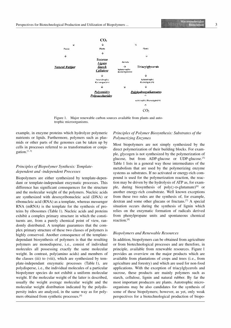
Biopolymers and Renewable Resources
In addition, biopolymers can be obtained from agriculture
or from biotechnological processes and are therefore, in
principle, available from renewable resources. Figure 1
provides an overview on the major products which are
available from plantations of crops and trees (i.e., from
agriculture and forestry) and which are used for non-food
applications. With the exception of triacylglycerols and
sucrose, these products are mainly polymers such as
starch, cellulose, lignin and natural rubber. By far the
most important producers are plants. Autotrophic micro-
organisms may be also candidates for the synthesis of
some of these biopolymers; however, as yet, only weak
perspectives for a biotechnological production of biopo-
Figure 1. Major renewable carbon sources available from plants and auto-trophic microorganisms.
4 A. Steinbuchel
lymers have been outlined, and only for polyhydroxyalk-
anoates employing chemolithoautotrophic[9] and photoau-
totrophic[10] microorganisms. In contrast, heterotrophic
microorganisms are considered as suitable producers of
several biopolymers such as PHAs, polysaccharides and
polyamides. Many biopolymers possess rather complex
chemical structures and compositions and are therefore
not available from chemical synthesis. Furthermore, bio-
polymers, like almost all products of living matter, are
generally biodegradable, whereas this is not a general fea-
ture of synthetic polymers.[11, 12] These few aspects indi-
cate reasons for the growing interest of industry to pro-
duce and use biopolymers for a steadily increasing num-
ber of applications.[13] Only few biopolymers have been
commercialized so far; this is mainly due to the produc-
tion costs, which are often much higher than for synthetic
polymers that have been established in the market during
the last decades. However, this situation may change for
some biopolymers in the future, when biotechnological
production processes have been further optimized and if
our knowledge of the material properties and processing
of biopolymers has increased.
Isolation and Production of Biopolymers
There are different ways to produce biopolymers in order
to make them available for interesting technical applica-
tions: (i) Many biopolymers occur abundantly in nature
and are isolated from plants and algae which grow in nat-
ural environments or are cultivated on plantations. Cellu-
lose and starch are isolated from several agricultural
crops and trees such as Zea maize or Pinus silvestris,
respectively. Natural rubber is isolated from plantanta-
tions of the rubber tree Hevea brasiliensis, and agar and
alginates are isolated from red algae belonging to the
genus Gelidium[14] or from various brown algae also
referred to as seaweeds,[15] respectively. (ii) Few biopoly-
mers are isolated from extremely scarce natural sources.
An example of such an exception is hyaluronic acid
which is extracted from the umbilical cords of new born
children.[16] (iii) In vitro synthesis of biopolymers with
isolated enzymes in cell-free systems offers another pos-
sibility to produce biopolymers. One example is the
application of the heat-stable DNA polymerases in the
polymerase chain reaction (PCR) to produce monodis-
perse defined DNA molecules.[17] Another example is
dextran, which can be produced on a technical scale with
isolated dextran sucrase.[18] (iv) Fermentative production
of biopolymers is used by industry to obtain, for example,
polysaccharides such as xanthan and dextran by employ-
ing the bacteria Xanthomonas campestris[19] or Leuconos-
toc mesenteroides,[7] respectively. Microbial cellulose
obtained by fermentation of Acetobacter xylinum seems
to offer some advantages for medical applications over
cellulose which has been isolated from plants.[5] The
homopolyester poly(3-hydroxybutyrate), poly(3HB), and
the copolyester poly[(3-hydroxybutyrate)-co-(3-hydroxy-
valerate)], poly(3HB-co-3HV), which were available
under the trade name “Biopol” from ZENECA and Mon-
santo, have been also produced on an industrial scale by
employing Ralstonia eutropha.[20] The aforementioned
examples are not based on genetically modified bacteria;
however, genetically engineered bacteria can also be
employed for the production of biopolymers. (v) Finally,
there are many efforts to generate transgenic plants for
the production of certain biopolymers. Examples are
plants producing Biopol[21] or amylose and amylopec-
tin.[22]
II Impacts of Advanced Technologies andMethodologies on Biopolymers
There are many reasons for scientists from academia and
industry to be interested in biopolymers. Firstly, during
the investigation of biological aspects of biopolymers and
their metabolism scientists will still find many “white
spots” since our knowledge about the physiology, bio-
chemistry and molecular genetics of biopolymers is often
scarce. Secondly, many biopolymers have unique proper-
ties, and practically all of them are biodegradable in con-
trast to most synthetic polymers. Thirdly, since chemical
synthesis is neither possible nor economically feasible,
even if the structure of a biopolymer seems to be not too
complex, organisms are often the only source for these
polymers which can therefore be obtained at lower costs
or higher purity. They are either isolated directly from
higher organisms as they occur in nature or on plantations
of agricultural crops or trees or they are biotechnologi-
cally produced by the fermentation of microorganisms.
Therefore, they are directly or indirectly available from
CO2 or renewable resources.
During the last decade basic and applied research have
revealed much knowledge on the biochemical and mole-
cular basis of the enzymatic processes for polymer syn-
thesis in microorganisms. The combination of detailed
physiological studies, utilization of the overwhelming
information provided by the numerous genome sequen-
cing projects, application of recombinant DNA technol-
ogy, engineering of metabolic pathways or enzymes, and
molecular breeding techniques applied to plants, provided
new perspectives to produce technically interesting bio-
polymers by novel or significantly improved biotechnolo-
gical processes or by agriculture. Geneticists and molecu-
lar biologists have developed many powerful methods for
in vitro and in vivo modification of DNA.[23] In addition,
efficient methods are available for the transfer of DNA
and expression of heterologous genes which are applic-
able to many organisms. Metabolic engineering has
become an important and powerful approach in biotech-
nology[24] (Table 2). Successful metabolic engineering
Perspectives for Biotechnological Production and Utilization of Biopolymers ... 5
requires, however, a detailed knowledge of the physiol-
ogy and metabolism of the respective organism to which
these technologies are applied. It also requires knowledge
and experience to utilize the large diversity of microor-
ganisms for example as sources for useful genes.[25, 26]
Scientists knowing only E. coli or being only perfect in
DNA sequencing will have problems, as will scientists
who have never worked on a larger scale than that of an
agar plate or a tiny reaction tube.
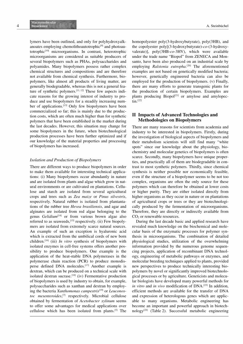
III Intracellular versus ExtracellularProduction of Biopolymers
The biotechnological production of biopolymers may
occur intracellularly or extracellularly. This causes sev-
eral severe consequences regarding the limitations of the
production and downstream processes to obtain the bio-
polymers in a purified state (Figure 2).
PHAs,[27] cyanophycin,[28] glycogen,[29] starch[30] and
polyphosphate[31] are examples of biopolymers which are
accumulated in the cytoplasm of cells. The availibility of
space in the cytoplasm therefore limits the amount of
polymer that can be produced by a cell. This is particu-
larly relevant for fermentative production processes
mostly employing microorganisms. Therefore, the yield
per volume is limited/determined by the cell density and
the fraction of the biopolymer in the biomass. Another
general consequence is that more or less tedious pro-
cesses must follow the production of the biomass contain-
ing the biopolymer to disintegrate the cells or tissues and
to release the biopolymer from the cells. Furthermore,
other cell constituents will be released concommitantly
with the biopolymer and must then be separated.
Poly(c-d-glutamate)[32] and many polysaccharides, such
as alginates,[15] dextran,[7] xanthan,[19] and microbial cellu-
lose[5] are examples of biopolymers which occur outside
the cells, either as a result of extracellular synthesis or of
excretion by the cells. For these biopolymers, the volume
of the cytoplasm is not a limiting factor, and, in principle,
the entire volume of the bioreactor (instead of only that
of the cytoplasm) would be available to deposit the
desired biopolymer. Furthermore, breakage of cells or tis-
sues is not required and separation of the biopolymer
from the other biomass is not very complex. However,
biotechnological processes can merely take advantage of
these features since the presence of these mostly water-
soluble biopolymers in the medium usually causes a high
viscosity in the medium, resulting in rheological pro-
blems during the fermentation process. Unfortunately,
hydrophobic water-insoluble biopolymers of biotechnolo-
gical interest that occur extracellularly are not known.
Therefore, in practice, the amount of biopolymer pro-
duced per volume by extracellular processes is usually
lower than can be obtained by intracellular processes.
Other strategies and the use of cell-free production pro-
cesses, may take advantage of the features of extracellular
processes. One strategy is to apply in vitro synthesis of
biopolymers employing isolated enzymes. Another strat-
egy is to produce the constituents of polymers as mono-
mers by fermentative processes and to polymerize these
components subsequently by solely chemical processes.
Both these strategies have already entered reality and
many different examples of scale have been demonstrated
(i.e., not only at the laboratory scale but also at the tech-
nical scale). Polylactide acid, for example, will be pro-
duced on a large scale by such a combined biotechnologi-
cal and chemical approach.[33] The third and most difficult
and pretentious strategy would be to convert an intracel-
lular process into an extracellular process by metabolic
and genetic engineering of the cells or organisms. This
could be done by, for example, extending the capability
of a cell to excrete a polymer which is synthesized intra-
cellularly. However, to the best knowledge of the author,
this has not been demonstrated, yet.
IV Occurrence of Natural Polyesters
Three different types of naturally occurring organic
polyesters and one inorganic polyester are known (Fig-
ure 3). These are polyoxyesters occurring in the prokar-
yotic bacteria and archaea, polymalic acid occurring in
Figure 2. Examples of biopolymers which are produced intra-cellularly and extracellularly and a demonstration of the“volume” problem.
Table 2. Tools for successful and powerful metabolic engineer-ing.
0 A large box of sophisticated in vitro and in vivo molecularmethods for modification of DNA and for transfer ofrecombinant DNA into other organisms
0 Detailed knowledge on physiology and metabolism0 Diversity of microorganisms0 Genome sequencing projects provide an overwhelming
amount of data

6 A. Steinbuchel
eukaryotic microorganisms, and cutin and suberin occur-
ring in plants. In addition, the inorganic polyester poly-
phosphate occurs in organisms of all the kingdoms. As
far as it is known, all these types of polyesters are synthe-
sized by different mechanisms and are catalyzed by
enzymes exhibiting quite different characteristics.
Bacterial Polyoxyesters
An overwhelming number of different polyhydroxyalk-
anoates (PHAs), comprising approximately 150 different
hydroxyalkanoic acids as constituents, has been isolated
from bacteria during the last 20 years.[34] Accumulation of
PHAs in the bacterial cell usually occurs if a carbon
source is provided in excess, and if at least one other
nutrient, which is essential for growth, has been depleted,
i.e., if growth is imbalanced.[27, 35] These water insoluble
polyesters accumulate in the cytoplasm and are deposited
as cytoplasmic inclusions which are referred to as PHA
granules.[36] They serve as storage compounds for energy
and carbon. PHAs are synthesized by diverting either
central intermediates of the carbon metabolism or deriva-
tives from precursor substrates, which are provided as
carbon source for the growth of the bacteria, to hydroxya-
cyl-CoA thioesters.[37] The thioesters are then polymer-
ized by PHA synthases[38, 39] that are bound to the surface
of PHA granules together with other proteins.[36, 40, 41]
Polymalic Acid
This is the only water-soluble polyester occurring in liv-
ing matter.[42] This anionic homopolyester is synthesized
by Physarum polycephalum[43, 44] and a few lower eukar-
yotic microorganism such as Penicillium cyclopium[45]
and Aureobasidium pullulans.[46, 47] So far, no bacteria
have been identified which synthesize polymalic acid.
The biosynthesis of this polyester is, as yet, only poorly
understood, and polymerization seems to occur by a
mechanism different from that of the bacterial polyoxo-
esters.[48] This biopolyester is mentioned here only for
completness and will not be further considered in this
review.
Cutin and Suberin
Cutin and suberin are complex and crosslinked copoly-
esters which are only synthesized by plants and occur as
structural components of the cuticle covering the aerial
parts of the plants.[49] Cutin consists of a large variety of
different monohydroxy acids, dihydroxy acids, tri- and
pentahydroxy acids, epoxy and oxi acids and also dicar-
boxylic acids; predominant constituents are, for example,
10,16-dihydroxy C16 acid, 18-hydroxy-9,10 epoxy C18
acid and 9,10,18-trihydroxy C18 acid. Suberin consists of
aromatic domains derived from cinnamic acid and alipha-
tic polyester domains with predominantly long-chain
fatty acids, fatty alcohols, x-hydroxy fatty acids and
dicarboxylic acids as its main constituents. The biosyn-
thesis of these two polyesters, in particular the polymeri-
zation reaction, is not yet fully understood.[50] Since this
review focuses on bacterial PHAs, cutin and suberin will
be not further considered in this review.
Polyphosphates
In this inorganic polyester, orthophosphate residues are
linked by phosphoanhydrid bonds. Polyphosphate is
synthesized by prokaryotes, eukaryotic microorganisms
and even higher eukaryotic organisms including mam-
mals, presumably as a storage compound for energy and
phosphate.[31] In the cells, it occurs mostly as insoluble
cytoplasmic inclusions as complexes with divalent
cations. The biosynthesis and degradation of this poly-
ester is well understood.[31]
Figure 3. Structural formula of organic and inorganic poly-esters.
Perspectives for Biotechnological Production and Utilization of Biopolymers ... 7
V Applied Aspects of PolyhydroxyalkanoicAcids
Properties of PHAs
All PHAs share some properties which recommend them
for some applications and make them interesting to indus-
try.[51–54] (i) They are thermoplastic and/or elastomeric
compounds which can be processed with apparatus used
by the plastic manufacturing industry. In addition, they
are (ii) insoluble in water, (iii) exhibit a rather high
degree of polymerization ranging from 105 to almost 107
Da, and (iv) they are enantiomerically pure chemicals
consisting, in general, only of the R-stereoisomer. They
are (v) non-toxic and (vi) biocompatible and (vii) exhibit
piezoelectric properties as revealed (at least) for
poly(3HB) and poly(3HB-co-3HV). (viii) Several PHAs
can be obtained from CO2 or renewable resources.
Finally, (ix) all PHAs are biodegradable; they are hydro-
lyzed by extracellular PHA depolymerases, and the cleav-
age products are subsequently utilized as sources of car-
bon and energy by many bacteria and fungi.[55] PHAs con-
sisting of x-hydroxyalkanoic acids are, in addition, also
hydrolyzed nonspecifically by lipases and esterases.[56, 57]
In addition, PHA accumulating bacteria usually possesses
intracellular PHA depolymerases, which mobilize the
storage compound if no other carbon source is available
to the cells.[58]
Uses and Applications of PHAs
Commercial production of PHAs is so far only possible
by fermentative biotechnological processes. Several
PHAs, such as, in particular, the homopolyester
poly(3HB), the copolyester poly(3HB-co-3HV) and
PHAs consisting of 3-hydroxyoctanoate, 3-hydroxyde-
canoate and a few other medium-chain-length 3-hydro-
xyalkanoates, poly(3HAMCL), have been manufactured
into various materials, and applications in various areas
have been revealed. Whereas the use of poly(3HB) and
poly(3HB-co-3HV) as biodegradable bioplastics was
established some time ago,[51] other applications such as
the manufacturing of latex paints,[59] and specifically
medical applications including retard materials[60] and use
as scaffolding material for tissue engineering,[61] are cur-
rently under development. Isolated and purified PHAs
can be used as such or in combination with other materi-
als such as starch, cellulose fibers, glass fibers or syn-
thetic plastics to obtain compounded materials.[62] Also,
from the purified material new polymers can be obtained
by transesterification in the melt in the presence of syn-
thetic polyesters,[63] by chemical modification of the side
chain[64] or by crosslinking through energized irradiation
and chemical reagents.[65] Another interesting application
of PHAs is the use of these polymers as a source to obtain
enantiomeric pure hydroxyalkanoic acids upon chemical
or enzymatic cleavage for the synthesis of chemicals.[66]
There have also been applications for bacterial cell mass
containing a high fraction of PHAs as binders for other
fibrous materials.[67]
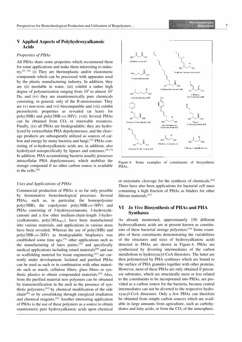
VI In Vivo Biosynthesis of PHAs and PHASynthases
As already mentioned, approximately 150 different
hydroxyalkanoic acids are at present known as constitu-
ents of these bacterial storage polyesters.[34] Some exam-
ples of these constituents demonstrating the variabilities
of the structures and sizes of hydroxyalkanoic acids
detected in PHAs are shown in Figure 4. PHAs are
synthesized by diverting intermediates of the carbon
metabolism to hydroxyacyl-CoA thioesters. The latter are
then polymerized by PHA synthases which are bound to
the surface of PHA granules together with other proteins.
However, most of these PHAs are only obtained if precur-
sor substrates, which are structurally more or less related
to the constituents to be incorporated into PHAs, are pro-
vided as a carbon source for the bacteria, because central
intermediates can not be diverted to the respective hydro-
xyacyl-CoA thioesters. Only a few PHAs can therefore
be obtained from simple carbon sources which are avail-
able in large amounts from agriculture, such as carbohy-
drates and fatty acids, or from the CO2 of the atmosphere.
Figure 4. Some examples of constituents of biosyntheticPHAs.
8 A. Steinbuchel
Since the cloning of the PHA operon of the Gram-
negative bacterium Ralstonia eutropha approximately 12
years ago, more than 50 PHA synthase structural genes,
and also many other genes related to PHA biosynthesis
from various other bacteria, have been cloned, and many
of them have been analyzed at a molecular level.[38, 39]
These genes comprise the structural genes for (i) PHA
synthases, (ii) granule-associated proteins and (iii)
enzymes, which catalyze the formation of hydroxyacyl-
CoA thioesters, as well as probably also genes for (iv)
proteins that have a regulatory function.
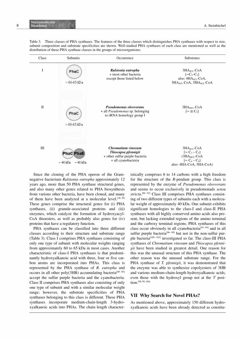
PHA synthases can be classified into three different
classes according to their structure and substrate range
(Table 3). Class I comprises PHA synthases consisting of
only one type of subunit with molecular weights ranging
from approximately 60 to 65 kDa in most cases. Another
characteristic of class-I PHA synthases is that predomi-
nantly hydroxyalkanoic acid with three, four or five car-
bon atoms are incorporated into PHAs. This class is
represented by the PHA synthase of R. eutropha and
occurs in all other poly(3HB) accumulating bacteria[68–85]
accept the sulfur purple bacteria and the cyanobacteria.
Class II comprises PHA synthases also consisting of only
one type of subunit and with a similar molecular weight
range; however, the substrate specificities of PHA
synthases belonging to this class is different. These PHA
synthases incorporate medium-chain-length 3-hydro-
xyalkanoic acids into PHAs. The chain length character-
istically comprises 6 to 14 carbons with a high freedom
for the structure of the R-pendant group. This class is
represented by the enzyme of Pseudomonas oleovorans
and seems to occur exclusively in pseudomonads sensu
strictu.[86–92] Class III comprises PHA synthases consist-
ing of two different types of subunits each with a molecu-
lar weight of approximately 40 kDa. One subunit exhibits
significant homologies to the class-I and class-II PHA
synthases with all highly conserved amino acids also pre-
sent, but lacking extended regions of the amino terminal
and the carboxy terminal regions. PHA synthases of this
class occur obviously in all cyanobacteria]93–95] and in all
sulfur purple bacteria[96–99] but not in the non-sulfur pur-
ple bacteria[100–103] investigated so far. The class-III PHA
synthases of Chromatium vinosum and Thiocapsa pfenni-
gii have been studied in greatest detail. One reason for
this was the unusual structure of this PHA synthase. The
other reason was the unusual substrate range. For the
PHA synthase of T. pfennigii, it was demonstrated that
the enzyme was able to synthesize copolyesters of 3HB
and various medium-chain-length hydroxyalkanoic acids,
even those with the hydroxyl group not at the 39 posi-
tion.[98, 99, 104]
VII Why Search for Novel PHAs?
As mentioned above, approximately 150 different hydro-
xyalkanoic acids have been already detected as constitu-
Table 3. Three classes of PHA synthases. The features of the three classes which distinguishes PHA synthases with respect to size,subunit composition and substrate specificities are shown. Well-studied PHA synthases of each class are mentioned as well as thedistribution of these PHA synthase classes in the groups of microorganisms.
Class Subunits Occurrence Substrates
I Ralstonia eutropha+ most other bacteria
except those listed below
3HASCL-CoA[lC3–C5]
also: 4HASCL-CoA,5HASCL-CoA, 3MASCL-CoA
II Pseudomonas oleovorans+ all Pseudomonas sp. belonging
to rRNA homology group I
3HAMCL-CoA[l F C5]
III Chromatium vinosumThiocapsa pfennigii
+ other sulfur purple bacteria+ all cyanobacteria
3HASCL-CoA[l C3 – C5]
(3HAMCL-CoA[l C4 – C8]
also: 4HA-CoA, 5HA-CoA)
Perspectives for Biotechnological Production and Utilization of Biopolymers ... 9
ents of bacterial PHAs. However, there is still ongoing
interest in the search for novel PHAs. This means the
search for PHAs with novel constituents (new HA), a
new combination of already known constituents (for
example a copolyester [-X-Y-X-Y-] instead of a blend of
two homopolyesters [–X-X-X-X-] plus [–Y-Y-Y-Y-]) or a
new order of constituents (for example a block copoly-
ester [–X-X-X-X-Y-Y-Y-Y-] instead of a random copo-
lyester [-X-Y-X-Y-X-Y-X-Y]). In addition, novel types of
biopolymers can be obtained applying a polymerizing
enzyme system, for example, to unusual substrates, as
was shown with the detection of polythioesters when
mercaptoalkanoic acids were used as a precursor carbon
source for the PHA-accumulating R. eutropha.[2, 105] Such
studies are not only to the satisfication of microbiologists
and biochemists, which try to reveal the true substrate
range of these fascinatingly unspecific PHA synthases,
but may have also far-reaching impacts on the provision
of novel biopolymers applicable to the manufacturing of
polymeric materials exhibiting novel and improved phy-
sical and material properties.
VIII Strategies to Obtain Novel PHABiosynthesis Pathways and ImprovedProduction Strains
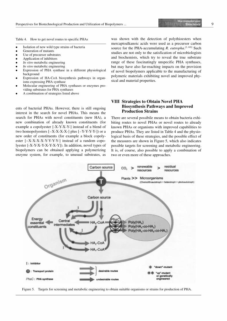
There are several possibile means to obtain bacteria exhi-
biting routes to novel PHAs or novel routes to already
known PHAs or organisms with improved capabilities to
produce PHAs. They are listed in Table 4 and the physio-
logical basis of these strategies, and the possible effect of
the measures are shown in Figure 5, which also indicates
possible targets for screening and metabolic engineering.
It is, of course, also possible to apply a combination of
two or even more of these approaches.
Table 4. How to get novel routes to specific PHAs
0 Isolation of new wild type strains of bacteria0 Generation of mutants0 Use of precursor substrates0 Application of inhibitors0 In vitro metabolic engineering0 In vitro metabolic engineering0 Expression of PHA synthase in a different physiological
background0 Expression of HA-CoA biosynthesis pathways in organ-
isms expressing PHA synthase0 Molecular engineering of PHA synthases or enzymes pro-
viding substrates for PHA synthases0 A combination of strategies listed above
Figure 5. Targets for screening and metabolic engineering to obtain suitable organisms or strains for production of PHA.
10 A. Steinbuchel
Isolation of New Wild-Type Strains of Bacteria
Many Bacteria accumulate poly(3HB), and most of them,
like R. eutropha, synthesize this polyester from acetyl-
CoA in a three-step pathway via acetoacetyl-CoA and
(R)-3-hydroxybutyryl-CoA catalyzed by the enzymes b-
ketothiolase, a pyridine nucleotide-dependant acetoace-
tyl-CoA reductase and the PHA synthase.[106] Microbiolo-
gists isolated and identified several bacteria after
extended screening processes which were able to synthe-
size PHAs consisting of constituents different from 3HB
from simple, structurally non-related carbon sources such
as glucose. The two most important examples comprise
the copolyester poly(3HB-co-3HV) and PHAs consisting
of medium-chain-length 3-hydroxyalkanoic acids
(3HAMCL).
Almost all pseudomonads sensu strictu synthesize
poly(3HAMCL), not only when they are cultivated on
alkanes such as octane[107] or organic acids such as octa-
noic acid,[108] but also from glucose and many other car-
bon sources.[109, 110] P. oleovorans has been well studied
with respect to the formation of PHA consisting mainly
of (R)-3-hydroxyoctanoate from octane or octanoic acid.
Octane is oxidized to octanoic acid by oxygenases and
dehydrogenases, and the latter is then activated to octa-
noyl-CoA and further oxidized via b-oxidation. Inter-
mediates of the b-oxidation cycle are then polymerized to
the polyester. P. oleovorans is one of the few pseudomo-
nads sensu strictu unable to synthesize poly(3HAMCL)
from unrelated substrates although the genes encoding
this pathway are present (see below).[111] Most other pseu-
domonads, such as P. putida and P. aeruginosa, posses
this pathway and accumulate during cultivation on gluco-
nate as carbon source a copolyester consisting of (R)-3-
hydroxydecanoate as main constituent and (R)-3-hydro-
xydodecanoate and (R)-3-hydroxyoctanoate as minor
constituents.[109, 110] Cultivation experiments employing
labelled carbon sources and inhibitors provided evidence
that the fatty acid de novo synthesis pathway is the major
pathway for the provision of the 3-hydroxyacyl moieties
in P. putida.[112] An acyltransferase was recently isolated
and the gene cloned from P. putida, which transfers the
hydroxyacylmoiety from (R)-3-hydroxydecanoyl-acyl
carrier protein to coenzyme A thus forming (R)-3-hydro-
xydecanoyl-CoA, which is a substrate of the two PHA
synthases in this bacterium.[92, 115] This key enzyme links
fatty acid de novo synthesis and poly(3HAMCL) in P.
putida and is also present in P. aeruginosa[114] and other
pseudomonads.[115] A highly homologous gene was also
identified in the genome of P. oleovorans,[111] however, in
this bacterium, the gene is either silent or inactive, since
this bacterium cannot accumulate poly(3HAMCL) from,
for example, gluconate.
Poly(3HB-co-3HV) with very high contents of 3HV are
synthesized by various species belonging to the Gram-
positive genera Nocardia or Rhodococcus.[116] Pyruvate,
which is formed during the degradation of glucose via the
2-keto-3-deoxy-6-phosphogluconate pathway in these
bacteria, is probably carboxylated to oxaloacetate and
subsequently converted to succinyl-CoA by the reverse
citric acid cycle which is then converted via methylmalo-
nyl-CoA to propionyl-CoA, as revealed by analysis of N.
corallina and R. ruber[117–119] Propionyl-CoA is then con-
densed with acetyl-CoA yielding 3-ketovaleryl-CoA
which is reduced to (R)-3-hydroxyvaleryl-CoA and
together with (R)-3-hydroxybutyryl-CoA polymerized to
poly(3HB-co-3HV). This is a very good example of the
endogenous generation of propionyl-CoA. Otherwise,
precursor substrates such as propionic acid, valeric acid
or other fatty acids with an odd number of carbon
atoms,[120] a,x-alkanediols with an odd number of carbon
atoms,[121] levulinic acid[122, 123] or certain amino acids
such as valine or isoleucine[124–127] must be used as a car-
bon source during the cultivation of the bacteria to obtain
poly(3HB-co-3HV). Actually, propionic acid is used in
addition to glucose in the Biopol process for the produc-
tion of poly(3HB-co-3HV) with R. eutropha.[128–129]
Other interesting, newly isolated bacteria are Pseudo-
monas sp. 61-3 and Aeromonas caviae which are able to
accumulate copolyesters of 3HB and 3HAMCL. Whereas
Pseudomonas sp. 61-3 accumulated a copolyester of 3HB
and 3HAMCL from sugars as well as from fatty acids of
medium chain length,[130] A. caviae accumulated a copo-
lyester of 3HB and 3-hydroxyhexanoate during cultiva-
tion on fatty acids of medium chain length.[131]
Generation of Mutants
Furthermore, from these and many other bacteria,
mutants were isolated which exhibited advanced features
for the accumulation of PHAs. The literature provides
many examples of such mutants. It would break the scope
and length of this review to mention all these mutants in
detail and it is therefore recommended to refer to pre-
vious reviews.[27, 37, 39, 132–134]
Use of Precursor Substrates
Rather frequently and abundantly, precursor substrates
were used as carbon sources to cultivate various bacteria
under conditions which permitted the synthesis and accu-
mulation of PHAs. Most of the hydroxyalkanoic acids
known to be constituents of PHAs were identified upon
such experiments. Despite the detection of so many dif-
ferent PHAs, the use of precursor substrates is still
another a tool to obtain novel, hitherto unknown PHAs.
One recent example are polythioesters, consisting, for
example, of 3-mercaptopropionic acid or 3-mercaptobu-
tyric acid.[2, 105] This entirely new type of biopolymer is
Perspectives for Biotechnological Production and Utilization of Biopolymers ... 11
obtained in R. eutropha due to the broad unspecificity of
the PHA synthase.
The description of hitherto unknown, sulfur-containing
polythioesters was the first report on a natural polymer
containing sulfur in its backbone.[2, 105] R. eutropha
synthesized a copolymer of 3-hydroxybutyrate and 3-
mercaptopropionate, poly(3HB-co-3MP), when 3-mer-
captopropionic acid or 3,39-thiodipropionic acid was pro-
vided as a carbon source, in addition to fructose or gluco-
nic acid, under nitrogen-limited growth conditions. The
peculiarity of this polymer was the occurrence of thioe-
ster linkages derived from the thiol groups of 3MP and
the carboxyl groups of 3MP or 3HB, respectively, which
occurred in addition to the common oxoester bonds of
PHAs. Depending on the cultivation conditions and the
feeding regime, poly(3HB-co-3MP) contributed up to
19% of the cellular dry weight, with a molar fraction of
3MP of up to 43%. The chemical structure of poly(3HB-
co-3MP) was confirmed by gas chromatography/mass
spectrometry, infrared spectroscopy, 1H- and 13C nuclear
magnetic resonance spectroscopy, and elemental sulfur
analysis. Therefore, poly(3HB-co-3MP) can be desig-
nated as the first representative of an eighth class of bio-
polymers, namely polythioesters. Another hitherto
unknown copolymer that contains sulfur in its backbone,
is a polythioester covalently linking 3-hydroxybutyrate
and 3-mercaptobutyrate, poly(3HB-co-3MB). It was also
synthesized by R. eutropha, when 3-mercaptobutyric acid
was fed as a carbon source in addition to gluconate. The
total polymer yield contributed to up to 31% of the cellu-
lar dry weight. Elemental sulfur analysis of poly(3HB-co-
3MB) revealed a total sulfur content of 11.65% (w.-%),
thus the molar fraction of 3MB was calculated as
33.24 mol-%. The molecular structure of this novel poly-
mer was also confirmed by gas chromatography/mass
spectrometry, infrared spectroscopy, and 1H- and 13C-
nuclear magnetic resonance spectroscopy.
Application of Inhibitors
The flux of intermediates may be influenced and directed
by the use of inhibitors. This approach will be helpful for
basic studies since it may reveal information about the
flux of metabolites and by which pathways and enzymes
substrates for the PHA synthase are provided. However,
the data have to be carefully analyzed because inhibitors
do interfere in most cases, with more than one particular
enzyme if they are not very specific. Therefore, artifacts
may occur, leading to a false interpretation of the data.
Considering the biotechnological production of PHAs,
the inhibitors will probably not be applicable because of
the extra costs and because the inhibitor will have to be
completely removed from the polyester during purifica-
tion if the polyester is to be used for food applications, or
for medical or pharmaceutical applications. In addition,
whereas inhibitors may be more easily applied during
submers cultivation of microorganisms from a technical
point of view, they are difficult to apply to the production
of PHAs in transgenic plants. Therefore, for large scale
production of PHAs, it might be better to use strains in
which the effect of the inhibitor is simulated by a muta-
tion or by other approaches in order to direct the flow of
metabolites in the desired direction. The application of
various inhibitors was quite useful during the analysis of
poly(3HAMCL) and the establishment of poly(3HAMCL)
biosynthesis in a recombinant strain of E. coli (see section
IX).
In Vivo Metabolic Engineering
The availibility of the key enzyme PHA synthase and of
enzymes involved in the biosynthesis of hydroxyacyl
coenzyme A thioesters and detailed knowledge on the
biochemistry of these enzymes as well as of additional
structural and regulatory proteins will allow engineering
of the metabolism to obtain new PHA biosynthesis path-
ways. In addition, it will be essential to establish func-
tional active PHA biosynthesis pathways in organisms
that are, themselves, not able to accumulate PHAs but are
considered as more suitable for commercial production of
PHAs either by fermentative processes or by cultivation
of transgenic crops. To achieve this, knowledge about
putative regulatory events effecting the activity of the
enzymes or the expression of these enzymes or other pro-
teins might also be important. In vivo metabolic engineer-
ing is of particular interest if pathways are engineered
which mediate between central intermediates of the meta-
bolism on one side and the hydroxyacyl coenzyme A
thioesters as substrates of PHA synthases on the other
side. This is because such studies could yield recombi-
nant bacteria or transgenic plants which are able to pro-
duce a wider range of PHAs from simple and cheap car-
bon sources or CO2, respectively. The current possibilities
and achievements regarding production of the various
PHAs by fermentation of wild-type and recombinant bac-
teria were recently extensively reviewed.[134] Of particular
interest is the engineering of pathways for the synthesis
of coenzyme A thioesters of hydroxyalkanoic acids which
differ from 3HB-CoA in order to obtain organisms suita-
ble for the production of a wider range of different PHAs
from cheap and abundantly available carbon sources.
Some examples for the in vivo engineering of metabolic
links between the fatty acid metabolism and poly(3-
HAMCL) will follow (see section IX).
In Vitro Metabolic Engineering
In vitro synthesis of biopolymers means that the polymer-
izing enzyme acts in a cell-free system and catalyzes the
formation of the polymer from its substrate(s). Various
12 A. Steinbuchel
assays are available for measuring PHA synthase activ-
ity.[135] For other enzymes, like an alcohol dehydrogenase,
it is quite normal to measure the NADH-dependant reduc-
tion of acetaldehyde by, for example, a photometric assay.
In contrast, for polymer-synthesizing enzymes, the assay
of its activity is often considered to be something special,
most probably due to the fact that the enzymes are often
more complex and require more cofactors and, in some
cases, also addition of a template and a primer. If polymer
biosynthesis is done with a highly active and more or less
purified enzyme preparation, the biopolymer can be
obtained in such quantities, that it can be isolated and
subsequently characterized with respect to its composi-
tion. Depending on the amount of the polymer, which can
be prepared, even some physical and/or material proper-
ties may be analyzed. Such studies will not only reveal
important data and knowledge on mechanistic aspects
and other characteristics of the enzyme, but may also
allow the synthesis of novel biopolymers which would
not be available from the enzyme acting in vivo. The rea-
sons for this are the extension to substrates and primers,
which can not be synthesized in a cell but are available
from chemical synthesis, and the possibility to switch
from one substrate to another within a short period of
time, thus allowing the synthesis of, for example, block
copolymers.
If the polymer synthesizing enzyme is combined with
other enzymes, short pathways can be engineered. It may
be a pathway as it also exists in cells in vivo or it may be
a pathway which is completely artifical and is not occur-
ring in cells and has therefore to be referred to as a none-
natural pathway. In both cases enzymes from different
organisms may be combined. Later, some examples for
the in vitro metabolic engineering will be provided (see
section X).
Molecular Expression of PHA Synthases or Enzymes
Providing Substrates for PHA Synthases
As already mentioned, approximately 50 PHA synthase
structural genes and many genes encoding enzymes cata-
lyzing the formation of hydroxyacyl-coenzyme A thioe-
sters have been cloned from different microorganisms
and characterized at a molecular level.[38, 39] Many of these
genes have been heterologously expressed at least once in
a foreign host, probably most often in one of the PHA-
negative mutants of R. eutropha (for example mutant
PHB4– [134]) and in E. coli. The most detailed and compre-
hensive studies have been done with the PHA synthase
operon of R. eutropha, which consists of the structural
genes of b-ketothiolase (phaA), NADPH-dependent
acetoacetyl-CoA reductase (phaB) and PHA synthase
(phaC) occurring in the order phaCAB. This operon, or at
least phaC, was successfully expressed in many different
bacteria, in Saccharomyces cerevisiae, in animal cells and
in plant cells and established, in most cases, functional
PHA biosynthesis. Expression in E. coli and plants will
most probably have the greatest impact with respect to
the production of PHAs in the future. Therefore, two
separate sections of this review article are devoted to this
aspect (see sections XI and XII).
IX Metabolic Engineering of Poly(3HAMCL)Biosynthesis Pathways
Metabolic engineering of PHA biosynthesis pathways
requires the establishment of a functional pathway that
allows the conversion of the carbon source to an inter-
mediate which is further routed to the desired PHA
synthase substrate and finally polymerized. This may
mean the establishment of an entire pathway hitherto not
present in the particular organism or only modifications of
a few or even a single step in the sequence of a pathway.
It should be noted that the genetic transfer of a PHA
synthase gene is only a first step (Table 5). Further meas-
ures must ensure that the PHA synthase is expressed in a
functionally active form. Finally, and this is the most
important aspect and may be the most difficult to achieve,
the substrate for the PHA synthase must be available in
the cytoplasm or in the compartment of the cell in which
the PHA synthase is expressed. The best PHA synthase
and a formidable activity of this enzyme will be useless if
the enzyme has no substrate. The substrate may be pro-
vided from a precursor substrate, which is more or less
directly converted into the desired hydroxyacyl coenzyme
A thioester, or, better, from a central intermediate. This
requires that a pathway for this conversion exists in the
cell or, if eukaryotic microorganisms are considered, in
the right compartment of the cell, and, if higher eukaryo-
tic organisms are considered, also in the right tissue of
the respective organism. Furthermore, the withdrawal of
intermediates for PHA biosynthesis must not impair the
normal functions of the cell; otherwise the cell, and most
likely the entire organism, may be negatively affected,
and growth may be retarded. The latter was often a ser-
ious problem during the establishment of PHA biosyn-
thesis in transgenic plants (see section XII).
Table 5. Establishment of PHA biosynthesis in non-PHA accu-mulating organisms
1. Genetic transfer of the PHA synthase gene2. Expression of active PHA synthase3. Appropriate provision of the substrate for the PHA synthase
(i) Provision from “precursor” carbon sourves(ii) Provision from central intermediates
0 The desired hydroxyalcyl-coenzyme A thioester mustbe synthesized in the cell (in the right tissue and com-partment) and accumulated to a certain extent
0 The withdrawal of intermediates for the synthesis of thishydroxyacyl-coenzyme A thioester must not impair thenormal function of the cell
Perspectives for Biotechnological Production and Utilization of Biopolymers ... 13
The biosynthesis of poly(3HAMCL) in pseudomonads has
been investigated in depth and the application of inhibitors
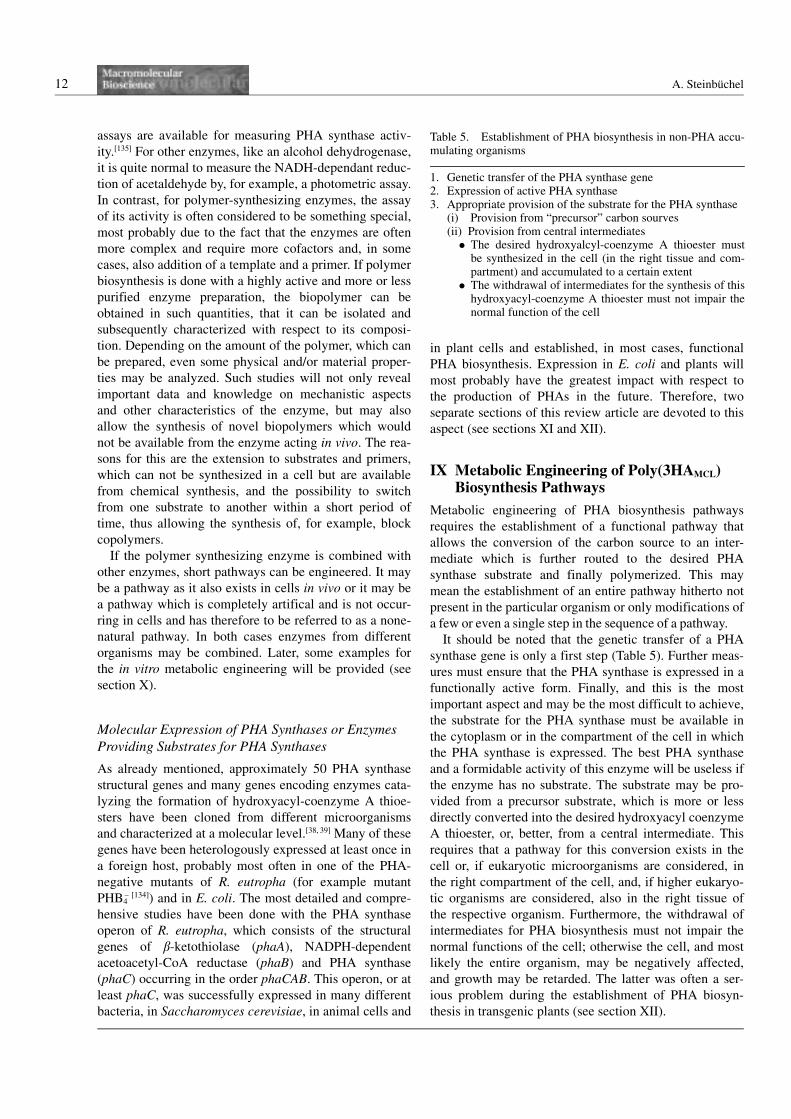
was found to be quite useful. Figure 6 shows the reactions
of the fatty acid de novo synthesis pathway and of the fatty
acid b-oxidation pathway for the conversion of glucose or
fatty acids, respectively; in addition, the enzymes catalyz-
ing the various reactions are indicated with reference to
the respective genes encoding these enzymes. This figure
also shows six chemicals which more or less specifically
inhibit one or the other reaction. Whereas acrylic acid[137]
Figure 6. Inhibitors of fatty acid metabolism.
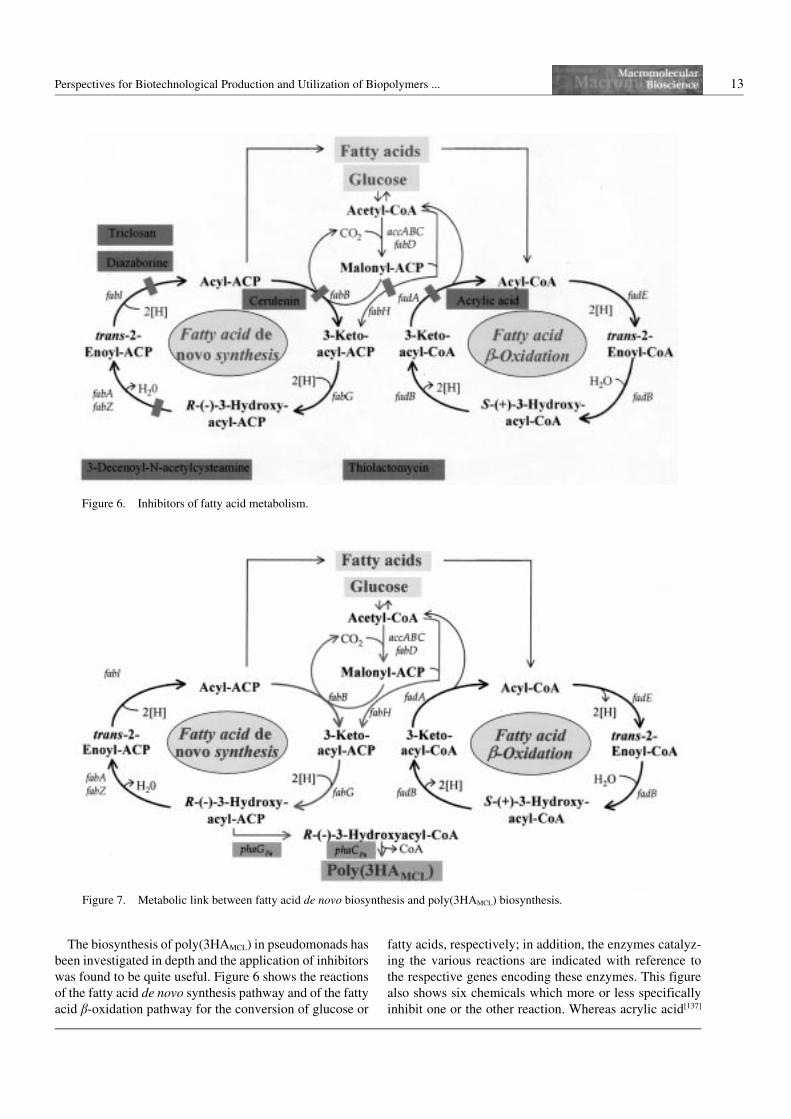
Figure 7. Metabolic link between fatty acid de novo biosynthesis and poly(3HAMCL) biosynthesis.
14 A. Steinbuchel
inhibits the b-ketothiolase of the b-oxidation pathway,
thiolactomycin,[138] 3-decenoyl-N-acetylcysteamine,[139]
triclosan,[140] diazaborine[141] and cerulenin[142] inhibit
enzymes of the fatty acid de novo synthesis pathway.
During study of poly(3HAMCL) biosynthesis in P. putida
from glucose, the application of the inhibitor cerulenin
revealed that fatty acid de novo synthesis plays a major
role providing approximately 90% of the PHA constitu-
ents. Provision of substrates by chain elongation through
b-ketothiolases or from cell lipids through the b-oxidation
pathway, the latter as revealed by application of the inhibi-
tor acrylic acid, are significant, but contribute to only a
minor fraction of the constituents.[112] Mutants were iso-
lated which grew normally on any carbon source tested
and which still synthesized poly(3HAMCL) from fatty acids
but not from gluconate or glucose.[113] The phenotype of
these mutants indicated a defect in the enzyme converting
intermediates of the fatty acid de novo synthesis pathway
into a substrate of the PHA synthases in P. putida. The
enzyme was purified, and biochemical studies revealed
that the enzyme catalyses the transfer of (R)-3-hydroxyde-
canoyl moieties from the acyl carrier protein to coenzyme
A (Figure 7); this reaction was dependant on magnesium
ions. It was referred to as a 3-hydroxyacyl-acyl carrier pro-
tein :coenzyme A transferase, and the respective gene,
which complemented the mutant, was referred to as
phaG.[113] PhaG has been also identified and cloned from
P. aeruginosa,[114] P. oleovorans[111] and Pseudomonas sp.
61–3[143] and seems to be present also in other pseudomo-
nads sensu strictu such as Pseudomonas sp. DSM1645, P.
mendocina and P. citronellolis as revealed by hybridiza-
tion experiments.[111] Therefore, a highly conserved transa-
cylase provides the metabolic link between fatty acid de
novo synthesis and PHA synthase in the Pseudomonas spe-
cies sensu strictu.
In subsequent experiments, phaG from P. putida was
transferred to and expressed in P. oleovorans which is
able to synthesize poly(3HAMCL) from alkanes or fatty
acids, but not from gluconate or fructose, and exhibits,
therefore, a similar phenotype as the phaG mutants of P.
putida (see above). Furthermore, phaG from P. aerugi-
nosa was, together with the PHA synthase gene from P.
aeruginosa, transferred to the non-PHA accumulating
bacterium P. fragi, and both were heterologously
expressed in this host (Figure 7).[144] When the recombi-
nant strains were cultivated with gluconate as a carbon
source under conditions, which permitted the accumula-
tion of PHAs, the cells accumulated poly(3HAMCL) contri-
buting approximately 50% of the cell dry matter in the
case of P. oleovorans and 10% in the case of P. fragi,
respectively, whereas in the parent strain or in a recombi-
nant strain harboring only the vector, PHAs were not
detected. These two studies demonstrated that the cloned
transacylase is functionally active and can be used to
establish poly(3HAMCL) synthesis in other bacteria.
Regarding the metabolism of poly(3HAMCL) in pseudo-
monads, another open question was the metabolic link
between fatty acid b-oxidation and PHA synthase.
Although a 3-hydroxyacyl coenzyme A intermediate
occurs, it represents the wrong stereoisomer. In principle,
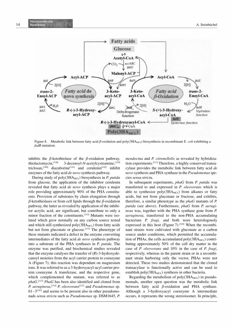
Figure 8. Metabolic link between fatty acid b-oxidation and poly(3HAMCL) biosynthesis in recombinant E. coli exhibiting afadB mutation.
Perspectives for Biotechnological Production and Utilization of Biopolymers ... 15
three enzymes may putatively provide this metabolic
link: (i) a 3-ketoacyl-CoA reductase reducing 3-ketoacyl-
CoA to (R)-3-hydroxyacyl-CoA, (ii) an epimerase con-
verting the (S)-isomer of 3-hydroxyacyl-CoA into the
(R)-isomer and (iii) a hydratase converting trans-2-enoyl-
CoA into (R)-3-hydroxyacyl-CoA. During our attempts to
establish poly(3HAMCL) biosynthesis in E. coli, the
recombinant strains expressed PHA synthase but accumu-
lated only trace amounts of PHAs if cultivated on
decanoic acid or various other fatty acids (Table 6). Only
if the fadB mutant LS1298 of E. coli was used, were
PHAs accumulated at a significant level, contributing
20–25% of the cell dry matter[145] (Table 6). In mutant
LS1298, the dehydrogenase function is defective,
whereas the hydratase and also the epimerase functions
of FadB remain active[146] (Figure 8). Obviously, (S)-3-
hydroxyacyl-CoA accumulates in the cytoplasm of the
mutant cells to a level which is sufficiently high to be
effectively converted into the (R)-stereoisomer and subse-
quently also into PHA. Instead of phaC1, phaC2 of P.
aeruginosa was also used, and very similar results were
obtained.[147] This is a good example of the efficient engi-
neering of the metabolic fluxes of intermediates of a
degradative pathway towards a new product which is not
normally synthsized by the cells. This system allowed the
synthesis of quite a large range of different PHAs,
depending on the fatty acid which was used as carbon
source. The length of the incorporated 3-hydroxyalkanoic
acids reflected the length of the carbon chain of the fatty
acid provided as carbon source.[145, 147]
A different system for the production of poly(3HAMCL)
by E. coli, allowing a larger flexibility, was established
employing the inhibitor acrylic acid[148] (Figure 9). When
recombinant strains of E. coli expressing either phaC1 or
phaC2 from P. aeruginosa were cultivated on fatty acids
such as decanoic acid, in the presence of low concentra-
tions of this inhibitor, PHAs were accumulated by any
strain of E. coli investigated (Table 6). Several strains
even accumulated significantly higher amounts of PHA
than the mutant LS1298 in the absence of acrylic acid.
Most PHA was accumulated by a fadR regulatory mutant,
in which poly(3HAMCL) contributed approximately 45%
Figure 9. Metabolic link between fatty acid b-oxidation and poly(3HAMCL) biosynthesis in recombinant E. coli in whichb-oxidation is inhibited by acrylic acid.
Table 6. Influence of acrylic acid on accumulation of PHAMCL
in recombinant strains of E. coli harboring plasmid pBHR71from decanoic acid as carbon source.
Strain ofE. coli
Property PHAMCL contentof the cells [wt.-% of CDW]
–Acrylic acid +Acrylic acid
LS1298 fadB 21.1 19.5K12 – 2.8 25.4
JM109 – 1.1 29.9XL1-Blue – 0.9 27.9RS3097 fadR41 a0.2 45.7
Cultivations were done in LB medium containing 4 g N l–1
decanoic acid, 1 mM IPTG and l0.2 g N l–1 acrylic acid.Abbreviations: CDW, cell dry weight; PHAMCL, polyhydroxyalk-anoates consisting of medium-chain-length 3-hydroxyalkano-ates. The content of this table was taken from ref.[148]
16 A. Steinbuchel
of the cellular dry matter (Table 6). Obviously, the addi-
tion of acrylic acid and the inhibition of the b-ketothio-
lase have a similar effect as a defective FadB dehydro-
genase function with respect to the accumulation of (S)-
3-hydroxyacyl-CoA and the routing of intermediates of
the b-oxidation cycle towards poly(3HAMCL) biosynthesis
and accumulation (Figure 9).
Further studies by other laboratories established the
relevance of the reductase and the hydratase for PHA bio-
synthesis. In Aeromonas caviae, a (R)-specific enoyl-
CoA hydratase is responsible for the conversion of trans-
2-enoyl-CoA to (R)-3-hydroxyacyl-CoA during the culti-
vation of this bacterium on, for example, hexanoate.[78]
Two (R)-specific enoyl-CoA hydratase genes were also
identified in P. aeruginosa.[149] Studies in recombinant
strains of E. coli provided evidence that FabG of fatty
acid de novo synthesis, which is a 3-ketoacyl-ACP reduc-
tase, is non-specific and also exhibits activity with the
corresponding CoA-thioesters.[150] On the other hand, it
was shown that recombinant strains of E. coli could also
use their own b-ketothiolase (FadA) in combination with
the acetoacetyl-CoA reductase (PhaB) of R. eutropha for
the conversion of fatty acids into poly(3HAMCL).[151] Fig-
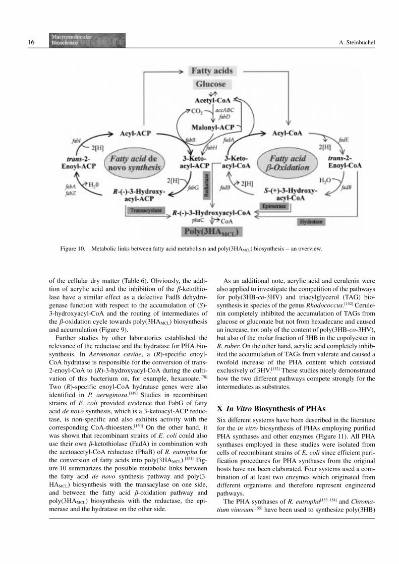
ure 10 summarizes the possible metabolic links between
the fatty acid de novo synthesis pathway and poly(3-
HAMCL) biosynthesis with the transacylase on one side,
and between the fatty acid b-oxidation pathway and
poly(3HAMCL) biosynthesis with the reductase, the epi-
merase and the hydratase on the other side.
As an additional note, acrylic acid and cerulenin were
also applied to investigate the competition of the pathways
for poly(3HB-co-3HV) and triacylglycerol (TAG) bio-
synthesis in species of the genus Rhodococcus.[152] Cerule-
nin completely inhibited the accumulation of TAGs from
glucose or gluconate but not from hexadecane and caused
an increase, not only of the content of poly(3HB-co-3HV),
but also of the molar fraction of 3HB in the copolyester in
R. ruber. On the other hand, acrylic acid completely inhib-
ited the accumulation of TAGs from valerate and caused a
twofold increase of the PHA content which consisted
exclusively of 3HV.[152] These studies nicely demonstrated
how the two different pathways compete strongly for the
intermediates as substrates.
X In Vitro Biosynthesis of PHAs
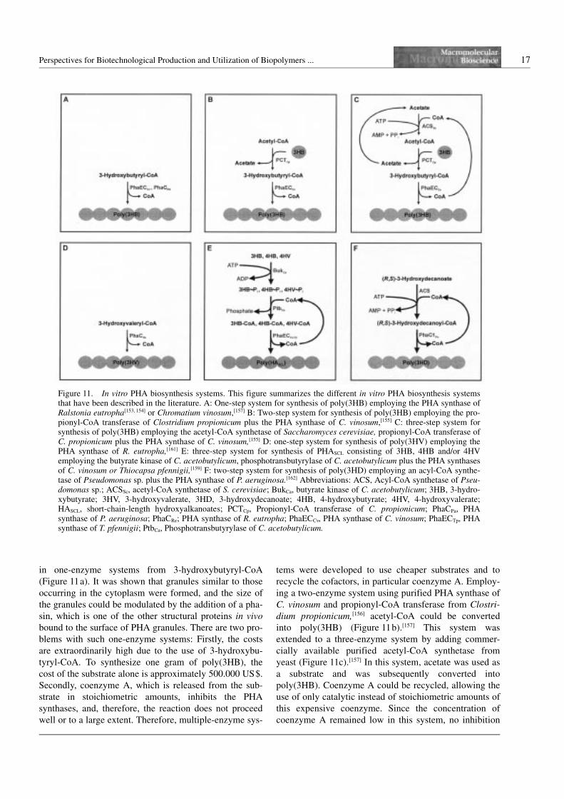
Six different systems have been described in the literature
for the in vitro biosynthesis of PHAs employing purified
PHA synthases and other enzymes (Figure 11). All PHA
synthases employed in these studies were isolated from
cells of recombinant strains of E. coli since efficient puri-
fication procedures for PHA synthases from the original
hosts have not been elaborated. Four systems used a com-
bination of at least two enzymes which originated from
different organisms and therefore represent engineered
pathways.
The PHA synthases of R. eutropha[153, 154] and Chroma-
tium vinosum[155] have been used to synthesize poly(3HB)
Figure 10. Metabolic links between fatty acid metabolism and poly(3HAMCL) biosynthesis – an overview.
Perspectives for Biotechnological Production and Utilization of Biopolymers ... 17
in one-enzyme systems from 3-hydroxybutyryl-CoA
(Figure 11a). It was shown that granules similar to those
occurring in the cytoplasm were formed, and the size of
the granules could be modulated by the addition of a pha-
sin, which is one of the other structural proteins in vivo
bound to the surface of PHA granules. There are two pro-
blems with such one-enzyme systems: Firstly, the costs
are extraordinarily high due to the use of 3-hydroxybu-
tyryl-CoA. To synthesize one gram of poly(3HB), the
cost of the substrate alone is approximately 500.000 US$.
Secondly, coenzyme A, which is released from the sub-
strate in stoichiometric amounts, inhibits the PHA
synthases, and, therefore, the reaction does not proceed
well or to a large extent. Therefore, multiple-enzyme sys-
tems were developed to use cheaper substrates and to
recycle the cofactors, in particular coenzyme A. Employ-
ing a two-enzyme system using purified PHA synthase of
C. vinosum and propionyl-CoA transferase from Clostri-
dium propionicum,[156] acetyl-CoA could be converted
into poly(3HB) (Figure 11b).[157] This system was
extended to a three-enzyme system by adding commer-
cially available purified acetyl-CoA synthetase from
yeast (Figure 11c).[157] In this system, acetate was used as
a substrate and was subsequently converted into
poly(3HB). Coenzyme A could be recycled, allowing the
use of only catalytic instead of stoichiometric amounts of
this expensive coenzyme. Since the concentration of
coenzyme A remained low in this system, no inhibition
Figure 11. In vitro PHA biosynthesis systems. This figure summarizes the different in vitro PHA biosynthesis systemsthat have been described in the literature. A: One-step system for synthesis of poly(3HB) employing the PHA synthase ofRalstonia eutropha[153, 154] or Chromatium vinosum,[157] B: Two-step system for synthesis of poly(3HB) employing the pro-pionyl-CoA transferase of Clostridium propionicum plus the PHA synthase of C. vinosum,[155] C: three-step system forsynthesis of poly(3HB) employing the acetyl-CoA synthetase of Saccharomyces cerevisiae, propionyl-CoA transferase ofC. propionicum plus the PHA synthase of C. vinosum,[155] D: one-step system for synthesis of poly(3HV) employing thePHA synthase of R. eutropha,[161] E: three-step system for synthesis of PHASCL consisting of 3HB, 4HB and/or 4HVemploying the butyrate kinase of C. acetobutylicum, phosphotransbutyrylase of C. acetobutylicum plus the PHA synthasesof C. vinosum or Thiocapsa pfennigii,[159] F: two-step system for synthesis of poly(3HD) employing an acyl-CoA synthe-tase of Pseudomonas sp. plus the PHA synthase of P. aeruginosa.[162] Abbreviations: ACS, Acyl-CoA synthetase of Pseu-domonas sp.; ACSSc, acetyl-CoA synthetase of S. cerevisiae; BukCa, butyrate kinase of C. acetobutylicum; 3HB, 3-hydro-xybutyrate; 3HV, 3-hydroxyvalerate, 3HD, 3-hydroxydecanoate; 4HB, 4-hydroxybutyrate; 4HV, 4-hydroxyvalerate;HASCL, short-chain-length hydroxyalkanoates; PCTCp, Propionyl-CoA transferase of C. propionicum; PhaCPa, PHAsynthase of P. aeruginosa; PhaCRe; PHA synthase of R. eutropha; PhaECCv, PHA synthase of C. vinosum; PhaECTp, PHAsynthase of T. pfennigii; PtbCa, Phosphotransbutyrylase of C. acetobutylicum.
18 A. Steinbuchel
of the PHA synthase could occur. The reaction was driven
by the hydrolysis of ATP to AMP and pyrophosphate.
The substrate costs for the synthesis of one gramm of
poly(3HB) were reduced to approximately 15 US$. These
costs are already sufficiently low to allow the synthesis of
enough PHAs for the investigation of the physical proper-
ties and maybe even for material testing.
Another three-enzyme system allowing synthesis of
poly(3HB) from 3-hydroxybutyrate via 3-hydroxylpho-
phate and 3-hydroxybutyryl-CoA with coenzyme A re-
cycling was engineered employing active butyrate kinase
(Buk) and phosphotransbutyrylase (Ptb) from Clostridium
acetobutylicum[158] isolated from recombinant strains of
E. coli, plus the PHA synthase of C. vinosum (Fig-
ure 11e).[159] In this system, the reaction was driven by
the hydrolysis of ATP to ADP, allowing different strate-
gies for the recycling of ATP and for the polymerization
of 3-hydroxybutyryl-CoA. Since the inspection of the
substrate specifities of all three enzymes showed that
they were also able to use the respective 4-hydroxybuty-
rate and 4-hydroxyvalerate compounds as substrates,
whether this three-enzyme system could be employed to
synthesize PHAs consisting of 4HB and 4HV (Fig-
ure 11e) was also investigated. The purified enzymes
were indeed successfully exploited to synthesize in vitro
homopolyesters of poly(4HB) or poly(3HV), as well as
copolyesters containing various molar fractions of 3HB,
4HB and 4HV, if the respective hydroxyfatty acids were
provided as substrates. Even a terpolyester containing all
three hydroxyalkanoic acids was synthesized. The highest
4HB content in these copolyesters was 46 mol-%. There-
fore, an interesting enzyme system for in vitro synthesis
of various PHAs was established.
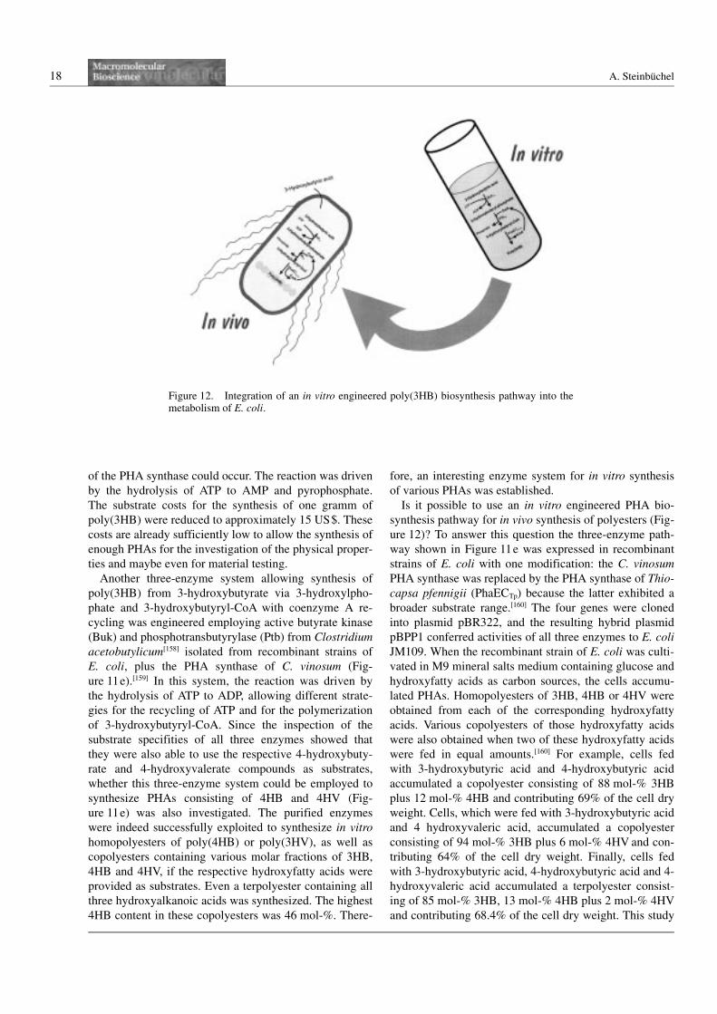
Is it possible to use an in vitro engineered PHA bio-
synthesis pathway for in vivo synthesis of polyesters (Fig-
ure 12)? To answer this question the three-enzyme path-
way shown in Figure 11e was expressed in recombinant
strains of E. coli with one modification: the C. vinosum
PHA synthase was replaced by the PHA synthase of Thio-
capsa pfennigii (PhaECTp) because the latter exhibited a
broader substrate range.[160] The four genes were cloned
into plasmid pBR322, and the resulting hybrid plasmid
pBPP1 conferred activities of all three enzymes to E. coli
JM109. When the recombinant strain of E. coli was culti-
vated in M9 mineral salts medium containing glucose and
hydroxyfatty acids as carbon sources, the cells accumu-
lated PHAs. Homopolyesters of 3HB, 4HB or 4HV were
obtained from each of the corresponding hydroxyfatty
acids. Various copolyesters of those hydroxyfatty acids
were also obtained when two of these hydroxyfatty acids
were fed in equal amounts.[160] For example, cells fed
with 3-hydroxybutyric acid and 4-hydroxybutyric acid
accumulated a copolyester consisting of 88 mol-% 3HB
plus 12 mol-% 4HB and contributing 69% of the cell dry
weight. Cells, which were fed with 3-hydroxybutyric acid
and 4 hydroxyvaleric acid, accumulated a copolyester
consisting of 94 mol-% 3HB plus 6 mol-% 4HV and con-
tributing 64% of the cell dry weight. Finally, cells fed
with 3-hydroxybutyric acid, 4-hydroxybutyric acid and 4-
hydroxyvaleric acid accumulated a terpolyester consist-
ing of 85 mol-% 3HB, 13 mol-% 4HB plus 2 mol-% 4HV
and contributing 68.4% of the cell dry weight. This study
Figure 12. Integration of an in vitro engineered poly(3HB) biosynthesis pathway into themetabolism of E. coli.
Perspectives for Biotechnological Production and Utilization of Biopolymers ... 19
demonstrated that a PHA biosynthesis pathway engi-
neered in vitro employing purified enzymes is function-
ally active in E. coli and can be utilized for in vivo syn-
thesis of PHAs.
Other authors[161] demonstrated the in vitro biosynthesis
of poly(3HV) employing the PHA synthase of R. eutro-
pha (Figure 11d). However, this system suffers from the
same disadvantages as the one-enzyme system for
poly(3HB) synthesis shown in Figure 11a. Another type
of PHA was obtained in vitro by employing purified PHA
synthase of P. aeruginosa and a commercially available
acyl-CoA synthetase from Pseudomonas sp. (Figure 11f).
This two-enzyme system synthesized in vitro poly(3-
hydroxydecanoate) when a racemic mixture of 3-hydro-
xydecanoate was used as a substrate.[162] This engineered
pathway allowed recycling of coenzyme A and was dri-
ven by the conversion of ATP to ADP.
Advantages and limitations of in vitro synthesis and
production processes are listed in Table 7. In vitro engi-
neering of pathways may be a useful strategy to evaluate
whether the establishment of a particular pathway in a
bacterium by in vivo metabolic engineering is feasible.
XI Production of PHAs by RecombinantStrains of E. coli
As shown in the above sections, E. coli has become an
interesting organism for the production of PHAs. Recom-
binant strains of E. coli-accumulating PHAs have been
repeatedly mentioned in this review. It should be empha-
sized that E. coli is unable to accumulate poly(3HB) or
any other PHAs as a storage compound; there are only
very small anounts of a lower molecular weight
poly(3HB) found mainly in the cytoplasmic mem-
brane.[163] Meanwhile, several quite different PHAs can be
obtained from E. coli and these are summarized in
Table 8. Not only poly(3HB),[68, 164, 165], but also the homo-
polyesters poly(4HB)[160, 166, 167] and poly(4HV)[160] have
been synthesized in E. coli. In addition, copolyesters con-
sisting of 3HB plus 3HV,[168, 169] 4HB[160, 166, 167] or 4HV,[160]
a terpolyester consisting of 3HB, 4HB plus 4HV[160] and
also PHAs consisting of various 3HAMCL have been
obtained from E. coli.[145, 148, 170, 171]
However, this research aimed, not only at the establish-
ment of a PHA biosynthesis pathway, but also at the gen-
eration of strains which might be suitable for the com-
mercial production of PHAs. For this, various strains of
E. coli were obtained which acccumulated high contents
of PHAs when cultivated to high cell densities, and, on
the basis of these studies, the production costs at an
industrial scale were calculated. It was found that such
recombinant strains could produce PHAs at much lower
cost than, for example, R. eutropha.[172–174] However, the
costs were still significantly higher than those for conven-
tional and established plastics such as polyethylene.
Table 7. The pros and cons of in vitro metabolic engineering.
Advantages and prospects Limitations andproblems
0 Detailed biochemical studiesthrough characterization ofthe enzyme(s) becomes possible
0 Purified PHAsynthase is required
0 Application of non naturalsubstratesPHAs with new constituentsmay be produced
0 Additional enzymesmust be availablein a purified stage
0 Rapid shifts of substrates applicablePHAs with a novel orderof constituents may be produced(e.g. blockcopolyesters)
0 Cell-free systemProduction is limited by the volumeof the reactor and not by the volumeof the cytoplasm
0 All enzymesmust be stable
Increase of PHA producedper volume is possible
0 Coenzymesmust be recycled
Synthesis may be performed in thepresence of compounds or underconditions which are inhibiting cellsNo expression requiredAmount of enzymes can beeasily varied
0 Only few components requireddownstream processing will be easier
0 Requires less efforts than in vivometabolic engineeringfast evaluation of the feasibilityof a strategy and the functionalityof a “designed” pathway
0 Special applicationsin situ PHA formation becomespossible
Table 8. PHAs available from recombinant Escherichia coli.
PHA Reference
Poly(3HB) [68][164][165]
Poly(4HB) [160][166][167]
Poly(4HV) [160]
Poly(3HB-co-3HV) [168][169]
Poly(3HB-co-4HB) [160][166][167]
Poly(3HB-co-4HV) [160]
Poly(3HB-co-4HB-co-4HV) [160]
Poly(3HAMCL) [145][147][148][170][171]
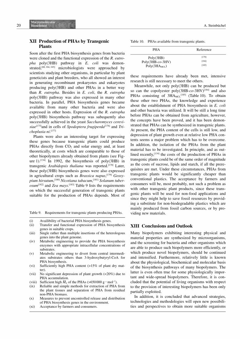
20 A. Steinbuchel
XII Production of PHAs by TransgenicPlants
Soon after the first PHA biosynthesis genes from bacteria
were cloned and the functional expression of the R. eutro-
pha poly(3HB) pathway in E. coli was demon-
strated,[68, 164, 165] microbiologists were approached by
scientists studying other organisms, in particular by plant
geneticists and plant breeders, who all showed an interest
in generating recombinant prokaryotes and eukaryotes
producing poly(3HB) and other PHAs in a better way
than R. eutropha. Besides in E. coli, the R. eutropha
poly(3HB) pathway was also expressed in many other
bacteria. In parallel, PHA biosynthesis genes became
available from many other bacteria and were also
expressed in other hosts. Expression of the R. eutropha
poly(3HB) biosynthesis pathway was subequently also
successfully achieved in the yeast Saccharomyces cerevi-
siae[175]and in cells of Spodoptera frugiperda[176] and Tri-
choplusia ni.[177]
Plants were also an interesting target for expressing
these genes because transgenic plants could produce
PHAs directly from CO2 and solar energy and, at least
theoretically, at costs which are comparable to those of
other biopolymers already obtained from plants (see Fig-
ure 1).[134] In 1992, the biosynthesis of poly(3HB) in
transgenic Arabidopsis thaliana was reported.[178] Later,
these poly(3HB) biosynthesis genes were also expressed
in agricultural crops such as Brassica napus,[179] Gossy-
pium hirsutum,[180] Nicotiana tabacum,[181] Solanum tuber-
osum[182] and Zea mays.[183] Table 9 lists the requirements
on which the successful generation of transgenic plants
suitable for the production of PHAs depends. Most of
these requirements have already been met, intensive
research is still necessary to meet the others.
Meanwhile, not only poly(3HB) can be produced but
so can the copolyester poly(3HB-co-3HV)[184] and also
PHAs consisting of 3HAMCL[185] (Table 10). To obtain
these other two PHAs, the knowledge and experience
about the establishment of PHA biosynthesis in E. coli
and other bacteria was utilized. It will be still a long time
before PHAs can be obtained from agriculture, however,
the concepts have been proved, and it has been demon-
strated that PHAs can be synthesized in transgenic plants.
At present, the PHA content of the cells is still low, and
depression of plant growth even at relative low PHA con-
tents seems a major problem which has to be overcome.
In addition, the isolation of the PHAs from the plant
material has to be investigated. In principle, and as out-
lined recently,[134] the costs of the production of PHAs in
transgenic plants could be of the same order of magnitude
as the costs of sucrose, lipids and starch, if all the prere-
quisites are met. Under these circumstances, PHAs from
transgenic plants would be significantly cheaper than
conventional plastics. The acceptance by farmers and
consumers will be, most probably, not such a problem as
with other transgenic plant products, since these trans-
genic plants will be used for non-food applications and
since they might help to save fossil resources by provid-
ing a substitute for non-biodegradable plastics which are
mainly produced from fossil carbon sources, or by pro-
viding new materials.
XIII Conclusions and Outlook
Many biopolymers exhibiting interesting physical and
material properties are synthesized by microorganisms,
and the screening for bacteria and other organisms which
are able to produce such biopolymers more efficiently, or
which produce novel biopolymers, should be continued
and intensified. Furthermore, relatively little is known
about the physiological, biochemical and molecular basis
of the biosynthesis pathways of many biopolymers. The
latter is even often true for some physiologically impor-
tant and wide-spread biopolymers. Therefore, it is con-
cluded that the potential of living organisms with respect
to the provision of interesting biopolymers has been only
partially exploited.
In addition, it is concluded that advanced strategies,
technologies and methodologies will open new possibili-
ties and perspectives to obtain more suitable organisms
Table 9. Requirements for transgenic plants producing PHAs.
(i) Availibility of bacterial PHA biosynthesis genes.(ii) Transfer and functional expression of PHA biosynthesis
genes in suitable crops.(iii) Single rather than multiple insertions of the heterologous
genes into the plant genome.(iv) Metabolic engineering to provide the PHA biosynthesis
enzymes with appropriate intracellular concentrations ofsubstrates.
(v) Metabolic engineering to divert from central intermedi-ates substrates others than 3-hydroxybutyryl-CoA forPHA biosynthesis.
(vi) Sufficiently high PHA content (A15% of plant dry mat-ter).
(vii) No significant depression of plant growth (a20%) due toPHA accumulation.
(viii) Sufficient high M—
w of the PHAs (A650000 g N mol–1).(ix) Reliable and simple methods for extraction of PHA from
the plant tissues and separation of PHA from residualnon-PHA biomass.
(x) Measures to prevent uncontrolled release and distributionof PHA biosynthesis genes in the environment.
(xi) Acceptance by farmers and consumers.
Table 10. PHAs available from transgenic plants.
PHA Reference
Poly(3HB) [178]
Poly(3HB-co-3HV) [184]
Poly(3HAMCL) [185]
Perspectives for Biotechnological Production and Utilization of Biopolymers ... 21
and optimized processes for the production of biopoly-
mers. Genetic engineering, metabolic engineering and the
utilization of microbial genoms, as well as the cloning of
genes from non-culturable microorganisms, provide the
potential for this. One important aim of further investiga-
tions must be the reduction of the production costs to
make biopolymers available at lower costs which are
competitive with those of established synthetic polymers.
An almost essential prerequisite for this will be the synth-
esis of such biopolymers from renewable resources or
directly from CO2 . Another aim must be the detection of
novel biopolymers, which exhibit features not shown by
synthetic polymers and are therefore of extra value.
PHAs are just one example, and as outlined in this
review, it was shown that intensive research can success-
fully address these questions and problems.
Acknowledgement: The author is very thankful to Dipl.-Biol.Tina Lutke-Eversloh and Mrs. Andrea Mersmann for preparingthe figures and tables perfectly.
Received: December 12, 2000
[1] M. T. Madigan, J. M. Martinko, J. Parker, “Biology ofMicroorganisms”, 9th edition, Prentice-Hall, Upper SaddleRiver 2000.
[2] T. Lutke-Eversloh, K. Bergander, H. Luftmann, A. Steinbu-chel, Microbiology 2001, 147, 11.
[3] A. L. Lehninger, D. L. Nelson, M. M. Cox, “Principles ofBiochemistry”, 2nd edition, Worth Publishers, New York1993.
[4] J. M. G. Cowie, “Polymers: Chemistry & Physics of Mod-ern Materials”, 1st edition, International Textbook Com-pany, Aylesbury 1973.
[5] R. Jonas, L. F. Farah, Polym. Degrad. Stabil. 1998, 59,101.
[6] M. Kunioka, Appl. Microbiol. Biotechnol. 1997, 47, 469.[7] M. Santos, J. Teixeira, A. Rodrigues, Biochem. Eng. 2000,
4, 177.[8] B. Monties, Polym. Degrad. Stabil. 1998, 59, 53.[9] H. G. Schlegel, E. Schuster, C. Konig, Zentralbl. Bakteriol.
I. Orig. Suppl. 1967, 2, 73.[10] Y. Asada, M. Miyake, J. Miyake, R. Kurane, Y. Tokiwa,
Int. J. Biol. Macromol. 1999, 25, 37.[11] R. B. Cain, in: Microbial Control of Pollution, J. C. Fry, G.
M. Gadd, R. A. Herbert, C. W. Jones, I. A. Watson-Craik,Eds., Cambridge University Press, London 1992, p. 293.
[12] G. Gottschalk, H.-J. Knackmuss, Angew. Chem. 1993, 105,1437.
[13] D. Byrom, Ed., “Biomaterials-Novel Materials from Biolo-gical Sources”, 1st edition, Macmillan Publishers, Basing-stoke 1991.
[14] R. A. Melo, J. Appl. Phycol. 1998, 10, 303.[15] B. H. A. Rehm, S. Valla, Appl. Microbiol. Biotechnol.
1997, 48, 281.[16] D. A. Swann, J.-W. Huo, “Biomaterials-Novel Materials
from Biological Sources”, D. Byrom, Ed., 1st edition, Mac-millan Publishers, Basingstoke 1991, p. 285.
[17] S. Scharf, F. Faloona, K. B. Mullis, G. T. Horn, H. A. Ehr-lich, N. Arnheim, Science 1985, 230, 1350.
[18] M. Alcalde, F. J. Plou, A. G. De Segura, M. Remaud-Simeon, R. M. Willemot, P. Monsan, A. Ballesteros, Bio-technol. Techniques 1999, 13, 749.
[19] B. Katzbauer, Polym. Degrad. Stabil. 1998, 59, 81.[20] D. Byrom, FEMS Microbiol. Rev. 1992, 103, 247.[21] Y. Poirier, C. Nawrath, C. R. Somerville, Biotechnology
1995, 13, 142.[22] J. Kossmann, J. Lloyd, V. Landschutze, J. Riesmeier, L.
Willmitzer, in: Biochemical Principles and Mechanisms ofBiosynthesis and Biodegradation of Polymers, Steinbuchel,Ed., Wiley-VCH, Weinheim 1999, p. 143.
[23] J. Kendrew, Ed., “The Encyclopedia of Molecular Biol-ogy”, 1st edition, Blackwell, Oxford 1994.
[24] J. E. Bailey, Science 1991, 252, 1668.[25] M. Schloter, M. Lebuhn, T. Heulin, A. Hartmann, FEMS
Microbiol. Rev. 2000, 24, 647.[26] T. R. Gingeras, C. Rosenow, ASM News 2000, 66, 463.[27] J. Anderson, E. A. Dawes, Microbiol. Rev. 1990, 54, 450.[28] F. B. Oppermann-Sanio, T. Hai, E. Aboulmagd, F. S. F.
Hezayen, S. Jossek, A. Steinbuchel, in: Biochemical Prin-ciples and Mechanisms of Biosynthesis and Biodegradationof Polymers, Steinbuchel, Ed., Wiley-VCH, Weinheim1999. p. 185.
[29] J. Preiss, in: Biochemical Principles and Mechanisms ofBiosynthesis and Biodegradation of Polymers“, Steinbu-chel, Ed., Wiley-VCH, Weinheim 1999, p. 134.
[30] H. F. J. Bligh, Biotechnol. Genet. Eng. Rev. 1999, 16, 177.[31] A. Kornberg, J. Bacteriol. 1995, 77, 491.[32] G. A. Birrer, A.-M. Cromwick, R. A. Gross, Int. J. Biol.
Macromol. 1994, 16, 265.[33] J. Lunt, Polym. Degrad. Stabil. 1998, 59, 145.[34] A. Steinbuchel, H. E. Valentin, FEMS Microbiol. Lett.
1995, 128, 219.[35] W. Babel, J. U. Ackermann, U. Breuer, Adv. Biochem. Eng.
Biotechnol. 2001, 71, 125.[36] A. Steinbuchel, K. Aerts, W. Babel, C. Follner, M. Lieber-
gesell, M. H. Madkour, F. Mayer, U. Pieper-Furst, A. Pries,H. E. Valentin, R. Wieczorek, Can. J. Microbiol. 1995, 41(suppl. 1), 94.
[37] L. L. Madison, G. W. Huisman, Microbiol. Molec. Biol.Rev. 1999, 63, 1.
[38] B. H. A. Rehm, A. Steinbuchel, Int. J. Biol. Macromol.1999, 25, 3.
[39] A. Steinbuchel, S. Hein, Adv. Biochem. Eng. Biotechnol.2001, 71, 81.
[40] U. Pieper-Furst, M. H. Madkour, F. Mayer, A. Steinbuchel,J. Bacteriol. 1994, 176, 4328.
[41] R. Wieczorek, A. Pries, A. Steinbuchel, F. Mayer, J. Bac-teriol. 1995, 177, 2425.
[42] E. Holler, B. Angerer, G. Achhammer, S. Miller, C. Wind-isch, FEMS Microbiol. Rev. 1992, 103, 109.
[43] H. Fischer, S. Erdmann, E. Holler, Biochemistry 1989, 28,5219–5226.
[44] B. S. Lee, T. Maurer, H. R. Kalbitzer, E. Holler, Appl.Microbiol. Biotechnol. 1999, 52, 415.
[45] K. Shimada, K. Matsushima, J. Fukumoto, T. Yamamoto,Biochem. Biophys. Res. Commun. 1969, 35, 619.
[46] N. Nagata, T. Nakahara, T. Tabuchi, Biosci. Biotech. Bio-chem. 1993, 57, 638.
[47] S.-J. Liu, A. Steinbuchel, Appl. Microbiol. Biotechnol.1996, 46, 273.
[48] B. Willibald, W. Bildl, B. S. Lee, E. Holler, Eur. J. Bio-chem. 1999, 265, 1085.
22 A. Steinbuchel
[49] P. E. Kolattukudy, “Plant Cuticles: an Integrated Func-tional Approach”, BIOS Scientific Publishers, Oxford1996.
[50] P. E. Kolattukudy, Adv. Biochem. Eng./Biotechnol. 2001,71, 1.
[51] D. Byrom, in: Plastics from Microbes – Microbial Syn-thesis of Polymers and Polymer Precursors, D. P. Mobley,Ed., Hanser Publishers, Munich 1994, p. 5.
[52] Y. Doi, “Microbial Polyesters”, VCH, New York 1990.[53] P. J. Hocking, R. H. Marchessault, in: Chemistry and Tech-
nology of Biodegradable Polymers, G. J. L. Griffin, Ed.,Blackie, London 1994, p. 48.
[54] G. A. M. van der Walle, G. J. H. Buisman, R. A. Westhuis,G. Eggink, Int. J. Biol. Macromol. 1999, 25, 123.
[55] D. Jendrossek, A. Schirmer, H. G. Schlegel, Appl. Micro-biol. Biotechnol. 1996, 46, 451.
[56] K. Mukai, Y. Doi, Y. Sema, K. Tomita, Biotechnol. Lett.1993, 15, 601.
[57] K.-E. Jaeger, A. Steinbuchel, D. Jendrossek, Appl. Environ.Microbiol. 1995, 61, 3113.
[58] T. Saito, K. Takizawa, H. Saegusa, Can. J. Microbiol.1995, 41 (Suppl. 1), 187.
[59] G. A. M. Van der Walle, G. J. M. De Koning, R. A.Weusthuis, G. Eggink, Adv. Biochem. Eng./Biotechnol.2001, 71, 263.
[60] H. M. Fraser. J. Sandow, H. R. Seudel, S. F. Nunn, ActaEndocrinol. (Copenhagen) 1989, 121, 841.
[61] S. F.Williams, D. P. Martin, D. M. Horowitz, O. P. Peoples,Int. J. Biol. Macromol. 1999, 25, 111.
[62] M. Scandola, in: Proceedings of the 1996 InternationalSymposium on Bacterial Polyhydroxyalkanoates, G.Eggink, A. Steinbuchel, Y. Poirier, B. Wiltholt, Eds., NRCResearch Press, Ottawa 1996, p. 20.
[63] S. P. McCarthy, unpublished results.[64] M.-M. Bear, React. Funct. Polym. 1997, 34, 65.[65] G. J. M. De Koning, H. M. M. Vanbilsen, P. J. Lemstra, W.
Hazenberg, B. Witholt, H. Preusting, J. G. Vandergalien, A.Schirmer, D. Jendrossek, Polymer 1994, 35, 2090.
[66] D. Seebach, FEMS Microbiol. Rev. 1992, 103, 215.[67] A. Timm, A. Steinbuchel, unpublished results.[68] O. P. Peoples, A. J. Sinskey, J. Biol. Chem. 1989, 264,
15298.[69] U. Pieper, A. Steinbuchel, FEMS Microbiol. Lett. 1992, 96,
73.[70] H. E. Valentin, A. Steinbuchel, Appl. Microbiol. Biotech-
nol. 1993, 39, 309.[71] M. A. Schembri, R. C. Bayly, J. K. Davies, FEMS Micro-
biol. Lett. 1994, 118, 145.[72] R. Tombolini, S. Povolo, A. Buson, A. Squartini, M. P.
Nuti, Microbiology 1995, 141, 2553.[73] S. P. Lee, V. Do, G. W. Huisman, O. P. Peoples, direct sub-
mission accession no U78047 (1996).[74] I. Lee, Y. H. Rhee, J.-Y. Kim, direct submission accesssion
no U78047 (1996).[75] S. Ueda, T. Yabutani, A. Maehara, T. Yamane, J. Bacteriol.
1996, 178, 774.[76] M. A. Cavellos, S. Encarnacion, A. Leija, Y. Mora, J.
Mora, J. Bacteriol. 1996, 178, 1646.[77] B. Hall, J. Baldwin, H. G. Rhie, D. Dennis, direct submis-
sion accession no AFO19964 (1997).[78] T. Fukui, Y. Doi, J. Bacteriol. 1997, 179, 4821.[79] J.-Y. Choi, S. Y. Lee, K. Han, Appl. Environ. Microbiol.
1998, 64, 4897.[80] K. Mandon, N. Michel-Reydellet, S. Encarnacion, P. A.
Kaminski, A. Leija, M. A. Cevallos, Elmerich, J. Mora, J.Bacteriol. 1998, 180, 5070.
[81] K. Sudesh, T. Fukui, Y. Doi, Appl. Environ. Microbiol.1998, 64, 3437.
[82] S. G. E. Andersson, A. Zomorodipour, J. O. Andersson,T. Sicheritz-Ponten, U. C. M. Alsmark, R. M. Podowski,A. K. Naeslund, A. S. Eriksson, H. H. Winkler, C. G. Kur-land, Nature 1998, 396, 133.
[83] J. McCool, M. C. Cannon, J. Bacteriol. 1999, 181, 585.[84] D. Kolibachuk, A. Miller, D. Dennis, Appl. Environ.
Microbiol. 1999, 65, 3561.[85] M. F. A. Rodrigues, H. E. Valentin, P. A. Berger, M. Tran,
J. Asrar, K. J. Gruys, A. Steinbuchel, Appl. Microbiol.Biotechnol. 2000, 53, 453.
[86] G. W. Huisman, E. Wonink, R. Meinma, B. Kazemier, P.Perpstra, B. Witholt, J. Biol. Chem. 1991, 266, 2191.
[87] A. Timm, A. Steinbuchel, Eur. J. Biochem. 1992, 209, 15.[88] A. Timm, S. Wiese, A. Steinbuchel, Appl. Microbiol. Bio-
technol. 1994, 40, 669.[89] H. E. Valentin, E. S. Stuart, R. C. Fuller, R. W. Lenz, D.
Dennis, J. Biotechnol. 1998, 64, 145.[90] H. Matsusaki, S. Manji, K. Taguchi, M. Kato, T. Fukui, Y.
Doi, J. Bacteriol. 1998, 180, 6459.[91] B. Garcia, E. R. Olivera, B. Minambres, M. Fernandez-
Valverde, L. M. Canedo, M. A. Prieto, J. L. Garcia, M.Martinez, J. M. Luengo, J. Biol. Chem. 1999, 274, 29228.
[92] K. Matsumoto, H. Matsusaki, K. Taguchi, M. Seki, Y.Doi, Biomacromolecules, submitted for publication.
[93] T. Kaneko, S. Sato, H. Kotani, A. Tanaka, E. Asamizu, Y.Nakamura, N. Miyajima, M. Hirosawa, M. Suigura, S.Sasamoto, T. Kimura, T. Hosouchi, A. Matsuno, A. Mur-aki, N. Nakazaki, K. Naruo, S. Okumura, S. Shimpo, C.Takeuchi, T. Wada, A. Tatnabe, M. Yamada, M. Yasuda,S. Tabata, DNA Res. 1996, 3, 109.
[94] S. Hein, T. Hai, A. Steinbuchel, Arch. Microbiol. 1998,170, 162.
[95] H. Tran, A. Steinbuchel, unpublished results.[96] M. Liebergesell, A. Steinbuchel, Eur. J. Biochem. 1992,
209, 135.[97] M. Liebergesell, A. Steinbuchel, Appl. Microbiol. Bio-
technol. 1993, 38, 493.[98] M. Liebergesell, F. Mayer, A. Steinbuchel, Appl. Micro-
biol. Biotechnol. 1993, 40, 292.[99] M. Liebergesell, S. Rahalkar, A. Steinbuchel, Appl.
Microbiol. Biotechnol. 2000, 54, 186.[100] E. Hustede, A. Steinbuchel, Biotechnol. Lett. 1993, 15, 709.[101] E. Hustede, A. Steinbuchel, H. G. Schlegel, FEMS Micro-
biol. Lett. 1992, 93, 285.[102] R. G. Kranz, K. K. Gabbert, T. A. Locke, M. T. Madigan,
Appl. Environ. Microbiol. 1997, 63, 3003.[103] T. Clemente, D. Shah, M. Tran, D. Stark, S. Padgette, D.
Dennis, K. Bruckener, A. Steinbuchel, T. Mitsky, Appl.Microbiol. Biotechnol. 2000, 53, 420.
[104] H. E. Valentin, A. Schonebaum, A. Steinbuchel, Appl.Microbiol. Biotechnol. 1996, 46, 261.
[105] T. Lutke-Eversloh, A. Steinbuchel, unpublished results.[106] A. Steinbuchel, H. G. Schlegel, Mol. Microbiol. 1991, 5,
535.[107] M. J. De Smet, G. Eggink, B. Witholt, J. Kingma, H.
Wynberg, J. Bacteriol. 1983, 154, 870.[108] H. Brandl. R. A. Gross, R. W. Lenz, R. C. Fuller, Appl.
Environ. Microbiol. 1998, 54, 1977.[109] G. W. Haywood, A. J. Anderson, D. F. Ewing, E. A.
Dawes, Appl. Environ. Microbiol. 1990, 56, 3354.[110] A. Timm, A. Steinbuchel, Appl. Environ. Microbiol.
1990, 56, 3360.[111] N. Hoffmann, A. Steinbuchel, B. H. A. Rehm, Appl.
Microbiol. Biotechnol. 2000, 54, 665–670.
Perspectives for Biotechnological Production and Utilization of Biopolymers ... 23
[112] G. N. M. Huijberts, T. De Rijk, P. De Waard, G. Eggink,J. Bacteriol. 1994, 176, 1661.
[113] B. H. A. Rehm, N. Kruger, A. Steinbuchel, J. Biol. Chem.1998, 273, 24044.
[114] N. Hoffmann, A. Steinbuchel, B. H. A. Rehm, FEMSMicrobiol. Lett. 2000, 184, 253.
[115] N. Hoffmann, B. H. A. Rehm, A. Steinbuchel, unpub-lished data.
[116] G. Haywood, A. J. Anderson, D. R. Williams, E. A.Dawes, D. F. Ewing, Int. J. Biol. Macromol. 1991, 13, 83.
[117] R. Williams, A. J. Anderson, E. A. Dawes, D. F. Ewing,Appl. Microbiol. Biotechnol. 1994, 40, 717.
[118] J. Anderson, D. R. Williams, E. A. Dawes, D. F. Ewing,Can. J. Microbiol. 1995, 41 (Suppl. 1), 4.
[119] H. E. Valentin, D. Dennis, Appl. Environ. Microbiol.1996, 62, 372.
[120] Y. Doi, A. Tamaki, M. Kunioka, K. Soga, Macromol.Chem., Rapid. Commun. 1997, 8, 631.
[121] Y. Doi, A. Segawa, S. Nakamura, M. Kunioka, in: NovelBiodegradable Microbial Polymers, E. A. Dawes, Ed.,Kluwer Press, Dordrecht 1990, pp. 37–48.
[122] H. Valentin, A. Schonebaum, A. Steinbuchel, Appl.Microbiol. Biotechnol. 1992, 36, 507.
[123] G. Schmack, V. Gorenflo, A. Steinbuchel, Macromole-cules 1998, 31, 644.
[124] M. Reh, in: Proceedings Zweites Symposium TechnischeMikrobiologie, H. Dellweg, Ed., Verlag Versuchs- undLehranstalt fur Spiritusfabrikation im Institut fur Ga-rungsgewerbe, Berlin 1970, p. 151.
[125] M. Reh, H. G. Schlegel, Arch. Microbiol. 1969, 67, 110.[126] M. Reh, H. G. Schlegel, Arch. Microbiol. 1969, 67, 99.[127] A. Steinbuchel, U. Pieper, Appl. Microbiol. Biotechnol.
1992, 37, 1–6.[128] Eur. Pat. Appl. EP 052.459 (1981), invs.: P. A. Holmes,
L. F. Wright, S. H. Collins.[129] D. Byrom, FEMS Microbiol. Lett. 1992, 103, 247.[130] H. Abe, Y. Doi, T. Fukushima, H. Eya, Int. J. Biol.
Macromol. 1994, 16, 115.[131] E. Shimamura, K. Kasuya, G. Kobayashi, T. Shiotani, Y.
Shima, Y. Doi, Macromolecules 1949, 27, 878.[132] A. Steinbuchel, in: Biomaterials, D. Byrom, Ed., Macmil-
lan Publishers, Basingstoke 1991, p. 123.[133] A. Steinbuchel, in: Biotechnology, 2nd edition, volume 6,
H. J. Rehm, G. Reed, A. Puhler, P. Stadler, Eds., Wiley-VCH, Weinheim 1996, p. 403.
[134] A. Steinbuchel, B. Fuchtenbusch,TIBTECH 1989, 16, 419.[135] H. E. Valentin, A. Steinbuchel, Appl. Microbiol. Biotech-
nol. 1994, 40, 699.[136] H. G. Schlegel, R. Lafferty, I. Krauss, Arch. Microbiol.
1970, 71, 283.[137] G. J. E. Thijsse, Biochem. Biophys. Acta 1964, 84, 195.[138] S. Miyakawa, K. Suzuki, T. Noto, Y. Harada, H. Okazaki,
J. Antibiot. (Tokyo), 1982, 411.[139] G. M. Helmkamp Jr, D. J. Brock, K. Bloch, J. Biol.
Chem. 1968, 243, 3229.[140] R. J. Heath, Y. T. Yu, M. A. Shapiro, E. Olson, C. O.
Rock, J. Biol. Chem. 1998, 273, 30316.[141] F. Turnowsky, K. Fuchs, C. Jeschek, G. Hogenauer, J.
Bacteriol. 1989, 171, 6555–65.[142] G. D’Agnolo, I. S. Rosenfeld, J. Awaya, S. Omura, P. R.
Vagelos, Biochem. Biophys. Acta. 1973 326, 155.[143] Y. Doi, personal communication 2000, 66, 2117.[144] S. Fiedler, A. Steinbuchel, B. H. A. Rehm, Appl. Environ.
Microbiol. 2000, 66, 2117.[145] S. Langenbach, B. H. A. Rehm, A. Steinbuchel, FEMS
Microbiol. Lett. 1997, 150, 303.
[146] C. C. DiRusso, J. Bacteriol. 1990, 172, 6459.[147] Q. Qi, B. H. A. Rehm, A. Steinbuchel, FEMS Microbiol.
Lett. 1997, 157, 155–162.[148] Q. Qi, A. Steinbuchel, B. H. A. Rehm, FEMS Microbiol.
Lett. 1998, 167, 89–94.[149] T. Tsuge, T. Fukui, H. Matsusaki, S. Taguchi, G. Kobaya-
shi, A. Ishizaki, Y. Doi, FEMS Microbiol. Lett. 2000, 184,193.
[150] K. Taguchi, Y. Aoyagi, H. Matsusaki, T. Fukui, Y. Doi,FEMS Microbiol. Lett. 1999, 176, 183.
[151] Q. Ren, N. Sierro, M. Kellerhals, B. Kessler, B. Witholt,Appl. Environ. Microbiol. 2000, 66, 1311.
[152] H. M. Alvarez, R. Kalscheuer, A. Steinbuchel, Fett/Lipid1997, 99, 239.
[153] T. U. Gerngross, D. P. Martin, Proc. Natl. Acad. Sci. USA1995, 92, 6279.
[154] R. W. Lenz, C. Farcet, P. J. Dijkstra, S. Goodwin, S. M.Zhang, Int. J. Biol. Macromol. 1999, 25, 55.
[155] R. Jossek, A. Steinbuchel, FEMS Microbiol. Lett. 1998,168.
[156] G. Schweiger, W. Buckel, FEBS Lett. 1984, 171, 79.[157] R. Jossek, R. Reichelt, A. Steinbuchel, Appl. Microbiol.
Biotechnol. 1998, 49, 258.[158] J. W. Cary, D. J. Petersen, E. T. Papoutsakis, G. N. Ben-
nett, J. Bacteriol. 1988, 170, 4613.[159] S.-J. Liu, A. Steinbuchel, Appl. Microbiol. Biotechnol.
2000, 53, 545.[160] S.-J. Liu, A. Steinbuchel, Appl. Environ. Microbiol. 2000,
66, 739.[161] L. Su, R. W. Lenz, Y. Takagi, Macromolecules 2000, 33,
229.[162] Q. Qi, A. Steinbuchel, B. H. A. Rehm, Appl. Microbiol.
Biotechnol., 2000, 54, 37.[163] R. P. Huang, R. N. Reusch, J. Biol. Chem. 1996, 271,
22196.[164] S. C. Slater, W. H. Voige, D. E. Dennis, J. Bacteriol.
1988, 170, 4431.[165] P. Schubert, A. Steinbuchel, H. G. Schlegel, J. Bacteriol.
1988, 170, 5837.[166] S. Hein, B. Sohling, G. Gottschalk, A. Steinbuchel,
FEMS Microbiol. Lett. 1997, 153, 411.[167] H. E. Valentin, D. Dennis, J. Biotechnol. 1997, 58, 33.[168] S. Slater, K. L. Houmiel, H. Tran, T. A. Mitsky, N. B.
Taylor, S. R. Padgette, K. Gruys, J. Bacteriol. 1998, 180,1979.
[169] C. Eschenlauer, S. K. Stoup, F. Srienc, D. A. Somers, Int.J. Biol. Macromol. 1996, 19, 121.
[170] S. Klinke, Q. Ren, B. Witholt, B. Kessler, Appl. Environ.Microbiol. 1999, 65, 540.
[171] B. H. A. Rehm, A. Steinbuchel, Appl. Microbiol. Biotech-nol., in press (2001)
[172] S. Y. Lee, J. I. Choi, H. H. Wong, Int. J. Biol. Macromol.1999, 25, 31.
[173] J. Choi, S. Y. Lee, Appl. Microbiol. Biotechnol. 1999, 51,13.
[174] S. Y. Lee, TIBTECH 1996, 14, 431.[175] T. A. Leaf, M. S. Peterson, S. K. Stoup, D. Somers, F.
Srienc, Microbiology 1996, 142, 1169.[176] M. D. Williams, J. A. Jahn, D. H. Sherman, Appl.
Environ. Microbiol. 1996, 62, 2540.[177] M. D. Williams, A. M. Fieno, R. A. Grant, D. H. Sher-
man, Protein Expression Purif. 1996, 7, 203.[178] Y. Poirier, D. Dennis, K. Klomparens, C. R. Somerville,
Science 1992, 256, 520.[179] H. E. Valentin, D. L. Broyles, L. A. Casagrande, S. M.
Colburn, W. L. Creely, P. A. DeLaquil, H. M. Felton, K.
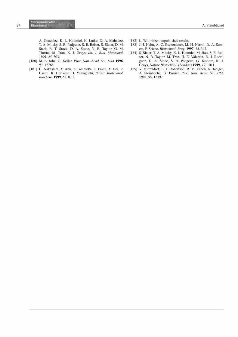
24 A. Steinbuchel
A. Gonzalez, K. L. Houmiel, K. Lutke, D. A. Mahadeo,T. A. Mitsky, S. R. Padgette, S. E. Reiser, S. Slater, D. M.Stark, R. T. Stock, D. A. Stone, N. B. Taylor, G. M.Thorne, M. Tran, K. J. Gruys, Int. J. Biol. Macromol.1999, 25, 303.
[180] M. E. John, G. Keller, Proc. Natl. Acad. Sci. USA 1996,93, 12768.
[181] H. Nakashita, Y. Arai, K. Yoshioka, T. Fukui, Y. Doi, R.Usami, K. Horikoshi, I. Yamaguchi, Biosci. Biotechnol.Biochem. 1999, 63, 870.
[182] L. Willmitzer, unpublished results.[183] J. J. Hahn, A. C. Eschenlauer, M. H. Narrol, D. A. Som-
ers, F. Srienc, Biotechnol. Prog. 1997, 13, 347.[184] S. Slater, T. A. Mitsky, K. L. Houmiel, M. Hao, S. E. Rei-
ser, N. B. Taylor, M. Tran, H. E. Valentin, D. J. Rodri-guez, D. A. Stone, S. R. Padgette, G. Kishore, K. J.Gruys, Nature Biotechnol. (London) 1999, 17, 1011.
[185] V. Mittendorf, E. J. Robertson, R. M. Leech, N. Kruger,A. Steinbuchel, Y. Poirier, Proc. Natl. Acad. Sci. USA1998, 95, 13397.