soi3p/rav1p functions at the early endosome to regulate endocytic trafficking to the vacuole and...
TRANSCRIPT
Molecular Biology of the CellVol. 15, 3196–3209, July 2004
Soi3p/Rav1p Functions at the Early Endosome to RegulateEndocytic Trafficking to the Vacuole and Localization ofTrans-Golgi Network Transmembrane ProteinsGyorgy Sipos,*†‡ Jason H. Brickner,*†§ E.J. Brace,* Linyi Chen,�¶Alain Rambourg,# Francois Kepes,#@ and Robert S. Fuller* **
*Department of Biological Chemistry, University of Michigan Medical Center, Ann Arbor, Michigan48109-0606; �Department of Pharmacology, Wayne State University School of Medicine, Detroit,Michigan 48201, and #Departement de Biologie Cellulaire et Moleculaire, Commissariat a l’EnergieAtomique Saclay, Gif-sur-Yvette 91191 Cedex, France
Submitted October 22, 2003; Revised April 5, 2004; Accepted April 7, 2004Monitoring Editor: Randy Schekman
SOI3 was identified by a mutation, soi3-1, that suppressed a mutant trans-Golgi network (TGN) localization signal in theKex2p cytosolic tail. SOI3, identical to RAV1, encodes a protein important for regulated assembly of vacuolar ATPase.Here, we show that Soi3/Rav1p is required for transport between the early endosome and the late endosome/prevacuolarcompartment (PVC). By electron microscopy, soi3-1 mutants massively accumulated structures that resembled earlyendosomes. soi3� mutants exhibited a kinetic delay in transfer of the endocytic tracer dye FM4-64, from the 14°Cendocytic intermediate to the vacuole. The soi3� mutation delayed vacuolar degradation but not internalization of thea-factor receptor Ste3p. By density gradient fractionation, Soi3/Rav1p associated as a peripheral protein with membranesof a density characteristic of early endosomes. The soi3 null mutation markedly reduced the rate of Kex2p transport fromthe TGN to the PVC but had no effect on vacuolar protein sorting or cycling of Vps10p. These results suggest thatassembly of vacuolar ATPase at the early endosome is required for transport of both Ste3p and Kex2p from the earlyendosome to the PVC and support a model in which cycling through the early endosome is part of the normal itineraryof Kex2p and other TGN-resident proteins.
INTRODUCTION
Steady-state localization of trans-Golgi network (TGN)membrane proteins in the budding yeast Saccharomyces cer-evisiae is achieved by continuous cycles of vesicular trans-port between the TGN and post-TGN compartments such asthe late endosome or prevacuolar compartment (PVC)(Fuller et al., 1988; Vida et al., 1993; Cereghino et al., 1995;Brickner and Fuller, 1997; Bryant and Stevens, 1997). Kex2pand Ste13p, proteolytic processing enzymes involved inmaturation of �-mating factor and other substrates (Fuller et
al., 1988), are localized to the TGN by the function of TGNlocalization signals (TLSs) in their cytosolic tails (C-tails)(Brickner and Fuller, 1997; Bryant and Stevens, 1997). TLS1directs retrieval from the PVC to the TGN and consists oftwo essential aromatic residues separated by a nonessentialcharged or polar residue (Tyr713Glu714Phe715 for Kex2p andPhe85Gln86Phe87 for Ste13p) (Wilcox et al., 1992; Nothwehr etal., 1993; Redding et al., 1996a). Substitutions for Tyr713 in theTLS1 of Kex2p leads to loss of TGN localization and rapiddelivery of the protein to the vacuole (Wilcox et al., 1992;Redding et al., 1996a). A less well defined signal, TLS2,requires sequences distal to TLS1 in the Kex2p C-tail andfunctions to delay transport of Kex2p from the TGN to thePVC, possibly by promoting TGN retention (Brickner andFuller, 1997). A signal similar to TLS2 is found in the Ste13pC-tail (Bryant and Stevens, 1997).
Using a mating assay sensitive to the efficiency of �-factorprocessing by Kex2p, we isolated allele-specific mutant sup-pressors soi1, SOI2, and soi3 that suppressed the effects of aTLS1 point mutation (Y713A) in Kex2p but not of a deletionof TLS1 or of the entire C-tail (Redding et al., 1996a). Thesesoi mutations also suppressed effects of point mutations inthe Ste13p TLS1 signal (Redding et al., 1996a). Cloning ofSOI1, allelic to VPS13, revealed that this gene encoded anovel 350-kDa peripheral membrane protein whose humanhomologues include two disease loci (Brickner and Fuller,1997; Rampoldi et al., 2001; Ueno et al., 2001; Kolehmainen etal., 2003). Suppression of the mislocalization of Y713A Kex2pby a soi1 mutation required TLS2, indicating that TLS2 func-tion became activated in the absence of Soi1p, slowing the
Article published online ahead of print. Mol. Biol. Cell 10.1091/mbc.E03–10–0755. Article and publication date are available atwww.molbiolcell.org/cgi/doi/10.1091/mbc.E03–10–0755.
† These authors made comparable contributions to this work.Present addresses: ‡Department of Medical Biochemistry, Univer-
sity and Biocenter of Vienna, A-1030 Vienna, Austria; §Departmentof Biochemistry and Biophysics, University of California, San Fran-cisco, CA 94143; ¶Department of Molecular and Integrative Physi-ology, University of Michigan Medical Center, Ann Arbor, MI48109-0622; @ Dynamique de la Compartimentation Cellulaire, In-stitut des Sciences du Vegetal, Centre National de la RechercheScientifique Unite Propre de Recherche 2355, Gif-sur-Yvette, France,and Epigenomics Project/Genopole, Evry, France.
** Corresponding author. E-mail address: [email protected] used: ALP, alkaline phosphatase; API, aminopep-tidase I; C-tail, cytosolic tail; GFP, green fluorescent protein; HA,hemagglutinin epitope; IP, immunoprecipitation; PVC, prevacu-olar compartment; TLS, trans-Golgi network localization signal;Vps, vacuolar protein sorting.
3196 © 2004 by The American Society for Cell Biology
escape of Kex2p from the TGN (Brickner and Fuller, 1997).The soi1 mutants also displayed defective TLS1-dependentlocalization, resulting in aberrant localization not only ofKex2p but also of the vacuolar precursor sorting receptorVps10p and of A-ALP, a hybrid protein in which the Ste13pC-tail is fused to alkaline phosphatase (ALP). These resultsindicated that Soi1p acts both at the PVC to stimulate TLS1function and at the TGN to repress TLS2 function and con-sequently facilitates cycling of transmembrane proteins be-tween the TGN and PVC (Brickner and Fuller, 1997).
Transport of Kex2p from the TGN to the PVC requires clathrinand the dynamin homolog Vps1p (Seeger and Payne, 1992; Wils-bach and Payne, 1993; Nothwehr et al., 1995; Redding et al.,1996a,b). Mutations in two types of clathrin adaptors affect Kex2plocalization. These are the Gga proteins, encoded by GGA1 andGGA2, and the Adaptor Protein (AP)-1 adaptor complex (Blackand Pelham, 2001). Mutations in the GGA genes blocked transportof carboxypeptidase Y (CPY) to the vacuole, abrogated TGN toPVC transport of Vps10p and Pep12p, and altered the efficiency ofpro-�-factor processing by Kex2p (Black and Pelham, 2000;Dell’Angelica et al., 2000; Hirst et al., 2000; Costaguta et al., 2001).Effects of mutations in genes encoding AP-1 subunits are onlyseen in combination with either gga mutations or clathrin muta-tions (Phan et al., 1994; Stepp et al., 1995; Huang et al., 1999; Yeunget al., 1999), suggesting that the transport pathway governed byAP-1 is either less important for Kex2p localization or is redun-dant with other pathways. Recently, it has been suggested thatAP-1 adaptors function in transport between the TGN and earlyendosome of Kex2p, Ste13p, and chitin synthase III (Chs3p), sug-gesting that TGN membrane proteins such as Kex2p and Ste13paccess the early endosome (Valdivia et al., 2002; Ha et al., 2003).
Here, we have cloned the SOI3 gene, which we find to beidentical to RAV1. Rav1p was isolated as a Skp1p-interact-ing protein that is part of a complex that contains Rav2p andthe V1 subcomplex of the proton-translocating vacuolarATPase (V-ATPase) (Seol et al., 2001; Smardon et al., 2002).Both RAV1 and RAV2 were shown to be important forglucose-regulated assembly of V1 onto V0 to form functionalV-ATPase on the vacuolar membrane (Seol et al., 2001).Analysis of the effects of soi3 mutations demonstrates thatloss of Soi3p function results both in alterations in the local-ization of TGN membrane proteins as well as selective de-fects in delivery of endocytic cargo to the vacuole. A syn-thesis of these results suggests that assembly of vacuolarATPase at the early endosome in yeast is essential for earlyendosome maturation and efficient transport from the earlyendosome to the PVC.
MATERIALS AND METHODS
Antibodies and ReagentsAntisera against the Kex2 Ctail and lumenal domains were as describedpreviously (Wilcox and Fuller, 1991; Wilcox et al., 1992). Anti-Pep12 andanti-CPY monoclonal antibodies were from Molecular Probes (Eugene, OR).Anti-hemagglutinin (HA) monoclonal antibody 12CA5 was from Roche Di-agnostics (Indianapolis, IN). Antisera against Anp1p, Mnn1p, Chs3p, Pep12p,Vps10p, CPY, ALP, aminopeptidase I (API), and Ste3p were gifts of Drs. S.Munro (Medical Research Council, Cambridge, United Kingdom), T. Graham(Vanderbilt University, Nashville, TN), R. Schekman (University of Califor-nia, Berkeley, CA), S. Emr (University of California, San Diego, CA), D.Klionsky (University of Michigan, Ann Arbor, MI), and N. Davis (WayneState University, Detroit, MI). Antibodies against Ste3p were affinity purifiedas described previously (Redding et al., 1991; Roth et al., 1998). Rabbit anti-mouse IgG was from Jackson ImmunoResearch Laboratories (West Grove,PA), and horseradish peroxidase-conjugated secondary antibodies were fromAmersham Biosciences (Piscataway, NJ). Boc-Leu-Lys-Arg-7-amino-4-meth-ylcoumarin (LKR-AMC) was from Bachem Biosciences (King of Prussia, PA).Restriction and DNA modification enzymes were from New England Biolabs(Beverly, MA). Unless indicated otherwise, all other chemicals and reagentswere from Sigma-Aldrich (St. Louis, MO).
Strains and PlasmidsPlasmids containing KEX2 under the KEX2 promoter on yeast centromereplasmids were pCWKX10 (wild-type Kex2p), pCWKX11 (Y713A-Kex2p), andpCWKX10-I718tail (I718tail-Kex2p) (Wilcox et al., 1992; Redding et al., 1996a;Brickner and Fuller, 1997). Plasmids containing KEX2 under the GAL1 pro-moter on yeast centromere plasmids were pCWKX20 (wild type), pCWKX21(Y713A-Kex2p), pCWKX27 (C-tail�-Kex2p), pCWKX20-I718tail (I718tail-Kex2p), and pCWKX21-I718tail (Y713A, I718tail-Kex2p) (Wilcox et al., 1992;Redding et al., 1996a; Brickner and Fuller, 1997). Plasmids pRS303-SOI3.1 andpRS313-SOI3 were constructed by ligating an EcoRI fragment containing basepairs 491021–498250 from yeast chromosome X into pRS303 (HIS3 integratingplasmid) and pRS313 (HIS3 CEN ARS plasmid), respectively (Sikorski andHieter, 1989). Plasmid pSOI3-HA was generated from pRS313-SOI3.1 bymutating the final three codons of SOI3 from GAC TTT GTA to GGC GGCCGC by polymerase chain reaction (PCR) to generate a NotI site and ligatinga fragment encoding a triple HA epitope tag (Tyers et al., 1992). Plasmidp413ADH-SOI3 was constructed by PCR amplifying the SOI3 structural geneand ligating the resulting fragment into the EcoRI site of p413ADH (Mumberget al., 1995). To express a Soi3p fusion to green fluorescent protein (GFP),pSOI3-GFP was created by ligating the 0.75-kb NotI fragment of pSF-GP1 (giftof Jeanne Hirsch, Mt. Sinai School of Medicine, New York, NY) into the NotIsite of pRS313-SOI3.1. Plasmids pSOI3-GFP-SL and pSOI3-HA-SL were cre-ated by replacing the SacI sites in pSOI3-GFP and pSOI3-HA with a SalI siteby linker ligation. Plasmids p413ADH-SOI3-GFP and p413ADH-SOI3-HAwere created by ligating the NarI/SalI fragments of pSOI3-GFP-SL andpSOI3-HA-SL containing the C termini of SOI3-GFP and SOI3-HA, intop413ADH-SOI3 cleaved by NarI plus SalI. Expression of Soi3-Soi3-GFP fullycomplemented the soi3 null mutation (our unpublished data).
Strains used in this study, created by standard genetic crosses and/or bytransformation, are shown in Table 1. Yeast media were as described (Rose etal., 1990). Two-step gene replacements were used to convert STE3 either toGal1-STE3, Gal1-STE3-HA or to Gal1-STE3�365, as described previously (Rothand Davis, 1996; Chen and Davis, 2002). To delete VPS1, the complete codingsequence of VPS1 was replaced with Kluyveromyces lactis LEU2 as describedpreviously (Gueldener et al., 2002). The vps1-100-ts allele was introduced bytransplacement. Plasmid pCAV40 (generous gift of Tom Stevens, Universityof Oregon, Portland, OR) containing vps1-100-ts was digested with EcoRI,transformed into CRY1, and Ura� transformants were selected. Ura� recom-binants isolated by 5-fluoroorotic acid selection were tested by the CPYcolony blot assay to identify a strain carrying the vps1-100-ts allele (EBY73).
Cloning and Analysis of SOI3Strain SPB400-3D carrying plasmid pCWKX10 was transformed with the CENARS LEU2 genomic library YPH1 (ATCC no. 77162). Library transformantswere screened by replica-plating to both YPD � 6 mM ZnCl2 and YPD � 10mM ZnCl2. Zinc-resistant transformants were tested for plasmid dependenceand for their ability to complement the Soi� phenotype measured by the onsetof impotence experiment as described previously (Redding et al., 1996a). Twoindependent transformants displayed plasmid-dependent complementationof both the Zn2� sensitivity and the Soi� phenotype (Redding et al., 1996a).DNA sequencing revealed that the inserts corresponded to base pairs 491021–498727 on chromosome X. To determine whether this DNA region corre-sponded to SOI3, an EcoRI fragment corresponding to base pairs 491021–498250 from chromosome X was cloned into pRS303 (HIS3 integratingplasmid) (Sikorski and Hieter, 1989) to create pRS303-SOI3.1A. This plasmidwas linearized with StuI, transformed into KRY18-1B, and His� transfor-mants were crossed to SPB400-3D transformed with pKWKX21. The resultingdiploid was sporulated and analyzed by random spore analysis, demonstrat-ing linkage of the integrated marker and soi3-1 (46 MAT�MAT� strains: 0 Soi�His�, 12 Soi� His�, 32 Soi� His�, and 0 Soi� His�), also confirmed by tetradanalysis (our unpublished data). DNA sequence analysis of the inserts com-bined with examination of the published yeast genomic sequence showed thatonly two full-length open reading frames, YJR033c and PET191, were con-tained within the overlapping region. YJR033c was seen as the more likelycandidate, because soi3-1 strains grew on nonfermentable carbon sources (ourunpublished data). However, subclones containing YJR033c along with 232base pairs of the 5� untranslated region and 220 base pairs of PET191 did notfully complement both the Zn2� sensitivity and suppressor phenotypes (ourunpublished data). To confirm that expression of YJR033c alone comple-mented soi3-1 phenotypes, YJR033c was amplified by PCR and subclonedunder the control of the ADH promoter on a single copy CEN plasmid(Mumberg et al., 1995), resulting in plasmid pADH-YJR033c.
Heterozygous replacement of the complete coding sequence of the SOI3gene with the kanr gene (Wach et al., 1994) in diploid strain KRY18 resulted inGSY11, which was sporulated to generate haploid soi3� strains (Table 1).
MicroscopyFor electron microscopy (EM), cells were fixed, stained, and visualized asdescribed previously (Rambourg et al., 1993). For FM4-64 uptake experiments,strains GSY11-2A (soi3�) and GSY11-4D (SOI3) were grown in 10 ml of YPDto �OD600 � 0.9, harvested, and resuspended in 500 �l of cold YPD � 40 �MFM4-64 (Molecular Probes). Cells were incubated on ice for 1 h, washed in
Soi3/Rav1p in EE to PVC Transport
Vol. 15, July 2004 3197
cold YPD, and resuspended in 1 ml of cold YPD and incubated at 14°C, withshaking. After 40 min at 14°C, 500 �l of cells was harvested and resuspendedin 400 �l of prewarmed 30°C YPD. Cells were harvested, washed in 1� 100 �lof 10 mM NaN3, 0 mM NaF, resuspended in 25 �l of 10 mM NaN3, 10 mMNaF, and stored on ice until they were examined using an Axioskop micro-scope (Carl Zeiss, Thornwood, NY), recording data on film as describedpreviously (Brickner and Fuller, 1997). Soi3-GFP images were examined usinga Nikon Eclipse 800 microscope and an ORCA2 charge-coupled device(Hamamatsu, Bridgewater, NJ). Cells were grown in log phase in minimalmedium at 25°C and mounted in low-temperature agarose. Z-stacks werecreated using eight 0.25-�m steps. ISEE Software (Inovision, Raleigh, NC) wasused for image capture and deconvolution of Z-stacks.
Ste3p and Zrt1-HAp EndocytosisConstitutive turnover of the Ste3p and the a-factor–dependent internaliza-tion, recycling, and turnover of Ste3�365p were all performed as describedpreviously (Chen and Davis, 2000). Expression of Ste3p and related proteins(Ste3�365p and Ste3HAp) was regulated from the GAL1 promoter (Chen andDavis, 2000). Single HA epitope-tagged Zrt1p (Zrt1-HAp) was expressed fromits own promoter on plasmid pMC5-HSET (Gitan et al., 1998), a gift of DavidEide (University of Missouri, Columbia, MO). To induce Zrt1-HAp in soi3�cells, soi3� and control cells were pretreated with 1 mM EDTA for 3 h toinduce Zrt1-HAp expression, and internalization was initiated by the additionof 2 mM Zn2�. Cell disruption and sample preparation protocols were asdescribed previously (Davis et al., 1993).
Subcellular FractionationFractionation procedures followed published methods (Sipos and Fuller,2002). Cells were spheroplasted with lyticase, and 200-�l samples equivalentto 50 ml of cells (OD600 � 1) were frozen over liquid N2. For differential
centrifugation, spheroplasts were thawed and homogenized, and lysates werecentrifuged twice at 500 � g for 5 min. The supernatant was centrifuged for15 min at 13,000 � g. The resulting pellet (P13) was washed with buffer andpelleted again for 15 min at 13,000. The medium speed supernatant (S13) wassplit into three samples. One was adjusted to 1 M NaCl, a second to 1% TritonX-100. These samples were incubated on ice for 20 min and centrifuged in aTLS55 rotor in a TLX centrifuge (Beckman Coulter, Fullerton, CA) at 55,000rpm (200,000 � g at ravg) for 30 min to generate P200 and S200 fractions.Samples equivalent to 1 ml of cells (OD600 � 1.0) of the P13, S13, P200, andS200 fractions were fractionated by SDS-PAGE, and the presence of markerswere analyzed by immunoblotting.
For sucrose gradient fractionation, 0.5 ml of P200 fraction, equivalent to 200ml of cells (OD600 � 1.0), was loaded on the top of the following sucrose stepgradient (from the bottom): 0.5 ml of 60%/2.0 ml of 48%/2.5 ml of 40%/2.5 mlof 36%/2.5 ml of 32%/2.0 ml of 29%. The gradient was centrifuged at 41,000rpm for 17 h at 4°C by using a Beckman Coulter SW41 rotor. Fractions (250 �l)were collected, and 10- to 15-�l samples from each interface were analyzed byimmunoblotting and assayed for Kex2 activity, as described previously (Siposand Fuller, 2002).
35S Pulse-Chase/Immunoprecipitation35S Pulse-chase/immunoprecipitation experiments were performed essen-tially as described previously (Wilcox and Fuller, 1991). Cells were labeledwith 35S-labeled amino acids (Express Label; Amersham Biosciences). Five-minute pulses were used for ALP, CPY, and API, and 10-min pulses wereused for Kex2p and Vps10p. Pulses were followed by chase with nonradio-active methionine and cysteine at 10 mM, and cell samples removed atvarious times were subjected to glass-bead lysis under denaturing conditions.Immunoprecipitations (IPs) of Kex2p and Vps10p were carried out twice.Other proteins were subjected to single IPs. IPs used polyclonal antisera and
Table 1. Strains used in this study
Strain Relevant genotype Parental strain
CRY1a MATa ade2-1 can1-100 his3-11,15 leu2-3,112 trp1-1 ura3-1 W303-1Aa
CRY2a MAT� ade2-1 can1-100 his3-11,15 leu2-3,112 trp1-1 ura3-1 W303-1Ba
CRY3a MATa/MAT� CRY1 � CRY2KRY18b KEX2/kex2��TRP1 CRY3GSY11 soi3�/SOI3 KRY18KRY18-1A MAT� kex2��TRP1 KRY18KRY18-1B MATa kex2��TRP1 KRY18GSY11-1A MATa soi3��kanr GSY11GSY11-2A MAT� soi3��kanr GSY11GSY11-4D MAT� SOI3 GSY11GSY11-7A MAT� soi3��kanr kex2��TRP1 GSY11SPB400-3Db MAT� soi3-1 kex2��TRP1 KRY18-1AKRY200-r2b MAT� soi2-3 kex2��TRP1 KRY18-1AKRY208-r3b MAT� soi3-1 kex2��TRP1 KRY18-1AJBY135-1A MAT� soi1�1�kanr JBY135c
JBY135-1C MAT� soi1�1�kanr kex2��TRP1 JBY135c
JBY142c MATa SOI1-HA CRY1GSY19 MATa vps10�kanr CRY2GSY32 MAT� GAL1-STE3 CRY2GSY34 MAT� GAL1-STE3 soi3��kanr GSY11-2AGSY35 MAT� GAL1-STE3 soi1�2�kanr JBY154-1Dc
GSY36 MAT� GAL1-STE3 soi3��kanr pep4�LEU2 GSY34GSY40 MAT� GAL1-STE3�365 CRY2GSY41 MAT� GAL1-STE3�365 soi3��kanr GSY11-2AGSY51 MAT� ste3��LEU2 CRY2EBY22 MAT� soi3��kanr kex2��TRP1 vps27��LEU2 GSY11-7AEBY68 MAT� vps1��LEU2 CRY2EBY69 MATa/MAT� vps1��LEU2/VPS1 soi3��kanr/SOI3 EBY68xGSY11-1AEBY73 MATa vps1-100-ts CRY1EBY17 MATa/MAT� soi3��kanr/soi3��kanr GSY11-1AxGSY11-2AEBY76 MATa/MAT� vps1��LEU2/VPS1 soi3��kanr/soi3��kanr EBY17EBY77 MATa/MAT� vps1-100ts/VPS1 soi3��kanr/SOI3 EBY73xGSY11-2AEBY77-4 MAT� vps1-100-ts EBY77EBY77-8 MAT� vps1-100-ts soi3��kanr EBY77
a Wilcox et al. (1992).b Redding et al. (1996a).c Brickner and Fuller, 1997.
G. Sipos et al.
Molecular Biology of the Cell3198

Pansorbin (Calbiochem-Novabichem, La Jolla, CA) (2.5 �l of 50% slurry perequivalent of 1 ml of cells at OD600 � 1.0). IPs were fractionated by SDS-PAGE, and radioactive signals were captured and quantified using a Phos-phorImager (Amersham Biosciences) and IPLabgel software (Signal Analyt-ics, Vienna, VA). Half-lives were calculated by linear regression.
RESULTS
SOI3 Corresponds to YJR033c/RAV1Unlike soi1 mutations (Brickner and Fuller, 1997), dominantSOI2 mutations and the recessive soi3-1 mutation (Reddinget al., 1996a) suppressed the Y713A substitution in TLS1 inthe context of a shortened Kex2p C-tail (I718tail) that lacksTLS2 (Figure 1). Neither the SOI2 mutations nor soi3-1 sup-pressed deletion of the C-tail, suggesting initially that bothsuppress through TLS1, i.e., by improving retrieval from thePVC back to the TGN. However, further analysis of the soi3null mutant, detailed in this work, showed that Soi3p func-tions in a distinct transport step important to Kex2p local-ization, between the early and late endosome.
Phenotypic analysis revealed that hypersensitivity to bothZn2� and Co2� cosegregated with the suppressor phenotypecaused by the soi3-1 mutation (our unpublished data). Wild-type SOI3 was cloned from a yeast genomic library bycomplementing the Zn2� hypersensitivity. Two clones
(clones 3 and 4) (Figure 2A) contained overlapping inserts,complemented the suppressor phenotype, and were shownby segregation analysis to map to the soi3-1 locus (see MA-TERIALS AND METHODS). A single yeast open readingframe from this locus, YJR033c, when placed under controlof the moderate ADH promoter in the single copy CENplasmid pADH-YJR033c (see MATERIALS AND METH-ODS), complemented both the Zn2� hypersensitivity (Figure2B) and Soi� suppressor phenotypes of soi3-1 (our unpub-lished data), confirming the identity of YJR033c as SOI3. Asdescribed previously (Kraemer et al., 1998; Seol et al., 2001),YJR033c encodes a large protein (�150 kDa) homologous tothe DmX protein of Drosophila melanogaster and the humanhomolog DMXL1 2000) (Kraemer et al., 1998; Kraemer et al.,2000). Soi3p, approximately one-half the size of the meta-zoan homologues, is predicted to contain at least sevenWD40 repeat sequences (Smith et al., 1999), whereas DmXand DMLX1 may contain 28–30 WD repeats (Kraemer et al.,2000). The carboxy terminus of the Soi3p sequence suggeststhe presence of a coiled-coil domain (residues 1311–1340).Soi3p is identical to Rav1p, which was isolated as a Skp1p–interacting protein implicated in assembly of vacuolarATPase (V-ATPase) (Seol et al., 2001). This work focuses onthe role of Soi3/Rav1p in early endosome to PVC transport,but implicit in this is the expectation that it is the role of theprotein in V-ATPase assembly at the early endosome that iskey. Unpublished data from our laboratory along with pub-lished data support the conclusion that V-ATPase mutantshave phenotypes similar to those exhibited by soi3 mutants(our unpublished data) (Perzov et al., 2002). Because soi3-1was described earlier than RAV1 (Redding et al., 1996a; Seolet al., 2001), SOI3 and Soi3p will be used here except whereidentity between Soi3p with Rav1p needs emphasis.
soi3-1 Accumulates Early Endosome-like StructuresElectron microscopic analysis of the soi3-1 mutant was con-ducted using thick sections (250 nm) analyzed at tilt anglesto permit creation of stereo pairs (Rambourg et al., 1993). Thesoi3-1 mutant cells massively accumulated ovoid membrane
Figure 1. soi3-1 mutation suppresses a TLS1 mutation in Kex2p inthe absence of TLS2. (A) Schematic diagram of Kex2p indicatingvarious C-tail mutants. C-tail ending at 778 is TLS2�; C-tail endingat 772 is TLS2� (Brickner and Fuller, 1997). (B) SOI2 and soi3-1mutations improve the localization defect of Y713A mutation ofKex2p in the context of both the short (I718tail) and full-lengthC-tail. KRY18–1A (wild type), JBY135–1C (soi1�), KRY200-r2 (SOI2-3), and KRY208-r3 (soi3-1) were transformed either with pCWKX21(Y713A-Kex2p) or pCWKX21-I718tail (Y713A, I718tail-Kex2p). Cellswere shifted from galactose to glucose for 5 h and tested for matingcompetence (Redding et al., 1996a).
Figure 2. Cloning of SOI3. Zn2�-resistant (Znr) clones were iso-lated by transforming SPB400-3D (soi3-1) cells carrying pCWKX10with a yeast CEN ARS genomic library. (A) After the pCWKX10plasmid was replaced with pCWKX21, Znr clones were tested forthe ability to revert the Soi� phenotype to wild-type. Clones 3 and4 were able to complement the Soi� phenotype and both containedthe full-length YJR033c ORF. (B) Expression of YJR033c comple-mented soi3-1 zinc hypersensitivity. Strains SPB400-r8 (soi3-1) con-taining pCWKX10 and pADH-YJR033c, SPB400-r8 containing pC-WKX10 and vector p413ADH and KRY18-1A (SOI3) containingpCWKX10 were streaked on a YPAD plate containing 5 mM ZnCl2.
Soi3/Rav1p in EE to PVC Transport
Vol. 15, July 2004 3199
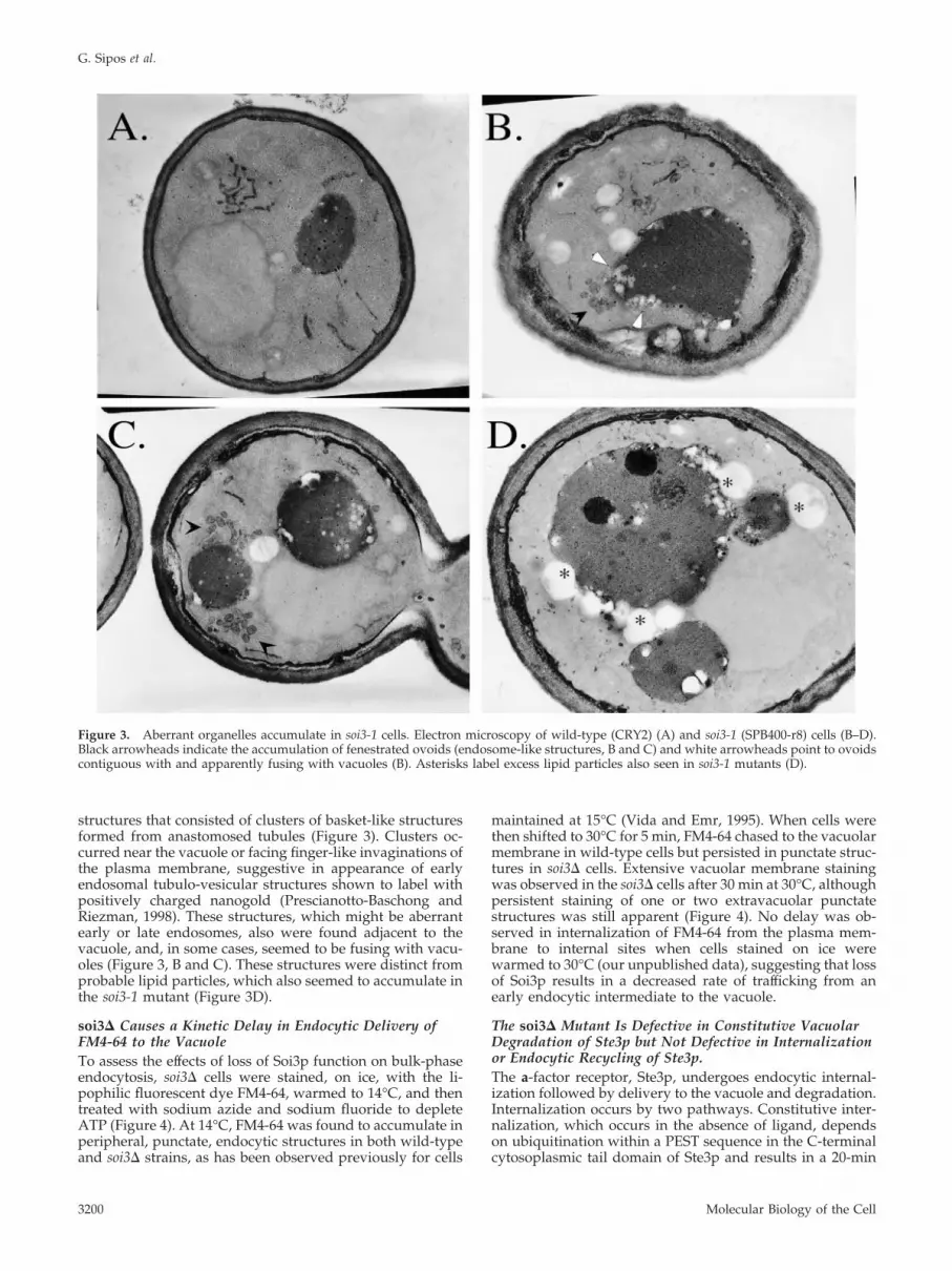
structures that consisted of clusters of basket-like structuresformed from anastomosed tubules (Figure 3). Clusters oc-curred near the vacuole or facing finger-like invaginations ofthe plasma membrane, suggestive in appearance of earlyendosomal tubulo-vesicular structures shown to label withpositively charged nanogold (Prescianotto-Baschong andRiezman, 1998). These structures, which might be aberrantearly or late endosomes, also were found adjacent to thevacuole, and, in some cases, seemed to be fusing with vacu-oles (Figure 3, B and C). These structures were distinct fromprobable lipid particles, which also seemed to accumulate inthe soi3-1 mutant (Figure 3D).
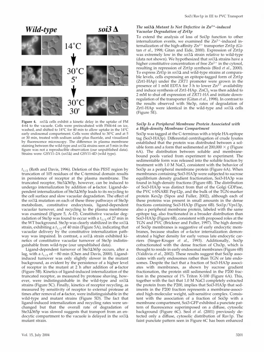
soi3� Causes a Kinetic Delay in Endocytic Delivery ofFM4-64 to the VacuoleTo assess the effects of loss of Soi3p function on bulk-phaseendocytosis, soi3� cells were stained, on ice, with the li-pophilic fluorescent dye FM4-64, warmed to 14°C, and thentreated with sodium azide and sodium fluoride to depleteATP (Figure 4). At 14°C, FM4-64 was found to accumulate inperipheral, punctate, endocytic structures in both wild-typeand soi3� strains, as has been observed previously for cells
maintained at 15°C (Vida and Emr, 1995). When cells werethen shifted to 30°C for 5 min, FM4-64 chased to the vacuolarmembrane in wild-type cells but persisted in punctate struc-tures in soi3� cells. Extensive vacuolar membrane stainingwas observed in the soi3� cells after 30 min at 30°C, althoughpersistent staining of one or two extravacuolar punctatestructures was still apparent (Figure 4). No delay was ob-served in internalization of FM4-64 from the plasma mem-brane to internal sites when cells stained on ice werewarmed to 30°C (our unpublished data), suggesting that lossof Soi3p results in a decreased rate of trafficking from anearly endocytic intermediate to the vacuole.
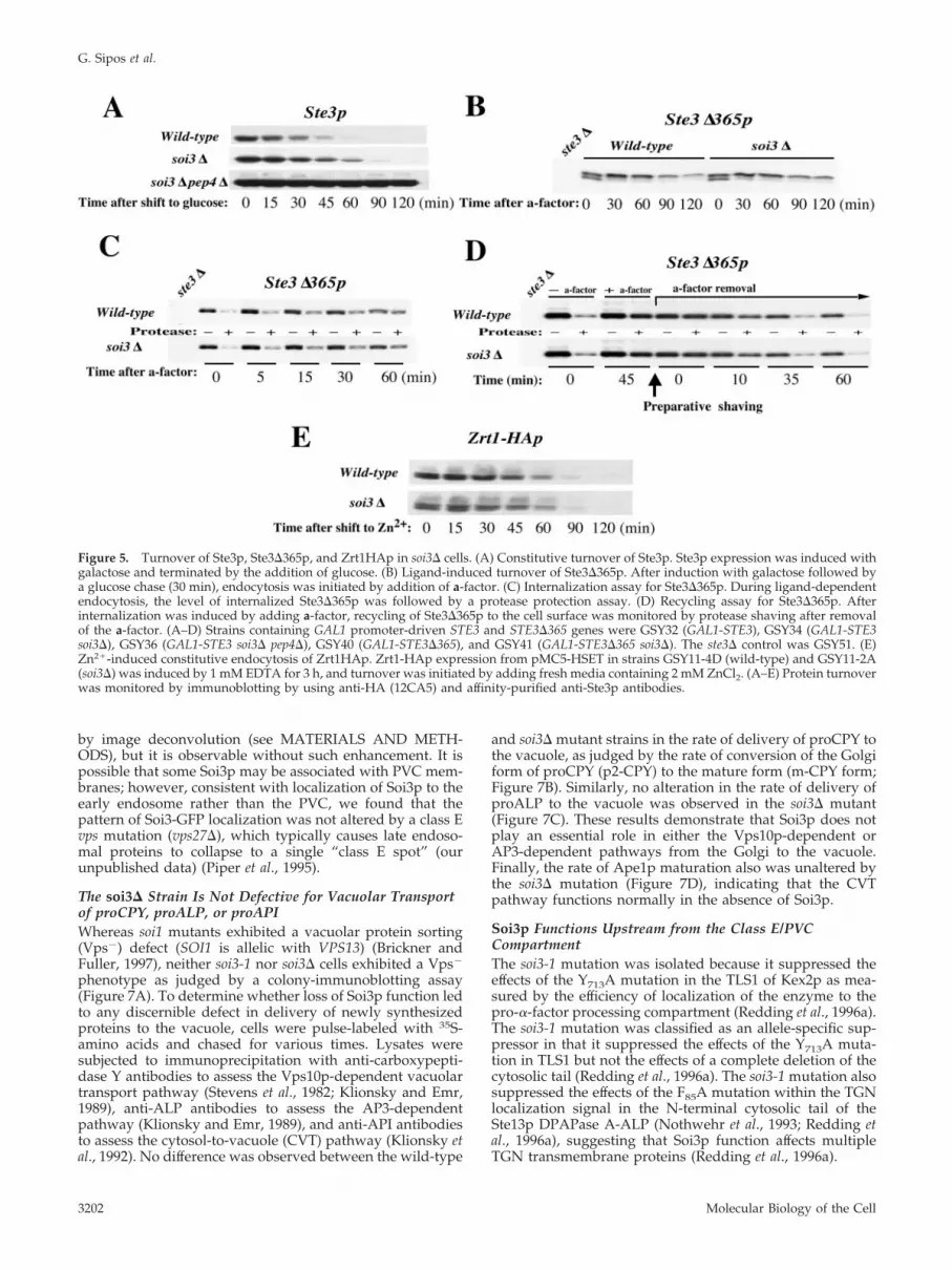
The soi3� Mutant Is Defective in Constitutive VacuolarDegradation of Ste3p but Not Defective in Internalizationor Endocytic Recycling of Ste3p.The a-factor receptor, Ste3p, undergoes endocytic internal-ization followed by delivery to the vacuole and degradation.Internalization occurs by two pathways. Constitutive inter-nalization, which occurs in the absence of ligand, dependson ubiquitination within a PEST sequence in the C-terminalcytosoplasmic tail domain of Ste3p and results in a 20-min
Figure 3. Aberrant organelles accumulate in soi3-1 cells. Electron microscopy of wild-type (CRY2) (A) and soi3-1 (SPB400-r8) cells (B–D).Black arrowheads indicate the accumulation of fenestrated ovoids (endosome-like structures, B and C) and white arrowheads point to ovoidscontiguous with and apparently fusing with vacuoles (B). Asterisks label excess lipid particles also seen in soi3-1 mutants (D).
G. Sipos et al.
Molecular Biology of the Cell3200
t1/2 (Roth and Davis, 1996). Deletion of this PEST region bytruncation of 105 residues of the C-terminal domain resultsin persistence of receptor at the plasma membrane. Thetruncated receptor, Ste3�365p, however, can be induced toundergo internalization by addition of a-factor. Ligand-de-pendent internalization of Ste3�365p leads to its recycling tothe cell surface and slow vacuolar degradation. The effect ofthe soi3� mutation on each of these three pathways of Ste3pmetabolism, constitutive endocytosis, ligand-dependentvacuolar turnover, and recycling to the plasma membranewas examined (Figure 5, A–D). Constitutive vacuolar deg-radation of Ste3p was found to occur with a t1/2 of 27 min inthe WT background. Ste3p was clearly stabilized in the soi3�strain, exhibiting a t1/2 of 40 min (Figure 5A), indicating thatvacuolar delivery by the constitutive internalization path-way was impaired. In contrast, a soi1� strain exhibited ki-netics of constitutive vacuolar turnover of Ste3p indistin-guishable from wild-type (our unpublished data).
Ligand-dependent turnover of Ste3�365p occurs, after alag, with a t1/2 of �80 min (Chen and Davis, 2000). Ligand-induced turnover was only slightly slower in the mutantbackground, as evident by the persistence of a higher levelof receptor in the mutant at 2 h after addition of a-factor(Figure 5B). Kinetics of ligand-induced internalization of thetruncated receptor, as measured by protease shaving, how-ever, were indistinguishable in the wild-type and soi3�strains (Figure 5C). Finally, kinetics of receptor recycling, asmeasured by sensitivity of receptor to external protease attimes after removal of a-factor, were indistinguishable in thewild-type and mutant strains (Figure 5D). The fact thatligand-induced internalization and recycling rates were un-changed but that the overall rate of degradation ofSte3�365p was slowed suggests that transport from an en-docytic compartment to the vacuole is delayed in the soi3�mutant strain.
The soi3� Mutant Is Not Defective in Zn2�-inducedVacuolar Degradation of Zrt1pTo extend the analysis of loss of Soi3p function to otherinternalization events, we examined the Zn2�-induced in-ternalization of the high-affinity Zn2� transporter Zrt1p (Gi-tan et al., 1998; Gitan and Eide, 2000). Expression of Zrt1pwas extremely low in the soi3� strain relative to wild-type(data not shown). We hypothesized that soi3� strains have ahigher constitutive concentration of free Zn2� in the cytosol,resulting in repression of Zrt1p synthesis (Bird et al., 2000).To express Zrt1p in soi3� and wild-type strains at compara-ble levels, cells expressing an epitope-tagged form of Zrt1p(Zrt1-HAp) under the ZRT1 promoter were grown in thepresence of 1 mM EDTA for 3 h to lower Zn2� availabilityand induce synthesis of Zrt1-HAp. ZnCl2 was then added to2 mM to shut off expression of ZRT1-HA and initiate down-regulation of the transporter (Gitan et al., 1998). In contrast tothe results observed with Ste3p, rates of degradation ofZrt1-HAp were identical in the wild-type and soi3� cells(Figure 5E).
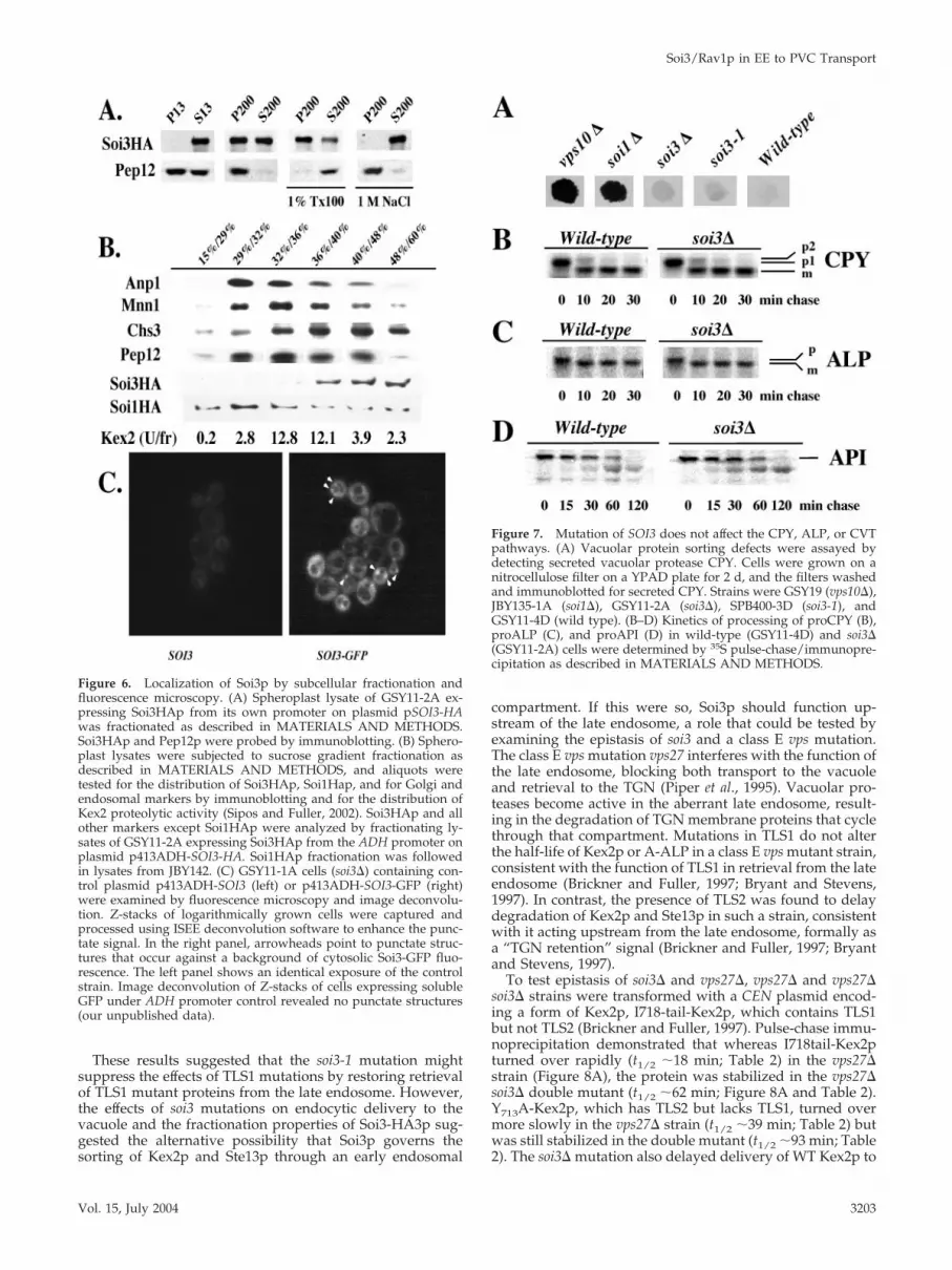
Soi3p Is a Peripheral Membrane Protein Associated witha High-density Membrane CompartmentSoi3p was tagged at the C-terminus with a triple HA-epitopetag (Soi3-HA3p). Differential centrifugation of crude lysatesestablished that the protein was distributed between a sol-uble form and a form that sedimented at 200,000 � g (Figure6A). The distribution between soluble and membrane-bound pools varied from experiment to experiment. Thesedimentable form was released into the soluble fraction bytreatment with 1.0 M NaCl, consistent with the behavior ofSoi3p as a peripheral membrane protein (Figure 6A). Whenmembranes containing Soi3-HA3p were subjected to sucroseequilibrium density gradient fractionation, Soi3-HA3p wasfound in high-density fractions (Figure 6B). The distributionof Soi3-HA3p was distinct from that of the Golgi GDPase,the PVC t-SNARE Pep12p, and the bulk of the TGN-markerprotein Kex2p (Sipos and Fuller, 2002), although each ofthese proteins was present in small amounts in the densefractions containing Soi3-HA3p (Figure 6B). Soi1p/Vps13p,also a peripheral membrane protein, labeled with the sameepitope tag, also fractionated in a broader distribution thanSoi3-HA3p (Figure 6B), consistent with proposed roles at theTGN and PVC (Brickner and Fuller, 1997). The high densityof Soi3p membranes is suggestive of early endocytic mem-branes, because studies of �-factor internalization demon-strated a higher density for early versus late endocytic car-riers (Singer-Kruger et al., 1993). Additionally, Soi3pcofractionated with the dense fraction of Chs3p, which isthought to reside in early endosomal membranes (Figure 6B)(Valdivia et al., 2002). These results suggest that Soi3p asso-ciates with early endosomes rather than TGN or late endo-somes. Despite the fact that a fraction of Soi3-HA3p associ-ates with membranes, as shown by sucrose gradientfractionation, the protein still sedimented in the P200 frac-tion in the presence of 1% Triton X-100 (Figure 6A). This,together with the fact that 1.0 M NaCl completely extractedthe protein from the P200, implies that Soi3-HA3p that sed-iments in the P200 fraction represents a membrane-associ-ated high-molecular weight, salt-sensitive complex. Consis-tent with the association of a fraction of Soi3p with amembrane compartment, Soi3-GFP exhibited a punctate pat-tern of fluorescence superimposed on a diffuse, cytosolicbackground (Figure 6C). Seol et al. (2001) previously de-tected only a diffuse, cytosolic distribution of Rav1p. Thefaint punctate pattern seen in Figure 6C has been enhanced
Figure 4. soi3� cells exhibit a kinetic delay in the uptake of FM4-64 to the vacuole. Cells were preincubated with FM4-64 on ice,washed, and shifted to 14°C for 40 min to allow uptake in the 14°Cearly endosomal compartment. Cells were shifted to 30°C and at 5or 30 min, treated with sodium azide plus fluoride, and visualizedby fluorescence microscopy. The difference in plasma membranestaining between the wild-type and soi3� strains seen at 5 min in thefigure was not a reproducible observation (our unpublished data).Strains were: GSY11–2A (soi3�) and GSY11-4D (wild type).
Soi3/Rav1p in EE to PVC Transport
Vol. 15, July 2004 3201
by image deconvolution (see MATERIALS AND METH-ODS), but it is observable without such enhancement. It ispossible that some Soi3p may be associated with PVC mem-branes; however, consistent with localization of Soi3p to theearly endosome rather than the PVC, we found that thepattern of Soi3-GFP localization was not altered by a class Evps mutation (vps27�), which typically causes late endoso-mal proteins to collapse to a single “class E spot” (ourunpublished data) (Piper et al., 1995).
The soi3� Strain Is Not Defective for Vacuolar Transportof proCPY, proALP, or proAPIWhereas soi1 mutants exhibited a vacuolar protein sorting(Vps�) defect (SOI1 is allelic with VPS13) (Brickner andFuller, 1997), neither soi3-1 nor soi3� cells exhibited a Vps�
phenotype as judged by a colony-immunoblotting assay(Figure 7A). To determine whether loss of Soi3p function ledto any discernible defect in delivery of newly synthesizedproteins to the vacuole, cells were pulse-labeled with 35S-amino acids and chased for various times. Lysates weresubjected to immunoprecipitation with anti-carboxypepti-dase Y antibodies to assess the Vps10p-dependent vacuolartransport pathway (Stevens et al., 1982; Klionsky and Emr,1989), anti-ALP antibodies to assess the AP3-dependentpathway (Klionsky and Emr, 1989), and anti-API antibodiesto assess the cytosol-to-vacuole (CVT) pathway (Klionsky etal., 1992). No difference was observed between the wild-type
and soi3� mutant strains in the rate of delivery of proCPY tothe vacuole, as judged by the rate of conversion of the Golgiform of proCPY (p2-CPY) to the mature form (m-CPY form;Figure 7B). Similarly, no alteration in the rate of delivery ofproALP to the vacuole was observed in the soi3� mutant(Figure 7C). These results demonstrate that Soi3p does notplay an essential role in either the Vps10p-dependent orAP3-dependent pathways from the Golgi to the vacuole.Finally, the rate of Ape1p maturation also was unaltered bythe soi3� mutation (Figure 7D), indicating that the CVTpathway functions normally in the absence of Soi3p.
Soi3p Functions Upstream from the Class E/PVCCompartmentThe soi3-1 mutation was isolated because it suppressed theeffects of the Y713A mutation in the TLS1 of Kex2p as mea-sured by the efficiency of localization of the enzyme to thepro-�-factor processing compartment (Redding et al., 1996a).The soi3-1 mutation was classified as an allele-specific sup-pressor in that it suppressed the effects of the Y713A muta-tion in TLS1 but not the effects of a complete deletion of thecytosolic tail (Redding et al., 1996a). The soi3-1 mutation alsosuppressed the effects of the F85A mutation within the TGNlocalization signal in the N-terminal cytosolic tail of theSte13p DPAPase A-ALP (Nothwehr et al., 1993; Redding etal., 1996a), suggesting that Soi3p function affects multipleTGN transmembrane proteins (Redding et al., 1996a).
Figure 5. Turnover of Ste3p, Ste3�365p, and Zrt1HAp in soi3� cells. (A) Constitutive turnover of Ste3p. Ste3p expression was induced withgalactose and terminated by the addition of glucose. (B) Ligand-induced turnover of Ste3�365p. After induction with galactose followed bya glucose chase (30 min), endocytosis was initiated by addition of a-factor. (C) Internalization assay for Ste3�365p. During ligand-dependentendocytosis, the level of internalized Ste3�365p was followed by a protease protection assay. (D) Recycling assay for Ste3�365p. Afterinternalization was induced by adding a-factor, recycling of Ste3�365p to the cell surface was monitored by protease shaving after removalof the a-factor. (A–D) Strains containing GAL1 promoter-driven STE3 and STE3�365 genes were GSY32 (GAL1-STE3), GSY34 (GAL1-STE3soi3�), GSY36 (GAL1-STE3 soi3� pep4�), GSY40 (GAL1-STE3�365), and GSY41 (GAL1-STE3�365 soi3�). The ste3� control was GSY51. (E)Zn2�-induced constitutive endocytosis of Zrt1HAp. Zrt1-HAp expression from pMC5-HSET in strains GSY11-4D (wild-type) and GSY11-2A(soi3�) was induced by 1 mM EDTA for 3 h, and turnover was initiated by adding fresh media containing 2 mM ZnCl2. (A–E) Protein turnoverwas monitored by immunoblotting by using anti-HA (12CA5) and affinity-purified anti-Ste3p antibodies.
G. Sipos et al.
Molecular Biology of the Cell3202
These results suggested that the soi3-1 mutation mightsuppress the effects of TLS1 mutations by restoring retrievalof TLS1 mutant proteins from the late endosome. However,the effects of soi3 mutations on endocytic delivery to thevacuole and the fractionation properties of Soi3-HA3p sug-gested the alternative possibility that Soi3p governs thesorting of Kex2p and Ste13p through an early endosomal
compartment. If this were so, Soi3p should function up-stream of the late endosome, a role that could be tested byexamining the epistasis of soi3 and a class E vps mutation.The class E vps mutation vps27 interferes with the function ofthe late endosome, blocking both transport to the vacuoleand retrieval to the TGN (Piper et al., 1995). Vacuolar pro-teases become active in the aberrant late endosome, result-ing in the degradation of TGN membrane proteins that cyclethrough that compartment. Mutations in TLS1 do not alterthe half-life of Kex2p or A-ALP in a class E vps mutant strain,consistent with the function of TLS1 in retrieval from the lateendosome (Brickner and Fuller, 1997; Bryant and Stevens,1997). In contrast, the presence of TLS2 was found to delaydegradation of Kex2p and Ste13p in such a strain, consistentwith it acting upstream from the late endosome, formally asa “TGN retention” signal (Brickner and Fuller, 1997; Bryantand Stevens, 1997).
To test epistasis of soi3� and vps27�, vps27� and vps27�soi3� strains were transformed with a CEN plasmid encod-ing a form of Kex2p, I718-tail-Kex2p, which contains TLS1but not TLS2 (Brickner and Fuller, 1997). Pulse-chase immu-noprecipitation demonstrated that whereas I718tail-Kex2pturned over rapidly (t1/2 �18 min; Table 2) in the vps27�strain (Figure 8A), the protein was stabilized in the vps27�soi3� double mutant (t1/2 �62 min; Figure 8A and Table 2).Y713A-Kex2p, which has TLS2 but lacks TLS1, turned overmore slowly in the vps27� strain (t1/2 �39 min; Table 2) butwas still stabilized in the double mutant (t1/2 �93 min; Table2). The soi3� mutation also delayed delivery of WT Kex2p to
Figure 6. Localization of Soi3p by subcellular fractionation andfluorescence microscopy. (A) Spheroplast lysate of GSY11-2A ex-pressing Soi3HAp from its own promoter on plasmid pSOI3-HAwas fractionated as described in MATERIALS AND METHODS.Soi3HAp and Pep12p were probed by immunoblotting. (B) Sphero-plast lysates were subjected to sucrose gradient fractionation asdescribed in MATERIALS AND METHODS, and aliquots weretested for the distribution of Soi3HAp, Soi1Hap, and for Golgi andendosomal markers by immunoblotting and for the distribution ofKex2 proteolytic activity (Sipos and Fuller, 2002). Soi3HAp and allother markers except Soi1HAp were analyzed by fractionating ly-sates of GSY11-2A expressing Soi3HAp from the ADH promoter onplasmid p413ADH-SOI3-HA. Soi1HAp fractionation was followedin lysates from JBY142. (C) GSY11-1A cells (soi3�) containing con-trol plasmid p413ADH-SOI3 (left) or p413ADH-SOI3-GFP (right)were examined by fluorescence microscopy and image deconvolu-tion. Z-stacks of logarithmically grown cells were captured andprocessed using ISEE deconvolution software to enhance the punc-tate signal. In the right panel, arrowheads point to punctate struc-tures that occur against a background of cytosolic Soi3-GFP fluo-rescence. The left panel shows an identical exposure of the controlstrain. Image deconvolution of Z-stacks of cells expressing solubleGFP under ADH promoter control revealed no punctate structures(our unpublished data).
Figure 7. Mutation of SOI3 does not affect the CPY, ALP, or CVTpathways. (A) Vacuolar protein sorting defects were assayed bydetecting secreted vacuolar protease CPY. Cells were grown on anitrocellulose filter on a YPAD plate for 2 d, and the filters washedand immunoblotted for secreted CPY. Strains were GSY19 (vps10�),JBY135-1A (soi1�), GSY11-2A (soi3�), SPB400-3D (soi3-1), andGSY11-4D (wild type). (B–D) Kinetics of processing of proCPY (B),proALP (C), and proAPI (D) in wild-type (GSY11-4D) and soi3�(GSY11-2A) cells were determined by 35S pulse-chase/immunopre-cipitation as described in MATERIALS AND METHODS.
Soi3/Rav1p in EE to PVC Transport
Vol. 15, July 2004 3203
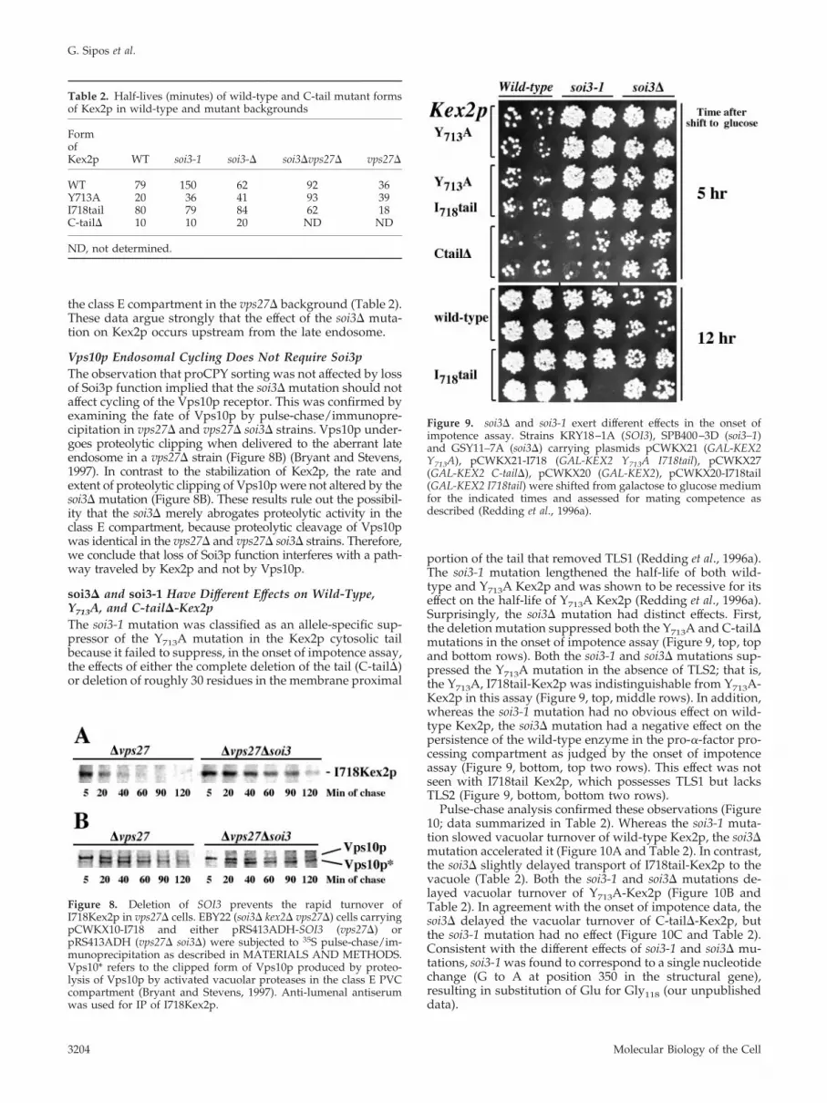
the class E compartment in the vps27� background (Table 2).These data argue strongly that the effect of the soi3� muta-tion on Kex2p occurs upstream from the late endosome.
Vps10p Endosomal Cycling Does Not Require Soi3pThe observation that proCPY sorting was not affected by lossof Soi3p function implied that the soi3� mutation should notaffect cycling of the Vps10p receptor. This was confirmed byexamining the fate of Vps10p by pulse-chase/immunopre-cipitation in vps27� and vps27� soi3� strains. Vps10p under-goes proteolytic clipping when delivered to the aberrant lateendosome in a vps27� strain (Figure 8B) (Bryant and Stevens,1997). In contrast to the stabilization of Kex2p, the rate andextent of proteolytic clipping of Vps10p were not altered by thesoi3� mutation (Figure 8B). These results rule out the possibil-ity that the soi3� merely abrogates proteolytic activity in theclass E compartment, because proteolytic cleavage of Vps10pwas identical in the vps27� and vps27� soi3� strains. Therefore,we conclude that loss of Soi3p function interferes with a path-way traveled by Kex2p and not by Vps10p.
soi3� and soi3-1 Have Different Effects on Wild-Type,Y713A, and C-tail�-Kex2pThe soi3-1 mutation was classified as an allele-specific sup-pressor of the Y713A mutation in the Kex2p cytosolic tailbecause it failed to suppress, in the onset of impotence assay,the effects of either the complete deletion of the tail (C-tail�)or deletion of roughly 30 residues in the membrane proximal
portion of the tail that removed TLS1 (Redding et al., 1996a).The soi3-1 mutation lengthened the half-life of both wild-type and Y713A Kex2p and was shown to be recessive for itseffect on the half-life of Y713A Kex2p (Redding et al., 1996a).Surprisingly, the soi3� mutation had distinct effects. First,the deletion mutation suppressed both the Y713A and C-tail�mutations in the onset of impotence assay (Figure 9, top, topand bottom rows). Both the soi3-1 and soi3� mutations sup-pressed the Y713A mutation in the absence of TLS2; that is,the Y713A, I718tail-Kex2p was indistinguishable from Y713A-Kex2p in this assay (Figure 9, top, middle rows). In addition,whereas the soi3-1 mutation had no obvious effect on wild-type Kex2p, the soi3� mutation had a negative effect on thepersistence of the wild-type enzyme in the pro-�-factor pro-cessing compartment as judged by the onset of impotenceassay (Figure 9, bottom, top two rows). This effect was notseen with I718tail Kex2p, which possesses TLS1 but lacksTLS2 (Figure 9, bottom, bottom two rows).
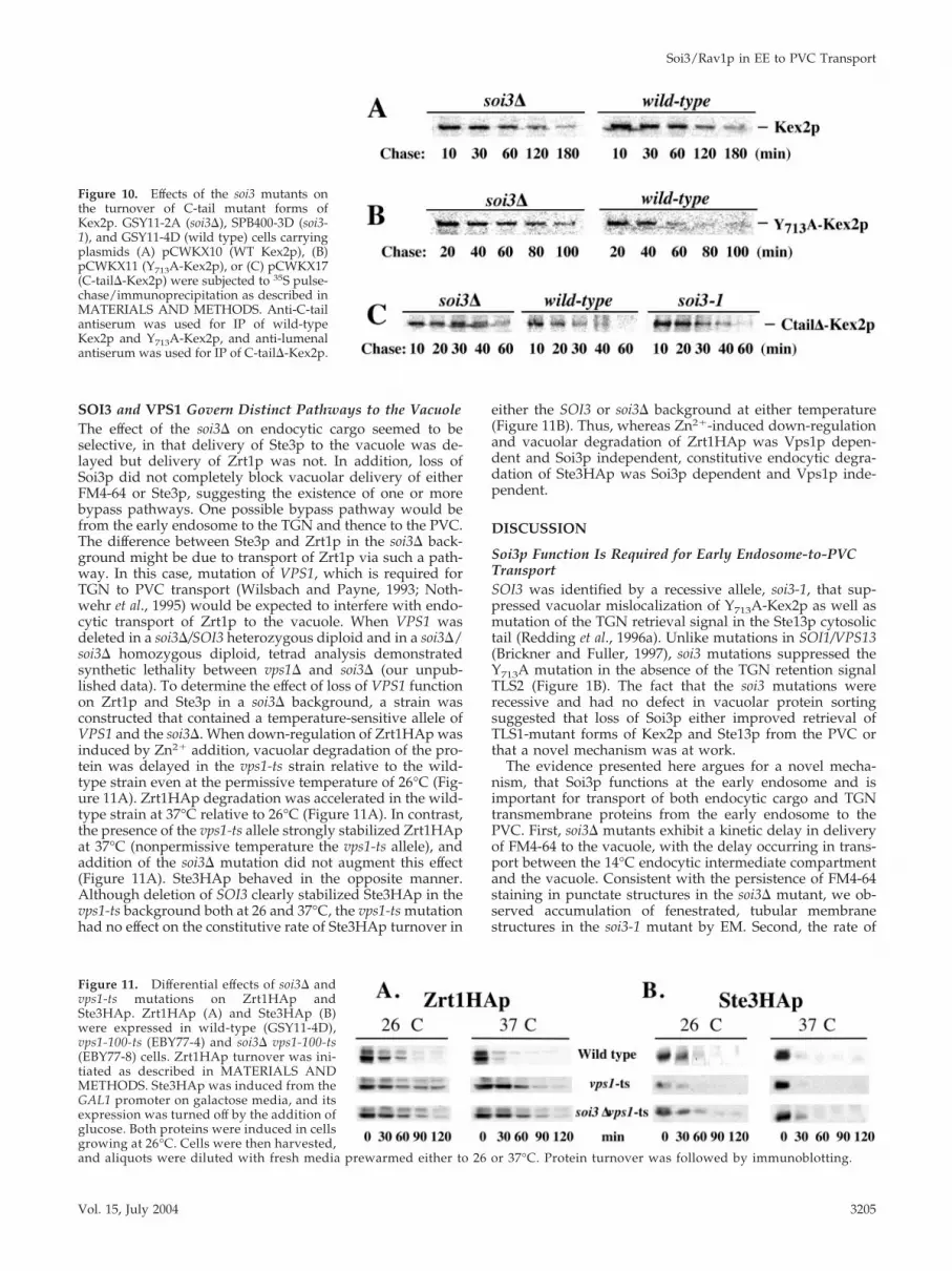
Pulse-chase analysis confirmed these observations (Figure10; data summarized in Table 2). Whereas the soi3-1 muta-tion slowed vacuolar turnover of wild-type Kex2p, the soi3�mutation accelerated it (Figure 10A and Table 2). In contrast,the soi3� slightly delayed transport of I718tail-Kex2p to thevacuole (Table 2). Both the soi3-1 and soi3� mutations de-layed vacuolar turnover of Y713A-Kex2p (Figure 10B andTable 2). In agreement with the onset of impotence data, thesoi3� delayed the vacuolar turnover of C-tail�-Kex2p, butthe soi3-1 mutation had no effect (Figure 10C and Table 2).Consistent with the different effects of soi3-1 and soi3� mu-tations, soi3-1 was found to correspond to a single nucleotidechange (G to A at position 350 in the structural gene),resulting in substitution of Glu for Gly118 (our unpublisheddata).
Table 2. Half-lives (minutes) of wild-type and C-tail mutant formsof Kex2p in wild-type and mutant backgrounds
FormofKex2p WT soi3-1 soi3-� soi3�vps27� vps27�
WT 79 150 62 92 36Y713A 20 36 41 93 39I718tail 80 79 84 62 18C-tail� 10 10 20 ND ND
ND, not determined.
Figure 8. Deletion of SOI3 prevents the rapid turnover ofI718Kex2p in vps27� cells. EBY22 (soi3� kex2� vps27�) cells carryingpCWKX10-I718 and either pRS413ADH-SOI3 (vps27�) orpRS413ADH (vps27� soi3�) were subjected to 35S pulse-chase/im-munoprecipitation as described in MATERIALS AND METHODS.Vps10* refers to the clipped form of Vps10p produced by proteo-lysis of Vps10p by activated vacuolar proteases in the class E PVCcompartment (Bryant and Stevens, 1997). Anti-lumenal antiserumwas used for IP of I718Kex2p.
Figure 9. soi3� and soi3-1 exert different effects in the onset ofimpotence assay. Strains KRY18–1A (SOI3), SPB400–3D (soi3–1)and GSY11–7A (soi3�) carrying plasmids pCWKX21 (GAL-KEX2Y713A), pCWKX21-I718 (GAL-KEX2 Y713A I718tail), pCWKX27(GAL-KEX2 C-tail�), pCWKX20 (GAL-KEX2), pCWKX20-I718tail(GAL-KEX2 I718tail) were shifted from galactose to glucose mediumfor the indicated times and assessed for mating competence asdescribed (Redding et al., 1996a).
G. Sipos et al.
Molecular Biology of the Cell3204
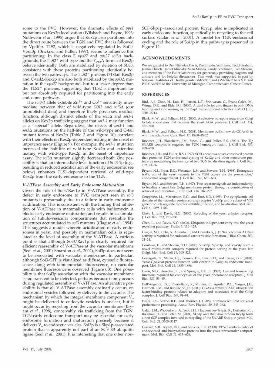
SOI3 and VPS1 Govern Distinct Pathways to the VacuoleThe effect of the soi3� on endocytic cargo seemed to beselective, in that delivery of Ste3p to the vacuole was de-layed but delivery of Zrt1p was not. In addition, loss ofSoi3p did not completely block vacuolar delivery of eitherFM4-64 or Ste3p, suggesting the existence of one or morebypass pathways. One possible bypass pathway would befrom the early endosome to the TGN and thence to the PVC.The difference between Ste3p and Zrt1p in the soi3� back-ground might be due to transport of Zrt1p via such a path-way. In this case, mutation of VPS1, which is required forTGN to PVC transport (Wilsbach and Payne, 1993; Noth-wehr et al., 1995) would be expected to interfere with endo-cytic transport of Zrt1p to the vacuole. When VPS1 wasdeleted in a soi3�/SOI3 heterozygous diploid and in a soi3�/soi3� homozygous diploid, tetrad analysis demonstratedsynthetic lethality between vps1� and soi3� (our unpub-lished data). To determine the effect of loss of VPS1 functionon Zrt1p and Ste3p in a soi3� background, a strain wasconstructed that contained a temperature-sensitive allele ofVPS1 and the soi3�. When down-regulation of Zrt1HAp wasinduced by Zn2� addition, vacuolar degradation of the pro-tein was delayed in the vps1-ts strain relative to the wild-type strain even at the permissive temperature of 26°C (Fig-ure 11A). Zrt1HAp degradation was accelerated in the wild-type strain at 37°C relative to 26°C (Figure 11A). In contrast,the presence of the vps1-ts allele strongly stabilized Zrt1HApat 37°C (nonpermissive temperature the vps1-ts allele), andaddition of the soi3� mutation did not augment this effect(Figure 11A). Ste3HAp behaved in the opposite manner.Although deletion of SOI3 clearly stabilized Ste3HAp in thevps1-ts background both at 26 and 37°C, the vps1-ts mutationhad no effect on the constitutive rate of Ste3HAp turnover in
either the SOI3 or soi3� background at either temperature(Figure 11B). Thus, whereas Zn2�-induced down-regulationand vacuolar degradation of Zrt1HAp was Vps1p depen-dent and Soi3p independent, constitutive endocytic degra-dation of Ste3HAp was Soi3p dependent and Vps1p inde-pendent.
DISCUSSION
Soi3p Function Is Required for Early Endosome-to-PVCTransportSOI3 was identified by a recessive allele, soi3-1, that sup-pressed vacuolar mislocalization of Y713A-Kex2p as well asmutation of the TGN retrieval signal in the Ste13p cytosolictail (Redding et al., 1996a). Unlike mutations in SOI1/VPS13(Brickner and Fuller, 1997), soi3 mutations suppressed theY713A mutation in the absence of the TGN retention signalTLS2 (Figure 1B). The fact that the soi3 mutations wererecessive and had no defect in vacuolar protein sortingsuggested that loss of Soi3p either improved retrieval ofTLS1-mutant forms of Kex2p and Ste13p from the PVC orthat a novel mechanism was at work.
The evidence presented here argues for a novel mecha-nism, that Soi3p functions at the early endosome and isimportant for transport of both endocytic cargo and TGNtransmembrane proteins from the early endosome to thePVC. First, soi3� mutants exhibit a kinetic delay in deliveryof FM4-64 to the vacuole, with the delay occurring in trans-port between the 14°C endocytic intermediate compartmentand the vacuole. Consistent with the persistence of FM4-64staining in punctate structures in the soi3� mutant, we ob-served accumulation of fenestrated, tubular membranestructures in the soi3-1 mutant by EM. Second, the rate of
Figure 10. Effects of the soi3 mutants onthe turnover of C-tail mutant forms ofKex2p. GSY11-2A (soi3�), SPB400-3D (soi3-1), and GSY11-4D (wild type) cells carryingplasmids (A) pCWKX10 (WT Kex2p), (B)pCWKX11 (Y713A-Kex2p), or (C) pCWKX17(C-tail�-Kex2p) were subjected to 35S pulse-chase/immunoprecipitation as described inMATERIALS AND METHODS. Anti-C-tailantiserum was used for IP of wild-typeKex2p and Y713A-Kex2p, and anti-lumenalantiserum was used for IP of C-tail�-Kex2p.
Figure 11. Differential effects of soi3� andvps1-ts mutations on Zrt1HAp andSte3HAp. Zrt1HAp (A) and Ste3HAp (B)were expressed in wild-type (GSY11-4D),vps1-100-ts (EBY77-4) and soi3� vps1-100-ts(EBY77-8) cells. Zrt1HAp turnover was ini-tiated as described in MATERIALS ANDMETHODS. Ste3HAp was induced from theGAL1 promoter on galactose media, and itsexpression was turned off by the addition ofglucose. Both proteins were induced in cellsgrowing at 26°C. Cells were then harvested,and aliquots were diluted with fresh media prewarmed either to 26 or 37°C. Protein turnover was followed by immunoblotting.
Soi3/Rav1p in EE to PVC Transport
Vol. 15, July 2004 3205
constitutive vacuolar turnover of the Ste3p a-factor receptorwas substantially decreased in the soi3� mutant, whereasrates of internalization and recycling of Ste3p were unal-tered. Third, membrane-associated Soi3p cofractionatedwith Chs3p in a dense fraction characteristic of early endo-somes. Consistent with early endosome localization, a frac-tion of Soi3-GFP localized in a peripheral, punctate pattern.Fourth, soi3 mutations delayed delivery of Y713A-Kex2p, butnot Vps10p, to the class E (aberrant PVC) compartment. TheVps10p-dependent, AP3-dependent, and CVT vacuolar tar-geting pathways were unaffected by loss of Soi3p. Each ofthese pathways is likely to require normal TGN functioning,including the CVT pathway, which depends on the TGNSNAREs Tlg1p and Tlg2p (Reggiori et al., 2003). Finally, thesynthetic lethality of soi3� with vps1�, by itself, is consistentwith Soi3p having a role in endocytotic transport to thevacuole, given the synthetic lethality of vps1 with end4 mu-tations (Nothwehr et al., 1995; Conibear and Stevens, 2000).Together, these data argue strongly for Soi3p function inearly endosome to PVC transport. We should emphasizehere, however, that we cannot rule out, on the basis of thesedata, roles for Soi3p in other transport steps, in particularbetween the PVC and the vacuole.
The soi3 null mutation only partially blocked FM4-64 traf-ficking to the vacuole and endocytic degradation of Ste3p,but such partial effects are in line with the effects of othermutants shown to affect vacuolar delivery of FM4-64 andSte3p (Gerrard et al., 2000; Heese-Peck et al., 2002; Shaw et al.,2003). The partial block may be due to the existence of abypass pathway, but the fact that the delayed vacuolardelivery of Ste3p (Figure 11B) and FM4-64 (our unpublisheddata) was not further impaired by a vps1-ts mutation arguesagainst transport of Ste3p and FM4-64 to the vacuole via theTGN-PVC pathway. The bypass pathway might instead cor-respond to direct fusion of early endosomes with the vacu-ole, a phenomenon suggested by EM analysis of the soi3-1mutant (Figure 3B). In contrast, the vps1-ts mutation did andthe soi3� did not affect endocytic degradation of Zrt1p (Fig-ures 5E and 11A), suggesting that Ste3p and Zrt1p followdifferent pathways to the vacuole.
Zinc Sensitivity of soi3 MutantsZn2� hypersensitivity of the soi3 mutants initially led us toexamine endocytosis of Zrt1p. However, although we foundthat vacuolar delivery of Zrt1p induced by Zn2� was unal-tered in the soi3 mutant, expression of Zrt1p was markedlyreduced in growth in standard synthetic growth medium(our unpublished data), suggesting that soi3 mutations dis-rupt Zn2� homeostasis, leading to higher cytosolic Zn2�
levels. The possibility that soi3 mutants are defective in Zn2�
sequestration in the vacuole is supported by the finding thatZRC1, which encodes a Zn2� transporter in the vacuolarmembrane (MacDiarmid et al., 2002), was isolated as a dou-ble-copy suppressor in the initial cloning of the soi3-1 mu-tation by complementation of the Zn2�-hypersensitive phe-notype (clones 1 and 30 in Figure 2A; other data not shown).
Kex2p and Ste13p Cycle through the Early EndosomeEvidence presented here that Soi3p functions at the earlyendosome along with the effects of soi3 mutations on Y713A-Kex2p and F85A-A-ALP (Redding et al., 1996a) argue thatboth Kex2p and A-ALP (as surrogate for Ste13p) cyclethrough the early endosome. Other studies support this.First, Kex2p partially colocalizes with Chs5p, which in turncolocalizes with Chs3p, a protein whose localization to aninternal punctate organelle termed the chitosome requiresendocytosis (Santos and Snyder, 1997; Ziman et al., 1998).
Second, localization of Kex2p to an early or recycling endo-some also was suggested by colocalization of Kex2p withGFP-labeled Snc1p, the v-SNARE of secretory vesicles, topunctate internal sites presumed to be intermediates in therecycling of Snc1p from the plasma membrane to the TGN(Lewis et al., 2000; Ha et al., 2001). Third, mutations in INP53,which encodes a synaptojanin homolog, interfered with thefunction of the TLS2-like signal in A-ALP (Ha et al., 2001).Mutation of INP53 accelerated vacuolar delivery not only ofF85A-A-ALP but also of Kex2p without affecting Vps10p.Like mutations in the � subunit of the clathrin adaptorcomplex AP-1, inp53 mutations were found to result insynthetic growth defects when combined with double mu-tations in GGA1 and GGA2, leading to the suggestion thatInp53p and AP-1 were required for a TLS2-dependent trans-port pathway between the TGN and early endosome (Ha etal., 2003). Whereas, inp53 mutations increased the rate ofvacuolar delivery of TLS1� TLS2� forms of Kex2p andA-ALP (Y713A-Kex2p and F85A-A-ALP), soi3 mutations de-creased the rate of transit to the vacuole of the TLS1� TLS2�
proteins. This can be understood if Inp53p (and AP-1) func-tion in delivery of Ste13p and Kex2p to the early endosomeand Soi3p functions in transport from the early endosome tothe PVC.
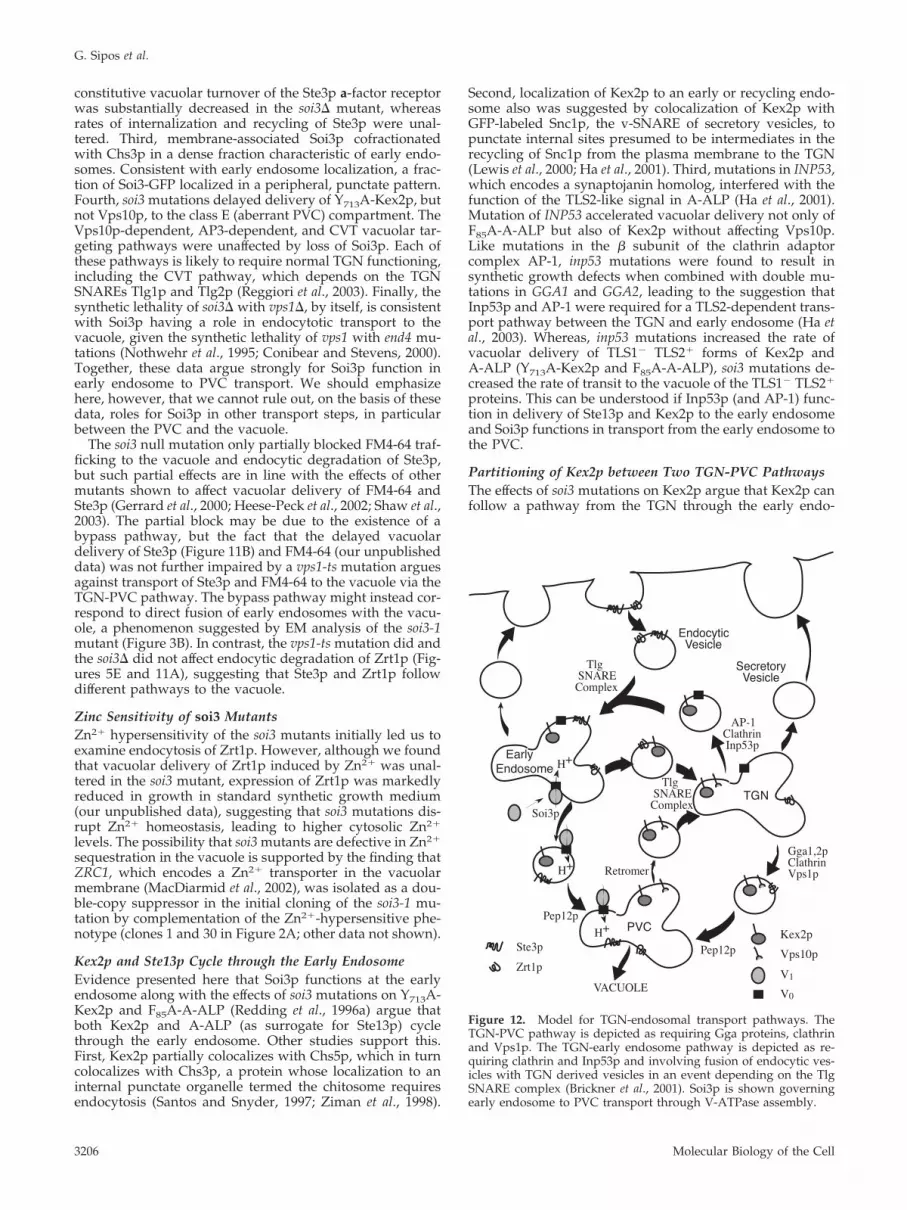
Partitioning of Kex2p between Two TGN-PVC PathwaysThe effects of soi3 mutations on Kex2p argue that Kex2p canfollow a pathway from the TGN through the early endo-
Figure 12. Model for TGN-endosomal transport pathways. TheTGN-PVC pathway is depicted as requiring Gga proteins, clathrinand Vps1p. The TGN-early endosome pathway is depicted as re-quiring clathrin and Inp53p and involving fusion of endocytic ves-icles with TGN derived vesicles in an event depending on the TlgSNARE complex (Brickner et al., 2001). Soi3p is shown governingearly endosome to PVC transport through V-ATPase assembly.
G. Sipos et al.
Molecular Biology of the Cell3206
some to the PVC. However, the dramatic effects of vps1mutations on Kex2p localization (Wilsbach and Payne, 1993;Nothwehr et al., 1995) argue that Kex2p also partitions intothe direct route between the TGN and PVC that is followedby Vps10p. TLS2, which is negatively regulated by Soi1/Vps13p (Brickner and Fuller, 1997), seems to influence thispartitioning. In the class E vps27 and vps27 soi3� back-grounds, the TLS2� wild-type and the Y713A-forms of Kex2pbehave identically. Both are stabilized by deletion of SOI3,consistent with these proteins partitioning identically be-tween the two pathways. The TLS2� proteins I718tail-Kex2pand C-tail�-Kex2p are also both stabilized by the soi3� mu-tation in the vps27 background, but to a lesser degree thanthe TLS2� proteins, suggesting that TLS2 is important forbut not absolutely required for partitioning into the earlyendosome pathway.
The soi3-1 allele exhibits Zn2� and Co2� sensitivity inter-mediate between that of wild-type SOI3 and soi3� (ourunpublished data) and therefore likely possesses residualfunction, although distinct effects of the soi3� and soi3-1alleles on Kex2p trafficking suggest that soi3-1 may functionas a “special” allele. Regardless, the effects of soi3-1 andsoi3� mutations on the half-life of the wild-type and C-tailmutant forms of Kex2p (Table 2 and Figure 10) correlatewith their effects on Kex2p-dependent mating in the onset ofimpotence assay (Figure 9). For example, the soi3-1 mutationincreased the half-life of wild-type Kex2p and extendedmating with wild-type Kex2p in the onset of impotenceassay. The soi3� mutation slightly decreased both. One pos-sibility is that an intermediate level function of Soi3-1p (e.g.,resulting in reduced acidification of the early endosome; seebelow) enhances TLS1-dependent retrieval of wild-typeKex2p from the early endosome to the TGN.
V-ATPase Assembly and Early Endosome MaturationGiven the role of Soi3/Rav1p in V-ATPase assembly, thedefect in early endosome to PVC transport seen in soi3mutants is presumably due to a failure in early endosomeacidification. This is consistent with the finding that inhibi-tion of V-ATPase in mammalian cells with bafilomycin Ablocks early endosome maturation and results in accumula-tion of tubulo-vesicular compartments that resemble thestructures accumulated in soi3 mutants (Clague et al., 1994).This suggests a model wherein acidification of early endo-somes in yeast, and possibly in mammalian cells, is regu-lated at the level of assembly of the V-ATPase. A curiouspoint is that although Soi3/Rav1p is clearly required forefficient reassembly of V-ATPase at the vacuolar membrane(Seol et al., 2001; Smardon et al., 2002), Soi3p does not seemto be associated with vacuolar membranes. In particular,although Soi3-GFP is visualized as diffuse, cytosolic fluores-cence along with faint punctate fluorescence, no vacuolarmembrane fluorescence is observed (Figure 6B). One possi-bility is that Soi3p association with the vacuolar membraneis too transient to be detected, perhaps because it only occursduring regulated assembly of V-ATPase. An alternative pos-sibility is that all V-ATPase assembly ordinarily occurs onendosomal vesicles followed by delivery to the vacuole. Themechanism by which the integral membrane component Vomight be delivered to endocytic vesicles is unclear, but itmight occur by recycling from the vacuolar membrane (Bry-ant et al., 1998), conceivably via trafficking from the TGN.TGN-early endosome transport may be essential for earlyendosome formation and maturation precisely because itdelivers Vo to endocytic vesicles. Soi3p is a Skp1p-associatedprotein that is apparently not part of an SCF E3 ubiquitinligase (Seol et al., 2001). It is interesting that one other non-
SCF-Skp1p–associated protein, Rcy1p, also is implicated inearly endosome function, specifically in recycling to the cellsurface (Galan et al., 2001). A model for TGN-endosomalcycling and the role of Soi3p in this pathway is presented inFigure 12.
ACKNOWLEDGMENTS
We are grateful to Drs. Nicholas Davis, David Eide, Scott Emr, Todd Graham,Jeanne Hirsch, Daniel Klionsky, Sean Munro, Randy Schekman, Tom Stevens,and members of the Fuller laboratory for generously providing reagents andantisera and for helpful discussions. This work was supported in part byNational Institutes of Health grants GM-50915 and GM-39697 to R.S.F. andP30 CA46592 to the University of Michigan Comprehensive Cancer Center.
REFERENCES
Bird, A.J., Zhao, H., Luo, H., Jensen, L.T., Srinivasan, C., Evans-Galea, M.,Winge, D.R., and Eide, D.J. (2000). A dual role for zinc fingers in both DNAbinding and zinc sensing by the Zap1 transcriptional activator. EMBO J. 19,3704–3713.
Black, M.W., and Pelham, H.R. (2000). A selective transport route from Golgito late endosomes that requires the yeast GGA proteins. J. Cell Biol. 151,587–600.
Black, M.W., and Pelham, H.R. (2001). Membrane traffic: how do GGAs fit inwith the adaptors? Curr. Biol. 11, R460–R462.
Brickner, J.H., Blanchette, J.M., Sipos, G., and Fuller, R.S. (2001). The TlgSNARE complex is required for TGN homotypic fusion. J. Cell Biol. 155,969–978.
Brickner, J.H., and Fuller, R.S. (1997). SOI1 encodes a novel, conserved proteinthat promotes TGN-endosomal cycling of Kex2p and other membrane pro-teins by modulating the function of two TGN localization signals. J. Cell Biol.139, 23–36.
Bryant, N.J., Piper, R.C., Weisman, L.S., and Stevens, T.H. (1998). Retrogradetraffic out of the yeast vacuole to the TGN occurs via the prevacuolar/endosomal compartment. J. Cell Biol. 142, 651–663.
Bryant, N.J., and Stevens, T.H. (1997). Two separate signals act independentlyto localize a yeast late Golgi membrane protein through a combination ofretrieval and retention. J. Cell Biol. 136, 287–297.
Cereghino, J.L., Marcusson, E.G., and Emr, S.D. (1995). The cytoplasmic taildomain of the vacuolar protein sorting receptor Vps10p and a subset of VPSgene products regulate receptor stability, function, and localization. Mol. Biol.Cell 6, 1089–1102.
Chen, L., and Davis, N.G. (2000). Recycling of the yeast a-factor receptor.J. Cell Biol. 151, 731–738.
Chen, L., and Davis, N.G. (2002). Ubiquitin-independent entry into the yeastrecycling pathway. Traffic 3, 110–123.
Clague, M.J., Urbe, S., Aniento, F., and Gruenberg, J. (1994). Vacuolar ATPaseactivity is required for endosomal carrier vesicle formation. J. Biol. Chem. 269,21–24.
Conibear, E., and Stevens, T.H. (2000). Vps52p, Vps53p, and Vps54p form anovel multisubunit complex required for protein sorting at the yeast lateGolgi. Mol. Biol. Cell 11, 305–323.
Costaguta, G., Stefan, C.J., Bensen, E.S., Emr, S.D., and Payne, G.S. (2001).Yeast Gga coat proteins function with clathrin in Golgi to endosome trans-port. Mol. Biol. Cell 12, 1885–1896.
Davis, N.G., Horecka, J.L., and Sprague, G.F., Jr. (1993). Cis- and trans-actingfunctions required for endocytosis of the yeast pheromone receptors. J. CellBiol. 122, 53–65.
Dell’Angelica, E.C., Puertollano, R., Mullins, C., Aguilar, R.C., Vargas, J.D.,Hartnell, L.M., and Bonifacino, J.S. (2000). GGAs: a family of ADP ribosylationfactor-binding proteins related to adaptors and associated with the Golgicomplex. J. Cell Biol. 149, 81–94.
Fuller, R.S., Sterne, R.E., and Thorner, J. (1988). Enzymes required for yeastprohormone processing. Annu. Rev. Physiol. 50, 345–362.
Galan, J.M., Wiederkehr, A., Seol, J.H., Haguenauer-Tsapis, R., Deshaies, R.J.,Riezman, H., and Peter, M. (2001). Skp1p and the F-box protein Rcy1p forma non-SCF complex involved in recycling of the SNARE Snc1p in yeast. Mol.Cell. Biol. 21, 3105–3117.
Gerrard, S.R., Bryant, N.J., and Stevens, T.H. (2000). VPS21 controls entry ofendocytosed and biosynthetic proteins into the yeast prevacuolar compart-ment. Mol. Biol. Cell 11, 613–626.
Soi3/Rav1p in EE to PVC Transport
Vol. 15, July 2004 3207
Gitan, R.S., and Eide, D.J. (2000). Zinc-regulated ubiquitin conjugation signalsendocytosis of the yeast ZRT1 zinc transporter. Biochem. J. 346, 329–336.
Gitan, R.S., Luo, H., Rodgers, J., Broderius, M., and Eide, D. (1998). Zinc-induced inactivation of the yeast ZRT1 zinc transporter occurs through en-docytosis and vacuolar degradation. J. Biol. Chem. 273, 28617–28624.
Gueldener, U., Heinisch, J., Koehler, G.J., Voss, D., and Hegemann, J.H. (2002).A second set of loxP marker cassettes for Cre-mediated multiple gene knock-outs in budding yeast. Nucleic Acids Res. 30, e23.
Ha, S.A., Bunch, J.T., Hama, H., DeWald, D.B., and Nothwehr, S.F. (2001). Anovel mechanism for localizing membrane proteins to yeast trans-Golgi net-work requires function of synaptojanin-like protein. Mol. Biol. Cell 12, 3175–3190.
Ha, S.A., Torabinejad, J., DeWald, D.B., Wenk, M.R., Lucast, L., De Camilli, P.,Newitt, R.A., Aebersold, R., and Nothwehr, S.F. (2003). The synaptojanin-likeprotein Inp53/Sjl3 functions with clathrin in a yeast TGN-to-endosome path-way distinct from the GGA protein-dependent pathway. Mol. Biol. Cell 14,1319–1333.
Heese-Peck, A., Pichler, H., Zanolari, B., Watanabe, R., Daum, G., andRiezman, H. (2002). Multiple functions of sterols in yeast endocytosis. Mol.Biol. Cell 13, 2664–2680.
Hirst, J., Lui, W.W., Bright, N.A., Totty, N., Seaman, M.N., and Robinson, M.S.(2000). A family of proteins with gamma-adaptin and VHS domains thatfacilitate trafficking between the trans-Golgi network and the vacuole/lyso-some. J. Cell Biol. 149, 67–80.
Huang, K.M., D’Hondt, K., Riezman, H., and Lemmon, S.K. (1999). Clathrinfunctions in the absence of heterotetrameric adaptors and AP180-relatedproteins in yeast. EMBO J. 18, 3897–3908.
Klionsky, D.J., Cueva, R., and Yaver, D.S. (1992). Aminopeptidase I of Sac-charomyces cerevisiae is localized to the vacuole independent of the secretorypathway. J. Cell Biol. 119, 287–299.
Klionsky, D.J., and Emr, S.D. (1989). Membrane protein sorting: biosynthesis,transport and processing of yeast vacuolar alkaline phosphatase. EMBO J 8,2241–2250.
Kolehmainen, J., et al. (2003). Cohen syndrome is caused by mutations in anovel gene, COH1, encoding a transmembrane protein with a presumed rolein vesicle-mediated sorting and intracellular protein transport. Am. J. Hum.Genet. 72, 1359–1369.
Kraemer, C., Enklaar, T., Zabel, B., and Schmidt, E.R. (2000). Mapping andstructure of DMXL1, a human homologue of the DmX gene from Drosophilamelanogaster coding for a WD repeat protein. Genomics 64, 97–101.
Kraemer, C., Weil, B., Christmann, M., and Schmidt, E.R. (1998). The newgene DmX from Drosophila melanogaster encodes a novel WD-repeat protein.Gene 216, 267–276.
Lewis, M.J., Nichols, B.J., Prescianotto-Baschong, C., Riezman, H., and Pel-ham, H.R. (2000). Specific retrieval of the exocytic SNARE Snc1p from earlyyeast endosomes. Mol. Biol. Cell 11, 23–38.
MacDiarmid, C.W., Milanick, M.A., and Eide, D.J. (2002). Biochemical prop-erties of vacuolar zinc transport systems of Saccharomyces cerevisiae. J. Biol.Chem. 277, 39187–39194.
Mumberg, D., Muller, R., and Funk, M. (1995). Yeast vectors for the controlledexpression of heterologous proteins in different genetic backgrounds. Gene156, 119–122.
Nothwehr, S.F., Conibear, E., and Stevens, T.H. (1995). Golgi and vacuolarmembrane proteins reach the vacuole in vps1 mutant yeast cells via theplasma membrane. J. Cell Biol. 129, 35–46.
Nothwehr, S.F., Roberts, C.J., and Stevens, T.H. (1993). Membrane proteinretention in the yeast Golgi apparatus: dipeptidyl aminopeptidase A is re-tained by a cytoplasmic signal containing aromatic residues. J. Cell Biol. 121,1197–1209.
Perzov, N., Padler-Karavani, V., Nelson, H., and Nelson, N. (2002). Charac-terization of yeast V-ATPase mutants lacking Vph1p or Stv1p and the effecton endocytosis. J. Exp. Biol. 205, 1209–1219.
Phan, H.L., Finlay, J.A., Chu, D.S., Tan, P.K., Kirchhausen, T., and Payne, G.S.(1994). The Saccharomyces cerevisiae APS1 gene encodes a homolog of the smallsubunit of the mammalian clathrin AP-1 complex: evidence for functionalinteraction with clathrin at the Golgi complex. EMBO J. 13, 1706–1717.
Piper, R.C., Cooper, A.A., Yang, H., and Stevens, T.H. (1995). VPS27 controlsvacuolar and endocytic traffic through a prevacuolar compartment in Saccha-romyces cerevisiae. J. Cell Biol. 131, 603–617.
Prescianotto-Baschong, C., and Riezman, H. (1998). Morphology of the yeastendocytic pathway. Mol. Biol. Cell 9, 173–189.
Rambourg, A., Clermont, Y., and Kepes, F. (1993). Modulation of the Golgiapparatus in Saccharomyces cerevisiae sec7 mutants as seen by three-dimen-sional electron microscopy. Anat. Rec. 237, 441–452.
Rampoldi, L., et al. (2001). A conserved sorting-associated protein is mutant inchorea-acanthocytosis. Nat Genet 28, 119–120.
Redding, K., Brickner, J.H., Marschall, L.G., Nichols, J.W., and Fuller, R.S.(1996a). Allele-specific suppression of a defective trans-Golgi network (TGN)localization signal in Kex2p identifies three genes involved in localization ofTGN transmembrane proteins. Mol. Cell. Biol. 16, 6208–6217.
Redding, K., Holcomb, C., and Fuller, R.S. (1991). Immunolocalization of Kex2protease identifies a putative late Golgi compartment in the yeast Saccharo-myces cerevisiae. J. Cell Biol. 113, 527–538.
Redding, K., Seeger, M., Payne, G.S., and Fuller, R.S. (1996b). The effects ofclathrin inactivation on localization of Kex2 protease are independent of theTGN localization signal in the cytosolic tail of Kex2p. Mol. Biol. Cell 7,1667–1677.
Reggiori, F., Wang, C.W., Stromhaug, P.E., Shintani, T., and Klionsky, D.J.(2003). Vps51 is part of the yeast Vps fifty-three tethering complex essentialfor retrograde traffic from the early endosome and Cvt vesicle completion.J. Biol. Chem. 278, 5009–5020.
Rose, M.D., Winston, F., and Heiter, P. (1990). Methods in Yeast Genetics: ALaboratory Course Manual, Cold Spring Harbor, NY: Cold Spring HarborLaboratory Press.
Roth, A.F., and Davis, N.G. (1996). Ubiquitination of the yeast a-factor recep-tor. J. Cell Biol. 134, 661–674.
Roth, A.F., Sullivan, D.M., and Davis, N.G. (1998). A large PEST-like sequencedirects the ubiquitination, endocytosis, and vacuolar degradation of the yeasta-factor receptor. J. Cell Biol. 142, 949–961.
Santos, B., and Snyder, M. (1997). Targeting of chitin synthase 3 to polarizedgrowth sites in yeast requires Chs5p and Myo2p. J. Cell Biol. 136, 95–110.
Seeger, M., and Payne, G.S. (1992). Selective and immediate effects of clathrinheavy chain mutations on Golgi membrane protein retention in Saccharomycescerevisiae. J. Cell Biol. 118, 531–540.
Seol, J.H., Shevchenko, A., and Deshaies, R.J. (2001). Skp1 forms multipleprotein complexes, including RAVE, a regulator of V-ATPase assembly. Nat.Cell Biol. 3, 384–391.
Shaw, J.D., Hama, H., Sohrabi, F., DeWald, D.B., and Wendland, B. (2003).PtdIns(3,5)P2 is required for delivery of endocytic cargo into the multivesicu-lar body. Traffic 4, 479–490.
Sikorski, R.S., and Hieter, P. (1989). A system of shuttle vectors and yeast hoststrains designed for efficient manipulation of DNA in Saccharomyces cerevisiae.Genetics 122, 19–27.
Singer-Kruger, B., Frank, R., Crausaz, F., and Riezman, H. (1993). Partialpurification and characterization of early and late endosomes from yeast.Identification of four novel proteins. J. Biol. Chem. 268, 14376–14386.
Sipos, G., and Fuller, R.S. (2002). Separation of Golgi and endosomal com-partments. Methods Enzymol. 351, 351–365.
Smardon, A.M., Tarsio, M., and Kane, P.M. (2002). The RAVE complex isessential for stable assembly of the yeast V-ATPase. J. Biol. Chem. 277,13831–13839.
Smith, T.F., Gaitatzes, C., Saxena, K., and Neer, E.J. (1999). The WD repeat: acommon architecture for diverse functions. Trends Biochem. Sci. 24, 181–185.
Stepp, J.D., Pellicena-Palle, A., Hamilton, S., Kirchhausen, T., and Lemmon,S.K. (1995). A late Golgi sorting function for Saccharomyces cerevisiae Apm1p,but not for Apm2p, a second yeast clathrin AP medium chain-related protein.Mol. Biol. Cell 6, 41–58.
Stevens, T., Esmon, B., and Schekman, R. (1982). Early stages in the yeastsecretory pathway are required for transport of carboxypeptidase Y to thevacuole. Cell 30, 439–448.
Tyers, M., Tokiwa, G., Nash, R., and Futcher, B. (1992). The Cln3-Cdc28 kinasecomplex of S. cerevisiae is regulated by proteolysis and phosphorylation.EMBO J. 11, 1773–1784.
Ueno, S., Maruki, Y., Nakamura, M., Tomemori, Y., Kamae, K., Tanabe, H.,Yamashita, Y., Matsuda, S., Kaneko, S., and Sano, A. (2001). The gene encod-ing a newly discovered protein, chorein, is mutated in chorea-acanthocytosis.Nat. Genet. 28, 121–122.
Valdivia, R.H., Baggott, D., Chuang, J.S., and Schekman, R.W. (2002). Theyeast clathrin adaptor protein complex 1 is required for the efficient retentionof a subset of late Golgi membrane proteins. Dev. Cell 2, 283–294.
Vida, T.A., and Emr, S.D. (1995). A new vital stain for visualizing vacuolarmembrane dynamics and endocytosis in yeast. J. Cell Biol. 128, 779–792.
G. Sipos et al.
Molecular Biology of the Cell3208
Vida, T.A., Huyer, G., and Emr, S.D. (1993). Yeast vacuolar proenzymes aresorted in the late Golgi complex and transported to the vacuole via a pre-vacuolar endosome-like compartment. J. Cell Biol. 121, 1245–1256.
Wach, A., Brachat, A., Pohlmann, R., and Philippsen, P. (1994). New heterol-ogous modules for classical or PCR-based gene disruptions in Saccharomycescerevisiae. Yeast 10, 1793–1808.
Wilcox, C.A., and Fuller, R.S. (1991). Posttranslational processing of theprohormone-cleaving Kex2 protease in the Saccharomyces cerevisiae secretorypathway. J. Cell Biol. 115, 297–307.
Wilcox, C.A., Redding, K., Wright, R., and Fuller, R.S. (1992). Mutation of atyrosine localization signal in the cytosolic tail of yeast Kex2 protease disrupts
Golgi retention and results in default transport to the vacuole. Mol. Biol. Cell3, 1353–1371.
Wilsbach, K., and Payne, G.S. (1993). Vps1p, a member of the dynaminGTPase family, is necessary for Golgi membrane protein retention in Saccha-romyces cerevisiae. EMBO J. 12, 3049–3059.
Yeung, B.G., Phan, H.L., and Payne, G.S. (1999). Adaptor complex-indepen-dent clathrin function in yeast. Mol. Biol. Cell 10, 3643–3659.
Ziman, M., Chuang, J.S., Tsung, M., Hamamoto, S., and Schekman, R.(1998). Chs6p-dependent anterograde transport of Chs3p from the chito-some to the plasma membrane in Saccharomyces cerevisiae. Mol. Biol. Cell9, 1565–1576.
Soi3/Rav1p in EE to PVC Transport
Vol. 15, July 2004 3209