the role of endocytic pathways in cellular uptake of plasma non-transferrin iron
TRANSCRIPT
Articles and Brief Reports Iron Metabolism & Its Disorders
Acknowledgments: we thankMs. Hava Glickstein for valuabletechnical assistance and thesupport of the Israel ScienceFoundation (141/06) and theCanadian Friends of the HebrewUniversity.
Manuscript received onSeptember 5, 2011. Revisedversion arrived on November16, 2011. Manuscript accepted on December 6, 2011.
Correspondence: Z. Ioav Cabantchik, The Alexander SilbermanInstitute of Life Sciences, TheHebrew University of Jerusalem,Safra Campus at Givat Ram,Jerusalem, 91904, Israel.Phone: international+972.2.6585429. Fax: international+972.2.6586974. E-mail: [email protected]
The online version of this articlehas a Supplementary Appendix.
The role of endocytic pathways in cellular uptake of plasma non-transferrin iron Yang-Sung Sohn,1 Hussam Ghoti,2,3 William Breuer,1 Eliezer Rachmilewitz,2 Samah Attar,3 Guenter Weiss,4and Z. Ioav Cabantchik1
1Department of Biological Chemistry, The Alexander Silberman Institute of Life Sciences, Hebrew University of Jerusalem, Israel;2Department of Hematology, Wolfson Medical Center, Holon, Israel; 3The European Hospital, Gaza, Palestinian Authority; and4Department for Internal Medicine I, Clinical Immunology and Infectious Diseases, Medical University of Innsbruck, Austria
ABSTRACT
670 haematologica | 2012; 97(5)
BackgroundIn transfusional siderosis, the iron binding capacity of plasma transferrin is often surpassed,with concomitant generation of non-transferrin-bound iron. Although implicated in tissuesiderosis, non-transferrin-bound iron modes of cell ingress remain undefined, largely becauseof its variable composition and association with macromolecules. Using fluorescent tracing oflabile iron in endosomal vesicles and cytosol, we examined the hypothesis that non-transfer-rin-bound iron fractions detected in iron overloaded patients enter cells via bulk endocytosis.
Design and MethodsFluorescence microscopy and flow cytometry served as analytical tools for tracing non-trans-ferrin-bound iron entry into endosomes with the redox-reactive macromolecular probeOxyburst-Green and into the cytosol with cell-laden calcein green and calcein blue. Non-trans-ferrin-bound iron-containing media were from sera of polytransfused thalassemia majorpatients and model iron substances detected in thalassemia major sera; cell models were cul-tured macrophages, and cardiac myoblasts and myocytes.
ResultsExposure of cells to ferric citrate together with albumin, or to non-transferrin-bound iron-con-taining sera from thalassemia major patients caused an increase in labile iron content of endo-somes and cytosol in macrophages and cardiac cells. This increase was more striking inmacrophages, but in both cell types was largely reduced by co-exposure to non-transferrin-bound iron-containing media with non-penetrating iron chelators or apo-transferrin, or bytreatment with inhibitors of endocytosis. Endosomal iron accumulation traced with calcein-green was proportional to input non-transferrin-bound iron levels (r2=0.61) and also preventa-ble by pre-chelation.
ConclusionsOur studies indicate that macromolecule-associated non-transferrin-bound iron can initiallygain access into various cells via endocytic pathways, followed by iron translocation to thecytosol. Endocytic uptake of plasma non-transferrin-bound iron is a possible mechanism thatcan contribute to iron loading of cell types engaged in bulk/adsorptive endocytosis, highlight-ing the importance of its prevention by iron chelation.
Key words: iron, chelators, thalassemia, macrophages, fluorescence.
Citation: Sohn Y-S, Ghoti H, Breuer W, Rachmilewitz E, Attar S, Weiss G, and Cabantchik ZI. Therole of endocytic pathways in cellular uptake of plasma non-transferrin iron. Haematologica2012;97(4):670-678. doi:10.3324/haematol.2011.054858
©2012 Ferrata Storti Foundation. This is an open-access paper.
Introduction
The presence of iron forms in the plasma that are nottightly bound to transferrin has been described for variousiron overload disorders.1-4 These forms, commonlyreferred to as non-transferrin bound iron or NTBI,1 appearin two pathological scenarios: i) when plasma transferrinbinding capacity for iron is exceeded due to an outpouringof iron into plasma;5 or ii) when the iron transferred toplasma fails to be catalytically incorporated into transfer-rin due to aceruloplasminemia or atransferrinemia.6Persistently high plasma NTBI levels can lead to uncon-trolled ingress of labile iron into cells and ensuing tissuedamage in organs such as liver, endocrine glands andheart.3,6-8 These properties have led NTBI to be consideredan indicator of impending tissue iron overload and a targetof chelation therapy.3,5,9 However, the identification ofmembrane-permeant iron species in plasma NTBI andtheir routes of ingress into particular cells have not beenestablished, largely because of the heterogeneic nature ofNTBI itself and its variable composition in differentpathologies.5 In systemic iron overload, NTBI has beenclaimed to be associated with various plasma compo-nents, such as organic acids like citrate, phosphates andproteins.10-12 The results of such associations are heteroge-neous mixtures of chemical composition that vary accord-ing to a variety of factors: the source and flux of incomingiron, previous chelation or phlebotomy or, conversely,with the frequency of blood transfusions or parenteraliron administration.Previous experiments to define the pathways of NTBI
entry into various cell types have examined: i) applicationof iron(III) complexed to various organic acids as modelpermeant substrates; ii) supplementation of agents capableof reducing ferric complexes so as to maintain it in the fer-rous state, which is presumed to be the only ionic perme-ant form of the metal; and iii) avoidance of incorporatingproteins in the transport assays, as these might reduce thechemical activity of iron or its salts and thereby preventtheir cell uptake. This contrasts with the fact that, in tha-lassemic patients, most of the plasma NTBI is excluded bysize filtration1,12 and that its chemical determinationrequires harsh extraction measures.10-12 This would indi-cate that in thalassemic plasma in general and, as wefound in this study, in plasma from non-chelated patients,the chemically active forms of NTBI consist of high-mole-cular weight complexes which may be protein-associated.We predict that for macromolecular forms to gain accessto cells, they would need to be initially transferred as fluidcargo or membrane adsorbed species that are taken up byendocytosis and subsequently by transmembrane mecha-nisms into the cytosol. In order to assess the initial stepsof ingress of plasma NTBI into model cells,13,14 we used asNTBI substrate the original sera from thalassemia majorpatients who had only undergone sporadic treatment withiron chelators, so that levels of Fe-chelates in circulationwere negligible. As model NTBI forms, we used wholehuman serum or only its albumin (HSA) fraction supple-mented with iron-citrate or other iron sources. The ingressof iron into cell compartments was followed by time-lapsefluorescence imaging tracing in two formats: i) in endo-somes by following the rise in fluorescence of Oxyburst-green, an endosomal macromolecular ROS indicator, andparticularly its sensitivity to iron chelators; ii) in cytosol,by following the quenching of the fluorescent metalosen-
sors calcein-green (CALG) or calcein-blue (CALB) and thereversal by permeating iron chelators as iron identificationtools. Both confocal and epifluorescence microscopy wereused to image cell fluorescence and, when applicable, flowcytometry was also used on suspended cells. Cell lines ofmacrophages, endocrine glands and heart were used asthose are major cells types that accumulate iron in poly-transfused patients. Specific blockers of endocytosis wereused as experimental tools to examine the involvement ofendocytic steps in NTBI cell loading and iron chelators forassociating fluorescence changes with labile iron.
Design and Methods
Information concerning sources of materials is available in theOnline Supplementary Appendix.
Cell cultureRAW264.7 mouse macrophage cells and the RAW264.7
macrophage cell lines stably transfected with a functionalNRAMP1 (RAW 37) or NRAMP1 antisense variant (RAW 21) (gen-erously provided by Dr H Barton, University of Southampton, UK)were grown in 5% CO2 Dulbecco's modified Eagle's (DMEM)medium supplemented with 10% fetal calf serum, 4.5 g/L D-glu-cose, glutamine and antibiotics (Biological Industries, Kibbutz BetHaemek, Israel).15 Cells were plated onto 12-well plates or ontomicroscopic slides glued onto perforated 3-cm diameter tissue cul-ture plates. The plates were analyzed by confocal microscopywith an FV‐1000 confocal microscope (Olympus, Japan) equippedwith an IX81 inverted objective and placed in a thermostated CO2
environmental chamber, or by a Nikon TE 2000 microscopeequipped with optigrid and autofocusing systems and aHamamatsu Orca-Era CCD camera. The system was operatedwith a Volocity 4 operating system (Improvision, Coventry, UK)that was used for both image data acquisition and analysis. TheNIH Image J program was also used.13,14
RAW cells and H9c2 cultured cardiomyoblasts were grown aspreviously described.13,14 Rat INS1 cells clone 832/13 (generouslyprovided by Dr HE Hohmeier, Duke University, NC, USA) derivedfrom rat pancreas were stably transfected with the human proin-sulin gene. The cells were grown in RPMI containing 10% fetalbovine serum, 10 mM Hepes, 1mM Sodium Pyruvate and 50 μMβ-Mercaptoethanol. Flow cytometric analysis was performed withan automated Eclipse instrument (iCyt, Champaign, IL, USA) onH9c2 cardiomyoblasts after trypsinization, as described elsewherefor other cells.13-15 We also used cultured HL-1 cardiac muscle cellsderived from the AT-1 mouse atrial cardiomyocyte tumor lineagethat continuously divide and spontaneously contract while main-taining a differentiated cardiac phenotype (generously provided byDr WC Claycomb, LSU, New Orleans, USA).16
Measurement of DCI (directly chelatable iron) in serum samplesThe concentration of DCI in the sera of patients was deter-
mined as described previously.17,18 The assay is based on the bind-ing of NTBI in serum to fluorescein-DFO (Fl-DFO), causingquenching of its fluorescence. Briefly, each serum sample is meas-ured under two separate conditions: A, with Fl-DFO only; and B,as in condition A but in the presence of a large excess of non-flu-orescent DFO. This ensures that the change in fluorescence is dueto the binding of iron to Fl-DFO rather than due to other unknownfactors in the sample. The concentration of DCI is calculated usingiron calibration solutions, from the difference between fluores-cence under conditions A and B divided by the maximal fluores-
Mechanism of iron overload by native plasma NTBI
haematologica | 2012; 97(5) 671
cence of the sample (under condition B). As there is a strong posi-tive correlation between DCI and NTBI,17.18 sera were defined asNTBI-positive or negative if their DCI values were above or below0.4 μM, respectively. The fact that inclusion of nitrilotriacetic acidin the DCI assay did not reveal additional DFO chelatable materialwas taken as an indication that the DCI values represented mostof the NTBI fraction.18
Human sera samplesThe sera samples used in this study were primarily from tha-
lassemia major adolescents (age 14-21 years) living in the Gazaarea, who were regularly transfused but only sporadically chelatedand who had not undergone any chelation for at least six monthsprior to the present study. The study was approved by theHelsinki ethics committee of the European Hospital in Gaza andpatients provided their written informed consent. All sera wereinitially tested for iron related parameters serum ferritin and trans-ferrin saturation and for non-transferrin bound iron (NTBI) meas-ured as directly chelatable iron (DCI).18 Sera with DCI over 1.2 μMwere used as source of high NTBI (hNTBI) and those with valuesbelow 0.4 μM as low NTBI (lNTBI). Model NTBI-containing serawere prepared by supplementing 20 μM ferric citrate to humansera that had originally a lower than 70% transferrin saturationand thereby attained NTBI levels of 10-15 μM (measured as DCI,see below). All human sera were applied to cells in culture at 30%concentrations in DMEM media.
Measurement of labile iron in endosomesCells were incubated for up to 90 min in growth media contain-
ing the Oxyburst green probe (Oxyburst-green) (40 μg/mL) andeither 30% human serum (with or without NTBI) or humanserum albumin (40 μM) supplemented (or not) with 30 μM ferriccitrate. After incubation, cells were washed and bathed in DMEM-HEPES medium and subsequently reacted with H2O2 (50 μM) for10 min at 37°C.
Treatment of cells with different iron containing mediaRAW264.7 cells were perfused with DMEM-HEPES (20 mM
pH 7.4) medium containing either 30% human sera or humanserum albumin (50 μM) or iron-saccharate (Venofer) (500 μM), ingeneral for up to 3 h at 37°C, and subsequently washed withDMEM-HEPES alone. In some studies, CALG (5-30 μM) wasadded in order to trace iron within cells by addition of the perme-ant iron chelator SIH (50 μM), which reveals all Fe quenched com-plexes. The sera used were from: i) normal individuals (N); ii) fromthalassemia major patients with high NTBI (hNTBI); and iii) frompatients with low NTBI (lNTBI) but rendered hNTBI by incubat-ing them with 10 μM ferric citrate (FC).
Measurement of cytosolic CALG or CALB fluorescencein cells exposed to NTBI-containing mediaCells which were pre-exposed (up to 3 h) to medium containing
30% sera with high NTBI (hNTBI) or low NTBI (lNTBI), or humanserum albumin (HSA) supplemented or not with ferric citrate,were cytosolically loaded with CALG by 1 min incubation at 37°Cwith CALG-AM (1 μM) in DMEM-HEPES medium or with CALBvia CALB-AM (10 μM at 37°C for 10 min). Cells were subsequent-ly washed with HEPES-buffered saline (HBS, 130 mM NaCl, 20mM Hepes, pH 7.4) and bathed at 37°C in DMEM-HEPES con-taining 0.5 mM probenecid (to minimize probe leakage).13,19,20 Toassess fluorescence properties, the cells were analyzed eithermicroscopically or by flow cytometry (following release bytrypsinization). Epi-fluorescence microscopy analysis of CALGwas carried out using EXC: 488nm and Em: 520 nm and for CALBEXC: 390 nm and Em: 430 nm).
Results
NTBI uptake into cells as revealed with cytosolic iron markers
The mechanism of plasma NTBI uptake by cellsdepends both on the chemical nature of the substrate, thecomposition of the medium and the cell in question. Thecanonical mode of assessing transport of metals or mole-cules into cells is by tracing substrate ingress into thecytosol. For tracing ingress of labile iron into the cytosolwe used CALG, a green fluorescent metal sensor that isloaded into cells via its CALG-AM precursor.13,20 The probeundergoes swift and stoichiometric quenching by interact-ing with labile iron and recovers its fluorescence whenchallenged with iron chelators. Using CALG-laden RAWmacrophages and fluorescence microscopy live imaging,we found that ferric compounds such as ferric citrate, con-sidered a major component of NTBI in iron overloadedsera,11,12 failed to evoke changes in cytosol fluorescenceover a period of 90 min, unless the medium was supple-mented with human serum albumin (HSA) (Figure 1shows data for only two time points: 0 and 40 min). The degree of CALG quenching in the cytosol that
resulted from exposure of cells to various types of iron-containing media is shown in Figure 1 (lower right panel).Several features can be observed: i) the change in fluores-cence evoked by ferric citrate applied together with HSAwas iron-related, as the addition of the impermeant ironchelator hydroxyethyl-starch-DFO to the medium essen-tially abrogated the change. Similarly, HSA alone led to alower but significant cytosolic quenching that was relatedto contaminating iron, as hydroxyethyl-starch-DFO alsoabrogated it; ii) the observation that quenching evoked byboth ferric citrate + HSA and HSA alone was markedlyreduced when cells were pre-treated with blockers ofendocytosis, nocodazole + ML9 or cytocholasin D, strong-ly suggested a cellular endocytic route in the process ofiron delivery to cytosol from a surrogate NTBI source. Theenhancing effect of HSA on endocytic uptake of iron canbe interpreted in terms of the observations of Evans et al.12showing that while ferric citrate consists of low-molecularweight forms (< 30 kD) in the absence of HSA, it acquireshigh-molecular weight properties (> 30 kD) immediatelyafter the addition of HSA.
NTBI uptake into cells as revealed with endosomal iron markersIn order to assess if iron derived from native
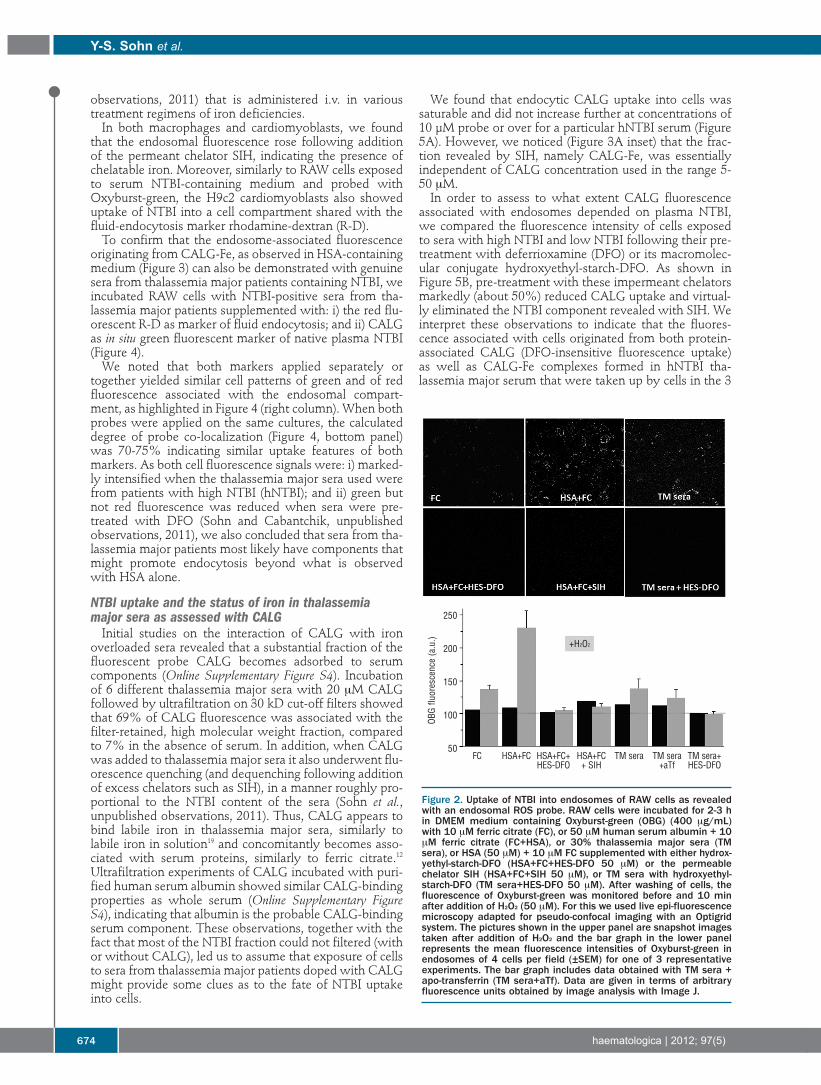
plasma/serum NTBI was initially taken up into the endo-somal compartment, we supplemented NTBI-containingmedia with the albumin-tagged Oxyburst-green probewhich is endocytosed and whose ability to fluoresce uponaddition of H2O2 can be attributed to labile iron due to itssensitivity to iron chelators. RAW cells exposed to mediacontaining Oxyburst-green showed a punctuated fluores-cence when the medium contained ferric citrate + HSAbut not ferric citrate alone. There was no differencebetween pictures of cells exposed only to ferric citrate(Figure 2) and those exposed only to HAS, patients’ sera orjust control medium (data not shown). Importantly, theendosome associated fluorescence is attributable to endo-somal labile iron, as the addition of a permeant chelator(SIH) after exposure to iron-containing media, but prior tohydrogen peroxide, abrogated the rise in fluorescence.
y-s. sohn et al.
672 haematologica | 2012; 97(5)

More importantly, Oxyburst-green supplemented to serafrom thalassemia major patients showed similar proper-ties to those of HSA + ferric citrate, while hydroxyethyl-starch-DFO or apo-transferrin abrogated the fluorescencechanges.All this information links labile iron to the fluorescence
changes occurring within endosomes and traces its originto the NTBI-containing medium, either associated withnative thalassemia major serum or with the artificially for-mulated NTBI in the form of HSA+ ferric citrate.A similar approach to monitor iron ingress into cytosol
following exposure to NTBI-containing medium wasapplied to H9c2 cardiomyoblasts and to HL-1 contractilecardiomyocytes. The cardiac cells are markedly less activein endocytosis than the RAW macrophages and according-ly, the follow up of NTBI uptake by cardiomyoblasts andcardiomyocytes demanded extended exposure of cells toNTBI-containing media so as to enable fluorescence detec-tion of iron-dependent signals. An NTBI-evoked rise in flu-orescence was observed: i) in endosomes using Oxyburst-green in conjunction with fluorescence microscopy (OnlineSupplementary Figures S1 and S2); and ii) in cytosol usingCALG in conjunction with fluorescence microscopy(Online Supplementary Figure S3) or flow cytometry follow-ing release of attached cells by trypsinization (OnlineSupplementary Figure S4). The analysis revealed qualitative-ly similar features of NTBI uptake into endosomes (OnlineSupplementary Figures S1 and S2) and to cytosol (OnlineSupplementary Figures S3 and S4) of cardiomyoblasts andcardiomyocytes as compared to RAW macrophage cells
(Figures 1 and 2), particularly their susceptibility toinhibitors of endocytosis and/or chelating agents.
NTBI uptake into cells as revealed with a fluorescentmembrane impermeant iron markerThe implication of these results is that initial stages of
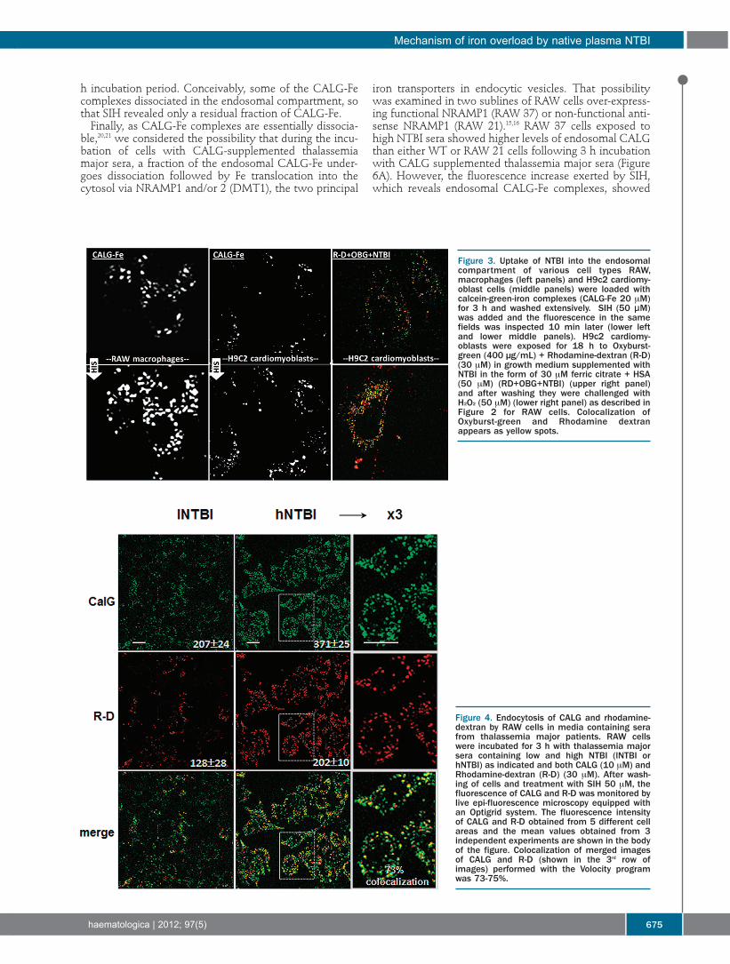
cellular uptake of NTBI occur via endocytosis of wholecomplexes of iron together with its presumed ligands (cit-rate and albumin). In order to assess this possibility by analternative means, we tried to generate a complex simulat-ing NTBI by use of an iron-responsive fluorescent probewith iron-binding moieties similar to those of citrate. Inthis approach, ferric citrate is replaced with ferric-calceingreen (CALG-Fe), a fluorescence-quenched NTBI surro-gate whose metal-quenched fluorescence can be revealedin cell compartments by addition of permeant chelatorssuch as SIH. We initially explored the possibility thatCALG added to sera might show different fluorescenceresponses depending on the presence or absence of NTBI.For this we used two different cell lines representative oftransfusional siderosis, macrophages and cardiomyoblastsand assessed uptake of the fluorescent (partially quenchedby the complexed iron) NTBI simulator CALG-Fe into theendosomal compartment (Figure 3). Macrophages showedprominent acquisition of endosomal fluorescence follow-ing exposure to NTBI-containing media supplementedwith CALG, possibly related to their high constitutiveendocytic activity. A similar property was observed inmacrophages when using polymeric forms of NTBI,12 suchas the iron-saccharate Venofer (Sohn et al., unpublished
Mechanism of iron overload by native plasma NTBI
haematologica | 2012; 97(5) 673
Figure 1. Uptake of ferric citrate into the cytosol of RAW cells. Effect of human serum albumin, inhibitors of endocytosis and impermeantiron chelators. RAW cells labeled in the cytosol with CALG via its AM precursor were followed for up to 1 h by epifluorescence microscopywhile perfused in DMEM-HEPES supplemented with ferric citrate only (Fe:citrate10: 30 μM) (FC), or together with 50 μM human serum albu-min (FC+HSA) and the indicated substances: nocodazole (N=30 μM), ML-9 (M=100 μM)(FC+HSA+N+M), cytochalasin D (cyD= 100 μM)(FC+HSA+CyD), and hydroxyethyl-starch-DFO (FC+HSA+HES-DFO 50 μM). Systems without added ferric citrate were HSA alone (50 μM)(HSA), HSA with nocodazole (N=30 μM) and ML-9 (M=100 μM)(HSA+N+M) and HSA with hydroxyethyl-starch-DFO (HSA+HES-DFO 50 μM).All incubations and perfusions were carried out at 37°C. The pictures show snapshots of the same field taken at 0 at 40 min, and the graphshows the mean fluorescence quenching (FQ) of each field (analyzed with NIH Image J; National Institutes of Health, Bethesda, MD, USA)normalized to the initial fluorescence intensity (control cells).
EQ (r.u) N+MCyDHES-DFO
0.4
0.2
FC
0.0
FC+HSA HSA
observations, 2011) that is administered i.v. in varioustreatment regimens of iron deficiencies.In both macrophages and cardiomyoblasts, we found
that the endosomal fluorescence rose following additionof the permeant chelator SIH, indicating the presence ofchelatable iron. Moreover, similarly to RAW cells exposedto serum NTBI-containing medium and probed withOxyburst-green, the H9c2 cardiomyoblasts also showeduptake of NTBI into a cell compartment shared with thefluid-endocytosis marker rhodamine-dextran (R-D).To confirm that the endosome-associated fluorescence
originating from CALG-Fe, as observed in HSA-containingmedium (Figure 3) can also be demonstrated with genuinesera from thalassemia major patients containing NTBI, weincubated RAW cells with NTBI-positive sera from tha-lassemia major patients supplemented with: i) the red flu-orescent R-D as marker of fluid endocytosis; and ii) CALGas in situ green fluorescent marker of native plasma NTBI(Figure 4).We noted that both markers applied separately or
together yielded similar cell patterns of green and of redfluorescence associated with the endosomal compart-ment, as highlighted in Figure 4 (right column). When bothprobes were applied on the same cultures, the calculateddegree of probe co-localization (Figure 4, bottom panel)was 70-75% indicating similar uptake features of bothmarkers. As both cell fluorescence signals were: i) marked-ly intensified when the thalassemia major sera used werefrom patients with high NTBI (hNTBI); and ii) green butnot red fluorescence was reduced when sera were pre-treated with DFO (Sohn and Cabantchik, unpublishedobservations, 2011), we also concluded that sera from tha-lassemia major patients most likely have components thatmight promote endocytosis beyond what is observedwith HSA alone.
NTBI uptake and the status of iron in thalassemiamajor sera as assessed with CALGInitial studies on the interaction of CALG with iron
overloaded sera revealed that a substantial fraction of thefluorescent probe CALG becomes adsorbed to serumcomponents (Online Supplementary Figure S4). Incubationof 6 different thalassemia major sera with 20 μM CALGfollowed by ultrafiltration on 30 kD cut-off filters showedthat 69% of CALG fluorescence was associated with thefilter-retained, high molecular weight fraction, comparedto 7% in the absence of serum. In addition, when CALGwas added to thalassemia major sera it also underwent flu-orescence quenching (and dequenching following additionof excess chelators such as SIH), in a manner roughly pro-portional to the NTBI content of the sera (Sohn et al.,unpublished observations, 2011). Thus, CALG appears tobind labile iron in thalassemia major sera, similarly tolabile iron in solution19 and concomitantly becomes asso-ciated with serum proteins, similarly to ferric citrate.12Ultrafiltration experiments of CALG incubated with puri-fied human serum albumin showed similar CALG-bindingproperties as whole serum (Online Supplementary FigureS4), indicating that albumin is the probable CALG-bindingserum component. These observations, together with thefact that most of the NTBI fraction could not filtered (withor without CALG), led us to assume that exposure of cellsto sera from thalassemia major patients doped with CALGmight provide some clues as to the fate of NTBI uptakeinto cells.
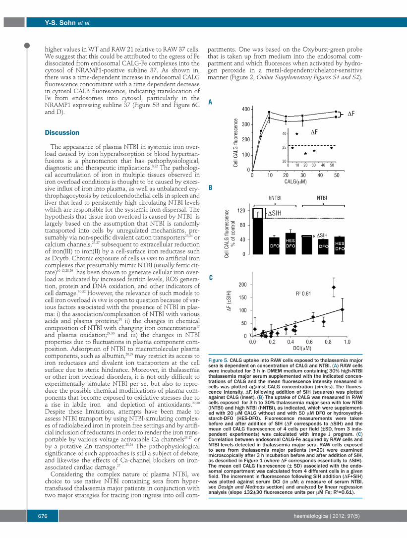
We found that endocytic CALG uptake into cells wassaturable and did not increase further at concentrations of10 µM probe or over for a particular hNTBI serum (Figure5A). However, we noticed (Figure 3A inset) that the frac-tion revealed by SIH, namely CALG-Fe, was essentiallyindependent of CALG concentration used in the range 5-50 μM. In order to assess to what extent CALG fluorescence
associated with endosomes depended on plasma NTBI,we compared the fluorescence intensity of cells exposedto sera with high NTBI and low NTBI following their pre-treatment with deferrioxamine (DFO) or its macromolec-ular conjugate hydroxyethyl-starch-DFO. As shown inFigure 5B, pre-treatment with these impermeant chelatorsmarkedly (about 50%) reduced CALG uptake and virtual-ly eliminated the NTBI component revealed with SIH. Weinterpret these observations to indicate that the fluores-cence associated with cells originated from both protein-associated CALG (DFO-insensitive fluorescence uptake)as well as CALG-Fe complexes formed in hNTBI tha-lassemia major serum that were taken up by cells in the 3
y-s. sohn et al.
674 haematologica | 2012; 97(5)
Figure 2. Uptake of NTBI into endosomes of RAW cells as revealedwith an endosomal ROS probe. RAW cells were incubated for 2-3 hin DMEM medium containing Oxyburst-green (OBG) (400 μg/mL)with 10 μM ferric citrate (FC), or 50 μM human serum albumin + 10μM ferric citrate (FC+HSA), or 30% thalassemia major sera (TMsera), or HSA (50 μM) + 10 μM FC supplemented with either hydrox-yethyl-starch-DFO (HSA+FC+HES-DFO 50 μM) or the permeablechelator SIH (HSA+FC+SIH 50 μM), or TM sera with hydroxyethyl-starch-DFO (TM sera+HES-DFO 50 μM). After washing of cells, thefluorescence of Oxyburst-green was monitored before and 10 minafter addition of H2O2 (50 μM). For this we used live epi-fluorescencemicroscopy adapted for pseudo-confocal imaging with an Optigridsystem. The pictures shown in the upper panel are snapshot imagestaken after addition of H2O2 and the bar graph in the lower panelrepresents the mean fluorescence intensities of Oxyburst-green inendosomes of 4 cells per field (±SEM) for one of 3 representativeexperiments. The bar graph includes data obtained with TM sera +apo-transferrin (TM sera+aTf). Data are given in terms of arbitraryfluorescence units obtained by image analysis with Image J.
+H2O2
OBG flu
ores
cenc
e (a.u.)
250
200
150
100
50FC HSA+FC HSA+FC+ HSA+FC TM sera TM sera TM sera+
HES-DFO + SIH +aTf HES-DFO
h incubation period. Conceivably, some of the CALG-Fecomplexes dissociated in the endosomal compartment, sothat SIH revealed only a residual fraction of CALG-Fe. Finally, as CALG-Fe complexes are essentially dissocia-
ble,20,21 we considered the possibility that during the incu-bation of cells with CALG-supplemented thalassemiamajor sera, a fraction of the endosomal CALG-Fe under-goes dissociation followed by Fe translocation into thecytosol via NRAMP1 and/or 2 (DMT1), the two principal
iron transporters in endocytic vesicles. That possibilitywas examined in two sublines of RAW cells over-express-ing functional NRAMP1 (RAW 37) or non-functional anti-sense NRAMP1 (RAW 21).15,16 RAW 37 cells exposed tohigh NTBI sera showed higher levels of endosomal CALGthan either WT or RAW 21 cells following 3 h incubationwith CALG supplemented thalassemia major sera (Figure6A). However, the fluorescence increase exerted by SIH,which reveals endosomal CALG-Fe complexes, showed
Mechanism of iron overload by native plasma NTBI
haematologica | 2012; 97(5) 675
Figure 3. Uptake of NTBI into the endosomalcompartment of various cell types RAW,macrophages (left panels) and H9c2 cardiomy-oblast cells (middle panels) were loaded withcalcein-green-iron complexes (CALG-Fe 20 μM)for 3 h and washed extensively. SIH (50 μM)was added and the fluorescence in the samefields was inspected 10 min later (lower leftand lower middle panels). H9c2 cardiomy-oblasts were exposed for 18 h to Oxyburst-green (400 µg/mL) + Rhodamine-dextran (R-D)(30 μM) in growth medium supplemented withNTBI in the form of 30 μM ferric citrate + HSA(50 μM) (RD+OBG+NTBI) (upper right panel)and after washing they were challenged withH2O2 (50 μM) (lower right panel) as described inFigure 2 for RAW cells. Colocalization ofOxyburst-green and Rhodamine dextranappears as yellow spots.
Figure 4. Endocytosis of CALG and rhodamine-dextran by RAW cells in media containing serafrom thalassemia major patients. RAW cellswere incubated for 3 h with thalassemia majorsera containing low and high NTBI (lNTBI orhNTBI) as indicated and both CALG (10 μM) andRhodamine-dextran (R-D) (30 μM). After wash-ing of cells and treatment with SIH 50 μM, thefluorescence of CALG and R-D was monitored bylive epi-fluorescence microscopy equipped withan Optigrid system. The fluorescence intensityof CALG and R-D obtained from 5 different cellareas and the mean values obtained from 3independent experiments are shown in the bodyof the figure. Colocalization of merged imagesof CALG and R-D (shown in the 3rd row ofimages) performed with the Volocity programwas 73-75%.
higher values in WT and RAW 21 relative to RAW 37 cells.We suggest that this could be attributed to the egress of Fedissociated from endosomal CALG-Fe complexes into thecytosol of NRAMP1-positive subline 37. As shown in,there was a time-dependent increase in endosomal CALGfluorescence concomitant with a time dependent decreasein cytosol CALB fluorescence, indicating translocation ofFe from endosomes into cytosol, particularly in theNRAMP1 expressing subline 37 (Figure 5B and Figure 6Cand D).
Discussion
The appearance of plasma NTBI in systemic iron over-load caused by iron hyperabsorption or blood hypertran-fusions is a phenomenon that has pathophysiological,diagnostic and therapeutic implications.3,22 The pathologi-cal accumulation of iron in multiple tissues observed iniron overload conditions is thought to be caused by exces-sive influx of iron into plasma, as well as unbalanced ery-throphagocytosis by reticuloendothelial cells in spleen andliver that lead to persistently high circulating NTBI levelswhich are responsible for the systemic iron dispersal. Thehypothesis that tissue iron overload is caused by NTBI islargely based on the assumption that NTBI is randomlytransported into cells by unregulated mechanisms, pre-sumably via non-specific divalent cation transporters23,24 orcalcium channels,25-27 subsequent to extracellular reductionof iron(III) to iron(II) by a cell-surface iron reductase suchas Dcytb. Chronic exposure of cells in vitro to artificial ironcomplexes that presumably mimic NTBI (usually ferric cit-rate)10-12,28,29 has been shown to generate cellular iron over-load as indicated by increased ferritin levels, ROS genera-tion, protein and DNA oxidation, and other indicators ofcell damage.30-32 However, the relevance of such models tocell iron overload in vivo is open to question because of var-ious factors associated with the presence of NTBI in plas-ma: i) the association/complexation of NTBI with variousacids and plasma proteins;28 ii) the changes in chemicalcomposition of NTBI with changing iron concentrations12and plasma oxidation;29,30 and iii) the changes in NTBIproperties due to fluctuations in plasma component com-position. Adsorption of NTBI to macromolecular plasmacomponents, such as albumin,28,29 may restrict its access toiron reductases and divalent ion transporters at the cellsurface due to steric hindrance. Moreover, in thalassemiaor other iron overload disorders, it is not only difficult toexperimentally simulate NTBI per se, but also to repro-duce the possible chemical modifications of plasma com-ponents that become exposed to oxidative stresses due toa rise in labile iron and depletion of antioxidants.29,30Despite these limitations, attempts have been made toassess NTBI transport by using NTBI-simulating complex-es of radiolabeled iron in protein free settings and by artifi-cial inclusion of reductants in order to render the iron trans-portable by various voltage activatable Ca channels25-27 orby a putative Zn transporter.23,24 The pathophysiologicalsignificance of such approaches is still a subject of debate,and likewise the effects of Ca-channel blockers on iron-associated cardiac damage.27Considering the complex nature of plasma NTBI, we
choice to use native NTBI containing sera from hyper-transfused thalassemia major patients in conjunction withtwo major strategies for tracing iron ingress into cell com-
partments. One was based on the Oxyburst-green probethat is taken up from medium into the endosomal com-partment and which fluoresces when activated by hydro-gen peroxide in a metal-dependent/chelator-sensitivemanner (Figure 2, Online Supplementary Figures S1 and S2).
y-s. sohn et al.
676 haematologica | 2012; 97(5)
Figure 5. CALG uptake into RAW cells exposed to thalassemia majorsera is dependent on concentration of CALG and NTBI. (A) RAW cellswere incubated for 3 h in DMEM medium containing 30% high-NTBIthalassemia major serum supplemented with the indicated concen-trations of CALG and the mean fluorescence intensity measured incells was plotted against CALG concentration (circles). The fluores-cence intensity, DF, following addition of SIH (squares) was plottedagainst CALG (inset). (B) The uptake of CALG was measured in RAWcells exposed for 3 h to 30% thalassemia major sera with low NTBI(lNTBI) and high NTBI (hNTBI), as indicated, which were supplement-ed with 20 μM CALG without and with 50 μM DFO or hydroxyethyl-starch-DFO (HES-DFO). Fluorescence measurements were takenbefore and after addition of SIH (DF corresponds to DSIH) and themean cell CALG fluorescence of 4 cells per field (±SD, from 3 inde-pendent experiments) was calculated with Image J program. (C)Correlation between endosomal CALG-Fe acquired by RAW cells andNTBI levels detected in thalassemia major sera. RAW cells exposedto sera from thalassemia major patients (n=20) were examinedmicroscopically after 3 h incubation before and after addition of SIH,as described in Figure 1 (where DF corresponds essentially to DSIH).The mean cell CALG fluorescence (± SD) associated with the endo-somal compartment was calculated from 4 different cells in a givenfield. The increment in fluorescence following SIH addition (DF+SIH)was plotted against serum DCI (in μM; a measure of serum NTBI,see Design and Methods section) and analyzed by linear regressionanalysis (slope 132±30 fluorescence units per μM Fe; R2=0.61).
A
B
C
DF
DSIH
DSIH
DF
0 10 20 30 40 50CALG(μM)
0.0 0.2 0.4 0.6 0.8 1.0DCI(μM)
Cell CA
LG fluo
resc
ence
Cell CA
LG fluo
resc
ence
% of c
ontro
l
400
300
200
100
0
120
80
40
0
R2 0.61
200
150
100
50
0
DF (±SIH)
0 10 20 30 40 50
40
35
30
hNTBI NTBI
The other strategy monitored labile iron ingress into thecytosolic CALG-laden or CALB-laden compartment byfollowing metal evoked quenching of fluorescence(Figures 1 and 6, Online Supplementary Figures S3 and S4).Evidence that both strategies monitored processes thatinvolved endocytosis of NTBI was based on the effects ofinhibitors of endocytosis and on those of impermeant ironchelators, such as hydroxyethyl-starch-DFO (Figures 1and 5, Online Supplementary Figures S1 and S2), whereasproof that the species monitored in endosomes or cytosolwas labile iron leaned on the action of permeant ironchelators, such as SIH or deferiprone (Figures 3-6). In addi-tion to thalassemia major sera containing NTBI, we alsoused ferric citrate, an accepted component of plasmaNTBI, either by itself or supplemented to human sera orhuman serum albumin (Figures 1 and 2, OnlineSupplementary Figures S1-S3). While cell exposure to ferric-citrate alone at concentrations measured in thalassemiamajor sera failed to evoke significant iron-associatedchanges in endosomal or cytosolic iron pools, its additiontogether with human sera or serum albumin (HSA) ren-ders it demonstrably accessible to cells: first by undergo-ing endocytosis and subsequently by releasing labile ironin endosomes and translocating it to the cytosol, in thecase of RAW cells, via NRAMP1 (Figure 6). That theprocesses monitored by endosomal Oxyburst-green orcytosolic CALG were associated with NTBI and not withTBI was deduced from the fact that addition of apotrans-ferrin to media containing native or artificial NTBI abro-gated the processes in a similar way as hydroxyethyl-starch-DFO.We also found that the proposed steps of NTBI uptake
into RAW cells were largely recapitulated with tha-lassemia major sera probed with CALG, that reversiblybinds iron,20,21 including components of plasma NTBI, andis endocytosed commensurately with NTBI levels (Figure3). However, a key question is to what extent CALGadded to sera reports NTBI ingress into cells rather thanpromotes NTBI ingress by binding to plasma NTBI,whether present as low-molecular weight complexes orbound to plasma proteins. Since endocytic uptake ofCALG-Fe and thalassemia major sera with CALG is inhib-ited by impermeant iron chelators, while the uptake ofrhodamine-dextran is unaffected by the same chelators, itmay be concluded that CALG is a reporter rather than apromoter of endocytosis of NTBI. This is further support-ed by our observation that cellular uptake of CALG-Fewas negligible in the absence of serum or serum albumin.Furthermore, not only CALG-Fe, but also ferric-citrate inthe presence of albumin, and NTBI in thalassemia majorsera also exist in macromolecular forms12 that could betaken up by cells by a similar bulk mechanism of endocy-tosis, whether adsorptive or pinocytic, which would be inline with the same mechanistic concept.Taken together, these data indicate that a major compo-
nent of plasma NTBI ingress into cells is associated withbulk mechanisms of endocytosis that prevail in cells ofvarious organs.33-37 The proposed mechanism explainshow NTBI species bound to plasma components gainaccess to particular cells, but does not exclude otherswhich comprise some plasma NTBI forms transportableby resident membrane transporters or channels. Endocytic
uptake of NTBI, which is demonstrated primarily inmacrophages, also operates, though to a lesser extent, inother cells such as cardiomyoblasts and cardiomyocytes(Figure 4, Online Supplementary Figures S1-S4) and insulino-ma cells (Glickstein and Cabantchik, unpublished observations,2011).The relative contribution of NTBI endocytosis toiron accumulation in the above organs will depend onboth their endocytic/pinocytic activities and on the levelsof NTBI in plasma that undergoes significant modifica-tions in chronic iron overload. Macropinocytic uptake of avariety of macromolecules has only recently become rec-ognized as a regulated pathway with features that distin-guish it from clathrin-dependent endocytic processes,such as receptor-mediated endocytosis.33 Numerous stim-uli regulate macropinocytosis, among which the bestdefined are growth factor signaling and surface binding ofintracellular pathogens. Pinocytic activity is particularlyhigh in macrophages, epithelial and endothelial cells, andcells of the immune system.33 However, it also prevails,though at relatively lower levels, in various cell types andtissues, including those susceptible to iron overload suchas the heart35-36 and endocrine glands.37A surprising observation in this study was the enhanced
endocytosis of rhodamine-dextran in the presence of seracontaining NTBI (Figure 4). Although the biochemicalbasis for this effect is still unclear, it could be a key com-ponent in the mechanism of tissue iron loading in ironoverloaded thalassemia major patients. As the uptake ofrhodamine-dextran added to thalassemia major plasma isnot affected by DFO, the contribution of NTBI per se toendocytosis is likely to involve pre-oxidized plasma com-ponents, for which there is ample evidence in hypertran-fused patients with inadequate chelation treatment.26Furthermore, albumin, especially in its oxidized form, hasbeen shown to avidly bind iron29 and has been suggestedto be a possible plasma carrier of NTBI.29 Considering thatthe half-life of oxidized human albumin in mice is reducedby almost 50%, mainly due to liver clearance,30 it is con-ceivable that oxidized albumin or other plasma compo-nents are preferentially endocytosed by macrophages. Infact, macrophages are essential to the recycling of ironextracted from senescent erythrocytes and the control ofsystemic iron levels.38 These properties have implicationsfor the pathophysiology and treatment of iron overload,and highlight the need to eliminate NTBI from the circula-tion by chelation as a means of reducing plasma proteinoxidation and ensuing tissue iron overload. However, fur-ther studies are also required to assess whether a similarmode of NTBI ingress by endocytosis prevails in other dis-orders of systemic iron overload, such as hemochromato-sis, or transfusional siderosis, such as myelodysplastic syn-dromes and sickle cell disease.
Authorship and Disclosures
The information provided by the authors about contributions frompersons listed as authors and in acknowledgments is available withthe full text of this paper at www.haematologica.org.Financial and other disclosures provided by the authors using the
ICMJE (www.icmje.org) Uniform Format for Disclosure ofCompeting Interests are also available at www.haematologica.org.
Mechanism of iron overload by native plasma NTBI
haematologica | 2012; 97(5) 677
y-s. sohn et al.
678 haematologica | 2012; 97(5)
References
1. Hershko C, Peto TE. Non-transferrin plasmairon. Br J Haematol. 1987;66(2):149-51.
2. Cabantchik ZI, Breuer W, Zanninelli G,Cianciulli P. LPI-labile plasma iron in ironoverload. Best Pract Res Clin Haematol.2005;18(2):277-87.
3. Porter JB. Pathophysiology of iron overload.Hematol Oncol Clin North Am.2005;19(Suppl 1):7-12.
4. Hider RC, Silva A, Podinovskaia M, Ma YM.Monitoring the efficiency of iron chelationtherapy: the potential of nontransferrin-bound iron. Ann NY Acad Sci. 2010;1202:94-9.
5. Breuer W, Hershko C, Cabantchik ZI. Theimportance of non-transferrin bound iron indisorders of iron metabolism. Transfus Sci.2000;23(3):185-92.
6. Ponka P. Rare causes of hereditary iron over-load. Semin Hematol. 2002;39(4):249-62.
7. Brittenham GM. Iron-chelating therapy fortransfusional iron overload. N Engl J Med.2011;364(2):146-56.
8. Liu P, Olivieri N. Iron overload cardiomy-opathies: new insights into an old disease.Cardiovasc Drugs Ther. 1994;8(1):101-10.
9. Cabantchik ZI, Fibach E, Breuer W. Canlabile plasma iron (LPI) and labile cell iron(LCI) levels serve as early indicators ofchelation efficacy in iron overload?BloodMed.com. 2009. Available from:http://www.bloodmed.com/800000/mini-reviews1.asp?id =254
10. Grootveld M, Bell JD, Halliwell B, AruomaOI, Bomford A, Sadler PJ. Non-transferrin-bound iron in plasma or serum from patientswith idiopathic hemochromatosis.Characterization by high performance liquidchromatography and nuclear resonancespectroscopy. J Biol Chem. 1989;264(8):4417-44.
11. Hider RC. Nature of nontransferrin-boundiron. Eur J Clin Invest. 2002;32(Suppl 1):50-4.
12. Evans RW, Rafique R, Zarea A, Rapisarda C,Cammack R, Evans PJ, et al. Nature of non-transferrin-bound iron: studies on iron cit-rate complexes and thalassemic sera. J BiolInorg Chem. 2008;13(1):57-74.
13. Glickstein H, Ben El R, Shvartsman M,Cabantchik ZI. Intracellular labile iron poolsas direct targets of iron chelators. A fluores-cence study of chelator action in living cells.Blood. 2005;106(9):3242-50
14. Sohn YS, Breuer W, Munnich A, CabantchikZI. Redistribution of accumulated cell iron: amodality of chelation with therapeuticimplications. Blood. 2008;111(3):1690-9.
15. Nairz M, Fritsche G, Crouch ML, BartonHC, Fang FC, Weiss G, et al. Slc11a1 limitsintracellular growth of Salmonella entericasv. Typhimurium by promoting macrophageimmune effector functions and impairingbacterial iron acquisition. Cell Microbiol.2009;11(9):1365-81.
16. Claycomb WC, Lanson NA Jr, StallworthBS, Egeland DB, Delcarpio JB, Bahinski A, etal. HL-1 cells: a cardiac muscle cell line thatcontracts and retains phenotypic characteris-tics of the adult cardiomyocyte. Proc NatlAcad Sci USA. 1998;95(6):2979-84.
17. Breuer W, Ermers MJ, Pootrakul P, AbramovA, Hershko C, Cabantchik ZI, et al.Desferrioxamine-chelatable iron, a compo-nent of serum non-transferrin-bound iron,used for assessing chelation therapy. Blood.2001;97(3):792-8.
18. Breuer W, Ghoti H, Shattat A, Goldfarb A,Koren A, Rachmilewitz E, et al. Non-trans-ferrin bound iron in Thalassemia: differen-tial detection of redox active forms in chil-dren and older patients. Am J Hematol.2012;87(1):55-61.
19. Epsztejn S, Kakhlon O, Breuer W, GlicksteinH, Cabantchik ZI. A fluorescence assay forthe labile iron pool (LIP) of mammalian cells.Anal Biochem. 1997;248(1):31-40.
20. Espósito BP, Epsztejn S, Breuer W,Cabantchik ZI. A review of fluorescencemethods for assessing labile iron in cells andbiological fluids. Anal. Biochem. 2002;304(1):1-18.
21. Ali A, Zhang Q, Dai J, Huang X. Calcein asa fluorescent iron chemosensor for the deter-mination of low molecular weight iron inbiological fluids. Biometals. 2003;16(2):285-93.
22. Hershko C, Link G, Cabantchik I.Pathophysiology of iron overload. Ann NYAcad Sci. 1998;850:191-201.
23. Liuzzi JP, Aydemir F, Nam H, Knutson MD,Cousins RJ. Zip14 (Slc39a14) mediates non-transferrin-bound iron uptake into cells. ProcNatl Acad Sci USA. 2006;103(37):13612-7.
24. Ludwiczek S, Theurl I, Muckenthaler MU,Jakab M, Mair SM, Theurl M, et al. Ca2+channel blockers reverse iron overload by anew mechanism via divalent metal trans-porter-1. Nat Med. 2007;13(4):448-54.
25. Oudit GY, Sun H, Trivieri MG, Koch SE,Dawood F, Ackerley C, et al. L-type Ca2+channels provide a major pathway for ironentry into cardiomyocytes in iron-overloadcardiomyopathy. Nat Med. 2003;9(9):1187-94.
26. Oudit GY, Trivieri MG, Khaper N, Liu PP,Backx PH. Role of L-type Ca2+ channels iniron transport and iron-overload cardiomy-
opathy. J Mol Med. 2006;84(5):349-64.27. Crowe S, Bartfay WJ. Amlodipine decreases
iron uptake and oxygen free radical produc-tion in the heart of chronically iron over-loaded mice. Biol Res Nurs. 2002;3(4):189-97.
28. Løvstad RA. Interactions of serum-albuminwith the Fe(III)–citrate complex. IntJBiochem. 1993;25(7):1015-7.
29. Silva AM, Hider RC. Influence of nonenzy-matic post-translation modifications on theability of human serum albumin to bindiron: implications for nontransferrin-boundiron speciation. Biochem Biophys Acta.2009;1794(10):1449-58.
30. Livrea MA, Tesoriere L, Pintaudi AM,Calabrese A, Maggio A, Freisleben HJ, et al.Oxidative stress and antioxidant status inbeta-thalassemia major: iron overload anddepletion of lipid-soluble antioxidants.Blood. 1996;88(9):3608-14.
31. Walter PB, Fung EB, Killilea DW, Jiang Q,Hudes M, Madden J, et al. Oxidative stressand inflammation in iron-overloadedpatients with beta-thalassaemia or sickle celldisease. Br J Haematol. 2006;135(2):254-63.
32. Kakhlon O, Gruenbaum Y, Cabantchik ZI.Repression of ferritin expression increases thelabile iron pool, oxidative stress and short-term growth of human erythroleukemia cells.Blood. 2001;97(9):2863-71.
33. Kerr MC, Teasdale RD. Definingmacropinocytosis.Traffic. 2009;10(4):364-71.
34. Soeiro Mde N, Mota RA, Batista Dda G,Meirelles Mde N. Endocytic pathway inmouse cardiac cells. Cell Struct Funct.2002;27(6):469-78.
35. Swildens J, de Vries AA, Li Z, Umar S,Atsma DE, Schalij MJ, et al. Integrin stimula-tion favors uptake of macromolecules bycardiomyocytes in vitro. Cell PhysiolBiochem. 2010;26(6):999-1010.
36. Cowan DB, Noria S, Stamm C, Garcia LM,Poutias DM, del Nido PJ, et al.Lipopolysaccharide internalization activatesendotoxin-dependent signal transduction incardiomyocytes. Circ Res. 2001;88(5):491-8.
37. Pow DV, Morris JF. Membrane routing dur-ing exocytosis and endocytosis in neuroen-docrine neurones and endocrine cells: use ofcolloidal gold particles and immunocyto-chemical discrimination of membrane com-partments. Cell Tissue Res. 1991;264(2):299-316.
38. Soe-Lin S, Apte SS, Andriopoulos B Jr,Andrews MC, Schranzhofer M, Kahawita T,et al. Nramp1 promotes efficientmacrophage recycling of iron following ery-throphagocytosis in vivo. Proc Natl Acad SciUSA. 2009;106(14):5960-5.