simulations of the consequences of tongue surgery on tongue mobility: implications for speech...
TRANSCRIPT
Simulations of the consequences of tongue surgery on
tongue mobility: Implications for speech production in
post-surgery conditions.
Stéphanie Buchaillard1, Muriel Brix2, Pascal Perrier1 & Yohan Payan3
1ICP/GIPSA-lab, UMR CNRS 5216, INP Grenoble, France
2Service de Chirurgie Maxillo-faciale, Grenoble University Hospital, Grenoble, France
3TIMC-IMAG, UMR CNRS 5525, Université Joseph Fourier, Grenoble, France
International Journal of Medical Robotics and Computer Assisted Surgery, Vol. 3(3), pp. 252-261
Corresponding author:
Pascal Perrier ICP/GIPSA-lab INPG, 46 Avenue Félix Viallet 38031 Grenoble cédex 01 [email protected]
2
ABSTRACT
Background In this paper, we study the ability of a 3D biomechanical model of the oral cavity to
predict the consequences of tongue surgery on tongue movements, according to the size and location
of the tissue loss and the nature of the flap used by the surgeon.
Method The core of our model consists of a 3D biomechanical model representing the tongue as a
Finite Element Structure with hexahedral elements and hyperelastic properties, in which muscles are
represented by specific subsets of elements. This model is inserted in the oral cavity including jaw,
palate and pharyngeal walls. Hemiglossectomy and large resection of the mouth floor are simulated by
removing the elements corresponding to the tissue losses. Three kinds of reconstruction are modelled,
assuming flaps with low, medium or high stiffness..
Results The consequences of these different surgical treatments during the activations of some of the
main tongue muscles are shown. Differences in global 3D tongue shape and in velocity patterns are
evaluated and interpreted in terms of their potential impact on speech articulation. These simulations
have shown to be efficient in accounting for some of the clinically observed consequences of tongue
surgery.
Conclusion Further improvements still need to be done before being able to generate easily patient-
specific models and to decrease significantly the computation time. However, this approach should
represent a significant improvement in planning tongue surgery systems and should be a very useful
means of improving the understanding of muscle behaviour after partial resection.
3
I. Introduction
Tongue surgery can have severe consequences on tongue mobility and tongue deformation
capabilities. It can generate strong impairments of three basic functions of the human life, namely
masticating, swallowing and speaking, which induce a noticeable decrease of the quality of life of the
patients. Several assessments of these impairments have been done in post-surgery conditions on
patients, who had undergone tongue and/or mouth floor carcinoma resections and reconstructions.
These assessments were based either on well-established functional protocols, such as the University
of Washington Quality of Life Instrument ( 1), evaluating among other tasks the patients'
communication skills ( 2- 3), on specific acoustic speech corpora ( 4- 5) assessing the lost of speech
movements amplitude and accuracy, or on quantitative measurements of the oral cavity ( 6- 7).
Statistical analyses of the results collected on large enough samples of patients could provide
valuable indications about the impacts of surgery and reconstruction techniques
In the line of works carried out in predictive medicine to set up systems of computer aided surgery,
such as in orthopaedic (spine and pelvis) ( 8- 10), cranio-facial ( 11- 13) surgeries, this paper presents
the current achievements of a long term project aiming at predicting and assessing the impact of
tongue and mouth floor surgery on speech production. The ultimate objective of this project is the
design of a software with which surgeons should be able 1) to design a 3D biomechanical model of
the tongue and of the mouth floor that matches the anatomical characteristics of each patient specific
oral cavity, 2) to simulate the anatomical changes induced by the surgery and the possible
reconstruction, and 3) to quantitatively predict and assess the consequences of these anatomical
changes on tongue mobility and speech production after surgery.
The interest of this approach is linked with functional aspects of the surgical reconstruction. Depending
on the site and the size of the tissue losses, the functional alteration can originate from a lack of
surface, lining and texture, a lack of volume and bulk, or a lack of mobility. The choice of the flap used
for the reconstruction is also an important issue. From this perspective, a biomechanical model of the
tongue is helpful, since it allows simulating the planned surgery in terms of cut design. Such a model

4
could also be helpful to select a specific kind of flap in comparison to others, since it allows a
quantitative study of the mobility of the tongue for different reconstructions.
The 3D model of the oral cavity is presented in the "Material and Methods" section, together with a
description of typical examples of simulations of tongue resection and reconstruction. In the "Results"
section the consequences of the simulated surgeries on tongue displacement are shown, and an
analysis of their potential consequences for speech production is proposed. Perspectives and further
developments of the projects are discussed in the "Conclusion" section.
II. Material and method
Our work is based on four basic steps:
. Development of a 3D biomechanical model of the oral cavity
. Implementation on this model of the anatomical and mechanical transformations generated by the
most frequent tongue resections and their associated reconstructions.
. Simulations with the model of the displacements and the shaping of the tongue associated with
the activation of the main tongue muscles, before and after surgery.
. Interpretation of these simulations in terms of implications for speech production after tongue
surgery.
The 3D oral cavity model was originally designed by Gérard et al. ( 14- 15) and it was further enhanced
recently for speech production control by Buchaillard et al. ( 16). It was developed in the ANSYSTM
Finite Elements software environment. Models of the jaw, the teeth, the palate, the pharynx, the
tongue and the hyoid bone are its basic constituents. The tongue and the hyoid bone are represented
by volumes meshed with 3D hexahedral and tetrahedral elements respectively, while the other
structures are modelled by 3D surface elements. These surface elements are essentially considered
as oral cavity limits with which the tongue interacts due to mechanical contacts.
II.1. The 3D biomechanical model of the tongue
II.1.1. Some basics about tongue anatomy Among the nine muscles that act on the tongue structure, there are five extrinsic muscles that
originate from structures external to the tongue (mostly bony structures) and insert into the tongue: the
genioglossus, the styloglossus, the hyoglossus, the geniohyoid and the palatoglossus. Four additional
5
intrinsic muscles are completely embedded in the tongue structure: the inferior and superior
longitudinal, the verticalis and the transversus. For all these muscles the fibres are mainly oriented in
the front/back or in the vertical direction, except for the transversus, which fibres are transversally
oriented. In addition the majority of them are paired muscles, symmetrically distributed on each side of
the midsagittal plane of the head. Detailed descriptions of tongue anatomy can be found in ( 17), ( 18),
and ( 19).
As concerns the functions of these muscles for tongue moving and tongue shaping, the description in
this paper will be limited to the muscles that have been used in the simulations shown in section III.
The anterior bundle of the genioglossus tracts the apex down and forward. Its middle bundle propels
the tongue out of the oral cavity. Its inferior and posterior bundles tracts the base forward and elevates
the core of the tongue. The styloglossus raises the base, and elevates globally the tongue in the velar
area. The hyoglossus lowers the tongue and brings it back after propulsion. The superior longitudinal
shortens the tongue and tracts the apex up and back. The transversus shortens the transversal
diameter and makes the tongue sharper.
The floor of the mouth is a crescent-shaped space extending from the lower alveolar ridge to the
ventral surface of the tongue. Its anterior part allows good mobility and protraction of the tongue.
II.1.2. Finite elements mesh design
The mesh design process is completely described in ( 14) and ( 20). The main guidelines are the
following. In a first step, a generic mesh was designed from the Visible Human Project TM data set. It
specifies the shape of the elements taking into account the main trends of muscle anatomy such as
the main orientations of muscles fibres. A precise implementation of muscles was done in this generic
mesh thanks to accurate anatomical data extracted from ( 17), ( 18), and ( 19) (see ( 14) for a
comprehensive description). This approach allowed a realistic description of the muscular anatomy of
the tongue. However, since it is based on data collected from a frozen cadaver, the geometry of the
tongue is likely to be altered as compared to living subjects.
This is why, in a second step, the geometry of the generic mesh was transformed, so that it matches
the external 3D contours of the tongue of a living subject (PB), for whom many different kinds of data
(CT-Scans, MRI data, X-Ray data) about the head and the oral cavity had been collected. The
external tongue contours were extracted from the MRI data, with manual image segmentation ( 21).
6
Thus, a new subject specific finite element mesh was achieved via a mesh-matching algorithm ( 22)
projecting the external nodes of the generic mesh onto the tongue surface measured from PB's MRI
data, followed by an interpolation process specifying the internal nodes positions from the new
external mesh while respecting the original structure of the generic mesh. Finally a geometrical
refinement was carried out from an X-Ray midsagittal view of PB's oral cavity at rest in a seating
position (Figure 1, left panel). This was done because the supine position of the subject during the
MRI data acquisition is likely to influence the tongue shape due to gravity. This final adaptation
guarantees the best possible definition of the tongue geometry at rest. This mesh was then included in
a whole oral cavity by inserting it in the jaw, palate, and pharyngeal surface meshes that were
extracted from PB's MRI and CT-scans data (Figure 1, right panel).
II.1.3. Muscle representation Muscles are represented as a set of adjacent elements in the tongue mesh. Figure 2 shows the
arrangement of muscles activated in section III. It should be noted that the genioglossus has been
divided in its three functional parts: posterior, medium and anterior. Macro-fibres, that represent
bundles of muscular fibres, were defined by joining finite elements nodes for every muscle, according
to the fibres known orientation (represented in red on Figure 2).
Muscle activation is produced by an external generator based on a functional model of muscle force
generation mechanisms, the Equilibrium Point Hypothesis also called λ model ( 23). The
implementation of this model was adapted to the tongue in order to ensure positioning accuracy and
stability. Details can be found in ( 16).
II.1.4. Tongue biomechanics
The tongue was modelled as a soft tissue attached to the hyoid bone, the jaw and the styloid process,
in interaction through soft contacts with the anterior part of the palate and the lower teeth. It consists of
a 3D Finite Element model assuming a large deformation framework with a non-linear hyper-elastic
constitutive law ( 15; 24). Arguing the fact that the constitutive law (i.e. the stress-strain relationship)
proposed by Gérard et al. ( 24) was extracted from data collected on tongue tissues removed from a
fresh cadaver, we proposed to adapt this original law to account for differences between passive
tissues and tissues belonging to active muscles. Therefore, two different constitutive equations were
introduced: one describing the passive behaviour of the tongue tissues and another one modelling the

7
stiffness of the tissues as an increasing function of tongue muscles activations. For a given element of
the model, the passive (respectively the active) constitutive law is used if the element belongs to a
region that is supposed to be passive (respectively active). The passive constitutive law was directly
derived from this non-linear law ( 24). However, as the stiffness of passive tissues removed from a
cadaver is known to be lower than the one measured on in vivo tissues, it was decided to modify the
constitutive law originally proposed in our tongue model. To our knowledge, one of the most relevant
in vivo measurements provided in the literature for human muscle stiffness is the one proposed by
Duck ( 25). This work provided a value of 6.2kPa for a human muscle in its rest position, and a value of
110kPa for the same muscle when it was contracted. As the original constitutive law of Gérard et al.
( 15) provided a value of 1.15kPa for the stiffness at the origin (i.e. the Young modulus value at low
strains), it was decided to multiply the original constitutive law by a factor of 5.4, in order to reach the
6.2kPa value at the origin while maintaining the overall non-linear shape of the passive constitutive
law. Figure 3 plots the corresponding law.
As concerns the constitutive law that describes the mechanical behaviour of an element belonging to
an active muscular region, it was decided to arbitrarily multiply the passive constitutive law by a factor
that is a function of the muscle activation. The underlying idea is that an activation of the muscle leads
to an increase of its stiffness. The multiplying factor was chosen in order to maintain the stiffness value
below 110kPa when maximal muscle activation is simulated ( 25).
The tongue mass density was chosen equal to 1040kg/m3. With the proposed finite element mesh the
tongue mass was calculated equal to 105g. The damping was defined by specifying mass and
stiffness Rayleigh damping constants that were set respectively to 10 and 0.7.
II.1.5. Hyoid bone biomechanics
The hyoid bone is represented by 4-nodes 3D tetrahedral elements. Its mass approximates 5g. Its
body gives insertion to the mylohyoid, the geniohyoid and the posterior part of the genioglossus, and
the greater cornua to the hyoglossus. Nodes on the tongue mesh corresponding to muscle insertions
were selected as insertion nodes. A set of 10 springs emerging from the hyoid bone were used to
represent the anterior part of the digastric, its posterior part, as well as the sternohyoid, omohyoid and
thyrohyoid muscles. The spring’s stiffness was set to 200N.m-1
. The insertion nodes and the hyoid
8
bone define a rigid region to ensure a realistic coupling between the hyoid bone and the surrounding
muscles.
II.1.6. Contacts between tongue and hard structures
Three contact regions were determined to represent the contact between the tongue and 1) the hard
palate, 2) the soft palate, 3) the lower teeth and the inner face of the mandible. For each region, a
group of elements belonging to the tongue surface, called the contact surface, and to the second
region of interest, called the target surface, were selected. At each step of the resolution, collisions
and contacts were detected for every surface/target pair and resolved using an augmented
Lagrangian contact algorithm (iterative penalty method). We used a contact stiffness factor of 0.10
(ANSYSTM
FKN parameter) for the different pairs, and an allowable penetration (ANSYSTM
FTOLN
parameters) of 0.2mm for the first two pairs (contact with the palate), and 0.3mm for the last one.
II.2. Simulations of tongue surgery
Clinically, tongue surgery is essentially considered in two cases: functional disabilities associated with
an excess of the tongue volume (macroglossectomy) and presence of carcinomas within the tongue
tissues. In some cases the tissues loss due to the resection is compensated by a reconstruction
procedure using flaps. In this section simulations of three very common tongue resections used to
remove tongue tumours are presented. Some cases of reconstructions have also been simulated.
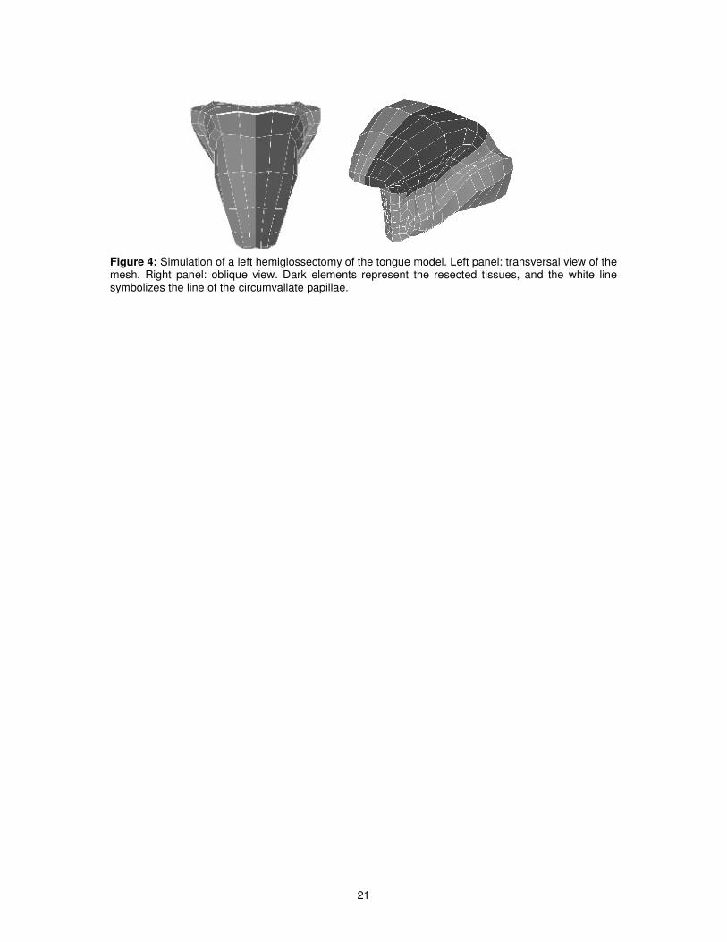
II.2.1. Hemiglossectomy
Most of the cancers occur on the lateral border of the tongue at the junction between the middle and
posterior thirds. Surgical resection for oral tongue cancer can be aggressive with planned margins of
at least 1 cm. For tumours with diameters as large as 2 cm the hemiglossectomy is the recommended
surgical treatment. The first surgery that was simulated on the model corresponds to this case. It is
shown on Figure 4 for a left hemiglossectomy.
In this implementation, the resection affects the large majority of the tongue muscles. The left part of
the styloglossus is removed as well as the left anterior parts of the inferior and superior longitudinal
muscles, the left anterior parts of the transverse and vertical muscles and the upper part of the left
hyoglossus. The medium and anterior parts of the left genioglossus are nearly entirely removed,

9
whereas its posterior part is only partially affected. Since the resection affects only half the tongue’s
body, we can expect highly dissymmetric movement of the tongue after such a resection.
II.2.2. Mouth floor resection
In the anterior floor of the mouth, the smallest tumours involve mucosa. The resection extends then in
the mucosa behind the alveolar ridge, to the excluded ventral face of the tongue. This corresponds to
the second example of surgery that was implemented on the model (see Figure 5). In that case, the
mobile tongue is totally preserved. The anterior part of the genioglossus is removed as well as the
totality of two major muscles of the mouth floor, namely the geniohyoid and the mylohyoid muscles.
For the moment, we consider surgery that affects the muscles only, not the lingual nerve or the
mandible.
II.2.3. Reconstruction Reconstruction of the tongue can be achieved with primary closure and local, regional or distant flaps.
Resections of the anterior part of mouth floor due to small tumours can be repaired with local flaps.
The most common local flap is the buccinator muscle flap. It is versatile and its elastic characteristics
seem to be comparable with those of the oral mucosa. The size of the flap is small. Two flaps (one of
each side) can be used to repair a defect of the mouth floor. In such cases, the reconstruction allows a
better mobility and avoids a spontaneous protrusion of the tongue out of the oral cavity. When the
resection includes the ventral tongue or/and the posterior part of the tongue, the reconstruction has to
be composite, with a musculocutaneous flap (pectoralis major and latissimus dorsi) or, even better,
with an osteocutaneous flap (fibula). These flaps are less versatile and their elasticity significantly
differs from the one of the oral mucosa. After surgery, radiotherapy is often necessary, especially
when the lymphatic nodes or the bone are involved by the tumour. The surface characteristics of the
soft tissues are modified by the radiations and they loose their softness. The final result differs
according to whether the patient underwent or not radiotherapy.
For the two modelled resections (namely hemiglossectomy and mouth floor resection), four cases
were studied that attempt at reproducing these different characteristics of the flap, and the
consequences of a possible radiotherapy. First we simply implemented a resection of the tongue
without reconstruction. Then three different kinds of flaps were used : a flap having exactly the same
biomechanical properties as the passive tissues of a healthy tongue (see Figure 3), a flap with a
10
stiffness 5 times smaller and a third one with a stiffness 6 times higher. These flaps are totally inactive
during the tongue activation.
III. Results
All the simulations presented below correspond to muscle activation patterns lasting 150 ms.
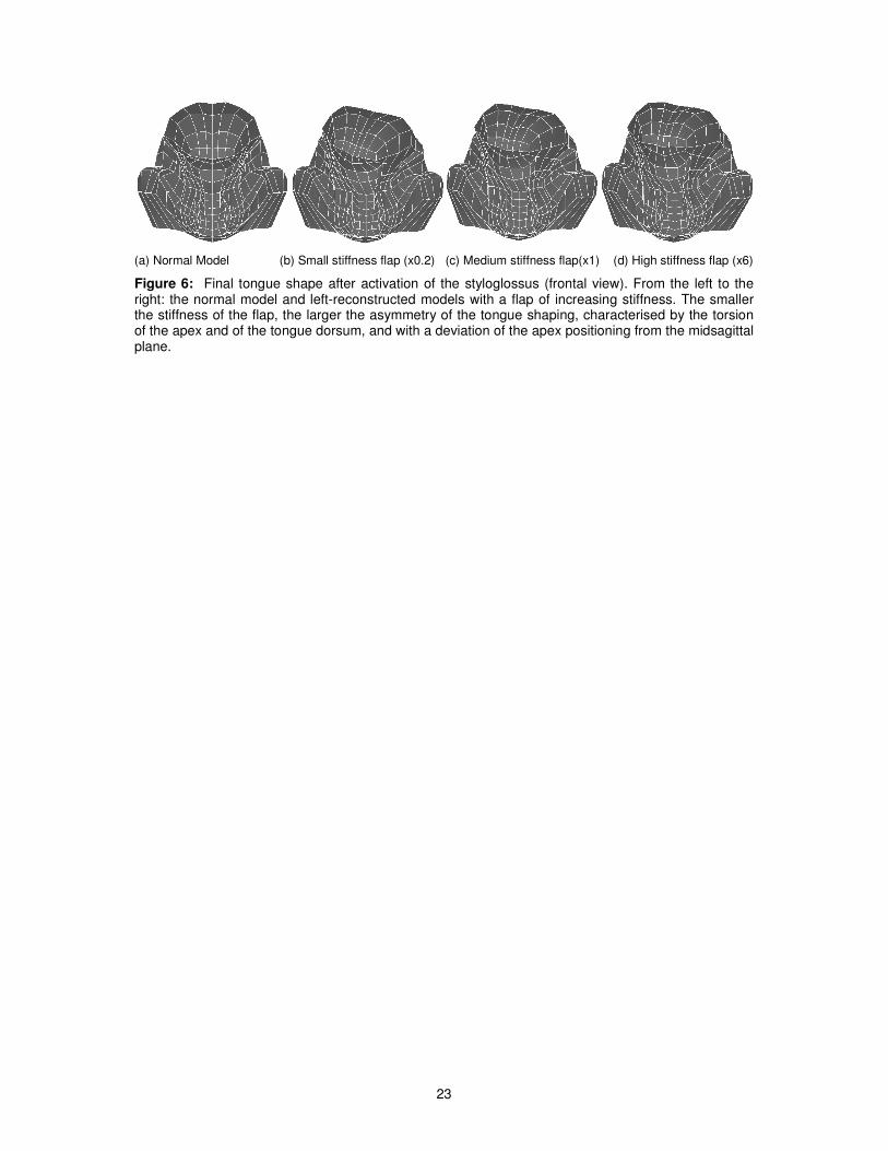
III.1. Asymmetrical movements of the tongue following a
hemiglossectomy
In normal conditions, the tongue movements are coarsely symmetric (pair muscles are activated
simultaneously according to a similar pattern of activation). After a hemiglossectomy, this symmetry is
broken, since half the tongue is removed and possibly reconstructed, and it induces asymmetries in
tongue displacements and shaping. The induced deviations from normal symmetrical movements are
more or less important depending on the muscles that are activated. Figure 6 shows a case of
important deviation with a frontal view of the tongue mesh after a 150 ms activation of the
styloglossus. As expected, in the absence of surgery, this activation pulls the tongue upwards and
backwards, and lets the model perfectly symmetric (normal model, Figure 6(a)). When the left-side of
the tongue is removed and reconstructed (Figure 6(b-d), an asymmetry of the tongue shaping is
observed, characterized by a torsion of the tongue apex and of the tongue dorsum, and by an
important deviation of the apex from the midsagittal plane. The physical properties of the flap have a
strong impact on the amplitude of the tongue torsion and consequently on the maximal elevation of the
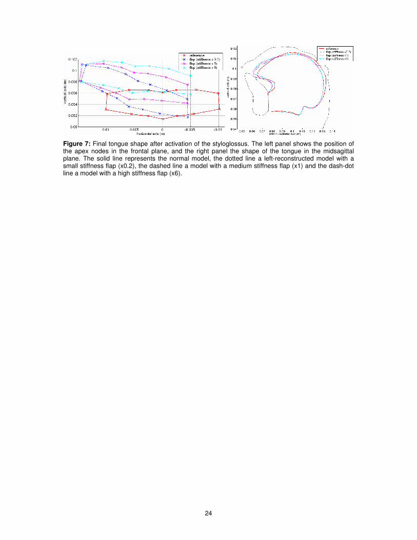
tongue in the velar region. Figure 7 (left panel) shows a frontal view of the apical tongue for the same
simulations. The torsion decreases when the stiffness of the flap increases. Hence, it seems more
difficult to control the positioning of the surface of the tongue when the stiffness of the flap is small.
However, these different images show that the stiffness of the flap has nearly no influence on the
lateral deviation of the apex (in the three conditions, the central node deviates of approximately 5 mm
towards the right). However, Figure 6(b-d) shows that increasing the stiffness of the flap reduces the
maximal elevation of the right part of the tongue dorsum, which is consistent with the reduction of the
torsion. Figure 7 (right panel) shows a complementary view in the midsagittal plane. Due to the fact
that that only the right styloglossus can be activated in reconstructed tongues, the backward and
upward displacement of the tongue is reduced compared to the normal condition. It was also observed
11
that the velocity of tongue movements is much lower. We can also notice that, in the midsagittal plane,
the tongue shapes associated with the three different flap stiffness differ essentially in the apical area,
not in the posterior region.
III.2. Impacts movements of the tongue after a mouth floor
resection
The resection of the mouth floor only affects the posterior genioglossus, the mylohyoid and the
geniohyoid. Therefore, consequences can be expected on the ability of the patients to move
simultaneously their tongue forwards and upwards. They are simulated below in section III.2.2 for
different hypotheses about muscle force recovery after resection. Interestingly, difficulties have also
been observed for upward movements in the velar region, which essentially involve the activation of
the styloglossus that is not directly affected by the surgery. In order to understand the origins of this
observed impairment, the action of this muscle assuming a resection of a medium size tumour of the
mouth floor was simulated. Results are described in section III.2.1.
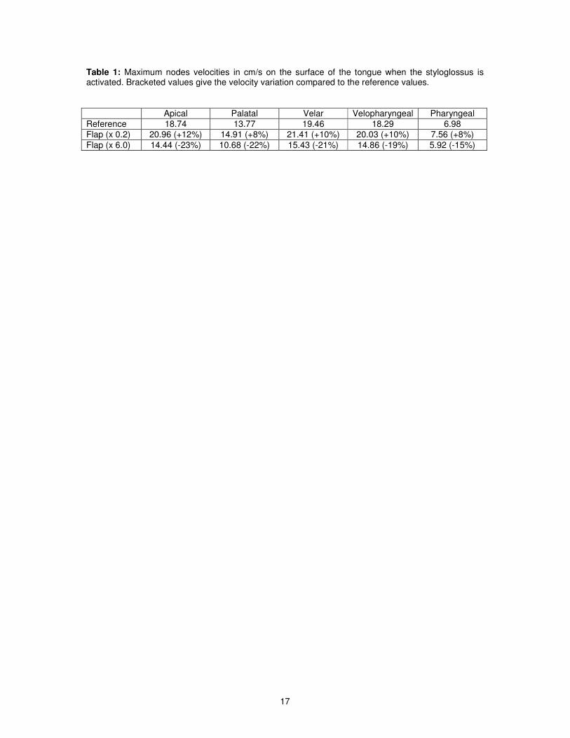
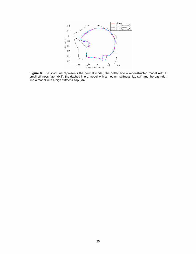
III.2.1. Effects on tongue elevation in the velar region.
The consequences of a styloglossus activation were evaluated assuming a resection of a medium size
tumour of the mouth floor and for reconstructions involving three levels of stiffness of the flaps. Figure
8 shows the resulting shapes of the tongue in the midsagittal plane after activation of the styloglossus.
Differences are very small. However, looking at the kinematic characteristics of the movement from
the rest position to the final tongue shape, it was observed that velocity is affected by flap stiffness.
Table 1 shows the maximal velocity reached during the movement by nodes located on the surface of
the tongue: a high stiffness flap reduces the maximal velocity of the tongue (about 20% of decrease)
as compared to the intact tongue. Thus, it can be assumed that difficulties in the production of upward
movements in the velar region could be the result of a too high stiffness of the flap. Moreover, as
illustrated in table 1, using softer flap could limit the disturbances caused by the resection and the
reconstruction for this particular movement.
III.2.2. Effect on tongue advancement

12
The last series of tests presented in Figure 9 shows the co-contraction of the posterior genioglossus,
styloglossus and transversus, resulting in a forward and upward movement of the tongue for a normal
configuration. During the resection of the floor of the mouth, only the anterior part of the posterior
genioglossus is removed, resulting in a shorter muscle which fibres insert on the flap after the
reconstruction. Little is known about the way muscles contract once they have been partially
shortened, and the level of activation that can be expected. Three options were tested for the
activation of the fibres of the posterior genioglossus: 1) absence of activation, 2) low level of activation
compared to the normal case, 3) similar level of activation compared to the normal case.
Results of the simulations show how difficult it can be to pull the tongue upward and front, once the
genioglossus has been partially removed, even in case 3 where it is hypothesized that muscle
activation is preserved. Once the genioglossus insertions on the superior mental spine of the mandible
have been removed, it losses its capacity to protrude and elevate the tongue. Increasing the stiffness
of the flap facilitates clearly the tongue propulsion, even if it is still not sufficient to retrieve the same
shape as in the normal case.
These results show that in case of mouth floor resection and reconstruction the stiffness of the flap
can dramatically influence tongue mobility. However, while it seems to be better to use soft flap to
preserve tongue elevation in the velar region, stiff flaps appear to be better to maintain the front/up
movement of the tongue. This shows how interesting the use of a biomechanical model of the tongue
can be in the planning of tongue surgery in order to limit the negative effects of the surgery in terms of
tongue mobility.
III.3. Interpretation in terms of impact on speech production
The control of the tongue for speech production essentially consists of an accurate positioning and
shaping of this articulator in reference to the external walls of the oral cavity, namely the hard palate,
the soft palate and the pharyngeal wall. This positioning determines first the size of the different
resonance cavities of the vocal tract and their coupling, and it influences the spectral characteristics of
the sound. It also determines the aeroacoustic nature of the airflow, laminar (when tongue is not too
close to the external walls) or turbulent (in case of small cross-sectional area of the oral cavity), which
differentiates vowels from consonants. Time characteristics are also very important in speech
13
production, which means that the velocity of the articulatory movements has to match a number of
constraints.
It was shown above that a hemiglossectomy generates a strong deviation of the tongue positioning in
the coronal plane. Consequently, in the absence of reconstruction, new strategies have to be
developed by the patients in order to compensate for this deviation. Further works with the model
should tell us whether such compensations are actually possible. More specifically, an important issue
is whether the residual volume of the tongue in the palatal region is large enough to allow the
production of the oral cavity closure that is required for the production of turbulent airflows underlying
the production of alveolar and palatal consonants. In case of reconstruction, the volume should not be
a problem any more. In this case it was shown that the control of the surface of the tongue, and then
of the cross-sectional shape of the oral cavity (important for the characteristics of turbulent airflows)
was easier with stiff flaps. However, it was also shown that reconstruction involving stiff flaps strongly
decrease the amplitude of upward tongue movements. As a consequence, the positioning of the
tongue near the hard and the soft palates is largely impaired, inducing difficulties in the production of
high vowels such as /i/, /e/, /y/ or /u/, or of consonants such as /t/ or /k/. This difficulty was clearly
observed in former studies of patients who underwent hemiglossectomy ( 5).
In the same study ( 5), it was observed that patients having undergone an important mouth floor
resection often had strong difficulties to produce sounds articulated in the velar region such as vowel
/u/ or consonant /k/. The simulations shown above in section III.2.2 provide an interesting explanation
for this phenomenon: the stiffness of the flap was probably too high. Indeed, in these conditions,
according to our simulations, even if these patients could have been able to produce the right
positioning and shaping of the tongue, the movement velocity could have decreased so much that it
would not longer be possible to shape properly the tongue within a speech sequence with the correct
timing. At the same time, our simulations have also shown that in case of mouth floor reconstruction,
stiff flaps would allow more forward displacement, facilitating the production of high front vowels such
as /i/, /y/ and /e/.
Hence, the simulations presented in this paper are in quite good agreement with observations made
on patients after tongue surgery. They also show how useful such simulations can be for surgery
14
planning, since they demonstrate that in case of reconstruction of the moving part of the tongue, the
stiffness of the flap has to be very carefully taken into consideration.
IV. Conclusion
Simulations with a realistic 3D biomechanical model of the tongue have shown that some of the
clinically observed consequences of tongue surgery, including partial resection and reconstruction with
a flap, on tongue mobility and speech articulation could be well accounted for by such a model. The
potential role of the mechanical characteristics of the flap on tongue mobility was shown. Moreover, it
appeared that controlling flap stiffness could help preserving speech production capabilities.
As shown in section V.2.2. our approach can also be very useful to improve the understanding of
muscle behaviour after partial resection. Comparison of simulation results with data collected on
patients could shed light on the hypothesis (no activation, partial activation or full activation) that
seems to be the most realistic.
Therefore, using such a model should represent a significant improvement in planning tongue surgery
systems, in order to preserve as much as possible tongue mobility during speech production, and
therefore the patients’ Quality of Life.
Further improvements of the model have to be done before these ultimate objectives can be actually
reached. First of all, fast finite element algorithms have to be implemented and tested in order to
significantly decrease the computation time, and to reach simulation durations compatible with an
interactive use of the model. First improvements in this direction have been recently done ( 26- 27).
Mesh-matching algorithms in the line of those developed by Couteau et al ( 22) have also to be
elaborated in order to allow the surgeons to adapt the generic tongue model to the anatomy of each
specific patient.
Acknowledgments:
This work is supported by a grant of the Region Rhône-Alpes (Emergence Program).
15
The authors express their grateful thanks to Pierre Badin (ICP/GIPSA-lab) for sharing its MRI and X-
Ray data, to Matthieu Chabanas (ICP/GIPSA-lab) for advices and help in the development of the
tongue model, to Christophe Savariaux (ICP/GIPSA-lab) and Jacques Lebeau (Head of the plastic and
maxillo-facial surgery department at Grenoble University Hospital) for theirs contributions to the
analysis of patients after partial glossectomy and reconstruction.
V. References
1. Talmi YP. Quality of life issues in cancer of the oral cavity. Journal of Laryngology and Otology.
2002;116(10):785-90.
2. Deleyiannis FWB, Weymuller Jr. A, Coltrera MD. Quality of Life of Disease-Free Survivors of
Advanced (Stage III or IV) Oropharyngeal Cancer. Head & Neck.1997; 19:466-73.
3. Furia CLB, Kowalski LP, Latorre MRDO, Angelis EC, Martins NMS, Barros APB et al. Speech
Intelligibility After Glossectomy and Speech Rehabilitation. Archives of Otolaryngology and Head
Neck Surgery. 2001; 127:877-883.
4. Perrier P, Savariaux C, Lebeau J, Magaña G. Speech production after tongue surgery and tongue
reconstruction. Proceedings of the 14th International Congress of Phonetic Sciences, San
Francisco, USA; 1999. Vol. 3, p. 1805-1808.
5. Savariaux C, Perrier P, Pape D, Lebeau J. Speech production after glossectomy and
reconstructive lingual surgery: a longitudinal study. Proceedings of the 2nd
International Workshop
on Models and Analysis of Vocal Emissions for Biomedical Applications (MAVEBA). Firenze, Italy.
2001.
6. Bressmann T, Thind P, Uy C, Bollig C, Gilbert RW, Irish JC. Quantitative three-dimensional
ultrasound analysis of tongue protrusion, grooving and symmetry: data from 12 normal speakers
and a partial glossectomy. Clinical Linguistics & Phonetics. 2005; 19(6-7):573-88.
7. Bressmann T. Speech adaptation to a self-inflicted cosmetic tongue split: perceptual and
ultrasonographic analysis. Clinical Linguistics & Phonetics. 2006; 20(2-3):205-10.
8. Lavallée S, Sautot P, Troccaz J, Cinquin P, Merloz P. Computer Assisted Spine Surgery : a
technique for accurate transpedicular screw fixation using CT data and a 3D optical localizer.
Journal of Image Guided Surgery. 1995; 1(1):65-73.
9. Merloz P, Tonetti J, Eid A, Faure C, Lavallée S, Troccaz J, et al. Computer assisted spine surgery.
Clin Orthop. 1997; 337: 86-96.
10. Tonetti J, Carrat L, Lavallée S, Cinquin P, Merloz P, Pittet L. Ultrasound-based registration for
percutaneous computer assisted pelvis surgery: Application to Iliosacral screwing of pelvis ring
fractures. Computer Assisted Radiology and Surgery. 1997; 961 – 966.
11. Chabanas M, Luboz V, Payan Y. Patient specific Finite Element model of the face soft tissue for
computer-assisted maxillofacial surgery. Medical Image Analysis. 2003; 7(2):131-151.
16
12. Clatz O, Delingette H, Talos IF, Golby AJ, Kikinis R, Jolesz FA et al. Robust nonrigid registration to
capture brain shift from intraoperative MRI. IEEE Trans Med Imaging. 2006; 24(11): 1417-1427.
13. Ferrant M, Nabavi A, Macq B, Jolesz FA, Kikinis R, Warfield SK. Registration of 3-D intraoperative
MR images of the brain using a finite-element biomechanical model. IEEE Transactions on
Medical Imaging. 2001; 20(12): 1384-1397.
14. Gérard J-M, Wilhelms-Tricarico R, Perrier P, Payan Y. A 3D dynamical biomechanical tongue
model to study speech motor control. Recent Research Developments in Biomechanics. 2003;1:
49-64, Transworld Research Network.
15. Gérard J-M, Perrier P, Payan Y. 3D biomechanical tongue modelling to study speech production
In: J. Harrington & M. Tabain Editors, Speech Production: Models, Phonetic Processes, and
Techniques. Psychology Press: New-York, USA; 2006. p. 85-102
16. Buchaillard S, Pascal P, Payan Y. A 3D biomechanical vocal tract model to study speech
production control: How to take into account the gravity? Proceedings of the 7th International
Seminar on Speech Production, Ubatuba, Brazil; 2006. p. 403-10
17. Miyawaki K. A study on the musculature of the human tongue. Annual Bulletin of the Research
Institute of Logopedics and Phoniatrics (Tokyo University). 1974; 8: 22-50.
18. Netter FH. Atlas of Human anatomy. CIBA-GEIGY Corporation editor. 1999
19. Takemoto H. Morphological analyses of the human tongue musculature for three dimensional
modelling. Journal of Speech, Language, Hearing Research. 2001; 44: 95-107.
20. Wilhelms-Tricarico R. Development of a tongue and mouth floor model for normalization and
biomechanical modelling. Proceedings of the 5th speech production seminar and CREST
Workshop on models of speech production. 2000; 141-148. Kloster Seeon, Bavaria.
21. Badin P, Bailly G, Revéret L, Baciu M, Segebarth C, Savariaux C. Three-dimensional linear
articulatory modeling of tongue, lips and face based on MRI and video images. Journal of
Phonetics. 2002; 30(3): 533-553.
22. Couteau B, Payan Y, Lavallée S. The Mesh-Matching algorithm: an automatic 3D mesh generator
for finite element structures. Journal of Biomechanics. 2000; 33(8); 1005-1009.
23. Feldman AG. Once more on the Equilibrium-Point hypothesis (λ model) for motor control. Journal
of Motor Behavior. 1986; 18(1):17-54.
24. Gérard J-M, Ohayon J, Luboz V, Perrier P, Payan Y. Non linear elastic properties of the lingual and
facial tissues assessed by indentation technique. Application to the biomechanics of speech
production. Medical Engineering & Physics. 2005; 27: 884–892.
25. Duck FA. Physical Properties of Tissues: A Comprehensive Reference Book. Academic Press,
London: 1990
26. Nesme M, Faure F, Payan Y. Hierarchical Multi-Resolution Finite Element Model for Soft Body
Simulation. Lecture Notes in Computer Science. 2006; 4072: 40-47.
27. Vogt F, Lloyd J, Buchaillard S, Perrier P, Chabanas M. Payan Y, et al.. Investigation of Efficient 3D
Finite Element Modeling of a Muscle-Activated Tongue. Lecture Notes in Computer Science.
2006; 4072: pp. 19-28.
17
Table 1: Maximum nodes velocities in cm/s on the surface of the tongue when the styloglossus is activated. Bracketed values give the velocity variation compared to the reference values.
Apical Palatal Velar Velopharyngeal Pharyngeal
Reference 18.74 13.77 19.46 18.29 6.98
Flap (x 0.2) 20.96 (+12%) 14.91 (+8%) 21.41 (+10%) 20.03 (+10%) 7.56 (+8%)
Flap (x 6.0) 14.44 (-23%) 10.68 (-22%) 15.43 (-21%) 14.86 (-19%) 5.92 (-15%)

18
Figure 1: Final oral cavity geometry at rest. Left panel: Superimposition of the model midsagittal contours on X-ray at rest in the midsagittal plane. Right panel: the 3D tongue
mesh in the whole oral cavity.

19
(a) Posterior genioglossus (b) Styloglossus (c) Hyoglossus (d) Transversus
Figure 2: Representation of four muscles of the tongue (oblique view). The red lines represent the muscles macro-fibres.
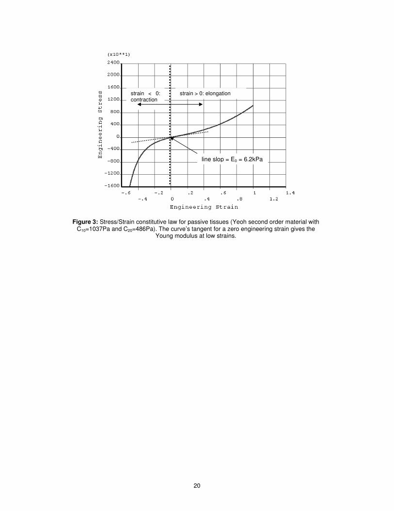
20
Figure 3: Stress/Strain constitutive law for passive tissues (Yeoh second order material with C10=1037Pa and C20=486Pa). The curve’s tangent for a zero engineering strain gives the
Young modulus at low strains.
strain > 0: elongation strain < 0: contraction
line slop = E0 = 6.2kPa
21
Figure 4: Simulation of a left hemiglossectomy of the tongue model. Left panel: transversal view of the mesh. Right panel: oblique view. Dark elements represent the resected tissues, and the white line symbolizes the line of the circumvallate papillae.

22
Figure 5: Simulation of mouth floor resection. Left panel: frontal view. Right panel: sagittal view. The resected part is represented in blue, green (geniohyoid muscle) and yellow (mylohyoid muscle).
23
(a) Normal Model (b) Small stiffness flap (x0.2) (c) Medium stiffness flap(x1) (d) High stiffness flap (x6)
Figure 6: Final tongue shape after activation of the styloglossus (frontal view). From the left to the right: the normal model and left-reconstructed models with a flap of increasing stiffness. The smaller the stiffness of the flap, the larger the asymmetry of the tongue shaping, characterised by the torsion of the apex and of the tongue dorsum, and with a deviation of the apex positioning from the midsagittal plane.
24
Figure 7: Final tongue shape after activation of the styloglossus. The left panel shows the position of the apex nodes in the frontal plane, and the right panel the shape of the tongue in the midsagittal plane. The solid line represents the normal model, the dotted line a left-reconstructed model with a small stiffness flap (x0.2), the dashed line a model with a medium stiffness flap (x1) and the dash-dot line a model with a high stiffness flap (x6).
25
Figure 8: The solid line represents the normal model, the dotted line a reconstructed model with a small stiffness flap (x0.2), the dashed line a model with a medium stiffness flap (x1) and the dash-dot line a model with a high stiffness flap (x6).

26
Figure 9: Co-contraction of the posterior genioglossus, styloglossus and transversus. Top-left hand corner: the genioglossus is not activated. Top-right hand corner: genioglossus is slightly activated. Bottom: genioglossus is highly activated. The solid line represents the normal model, the dotted line a reconstructed model with a small stiffness flap (x0.2), the dashed line a model with a medium stiffness flap (x1) and the dash-dot line a model with a high stiffness flap (x6).