similarity of proteins synthesized by isolated blastomeres of early sea urchin embryos
TRANSCRIPT

DEVELOPMENTAL BIOLOGY 72,3%L397 (1979)
Similarity of Proteins Synthesized by Isolated Blastomeres of Early Sea
Urchin Embryos
FRANKTUFARO AND BRUCE P. BRANDHORST' Department of Biology, McGill University, 1205 McGregor Avenue, Montreal, Quebec, Canada H3A, 1BI
Received February 6, 1979; accepted in revised form April 2, 1979
The 16-cell sea urchin embryo has blastomeres of three distinct size classes: micromeres, mesomeres, and macromeres. Each class is already restricted in its developmental fate, micromeres being committed to formation of primary mesenchyme cells. The three classes of blastomeres were isolated in high purity and incubated in [%]methionine until the next cleavage. Nearly all the radioactive protein was solubilixed and subjected to two-dimensional electrophoresis according to O’Farrell. Of approximately 1006 spots resolved, there are no qualitative differences among the three blastomeres. When embryos were labeled between the first and fourth cleavages and blastomeres then isolated, no qualitative differences in protein synthesis were observed. Moreover, there are very few changes when unfertilized eggs are compared to 16-cell embryos. Thus cellular determination during embryonic development is not accompanied by qualitative changes in the distribution within the embryo of abundantly synthesized proteins, virtually all of which are coded for by sequences present in the egg.
INTRODUCTION
The fourth cleavage of the sea urchin egg gives rise to three distinct types of blasto- meres having different sizes: micromeres, mesomeres, and macromeres. These blas- tomeres have different developmental fates in intact embryos and when isolated (Hor- stadius, 1939). The four micromeres, which form at the vegetal pole, give rise to pri- mary mesenchyme cells of the blastula, which ultimately secrete the spicules of the larval skeleton. Isolated micromeres can be maintained in culture and eventually se- crete skeletal spicules resembling in form those of larvae (Okazaki, 1975). Thus, by the 16-cell stage, micromeres are deter- mined to follow a specific developmental pathway in the absence of interaction with other embryonic cells.
It has long been thought that determi- native events in early embryos may be brought about by an asymmetric distribu- tion of factors in the egg and their segre- gation into different blastomeres during
’ To whom correspondence should be addressed.
cleavage (e.g., Wilson, 1927; Davidson and B&ten, 1971; Raff, 1977). These factors might be regulatory molecules (or precur- sors thereof), or they might be structural proteins or the mRNAs coding for them. Several recent reports suggest that mRNA is segregated into different blastomeres of early embryos. In the marine snail IZy- anassa, factors required for the formation of mesodermal organs are localized in the polar lobe, a cytoplasmic extension of one of the first two blastomeres: Removal of the lobe results in an embryo most notably devoid of heart, shell, and foot (Clement, 1962). Newrock and Raff (1975) observed that lobed and lobeless embryos synthesize proteins having different patterns when an- alyzed by discontinuous SDS-polyacryl- amide gel electrophoresis. These differ- ences, which might be qualitative, persist even when new mRNA synthesis is in- hibited. Moen and Namenwirth (1977) have shown that the electrophoretic patterns of proteins synthesized in Xenopus eggs and early embryos are different along the ani- mal-vegetal axis, implying a localization of
396 0012-1696/79/100390-08$02.00/O Copyright 0 1979 by Academic Press, Inc. All rights of reproduction in any form reserved.

BRIEF NOTES 391
the maternal mRNA coding for these abun- dant proteins. Whitaker (1977) has shown that the capacity to synthesize alkaline phosphatase is segregated during the early cleavage divisions of the ascidian Ciona, but the enzyme activity first becomes de- tectable in gastrulae. The enzyme activity appears in the appropriate blastomeres even when RNA synthesis in embryos is inhibited by actinomycin D, suggesting that a stored maternal mRNA coding for it is segregated.
Sea urchin eggs contain a store of mRNA synthesized during oogenesis, but trans- lated only after fertilization (Gross et al., 1964; Humphreys, 1969, 1971). It is possible that selected stored mRNA molecules might be localized within the eggs and seg- regated during early cleavages, resulting in cellular determination. Rodgers and Gross (1978) have recently provided evidence that different blastomeres of the 16-cell stage contain qualitatively distinct populations of RNA. They did not demonstrate that the distinctive molecules are actively trans- lated mRNA. We approached this question by subjecting the proteins synthesized by isolated micromeres, mesomeres, and mac- romeres to two-dimensional electrophoresis (O’Farrell, 1975).
EXPERIMENTAL PROCEDURES
Preparation of eggs and removal of fer- tilization membranes. Stronglocentrotus purpuratus were obtained from Pacific Biomarine Inc. For each experiment spawn- ing was induced in a single gravid female by the injection of KCl. The eggs were collected into artificial seawater at 4°C and washed by settling three times. Eggs were suspended in 10 n&f dithiothreitol in sea- water buffered with 10 mJ4 glycylglycine, pH 9.3, at 10°C for 90-120 set, and then diluted with 40 vol seawater at 4°C (Epel et al., 1970). Eggs treated in this manner fail to raise a fertilization membrane upon fertilization. Eggs were routinely incubated with a 50,000-fold dilution of sperm for 1
min, the time required for fertilization to be more than 95% complete. Demembraned eggs tend to clump upon fertilization if maintained in a concentrated suspension. Fertilized eggs (less than 106) were there- fore diluted into 1 liter of seawater and washed 2-3 times by settling through this volume. For culturing, embryos were sus- pended in 500 ml seawater containing 50 pg/ml penicillin and streptomycin and al- lowed to develop at 14°C. At this temper- ature synchrony of early cleavage is opti- mal, and embryos reached fourth cleavage at about 5 hr after fertilization. Treatment of embryos with dithiothreitol had no effect on normal development to pluteus stage.
Separation of blastomeres. At the 16-cell stage, embryos were collected by light cen- trifugation, and resuspended and washed 3 times in 10 ml seawater lacking calcium and magnesium ions in order to dissociate the hyalin layer. Brisk aspiration of a 2-ml sus- pension 5-8 times through a Pasteur pipet dissociated most embryos into single blas- tomeres. These were layered onto 30-ml linear gradients of 5 to 50% (v/v) isotonic sucrose (1 m in seawater lacking Ca2+ and Mg2+ ions, and allowed to settle by gravity. After 30-45 min, three bands of cells are visible corresponding to the three types of blastomeres. The approximate positions and identification of these bands are: mi- cromeres in a very narrow band about 1 cm below the surface, mesomeres in a broader band (about 0.5 cm wide) approximately 2 cm below the micromeres, and macromeres in a similar band separated from the bottom of the mesomeres by less than 0.5 cm. Un- dissociated embryos sediment to the bot- tom of the gradient (about 6 cm below the surface). The gradients were fractionated by gradually lowering a capillary tube sup- ported by a small jack. The tube was at- tached to a peristaltic pump and fractions were collected into cold centrifuge tubes. To minimize cross contamination, meso- meres were withdrawn from the top of the band and macromeres from the bottom of
392 DEVELOPMENTAL BIOLOGY VOLUME 72.1979
the band. The purity of the fractionated blastomeres was estimated by microscopy. In the investigations reported, no larger blastomeres are seen among hundreds of micromeres. Micromeres were never ob- served among macromeres. There is some cross contamination between mesomeres and macromeres, but it is less than 5%. Occasional smaller cells which might be unusually large micromeres were some- times observed among mesomeres, but such possible contamination is less than 2%. Fractionated blastomeres were either col- lected by light centrifugation and frozen or resuspended in seawater for incubation with [35S]methionine.
Labeling of proteins. A suspension of 200,000 embryos lacking fertilization mem- branes in 5 ml seawater was incubated at 14°C with 100 &i/ml [35S]methionine (about 700 Ci/mole) obtained from Amer- sham. Labeling was halted by immersion in cold seawater lacking calcium and magne- sium ions. For labeling separated blasto- meres, each type was suspended in 1 ml seawater containing 250 &i/ml [3”S]methi- onine for 1 hr. Labeling was stopped by washing in cold seawater followed by freez- ing in a dry ice-ethanol bath and storage at -70°C.
Two-dimensional polyacrylamide gel electrophoresis. Samples were prepared by a modification of the method of Alton and Lodish (1977). Frozen cells were rapidly resuspended in SDS sample buffer (in which 0.1 M dithiothreitol is substituted for mercaptoethanol) and immersed in a boil- ing water bath for 1 min. Aliquots were withdrawn for determination of radioactiv- ity and protein content and the remainder was frozen and stored at -70°C.
The amount of radioactivity in protein was determined by precipitation in hot 15% trichloroacetic acid and collection on Mil- lipore filters. The amount of protein was determined according to Lowry et al. (1951). Samples for isoelectric focusing were prepared by the addition of 10 1.11 of
thawed extract to 9 mg urea (Bio-rad elec- trophoresis grade) and dilution with 80 ~1 lysis buffer containing 1.6% pH 5-7 ampho- lytes and 0.4% pH 3.5-10 ampholytes (Bio- rad). Isoelectric focusing was according to O’Farrell (1975). Samples to be compared contained equal amounts of protein of sim- ilar specific activity.
To determine the fraction of radioactive protein entering the isoelectric focusing gels, a parallel gel was sliced into 0.5-cm lengths. Each slice was incubated for 8 hr at 70°C in a capped scintillation vial con- taining 0.5 ml 30% H202. The dissolved slices were counted in Aquasol- (New Eng- land Nuclear). More than 85% of the radio- active protein loaded enters the isoelectric focusing gel. The pH range was about 4.5 to 7.2 as measured by a microelectrode (Biorad prophiler) inserted into the gels.
Isoelectric focusing gels were equili- brated with four changes of SDS sample buffer over 1 hr and subjected to electro- phoresis on slab gels consisting of a 10-16s exponential gradient of acrylamide (Brand- horst, 1976; O’Farrell, 1975).
The method used to prepare protein ex- tracts results in the solubilization of vir- tually all the radioactive protein of the em- bryos. The molecular weight standards were as previously described (Hutchins and Brandhorst, 1978).
RESULTS
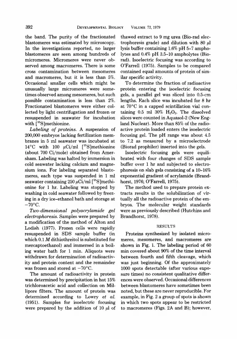
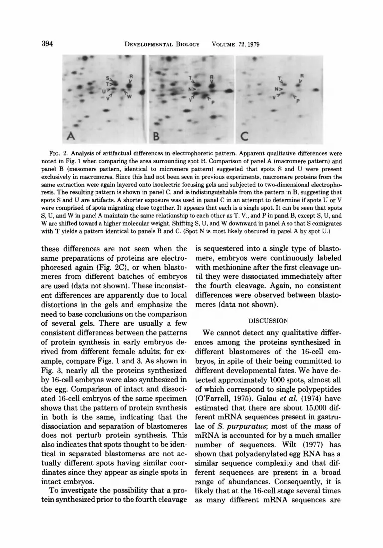
Proteins synthesized by isolated micro- meres, mesomeres, and macromeres are shown in Fig. 1. The labeling period of 60 min covered about 90% of the time interval between fourth and fifth cleavage, which was just beginning. Of the approximately 1000 spots detectable (after various expo- sure times) no consistent qualitative differ- ences were observed. Occasional differences between blastomeres have sometimes been noted, but these are never reproducible. For example, in Fig. 2 a group of spots is shown in which two spots appear to be restricted to macromeres (Figs. 2A and B); however,

25
50
25
7 PH 5 FIG, 1. Two-dimensional gel electrophoresis of proteins synthesized by isolated blastomeres. Blastomeres
isolated from 16-cell embryos were incubated with [35S]methionine for 60 min. Extracts containing 7 X 10” dpm in protein were layered onto isoelectric focusing gels and subjected to two-dimensional electrophoresis. .Autoradiograms were exposed to 10” disintegrations (+_lO%), which results in the appearance of 995 distinct spots. Approximate pH values for the isoelectric focusing dimension are shown on the horizontal axis and molecular weights (~10~~) are shown on the vertical axis. Spot 1 is never detectable in unfertilized eggs. Spot R is shown for reference to the same spot in Fig. 2. (A) Micromeres, (B) mesomeres, (C) macromeres.
393
394 DEVELOPMENTAL BIOLOGY VOLUME 72,1979
FIG. 2. Analysis of artifactual differences in electrophoretic pattern. Apparent qualitative differences were noted in Fig. 1 when comparing the area surrounding spot R. Comparison of panel A (macromere pattern) and panel B (mesomere pattern, identical to micromere pattern) suggested that spots S and U were present exclusively in macromeres. Since this had not been seen in previous experiments, macromere proteins from the same extraction were again layered onto isoelectric focusing gels and subjected to two-dimensional electropho- resis. The resulting pattern is shown in panel C, and is indistinguishable from the pattern in B, suggesting that spots S and U are artifacts. A shorter exposure was used in panel C in an attempt to determine if spots U or V were comprised of spots migrating close together. It appears that each is a single spot. It can be seen that spots S, U, and W in panel A maintain the same relationship to each other as T, V., and P in panel B, except S, U, and W are shifted toward a higher molecular weight. Shifting S, U, and W downward in panel A so that S comigrates with T yields a pattern identical to panels B and C. (Spot N is most likely obscured in panel A by spot U.)
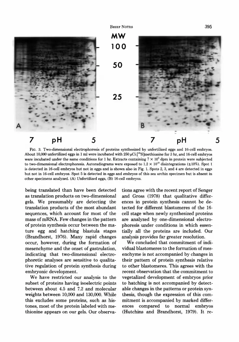
these differences are not seen when the same preparations of proteins are electro- phoresed again (Fig. BC), or when blasto- meres from different batches of embryos are used (data not shown). These inconsist- ent differences are apparently due to local distortions in the gels and emphasize the need to base conclusions on the comparison of several gels. There are usually a few consistent differences between the patterns of protein synthesis in early embryos de- rived from different female adults; for ex- ample, compare Figs. 1 and 3. As shown in Fig. 3, nearly all the proteins synthesized by 16-cell embryos were also synthesized in the egg. Comparison of intact and dissoci- ated 16-cell embryos of the same specimen shows that the pattern of protein synthesis in both is the same, indicating that the dissociation and separation of blastomeres does not perturb protein synthesis. This also indicates that spots thought to be iden- tical in separated blastomeres are not ac- tually different spots having similar coor- dinates since they appear as single spots in intact embryos.
To investigate the possibility that a pro- tein synthesized prior to the fourth cleavage
is sequestered into a single type of blasto- mere, embryos were continuously labeled with methionine after the first cleavage un- til they were dissociated immediately after the fourth cleavage. Again, no consistent differences were observed between blasto- meres (data not shown).
DISCUSSION
We cannot detect any qualitative differ- ences among the proteins synthesized in different blastomeres of the 16-cell em- bryos, in spite of their being committed to different developmental fates. We have de- tected approximately 1000 spots, almost all of which correspond to single polypeptides (O’Farrell, 1975). Galau et al. (1974) have estimated that there are about 15,000 dif- ferent mRNA sequences present in gastru- lae of S. purpuratus; most of the mass of mRNA is accounted for by a much smaller number of sequences. Wilt (1977) has shown that polyadenylated egg RNA has a similar sequence complexity and that dif- ferent sequences are present in a broad range of abundances. Consequently, it is likely that at the 16-cell stage several times as many different mRNA sequences are
BRIEF NOTES 395
MW
100
50
25
7 PH FIG. 3. Two-dimensional electrophoresis of proteins synthesized by unfertilized eggs and N-cell embryos.
About 10,000 unfertilized eggs in 1 ml were incubated with 250 PCi [“S]methionine for 3 hr, and 16-cell embryos were incubated under the same conditions for 1 hr. Extracts containing 7 x lo5 dpm in protein were subjected to two-dimensional electrophoresis. Autoradiograms were exposed to 1.2 x 10” disintegrations (&lo%). Spot 1 is detected in 16-cell embryos but not in eggs and is shown also in Fig. 1. Spots 2, 3, and 4 are detected in eggs but not in 16-cell embryos. Spot 5 is detected in eggs and embryos of this sea urchin specimen but is absent in other specimens analyzed. (A) Unfertilized eggs, (B) l&cell embryos.
being translated than have been detected tions agree with the recent report of Senger as translation products on two-dimensional and Gross (1978) that qualitative differ- gels. We presumably are detecting the ences in protein synthesis cannot be de- translation products of the most abundant tected for different blastomeres of the 16- sequences, which account for most of the cell stage when newly synthesized proteins mass of mRNA. Few changes in the pattern are analyzed by one-dimensional electro- of protein synthesis occur between the ma- phoresis under conditions in which essen- ture egg and hatching blastula stages tially all the proteins are included. Our (Brandhorst, 1976). Many rapid changes analysis provides far greater resolution. occur, however, during the formation of We concluded that commitment of indi- mesenchyme and the onset of gastrulation, vidual blastomeres to the formation of mes- indicating that two-dimensional electro- enchyme is not accompanied by changes in phoretic analyses are sensitive to qualita- their pattern of protein synthesis relative tive regulation of protein synthesis during to other blastomeres. This agrees with the embryonic development. recent observation that the commitment to
We have restricted our analysis to the vegetalized development of embryos prior subset of proteins having isoelectric points to hatching is not accompanied by detect- between about 4.5 and 7.2 and molecular able changes in the patterns or protein syn- weights between 10,000 and 130,000. While thesis, though the expression of this com- this excludes some proteins, such as his- mitment is accompanied by marked differ- tones, most of the protein labeled with me- ences compared to normal embryos thionine appears on our gels. Our observa- (Hutchins and Brandhorst, 1979). It re-
5

396 DEVELOPMENTAL BIOLOGY VOLUME 72,1979
mains possible that less extensively labeled proteins or proteins already present in the egg may be segregated into different blas- tomeres at the fourth cleavage and that this segregation is responsible for develop- mental determination. Moreover, it is pos- sible that mRNAs coding for distinct pop- ulations of proteins are segregated into the different blastomeres but are translated later. Consistent with this possibility is the recent report of Rodgers and Gross (1978) that RNA sequences are segregated into different blastomeres at the 16-cell stage. Specifically, they concluded from nucleic acid hybridization investigations that some sequences of the high-complexity class of total RNA present in eggs are not detecta- ble in micromeres but are detectable in a mixture of mesomeres and macromeres. Since these sequences are unlikely to be abundant enough to be detectable as trans- lation products on two-dimensional gels, since they may not be translated at the 16- cell stage, and since they might not be mRNA at all, there is no discrepancy be- tween our observations and those of Rodg- ers and Gross (1978). It is possible that the RNA which has been segregated by the 16- cell stage serves a regulatory role, as pro- posed by Davidson and Britten (1972).
Since we failed to detect qualitative dif- ferences between the patterns of protein synthesis of the three types of blastomeres, we were concerned that the similarities might be due to cross contamination of the separated blastomeres. Our method for iso- lation of blastomeres is modified from methods described by Hynes and Gross (1970) and Spiegel (1975) and, in our hands, yields fractions of even greater purity than these other methods. Of particular impor- tance is the use of a relatively small number of synchronous embryos. We could not de- tect any cross contamination between mi- cromere and macromere fractions which have extremely different sizes. The slight possible contamination (unconfirmed) of the mesomere fraction should be much less
than the limit of sensitivity to large quan- titative differences; large quantitative dif- ferences are extremely rare.
A variety of evidence suggests that ma- ternal mRNA is segregated into different blastomeres having different and restricted developmental fates during early cleavage of spiralian eggs (e.g., Newrock and Raff, 1975; Donohoo and Kafatos, 1973; Cheney and Ruderman, 1978), tunicate eggs (Whi- taker, 1977), and amphibian eggs (Moen and Namenwirth, 1977). It will be interest- ing to determine whether a common feature of organisms having determinative embry- onic development is a qualitatively differ- ent pattern of protein synthesis in isolated early blastomeres. Such comparisons may shed light on the relatively regulative de- velopment of sea urchin embryos.
This research was supported by grants from the National Science and Engineering Research Council of Canada and the Quebec Department of Education.
REFERENCES
ALTON, T. H., AND LODISH, H. F. (1977). Translational control of protein synthesis during the early stages of differentiation of the slime mold Dictyostelium discoideum. Cell 12,301-310.
BRANDHORST, B. P. (1976). Two-dimensional gel pat- terns of protein synthesis before and after fertiliza- tion of sea urchin eggs. Deuelop. Biol. 52, 310-317.
CHENEY, C. M., AND RUDERMAN, J. V. (1978). Segre- gation of maternal mRNA at first cleavage in em- bryos of the mollusc Spisula solidissima. J. Cell. Biol. 79, 349a.
CLEMENT, A. C. (1952). Experimental studies on ger- minal localization in Ilyanassa. I. The role of the polar lobe in determination of the cleavage pattern and its influence in later development. J. Exp. 2001. 121, 593-613.
DAVIDSON, E. H., AND BRITTEN, R. J. (1971). Note on the control of gene expression during development. J. Theor. Biol. 32, 123-130.
DONOHOO, P., AND KAFATOS, F. C. (1973). Differences in the proteins synthesized by the progeny of the first two blastomeres of Ilyanassa, a “mosaic” em- bryo. Develop. Biol. 32, 224-229.
EPEL, D., WEAVER, A. M., AND MAZIA, D. (1970). Methods for removal of the vitelline membrane of sea urchin eggs. Exp. Cell Res. 61,64-68.
GALAU, G. A., BRITTEN, R. J., AND DAVIDSON, E. H. (1974). A measurement of the sequence complexity
BRIEF NOTES 397
of polysomal messenger RNA in sea urchin embryos.
Cell 2,9-21. GROSS, P. R., MALKIN, L. I., AND MOYER, W. A. (1964).
Templates for the first proteins of embryonic devel- opment. Proc. Nat. Acad. Sci. USA 51.407-415.
H~RSTADIUS, S. (1939). The mechanics of sea urchin development, studies by operative methods. Biol. Rev. Cambridge Phil. Sot. 14, 132-179.
HUTCHINS, R., AND BRANDHORST, B. P. (1979). Com- mitment to vegetalized development in sea urchin embryos: Failure to detect changes in patterns of protein synthesis. Wilhelm Roux Arch., 186, 95- 102.
HUMPHREYS, T. (1969). Efficiency of translation of messenger RNA before and after fertilization in sea urchins. Develop. Biol. 20,435-458.
HUMPHREYS, T. (1971). Measurement of messenger RNA entering polysomes upon fertilization of sea urchin eggs. Develop. Biol. 26, 201-208.
HYNES, R. O., AND GROSS, P. R. (1970). A method for separating cells from early sea urchin embryos. De- velop. Biol. 21, 383-402.
LOWRY, 0. H., ROSEBROUGH, N. J., FARR, A. L., AND RANDALL, R. J. (1951). Protein measurement with the Folin phenol reagent. J. Biol. Chem. 193,265- 275.
MOEN, T. L., AND NAMENWIRTH, M. (1977). The dis- tribution of soluble proteins along the animal-vege-
tal axis of frog eggs. Develop. Biol. 58, l-10. NEWROCK, K. M., AND RAFF, R. A. (1975). Polar lobe
specific regulation of translation in embryos of Ily- anassa obsoleta. Develop. Biol. 42, 242-261.
O’FARRELL, P. H. (1975). High resolution two-dimen- sional electrophoresis of proteins. J. Biol. Chem. 250,4007-4021.
OKAZAKI, K. (1975). Spicule formation by isolated micromeres of the sea urchin embryo. Amer. 2001. 15, 567-581.
RODGERS, W. H., AND GROSS, P. R. (1978). Inhomo- geneous distribution of egg RNA sequences in the early embryo. Cell 14, 279-288.
SENGER, D. R., AND GROSS, P. R. (1978). Macromol- ecule synthesis and determination in sea urchin blastomeres at the sixteen-cell stage. Develop. Biol. 65,404-415.
SPIEGEL, M., AND SPIEGEL, E. S. (1975). The reaggre- gation of dissociated embryonic sea urchin cells. Amer. 2001. 15, 583-606.
WHITAKER, J. R. (1977). Segregation during cleavage of a factor determining endodermal alkaline phos- phatase development in ascidian embryos. J. Exp. Zool. 202, 139-154.
WILT, F. H. (1977). The dynamics of maternal poly(A)- containing mRNA in fertilized sea urchin eggs. Cell 11,673-681.