functional diversification of sea urchin abcc1 (mrp1) by alternative splicing
TRANSCRIPT
Functional diversification of sea urchin ABCC1 (MRP1) by alternativesplicing
Tufan Gökirmak,1 Joseph P. Campanale,1 Adam M. Reitzel,2 Lauren E. Shipp,1 Gary W. Moy,1
and Amro Hamdoun1
1Marine Biology Research Division, Scripps Institution of Oceanography, University of California San Diego, La Jolla,California; and 2Department of Biological Sciences, University of North Carolina at Charlotte, Charlotte, North Carolina
Submitted 2 February 2016; accepted in final form 1 April 2016
Gökirmak T, Campanale JP, Reitzel AM, Shipp LE, MoyGW, Hamdoun A. Functional diversification of sea urchinABCC1 (MRP1) by alternative splicing. Am J Physiol Cell Physiol310: C911–C920, 2016. First published April 6, 2016;doi:10.1152/ajpcell.00029.2016.—The multidrug resistance pro-tein (MRP) family encodes a diverse repertoire of ATP-bindingcassette (ABC) transporters with multiple roles in development,disease, and homeostasis. Understanding MRP evolution is centralto unraveling their roles in these diverse processes. Sea urchinsoccupy an important phylogenetic position for understanding theevolution of vertebrate proteins and have been an important inver-tebrate model system for study of ABC transporters. We usedphylogenetic analyses to examine the evolution of MRP transport-ers and functional approaches to identify functional forms of seaurchin MRP1 (also known as SpABCC1). SpABCC1, the onlyMRP homolog in sea urchins, is co-orthologous to human MRP1,MRP3, and MRP6 (ABCC1, ABCC3, and ABCC6) transporters.However, efflux assays revealed that alternative splicing of exon22, a region critical for substrate interactions, could diversifyfunctions of sea urchin MRP1. Phylogenetic comparisons alsoindicate that while MRP1, MRP3, and MRP6 transporters poten-tially arose from a single transporter in basal deuterostomes,alternative splicing appears to have been the major mode offunctional diversification in invertebrates, while duplication mayhave served a more important role in vertebrates. These resultsprovide a deeper understanding of the evolutionary origins of MRPtransporters and the potential mechanisms used to diversify theirfunctions in different groups of animals.
ATP-binding cassette transporters; multidrug resistance protein 1(also known as ABCC1); development; sea urchin; protein evolution
MULTIDRUG RESISTANCE PROTEIN (MRP) 1 [also known as ATP-binding cassette (ABC) C1 (ABCC1)] is a conserved effluxtransporter that belongs to the C subfamily of ABC transport-ers, which comprises 12 structurally and functionally diverseproteins (12). MRP1 was first characterized for transport ofcytotoxic chemotherapeutic compounds, including doxorubi-cin, daunorubicin, vincristine, and etoposide (8, 18). However,in addition to chemotherapeutics, MRP1 also effluxes physio-logical neutral and anionic hydrophobic substrates and theirglutathione (GSH), glucuronide, and sulfate conjugates, suchas leukotriene C4 (LTC4), estradiol-17!-D-glucuronide, andestrone 3-sulfate (9, 33, 35, 39, 54).
The broad substrate portfolio of MRP1 enables this proteinto perform diverse functions. For instance, in prostate cancer,breast cancer, and neuroblastoma (24, 61, 64), MRP1 has been
implicated in drug resistance. However, even in cancer, itsfunctions are likely not only limited to drug disposition, butalso include broader roles such as control of cell motility (16).Similarly, under nonpathological conditions in mammals,MRP1 has been demonstrated to play various roles that includecontrol of inflammatory response of mast cells (3, 37) andregulation of dendritic cell migration (57, 58). These functionsare presumably related to the capacity of this protein totransport endogenous substrates that act directly or indirectlyas signaling molecules.
An unresolved question that is central to understanding thebasis for these diverse functions is the evolutionary origin ofMRP1. In mammals, MRP1 shares homology with eight otherefflux transporters [MRP2, MPR3, MRP4, MRP5, MRP6,MRP7, MRP8, and MRP9 (ABCC2, ABCC3, ABCC4,ABCC5, ABCC6, ABCC10, ABCC11, and ABCC12)] andthree nonefflux membrane proteins [cystic fibrosis transmem-brane conductance regulator (CFTR/ABCC7), a chloride ionchannel, and 2 ATP-sensitive potassium channel regulators(SUR1-SUR2/ABCC8-ABCC9)] (48).
Although there have been very few functional studies ofMRP transporters outside mammals, several observationssuggest that these proteins and their endogenous substratescould be conserved. For instance, MRP transporter ho-mologs from Saccharomyces cerevisiae and Drosophilamelanogaster, YCF1 and dMRP (CG6214), can efflux LTC4
effectively when expressed in insect cells (41, 55), and bothYCF1 and dMRP cluster with human MRP1, MRP2, MRP3,and MRP6. In addition, protective roles of ABCC transport-ers have also been proposed in diverse species (6, 36, 59,60), suggesting that C-type transporters may have ancestralroles in cell protection.
In this study we examined the phylogenetic position andefflux functions of sea urchin ABCC1 relative to members ofthe C subfamily of vertebrate ABC proteins. Sea urchins,Strongylocentrotus purpuratus (Sp), along with hemichor-dates, are early diverging deuterostomes that occupy animportant phylogenetic position for understanding the evo-lution of vertebrate proteins. Extensive functional studies ofMDR transporters encoded in the sea urchin genome (19,22, 23, 27, 46, 47, 58) make it one of the best-characterizedinvertebrate models for MDR proteins. However, until thisstudy, the transporter(s) that could be responsible for seaurchin MRP-like transport activity had yet to be identified.Using functional and phylogenetic approaches, we charac-terize SpABCC1 and identify alternatively spliced regionsof this transporter that could be critical for diversification ofits functions.
Address for reprint requests and other correspondence: A. Hamdoun, MarineBiology Research Division, Scripps Institution of Oceanography, Univ. ofCalifornia San Diego, La Jolla, CA 93093-0202 (e-mail: [email protected]).
Am J Physiol Cell Physiol 310: C911–C920, 2016.First published April 6, 2016; doi:10.1152/ajpcell.00029.2016.
0363-6143/16 Copyright © 2016 the American Physiological Societyhttp://www.ajpcell.org C911
by guest on August 3, 2016http://ajpcell.physiology.org/
Dow
nloaded from
MATERIALS AND METHODS
Phylogenetic analysis. To assess the evolutionary positions of thesea urchin ABCC transporters, we generated a phylogenetic tree ofC-type ABC transporters with homologs from Homo sapiens, Xeno-pus tropicalis, Danio rerio, Petromyzon marinus, Ciona intestinalis,Drosophila melanogaster, Nematostella vectensis, and Saccharomy-ces cerevisiae (1, 22, 40, 52). Using similarity searches at Metazomev3.0 and the Branchiostoma Joint Genome Institute (JGI) page, wealso included reciprocal National Center for Biotechnology Informa-tion (NCBI) BLASTp matches for ABCC1-like proteins in the he-michordate Saccoglossus kowalevskii and the cephalochordate Bran-chiostoma floridae, respectively.
Sea urchin ABCC proteins were previously identified as part of thegenome annotation of sea urchins (19, 49). In these studies we usedreciprocal BLAST of conserved domains to identify putative ABCCsand manual analysis and validation of gene models against tiling arraydata. We have further refined ABCC annotations and gene modelsbased on high-quality quantitative PCR (qPCR) analyses of ABCCgene expression and functional assays (22, 23, 46). Protein sequenceswere aligned with MUSCLE 3.8 (15) and manually trimmed to theregion corresponding to the nucleotide-binding domains (NBDs),indicated by Pfam descriptions on NCBI conserved domains, and theintervening amino acids. Phylogenetic analysis was performed on thetrimmed protein region (NBDs " intervening sequence, #660 aminoacids) as well as NBDs only (#415 amino acids). NBDs " interven-ing sequence was selected for the phylogenetic analysis, although theresulting trees from each alignment were congruent. Phylogeneticrelationships for all proteins were determined using maximum-likeli-hood and Bayesian analysis. Maximum-likelihood analysis was con-ducted with RAxML version 8.2.0 (51) (http://bioinformatics.oxford-journals.org/content/30/9/1312) and Bayesian analyses with MrBayesversion 3.2.5 (42) using the LG " G model (model determined withProtTest version 3.2) (11). In the maximum-likelihood analysis, sup-port for individual nodes was assessed through 1,000 bootstrap rep-licates. For the Bayesian analysis, two independent analyses wereperformed with five chains run for 5 $ 106 generations, with treessampled every 500 generations. The first 1.25 $ 106 generations werediscarded as burn-in. Log-likelihood values were plotted and found tobe asymptotic well before the burn-in fraction. The rooted likelihoodtree with bootstrap and posterior probabilities was visualized andillustrated with FigTree v1.4.2 (http://tree.bio.ed.ac.uk/software/fig-tree/).
Embryo preparation and reagents. Purple sea urchin (S. purpura-tus) embryos were prepared as described previously (7). The fluo-rescent dyes CellTracker Green [5-chloromethylfluorescein(CMF)-diacetate (CMFDA)], 2=,7=-bis-(2-carboxyethyl)-5-(and-6)-carboxyfluorescein-acetoxymethyl ester (BCECF-AM), and bo-dipy-verapamil (b-VER) were purchased from Invitrogen; fluores-cein-diacetate (FDA) from Sigma; and calcein-AM from Biotium(Hayward, CA). All stock solutions were prepared in DMSO.
Expression profiles of sea urchin ABCC1 isoforms. Expressionprofiles of SpABCC1! (GenBank ID: KT725593) and SpABCC1"(GenBank ID: JQ354984) splice variants during sea urchin develop-ment were characterized by quantitative real-time PCR (qPCR) asdescribed by Shipp and Hamdoun (46). Two pairs of qPCR primerswere designed to differentiate the expression profiles of each splicevariant. The expression profile of the SpABCC1! splice variant wascharacterized by the primer pair 5=-ATCTCGTATTCCACGC-CTTG-3= (forward) and 5=-TTAACTGCCGGGAGGTACAG-3= (re-verse), specific to exon 22%. The expression profile of the SpABCC1"splice variant was determined with the forward primer 5=-CAGGGC-TACCTCTCACATGC-3=, specific to exon 22&, and the reverseprimer 5=-CCGGTTCAACATCTGACCTT-3=, binding to exon 23. Inaddition, a generic qPCR primer pair (pan-SpABCC1), 5=-AGTCT-TGGGTTGCTGCTCAT-3= (forward) and 5=-TATACGGCTG-GCAAGTCTCC-3= (reverse), that recognizes a shared region in exon
3 present in both splice variants was designed to measure the relativelevels of each splice variant.
DNA constructs, in vitro mRNA synthesis, and microinjection ofSpABCC1 isoforms. In-Fusion HD Cloning Kit (Clontech Laborato-ries, Mountain View, CA) was used to clone sea urchin SpABCC1%and human ABCC1 cDNA (a gift from Dr. Susan P. C. Cole) intoAcsI-PacI sites of pCS2"8CmCherry plasmid (22). Cloning ofSpABCC1& into pCS2"8CmCherry was described previously (22).mRNA was synthesized and transporter mRNAs were microinjectedinto the fertilized eggs for protein overexpression as described previ-ously (22, 58).
Confocal microscopy for localization and efflux assays. A laserscanning confocal microscope (TCS SP8, Leica) equipped with a$40/1.10 water-immersion objective was used to determine trans-porter localization in early blastulae and early gastrulae. Micrographswere prepared using Imaris 7.3.1 software (Bitplane, Zurich, Switzer-land). Calcein-AM (250 nM) and b-VER (125 nM) assays wereperformed as described previously (7, 22). Other fluorescent dyeefflux assays were also done in early blastula-expressing recombinanttransporters. mRNA-injected and non-mRNA-injected control em-bryos were incubated with BCECF-AM (250 nM), CMFDA (100nM), and FDA (100 nM) at 15°C for 1 h. Embryos were washed 10times with filtered seawater to remove the background fluorescenceand incubated for an additional 30 min before imaging. Intracellularand blastocoel fluorescent dye accumulation was imaged in theblastula (#16 h postfertilization) and early gastrula (#40 h postfer-tilization) on a laser scanning confocal microscope (LSM 700, Zeiss)equipped with a $20/0.8 apochromatic air objective. Images werecaptured with the ZEN software package (Zeiss, revision 5.5). Micro-graphs were prepared and analyzed with ImageJ 1.47v (NationalInstitutes of Health) as described previously (22).
RESULTS
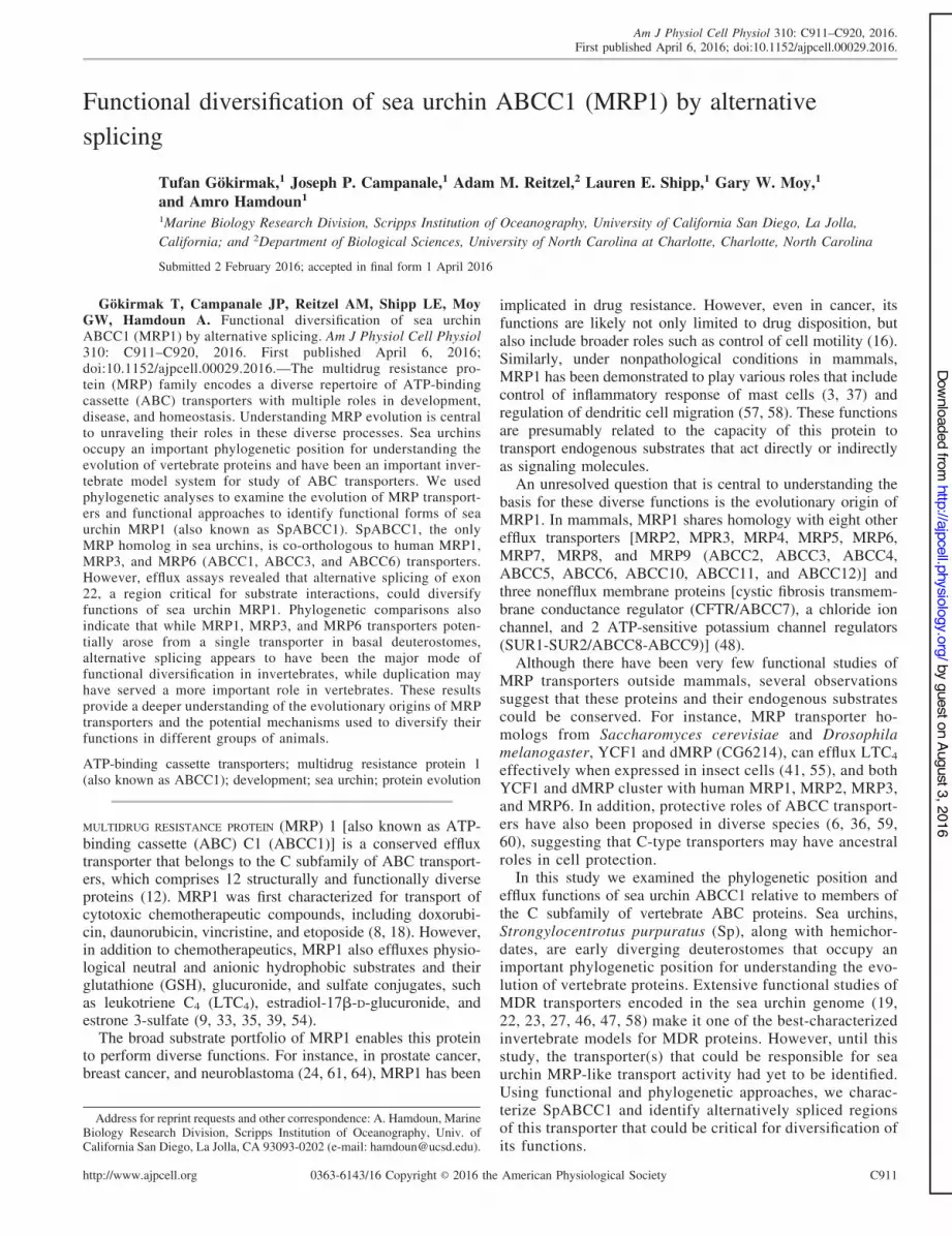
Phylogenetic analysis of ABCC proteins. To understand thephylogenetic position of sea urchin SpABCC1, we constructeda tree of sea urchin ABCC transporters in relation to human,zebrafish, frog, lamprey, sea squirt, sand lancelet, acorn worm,fruit fly, anemone, and yeast C-type ABC transporters. Sp-ABCC1 clusters with vertebrate ABCC1, ABCC3, and ABCC6and is co-orthologous to these transporters. The ABCC1-ABCC3-ABCC6 clade also includes single proteins from thefly (dMRP, GC6214), the hemichordate Saccoglossus (Meta-zome Sakowv30012504m), the anemone (JGI190171), andinferred paralogs for the cephalochordate Branchiostoma(JGI118636 and JGI232174) and urochordates Ciona(XP_009862422.1 and XP_009859093.1), suggesting an an-cient origin for this protein group to at least a cnidarian-bilaterian ancestor ('600 million years). These results alsoindicate that human and other vertebrate ABCC1, ABCC3, andABCC6 share a common ancestor with SpABCC1.
Vertebrate ABCC2 transporters form a separate cluster andjoin to the same node with the ABCC1-ABCC3-ABCC6 clade.Finally, two vacuolar yeast transporters, YCF1 and BPT1,which are involved in cellular detoxification of GSH conju-gates (28, 44), cluster together and are positioned at the root ofthe metazoan ABCC1-ABCC3-ABCC6 and ABCC2 clades,but not with other yeast ABCC transporters, suggesting thatthis group of transporters evolved from a common ancestor.
Sea urchin SpABCC5a is located at the base of the clade thatincludes vertebrate orthologs of the ABCC5 transporter and theclosely related ABCC12 transporter and its paralog ABCC11,which is present in primates but not found in rodents (45). Asimilar evolutionary pattern was also observed with sea urchin
C912 EVOLUTION AND FUNCTION OF SEA URCHIN ABCC1
AJP-Cell Physiol • doi:10.1152/ajpcell.00029.2016 • www.ajpcell.org
by guest on August 3, 2016http://ajpcell.physiology.org/
Dow
nloaded from
SpABCC9a, which shares co-orthology with the vertebrateABCC8 and ABCC9 potassium channel regulators and ispositioned at the root of the deuterostome lineage. The seaurchin genome does not have an ABCC8 gene, but there is anexpansion of the ABCC9 clade with 11 annotated genes (19),suggesting that functional diversity in the sea urchin ABCC9group might be achieved through gene duplication.
Although phylogenetic relationships between sea urchin andvertebrate ABCC transporters follow the general pattern ofco-orthologous gene pairing with vertebrate-specific gene du-plications, two sea urchin transporters, SpABCC4 and Sp-ABCC10, showed one-to-one orthology with their respectivevertebrate transporters (Fig. 1). This direct orthology indicatesthat these transporters may have conserved substrates andfunctions in invertebrates and vertebrates (Fig. 1).
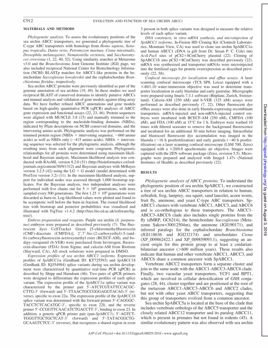
Genomic structure of sea urchin SpABCC1 transporter andexpression of its splice variants. Gene duplication and alterna-tive splicing are two common mechanisms to generate novelfunctions in gene families (27). Unlike vertebrates, the seaurchin genome has not undergone gene duplications in theABCC1-ABCC3-ABCC6 clade, and SpABCC1 is the onlygene found in this clade. Therefore, we examined the potentialfunctional consequences of alternative splicing of this proteinby examining the genomic structure of ABCC1 and cloning analternative full-length ABCC1 mRNA we found to be ex-pressed in the embryos.
The SpABCC1 gene comprises 33 exons and 32 introns andspans 54.6 kb of genomic DNA. At least four splice variants,SpABCC1%, SpABCC1!, SpABCC1(, and SpABCC1&, arepredicted in the genome and are generated by alternative
Fig. 1. Phylogenetic relationships of ATP-binding cassette C subfamily (ABCC) transporters of Strongylocentrotus purpuratus (Sp) with human (Hs), Xenopustropicalis (Xt), Danio rerio (Dr), Petromyzon marinus (Pm), Ciona intestinalis (Ci), Branchiostoma floridae (Bf), Saccoglossus kowalevskii (Sk), Drosophilamelanogaster (Dm), Nematostella vectensis (Nv), and Saccharomyces cerevisiae (Sc). The sea urchin efflux transporter SpABCC1 clusters in a well-supportedclade containing ABCC1-ABCC2-ABCC3-ABCC6 from vertebrates and single ABCC proteins or likely lineage-specific paralogs from other invertebrates (Ci,Bf, Sk, Nv, and Dm). SpABCC1 is co-orthologous to vertebrate ABCC1-ABCC3-ABCC6 transporters. The sea urchin also has orthologs to ABCC4 andABCC10 and a co-ortholog to ABCC5-ABCC11-ABCC12 and ABCC8-ABCC9 clades in vertebrates. The maximum-likelihood phylogeny is presented withposterior probability ('0.9) and maximum-likelihood bootstraps ('50%) indicated for each branch.
C913EVOLUTION AND FUNCTION OF SEA URCHIN ABCC1
AJP-Cell Physiol • doi:10.1152/ajpcell.00029.2016 • www.ajpcell.org
by guest on August 3, 2016http://ajpcell.physiology.org/
Dow
nloaded from
splicing of exon 22 (Fig. 2A). We detected two of these splicevariants, SpABCC1% and SpABCC1&, in embryos. qPCRprofiles of SpABCC1 splice variants in the first 3 days of seaurchin development showed that SpABCC1% and SpABCC1&
have similar expression profiles with a steady increase tomid-pluteus (Fig. 2B). However, comparison of the expressionprofiles of the two splice variants against the generic pan-ABCC1 expression profile showed that SpABCC1% has an
Fig. 2. Sea urchin SpABCC1 gene has 4 forms of exon 22 that correspond to membrane-spanning domain 2 (MSD2) of SpABCC1 transporters. A: gene structureof sea urchin SpABCC1 shows that it consists of 33 exons, 4 of which encode exon 22. Arrows indicate positions of primers used to determine the quantitativePCR (qPCR) profiles of SpABCC1! (green) and SpABCC1" (blue) splice variants and pan-SpABCC1 (black). B: qPCR expression profiles of SpABCC1! andSpABCC1" show that both genes are expressed throughout embryonic development and that the SpABCC1! expression profile is similar to the pan-SpABCC1expression profile. Values are means ) SD. C: variable exon 22 corresponds to a region in MSD2 marked in red. This exon encodes part of transmembranesegment 13 (TM13), cytoplasmic loop 6 (CL6), all of TM14, and part of TM15. Alignment of sea urchin SpABCC1 exon 22 splice variants with human multidrugresistance protein (MRP) 1 exon 24 indicates that some residues are conserved. D: protein sequence percent identity matrix of 4 splice variants of sea urchinexon 22 and the corresponding regions in human MRP1, MRP2, MRP3, and MRP6. Bold and underlined numbers indicate percent similarity of exon 22 ofSpABCC1% in relation to the human MRP transporter sequences.
C914 EVOLUTION AND FUNCTION OF SEA URCHIN ABCC1
AJP-Cell Physiol • doi:10.1152/ajpcell.00029.2016 • www.ajpcell.org
by guest on August 3, 2016http://ajpcell.physiology.org/
Dow
nloaded from

expression profile similar to pan-ABCC1 (Fig. 2B) and sug-gests that SpABCC1% might be more abundant thanSpABCC1&.
Both SpABCC1% and SpABCC1& and two other predictedsplice variants, SpABCC1! and SpABCC1(, consist of 1,577amino acids and have “long MRP” architecture with threemembrane-spanning domains (MSD0, MSD1, and MSD2) andtwo NBDs (NBD1 and NBD2). Amino acid sequence alignment(Fig. 2C) of the four splice variants of sea urchin exon 22 withthe corresponding region in human ABCC1 shows that exon 22corresponds to the predicted transmembrane helices 13–15 ofhuman ABCC1 (13) and that the variable region ofSpABCC1% is more closely related, in terms of amino acididentity, to human ABCC1, ABCC2, ABCC3, and ABCC6than to other sea urchin SpABCC1 splice variants (Fig. 2D).
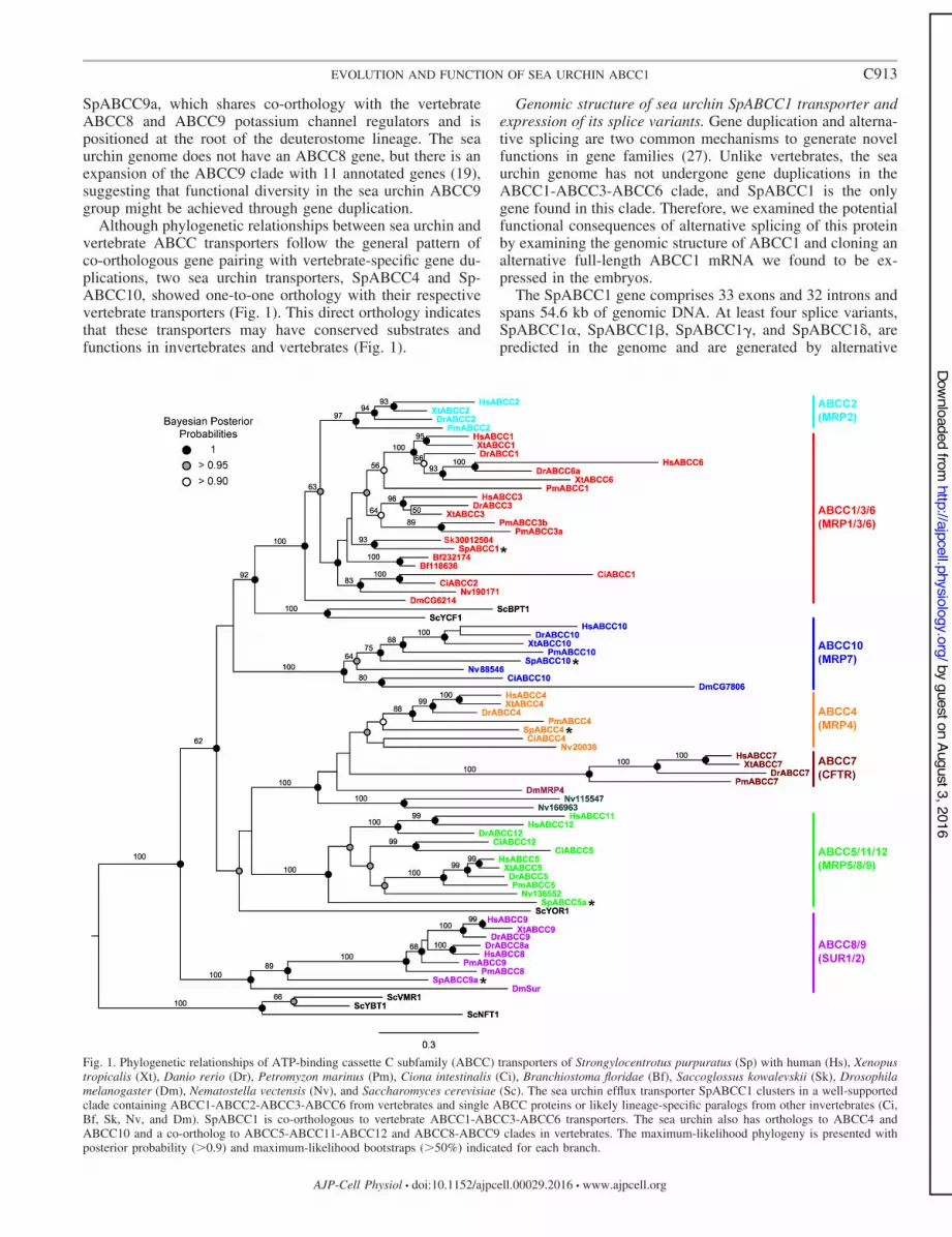
Functional characterization of sea urchin SpABCC1! andSpABCC1". Next, we characterized the efflux activities ofrecombinant SpABCC1% and SpABCC1& overexpressed athigh levels in embryos. We used three fluorescent substrates,calcein-AM, BCECF-AM, and CMFDA (Fig. 3A). We alsoused a non-MRP substrate (Fig. 3A), b-VER, a fluorescent
derivative of verapamil, which is effluxed by P-glycoprotein-type transporters, but not MRP transporters (22, 34).
Expression of mCherry-tagged SpABCC1% reduced intra-cellular accumulation of calcein to 28.41 ) 6.17% of thecontrol noninjected embryos (Fig. 3, B and C). Similarly,BCECF accumulation was 28.9 ) 12.14% and CMF accumu-lation was 6.29 ) 1.88% of that of the control embryos (Fig.3, B and C). As expected, b-VER was not effluxed bySpABCC1% (Fig. 3C). We also tested efflux of FDA bySpABCC1%. FDA is structurally similar to CMFDA and is aknown substrate for sea urchin SpABCC5a (47). FDA accu-mulation of SpABCC1%-expressing embryos was only 0.53 )0.2% of that of the control embryos.
Interestingly, SpABCC1& effluxed only BCECF-AM. Ex-pression of SpABCC1& reduced intracellular accumulation ofBCECF-AM to 45.40 ) 10.67% of the control embryos (Fig.3, B and C). Together, these data show that sea urchinSpABCC1% has a broad substrate profile similar to its mam-malian co-orthologs (5, 34), whereas SpABCC1& may have amore restricted substrate profile.
Fig. 3. SpABCC1% and SpABCC1& have differentefflux activity profiles. A: chemical structures offluorescent substrates used in efflux assays. B:micrographs showing that overexpression ofmCherry-tagged SpABCC1% reduced intracel-lular accumulation of BCECF-AM, calcein-AM,and 5-chloromethylfluorescein (CMF)-diacetate(CMFDA), but not verapamil (bodipy-VER),while the SpABCC1& splice variant can effluxonly BCECF-AM. DIC, differential interferencecontrast. Scale bar * 35 +m. C and D: quanti-tative analysis of intracellular and blastocoelaccumulation of fluorescent substrates relativeto noninjected control embryos. Values aremeans ) SD (n * 18 –20 embryos in total over3 experiments). *P , 0.01.
C915EVOLUTION AND FUNCTION OF SEA URCHIN ABCC1
AJP-Cell Physiol • doi:10.1152/ajpcell.00029.2016 • www.ajpcell.org
by guest on August 3, 2016http://ajpcell.physiology.org/
Dow
nloaded from

Finally, to determine whether the observed efflux activitiesof SpABCC1 splice variants are mediated by activity of theprotein present at apical vs. basolateral cell membranes, wemeasured accumulation of the fluorescent substrate in theblastocoel. We expected that efflux transport from basolateralsurfaces would lead to translocation of substrates into theblastocoel, whereas protein at the apical surface would preventuptake altogether by blocking passage through the ectoderm.
The data show that overexpression of either splice variant ofSpABCC1 did not cause significant fluorescent substrate accu-mulation in the blastocoel (Fig. 3, B and D), suggesting that theefflux activity is mediated by transporters at the apical mem-brane. However, activity from basolateral transporters becamemore apparent later in development (see below).
Membrane localization of recombinant SpABCC1!. Mem-brane localization of human ABCC transporters has beenextensively studied in different cell types. The human ABCC1and closely related ABCC3 and ABCC6 localize to basolateralcell membranes of polarized epithelial cells (32, 43). In con-trast, the major drug transporter ABCC2 localizes to apicalmembranes (38).
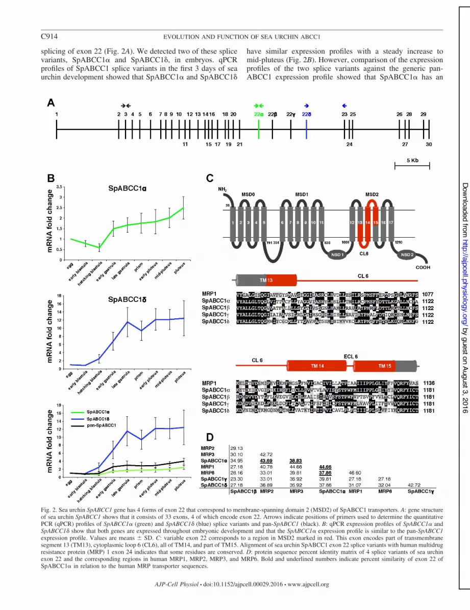
Here we determined the subcellular localization ofSpABCC1% from expression of the COOH-terminal mCherryfusion from 1 +g/+l injected mRNA. SpABCC1% localizedto both apical and basolateral membranes in early blastula(Fig. 4A). This result shows that subcellular localization ofSpABCC1% could be different from that of its humanco-orthologs. Since the amount of exogenous transporter inembryos is above the physiological level of SpABCC1%protein, we next sought to determine if this apical localiza-tion of SpABCC1% could be due to excessive amounts ofexogenous protein.
To determine a more physiologically relevant level, wetitrated SpABCC1% mRNA and conducted efflux assays todetermine the relative increase in efflux activity with increas-ing transporter expression (Fig. 4B). We found that embryosexpressing SpABCC1% from 50 ng/+l mRNA accumulatedroughly half (47.18 ) 14.14%) of the CMF of uninjectedcontrol embryos, suggesting that this mRNA concentrationmight produce recombinant protein levels closer to the physi-ological level (Fig. 4B).
Importantly, we also found that, even when expressed from50 ng/+l mRNA, SpABCC1% localized into both apical andbasolateral membranes in blastulae (Fig. 4B), indicating thatthis localization is not simply due to high levels of proteinexpression. In addition, we investigated the localization ofSpABCC1% expressed from 50 ng/+l mRNA in early gastrula(#40 h postfertilization). At this stage, the protein can still bedetected on the apical surface, but the amount is reduced andSpABCC1% is more strongly basolateral (Fig. 4C), suggestingthat localization could be stage/cell type-dependent.
Comparative analysis of sea urchin and human ABCC1transporters in localization and efflux assays. Finally, to ex-amine species differences in ABCC1 localization, we deter-mined heterologous expression of the COOH-terminalmCherry-tagged fusion of human HsABCC1 (MRP1) in seaurchin embryos (Fig. 5A). MRP1 was slower to be translatedand trafficked to plasma membranes than was the sea urchinprotein but, ultimately, localized strongly to basolateral mem-branes in early gastrula (Fig. 5B), consistent with its localiza-tion in polarized human epithelial cells. In gastrulae, the sea
urchin SpABCC1% is predominantly basolateral, with a small,but detectable, level of apical localization (Fig. 5B).
Given that, later in development, localization of both pro-teins was more strongly basolateral, we next investigatedwhether sea urchin and human ABCC1 transporters pumpsubstrates into the blastocoel. Both SpABCC1% and HsABCC1were functional in sea urchin gastrulae and increased blasto-
Fig. 4. Dynamic localization of SpABCC1% in sea urchin blastulae and earlygastrulae. A: representative equatorial and surface images of blastula [16high-power fields (hpf); n * 10–12 embryos in total over 2 experiments] showthat mCherry-tagged SpABCC1% localizes to both apical and basolateralmembranes. Scale bars * 200 +m (equatorial view) and 150 +m (surfaceview). B: titration of SpABCC1! mRNA with CMFDA efflux assay shows that,50 ng/+l mRNA is required to achieve physiologically relevant SpABCC1%expression. Micrographs of blastulae expressing SpABCC1% from 50 and1,000 ng/+l mRNA show that apical localization of SpABCC1% (magnified ininsets) is independent of mRNA concentration. Values are means ) SD (n *24–26 embryos in total over 4 experiments for each mRNA concentration).Scale bar * 150 +m. C: representative equatorial and surface images of earlygastrula (40 hpf; n * 10–12 embryos in total over 2 experiments) show thatapical localization of SpABCC1% in this stage is significantly reduced andbecomes more basolateral. Scale bars * 200 +m (equatorial view) and 150 +m(surface view).
C916 EVOLUTION AND FUNCTION OF SEA URCHIN ABCC1
AJP-Cell Physiol • doi:10.1152/ajpcell.00029.2016 • www.ajpcell.org
by guest on August 3, 2016http://ajpcell.physiology.org/
Dow
nloaded from
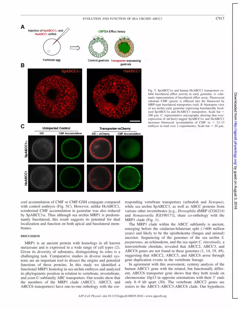
coel accumulation of CMF or CMF-GSH conjugate comparedwith control embryos (Fig. 5C). However, unlike HsABCC1,ectodermal CMF accumulation in gastrulae was also reducedby SpABCC1%. Thus although sea urchin MRP1 is predomi-nantly basolateral, this result suggests its potential for duallocalization and function on both apical and basolateral mem-branes.
DISCUSSION
MRP1 is an ancient protein with homologs in all knownmetazoans and is expressed in a wide range of cell types (2).Given its diversity of substrates, distinguishing its roles is achallenging task. Comparative studies in diverse model sys-tems are an important tool to dissect the origins and potentialfunctions of these proteins. In this study we identified afunctional MRP1 homolog in sea urchin embryos and analyzedits phylogenetic position in relation to vertebrate, invertebrate,and yeast C-subfamily ABC transporters. Our results show thatthe members of the MRP1 clade (ABCC1, ABCC3, andABCC6 transporters) have one-to-one orthology with the cor-
responding vertebrate transporters (zebrafish and Xenopus),while sea urchin SpABCC1, as well as ABCC proteins fromvarious other invertebrates [e.g., Drosophila dMRP (CG6214)and Nematostella JGI190171], share co-orthology with theMRP1 clade (Fig. 1).
The MRP1 clade within the ABCC subfamily is ancient,emerging before the cnidarian-bilaterian split ('600 millionyears) and likely to be the opisthokonta (fungus and animal)ancestor. Sequencing of the genomes of the sea urchin S.purpuratus, an echinoderm, and the sea squirt C. intestinalis, anonvertebrate chordate, revealed that ABCC2, ABCC3, andABCC6 genes are not found in these genomes (1, 14, 19, 49),suggesting that ABCC2, ABCC3, and ABCC6 arose throughgene duplication events in the vertebrate lineage.
In agreement with this assessment, synteny analysis of thehuman ABCC1 gene with the related, but functionally differ-ent, ABCC6 transporter gene shows that they both reside onchromosome 16p13 in opposite orientations with their 3= endsonly 8–9 kb apart (30). The vertebrate ABCC2 genes aresisters to the ABCC1-ABCC3-ABCC6 clade. Our hypothesis
Fig. 5. SpABCC1% and human HsABCC1 transporters ex-hibit basolateral efflux activity in early gastrulae. A: sche-matic representation of basolateral efflux assay. Fluorescentsubstrate CMF (green) is effluxed into the blastocoel byMRP-type basolateral transporters (red). B: blastopore viewof sea urchin early gastrulae expressing basolaterally local-ized SpABCC1% and HsABCC1 transporters. Scale bar *200 +m. C: representative micrographs showing that over-expression of mCherry-tagged SpABCC1% and HsABCC1increases blastocoel accumulation of CMF (n * 12–13embryos in total over 2 experiments). Scale bar * 20 +m.
C917EVOLUTION AND FUNCTION OF SEA URCHIN ABCC1
AJP-Cell Physiol • doi:10.1152/ajpcell.00029.2016 • www.ajpcell.org
by guest on August 3, 2016http://ajpcell.physiology.org/
Dow
nloaded from

for this positioning is that ABCC2 underwent a diversificationin the vertebrate line prior to the fish-tetrapod ancestor and,hence, falls outside the ABCC1-ABCC3-ABCC6 clade as asister group. ABCC2 could have been the first duplicationevent of the ancestral gene that resembled ABCC1-ABCC3-ABCC6 from invertebrates and has retained this divergentsequence since the duplication event. The ABCC1-ABCC3-ABCC6 gene then underwent additional duplication events. Infact, ABC transporters from lamprey (an intermediate taxo-nomic group) revealed that it has an ABCC1, an ABCC2ortholog, and two ABCC3 genes (PmABCC3a andPmABCC3b), which are co-orthologous to vertebrate ABCC3proteins, supporting this hypothesis, as well as the order ofABCC1-ABCC2-ABCC3-ABCC6 (40) (Fig. 6). Other geneduplication events have also apparently occurred in the uro-chordates and cephalochordate lineages due to paralogs inCiona and Branchiostoma.
In addition to gene duplication, generation of novel proteinsthrough alternative splicing is also a mechanism for increasingthe functional diversity in most eukaryotic protein families. Inthe C-subfamily of human ABC transporters, numerous splicevariants have been reported (50). Most of these variants aregenerated, however, by splice site mutations that cause re-moval of one or multiple exons or insertion of introns into themature mRNA, most of which cause premature protein trun-cation by nonsense mutations (17, 21) or by alternate transcrip-tion start sites (4, 53). For instance, in human ovarian cancercells, multiple splice variants of human ABCC1 were createdby exon skipping and/or intron inclusion (26).
Alternative-splicing events could be critical for diversifica-tion of transporter function in invertebrates lacking the dupli-cated MRP members found in vertebrates. Indeed, in Drosoph-ila, 14 different full-length splice variants of dMRP weredetected. These isoforms are generated from a single gene byexchange of two variant copies of exon 4 and seven variantcopies of exon 8 (20). Considering that Drosophila has onlyone co-orthologous gene in the MRP1 clade, generation ofprotein diversity by exchange of internal exons could be a wayto compensate the lack of paralogs created by gene duplicationand innovate novel protein diversity while maintaining a singlegene. In support of this hypothesis, Kopelman et al. (31)showed an inverse correlation between alternative splicing andgene duplication. Singletons (gene families of one) are moreinclined to undergo exon splicing than their orthologs that haveundergone gene duplication (31).
We found a similar mode of alternative splicing in sea urchinSpABCC1, an MRP-type transporter that shares co-orthologywith human ABCC1, ABCC3, and ABCC6 transporters (Fig.
1), and characterized the consequences of the splicing event onefflux activity. The new splice variant described here,SpABCC1%, was generated by alternative splicing of exon 22(Fig. 2). The boundaries of exon 22 are conserved in humanABCC1, ABCC2, and ABCC3, and it encodes part of TM13,CL6, TM14, and part of TM15 of MSD2 (Fig. 2). Interestingly,the frequently spliced exon 8 of Drosophila dMRP also cor-responds to the 5= end of exon 22 of sea urchin SpABCC1,suggesting that this protein region in MRP homologs is anevolutionarily active region for generating functional diversity.
Previous mutagenesis studies in human ABCC1 showed thatproline, polar and charged, residues in this region are importantfor substrate recognition (10, 29, 62, 63). Considering thatresidues in MSDs are important for the substrate interactions,organisms with singleton MRP homologs may use alternativesplicing of exons corresponding to the regions where substratebinding occurs. In support of this hypothesis, we showed that,by alternative splicing of exon 22, SpABCC1% can effluxcalcein-AM, BCECF-AM, CMFDA, and FDA, all knownfluorescent substrates for human ABCC1, ABCC2, andABCC3, while SpABCC1& effluxes only BCECF-AM with alower capacity (Fig. 3). Protein alignment of exons 22%, 22!,22(, and 22& with the corresponding exon sequences of humanABCC1, ABCC2, and ABCC3 proteins showed thatSpABCC1% is more similar to human proteins in this regionthan SpABCC1&, which may explain why SpABCC1% canefflux canonical human MRP substrates, while SpABCC1& canefflux only BCECF-AM. We previously showed that mCherry-tagged SpABCC1& localizes to both apical and basolateralmembranes in sea urchin blastulae (23). Identical subcellularlocalization of SpABCC1& also indicates that distinctive effluxactivity profiles of each splice variant are determined by thechanges in the substrate recognition region corresponding toexon 22, but not by the subcellular localizations.
The results of this study extend and validate the heterolo-gous expression approach we have adapted for analysis of seaurchin ABC transporters (22, 23, 46, 47). These assays areimportant, because they reveal the transporters that could beresponsible for observed activities and fingerprint the relevantsuspects for analysis by more laborious immunochemicalmethods. Large overexpression has been used to ensure that thevast majority of the observed efflux activity comes from therecombinant protein (22), which is essential for characteriza-tion of substrate selectivity.
Here, we showed that for localization studies the protein canbe reproducibly titrated to levels that increase correspondingtransport activity only twofold and still be functionally imagedby routine confocal microscopy (Fig. 4B), albeit with use ofhigh-sensitivity detectors. More importantly, these experi-ments indicated that localization of both forms of sea urchinABCC1 could be both apical and bilateral, depending ondevelopmental stage and independent of expression level. Thisdual location could be an additional mechanism for diversifi-cation of function in both protection and signaling.
Finally, an important step was to demonstrate the expressionof functional human MRP1. An interesting observation wasthat while sea urchin protein exhibited both apical and baso-lateral efflux activity, the human protein only appeared to act atthe basolateral membrane, possibly indicating that the differ-ences in localization of sea urchin and human proteins arelikely to be inherent features of the transporters themselves.
Fig. 6. Diversification of the ABCC1-ABCC2-ABCC3-ABCC6 clade acrossthe animal kingdom. Schematic representation shows the most likely duplica-tion history of ABCC1-ABCC2-ABCC3-ABCC6 based on available evidence.
C918 EVOLUTION AND FUNCTION OF SEA URCHIN ABCC1
AJP-Cell Physiol • doi:10.1152/ajpcell.00029.2016 • www.ajpcell.org
by guest on August 3, 2016http://ajpcell.physiology.org/
Dow
nloaded from

Collectively, the results indicate that the sea urchin is animportant system in which to understand the evolution of MRPproteins.
ACKNOWLEDGMENTS
We thank Dr. Susan Cole for human ABCC1 cDNA construct and com-ments on the manuscript and Dr. Victor D. Vacquier for valuable scientificdiscussions. We also thank Dr. Sascha C. T. Nicklisch and Lisa Mesrop fordiscussion and assistance.
GRANTS
This research was supported, in whole or in part, by National Institutes ofHealth Grants HD-058070 and ES-021985, National Science Foundation GrantOCE1314480, a grant from the University of California Cancer ResearchCoordinating Committee, and University of California San Diego AcademicSenate Grant RN141S.
DISCLOSURES
No conflicts of interest, financial or otherwise, are declared by the authors.
AUTHOR CONTRIBUTIONS
T.G. and A.H. developed the concept and designed the research; T.G.,J.P.C., A.M.R., L.E.S., and G.W.M. performed the experiments; T.G., J.P.C.,A.M.R., L.E.S., G.W.M., and A.H. analyzed the data; T.G., J.P.C., A.M.R.,L.E.S., G.W.M., and A.H. interpreted the results of the experiments; T.G. andA.M.R. prepared the figures; T.G. drafted the manuscript; T.G., J.P.C.,A.M.R., L.E.S., G.W.M., and A.H. approved the final version of the manu-script; A.H. edited and revised the manuscript.
REFERENCES
1. Annilo T, Chen ZQ, Shulenin S, Costantino J, Thomas L, Lou H,Stefanov S, Dean M. Evolution of the vertebrate ABC gene family:analysis of gene birth and death. Genomics 88: 1–11, 2006.
2. Bakos E, Homolya L. Portrait of multifaceted transporter, the multidrugresistance-associated protein 1 (MRP1/ABCC1). Pflügers Arch 453: 621–641, 2007.
3. Bartosz G, König J, Keppler D, Hagmann W. Human mast cellssecreting leukotriene C4 express the MRP1 gene-encoded conjugate exportpump. Biol Chem 379: 1121–1126, 1998.
4. Bera TK, Lee S, Salvatore G, Lee B, Pastan I. MRP8, a new memberof ABC transporter superfamily, identified by EST database mining andgene prediction program, is highly expressed in breast cancer. Mol Med 7:509–516, 2001.
5. Boraldi F, Quaglino D, Croce MA, Garcia Fernandez MI, Tiozzo R,Gheduzzi D, Bacchelli B, Pasquali Ronchetti I. Multidrug resistanceprotein-6 (MRP6) in human dermal fibroblasts. Comparison between cellsfrom normal subjects and from Pseudoxanthoma elasticum patients. Ma-trix Biol 22: 491–500, 2003.
6. Bosnjak I, Uhlinger KR, Heim W, Smital T, Franekic-Colic J, CoaleK, Epel D, Hamdoun A. Multidrug efflux transporters limit accumulationof inorganic, but not organic, mercury in sea urchin embryos. Environ SciTechnol 43: 8374–8380, 2009.
7. Campanale JP, Hamdoun A. Programmed reduction of ABC transporteractivity in sea urchin germline progenitors. Development 139: 783–792,2012.
8. Cole SP. Multidrug resistance protein 1 (MRP1, ABCC1), a “multitask-ing” ATP-binding cassette (ABC) transporter. J Biol Chem 289: 30880–30888, 2014.
9. Cole SP, Bhardwaj G, Gerlach JH, Mackie JE, Grant CE, AlmquistKC, Stewart AJ, Kurz EU, Duncan AM, Deeley RG. Overexpression ofa transporter gene in a multidrug-resistant human lung cancer cell line.Science 258: 1650–1654, 1992.
10. Conseil G, Deeley RG, Cole SP. Functional importance of three basicresidues clustered at the cytosolic interface of transmembrane helix 15 inthe multidrug and organic anion transporter MRP1 (ABCC1). J Biol Chem281: 43–50, 2006.
11. Darriba D, Taboada GL, Doallo R, Posada D. ProtTest 3: fast selectionof best-fit models of protein evolution. Bioinformatics 27: 1164–1165,2011.
12. Dean M, Rzhetsky A, Allikmets R. The human ATP-binding cassette(ABC) transporter superfamily. Genome Res 11: 1156–1166, 2001.
13. Deeley RG, Westlake C, Cole SP. Transmembrane transport of endo- andxenobiotics by mammalian ATP-binding cassette multidrug resistanceproteins. Physiol Rev 86: 849–899, 2006.
14. Dehal P, Satou Y, Campbell RK, Chapman J, Degnan B, De TomasoA, Davidson B, Di Gregorio A, Gelpke M, Goodstein DM, Harafuji N,Hastings KE, Ho I, Hotta K, Huang W, Kawashima T, Lemaire P,Martinez D, Meinertzhagen IA, Necula S, Nonaka M, Putnam N, RashS, Saiga H, Satake M, Terry A, Yamada L, Wang HG, Awazu S,Azumi K, Boore J, Branno M, Chin-Bow S, DeSantis R, Doyle S,Francino P, Keys DN, Haga S, Hayashi H, Hino K, Imai KS, Inaba K,Kano S, Kobayashi K, Kobayashi M, Lee BI, Makabe KW, ManoharC, Matassi G, Medina M, Mochizuki Y, Mount S, Morishita T, MiuraS, Nakayama A, Nishizaka S, Nomoto H, Ohta F, Oishi K, RigoutsosI, Sano M, Sasaki A, Sasakura Y, Shoguchi E, Shin-i T, Spagnuolo A,Stainier D, Suzuki MM, Tassy O, Takatori N, Tokuoka M, Yagi K,Yoshizaki F, Wada S, Zhang C, Hyatt PD, Larimer F, Detter C,Doggett N, Glavina T, Hawkins T, Richardson P, Lucas S, Kohara Y,Levine M, Satoh N, Rokhsar DS. The draft genome of Ciona intestinalis:insights into chordate and vertebrate origins. Science 298: 2157–2167,2002.
15. Edgar RC. MUSCLE: multiple sequence alignment with high accuracyand high throughput. Nucleic Acids Res 32: 1792–1797, 2004.
16. Fletcher JI, Haber M, Henderson MJ, Norris MD. ABC transporters incancer: more than just drug efflux pumps. Nat Rev Cancer 10: 147–156,2010.
17. Fromm MF, Leake B, Roden DM, Wilkinson GR, Kim RB. HumanMRP3 transporter: identification of the 5=-flanking region, genomic orga-nization and alternative splice variants. Biochim Biophys Acta 1415:369–374, 1999.
18. Gadsby DC, Vergani P, Csanády L. The ABC protein turned chloridechannel whose failure causes cystic fibrosis. Nature 440: 477–483, 2006.
19. Goldstone JV, Hamdoun A, Cole BJ, Howard-Ashby M, Nebert DW,Scally M, Dean M, Epel D, Hahn ME, Stegeman JJ. The chemicaldefensome: environmental sensing and response genes in the Strongylo-centrotus purpuratus genome. Dev Biol 300: 366–384, 2006.
20. Grailles M, Brey PT, Roth CW. The Drosophila melanogaster multi-drug-resistance protein 1 (MRP1) homolog has a novel gene structurecontaining two variable internal exons. Gene 307: 41–50, 2003.
21. Grant CE, Kurz EU, Cole SP, Deeley RG. Analysis of the intron-exonorganization of the human multidrug-resistance protein gene (MRP) andalternative splicing of its mRNA. Genomics 45: 368–378, 1997.
22. Gökirmak T, Campanale JP, Shipp LE, Moy GW, Tao H, HamdounA. Localization and substrate selectivity of sea urchin multidrug (MDR)efflux transporters. J Biol Chem 287: 43876–43883, 2012.
23. Gökirmak T, Shipp LE, Campanale JP, Nicklisch SC, Hamdoun A.Transport in technicolor: mapping ATP-binding cassette transporters insea urchin embryos. Mol Reprod Dev 81: 778–793, 2014.
24. Haber M, Smith J, Bordow SB, Flemming C, Cohn SL, London WB,Marshall GM, Norris MD. Association of high-level MRP1 expressionwith poor clinical outcome in a large prospective study of primaryneuroblastoma. J Clin Oncol 24: 1546–1553, 2006.
25. Hamdoun AM, Cherr GN, Roepke TA, Epel D. Activation of multidrugefflux transporter activity at fertilization in sea urchin embryos (Strongy-locentrotus purpuratus). Dev Biol 276: 452–462, 2004.
26. He X, Ee PL, Coon JS, Beck WT. Alternative splicing of the multidrugresistance protein 1/ATP binding cassette transporter subfamily gene inovarian cancer creates functional splice variants and is associated withincreased expression of the splicing factors PTB and SRp20. Clin CancerRes 10: 4652–4660, 2004.
27. Jin L, Kryukov K, Clemente JC, Komiyama T, Suzuki Y, Imanishi T,Ikeo K, Gojobori T. The evolutionary relationship between gene dupli-cation and alternative splicing. Gene 427: 19–31, 2008.
28. Klein M, Mamnun YM, Eggmann T, Schüller C, Wolfger H, Marti-noia E, Kuchler K. The ATP-binding cassette (ABC) transporter Bpt1pmediates vacuolar sequestration of glutathione conjugates in yeast. FEBSLett 520: 63–67, 2002.
29. Koike K, Conseil G, Leslie EM, Deeley RG, Cole SP. Identification ofproline residues in the core cytoplasmic and transmembrane regions ofmultidrug resistance protein 1 (MRP1/ABCC1) important for transportfunction, substrate specificity, and nucleotide interactions. J Biol Chem279: 12325–12336, 2004.
30. Kool M, van der Linden M, de Haas M, Baas F, Borst P. Expression ofhuman MRP6, a homologue of the multidrug resistance protein geneMRP1, in tissues and cancer cells. Cancer Res 59: 175–182, 1999.
C919EVOLUTION AND FUNCTION OF SEA URCHIN ABCC1
AJP-Cell Physiol • doi:10.1152/ajpcell.00029.2016 • www.ajpcell.org
by guest on August 3, 2016http://ajpcell.physiology.org/
Dow
nloaded from

31. Kopelman NM, Lancet D, Yanai I. Alternative splicing and geneduplication are inversely correlated evolutionary mechanisms. Nat Genet37: 588–589, 2005.
32. Kruh GD, Belinsky MG. The MRP family of drug efflux pumps.Oncogene 22: 7537–7552, 2003.
33. Leier I, Jedlitschky G, Buchholz U, Cole SP, Deeley RG, Keppler D.The MRP gene encodes an ATP-dependent export pump for leukotrieneC4 and structurally related conjugates. J Biol Chem 269: 27807–27810,1994.
34. Litman T, Brangi M, Hudson E, Fetsch P, Abati A, Ross DD, MiyakeK, Resau JH, Bates SE. The multidrug-resistant phenotype associatedwith overexpression of the new ABC half-transporter, MXR (ABCG2). JCell Sci 113: 2011–2021, 2000.
35. Loe DW, Almquist KC, Cole SP, Deeley RG. ATP-dependent 17!-estradiol 17-(!-D-glucuronide) transport by multidrug resistance protein(MRP). Inhibition by cholestatic steroids. J Biol Chem 271: 9683–9689,1996.
36. Long Y, Li Q, Zhong S, Wang Y, Cui Z. Molecular characterization andfunctions of zebrafish ABCC2 in cellular efflux of heavy metals. CompBiochem Physiol C Toxicol Pharmacol 153: 381–391, 2011.
37. Mitra P, Oskeritzian CA, Payne SG, Beaven MA, Milstien S, SpiegelS. Role of ABCC1 in export of sphingosine-1-phosphate from mast cells.Proc Natl Acad Sci USA 103: 16394–16399, 2006.
38. Nies AT, Keppler D. The apical conjugate efflux pump ABCC2 (MRP2).Pflügers Arch 453: 643–659, 2007.
39. Qian YM, Song WC, Cui H, Cole SP, Deeley RG. Glutathione stimu-lates sulfated estrogen transport by multidrug resistance protein 1. J BiolChem 276: 6404–6411, 2001.
40. Ren J, Chung-Davidson YW, Yeh CY, Scott C, Brown T, Li W.Genome-wide analysis of the ATP-binding cassette (ABC) transportergene family in sea lamprey and Japanese lamprey. BMC Genomics 16:436, 2015.
41. Ren XQ, Furukawa T, Chen ZS, Okumura H, Aoki S, Sumizawa T,Tani A, Komatsu M, Mei XD, Akiyama S. Functional comparisonbetween YCF1 and MRP1 expressed in Sf21 insect cells. Biochem Bio-phys Res Commun 270: 608–615, 2000.
42. Ronquist F, Huelsenbeck JP. MrBayes 3: Bayesian phylogenetic infer-ence under mixed models. Bioinformatics 19: 1572–1574, 2003.
43. Scheffer GL, Hu X, Pijnenborg AC, Wijnholds J, Bergen AA, ScheperRJ. MRP6 (ABCC6) detection in normal human tissues and tumors. LabInvest 82: 515–518, 2002.
44. Sharma KG, Kaur R, Bachhawat AK. The glutathione-mediated detox-ification pathway in yeast: an analysis using the red pigment that accu-mulates in certain adenine biosynthetic mutants of yeasts reveals theinvolvement of novel genes. Arch Microbiol 180: 108–117, 2003.
45. Shimizu H, Taniguchi H, Hippo Y, Hayashizaki Y, Aburatani H,Ishikawa T. Characterization of the mouse Abcc12 gene and its transcriptencoding an ATP-binding cassette transporter, an orthologue of humanABCC12. Gene 310: 17–28, 2003.
46. Shipp LE, Hamdoun A. ATP-binding cassette (ABC) transporter expres-sion and localization in sea urchin development. Dev Dyn 241: 1111–1124, 2012.
47. Shipp LE, Hill RZ, Moy GW, Gökırmak T, Hamdoun A. ABCC5 isrequired for cAMP-mediated hindgut invagination in sea urchin embryos.Development 142: 3537–3548, 2015.
48. Slot AJ, Molinski SV, Cole SP. Mammalian multidrug-resistance pro-teins (MRPs). Essays Biochem 50: 179–207, 2011.
49. Sodergren E, Weinstock GM, Davidson EH, Cameron RA, Gibbs RA,Angerer RC, Angerer LM, Arnone MI, Burgess DR, Burke RD,Coffman JA, Dean M, Elphick MR, Ettensohn CA, Foltz KR, Ham-doun A, Hynes RO, Klein WH, Marzluff W, McClay DR, Morris RL,Mushegian A, Rast JP, Smith LC, Thorndyke MC, Vacquier VD,Wessel GM, Wray G, Zhang L, Elsik CG, Ermolaeva O, Hlavina W,Hofmann G, Kitts P, Landrum MJ, Mackey AJ, Maglott D, Panopou-lou G, Poustka AJ, Pruitt K, Sapojnikov V, Song X, Souvorov A,Solovyev V, Wei Z, Whittaker CA, Worley K, Durbin KJ, Shen Y,Fedrigo O, Garfield D, Haygood R, Primus A, Satija R, Severson T,Gonzalez-Garay ML, Jackson AR, Milosavljevic A, Tong M, KillianCE, Livingston BT, Wilt FH, Adams N, Bellé R, Carbonneau S,Cheung R, Cormier P, Cosson B, Croce J, Fernandez-Guerra A,Genevière AM, Goel M, Kelkar H, Morales J, Muln er-Lorillon O,Robertson AJ, Goldstone JV, Cole B, Epel D, Gold B, Hahn ME,Howard-Ashby M, Scally M, Stegeman JJ, Allgood EL, Cool J,Judkins KM, McCafferty SS, Musante AM, Obar RA, Rawson AP,
Rossetti BJ, Gibbons IR, Hoffman MP, Leone A, Istrail S, MaternaSC, Samanta MP, Stolc V, Tongprasit W, Tu Q, Bergeron KF,Brandhorst BP, Whittle J, Berney K, Bottjer DJ, Calestani C, Peter-son K, Chow E, Yuan QA, Elhaik E, Graur D, Reese JT, Bosdet I,Heesun S, Marra MA, Schein J, Anderson MK, Brockton V, BuckleyKM, Cohen AH, Fugmann SD, Hibino T, Loza-Coll M, Majeske AJ,Messier C, Nair SV, Pancer Z, Terwilliger DP, Agca C, Arboleda E,Chen N, Churcher AM, Hallböök F, Humphrey GW, Idris MM,Kiyama T, Liang S, Mellott D, Mu X, Murray G, Olinski RP, RaibleF, Rowe M, Taylor JS, Tessmar-Raible K, Wang D, Wilson KH,Yaguchi S, Gaasterland T, Galindo BE, Gunaratne HJ, Juliano C,Kinukawa M, Moy GW, Neill AT, Nomura M, Raisch M, Reade A,Roux MM, Song JL, Su YH, Townley IK, Voronina E, Wong JL,Amore G, Branno M, Brown ER, Cavalieri V, Duboc V, Duloquin L,Flytzanis C, Gache C, Lapraz F, Lepage T, Locascio A, Martinez P,Matassi G, Matranga V, Range R, Rizzo F, Röttinger E, Beane W,Bradham C, Byrum C, Glenn T, Hussain S, Manning G, Miranda E,Thomason R, Walton K, Wikramanayke A, Wu SY, Xu R, Brown CT,Chen L, Gray RF, Lee PY, Nam J, Oliveri P, Smith J, Muzny D, BellS, Chacko J, Cree A, Curry S, Davis C, Dinh H, Dugan-Rocha S,Fowler J, Gill R, Hamilton C, Hernandez J, Hines S, Hume J, JacksonL, Jolivet A, Kovar C, Lee S, Lewis L, Miner G, Morgan M, NazarethLV, Okwuonu G, Parker D, Pu LL, Thorn R, Wright R, Sea UrchinGenome Sequencing Consortium. The genome of the sea urchinStrongylocentrotus purpuratus. Science 314: 941–952, 2006.
50. Srinivasan S, Bingham JL, Johnson D. The ABCs of human alternativesplicing: a review of ATP-binding cassette transporter splicing. Curr OpinDrug Discov Dev 12: 149–158, 2009.
51. Stamatakis A. RAxML version 8: a tool for phylogenetic analysis andpost-analysis of large phylogenies. Bioinformatics 30: 1312–1313, 2014.
52. Sturm A, Cunningham P, Dean M. The ABC transporter gene family ofDaphnia pulex. BMC Genomics 10: 170, 2009.
53. Suzuki T, Sasaki H, Kuh HJ, Agui M, Tatsumi Y, Tanabe S, TeradaM, Saijo N, Nishio K. Detailed structural analysis on both human MRP5and mouse mrp5 transcripts. Gene 242: 167–173, 2000.
54. Tamaki A, Ierano C, Szakacs G, Robey RW, Bates SE. The contro-versial role of ABC transporters in clinical oncology. Essays Biochem 50:209–232, 2011.
55. Tarnay JN, Szeri F, Iliás A, Annilo T, Sung C, Le Saux O, Váradi A,Dean M, Boyd CD, Robinow S. The dMRP/CG6214 gene of Drosophilais evolutionarily and functionally related to the human multidrug resis-tance-associated protein family. Insect Mol Biol 13: 539–548, 2004.
56. van de Ven R, Scheffer GL, Scheper RJ, de Gruijl TD. The ABC ofdendritic cell development and function. Trends Immunol 30: 421–429, 2009.
57. Weekes MP, Tan SY, Poole E, Talbot S, Antrobus R, Smith DL,Montag C, Gygi SP, Sinclair JH, Lehner PJ. Latency-associated deg-radation of the MRP1 drug transporter during latent human cytomegalo-virus infection. Science 340: 199–202, 2013.
58. Whalen K, Reitzel AM, Hamdoun A. Actin polymerization controls theactivation of multidrug efflux at fertilization by translocation and fine-scalepositioning of ABCB1 on microvilli. Mol Biol Cell 23: 3663–3672, 2012.
59. Xu X, Fu J, Wang H, Zhang B, Wang X, Wang Y. Influence ofP-glycoprotein on embryotoxicity of the antifouling biocides to sea urchin(Strongylocentrotus intermedius). Ecotoxicology 20: 419–428, 2011.
60. Zaja R, Munic V, Klobucar RS, Ambriovic-Ristov A, Smital T.Cloning and molecular characterization of apical efflux transporters(ABCB1, ABCB11 and ABCC2) in rainbow trout (Oncorhynchus mykiss)hepatocytes. Aquat Toxicol (Amst) 90: 322–332, 2008.
61. Zalcberg J, Hu XF, Slater A, Parisot J, El-Osta S, Kantharidis P,Chou ST, Parkin JD. MRP1 not MDR1 gene expression is the predom-inant mechanism of acquired multidrug resistance in two prostate carci-noma cell lines. Prostate Cancer Prostatic Dis 3: 66–75, 2000.
62. Zhang DW, Cole SP, Deeley RG. Identification of an amino acid residuein multidrug resistance protein 1 critical for conferring resistance toanthracyclines. J Biol Chem 276: 13231–13239, 2001.
63. Zhang DW, Gu HM, Situ D, Haimeur A, Cole SP, Deeley RG. Functionalimportance of polar and charged amino acid residues in transmembrane helix14 of multidrug resistance protein 1 (MRP1/ABCC1): identification of anaspartate residue critical for conversion from a high to low affinity substratebinding state. J Biol Chem 278: 46052–46063, 2003.
64. Zöchbauer-Müller S, Filipits M, Rudas M, Brunner R, Krajnik G,Suchomel R, Schmid K, Pirker R. P-glycoprotein and MRP1 expressionin axillary lymph node metastases of breast cancer patients. AnticancerRes 21: 119–124, 2001.
C920 EVOLUTION AND FUNCTION OF SEA URCHIN ABCC1
AJP-Cell Physiol • doi:10.1152/ajpcell.00029.2016 • www.ajpcell.org
by guest on August 3, 2016http://ajpcell.physiology.org/
Dow
nloaded from