signaling mechanisms underlying metamorphic transitions in animals
TRANSCRIPT
Signaling mechanisms underlying metamorphic transitionsin animals
Andreas Heyland1,*,§ and Leonid L. Moroz2,*,y
*The Whitney Laboratory for Marine Bioscience, University of Florida, FL 32080, USA; yDepartment of Neuroscience,
University of Florida, FL 32611, USA; §Friday Harbor Laboratories, University of Washington, WA 98250, USA
Synopsis Metamorphosis in many animal groups involves a radical transition from a larval to a juvenile/adult body plan
and the challenge of orchestrating 2 overlapping developmental programs simultaneously, that is, larval development and
juvenile development. Metamorphic competence directly precedes this radical change in morphology and can be best
described as the developmental potential of a larva to undergo the radical transition in response to internal or external
signals. Several studies have employed genomic approaches (for example, microarrays or subtractive hybridization methods)
to gain insights into the complexity of changes in gene expression associated with metamorphic transitions. Availability of this
technology for an increasing number of organisms from diverse taxonomic groups expands the scope of species for which we
can gain detailed understanding of the genetic and epigenetic architecture underlying metamorphosis. Here, we review
metamorphosis in insects, amphibians, and several marine invertebrate species including the sea hare Aplysia californica
and summarize mechanisms underlying the transition. We conclude that all metamorphoses share at least 4 components: (1)
the differentiation of juvenile/adult structures, (2) the degeneration of larval structures, (3) metamorphic competence, and (4)
change in habitat. While transcription levels detected by microarray or other molecular methods can vary significantly, some
similarities can be observed. For example, transcripts related to stress response, immunity, and apoptosis are associated with
metamorphosis in all investigated phyla. It also appears that signaling mediated by hormones and by nitric oxide can
contribute to these stress-related responses and that these molecules can act as regulators of metamorphic transitions.
This might indicate either that all of these distantly related organisms inherited the same basic regulatory machinery that
was employed by their most recent common ancestor (RCA) in orchestrating life history transitions. Alternatively, these
regulatory modules may have been used by the RCA for other purposes and have been independently co-opted to regulate
metamorphic transitions in a variety of distantly related animals. We propose that such instances of independent origin or
homoplasy in the evolution of metamorphosis might have resulted from specific constraints in signal transduction pathways.
Modern genomic tools can help to further explore homoplastic signaling modules when used in a comparative context.
Metamorphic transitions acrossanimal phyla
In 1978, Fu-Shiang Chia (1978) observed that the
“problems of settlement and metamorphosis are diverse
and complicated”; despite many studies in the interim,
this remains true today. Metamorphosis among animals
includes a change in habitat and the abandoning
of the larva, a transitory postembryonic stage in an
animal’s life history that is adapted to a different
environment than the adult. It possesses 2 types of
structures: larval and juvenile/adult. While larval
structures stop growing and differentiating directly
after settlement, juvenile/adult tissues may differentiate
both before and after metamorphosis. Therefore, a larva
can be further characterized by the overlapping devel-
opment of larval and juvenile/adult structures.
Phenotypically, there are many obvious differences
between, for example, metamorphosis of flies, frogs,
and sea slugs. On a more general level, Hadfield
(2000) and Hadfield and others (2001) emphasized
that important differences exist between metamorphic
transitions in amphibians and flies as compared with
the metamorphic transitions of many marine inverte-
brate larvae. While many marine invertebrates produce
small larvae that undergo very rapid transitions from
plankton to benthos, amphibian and insect larvae
are generally bigger and the transition appears much
more gradual (but see Hodin 2006). Settlement
From the symposium “Metamorphosis: A Multikingdom Approach” presented at the annual meeting of the Society for Integrative and
Comparative Biology, January 4–8, 2006, Orlando, Florida.1 E-mail: [email protected] E-mail: [email protected]
Integrative and Comparative Biology, volume 46, number 6, pp. 743–759
doi:10.1093/icb/icl023
Advance Access publication July 27, 2006
� The Author 2006. Published by Oxford University Press on behalf of the Society for Integrative and Comparative Biology. All rights reserved.
For permissions please email: [email protected].
743
by guest on September 27, 2013
http://icb.oxfordjournals.org/D
ownloaded from
by guest on Septem
ber 27, 2013http://icb.oxfordjournals.org/
Dow
nloaded from
by guest on September 27, 2013
http://icb.oxfordjournals.org/D
ownloaded from
by guest on Septem
ber 27, 2013http://icb.oxfordjournals.org/
Dow
nloaded from
by guest on September 27, 2013
http://icb.oxfordjournals.org/D
ownloaded from
by guest on Septem
ber 27, 2013http://icb.oxfordjournals.org/
Dow
nloaded from
by guest on September 27, 2013
http://icb.oxfordjournals.org/D
ownloaded from
by guest on Septem
ber 27, 2013http://icb.oxfordjournals.org/
Dow
nloaded from
by guest on September 27, 2013
http://icb.oxfordjournals.org/D
ownloaded from
by guest on Septem
ber 27, 2013http://icb.oxfordjournals.org/
Dow
nloaded from
by guest on September 27, 2013
http://icb.oxfordjournals.org/D
ownloaded from
by guest on Septem
ber 27, 2013http://icb.oxfordjournals.org/
Dow
nloaded from
by guest on September 27, 2013
http://icb.oxfordjournals.org/D
ownloaded from
by guest on Septem
ber 27, 2013http://icb.oxfordjournals.org/
Dow
nloaded from
by guest on September 27, 2013
http://icb.oxfordjournals.org/D
ownloaded from
by guest on Septem
ber 27, 2013http://icb.oxfordjournals.org/
Dow
nloaded from
by guest on September 27, 2013
http://icb.oxfordjournals.org/D
ownloaded from
(that is, habitat change) occurs in response to specific
external cues. Data from a few selected species suggest
that little de novo gene transcription is required during
this process and that mechanisms of signal transduc-
tion appear to rely primarily on cell–cell conductance
(reviewed by Hadfield 2000; Hadfield and others 2001).
This contrasts with the metamorphosis of insects and
amphibians where postembryonic development pro-
ceeds via hormonal coordination Nijhout (1994) and
Tata (1996). Still, recent evidence indicates that hor-
mones regulate development to metamorphosis in a
variety of marine invertebrate larvae as well (Eales
1997; Heyland and Hodin 2004; Heyland and others
2004, 2005, 2006; Heyland and Moroz 2005). In the
context of this evidence, signal transduction events
during settlement can be viewed as a special ecological
adaptation to the marine environment (Hadfield
2000).
Gene regulation is inherently modular. Networks of
regulatory genes are generally robust to perturbation
and can be expressed in various contexts for diverse
functions (Schlosser 2002). Therefore, such networks
(modules) can be co-opted for novel developmental
processes. Moreover, this property also allows the testing
of whether certain modules are independently co-opted
for the same function owing to specific constraints.
The so-called true or primary larvae evolved several
times independently among animals (Hadfield and
others 2001; Wray 2000) and show a remarkable diver-
sity of form and function. Yet from a developmental
point of view, all larvae face the challenge of coordi-
nating 2 disparate signaling networks, one that regu-
lates the development of larval structures and one that
regulates the development of juvenile/adult structures.
We hypothesize that this represents a constraint at the
level of the signal transduction pathways involved in
this process and predict that similar modules regulating
the 2 divergent developmental programs within the
same organism were co-opted in disparate organisms
independently multiple times. Comparing mechanisms
underlying the metamorphic transition across phyla
therefore has the potential to uncover some of these
instances of homoplasy, that is, similarities based on
convergent or parallel evolution on a mechanistic, cell-
signaling level, and point to specific constraints in
signaling mechanisms (see also Hodin 2000).
In order to compare metamorphic transitions
between disparate species we propose to distinguish
the following components of metamorphosis: (1)
growth and differentiation of juvenile/adult-specific
structures, (2) breakdown of larva-specific structures,
(3) metamorphic competence, and (4) change in habi-
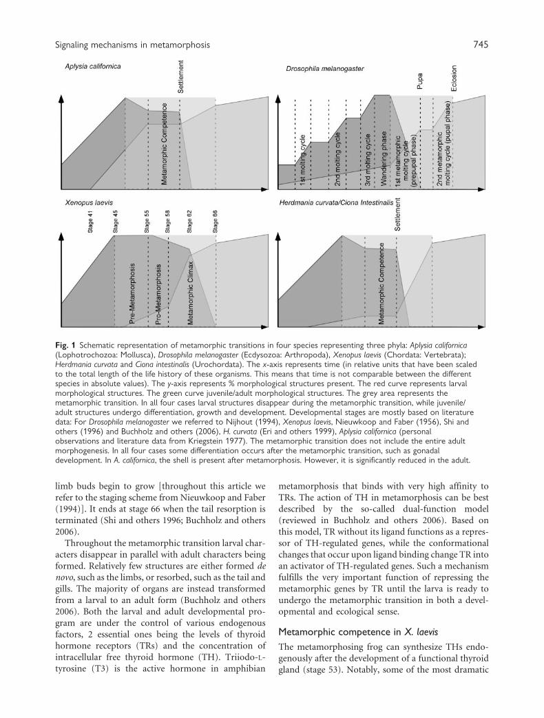
tat. Figure 1 illustrates these general characteristics of
metamorphic transitions for several species graphically.
The x-axis represents time and the y-axis represents the
percentage of the developmental program that was
completed. The red curve shows larval development
and differentiation (the larval developmental pro-
gram), the green curve shows adult development
and differentiation (the juvenile/adult developmental
program). For example, in Aplysia californica, all larval
structures are present after hatching and the larva
grows in size while adult-specific structures continue
to differentiate inside the larva. After settlement, larval
structures disappear rapidly as the juvenile transforms
gradually into the adult (Fig. 2).
We selected one or 2 illustrative and well-studied
examples from 3 animal phyla for which we will give
a brief overview of metamorphosis. These are amphi-
bians (Chordata), ascidians (Chordata), insects
(Arthropoda), and sea slugs (Mollusca). In all cases
we focus on the 4 common phases of metamorphosis
outlined above. We then discuss the change in habitat
more specifically in the context of environmental fac-
tors influencing the regulation and timing of metamor-
phosis. Finally, we review selected intrinsic regulatory
mechanisms underlying the metamorphic transition.
Amphibian metamorphosis
Many excellent reviews have been published on the
regulation of metamorphosis in anuran amphibians
(frogs) (for recent reviews see Tata 2005 and
Buchholz and others 2006). We do not intend to
provide a complete review here. Instead we will only
discuss the 4 phases mentioned above for the meta-
morphosis of Xenopus laevis and summarize informa-
tion on signaling events involved in these various
parts of the metamorphic transition. A schematic
representation of X. laevis metamorphosis is given in
Figure 1.
Based on previous work, 3 periods can be distin-
guished in the metamorphic transition of X. laevis:
premetamorphosis (stages 45–53), prometamorphosis
(stages 54–58), and metamorphic climax (stages
59–66). Some of the most significant changes are the
remodeling of the intestine at around stage 58, the
degeneration of the larval epithelium around stages
60–62, the subsequent differentiation of the secondary
epithelium of the adult frog, and finally the degenera-
tion of the tail at the end of stage 62.
Growth and differentiation of juvenile/adultstructures and breakdown of larval structuresin X. laevis
Metamorphosis in X. laevis is a relatively slow and
gradual process. It stretches over a period of approxi-
mately 30 days. It begins at stage 55 when the hind
744 A. Heyland and L. L. Moroz
limb buds begin to grow [throughout this article we
refer to the staging scheme from Nieuwkoop and Faber
(1994)]. It ends at stage 66 when the tail resorption is
terminated (Shi and others 1996; Buchholz and others
2006).
Throughout the metamorphic transition larval char-
acters disappear in parallel with adult characters being
formed. Relatively few structures are either formed de
novo, such as the limbs, or resorbed, such as the tail and
gills. The majority of organs are instead transformed
from a larval to an adult form (Buchholz and others
2006). Both the larval and adult developmental pro-
gram are under the control of various endogenous
factors, 2 essential ones being the levels of thyroid
hormone receptors (TRs) and the concentration of
intracellular free thyroid hormone (TH). Triiodo-L-
tyrosine (T3) is the active hormone in amphibian
metamorphosis that binds with very high affinity to
TRs. The action of TH in metamorphosis can be best
described by the so-called dual-function model
(reviewed in Buchholz and others 2006). Based on
this model, TR without its ligand functions as a repres-
sor of TH-regulated genes, while the conformational
changes that occur upon ligand binding change TR into
an activator of TH-regulated genes. Such a mechanism
fulfills the very important function of repressing the
metamorphic genes by TR until the larva is ready to
undergo the metamorphic transition in both a devel-
opmental and ecological sense.
Metamorphic competence in X. laevis
The metamorphosing frog can synthesize THs endo-
genously after the development of a functional thyroid
gland (stage 53). Notably, some of the most dramatic
Fig. 1 Schematic representation of metamorphic transitions in four species representing three phyla: Aplysia californica(Lophotrochozoa: Mollusca), Drosophila melanogaster (Ecdysozoa: Arthropoda), Xenopus laevis (Chordata: Vertebrata);Herdmania curvata and Ciona intestinalis (Urochordata). The x-axis represents time (in relative units that have been scaledto the total length of the life history of these organisms. This means that time is not comparable between the differentspecies in absolute values). The y-axis represents % morphological structures present. The red curve represents larvalmorphological structures. The green curve juvenile/adult morphological structures. The grey area represents themetamorphic transition. In all four cases larval structures disappear during the metamorphic transition, while juvenile/adult structures undergo differentiation, growth and development. Developmental stages are mostly based on literaturedata: For Drosophila melanogaster we referred to Nijhout (1994), Xenopus laevis, Nieuwkoop and Faber (1956), Shi andothers (1996) and Buchholz and others (2006), H. curvata (Eri and others 1999), Aplysia californica (personalobservations and literature data from Kriegstein 1977). The metamorphic transition does not include the entire adultmorphogenesis. In all four cases some differentiation occurs after the metamorphic transition, such as gonadaldevelopment. In A. californica, the shell is present after metamorphosis. However, it is significantly reduced in the adult.
Signaling mechanisms in metamorphosis 745
morphological changes happen after a thyroid gland
is formed. (Buchholz and others 2006). Both T4
(L-thyroxine) and T3 (3,30,5-triiodo-L-thyronine)levels increase exponentially until they peak at meta-
morphic climax (Shi and others 1996). Two forms of
TR occur in Xenopus: TRalpha and TRbeta. Both are
expressed premetamorphically. Metamorphic compe-
tence can best be described by the patterns of expres-
sion of TRalpha; late in embryogenesis, a tissue can
only transform in response to TH if TRalpha is
expressed (that is, at around stage 47) (Shi and others
1996; Denver and others 2002; Buchholz and others
2006). This has been confirmed by knockdown experi-
ments using dominant negative TRs (Buchholz and
others 2006). Moreover, if the larval thyroid gland is
removed experimentally, tadpoles maintain the capa-
city to undergo metamorphosis indefinitely (Shi and
others 1996; Buchholz and others 2006; Buchholz
personal communication). TRalpha protein levels are
�2–3 times higher than those of TRbeta throughout
larval development. However, at metamorphic climax,
TRbeta expression levels increase exponentially to
similar levels as TRalpha [for a description of hormone
and receptor levels see Buchholz and others (2006) and
Shi and others (1996)].
Environmental control of metamorphosisin X. laevis
The timing of metamorphosis, measured by the time
from hatching until transition from the aquatic to ter-
restrial habitat, can vary substantially even within spe-
cies. This phenotypic plasticity is triggered by various
biotic and abiotic factors, which, in the majority of
cases, act via the hypothalamic–pituitary axis (reviewed
by Denver and others 2002). Specifically, larvae of
several amphibian species accelerate development
in response to desiccation and other stress-related
environmental triggers (Denver and others 2002).
This response involves corticosteroid stimulation of
thyroid stimulating hormone, which results in TH
synthesis and accelerated restructuring of the tadpole
towardmetamorphic climax (Denver and others 2002).
In the larval nervous system, major remodeling
occurs in specific brain regions necessary for the devel-
opment of adult-specific behaviors or sensory organs
that will fulfill crucial functions in the adult environ-
ment. For example, the Mauthner neurons and motor
neurons that innervate tail muscles are involved in the
escape response of tadpoles. Another example is the
transformation of the visual system from the panora-
mic vision of the tadpole to the binocular vision of the
adult frog, involving a large part of the retina and visual
projections into the diencephalon associated with it
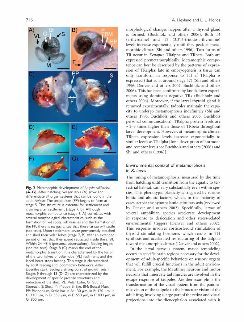
Fig. 2 Metamorphic development of Aplysia californica(A–G). After hatching, veliger larva (A) grow anddifferentiate all organ systems that can be found in theadult Aplysia. The propodium (PP) begins to form atstage 5. This structure is essential for settlement andcrawling after settlement (stage 7, B). Althoughmetamorphic competence (stage 6, A) correlates withseveral morphological characteristics, such as theformation of red spots, ink vesicles and the formation ofthe PP, there is no guarantee that these larvae will settle(see text). Upon settlement larvae permanently attachedand shed their velar lobes (stage 7, B) after an extendedperiod of rest that they spend retracted inside the shell.Within 24–48 h (personal observations), feeding begins(see the text). Stage 8 (C) marks the end of themetamorphic transition. It is characterized by the fusionof the two halves of velar lobe (VL) rudiments and thelarval heart stops beating. This stage is characterizedby adult feeding and locomotory behaviors. Afterjuveniles start feeding a strong burst of growth sets in.Stages 9 through 13 (D–G) are characterized by thedevelopment of specific juvenile structures andreduction of the shell. VL: Velar Lobe, G: Gut, St:Stomach, S: Shell, M: Mouth, E: Eye, BM: Buccal Mass,PP: Propodium, Scale bar in A: 130 mm, in B: 120 mm, inC: 110 mm, in D: 550 mm, in E: 550 mm, in F: 800 mm, inG: 800 mm.
746 A. Heyland and L. L. Moroz
(Denver and others 1997; Denver 1998; Gilbert 2005).
Furthermore, a functional neuroendocrine system is
required before the end of the metamorphic transition.
Several neurosecretory structures are therefore formed
during prometamorphosis and their function is under
the direct control of T3 (Denver and others 1998).
Not all adult structures are formed by the time the
tadpole has transformed into a frog and moved into
terrestrial habitat. While sex determination and pri-
mary differentiation occur during embryonic and lar-
val development in frogs (for review see Hayes 1998),
sexual maturity is often reached long after (that is,
several months to years) the metamorphic transition
(Duellman and Trueb 1994).
Signaling during the metamorphic transition
We outlined above the importance of THs and TRs for
the metamorphic transition in amphibians. Both sub-
tractive hybridization and microarray approaches have
been used to identify T3/TR-regulated genes. Many T3-
responsive genes are differentially expressed after stage
47 (transition to competence), including TH-binding
proteins, key regulators of signal transduction, tran-
scription, and metabolism (Veldhoen and others
2002).
Tail regression is one of the most visible and dra-
matic morphological changes during anuran meta-
morphosis, and has therefore received considerable
attention. Upon binding T3, TR directly triggers apop-
tosis and other forms of programmed cell death (PCD)
in the tail at the end of the metamorphic transition
(reviewed by Nakajima and others 2005). Key enzymes
induced by TR are stromelysin-3, collagenase-3, and
serine dipeptidyl peptidases (Ishizuya-Oka and others
2000; Ishizuya-Oka and others 2001a, 2001b).
Recent research has uncovered an interesting link
between THs and NO signaling. NO is a gaseous mole-
cule that can have both activating and repressing effects
on apoptosis and various other essential cellular pro-
cesses (Dimmeler and Zeiher 1997). In vitro and in vivo,
NO can lead to the inhibition of catalase activity, caus-
ing oxidative stress in cells via the production of reac-
tive oxygen species (ROS) (Brown 1995; Hanada and
others 1997; Kashiwagi and others 1999; Chandra and
others 2000; Inoue and others 2004). Studies on tail
regression in vitro have confirmed that hydrogen per-
oxide (H2O2) and aminotriazole, a catalase inhibitor,
enhanced markedly the apoptotic process and the
activity of Cu/Zn-type superoxide dismutase (SOD)
increased with a concomitant decrease in its catalase
activity in the tail (Hanada and others 1997). ROS
generation in the proximity of mitochondria can sti-
mulate apoptosis and preliminary evidence suggests
that this process might be involved in tail regression
during amphibian metamorphosis (Inoue and others
2004). THs can enhance NO generation by stimulating
the activity of inducible and constitutive NO synthase
activity (NOS) (Remirez and others 2002; Fernandez
and others 2005), potentially contributing to tail
regression (Kashiwagi and others 1999).
Recent studies have revealed that the promoter
regions of inducible peptide antibiotics are often regu-
lated by the transcriptional control machinery of NF
kappa B in the skin of amphibians. Apart from being
involved in various developmental processes (Steward
1987; Kanegae and others 1998; Shimada and others
2001) NF kappa B has also been linked to inflammatory
events in vertebrates including innate immune
response (Belvin and Anderson 1996; Wu and
Anderson 1998; Chen and others 1999; Lawrence
and others 2005). Furthermore, they function as acti-
vators of cell-survival genes (Wang and others 1998).
The finding of NF kappa B being involved in the
immune response of the skin might have potential
implications for amphibian metamorphosis as well.
The skin of amphibians is one of the larval structures
that is completely remodeled at metamorphosis, a pro-
cess involving apoptosis (for review see Nakajima and
others 2005). A direct link to this process, however, has
not been established at this point.
Metamorphosis in Drosophilamelanogaster and other insects
Insect metamorphoses are highly diverse. For the pur-
pose of this review we will generalize a few aspects of
metamorphosis as it occurs in dipterans and lepidop-
terans and specifically discuss mechanisms underlying
the metamorphic transition for the fruit fly Drosophila
melanogaster and related species, primarily because of
the overwhelming amount of genomic data that is
available compared to other species. Note, however,
that some important differences exist between the
metamorphic endocrinology in D. melanogaster and,
for example, the hawkmoth Manduca sexta, for which
there is a large amount of information on hormone
levels and their role in orchestrating metamorphosis.
Unless indicated otherwise, information on the meta-
morphosis of holometabolous insects and specifically
D. melanogaster was derived from the studies by Bate
and Martinez (1993), Nijhout (1994), Riddiford
(1996b), White and others (1999), and Gilbert
(2005). For a recent review on Drosophila endocrinol-
ogy see Flatt and others (2005, 2006). A schematic
representation of D. melanogaster metamorphic devel-
opment can be found in Figure 1.
Signaling mechanisms in metamorphosis 747
Growth and differentiation of juvenile/adultstructures and breakdown of larva-specificstructures in insects
In many holometabolous insects, metamorphosis
represents an abrupt change in morphology, physiol-
ogy, and habitat that transforms a voraciously feeding
terrestrial or aquatic larva into a flying (aerial) and
reproducing adult. The most dramatic part of the
metamorphic transition begins after the larva has
passed through several larval instars each ended by a
larval ecdysis (see also Fig. 1). The rudiments of adult-
specific structures begin to form in early stages of
embryogenesis. These rudiments are called imaginal
disks and contain the precursor cells that will give
rise to all the appendages of the adult: the eyes, head
appendages, legs, genitalia, and wings. Note, however,
that the precursors of the abdomen and the internal
organs of the adult, such as the gut, salivary glands, and
brain, do not arise from discs per se. Instead, they
differentiate from groups of histoblasts (Curtiss and
Heilig 1995).
Imaginal disks form in a relatively continuous man-
ner throughout larval development. Unlike the larval
epidermis, they do not secrete any cuticle at ecdysis and
their cell divisions are not coordinated with those of
the molts. However, at the end of the third and final
instar (for D. melanogaster), during the first meta-
morphic molting cycle (prepupal phase) which trans-
forms the larva into a pupa, imaginal discs undergo a
phase of accelerated growth and differentiate. The first
metamorphic molting cycle is followed by a second one
(pupal phase) during which the adult morphology dif-
ferentiates. In the genus Drosophila, as in all of the
derived cyclorrhaphous Diptera, adult morphogenesis
occurs within the puparium, a hard case that lasts until
eclosion, the hatching of the adult fly.
As in amphibians, many processes of adult morpho-
genesis and degeneration of larval structures occur
simultaneously. Within the puparium, the larval epi-
dermal cells, muscles, salivary glands, and prothoracic
glands break down. On the other hand, adult muscles
and the tracheal system are being formed. Many com-
ponents of the larval nervous system degenerate while
new neurons arise from groups of undifferentiated
neuroblasts. These changes are essential for the devel-
opment of adult-specific behaviors (Truman and
others 2004).
Larval development and metamorphosis in insects
is largely orchestrated by the action of 2 hormones, 20-
hydroxyecdysone (20E) and juvenile hormone (JH)
(Riddiford 1993, 1996a, 1996b; Nijhout 1994; Flatt
and others 2005, 2006). Morphological changes during
the prepupal and pupal phase occur in indirect and
direct response to 20E pulses. InManduca, JH acts as a
status quo hormone: for any of the larval molts, JH has
to be present when 20E is beginning to rise. For the
pupal molt, on the other hand, it has to be absent at the
onset of 20E rise. Both, the synthesis and secretion of
20E is controlled by another important insect hor-
mone, PTTH (prothoracicotropic hormone, released
from the neurosecretory cells in the prothoracic
gland). In the 5th instar, PTTH release is inhibited
by JH. This will inhibit the metamorphic molt (via
20E action) until all the JH has been cleared from
the hemolymph. During a normal metamorphosis
this has to happen after a critical weight has been
reached by the larva which will subsequently shut off
the corpora allata (JH secretion gland). If a larva is
starved before it reached this critical weight, the cor-
pora allata will remain active and pupation will be
postponed.
Although the effect of 20E and JH on the develop-
ment of the fruit fly D. melanogaster is comparable to
that in Manduca, the degree to which JH affects meta-
morphosis in D. melanogaster is reduced, possibly due
to the highly accelerated life history of the fruit fly. For
example, high JH levels neither completely inhibit the
larval–pupal transition nor do they inhibit subsequent
differentiation of the head and thorax, although they
do disrupt metamorphosis of the nervous and muscu-
lar system when applied during the prepupal period
(Restifo and Wilson 1998; Zhou and others 2002). The
function of another player, the transcription factor
broad, appears to be largely conserved between
Manduca and D. melanogaster where it is expressed
during the larval to pupal transition (Zhou and others
2002). Generally, broad has the ability to suppress adult
genes, while activating pupal genes. As such it fulfills
a key regulatory function in the metamorphosis of
holometabolous insects. Intriguingly, a recent study
by Erezyilmaz and colleagues (2006) revealed that
broad expression is maintained at each nymphal
molt but disappears at the molt to the adult. Their
data further suggests that evolutionary shifts in
broad expression may have been involved in the evolu-
tion of metamorphosis in insects.
Metamorphic competence and the change inhabitat in insects
In insects, the term competence has been used to
describe the ability of tissues or imaginal disks
to undergo the transition to a new stage in response
to a specific hormonal trigger (that is, either larval
instar to the next larval instar, larval instar to pupa,
or pupa to adult) (Nagata and others 1999; Zhou and
others 1998; King-Jones and others 2005; Mirth 2005).
748 A. Heyland and L. L. Moroz
For example, larval tissues become sensitive to JH
before the end of the final instar, and only in the
absence of JH the change in commitment from larva
to pupa can occur. The subsequent 20E peak (commit-
ment peak) will induce wandering behavior, a process
during which larvae stop feeding and wander around
on the substrate to find a suitable pupation habitat.
On a very general level commitment followed by
wandering behavior in D. melanogaster is comparable
with competence of many marine invertebrate species
(see Hodin and others 2001; Bishop and others 2006).
It is evenmore tempting to draw a comparison between
marine invertebrate larvae and aquatic larvae of mos-
quitoes (Brackenbury 1999). Another shared aspect
between developmental transitions of marine inverte-
brates and insects is that they are under the strict con-
trol not only of specific exogenous factors such as
temperature and photoperiod but also of specific exter-
nal chemical cues, some of which remain to be char-
acterized. Recently, Truman and others (2006) showed
that a nutritional cue results in the release of a yet
unknown metamorphosis initiating factor that can
override the suppression of disc formation by JH.
Signaling during the metamorphic transitionin insects
The transcriptional response to the main metamorphic
players inD.melanogaster, 20E, the functional ecdysone
receptor (EcR and its partner Ultraspiracle usp), JH,
its putative receptors, and broad have been addressed
through a variety of approaches, including the puffing
pattern of salivary gland polytene chromosomes
(Becker 1959; Clever 1965; Ashburner 1972, 1974),
subtractive hybridizations (Hurban and Thummel
1993), and microarray analysis (White and others
1999; Arbeitman and others 2002;Rifkin and
others 2003; Beckstead and others 2005; Flatt and
others 2005; Tu and others 2006). Major changes in
expression levels can be linked to processes that remove
larval tissues and build up adult tissue, respectively. For
example, the entire larval musculature is replaced in the
adult, involving extensive apoptosis and myogenesis.
On the level of the nervous system, adult-specific neu-
rons establish new connections, and various factors,
such as the neurotactin gene, are involved in growth
cone and neuronal guidance (Delaescalera and others
1990; Truman and others 2004).
Despite many broad similarities in metamorphic
regulation in different taxa, a rather sobering result
came out of a recent comparison of genome-wide
transcription levels during the development of several
closely related Drosophila species. Less than 30% of
all genes that change expression levels during onset
of metamorphosis in one lineage show comparable
changes in the other investigated lineages (Rifkin
and others 2003). Genes that show consistent expres-
sion levels across lineages are primarily coding for tran-
scription factors, while their downstream targets can be
highly variable (White and others 1999; Arbeitman and
others 2002; Rifkin and others 2003, 2005; Beckstead
and others 2005). Outside of the Drosophila clade, rela-
tively little is known about such global expression-level
changes during metamorphosis in insects. However, a
preliminary gene expression analysis of metamorphosis
in carpenter ant (Camponotus festinates) partially
confirms the findings by Rifkin and colleagues
(Goodisman and others 2005). A large amount of var-
iation in gene expression between species is revealed
when compared with Drosophila, suggesting that spe-
cific molecular mechanisms involved in the regulation
of metamorphosis may vary substantially among insect
taxa (Goodisman and others 2005).
As in amphibians, apoptosis and other forms of PCD
are crucial components of the metamorphic transition
in insects, and as expected, many genes involved in this
process are expressed directly in response to 20E and
its binding to the nuclear hormone receptor complex
ECR/USP (Beckstead and others 2005; Flatt and others
2005, 2006). Several of these genes, such as the broad
complex, E74 and E93, regulate caspase-mediated
apoptosis leading to the destruction of larval tissue
during both phases of metamorphosis (Baehrecke
and Thummel 1995; Yin and Thummel 2005).
Intriguingly, a recent study suggested that ecdysone
induces autophagy, a different form of cell death,
which may allow the pupa to extract nutrients from
the dying tissue, thereby supporting growth of new
adult structures and tissues (Rusten and others 2004).
Using a microarray approach, Beckstead and others
(2005) discovered that many immunity-related and
stress-related genes are activated in response to 20E
treatment at the onset of metamorphosis, a result we
independently confirmed in Drosophila S2 cells
(Heyland and Flatt, unpublished) and discuss further
in this symposium (Flatt and others 2006).
NO acts as a suppressive signal of cell proliferation in
several insect species. Specifically, it has been docu-
mented in Drosophila metamorphosis (Kuzin and
others 1996) and the proliferation of neural precursors
in the imaginal eye disc in M. sexta, a process induced
by ecdysteroids (Champlin and Truman 2001).
Furthermore, studies on D. melanogaster eye develop-
ment indicate that inhibition of NOS (nitric oxide
synthase, the NO synthesis enzyme) against the back-
ground of suppressed apoptosis can result in an
increased number of ommatidia (Enikolopov and
others 1999). In the silkworm Bombyx mori, an
Signaling mechanisms in metamorphosis 749
infection signal (in this case endotoxin, a part of the
outer membrane of the cell wall of gram-negative bac-
teria) led to the upregulation of NOS, thereby triggering
apoptotic events in target cells during metamorphosis
(Inoue and others 2004), a process that can also be
induced by ecdysone in this species (Choi and others
1995).
Finally, a recent study by Reinking and others (2005)
showed that the nuclear hormone receptor E75 has the
ability to bind heme inD. melanogaster. The redox state
of E75-heme subsequently affects the ability of this
complex to bind NO and CO. Moreover, it affects
the interaction between E75 and its heterodimerization
partner HR3. E75 is an early 20E-responsive gene pro-
duct and is, among other things, an important regu-
lator of JH and 20E signaling during molting cycles and
metamorphosis (Dubrovskaya and others 2004).
Although still preliminary, these results might help
explain the link between NO signaling, metamorpho-
sis, and apoptosis in insects.
Metamorphosis in marineinvertebrates
Life histories in marine invertebrate species are extre-
mely diverse. Still, the general theme of a pelago-
benthic life cycle is that a free-swimming larval form
feeds and/or disperses in the plankton for a time, and
then settles into the benthic habitat where it undergoes
a more or less dramatic transition both morphologi-
cally and physiologically to its juvenile/adult habitat on
the sea floor (benthos). Owing to the diversity of taxa
with such a transition and the lack of information
about the specific mechanisms underlying this process
we have to focus on a few selected examples. Figure 1
shows 3 examples with such transitions: 2 solitary asci-
dian species and the sea hare A. californica (discussed
below).
Growth and differentiation of juvenilestructures and breakdown of larval structures
Much like the imaginal discs of insects, juvenile struc-
tures in the larvae of many species of marine inverte-
brates are formed during the larval period. The
majority of adult tissues and organs, therefore, are
often present before the larva undergoes a switch in
habitat. Larvae of the sea hare A. californica (Mollusca:
Opisthobranchia) provide a good example (Fig. 2).
Aplysia develops via a so-called veliger larva, a spe-
cialized feeding stage that can swim by means of 2
large, ciliated velar lobes (Fig. 2A). After hatching
from the egg mass, it feeds on microscopic unicellular
algae from the phytoplankton for approximately 3
weeks. During this time it develops all essential
adult organs except the reproductive system. At
stage 6 in Kriegstein’s (1977b) staging scheme (see
Fig. 2A), the veliger enters the competent period
(see also below).
In order to illustrate differentiation of larval and
juvenile structures, we will discuss the development
of the nervous system of Aplysia. Neurogenesis in
the sea hare is particularly interesting because it pro-
vides an opportunity to understand the ontogeny of
individual neurons and their projections, which
regulate specific behaviors such as feeding and locomo-
tion. The mechanistic basis of these processes is well
understood at the level of the adult nervous system
(Carew and Sahley 1986; Bailey and Kandel 1993,
1996), but developmental information is largely
lacking.
One commonly used marker of selected larval
neurons is serotonin (5HT) (Croll and Chiasson
1989; Goldberg and Kater 1989; Barlow and Truman
1992; Marois and Croll 1992; Kempf and Page 2005).
The first serotonergic cells appear on day 5 in embry-
ogenesis (Marois and Carew 1990) and are located in the
apical organ. This organ is critical in mediating sensory
input from the velar lobes and, possibly, is involved
in response to specific settlement cues (Byrne and others
2001; Byrne and Cisternas 2002; Leise and others 2004;
Kempf and Page 2005). Adult ganglia are starting
to form within the first 10 days of embryonic develop-
ment in parallel with the larval nervous system. At the
moment of hatching both the cerebral and pedal ganglia
are present (Kriegstein 1977a). Other ganglia are formed
subsequently resulting in a complete adult nervous
system by the time competence is reached.
The role of the adult nervous system during larval
development is not well understood. It is, however,
likely that the larval and adult nervous systems interact
during larval development. Possible functions of such
interaction could be related directly to metamorphic
competence and the integration of development with
environmental cues. Behavioral changes developing
during the metamorphic period are likely based on
the re-organizations and rewiring of the ganglionic
adult nervous system since the velar lobes and
other parts of the larval nervous system disappear at
metamorphosis.
Aplysia represents just one extreme in the spectrum
of metamorphic patterns of marine invertebrates.
Some larvae such as those of cnidarians and bryozoans
form adult structures after transition to the adult
habitat and not before (Fig. 1). Larvae of solitary
ascidians generally spend a very short time (few
hours to few days) in the plankton before they
become sensitive to settlement cues (Cloney 1982).
750 A. Heyland and L. L. Moroz
InHerdmania curvata (a species wewill discuss below
in terms of mechanisms underlying metamorphic
transition) differentiation of juvenile organs primarily
occurs after settlement (Fig. 1). While this is true for
many ascidian species, considerable variation exists
within this group (Davidson and others 2002).
Postsettlement morphological changes involve the
resorption of the tail, large parts of the larval nervous
system, and several transitory larval organs (Cloney
1982). At the same time adult structures such as
the gut, muscles, heart, and adult nervous system
differentiate.
Metamorphic competence
Several contributions to this symposium discuss meta-
morphic competence in marine invertebrate larvae
(Bishop and others 2006; Hodin 2006; Jacobs and
others 2006) and other papers have significantly
expanded on this issue (Pechenik 1987; Pechenik
and others 1998; Hadfield 2000; Bishop and
Brandhorst 2001, 2003; Hadfield and others 2001).
Competence can be best described as the potential of
larvae to undergo metamorphic transition in response
to appropriate settlement cues. As such, it fulfills
several important functions in an animal’s life cycle.
From an ecological standpoint it gives the larvae
an opportunity to find suitable settlement sites.
Developmentally, it appears that some larvae undergo
little to no differentiation at this stage (Hadfield 2000;
Del Carmen 2003), indicating that all necessary struc-
tures required for the juvenile stage are either fully
developed or will develop after settlement. From the
point of view of costs and benefits, a larva needs to
weigh the costs of staying in the plankton (high mor-
tality in the plankton, loss of energy with eventual burn
out, loss of the capacity to settle at all) against the
benefits of staying in the plankton (high mortality in
the benthos, increased probability to find suitable habi-
tat) (but see Pechenik 1987).
Competent larvae of Aplysia, for example, can
remain at stage 6 for weeks and will only settle if
they encounter appropriate cues (Fig. 2) (Kriegstein
1977b). While competence correlates with several mor-
phological characteristics, such as the formation of red
spots, ink vesicles, and the formation of the propodium
(Fig 2B and C), there is no guarantee that these larvae
will settle, indicating that internal factors such as hor-
mones or neurotransmitters determine this stage as
well. Note, however, that these morphological charac-
ters can be viewed as a minimal requirement in that
larvae will not settle if these structures or characters are
absent. Still, if competent larvae are exposed to the red
alga Laurencia, they will try to attach to it using a
mucous-like compound secreted by the metapodial
glands (Kandel 1979).
The larvae of H. curvata are the opposite of Aplysia
in that they will undergo spontaneous settlement, that
is, they will settle in the absence of specific cues. In fact,
the timing of acquiring competence is rather predict-
able. Larvae can usually be induced to settle a few hours
after hatching (Degnan and Johnson 1999). It is pos-
sible that postsettlement morphogenesis and low spe-
cificity to settlement cues (see below) are both direct
consequences of the dramatically shortened larval
period in many solitary ascidians.
Environmental control of metamorphosis andsettlement
Aplysia larvae that have encountered appropriate set-
tlement cues usually explore the substrate (a particular
kind of seaweed) from several minutes to several hours.
No permanent settlement takes place unless they are
metamorphically competent. After an extended period
of rest which they spend retracted inside the shell, the
larvae settle and shed their velar lobe (stage 7; Fig. 2B).
Within 24–48 h (personal observations), larvae start to
feed on the seaweed using their radular apparatus
(adult mode of feeding). The buccal mass moves con-
stantly, indicating that the settled larva is actively feed-
ing. Stage 8 marks the end of the metamorphic
transition (Fig. 2C). It is characterized by the fusion
of the 2 rudiments of the velar lobes and by cessation of
the larval heart beating. At this point, all adult feeding
and locomotory behaviors have developed. Stages 9
through 13 (Fig. 2D–G) are characterized by the
development of specific adult structures such as the
rhinophores and the reproductive system. Note that
we do not consider this differentiation as part of
the metamorphic transition since it is comparable
with the growth and differentiation that occurs in
many organisms without metamorphosis.
As discussed above for amphibians, insects, and
Aplysia, the larval nervous system plays an essential
role in coordinating metamorphic events and integrat-
ing them with the environment in marine invertebrate
species (Burke 1983; Marois and Carew 1990; Degnan
and others 1997a; Byrne and Cisternas 2002; Page
2002a, 2002b; Kempf and Page 2005). Chemosensory
pathways, which frequently can be activated by
various neurotransmitters and other nonspecific
compounds are mediators between settlement cues
and subsequent metamorphic changes (reviewed in
Degnan and Morse 1995).
Still, for some species, such as the ascidian
H. curvata, a functional central nervous system is
not required to propagate signals that induce
Signaling mechanisms in metamorphosis 751
morphological changes during the metamorphic tran-
sition (Degnan and others 1997a). While separate,
competent anterior fragments can be induced to
‘metamorphose’; posterior fragments can only be
induced to do so in the presence of papillae from
the anterior fragment. It appears that specific cells
from these papillae have the ability to employ the post-
larval morphogenetic program directly via the stimula-
tion by a secreted factor (Degnan and others 1997a).
A recent mutagenesis screen on the ascidian species
Ciona savignyi identified a mutant called vagabond
(vag). The vag mutant fails to form the 3 palps on
the anterior trunk, has defects in the morphology of
the sensory organs in the sensory vesicle, and is unable
to settle on a substrate and/or to complete metamor-
phosis. While mutants do have palp neurons, they form
ectopically in the trunk, rather than in the normal
triangular field (Tresser, personal communication).
Signaling during metamorphic transitions ofmarine invertebrates
As discussed for amphibians and insects, metamorpho-
sis involves increased amounts of cell proliferation and
degradation via various forms of PCD. The successful
coordination of these 2 systems in the same organism
during the metamorphic transition presumably
requires the expression of genes in a specific spatial
and temporal pattern. What factors might regulate
such a coordination?
Tail regression is one of the 2 main apoptotic events
during development of Ciona intestinalis, which evokes
a comparison with the similar, but independently
evolved, process of amphibian tail resorption,
described above. Ascidian tail regression, as in amphi-
bians, has been shown to be regulated via caspase-
dependent apoptosis (Chambon and others 2002).
The second main apoptotic event in ascidian develop-
ment is test cell regression during embryogenesis.
Despite occurring well before settlement, test cell
death is indirectly metamorphic in nature, since
removal of test cells results in perturbation of the meta-
morphic transition (Sato and others 1997). Maury and
others (2006) recently established that test cells
undergo caspase-dependent apoptosis, which is also
true for tail resorption. Test cell death can be repressed
by the transcription factor NF kappa B upon activation
by the TH thyroxine (T4) which Ciona larvae can
synthesize endogenously (Patricolo and others 2001)
and which also might be maternally provisioned.
EGF signaling has been implicated in coordinating
differentiation and cell death during adult eye
development in Drosophila (Bangs and White 2000).
Recently, an EGF-like molecule (HEMPS) in the asci-
dian H. curvata has been shown to be involved in
the regulation of cell differentiation and proliferation.
The levels of HEMPS increase during development to
competence suggesting that this factor may be involved
in metamorphic coordination in this species (Eri and
others 1999; Woods and others 2004). Furthermore,
HEMPS appears to be acting as a modulator of gene
expression rather than transcriptional initiator. A
macroarray analysis of genes affected by HEMPS
reveals that it affects several signal transduction path-
ways including those involved in innate immunity
(Woods and others 2004). Note, however, that no
direct link between HEMPS and apoptosis has been
established and Eri and others (1999) emphasized,
that no expression of HEMPS could be detected in
the proximity of the resorbed tail. This suggests that
other signals must be involved in PCD of the tail in this
species. Still, an indirect link is considered likely
(Degnan personal communication).
Previous to the study by Woods and others (2004),
subtractive hybridizations in the ascidian species
Boltenia villosa identified transcripts involved in
invertebrate innate immunity that are differen-
tially expressed during metamorphic competence
(Davidson and Swalla 2002; Davidson and others
2002). Some of these factors may be associated with
the ability of larvae to respond to bacterial films in the
benthos and might be used as receptors for settlement
cues (see also Maki and Mitchell 1985). Alternatively,
this response could be a result of cellular stress asso-
ciated with morphological changes during and after
metamorphosis (Davidson and Swalla 2002). Finally,
it could simply be a general mechanism protecting
metamorphosing juveniles from infectious microbes
encountered in their new benthic habitat.
Hormones have remarkably pleiotropic effects on
development (see also Nijhout 1994; Denver and others
2002; Heyland and others 2005; Flatt and others 2006)
and are ideal candidates for differential regulation of
larval and juvenile morphogenesis and apoptosis. THs
have been discussed as regulators of life history transi-
tion in many organisms (Eales 1997; Heyland and
others 2005; Flatt and others 2006) and new data par-
ticularly emphasize the role of THs as regulators of
larval development to competence in echinoderms
(Heyland and Hodin 2004; Heyland and others
2004, 2005; Heyland and Moroz 2005; Bishop and
others 2006; Hodin 2006). Intriguingly, in all experi-
ments THs promote differentiation of juvenile struc-
tures while simultaneously leading to the early
degeneration of larval structures.
TH action has also been investigated in ascidian
species. In B. villosa TH synthesis inhibitors
dramatically inhibited adult differentiation after
752 A. Heyland and L. L. Moroz
settlement (Davidson and others 2002). Interestingly,
thyroxine appears to accelerate development to meta-
morphosis in another ascidian species (C. intestinalis),
that is, it exerts effects presettlement (Patricolo and
others 2001; D’Agati and Cammarata 2006). This
example demonstrates that the action of specific sig-
naling molecules has to be viewed in the context of an
animal’s life history. THs clearly affect the differentia-
tion of juvenile structures in both species. The timing
of this event, however, is shifted relative to settlement,
leading to the different effects. Moreover, retinoic acid
mediates patterning of the adult endoderm after set-
tlement in H. curvata. As in B. villosa, this species also
undergoes the majority of juvenile differentiation after
settlement (Hinman and Degnan 1998, 2001; Hinman
and others 2000).
Still, the signaling pathway activated by TH and
other hormones in ascidians and other marine inverte-
brates remains elusive. Ectopic application of hor-
mones in B. villosa did not rescue the effects of
inhibitors of TH synthesis when applied to settled juve-
niles (Davidson and others 2002). Carosa and others
(1998) identified a TR ortholog (CiNR1) in the asci-
dian species C. intestinalis. However, structural and
functional analysis of this nuclear hormone receptor
revealed that this form does not bind THs and expres-
sion was never investigated postsettlement.
The barnacle species Balanus galeatus responds
to JH-1 and a JH analog by undergoing the cyprid
to adult transition (metamorphosis) precociously
(Gomez and others 1973; Ramenofsky and others
1974). An unepoxidated form of JH, methyl farnesoate
(MF), has also been shown to have identical effects in
other barnacle species (Yamamoto and others 1997a,
1997b) and MF has now been identified in over 30
crustacean species (reviewed in Laufer and Biggers
2001). These effects can be considered rather unusual
compared to the inhibitory effects JHs have on the
development and differentiation of many dipterans
and lepidopterans (see above) and also on several mar-
ine arthropods (but see Laufer and Biggers 2001;
Erezyilmaz 2006). While many of the functions of
this hormone still remain to be elucidated, there is little
doubt that MF acts as a signal for both reproduction
and morphogenesis in many crustacean species (Laufer
and Biggers 2001).
Recent evidence led to the hypothesis that NO acts as
a repressive signal in the metamorphic transition of
mollusks, echinoderms, and ascidians (reviewed by
Bishop and Brandhorst 2003). Experiments with dif-
ferent species from these groups (one mollusk, 2 asci-
dians, and an echinoid) suggests that NO is required to
maintain the larval state at metamorphic competence.
Moreover, Bishop and Brandhorst (2003) discussed
NO signaling in the context of the stress response
based on the dependence of NOS (nitric oxide
synthase) on HSP90. As pointed out earlier, TH signal-
ing has now been proposed as a signal regulating devel-
opment in a variety of echinoderm larvae and has been
further discussed as a regulator of the metamorphic
transition in these groups (Heyland and others 2005;
Hodin 2006). Future research will be necessary to
establish the link between NO and TH-signaling within
the metamorphic signaling architecture (Bishop and
others 2006; Hodin 2006).
Finally, muscle degeneration and differentiation in
the abalone Haliotis rufescens during metamorphic
transition is regulated by divergent forms of tropo-
myosin simultaneously in the larvae and postlarvae
(Degnan and others 1997b). In Haliotis asinia compe-
tence may be influenced by genes that determine the
developmental or physiological state of chemosensory
cells or their targets (Jackson and others 2005).
Moreover, several studies suggested that threshold con-
centration of chemoreceptors inside specific larval tis-
sues can be linked to the development of metamorphic
competence (Trapidorosenthal and Morse 1986). Such
chemoreceptors could be involved in the detection of
settlement cues and modulate the rapid expression of
metabolic and developmental genes necessary to bring
about radical morphological changes.
Synthesis
Based on the discussion of metamorphosis in frogs,
flies, and diverse marine invertebrate species we high-
light some of the commonalities and differences
between these species.
Generalization of metamorphic competenceand the metamorphic transition
We proposed to distinguish between the following 4
phases in the metamorphic transition: (1) growth and
differentiation of juvenile/adult-specific structures,
(2) breakdown of larva-specific structures, (3) meta-
morphic competence, and (4) change in habitat. These
phases appear to be present in all metamorphoses dis-
cussed. Since the metamorphic transition of animals
evolved under various selective pressures, however,
some phases appear temporally shifted relative to
others. For example, in solitary ascidians and some
other phyla (not discussed here) such as bryozoans,
cnidarians, and sponges, settlement precedes the differ-
entiation of adult structures while in many other groups
such as amphibians, insects, colonial ascidians, and
mollusks, adult morphogenesis precedes settlement
(Hodin 2006). It is therefore critical to define these
Signaling mechanisms in metamorphosis 753
phases of the metamorphic transition in an organism in
order to make meaningful comparisons across phyla.
Metamorphic competence amongmany invertebrate
species can be viewed as a unique adaptation to the
marine environment in that it allows organisms to
quickly transform the planktonic larva into a benthic
juvenile (Hadfield 2000; Hadfield and others 2001).
Mechanistically, competence always requires larval
and possibly also juvenile tissues to become sensitive
to inductive cues. This clearly parallels competence in
amphibians and insects in the ability of tissues to
respond to THs and ecdysteroids, respectively. Still,
there are important differences. In late-stage embryos
of amphibians morphological changes can be induced
precautiously as soon as TRalpha is present by applying
ectopic TH. Such a precocious metamorphosis often
leads to abnormal development (Buchholz and others
personal communication), indicating that the TH
response is, probably, not well coordinated in this
case, possibly, because some of the tissues have not
acquired the ability to respond appropriately to the
hormone.
Comparison of signaling mechanisms underlyingmetamorphosis
In the majority of metamorphoses we reviewed, PCD of
larval tissues occurs in parallel with the differentiation
of adult tissues during the metamorphic transition.
Hormones, specifically THs and steroid hormones,
as well as several other compounds such as nitric
oxide (NO), and epidermal growth factors can initiate
and/or regulate PCD. We hypothesize that the inter-
action between hormonal signals and NO with specific
components of cell death pathways orchestrates these 2
divergent developmental programs, thereby leading to
the successful formation of adult tissues and organs
(see Hodin 2006). We hypothesize that in many organ-
isms, NO can suppress differentiation of adult tissues
and at the same time activate apoptosis of larval tissues.
Increasing hormone levels can subsequently lower NO
levels leading to reduced apoptosis and increased cell
proliferation in adult tissues.
Injury, stress, or exposure to pathogens can initiate
cell proliferation and/or various forms of PCD (Cohen
and others 1992; Chandra and others 2000; Eldadah
and Faden 2000; Creagh and others 2003). In many of
these pathways hormones or NO are mediators of cell
proliferation and apoptosis, respectively, and we
emphasize the putative role of the transcription factor
NF kappa B, which plays a central role in immunity and
anti-inflammatory responses and has the ability to link
hormonal signaling with NO signaling and apoptosis
(Ruff and others 1994; Natori and others 1999; Bates
2004; Yamada-Okabe and others 2004; Maury and
others 2006). Moreover, innate immunity genes,
such as components of the toll signaling pathways,
are frequently represented as components in signaling
networks underlying metamorphic transitions. At this
point it is impossible to predict what role innate immu-
nity played in the evolution of metamorphosis.
Increased cellular stress and exposure to pathogens
might have led to the integration of immunity path-
ways into the metamorphic signaling network during
the evolution of metamorphosis. Still, it could be sim-
ply a proximate mechanism of the juvenile that pro-
tects it from pathogens during the transition (see also
Davidson and others 2002).
Conclusions and future directions
The somewhat surprising difference of transcriptional
levels preceding metamorphosis in closely related
Drosophila species (Rifkin and others 2003) is in
stark contrast with the idea that similar signaling mod-
ules were independently co-opted for the regulation
of metamorphosis across phyla. If we assume that
metamorphoses in amphibians, insects, ascidians,
and mollusks evolved independently, we have to admit
to a remarkable convergence in regulatory mechanisms
implying specific constraints on the evolution of the
metamorphic signaling architecture. Such constraints
could originate from the marine environment in
which these transitions take place (for ascidians and
mollusks), as we and others have previously discussed
for the time course and the predominance of cell–cell
signaling events during the settlement process.
Moreover, the use of THs and NO as signaling mole-
cules during metamorphosis in amphibians, insects,
and marine invertebrates suggests that these com-
pounds are particularly suitable to coordinate the
complex morphological and physiological changes
occurring during the metamorphic transition. Finally,
the association of apoptosis and innate immunity
responses with metamorphosis provide a good example
for the importance of homoplasy rather than homology
as a process shaping the evolution of complex life history
transitions such as metamorphosis.
Another field that has not received sufficient atten-
tion in this context is the characterization and function
of nuclear hormone receptor signaling among marine
invertebrate species. These transcription factors can be
activated by hormones but are also critically involved
in receptor cross talk and can activate other signaling
pathways during the metamorphic transition. For
example, in D. melanogaster 17 out of the 18 classical
characterized NHR have no known ligands (reviewed
in Thummel 1995). The ecdysone receptor (EcR)
754 A. Heyland and L. L. Moroz
requires USP (ortholog to vertebrate RXR) to build a
functional complex that initiates transcription upon
binding 20E. Still more co-activators and repressors
which are involved in integrating other metamorphic
signals with EcR are required. Seven-up (svp), an
ortholog to the chicken ovalbumin upstream promoter
transcription factor (COUP-TF) (Zelhof and others
1995; Gates and others 2004) interacts with EcR.
Orthologs of this gene exist in many marine inverte-
brate species (see also Heyland and others 2006) and
could, potentially, be involved in signaling events
during the metamorphic transition.
We propose a scheme that compares the meta-
morphic transition in different phyla and emphasize
both similarities and differences (Fig. 1). The simila-
rities lie in the temporal pattern of morphogenesis of
larval and juvenile/adult structures, while important
differences between species primarily lie in the tem-
poral shifts of specific phases relative to each other. We
hope that this scheme will serve as a useful illustration
for comparisons of metamorphic transitions of a vari-
ety of organisms. As we have discussed for ascidians, it
is important to consider the life history of an organism
as a whole in order to make meaningful comparisons
between species. Moreover, the proposed categoriza-
tion might turn out to be useful for future genomic
studies of metamorphosis since it illustrates which
phases of metamorphic transition should, or should
not, be compared. We are currently using microarray
studies on A. californica to specifically test some of the
hypotheses discussed in this review.
Acknowledgments
We thank Drs. Jason Hodin, Lynn Riddiford, Cory
Bishop, Svetlana Maslakova, Bernie Degnan, James
Tresser, Hal Heatwole, Dan Buchholz, and two anon-
ymous reviewers for discussions and suggestions on
earlier versions of the manuscript. We also thank
SICB for promoting and partially funding this sympo-
sium. Furthermore, we would like to thank the follow-
ing organizations for generous financial support of the
symposium: the University of Florida, the Whitney
Laboratory for Marine Biosciences, the Division of
Evolutionary Developmental Biology through SICB,
and the American Microscopic Society. This research
was supported by the Swiss National Science
Foundation (to A.H.) and McKnight Brain Research
Foundation (to L.L.M.).
Conflict of interest: None declared.
References
Arbeitman MN, Furlong EEM, Imam F, Johnson E, Null BH,
Baker BS, Krasnow MA, Scott MP, Davis RW, White KP.
2002. Gene expression during the life cycle of Drosophila
melanogaster. Science 297:2270–5.
Ashburner M. 1972. Patterns of puffing activity in salivary gland
chromosomes of Drosophila melanogaster. Chromosoma
38:255–68.
Ashburner M. 1974. Sequential gene activation by ecdysone in
polytene chromosomes of Drosophila melanogaster. 2. Effects
of inhibitors of protein synthesis. Dev Biol 39:141–57.
Baehrecke EH, Thummel CS. 1995. The Drosophila E93 gene
from the 93f early puff displays sage specific and tissue specific
regulation by 20-hydroxyecdysone. Dev Biol 171:85–97.
Bailey CH, Kandel ER. 1993. Structural changes accompanying
memory storage. Annu Rev Physiol 55:397–426.
Bangs P, White KP. 2000. Regulation and execution of apoptosis
during Drosophila development. Dev Dynam 218:68–79.
Barlow LA, Truman JW. 1992. Patterns of serotonin and Scp
immunoreactivity during metamorphosis of the nervous sys-
tem of the red abalone Haliotis rufescens. J Neurobiol
23:829–44.
Bate M, Martinez AA. 1993. The development of Drosophila
melanogaster. Cold Spring Harbor Laboratory Press.
Bates WR. 2004. Cellular features of an apoptotic form of pro-
grammed cell death during the development of the ascidian,
Boltenia villosa. Zool Sci 21:553–63.
Becker HJ. 1959. Die Puffs der Speicheldrusenchromosomen
von Drosophila melanogaster. 1. Beobachtungen zum verhla-
ten des Puffmusters im Normalstamm und bei zwei
Mutanten, giant und lethal giant larvae. Chromosoma
10:654–78.
Beckstead RB, Lam G, Thummel CS. 2005. The genomic
response to 20-hydroxyecdysone at the onset of Drosophila
metamorphosis. Genome Biol 6:R99.
Belvin MP, Anderson KV. 1996. A conserved signaling pathway:
the Drosophila toll-dorsal pathway. Annu Rev Cell Dev Biol
12:393–416.
Bishop C, Huggett M, Heyland A, Hodin J, Brandhorst BP. 2006.
Interspecific variation in metamorphic competence in marine
invertebrates: the significance for comparative investigations
of regulatory systems. Paper presented at: Society for
Integrative and Comparative Biology Annual Meeting; 2006
January 4–8; Orlando, FL.
Bishop CD, Brandhorst BP. 2001. NO/cGMP signaling and
HSP90 activity represses metamorphosis in the sea urchin
Lytechinus pictus. Bio Bull 201:394–404.
Bishop CD, Brandhorst BP. 2003. On nitric oxide signaling,
metamorphosis, and the evolution of biphasic life cycles.
Evol Dev 5:542–50.
Brackenbury J. 1999. Regulation of swimming in the Culex
pipiens (Diptera, Culicidae) pupa: kinematics and locomotory
trajectories. J Exp Biol 202:2521–9.
Brown GC. 1995. Reversible binding and inhibition of catalase
by nitric oxide. Eur J Biochem 232:188–91.
Buchholz DR, Paul BD, Fu LZ, Shi YB. 2006. Molecular and
developmental analyses of thyroid hormone receptor function
Signaling mechanisms in metamorphosis 755
in Xenopus laevis, the African clawed frog. Gen Comp Endocr
145:1–19.
Burke RD. 1983. Development of the larval nervous system of
the sand dollar, Dendraster excentricus. Cell Tissue Res
229:145–54.
Byrne JH, Kandel ER. 1996. Presynaptic facilitation revisited:
state and time dependence. J Neurosci 16:425–35.
Byrne M, Cisternas P. 2002. Development and distribution of
the peptidergic system in larval and adult Patiriella: compar-
ison of sea star bilateral and radial nervous systems. J Comp
Neurol 451:101–14.
Byrne M, Emlet RB, Cerra A. 2001. Ciliated band structure in
planktotrophic and lecithotrophic larvae of Heliocidaris
species (Echinodermata; Echinoidea): a demonstration of
conservation and change. Acta Zool-Stockholm 82:189–99.
Carew TJ, Sahley CL. 1986. Invertebrate learning and memory—
from behavior to molecules. Annu Rev Neurosci 9:435–87.
Carosa E, Fanelli A, Ulisse S, Di Lauro R, Rall JE, Jannini EA.
1998. Ciona Intestinalis nuclear receptor 1: a member of the
steroid/thyroid hormone receptor family. Proc Natl Acad Sci
Biol 95:11152–7.
Chambon JP, Soule J, Pomies P, Fort P, Sahuquet A,
Alexandre D, Mangeat PH, Baghdiguian S. 2002. Tail regres-
sion in Ciona intestinalis prochordate involves a caspase-
dependent apoptosis event associated with ERK activation.
Development 129:3105–14.
Champlin DT, Truman JW. 2001. Cell cycle control by ecdys-
teroid and nitric oxide during insect metamorphosis. Dev Biol
235:233.
Chandra J, Samali A, Orrenius S. 2000. Triggering and modula-
tion of apoptosis by oxidative stress. Free Radic Bio Med
29:323–33.
Chen F, Castranova V, Shi XL, Demers LM. 1999. New insights
into the role of nuclear factor-kappa B, a ubiquitous tran-
scription factor in the initiation of diseases. Clin Chem
45:7–17.
Chia FS. 1978. Settlement and metamorphosis of marine inver-
tebrate larvae. New York: Elsevier.
Choi SK, Choi HK, Kadonookuda K, Taniai K, Kato Y,
Yamamoto M, Chowdhury S, Xu JH, Miyanoshita A,
Debnath NC and Others. 1995. Occurrence of novel types
of nitric-oxide synthase in the silkworm, Bombyx mori.
Biochem Biophys Res Com 207:452–9.
Clever U. 1965. Puffing changes in incubated and in ecdysone
treated Chironomus tentans salivary glands. Chromosoma
17:309–22.
Cloney RA. 1982. Ascidian larvae and the events of metamor-
phosis. Am Zool 22:817–26.
Cohen JJ, Duke RC. 1992. Apoptosis and programmed cell death
in immunity. Annu Rev Immunol 10:267–93.
Creagh EM, Conroy H, Martin SJ. 2003. Caspase-activation
pathways in apoptosis and immunity. Immunol Rev
193:10–21.
Croll RP, Chiasson BJ. 1989. Postembryonic development of
serotonin like immunoreactivity in the central nervous system
of the snail, Lymnaea stagnalis. J Comp Neurol 280:122–42.
Curtiss J, Heilig JS. 1995. Establishment of Drosophila imaginal
precursor cells is controlled by the arrowhead gene.
Development 121:3819–28.
D’Agati P, Cammarata M. 2006. Comparative analysis of thyr-
oxine distribution in ascidian larvae. Cell Tissue Res
323:529–35.
Davidson B, Jacobs M, Swalla BJ. 2002. The individual as a
module: metazoan evolution and coloniality. In modularity
in development and evolution: University of Chicago Press.
Davidson B, Swalla BJ. 2002. A molecular analysis of ascidian
metamorphosis reveals activation of an innate immune
response. Development 129:4739–51.
Degnan BM, Degnan SM, Morse DE. 1997b. Muscle-specific
regulation of tropomyosin gene expression and myofibrillo-
genesis differs among muscle systems examined at metamor-
phosis of the gastropod Haliotis rufescens. Dev Genes Evol
206:464–71.
Degnan BM, Johnson CR. 1999. Inhibition of settlement and
metamorphosis of the ascidian Herdmania curvata by non
geniculate coralline algae. Bio Bull 197:332–40.
Degnan BM, Morse DE. 1995. Developmental and morphoge-
netic gene regulation in Haliotis rufescens larvae at metamor-
phosis. Am Zool 35:391–8.
Degnan BM, Souter D, Degnan SM, Long SC. 1997a. Induction
of metamorphosis with potassium ions requires development
of competence and an anterior signalling centre in the asci-
dian Herdmania momus. Dev Genes Evol 206:370–6.
Del Carmen K. 2003. Pharmacological and molecular investiga-
tions of mechanisms of metamorphosis in the marine gastro-
pod Phestilla sibogae [Dissertation]. University of Hawaii.
Delaescalera S, Bockamp EO, Moya F, Piovant M, Jimenez F.
1990. Characterization and gene cloning of neurotactin, a
Drosophila transmembrane protein related to cholinesterases.
EMBO J 9:3593–601.
Denver RJ. 1998. The molecular basis of thyroid hormone
dependent central nervous system remodeling during
amphibian metamorphosis. Comp Biochem Phys C
119:219–28.
Denver RJ, Boorse GC, Glennemeier KA. 2002. Endocrinology of
complex life cycles: amphibians. In: Pfaff AAD, Etgen A,
Fahrbach S, Moss R, Rubin R, editors. Hormones, brain
and behavior Vol. w2. San Diego: Academic Press, Inc.
p 469–513.
Denver RJ, Pavgi S, Shi YB. 1997. Thyroid hormone dependent
gene expression program for Xenopus neural development.
J Biol Chem 272:8179–88.
Dimmeler S, Zeiher AM. 1997. Nitric oxide and apoptosis:
another paradigm for the double-edged role of nitric
oxide. Nitric Oxide 1:275–81.
Dubrovskaya VA, Berger EM, Dubrovsky EB. 2004. Juvenile
hormone regulation of the E75 nuclear receptor is conserved
in Diptera and Lepidoptera. Gene 340:171–7.
756 A. Heyland and L. L. Moroz
Duellman WE, Trueb L. 1994. Biology of amphibians. London:
John Hopkins.
Eales JG. 1997. Iodine metabolism and thyroid related functions
in organisms lacking thyroid follicles: are thyroid hormones
also vitamins? Proc Soc Exp Biol Med 214:302–17.
Eldadah BA, Faden AI. 2000. Caspase pathways, neuronal apop-
tosis, and CNS injury. J Neurotraum 17:811–29.
Enikolopov G, Banerji J, Kuzin B. 1999. Nitric oxide and
Drosophila development. Cell Death Differ 6:956–63.
Erezyilmaz DF, Riddiford LM, Truman JW. 2006. The pupal
specifier broad directs progressive morphogenesis in a direct
developing insect. Proc Natl Acad Sci Biol 103:6925–30.
Eri R, Arnold JM, Hinman VF, Green KM, Jones MK,
Degnan BM, Lavin MF. 1999. Hemps, a novel EGF-like pro-
tein, plays a central role in ascidian metamorphosis.
Development 126:5809–18.
Fernandez V, Tapia G, Varela P, Videla LA. 2005. Redox reg-
ulation of thyroid hormone induced Kupffer cell-dependent I
kappa B-alpha phosphorylation in relation to inducible nitric
oxide synthase expression. Free Radic Res 39:411–18.
Flatt T, Moroz LL, Tatar M, Heyland A. 2006. Comparing
thyroid and insect hormone signaling. Paper presented at:
Society for Integrative and Comparative Biology Annual
Meeting; 2006 January 4–8; Orlando, FL.
Flatt T, Tu MP, Tatar M. 2005. Hormonal pleiotropy and the
juvenile hormone regulation of Drosophila development and
life history. Bioessays 27:999–1010.
Gates J, Lam G, Ortiz JA, Losson R, Thummel CS. 2004. Rigor
mortis encodes a novel nuclear receptor interacting protein
required for ecdysone signaling during Drosophila larval
development. Development 131:25–36.
Gilbert SF. 2005. Developmental biology. Sunderland, MA:
Sinauer Associates.
Goldberg JI, Kater SB. 1989. Expression and function of
the neurotransmitter serotonin during development of the
Helisoma nervous system. Dev Biol 131:483–95.
Gomez ED, Faulkner DJ, NewmanWA, Ireland C. 1973. Juvenile
hormone mimics—effect on cirriped crustacean metamor-
phosis. Science 179:813–14.
Goodisman MAD, Isoe J, Wheeler DE, Wells MA. 2005.
Evolution of insect metamorphosis: a microarray-based
study of larval and adult gene expression in the ant
Camponotus festinatus. Evolution 59:858–70.
Hadfield MG. 2000. Why and how marine invertebrate larvae
metamorphose so fast. Semin Cell Dev Biol 11:437–43.
Hadfield MG, Carpizo-Ituarte EJ, del Carmen K, Nedved BT.
2001. Metamorphic competence, a major adaptive conver-
gence in marine invertebrate larvae. Am Zool 41:1123–31.
Hanada H, Kashiwagi A, Takehara Y, Kanno T, Yabuki M,
Sasaki J, InoueM, Utsumi K. 1997. Do reactive oxygen species
underlie the mechanism of apoptosis in the tadpole tail? Free
Radic Bio Med 23:294–301.
Hayes TB. 1998. Sex determination and primary sex differentia-
tion in amphibians: genetic and developmental mechanisms.
J Exp Zool 281:373–99.
Heyland A, Hodin J. 2004. Heterochronic developmental shift
caused by thyroid hormone in larval sand dollars and its
implications for phenotypic plasticity and the evolution of
nonfeeding development. Evolution 58:524–38.
Heyland A, Hodin J, Reitzel AM. 2005. Hormone signaling in
evolution and development: a non-model system approach.
Bioessays 27:64–75.
Heyland A, Moroz LL. 2005. Cross-kingdom hormonal signal-
ing: an insight from thryoid hormone functions in marine
larvae. J Exp Biol 208:4355–61.
Heyland A, Price DA, Bodnarova M, Moroz LL. 2006. Thyroid
hormone metabolism and thyroid peroxidase function in two
non-chordate animals. J Exp Zool 306B.
Heyland A, Reitzel AM, Hodin J. 2004. Thyroid hormones deter-
mine developmental mode in sand dollars (Echinodermata:
Echinoidea). Evol Dev 6:382–92.
Hinman VF, Becker E, Degnan BM. 2000. Neuroectodermal and
endodermal expression of the ascidian Cdx gene is separated
by metamorphosis. Dev Genes Evol 210:212–16.
Hinman VF, Degnan BM. 1998. Retinoic acid disrupts anterior
ectodermal and endodermal development in ascidian larvae
and postlarvae. Dev Genes Evol 208:336–45.
Hinman VF, Degnan BM. 2001. Homeobox genes, retinoic acid
and the development and evolution of dual body plans in the
ascidian Herdmania curvata. Am Zool 41:664–75.
Hodin J. 2000. Plasticity and constraints in development and
evolution. J Exp Zool 288:1–20.
Hodin J. 2006. Expanding networks: a hypothesis for the evolu-
tion of metamorphosis. Paper presented at: Society for
Integrative and Comparative Biology Annual Meeting; 2006
January 4–8; Orlando, FL.
Hodin J, Hoffman J, Miner BJ, Davidson BJ. 2001. Thyroxine
and the evolution of lecithotrophic development in echinoids.
In: Barker MF, editor. Proceeding of the 10th Int Echin Conf.
Dunedin, New Zealand.
Hurban P, Thummel CS. 1993. Isolation and characterization of
15 ecdysone-inducible Drosophila genes reveal unexpected
complexities in ecdysone regulation. Mol Cell Biol
13:7101–11.
Inoue M, Sato EF, Nishikawa M, Hiramoto K, Kashiwagi A,
Utsumi K. 2004. Free radical theory of apoptosis and meta-
morphosis. Redox Rep 9:237–47.
Ishizuya-Oka A, Li Q, Amano T, Damjanovski S, Ueda S, Shi YB.
2000. Requirement for matrix metalloproteinase stromelysin-
3 in cell migration and apoptosis during tissue remodeling in
Xenopus laevis. J Cell Biol 150:1177–88.
Ishizuya-Oka A, Ueda S, Amano T, Shimizu K, Suzuki K,
Ueno N, Yoshizato K. 2001a. Thyroid-hormone-dependent
and fibroblast-specific expression of BMP-4 correlates with
adult epithelial development during amphibian intestinal
remodeling. Cell Tissue Res 303:187–95.
Ishizuya-Oka A, Ueda S, Inokuchi T, Amano T, Damjanovski S,
Stolow M, Shi YB. 2001b. Thyroid hormone-induced expres-
sion of Sonic hedgehog correlates with adult epithelial devel-
opment during remodeling of the Xenopus stomach and
intestine. Differentiation 69:27–37.
Signaling mechanisms in metamorphosis 757
Jackson D, Ellemor N, Degnan B. 2005. Correlating gene expres-
sion with larval competence, and the effect of age and par-
entage on metamorphosis in the tropical abalone Haliotis
asinina. Mar Biol 147:681–97.
Jacobs MW, Degnan SM, Woods R, Williams E, Roper K,
Green K, Degnan BM. 2006. The effect of larval age on mor-
phology and gene expression during ascidian metamorphosis.
Paper presented at: Society for Integrative and Comparative
Biology Annual Meeting; 2006 January 4–8; Orlando, FL.
Kandel ER. 1979. Behavioral biology of Aplysia. Freeman and
Company.
Kanegae Y, Tavares AT, Belmonte JCI, Verma IM. 1998. Role of
Rel/NF-kappa B transcription factors during the outgrowth
of the vertebrate limb. Nature 392:611–14.
Kashiwagi A, Hanada H, Yabuki M, Kanno T, Ishisaka R,
Sasaki J, Inoue M, Utsumi K. 1999. Thyroxine enhancement
and the role of reactive oxygen species in tadpole tail apop-
tosis. Free Radic Bio Med 26:1001–9.
Kempf SC, Page LR. 2005. Anti-tubulin labeling reveals ampul-
lary neuron ciliary bundles in opisthobranch larvae and a new
putative neural structure associated with the apical ganglion.
Bio Bull 208:169–82.
King-Jones K, Charles JP, Lam G, Thummel CS. 2005. The
ecdysone-induced DHR4 orphan nuclear receptor coordi-
nates growth and maturation in Drosophila. Cell 121:773–84.
Kriegstein AR. 1977a. Development of the nervous system of
Aplysia californica. Proc Natl Acad Sci Biol 74:375–8.
Kriegstein AR. 1977b. Stages in the post-hatching development
of Aplysia californica. J Exp Zool 199:275–88.
Kuzin B, Roberts I, Peunova N, Enikolopov G. 1996. Nitric oxide
regulates cell proliferation during Drosophila development.
Cell 87:639–49.
Laufer H, Biggers WJ. 2001. Unifying concepts learned from
methyl farnesoate for invertebrate reproduction and post-
embryonic development. Am Zool 41:442–57.
Lawrence T, Bebien M, Liu GY, Nizet V, Karin M. 2005. IKK
alpha limits macrophage NF-kappa B activation and contri-
butes to the resolution of inflammation. Nature 434:1138–43.
Leise EM, Kempf SC, Durham NR, Gifondorwa DJ. 2004.
Induction of metamorphosis in the marine gastropod
Ilyanassa obsoleta: 5HT, NO and programmed cell death.
Acta Biol Hung 55:293–300.
Maki JS, Mitchell R. 1985. Involvement of lectins in the settle-
ment and metamorphosis of marine invertebrate larvae. Bull
Mar Sci 37:675–83.
Marois R, Carew TJ. 1990. The gastropod nervous system in
metamorphosis. J Neurobiol 21:1053–71.
Marois R, Croll RP. 1992. Development of serotonin like immu-
noreactivity in the embryonic nervous system of the snail
Lymnaea stagnalis. J Comp Neurol 322:255–65.
Maury B, Martinand-Mari C, Chambon JP, Soule J, Degols G,
Sahuquet A, Weill M, Berthomieu A, Fort P, Mangeat P and
others. 2006. Fertilization regulates apoptosis of Ciona intes-
tinalis extra-embryonic cells through thyroxine T4-dependent
NF-kappa B pathway activation during early embryonic
development. Dev Biol 289:152–65.
Mirth C. 2005. Ecdysteroid control of metamorphosis in the
differentiating adult leg structures of Drosophila melanogaster.
Dev Biol 278:163–74.
Nagata T, Inoue-Nagata AK, Smid HM, Goldbach R, Peters D.
1999. Tissue tropism related to vector competence of
Frankliniella occidentalis for tomato spotted wilt to spovirus.
J Gen Virol 80:507–15.
Nakajima K, Fujimoto K, Yaoita Y. 2005. Programmed cell death
during amphibian metamorphosis. Semin Cell Dev Biol
16:271–80.
Natori S, Shiraishi H, Hori S, Kobayashi A. 1999. The roles of
Sarcophaga defense molecules in immunity andmetamorpho-
sis. Dev Comp Immunol 23:317–28.
Nieuwkoop PD, Faber J. 1994. Normal table of Xenopus laevis.
Daudin. New York: Garland Publishing Inc.
Nijhout HF. 1994. Insect hormones. NJ: Princeton University
Press.
Page LR. 2002a. Apical sensory organ in larvae of the patello-
gastropod Tectura scutum. Biol Bull 202:6–22.
Page LR. 2002b. Comparative structure of the larval apical sen-
sory organ in gastropods and hypotheses about function and
developmental evolution. Invertebr Reprod Dev 41:193–200.
Patricolo E, Cammarata M, D’agati P. 2001. Presence of thyroid
hormones in ascidian larvae and their involvement in meta-
morphosis. J Exp Zool 290:426–30.
Pechenik JA. 1987. Environmental influences on larval survival
and development. In Reproduction of marine invertebrate.
Vol. 9. Boxwood. p 551–608.
Pechenik JA, Wendt DE, Jarrett JN. 1998. Metamorphosis is not
a new beginning. Bioscience 48:901–10.
Ramenofshy M, Faulkner DJ, Ireland C. 1974. Effect of juvenile
hormone on cirriped metamorphosis. Biochem Biophys Res
Co 60:172–8.
Reinking J, LamMMS, Pardee K, Sampson HM, Liu SY, Yang P,
Williams S, White W, Lajoie G, Edwards A and others 2005.
The Drosophila nuclear receptor E75 contains heme and is gas
responsive. Cell 122:195–207.
Remirez D, Fernandez V, Tapia G, Gonzalez R, Videla LA. 2002.
Influence of c-phycocyanin on hepatocellular parameters
related to liver oxidative stress and Kupffer cell functioning.
Inflamm Res 51:351–6.
Restifo LL, Wilson TG. 1998. A juvenile hormone agonist reveals
distinct developmental pathways mediated by ecdysone
inducible broad complex transcription factors. Dev Genet
22:141–59.
Riddiford LM. 1993. Hormone receptors and the regulation of
insect metamorphosis. Receptor 3:203–9.
Riddiford LM. 1996a. Juvenile hormone: The status of its “status
quo” action. Arch Insect Biochem 32:271–86.
Riddiford LM. 1996b. Molecular aspects of juvenile hormone
action in insect metamorphosis. In: Gilbert LI, Tata JR,
Atkinson BG, editors. Metamorphosis: postembryonic
758 A. Heyland and L. L. Moroz
reprogramming of gene expression in amphibian and insect
cells. San Diego: Academic Press. p 223–51.
Rifkin SA, Houle D, Kim J, White KP. 2005. A mutation accu-
mulation assay reveals a broad capacity for rapid evolution of
gene expression. Nature 438:220–3.
Rifkin SA, Kim J, White KP. 2003. Evolution of gene expression
in the Drosophila melanogaster subgroup. Nat Genet
33:138–44.
Ruff M, Henne C, Barth T, Rinaldi N, Strater J, Schwartzalbiez R,
Moller P. 1994. Pma-activation of peripheral blood and ton-
sillar B-lymphocytes induces large adhesive cells reminiscent
of large extrafollicular monocytoid B-cells. Virchows Arch
424:195–204.
Rusten TE, Lindmo K, Juhasz G, Sass M, Seglen PO, Brech A,
Stenmark H. 2004. Programmed autophagy in the Drosophila
fat body is induced by ecdysone through regulation of the
PI3K pathway. Dev Cell 7:179–92.
Sato Y, Terakado K, Morisawa M. 1997. Test cell migration and
tunic formation during posthatching development of the
larva of the ascidian, Ciona intestinalis. Dev Growth Differ
39:117–26.
Schlosser G. 2002. Modularity and the units of evolution. Theor
Biosci 121:1–80.
Shi YB, Wong J, Puzianowska-Kuznicka M, Stolow MA. 1996.
Tadpole competence and tissue-specific temporal regulation
of amphibian metamorphosis: roles of thyroid hormone and
its receptors. Bioessays 18:391–9.
Shimada M, Satoh N, Yokosawa H. 2001. Involvement of Rel/
NF-kappa B in regulation of ascidian notochord formation.
Dev Growth Diff 43:145–54.
Steward R. 1987. Dorsal, and embryonic polarity gene in
Drosophila, is homologous to the vertebrate protooncogene,
c-rel. Science 238:692–4.
Tata JR. 2005. Hormonal control of metamorphosis. Chapter 4
Amphibian biology. Volume 6. In: Heatwole H, editor.
Chipping Norton, Sydney, Australia: Surrey Beatty and
sons. p 2208–27.
Thummel CS. 1995. From embryogenesis to metamorphosis
the regulation and function of Drosophila nuclear receptor
superfamily members. Cell 83:871–7.
Trapidorosenthal HG, Morse DE. 1986. Availability of chemo-
sensory receptors is down regulated by habituation of larvae
to a morphogenetic signal. Proc Natl Acad Sci Biol
83:7658–62.
Truman JW, Hiruma K, Allee JP, Macwhinnie SG,
Champlin DT, Riddiford LM. 2006. Juvenile hormone is
required to couple imaginal disc formation with nutrition
in insects. Science 312:1385–8.
Truman JW, Schuppe H, Shepherd D, Williams DW. 2004.
Developmental architecture of adult-specific lineages in the
ventral CNS of Drosophila. Development 131:5167–84.
TuMP, Flatt T, Tatar M. 2006. Juvenile and steroid hormones in
Drosophila melanogaster longevity. In: Masoro EJ, Austad SN,
editors. Handbook of the biology of agingeditors. San Diego:
Academic Press Elsevier. p 415–48.
Veldhoen N, Crump D, Werry K, Helbing CC. 2002. Distinctive
gene profiles occur at key points during natural metamor-
phosis in the Xenopus laevis tadpole tail. Dev Dyn 225:457–68.
Wang CY, Mayo MW, Korneluk RG, Goeddel DV, Baldwin AS.
1998. NF-kappa B antiapoptosis: induction of TRAF1 and
TRAF2 and c-IAP1 and c-IAP2 to suppress caspase-8 activa-
tion. Science 281:1680–3.
White KP, Rifkin SA, Hurban P, Hogness DS. 1999. Microarray
analysis of Drosophila development during metamorphosis.
Science 286:2179–84.
Woods RG, Roper KE, Gauthier M, Bebell LM, Sung K,
Degnan BM, Lavin MF. 2004. Gene expression during early
ascidian metamorphosis requires signalling by Hemps, an
EGF-like protein. Development 131:2921–33.
Wray GA. 2000. The evolution of embryonic patterning
mechanisms in animals. Semin Cell Dev Biol 11:385–93.
Wu LP, Anderson KV. 1998. Regulated nuclear import of Rel
proteins in the Drosophila immune response. Nature
392:93–7.
Yamada-Okabe T, Aono T, Sakai H, Kashima Y, Yamada-
Okabe H. 2004. 2,3,7,8-tetrachlorodibenzo-p-dioxin aug-
ments the modulation of gene expression mediated by the
thyroid hormone receptor. Toxicol Appl Pharm 194:201–10.
Yamamoto H, Kawaii S, Yoshimura E, Tachibana A, Fusetani N.
1997a. 20-hydroxyecdysone regulates larval metamorphosis
of the barnacle, Balanus amphitrite. Zool Sci 14:887–92.
Yamamoto H, Okino T, Yoshimura E, Tachibana A, Shimizu K,
Fusetani N. 1997b. Methyl farnesoate induces larval meta-
morphosis of the barnacle, Balanus amphitrite via protein
kinase C activation. J Exp Zool 278:349–55.
Yin VP, Thummel CS. 2005. Mechanisms of steroid-triggered
programmed cell death in Drosophila. Semin Cell Dev Biol
16:237–43.
Zelhof AC, Yao TP, Chen JD, Evans RM, McKeown M. 1995.
Seven-up inhibits ultraspiracle based signaling pathways in
vitro and in vivo. Mol Cell Biol 15:6736–45.
Zhou BH, Hiruma K, Shinoda T, Riddiford LM. 1998. Juvenile
hormone prevents ecdysteroid induced expression of broad
complex RNAs in the epidermis of the tobacco hornworm,
Manduca sexta. Dev Biol 203:233–44.
Zhou X, Riddiford LM. 2002. Broad specifies pupal develop-
ment and mediates the ‘status quo’ action of juvenile hor-
mone on the pupal adult transformation in Drosophila and
Manduca. Development 129:2259–69.
Signaling mechanisms in metamorphosis 759