shorter incubation period among unvaccinated delta variant
TRANSCRIPT
�����������������
Citation: Ogata, T.; Tanaka, H.; Irie,
F.; Hirayama, A.; Takahashi, Y.
Shorter Incubation Period among
Unvaccinated Delta Variant
Coronavirus Disease 2019 Patients in
Japan. Int. J. Environ. Res. Public
Health 2022, 19, 1127. https://
doi.org/10.3390/ijerph19031127
Academic Editor: Marcello Covino
Received: 18 December 2021
Accepted: 18 January 2022
Published: 20 January 2022
Publisher’s Note: MDPI stays neutral
with regard to jurisdictional claims in
published maps and institutional affil-
iations.
Copyright: © 2022 by the authors.
Licensee MDPI, Basel, Switzerland.
This article is an open access article
distributed under the terms and
conditions of the Creative Commons
Attribution (CC BY) license (https://
creativecommons.org/licenses/by/
4.0/).
International Journal of
Environmental Research
and Public Health
Article
Shorter Incubation Period among Unvaccinated Delta VariantCoronavirus Disease 2019 Patients in JapanTsuyoshi Ogata 1,*, Hideo Tanaka 2, Fujiko Irie 3, Atsushi Hirayama 4 and Yuki Takahashi 2
1 Itako Public Health Center of Ibaraki Prefectural Government, Itako 311-2422, Japan2 Fujiidera Public Health Center of Osaka Prefectural Government, Fujiidera 583-0024, Japan;
[email protected] (H.T.); [email protected] (Y.T.)3 Tsuchiura Public Health Center of Ibaraki Prefectural Government, Tsuchiura 300-0812, Japan;
[email protected] Department of Public Health and Medical Affairs, Osaka Prefectural Government, Osaka 540-8507, Japan;
[email protected]* Correspondence: [email protected] or [email protected]
Abstract: Few studies have assessed incubation periods of the severe acute respiratory syndromecoronavirus 2 Delta variant. This study aimed to elucidate the transmission dynamics, especially theincubation period, for the Delta variant compared with non-Delta strains. We studied unvaccinatedcoronavirus disease 2019 patients with definite single exposure date from August 2020 to September2021 in Japan. The incubation periods were calculated and compared by Mann–Whitney U test forDelta (with L452R mutation) and non-Delta cases. We estimated mean and percentiles of incubationperiod by fitting parametric distribution to data in the Bayesian statistical framework. We enrolled214 patients (121 Delta and 103 non-Delta cases) with one specific date of exposure to the virus.The mean incubation period was 3.7 days and 4.9 days for Delta and non-Delta cases, respectively(p-value = 0.000). When lognormal distributions were fitted, the estimated mean incubation periodswere 3.7 (95% credible interval (CI) 3.4–4.0) and 5.0 (95% CI 4.5–5.6) days for Delta and non-Deltacases, respectively. The estimated 97.5th percentile of incubation period was 6.9 (95% CI 5.9–8.0) daysand 10.4 (95% CI 8.6–12.7) days for Delta and non–Delta cases, respectively. Unvaccinated Deltavariant cases had shorter incubation periods than non–Delta variant cases.
Keywords: COVID-19; delta variant; incubation period; transmission time relative to symptom onset;serial interval; unvaccinated; Japan; SARS-CoV-2
1. Introduction
The Delta variant (Phylogenetic Assignment of Named Global Outbreak lineagedesignation B.1.617.2) is a lineage of severe acute respiratory syndrome coronavirus 2(SARS-CoV-2). It was first identified in India in late 2020 and was classified as a variantof concern (VOC) on 11 May 2021 [1]. This variant was associated with an estimatedincrease in transmissibility of 97% [2]. SARS-CoV-2 VOCs bearing the L452R spike proteinmutation demonstrate increased transmissibility, infectivity, and avoidance of antibodyneutralization [3]. As of 12 October 2021, Delta variant coronavirus disease 2019 (COVID-19) cases had been reported in 191 countries across all six WHO regions. Furthermore, theDelta variant has become the dominant strain in Japan and many other countries [4].
Understanding the transmission dynamics of the virus is important for prediction, in-terventions, and evaluations of the pandemic. However, knowledge about the transmissiondynamics of the Delta variant is insufficient. For example, to the best of our knowledge,only two reviewed articles reported shorter incubation period for the Delta variant [5,6].
Understanding transmission dynamics is important for adequate public health inter-ventions, such as the duration of quarantine, isolation, and contact tracing. The current
Int. J. Environ. Res. Public Health 2022, 19, 1127. https://doi.org/10.3390/ijerph19031127 https://www.mdpi.com/journal/ijerph
Int. J. Environ. Res. Public Health 2022, 19, 1127 2 of 15
interventions are based on the findings of transmission dynamics of the SARS-CoV-2 wild-type strain in the early stages of the pandemic. For example, the period of close contact fordecisions on the subject of quarantine begins 2 days before symptom onset in a patient insome countries [7,8]. Close contacts are quarantined for 14 days after exposure throughcontact [7,9–11]. Patients with COVID-19 are isolated for 10 days [7,9,11]. These measuresare based on the following findings on wild-type strain COVID-19 cases: the period be-tween infection and symptom onset (incubation period) was generally 1–14 days [12–14].Furthermore, the period from the time of symptom onset in the primary patient (infector)to the infector transmitting the virus to a secondary patient (infectee) (transmission timerelative to symptom onset) was between two days before and ten days after symptom onsetin the infector [15,16]. However, recent studies in Guangdong province, China reportedthat the Delta variant may have a shorter incubation period than non-Delta variants [5,6].Thus, current interventions should be discussed based on the transmission dynamics of theDelta variant.
Understanding transmission dynamics is also important for the evaluation of an epi-demic. Quantifying the transmission potential before symptom onset will inform epidemicprogression predictions [17]. In Japan, cases of Delta variant domestic transmission beganto be confirmed in the latter half of May 2021 [18]. The fifth wave of COVID-19, whichwas primarily caused by the Delta variant, occurred from the latter half of July 2021 toAugust 2021, with a peak daily number of patients of 25,858 reported on 20 August. Theproportion of SARS-CoV-2 virus strains with the L452R mutation was 89% in the week from16 August to 22 August 2021. However, the daily reported number of patients decreasedrapidly after September, as follows: 17,702 on 31 August, 1568 on 30 September, and 147 on25 October [19,20]. Although the rapid development of vaccines might have contributed tothe decline in the number of patients in the fifth COVID-19 wave in Japan, the cause of thisrapid decline has not been completely elucidated. Thus, it might be useful to investigatethe transmission dynamics of the Delta variant to understand such a decline. While thepercentage of pre-symptomatic transmission and transmission time relative to symptomonset might be significant [17,21,22], they could be reduced by public health interventions.
Various factors influence the estimation of the transmission parameters for the Deltavariant. In particular, the window of possible exposure in COVID-19 patients is a challengefor assessing estimations [13,23]. Assessing the dynamics of COVID-19 patients withdefinite single exposure dates would resolve this problem.
With its current predominance, it is necessary to study the transmission dynamics ofthe Delta variant in order to properly determine public health interventions and under-stand the mechanism of the pandemic. This study aimed to elucidate the transmissiondynamics, especially incubation period, for the Delta variant in comparison with those ofnon-Delta strains.
2. Materials and Methods2.1. Setting
This study was conducted in the jurisdiction of three public health centers (PHCs) inJapan: Itako PHC, Tsuchiura PHC of the Ibaraki Prefectural Government, and FujiideraPHC of Osaka Prefectural Government. The jurisdictional areas are located in suburbs inTokyo and Osaka metropolises and have a total population of approximately 874,000.
2.2. Unvaccinated COVID-19 Cases
This study enrolled individuals who lived in one of the jurisdictions and were con-firmed to have SARS-CoV-2 infection, as defined by the relevant PHC. The cases wereenrolled for symptom onset from 1 July 2020 through 16 September 2021, and the day ofexposure to the virus was specified as a single definite date. We censored enrolment on 30September 2021.
In Japan, according to the Infectious Diseases Control Law (the law), the PHC must benotified of all COVID-19 cases [7]. SARS-CoV-2 infections were confirmed using polymerase
Int. J. Environ. Res. Public Health 2022, 19, 1127 3 of 15
chain reaction (PCR) tests with a cycle threshold value of 40, loop-mediated isothermalamplification tests, antigen quantitative tests, or monoclonal antigen qualitative tests. ThePCR test was implemented if the results of any of the other tests were indeterminate.
The PHC implemented an epidemiological investigation of the patients based onthe law. The PHC nurses interviewed the patients and collected data on demographics,symptoms, and history of definite contact with a patient with COVID-19. The onset ofsymptoms was defined as having any of symptoms including fever (≥37.0 ◦C), sore throat,headache, and others. The PHC implemented a law-based bidirectional contact tracing ofthe patients, whether symptomatic or not. Based on the regulations on infectious diseases,the PHC collected PCR test samples from the contacts of index cases. If the first PCR testwas negative for a contact, we implemented quarantine of the contact for 14 days fromthe last exposure. When a contact became symptomatic during quarantine period, weimplemented PCR test again.
Among patients, we extracted COVID-19 cases for whom we could estimate a definitesingle date of exposure. In most cases, the COVID-19 infectee had contact with the COVID-19 infector on a single date and was not in any other transmission setting. We also includedCOVID-19 patients in a cluster with no less than three COVID-19 patients and were notin other transmission settings. In these cases, the infector could not be identified, and theexposure date was that of the event when the cluster occurred. We excluded patients withcomplete vaccination, which we defined as symptom onset that had passed not less than14 days after the second vaccination. Number of patients with completely vaccinationwas expected to be very small. The cumulative number of confirmed COVID-19 cases perpopulation in the jurisdiction of the three PHCs was 0.61% at the end of June 2021 and1.33% at the end of September 2021; therefore, we assumed that contacts were susceptibleto SARS-CoV-2 infection.
2.3. Participant COVID-19 Cases with Delta Variant and Non-Delta Strains
Among unvaccinated COVID-19 patients with a definite single date of exposure, wedetermined the following patients with the Delta variant strain: cases reported from 8 Julyto September 2021 and in which the L452R variant was detected in the patients or theircontacts. In Japan, screening for the L452R mutation was implemented for approximately40–50% of samples from July to August 2021. The L452R mutation was also found inother variant strains of interest, such as the B.1.617.1 (Kappa) variant. However, almost allcases with the L452R mutation in Japan were confirmed to be the Delta variant by RNAsequencing. For example, the domestic number of VOCs confirmed by genome sequencingwas 42,721 for B.1.617.2 (Delta), 47,856 for B.1.1.7 (Alpha), and 8 for B.1.617.1 (Kappa) as of27 September [19].
Among unvaccinated COVID-19 patients with a definite single date of exposure, wedefined non-Delta strain cases as either those reported from November 2020 to 6 June orthose reported after 7 June and with L452R mutation negative results for not less than twopeople among the patients and their contacts. No L452R variant had been confirmed inCOVID-19 cases reported until 7 June 2021 in these areas [24,25]. Because the result ofL452R mutation was sometimes false negative, we thought that we could not classify apatient as non-Delta in case only one L452R variant was negative in the patient or amongtheir contacts.
If a case was reported after 7 June, and, in addition, L452R mutation screening wasnot performed for the patient, or was all negative, or only one L452R variant was negativein the patient or among their contacts as described above, the patient was excluded fromthe study.
2.4. Statistical Analysis
The periods between viral exposure and symptom onset (incubation period), symptomonset date of the infector and that of infectee (serial interval), and the symptom onset
Int. J. Environ. Res. Public Health 2022, 19, 1127 4 of 15
date for the infector and the exposure date for the infectee (transmission time relative tosymptom onset) were calculated in the Delta variant and non-Delta strain patient groups.
We implemented Mann–Whitney U test for comparison of parameters between Deltastrain and non-Delta strain, and we defined statistical significance as p < 0.05.
We fitted the parametric distribution of Gaussian, Gamma, Lognormal and Weibull tothe data, and calculated parameters and 95% credible interval (95% CI) of the distributionwith the smallest value of Akaike’s information criterion (AIC) on Delta patient group inthe Bayesian statistical framework. We adopted the identical distribution for fitting to dataof non-Delta patients. Because data on serial interval and transmission time relative tosymptom onset included non-positive value, we shifted data by adding 3 days to eachserial interval and adding 5 days to each transmission time relative to symptom onset sothat we could fit distribution [26].
We estimated and compared mean and percentiles of incubation period by fittingLognormal distribution among SARS-CoV-2 Delta and non-Delta patients. In Delta variantcases, mono-variable estimates of means of incubation periods were calculated by fittingdistribution, using factors such as sex, age, infector and/or patient eating at exposure, andexposure setting. The transmission settings were classified as restaurants, schools, houses,and other contact settings. The transmission in house included both that in householdmembers and that in friends.
Statistical analyses were performed using R (version 4.4-1; R Foundation for StatisticalComputing, Vienna, Austria).
2.5. Ethical Approval
The study protocol was approved on 26 October 2019, by the Osaka University Hospi-tal Observational Research Ethics Review Committee (protocol number: T20114). Activeepidemiological investigation data analyses were performed in accordance with the In-fectious Diseases Control Law, and the study was exempted from the requirement forinformed consent under “the ethical guidelines for life science and medical research onhuman subjects” in Japan.
3. Results
In total, 232 COVID-19 patients had one definite date of exposure to the virus. Only8 patients out of them were completely vaccinated, and they were excluded from theparticipants. We enrolled 224 unvaccinated COVID-19 patients with one definite date ofexposure to the virus, as follows: 121 patients with the Delta strain and 103 with non-Deltastrains. The characteristics of the participants are listed in Table 1.
Table 1. Characteristics of participants.
Variables N
Total 224
SexMale 156
Female 68
Age≤29 119≥30 105
Delta mutationDelta 121
Non-Delta 103
Table 2 shows number of participants and results of Mann–Whitney U test for com-parison of parameters of transmission dynamics between SARS-CoV-2 Delta strain andnon-Delta strain. In 120 symptomatic patients with the L452R variant from 72 infectors,
Int. J. Environ. Res. Public Health 2022, 19, 1127 5 of 15
the mean incubation period was 3.7 days, and in 100 symptomatic patients with non-Delta strains, the mean incubation period was 4.9 days; the difference was significant(p-value = 0.000). The mean serial interval was 2.8 days in 88 patients with the Deltavariant and 3.3 days in 66 non-Delta variant cases; the difference was not significant.
Table 2. Results of Mann–Whitney U test for comparison of parameters of transmission dynamicsbetween SARS-CoV-2 Delta strain and non-Delta strain.
Parameters Delta Non-Delta Mann–Whitney U Test
N Mean N Mean p-Value(Days) (Days)
Incubation period 120 3.7 100 4.9 0.000
Serial interval 88 2.8 66 3.3 0.227
Transmission timerelative to onset 94 −0.94 66 −1.39 0.012
SARS-CoV-2 = severe acute respiratory syndrome coronavirus.
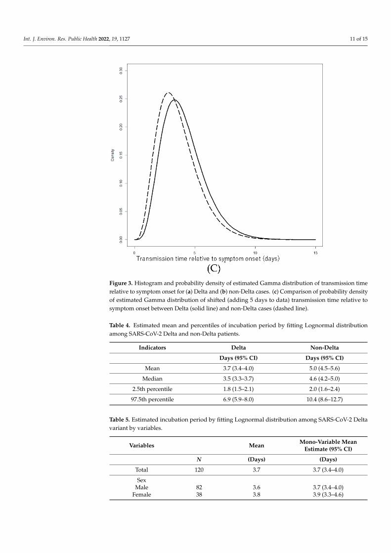
The transmission time relative to symptom onset was distributed between −4 and7 days in 94 patient pairs with the Delta variant, and it was negative (i.e., transmitted atleast one day before the date of onset) in 63 (67%) patient pairs. The mean transmission timerelative to symptom onset was −0.94 days. In 66 non-Delta patient pairs, the transmissiontime relative to symptom onset was distributed between −4 days and 4 days, and it wasnegative in 53 (80%) patient pairs. The mean transmission time relative to symptom onsetwas −1.39 days; the difference was significant by Mann–Whitney U test (p = 0.012).
We implemented sensitive analyses by Mann–Whitney U test for participants exclud-ing those with incomplete vaccination. Mean incubation period of participants with zerovaccination was 3.7 days among 104 Delta variant patients and 4.9 days among 99 non-Delta variant patients; the difference was significant (p-value = 0.000). Transmission timerelative to onset was −0.90 days among 80 Delta variant patients with zero vaccination and−1.36 days among 62 non-Delta variant patients with zero vaccination; the difference wassignificant (p-value = 0.020).
Table 3, Figures 1–3 show the estimated parameters for SARS-CoV-2 transmissiondynamics.
Table 3. Estimated parameters for SARS-CoV-2 transmission dynamics.
Parameters FittedDistribution Delta Non-Delta
Meanlog/Shape Sdlog/Rate Meanlog/Shape Sdlog/Rate
Days (95% CI) Days (95% CI) Days (95% CI) Days (95% CI)
Incubation period Lognormal(Meanlog/Sdlog) 1.25 (1.19–1.31) 0.34 (0.30–0.39) 1.52 (1.43–1.60) 0.42(0.37–0.48)
Serial interval(added 3 days)
Gamma(Shape/Rate) 7.1 (5.1–9.5) 1.24 (0.88–1.67) 6.1 (4.2–8.2) 0.97 (2.6–4.0)
Transmission timerelative to onset(added 5 days)
Gamma(Shape/Rate) 5.5 (4.0–7.3) 1.37 (0.98–1.81) 4.7 (3.3–6.3) 1.32(0.90–1.80)
CI = credible interval; SARS-CoV-2 = severe acute respiratory syndrome coronavirus 2.
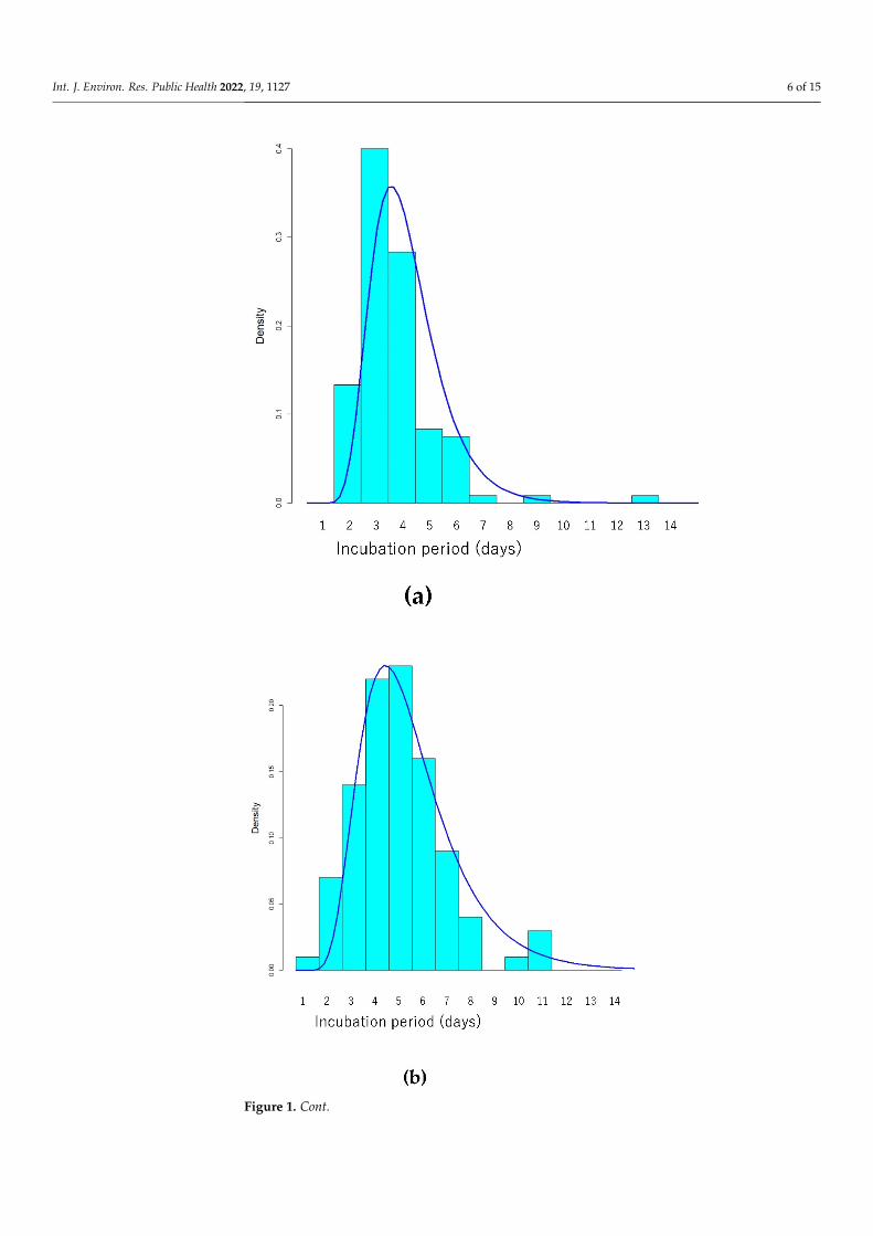
Int. J. Environ. Res. Public Health 2022, 19, 1127 6 of 15Int. J. Environ. Res. Public Health 2022, 19, x 6 of 16
Figure 1. Cont.
Int. J. Environ. Res. Public Health 2022, 19, 1127 7 of 15Int. J. Environ. Res. Public Health 2022, 19, x 7 of 16
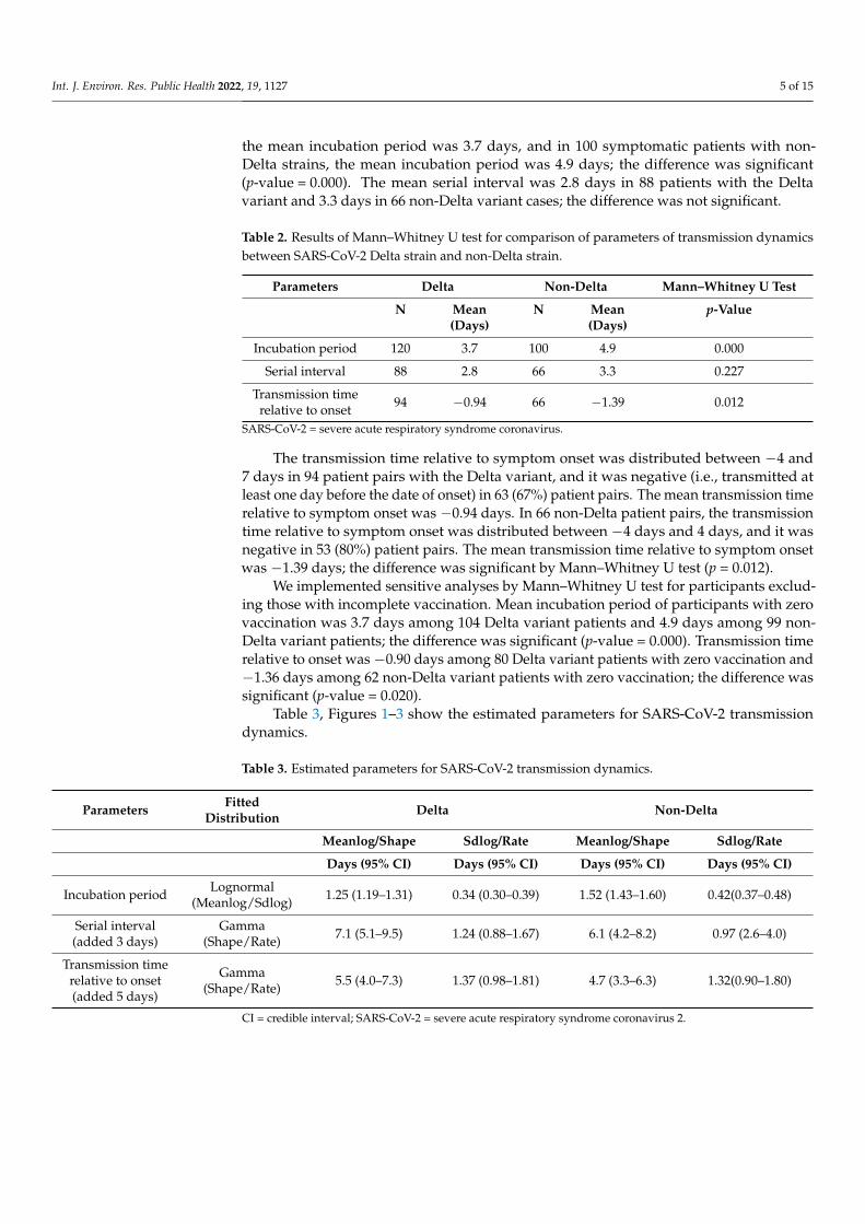
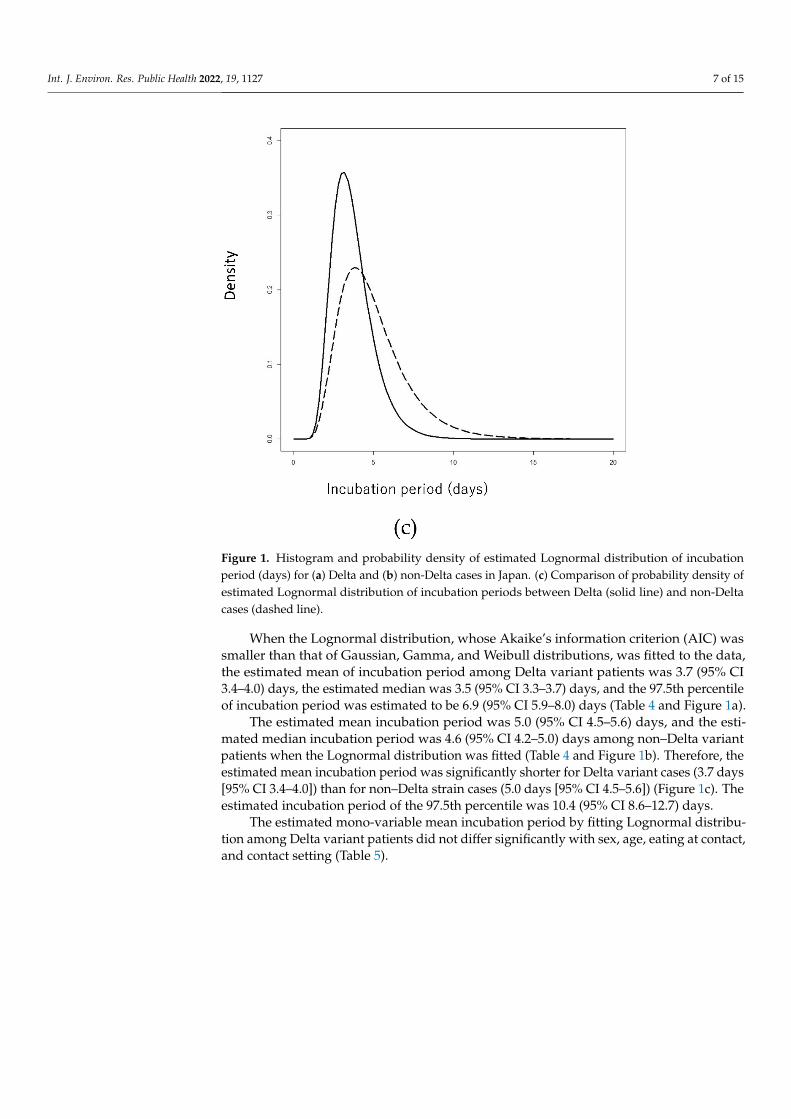
Figure 1. Histogram and probability density of estimated Lognormal distribution of incubation pe-riod (days) for (a) Delta and (b) non-Delta cases in Japan. (c) Comparison of probability density of estimated Lognormal distribution of incubation periods between Delta (solid line) and non-Delta cases (dashed line).
Figure 1. Histogram and probability density of estimated Lognormal distribution of incubationperiod (days) for (a) Delta and (b) non-Delta cases in Japan. (c) Comparison of probability density ofestimated Lognormal distribution of incubation periods between Delta (solid line) and non-Deltacases (dashed line).
When the Lognormal distribution, whose Akaike’s information criterion (AIC) wassmaller than that of Gaussian, Gamma, and Weibull distributions, was fitted to the data,the estimated mean of incubation period among Delta variant patients was 3.7 (95% CI3.4–4.0) days, the estimated median was 3.5 (95% CI 3.3–3.7) days, and the 97.5th percentileof incubation period was estimated to be 6.9 (95% CI 5.9–8.0) days (Table 4 and Figure 1a).
The estimated mean incubation period was 5.0 (95% CI 4.5–5.6) days, and the esti-mated median incubation period was 4.6 (95% CI 4.2–5.0) days among non–Delta variantpatients when the Lognormal distribution was fitted (Table 4 and Figure 1b). Therefore, theestimated mean incubation period was significantly shorter for Delta variant cases (3.7 days[95% CI 3.4–4.0]) than for non–Delta strain cases (5.0 days [95% CI 4.5–5.6]) (Figure 1c). Theestimated incubation period of the 97.5th percentile was 10.4 (95% CI 8.6–12.7) days.
The estimated mono-variable mean incubation period by fitting Lognormal distribu-tion among Delta variant patients did not differ significantly with sex, age, eating at contact,and contact setting (Table 5).
Int. J. Environ. Res. Public Health 2022, 19, 1127 8 of 15
Int. J. Environ. Res. Public Health 2022, 19, x 8 of 16
Figure 2. Cont.
Int. J. Environ. Res. Public Health 2022, 19, 1127 9 of 15Int. J. Environ. Res. Public Health 2022, 19, x 9 of 16
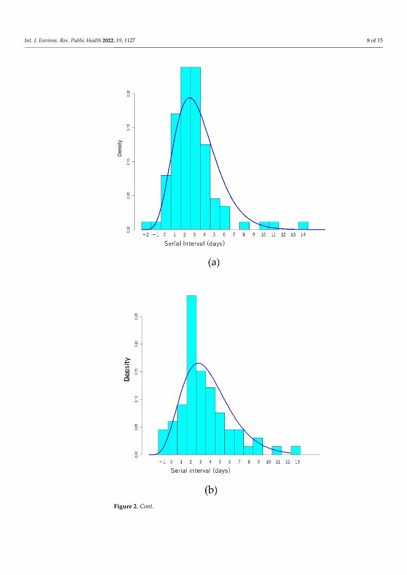
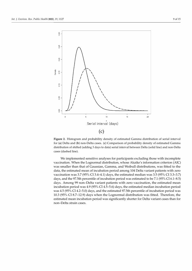
Figure 2. Histogram and probability density of estimated Gamma distribution of serial interval for (a) Delta and (b) non-Delta cases. (c) Comparison of probability density of estimated Gamma distri-bution of shifted (adding 3 days to data) serial interval between Delta (solid line) and non-Delta cases (dashed line).
Figure 2. Histogram and probability density of estimated Gamma distribution of serial intervalfor (a) Delta and (b) non-Delta cases. (c) Comparison of probability density of estimated Gammadistribution of shifted (adding 3 days to data) serial interval between Delta (solid line) and non-Deltacases (dashed line).
We implemented sensitive analyses for participants excluding those with incompletevaccination. When the Lognormal distribution, whose Akaike’s information criterion (AIC)was smaller than that of Gaussian, Gamma, and Weibull distributions, was fitted to thedata, the estimated mean of incubation period among 104 Delta variant patients with zerovaccination was 3.7 (95% CI 3.4–4.1) days, the estimated median was 3.5 (95% CI 3.3–3.7)days, and the 97.5th percentile of incubation period was estimated to be 7.1 (95% CI 6.1–8.5)days. Among 99 non–Delta variant patients with zero vaccination, the estimated meanincubation period was 4.9 (95% CI 4.5–5.6) days, the estimated median incubation periodwas 4.5 (95% CI 4.2–5.0) days, and the estimated 97.5th percentile of incubation period was10.3 (95% CI 8.7–12.9) days when the Lognormal distribution was fitted. Therefore, theestimated mean incubation period was significantly shorter for Delta variant cases than fornon–Delta strain cases.
Int. J. Environ. Res. Public Health 2022, 19, 1127 10 of 15
Int. J. Environ. Res. Public Health 2022, 19, x 10 of 16
Figure 3. Cont.
Int. J. Environ. Res. Public Health 2022, 19, 1127 11 of 15
Int. J. Environ. Res. Public Health 2022, 19, x 11 of 16
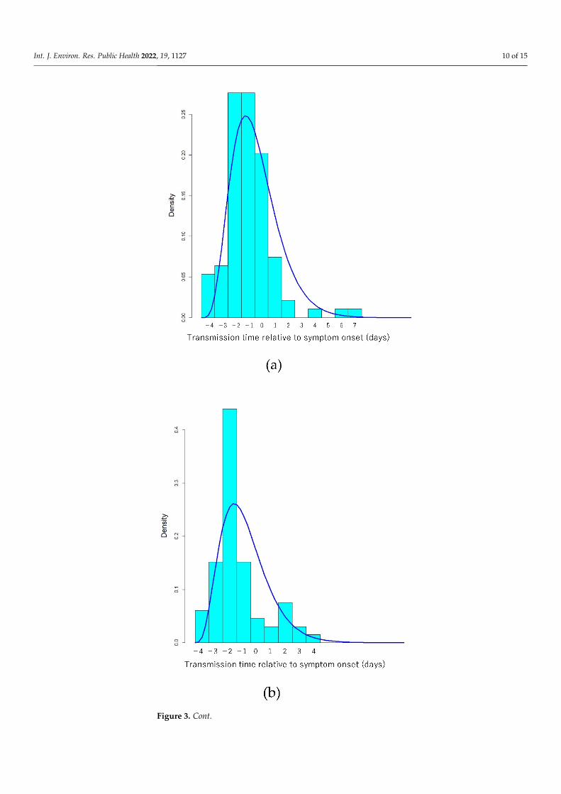
Figure 3. Histogram and probability density of estimated Gamma distribution of transmission time relative to symptom onset for (a) Delta and (b) non-Delta cases. (c) Comparison of probability den-sity of estimated Gamma distribution of shifted (adding 5 days to data) transmission time relative to symptom onset between Delta (solid line) and non-Delta cases (dashed line).
When the Lognormal distribution, whose Akaike’s information criterion (AIC) was smaller than that of Gaussian, Gamma, and Weibull distributions, was fitted to the data, the estimated mean of incubation period among Delta variant patients was 3.7 (95% CI 3.4–4.0) days, the estimated median was 3.5 (95% CI 3.3–3.7) days, and the 97.5th percen-tile of incubation period was estimated to be 6.9 (95% CI 5.9–8.0) days (Table 4 and Figure 1a).
The estimated mean incubation period was 5.0 (95% CI 4.5–5.6) days, and the esti-mated median incubation period was 4.6 (95% CI 4.2–5.0) days among non–Delta variant patients when the Lognormal distribution was fitted (Table 4 and Figure 1b). Therefore, the estimated mean incubation period was significantly shorter for Delta variant cases (3.7 days [95% CI 3.4–4.0]) than for non–Delta strain cases (5.0 days [95% CI 4.5–5.6]) (Figure 1c). The estimated incubation period of the 97.5th percentile was 10.4 (95% CI 8.6–12.7) days.
Table 4. Estimated mean and percentiles of incubation period by fitting Lognormal distribution among SARS-CoV-2 Delta and non-Delta patients.
Indicators Delta Non-Delta Days (95%CI) Days (95%CI)
Mean 3.7 (3.4–4.0) 5.0 (4.5–5.6) Median 3.5 (3.3–3.7) 4.6 (4.2–5.0)
2.5th percentile 1.8 (1.5–2.1) 2.0 (1.6–2.4)
Figure 3. Histogram and probability density of estimated Gamma distribution of transmission timerelative to symptom onset for (a) Delta and (b) non-Delta cases. (c) Comparison of probability densityof estimated Gamma distribution of shifted (adding 5 days to data) transmission time relative tosymptom onset between Delta (solid line) and non-Delta cases (dashed line).
Table 4. Estimated mean and percentiles of incubation period by fitting Lognormal distributionamong SARS-CoV-2 Delta and non-Delta patients.
Indicators Delta Non-Delta
Days (95% CI) Days (95% CI)
Mean 3.7 (3.4–4.0) 5.0 (4.5–5.6)
Median 3.5 (3.3–3.7) 4.6 (4.2–5.0)
2.5th percentile 1.8 (1.5–2.1) 2.0 (1.6–2.4)
97.5th percentile 6.9 (5.9–8.0) 10.4 (8.6–12.7)
Table 5. Estimated incubation period by fitting Lognormal distribution among SARS-CoV-2 Deltavariant by variables.
Variables Mean Mono-Variable MeanEstimate (95% CI)
N (Days) (Days)
Total 120 3.7 3.7 (3.4–4.0)
SexMale 82 3.6 3.7 (3.4–4.0)
Female 38 3.8 3.9 (3.3–4.6)

Int. J. Environ. Res. Public Health 2022, 19, 1127 12 of 15
Table 5. Cont.
Variables Mean Mono-Variable MeanEstimate (95% CI)
N (Days) (Days)
Age≤29 77 3.6 3.6 (3.3–4.0)≥30 43 3.9 3.9 (3.4–4.5)
Eating of infector and/or patient at exposureYes 58 3.9 3.8 (3.4–4.4)
No or unknown 62 3.6 3.6 (3.3–3.9)
SettingRestaurant 33 3.9 3.9 (3.2–4.8)
School 29 3.8 3.8 (3.2–4.6)House 22 3.8 3.8 (3.3–4.6)Others 36 3.4 3.4 (3.1–3.9)
4. Discussion
In the present study, the Delta variant strain had a shorter incubation period comparedwith non-Delta strains.
The incubation period for the Delta variant was 3.7 days, which was significantlyshorter than that for non-Delta strains. The incubation period did not significantly differwith the factors analyzed. To the best of our knowledge, only three peer-reviewed studiesin Guangdong province, China, reported the incubation period (4.0, 4.4, and 6.0 days) inDelta variant cases [5,6,27]. These incubation periods were shorter [5,6] or longer [27] thanthose of non-Delta cases. The present study supports the former results.
The upper limit of the estimated incubation period of the 97.5th percentile was approx-imately 8 days in the present study. In Japan and other countries, close contacts have beenquarantined for 14 days [7,9–11]. However, Delta variant cases have a short incubationperiod, and currently vaccination promotes the coexistence of humans with SARS-CoV-2rather than the “zero-COVID” strategy. Therefore, it may be appropriate for unvaccinatedclose contacts of COVID-19 patients to shorten the quarantine period to, for example,approximately 8 days.
The mean serial interval for the Delta variant was 2.8 days and was not significantlyshorter than that for non-Delta variants in the present study. In the previous literature,it was 2.3 days (mean) in China, 3 days (median) in Singapore, and 3.3 days (mean) inKorea [5,28,29]. Although these figures are apparently shorter than those in previous reportsfor the wild-type strain [30,31], they could have been reduced by various interventions,such as self-isolation, quarantine of close contacts, and lockdown [26,32].
The transmission time relative to symptom onset was distributed between −4 and7 days in this study. Contact tracing and quarantine have been implemented for closecontacts with exposure to the virus 2 days before symptom onset in the infector patientor later in several countries [7,8]. However, it may be necessary to expand subject closecontacts for tracing and quarantine to those with exposure to the virus three and four daysbefore symptom onset in the infector patient.
In the Delta variant group of the present study, viral transmission was not observedafter the eighth day after symptom onset in the infector patient. The viral load after symp-tom onset may be higher in Delta variant cases than in non-Delta cases [5,33]. However,the present study did not support extending the currently recommended 10-day patientisolation period [33].
In the present study, most of the transmission occurred during the infector’s pre-symptomatic period. These findings are consistent with those of previous studies [21,22].The proportion of pre-symptomatic transmission may be an important indicator becausepre-symptomatic and asymptomatic transmission may play an important role [34]. Whiletransmission after symptom onset in infector patients can be reduced by interventions,
Int. J. Environ. Res. Public Health 2022, 19, 1127 13 of 15
such as voluntary quarantine after symptom onset, it is generally difficult to preventtransmission from asymptomatic or pre-symptomatic infectors. Therefore, the proportionof pre-symptomatic transmission could be reduced by interventions and time varying.
In this study, patients with the Delta variant strain had a shorter transmission timebefore symptom onset (1.4 days) than those with non-Delta strains (2.0 days) by Mann–Whitney U test. In previous studies on the wild-type strain, the transmission time relativeto symptom onset varied among countries [17]. Nonetheless, the transmission time relativeto symptom onset could be reduced by voluntary quarantine after symptom onset. Onthe contrary, it is difficult to influence the transmission time before symptom onset usingsuch interventions. Compared with non-Delta strains, the Delta-variant may have a shorterduration of transmission to contacts before symptom onset.
The fifth wave of COVID-19 in Japan, which occurred in the latter half of July 2021,was mainly caused by the Delta variant. Compared with non-Delta variant cases, Deltavariant cases may have higher viral shedding [5,33], resulting in a higher basic reproductionnumber. If intervention is inadequate, the transmission and number of patients with theDelta variant may increase sharply in the area. However, the number of patients in Japandecreased continuously after September. Although the rapid development and roll-outof vaccines has contributed to the decline in the number of patients in the fifth wavein Japan, the cause of this rapid decline has not been completely elucidated. Possiblereasons for the rapid decline include the relatively recent timing of vaccination, mainlyin July and August, continuous observance of public health interventions, such as maskwearing [20], and climate change [35]. In Japan, the rapid increase in the number of COVID-19 cases in the early stage of the fifth wave might have promoted patient behavior aimedat avoiding transmission, such as self-quarantine and undergoing COVID-19 testing aftersymptom onset. Therefore, in the later stage of the fifth wave, the shorter pre-symptomatictransmission period, as described above, indicating higher proportion of behaviorallyavoidable transmission, in addition to other factors, might be a factor in the sharp declinein the number of cases.
This study had several limitations. First, the relatively small number of participantcases compared to the total number of patients in the area might have caused selectionbias [14,36]. However, we adopted similar procedures and estimation methods for bothDelta and non-Delta cases, which might have partially offset the bias. Second, the syn-dromic case definition may have influenced the outcome period, compared to other stud-ies [14,37]. Third, censoring of follow-up observations and truncation might have influ-enced the selection of participants and outcome period [23]. Fourth, the Delta variantwas mainly confirmed by the L452R mutation. However, genome sequencing showedcoincidence between the Delta variant and L452R mutation in Japan [19].
In the future, it is necessary to further study transmission dynamics and live viralshedding, including in vaccinated patients, household attack rate [27,38], and genomic anal-ysis of the Delta variant. It is also necessary to continue the surveillance of epidemiologicaldata and other VOCs including the Omicron variant.
5. Conclusions
Patients with the Delta variant strain had a shorter incubation period and a shorterperiod of transmission of the virus to contacts before symptom onset, compared with thosewith non-Delta strains.
Author Contributions: Conceptualization, T.O. and H.T.; data curation, F.I., Y.T., and T.O.; formalanalysis, T.O.; validation T.O., H.T, I.F, and A.H.; writing, T.O. and H.T. All authors have read andagreed to the published version of the manuscript.
Funding: This research received no external funding.
Institutional Review Board Statement: The study was conducted in accordance with the Declarationof Helsinki and approved by the Osaka University Hospital Observational Research Ethics ReviewCommittee (protocol code, T20114; Date of approval, 26 October 2021).
Int. J. Environ. Res. Public Health 2022, 19, 1127 14 of 15
Informed Consent Statement: Active epidemiological investigation data analyses were performed inaccordance with the Infectious Diseases Control Law, and the study was exempt from the requirementfor informed consent under “the ethical guidelines for life science and medical research on humansubject” in Japan.
Data Availability Statement: The data presented in this study are available upon reasonable requestfrom the corresponding author. The data are not publicly available because of the protection ofpersonal information.
Conflicts of Interest: The authors declare no conflict of interest.
References1. World Health Organization. Tracking SARS-CoV-2 Variants. Available online: https://www.who.int/en/activities/tracking-
SARS-CoV-2-variants/ (accessed on 1 December 2021).2. Campbell, F.; Archer, B.; Laurenson-Schafer, H.; Jinnai, Y.; Konings, F.; Batra, N.; Pavlin, B.; Vandemaele, K.; Van Kerkhove, M.D.;
Jombart, T.; et al. Increased transmissibility and global spread of SARS-CoV-2 variants of concern as at June 2021. Eurosurveillance2021, 26, 2100509. [CrossRef]
3. Deng, X.; Garcia-Knight, M.A.; Khalid, M.M.; Servellita, V.; Wang, C.; Morris, M.K.; Sotomayor-González, A.; Glasner, D.R.;Reyes, K.R.; Gliwa, A.S.; et al. Transmission, infectivity, and neutralization of a spike L452R SARS-CoV-2 variant. Cell 2021, 184,3426–3437.e8. [CrossRef]
4. World Health Organization. Weekly Epidemiological Update on COVID-19–13 October 2021. Available online: https://www.who.int/publications/m/item/weekly-epidemiological-update-on-covid-19---13-october-2021 (accessed on 1 December 2020).
5. Wang, Y.; Chen, R.; Hu, F.; Lan, Y.; Yang, Z.; Zhan, C.; Shi, J.; Deng, X.; Jiang, M.; Zhong, S.; et al. Transmission, viral kineticsand clinical characteristics of the emergent SARS-CoV-2 Delta VOC in Guangzhou, China. EClinicalMedicine 2021, 40, 101129.[CrossRef] [PubMed]
6. Zhang, M.; Xiao, J.; Deng, A.; Zhang, Y.; Zhuang, Y.; Hu, T.; Li, J.; Tu, H.; Li, B.; Zhou, Y.; et al. Transmission Dynamics of anOutbreak of the COVID-19 Delta Variant B.1.617.2—Guangdong Province, China, May–June 2021. China CDC Wkly. 2021, 3,584–586. [CrossRef] [PubMed]
7. National Institute of Infectious Diseases, Infectious Disease Epidemiology Center. Manual Conducting for Active EpidemiologicalSurveillance of Patients with Novel Coronavirus Infection (Provisional Version on May 29). January 2021 (in Japanese). Availableonline: https://www.niid.go.jp/niid/images/epi/corona/COVID19-02-210108.pdf (accessed on 15 December 2021).
8. Centers for Disease Control and Prevention (CDC). Interim Guidance on Developing a COVID-19 Case Investigation & ContactTracing Plan: Overview: Appendices. Available online: https://www.cdc.gov/coronavirus/2019-ncov/php/contact-tracing/contact-tracing-plan/appendix.html#contact (accessed on 1 December 2021).
9. Centers for Disease Control and Prevention (CDC). National Center for Immunization and Respiratory Diseases (NCIRD),Division of Viral Diseases. Quarantine and Isolation (Updated 19 October 2021). Available online: https://www.cdc.gov/coronavirus/2019-ncov/your-health/quarantine-isolation.html (accessed on 1 December 2021).
10. World Health Organization. Contact Tracing in the Context of COVID-19. 2021. Available online: https://www.who.int/publications/i/item/contact-tracing-in-the-context-of-covid-19 (accessed on 1 December 2021).
11. Tu, H.; Hu, K.; Zhang, M.; Zhuang, Y.; Song, T. Effectiveness of 14 day quarantine strategy: Chinese experience of prevention andcontrol. BMJ 2021, 375, e066121. [CrossRef]
12. Li, Q.; Guan, X.; Wu, P.; Wang, X.; Zhou, L.; Tong, Y.; Ren, R.; Leung, K.S.M.; Lau, E.H.Y.; Wong, J.Y.; et al. Early TransmissionDynamics in Wuhan, China, of Novel Coronavirus-Infected Pneumonia. N. Engl. J. Med. 2020, 382, 1199–1207. [CrossRef]
13. McAloon, C.; Collins, Á.; Hunt, K.; Barber, A.; Byrne, A.W.; Butler, F.; Casey, M.; Griffin, J.; Lane, E.; McEvoy, D.; et al. Incubationperiod of COVID-19: A rapid systematic review and meta-analysis of observational research. BMJ Open 2020, 10, e039652.[CrossRef]
14. Xin, H.; Wong, J.Y.; Murphy, C.; Yeung, A.; Ali, S.T.; Wu, P.; Cowling, B.J. The incubation period distribution of coronavirusdisease 2019 (COVID-19): A systematic review and meta-analysis. Clin. Infect. Dis. 2021, 73, 2344–2352. [CrossRef] [PubMed]
15. Wei, W.E.; Li, Z.; Chiew, C.J.; Yong, S.E.; Toh, M.P.; Lee, V.J. Presymptomatic Transmission of SARS-CoV-2—Singapore, 23January–16 March 2020. MMWR Morb. Mortal Wkly. Rep. 2020, 69, 411–415. [CrossRef] [PubMed]
16. Owusu, D.; Pomeroy, M.A.; Lewis, N.M.; Wadhwa, A.; Yousaf, A.R.; Whitaker, B.; Dietrich, E.; Hall, A.J.; Chu, V.; Thornburg, N.;et al. Persistent SARS-CoV-2 RNA Shedding Without Evidence of Infectiousness: A Cohort Study of Individuals With COVID-19.J. Infect. Dis. 2021, 224, 1362–1371. [CrossRef]
17. Casey-Bryars, M.; Griffin, J.; McAloon, C.; Byrne, A.; Madden, J.; Mc Evoy, D.; Collins, Á.; Hunt, K.; Barber, A.; Butler, F.; et al.Presymptomatic transmission of SARS-CoV-2 infection: A secondary analysis using published data. BMJ Open 2021, 11, e041240.[CrossRef]
18. National Institute of Infectious Diseases. Current Situation of Infection, 26 May 2021. Available online: https://www.niid.go.jp/niid/en/2019-ncov-e/10415-covid19-ab36th-en.html (accessed on 1 December 2021).
19. COVID-19 Advisory Board of the Ministry of Health, Labor and Welfare. Current Situation of Infection and Others (in Japanese).Available online: https://www.mhlw.go.jp/stf/seisakunitsuite/bunya/0000121431_00294.html (accessed on 1 December 2021).
Int. J. Environ. Res. Public Health 2022, 19, 1127 15 of 15
20. Ministry of Health, Labor and Welfare. Visualizing the Data: Information on COVID-19 Infections. Available online: https://covid19.mhlw.go.jp/en/ (accessed on 15 December 2021).
21. Tindale, L.C.; Stockdale, J.E.; Coombe, M.; Garlock, E.S.; Lau, W.Y.V.; Saraswat, M.; Zhang, L.; Chen, D.; Wallinga, J.; Colijn, C.Evidence for transmission of COVID-19 prior to symptom onset. Elife 2020, 9, e57149. [CrossRef]
22. He, X.; Lau, E.H.Y.; Wu, P.; Deng, X.; Wang, J.; Hao, X.; Lau, Y.C.; Wong, J.Y.; Guan, Y.; Tan, X.; et al. Temporal dynamics in viralshedding and transmissibility of COVID-19. Nat. Med. 2020, 26, 672–675. [CrossRef] [PubMed]
23. Cowling, B.J.; Muller, M.P.; Wong, I.O.; Ho, L.M.; Louie, M.; McGeer, A.; Leung, G.M. Alternative methods of estimating anincubation distribution: Examples from severe acute respiratory syndrome. Epidemiology 2007, 18, 253–259. [CrossRef]
24. Institute of Health of Ibaraki Prefectural Government. Situation of Tests on Variant Virus of SARS-CoV-2 (in Japanese). Availableonline: https://www.pref.ibaraki.jp/hokenfukushi/eiken/kikaku/covid-19_ibarakieiken_kensa.html (accessed on 1 December2021).
25. Government of Osaka Prefecture. Proof of Positive Cases in Mutation PCR Test (Screening Test), 8 June 2021 (in Japanese).Available online: https://www.pref.osaka.lg.jp/attach/23711/00400840/0608heni.pdf (accessed on 1 December 2021).
26. Du, Z.; Xu, X.; Wu, Y.; Wang, L.; Cowling, B.J.; Meyers, L.A. Serial Interval of COVID-19 among Publicly Reported ConfirmedCases. Emerg Infect. Dis. 2020, 26, 1341–1343. [CrossRef]
27. Li, L.; Han, Z.G.; Qin, P.Z.; Liu, W.H.; Yang, Z.; Chen, Z.Q.; Li, K.; Xie, C.J.; Ma, Y.; Wang, H.; et al. Transmission and containmentof the SARS-CoV-2 Delta variant of concern in Guangzhou, China: A population-based study. PLoS Negl. Trop. Dis. 2022, 16,e0010048. [CrossRef] [PubMed]
28. Pung, R.; Mak, T.M.; Kucharski, A.J.; Lee, V.J. Serial intervals in SARS-CoV-2 B.1.617.2 variant cases. Lancet 2021, 398, 837–838.[CrossRef]
29. Hwang, H.; Lim, J.S.; Song, S.A.; Achangwa, C.; Sim, W.; Kim, G.; Ryu, S. Transmission dynamics of the Delta variant ofSARS-CoV-2 infections in South Korea. J. Infect. Dis. 2021, jiab586, online ahead of print. [CrossRef]
30. Nishiura, H.; Linton, N.M.; Akhmetzhanov, A.R. Serial interval of novel coronavirus (COVID-19) infections. Int. J. Infect. Dis.2020, 93, 284–286. [CrossRef] [PubMed]
31. Xu, X.K.; Liu, X.F.; Wu, Y.; Ali, S.T.; Du, Z.; Bosetti, P.; Lau, E.H.Y.; Cowling, B.J.; Wang, L. Reconstruction of Transmission Pairsfor Novel Coronavirus Disease 2019 (COVID-19) in Mainland China: Estimation of Superspreading Events, Serial Interval, andHazard of Infection. Clin. Infect. Dis. 2020, 71, 3163–3167. [CrossRef]
32. Ali, S.T.; Wang, L.; Lau, E.H.Y.; Xu, X.K.; Du, Z.; Wu, Y.; Leung, G.M.; Cowling, B.J. Serial interval of SARS-CoV-2 was shortenedover time by nonpharmaceutical interventions. Science 2020, 369, 1106–1109. [CrossRef]
33. Siedner, M.J.; Boucau, J.; Gilbert, R.F.; Uddin, R.; Luu, J.; Haneuse, S.; Vyas, T.; Reynolds, Z.; Iyer, S.; Chamberlin, G.C.; et al.Duration of viral shedding and culture positivity with post-vaccination SARS-CoV-2 delta variant infections. JCI Insight 2021,e155483. [CrossRef]
34. Hellewell, J.; Abbott, S.; Gimma, A.; Bosse, N.I.; Jarvis, C.I.; Russell, T.W.; Munday, J.D.; Kucharski, A.J.; Edmunds, W.J.; Funk, S.;et al. Feasibility of controlling COVID-19 outbreaks by isolation of cases and contacts. Lancet Glob. Health 2020, 8, e488–e496.[CrossRef]
35. Fontal, A.; Bouma, M.J.; San-José, A.; López, L.; Pascual, M.; Rodó, X. Climatic signatures in the different COVID-19 pandemicwaves across both hemispheres. Nat. Comput. Sci. 2021, 1, 655–665. [CrossRef]
36. Qin, J.; You, C.; Lin, Q.; Hu, T.; Yu, S.; Zhou, X.H. Estimation of incubation period distribution of COVID-19 using disease onsetforward time: A novel cross-sectional and forward follow-up study. Sci. Adv. 2020, 6, eabc1202. [CrossRef] [PubMed]
37. Böhmer, M.M.; Buchholz, U.; Corman, V.M.; Hoch, M.; Katz, K.; Marosevic, D.V.; Böhm, S.; Woudenberg, T.; Ackermann, N.;Konrad, R.; et al. Investigation of a COVID-19 outbreak in Germany resulting from a single travel-associated primary case: Acase series. Lancet Infect. Dis. 2020, 20, 920–928. [CrossRef]
38. Allen, H.; Vusirikala, A.; Flannagan, J.; Twohig, K.A.; Zaidi, A.; Chudasama, D.; Lamagni, T.; Groves, N.; Turner, C.; Rawlinson,C.; et al. Household transmission of COVID-19 cases associated with SARS-CoV-2 delta variant (B.1.617.2): National case-controlstudy. Lancet Reg. Health Eur. 2022, 12, 100252. [CrossRef]