sensitivity and acclimation to uv radiation of zoospores from five species of laminariales from the...
TRANSCRIPT

RESEARCH ARTICLE
C. Wiencke Æ M. N. Clayton Æ M. Schoenwaelder
Sensitivity and acclimation to UV radiation of zoosporesfrom five species of Laminariales from the Arctic
Received: 24 November 2003 / Accepted: 9 January 2004 / Published online: 12 February 2004� Springer-Verlag 2004
Abstract Spores of five Laminariales from Arctic Spits-bergen were exposed in the laboratory to photosyn-thetically active radiation (PAR; 400–700 nm),PAR+UVA radiation (UVAR; 320–400 nm) andPAR+UVAR+UVB radiation (UVBR; 280–320 nm).Subsequently, germination was monitored over periodsof 3, 6 and 9 days. The investigated species were theupper sublittoral Saccorhiza dermatodea, the upper tomid-sublittoral Alaria esculenta and Laminaria digitata,the mid-sublittoral L. saccharina and the lower sub-littoral L. solidungula. The germination capacitydecreased sharply after 16 h exposure to PAR+-UVAR+UVBR in all species. However, S. dermatodeawas able to recover from the damaging effects of UVBR.There was also a small increase in percentage germina-tion of A. esculenta 6–9 days after the treatment. Norecovery was evident in the other species. After 8 hexposure to PAR+UVA+UVB, L. digitata recoveredcompletely, and L. saccharina and L. solidungula, par-tially. The only species susceptible to PAR+UVAR wasL. solidungula. One prominent cytological feature ofUVR-exposed spores was the enlargement of phenolicvesicles (physodes) (particularly seen in S. dermatodeaand A. esculenta), which may have a protective functionagainst UVR. Pilot experiments under natural irradi-ance conditions indicate that the PAR component of
solar radiation exerts an additional stress. Overall thedata show that zoospores of the species from the uppersublittoral are less sensitive to UVR or have the capacityto recover from UV stress in contrast to species fromdeeper waters, probably due to their UV protective andrepair capabilities.
Introduction
Stratospheric ozone depletion is evident not only overAntarctica (Solomon 1999; Staehelin et al. 2001), there isalso a great potential for ozone losses over the Arctic(Gathen et al. 1995; Rex et al. 2002). According to thelatest predictions (Knudsen et al., unpublished data),ozone losses over the Arctic are likely to increase until2010 to 2020 and decrease only slightly by 2030.Reductions in the thickness of the ozone column lead toenhanced levels of UVB radiation (UVBR) at the earth’ssurface, and shifts the solar spectrum to shorter wave-lengths (Roy et al. 1990). UVBR also penetrates into thewater. For example, on the Arctic island of Spitsbergenbiologically significant UVBR levels are recorded downto 8 m water depth (Hanelt et al. 2001; van de Poll et al.2002d).
In macroalgae, UVBR causes molecular damage tonucleic acids and proteins and inhibits important met-abolic processes (Franklin and Forster 1997). UVexposure can show photoinhibition or even photodam-age to photosynthesis (Dring et al. 1996a; Hanelt et al.1997; Bischof et al. 1998a, 2000). Cyclobuthane-pyrim-idine dimers are formed in the DNA, thus blockingDNA and RNA polymerases and consequently inhib-iting genome replication and expression (van de Pollet al. 2002a, 2002b). In addition, UVBR exerts indirecteffects mediated by oxidative stress (Foyer et al. 1994;Aguilera et al. 2002). On the other hand, there is con-siderable potential for acclimation to UVBR in themacrothalli of seaweeds. Saccorhiza dermatodea, Alaria
Communicated by O. Kinne, Oldendorf/Luhe
C. Wiencke (&) Æ M. SchoenwaelderAlfred Wegener Institute for Polar and Marine Research,Am Handelshafen 12, 27570 Bremerhaven, GermanyE-mail: [email protected].: +49-471-48311338Fax: +49-471-48311425
M. N. ClaytonSchool of Biological Sciences, Monash University,3800 Clayton, Victoria, Australia
M. SchoenwaelderDepartment of Molecular, Cellular and Developmental Biology,University of Colorado, 347 UCB, Boulder,CO 80309, USA
Marine Biology (2004) 145: 31–39DOI 10.1007/s00227-004-1307-9

esculenta and Laminaria saccharina collected over adepth gradient on Spitsbergen exhibited a different UVsensitivity in relation to photosynthesis, with the mosttolerant individuals growing in shallow waters (Bischofet al. 1998b). Maximum quantum yield of photo-synthesis of A. esculenta acclimates to enhanced levelsof UV radiation (UVR) within a few days (Bischofet al. 1999).
The acclimation potential is based on various repairand protective mechanisms. The D1 protein of photo-system II undergoes a permanent turn-over cycle afterradiative damage (Campbell et al. 1998; Mate et al.1998). DNA damage can be repaired by light-dependentphotolyases and light-independent nucleotide excisionrepair (Pakker et al. 2000a, 2000b; Poll et al. 2002b).Oxidative stress is counteracted by enzymatic defensesystems and scavenging by antioxidants (Collen andPedersen 1996; Aguilera et al. 2002; Dummermuth et al.2003). UVR-absorbing substances may prevent damage.In red algae, mycosporine-like amino acids were invokedto protect cellular molecules, structures and processesagainst the damaging effects of UVR (Karsten et al.1998, 1999; Cockell and Knowland 1999). In brown al-gae phlorotannin (polyphenolic)-containing vesicles, theso-called physodes, may play a role in chemical UVdefense. Phlorotannins strongly absorb in the UVB (andUVC) region of the spectrum and their formation isinducible by UVBR (Pavia et al. 1997; Pavia and Brock2000; Schoenwaelder 2002a).
The balance between the various negative effects ofUVBR and the repair and protective mechanisms isindicated by the integrative parameters growth andreproduction. Growth in species of the eulittoral/uppersublittoral is initially inhibited by UVBR, but canacclimate to a considerable extent (Altamirano et al.2000; Han et al. 2003). Similarly, growth of Fucus dis-tichus from Spitsbergen is not significantly reduced byUVR, whereas growth of sporophytes of S. dermatodea,L. digitata and L. saccharina shows considerable inhi-bition after exposure to the full solar spectrum (Aguileraet al. 1999). Also, growth rates of young and maturesporophytes of the upper sublittoral L. digitata fromHelgoland are less sensitive to UVR compared to mid-sublittoral L. saccharina and L. hyperborea (Dring et al.1996b).
The reproductive cells and the early developmentalstages of many organisms are more susceptible to UVRwhen compared to the adult stages (summarized byCoelho et al. 2000). Motility of spores from L. saccha-rina (Makarov and Voskoboinikov 2001) and phototaxisof swarmers of Scytosiphon lomentaria and Petaloniafascia (Flores-Moya et al. 2002) is reduced by UVBR.Huovinen et al. (2000) pointed out that microtubulesmight be affected by UVBR as nuclear division andtranslocation of the nucleus in zoospores of Macrocystispyrifera are inhibited. In L. digitata from Spitsbergen theloss of zoospore viability is positively correlated to DNAdamage and photodamage of the photosynthetic appa-ratus (Wiencke et al. 2000). Moreover, zoospores of
species from the upper shore were generally more sus-ceptible to UVR than species occurring at greater depths(Wiencke et al. 2000). The published data on UV sen-sitivity of spores of Laminariales from Spitsbergen areso far limited to A. esculenta, L. digitata and L. sac-charina.
The present study was conducted to describe theUVR susceptibility of zoospores of five species ofLaminariales occurring in the Kongsfjord on the westcoast of Spitsbergen, a glacial fjord in the Arctic, inrelation to their depth distribution. Saccorhiza derma-todea grows in the uppermost sublittoral, followed byA. esculenta, L. digitata and L. saccharina. The endemicArctic L. solidungula grows almost exclusively in thelower sublittoral (Hop et al. 2002; Vogele et al.,unpublished data). Species from this region might beparticularly affected due to the ozone losses over theArctic and the related increase in UVBR levels (Großet al. 2001; Dahlback 2002). Moreover, we studied forthe first time the ability of these small developmentalstages to repair UV-induced damage and also to exam-ine the protective potential of phlorotannin containingphysodes formed during and after exposure to artificialUVR in the laboratory. Additionally, we conducted forthe first time a pilot study on the performance of sporesunder natural irradiance conditions.
Materials and methods
Fertile specimens of Saccorhiza dermatodea, Alaria esculenta,Laminaria digitata, L. saccharina and L. solidungula were collectedin August 2002 by SCUBA diving in Kongsfjorden close to NyAlesund (78�55¢N, 11�56¢E) on the west coast of Spitsbergen. Anoverview on the physical environment and the ecosystem of theKongsfjord was given by Svendsen et al. (2002) and Hop et al.(2002). Thallus parts with sori were blotted with tissue paper andkept overnight or for a few days in a wet chamber in dim light at atemperature of 5–7�C. Spores were released by flooding the tissuewith filtered seawater in a Petri-dish. The initial spore concentra-tion was determined by use of a Thoma chamber (Brand, Ger-many) and was usually in the range between 30,000 and 80,000spores ml)1. For the experiments two to four drops from thesesuspensions were put into Petri-dishes 5 cm in diameter containing11 ml of Provasoli-enriched seawater (Starr and Zeikus 1987). Inthis way the density of spores per unit area was similar in allexperiments.
Spores obtained from five individual sporophytes of A. escul-enta, L. digitata, and L. saccharina were separately exposed for 8 or16 h to artificial UVR. In S. dermatodea spores came from twoindividual sporophytes and were mixed before UVR exposure; in L.solidungula only one fertile sporophyte was available. UVR wasgenerated by UVA-340 fluorescent tubes (Q-Panel, Cleveland,Ohio, USA), emitting a radiation similar to the solar spectrum inthe wavelength range below 340 nm. Photosynthetically activeradiation (PAR) was additionally provided by daylight fluorescenttubes (Osram Lumilux Deluxe, L36 W/12–950). UV measurementswere performed using a Solar Light PMA 2100 broadband radi-ometer equipped with the UVA sensor PMA 2110 and the UVBsensor PMA 2106 (Solar Light, Philadelphia, Pa., USA). As thespectral range of the UVA sensor extends into the UVB region ofthe spectrum and vice versa, UVA measurements were taken belowa Schott WG 320 filter (Schott, Mainz, Germany). UVBR wasdetermined after subtraction of measured irradiance under the filterfrom the irradiance without the filter. PAR was measured with a
32

Li-Cor LI 1000 data logger (Li-Cor, Lincoln, Neb., USA) equippedwith a LI-190 SA cosine corrected flat-head sensor. The irradiancesapplied were: 28.8±5.05 lmol photons m)2 s)1 PAR,8.22±0.64 W m)2 UVAR and 1.27±0.12 W m)2 UVBR. Tostudy the effect of different wavelength ranges, the Petri dishes werecovered with three different cut-off filters: (1) Ultraphan URT 300foil (Digefra, Munich, Germany) for exposure to the full spectrum,(2) Folex PR Montage Folie (Dr. Schleussner, Dreieich, Germany)for exposure to PAR and UVAR and (3) Ultraphan URUV farblos(Digefra) for exposure to PAR. The spectra under these radiationregimes correspond to those used by Wiencke et al. (2000). Afterexposure the spore suspensions were put under dim white daylightat a temperature of 10�C for 3, 6 and 9 days to monitor germina-tion rates. Germination was determined microscopically by use ofan Axioplan microscope (Zeiss, Gottingen, Germany) equippedwith a 25·seawater immersion objective. A spore was classified asgerminated if at least a germ-tube was formed. We did not differ-entiate between dead and living, but germinated and not germi-nated spores. In each sample about 300 spores were counted andthe percentage of germinated and non-germinated spores deter-mined. Dead spores and their remains were clearly visible withinthe time periods under study.
For photography, cover slips were placed in extra Petri dishesand spore suspensions were added. These dishes were then treatedin the same way as the others. At various times cover slips withspores/germlings were taken out of the dish and used for prepa-ration of microscopic slides. Microscopy of the various develop-mental stages was performed using oil immersion objectivesmounted on the Axioplan microscope. Micrographs were takendigitally using a Nikon Coolpix camera.
For the outdoor experiments, spore suspensions of A. esculenta,L. saccharina, L. digitata and L. solidungula were exposed in Petridishes to surface solar radiation outside the laboratory. Tempera-tures varied in these experiments between about 5 and 15�C. To
estimate the effect of UVR, some of the Petri dishes with spores ofA. esculenta were covered with a Schott GG 400 filter (Schott,Mainz Germany). All other dishes were covered with Schott WG280 filters to stop possible evaporation and salinity changes in themedium. UVBR was measured in the field by use of a spectrora-diometer with a 32-channel photon counting detector equippedwith a 2 p diffusor (Hanken and Tug 2002). UVAR and PAR weremeasured using a spectroradiometer with a 256-channel photodi-ode array detector and a cosine diffusor (ISITEC Bremerhaven,Germany).
Results
Saccorhiza dermatodea, Alaria esculenta, Laminariadigitata and L. saccharina zoospores germinated quicklyafter 8 or 16 h exposure to PAR. Between 83% and 93%of the spores germinated after 3 days of post-culture andthere was only a minor increase in germination between92% and 99% in the next 3–6 days (Fig. 1, 2). In con-trast, germination of L. solidungula spores was muchslower. Only 11% and 15% of the spores were germi-nated after 8 or 16 h of exposure to PAR and 3 dayspost-culture. After 3 more days of post-culture thesevalues increased to only 69% and 57%, respectively(Figs. 1, 2).
Germination rates were similar or only slightly lowerafter 8- and 16-h exposures to UVAR in addition toPAR and 3 days post-culture in S. dermatodea,
Fig. 1 Germination of sporesof Alaria esculenta, Saccorhizadermatodea, Laminaria digitata,L. saccharina and L. solidungula3, 6 and 9 days after exposureto 8 h PAR (photosyntheticallyactive radiation) and UVradiation using cut-off filters(means±SD; n=5)
33

A. esculenta L. digitata and L. saccharina (Figs. 1, 2). Incontrast, in L. solidungula, germination rates werestrongly reduced in most cases after the UVAR+PARexposure. Even after 6 days of post-culture germinationrates of only 20–30% were recorded in the UVAR+-PAR experiment compared to 55% and 70% in the PARcondition (Fig. 1, 2).
Additional exposure to UVBR for 8 h resulted in asimilar pattern in L. solidungula for the two post-culturetimes tested. However, after the 16-h exposure to the fullspectrum almost no germination was found after 3 andalso after 6 days of post-culture (Fig. 2). Exposure tothe full spectrum had a similar effect in L. saccharinaand L. digitata. Only an extremely low germination ratewas recorded after the 16-h exposure to the full spectrumunder all three post-culture times (Fig. 2). In the 8-hexperiment, however, there was only a small differencein germination rates under PAR+UVAR+UVBRcompared with PAR+UVAR and a post-culture time of3 days (Fig. 1). After 6 days of post-culture the germi-nation rate increased, indicating a considerable recoveryin the spores, which did not germinate within the first3 days of post-culture (Fig. 1).
Alaria esculenta and S. dermatodea were the speciesmost tolerant to UVBR. After a 16-h exposure to the fullspectrum and 3 days of post-culture a germination rateof 21% and 40% was recorded, respectively (Fig. 2).Additionally there was a great potential for recovery, as
germination rates increased slightly to 38% after 9 daysof post-culture in A. esculenta. In S. dermatodea valuesmeasured after 6 days of post-culture were similar tothose obtained after exposure to PAR and PAR+-UVAR (Fig. 2). In the 8-h experiment there were nodifferences in germination rates between the three con-ditions in either species (Fig. 1).
The different developmental stages of spores ofS. dermatodea, A. esculenta, L. digitata and L. saccha-rina under the various exposure conditions are shown inFig. 3. Freshly released zoospores of species in theLaminariales (Fig. 3a–c) are biflagellate, containing oneto several small chloroplasts and several physodes andmeasure approximately 5 lm in diameter. Zoospores ofthe species examined (except S. dermatodea) lack aneyespot. When exposed to PAR, germ tubes started todevelop within the first day (Fig. 3d, e). Usually after4 days the embryospore and germination tube wereempty and the first cell of the gametophyte was formed(Fig. 3f). Figure 3g shows healthy 9-day-old gameto-phytes of A. esculenta exposed to the PAR condition.After exposure to PAR+UVAR+UVBR, differentdevelopmental features were evident in the various spe-cies. Whereas in S. dermatodea (Fig. 3h) and also inmany spores of A. esculenta (Fig. 3i) the chloroplastswere typically pigmented, they lost pigmentation inL. digitata and L. saccharina. Physodes became moreabundant in all species and increased in size (Fig. 3j, k).
Fig. 2 Germination of sporesof A. esculenta, S. dermatodea,L. digitata, L. saccharina andL. solidungula 3, 6 and 9 daysafter exposure to 16 h PAR andUV radiation using cut-offfilters (means±SD; n=5)
34
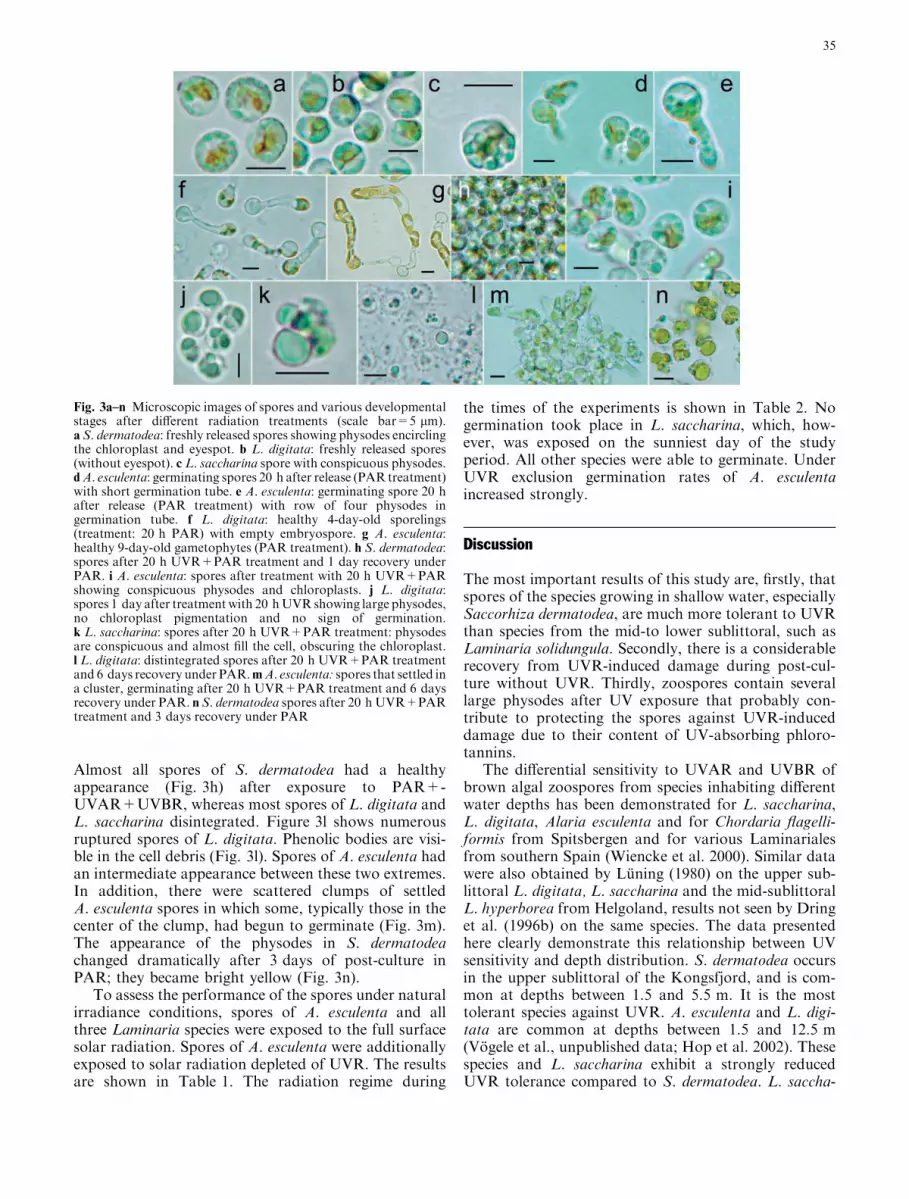
Almost all spores of S. dermatodea had a healthyappearance (Fig. 3h) after exposure to PAR+-UVAR+UVBR, whereas most spores of L. digitata andL. saccharina disintegrated. Figure 3l shows numerousruptured spores of L. digitata. Phenolic bodies are visi-ble in the cell debris (Fig. 3l). Spores of A. esculenta hadan intermediate appearance between these two extremes.In addition, there were scattered clumps of settledA. esculenta spores in which some, typically those in thecenter of the clump, had begun to germinate (Fig. 3m).The appearance of the physodes in S. dermatodeachanged dramatically after 3 days of post-culture inPAR; they became bright yellow (Fig. 3n).
To assess the performance of the spores under naturalirradiance conditions, spores of A. esculenta and allthree Laminaria species were exposed to the full surfacesolar radiation. Spores of A. esculenta were additionallyexposed to solar radiation depleted of UVR. The resultsare shown in Table 1. The radiation regime during
the times of the experiments is shown in Table 2. Nogermination took place in L. saccharina, which, how-ever, was exposed on the sunniest day of the studyperiod. All other species were able to germinate. UnderUVR exclusion germination rates of A. esculentaincreased strongly.
Discussion
The most important results of this study are, firstly, thatspores of the species growing in shallow water, especiallySaccorhiza dermatodea, are much more tolerant to UVRthan species from the mid-to lower sublittoral, such asLaminaria solidungula. Secondly, there is a considerablerecovery from UVR-induced damage during post-cul-ture without UVR. Thirdly, zoospores contain severallarge physodes after UV exposure that probably con-tribute to protecting the spores against UVR-induceddamage due to their content of UV-absorbing phloro-tannins.
The differential sensitivity to UVAR and UVBR ofbrown algal zoospores from species inhabiting differentwater depths has been demonstrated for L. saccharina,L. digitata, Alaria esculenta and for Chordaria flagelli-formis from Spitsbergen and for various Laminarialesfrom southern Spain (Wiencke et al. 2000). Similar datawere also obtained by Luning (1980) on the upper sub-littoral L. digitata, L. saccharina and the mid-sublittoralL. hyperborea from Helgoland, results not seen by Dringet al. (1996b) on the same species. The data presentedhere clearly demonstrate this relationship between UVsensitivity and depth distribution. S. dermatodea occursin the upper sublittoral of the Kongsfjord, and is com-mon at depths between 1.5 and 5.5 m. It is the mosttolerant species against UVR. A. esculenta and L. digi-tata are common at depths between 1.5 and 12.5 m(Vogele et al., unpublished data; Hop et al. 2002). Thesespecies and L. saccharina exhibit a strongly reducedUVR tolerance compared to S. dermatodea. L. saccha-
Fig. 3a–n Microscopic images of spores and various developmentalstages after different radiation treatments (scale bar=5 lm).a S. dermatodea: freshly released spores showing physodes encirclingthe chloroplast and eyespot. b L. digitata: freshly released spores(without eyespot). c L. saccharina spore with conspicuous physodes.dA. esculenta: germinating spores 20 h after release (PAR treatment)with short germination tube. e A. esculenta: germinating spore 20 hafter release (PAR treatment) with row of four physodes ingermination tube. f L. digitata: healthy 4-day-old sporelings(treatment: 20 h PAR) with empty embryospore. g A. esculenta:healthy 9-day-old gametophytes (PAR treatment). h S. dermatodea:spores after 20 h UVR+PAR treatment and 1 day recovery underPAR. i A. esculenta: spores after treatment with 20 h UVR+PARshowing conspicuous physodes and chloroplasts. j L. digitata:spores 1 day after treatmentwith 20 hUVR showing large physodes,no chloroplast pigmentation and no sign of germination.k L. saccharina: spores after 20 h UVR+PAR treatment: physodesare conspicuous and almost fill the cell, obscuring the chloroplast.l L. digitata: distintegrated spores after 20 h UVR+PAR treatmentand 6 days recovery under PAR.mA. esculenta: spores that settled ina cluster, germinating after 20 h UVR+PAR treatment and 6 daysrecovery under PAR. n S. dermatodea spores after 20 hUVR+PARtreatment and 3 days recovery under PAR
35

rina grows predominantly between 1.5 and 15.5 m. Theupper distribution limit of S. dermatodea, A. esculentaand L. digitata is 0.5 m, that of L. saccharina 1.5 m. Thespecies most susceptible to UVR is the endemic Arcticdeep water alga L. solidungula. This species occurs atdepths between 10.5 and 15.5 m in the middle zone ofthe Kongsfjord. These differences in the sensitivity ofbrown algal spores against UVR are comparable to thedifferential UVR tolerance of photosynthesis in macro-thalli of species growing at different water depths asshown, for example, by Dring et al. (1996a), Hanelt et al.(1997), Bischof et al. (1998a) and Karsten et al. (2001).In all these studies, photosynthesis in species fromshallow waters was much more UVR tolerant thanspecies from deeper waters.
The reasons for the UVR-induced spore mortalityare, among others, damage to the photosyntheticapparatus, to the DNA and, possibly, to the microtu-bules. The latter possibility was invoked by Huovinenet al. (2000) for UVR-exposed zoospores of Macrocystispyrifera. Damage of the photosynthetic apparatus wasdemonstrated in zoospores of L. digitata from Spits-bergen (Wiencke et al. 2000). Interspecific differences inDNA damage were found after exposure to the fullspectrum in L. digitata, L. saccharina and A. esculentafrom Spitsbergen (Wiencke et al. 2000). Whereas cyc-lobuthane-pyrimidine dimer formation as an indicatorof DNA damage was greatly enhanced after exposure tothe full spectrum in the first two species, in A. esculenta
from shallower waters it was negligible and comparableto the controls only exposed to PAR. In a detailedexperiment with L. digitata spores the formation ofcyclobuthane-pyrimidine dimers was positively corre-lated with the UVB dose (Wiencke et al. 2000).
The recovery from UVR-induced damage has beenshown here for the first time for spores during post-culture without UVR by the increase of germinationrates after 6 and 9 days in S. dermatodea and A. escul-enta. Undoubtedly, this recovery must be the result ofrepair processes. Repair of the D1 protein in the reactioncenter of photosystem II and UVBR-induced differentialtranscription of psbA genes encoding the D1 protein hasbeen demonstrated by Campbell et al. (1998) and Mateet al. (1998). Repair of UVR-induced damage of theDNA has been shown recently for macrothalli of anumber of tropical, temperate and Arctic marine mac-roalgae (Pakker et al 2000b; van de Poll et al. 2002a,2002b, 2002c, 2002d). Although no data are available onDNA repair processes in the unicellular propagules ofmarine algae, it is reasonable to assume that the repairmechanisms would be operating in these developmentalstages.
The increase in the number and size of the physodesin the UVR-exposed zoospores, especially in S. derma-todea, A. esculenta, but also in L. digitata and L. sac-charina, is described here for the first time and makesquantitative studies pressing. It is regarded as a protec-tive reaction against UVR. Physodes contain UVR-
Table 1 Germination of spores (means±SD;n=5) of Laminariales 6–7 days after exposure to solar radiation for the time periods given.Photosynthetically active radiation (PAR) and UVR doses taken from Table 2. In Alaria, germination was additionally recorded after UVexclusion
Germination(%)
Cut-off filter Date (2002) Time (hours) PAR UVAR+UVBRJ m)2 J m)2
Alaria esculenta 42.0±0.00 GG400 (solar radiation,UV-depleted)
21/22 August 14:00–08:00 2.18·106 –
A. esculenta 3.2±0.05 WG 280 (full solar radiation) 21/22 August 14:00–08:00 2.18·106 2.04·105Laminariasaccharina
0.0±0.00 WG 280 (full solar radiation) 09/10 August 10:15–02:15 3.19·106 3.27·105
L. digitata 5.0±0.06 WG 280 (full solar radiation) 10/11 August 16:50–08:15 3.17·106 3.04·105L. solidungulaa 25.6±0.11 WG 280 (full solar radiation) 13 August 14:30–23:15 1.04·106 0.98·105
a Under shade-cloth excluding 50% of the incident radiation
Table 2 Comparison between PAR and UVR doses for the datesof the outdoor experiments in Table 1 and those from the labora-tory experiments. The field PAR doses were calculated by anextrapolation of the UVA region using a standard atmospheric
model. Values for the experiment with L. solidungula on 13 August2002 were divided by 2 as we used shade-cloth excluding 50% ofthe incident radiation in this particular experiment
Dates and/or times PAR UVA+UVB UVA UVBJ m)2 J m)2 J m)2 J m)2
Field experiments (2002)09/10 August 10:15–02:15 hours 3.19·106 3.27·105 3.19·105 8.59·10310/11 August 16:50–08:15 hours 3.17·106 3.04·105 2.98·105 6.21·10313 August 14:30–23:15 hours 1.04·106 0.98·105 0.96·105 1.92·10321/22 August 14:00–08:00 hours 2.18·106 2.04·105 2.00·105 3.75·103Laboratory experiments8 h 1.97·105 2.73·105 2.37·105 3.60·10416 h 3.95·105 5.46·105 4.73·105 7.30·104
36

absorbing phlorotannins (Ragan and Glombitza 1986;Schoenwaelder 2002a) and represent, therefore, aneffective protective mechanism. In support of thisexplanation, Peckol et al. (1996) found higher contentsof phlorotannins in Fucus vesiculosus thalli collected inthe high intertidal compared to a population from thelower intertidal. Highest concentrations occurred inearly summer and were lowest in winter. In Homosirabanksii, phlorotannins are released from physodes in thesun-exposed cortical cells and form a protective layer forthe underlying tissue (Schoenwaelder 2002b). The moststriking support for this hypothesis is the induction ofphlorotannin formation after UVR exposure in Asco-phyllum nodosum (Pavia et al. 1997). Overall, it is rea-sonable to claim that physodes in the zoospores of theLaminariales studied here absorb UVR and protect theprotoplasm of the three species occurring in shallowwater (S. dermatodea, Alaria esculenta, L. digitata)against the damaging effects of UVR, a hypothesis forwhich we will provide additional evidence in anaccompanying paper (Clayton et al. 2004). The observedchange in color of the physodes in S. dermatodea sporesafter 3 days is presumably due to changes in thephlorotannins, the significance of which is not clear.
One major limitation of laboratory experiments is thegenerally low level of PAR compared to the high lightconditions in the field. This is also the case in our lab-oratory experiments. For example, spores of A. esculentaexposed to solar radiation depleted of UVR showed agermination rate of only 42% (Table 1) compared to96% (Fig. 1) in the PAR treatment in the laboratory(8 h exposure, 6 days post culture ) with about 10 timeslower PAR doses (Table 2). However, additional UVRlowered the germination rate in the outdoor experimentto only 3% (Table 1). In L. saccharina, there is no dif-ference at all between laboratory and outdoor experi-ments: Under similar UVAR conditions and thedissimilar PAR+UVBR conditions outdoors and in thelaboratory (16 h exposure, 6 days post culture), therewas no germination at all (Tables 1, 2, Fig. 2). Thegermination rate of L. digitata under natural irradianceconditions seems to be considerably depressed to only5% (Table 1) compared to a germination rate of 92%under the 16 times lower PAR dose and a 5 times higherUVBR dose in the laboratory (8 h exposure, 6 days postculture; Fig. 1, Table 2). In L. solidungula similar ger-mination rates were obtained in the 8-h exposure and6-days post-culture laboratory experiment and outdoorsunder about 5 times higher PAR doses and about 3times lower UVR doses during the outdoor experiment(Tables 1, 2; Fig. 1). Beside the differences in PAR be-tween field and laboratory, another limitation of labo-ratory experiments are the usually much higher UVBRdoses, which in our experiments were about 10 timeshigher than in the field (Table 2). Clearly, more data arenecessary to characterize better the performance of algaein the field and experiments at different water depthswith close monitoring of the radiation conditions areindispensable. According to the presently available data
biologically effective UVBR goes down to about 8 m inthe Kongsfjord and exhibits a considerable seasonalvariation due to strong changes in water transparency(Hanelt et al. 2001; van de Poll et al. 2002d). To estimatethe ecological effect of enhanced UVBR the seasonalchanges of UVR stress have to be put in the context ofthe timing of spore release in the various species and thepatterns of spore dispersal.
The detection of recovery from UVR damage andthe unexpected acclimation potential of the zoosporesfrom shallow water species especially in S. dermatodeaand A. esculenta diversifies our scenario about theecological implications of the UVR susceptibility ofspores. Certainly, spores are the life-history stages mostsensitive to UVR (Dring et al. 1996b; Wiencke et al.2000). But according to our results it will be necessaryto test the acclimation potential to UVR, which includesa detailed study of the UVR-protective and repairmechanisms.
Acknowledgements This work was performed at the Ny AlesundInternational Research and Monitoring Facility on Spitsbergen(Svalbard). The authors are grateful to M. Schwanitz, C. Daniel,M. Assmann, H. Wessels and H. Schmidt for providing samples bySCUBA diving, as well as to the staff at Koldewey Station, espe-cially H. Potschick. The experiments comply with the current lawsof Germany and Norway.
References
Aguilera J, Karsten U, Lippert H, Vogele B, Philipp E, Hanelt D,Wiencke C (1999) Effects of solar radiation on growth, pho-tosynthesis and respiration of marine macroalgae from theArctic. Mar Ecol Prog Ser 191:109–119
Aguilera J, Dummermuth A, Karsten U, Schriek R, Wiencke C(2002) Enzymatic defenses against photooxidative stress in-duced by ultraviolet radiation in Arctic marine macroalgae.Polar Biol 25:432–441
Altamirano M, Flores-Moya F, Figueroa FL (2000) Long-termeffects of natural sunlight under various ultraviolet radiationconditions on growth and photosynthesis of intertidal Ulvarigida (Chlorophyceae) cultivated in situ. Bot Mar 43:19–126
Bischof K, Hanelt D., Wiencke C (1998a) UV-radiation canaffect depth-zonation of Antarctic macroalgae. Mar Biol131:597–605
Bischof K, Hanelt D, Tug H, Karsten U, Brouwer PEM, WienckeC (1998b) Acclimation of brown algal photosynthesis toultraviolet radiation in Arctic coastal waters (Spitsbergen,Norway). Polar Biol 20:388–395
Bischof K, Hanelt D., Wiencke C (1999) Acclimation of maximalquantum yield of photosynthesis in the brown alga Alariaesculenta under high light and UV radiation. Plant Biol 1:435–444
Bischof K, Hanelt D, Wiencke C. (2000) UV-effects on photosyn-thesis and related enzyme reactions of marine macroalgae.Planta 211:555–562
Campbell D, Eriksson MJ, Oquist G, Gustafsson P, Clarke AK(1998) The cyanobacterium Synechococcus resists UV-B byexchanging photosystem II reaction-center D1 proteins. ProcNatl Acad Sci 95:364–369
Clayton MN, Wiencke C, Schoenwaelder MEA (2004) UV-absorbing compounds in spores of Arctic Laminariales. EurJ Phycol (in press)
Cockell CS, Knowland J (1999) Ultraviolet radiation screeningcompounds. Biol Rev 74:311–345
37

Coelho SM, Rijstenbil JW, Brown MT (2000) Impacts of anthro-pogenic stresses on the early development stages of seaweeds.J Aquat Ecosyst Stress Recovery 7:317–333
Collen J, Pedersen M (1996) Production, scavenging and toxicity ofhydrogen peroxide in the green seaweed Ulva rigida. EurJ Phycol 31:265–271
Dahlback A (2002) Recent changes in surface ultraviolet solarradiation and stratospheric ozone at a high Arctic site. In:Hessen D (ed) UV radiation and Arctic ecosystems. Springer,Berlin Heidelberg New York, pp 3–22
Dring MJ, Wagner A, Boeskov J, Luning K (1996a) Sensitivity ofintertidal and subtidal red algae to UVA and UVB radiation, asmonitored by chlorophyll fluorescence measurements: influenceof collection depth and season, and length of irradiation. Eur JPhycol 31:293–302
Dring MJ, Makarov V, Schoschina E, Lorenz M, Luning K(1996b) Influence of ultraviolet-radiation on chlorophyll fluo-rescence and growth in different life-history stages of threespecies of Laminaria (Phaeophyta). Mar Biol 126:183–191
Dummermuth AL, Karsten U, Fisch KM, Konig, GM, Wiencke C.(2003) Responses of marine macroalgae to hydrogen-peroxide-stress. J Exp Mar Biol Ecol 289:103–121
Flores-Moya A, Posudin YI, Fernandez JA, Figueroa FL, KawaiH (2002) Photomovement of the swarmers of the brown algaeScytosiphon lomentaria and Petalonia fascia: effect of photonirradiance, spectral composition and UV dose. J PhotochemPhotobiol B 66:134–140
Foyer CH, Lelandais M, Kunert KJ (1994) Photooxidative stress inplants. Physiol Plant 92:696–717
Franklin LA, Forster RM (1997) The changing irradiance envi-ronment: consequences for marine macrophyte physiology,productivity and ecology. Eur J Phycol 32:207–232
Gathen P von der, Rex M, Harris NRP, Lucic D, Knudsen BM,Braathen GO, Backer H De, Fabian R, Fast H, Gil M, Kyro E,Mikkelsen I St, Rummukainen M, Staehelin J, Varotsos C(1995) Observational evidence for chemical ozone depletionover the Arctic in winter 1991–92. Nature 375:131–134
Groß C, Tug H, Schrems O (2001) Three years spectral resolvedUV-measurements at Koldewey-Station (1997–1999). MemNatl Inst Polar Res Spec Issue 54:113–123
Han T, Han Y-S, Kim K-Y, Kim J-H, Shin H-W, Kain JM, CallowJA, Callow ME (2003) Influences of light and UV-B on growthand sporulation of the green alga Ulva pertusa Kjellman. J ExpMar Biol Ecol 4125:1–17
Hanelt D, Wiencke C, Nultsch W (1997) Influence of UV radiationon photosynthesis of Arctic macroalgae in the field. J Photo-chem Photobiol B Biol 38:40–47
Hanelt D, Tug GH, Bischof K, Groß C, Lippert H, Sawall T,Wiencke C (2001) Light regime in an Arctic fjord: a studyrelated to stratospheric ozone depletion as a basis fordetermination of UV effects on algal growth. Mar Biol138:649–658
Hanken T, Tug, H (2002) Development of a multichannel UV-spectroradiometer for field measurements. Environ Sci PollutRes Spec Issue 4:35–39
Hop H, Pearson T, Hegseth EN, Kovacs KM, Wiencke C,Kwasniewski S, Eiane K, Mehlum F, Gulliksen B, Wlodarska-Kowalczuk M, Lydersen C, Weslawski JM, Cochrane S, Ga-brielsen GW, Leakey R, Lønne OJ, Zajaczkowski M, Falk-Petersen S, Kendall M, Wangberg SA, Bischof K, VoronkovAY, Kovaltchouk NA, Wiktor J, Poltermann M, di Prisco G,Papucci C, Gerland, S (2002) The ecosystem of Kongsfjorden,Svalbard. Polar Res 21:167–208
Huovinen PS, Oikari AOJ, Soimasuo MR, Cherr GN (2000) Im-pact of UV radiation on the early development of the giant kelp(Macrocystis pyrifera) gametophytes. Photochem Photobiol72:308–313
Karsten, U, Sawall T, Hanelt D, Bischof K, Figueroa FL, Flores-Moya A, Wiencke C (1998) An inventory of UV-absorbingmycosporine-like amino acids in macroalgae from polar towarm-temperate regions. Bot Mar 41:443–453
Karsten U, Bischof K, Hanelt D, Tug H, Wiencke C (1999) Theeffect of UV radiation on photosynthesis and UV-absorbingsubstances in the endemic Arctic macroalga Develaraea ra-mentacea (Rhodophyta). Physiol Plant 105:58–66
Karsten U, Bischof K, Wiencke C (2001) Photosynthetic perfor-mance of Arctic macroalgae after transplantation from deep toshallow waters followed by exposure to natural solar radiation.Oecologia 127:11–20
Luning K (1980) Critical levels of light and temperature regulatingthe gametogenesis of three Laminaria species (Phaeophyceae).J Phycol 16:1–15
Makarov MV, Voskoboinikov GM (2001) The influence of ultra-violet-B radiation on spore release and growth of the kelpLaminaria saccharina. Bot Mar 44:89–94
Mate Z, Sass L, Szekeres M, Vass I, Nagy F (1998) UV-B induceddifferential transcription of psbA genes encoding the D1 proteinof photosystem II in the cyanobacterium Synechocystis 6803.J Biol Chem 273:17439–17444
Pakker H, Beekman CAC, Breeman AM (2000a) Efficient photo-reactivation of UVBR-induced DNA damage in the sublittoralmacroalga Rhodymenia pseudopalmata (Rhodophyta). EurJ Phycol 35:109–114
Pakker H, Martins RST, Boelen P, Buma AGJ, Nikaido O, Bre-eman AM (2000b) Effects of temperature on the photoreacti-vation of ultraviolet-B-induced DNA damage in Palmariapalmata (Rhodophyta). J Phycol 36:334–341
Pavia H, Brock E (2000) Extrinsic factors influencing phlorotanninproduction in the brown alga Ascophyllum nodosum. Mar EcolProg Ser 193:285–294
Pavia H, Cervin G, Lindgren A, Aberg P (1997) Effects of UV-Bradiation and simulated herbivory on phlorotannins in thebrown alga Ascophyllum nodosum. Mar Ecol Prog Ser 157:139–146
Peckol P, Krane JM, Yates JL (1996) Interactive effects of induc-ible defense and resource availability on phlorotannins in theNorth Atlantic brown alga Fucus vesiculosus. Mar Ecol ProgSer 138:209–217
Poll WH van de, Bischof, K, Buma AGJ, Breeman AM (2002a)Habitat related variation in UV tolerance of tropical marine redmacrophytes is not temperature dependent. Physiol Plant118:74–83
Poll WH van de, Eggert A, Buma AGJ, Breeman AM (2002b)Effects of UV-B induced DNA damage and photoinhibition ongrowth of temperate marine red macrophytes: habitat-relateddifferences in UV-B tolerance. J Phycol 37:30–37
Poll WH van de, Eggert A, Buma AGJ, Breeman AM (2002c)Temperature dependence of UV radiation effects in Arctic andtemperate isolates of three red macrophytes. Eur J Phycol37:59–68
Poll WH van de, Hanelt D, Hoyer K, Buma AGJ, Breeman AM(2002d) Ultraviolet-B induces cyclobutane-pyrimidine dimerformation and repair in Arctic marine macrophytes. PhotochemPhotobiol 76:493–501
Ragan MA, Glombitza KW (1986) Phlorotannins, brown algalpolyphenols. In: Round FE, Chapman DJ (eds) Progress inphychological research 4. Biopress, Bristol, pp 129-241
Rex M, Salawitch RJ, Harris NRP, Gathen P von der, BraathenGO, Schulz A, Deckelmann H, Chipperfield M, Sinnhuber BM,Reimer E, Alfier R, Bevilacqua R,Hoppel K, Fromm M,Lumpe J, Kullmann H,Kleinbohl A, Bremer H, Konig M,Kunzi K, Toohey D, Vomel H, Richard E, Aikin K, Jost H,Greenblatt JB, Loewenstein M, Podolske JR, Webster CR,Flesch GJ, Scott DC, Herman R, Margitan L, Elkins JW, RayEA, Moore FL, Hurst DF, Romashkin P, Toon GC, Sen BJJ,Wennberg P, Neuber R, Allart M, Bojkov RB, Claude H,Davies J, Davies W, Backer H, de Dier H, Dorokhov V, Fast,H, Kondo Y, Kyro E, Litynska Z, Mikkelsen IS, MolyneuxMJ, Moran E, Murphy G, Nagai T, Nakane H, Parrondo C,Ravegnani F, Skrivankova P, Viatte P, Yushkov V (2002)Chemical loss of Arctic ozone in winter. 1999/2000. J GeophysRes 107/D20:8276
38

Roy CR, Gies HP, Elliot G (1990) Ozone depletion. Nature347:235–6
Schoenwaelder ME (2002a) The occurrence and cellular signifi-cance of physodes in brown algae. Phycologia 41:125–139
Schoenwaelder ME (2002b) Physode distribution and the effect of‘thallus sunburn’ in Homosira banksii (Fucales, Phaeophyceae).Bot Mar 45:262–266
Solomon S (1999) Statospheric ozone depletion: a review of con-cepts and history. Rev Geophys 37:275–316
Staehelin J, Harris NRP, Appenzeller C, Eberhard J (2001) Ozonetrends: a review. Rev Geophys 39:231–290
Starr RC, Zeikus JA (1987) UTEX—The cultural collection of algaeat the University of Texas at Austin. J Phycol [Suppl] 23:1–47
Svendsen H, Beszczynska-Møller A, Hagen JO, Lefauconnier B,Tverberg V, Gerland S, Ørbaek JB, Bischof K, Papucci C,Zajaczkowski M, Azzolini R, Bruland O, Wiencke C, WintherJ, Dallmann W (2002) The physical environment of Kongsf-jorden–Krossfjorden, an Arctic fjord system in Svalbard. PolarRes 21:133–166
Wiencke C, Gomez I, Pakker H, Flores-Moya A, Altamirano M,Hanelt D Bischof, K, Figueroa FL (2000) Impact of UV radi-ation on viability, photosynthetic characteristics and DNA ofbrown algal zoospores: implications for depth zonation. MarEcol Prog Ser 197:217–229
39