seeing is believing: visualizing circular rnas - mdpi
TRANSCRIPT
non-coding
RNA
Review
Seeing Is Believing: Visualizing Circular RNAs
Pruthvi Raj Bejugam 1,† , Aniruddha Das 1,2,† and Amaresh Chandra Panda 1,*1 Institute of Life Sciences, Nalco Square, Bhubaneswar, Odisha 751023, India; [email protected] (P.R.B.);
[email protected] (A.D.)2 School of Biotechnology, KIIT University, Bhubaneswar, Odisha 751024, India* Correspondence: [email protected]; Tel.: +91-674-2304-314; Fax: +91-674-2300-728† These authors contributed equally to this paper.
Received: 30 September 2020; Accepted: 9 November 2020; Published: 11 November 2020 �����������������
Abstract: Advancement in the RNA sequencing techniques has discovered hundreds of thousands ofcircular RNAs (circRNAs) in humans. However, the physiological function of most of the identifiedcircRNAs remains unexplored. Recent studies have established that spliceosomal machinery andRNA-binding proteins modulate circRNA biogenesis. Furthermore, circRNAs have been implicatedin regulating crucial cellular processes by interacting with various proteins and microRNAs. However,there are several challenges in understanding the mechanism of circRNA biogenesis, transport,and their interaction with cellular factors to regulate cellular events because of their low abundanceand sequence similarity with linear RNA. Addressing these challenges requires systematic studiesthat directly visualize the circRNAs in cells at single-molecule resolution along with the molecularregulators. In this review, we present the design, benefits, and weaknesses of RNA imaging techniquessuch as single-molecule RNA fluorescence in situ hybridization and BaseScope in fixed cells andfluorescent RNA aptamers in live-cell imaging of circRNAs. Furthermore, we propose the potentialuse of molecular beacons, multiply labeled tetravalent RNA imaging probes, and Cas-derived systemsto visualize circRNAs.
Keywords: circRNA; FISH; BaseScope; aptamer; localization; RNA-binding protein; miRNA
1. Introduction
RNA molecules are conventionally known for the synthesis of proteins coded in the genome.However, this decade has seen an exploding number and type of RNA molecules in eukaryotic cells withthe advent of next-generation sequencing. Interestingly, the noncoding (nc)RNAs contribute to morethan 95% of the total RNA in the cell [1]. The ncRNA molecules such as snRNA, snoRNA, piRNA, tRNA,rRNA, and circular RNAs (circRNAs) work in coherence to express proteins from mRNAs. Over thepast few years, next-generation sequencing technologies coupled with novel bioinformatic methodshave led to the identification of ubiquitously expressed closed-loop circRNA molecules [2,3]. CircRNAsare widely expressed in all eukaryotes, conserved, and show cell type-specific expression [4]. Generally,the expression of the majority of circRNAs is less abundant than the counterpart linear RNAs [3].Although some genes produce more than ten circRNAs, most genes with circular transcripts generateone or two circRNAs [5,6]. Based on the circRNA sequence overlap with the parental gene, circRNAsare categorized into various types, such as exonic circRNAs (ecircRNAs), exon–intron circRNAs(EIcircRNAs), circular intronic RNAs (ciRNAs), stable intronic sequence RNAs (sisRNAs), and tRNAintronic circular RNAs (tricRNAs) [7–10]. Circularization of exonic sequences and exon–intronsequences from the precursor RNAs through backsplicing generates ecircRNAs and EIcircRNAs,respectively [6,11]. Unlike linear RNAs, circRNAs are generated by the head-to-tail joining ofcircularizing exons through backsplicing (reviewed in [11]). Backsplicing of circRNAs from precursor
Non-coding RNA 2020, 6, 45; doi:10.3390/ncrna6040045 www.mdpi.com/journal/ncrna
Non-coding RNA 2020, 6, 45 2 of 18
mRNA requires the canonical spliceosomal machinery and the inverted intronic repeat sequences in theflanking introns of the circularizing exon [2,11,12]. Additionally, several RNA-binding proteins (RBPs),including MBNL1, NF90, quacking, and DHX9, have been shown to interact with the pre-mRNAand modulate circRNA biogenesis [11–16]. Furthermore, the intronic sequences have been shownto generate many lariat-derived ciRNAs and sisRNAs [9,10]. The ciRNAs escape the debranchingprocess due to the C-rich 11 nt motif near the branch point and the GU-rich 7 nt sequence at the5′ splice site [10]. TricRNAs are another class of circRNAs generated from the intron of the pre-tRNA.The bulge–helix–bulge motif of the pre-tRNA is spliced by the tRNA splicing endonuclease (TSEN)complex, followed by ligation of the intron ends by the RtcB to generate tricRNAs [8].
Hundreds of thousands of circRNAs have been identified in humans using high-throughputsequencing coupled with various bioinformatics tools (reviewed in [17]). However, the function of themajority of the circRNAs remains to be explored. Although much research has been performed tocharacterize the novel circular transcripts, no consensus has been reached to date on the biologicalfunction of these intriguing circles (reviewed in [7]). CircRNAs lack the 5′ cap and 3′ poly-A tails,which makes them more stable compared to linear RNA, making them the right candidate for generegulation [3]. Increasing evidence suggests that circRNAs act as a decoy for RBPs, as a proteinscaffold, as miRNA sponges, as a splicing regulator, and as a template for protein translation (reviewedin [18]). For example, ciRNAs and EIcircRNAs have been shown to interact with RNA pol II complexand U1 snRNA to regulate the transcription of the parental genes [10,19]. Additionally, backsplicinggenerating circMBL from exon 2 of the MBL gene competes with the pre-MBL mRNA splicing, leadingto alternative splicing of MBL mRNA [13]. Since circRNA biogenesis leads to exon skipping, circRNAwith the start codon of the parental mRNA can act as an mRNA trap and affect the protein expressionfrom the parental mRNA [20]. In addition, circRNAs have been shown to contain miRNA target sitesand act as competing endogenous RNAs (ceRNAs) for miRNAs [21]. miRNA sponging by circRNAsleads to increased expression of the cognate target mRNAs and has been extensively reviewed in [22].Interestingly, miRNA association with circRNA has also been shown to regulate circRNA stability [23].
Increasing evidence suggests that circRNAs are aberrantly expressed in disease conditions andbody fluids. Many recent studies have established that circRNAs may serve as a biomarker andtherapeutic targets in various diseases, including cancer, diabetes, muscular atrophy, and aging(reviewed in [24–27]). CircRNAs are also increasingly shown to regulate various physiologicaland developmental processes by acting as a sponge for miRNAs or RBPs (reviewed in [24,26–28]).To understand the function of circRNA within a cell, it becomes essential to understand the expressionpattern of circRNAs and their association with other biomolecules. Several computational tools havebeen developed to predict the association of circRNAs with cellular factors and predict their function(reviewed in [17]). In this review, we discuss different circRNA analysis methods with a particularfocus on imaging techniques.
2. Methods to Analyze circRNAs
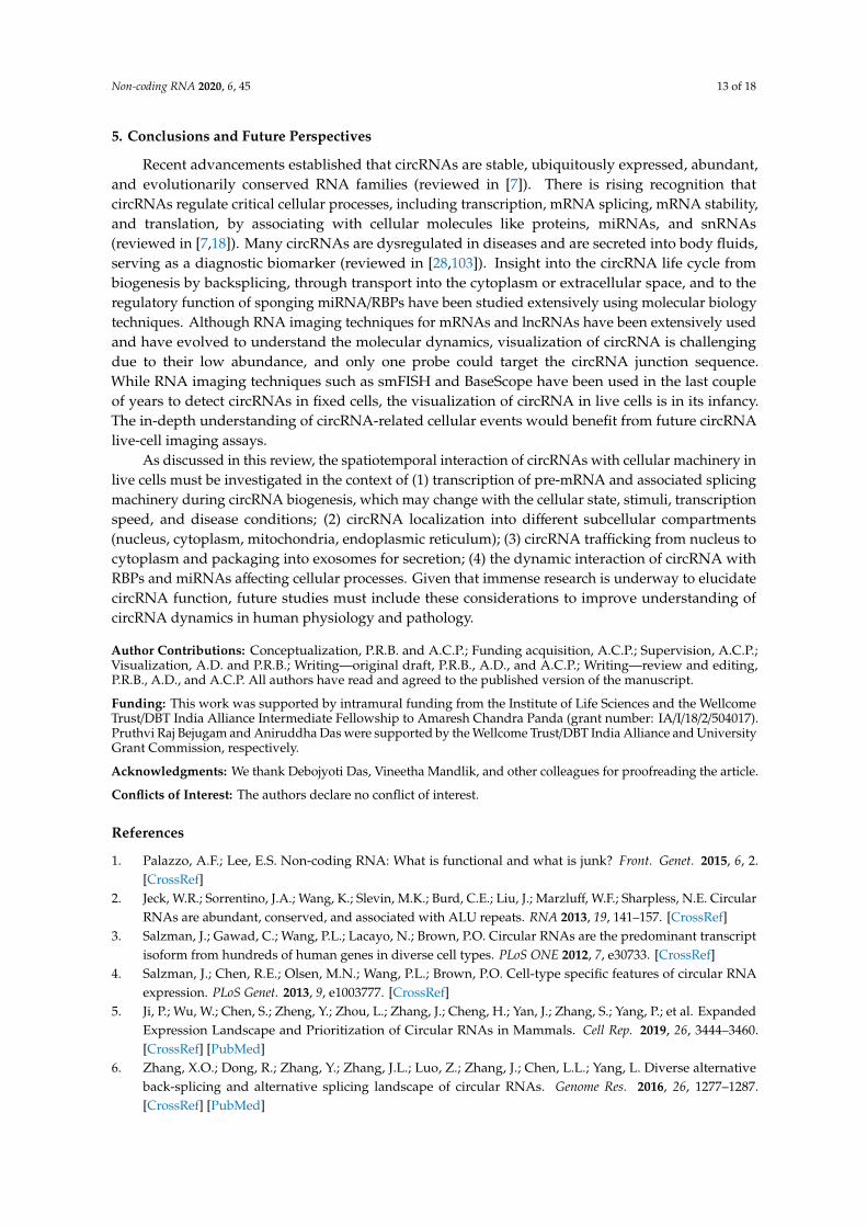
Due to the lack of free ends and their variable size, circRNAs cannot be separated from otherRNAs in gel and detected by molecular biology methods that rely on rapid amplification of cDNA ends(RACE) or RNA seq of poly-A enriched samples. Additionally, the circRNA sequence is the same asthe parental mRNA, making it harder to detect and modulate their expression precisely. Intriguingly,the backsplice junction of circRNA provides a unique opportunity for their detection and manipulation.Several molecular biology approaches have been developed to identify and understand the molecularmechanism of circRNAs in cell physiology (Figure 1).
Non-coding RNA 2020, 6, 45 3 of 18Non-Coding RNA 2020, 6, x 3 of 18
Figure 1. Schematic representation of different methods for circular RNA analysis. (From top clockwise) RNA-seq and circRNA microarrays are used for the genome-wide identification and quantification of circRNAs. RT-PCR of circRNA using the divergent and full-length primers across the backsplice junction, followed by Sanger sequencing, confirms the expression of specific circRNA. In addition, divergent primer PCR can be used for circRNA quantification. Other methods, such as northern blotting, SplintQuant, and NanoString can be used for the quantification of circRNAs. Several databases and web-tools have been developed for the in silico analysis of circRNA expression and function. Loss-of-function analysis for circRNA can also be performed using siRNA/GapmeR for circRNA silencing, while gain-of-function analysis can be achieved by overexpressing the circRNA of interest using a plasmid vector. The circRNA-associated cellular miRNAs and RNA-binding proteins (RBPs) can be analyzed by performing circRNA pulldown assays using antisense oligo-targeting circRNA junctions. Finally, circRNAs can be visualized in the cells using fluorescent-tagged probes targeting the backsplice junction of target circRNA.
2.1. Genome-Wide Analysis of circRNA Expression
The last decade has seen significant advancements in genomic sequencing technologies, including ribosomal RNA depletion strategies, longer sequencing reads, greater sequencing depth, and novel algorithms to map the reads to the genome. These developments have led to the identification of splice variants of mRNAs, non-polyadenylated RNAs, including circRNAs. CircRNAs are identified from RNA-seq reads mapped to the same gene but in the opposite order compared to the linear genomic sequence [2,3,29]. However, the backsplice junction in RNA-seq can be formed not only because of backsplicing but with other mechanisms such as trans-splicing, tandem duplication, or by template switching of reverse transcriptase (reviewed in [29,30]). Various methods, such as poly-A RNA depletion, RNase R treatment, and RPAD (RNase R treatment, polyadenylation, and poly(A)+ RNA depletion), have been developed to enrich circRNA population for downstream
Figure 1. Schematic representation of different methods for circular RNA analysis. (From top clockwise)RNA-seq and circRNA microarrays are used for the genome-wide identification and quantificationof circRNAs. RT-PCR of circRNA using the divergent and full-length primers across the backsplicejunction, followed by Sanger sequencing, confirms the expression of specific circRNA. In addition,divergent primer PCR can be used for circRNA quantification. Other methods, such as northernblotting, SplintQuant, and NanoString can be used for the quantification of circRNAs. Several databasesand web-tools have been developed for the in silico analysis of circRNA expression and function.Loss-of-function analysis for circRNA can also be performed using siRNA/GapmeR for circRNAsilencing, while gain-of-function analysis can be achieved by overexpressing the circRNA of interestusing a plasmid vector. The circRNA-associated cellular miRNAs and RNA-binding proteins (RBPs)can be analyzed by performing circRNA pulldown assays using antisense oligo-targeting circRNAjunctions. Finally, circRNAs can be visualized in the cells using fluorescent-tagged probes targeting thebacksplice junction of target circRNA.
2.1. Genome-Wide Analysis of circRNA Expression
The last decade has seen significant advancements in genomic sequencing technologies, includingribosomal RNA depletion strategies, longer sequencing reads, greater sequencing depth, and novelalgorithms to map the reads to the genome. These developments have led to the identification ofsplice variants of mRNAs, non-polyadenylated RNAs, including circRNAs. CircRNAs are identifiedfrom RNA-seq reads mapped to the same gene but in the opposite order compared to the lineargenomic sequence [2,3,29]. However, the backsplice junction in RNA-seq can be formed not onlybecause of backsplicing but with other mechanisms such as trans-splicing, tandem duplication, or bytemplate switching of reverse transcriptase (reviewed in [29,30]). Various methods, such as poly-ARNA depletion, RNase R treatment, and RPAD (RNase R treatment, polyadenylation, and poly(A)+
Non-coding RNA 2020, 6, 45 4 of 18
RNA depletion), have been developed to enrich circRNA population for downstream analysis [30,31].However, the complete removal of linear RNAs remains a challenge today. Several algorithms have beendeveloped to identify backsplice junctions, full-length sequences, and quantify circRNAs in RNA-seqdata (reviewed in [29]). However, there is no consensus on using the circRNA annotation algorithmto date due to the varying degree of overlap between different algorithms. In addition to RNA-seq,circRNA microarray platforms have been developed to identify and quantify known circRNAs [32].Unlike RNA-seq, circRNA microarray uses probes targeting the backsplice junction of known circRNAs.Since the microarray probes have sequence similarity with linear RNAs, prior treatment of total RNAwith RNase R may improve the specificity of detection. Unlike RNA-seq, circRNA microarray islimited to the number of circRNA probes present in the platform, cannot identify mature circRNAsequences, and cannot measure the ratio of circRNA to linear RNA.
2.2. Validation of circRNA Expression
Since both the high-throughput methods can identify false-positive circRNAs, other molecularmethods like northern blotting, RT-PCR, and Sanger sequencing of PCR products are recommendedfor further validation and quantification of identified circRNAs. Due to the speed and convenience,PCR-based identification and quantification of circRNAs have been widely used. The divergent primersacross backsplice junctions are used for PCR amplification of specific circRNAs [33]. Since divergentprimers do not amplify linear RNAs, RNase R treatment of total RNA is not required for circRNAdetection and quantification by PCR. The Sanger sequencing of the PCR product amplified with divergentprimer can verify the backsplice junction sequence. However, divergent primer PCR cannot reveal thefull-length sequence of circRNA and can amplify multiple circRNAs generated from the same region ofthe gene. The circRNA rolling circle amplification (circRNA-RCA) method was recently developed toamplify the full-length sequence of circRNA using primers on the junction sequence [34]. In addition,the splice variants of circRNA with the same backsplice junction can be identified with circRNA-RCA.However, this method cannot accurately quantitate different splice variants with the same circRNAjunction. Although RT-PCR is the preferred method for circRNA identification and quantification,reverse transcription enzymes are known to interfere with circRNA quantification due to rolling circleamplification and template switching (reviewed in [29,30]). To overcome the RT-PCR bias, a few studieshave used radioactive probes targeting the backsplice junction sequence for the northern blot analysis ofcircRNA [21]. Recently, other methods such as SplintQuant and NanoString have been developed foraccurate quantification of endogenous circRNAs [35,36] (Figure 1).
2.3. Prediction of circRNA Expression and Function
Hundreds of thousands of circRNAs have been identified in human RNA-seq data using variouscircRNA annotation algorithms [17]. Several circRNA databases have been developed, containingvarious circRNA annotations, including tissue-specific expression, evolutionary conservation, diseaseassociation, circRNA–RBP interactions, and circRNA–miRNA interactions (Figure 1). Most databasescontain human circRNAs, while some databases include monkey, mouse, rat, chicken, and yeastcircRNAs. CircRNA databases such as TSCD, CSCD, CIRCpedia, circBase, and circAtlas include theexpression levels of various circRNAs in different cells or tissues [37–41]. Several databases such asCSCD, circAtlas, circInteractome, circNet, starBase, and circFunBase have been developed to predictthe function of circRNAs, including their interactions with miRNAs and RBPs [38,41–45]. A fewdatabases also indicate their protein-coding potential, including circAtlas, circInteractome, circBank,and circRNADb [41,42,46,47]. Although all these databases predict the cellular function of circRNAsby interaction with cellular factors, they need to be validated experimentally.
2.4. Functional Characterization of circRNAs
The function of circRNA is studied with silencing or overexpression in the target cells or tissues.Given that backsplice junction is the unique sequence for circRNA, the siRNAs or GapmeRs are
Non-coding RNA 2020, 6, 45 5 of 18
designed to target the junction sequence. GapmeRs and siRNAs targeting the junction can be manuallydesigned and checked for the specific knockdown of target circRNA without affecting the counterpartlinear RNA. Since siRNAs may act as miRNAs for the counterpart linear RNA due to similar sequences,GapmeR may be better for specific silencing of target circRNAs. Since backsplicing of circRNA isknown to be enhanced by intronic inverted repeats, vectors have been developed with invertedrepeat sequences where the circRNA of interest can be inserted for overexpression in the cells [48].Since overexpression of circRNA from the vectors may generate a lot of linear RNAs with circRNAinserts and circular concatamers, the quantification of circRNA overexpression may be performedupon RNase R treatment [49]. For analyzing the molecular interaction of circRNA with predictedmiRNAs or RBPs, circRNA pulldown assays can be performed using biotin-labeled antisense oligostargeting the junction sequence [50,51]. The pulldown of circRNA with streptavidin magnetic beadsfollowed by RNA-seq or RT-qPCR and mass spectroscopy/western blot analysis can identify associatedmiRNAs and RBPs, respectively [50,51]. Additionally, immunoprecipitation of predicted RBPs ofinterest followed by RNA-seq, microarray, or RT-qPCR can identify circRNAs associated with specificRBPs (reviewed in [12]). Besides, fluorescence in situ hybridization (FISH) assays using probes specificfor backsplice junction sequences have been used in many studies for studying the expression as wellas localization of circRNAs in cells (Figure 1) [52,53]. In this review, we summarize different circRNAvisualization tools and their use in analyzing circRNA expression and function.
3. CircRNA Detection and Quantification by RNA Imaging Techniques
They say “seeing is believing”. The ability to visualize a circRNA in situ not only confirmsthe existence of circRNA in a cell but also gives information about circRNA quantity, localization,and association with other biomolecules, which are critical to finding the function of circRNA incell physiology. Interestingly, a recent study showed that the very well-studied oncogene circRNACDR1as (ciRS-7) is not present in colon cancer cells, but abundantly expressed in stromal cells ofthe tumor microenvironment [54]. This study highlights the importance of circRNA visualization atthe single-cell level for the accurate functional analysis of circRNAs [54]. Unlike linear mRNAs andlincRNA, circRNA detection with imaging techniques is challenging due to the lack of long stretchesof unique sequences except for the circRNA junction. However, the probes targeting the circRNAjunction offer a unique opportunity for circRNA imaging [52]. Given that circRNA function may varydepending on its presence in either cytoplasmic or nuclear compartments, it is crucial to check thelocalization of circRNAs in the cells. Localization in the cytoplasmic compartment would suggest amore miRNA- or RBP-sponging activity, whereas nuclear localization would suggest their potentialregulatory role in transcription or splicing. In this review, we attempt to illustrate the current imagingapproaches for circRNA analyses and discuss potential future developments in circRNA imaging.
Imaging techniques are broadly divided into the fixed-cell and live-cell imaging methods basedon the kind of sample. The goal of both methods, however, is to visualize the target RNA and extractthe relevant information. In a fixed-cell imaging approach, the cells are fixed to keep the cellularbiomolecules intact. In contrast, the live-cell imaging approach usually tracks the dynamic movementof target RNAs over time.
3.1. Fixed-Cell circRNA Imaging
In situ hybridization (ISH) is one of the oldest imaging techniques used to detect the abundanceand precise localization of RNAs in cells [55]. Traditionally, ISH is performed with radioactive probesthat were expensive, time-consuming, and hazardous to human health. The utility of FISH increasedin the late 1980s with the development of fluorescently labeled probes [56]. Since then, fluorescentprobes are used in ISH that bind to nucleic acids based on sequence complementarity. Over time,even though the basic principle of this technique remains the same, there have been advances made inprobe design, choice of the specimen, reduced hybridization times, and increased automation of theassay. Numerous variations in FISH methodologies have now been developed, which are much more
Non-coding RNA 2020, 6, 45 6 of 18
sensitive, specific, and faster than the traditional FISH techniques. In recent years, there has been asignificant advancement in the FISH techniques to reduce the off-target hybridization of the probesand enhance signal quality. Several approaches, such as FISH-STICS, padlock probes, and BaseScopetechniques, have been developed to improve the signal quality. Since circRNA research is a relativelynew field, only a few methods like single-molecule FISH (smFISH) and BaseScope have been used forthe detection and localization of circRNAs.
3.1.1. circRNA Imaging Using smFISH
The traditional FISH technique uses multiple short fluorescently labeled DNA probes that bind tothe RNA. Due to the binding of multiple probes on a single RNA, the signal to noise ratio is enhanced.The image is then analyzed using appropriate fitting algorithms that detect spots in the image. Unliketraditional FISH, smFISH can detect even a single RNA molecule [57]. Since the circRNA junction isthe unique sequence for a particular circRNA, the probes must target the backsplice junction of thecircRNA of interest [52]. Several studies have used FISH or smFISH for quantification and localizationof circRNAs (Figure 2, Table 1). The FISH assay using fluorescent probes targeting the junction ofcircRNA was successfully employed to detect the expression and localization of circRNAs, includingCDR1as (ciRS-7), circHECTD1, circARHGAP10, hsa_circRNA_103809, hsa_circ_0017639, circSAMD4A,circPVT1, circTTN, EIcircEIF3J, EIcircPAIP2, circRHOT1, circFAT1, and circTADA2A [19,21,52,58–68].Furthermore, recent studies employed FISH to confirm the cytoplasmic colocalization of circRNA andtarget miRNAs, including colocalization of miR-7 and ciRS-7 in HEK293 and neuronal cells [21,52,69],miR-135a and ciRS-7 in bladder cancer [70], miR-330-5p and circITCH in cardiomyocytes [71], miR-143and circDLGAP4 in cerebral ischemia [72], circCCDC9 and miR-6792-3p, circRHOBTB3 and miR-654-3pin gastric cancer cells [73,74], circFAM114A2 and miR-762 in urothelial carcinoma of the bladder [75],and circRNA cZNF532 and miR-29a-3p in the cytoplasm of pericytes [76].
Additionally, ImmunoFISH is a variant of the smFISH technique where a combination of FISH andimmunohistochemistry enables the detection of RNAs and proteins in the same sample. In ImmunoFISH,circRNA-associated protein is detected by immunofluorescence, while the target circRNA is detectedwith smFISH. ImmunoFISH confirmed the nucleolar colocalization of PA2G4 with circERBB2 thatregulates ribosomal DNA transcription in gallbladder cancer and the association of circRHOT1 withTIP60 initiates NR2F6 expression in hepatocellular carcinoma [77,78]. Although FISH/smFISH is oneof the most popular circRNA detection techniques, the visualization of low-abundance circRNA usinga single probe targeting the circRNA junction might be challenging.
Non-coding RNA 2020, 6, 45 7 of 18
Non-Coding RNA 2020, 6, x 7 of 18
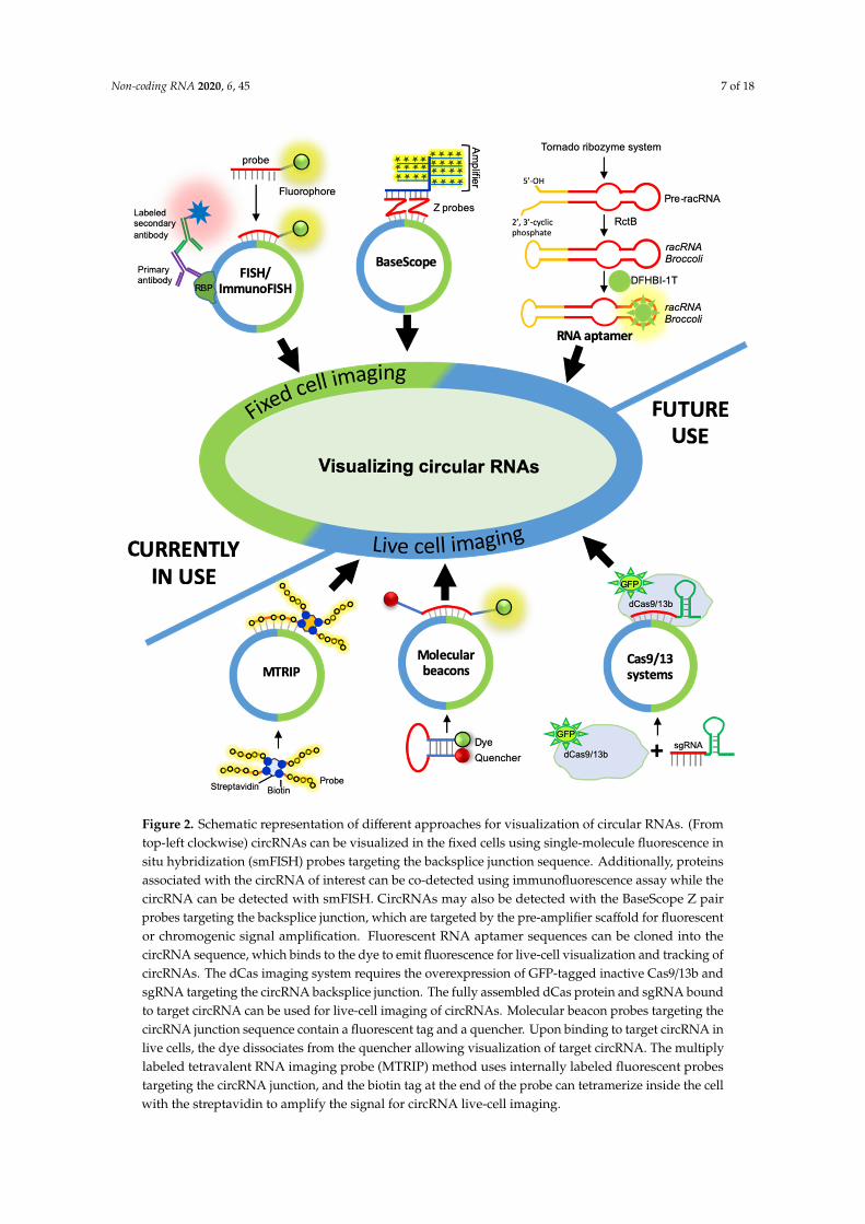
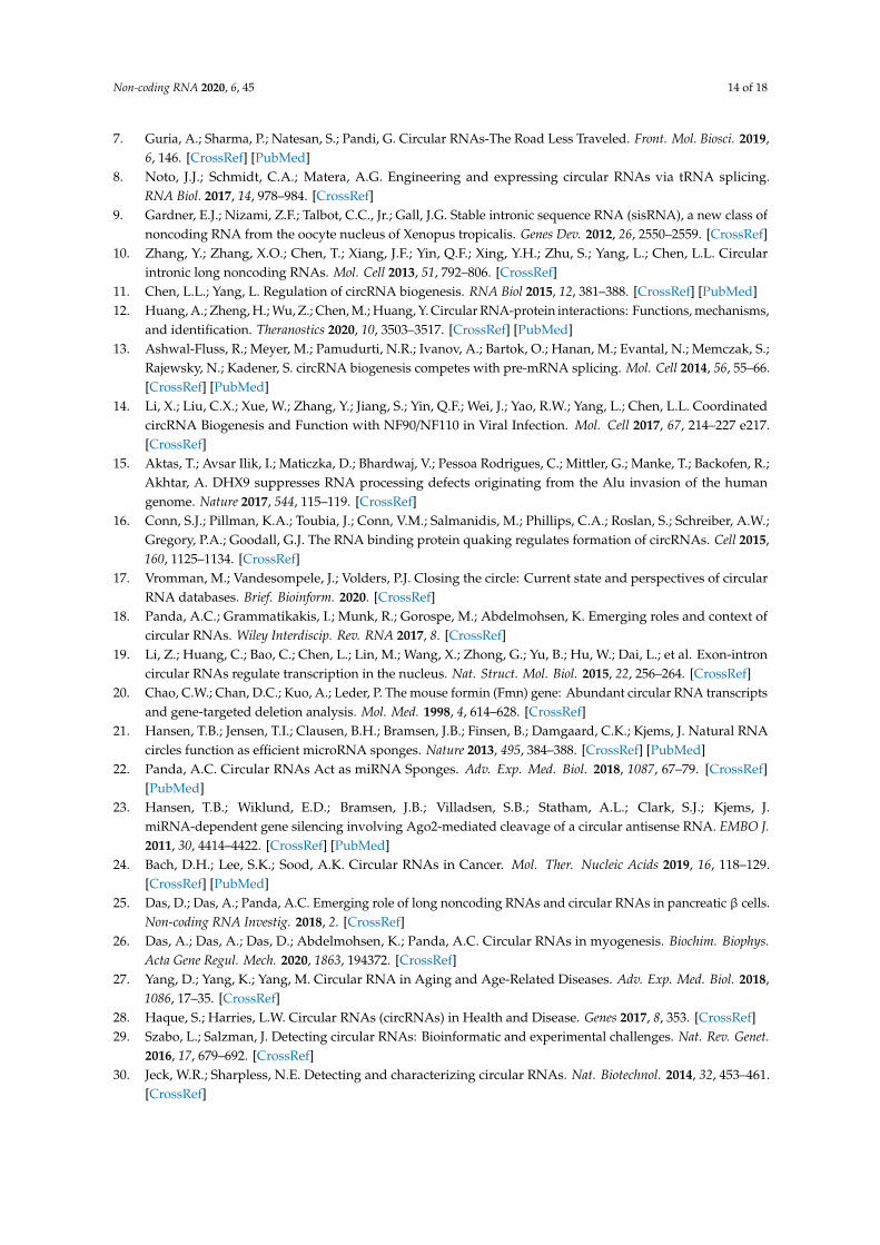
Figure 2. Schematic representation of different approaches for visualization of circular RNAs. (From top-left clockwise) circRNAs can be visualized in the fixed cells using single-molecule fluorescence in situ hybridization (smFISH) probes targeting the backsplice junction sequence. Additionally, proteins associated with the circRNA of interest can be co-detected using immunofluorescence assay while the circRNA can be detected with smFISH. CircRNAs may also be detected with the BaseScope Z pair probes targeting the backsplice junction, which are targeted by the pre-amplifier scaffold for fluorescent or chromogenic signal amplification. Fluorescent RNA aptamer sequences can be cloned into the circRNA sequence, which binds to the dye to emit fluorescence for live-cell visualization and tracking of circRNAs. The dCas imaging system requires the overexpression of GFP-tagged inactive Cas9/13b and sgRNA targeting the circRNA backsplice junction. The fully assembled dCas protein and sgRNA bound to target circRNA can be used for live-cell imaging of circRNAs. Molecular beacon probes targeting the circRNA junction sequence contain a fluorescent tag and a quencher. Upon binding to target circRNA in live cells, the dye dissociates from the quencher allowing visualization of target circRNA. The multiply labeled tetravalent RNA imaging probe (MTRIP) method uses internally labeled fluorescent probes targeting the circRNA junction, and the biotin tag at the end of the probe can tetramerize inside the cell with the streptavidin to amplify the signal for circRNA live-cell imaging.
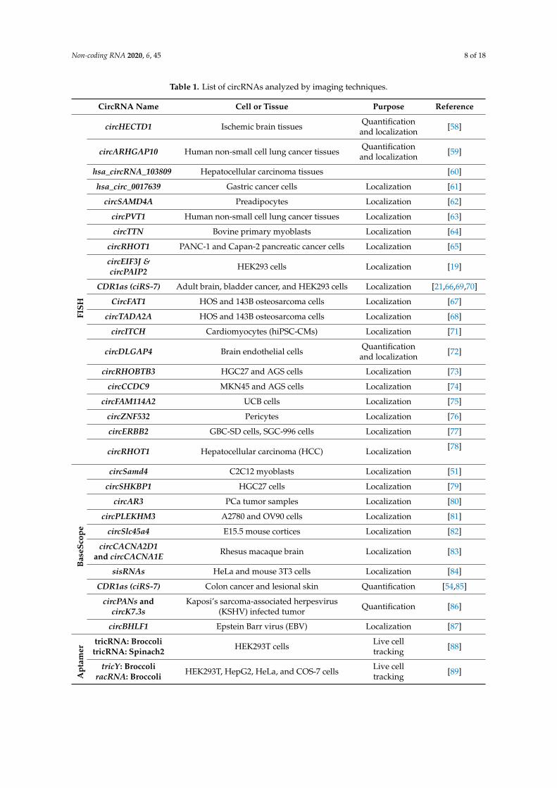
Table 1. List of circRNAs analyzed by imaging techniques.
Figure 2. Schematic representation of different approaches for visualization of circular RNAs. (Fromtop-left clockwise) circRNAs can be visualized in the fixed cells using single-molecule fluorescence insitu hybridization (smFISH) probes targeting the backsplice junction sequence. Additionally, proteinsassociated with the circRNA of interest can be co-detected using immunofluorescence assay while thecircRNA can be detected with smFISH. CircRNAs may also be detected with the BaseScope Z pairprobes targeting the backsplice junction, which are targeted by the pre-amplifier scaffold for fluorescentor chromogenic signal amplification. Fluorescent RNA aptamer sequences can be cloned into thecircRNA sequence, which binds to the dye to emit fluorescence for live-cell visualization and tracking ofcircRNAs. The dCas imaging system requires the overexpression of GFP-tagged inactive Cas9/13b andsgRNA targeting the circRNA backsplice junction. The fully assembled dCas protein and sgRNA boundto target circRNA can be used for live-cell imaging of circRNAs. Molecular beacon probes targeting thecircRNA junction sequence contain a fluorescent tag and a quencher. Upon binding to target circRNA inlive cells, the dye dissociates from the quencher allowing visualization of target circRNA. The multiplylabeled tetravalent RNA imaging probe (MTRIP) method uses internally labeled fluorescent probestargeting the circRNA junction, and the biotin tag at the end of the probe can tetramerize inside the cellwith the streptavidin to amplify the signal for circRNA live-cell imaging.
Non-coding RNA 2020, 6, 45 8 of 18
Table 1. List of circRNAs analyzed by imaging techniques.
CircRNA Name Cell or Tissue Purpose Reference
FISH
circHECTD1 Ischemic brain tissues Quantificationand localization [58]
circARHGAP10 Human non-small cell lung cancer tissues Quantificationand localization [59]
hsa_circRNA_103809 Hepatocellular carcinoma tissues [60]
hsa_circ_0017639 Gastric cancer cells Localization [61]
circSAMD4A Preadipocytes Localization [62]
circPVT1 Human non-small cell lung cancer tissues Localization [63]
circTTN Bovine primary myoblasts Localization [64]
circRHOT1 PANC-1 and Capan-2 pancreatic cancer cells Localization [65]
circEIF3J &circPAIP2 HEK293 cells Localization [19]
CDR1as (ciRS-7) Adult brain, bladder cancer, and HEK293 cells Localization [21,66,69,70]
CircFAT1 HOS and 143B osteosarcoma cells Localization [67]
circTADA2A HOS and 143B osteosarcoma cells Localization [68]
circITCH Cardiomyocytes (hiPSC-CMs) Localization [71]
circDLGAP4 Brain endothelial cells Quantificationand localization [72]
circRHOBTB3 HGC27 and AGS cells Localization [73]
circCCDC9 MKN45 and AGS cells Localization [74]
circFAM114A2 UCB cells Localization [75]
circZNF532 Pericytes Localization [76]
circERBB2 GBC-SD cells, SGC-996 cells Localization [77]
circRHOT1 Hepatocellular carcinoma (HCC) Localization [78]
Bas
eSco
pe
circSamd4 C2C12 myoblasts Localization [51]
circSHKBP1 HGC27 cells Localization [79]
circAR3 PCa tumor samples Localization [80]
circPLEKHM3 A2780 and OV90 cells Localization [81]
circSlc45a4 E15.5 mouse cortices Localization [82]
circCACNA2D1and circCACNA1E Rhesus macaque brain Localization [83]
sisRNAs HeLa and mouse 3T3 cells Localization [84]
CDR1as (ciRS-7) Colon cancer and lesional skin Quantification [54,85]
circPANs andcircK7.3s
Kaposi’s sarcoma-associated herpesvirus(KSHV) infected tumor Quantification [86]
circBHLF1 Epstein Barr virus (EBV) Localization [87]
Apt
amer
tricRNA: BroccolitricRNA: Spinach2 HEK293T cells Live cell
tracking [88]
tricY: BroccoliracRNA: Broccoli HEK293T, HepG2, HeLa, and COS-7 cells Live cell
tracking [89]
Non-coding RNA 2020, 6, 45 9 of 18
3.1.2. circRNA Imaging Using BaseScope Assay
RNAscope technology is a recently developed version of the ISH technique that detects mRNAsand ncRNAs with a length of more than 300 nucleotides [90]. With its unique proprietary probedesign that can amplify target-specific signals and not the background noise, RNAscope ensures ahigher specificity as compared to the ISH techniques. It is also combined with immunohistochemistryto ensure the simultaneous detection of RNAs and proteins [91]. BaseScope assay is the advancedversion of RNAscope, which is generally used for sequence detection of short target sequences of50–300 bp (snoRNA, circRNA, miRNA, partially degraded RNA, and transiently expressed RNAs) using1–3 Z pair probes. Since the circRNA junction sequence is the unique sequence, BaseScope can detectcircRNA with a ZZ pair probe targeting the backsplice junction sequence (Figure 2). Hybridization ofthe signal pre-amplifiers using branched DNA technology and simultaneous signal amplification withchromogenic enzymes or florescent probes allows for the visualization of low-abundance circRNAs.Recently, the BaseScope assay was used in C2C12 muscle cells to visualize circSamd4 that regulatesmyogenesis by sponging PUR proteins [51]. Similarly, several studies successfully employed aBaseScope assay for circRNA detection and localization in different cellular systems, includingcircSHKBP1 in gastric cancer tissues [79], circAR3 expression in prostate cancers [80], circPLEKHM3 inthe tumor samples [81], circSlc45a4 in neuronal differentiation [82], circCACNA2D1 and circCACNA1Ein rhesus macaque brains [83], sisRNAs in HeLa and mouse 3T3 cells [84], and ciRS-7 in colon cancerand lesional skin [54,85]. Furthermore, a few studies used the BaseScope assay to detect circRNAsexpressed during viral infection, including circPANs and circK7.3s in Kaposi’s sarcoma-associatedherpesvirus (KSHV) replication and circBHLF1 in Epstein Barr virus (EBV) infection [86,87].
3.2. Live-Cell Imaging of circRNAs
Fixed-cell imaging enables the observation of RNA only at a single time point in the cellular RNApathway. It is generally useful for observing RNA localization. However, unlike this static observation,live-cell imaging adds a dynamic dimension that can track the entire RNA lifecycle. Live-cell RNAimaging captures many important events, such as RNA biosynthesis, splicing events, transport,function, and decay (reviewed in [92]). Unlike fixed-cell imaging, where cells become dead duringfixation, live-cell imaging preserves the physiological condition of the cell. Tracking probes can eitherbe endogenous or exogenous. Plasmid-based systems that express both the RNA and a fluorescent tag,such as the MS2-GFP system, are used that can bind specifically to the RNA of interest. Furthermore,multiple MS2-GFP binding domains have also been inserted in the RNA to enhance the signal intensity.Some other systems, such as PUM-HD, PP7, and Pepper, have also been developed to track endogenousmRNAs (reviewed in [92,93]). More recently, fluorescent proteins (GFPs) have been replaced by Haloand SNP tags, which can also be coupled with organic dyes (reviewed in [94]). Plasmid-derived probes(endogenous systems) offer flexibility; however, they do have limitations on the cell types that allowtransfection of these systems and the copy numbers generated, possibly changing the fundamentalstoichiometry of RNA expression. This led to the development of exogenous fluorophore-labeledantisense probes. Some of these are single-label probes (aptamers, molecular beacons) and some havemultivalent labels (multiply labeled tetravalent RNA imaging probes (MTRIPs)). Although the RNAaptamer system has only been used for live-cell imaging of circRNAs, we propose the potential uses ofother live-cell approaches that can be used for circRNA imaging.
3.2.1. Fluorescent RNA Aptamers
RNA aptamer-based imaging systems require an aptamer sequence fused with the transcriptof interest that can only be visualized when the dye is captured by the aptamer structure withno background issue during RNA tracking experiments (reviewed in [92]). The first smallmolecule-based fluorophore-binding aptamer (Spinach) system mimicked the structure of GFP.Since then, other aptamers such as Broccoli and Spinach 2 have been developed. More recently, aptamer
Non-coding RNA 2020, 6, 45 10 of 18
dye systems such as Broccoli-DFHB1, and Corn-DFHO have been developed, showing higher thermalstability and improved photostability (reviewed in [92]). One potential limitation of these aptamersis the presence of G-quadruplexes that is often critical for fluorophore binding and rigidification ofthe aptamer. Newer aptamer-based systems, such as SRB-2, RhoBAST, and Gemini-561, do not havethis structure and have been shown to have increased photostability (reviewed in [92,93]). In contrastto these aptamer-based systems, aptamer systems inspired by naturally occurring riboswitches havealso been developed. Riboglow has been developed, which makes use of the covalent conjugationof cobalamine, a metabolite that binds to the riboswitches. The binding of cobalamine to an RNAriboswitch results in the separation of the quencher and fluorophore than can turn on the fluorescencefor visualization (reviewed in [93]).
Although several aptamer–dye systems have been developed to visualize linear RNAs in livecells, only a few studies have used RNA aptamers for visualizing circRNAs. Given the tRNAintrons are spliced to form RNA circles known as tricRNA, a recent study engineered aptamerscontaining circRNAs by inserting sequences corresponding to Broccoli and Spinach 2 RNA aptamersin the tRNA intron [8,88]. Interestingly, the chimeric circRNA with these aptamers bind to thechromophores, like DFHBI-1T, resulting in green fluorescence that mimics GFP with very lowbackground fluorescence [88]. Furthermore, an artificial autocatalytic tornado system was developedfor the overexpression of ribozyme-assisted circRNA (racRNA) with Broccoli aptamers and visualizedin live cells using DFHBI-1T (Figure 2) [89].
3.2.2. Cas-Derived Fluorescent Protein
Although the CRISPR/Cas system is well known for gene silencing and genome editing,fluorescently labeled modified Cas proteins have recently been used for live-cell tracking of targetRNA with high specificity (reviewed in [95]). Several systems, such as dCas13, dCas9, dPspCas13b,have recently been developed for real-time RNA imaging where the Cas protein homologs are fusedwith fluorescent proteins such as EGFP [95–97]. With a discrimination ability of even a single basemismatch, CRISPR/Cas technology can detect RNAs with very high specificity. These CRISPR-basedsystems are compatible with both the fluorescent-based and the chromogenic method of detection.Although the CRISPR/Cas system is widely used for gene silencing and editing, a recent studyused the dCas9-mCherry-based system to image the GAPDH mRNA in live cells [97]. Furthermore,dLwaCas13a and dCas13d systems have been used for real-time imaging of the RNA in living cells [96].We hypothesize that single-guide RNA (sgRNA) targeting the backsplice junction sequence coupledwith the fluorescent-labeled dCas9/dCas13d may be used for live-cell imaging of circRNAs (Figure 2).
3.2.3. Molecular Beacons
The molecular beacon (MB) was first developed in 1996 for nucleic acid amplification assays aswell as RNA tracking in live cells [98]. Unlike fluorescent probes used in FISH, molecular beacons use aprobe complementary to target RNA labeled with a fluorophore and a quencher [99]. Molecular beaconsform a stem–loop structure in the native state where the fluorophore is quenched by the quencher,giving no fluorescence. Upon hybridization to the target RNA, the probes become fluorescent as thequencher and the fluorescent dye move apart due to the conformational reorganization. Molecularbeacons are promising probes for real-time monitoring of RNA dynamics within living cells and tissues(reviewed in [100]). Although MBs have not yet been used for circRNA imaging, we believe thatthe MB probe targeting the backsplice junction sequence of circRNA may enable live cell tracking ofcircRNAs of interest (Figure 2).
3.2.4. Multiply Labeled Tetravalent RNA Imaging Probes
Multiply labeled tetravalent RNA imaging probes (MTRIPs) can be used for RNA imaging withhigh specificity. To increase the sensitivity of nucleic acids based on exogenous probes, Santangelo et al.developed MTRIPs by conjugating two to four fluorophores to a single nucleic acid. These nucleic
Non-coding RNA 2020, 6, 45 11 of 18
acids (also referred to as ligands) were combined using streptavidin such that the probe brightness wasincreased four fold [101,102]. One key advantage of using MTRIPs is that multiple probes with multiplefluorophores per target RNA achieve several-fold higher brightness as compared to the traditionalmethod of using fluorescent protein-based RNA detection. We hypothesize that MTRIPs targeting thecircRNA junction could help in specific detection and live-cell imaging of circRNA (Figure 2).
4. Limitations and Additional Considerations for circRNA Imaging Techniques
CircRNAs regulate cell physiology by acting as sponges for microRNAs, a decoy for RBPs,competitors of splicing regulators, and substrates for protein translation (reviewed in [18]). AlthoughcircRNAs are believed to be localized into the cytoplasm and regulate the function of RBPs andmiRNAs, several circRNAs have been reported to localize into the nucleus to regulate nuclear events,including transcription and RNA splicing [10,19,69]. We have discussed recent imaging techniquesfor identification, quantification, and localization of circRNA in the cells (Figure 2, Table 1). All thesemethods for visualization of circRNAs have their strengths and weaknesses, which need to beconsidered during experimental design (Table 2).
Given that circRNAs are generated from pre-mRNAs, the body of the circRNAs resembles thecounterpart linear RNAs, limiting the specific probe to be complementary only to the junction sequenceof circRNA. As discussed above, smFISH employs a fluorescent-labeled probe that is complementaryto the circRNA junction sequence and useful for detecting abundant circRNAs (Table 1). It might bechallenging to detect low-abundance circRNAs with smFISH. Since most circRNAs have low abundance,with a few copies per cell, the BaseScope assay is very useful in circRNA imaging. The smFISH andBaseScope technique for circRNA imaging can also be multiplexed to detect multiple target RNAs andproteins using probes/antibodies labeled with different fluorophores or chromophores in a single assay.However, both smFISH and BaseScope assays are limited to fixed cells only.
Live-cell imaging of circRNAs using RNA aptamers has only been reported recently, utilizingthe tricRNAs and racRNAs [8,88,89]. This system holds promise for visualizing circRNAs with otherRNAs and labeled proteins. However, additional research is required to determine the size of thecircRNA that can be generated with the RNA aptamers using the tRNA or ribozyme-assisted splicingmachinery. Additionally, the insertion of RNA aptamer sequences into the circRNA of interest mayalter the aptamer folding and perturb circRNA interaction with RBPs and miRNAs, depending on thesurrounding sequences, temperature, and cellular salt concentration. Furthermore, the overexpressionof aptamer-containing circRNA in the cells may leave some unprocessed linear RNA with the aptamersequence, which could interfere with circRNA tracking.
Furthermore, the inactive Cas-13 system with a fluorescent tag and the sgRNA targeting thecircRNA junction could help live-cell tracking of circRNAs [96,97]. However, the gRNA specificityand the localization of the Cas protein needs to be verified for accurately tracking the target circRNA.Molecular beacon probes conjugated with a fluorophore and quencher could target the junctionsequence of circRNA [99,100]. They can be introduced into cells where binding of the probe to thecircRNA separates the fluorophore and quencher, allowing the visualization of the circRNA in realtime. Like smFISH, this assay could be useful for the visualization of abundant circRNAs in fixedcells and live cells. The MTRIP method discussed here could use a multiply labeled fluorescent probeagainst the circRNA junction, and the streptavidin core can be introduced into the cell for live-cellimaging of target circRNA [101,102]. Finally, the success of smFISH, BaseScope, molecular beacons,the Cas-13 system, and MTRIP assays depends on the accessibility of the circRNA junction sequence,which might be hidden or bound with RBPs, preventing the probe binding.
Non-coding RNA 2020, 6, 45 12 of 18
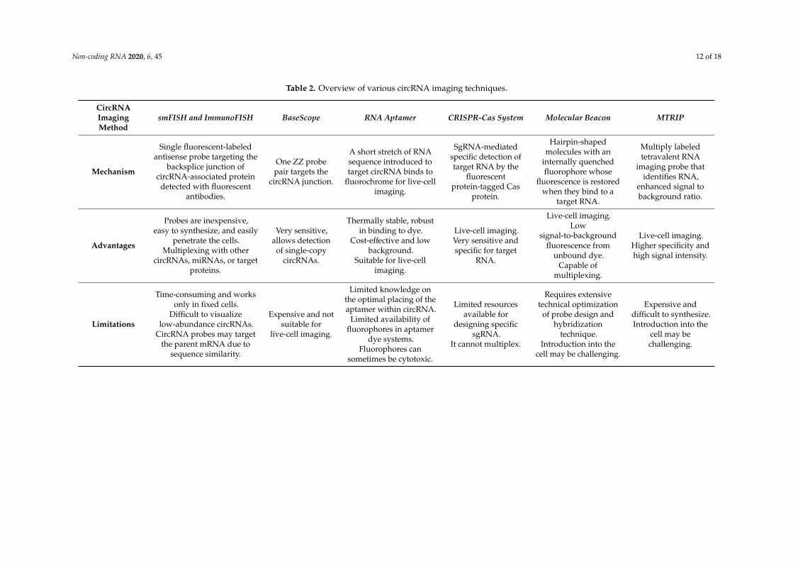
Table 2. Overview of various circRNA imaging techniques.
CircRNAImagingMethod
smFISH and ImmunoFISH BaseScope RNA Aptamer CRISPR-Cas System Molecular Beacon MTRIP
Mechanism
Single fluorescent-labeledantisense probe targeting the
backsplice junction ofcircRNA-associated protein
detected with fluorescentantibodies.
One ZZ probepair targets the
circRNA junction.
A short stretch of RNAsequence introduced totarget circRNA binds to
fluorochrome for live-cellimaging.
SgRNA-mediatedspecific detection oftarget RNA by the
fluorescentprotein-tagged Cas
protein.
Hairpin-shapedmolecules with an
internally quenchedfluorophore whose
fluorescence is restoredwhen they bind to a
target RNA.
Multiply labeledtetravalent RNA
imaging probe thatidentifies RNA,
enhanced signal tobackground ratio.
Advantages
Probes are inexpensive,easy to synthesize, and easily
penetrate the cells.Multiplexing with other
circRNAs, miRNAs, or targetproteins.
Very sensitive,allows detection
of single-copycircRNAs.
Thermally stable, robustin binding to dye.
Cost-effective and lowbackground.
Suitable for live-cellimaging.
Live-cell imaging.Very sensitive andspecific for target
RNA.
Live-cell imaging.Low
signal-to-backgroundfluorescence from
unbound dye.Capable of
multiplexing.
Live-cell imaging.Higher specificity andhigh signal intensity.
Limitations
Time-consuming and worksonly in fixed cells.
Difficult to visualizelow-abundance circRNAs.
CircRNA probes may targetthe parent mRNA due to
sequence similarity.
Expensive and notsuitable for
live-cell imaging.
Limited knowledge onthe optimal placing of theaptamer within circRNA.
Limited availability offluorophores in aptamer
dye systems.Fluorophores can
sometimes be cytotoxic.
Limited resourcesavailable for
designing specificsgRNA.
It cannot multiplex.
Requires extensivetechnical optimization
of probe design andhybridization
technique.Introduction into the
cell may be challenging.
Expensive anddifficult to synthesize.Introduction into the
cell may bechallenging.
Non-coding RNA 2020, 6, 45 13 of 18
5. Conclusions and Future Perspectives
Recent advancements established that circRNAs are stable, ubiquitously expressed, abundant,and evolutionarily conserved RNA families (reviewed in [7]). There is rising recognition thatcircRNAs regulate critical cellular processes, including transcription, mRNA splicing, mRNA stability,and translation, by associating with cellular molecules like proteins, miRNAs, and snRNAs(reviewed in [7,18]). Many circRNAs are dysregulated in diseases and are secreted into body fluids,serving as a diagnostic biomarker (reviewed in [28,103]). Insight into the circRNA life cycle frombiogenesis by backsplicing, through transport into the cytoplasm or extracellular space, and to theregulatory function of sponging miRNA/RBPs have been studied extensively using molecular biologytechniques. Although RNA imaging techniques for mRNAs and lncRNAs have been extensively usedand have evolved to understand the molecular dynamics, visualization of circRNA is challengingdue to their low abundance, and only one probe could target the circRNA junction sequence.While RNA imaging techniques such as smFISH and BaseScope have been used in the last coupleof years to detect circRNAs in fixed cells, the visualization of circRNA in live cells is in its infancy.The in-depth understanding of circRNA-related cellular events would benefit from future circRNAlive-cell imaging assays.
As discussed in this review, the spatiotemporal interaction of circRNAs with cellular machinery inlive cells must be investigated in the context of (1) transcription of pre-mRNA and associated splicingmachinery during circRNA biogenesis, which may change with the cellular state, stimuli, transcriptionspeed, and disease conditions; (2) circRNA localization into different subcellular compartments(nucleus, cytoplasm, mitochondria, endoplasmic reticulum); (3) circRNA trafficking from nucleus tocytoplasm and packaging into exosomes for secretion; (4) the dynamic interaction of circRNA withRBPs and miRNAs affecting cellular processes. Given that immense research is underway to elucidatecircRNA function, future studies must include these considerations to improve understanding ofcircRNA dynamics in human physiology and pathology.
Author Contributions: Conceptualization, P.R.B. and A.C.P.; Funding acquisition, A.C.P.; Supervision, A.C.P.;Visualization, A.D. and P.R.B.; Writing—original draft, P.R.B., A.D., and A.C.P.; Writing—review and editing,P.R.B., A.D., and A.C.P. All authors have read and agreed to the published version of the manuscript.
Funding: This work was supported by intramural funding from the Institute of Life Sciences and the WellcomeTrust/DBT India Alliance Intermediate Fellowship to Amaresh Chandra Panda (grant number: IA/I/18/2/504017).Pruthvi Raj Bejugam and Aniruddha Das were supported by the Wellcome Trust/DBT India Alliance and UniversityGrant Commission, respectively.
Acknowledgments: We thank Debojyoti Das, Vineetha Mandlik, and other colleagues for proofreading the article.
Conflicts of Interest: The authors declare no conflict of interest.
References
1. Palazzo, A.F.; Lee, E.S. Non-coding RNA: What is functional and what is junk? Front. Genet. 2015, 6, 2.[CrossRef]
2. Jeck, W.R.; Sorrentino, J.A.; Wang, K.; Slevin, M.K.; Burd, C.E.; Liu, J.; Marzluff, W.F.; Sharpless, N.E. CircularRNAs are abundant, conserved, and associated with ALU repeats. RNA 2013, 19, 141–157. [CrossRef]
3. Salzman, J.; Gawad, C.; Wang, P.L.; Lacayo, N.; Brown, P.O. Circular RNAs are the predominant transcriptisoform from hundreds of human genes in diverse cell types. PLoS ONE 2012, 7, e30733. [CrossRef]
4. Salzman, J.; Chen, R.E.; Olsen, M.N.; Wang, P.L.; Brown, P.O. Cell-type specific features of circular RNAexpression. PLoS Genet. 2013, 9, e1003777. [CrossRef]
5. Ji, P.; Wu, W.; Chen, S.; Zheng, Y.; Zhou, L.; Zhang, J.; Cheng, H.; Yan, J.; Zhang, S.; Yang, P.; et al. ExpandedExpression Landscape and Prioritization of Circular RNAs in Mammals. Cell Rep. 2019, 26, 3444–3460.[CrossRef] [PubMed]
6. Zhang, X.O.; Dong, R.; Zhang, Y.; Zhang, J.L.; Luo, Z.; Zhang, J.; Chen, L.L.; Yang, L. Diverse alternativeback-splicing and alternative splicing landscape of circular RNAs. Genome Res. 2016, 26, 1277–1287.[CrossRef] [PubMed]
Non-coding RNA 2020, 6, 45 14 of 18
7. Guria, A.; Sharma, P.; Natesan, S.; Pandi, G. Circular RNAs-The Road Less Traveled. Front. Mol. Biosci. 2019,6, 146. [CrossRef] [PubMed]
8. Noto, J.J.; Schmidt, C.A.; Matera, A.G. Engineering and expressing circular RNAs via tRNA splicing.RNA Biol. 2017, 14, 978–984. [CrossRef]
9. Gardner, E.J.; Nizami, Z.F.; Talbot, C.C., Jr.; Gall, J.G. Stable intronic sequence RNA (sisRNA), a new class ofnoncoding RNA from the oocyte nucleus of Xenopus tropicalis. Genes Dev. 2012, 26, 2550–2559. [CrossRef]
10. Zhang, Y.; Zhang, X.O.; Chen, T.; Xiang, J.F.; Yin, Q.F.; Xing, Y.H.; Zhu, S.; Yang, L.; Chen, L.L. Circularintronic long noncoding RNAs. Mol. Cell 2013, 51, 792–806. [CrossRef]
11. Chen, L.L.; Yang, L. Regulation of circRNA biogenesis. RNA Biol 2015, 12, 381–388. [CrossRef] [PubMed]12. Huang, A.; Zheng, H.; Wu, Z.; Chen, M.; Huang, Y. Circular RNA-protein interactions: Functions, mechanisms,
and identification. Theranostics 2020, 10, 3503–3517. [CrossRef] [PubMed]13. Ashwal-Fluss, R.; Meyer, M.; Pamudurti, N.R.; Ivanov, A.; Bartok, O.; Hanan, M.; Evantal, N.; Memczak, S.;
Rajewsky, N.; Kadener, S. circRNA biogenesis competes with pre-mRNA splicing. Mol. Cell 2014, 56, 55–66.[CrossRef] [PubMed]
14. Li, X.; Liu, C.X.; Xue, W.; Zhang, Y.; Jiang, S.; Yin, Q.F.; Wei, J.; Yao, R.W.; Yang, L.; Chen, L.L. CoordinatedcircRNA Biogenesis and Function with NF90/NF110 in Viral Infection. Mol. Cell 2017, 67, 214–227 e217.[CrossRef]
15. Aktas, T.; Avsar Ilik, I.; Maticzka, D.; Bhardwaj, V.; Pessoa Rodrigues, C.; Mittler, G.; Manke, T.; Backofen, R.;Akhtar, A. DHX9 suppresses RNA processing defects originating from the Alu invasion of the humangenome. Nature 2017, 544, 115–119. [CrossRef]
16. Conn, S.J.; Pillman, K.A.; Toubia, J.; Conn, V.M.; Salmanidis, M.; Phillips, C.A.; Roslan, S.; Schreiber, A.W.;Gregory, P.A.; Goodall, G.J. The RNA binding protein quaking regulates formation of circRNAs. Cell 2015,160, 1125–1134. [CrossRef]
17. Vromman, M.; Vandesompele, J.; Volders, P.J. Closing the circle: Current state and perspectives of circularRNA databases. Brief. Bioinform. 2020. [CrossRef]
18. Panda, A.C.; Grammatikakis, I.; Munk, R.; Gorospe, M.; Abdelmohsen, K. Emerging roles and context ofcircular RNAs. Wiley Interdiscip. Rev. RNA 2017, 8. [CrossRef]
19. Li, Z.; Huang, C.; Bao, C.; Chen, L.; Lin, M.; Wang, X.; Zhong, G.; Yu, B.; Hu, W.; Dai, L.; et al. Exon-introncircular RNAs regulate transcription in the nucleus. Nat. Struct. Mol. Biol. 2015, 22, 256–264. [CrossRef]
20. Chao, C.W.; Chan, D.C.; Kuo, A.; Leder, P. The mouse formin (Fmn) gene: Abundant circular RNA transcriptsand gene-targeted deletion analysis. Mol. Med. 1998, 4, 614–628. [CrossRef]
21. Hansen, T.B.; Jensen, T.I.; Clausen, B.H.; Bramsen, J.B.; Finsen, B.; Damgaard, C.K.; Kjems, J. Natural RNAcircles function as efficient microRNA sponges. Nature 2013, 495, 384–388. [CrossRef] [PubMed]
22. Panda, A.C. Circular RNAs Act as miRNA Sponges. Adv. Exp. Med. Biol. 2018, 1087, 67–79. [CrossRef][PubMed]
23. Hansen, T.B.; Wiklund, E.D.; Bramsen, J.B.; Villadsen, S.B.; Statham, A.L.; Clark, S.J.; Kjems, J.miRNA-dependent gene silencing involving Ago2-mediated cleavage of a circular antisense RNA. EMBO J.2011, 30, 4414–4422. [CrossRef] [PubMed]
24. Bach, D.H.; Lee, S.K.; Sood, A.K. Circular RNAs in Cancer. Mol. Ther. Nucleic Acids 2019, 16, 118–129.[CrossRef] [PubMed]
25. Das, D.; Das, A.; Panda, A.C. Emerging role of long noncoding RNAs and circular RNAs in pancreatic β cells.Non-coding RNA Investig. 2018, 2. [CrossRef]
26. Das, A.; Das, A.; Das, D.; Abdelmohsen, K.; Panda, A.C. Circular RNAs in myogenesis. Biochim. Biophys.Acta Gene Regul. Mech. 2020, 1863, 194372. [CrossRef]
27. Yang, D.; Yang, K.; Yang, M. Circular RNA in Aging and Age-Related Diseases. Adv. Exp. Med. Biol. 2018,1086, 17–35. [CrossRef]
28. Haque, S.; Harries, L.W. Circular RNAs (circRNAs) in Health and Disease. Genes 2017, 8, 353. [CrossRef]29. Szabo, L.; Salzman, J. Detecting circular RNAs: Bioinformatic and experimental challenges. Nat. Rev. Genet.
2016, 17, 679–692. [CrossRef]30. Jeck, W.R.; Sharpless, N.E. Detecting and characterizing circular RNAs. Nat. Biotechnol. 2014, 32, 453–461.
[CrossRef]
Non-coding RNA 2020, 6, 45 15 of 18
31. Pandey, P.R.; Rout, P.K.; Das, A.; Gorospe, M.; Panda, A.C. RPAD (RNase R treatment, polyadenylation,and poly(A)+ RNA depletion) method to isolate highly pure circular RNA. Methods 2019, 155, 41–48.[CrossRef] [PubMed]
32. Li, S.; Teng, S.; Xu, J.; Su, G.; Zhang, Y.; Zhao, J.; Zhang, S.; Wang, H.; Qin, W.; Lu, Z.J.; et al. Microarray is anefficient tool for circRNA profiling. Brief. Bioinform. 2019, 20, 1420–1433. [CrossRef] [PubMed]
33. Panda, A.C.; Gorospe, M. Detection and Analysis of Circular RNAs by RT-PCR. Bio Protoc. 2018, 8. [CrossRef][PubMed]
34. Das, A.; Rout, P.K.; Gorospe, M.; Panda, A.C. Rolling Circle cDNA Synthesis Uncovers Circular RNA SpliceVariants. Int. J. Mol. Sci. 2019, 20, 3988. [CrossRef] [PubMed]
35. Conn, V.; Conn, S.J. SplintQuant: A method for accurately quantifying circular RNA transcript abundancewithout reverse transcription bias. RNA 2019, 25, 1202–1210. [CrossRef] [PubMed]
36. Dahl, M.; Daugaard, I.; Andersen, M.S.; Hansen, T.B.; Gronbaek, K.; Kjems, J.; Kristensen, L.S. Enzyme-freedigital counting of endogenous circular RNA molecules in B-cell malignancies. Lab. Investig. 2018, 98,1657–1669. [CrossRef]
37. Xia, S.; Feng, J.; Lei, L.; Hu, J.; Xia, L.; Wang, J.; Xiang, Y.; Liu, L.; Zhong, S.; Han, L.; et al. Comprehensivecharacterization of tissue-specific circular RNAs in the human and mouse genomes. Brief. Bioinform. 2017,18, 984–992. [CrossRef]
38. Xia, S.; Feng, J.; Chen, K.; Ma, Y.; Gong, J.; Cai, F.; Jin, Y.; Gao, Y.; Xia, L.; Chang, H.; et al. CSCD: A databasefor cancer-specific circular RNAs. Nucleic Acids Res. 2018, 46, D925–D929. [CrossRef]
39. Dong, R.; Ma, X.K.; Li, G.W.; Yang, L. CIRCpedia v2: An Updated Database for Comprehensive Circular RNAAnnotation and Expression Comparison. Genomics Proteomics Bioinformatics 2018, 16, 226–233. [CrossRef]
40. Glazar, P.; Papavasileiou, P.; Rajewsky, N. circBase: A database for circular RNAs. RNA 2014, 20, 1666–1670.[CrossRef]
41. Wu, W.; Ji, P.; Zhao, F. CircAtlas: An integrated resource of one million highly accurate circular RNAs from1070 vertebrate transcriptomes. Genome Biol. 2020, 21, 101. [CrossRef] [PubMed]
42. Dudekula, D.B.; Panda, A.C.; Grammatikakis, I.; De, S.; Abdelmohsen, K.; Gorospe, M. CircInteractome:A web tool for exploring circular RNAs and their interacting proteins and microRNAs. RNA Biol. 2016, 13,34–42. [CrossRef] [PubMed]
43. Liu, Y.C.; Li, J.R.; Sun, C.H.; Andrews, E.; Chao, R.F.; Lin, F.M.; Weng, S.L.; Hsu, S.D.; Huang, C.C.; Cheng, C.;et al. CircNet: A database of circular RNAs derived from transcriptome sequencing data. Nucleic Acids Res.2016, 44, D209–D215. [CrossRef] [PubMed]
44. Li, J.H.; Liu, S.; Zhou, H.; Qu, L.H.; Yang, J.H. starBase v2.0: Decoding miRNA-ceRNA, miRNA-ncRNA andprotein-RNA interaction networks from large-scale CLIP-Seq data. Nucleic Acids Res. 2014, 42, D92–D97.[CrossRef] [PubMed]
45. Meng, X.; Hu, D.; Zhang, P.; Chen, Q.; Chen, M. CircFunBase: A database for functional circular RNAs.Database 2019, 2019. [CrossRef] [PubMed]
46. Liu, M.; Wang, Q.; Shen, J.; Yang, B.B.; Ding, X. Circbank: A comprehensive database for circRNA withstandard nomenclature. RNA Biol. 2019, 16, 899–905. [CrossRef] [PubMed]
47. Chen, X.; Han, P.; Zhou, T.; Guo, X.; Song, X.; Li, Y. circRNADb: A comprehensive database for humancircular RNAs with protein-coding annotations. Sci. Rep. 2016, 6, 34985. [CrossRef]
48. Liu, D.; Conn, V.; Goodall, G.J.; Conn, S.J. A Highly Efficient Strategy for Overexpressing circRNAs.Methods Mol. Biol. 2018, 1724, 97–105. [CrossRef]
49. Ho-Xuan, H.; Glazar, P.; Latini, C.; Heizler, K.; Haase, J.; Hett, R.; Anders, M.; Weichmann, F.; Bruckmann, A.;Van den Berg, D.; et al. Comprehensive analysis of translation from overexpressed circular RNAs revealspervasive translation from linear transcripts. Nucleic Acids Res. 2020, 48, 10368–10382. [CrossRef]
50. Panda, A.C.; Grammatikakis, I.; Kim, K.M.; De, S.; Martindale, J.L.; Munk, R.; Yang, X.; Abdelmohsen, K.;Gorospe, M. Identification of senescence-associated circular RNAs (SAC-RNAs) reveals senescence suppressorCircPVT1. Nucleic Acids Res. 2017, 45, 4021–4035. [CrossRef]
51. Pandey, P.R.; Yang, J.H.; Tsitsipatis, D.; Panda, A.C.; Noh, J.H.; Kim, K.M.; Munk, R.; Nicholson, T.;Hanniford, D.; Argibay, D.; et al. circSamd4 represses myogenic transcriptional activity of PUR proteins.Nucleic Acids Res. 2020, 48, 3789–3805. [CrossRef] [PubMed]
52. Kocks, C.; Boltengagen, A.; Piwecka, M.; Rybak-Wolf, A.; Rajewsky, N. Single-Molecule Fluorescence In SituHybridization (FISH) of Circular RNA CDR1as. Methods Mol. Biol. 2018, 1724, 77–96. [CrossRef] [PubMed]
Non-coding RNA 2020, 6, 45 16 of 18
53. Zirkel, A.; Papantonis, A. Detecting Circular RNAs by RNA Fluorescence In Situ Hybridization.Methods Mol. Biol. 2018, 1724, 69–75. [CrossRef] [PubMed]
54. Kristensen, L.S.; Ebbesen, K.K.; Sokol, M.; Jakobsen, T.; Korsgaard, U.; Eriksen, A.C.; Hansen, T.B.; Kjems, J.;Hager, H. Spatial expression analyses of the putative oncogene ciRS-7 in cancer reshape the microRNAsponge theory. Nat. Commun. 2020, 11, 4551. [CrossRef] [PubMed]
55. Gall, J.G.; Pardue, M.L. Formation and detection of RNA-DNA hybrid molecules in cytological preparations.Proc. Natl. Acad. Sci. USA 1969, 63, 378–383. [CrossRef]
56. Rudkin, G.T.; Stollar, B.D. High resolution detection of DNA-RNA hybrids in situ by indirectimmunofluorescence. Nature 1977, 265, 472–473. [CrossRef]
57. Haimovich, G.; Gerst, J.E. Single-molecule Fluorescence in situ Hybridization (smFISH) for RNA Detectionin Adherent Animal Cells. Bioprotocol 2018, 8. [CrossRef]
58. Han, B.; Zhang, Y.; Zhang, Y.; Bai, Y.; Chen, X.; Huang, R.; Wu, F.; Leng, S.; Chao, J.; Zhang, J.H.; et al.Novel insight into circular RNA HECTD1 in astrocyte activation via autophagy by targeting MIR142-TIPARP:Implications for cerebral ischemic stroke. Autophagy 2018, 14, 1164–1184. [CrossRef]
59. Jin, M.; Shi, C.; Yang, C.; Liu, J.; Huang, G. Upregulated circRNA ARHGAP10 Predicts an UnfavorablePrognosis in NSCLC through Regulation of the miR-150-5p/GLUT-1 Axis. Mol. Ther. Nucleic Acids 2019, 18,219–231. [CrossRef]
60. Zhan, W.; Liao, X.; Chen, Z.; Li, L.; Tian, T.; Yu, L.; Wang, W.; Hu, Q. Circular RNA hsa_circRNA_103809promoted hepatocellular carcinoma development by regulating miR-377-3p/FGFR1/ERK axis. J. Cell. Physiol.2020, 235, 1733–1745. [CrossRef]
61. Li, B.; Jin, M.; Cao, F.; Li, J.; Wu, J.; Xu, L.; Liu, X.; Shi, Y.; Chen, W. Hsa_circ_0017639 expression promotesgastric cancer proliferation and metastasis by sponging miR-224-5p and upregulating USP3. Gene 2020, 750,144753. [CrossRef] [PubMed]
62. Liu, Y.; Liu, H.; Li, Y.; Mao, R.; Yang, H.; Zhang, Y.; Zhang, Y.; Guo, P.; Zhan, D.; Zhang, T. CircularRNA SAMD4A controls adipogenesis in obesity through the miR-138-5p/EZH2 axis. Theranostics 2020, 10,4705–4719. [CrossRef] [PubMed]
63. Li, X.; Zhang, Z.; Jiang, H.; Li, Q.; Wang, R.; Pan, H.; Niu, Y.; Liu, F.; Gu, H.; Fan, X.; et al. Circular RNAcircPVT1 Promotes Proliferation and Invasion Through Sponging miR-125b and Activating E2F2 Signalingin Non-Small Cell Lung Cancer. Cell. Physiol. Biochem. 2018, 51, 2324–2340. [CrossRef] [PubMed]
64. Wang, X.; Cao, X.; Dong, D.; Shen, X.; Cheng, J.; Jiang, R.; Yang, Z.; Peng, S.; Huang, Y.; Lan, X.; et al. CircularRNA TTN Acts As a miR-432 Sponge to Facilitate Proliferation and Differentiation of Myoblasts via theIGF2/PI3K/AKT Signaling Pathway. Mol. Ther. Nucleic Acids 2019, 18, 966–980. [CrossRef] [PubMed]
65. Qu, S.; Hao, X.; Song, W.; Niu, K.; Yang, X.; Zhang, X.; Shang, R.; Wang, Q.; Li, H.; Liu, Z. Circular RNAcircRHOT1 is upregulated and promotes cell proliferation and invasion in pancreatic cancer. Epigenomics2019, 11, 53–63. [CrossRef] [PubMed]
66. Piwecka, M.; Glazar, P.; Hernandez-Miranda, L.R.; Memczak, S.; Wolf, S.A.; Rybak-Wolf, A.; Filipchyk, A.;Klironomos, F.; Cerda Jara, C.A.; Fenske, P.; et al. Loss of a mammalian circular RNA locus causes miRNAderegulation and affects brain function. Science 2017, 357. [CrossRef]
67. Liu, G.; Huang, K.; Jie, Z.; Wu, Y.; Chen, J.; Chen, Z.; Fang, X.; Shen, S. CircFAT1 sponges miR-375 to promotethe expression of Yes-associated protein 1 in osteosarcoma cells. Mol. Cancer 2018, 17, 170. [CrossRef]
68. Wu, Y.; Xie, Z.; Chen, J.; Chen, J.; Ni, W.; Ma, Y.; Huang, K.; Wang, G.; Wang, J.; Ma, J.; et al. Circular RNAcircTADA2A promotes osteosarcoma progression and metastasis by sponging miR-203a-3p and regulatingCREB3 expression. Mol. Cancer 2019, 18, 73. [CrossRef]
69. Memczak, S.; Jens, M.; Elefsinioti, A.; Torti, F.; Krueger, J.; Rybak, A.; Maier, L.; Mackowiak, S.D.;Gregersen, L.H.; Munschauer, M.; et al. Circular RNAs are a large class of animal RNAs with regulatorypotency. Nature 2013, 495, 333–338. [CrossRef]
70. Li, P.; Yang, X.; Yuan, W.; Yang, C.; Zhang, X.; Han, J.; Wang, J.; Deng, X.; Yang, H.; Li, P.; et al. CircRNA-Cdr1asExerts Anti-Oncogenic Functions in Bladder Cancer by Sponging MicroRNA-135a. Cell. Physiol. Biochem.2018, 46, 1606–1616. [CrossRef]
71. Han, D.; Wang, Y.; Wang, Y.; Dai, X.; Zhou, T.; Chen, J.; Tao, B.; Zhang, J.; Cao, F. The Tumor-SuppressiveHuman Circular RNA CircITCH Sponges miR-330-5p to Ameliorate Doxorubicin-Induced CardiotoxicityThrough Upregulating SIRT6, Survivin, and SERCA2a. Circ. Res. 2020, 127, e108–e125. [CrossRef] [PubMed]
Non-coding RNA 2020, 6, 45 17 of 18
72. Bai, Y.; Zhang, Y.; Han, B.; Yang, L.; Chen, X.; Huang, R.; Wu, F.; Chao, J.; Liu, P.; Hu, G.; et al. Circular RNADLGAP4 Ameliorates Ischemic Stroke Outcomes by Targeting miR-143 to Regulate Endothelial-MesenchymalTransition Associated with Blood-Brain Barrier Integrity. J. Neurosci. 2018, 38, 32–50. [CrossRef] [PubMed]
73. Deng, G.; Mou, T.; He, J.; Chen, D.; Lv, D.; Liu, H.; Yu, J.; Wang, S.; Li, G. Circular RNA circRHOBTB3 acts asa sponge for miR-654-3p inhibiting gastric cancer growth. J. Exp. Clin. Cancer Res. 2020, 39, 1. [CrossRef][PubMed]
74. Luo, Z.; Rong, Z.; Zhang, J.; Zhu, Z.; Yu, Z.; Li, T.; Fu, Z.; Qiu, Z.; Huang, C. Circular RNA circCCDC9 acts asa miR-6792-3p sponge to suppress the progression of gastric cancer through regulating CAV1 expression.Mol. Cancer 2020, 19, 86. [CrossRef]
75. Liu, T.; Lu, Q.; Liu, J.; Xie, S.; Feng, B.; Zhu, W.; Liu, M.; Liu, Y.; Zhou, X.; Sun, W.; et al. CircularRNA FAM114A2 suppresses progression of bladder cancer via regulating NP63 by sponging miR-762.Cell Death. Dis. 2020, 11, 47. [CrossRef]
76. Jiang, Q.; Liu, C.; Li, C.P.; Xu, S.S.; Yao, M.D.; Ge, H.M.; Sun, Y.N.; Li, X.M.; Zhang, S.J.; Shan, K.; et al.Circular RNA-ZNF532 regulates diabetes-induced retinal pericyte degeneration and vascular dysfunction.J. Clin. Investig. 2020, 130, 3833–3847. [CrossRef]
77. Huang, X.; He, M.; Huang, S.; Lin, R.; Zhan, M.; Yang, D.; Shen, H.; Xu, S.; Cheng, W.; Yu, J.; et al.Circular RNA circERBB2 promotes gallbladder cancer progression by regulating PA2G4-dependent rDNAtranscription. Mol. Cancer 2019, 18, 166. [CrossRef]
78. Wang, L.; Long, H.; Zheng, Q.; Bo, X.; Xiao, X.; Li, B. Circular RNA circRHOT1 promotes hepatocellularcarcinoma progression by initiation of NR2F6 expression. Mol. Cancer 2019, 18, 119. [CrossRef]
79. Xie, M.; Yu, T.; Jing, X.; Ma, L.; Fan, Y.; Yang, F.; Ma, P.; Jiang, H.; Wu, X.; Shu, Y.; et al. Exosomal circSHKBP1promotes gastric cancer progression via regulating the miR-582-3p/HUR/VEGF axis and suppressing HSP90degradation. Mol. Cancer 2020, 19, 112. [CrossRef]
80. Luo, J.; Li, Y.; Zheng, W.; Xie, N.; Shi, Y.; Long, Z.; Xie, L.; Fazli, L.; Zhang, D.; Gleave, M.; et al.Characterization of a Prostate- and Prostate Cancer-Specific Circular RNA Encoded by the AndrogenReceptor Gene. Mol. Ther. Nucleic Acids 2019, 18, 916–926. [CrossRef]
81. Zhang, L.; Zhou, Q.; Qiu, Q.; Hou, L.; Wu, M.; Li, J.; Li, X.; Lu, B.; Cheng, X.; Liu, P.; et al. CircPLEKHM3 actsas a tumor suppressor through regulation of the miR-9/BRCA1/DNAJB6/KLF4/AKT1 axis in ovarian cancer.Mol. Cancer 2019, 18, 144. [CrossRef] [PubMed]
82. Suenkel, C.; Cavalli, D.; Massalini, S.; Calegari, F.; Rajewsky, N. A Highly Conserved Circular RNA IsRequired to Keep Neural Cells in a Progenitor State in the Mammalian Brain. Cell Rep. 2020, 30, 2170–2179e2175. [CrossRef] [PubMed]
83. Xu, K.; Chen, D.; Wang, Z.; Ma, J.; Zhou, J.; Chen, N.; Lv, L.; Zheng, Y.; Hu, X.; Zhang, Y.; et al. Annotationand functional clustering of circRNA expression in rhesus macaque brain during aging. Cell Discov. 2018, 4,48. [CrossRef] [PubMed]
84. Talhouarne, G.J.S.; Gall, J.G. Lariat intronic RNAs in the cytoplasm of vertebrate cells.Proc. Natl. Acad. Sci. USA 2018, 115, 7970–7977. [CrossRef]
85. Moldovan, L.I.; Hansen, T.B.; Veno, M.T.; Okholm, T.L.H.; Andersen, T.L.; Hager, H.; Iversen, L.; Kjems, J.;Johansen, C.; Kristensen, L.S. High-throughput RNA sequencing from paired lesional- and non-lesional skinreveals major alterations in the psoriasis circRNAome. BMC Med. Genomics 2019, 12, 174. [CrossRef]
86. Abere, B.; Li, J.; Zhou, H.; Toptan, T.; Moore, P.S.; Chang, Y. Kaposi’s Sarcoma-AssociatedHerpesvirus-Encoded circRNAs Are Expressed in Infected Tumor Tissues and Are Incorporated intoVirions. mBio 2020, 11. [CrossRef]
87. Ungerleider, N.; Concha, M.; Lin, Z.; Roberts, C.; Wang, X.; Cao, S.; Baddoo, M.; Moss, W.N.; Yu, Y.;Seddon, M.; et al. The Epstein Barr virus circRNAome. PLoS Pathog. 2018, 14. [CrossRef]
88. Schmidt, C.A.; Noto, J.J.; Filonov, G.S.; Matera, A.G. A Method for Expressing and Imaging Abundant, Stable,Circular RNAs In Vivo Using tRNA Splicing. Methods Enzymol. 2016, 572, 215–236. [CrossRef]
89. Litke, J.L.; Jaffrey, S.R. Highly efficient expression of circular RNA aptamers in cells using autocatalytictranscripts. Nat. Biotechnol. 2019, 37, 667–675. [CrossRef]
90. Wang, F.; Flanagan, J.; Su, N.; Wang, L.C.; Bui, S.; Nielson, A.; Wu, X.; Vo, H.T.; Ma, X.J.; Luo, Y. RNAscope:A novel in situ RNA analysis platform for formalin-fixed, paraffin-embedded tissues. J. Mol. Diagn. 2012, 14,22–29. [CrossRef]
Non-coding RNA 2020, 6, 45 18 of 18
91. Gross-Thebing, T.; Paksa, A.; Raz, E. Simultaneous high-resolution detection of multiple transcripts combinedwith localization of proteins in whole-mount embryos. BMC Biol. 2014, 12, 55. [CrossRef] [PubMed]
92. Braselmann, E.; Rathbun, C.; Richards, E.M.; Palmer, A.E. Illuminating RNA Biology: Tools for ImagingRNA in Live Mammalian Cells. Cell Chem. Biol. 2020, 27, 891–903. [CrossRef] [PubMed]
93. Urbanek, M.O.; Galka-Marciniak, P.; Olejniczak, M.; Krzyzosiak, W.J. RNA imaging in living cells - methodsand applications. RNA Biol. 2014, 11, 1083–1095. [CrossRef] [PubMed]
94. Querido, E.; Chartrand, P. Using fluorescent proteins to study mRNA trafficking in living cells.Methods Cell Biol. 2008, 85, 273–292. [CrossRef] [PubMed]
95. Wang, F.; Wang, L.; Zou, X.; Duan, S.; Li, Z.; Deng, Z.; Luo, J.; Lee, S.Y.; Chen, S. Advances in CRISPR-Cassystems for RNA targeting, tracking and editing. Biotechnol. Adv. 2019, 37, 708–729. [CrossRef]
96. Yang, L.Z.; Wang, Y.; Li, S.Q.; Yao, R.W.; Luan, P.F.; Wu, H.; Carmichael, G.G.; Chen, L.L. Dynamic Imagingof RNA in Living Cells by CRISPR-Cas13 Systems. Mol. Cell 2019, 76, 981–997. [CrossRef]
97. Nelles, D.A.; Fang, M.Y.; O’Connell, M.R.; Xu, J.L.; Markmiller, S.J.; Doudna, J.A.; Yeo, G.W. ProgrammableRNA Tracking in Live Cells with CRISPR/Cas9. Cell 2016, 165, 488–496. [CrossRef]
98. Tyagi, S.; Kramer, F.R. Molecular beacons: Probes that fluoresce upon hybridization. Nat. Biotechnol. 1996,14, 303–308. [CrossRef]
99. Chen, M.; Ma, Z.; Wu, X.; Mao, S.; Yang, Y.; Tan, J.; Krueger, C.J.; Chen, A.K. A molecular beacon-basedapproach for live-cell imaging of RNA transcripts with minimal target engineering at the single-moleculelevel. Sci. Rep. 2017, 7, 1550. [CrossRef]
100. Monroy-Contreras, R.; Vaca, L. Molecular beacons: Powerful tools for imaging RNA in living cells.J. Nucleic. Acids 2011, 2011, 741723. [CrossRef]
101. Alonas, E.; Vanover, D.; Blanchard, E.; Zurla, C.; Santangelo, P.J. Imaging viral RNA using multiply labeledtetravalent RNA imaging probes in live cells. Methods 2016, 98, 91–98. [CrossRef] [PubMed]
102. Santangelo, P.J.; Lifland, A.W.; Curt, P.; Sasaki, Y.; Bassell, G.J.; Lindquist, M.E.; Crowe, J.E., Jr. Singlemolecule-sensitive probes for imaging RNA in live cells. Nat. Methods 2009, 6, 347–349. [CrossRef] [PubMed]
103. Zhang, Z.; Yang, T.; Xiao, J. Circular RNAs: Promising Biomarkers for Human Diseases. EBioMedicine 2018,34, 267–274. [CrossRef] [PubMed]
Publisher’s Note: MDPI stays neutral with regard to jurisdictional claims in published maps and institutionalaffiliations.
© 2020 by the authors. Licensee MDPI, Basel, Switzerland. This article is an open accessarticle distributed under the terms and conditions of the Creative Commons Attribution(CC BY) license (http://creativecommons.org/licenses/by/4.0/).