(sborník lékařský) vol. 119 (2018) no. 1
TRANSCRIPT
(Sborník lékařský)
Multidisciplinary Biomedical Journal of the First Faculty of Medicine, Charles University
Vol. 119 (2018) No. 1
2)
Contents
Prague Medical Report / Vol. 119 (2018) No. 1, p. 2
Memoirs
Professor Milan Špála, MD., PhD. – 1930–2018 / Šonka K. page 5
Reviews
Sepsis Diagnostics in the Era of “Omics” Technologies / Průcha M., Zazula R., Russwurm S. page 9
Primary Scientific Studies
Evaluation of IFN-γ Enzyme-linked Immunospot Assay (ELISPOT) as a First-line Test in the Diagnosis of Non-Immediate Hypersensitivity to Amoxicillin and Penicillin / Sedláčková L., Průcha M., Poláková I., Míková B. page 30
Chronological Age as Factor Influencing the Dental Implant Osseointegration in the Jaw Bone / Papež J., Dostálová T., Chleborád K., Kříž P., Strnad J. page 43
Comparison of Revised Trauma Score Based on Intracranial Haemorrhage Volume among Head Injury Patients / Tito A., Saragih S. G. R., Natalia D. page 52
Identification of Etiologic Agents of the Pertussis-like Syndrome in Children by Real-time PCR Method / Mahmoudi S., Banar M., Pourakbari B., Alavi H. S., Eshaghi H., Aziz Ahari A., Mamishi S. page 61
Case Reports
Isolated Arteritis of Both Lower Limbs / Průcha M., Šnajdrová A., Zdráhal P. page 70
Instructions to Authors page 76


Prague Medical Report (Prague Med Rep) is indexed and abstracted by Index-medicus, MEDLINE, PubMed, DOAJ, EBSCO, and Scopus.
Abstracts and full-texts of published papers can be retrieved from the World Wide Web (https://pmr.lf1.cuni.cz).
Engraving overleaf: Laurentius Heister, Institutiones chirurgicae, Amsterdam 1750. Illustration provided by the Institute for History of Medicine and Foreign Languages.
Professor Milan Špála, MD., PhD. – 1930–2018
Prague Medical Report / Vol. 119 (2018) No. 1, p. 5–8 5)
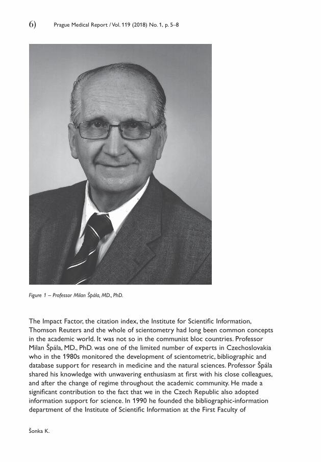
Professor Milan Špála, MD., PhD. – 1930–2018Karel ŠonkaDepartment of Neurology, First Faculty of Medicine, Charles University and General University Hospital in Prague, Prague, Czech Republic
Accepted March 19 , 2018 .
https://doi.org/10.14712/23362936.2018.1 © 2018 The Authors. This is an open-access article distributed under the terms of the Creative Commons Attribution License (http://creativecommons.org/licenses/by/4.0).
Mailing Address: Prof. Karel Šonka, MD., DSc., Department of Neurology, First Faculty of Medicine, Charles University and General University Hospital in Prague, Kateřinská 30, 120 00 Prague 2, Czech Republic; e-mail: [email protected]
Šonka K.
6) Prague Medical Report / Vol. 119 (2018) No. 1, p. 5–8
The Impact Factor, the citation index, the Institute for Scientific Information, Thomson Reuters and the whole of scientometry had long been common concepts in the academic world. It was not so in the communist bloc countries. Professor Milan Špála, MD., PhD. was one of the limited number of experts in Czechoslovakia who in the 1980s monitored the development of scientometric, bibliographic and database support for research in medicine and the natural sciences. Professor Špála shared his knowledge with unwavering enthusiasm at first with his close colleagues, and after the change of regime throughout the academic community. He made a significant contribution to the fact that we in the Czech Republic also adopted information support for science. In 1990 he founded the bibliographic-information department of the Institute of Scientific Information at the First Faculty of
Figure 1 – Professor Milan Špála, MD., PhD.

Professor Milan Špála, MD., PhD. – 1930–2018
Prague Medical Report / Vol. 119 (2018) No. 1, p. 5–8 7)
Medicine at Charles University, which he also managed until the year 2000. At Charles University he introduced courses in the methodology of science and courses on the principles of scientific writing, within the framework of the newly emerging postgraduate research education in Biomedicine. At that time, he also lectured at the Institute of Information Studies and Librarianship at the Faculty of Arts at Charles University and was very committed to organising the INFOMEDIA, later renamed INFORUM, conferences at which he also lectured. The study of science led Professor Špála to the founder of this field, Eugene Garfield, whom he hosted during the latter’s stay in Prague.
On the contribution of Professor Špála to the field of information science and bringing it into existence in the Czech Republic, I must add my own experience which is surely not unique. I was fortunate enough that Professor Špála took every opportunity to let me know about various innovations – I remember well how at the corner of Kateřinská and Viničná streets he excitedly explained the benefits of the newly available Web of Science. These were genuinely stimulating meetings. While monitoring his information sources Professor Špála would select innovations, important information and articles, which he kindly sent to me and other colleagues (on paper, before the development of e-mail). An enthusiasm for the dissemination of knowledge was in Professor Špála’s very nature.
Editorial work was another of Professor Špála’s important professional activities. From 1965 to 1975, he was the secretary of the Editorial Board of Sborník lékařský, and from 1976 to 2003 the secretary of the Editorial Board of Acta Universitatis Carolinae Medica, where a number of high-quality monographs came out thanks to his efforts. In the 1990s he co-founded the Medical Science Monitor, and in 1993–1999 he was a member of its international editorial board. In 1991 he became the Editor-in-Chief of Sborník lékařský, a major Czech medical journal, which had ceased to come out in 1990, following the death of its previous editor-in-chief, and had almost ceased to exist. Professor Špála helped to resuscitate the journal and managed it until 2003, when the journal changed its title to Prague Medical Report.
Professor Milan Špála, MD., PhD. was born on 20 November 1930. As early as at his studies in the 1955/1956 academic year he worked as an assistant at the Institute of Pathological Physiology of the Faculty of Pediatrics, Charles University. He graduated “sub auspiciis” in 1956 at the Faculty of General Medicine, Charles University in Prague, and became full-time research assistant to Professor Hepner at the Institute of General and Experimental Pathology, Faculty of General Medicine, Charles University (now the Institute of Pathological Physiology, First Faculty of Medicine, Charles University), where he was appointed Assistant Professor in 1961. Professor Špála worked for more than 50 years at the Institute of Pathological Physiology, First Faculty of Medicine, and many faculty graduates remember him as an excellent teacher. He defended his PhD dissertation thesis in 1963.
Šonka K.
8) Prague Medical Report / Vol. 119 (2018) No. 1, p. 5–8
From 1970 to 1974, he served as head of the Department of Physiology at the University of Oran, Algeria (Département de Physiologie, Institut des Sciences Médicales, Université d’Oran), which he himself later regarded as an important and positive stage in his life. From 1978 to 1996 he was a representative for teaching at the Department of Pathological Physiology of the First Faculty of Medicine and in 1989 he was habilitated in pathological physiology at Charles University in Prague.
From 1991 to 1993 he was a member of the Scientific Council of the First Faculty of Medicine and from 1996 to 1998 a member of its Academic Senate. In 1998, on the occasion of the 650th anniversary of the founding of Charles University, Professor Špála was awarded the Commemorative Medal of Charles University and the First Faculty of Medicine, and in 2003 for his work on Sborník lékařský, the Commemorative Certificate of the First Faculty of Medicine of Charles University.
Professor Špála was a member of the Physiological Society of the Czech Medical Society and the Czech Society for Medical Informatics and Scientific Information of the Czech Medical Society, where he worked from 2000 to 2006 as a member of the Board. In 1990 he was appointed Honorary Member of the Society for Pathological and Clinical Physiology of the Czech Medical Society and in 2006 was an Honorary Member of the Czech Society for Medical Informatics and Scientific Information.
I want this reminiscence of Professor Milan Špála, MD., PhD., Emeritus Chief Editor of this journal, to be optimistic in tone. Professor Špála taught us much while we peered out from the isolationism of the previous regime, he helped establish various kinds of support for scientific work at Charles University and thus his activities in university and professional societies will continue to contribute to scientific and academic life even though Professor Špála is no longer among us.
We honour his memory!
Prof. Karel Šonka, MD., DSc.Member of the Prague Medical Report editorial board
9)
Sepsis and Omics Technologies
Prague Medical Report / Vol. 119 (2018) No. 1, p. 9–29
Sepsis Diagnostics in the Era of “Omics” TechnologiesMiroslav Průcha1, Roman Zazula2, Stefan Russwurm3
1Department of Clinical Biochemistry, Haematology and Immunology, Na Homolce Hospital, Prague, Czech Republic;2Department of Anesthesiology and Intensive Care, First Faculty of Medicine, Charles University and Thomayer Hospital, Prague, Czech Republic;3Department of Anesthesiology and Intensive Care, University Hospital Jena, Jena, Germany
Rece ived December 5 , 2017 ; Accepted March 19 , 2018 .
Key words: Sepsis – Genomics – Transcriptomics – Proteinomics – Metabolomics – Pharmacogenomics – Diagnostics
Abstract: Sepsis is a multifactorial clinical syndrome with an extremely dynamic clinical course and with high diverse clinical phenotype. Early diagnosis is crucial for the final clinical outcome. Previous studies have not identified a biomarker for the diagnosis of sepsis which would have sufficient sensitivity and specificity. Identification of the infectious agents or the use of molecular biology, next gene sequencing, has not brought significant benefit for the patient in terms of early diagnosis. Therefore, we are currently searching for biomarkers, through “omics” technologies with sufficient diagnostic specificity and sensitivity, able to predict the clinical course of the disease and the patient response to therapy. Current progress in the use of systems biology technologies brings us hope that by using big data from clinical trials such biomarkers will be found.
This study was supported by the Czech Republic Ministry of Health’s conceptual development of research organisation (Nemocnice Na Homolce – NNH, 00023884, IG144101 and Thomayerova nemocnice – TN, 00064190).
Mailing Address: Assoc. Prof. Miroslav Průcha, MD., PhD., Department of Clinical Biochemistry, Haematology and Immunology, Na Homolce Hospital, Roentgenova 2, 150 30 Prague 5, Czech Republic; Phone: +420 257 273 051; e-mail: [email protected]
https://doi.org/10.14712/23362936.2018.2 © 2018 The Authors. This is an open-access article distributed under the terms of the Creative Commons Attribution License (http://creativecommons.org/licenses/by/4.0).
10)
Průcha M.; Zazula R.; Russwurm S.
Prague Medical Report / Vol. 119 (2018) No. 1, p. 9–29
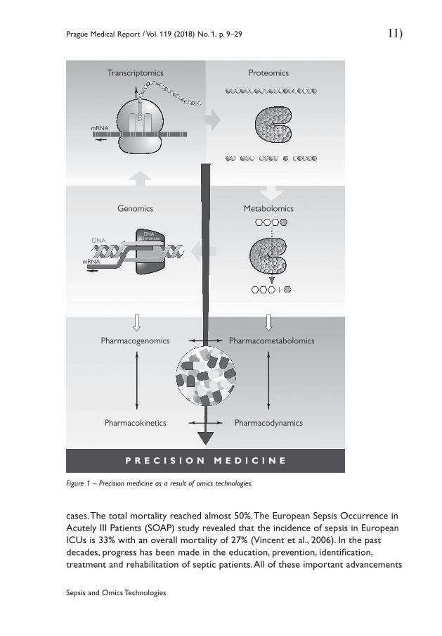
IntroductionWhen Friedrich Miescher published a work on nuclein in 1869, nobody was aware of where this development in the medical field would go in the next 140 years (Dahm, 2010). In 1953, Watson and Crick only needed one DIN4 page to publish their groundbreaking work on the discovery of DNA structure (Watson and Crick, 1953). This discovery heralded the explosion of new findings related to research into the relation between the genome and its effect on the development and course of diseases. The beginning of the 21st century saw an unprecedented development of systemic biology. This scientific field in biology uses knowledge in mathematics, biochemistry, chemistry and informatics to study complex interactions present in biological systems. Its expansion was made possible by the development of technology used for obtaining genomic and proteomic data and information technology (IT) development. These technologies provide us with new knowledge in medicine in terms of disease diagnosis and pathogenesis and therapy. So-called precision medicine is the final product (Figure 1). Molecular profiling and the use of therapies that target a specific disease’s genetic traits are the two pillars of this approach. The most spectacular progress was made in oncology and hemato-oncology (Ginsburg and Willard, 2009; Bombard et al., 2013; Ciardiello et al., 2014; Yu and Snyder, 2016), in other medical fields, we witness their large dissemination and gradual implementation into clinical practice. Significant progress has also been achieved in the diagnostics and further research of sepsis pathogenesis in the last 20 years. But, despite investments of several billion euros (EUR) by the pharmaceutical industry in the last decades, only one new drug so far has made it to the market (Toft and Tønnesen, 2011; Marshall, 2014). Our review provides an overview of the current situation in using “omics” technologies (genomics, transcriptomics, proteinomics, metabolomics, pharmacogenomics) in the diagnostics and its possible impact to the therapy of septic patients.
SepsisSepsis is among the leading causes of death worldwide. It accounts for more than 210,000 deaths annually in the United States, whereas similar death rates are reported for other countries (Angus et al., 2001; Shen et al., 2010; Kumar et al., 2011; Rhee et al., 2017). In Germany, it is the third most frequent cause of death in the German population and already the leading cost factor in German intensive care medicine with total costs of EUR 1.7 billion per year (Brunkhorst and Reinhart, 2005). It affects all age groups and it is the leading cause of morbidity and mortality in critically ill patients following intensive care unit (ICU) admission. Mortality from sepsis seems unchanged despite intense efforts in intensive care management of the patient. Mortality of sepsis is currently higher than that of myocardial infarction; even worse outcomes are revealed when patients with septic shock are considered (Esper and Martin, 2007). Martin et al. (2009) analysed more than 11,000 patients included in the international registry comprising severe sepsis
11)
Sepsis and Omics Technologies
Prague Medical Report / Vol. 119 (2018) No. 1, p. 9–29
Figure 1 – Precision medicine as a result of omics technologies.
cases. The total mortality reached almost 50%. The European Sepsis Occurrence in Acutely III Patients (SOAP) study revealed that the incidence of sepsis in European ICUs is 33% with an overall mortality of 27% (Vincent et al., 2006). In the past decades, progress has been made in the education, prevention, identification, treatment and rehabilitation of septic patients. All of these important advancements
Pharmacogenomics
Pharmacokinetics
Pharmacometabolomics
Pharmacodynamics
P R E C I S I O N M E D I C I N E
mRNA
Transcriptomics Proteomics
Genomics Metabolomics
mRNA
DNADNA
polymerase
12)
Průcha M.; Zazula R.; Russwurm S.
Prague Medical Report / Vol. 119 (2018) No. 1, p. 9–29
together have gradually ameliorated the individual burden of sepsis; nevertheless, it remains unacceptably high. Moreover, due to the ever-growing incidence of sepsis, the overall number of patients who die from sepsis continues to increase (Adhikari et al., 2010; Jawad et al., 2012; Martin, 2012; De La Rica et al., 2016). Very important aspect is impact of socioeconomic status on mortality and morbidity in patients with sepsis (Schnegelsberg et al., 2016). It is not surprising that the scientific community continues to call for improved treatment options, including a timely diagnosis and active surveillance in hospitalized patients at high risk for the development of sepsis.
Present state of the art in the field – science and methodologyIn the pathogenesis of sepsis, the key player is the dysregulation of the mechanisms of innate and adaptive immunity. A local inflammatory response progresses into a systemic response, reflecting a failure of the immunological compensatory mechanisms. Traditionally, sepsis is considered an exaggerated inflammatory response of the host to a bacterial invader (Calandra et al., 2000; Cavaillon et al., 2003; Huang et al., 2010; van Vught et al., 2017). As such, it is conceived that monitoring of the inflammatory host response may provide critical information for diagnosis and prognosis. A plethora of circulating proteins has been studied in the last 20 years as probable surrogate markers of the physical course of sepsis. Their use is based on the rationale that since inflammation results from a bacterial stimulus, changes of a biomarker or a combination of biomarkers may reflect eradication or propagation of the infection site. However, none of the studied biomarkers is considered an absolute reflection of the inflammatory state of the host and none has yet reached wide routine use (Biron et al., 2015; Prucha et al., 2015).
In the process of evolution, the innate immune system has developed many mechanisms that are capable of recognizing a pathogenic organism and responding to its presence accordingly. Furthermore, these mechanisms are capable of identifying homeostasis disrupting stimuli of a non-infectious nature (e.g. ischemia, trauma). For this purpose, immunocompetent cells possess pattern recognition receptors (PRRs) that are capable of recognizing characteristic pathogen associated molecular patterns (PAMPs). Upon interaction of PAMPs and PRRs, adaptor proteins come into play whose task is to activate transcription factors (Kono and Rock, 2008; van der Poll and Opal, 2008; Kumar et al., 2009a). Adaptor proteins activate the transcription factors, including nuclear factor ĸB (NF-ĸB), activation protein-1 (AP-1) and interferon response factor (IRF). The activation results in the expression of genes for pro- and anti-inflammatory cytokines (Medzhitov, 2001; Wiel et al., 2002; Akira et al., 2006). When severe sepsis becomes clinically apparent, the host has already entered into a state of immunoparalysis where circulating monocytes are no longer able to secrete pro-inflammatory cytokines, antigen presentation fails and apoptosis of lymphocytes predominates (Ayala et al.,
13)
Sepsis and Omics Technologies
Prague Medical Report / Vol. 119 (2018) No. 1, p. 9–29
2003; López-Collazo et al., 2006; Biswas et al., 2007; del Fresno et al., 2008, 2009).
Indeed, most of the studied potential biomarkers in severe sepsis, capable to reflect immune system mode and inflammatory cascade stage(s), are protein molecules with downregulated gene expression. Under this understanding of timely non-coordinated and yet overlapping counter-balancing processes of pro-inflammatory trigger and immunoparalysis follow-up stage, it is obvious that circulatory proteins are not the ideal surrogate biomarkers for sepsis. The underlying transcription of the genetic machinery of the host, which precedes translation and post-translational modifications of functionally ready proteins in time, occurs faster and reflects initial regulatory mechanisms necessary for the final protein output; and is then expected to provide more important information (Feezor et al., 2005). Novel “biomarkers” are urgently needed to identify correctly and timely the “infection” as the underlying cause of a systemic host response, because each hour of delay of anti-infectious therapy leads to a 5% increase in mortality (Kumar et al., 2009b). A sensitive biomarker is desirable when the consequences of missing the diagnosis are important; vice versa, a specific measure is desirable when costs or potential side effects of treatment are considerable. Both aspects are important in the context of sepsis where missed infections will double mortality rates, and antibiotic overuse is associated with rising resistance but also other problem such as organ toxicity, development of Clostridium difficile infection, etc. Therefore, it is important for the clinician to know when antibiotics can be safely withheld. While single proteins are useful biomarkers in a variety of well-characterized diseases, such as troponin for the early detection of myocardial infarction, new biomarker candidates for complex clinical syndromes such as sepsis cannot reliably discriminate non-infectious from infectious systemic inflammation. Procalcitonin, which probably best meets the requirements for a biomarker of infection at present, is hampered by false positives in the setting of non-infectious inflammation and a rather late induction during the host response to infection (Becker et al., 2008; Sager et al., 2017). Recently clinical studies were published on the relationship between presepsin and sepsis. Presepsin levels are increased in septic patients with no significant difference between patients with gram-positive or gram-negative infection. Presepsin appears to be very early biomarker of sepsis with place in the clinical diagnostics and may be of interest for future studies (Carpio et al., 2015; Zhang et al., 2015). As the complexity of the host response makes it unlikely that one single biomarker can adequately describe and stratify this complex syndrome, response patterns come into play. Because changes in the signalling of effector cells of innate immunity represent the earliest event in the “infection-host response-continuum”, lab-on-a-chip assays addressing multiple “omics” levels and analysing sets of biomarkers that reflect these changed patterns can meet this need. Such an approach promises not only to substantially improve sensitivity and specificity but also to shorten the time to diagnosis and therapy.
14)
Průcha M.; Zazula R.; Russwurm S.
Prague Medical Report / Vol. 119 (2018) No. 1, p. 9–29
The role of “omics” technologies in sepsis research:1) Diagnostics of sepsis – to find biomarkers differentiating infectious and
non-infectious inflammation2) To find biomarkers predicting the clinical outcome3) To find biomarkers that offer the possibility for the therapy of sepsis4) To find biomarkers predicting patient response to therapy
Genomics and epigenomicsOn 26 June 2000, Celera Genomics and the International Human Genome Sequencing Consortium (HGSC) announced the completion of the first assembly of the human genome and the completion of the rough draft, respectively. In February of 2001, two teams simultaneously published their analyses of the genome sequences generated (Lander et al., 2001; Venter et al., 2001). At present, we know that human genome contains approximately 22,000 genes that code proteins and 5–10 times more proteins are formed as their final product. The price of human genome research has been immense – approximately 3 billion US dollars. In the last 15 years, new technologies such as next gene sequencing (ngs) dramatically reduced the price and, thus, the availability of complete genetic information. New technologies provide for a faster and more complex approach (Ng and Kirkness, 2010). At present, it is possible to map the genome of an individual cell (Gawad et al., 2016; Hynes et al., 2017). However, the beginnings of the relation between genomics and infections date back to a more distant past. In 1988, Sørensen and colleagues demonstrated that the risk of dying from infectious disease was five times higher if an individual’s biological parent had also died of infectious disease. Since then, numerous studies have attempted to associate genetic markers of genomic variation (polymorphisms) with incidence or outcome of infectious disease and its sequelae in critically ill patients. Tumour necrosis factor (TNF) gene polymorphisms showed association with an increased incidence as well as adverse outcomes in patients with severe sepsis and septic shock (Stüber et al., 1996). Similarly pro- and anti-inflammatory cytokines like the interleukin-1 (IL-1) gene family, interleukin-6 (IL-6) and interleukin-10 (IL-10) are associated with different outcomes of septic patients (Fang et al., 1999). Genomic variants of candidate genes involved in pathogen recognition and signal transduction of inflammatory pathways like CD14, Toll-like receptors (TLRs), lipopolysaccharide binding protein (LBP), interleukin-1 receptor-associated kinase (IRAK 4), IRAK 1 may also contribute to the incidence, severity and mortality of infectious complications in the critically ill (Hubacek et al., 2001; Feterowski et al., 2003; Medvedev et al., 2003; Sutherland et al., 2005; Arcaroli et al., 2006; Khor et al., 2007; Chien et al., 2008; Mansur et al., 2015). In addition, it has been recognized that protein cascades involved in the pathophysiology of sepsis, such as the coagulation cascade, represent strong genomic candidate markers for association studies (Hermans et al., 1999). In a recent study, German authors conducted GWAS in a cohort of
15)
Sepsis and Omics Technologies
Prague Medical Report / Vol. 119 (2018) No. 1, p. 9–29
740 adult patients with sepsis. They found 14 loci with suggestive evidence for an association with 28-day mortality and found supportive, converging evidence for three of them in independent data sets. Authors have concluded that elucidating the underlying biological mechanisms of VPS13A, CRISPLD2, and the chromosome 13 locus should be a focus of future research activities (Scherag et al., 2016). A study performed by Rautanen et al. (2015) revealed interesting results. Authors identified variants in the FER gene, which are associated with a reduced risk of death from sepsis due to pneumonia. The FER gene encodes non-receptor protein tyrosine kinase. Tyrosine kinase is an enzyme from the protein kinase group, which catalyzes the transfer of a phosphate group (phosphorylation) from nucleoside triphosphate (mostly ATP) to the amino acid tyrosine in proteins. Non-receptor tyrosine kinases function in the cytoplasm and transfer signals within the cell into the nucleus (Gocek et al., 2014). The FER gene affects leukocyte recruitment and intestinal barrier dysfunction caused by lipopolysaccharide (LPS) (Parsons et al., 2007). Srinivasan et al. (2017) studied single nucleotide polymorphisms with sepsis in a cohort of 757 prematurely born children. The authors concluded that they did not find a significant association between single nucleotide polymorphism (SNP) and sepsis, however, areas of the potential association and pathways meriting for further study were identified. Sapru et al. (2016) proved association of common genetic variation in the protein C pathway genes with clinical outcomes in acute respiratory distress syndrome. The Netherland study provides a method for the molecular classification of patients with sepsis to 4 different endotypes upon ICU admission. Detection of sepsis endotypes might assist in providing personalized patient management and in selection for trials (Scicluna et al., 2017). Current results show that sepsis is a multifactorial disease, which is not necessarily related to a certain gene of a group of genes and/or their variants. Patients with primary immunodeficiency who are sensitive to certain types of infection are an exception (Feezor et al., 2005; Bustamante et al., 2014). Gene activity varies due to different epigenetic mechanisms. The mechanisms involve DNA modification by methylation and histone modification. Histone modifications represent acetylation, ubiquitination and phosphorylation. Both histone modifications and DNA cytosine methylation have been shown to regulate gene expression (Esteller, 2007; Dong and Weng, 2013). The main principle is the gene expression variability without a change in DNA sequence (Phillips, 2008). The example of this phenomenon is the bacteria-host interaction. Bacterium-induced epigenetic deregulations may affect host cell function either to promote host defence or to allow pathogen persistence. Thus, pathogenic bacteria can be considered as potential epimutagens able to reshape the epigenome (Bierne et al., 2012; Stephens et al., 2013).
TranscriptomicsTranscriptomics evaluates messenger RNA levels for genes in specific cells or tissues. Transcriptomics aims at monitoring gene activity and regulation,
16)
Průcha M.; Zazula R.; Russwurm S.
Prague Medical Report / Vol. 119 (2018) No. 1, p. 9–29
differentiating infectious and non-infectious inflammation, finding pathogenetic mechanisms and parameters that will predict clinical outcome. A number of issues arise in these studies dealing with sepsis. The first one is the enormous dynamics of disease progression in sepsis. From the pathogenetic point of view, sepsis is characterized by the concurrent presence of pro-inflammatory and anti-inflammatory phases that translates into immunosuppression (Boomer et al., 2011; Xiao et al., 2011; Hotchkiss et al., 2013; Cazalis et al., 2014). However, the clinical course of sepsis shows that in its early stages, pro-inflammatory elements are dominant and characterized by high production of pro-inflammatory cytokines, which at a later time lead to functional immunosuppression. Therefore, gene expression is likely to differ according to the development of sepsis over time (Leentjens et al., 2013). Another issue that we have to deal with when studying sepsis, is the target tissue of the organ in which gene expression is to be measured. It is not possible to harvest tissue in septic patients for ethical reasons; therefore, our data are based on measurements of the best available analyte – full blood and/or individual blood cell subpopulations. It is clearly not a bad alternative from the pathogenetic point of view because full blood, with all its immunocompetent cells, represents a robust and complex immunological mechanism involved in sepsis pathogenesis (Leliefeld et al., 2016). On the other hand, it is a potential source of possible erroneous conclusions. The reasons for this are multiple (starting with the patient’s age) children vs. adults (immaturity of the immune system vs. a large number of co-morbidities in elderly patients who represent the majority of patients dying from sepsis) and different gene expression depending on cell subpopulations (e.g. neutrophils, NK cells and lymphocytes). A study by Palmer et al. (2006) revealed different gene expression, depending on the subpopulation of immunocompetent cells. Study by Parnell et al. (2013) measuring gene expression in full blood identified genes with immune dysfunction in septic patients. Practically, no study has been performed that would evaluate gene expression in individual organs during clinical sepsis, with the exception of experimental studies (Cobb et al., 2002).
Differentiation of infectious and non-infectious SIRS, pathogenetic mechanisms of sepsisThe first studies evaluated gene expression in volunteers who received an endotoxin dose (Calvano et al., 2005; Talwar, 2006). They found a different expression in volunteers who received endotoxin and identified target gene groups with their bonds in gene maps. Since the beginning, the limits of experimental endotoxin use were clear compared with clinical sepsis with a living infectious agent. One of the first clinical study performed in patients with sepsis that confirmed different gene expression in systemic inflammatory response syndrome (SIRS) of infectious and non-infectious etiology was published in 2004 (Prucha et al., 2004). The results were confirmed by other studies (Johnson et al., 2007; Lissauer et al., 2009; Seok et al., 2013; Sweeney et al., 2015). Differences were
17)
Sepsis and Omics Technologies
Prague Medical Report / Vol. 119 (2018) No. 1, p. 9–29
found with respect to the septic agent. While a study by Tang et al. (2008) did not reveal a different gene expression in gram-positive (G+) and gram-negative (G–) infections, a study by Yu et al. (2004) obtained different results. When studying gene expression, attention is paid to mechanisms characterizing inflammation of infectious etiology and disease severity. A study by Grealy et al. (2013) revealed different gene expression of IL-2, IL-10, IL-23, IL-27, interferon-γ (IFN γ) and TNFα, based on whether it was an infection or severe sepsis. A study performed by Hinrichs et al. (2010) identified genes, the expression of which predicts postoperative sepsis. Significant differences (P<0.005) in gene expression between the 2 groups were observed for IL1B (interleukin 1, beta), TNF [tumour necrosis factor (TNF superfamily, member 2)], CD3D [CD3d molecule, delta (CD3-TCR complex)] and PRF1 [perforin 1 (pore forming protein)]. The combination of TNF, IL1B and CD3D expression had a sensitivity and specificity of 90% and 85%, respectively, and predicted exclusion of postoperative sepsis with an estimated negative predictive value of 98.1% (Hinrichs et al., 2010). Different gene expression was demonstrated in bacterial and virus infections related to immune dysfunction. Authors identified a T-cell-dominant gene-expression signature that is associated with the host response to severe influenza pneumonia. Genes linked to the cell cycle and its regulation were the main determinants of the host response in influenza infection. Interestingly study failed to identify an immune response specific to bacterial pneumonia (Parnell et al., 2012). A study by Sampson et al. (2017) which revealed different gene expression depending on virus or bacterial etiology, produced similar results. Should these abilities of a specific group of genes be confirmed, there is a real possibility of their implementation in clinical practice, which would affect antibiotic consumption, resistance development, etc. Another very recent study revealed individual predisposition of septic patients with regard to the final outcome and prognosis. Davenport et al. (2016) analysed gene expression of peripheral blood leucocytes in ICU patients who were admitted for sepsis caused by community acquired pneumonia and evidence of organ dysfunction. Transcriptomic analysis defined two different pictures – so-called sepsis response signature (SRS). SRS1 found in 41% of patients, identified patients with an immunosuppression phenotype (endotoxin tolerance, T-cell exhaustion and human leukocyte antigen (HLA) class II regulation disorder). This phenotype was associated with higher 14-day mortality (Davenport et al., 2016). In 2014, Fiusa et al. published a meta-analysis of gene expression in severe sepsis and septic shock. From 45 studies out of 22,216 probe sets, authors observed 352 as candidates (215 of which were upregulated and 137 downregulated). The top 5 up-regulated genes were CD177, MMP8, HP, ARG1 and ANXA3. The top downregulated genes were FCER1A, YMEI1L1, TRDV3, LRRN3 and MYBL1 (Fiusa et al., 2014). In response to a need for better sepsis diagnostics, several new gene expression classifiers have been recently published, including the 11-gene “Sepsis MetaScore”, the “FAIM3-to-PLAC8” ratio and the Septicyte Lab. The three diagnostics do not
18)
Průcha M.; Zazula R.; Russwurm S.
Prague Medical Report / Vol. 119 (2018) No. 1, p. 9–29
show significant differences in overall ability to distinguish non-infectious SIRS from sepsis (Sweeney and Khatri, 2017). On the other hand, the study of Zimmerman et al. (2017) showed that the Septicyte Lab test is able to discriminate between clinically severe sepsis syndrome and infection-negative systemic inflammation among critically ill children.
ProteomicsWe differentiate between express proteomics detecting proteins, which are characteristic of sepsis, and functional proteomics, which identifies proteomic markers and their function on the molecular level. A key role is played by the type of technology used. At present, there are two-dimensional differential gel electrophoresis (2D-DIGE), matrix-associated laser desorption/ionization-time of flight (MALDI-TOF), surface-enhanced laser desorption/ionization-time of flight (SELDI-TOF), laser capture microdissection-MS (LCM-MS) and protein microarray (Mesri, 2014). It should be noted that different technological platforms do not provide the same results. There is only a partial overlap between the biomarkers identified by individual technologies. This of course makes the verification of results more difficult. In addition to genomics, proteomics is a significant means of precision medicine that advances diagnostics and disease treatment a large step ahead. Similar objectives are followed here to find new biomarkers, which will differentiate infectious systemic inflammation from non-infectious systemic inflammation, and to find pathogenetic mechanisms that could be implemented in clinical practice with regard to diagnostics and treatment. Buhimschi used 2D-DIGE and mass spectrometry to study umbilical blood proteome in order to find biomarkers that could identify patients with an early onset of sepsis. The authors found that a switch-on in haptoglobin to haptoglobin-related protein expression reflected a fetal adaptive response to intraamniotic infection exposure in utero (Buhimschi et al. 2011). A study Paugam-Burtz et al. (2010) identified new proteins detected in patients following liver transplant and sepsis. This study used plasma profiling coupling protein chip array with SELDI-TOF. In the validation set of 31 patients with infection and 34 without infection, the 5 peaks were differentially expressed as well and allowed day 5 sepsis diagnosis with a positive likelihood ratio of 5.1 and C-statistics of 0.74 (0.58–0.85) (Paugam-Burtz et al., 2010). Proteomics is used to study infectious model and describe protein-protein interactions. A Swedish study using mass spectrometry maps the interaction between bacterial and plasma proteins. Using Streptococcus pyogenes as an infectious agent and adhered human blood plasma protein, the author constructed a stoichiometric model of protein structure interaction. The model and knowledge of these constructions will help to better understand protein-protein interactions and their importance for bacterial virulence (Sjöholm et al., 2017). Another Swedish study based on an experimental sepsis model with virulent Streptococcus pyogenes in mice maps proteins in blood plasma and individual organs. The results not only showed
19)
Sepsis and Omics Technologies
Prague Medical Report / Vol. 119 (2018) No. 1, p. 9–29
a partial overlapping of proteins in individual organs but also protein abundance levels, which differ in different organs (Malmström et al., 2016). These findings confirmed the results of previous studies (Huttlin et al., 2010; Geiger et al., 2013).
MetabolomicsMetabolomics is the large-scale study of small molecules (commonly known as metabolites) within cells, fluids, tissues or organisms. Collectively, these small molecules and their interactions within a biological system are known as the metabolome. Metabolomics is the study of substrates and products of metabolism, which are influenced by both genetic and environmental factors (Kosmides et al., 2013). Metabolomics is a powerful approach because metabolites and their concentrations, unlike other “omics” measures, directly reflect the underlying biochemical activity and state of cells/tissues. Thus, metabolomics best represents the molecular phenotype (Serkova et al., 2011; Patti et al., 2012). The Human Metabolome Database contains records for more than 42,000 metabolites, from sugars to peptides to cofactors. But the total may be significantly higher, and single analytical methods often struggle to capture the chemical diversity. Current technologies can even analyse a metabolome in one cell. With regard to the immense heterogeneity of cell populations, the question is whether this approach can bring about significant progress (Fessenden, 2016). The aims of metabolomics are the same as with other “omics” technologies – diagnosis, prognosis and identification of at risk patients. Swedish authors published an interesting study. In a prospective study, whole blood samples from 65 patients with bacteremic sepsis and 49 controls were compared. The blood samples were analysed using gas chromatography coupled to time-of-flight mass spectrometry. A 6-metabolite predictive logistic regression model showed a sensitivity of 0.91 (95% confidence interval (CI) 0.69–0.99) and a specificity of 0.84 (95% CI 0.58–0.94) with an area under curve (AUC) of 0.93 (95% CI 0.89–1.01). Myristic acid was the single most predictive metabolite, with a sensitivity of 1.00 (95% CI 0.85–1.00) and specificity of 0.95 (95% CI 0.74–0.99), and performed better than various combinations of conventional laboratory and clinical parameters (Kauppi et al., 2016). Ambrogio examined urine metabolome in an infant with fatal methicillin resistant Staphylococcus aureus (MRSA) pneumonia, 4 children with influenza pneumonia (pneumonia control group) and 7 healthy children with no known infections. Urine metabolite concentrations previously identified as associated with sepsis in children (e.g. 3-hydroxybutyrate, carnitine and creatinine) were higher in the patient with fatal MRSA pneumonia compared with those of patients with influenza pneumonia and healthy controls. The concentrations of additional metabolites (acetone, acetoacetate, choline, fumarate, glucose and 3-aminoisobutyrate) were more than 25-fold higher in the patient with MRSA pneumonia than those of patients with influenza pneumonia and healthy controls. These metabolic changes in the urine preceded the clinically severe sepsis phenotype, suggesting that detection
20)
Průcha M.; Zazula R.; Russwurm S.
Prague Medical Report / Vol. 119 (2018) No. 1, p. 9–29
of the extent of metabolic disruption can aid in the early identification of a sepsis phenotype in advance of the clinical diagnosis (Ambroggio et al., 2017). In other study, Ferrario et al. (2016) examined plasma metabolome and clinical features in a subset of 20 patients with severe septic shock. Early changes in the plasma levels of low unsaturated long-chain phosphatidylcholines and kynurenine were associated with mortality (Ferrario et al., 2016). A urine metabolomic analysis in terms of the patient’s prognosis was also performed by the authors of a Spanish study. Urine samples were collected from 64 patients with severe sepsis or septic shock in the ICU. Authors compared the prediction power of metabolomics data respect with respect to Sequential Organ Failure Assessment (SOFA) score. Supervised multivariate analysis afforded a good predictive model to distinguish the patient groups and to detect specific metabolic patterns. Negative prognosis patients presented higher values of ethanol, glucose and hippurate, and, on the contrary, lower levels of methionine, glutamine, arginine and phenylalanine (Garcia-Simon et al., 2015). And finally, a Chinese study provides the proteomic analysis of urine to identify prognostic biomarkers of sepsis. The 7 identified proteins provide insight into the mechanism of sepsis. Low urinary lysosome-associated membrane protein-1 levels may be useful for the early prognostic assessment of sepsis (Su et al., 2013). Su et al. (2013) described the metabolic profile of normal patients and patients with SIRS or sepsis, which was markedly different. Seven metabolites may potentially be used to diagnose sepsis. A significant decrease in the levels of lactitol dehydrate and S-phenyl-D-cysteine and an increase in the levels of S-(3-methylbutanoyl)-dihydrolipoamide-E and N-nonanoyl glycine were observed in patients with sepsis in comparison to patients with SIRS (P<0.05). Patients with severe sepsis and septic shock displayed lower levels of glyceryl-phosphoryl-ethanolamine, Ne, Ne dimethyllysine, phenylacetamide and D-cysteine (P<0.05) in their sera (Su et al., 2014).
Pharmacogenomics – Pharmacometabolomics“Omics” technologies are developing further. They result not only in identifying new biomarkers, new knowledge of disease pathogenesis and prediction of the clinical course of disease and outcomes in patients but have also direct consequences for research dealing with drug efficiency. Pharmacogenomics and pharmacometabolomics provide tools for mapping the effects of drugs on metabolism and for identifying pathways that contribute to drug response variation (Kaddurah-Daouk et al., 2015). In septic patients, only a few studies were presented. Man et al. (2013) conducted a GWAS using a large randomised clinical trial cohort to discover genetic biomarkers of response to therapy in septic patients. Evidence for gene-gene interactions were identified for sepsis treatment responses with genetic biomarkers dominating models for predicting therapeutic response (Man et al., 2013). Study of Puskarich et al. (2015) shows a unique metabolite profile of l-carnitine responders in patients with septic shock.
21)
Sepsis and Omics Technologies
Prague Medical Report / Vol. 119 (2018) No. 1, p. 9–29
Pharmacometabolomics has clear potential for the future in predicting patients’ reactions to individual drugs (Everett, 2016; Huan et al., 2017; Rattray and Kaddurah Daouk, 2017).
Limitations of the use of omics technology in sepsisSepsis, as a systemic expression of a pathological response of the immune system to infection, is an extremely complex and dynamic process. Unlike chronic inflammatory autoimmune diseases and oncological diseases, whose preclinical and clinical progression is relatively long and enables current use of omics technology findings both in diagnostics and therapy, sepsis is a clinical syndrome the trigger mechanism of which still fails to be identified. It is already possible to identify some biomarkers, which predict a higher risk of adverse outcomes in these patients. The use of increasingly better technologies including the evaluation of “big data” raises hope that we will be able to use precise medicine even with them. As the example of chronic inflammatory autoimmune diseases shows (e.g. rheumatoid arthritis (RA) or Crohn disease) they are, on the basis of molecular analyses, differentiated to new subtypes or various nosologic units with different patient’s phenotype and with different pathogenesis resulting presumably in different therapeutic approaches (Li and Kauffman, 2014; Smolen et al., 2016; Wang et al., 2017; Weiser et al., 2018).
ConclusionAt present, omics technologies represent significant technological progress in the further study of sepsis pathogenesis, its relation with genetic predisposition and a pathway to more effective pharmacotherapy. So far, there is no practical use in sepsis diagnosis and treatment, but it seems to be a very hopeful and promising journey. Omics data can be a powerful tool for patient diagnostics, stratification and therapy similar to patients with traumatic and thermal injury (Hazeldine et al., 2016). In addition, the recent discovery of CRISPR-Cas9 (clustered regularly interspaced short palindromic repeats-CRISPR associated protein 9) and their specific use in disease diagnostics and treatment having a significant predictive value for genetic predisposition is revolutionary (Gootenberg et al., 2017; Patel et al., 2017).
ReferencesAdhikari, N. K., Fowle, R. A., Bhagwanjee, S., Rubenfeld, G. D. (2010) Critical care and the global burden of
critical illness in adults. Lancet 376, 1339–1346.Akira, S., Uematsu, S., Takeuchi, O. (2006) Pathogen recognition and innate immunity. Cell 124, 783–801.Ambroggio, L., Florin, T. A., Shah, S. S., Ruddy, R., Yeomans, L., Trexel, J., Stringer, K. A. (2017) Emerging
biomarkers of illness severity: Urinary metabolites associated with sepsis and necrotizing methicillin-resistant Staphylococcus aureus pneumonia. Pharmacotherapy 37, 1033–1042.
Angus, D. C., Linde-Zwirble, W. T., Lidicker, J., Clermont, G., Garcillo, J., Pinsky, M. R. (2001) Epidemiology of severe sepsis in the United States: analysis of incidence, outcome, and associated costs of care. Crit. Care Med. 29, 1303–1310.
Arcaroli, J., Silva, E., Maloney, J. P., He, Q., Svetkauskaite, D., Murphy, J. R., Abraham, E. (2006) Variant IRAK-1
22)
Průcha M.; Zazula R.; Russwurm S.
Prague Medical Report / Vol. 119 (2018) No. 1, p. 9–29
haplotype is associated with increased nuclear factor-kappa B activation and worse outcomes in sepsis. Am. J. Respir. Crit. Care Med. 173, 1335–1341.
Ayala, A., Lomas, J. L., Grutkoski, P. S., Grutkoski, P. S., Chung, C. S. (2003) Pathological aspects of apoptosis in severe sepsis and shock? Int. J. Biochem. Cell Biol. 35, 715–720.
Becker, K. L., Snider, R., Nylen, E. S. (2008) Procalcitonin assay in systemic inflammation, infection, and sepsis: clinical utility and limitations. Crit. Care Med. 6, 941–952.
Bierne, H., Hamon, M., Cossart, P. (2012) Epigenetics and bacterial infections. Cold Spring Harb. Perspect. Med. 2, a010272.
Biron, B. M., Ayala, A., Lomas-Neira, L. J. (2015) Biomarkers for sepsis: What is and what might be? Biomark. Insights 10(S4), 7–17.
Biswas, S. K., Bist, P., Dhillon, M. K., Kajiji, T., del Fresno, C., Yamamoto, M., Lopez-Collazo, E., Akira, S., Tergaonkar, V. (2007) Role for MyD88-independent, TRIF pathway in lipid A/TLR4-induced endotoxin tolerance. J. Immunol. 179, 4083–4092.
Bombard, Y., Bach, P. B., Offit, K. (2013) Translating genomics in cancer care. J. Natl. Compr. Canc. Netw. 11, 1343–1353.
Boomer, J. S., To, K., Chang, K. C., Takasu, O., Osborne, D. F., Walton, A. H., Bricker, T. L., Jarman, S. D., Kreisel, D., Krupnick, A. S., Srivastava, A., Swanson, P. E., Green, J. M., Hotchkiss, R. S. (2011) Immunosuppression in patients who die of sepsis and multiple organ failure. JAMA 306, 2594–2605.
Brunkhorst, F. M., Reinhart, K. (2005) German competence network for the study of severe sepsis and septic shock (SepNet). ICU Manag. Pract. 5.
Buhimschi, C. S., Bhandari, V., Dulay, A. T., Nayeri, U. A., Abdel-Razeq, S. S., Pettker, C. M., Thung, S., Zhao, G., Han, Y. W., Bizzarro, M., Buhimschi, I. A. (2011) Proteomics mapping of cord blood identifies haptoglobin “switch-on” pattern as biomarker of early-onset neonatal sepsis in preterm newborns. PLoS One 6, e26111.
Bustamante, J., Boisson-Dupuisa, S., Abela, L., Casanova, J. L. (2014) Mendelian susceptibility to mycobacterial disease: Genetic, immunological, and clinical features of inborn errors of IFN-γ immunity. Semin. Immunol. 26, 454–470.
Calandra, T., Echtenacher, B., Roy, D. L., Pugin, J., Metz, N., Hültner, L., Heumann, D., Männel, D., Bucala, R., Glauser, M. P. (2000) Protection from septic shock by neutralization of macrophage migration inhibitory factor. Nat. Med. 6, 164–170.
Calvano, S. E., Xiao, W., Richards, D. R. (2005) A network-based analysis of systemic inflammation in humans. Nature 437, 1032–1037.
Carpio, R., Zapata, J., Spanuth, E., Hess, G. (2015) Utility of presepsin (sCD14-ST) as a diagnostic and prognostic marker of sepsis in the emergency department. Clin. Chim. Acta 450, 169–175.
Cavaillon, J. M., Adib-Conquy, M., Fitting, C., Adrie, C., Payen, D. (2003) Cytokine cascade in sepsis. Scand. J. Infect. Dis. 35, 535–544.
Cazalis, M. A., Lepape, A., Venet, F., Frager, F., Mougin, B., Vallin, H., Paye, M., Pachot, A., Monneret, G. (2014) Early and dynamic changes in gene expression in septic shock patients: a genome-wide approach. Intensive Care Med. Exp. 2, 20–37.
Chien, J. W., Boeckh, M. J., Hansen, J. A., Clark, J. G. (2008) Lipopolysaccharide binding protein promoter variants influence the risk for gram-negative bacteremia and mortality after allogeneic hematopoietic cell transplantation. Blood 111, 2462–2469.
Ciardiello, F., Arnold, D., Casali, P. G., Cervantes, J. Y., Douillard, A., Eggermont, A., Eniu, K., McGregor, S., Piccart, P. M., Popescu, R., Van Cutsem, E., Zielinski, R., Stahel, R. (2014) Delivering precision medicine in oncology today and in future-the promise and challenges of personalised cancer medicine: a position paper by the European Society for Medical Oncology (ESMO). Ann. Oncol. 25, 1673–1678.
23)
Sepsis and Omics Technologies
Prague Medical Report / Vol. 119 (2018) No. 1, p. 9–29
Cobb, J. P., Laramie, J. M., Stormo, G., Morrissey, J. J., Shannon, W. D., Qiu, Y., Karl, I. E., Buchman, T. G., Hotchkiss, R. S. (2002) Sepsis gene expression profiling: Murine splenic compared with hepatic responses determined by using complementary DNA microarrays. Crit. Care Med. 30, 2711–2721.
Dahm, R. (2010) From discovering to understanding. EMBO Rep. 11, 153–160.Davenport, E. E., Burnham, K. L., Radhakrishnan, J., Humburg, P., Hutton, P., Mills, T. C., Rautanen, A.,
Gordon, A. C., Garrard, C., Hill, A. V., Hinds, C. J., Knight, J. C. (2016) Genomic landscape of the individual host response and outcomes in sepsis: a prospective cohort study. Lancet Respir. Med. 4, 259–271.
De La Rica, A. S., Gilsanz, F., Maseda, E. (2016) Epidemiologic trends of sepsis in western countries. Ann. Transl. Med. 4, 325–331.
del Fresno, C., Gómez-Piña, V., Lores, V., Soares-Schanoski, A., Fernández-Ruiz, I., Rojo, B., Alvarez-Sala, R., Caballero-Garrido, E., García, F., Veliz, T., Arnalich, F., Fuentes-Prior, P., García-Río, F., López-Collazo, E. (2008) Monocytes from cystic fibrosis patients are locked in an LPS tolerance state: down-regulation of TREM-1 as putative underlying mechanism. PLoS One 3, e2667.
del Fresno, C., García-Rio, F., Gómez-Piña, V., Soares-Schanoski, A., Fernández-Ruíz, I., Jurado, T., Kajiji, T., Shu, C., Marín, E., Gutierrez del Arroyo, A., Prados, C., Arnalich, F., Fuentes-Prior, P., Biswas, S. K., López-Collazo, E. (2009) Potent phagocytic activity with impaired antigen presentation identifying lipopolysaccharide-tolerant human monocytes: demonstration in isolated monocytes from cystic fibrosis patients. J. Immunol. 182, 6494–6507.
Dong, X., Weng, Z. (2013) The correlation between histone modifications and gene expression. Epigenomics 5, 113–116.
Esper, A., Martin, G. S. (2007) Is severe sepsis increasing in incidence and severity? Crit. Care Med. 35, 1414–1415.
Esteller, M. (2007) Cancer epigenomics: DNA methylomes and histone-modification maps. Nat. Rev. Genet. 8, 286–298.
Everett, J. R. (2016) From metabonomics to pharmacometabonomics: the role of metabolic profiling in personalized medicine. Front. Pharmacol. 7, 297.
Fang, X. M., Schröder, S., Hoeft, A., Stuber, F. (1999) Comparison of two polymorphisms of the interleukin-1 gene family: interleukin-1 receptor antagonist polymorphism contributes to susceptibility to severe sepsis. Crit. Care Med. 27, 1330–1334.
Feezor, R. J., Cheng, A., Paddock, H. N., Baker, H. V., Moldawer, L. L. (2005) Functional genomics and gene expression profiling in sepsis: beyond class prediction. Clin. Infect. Dis. 41, S427–S435.
Ferrario, M., Cambiaghi, A., Brunelli, L., Giordano, S., Caironi, P., Guatteri, L., Raimondi, F., Gattinoni, L., Latini, R., Masson, S., Ristagno, G., Pastorelli, R. (2016) Mortality prediction in patients with severe septic shock: a pilot study using a target metabolomics approach. Sci. Rep. 6, 20391.
Fessenden, M. (2016) Metabolomics: small molecules, single cells. Nature 540, 153–155.Feterowski, C., Emmanuilidis, K., Miethke, T., Gerauer, K., Rump, M., Ulm, K., Holzmann, B., Weighardt, H.
(2003) Effects of functional Toll-like receptor-4 mutations on the immune response to human and experimental sepsis. Immunology 109, 426–431.
Fiusa, M. M., Carvalho, B. S., Hubert, R. M., Souza, W., Lopes-Cendes, I., Annichino-Bizzacchi, J. M., De Paula, E. V. (2014) A meta-analysis of gene expression studies in severe sepsis and septic shock. Blood 124, 2741.
Garcia-Simon, M., Morales, J. M., Modesto-Alapont, V., Gonzalez-Marrachelli, V., Vento-Rehues, R., Jorda-Miñana, A., Blanquer-Olivas, J., Monleon, D. (2015) Prognosis biomarkers of severe sepsis and septic shock by 1H NMR urine metabolomics in the intensive care unit. PLoS One 10, e0140993.
Gawad, C., Koh, W., Quake, S. R. (2016) Single-cell genome sequencing: current state of the science. Nat. Rev. Genet. 17, 175–188.
24)
Průcha M.; Zazula R.; Russwurm S.
Prague Medical Report / Vol. 119 (2018) No. 1, p. 9–29
Geiger, T., Velic, A., Macek, B., Lundberg, E., Kampf, C., Nagaraj, N., Uhlen, M., Cox, J., Mannet, M. (2013) Initial quantitative proteomic map of 28 mouse tissues using the SILAC mouse. Mol. Cell. Proteomics 12, 1709–1722.
Ginsburg, G. S., Willard, H. F. (2009) Genomic and personalized medicine: foundations and applications. Transl. Res. 154, 277–287.
Gocek, E., Moulas, A. N., Studzinski, G. P. (2014) Non-receptor protein tyrosine kinases signaling pathways in normal and cancer cells. Crit. Rev. Clin. Lab. Sci. 51, 125–137.
Gootenberg, J. S., Abudayyeh, O. O., Lee, J. W., Essletzbichler, P., Dy, A. J., Joung, J., Verdine, V., Donghia, N., Daringer, N. M., Freije, C. A., Myhrvold, C., Bhattacharyya, R. P., Livny, J., Regev, A., Koonin, E. V., Hung, D. T., Sabeti, P. C., Collins, J. J., Zhang, F. (2017) Nucleic acid detection with CRISPR-Cas13a/C2c2. Science 356, 438–442.
Grealy, R., White, M., Stordeur, P., Kelleher, D., Doherty, D. G., McManus, R., Ryan, T. (2013) Characterising cytokine gene expression signatures in patients with severe sepsis. Mediators Inflamm. 2013, 164246.
Hazeldine, J., Hampson, P., Lord, J. M. (2016) The diagnostic and prognostic value of systems biology research in major traumatic and thermal injury: a review. Burns Trauma 4, 33–44.
Hermans, P. W., Hibberd, M. L., Booy, R., Daramola, O., Hazelzet, J. A., de Groot, R., Levin, M. (1999) 4G/5G promoter polymorphism in the plasminogen-activator-inhibitor-1 gene and outcome of meningococcal disease. Meningococcal Research Group. Lancet 354, 556–560.
Hinrichs, C., Kotsch, K., Buchwald, S., Habicher, M., Saak, N., Gerlach, H., Volk, H. D., Keh, D. (2010) Perioperative gene expression analysis for prediction of postoperative sepsis. Clin. Chem. 56, 613–622.
Hotchkiss, R. S., Monneret, G., Payen, D. (2013) Sepsis-induced immunosuppression: from cellular dysfunctions to immunotherapy. Nat. Rev. Immunol. 13, 862–874.
Huan, T., Forsberg, E. M., Rinehart, D., Johnson, C. H., Ivanisevic, J., Benton, H. P., Fang, M., Aisporna, A., Hilmers, B., Poole, F. L., Thorgersen, M. P., Adams, M. W. W., Krantz, G., Fields, M. W., Robbins, P. D., Niedernhofer, L. J., Ideker, T., Majumder, E. L., Wall, J. D., Rattray, N. J. W., Goodacre, R., Lairson, L. L., Siuzdak, G. (2017) Systems biology guided by XCMS online metabolomics. Nat. Methods 14, 461–462.
Huang, W., Tang, Y., Li, L. (2010) HMGB1, a potent proinflammatory cytokine in sepsis. Cytokine 51, 119–126.Hubacek, J. A., Stüber, F., Fröhlich, D., Book, M., Wetegrove, S., Ritter, M., Rothe, G., Schmitz, G. (2001) Gene
variants of the bactericidal/permeability increasing protein and lipopolysaccharide binding protein in sepsis patients: gender-specific genetic predisposition to sepsis. Crit. Care Med. 29, 557–561.
Huttlin, E. L., Jedrychowski, M. P., Elias, J. E., Goswami, T., Rad, R., Beausoleil, S. A., Villén, J., Haas, W., Sowa, M. E., Gygi, S. P. (2010) A tissue-specific atlas of mouse protein phosphorylation and expression. Cell 143, 1174–1189.
Hynes, S. O., Pang, B., James, J. A., Maxwell, P., Salto-Tellez, M. (2017) Tissue-based next generation sequencing: application in a universal healthcare system. Br. J. Cancer 116, 553–560.
Jawad, I., Luksic, I., Rafnsson, S. B. (2012) Assessing available information on the burden of sepsis: global estimates of incidence, prevalence and mortality. J. Glob. Health 2, 010404.
Johnson, S. B., Lissauer, M., Bochicchio, G. V., Moore, R., Cross, A. S., Scalea, T. M. (2007) Gene expression profiles differentiate between sterile SIRS and early sepsis. Ann. Surg. 245, 611–621.
Kaddurah-Daouk, R., Weinshilboum, R.; Pharmacometabolomics Research Network (2015) Metabolomic signatures for drug response phenotypes: Pharmacometabolomics enables precision medicine. Clin. Pharmacol. Ther. 98, 71–75.
Kauppi, A. M., Edin, A., Ziegler, I., Mölling, P., Sjösted, A., Gylfe, A., Strålin, K., Johansson, A. (2016) Metabolites in blood for prediction of bacteremic sepsis in the emergency room. PLoS One 11, e0147670.
Khor, C. C., Chapman, S. J., Vannberg, F. O., Dunne, A., Murphy, C., Ling, E. Y., Frodsham, A. J., Walley, A. J., Kyrieleis, O., Khan, A., Aucan, C., Segal, S., Moore, C. E., Knox, K., Campbell, S. J., Lienhardt, C., Scott, A.,
25)
Sepsis and Omics Technologies
Prague Medical Report / Vol. 119 (2018) No. 1, p. 9–29
Aaby, P., Sow, O. Y., Grignani, R. T., Sillah, J., Sirugo, G., Peshu, N., Williams, T. N., Maitland, K., Davies, R. J., Kwiatkowski, D. P., Day, N. P., Yala, D., Crook, D. W., Marsh, K., Berkley, J. A., O’Neill, L. A., Hill, A. V. (2007) A Mal functional variant is associated with protection against invasive pneumococcal disease, bacteremia, malaria and tuberculosis. Nat. Genet. 39, 523–528.
Kono, H., Rock, K. L. (2008) How dying cells alert the immune system to danger. Nat. Rev. Immunol. 8, 279–289.Kosmides, A. K., Kamisoglu, K., Calvano, S. E., Corbett, S. A., Androulakis, I. P. (2013) Metabolomic
fingerprinting: challenges and opportunities. Crit. Rev. Biomed. Eng. 41, 205–221.Kumar, A. T., Sudhir, U., Punith, K., Kumar, R., Kumar, V. N., Rao, M. Y. (2009a) Cytokine profile in elderly
patients with sepsis. Indian J. Crit. Care Med. 13, 74–78.Kumar, A., Ellis, P., Arabi, Y., Roberts, D., Light, B., Parrillo, J. E., Dodek, P., Wood, G., Kumar, A., Simon, D.,
Peters, C., Ahsan, M., Chateau, D.; Cooperative Antimicrobial Therapy of Septic Shock Database Research Group (2009b) Initiation of inappropriate antimicrobial therapy results in a fivefold reduction of survival in human septic shock. Chest 136, 1237–1248.
Kumar, G., Kumar, A., Taneja, A., Kaleekal, T., Tarima, S., McGinley, E., Jimenez, E., Mohan, A., Khan, R. A., Whittle, J., Jacobs, E., Nanchal, R.; Milwaukee Initiative in Critical Care Outcomes Research (MICCOR) Group of Investigators (2011) Nationwide trends of severe sepsis in the 21st century (2000–2007). Chest 140, 1223–1231.
Lander, E. S., Linton, L. M., Birren, B., Nusbaum, C., Zody, M. C., Baldwin, J., Devon, K., Dewar, K., Doyle, M., FitzHugh, W., Funke, R., Gage, D., Harris, K., Heaford, A., Howland, J., Kann, L., Lehoczky, J., LeVine, R., McEwan, P., McKernan, K., Meldrim, J., Mesirov, J. P., Miranda, C., Morris, W., Naylor, J., Raymond, C., Rosetti, M., Santos, R., Sheridan, A., Sougnez, C., Stange-Thomann, Y., Stojanovic, N., Subramanian, A., Wyman, D., Rogers, J., Sulston, J., Ainscough, R., Beck, S., Bentley, D., Burton, J., Clee, C., Carter, N., Coulson, A., Deadman, R., Deloukas, P., Dunham, A., Dunham, I., Durbin, R., French, L., Grafham, D., Gregory, S., Hubbard, T., Humphray, S., Hunt, A., Jones, M., Lloyd, C., McMurray, A., Matthews, L., Mercer, S., Milne, S., Mullikin, J. C., Mungall, A., Plumb, R., Ross, M., Shownkeen, R., Sims, S., Waterston, R. H., Wilson, R. K., Hillier, L. W., McPherson, J. D., Marra, M. A., Mardis, E. R., Fulton, L. A., Chinwalla, A. T., Pepin, K. H., Gish, W. R., Chissoe, S. L., Wendl, M. C., Delehaunty, K. D., Miner, T. L., Delehaunty, A., Kramer, J. B., Cook, L. L., Fulton, R. S., Johnson, D. L., Minx, P. J., Clifton, S. W., Hawkins, T., Branscomb, E., Predki, P., Richardson, P., Wenning, S., Slezak, T., Doggett, N., Cheng, J. F., Olsen, A., Lucas, S., Elkin, C., Uberbacher, E., Frazier, M., Gibbs, R. A., Muzny, D. M., Scherer, S. E., Bouck, J. B., Sodergren, E. J., Worley, K. C., Rives, C. M., Gorrell, J. H., Metzker, M. L., Naylor, S. L., Kucherlapati, R. S., Nelson, D. L., Weinstock, G. M., Sakaki, Y., Fujiyama, A., Hattori, M., Yada, T., Toyoda, A., Itoh, T., Kawagoe, C., Watanabe, H., Totoki, Y., Taylor, T., Weissenbach, J., Heilig, R., Saurin, W., Artiguenave, F., Brottier, P., Bruls, T., Pelletier, E., Robert, C., Wincker, P., Smith, D. R., Doucette-Stamm, L., Rubenfield, M., Weinstock, K., Lee, H. M., Dubois, J., Rosenthal, A., Platzer, M., Nyakatura, G., Taudien, S., Rump, A., Yang, H., Yu, J., Wang, J., Huang, G., Gu, J., Hood, L., Rowen, L., Madan, A., Qin, S., Davis, R. W., Federspiel, N. A., Abola, A. P., Proctor, M. J., Myers, R. M., Schmutz, J., Dickson, M., Grimwood, J., Cox, D. R., Olson, M. V., Kaul, R., Raymond, C., Shimizu, N., Kawasaki, K., Minoshima, S., Evans, G. A., Athanasiou, M., Schultz, R., Roe, B. A., Chen, F., Pan, H., Ramser, J., Lehrach, H., Reinhardt, R., McCombie, W. R., de la Bastide, M., Dedhia, N., Blöcker, H., Hornischer, K., Nordsiek, G., Agarwala, R., Aravind, L., Bailey, J. A., Bateman, A., Batzoglou, S., Birney, E., Bork, P., Brown, D. G., Burge, C. B., Cerutti, L., Chen, H. C., Church, D., Clamp, M., Copley, R. R., Doerks, T., Eddy, S. R., Eichler, E. E., Furey, T. S., Galagan, J., Gilbert, J. G., Harmon, C., Hayashizaki, Y., Haussler, D., Hermjakob, H., Hokamp, K., Jang, W., Johnson, L. S., Jones, T. A., Kasif, S., Kaspryzk, A., Kennedy, S., Kent, W. J., Kitts, P., Koonin, E. V., Korf, I., Kulp, D., Lancet, D., Lowe, T. M., McLysaght, A., Mikkelsen, T., Moran, J. V., Mulder, N., Pollara, V. J., Ponting, C. P., Schuler, G., Schultz, J., Slater, G., Smit, A. F., Stupka, E., Szustakowki, J., Thierry-Mieg, D., Thierry-Mieg, J., Wagner, L., Wallis, J., Wheeler, R.,
26)
Průcha M.; Zazula R.; Russwurm S.
Prague Medical Report / Vol. 119 (2018) No. 1, p. 9–29
Williams, A., Wolf, Y. I., Wolfe, K. H., Yang, S. P., Yeh, R. F., Collins, F., Guyer, M. S., Peterson, J., Felsenfeld, A., Wetterstrand, K. A., Patrinos, A., Morgan, M. J., de Jong, P., Catanese, J. J., Osoegawa, K., Shizuya, H., Choi, S., Chen, Y. J., Szustakowki, J.; International Human Genome Sequencing Consortium (2001) Initial sequencing and analysis of the human genome. Nature 409, 860–921.
Leentjens, J., Kox, M., van der Hoeven, J. G., Netea, M. G., Pickkers, P. (2013) Immunotherapy for the adjunctive treatment of sepsis: from immunosuppression to immunostimulation. Time for a paradigm change? Am. J. Respir. Crit. Care Med. 187, 1287–1293.
Leliefeld, H. F., Wessels, C. M., Leenan, L. P., Koenderman, L., Pillay, J. (2016) The role of neutrophils in immune dysfunction during severe inflammation. Crit. Care 20, 73–82.
Li, Y. R., Kauffman, J. M. (2014) Molecular medicine of rheumatoid arthritis: From molecular pathophysiology to novel therapeutics and evidence-based practice. Ann. Orthop. Rheumatol. 2, 1014–1024.
Lissauer, M. E., Johnson, S. B., Bochicchio, G. V., Carinda, J., Feild, A. S., Cross, J. D., Hasday, C. C., Whiteford, W. A., Nussbaumer, M. T., Scalea, T. (2009) Differential expression of Toll-like receptor genes: sepsis compared with sterile inflammation 1 day before sepsis diagnosis. Shock 31, 238–244.
López-Collazo, E., Fuentes-Prior, P., Arnalich, F., del Fresno, C. (2006) Pathophysiology of interleukin-1 receptor-associated kinase-M: implications in refractory state. Curr. Opin. Infect. Dis. 19, 237–244.
Malmström, E., Kilsgård, O., Hauri, S., Smeds, E., Herwald, H., Malmström, L., Malmström, J. (2016) Large-scale inference of protein tissue origin in gram-positive sepsis plasma using quantitative targeted proteomics. Nat. Commun. 7, 10261.
Man, M., Close, S. L., Shaw, A. D., Bernard, G. R., Douglas, I. S., Kaner, R. J. (2013) Beyond single-marker analyses: Mining whole genome scans for insights into treatment responses in severe sepsis. Pharmacogenomics J. 13, 218–226.
Mansur, A., Liese, B., Steinau, M., Ghadimi, M., Bergmann, I., Tzvetkov, M., Popov, A. F., Beissbarth, T., Bauer, M., Hinz, J. (2015) The CD14 rs2569190 TT genotype is associated with an improved 30-days survival in patients with sepsis: a prospective observational study. PLoS One 10, e0127761.
Marshall, J. C. (2014) Why have clinical trials in sepsis failed? Trends Mol. Med. 20, 195–203.Martin, G. S. (2012) Sepsis, severe sepsis and septic shock: Changes in incidence, pathogens and outcomes.
Expert Rev. Anti Infect. Ther. 10, 701–706.Martin, G., Brunkhorst, F. M., Janes, J. M., Reinhart, K., Sundin, D. P., Garnett, K., Beale, R. (2009) The
international PROGRESS registry of patients with severe sepsis: drotrecogin alfa (activated) use and patients outcomes. Crit. Care 13, R103.
Medvedev, A. E., Lentschat, A., Kuhns, D. B., Blanco, J. C., Salkowski, C., Zhang, S., Arditi, M., Gallin, J. I., Vogel, S. N. (2003) Distinct mutations in IRAK-4 confer hyporesponsiveness to lipopolysaccharide and interleukin-1 in a patient with recurrent bacterial infections. J. Exp. Med. 198, 521–531.
Medzhitov, R. (2001) Toll-like receptors and innate immunity. Nat. Rev. Immunol. 1, 135–139.Mesri, M. (2014) Advances in proteomic technologies and its contribution to the field of cancer. Adv. Med.
2014, 238045.Ng, P. C., Kirkness, E. F. (2010) Whole genome sequencing. Methods Mol. Biol. 628, 215–226.Palmer, C., Diehn, M., Alizadeh, A. A., Brown, P. O. (2006) Cell-type specific gene expression profiles of
leukocytes in human peripheral blood. BMC Genomics 7, 115–130.Parnell, G. P., McLean, A. S., Booth, D. R., Armstrong, N. J., Nalos, M., Huang, S. J., Manak, J., Tang, W., Tam, O. Y.,
Chan, S., Tang, B. M. (2012) A distinct influenza infection signature in the blood transcriptome of patients with severe community-acquired pneumonia. Crit. Care 16, R157.
Parnell, G. P., Tang, B. M., Nalos, M., Armstrong, N. J., Huang, S. J., Booth, D. R., McLean, A. S. (2013) Identifying key regulatory genes in the whole blood of septic patients to monitor underlying immune dysfunctions. Shock 40, 166–174.
27)
Sepsis and Omics Technologies
Prague Medical Report / Vol. 119 (2018) No. 1, p. 9–29
Parsons, S. A., Mewburn, J. D., Truesdell, P., Greer, P. A. (2007) The Fps/Fes kinase regulates leucocyte recruitment and extravasation during inflammation. Immunology 122, 542–550.
Patel, S. J., Sanjana, N. E., Kishton, R. J., Eidizadeh, A., Vodnala, S. K., Cam, M., Gartner, J. J., Jia, L., Steinberg, S. M., Yamamoto, T. N., Merchant, A. S., Mehta, G. U., Chichura, A., Shalem, O., Tran, E., Eil, R., Sukumar, M., Guijarro, E. P., Day, C. P., Robbins, P., Feldman, S., Merlino, G., Zhang, F., Restifo, N. P. (2017) Identification of essential genes for cancer immunotherapy. Nature 548, 537–542.
Patti, G. J., Yanes, O., Siuzdak, G. (2012) Innovation: Metabolomics: The apogee of the omics trilogy. Nat. Rev. Mol. Cell Biol. 13, 263–269.
Paugam-Burtz, C., Albuquerque, M., Baron, G., Bert, F., Voitot, H., Delefosse, D., Dondero, F., Sommacale, D., Francoz, C., Hanna, N., Belghiti, J., Ravaud, P., Bedossa, P., Mantz, J., Paradis, V. (2010) Plasma proteome to look for diagnostic biomarkers of early bacterial sepsis after liver transplantation: a preliminary study. Anesthesiology 112, 926–935.
Phillips, T. (2008) The role of methylation in gene expression. Nature Education 1, 116.Prucha, M., Ruryk, A., Boriss, H., Möller, E., Zazula, R., Herold, I., Claus, R. A., Reinhart, K. A., Deigner, P.,
Russwurm, S. (2004) Expression profiling: Toward an application in sepsis diagnostics. Shock 22, 29–33.
Prucha, M., Bellingan, G., Zazula, R. (2015) Sepsis biomarkers. Clin. Chim. Acta 440, 97–104.Puskarich, M. A., Finkel, M. A., Karnowsky, A., Jones, A. E., Trexel, J., Harris, B. N., Stringer, K. A. (2015)
Pharmacometabolomics of l-carnitine treatment response phenotypes in patients with septic shock. Ann. Am. Thorac. Soc. 12, 46–56.
Rattray, N. J., Kaddurah Daouk, R. (2017) Pharmacometabolomics and precision medicine special issue editorial. Metabolomics 13, 59.
Rautanen, A., Mills, T. C., Gordon, A. C., Hutton, P., Steffens, M., Nuamah, R., Chiche, J. D., Parks, T., Chapman, S. J., Davenport, E. E., Elliott, K. S., Bion, J., Lichtner, P., Meitinger, T., Wienker, T. F., Caulfield, M. J., Mein, C., Bloos, F., Bobek, I., Cotogni, P., Sramek, V., Sarapuu, S., Kobilay, M., Ranieri, V. M., Rello, J., Sirgo, G., Weiss, Y. G., Russwurm, S., Schneider, E. M., Reinhart, K., Holloway, P. A., Knight, J. C., Garrard, C. S., Russell, J. A., Walley, K. R., Stüber, F., Hill, A. V., Hinds, C. J.; ESICM/ECCRN GenOSept Investigators (2015) Genome-wide association study of survival from sepsis due to pneumonia: an observational cohort study. Lancet Respir. Med. 3, 53–60.
Rhee, C., Dantes, R., Epstein, L., Murphy, D. J., Seymour, C. W., Iwashyna, T. J., Kadri, S. S., Angus, D. C., Danner, R. L., Fiore, A. E., Jernigan, J. A., Martin, G. S., Septimus, E., Warren, D. K., Karcz, A., Chan, C., Menchaca, J. T., Wang, R., Gruber, S., Klompas, M.; CDC Prevention Epicenter Program (2017) Incidence and trends of sepsis in US hospitals using clinical vs. claims data, 2009–2014. JAMA 318, 1241–1249.
Sager, R., Kutz, A., Mueller, B., Schuetz, P. (2017) Procalcitonin-guided diagnosis and antibiotic stewardship revisited. BMC Med. 15, 15–26.
Sampson, D. L., Fox, B. A., Yager, T. D., Bhide, S., Cermelli, L. C., McHugh, T. A., Seldon, R. A., Brandon, E., Sullivan, J. J., Zimmerman, M., Noursadeghi, R. B. (2017) A four-biomarker blood signature discriminates systemic inflammation due to viral infection versus other etiologies. Sci. Rep. 7, 2914.
Sapru, A., Liu, K. D., Wiemels, J., Hansen, H., Pawlikowska, L., Poon, A., Jorgenson, E., Witte, J. S., Calfee, C. S., Ware, L. B., Matthay, M. A.; NHLBI ARDS Network (2016) Association of common genetic variation in the protein C pathway genes with clinical outcomes in acute respiratory distress syndrome. Crit. Care 20, 151–161.
Scherag, A., Schöneweck, F., Kesselmeier, M., Taudien, S., Platzer, M., Felder, M., Sponholz, C., Rautanen, A., Hill, A. V. S., Hinds, C. J., Hossain, H., Suttorp, N., Kurzai, O., Slevogt, H., Giamarellos-Bourboulis, E. J., Armaganidis, A., Trips, E., Scholz, M., Brunkhorst, F. M. (2016) Genetic factors of the disease course after sepsis: a genome-wide study for 28 day mortality. EBioMedicine 12, 239–246.
28)
Průcha M.; Zazula R.; Russwurm S.
Prague Medical Report / Vol. 119 (2018) No. 1, p. 9–29
Schnegelsberg, A., Mackenhauer, J., Nibro, H. L., Dreyer, P., Koch, K., Kirkengaard, H. (2016) Impact of socioeconomic status on mortality and unplanned readmission in septic intensive care unit patients. Acta Anaesthesiol. Scand. 60, 465–475.
Scicluna, B. P., van Vught, L. A., Zwinderman, A. H., Wiewel, M. A., Davenport, E. E., Burnham, K. L., Nürnberg, P., Schultz, M. J., Horn, J., Cremer, O. L., Bonten, M. J., Hinds, C. J., Wong, H. R., Knight, J. C., van der Poll, T.; MARS Consortium (2017) Classification of patients with sepsis according to blood genomic endotype: a prospective cohort study. Lancet Respir. Med. 5, 816–826.
Seok, J., Warren, H. S., Cuenca, A. G., Mindrinos, M. N., Baker, H. V., Xu, W., Richards, D. R., McDonald-Smith, G. P., Gao, H., Hennessy, L., Finnerty, C. C., López, C. M., Honari, S., Moore, E. E., Minei, J. P., Cuschieri, J., Bankey, P. E., Johnson, J. L., Sperry, J., Nathens, A. B., Billiar, T. R., West, M. A., Jeschke, M. G., Klein, M. B., Gamelli, R. L., Gibran, N. S., Brownstein, B. H., Miller-Graziano, C., Calvano, S. E., Mason, P. H., Cobb, J. P., Rahme, L. G., Lowry, S. F., Maier, R. V., Moldawer, L. L., Herndon, D. N., Davis, R. W., Xiao, W., Tompkins, R. G.; Inflammation and Host Response to Injury, Large Scale Collaborative Research Program (2013) Genomic responses in mouse models poorly mimic human inflammatory diseases. Proc. Natl. Acad. Sci. U. S. A. 110, 3507–3512.
Serkova, N. J., Standiford, T. J., Stringer, K. A. (2011) The emerging field of quantitative blood metabolomics for biomarker discovery in critical illnesses. Am. J. Respir. Crit. Care Med. 184, 647–655.
Shen, H. N., Lu, C. L., Yang, H. H. (2010) Epidemiologic trend of severe sepsis in Taiwan from 1997 through 2006. Chest 138, 298–304.
Sjöholm, K., Kilsgård, O., Teleman, J., Happonen, L., Malmström, L., Malmström, J. (2017) Targeted proteomics and absolute protein quantification for the construction of a stoichiometric host-pathogen surface density model. Mol. Cell. Proteomics 16, S29–S41.
Smolen, J. S., Aletaha, D., McInnes, I. B. (2016) Rheumatoid arthritis. Lancet 388, 2023–2038.Sørensen, T. I., Nielsen, G. G., Andersen, P. K., Teasdale, T. W. (1988) Genetic and environmental influences on
premature death in adult adoptees. N. Engl. J. Med. 318, 727–732.Srinivasan, L., Page, G., Kirpalani, H., Murray, J. C., Das, A., Higgins, R. D., Carlo, W. A., Bell, E. F., Goldberg, R. N., Schibler, K., Sood, B. G., Stevenson, D. K., Stoll, B. J., Van Meurs, K. P., Johnson, K. J., Levy, J., McDonald, S. A., Zaterka-Baxter, K. M., Kennedy, K. A., Sánchez, P. J., Duara, S., Walsh, M. C., Shankaran, S., Wynn, J. L.,
Cotton, C. N.; Eunice Kennedy Shriver National Institute of Child Health and Human Development Neonatal Research Network (2017) Genome-wide association study of sepsis in extremely premature infants. Arch. Dis. Child. Fetal Neonatal Ed. 102, F439–F445.
Stephens, K. E., Miaskowski, C. A., Levine, J. D., Pullinger, C. R., Aouizerat, B. E. (2013) Epigenetic regulation and measurement of epigenetic changes. Biol. Res. Nurs. 5, 373–381.
Stüber, F., Petersen, M., Bokelmann, F., Schade, U. (1996) A genomic polymorphism with in the tumor necrosis factor locus influences plasma tumor necrosis factor-alpha concentrations and outcome of patients with severe sepsis. Crit. Care Med. 24, 381–384.
Su, L., Cao, L., Zhou, R., Jiang, Z., Xiao, K., Kong, W., Wang, H., Deng, J., Wen, B., Tan, F., Zhang, Y., Xie, L. (2013) Identification of novel biomarkers for sepsis prognosis via urinary proteomic analysis using iTRAQ labeling and 2D-LC-MS/MS. PLos One 8, e54237.
Su, L., Huang, Y., Zhu, Y., Xia, L., Wang, R., Xiao, K., Wang, H., Yan, P., Wen, B., Cao, L., Meng, N., Luan, H., Liu, C., Li, X., Xie, L. (2014) Discrimination of sepsis stage metabolic profiles with an LC/MS-MS-based metabolomics approach. BMJ Open Respir. Res. 1, e000056.
Sutherland, A. M., Walley, K. R., Russell, J. A. (2005) Polymorphisms in CD14, manose binding lectin, and Toll-like receptor-2 are associated with increased prevalence of infection in critically ill adults. Crit. Care Med. 33, 638–644.
29)
Sepsis and Omics Technologies
Prague Medical Report / Vol. 119 (2018) No. 1, p. 9–29
Sweeney, T. E., Khatri, P. (2017) Benchmarking sepsis gene expression diagnostics using public data. Crit. Care Med. 45, 1–10.
Sweeney, T. E., Shidham, A., Wong, H. R., Khatri, P. (2015) A comprehensive time-course-based multicohort analysis of sepsis and sterile inflammation reveals a robust diagnostic gene set. Sci. Transl. Med. 7, 287ra71.
Talwar, S. (2006) Gene expression profiles of peripheral blood leukocytes after endotoxin challenge in humans. Physiol. Genomics 25, 203–215.
Tang, B. M., McLean, A. S., Dawes, I. W., Huang, S. J., Cowley, M. J., Lin, R. C. (2008) Gene-expression profiling of gram-positive and gram-negative sepsis in critically ill patients. Crit. Care Med. 36, 1125–1128.
Toft, P., Tønnesen, E. (2011) Immune-modulating interventions in critically ill septic patients: pharmacological options. Expert Rev. Clin. Pharmacol. 4, 491–501.
van der Poll, T., Opal, S. M. (2008) Host-pathogen interactions in sepsis. Lancet Infect. Dis. 8, 32–43.van Vught, L. A., Wiewel, M. A., Hoogendijk, A. J., Frencken, J. F., Scicluna, B. P., Klein, K., Klouwenberg, P. M. C.,
Zwinderman, A. H., Lutter, R., Horn, J., Schultz, M. J., Bonten, M. M. J., Cremer, O. L., van der Poll, T. (2017) The host response in patients with sepsis developing intensive care unit-acquired secondary infections. Am. J. Respir. Crit. Care Med. 196, 458–470.
Venter, J. C., Adams, M. D., Myers, E. W., Li, P. W., Mural, R. J. (2001) The sequence of the genome. Science 291, 1304–1351.
Vincent, J. L., Sakr, Y., Sprung, C. L., Gerlach, H., Ranieri, V. M. (2006) Sepsis in European intensive care units: results of the SOAP study. Crit. Care Med. 34, 344–353.
Wang, Z. Z., Shi, K., Peng, J. (2017) Serologic testing of a panel of five antibodies in inflammatory bowel diseases: diagnostic value and correlation with disease phenotype. Biomed. Rep. 6, 401–410.
Watson, J. D., Crick, F. H. (1953) Genetical implications of the structure of deoxyribonucleic acid. Nature 171, 737–738.
Weiser, M., Simon, J. M., Kochar, B., Tovar, A., Israel, J. W., Robinson, A., Gipson, G. R., Schaner, M. S., Herfarth, H. H., Sartor, R. B., McGovern, D. P., Rahbar, R., Sadiq, T. S., Koruda, M. J., Furey, T. S., Sheikh, S. Z. (2018) Molecular classification of Crohn’s disease reveals two clinically relevant subtypes. Gut 67, 36–42.
Wiel, E., Lebuffe, G., Vallet, B. (2002) Bacterial CpG DNA in septic shock. In: Yearbook of Intensive Care and Emergency Medicine. Vincent, J. L., Editor, pp. 388–398, Springer, Berlin.
Xiao, W., Mindrinos, M. N., Seok, J., Cuschieri, J., Cuenca, A. J., Gao, H., Hayden, D., Hennessy, L., Moore, E. E., Minei, J. P., Bankey, P. E., Johnson, J. L., Sperry, J., Nathens, A. B., Billiar, T. R., West, M. A., Brownstein, B. H., Mason, P. H., Baker, H. V., Finnerty, C. C., Jeschke, M. G., López, M. C., Klein, M. B., Gamelli, R. L., Gibran, N. S., Arnoldo, B., Xu, W., Zhang, Y., Calvano, S. E., McDonald-Smith, G. P., Schoenfeld, D. A., Storey, J. D., Cobb, J. P., Warren, H. S., Moldawer, L. L., Herndon, D. N., Lowry, S. F., Maier, R. V., Davis, R. W., Tompkins, R. G.; Inflammation and Host Response to Injury Large-Scale Collaborative Research Program (2011) A genomic storm in critically injured humans. J. Exp. Med. 208, 2581–2590.
Yu, K. H., Snyder, M. (2016) Omics profiling in precision oncology. Mol. Cell. Proteomics 16, 2525–2536.Yu, S. L., Chen, H. W., Yang, P. C., Peck, K., Tsai, M. H., Chen, J. J., Lin, F. Y. (2004) Differential gene expression
in gram-negative and gram-positive sepsis. Am. J. Respir. Crit. Care Med. 169, 1135–1143.Zhang, X., Liu, D., Liu, Y. N., Wang, R., Xie, L. X. (2015) The accuracy of presepsin (sCD14-ST) for the
diagnosis of sepsis in adults: a meta analysis. Crit. Care 19, 323–334.Zimmerman, J. J., Sullivan, E., Yager, T. D., Cheng, C., Permut, L., Cermelli, S., McHugh, L., Sampson, D.,
Seldon, T., Brandon, R. B., Brandon, R. A. (2017) Diagnostic accuracy of a host gene expression signature that discriminates clinical severe sepsis syndrome and infection-negative systemic inflammation among critically ill children. Crit. Care Med. 45, e418–e425.
Sedláčková L.; Průcha M.; Poláková I.; Míková B.
30) Prague Medical Report / Vol. 119 (2018) No. 1, p. 30–42
Evaluation of IFN-γ Enzyme-linked Immunospot Assay (ELISPOT) as a First-line Test in the Diagnosis of Non-Immediate Hypersensitivity to Amoxicillin and PenicillinLenka Sedláčková1, Miroslav Průcha1, Ingrid Poláková2, Blanka Míková1
1Department of Clinical Biochemistry, Haematology and Immunology, Na Homolce Hospital, Prague, Czech Republic;2Department of Immunology, Institute of Haematology and Blood Transfusion, Prague, Czech Republic
Rece ived December 29 , 2017 ; Accepted March 19 , 2018 .
Key words: ELISPOT – Allergy – Hypersensitivity – Penicillin – Amoxicillin
Abstract: The current diagnostic algorithm for beta-lactam allergy is based on skin and provocation tests, both of which carry a certain risk of inducing hypersensitivity reactions. Thus, non-invasive in vitro tests reliable enough to replace skin and provocation tests at least in a portion of patients are desirable. We aimed to verify the utility of IFN-γ ELISPOT as a first-line test in patients with suspected non-immediate hypersensitivity reaction to amoxicillin (AMX) and penicillin (PNC). The prospective observational study included 24 patients with recent, suspected non-immediate hypersensitivity reaction to AMX or PNC and 6 recently-exposed healthy subjects. In vitro tests were performed in all patients and healthy subjects: a) IFN-γ ELISPOT with PNC, AMX and amoxicillin plus clavulanic acid (AMX-CL); b) penicillin specific IgE; c) basophil activation test (BAT). Skin and provocation tests followed only in certain patients. IFN-γ ELISPOT results with PNC and AMX stimulation did not differ from the unstimulated condition. The highest IFN-γ responses to AMX-CL were close to previously published criteria in three patients;
This study was supported by the Ministry of Health of the Czech Republic – conceptual development of the research organisation (Nemocnice Na Homolce – NNH, Prague, Czech Republic, 00023884), IG144102.
Mailing Address: Lenka Sedláčková, MD., Department of Clinical Biochemistry, Haematology and Immunology, Na Homolce Hospital, Roentgenova 2, 150 30 Prague 5, Czech Republic; Phone: +420 257 272 823; e-mail: [email protected]
https://doi.org/10.14712/23362936.2018.3© 2018 The Authors. This is an open-access article distributed under the terms of the Creative Commons Attribution License (http://creativecommons.org/licenses/by/4.0).
ELISPOT in Beta-lactam Allergy
31)Prague Medical Report / Vol. 119 (2018) No. 1, p. 30–42
one of which had true hypersensitivity according to drug provocation tests. Five patients with confirmed hypersensitivity by skin tests showed no response to the culprit antibiotic on IFN-γ ELISPOT assay. Our results did not support the utility of IFN-γ ELISPOT in the diagnosis of mild, non-immediate hypersensitivity to amoxicillin and penicillin.
IntroductionBeta-lactam hypersensitivity has a significant impact on treatment options in affected patients. Up to 10% of patients have a self-reported allergy to penicillin. The avoidance of penicillin and other beta-lactam antibiotics increases the consumption of second-line broad-spectrum antibiotics and is followed by many unfavourable consequences: higher rate of treatment failures and complications, increased microbial resistance and higher treatment costs (Macy and Contreras, 2014; Solensky, 2014). However, true penicillin hypersensitivity is known to be present in only 10% of patients labelled penicillin-allergic. The correct determination of penicillin allergy has become a very important issue in public health.
Beta-lactam hypersensitivity has heterogeneous manifestations. From a clinical and diagnostic point of view, the most useful classification distinguishes between immediate and non-immediate hypersensitivity. Immediate hypersensitivity generally begins within 1 hour from exposure to an antibiotic and is often mediated by immunoglobulin E (IgE). Non-immediate hypersensitivity manifests later, usually within several hours or days, and is primarily mediated by T-lymphocytes (Blanca et al., 2009). The current diagnostic algorithm for beta-lactam hypersensitivity recommended by the European Network of Drug Allergy (ENDA) and the Drug Allergy Interest Group (DAIG) of the European Academy of Allergy and Clinical Immunology (EAACI) relies mostly on in vivo tests (skin and provocation tests) (Demoly et al., 2014), while the role of in vitro tests is complementary. The availability of complex allergy diagnostics by in vivo testing is currently suboptimal in the Czech Republic (CZ) as it carries certain risks for patients, and thus is provided in only a few experienced allergy centres. Currently, a simple approach to screening suspected beta-lactam allergy in vitro is used for immediate hypersensitivity in CZ, mainly using specific IgE, as well as the basophil activation test (BAT) to lesser extent. When a positive result confirms sensitisation and corresponds to clinical history, further examination is not required. All negative results should be followed up as the sensitivity of specific IgE and BAT for beta-lactam hypersensitivity is low (Demoly et al., 2014; Mayorga et al., 2016). Most beta-lactam hypersensitivity reactions are non-immediate, therefore the ability to confirm at least some cases by in vitro methods is desirable.
As non-immediate hypersensitivity reactions to beta-lactams are mediated primarily by T-lymphocytes, in vitro determination is focused on the detection of beta-lactam specific T-lymphocytes. Various tests of lymphocyte proliferation,
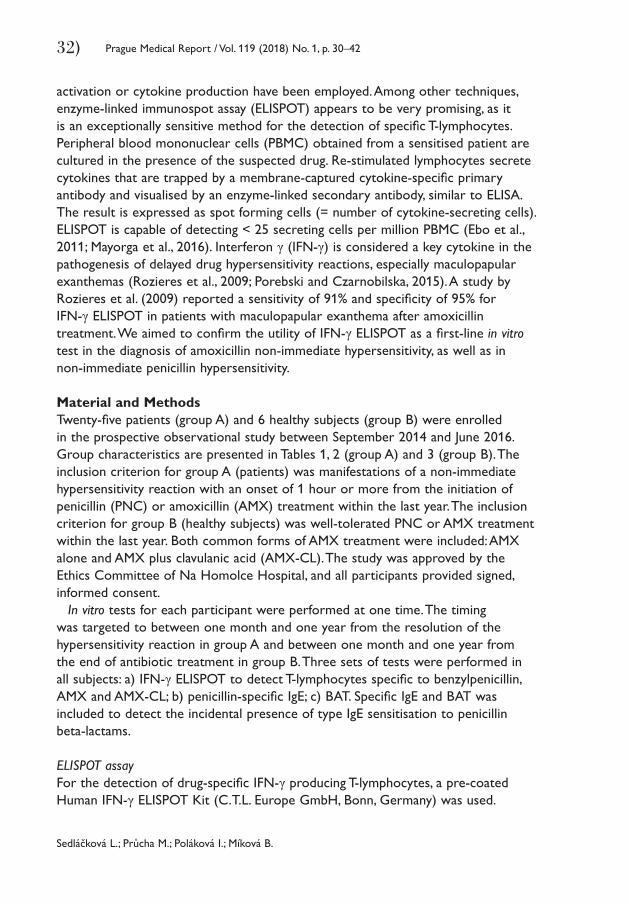
Sedláčková L.; Průcha M.; Poláková I.; Míková B.
32) Prague Medical Report / Vol. 119 (2018) No. 1, p. 30–42
activation or cytokine production have been employed. Among other techniques, enzyme-linked immunospot assay (ELISPOT) appears to be very promising, as it is an exceptionally sensitive method for the detection of specific T-lymphocytes. Peripheral blood mononuclear cells (PBMC) obtained from a sensitised patient are cultured in the presence of the suspected drug. Re-stimulated lymphocytes secrete cytokines that are trapped by a membrane-captured cytokine-specific primary antibody and visualised by an enzyme-linked secondary antibody, similar to ELISA. The result is expressed as spot forming cells (= number of cytokine-secreting cells). ELISPOT is capable of detecting < 25 secreting cells per million PBMC (Ebo et al., 2011; Mayorga et al., 2016). Interferon γ (IFN-γ) is considered a key cytokine in the pathogenesis of delayed drug hypersensitivity reactions, especially maculopapular exanthemas (Rozieres et al., 2009; Porebski and Czarnobilska, 2015). A study by Rozieres et al. (2009) reported a sensitivity of 91% and specificity of 95% for IFN-γ ELISPOT in patients with maculopapular exanthema after amoxicillin treatment. We aimed to confirm the utility of IFN-γ ELISPOT as a first-line in vitro test in the diagnosis of amoxicillin non-immediate hypersensitivity, as well as in non-immediate penicillin hypersensitivity.
Material and MethodsTwenty-five patients (group A) and 6 healthy subjects (group B) were enrolled in the prospective observational study between September 2014 and June 2016. Group characteristics are presented in Tables 1, 2 (group A) and 3 (group B). The inclusion criterion for group A (patients) was manifestations of a non-immediate hypersensitivity reaction with an onset of 1 hour or more from the initiation of penicillin (PNC) or amoxicillin (AMX) treatment within the last year. The inclusion criterion for group B (healthy subjects) was well-tolerated PNC or AMX treatment within the last year. Both common forms of AMX treatment were included: AMX alone and AMX plus clavulanic acid (AMX-CL). The study was approved by the Ethics Committee of Na Homolce Hospital, and all participants provided signed, informed consent.
In vitro tests for each participant were performed at one time. The timing was targeted to between one month and one year from the resolution of the hypersensitivity reaction in group A and between one month and one year from the end of antibiotic treatment in group B. Three sets of tests were performed in all subjects: a) IFN-γ ELISPOT to detect T-lymphocytes specific to benzylpenicillin, AMX and AMX-CL; b) penicillin-specific IgE; c) BAT. Specific IgE and BAT was included to detect the incidental presence of type IgE sensitisation to penicillin beta-lactams.
ELISPOT assayFor the detection of drug-specific IFN-γ producing T-lymphocytes, a pre-coated Human IFN-γ ELISPOT Kit (C.T.L. Europe GmbH, Bonn, Germany) was used.
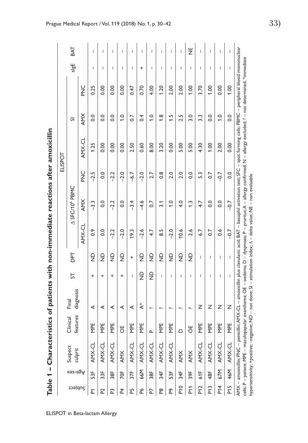
ELISPOT in Beta-lactam Allergy
33)Prague Medical Report / Vol. 119 (2018) No. 1, p. 30–42
Tabl
e 1
– C
hara
cter
isti
cs o
f pat
ient
s w
ith
non-
imm
edia
te r
eact
ions
aft
er a
mox
icill
inSubject
Age-sexSu
spec
tcu
lpri
tC
linic
al
feat
ures
Fina
l di
agno
sis
STD
PT
ELIS
POT
sIgE
BAT
Δ S
FC/1
06 PBM
CSI
AM
X-C
LA
MX
PNC
AM
X-C
LA
MX
PNC
P153
FA
MX
-CL
MPE
A+
ND
0.9
–3.3
–2.5
1.25
0.0
0.25
––
P233
FA
MX
-CL
MPE
A+
ND
0.0
0.0
0.0
0.00
0.0
0.00
––
P338
FA
MX
-CL
MPE
A+
ND
–2.2
–2.2
–2.2
0.00
0.0
0.00
––
P470
FA
MX
OE
A+
ND
–2.0
0.0
–2.0
0.00
1.0
0.00
––
P537
FA
MX
-CL
MPE
A–
+19
.3–3
.4–6
.72.
500.
70.
47–
–
P666
MA
MX
-CL
MPE
A*
ND
ND
–2.6
–4.6
–2.0
0.60
0.4
0.70
+–
P738
FA
MX
-CL
P?
ND
ND
4.7
0.7
2.7
8.00
1.0
4.00
––
P834
FA
MX
-CL
MPE
?–
ND
8.5
3.1
0.8
3.20
1.8
1.20
––
P953
FA
MX
-CL
MPE
?–
ND
–2.0
1.0
2.0
0.00
1.5
2.00
––
P10
34F
AM
XD
?–
ND
10.6
4.0
2.0
5.00
2.5
2.00
––
P11
39F
AM
XO
E?
–N
D2.
61.
30.
05.
003.
01.
00–
NE
P12
61F
AM
X-C
LM
PEN
––
6.7
4.7
5.3
4.30
3.3
3.70
––
P13
48F
AM
X-C
LM
PEN
––
0.7
0.0
0.7
1.00
0.0
1.00
––
P14
67M
AM
X-C
LM
PEN
––
0.6
0.0
–0.7
2.00
1.0
0.00
––
P15
46M
AM
X-C
LM
PEN
––
–0.7
–0.7
0.0
0.00
0.0
1.00
––
AM
X –
am
oxic
illin
; PN
C –
pen
icill
in; A
MX
-CL
– am
oxic
illin
plu
s cl
avul
anic
aci
d; B
AT –
bas
ophi
l act
ivat
ion
test
; SFC
– s
pot
form
ing
cells
; PBM
C –
per
iphe
ral b
lood
mon
onuc
lear
ce
lls; P
– p
atie
nt; M
PE –
mac
ulop
apul
ar e
xant
hem
a; O
E –
oede
ma;
D –
dys
pnoe
a; P
– pr
uritu
s; A
– a
llerg
y co
nfirm
ed; N
– a
llerg
y ex
clud
ed; ?
– n
ot d
eter
min
ed; *
imm
edia
te
hype
rsen
sitiv
ity; +
posi
tive;
–ne
gativ
e; N
D –
not
don
e; S
I – s
timul
atio
n in
dex;
Δ –
del
ta v
alue
; NE
– no
n ev
alua
ble
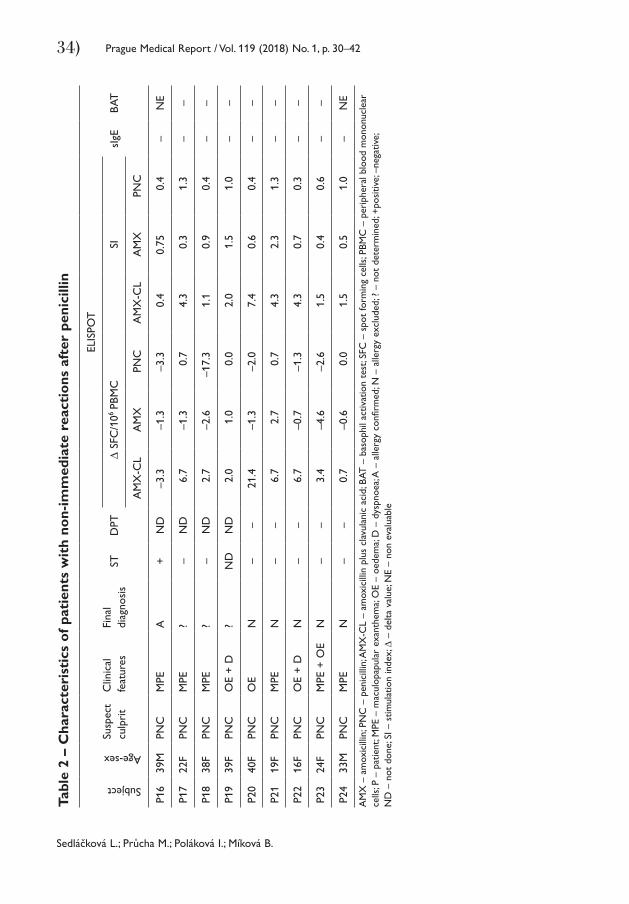
Sedláčková L.; Průcha M.; Poláková I.; Míková B.
34) Prague Medical Report / Vol. 119 (2018) No. 1, p. 30–42Ta
ble
2 –
Cha
ract
eris
tics
of p
atie
nts
wit
h no
n-im
med
iate
rea
ctio
ns a
fter
pen
icill
in
Subject
Age-sex
Susp
ect
culp
rit
Clin
ical
fe
atur
esFi
nal
diag
nosi
sST
DPT
ELIS
POT
sIgE
BAT
Δ S
FC/1
06 PB
MC
SI
AM
X-C
LA
MX
PNC
AM
X-C
LA
MX
PNC
P16
39M
PNC
MPE
A+
ND
–3.3
–1.3
–3.3
0.4
0.75
0.4
–N
E
P17
22F
PNC
MPE
?–
ND
6.7
–1.3
0.7
4.3
0.3
1.3
––
P18
38F
PNC
MPE
?–
ND
2.7
–2.6
–17.
31.
10.
90.
4–
–
P19
39F
PNC
OE
+ D
?N
DN
D2.
01.
00.
02.
01.
51.
0–
–
P20
40F
PNC
OE
N–
–21
.4–1
.3–2
.07.
40.
60.
4–
–
P21
19F
PNC
MPE
N–
–6.
72.
70.
74.
32.
31.
3–
–
P22
16F
PNC
OE
+ D
N–
–6.
7–0
.7–1
.34.
30.
70.
3–
–
P23
24F
PNC
MPE
+ O
EN
––
3.4
–4.6
–2.6
1.5
0.4
0.6
––
P24
33M
PNC
MPE
N–
–0.
7–0
.60.
01.
50.
51.
0–
NE
AM
X –
am
oxic
illin
; PN
C –
pen
icill
in; A
MX
-CL
– am
oxic
illin
plu
s cl
avul
anic
aci
d; B
AT –
bas
ophi
l act
ivat
ion
test
; SFC
– s
pot
form
ing
cells
; PBM
C –
per
iphe
ral b
lood
mon
onuc
lear
ce
lls; P
– p
atie
nt; M
PE –
mac
ulop
apul
ar e
xant
hem
a; O
E –
oede
ma;
D –
dys
pnoe
a; A
– a
llerg
y co
nfirm
ed; N
– a
llerg
y ex
clud
ed; ?
– n
ot d
eter
min
ed; +
posi
tive;
–ne
gativ
e;
ND
– n
ot d
one;
SI –
stim
ulat
ion
inde
x; Δ
– d
elta
val
ue; N
E –
non
eval
uabl
e
ELISPOT in Beta-lactam Allergy
35)Prague Medical Report / Vol. 119 (2018) No. 1, p. 30–42
Table 3 – Characteristics of healthy controlsSu
bjec
t
Age
-sex
Tolerated beta-lactam
ELISPOT
sIgE BATΔ SFC/106 PBMC SI
AMX-CL AMX PNC AMX-CL AMX PNC
HC1 54F AMX-CL 7.8 –2.2 –10.0 0.0 0.8 0.0 – –
HC2 60F AMX-CL 6.7 –3.3 –10.0 1.3 0.8 0.5 – –
HC3 50F AMX-CL 2.0 0.6 –0.7 4.0 2.0 0.0 – –
HC4 36F AMX-CL 1.3 0.7 0.7 0.0 0.0 0.0 – –
HC5 44F PNC 0.0 0.0 0.0 0.0 0.0 0.0 – –
HC6 21F PNC –1.4 –1.4 0.0 0.5 0.5 1.0 – –
AMX – amoxicillin; PNC – penicillin; AMX-CL – amoxicillin plus clavulanic acid; BAT – basophil activation test; SFC – spot forming cells; PBMC – peripheral blood mononuclear cells; HC – healthy control; SI – stimulation index; Δ – delta value; +positive; –negative
Blood was drawn into a Cell Preparation Tube (Vacutainer® CPTTM Heparin, Becton Dickinson). Peripheral blood mononuclear cells (PBMC) were isolated after centrifugation at 1500 g and their concentration was adjusted to 5×10⁶ cells/ml by adding serum-free CTL TestTM Medium (C.T.L. Europe GmbH, Bonn, Germany). The assay was performed in triplicate. PBMC (100 μl, 5×10⁵/well) were incubated with benzylpenicillin (Penicilin G draselna sol® Biotika, 100 μl, concentration 5×10⁵ IU/ml), AMX-CL (Amoksiklav® Lek Pharmaceuticals, 100 μl, concentration 1 mg/ml) and AMX (Amoxicillin for skin tests, Diater, 100 μl, concentration 1 mg/ml) for 24 hours at 37 °C in 5% CO2. The antibiotic concentration used was adapted from Rozieres et al. (2009). As a negative control, cells were incubated with medium (100 μl CTL medium), and phytohemagglutinin (PHA, Sigma Aldrich, St Louis, USA, 100 μl, concentration 5 μg/ml) stimulation was used as a positive control. The cells were then removed by two washes with PBS (phosphate buffered saline) and two washes with PBS – 0.05% Tween-20, IFN-γ detection antibody was added for 2 hours, the wells were washed three times with PBS – 0.05% Tween-20. Streptavidin-AP Solution was added for 30 min, followed by two washing steps with PBS – 0.05% Tween-20 and with distilled water. Spots were developed using the Developer Solution and the reaction was stopped by washing three times with tap water. Spot forming cells (SFC) were counted by a CTL-ImmunoSpot S5 UV Analyzer (C.T.L. Europe GmbH, Bonn, Germany). Results were expressed as: a) the number of IFN-γ SFC/106 PBMC for the unstimulated condition (medium) and stimulation with each antibiotic (AMX-CL, AMX and PNC); b) delta (Δ) values, the difference between the response value in the presence of antibiotic and the unstimulated condition value (Tables 1–3); c) stimulation index (SI), response value in the presence of the antibiotic divided by the unstimulated value in each participant (Tables 1–3).
Sedláčková L.; Průcha M.; Poláková I.; Míková B.
36) Prague Medical Report / Vol. 119 (2018) No. 1, p. 30–42
Specific IgE to penicilloyl G, penicilloyl V, ampicilloyl and amoxicilloyl was performed by ImmunoCAP assay (ThermoFisher Scientific, Uppsala, Sweden).
BAT was performed by the FlowCAST assay (Bühlmann, Basel, Switzerland), with benzylpenicilloyl-L-octa-lysin, sodium benzylpenilloate and sodium amoxicillin (Diater, Madrid, Spain), penicillin G (Penicilin G draselna sol®, Biotika), ampicillin (Ampicilin®, Biotika) and amoxicillin plus clavulanic acid (Amoksiklav®, Lek Pharmaceuticals). BAT was analysed by a flow cytometer FACS Calibur (BD Biosciences, San Jose, USA).
Follow-upClinical evaluation by skin and provocation tests followed in one subgroup of patients. Skin tests (ST) with major and minor penicillin determinant (benzylpenicilloyl-L-octa-lysin and sodium benzylpenilloate, DAP Diater, Madrid, Spain), penicillin G (Penicilin G draselna sol®, Biotika, 10000 IU/ml) and amoxicillin + clavulanic acid (Amoksiklav® Lek Pharmaceuticals, 20 mg/ml) were performed in 21 patients according to the ENDA and DAIG of EAACI recommendations (Brockow et al., 2013). A drug provocation test (DPT) with culprit beta-lactam was performed in 10 patients; in 5 cases with oral phenoxymethylpenicillinum (V-Penicilin®, Biotika) and in 5 cases with amoxicillin + clavulanic acid (Amoksiklav®, Lek Pharmaceuticals). The DPTs were performed as described previously (Romano et al., 2004).
Statistical analysisNormality was evaluated using a chi-square test for each variable. As normal data distribution was not shown by the chi-square test, the Wilcoxon paired test was used to evaluate the significance testing between patient group results for unstimulated conditions and stimulation with each antibiotic. P-values < 0.05 were considered statistically significant.
ResultsIFN-γ ELISPOT assay results were evaluated in 24 of 25 patients from group A and in all 6 healthy subjects from group B. The number of IFN-γ producing PBMC showed strong response to PHA (positive control) in all but one patient, who was therefore excluded from further analyses. Antibiotic response was generally weak, very close to the unstimulated condition (incubation with medium alone). Results expressed as delta (Δ) values (number of IFN-γ SFC/106 PBMC cultured with each antibiotic after subtraction of values obtained from PBMC cultured with medium only) are presented in Table 1 for patients reacting to amoxicillin, Table 2 for patients reacting to penicillin and Table 3 for healthy controls tolerating amoxicillin or penicillin. Often the number of IFN-γ SFC was slightly higher in wells with medium than with the antibiotic, and therefore some delta values were less than zero. The stimulation indices (SI) were also calculated (Tables 1–3) and data
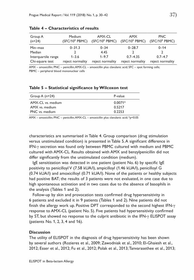
ELISPOT in Beta-lactam Allergy
37)Prague Medical Report / Vol. 119 (2018) No. 1, p. 30–42
Table 4 – Characteristics of results
Group A(n=24)
Medium (SFC/106 PBMC)
AMX-CL (SFC/106 PBMC)
AMX (SFC/106 PBMC)
PNC (SFC/106 PBMC)
Min–maxMedianInterquartile rangeChi-square test
0–31.32
1–3.6reject normality
0–344.451–9.7
reject normality
0–28.72
0.7–4.35reject normality
0–142
0.7–4.7reject normality
AMX – amoxicillin; PNC – penicillin; AMX-CL – amoxicillin plus clavulanic acid; SFC – spot forming cells; PBMC – peripheral blood mononuclear cells
Table 5 – Statistical significance by Wilcoxon test
Group A (n=24) P-value
AMX-CL vs. mediumAMX vs. mediumPNC vs. medium
0.0071*0.52170.2253
AMX – amoxicillin; PNC – penicillin; AMX-CL – amoxicillin plus clavulanic acid; *p<0.05
characteristics are summarised in Table 4. Group comparison (drug stimulation versus unstimulated condition) is presented in Table 5. A significant difference in IFN-γ secretion was found only between PBMC cultured with medium and PBMC cultured with AMX-CL. Results obtained with AMX and benzylpenicillin did not differ significantly from the unstimulated condition (medium).
IgE sensitization was detected in one patient (patient No. 6) by specific IgE positivity to penicilloyl V (7.58 kUA/l), ampicilloyl (1.46 kUA/l), penicilloyl G (0.74 kUA/l) and amoxicilloyl (0.71 kUA/l). None of the patients or healthy subjects had positive BAT; the results of 3 patients were not evaluated, in one case due to high spontaneous activation and in two cases due to the absence of basophils in the analysis (Tables 1 and 2).
Follow-up by skin and provocation tests confirmed drug hypersensitivity in 6 patients and excluded it in 9 patients (Tables 1 and 2). Nine patients did not finish the allergy work up. Positive DPT corresponded to the second highest IFN-γ response to AMX-CL (patient No. 5). Five patients had hypersensitivity confirmed by ST, but showed no response to the culprit antibiotic in the IFN-γ ELISPOT assay (patients No. 1, 2, 3, 4 and 16).
DiscussionThe utility of ELISPOT in the diagnosis of drug hypersensitivity has been shown by several authors (Rozieres et al., 2009; Zawodniak et al., 2010; El-Ghaiesh et al., 2012; Esser et al., 2012; Fu et al., 2012; Polak et al., 2013; Tanvarasethee et al., 2013;
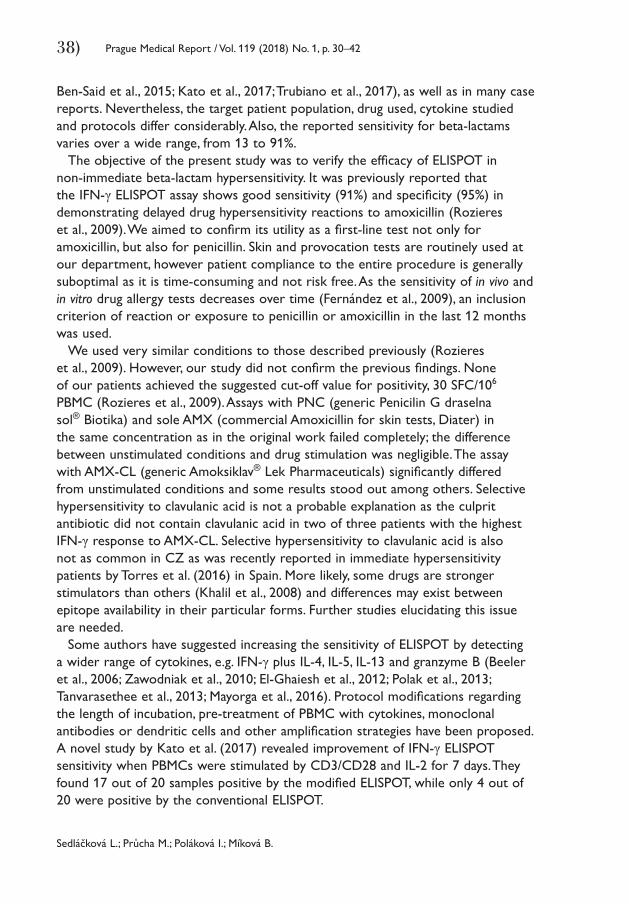
Sedláčková L.; Průcha M.; Poláková I.; Míková B.
38) Prague Medical Report / Vol. 119 (2018) No. 1, p. 30–42
Ben-Said et al., 2015; Kato et al., 2017; Trubiano et al., 2017), as well as in many case reports. Nevertheless, the target patient population, drug used, cytokine studied and protocols differ considerably. Also, the reported sensitivity for beta-lactams varies over a wide range, from 13 to 91%.
The objective of the present study was to verify the efficacy of ELISPOT in non-immediate beta-lactam hypersensitivity. It was previously reported that the IFN-γ ELISPOT assay shows good sensitivity (91%) and specificity (95%) in demonstrating delayed drug hypersensitivity reactions to amoxicillin (Rozieres et al., 2009). We aimed to confirm its utility as a first-line test not only for amoxicillin, but also for penicillin. Skin and provocation tests are routinely used at our department, however patient compliance to the entire procedure is generally suboptimal as it is time-consuming and not risk free. As the sensitivity of in vivo and in vitro drug allergy tests decreases over time (Fernández et al., 2009), an inclusion criterion of reaction or exposure to penicillin or amoxicillin in the last 12 months was used.
We used very similar conditions to those described previously (Rozieres et al., 2009). However, our study did not confirm the previous findings. None of our patients achieved the suggested cut-off value for positivity, 30 SFC/106 PBMC (Rozieres et al., 2009). Assays with PNC (generic Penicilin G draselna sol® Biotika) and sole AMX (commercial Amoxicillin for skin tests, Diater) in the same concentration as in the original work failed completely; the difference between unstimulated conditions and drug stimulation was negligible. The assay with AMX-CL (generic Amoksiklav® Lek Pharmaceuticals) significantly differed from unstimulated conditions and some results stood out among others. Selective hypersensitivity to clavulanic acid is not a probable explanation as the culprit antibiotic did not contain clavulanic acid in two of three patients with the highest IFN-γ response to AMX-CL. Selective hypersensitivity to clavulanic acid is also not as common in CZ as was recently reported in immediate hypersensitivity patients by Torres et al. (2016) in Spain. More likely, some drugs are stronger stimulators than others (Khalil et al., 2008) and differences may exist between epitope availability in their particular forms. Further studies elucidating this issue are needed.
Some authors have suggested increasing the sensitivity of ELISPOT by detecting a wider range of cytokines, e.g. IFN-γ plus IL-4, IL-5, IL-13 and granzyme B (Beeler et al., 2006; Zawodniak et al., 2010; El-Ghaiesh et al., 2012; Polak et al., 2013; Tanvarasethee et al., 2013; Mayorga et al., 2016). Protocol modifications regarding the length of incubation, pre-treatment of PBMC with cytokines, monoclonal antibodies or dendritic cells and other amplification strategies have been proposed. A novel study by Kato et al. (2017) revealed improvement of IFN-γ ELISPOT sensitivity when PBMCs were stimulated by CD3/CD28 and IL-2 for 7 days. They found 17 out of 20 samples positive by the modified ELISPOT, while only 4 out of 20 were positive by the conventional ELISPOT.
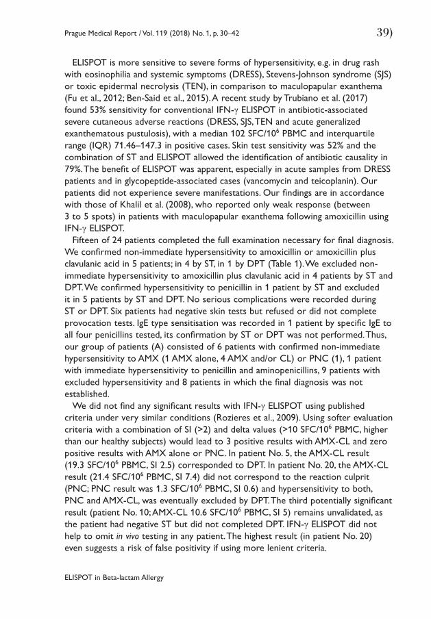
ELISPOT in Beta-lactam Allergy
39)Prague Medical Report / Vol. 119 (2018) No. 1, p. 30–42
ELISPOT is more sensitive to severe forms of hypersensitivity, e.g. in drug rash with eosinophilia and systemic symptoms (DRESS), Stevens-Johnson syndrome (SJS) or toxic epidermal necrolysis (TEN), in comparison to maculopapular exanthema (Fu et al., 2012; Ben-Said et al., 2015). A recent study by Trubiano et al. (2017) found 53% sensitivity for conventional IFN-γ ELISPOT in antibiotic-associated severe cutaneous adverse reactions (DRESS, SJS, TEN and acute generalized exanthematous pustulosis), with a median 102 SFC/106 PBMC and interquartile range (IQR) 71.46–147.3 in positive cases. Skin test sensitivity was 52% and the combination of ST and ELISPOT allowed the identification of antibiotic causality in 79%. The benefit of ELISPOT was apparent, especially in acute samples from DRESS patients and in glycopeptide-associated cases (vancomycin and teicoplanin). Our patients did not experience severe manifestations. Our findings are in accordance with those of Khalil et al. (2008), who reported only weak response (between 3 to 5 spots) in patients with maculopapular exanthema following amoxicillin using IFN-γ ELISPOT.
Fifteen of 24 patients completed the full examination necessary for final diagnosis. We confirmed non-immediate hypersensitivity to amoxicillin or amoxicillin plus clavulanic acid in 5 patients; in 4 by ST, in 1 by DPT (Table 1). We excluded non-immediate hypersensitivity to amoxicillin plus clavulanic acid in 4 patients by ST and DPT. We confirmed hypersensitivity to penicillin in 1 patient by ST and excluded it in 5 patients by ST and DPT. No serious complications were recorded during ST or DPT. Six patients had negative skin tests but refused or did not complete provocation tests. IgE type sensitisation was recorded in 1 patient by specific IgE to all four penicillins tested, its confirmation by ST or DPT was not performed. Thus, our group of patients (A) consisted of 6 patients with confirmed non-immediate hypersensitivity to AMX (1 AMX alone, 4 AMX and/or CL) or PNC (1), 1 patient with immediate hypersensitivity to penicillin and aminopenicillins, 9 patients with excluded hypersensitivity and 8 patients in which the final diagnosis was not established.
We did not find any significant results with IFN-γ ELISPOT using published criteria under very similar conditions (Rozieres et al., 2009). Using softer evaluation criteria with a combination of SI (>2) and delta values (>10 SFC/106 PBMC, higher than our healthy subjects) would lead to 3 positive results with AMX-CL and zero positive results with AMX alone or PNC. In patient No. 5, the AMX-CL result (19.3 SFC/106 PBMC, SI 2.5) corresponded to DPT. In patient No. 20, the AMX-CL result (21.4 SFC/106 PBMC, SI 7.4) did not correspond to the reaction culprit (PNC; PNC result was 1.3 SFC/106 PBMC, SI 0.6) and hypersensitivity to both, PNC and AMX-CL, was eventually excluded by DPT. The third potentially significant result (patient No. 10; AMX-CL 10.6 SFC/106 PBMC, SI 5) remains unvalidated, as the patient had negative ST but did not completed DPT. IFN-γ ELISPOT did not help to omit in vivo testing in any patient. The highest result (in patient No. 20) even suggests a risk of false positivity if using more lenient criteria.
Sedláčková L.; Průcha M.; Poláková I.; Míková B.
40) Prague Medical Report / Vol. 119 (2018) No. 1, p. 30–42
The present study has some limitations. The study design reflected the intended use of IFN-γ ELISPOT as a first-line test, so patient enrolment was based on clinical suspicion and not on hypersensitivity confirmation by ST and DPT. Therefore, the patient group was “diluted” by subjects without true hypersensitivity and the number of definitively confirmed cases was small. The eight patients lost to follow up reflect the common issue of poor compliance with complicated time-consuming procedures and concerns about safety. We did not create our own cut-off values for IFN-γ ELISPOT evaluation. As normal data distribution was not shown, we were unable to use mean +2 SD (standard deviation) to establish cut-off values using assays incubated under unstimulated conditions. Moreover, due to the small number of healthy participants in the present study, it would be inappropriate to create cut-offs from antibiotic assays in these subjects.
We did not assess the exact sensitivity and specificity of the IFN-γ ELISPOT method for each antibiotic studied due to the small number of patients with a final diagnosis. According to our findings, the sensitivity of the assay appears to be much lower than the optimistic expectations based on the work of Rozieres et al. (2009). With 1 potentially positive result in 6 confirmed hypersensitivity cases, we may assume an approximate sensitivity to be less than 20% in mild non-immediate hypersensitivity reactions to penicillin and amoxicillin using an assay with AMX-CL. The failure of PNC and AMX alone requires further research focused on the particular form of beta-lactam employed in the assay.
ConclusionOur results did not support the utility of IFN-γ ELISPOT in the diagnosis of mild non-immediate hypersensitivity to amoxicillin and penicillin in daily practice. The first-line diagnostic approach remains a combination of detailed history and in vivo tests. Skin tests are the most useful tests for allergy confirmation. After ST negativity or even without performing ST in mild childhood MPE (maculopapular exanthema), the gold standard of drug allergy diagnosis is a drug provocation test. DPT is considerably safe in non-severe non-immediate cases and it is the only test able to exclude hypersensitivity. Possible benefits of in vitro tests including IFN-γ ELISPOT in more severe forms of non-immediate beta-lactam hypersensitivity (DRESS, SJS, TEN) or in patients with contraindications to ST and DPT due to comorbidities requires further research and validation. Sensitivity enhancement of ELISPOT by protocol modification may be helpful.
ReferencesBeeler, A., Engler, O., Gerber, B. O., Pichler, W. J. (2006) Long-lasting reactivity and high frequency of
drug-specific T cells after severe systemic drug hypersensitivity reactions. J. Allergy Clin. Immunol. 117(2), 455–462.
Ben-Said, B., Arnaud-Butel, S., Rozières, A., Rodet, K., Bérard, F., Nicolas, J. F., Nosbaum, A. (2015) Allergic delayed drug hypersensitivity is more frequently diagnosed in drug reaction, eosinophilia and systemic
ELISPOT in Beta-lactam Allergy
41)Prague Medical Report / Vol. 119 (2018) No. 1, p. 30–42
symptoms (DRESS) syndrome than in exanthema induced by beta-lactam antibiotics. J. Dermatol. Sci. 80(1), 71–74.
Blanca, M., Romano, A., Torres, M. J., Férnandez, J., Mayorga, C., Rodriguez, J., Demoly, P., Bousquet, P. J., Merk, H. F., Sanz, M. L., Ott, H., Atanaskovic-Markovic, M. (2009) Update on the evaluation of hypersensitivity reactions to betalactams. Allergy 64, 183–193.
Brockow, K., Garvey, L. H., Aberer, W., Atanaskovic-Markovic, M., Barbaud, A., Bilo, M. B., Bircher, A., Blanca, M., Bonadonna, B., Campi, P., Castro, E., Cernadas, J. R., Chiriac, A. M., Demoly, P., Grosber, M., Gooi, J., Lombardo, C., Mertes, P. M., Mosbech, H., Nasser, S., Pagani, M., Ring, J., Romano, A., Scherer, K., Schnyder, B., Testi, S., Torres, M., Trautmann, A., Terreehorst, I.; ENDA/EAACI Drug Allergy Interest Group (2013) Skin test concentrations for systemically administered drugs – an ENDA/EAACI Drug Allergy Interest Group position paper. Allergy 68(6), 702–712.
Demoly, P., Adkinson, N. F., Brockow, K., Castells, M., Chiriac, A. M., Greenberger, P. A., Khan, D. A., Lang, D. M., Park, H. S., Pichler, W., Sanchez-Borges, M., Shiohara, T., Thong, B. Y. (2014) International Consensus on drug allergy. Allergy 69, 420–437.
Ebo, D. G., Leysen, J., Mayorga, C., Rozieres, A., Knol, E. F., Terreehorst, I. (2011) The in vitro diagnosis of drug allergy: status and perspectives. Allergy 66, 1275–1286.
El-Ghaiesh, S., Monshi, M. M., Whitaker, P., Jenkins, R., Meng, X., Farrell, J., Elsheikh, A., Peckham, D., French, N., Pirmohamed, M., Park, B. K., Naisbitt, D. J. (2012) Characterization of the antigen specificity of T-cell clones from piperacillin-hypersensitive patients with cystic fibrosis. J. Pharmacol. Exp. Ther. 341(3), 597–610.
Esser, S., Jablonka, R., Heinemann, F. M., Reuter, S., Jaeger, H., von Krosigk, A., Schenk-Westkamp, P., Schadendorf, D., Horn, P. A., Lindemann, M. (2012) Detection of abacavir hypersensitivity by ELISpot method. Inflamm. Allergy Drug Targets 11(3), 227–234.
Fernández, T. D., Torres, M. J., Blanca-López, N., Rodríguez-Bada, J. L., Gomez, E., Canto, G., Mayorga, C., Blanca, M. (2009) Negativization rates of IgE radioimmunoassay and basophil activation test in immediate reactions to penicillins. Allergy 64(2), 242–248.
Fu, M., Gao, Y., Pan, Y., Li, W., Liao, W., Wang, G., Li, C., Li, C., Gao, T., Liu, Y. (2012) Recovered patients with Stevens-Johnson syndrome and toxic epidermal necrolysis maintain long-lived IFN-γ and sFasL memory response. PLoS One 7(9), e45516.
Kato, K., Kawase, A., Azukizawa, H., Hanafusa, T., Nakagawa, Y., Murota, H., Sakaguchi, S., Asada, H., Katayama, I. (2017) Novel interferon-γ enzyme-linked immunoSpot assay using activated cells for identifying hypersensitivity-inducing drug culprits. J. Dermatol. Sci. 86(3), 222–229.
Khalil, G., El-Sabban, M., Al-Ghadban, S., Azzi, S., Shamra, S., Khalifé, S., Maroun, R. (2008) Cytokine expression profile of sensitized human T lymphocytes following in vitro stimulation with amoxicillin. Eur. Cytokine Netw. 19(3), 131–141.
Macy, E., Contreras, R. (2014) Health care use and serious infection prevalence associated with penicillin “allergy” in hospitalized patients: a cohort study. J. Allergy Clin. Immunol. 133, 790–796.
Mayorga, C., Celik, G., Rouzaire, P., Whitaker, P., Bonadonna, P., Rodrigues-Cernadas, J., Vultaggio, A., Brockow, K., Caubet, J. C., Makowska, J., Nakonechna, A., Romano, A., Montanez, M. I., Laguna, J. J., Zanoni, G., Gueant, J. L., Oude Elberink, H., Fernandez, J., Viel, S., Demoly, P., Torres, M. J.; In vitro tests for Drug Allergy Task Force of EAACI Drug Interest Group (2016) In vitro tests for drug hypersensitivity reactions: an ENDA/EAACI Drug Allergy Interest Group position paper. Allergy 71, 1103–1134.
Polak, M. E., Belgi, G., McGuire, C., Pickard, C., Healy, E., Friedmann, P. S., Ardern-Jones, M. R. (2013) In vitro diagnostic assays are effective during the acute phase of delayed-type drug hypersensitivity reactions. Br. J. Dermatol. 168(3), 539–549.
Sedláčková L.; Průcha M.; Poláková I.; Míková B.
42) Prague Medical Report / Vol. 119 (2018) No. 1, p. 30–42
Porebski, G., Czarnobilska, E. (2015) Drug-specific in vitro secretion of IFNγ in the diagnosis of drug-induced exanthemas: electrochemiluminiscence assay versus previously used diagnostic methods. Przegl. Lek. 72(12), 721–724.
Romano, A., Blanca, M., Torres, M. J., Bircher, A., Aberer, W., Brockow, K., Pichler, W. J., Demoly, P. (2004) Diagnosis of nonimmediate reactions to beta-lactam antibiotics. Allergy 59(11), 1153–1160.
Rozieres, A., Hennino, A., Rodet, K., Gutowski, M. C., Gunera-Saad, N., Berard, F., Cozon, G., Bienvenu, J., Nicolas, J. F. (2009) Detection and quantification of drug-specific T cells in penicillin allergy. Allergy 64(4), 534–542.
Solensky, R. (2014) Penicillin allergy as a public health measure. J. Allergy Clin. Immunol. 133, 797–798.Tanvarasethee, B., Buranapraditkun, S., Klaewsongkram, J. (2013) The potential of using enzyme-linked
immunospot to diagnose cephalosporin-induced maculopapular exanthems. Acta Derm. Venereol. 93(1), 66–69.
Torres, M. J., Montanez, M. I., Ariza, A., Salas, M., Fernandez, T. D., Berbero, N., Mayorga, C., Blanca, M. (2016) The role of IgE recognition in allergic reactions to amoxicillin and clavulanic acid. Clin. Exp. Allergy 46, 264–274.
Trubiano, J. A., Strautins, K., Redwood, A. J., Pavlos, R., Konvinse, K. C., Aung, A. K., Slavin, M. A., Thursky, K. A., Grayson, M. L., Phillips, E. J. (2017) The combined utility of ex vivo IFN-γ release enzyme-linked immunoSpot assay and in vivo skin testing in patients with antibiotic-associated severe cutaneous adverse reactions. J. Allergy Clin. Immunol. Pract. (Epub ahead of print)
Zawodniak, A., Lochmatter, P., Yerly, D., Kawabata, T., Lerch, M., Yawalkar, N., Pichler, W. J. (2010) In vitro detection of cytotoxic T and NK cells in peripheral blood of patients with various drug-induced skin diseases. Allergy 65(3), 376–384.
Dental Implant Osseointegration Influenced by Age
43)Prague Medical Report / Vol. 119 (2018) No. 1, p. 43–51
Chronological Age as Factor Influencing the Dental Implant Osseointegration in the Jaw BoneJakub Papež1, Taťjana Dostálová1, Karel Chleborád1, Pavel Kříž1, Jakub Strnad2
1Department of Stomatology, Second Faculty of Medicine, Charles University and University Hospital Motol, Prague, Czech Republic;2LASAK, Prague, Czech Republic
Rece ived October 19 , 2017 ; Accepted March 19 , 2018 .
Key words: Chronological age – Dental implant – Osseointegration – Marginal bone level – Intraoral X-rays
Abstract: The objectives of this study were to evaluate osseointegration of dental implant in the jaw bone in the young and elderly population and comparing the results to assess indicators and risk factors as age for the success or failure of dental implants. A retrospective study of 107 implants (Impladent, LASAK, Czech Republic) was prepared. The patients at implants surgery were divided in three groups. The patients were followed-up for a 7-year period. We evaluated osseointegration from long term point of view as a change of marginal bone levels close to dental implant. Marginal bone levels were recorded and analysed with regard to different patient- and implant-related factors. An influence of chronological age on change of marginal bone levels during 6-year retrospective study vas evaluated. The study examined 47 patient charts and 107 implants from the Second Faculty of Medicine, Charles University and University Hospital Motol. We proved that young healthy patients with long bridges or Branemarks have the same progression of marginal bone levels changes. The chronological age hasn’t therefore direct influence on the osseointegration from long term point of view. But we found that the length of dental suprastrucure-prosthetic construction negatively influences marginal bone changes, though these results weren’t
https://doi.org/10.14712/23362936.2018.4 © 2018 The Authors. This is an open-access article distributed under the terms of the Creative Commons Attribution License (http://creativecommons.org/licenses/by/4.0).
This study was supported by project 00064203 (FN MOTOL).
Mailing Address: Prof. Taťjana Dostálová, MD., DSc., MBA, Department of Stomatology, Second Faculty of Medicine, Charles University and University Hospital Motol, V Úvalu 84, 150 06 Prague 5, Czech Republic; Mobile Phone: +420 728 970 059; e-mail: [email protected]
Papež J.; Dostálová T.; Chleborád K.; Kříž P.; Strnad J.
44) Prague Medical Report / Vol. 119 (2018) No. 1, p. 43–51
statistically significant. More extensive dental implant suprastrucure undergoes smaller osseointegration. On the other hand the length of dental suprastrucure (prosthetic construction) negatively influences dental osseointegration in both groups of patient.
IntroductionOsseointegration is seen as the close contact between bone and implant without interposition of non-bone tissue (Albrektsson and Wennerberg, 2004). This concept has been described by Brånemark (1959, 1983) as consisting of a highly differentiated tissue making a direct structural and functional connection between ordered, living bone and the surface of a load-carrying implant. Osseointegration of dental implant is influenced on i.e. dental implant surface, age of patient, size, shape and extension of suprastrucure, general health condition, etc.
Various techniques of surfaces treatments have been studied and applied to improve biological surface properties, which favour the mechanism of osseointegration (Wong et al., 1995; Wennerberg and Albrektsson, 2010). This strategy aims at promoting the mechanism of osseointegration with faster and stronger bone formation, to confer better stability during the healing process, thus allowing more rapid loading of the implant (Wennerberg and Albrektsson, 2009; Beutner et al., 2010).
In our article we focused on the age of patient and extend of dental implant suprastrucure as factors influencing bone level changes (osseointegration) during the observed period. The use of dental implants in young patients is not limited, but multidisciplinary treatment planning is directly connected with skeletal maturation. It is evident that jaw growth is important for dental implants insertion. It is known that more and more implants are placed in adolescents, especially after trauma or anodontia.
By adolescent patients we can insert dental implant in the jaw bone only if the growth and skeleton maturation is finished. If implant insertion is planned in a growing child, we must accept the fact that osseointegration forms ankyloses and implants do not follow the spontaneous and continues eruption of the natural dentition (Papez et al., 2015). Similar to ankylosed teeth (Kawanami et al., 1999), the implants remain stationary in the bone and do not follow changes of the alveolar process with continuous eruption of the natural dentition (Oesterle et al., 1993; Cronin et al., 1994). This inability to move with adjacent teeth causes deficiencies in the alveolar bone and surrounding gingival tissues and leads to a discrepancy in the sagittal and transversal dimension, described as infraocclusion or infraposition of the implant (Thilander et al., 1999). Such implants may also disturb the normal development of jawbones. In order not to interfere with the growth of the jawbones, the installation of an implant should generally be postponed on average until after puberty or after the so-called growth spurt of the child (Op Heij et al., 2003). Since changes in the dentoalveolar complex are
Dental Implant Osseointegration Influenced by Age
45)Prague Medical Report / Vol. 119 (2018) No. 1, p. 43–51
of particular importance for the functional/aesthetic outcome of implants, a study by Iseri and Solow (1996) showed that between the ages of 15 and 25 years the vertical tooth movement can amount to 5 mm – a distance difficult to span with implants.
The follow-up study of dental implants in the upper adolescent region inserted in adolescent patients, has shown that continuous eruption of the adjacent teeth, even after completed dental and skeletal development, may end up in an infraoccluded implant-supported crown (Thilander et al., 1999). For that reason implant insertion in the anterior tooth area should be postponed until after the completion of the 15th year of age in girls and the 17th year of age in boys, and, therefore, it is necessary to evaluate the upper and lower jaw development. It is known, that the biological indicators of skeletal maturity refer mainly to somatic changes in puberty, thus emphasizing the strict interactions between the development of the craniofacial region and the modifications in other body regions (Baccetti et al., 2005). Individual skeletal maturity can be also assessed by means of several biological indicators: increase in body height; skeletal maturation of the hand and wrist; dental development and eruption; menarche or voice changes; and cervical vertebral maturation (Thilander et al., 1999). For that reason the identification of the pubertal growth spurt has great value in dentistry, mainly in implant insertion area. The effectiveness of a biological indicator of skeletal maturity is directly related to factors such as the ability to detect and predict the growth spurt peak without the need for additional radiation exposure and the high level of agreement between examiners for the definition of the stages (Franchi and Baccetti, 2002). On the opposite site are older patients. Increasing age is strongly associated with the risk of implant failure. Gender, hypertension, coronary artery disease, pulmonary disease, steroid therapy, chemotherapy and not being on hormone replacement therapy for postmenopausal women were not associated with a significant increase in implant failure. Generalized periodontal disease and/or severe periodontal disease negatively influenced the survival probability of the implant (Compton et al., 2017). Smoking, diabetes, head and neck radiation and postmenopausal estrogen therapy are correlated with a significantly increased failure rate (Moy et al., 2005).
Material and MethodsA retrospective study of 107 implants 47 patients (20 men and 27 women) (Impladent, LASAK, Czech Republic) was prepared. The patients at implants surgery were divided in three groups: group 1 (young patients ranged 18–25 years of age), group 2 (young patients ranged 18–25 years of age), group 3 (older patients ranged 50–60 years of age). The difference between group 1 and 2 was in number of implants and size of suprastrucure. Group 1 are young patients with short suprastrucure that means maximum one bridge connected with two implants or one implant with crown. Group 2 are young patients with extensive suprastrucure,
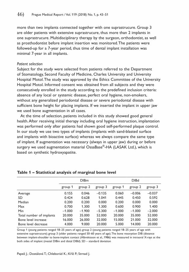
Papež J.; Dostálová T.; Chleborád K.; Kříž P.; Strnad J.
46) Prague Medical Report / Vol. 119 (2018) No. 1, p. 43–51
more than two implants connected together with one suprastrucure. Group 3 are older patients with extensive suprastrucure, thus more than 2 implants in one suprastrucure. Multidisciplinary therapy by the surgeon, orthodontist, as well as prosthodontist before implant insertion was monitored. The patients were followed-up for a 7-year period, thus time of dental implant installation was minimal 7-year in all implants.
Patient selectionSubject for the study were selected from patients referred to the Department of Stomatology, Second Faculty of Medicine, Charles University and University Hospital Motol. The study was approved by the Ethics Committee of the University Hospital Motol. Informed consent was obtained from all subjects and they were consecutively enrolled in the study according to the predefined inclusion criteria: absence of any local or systemic disease, perfect oral hygiene, non-smokers, without any generalized periodontal disease or severe periodontal disease with sufficient bone height for placing implants. If we inserted the implant in upper jaw we used bone augmentation in all cases.
At the time of selection, patients included in this study showed good general health. After receiving initial therapy including oral hygiene instruction, implantation was performed only after patients had shown good self-performed plaque control. In our study we use two types of implants (implants with sand-blasted surface and implants with bioactive surface) whereas we always compare the same type of implant. If augmentation was necessary (always in upper jaw) during or before surgery we used augmentation material OssaBase®-HA (LASAK Ltd.), which is based on synthetic hydroxyapatite.
Table 1 – Statistical analysis of marginal bone level
DIBm DIBd
group 1 group 2 group 3 group 1 group 2 group 3
AverageSDMedianMaxMinTotal number of implantsBone level increaseBone level decrease
0.1550.4240.2000.700
–1.00020.00016.0004.000
0.0460.6280.2001.300
–1.90035.00026.0009.000
–0.1351.0410.0001.300
–5.30052.00032.00020.000
0.0600.4450.2000.600
–1.00020.00015.0005.000
–0.0060.4500.0000.900
–1.00035.00021.00014.000
–0.0370.5920.0001.400
–2.00052.00032.00020.000
Group 1 (young patients ranged 18–25 years of age); group 2 (young patients ranged 18–25 years of age with extensive suprastrucure); group 3 (older patients ranged 50–60 years of age). The bone resorption DIB (distance between implant-shoulder to bone-implant contact (Albrektsson et al., 1986) was measured in intraoral X-rays at the both sides of implant (mesial DIBm and distal DIBd). SD – standard deviation
Dental Implant Osseointegration Influenced by Age
47)Prague Medical Report / Vol. 119 (2018) No. 1, p. 43–51
Treatment procedureFollowing the manufacturer’s directions, the fixtures were installed in a randomized order at the edentulous area of each patient. Individual skeletal maturity was checked using skeletal maturation of the hand and wrist by adolescent/young adult patients. In upper jaw we always used bone augmentation. After a healing period of 3 months in the mandible and 6 months in the maxilla, second surgery was performed followed after three weeks by prosthesis delivery. The implants were always inserted in bone level not submerged. During the implant insertion we used the same protocol of implantation. The CAD-CAM technique Zircon Zahn (Prettau® Zirconia, ZirconZahn Gmbhc) and BioCam (LASAK) were used to establish a supra-construction. The patients were recalled every 6 months for through professional plaque control and repeated oral hygiene training. In total, 107 implants (Impladent) were installed (Table 1).
Follow-up parametersClinical examination was conducted every 6 months. An intra-oral digital radiographs (Gendex EXPERT® DC with VistaScan Mini image plate scanner) for each patient were taken from 1 to 7-year follow-ups. The following clinical variables were recorded: pain from implant regions; implant stability; gingival inflammation; suprastrucure complications; photo and radiographic examination. A periapical digital radiograph (Gendex EXPERT® DC with VistaScan Mini image plate scanner) was taken using the parallel cone technique. We compared bone level in two periods, after fixation of final suprastructure on dental implant and after seven years from implant insertion in the bone. Preventive clinical examination followed-up regularly.
Marginal bone-level changesDuring our study we focused on marginal bone-level change thus on resorption of the marginal bone around the implant on the intraoral X-rays. The bone resorption DIB (distance between implant-shoulder to bone-implant contact) (Albrektsson et al., 1986) was measured on intraoral X-rays on the both sides of implant (mesial DIBm and distal DIBd) (Figure 1), results were calculated in millimetres with decimal point (e.g. 0.6 mm). Calibration was performed with the known length of the fixture. The reference point was the margin of the fixture in cervical part of dental implant. The measurement was done with an accuracy of half a millimetre. Final value DIB was the average DIBm a DIBd, The marginal bone increase for each type of implant was calculated using calibration of radiograph according to length of dental implant (Figure 1). Because we always used the same holder of X-ray sensor and periapical digital radiograph (Gendex EXPERT® DC with VistaScan Mini image plate scanner) with using the parallel cone technique we could set the calibration for all patients and their X-rays. We performed measurements on two sides of dental implants, precisely on mesial and distal aspect of implants; all results
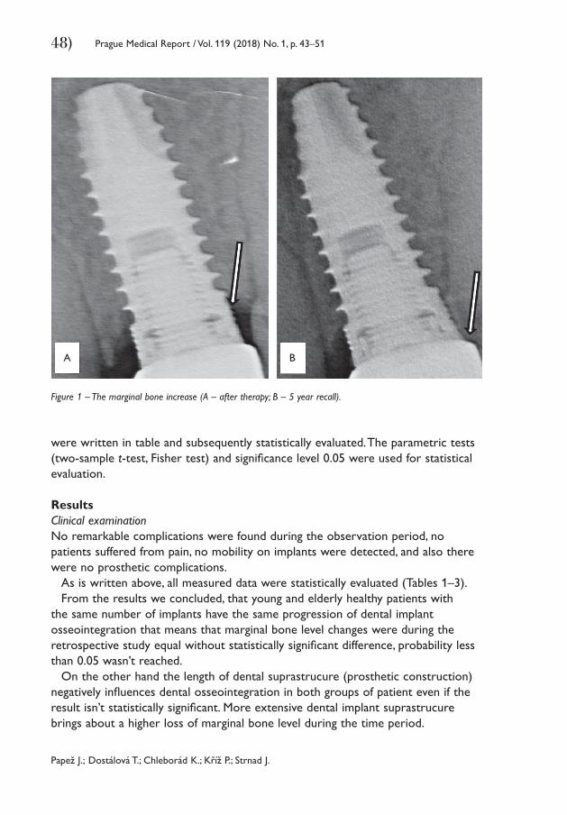
Papež J.; Dostálová T.; Chleborád K.; Kříž P.; Strnad J.
48) Prague Medical Report / Vol. 119 (2018) No. 1, p. 43–51
were written in table and subsequently statistically evaluated. The parametric tests (two-sample t-test, Fisher test) and significance level 0.05 were used for statistical evaluation.
ResultsClinical examinationNo remarkable complications were found during the observation period, no patients suffered from pain, no mobility on implants were detected, and also there were no prosthetic complications.
As is written above, all measured data were statistically evaluated (Tables 1–3).From the results we concluded, that young and elderly healthy patients with
the same number of implants have the same progression of dental implant osseointegration that means that marginal bone level changes were during the retrospective study equal without statistically significant difference, probability less than 0.05 wasn’t reached.
On the other hand the length of dental suprastrucure (prosthetic construction) negatively influences dental osseointegration in both groups of patient even if the result isn’t statistically significant. More extensive dental implant suprastrucure brings about a higher loss of marginal bone level during the time period.
Figure 1 – The marginal bone increase (A – after therapy; B – 5 year recall).
A B
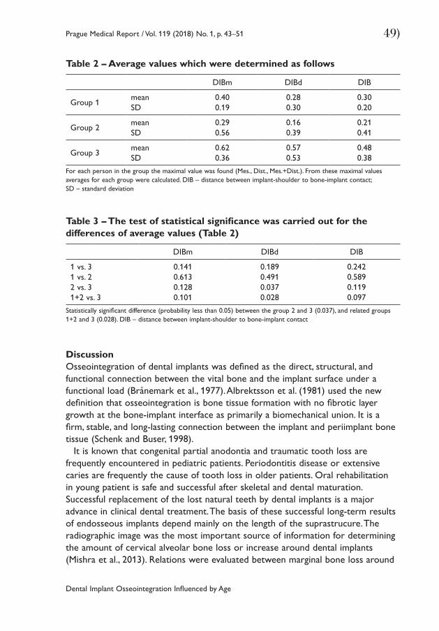
Dental Implant Osseointegration Influenced by Age
49)Prague Medical Report / Vol. 119 (2018) No. 1, p. 43–51
DiscussionOsseointegration of dental implants was defined as the direct, structural, and functional connection between the vital bone and the implant surface under a functional load (Brånemark et al., 1977). Albrektsson et al. (1981) used the new definition that osseointegration is bone tissue formation with no fibrotic layer growth at the bone-implant interface as primarily a biomechanical union. It is a firm, stable, and long-lasting connection between the implant and periimplant bone tissue (Schenk and Buser, 1998).
It is known that congenital partial anodontia and traumatic tooth loss are frequently encountered in pediatric patients. Periodontitis disease or extensive caries are frequently the cause of tooth loss in older patients. Oral rehabilitation in young patient is safe and successful after skeletal and dental maturation. Successful replacement of the lost natural teeth by dental implants is a major advance in clinical dental treatment. The basis of these successful long-term results of endosseous implants depend mainly on the length of the suprastrucure. The radiographic image was the most important source of information for determining the amount of cervical alveolar bone loss or increase around dental implants (Mishra et al., 2013). Relations were evaluated between marginal bone loss around
Table 2 – Average values which were determined as follows
DIBm DIBd DIB
Group 1meanSD
0.400.19
0.280.30
0.300.20
Group 2meanSD
0.290.56
0.160.39
0.210.41
Group 3meanSD
0.620.36
0.570.53
0.480.38
For each person in the group the maximal value was found (Mes., Dist., Mes.+Dist.). From these maximal values averages for each group were calculated. DIB – distance between implant-shoulder to bone-implant contact; SD – standard deviation
Table 3 – The test of statistical significance was carried out for the differences of average values (Table 2)
DIBm DIBd DIB
1 vs. 31 vs. 22 vs. 31+2 vs. 3
0.1410.6130.1280.101
0.1890.4910.0370.028
0.2420.5890.1190.097
Statistically significant difference (probability less than 0.05) between the group 2 and 3 (0.037), and related groups 1+2 and 3 (0.028). DIB – distance between implant-shoulder to bone-implant contact
Papež J.; Dostálová T.; Chleborád K.; Kříž P.; Strnad J.
50) Prague Medical Report / Vol. 119 (2018) No. 1, p. 43–51
implants and the level of the first thread with other systems after 12 months (Kopecká and Šimůnek, 2015). The retrospective study “The success rate of dental implants in elderly” compares the success rate of implants in the edentulous lower jaw in elderly and younger patients. The principal finding of this study was that the long-term success rate of the implants in elderly patients was not lower than that of you with younger patients (Albrektsson et al., 1986). In our article we confirmed that young healthy patient and old healthy patient with the same length of suprastrucure have the same progression of marginal bone loss thus chronological age hasn’t direct influence on dental implant osseointegration.
Implants are an alternative to orthodontic space closure, auto transplantation, and conventional prosthetic replacement (Schrotenboer et al., 2008). Implant-supported CAD CAM crowns achieved the best possible long-term result from an aesthetic point of view, and with the least possible distress and suffering for the patient (Behr et al., 2008). Our contribution confirmed the fact that an age is not the decisive factor for implant placement. Only a dental stage indicating fully erupted permanent teeth and skeletal maturation protects dental rehabilitation against infraocclusion of the implant-supported crown.
ConclusionOsseointegration is necessary for successful dental implant insertion in the jaw bone and ensures the stability of dental implant from long term point of view. As we know from our results when we compare healthy young and healthy old patients the marginal bone loss in the mentioned groups does not show statistically significant differences.
The length of dental suprastrucure-prosthetic construction negatively influences dental osseointegration; more precisely it can progressively change the alveolar bone around the implant during the observed time period. More extensive dental implant suprastrucure has a negative influence on the marginal bone levels.
ReferencesAlbrektsson, T., Wennerberg, A. (2004) Oral implant surfaces: Part 1 – Review focusing on topographic and
chemical properties of different surfaces and in vivo responses to them. Int. J. Prosthodont. 17, 536–543.Albrektsson, T., Brånemark, P. I., Hansson, H. A., Lindström, J. (1981) Osseointegrated titanium implants.
Requirements for ensuring a long-lasting, direct bone-to-implant anchorage in man. Acta Orthop. Scand. 52, 155–170.
Albrektsson, T., Zarb, G., Worthington, P., Eriksson, R. A. (1986) The long-term efficacy of currently used dental implants: A review and proposed criteria for success. Int. J. Oral Maxillofac. Implants 1, 11–25.
Baccetti, T., Franchi, L., McNamara, J. A. (2005) The cervical vertebral maturation (CVM) method for the assessment of optimal treatment timing in dentofacial orthopedics. Semin. Orthod. 11, 119–129.
Behr, M., Driemel, O., Mertins, V., Gerlach, T., Kolbeck, C., Rohr, N., Reichert, T. E., Handel, G. (2008) Concepts for the treatment of adolescent patients with missing permanent teeth. Oral Maxillofac. Surg. 12, 49–60.
Beutner, R., Michael, J., Schwenzer, B., Scharnweber, D. (2010) Biological nano-functionalization of titanium-based biomaterial surfaces: a flexible toolbox. J. R. Soc. Interface 7, 93–105.
Dental Implant Osseointegration Influenced by Age
51)Prague Medical Report / Vol. 119 (2018) No. 1, p. 43–51
Brånemark, P. I. (1959) Vital microscopy of bone marrow in rabbit. Scand. J. Clin. Lab. Invest. 11, 1–82.Brånemark, P. I. (1983) Osseointegration and its experimental studies. J. Prosthet. Dent. 50, 399–410.Brånemark, P. I., Hansson, B. O., Adell, R., Breine, U., Lindström, J., Hallén, O. (1977) Osseointegrated implants
in the treatment of the edentulous jaw. Experience from a 10-year period. Scand. J. Plast. Reconstr. Surg. 16, 1–132.
Compton, S. M., Clark, D., Chan, S., Kuc, I., Wubie, B. A., Levin, L. (2017) Dental implants in the elderly population: a long-term follow-up. Int. J. Oral Maxillofac. Implants 32, 164–170.
Cronin, R. J. Jr., Oesterle, L. J., Renly, D. M. (1994) Mandibular implants and the growing patient. Int. J. Oral Maxillofac. Implants 9, 55–62.
Franchi, L., Baccetti, T. (2002) The use of maturational indices for the identification of optimal treatment timing in dentofacial orthopedics. In: Treatment Timing: Orthodontics in Four Dimensions. Craniofacial Growth Series, Vol. 39. McNamara, J. A. Jr., Kelly, K. A., eds., pp. 151–170, Center for Human Growth and Development, University of Michigan, Ann Arbor.
Iseri, H., Solow, B. (1996) Continued eruption of maxillary incisors and first molars in girls from 9 to 25 years, studies by the implant method. Eur. J. Orthod. 18, 246–256.
Kawanami, M., Andreasen, J. O., Borum, M. K., Schou, S., Hjorting-Hansen, E., Kato, H. (1999) Infraposition of ankylosed permanent maxillary incisors after replantation related to age and sex. Endod. Dent. Traumatol. 15, 50–56.
Kopecká, D., Šimůnek, A. (2015) Úspěšnost dentálních implantací ve vyšším věku (The success rate of dental implants in elderly). Česká Stomatologie 115, 13–17. (in Czech)
Mishra, S. K., Chowdhary, N., Chowdhary, R. (2013) Dental implants in growing children. J. Indian Soc. Pedod. Prev. Dent. 31, 3–9.
Moy, P. K., Medina, D., Shetty, V., Aghaloo, T. L. (2005) Dental implant failure rates and associated risk factors. Int. J. Oral Maxillofac. Implants 20, 569–577.
Oesterle, L. J., Cronin, R. J. Jr., Renly, D. M. (1993) Maxillary implants and the growing patient. Int. J. Oral Maxillofac. Implants 8, 377–387.
Op Heij, D. G., Opdebeeck, H., van Steenberghe, D., Quirynen, M. (2003) Age as compromising factor for implant insertion. Periodontol 2000 33, 172–184.
Papez, J., Dostalova, T., Kriz, P., Chleborad, K., Polaskova, A., Feberova, J. (2015) Dental implants and skeleton maturation as factors influenced implant insertion by adolescent patients with missing permanent teeth: 5-year prospective study. IOSR-JDMS 14, 43–48.
Schenk, R. K., Buser, D. (1998) Osseointegration: a reality. Periodontol 2000 17, 22–35.Schrotenboer, J., Tsao, Y., Kinariwala, V., Wang, H. (2008) Effect of microthreads and platform switching on
crestal bone stress levels: a finite element analysis. J. Periodontol. 79, 2166–2217.Thilander, B., Odman, J., Jemt, T. (1999) Single implants in the upper incisor region and their relationship to
the adjacent teeth. An 8-year follow-up study. Clin. Oral Implants Res. 10, 346–355.Wennerberg, A., Albrektsson, T. (2009) Effects of titanium surface topography on bone integration: a systemic
review. Clin. Oral Implants Res. 20, 172–184 (Suppl. 4).Wennerberg, A., Albrektsson, T. (2010) On implant surfaces: a review of current knowledge and opinions.
Int. J. Oral Maxillofac. Implants 25, 63–75.Wong, M., Eulenberger, J., Schenk, R., Hunziker, E. (1995) Effect of surface topology on the osseointegration
of implant materials in trabecular bone. J. Biomed. Mater. Res. 29, 1567–1575.
Tito A.; Saragih S. G. R.; Natalia D.
52) Prague Medical Report / Vol. 119 (2018) No. 1, p. 52–60
Comparison of Revised Trauma Score Based on Intracranial Haemorrhage Volume among Head Injury PatientsAlbert Tito1, Sonny G. R. Saragih2, Diana Natalia3
1Medical Education Program, Faculty of Medicine, Tanjungpura University, Pontianak, Indonesia;2Department of Neurosurgery, Dr. Abdul Aziz General Hospital, Singkawang, Indonesia;3Department of Parasitology, Faculty of Medicine, Tanjungpura University, Pontianak, Indonesia
Rece ived J anuar y 20 , 2018 ; Accepted March 19 , 2018 .
Key words: Revised Trauma Score – Intracranial haemorrhage volume – Head injury – Head CT-scan
Abstract: Head injury is one of the leading causes of mortality and morbidity at all ages and may develop into intracranial haemorrhage and increasing intracranial pressure. Pre-assessment must be conducted to head injury patients to decide the treatment plan. The aim of this study was to compare Revised Trauma Score (RTS) based on intracranial haemorrhage volume among head injury patients. This study was an analytic study with cross-sectional design where 31 patients were studied. The admission RTS and patients’ status data were obtained from medical records at Dr. Abdul Aziz General Hospital, Singkawang, Indonesia and intracranial haemorrhage volume data were obtained from the head CT-scan. The data were analysed by Mann-Whitney U-test. The admission Revised Trauma Score rates were significantly different (95% CI, p=0.006) by intracranial haemorrhage volume which the RTS rate of less intracranial haemorrhage volume group was 11.40 ± 0.74 and the RTS rate of greater intracranial haemorrhage volume group was 10.13 ± 1.54. The greater intracranial haemorrhage volume showed the lower RTS value which means the worse physiological condition.
https://doi.org/10.14712/23362936.2018.5 © 2018 The Authors. This is an open-access article distributed under the terms of the Creative Commons Attribution License (http://creativecommons.org/licenses/by/4.0).
Mailing Address: Dr. Albert Tito, S. Ked., Medical Education Program, Faculty of Medicine Tanjungpura University, Pontianak, 78124, West Kalimantan, Indonesia; Phone: +628 525 205 26 90; e-mail: [email protected]
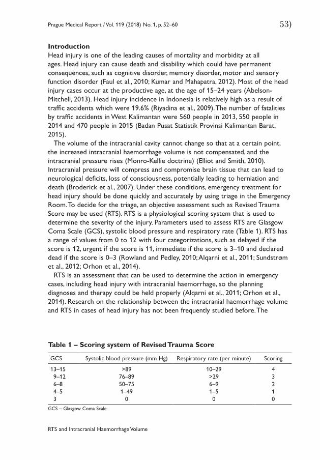
RTS and Intracranial Haemorrhage Volume
53)Prague Medical Report / Vol. 119 (2018) No. 1, p. 52–60
IntroductionHead injury is one of the leading causes of mortality and morbidity at all ages. Head injury can cause death and disability which could have permanent consequences, such as cognitive disorder, memory disorder, motor and sensory function disorder (Faul et al., 2010; Kumar and Mahapatra, 2012). Most of the head injury cases occur at the productive age, at the age of 15–24 years (Abelson-Mitchell, 2013). Head injury incidence in Indonesia is relatively high as a result of traffic accidents which were 19.6% (Riyadina et al., 2009). The number of fatalities by traffic accidents in West Kalimantan were 560 people in 2013, 550 people in 2014 and 470 people in 2015 (Badan Pusat Statistik Provinsi Kalimantan Barat, 2015).
The volume of the intracranial cavity cannot change so that at a certain point, the increased intracranial haemorrhage volume is not compensated, and the intracranial pressure rises (Monro-Kellie doctrine) (Elliot and Smith, 2010). Intracranial pressure will compress and compromise brain tissue that can lead to neurological deficits, loss of consciousness, potentially leading to herniation and death (Broderick et al., 2007). Under these conditions, emergency treatment for head injury should be done quickly and accurately by using triage in the Emergency Room. To decide for the triage, an objective assessment such as Revised Trauma Score may be used (RTS). RTS is a physiological scoring system that is used to determine the severity of the injury. Parameters used to assess RTS are Glasgow Coma Scale (GCS), systolic blood pressure and respiratory rate (Table 1). RTS has a range of values from 0 to 12 with four categorizations, such as delayed if the score is 12, urgent if the score is 11, immediate if the score is 3–10 and declared dead if the score is 0–3 (Rowland and Pedley, 2010; Alqarni et al., 2011; Sundstrøm et al., 2012; Orhon et al., 2014).
RTS is an assessment that can be used to determine the action in emergency cases, including head injury with intracranial haemorrhage, so the planning diagnoses and therapy could be held properly (Alqarni et al., 2011; Orhon et al., 2014). Research on the relationship between the intracranial haemorrhage volume and RTS in cases of head injury has not been frequently studied before. The
Table 1 – Scoring system of Revised Trauma Score
GCS Systolic blood pressure (mm Hg) Respiratory rate (per minute) Scoring
13–159–126–84–53
>8976–8950–751–49
0
10–29>296–91–50
43210
GCS – Glasgow Coma Scale
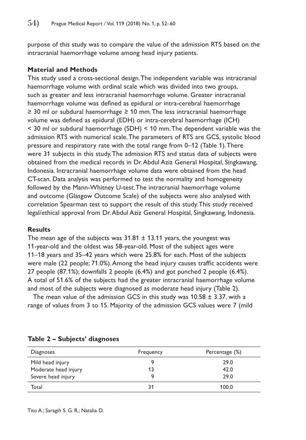
Tito A.; Saragih S. G. R.; Natalia D.
54) Prague Medical Report / Vol. 119 (2018) No. 1, p. 52–60
purpose of this study was to compare the value of the admission RTS based on the intracranial haemorrhage volume among head injury patients.
Material and MethodsThis study used a cross-sectional design. The independent variable was intracranial haemorrhage volume with ordinal scale which was divided into two groups, such as greater and less intracranial haemorrhage volume. Greater intracranial haemorrhage volume was defined as epidural or intra-cerebral haemorrhage ≥ 30 ml or subdural haemorrhage ≥ 10 mm. The less intracranial haemorrhage volume was defined as epidural (EDH) or intra-cerebral haemorrhage (ICH) < 30 ml or subdural haemorrhage (SDH) < 10 mm. The dependent variable was the admission RTS with numerical scale. The parameters of RTS are GCS, systolic blood pressure and respiratory rate with the total range from 0–12 (Table 1). There were 31 subjects in this study. The admission RTS and status data of subjects were obtained from the medical records in Dr. Abdul Aziz General Hospital, Singkawang, Indonesia. Intracranial haemorrhage volume data were obtained from the head CT-scan. Data analysis was performed to test the normality and homogeneity followed by the Mann-Whitney U-test. The intracranial haemorrhage volume and outcome (Glasgow Outcome Scale) of the subjects were also analysed with correlation Spearman test to support the result of this study. This study received legal/ethical approval from Dr. Abdul Aziz General Hospital, Singkawang, Indonesia.
ResultsThe mean age of the subjects was 31.81 ± 13.11 years, the youngest was 11-year-old and the oldest was 58-year-old. Most of the subject ages were 11–18 years and 35–42 years which were 25.8% for each. Most of the subjects were male (22 people; 71.0%). Among the head injury causes traffic accidents were 27 people (87.1%); downfalls 2 people (6.4%) and got punched 2 people (6.4%). A total of 51.6% of the subjects had the greater intracranial haemorrhage volume and most of the subjects were diagnosed as moderate head injury (Table 2).
The mean value of the admission GCS in this study was 10.58 ± 3.37, with a range of values from 3 to 15. Majority of the admission GCS values were 7 (mild
Table 2 – Subjects’ diagnoses
Diagnoses Frequency Percentage (%)
Mild head injuryModerate head injurySevere head injury
9139
29.042.029.0
Total 31 100.0
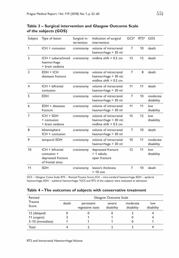
RTS and Intracranial Haemorrhage Volume
55)Prague Medical Report / Vol. 119 (2018) No. 1, p. 52–60
Table 3 – Surgical intervention and Glasgow Outcome Scale of the subjects (GOS)
Subject Type of lesion Surgical in-tervention
Indication of surgical intervention
GCS* RTS* GOS
1 ICH + contusion craniotomy volume of intracranial haemorrhage > 30 ml
7 10 death
2 ICH + subarachnoid haemorrhage + brain oedema
craniotomy midline shift > 0.5 cm 13 12 death
3 EDH + ICH diastases fracture
craniotomy volume of intracranial haemorrhage > 30 ml; midline shift > 0.5 cm
7 8 death
4 ICH + bifrontal contusion
craniotomy volume of intracranial haemorrhage > 30 ml
11 11 death
5 EDH craniotomy volume of intracranial haemorrhage > 30 ml
7 10 moderate disability
6 EDH + diastases fracture
craniotomy volume of intracranial haemorrhage > 30 ml
11 11 low disability
7 ICH + SDH + contusion + brain oedema
craniotomy volume of intracranial haemorrhage = 30 ml;midline shift > 0.5 cm
15 12 low disability
8 bihemisphere ICH + contusion
craniotomy volume of intracranial haemorrhage > 30 ml
7 10 death
9 temporal EDH craniotomy volume of intracranial haemorrhage > 30 ml
10 11 moderate disability
10 ICH + bifrontal contusion + depressed fracture of frontal sinus
craniotomy depressed fracture > 1 tabula; open fracture
12 11 low disability
11 SDH craniotomy lesion’s thickness > 10 mm
7 10 death
GCS – Glasgow Coma Scale; RTS – Revised Trauma Score; ICH – intra-cerebral haemorrhage; EDH – epidural haemorrhage; SDH – subdural haemorrhage; *GCS and RTS of the subjects were evaluated at admission
Table 4 – The outcomes of subjects with conservative treatment
Revised Trauma Score
Glasgow Outcome Scale
death persistent vegetative state
severe disability
moderate disability
low disability
12 (delayed)11 (urgent)3–10 (immediate)
031
011
011
300
441
Total 4 2 2 3 9
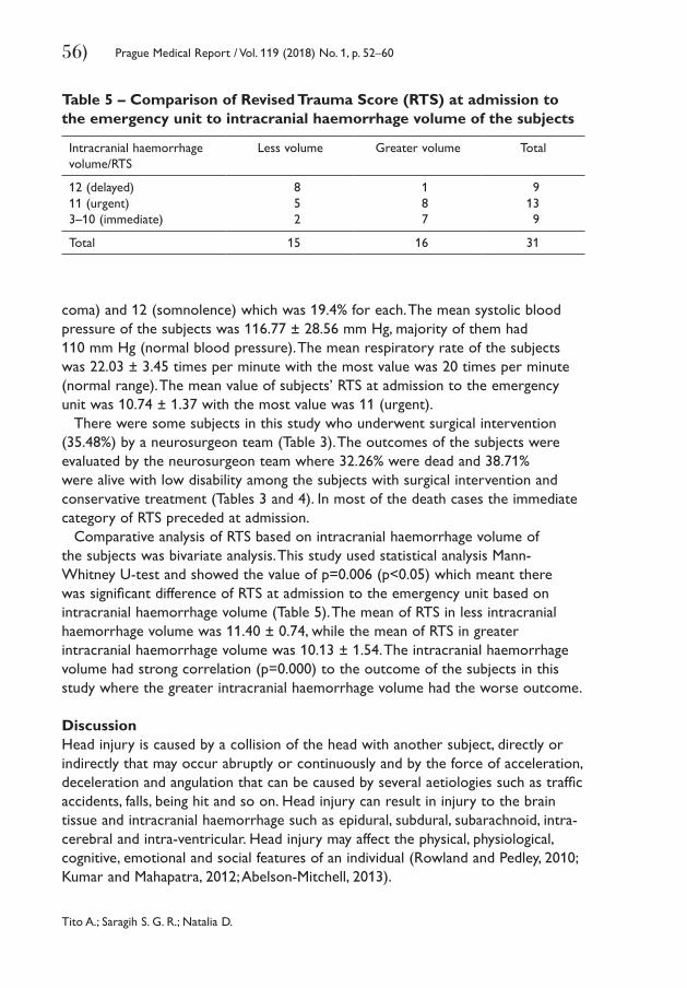
Tito A.; Saragih S. G. R.; Natalia D.
56) Prague Medical Report / Vol. 119 (2018) No. 1, p. 52–60
coma) and 12 (somnolence) which was 19.4% for each. The mean systolic blood pressure of the subjects was 116.77 ± 28.56 mm Hg, majority of them had 110 mm Hg (normal blood pressure). The mean respiratory rate of the subjects was 22.03 ± 3.45 times per minute with the most value was 20 times per minute (normal range). The mean value of subjects’ RTS at admission to the emergency unit was 10.74 ± 1.37 with the most value was 11 (urgent).
There were some subjects in this study who underwent surgical intervention (35.48%) by a neurosurgeon team (Table 3). The outcomes of the subjects were evaluated by the neurosurgeon team where 32.26% were dead and 38.71% were alive with low disability among the subjects with surgical intervention and conservative treatment (Tables 3 and 4). In most of the death cases the immediate category of RTS preceded at admission.
Comparative analysis of RTS based on intracranial haemorrhage volume of the subjects was bivariate analysis. This study used statistical analysis Mann-Whitney U-test and showed the value of p=0.006 (p<0.05) which meant there was significant difference of RTS at admission to the emergency unit based on intracranial haemorrhage volume (Table 5). The mean of RTS in less intracranial haemorrhage volume was 11.40 ± 0.74, while the mean of RTS in greater intracranial haemorrhage volume was 10.13 ± 1.54. The intracranial haemorrhage volume had strong correlation (p=0.000) to the outcome of the subjects in this study where the greater intracranial haemorrhage volume had the worse outcome.
DiscussionHead injury is caused by a collision of the head with another subject, directly or indirectly that may occur abruptly or continuously and by the force of acceleration, deceleration and angulation that can be caused by several aetiologies such as traffic accidents, falls, being hit and so on. Head injury can result in injury to the brain tissue and intracranial haemorrhage such as epidural, subdural, subarachnoid, intra-cerebral and intra-ventricular. Head injury may affect the physical, physiological, cognitive, emotional and social features of an individual (Rowland and Pedley, 2010; Kumar and Mahapatra, 2012; Abelson-Mitchell, 2013).
Table 5 – Comparison of Revised Trauma Score (RTS) at admission to the emergency unit to intracranial haemorrhage volume of the subjects
Intracranial haemorrhage volume/RTS
Less volume Greater volume Total
12 (delayed)11 (urgent)3–10 (immediate)
852
187
9139
Total 15 16 31
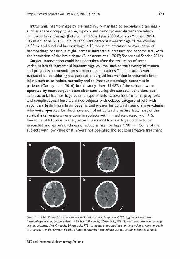
RTS and Intracranial Haemorrhage Volume
57)Prague Medical Report / Vol. 119 (2018) No. 1, p. 52–60
Intracranial haemorrhage by the head injury may lead to secondary brain injury such as space occupying lesion, hypoxia and hemodynamic disturbance which can cause brain damage (Peterson and Scardiglia, 2008; Abelson-Mitchell, 2013; Takahashi et al., 2015). Epidural and intra-cerebral haemorrhage of the volume ≥ 30 ml and subdural haemorrhage ≥ 10 mm is an indication to evacuation of haemorrhage because it might increase intracranial pressure and become fatal with the herniation of the brain tissue (Sundstrøm et al., 2012; Sherer and Sander, 2014).
Surgical intervention could be undertaken after the evaluation of some variables beside intracranial haemorrhage volume, such as the severity of trauma and prognosis; intracranial pressure; and complications. The indications were evaluated by considering the purpose of surgical intervention in traumatic brain injury, such as to reduce mortality and to improve neurologic outcomes in patients (Carney et al., 2016). In this study, there 35.48% of the subjects were operated by neurosurgeon team after considering the subjects’ conditions, such as intracranial haemorrhage volume, type of lesions, severity of trauma, prognosis and complications. There were two subjects with delayed category of RTS with secondary brain injury, brain oedema, and greater intracranial haemorrhage volume who were operated for decompression of intracranial pressure. But, most of the surgical interventions were done in subjects with immediate category of RTS, low value of RTS, due to the greater intracranial haemorrhage volume to be evacuated and lesion’s thickness of subdural haemorrhage ≥ 10 mm. Some of the subjects with low value of RTS were not operated and got conservative treatment
Figure 1 – Subject’s head CT-scan section samples (A – female, 53-years-old, RTS 6, greater intracranial haemorrhage volume, outcome: death < 24 hours; B – male, 32-years-old, RTS 12, less intracranial haemorrhage volume, outcome: alive; C – male, 20-years-old, RTS 11, greater intracranial haemorrhage volume, outcome: death in 3 days; D – male, 40-years-old, RTS 11, less intracranial haemorrhage volume, outcome: death in 8 days).
AA
C
B
D
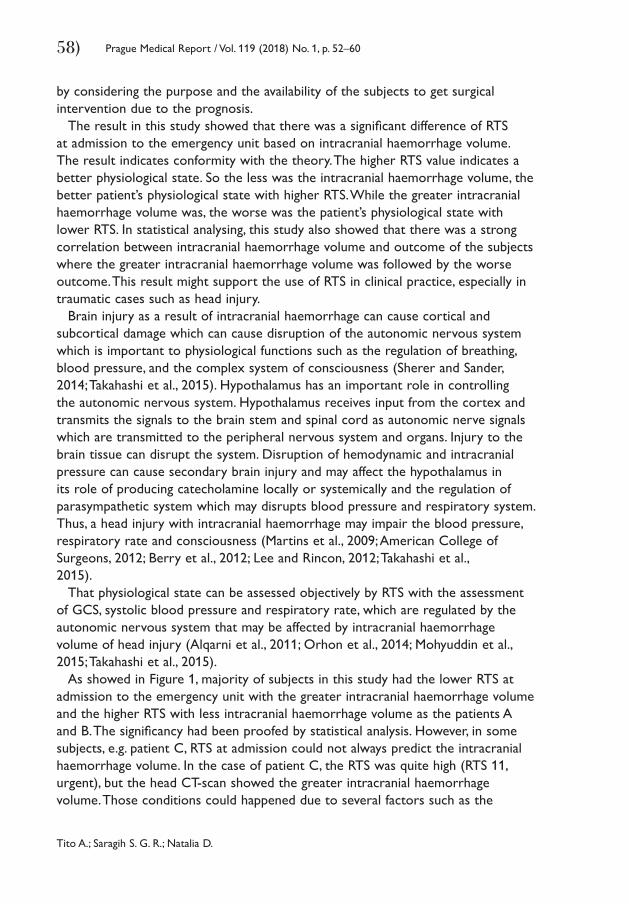
Tito A.; Saragih S. G. R.; Natalia D.
58) Prague Medical Report / Vol. 119 (2018) No. 1, p. 52–60
by considering the purpose and the availability of the subjects to get surgical intervention due to the prognosis.
The result in this study showed that there was a significant difference of RTS at admission to the emergency unit based on intracranial haemorrhage volume. The result indicates conformity with the theory. The higher RTS value indicates a better physiological state. So the less was the intracranial haemorrhage volume, the better patient’s physiological state with higher RTS. While the greater intracranial haemorrhage volume was, the worse was the patient’s physiological state with lower RTS. In statistical analysing, this study also showed that there was a strong correlation between intracranial haemorrhage volume and outcome of the subjects where the greater intracranial haemorrhage volume was followed by the worse outcome. This result might support the use of RTS in clinical practice, especially in traumatic cases such as head injury.
Brain injury as a result of intracranial haemorrhage can cause cortical and subcortical damage which can cause disruption of the autonomic nervous system which is important to physiological functions such as the regulation of breathing, blood pressure, and the complex system of consciousness (Sherer and Sander, 2014; Takahashi et al., 2015). Hypothalamus has an important role in controlling the autonomic nervous system. Hypothalamus receives input from the cortex and transmits the signals to the brain stem and spinal cord as autonomic nerve signals which are transmitted to the peripheral nervous system and organs. Injury to the brain tissue can disrupt the system. Disruption of hemodynamic and intracranial pressure can cause secondary brain injury and may affect the hypothalamus in its role of producing catecholamine locally or systemically and the regulation of parasympathetic system which may disrupts blood pressure and respiratory system. Thus, a head injury with intracranial haemorrhage may impair the blood pressure, respiratory rate and consciousness (Martins et al., 2009; American College of Surgeons, 2012; Berry et al., 2012; Lee and Rincon, 2012; Takahashi et al., 2015).
That physiological state can be assessed objectively by RTS with the assessment of GCS, systolic blood pressure and respiratory rate, which are regulated by the autonomic nervous system that may be affected by intracranial haemorrhage volume of head injury (Alqarni et al., 2011; Orhon et al., 2014; Mohyuddin et al., 2015; Takahashi et al., 2015).
As showed in Figure 1, majority of subjects in this study had the lower RTS at admission to the emergency unit with the greater intracranial haemorrhage volume and the higher RTS with less intracranial haemorrhage volume as the patients A and B. The significancy had been proofed by statistical analysis. However, in some subjects, e.g. patient C, RTS at admission could not always predict the intracranial haemorrhage volume. In the case of patient C, the RTS was quite high (RTS 11, urgent), but the head CT-scan showed the greater intracranial haemorrhage volume. Those conditions could happened due to several factors such as the
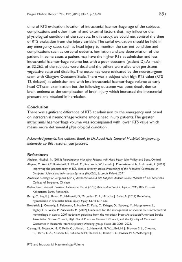
RTS and Intracranial Haemorrhage Volume
59)Prague Medical Report / Vol. 119 (2018) No. 1, p. 52–60
time of RTS evaluation, location of intracranial haemorrhage, age of the subjects, complications and other internal and external factors that may influence the physiological condition of the subjects. In this study, we could not control the time of RTS evaluation from the injury variable. The serial evaluation should be held in any emergency cases such as head injury to monitor the current condition and complications such as cerebral oedema, herniation and any deteroriation of the patient. In some cases, a patient may have the higher RTS at admission and less intracranial haemorrhage volume but with a poor outcome (patient D). As much as 32.26% of the subjects were dead and the others were alive with persistent vegetative state and disability. The outcomes were evaluated by the neurosurgeon team with Glasgow Outcome Scale. There was a subject with high RTS value (RTS 12, delayed) at admission and with less intracranial haemorrhage volume at early head CT-scan examination but the following outcome was poor, death, due to brain oedema as the complication of brain injury which increased the intracranial pressure and resulted in herniation.
ConclusionThere was significant difference of RTS at admission to the emergency unit based on intracranial haemorrhage volume among head injury patients. The greater intracranial haemorrhage volume was accompanied with lower RTS value which means more detrimental physiological condition.
Acknowledgements: The authors thank to Dr. Abdul Aziz General Hospital, Singkawang, Indonesia, so this research can proceed.
ReferencesAbelson-Mitchell, N. (2013) Neurotrauma: Managing Patients with Head Injury. John Wiley and Sons, Oxford.Alqarni, M., Arabi, Y., Kakiashvili, T., Khedr, M., Koczkodaj, W., Leszek, J., Przelaskowski, A., Rutkowski, K. (2011)
Improving the predictability of ICU illness severity scales. Proceedings of the Federated Conference on Computer Science and Information Systems (FedCSIS), Szczecin, Poland, 2011.
American College of Surgeons (2012) Advanced Trauma Life Support: Student Course Manual, 9th Ed. American College of Surgeons, Chicago.
Badan Pusat Statistik Provinsi Kalimantan Barat (2015) Kalimantan Barat in Figures 2015. BPS Provinsi Kalimantan Barat, Pontianak.
Berry, C., Ley, E. J., Bukur, M., Malinoski, D., Margulies, D. R., Mirocha, J., Salim, A. (2012) Redefining hypotension in traumatic brain injury. Injury 43, 1833–1837.
Broderick, J., Connolly, S., Feldmann, E., Hanley, D., Kase, C., Krieger, D., Mayberg, M., Morgenstern, L., Ogilvy, C. S., Vespa, P., Zuccarello, M. (2007) Guidelines for the management of spontaneous intracerebral hemorrhage in adults: 2007 update: A guideline from the American Heart Association/American Stroke Association Stroke Council, High Blood Pressure Research Council, and the Quality of Care and Outcomes in Research Interdisciplinary Working group. Stroke 38, 2001–2023.
Carney, N., Totten, A. M., O’Reilly, C., Ullman, J. S., Hawryluk, G. W. J., Bell, M. J., Bratton, S. L., Chesnut, R., Harris, O. A., Kissoon, N., Rubiano, A. M., Shutter, L., Tasker, R. C., Vavilala, M. S., Wilberger, J.,
Tito A.; Saragih S. G. R.; Natalia D.
60) Prague Medical Report / Vol. 119 (2018) No. 1, p. 52–60
Wright, D. W., Ghajar, J. (2016) Guidelines for the management of severe traumatic brain injury, fourth edition. Neurosurgery 80, 6–15.
Elliott, J., Smith, M. (2010) The acute management of intracerebral hemorrhage: a clinical review. Anesth. Analg. 110(5), 1419–1427.
Faul, M., Xu, L., Wald, M. M., Coronado, V. G. (2010) Traumatic Brain Injury in the United States: Emergency Department Visits, Hospitalizations, and Deaths. Centers for Disease Control and Prevention, Atlanta.
Kumar, R., Mahapatra, A. K. (2012) A Textbook of Head Injury, 1st Ed. JP Medical Ltd., New Delhi.Lee, K., Rincon, F. (2012) Pulmonary complications in patients with severe brain injury. Crit. Care Res. Pract.
2012, 207247.Martins, E. T., Linhares, M. N., Sousa, D. S., Schroeder, H. K., Meinerz, J., Rigo, L. A., Bertotti, M. M., Gullo, J.,
Hohl, A., Dal-Pizzol, F., Walz, R. (2009) Mortality in severe traumatic brain injury: a multivariated analysis of 748 Brazilian patients from Florianópolis City. J. Trauma 67(1), 85–90.
Mohyuddin, G. R., Alam, Z., Malik, U. Z., Shakil, O., Haq, A. (2015) Revised Trauma Score as a predictor of outcome in trauma cases: Experiences at a tertiary care hospital in Karachi, Pakistan. J. Ayub Med. Coll. Abbottabad 27(3), 584–586.
Orhon, R., Eren, Ş. H., Karadayı, Ş., Korkmaz, I., Coskun, A., Eren, M., Katrancıoglu, N. (2014) Comparison of trauma scores for predicting mortality and morbidity on trauma patients. Ulus. Travma Acil Cerrahi Derg. 20(4), 258–264.
Peterson, N., Scardiglia, J. (2008) Advanced Trauma Life Support for Doctors: ATLS Student Course Manual, 18th Ed. American College Surgeons, Chicago.
Riyadina, W., Suhardi, Permana, M. (2009) Pola dan determinan sosiodemografi cedera akibat kecelakaan lalu lintas di Indonesia. Maj. Kedokt. Indon. 59(10), 464–472.
Rowland, L. P., Pedley, T. A. (2010) Merritt’s Neurology, 12th Ed. Lippincott Williams and Wilkins, Philadelphia.Sherer, M., Sander, A. M. (2014) Handbook on the Neuropsychology of Traumatic Brain Injury. Springer, New York.Sundstrøm, T., Grände, P. O., Juul, N., Kock-Jensen, C., Romner, B., Wester, K. (2012) Management of Severe
Traumatic Brain Injury: Evidence, Tricks, and Pitfalls. Springer Science and Business Media, Heidelberg.Takahashi, C., Hinson, H., Bagulay, I. J. (2015) Autonomic Dysfunction Syndromes after Acute Brain Injury,
3rd Ed. Elsevier, Philadelphia.
61)Prague Medical Report / Vol. 119 (2018) No. 1, p. 61–69
The Etiologic Agents of the Pertussis-like Syndromehttps://doi.org/10.14712/23362936.2018.6 © 2018 The Authors. This is an open-access article distributed under the terms of the Creative Commons Attribution License (http://creativecommons.org/licenses/by/4.0).
Identification of Etiologic Agents of the Pertussis-like Syndrome in Children by Real-time PCR MethodShima Mahmoudi1, Maryam Banar1, Babak Pourakbari1, Hediyeh Sadat Alavi2, Hamid Eshaghi2, Alireza Aziz Ahari3, Setareh Mamishi1,2
1Pediatric Infectious Disease Research Center, Tehran University of Medical Sciences, Tehran, Iran;2Department of Infectious Diseases, Pediatrics Center of Excellence, Children’s Medical Center Hospital, Tehran University of Medical Sciences, Tehran, Iran;3Department of Radiology, Rasool-e-Akram Hospital, Iran University of Medical Sciences, Tehran, Iran
Rece ived November 14 , 2017 ; Accepted March 21 , 2018 .
Key words: Pertussis-like syndrome – B. pertussis – Adenovirus – Respiratory syncytial virus – Human metapneumovirus – Influenza virus A
Abstract: The aim of this study was to recognize the identity and frequency of etiologic agents of the pertussis-like syndrome in children < 2 years of age. A cross-sectional hospital-based study conducted from August 2014 to August 2015. All children < 2 years of age (n=100) who were suspected as pertussis infected were enrolled in this study and tested for Bordetella pertussis, adenovirus (Adv), respiratory syncytial virus (RSV), human metapneumovirus (hMPV), and influenza virus A (INF-A) by real-time PCR technique. RSV was the most detected pathogen (20%), followed by B. pertussis (18%), Adv (16%), INF-A (11%), and hMPV (10%). Co-infection was observed in 8 patients (11%) and the combinations of RSV/INF-A (n=3, 4%), and AdV/B. pertussis (n=3, 4%) were more frequent. RSV, B. pertussis, and hMPV were more frequent pathogens among infants < 4 months of age. However, Adv and INF-A were more frequent pathogens among children > 6 months of age. In this study, RSV was the most frequent identified pathogen (n=20, 20%), followed by B. pertussis (n=18, 18%) and AdV (n=16, 16%). Pertussis was more frequent in
This study was supported by a grant (No. 94-03-88-29448) from Tehran University of Medical Sciences to Dr. Setareh Mamishi.
Mailing Address: Dr. Setareh Mamishi, Department of Pediatric Infectious Disease, Children’s Medical Center Hospital, School of Medicine, Tehran University of Medical Sciences, No. 62, Gharib St., Keshavarz Blvd., Tehran, Iran; Phone: +98 021 6642 8996; Fax: +98 021 6642 8996; e-mail: [email protected]
62) Prague Medical Report / Vol. 119 (2018) No. 1, p. 61–69
Mahmoudi S.; Banar M.; Pourakbari B.; Alavi H. S.; Eshaghi H.; Aziz Ahari A.; Mamishi S.
spring (8%) and summer (6%). In addition, clinical symptoms of pertussis were the same as some viral pathogens, which can lead to misdiagnosis of infection. Therefore, diagnosis of pertussis should be established on the bases of both the clinical symptoms and the laboratory methods.
IntroductionBordetella pertussis is a fastidious gram-negative coccobacillus bacterium that causes pertussis (whooping cough), an acute infection of the respiratory tract in humans (Castillo et al., 2015; Pavic-Espinoza et al., 2015). Pertussis is one of the top ten fatal infections in children (Haghighi et al., 2014). Although it is a vaccine-preventable disease, a considerable number of pertussis cases are yet recorded (Nunes et al., 2016). According to the World Health Organization (WHO), approximately 50 million cases of pertussis occur each year, which results in 300 000 deaths (World Health Organization, 2016). Infants < 2 months are more susceptible to pertussis complications since they did not receive the pertussis vaccines (Nunes et al., 2016). The infant mortality rate of pertussis in developing countries is about 4% (World Health Organization, 2016). The well-known symptoms of pertussis defined as repeated paroxysmal cough, inspiratory whoop, and post-cough vomiting (Shojaei et al., 2014). The atypical milder infection can develop in persons with the prepared immune system either by vaccination or previous infection (Nunes et al., 2016).
Co-infections with respiratory viruses such as respiratory syncytial virus (RSV), rhinovirus/enterovirus, adenovirus (Adv), influenza virus (INF), and human metapneumovirus (hMPV) frequently occur in infants with pertussis infection (Dube et al., 2016; Nicolai et al., 2016). The clinical manifestations of these viral infections are indistinguishable from pertussis (Pavic-Espinoza et al., 2015); therefore, diagnosis of pertussis infection cannot be done only by clinical symptoms and should be confirmed by laboratory tests (del Valle-Mendoza et al., 2015). Real-time PCR is a rapid and sensitive technique that can be employed for fast detection of B. pertussis in order to disrupt its transmission and initiate appropriate therapy (Nikbin et al., 2013).
In this study, we aimed to recognize the identity and frequency of etiologic agents of the pertussis-like syndrome in children < 2 years of age who admitted to Children’s Medical Center (CMC) Hospital.
Material and MethodsStudy design and populationThis was a cross-sectional hospital-based study designed to recognize the identity and frequency of etiologic agents of the pertussis-like syndrome in all children < 2 years of age who were suspected to pertussis and admitted to CMC Hospital between August 2014 and August 2015. CMC Hospital is a tertiary referral children’s hospital that is located in Tehran, the capital city of Iran.
63)Prague Medical Report / Vol. 119 (2018) No. 1, p. 61–69
The Etiologic Agents of the Pertussis-like Syndrome
Detailed demographic and clinical data such as age, gender, pertussis vaccination status, previous antibiotic consumption, history of pertussis in the family, and clinical symptoms were obtained from questionnaires that were answered by patient’s parents and from medical records. Children who met some of the WHO clinical case criteria for pertussis – i.e. cases with a cough more than two weeks with at least one of the following symptoms: paroxysm of a cough (many, rapid coughs followed by a high-pitched “whoop” sound); or inspiratory whoop (a forceful inspiration of air through a narrow glottis, usually developing after a paroxysmal cough); or vomiting after a cough; or apnea (long cessation of breathing) (with or without cyanosis (blue/purplish skin coloration due to lack of oxygen)) were included into the study (Shojaei et al., 2014; World Health Organization, 2016).
Ethical statementThis study was permitted by the Ethical Committee of the Research Center for Pediatric Infectious Diseases in Tehran University of Medical Sciences. Written informed consent was obtained from parents of all children.
Sample collectionFor each patient, one Dacron swab was obtained from the posterior part of the nasopharynx and inoculated onto the 1 ml of sterile normal saline (0.9% W/V NaCl solution). All specimens were transported at room temperature within 1–2 hours to the microbiology laboratory of the Research Center for Pediatric Infectious Diseases at CMC Hospital and stored at –70 °C.
TaqMan real-time PCRA TaqMan real-time PCR assay was applied to detect the identity of pathogens. For each specimen, both RNA and DNA were extracted from the elution liquid of swabs, using the MagJET viral DNA and RNA purification kit (Thermo ScientificTM, USA) and RTP bacteria DNA mini kit (STRATEC Biomedical AG, Germany) according to the manufacturer’s protocols. In the case of RNA samples, a random-primed reverse transcription reaction was performed using the RevertAid reverse transcriptase kit (Thermo scientificTM, USA) and cDNA was synthesized according to the manufacturer’s instructions. The real-time PCR was performed based on the protocols of Adv, RSV, INF-A, hMPV and B. pertussis real-time PCR kits (Liferriver biotech, USA). An ABI step one real-time PCR system (Applied Biosystems, Foster City, California, USA) was used for this purpose.
ResultsStudy populationDuring the study period, a total of 100 children < 2 years of age (mean age 5.5 months) presenting some of the symptoms of pertussis such as a paroxysmal
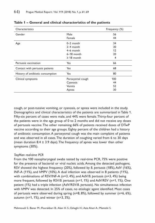
64) Prague Medical Report / Vol. 119 (2018) No. 1, p. 61–69
Mahmoudi S.; Banar M.; Pourakbari B.; Alavi H. S.; Eshaghi H.; Aziz Ahari A.; Mamishi S.
Table 1 – General and clinical characteristics of the patients
Characteristics Frequency (%)
Gender MaleFemale
5644
Age 0–2 month2–4 month4–6 month6–18 month≥ 18 month
343012204
Pertussis vaccination Yes 66
Contact with pertussis patients Yes 68
History of antibiotic consumption Yes 80
Clinical symptoms Paroxysmal coughCyanosisVomitsApnea
100445230
cough, or post-tussive vomiting, or cyanosis, or apnea were included in the study. Demographics and clinical characteristics of the patients are summarized in Table 1. Fifty-six percent of cases were male, and 44% were female. Thirty-four percent of the patients were in the age group of 0 to 2 months and did not receive any doses of pertussis vaccine. The other remaining 66% of patients received doses of DTwP vaccine according to their age groups. Eighty percent of the children had a history of antibiotic consumption. A paroxysmal cough was the main complaint of patients and was observed in all cases. The duration of coughing varied from 6 to 30 days (mean duration 8.4 ± 3.9 days). The frequency of apnea was lower than other symptoms (30%).
TaqMan real-time PCRFrom the 100 nasopharyngeal swabs tested by real-time PCR, 75% were positive for the presence of bacterial or viral nucleic acids. Among the detected pathogens, RSV showed the highest frequency (20%), followed by B. pertussis (18%), AdV (16%), INF-A (11%), and hMPV (10%). A dual infection was observed in 8 patients (11%), with combinations of RSV/INF-A (n=3, 4%), and AdV/B. pertussis (n=3, 4%) being more frequent, followed by RSV/B. pertussis (n=1, 1%) and AdV/RSV (n=1, 1%). One patient (1%) had a triple infection (AdV/RSV/B. pertussis). No simultaneous infection with hMPV was detected. In 25% of cases, no etiologic agent identified. Most cases of pertussis were observed during spring (n=8, 8%), followed by summer (n=6, 6%), autumn (n=1, 1%), and winter (n=3, 3%).
65)Prague Medical Report / Vol. 119 (2018) No. 1, p. 61–69
The Etiologic Agents of the Pertussis-like Syndrome
DiscussionAlthough a paroxysmal cough is regarded as the most important classical symptom of pertussis and is used for pertussis clinical case definition by WHO and center for disease control and prevention (van den Brink et al., 2014), results of this study demonstrated that it is not a specific symptom for pertussis and of the 100 suspected cases of pertussis, only 18% were correctly predicted by this criterion. In addition, other symptoms were non-specific to pertussis and they were also observed in some children with viral respiratory infections. In agreement with this study, other reports have emphasized on the lack of specificity of these classical symptoms (Shojaei et al., 2014; van den Brink et al., 2014; Vittucci et al., 2016).
If pertussis diagnosis is based only on the clinical manifestations, atypical and mild infections would be unrecognized (van den Brink et al., 2014) especially in vaccinated children that show mild or less severe infection (van den Brink et al., 2014); or other respiratory infections can be confused with pertussis (Shojaei et al., 2014). This can lead to the consecutive spreading of the pathogen in the population and increases the danger of hospital outbreaks (van den Brink et al., 2014; Vittucci et al., 2016). The identification of pertussis cases should not be based only on the clinical symptoms (Cherry et al., 2012; van den Brink et al., 2014), and laboratory techniques such as real-time-PCR should be used as complementary diagnostic methods.
In this study, B. pertussis was detected only in 18% of suspected cases. In the study conducted by Hajia et al. (2012) from Tehran, Iran, 12 out of 138 children < 6 months (9%) were positive for B. pertussis, which was lower compared to the current result. Castillo et al. (2015) from Peru reported an incidence of pertussis as high as 40% in patients younger than one year admitted to five hospitals in Lima, Peru, which was higher than the rate of this study. Discrepancies observed among the pertussis frequency could be due to the different studied age ranges, different utilized diagnostic methods that have diverse sensitivity and specificity, and different vaccination coverage in various areas.
As noted, 80% of patients had used antibiotics before sampling that can cause false-negative results. However, since in the current study identification of the causative agents of the pertussis-like syndrome was performed by real-time PCR method, application of antibiotics could not affect the results. The rapid detection of etiologic agents of the pertussis-like syndrome plays an important role in stopping irrational antibiotic usage and adopting the correct therapeutic approaches.
Whooping cough vaccination program in Iran consists of three doses of DTwP vaccine (diphtheria and tetanus toxoid in combination with whole-cell pertussis) at the second, 4th and 6th months of life. Booster doses are given at 18 months and at 4 to 6 years of age (Sedaghat et al., 2014). In the current study, 89% (n=16) of pertussis cases were observed in the age group 0 to 6-month that were not
66) Prague Medical Report / Vol. 119 (2018) No. 1, p. 61–69
Mahmoudi S.; Banar M.; Pourakbari B.; Alavi H. S.; Eshaghi H.; Aziz Ahari A.; Mamishi S.
fully immunized and received only the three initial doses of DTwP vaccine. Some studies demonstrated that based on the efficiency of vaccine used, the infection can develop soon after the vaccination (van den Brink et al., 2014). Due to the waning efficacy of pertussis vaccine, adults play a significant role in the transmission of disease to the young infants (Sedighi and Sadrosadat, 2015). Therefore, some strategies are suggested for protection of unimmunized young infants, including revaccination of juveniles and adults with DTaP vaccine (acellular pertussis vaccine) to prevent household spread of infection, immunization of pregnant women during the third trimester of gestation that leads to the transfer of maternal anti-pertussis antibodies to the fetus to grant protection of newborns from pertussis prior their vaccination (Sedighi and Sadrosadat, 2015; Vittucci et al., 2016). Since whole-cell pertussis vaccine is not suggested for adults; and there is no access to acellular vaccine in Iran, it is recommended to introduce this type of vaccine into the Iranian national vaccination schedule (Sedighi and Sadrosadat, 2015).
In this research, RSV was the most prominent respiratory pathogen and was identified in 20% of the cases. In the previous study conducted at CMC Hospital (Pourakbari et al., 2014), RSV was the most prevalent viral pathogen (17.2%) isolated from nasopharyngeal aspirates of children < 5 years. According to the results, it can be stated that RSV is the principal cause of acute respiratory infection in children (Parsania et al., 2016); and further studies are required to determine the prevalence and genotype distribution of RSV in Iran (Salimi et al., 2016).
In this study, Adv and INF-A were detected in 16% and 11% of the patients, respectively. In the previous study carried out in CMC Hospital (Pourakbari et al., 2014), Adv and INF-A showed the same frequencies (3.4%) and was lower than the rates of this study. In the study conducted by Naghipour et al. (2007) from Gilan, Iran, all the children < 5 years of age attending the out-patient clinics and those admitted to the hospital wards were tested; and Adv and INF-A was identified in 14% and 4% of cases, respectively. In the study performed by Moattari et al. (2015) from Shiraz, Iran, nasopharyngeal swabs of 435 hospitalized children under 5 years of age were evaluated and 22% of patients were positive for Adv, which was higher than the rates of this study.
In this report, 10% of patients were positive for hMPV. Sanaei Dashti et al. (2016) from Shiraz, Iran, investigated 200 children under 12 years of age with upper respiratory tract complaints referred to Infectious Clinic of Mofid Children Hospital and reported a similar frequency for hMPV. However, in the studies conducted by Sultani et al. (2015) from Tehran, Iran (0%), and Vittucci et al. (2016) from Italy (3.4%) lower frequencies were reported. Furthermore, higher frequency for hMPV was reported by Moattari et al. (2015) from Shiraz, Iran (15.7%). The aforementioned differences in the frequency of Adv, INF-A, and hMPV in different studies could be due to the annual variations in the incidence of infections, the age of studied patients, different diagnostic methods used, the size of understudy population, and climate variations (Parsania et al., 2016).
67)Prague Medical Report / Vol. 119 (2018) No. 1, p. 61–69
The Etiologic Agents of the Pertussis-like Syndrome
In the current study, co-infections with RSV/INF-A, AdV/B. pertussis, RSV/B. pertussis, AdV/RSV, and AdV/RSV/B. pertussis were detected in 8 patients (12%). Several studies revealed that in respiratory infections often more than one pathogen is detected. For instance, Korppi and Hiltunen (2007) reported the co-infection of RSV/B. pertussis in 7 out of 117 infants < 6 months of age who had lower respiratory tract infections. In the case of mixed infections, it is unclear which pathogen is the principal contributing agent and what is their association with disease severity. It is also unclear which pathogen is more important for the emergence of disease manifestations (van den Brink et al., 2014). Therefore, to avoid unrecognized cases, pertussis should be considered in all infants with respiratory tract infections and the diagnosis of viral pathogens does not mean that pertussis is dismissed.
In this research, the frequency of pertussis in spring and summer was higher than in the other seasons. This was similar to the reports of Ghorbani et al. (2016) from Iran that examined the nasopharyngeal swabs of all notified pertussis cases from different age groups between the years 2011 and 2013 and most of the confirmed cases of pertussis were identified during the spring and summer seasons. Gonfiantini et al. (2014) from Italy, evaluated the data on notified pertussis cases from 1888 to 2012 and observed similar season distribution to the current study. However, in the study performed by Goktas and Sirin (2016) from Turkey, most cases of pertussis (40%) were identified in winter. Consequently, being aware of the seasons with high rates of pertussis, it can be helpful to employ appropriate programs for active case finding and early treatment of patients in order to control quickly the spread of infection.
ConclusionIn this study, RSV was the most frequently identified pathogen (n=20, 20%), followed by pertussis (n=18, 18%) and AdV (n=16, 16%). Pertussis was more frequent at spring (8%) and summer (6%). In addition, clinical symptoms of pertussis were the same as that of some viral pathogens, and this can lead to misdiagnosis of infection. Therefore, diagnosis of pertussis should be based on both the clinical symptoms and the laboratory methods. Moreover, effective strategies for induction of immunity in unvaccinated young infants including revaccination of juveniles and adults with DTaP, and immunization of pregnant women during the third trimester of gestation; should be introduced into the public health control programs.
ReferencesCastillo, M. E., Bada, C., Del Aguila, O., Petrozzi-Helasvuo, V., Casabona-Ore, V., Reyes, I., del Valle-Mendoza, J.
(2015) Detection of Bordetella pertussis using a PCR test in infants younger than one-year-old hospitalized with whooping cough in five Peruvian hospitals. Int. J. Infect. Dis. 41, 36–41.
68) Prague Medical Report / Vol. 119 (2018) No. 1, p. 61–69
Mahmoudi S.; Banar M.; Pourakbari B.; Alavi H. S.; Eshaghi H.; Aziz Ahari A.; Mamishi S.
Cherry, J. D., Tan, T., Wirsing von König, C. H., Forsyth, K. D., Thisyakorn, U., Greenberg, D., Johnson, D., Marchant, C., Plotkin, S. (2012) Clinical definitions of pertussis: Summary of a global pertussis initiative roundtable meeting, February 2011. Clin. Infect. Dis. 54(12), 1756–1764.
del Valle-Mendoza, J., Casabona-Oré, V., Petrozzi-Helasvuo, V., Cornejo-Tapia, A., Weilg, P., Pons, M. J., Cieza-Mora, E., Bazán-Mayra, J., Cornejo-Pacherres, H., Ruiz, J. (2015) Bordetella pertussis diagnosis in children under five years of age in the Regional Hospital of Cajamarca, Northern Peru. J. Infect. Dev. Ctries. 9(11), 1180–1185.
Dube, F. S., Kaba, M., Robberts, F. L., Tow, L. A., Lubbe, S., Zar, H. J., Nicol, M. P. (2016) Respiratory microbes present in the nasopharynx of children hospitalised with suspected pulmonary tuberculosis in Cape Town, South Africa. BMC Infect. Dis. 16(1), 597.
Ghorbani, G. R., Zahraei, S. M., Moosazadeh, M., Afshari, M., Doosti, F. (2016) Comparing seasonal pattern of laboratory confirmed cases of pertussis with clinically suspected cases. Osong Public Health Res. Perspect. 7(2), 131–137.
Goktas, S., Sirin, M. C. (2016) Prevalence and seasonal distribution of respiratory viruses during the 2014–2015 season in Istanbul. Jundishapur J. Microbiol. 9(9), e39132.
Gonfiantini, M. V., Carloni, E., Gesualdo, F., Pandolfi, E., Rizzuto, E., Iannazzo, S., Degli Atti, M. C., Villani, A., Tozzi, A. E. (2014) Epidemiology of pertussis in Italy: disease trends over the last century. Euro Surveill. 19(40), 20921.
Haghighi, F., Shahcheraghi, F., Abbasi, E., Eshraghi, S. S., Zeraati, H., Mousavi, S. A. J., Asgarian-Omran, H., Douraghi, M., Shokri, F. (2014) Genetic profile variation in vaccine strains and clinical isolates of Bordetella pertussis recovered from Iranian patients. Avicenna J. Med. Biotechnol. 6(3), 178.
Hajia, M., Rahbar, M., Fallah, F., Safadel, N. (2012) Detection of Bordetella pertussis in infants suspected to have whooping cough. Open Respir. Med. J. 6, 34–36.
Korppi, M., Hiltunen, J. (2007) Pertussis is common in nonvaccinated infants hospitalized for respiratory syncytial virus infection. Pediatr. Infect. Dis. J. 26(4), 316–318.
Moattari, A., Aleyasin, S., Emami, A., Fyruzi, M., Pirbonyeh, N. (2015) The prevalence of human metapneumovirus and respiratory syncytial virus and coinfection with both in hospitalized children with acute respiratory infection in south of Iran. Arch. Pediatr. Infect. Dis. 3(3), e32974.
Naghipour, M., Cuevas, L. E., Bakhshinejad, T., Mansour-Ghanaei, F., Noursalehi, S., Alavy, A., Dove, W., Hart, C. A. (2007) Contribution of viruses, Chlamydia spp. and Mycoplasma pneumoniae to acute respiratory infections in Iranian children. J. Trop. Pediatr. 53(3), 179–184.
Nicolai, A., Nenna, R., Frassanito, A., Pierangeli, A., Mileto, F., Scagnolari, C., Mancino, E., Di Mattia, G., Petrarca, L., Cangiano, G., Midulla, F. (2016) Respiratory viruses and Bordetella pertussis co-infections: A frequent occurrence in children hospitalized with Bordetella pertussis. Eur. Resp. J. 48(S60), PA1275.
Nikbin, V. S., Shahcheraghi, F., Lotfi, M. N., Zahraei, S. M., Parzadeh, M. (2013) Comparison of culture and real-time PCR for detection of Bordetella pertussis isolated from patients in Iran. Iran. J. Microbiol. 5(3), 209–214.
Nunes, M. C., Downs, S., Jones, S., van Niekerk, N., Cutland, C. L., Madhi, S. A. (2016) Bordetella pertussis infection in South African HIV-infected and HIV-uninfected mother-infant dyads: a longitudinal cohort study. Clin. Infect. Dis. 63, 174–180 (Suppl. 4).
Parsania, M., Poopak, B., Pouriayevali, M. H., Haghighi, S., Amirkhani, A., Nateghian, A. (2016) Detection of human metapneumovirus and respiratory syncytial virus by real-time polymerase chain reaction among hospitalized young children in Iran. Jundishapur J. Microbiol. 9(3), e32974.
Pavic-Espinoza, I., Bendezú-Medina, S., Herrera-Alzamora, A., Weilg, P., Pons, M. J., Aguilar-Luis, M. A., Petrozzi-Helasvuo, V., del Valle Mendoza, J. (2015) High prevalence of Bordetella pertussis in children under 5 years old hospitalized with acute respiratory infections in Lima, Peru. BMC Infect. Dis. 15(1), 554.
69)Prague Medical Report / Vol. 119 (2018) No. 1, p. 61–69
The Etiologic Agents of the Pertussis-like Syndrome
Pourakbari, B., Mahmoudi, S., Movahedi, Z., Halimi, S., Momeni, S., Hosseinpour-Sadeghi, R., Mamishi, S. (2014) Viral etiology of acute lower respiratory tract infections in hospitalized young children in a children’s referral hospital in Iran. Turk. J. Pediatr. 56(4), 354–359.
Salimi, V., Tavakoli-Yaraki, M., Yavarian, J., Bont, L., Mokhtari-Azad, T. (2016) Prevalence of human respiratory syncytial virus circulating in Iran. J. Infect. Public Health 9(2), 125–135.
Sanaei Dashti, A., Emamifar, S. M. H., Hashempour, T., Malekan, M. A. (2016) Human metapneumovirus frequency in Iranian children with respiratory symptoms. Future Virol. 11(3), 181–186.
Sedaghat, M., Lotfi, M. N., Talebi, M., Saifi, M., Pourshafie, M. R. (2014) Status of pertussis in Iran. Jundishapur J. Microbiol. 7(11), e12421.
Sedighi, I., Sadrosadat, T. (2015) Acellular pertussis vaccine: Iran’s next step to control the disease? Arch. Pediatr. Infect. Dis. 3(2), e23918.
Shojaei, J., Saffar, M. J., Hashemi, A., Ghorbani, G. R., Rezai, M. S., Shahmohammadi, S. (2014) Clinical and laboratory features of pertussis in hospitalized infants with confirmed versus probable pertussis cases. Ann. Med. Health Sci. Res. 4(6), 910–914.
Sultani, M., Azad, T. M., Eshragian, M., Shadab, A., Naseri, M., Eilami, O., Yavarian, J. (2015) Multiplex SYBR green real-time PCR assay for detection of respiratory viruses. Jundishapur J. Microbiol. 8(8), e19041.
van den Brink, G., Wishaupt, J. O., Douma, J. C., Hartwig, N. G., Versteegh, F. G. (2014) Bordetella pertussis: An underreported pathogen in pediatric respiratory infections, a prospective cohort study. BMC Infect. Dis. 14(1), 526.
Vittucci, A. C., Vennarucci, V. S., Grandin, A., Russo, C., Lancella, L., Tozzi, A. E., Bartuli, A., Villani, A. (2016) Pertussis in infants: an underestimated disease. BMC Infect. Dis. 16(1), 414.
World Health Organization (2016) WHO-recommended Surveillance Standard of Pertussis.
70)
Průcha M.; Šnajdrová A.; Zdráhal P.
Prague Medical Report / Vol. 119 (2018) No. 1, p. 70–75
https://doi.org/10.14712/23362936.2018.7 © 2018 The Authors. This is an open-access article distributed under the terms of the Creative Commons Attribution License (http://creativecommons.org/licenses/by/4.0).
Isolated Arteritis of Both Lower LimbsMiroslav Průcha1, Alena Šnajdrová2, Pavel Zdráhal31Department of Clinical Biochemistry, Haematology and Immunology, Na Homolce Hospital, Prague, Czech Republic;2Department of Radiology, Na Homolce Hospital, Prague, Czech Republic;3Department of Vascular Surgery, Na Homolce Hospital, Prague, Czech Republic
Rece ived November 22 , 2017 ; Accepted March 19 , 2018 .
Key words: Arteritis – Lower limbs – Diagnostics – Therapy
Abstract: Isolated arteritis of the lower limb vessels is an extremely rare condition. The use of modern vascular imaging techniques substantially facilitates and accelerates the diagnostics. In the isolated lower limb arteritis, it is always necessary to exclude Takayasu’s and giant-cell arteritis. We present the case of a female patient with an isolated lower extremity arteritis without any other symptoms of systemic vascular damage or systemic autoimmune disease. Immunosuppressive therapy is obligatory in this case. Interdisciplinary co-operation is required for rapid diagnosis and successful therapy. Our patient has consented to the publication of this report.
This study was supported by an MH CZ – DRO (Nemocnice Na Homolce – NNH, 00023884), IG144101.
Mailing Address: Assoc. Prof. Miroslav Průcha, MD., PhD., Department of Clinical Biochemistry, Haematology and Immunology, Na Homolce Hospital, Roentgenova 2, 150 30 Prague 5, Czech Republic; Phone: +420 257 273 051; e-mail: [email protected]
71)
Isolated Arteritis of Both Lower Limbs
Prague Medical Report / Vol. 119 (2018) No. 1, p. 70–75
IntroductionIsolated arteritis of the vessels of the lower limbs is a rare condition. Impairment of the lower limb arteries usually occurs in connection with Takayasu’s arteritis (TA) or giant-cell arteritis (GCA). The clinical symptomatology brings the patients to the vascular surgery or cardiovascular departments. Differential diagnosis must be applied to distinguish between impairment caused by arteriosclerotic disease and occurrence of arteritis in systemic autoimmune disease. Modern imaging techniques provide an adequate means of verifying the clinical symptomatology. The treatment consists of a combination of corticosteroids, immunosuppressive therapy and, if needed, surgical intervention by vascular surgeons. Where such surgical treatment is not possible, there is the option of interventional angioplasty. Combination of these procedures leads to a marked improvement in the efficacy of the treatment and patient prognosis.
Case reportA 58-year-old female patient with bilateral lower extremity claudication was referred to the Department of Vascular Surgery. From patient history: She suffered for pollinosis, bronchial asthma, had a medication for hypertension. She did not smoke. Subjectively: In the last month, pain in both lower limbs, more on the left, painful also during the night – the patient holds the limb suspended. Bilateral claudication after walking 50 m. The difficulties started a month ago together with night sweats, muscle and joint pain.
Objective finding: height: 152 cm, weight: 55.0 kg, bilateral blood pressure 140/85 mm Hg, the pulse was 82/min. Palpable pulsation of common femoral arteries (CFA) bilaterally; further towards the periphery not palpable bilaterally; the toes are cold, pink, without defects. Laboratory findings: erythrocyte sedimentation rate (ESR) 120 mm/h, C-reactive protein (CRP) 25 mg/l, haemoglobin level 115 g/l, white blood cell count (WBC) 11.100 cells/mm3, platelet count 483.000/mm3, factor V activity 152%, factor VII activity 133%, factor VIII activity 289%, immuno-ELFO: polyclonal hypergammaglobulinemia, IgG 20.8 g/l, IgG1, IgG2, IgG4, IgA, IgM within the norm, elevated IgG3 1.95 g/l, elevated C3 1.73 g/l, and C4 0.59 g/l, positive acute phase reactants – orosomucoid 2.0 g/l, haptoglobin 3.15 g/l, positive antinuclear antibody (ANA) 1:80, extractable nuclear antibodies (ENA), anti-ds DNA and anti-neutrophil cytoplasmic antibodies (ANCA) negative. Lower limbs ultrasound: superficial femoral artery (SFA) almost obliterated, hyperechogenic wall, flow up to 30 cm/s, venous system freely patent, normal signal, compressible walls, correct augmentation manoeuvres, peripheral blood pressure on right lower leg (RLL): dorsalis pedis artery (DPA) 35 mm Hg, index 0.22, posterior tibial artery (PTA) 60 mm Hg, index 0.38; left lower leg (LLL): DPA 35 mm Hg, index 0.22, PTA 40, index 0.20, pressure at the big toes was low – could not be measured. Computer tomography-arteriography (CT-AG) showed multiple diffusion stenosis of the femoral arteries. Stenosis and occlusions of crural arteries bilaterally,
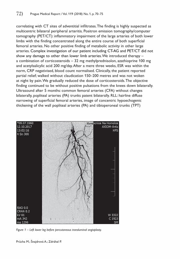
72)
Průcha M.; Šnajdrová A.; Zdráhal P.
Prague Medical Report / Vol. 119 (2018) No. 1, p. 70–75
correlating with CT sites of adventitial infiltrates. The finding is highly suspected as multicentric bilateral peripheral arteritis. Positron emission tomography/computer tomography (PET/CT): inflammatory impairment of the large arteries of both lower limbs with the finding concentrated along the entire course of both superficial femoral arteries. No other positive finding of metabolic activity in other large arteries. Complex investigation of our patient including CT-AG and PET/CT did not show any damage to other than lower limb arteries. We introduced therapy – a combination of corticosteroids – 32 mg methylprednisolon, azathioprine 100 mg and acetylsalicylic acid 200 mg/day. After a mere three weeks, ESR was within the norm, CRP negativised, blood count normalised. Clinically, the patient reported partial relief; walked without claudication 150–200 metres and was not woken at night by pain. We gradually reduced the dose of corticosteroids. The objective finding continued to be without positive pulsations from the knees down bilaterally. Ultrasound after 5 months: common femoral arteries (CFA) without changes bilaterally, popliteal arteries (PA) trunks patent bilaterally. RLL: hairline diffuse narrowing of superficial femoral arteries, image of concentric hypoechogenic thickening of the wall popliteal arteries (PA) and tibioperoneal trunks (TPT)
Figure 1 – Left lower leg before percutaneous transluminal angioplasty.
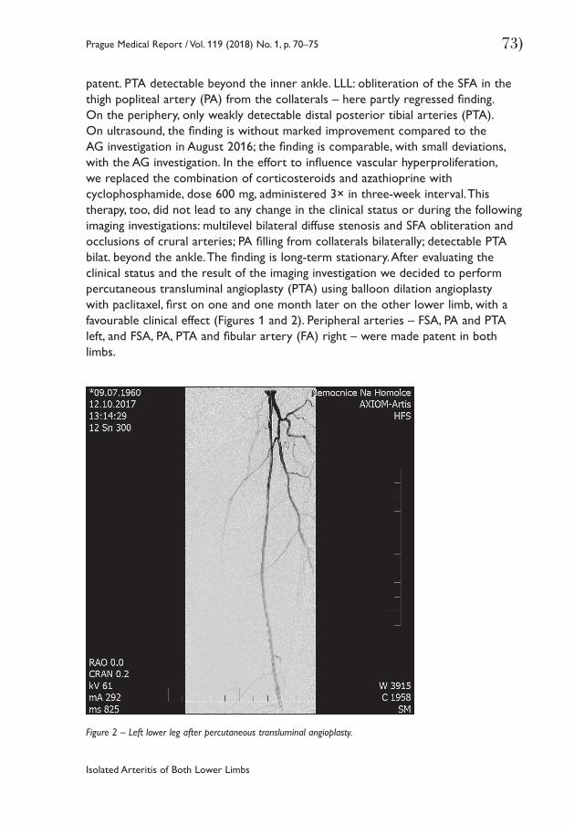
73)
Isolated Arteritis of Both Lower Limbs
Prague Medical Report / Vol. 119 (2018) No. 1, p. 70–75
patent. PTA detectable beyond the inner ankle. LLL: obliteration of the SFA in the thigh popliteal artery (PA) from the collaterals – here partly regressed finding. On the periphery, only weakly detectable distal posterior tibial arteries (PTA). On ultrasound, the finding is without marked improvement compared to the AG investigation in August 2016; the finding is comparable, with small deviations, with the AG investigation. In the effort to influence vascular hyperproliferation, we replaced the combination of corticosteroids and azathioprine with cyclophosphamide, dose 600 mg, administered 3× in three-week interval. This therapy, too, did not lead to any change in the clinical status or during the following imaging investigations: multilevel bilateral diffuse stenosis and SFA obliteration and occlusions of crural arteries; PA filling from collaterals bilaterally; detectable PTA bilat. beyond the ankle. The finding is long-term stationary. After evaluating the clinical status and the result of the imaging investigation we decided to perform percutaneous transluminal angioplasty (PTA) using balloon dilation angioplasty with paclitaxel, first on one and one month later on the other lower limb, with a favourable clinical effect (Figures 1 and 2). Peripheral arteries – FSA, PA and PTA left, and FSA, PA, PTA and fibular artery (FA) right – were made patent in both limbs.
Figure 2 – Left lower leg after percutaneous transluminal angioplasty.
74)
Průcha M.; Šnajdrová A.; Zdráhal P.
Prague Medical Report / Vol. 119 (2018) No. 1, p. 70–75
DiscussionIsolated arteritis of both lower limbs is a rare disease. Arteritis of the lower limbs occurs most frequently in connection with Takayasu’s arteritis or giant-cell Horton’s arteritis (Kermani et al., 2009; Assie et al., 2011; Sigl et al., 2014). We focused on the possibility of TA or GCA with or without connection with a systemic autoimmune disease. It is necessary to stress, that complex investigation of our patient, including CT-AG and PET/CT, did not show any damage to other than lower limb arteries. So our patient did not meet the criteria of large vessel vasculitis typical for this age – GCA. We excluded rare case of polyarteritis nodosa vasculitis mostly affecting the skeletal leg muscles (Khellaf et al., 2007). Similarly as in the GCA or TA, the treatment of choice is the corticosteroid and/or immunosuppressive therapy. An alternative may be the combination of systemic corticosteroids and azathioprine, which we used. In the case of unsuccessful immunosuppressive therapy, the treatment with tocilizumab or rituximab is possible (Stagnaro et al., 2015). Tocilizumab is a humanized anti-human IL-6 receptor antibody that binds to soluble and membrane-bound IL-6 receptor. Rituximab is an anti-CD20 monoclonal antibody that depletes B cells and is often used in the treatment of non-Hodgkin’s lymphoma and B-cell leukemias. Our treatment had a partial effect – the disappearance of positivity of inflammatory parameters – but did not lead to the disappearance or significant retreat of hyperproliferation in the affected vessels that would allow restoration of flow. An important factor of the favourable effect of immunosuppressive therapy was gaining time for setting up collateral circulation, with the final consequence of the patient avoiding amputation of the limbs. We tried to affect the hyperproliferation by administering cyclophosphamide, which is used with relative success in the treatment of vasculitis (de Boysson et al., 2013; Roberts and Clifford, 2017). This therapy, too, had not the desired effect. That is why we decided to perform percutaneous transluminal angioplasty, which was successful. Arterial flow was restored down to the periphery in both lower limbs and the clinical symptomatology disappeared. In this context it is necessary to mention the possibility of locally influencing the vascular architecture with the help of PTA (Barra et al., 2017). Drug-eluting balloons (DEBs) are effective at reducing intimal hyperplasia after angioplasty of the superficial femoral artery, infrapopliteal circulation, and arteriovenous fistulas. In our patient we applied endovascular intervention with a paclitaxel drug eluting balloon. Paclitaxel is a mitotic inhibitor with effect on cellular proliferation. The comparison between a standard balloon dilator without the drug and balloon with paclitaxel in patients with lower limb peripheral arterial occlusion disease has shown higher efficacy of the balloon with paclitaxel in the prevention of restenosis of the treated arteries (Kinstner et al., 2016). The presented case study is an example of successful inter-disciplinary cooperation in the diagnosis and treatment of large artery vasculitis.
75)
Isolated Arteritis of Both Lower Limbs
Prague Medical Report / Vol. 119 (2018) No. 1, p. 70–75
ReferencesAssie, C., Janvresse, A., Plissonier, D., Levesque, H., Marie, I. (2011) Long-term follow-up of upper and lower
extremity vasculitis related to giant cell arteritis. A series of 36 patients. Medicine 90, 40–51.Barra, L., Liang, P., Benseler, S. M., Cabral, D. A., Mah, A. F., Li, Y., Milman, N., Twilt, M., Yacyshyn, E., Pagnoux, C.
(2017) Variations in the clinical practice of physicians managing Takayasu arteritis: a nationwide survey. Open Access Rheumatol. 9, 91–99.
de Boysson, H., Boutemy, J., Creveuil, C., Ollivier, Y., Letellier, P., Pagnoux, C., Bienvenu, B. (2013) Is there a place for cyclophosphamide in the treatment of giant-cell arteritis? A case series and systematic review. Semin. Arthritis Rheum. 43, 105–112.
Kermani, T. A., Matteson, E. L., Hunder, G. G., Warrington, K. J. (2009) Symptomatic lower extremity vasculitis in giant cell arteritis: a case series. J. Rheumatol. 36, 2277–2283.
Khellaf, M., Hamidou, M., Pagnoux, C., Michel, M., Brisseasu, J. M., Chevallier, X., Cohen, P., Guillevin, L., Godeaus, B. (2007) Vasculitis restricted to the lower limbs: a clinical and histopathological study. Ann. Rheum. Dis. 66, 554–556.
Kinstner, C. M., Lammer, J., Willfort-Ehringer, A., Matzek, W., Gschwandtner, M., Javor, D., Funovics, M., Schoder, M., Koppensteiner, R., Loewe, C., Ristl, R., Wolf, F. (2016) Paclitaxel-eluting balloon versus standard balloon angioplasty in in-stent restenosis of the superficial femoral and proximal popliteal artery: 1-year results of the PACUBA trial. JACC Cardiovasc. Interv. 9, 1386–1392.
Roberts, J., Clifford, A. (2017) Update on the management of giant cell arteritis. Ther. Adv. Chronic Dis. 8, 69–79.
Sigl, M., Hsu, E., Scheffel, H., Haneder, S., Rümenapf, G., Amendt, K. (2014) Lower extremity vasculitis in giant cell arteritis: Important differential diagnosis in patients with lower limb claudication. Vasa 43, 326–336.
Stagnaro, C., Cioffi, E., Talarico, R., Della Rossa, A. (2015) Systemic vasculitides: a critical digest of the most recent literature. Clin. Exp. Rheumatol. 33, 145–154 (Suppl. 89).
Instructions to Authors
76) Prague Medical Report / Vol. 119 (2018) No. 1, p. 76–79
Prague Medical Report is an English multidisciplinary biomedical journal published quarterly by the First Faculty of Medicine of the Charles University. Prague Medical Report (Prague Med Rep) is indexed and abstracted by Index-medicus, MEDLINE, PubMed, DOAJ, EBSCO, and Scopus.
Articles issued in the journala) Primary scientific studies on the medical topics (not exceeding 30 pages in
standardized A4 format – i.e. 30 lines and 60–65 characters per line – including tables, graphs or illustrations)
b) Short communications c) Case reportsd) Reviewse) Lectures or discourses of great interestf) Information about activities of the First Faculty of Medicine and other associated
medical or biological organizations
Layout of the manuscripta) Title of the study (brief and concise, without abbreviations)b) Information about the author(s) in the following form: ■ first name and surname of the author(s) (without scientific titles) ■ institution(s) represented by the author(s) ■ full corresponding (mailing) author’s reference address (including first name,
surname and scientific titles, postal code, phone/fax number and e-mail)c) Abstract (maximum 250 words)d) Key words (4–6 terms)e) Running title (reduced title of the article that will appear at the footer (page
break), not more than 50 typewritten characters including spaces)f) Introduction ■ The use of abbreviations should be restricted to SI symbols and those
recommended by the IUPAC-IUB. Abbreviations should be defined in brackets on first appearance in the text. Standard units of measurements and chemical symbols of elements may be used without definition.
g) Material and Methodsh) Resultsi) Discussion
Instructions to Authors
Instructions to Authors
77)Prague Medical Report / Vol. 119 (2018) No. 1, p. 76–79
j) Conclusionk) References ■ All the sources of relevant information for the study should be cited in the
text (citations such as “personal communication” or “confidential data” are not accepted).
■ It is not permitted to cite any abstract in the References list. ■ References should be listed alphabetically at the end of the paper and typed
double-spaced on separate pages. First and last page numbers must be given. Journal names should be abbreviated according to the Chemical Abstract Service Source Index. All co-authors should be listed in each reference (et al. cannot be used).
■ Examples of the style to be used are: Yokoyama, K., Gachelin, G. (1991) An Abnormal signal transduction pathway
in CD4–CD8– double-negative lymph node cells of MRL lpr/lpr mice. Eur. J. Immunol. 21, 2987–2992.
Loyd, D., Poole, R. K., Edwards, S. W. (1992) The Cell Division Cycle. Temporal Organization and Control of Cellular Growth and Reproduction. Academic Press, London.
Teich, N. (1984) Taxonomy of retroviruses. In: RNA Tumor Viruses, eds. Weiss, R., Teich, N., Varmus, H., Coffin, J., pp. 25–207, Cold Spring Harbor Laboratory, Cold Spring Harbor, New York.
References in the text should be cited as follows: two authors, Smith and Brown (1984) or (Smith and Brown, 1984); three or more authors, Smith et al. (1984) or (Smith et al., 1984). Reference to papers by the same author(s) in the same year should be distinguished in the text and in the reference list by lower-case letters, e.g. 1980a, or 1980a, b.
l) tables, figures, illustrations, graphs, diagrams, photographs, etc. (incl. legends)
Technical instructionsa) Manuscripts (in UK English only) must be delivered in the electronic
form via Online Manuscript Submission and Tracking system (http://www .praguemedicalreport.org/). In case of problems, contact the Prague Medical Report Office ([email protected]). The online submission has to include the complete version of the article in PDF format, separately the manuscript as a MS Word file and a cover letter. The detailed version of the Instructions to Authors can be found at: http://www.praguemedicalreport.org /download/instructions_to_authors.pdf.
b) Text should be written in MS WORD only. We accept only documents that have been spell-checked with UK English as a default language.
c) Please, write your text in Times New Roman script, size 12, and line spacing 1.5.d) Text should be justified to the left, with no paragraph indent (use Enter key
only); do not centre any headings or subheadings.

Instructions to Authors
78) Prague Medical Report / Vol. 119 (2018) No. 1, p. 76–79
e) Document must be paginated-numbered beginning with the title page.f) Tables and graphs should represent extra files, and must be paginated too.g) Edit tables in the following way: Make a plain text, indent by Tab (arrow key) all
the data belonging to a line and finish the line by Enter key. For all the notes in table, use letter x, not *.
h) Make your graphs only in black-and-white. Deliver them in electronic form in TIFF or JPG format only.
i) Deliver illustrations and pictures (in black-and-white) in TIFF or JPG format only. The coloured print is possible and paid after agreement with the Prague Medical Report Office.
j) Mark all the pictures with numbers; corresponding legend(s) should be delivered in an extra file. Mark the position of every picture (photo) in the manuscript by the corresponding number, keep the order 1, 2, 3...
Authors’ DeclarationThe corresponding (or first author) of the manuscript must print, fill and sign by his/ her own hand the Authors Declaration and fax it (or send by post) to the Prague Medical Report Office. Manuscript without this Declaration cannot be published. The Authors’ Declaration can be found by visiting our web pages: http://pmr.lf1.cuni.cz or web pages of Prague Medical Report Online Manuscript Submission and Tracking system: http://www.praguemedicalreport.org/.
Editorial procedureEach manuscript is evaluated by the editorial board and by a standard referee (at least two expert reviews are required). After the assessment the author is informed about the result. In the case the referee requires major revision of the manuscript, it will be sent back to the author to make the changes. The final version of the manuscript undergoes language revision and together with other manuscripts, it is processed for printing.Concurrently, proofs are electronically sent (in PDF format) to the corresponding (mailing) author. Author is to make the proofs in PDF paper copy and deliver it back to the editorial office by fax or as a scanned file by e-mail. Everything should be done in the required time. Only corrections of serious errors, grammatical mistakes and misprints can be accepted. More extensive changes of the manuscript, inscriptions or overwriting cannot be accepted and will be disregarded. Proofs that are not delivered back in time cannot be accepted.
Article processing chargeAuthors do not pay any article processing charge.
Open Access StatementThis is an open access journal which means that all content is freely available without charge to the user or his/her institution. Users are allowed to read,

Instructions to Authors
79)Prague Medical Report / Vol. 119 (2018) No. 1, p. 76–79
download, copy, distribute, print, search, or link to the full texts of the articles, or use them for any other lawful purpose, without asking prior permission from the publisher or the author. This is in accordance with the BOAI definition of open access.
Copyright StatementThe journal applies the Creative Commons Attribution 4.0 International License to articles and other works we publish. If you submit your paper for publication by Prague Medical Report, you agree to have the CC BY license applied to your work. The journal allows the author(s) to hold the copyright without restrictions.
Editorial OfficePrague Medical ReportKateřinská 32, 121 08 Prague 2, Czech Republice-mail: [email protected]: +420 224 964 570. Fax: +420 224 964 574

Published by the First Faculty of Medicine, Charles University, Karolinum Press, Ovocný trh 3, 116 36 Praha 1 – Staré Město, Czech Republic, www.karolinum.cz
Editorial Office: Prague Medical Report, Kateřinská 32, 121 08 Prague 2, Czech Republic, Phone: +420 224 964 570, Fax: +420 224 964 574, e-mail: [email protected] Editor in Chief: Kateřina Jandová, MD., PhD. Editor: Assoc. Prof. Jan Šváb, MD., PhD.Foreign Language Editor: Prof. Jaroslav Pokorný, MD., DSc.Executive Editors: Mgr. Jiří Frühauf, Mgr. Lucie Šulcová Editorial Board: Prof. Jan Betka, MD., DSc.; Zdeněk Kostrouch, MD., PhD.; Prof. Emanuel Nečas, MD., DSc.; Prof. František Perlík, MD., DSc.; Prof. Karel Smetana, MD., DSc.; Prof. Karel Šonka, MD., DSc.; Assoc. Prof. Jan Tošovský, MD., PhD.; Prof. Jiří Zeman, MD., DSc.
Published as quarterly journal. Typeset and printed by Karolinum Press. Annual subscription (4 issues) EUR 60,–. Single copy EUR 20,–.Distribution: A.L.L. Production, Ve Žlíbku 1800/77 – Hala A7, 139 00 Praha 9, Czech Republic,tel.: +420 840 306 090, e-mail: [email protected], www.predplatne.cz
ISSN 1214-6994 (Print)ISSN 2336-2936 (Online)
Reg. No. MK ČR E 796
(Sborník lékařský)