sanders_multiple_congenital.pdf - mspace
TRANSCRIPT
MULTIPLE CONGENITAL ANOMALIES OF UNKNOWNETIOLOGY : A R-ETROSPECTIVE STUDY
&SYSTEMATIC R-E\AEW OF
94 CASES
ATHESIS SUBMITTED
TO
THB FACULTY OF GRADUATB STUDIESTINTVERSITY OF MANITOBA
IN PARTIAL FULFILLMENT OF THE REQUIREMENTS FOR THEDEGREE OF MASTERS OF SCIENCE
BY
SIIANNON ROSB SANDERS
DEPARTMENT OF BIOCHEMISTRY AND MBDICAI GENBTICS
l*¡ 5¡3:l'#o'oAcqu¡s¡tions and Acquisitions etBibliographicServ¡ces serv¡cesbibl¡ographìques
395 W€llingnon Str€6t 395, ¡ùe WallinglonOttawa ON Kl A OM Ottav¡ã ON K1 A 0N4Ca¡ada Cânada
The authorhas granted anon-exclusive licence allowing theNational Library of Canada toreproduce, loan, distribute or sellcopies of this thesis in microform,paper or electonic formats.
The author rerzins ownership of thecopyright in this thesis. Neither thethesis nor substantial extacts from itmay be printed or otherwisereproduced without the author'spermission.
L'auteur a accordé une licence nonexclusive permettant à laBùliothèque nationale du Canada de
reproduire, prêter, disFibuer ouvendre des copies de cette thèse sous
la forme de microfiche/ñln, dereproduction sw papier ou sur formatélectronique .
L'auteur conserve la propriété dudroit d'auteur qui protège cette thèse.Ni la thèse ni des exfinits substantielsde celle-ci ne doivent être imprimésou autrement reproduits sâns sonautorisation.
Bibliothèque nat¡onaledu Cenadâ
Vùt ãlâ votþ tété@@
Oùt 6ta Noùe fittét@
0-612-76868-6
Canadä
THE UNIVERSITY OF MANITOBA
FACULTY OF GRADUATE STUDIES
COPYRIGHT PERMISSION PAGE
Multiple Congenital Anomalies of Unknown Etiology:
A Retrospective Study & Systematic Review of 94 Cases
BY
Shannon Rose Sanders
A Thesis/Practicum submitted to the Faculty of Graduate Studies of The University
of Manitoba in partial fulflrllment of the requirem€nts of th€ d€gree
of
MASTER OF SCIENCE
SHANNON ROSE SANDERS @2002
Permission has been grant€d to the Library ofThe University of Manitoba to lend or sell copiesof this thesiypracticum, to the National Library of Cânada to microfilm this thesis and to lendor sell copies of the frlm, ând to University Microfilm Inc, to publish an abstract of thisthesis/practicum.
The author reserves other publication rights, and neither this thesis/practicum nor extensiveextracts from it may be printed or otherwise reproduced lvithout the author's writtenpermission.
TABLE OF CONTENTS
ACKNOWLEDGEMENTSABSTRACTLIST OF TABLESLIST OF FIGI,T.ESLIST OF APPENDICESLIST OF ABBREVIATIONS & ACRONYMS
tV
ViIxXXI
t. INTRODUCTION 1
1.1 Multiple Congenital Anomalies 1
1.2 Dysmorphology 21.3 Mechanism of Abnomal Morphogenesis 4
1.3.1 Malfonnation 41 .3.2 Deformation 41.3.3 Disruption 51.3.4 Dysplasia 5
1.4 Pattems of Abnomral Morphogenesis 61.4.1 S;'ndro¡re 61.4.2 Sequence 71.4.3 Association J1.4.4 Developmental field defect 7
1.5 Etiology 81.5.1 Environmental 81 .5.2 Genetic 9
1.5.2.1 chromosomal 91.5.2.2 rnonogenic 10
1.5.3 Multifactorial 101.5.4 Unkrown Etiology I 1
1.6 Computerized Databases 121.6.1 POSSUM 121.6.2 LDDB 13
1.7 Duty to Recontact 161.8 Objectives and Hypotheses 18
METHODS 192.I Case Ascertainment 192.2 Case Exclusion 202.3 Systernatic re-evaluation of undiagnosed MCA: criteria one 22
2.3.1 Retrospective diagnosis 222.4 Previous MCA syndrome & association diagnosis: criteria two 232.5 Provisionally new MCA s),ndromes & associations: criteria three 232.6 Computerized database evaluation 242.7 Phenolypic & demographic analysis 24
2.7.1 Formation olthe phenotype & demographic sheet 242.7 .2 Formalion of the coding sheet 252.7 .3 Discriminant functional analysis Zs
4.
2.8 Recurrence risk estimation analysisRTSULTS
3.1 Case asceftainment: breakdown per referral methods3.2 Case exclusion3.3 Systematic re-evaluation of undiagnosed MCA
3.3. 1 Retrospective diagnosis: syndromes3.3.2 Retrospective diagnosis: associations & sequences3.3.3 Undiagnosed Cases
4 Previous MCA s}'ndrome & association diagnoses5 Provisionally 'new' s¡mdromes & associations6 Computerized Databases7 Phenotype & demograpliic8 Recurence risk estinration
CASE COMMENTARY: Clinical4.1. Syridromes
analysisanalysisDescription & Review of Cases
262727292930J]
3439404t
444848485053565l60626467697l73
75
7778807285
87
91
91
92949596979899100101
102
case one-Scase two-Scase three-Scase four-Scase fve-Scase six-Scase seven-5.case eight-Scase nine-Scase ten-Scase eleven-Scase twelye-Scase thirteen-Scase fourteert-Scase ffteen-Scase sixteen-Scase seventeen-Scase eíghteert-Scase nineîeen-S
4.2 Associations & Sequencescase one-Acase two-Acase three-Acase four-Acase five-Acase six-Acase seven-Acase eight-Acnse nine-Acase ten-Acase eleven-A
cuse twelve-Acase thirteen-A
5, DISCUSSION5.1 Case ascefiainment5.2 Systematic re-evaluation of undiagnosed MCA5.3 Previous MCA syrdrome diagnoses5.4 Provisionally new MCA s¡,ndromes & associations5.5 Computerized databases5.6 Phenotlpìc & demographic analysis5.7 Recurrence risk estímation analvsis
6. FUTI,RE WORK6.1 Cluster analysis6.2 Recontacting larnilies
7. SI'MMARY
REFERENCESAPPENDICES
103
106
r09t09t09t5t6T1
t8
126142
1211241all
124125
ACKNOWLEDGEMENTS
First, I would like to thank Dr. Bemie Chodirker and Dr. Jane Evans for their suppoÍ,
guidance, patience and encouragement throughout this joumey. They helped shed light
into the dark comers of the world of dysrnorphology.
Secondly, I would like to thank my committee members, Dr. Albert Chudley and Dr.
Susan Phillips for their commitment and rnsiglrt to this project. I would also like to thank
Dr. L. B. Erdile for his statistical expertise and help.
I would like to acknowledge the many members of the Section of Genetics & Metabolism
for their understanding and endless help in my search for those "misplaced" charts. I
would especially like to thank Linda Carter for her constant words of encouragement and
belief in me.
Special thanks to my family, my two daughters Arnanda and Mackenzie and my fiiends
for their patience and understanding when this work took me away from them.
This project was nade possible in part by fi.urding from the Children's Hospital of
Wimipeg Research Foundation.
ABSTRACT
Approximately 2-3% of newboms will have an identifiable major congenital anomaly
and 4-7 per thousand wì1l have multìple congenital anomalies. Congenital anomalies are
among the leading causes ofìnfant mortality and contribute substantially to infant
morbidity. Early and accurate diagnosis of a child with multiple congenital anomalies is
important lor patient management, providing useful genetic counselling regarding
etiology and recurrence risk, prenatal diagnosis, screening and recommendation for
evaluation of other family members. Unforlunately, provìdìng a diagnosis for a child
who presents with multiple congenital anornalies is a complex task and in many cases the
etiology ìs unknown. The objectives of this study were: l) to detemine the value of a
systematic review of patients with multiple congenital anomalies of unknown etiology,
including addressing the success rate in making a diagnosis and to detenline the factors
that are associated with an increased chance of making a diagnosis and 2) to determine an
appropriate recurrence risk estimate for infants with multiple congenital anomalies of
unknown etiology.
At the time ofthis study over 35,000 patients had been referred to the Section of
Genetics & Metabolism. Records were kept in the Section's database systern ìncluding
the reason for the refenal. 2,681 cases undelwent a chart review because ofan indication
of multiple congenital anomalies. Of those initial cases, 94 were included in the study for
the followirg reasons: 75 were undiagnosed multiple congenital anomaly cases, 9 were
'hew" diagnoses reported by rnembers of the Section of Genetics & Metabolism and l0
were cases in which the diagnosis was made aÍÌer one year's time from the initial contact.
All 75 cases of undiagnosed multiple congenital anomalies were re-evaluated by
using LDDB, POSSLIM and on-lire databases such as OMIM and Medline. Of those 75
cases, 19 were given a new and/or confirmed s].ndrome diagnosis, 13 were given a new
and./or confimred association/sequence dìagnosis and the remaining 43 cases remained
unknown giving an overall success rate of 42.6%
Disclirninant functional analysis was perfomed on demographic variables and on
phenoty?ic traits to detemine what factors potentially detennine the likelihood of maki¡g
a diagnosìs. No demographic variables were found to have significant probability values.
Three phenotypic traits were identified to have significant probability values, These traits
were lenal dysplasia,/cystic kidneys, postaxial polydactyly and tracheal defects.
Recurrence lisk estimates for infants with MCA of unknown etiology were
perfonned. Two separate groups were analyzed, Group 1 diagnosis made and Group 2,
no diagnosis made. In the cases in which a diagnosis was made, the estimated risk of
recurrence was 14.8% and in the cases in which no diagnosis was made, the estimated
risk of recurrence was 15.0%.
LIST OF TABLES
TABLE 1 Concunence of minor and major anomalies at birth in th¡ee series 1
TABLE 2 The lumber ofcases ascertained by each referral method perinclusion criteria 28
TABLE 3 Number ofcases excluded from the study per exclusion criteriafor each referral method
TABLE 4 Sunrmary of the 75 cases of MCA of unknown etiology afterre-evaluation using POSSUM, LDDB, Medline/PubMed &other references
TABLE 5 List of sy.rdromes diagnosed through re-evaluation including thenumber of rnajor malformations present per case
TABLE 6 Post-search mode ofinheritance and associated risk ofrecunenceper newly diagnosed syrdrome cornpared to pre-search riskestimates
TABLE 7 List of associations/sequences diagnosed through re-evaluationincluding the number of major malfon¡ations present per case 33
TABLE 8 Post-search mode ofinheritance and associated risk ofrecunenceper newly diagnosed associations & sequences compared topre-search risk estimated 34
TABLE 9 Summary of cases that remain unknown: medical/family history &major anomalies 35
TABLE 10 Summary of cases asceftained by criteria 2: MCA cases - diagnosisn.rade after I year's time f¡om initial contact 39
TABLE 1 I Summary of the "new" slmdromes/associations delineated bymembers of the Section of Genetics & Metabolisrl
TABLE 12 Summary of all 32 diagnosed cases and the database mostuselul lor diagnostic pùposes
29
30
31
32
40
vll
41
TABLE 13 Success rate ofcomputerized databases with comparison inpercentage for POSSUM, LDDB, OMIM & PubMed
TABLE 14 The occurrence ofdemographic variables in Group 1 &Group 2: comparison of probabilities
TABLE 15 Renal dysplasia,/cystic, postaxial polydactyly and trachealdefects: comparison of flequency in Group 1 & Group 2
TABLE 16 Recunence of rnalfomation(s) in sibling(s) of the proband:Comparison of Gloup I & Group 2
TABLE 17 Percentage of the occuruence of malformation(s) in thesubsequent siblings ofall 94 cases reviewed in the study:Comparison of Group 1 & Group 2
TABLE 18 Success rate ofa systematic review & re-evaluation of75 cases: MCA of unknown etiology
/11
+3
43
44
4l
t14
LIST OF FIGURES
FIGURE 1 Schematic for case ascerlainment and breakdown of casesinto inclusion critena
LIST OF APPENDICES
APPENDIX 1 Phenotlpe sheet of all traits grouped by System./Regionand by presentatìon
APPENDIX 2 CODING SHEET: Dehnitions, Field names and Codespcr trait
APPENDIX 3 Dendrogram showing Ward's Mixed Cluster analysisof 95 Cases: Preliminary Results
t42
l4J
148
LIST OF ABBREVIATIONS & ACRONYMS
A Association & sequence diagnosìsACC Agenesis of the corpus callosumAMA Advanced matemal age
AS Aqueductal stenosisASD Atrial septal defectBCD Blepharo-cheilo-dontic s1'ndromeCA Congenital anomalyCFC Caldio-facìal-cutaneous s¡mdromeCHARGE Coloboma, lreaf anomaly, choanal atresia, mental
retardation, genital and ear anomaliesCLP Cleft lip and palateCNS Central nervous systemCP Cleft palateCODAS Cerebral, ocular, dental, auricular, skeletal
anomalies s¡.ndromeCOFS Cerebro-oculo-facial-ske1eta1 slmdromeCS Cockayne syrdromeDD Developmental delayDOB Date of birthDX DiagnosisEFE Endocardial fibroelastosisFAS Fetal alcohol sl,ndromeFAV Facio-auriculo-vertebral dysplasiaFHX Family historyFISH Fluorescence in situ hybridizationGAPO Growth retardation, alopecia, pseudoanodontia and
optic atrophyGI GastrointestinalIUGR Intrauterine growth retardationI\VG Intemational Working GroupL LeftLD Late diagnosisLDDB London dysmorphology databaseMART Martsolf: skeletal dysplasia, polydactyly and Pierre Robin
syrdromeMASA Mental retardation, aphasia, shuffling gait and
adducted thumbsMAT MatemalMCA Multiple congenital anomaliesMOTA Manitoba oculotrichoanal syrdromeMR Mental retardationMURCS Mullerian duct, renal agenesis and cervical thoracic somite
dysplasiaN New diaørosis
NER Nucleotide-excision repairNTD Neural tube defectOMIM Online Mendelian Inheritance in ManPAT PatemalPDA Patent ductus ateriosusPOSSUM Pictures of standard s)¡ndromes and undìagnosed
malfomationR RightR-V FISTULA Recto-vaginal fistulaS Slardrome diagnosisSAMS Short stature, auditory canal atresia, mandibular hypoplasia
and skeletal abnormalities slrrdromeSCD Spondylocostal dysostosisSHH Sonic hedgehogSMA Spinal muscular atrophyTBS Towns-Brocks syrdromeT-E FISTULA Tracheo-esophageal fìstulaTGV Transposition ofthe great vesselsTOF Teratology of fallotT21 Trisomy 21
U Unknown diagnosisVATER Verteb¡al anomalies, anal atresia, tracheo-esophageal
fistula, radial ray and renal anomaliesVSD Ventricular septal defectYOB Year of Birth
XII
I. INTRODUCTION
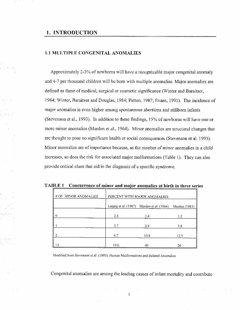
1.1 MULTIPLE CONGENITAL ANOMALIES
Approximately 2-3o% of newboms will have a recognizable major congenital anomaly
and 4-7 per thousand children will be bom with multiple anomalies. Major anomalies are
defined as these of medical, surgical or cosmetic significance (Winter and Baraitser,
1984; Winter, Baraitser and Douglas, 1984; Patton, 1987; Evans, 1991). The incidence of
major anomalies is even higher among spontaneous aborlions and stillbom infants
(Stevenson et a1., 1993). ln addition to these hndings, 15% ofnewboms will have one or
more nrinor anomalies (Marden et al.,1964). Minor anomalies are structural changes that
are thought to pose no significant health or social consequences (Stevenson et al. 1993).
Minor anomalies are of imporlance because, as the number of minor anomalies in a child
increases, so does the risk fol associated major malfonnations (Table 1). They can also
provide critical clues that aid in the diagnosis ofa specific slmdrome.
TABLE I Concurrence of minor and anomalies at birth in three series
Modiiied from Stevenson et aì. (1993): Hurnân Malforn.lations and Relâted Anomalies
Congenital anomalies are among the leading causes ofinfant mortality and contribute
( 1964) Meches ll98
1.4 1.2
3.',7 2.9
6.7 10.8 12.5
substantially to ìnfant morbidity (Yoon et a1.,1991). The overall frequency of birlh
defects has remained constant. However, the decline in infant mortality due to infeclions.
poor prenatal or postnatal care, or nutritional factors has significantly heightened the
imporlance ofbirth defects. Population-based studies have shown that birlh defects and
genetic diseases account for a high percentage (9-a0%) of a1l pediatric hospitalizations
(Scriver et air.,1973; Hall et al., 1978; Yoon et aI.,1997). In addition to higher
hospitalization rates, these children also tend to have longer hospital stays and higher
readmission rates, and their hospitalizations are proporlionally more costly then other
types ofpediatnc hospitalizations (Cunniff et al., 1995).
Early and accurate diagnosis of a child with multiple congenital anomalies (MCA) is
imporlant for patient management, providing useful genetic counselling regarding
etiology and recurence risk, prenatal diagnosis, screening and recommendations for
evaluation of other falnily members (Witt and Hall, 1985). Making a diagnosis in a child
who presents with MCA can be a daunting task due to the complexity of these conditions
with regard to understanding of the etiology and mechanism of action. Further, the large
number of syrdrornes described in the literature makes the task that much more difficult.
It has been estìmated that a new syndrome is described at a rate ofone or more a week
(Toriello, 1988).
1.2 DYSMORPHOLOGY
Dysmorphology is the study of abnonnal physical development by interpreting
pattems of structurâl defects. The term "dysmorphic" is used to describe a body part that
has not followed a nomal pattem of growth or fomation and it is often disproportionate
when conrpared to normal development (Witt and Hal1, 1985). A specihc diagnosis is
usually made on the overall pattem of anomalies. However, va¡iations in the presentation
ofan anomaly can arise from patient to patient and morphologic features can change over
time, becoming more or less pronounced.
One of the most frequent tasks the dysnorphologist/clinical geneticist performs is to
try and reach a dìagnosis for a patient or family. In some cases, this can be done in one
initial consultation with the patìent. However, the majority of cases require a more
extensive investigatìon. There are a number of steps that a dysmorphologist may take to
try and reach a tentative diagnosis. Dilibertì (1988) outlined the steps and procedures that
a dysmorphologist typically takes. The first step usually involves a complete physical
examination and detailed family and pre and postnatal history. Ifthe dìagnosis is no1
obvious, the next step is to review reference texts. With this, there are a number of
approaches one can take. One strategy is to select a single physical feature and search
reference texts for syrdromes that contain that feature. Obviously this approach will
yield a large number ofpossible diagnoses unless the physical feature is quite rare. From
this, one can create a working list ofcandidate sl,ndromes. Comparison of the patient's
features with those of the candidate s1'ndromes can shoften the list into a small number of
potential diagnoses. The use ofpublished reference material such as medical journals and
photographs, and consultation with other dysmorphologists, in addition to performing a
number oftests, e.g., radiographs, ultrasounds, chromosomal analysis and molecular tests,
may also be necessary in order to reach or confirm a diagnosis (Winter and Baraitser,
i984).
Reference books such as Smith's lexÍbook Recognizable Patterns of Htmtan
Malforrnations 5th edition (Jones, 1997) list only a small fraction of malformation
slmdromes. Computer technology has been able to offer some solutions for this problem.
A number ofcomputerized databases have been developed to allow the user to search a
large volume of s¡mdromes by a variety of means.
1.3 MECHANISMS OF ABNORMAL MORPHOGENESIS
There are four categories that are often used to describe the major types of structural
anomalies. These are malformation, deformation, disruption and dysplasia.
].3.] MALFORMATION
A malfomration is defined as a " morphological defect which resulted fiom an
intrinsically abnormal developrnental process" (Thompson et al., 1991). These tend to be
defects oforgans, parl ofan organ or larger areas of the body (Witt and Hall, 1985).
Malfonnations can be the result of chronosomal or monogenic defects, and can be
grouped into three classes: incomplete morphogenesis, which occurs when there is
developmental arrest (e.g. renal agenesis), redundant morphogenesis (e.g. polydactyly)
and aberant morphogenesis in which the malformatìon has no normal counterpart (e.g.
paratesticular spleen) (Cohen, 1986).
].3.2 DEFORMATION
A defomation is defined as an " abnomality in form or position of a body part caused
by a non-disruptive mechanical force" (Thornpson et a1., 1991). These defects canbe
distinguished fiom malformations by the fact that they tend to be reversible and
correctable and arise most often late in fetal development. Deformations may ar-ise from
malformational or functional causes (e.g. neurologic and muscle disturbances, connective
tissue defects) and/or intrauterine constraint (Cohen, 1986). An important distinction
must be made between malfomations and deformations. Malformations arise in the
embryo during organogenesis and are primary errors in morphogenesis. Deformations
tend to arise during the fetal stage ofpregnancy and are changes in a shape of the
previously normal structure. Perinatal mortality tends to be much higher in chi'ldren with
malfomations as compared with those children with deformations. A final distinction
that can be made between malformations and defomrations is the potential for conection
of the deformation defect either spontaneously or by intervention by posturing means.
Malformations can not spontaneously revert back to the nonnal structure nor can they be
corrected without major surgery or medical intervention (Cohen, 1986).
].3.3 DISRUPTION
A disruption is dehned as a " morphological defect resulting from breakdown of; or
interlerelce with, an originally normal developmental process" (Thompson et al., 1991).
Disruptions are due to events that occur after embryogenesis; they tend to be sporadic
events with a low recurrence risk. While disruptions are often environmental in nature,
genetic factors may also be involved. For example, amniotic bands are strands of
amniotic tissue that can adhere to the ernbryo or fetus causing constriction, arnputation of
limbs and digits as well as other defects such as facial clefts (Stevenson et al., 1993).
Teratogens can also interfere with nomral development of the fetus causing a wide range
of anomalies. Fetal Alcohol Syrdrome is an example of a common teratogenic
sl,ndrorne.
1.3.4 DYSPLASIA
A dysplasia is defined as an " abnomal organization of cells into tissues and its
morphological consequence" (Thompson et al., 1991). Dysplasias tend not to be
restricted to specific sites or organs as the abnolmality pertains to a specihc tissue t1pe.
Therefore anomalies tend to be tissue specific (e.g. Chondrodysplasia punctata)
(Wlarbrandt and Ludman, 1990).
1.4 PATTERNS OF ABNORMAL MORPHOGENESIS
Recognizing the specific pattems of birth defects can aid in the understanding ofwhere
and when in embryogenesis the defect(s) occurred. Understanding the type of pattem can
influence how the family is counselled, and can play a role in patient management.
Because it is important to understand and use the correct terminology when describing the
pattems of abnonnal development, the Intemational Working Group (IWG) redefined and
clarified the four terms used in dysmorphology: slardrome, sequence, association, and
developmental field defects (Spranger et al., 1982).
1.4.1 SYNDROME
A sy.rdrome is defined as a "pattem of multiple anomalies thought to be
pathogenetically related and not known to represent a single sequence or a polytopic held
defect" (Spranger et al., 1982). The use of the temr "syrdrome" indicates that a specific
diagnosis has been made and that the natural history and recurrence risks are potentially
known. That doesn't necessarily imply that the etiology is known or well understood.
Syrdrones tend not to be static entities as thele is continuous expansion and revision of
the phenotype through new case reports. Advances in both embryology and molecular
genetics can also redefine the grouping of s;'ndromes.
The term "syndrome" encompasses a dìverse category ofabnormal morphogenesis.
To date, there are over two thousand sy,ndromes described in the literature, of which the
etiology can be chromosomal (10-15%), monogenic (6-8'Yo), teratogenic (5-7%) or of
unknown etiology (50%) (Thompson et al., 1991; Stevenson el a1.,1993).
1.4.2 SEQUENCE
A sequence is defined as a " pattem of multiple anomalies derived from a single
known or presumed prior anomaly or mechanical factor" (Spranger et al., 1982), and that
"a sequence is a pathogenic and not a causal concept" (Martinez-Frias et a1., 1998). The
secondary effects that result from the initial insult can be structural defects, fuirctional
defects or defecls in fonn and growth. While the secondary effects are known to tesult
from the primary defect, the etiology of the primary defect may not be understood.
1.4.3 ASSOCIAT]ON
An association is a " non-random occurrence in two or more individuals of multiple
anomalies not known to be polytopic field defects, sequence, or s¡mdrome" (Spranger et
al., 1982). Ar association is considered a statistical relationship, and not a pathogenetic
ol causal relationship. The concept of associations lras been generally accepted; however,
some authors have questioned whether or not associations are in fact developmental field
defects (Opitz, 1994; Maúinez-Frias, 1995). Associations are believed to not show
altered sex ratio, tend to have low recunence risks (usually thought to be sporadic in
natule), are found in lrigher proportion in twins and tend to affect the midline (Opitz,
1993; Martinez-Frias and Frias, 1997).
1.4.4 DEVELOPMENTAL FIELD DEFECTS
The developmental held defect theory was first inlroduced by the IWG in 1982
(Spranger et a1., 1982) and then clarified and arnended at the Intemational Congress of
Human Genetics in Berlin (Opitz et al., 1987). The developmental field defect concept is
as follows: "a morphogenetic (or developmental) field is a region or parl of the embryo
which responds as a coordinated unit to embryonic induction and results ìn complex or
multiple anatomic structures." Simply stated, a polytopic or developmental field defect is
a pattem of anomalies that are caused by disruption of a single developmental field.
Therefore, disruption in any field regardless of the size ofthe field, or stage of the
development, whether due to teratogenic or mutation factors, will have rnorphological
consequences (Marlinez-Frias et al., 1998).
1.5 ETIOLOGY
Etiology simply means, "cause." The etiology of congenital anomalies can be the
result of genetic factors, envìronmental factors, a combination ofboth genetic and
environmental factors (here tenned multifactorial inheritance), the twinning process or
can be due to factors not yet known. Understanding the etiology may determine the
manner in which the farnily is counselled and how patient management is conducted.
].5.] ENVIRONMENTAL
It is estimated that 5 to lYo of all congenital anomalies can be attributed to
environmental teratogens, (Evaris, 1991). A teratogen is any agent that can produce an
anomaly or raise the population incidence of an anomaly. There are many known human
teratogenic agents and they can be grouped into four main categories: infections, matemal
disorders, drugs and ionizing radiation (Thompson et a1., 1991).
There are a number of common characteristics that all teratogens share. Teratogens
can only have their influence during fetal developmenl, and only at the time ofexposure.
They tend to be sporadic events with a low likelihood of recurrence unless exposule to
the teratogen persists ìn subsequent pregnancies. They have a direct influence on
developmerf by interfering with cellular metabolism, disturbing regional vascular supply
and killing cells (Stevenson et al., 1993). They are dose dependent such that the greater
the length of time of exposure and the greater the amount ofexposure during
development, the greater the severity of the teratogenic effect to the fetus. Teratogens can
also have an indirect influence by causing chromosomal aberrations or mutations
(Thonpson et al., 1991). Many well-recognized conditions are the result of teratogenic
effects.
1.5,2 GENETIC
In all, 15-25%" of cases of congenital anomalies can be attributed to genetic causes
(Evans, 1991;Thompsonetal., 1991; Stevenson et a1., 1993). The genetic causes canbe
subdivided into two main categories, chrourosomal or monogenic.
L5.2.1 Chrontosotnal
Ch¡omosomal aberations are estimated to account for 10-15% of all cases of
congenital anomalies (Stevenson et al., 1993). Chromosome based pattems of anomalies
tend to share a number of common characteristics. Children wìth a chromosorne
abnormality tend to have growth retardation, both prenatally and postnatally, varying
degrees ofmental retardation, and tend to have multiple systems involved. There are two
main types of ch¡omosomal abnormalities: structural and numerìcal. These abnonnalities
can involve either the autosol'ìres or sex chromosomes. They tend to be sporadic events,
thus have a low risk ofrecurrence in future pregrrancies. There are; however, cases of
familial structural ch¡ornosome reanangemenls. These rearrangements will have
different risks ofrecunence depending on the type ofrearrangement and the
chromosornes involved (Gardner and Sutherland, 1996). There can be an increased
incidence of recu¡rent spontaneous abortions in families with a chromosome abnormality.
1.5.2.2 Monogenic
It is estimated lha| 6-80/0 of all MCA can be attributed to the pleiotropic effects of
single gene mutations (Evans, 1991; Stevenson et al., 1993). Disruptions in a gene's
function tend to result in a distinct pattem of abnormalities that can be recognized and
classified into a distinct syndrome. To date, there are well over two thousand non-
ch¡omosomal s¡mdromes that have been described in the literature (Winter and Baraitser,
1987). Classification ofthese syndromes are based on their mode ofinheritance:
autosomal dominant, autosomal recessive or X-linked.
1.5.3 MULTIFACTORIAL
Multifactorial inheritance is based on the ìdea tliat both genetic and environmental
factors work in an inter-related manner to influence phenotypic expression. This concept
relies on the threshold theory, which is based on the concept that an individual is
genetically predisposed, but that expression of the phenotype will only occur when
environmental stress forces that predisposition beyond a certain point (i.e. the threshold)
(Fraser, 1996). Multifactorial inhelitance accounts for 25%o of a\lbifh defects and
includes such anomalies as cleft lip/palate, spina bihda and congenital heart defects
(Evans, 1991; Stevenson et a1., 1993). While multifactorial inheritance accounts for a
large number of isolated bìrth defects, it is not implicated in most cases of MCA (Witt
and Hall, 1985).
].5.4 UNKNOWN ETIOLOG'
Chromosomal, rnonogenic and multifactorial inheritance accounts for approximately
50% of anomalies in newboms. The remaining 50% ofthese cases are ofunknown
etiology. Being unable to determine the etiology impacts upon patìent management,
prognosìs, and the way in which the individual/family is counselled with regard to risk of
recurrence for future pregnancies, and risk to other family members. In those situations
where a diagnosis is not r¡ade, parents are counselled with an estimated recurrence risk ol
I to 5%o, and cautioned that the risk of recurrence maybe as high as 25% Io 50% (Io
represent an unknown recessive or dominant dìsorder). While there have been many
studies that have looked at empiric recunence risks for single birlh defects, there has been
only one study to date that has looked at the recurrence risk following the birth of a child
witli MCA (Czeizel et al., 1988). This study found that sibs of the index patient bom with
MCA of unknown etiology had a 3.9% risk of having the same pattem of anomalies and
ThaT 3.5%o ofthe sibs had at least one of the anomalies present in the index patient.
Hall et al. (1998) reported on a retrospective study ofall cases of "unknown multiple
congenital anomaly sy.rdrome" seen at the University of Kentucky from 1981 to May
1998. They reviewed the number of follow-up visits each case had and the number of
follow-ups (average of2.3) required before a diagnosis was given, and broke those cases
down irto the t)?e of condition (i.e. chromosome versus monogenic). They found that in,
most cases, repeated follow-up was required to successfully diagnose an unknown MCA
slmdrome. They stated that this was due inpart to: 1) phenotype change into a
recognizable symdrorne, 2) follow-up stimulated additional successful Iiterature
searches/matches, and 3) critical features/pattems initially missed became obvious. They
suggested that periodic follow-up for cases of undiagnosed MCA s¡.ndromes should
become a starìdard o I practice.
To date, the Hall et al. (1998) review has been the only reported study in the literature
to look at the value of a systematìc re-evaluation of undiagnosed cases of MCA. One
point that the ÌepoÍ did not comment on was what impact did new advances in the field
of human genetics, along with continuous reporting of "new" slmdromes had on the
success of reach ing a diagnosis.
There have been a number ofnew advances made in diagnostic techniques and in
recognition of multiple congenital s1'ndromes. New syrdromes are being continuously
delineated and reported in joumals, dysmorphology texts and other such references.
Computerized database systems have been developed to aid in slardrome identification
including online databases available through the World Wide Web on the Intemet.
1.6 COMPUTERIZED DATABASES
The development of efficíent computer databases opened the possibility of leaving the
task of searching repetitìvely list ofsigns and slmdromes in the hopes offinding the
correct diagnosis to a computer. Research on computer-aided diagnosis stafed in the
1960s with Wamer and his group and urore specifically research in the field of computer-
aided diagnosis for malformation slmdromes began in the 1980s (Pelz et al., 1996).
These computerized databases were designed to aid in the diagnosis of already well-
known sytdromes and to recognize rare or potentially new sy.rdromes (Winter and
Baraitser, 1987).
],6.] POSSUM
POSSUM stands for Pictures of Standard Sl,ndromes and Undiagnosed
Malformations. It is an Australian computer program developed in 1987 from the
Murdoch Institute for Research into Birlh Defects, Melborr Australia (Stromme, 1991).
The curent version, POSSLIM 4, contains information on2,120 syrdromes with an
atlas containing 1,33I traits. Each s1,r:rdrome contains reference material that can direct
the user to the original sources. ln addition, the majority of the slmdromes have
illustrations of the clinical phenotlpe, examples ofpatients with the condition at different
ages to demonstrate the variations of the conditìon, X-rays or radiographic findings, and
some are acconrpanied by video-clips. To perform a search with POSSLII4, the clinical
features ofthe patient must be entered into the database. The clinical features can include
malformations, minor anomalies, and neurological, biochemical, mental, chromosomal or
other such characteristics. Once these features are coded into the database, it can then
produce a list of s;mdromes compatible with those features and will rank them in order
from most compatible (highest number of features found in both the patient and
slardrome) to the least compatible. Using different pârameters can modify the search.
For example, one can modify the search by marking a particular feature as a major
hnding such that the list ofcandidate syrdromes must contain that feature. One can
perfonn a broad based search (use many features in the search all with equal "weight") or
a nanow based search (by using only major features of the patient). Once the list of
candidate sy,ndromes has been generated, the clinical characteristics ofeach sl,ndrome
can then be studied and compared with the patient's findings.
1.6.2 LDDB
The London Dysmorphology Database (LDDB) was developed by Winter, Baraitser
and Douglas in 1984. The 1995 version contains ilformation on over 2,500 non-
cfuomosomal syrdromes with reference to the literature for each sl,ndrome. Like
POSSLM, most of the syndromes are illustrated with photogaphs. Infomation in the
13
LDDB database was obtained from a review of all genetic and pediatric joumals fiom
1969 onwards. Included were all reported cases of patients with non-ch¡omosomal MCA,
including single case reporls (Patton, 1987).
The fundamental principles of the LDDB system are the same as that of the POSSUM
system. In order to search the LDDB database, identifying characteristics of the patient
must be entered into the system. The LDDB database contains a master list of over 1,200
features that are arranged into three levels. The hrst level corresponds to the general
clinical region (e.g. eyes), the second level refers to a subdivision ofthat region (e.g. iris)
and the third level refers to a specific abnormality (e.g. coloboma). This allows the user
to use broad or narrow search parameters (Evans, 1995). Once the clinical features have
been entered into the system, a list ofcandidate s¡.ndromes can be produced. By a
process of eliminatìon, one may then anive at a tentative diagnosis. The user can also
modify their searches by usìng different key features, varying the number of features used
as criteria and by changing the weight (i.e. importance) given to each clinical feature
(Dilibeti, 1988).
The role of both the LDDB and POSSUM is to function as a diagnostic tool for the
user and to aìd in slndrome identification. Both systems were designed with the intention
ofbeing used by specialists in the field ofdysmorphology to aid in their decision makìng
and not to make the diagnosis for the user. The authors of both LDDB and POSSIIM
systems have emphasized that a good approach to using these systems is to base a search
around one key leature or anomaly together with general clinical features. The ability to
search on general features is useful, as features tend to show considerable variability from
patient to patient with the same syndrome.
Pelz et al. (1996) looked at the usefulness of botli the LDDB and the POSSIJM
systems. Two search strategìes were used. A "novice's strategy" where all clinical
finding were used in the search and an "experl's strategy" where only a select group of
clinical features were used in the search. All cases used ìn the study already had a
confirrned diagnosis. They found that, using the expert's strategy, the correct diagnosis
was suggested by the LDDB in 68% of the cases and by POSSIJM in 637o ofthe cases
with the percentage being slightly lower for each when the novice strategy was used.
This suggests that a "novice" use of both POSSLM and LDDB to aid in sy,ndrome
diagnosis is a valid approach.
There are some inherent problems associated with the use ofcomputerized databases
in syndrome identification. With both the LDDB and the POSSIIM systems, there are
concems with regald to the irflexibility of the features represented in the master lists/atlas
oftraits. Concems arise over the tenninology and interpretation ofhow the features may
be used or described (Diliberli, 1988; Evans, 1995; Hamed et al., 1996; Pelz et al., 1996).
The user is confined to using those traits listed in the master list.
Another problem is the lack of certainty witli respect to individual features ofa
syrdrome. What cefainty does the user have in knowing whether or not an abnonnality
exists for those syrdromes? For example, if the abnomalìty is only found in a small
percentage of the cases for a parlicular sl.ndrome, it may not be listed ir the database as
pafi ofthat syrdrome. This may lead to sonre doubt as to the validity of the diagnosis in
the patient wlro presents with the feature in question (Evans, 1995; Pelz et aL.,1996).
Overall, conputerized databases like POSSIIM and LDDB, in addition to on-line
databases such as OMIM (Online Mendelian Inheritance in Man), are valuable diagnostic
tools. When these systems are used correctly, they are an effective step towards
establishing a diagnosis in a patient with MCA of uniorown etiology.
i5
1.7 DUTY TO RECONTACT
The term "duty to recontact" refers to the possible ethical and lor legal obligations of
genetic service providers to recontact former patients about advances in research that
might be relevant to them (Fitzpatrick el a1.,1999). Advances in medical genetics are
occurring at an exponential rate. This is the result in paÍ to the progress in the Human
Genome Project and elucidation of the genetic bases ofcancer and other genetic
conditions. As a result of this growth, a concem has been raised that there is an ethical
and potentially legal obligation ofthose ir this held to recontact fonner patients when
new advances occur.
Theoretically, recontacting patients when new infonnation becomes available is an
honorable goal 1o strive for. However, there are a number ofproblems assocìated with
this concept. Likely the most substantial problem is the large task of recontacting
patients. Lr order to recontact all patients who would potentially benefit ffom the new
infotmation, one would have to first identify these individuals. This may mean case/char1
reviews, wl.ricl.r would require a large effort in both tirne and funds. Most facilities would
lack the resources to fulfill this task. Once those individuals were identified, one must
than make contact with them. The concept of recontacting patients has raised some
concenrs. Does the benefit ofrecontacting patients out-weigh the possible burdens
associated with recontacting patients, especially ifa long period of time has elapsed since
the last contact? The benefits ofrecontacting patients include in.rproved patient care,
reduced uncertainty in recunence risk and renewed hope for the future. Possible negative
oulcomes would be patient anxiety and stress, intrusion ofprivacy and concerns about
health/life insurance (Fitzpatrick ef a1.,1999). Some authors have advocated that it is the
patient's responsìbility to take a more active role in their medical care. This would mean
patients would have more responsibility for keeping informed about research advances
I6
(Fitzpatrick et al., 1999; Sliarpe, 1999). This may not be a practical approach either,
especially ìf the patient has liniited understanding about his/her condition or if the patient
does not have access to the information.
Regardless of which approach is taken, neither is optimal. Most centres do not have
fomral guidelines with respect to their duty to recontact patients. The concept ofan
ethical/legal duty to recontact may not even be a manageable or obtainable goal.
However, as this study has demonstrated, there is a benefit in having some sort of
systematic re-evaluation ofpatients. While it is not feasìble to start re-evaluatìng all
patients for every new advancement or breakthrough that occurs in the f,reld of human
genetics, it may be worlh while for each centre to examine their demographics and
determine wl.rat, if any, systematic re-evaluation of specific cases (i.e. unknown MCA)
would be cost-effective and beneficial.
1.8 OBJECTIVES AND HYPOTHESES OF THE STUDY
The objectives ofthe study were as follows:
1. To determine the value of a systematic review of patients with MCA of unknown
etiology. This included addressing the success rate in making a diagnosis and
detennining what factors we¡e associated with an increased chance of making a
dìagnosis.
2. To determine an applopriate recurrence risk estimate for infants with MCA of
unknown etiology.
There were two hypotheses tested in this study:
1 Advances in slmdrome diagnosis will allow a significant number of new diagnoses to
be made following a systematic review of previously undiagnosed multiple
malfonnation cases.
2. Different pattems of abnonnal morphogenesis are associated with different recllrrence
risks.
18
2. METHODS
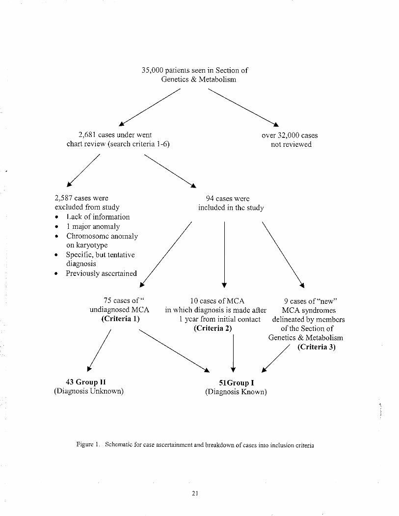
2.1 CASE ASCERTAINMENT
When this study was initiated in 1997, over 35,000 patients had been seen in the
Section of Genetics and Metabolism. Records of all of these patients were available in
the Sectìon's computerized database. Patient demographics, reason for referral, the
geneticist who saw them and their diagnosis were recorded. Ofthese 35,000 patients
seen, 2,681 cases underwent a chaft review. These cases were ascefained through the
Section of Genetics and Metabolism's database system using the following search
c¡iteria:
1. Referral for assessment of a patient affected with MCA without a specific diagnosis.2. Referral for counselling because of a family history of MCA.3. Refenal to the Section of Genetics and Metabolism other than the General Genetics
Clinic for assessment of or counselling for family history of MCA.4. Referral to the c).togenetics laboratory for karyotype analysis because of MCA of
unknown etiology.5. Referral because the patient was affected with or had a family history of "congenital
anomalìes".6. Referral fo¡ assessment because ofa single congenital anornaly.
Of the initial 2, 681 cases, 94 were included in the study under one ofthree inclusion
cdteria:
1. The affected individual had at least two major malfomations and did not have aknown diagnosis and/or more than one potential diagnosis in the differential.
2. The affected individual had MCA, but a diagnosis had not been made within oneyear's time fronr the initial genetic assessment.
3. The affected individual had a provisionally "new" MCA syrdrome that had beendelineated and reported by a member of the Section of Genetics and Metabolism.
Of the 94 cases, 75 cases fell within the first criterion, 10 cases fell within the second
criterion and 9 cases lell within the third criterion.
Figure one is a schematic summarization of the case ascertaiÍìment.
2.2 CASE EXCLUSION
Of the ìnitial 2, 681 cases that underwent a chaÍ review, 2,587 cases were excluded
from the study based on the following reasons:
1. There was only one major anomaly in the ìndex patient.2. There was insufficient information available on the affected individual.3. The case (affected individual) was previously ascertained into the study through
previous search criteria (e.g. a relative of an affected individual was seen forcounselling because of the farnily history of MCA). Lack of adequate information onthe affected individual. The majority of the cases for this study were ascertained underthe second refen'al method - refer¡al to Genetics for a family liistory of MCA. Theretended to be little information or docunrentation on the affected individual.
4. The referred individual was not seen or counselled by a member of the Section ofGenetics and Metabolism (i.e. sample sent in to the cytogenetics laboratory forchromosome analysis without referral to Genetics).
5. The affected individual was thought to have a specific but tentative diagnosis due toatlpical clìnical features or unconfilmed diagnostic results.
6. Any case of MCA in which a chromosome abnonnality had been demonstrated bycluomosomal analysis.
20
35,000 patients seen in Section ofGenetics & Metaboììsm
,a'x'2,681 cases under went
chart review (search criteria 1-6)
,/\
/\2,5 87 cases wereexcluded from study. Lack of informationo I major anomaly /o Chromosonle anonìaly /
on karyotype /o Speciñc. but tentative /diagnosis /¡ Previo uslv ascenained ,/'/
over 32,000 cases
not reviewed
75 cases of " 10 cases of MCAundiagnosed MCA in which diagnosis is made after
(Criteria 1) 1 year frorn initial contact
94 cases wereincluded in the study
5lGroup I(Diagnosis Known)
9 cases of "new"MCA syrdromes
delineated by membersofthe Section of
Genetics & Metabolism(Criteria 3)
(Crite ria 2)
I43 Group II
(Diagnosrs Unknown)
Flgure 1. Schenatic for case ascertairu¡ent and breakdown ofcases into inclusion c¡jterìa
2.3 SYSTEMATIC RE-EVALUATION OF UNDIAGNOSED MCA: criteria 1
2.3. I Retrospcctive Diagnosis
All 75 cases of undiagnosed MCA were re-evaluated using both the LDDB and
POSSUM databases, on-line databases including OMIM, Medline and its subbranch
PubMed, and reference material such as Smith's Recognizable Pattems oÍ Humün
Malfonnations 5tr' Ed. lJones, 1997). For each case, al1 clinical findings, including both
rnajor and minor anomalies, were entered into both the LDDB and POSSUM prograrns
for analysis. All syrdromes with a known chromosomal defect were excluded fiom the
searcl'r. This was done as it was assumed that all cases in the study had a nomal
karyotlpe. However, 14 cases had not had a chrotrrosomal analysis preformed. This was
due to two main reasons: 1) failure of cell culture growth, or 2) chromosome analysis was
not requested- A successful search required that there was a minimal match of two
anomalies between the cases and the candidate s;mdromes. A number of searches were
perfomred for each patient using various combinations of anomalies and weight
(importance) given to each anomaly. Once a working list of candidate s1'ndromes was
identified, reference material, including original case reports ifavailable, was used to
reach a tentative diagnosis. Ifno diagnosis could be made, the case remained as an
"unknown". All new diagnoses had to be agreed upon by two members of the Sectiou of
Genelics and Metabolism. A third rnember of the Section of Genetics and Metabolism
was consulted ifthere was disagreement over a diagnosis. Ifpossible, all diagnoses rnade
in this study were confirmed by re-evalualing the patient in the Genetic Clinic and/or by
cltogenetic, molecular or biochemical testing. Once a diagnosis was confinned the new
risk ofrecurrence was assessed and compared with the initial recumence risk given to the
farnily/patient. For these cases in which a diagnosis was made, two subdivisions were
created to separate the cases based on the pattem of abnormal morphogenesis: 1)
22
s)Ìrdromes and, 2) associations and sequences.
After re-evaluation ofall 75 cases, the cases were grouped into two categories: Gloup
one included all cases in which a diagnosìs had been made and Group two included all
cases that remained unknown. Included into Group one were all cases ascertained into
study criteria two and three.
2.4 PR-EVIOUS MCA SYNDROME & ASSOCIATION DIAGNOSIS: criteria 2
Any MCA case witl-r a dehnitive diagnosis, in wliich the length of time it took to make
that diagnosis exceeded one year's time from the initial genetic assessment, was included
into the study. Al1 cases (10 cases in total) were evaluated to determine 1) the length of
time it took from the initial assessment until a diagnosis was made and 2) why the
diagnosis was delayed. These cases were also included in Group one for discriminant
function analysis and recuuence risk estimations.
2.5 PROVISIONALLY NEW MCA SYNDROMES & ASSOCIATIONS: criteria 3
Any MCA case that was initially seen and repoded as new MCA sy'ndron.res and/or
associations by members of the Section of Genetics & Metabolisrn were included into this
study. A total of 9 cases of "new" MCA syndromes and./or associations were delineated
and reported by members of the Section of Genetics and Metabolism. These cases were
also included in Group one for discriminant function analysis and recurence risk
estimations.
2.6 COMPUTERIZED DATABASÐ EVALUATION
For each case in which a dìagnosis was made, the usefulness olcomputerized
databases in helping to successfully make a diagnosis was evaluated. For each case that a
diagnosis was made, each "search" was reviewed to detemine what database(s) if any
suggested tlie final diagnosis. The databases that were evaluated included POSSIIM,
LDDB, OMIM and PubMed. The success rate in percentage for each of the databases
was calculated by taking the number ofcases where the database was successful over the
total number of cases in which a diagnosis was made (32 in total). This calculation was
done for the above 4 databases. The total success rate was also calculated.
2.7 PHENOTYPIC & DEMOGRAPHICS ANALYSIS
2.7I Fornntion ofthe Phenotype & Dentographic Sheet
A list of anomalies (traits) was obtained by review ofthe cases included in the study
(95 cases in total as one "case" represented two affected individuals). Each trait was
grouped into 10 main categolies. These categories fell under two rnain groupings,
systems or anatomical region: respiratory, cardiovascular, central nervous system (CNS),
gastrointestinal, genitourinary, endocrine, musculoskeletal, craniofacial, skin and other.
Most traits were retaìned for analysis with the exception ofthose traits that occurred
fewer than four times. Additionally, for each case, dernographic variables were included
The demographic variables used in this study were sex, year of bir1h, ethnicity, positive
family history of similar anomalies, positive family history of other anomalies and
s1'mmetry of anomalies (i.e. anomalies occurring unilateral or bilateral).
24
This list oftraits was used to create the phenotype sheet by categorizing each trait into
appropriate system/region and by presentation of that trait. Each trait was coded as
binary (present or abserrt) or multistate variable ifapplicable, e.g.:
Renal agenesis: No Left Sided Right Sided Bilateral
A listing of each trait classified by system/region and presentation can be found in
Appendix 1.
2.7.2 Formation of the Coding Sheet
In order to accommodate the numerical parameters, each trait was conelated (coded)
with a numerical value. Each multistate variable was coded such that the numerical value
leflected the severity of the presentation with 0 equaling no involvement, e.g.:
Renal agenesis: No:0 Left- 1 Right:2 Bilateral:3
A list of each trait with its associated nunerical value (code) can be found in Appendix
2.
2. 7. 3 Discrin tin an t Fun ctiort Analysis
Al1 numerical data íìom the coding sheet was enteled into a Microsoft Access
spreadsheet by case number. There were 71 variables per case. The presence or absence
ofeach variable and the presentation ofthat variable if appropriate was indicated on the
database. The 95 cases were divided into two groups as mentioned previously. Group
one included all cases in which a diagnosis had been rnade and Group two included all
cases in which no diagnosis was made. Discriminant function analysis was preformed to
determine which vanables might differentiate the two groups. Two separate analyses
were preformed. The fìrst analysis evaluated the demographic variables and the second
analysis evaluated the rernaining 64 traits (anomalies). Probability values were derived
for each trait/variable for both analyses.
2.8 RECURRENCE RISK ESTIMATION ANALYSIS
To determine an appropriate recurrence risk estimate for infants with MCA, each case
(94) was reviewed and all indicated subsequent pregnancies were recorded (unless stated
otherwise, all pregnancies in whicli the birlh outcome was not known, were lreated as
"normal" outcomes). In 11 cases the¡e was no information on subsequent pregnancies
because: 1) the family/patient had been lost to follow up and thus subsequent pregnancy
history was not available, or 2) the proband was seen once in the newbom period and the
family was not brought back for follow- up counselling.
Each case was evaluated to determine if there had been a recurrence of a similarly
affected sib(s) (affected same) or a recurrence of an affected sib(s) with malformations
not similar to or associated with the proband's findings (affected other). The "affected
other" category included both rninor and major anomalies as well as multiple anomalies.
This information was grouped into the two previously mentioned categories, diagnosis
made (Group one) and diagnosis unknown (Group two). For Group one and Group two,
the total number of"affected same" was compared to the total number of subsequent
pregnancies to determine the percent of sibs with a recuffence. The same was done for
the "affected other" category.
26
3. RESULTS
3.1 CASE ASCERTAINMENT: Breakdown per Referral Method
1) Ofthe 135 cases initially ascertained under category (1): refenal made to the Section
of Genetics and Metabolism because of MCA, 31 met the inclusion criteria for the
following reasons: 23 cases had at least two major anomalies, 1 case was diagnosed
after one year's time from their initial contact with genetics, 7 cases were new
syrdromes described by a member of the Section of Genetics and Metabolism.
2) Of the 149 cases initially ascerlained under category (2): family history of MCA, 24
cases nlet the inclusion criteria for the following reasolts: 23 cases had at least two
major anomalies, and 1 case was diagnosed after one year's time from their initial
contact with genetìcs.
3) Ofthe 121 cases initially ascertained under category (3): patient seen by a member of
the Section of Genetics and Metabolism for MCA, outside of the regular general
genetics clinic, 12 met the study's inclusion criteria for the following reasons: 10
cases had at least two major anomalies, and 2 cases were diagnosed after one year's
time from their initial contact with the Section of Genetics and Metabolism.
4) Of tlie 52 cases initially ascertained under category (4): referral to the Section's
cytogenetics laboratory for chromosomal analysis of a patient with MCA, none met
the st udy's inclusion criteria.
5) Of the 13 cases initially ascertained under category (5): referral for "congenital
27
anomalies" none met the inclusion criteria.
6) Of the 2,211 cases initially ascertained under category (6): referral made for a single
congenìtal anomaly (i.e. cleft lip/palate, limb anomaly, coloboma), 27 cases met the
inclusion criteria for the following reasons: 19 cases had at least 2 major anomalies, 6
cases \¡/ere dìagnosed after one year's time from their initial contact with genetics, and
2 cases were new slardromes descnbed by a member of the Section of Genetìcs and
Metabolism.
In total, 75 cases were included ìn the study because of undiagnosed MCA. 10 cases
were included in the study because a diagnosis was made after one year's time from the
initial contact. Additional 9 cases were included in the study because a "new diagnosis"
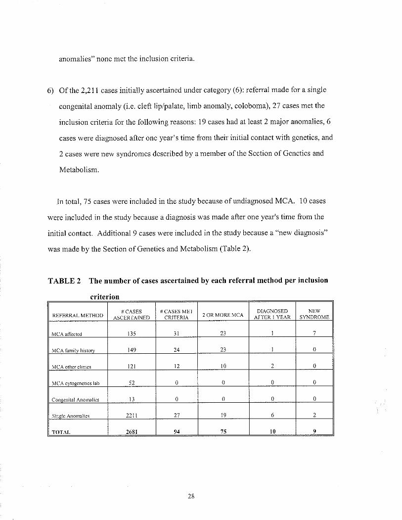
was rnade by the Section of Genetics and Metabolism (Table 2).
TABLE 2 The number ofcases ascertained by each referral method per inclusion
criterion
]IEFERRAL METHOD# CASES # CASES MET
CRITFRIA 2 OR MORE MCADIAGNOSEI)
A}'TER I YEARNEW
ß5 3I 23 7
MCA i¡nìilr hi!ror! 149 14 23 0
MCA other clìnics 121 t2 0 0
MaÀ.vrôoenelì.c I2h 52 0 0 0 0
CônrÌcnitâl Anomâìies 0 0 0 0
Sincle Änon1aIies 2211 71 9 6 2
TOTAT- 2681 94 1\ Íì I
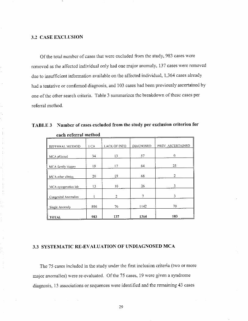
3.2 CASE EXCLUSION
Of the total number ofcases that were excluded flom the study, 983 cases were
removed as the affected individual only had one major anomaly, 137 cases were removed
due to insufllcient information available on the affected individual, 1,364 cases already
had a tentative or conlimred diag osis, and 103 cases had been previously ascerlained by
one of the other search criteria. Table 3 summarizes the breakdown ofthese cases per
referral method.
TABLE 3 Number ofcases excluded from the study per exclusion criterion for
each referral method
RFFIìNR AI MFTììOD tcA LACK OÊ. INFO NIÀGNOSFf) PRFV ÄSC]'RTAINED
MC^ âffeclcd 34 t3 5'7 0
MCA iàmilv h¡storv 19 11 64 25
MCÀ ôiher clìnics 20 I9 68
MCA.vrôrc¡etics lâh l3 l0 26
Cônsenilal Anorìralies'7
ß96 '76 ll42 '70
TOTAI, 983 111 1164 r03
3.3 SYSTEMATIC RN-EVALUATION OF UNDIAGNOSED MCA
The 75 cases ìncluded in the study under the ltrst inclusion criteria (two or more
major anomalies) were re-evaluated. Of the 75 cases, 19 were gìven a s¡mdrome
diagnosis, 13 associations or sequences were identihed and the remaining 43 cases
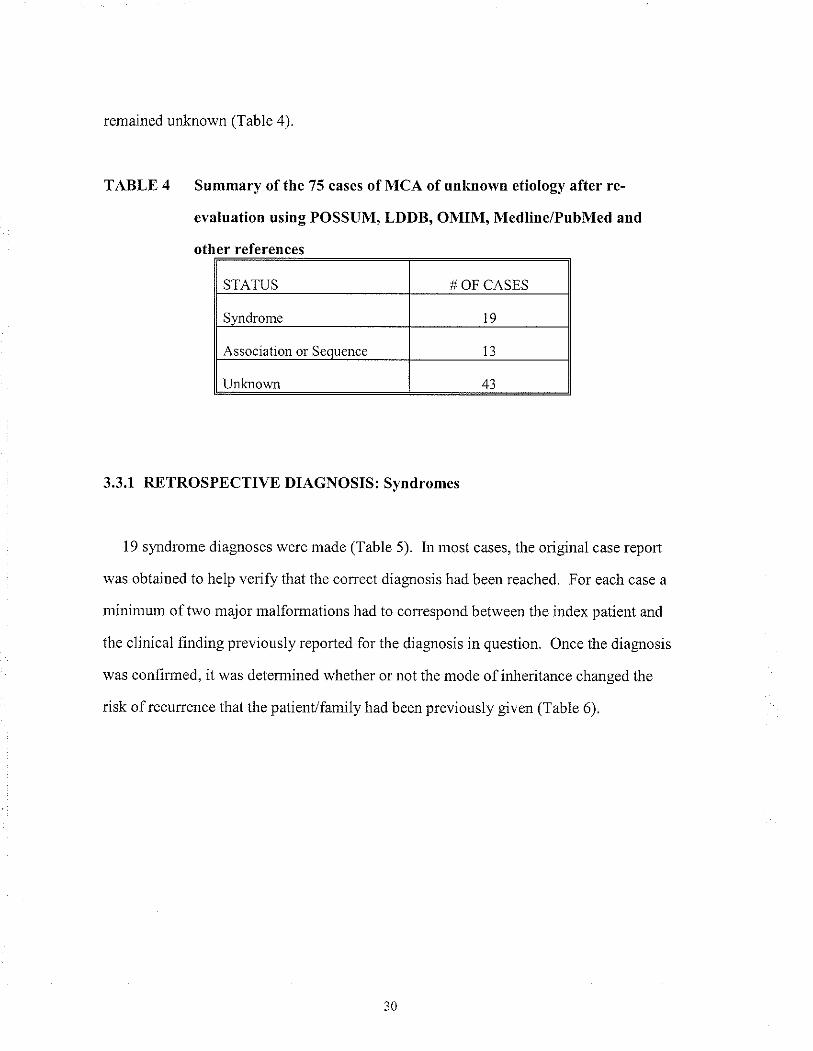
remained unknown (Table 4).
TABLE 4 Summary of the 75 cases of MCA of unknown etiology after re-
evâluation using POSSUM, LDDB, OMIM, Medline/PubMed and
other references
STATUS # OF CASES
Syndrome t9
Association or Seouence I3
Unknown 43
3.3.1 RETROSPECTIVE DIAGNOSIS: Svndromes
19 syerdrome diagnoses were made (Table 5). In most cases, the original case repoÍ
was obtained to help verify that the correct diagnosis had been reached. For each case a
minimum of two major malformations had to correspond between the index patient and
the clinical hnding previously reported for the diagnosis in question. Once the diagnosis
was conhrmed, it was detennined whether or not the mode of inheritance changed the
risk ofrecurrence that the patient/family had been previousÌy given (Table 6).
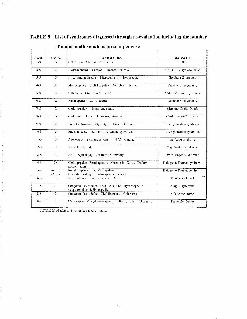
TABLE 5 List of syndromes diagnosed through re-evaluation including the number
of maior malformations
* : nunber of major anonulies more than 3
Reìral ågenesis Sacml defect
Inrperforåteaìrus Polydaclyly lìenâl Cardiâc
Renal dysplasiû Clel¡ lip/pålâte
ital lìeamdelèct-VSD, ^SD,llD^
l{ydrocephalûs
Congcnital heaì1dclcct Cleftìip/palate CoìoborÌa
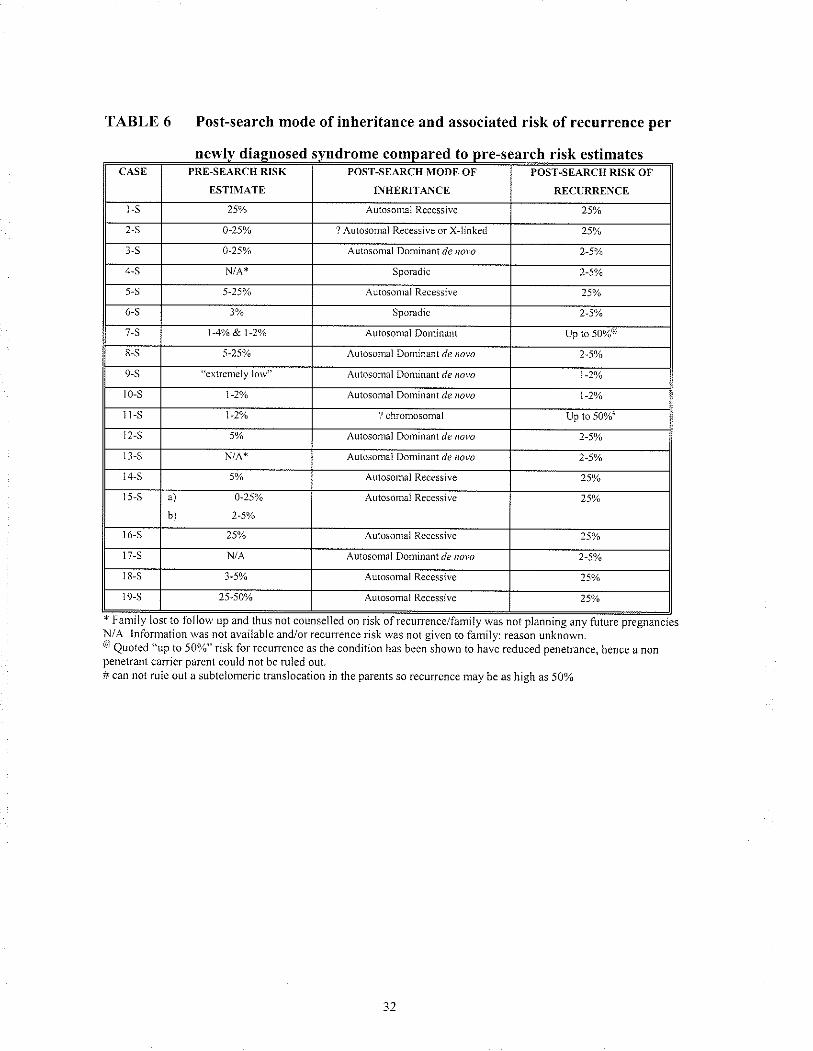
newly diagnosed syndrome compared to pre-search risk estimatescAst PRFJ-SEARCH RISK
ËSTIIlT,\TF]
POS'I'.SEARCH TODE OF
INHERITANCE
POST-SEARCH RISK OF
RECURRENCE
l-s Autosomal Recessive
2-S r)-25a/,, 7 Aulosomaì Recessive or Xlinked 2sa/o
Autosonraì Dominant d¿ r¡7v¿
4-S Sporadic 2-50/.
5-S 5-25% Autosonìâl KecessÌve 5%
6-S 3% Sporadic
/-s I -4!/a &. t-zyr Aurosomal Donlinânt Up lo 50%"
8-S Aulosomal LlonÌnBnt d¿ royo 2,5%
9-S cxlr'cnlely low"^utosonral
Don1ìnant.1¿ rolo
t0-s t -2ya
^ulosonral Domiiant./e roro
t-s l-2va ? chromosonìal Up to 50%
12-S^ulosomal
lforÌlìnanl./e roro
r l-s qutosomâi uonrnant de ,ror,¿
S 5% Autosonìâì Rcccssive 25'%
5-S ä) 0-25%
b)
Autosomâl Recessi!'c 2501,
¿5% Autosontal Recesstve 25')o
l7-s N/,A Autosomal Domiiant ¿¿ ,'oro
)-5V¡ Autosomâl Recessiv€ 25Ya
¿5,50Vo Autosomal Recessive 25yo
TABLE 6 Post-search mode of inheritance and associated risk of recurrence per
y was not planning any future pregnanciesN/A Iniornlation was not availablc and/or recunence risk was not gtven to family: reason unknow¡.@ Quoted "up to 50%" risk for ¡ecuncnce as the condition has bccn shown to have rcduced penetrance, hcncc a nonpenetrant cârricr pa¡ent could not be rulcd out.# can not rulc out a subtelomeric translocation jn the parents so .ecurrcnce may be as hjgh as 5Oyo
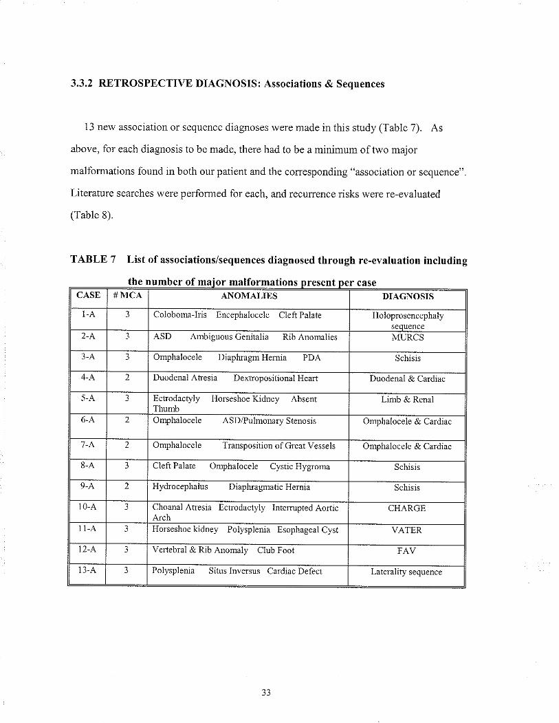
3.3.2 RETROSPECTIVE DIAGNOSIS: Associations & Sequences
13 new association or sequence diagnoses were made in this study (Table 7). As
above, for each diagnosis to be made, there had to be a minimum of two major
malfomrations found in both our patient and the conesponding "association or sequence".
Literature searches were perlomed for each, and recunence risks were re-evaluated
(Table 8).
TABLE 7 List of associations/sequences diagnosed through re-evaluation including
the number of malformations câseCASE # MCA ANOMAIIES DIAGNOSIS
t-A Coloboma-Iris Encephalocele CleftPalate Holoprosencephaly
2-A ASD Ambiguous Genitalia Rib Anomalies MURCS
3-A Omphalocele Diapluagm Hernia PDA Schisis
2 DuodenalAhesia DextropositiomlHeart Duodenal & Catdiac
5-A 3 Echodactyly l{orseshoeKidney AbsenlThun.rb
Limb & Renal
6-A 2 Omphalocele ASD/PulmomryStenosis Omphalocele & Cardiac
7-A 2 Omphalocele TranspositionofGreatVessels Omphalocele & Cardiac
8-A 3 Cleft Palate Onrphalocele Cystic Hygroma Schisis
9-A 2 Hydlocephalus DiapluagmaticHernia Schisis
10-A 3 Choanal At¡esia Ectrodactyly IntenuptedAorticArch
CHARGÊ
11-A Horseshoe kidney Polysplenia Esophageal Cyst VATER
12- A, 3 Veftebral & Rib Anomaly Club Fool FAV
13-A 3 Polysplenia Sihrs Inversus Ca¡diac Defect Laterality sequence
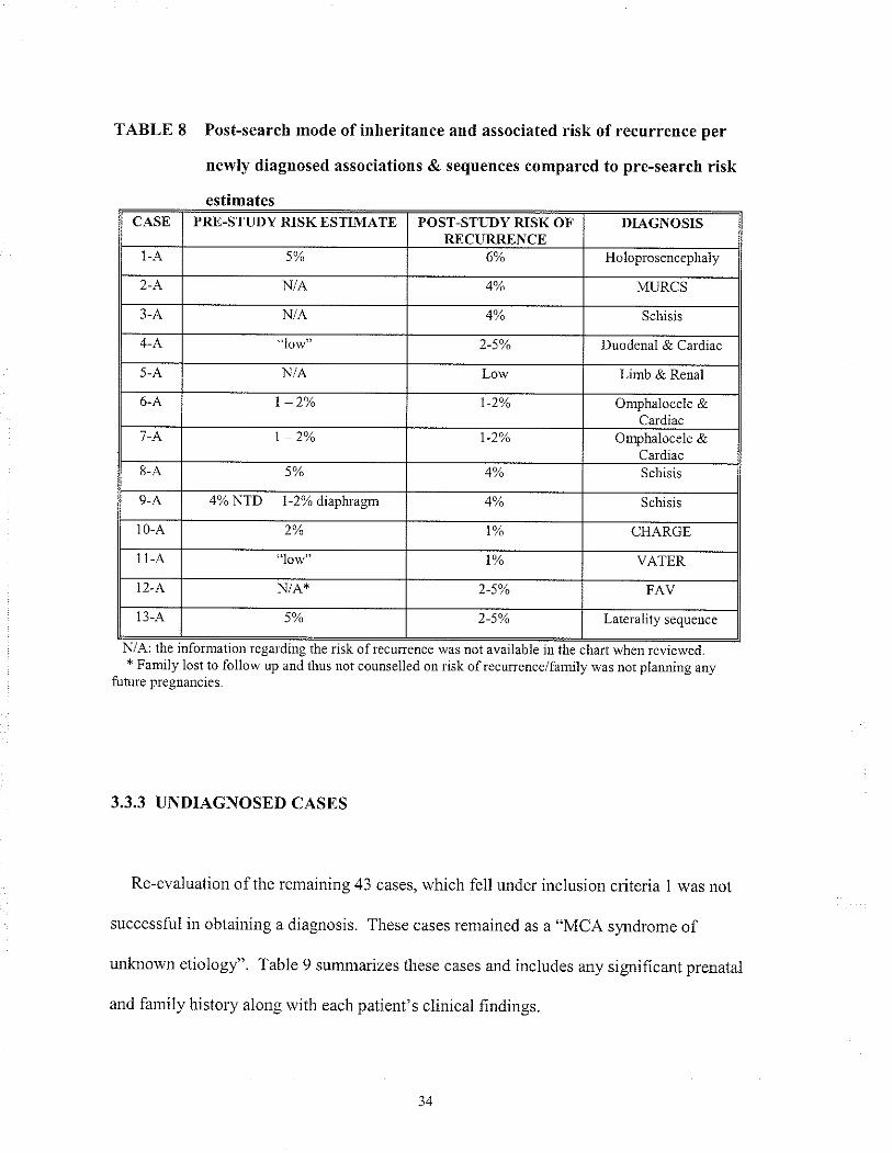
TABLE 8 Post-search mode of inheritance and associated risk of recurrence per
newly diagnosed associations & sequences compared to pre-search risk
estimates
regarding+ Family lost to follow up and thus not counselled on risk ofrecurreuce/family was not planning any
future pregnancies.
3.3.3 UNDIAGNOSED CASES
Re-evaluation ofthe remaining 43 cases, which fell under inclusion criteria I was not
successful in obtaining a diagnosis. These cases remained as a "MCA syndrome of
unknown etiologl'. Table 9 summarizes these cases and includes any significant prenatal
and farnily history along with each patient's clinical findings.
CASE PRE-STTJDY RISK ESTIMATE POST-STI-,'DY RISK OF'f¡ f'l- I lp p I, tva t¡'
DIAGNOSIS
l-A 6% Holoprosencephaly
2-A N/A 4% MURCS
3-A N/A Schisis
low Duodenal & Cardiac
5-A N/A Low Linrb & Renal
6-A I 2yo Omphalocele &Car¡1i¡c
'7-A Omphalocele &Ca¡diac
8-A 40/" Schisis
9-A 4% NTD 1-2% diaphagrr 40/,, Schisis
10-A CHARGE
lt-A T% VATER
N/A*
13-A 5% Late¡ality sequence
the risk ofreculrence was not available reviewed.
34
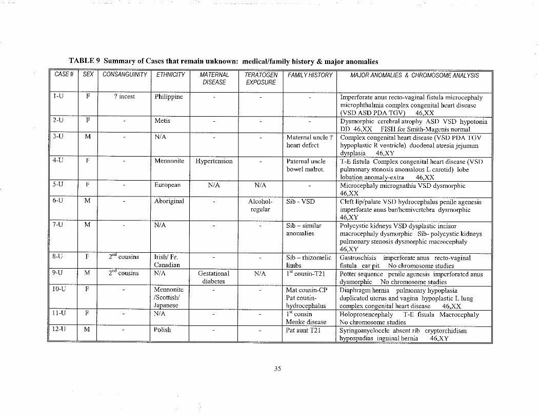
CASE#
TABLE 9 Summary of Cases thât remain unknorvn: medicat/family history & major anomalies
-U
SE}
2 -U
UU/VùAJVGU/IV// Y
F
3-U
t
? incest
4-U
M
ETHNICIW
)-u
F
Philippine
6-U
F
MATERNALD/SEASE
Metis
/-u
N/A
8
M
-U
Men¡onite
TERATOGEN
EXPOSURE
9 .IJ
European
F
I U-U
Aboriginal
H'?e¡tension
M
l1-Il
l _ cousms
t
N/A
t2-u
2'" cousins
N/A
F'
IrisV Fr.
M
Maternal uncle ?heart defect
MAJOR ANOMALIES & CHROMOSOME ANALYS/S
N/A
Imperforate anus reclo-vaginal fistula microcephalymicrophthalmia complex congeuital he¿rt diseaseIVSD ASD PDA 'I'GV) 46 XX
N/A
Mennonite/Scottish./Jananese
Patemal unclebowel malrot.
Alcohol-regular
Dysmorphic cerebral atrophy ASD VSD h)?otoniaDD 46.XX FISH fo¡ Smith-Masenis normal
N/A
Gestationaldiabetes
Complex congenital heart disease (VSD PDA TGVh)?oplastic R ventricle) duodenal atresia jejunumrlr¡cnÌacia
^ai YY
Polish
Sib - VSD
'l-E listula Complex congenital hea¡t disease (VSDpulmonary stenosis anomalous L carotid) lobelobationanomalv-exûa 46.XX
Sib - simila¡anomalies
Microcephaly micrognathia VSD dysmorphic46.XX
N/A
Cleft lip/palate VSD hydrocephalus penile agenesisimperforate anus ba¡/hemivertebra dysmorphrc46.XY
ò1D - tnlzomellclimhcI "' cousin-T2l
Polycystic kidneys VSD dysplastic incisormacrocephaly dysmoryhic Sib- polycystic kidneyspulmonary stenosis dysmorphic macrocephaly46 Xv
Mat cousin-CPPat cousin-hyd¡oceohalus
Gastroschisis inrperforateanus recto-vaginalfishrla ear oit No ch¡omosome sturlies
l" cousin
Potter sequence penile agenesis imperforated a[usdvsnromhic No chrnmocome qtrrdìe<
Pat aunt T21
Diaphragmhernia pulnonary hlpoplasiaduplicated uterus and vagina hypoplastic L lungcomolex consenilal lìeañ disease 46XXHoloprosencephaly T-E hstula MacrocephalyNo chromosome studiesSy,nngomyelocele absent db cryptorchidismhr,'oosoadias insuinalhernia 46.XY
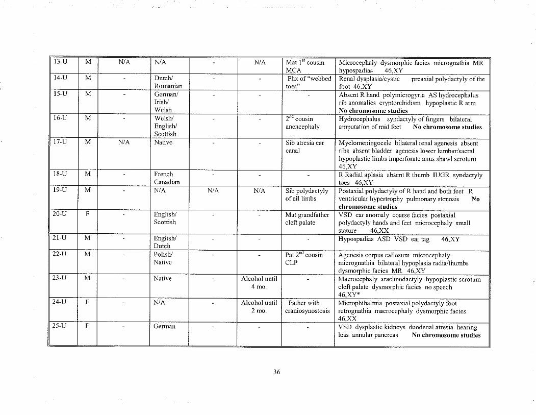
t3 U
14 -Il
M
l5 U
M
16 -U
M
N/A
17
M
U
N/A
18-U
M
Dutch-/Romanian
r9-u
Germa¡r/Irish/Welsh
M
N/A
20-u
WCISflEnglish/Scottish
M
21-U
Native
r
22 -U
N/A
M
F¡enchCanadi¿n
23 -U
M
N/A
Mat 1" cousinMCA
¿4
M
.U
Fhx of "webbed
Englistr/Scottish
25 -U
¡
English/Drtch
N/A
Microcephaly dysmoçhic facies micrognathia MRhvoosoadias 46.XY
2"_ coustnanencephaly
Polish./Native
F
Renaldysplasia/cystic p¡eaxialpolydactylyoftlìefoot 46 XY
Sib atresia ear
canal
t\anv€
Absent R hand pollrnicroglria AS hydrocephalus¡ib anomalies cryptorchidism hypoplastic R armNo chromosome stùd¡cs
N
N/A
Hydrocephalus syndactylyoffingers bilate¡alamputation of mid feet No chromosome studies
Sib polydactylyof all limbs
German
Myelomenìngocele bilateral re|al agcnesis absentribs absent bladde¡ agenesis lower lumbar/sacralhlpoplastic limbs inperforate anus shawl scrotum
Mat grandfa thercleft palate
ll Radral aplasia absent R thumb IUGR syndactylyroeq 46 XY
Alcohol rrnfil4 mo.
Postaxial polydactyly of R hand and both feetventricular hypertrophy pulmonary stenosischromosonre sfndics
rat I couslnCLP
Alcohol until2 mo.
VSD ear anomaly coarse facies postaxialpolydactyly hands and feet nricrocephaly smallstature 46.XXHypospadias ASD VSD ear tag 46,XY
Father wlthcranios)mostosis
Agenesis corpus callosum microccphalymicrognathia bilateral hypoplasia radia/thumbsdysmorphic facies MR 46.XYMacrocephaly araclmodactyly h)?oplastic scrotuncleft palate dysmorphic facies no speech46 XY*Mic¡ophthalmia postaxial polydactyly footretrognathia macrocephaly dysmorphic facies46.XX
36
R
No
VSD dysplastic kidneys duodenal atresia hearingloss annular pancreas No chromosome studies
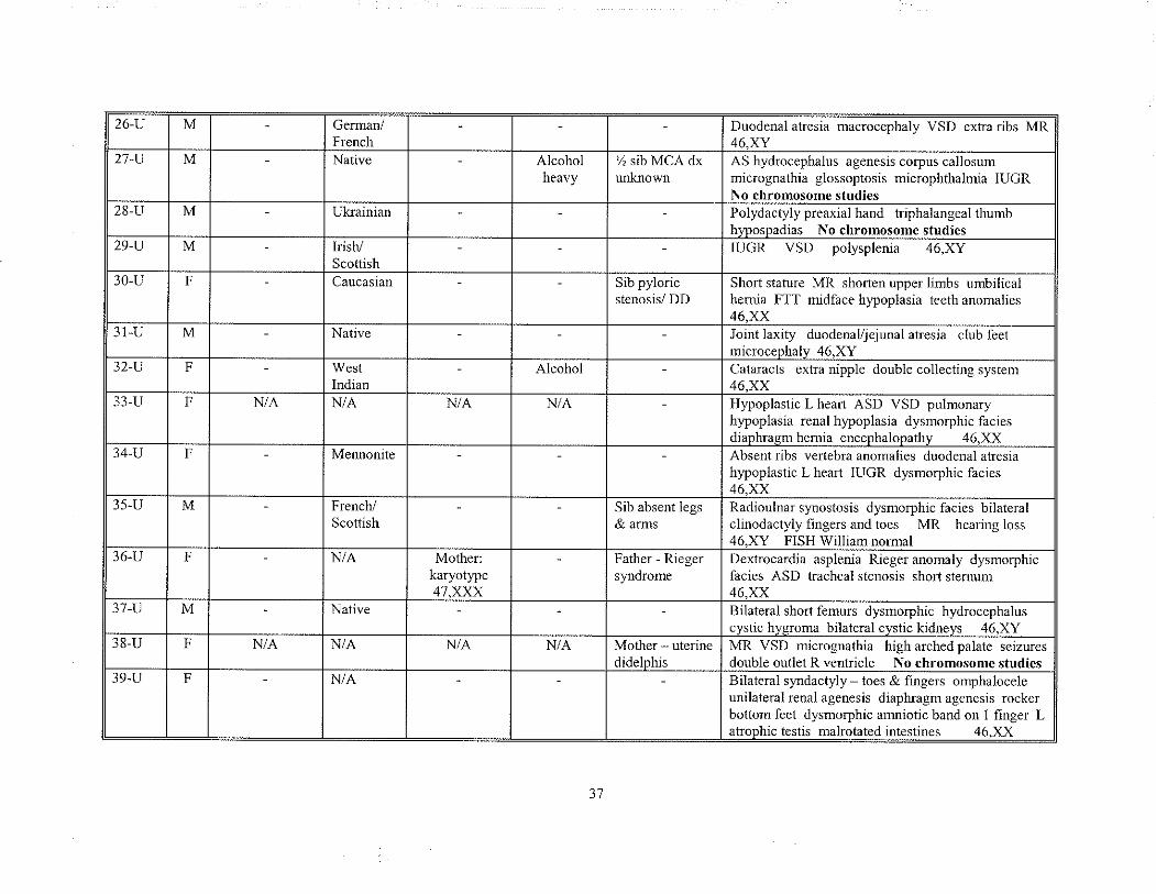
26 -U
21-U
M
¿8-U
M
29-U
M
30-II
M
31-U
Ge¡ma¡/French
F
32-U
Native
J3
M
-U
Ukrainian
F
34
I¡ish/Scottish
,U
I
Caucasian
l5 -U
F
N/A
Native
16 -U
M
Alcoholheavy
West
N/A
t-u
F
38-U
% sib MCA dxunk¡own
Mennonite
M
JY.U
¡
Frencl/Scottish
N/A
Duodenal atresia macrocephaly VSD extra ¡ibs MR46.XY
F
N/A
AS hydrocephalus agenesis corpus callosummiÇrognathia glossoptosis microphthalmia IUGRNn chromnsome sfrdiec
N/A
Sib pyloricstenosis/ DD
Alcohol
Native
Polydactyly preaxial hand triphalangeal thumbhltosoadias No chromosome stùdics
N/A
N/A
IUGR VSD polysplenia 46,XY
Mother:karyotype47.XXX
N/A
Short stature MR shorten upper li¡nbs umbilicalhemia FTT midface hypoplasia teefh anomalies46.XXJoint laxlty duodenal/JeJunal ahesia club feelmicrocephaly 46.XY
N/A
Catamcts ext¡a nipple double collecting system46.XX
Slb absent legs& arms
I-I)?oplastic L heart ASD VSD pulmonaryhlpoplasia renal hlpoplasia dysmorphic faciesdiaphrasmhemiaencephalopathv 46.XX
Father - Riegerslmdrome
Absent ribs vefebra anonËlies duodenal atresiah)?oplastic L heaÍ IUGR dysmorphic facies46.XX
N/A
Radioulna¡ synostosis dysmorphic facics bilate¡alclinodactyly fingers and toes MR hearing loss46.XY FISH William nomral
Mother - ute¡ine,li,l.t-hi.
Dext¡ocardia asplenia Rieger anomaly dysmorphicfacies ASD tracheal stenosis short stemum46 XXBilateral sho¡t femurs dysmorphic hydrocephaluscvstic hvsroma bilateral cvstic kidnevs 46-XYMR VSD micrognathia high arched palatc seizuresdouble outlet R vent¡icle No chromosomc studies
37
Bilateral syndactyly * toes & fingers omphaloceleunilateral renal agenesis diaphragm agenesis rockerbottom feet dysmorphic amniotic band on I finger Latuoohic testis malrotated intestines 46 XX
40-u
41-II
M
42-U
M
ì'" cousins
43-U
M
t,
Native
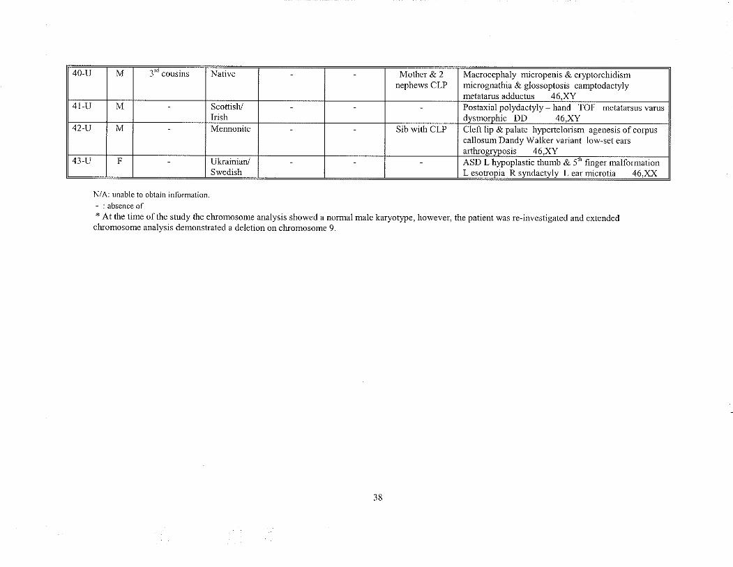
N/A: unable to obtain information.
- : abscnce of* At the time ofthe study the chromosome analysis showed a normal male karyotype, however, the patient v,,as re-investigated and extended
cluomosome analysis demonshated a deletion on ch¡omosome 9.
Scottisl/t¡ishMennonite
Ukra iniar/Swedish
Mothe¡ & 2nephews CLP
Sib with CLP
Macrocephaly nLicropenis & cryplorchidismmicrognathia & glossoptosis camptodactylymetatarusadduchrs 46.XYPostaxial polydactyly hand TOF netatarsus varusdvsmomhic Dl) 46 XYCleft lip & palate h)?erlelo¡ism agenesis ofcorpuscallosum Dandy Walker variant low-set earsarthrosrlDosis 46-XYASD L hypoplastic thumb & 5"' finge¡ mallornationL esotropia R s\,'ndactvlv L ea¡ microtia 46.XX
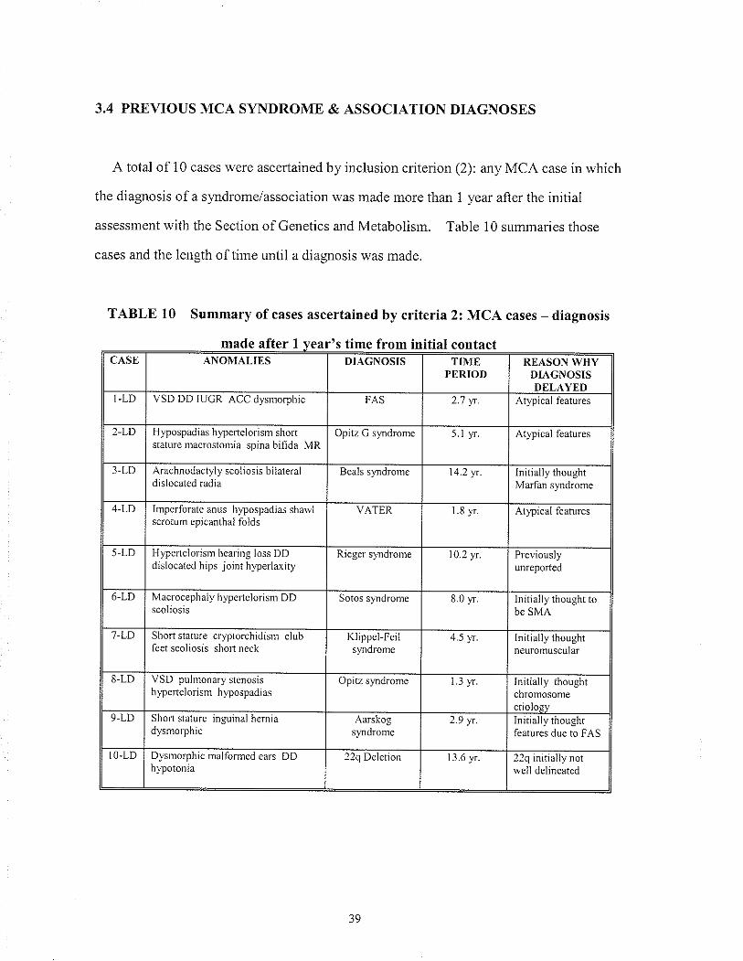
3.4 PREVIOUS MCA SYNDROME & ASSOCIATION DIAGNOSES
A total of 10 cases were ascertained by inclusion criterion (2): any MCA case in which
the diagnosis of a syrdrome/association was made more than 1 year after the initiâl
assessment with the Section of Genetics and Metabolisrn. Table 10 summaries those
cases and the length of tirne until a diagnosis was made.
TABLE 10 Summary of cases ascertained by criteria 2: MCA cases - diagnosis
made after I vear's time from initial contactromCASE ANOMAI,IES DIÂGNOSIS TIME
PERIODREASON WHY
DIAGNOSIS
I-LD VSD DD IUGR ACC dysmorphic FAS 2.'7 vr. Atypical features
2-LD Hypospadias hypertelorism shortstature nlacrostofiiâ spina bifida MR
Opitz C s).ndrome 5.I yr Atlpical features
3 -LD Arachnodactyly scoliosis bjÌateraldislocâted radiâ
Beals s)îdrome 14.2 yr. Initrally thoughtMarfan syndrome
4-LD Impcrforate anus h¡pospadìas shawlscrotum epjcanthaì folds
VATER 1.8 1o Atlpical fealìrres
5-LD H)pertelolism hcaring loss DDdislocated hìps Joint hlperlaxity
Rjeger s)Tld¡oD1e 10.21r. Pleviouslyunreported
6-LD Mâcrocephaly hlpc¡tcìorism DD Sotos s)îdronte 8.0 yr. lnrrrally tnougnt tobe SMA
7-LD Short stature cr¡.ptorchidism clubfeet scoìiosis short neck
Kììppcl-Feilsyndrome
4.5 )4.. rnlrìalty tnougnlneu¡omuscula¡
8-LD VSD pullnonary stenosishypertclorism hlpospadias
Opitz syndrome J )',f. Initially thoughtchromosome
9- Lt) Shoil stature inguinaì hcrniadysrnorphic
A¿rskogs¡mdrome
v y-l Initially thoughtfeatures duc to FAS
IO-LD Dysnrorphìc nralfornled ears DDhlpotonja
22q Deletion 13.6 yr 22q initially notwell delrneated
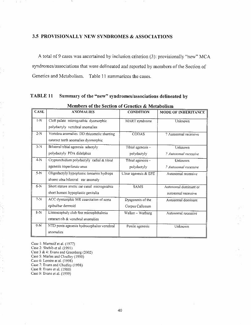
3.5 PROVISIONALLY NEW SYNDROMES & ASSOCIATIONS
A total of 9 câ.ses was ascertained by inclusion criterion (3): provisionally "new" MCA
slmdromes/associations thal were delineated and reported by members of the Section of
Genetìcs and Metabolism. Table 11 summarizes the cases.
TABLE l1 Summary of the "new" syndromes/associations delineated by
Members of the Section of Genetics & MetabolismCASE ANOMALIES CONDITION MODE OF INHERITANCE
I-N Cleft palatc micrognathia dysrrorphtc
polydactyly vertebral anomalies
MART syndrome Unknorvn
2-N Vertcbra anomalies DD rhìzomelic shoning
cataract teeth anonlalies dysmorphic
CODAS ? Autosomal recessivc
3-N Bilateral tibìal agenesis adactyly
polydactyly PDA didelphus
Tjbiâl âgencsis -polydactyly
Unkno\\,n
Aulosonßl recessi\,e
4-N Crlptorchidisnl polydactyly radjal & tibiåì
âgenesis jñperforate anus
Tibial agenesis
polydactyly
L no\
2 Aulosontal recessÌte
5-N Oljgodactyly hypoplâstic forearnls hydrops
allscnt ulna bilateral car anomaly
Ulnar agenesis & EFE Aùtosomal recessivc
6-N Short stature aΡctic ear canal tnicrognathja
short huureri hy?oplastic genitalia
SAMS Autosomal dominant or
autosomal recessivc
7-N ACC dysnrorphic MR coarctation ofaorta
epibuìbar dermoid
Dysgenesis ofthe
Corpus Callosum
Autosomal dominant
8-N Llssencephaly cÌuìJ fèet nticrophthalmia
cata¡act rib & vcrtebral anomalies
Waìkcr - Warburg Autosorîâl recessìve
9-N N I D penis agenesis hydrocephalus vcrtebral
anomalies
Pcnile agenesis Unknown
Case l: Martsolf et al. (1977)Casc 2r Shebib ct al. (1991)Case 3 & 4: Evans and C¡eenbc¡g (2002)Case 5: Marles and Chudley (1990)Case 6: Lenire et al. (1998)Case 7: Evans ând Chudlcy (1998)Câse 8r Evans et al. (1980)Casc 9: Evans et aì. (1999)
40
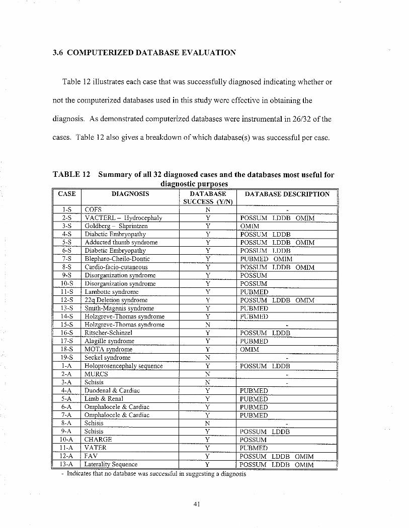
3.6 COMPUTERIZED DÄ,TABASE EVALUATION
Table 12 illustrates each case that was successfully diagnosed indicating whethet or
not the conputerized databases used in this study were effective in obtaining the
diagnosis. As demonstrated computerized databases were instrumental in 26132 of the
cases. Table 12 also gives a breakdown of whicb database(s) was successful per case.
TABLE 12 Summary of all 32 diagnosed cases and the databases most useful fortc
CASE DIAGNOSIS DAT,A,BASESUCCESS IY,T{'ì
DATABASE DESCRIPTTON
l-s coFs2-S VAaTFRI Hvr{rô.cnhâl\/ Y POSSUM LDDB OMIM3-S flnlãhcrc qh-' OMIM4-S Diabetic EmbryoDathy Y POSSUM LDDB5-S Aclducted thumb s POSSUM LDDB OMIM6-S Diabetic EmbryoDathv Y POSSIJM I-DDB7-S ÞlÞ-t "^-al'êil^-n^-f i. Y PUBMED OMIM8-S uardlo-laclo-cutaneous Y POSSIJM I,DDR OMTMs-s Disorganization s\tdrome Y POSSUMt0-s lJlsorganrzatroD s\¡rÌdrorre POSSUM
-S Laubotte sr,îdrome Y PUBMED2-S 22o f)eletio¡ srmdron POSSUM LDDB OMIM
S Smith-Maeenis sr'ndrome Y PUBMED-S Holzsreve-Thomas s PUBMED
5-S Holzereve-Thomas svndrome N6-S Ritscher-Schinzel Y POSSUM LDDB
-S Alasille s\'Ìldrome Y PUBMED-S MOTA syrd¡ome Y OMIM
9-S Seckel sv¡d¡ome NI-A Holoprosenceohalv seouence Y POSSUM LDDB2-A MIIRCS N
Schisis N4-A Duodenal & Cardiac Y PUBMED5-A Lrmb & Renal Y PIJBMED6-A Omohalocele & Carrliac Y PUBMED7-A On.rphalocele & Ca¡diac Y PTIBMED8-A Schisis N9-A Schisis Y POSSI]M I,DDBt0-A CHARGE Y POSSI,\,1II-A VATER Y PIIBMEDt2- A, FAV Y POSSUM LDDB OMìMt3-A Lateralitv Seouence POSSL,\4 LDDB OMIM- lndicates that no database was successful in suggesting a diagnosis
4I
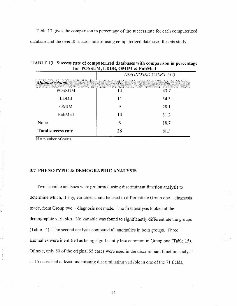
Table 13 gìves the comparison in percentage of the success rate for each computerized
database and the overall success rate ofusing computerized databases for this study.
TABLE 13 Success rate of computerized databases with comparison in percentagefor POSSUM. LDDB. OMIM & PubMed
DIAGNOSED CASES (32)
D.atobøse Namè|,;. .'',
POSSI]M
LDDB
OMIM
PubMed
None
Total success rate
.N,.ì..ì. ,
l4
11
9
10
6
26
'':.:,'.:.ri tìì,::
43.7
34.3
28.1
3r.2
18.7
81.3
N = number of cases
3.7 PHENOTYPIC & DEMOGRAPHIC ANALYSIS
Two separate analyses were preformed using discriminant function analysis to
determire which, if any, vanables could be used to differentiate Group one diagrosis
made, from Group two diagnosis not made. The first analysis looked at the
dernographic variables. No variable was found to significantly differentiate the groups
(Table 14). The second ar.ralysis compared all anomalies in both groups. Three
anomalies were identified as being significantly less common in Group one (Table 15).
Ofnote, only 80 of the origiual 95 cases were used in the discriminant function analysis
as 15 cases had at least one missing discriminating variable in one of the 71 fields.
42
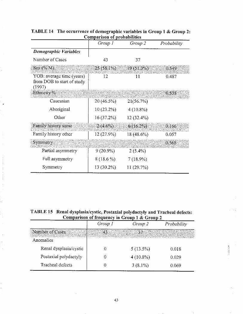
TABLE 14 The occurrence ofdemographic variables in Group I & Group 2:
Group I Grottp 2 Probabilityrison of
43
is:\Sï:to/ )
12
20 (465%)
10 (23.2%)
16 (31.2%)
',2'@,6%)'ì .. . . ..
..;.... . i.:.r.ì
12 (27.e%)
37
t9 ¡rr,l/O11
2r(s6.7%)
4 (10.8%)
t2 (32.4%)
9,.\!.6;2'1;)
t8 (48.6%)
.Q{!!0.48'.7
O,S:g
Demograp ltic Vøriables
Nun-rber of Cases
l!,e!.,(7o.frQ,;.,¡r1,¡r'r : ì . ..,...i
YOB: average time (years)from DOB to staÍ of study(reel)Et¡nici.bi o/ù,.' ,. ì ,-,., ,,,,.,.,11
Caucasian
Aboriginal
Other
P+n:f v, hi¡lorY saa19,',
Family history other
Siünetry..:r'..,.' .'i.
Partial asynmetry
Full asyr-rmetry
Sl,mmetry
e (20.e%) 2 (s.4%)
8 (18.6 %) t (rgj%)
13 (30.2%) 11 (29.7%)
, r:r:37..,:,:..,
s (13j%)
4 (r0.8%)
3 (8.1%)
0.16-6
0.057
0.56E
TABLE 15 RenaI dysplasia/cystic, Postaxial polydactyly and Tracheal defects:Comparison of freouencv in Groun I &
Group I Group 2 Probabilitv
Nuûbdròf CaseS.r ::t' : . .'
Anornalies
Renal dyspl asia./cystic
Postaxial polydactyly
Tracheal defects
0
0
0
,43
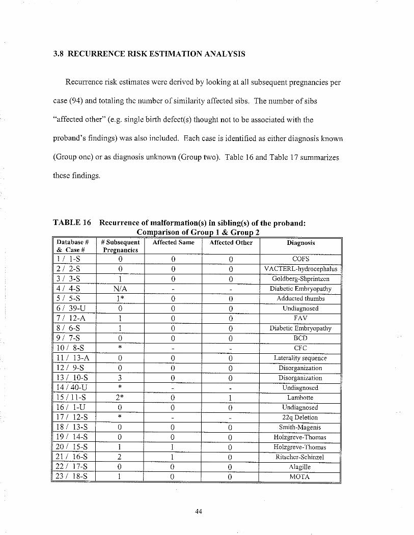
3.8 RECURRENCE RISK ESTIMATION ANALYSIS
Recurrence risk estimates were derived by looking at all subsequent pregnancies per
case (94) and totaling the number of sinilarity affected sibs. The number of sibs
"affected other" (e.g. single bìrth defect(s) thought not to be associated with the
proband's findings) was also ìncluded. Each case is identified as either diagnosis known
(Group one) or as diagnosis unknown (Group two). Table 16 and Table 17 summarizes
these findings.
TABLE 16 Recurrence of malformation(s) in sibling(s) of the proband:co o &G
Database #& Case #
# SubsequentPregnancies
Affected Same Affected Other Diagnosis
1/ 1-S 0 0 0 COFS
2tz-s 0 0 0 VACTERL-hydr ocephalus
3/ 3-S 0 0 Goldberg-Shprintzen
4/4-S N/A Diabetic Embryopathy
5/ s-s 0 0 Adducted thurnbs
6 / 39-U 0 0 0 Undiagnosed
0 0 FAV
8/ 6-5 0 0 Diabetic Embryopathy
917-S 0 0 0 BCD
0 8-S cFcI l3-A 0 0 0 Laterality sequence
2 9-S 0 0 0 DrsoÌganization
3 l0-s 3 0 0 Disorganization
4 40-u Undiagnosed
5 11-S 2* 0 1 Laûrbotte
16 / L-U 0 0 0 Undiagnosed
7 12-S 22q Deletion
18 / 13-S 0 0 0 Snith-Magenis
19 / r4-S 0 0 0 Holzgreve-Thornas
20 / 15-S 0 HoÌzgreve-Thomas
21 / 16-S 2 0 Rrtscher-Schinzel
22 17-S 0 0 0 Alagille
23 18-S I 0 0 MOTA
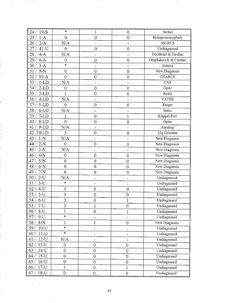
24 / 19-S 0 Seckel
25 I 1-A 0 0 0 Holoprosencephaly
N/A MLRCS
27 I 41-Il 0 0 0 Undiagnosed
28 I 4-A N/A Duodenal & Ca¡diac
29 I 6-A 0 0 0 Omphalocele & Ca¡diac
30 / 3-A Schisis
3i / 9-N 0 0 0 New Diasnosis
Jl/ t0-A 0 0 0 CHARGE
33 I 1-LD N/A FAS
34 I 2-LD 0 0 0 Opitz
35 I 3 -I,T) 0 0 Beals
36 / 4-LD N/A VATER
37 I 5-LD 0 0 0 Rieger
38 / 6-LD N/A Sotos
39 I 7-LD 0 I Klippel-Feil
40/ S-LD 0 0 0 Opitz
47 I g-LD N/A Aarskog
42 /10-t.Í) 3 0 0 22q Deletion
43 I 1-N N/A New Diagnosis
44 I 2-N 0 0 0 New Diagnosis
45 I 3-N N/A New Diagnosis
46 / 4-N 0 0 0 New Diagnosis
47 I 5-N 0 0 0 New Diasnosis
48 / 6-N 0 0 0 New Dìagnosis
49 I 7-N 0 0 0 New Diagnosis
50 I 2-U N/A Undiagnosed
sl I 3-U Undiagirosed
s2/ 4-U 0 0 0 Undiagnosed
53 I 5-U 0 0 0 Undiagnosed
54 I 6-U 3 0 Undiagnosed
55 I 7-U I 1 0 Undiasnosed
s6 / 8-U I 0 Undiagnosed<1 I 9-U Undiagnosed
58 / 8-N I 1 0 New Diasnosis
59 I r0-u Undiagnosed
60 I 11-U Undiagnosed
61 / t2-rl N/A Undiagnosed
62 I 13-II 0 0 0 Undiagnosed
63 I 14-U 0 0 0 Undiagnosed
64 I rs-u 0 0 0 Undiagnosed
65 I 16-U 0 0 0 Undiagnosed
66 I 17-U 0 Undiagnosed
67 / 18-U 0 0 0 Undiagnosed
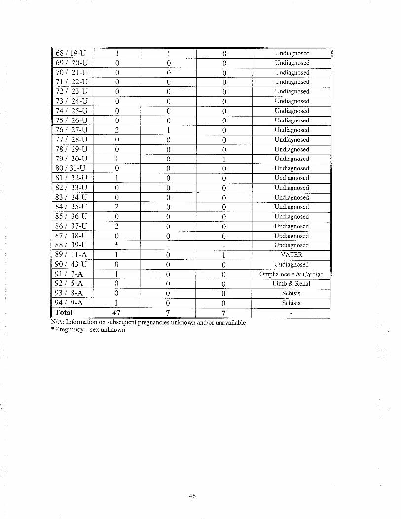
68 I 19-rr 1 0 Undiagnosed
69 / 20-U 0 0 0 Undiagnosed
70/ 21-U 0 0 0 Undiagnosed
7I I 22-U 0 0 0 Undìasnosed
72 I 23-U 0 0 0 Undiagnosed
73 / 24-U 0 0 0 Uudiagnosed
74 I 2s-U 0 0 0 Undiagnosed'75 I 26-rl 0 0 0 Undiagnosed
76 / 27-U 2 I 0 Undiagnosed
77/ 28-U 0 0 0 Undiagnosed
78 I 29-U 0 0 0 Undiagnosed
79 I 30-u I 0 Undiagnosed
80 / 31-U 0 0 0 Undiasnosed
81 I 32-U I 0 0 Undiagnosed
82/ 33-tl 0 0 0 Undiagnosed
83 34-U 0 0 0 Undiagnosed
84 3s-u 2 0 0 Undiagnosed
8s/ 36-U 0 0 0 Undiagnosed
86 I 37-U 2 0 0 Uudiagnosed
87 38-U 0 0 0 Undiagnosed
88 / 39-U Undiagnosed
89 11-A 0 VATER
90 I 43-U 0 0 0 Undiasnosed
9l 0 0 Omphalocele & Cardiac
92 5,A' 0 0 0 Limb & Renal
93 8-A 0 0 0 Schisis
94 9-A 0 0 Schrsrs
Total 47 7 7A: Infornation on subsequent Þregnancies unk¡own aud/orsubsequent pregnal1cies
* Pregnancy - sex unloowl
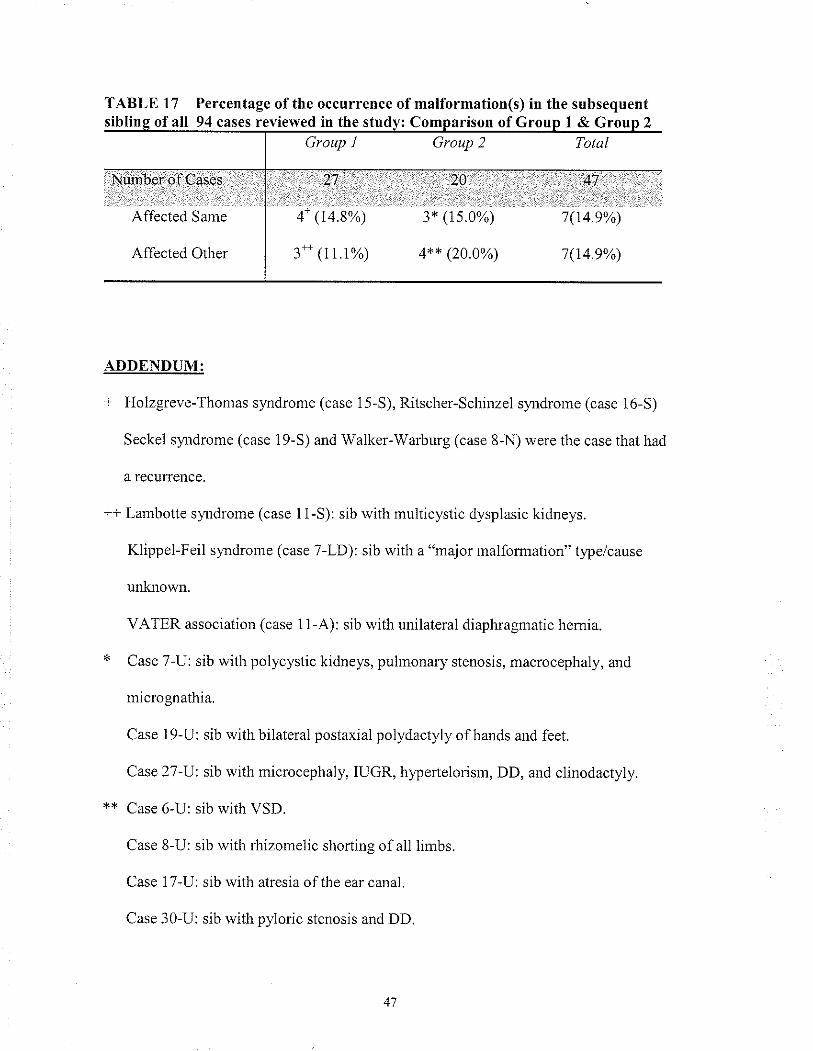
TABLE f7 Percentage ofthe occurrence of malformation(s) in the subsequenfof all 94 cases reviewed in the studv: C of Groun 1 & Grouo 2
Group I Group 2 Total
Niunber óf Cases
':i..r"'Affected Same
Affected Other
.,'.,.. ..27.:.:, ::.,.,,
4- (14.8%)
. ..,:28 . : ...: .'::.:::.,,.:.:'.47::'..
3* (15.0%) 7(14.9%)
4*+ (20.0%) 7(t4.9%)
ADDENDUM:
+ Holzgreve-Thomas slmdrome (case 15-S), Ritscher-Schinzel sy.ndrome (case 16-5)
Seckel syrdrome (case 19-S) and Walker-Warburg (case 8-N) were the case that had
â recun:ence.
++ Lambotte sytdrome (case 11-S): sib with multicystic dysplasic kidneys.
Klippel-Feil s¡mdrome (case 7-LD): sib with a "major malformation" type/cause
unknown.
VATER association (case 1 1-A): sib with unilateral diaphragmatic hemia.
* Case 7-U: sib with polycystic kidneys, pulmonary stenosis, macrocephaly, and
micrognathia.
Case 19-U: sib with bilateral postaxial polydactyly of hands and feet.
Case 27 -U: sib with microcephaly, IUGR, hyperlelorism, DD, and clinodactyly.
** Case 6-U: sib with VSD.
Case 8-U: sib with rhizomelic shortins of all lìmbs.
Case 17-U: sib with atresia of tn. .u, lunut.
Case 30-U: sib with pyloric stenosis and DD.
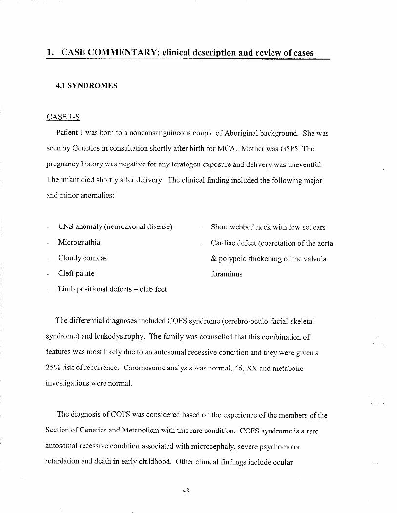
1. CASE COMMENTARY: clinical description and review of cases
4.1 SYNDROMES
CASE 1-S
Patient 1 was bom to a nonconsanguineous couple ofAboriginal background. She was
seen by Genetics in consultation shortly after birth for MCA. Mother was G5P5. The
pregnancy history was negative for any teratogen exposure and delivery was uneventful.
The infant died shofly after delivery. The clinical finding included the following major
and minor anomalies:
CNS anomaly (neuroaxonal drsease)
Microgr-rathia
Cloudy comeas
Cleft palate
Limb positional defects - club feet
Shorl webbed neck with low set ears
Caldiac defect (coalctation ofthe aorta
& pollpoid thickening of the valvula
foraminus
The differential diagnoses included COFS sl,ndrome (cerebro-oculo-facial-skeleta1
slmdrorre) and leukodystrophy. The farnily was counselled that this combination of
features was most likely due to an autosomal recessive condition and they were given a
25To risk of recurrence. Chromosome analysis was nomral, 46, XX and metabolic
investigations were normal.
The diagnosis ofCOFS was considered based on the experience of the members ofthe
Section of Genetics and Metabolism with this rare condition. COFS s1'ndrome is a rare
autosomal recessive condition associated with microcephaly, severe psychomotor
retardation and death in early childhood. Other clinical findings include ocular
anomalies, dysmorphic facies, micrognathia, flexion contractors of the limbs and CNS
anomalies (Del Bigio eI a1., 1,997). COFS s¡mdrome was originally described in
Manitoba by Pena and Shokeir (1974) in 3 Aboriginal kindreds. Homozygous mutations
in the ERCC6 gene are responsible for the COFS phenotype (Meira et al., 2000).
When the patient was initially seen by Genetics in 1984 the differential diagnosis
included COFS and leukodystrophy. "Leukodystrophy" encompasses a group ofwhite
matter diseases that are characterized by progressive cerebral deterioration ofthe myelin
of the central and peripheral nervous systen. Leukodystrophy may or may not have other
associated anomalies. Leukodystrophies are comprised ofover 40 unìque disorders,
many of which are autosomal recessive in inheritance (OMIM). Hence the term
"leukodystrophy" is more ofa classihcation ofdisorde¡s rather than an actual diagnosis.
Cockalare syrdrome (CS), which falls under the grouping of leukodystrophy, shows
similarities to COFS syndrome. Both are associated with neurodegeneration and
ca|aracts. Initìally COFS and CS were considered distinct entities as COFS syrdrome eye
defects were rrore severe than those found in CS patients and because cutaneous
photosensitivity was not noted in the patients originally diagnosed with COFS slardrome.
Recently, it has been demonstrated that COFS syndrome cases have r¡utations in the
ERCC6 gene and Cockalare syndrome type B patients also have mutations in the ERCC6
gene (Meìra et al., 2000). Graham (2001) demonstrated a mutation in the XPD gene,
which ìs associated with the CS phenotype, in a patient clinically diagnosed with COFS.
Whiìe they stated tliat COFS syndrome should remain a unique entity, it should be
included ìn the spectrum ofNER (nucleotide-excision repair) disorders along with
Cockal.ne syrdrome, xeroderma pigmentosum and trichothiodystrophy.
The diagnosis of COFS slmdrome was suggested in this patient based on the
experience of members of the Section of Genetics & Metabolism with this rare disorder.
Surprisingly, this diagnosis was not suggested by POSSUM, LDDB or OMIM searches.
This may be due in parl to the "handles" or clìnìcal features used in the ìnitial searches.
We made the diagnosis of COFS slmdrome in this patient based on the CNS findings, the
ocular anomaly, flexion contractures, dysmorphic facies and the webbed neck. Cleft
palate is not a common anomaly associated with COFS sytdrome; however, it has been
reported once before in another patient wìth COFS syrdrome (Hamel et al., 1996). In
addition to the clinical findings, the family was of North American Aboriginal
background, whìch has a higher incident ofCOFS s;mdrome than the general population.
A sample of DNA ffom this patient was obtained from tissue blocks and mutation
analysis for tlre common COFS mutation found in this population was performed. A gene
rnutation was identified in the patient's sample. Whrle only one mutation was identified
this does not change the diagnosis ofCOFS syndrome for this patient, as only screening
for the ethnìc specific mutation was done and this patient may have a second unknown
mutation. Mutation analysis is currently underway at an independent lab to try and
identify the second mutation. The recurence risk for this family remains the same with a
25"/o risk ofrecunence. The diagnosis of COFS sl,ndrome in this patient does not change
the initial recurrence risk given to the family. We have, however, by confìrming the
diagnosis offered the possibility ofprenatal diagnosis to the family and potentially carrier
testing for other fanrily members. One limitation is that, currently, the second mutation
has not yet been identified in this patìent.
CASE 2-S
Patient 2 was a stillbom bom to a nonconsanguineous couple, The mother was of
French / Lkrainian background and the father was ofUkrainian descent. The familywas
seen by genetics in consultation shortly after birth because of MCA. Review of the
pregnancy and birth history was unremarkable. There was a positive family history of a
cousin who had Down slardrome and a more distantly related cousin, related through
marriage only, who was though to have the VATER assocìation. Routine chromosome
analysis demonstrated a normal 46,XY karyotype. The clinical findings included the
following:
Hydrocephalus with aqueductal
stenosis
Hypoplastic left hearl
Trachea stenosis
Transposition of the great vessels
& single atrial ventricular valve
Bronchopulmonary dysplasia
Bilateral unilobular lungs
The differential diagnoses include VACTERL - hydrocephalus and Hydrolethalus
syndrone. The family was counselled that this could be either an autosomal recessive
condition or an X-linked condition with a 25o/o reatnence risk or that this was a sporadic
event with a recunence risk of 50%.
The diagnosis of VACTERL association with hydrocephalus was suggested by the
OMIM database based on two clirical findings, aqueductal stenosis and cardiac defects.
The only other diagnosis suggested by OMIM based on those two clinical findings was
neurofìbromatosis type 1, which the patient clearly did not have (lacked any other clinical
features ofNFl). VACTERl-hydrocephalus was also suggested by POSSUM and LDBB
searches.
VACTERL - tlpe anomalies associated with hydrocephalus was hrst delineated by
Bria¡d et al. (1984). They reported 16 patients who had this combination of findings.
VACTERL association is an acron).,rn for verlebral anomalies, anal atresia, cardiac
defects, tracheosophageal fistula, r'enal anomalies and limb defects. VACTERL-
hydrocephalus appears to be a separate entity associated with, in addition to the anomalies
seen in the VACTERL association, hydrocephaly, microphthalmia, n-ricrognathia,
branchial arch anomalies, lung lobation defects and spleen anornalies (Evans et al., 1989).
VACTERL-hydrocephalus is frequently a lethal association as most children died in the
newbom period or are stillbom.
The diagnosis of VACTERl-hydrocephalus was made ìn this patìent based on the
hydrocephalus, trachea stenosis, cardiac anomalies and lung lobe anornalies. VACTERL-
hydrocephalus has been documented to have autosomal recessive and X-linked
inheritance depending on the family (Evans et al., 1989; Sommer et a1., 1989; Corsello
and Giuffre, 1994). It is unclear whether or not this patient had the autosomal recessive
or the Xlinked form of VACTERL hydrocephalus. The parents were nonconsanguineous
and this was their first pregnancy. Given that the couple was nonconsanguineous and that
the affected was male, this could suggest either the X-linked or autosomal recessive form.
In either situation, the couple would have a25Yo risk of recurrence. It would be impoÍant
to determine which mode of inheritance the patierf had to better counsel other family
members regarding their risk. Clearly, if this were the X-linked fonn tl.ren there would be
a risk of carier females on the matemal side having similarly affected children. The
autosomal recessive form would have a much lower risk for other family members unless
there was consanguinity. Some cases ofVACTERL-hydrocephalus have been shown to
lrave Fanconi anemia (Porteous el al.,1992; Wang et a1.,1993; Rossbach et al., 1996).
Fanconi anemia is associated with increased chromosome breakage and mutations in the
Fanconi anemia complernentation group C gene (Cox el al., 1997). Chromosome
breakage and/or molecular analysis might be a consideration to help support the diagnosis
of VACTERL-hydrocephalus in this patient.
The initial differential diagrrosis included Hydrolethalus syndrome, although the
patient was thought not to be typical for this condition. Hydrolethalus slardrome is a
lethal condition characterized by polydactyly and CNS malformations, stenosis of the
airway and lung abnormalities (OMIM #236680). Our patient had a number of the
features in common with this disorder including hydrocephalus, trachea stenosis and lung
lobe abnormalities. Hydrolethalus slmdrome was ruled out as a potential diagnosis based
on the lack ofany dysmorphic facial features in our patient. Hydrolethalus syndronre is
associated with a t)?ical facies including micrognathia, cleft palate/lip and broad nasal
root. In addition, key characteristics of this condition, specifically polydactyly and the
"key hole-shaped" opening at the base ofthe skull were not present in our patient (Jones,
1997).
CASE 3-S
Patient 3 was bom to a nonconsanguineous couple of Polish and English background.
A relerral was made to genetics because of MCA. Pregnancy and biÍh history were
reviewed and were unrenrarkable. Mother was G3P3. Routine chromosome analysis on
the patient was Íromtal, 46, XY. Upon examination the following anomalies were found:
Hirschsprung disease
Hlpertelorism
Ear anomaly
Hypospadias
Microcephaly
Constitutional growth delay
Cranial asymmetry
Camptodactyly
Developmental delay
The inilial differential diagnosis included hypertelorismJrypospadias s¡ardrome and
VACTERL association. The family was counselled that this combination of anomalies
might be a sporadic event or mìght represent an unknown autosomal recessive condition.
Tlrey were given a 0-25o/o risk ofrecurrence. Unfortunately the patient died, at age 2 year,
six r¡onths due to complications of the Hirschsprung disease.
As previously documented, VACTERL association is characterized by vertebral
anomalies, anal atresìa, T-E fistula, renal anomalies and limb defects. Review ofthe
patient's clinical findings did not support this diagnosis. The constellation of features are
not typical for VACTERL association.
Hyperlelorisrn-hlpospadias sy'ndrome (Opitz syrdrome) is a condition associated with
mild to moderate mental retardation, ocular hypertelorism, hypospadias and other
anomalies (Jones, 1997). It can be an Xlinked condition where carrier females can have
hypertelorism or an autosomal recessive condition. While this patient did have the
hallmark findings of Opitz s¡.ndrome - hyperlelorism and hypospadias - there has not
been a repofied case of Opitz sytdrome associated with Hirschsprung disease. In
addition, rnicrocephaly and camptodactyly are not characteristic findings in this
condition.
The diagnosis of Goldberg-Shprintzen syndrome (Hirschsprung disease - Mental
Retardation syrdrome) was suggested by OMIM based on microcephaly, hypospadias
and Hirschsprung dìsease. Goldberg-Shprintzen sy'ndrome was fir.st reported by
Goldberg and Shprintzen (1981) who described apairof sibs who had Hirschsprung
disease, submucous cleft palate, microcephaly, short statue, hypertelorism, synophrys,
prominent nose, thick eyelashes and fine sparse hair. They also had leaming delay. Since
that initial repofi, otl,er cases of Goldberg-Sþrintzen slmdrome have been reporled in the
literature. Initial cases repofts supported at autosomal recessive mode ofinheritance
(Hurst et al., 1988; Halal and Morel, 1990). However, Mowart et al. (1998) published 4
cases of Goldberg-Shprintzen syrdrome that suggesled an autosomal dominant mode of
inheritance. Wakamatsu et al. (2001) later confirmed this by demonstrating that 3 out of
4 patients diagnosed with Goldberg-Strptintzen syrdrome had heterozygous deletions in
the SMAD1P1 gene, all of which occuted de novo.
The diagnosis of Goldberg-Shprintzen syrdrome was made in this case based on the
Hirschsprung disease, characteristic facies, microcephaly and shorl stature. Our patient
also had other findings in common with the patient reported by Halal and Morel (1990).
Their patient also had ear and genital anomalies. A1l cases of Goldberg-shprintzen
s)Ìrdrome reporled to date, including this patient, have had developmental delay. Given
that there was no positive family history ofany findings suggestive of Goldberg-
Shprintzen syrdrone in the parents or the siblings, this condition most likely occurred de
novo in the affected. Barring gonadal mosaicism, the risk ofrecu¡rence for the fanily
would be very low. Unfofiunately, because the patient had died, molecular testing for the
SMADlPl gene could not be offered to confirm the diagnosis.
The patient was initially seen by Genetics in 1976. At that time, only the X-linked
form of Opitz slmdrome was known and the Goldberg-shprintzen slndrome had not yet
been described in the literature. The famìly was lost to follow up until 1989 when the
mother was seen for AMA counselling with a new partner. When reviewed by Genetics
again, the differential diagnosis included Opitz syardrome (X-linked form only), an
unknown MCA s)'ndrome, Goldberg-Shprintzen syrdrome and Jeshina-Koeda sy'r.rdrome.
A review of the literature could not identify any s),ndrone under that last name.
In 1989, the Goldberg-Shprintzen slmdrome was considered to be an autosomal
recessive condition. The family was counselled that the patient's phenotlpe was not
characteristic of those reporled at that time and that Goldberg-shprintzen sy.rdrome most
likely did not explain his findings. In i989 only 5 cases had been reporled (Goldberg and
Shprintzen, 1981; Hurst et al., 1988; Winter et al., 1984). This might be in part, why the
diagnosis u as rror lnade at rhat time.
55
CASE 4-S
Patient 4 was delivered by cesarean section because ofdecreased fetal movement, to a
nonconsanguineous couple. The mother was ofEast Indian descent and the father was of
Ukrainian descent. The fenale infant was seen by genetics shortly after birth because of
the presence of MCA. Mothel did not have any prenatal care until 32 weeks gestation, at
which time it was noted that the pregnancy was complicated by gestational diabetes with
no blood sugar control. The rest ofthe pregnancy history was unremarkable. Mother had
had a previous relationship that had resulted in one spontaneous abortion and one healthy
child. ln this current relationship, she and her partÍìer, in addition to this child, had had
another healthy child and three spontaneous aborlions. Routine chromosome analysis on
the patient revealed a normal karyotype: 46, XX. Parental karyotypes were not done. On
examination and subsequent investigations, the following anomalies were noted:
Cleft lip and palate
Vertebral and rib anomalies (13 pairs
of ribs)
Microcephaly
Unilateral renal agenesis
The differential diagnosis included an autosomal recessive condition with facial
clefting, microcephaly and MR or an unknown MCA slardrome. Unfortunately the
family was lost to follow up and they were never counselled regarding recurrence risks.
Due to the pregnancy history of gestational diabetes, diabetic embryopathy was
suspected as the cause of the clinical fìnding ir the affected. The results of the search
suppofed the clinical suspicion of diabetic embryopathy. Diabetic embryopathy
encompasses a large number and range of anomalies. Most conlmon fìndings include
CNS anomalies such as anencephaly, microcephaly, and hydrocephaly, cardiovascular
defects, renal, rib and vertebral anomalies, and cleft lip/palate (Buyse, 1990). The
56
diagnosis of diabetic embryopathy was made based on the pattem of anomalies found in
our patient and the fact that the mother had unlreated gestational diabetes. One lirnitation
of this diagnosìs is that i1 is unknown how ìong the pregnancy was complicated by
gestational diabetes. Why the diagnosis ofdiabetic embryopathy was not considered
when the family was initially seen is not clear. Children of diabetic/gestational diabetic
mothers have a 10 times higher frequency ofcongenital anomalies and the mothers have a
5 times higher frequency of spontaneous abortions (Nazer and Ramirez, 2000). The
family was seen in 1992, at which time the effects of maternal gestational diabetes were
well documented (Ramos-Anoyo et al., 1992; Hod et al., 1991; Kitzmiller et al., 1991).
Diabetic embryopathy has a teratogenic effect and as such is a sporadic event. The risk of
recurrence for this family would depend on how well the diabetes was controlled during
subsequent pregnancies. A study by Steel et al. (1990) demonstrated that improved
control in early pregnancy does lower the incidence of MCA in newboms of diabetic
mothers.
CASE 5-S
Patient 5 was born to a nonconsanguineous couple, both ofwhom were ofCree
background. The patient was seen in Ger.retics shodly after birlh because of MCA.
Review ofthe pregnancy and birlh history was unremarkable. There was no exposure to
any teratogens during the pregnancy. Family history was reviewed and showed that a
sibling of the affected child had a cleft palate that was felt to be non-symdromic. There
was also a first cousìn who had been diagnosed with Rett slmdrome. Routine
chromosorle analysis on this patient was nonnal with a 46,XY karyotype. On
examination the following clinical findìngs were noted:
Craniosynostosis - sagittal
Iris coloboma
Submucous cleft palate
Ventricular septal defect
Pyloric stenosis
Adducted thun-rbs - bilateral
Torticollís
MR/autistic features
Dysmorphic facies
Neurosensory hearing loss
The differential diagnosis included CHARGE association, Adducted Thumb
syndrome, Ritscher-Schinzel slmdrome and an autosomal recessive MRÀ4CA condition
repofed by Pfeiffer (1987). The family was counselled that this most likely was an
autosomal recessive condition with a 5 to 25% i'sk of recurrence.
CHARGE association is an acron)..rn for coloboma of the eye, heart anomaly, choanal
atresia, mental retardalion, genital and ear anomalies. Facial clefting and dysrlorphic
facies are also common characteristics of CHARGE association (OMIM # 214800; Pagon
et al., 1981). The patient had many of the criteria necessary to fulfill this diagnosis
including iris coloboma, cardiac anomaly (VSD), ear anomaly (low-set ears) and MR. In
addition, torticollis and neurosensory deafness have also been reported in a patient
believed to have CHARGE associatìon (North et a1., 1995; Van Meter and Weaver,
1996). CHARGE association has a low risk of recurrence.
The diagnosis of CHARGE association was considered unlikely in this patient, as the
overall clinical phenotlpe did not seem to be best explained by the CHARGE association.
Specihcally, the fealures that were not characteristic of CHARGE association were
craniosyrostosis and bilateral adducted thuml¡s. A review of the literature did not
produce any reporled cases of CHARGE with either of these two fìndings, although limb
anomalies have been reporled in patients with CHARGE association (Prasad et al., 1997).
Ritscher-Schinzel slardrome is an autosomal recessive disorder characterized by
c¡aniofacial, cerebral and cardiac defects. Cleft palate and ocular colobomas are also
comnron findings in this syndrome (Leonardi et al., 2001). This patient had a number of
findings consistent with this sl,ndrome, namely ocular coloboma, cleft palate, cardiac
defects and MR. However, again our patient's overall gestalt did not seem to fit with this
slmdrorne. The patient lacked any cerebral or structural CNS findings and
cranios¡mostosis, adducted thumbs and hearing loss have no1 been reported in Ritscher-
Schfuzel s;,ndrome. Therefore, it seems unlikely that this patient had this condition.
The third cardiocranial syndrome that was on the dilferential list was a case report by
Pfeiffer et al. (1987) where he described two sibs with sagittal craniosyrostosis, cardiac
defects, micrognathia with mandìbular ankylosis, genital defects, growth retardation and
MR. Since that initial repof, there have been a small number of other case reports similar
to the Pfeiffer et al. report (Stratton and Parsons, 1989; Williamson-Kluse and Biesecker,
1995; Digilio eI al.,1997). Review of the literature did not suppof this diagnosis in this
patient. Hearing loss wìth ear anomalies, ocular coloboma and oral clefts have not been
reported in this autosomal recessive condition.
MASA, a condition described by Bianchine and Lewis (1974) is an X-linked disorder
associated with MR, aphasia, shuffling gait and adducted thumbs. Mutations in the
LICAM gene are responsible for the clinìcal phenotype. MASA encompasses a number
of previously described MCA conditions (OMIM #303350). Although one of the key
features ofthis condition is "adducted thumbs", the overall clinical findings ofour patient
do not seern to fit itto this spectrum.
Adducted Thunb slardrome was suggested by borh the POSSUM and LDDB
databases. It was also suggested by OMIM when "adducted thumb" was used as the
search parameter. It was first described by Christian eI al. (1971). They repoded on 3
sibships in an Amish kiridred with cleft palate, arlhrogryposis, craniosymostosis and
microcephaly. Since that inìtial report additional cases have been reported (Fitch and
Levy,7975; Anderson and Breed, 1981;Kunze et al., 1983). The major diagnostic
features for this condition are craniosyrostosis, rricrocephaly, cleft palate and adducted
thumbs (Buyse, 1990). Other findings, such as VSD defects, low-set ears, torticollis and
MR lrave also been reported in this condition (Fitch and Levy,7975). It is thought to be
an autosomal recessive condition with a recurrenc e risk of 25o/o.
This patient has many of the clinical features of Adducted Thumb sy,r-rdrome in
addition to other findings not previously described in this syndrome (neurosensory
hearing loss, pyloric stenosìs and ocular coloboma). While there seems to be an overlap
between the patient's clinical findings and both Adducted Thumb syrdrome and
CHARGE association, after review of the literature, this patient's overall clinical
phenot)?e is rnore suggestive of the diagnosìs ofAdducted thumb syrdrome. The
patient's unique constellation offindings might be why the diagnosis was not made
initially. His features certainly overlap with a number of slmdromes. The recurrence risk
for this family has been changed from a range of 5 to 25o/o to 25%. Unless there is
consanguinity, the risk for other family members is low. Unforlunately, there is no
molecular testing available for tliis condition as yet, so confinnational molecular
diagnosis, canier testing, or prenatal diagnosis is not an option for this family.
CASE 6-5
Patient 6 was bom to a nonconsanguineous couple. The father was of Caucasian
descent and the mother was of Aboriginal descent. The patient was seen shoÍly after
biÍh by Genetics because of MCA. Review of the pregnancy and birth history was
remarkable in that mother had had untreated gestational diabetes until the 4th month ofher
pregnancy. The family history was unremarkable. on examination the following clinical
findings were noted:
Unilateral renal agenesis
Sacral dimple with associated hairy
patch
Bitemporal depression
Limb length discrepancy
The differential diagnosis included caudal regression velsus an early first trimester
insult due to gestational diabetes. The family was counselled that this consolation of
features most likely represented a sporadic event and they were given a 3%o risk of
recurÏenc9.
As with patient 4-S, diabetic embryopathy was suspected as the mother had
uncontrolled gestational diabetes throughout the pregnancy. A search using POSSLrM
and LDDB suppoÍed the clinical suspicion of diabetic embryopathy. As mentioned in
case 4-S, diabetic ernbryopathy is associated with a large number and range of anomalies
and certainly, renal agenesis and sacral defects are seen in this spectruln.
The diagnosis of diabetic embryopathy was considered in the initial differential
diagnosis and it is unclear as to why this diagnosis was not given to the child. Caudal
regression s),ndrome is a heterogeneous group. Its pattem of malformations includes
incomplete development ofthe sacrum and lumber vertebrae, disruption of the distal
spinal cord leading to secondary neurologic impaimient and associated lower limb
defects. Renal agenesis, anal anomalies, cleft lip/palate, and microcephaly are associated
with this condition (Jones, 1997). These cases are usually sporadic, although it has been
shown that a number (16%) occured in infants of diabetic mothers (Rusnak and Drìscoll,
1965; Passarge anó, Lenz, 1966; Stewart and Stoll, 1979; Goto and Goldman, 1994).
SntíTh's Recognizable Patterns of Huntan Malformatiott 5tL edition (Jones, 1997)
considers this a caudal dysplasia sequence stating that the "caudal regression" most likely
is a secondary sequence ofevents with one of the known primary etiologies being
matemal diabetes. Based on this statement, caudal regression in some cases js a clinical
finding within the spectrum of dìabetic embryopathy. As previously stated, it is
important to clarify the primary cause of a birlh defect(s) as it may have many etiologies
and knowing which is specific to the case in question is necessary to provide accurate
counselling to the family. With proper prenataì management of the mother's gestational
diabetes, there would be a low risk ofrecurrence for this couple. One limitation in this
case is that chromosome analysis was not done on this child, therefore a chromosome
etiology can not be ruled out.
CASE 7-S
Patient 7 was seen by Genetics as a young child because of MCA. She was bom to a
nonconsanguineous couple of Caucasian background. Review of the pregnancy and birth
history was unremarkable. There was a family history of cystic fibrosis on the matemal
side. Routire chromosorne analysis in the patient was norm al: 46,XX. The following
clinical features were noted on examination:
Cleft lip and palate - bilateral
hnperforate anus
Bilateral lower lid ectropion
Double row of eye lashes
Shoft stature (5tr' % for growth)
Cone-shaped teeth with oligodortia
The family was originally counseled that this constellation of defects (cleft lip/palate
and imperforate anus), represented two independent anomalies, both of which were
multifactodal in inheritance. Two separate recurrence risk figures were given. A 4% nsk
ofrecurrence for cleft lip and palate and a 1-2%o risk ofrecurrence for imperforate anus.
Focal dermal hlpoplasia (Goltz syndrome) was also nentioned in the diffe¡ential
diagnosis, although it was considered unlikely.
Goltz syndrome (focal dennal hypoplasia) is an X,linked dominant condition with in
utero lethality jn males. It ìs characterized by atrophy and linear pigmentation of the skin,
hemiation of fat through dermal defects, and multiple papillomas of the mucous
membranes or skin. In addition, there is syndactyly, polydactyly, hypoplastic teeth,
colobomas with microphthalmia and MR (OMIM #305600). This condition was hrst
repofed by Goltz ef al. (1962). Review of the literature did not support the diagnosis ìn
this patient as she lacked any of the skin findings, is developmentally normal and has
other findings not consistent with this disorder (e.g. oral cleft and eye abnormalities).
As suggested by OMIM and a Medline search using "disticliasis and ectropion" as
search parameters, the diagnosis of Blephalo-Cheilo-Dontic (BCD) syrdrome was made.
Allanson and McGillinay (1985) reported a family with 4 generations of individuals with
cleft lip and palate, ectropion of lower eyelids and conical teeth. This large kindred
demonstrated an autosomal dominant inheritance with marked clinical expression. Falace
and Hall (1988) repofed on a 5-generation family that had a combination oforal clefting
with eye and teeth anomalies supporting the suggestion that this was a distinct autosomal
dominant s1'ndrome. Gorlin et al. (1996) in his 8 patient case repoÍ tenned this condition
the "Blepharo-Cheilo-Dontic (BCD) s1'ndrome." In addition to the oral clefting, eyelid
and teeth hndings, sparse scalp hair, minor digit anomalies, poot growth with shoú
stature, ear anomalies and imperforate anus have all been repoded (Allanson and
McGillivray, 1985; Martinez et al., 1987; Falace and Hall, 1988; Korula et al., 1995;
Gorlin et a1.,1996; Guion-Almeida et al., 1998; Valdez-de la Torre et al., 1999). The
case reporled by Falace and Hall ( 198 8) demonstrated a pedigree whe¡e a father had 4
affected children and his mother was affected. However, he himself had no clinical
findings of BCD sl,ndrome, suggesting that this condition also shows incomplete
penetrance.
The diagnosis ofBCD syndrome was made in this patìent based on her eye hndings,
specihcally the bilateral lower lid ectropion with double row ofeye lashes, as well as the
cone-shaped teeth and cleft lip and palate. As mentioned above, shorl stature and
imperforate anus have also been described in this syrdrome. Unfortunately, the exact
genetic defect of this condition has not been identified. The risk to this family, as this
patient appears to be a sporadic case, ìs most likely low. However, as demonstrated by
Falace and Hall (1988) one of the parents may be a non-penetrant carrier, so there may be
a higher risk (50%) ofhaving other affected children. This patient has up to a 50% risk of
recurrence for each ofher future pregnancies (noting that there is decreased penetrance).
In the initial assessment, BCD syndrorne was considered (at that time the syndrome
'was repoÍ in OMIM as a MCA with clefting, eyelid ectropion ar-rd conical teeth).
However, the diagnosis was thought to be unlikely. This was because the patient also
preser.rted with inrperforate anus and at the time of the iiitial assessrnent, the association
of imperforate anus with BCD sy.ndrome was not known.
CASE 8.S
Patient 8 was delivered by cesarean section to a nonconsanguineous couple of
Norwegian and French Canadian background respectively. He was seen by Genetics
shofly after birth because of MCA. Mother was G2P2. An ultrasound examination
done during the pregnancy revealed a cystic hygroma and polyhydramnios.
Amniocentesis was carried out to rule out a chromosor¡e abnormality and demonstrated a
nonnal 46,XY karyotlpe. A second ultrasound in pregnancy revealed bilateral pleural
effusions and ascites, and confinned tlie polyhydramnios. There was no history of
teratogen exposure during the pregnancy. On examination and subsequent
investigations, the following clinical features were revealed:
Cryptorchidisrn
Talipes equinovarus
Poll'rl. icrogyria
Palmoplantar hyp erkeratosis
Short stature and rhizomelic shorting
Polysplenia
Encephalopathy
Large fontanelle
Short up{umed nose
Hyperlelorism and small ears
Gut malrotation
Hydrops
Dysmorphic facies
Short webbed neck
Pulmonary stenosis, ASD &
cardiomegaly
The initial differential diagnoses included GAPO sy,ndrome and Noonan s¡mdrome.
The family was counselled that this might be a sporadic event with a recurrence risk of
5% or that thìs might an autosomal recessive condition with a recuffence risk of 25%.
GAPO syrdrome was initially suggested in the original differential diagnosis. GAPO
is an acron).,rn for glowth retardation, alopecia, pseudoanodontia and optic atrophy
(OÀ4I}rl #2307 40). It is an autosonal recessive condition with bone age retardation
(Tipton and Gorlin, 1984). Review of this patient's clinical findings did not seem to
suggest this diagnosis. One limitation ofour re-evaluation is that this patient died at age
2 months so it is not know whether or not he would have developed alopecia, delayed
teeth eruption andlor optic atrophy. However, the extent of the clinical findings in this
patient did not suggest the diagrosis of GAPO s).'ndrome.
As suggested by POSSUM, LDDB and OMIM, the diagnosis of Cardio-facial-
cutaneous (CFC) slardrome was made in this patient. Cardio-facial-cutaneous slndrome
(CFC) was first described by Relmolds et al. (1986) in 8 patìents, all ofwho were from
different farnilies. These 8 individuals all had heart delects (pulmonary stenosis and
ASD), characteristic facies, ectodermal abnormalities, and growth failure. Since that
initial report, the phenotype has expanded to include other findings such as hyperkeratosis
to severe generalized ichthyosis-like skin findings, dysmorphic facies with high large
forehead and bitemporal narrowing, depressed nasal bridge with ear abnormalities, sparse
fine hair and a variety ofneurological hndings (OMIM #115150).
A condition that overlaps phenotypically with CFC syndrome is Noonan s)4ldrome.
There have been debates in the literature as to whether or not CFC syndrome and Noonan
syrdrome represent the same entity (Fryer et al., 1991; Ward et al., 1994; Leichtman,
1996). Noonan spdrome is characterized by lrypertelorism, down slanting palpebral
fissures with low-set eals, short stature, short and/or webbed neck, cardiac anomalies,
deafness and motor delay (OMIM #163950). Rauen et al. (2000) repoÍed a patient with
CFC syrdrome who had an interstitial deletion at 12q21.2-q22, which is proximal to the
critical region for Noonan s1'ndrome. This suggests that CFC s)mdrome is a distinct
entity fiom Noonan slmdrome. CFC syndrome and Noonan sytdrome are still recognized
as separate entities in the literature.
This patient did have features that overlap with Noonan sl,ndrome. Those features
ínclude shorl stature, I1)?eÍelonsm, web neck, cardiac anomalies and cryptorchidism.
However, our patient's overall phenotype seerns much more severe than that of a typical
Noonan sl,ndrome patient. For example, neurologic defects are not typical in Noonan
syndrome (Jones, 1997). A search of the literature did not produce any reports of
pol)Ìlicroglria (or other gyral defects) associated with Noonan syndrome. Our patient
also had skin findings typical of those seen in CFC patients, but not in Noonan s).,ndrome.
The overall gestalt of the patient did not suggest the diagnosis ofNoonan sla.rdrone.
Grebe and Clericuzio (2000) proposed a set ofcriteria to aid in makiug the diagnosis
of CFC syndlome. The criteria included macrocephaly, characteristic facies, growth
66
retardation, cardiac defects, sparse curly hair, neurologic impairment/developmental
delay, ocular abnormalitìes, a history of polyhydramnios in pregnancy and
hlperkeratosis. Using these criteria, this patient certainly fits into this spectrum. The
diagnosis was made based on the following criteria:
Growth - shofi stature
Head & neck - prominent large
forehead with hypertelorism and shorl
uptumed nose with depressed nasal
bridge
Genital - cryptorchidism
Cardiovascular - ASD, puhnonary
stenosis and cardiomegaly
Abdonlen - spleen abnonrralities
Ectodermal - hyperkeratosis
Neurolosic polynicrogyria
Pregnancy hìstorv polyhydramnious
All cases of CFC syndrome reported to date have been sporadic. Therefore, CFC
s1'ndrome is thought to be an autosomal dominant condition with all cases occurring de
novo. Given that gonadal mosaicism can occur, the recuffence risk for this condition is
CFC syndrome may not have been suggested when the patient was orìginally seen by
Genetics for the following reasons. First, the patient is a severe case ofCFC sl,ndrome
with specihc neurologic abnormalities not previously reported for this condition. Second,
the patient died at 2 months ofage and may not have "grown" into the full phenotlpe of
CFC s¡ardrome (e.g. lack ofhair findings).
CASE 9-S
Patient 9 was stillbom to a nonconsanguineous couple ofunknown ethnic background.
Genetics saw the infant because of MCA. The pregnancy and birth history were reviewed
and were umemarkable. The family history was significant for two other members of the
6',7
fanlily on the matemal side that had pregnancies that ended in stillbirths. The mother was
also found to lrave a Meckel divertjculum. Chromosome analysis on the patìent was not
done. The following anomalies were noted on examination:
- Imperforate anus Ectopic kidney - bilateral
- L postaxial polydactyly and R Cardiac anomaly - pulmonary vein
adactyly - hands defect
- Sacral and coccygeal agenesis , Incomplete development ofvocal
Popliteal fossa at the end ofthe left cords
leg Genital malformation scrotum &
- Absent L foot penis
The famìly was counselled that this was most likely an unknown MCA s¡mdrome and
thât it was a sporadic event. They were given an "extremely low" risk of recunence.
Disorganization syrdrome was suggested by POSSIIM. Review of the literature
supporled this diagnosis in this patient. Donnai and Winter (1989) described 6 patients
with MCA that they suggested might have resulted from a mutated gene that is the
homolog of the rnouse mutant disorganization (DS) gene first descnbed by Hummel
(1985, 1986). Mutations in this gene in mice have been shown to cause a wide range of
abnon¡alities fron-r facial clefting, limb abnormalities (duplication, reduclion and
polydactyly), urogenital abnom:ralities, gastroschisis and hamartomas (Winter and
Donnai, 1989). The patient Winter and Dornai (1989) reported had polydactyly of the
right foot, a "digit" arising out of the right side of the abdominal wall, which contained
bone and muscle and had a nail, and unilateral renal agenesis. The right leg also had
shorting of the upper and lower segments with flexion of the knee and popliteal webbing.
They suggested that the diagnosis of "disorganization" should be made when limb and
digit alomalies (i.e. extra limbs, appendages or hamartomatous structures) occur in
association with polydactyly or duplicationheduction of limbs with urogenital, body wal1
and craniofacial abnormalities (i.e. clefting).
Since that initial report by Don¡âi and Wjnter (1989), there has been a number of
cases reported in the literature (Lowry and Yong, 1991; Hennekam, 1992; Kabra et al.,
1994; Sabry et al., 1995). The anomalies seen in these cases can not be explained by
amniotic disruption sequence, though some of the findings (i.e. clefting and digit/lirnb
amputations) are reminiscent offindings in that disorder. The diagnosis of
Disorganizatìon slndrome was made in this patient based on the imperforate anus,
polydactyly, renal abnormalities, adactyly, absent foot, popliteal fossa on the left leg and
genital abnomalities, all of which have been described in this conditiol. Vefebr.al
anomalies have also been reporled (Dorurai and Winter, 1989).
Disorganization syndrome is thought to be an autosomal dominant condition with
decreased penetrance. The parents of the patient had a normal physical exam. Therefore,
this condition rnost likely occurred de novo in this patient. However, one must be
cautious when giving the family a low (1-2%) risk ofrecurence, as one of the parents
might be a nonpenetrant carrier. To date, the human disorganization gene has not been
identified, thus nlolecular testing is not available. One lilitation in this case is that
chromosome analysis was not done. Although it seems unlikely that a chromosome
defect could account for the range and severity of the defects found in the patient, it can
not be ruled out.
CASE 1O-S
A young nonconsanguineous couple of Mennonite background was seen by Genetics
following a temination at 20 weeks for MCA found on ultrasound_ Untit the
69
identihcation of the anomalies on ultrasound, the preg¡ancy was unremarkable. There
was a nale first cousìn with MR on the mother's side and the mother's mother had had a
number of ltrst trimester pregnancy losses. The fetus was examined at the time of
delivery and the following anomalies were noted (also docurnented on autopsy):
Encephalocele - occipital Scoliosis
- Gastroschisis - Malrotated kidneys
- Contratures ofshoulders, elbows and Parlial amputatìon ofthe left second
wrists & third digits of the hand
, Micrognathia & microglossia - Absent right radius & right first
Cleft palate (midline) radial ray
, Abnormal liver
The differerfial diagnoses included amniotic band disruption and VATER association.
The lamily was counselled that this was a sporadic event and given a 1-2% risk of
recurrence. The fetus had normal chromosomes, 46, XX.
As with the previous case, Disorganization s;ardrome came up in the POSSUM search
and review of the literature again seem to supporl this diagnosis in this patient. The
diagnosis was mads in this patient based on the abnonlal wall defect, the adactyly, cleft
palate and radial agenesis. Amniotìc band disruption was suggested as a potential
diagnosis for this case as "apparent bands" (quoted in the autopsy report) were found on
the left second to fifth digits and in the mouth. The diagnosis of amniotic band disruption
was rejected, as this would not explain some ofthe other findings in our patient.
Specifically, the anomalies that could not be explained by amniotic bands were the
hypoplastic liver, malrotated kidney, absent right radius and first radial ray. Domia and
Winter (1989) have argued that some cases of arnniotic band disruption sequence might
70
represent cases of Disorganìzation syrdrome as some of the clinical findings can not be
explained by amrriotic bands (e.g. unilateral renal agenesis).
VATER association was also suggested as a potential diagnosis. Again the spectrum
of anomalies seen in this patient does not suggest the diagnosis ofVATER association.
The radial ray defect fits into this spectrum. However, the remaining findìngs are not
typical.
The couple went on to have a second normal pregnancy. As with the previous case, a
nonpenetrant canier parent can not be ru1ed out. The recurrence risk for this couple
would be low (1-2%).
CASE 11-S
Patient 11 was bom to a nonconsanguineous couple. The mother was of
English,/Scottish./ Aboriginal background and the father was of German descent. Genetics
was asked to see the child because of MCA. The mother was a 28 year old G7P2 SA1
TA4. Pregnancy history was positive for alcohol exposure once at 16 weeks (amount
unknown). At the time when the patient was seen, there were no other remarkable
findings in the farnily or pregnancy history. Routine ch¡omosome analysis revealed a
nomal karyot¡.pe of 46, XY. The following clinical findings were found on examiration
and subsequent investigatiorrs:
ruGR
Microcephaly
Agenesis of the corpus callosum
NTD- myelomeningocele
Transposition of the great vessels
Hydrocephalus with an Amold-
Chiari malformation
Unusual facies with broad tipped
nose
71
The diagnosis given at that time was a "cardiac and neural tube defect association."
The family was counselled that this was a multifactorial defect and they were given a
recurrence risk of 1-2%. The mother went on to have another pregnancy that resulted in
son with multicystic dysplasic kidneys. This was considered to be un¡elated to her
previous child's hndings.
A PubMed search suggested the Lambotte slmdrome and review of the literature
supported this diagnosis for this patient. Lambotte syndrome was first described by
Verloes et al. (1990) in an Arabìc sibship from Morocco in which four children presented
witli IUGR, microcephaly, large ears, telecanthus and/or hypertelorism, hooked nose,
narrow mouth with retrognathia, semilobar holoprosencephaly and severe neurologic
problems. One of the sibs was stillbom and the other 3 died within the first 2 years of
life. Given that the affected cases were from one sibship in which the parents shared the
same family name and came lrom the sarne village, autosomal recessive inheritance was
suggested.
Thakker and Domai (1991) reported on another sibship that appeared to have
Lambotte slmdrome. Two sibs bom to first cousin parents seemed to support Verloes'
ìnitial suggestìon that the Lambotte slmdrome was an autosomal recessive condition. The
fildings in their patient included nonnal karyotypes, IUGR, hyperlelorism, large long
ears, TGV, ACC, vertebral anomalies (Klippel-Feil), GI anomalies (R-V fistula), renal
malformations (bilateral hydronephrosis), unusual nose with bulbous tip, small mouth,
dilatation of the ventricular system without an Amold-Chiari malfonnation and occult
spina bifida. The authors commented on the striking similarities oftheir patients' facies
with at of Verloes' patients.
Herens et à1. (1997) reported on the same sibship ofthat in Verloes' 1990 paper after
an unaffected sìster to the affected sibs had a child bom with the same findings. They
showed, with in situ hybridization analysis and chromosome painting techniques, a subtle
T (2;4) (q37.I;p12.2) translocation in the mother ofthe affected child. They proposed thal
all affected children had a con.rbinati on oî 2ql4p trisomy/monosomy that was
undetectable by conventional banding techniques. The mother went on to have a second
pregnancy and on amniocentesis they demonstrated al1 abnormal karyotype of 46,XX, -4
+ der (4), t (2;4) (q37.1;p\6.2), confirming their previous statement. Therefore, the
Lambotte slmdrome appears to be the result of a cryptic subtelomeric rearrangement and
not an autosornal recessive conditìon as previously suggested.
The mother of this patìent was seen again in another pregnancy. She was inforrned
that her son most likely had Lambotte sl,ndrome and based on the information at that
time, was given a low risk of recurrence. She was counselled that Lambotte syrdrome
was an autosomal recessive condition (she was not longer with her previous partner). ln
light of the new infonnation, this patient may have had a cryptic rearrangement not
previously detected by chror.nosome analysis. The parents should have chromosome
painting to detemrûre whether either ofthem are caniers for a subtle subtelomeric
translocation. The mother did go on to have a normal pregnarlcy, although the child had
not been seen by genetics. Given the risk of an unknown chrornosome rearrangement, the
risk ofrecunence is unclear, but may be higher than previously counselled.
CASE 12-S
Patient 12 was bom to a nonconsanguineous couple ofCree background. The patient
was seen in Genetics at the age of 8 months because of MCA. The pregnancy and birth
hìstories were reviewed and were unremarkable. The mother was a 28 year old, G6P5
SA1. FamiÌy history was significant in that a first cousin on the matemal side had MR of
unknown etiology. On examination/investigation the following anomalies were noted
Ventricular septal defect
Mild dysmorphic facies with
hypefelorism and shorl palpebral
fissures, unusual nose with
overhanging columella & midline
distal nasal groove
Posteriorly rotated ears with
overfolded upper helices
Hypotonia
Shoú neck with low posterior
hairline
Cleft palate
The differential diagnosis included "a prenatal insult" or Toriello-Carey Syndrome.
The family was counselled that this was most likely a sporadic event and given a 5% risk
ofrecurrence.
22q Deletion Syrdrome (Velo-Cardio-Facial Syndrorne) was suggested by POSSIIM,
LDDB and OMIM and this concur¡ed with our clinical suspicion upon review of the case.
The diagnosis of22q Deletion sytdrome was suspected based on the patient's heart
anomaly and cleft palate. Blood was obtained frorn the patient and sent for FISH analysis
of the 22q region. FISH analysis conhrmed that the patient had a deletion in fhe 22q1\.2
region, confimring the diagnosis of22q Deletion Syndron-re. Parental bloods were not
obtained. 22q Deletion syrdrome is inherited in an autosomal dominant fashion.
However, most cases are sporadic events and the family was counselled with a low risk of
recurreÍrce for future pregnancies. The family was counselled that their daughter had a
50% risk of transmitting the deletion in future pregnancies.
The diagnosis of 22q Deletion syndrome was originally not considered. This is most
likely due to the fact that the patient was initially assessed in 1991 at which tíme 22q
Deletion syrdrome, also know as Velo-cardio-facial syndrorne or Shprintzen sytdrome
was not yet well defined rn the literature. The patient was seen before FISH analysis was
available to confirm rhe diagnosis.
The differential diagnosis included Toriello-Carey slmdrome, which is a MCA
slardrome described by Toriello and Carey (1988). The features include ACC,
telecanthus, short palpebral fissures, small nose with anteverted nares, Piene Robjn
sequence, cardiac defects and hlpotonia. It is believed to be an autosomal recessive
condition. This diagnosis was originally considered because of the similarity of the
patient's features with that of this sy.rdrome.
CASE I3-S
Patient 13 was bom to a nonconsanguineous couple of unknown ethnic background.
The patient was seen in Genetics at the age of 21 because of MCA and behavior
problems. The plegnancy and bifih histories were reviewed and were unremarkable.
Family history was significant for two relatives with MR of unknown etiology and a
history of neonatal deaths. No further information was available. Routine chromosome
analysis revealed a normal karyotlpe of 46, XX. On examination/investigation the
following anomalies were found:
Ventricular septal defect
Syndactyly and clinodactyly
Microcephaly with prognathism
Prominent low-set eals
Blepharophimosis
Mental retardation and self
mutilating behaviour
The differential diagnoses included a chromosome abnormalìty (ruled out by a normal
ch¡omosome constitution on G-banding), FAS and Fragile X sy.drome. FAS s¡erdrome
is unlikely as there was no evidence ofalcohol use during pregnancy and Fragile X
s)4rdrome was rule out by a DNA analysis (nomral number of FMR CGG repeats). The
patient was lost to follow up and therefore not given any counselling regarding risk of
recu rrencc and./or porenrial diagnosis.
A review of the literature with PubMed using the behavioral phenot)?e ofthe patient
suggested the diagnosis of Smith-Magenis syrdrome. Smith et al. (198ó) first described
Smith-Magenis syrdrome. They showed an interstitial deletion of 17p in 9 un¡elated
patients. Juyal et al. (1995) used FISH analysìs to demonstrate a deletion in ITpIL2 That
was not detected by conventional cltogenetic techniques in a Smith-Magenis patient.
The main clinical hndings in Smith-Magenis syrdrome are brachycephaly, broad nasal
bridge with midface hlpoplasia, congenital hearl defects, brachydactyly and neurologic
findings. Most patients have behavioural problems including self-destructive behaviour
(e.g. head banging and wrist biting), onychotillomonia (pulling out nails),
polyembolokoilamonia (insertion of foreign bodies into body orifices), sleep distutbances
and decreased pain sensitivity.
The diagnosis of Smith-Magenis sy'ndrome was made in our patient based on the self-
mutilating behavior, MR and autistic like behaviors, speech delay, prognathia and
dysrnorphic facies. As this is a microdeletìon syrdrome, FISH analysis could confirm
the diagnosis. However, repeated attempts to re-contact the patient failed and no sample
was obtained, thus the diagnosis was not confìrmed cytogenetically. Ch¡omosome
analysis was done previously; however, it was done with G-banding and an interstitial
deletion could have been missed. Recurrence rìsk is low, as most cåses of Smìth-
Magenis s1'ndrome occur de novo.
'76
CASE 14-S
Patient 14 was seen by Genetics for counselling following a termination of a
pregnancy. Review of the family history was unremarkable and there was no
consanguinity. Both parents were of Aboriginal heritage. Mother was G6P6. The
pregnancy had been un¡emarkable until an ultrasound at 20 weeks gestation detected
MCA. On autopsy, the following anomalies were found:
Dandy-Walker malformation ruGR
Cervical hemiverlebrae with absent - Short fingers
rib Cleft lip and palate
Scoliosis Bilateral renal agenesis
Ch¡omosome analysis done on cardiac blood showed a normal 46,XY karyotlpe. The
couple was counselled that the constellation of anomalies was most like1y due to a
primary abnormality of the ernbryonic midline resulting from a single insult to the fetus
early on in the pregnancy. The couple were counselled that this was a sporadic event and
given a 5%o empirical risk of recurrence.
After careful review of the literature (using PubMed) the diagnosis of Holzgreve-
Thomas s1'ndrome was made in our patient. Holzgreve et al. (1984) first described this
condition in a fetus with Potter sequence, heart defect, cleft palate, polydactyly and
skeletal defects including vefiebral abnormalities and extra./missing ribs. Since that initial
repoú, there have been other case repofs in the literature (Bonnet et al., 1987, Legius et
a1., 1988; Thomas el a1.,1993; Zlotogora el a1.,1996). The cardiac defects range from
VSD to more complex defects such as Tetralogy of Fallot. Almost all cases reported have
renal anomalies that range from bilateral renal agenesis to small sized kidneys. Thomas
et al. (1993) reporled on a family who had 2 affected children, one with hlpoplastic left
heail sl.ndrome and small kidneys and the other child wìth bilateral cleft lip and palate,
complex heart defect and bilateral renal agenesis. This case report emphasizes the
variation in expression ofthis condition from a relatively mild form to a much more
severe lethal fonn. Zlotogora et al. (1996) suggested that their patient along with the sibs
reporled by Thomas et a1. (1993) r'epresented a distinct s;'ndrome and were not the same
condition first presented by Holzgreve. Tl.ris was based on the lack ofpolydactyly in the
affected patients.
These cases howevel, probably represent the same syndrome with variable expression.
Thus, for this repof, no distinction is made between the patìents repofted in these cases.
The diagnosis of Holzgreve-Thomas s¡mdrome was made in our patient based on the
bilateral renal agenesis, cleft lip and palate and the vertebral and rib anomalies.
Holzgreve-Thomas s¡mdrome is thought to be an autosomal recessive condition due to the
familial recurrence of this condition in two phenotypically nonnal parents. Therefo¡e our
farnily would have a25%o risk of recunence. As this family was lost to follow up, it is
unknown if they ìrad any lurther pregnalrcies.
CASE 15-S
The nother of patient 15 was seen by Genetics because of a history of MCA in a
previous pregnancy. Her third pregnancy had ended in the birth of a fetus with MCA.
The history of this pregnar.rcy was reviewed and there was the occasional use of alcohol
throughout the pregnancy. There were no other teratogenic exposures. The couple was
unsure as to whether ot not they miglit be related. Both pareús were of Aboriginal
heritage. Autopsy had revealed the following anomalies:
Bilateral cystic renal dysplasia
Cleft lip and palate
78
Bilateral clinodactyly
Chromosome analysis done on fetal blood showed a normal 46,XX karyotype. The
differential diagnoses included a "possible unknown autosomal recessive condition given
the possibility that there was consanguinity". They were counselled that the risk of
recurrence might be as high as 25%.
The palents were seen in Genetics for a second time after the birth ofanother affected
child. This biúh reslrlted in a live full temr female who was noted to have the followins
anomalies at biÍh:
Inteffupted aortic arch
Horseshoe kidney
Depressed nasal bridge with
anteverted nâ'es
Shorl neck with low posteriorly
rotated ears
Short stature
Chromosome studies and FISH for 22q were done to rule out 22q Deletion S1'ndrome
based on the cardiac findirg; both were normal. The differential diagnoses included
DiGeorge syrdrome (22q Deletion syrdrome), Tumer S;'ndrome and Fetal Alcohol
Effects. The DiGeorge and Tumer s¡mdrone were ruled out by a normal chromosome and
FISH results. The couple was counselled that this most likely was a sporadic event
lelated to the use of alcohol in pregnancy.
Given the previous case and the similarity between the affected jndividuals, the
diagnosis of Holzgreve-Thonras sl,ndrome was made in this family. The first affected
fetus had bilateral cystic kidneys and cleft lip and palate, which was consistent with the
other case reporls of Holzgr eve-Thomas slmdrome. The second affected child also had
renal and cardiac findings. while tliis patient's particular renal anomaly have not been
previously repoÍed, aortic arch anomalies are the main cardiac finding in patients with
Holzgreve-Thomas slmdrome. Given the more classical Holzgreve-Thomas sy'ndrome in
the first sib, we believe the second sib also had this condition. This case lends additional
suppoft to an autosomal recessìve mode of inheritance, giving this family a 25% risk of
recunence. One factor that should be considered was that there was occasional use of
alcohol during both pregnancies. The effects of the alcohol, while unclear, may have had
some impact on the development of the fetus in addition to the underlying genetic
disorder. It can be quite difficult to tease out teratogenic effects from that ofgenetic
effects.
CASE 16-5
Patient 16 was bom to a consanguineous couple (first cousins) ofAborigìnal ethnic
background. The patient was seen by Genetics because of MCA. Pregnancy and birth
history were not available. There was a positive farnily history of stillbirths and
spontaneous aborlions. The couple's first pregnancy resulted in a stillbirth. Their second
bom child was diagnosed with Herpes encephalitis and their fourlh pregnancy was lost a1
8 weeks gestation. On exarnination/investigation, the following anomalies were noted in
this patient:
Mental retardation
Hypotonia
Iris coloboma
Cryptorchidism
Low-set ears with h)?el1elorism and
down-slanting palpebral hssures
Chromosome analysis revealed a normal
Seizure
Small stature with failure to thrive
Macrocephaly
Syndactyly and camptodactyly with
adducted tliumbs
ASD
male karyotype: 46,XY. The diagnosis given
80
to the family was "an unknown autosomal recessive condition unique to this family"
based on the consanguinity. The couple was counselled with a25o/o risk ofrecurrence.
Ritscher-Schinzel sl.r.rdrome (also called craniocerebello cardiac slmdrome) was
suggested by POSSUM and LDDB database searches. Review of the literature suppoded
this diagnosis in the patient. In addition, this family was from a geographic region where
this condition is found in high prevalence.
Ritscher et al. (1987) reported on two sisters wjth similar craniofacial anomalies, brain
malfomations and congenital heart defects. The craniofacial anomalies included
macrocephaly with pronrirent forehead and occiput, hypertelorisrn with down-slanting
palpebral fissures, deplessed nasal bndge and low-set ears. Both sibs had mild MR. In
this reporl, they suggested that this was an autosomal recessive condition.
Marles et al. (1995) reporled on eight native children with craniofacial anomalies,
congenital hearl defects and CNS findings consistent with those found in Ritscher-
Schinzel s;.ndronre. These children had a variety of ocular colobomas, hand anomalies
(adducted thumbs), macrocephaly and cardiac anomalies. These patients were from a
specific isolated geographic region. Al1 of the parents were clinically nomral, lending
support to the autosomal recessive rnode ofìnheritance.
Leonardi et al. (2001) reviewed all repoÍed cases of Ritscher-Schinzel s;ardrome and
reported on four patients who they considered had tliis condition. They concluded that
cleft palate and ocular anomalies were the most readily ascertained hndings. They
proposed that all cases Íì1ust llave a normal chromosome analysis and the following traits
to confirm the diagnosis: cardiac malformation, cerebellar malfomation, cleÍÌ palate or
ocular coloboma, or four of the seven following traits: prominent forehead, prominent
occiput, h)?eftelo¡ism, down-slantìng palpebral fissures, low-set ears, depressed nasal
bridge and micrognathìa.
Based on the above criteria, the diagnosis of Ritscher-Schinzel can be made in this
patient. The features include bilateral iris colobomas, ASD, hyperlelorism, low-set ears,
down-slanting palpebral fissures and a nomal chromosome karyotlpe. One limitation is
that the patient did not have any investigations to determine whether there was any brain
malfonnations as are found in the majority of patients with Ritscher-Schinzel syndrome.
Tlris is an autosomal recessive condition with a25%o risk ofrecurrence. This diagnosis
has not changed the initial recurence risk that the family was given when they were seen
by Genetics. Making a diaglosis does h.owever, allows for better medical management of
this child (e.g. cranial imaging). Making a diagnosis also allows for more accurate
counselling for other family members.
CASE 17-S
Patient 17 was seen by Genetics as a consult because of dysmorpl,ic facies, IUGR and
ambiguous genitalia. He was bom at 34 weeks to a G1P 16-year-old Aboriginal woman.
Pregnancy history was remarkable in that there was no prenatal care untìl 23 weeks'
gestation, use of marijuana, hash and LSD (one exposure in the first two trimesters) and
alcohol use on 3 to 4 occasions. The mother was also exposed to varicella virus during
the third trimester. The parents were second cousins once removed. The family history
was unremarkable. On examinatior/investigation, the following clinical features were
noted:
Congenital hearl defect VSD, ASD
IUGR
Hydrocephalus
Cholestatic Iiver disease with
secondary rickets
History of hyperbilirubinemia
Hypospadias
Macrocephaly with large fontanelle
Mental retardalion
Dysmorphic facies with
hlperlelorism and micrognathia
The differential diagnosis included Hypeftelorism-Hypospadias syndrome, Alagille
s¡mdrome and DiGeorge s)¡ndrome. Recurrence risks for this faurily was not commented
on. Routine chromosomal analysis revealed a normal male karyotype of 46,Xy. He also
had a nonual ophrhalmologic examination.
A search using PubMed suggested Alagille syndrome and review of the literature
supported thìs diagnosis in the patient. Alagille syrdrome is characterized by retinal
pigmentary changes, pulnronary stenosis/arterial stenosis, vertebral anomalies (butterfly
vertebrae), absent deep tendon reflexes, leaming delays, charactenstic facies with broad
forehead, pointed mandible and bulbous tip ofthe nose and digit abnormalìties (primarily
shorling). In addition, many Alagille patients have cholestatic liver disease (watson and
Miller, 1973; Alagille eT al., 1975; Rosenfield et al., 1980; Bemlan et al., 1981). Oda et
al. (1997) and Li et aI. (1997) demonstrated that Alagille s;'ndrome is caused by
heterozygous mutations in the JAG 1 gene, which encodes a ligand for NOTCH 1 .
Deletions in the region of the JAG 1 gene have also been found in some Alagille patients
(Krantz et al., 1997). These.esults cor.rfimred the autosomal dominant inheritance of
Alagille syndrome.
Our patient had had an extensive work up regarding his cholestatic liver disease and 1o
cause could be deten.nined. He had a liver biopsy that was non-informative. Renal
ultrasound and eye exams were normal. The patient did not have any skeletal X-rays.
Despite the lack of other investigations, the diagnosis of Alagille sy,ndrome was made in
this patient based on the cholestatic liver disease, cardiac abnormalities, failure to thrive
and characteristic facies with broad forehead and hypertelonsm, and mild MR. Our
patient also had hypospadias, which, based on review of the literature, has not been
previously reported in Alagille s)îdrome patients.
The initial differential diagnosis included hypertelorism-hypospadias spdrome and
DiGeorge slndrome (22q deletion). Based on a review of the literature, hypertelorism-
hypospadias and DiGeorge syldrome seenred unlikely. Neither condition was suggested
in any of the database sealches and our patient's overall clinical picture does not fit with
either of these conditions, which were discussed in previous cases. This case is
complicated by the exposure of alcohol (although limited), marijuana,/hash and LSD
during pregnancy. While there is no debate over the teratogenic effects ofalcohol, the
exposure was small u'ith three to four exposures throughout out the pregnancy.
Marijuana, hash ar-rd LSD have not been shown to have teratogenic effects on the
developing fetus (Friedman and Polifka, 1994). Thus, it seems unlikely that these factors
would have contributed to this patient's clinical findings.
Alagille syrdrome is an autosomal dominant condition with a recurrence risk of 50%.
Most likely, this condition occuned de novo in this patient as neither parent had any
clinical findings of Alagille s;mdrome. Therefore, the risk for this family of having a
recurrence is low. The risk to the patient ofhaving an affected child is 50%. The
diagnosis of Alagille could be confinned by DNA analysis; however, molecular testing
has not yet been pursued. Although the risk to the family for having another affected
child is low, prenatal diagnosis with DNA analysis would be a possibility if a mutation
were identified in this patient.
CASE 18-S
Patient 18 was seen in the newbom period by Genetics because ofa cleft lip and
palate and minor anomalies. He was bom to a nonconsanguineous couple (although from
the same small community) of Aboriginal heritage. Review of the family history revealed
that there was a first cousin who was developmentally delayed with minor dysrnorphic
facies. This was thought to be due to the matemal drug abuse during pregnancy. This
patient's birth and pregnancy history were unremarkable. The patient had a nomral
karyotype: 46, XY. On examination/investigation the following clinical features were
found:
Eyelid colobonia both upper & lower
lids of right eye
Bilateral cleft lip & palate
Hyperlelorism
Hypoplastic toe nail
A¡terior lying hairline on right side
Pulmonary stenosis
The differential diagnosis included Fraser s¡mdrome, Marles (MOTA) sl,ndrome,
median cleft facjes and arnnion disruption sequence. The family was counselled with a 3-
50% recurence risk for any luture preglancies.
MOTA slmdrome (also known as Marles syrdrorne) was suggested by OMIM when
the clinical findir.rg "aberrant hair line" was used as a search parameter. Review of the
literature on this condition supported this diagnosis in this patient. During this study, the
patient was re-evaluated and the family was counselled regarding this new diagnosis and
given a 25%o recurrence risk.
MOTA slardrorne is an autosomal recessive condition, which has been described in 6
Manitoba Indian children by Marles et al. (1992). This sl,ndrome rs comprised of
hypertelorism, unilateral eye malformations (colobomas), aberrant anterolateral scalp
hairline, nasal and anal anomalies (MOTA stands for Manitoba oculotrichoanal
syndrome). All affected were from related families. The most interesting anomaly was
the anomalous wedge ofscalp hair, which extended from the frontotemporal region to the
eyebrow/eye region. One ofthe patients described had an upper eyelid coloboma with a
wedge of scalp hair extending down to her eyebrow. Review of the clinical findìngs and
clinical photograplis strongly supported the diagnosis of MOTA slardrome in our patient.
The diagnosis was made on the unique abnormal scalp hairline and the coloborna ofthe
upper and lower eyelid. This patient also had a bilateral cleft lip and palate, which has
not been previously described in thìs sl,ndrome. Growth and development was normal in
this patient as has been documented in other cases of MOTA s1'ndrome.
The inìtial differential diagnoses included Fraser slmdrome. This is an autosomal
recessive condition first described by Fraser (I962). It is a MCA condition characterized
by cryptophthalmos, absent/malformed lacrimal ducts, ear malformations, hypefielorism,
larytgeal stenosis, kidney and genital malfonlations. It has also been called the
cryptophthalnos-syndactyly sl.ndrome (Lune and Cherstroy, 1984). Tliis patient's
spectrum of clìnical findings does not fit into this pattem of defects. This patient lacks
any ofthe main features of this condition, specifically ear malfonnations,
cryptophthalmos, larl.ngeal stenosis/atresia, genital and renal anomalies and syrdactyly.
Interestingly, this condition is also associated with lateral scalp hair growth extending to
the lateral eyebrow (Jones, 1997).
Amnion band sequence is a sporadic event that occurs when small strands ofamnion
encìrcle or adhere to developing structures of the fetus. This can cause a range ofdefects
fron clefting to linb an.rputation. The features of amnion band sequence should all be
"surface" defects; thus there should not be intemal rnalformations. Evidence of the
amniotic bands is also usually present. Given the clinical picture of this patient, anrnion
band sequence does not seem to fit. There was no history of oligohydramnious from
amnion leakage (sometimes seen), no evidence of amniotic bands and there were other
malformations, which would no be explained by amniotic bands (i.e. aberrant hairline,
pulmonary stenosis and hypefelorism).
Median cleft facies is generally a sporadic event with a wide spectr-um of clinical
findings. Affected persons can have mild ocular hyperlelorism with a broad nasal tip to a
completely divided nostril with a median cleft lip (Jones, 1997). The inheritance ofthis
condition is uncertain. Again, a literature review ofthis condition did not support this
diagnosis for this patient. This diagnosis was most likely originally suggested, as there is
a sligl-rt resemblance between our patient's facial features (hypefielorism and board nasal
root) with that of Median cleft facies.
MOTA was initially suggested in the differential diagnosis. However, because of
atypical features, MOTA syndrome was thought urlikely.
CASE 19-S
Patient 19 was bom to a 21-year-old G2P1 nonconsanguineous couple, both ofwhom
were of Caucasian background. They had had one previously healthy child. On
ultrasound examination at 16 weeks, IUGR and an unusual head shape were noted. The
child was stillbom at 39 weeks with weight, length and head circumference below the 3'd
percentile. The farnily history was unremarkable. The couple went on to have another
pregnancy that was tenninated at 21 weeks when severe IUGR and microcephaly were
detected on ultrasound. The clinical findings noted on this patient were:
Sloping forehead Protuberant nose & low-set ears
Cloudy comeas
Marked micrognathia & high arched
palate
5t¡ finger clinodactyly wìth single
cfease
Hydranencephaly with cerebral &
cerebellar hlpoplasia
Non-fixation of the ascending colon
Hypoplastic lungs
Dysplastic cartilaginous foci both
kidneys
Sacral dimple
Absent ribs (1 1 pairs)
Hypoplastic/non-ossifi ed bones on
radiography
Scoliosis
The differential dìagnosis included cerebro-costo-mandibular slmdrome and Seckel
syndrome. The family was given a 25 to 50% risk of recurrence. The family was lost to
follow up after this initial consultation and the olrtcome of the second pregnancy was not
reviewed with the family. The patient had a normal karyotyp e 46,XX and the parental
karyotlpes were also normal.
Based on clinical suspicion, the diagnosis ofSeckel syrdrome was suggested and
review ofthe literature supported this diagnosis. Seckel first described "Seckel
sl,ndrome" in 1960 as a severe shor1 stature MCA condition. The clinical findings of this
condition are prenatal onset of growth retardation and postnatal growth de1ay,
microcephaly with receding forehead, prominent nose, micrognathia, low-set malformed
ears, large eyes witli down slanling paìpebral fissures, clindodactyly of the 5tL finger with
simian crease and absent ribs (only 11). It may also be associated with cleft palate and
mental retardation. Sugio et al. (1993) r'eported on 2 Japanese cases ofSeckel syrdrome
with severe brain dysplasia. Shanske et al. (1997) reported on 2 siblings with Seckel
sl,ndrome who also had neurological findìngs ìncluding dysgenetic cerebral cortex with
pachygl,ria, agenesis of the corpus callosum and hlpoplasia of the cerebellar. Seckel
88
syndrome is an autosomal recessive condition with many exarnples ofrecurrences in
farnilies.
The diagnosis of Seckel sl,ndrome was made in this patient based on the craniofacial
fìndings (mìcrocephaly with sloping forehead, protuberant nose, large low-set ea¡s and
micrognathia), the neurological findings (cerebellar and cerebral anomalies) and severe
IUGR. Our patient also had other hndirgs that have been described in this condition (11
pairs ofribs and 5'r'finger clinodactyly). The recurrence ofa similarly affected sibling
with severe IUGR and microcephaly suppofs the autosomal recessive nature of Seckel
syndrome. This patient seems to fit into the severe end of the Seckel syndrome spectrum.
The differential diagnosis included cerebro-costo-mandibular syrdrome (also called
rib-gap syrdrorne). This condition was initially described by Smitli et al. (1966). It is
dehned by postnatal growth deficier-rcy, MR and speech problems, severe micrognathia
with glossoptosis, palate defects, bell-shaped small thorax wìth gaps between posterior
ossified ribs and rudimentary ribs with other rib anomalies, microcephaly and other
anomalies (e.g. cardiac defects) are found occasionally. Half died with in the first year
of life due to the severity of the thoracic defects. The main findings of this condition tend
to be the thorax-rib and micrognathia anomalies (Silveman et al., 1980; Plotz et al.,
1996; Kirk et al., 1999).
This patient did have micrognathia, IUGR, hydranencephaly, rnìcrocephaly and a high
arched palate, all of which have been described in cerbro-costo-mandibular slndrome
(Jones, 1997). However, this patient's rib anomalies (absent ribs) are not the rib
anomalies comrnonly associated with this condition. The rib anomalies found in cerebro-
costo¡landibular syndrome tend to be lack ofossificatìon and short ribs with "gaps".
Plotz et al. (1996) reviewed the literature and showed that respiratory distress, gaps of
posterior ribs and micrognathia were almost always present. Heart and kidney defects
were urìcommon. While tlle clinical findings in this patient overlap those found in
cerbro-costo-mandibular syndrome, after review of the literature, it seems unlikely that
our patient and her sibling fit into this s),ndrome. In addition to lacking the typical rib
hndings, our patient had renal and ocular anomalies, dysmorphic facies, severe
micrognathia and GI findings not usually associated with this condition.
The clinical similanty between Seckel sy.rdrorne and cerebro-costo-mandibular
syndrome along with the fact that the family was lost to follow up, is most likely why the
diagnosis was not made initialty. Seckel syrdrome is an autosomal recessive condition
and as such bas a 25%o recurrence risk. Cerebo-costo¡landibular syrdrome has been
shown to be both autosomal recessive and autosomal dominant in different families. The
family was initially counselled with a potential recurrence nsk of 25,'/o. Making a
diagnosis ofSeckel syrdrome has supporled the 1 in 4 recurrence risk. currently there is
no rnolecular testing available for this condition, thus DNA based prenatal counselling
and testing is not an option ât this tirne. There is a low risk for extended farnily members
unless they were consanguineous.
4.2 ASSOCIATIONS & SEQUENCES
CASE I-A
Patient one was bom to a nonconsanguineous Caucasian couple. The patient was seen
by Genetics because of the presence ofan encephalocele at birth. The pregnancy and
birth history were revìewed and revealed that the mother was on Nadolol for hypertension
but that it was discontinued once she was awar e she was pregnant. Nadolol was not
believed to be a teratogen. The family history was reviewed and was unremarkable. The
patient had two older healthy brothers. On examination/investigation, the following
anomalies were identified:
- R coloboma iris and retinal - Encephalocele
- Cleft palate Absent crista galli
- Mental retardation Single incisor
Midface hypoplasia - Cerebral atrophy
- Shol1 stature
The differential diagnosis included holoprosencephaly sequence and CHARGE
association. The family was counselled and given a 5% risk ofrecurrence.
Holoprosencephaly sequence was suggested by both the POSSIIM and LDDB
searches. Review of the literature supported this diagnosis in the patient. The patient
presented with a single ilcisor, coloboma and cleft palate. This combination of findings
has been previously reported and has been suggested to be manifestations ofthe
lroloprosencephaly sequence (Liberfarb et al., 1987). Nanni et al. (2001) performed
molecular studies in 13 patients wilh sirgle incisor without holoprosencephaly and
demonstrated mutatjons in the SHH gene (Sonic Hedgehog gene), which had previously
been demonstrated to be associated with holoprosencephaly (OMIM # 600725),
suggesting that these patients fit into the holoprosencephaly spectrum. Other features of
this condition include short stature and mild craniofacial defects (e.g. retrognathia). The
cor¡bination of occipital encephalcele and holoprosencephaly has been reported in the
literature (Hutchison eT aL.,1979 Saatci et a., 1998; Elgin et al., 2001).
The differential diagnoses included CHARGE association. The findings of this patient
were reviewed and compared to those features found ìn CHARGE associalion and it
seems unlikely that he fits ìnto this spectrum. This patient does not have any hear1,
genital or ear anomalies. Nor does he have choanal atresia. Recunence risk for
holoprosencephaly sequence is estinated to be about 67o.
CASE 2-A
Patient 2 was seen in the Genetics Clinic at the age of 6 years because of MCA. She
was bom to parents of unknown ethnic background and unknown consanguinity. Birth
history and prenatal history were unremarkable. Family history was unknown. On
examination/investigation, the following anomalies were noted:
, Ambiguous genitalia ( enlarged - Hydronephrosis and malrotated
clitoris, fused labia rninora & one kidneys with bifid renal pelvis
urogenital sinus opening) Short phalanges with short flat nails
- Left-sided ribs fused & hypoplastic & tapered digits
right rib Epicanthal folds
- ASD
Chromosorne analysis showed a normal female karyotype 46,XX. The initial
92
differential diagnosìs included adrenogenital syndrome (congenital adrenal hyperplasia)
and gonadal dysgenesis due to adrenal insufficiency, which were subsequently ru1ed out
by endocrìne studies. The recun'er'r.ce risk quoted to the family at that tìme was unknown.
Review of the patient's fìndings with members of the Section of Genetics identified a
potential diagnosis known as the MI-IRCS association. Review of the literature on this
condition supporled this diagnosis ir our patient. The MLTRCS association consists of
mullerian duct and renal agenesis and cervicothoracic somite dysplasia (OMIM#
223340). It was first reported by Duncan et al. (1979) who documented a nonrandom
occuûence of these malfomations in 30 patients, 28 from previously reporled cases and 2
of their own cases. The common findings in thìs association include srnall stature,
cervicotholacìc verlebral delects (including KIippel-Feil malformation),
hypoplastic/absent vagina, uterus and/or ambiguous genitalia, ectopic and/or renal
agenesis, rib anomalies, upper limb defects, GI, cardiac and craniofacial anomalies
(Greene et al., 1986; Braun-Quentin et al., 1996;Jones, 1997; Geipel et aI.,2001). The
diagnosis of MURCS association was made in this patient based on the absent vagina and
ambiguous genitalia, rib anomalies and renal findings.
The initial differential diagnosis included adrenogenital slmdrome (congenital adrenal
hlperplasia) and adrenal insufficiency, which were ruled out by nonlal endocrine
investigations. Neither diagnosis would account for the patìent's other findings (i.e. rib
and digit anomalies).
Our patient was initially seen by Genetics tn 1977, before the MIIRCS association was
delileated by Duncan et al., in 1979. After initial consultation with Genetics, she was
lost to follow up. The MURCS association is sporadic in most cases, however there have
been reports of familial cases. The recurrence risk for this family is approximately 4%
93
(Jones, 1997).
CASE 3-A
Patient 3 was bom to a couple of ScottislVEnglish ethnic background. The patient was
seen in the Genetics clinic for evaluation because ofpossible Marfan s)41drome. Review
ofthe prenatal and birlh history showed that the mother had had one abnormal glucose
tolerance test late in pregnancy. This was not thought to be a signihcant hnding and the
mother was not treated. The rest of tl're prenatal and bitth history was unremarkable. The
family history was reviewed. The father had macular degeneration. There were two first
cousins on the maternal side with clubfeet. The patient had a sibling who had had
nultiple pregnancy losses. On examination/investigation, the following features were
noted:
R sided diaphragnatic hemia with
agenesis of the anterior medial
diaphragm
Developmental delay
Omphalocele
PDA
Contractures of the lower limbs
Abnonnal lobation of the liver
ruGR
Dysmorphic facies with ptosis and
malar hypoplasia
A differential diagnosis was not mentioned on review of the chart nor was the risk of
recurrence noted. Cluomosome analysis showed a nomral male karyotype: 46,XY .
Parental cl.romosomes were also nonnal.
The patient's phenot)?e was reviewed with members of the Section of Genetics and
Schisis association was suggested. Review ofthe literature supported this diagnosis in
this patient. In 1 981, Czetzel reporfed and reviewed the nonrandorl occurence of birth
defects known as the Schisis association. These defects are NTD (anencephaly,
encephalocele and spina bifida), oral clefts (cleft lip and palate), omphalocele and
diaphragnratic hemias. In this paper, Czeizel suggested that the diagnosis of Schisis
association could be made when 2 or more of the above anomalies are found in absence
of other major congenital anomalies. His analysis suggested a recurrence risk of 4%.
A second epidemiology paper was presented by Martinez-Frias et al. (1997) and using
Czeizel' s methods, they re-evaluated the Schisis association using the Spanish
Collaborative Study of Congenital Malfomations. They also demonstrated the
nonrandom association of Schisis defects. They argued thal schisislike defects represent
a primary developmental field defect. This then lead to the argument by some authors
that solrre or all assocìations are also primary field defects (Martinez-Frias, 1997).
The diagnosis ofSchisis association was made in this patient based on the
diaphragmatic defects and the omphalocele rn the absence ofany other major
malfomatìons. The recunence risk for this family is 4%.
CASE 4-A
Patient 4 was bonr to parents of u¡known etlrnic background and unknown
consanguinity. She was seen at birlh because of duodenal atresia. Pregnancy history and
bith history were unremarkable except that she was bom prematurely at 34 weeks. The
family history was not remarked upon. Tlre following features were noted on
examination:
Duodenal atresia
Dextropositional hearl
95
- Ectoprc anus
The constellation ofdefects was thought to be due to an early embryonic insult of
unknown etiology. The family was counselled that the¡e was a "low" risk of recurrence
for future pregnancies. No chromosomal studies were carried out this patient. However,
her findings were not thought to be due to Down syndrome as she lacked any of the
tlpical facial features of
Down sytdrome.
Hancock and Wiseman (1989) discussed the association between duodenal anomalies
and cardiac anomalies. They looked at 34 patients with duodenal atresia and 8 out of
those 34 patìents had associated cardiac anomalies. They suggested the occunence of
duodenal atresia and cardiac anomalies were a real "association". Duodenal atresia is
associated with other anomalies in 50 - 70% of the cases. Duodenal atresia is associated
with gastrointestinal anomalies (e.g. imperforate or ectopic anus) in about 260/o of the
patients. An additional 20%o have congenital heart defects (Romero et al., 1988). After
review of the literature on this association, it was decided that this patient's clinical
findings fit into this category. This associatior.r has a recurrence risk of 2-5o/o.
CASE 5-A
Patient 5 was bom to a consanguineous couple (second cousins) ofCree background.
The pregnancy history was reviewed and was unremarkable. Family history was also
uruemarkable. On examination the following features were noted:
Bilateral split hand anomalies with
abser.rt thur¡b & absent metacarpals
L radial agenesis
Developmental delay
Elbow contracture - bilaterai
Horseshoe kidney
96
The differential diagnosis included an "unknown autosomal recessive skeletal
disorder" and Fanconi anemia. The latter was ruled out by a normal chromosome
breakage analysis. The recurrence nsk was not commented on in the chart.
PubMed identified a paper by Leiter and Lipson (1975) and a more recent paper by
Evans et al. (1994) that described the association between limb deficiency defects and
renal anomalies. The literature was reviewed and the lirnb-renal association diagnosis
was made in this patìent.
Dicker and OptIz (1969) first noted the association between limb and renal anomalies.
They described three unrelated males with a range of limb and renal anornalies. Since
that origiral report other authors have leported on this "association" (Leiter and Lipson,
1975; Siegler et al., 1980; Evans et al.,1994). Al arlicle by Leiter and Lipson (1975)
repoúed a child with ectrodactyly (lobster claw syrdrome) and ger-ritourinary anomalies.
They pointed out in their paper that there is a well known embryological association
between renal and limb-bud development. However, given this embryonic association,
this combination ofdefects is not overly repofed on in the literature. As far as this writer
is aware, this is the first report of the combination of ectrodactyly and horseshoe kidney.
While it is not clear fror,r the literature, it is likely, as in most associations, a low risk of
recunence.
CASE 6-A
Patient 6 was bom to a nonconsanguineous couple of mixed European background.
The mother was G2P2 with an unremarkable pregnancy history. Family history was
unremarkable. The patient had a CT scan that showed lucency of the anterior thalamus,
wlriclr was thought to be gliosis. He had a nonnal chromosome analysis: 46,XY. The
following anomalies were noted:
97
- Omphalocele - Microcephaly with developmental
- ASD with pulmonary stenosis delay
Mild left hemiparesis ruGR
- Growth delay
The ìnitial differential diagnosis included an unknown syrdrome or an isolated
omphalocele. The family was counselled with a recurrence risk of 1-2%o for the
recuffence of the omphalocele.
A search using PubMed suggested an omphalocele-cardiac association. A paper by
Gilberl and Nicolaides ( 198 8) reporled on 30 cases of omphalocele with associated
malforrnations. Fourteen had cardiac defects, all of which had normal chromosome
studies. Eleven of these cases also had other malformations, but the types of anomalies
were Íìot documented. There have been other reports that have documented the
association between omphalocele and cardiac defects, the most common of whicli are
septal defects (ASD and VSD) and Tetralogy of Fallot. It is estimated that cardiac defects
are found in 47%o of cases ofomphaloceles (Romero et al., 1988). These cases also tend
to lrave IUGR (altoú 20%o). The recunence risk for this family would be 1-2%.
CASE 7-A
Patient 7 was bom to a nonconsanguineous couple of Gemran/English ethnic
background. The patìent was seen in the newbom period because of an omphalocele and
congeDital heart delect. Pregnancy and birth histories were revìewed and were
unremarkable. There was a positive family history on the patemal side for congenital
heart disease ofunknown etiology in a first cousin. The patieut was seen by Genetics
and the following anomalìes were noted:
98
- Transpositior.r of great vessels with - Omphalocele
VSD/ASD, tncuspid stenosis, R - Intestinal duplìcation with
hypoplastic ventncle malrotation
The family was counselled that this most likely represented two sporadic anomalies
and was non-syndromic. They were given a recurrence f'sk of l-2"/o for complex
congenital heart anomalies in future pregnancies. Chromosome analysìs was normal:
46,XY. The patient died at 2 years of age.
As in the previous case the omphalocele-cardiac associatìon was suggested for this
patient. The recurrence risk for this family wotld,be 1-2Yo.
CASE 8-A
The parents ofpatient 8 were seen after MCA were detected on an ultrasound done for
dating. They were a nonconsanguineous couple. The mother was of Cree descent and the
father was of GemarVYugoslav background. Pregnancy history was reviewed and was
umemarkable. There was a positive family history on the mother's side of a half cousin
who had MCA. That individual was said to have facial paralysis with micrognathia and
missing hngers and toes. This individual had not been assessed by Genetics. The parents
were seen for counselling regarding recurrence risk in future pregnancies. The patient
was not seen by Genetics. However, the autopsy was reviewed with the family. On
review the following anomalies were noted:
Cystic hygroma
Midline c1eft palate
- Omphalocele
- Scoliosis
99
The family was counselled that this constellation of anomalies might represent a
chromosome defect or an unknown autosomal recessive syrdrome. Chromosome analysis
was not done on the patient. The lamily was given a 5"/o risk ofrecurence.
This case was reviewed with the members of the Section of Genetics and the diagnosis
ofSchisis association was suggested. Review of the literature supported thrs diagnosis in
this patient. As Schisis association was discussed previously, it will not be reviewed in
detail here. The diagnosis was made based on the omphalocele and the midline cleft
palate. One limitation in this case was that chromosome analysis was not done, thus one
is unable to rule out a chromosomal etiology for thìs constellation of features. Cystic
hygroma can be associated with chromosome syrdromes (e.g. Tumer sl,ndrome).
However, the overall gestalt of the patient's clinical features did not fit into any
recognized cltonosomal s¡mdrome. The recurence risk for Schisis association is 4%.
CASE 9-A
Patient 9 was bom to a consanguineous couple (second cousins) ofAboriginal ethlic
backgr ound. The pregnancy and birth history were reviewed and were unremarkable.
There was a positive family history of early onset head disease of unknown etiology on
both sides of the family. The patient died at 2 years ofage due to complications ofher
congenital anomalìes. The patient was not seen by Genetics, but the parents were seen
for counselling regarding recurrence in future pregnancies. The child did have a
chromosome analysis and had a nonnal female karyotlpe of 46, XX. No autopsy was
preformed. Review of the patient's hospital chart revealed the following anomalies:
Hydrocephalus
Spina biflda
100
Diapfuagmatic hemia
The family was counselled that thìs combìnatìon of anomalìes most likely represented
two sporadic anomalies. The farnily was counselled with a recurrence risk of 4o/o for
neural tube defect and a recuffence nsk of l -2o/o for diaphragmatic hemia.
Schisis association was suggested in both the POSSIIM and LDDB database queries
for thìs patient. Schisis association was reviewed and the diagnosis ofSchisis was made
ín this patient based on the spina bifida and diaphragnratic hemia. The recurrence ¡isk for
this family is 4%.
CASE 1O-A
Patient 10 was bom to a nonconsanguìneous couple of Scottish,/French Canadian
background. Tlre mother smoked one halfpack of cigarettes per day throughout the
pregnancy. The birlh history and family history were unremarkable. Chromosome
analysis was nomal: 46,XY. The infant died at 2 weeks of age. The infant was seen by
Genetics shorlly after birth and the following anomalies were noted then or found on
subsequent investigations :
- Choanal atresia Ectrodactyly R hand
- Complex congenital heart with - Low set simple ears
intemrpted aortic arch Absent median ray R hand
The initial differential diagnosis included CHARGE association and DiGeorge
syrdrome. The family was counselled with a low risk of recunence (2%).
CHARGE association was suggested by the POSSUM database search. Review of the
clinical findings and the literature supported this diagnosis ìn this patient. CHARGE
association has been reviewed ir a previous case so it wlll not be discussed in detail here.
The diagnosis was made for this patient based on the following features: choanal atresia,
cardiac anomaly, and the ear anomaly. This patient also had ectrodactyly with absent
median ray. Limb defects in combinatìon with CHARGE association has been described
in the literature (Meinecke et al., 1989; Williams and Rooney, 1996; Prasad et al., 1997).
Williams and Rooney (1996) reported on two patients who had atypical split hand/spli1
foot deformities as part of the CHARGE phenotlpe. The recurrence risk for this family
would be low (1%). However, familial cases of CHARGE association have been repofied
(Mitchell et al., 1985).
In the ìnìtial differential diagnoses, DiGeorge sequence was suggested. After review
of the literature, it seems unlikely that thìs patient fit into this spectrum. DiGeorge
sequence patierfs typically have hypoplasia/aplasia of the thymus and parathyroids in
association with conotruncal defects. If it is associated with a deletion of 22q. 1 I (known
as 22q Deletìon s¡.ndrome), there is usually a dysmorphic facìes as well (Jones, 1997).
FISH for 22q might have helped to rule out this condìtion.
CASE 11-A
Patient 11 was bom to a nonconsanguineous couple of German/English/hish heritage.
Pregnancy history showed borderline gestational diabetes with one abnormal glucose
tolerance test; this was treated effectively with diet. Labour was induced at 38 weeks.
The patient suffered a cardiac anest at 31 hours ofage and resuscitation attempts failed.
Family history was reviewed and showed that the couple had had a previous child with
unilateral diapluagmatic hen,ia and an intrauterine death at 16 weeks. The patemal
grandfather had s¡.ndactyly of one hand. Ch¡omosome analysis was done on the parents
and was normal for both. Chromosome analysis on the patìent was nomal 46,XX. The
following anomalies were noted on exaül and autopsy:
Large infant
Hlpopharlergeal/ esophageal cyst
Horseshoe kidney
Accessory spleen
The family was counselled that the esophageal cyst was a sporadic developmental
error and that the other anomalies were nonslmdromic (i.e. the kidney and spleen findings
were incidental findings). The couple was given a low risk of recurrence.
Because esophageal cyst is an uncommon finding, a search using that specific anonaly
with PudMed was performed. A paper by Goktay et al. (1999) reported on a case of
esophageal cyst, Bochdalek hemia and polysplenia in a 4 month old girl. Because
esophageal cyst is an unusual hnding, the association ofthis finding with accessory
spleen and renal anomaly, such as those found in this patient, suggested the dìagnosis of
VATER association. VATER association consists of verlebral anomalies, anal atresia,
tracheo-esophageal fistula or esophageal atresìa, radial ray and renal anomalies. Other
anomalies can be found in the VATER association including spleen anomalies (Botto et
al., 1997). Horseshoe kidney has also been repoÉed in the VATER association (Unuvar
et al., 1998). This patìent has an atypical presentation of VATER association with a rarer
esophageal ar.romaly (not a T-E fistula or esophageal atresia), a renal defect and
associated spleen anomaly. VATER association has a l% risk ofrecurence.
CASE 12-A
Patient 12 was bom to a nonconsanguineous couple. The mother was of
Englislr./Scottish heritage and the father was of Mennonite background. The patient was
seen by Genetics in the newbom period because of MCA. Review of the pregnancy and
birth history was essentially unremarkable. Family history revealed that a sibling of the
patient had pectus excavatum and that several children on the father's side of the family
lrad "tunred-in feet"- Routine chromosome analysis demonsTrated a 46, XX, 13 S++
103
karyotlpe and this finding was thought to be a normal variant. During examination and
subsequent investigation the following clinìcal findings were noted:
Vertebral anomalies with tethered
cord and fused ribs
Aslmmetnc facies
Plagiocephaly with torticollis
Talipes equinovarus and pes cavus
Motor and developmental delay
The initial differential diagnosis ìncluded VACTERL association and spondylocostal
dystostosis. The famìly was counselled that the patìent's clinical findings most likely
were the result of a sporadic event. The farnily was not planning on having any more
children so they were not counselled about recurrence rìsks.
Spondylocostal dysostosìs (SCD) is associated with verlebral and rib malfonnations.
It is thought by some authors to be a subtype of the Jarcho-Levin syrdrome, which is
represented by varìations in the severity of expression (Oi|lfIili{ #277300). It is a letlial
shorl trunk dwarfism associated wìth rib (fused) anomalies, dysntorphic facies and
genital - urinary anomalies (Jones, 1997). It is thought to be an autosomal recessive
condition.
Review of thìs patient's clinical findings, while consistent with the ve¡tebral and rib
anomalies seen in SCD, did not seem to suggest the diagnosis ofSCD. Other clinical
findings, (e.g. asyanmetric crying facies) suggests tl'ìa1 our patiert falls under the Facio-
auriculo-vertebral (FAV) dysplasia (also known as Goldenhar and Hemifacial-
microsomia dysplasia). Searches were performed using both the POSSI-M and LDDB
databases and both analyses supported the clinical suspicion ofFAV. The features of this
condition include aslanmetric facies (hemifacial microsomia with or without nicrosomia
and micrognathia), ear anomalies consisting of ear pits or ear tags and microtia and
vertebral anomalies. Other associated hndings such as developmental delay, rib
anomalìes, and positional limb defects have also been repofed (Avon and Shively, 1988;
Rodriguez et al.,1993). Rodriguez et al. (1993) reported a patient with FAV dysplasia
who had rib and vertebral anomalies similar to those found in SCD syndrome. They
pointed out that these malfor-mations are associated with both FAV dysplasia and SCD
sl.ndrome and made for a diagnostic problem. SCD s;'ndrome is considered to be an
autosomal recessive condition (25%o risk of recurrence) whereas FAV dysplasia is
heterogeneous with most cases occurring sporadically. Therefore the estimated
recurrence risk for this family w ould,be 2-5%o.
The diagnosis ofVACTERL association was not considered for this patient, as the
only finding consistent with VACTERL association was the vertebral anomalies. Renal
ultrasound r-uled out any renal abnomralities. Duncan and Sliapiro (1993) pointed out the
association between the phenotlpe of hemifacial microsomia (FAV dysplasia) and
VATER assocìation. They suggest that a subgroup ofFAV patients may represent a
hemifacial-VATBR phenotype. They did not suggest whether or not thìs is a distinct
entity or whetherthese patients fit under the VATER association phenotype with
associated hemifacial fuidings.
Townes-Brock syrdrome (TBS) consists of auricular anomalies, features of FAV
dysplasia, (e.g. ear tags), radial - ray defects with anal and renal anomalies (Jones, 1997),
It is an autosomal dominant condition with marked variability of expression. It has long
been noted that there is clinical overlap between TBS and the FAV spectrum (Gabrielli el
a1., 1993; Johnson et al., 1996) and the VATER association (OMIM#164210). lna
retrospective study, Keegan et al. (2001) denronstrated mutations in the SALL 1 gene
(which is associated with TBS) in a subgroup ofpatients who had been clinically
diagnosed with FAV dysplasia. They noted, however, that these patients had additional
hndìngs commonly seen in Townes-Brock s¡mdrome. Our patient does not have any of
the typical TBS features (e.g. ear anomalies, anal anomalies or digit defects). It seems
unlikely our patient fits into this spectrum ofdeflects. Mutation analysis of the SALL 1 in
this patient might not be beneficial (i.e. negative results would not necessary rule out
rBS).
Although there is some debate in the literature over the grouping ofthese clinical
features (VATER verses SCD verses TBS verses FAV) at this time, the patient's findings
are most representative of the FAV spectrum. Thelefore, the recurrence risk for this
family is likely low (2-5%).
CASE 13-A
Patient 13 was stillbom to a nonconsanguineous couple of Memonite background. The
couple was seen by Genetics shofly after the birth of this child who had MCA. The
pregrancy and bidh histories were unremarkable. The family history was positive in that
a patemal great aunt had died of a congenital hearl defect. Routine chromosome analysis
on the patient revealed a normal male karyotype, 46,XY. The following clinical features
we¡e noted on examination and autopsy:
Hypoplastic L heart, patent ductus
arteriosus and dextrocardia
Polysplenia
Situs inversus and bowel-malrotation
Bilateral liver
Lung lobation anomaly: bilobed R
lung and a trilobed L lung
The differentlal diagnoses included an unknown autosomal recessive condition with a
recurrence risk of 25o/o; KarTagener syrdrome, which is also thought to be an autosomal
recessive condition with a recuffence nsk of 25o/o, or a multifactorial condition with a 2%
risk ofrecunence. The family was counselled wilh a 5% risk ofrecurrence for future
pregnancies.
Laterality sequence was suggested by POSSLIM, LDDB and OMIM databases. It was
first described by Mathias et al. (1987) in a group ofpatients who had complex cardiac
defects, situs irversus and asplenia,/polysplenia. In that initial description, X-linked
inheritance was suggested. Sirce then autosomal recessive and autosomal dominant
modes of inheritance have also been documented (Nikkawa et a1., 1983; Mikkila et al.,
1994; Casey et al., 1996). There is some debate in the literature as to whether or not the
different modes of inheritance should remain as distinct entities or one entity
acknowledging it to be heterogeneous in nature. OMIM recognizes each as a sepaÍate
coldition. Asplenia wìth cardio-vascular anomalies, also known as Ivemark or
polysplenia syrdrome, is characterized by asplenia./polysplenra, complex cardiovascula¡
abnon¡alities and GI and lung lobe abnonnalities. It is inhented in an autosomal
recessive fashion (OMIM # 208530). The laterality Xlinked fonn is characteizedby
complex cardiac malformations, situs inversus and asplenia,/polysplenia (OMIM #
306955).
This patient's findings ceftainly cluster into the "laterality" spectrum with complex
cardiac rnalformations, polysplenia, situs inversus, liver and lung lobe anomalies. The
difficulty lies in counselling the family accurately in term of risk of recurrencs. The
family history is essentially unremarkable with the exception of the patemal great aunt
who had a congenital heart defect, although there was little infomation on the type of
defect that she had. The parents were not known to be consanguineous, although they
were from the same eth¡ic background and the same community. Their risk trray be low
(less tlren 1%o for a new dominant mutation in the child) to 25%o due to autosomal
recessive or Xlinl<ed inhe¡itance. The overall risk of recur¡ence for this familv is most
likely between 2-5%.
Kafiagener syrdrome was suggested in the initial differential diagnosis at the initial
assessment. It is an autosomal recessive condition characterized by bronchiectasis, situs
inversus, asplenia, characteristic facies, dextrocardia and infertìlity (OMIM #244400). It
is also characterized by defects in the cilia (Afzelius, 1980; Jonason et al., 1982). It is
unlikely that this patient had Kartagener sl,ndrome given the constellatìon of the findings
including the complete cardiac anomalies, which are not usually found in this condition.
However, without EM studies to look for abnonnalities of the cilia. this coldition can not
be completely ruled out.
108
5. DISCUSSION
5.1 CASE ASCERTAINMENT
As noted in the Methods section, potentially all cases of MCA were reviewed. Ideally.
all cases seen in the Section of Genetics and Metabolìsm are entered and coded into the
Section's database by "reason for referral" (e.g. case would be coded as "MCA affected")
along with other demographics. A search through the Section's database using the 6
search criteria noted in the Methods section, should have potentially identified al1 cases of
MCA be it "affected," "family history of'or "other". Hower.er, during the study, two
MCA cases were found that had not been ascerlained into the study because they were
incorrectly coded into the database. Presumably, other cases were missed for the same
reason, although this would likely involve a small number ofcases. Overall, there should
be a good representation ofall MCA cases that have been seen and evaluated by members
of the Section of Genetics and Metabolisrn in tliis study.
5.2 SYSTEMATIC RE-EVALUATION OF UNDIAGNOSED MCA
As outlined ìn the Methods section, all 75 cases of undiagrosed MCA cases were re-
evaluated using the previously described methods (computerized databases, reference
material etc.). After re-evaluation, atolal of 32 sytdrome, association and sequence
diag:roses were made. The diagnoses were broken down into 2 main categories: 1)
slmdrome diagnoses, and 2) association/sequence diagnoses. For each category, the
recurrence risks were re-evaluated to determine if the diagnosis changed the previously
given recunence nsk. In the first category (syrdrome diagnosis), 19 slatdrome diagnoses
were made. Of the 19 diagnoses, 7 cases had autosomal recessive inheritance. Ofthese 7
cases, making the dìagnosis altered the original recurrence risk estimates in 5 of the cases.
In these 5 cases the families had been previously counselled with: 1) an estimated risk of
2-5% (one was quoted as 3-5%),2) a range ofrisk from 0 to 25Yo, and 3) a range ofrisk
frorn 25 to 50%. The remaining two cases (case 1-S and 16-5) had been previously
counselled with a 25o/o recunence risk based on the belief that the patient represented an
unknown autosornal recessive MCA condition.
In total, 8 cases had autosomal donìnant inheritance with 7 of the cases occuning de
royo. In those 7 cases, the previous recurrence risks estimations were: 1) generally low
("low" to 5%) in 4 cases, 2) a 0-25o/o rectnence risk was given in case 3-S, and 3) in
cases 13-S and 17-S initial risk estimates were unknown. For the 7 cases that occuned de
ttovo,Ilre new recurrence risk estilates (1-2"/o and 2-5%) did not signihcantly alter the
initial recurence risk estinrates. Of the one autosomal dominant condition (7-S) that is
known to have decreased penetrance (thus the potential for a nonpenetrant catrier parent)
the recurrence risks would rern ain 1-2%o while recognizing that it may be higher due to
the uncerlainly of the nonpenetrant parent (hence up to 50%).
The remaining 4 cases were: 1) sporadic with a recurrence lisk of2-5% (cases 4-S and
6-5), 2) mode of inheritance heterogeneous with a recurrence risk of 2-5% (case 2-S), and
3) query chromosome etiology with a risk ofrecurrence estimate as high as 50% based on
a potential for a carier parent (case 1 1-S). The previous recurrence risks were not
110
sigrrificantly altered by the dìagnoses in cases 2-S arid 6-5 (initial recurrence risk
estimations were 0-25% and 3% respectively). In case 4-S the initial risk estimate was
unknown and in case 11-S the new recurrence risk estimate (which may be as high as
50%) is higher than the initìal 1-2% risk given.
In summary, out of the 19 syrdrome diagnoses, 6 cases (31.5%) had the previous
estimated recurrence risks significantly altered. These cases were Seckel s¡mdrome,
MOTA syrdrome, Holzgreve-Thomas slmdrome (both cases), Lambotte s1'ndrome, and
the BCD sy,ndrome cases.
In the second category (association/sequence diagnoses), 13 diagnoses were given. Of
those 13 cases, 2 were sequences and the remaining 11 were associations. When previous
recurrence risk estìmates were compared, no significant differences were noted. As
association and sequences etiologies are usually unknown and most cases occur
sporadically, recurrence risks are based on estimated risks and are usually low. For
example, the VATER association has a 17o recurrence risk and the holoprosencephaly
sequence has a 670 recurrence risks. As the patients that fell into this category were
previously given low recurrence risk, rnaking a diagnosis did not impacl on the recurrence
risks for these famìlies.
Besides altering the recurrence risks, makìng a diagnosis does impact the family and
/or patient in other ways.
1) It clarihes the recurrence risks for the immediate family members and the patient,
and also clarifies any nsk to extended family members. For example, if the
diagnosis given shows autosomal recessìve inhe¡itance, then the risks to extended
family members is low unless there is consanguinity.
2) Once a diagnosis is made, nrolecular and/or cy'togenetic testing may be available to
confirm the diagnosis and may a1low prenatal diagnosis ir subsequent pregnancies
(by molecular, cltogenetic or ultrasound means).
3) Confrnning or making a diagnosis impacts on family planning. For exarnple, in the
MOTA slardrome case, the parents had previously been counselled that there was a
3-5% risk ofrecurrence. When tlie family was re-counselled with the 25o/. i'sk of
recurrence, the parents decided not to have any more children. Making a diagnosis
can have psychosocial issues that should be considered.
4) Confirmirrg or making a diagnosis impacts on patient managenent. For many
conditions the natural history of the condition as well as other risks associated with
that condition, are well documented. Knowing this information can irfluence
patient management and can aid in proper work-up by indicating what
investigations are necessary. For example all patients with Ritsclier-Schinzel
sl,ndrome should have cranial imaging and all patients with VATER association
should have abdorninal ultrasounds.
There are a number of limitations that are inherent in a retrospective study such as this
one. There are 3 maìn limitations in this study in regards to making retrospective
diagnoses.
1) All ilfomation obtaìned in tliis study was gathered from patient charts/records.
This tlpe of data gathering has drawbacks. Family and medical history records
can be incomplete. There tends to be little infonlation on subsequent pregnancìes
and outcomes. hr addition, when one is seeing a patient because of MCA,
reporting of all findings, both major and minor malformations are key factors in
rnaking a diagnosls. One problem with retrospectively assessing a patient is ifa
parlicular feature is not commented on, does that imply that it was normal? This is
particularly important when it rs a key feature in the diagnosis ìn question. As
stated in the Methods, there were a number of MCA cases that had to be excluded
fiom the study because of lack of adequate information.
Many of the cases were seen only for one or two follow-ups. As some syndrome's
phenot¡pe changes over time with age or as some features become evident (e.g.
hair abnormalities in CFC syrdrome), without continuous follow-up, phenotypic
changes would not be noted.
Due to the extended period of time from which the patient was initially seen by
Genetics to the start of the study for a number ofcases, recontacting the
families/patients to offer follow-up counselling and testirg was limited to a few
cases (4 in total). Many families/patients are lost to follow-up.
As noted previously, 14 of the 75 cases did not have chromosomal analysis for
various reasons. Therefore a chromosome etiology can not be cornpletely ruled ou1
for these cases. To conect for this, these 14 MCA cases could be removed frorn
the study.
Ideally, in a retrospective study, one would want to reassess all patients/families to: 1)
update pedigree and medical liistory, 2) re-counsel farnilies/patìents regarding recurrence
risk and mode of inhe¡itance, and 3) offer molecular or cytogenetic testing to confirm the
diag'nosis and to offer extended family member testing/counselling.
2)
3)
4)
113
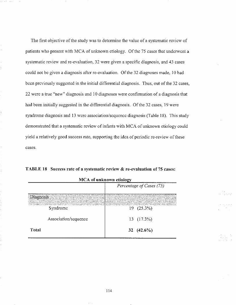
The frrst objective of the study was to determine the value of a systematic review of
patients who present with MCA of unftnown etiology. Of the 75 cases that underwent a
systematic review and re-evaluation, 32 were given a specific diagnosis, and 43 cases
could not be given a diagnosis after re-evaluation. Of the 32 diagnoses made, 10 had
been previously suggested in the initial differential diagnosis. Thus, out of the 32 cases,
22 were a lnrc "new" diagnosis and 10 diagnoses were confinnation of a diagnosis that
Irad been ìnitially suggested in the differential diagnosis. Of 1,he 32 cases, 19 were
syrdrome diagnosis and 13 were association/sequence diagnosis (Table 18). This study
demonstrated that a systematic review of infants with MCA of unknown etiology could
yield a relatively good success rate, supporting the idea ofperiodic re-review ofthese
TABLE 18 Success rate ofa svstematic review & re-evaluation of75 cases:
MCA of unknownPercentage of Cases (7 5)
Diagrrosis
Sl,,lldrome
Association/sequence
19 (2s.3%)
13 (173%)
32 (42.60 )
5.3 PREVIOUS MCA SYNDROME DIAGNOSES
Included ir-r the study were all cases of MCA of unknown etiology in which the length
of time from initial contact with the Section of Genetics and Metabolism to diagnosis
exceeded one year's time. As stated in the Introduction, ideally when a child presents
with MCA, one would want to make a diagnosis during that initial sessìon or shortly
afterwards. In many cases a diagnosis is not made during that time period. Deterrnining
what factors influence the eventuality of makìng a diagnosis and also determining how
long or the number of follow-up sessions required before a diagnosis is made was briefly
looked at in this study. Follow-ups usually occurred once a year, thus the length of time
chosen in the study is approximately equivaler.rt to the number of follow-ups.
Ten cases fell into this inclusion criteria and had an average length of time from initial
contact to diagrrosis of 6.4 years with a range of 1.3 years to 14.2 years. Three of these
cases were initially not given a diagnosis because the patient's overall pattem of
malformations although was suggestive of a specific diagnosìs, were considered atlpical
features not known to be associated with that diagnosis. It was not until repeated follow-
up and subsequent reporling in the literature ofthose features in association with the
condition, that the diagnosis was given to the patient/family (FAS, Opitz G syrdrome and
VATER association). In 2 cases, the patients were given a diagnosis only after clinical
reports appeared in the literature recognizing the pattem of malformations as a
distinguishable 'hew" MCA condition with a known etiology allowing for confirmation
by rnolecular and/or cytogenetic analysis (Rieger syrdrom e and 22qDeletion syndrome).
The remaining 5 cases were undiagnosed initially as the patient was thought to have a
115
different although tentative diagnosis. It was not untì1 contìnuous reinvestigation that the
"correct" diagnosis was given (Aarskog syrdrome, Beals slmdrome, Sotos sl,ndrome,
Opitz s1'ndrome and Klippel-Fiel sl,ndrome). In 2 of these 5 cases, the diagnosìs was not
obvious until the patient had "grown" into the phenotype
As Hall et al. (1998) demonstrated in their retrospective study of undiagnosed MCA,
periodic follow-up of cases of undiagnosed MCA slmdromes does allow for a number of
successful diagnoses. Success is due in part to: 1) the advent ofnew technology and the
ability to confim the diagnosis with molecular or c1'togenetic methods, 2) reporting of
new or previously described conditions with expansion of the phenotype, and 3)
phenotypic changes over time into recognizable sy.rdromes. As illustrated here, ìt may
require a significant length of time and continuous follow-up before a diagnosis is made.
Hall et al. (1998) found that among the 50 cases that were diagnosed, 36%o ltad
chromosomal anomalies or microdeletions, including 22q Deletion syndrome and Smith-
Magenis syndrome. Their findings are similar to that of this study. Two cases of 22q
Deletion s;mdrome and one case of Smith-Magenis sy,ndrome were retrospectively
diagnosed. As stated by Hall et al. ( 1998) and supported by this study, periodic follow-up
should become a standard ofpractice at least for this group ofpatients.
5.4 PROVISIONALLY NEW MCA SYNDROMES & ASSOCIATIONS
A total of 9 new slmdromes and associations were reported and delineated by members
of the Section of Genetics and Metabolism. Toriello (1988) had estimated that a new
syndronre is described in the lite¡ature at a rale of one or more a week. This illustrates the
u6
above mentioned point that repofting of "new" previously undescribed conditions allows
for the diagnosis in other patients with the same/similar findings. It is only through
continuous follow-up ofthese "unknown" patients that subsequent literatule searches
would identify these newly delineated conditions. Certainly, additional case reports have
been described in the literature since the initial reporting of some of these "new" MCA
conditions diagnosed by the Section, furlher illustrating the above mentioned point
(Harbord, Baraitser and Wilson, 1989; Baltaci et al., 1999). Continuous investigation
could also lead to the identification of the etiology and/or mode ofinheritance.
5.5 COMPUTERIZEDDATABASES
As discussed in tlie Introduction, the utility ofcomputerized databases for slmdrome
diagloses has been debated in the literature. The usefulness ofthese tools was evaluated
in this study. Of the 32 cases in which a diagnosis was made, 26 of ltre diagnosis were
suggested by at least one of the 4 databases used in this study (POSSI-JM, LDDB, OMIM
and PubMed). POSSUM ranked the highest with a 43.7o/" success rate with LDDB
ranking second with a34.3o/o success rate, PubMed had a31.2"/o success rate and OMIM
had a28.1o/o success rate. The accumulative success rate was 81.3% when all databases
were used. Overall, each program was fairly equivalent in regards to their success rate
and usefulness. As shown in this study, the combination of at least two or more databases
yields the best success rate and also allows for comparison of the list ofcandidate
slmdlomes generated by each search, which can be helpful when trying to narrow down
the lisT to one polential diagnosis.
These programs are meant to function as diagnostic tools/aids and are not meant to
provide a diagnosis. Each program generates a list ofcandidate syndromes that must be
reviewed at length in order to arrive at a potential diagnosis. As discussed in the
Inlroduction, the "novice strategy" to use oftllese tools has been shown to be valid.
Therefore, there should not have been any additional biases introduced into the study (i.e.
¡educed usefulness of the databases and thus reduced number of s)nrdrome diagnosis) by
the author using the "novice strategy" as compared to the "expeft strategy."
The success rate denonshated in this study is similar to that of the study carried out by
Pelz el al. (1996). They looked at POSSLIM and LDDB success rates and found 63%o and
68% r'espectively. One major difference between their study and this one was that they
re-evaluated cases in which a diagnosis was already known, to determine the program's
success rate. This may account for the slightly lower values in this study as compared to
their study.
5,6 PHENOTYPIC & DEMOGRAPHIC ANALYSIS
Two separate discriminant function analyses were preformed looking at: 1)
derrographic variables, and 2) phenotypic traits (anomalies). In the first analysis, no
differences could be identified between Group 1 and Group 2. For both groups, most of
the demographic variables had approximately equivalent values with nonsignihcant
probability values. When the percentage of males (and thus conversely females) were
conrpared, 5 8. 1% of the cases in Group 1 were males and, 5L3%o of the cases in Group 2
were males. The average time from date of biÍh to staÍ of the study (thus looking at the
age of the individual) was also similar with an average age of 72 years for Group 1 and an
average age of 11 years for Group 2. No ethnic variable could be demonstrated to be
significant, nor could these two groups be differentiated in terms of "family history same"
or in tenns of syrnmetry of anomalies. The only demographic variable that was
somewhat significant was "family history other" with a probability value of 0.057. Group
2 had almost double the frequency (48.2%) ofoccunence ofother anomalies then Group
I (27.9%). This finding would suggest then that those families, in which siblings of the
proband presented with malformation(s) different from those in the proband, decreased
the likelihood ofmaking a diagnosis. This may suggest that there are other genetic and/or
environmental factors that are influencing the expression of certain malformation(s), thus
complicating the situation. Over all, other than potentially family history of other
anomalies, none ofthe demograpl.ric variables looked at in this study could be used to
distinguish the two groups nor could they be used to determine what factor(s) influence
the ability to make a diagtosis.
The phenotypic traits were compared between Group 1 and Group 2. Three anomalies
were found to have significant probability values. These 3 anomalies were renal
dysplasia,/cystic, postaxial polydactyly and tracheal defects. In the 80 cases examined,
these 3 anomalies were not found in any of tlie 43 cases in which a diagnosis had been
made (Group I ) whereas renal dysplasia,/cystic anomalies occurred in 13.5%, postaxial
polydactyly in 10.8% and tracheal defects in 8.1% of the cases in which no diasnosis had
been rnade (there were a total of 37 cases in Group 2).
Ideally, one would want discriminant function analysis to identify those anomalies that
1r9
would have differentiated Group 1 from Group 2 such that one could determine what
factors influenced the ability to achieve a diagnosis rather than what factors are associated
with failure to achieve a diagnosis. Th¡ee anomalies were identified, which suggested
that, the occurrence of these anomalies in a child who presents with MCA is associated
with a lower likelihood ofmaking a diagnosis. This does not follow logical reasoning.
However, when one looks at the anomalies ìdentified by the discriminant function
analysis there is some reasoning to support this finding. Few slmdromes are associated
with tracheal anomalies. A search using OMIM only identified 2 syndromes that are
commonly associated with tracheal defects: Pfeiffer s1'ndrome (OMIM #101600) and
Hydrolethalus s¡mdromes (OMIM #236680). Besides the VATER association, there are
few other condrtions that have tracheal defects as a coÍnmon chalacteristic. Tbis rnay be
why tracheal defects were found in Group 2 only. Presumably, for the cases in Group 2,
any of the known sy,ndromes and associations with tracheal defects would have been
r-uled out. When the 3 undiagnosed cases with lracheal defects were re-reviewed none of
them were typical fol the VATER association (or the above two mentioned syrdromes).
All 3 cases had other malfonnations not typically associated with VATER (e.g.
holoprosencephaly).
Renal dysplasia./cystic and postaxial polydactyly were found in Group 2 but not in
Group 1. This may be due to the fact that both anomalies are associated with a large
number of conditions. For example, an OMIM search was performed using renal
dysplasia and renal cystic. Over two hundred different conditions were suggested, rnany
of which were MCA syrdromes. Similarly, one hundred entries were found ir OMIM
that had postaxial polydactyly as one ofthe features. This suggests that both ofthese
120
features are nonspecihc common malformations (as opposed to tracheal defects). It may
be that having either of these 2 anornalies in a patient, who presents with MCA, makes
the task ofachìeving a diagnosis that much more complex.
A limitation of this discriminant function analysis was that, not all of the original 95
cases underwent analysis. In 15 of the cases, there was at least one missing
discriminanting variable. These cases were removed fronr the analysis. When the
original data was reviewed the following was noted: 1) one case from Group t had a
tracheal defects and was removed from the analysis, 2) one case from Gloup t had a renal
dysplasia,/cystic anomaly and was removed from the analysis, and 3) one case from Group
t had postaxial polydactyly and was removed fi'om the analysìs. Therefore, in actuality,
each of these anomalies occuned in Group 1 with a ftequency of 2.3o/". While this is a
relatively low value as compared to the frequencies ofthese anomalies in Group 2, the
probability values might not be as significant as originally stated in table 15.
5.7 R.ECURRTNCE RISK ESTIMATION ANALYSIS
The second objective in this study was to determine an appropriate recurrence risk
estimate for infants with MCA of unknown etiology. To do this, a1l subsequent
pregnancies in the 94 cases that were reco¡ded in the patient charts were tabulated to
detennine an estimation of the recurrence risk for persons with MCA. In all,47
subsequent pregnancies were reported. There were 7 cases ofa recurrence in which the
sibling had a similar pattem of malfomations and 7 cases in which the sibling had a
dissimilar malformation. This infomation was then broken down into the two groups,
diagnosis made (Group 1, which had 51 in total) and diagnosis unknown (Group 2, which
had 43 in total). Group t had 27 subsequent pregnancies with 4 recunences of sirnilarly
affected sibs (14.8%) and 3 occunences of other malformations (1\.1%). The 4 cases in
which there was a recurrence included the Holzgreve-Thomas syndrone case, the
Ritscher-Schinzel sl,ndrome case, the Seckel syndrome case and the Walker-Warburg
syndrome case. All 4 conditions are thought to be autosomal recessive inheritance and
thus it is not surprising that there were recuffences in the family.
Surprisingly, Group 2 had 3 recuruences out ofa total of20 subsequent pregnancies
(15.0%) of sunilarly affected siblings. This miglit be due to the fact that sonre ofthe
famiÌies had a new private autosomal recessive MCA s1a-rdrome. Recurrence of ar.r
affected sib with normal parents is suggestive of an autosomal recessive condition.
However, when these cases were re-reviewed, none of the families were consanguineous,
which would have suggested a private autosomal recessive MCA condition. This group
also had 4 occurrences of other malfomations (20.0%).
These fìgures are much higher than Czeizel et al. (1988) study. Theyfounda3.9%
risk of recurrence ir the siblings of patients with MCA of unknown etiology. The
discrepancy betw een Czeizel et al. (1988) study and the recurrence risk estimates in this
study are most likely due to a number ofreasons:
1) There was ìnadequate or lack of information on subsequent pregnancies for 11 of the
cases, thus potentially under-estimating the true nutnber of subsequent pregnancies.
Information on subsequent pregnancies was obtained by review of the patient char1.2)
Ideally, to obtain accurate information on al1 subsequent pregnancies, the
families/patients need to be recontacted to up date the family and pregnancy histories
and outcomes. ln many ofthe cases, they were only seen once, thus any subsequent
pregnancies would not have been known or recorded in the patient's chart.
3) Families were more likely to be seen in the Section of Genetics and Metabolism a
second time if there was a recuffence of an affected sib. Thìs could lead to an over
estimate of recurences of affected sibs. Normal pregnancies are less likely to come
to the attention of the Section of Genetics and Metabolism.
It is likely that 47 subsequent pregnancies is an urder estimation of the true number of
pregnancies. This would account for the dìscrepancy between the recurrence risk
estination (15.0%) for patients with MCA of unknown etiology in our study and Czeizel
et al. (1988) recurrence risk estination of 3.9%.
There was a total of 7 (14.9%) recun'ences of sìblings who had other malformations
(both mfuor and major malformations), 3 in Group 1 and 4 in Group 2. This figure
should presurnably reflect the population lrequency ofboth rninor and major
malfomations in newbons. The finding of 14.9% is fairly representative of the expected
population frequencies. One limitation of this analysis was that minor and major
malformations weÌe not distinguished from each other, thus |he 14.9%o is the cornbined
frequency. A second limitation is, as stated previously,4T subsequent pregnancies is
most lìkely not the true pregnancy number for this group and therefore, the I4.9'Yo maybe
an under or over estimation.
t23
6. FUTURE WORK
6.1 CLUSTER ANAI,YSIS
Given that all anomalies for each case have been recorded in a phenotype sheet and
entered into a Microsoft Access spreadsheet, cluster aralysis can now be perfonled to
identify those groups that would fall into the same subgroups, and to identify those cases
that were most similar. Discriminant function analysis could be used to determine which
phenotypic traits (anomalies) could be used to distìnguish between cases. This analysis
may give furlher insight into the pattem of malfomratìon in those cases in which the
diagnosis remains unknown. Prelìminary cluster analysis did demonstrate 4 main
clustering of cases (appendìx 3).
6.2 RECONTACTINGFAMILIES
Ideally, the familíes and patients in this study should be recontacted so that farnily and
medical history can be updated. Families and patients can be re-counselled in regards to
the recurrence risk, and mode of inheritance so that one can identify those family
members at risk and offer testing and/or subsequent investigations. Recontacting these
families could also help validate the diagnosis given to the patient/family.
7. SUMMARY
1 . A systematic re-evaluation of pteviously undiagnosed cases of MCA yielded a
relatively high success rate (42.6 %) of slardr ome/association /sequence diagnosis.
2. Computerized databases both online and software forms (POSSUM, LDDB,
PubMed and OMIM) are valid and useful dìagnostic tools in dysmorphology.
3. Continued follow-up of undiagnosed MCA cases, as well as detaìled description
and reporting of clinical features including rninor dysmorphic features, contribute
to the likelihood of identifying a slardrome/association diagnosis.
4. Reportìng of 'hew" MCA sl,ndromes contributes to the success of syrdrome
diagnosis in other patierfs who plesent with MCA of unknown etiology.
5. No demographic variables that were looked at in this study could be used to
detemine the likelihood of making a diagnosis. Renal dysplasia./cystic, postaxial
polydactyly and tracheal defects were negatively associated with the likelihood of
being able to rrake a dìagnosis.
6. Recurrence risk estimations were found to be 14.8% for cases in which a diagnosis
was known and 15 -0%¡' for cases in which no diagnosis was made.
REFERENCE
Afzelius, B. A.(1980): Genetic aspects of the immotile-cilia syrdrome. (Abstract) ClinGeneÍ 17: 52.
Alagille, D., Odìevre, M., Dommergues, J. P.(1975): Hepatic ductular hypoplasiaassociated witb characteristic facies, veÍebral malformations, retarded physical,nrental and sexual development, and cardiac murrnur. J Pediat 86: 63 - 71.
Allanson, J. E, and McGillivary, B.C (1985): Familial clefting syndlome with ectropionand dental anomaly - without limb anomalies. Clin Genet 27,426 - 429.
Anderson, T. E. and Breed, A. L. (1981): Congenital clasped thumb and the Moro reflex.(Letter) J PediaÍ99:664 665.
Avon, S. W. and Shively, J. L. (1988): Orthopeadic manifestation of Goldenharsyrdrome. J Pediatr OÍhop 8: 683 - 686.
Baltaci, V., Ors, R., Kaya, M., Balci, S. (1999): A case associated with Walker Warburgsyrdrome phenotype and homozygous perìcentric inversion 9: coincidental findingor aetiological factor? Acta Paediatr 88: 579 583.
Berman, M. D., Ishak, K. G., Schaefer, E. J., Bames, S., Jones, E. A. ( 1981):Syndromatic hepatic ductular hypoplasia (arleriohepatic dysplasia): a clìnical andhepatic histologic study ofthree patients. DigestDisSci26: 485 497.
Bianchine, J. W. and Lewis, R.C., Jr. (1974): The MASA s)r.ìdronre: a new heritablemental retardation syrdrome. Clin Genet 5: 298 - 306.
Bomet, J., Cordier, M. P., Ollagnon,8., Guillaud, M. H., Raudrant, D., Robef, J. M.,Charvet, F. (1987): Hypoplastic renale polydactylie, cardiopathìe: un nouveauslardrome? J Genet Hum 35: 279 - 289.
Bontluon, D., Fitzpatrick, D., Porteous, M., Trainer, A. Clinical Genetics: A case basedapproach. Philadelphia: WB Saunders, 1998
Botto, L. D., Khoury, M. J., Mastroiacovo, P., Castilla. E. E., Moore, C. 4., Skjaerven,R., Mutchinick, O. M., Borman,8., Cocchi, G., Czeizel, A. 8., Goujard, J., kgens,L. M., Lancaster, P. A. L., Martinez-Frias, M. L., Merlob, P., Ruusinen,4., Stoll, C.,Sunriyoshi, Y . (1997): The spectrum of congenital anomalies of the VATERassociation: An intemational study. Am J Med Genet 71: 8 - 15.
Braun Quentin, C., Billes, C., Bowing, 8., Kotzot, D. (1996): MURCS association:case repod and review. J Med Genet 33:618-620.
Briard, M. L., Le Merre, M., Plauchu, H., Dodìnval, P., Lambotte, C., Moraine, C.,Serville, F. (1984): Association VACTERL et hydrocephalies une nouvelle entitefamiliale. Arln Genet 2'7 : 220 223
Buck - Gramcko, D. and Wood, V. E. (1993): The tteatment of metacarpal synostosis. J
Hand Surg 184: 565 581.
Buyse, M. L. (1990): Birth Defects Encyclopedia. Blackwell Scientihc Publications.Cambndge, Massachusetts
Casey, 8., Cuneo, B. F., Vitali, C., Van Hecke, H., Banish, J., Hicks, J., Ballabio, 4,,Hoo, J. .T. (1996): Autosomal dominant transmission of familial laterality defects.Am J Med Genet 61: 325 -328.
Christian, Joe C., Andrew, P.4., Corneally, P.M., and Muller, Jans (1971). Theadducted thurrbs s¡mdrome. An autosomal recessive disease with artluogryposis,dysmyelination, craniostenosis, and cleft palate. Clin. Genet. 2,95 - 103.
Cohen, M. J. and Cole, D. E. (1989): Origins of recognizable syndromes: Etiology andpathogenetic mechanisms and the process of s¡,ndrome delineation. J Ped 115: 161 -164.
Cohen, M. M. (1986): Canadian Paediatric Society lecture sponsored by the RoyalCollege. Children, Birth Defects and Multiple Birth Defects: Part 1. A¡nals RCPSC,19:375-384.
Cohen, M. M. (1986): Canadian Paediatric Society lecture sponsored by the RoyalCollege. Children, Birth Defects and Multiple Birth Defects: Part 2. A¡rals RCPSC,19:465 4'70.
Corsello, G. and Giuffre, L. (199\: VACTERL with hydrocephalus: a fuilher case withprobable autosomal recessive inheritance. (Letter). Am J Med GercI49: 737 138.
Cox, P. M., Gibson, R. 4., Morgan, N., Brueton, L. A. (1997): VACTERL withhydrocephalus in lwins due to Fanconi anemia (FA): nutation in the FAC gene. Am.T Med Genet 86:86 - 90.
Cumiff, C., Canlack, J. L., Kirby, R. S., Fiser, D. H. (1995): Contribution of heritabledisorders to mortality irr pediatrìc intensive case unit. Pedìatrics 95: 678 - 681.
Czeizel, A. (1981): SCHISIS association. An J Med Genet 10: 25 - 35.
CzeizeI,4., Telegdi, L., Tusnady, G. (1988): Multiple congenìtal abnormalities.Budapest: Akademia Kiado.
12'7
Del Bigio, M. R., Greenberg, C.R., Rorke, L. 8., Schnur, R., McDonald McGinn, D.M., Zacka| E. H. (1997): Neuropathological findings in Eight Children with Cerebro
- Oculo - Facio - Skeletal (COFS) s¡mdrome. Joumal of Neuropathology andExperimental Neurology. 56: ll41 - 1157 .
De Smet, L,, Keymolen, K., Fr¡ars, J. P. (1999): Unilateral longitudinal radial raydeficiency ofthe hand and metacarpal 4-5 synostosis. Genet Couns 10:369 372.
Dicker, H. and Opitz, J. M. (1969): Associated acral and renal rnalformations. BirthDefects Original Article Series V(3): 68 - 77.
Dilibefti, .I. H. (1988): Use of Computers in dysmorphology: J Med Genet.25: 445 -4)J.
Digilio, M. C., Marino, B.,Borzaga, U., Giamrotti,,A., Dallapiccola,B. (1991):Intrafamilial variability of Pfeiffer{ype cardiocranial s1,r-rdrome. Am J Med Genet73: 480 - 483.
Donnai, D. and Winter, R. M. (1989): Disorganisation: a model for'early amnionnrpture'? J Med Genet 26:421 - 425.
Duncan, P.4., Slrapiro, L. R., Stangel, J. J., Klein, R. M., Addonizio, J. C. (1979): TheMURCS assocìation: Mullerian duct aplasia, renal aplasia, and cervicothoracicsonrite dysplasia. J Pedìat 95:399 402.
Duncan, P. A. and Shapiro, L. R. (1993): Intenelationship of the Hemifacial microsomiaVATER, VATER, and Sirenomelia phenotypes. Am J Med Genet 41:'75 - 84.
Elgin, V. E., Connolly, E. S., Millar, W. S., Feldstein, N. 4., Dwork, A. J. (2001):Extramedullary lrematopoiesis within a frontoethmodidal encephalocele in anewbom with holoprosencephaly. Pediatr Dev P athol 4: 289 297 .
Evans, C. D. (1995): Computer systems in dysmorphology. Cliri Dysmorph 4: 185 -201.
Evans, J. A. (1991): Epidemiology of birlh defects. Manitoba Medicine 6I: 47 49.
Evans, J. 4., Stranc, L. C., Kaplan, P., Hunter, A. (1989): VATERL with hydr.ocephalus:fufiher delineation of the s¡'ndrome G). Am J Med Genet 34:177 - 182.
Evans J, A.,Yitez,M., Czeizel A. (1994): Congenital abnormalities associated with limbdeficiency defects: a population study based on cases from the Hungarian CongenitalMalfomration registry (1975 - 1984). Am J Med Genet 49: 52 - 66.
r28
Evans, J. 4., Trevenan, C.,Leahy, F., Rockman-Greenberg, C. (1980): Lissencephaly,hydrocephaly and ocular manifestation. (Presented at the Birth Defects Conference,NY). Abstract.
Evans, J. A. and Chudley, A. E. (1998): Dysgenesis of the corpus callosurn withcardiovascular and other mìdline anomalies: a new autosomal dominant s;rndrome?Am J Hum Genet Vo1 63 No4 4103. Abstract.
Evans, J. A. and Greenberg, C. R. (2002): Tibial agenesis, radìal ray and cardiovasculardefects. Clìn Dysmorphol 11:163-169.
Evans, J. 4., Erdile, L. 8., Greenberg, C. R., Chudley, A. E. (1999): Agenesis of thepenis: Pattems of associated malformation. Am J Med Genet 84: 47 55.
Falace, P. B. and Hall, B. D. (1988): Congenital euryblepharon with ectÌopion and dentalanomaly: an autosomal dominant clefting disorder with marked variability ofexpression. Proc Greenwood Genet Ctr 8: 208 - 209.
Fitch, Naonri and Levy, Edith P. (1975): Adducted thumb syrdrome. Clin Genet 8: 190198.
Fitzpatrick, J. L., Halm, C., Costa, T., Huggins, M. J. (1999): The duty to recontact:attitudes of Genetìc Seruice Providers. Am J Med Genet 64: 852 - 860.
Fraser, G. R. (1962): Our genetic 'load': a review of some aspects of genetical variation.A¡n Hum Genet 25: 387 415.
Fraser, F. C. (1996): Liability, thershold, malformations and sy.ndromes. Am.T MedGenel66:75 - 76.
Fryer, A. E., Holt, P. J., Hughes, H. E. (1991): The cardio-facio-cutaneous (CFC)slmdrome and Noonan syndrome: are they the same? Am J Med Genet 38: 548 -551 .
Gabrielli, O., Bonifazi, V., Offidani, A. M., Cellini,4., Coppa, G. V., Giorgi, P. L.(1993): Description of a patient with difficult nosological classification: Goldenharsyndrome or Towns-Brocks syrdrome? Minerva Pediatr 45: 459 - 462.
Gagnon, A. L. and Wilson, R. D. (1997): Fetal anomalies: what is the diagnosis when theclrromosomes are normal? Ultrasound Obstet G¡arecol 10:254 260.
Gardner, R. J. M. and Sutherland, G. R. (1996): Chronrosome abnonnalities and geneticcounselling. New York: Oxford University Press.
Geipel,4., Berg, C., Gemer, U., Ahrens, P., Gloeckner-Hofman, K., Moller, J.,Gembruch, U. (2001): Diagnostic and therapeutic problems in a case of prenatallydetected fetal hydrocolpos. Ultrasound Obstet Gyrecol 18: 169 172.
Gilberl, W. M. and Nicolaides, K. H. (1988): Fetal omphalocele: associatednralfomations and chromosomal defects. Obstet and Gynecol 10: 633 - 635.
Goktay, A. Y., Seci1, M., Karabay, N., Igci, E., Pimar, T. (1999): Esophageal duplicarioncyst coexisting witli Bochdalek's hemia and polysplenia. Eur Radiol 9 478 - 480.
Goldberg, R. B. and Shprintzen, R. J. ( 1981): Hirschsprung megacolon and cleft palate ìntwo sibs. J Craniofacial Genetics and Developmental Biology 1: 185 - 189.
Goltz, R. W., Peterson, W. C. Jr., Gorlin, R. .I., Ravits, H. G. (1962): Focal dermalliypoplasia. Arch Derm 86: 708 717 .
Gorlin, R. J., Zellweger, H., Curtis, M., Majewski, F., Gillessen-Kaesbacli, G., Prahl-Anderson, 8., Zackai, E. (1996): Blepharo-cheilo-dontic (BCD) sy,ndrome. Am JMed Genet 65: 109 I 12.
Goto, M. P. and Goldman, A. S. (1994): Dìabetic ernbryopathy. Curr Opin Pediatr 6:486 49r.
Grebe, T. A. and Clelicuzio, C. (2000): Neurologic and gastrointestinal dysfuirction incardio-facio-cutaneous slmdrome: identihcation of a severe phenotype. Am .I MedGenet 95: 135 - 143.
Graham, J. M., J¡., Alyane-Yeboa, K., Raams, A., Appeldoom, E., Klerjer, W. J.,Garritsen, V. H., Busch, D., Edersheim, T. G., Jaspers, N. G. J. (2001): Cerebro-oculo-facio-skeletal sytdrome with a nucleotide excision-repair defect and a mutatedXPD gene, with prenatal diagnosis in a triplet pregnancy. Am J Med Genet 69: 291300.
Greenberg, F. (1993): Contiguous gene syrdromes. Growth, Genetìcs and Hormones 9:5 - 10.
Greene, R. 4., Bloch, M. J., Huff D. S., Iozzo R. V. (1986): MLIRCS association withadditional congenital anomalies. Hum Pathol 17: 88 91.
Guion Ahneida, M. L., Rodini, E. S. O., Kokitsu-Nakata, N. M., Bologua-Amantini, D.(1998): Blepharo-cheilo-dontic (BCD) s¡.,ndronie: repoÍ on four new patients. Am JMcd Genet 76: 133 Ii6.
Halal, F. and Movel J. (1990): The syrdrome of Hirschsprung disease, microcephaly,unusual face and mental retardation. Am J Med Genet 37: 106 - 108.
Hall, B. D., Rob1, J. M., Cadle, R. G. (1998): Lnporlance of diagnostic follow-up ofunknown multiple congenital anomaly s¡.ndromes. (Abstract) Am J Human Genel63: 448.
Hall, J. G-, Powers, E. K., Mcllraine, R. T., Ean, V. H. (1978): The frequency andfrnancial burden of genetic dìsease in a pediatric hospital. Am J Med Genel:4ll4JO.
Hamel, B. C. J., Raams, 4., Schuitema - Dijkstra, A. R., Simons, P., van der Burgt, L,Jasper, N. G. J., Kleijer, W. J. (1996): Xeroderma pigmentosum-cockalme s¡mdromecomplex: a further case. .l Med Genet 33: 607 610.
Hancock, B. L. and Wiseman, N. E. (1989): Congenital duodenal obstruction: the impactofan antenatal diagnosis. J Pediat Surg 24:1027 - 103I.
Harbord, M. G., Baraitser, M., Wrlson, J. (1989): Microcephaly, mental retardation,cataracts, and hypogonadism in sibs: Marlsolls syrdrome. J Med Genet 26:391 -400.
Hamed, R. K., Patrick, L. E., Gay, B. 8., Atkinson, G. O., Niemer, P. K., Wyly, L. 8.,Clark, W. S. (1996): Standard method of diagnosis versus use of a computerdatabase in the evaluation ofskeletal dysplasias. Pediatr Radiol 26: 887 - 890.
Henlekam, R. C. M. (1992): Another hunran hornologue for the mouse mutantdisorganisation. J Med Gent 29: 71 - 72.
Heren, C., Jasmar, M., Alvarez-Gonzalez, M. L., Lesenfants, S., Lombet, J., Bonniverl,J., Koulischer, L., Verlos, A. (1997): Private multiple congenital anomaly syrdronesmay result fonn unbalanced subtle translocations: t(2q;4p) explains the Lambotteslmdrome. Am J Med Genet 73: 127 - I31 .
Hod, M., Merlob, P., Friedman, S., Rusecki, Y., Sclioenfeld,4., Ovadia, J. (1991):Prevalence ofcongenital anomalies and neonatal complications in the ofßpring ofdiabetic mothers in Israel. Isr J Med Sci 27: 498 502.
Hollister, D. W., Klein, S. H., Dejager, H. J., Lachman, R. S., Rimoin, D. L. (1973): Thelacrimo-auriculo-dento-digital sl.ndrome. J Pediat 83: 438 444.
Hoft, M. and Oram, S. (1960): Farrilial hearl disease with skeletal malformations. BritHeaftJ22:236 242.
Holzgreve, W., Wagner, H., Rehder, H. (1984): Bilateral renal agenesis with Potterphenotype, cleft palate, anomalies of the cardiovascular system, skeletal anomaliesincluding hexadactlyly and bifid metacarpal: a new slmdrome? Am J Med Genet 18:117 - 182.
131
Hummel, K. P.(1959): Developmental anomalies in mice resulting from action of thegene disorganization, a semidominant lethal. PedíaTncs 23: 212 221 .
Hurst, J.4., Markiewicz, M., Kumar, D., Brett, E. M. (1988): Hirschsprung's disease,microcephaly, and iris coloboma: a new slmdrome of defective neuronal migration.J Med Genet 25: 494 - 497 .
Hutclrison, J. W., Stovring, J., Tumer, P . T. (1979): Occipital encephalocele withlroloprosencephaly and aqueduct stenosis. Surg Neurol 12:331 -335.
Johrson, .J. P., Poskanzer, L. S., Sherman, S. (1996): Three-generatìon family withresemblance to Townes-Brocks syndrome and Goldenhar/oculoauriculovertebralspectrum. Am J Med Genet 61: 134 1,39.
Jonason, M. S., McConrick, J. R., Gillies, C. G., Gondos, B. (1982): Kartagener'sslmdrome with motìle spematozoa. New Eng .T Med 301 : 1 1 3 1 - I 1 33.
Jones, K. L. (1991): Smith's Recognizable Pattems of Human Malfomation. 5tr'ed.Phìladelphia: WS Saunders.
Juyal, R. C., Greenberg, F., Mengden, G. .4., Lupski, J. R., Trask, B, .I., van den Engh,G., Lindasy, E. 4., Christy, H., Chen, K. 4., Baldini,4., Shaffer, L. G., Patel, P. I.(1995): Smitli-Magenis s1'r.rdrome deletion: a case with equivocal cytogeneticfindings resolved by fluorescence in situ hybridization. Am J Med Genet 58:286291.
Kabra, M., Suri, M., Jain, U., Verma, I. C. (1994): Poland anomaly with unusualassociated anomalies: case report ofan apparent disorganization defect. Am J MedGenet52:402 405.
Keeger.r, C. E., Mullikem, J. B., Wu, B. L., Korf, B. R. (2001): Townes-Brocks syndromeversus expanded spectrum hemifacial microsomia: review of eight patients andfuilher evidence of a 'hot-spot' for mutation in the SALL 1 gene. Genet Med 3: 3 10
- 313.
Kirk, E. P. E, Arbuckle, S., Ramm, P. L. Ades, L. C. (1999): Severe micrognathia, cleftpalate, absent olfactory tract, and abnormal rib development: cerebro-costo-nrandibular slmdrome or a new syrdrome? Am J Med Genet 84: 120 - 124.
Kitzrniller, J. L., Gavin, L. ,4., Gin, G. D., Jovanovic-Peterson, L., Main, E. K., Zigrang,W.D. (1991): Preconception care ofdiabetes. Glycemic control prevents congenitalanomalies. JAMA 265: 131 -736.
Korula, S., Wilson, L., Salomonson, J. (1995): Distinct craniofacial syndrome oflagophthalmia and bilateral cleft 1ip and palate. Am J Med Genet 59:229 -233.
132
Krantz, L S., Piccoli, D. 4., Spinner, N. B. (1997): Alagille syndrome. J Med Genet 34:152 - 1s7.
Kunze, J., Park, W., Hansen, K. H., Hanefeld, F. (1983): Adducted thumb slardrome:report ofa new case and a diagnostic approach. Europ J Pediat 741:122-126.
Legius, E., Moenlan, P., Fr1'ns, J. P., Vandenberghe, K., Eggermont, E. (1988):Holzgreve-Wagner-Rehder slmdrome: Potter sequence association with persistentbuccophar;'ngeal membrane: a second observation. Am J Med Genet 31: 269 -272
Leichtman, L. G. (i996): Are cardio-facio-cutaneous slardrome and Noonan s1'ndromedistinct? A case of CFC offspring of a mother with Noonan syndrome. ClinDysnrorph 5: 6I - 64.
Leiter, Elliot and Lipson, Jeffery (1976): Genitourinary tract anomalies in lobster clawsyndrome. J Urology 115: 339 - 341.
Lemire, E. G., Hildre-Ripstein, E., Reed, M. H., Chudley, A. E. (1998): SAMS:provisionally unique multiple congenital anomalies sl,ndlome consisting of shorlstature, auditory canal atresia, mandibular hypoplasia, and skeletal abnormalities.Am J Med Genet 75: 256 - 260.
Leonardi, M. L., Pai, G. S., Wilkes,8., Lebel, R. R. (2001): Ritscher-Schinzel cranio-cerebello-cardiac (3C) syndrome: report of four new cases and review. Am J MedGenetl02:237 242.
Leppig, K. ,A., Werler, M. M., Canl, C. I., Cook, C.4., Holmes, L. B. (1987): Predictivevalue of minor anomalies. I. Association with major malformations. JPediatrll0:531.
Li,L.,Krantz,I. D. Deng, Y., Genin,,A'., Banta, A. 8., Collins, C. C., Qi, M., Trask, B.J., Kud, W. L., Cochran, J., Costa, T., Pierpont, M. E. M., Rand, E. 8., Piccoli, D..4., Hood, L., Spinner', N. B. (1997): Alagille syrdrome is caused by rnutations inhuman Jagged 1, wliich encodes a ligand for Notch 1 . Nature Genet 16: 243 , 251 .
Liberfarb, R. M., Abdo, O. P., Preutt, R. C. (1987): Ocular coloboma associated with asolitary rnaxillary central incisor and growth failure: manifestations ofholoprosencephaly. Ann Ophthal 19: 226 221.
Lowry, R. B. and Young, S. L. (1991): Cleft lip and palate, sensodneural deafüess, andsacral liporna in two brothers: a possible exanrple ofthe disorganisation mutant. .IMed CeneL 28: 135 137.
Lurie, I. W. and Cherstvoy, E. D. (1984): Renal agenesis as a diagnostic featu¡e of thecryptophthalmos-syrdactyly slmdrome. Clin Genet 25: 528 532.
Marden, P. M., Smith, D. W., McDonald, M. J. (1964): Congenital anomalies ir thenewbom infant including minor variations. JPeds 64:351 -31L
Marles, S. L., Greenberg C. R., Persaud, T. V. N., Shuckett, E. P., Chudley, A. E. (1992):New familial syrdrorne of unilateral upper eyelid coloboma, abeffant anteriorhairline pattem, and anal anomalies in Manitoba Indians. Am J Med Genet 42: 793
- 799-
Marles, S. L., Chodirker, B. N., GreenberC, C R, Chuldley, A. E. (1995): Evidence forRitscher-Schinzel s¡mdrome in Canadian Native Indians. Am .I Med Genet 56: 343 -350.
Marles, S. L. and Chudley, A. E. (1990): Ulnar agenesis and endocardial fìbroelastosis.Am .I Med Genet 3'/:258 - 260.
Martinez, R. 8., Monasterio, A. L., Pinheiro, M., Freire-Maia, N. (1987): Cleft lip/palate-oligodontia-sl,ndactylyÍair alterations, a new slmdrome: review ofthe conditionscombining ectodennal dysplasia and cleft lip/palate. Am J Med Genet 2T: 23 , 31 .
Matlinez-Frias, M. L. (1995): Primary midline developmental fie1d. I. Clinical andepidemiological characteristics. Am J Med Genet 56: 374 -381.
Martinez-Frias, M. L. and Frias, J. L. (1997): Are blastogenetic anomalies sporadic? AmJ Med Genet 68: 381 - 385.
Martinez-Frias, M. L., Frias, L L., Op\tz, J. M. (1998): E[ors of morphogenesis anddevelopmental field theory. Am .T Med Genet 76: 291 - 296.
Marlsolf, J. T., Reed, M. H., Hunter, A. G. W. (1977): Case repoÍ 56. Sl,nd Ident 5:1418.
Mathias, R. S., Lacro, R. V., .Tor.res, K. L. (1987): X-linked laterality sequence: situsinversus, complex cardiac defects, splenic defects. Am J Med Genet 28: 111 - 116.
Mehes, K. (1983): Minol malformations in the neonate. Budapest: Akademiai Kiado
Meinecke, P., Polke,.A., Schmiegelow, P. (1989): Limb anomalies in the CHARGEassocìation. J Med Genet 26: 202 - 203.
Meira, L. 8., Graham, J. M., Greenberg, G. R., Busch, D. 8., Doughty, A.T.B.,Ztffer,D. W., Coleman D. M., Saure-Train, I., Freidberg, E. C. (2000): ManitobaAboriginal kindred with original cerebro-oculo-facio-skeletal syrdrome has aautalion in the Cockayne syrdrome Group B (CSB) gene. Am J Med Genet 66:1221 - 1228.
134
Mikkila, S. P., Janas M., Karikoski, R., Tarkkila, T., Simola, K. O. J. (1994): X-linkedlaterality sequence in a family with carrier manifestations. Am J Med Genet 49: 435_ 438.
Mitchell, J. A, Giangiacomo, J., Hefner, M. 4., Thelin, J. W., Pickens, J. M. (1985):Dominant CHARGE assciation. Ophthalmic Paediatr Genel6:2'71 276.
Mowart, D. R., Croaker, G. D. H., Cass, D. T., Ken, B. .A., Chaitow, J., Ades, L. C.,Chia, N. L., Wilson, M. J. (1998): Hirschsprung disease, microcephaly, mentaìretardation, and characteristic facial features: delìneation of a new syndrome andidentification of a locus at ch¡omosome 2q22-q23. J Med Genet 35: 617 623.
Nanni, L., Ming, J. E., Du, Y., Hall, R. K., Aldred, M., Bankier,4., Muenke, M. (2001):SHH mutation is associated with solitary mediar.r maxillary central incisor: a study of13 patients and review ofthe literature. J Med Genet 102: 1 - 10.
Nazer, J. and Ramirez, R. (2000): Congenital malformations ìn the offspring of diabeticmother. Rev Med Chil 128: 1045 - 1052.
Nikkawa, N., Kohsaka, S., Mizumoto, M., Hamada, I., Kajii, T. (1983): Farnilialclustering of situs inversus totalis and asplenia and polysplenia syrdromes. Am JMed Genet 16: 43 - 47 .
North, K. N., Wu, B. L., Cao, B. N., Whiteman, D. A. H., Korf, B. R. (1995): CHARGEassociation in a child with de novo inverted duplication Q \@22-q2a$. Am J MedGenet 57: 610 614.
Oda, T., Elkahloun, A. G., Pike, B. L., Okajima, K., Krantz, I. D., Genin,4., Piccoli, D..A., Mellzer, P. S., Spinner, N. 8., Collins, F. S., Chandrasekìrarappa, S. C. (1997):Mutations in the lruman Jagged 1 gene are responsible for Alagille symdrome.Nature Genet 16: 235 - 242.
OMIM: Online Mendelian Inheritance in Man rM. Center for Medical Genetics, JohnsHopkins University (Baltimore, MD) and Natiolal Center for BiotechnologyInfonlation, Natior.ral Library of Medicine (Bethesda, MD), 1997. World WideWeb tlRL
Opitz, J.M., Czeizel,4., Evans, J. 4., Hall, J. G., Lubinsky, M. S., Spranger, J. W.(1987): Nosologic grouping in birth defects. InVogelF, Sperling K (eds): HumanGenetics. Berlin: Springer Verlag 382 385.
Opilz,I. M. (1993): Blastogenesis and the'þrimary field" in human development. BirthDefects Orig Arlic Ser 29(1): 3 37.
Opitz, L M. (1994): Association and slmdromes: Terminology in clinical genetics andbirlh defects epidemiology: comments on Khoury, Moore, and Evans. Am J MedGenet 49: 14 20.
Pagon, R. 4., Graham, J. M., Jr., Zonana, J., Young, S. L. (1981): Coloboma, cogenitalheart disease, and choanal atresia with multiple anomalies: CHARGE association. JPediat99:223 227.
Passarge, E. and Lenz, W. (1966): Sy'ndrome ofcaudal regression jn infants ofdiabeticmothers: observations of furlher cases. Pediatric s 31: 612.
Patton, M. A. (1987): A corrrputerized approach to dysmorphlolgy. M.D. Computing 4:33 39.
Pauli, R. M. (1994): Lower mesodennal defects: a coû ron cause offetal and earlyneonatal death. Am J Med Genet 50: 154 - 172.
Peebles, D. M. (1998): Holoprosencephaly. Prenat Diagn 18: 477 ,480.
Pelz, J., Arendt, V., Kunze, J. (1996): Computerized assisted diagnosis of malformationslmdrome: an evaluation of tlrree databases (LDDB, POSSUM and SYNDVOC).Am J Med Genet 63: 257 - 261 .
Pena, S. D. J., and Shokeir, M. H. K. (1974): Autosomal recessive cerbro-oculo-facio-skeletal (COFS) syrdrome. Clin Genet 5: 285 - 293.
Pfeiffer, R. 4., Singer, H., Zschiesche, S. (1987): Sagittal craniostenosis, congenital heafidisease, mental deficiency and various dysmorphicies in two sibs-a 'new'syrdrome? Eur J Pediatr 146: 75 - 78.
Plotz, F. 8., van Essen, A. J., Bosschaarl, A. N., Bos, A. P. (1996): Cerebro-costo-mandibular s¡mdrome. Am J Med Genet 62:286 292.
Podani, J. (1993): SYN-TAX-po version 5.0 computer programs for multivariable dataanalysis in ecology ar.rd systematics. Budapest: Scientia Publishing.
Porteous, M. E., Cross, I., Bum, L (1992): VACTERL with hydrocephalus: one end oftlre Fanconi anemia spectrum of anomalies? Am J Med Genet 43: 1O3Z , 1034.
Prasad, C., Quackenbush, E. J., Wliiteman, D., Korf, B. (1997): Limb anomalies inDiGeorge and CHARGE syrdromes. AmJMedGenet6S: 179 181.
Ran.ros-Anoyo, M.4., Rodriguez-Pjnilla, E., Cardero, J. F.(1992): Matemal diabetes: therisk for specific birth defects. Eur J Epidemiol 8: 503 - 508.
136
Raven, K.4., Cotter, P. D., Bitts, S. M., Cox, V. 4., Golabi, M. (2000): Cardio-facio-cutaneous s¡,ndroure phenot)?e in an irdividual with an interstitial deletion of 12q:identification of a candidate region for CFC slmdrome. Am J Med Genet 93: 219222.
Re1molds,.I. F., Nevi,G., Hevrmann, J. P., Blumberg, B., Coldwell, J. G., Miles, P. V.,Opitz, J. M. (1986): New multiple congenital anomalies/mental retardations¡'ndrome with cardio-facio-cutaneous involvement - The CFC syndrome. Am J
Med Genet 25: 413 427.
Rjtsclrer, D., Schinzel,-4., Boltshauser,8., Briner, 1., Atbenz, U., Sigg, P. (1987): Ðandywalker (like) malformation, atrio-ventricular septal defect and a sìmilar pattem ofminor anomalies in 2 sisters: a new s¡mdrome? Am J Med Genet 26: 481 491.
Rodriguez, I. J., Palacios, J., Lapunzina, P. (1993): Severe axial anomalìes in the oculo-auriculo-verlebral (Goldenhar) complex. Am J Med Genet 47: 69 - 14.
Romero, R., Pilu, G., Jeanty, P., Ghidini,4., Hobbins, J. C. (1988): Prenatal diagnosis ofcongenital anomalies. Connecticut: Appleton & Lange
Rosenfield, N. S., Kelly, M. J., Jensen, P. S., Cotlier,8., Rosenfield, A. T., Riely, C. A.(1980): Aferiohepatic dysplasia: radiologic features ofa new slmdrome. AmJRoentgen. 135: 1211 - 1223.
Rossbach, H.C., Sutcliffe, M. J., Haag, M.M., Grana, N. H., Rossi, A. R., Barbosa, J. L.(1996): Fanconi anemia in brothers initially diagrosed with VACTERL associationwith hydrocephalus, and subsequently with Baller-Gerold syndrome. Am J MedGene| 6I:65 - 67 .
Rusnak, S. L. and Driscoll, S. G. (1965): Congenital spinal ar.romalies in infants ofdiabetic mothers. Pediatrics 35: 989.
Saatci, I., Yelgec, S., Aydin, K., Akalan, N. (1998): Ar atretic parietal cephaloceleassociated with multiple intracranial and eye anomalies. Neuroradiology 40: BI2 -815.
Sabry, M ,A., Al-Salen, Q., Al-Saw'an, R., Al-Awadi, S. ,A.., Farag, T. I. (195): Rightupper limb bud triplication and polythelia, left sided hemihyper-trophy andcongenital heart disease, ar.rd scoliosis: Disorganisation-like spectrum or patteninggene defect? .I Med Genet 32: 555 - 556.
Scriver', C. R., Neal J. L., Sagìnur, R., C1ow, A. (1973): The frequency of genetic diseaseand congenital malformation among patients in a pediatnc hospital. Can Med AssocJ 108: 1111 1115.
137
Seckel, H. P. G. (1960): Bird-headed dwarfs: studies ìn developmental anthropologyincluding human proportions. Springfìeld, Ill.: Charles C Thomas.
Shanske,4., Caride, D. G., Menasse-Palmer, L., Bogdanow,4., Marin, R. W. (1997):Central nervous slndrome anomalies in Seckel s)mdrome: reporl of a new familyand review of the literatu¡e. Am J Med Genet 70: 155 - 158.
Sharpe, N. F. (1999): The duty to recontact: benefit and harm. Am J Med Genet 65:7201 1204.
Shebib, S. M., Reed, M. H., Shuckett, P. E. P., Cross, H, G., Perry, J. B. (1991): Newlyrecogllized slmdrome ofcerebral, ocular, dental, auricular, skeletal anomalies:CODAS syndrome-A case repoÍ. Am J Med Genet 40:88-93.
Siegler, Richard L., Larsen, Paul, Buehler, Bruce A. (1980): Upper ljmb anomalies andrenal disease. Clin Genet 1l:117 - 119.
Silverman, F. N., Streflìng, A. M., Stevenson, D. K., Lazarus, J. (1980): Cerebro-costo-mandibular slndrome. J Pedtat 97: 406 - 416.
Smith, D. W., Theiler, K,, Schachenmann, G. (1966): Rìb-gap defects with micr.ognathia,malforrrred tracheal carlilages, and redundant skir: a new pattem of defectivedevelopment. J Pediat 69: 799 - 803.
Smith, A. C. M., McGavran, L., Robinson,.T., Waldstein, G., Macfarlane, L,Zonana,J.,Reiss, J., Lahr, M., Allen, L., Magenis, E. (1986): Interstitial deletion of (17)(p 1 I .2p 1 1.2) in nine patients. AmJ Med Genet24:393 414.
Sommer,4., Harme, R., Zwick, D. (1989): Multiple congenital anomalies: Fanconipancl.topenia slndrome? Proc Greenwood Genet Center 8: 188 - 190.
Spranger, J., Benirschke, K., Hall, J. G.,Lenz, W., Lowry, R. 8., Opitz, J. M., pinsky, L.,Schwarzacher, H, G., Smith, D. W. (1982): Errors of n.rorphogenesis: concepts andterms. Recommendations of an intemational working group. J Pediatr 100: 160 -165.
Steel, J. M., .Iohnstone, F. D., Hepbum, D. .4., Smith, A. F. (1990): Can prepregnancycare of diabetìc women reduce the risk of abnormal babies? BMJ 301: 1070 1074.
Stevenson, R. E., Hall, J.G., Goodrnan, R. M. (1993): Human malfonlations and relatedanomalies. Volume 1 New York: Oxford University Press.
Stewart, J. M. and Stoll, S. (1979): Familial caudal regression anomalad and rnatemaldiabetes. J Med Genet 16: 17.
r38
Stratton, R. F. and Parsons, D. S. (1989): Third case of Pfeiffer-type cardiocranialslmdrome. Am J Med Genet 34: 587 588.
Stromme, P. (1991): The diagnosis of s),ndromes by use of a dysmorphology database.Acta Paediatr Scand 80: 106 - 109.
Sugio, Y., Tsukahara, M., Kajii, T. (1993): Two Japanese cases with microcephalicprimordial dwarfism: classical Seckel slardrome and osteodysplastic primordialdwarfism type 11. Jpn J Hum Genet 38:209 - 2I7 .
TERIS (3.0): Friedman, .T. M. and Polifka,J E. (1994): Teratogenic effects of drugs: Aresource for clinicians. Baltimore: The John Hopkins University Press
Thakker, Y. and Don¡ai, D. (1991): A new recessive slmdronre ofunusual facies andmultiple structural abnormalities. J Med Genet 28:633 - 635.
Thomas, I. T., Honore, G. M., Jewett, T., Velvis, H., Garber, P., Ruiz, C. (1993):Holzgreve slndrone: recurrence in sibs. Am J Med Genet 45: 767 769.
Thompson, M. W., Mclnnes, R. R., Willard, H. F. ( 1991): Thompson and Thompson.Genetic in Medicine. 5t¡ ed. Philadelphia: WB Saunders.
Tipton, R. E. and Gorlin, R. J. (1984): Growth retardatio, alopecis, pseudo-anodontia,and optic atrophy- the GAPO s)¡ndrome: report ofa patìent and review oftheliterature. Am J Med Genet 19: 209 ,216.
Toriello, H. V. (1988): New syldromes fonn old: evaluation of heterogeneity andvariability in syrdrome definition and delineation. Am J Med Genet Suppl 4: 5570.
To¡iello, H. V. and Carey, J. C. (1988): Corpus callosum agenesis, facial anomalies,Robin sequence, and other anomalies: a new autosomal recessive sytdrome? Am JMed Genet 31: 17 - 23.
Townes, P. L. and Brocks, E. R. (1972): Hereditary syndrome of imperforate anus withhand, foot, and ear anomalies. JPediat 8l:321 -326.
Unuvar,8., Oguz, F., Sahin, K., Naflr, A., Ozbey, H., Sidal, M. (1998): Coexisrence ofVATER associatìon and recurrent urolithiasis: a case report. Pediatr Nephr.ol 12:14r - 143.
Valdez-de la Torre, M. H., Quintana-Garcia, M., Canun, S. (1999): Blepharic-cheilo-dontic (BCD) s1'ndrome in two Mexican patients. Am J Med Genet 85: 157, 159.
139
Van Meter, T. D. and Weaver, D. D. (1996): Oculo-auricul-vefebral spectrum and theCHARGE association: clinical evidence for a common pathogenetic mechanism.Clin Dysmorph 5: 187 * 196.
Verloes, Alain (1995): Numerical sy,r-rdromology: A mathernatìcal approach to thenosology of complex phenotypes. Am J Med Genet 55: 433 - 443.
Wakamatsu, N., Yamada, Y., Yamada, K., Ono, T., Nomura. N., Taniguchì, H., Kitoh,H., Mutoh, N., Yamanaka, T., Mushiake, K., Kato, K., Sonta, S., Nagaya, M. (2001)Mutation ìn SIP 1, ellcoding Smad interacting protein 1, causing a form ofHirschsprung disease. Nature Genet2T: 369 370.
Wang, H., Hunter, A. G., Clifford, B., Mcl-aughlin, M., Thompson, D. (1993):VACTERL with hydrocephalus: spontaneous chromosome breakage andrearrangement in a family showing apparent sexlinked recessive inheritance. Am JMed GeneL 47: ll4 - 117.
Ward, K.4., Moss, C., McKeown, C. (1994): The cario-facio-cutaneous s¡mdrome: amanifestation of the Noonan syndrome? Brit J Derm 131 270 - 274.
Watson, G. H. and Miller, V. (1973): Aferio-hepatic dysplasia: familial pulmonaryarterial stenosis with neonatal liver disease. Arch Dis Child 48: 459 - 466.
Williams, M. S. and Rooney, B. L. (1996): Limb anomalies in CHARGE association.ACMG 487.
Williamson-Kruse, L. and Biesecker, L. G. (1995): Pfeiffer type cardiocranial slmdrone:a third case reporl. J Med Genet 32: 901 903.
Winter, R. M., Baraitser, M. (1987): The London Dysmorphology Database. J MedGenef 24: 509 - 51 1 .
Winter, R. M., Baraitser, M. (198a): Malformations syndromes a diagnostic approach.Archives of Disease in Childhood 59 294 - 295.
Winter, R. M., Baraitser, M., Douglas, J. M. (1984): A computenzed database for thediagnosis of rare dysmorphic sytdromes. J Med Genet 21: l2I - 123.
Winter, R. M. and Donnai, D. (1989): A possible human homologue for the mousemutant disorganjsation. J Med Genet 26: 211 - 420.
Witt, D. R. and Hall, J. G. (1985): Approach to multiple congenital anornaly syrdromes.Senrinars in perinatology 9 : 219 - 231 .
Wl.nbrandt, J., Ludman, M. D. (1990): The Encyclopedia of Genetic Diso¡ders and BirthDefects. Facts on file. New York: Oxford
Yoon, P. W., Olney, R. S., Khoury, M. J., Sappenfield, W. M., Chavez, G. F., Taylor, D.(1997): Contribution of birth defects and genetic diseases to pediatnchospitalizations. A population based study. Arch Pediatr Adolesc Med 151: 109ó -1 103.
Zlotogora, J., Ariel, I., Omoy,4., Yagel, S., Eidelman, A. I.(1996): Thomas slardrome:potter sequence with cleft lip/palate and cardiac anomalies. Am J Med Genet 62:224 - 226.
APPENDIX 1 Phenotype sheet of all traits grouped by system/r€gion and bypresentation.
SYSTEM/REGION PRESENTATION
Diagnosed: Y,A.lSex: l\4/FYear of Birlh:Ethicity: Caucasian Aboriginal OtherFamily history similar: Y,4rJ
Family history other: Y,ô{Anomalies s),mmetrical: N/A Parlial asymmetry Full asl,mmetry symmetrical
RESPIRATORY
Trachea:Lung - lobed anomaly:Other anomalies:
CARDIOVASCULAR
Patent ductus ateriosus:Dextroposition heart:Transposition of great vesselsVentricle hypoplasia:Preductal coarctatior.r:Atrial septal defect:Ventricular septal defect:Other cardiac anomalies:Other vessel anomalies:
CNS
Microcephaly:Macrocephaly:Corpus callosum:Hydrocephalus:Neural tube defect:
Other anonalies:
NormalNoY/N
Stenosis TE fistulaL R Bilateral
Bilateral
YA{Y,{{Y/NNoYAIY,1{Y/NYN\IY/N
Y/NY/îJNomral Agenesis Hypoplastic
No Hydrocephalus Aqueductal StenosisMeningomyelocele EncephaloceleTethered Cord Spina Bifìda
Y,î¡
GASTROINTESTINAL
Intestinal t¡act:Intestiral atresia:Abdominal wall defect:A¡us:Other anomalies:
GENITOURINARY
Scrotum:
Penis:Cryptorchidìsrl:Hlpospadias:Renal:Renal dysplasia:Renal agenesis:Other urinary tract anomalies:Other GU anonralies:
ENDOCRINE
Spleen:Other anomalies:
MUSCULOSKELETAL
Vertebral:
Rib:
Diaphragrl:Limb positional defect:Limb - short:Limb contracture:Radial Ray defects:Scoliosis:Joir.rts:
Hemia:
NoNomralForegut
Malrotated DuplicatedMidgut Hindgut
No Omphalocele Gastroschisis OtherNormal Ectopic ImperforateY'1'l
N/A
N/AN/AN/ANonnal
Non¡alAbN
NomralY/NYA.I
MalrotatedNoNoY/NY,ì{
Normal AbsentY/N
Hlpoplastic ShawlAbsent
Micro Agenesis AbN
HorseshoeL R BilateralL R Bilateral
Polysplenia
Normal
Nomal
Nonnal
Segnental defects Absent ExtraFistula Not Specified
Absent Extra MalformedNot Specified
Agenesis Hypoplastic HemiaYA'INo Upper Lower BothNo Upper Lower BolhNo L R BilateralY/'¡.1
Normal Laxity Hlperextensible DislocationAnkylosis Synostosis
No Urnbilical llguinal Both
143
Polydactyly - preaxial:Polydactyly - postaxial:Other lirnb deficiencies defects:
Other digit anomaljes:
CRANIOFACIES
Skull defect:Head shape:Ears:Epicanthal folds:Eye spacing:Micrognathia:Coloboma:Eye anomalies:
Oral anomalies:Cleft lip:Cleft palate:
SKIN
Ectodemal defects:
OTHER
Hearing loss:Shorl stature:Failure to tluive:Other anomalies:
L R BilateralL R Bilatelal
Transverse Longitudinal CentraldefectOther
No Syndactyly Malformed Absent Adducted
NoNoNo
Y/NNonnal Brach Trig Turrì Dolich
Normal AbnormalY/N
Non¡al HypertelorisrrY^{No
Hypotelorism
Iris Retina EyelidNo
No
Rieger's anomaly Cataracts OtherMicrophthalmia Anophthalmia
Glossoptosis MacrostomiaNo L R Bilateral
Normal High arched palate Cleft
No Teeth Skin Nail Hair
Y/NY/IJY/IIYAJ
APPENDIX 2
DEFINITION
DiagnosedSexYear of BirthEthnicityFamily history similarFamily history otherAnomalies slmmetrical
1 . Trachea2. Lung lobed anomaly3. RESP other anomalies4. Patent ductus aleriosus5. Dextroposition hearl6. Transposition of great vessels7. Ventricle hypoplasia8. Preductal coarctatìou9. Atrial septal defect10. Ventricular septal defect1 1. Other cardiac anomalies12. Other vessel anomalies13. Corpus callosum14. Hydrocephalus15. Neural tube defect
16. CNS other anomalies
CODING SHEET: Definitions, Field names and Codes per trait.
FIELD NAME
DXSEXYOBETHNCFHXSFHXOSYMM
TRACHLOBELRESPPDADEXTROTGVVENTSCOARCASDVSDCVSVESSCORPUHYDRONTD
CNS
Y:1 N-OM:l F:O
Nume¡icalCaucasian:O Aboriginal-1 Other:2
Y:l N:OY=1 N-0
N/A:O Partial aslmmetrSl Full asynmert52sY.l.rn.retrical:3
Normal:0 Stenosìs:l TE fistula:2No-0 Left-1 Right-2 Bilateral:3
Y:l N:OY-1 N:OY:1 N:OY:l N:O
No-O Left:1 Right-2 Bilateral-3Y:l N:OY:1 N=OY:l N:OY:1 N_OY-l N:O
Normal:O Hlpoplastic:l Agenesis:2No:O Hydrocephalus-l Aqueductal stenosis:2No:0 Tethered Cord:l Spina Bifida:2Encephalocele:3 Meningomyelocele:4
Y:1 N-O
CODE
145
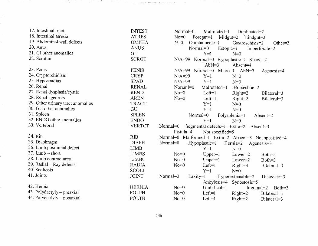
17. lntestinal tract18. Intestinal atresia19. Abdominal wall defects20. Anus21 . GI other anomalies22. Scrotum
23. Penis24. Cryptorchidism25. Hypospadias26. Renal27. Renal dysplasia,/cystic28. Renal agenesis29. Other urinary tract anomalies30. GU other anomalies31 . Spleen32. ENDO other anomalies33. Vertebral
34. Rib35. Diaphragm36. Limb positional defect37. Limb - short38. Limb contractures39. Radial Ray defects40. Scoliosis41. Joints
42.Hemia43. Polydactyly preaxial44. Polydactyly postaxial
INTESTATRESOMPHAANUSGISCROT
PENISCRYPSPADRENALRENDARENTRACTGUSPLENENDOVERTCT
RIBDIAPHLIMBLIMBSLIMBCRADIASCOLIJOINT
HERNIAPOLPHPOLTH
Normal:O Malrotated-l Duplicated:2No-0 Foregut:l Midgut:2 Hindgut:3N:0 Omphalocele:1 Gastroschisis-2 Other-3
Nomral:O Ectopic:1 Imperforate:2Y:1 N:O
N/A:99 Normal=O Hypoplastic-l Shawl:2AbN:3 Absent:4
N/A-99 Normal:O Mic¡o:l AbN:3 Agenesis-4N/A:99 Y:1 N:ON/A:99 Y:1 N:ONoraml:0 Malrotated-1 Horseshoe:2No:0 Left=l Right-2 Bilateral-3No:O Left:l Right:2 Bilateral:3
Y-l N-0Y:l N:O
Normal:0 Polysplenia-l Absent-2Y-1 N:O
Normal:O Segmentaldefects:1 Extra:2 Absent:3Fistula:4 Not specified-5
Nomal:O Malfomed:1 ExTræ:2 Absent-3 Not specified:4Normal-O Hypoplastic:1 Helrrix=2 Agenesis:3
Y:l N:ONo:O Upper:l Lowe=2 Both:3No:0 Upper:l Lower:2 Both:3No:O Left=l Right:3 Bilateral-3
Y-1 N:ONormal:0 Laxity:l Hyperextensible:2 Dislocate-3
Atrkylosis=4 Synostosis-5No:0 Urnbilical:l Inguinal-2 Both:3No-0 Left:l Righr:2 Bilaleral-3No:0 Left:l Right:2 Bilate¡a1:3
t46
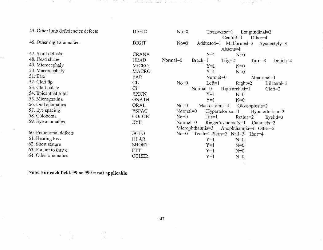
45. Other limb dehciencies defects
46. Other digit anomalies
47. Skull defects48. Head shape49. Microcephaly50. Macrocephaly51. Ears52. Cleft lip53. Cleft palate54. Epicanthal folds55. Mìcrognathia56. Oral anomalies57. Eye spacing58. Coloboma59. Eye ar.romalies
60.61.62.b-.t.
64.
DEFIC
DIGIT
CRANAHEADM]CROMACROEARCLCPEPICNGNATHORALESPACCOLOBEYE
ECTOHEARSHORTFTTOTHER
Ectodermal defectsHearing lossShor1 statureFailure to thriveOther anomalies
No-O T¡ansverse:l Lonsitudinal:2Cer.rtral=3 bthe=¿
No:O Adducted:l Malformed:2 Syrdactyl53Absent-4
Y-1 N:ONormal:0 Brach:1 Tig:2 Turri:3 Dolich:4
Y-1 N-0Y:1 N:ONomral:O Abnormal-l
No:0 Left:l Right:2 Bilateral:3Nomral-O High arched-l Cleft:2
Y:1 N:OY:l N:O
No:O Macrostomia:1 Glossoptosis-2Normal:0 Hypertelorism:l Hypoterlorism:2No-O Iris:l Retina:2 Eyelid-3Normal:O Rieger'sanonralpl Cataracts=2Microphthalmia:3 Anophthalmia:4 Other-5No:O Teetli=1 Skin:2 Nail:3 Hai=4
Y:1 N:OY:l N:OY:l N:OY:1 N:O
Note: For each field, 99 or 999 = not applicable
147
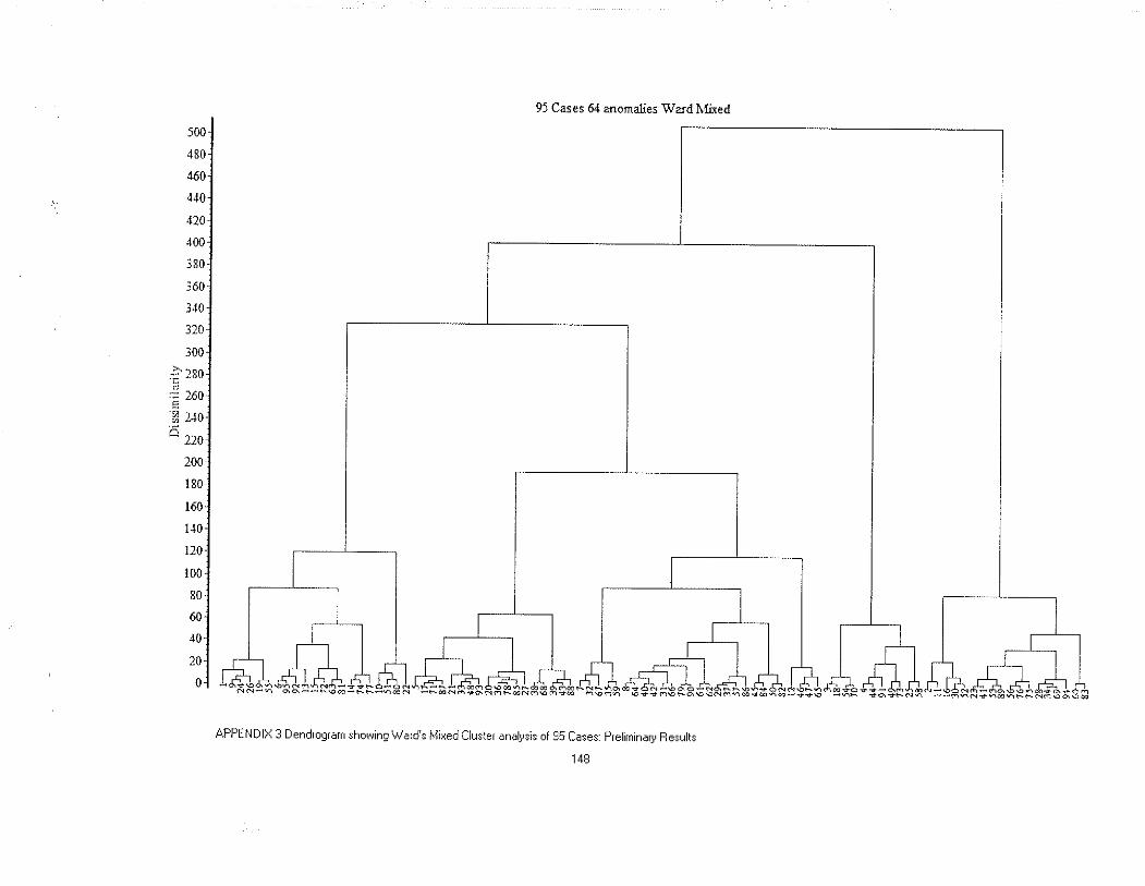
i00
480
460
4.10
,t?0
400
is0
i60
i40
3?0
i00ò -.r.so
EZ t'to'- ll0
100.
180
tó0.
1.r0-
t20-
t00:
801
ó0:
io,20i
0l
9J Cases 64 anornalies \\¡a¡d tr{ired
APPENDIX 3 Dendroqram shor+ing V,/ard's Mired Clr..rsier enaþsis of g5 Cases: preliminary Fesultr