patricia a" roth - mspace - university of manitoba
TRANSCRIPT
The University of Manitoba
COMPARISON OF THE VARTOUS IgE RECEPTORS OF
RAT BASOPHILIC LEUKEMIA CELLS
by
Patricia A" Roth
A Thesis
Submitted to the Faculty of Graduate Studies in
Partial Fulfillment of the Requirements for the
Degree of Doctor of Philosophy
Department of Immunology
l^linnipeg, l"lanitoba
July,1985
COMPARISON OF THE VARIOUS IgE RECEPTORS
OF RAT BASOPHILIC LEUKEMIA CELLS
BY
PATRICIA A. ROTH
A thcsis st¡b¡nitted to thc fiacrrltl, of Craduate Studies ol
the University oI Manitoba in partial fulfillnrent of the requirenretrts
of thc degree of
DOCTOR OF PIIILOSOPHY
o 1985
Permissio¡r has been grartted to tlte LIBRARY OF THE UNIVER-
SITY OF MANITOBA to lend or sell copies of this thesis. to
the NATIONAL LIBRARY OF CANADA to microfilnr this
thesis and to lend or sell copies of the film, and UNIVERSITY
MICROFILMS to publish an abstract of this thesis.
The author reserves other publicatio¡r rights, and neither the
thesis nor extensive extracts from it may be printed or other-
wise reproduced without the author's written permission.
i
TABLB UF CONTE}iTS
ACKNOhILEDGEi,iENTS
ABSTRACT
LIST OF FIGURES
LÏST OF ABBREVIATIONS
CHAPTER I
INTRODUCTIOI.I
IA] HISTORICAL PERSPECTIVE
1. Allergy and slcin sensit.izing anLibodies
4. Non-IgE homocytot,ropic antibodies
tB] BASOPFIILS AND MAST CELLS
i Biology of basophils and mast cells
2. Release of chemical mediators
Page i'{o.
v1
1
2
vl_l-1
10
2. Immunoglobulin E
3" Target organs, tissues and cel1s of reaginichypersensitivity reactions 4
6
a
tCl CELL SURFACE PiIEi'J0i'.{Ebi AND TRAi'lSi'iEi'fBR^Al'iE SIGì'í;\LS
1. Plasma rnembrane struct.ure 11
2. Ligand-recepLor interaclion .. ô o... o... o. o . Lz
3. Transduction of receptor mediated signals ...... Lz
tDl RECEPTORS FOR THE Fc PORTION 0F IItltlllOGLOBULIi{S
1. Receptors for IgG (l-cyR) on lymphocytes andmacrophages ....:.: ..... o.cooo3e o. 15
2. Receptors for IgPi (FcaR) c. o.. o c r. o e c. o.. o o..... 18
3. Other FcR.. ......o.o. 2ù
tEl IgE Fc RECEPT0II'S (FcsR)
f . i'iast cells and basophils 22
(a) Evidence for the localization of receptors:visualization on intact cells
{}'iI'if,i'}',
LJ
L1
(b) Number of receptors on target cells, kineticsand valency o. c e.. o.. o o.. o c o. o..... c.. o... o... 24
(c) Specificity of receptors ¿ö
2. Lymphocytes
3. l'lacrophages
4. Eosinophils
and monocytes
tF] PHYSICOCHEMICAL CHARACTERIZATION OF SOLUBILTZEDFcER FROM RBL CELLS AND MAST CELLS
1. Isolat,ion of receptors
2. The number of differenL receptor moleculesand their molecular weights
3. Biochemical nature of the receptor . r..... o.....
4. Antisera Lo receptors
5. Subunit composition of the receptors o.... c.....
SCOPE OF THE PRESENT INVESTIGATION
Jt)
31
34
35
37
4T
43
46
50
CHAPTER IIComparison ofof RBL Cell
Peptides Generated from R and H IgE Receptors
INTRODUCTION 52
¡{ATERIALS AND }ÍETHODS
Buffers 54
Affinit.y chromaLography vrith lgE-sepharose 58
AffiniLy chromatography with protein A-Sepharose e...... c 58
Polyacrylamide gel electrophoresis in SDS (SDS-PAGE) 5B
Rat Basophilic leukemia (RBL) cells
RBL cell iodination and disruption .......... o.. e. e. o o ô c o 55
Preparation of rat monoclonal IgE ...... c. o... c. ô o e.. o o o ô 55
Preparation of antisera 56
s4
Immunosorbgnts ........ o r.... o.....................,... .. 57
Limited proteolysis in the presence of SDS 60
RESULTS
-ffia
l- 11
tion of receptors from ì'{P-40-solubilizedsurface-labelled RBL cells
SDS-PAGE of affinity chromatography-purifiedsurface components from solubilized RBL cells
Comparison of papain-generated peptides fromR and H receptors .... .... o........ ...... o. ..... o...... ..
DISCUSSION
CHAPTER IIIProduction of lgE-Binding Fragmenls of Receptors from RBL Cells
INTRODUCTION
I',I.ATERIALS AND METHODS
Digestion of receptors in the presence of NP-40of SDS ........... o.......... o.. .... o o......... 72
Digestion of RBL cell extract t,o produce anIgE-binding fragment of IgE receptors . o. o...... e..... c.3
RESULTS
62
63
64
66
72
70
75
76
B5
DigestionNP-40 and
ofSDS
R and H recept,ors in the presence of
Isolation of lgE-binding fragments of receptors
DISCUSSION
CHAPTER IVDetermination of theReceptors
80
Relationship of 71K to the R and H
INTRODUCTION
MATBRIALS AND METHODS
Preparation of anti-IgE-Sepharose (HARE-Sepharose)immunosorbgnt, ....o......................... ........SequentialSepharose
affinity chromatography using HARE-and ïgE-Sepharose
SDS-PAGE On Slab gelS c............. o.. o.................
digestion of receptors in polyacrylarnideticTrypgels
87
87
87
88
8BOne-dimensional analysis of tryptic digests
l-v
Two-dimensional analysis of tryptic
Biosynthet.ic labelling of RBL cells
Repetititve Affinity chronatography
digests
Analysis of purified receptors .... .......,... ... c. . c. . ..
Autoradiography o . . .. . . . . .. . . . . .. . . .. ... . . . .. . o . . . . . c c. . e
RESULTS
Isolation of R, H and 71K receptors using sequentialaffinity chromatography ........ o..... o.... ... c..
Analysis of tryptic peptides
a) One dimensional analysis
b) Trvo dimensional analysis
8B
B9
89
90
91
o')
94
95
96
of receptors
IsolaLion of purified receptors by repetitive affinitychromatography ... .. . .. .. . . ... ... .. .. . ... . .. . . o . . . . . . . . . . 98
Two dimensional SDS-PAGE analysis of purified receptors 100
DISCUSSION 103
GENERAL DISCUSSION LT2
BIBLIOGRAPHY r23
v
ACKNOITTLEDGEMENTS
I wish to express my sincere gratitude and heartfelt thanks Lo
Dr. Arnold Froese for his guidance and support throughout, the course of
this investigation and for his assistance and construcLive criticism in
the preparation of this thesis.
I also wish to thank ì"1s. Karol ÞlcNeill for her fine technical
assistance.
Finally, I owe a special thanks to rny husband David and my son
Aaron, without whom this endeavor would have been less neaningful.
vt_
ABSTRACT
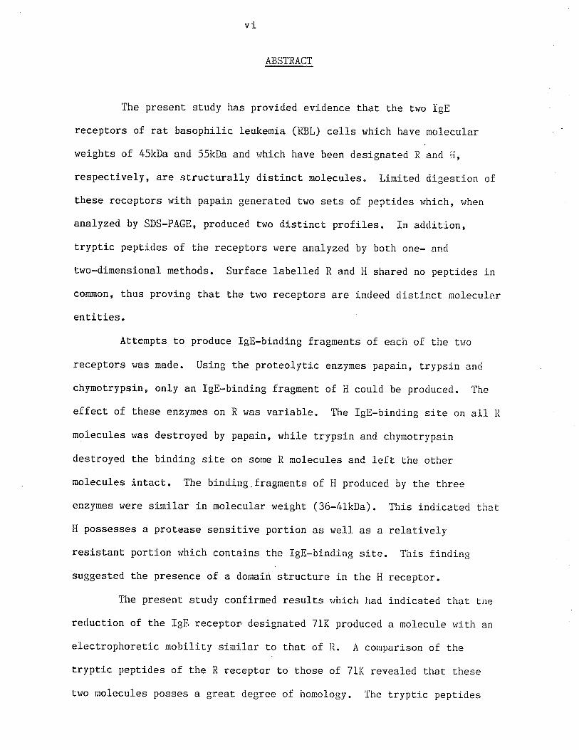
The present. study has provided evidence that the Lrvo IgE
recept.ors of rat basophilic leukemia (RBL) cells rvhich have rnolecular
weights of 45kDa an<l 55kDa and which have been designateci iì ancl i-i,
respectively, are structurally distinct molecules" Limited digestion of
these receptors rvith papain generated two sets of peptides vrhich, when
analyzed by SDS-PAGE, produced two distinct profiles. In addition,
tryptic peptides of the receptors r'¡ere analyzed by both one- and
two-dimensional met,hods. Surface labelled R and H shared no peptides in
conmon' thus proving that the tr¡o receptors are i-ndeed distinct molecular
ent,it.ies.
Attempts to produce fgE-binding fragments of each of the tiyo
receptors was made. Using the prot.eolytic enzymes papain, trypsi-n ancf
chymoLrypsin, only an lgE-binding fragment of H could be produced. The
effect, of these enzymes on R rvas variable. The lgE-binding sit.e on all ìl
molecules was destroyed by papain, rvhile trypsin and chymotrypsin
destroyed the binding site on some R molecules and left the other
molecules intact. The binding.fragments of H produced by the t.hree
enzymes r'¡ere similar in molecular rveight (36-41kDa). This inriicated that
I{ possesses a protease sensitive porLion as v¿e1l as a relatively
resistant portion rr¡hich conLains the IgE-binding sj-te. This finding
suggested the presence of a domain strucLure in the li receptor.
The present study confirmed resulLs r'¡hich had indicated that the
reduction of the IgE receptor designated 7lK produced a molecule rvitir an
electrophoreLic mobilit.y similar to that of Iì. A comparison of the
t.ryptic peptides of the R recept.or to those of 71K revealed that these
tvro molecules posses a great degree of hornology" Ihe tryptic pepticles
v11
which r'rere produced from both reduced and unreduced R anri 71K v¡ere
analyzed by one-dimensional SDS-PÀGE. The results clearly sho.,red a great
deal of similarity between t,hese two molecules, both in tire reduced anci
unreduced forms. Trvo dimensional analysis demonstrated that wilile not
all pepLides from R and 71K had t,he same rnobilities, many did share a
conmon position. These results suggesLed that 71K is conpose<i of tire R
molecule disulfide-bonded, either t.o anot,her R molecule or to some other
polypeptide chain r'ririch is noL surf ace labelled "
A two-dimensional SDS-PAGE analysis of recepLors from
biosynthet.ically 1abel1ed RBL cells rvas performed to deternine the
subunit composition of 7lK" Reduction of receptors in the second
dimension denonstrated that, aside from the molecule rvith a mobility
similar to t.haL of R, no other nolecule was produced upon recluction of
71K" This strongly suggested that 71K is composed of Lwo,
disulfide-linked R molecules.
V I1I
LIST OF FIGURES
Fig. 1 Isolation of the IgE receptors from radiolabelled rat basophilic
leukemia (RBL) cells. p. 62a
Fig. 2 Calibration curve for the SDS-PAGE system according to Laemmli
(1970) using 10% polyacrylamide tube gels. .. p. 63b
Fig. 3 SDS-PAGE analysis on 102 gels of surface labelled RBL cell
components bound by lgE-Sepharose. p.63d
Fig. 4 SDS-PAGE analysis on 10% gels of surface lal¡elled RBL cell
molecules isolated by IgE and HARE and RAR. p. 64b
Fig. 5 Calibrat,ion curve for the SDS-PAGE system using 15% tube gels.p. 65b
Fig. 6 SDS-PAGE analysis on L57" gels of papain generated peptides of R
and H receptors. p. 65d
Fig.7 SDS-PAGE analysis (on 152 gels) of surface labelled RBL cell
componenLs bound to lgE-Sepharose and eluted in the presence of
SDS or KSCN. p.76b
Fíg. 8 SDS-PAGE analysis of papain digest of receptors digested in t.he
presence of eiLher SDS or NP-40" p. 76d
Fig.
Fig.
Fig "
Fig.
Fig.
9
10
11
L2
13
SDS-PAGE analysis (on.10% gels) of papain generated lgE-binding
fragments bound by lgE-Sepharose. ... o................. p. 77b
SDS-PAGE analysis of papain generaLed lgE-binding fragments
bound by IgE and HARE and RAR" c............. o.... o.. o. p. 77d
Two dimensional tryptic -þeptide maps of papain-generated
IgE-binding fragent compared to peptide map of H. . r... p. 78b
SDS-PAGE analysis of IgE-binding receptor fragments produced by
tryptic digesLion. .. o........ ô o ....... p. 79b
SDS-PAGE analysis of lgE-bínding receptor fragments produced by
chymotrypLic digestion. o c.... o ô. o............. o.... o o. p. 79d
Fig.
Fig"
Fig.
I4
15
I6
ixSDS-PAGE analysis of reduction of IgE receptors. o..... p. 92b
SDS-PAGE analysis of reduced 71K receptor. o po 92d
SDS-PAGE analysis of receptors isolated using preparative
Fig.
Fig.
sequential affinity chromatography. pn 94b
17 SDS-PAGE analysis of reduced receptors. p. 94d
18 SDS-PAGE analysis of tryptic peptides of receptors on 15% gels.
p. 95b
Tr,¡o dimensional analysis of tryptic peptides of H and R. p.96b
Two dimensional analysis of tryptic peptides of R and 71K.
... . o . . . p. 97b
Two dimensional analysis of tryptic peptide of reduced R and
71K. o. . . o . . . o . .. . .. . . . . . . . . . . . .. . . . .. . o o . . . . . . .. . . o . o . p. 97d
Flow chart of purification of 3H-receptors by repetitive
affinity chromatography. ......o........eo.......o...e. p" 99a
One-dimensional SDS-PAGE analysis of receptors purified by
Fig. L9
Fig. 20
Fig. 2L
Fig. 22
Fig. 23.
repetitive affinity chromatography. p. 100b
Fig. 24 Two dimensional SDS-PAGE analysis of 1251-tabe1led receptors.
p. 102b
Fig. 25 Two dimensional SDS-PAGE analysis of 3H-1.be11ed recepLors.
. a a. a.. e........ t...... e o.. e o... o.. o o..4. a..... " c " .. pt 102d
x
BSA
CNBr
CPI'I
CT}1C
DAAD
DNP
DTT
EDTA
FcR
FCS
HAiìE
kDa
2-ME
MEM
r'fl4c
ì'lrNP-40
nRGG
PBS
PCA
P-K
PI'ISF
RAR
RBL
lìr
R-r{C
LIST OF ÀBEREVÏATTOI{S
bovine serum albumin
cyanogen bromide
counts per minute
connective tissue mast cell
direct allogenic anaphylacLic degranulation
diniLrophenyl
dithiothrietol
et.hylene diamine tetraaceLate
receptor for Fc port.ion of Ig
fetal calf serum
horse anti-rat IgE
kilodaltons
2-mercaptoethanol
minimum essential medium
mucosal mast ce1l
apparent molecular weight
Itlonidet P-40
IgG fraction from normal rabbit serurn
phosphate buffered saline
passive cutaneous anaphylaxis
Prausnitz-Kustner
phenyl methyl sulfonyl chloride
rabbit anti-receptorimmunoglobulin
rat basophilic leukemia
relat,ive mobility
raL mast cel1
x1
SC secretory component
SDS sodiurn dociecyl sulfate
SDS-PAGE polyacrylamide gel electrophoresis in sodium <iodecyl sulfate
SRS-Â slow reacting substance of anaphylaxis
TLC thin layer chromatography
CHAPTER T
II'.¡TRODUCTION
tAl HTSTORICAL PEF.SPH,CTIVE
1. Àllergy and skin sensitizing antibodies
The presence of a humoral facÈor able to transfer specific
allergic sensitivities from allergic to non-allergic patlents t.¡as first
demonsLrat,ed by Frausnitz and Kustner (1921). In their study, Prausnit.z
sensitized his or,'n forearm to fish by inLracutaneous injection of serun:
from an allergic individual. Upon challenge rr¡ith the specific antigen,
tl-re skin site gave an erythema-wheal reaction, a symptom indicating the
presence of tire tissue injury commonly elicited during an allergic
response. Subsequently, the passi-ve transfer of immediate
hypersensitivity to other allergens r'¡as observed and, in L925, Coca ancl
Grove (1925) applied the term rrreaginrf Lo the skin-sensitizing
antibodies found in allergic sera. Investigation of the nature of
reaginic antibodies then had to await the development of nerv methods for
fractionation of serum proteins"
Improvement of protein separation Lechniques allor.¡ed ior the
classificat,ion of antibodies into specific classes and subclasses based
predominantly on distinct antigeníc characteristics as r.¡ell as on net
charge and molecular neight. It r,¡as establÍshed that, regardless of size
and charge, antibodies share a 4-polypeptide chain strucLure consisting
of two heavy and trvo light chains-. The structures of the heavy chains
differ among classes and subclasses while all share conmon light chains.
By the early 1960rs, use of the nevrly developed methc¡d of
immunoelectrophoresis aided in the discovery of three classes of human
serum immunoglobulins. IgG and lgl-f were found to correspond to the 73-
and l9S-type irnmunoglobulin(Ig) antibody, respectively, vrhereas IgA
-2-represenLed a nerv Ig class baserl on it.s antigenic properties. After the
properties of this nerv class of Ig rvere examined, its precipitation and
elecLrophoretic characteristics suggesteci a resernblance to reaginic
anlibody. Furthermore, Iieremans and Vaerman (1962) demonstrated thaL the
IgA fraction of allergíc patientsf sera had skin-sensitizing activity,
r,rhile antiserum agaínst IgA destroyed reaginic activity (Fireman eL al.,
i963). Finally, Ishizaka el- al. (i963) shor,'ed that the IgA fraction of
normal seïurn could block the passive sensitizalion of human skin witlr
reagin but neither IgG nor lgi'í would do so. IJor,rever, by the late l9tiOfs,
further fractionation of tpuret IgA from reaginic serum revealed thal
reaginic activiLy <lid not parellel IgA concent.ration (Ishizaka and
rshizaka, L966). The negaLive result.s obtained from experiment.s on
blocking of passive sensitization by myeloma IgA proteins insteacl of
normal serum IgA fractions strongly suggested that reaginic antibociies <1o
not belong to the IgA subclass but represent. a contaminant in many IgA
fractions. Upon discovery of yet another antigenically distinct Ig class
- IgD - evidence for its relationship to reaginic antibodies was sougÌrt,
but Lhe data obtained (based on immunochemical and pirysícochemical
properties) did not. support such a relationship.(Ishizaka et al., I966a,
Perelmutter et al., 1966). In addition, because of its size (8S) and rhe
antigenic differences (Ishizaka et al., L967a), reaginic ant.ibody r,ras not
considered to be of the Iglvl class. Upon ruling ouL identity betrueen
reaginic antibodies and IgG antibodies (Ishizaka eL a1., Lr)67a), the
search was begun for a unique'immunoglobulin class which had not yet been
described.
2" Immunoelobulin E
The first attempt to identify the protein responsible for
-3*
reaginic activity bras nade with antibodies to a reagin-rich fraction from
ragweed-sensitive serum. After absorption r¿ith normal IgG and IgA and
IgD myeloma proteins, the antiserum reacting with the reagin-rich
fraction showed a single band which, upon imnunoelectrophoresis, had a
fI mobility. Binding of radioactively labelled ragweed antigen to this
band demonstrated that it contained antibody binding act,ivity" This
apparently unique imnunoglobulin class was tent,atively designated rE
(Ishizaka et al., 1966b). Further studies revealed a correlat,ion between
the disappearance of skin-sensit,izing acLivity in allergic sera and
removal of xE by imnunoprecipitation with anLi-ÌE in the ragweed system
(Ishizaka et a1., 1966c) as well as in other allergen systems (Ishizaka
and Ishizaka, 1968)" Purification of ¡E (from reaginic sera of
ragweed-sensitive patients) using ammonium sulfate precipitation and
DEAE-cellulose chromat,ography and absorption with anLisera to other
immunoglobulin classes provided more conclusive evidence for t.he identity
of this protein and reaginic antibody. This purifÍed preparation
contained only lE antibodies specific to ragweed, as determined by
radioimmunodiffusion, and both.reaginic activity and lE were completely
removed by precipitation with anti-tE (Ishizaka and Ishizaka, 1967) "
These results clearly show that ðE antibody is responsible for reaginic
activity.
The extremely low levels -of UE normally found in humans would
have made investigation of its biochemical and biological properties very
difficult. Fortunately, Johansson and Bennich (1967) discovered a human
rnyeloma protein, IgND, which, by antigenic analysis using antisera to
both ND and Í-E globulin, seemed to be structurally similar to the
protein previously described by the Ishizakas (Bennich et a1., 1969).
-4-
Biological evidence for the structural relationship between IgND and
reaginic antibody r.¡as obtained by specifically inhibiting the P-K
reaction with the myeloma protein ND (StanworLh et al., 1968).
Physicochemical analysis of protein ND revealed a carbohydrate content of
about, II7", a sedimentaLion constant of 7.9S, and an apparent molecular
weight of 196kDa. Reduct,ion and alkylation produced 2 cornponents, the
molecular weights of which indicated the presence of the 2 heavy and 2
light chains common to all other imrnunoglobulin classes (Bennich and
Johansson, L97L). In conformity with immunoglobulin nomenclat.ure, lE is
now designated IgE"
Digestion of human IgE by papain produces Fab and Fc fragments
(Bennich and Johansson, I97I), and Stanworth et al. (1968) found that
only the Fc fragment was required to inhibit the P-K reaction. Heating
of the IgE molecule at, 56 C for four hours ínactivates iLs
skj"n-sensitizing ability (Ishizaka et al., 1970a), as does reduction with
0.lM mercaptoethanol followed by alkylation (Ishizaka and Ishizaka,
1969a)" A more detailed study performed by Takatsu et al. (1975) on a
nyeloma IgE protein (PS) indicated t,hat cleavage of one of the two
inter-heavy-chain disulfide bonds is required for the complete loss of
biological activity. Furthermore, IgE molecules do not fix complemenL to
activate complement by the classical pathway, even in an aggregated form
(Ishizaka et, al., 1970b), although aggregated IgE does activate
complement by the alternate pathway (Ishizaka et al., L972b)"
3. Target organs, tissues and. cells of reaginic hypersensitivity
reactions
The elicitation of a P-K reaction with reaginic antibodies
demonstrated that skin is one of the organs susceptible to the
tr
hypersensit.izing effects of IgE" In monkeys, human reaginic serum
produced a passive cutaneous anaphylaxis (PCA) (Layton et a1., 1962) and
this sensitizing acLivity was found only in a fraction containing IgE
(Ishizaka et 41., 1967b). In addition, Arbesman et al. (1964) reported
that monkey ileum, passively sensitized in vitro with human reaginic
serar contracted upon exposure to allergen. Further examinaLion of this
phenomenon showed thaL an fgE-rich ction of reaginj.c serum sensitized
the ileum and that this activiLy was removed by anti-IgE (Wicher eL al.,1968).
One of the most extensively studied organs in relation to
reaginic hypersensit,ivity is the lung. Goodfriend et al. (1966)
demonstrated that in monkey lung sensit,ized with hunan reaginic serum,
challenge rvith antigen induced histamine release. Another mediator of
hypersensitivity, SRS-A, was released from human asthmatic lung upon
challenge $¡ith allergen (Brocklehurst, 1960). It was subsequently shown
that IgE was the agent responsible for sensiLizing monkey lung for Lhe
release of both histamine and SRS-A (Ishizaka et, a1., 1970c). Thus itbecame apparent that reagínic hypersensitivity reactions involved the
effect, of IgE on some componenL(s) of the various tissues studied.
At the cellular level, the mast cell became the prime candidate
for the target of reaginic antibody. Tn L967, Parish sensitized human
lung slices with human, heat-tabile antibodies and was able to
demonst,rate a correlation between histamine and SRS-A release and
morphological changes in the mast cells. Tomioka and Ishizaka (1971)
found that the major cell type in monkey lung which binds IgE is the mast
cell. This study also demonstrated such binding to masL celIs in skin,
omentum and the lamina propria of the small intestine of monkeys"
-6-
While Lichtenstein and Osler (L964) established the methodology
for antigen-induced histamine release from peripheral blood leukocyt.es in
L964, it was not until after IgE was characterized that evidence for itsbinding to the target cell was obtained. Ishj.zaka et al. (1969) showed
that leukocytes of atopíc patients (and some normal individuals) released
histamine upon Èreatnent, wiLh anti-IgE, which indicated that IgE was
indeed bound to some of these cells. The identity of this Earget ce1l
was det,ermined when radiolabelled IgE was demonst,rated on t.he surface of
basophil granulocytes but not on oLher leukocytes (Ishizaka et. al.,1970d). In addition, incubation of radiolabelled anti-IgE or anti-IgG
with cell suspensions from passively sensitized monkey lung resulted in
the binding of anti-IgE to mast cells, whereas anti-IgG combined only
wiLh nacrophages and neutrophils (Ishizaka et a1., L972a). Thus, the
binding of IgE to basophils and mast cells, which are the major source of
histamine in Lhe tissues and b1ood, suggested that ant,igen-antibody
reactions on these cells result in the release of histamine from Lhese
cel1s.
4. Non-IgE homocytotropic antibodies
' Although the IgE class of antibody clearly fits the definition of
homocytotropic antibodies, which are immunoglobulin molecules capable of
binding to cells of the same or closely related species, it has become
apparent that, iL is not the only class with such a capacity. IgG
passively sensitizes human lung and skin tissues, although the results
have been inconsistenL" These variable results appear Èo be due to the
ease with which the antibody ís washed off (Parish, 1978). Sensitization
appears to be short Èerm, lasting onLy 2-6 hours. The subclass
responsible for this activity is unknown, although in one study,
-7*
Stanworth and Smith (1973) found a myeloma IgGO that blocked
sensitization of skin by IgE. Providing more direct evidence for
binding, Ishizaka et al. (1979) demonstrated that human basophil
granulocytes whj-ch had been incubaLed with aggregated IgG could bind
radiolabelled anLi-IgG and that the receptor that bound IgG was distinct
fron IgE receptors on the same celIs.
In Lhe rat, a non-IgE anLibody belonging to the IgG class,
capable of inducing a PCA reaction with a 2-6 hour latent periodr and
generating the release of histamine and SRS-A, has been identified. This
antibody is recognized as belonging to the TgGZa subclass (Stechschulte,
1978). In addition to eliciting a PCA reaction, TgGZais involved in Ehe
conplement dependent release of SRS-A from neut,rophils in the peritoneum
(Orange et a1., 1968). Studj"es on the mast ce1l by Bach et al. (1971)
revealed ÈhaÈ IgE could inhibit l8Gr"-induced PCA reactions and that
IgG2u inhibited IgE sensitization of mast cells in vitro, indicating that
int,eractj.on of these two immunoglobulin classes with the Larget cell
involves a common receptor. l"Iore recently, Halper and Metzger (1976)
used a direct binding inhibitiqn assay to demonstraLe that only lgGru in
immune complexes could inhibit the binding of IgE to rat basophilic
leukemia(RBl) ce1ls. Monomerie IgG was not effective. This led the
authors to suggest, that the affiniEy of the receptor for IgG was much
lower than that for IgE and that -only multi-point attachment of inmune
complexes allows for inhibitíon. However, further st.udies have since
shor+n that RBL cells carry tr¿o kinds of Fc receptors, one having a higher
affinity for IgG than the other (Segal et a1., 1981, Kepron et 41.,
1982).

-8-
tBl Basophils and Mast, Cells
1. Biology of basophils and mast cel1s
In Èhe late 19th century, Lwo cell populations were described
which contained prominent cytoplasmic granules with an affinity for
certain basic dyes. One cell, the basophil, circulated in the blood
while the mast cell resided in connective tissue. Both cell types are a
major source of the bioactive amines involved in a wide spectrum of
i-nflamnatory and immunologic processes. They also express plasma
membrane receptors thaL specifically bind the Fc portion of IgE antibody
with high affinity and, upon sensitization with IgE and subsequent
challenge with ant.igen, undergo anaphylactic degranulation which releases
chemical mediators into the external medium. More recent evidence has
implicat,ed both basophils and mast cells in certain cellu1ar j.mmune
processes r¿hich occur through non-anaphylactic degranulation (Galli et
al., 1984).
DespiÈe these similarities, basophils and mast cells are clearly
dist,inct populations. Basophils, like other granulocytes, differenLiat.e
and mature in the bone marrow, .and circulate in the blood. Mast cells
are dist,ributed throughouL connective tissues, often situated around
blood and lymphatic vessels and beneath epithelial surfaces. They mature
locally in connective tissue from precursor cells which contain few
cytoplasmic granules (Combs et aT., 1965). In viLro experiments by
Ginsburg and Lagunoff (L967) were Lhe first to provide evidênce that mast
cell precursors are present in mouse thymus and other lymphoid Lissue
although it, has been demonst,rated more recently that rat mast cel1s are
derived from bone marrow, not thymus (Crowle and Reed, 1984)"
Morphologically, basophils generally contain electron dense aggregates of
-9-
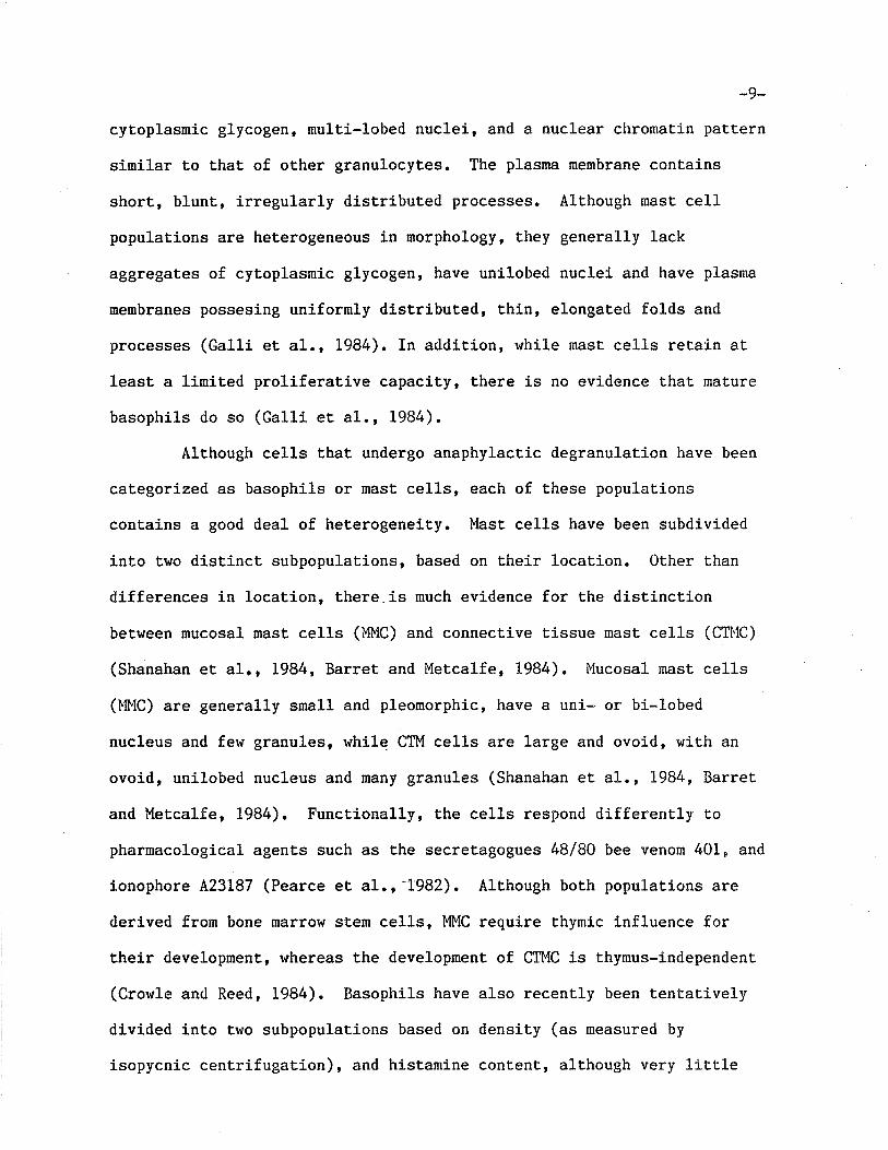
cytoplasmic glycogen, multi-lobed nuclei, and a nuclear chromatin pattern
similar Lo that of other granulocytes. The plasma membrane contains
short, blunt, irregularly distributed processes. Although mast cell
populations are heterogeneous in morphology, t,hey generally lack
aggregates of cytoplasmic glycogen, have unilobed nuclei and have plasma
membranes possesing uniformly distributed, thin, elongated folds and
processes (Galli et a1., 1984). In addition, while mast ce1ls reLain at
least a limited proli-feraEive capaciLy, there is no evidence that r¡ature
basophils do so (Galli et al., 1984).
Although cells that, undergo anaphylacLic degranulat,ion have been
categorized as basophils or mast cells, each of these populations
contains a good deal of heterogeneiLy. Mast cel1s have been subdivided
into two disLinct subpopulations, based on Lheir location. 0ther than
differences in location, there.is ¡nuch evidence for the distinction
between mucosal mast ce1ls (MMC) and connective tissue mast cells (CTl'lC)
(Shanahan et al,, 1984, Barret and Metcalfe, 1984). Mucosal mast cells
(l'MC) are generally small and pleomorphic, have a uni- or bi-lobed
nucleus and few granules, while CTM cells are large and ovoid, with an
ovoid, unilobed nucleus and many granules (Shanahan et 41., 1984, Barret
and l{etcalfe, 1984). Functionally, the cells respond differently Lo
pharmacological agents such as the secretagogues l+8/80 bee venom 401, and
ionophore A23L87 (Pearce et al.r -1982). Although both populations are
derived from bone marrow stem cells, MMC require thymic influence for
their development,, whereas the development of CTMC is thymus-independent
(Crowle and Reed, 1984). Basophils have also recently been tentatively
divided into two subpopulations based on density (as measured by
isopycnic centrifugation), and histamine contenL, although very lítt,le
-10-
other data is available at this time (Barret and Met.calfe, 1984).
fn recent years, methods have been developed for the long-term
culture of mast cells. Nabel et a1. (1981) found that mouse mast ce1l
clones cult.ivated from fetal liver, required for their survival a.
nacromolecular growth factor which was derived from LyI+z'T cells.
Although the identity of Èhis factor has not yet been det,ermined, itshares some characteristics with the lymphokine called interleukin-3
(Galli, et al., 1984). The dependence of these cult.ured cells on a
T-cell derived factor may have some relevance to the dependence of.
mucosal mast cell proliferatj-on on thymic influence (Ruitenberg and
Elgersma, L976). Human basophils have now been cultured as well.
Ishizaka et al. (1985) were able to maintain cord blood basophils in
culture f.or 2-3 weeks and demonsÈraLed that basophils from these cultures
were functionally matr¡re (as determined by histamine content). These
developments facilitate the study of cells which had previously been
difficult to obtain in large numbers.
2" Release of chemical mediators
Imrnediate hypersensitivity reactions are initiated by the binding
of ant,igen to basophil and mast cel1-bound IgE. This event ultimately
leads to the secretion of a variety of mediators which are responsible
for the pathology of the react,ion. These mediators include preformed
secretory granular component,s sudh as histamine, serotonin, heparin
proteoglycan and the chondroitin sulfates, as well as ner"rly generated
mediators (from mast cel1s) such as prostaglandin D' platelet-activating
factor and leukotrienes (Galli et al., 1984). The cellular process
involved in the release of granular components is termed noncytolytic
degranulation. A two sËage process begins wiLh the fusion of membranes
_1 1_
surrounding Lhe granules to the plasma membrane (and sometimes t.o each
other) so that granules are exposed to the cell exterior and concludes
r+ith the release of heparin-bound hisÈamine by a process of cation
exchange (Thon and Uvnas, L967, RohlÍch et al., 1971). In basophils, the
mucopolysaccharides chondroitin sulfates A and C appear to replace
heparin as the histamine-binding cation-exchanger (Sue and Jacques,
L974). Mucosal mast, cells also contain a chondroitin sulfate-like
glycosarninoglycan instead of heparin (Tas et al. , Lg77). Both cell types
remain viable after degranulation and indeed, undergo regranulation in
anticipation of another exocyt.otic event (Galli et al., 1984, Burwen,
1982).
tcl 11 f and Transmembrane Si 1s
1. Plasma membrane structure
The first important hypothesis on the structure of biological
membranes, proposed by Davson and Danielli in L932, and later modified
into the unit-menbrane hypothesis, features a bilayer of mixed polar
lipids, with their hydrophobic.hydrocarbon ends oriented inward and their
hydrophilic heads pointed outward. In L972, Singer and Nicolson
introduced the fluid-mosaic model. This model postulat.ed that the
phospholipids of membranes are arranged in a bilayer to form a fluid,
liquid-crystalline core in which lipid molecules can move laterally.
Intrinsic proteins are partially or completely embedded in t,he membrane,
depending on their hydrophobic amino acid orientation, while extrinsic
proteins are associated with the membrane by ionic or hydrogen binding
forces. Many membrane proteins which are anchored in Ehe membrane by
hydrophobic interactions contain oli.gosaccharide side chains facing the
-r2-outside of the cell. Some proteins may traverse the bilayer or,
alternatively, form part of a complex which t,raverses it and therefore is
in contact v¡iLh both the external and cytoplasmic sides of the membrane.
These molecules can provide the means of communication across the lipid
bilayer either through the flow of certain solutes or by transducing
trsignalstr from ttmessengersrt such as hormones, neurot,ransmit,ters
antibodies or other cells. Thus, membrane proteins (often glycoproteins)
play a specific role in the response of cells to environmental stimuli.
The response involves three types of events: first, recognition (specific
binding) of an extracellular molecule by the receptor protein; then a
transduction of a signal across the membrane leading to inÈeractions
between the nembrane and the cytoplasmic milieu which, ultimately,
results in a cellular response that modulates Lhe environment.
2. Ligand-receptor inLeraction
By definit,ion, a lrue receptor is a distinct, molecular entiLy
whose function is t.o bind an endogenous ligand and thereby achieve a
physiological effect (Goldstein et al., L979). The receptor should
normally be a product of the cell itself and the binding of specific
ligand to the receptor site should be a necessary but not necessarily a
sufficient step in the sequence leading to changes in the ceIl. The
receptor nrust further meet the qualifications of (i) saturability, i.en
the cell should possess a finite-number of sites which can be filled at
trigh ligand concentrations; (ii) specificit,y, i.e. the receptor should
demonstrate a preference for'binding of a known effector compared to
negative control; and (iii) it must demonstrate a high affinity
interaction (Dorrington, 1976).
3. Transduction of receptor mediated signals
_13_
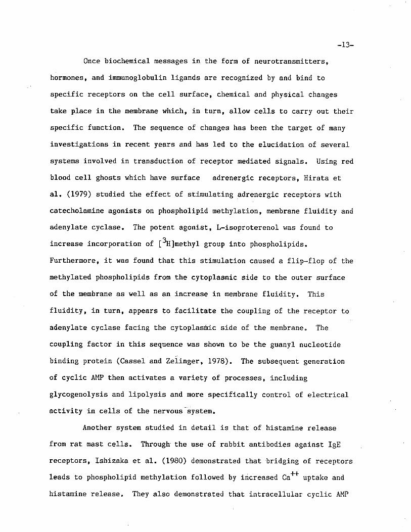
Once biochemical messages in the form of neurotransmitters,
hormones, and immunoglobulin ligands are recognized by and bind to
specific receptors on the cell surface, chemical and physical changes
take place in the rnembrane which, in Lurn, allow cells to carry out their
specific funct,j.on. The sequence of changes has been the target of nany
investJ-gations in recent years and has led to the elucidaLion of several
systems involved in transduction of receptor mediated signals. Using red
blood cell ghost,s which have surface adrenergic recepcors, Hirata et
al. (L979) studied the effect of stimulating adrenergic receptors with
catecholamine agonists on phospholipid methylation, membrane fluidity and
adenylate cyclase. The potent agonist, L-isoproterenol was found to
increase incorporation of [3H]methyl group into phospholipids.
Furthermore, it was found that. this stimulation caused a flip-flop of the
meÈhylated phospholipids from the cytoplasmic side Lo Lhe outer surface
of the menbrane as well as an increase in membrane fluidity. This
fluidity, in turn, appears to facilitate the coupling of the receptor to
adenylate cyclase facing t.he cyt,oplas¡iric side of the membrane" The
coupling factor in this sequence was shown to be the guanyl nucleotide
binding protein (Cassel and Zeiinger, 1978). The subsequenL generation
of cyclic AMP then activates a variety of processes, including
glycogenolysis and lipolysis and more specifically control of electrical
activity in cells of the nervous -system.
Another system studied in detail is t,hat of histamine release
from rat masL cells. Through'the use of rabbÍt antibodies against IgE
receptors, Ishizaka et al. (1980) demonstrated thaL bridging of receptors
leads Lo phospholipid methylation followed by increased Ca# uptake and
histamine release. They also demonstrated that int.racellular cyclic AMP
-r4-
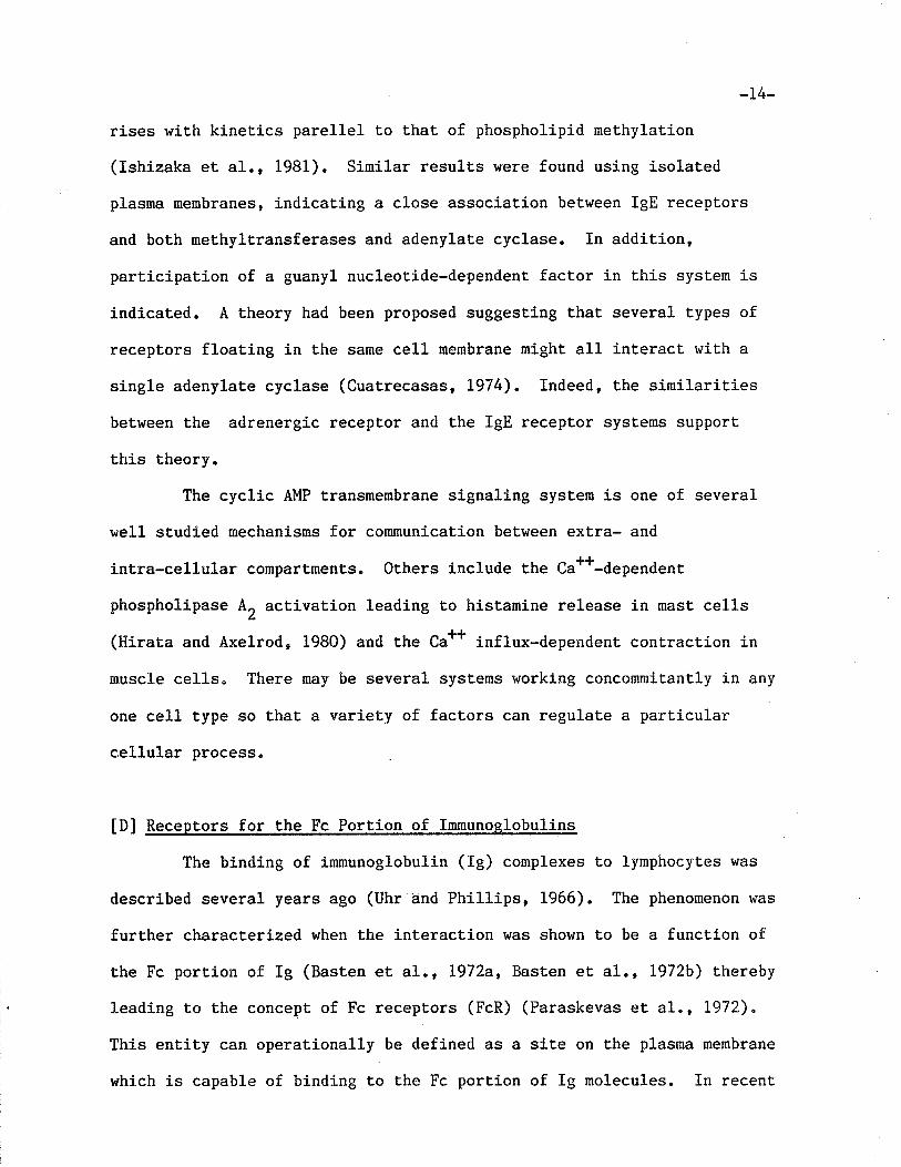
rises with kinetics pare11el to that of phospholipid methylation
(Ishizaka et al., 1981). Similar results were found using isolat,ed
plasma membranes, indicating a close association between IgE receptors
and both nethyltransferases and adenylate cyclase. In addit.ion,
participation of a guanyl nucleotide-dependent factor in this system is
indicated. A theory had been proposed suggesting that several types of
receptors float.ing in the same cel1 membrane might all interact with a
single adenylate cyclase (Cuatrecasas, L974). Indeed, the similarities
between the adrenergic receptor and the IgE receptor systems support
this theory.
The cyclic AMP transmembrane signaling system is one of several
well studied mechanisms for communication between extra- and
íntra-cellular comparLments. Others include the Ca#-dependent
phospholipase A, activation leading to histamine release in mast cells
(Hirata and Axelrod, 1980) and the Ca** influx-dependent contraction in
muscle cells" There may be several systems working concorunitantly in any
one cell type so that, a variety of factors can regulate a parLicular
cellular process.
tDl Recep tors for the Fc Porti.on of Immunoslobulins
The binding of imrnunoglobulin (Ig) conplexes Lo lymphocytes vras
descrj-bed several years ago (Uhr-and Phillips, 1966). The phenomenon was
further characterized when the interaction was shown to be a funcLion of
the Fc portion of Ig (Basten et a1., L972a, Basten et 41., L972b) thereby
leading to the concept of Fc receptors (FcR) (Paraskevas eL a1", L972)"
This entity can operationally be defined as a site on the plasnna membrane
which is capable of binding to t,he Fc portion of ïg molecules. In recent
*15-
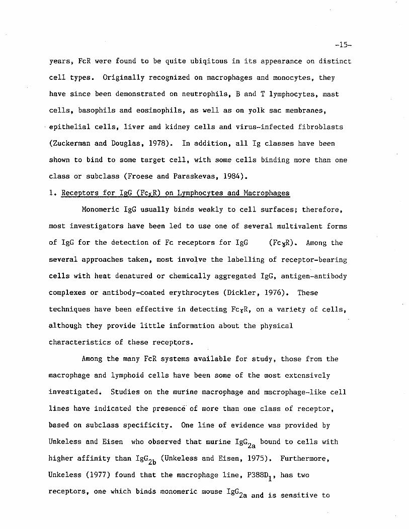
years, FcR were found to be quite ubiqitous in it,s appearance on distinct
cell types. Originally recognized on macrophages and monocytes, Lhey
have sj-nce been demonstrated on neutrophils, B and T lymphocytes, mast
cells, basophils and eosinophils, as well as on yolk sac membranes,
epithelial cells, liver and kidney cells and virus-infected fibroblasls
(Zuckerman and Douglas, 1978). In addition, all Ig classes have been
shown to bind to some target ce1l, wÍth some cells binding more than one
class or subclass (Froese and Paraskevas, 1984).
1. Receptors for IeG (FcrR) on Lymphocytes and Macrophages
Monomeric IgG usually binds weakly to cell surfaces; therefore,
most investigators have been led to use one of several mulrivalent. forms
of IgG for the detect,ion of Fc receptors for IgG (Fc6R). Among Lhe
several approaches taken, most involve the labelling of receptor-bearing
cells with heat denaLured or chemically aggregated IgG, antigen-antibody
complexes or antibody-coated erythrocytes (Dickler, 1976). These
techniques have been effecLive in detecting Fc¡R, on a variet.y of cells,
although they provide little information about the physical
characteristics of these recepLors.
Among the many FcR systems available for study, those from the
macrophage and lymphoid cells have been some of the most extensively
investigatedi Studies on the murine macrophage and macrophage-like cell
lines have indicated the presencê of more than one class of receptor,
based on subclass specificity. One line of evidence was provided by
Unkeless and Eisen who observed that murine TgGZa bound t.o cells v¡ith
higher affinity than IgGrO (Unkeless and Eisen, L975) " Furthermore,
Unkeless (L977) found that the macrophage 1ine, P388Dl, has Lwo
receptors, one which binds monomeric mouse TgGZa and is sensitive Lo
-16-
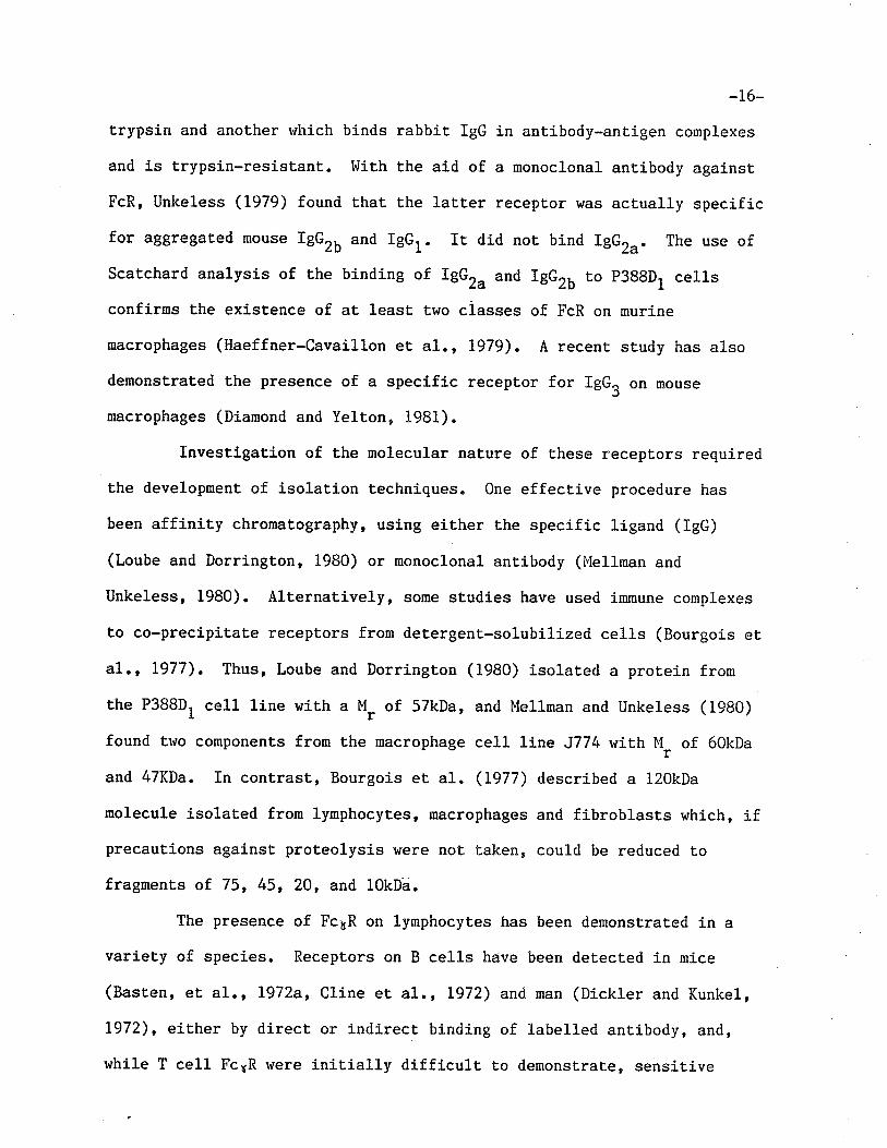
trypsin and another r,¡hich binds rabbit IgG in antibody-ant.igen complexes
and is trypsin-resistant. liith the aid of a monoclonal antibody against
FcR, Unkeless (L979) found that Lhe laLt.er receptor h'as actually specific
for aggregated mouse rgGz¡ and rgGr. rt did not bind rgGru. The use of
Scatchard analysis of the binding of IgGr. and lgGro to p3B8D1 cells
confirns the existence of aÈ least two ciasses of FcR on murine
macrophages (Haeffner-Cavaillon et aI., L979). A recenL sËudy has also
demonstrated the presence of a specific receptor for IgGU on mouse
macrophages (Diamond and Yelton, 1981).
Investigation of the molecular nat,ure of these receptors required
the development of isolation Lechniques. One effecLive procedure has
been affinity chromatography, using either the specific ligand (IgG)
(Loube and Dorrington, 1980) or monoclonal antibody (Mellman and
Unkeless, 1980). Alternatively, some studies have used immune complexes
to co-precipitate receptors from detergent-solubilized cells (Bourgois et
41., 1977). Thus, Loube and Dorrington (1980) isolated a protein from
the P388D, cell line with a M, of 57kDa, and Mellnan and Unkeless (1980)
found tÌüo components from the macrophage cell Line J774 h¡ith llr of 60kDa
and 47KDa. In contrast, Bourgois et aI. (1977) described a 120kDa
molecule isolated from lymphocytes, macrophages and fibroblasts which, ifprecautions against proteolysis ruere not taken, could be reduced to
fragments of 75, 45, 20, and 10kDa.
The presence of Fc¡R on lymphocytes has been demonsLrated in a
variety of species. Receptors on B cells have been detected in mice
(BasLen, et a1., L972a, Cline et al., L972) and man (Dickler and Kunkel,
1972), either by direct or indirect binding of 1abel1ed antibody, and,
while T cell Fc¡R were initially difficult to demonstrate, sensiti-ve
-r7-
assays did finally detect them in the rnouse (Anderson and Grey, L974'
Basten et a1., 1975) and in man (Díckler et al., 1974, Moretta et a1.,
1975). Indeed, additional det.ection of Ig[1 FcR on a great percenLage of
human T cells (Moretta eL al., 1975) subsequently became the basis for
distinction between helper and suppressor T cell subsets (Moretta et 41.,
L977). Furthernore, participation of a non-B, non-T lymphocyte (K ce1l)
in antibody-dependent ce1l-mediated cytotoxicity (ADCC), is known to
involve Fc¡R on that cell (Mingari et, al., 1984). Another interesting
finding was the detecÈion of a soluble Ig binding fact,or (IBF) secreted
or shed by Fc¡.R+ T cells which can suppress ant.ibody response
(Rabourdin-Combe et al., 1984).
Several invesLigations of the subclass specificity of murine
lymphocyte Fc6R produced some disagreement on comparative affinities for
IgG subclasses, although most agree that IgG, , IEGZ' and IBG,O are
readily bound (Basten et 41., I972b, Cline et a1., L972, Anderson and
Grey, 1974) by both B and T cells. In humans, IBG, and IBG, bind to B
cells with much greater affinity that. IgG, and IgGO (Dickler , 1976), a
finding that parellels results.with human macrophages (Anderson, 1984).
There has been a great deal of diversity of molecular weights
found for lymphocyte Fc¡R. The purification of receptor from a
lymphoblastoid cell line produced a mult,imeric glycoproLein wiLh a
subunit l"l_ of 46kDa (Takacs, 1980), while Fc¡R isolat,ed from chronicrlymphocytic leukemia ce1ls (B cells) were unglycosylated,
phospholipid-associated polypeptides with a M, of 30kDa (Suzuki et a1.,
1980a). The lat.ter molecule also possessed phospholipase A, activity
(Suzuki et al., 1980b). Other studies revealed molecules with molecular
weights ranging from 10-130kDa (Bourgois et, 41., L977, Cunningham-Rundles

-18-
et al., 1980, Cohen et, al., 1983, Rask et al., L975, Frade and Kourlisky,
L977). Indeed, there appears to be some consist,ency in the differences
found, leading at least one group to propose a model which explains much
of the data. Kahn-Perles et a1. (1980) suggest that FcyRs from many cell
types, including nurine and human macrophages and lymphocytes, are
sinilar in molecular structure. The nodel they proposed contains 5
globular domains linked by regions accessible to proteolytic enzymes and
at least two inter-domain disulfíde bridges. Each domain has a M, of
23kDa. Thus, depending on the isolaLion conditions (whether or not
reducing agents and/or inhibitors of proteolytic enzymes are used),
IgG-binding molecules of 115, 90, 70, 45 or 23kDa can be obtained. FcyR
from many different cell types may have a similar overall structure
consist,ing of comparable domains but may differ in number of domains or
position of disulfide bonds.
The data on Fc¡R suggests there exists a general structure common
to many cell types from several different species. Experimental
differences may either be due to changes brought about by cellular
evolution or perhaps by different loca1 (membrane) environments. A study
of the species specificiÈy of IgG binding to the P38BD, celI line
suggesled that. Fc6R on rabbit, guinea pig, mouse and human cells are
structurally related and that, primary structure in the Fc portion of t.he
IgG molecule has diverged in paréllel with Fc receptor st,rucure
(Haeffner-Cavaillon et. al., 1979).
2. Receptors for Isl"l lFc,,R)
Since phagocyt,ic cells express Fc¡R, it seemed líkely that they
would also express Fc4R. However, for some time, receptors for lglf were
difficult to demonstrate. Several studies reported t.hat. peripheral blood
_19_
monocytes lacked FcüR (Huber eL a1., 1968, Lawrence et a1., L975, lùalker,
L976). Finally, a study using human and rabbit monocytes demonstrated
t,hat Lhese molecules exist but are present in low frequency (Haegert,
1979). Furthermore, the number of cells giving Fc¡R positive rosettes
could be greaLly increased by ÈreaLment with neuramj-nidase, most likely
due to unmasking of cryptic receptors. Aggregated IgM is not necessary
for binding, although this does not point to high affinity receptors,
since native IgM is pentaneric and nay thus be involved in multi-point
attachment to targeÈ cells.
One of the first reports of lgM-specific receptors on human
peripheral T cells described the expression of these molecules following
a period of in vitro incubation in lgM-free media containing fetal calf
serum (MoretLa eL a1., Lg75). The receptors could be removed by
treatment with pronase, and their expression required protein synthesis.
I'lore recent studies indicated that the expression of Fc4R by freshly
isolated T cells depends upon synthesis of Fc¡aR (Romagnani et al., L979)
and it lras suggested that extremely labile receptor is shed during
preparation of T ce1ls and it !s re-synt,hesized during a 37oC incubation
period. Neuraminidase treatment of Fc R+ lymphocytes decreases Fc¡R
expression (in direct contrast to findings on monocytes) and
simultaneously increases FcyR expression (Schulof eL al., 1980), implying
that the trealment acts directly on sialic acid-containing Fc¡R while it
removes a blocking moiety to expose Fc¡R.
Using a rosette assay., a subpopulation of normal B cells was
found to have Fc¡R (Ferrarini et al., Lg77) as did neoplastic cel1s
(Ferrarini et, al., L977, Pichler and Knapp, L977). Neoplastic B cells
expressed Fcl4R is¡¡nediately afLer isolaLion from peripheral blood and
-20-
washing. However, normal B cells required overnight incubation at 37tC
for the expression of recepLors, suggesting that the recepLors on these
ce1ls were blocked by serum IgM unt,il after this incubation" In
addition, individual neoplastic cells often rosetted h'ith indicator cells
separately coated with IgG, Igl.l or IgE, rneaning that, one cell can carry
one, two or three different, classes of FcR (Spiegelberg and Dainer,
1979). T cells also have Lhe potent.ial for expressing FcR for both Igll
and IgG as dernonstrated by the transiLion from FcsR+ to Fc¡R+ after
stimulus with insoluble immune complexes (Pichler et al" 1978) and from
FcnnR+ to Fc6R+ cells aft,er concanavalin A treatment (Gupta et al., 1979).
Studies focusing on the various functional activities displayed
by peripheral T cel1s bearing FcR have demonstrat,ed the Fc¿R+ and Fc¡4R+ T
ce1l populat,ions play an anLithetical role in pokeweed mitogen-driven
B-cell dífferentiat,ion (Moretta et al., 1977). Thus, Fc¡R+ cells act as
helpers while Fc¡R+ cells act as suppressors. I{owever, since this study
was done, it has become clear that identificat.ion of funct,ional subsets
based only on the presence of FcR is not saLísfâctory. As described
above, FcR are not sLable surface structures. In addit,ion, only a sma11
proportion of the ce11s in a subset identified by FcR may be involved in
the functional activity so that, assignment of function based on surface
markers may only be valid for the subset as a whole and noL for single
cells (Mingari et a1., 1984).
3. Other FcR
A search for receptors for IgA on lymphocyLes has produced data
which suggests a possible regulatory role in IgA synthesis for these Fco.
R+ cel1s. Thus, a rosette assay of rabbit lymphoid cells detected
IgA-specific recepLors in the najor lgA-producing organs such as
_2I_
mesenteric lymph nodes, Peyerts patches and appendix, and, Lo a lesser
extent, in systernic lymphoid tissue (Stafford and Fanger, 1980). As with
Fc¡^R, these receptors could only be detected after an overnight
incubation. Furthermore, polyclonal act,ivation of B cells rtrith goat
anti-rabbit Fab significantly increased the number of Fc..R+ cells in
Peyerrs patches and spleen, indicating t,hat receptor modulation may play
a role in the regulation of IgA immune responses. Another study
demonstrated that both T and B cells frorn human peripheral blood have
receptors for IgA (Sjoberg, 1980a).
The demonst,ration of |tup-regulationrf of Fc*R on murine T cells by
IgA is further evidence for the regulatory role of these receptors (Yodoi
et a1., 1982). These authors also found that a T cel1 hybridoma
expresses both Fc,.R and FcyR (Yodoi et al., 1983a). Upon incubation with
IgA, the hybridoma releases an IgA binding factor which can compet,itively
inhibit binding of IgA to Fc-<R and, which suppresses.IgA synthesis (Yodoi
eL a1., 1983b). These are findings similar Lo those for FcyR+ T cells
(Rabourdin-Cornbe et al., 1984) and FcçR+ T cells (see section E [2]).Secretory component (SC) is a glycoprotein constiLuting an
integral part of secretory IgA. Several studies have confirmed that SC
functions as an imnunoglobulin receptor (Crago et al., L978, Orlans et
a1., 1979, Socken et a1", 1979). It was identified as the receptor for
IgA and IgM on a colonic carcinoma cell line (HT-29) and on epithelial
cells of human fetal tissue (Crago et a1., 1978)" The nature of the
receptor was established by inhibition of binding upon treatment with
anti-SC antibody. Cultured rat hepalocytes vrere found to synthesize SC
(Socken et a1., 1979) and binding of polymeric IgA Eo these cells could
also be inhibited rt¡ith anti-SC reagents (Orlans et al., 1979, Socken eL
-22-
al., L979) "
Very few studies have been done on the receptor for the IgD
immunoglobulin c1ass. This is noL surprising since IgD is primarily
considered to be a surface immunoglobulin and, in humans, circulates with
conparatively 1or,r serum concentrations (30mg/L vs. T}g/f for IgG). By
using lgD-coated latex particles' one investigator detected Fc R
receptors on a small percentage of human T and non-T lymphocytes
(Sjoberg, 1980b). The functional significance of these receptors is
unknown.
IE] IsE Fc Receptors (FcpR)
1. Mast cel ls and basoohils
Qne of the most extensively characterized FcR Lo date is Ehat for
IgE on mast cells and basophils. This receptor plays a role in immediate
hypersensitivity reactions, in which the interaction of a specific
antigen with a specific ligand-receptor complex on target cells initiates
the release of pharmacological agents into surrounding tissues. The
concept of a receptor rvhich plays a role in anaphylaxis was first
proposed by Dale (1913) and l,leil (1913), before the role of mast cells
and basophils was understood, and without knowledge of the nature of the
lÍgand involved. By definition, anaphylaxis is an imrnunological reaction
in which previous sensitization with a specific antigen has elicited
homocytotropic antibodies whose targeÉ cells are masL cells and
basophils. Upon renerved appearance, the anligen binds to cell-bound
antibody and causes the release of pharmacologically active agents.
The demonstration of IgE binding to mast cells (Ishizaka et al.,
L972a) and basophils (Ishízaka et al., 1970c) suggested the presence of a
_23_
specific receptor for IgE on these cells, and prompted a search for t.he
molecule(s) responsible for this binding. Thus, isolation and
characterization of these receptors should lead to a beLter understanding
of the triggering mechanisms of allergic reacLions at the molecular
leve1.
(a) Evidence for the localization of receDtors: visualization on intact
ce1ls
Direct evidence for the localization of receptors for IgE on t,he
plasma membrane of human basophils was obtained by Ishizaka et a1.
(1970c), using autoradiography after allowing the cells to react with
either 125I-.nti-IgE or 125I-IgE, and by Becker et al. (Lg73) who used
fluorescein-conjugated inst.ead of radiolabeled ligand. The presence of
recept,ors'for human IgE on monkey lung mast, cells (Tonioka and Ishizaka,
L97L) and for rat IgE on rat peritoneal mast cells (Ishizaka et a1.,
1975) was detected using similar techniques. A more detailed
denonst,rat,ion, providing more direct, evidence for the existence of t,hese
receptors, tì/as achieved by elec.tron microscopy of human basophils which
had been exposed to IgE, rabbit anti-hunan IgE and a hybrid burro
antibody to rabbit IgG and to ferritin (Sullivan et aI., 1971), Using
this approach, iL was observed that lhe bound IgE molecules were
diffusely distribuLed over the surface of the cells and could be
redistributed with anti-IgE to form patches and caps. Such a
redistribution is analogous Lö that seen by surface Ímmunoglobulins on
lymphocytes aft.er treatment with anti-immunoglobulin antibodies (Taylor
et 41., l97L). This finding agrees with the generally accepted concept.
that cel1 surface recepLors are mobile. Fluorescein-conjugated anti-IgE
-Zl+-
was used to show a similar movement of receptors on raL basophilic
leukemia (RBL) cells (Carson and Metzger, L974). A more recent study, in
which binding of fluoresceinated IgE tetramers Lo RBL cells induced
large-scale aggregat.ion of receptors (1000-10,000 receptors per cluster),
indicated that ligand-induced clustering is accompanied by cell-induced
clustering, possibly via cytoskeletal attachment of receptors (llenon et
41., 1984). This phenomenon may be relevant to the triggering signal for
degranulat,ion.
The finding that brief exposure of human basophils (Ishizaka and
Ishizaka, 1974) or RBL cells (Kulczycki and Metzger, 1974) to pll 4 or
lower removed the bound IgE while the cells retain their capacity to bind
fresh IgE, provided further evidence for surface localizatíon of the
receptors. QuantiLat,ion of eluted IgE has also been used to measure the
number of receptors on the cell (Conroy et a1., L977).
(b) Number of receptors on tarset cells , kinetics and valency
The cel1 bound IgE molecules on human basophils rüere enumerated
by Ishizaka et al. using the Cl fixation and Eransfer Lechnique (Ishízaka
et.41., L973). The method consists of addition of excess anti-IgE to
basophils, fixation of the CI component of complement by anti-IgE and
subsequent lysis of sensitized erythrocyLes by the Cl" The assay
revealed 10r000-40,000 IgE molecufes per basophil and, by saturat,ing the
receptors with myeloura IgE molecules, the number of receptor sites rsas
estimated to be 301000-85r000i Furthermore, they found that there was no
correlation between the number of endogenous IgE molecules per cell and
the serum IgE concentration. The affinity constant vras estimated to be
from 1oB to 1o9M-1.
-25_
The availability of relatively pure (75-957,) preparations of raL
peritoneal mast, cells (RMC), of RBL cells and of myeloma rat, IgE made
studies of this systeln very attractive. Direct binding of 1251-IgE to
RMC (Conrad et a1., 1975) and to RBL cells (Kulczycki and MeLzger, L97t+,
Conrad et al., L975) demonstrated that the number of receptors on these
cells ranged between 0.3-1x10⁶ per cell. Two protocols were applied to
quantitatively study these receptors. Kulczycki and Metzger (I97/+)
shor.¡ed that, recepLors on RBL cells can be saturated by adding excess
125f-Ign to the cells. After correcting for non-specific binding, the
amount of IgE bound Lo saÈurated cells indicated that there were
0.3-1.0x10⁶ binding sites per cell. The varialbility was accounted for
by changes in the number of receptors during the cell cycle (Isersky et
a1., 1975). luleasuring binding of IgE to RBL cells and to RMC under
non-saturating conditions, Conrad et al., (1975) plotted lheir binding
data according to the Scatchard equation (Scatchard, L949). They found
that RBL cells have a substantially higher number of receptor sites than
RI\'IC - approxi-mat,ely 6x10⁵ u"r"u" 3x10⁵, respectively" A study of mouse
mast cells found 0.6x10⁵ sítes.per cell (Mendoza and Metzger, !976a).
Thus, normal cells from bot,h human and rat appear to have 2-10 times
fewer FcsR per cell than neoplasLic rodent cells"
The study of Kulczycki and I'letzger (L974) also showed that the
binding between IgE and the receptor is reversible since 1251-lgn could
be displaced with a 50-100 fold excess of unlabelled IgE. Thus, direct
binding measurements demonstrated that the binding of IgE to RBL-I cells
is governed by a simple reversible bimolecular reaction:
-26-
ntIgE + receptorS::Jreceptor-IgE complex
k-1
in which the equilibrium consLant, Ka, can be defined as follows:
K
k-t
is Lhe foward rate constant and k1
is the reverse rate
constanL.
For RBL-I cells, Kulczycki and Metzger (1974) calculated the foward rate
constant, (kt), to be 9.6x1041,1-1"".-1 and the dissociation constant, k-1,
to be a naximum of 1.6x10⁻⁵"".-1, thereby yielding a minimum K" of
6x1O9t't-1. Similar results were obtained for rat mast cells (Þfendoza and
Metzger, L976a). Conrad et a1. (1975) obtained an equilibrium constanÈ
of 1x10⁹M-1 for the binding of IgE to both RBL cells and RMC, from the
slope of the Scatchard plot. The equilibrium consLanL using cel1-free
particles hras approximately one order of magnitude greater than that for
intact RBL cells, partially due to a smaller dissociation constant
(Metzger et a1., L976). This difference may be explained by t,he fact
that cell-free particles are not affecLed by receptor turnover as are
intact cells. Reactions involving solubilized receptors have
dramatically great.er K. due Èo a much slower dissociaLion rate (Rossi et
a1., L977), a finding that is not yet well understood" 0vera11, the
affiníty of mast cell and basophil receptors for IgE is much greater Lhan
that of Ehe imnunoglobulin-receptor interactions of other Ig classes on
rnost cel1 types (lOa-fO8U-l) (Froese, 1984). This characteristic of uhe
IgE receptor has greatly facilitated attempts to isolate the molecule
(see section F[ 1 ] ).
kta
where kt
-27-
Many of these quantitative studies assume that the receptor for
IgE is monovalent. Mendoza and Metzger (1976b) addressed this quest,ion
by saturating cells with a mixture of two distinguishable Lypes of rat
IgE and determining whether, by redistributing (patching and capping) one
of then vith specÍfic antibody, the other would co-migrate. Thus,
receptors binding rhodamine-IgE did not co-migrate with receptors
occupied by fluorescein-IgE when treated with anti-fluorescein, but did
so when reacted with anti-IgE, indicating that these IgE receptors are
monovalent. In contrast, the results of Ishizaka and Ishizaka (i975) 1ed
to a differenL conclusion. In this study, human basophils, the receptors
of which were partially occupied, were either saturated with unlabelled
IgE or left unLreated, and were then induced to cap with anti-IgE. The
cells were subsequently exposed to pH 4 to dissociate lgE-anti-IgE
conplexes and, upon neutralization, were reincubaLed with 125I-IgE and
examined by radioautography. Ce1ls which had and had not. been
pre-saturated showed a similar distribut,ion of grains, exhibiting capped
or asy¡nmetrical distribution of the labe1. This suggests that there v/as
co-rnigration of empty receptors with receptors to which IgE was bound and
that the receptors are at least divalent. Differences in the results
obtained in these Lwo studíes may have been due to differences in species
and cell type as well as due to the methods used. However, the recent
study of Menon et al., (1984) which describes a cell-induced (as opposed
to ligand-inducecl) aggregaLion of IgE receptors upon cross-linking of
receptors with tetramers of IgE, indícat,es t.hat. while the receptors
themselves may be monovalent, there roay be an intracellular association,
possibly mediated by cytoskeletal elements. The demonsLration that
soluble receptors interact with IgE with a 1:1 ratio indicates that they
-28_
are indeed monovalent in such a stat,e (Nervman eL a1., L977)"
It should be noted that the IgE used in the rodent studies
(Kulczycki and lrletzgerr lgT4, Conrad et al., 1975, tfendoza and lletzger,
L976a, Metzger et aI., 1976, Rossi et al., L977, Mendoza and I'fetzger,
L976b, ) is a myeloma protein obtained from rats bearing the IR 162 tumor
(Bazin et al., 1974). A comparison of this IgE and IgE obtained from rat
reaginic serum demonsLrated uhat both preparations behave similarly with
respect t,o physicochemical and RBL cell-binding characteristics
(Kulczyckí and Met,zger , L974).
(d) SBecificitv of receptors
There has been a great deal of interest in both the species and
Ïg class specificity of the mast cell and basophil IgE recepLors. There
appears to be considerable cross-reactivity between recept,ors and IgE
from closely related species. Ishizaka et al. (1970c) observed that
monkey lung fragments could be passively sensitized with human aLopic
serum to release histamine upon treatment, with anLi-IgE. Tomioka and
Ishizaka (1971) were also able t,o demonstrate, using autoradiography with
radiolabelled anLi-IgE, that human myeloma IgE binds t,o monkey skin mast
cells. In rodents, it is generally accepÈed t,hat mouse IgE binds to rat
mast cells. Since it was shown that mouse reaginic anLibody sensitizes
rat skin for passive cutaneous anaphylaxis (PcA) (Mota and tr^iong, 1969)o
raLs are commonly used for assayfng IgE levels in mouse serum. It was
also shown Lhat absorption with intact RBL cells and solubilized
receptors could deplete PCA activity from mouse reaginic serum (Conrad et
41., 1976). Furthermore, Lhe binding of rat IgE t,o mouse mast. cells has
been denonst.raLed (Prouvost-Danon et al., 1975) and the binding constant
for this reaction is slightly lower than that for the homologous reaction
-29_
(Ì4endoza and iuletzger, 1976), indicaEing that while there is great.
similarity in structure between rat and mouse recept,ors, they are not
identical. Indeed, Sterk and Ishizaka (1982) have found that, while rat
mast cells bind rat and mouse IgE equally well, mouse mast ce1ls have one
type of receptor which binds rat and mouse IgE with the same affinit,y and
a separate receptor which binds mouse IgE alnost, exclusively.
The need for a simple assay of human reaginic serum has led to
studies of the interaction between human IgE and raL mast cells and to
some conLradicLory results. While Ishizaka et al. (1970b) failed to
sensj-t,ize the skin of rat.s lrith human reaginic serum, an in viLro sLudy
demonstrated that RMC could be degranulated by incubation with human
reaginic serum and subsequent challenge with allergen (Perelmutter and
Khera, 1970). I,rlashj-ng masL ce11s after sensitization prevented
degranulat,ion, indicating that'the interaction was rather weak. There
was also a great deal of non-specific degranulation of RtlC upon addition
of human serum. These observations may partly explain some of the
divergent results. A more direct attempt to demonstrate binding of human
IgE to rat cells was made by Kulczycki eL al. (L974), who failed to show
significant. inhibition of 125l-t.t IgE to RBL cells by hunan nyeloma IgE.
The ability of human IgG t.o passively sensit.ize human lung and
skin tissues (Parish, 1978) has led to speculation on the nature of the
receptor for this imnunoglobulin class and its relationship to the IgE
receptor. Demonstration that a myeloma IgGO could inhibiL passive
sensitization of human skin by IgE suggesLed that the two immunoglobulin
classes cross-reacted with the same receptor (Stanworth and Smith, 1973).
In the rat, IgGra hras found to induce PCA reactions, and the
sensitizat,j.on could be blocked by IgE. Conversely, IgGZa could inhibit
-30-
sensigization of rat peritoneal mast cel1s with IgE in vitro, thus, iL
was postulated that a conmon receptor is involved (Bach et a1., 1971).
No direct binding of 125I-I g}ru to RBL ce1ls was detected by Halper and
Ì,letzger (L976), and even a 1000-fold exess of monomeric IgG failed to
inhibit IgE binding while partial inhibition lras observed if IgG immune
complexes r*ere used" These results suggest that IgE and IeGru share a
corn¡r¡on FcR but that the lgG-receptor interaction is a rather weak one
which requires multi-point attachment for adequate binding. Conrad and
Froese (1978a) were similarly unable to demonstrate inhibition of
solubilized recept,or-binding to lgE-Sepharose with normal rat IgG. All
of these results would be reconcilable if it is assumed that the IBGru
used by Bach et al. (1971) contained some aggregated immunoglobulin.
Daeron et al. (1975) described a phenomenon in the mouse known as
direct allogeneic anaphylactic degranulation (DAAD) ' in which IgGt
antibodies specific for mouse mast cell histocompatability antigens can
degranulate these cells. They postulated Uhat the bridging caused by Lhe
interaction of the IgG Fc with FcR and the Fab with hj-stocompatability
antigens is a sufficient signal for degranulation" Further investigation
revealed that myeloma IgG, produced partial inhibition of DAAD, while
pre-saturating cells with rat myeloma IgE failed t.o do so (Daeron et al.,
1980). Since the rat lgE-mouse nast cell interaction has a relatively
high affinity constant (Mendoza and l"letzger, L976), it seemed unlikely
that mouse IgG could replace the IgE on the same receptors, thus it was
concluded that the tr¿o immunoglobulin classes were interacting with
separate receptors. Results seemingly contradiclory to these were
obtained by Nfossman eL a1., (1979) who used a similar system in the rat
and who demonsLrated that rat T-EGr^ alloantibodies
-31-
(ant,i-histocompatability antigen) could inhibit lgE-mediated histamine
release fron RMC and rat IgE could inhibit lgGru-mediated DAAD.
This confusing picture $ras somewhat, clarified by the work of
Segal et, al., (1981). Using chemically cross-linked rat IgG oligomers of
different sizes, they demonstrated an increasing affinity of RBL ce1ls
for oligomers of increasing size. Thus, affinity constants were
calculated to be 6.6x10⁶, 1.6x10⁷ and 8x1071,1-1 for dimers, Lrimers and
higher oligomers, respectively. As previously demonst.rated (Halper and
Metzger, 1976), binding of nonomeric IgG was t,oo weak to be detected by
direct, measurenent. Furthermore, binding of IgE monomer could not be
inhibited by rabbit IgG aggregates which effectively block uptake of rat
IgG oligomers or by rat oligomers themselves. Mononeric IgE could
inhibit IgG oligomer binding nore efficiently than monomeric IgG could.
Association constants for IgE monomer and IgG monomer binding to IgG
binding receptors were estimated Lo be 1.6x10⁶ and 4.2x10⁵M-1,
respectively. These results indj-caLe thaL RBL cells carry two kinds of
receptors. One of these is specific for IgE and cannot be inhibit.ed by
monomeric IgG or aggregated IgG; the other binds oligomers of IgG and
this binding can be inhibiLed by monomeric IgG and IgE. Hor+ever,
monomeric IgE has a greater affinity for the ttFc¡Rtt than than does
mononreric IgG. These observations were extended by Kepron et al" (1982),
who made very similar observations with solubilized recepLors (see
section [F2]).
2. Lymphocytes
One of t,he first studies demonstrating Fc R on lymphocytes was
performed by Lawrence et al. r+ho found a small but significanL uptake of
radiolabelled, aggregated IgE (Lawrence eL al., 1975). Rosette assays
-32-
vrere used Lo enumerate and characterize IgE-binding lymphocytes from
human peripheral blood lymphocytes (PBLs) (GonzaLez-Molina and
Spiegelberg, L977, Hellstrom and Spiegelberg, 1979, Yodoi and Ishizaka,
1979a) and the results indicale thar L-4i( of PBLs are Fc6R+. Inlhen
lymphocytes rrere separated into B and T cells, Yodoi and Ishizaka (L979a)
found that FcçR+ lyrnphocytes vrere enriched in the B cell fract,ion, while
the T cell fraction contained an insignificant number of rosette forming
cel1s. 0n1y cells from some atopic patienLs contained a significant
number of Fc¿R+ T ce1ls. Indeed, Spiegelberg et a1" (1979) also found
thaL severely atopic patient,s had elevated numbers of Fc6R+ lynnphocytes.
In addition, no FcyR were co-expressed ruith Fc¿R on these cells. The
presence of FcgR on Lhe cel1s of several human lyrnphoblastoid cell lines
was investigated as well and it was found that the majority of these
lines are Fc¿R+ (Gonzalez-Molina and Spiegelberg, 1976). lfany chronic
lymphatic leukemia patients had high proport,ions of Fc¿R+ lymphocyt,es
(IL-827") and many of these lymphocytes expressed Fc¡R, Fc¡aR and Fc6R
simultaneously (Spiegelberg and Dainer, 1979).
In vitro investigations of Fc6R+ rat lymphocytes carried out by
Yodoi eL a1. (1979) showed that, the proportion of these cells markedly
increased after incubation with isologous IgE. Other immunoglobulins,
such as human IgE, rat IgG or rabbit IgG were not, effective. Yodoi and
Ishizaka (1979b) found this incréase to be the resulL of a t,ransition of
Fc¡R+ to FcsR+ cells. The results from this study suggest that IgE can
bind to either FcER or Fc¡R and provide a signal for the formation of new
Fc¿R. In addition, culture of nesenteric lymph node ce1ls from rats
infected with the parasite, N. brasiliensis (which enhances IgE
synthesis) results in the release of a soluble lgE-binding factor that
-33-
competes with Fc R on rat lymphocytes in a rosette assay (Yodoi and
Ishizaka, 1980). The release correlates wit,h the increase in Fc¿R+
cells, suggesEing that the factor may be derived from these cells. 'Ihe
major source of the factor r.¡as shown to be the T cel1 population.
Further investigation of this factor dernonstrated that it has an
enhancing or suppressive effect on the IgE antibody response, depending
on its state of glycosylation. More recently, it has been shown that B
cell lines also release an IgE-binding factor capable of regulaLing the
IgE response (SarfaLi et a1., 1984). These investigations led to the
conclusion that FcgR+ lymphocytes and FcèR-like molecules from these
cel1s play an inportant role in the regulation of the IgE imnrune
response, nuch like the IgG- and lgA-binding factors already discussed
(sect,ions D[1] and D[3], respecti-vely).
The molecular nature of the lymphocyte Fc¿R was deLermined from
studies on detergent solubilized receptors from a variety of cells"
Meinke et al. (1978) found that the molecules precípitated from a
lymphoblastoid cell line by an ant,i-receptor antiserum had M, of 86, 47
and 23kDa as determined by polyacrylamide gel electrophoresis in the
presence of SDS (SDS-PAGE), the smallest being a prot,eolytic degradation
product. The affinit,y of the receptor for monomeric IgE was relatively
low (fO6-fO7U-1) (Spiegelberg and Melewicz, 1980). Conrad and Peterson
(1984) found that the molecule iSolated from normal murine B lyrnphocytes
with IgE-Sepharose had a molecular weight of 49kDa and comparison to t,he
45kDa receptor from human B cells by two-dimensional ge1 analysis shorved
great similarities between these two molecules. The high affinity FcgR
from RBL cells produced a very different 2-D pattern. Furthermore, t,he
murine B cell receptor was found to be nultivalent, as demonstraLed by
-34-
the fact that radiolabelled IgE, r+hen bound to the B cell Fc6R, will
co-isolate with the Fc R on an lgE-Sepharose column (Lee and Conrad,
1984). Along with the fact that antiserum to lymphocyte Fc.R r"ill not
release histamine from basophils (Meinke et al., 1978), Lhese results
suggest that FcgR from a variety of lymphocyLes are structurally
different from mast ceIl and basophil high affinity Fc¿R"
3. Macrophages and monocytes
ApproximateLy 207" of peripheral blood monocyt,es (latex-ingesting
cells) from healthy human donors were found to form rosettes with
IgE-coated indicaÈor ce1ls (Melewicz and Spiegelberg, 1980)" This would
suggest that a subpopulation of monocytes is involved in Lhe phagocytosis
and lysis of lgE-coated target cells" Severely allergic patients had
significantly greater proportions of FcqR+ monocytes (Ì,felewicz et al.,
1981), indicating that these ce1ls also play a role in the regulaLion or
expression of allergic disease.
In the rat, Dessaint et al. (1979b) found weak binding of
nonomeric IgE to macrophages, and u¡ere able to show increased bincling
when using aggregated IgE. It.was also discovered that the rnajority of
peritoneal and alveolar macrophages bear FcgR (Boltz-Nitulescu and
Spiegelberg, 1981), and the proportion increases with increased serum IgE
levels (Boltz-Nit,ulescu et al., 1984). Capron et al" (1975) discovered
that normal rat peritoneal macroþhages incubated with serum from rals
immune to the parasite S. man soni. become highly cytotoxic for
schistosome larvae, a phenomenon shown to be mediaLed by IgE antibody
specific for t,he parasite. These macrophages were further shown to
release lysosomal enzymes in response to incubation with IgE and ant.i-IgE
(Dessaint et al., L979a). These results provide evidence that IgE
-35-
interacLion with the macrophage and its subsequenL cross-linking leads to
the activation of macrophage killing ability, important in the defense
against lgE-coated parasites.
4. Eosinophils
The involvement of eosinophil leukocytes and rat TEGZ' in the
antibody-mediated cytotoxiciLy against S. mansoni (Capron et al., 1978),
and several accounLs of IgE binding to eosinophils from allergic patients
(Hubscher and Eisen, L97I, Hubscher, L975), led to the investigation of
the role of eosinophils as effector ce1ls in ühe killing of parasit,es.
Thus, Capron et a1. (1981b) found that rat eosinophils and human
eosinophils from hypereosinophilic patients bear FcgR, and that the
proportÍon of cells bearing receptor increased upon incubation with
eosinophil chemotactic factor of anaphylaxis (ECF-A), a mast cell-derived
mediator. They also demonstrated that rat. eosinophils are directly
involved in the killing of schistosomula, and the presence of mast cells
(ECF-A in particular) enhanced cytotoxicity (Capron eL al., 1981a). This
effect was mediated by IgE antibodies. Similar findings for the
IgGr"-mediated eosinophil cyLotoxicity (Capron et 41., 1978) point Lo a
relat.ionship between anaphylacLic antibodies and phagocyLic cells in the
cytotoxic mechanisms against certain parasit.es, with mast cells playing a
central role as well.
An interest,ing finding by Capron et al. (1984) was Lhat an
antiserum direcLed against the FcgR of a human lymphoblastoid cel1 line
(Meinke et, al. , l97B) could inhibit lgE-roset.te format,ion by human
eosinophils suggests that the receptors on these cells are antigenically
related.
-36-
ce11sF. characterization of solubilized Fc R from
and mast cells
1. Isolation of recepLors
One factor which greatly facilitates the analysis of a receptor
is a convenient source of cells bearing the receptor. Initially, raL
peritoneal mast cel1s were routinely obtained in relatively pure
preparations. The induct,ion of a raL basophilic leukemia by
transformation with p-chlorethylamine (Eccleston et 41., 1973) provided
an IgE binding cel1 which could be maintained in vitro. A large part. of
the characterization of the rat mast cell/basophil FcER has been done
with this cel1 line.
A thorough analysis of the receptors for IgE requires that. Lhey
be solubilized without loss of lgE-binding capacity" This ability has
been demonstrated both for rat mast cells (RMC) and RBL cells by Conrad
et a1. (1976) who used the non-ionic detergento Nonidet P-40 (NP-40), to
solubilize the plasma membrane. They found t,hat, radiolabelled IgE bound
to Rl,lC and RBL cell prior to NP-40 solubilization eluted aheacl of free
IgE alone upon gel chromatography" The increased molecular weight of IgE
was due to complex formation with receptors and indicated t.hat receptors
in Lhis extract retain their lgE-binding capacity in NP-40. An assay for
solubilized receptor was developed by Rossi et al " (1977), based on the
fact that the solubility of free-l2st-tgn is much greater in a t+27"
ammonium sulfate solution than that of l2st-IgE-receptor complexes.
To isolate and furt,her charact,erize solubilized receptors, Conrad
and Froese (1976) surface iodinat,ed RlfC and RBL cells by the
lactoperoxidase-caLalyzed reaction. Radiolabelled, solubilized receptors
were then isolated by a variety of met.hods. A sandwich precipitation
-37-
technique using rat myeloma IgE (IRl62)r é-specific rabbit anti-rat IgE,
and goat anti-rabbit Ig was used in this study to isolaLe receptor.
Alternatively, precipitates were obtained r,¡it,h IgE and rabbit anti-fgE at
equivalence, or the complex was bound by anti-IgE-Sepharose. Protein
A-Sepharose has also been used to insolubilize the complex Lhrough
binding of anti-IgE (Conrad and Froese, L978a). Isolation of recepLors
free of IgE was achieved using lgE-Sepharose as the affinity gel and KSCi'l
(Conrad and Froese, 1978a), 0.5M acetic acid or 6}l guanidine HCI
(Kulczycki and Parker, 1979) as eluting reagents. In the latter study,
repet,itive binding of receptor material to lgE-Sepharose was shown t,o
yield highly purified receptor as det,ermined by binding of recepLor from
biosynthet.ically labelled RBL cells.
An alternate form of affinity chromat,ography was developed in two
laboratories t.o decrease the elution of cellular rnaterial nonspecifically
bound to affinity gels. Thus, either dinit.rophenol residues (Conrad and
Froese, 1978b) or arsanilic acid molecules (Kanellopoulos eL al., 1979)
were coupled to IgE which subsequently formed lgE-receptor complexes.
These complexes were adsorbed onto conjugates of Sepharose and the
appropriate anti-hapten antibody and then eluted by addition of the
corresponding hapten.
Several different anti-receptor antisera have been produced (see
Section F[4]) and, these reageneé have been used to isolate recept,ors.
Isersky et al. (1978) used polyethylene glycol to faciltate precipitat.ion
of receptor-anti',-receptor complexes, while others used protein
A-Sepharose Lo isolate immune complexes (Conrad et al., 1978, Conrad et
a1., 1979).
2. The number of different receptor molecules and their molecular weight
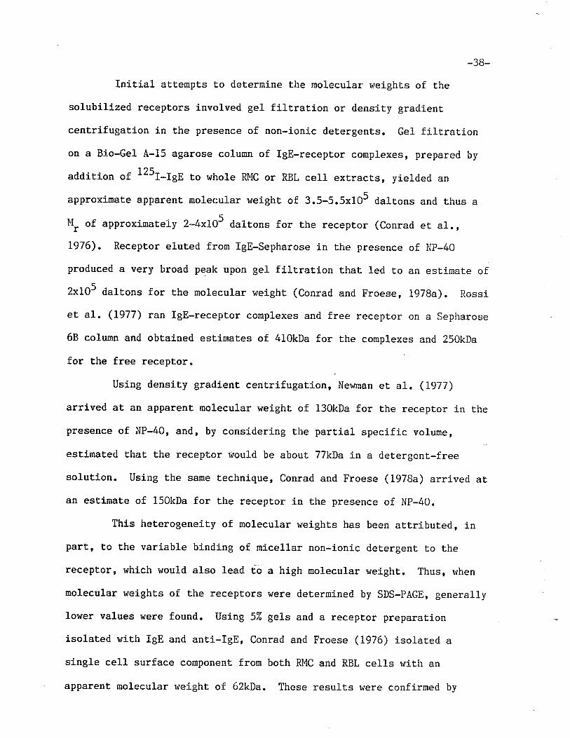
-38-
Initial aLtempts Èo determine the molecular rueights of the
solubilized receptors involved gel filtration or density gradient
centrifugation in the presence of non-ionic det,ergents. Gel filtrationon a Bio-Gel A-15 agarose column of lgE-receptor complexes, prepared by
addition of 125r-tgE t.o whole RMC or RBL cel1 extracLs, yielded an
approximate apparent molecular weight. of 3.5-5.5x10⁵ d"ltorr" and thus a
M, of approximateLy 2-4x10⁵ daltons for the receptor (conrad et al",L976). Receptor eluted from lgE-Sepharose in the presence of Np-40
produced a very broad peak upon gel filt.ration that 1ed to an estimate of
2x10⁵ daltons for the molecular weight (Conrad and Froese, L978a). Rossi
et al " (1977) ran IgE-receptor complexes and free receplor on a Sepharose
68 column and obtained est.imates of 410kDa for the complexes and 250kDa
for the free receptor"
Using density gradient, centrifugaÈion, Newman et al . (Ig77)
arrived at, an apparent, molecular weighL of 130kDa for the receptor in the
presence of NP-40, and, by considering the partial specifi.c volume,
estimated that the receptor would be about 77kDa in a detergent,-free
solution. Using the same technique, Conrad and Froese (1978a) arrived at
an estÍmate of 150kDa for the receptor in the presence of Np-40.
This heterogeneity of molecular weights has been aEtributed, in
part' to the variable binding of micellar non-ionic detergent to the
receptor, which would also lead Co a high molecular weight. Thus, when
molecular weight.s of the receptors were determined by SDS-PAGE, generally
lower values were found. using 52 gels and a receptor preparation
isolaLed with IgE and ant.i-rgE, Conrad and Froese (1976) isolated a
single cell surface component from both RMC and RBL cells with an
apparent molecular weight of 62kDa. These results were confirmed by
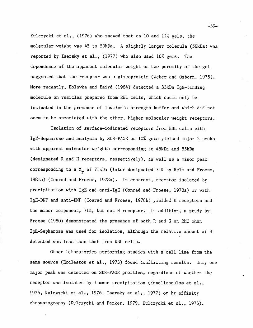
-39_
Kulczycki et, a1., (L976) who showed that on 10 and 12% ge1s, t.he
molecular weighÈ was 45 t.o 50kDa. A slightly larger molecule (SBkDa) was
reported by Isersky et a1., (1977) who also used 10% gels. The
dependence of the apparent molecular weight on the porosity of the ge1
suggested that the receptor was a glycoprotein (hleber and Osborn, L975) "
More recently, Holowka and Baird (1984) detected a 33kDa fgE-binding
nrolecule on vesicles prepared from RBL cells, vrhich could only be
iodinaLed in the presence of low-ionic strength buffer and which did not
seem Lo be associaled with the other, higher moleculer r+eight receptors.
fsolation of surface-iodinated receptors frorn RBL cells rvith
IgE-Sepharose and analysis by SDS-PAGE on 102 gels yielded major 2 peaks
with apparent, molecular weights corresponding to 45kDa and 55kDa
(designated R and H receptors, respectively), as well as a minor pealc
corresponding to a l'{. of 7lkDa (later designated 71K by lleln and Froese,
1981a) (Conrad and Froese, 1978a). In cont,rast, receptor isolat.ed by
precipiLation with IgE and anti-IgE (Conrad and Froese, 1978a) or rviLh
IgE-DNP and anti-DNP (Conrad and Froese, 1978b) yielded R recepLors and
Ëhe minor component, 71K, but not H recept,or. In addition, a study by
Froese (1980) demonstraLed Lhe presence of both R and H on Rl"lC r,¡hen
IgE-Sepharose h¡as used for isolation, although the relative amount of H
det,ect,ed r'ras less than that from RBL cells.
Other laborat,ories performing studies with a ce11 line from the
same source (Eccleston eL a1., L973) found conflicting results. 0n1y one
major peak was detecLed on SDS-PAGE profiles, regardless of whether t.he
receptor was isolated by immune precipitation (Kanellopoulos et al.,L976, Kulczycki et al., L976, Isersky et al., L977) or by affinitychromatography (Kulczycki and Parker, 1979? Kulczyckí et al., L976).
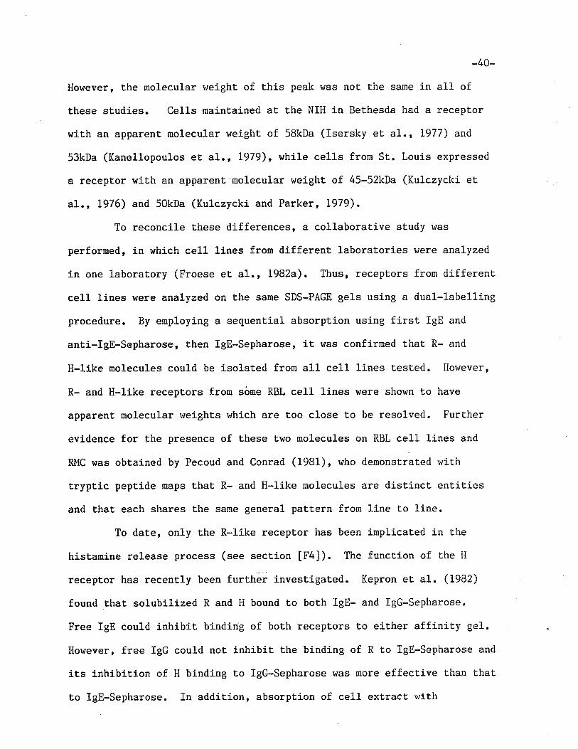
-40-
However, the molecular r.reight of this peak was not the same in all of
these studies. Ce1ls mainLained at the NIH in Bethesda had a recepLor
with an apparent molecular weight of 58kDa (Isersky et a1., L977) and
53kDa (Kanellopoulos et al., lgTg), while cells from St. Louis expressed
a receptor with an apparent molecular weight of 45-52kDa (Kulczycki et
a1., 1976) and 50kDa (Kulczycki and Parker, L979).
To reconcile these differences, a collaborative sLudy vias
performed, in which cell lines from differenL laboratories were analyzed
in one laboratory (Froese eL a1., T982a). Thus, receplors from different
celI lines were analyzed on Lhe same SDS-PAGE gels using a dual-1abelling
procedure. By employing a sequenLial absorption using firsL IgE and
anti-IgE-Sepharose, Lhen lgE-Sepharose, it was confÍrmed that R- and
H-like molecules could be isolated from all cell lines t,ested. However,
R- and H-like receptors from sôme RBL ce1l lines were shown to have
apparenL molecular weights which are too close to be resolved. Further
evidence for Lhe presence of these two molecules on RBL cell lines and
RMC was obtained by Pecoud and Conrad (1981), ruho demonstrated rvith
tryptic peptide maps that R- and H-like molecules are distinct entities
and that each shares the same general pattern from line to line.
To date, only the R-like receptor has been implicated in the
histamine release process (see section [F4]). The function of the I{
receptor has recently been furthãr investi-gated. Kepron eL al. (1982)
found that solubilized R and H bound to both IgE- and lgG-Sepharoseo
Free IgE could inhibit binding of both receptors to either affiniLy gel.
However, free IgG could not inhibit the binding of R to lgE-Sepharose and
its inhibition of H binding to lgG-Sepharose was more effective than that
t,o lgE-Sepharose. In addit,ion, absorption of cell extract. with
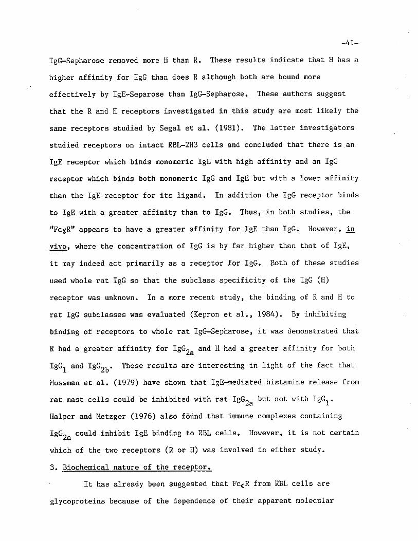
_4L_
IgG-Sepharose removed more H than R. These results indicate that FI has a
higher affinity for IgG than does R although both are bound more
effectively by lgE-Separose than lgG-Sepharose. These authors suggest
that the R and H receptors investigated in t,his study are nosL likely the
same receptors studied by Segal et al. (1981). The lacter investigators
studied recepLors on intact RBL-2H3 cells and concluded that there is an
IgE receptor which binds monomeric IgE with high affinity and an IgG
receptor which binds both monomeric IgG and IgE but with a lower affinity
than the IgE receptor for its ligand. In addition the IgG receptor binds
to IgE with a greater affinity than to IgG. Thus, in both studies, the
trFcyRrr appears Lo have a greater affinity for IgE than IgG. I-lowever, in
vivo, where the concentration of IgG is by far higher than that of IgE,
Ít may indeed act, primarily as a receptor for IgG. Both of t,hese studies
used whole rat IgG so that the subclass specificit,y of the IgG (H)
recepEor r,¡as unknown. In a more recent study, Lhe binding of R and H to
rat IgG subclasses was evaluated (Kepron et a1., 1984). By inhibit.ing
binding of receptors to whole rat lgG-Sepharose, it was clemonstrated thal
R had a greater affinity f.or IEGru and H had a greater affinity for both
IgG, and fgG2b. These resulLs are interesLing in light of the fact that
PÍossman et al-. (lg7g) have shown thag lgE-medíaÈed histamine release from
rat mast cells could be inhibited with rat IBG,. but not with IgGt.
Halper and Metzger (1976) also fôund that immune complexes containing
IgGZa could inhibit IgE binding t,o RBL cells. However, it is not certaín
which of the tr,Io receptors (R or H) was involved in either study.
3. Biochemical nature of the receDtor.
It has already been suggested that FcER from RBL cells are
glycoproteins because of the dependence of their apparent molecular
-42_
$reight on the porosiLy of Lhe gel used in SDS-PAGE" Further evidence for
the presence of carbohydrate on t,hese molecules comes from their abilityto biosyntheLically i-ncorporate radiolabelled sugars (Kulczycki and
Parker' L979, Kulczycki et al., L976, Heln and Froese, l98la). Llhile the
studies of Kulczycki and his colleagues (Kulczycki and Parker , rg7g,
Kulczycki et al., L976) did not distinguish between R and H recept.ors,
that of Helm and Froese (1981a) did establish r.har borh of rhese
molecules incorporate sugars and that 71K ís also a glycoprotein. It has
also been suggested that the carbohydrate composition of R and i{ are
different as demonstrated by Lhe difference in affinity of these two
ruolecules for different lectins (Helm and Froese, 1981b, Flelm et al.,1979). Recent cornposiËional analysis by Kanellopoulos et, al. (1980) has
demonstrated that the R-like molecule (which has nor'¡ been designated
subuniÈ in some laboratories) has a carbohydrate conLent of 32i(,
consisÈj-ng of fucose, mannose, galactose and N-acetyl-glucosamine. No
sialic acid could be detected by dÍrect analysis in this study. i{owever,
Pecoud et al. (1981) obtained some indirect evidence for the presense of
this molecule by showing that neuraminidase treatment of the cells
enhances surface labelling with 3H-NaBHO. The effect of tunicamycin, a
glycosylation-inhibiting antibiotic, on the RBL cell Fc6R r.'as
investigated by llempsLead et al. (1981b). They found that the diffuse
SDS-PAGE gel recepLor band routinely seen in several studies (Conrad and
Froese, L978a, Kanellopoulos et al., L979, Kulczycki et, al., Lg76),
spanning nolecular weights of 45-60kDa, was replaced with a sharper bancl
after Lreatment with the antibiotíc, indicating that mosL of the
microheterogeneiEy previously observed can be accounted for by
carbohydrate het,erogeneity.
_43-
Evidence for the protein nature of these receptors was first
provided by Kulczycki et al., (L976) who were able to eliminate receptor
bands from SDS-PAGE gels by t,reatment with pepsin and pronase. This
study and that of Kulczycki and Parker (1979) also demonstrates the
biosynthetic incorporation of tritiated amino acids into a single
receptor band. In addition, Helm and Froese (1981a) showed thaL all
three receptors, R, II, and 71K could be labelled with 3H-leucine and
I'H-tyrosine. Anino acid analysis of thea< subunit (see section F[5]) has
been performed and demonstrated a relatively high proportion of acidic
anino acids (2Oî4) and lorv numbers of hydrophobic ones (Kanellopoulos et
a1., 1980). A total of seven cysteic acid residues per molecule was
found. Indeed, the protein naLure of these receptors has allowed for
their further characterization in several studies (See Section F [5] and
resulLs oî the present study).
In addition to being a glycoprotein, there is some evidence that
Lhe o( subunit from rat mast cells is a phosphoprotein" Incorporation of32P-orthophosphate into the receptor band of an SDS-PAGE gel rvas
demonstrat,ed and r,¡as markedly enhanced in ce1ls exposed Lo the divalent.
cation Íonophore A23IB7 (Hempstead et al., 1981a). The radiolabel v¡as
found in a phosphoserine moiety, Índicat.ing that a prot.ein kinase r,'as
involved in the phosphorylat,ion. A similar enhancement of
phosphorylation vras seen after antigenic stimulation of sensitized rat
mast ce1ls (Hempstead et a1., 1983). Because increased phosphorylation
is noted within seconds of incubaLion r+ith antigen, it was suggested that
it may be a part of the secretory process. Phosphorylation of receptor
associated proteins will be discussed in Section F [5]
4. Antisera to receptors
-t+4*
Several attempts have been made Lo produce antibodies to either
intact RMC or RBL cells (Yiu and Froese, L976, Basciano et a1., 1981),
IgE-receptor complexes (Ishizaka et al., L977) or partially purified
receptor (Isersky et a1., 1978, Conrad et al., L979). Each of these
preparat.ions lras shown to inhibit the binding of'IgE Lo the cell which
served as the source of the receptor. The anti-RMC antiserum produced by
Yiu and Froese (1976) precipiLated several surface components, one of
which had a mobility similar to the receptor for IgE on SDS-PAGE
analysis. The preparaLion was cytotoxj.c for mast cells, although its
histamine-releasing capabilities was not tested.
Bridging of receptor-bound IgE molecules by multivalent antigen
or divalent anti-IgE has been shor^rn Lo induce the release of cher¡ical
mediaLors from Larget cells (Ishizaka and Ishizaka, 1969b, Siriganian et
a1", L975). It was then suggested t.haL interaction of adjacent. receptor
molecules rnay induce activation of enzymes which lead to mediat.or release
(Ishizaka et al., 1978). To test this hypothesis, antibodies specific
for Lhe portion of receptors that is exposed to the cel1 exterior were
necessary. Antibodies produced. in rabbits by imnrunization rvith
receptor-IgE-anti-IgE precipiLates were purífied by adsorption to and
elution from RBL cells (Ishizaka et a1", L977) or, alt,ernatively, an
antiserum was produced aft,er immunization with partially purÍfied
receptor and purified by absorptíon with IgE columns and lymphoid cells
(Isersky et al., 1978).
Both ant,ibody preparaLions were then tested for their capacity to
induce mediator release. Both whole antibody molecules and F(abr),
dimers were capable of inducing mediator release from Rl'lC and RBL cells
(Isersky eL al., 1978, Ishizaka et al., 1977, Ishizaka et al., 1978),
_45_
while Fab monomers were not effective. The antibodies rvere shorr¡n to
precipitate molecules from extracts of surface labelled RBL ce1ls which
had a relative mobiliLy idenLical t.o that of the receptor for IgE
(Isersky eE al., L978, Conrad et al., 1978). In one study, the
anti-receptor antibodies were shown to precipitate lgE-receptor complexes
(Isersky et, a1., 1978), whereas the other antiserum had litt1e
activity towards such complexes (Ishizaka et a1., 1977), indicat,ing Lhat.
Lhe tr¿o antisera vrere specific for different antigenic deLerminants.
These results demonstraLe that aggregat.ion of receptors rather Èhan
cross-linking of cell-bound IgE molecules is the necessary event for
antigen-induced mediaLor release. In light of the more recent findings
by Froese et a1. (1982a) which demonstrated the presence of trvo distinct
IgE receptors (R and H or their analogs) on all the RBL cell lines
tested, the antisera produced by Ishizaka et al. (1977) and Isersky et
a1" (1978) (designated anti-R".Bu1t_1 and anti-R".NIH, respectively) were
further evaluated by Froese et al. (1982b) to determine r¿hich of these
receptors was involved. By precipit,ating receptors from i,Iinnipeg RBL
cells with various anti-receptor ant,isera, it r,¡as det,ermined that
anti-Rec'alt_l (Ishizaka et al., L977) was specific for the R receptor,
as had already been demonstrated by Conrad et al. (1978), while
anti-Recr1¡1 (Isersky et al., L977) isolated predominantly H recept,or.
The results rr¡ith the latter antiserum are somer^¡hat surprising since this
reagenL was produced by immunj-zation with receptors isolated with
IgE-ant.i-IgE, a method which has been shown to isolat.e only R (Conrad and
Froese, 1978). One possible explanation is based on the fact that H has
been shown to have a higher affinity for IgG t.han R (Kepron et a1", L982,
see section [F2]). The antibody in anti-RecrïH may reacr with H Ehrough
-46-
the Fc portion of the IgG as r.'e11 as r.rith R through iLs antigen specific
Fab porti-on. The fact that this antiserum is capable of inducing
mediator release may mean that the relatively smal1 numbers of R receptor
crosslinked by antibody provided a sufficienL release signal, but the
possibilíty of crosslinking of H through aggregated IgG cannot be ruled
out.
Conrad et al. (1979) produced an antiserum by injecting a KSCN
eluate from IgE-Sepharose (which contained both R aird H) into rabbits.
The resulting antibody preparation (RAR) precipitated predominantly H
receptor, though R was bound to a lesser extent. RAR was not, able to
induce histamine release, and the fact that the antiserum r+as only able
to bind to H on intact cells may explain Ehis. To date only R has been
shown t,o be involved in the histamine release process. Since a mixture
of R and I{ were used for immunization, it is possible that H t{as more
imnunogenic than R and thus elicited the greater proportion of the
response.
A monoclonal anLibody to Lhe Fc6R of a clone of RBL cells ruas
induced in mice by immunizing then rvith intact RBL-2113 cells (Basciano eü
a1., 1981). This preparation r'Ias found t,o inhibit binding of fgE ro RBL
cells but was unable to induce histarnine release. Iulore recenLly, four
monoclonal ant.ibody preparations were produced, all of which inhibited
IgE-binding (Basciano et a1., 1984). 0n1y three induced histamine
release, and these could also precipitate a broad band wiLh a M, of
50-55kDa. The fourth could not. induce histamine release but was able to
nodulate it, indj-caLing that this preparat,ion binds to a molecule which
is closely associated r.¡ith the IgB receptor and which is involved in the
regulation of histamine release.
-47-
5" Subunit composition of the receptors
hlhen it, had been demonstrated that Rl{C and RBL cells have several
surface glycoproteins which specifically bind monomeric IgE, attention
turned t,o non-surface, receptor associated proteins. Thus, crosslinking
studies revealed a polypepLide with a molecular rveight of 30-35kDa which
could not be surface labelled, and which was found to be assocj-ated with
the R-like receptor (designatedo< ) (l{olowka et al., 1980). This
polypeptide, rvhich tras called þ, could be radiolabelled with the
hydrophobic probe, 5-iodonaphrhyl-1-azide (125r-riqe) (Holowka er a1.,
1981). These results indicate that. f is embedded in the membrane. A
molecule wit.h a similar molecular weight was intermittently found by l{e1m
and Froese (1981a) when RBL cells were biosynt.hetically labelled with3H-amino acids. Kulczycki and Parke r (1979) also noted that a
polypeptide which could be biosynthet,ically labelled wit.h amino acids but
not sugars' co-purified with the lgE-binding receptor and rvas absenL
after extensive purification. These results, and Lhe fact that rigorous
washing of lgE-receptor complexes with non-ionic detergent resulLs in
dissociation of p from the *-IgE complex (Holor+ka and Nlerzger, LgBz),
strongly suggest t,hat the association between "r and p is a non-covalenL
one which is weak in the presence of detergenLs. The interaction could
be stabilized by Lhe presence of phospholipids in an appropriate
lipid:detergent ratio (Rivnay et-al., 1982).
Under conditions in which the "{-p interaction is stabilized,
another protein band rvas discbvered (Perez-Montford et a1" 1983b). A
20kDa nolecule, which, when reduced became a 10kDa molecule, co-purified
with the IgE receptor and was termed y chain. The molecule is not
surface iodinaled but can be labelled by the hydrophobíc reagent INA and
-48_
can incorporate 3H-leucine" A functional interaction between these
polypept.ides was demonstraÈed by monitoring changes in extent of
phosphorylat,ion of t.he molecules upon immunological stimulation
(Perez-Monlford et a1., 1983a). rt was shown that the amount of 32p
associated ruith Y decreases as Lhat associat,ed with p increases after
binding of a nultivalenL hapten to cell-bound IgE" The physiological
significance of these results remains, as yet, undet,ermined, although t,he
involvement of protein kinase in cel1 triggering has been demonst.rated
(Teshina eL al. , 198/$) .
An analysis of Lhe domain structure of the o{ and p subunits of
the IgE receptor from RBL cells and their spatial relationship to one
another was at,tenpted with the use of proteolytic enzymes. Goetze et al.(1981) cleaved isolated "< chain with a variety of enzymes, including
papain, pronase and Lrypsin, and found Lhat. all gave similar results.
Two fragments rt¡ere repeat,edly produced: a 30kDa fragment which was
associated with the surface label and the majority of the carbohydrate,
and a 34kDa fragment. Extensive digestion produced many low molecular
weight nolecules. In additionr. the þ subunit was found to be cleaved
when iL was isolated in the absence of protease inhibitors, proclucÍ.ng a
23kDa fragment which bound the INA label and ¡vhich appeared to make
contact rr¡ith -( subunit, as well as a smaller fragment (Holor^¡ka and
Metzger, L982). An integration of these result.s led to the proposal of a
possible rnodel for Lhe disposition of the IgE receptor in the membrane.
Thus, the o( subunit consists of 2 fdomainst joined by a loosely coiled
segment which is susceptible t,o proteolytic cleavage. Both domains are
exposed on the surface, since Lhey bot.h conLaj-n carbohydraLe, and ¡he
receptor may therefore exhibit a bilateral symmetry with respect to IgE
-49-
binding. The fragment of p which is bound to "t is completely
intramembranous, while the remaining fragment, is exposed on the
cytoplasmic side. Evidence for this last point comes from Lhe finding
that lactoperoxidase Íodination of the cytoplasmic side of RBL cell
plasma membrane vesicles labels both the p and 5 subunits (Flolowka and
Baird,1984).
-50-
SCOPE OF THE II\trVESTIGATÏON
The aims of this investigation were to establish that R and il
receptors of rat basophilic leukemia (RBL) cells having molecular weights
of 45kDa and 55kDa, respecLively, are distinct entities and to produce
fragments of these molecules which retain lgE-binding capacity. The
study wenL on to determine the relationship between R or H receptors and
the 71K component and to deterrnine the subunit composition of the 71K
molecule.
In Chapter II, the isolaLion and characterization of
surface-iodinated receptors is described. Techniques which allowed for
the isolation of separate R and II receptors had already been described by
others and rvere perforned as a requisite to t.he cornparatÍve analysis of
these molecules. Limited proteolysis of the receptors is Lhen described
as a method of comparing the composition of these tr'ro receptors.
Chapter III contains the description of digestions of the
receptor with a variety of reagent.s in an atLempt to produce IgE binding
fragments. Some of these fragments were further characterized rvith tr¿o
dinensional maps t.o establish their receptor origin.
In Chapter IV, Lhe preparative isolation of 71K' R and I{
receptors by sequenLial affinity chromatography is described.
Comparisons of t,he isolated receptors were performed by one- and
two-dinensional mapping of trypti'c digests. Purified receptors were also
isolated by repetitive affinity chromat.ography. This method allorved for
the analysis of the subunit composition of the 71K receptor by two
dimensional SDS-PAGE" This chapter establishes the relationship among
the three receptors.
Finally, the results obtained in this study are discussed in
-51-
det.ail in a section entitled ttGeneral Discussiontr.
-52-
CHAPTER IÏ
Comparison of PepLides Generated From R and H I gE Recepcors of RBL Cells
INTRODUCTION
Using surface-iodinated cell preparations, the IgE receptors from
raL mast cells (RMC) and rat basophilic leukemia (RBL) cells have been
isolaÈed by a variety of techniques. In our laboratory, different
methods of isolation have yielded different receptor preparations. Thus,
immunoprecipitation of lgE-receptor complexes with anti-IgE (Conrad and
Froese, L976) or DNP-IgE-receptor complexes with anti-DNP (Conrad and
Froese, 1978b) isolated predominantly the 45kDa R receptor, with traces
of the 71kDa receptor also appearing when the latter method of isolation
was used. Alternatively, the use of the anti-receptor antiserum (RAR),
in conjunction with either anti-rabbit Ig or protein A-Sepharose, yielded
predominantly the 55kDa H receptor (Conrad et al., LgTg), and all three
receptors were found t,o bind to and elute from lgE-Sepharose (Conrad and
Froese, L978a, Helm and Froese 1981a).
Studies from other laboratories had provided conflicting results,
in as much as all methods of receptor isolation appeared to yield only a
single molecular entity (Kanellopoulos et al., 1979, Kulczycki eL a1.,
1976, Kulczycki and Parlcer, L979), although the apparent molecular rveight
(!1r) of the isolated receptor waé variably reported to fall into t.he
40-50kDa range or 50-60kDa range. A later collaborat,ive study had
established that RBL cell linês maintained in other laboratories are also
assocíated with both R- and l{-lilce receptor molecules, and that the 1"1, of
the Èwo molecules are very similar, making it difficult to disLinguish
betr.¡een then by SDS-PAGE (Froese et al., L982a)"
-53-
Thus, one of the major discriminating feat,ures between R and H
receptors of all RBL cell lines was their reactivity with lgE-Sepharose
on one hand, and IgE and anti-IgE on Lhe other" Differences betrueen R
and H receptors r'¡ere also established on the basis of their binding to
lectins. In fact, it was shown that the trüo receptors had dissimilar
affinities for lent,il (Hefun and Froese, 1981a)r pea and gorse lectins
(He1m and Froese, 1981b).
In spite of these differences, the possibiliLy still remained
that Lhe thto receptor molecules were relaLed in terms of their amino acid
sequences or that the smaller R molecule rvas an enzymat,ic degradation
product of the larger H. Thus, Lo further compare the truo receptor
rnolecules, i-t was decided to treat, the isolated molecules in the presence
of SDS wiLh proteolytic enzymes and to compare the peptides produced by
performing sDS-PAGE. The method was developed by cleveland et al.(L977), who demonstrated that one-dimensional SDS-PAGE profiles of
peptides generated from given proteins are unique Lo these proteins.
Before the receptors are compared on this basis, t,he procedures
for isolation of recept,ors are .described. These procedures are the basis
for all subsequent experiments and it was thus necessary to confirm
previous results, which showed that, different, receptors are isolated
using different isolation procedures.
-54-
MATERIALS AND METHODS
Buffers:
The following buffers were used where indicated:
(a) Phosphate buffered saline (PBS) (0.14M }traCl, 0.011\,i P04,
pH 7.4) with 0.05% bovine serum albumin (PBS/BSA).
(b) PBS containing Nonidet P-40 (Particle Data LaboraLories Lrd.,
Ehnhurst, IL.) rvas used routinely for ce11 disruption and
experimental procedures (0.1 or 0.52 NP-4O/PBS).
Rat Basoohilic a (RBL) Cells
The rat basophilic leukemia cells r.rere propagated in vivo in
lrlisLar rats as described by Eccleston eu al. (L973) and Kulczycki et a1.
(Lg74). A total of 1x10⁶ ngl ce11s in 0.1 nl pBS were injecred
subcutaneously into the nuchal region of nervborn (1-48 hour-old) ICI
hlistar rats. Tunours, which routinely developed within 10-15 days in
about 70-807. of the raLs, r{ere aseptically removed and minced in cold MEl"l
with Spinnerts salts. Single cel1 suspensions were obtainecl by
trypsinizaLion (0.051,. trypsin IDifco Laboratories, Detroit, i"ll] in i'lEf'l)
at 0 C and filtration through wire screens. Cells were adapted to Lissue
cult,ure as described by Conrad et al. (1976), using Eaglers minimal
essential rnedium (ÞfEl'f) with Earlers salts (Difco Laboratories, Detroit,,
MI). The medium r,ras supplemented with non-essent.ial amino acids,
vitamins, 100 uníts /nL of. penicillin, 100r¡g/m1 of Streptomycin (a1l
purchased from Grand Island Biological Co., Burlington Ont.), and 15%
heat-inact.ivated fetal calf serurn (purchased from either GIBCO or Flor+
Laboratories). Filtered t,umour cells were aliquoLed at 5-10x105 ce11s/ml
in either 30 or 50 ml Falcon flasks. Cultures !'/ere then incubated in a
5Z CO2 atmosphere and mai.nt.ained, at 370C ín a humidified incubator wiLh
-55-
constant air flow. llhen ce11s reached a densit,y of 2-3x10⁶ cells/mlo
they were utilized for experiments or passaged to new flasks by
r¡echanical stripping of adherent cells or by treatment of cells wich
EDTA. During the init.ial phases of this study, cells maintained in vitro
vrere passaged through animals at regular intervals (3-4 months), as
described above.
RBL Cell Iodination and Disruption
RBL cells used in the course of this study were obtained from
tissue cult,ure. Cells were washed 5 times in PBS and labelled by the
lact,operoxidase procedure described by Conrad and Froese (1976).
Briefly, 2-3x10⁷ cells vlere suspended in 0.5m1 of PBS, and 0.5mCi of
carrier-fr"" N.125I (Amersham Corp. Oakville, Ont.) was added. Three
aliquots of lact,operoxidase (Calbiochem, San Diego, CA) (10 ø1 of. 166
IU/ml) and hydrogen peroxide (Fisher Scientific Co., Canada) (10¡1 of
O.O3% HZOZ) spaced one minute apart, were then added with intermittent
agitation. The reaction vlas quenched by adding the cells to 10 ml of
PBS/BSA at OoC and cells were collected by centrifugation. In order to
reduce the presence of 125I bour,d to low molecular weight lipids, intact
labelled cel1s were incubaled in PBS/BSA for I hour at 37oC with
intermittent agitation. The iodinated RBL cel1s were then centifuged
through 1-2m1 of FCS, and washed twice r"ith PBS/BSA. Cells were
solubilized by mixing with 0.5% ÑÞ-40/PBS at a concentration of 1x10⁸
cells/ml for 30 min. at 4'C. Particulate material was removed by
centrifugation al LZr000 g in an Eppendorf microcentrifuge (l.fodel 3200)
and the supernatant material was used for further experiments.
PreparatÍ.on of Rat lulonoclonal IgE
Rat ascitic fluid was obtained from Lou/M/hlsl rat.s bearing t,he
-56-
IgE-secreLing IR-162 imrnunocytoma (Bazin et al., Ig74). Borh rats and
immunocytoma were originally a gift from Dr. I{. Bazin. Subsequently,
ascit,ic fluid was harvested from intraperitoneally grafted menbers of a
LOU/M/llsl rat colony naintained in this department. IgE was routinely
purified in this laboratory by Ms. K. McNeill using the following
procedure. The ascitic fluid was adjusted to 502 saturaLion of
(NH4)2504, st,irred for 2 hours at room temperature and the precipitate
r,ras removed by centrifugation, washed vrilh 502 saturat,ed (l'lH4)2504,
dissolved and dialyzed against 0.2M Tris-HCl, 0.15M NaCl , O.5% I'laN3, pH
8.0, overnight at 4vC. Gel filtration chromatography of 500mg of IR-162
was then carried out on either Biogel P-300 (Bio-Rad Laboratories,
Richmond, CA) or, more recently, on Ult,ra-gel ACA 34 (LKB Produktor,
Broma, Sweden). Proteins conprising the second peak were collected and
adjusLed to 4O% of (NH4)rS0O and the supernatants fro¡n this precipilation
step were dialyzed against 0"0111 glycine HCl, pH 7.6, overnight at 40C.
Dialyzed samples were then subjected to isoelectric focusing using 12
carrier ampholytes, pH 4.0 - 6.5 (LKB) in a sucrose gradient of 0 - 40"/..
Samples were applied at t,he center of the preformed ampholine gradient
(to prevent, precipitation of IgE) and run at 600V for 48 hours. Naterial
focusing at pH 5.9 was collected and dialyzed against PBS to remove
ampholyt,e and the sucrose. The IgE concentration rvas det,ermined using an
extinction coefficient of L3.6 (El7"2g0nn (Carson and ÞleÈzger, lg74), and
Lhe preparations were st,ored at -700C until used. IgE to be coupled to
Sepharose CL-48 for use in affiniLy chromatography rvas the only
preparaLion not subjecued t.o isoelectic focusing.
Preparation of Antisera
Monospecific horse anti-rat IgE (HARE) was kindly supplied by Dr.
-57'
K. Kelly of the DepartmenL of Irununology, Universit.y of l,lanit.oba and was
prepared as described by Ke1ly et aI. (L979).
The preparation of anti-recepLor antiserum (RAR) has been
previously described (Conrad et al., 1979). Briefly, the NP-4O/PBS
extract from 1-3xtO9 Rgl, cells was incubated for I hour with
IgE-sepharose. The gel r+as then r,¡ashed with NP-4O/PBS and receptor
material was elut,ed with 3l'f KSCN that contained 0.12 NP-40. Eluates r.¡ere
dialyzed against PBS, then used as antigen to immunize rabbits. A crude
immunoglobulin preparation $ras made by precipitatj-on of the immune serum
with 40% (NH4)2504. After dissolving the precipit,ate, the Ig preparaLion
was absorbed twice wit.h lgE-Sepharose and was thereafter used at a
concentration of 9ng/nl. This preparat,ion was routinely available in the
laboratory and had been prepared by Dro D. Ho Conrad"
Immunosorbents
IgE-Sepharose CL-48 was routinely prepared using the cyanogen
bromide procedure of Cuat.recasas and Anfinsen (L97L). Sepharose CL-48
(Pharmacia, Uppsala, Sweden) was activated by mixing 150-200mg of
cyanogen bromide per ml of washed Sepharose beads. The reaction was
carried out in distilled HrO, by maintaining the pH between 9 and 11,
either for 2O min. or until it failed t,o drop below 9" The activated
Sepharose was then washed with 0.11'l NaHC0r, pH 8.0" Usually, 10-20mg of
IgE were reacted overnight at 4oC with lm1 of packed, activated
Sepharose. l'lore recently, the IgE concentration was reduced to 5mg/m1 of
gel. The gel was then washed'Lo remove uncoupled protein and quenche<l
lrith 5n1 of 0.5M ethanolamine, pH 8.0, f.or 3-4 hours. Coupling
efficiency, r"hich ranged between 8O-95i¿, was determined by
spectrophotometric measurement of the supernatanL. A control
-58-
immunosorbent ivas prepared by activaling Sepharose CL-48 v¡ith CNBr and
quenching it with ethanolamine. All gels rvere r¿ashed ruit,h PBS containing
0.12 sodium azide and stored at 4eC.
Protein A-Sepharose t¡as purchased from Pharmacia in the form of
dehydrated beads which were rehydrated vrit,h PBS containing 0.12 sodium
azide to yield a 1:1 gel slurry.
Af f initv Chromatosraohy with IsE-Sepharose
The affinit.y ge1 was first equilibrated ruiLh 0.1U NP-4Q/PBS Uy
washing the gel two times with this solution. A 0.1¡n1 aliquot of a
0.5ZNP-4O/PBS extract, of surface labelled RBL cells, containing 1x10⁷
cell equivalent,s, was added to 0.lml of the lgE-Sepharose immunosorbent,
in plastic tubes at 4cc and rotated on a Fisher Roto Rack (Fisher
Scient.ific Co. Ltd., Canada) for 60-90 min. The immunosorbent beads were
then rvashed four times in 3-4m1 of 0.L% NP-4}/PBS and once in 0.06251'{
Tris, pH 6.8. Receptor material was eluted by boiling ín 0.0625M Tris,
pH 6.8, containing 22 SDS, in preparation for polyacrylamide gel
electrophoresis in the presence of SDS (SDS-PAGE).
Affinitv toeraphv with Protein A-Sepharose
A 0.1ml aliquot of a NP-40 extract containing 1x10⁷ ce1l
equivalents of surface labelled RBL cells was mixed with a) 900 q g of RAR
in 100 1, the concent,ration demonst,rated to be optimal by Conrad et al.(L979) or b) 10 r,,1 of rgE (1mg/m1) and 100 a1 of anri-rgE (HARE) (9mglnl)
for 60 min. at 37oc. Each sample was t,hen added to 0.1 ml of ruashed
prot.ein A-Sepharose and rotated for 60 min. at 40C. Immunosorbent gels
were washed and receptor material was eluted as described above.
Polvacrvlamide Gel Electrophoresis in SDS (SDS-PAGE)
The Tris-buffered system described by Laemnli (1970) r.¡as used
_s9_
throughout this study. For separat.ion of radiolabelled componenLs,
specifically bound to affinit,y gels, elution was carried out by boiling
in 0.0625M Tris, pH 6.8, containing 22 SDS. After cooling, LQ% glycerol
and 5 1 of bronphenol blue tracking dye were added. Gels cast. in tubes
(6mm i.d.) consisted of a 140mm separating gel of either 10 or 152
acrylanide and a 30mn stacking gel. To separate j.uununosorbent-purified
materj.al, samples r'rere applied to 102 gels and electrophoresis r{as
carried out aL tme/gel for stacking and 3-4mA/ge1 for separaLion. Gels
were Lhen sliced into 2m¡n fractions wiLh a Gilson l'{odel B-100 or B-200
fractionator (Gilson l'ledical Electronics Inc., tliddleton, IJI)o In
experiments ruhere fractions containing recept,or material v¡ere
subseguently collected, gels were sliced using 0.125M Tris, pH 6.8 as
eluanL. For analysis of receptor digests, 15% gels were used and the
sample buffer ¡vas 0.125t4 Tris, pH 6.8, containing 2% SDS. Following
slicing, radioactivity in each f raction rvas counted in a Beckman l"fodel
8000 gamura counLer.
The system was calibrated using proteins of known molecular
weights as standards: €-chain .of IR-l62 TgE, 75kDa; catalase, 58lcDa
(Sigma Chemical Co., SL. Louis, M0); f-chain of Þ10PC2la TgG,53kDa;
pepsin, 35kDa (l'/orthington Biochennical Corp. Freehold, NJ); light chain
of MOPC21a, 22.5RDa; chymoLrypsin, 2L.6kDa (Sigma Chemical Co., St"
Louis, Þf0); myoglobin, L7.t+kDa (Miles Laboratories, Kankakee, ïL);
cytochrome c, 12.3kDa (Sigma Chemical Co., St. Louis, M0). Protein
standards rvere radiolabelled by the chloramine T procedure (l'lcConahey and
Dixon, L966). Following electrophoresis, gels r^rere sliced ínto 2mm
fractions and counted. Standard curves k¡ere construct.ed by plotting the
relative nrigrat'ion distance (Rt¡ or each standard versus the logarithm of
-60-
its ¡nolecular weight. l'{ore recenLly, standardization of slab gels was
carried ouL using a BioRad Molecular l^leight Standard Kit (BioRad
Laboratories, Richmond, CA) containing the following markers:
phosphoryslase B, 92.5kDa; bovine serum albumin (BSA), 66.2kDa;
ovalbumin, 45kDa; carbonic anhydrase, 31kDa; soybean trypsin inhibitor,
21.5kDa; lysozyme, 14.4kDa. These gels were stained rvith a modifÍed
procedure described by Fairbanks et al. (1971). Briefly, the gels were
fixed and stained simultaneously in 0.052 Coomassie b1ue, 252 isopropyl
alcohol and 10% acetic acid for L8-24 hours. This solut.Íon was then
replaced r4'ith 0.025% Coomassie blue , LOZ" isopropyl alcohol and 102 acetic
acid for an additional 18-24 hours. The gels were then destained to
clearness with 10% acetic acid containing 5Z glycerol, leaving the
protein bands sLained blue" R, values were used to construcL the
sLandard curves which were routinely used t,o calculate molecular weights
of unknown samples"
Limited Proteolvsis in the Presence of SDS
Partial purificaLion and separation of R and Il receptors v¡as
achieved using differential affinit.y chromatography. The NP-40 exLract
of surface iodinated cel1s was treat,ed with either IgE and I{ARE or RAR as
described above. Complexes were insolubilized by mixing with prolein
A-Sepharose" After washing with NP-40/PBS, then 0"0625M Tris, pH 6"8
(sample buffer), recepLor rnateriaÏ was eluted by boiling in sample buffer
containing 2% SDS. The samples were applied to 102 polyacrylamide gels
and electrophoresis hras performed. Partially purified receptors v¡ere
then digested using a modification of a procedure described by Cleveland
et al. (1977). After electrophoresis, fractions sliced on the Gilson
fract,ionat,or, using 0.1251.i Tris, pH 6.8 as eluant, were counted in a
-6i_Beckman Model 8000 gamma counter. Following an overnight incubation au
room temperaLure, buffer from sliced and minced fractions constituting
the major peaks from each gel r'rere collect,ed, pooled, concentrated ruith
Sephadex G-200 and dialyzed againsr 0.I251'f Tris, pH 6.8 for lg hours.
Samples r"¡ere then boiled Lo dest,roy any endogenous proteolytic activity.They were treat.ed r,rith either 116 ug/nl nercuripapain (trjorthington
Biochemical Corp., Freehold, NJ) and 1.2x10²t{ 2-mercaptoethanol (2-l{E) or
r¿ith 2-tlE alone for 30 min. at 37oc. Samples were boiled to destroy
proteolytic activity and, after adjusting the SDS concentration to 27",
were analyzed by SDS-PAGE using 152 gels.
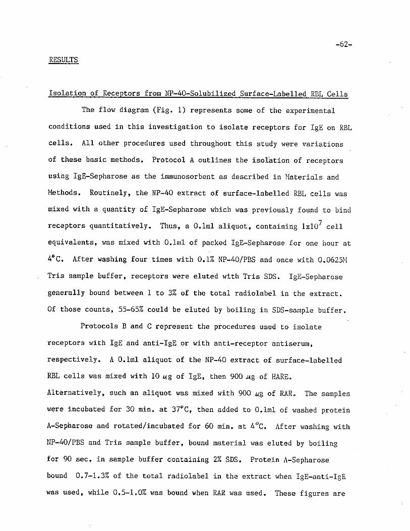
-62-
RESULTS
Isolation of Receptors from NP-40-Solubilized Surface-Labelled RBL Cel1s
The flow diagram (Fig. 1) represents some of the experiment.al
condit,ions used in this investigation t.o isolate receptors for IgE on RBL
cel1s. All other procedures used throughout this study were variations
of these basic methods. Protocol A outlines the isolätion of recept,ors
using lgE-Sepharose as the immunosorbent as described in I'iaterials and
Iulethods. Routinely, the NP-40 extract of surface-labelIed RBL cells was
mixed with a quantity of lgE-Sepharose which was previously founrl to bind
receptors quantitatively. Thus, a 0.lml aliquot, conlainíng 1x10⁷ cell
equivalents, rvas mixed wit,h 0.lml of packed ÏgE-Sepharose for one hour at
4oC. After washing four times with 0.12 NP-4O/PBS and once with 0.0625î'f
Tris sample buffer, receptors were eluted with Tris SDS. IgE-Sepharose
generally bound between 1 to 3Z of the t,otal radiolabel in the extract.
0f those counts, 55-657" could be eluted by boiling in SDS-sanple buffer.
Protocols B and C represent the procedures used to isolate
receptors rvith IgE and anti-IgE or with anti-recept.or antiserurn,
respect,ively. A 0.lml aliquot of the NP-40 extract of surface-labelled
RBL cells was mixed r¿ith 10 rag of IgE, then 900 ag of HARE.
Alternatively, such an aliquot r'¡as mixed v¡ith 900 ¿g of RAR" The samples
were incubated for 30 min. at 37èC, then added to 0"1n1 of r,¡ashed protein
A-Sepharose and rotated/incubated for 60 rnin. at 4oC. After washing r,rith
NP-40/PBS and Tris sample buf'fer, bound material r,¡as eluted by boiling
for 90 sec. in sample buffer containing 22 SDS. Protein A-Sepharose
bound 0.7-1.3"/" of the Lotal radiolabel in the extract when lgE-anti-IgE
was used, while 0.5-1.02 was bound when RAR was used. These figures are
125¡-RBL Ceurs
62a
Pnorocol t
ANr r -REcEProR ANT r se nuiq( äi\R )
PROTE I1ü -SrpnHnose
T
P ROTOCOL A
I c[-SrpHARosE
Fígure 1
1251-( I¡p-40 ) RtsL cELL EXTRAcT
Pnorocor- B
Io[I
HoRSE ANrr-le[ (HARI)
IPRorErN A-SepHnRosE A
E.LUT ION AND SDS-PAGE
Isolation of the IgE receptors from radiolabelled rat
basophilic leukemia (RBL) cel1s.
-63-
in agreement r¿ith those for lgE-Sepharose binding, if one assumes t,hat
IgE-anti-IgE and RAR bind different components, both of which bind ro
IgE-Sepharose (see below). Eluting with sample buffer-SDS yielded 60-707"
of bound counts when lgE-anti-IgE was used and 65-752 of bound counts
when RAR was used. Since batchwise elution rtras used, most of the
renaining count,s most lilcely represent mat,erial r¿hich remained trapped in
the matrix of the gel.
SDS-PAGE of Affinit y Chromatographv-Pur ified Surface Components fron
Solubilized RBL Cel1s
Calibration of Èhe 10% gels used in the Laemmli SDS-PAGE system
is shown in Fig. 2. Radiolabelled proteíns were run on tube gels and the
molecular weight was plotted versus the relative mobiliLy on a
senilogarit.hmic scale.
The distribution of radiolabelled maLerial eluted from
rgE-sepharose, and run on 10% gels is shown in Fig. 3. rt can be seen
that two major peaks were found having an l-1, of approximately 45kDa and
55kDa" These two nolecules rui1l henceforth be called R and H,
respecLively. In addition, a cpnsiderably smaller peak was foundr the i,f,
of r¡hich was estÍmated to be 7lkDa and rvhich will be called 71K.
' Figure 4 represenLs the naterial eluted from prot.ein A-sepharose
after treatment of radiolabelled cell extract r¡ith either IgE*HARE or
RAR. One major peak was observed when IgE and HARE v¡ere used to isolate
receptor (Fig. 4a). It,s Mr was est,imated to be 44K daltons. A smaller
peak near fract,ion 18, was found to have a mobility corresponding to an
I'1, of 72kDa. Both the 72 and, l+LkDa peaks have a broadness which isgenerally characteristíc of R and 7lK receptors (Conrad and Froese,
r978a, Conrad and Froese, 1976). rn addition, a third, sharper peak with
)3a
Figure 2: Calibration curve for the SDS-PAGE system according to
Laemmli (1970) using 102 polyacrylamide tube ge1s. Proteins were
labelled wittr 125I by the chloramine T method (McConahey and
Dixon, 1966). Proteins (5 Ug) were dialyzed against 0.0625M Tris
buffer, adjusted to 2% SDS and analyzed by SDS-PAGE. The
following proteins were used to calibrate the gels: E-chain of
rat monoclonal IgE (IR-162), 75kDa; catalase, 58kDa; Y-chain of
mouse monoclonal IgG (l'lOPC 2La), 53kDa; pepsin, 35kDa; light
chain of MOPC 21a, 22.5 kDa¡ chymotrypsin, 21.6 kDa.
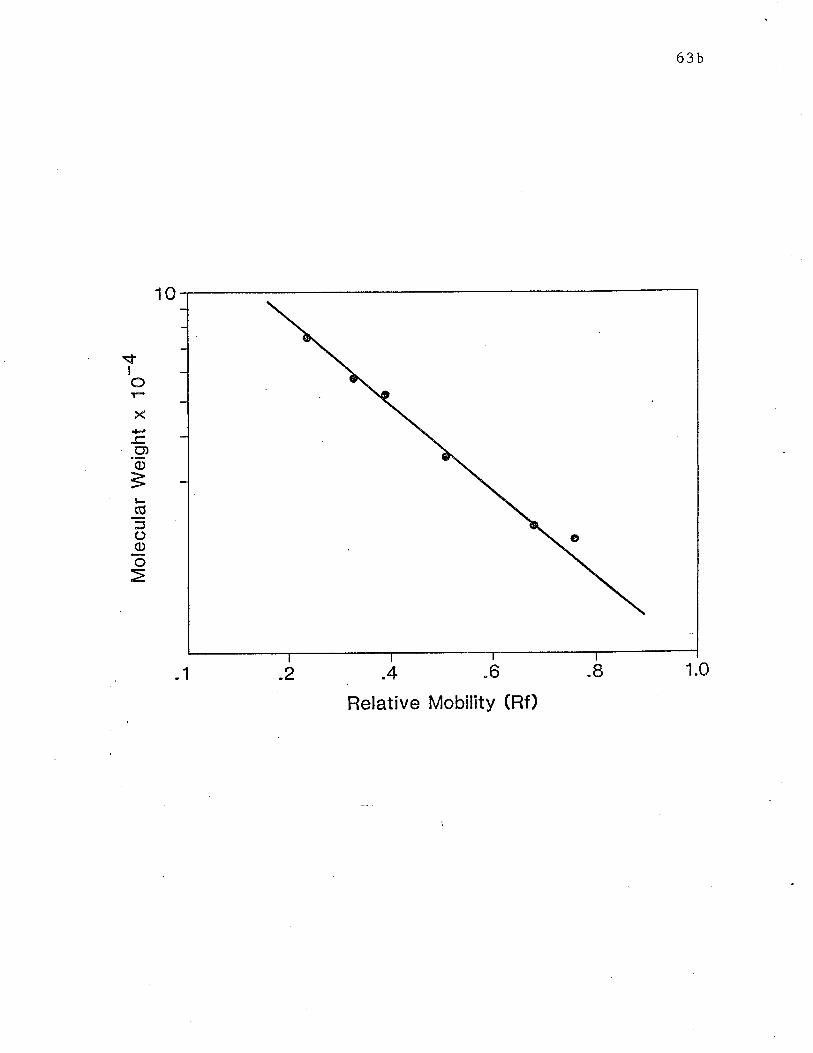
sfI
O
X
.9,o3(ú
ooõ
63b
1.01 .2 -4 "6
Relative Mobility (Rf)
I
10
63c
Fieure 3: SDS-PAGE analysis on 10% gels of surface labelled RBL
ce11 components bound by lgE-sepahrose. P.BL cell equivalents (1
1x 10') in the NP-40 exLract, were added to 0.1 ml of lgE-Sepharose
and r,¡ere allowed to mix for 60 min. Gels were r'¡ashed four times
wiLh 3-4 ml of 0.12 NP-4O/PBS and once r+ith 0.06251,{ Tris.
Receptor material rvas eluted with Tris sample buffer containing
2% SDS. The samples were heatecl in a boiling t¡ater bath and
eluates were analyzed by SDS-PAGE. TD marks the position of the
tracking dye.
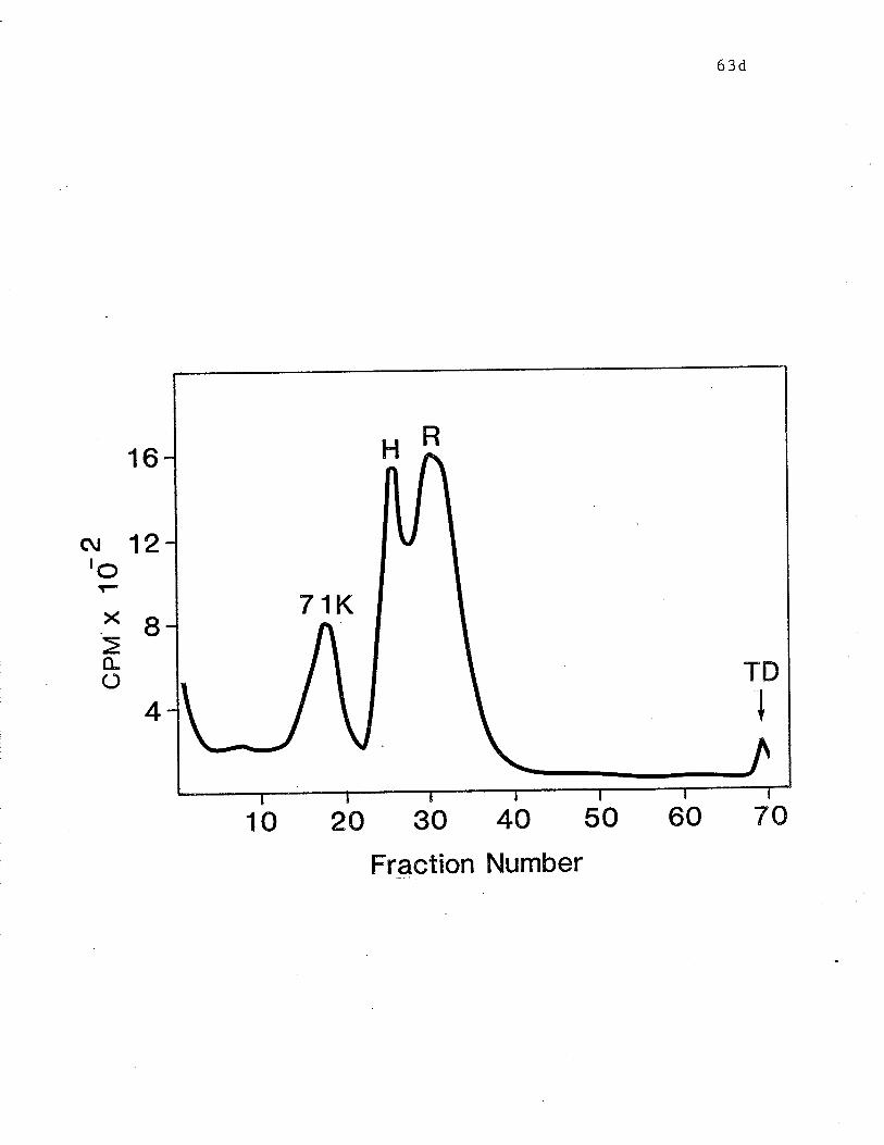
16
(\¡ 12ro
63d
60 70
Bx>o-(')
4
10 2A 30 4A 50
Fraction Number
-64*
a xûobility corresponding t,o an M. of 77kùa was observed" l"lolecules
isolated by means of RAR are also represent,ed by one major peak (Fig.
4b)' with an M,. of 53kDa. It is less broad than the band isolated rt¡ith
rgE and HARE" The minor peak, isolated in fraction 20, has an l,{,. of
67kDa and is sharper than the band usually attributed to 71K. rn
addiLion, a peak r,¡hich travels with Lhe tracking dye was observed in both
profiles and rnost likely represents unsaturated lipids which had been
labelled uy 1251 (zimmerman and chapman, Lg77). These results are in
agreemenL with those from previ.ous.studies usj.ng lgE-sepharose (Conrad
and Froese 1978a)' IgE and anti-IgE (Conrad and Froese, L978a, Conrad and
Froese, L976) and RitrR (conrad et al., L97g). They indicate that R and lI
recept,ors are the entities represented by the major peaks shorvn in Figs.
4a and' 4b, respectively, and that these Lwo molecules can be isolated
free of each other.
Comoarison of Papain-Generated Peptides From R and H Receptors
The radioactive material in the fractions consLituting the major
peaks in Figs. 4a and 4b and containing R and H receptors, respectively,
was collected from gel slices b.y elution int,o 0.125Ì'Í Tris over an 18 irour
period. The eluant was found to contain approximat.ely 757" of. the Lotal
radioactivity in the ge1 slices. After concenLratÍon and dialysis
against 0.1251'l Tris, samples rvere subject,ed to proteolysis by
2ME-activated papain (116 uZ/nL) -for 30 min" ar 37"C. Conrrol aliquoLs
were treat,ed with 2-ME alone. After adjusting the SDS concentration to
2% and boiling f.or 2 min., the sanples r^rere analyzed by SDS-PÂGE on 152
polyacrylamide gels. calibraLion of 15% gels is shown in Fig. 5 as a
plot of the relative mobility of radiolabelled standards versus molecular
weight. Analysis of peptides generated by proteolysis with papain and
64a
Figure 4: SDS-PAGE analysis on 102 gels of surface labelled lltsL
ceIl molecules isolated by rgE and HARE and RAR. a) 1 x 10⁷ cellequivalents in an NP-40 ext,ract r,rere mixed rvith 10ug of rgE and
900 ug of horse anti-rat IeE (HARE) for 60 min. at 37ê C. b) I x-t
10' cell equivalents were míxed r'¡ith 900 ¡g of rabbit
anti-receptor (RAR) antiserum for 60 min. at 37oc. samples from
(a) and (b) rvere then added t,o 0.1 ml of protein A-sepharose and
allowed to mix for 60 min. at 4oc. Gels were washed and receptor
material was elut,ed by boiling in Tris sample buffer containina,
2U SDS. Eluat,es were analyzed by SDS-PAGE.
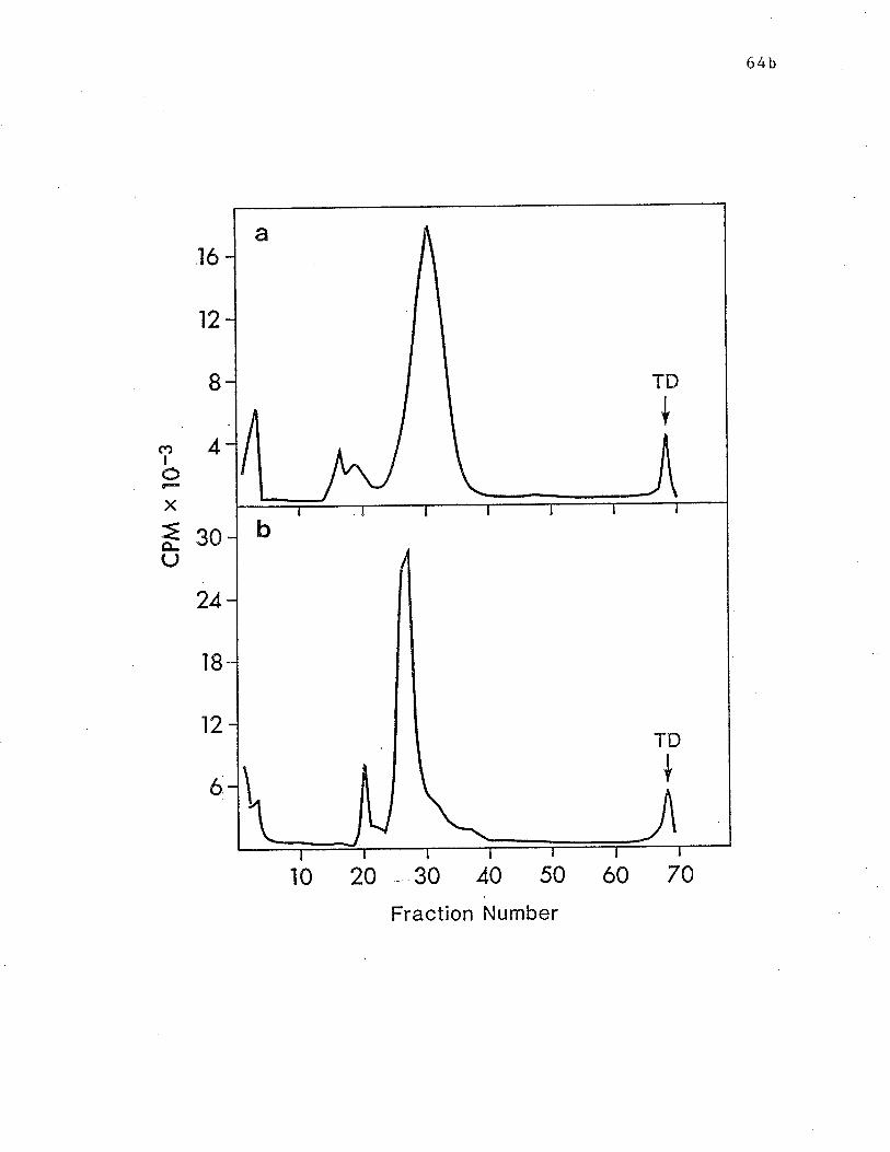
64b
tó
12
I
4(ÐIIX
=ê-U
30
24
18
12
6
t0 20 30 40 50 ó0 70
Fraction Number
TD
a
{
TD
I¡ I¡
b
+
-6s-
that of 2-l'iE-treated controls is depicted in Fig 6. The single pealcs
seen in the gel profiles of control solutions indicate that
2-mercaptoethanol alone did not reduce or degrade either R or H Lo any
extent,. The mobilit,ies of these peaks correspond to molecular weights of
45kDa and 55kDa' respectively. The gel profile of digested R receptor is
shown in Fig. 6a. It indicates that a group of peptides, t,he majority of
which have molecular rueights ranging from 23K to 15K daltons, r{ere
generated by the proLeolysis of R. No undigested material remained, as
seen by the lack of any radioactivity in the region where undigest.ed
receptor would normally be found. The digestion of H receptor (Fig. 6b)
apparently generated fewer fragments than that of R. In addition, all of
Lhese peptides had apparent molecular r+eights of less than 15K dalt.ons.
Indeed, most of the radioact,ivit.y resides in one sharp peak the mobility
of r¿hich corresponds to slighly less than 12K daltons. In this case, a
small amounl of radioactive material appeared t,o remain undigested.
65a
Figure 5: Calibration curve for the SDS-P AGE system using 15%
tube gels. Prot,eins were labelled rvith 125r by the chloramine T
method. Proteins ürere dialyzed against 0.0625Ì{ Tris, arljusted to
27" sDS and analyzed by SDS-PAGE. The following proteins were
used t,o calibrate the gel: catalase, 5BkDa; pepsin, 35kDa;
chymotrypsin, 21.6kDa; rnyoglobin, L7.4kDa; cytochrome c, IZ.3kDa.
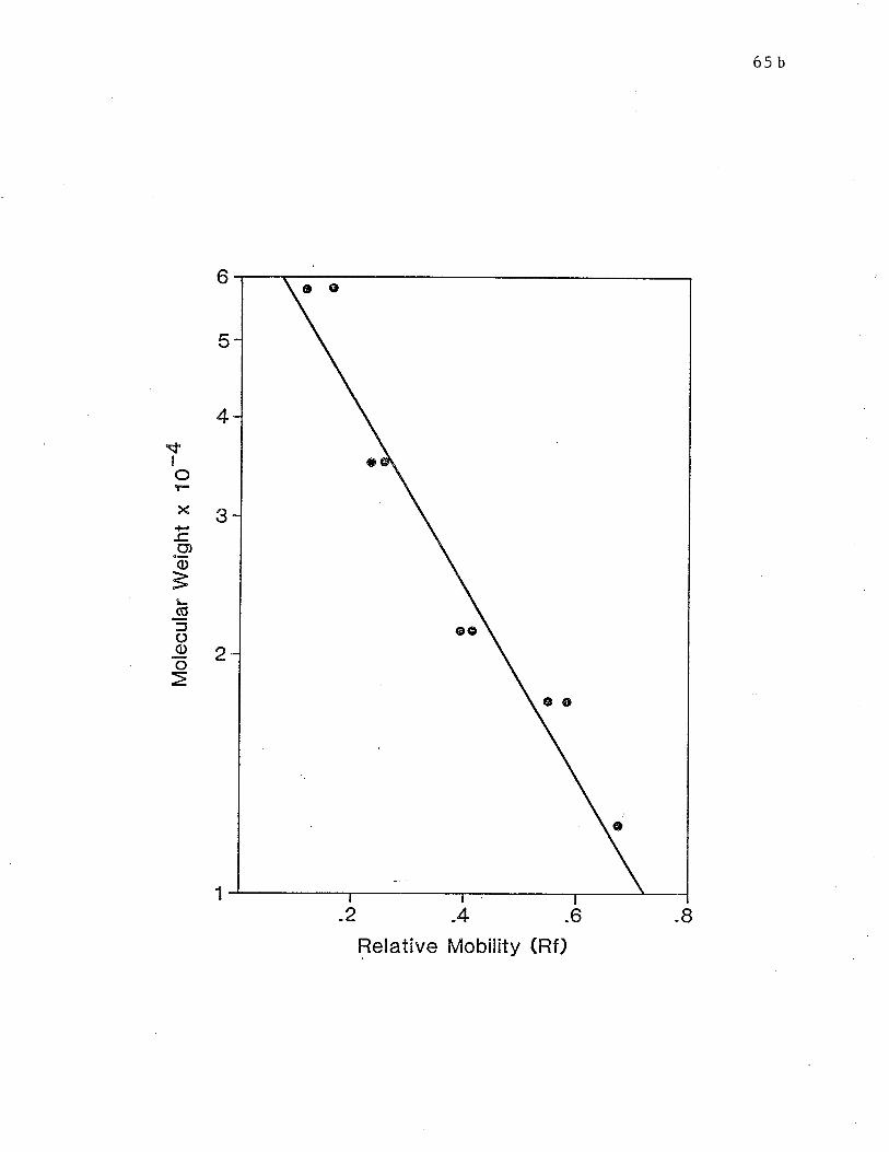
6s b
6
5
4\f
IoX
-c"9o
6ooo
3
2
1
.2 .4 .6
Relative Mobility (Rf)I
øo
e
oo
65c
Figure 6: SDS-PAGE analysis on 152 gels of papain generated
peptides of R and H receptors. llaterial in fractions
constituting the major peaks in Figs. 4a and 4b was collecLed by
eluting from gel slices. Sarnples \./ere treated rvith
2-ME-activated papain (final concentraLion: 116 ue/nI) (-4 or
2-ùlE alone (----) for 30 min. at 37oC. The SDS concentration rvas
adjusted to 27", samples were boiled and analyzed by SDS-PAGE.
Digests of material isolated from gel represented in (a) Fig. 4a,
containing R receptor and (b) Fig. 4b, containing H receptor.
Fiolecular r'reight markers in descending order from left to right
are: catalase; pepsin; myoglobin; and cyt.ochrome c.
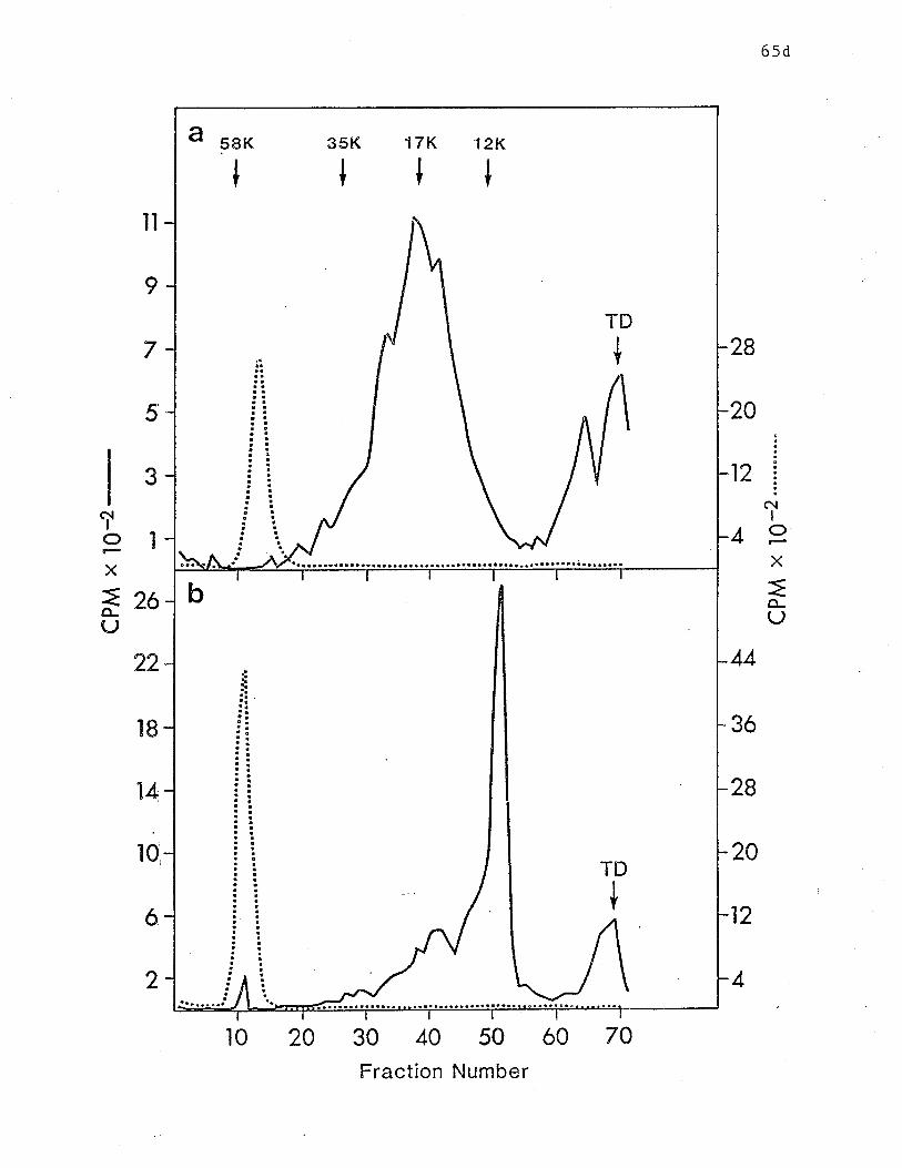
6sd
28
20
12
il
I
7
5
3
22
'lB
14
10
6,
2
I
Tô¡
IIX
=o-U
4olX
=26(J
44
3ó
28
20
12
4
30 40 50 ó0
Fraction Number
58K
TD
a
II I I
{
35K 17K 12K
b
TD
+
7020l0
-66-
DISCUSSION
The purpose of the studies undertaken in this Chapter was
two-fold. It was first necessary Eo confirn results from previous
studies (Conrad and Froese, 1978a, Conrad and Froese 1976, Conrad et a1.,
L979) in which the receptors for rgE on RBL cel1s are isolated by
different means" Secondly, the different types of receptors, riesignated
R and H, were compared through their papain-generat,ed fragments to
determine wheÈher these two molecules are structurally similar.
In agreement with the finding by Conrad and Froese (1978a),
isolation of receptors wíth the aid of lgE-Sepharose yielded tvro
molecules having apparent l"l, of 45kDa and 55kDa, designated R and H. In
addition, a Lhird surface molecule was routinely presenL and it, had
aoparent M of approximat,ely 72kDa. This molecule rvas also occasionallyrisolated by Conrad and Froese (1978a) though to a lesser extent. Ì'fore
recent,ly, Helm and Froese (1981a) regularly isolated small amounts of
this molecule and named ít 71K. The signifj-cance of this molecule and
its relationship to other receptors is the subject of subsequent
chapters.
These experíments also confirmed the finding of Conrad and Froese
(L976) by isolaLing R receptor, but not H, r*'hen using IgE and anti-IgE.
Again, 71K co-purified with R. The reason for the differences in types
of receptors Ísolated by different procedures is not, knoln. Kepron et
a1. (1982) showed that the affinity of IgE for R is higher than rhat for
H. They suggested that the amount, of IgE used in the lgE-anti-IgE
procedure is not in high enough excess t,o react with significant amounts
of H. Another possibility is that the H-IgE complexes formed are
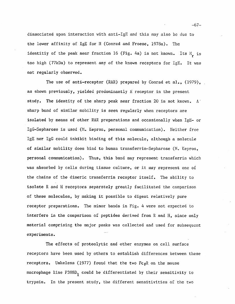
-67-
dissociated upon interaction with ant.i-IgE and this rnay also be due to
Ëhe lower affinity of IgE for H (Conrad and Froese, I978a)" The
identitiy of the peak near fracLion 16 (Fig" 4a) is not knor'¡n. Its Þ1, is
t,oo high (77kDa) to represent any of the lcnown receptors for IgE. It was
not regularly observed.
The use of anti-receptor (RAR) prepared by Conrad et a1., (1979),
as shown previously, yielded predorninantly H receptor in the present
study. The identit,y of t,he sharp peak near fraction 20 is not known. A'
sharp band of similar mobility is seen regularly r,¡hen receptors are
isolated by means of other RAR preparations and occasionally r'rhen IgE- or
IgG-Sepharose is used (l'{. Kepron, personal communication). l.ieither free
IgE nor IgG could inhibit binding of this molecule, although a molecule
of similar mobility does bind to human t,ransferrin-Sepharose (M. Kepron,
personal communication). Thus, this band may represent transferrin r,¡hich
was absorbed by ce1ls during tissue culture, or iL may represent, one of
the chains of the dimeric transferrin receptor it,se1f. The ability to
isolate R and H receptors separat,ely greatly facilitat.ed the connparison
of these molecules, by making it possible to digest relatively pure
receptor preparaLions. The minor bands in FÍg. 4 were not expected to
interfere in the comparison of peptides derived from R and H, since only
materj-al comprising the major peaks was collected and used for subsequent
experimenLs.
The effects of proteolytic and other enzymes on cel1 surface
receptors have been used by others to establish differences betv¡een these
receptors. Unkeless (1977) found that the two Fc6R on the mouse
macrophage line P3B8D1 could be differentiaLed by Lheir sensitivity to
trypsin. In the present, study, the different sensitivities of the two
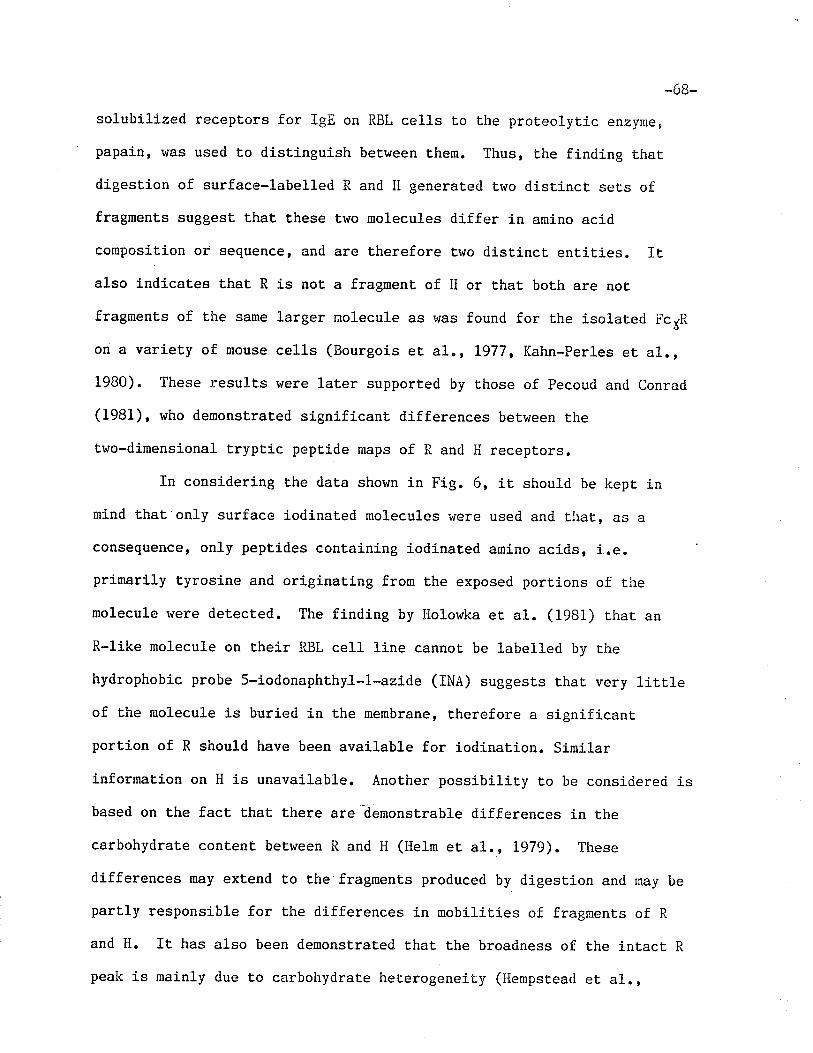
-68-
solubilized receptors for IgE on RBL cel1s to the proteolytic enzl'me,
papain, was used to distinguish between them. Thus, the finding that
digest,ion of surface-labelled R and H generated two distinct sets of
fragments suggest that, these two molecules differ in amino acid
composition or sequence, and are therefore two distinct entities. Italso indicates Lhat R is not a fragment of ll or that both are not
fragments of the same larger molecule as was found for the isolated FcyR
on a variety of nouse cel1s (Bourgois eL a1., Ig77, Kahn-perles et al.,1980). These results rvere later supported by those of Pecoud and Conrad
(1981), who demonst,rated significant differences between the
two-dimensional tryplic peptide maps of R and H receptors.
rn considering the data shorvn in Fig. 6, it should be kept in
mind that only surface iodinated molecules were used and that, as a
consequence, only peptides containing iodinated amino acids, i.eprimarily tyrosine and originating from the exposed portions of the
molecule were detected. The finding by Holorvka et al. (l98l) that an
R-like molecule on their RBL cel1 line cannot be labelled by r.he
hydrophobic probe 5-íodonaphthyl-1-azide (INA) suggesLs rhar. very litrleof the molecule is buried in the membrane, therefore a significant
portíon of R should have been available for iodination. sÍmilar
informat.ion on H is unavai.lab1e. Another possibility to be considered j-s
based on the fact, that there are denonstrable differences in the
carbohydrate contenÈ between R and H (l{e1m et al., IgTg). These
differences may extend to the fragments produced by digest,ion and may be
partly responsible for the differences in mobilities of fragment.s of R
and H. It has also been demonstrated that the broadness of the intact R
peak is mainly due to carbohydrate heterogeneity (Hempstead et al.,
-69-
1981b) " This het,erogeneity may affect the susceptibility of certain
sites in some receptor populations, leading to the demonstrable
heterogeneity in R fragments" The differences found between these trt¡o
molecules does suggest that they are distinct entities and this will be
further explored in subsequent. chapters.
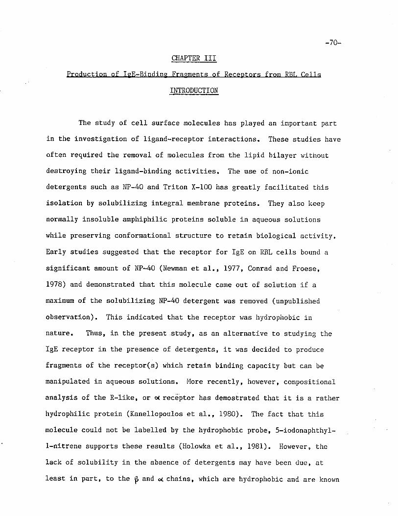
-70-
CHAPTER III
ProducLion of lgE-Binding Fragments of Receptors from RRT. Cel 1 s
INTRODUCTION
The study of cell surface molecules has played an important part
in the investigation of ligand-receptor interactions. These studies have
often required the removal of molecules from the lipid bilayer r,¡ithout
destroying their ligand-binding act,ivit,ies. The use of non-ionic
detergents such as NP-40 and Triton X-100 has greatly facilitated this
isolation by solubilizing integral menbrane proteins. They also lceep
normally insoluble amphiphilic proteins soluble in aqueous solutlons
while preserving conformational structure to retain biological activit.y"
Early studies suggested that the receptor for IgE on RBL cells bounci a
significant, amount of NP-40 (Neruman et a1., L977, Conrad and Froese,
1978) and demonstrat.ed that this nolecule came out of soluuion if a
maximum of the solubilizing NP-40 detergent was removed (unpublished
observation). Thj-s indicated that the receptor was hyclrophobic in
nature. Thus, in the present,study, as an alternative Lo studying the
IgE receptor in the presence of detergents, it was decided to produce
fragments of the receptor(s) which retain binding capacit,y but can be
manipulated in aqueous solutions. More recently, however, compositional
analysis of the R-like, or o( recéþtor has demostrated that it is a rather
hydrophilic protein (Kanellopoulos et, aI., 1980). The fact that this
molecule could not be labelled by the hydrophobic probe, 5-iodonaphthyl-
l-nitrene supports these results (Holowlca et al., 1981). However, the
lack of solubility in Lhe absence of detergents may have been due, aL
least in part, to the p and o¿ chains, which are hydrophobic and are known
-7r-to be associated rvith R (ø(). Indeedo Kumar and i,leLzger (i982) were able
to analyze the receptor in a 6Þl guanidine HCl solution in the absence of
any deLergent.
The availability of binding fragrnents of the IgE receptor(s)
nonetheless provides the opportunity to study an acti-ve receptor molecule
with a lorser structural complexity than that of intact receptor. This
would facilitate the elucidation of the structure (both primary and
tertiary) of the receptor binding site. In another systen, t,his concept
prompted Bartfeld and Fuchs (1979) to produce an active portion of the
acetylcholine receptor by digestion with trypsin. They demonstrated that
Ehe 27kDa fragment of intact receptor (40kDa) could bind specific ligand
as well as induce an autoimmune myasthenia gravis in rabbits when it r¿as
injected vrith complete Freundrs adjuvant. This indicates that the
fragment contained both Lhe binding site and the mediator of a
Pnys].ologLcal. response.
This chapter describes the search for binding fragments of the
IgE receptor(s) on RBL cells. In order to isolat.e an ïgE-binding
molecule, t,he digest,ion had to.take place before the receptor was exposed
to any denaturing reagents. Thus, an approach dj-fferent from the one
taken in Chapter II was needed. Therefore, in this study, receplors were
digested before isolation with affinity gels. In addition, several
different proteases were used in -an attempt to generaLe a variety of
different fragments. These attempts have provided. further information on
the st,ructure of the IgE receptors.
-72-
I'TATBRIALS AND I'IETHODS
The buffers, preparation of IgE and lgE-Sepharose, RBL ce1l
iodination and disrupt,ion have already been described in Chapter II,l,faterials and i,lethods.
Disestion of ED tors in the Presence of NP-40 or SDS
Extracts of RBL cells which were surface labe11ed with either125r or 131r
""r" mixed !¡ith rgE-sepharose for one hour at 4oc. The gel
Lo which th" l25l-exLract had been added,was Lhen Lransferred to a 1.0 ml
Pasteur pipette column and was r'¡ashed with 15-20 rnl of 0.lZ NP-4O/PBS.
Receptor mat,erial was elut,ed wiLh 3.0 Ì,1 KSCN containing 0.12 NP-40 and
Lhe eluate dialyzed against 0.1251'{ Tris, pH 6.8, containing o.I"/. Np-40.
The gel t.o which l3ll-tu.uptor material had been added was r,¡ashed 4 times
with 0.12 NP-4O/PBS and once with 0.1251{ Tris. Bound receplors r.ras
eluted ,by
boiling in sample buf f er containing 2% SDS. Both eluates r.¡ere
treated trith 116 ug/nL mercuripapain and in the presence of 1. 2xLO-Lr¡ Z-
ME for 30 min. at 37oc. control aliquots were adjust.ed to 1.2x10⁻¹!i 2-
ME. SDS was added to KSCN eluates t,o a final concentration of. 27"" All
sanples were boiled for 2 min. .and then applied Eo 15I^ polyacrylamide
gels for SDS-PAGB analysis. Both digests were applied to one ge1, as
were both controls.
Dises tion of RBL Cell Extract to Produce an ï pE-Bindins Fraqment of fqE
ReceotorS
IìBL ce11s, surface-labelled "itt 125I, ü¡ere solubilized with 0.12
NP-4O/PBS. An aliquot of the extract containj-ng 6x10⁷ ce1l equivalents
was digested with 1.16mg/m1 papain in the presence of 1.2xt0-1li 2 lin
while onLy 2 ME was added to a control aliquot containing 1xl07 ce1l
equivalents. Samples were incubated for 90 min. at 37oC and. then Lreated
-73_
vrith 1.5x10⁻¹¹'{ iodoacetamide (Serva, Heidelberg, I,J. Germany) for 45 min
aE 4oC. Receptors from t,he control aliquot were isolaLed wit,h 0.2 ml
IgE-Sepharose, while their fragments were isolated wiLh a variety of
reagents (as described in the Methods and l,laterials section of Chapter
II), including 0,2 ml lgE-Sepharose; IeE (10ue), HARE (900¡¿e) and 0.2
ml protein A-Sepharose; and RAR (900 ¡.tg) and 0.2 ml protein A-Sepharose.
The controls of these various met.hods of isolation r,¡erer 0.2 ml activated
and quenched Sepharose; IIARE and protein A-Sepharose; and normal rabbit
gamna globulin (nRGG) (900¡g) and protein A-Sepharose, respectively.
þlaterial eluted from these gels was analyzed on 10% polyacrylamide gels.
To det.ermine the origin of papain-derived fragments, fractions of
the PAGE gel consLituting Lhe peaks were cut out and the receptor
material conLained in these fractions vrere extensively digested rvith
trypsin. The resulting pept.ides were analyzed by a Lwo dimensional
procedure. The deLails of these methods will be described in Chapt.er IV.
A tryptic rnap of intact, H recept,or lras prepared in a similar fashion,
using RAR and protein A-Sepharose to isolate the receptor from a
surface-labelled RBL extract.
In a separate experiment, aliquots of surface-labelled RBL ce1ls
containing 3.6x10⁷ cell equivalents were treat,ecl with eit,her trypsin
(Schwartz/Mann, Orangeburg, NY) or chymotrypsin (Sigma Scientific Co. St"
Louis, M0) at a final concentratfon of 5mg/m1 of the enzyme in PBS or
vrith PBS alone. After a 2 hour j.ncubation at, 37oC, phenyl methyl
sulfonyl fluoride (PMSF) (Sigina Scient.ific Co. SL. Louis, þlO) was added
to all samples to a fÍnal concentrat,ion of Bx10-31'l to stop digestion.
Receptors and their fragments r¡rere once again isolated r.¡ith
IgE-Sepharose, IgE, HARE and protein A-sepharose or RAR and protein
-74-
A-Sepharose. SDS-sample buffer eluates from affinity gels ruere Lhen
analyzed by SDS-PAGE on 102 gels.

-75-
RESULTS.
Disestion of R and II Recept,ors in the Pres NP-40 and SDS
The experiments described in Chapter II had demonst,rated that R
and H recePtors are susceptible t,o digestion by papain in Lhe presence of
the anj.onic detergent, SDS. Hor,rever, if an attempL to prepare an
IgE-binding fragment of t,hese nolecules was Lo be made, the digestion
would have to take place before receptors came into conLact v¡ith a
denaturing reagent. Thus, it seemed appropriate to at.tempt t,he digestion
while receptors r'rere in the NP-40 extract,, before their isolation by
affinity chromatography. The following experiment was performed to
determine rvhether digestion of receptor molecules was possible in the
presence of I'lP-40, and to compare the extent of this digestion to that in
the presence of SDS.
Two aliquots of 3x10⁷ ce1ls were surface iodinated, one *ith 125I
the other witt 131I. After NP-40 solubilization, each cell ext,ract. rvas
added to 0.3m1 of lgE-Sepharose and incubated for t hour ar 4oC" 0f the125I tub"lled receptor material 1.32 was bound by the affinity gel and
757" of. the bound material could be eluted with 3M KSCN/PBS containing
0.12 NP-40. Tt" 131l-1abe11ed receptor material bound to an extent, of L%
and 552 of the bound material could be eluted with Tris/SDS. Following
dialysis of the KSCN eluate against 0.125M Tris/O.1% NP-40, boLh eluaLes
were subjected Lo digestion by papain under conditions identical to Lhose
described in Chapter II" Again, control aliquots were Lreat,ed with 2-þ18
only. Samples in NP-40 were adjusted to 2Z SDS. Digest,s of both eluat,es
were applied to one 15% polyacrylamide gel and controls were applied to
another. SDS-PAGE analysis vlas then performed. Fig. 7 shorr¡s undigested
-76-
eluaLes, and demonstrates that both R and H receptors are eluterl r,rith
KSCN/NP-4O as well as lrith SDS. There also appears to be sone
contaminating labelled non-receptor material in the I(SCN eluate. This is
most likely due to trapping of non-specific material which vras not rvashed
away in the pipette column. It is not present in t,he SDS eluate. The
patt,erns of dÍgested material appear in Fig. B and clearly show t,hat
papain retains its abilit,y to digest, both R and H receptors in the
presence of NP-40 as well as in SDS. In either case, no undigested
material remained, as demonstrated by the lack of significant
radioactivity in fractions corresponding t.o molecular weights of 45kDa
and 55kDa. lnlhen cornparing the digests of the two preparations, it is
apparenL t.hat somewhat larger fragments \rere generated by digestion in
NP-40 than by that in SDS. This is possibly due to the fact that Ì'lP-40
is a non-ionic detergent and nay noL unfold Lhe proteins Lo as great an
extent, as the ionic detergent SDS, thus making fewer sites available Lo
proteolysis and leading to production of fewer fragment,s.
Isolation of leE-Bindins Frasment,s of Rece ptors
Once it was established that papain retains its proteolytic
activity in the presence of NP-40, Lhe following experiments rvere
performed in an attempt to isolate binding peptides of R and Il receptors.
Surface iodinated RBL cells (zxro7) were solubilized in 1 ml of 0.12
NP-40/PBS. An aliquot containing 6x10⁷ cell equivalent,s ruas i-ncubated
with 1.L6 ng/nL papain and 2-T,fE, while t.he control aliquot, containing1
1x10'cell equivalents, was t.reated v¡ith 2-l.fE on1y. Both digest and
control were then incubated with iodoacetamide to stop Lhe reaction. The
control aliquot was incubated with lgE-Sepharose, as rüas an aliquot of
the digest,ed ext,ract,. The remainder of Lhe digest rr¡as incubat,ed nith one
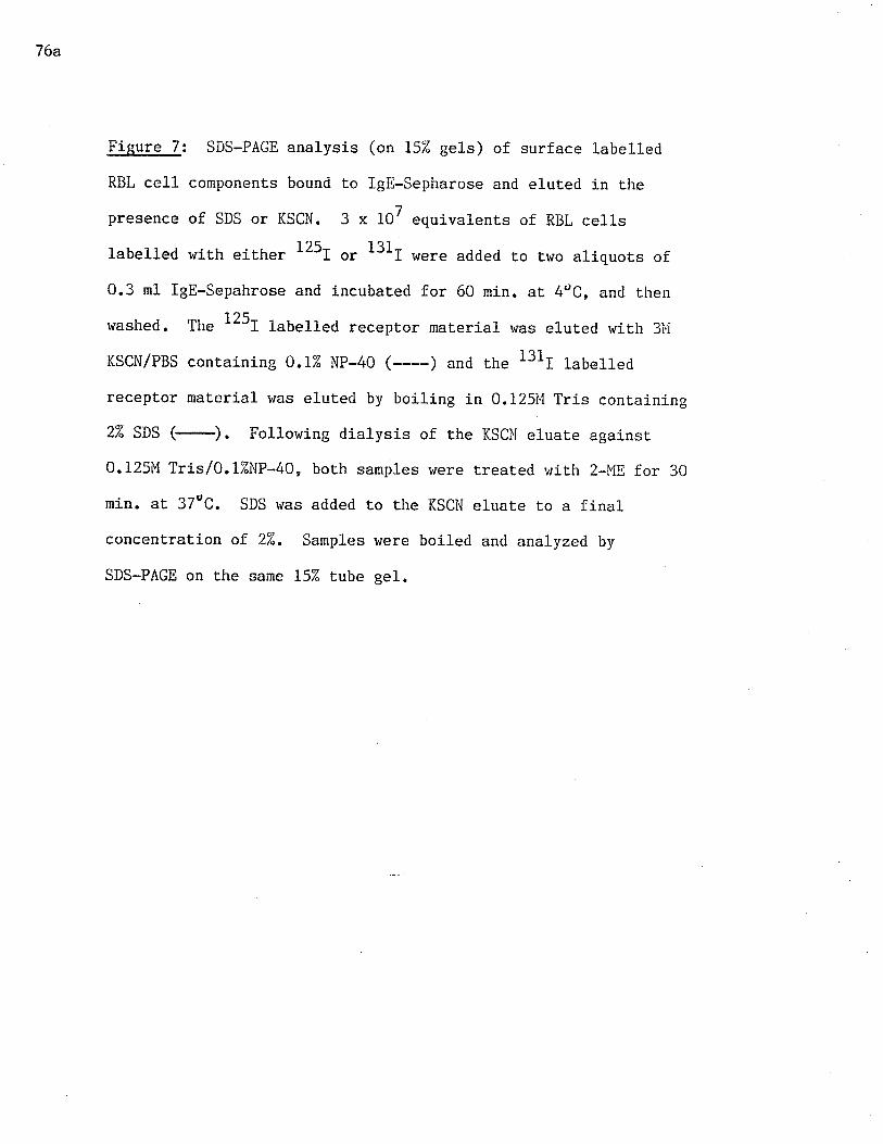
76a
Figure 7: SDS-PAGE analysis (on 15% gels) of surface labelled
RBL cell cornponents bound Lo lgE-Sepharose and eluted in t.he
presence of SDS or KSCN. 3 x 10⁷ equivalents of RBL cells
labelled r,¡ith eiLher 125r or 131r r+ere added to truo aliquots of
0.3 ml lgE-Sepahrose and incubated for 60 min. at 4dC, and then
washed. The 1251 lubu11ed receptor materj-ar was eluted with 3Ì'i
KSCN/PBS conraining O.I% NP-40 (----) and rhe 1311 lub"u",l
receptor material was eluted by boiling in 0.125M Tris containing
2% SDS (-). Following dialysis of the KSCII eluare againsr
0.125M Tris/0.1ZllP-40, both samples were t.reated with 2-ME for 30
min. at. 37eC. SDS ruas atlded to the KSCN eluaLe Lo a finalconcentratj.on of 2%. Samples ruere boiled and analyzecl by
SDS-PAGE on the same 152 tube gel.
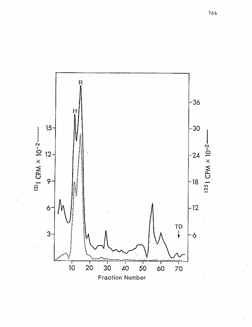
76b
TD
{
R
H
30
Ic\t
I
249X
=ê-TJ
18fÕc\t
I
ô{
X
=o-(J
c/)
3ó
12
15
OiôFl¿
6
63
30 40 50Fraction Number
ó0 70t0 20
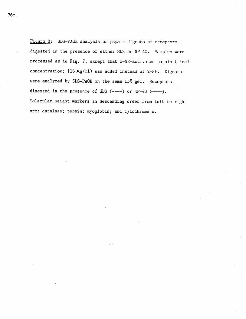
76c
Figure 8: SDS-PAGE analysis of papain digests of receptors
digest,erl in the presence of either SDS or I,lP-40. Sanples were
processed as in Fig. 7, except. that 2-ùlE-act.ivated papain (final
concentrat.ion: 116.ÀAg/ml) was added instead of 2-Ì"i8. Digests
were analyzed by SDS-PAGE on Lhe same 152 gel. Receptors
digested in the presence of SDS (----) or lrlP-4Q (-).
[lolecular weight markers in descending order from left to right
are: catalase; pepsin; myoglobin; and cytochrone c.
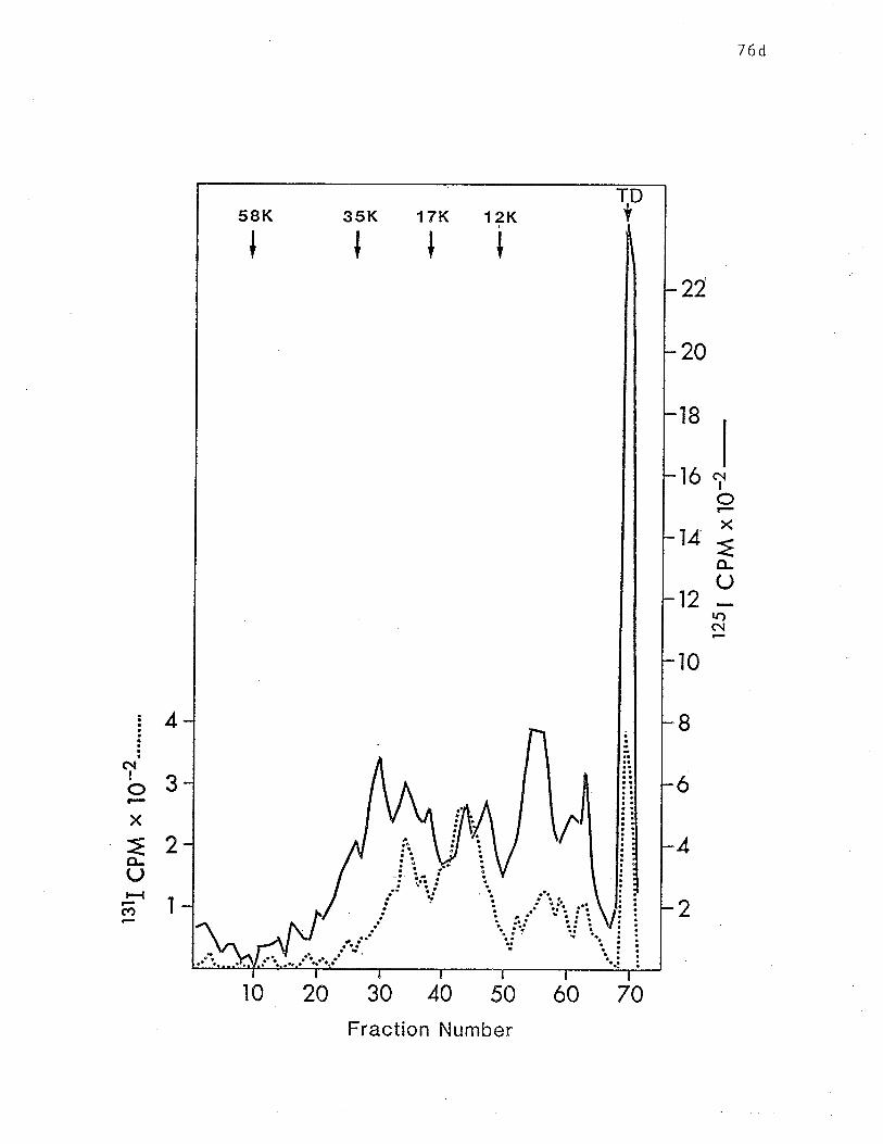
76d
Iô{
IIxÉo-U
,¿.)-(\
22
20
18
ló
14
12
l0
I
6
4
2
4
3
2
1
ô{IoX
ão-(J
c)
30 40 50
Fraction Number
TDf1?K
I
35K 17Rtt58K
70ó0r0 20
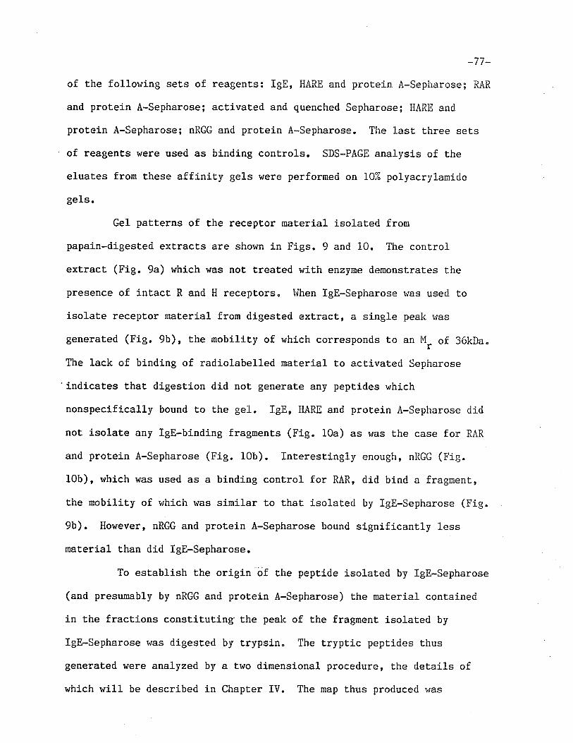
_77 _
of the following sets of reagents: IgE, HARE and protein A-Sepharose; RAR
and protein A-Sepharose; activated and quenched Sepharose; HARE and
protein A-Sepharose; nRGG and protein A-Sepharose. The lasl t,hree sets
of reagents were used as binding controls. SDS-PAGE analysis of the
eluates from these affinity gels were performed on 102 polyacrylamide
gels.
Gel patterns of the receptor maLerial isolaLed from
papain-digested extracts are shor'¡n in Figs. 9 and 10. The control
extract (Fig. 9a) which was not treaLed with enzyme dernonsLrates the
presence of inLact R and H recepLors. I'lhen lgE-Sepharose r'ras used to
isolaLe receptor material from digested extract, a single peak ruas
generated (Fig. 9b), t,he mobiliÈy of which corresponds to an M, of 36kDa.
The lack of binding of radiolabelled material t,o activated Sepharose
"indicates that digestion did not generate any peptides which
nonspecifically bound to the gel. IgE, HARE and protein A-Sepharose did
not isolate any lgE-binding fragments (Fig" 10a) as was the case for RAR
and proLein A-Sepharose (Fie. 10b). Int.erestingly enough, nRGG (Fig.
10b), rvhich was used as a binding control for RAR, did bind a fragrnent,
the mobility of r.¡hich rvas símilar to that isolat.ed by lgE-Sepharose (Fig.
9b). However, nRGG and protein A-sepharose bound signifÍcantly less
material than did lgE-Sepharose.
To esLablish the origin -ijf tfre peptide isolated by IgE-Sepharose
(and presumably by nRGG and protein A-Sepharose) E,he material conLained
in the fractions constituti.ng'the peak of the fragment isolated by
IgE-Sepharose h¡as digested by trypsin. The t.ryptic peptides thus
generated were analyzed by a two dimensional procedure, the details of
which will be described in Chapter IV. The map thus produced v¡as
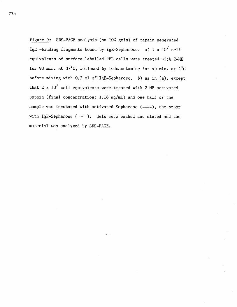
77a
Figure 9: SDS-PÂGE analysis (on 10% gels) of papain generat.ed
IgE -binding fragments bound by lgE-sepharose. a) 1 x 10⁷ ce1l
equivalents of surface 1abelled RIIL cells rüere treated with 2-i,iE
for 90 min. at 37oC, follor.¡ed by iodoaceLamide for /+5 min. at 4oC
before mixing with 0,2 mL of lgE-Sepharose. b) as in (a), except
that 2 x 10⁷ cell equivalenLs rüere t,reated with 2-I1E-activatecl
papain (final concentrat.ion: 1.16 mg/ml) and one half of the
sample was incubated with activated Sepharose (----), the other
with lgE-Sepharose . Gels were washed and eluted antl the
material was analyzed by SDS-PAGE.
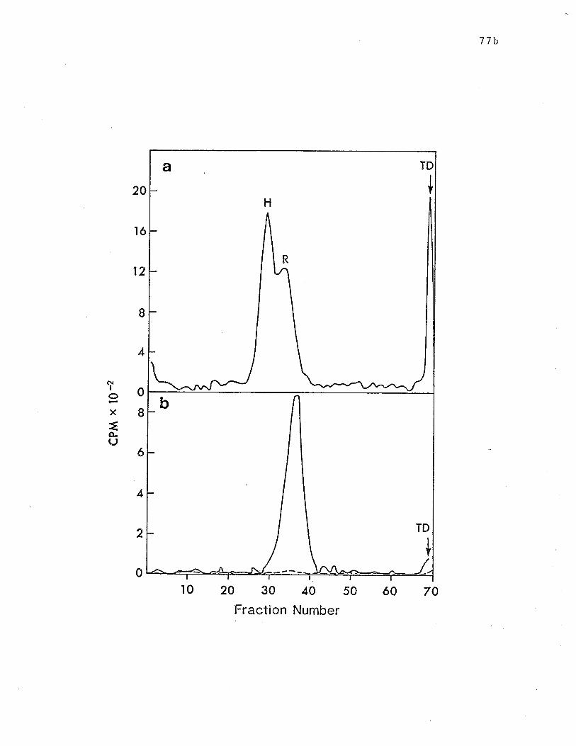
77b
20
ló
12
ô{IoX
=,CLrJ
I
4
0
I
é
4
2
0
r0 20 30 40 50
Fraction Number
ó0 70
a
H
R
TD
I
TD
b
t
77c
Fisure 10: SDS-PAGE analysis of papain generaLed lgll-binding
fragment.s bound by IgE and HARE and RAR. 4 x 10⁷ cell
equivalents of surface labelled RBL cells !üere treated with
2-l'{E-activated papain (final concenlraLion: 1.16 mg/ml) for 90
min. at 37oC followed by iodoacetamide for 45 min. at 4 C. a)
Aliquots of the digesl were incubated wit.h IgE and FIAP.E (-)
or wit.h HAI{E alone (----) and then mixed wíth protein
A-Sepharose. b) Aliquots of the digest, rvere incubat.ed with llAR
æ or nRGG (----) and then mixed ruith protein A-Sepharose.
Gels were washed and eluat,es hrere analyzed by SDS-PAGE.
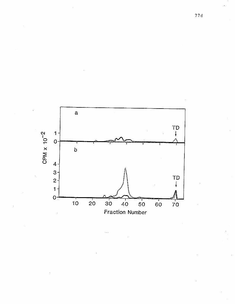
77d
a
TD+
TD
b
I
ñl 1IPox
o-o43
2
1
010 20 30 40 50
Fraction Number
60 70
-78-
compared t,o a map of peptities generated from iI receptor as isolated by
RAR and proteín A-Sepharose, as described in Chapter II. As can be seen
fron Fig. 11, the peptides from the papain-generated fragment. isolated by
IgE-Sepharose and those from intact H produce similar if not identical
maps. This provides evidence that the single lgE-binding fragment
produced by papain digestion is a fragnent of H receptor rat,her than R,
the peptide map of which is quit,e different (as will be shown in Chapter
rv).
Trypsin and chymotrypsin rn'ere also used in an att,empt to produce
binding fragments. The NP-40 extract of 1x10⁸ surface iodinated RBL
cells was incubated with either 5mg/m1 trypsin, 5mg/m1 chymotrypsin or
PBS at 37ÚC for 2 hours. All aliquots rvere then incubated h¡ith PI,{SF at a
concentration of 8x10⁻³lt for 45 mÍn. at 4oC" to terminate the reactions"
Each aliquot was then t,reated wilh lgE-Sepharose, IgE-iìARE-protein
A-Sepharose or RAR-proLein A-Sepharose" Fig. 12 shows the results of
limited tryptic digestion of the receptors. Isolation of receptors from
the control aliquot with lgE-sepharose yielded both R and H in inLact
forms (Fie. 12a) rthile isolation wj-th RAR and IgE-llARE isolated H and R
separately (Figs. 12b and 12c, respectively). Treatmen¿ of the
trypsin-digested extract using lgE-Sepharose yielded rvhaL appears Lo be
one fragment,, the mobility of v¡hich corresponded to an M, of 41kDa (Fig.
LZa). Treatnent rr¡Íth R¡\R isolated a fragment with similar mobility,
although the peak contained considerably less radioact.iviLy than that
obtained from an undigest.ed control exlracl (Fig. 12b). Treatment of the
digested ce1l exLract ruith IgE and HARE yielded a molecule which, in
terms of mobility, did not differ very much from inLact R isolaled from a
control extract though again the amount of radiolabel isolated from the
78a
Figure 11: Two dimensional tryptic peptide map of
papain-generated ïgE-binding fragment compared t,o peptide map of
H. The IgE binding fragment was fixed in fractions constiLuting
the major peak as it is represented in Fig. 9b. 'Ihe II receptor
was fixed in fractions constÍtuting the major peak as it. is
represented in !'ig. 4b. Fractions containing recept.or rnaterial
were dried and rehydrated wiLh 0.051t'f NFI4HC03 containing 50 ug/ml
TPCK-trypsin. After a 20 hour incubat.ion at 37oC, supernatants
were removed and gels were incubated wiLh fresh 0.05T"1 NH4l,iC03.
Supernatants from both incubations were pooled and lyophilized.
Samples r,rere reconstituted in 88% formic acid and applied in
different spots Lo one cellulose acet,ate TLC plate.
Electrophoresis was performed at 1000V for 45 min. in a formíc
acid:acet.ic acid:HrO buffer. Gels rvere air dried and subjected
to thin layer chromatography in the second dimension using a
buLanol:pyridine:acetÍc acid:HrO solvent. Gels v/ere exposed to
X-ray film v¡hich was then subjected to auLoradÍography. Tryptic
maps of a)intact H and b) papain generated lgE-binding fragment.
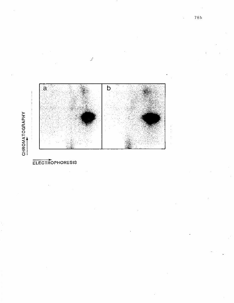
CH
RO
MA
TO
GR
AP
HY
.-.>
trt ht ol +l tt ì0 o l0 m U
) 6
{ oo
-79_
digest was less than that from t,he control (Fig" L2c). The slight shift
in mobiliLy of Lhe peak isolated from the digest may indicate that, of
the het,erogeneous R molecules represented by the broad peaic, those r+iLh
the higher molecular weights lrere degraded into non-binding fragrnents.
This would also explain why less radiolabel is isolat,ed from the digest.
Añalysis of the chymotryptic fragment,s in an identical manner
produced results similar to those obtained wit,h trypsin (Fig. 13).
IgE-Sepharose and RAR isolated a fragment r¿ith an I'I, of 4lkDa (Figs. 13a
and 13b, respectÍvely). IgE-I{ARE again, isolated a relaLively srnaller
amount of whaL appears to be int,act R receptor (Fig. 13c).
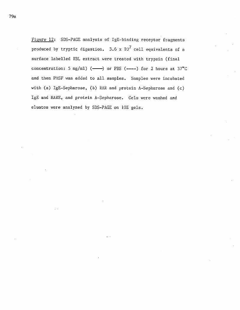
79a
!'isure 12: SDS-PAGE analysis of lgE-binding receptor fragments
produced by t.ryptic digestion. 3.6 x 10⁷ cell equivalents of a
surface labelled RBL extract were treaLed r^rith trypsin (final
concent,raLion: 5 me/ml) (-) or PBS (----) f.or 2 hours at 37oC
and then Pl"lSF was added to all samples. Samples \.rere incubated
with (a) IgE-sepharose, (b) RAR an<i protein À-Sepharose and (c)
IgE and HARE, and protein A-Sepharose. Gels were washed and
eluates r¿ere analyzed by SDS-PAGE on 102 gels.
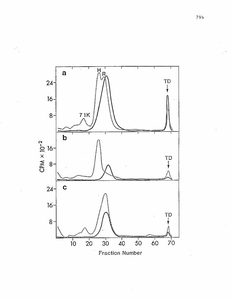
79b
24
1ó
I
ç\
þroX
=o-rJ
r6
I
8
24
30 40 50
Fraction Number
a
7 1
.....aat ta.-tt'
TD
+
H
Ki
TD
I
b
{
¡'3
I
c
+¡1
TD
r0 20 ó0 70
79c
Figure !3: SDS-PAGE analysis of lgE-binding receptor fragments
produced by chymotryptic digestion" Âs in Fig. 12, except. that
chymotrypsin (final concentrauion: 5 mg/ml) was used instead of
Lrypsin" Receptors isolated with (a) IgE-Sepharose, (b) RAR and
protein A-sepharose and (c) IgE and HARE, and protein
A-Sepharose.
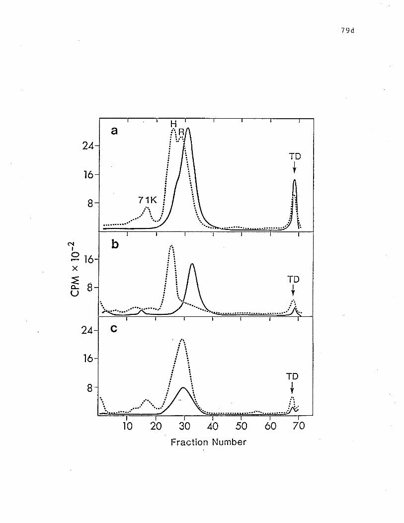
79d
(\¡
OX
=o-U
24
ló
1ó
1ó
I
I
24
B
30 40 50
Fraction Number
a
7 1
R
TD
+
H
i
Ki
tiiii TD
b
+
TD
c
+
j"'..
10 20 ó0 70
-80-
DISCUSSIO¡I
Attempts Lo obtain ligand-binding fragments of recept.ors have
been successfully made in the acetylcholine receptor systen as rvell as in
the present, invest,igation. In the former study, the affinitychromatography purified recepLor was digested with trypsin, resulting in
the formation of a fragmenL which retained capacity to bind its ligands
(Barfeld and Fuchs, 1979). Such a receptor derivative may provide
invesÈigators with a less complex molecule r,¡ith rvhich to study
ligand-receptor interactions. In addition, it may contribute information
on the actual structure of the molecule, particularly its ligand-binding
portion. In the present study, receptors were digested with Lhree
different proteases in an attempt to produce lgE-binding fragrnents of
both R and H receptors. Since, unlike the acetylcholine receptor, the
recovery of biologically active IgE recepLors after affiniLy
chromatography is quite poor, these digestions were performed on
recept,ors in the NP-40 extract of surface-iodinated RBL cel1s prior to
t,he isolation of IgE-binding m4teria1. It was establíshed that digestion
was possible in the presence of NP-40 (Fig. 8).
Papain digestion of a cell extract containing R and H yielded a
fragment which r'¡as bound by lgE-Sepharose (Fig" 9b) but not by IgE anrl
HARE (Fig. 10a). From these results, it was deduced that the isolated
fragment,, a molecule of 36kDa, was part of the H recept,or: if it l-rad been
of R origin it presumably would have been bound by IgE and HARE, as well
as by lgE-Sepharose, since only R is isolat.ed by both of these
approaches. The fact Lhat RAR could not bind this fragmenL (Fig. 10b) is
probably due to the degradaLion of the antigenic determinant for which
_Bi-
this ant,iserum is specific. This ruould not necessarily affect the IgE
bindíng site as it was demonstrated that this antibody preparat,ion
interacts rvit.h a part of the receptor rvhich does not bind to igE (Conrad
et al., L979). In subsequenL experiments, the papain-derived fragnent
did at t,imes react with RAR' and this may indicate that the antigenic
determinant(s) may be close to a port.ion of the molecule which is
susceptible to proteolysis" It is interesting to note that nRGG, the
control for RAR, does bind rvhat appears to be t,he same fragment as that
bound by lgE-Sepharose. This result may be explained by the recent
finding that H receptor has affinity for rat IgG as well as IgE (Kepron
et aI., 1982), and also cross-reacts with rabbit IgG (Kepron et al.,1984). One would expect that this fragment would also bind to the Fc
portion of the IgG in RAR, although why this is not Lhe case rernains
unclear. The IgG fraction from some but not all normal rabbit. sera L¡as
shown t,o precipitate a molecule(s) the SDS-PAGE mobility of which is
similar to that of Il although it has not been demonstrated thaL Lhis
molecule was Lhe IgE receptor (unpublished observation). Such activity
could, perhaps, have been due to the presence of nat,ural antibodies in
some rabbit, sera. The RAR preparat,ion used j-n these experiments may have
been produced in rabbits whose pre-immunization sera did not bind to an
H-lilce molecule. To establish conclusively that the fragment. produced by
papain originated from H, trvo dimênsional maps of tryptic peptides from
the fragment and from intact H were compared (Fig. 11). The striking
similarity between these tvro maps clearly demonstrated that the isolated
fragmenL r{as a part of H. This similarity also suggests that most, if not.
all amíno acid residues labelled by iodine are in the lgE-binding portion
of the rnolecule rvhich, of course, would be expected to be an exposed
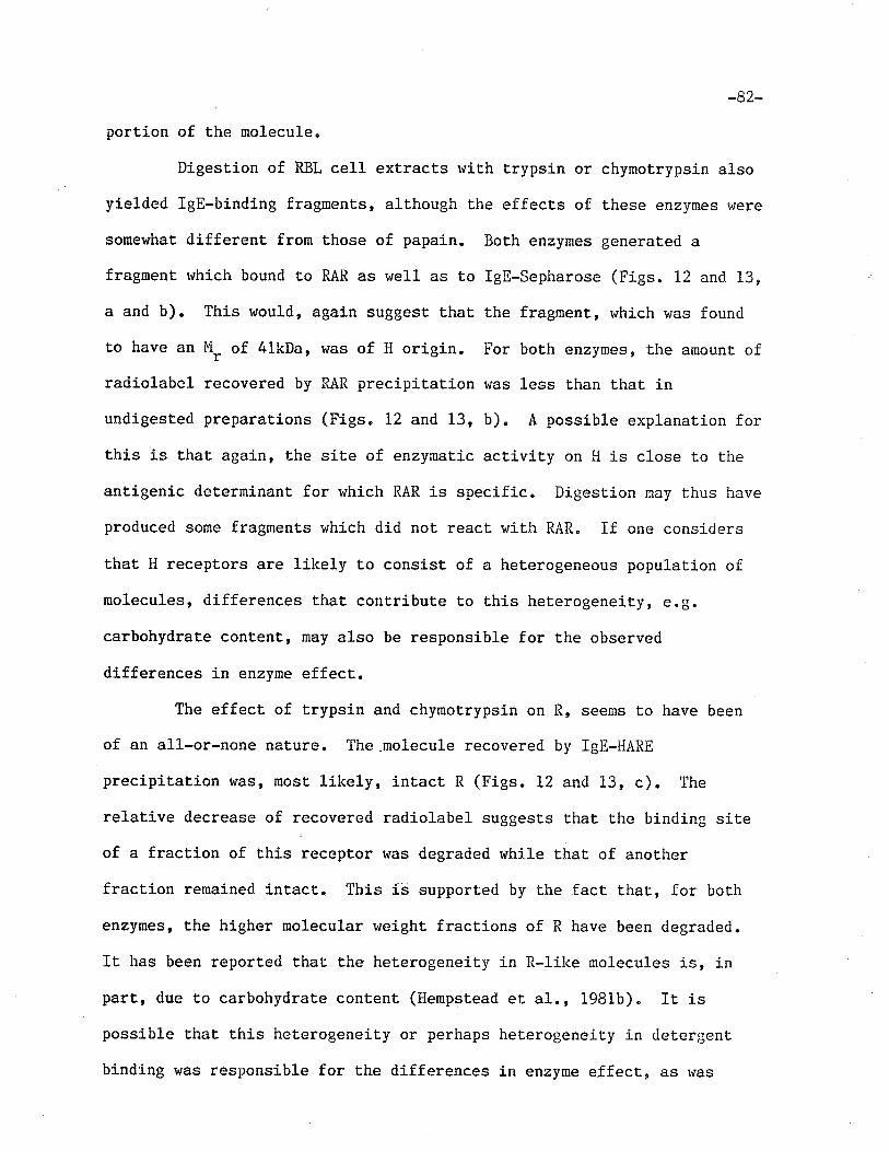
'82-
portion of t.he molecule.
Digestion of RBL cell extracts wiLh trypsin or chymotrypsin also
yielded ïgE-binding fragnents, although the effects of these enzymes r.¡ere
somewhat differenÈ from those of papain. Both enzymes generated a
fragment which bound t.o RAR as well as to lgE-Sepharose (Figs. 12 and 13,
a and b). This would, agai.n suggest that the fragrnent, which r,¡as found
to have an M- of 41kDa, was of H origin. For both enzymes, the amounl ofrradiolabel recovered by RAR precipitation was less t,han that in
undigested preparations (Figs. 12 and 13, b). A possible explanation for
Lhis is that again, the site of enzymat,ic activity on H is close to tireantigenic determinant for rvhich RAR is specific. Digestion may Lhus have
produced some fragmenLs which did not. react with RAR" If one considers
that, H receptors are likely to consist of a heLerogeneous population of
molecules, differences that contribute to this heterogeneity, €.g.
carbohydrate content, may also be responsible for the observed
differences in enzyme effect.
The effect, of trypsin and chymotrypsin on R, seems to have been
of an all-or-none nature. The.molecule recovered by IgE-HARE
precipitation was, most lÍkely, inLact R (Figs. 12 and 13, c). The
relative decrease of recovered radiolabel suggests that the binding siLe
of a fraction of this receptor was degraded while that of another
fraction remained intact. This i.s supported by the fact. that, for both
enzymes, t,he higher molecular weight fractions of R have been degraded.
It has been reported thaL the'heterogeneiLy in R-like nolecules is, in
parÈ, due to carbohydraLe content (Hempstead et al., 1981b)" It is
possible that this heterogeneity or perhaps het,erogeneity in detergent
binding htas responsible for the differences in enzyme effect, as nas
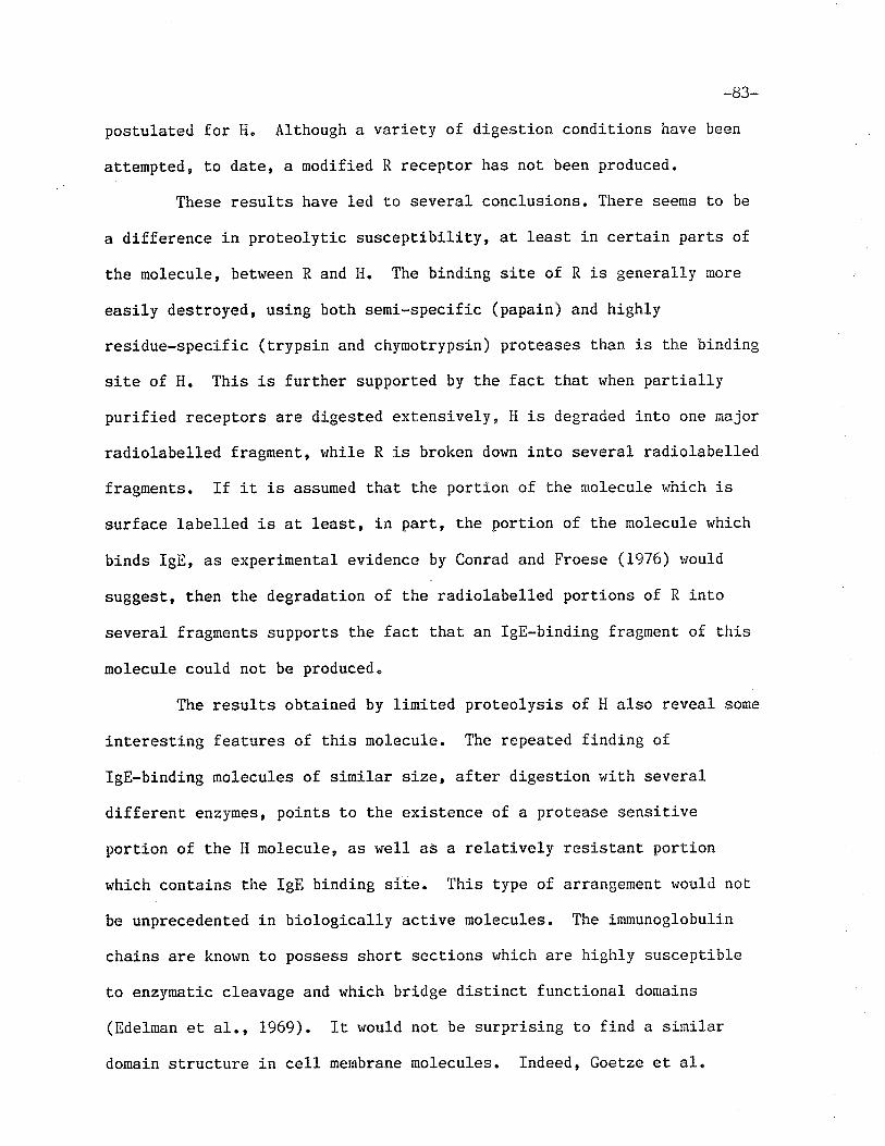
-83-
postulated for H. Although a variety of digestion conditions have been
attempted, to date, a modified R receptor has not been produced.
These results have led to several conclusions. There seems to be
a difference Ín prot.eolytic susceptibility, at least in certain parts of
the molecule, between R and I{. The binding site of R is generally more
easily destroyed, using both semi-specific (papain) and highly
residue-specific (trypsin and chymotrypsin) proteases than is the binding
sile of H. This is further supported by the fact that when partially
purified receptors are digested extensively, H is degraded into one major
radiolabelled fragment,, while R is broken down inLo several radiolabelled
fragments. If it is assumed that the portion of the molecule vrhich is
surface labe11ed is at least,, in part, the portion of the molecule which
binds IgE, as experinental evidence by Conrad and Froese (1976) r¡ou1d
suggest, then the degradation of the radiolabelled portions of R into
several fragments supports the fact that an IgE-binding fragment of this
molecule could not be produced"
The results obtained by linited proteolysis of H also reveal some
interesting features of this molecule. The repeated finding of
IgE-binding molecules of similar size, after digestion wit,h several
differenL enzymes, points to the existence of a protease sensitive
porti-on of the H molecule, as well as a relatively resistant, portion
which contains the IgE bindÍng site. This type of arrangemenl would not
be unprecedented in biologically active molecules. The immunoglobulin
chains are known to possess short sections which are highly susceptible
t,o enzymatic cleavage and r+hich bridge distinct functional domains
(Bdelman et al., 1969). IL rvould not be surprising to find a sinrilar
domain sLructure in cel1 membrane molecules, Indeed, GoeLze et, al"
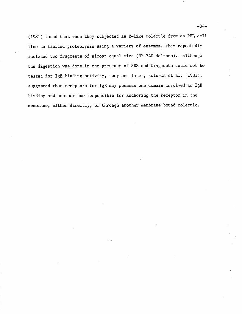
-8/+-
(1981) found Lhat when they subjected an R-like molecule from an RBL cell
line Èo liniLed proteolysis using a variety of enzynes, they repeatedly
isolated two fragrnents of almost equal size (32-34K dalt,ons). Although
the digestion was done in the presence of SDS and fragments could not be
tested for IgE binding activity, they and lat,er, Holor+ka et al. (1981)'
suggested that receptors for IgE may possess one domain involved in IgE
binding and another one responsible for anchoring the receptor in Lhe
membrane, either directly, or through another membrane bound molecule.
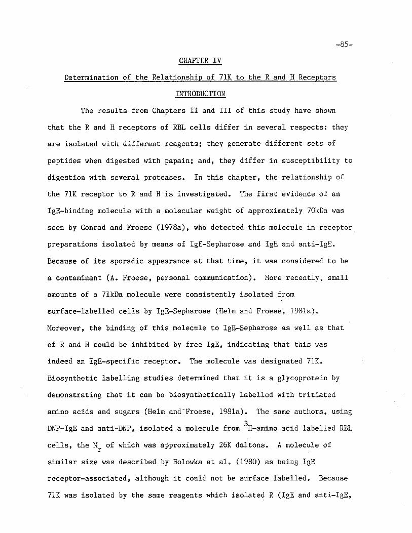
-85-
CHAPTER IV
Determinat.ion of the Relationship of 71K to the R and H Receptors
INTRODUCTION
The results frorn Chapters II and III of this study have shov¡n
Lhat, the R and H receptors of RBL ce1ls differ in several respects: they
are isolated rvith different reagents; they generate different sets of
peptides when digested with papain; and, they differ in susceptibility to
digestion with several proteases. In this chapcer, the relationship of
the 7lK receptor Lo R and H is investigated. The first, evidence of an
IgE-binding molecule with a molecular weight of approximately TOkDa was
seen by Conrad and Froese (1978a), who detected this molecule in receptor
preparations isolated by means of lgE-Sepharose and IgE and ant.i-IgE.
Because of its sporadic appearance at that Lime, iL was considered to be
a contaninant (4. Froese, personal communication). lnlore recently, sual1
amounts of a 71kDa molecule were consistently isolated from
surface-labelled ce1ls by lgE-Sepharose (Helm and Froese, i9B1a).
luloreover, the binding of this molecule to lgE-sepharose as well as that
of R and H could be inhibited by free IgE, indicating t.hat Lhis was
indeed an lgE-specific receptor. The molecule was designated 7lK.
Biosynthet,ic labelling sLudies deternined that it is a glycoproLein by
denonstrating that, it can be biosynthetically labelled with tritiated
amino acids and sugars (Heln and-Froese, 1981a). The same authors, using
DNP-IgE and anti-DNP, isolated a molecule from 3H-.*irro acid labelled RBL
cells, the PI, of which was approximately 26K daltons. A molecule of
similar size was described by Holowka et, al. (1980) as being IgE
receptor-associated, although it could not be surface labelled. Because
71K was isolated by the same reagenLs which isolated R (IgE and anti-IgE,
-86-
Froese et al., L9B2a, DNP-IgE and anti-DNP, Helm and Froese, l98la) and,
in light, of the discovery of a molecule (1ater named p chain) associated
rvith the R-like receptor (Holowka et a1. , i980) , it, r,ras suggest.ed that
71K may be a more stable adduct of R and 26K (or "( and F , t,o use
different nomenclature) (He1m and Froese, 198ia). I'Jhile early attemps to
reduce 71K to yield polypepLide chains of lower M, had failed (lletm and
Froese, 1981a), a more recenL attenpt, by M. tìao of this lai¡oratory,
using higher concenLrat,ions of reducing agents, produced a surface
labelled molecule with an 11, identical to that of R (ti. Rao, personal
communication). This led to speculation that 71K might consist of It
disulfide linked to some other molecule" This chapter describes the
experiments designed to determj-ne whether there is a structural
relationship between R and 71K.
Tryptic peptides of surface-labelled 71K and R and H are
compared by one- and two-dimensional ncapping. Conparison of R anrl Il
confirmed the results obtained in Chapter ïI, r'rhich indicate thau these
two receptors are different molecular entities. Comparison of 71K to
these receptors established a r.elationship betr¡een 711( and R and thus led
to subsequent experiments using biosynthetically labelled receptors, to
determine the rsubunitt composition of 71K"
-87-
I'IATERIALS AND METHODS
The iodination and disruption of i(BL cel1s was performed as
described in Chapter fI.
PreparaLion of Anti-IeE-Sepharos e (HARE-Sepharose) Immunosorbent: Horse
anti-rat IeE (HARE) (kindly supplied by Dr. K. Kelly) r'ras coupled to
Sepharose CL-48 at, a concentration of. 2.5 mg/ml using the procedure
described in the Methods and Ùlat.erials section of Chapter II" The
coupling efficiency vras 9O-95i4"
SequenÈial Affinity Chromatography Using IIARE-SeLrhatqse atl4
IeE-Sepharose: Rece pt.ors were isolated according Lo a meLhod described by
Froese et al. (1982a), modified Lo become a preparatíve procedure. To
isolaÈe R and 71K, the I\iP-40 extract of 3-4x10⁸ "urf.cu-iodinated RBL
'1
cel1s (5x10' cells/ml) was mixed wit,h 300-400 ttg of IgE, incubated for 30
min. at 37oC, then added to 6-8 ml of l{ARE-Sepharose and rotated for 90
min. at 4sC. To remove any residual R and 71K, the supernatant vras
removed, then added to 3-4 m1 of fresh ITARE-Sepharose and incubated for
60 min. at leC. To isolate I{, the supernatant was again removed, acldecl
to 4-6 ml of lgE-sepharose and.rotated overnight at 4"C. Affinit,y gels
were washed 4 times with 0.1% NP-40/PBS and once r'¡ith 0.06251'i Tris, pH
6.8 (sample buffer). Elution was performed by boiling ruiLh equal volurnes
of sample buffer containing 2% SDS. Eluates lrere concenLrated and
dialyzed overnight against sample buffer.
SDS-PAGE on Slab Gels: LO7, polyacrylamide slab gels vrere prepared
according Lo Laemmli (1970) r*ith a L2 cn separating getr and a 1.5 cm
stacking gel. Bromphenol blue and glycerol rvere added to samples which
were then applied to slab gels. Electrophoresis was perforrned using a
Protean cell (BioRad, Richmond, CA) at 30nA/ge1. Upon completion of tire
oo-(J(J-
run, a 0.5 cm wide vertical section of the gel t{as removed and manually
cut into 2 mn fractions. The fractions were counted on a Beckman 8000
gamma counter. Sections of the slab gel conLaining receptor peaks were
then removed and either irunersed in double distilled HrO or fixed with
10% acetic acid/lO% isopropyl alcohol for t hour. Fixed gels were then
incubat,ed wi,th 257, isopropyl alcohol for L2 hours and 10% meLhanol for 12
hours. Gels in HrO were incubated for 48-72 hours aE 37oC. Their
supernatants were lyophilized, then reconstituted with sample buffer
containing 22 SDS and 10% Z-ME and boiled for 2 min. These samples r.rere
applied to 102 tube gels for SDS-PAGE. After electrophoresis, fractions
containing recepLor peaks were fixed as described above-
Tryptic Dieestion of Receptors in Polvacrvlamide Gels: This proce<lure
was performeci according to Elder et a1. (1977) and Pecoud and Conrad
(1981). After incubation in 10% methanol, gels ruere dried under a heat
lamp and subsequent.ly rehydrated $tith 0.051-1 NII4HC03 pH 7.8, containing 50
.qg/ml TPCK trypsin (llorthington, Freehold' i,lJ)" After a 20 hour
incubation at 370C, supernatants lrere removed and gels were incubat.ed
with fresh 0.05M NH4HC03 for 24 hours aL 37oC. Supernatants from botir
incubations vlere pooled and lyophylized.
One-Dimensional Analvsis of Tryptic Disests: Receptor digests r'rere
reconstituted v¡ith sample buffer containing 27" SDS, glycerol and
bromphenol blue, then applied to'a L57' tube gel for SDS-PAGE. Gels were
sliced into 2 mm fractions and counted in a Beck¡nan 8000 gamma counter.
Two-Dimensional Analvsis of Tryptic Disests: Alternativel y, receptor
digests were reconstituted with 88% formíc acid (Fisher Scientific Co.)
and applied to 20 x 2Q cm East¡nan Chromagram cellulose-coated thin layer
chromatography plates (Eastman Kodak, Rochester, NY)" Blectrophoresis
_89_
was performed at 0oC on a high-voltage electrophoresis apparatus (Desaga,
Heidelberg, I,J. Germany) wiLh an electrode buffer of acetic acid : formic
acid : HZO (15:5:8) for 90 to 120 min. at 1000 V. migration vas
monitored by the progression of a dye (Q.257, acid fuchsin [liatheson Co.
Cincinnati, OH], Q.I257" Orange G [Signa Scientific Co. St. Louis, ]{0]
prepared in electrode buffer) spotted on the other side of Lhe plat.e and
rshich nigrated in a dírection opposite to that of the samples. Following
elect.rophoresis, plates r'rere dried and t,rypLíc peptides were then
subjected to thin-layer chromatography in the 2nd dimension using a
butanol : pyridine : acetic acid : HrO (32.5225t5:20) buffer.
Biosvnthetic labe lline of RBL cells: A procedure described by Kulczycki
and Parker (1979) r,¡as used. RBL ce1ls entering the stat.ionary gror'rth
phase r¿ere harvested, rvashed and resuspended at a concentration of 1.5 x
106 cells/ml in leucine-free Eaglets llEl,l (Gibco) supplernented !¡ith 157;
fetal calf serum. L-t4rS-3U] leucine (Amersham, Oakville, Ont.) was
added under sterile conditions at a concentration of 50ug/ml. Cultures
rvere incubated at 37oC in a 57' C0, atmosphere for 20-24 hours. Cells
were then harvested using ÉDTA, washed 3 times \,¡ith PBS/BSA and their
viabilities determined by trypan blue exclusion. Incorporat.ion of 3H ,0."
determined by measuring the radioactivity in 10 ¡1 of Lhe r¡ashed ce1ls
and of the culLure supernatant. The 10 ul aliquot,s were each mixed with
10 mI of Econofluor (l'ler.r England-l,iuc1ear, Lachine, Que.) and counted in a
Beckman liquid scintillat,ion counter, l"Iodel LS-335 (Beckman InsLruments,
Fullerton CA). This procedure rvas used to monitor radioacLivity
throughout the subsequent purification.
Repetitive Affinity Chromatography: Rac IgG, which was precipitated r.¡ith
ammonium sulfate from normal rat serum and filtered through an ACA 34
-90-
column, \,¡as prepared by l'lr" Þ1. Kepron of this laborat,ory as described in
Kepron et al. (1982) and 4x crystallized BSA was purchased from ICil
PharmaceuLicals (Cleveland, 0H). IgG-Sepharose and BSA-Sepharose !/ere
prepared as described in Chapter II at a concentration of 5mg/m1.
Receptors were isolated according to Kulczycki and Parker (L979). The
NP-40 extract of 2-3 x 10⁸ biosynthetically labelled RBL cells was mixed
with 4-6 ml of lgG-Sepharose overnight aE 40C. The supernatanL was then
removed and mixed with 2-3 ml of lgE-Sepharose for 2 hours at [oC. The
IgE-Sepharose was transferred to Pasteur pipette columns in 10 aliquots
and the gel vras washed wit,h a toLal of approximately 120 ml of PBS/0.1%
NP-40 at 4oC. Bound material rvas eluted by quickly passing 0.5 ml of
0.5N acetic acid contaj.nj.ng 17" NP-40 through each gel, and iurmediately
neutralizing Lhe eluate v¡ith 0.185 ml of 2.01vI Tris, pl{ 8.6. The eluate
was added to 1-2 ml of BSA-Sepharose and incubated at 4oC for 30 min.
The supernatant v¡as then removed and mixed overnight with l-2 m1 of
IgE-Sepharose at 4oC. After washing 4 times with PBS/O.12 NP-40 and once
wit,h 0.0625t'l Tris, pll 6.8 (sample buffer), receptor material r,¡as eluted
by boiling in sample buffer containing 2% SDS"
Analysis of Purified Receptors: Eluates fron the second IgB-Sepharose
incubation vrere applied t,o 102 polyacrylamide slab gels for SDS-PÀGE
analysis. Electrophoresis was perforned at 30mA/ge1. Gels rt¡ere then
stained with Coomassie blue, as described in Chapter II, destaíned with
102 acetic acid, t,hen Lreated with En3Hance (Neru England Nuc1ear,
Lachine, Que.) for one hour..Gels were dried in a BioRad Gel Dryer
(Richmond, CA), autoradiography was performed and films were scanned on a
Quick Scan densitometer (Helena Laboratories, Beaurnont, TX).
Alternatively, eleclrophoresis was performed on purified receptor
-9L-
material using 10% tube gels on eluates separated into tlo aliquots.
Gels were frozen overnighÈ in 0.0625M Tris. Thawed gels were placed in
Uroughs above 10% slab gels. The tube gels were secured on top of the
slab gels with 1.5% agarose prepared in electrode buffer. The upper
electrode buffer contained O.O4% bromphenol blue (0.0012) as the Lracking
dye. During Lhe electrophoresis, performed under reducing condit,ions,
the agarose contained 7.57" 2-l4E and the upper electrode buffer contained
L% z-ME. The same 2-dimensional procedure was applied to
surface-iodinated receptors isolaLed by a single incubation wiLh
IgE-Sepharose. Slab gels containing biosynthetically labelled material
lrrere treated with En3Hance. Gels were then dried and autoradiography \t/as
performed.
Autoradiographv: Plates were exposed to Kodak X-Omat, AR x-ray film
(Easturan Kodak, Rochester, NY) and Cronex Li-ghtening Plus intensifying
screens (Dupont, lriilmington, DE) at -7OoC for 2-4 weeks. Films were
developed using Kodak Liquid X-Ray Developer and Replenisher and Kodak
X-Ray Rapid Fixer (Eastman Kodak, Rochester, NY). Films tuhich t¡ere
exposed to trit.iated material were hypersensitized before exposure
according to the method of Laskey and þlills (1977).
-92-
RESULTS
Investigation of the relationship between the R and 71K IgE
receptors began in this laboratory with the finding Lhat reduction of 71K
produced a molecule whose SDS-PAGE elect.ophoretic mobility r+as
practically identical to Ehat of R. The results of t.hese experiments,
performeci by Dr. Þiangala Rao, are shown in Figures 14 and 15. i,ihen
receptors for IgE, isolat.ed by means of fgE-Sepharose, r.¡ere reduced by 5Z
2-l'iE and subsequently analyzed by SDS-PAGE (Fig. 14), rhe peak
corresponding to 71K disappeared, and that corresponding to R increased.
To show more clearly that 71K was converted to a molecule r¿ith R-like
mobility' receptors were isolated from an extract of surface iodinated
lìBL cel1s using IgE-Sepahrose and separaLed by SDS-PAGE on 102 slab gels.
Radiolabelled maLerial from fractions constituting R and 71K receptor
peaks rvere collected and aliquots of t,he 71K receptor were reduced or run
unreduced. Figure 15c depicts the SDS-PAGE profile of unreduced 7lK"
Fig. 15, a and b shorv the SDS-PAGE profiles of isolated 71K, reduced ru'itir
25mlt'f dithiotrietol and 5% 2-ll1E', respectively. The mobilities of these
peaks are similar to that of R (Fig. 15d). This result suggested r.har
71K might be composed of R recept,or, disulfide linked either to itself or
to some non-iodinated nolecule. The follorvi-ng experiments r.rere perforrned
to investigate this possibÍlity.
Isolation of R. H and 71K rece Lors-usinq sequenLial affinitv
chromaLoeraphv
Isolation of the recept,or material, necessary for tryptic
mapping, required a preparative procedure capable of resolving R, H and
711( receptors. l'Jhile the met.hods of recept,or isolation described in
Chapters II and III provided a means of separating R and 71K on the one
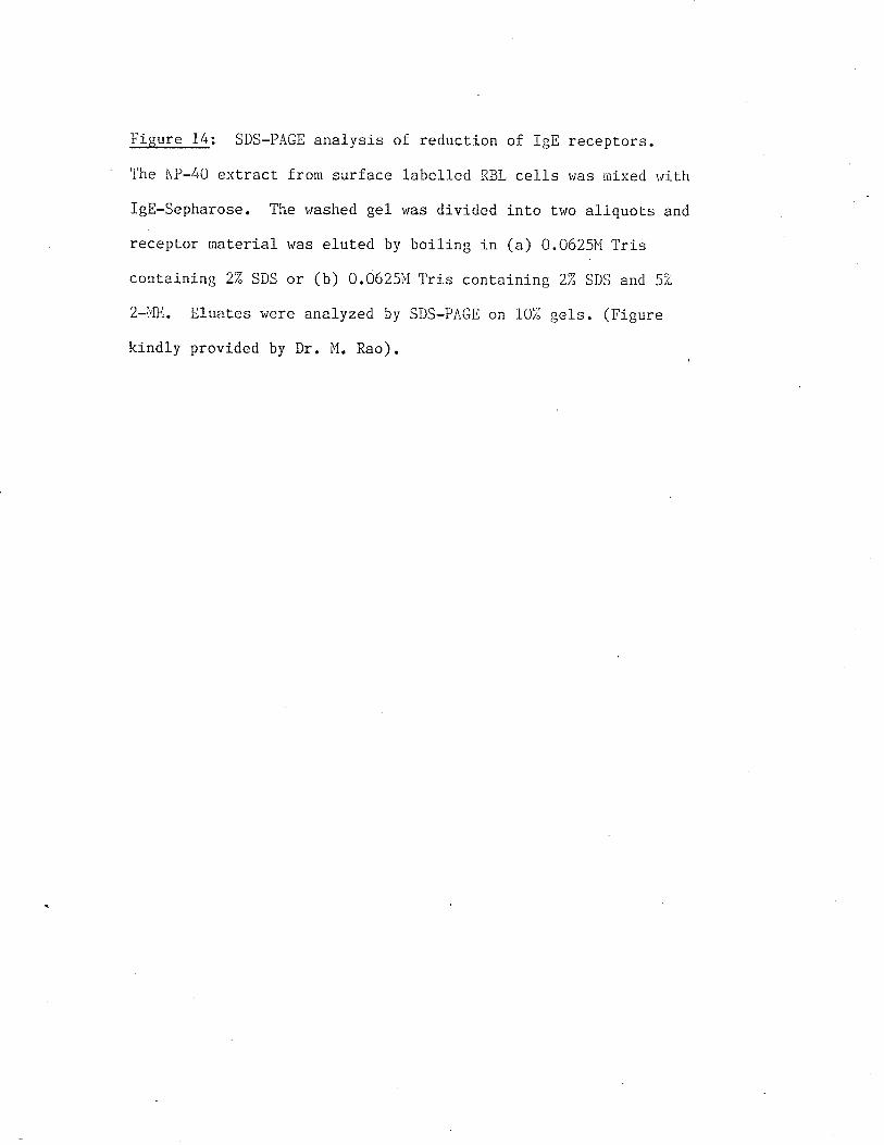
Figure 1/+: SDS-PAGE anal ysis of reduction of IgE recepLors.
The i'lP-40 extract fron surface 1abe1led FIBL ce11s r,/as rrtixed lirith
IgE-Sepharose. The washed ge1 was divided into trvo aliquots and
recept,or maLerj.al rqas eluLed by boiling in (a) 0.0625Ì'l Tris
containÍng 2Z SDS or (b) 0.06251'l Tris containing 2:í SDS and 5Z
2-i'1E. Eluat.es r'¡ere analyzed by SDS-PAGE on 10i; ge1s. (Figure
kindly provided by Dr. N{. Rao).
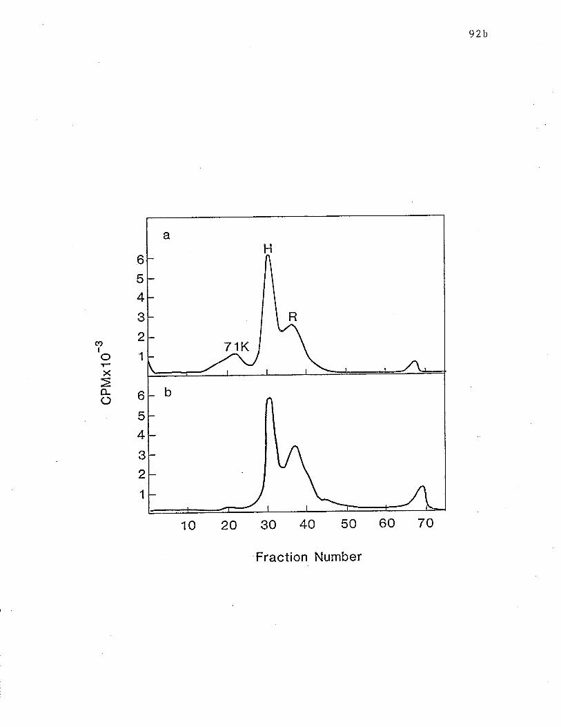
92b
b
54
3
2
1
coI
aH
R
71K
b
10 20 30 40 50 60 70
Fraction Number
oX
o-() 6
5
4
3
2
1
92c
Fisure 15: SDS-PAGE analysis of reduced 711( receptor. The
extract of surface labelled RBL ce1ls was mixed rvith IgE and
then HARE-Sepharose. The eluate was subject,ed to SDS-PAGE and R
and 71K receptors were eluted from fractions constituting the
major peaks. Samples v¡ere dialyzed against 0.06251''l Tris,
adjusted to 2% SDS (c and d) , 2% SDS and 25mM <lithiothriet.ol
(DTT) (a) or 2Z SDS'and 5% 2-1"18 (b) and analyzed by SDS-PAGB"
(Figure kindly provided by Dr. M Rao).
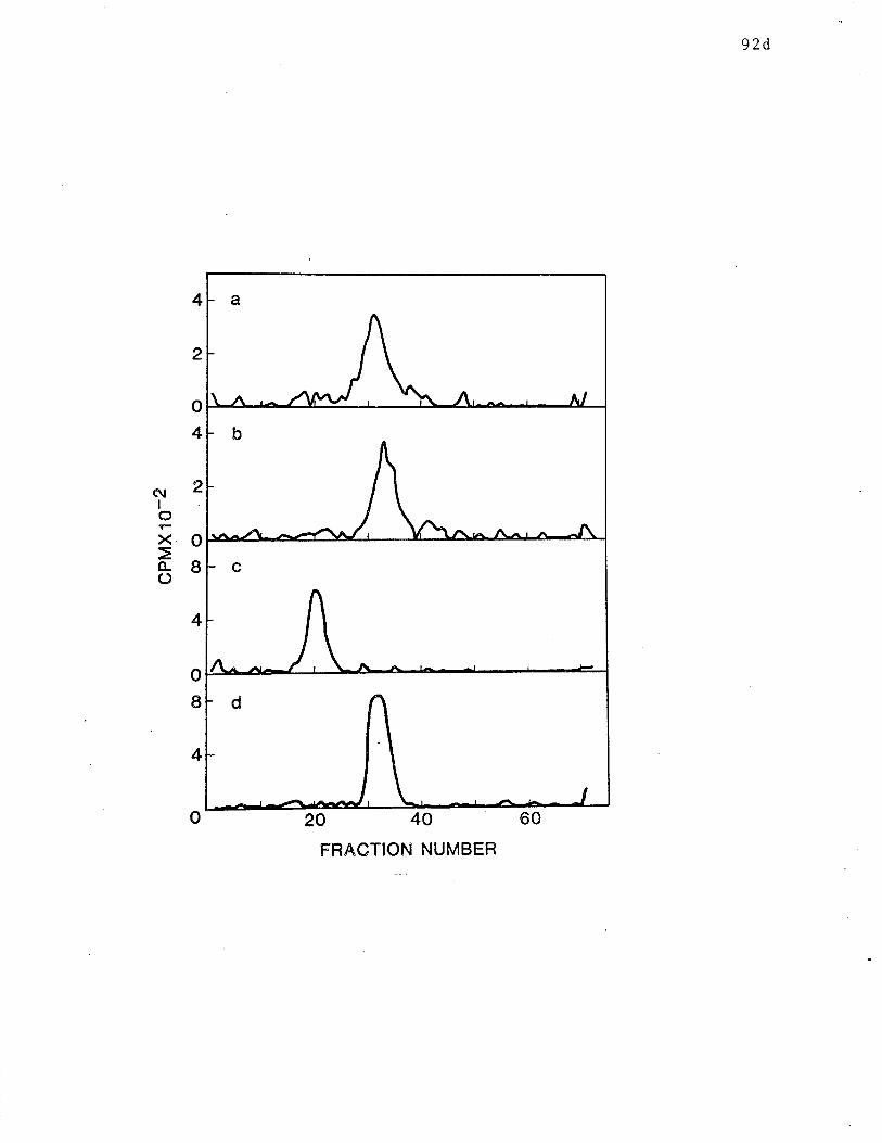
92d
4
2
o
4
ñ¡2IIxoÀgo
4
o
I
4
6020 40
FRACTION NUMBER
o
a
\ .,\ A^l
b
c
d
-93-
hand (IgE and anti-IgE) and H on the oLher (RAR), these meLhods resulted
in a good deal of wasted receptor material, since H recepLor, in aliquots
treated r.rith IgE and anti-IgE, and R and 71K receptors in aliquot.s
treated with RAR would not be isolated. Thus, a procedure which was used
analytically by Froese et al. (1982), v¡as modified to become preparative
(as outlined in lulaterials and Ïulethods) for this study. The first
incubation of a NP-40 extract, of l251-tabelled RBL cell extract ruit,h IgE
and HARE-Sepharose resulted in the binding of. 2-3% of the total
radioactivity in the extract, while the second incubation bound only an
additional 0.052 of the total radioactvity. This indicaLes that the
first incubation removed virtually all of the receptor material uhat such
a quanÈity of this reagent is capable of removing. The subsequent
incubation of extract with lgE-Sepharose led Lo the binding of an
additional 0.5-17, of the total radioactivi.ty. Receptor material was
eluted by boiling in equal volumes of sample buffer containing 2% SDS"
Eluates from HARE-Sepharose and IgE-Sepharose trere applied to
separate 10% polyacrylamide slab gels for SDS-PAGE according to Laernmli
(1970). Upon completion of the run, a 0.5crn r.¡ide vertical section of
each gel $ras removed and manually cut into 2um fractions. These
fractions rì¡ere counted and the resulting profile is shown in Fig" i6.
The eluate frorn tlARE-Sepharose contained two najor radiolabelled peaks
the mobilities of rvhich correspoñded to 43kDa and 67kDa (Fig. 16a). This
indicates that R and 71K receptors are isolat,ed using tl'ris reagent, which
is in agreement r.rith the resulLs obtained in ChapLer II. The double peak
representing R is nost likely due to the facl that these gels r'rere
manually sliced, leading to slight differences in the fract.ions. The
IgE-Sepharose eluate (Fig. 16b) contained one major peak with a mobility
-94-
corresponding to a molecular weight of approximately 52kDa. The small,
higher molecular weight peak has already been described in Chapter IÏ.
The small peak on the descending side of the major peak most likely
represents residual R receptors which were not isolated with
Igg-l{ARE-Sepharose. With this possibility in mind, and to insure maximal
purit,y of the receptor preparations, only fractions corresponding Lo the
shaded areas were used for further experiments. In a typical experinent,
an $mm horízontal section of the gel corresponding to the 4 fractions
containing 71K and a 16mn section of the gel corresponding to the I
fractions containing R were renoved from a 120mm gel for further
analysis. In a similar fashion, an 8mm section of Ehe gel separating the
IgE-Sepharose eluate, rvhich contained H receptor' was also removed.
Analysis of Trvptic Pept ides of Recept,ors
The relationship between R, 71K and H receptors r'tas
Ínvest,igated using one- and two dimensional mapping of receptor tryptic
peptides which vrere prepared according to Elder et al " (1977) and Pecoud
and Conrad (1981). Before digestion, sections of the slab gels
containing receptor material r'¡hich was to undergo reduction, t'/ere
incubated tor 48-72 hours in distilled rsater. After incubation, the
supernatanls were found to contain approximately 5O'/. of the radioactivty
in the original 71K band and 65% of the radioact,ivity in the R band. The
supernatants l,/ere dialyzed againsL distilled \,tater and lyophilyzed. The
reconstituting sample buffer (0.06251'f Tris) contained 27. SDS and 10%
2-lulE. Samples were then subjected to SDS-PAGE in tube gels whÍch r,¡ere
subsequently manually sliced into 5mm fractions. Figure 17 depict's the
distribution of radioact,ivity after eleclrophoresis. As expected, and,
in agreement with Fig. 15, the reduction of both R and 71K yielded
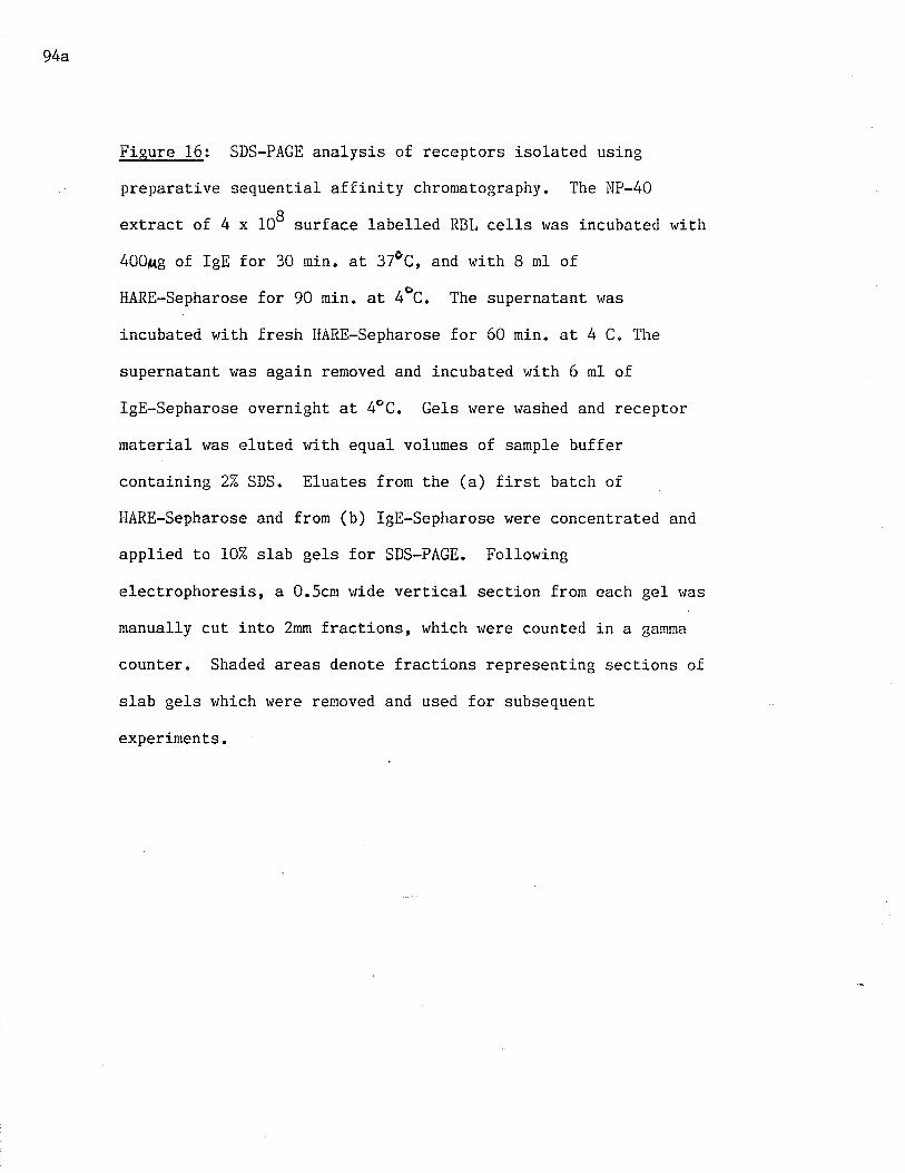
94a
Figure 16: SDS-PAGE analysis of receptors isolated using
preparat.ive sequential affinity chromatography. The IIP-40
extract of. 4 x 10⁸ surface labelled RBL cells was incubated wittr
400Ae of IgE for 30 min. at 37ÙC, and wit,h 8 ml of
HARE-Sepharose for 90 min. at 4eC. The supernatant was
incubated with fresh IIARE-Sepharose for 60 min. at. 4 C. The
supernatant v¡as again removed and incubated wifh 6 ml of
IgE-Sepharose overnight at 4oC. Gels were r,¡ashed and receptor
material was eluted wit.h equal volumes of sample buffer
containing 2Z SDS. Eluates from the (a) first batch of
HARE-Sepharose and from (b) IgE-Sepharose llere concentrated and
applied to 102 slab gels for SDS-PAGB. Following
electrophoresis, a 0.5c¡n wide vertical section from each gel rvas
manually cut, inLo 2mm fractions, rvhich lüere counted in a garnma
counter" Shaded areas denote fractj-ons representing sections of
slab gels which rüere renoved and used for subsequent
experiment.s.
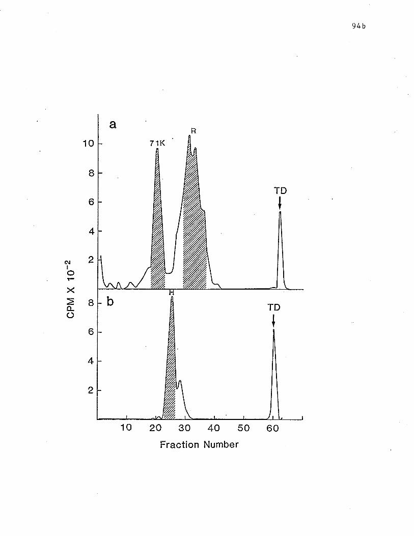
94b
aR
10 7lK
20 30 40 50
Fraction Number
I
6
4
2
bI
6
4
2
TD
I
ôtIoX
fLo
10
TD
I
60
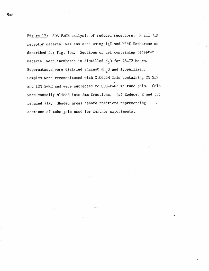
94c
Fisure 17: SDS-PAGE analysis of reduced recepLors. R and 71K
recepEor material was isolated using IgE and HARE-Sepharose as
described for Fig. 16a. Sections of gel containing receptor
material were incubated in distilled iltO for 48-72 hours.
Supernatants v'¡ere dialyzed against dHrO and lyophilized.
Samples vlere reconstituted with 0.0625i'1 Tris containing 2% SDS
and 102 2-1,18 and rvere subjected Lo SDS-PAGE in tube gels. Gels
v¡ere manually sliced into 5mm fractions. (a) Reduced R and (b)
reduced 71K. Shaded areas denote fracti-ons representing
sections of tube gels used for further experiments.
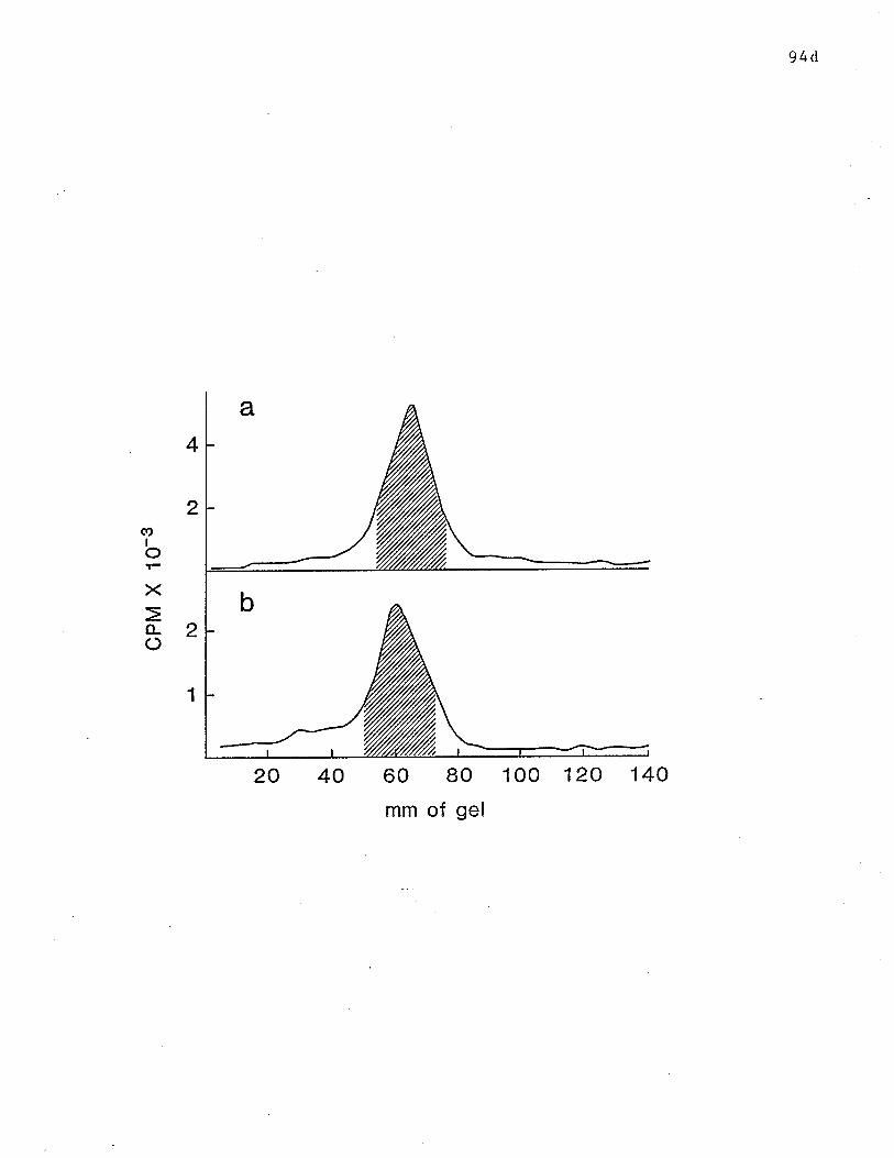
94d
a4
2
b2
COIoX
fL()
1
20 40 60 80
mm of gel
1 00 12A 140
-95-
molecules wiLh similar mobilities. Fractions constituting the
radioactive peaks (5 fractions each) were collected. These, as well as
the slab ge1 sections conLaining unreduced receptor material r+ere fixed
r+iLh 102 acetj.c acid/107" isopropyl alcohol. They rvere ruashed. in 257,
isopropyl alcohol, then 102 methanol, to remove the SDS. Gels were then
dried under a heat lamp and rehydrated with 0.05M amnonium bicarbonate
containing TPCK-trypsin. After tr'¡o 2O-hour incubations at 37 C' the
supernatants $rere found to contain 70-80% of the counts originally in the
receptor bands. Sanrples were lyophilized.
a) One dimensional analysis
Aliquots of all samples were reconsLituted with SDS-sample
buffer, Lhen applied to I57" polyacrylamide gels for SDS-PAGE. Profiles
of the digests are shown in Fig. 18. DigesLion of H produced few
labelled peptides, none of which had apparent molecular weights greater
than 10kDa (Fíg. 18a). In contrast, digest,ion of R and 71K' in both
reduced and non-reduced forms, produced several peptides. Their exact
nunber was difficult to determine, due to the broadness of the peaks
(Fig. 18, b-d). Also in all cases, the larger peptides had molecular
weights between 20-35kDa. It is clear that the patterns generated from
reduced and unreduced R and 71K were very similar. ilor"ever, some changes
in the distribution of peptides lrere apparent after reduction of both R
and 71K. In comparing Fig. 18b Eô 18c and Fig. 18d to l8e' one can see
that the relative amounts of higher molecular weight peptides change upon
reduct,ion of R and 71K. This.change appeared to be similar for bot,h
recepLors. Thus, rvhile digestion of unreduced R and 71K produced similar
patterns, digestion of the reduced receptors made the simil-arity even
more apparent.
95a
Figure 18: SDS-PAGE analysis of tryptic peptides of receptors
on 152 ge1s. R, H, and 71K receptors rvere isolated as described
in Figs. 16 and 17. Gel sections containing receptor maLerial
were dried and rehydrated with 0.05ÌU NH4HC03 containing 50p(g/ml
of trypsin. After a 20 hour incubation at 37oC, supernatants
ï¡ere removed and gels were incubated with fresh 0.051'i I'lH4l{C03.
SupernatanLs from both incubations were pooled and lyophilized.
Digest.s were reconstitut.ed with sample buffer conlaining 2Z SDS
and applied to 15% tube gels for SDS-PAGE analysis. Tryptic
digest of (a) H, (b) R, (c) reduced R, (d) 71K and (e) reduced
7lK.
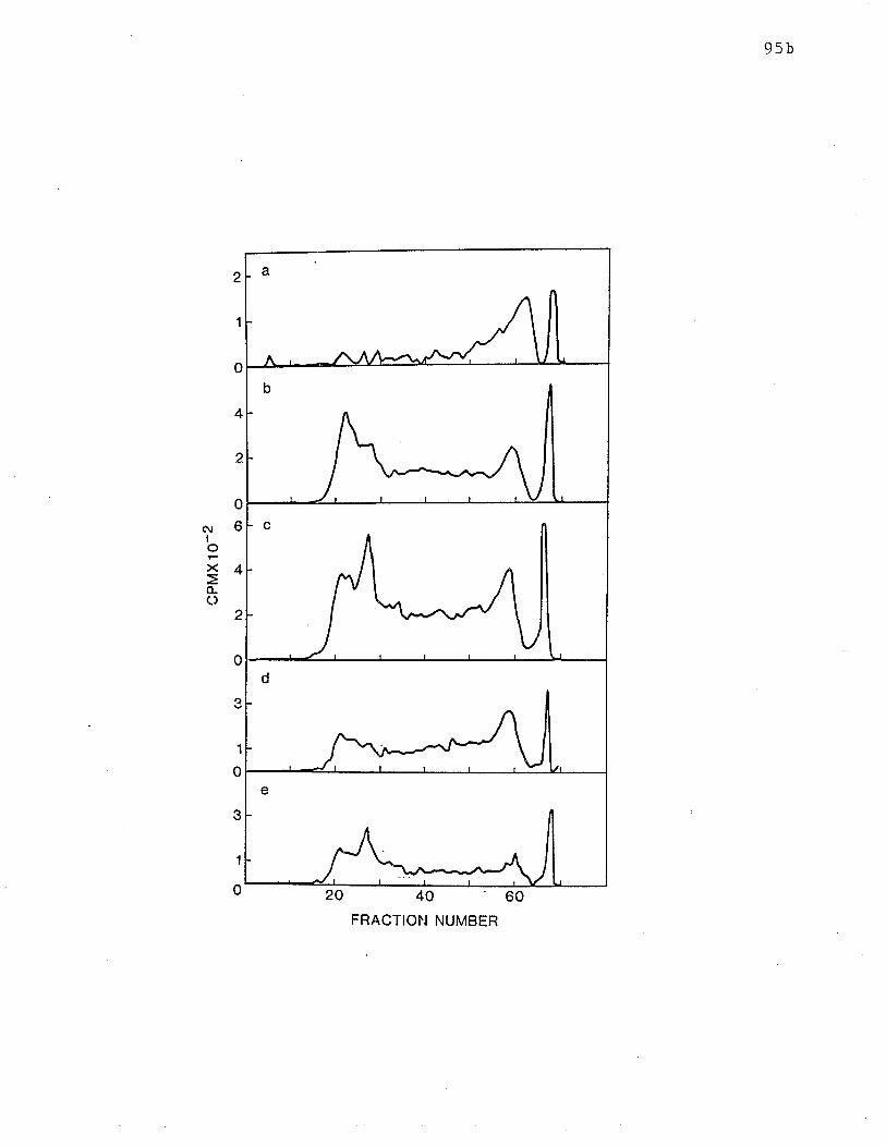
95b
2 a
^ r-
b
c
d
e
20 40 60FRACTIO¡.¡ NUMBER
ôlIoxo-O
o
4
2
0
6
4
2
0
â
1
0
3
1
0
-96-
b) Trvo dinensional analvsls
The one-dimensional analyses presented in this Chapter and in
Chapter II demonstrate beyond a doubt that R and i{ receptors have little
if any similarity in primary structure" By contrast, results of
one-dinensional tryptic mapping, presented in this Chapter, indicate a
good deal of similariLy between R and 71K. However, it was felt that
greater resolving potrer was required to establish more definitively the
extent of the sinilarities. Thus, a thto dimensional separation of
tryptic peptides was performed in an attempt to establish more clearly
the differences and similarities between these tv¡o molecules.
Trypt,ic peptides dissolved in fornic acid were applied onto a
thin-layer chromatography plate coated with cellulose acetate ge1.
High-voltage electophoresis was performed with an acetic acid : formic
acid : HrO electrode.buffer. The plates were then dried and the samples
rvere subjected to thin layer chromatog,raphy for separation in the second
dimension, using a butanol : pyridine : aceLic acid : I{r0 solvent.
Plates r'rere exposed to X-ray film for 2-4 rueeks and then developetl
Tryptic peptide naps of H and R receptor digests are shown in
Fig. 19 a and b, respectively. A schematic representation of each map is
depicted below the corresponding map. The digestion of surface labelled
R generated 12-15 label1ed peptides (Fig. 19b) while Lhat of H produced
5-6 pepLides (Fig. 19a). Approximately equal a¡nounts of radiolabel were
applied. Qne peptide derived from R appears to have a greaLer intensity
than the other peptides, which share the remaining radiolabel fairly
evenly. In contrast, almosL all the radiolabel in H peptides was
associated rvith a single spot, with a small proport.ion of the radiolabel
being found in the remaining 4-5 peptides. In addit.Íon' as can be seen'
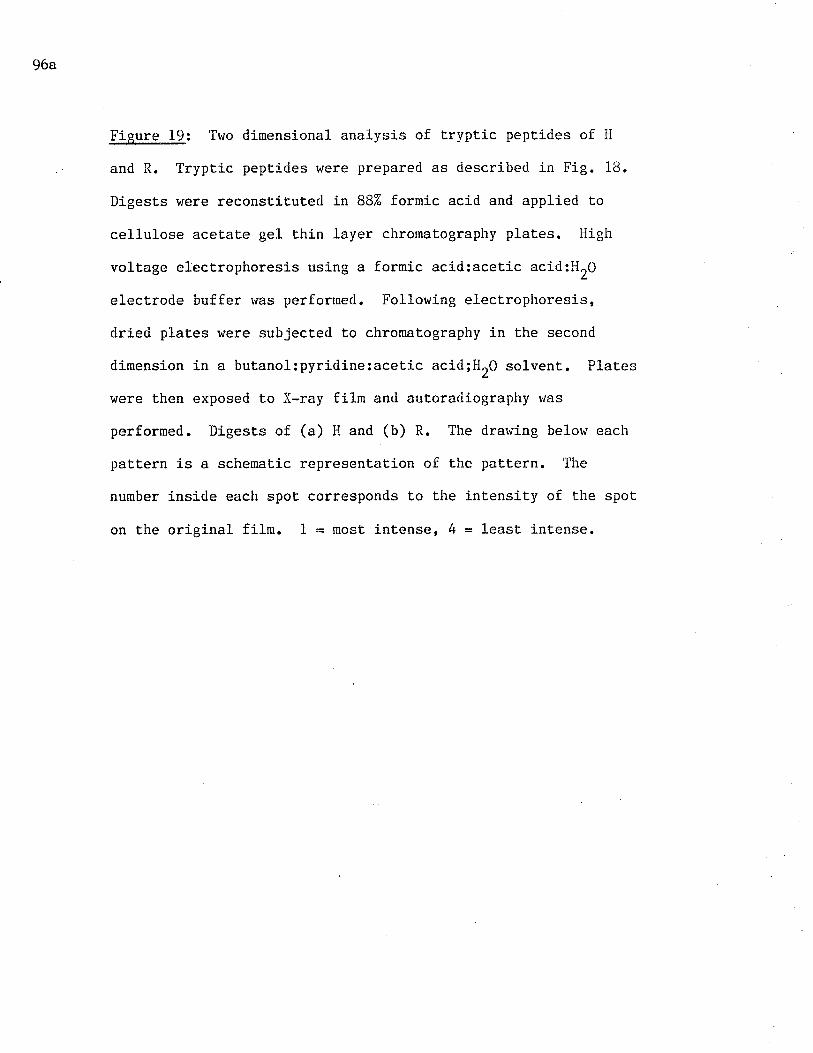
96a
Fieure 19: Tv¡o dimensional analysis of t,rypLic peptides of H
and R. Tryptic peptides v/ere prepared as descriϡetl in Fig. 18"
Digests v¡ere reconstituted in 882 formic acid and applied uo
cellulose acetate gel thin layer chromatography plates. High
voltage electrophoresis using a formic acid:acetic acid:HrO
electrode buffer r'ras performed. Following electrophoresis,
dried plates were subjected Lo chromalography in the second
dimension in a buLanol:pyridine:aceLic acid;llr0 solvent.. Plates
were Lhen exposed to X-ray film and autoradiography r.ras
performed. Digests of (a) H and (b) R. The drawing belovr each
pattern is a schemalic represenLaLion of the pattern. The
number inside each spot corresponds to the intensity of the spot
on the original film. 1 = most intense, tr = least intense.
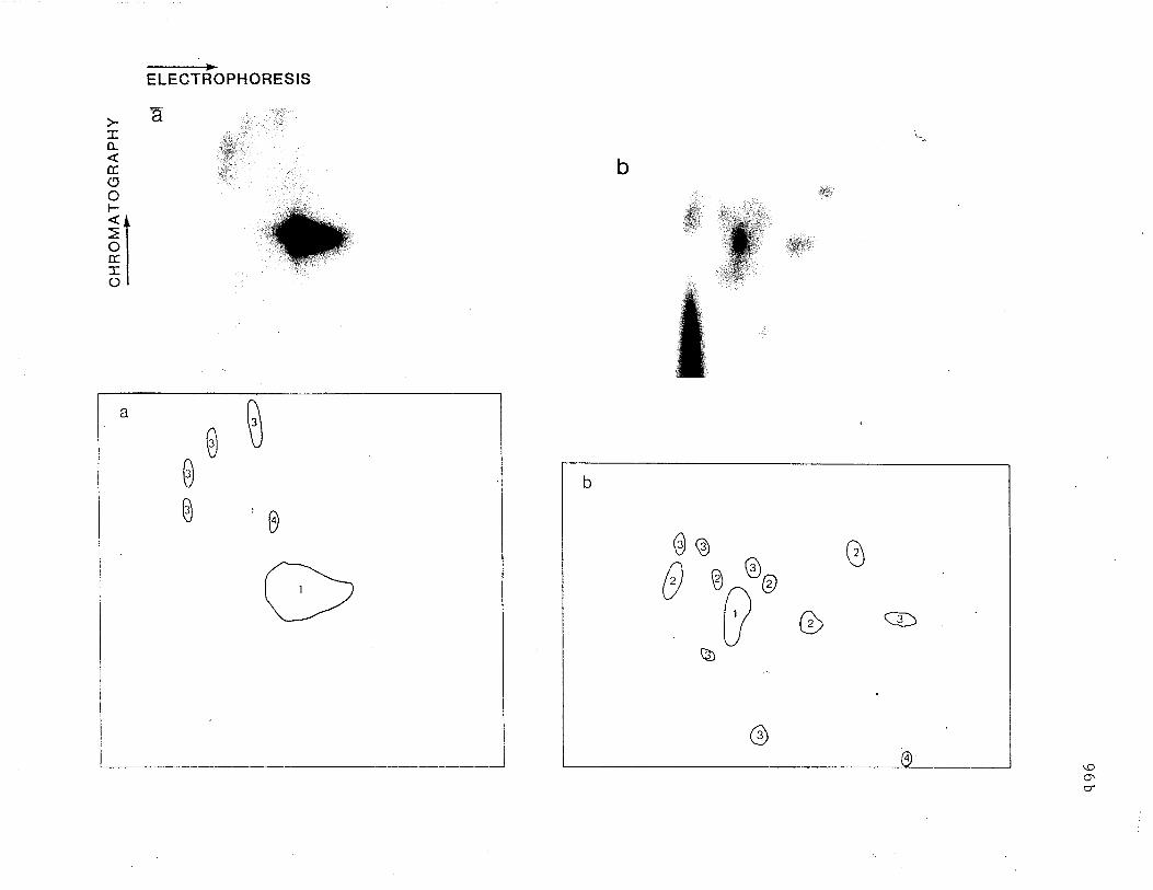
-----+ELEGTROPHORESIS
d.I
b
,¡4r.:'iÌl:r'
:ìrl
IÀ
E.(5ol-soÉ.:Eo
,',. !t:
&ffiW
lsffii¡rl
I,#iw.*::j...
,,'.
ø
0
0
0
a
\o
e(à o
@
\¿J
0o
b
c
-97-
there is no overlap of pept.ides from R rvith those from ÌI. Thus'
digestion of R and H produced labelled peptides r'rhich differed both in
number and in Position"
The peptide map of R is compared to that of 71K in Fig. 20. In
this case (as ruelI as in Fig. 21), schenatic representations of the naps
allow visualization of peptides which appeared on the film but were too
fain¡ to appear in the photographs. It should be noted that 17'000 cprn
of R and 81000 cpm of 71K were applied. In comparing Figs. 20, a and b,
peptides from 71K are fev¡er (11) and less intense than those of R. This
r,rould be expected, since fewer counts of 71K digest rvere applied. 0f the
ones which are visiblen 5 pept.ides derived from 71K have positions in
common with peptides from the R digest. These are represented by shaded
spots in the schematic draruing. The renainder of the peptides are in
different positions. This indicates a certain degree of homology betr'reen
labelIed peptides of R and 711(.
The peptide maps of R and 71K which were reduced before uryptic
digestion are shown in Fig. 2L. In Lhis case' 71K peptides comprising
41000 cpm and R peptides arnounÇing to 7'000 cpm were applied. 0f the 11
71K pepLides (Fig. 21b) and rhe 12 R peptides (Fig. ZLa), 5 share a
corrunon position (shaded in the schematic drawÍngs). 0f these 5 peptides,
3 also share positions with peptides from unreduced R an<Í 71K (Fig. 20 a
and b). The rernaining peptides hãve no positions in common. Thus, a
similar degree of homology does appear to exists betrveen reduced 71K and
R as does between unreduced 71K and R. If maps of unreduced and reduced
R are compared (Figs. 20a and 21a, respectively), 4 peptides are found to
share common positions. Three peptides from unreduced and reduced 7lK
also share positions (Figs. 20b and 21b). Thi-s indicates that reduction
97a
Figure 20: Two dimensional analysis of t.ryptic peptides of R
and 71K. Ilaps r,rere prepared as in Fig. 19. Digests of (a) R
and (b) 71K. In this figure, the schematic drawings contain
shaded spots r¿hich represent peptides sharing common positions
in both (a) and (b).
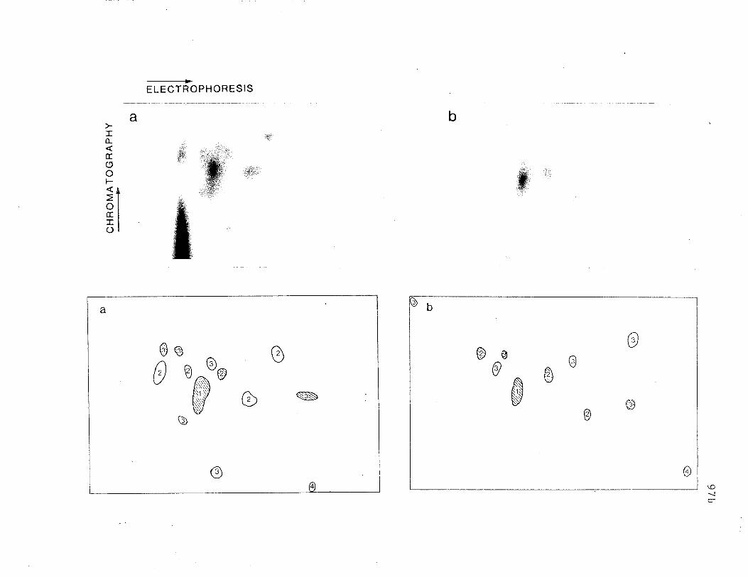
ba
+ELECTROPI{ORESIS
.::ii.
I0-
troot-
oEIo
..:1'lr
@,
ffiW
,ttr:::ffi
:,#$:r:,
--J
Ø
ÐI
0èñ
ÑN
o
0Þ
b
N\ììv
S
0e o0 @
b
a
o
97c
Figure 21: Two dimensional analysis of tryptic peptides of
reduced R and 7lK. lr'taps were prepared as in Fig. 19. Digests
of (a) reduced R and (b) reduced 71K. The shaded spots in the
schematic drawings represent peptides sharing common posit.ions
in both (a) and (b).

(¡
@
@@@
s)
@@
Þ @
@ ø6^ w
CHROMATOGRAPHY-.--_-->
À)
t
ml r-l mlc)l-{l !+o' 1t¿oFmU)-v,
ul
,
@
@
Z77pZ77^(4à/2W
@
@@
@
@
(t
pL6
-98-
leads to some change in both 71K and R molecules which resulls in the
generation of several different peptides. This change appears Lo be more
pronounced for 71K than R.
Isolation of Purified Receotors Bv Repetitive Affinit v Chr tosraohv
The results described in the preceeding section strongly
suggested that, 71K is a receptor consisting of R or an R-lilce molecule
disulfide-bonded to another polypeptide chain. Since only one peak is
apparent after SDS-PAGE of reduced, surface labelled 71K (Figs. 15 and
17), the other polypeptide chain would eit,her have to be another Il or
R-líke molecule or a molecule which is not surface labelled. To test the
latter possibility, RBL cells biosynLhet,ically labelled r,¡ith 3H-leucine
were used as the source of receptors. As demonstrated by lïelm and Froese
(1981a), isolation of Èrit,iated receptors from RBL cells using
IgE-Sepharose results in a complex SDS-PAGE pattern. The presence of
free IgE was unable to inhibit the binding of several peaks, inrlicating
that there tilas a good deal of non-specific binding. The presence of
molecules non-specifically bound to lgE-sepharose rr¡ould make a pattern,
run under reducing conditions,,difficult to interpret. In the present
study, recepLors from RBL cells which were biosynthetically labelled rvith
3g-I"u.ine (cells incorporated approxirnately 75% of. radiolabel) were
purified accorcling to a procedure described by Kulczycki and Parker
(1979). The flow chart representing this procedure is depicted in Fig.
22. The first column, rat ÏgG-Sepharose, bound 0.77" of the total
radioactivity in the extract.aft,er it was washed. However, the
supernatant, r,rhich was Lransferred to lgE-Sepharose, contained only 75%
of the original extract activity, indicating that approximatel-y 257 of
the extract material was physically trapped in the gel or l'ras very
-99-
loosely bound to IgG" IgE-Sepharose bound O.077" of the lgG-Sepharose
supernaLant. This percentage is less than Lhat bound in the case of
surface-iodinated extract i.e. 1-32 (see Results, Chapter II)' and is
probably due to the fact that in biosynthetically labelled cells' both
surface and non-surface proteins were labelled and therefore the original
cell 3H-extract could be expected to have contained many more labelled
molecules rvhich did not bind t.o lgE-Sepharose. Binding to lgE-Sepharose
was also less t.han that to lgG-Sepharose.. This suggests that the IgG
colu¡rn nnainly bound v¡hat would oLhèrwise have been bound non-specifically
by t,he IgE colurur and that t,he use of lgG-Sepharose for the removal of
non-specifically bound nolecules rvas therefore appropriate. Acetic acid
treatment of lgE-Sepharose resulted in the elution of. 4O% of bound
counLs. This percenLage varied depending on the amounL of tinre r,rhich the
ge1 was j.n contact with the acetic acid. This also determined the amounL
of lgfi-binding activity remaining in the eluate, thus, exposure of the
receptors to low pll for more than 5-10 sec. destroyed almost all
activity. The lgE-sepharose eluaLe was then mixed l¡ith BSA-Sepharose'
rvhich did not bind significant.counts. Rebinding of receptors in the
BSA-Sepharose supernatant to lgE-sepharose \'tas approximately 12%.
Analysis of purified receptors was carried out by SDS-PAGE on 102
polyacrylamide slab gels. After staining, gels were Lreated rvith.>
En'Hance, dried and exposed to X-ray film. The resulting track was
examined on a Helena densitometer and the pattern is depicted in !-ig. 23.
Three najor peaks are apparerlt in this profile. Their mobilit,ies
correspond Eo 72, 48 and 271<Da. The 48kDa peak has a prominent shoulder
on its ascending side. This is most likely due Lo the presence of H
receptor. In addition, a peak migrating with the tracking dye can also
99a
Fi-sure 222 Flow chart of purification of 3H-recepLors by
repetitive affiniLy chromatography. l'ùumbers in parenLhesis
represenL the percentage of available mat.erial rvhich was bound
or eluted.
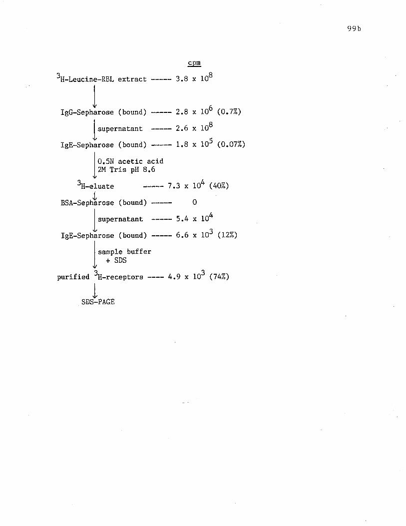
99b
crrm
3H-Leucine-RBL ext,ract 3.8 x 10⁸
IgG-Sepharose (bound) 2.8 x II
I supernatant 2.6 x It
IgE-Sepharose (bound) 1.8 x 1
I o.rlrl acetic acidI zu tri" pH 8.6.t
3H-eluate 7.3 x Io4I
BSA-Sephårose (bound) ----- 0
I
I supernatant 5.4 x 1
TIgE-sepharose (bound) ----- 6.6 x I
I
I sample bufferI +SDS,t
o6 (o.lT")
08
o5 (o.ozz)
(407")
04
o3 (tzz)
purified 3H-receptors ---- t+.9 x to3 (z¿nZ)
PAGE
-i00-
be seen. From previous results (see Chapter II), two distinct peaks,
representing H and R lrere expect.ed. However, this procedure included a
pre-incubation wÍth raL lgG-Sepharose, extensive washing of gels as well
as rebinding of eluted receptors to lgE-Sepharose. According to Kepron
et al. (1982), there is evidence for the preferential binding to
IgG-Sepharose of H oVer R, thus, in this procedure, more H than R may
have been removed by the pre-incubation $rith Igc-Sepharose. In addition'
the study by Kepron et al. also indicated t.hat II has a lower affinity for
IgE-Sepharose than does R. This may explain why, in Èhe present study,
rigorous washing and rebinding of eluLed recepLors may have resulted in a
greater loss of H. Since this loss did not interfere ruit.h Lhe
investigation of 71K and R, it was not considered det,rimental"
Two-Dimensional SDS-PAGE Analys is of Purified Receptors
To determine whether 71K could be reduced into more than one
biosynthetically labelled polypeptide chain, receptors were analyzed by a
two-dinensional procedure, the first dimension being SDS-P/rGE under
non-reducing conditions and the second, SDS-PAGE under reducing
condj.tions. A preliminary experinent was perforned using surface
iodinated receptors which had been purified by a single incubation with
IgE-Sepharose. SDS-PAGB was perforr¡ed under non-reducing conditions in
three lO7" poLyacrylamide tube gels. Two of these gels were then placed
over 10% polyacrylamide slab gels and SDS-PAGE was performed under either
non-reducing or reducing conditions. The other tube gel was sliced into
2 mm fractj.ons which vrere counted in a gamma counter. The slab gels were
fixed, dried and subjected to autoradioagraphy. Figure 24a shor,¡s the
two-dimensional SDS-PAGE analysis of surface-iodinaled receptors vrhich
were not reduced in the second dimension. As expected, all visible bands
100a
Fieure 23: One-dimensional SDS-PAGE analysis of receptors
purified by repetitive affinity chromatography. 3H-lobu11",1
receptors purified using the protocol depicted in Fig. 22 were
anaLyzed on a 10% slab gel. The ge1 was fixed and stained rvith
Coomassie Blue, then treated wiLh En3Hance. The dried gel was
exposed to X-ray film at, -70uC and autoradiography ïias
performed. The gel was scanned with a desitometer. The arrot'¡s
represent the posit,ions of prot,ein standards in order of their
decreasing molecular weigh from left to right: Phosphorylase B,
92.5kDa; bovine serum albumin, 66.2kDa; ovalbumin, 451cDa;
carbonic anhydrase, 31kDa; soybean t.rypsin inhibitor, 21kDa.
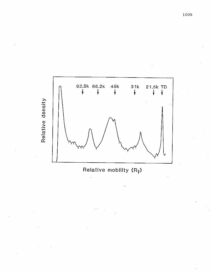
100 b
92.5k+ t
45k 31k 21.5k TD+ +{66.2k
t
=U)coEo.ìCO
oE
Relative mobility (R¡)
-101-
fel1 onto a diagonal 1ine, indicating that all molecules had Lhe same
mobility in both dimensions.
trlhen receptors $rere run under reducÍ-ng condition' (Fig. 24b) the
spot corresponding to 7lK in t,he first dimension has a greater mobility
in the second dinension, i.e. similar to Lhe mobility of the spot
corresponding to R, The spot corresponding to 71K thus falls off the
diagonal line. The profile depicted over each pattern represents Lhe
receptor mobilit,y in the first dimension gel, as determined by slicing
and counting t,he third t,ube ge1. It should be noted that the spots
representing receptor material are very diffuse in both the reduced and
unreduced patterns. This is not unexpected in light of the fact thaL
one-dimensional SDS-PAGE analysis produces diffuse receptor bands. ldhen
these already heterogeneous bands are subjected to second-dimensional
SDS-PAGE, this heterogeneity would be expected to become even more
apparenL.
The ident,ical experiment was performed usÍng extensively purified
receptors from RBL cells biosynthetically labelled wi-th 3H-leucine"
Figure 25a illustrates the tr,¡o-dj-mensional analysis of uritiated
receptors which were not reduced in the second dimension. Clearly, a1l
visible spo¡s fa1l r¿ithin the diagonal line. Due to cracking of the gel
during the drying process, the spot, with high mobilit,y corresponding to
the 1ow molecular weighL band (27kDa), seen in l-igure 23, is difficulu to
dist,inguish. Its presence is clear, however, in Fig. 25b, which
represents analysis of purifi,ed receptors reduced in the second dimension
of SDS-PAGE. In this case all spots' except' one fall on a diagonal
line, the except,ion being the spot r+ith the lor,¡est mobility rvhich, in
agreenent with the pattern in Fig. 24b, represents 71K. The spot which
-i02-
represents Lhe 271<Da molecule clearly remained on t,he diagonal line and
was therefore not drastically affect,ed by reduction" So did a spot which
in the first dimension, musL have migrated wiLh the tracking dye. 0f
great interest in this pattern is the fact that while the spot
representing the 71K receptor fal1s off the diagonal upon reduction, no
other spots are apparent, either at the level of the spot representing
the 27kDa molecule or anywhere else on the vertical under 71K, r,rhere a
reduction product of 71K would be expected to appear.
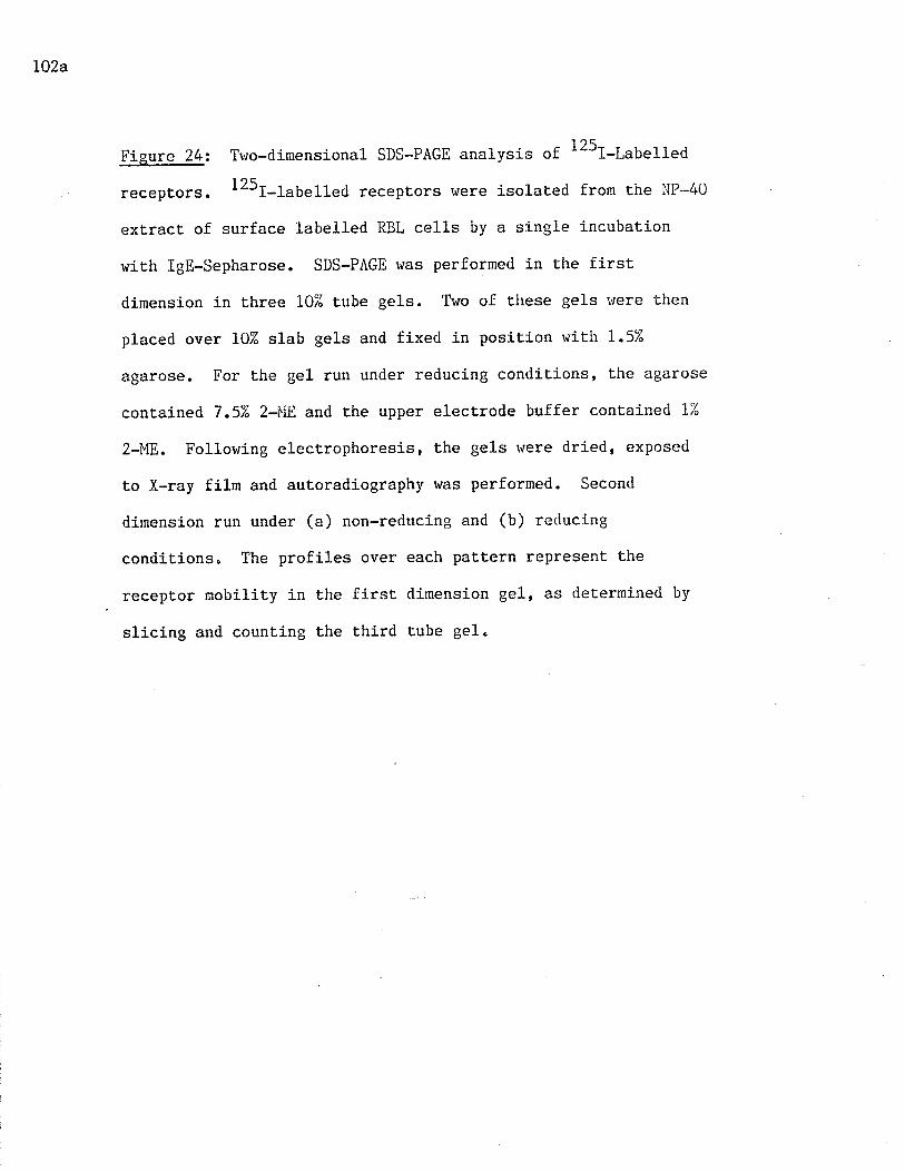
L02a
Figure 24: Tr,¡o-dímensional SDS-PAGE analysis of 125t-labelled
receptors. 125I-1abe11ed receptors r'¡ere isolated from the NP-40
extract of surface 1abe1led RBL cells by a single incubatj-on
r^rith lgE-sepharose. SDS-PAGE was performed in the first
dimension in three 10iå tube gels' Two of these gels t'rere then
placed over 10% slab gels and fixed in position r'rith 1.5%
agarose. For the gel run under reducing conditions, the agarose
contained 7.5% 2-llI:.8 and the upper electrode buffer contained 1%
2-ME. Follorving electrophoresis, the gels rvere dried, exposed
to X-ray film and autoradiography was performed. Second
dimension run under (a) non-reducing and (b) reducing
conditions" The profiles over each pattern represent the
recepLor mobility in the first dimension gel, as determined by
slicing and counting the third tube gel.
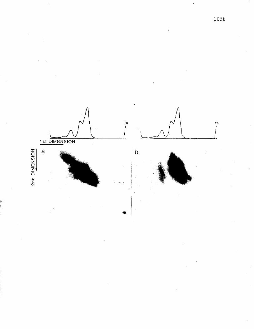
102b
TD
1st DIMENSION
a
TD
*
bzqU)zIU
=oEcôJ
IO2c
Figure 25: Tr,ro-dimensional SDS-PAGE analysis of 3H-1obu11"d
receptors. 3H-l.belled receptors rvere purified using the
protocol depicted in Fig. 22. Tv¡o dimensional analysis was
performed as in Fig. 24 for 125I-1"b"lled receptors. Slab gels
were treated r,¡ith En3Hance, dried and exposed to X-ray film.
AuLoradiography rvas performed. Second dimension run under (a)
non-reducing conditions and (b) reducing conditions.
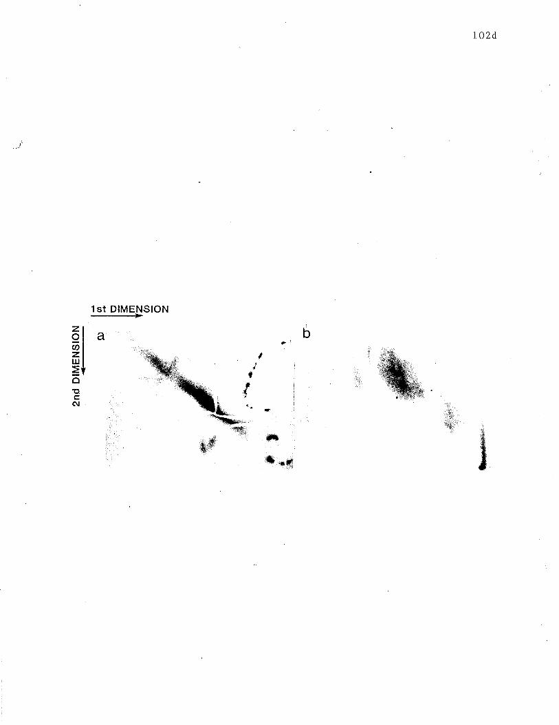
102d
)'
b
1st DIMENSION
az9cr)zUJ
oec(\¡
It
t,{
*
å
ffiwa,;
.,:f:Ç.
''-xìl
!?.
#ft.ryq
fr.'a{
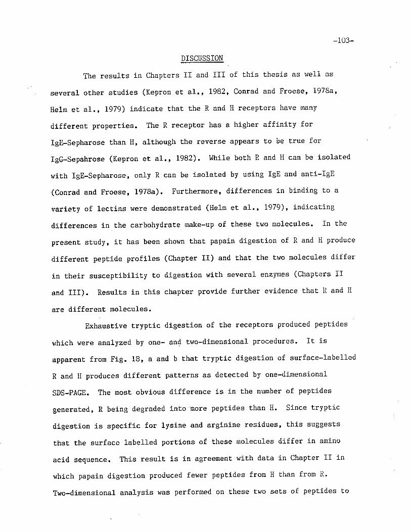
-103-
DISCUSSION
The results in chapters II and III of this thesis as r'¡ell as
several other studies (Kepron et al., L982, Conrad and Froese, I97Ba'
Helm et a1., L979) indicate Lhat the R and H receptors have many
different properties. The R receptor has a higher affinity for
Igfl-Sepharose than H, alt,hough the reverse appears Lo be true for
IgG-Sepahrose (Kepron et a1., 1982). I'Jhile both R and H can be isolated
with lgE-Sepharose, only R can be isolated by using IgE and anti-IgE
(Conrad and Froese, 1978a). Furthermore' differences in binding to a
variety of lectins ltere demonstrated (tleIm et a1., L979), indicating
differences in the carbohydrate make-up of these two nolecules" In the
present study, it has been shown that papain digestion of R and iì produce
different peptide profiles (Chapter II) and that the ttuo molecules differ
in their susceptibility to digestion with several enzymes (Chapters II
and III). Results in this chapter provide further evidence that R and il
are different molecules.
Exhaustive trypLic digestion of the receptors produced peptides
which were analyzed by one- and two-dimensional procedures. ÏL is
apparen¡ from Fig. 18, a and b that tryptic digestion of surface-labelled
R and H produces different patterns as detected by one-dimensional
SDS-PAGE. The most obvious difference is in the number of peptides
generated, R being degraded into-'more peptides than H. Since tryptic
digestion is specific for lysine and arginine residues, this suggests
that the surface 1abel1ed portions of these molecules differ in amino
acid sequence. This result is in agreement vtith data in Chapter II in
rr¡hich papain digesLion produced ferr¡er peptides from H than from R"
Two-dimensional analysis was performed on these two sets of peptides to
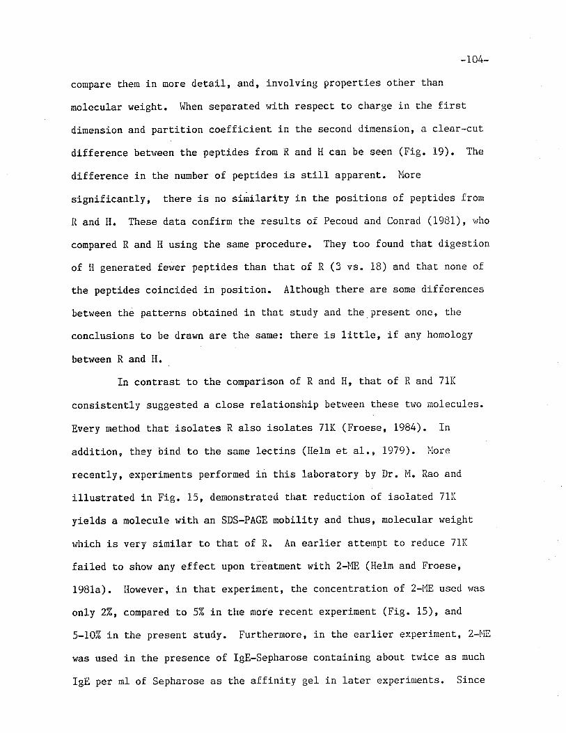
-i04-
compare the¡n in more detaj-l, and, involving properties other than
molecular weight. lrlhen separated r,Iith respecL to charge in the first
dimension and partition coefficient in the seconcl dimension, a clear-cut
diff erence betr.reen the peptides fronn R and H can be seen (Fig. 19). The
difference in the number of pept,ides is sLill apparent. More
significant,ly, there is no similarity in the positions of peptides frorn
R and H. These dat,a confirn the results of Pecoud and Conrad (1981), rvho
compared R and i{ using Lhe same procedure. They too found that digestion
of H generated fewer peptides than that of R (3 vs. 18) and that none of
the peptides coincided in position. Although there are sone <iifferences
betrveen the patterns obLained in t,hat study and the presenL one, the
conclusions to be drawn are the same: there is little, if any homology
between R and H.
In cont,rasL t,o the connparison of R and H, that of R and 71K
consistently suggested a close relationship betlveen these tr¿o molecules.
Every method that isolates R also isolates 71K (Froese' 198i+). In
addition, they bind to the same lectins (He1m et a1., LgTg). l'lore
recently, experimenLs performed in this laboraLory by Dr. M. Rao and
illustrated in Fig. 15, demonstrated tlìaL reducLion of isolated 71K
yields a molecule with an SDS-PAGE mobility and t,hus, molecular rueight
which is very similar to thaL of R. An earlier atternpt to reduce 71K
failed to show any effect upon tieatment r,¡ith 2-ME (He1m and Froese,
1981a). I{owever, in that experimenL, the concentration of 2-1"1E useri was
only 27", conpared to 57" in the more recent experiment (Fig. i5), and
s-rcZo in the present study. Furthermore, in the earlier experinent, 2-i{E
was used in the presence of lgE-Sepharose conLaining about twice as much
IgE per m1 of Sepharose as the affinity gel in lauer experinents. Since
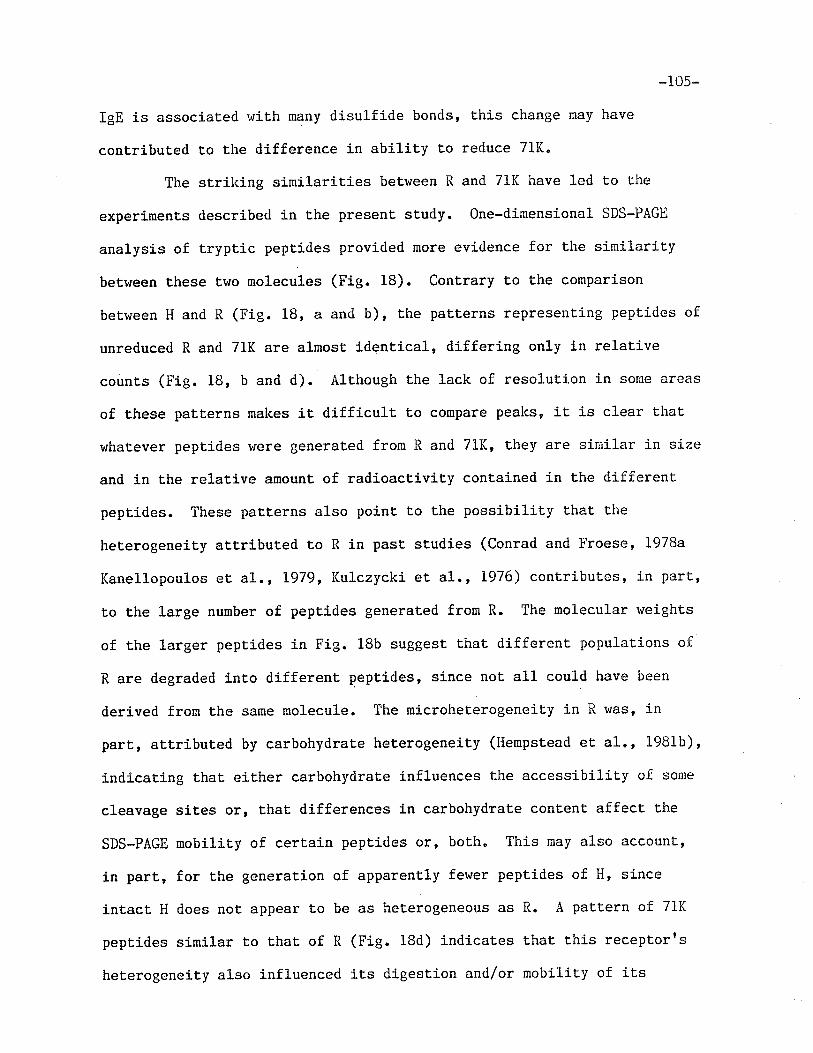
-10s-
IgE is associated with many disulfide bonds, t,his change may have
cont,ributed to the difference in abiliLy to reduce 71K.
The strilcing similarities between R and 71K have led to the
experiments described in the present study. One-dirnensional SDS-PAGE
analysis of tryptic peptides provided more evidence for the similarity
between these two molecules (Fig. 18). ConLrary to the comparison
betrveen H and R (Fig. 18, a and b), the patterns representing peptídes of
unreduced R and 71K are almost ÍdenLical, differing only i-n relative
counLs (Fig. 18, b and d). Although the lack of resolut,ion in some areas
of these patterns makes iL diffÍcu1t to compare peaks, it is clear that
whatever peptides tüere generat,ed fron R and 71K, they are sir¡iilar in size
and in the relative amount of radioactiviLy contained in the different
peptides. These patterns also point to the possibillty that the
heterogeneit,y attribut,ed to R in past studies (Conrad and Froese, L978a
Kanellopoulos eL al., 1979, Kulczycki et 41., 1976) contributes, in part,
to the large number of pept,ides generated frorn R. The molecular weights
of the larger pepticles in Fig. 1Bb suggest that different populat,íons of
R are degraded into differenL peptides, since not all could have been
derived from the same rnolecule. The microheEerogeneity in R was, in
part, at,tributed by carbohydrate heterogeneity (Hempstead et al., 1981b)'
indicating that either carbohydraLe influences Lhe accessibility of some
cleavage sites or, that differences in carbohydrate content affect the
SDS-PAGE mobility of certain peptides or, both" This may also account,
in part, for the generation of apparently fewer peptides of H, since
inLact II does not appear to be as heterogeneous as R. A pattern of 71K
peptides sinilar to that of R (Fig. l8d) indicates that this recepLorrs
heterogeneity also influenced its digestÍon and/or mobilit.y of its
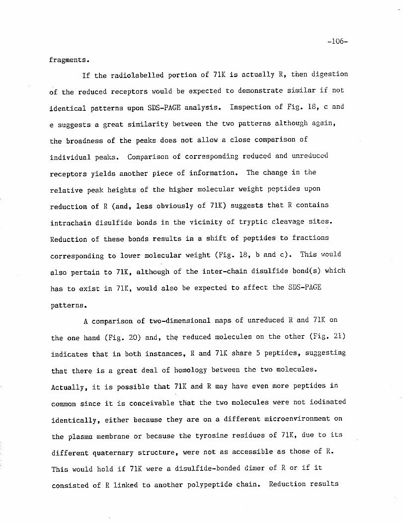
-106-
fragments.
If the radiolabelled portion of 7lK is actually R, then digestion
of the reduced receptors would be expected to demonstrat,e si¡nilar if not
identical patterns upon SDS-PAGE analysis" Inspection of Fig. 18, c and
e suggests a great similarity betrveen Lhe two patterns although again'
the broadness of the peaks does not allow a close comparison of
individual pealcs. Comparison of corresponding reduced and unreduced
receptors yields another piece of information. The change in the
relative peak heights of t,he higher molecular weight peptides upon
reduction of R (and, less obviously of 71K) suggests that' R contains
intrachain disulfide bonds in the vicinit,y of t,ryptic cleavage sites.
Reduction of these bonds results in a shift of peptides to fractions
corresponding to loruer molecular weight (Fig" 18, b and c). This r'¡ou1d
also pertain to 71K, although of the inter-chain disulfide bond(s) ruhich
has to exist in 71f., would also be expected to affect the SDS-PAGE
patLerns.
A comparison of two-dimensional maps of unreduced R and 711( on
the one hand (Fig. 20) and, the reduced molecules on the other (Fig. 2L)
indicaLes thaL in both instances, ll and TlL share 5 pept,ides' suggesLing
that there is a great deal of homology beLr'¡een the two molecules'
Actually, it is possible that 71K and R nay have even more peptides in
common since iL is conceivable thãt the t'wo molecules \,rere not iodinated
identically, either because they are on a different microenvironment on
the plasma membrane or because the Lyrosine residues of 71K, due to its
different, quaternary structure, were not as accessible as those of R-
This would hold if 71K vlere a disulfide-bonded dinter of R or if it
consisted of R linked to another polypeptide chain. ReducLion resulLs
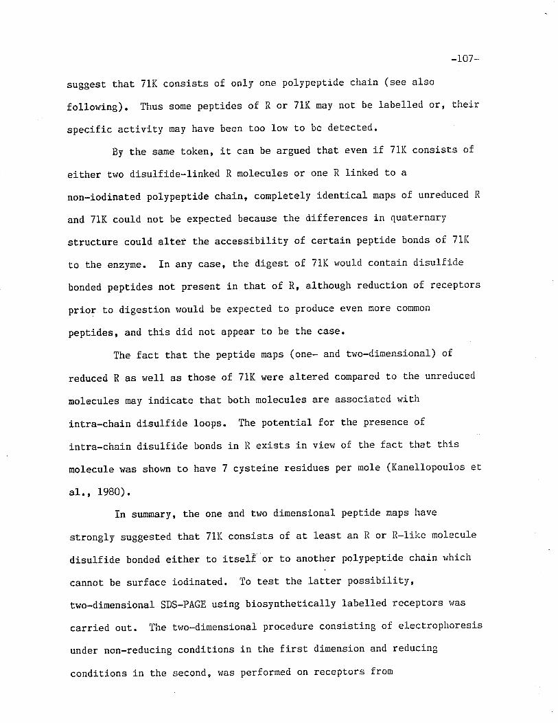
-107-
suggest that 711( consists of only one polypeptide chain (see also
following). Thus some pept.ides of R or 71K may not be labelle<l or' their
specific activity may have been too low to be detecLed.
By the sarne token, it can be argued that even if 71K consist,s of
either trvo disulfide-linked R molecules or one R linked to a
non-iodinated polypept.ide chain, completely identical maps of unreduced R"
and 71K could not be expected because the differences in quaternary
structure could alt,er the accessibility of certain peptide bonds oî. 7LK
to the enzyne. In any case, the digest of 71K r^¡ould contain disulfícle
bonded peptides not, present in that of R, although reduction of receptors
prior t,o digestion rvould be expected to produce even nore conmon
peptides, and this did not appear to be the case.
The fact that the peptide maps (one- and Lwo-dinensional) of
reduced R as well as those of 71K were altered compared t.o the unreduced
molecules may indicate that both molecules are associated with
j-ntra-chain disulfide loops. The potential for the presence of
intra-chain disulfide bon<is in R exists i.n view of the fact that Lhis
molecule was shov¡n to have 7 cyst,eine residues per mole (Kanellopoulos et
al., 1980).
In summary, the one and two dimensional peptide maps have
strongly suggested that 71K consists of at. least an R or R-like molecule
disulfide bonded either to itself or to anoLher polypept.ide chain r'¡hich
cannot be surface iodinated. To test the lalter possibility,
two-dimensional SDS-PAGE using biosynthetically labelled receptors ruas
carried out. The tv¡o-dirnensional procedure consisting of elecLrophoresis
under non-reducing conditions in t.he first dimension and reducing
condit,ions in the second, Ïias performed on receptors from
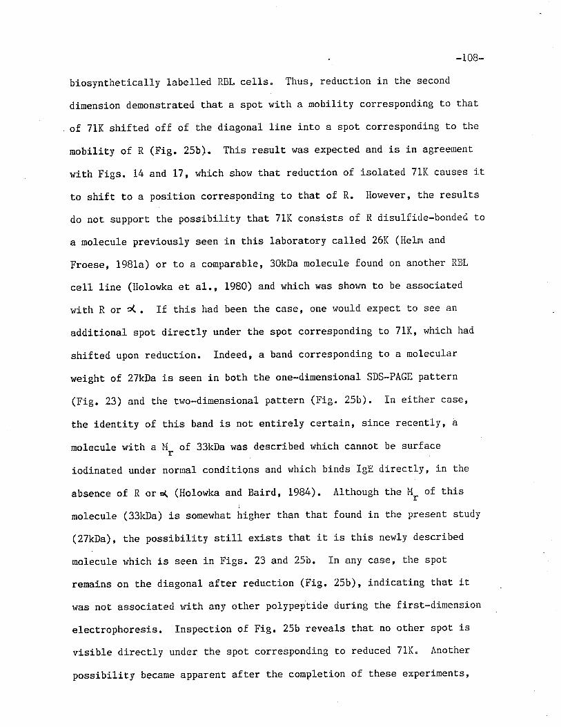
-i08-
biosynthetically labelled RBL cel1s" Thus, reduction in the second
dinension demonstrated that a spot. wiLh a nobility corresponding to that
of 71K shifted off of the diagonal line into a spot corresponding to the
mobility of R (Fig. 25b). This result was expecued and is in agreement
with Figs. 14 and 17, which shorv that reduction of isolated 71K causes it
to shift to a position corresponding to that of R. I1owever, the results
do not support the possibilit,y that 71K consists of R disulficie-bonded to
a molecule previously seen in this laboratory called 26t( (llelm and
Froese, ig8la) or to a comparable, 30kDa molecule found on another RBL
cel1 line (Holowka et a1., 1980) and r"hích was shown to be associated
with R or c(. If this had been the case, one would expect to see an
additional spot directly under the spot corresponding to 711(, rvhich had
shifted upon reduction. Indeed, a band corresponding to a molecular
weight of 27kÐa is seen in both the one-dimensional SDS-PAGB pattern
(Fig. 23) and the two-direensional pattern (Fig. 25b). In either case'
Èhe identit,y of this band is not entirely certain, since recently, a
molecule with a Ùi, of 33kDa rsas described rvhich cannot be surface
iodinated under normal conditiqns and r,¡hich binds IgE directly, in the
absence of R ors( (Holowka and Baird, 1984). Although the M, of this
molecule (33kDa) is somewhat frigfrer than that found in the present study
(27kÐa), the possibility still exj.sts that it. is this newly described
molecule r,¡hich is seen in Figs. 23 and,25b. In any case' the spot
reroains on the diagonal aft,er reduction (Fig. 25b), indicating that it
was not associated rvith any other polypept.ide during the first-dimension
electrophoresis. Inspect,ion of Fig. 25b reveals t,hat no other spot is
visible directly under the spot corresponding to reduced 7lK" Another
possibility became apparent afLer the completion of these experimenLs'
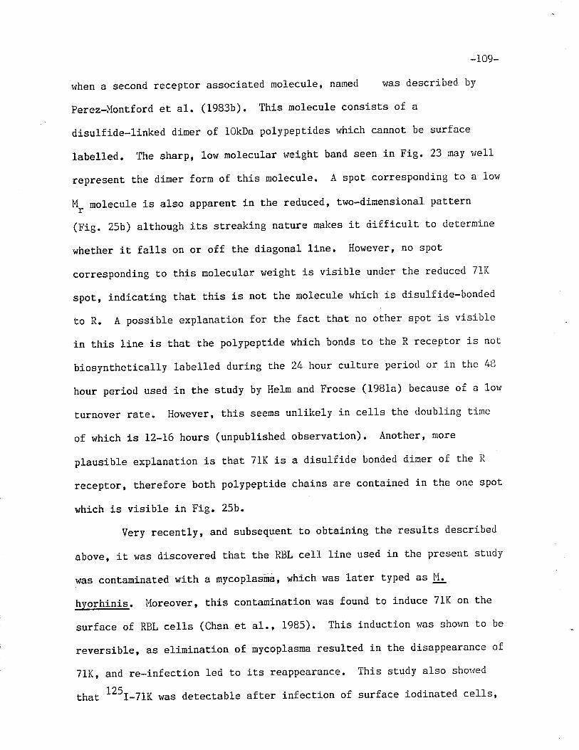
-109-
lfhen a second receptor associated molecule, naned r,ras described by
Perez-Flontford et al. (1983b). This molecule consists of a
disulficte-linked dimer of lOkDa polypeptides r'¡hich cannot be surface
labelled. The sharp, low molecular rveight band seen in Fig. 23 may rvell
represent the dimer forn of this molecule. A spot corresponding to a lor+
M_ molecule is also apparent in the reduced, two-dimensional paLternr
(Fig. 25b) although its streaking nature makes it difficult to deLermine
wheLher it falls on or off the diagonal line' Hor'¡evere no spot
corresponding t,o this molecular weight is visible under the reduced 71i(
spot, indicating that t,his is not the molecule nhich is disulfide-bonded
to R. A possible explanaLion for Lhe fact that no oLher spot is visible
in Lhis line is that Lhe polypeptide r.'hich bonds to t.he R recepLor is not
biosynthet,ically labelled during the 24 hour cullure period or in the 43
hour period used in the study by Helm and Froese (1981a) because of a low
turnover rate. However, this seems unlikely in cells the doubling time
of which is 12-16 hours (unpublished observation). Another' more
plausible explanaLion is that 71K is a disulfide bonded dimer of the R
receptor, therefore both polypeptide chains are contained in the one spot
which is visible in Fig. 25b.
Very recently, and subsequent to obtaining the results descríbed
above, it was dj-scovered thaL the RBL cell line used in Lhe present study
\,/as contaminated wiLh a mycoplasñá, which was later typed as lui.
hyorhinis. l"loreover, this contamination was found to induce 71K on the
surface of RBL ce11s (Chan et a1., 19e5). This induction rvas shorvn to be
reversible, as elimination of mycoplasma resulted in the disappearance of
7lK, and re-infection led to its reappearance. This study also shor'¡ed
tfrat 125I-7lK was detectable aft.er infection of surface iodinated cells,
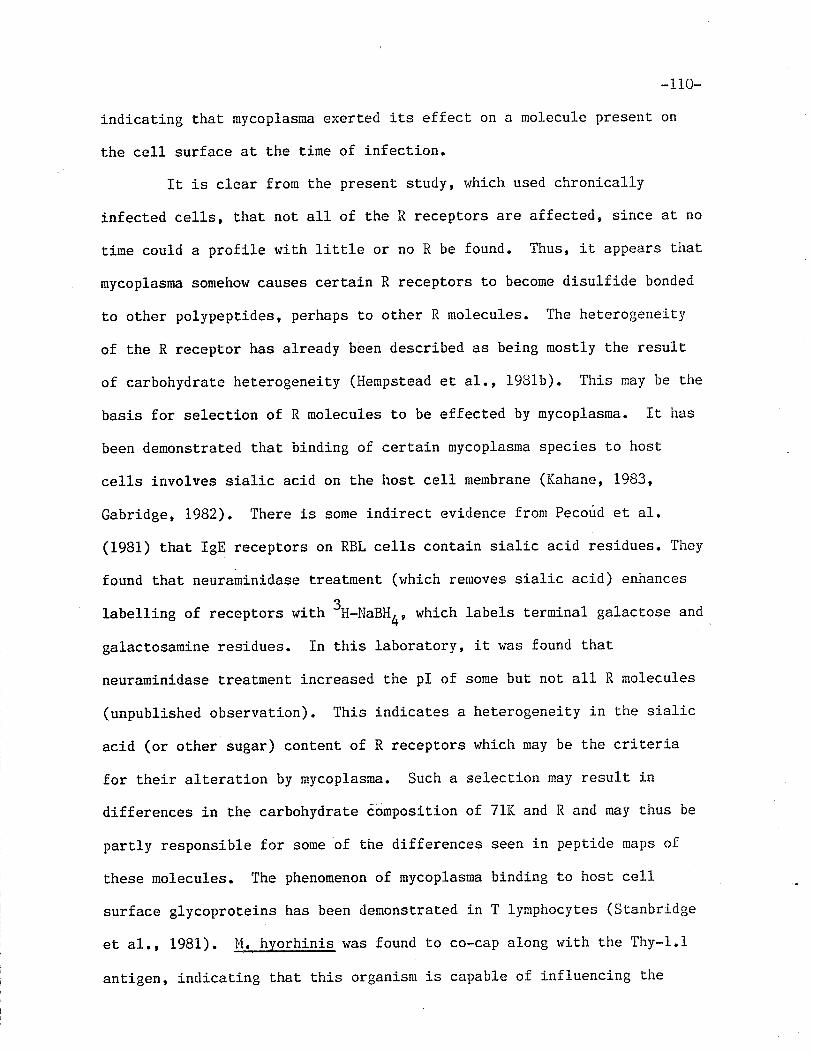
-1 10-
indicating that mycoplasna exerteci its effect on a molecule present on
the cell surface at Lhe time of infection.
It is clear from Lhe present study, which used chronically
infected ce11s, that not all of the R receptors are affected, since at no
tirne could a profile ruith little or no R be found. Thus, it appears that
mycoplasma somehow causes certain R receptors to become disulfide bonded
Lo other polypeptides, perhaps to other R molecules. The heLerogeneity
of the R receptor has already been described as being mostly the result
of carbohydrate heterogeneit,y (l{empstead et al., 198ib). This nay be the
basis for selection of R rnolecules to be effected by mycoplasma. It has
been demonstrated that binding of certain mycoplasma specÍes to host
cells involves sialic acid on the host cell membrane (Kahane, 1983'
Gabridge, 1982). There is some indirect evidence frorn Pecoúd et al.
(1981) that IgE recepuors on RBL cells contain sialic acid residues. They
found that neuraminidase treatrnent (vrhich removes sialic acid) enhances
labelling of receptors with 3H-NaBHO, which labels terminal galactose and
galact,osamine residues. In this laboratory, it was found t'hat
neuraminidase treatment increased the pI of some but not all R nolecules
(unpublished observation). This indicates a heterogeneity in the sialic
acid (or other sugar) content of R receptors r¡hich nay be the criterj-a
for their alteration by mycoplasma. Such a selection may result in
differences in t.he carbohydrate cónposition of 71K and R and nay thus be
partly responsible for some of the differences seen in pepLide maps of
these molecules. The phenomenon of mycoplasma binding to host cell
surface glycoproteins has been demonstraLed in T lymphocytes (Stanbridge
et al., 1981) . M. hyorhj-nis was found to co-cap along rvith the Thy-1.1
antigen, indicating that this organism is capable of influencing the
-111-
lateral mobility of surface molecules" In a study by I'lise, et al.
(1982), the Thy-l.l ant.igen was found to be localized on the surface of
the l"f. hyorhinis organisms which had colonized the host cel1s. Ïn
addition, they found that. a lipid probe inserted into the lymphocyte
membrane was translocated into the mycoplasma membrane. These fact's
suggest that there was some exchange of surface glycoprotej-ns as uell as
ext,ernal modulation of the lipid composÍtion of the host cell membrane by
the mycoplasma cell. Such phenomena may play a role in the mycoplasnal
infection of RBL cel1s and in the resulting alteration of IgE receptors
on these host, ce1ls. Indeed, Lhese observations raise the possibility
that 71K may consist of R and a mycoplasma derived molecule, aluhough
such a molecule could be expected to be labelled under the conriitions
used in this st,udy.
In light of the fact that cross-linking of R-like receptors h'as
shown to be sufficient for Lhe induction of mediator release (Iserslty et
al.¡ 1977, Ishizaka eL al., L977, Ishizaka et al., 1978), the possibilit'y
that mycoplasma infection may result in the covalent crosslinking of cr'ro
R receptors and thus in the modulation of mediaLor release. Even if it
should t,urn out Lhat, in order to produce 71K, R is linked to another
molecule, mediator release could be effected since it rvas shorvn that.
bridging of Fc receptors and histocompatibility antigens can also lead to
mast cell degranulaLion (Daeron êt a1., L977).
-IL2-
GENERAL DISCUSSION
Studies of the IgE receptors on RBL cells as well as on RMC have
led to an j-ncreasingly complex picture of these molecules" Some of the
earliest investigations of the RBL cell Fc¿ receptors revealed a single
cell surface molecule capable of binding IgE. Conrad and Froese (1976)
used a sandwich precipit.ation method with rat myeloma IgE, g-specific
rabbit anti-rat IgE and goat anti-rabbit Ig, and isolated a single
surface iodinated molecule, t,he SDS-PAGE mobility of which on 5% gels
corresponded to an apparent molecular weight of. 62kDa. This result was
confirmed by Kulczycki, et al. (L976), who found a molecule with an
apparenL molecular weight of 45-50kDa using 10 and L2% gels. At the same
time, t.his st,udy provided some of fhe first. concrete evidence for the
protein nature of the receptor by demonstrating its sensit.ivity to a
variety of proteases. The dependence of the apparent molecular weight on
the porosity of the polyacrylamide gel also suggested t,hat the receptor
was a glycoprotein (lleber and Osborn, 1975), a characLeristic which has
since been repeatedly demonstrated (Kulczycki- and Parker, 1979 Kulczyckj-
et al., L976, Helm and Froese,.1981a, Helm and Froese, 1981b). Another
study, by Isersky, et al., (1977) reported Lhe presence of a slightly
larger molecule (58kDa) on 102 gels, and first suggested the existence of
nolecular weight heterogeneÍty between RBL cell lines.
Isolation of receptors using IgE-Sepharose revealed a further
complexity in t.heir nature. Conrad and Froese (1978a) found that a
molecule with an apparent molecular weight of 55kDa could be recovered
along with Lhe 45kDa component, and named the two receptors H and R,
respectively. Kulczycki and Parker (L979) found only one receptor with
an !lr of 50kDa by this method. A collaborative study involving a number
-113-
of laboratories which utilized Ehe different cell lines demonstrated that
these seemingly discordant results could be reconciled (Froese et al.,
1982). All the cell lines were shown to express both the R and H
receptors, t,hough the molecular weights of t,he receptors' particularly R'
were shown to vary from cell line to line. In addition, the relative
amounts of each receptor also differed among the lines" Both types of
receptors were also found on normal peritoneal rat mast cells (Froese,
1980). To date, only R-like molecules have been implicated in mediator
release from RBL cells (Isersky et al., 1978), and from RMC (Ishizaka et
al., 1977). No function has been attributed to the H molecule. However,
a study from this laboratory has shown that while bot,h R and H bind to
IgG-Sepharose, H appears to have the higher affinity for this
im¡nunosorbent and a lower affinity for lgE-sepharose (Kepron et al.,
1982)
The present study has compared R and H receptors to determine
whether or not there is structural homology between these trvo molecules.
It was prompted by the possibility that. R is a degradation product of I{.
This would be similar to Ehe results obtained with the Fc¡R from a
variety of cells, in which 70kDa and 90kDa molecules were found to be
spontaneous degradation products of a 115kDa molecule (Kahn-Perles et
al., 1980). Alterna¡ively, H might represent a precursor of R. The
results presenLed in this study fâiled to demonstrate any similarity
between the Lwo molecules and actually showed t,haÈ they are different.'
Digestion of isolated recept,ors by papain produced strikíngly different
paLrerns when peptides were analyzed by SDS-PAGE (Fig. 6). These
patterns demonstrate that H (or its surface labelled portion) is more
susceptible to cleavage under Lhese conditions than R and thus, smaller
-I14-
peptides are produced from the former receptor. The differences between
R and H were further emphasized when an attempL vras made to obtain
IgE-binding fragments of the receptors by proteolytic degradation. I',rhen
the residue-non-specific papain was employed, only a binding fragment of
H was produced (Figs.9 and 10), indicating that, although t,he
surface-labelled portion of R is less suscept.ible to papaÍn digestion
than that of H, its binding site is destroyed by this enzyme. The loss
of the lgE-binding activity of R could either be due to a direct effect
on the binding site, or Èo an alteration of secondary or tertiary
structure. Digestion by trypsin and chymotrypsin also produced an
Igfl-binding fragment of H (Figs. I2a, b and 13a, b respectively). In
these experiments, however, a population of R receptors remained inLact.
(Figs . LZc and 13c). Since carbohydrate heterogeneity has repeat.edly
been shown to be responsible for the heterogeneily seen in R (Hempstead
et al., 1981b, Goetze eL al., 1981), this result suggests that
carbohydrates may influence the susceptibilit.y of the R binding site to
desLruction by t,hese enzymes. When subjectÍng Lhe equivalent of R on
their RBL cell line to controlled digestion with a variety of proteolytic
enzymes, Goetze, et al. (1981) consistenLly produced two fragments of
similar size, both of which cont,ained some carbohydraLe. They suggested
that the < chain, or R, exists in the form of two domains, both of which
are exposed on the surface of the-cell. They carried out Lheir
experiments under denaturing conditions, which did not, allow subsequent
studies on the lgE-binding capabilities of the isolaÈed fragments or
domains. Finally, analysis of peptides of R and H produced by exhaustive
tryptic cleavage demonstrate that there is litt1e if any structural
homology between ttre surface labelled portion of these two recepLors
-115-
(Figs. 17a and b, 18a and b). These results confirm those obtained by
Pecoud and Conrad (198f), which were published while this study was in
progress. Although only surface-labelled peptides were being compared in
the present "tuày
as well as in that of Pecoud and Conrad, the
differences found beÈween R and H are nonetheless significant. Ït is not
unreasonable to assume that it is the surface-labelled portions of the
intact, molecules which contain the lgE-binding acLivity. Therefore, this
study provides evidence that Lhe functional port,ions of these two
molecules are struct,urally different. Thus, it is not surprising that
these molecules show differences in t,heir interaction with IgE and IgG
(Kepron et al., 1982). Further i-nvestigations would be required to
determine whether or not the remainder of the molecules, which are not
accessible to surface iodination, display any homology.
Rat basophilic leukemia cells are noL unique in possessing
distinct Fc receptors for differenL Ig classes or subclasses. Conrad and
Peterson (1984) found Èhat the Fc R and t.he Fc ,OR on murine B
lymphocytes are structurally distinct based on the lack of
crossreactivity in Ig-binding.. However, both receptors bound to rabbit
IgG, indícating thaL some crossreactivity does exist" Murine macrophages
were also found to have at least two distinct receptors for IgG, and,
although both bound rhe two classes of IgG (IgGr" and IgG2¡) ' one had a
higher affinity for IBG'., the other for IEG'O (Haeffner-Cavaillon et
al., L979). Using the same cell line, Unkeless (1977) provided evidence
for an I1GZ^ receptor which was trypsÍn sensitive as well as a
trypsin-resistant aggregaÈed-rabbit IgG recept,or" Whether these two
receptors were the same tvro receptors described in the study by
Haeffner-Cavaillon, et al. (L979) was not determined. RaC macrophages
-1 16-
r,Iere found to have at leasL three distinct Fc receptors: one for rat IgE¡
one for TgGZa; one for IgG1, IgG2b and heterologous IgG (Bolt.z-Nitulescu
et 41., 1981). Rat IgE was capable of inhíbiting binding to the last
receptor. Indeed, there are many cell types which bear distinct,
fspecifict receptors for different Ig classes and sublasses. In several
cases, the structurally distinct receptors vrere shown to display affinity
for the same Ig classes. A similar situation may exist in RBL cells,
which, according t.o the studies by Kepron, et al. (1982) and Segal, et
al. (1981), bear two distinct receptors for IgE and IgG. Both Ig classes
bind to either receptor with differing affinities.
The presence of a third-receptor for IgE on RBL cells was first
detected by Conrad and Froese (1978a). At that time, it was not
regularly observed, and was therefore considered a contaminant (4.
Froese, personal communication). The molecule, the molecular weight of
which was estimated to be 7lkDa and which was called 71K' was described
as an IgE receptor by Helm, et al. (1981a) who showed that its binding lo
IgE-Sepharose could be inhibited by presaturating receptors with free
IgE. The same study demonstraËed the biosynthetic incorporation of
3H-l"ucine into the three receptors, R, H and 71K, as well as into a
26kqa molecule. A molecule analogous to the 26kDa molecule had earlier
been detected on RBL cells by Holowka, et al. (1980), who used a
crosslinking reagent to show tha-t a 30-35kDa polypeptide (called p ) was
associated with the R-like receptor (called o< ). The polypeptide could
not be surface labelled in either sLudy. In addition, in the study by
Helm, et al. (1981a), the affinity of.26K for R (if such an associaLion
did exist) was weak since the former molecule could not be consistently
isolated by lgE-Sepharose or DNP-IgE and anti-DNP" In another
-TL7-
laboraLory, using a different RBL cell line, only treatment \^rith
crosslinking reagents (Holowka et al., 1980, Holowka et 41., 1981) or
isolation of receptors in the presence of the appropriate
phospholipid:detergent ratio produced a consistent yield of the
molecule (Rivnay et al., 1982). These results indicate that the
associaÈion between o¿ and F (and the putative one between R and 26K) is
a weak and non-covalent one.
Although reduction of receptors did not appear to change their
mobility in the study by Helm, et al. (1981a), in a more recent
experiment, reduction did result in a change in the mobilit,y of the 71K
molecule (Fig. 15) (M. Rao, unpublished results). This discrepancy may
have been due to the difference in the concentration of reducing reagent
used or to the difference in the amounL of IgE coupled to Sepharose, or
to both. The appearance, upon reduction of isolated 71K, of a molecule
the molecular weight of which was similar Lo that of R led to speculation
thau 71K r{as composed of R, disulfide linked to some other polypepLide'
The previously demonstraLed similarities between the binding
characteristics of R and 7lK Ient, further support to this hypothesis.
Thus, both receptors could be isolated with lgE-anti-IgE, (Froese et al.,
LgBz, Helm and Froese, 1981a, Figs. 4 and 16, present study), with
DNP-IgE-anti-DNP (Helm and Froese, 1981a), as well as with lgE-Sepharose
(Froese et al., 1982, Helm and Fiôese 1981a, Figs. 3 and 23, present
study). 0n the other hand, neither R nor 71K bind significantly when
anti-receptor antiserum (RAR) is used t,o isolate H receptors (Fig . 4).
In addition, the lectin binding characteristics of R and 71K are very
similar but are somewhat different from that of H (Helm and Froese,
1981b). These facts suggested some structural relationship between R and
-i 18-
71K.
A comparison of tryptic peptides in the present study provides
some further information on this relationship. Both one- and two
dimensional trypt,ic peptide maps of R and 71K poinE to a great deal of
similarity between these thro receptors. Horvever, differences were also
observed and must be accounted for. In Chapter IV, it was suggested that
even if 71K consists of either a disulfide-linked dimer of R or an adduct
of R and another, as yeL undetected polypeptide chain, some differences
in peptide maps could perhaps have been expected due to differences in
iodination of the two molecules. An alternate or additional explanation
could be found in the fact that R exhibits a great deal of
microheterogeneity, most likely due to differences in carbohydrate
conLent (Hempstead et al., 1981b, Goetze et 41.' 1981). Such a
heterogeneity could give rise to differences in the properties of
peptides generated from different R molecules and t,hus lead t,o
differences in two-dimensional pept.ide maps of individual molecules.
Differences could be due to variability in charge, i.e. variability in
sialic acid content and/or to partition coefficient differences, and
would be detected in the first and second dimension, respectively.
Therefore, even if R molecules are present in 7lK, the microheterogeneiLy
in 71K may díffer from that of conventional R recept'ors.
In rhe report by Helm and Froese (1981a), iL was suggested that
71K might be a more stable conformation of R and 26K' since, along with
all the other evidence, the arithmetic also fits this hypothesis, i"e.
the sum of 45kDa and 26kDa yields 71kDa. In the present study, this
possibility was investigated by separating biosynLhetically labelled
receptors in two dimensions by SDS-PAGE. The results indicate that
-1i9-
although a biosynthetically labelled molecule similar in mobility to 26K
was isolated, it was not disulfide-bonded to R (Fig.25b). No such
molecule was found when 7lK was reduced. Indeed, no biosyntheLically
labelled molecule other than R lras seen upon reduction of 71K ín Èhe
second dirnension. It therefore seems unlikely that 71K is a form of R,
covalently linked to 26K. Alternat,ively, 7lK may be a disulfide-bonded
dimer of R. Simplistically, one could argue that a dimer of R should
have a molecular weight of 90kDa. However, differences in
microheterogeneity and the fact that 71K, when st,udied by SDS-PAGE under
non-reducing conditions, retains its quaternary strucLure (which could
influence iLs mobility) may make such simple arithmetic meaningless.
Although the resulLs presented in this thesis do not prove conclusively
that,71K is a dimer of R, they rule out several other alLernatives and
provide no evidence to contradict this possibility"
Subsequent to performing the experiments for this thesis, t.he
discovery that infectj-on with mycoplasma (M. hvorhinis) was responsible
for the expression of ehe 71K receptor (Chan eL a1., 1985) led to a nehl
perspective in the investigation of this molecule. It was demonstrated
that uninfected cells begin to express 71K on their surface 24 hours
after infection and that the effect of the organism was on receptors
pre-existing on the cell suface at the time of infection (Chan et a1.,
1985). The pre-existing recept,ois were most. likely R since RBL cel1
lines which have a larger R-like molecule than the one used in t,he
present study appear to express a larger 7lK-like molecule upon infection
(Froese eL al., L982, B.M.C. Chan, personal communication)" It has also
been demonstrated that RBL cells infecLed in the presence of IgE do not
express 71K (B.l'{.C. Chan personal communication), whieh suggests Lhat the
-120-
mycoplasma organism must come into contact with the receptor in order to
exert it.s effect.
The effect of a variety of mycoplasma species on the immune
system became a focal point of investigat.ion when it became evident that
the pathogenesis of some mycoplasma is due both to Ehe direct effect on
tissue cells and to a non-specific stimulation of a host immune response
(Brunner, 1981). Both mycoplasmal pneumonia (Brunner, 19Bl) and
arthritis (Jordan, 1981) have been shown to fall into this caEegory. The
interactíons of mycoplasma with ce1ls of the immune system have since
been st,udied in viLro and have been shown to be responsible for a variet,y
of phenomena. Certain species of mycoplasma are inducers of interferon
in spleen cells and Lhus increase the susceptibility of infected tumor
cells to natural killer cell-mediated lysis (Birke et al", 1981).
Phagocytic cells are activated upon infection with some species of this
organism (Koppel, L984, Dietz and Cole, L982). Mycoplasma may either
inhibit or stimulate blast, transfornation of splenic lymphocytes
(Stanbridge et, aI., 1981). The organisms are mitogenic for both B and T
lymphocytes but the mechanisms.are likely to be dífferent (Stanbridge et
al., 1981). In fact, different, mycoplasma species exert their effects
through different mechanisms. The mitogenic activity thau M arthritidis
has for T cells is due to a soluble factor present, in the mycoplasma
supernatant (Cole et al., 1982) .--In infections with M. hyorhinis, T
cells must bear Thy 1 antigens in order for the organisms Lo attach
(Stanbridge, 1983). Once the organisms are bound, they and the Thy 1
antigen cluster and then co-cap. This capping correlates with the blast
t.ransformation of the T cells. Thus, it appears that the mycoplasma
mimic multivalent antigens during t.his interacLion. The mycoplasma may
-Lzl-
also interact vrith other glycoproteins on these cells, such as gp70 and
some H-2 antigens, since it was also shown t,o co-cap with these surface
molecules "
The demonstration of an interaction betvreen mycoplasma organisms
and the glycoproteins on the T cell surface may have some relevance to
the present study of IgE receptors on RBL cells. Idfection of RBL cells
with M. hyorhinis induces the expression of the 71K receptor on the
membrane surface. The results of this sLudy suggest that 71K may be a
dimer of R, the receptor the crosslinking of which on RBL cells and RMC
initiates t,he pathway leading to mediator release (Isersky et al., 1977,
Ishizaka et al., f978). It is not known how the mycoplasma organÍsm
would covalently crosslink two receptors. It has been demonstrat.ed with
a lipid probe that there is some transfer of lipid molecules from
lymphocyt,e membrane to the attached lul. hvorhinis mycoplasma membrane
(tlise et al., L982) . If Ehe I'1. hyorhinis which has chronicall y infected
the cell line used in the present study is capable of effecting a similar
modulation in the RBL cell membrane it might be part of a mechanism vhich
leads to the disulfide bond formation which has been demonstraLed.
The possibiliÈy of receptor crosslinking by mycoplasmal organisms
may be relevant to the pathogenesis of these organisms..
Non-IgE-dependenE mediator release and t.he subsequent sequelae could be a
possible consequence of in vivo ñycoplasma infection. Indeed, it has
been demonst,rated in this laboraLory that mycoplasma infection of RBL
ce1l lines exerts an influence on cellular histamine content, i. ê.r the
total cellular hj-stamine conLent is increased (Chan et al., 1985).
Although the significance of this finding is not known, it does indicate
that mycoplasma infect,ion affects not only surface phenomena, but
-r22-
intracellular events as we11. Experinents are now j-n progress t.o furÈher
elucidate the effects of mycoplasma on RBL cells.
-r23-BIBLIOGRAPHY
Anderson, C. L. (1984). Þ: Structure and Function of Fc Receptors (4,Froese and F. Paraskevas, eds.), P.33, Marcel Dekker, Inc. Ì,lelYork.
Anderson, C. L. and H. Ì'1. Grey (L974). J. Exp. l"led. Þ: 1175.
Arbesman, E. C., J. P. Girard and N. R. Rose (1964). J. Allergy 35:535.
Bach, M. K., K. J. Bloch and K. F. Austen (1971) J.. Exp. lled. E: 772.
Barret, K. E. and D. D. lletcalfe (1984). J. C]in. Immunol.4: 253.
Baitfeld, D. and S. Fuchs (Ig7g). Biochem. Biophys. Res. Comm. 89: 5I2.
Basciano, L. K., E. H. Berenstein, P. C. Fox and R. P. Siraganian (1981).Fed. Proc. 40: 968 (Abstract #4178).
Basciano, L. K., E. H. Berenst,ein, L. Kmak and R. P. Siraganian (1984).Fed. Proc . 9, 1625 (Abstract #L22L).
Basten,4., J. F. A. P. Miller, J. Sprent and J. Pye (L972a). J. Exp.Þled. 135:610.
Basten,4., J. Sprent, N. L. l,larner, R. Abraham, Eo Chia and J. Gamble(1975). J. Irnmunol. !þ: 1159.
Basten,4., N. L. I^larner and T. Mandel (1972b). J. Exp" þled. Þ: 627.
Bazin, H., P. Querinjean, A. Beckers, J. F" Hermans and F. Dessy (L974).Immunology 26'. 7I3.
Becker, K. 8., T. Ishizaka, H. l,{etzger, K. Ishizaka and P" Þ1. Grimley(1973). J. Exp. lfed. 138: .394.
Bennich, H., K. Ishizaka, T. Ishizaka and S. G. O. Johansson (1969). J.Immunol. 102: 826.
Bennich, H. and S. G.0. Johansson (1971). Adv. Immunol.13: 1.
Birke, C., H. H. Peter, V. Langenberg, 1^1. J. P. t'fu11er-Hermes, J. II.Peters, J. Heitmann' l'/. Leibold, II. Dalluge, E. Krapf and H.Kirchner (1981). J. Immunol. L27t 94.
Boltz-Nitulescu, G., H. Bazin and H. Spiegelberg (1981). J. Exp. lnled.
L54z 374.
Boltz-Nitulescu, G., J. !1. Plummer and H. L. Spiegelberg (1984).Immunology 53: 9.
Boltz-Nitu1escu, G. and H. L. Spiegelberg (1981). Cell. Immunol. Ð: 106.
-L24-Bourgois,4., Eo Abney and R. lul. E. Parkhouse (1977). Eur. J. Imrnunol. 1:
69r.
Brocklehurst, hl. E. (1960). J. Physiol. l51 : 416.
Brunner, H. (1981). Isr. J. Ùled. Sci. l7z 517"
Burwen, S. J. (1982). Tissue Cell 14: 125.
Capron,4., J.-P. Dessain|, M. Capron and H. Bazin (1975). Nature 253:474.
Capron, M", H. Bazín, M. Joseph and A. Capron (1981a)" J. Immunol. L26zL764.
Capron, I'1., A. Capron, J.-P. Dessaint, G. Torpier, S. G.0. Johansson andL. Prin (1981b). J. Imnunol. 126: 2087.
Capron, l*1., A. Capron, G. Torpier, H. Bazin, D. Bout and I'1. Joseph(1978). Eur. J. Im¡nunol. 8: 127,
Capron, I'1., H. L. Spiegelberg, L. Prin, H. Bennich, A. E. Butterworth, R.- J. Pierce, l"l. Aliouaissi anci A. Capron (1984). J. IxnmunoL. L32z l+62.
Carson, D. A. and H. Metzger (L974). J. Immunol. 113: 1271.
Cassel, D., and Z. Zelinger (1978). Proc. Natl. Acad. Sci. U.S.A. 75:4155.
Chan, B. l{. C., K. ùÍcNeill, I. Berczi and A. Froese (1985). Fed. Proc.442 585 (Abstract #1020)"
Cleveland, D. !ü., S. G. Fischer, l'1. I'1" Kirschner and U. K. LaemmlL (1977)J. Biol. Chem. 2522 1102.
Cline, M. J., J. Sprent, N. L..i'larner and A. hl. Harris (1972). J.Inr¡unol. !Qs: LL26.
Coca, A. F., and E. P. Grove (1925). J. Immunol. l0: 445.
Cohen, L., S. Sharp, A. Kulczycki, Jr. (1983). J. Immunol.131:378.
Co1e, B. C., G. J. Sullivan, R. A. Daynes, I. A. Sayed and J. R" l^lard(1982). J. Immunol. 128: 2013.
Combsr'J. I{., D. Lagunoff, and E. P. BenditL (1965). J. Cell. Bio1.25z577.
Conrad, D. H., H. Bazin, A. H. Sehon and A Froese (1975). J. Immunol.ll4z 1688.
Conrad, D. H., I. Berczi and A. Froese (1976). ImmunochemisLry 13: 329"
Conrad, D. H. and A. Froese (L976). J. Immunol. 116: 319.
-I25-
Conrad, D. H. and A. Froese (1978a)" J" Immunol" IZQz 429.
Conrad, D. II. and A. Froese (i978b) Ilnmunochemistry 15: 283.
Conrad, D. H., A. Froese, T. Ishizaka and K" Ishizaka (i978). J. fmmunol.120: 507.
Conrad, D. H., R. ltl. Helm, S. H. Yiu, A. Froese, T. Ishizaka and K.Ishizaka (1979). J. kmunol. l22z 2238.
Conrad, D. H. and L. H. Peterson (1984). J. Imnunol. 132: 796.
Conroy, Iul. C., N. F. Adkinson, Jr. and L. l.{. Lichtenstein (1977). J.Imrnunol. 118: 13L7.
Crago, S. S., R. Kulhavey, S. J. Prince and J. Mestecky (1978). J. Exp.Þled. I47z 1832.
Crowle, P. K. and N. D. Reed (1984). Int. Archs. Allergy aPPl, Immunol.732 242.
Cuatrecasas, P. (L971+). Ann. Rev. Biochem. 43: L69.
Cuatrecasas, P. and C. B. AnfÍnsen (1971). In: MeEhods in EnzymologP. Colowick and N.0. Kaplan, eds.) Academic Press, Nerv York,22, p. 345.
Cunninghan-Rundles, C., D. Lawless, S. Gupta, C. Galanos and R. A. Good(1980). Proc. Natl. Acad. Sci. U.S.A,. 7Lz 3645.
Daeron, M., H. T. Duc, J. Kanelopoullos, Ph" LeBouteiller, R" Kinslcy and
G. A. Voisin (1975). Cell. Imunol. 29: 133.
Daeron, l{., J. Kanellopoulos, FI. T. Duc, R. Kinsky and G. A. Voisin(L977). Ann. Immunol. 128: 61.
Daeron, ì,1.r'4. Prouvost-Danon and G. A. Voison (1980). Cell. Immunl . 49:178.
Dessaint, J.-P., G. Torpíer, M. Capron, H. Bazin and-4. Capron (1979b).Ce1l. Immunol " þt 12.
Dessaint, J.-P., A. Capron, M. Joseph and H. Bazin (1979a). Cell.Immunol. 462 24.
Dale, H. H. (1913). J. Pharm. Exp. Ther. 4: L67"
Diamond, B. and D" E. Yelton (1981)" J. Exp. Med. 153: 5L4.
Dickler, H. B. (L976). Adv. Immunol. 24: L67.
Dickler, H.8., N. F. Adkinson and hl. D. Terry (1974). Nature 247: 2I3"
y (s.Vo1,
-L26-Dickler, H.8., and H. G. Kunkel (L972). J. Bxp. Med.136: 191.
Deit,z, J. N. and B. C. Cole (1982)" Inf . Imrnun. 37: 811.
Dorrington, K. J. (1976). Immunol. Commun. å: 263.
Eccleston, 8., B. J. Leonard, J. S. Lor,¡e and H. J. I{elford (1973). l{ature2242 73.
Edelman, G. M., B. A. Cunningham,Rutishausen and lul. J. l^Jaxdal63: 78.
Elder, J. H., R. A. Pickett, J. Hampton and R. A. Lerner (1977). J. Biol.Chem. 2522 6510.
Fairbanks, G., T. L. Steck and D. F. H. l^lallach (1971). Biochem. 13:2606.
Ferrarini, M., T. Hoffman, S. M. Fu, R. trrlinchesLer and H. G. Kunkel(1977). J. Immunol. f}!: L525.
Fireman, P., W. E. Vannier and H. C. Goodman (1963). J. Allergy 34: 395.
Frade, R. and F. M. Kourlisky (1977). Eur" J. Immunol. 7: 663.
Froese, A. (1980). J. Immunol, L25z 981.
Froese, A. (1984). &: Structure and Function of Fc Receptors. (4. Froeseand F. Paraskevãs, eds.) P.83, Marcel Dekker, Inc. Ner'¡ York.
Froese,4., R. lul. Helm, D. H. Conrad, C. Isersky and T. Ishizaka (1982a).Immunology 462 107.
Froese,4., R. M. Helm, D. H. Conrad, C. Isersky and T. Ishizaka (1982b)"Imnunology 462 LL7.
Froese,4., and F. Paraskevas (1984). In: Structure and Function of Fc
Receptors (4. Froese and F. Paraskevas, eds.) p. 1, Marcel Dekker,Inc. New York.
Gabridge, l'1. G. (1982). Rev. Inf . Dis. 3å: L79"
Galli, S. J., A. l"l. Dvorak and H; F. Dvorak (1984). Prog. Allergy 34: 1.
Ginsburg, H. and D. Lagunoff (1967). J. Cell. Biol.35:685"
Goetze,4., J. Kanellopoulosr.D. Rice and H. Met,zger (1981). Biochem.20z634L.
Goldstein, J. C., R. G. i'1. Anderson and M. S. Brown (L979). iilaLure 279:679.
GonzaLez-Molina, A. and H. L. Spiegelberg (1976). J. Immunol. 117: 1838.
W. E. Wall, P. D. Gottlieb, U.(1969). Proc. Natl. Acad. Sci. U.S.A
-I27-
GonzaLez-i'lo1ina, A. and H. L. Spiegelberg (1977). J. Clin. Invest.59:616 "
Goodfriend, L., B. A. Kovass and B. Rose (1966). Int. Arch. Allergy appl.Immunol. Ð: 511.
Gupta, S", S" A" Schwartz and R. A. Good (L979). Cell. Imnunol.44:242.
Haeffner-Cavaillon, N., l"l. Klein and K. J. Dorrington (1979). J. Irnmunol.I23z 1905.
Haegert, D. G. (L979). Clin. Exp. Imnunol. ä: 484.
Halper, J. and I{. }letzger (L976). Immunochemistry 13: 9O4
Hellstrom, U. and H. L. Spiegelberg (1979). Scand J. Imnunol.9: 75.
Helm, R. M., D. H. Conrad and A. Froese (1979). InL" Archs" Allergy appl.Immunol. E: 90.
Helm, R. H. and A. Froese (1981a). Immunology 9z 629.
He1m, R. H. and A. Froese (1981b). Int. Archs, Allergy appl. Immunol.65:81.
Hempstead, B. L., A. Kulczyckí, Jr and C. \nI. Parker (1981a). Biochem.Biophys. Res. Comm. 98: 815.
Henpstead, B. L., C. hl. Parker and A. Kulczycki, Jr. (1981b). J. Biol.Chen. 257: IO7I7.
Hempstead, B. L., C. Wo Parker and A. Kulczycki, Jr. (1983). Proc" Nat1.Acad. Sci. U.S.A. 80: 3050.
Heremans, J. F. and J. P. Vaerman (1962). Nature 193: 1091.
Hirata, F and J. Axelrod (1980). Science 209: 1082.
Hirata, F., l^1. J. StrittmatLer and J. Axelrod (1979). Proc. Nat.l. Acad.Sci. U.S.A. 76: 368.
Holowka, D. and B. Baird (1984)._.J" Biol. Chem. 259: 3720.
Holowka,0., C. Gitler, T. Bercovici and H. Tufetzger (1981). Nature 289:806.
Holowka, D., H. Hartmann, J..Kanellopoulos, H. Metzger (1980). J. Rec.Res. 1: 4I.
Holowka, D. and H. Þietzger (1982). lvlol. Immunol. 19: 2I9.
Huber, H., lul. J. Po11ey, I^i. D. Linscott, H. H. Fudenberg, H. H.Muller-Eberhard (1968). Science 162: 1281.
-128-
Hubscher, T. (1975). J. Immunol.114:1379"
Hubscher, T. and A. H. Eisen (1971'). Int. Archs. Allergy apPl. Immunol.4Lz 689.
Isersky, C., G. R. Mendoza and H. Met,zger (1977). J. Immunol. 119: L23.
Isersky, C., H. Metzger and D. N. Buell (1975). J. Exp. !led. 141: I!/+7.
Isersky, C. J. D. Taurog, G. Poy and H. I'letzger (1978). J. Inununol. 121:549.
Ishizaka, K. and T. Ishizaka (1966). J. Allergy E: 108.
Ishizaka, K. and T. Ishizaka (1967). J. Immunol. 992 1187.
Ishizaka, K. and T. Ishizaka (1968). J. Allergy 42: 330.
Ishizaka, K. and T. Ishizaka (1969a). J. Immunol. 102: 69.
Ishizaka, K. and T. Ishizaka (1969b). J" Immunol. 103: 588.
Ishizaka, K., T. Ishizalca and E. C. Arbesman (1967b). J. Âllergy 39: 254.
Ishizaka, K., T. Ishizaka and M. M. Hornbrook (1963). J. Allergy 34: 395.
Ishizaka, K., T. Ishizalca and lul. l'f" Hornbrook (1966b). J. Immunol .97275.
Ishizaka, K., T. Ishizaka and Ì'f. M. Hornbrook (1966c). J" Immunol" 972840.
Ishizaka, K., T. Ishizaka and E. H. Lee (1966a). J. Allergy 37:336.
Ishizaka, K., T. Ishizaka and E. H. Lee (1970a). Immunochemistry 7: 687.
Ishizaka, K., T. Ishizaka and I,'I. D. Terry (1967a). J. Immunol.99: 849.
Ishizaka, K.¡ H. Tomioka and T. Ishizaka (1970d). J, Immunol. 105: 1459.
Ishízaka, T., T. H. Chang, Þ1. TaggarL and K. Ishizaka (L977). J" Immunol.119:1589.
Ishizaka, T., R. DeBernardo, H. Tonnioka, L. M. Lichtenstein and K.Ishizaka (1972a). J. Immunol. @: 1000.
Ishizaka, T., A. M. Dvorak, D. H. Conrad, J. R. Niebyl, J. P, Marquetteand K. Ishizaka (1985). J. Immunol. 134: 532.
Ishizaka, T., F. Hirata, K. Ishizaka and J. Axelrod (1980)" Proc. Natl.Acad. Sci. U.S.A flz 1903.
Ishizaka, T., F. Hirat,a, A. R. Sterk, K. Ishizaka and J. A. Axelrod
-r29-(198i). Proc. Nat1. Acad Sci. U.S"A. 78: 6812"
Ishi.zaka, T. and K. Ishizaka (1975). Ann. N. Y. Âcad. Sci.254: 462.
Ishizaka, T. and K. Ishizaka (1974). J. Imrnunol. LI2z 1078.
Ishizaka, T., K. Ishizaka H. Bennich and S. G.0. Johansson (1970b). J.Immunol. fQ{: 854.
Ishizaka, T., K. Ishizaka, D. H. Conrad and A. Froese (1978). J. AllergyC1in. Immunol. 61: 320.
Ishizaka, T., K. Ishizaka, S. G. O. Johansson and H. Bennich (1969). J.Iurmunol. !Q!: 884.
Ishizaka, T., K. Ishizaka, R. P.Orange and K. F. Austen (1970c). J.Immunol. 104: 335.
Ishizaka, T., l¡l'. Konig, M. KinaLa, L. Mauser and K. Ishizaka (1975). J.Immunol. !þ: 1078.
Ishizaka, T., C. M. Sian and K. Ishizaka (1972b). J. Immunol. !Qp:848.
Ishizaka, T., C. S. Soto and K. Ishizaka (1973). J. Immunol.!!!:500.
Ishizaka, T., A. R. Sterk and K. Ishizaka (1979). J. Immunol. I23z 578.
Johansson, S. G.0. and H. Bennich (1967). Immunology 13:381.
Jordan, F. T. W. (1981). Isr. J. luied. Sci" lz 622.
Kahane, I. (1983). Yale J. Biol. Med. 56: 665.
Kahn-Perles,8., J. Sire, A. Boned and A. Bourgois (1980). J. Immunol"L25z 1360.
Kanellopoulos, J. M., T.-Y. Liu, G. Poy and FI. l'letzger (1980). J. Biol.Chem. 255: 9060.
Kanellopoulos, J., G. Rossí and H. I'letzger (1979)' J" Biol. Chem.254:769I.
Kelly, K. A., A. H. Sehon and H. Bazin (L979). Molec. Immunol " L6z 597.
Kepron, M. K., H. Bazin and A. Froese (1984). Fed. Proc.43: 1933(Abstract #3016).
Kepron, Ìul. K., D. H. Conrad and A. Froese (1982). Molec. Irmunol. 19:1631.
Koppel, P., E. Peterhaus, G. Bertoni, R. Keist,r P. Groscurth' R" \rlylerand R. Keller (1984). J. Immunol. 132: 202I"
Kulczycki,4., Jr., C. Isersky and H. Metzger (L971+). J. Exp" !led" 139:
-i30-600.
Kulczycki,4., Jr., T. A. McNearney and C. I'I. Parker (1976). J. Immunol"IL7: 66I.
Kulczycki,4., Jr. and H. Met,zger (L974). J. Exp. ivied. 140: L676.
Kulczycki,4., Jr. and C" hl" Parker (1979). J" Biol. Chem.254:3187.
Kumar, N. and H. Metzger (1982). Molec. Immunol. 19: 1561.
Laemrnli, U. K. (1970). Nature 227: 680.
Laskey, R. A. and A. D. t\'lills (1982). FEBS Letts.9z 314.
Lawrence, D.4., l',1. O. Ileigle and H. L. Spiegelberg (1975). J. Clin.Invest. Þ: 368.
Layton, L. L., E. Yamada, S. Lee and C. !rl. Kenko (1962)" J. Allergy 33:27r.
Lee, ll. T and D. H. Conrad (1984). J. Exp. I'{ed. fP: 1790.
Lichtenstein, L. M. and A. G.0s1er (i964). J. Exp. Þfed. !fQ: 509.
Loube, S. R. and K. J. Dorringt,on (1980). J. Immunol. ë: 970.
lulcConahey, P. J. and R. J. Dixon (1966). Int. Archs. Allergy appl.Immunol. 29¿ 185.
Meinke, G. C., A. lul. Magro, D. A. Lawrence and I{" L. Spiegelberg (1978).J. Immunol. I2lz 1328.
Melewicz, F. l,l. and II. L. Spiegelberg (1980). J" Immunol . l25z 1026.
l"lelewicz, F. l"f ., R. S. Zei.;getr.Ù1. H. luiellon, R. D. 0rConnor and tl. L.Spiegelberg (1981). J. Immunol. 126: L592.
Mellman, I. S. and J. C. Unkeless (1980). J. Exp. ${ed. l52z 1048.
Mendoza, G. and 11. i'{etzger (1976b). l'trature 264: 51+8.
Mendoza, G. and H. Þletzger (1976a). J. Immunol. LlTz 1573.
Þlenon, A. K., D. Holowka and B. Baird (1984). J. Cell. Biol.98: 577.
Metzger, H., D. Budman and P. Lucky (1976). Immunochemistry 13: /+L7.
Mingari, M. C., A. Moret,ta, G. l^J. Canonica, G. Melioli and L. l'{oretua(1984). þ: Structure and Function of Fc RecepEors (4. Froese and F.Paraskevas, eds.), p. L77, Marcel Dekker, Inc. hlew York.
I'foretta, L., M. Ferrarini, lul. L. Durante and M. C. Mingari (1975). Eur.J. Immunol. 5: 565.
-131-
I{oretta, L., S. R. Itiebb' C. E.(L977). J. Exp. i4ed. 146:
Grossi, P" Þ,i. Lydyard and t\'I. D. Cooper184.
lvlossman, H., B. Schmitz, l,/. Lauer, U. Stauffer, M. Gehrung, E. Guntherand D. K. Ilammer (1979). Eur. J. Immunol. 9: 219.
l'{ota, I. and D" \,long (1969). Life Sci. Pt. II 8: 813.
Nabel, G., S. J. Galli, A. l'1. Dvorak, H. F. Dvorak and H. Cantor (1981).Nature 29lt 332.
Newman, S.4., G. Rossi and H. Metzger (L977). Proc" Natl. Acad. Sci.U.S.A. 742 869.
Qrange, R. P., Þ1. D. Valentine and K. F. Austen (1968). J. Exp. l4ed.I27t767 .
Qrlans,8., J. Peppard, J. F. Fry, R. H. Hi-nton and B. I'f. Þfullock (1979).J. Exp. l'Ied. 1502 L577.
Paraskevas, F., K. B.0rr and S.-T. Lee (L972). J. Immunol. l-19: 1254.
Parish, ld. E. (L967). Nature 215: 738.
Parish, I'i. E. (1978). In: Immediate Hypersensitivity: Modern Concepts andDevelopments (M. K. Bach, ed.) Vol. 7, p. 277, lfiarcel Dekker, Inc.New York.
Pearce, F. L., A. D. Befus, J. Gauldie and J. Bienenstock (1982). J.Imnunol. 128: 248L
"
Pecoud, A. R. and D. H. Conrad (1981). J" Immunol" 127:2208.
Pecoud, A. R., S. Ruddy and D. H. Conrad (1981). J. Immunol" f29: L624.
Perelmutter, L., B. Rose and L. Goodfriend (1966). Int. Arch. Allergy 29:364.
Perelmutt.er, L. and K. Khera (1970). Int. Archs. Allergy appl. Imrnunol.3?t 27.
Perez-Montford, R., C. Fewtrell and H. Metzger (1983a)" Biochem. 222
5733.
Perez-MonLford, R., J.-P. Kinet and H. Metzger (1983b). Biochem. 2225722.
Pichler, I.I. J. and \'rl. Knapp (Lg77). J. Immunol. 118: 1010.
Pichler, Inl. J., L. Lum and S. Broder (1978). J. Immunol . LZIz 1540.
Prausnitz, C and FI. Kustner (1921). Zenterobl. Bakteriol. 86: i60"
-r32-Prouvost-Danon, 4., J. ilyczolkonska, R. Binaghi and A. Abadu (1975).
Imrnunology 292 151.
Rabourdin-Combe, C., C. Neuport-Sautes and l^J. H. Fridman (1984). In:Slructure and Function of Fc Receplors (4. Froese and F. Paraslcevas,eds . ) , p. 255 , lolarcel Dekker , Inc . New York.
Rask, L., L. Klareskog, L" Ostberg and P. A. Peterson (1975). ltrature 257:23I.
Rivnay,8., S. A. Wank, G. Poy, and H. Þletzger (1982). Biochem.2lz 6922.
Rohlich, P., P. Anderson and B. Uvnas (1971). J. Cel1. Bi-ol. 5It 465.
Romagnani, S., E. l"laggi, l,l. Lorenzj-ni, G. M. Grazia, R. Biagiotti and ì'i.Rj-cci (L979). Clin. Exp. Immunol. Ét 5O2.
Rossi, G., S. A. Newman and FI. lfetzger (1977). J. Bil. Chem. 252: 704.
Ruitenberg, E. J. and A. Elgersma (1976)" NaLure 264: 258.
Sarfati, Û1., E. Rector, lul. Rubio-Trujillo, K. Ilong, A. II. Sehon and G.
Delespesse (1984). Immunology 532 207.
Scatchard, G. (1949). Anno N. Y. Acad" Sci. 51: 660.
Schramm, Il. and Z. ZeLinger (1984). Science 225: 1350.
Schulof, R. S., G. Fernandes, R. A. Good and S. Gupta (1980), Clin. Exp.Immunol.40:611.
segal, D. M., S. O. Sharrow, J. F. Jones and R. P" Siraganian (1981). J.Immunol. L26: 138.
Shanahan, F. J., A. Denburg, J. Bienenstock and A. D. Befus (1984)" Can.J. Physiol. PharmacoL. 62t 734.
Singer, S. J. and G. L. Nicolson (1972). Science 175:720.
Siraganian, R. P., I^i. A. Hook and B. B. Levine (L975). ImmunochemistryLZz L49.
Sjoberg, 0. (1980a). Eur. J. Immunol" 10: 226.
Sjoberg, O. (1980b). Scand. J. Immunol. 11: 377.
Socken, D. J., K. N. JeejeebhoY, H. Bazin and B. J. Underdown (1979). J"Exp. Med. ]Ð: 1538.
Spiegelberg, H" L. and P. l{. Dainer (lg7g). Clin. Exp. Immunol. Þ: 286.
Spiegelberg, H. L. and F. M. Melewicz (1980). Clin. ImmunolImmunopharmacol. Þ: 424.
-133-SpiegelberB, H. L", R. D" OrConnor' R. A. Simon and D. A. Tuiathison
(1979). J. C1in. Invest. 643 7L4.
Stafford, H. A. and I'1. l'J. Fanger (1980). J. Immunol . L25z 246L.
Stanbridge, E" J., K. A. Bretzj-us, and R. F. Good (1981). l7z 628-
StanworLh, D. R., J. H" Humphrey, H. Bennich and S. G. 0. Johansson(1968). Lancet 2: L7.
Stanworth, D. R., H. H. Humphrey, H. Bennich and S. G.0. Johansson(1969). LanceÈ 2: 330.
Stanworth, D. R. and A. I(. SmiÈh (1973). Clin. Allergy 3:37.
Stechschulte, D. J. (1978). Ig: Immediate Hypersensitivity: Ì'ÍodernConcepts and Developments (l'{. K. Bach, ed.), Vol.7, p.259' l'iarcelDekker, Inc. New Yorlc.
Sterk, A. R. and T. Ishizaka (1982). J. Imnunol. 128:838.
Sue, T. K. and L. B. Jaques (L974). Proc. Soc. Exp. Biol. i"ied. L46:1006.
Sullivan, A. L. P. M. GrÍmley and II. Metzger (1971). J. Exp. iuied, I34z1403.
Suzuki, T., R. Sadasivan, T. Saito-Taki, D. J. Stechschulte, L. Balentineand G. M. Helmkamp, Jr. (1980b). Biochem. 19: 6037.
Suzuki, T., R. Sadasivan, G. lt'ood and l,J. C. Bayer (1980a)" Þlolec.Imnunol. I7z 49I"
Takacs, B. J. (1980)" Plolec. Immunol" Lz 1293"
Takatsu, K., T. Ishizaka and K. Ishizaka (1975). J. Immunol. 114: 1838.
Tas, J. and R. G. Berndsen (1977). J. Histechem. Cytochem.25: 1058.
Taylor, R.8., W. P. H. Duffus, M. C. Raff and S. dePetris (1971). I'lature2332 225.
Teshina, R., H. Ikebuchi and T. Terao (1984). Biochem. Biophys. Res.Comm. 125: 867.
Thon, I. L. and B. Uvnas (1967). Acta. Physiol. Scand. 71: 303.
Tomioka, H. and K. Ishizaka (L971). J. Immunol. !Q]: 97L"
Uhr, J. l'J. and J. l"f. Phillips (1966). Ann. N. Y. Acad. Sci. Wz 7g2.
Unkeless, J. C. (1977). J. Exp. I'led. 145: 931.
Unkeless, J. C. (1979). J. Exp. ùled. !|Q: 580"
-r34-Unkeless, J. C. and ll" lrl. Eisen (1975). J. Exp. Med. !rZ:1520"
Walker, hl. S. (1976)" J. Immunol" 1_1_É-: 911.
I'leber, K. and Ìuf" Osborn (1975). In: The Proteins (H. Neurath and R. L.FIi1l, eds.), p" 175, AcadenrG Press, llew York.
Weil, R. (1913). J. Iied. Res. 28: 243.
trriicher, K., S. Kobayashi, B. C. Arbesman and K. Ishizaka (1968). J.Allergy 41: 74.
lnlise, K. S., F. Co iulinion and H. C. Cheung (1982). Iìev. Inf. Dis.45:210.
Yiu, S. H. and A. Froese (1976). J. Immunol.117z 269.
Yodoi, J., M. Adachi and T. b'lasuda (1982). J. Immunol . LZBI 858.
Yodoi, J., M. Adachi, K. Teshigarvara, T. l'lasuda and l^1. H. Fridman(1983a). Immunology 48: 551.
Yodoi, J., Iul. Adachi, K. Teshigawara, M. Miyama-Inaba, T" Masuda and I^i.
H. Fridnan (1983b). J. Immunol. 131: 303.
Yodoi, J. and K. Ishizaka (1979a). J. Immunol. I22z 2577.
Yodoi, J. and K. Ishizaka (1979b). J. Immunol. I23z 2004.
Yodoi, J. and I(. Ishizaka (1980). J" Immunol. I24: L322"
Yodoi, J., T. Ishizaka and K. Ishizaka (1979). J" Immunol. 123: 455.
Zimmerman, B. and I'f. L. Chapman (1977)" J" Immunol. iuleth. 15: 183.
Zuckerman, S. H. and S. D. Douglas (1978)" CRC Crit. Rev. Ilicrobiol. 7:1.