saccades and microsaccades during visual fixation, exploration, and search: foundations for a common...
TRANSCRIPT

Saccades and microsaccades during visual fixation,exploration, and search: Foundations for a commonsaccadic generator
Barrow Neurological Institute, Phoenix, AZ, USA, &University of Vigo, Vigo, SpainJorge Otero-Millan
Barrow Neurological Institute, Phoenix, AZ, USAXoana G. Troncoso
Barrow Neurological Institute, Phoenix, AZ, USAStephen L. Macknik
Barrow Neurological Institute, Phoenix, AZ, USAIgnacio Serrano-Pedraza
Barrow Neurological Institute, Phoenix, AZ, USASusana Martinez-Conde
Microsaccades are known to occur during prolonged visual fixation, but it is a matter of controversy whether they alsohappen during free-viewing. Here we set out to determine: 1) whether microsaccades occur during free visual explorationand visual search, 2) whether microsaccade dynamics vary as a function of visual stimulation and viewing task, and3) whether saccades and microsaccades share characteristics that might argue in favor of a common saccade–microsaccade oculomotor generator. Human subjects viewed naturalistic stimuli while performing various viewing tasks,including visual exploration, visual search, and prolonged visual fixation. Their eye movements were simultaneouslyrecorded with high precision. Our results show that microsaccades are produced during the fixation periods that occur duringvisual exploration and visual search. Microsaccade dynamics during free-viewing moreover varied as a function of visualstimulation and viewing task, with increasingly demanding tasks resulting in increased microsaccade production. Moreover,saccades and microsaccades had comparable spatiotemporal characteristics, including the presence of equivalentrefractory periods between all pair-wise combinations of saccades and microsaccades. Thus our results indicate amicrosaccade–saccade continuum and support the hypothesis of a common oculomotor generator for saccades andmicrosaccades.
Keywords: drifts, fixational eye movements, free-viewing, intersaccadic intervals, natural scenes, oculomotor control
Citation: Otero-Millan, J., Troncoso, X. G., Macknik, S. L., Serrano-Pedraza, I., & Martinez-Conde, S. (2008). Saccades andmicrosaccades during visual fixation, exploration, and search: Foundations for a common saccadic generator. Journal ofVision, 8(14):21, 1–18, http://journalofvision.org/8/14/21/, doi:10.1167/8.14.21.
Introduction
Visual exploration and visual search are characterizedby the alternation of saccades and fixation periods.However, fixation periods are defined arbitrarily becausethe eyes are never completely still (Ditchburn & Ginsborg,1952, 1953; Martinez-Conde, Macknik, & Hubel, 2004;Ratliff & Riggs, 1950; Riggs & Ratliff, 1952; Yarbus,1967). Fixational eye movements include tremor, drifts,and microsaccades, i.e., small involuntary saccades thatoccur during fixation. But microsaccades cannot bedifferentiated from saccades according to their magnitudealone, as exploratory or voluntary saccades can be thesame size as microsaccades. Indeed, it is not possible todifferentiate saccades from microsaccades according toany physical characteristic. For this reason, one cannotknow whether a small-sized saccade constitutes a fixa-
tional microsaccade (and thus it is part of a fixationperiod), or an exploratory, non-fixational saccade.Much work has been done to address the descriptive
parameters of saccades and microsaccades; see Martinez-Conde et al. (2004) and Martinez-Conde & Macknik(2008) for reviews of microsaccade characteristics. How-ever, little is known about the timing of microsaccades, andits interplay with the timing of saccades. Here we explore thespatiotemporal interactions of saccades and microsaccadesduring the presentation of naturalistic stimuli in visualexploration, visual search, and prolonged visual fixation. Ifmicrosaccades and saccades share both their spatial andtemporal dynamics, it would support the notion that saccadesand microsaccades share a common oculomotor basis.Mounting evidence points toward a unified neural
generator of saccades and microsaccades. Zuber and Stark(1965) originally found that microsaccades lie on thesaccadic main sequence. Saccades and microsaccades are
Journal of Vision (2008) 8(14):21, 1–18 http://journalofvision.org/8/14/21/ 1
doi: 10 .1167 /8 .14 .21 Received February 18, 2008; published December 18, 2008 ISSN 1534-7362 * ARVO

generally binocular and conjugate (Ditchburn & Ginsborg,1953; Lord, 1951; Yarbus, 1967), and both saccades andmicrosaccades are correlated to shifts in spatial attention(Engbert, 2006; Engbert & Kliegl, 2003b; Rolfs, Engbert,& Kliegl, 2004, 2005). Rolfs, Laubrock, and Kliegl (2006)recently examined the latency of voluntary saccadesdirected to a peripheral target as a function of precedingmicrosaccade rate. They found that saccadic latencyincreased if microsaccades occurred up to 300 ms beforethe saccadic ‘go signal’. In a subsequent paper, Rolfs,Kliegl, and Engbert (2008) proposed that microsaccadesmay be generated in a motor map commonly coding formicrosaccades and saccades in the superior colliculus.Here we build on these results by determining the
precise refractory periods between all pair-wise combina-tions of microsaccades and saccades as a function ofviewing condition and task. Our results show thatsaccades and microsaccades have comparable spatiotem-poral characteristics in all visual tasks and viewingconditions tested, thus supporting the common generatorhypothesis.
Methods
Subjects
Eight subjects (6 females, 2 males) with normal orcorrected-to-normal vision participated in this study.Each subject participated in 3 experimental sessions, ofÈ60 minutes each. Seven of the subjects were naıve (theywere paid /15/session). Experiments were carried outunder the guidelines of the Barrow Neurological Insti-tute’s Institutional Review Board (protocol number04BN039) and written informed consent was obtainedfrom each participant.
Experimental design
Subjects rested their head on a chin-rest, 57 cm from alinearized video monitor (Barco Reference Calibrator V,75 Hz refresh rate). Eye position was acquired non-invasively with a fast video-based eye movement monitor(EyeLink II, SR Research, Ontario, Canada). The EyeLinkII system records fixational eye movements simultane-ously in both eyes (temporal resolution 500 samples/s;instrument noise 0.01 deg RMS), in its off-the-shelfconfiguration. We identified saccades and microsaccadesautomatically with an objective algorithm (see Engbert &Kliegl, 2003b, for details). Equivalent results wereobtained with a different algorithm (Martinez-Conde,2006; Martinez-Conde & Macknik, 2007; Martinez-Conde, Macknik, & Hubel, 2000, 2002; Martinez-Conde,Macknik, Troncoso, & Dyar, 2006; data not shown). To
reduce the amount of potential noise (Engbert, 2006), weconsidered only binocular saccades/microsaccades, that is,saccades/microsaccades that occurred simultaneously inboth eyes during at least one data sample (2 ms) (Engbert,2006; Engbert & Mergenthaler, 2006; Laubrock, Engbert,& Kliegl, 2005; Rolfs et al., 2006; Troncoso, Macknik, &Martinez-Conde, 2008; Troncoso, Macknik, Otero-Millan,& Martinez-Conde, 2008). Additionally, we imposed aminimum intersaccadic interval of 20 ms so that potentialovershoot corrections might not be categorized as newsaccades/microsaccades (MLller, Laursen, Tygesen, &SjLlie, 2002; Troncoso, Macknik, & Martinez-Conde,2008).We tested 8 experimental conditions (4 fixation con-
ditions and 4 free-viewing conditions). In the fixationconditions, subjects had to fixate a red cross (0.75 degreeswide) on the center of the screen, within a 2 deg � 2 degwindow. This window size produced loose fixation,typical of natural fixation behavior (Martinez-Conde,2006; Martinez-Conde et al., 2000, 2002, 2004). Thesubject received auditory feedback (a short beep) when-ever his/her gaze left the fixation window for more than500 ms (G500 ms gaze excursions were permitted to allowfor blinks). In the free-viewing conditions, subjects werefree to move their eyes over the visual scene. No fixationcross was presented, and the auditory alert was onlyplayed if the subject’s gaze left the area of the image formore than 500 ms. Eye movements exceeding the fixationwindow/image area were also recorded.We presented 15 different visual scenes per condition
(except for the blank conditions, see below). As therewere 8 conditions, this resulted in a total of 120 trials. Theexperiment was conducted over 3 sessions of 40 trialseach. Each visual scene was one of the following:
a. Blank scene,b. Natural scene,c. “Picture puzzle”, ord. “Where’s Waldo” scene.
The scenes presented in conditions b and c were scannedfrom the LIFE Picture Puzzle books (Adams, 2006a,2006b, 2006c). The scenes presented in condition d werescanned from the Where’s Waldo books (Handford,2007a, 2007b, 2007c). All images were equalized foraverage luminance and RMS contrast (except for theblank scene, which was 50% gray). All images had thesame size (36 deg (w)� 25.2 deg (h)) and were centered onthe monitor screen. The size and resolution of the objectsdepicted in the images were such that subjects couldperform all tasks comfortably. The visual scenes presentedin the fixation and free-viewing conditions were identical,except for the presence/absence of the fixation cross.In the fixation conditions, the subject’s task (i.e.,
prolonged fixation) did not vary: only the visual scenechanged. In the free-viewing conditions, the subject’s taskvaried according to the visual scene presented (Figure 1).
Journal of Vision (2008) 8(14):21, 1–18 Otero-Millan et al. 2

Conditions a and b (blank scene and natural scene)required free visual exploration of the scene (i.e., thesubject was instructed to explore the visual scene at will).Conditions c and d involved visual searches. In condition c(Picture puzzles), the subject was presented with two side-by-side near-identical visual scenes and had to find all thedifferences between them. In condition d (Where’sWaldo) the subject had to conduct the classic cartoonvisual search task (i.e., the subject had to find Waldo andother relevant characters/objects from the Where’s Waldobooks).Conditions were pseudorandomly interleaved. Each trial
was preceded by an “instructions” screen that indicatedthe type of task to be performed. Before the Where’sWaldo trials, the instructions screen illustrated the variouscartoon characters and objects to be identified. When thesubjects pressed the spacebar, the instructions screendisappeared and the trial started. Each trial was 45-s long.At the end of the Picture puzzle and Where’s Waldo trials,the subjects were asked to indicate, using the mouse, thescreen locations corresponding to the detected objects/differences. In the Picture puzzle condition, subjects wererequired to indicate the differences on the left image only.Previous to our analyses, we duplicated these reported
locations on the corresponding regions of the right image.Table 2 indicates various parameters of fixations andmicrosaccades near identified targets. Such regions ofinterest were defined as the area under a 2 � 2 degwindow centered on each reported location (and its“mirror” area in the Picture puzzle condition). Thismethod worked very well to identify the regions ofinterest in the Where’s Waldo condition, but it had somepotential caveats in the identification of regions of interestin the Picture puzzles. Specifically, the location of the 2 �2 deg window over the Picture puzzle images may nothave always corresponded to a region of interest. Forinstance, if a visual object was larger/longer in one imagethan in the other, the location clicked by the subject maynot have been the specific region fixated by the subjectwhen he/she spotted the difference. Further, if the differ-ence between the two images consisted on two differentlocations of an identical object, mirroring the location ofthe left-image click over the right image would not havebeen an optimal method to identify the right-image regionof interest. These issues may have led us to underestimatethe strength of the effects (i.e., the various microsaccadeand fixation parameters near identified targets summarizedin Table 2) in the Picture puzzle vs. the Where’s Waldo
Figure 1. Monocular eye-position traces (45 s each) during typical free-viewing trials. Different strategies can be observed for differentcombinations of visual stimuli and viewing task. The visual images are reproduced in low contrast, for clarity. (A) Visual exploration of ablank (50% gray) scene is sluggish and uneven. The subject’s gaze tends to remain near the center of the screen. (B) Visual explorationof a natural scene. Eye fixations concentrate on salient parts of the image (such as faces vs. non-faces, and foreground vs. background).(C) Picture puzzle visual search. Large horizontal saccades are predominant, linking equivalent points in the two images. (D) Where’sWaldo search task. Higher concentrations of fixations can be observed over the two identified targets (“Waldo” and “Wenda” characters).See also Supplementary Movies 1–4.
Journal of Vision (2008) 8(14):21, 1–18 Otero-Millan et al. 3
conditions. Thus the Picture puzzle microsaccade andfixation parameters near identified targets indicated inTable 2 must be considered a conservative estimate. It ispossible that more refined methods to identify the regionsof interest in the Where’s Waldo and Picture puzzleconditions would have lead to closer (or even equivalent)microsaccade and fixation parameters in both types oftrials. Future research will explore this possibility.
Calculation of microsaccadeand saccade parameters
Average fixation durations and saccade/microsaccademagnitudes, durations, and peak velocities were firstcalculated for each subject and each trial separately.
Then, all trials in each condition were averaged. Finally,averages and standard errors were calculated acrosssubjects. Microsaccade rates during free-viewing werecalculated taking into account only the time spent infixation periods: the total number of microsaccades ineach subject and trial was divided by the total timespent in fixation during that trial. We then averaged allthe trials for each condition, and calculated the averagesand standard errors across subjects (Figures 3 and 6,Tables 1 and 2).
Intersaccadic interval distribution fitting
For the analyses in Figures 7 and 8 we fitted theintersaccadic interval distributions using an ex-Gaussian
Blank scene Natural scene Picture puzzle Where’s Waldo
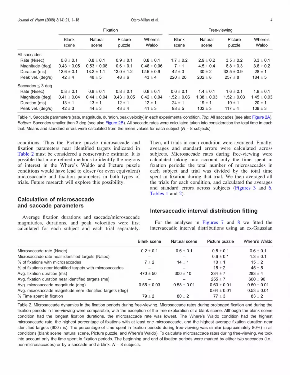
Microsaccade rate (N/sec) 0.2 T 0.1 0.6 T 0.1 0.5 T 0.1 0.6 T 0.1Microsaccade rate near identified targets (N/sec) – – 0.6 T 0.1 1.3 T 0.1% of fixations with microsaccades 7 T 2 14 T 1 10 T 1 15 T 2% of fixations near identified targets with microsaccades – – 15 T 2 45 T 5Avg. fixation duration (ms) 470 T 50 300 T 10 234 T 7 283 T 4Avg. fixation duration near identified targets (ms) – – 255 T 7 600 T 90Avg. microsaccade magnitude (deg) 0.55 T 0.03 0.58 T 0.01 0.63 T 0.01 0.60 T 0.01Avg. microsaccade magnitude near identified targets (deg) – – 0.64 T 0.01 0.53 T 0.01% Time spent in fixation 79 T 2 80 T 2 77 T 3 83 T 2
Table 2. Microsaccade dynamics in the fixation periods during free-viewing. Microsaccade rates during prolonged fixation and during thefixation periods in free-viewing were comparable, with the exception of the free exploration of a blank scene. Although the blank scenecondition had the longest fixation durations, the microsaccade rate was lowest. The Where’s Waldo condition had the highestmicrosaccade rate, the highest percentage of fixations with at least one microsaccade, and the highest average fixation duration nearidentified targets (600 ms). The percentage of time spent in fixation periods during free-viewing was similar (approximately 80%) in allconditions (blank scene, natural scene, Picture puzzle, and Where’s Waldo). To calculate microsaccade rates during free-viewing, we tookinto account only the time spent in fixation periods. The beginning and end of fixation periods were marked by either two saccades (i.e.,non-microsaccades) or by a saccade and a blink. N = 8 subjects.
Fixation Free-viewing
Blankscene
Naturalscene
Picturepuzzle
Where’sWaldo
Blankscene
Naturalscene
Picturepuzzle
Where’sWaldo
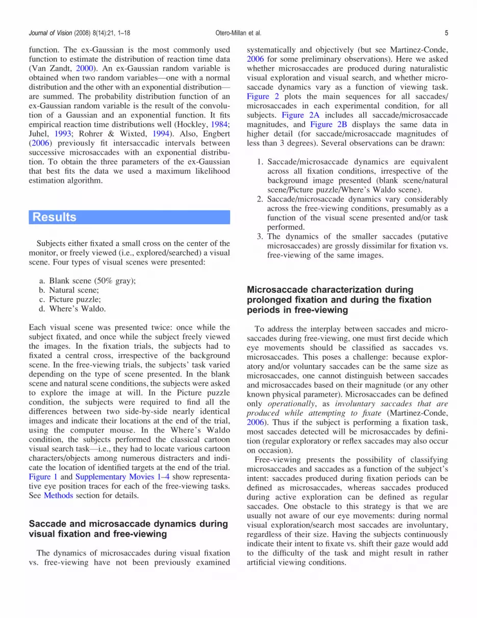
All saccadesRate (N/sec) 0.8 T 0.1 0.8 T 0.1 0.9 T 0.1 0.8 T 0.1 1.7 T 0.2 2.9 T 0.2 3.5 T 0.2 3.3 T 0.1Magnitude (deg) 0.43 T 0.05 0.53 T 0.08 0.6 T 0.1 0.46 T 0.06 7 T 1 4.5 T 0.4 6.8 T 0.3 3.6 T 0.2Duration (ms) 12.6 T 0.1 13.2 T 1.1 13.0 T 1.2 12.5 T 0.9 42 T 3 30 T 2 33.5 T 0.9 28 T 1Peak vel. (deg/s) 42 T 4 48 T 5 48 T 6 43 T 4 220 T 20 202 T 8 257 T 8 184 T 5
Saccades e 3 degRate (N/sec) 0.8 T 0.1 0.8 T 0.1 0.8 T 0.1 0.8 T 0.1 0.6 T 0.1 1.4 T 0.1 1.6 T 0.1 1.8 T 0.1Magnitude (deg) 0.41 T 0.04 0.44 T 0.04 0.43 T 0.05 0.42 T 0.04 1.52 T 0.06 1.38 T 0.03 1.52 T 0.03 1.45 T 0.03Duration (ms) 13 T 1 13 T 1 12 T 1 12 T 1 24 T 1 19 T 1 19 T 1 20 T 1Peak vel. (deg/s) 42 T 3 44 T 3 43 T 4 41 T 3 98 T 5 102 T 3 117 T 4 108 T 3
Table 1. Saccade parameters (rate, magnitude, duration, peak velocity) in each experimental condition.Top: All saccades (see also Figure 2A).Bottom: Saccades smaller than 3 deg (see also Figure 2B). All saccade rates were calculated taken into consideration the total time in eachtrial. Means and standard errors were calculated from the mean values for each subject (N = 8 subjects).
Journal of Vision (2008) 8(14):21, 1–18 Otero-Millan et al. 4
function. The ex-Gaussian is the most commonly usedfunction to estimate the distribution of reaction time data(Van Zandt, 2000). An ex-Gaussian random variable isobtained when two random variablesVone with a normaldistribution and the other with an exponential distributionVare summed. The probability distribution function of anex-Gaussian random variable is the result of the convolu-tion of a Gaussian and an exponential function. It fitsempirical reaction time distributions well (Hockley, 1984;Juhel, 1993; Rohrer & Wixted, 1994). Also, Engbert(2006) previously fit intersaccadic intervals betweensuccessive microsaccades with an exponential distribu-tion. To obtain the three parameters of the ex-Gaussianthat best fits the data we used a maximum likelihoodestimation algorithm.
Results
Subjects either fixated a small cross on the center of themonitor, or freely viewed (i.e., explored/searched) a visualscene. Four types of visual scenes were presented:
a. Blank scene (50% gray);b. Natural scene;c. Picture puzzle;d. Where’s Waldo.
Each visual scene was presented twice: once while thesubject fixated, and once while the subject freely viewedthe images. In the fixation trials, the subjects had tofixated a central cross, irrespective of the backgroundscene. In the free-viewing trials, the subjects’ task varieddepending on the type of scene presented. In the blankscene and natural scene conditions, the subjects were askedto explore the image at will. In the Picture puzzlecondition, the subjects were required to find all thedifferences between two side-by-side nearly identicalimages and indicate their locations at the end of the trial,using the computer mouse. In the Where’s Waldocondition, the subjects performed the classical cartoonvisual search taskVi.e., they had to locate various cartooncharacters/objects among numerous distracters and indi-cate the location of identified targets at the end of the trial.Figure 1 and Supplementary Movies 1–4 show representa-tive eye position traces for each of the free-viewing tasks.See Methods section for details.
Saccade and microsaccade dynamics duringvisual fixation and free-viewing
The dynamics of microsaccades during visual fixationvs. free-viewing have not been previously examined
systematically and objectively (but see Martinez-Conde,2006 for some preliminary observations). Here we askedwhether microsaccades are produced during naturalisticvisual exploration and visual search, and whether micro-saccade dynamics vary as a function of viewing task.Figure 2 plots the main sequences for all saccades/microsaccades in each experimental condition, for allsubjects. Figure 2A includes all saccade/microsaccademagnitudes, and Figure 2B displays the same data inhigher detail (for saccade/microsaccade magnitudes ofless than 3 degrees). Several observations can be drawn:
1. Saccade/microsaccade dynamics are equivalentacross all fixation conditions, irrespective of thebackground image presented (blank scene/naturalscene/Picture puzzle/Where’s Waldo scene).
2. Saccade/microsaccade dynamics vary considerablyacross the free-viewing conditions, presumably as afunction of the visual scene presented and/or taskperformed.
3. The dynamics of the smaller saccades (putativemicrosaccades) are grossly dissimilar for fixation vs.free-viewing of the same images.
Microsaccade characterization duringprolonged fixation and during the fixationperiods in free-viewing
To address the interplay between saccades and micro-saccades during free-viewing, one must first decide whicheye movements should be classified as saccades vs.microsaccades. This poses a challenge: because explor-atory and/or voluntary saccades can be the same size asmicrosaccades, one cannot distinguish between saccadesand microsaccades based on their magnitude (or any otherknown physical parameter). Microsaccades can be definedonly operationally, as involuntary saccades that areproduced while attempting to fixate (Martinez-Conde,2006). Thus if the subject is performing a fixation task,most saccades detected will be microsaccades by defini-tion (regular exploratory or reflex saccades may also occuron occasion).Free-viewing presents the possibility of classifying
microsaccades and saccades as a function of the subject’sintent: saccades produced during fixation periods can bedefined as microsaccades, whereas saccades producedduring active exploration can be defined as regularsaccades. One obstacle to this strategy is that we areusually not aware of our eye movements: during normalvisual exploration/search most saccades are involuntary,regardless of their size. Having the subjects continuouslyindicate their intent to fixate vs. shift their gaze would addto the difficulty of the task and might result in ratherartificial viewing conditions.
Journal of Vision (2008) 8(14):21, 1–18 Otero-Millan et al. 5

An alternative way to classify microsaccades vs.saccades during free-viewingVwithout complicatingand/or interfering with the subject’s taskVis to:
1. Establish the physical parameters of saccadesproduced during prolonged fixation (most of thesesaccades are microsaccades by definition, as statedabove), and
2. Use those parameters to identify microsaccades infree-viewing conditions.
Figure 2 plots the magnitude–peak velocity relationshipfor all saccades produced during prolonged fixation (45-slong trials; see Methods section for details). Regardless ofthe background scene presented, the vast majority of
saccades produced during prolonged fixation had magni-tudes below 1 deg. Here we will consider those saccadesas microsaccades, in agreement with previous studies(Betta, Galfano, & Turatto, 2007; Betta & Turatto, 2006;Engbert, 2006; Engbert & Kliegl, 2003a, 2003b, 2004;Engbert & Mergenthaler, 2006; Galfano, Betta, & Turatto,2004; Laubrock et al., 2005; Martinez-Conde, 2006;Martinez-Conde et al., 2000, 2002, 2004, 2006; Rolfset al., 2004, 2006; Troncoso, Macknik, & Martinez-Conde, 2008; Troncoso, Macknik, Otero-Millan, et al.,2008; Turatto, Valsecchi, Tame, & Betta, 2007; Valsecchi,Betta, & Turatto, 2007; Valsecchi & Turatto, 2007) andwhile keeping in mind the caveats discussed above.See also Methods section for further details on thesaccade/microsaccade detecting algorithm. Now, we may
Figure 2. Saccadic main sequences during visual fixation and free-viewing. (A) Main sequences illustrating all saccades. Notice cluster ofÈ20-deg saccades in the free-viewing Picture puzzle condition (corresponding to horizontal saccades linking equivalent points in the twoside-by-side images; see also Figure 1C). (B) Main sequences from (A) in higher detail (saccade/microsaccade magnitudes of less than3 degrees). Main sequences are equivalent for all the fixation conditions. However, free-viewing of the same images results in verydifferent saccade dynamics. Also, the dynamics of small saccades in the free-viewing conditions appear to vary as a function of stimulus(blank vs. visual scene) and task (free exploration vs. Picture puzzle search vs. Where’s Waldo search). Such differences may be partiallydue to varying cognitive/attentional demands across the free-viewing conditions. N = 8 subjects.
Journal of Vision (2008) 8(14):21, 1–18 Otero-Millan et al. 6

apply the same classification to saccades/microsaccadesproduced during free-viewing. Thus from here on wewill refer to saccades smaller than 1 deg as micro-saccades, irrespective of whether they were producedduring prolonged fixation conditions, or during the brieffixation periods encompassed during the free-viewingconditions. Correspondingly, we will define fixationperiods in free-viewing as those periods betweensaccades larger than 1 deg (or in between a saccadelarger than 1 deg and a blink, see Methods section). Thisprocedure has the important advantage that the parame-ters used to identify microsaccades during free-viewingare derived from the distribution of involuntary saccadesduring visual fixation (i.e., veritable microsaccades,
Figures 2 and 3). However, one must keep in mind thatno microsaccade-detecting method can ensure that allputative microsaccades (produced during free-viewing oreven during prolonged fixation) are involuntary (asopposed to small voluntary saccades). Conversely, someof the 91 deg saccades produced during prolongedfixation (and possibly during free-viewing) may beinvoluntary and could be thus categorized as micro-saccades. Table 1 summarizes various dynamics ofsaccades/microsaccades during fixation and free-viewingconditions (corresponding to the main sequences inFigure 2). Figure 3 plots the microsaccadic main sequenceand related parameters for the four fixation conditionstogether.
Figure 3. Microsaccade parameters during prolonged fixation. (A) Microsaccade main sequence (N = 33,230). (B) Microsaccade peakvelocity distribution. (C) Distribution of microsaccade magnitudes. (D) Distribution of microsaccade durations. All the fixation conditionshave been grouped. The table summarizes various microsaccade dynamics (rate, magnitude, duration, peak velocity) in eachexperimental condition. Microsaccade rates were calculated taken into consideration the total time in each trial. Means and standarderrors were calculated from the mean values for each subject (N = 8 subjects).
Journal of Vision (2008) 8(14):21, 1–18 Otero-Millan et al. 7

Microsaccades during free-viewing were most prevalentat the points of the image that were meaningful for the task.Thus microsaccades tended to occur when foveating humanfaces and other salient objects during free visual explora-tion (Figure 4), or on the regions with identified targetsduring visual search tasks (Picture puzzles and Where’sWaldo conditions). See Supplementary Movies 5–6.These observations may be related to the recent proposalthat microsaccades significantly “re-sharpen” the imageand improve spatial resolution (Donner & Hemila, 2007).Figure 5 illustrates the distribution of fixation durations
across the free-viewing conditions. The rate and numberof microsaccades increased parametrically with fixationduration in all free-viewing conditions (Figures 5C and5D), with a quasi-linear relationship between number ofmicrosaccades and fixation duration (Figure 5D). Theexample in Figure 4C illustrates how microsaccades arecontained within the fixation periods with longest dura-tions. However, the slope of the curves in Figures 5C and5D varied across conditions, with the steepest increase forthe Where’s Waldo condition. Interestingly, visual explo-ration of a blank scene resulted in the longest fixationdurations (Figure 5A), but the lowest number of fixationswith microsaccades (Figure 5B), thus suggesting that
microsaccades may require the presence of a visual/attentional target to anchor to (see also Table 2). Further,the difference in microsaccade dynamics in the Where’sWaldo condition vs. the blank scene exploration conditionmay result from the varied attentional/cognitive demandsof both tasks (highest in the Where’s Waldo search taskand lowest in the blank scene exploration task). Thusmicrosaccade production is not solely dependent onfixation duration, but it may also be affected by bothvisual stimulation (blank vs. natural scene) and thecognitive demands of the task performed.Table 2 summarizes the occurrence of microsaccades
across the free-viewing conditions. Subjects were engagedin fixation during approximately 80% of the free-viewingtime, irrespective of experimental condition. During theblank scene exploration, average fixation durations werelong (470 T 50 ms) but average microsaccade rates werelowest (0.2 T 0.1 Hz). Microsaccade production washighest in theWhere’sWaldo condition. During theWhere’sWaldo search, the average microsaccade rate was 0.6 T0.1 Hz, and 15 T 2% of all fixations contained at least onemicrosaccade. Microsaccade production increased evenfurther when only the regions with identified targets (asindicated by the subject) were considered. In such case, the
Figure 4. Microsaccades during free-viewing. (A) Image equalized for luminance and RMS contrast. (B) A 45-s monocular eye positiontrace during free visual exploration, plotted over a low-contrast version of the image (for clarity). (C) A 10-s period from (B). The area ofeach circle indicates the duration of the fixation period (smaller area circles correspond to fixations of linearly shorter durations). Thelargest circle (dashed purple line) corresponds to a 1,678 ms fixation period. Human faces attracted long-duration fixations and proved tobe a primary focus of microsaccades (red).
Journal of Vision (2008) 8(14):21, 1–18 Otero-Millan et al. 8

average microsaccade rate escalated to 1.3 T 0.1 Hz for theWhere’s Waldo task (a 70% increase with respect tomicrosaccade rates during prolonged fixation), and abouthalf of the fixation periods (45 T 5%) contained micro-saccades. Moreover, the average duration of fixations in theregions of identified Where’s Waldo targets (600 T 90 ms)surpassed the average fixation duration during free-viewingof a blank scene. These measurements suggest a strongrelationship between microsaccade generation and targetdetection during visual search. The long fixation durationsduring the blank scene exploration rule out the possibilitythat fixation duration is critical to target detection: theproduction of microsaccades was more significantly linked.Average microsaccade magnitudes were higher in the
free-viewing conditions (Table 2) than in the prolongedfixation conditions (Figure 3), lending further support tothe idea that increased visual stimulation and/or task
demands may result in increased microsaccade dynamics.We previously showed that precise fixation leads todecreases in microsaccade magnitudes (as well as micro-saccade rates; Martinez-Conde et al., 2006), and that suchdecreases result in visual fading. Because subjects are not‘required’ to fixate in the fixation periods that occurspontaneously during free-viewing, the reduction ofmicrosaccade sizes associated with precise fixation mayhave not applied (or it may have applied less often), thusresulting in larger microsaccades than during prolongedfixation. An additional (non-exclusive) possibility is thatsome of the G1 deg saccades produced during free-viewingare not actual (involuntary) microsaccades but are rathervoluntary or exploratory small saccades. But as discussedearlier, this potential caveat would also apply to the G1 degsaccades produced during visual fixation (which aredefined by most current studies as microsaccades; Betta
Figure 5. Fixations and microsaccades during free-viewing. (A) Distribution of fixation durations across free-viewing conditions.(B) Distribution of fixation durations, for fixation periods containing at least 1 microsaccade. (C) Microsaccade rate as a function of fixationduration. Microsaccade rate is approximately constant after 400 ms in all conditions. (D) Microsaccade numbers per fixation period, as afunction of fixation period duration. The number of microsaccades per fixation period increases linearly after approximately 400 ms.Panels C and D illustrate that microsaccade production does not solely depend on fixation duration, but it is also affected by visualstimulation (blank vs. natural scene) andVto a lesser extentVby the specific free-viewing task performed. N = 8 subjects.
Journal of Vision (2008) 8(14):21, 1–18 Otero-Millan et al. 9

et al., 2007; Betta & Turatto, 2006; Engbert, 2006;Engbert & Kliegl, 2003a, 2003b, 2004; Engbert &Mergenthaler, 2006; Galfano et al., 2004; Laubrock et al.,2005; Martinez-Conde, 2006; Martinez-Conde et al.,2000, 2002, 2004, 2006; Rolfs et al., 2004, 2006;Troncoso, Macknik, & Martinez-Conde, 2008; Turattoet al., 2007; Valsecchi et al., 2007; Valsecchi & Turatto,2007). Another potential explanation could be thatrelatively brief fixation periods (such as those duringfree-viewing) result in larger ocular instability (and thuslarger microsaccades) than periods of prolonged fixation.To exclude this possibility, we compared the microsaccademagnitudes during the first several hundred milliseconds ofthe prolonged fixation trials to the microsaccade magnitudesfound in the fixation periods during free-viewing. Thedifference in microsaccade magnitude for both types of trialremained mostly unaffected (data not shown).
Temporal interactions between saccades andmicrosaccades
If saccades and microsaccades share the same oculo-motor bases, then microsaccade generation should affect
the timing of saccade generation, and vice versa. Rolfset al. (2006) found that microsaccades produced duringfixation affect the timing of subsequent saccades. Here wedetermine the interactions for all the pair-wise combina-tions of saccades and microsaccades, both during fixationand free-viewing.Zuber and Stark (1965) first determined that micro-
saccades produced during fixation follow the saccadicmain sequence, and thus proposed that there is a commongenerator for saccades and microsaccades. Figure 6extends the range of the main sequence to include allsaccades and microsaccades produced by the samesubjects during visual fixation and free-viewing of thesame images. Saccades and microsaccades producedduring the fixation tasks are indicated in red. Saccadesand microsaccades produced during the free-viewing tasksare indicated in blue. Both distributions follow the samemain sequence, with the same slope.Figure 7A shows that intersaccadic intervals are
equivalent for all pair-wise combinations of saccadesand microsaccades in free-viewing. That is, both saccadesand microsaccades were more likely produced approx-imately 200 ms after a previous eye movement (whichcould itself be either a saccade or a microsaccade). Inother words, the refractory periods between saccades andmicrosaccades are equivalent, irrespective of their sequen-tial order. This observation is at odds with the idea of twodifferent circuits for the generation of saccades andmicrosaccades. Saccades and microsaccades appear toshare the same timing constraints, which supports thehypothesis of a common saccade–microsaccade generator.During fixation, the intervals between successive micro-saccades are somewhat longer than during free-viewing.The reason may be that subjects try to hold their gazesteady during fixation, and so their microsaccade produc-tion may beVat least partlyVsuppressed (Martinez-Conde et al., 2006), resulting in longer intervals betweensuccessive microsaccades. Figure 7B plots the normalizeddistribution of intersaccadic intervals (for all saccadesand microsaccades combined) according to experimentalcondition (all the fixation conditions are lumpedtogether). In agreement with Figure 7A, intersaccadicintervals during the fixation conditions are slightly longer
Figure 6. Microsaccades and saccades follow the same mainsequence. Saccades and microsaccades recorded during free-viewing (blue) follow the same main sequence as those producedduring the fixation conditions (red). Note that some of the bluedots are obscured by the superimposed red dots (i.e., when a redand a blue dot occupy the same location in the graph, the red dotis plotted over the blue dot). Microsaccade and saccade rateshave been calculated taken into consideration the total time ineach trial. N = 8 subjects.
Figure 7. Intersaccadic interval distributions. (A) Intersaccadicintervals follow similar distributions for all saccade–microsaccadecombinations. The only variation between distributions occurs forintersaccadic intervals larger than È200 ms. (B) Intersaccadicintervals follow similar distribution for all experimental conditions.(C) Intersaccadic interval distributions for individual conditionshave been fit with ex-Gaussian curves (red). The blue dots showthe histograms of the data used for the fits (same data as in (B)).(D) Variability of parameter estimations across experimentalconditions. Only the exponential parameter (C) varies significantlyacross conditions. (E) Parameter estimation as a function ofsaccade rate. There is a clear linear correlation between theexponential parameter (C) and the rate of saccades.
Journal of Vision (2008) 8(14):21, 1–18 Otero-Millan et al. 10

Journal of Vision (2008) 8(14):21, 1–18 Otero-Millan et al. 11
than intersaccadic intervals during free-viewing. Most ofthe free-viewing conditions result in equivalent intersac-cadic interval distributions. It is interesting to note that thefree exploration of a blank scene results in very similarintersaccadic intervals to those produced during prolongedfixation. The underlying reason may be the relativescarcity of both saccades and microsaccades duringblank-scene explorations, when compared to the otherfree-viewing conditions (see Tables 1 and 2). To sum up,the only variation between distributions occurs for inter-saccadic intervals larger than È200 ms, and this differenceseems better related to the nature of the task than to
dissimilarity in the generation of saccades vs. micro-saccades. In Figure 7C, the distributions of intersaccadicintervals for individual conditions are fit with ex-Gaussianfunctions (see Methods section). Only the exponentialparameter (C) of the ex-Gaussian curve varied significantlyacross conditions (Figure 7D). This parameter indicatesthe rate of decay of the probability of a long intersaccadicinterval. Figure 7E shows that the exponential parameter(C) is linearly related to the saccade and/or microsaccaderate, as proposed earlier. The Gaussian component of theex-Gaussian distribution is described by parameters 2(mean of the Gaussian distribution) and A (width of the
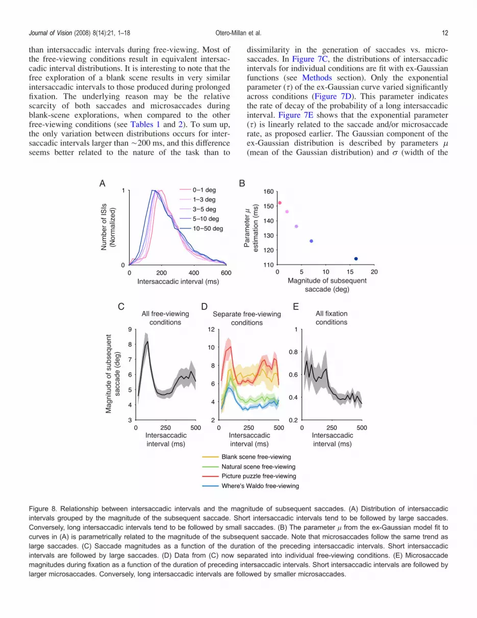
Figure 8. Relationship between intersaccadic intervals and the magnitude of subsequent saccades. (A) Distribution of intersaccadicintervals grouped by the magnitude of the subsequent saccade. Short intersaccadic intervals tend to be followed by large saccades.Conversely, long intersaccadic intervals tend to be followed by small saccades. (B) The parameter 2 from the ex-Gaussian model fit tocurves in (A) is parametrically related to the magnitude of the subsequent saccade. Note that microsaccades follow the same trend aslarge saccades. (C) Saccade magnitudes as a function of the duration of the preceding intersaccadic intervals. Short intersaccadicintervals are followed by large saccades. (D) Data from (C) now separated into individual free-viewing conditions. (E) Microsaccademagnitudes during fixation as a function of the duration of preceding intersaccadic intervals. Short intersaccadic intervals are followed bylarger microsaccades. Conversely, long intersaccadic intervals are followed by smaller microsaccades.
Journal of Vision (2008) 8(14):21, 1–18 Otero-Millan et al. 12
Gaussian distribution). Parameters 2 and A were notrelated to saccade and/or microsaccade rate, and they didnot differ significantly across conditions.Figure 8 relates the duration of intersaccadic intervals to
the magnitude of the second (subsequent) saccade/micro-saccade. Short intersaccadic intervals tend to be followedby large saccades; long intersaccadic intervals tend to befollowed by small saccades/microsaccades (Figure 8A).Figure 8B shows that the magnitude of each subsequentsaccade (in a saccade pair) is parametrically related to theparameter 2 from the ex-Gaussian model fit to the data inFigure 8A. Note that microsaccades follow the same trendas large saccades. Figures 8C–8E show that the relation-ship between the duration of the intersaccadic intervalsand the magnitude of the next saccade/microsaccadeapplies to both free-viewing conditions (as previouslyshown by Unema, Pannasch, Joos, & Velichkovsky, 2005,Figures 8C and 8D) and fixation conditions (Figure 8E),further supporting the hypothesis that saccades andmicrosaccades share a common generator.
Discussion
Microsaccades during free-viewing
Microsaccades are known to occur during prolongedvisual fixation, but it has been a matter of controversywhether they are also produced during free-viewing. Herewe set out to determine:
1. Whether microsaccades occur during free visualexploration and visual search,
2. Whether microsaccade generation varies with task,and
3. Whether saccades and microsaccades share equiv-alent spatiotemporal characteristics, which wouldargue in favor of a common saccade–microsaccadeoculomotor generator.
In the late 1970s, Kowler and Steinman (Kowler &Steinman, 1980; Skavenski, Hansen, Steinman, &Winterson, 1979; Steinman & Collewijn, 1980) concludedthat the generation of microsaccades was a laboratoryartifact: i.e., that microsaccades did not occur in normalviewing conditions, but that they resulted from artificiallaboratory conditions, in which subjects were forced tohold their gaze for very long periods of time, while theirhead was restrained (for instance, with a bite bar). Steinman,Haddad, Skavenski, and Wyman (1973) and Kowler andSteinman (1979, 1980) furthermore stated that micro-saccades are not helpful in tasks requiring complex visualinformation processing, and thus are much less commonduring brief fixations interposed between large saccades (inactivities such as reading or counting) than duringprolonged fixation.
Contrary to these conclusions, we and others found inthe last decade that microsaccades generate strong reliablefiring in visual neurons during fixation, and also duringthe fixation periods in guided-viewing (Bair & O’Keefe,1998; Martinez-Conde, 2006; Martinez-Conde et al.,2000, 2002, 2004). Moreover, microsaccades counteractedvisual fading and filling-in and increased target’s visibilityin human subjects with both restrained and unrestrainedheads (Martinez-Conde et al., 2006; Troncoso, Macknik,& Martinez-Conde, 2008). The dynamics of microsac-cades with restrained versus unrestrained heads wereequivalent, suggesting that microsaccades are generatedwith and without the presence of head movements(Martinez-Conde et al., 2006). One critical differencebetween these recent studies and the early microsaccadestudies from the 1970s is the current standard use ofobjective microsaccade-detecting algorithms (developedwithin the last decade; Engbert & Kliegl, 2003b; Martinez-Conde et al., 2000). Current objective algorithms basemicrosaccade characterization on parameters derived fromthe distribution of involuntary saccades during visualfixation, rather than on arbitrary magnitude or velocitythresholds (as done in the earlier studies). Anotherpossible confound in the earlier studies is that micro-saccades were identified subjectively (i.e., picked by handfrom the eye-position traces), which poses the potentialdifficulty of replication by other groups.In a relatively recent example of the early subjective
approach to microsaccade detection, Malinov, Epelboim,Herst, and Steinman (2000) identified microsaccades byhand, rather than by applying an objective algorithm.They also defined microsaccades arbitrarily (i.e., withoutpreviously quantifying the distribution of involuntarysaccades during fixation), as saccades with magnitudesof G12 arcmin. This very stringent parameter is wellbelow the average magnitude found and/or the uppermicrosaccade threshold used in recent microsaccadestudies in humans and primates (Betta et al., 2007; Betta& Turatto, 2006; Engbert, 2006; Engbert & Kliegl, 2003a,2003b, 2004; Engbert &Mergenthaler, 2006; Galfano et al.,2004; Laubrock et al., 2005; Martinez-Conde, 2006;Martinez-Conde et al., 2000, 2002, 2006; Rolfs et al.,2004, 2006; Snodderly, Kagan, & Gur, 2001; Turattoet al., 2007; Valsecchi et al., 2007; Valsecchi & Turatto,2007), see alsoMartinez-Conde et al. (2004) for a review ofhuman and primate microsaccade parameters. Theselimitations and potential confounds may help to explainwhy only 2 out of 93,000 total saccades recorded in Malinovet al.’s study in freely moving humans were classified as“microsaccades”, in contradiction to the much higher numberof microsaccades we find here. It is also important to keep inmind that microsaccade production during free-viewingdepends on the nature of the visual stimulation and the taskperformed, as shown here. Thus free-viewing tasks that donot require the subject’s attentive fixation may lead toreducedmicrosaccade production (such as in the free-viewingexploration of a blank scene; Figures 2 and 5, Table 2).
Journal of Vision (2008) 8(14):21, 1–18 Otero-Millan et al. 13

To sum up, the role of microsaccades in free-viewinghas remained controversial to date. However, the dynamicsof microsaccades during free-viewing vs. fixation have notpreviously been objectively and systematically measured(i.e., with current microsaccade-detecting algorithms,previously unavailable). Our results show that micro-saccades occur in the fixation periods that naturally takeplace during visual exploration and visual search (Figures 2,4, and 5, Supplementary Movies 5–6). Moreover, micro-saccade rates during the fixation periods in visual explora-tion/search were comparable to microsaccade rates duringprolonged fixation (Table 2).Our results also suggest that microsaccades and saccades
have equivalent functional roles, both during prolongedfixation and during free-viewing. The spatiotemporalcharacteristics of microsaccades and saccades may reflectan optimal sampling method by which the brain discretelyacquires visual information. Thus we put forward that thedichotomy between saccades and microsaccades proposedby previous studies is fundamentally arbitrary.
Saccades and microsaccades as an optimalsampling strategy
The dynamics of saccades and microsaccades may reflectan optimal strategy by which visual neurons discretelysample information from a scene. Visual exploration of ablank scene (in which visual information is absent bydefinition) resulted in low production of both saccades andmicrosaccades. The visual exploration/search of scenes thatwere rich with visual content resulted in much higher ratesof saccades and microsaccades (Figure 4, Tables 1 and 2).As the cognitive demands of the task increased (Where’sWaldo visual search vs. free visual exploration), micro-saccade generation increased even further, especially inthe regions with identified targets (Table 2, Figure 5,Supplementary Movies 5–6). These results are in agree-ment with physiological and modeling studies in theprimate visual system, in which strong neural transientswere observed in response to microsaccades (Donner &Hemila, 2007; Martinez-Conde, 2006; Martinez-Condeet al., 2000, 2002), suggesting that microsaccades mayimprove the efficient sampling of fine spatial detail(Donner & Hemila, 2007). Other studies suggest that V1neurons produce stronger responses to transient stimulithan to drifting stimuli. Such neural transients mayunderlie the behavior of cortical neurons as coincidencedetectors (Shelley, McLaughlin, Shapley, & Wielaard,2002; Williams & Shapley, 2007). Moreover, neuraltransients to stimuli onsets and terminations (similar tothose produced by microsaccades in the primate visualsystem; Martinez-Conde, 2006; Martinez-Conde et al.,2000, 2002) have been related to target visibility in visualmasking paradigms (Macknik & Livingstone, 1998;Macknik & Martinez-Conde, 2004; Macknik, Martinez-Conde, & Haglund, 2000).
Gilchrist, Brown, and Findlay (1997) and Gilchrist,Brown, Findlay, and Clarke (1998) moreover observedthat a patient who was unable to make eye movements(except for small-magnitude drifts) produced head-saccadesof comparable characteristics to eye-saccades. Such head-saccades enabled the patient to read at normal speed andeven perform complicated visuo-motor tasks, such asmaking a cup of tea, with no problems. The authorsconcluded that “saccadic movements, of the head or theeye, form the optimal sampling method for the brain”(Gilchrist et al., 1997, 1998). This type of discrete samplingis potentially optimal in other sensory systems as well.Sniffs during rodent olfaction also sample sensory infor-mation discretely every 200–300 ms and are thus com-parable in their temporal dynamics to saccades (Uchida,Kepecs, & Mainen, 2006) and microsaccades in humansand primates. A similar mode of discrete sampling mayalso be at play when objects are recognized through tactileinformation, for instance if we sweep our fingertips over anobject’s surface with our eyes closed, or when blindindividuals read Braille script.
Microsaccades in visual search and the roleof attention
It has remained unknown whether microsaccade dynam-ics vary as a function of free-viewing task. Here we foundmicrosaccades to be more prominent in conditions thatinvolved complex/meaningful visual information (naturalvs. blank scene, faces vs. non-faces) and increasedcognitive/attentional demands (Where’s Waldo vs. freevisual exploration; Tables 1 and 2; Figures 4 and 5;Supplementary Movies 5–6). Conversely, the free explo-ration of a blank sceneVwhere the visual content is nulland the task demands are lowVresulted in long fixationperiods, but comparatively low microsaccade rates.Previous studies have found that the spatial location of
attention strongly influences the rate and/or the direction ofmicrosaccades during visual fixation (Engbert, 2006; Engbert& Kliegl, 2003b; Galfano et al., 2004; Hafed & Clark, 2002;Rolfs et al., 2004, 2005). Thus increased microsaccadeproduction due to increased attentional load may explain ourcurrent results, especially as microsaccade rates were highestin the regions of identified targets (Table 2).Future research should determine how varied amounts
of attentional load may impact microsaccade dynamicsduring visual search and other naturalistic tasks, and thepotential physiological and perceptual consequences ofsuch modulations. One possibility is that increased micro-saccade production (perhaps due to increased attention)directly results in successful target detections (due tosuccessive microsaccades repeatedly stimulating thereceptive fields of visual neurons in the target area).Alternatively, the very first saccade or microsaccade toland on the target may be sufficient for detection, and thefunction of subsequent microsaccades may be to confirm
Journal of Vision (2008) 8(14):21, 1–18 Otero-Millan et al. 14
the original identification of the target. Thus, futurestudies should also investigate the precise timing ofmicrosaccade generation with regard to target detectionand the interactions with attention.
A saccade–microsaccade continuum
A growing list of common characteristics to saccades andmicrosaccades supports the hypothesis of a shared oculo-motor generator (Martinez-Conde et al., 2004; Rolfs et al.,2006, 2008; Zuber & Stark, 1965). Most studies to datehave focused on the descriptive parameters of saccadesand microsaccades (magnitude, duration, peak velocity–magnitude relationship). Here we hypothesized that, ifsaccades and microsaccades share the same oculomotorbases, microsaccade generation should affect saccade gen-eration, and vice versa. Our results indicate that thespatiotemporal parameters of saccades and microsaccadesare equivalent (Figures 6, 7, and 8), providing furtherevidence for the common generator hypothesis. In agree-ment with this idea, Van Gisbergen and colleagues foundthat the activity of burst neurons in the abducens nucleusand nearby pontomedullary reticular formation is similar forsaccades and microsaccades (Van Gisbergen & Robinson,1977; Van Gisbergen, Robinson, & Gielen, 1981).To date, the study of microsaccades during free-viewing
has faced a two-pronged challenge:
1. If fixation periods are defined as saccade-freeperiods, it follows that fixational microsaccades arenot part of fixation; a contradiction in terms.
2. But if microsaccades are indeed a type of fixationaleye movement (Ditchburn & Ginsborg, 1952, 1953;Ratliff & Riggs, 1950, see Martinez-Conde et al.,2004 for a review), then they must be includedwithin the fixation periods.
Our results suggest that such difficulty is fundamentallysemantic: we propose that there is a microsaccade–saccade continuum, and that visual information is dis-cretely sampled with all saccades, large and small(including microsaccades). The fact that there is aminimal intersaccadic interval (i.e., a refractory period)preceding saccades and microsaccades, and that thisinterval is similar for all pair-wise combinations ofsaccades and microsaccades, argues strongly against avery strict divide between the neural mechanisms respon-sible for the generation of saccades and microsaccades.
Practical implications for future research
Commercially available algorithms for saccade detectionare often used to separate saccades from fixation periodsduring free-viewing tasks (such as visual exploration, visualsearch, reading, etc.). The thresholds used for such saccade
detection can be quite arbitrary. For instance, the EyeLink IImanual (SR Research, 2006) recommends a velocitythreshold of 22 deg/s for “smooth pursuit and psychophys-ical research” and a velocity threshold of 30 deg/s for“reading and cognitive research”. The present results showthat the use of such thresholds for the identification ofsaccades and/or microsaccades is problematic. Here wewould like to emphasize two practical points:
1. Microsaccade characterization during free-viewingshould be based on parameters obtained from micro-saccade distributions during prolonged fixation,ideally collected from the same subjects (and necessa-rily from the same species, i.e., primates vs. humans).
2. Future studies investigating microsaccades and/orfixation periods during free-viewing should reportthe precise thresholds used for the classification ofsaccades/microsaccades/fixation periods, rather thansimply state the name of the commercial softwarepackage used to characterize eye movements.
Finally, because of the microsaccade–saccade contin-uum proposed above, we recommend that future studies ofvisual exploration/search employ saccade-detecting algo-rithms that allow the identification and inclusion ofmicrosaccades (rather than using thresholds that arbitrarilyexclude the potential contributions of microsaccades/smallsaccades).
Conclusions
We found that microsaccades occur during visualexploration and visual search, and that their specificdynamics vary as a function of visual stimulation andviewing task, with more challenging tasks resulting inhigher microsaccade production. Saccades and micro-saccades had comparable spatiotemporal characteristics,including equivalent intersaccadic intervals between allpair-wise combinations of saccades and microsaccades.We propose that the dichotomy between saccades andmicrosaccades suggested by previous studies is funda-mentally arbitrary. Rather, our results indicate a micro-saccade–saccade continuum and suggest that saccades andmicrosaccades are generated by common brain circuits.The spatiotemporal characteristics of saccades and micro-saccades may reflect an optimal sampling method by whichthe brain discretely acquires visual information.
Acknowledgments
We thank Mona Stewart and Isabel Gomez-Caraballofor technical assistance, Hector Rieiro for comments on
Journal of Vision (2008) 8(14):21, 1–18 Otero-Millan et al. 15

the manuscript, and Dr. John H. R. Maunsell for his inputon the experimental design and analyses. This study wasfunded through grants from the Barrow NeurologicalFoundation (to SM-C and SLM), the National ScienceFoundation (NSF award 0643306 to SM-C and NSFaward 0726113 to SLM), the Arizona BiomedicalResearch Commission (award 07-102 to SMC and award0724 to SLM), and the Science Foundation Arizona(Award CAA 0091-07 to SLM). XGT is a fellow of theCaja Madrid Foundation. IS-P’s current address is:Department of Psychology, Keynes College, Universityof Kent, Canterbury, Kent, CT2 7NP, UK.
Commercial relationships: none.Corresponding author: Susana Martinez-Conde, PhD.Email: [email protected]: 350 W. Thomas Rd., Phoenix, AZ 85013, USA.
References
Adams, M. (Ed.). (2006a). Life picture puzzle: Can youspot the differences? (vol. 6(2)). Life Books.
Adams, M. (Ed.). (2006b). Life picture puzzle: Can youspot the differences? (vol. 7(2)). Life Books.
Adams, M. (Ed.). (2006c). Life picture puzzle: Can youspot the differences? (vol. 7(5)). Life Books.
Bair, W., & O’Keefe, L. P. (1998). The influence offixational eye movements on the response of neuronsin area MT of the macaque. Visual Neuroscience, 15,779–786. [PubMed]
Betta, E., Galfano, G., & Turatto, M. (2007). Micro-saccadic response during inhibition of return in atarget–target paradigm. Vision Research, 47, 428–436.[PubMed]
Betta, E., & Turatto, M. (2006). Are you ready? I can tellby looking at your microsaccades. Neuroreport, 17,1001–1004. [PubMed]
Ditchburn, R. W., & Ginsborg, B. L. (1952). Vision with astabilized retinal image. Nature, 170, 36–37.[PubMed]
Ditchburn, R. W., & Ginsborg, B. L. (1953). Involuntaryeye movements during fixation. The Journal ofPhysiology, 119, 1–17. [PubMed] [Article]
Donner, K., & Hemila, S. (2007). Modelling the effect ofmicrosaccades on retinal responses to stationarycontrast patterns. Vision Research, 47, 1166–1177.[PubMed]
Engbert, R. (2006). Microsaccades: A microcosm forresearch on oculomotor control, attention, and visualperception. Progress in Brain Research, 154, 177–192.[PubMed]
Engbert, R., & Kliegl, R. (2003a). Binocular coordinationin microsaccades. In R. R. J. Hyona & H. Deubel
(Eds.), The mind’s eyes: Cognitive and appliedaspects of eye movements (pp. 103–117). Oxford,UK: Elsevier.
Engbert, R., & Kliegl, R. (2003b). Microsaccades uncoverthe orientation of covert attention. Vision Research,43, 1035–1045. [PubMed]
Engbert, R., & Kliegl, R. (2004). Microsaccades keep theeyes’ balance during fixation. Psychological Science,15, 431–436. [PubMed]
Engbert, R., & Mergenthaler, K. (2006). Microsaccadesare triggered by low retinal image slip. Proceedingsof the National Academy of Sciences of the UnitedStates of America, 103, 7192–7197. [PubMed][Article]
Galfano, G., Betta, E., & Turatto, M. (2004). Inhibition ofreturn in microsaccades. Experimental BrainResearch, 159, 400–404. [PubMed]
Gilchrist, I. D., Brown, V., & Findlay, J.M. (1997). Saccadeswithout eye movements. Nature, 390, 130–131.[PubMed]
Gilchrist, I. D., Brown, V., Findlay, J. M., & Clarke, M. P.(1998). Using the eye-movement system to control thehead. Proceedings of the Royal Society B: BiologicalSciences, 265, 1831–1836. [PubMed] [Article]
Hafed, Z. M., & Clark, J. J. (2002). Microsaccades as anovert measure of covert attention shifts. VisionResearch, 42, 2533–2545. [PubMed]
Handford, M. (2007a). Where’s Waldo now? Cambridge,MA: Candlewick Press.
Handford, M. (2007b). Where’s Waldo? The wonder book.Cambridge, MA: Candlewick Press.
Handford, M. (2007c). Where’s Waldo? Cambridge, MA:Candlewick Press.
Hockley, W. E. (1984). Analysis of response timedistributions in the study of cognitive processes.Journal of Experimental Psychology: Learning, Mem-ory, & Cognition, 10, 598–615.
Juhel, J. (1993). Should we take the shape of reaction timedistributions into account when studying the relation-ship between RT and psychometric intelligence?Personality & Individual Differences, 15, 357–360.
Kowler, E., & Steinman, R. M. (1979). Miniaturesaccades: Eye movements that do not count. VisionResearch, 19, 105–108. [PubMed]
Kowler, E., & Steinman, R. M. (1980). Small saccadesserve no useful purpose: Reply to a letter by R. W.Ditchburn. Vision Research, 20, 273–276. [PubMed]
Laubrock, J., Engbert, R., & Kliegl, R. (2005). Micro-saccade dynamics during covert attention. VisionResearch, 45, 721–730. [PubMed]
Lord, M. P. (1951). Measurement of binocular eyemovements of subjects in the sitting position. British
Journal of Vision (2008) 8(14):21, 1–18 Otero-Millan et al. 16

Journal of Ophthalmology, 35, 21–30. [PubMed][Article]
Macknik, S. L., & Livingstone, M. S. (1998). Neuronalcorrelates of visibility and invisibility in the primatevisual system. Nature Neuroscience, 1, 144–149.[PubMed]
Macknik, S. L., & Martinez-Conde, S. (2004). The spatialand temporal effects of lateral inhibitory networksand their relevance to the visibility of spatiotemporaledges. Neurocomputing, 58–60, 775–782.
Macknik, S. L., Martinez-Conde, S., & Haglund, M. M.(2000). The role of spatiotemporal edges in visibilityand visual masking. Proceedings of the NationalAcademy of Sciences of the United States of America,97, 7556–7560. [PubMed] [Article]
Malinov, I. V., Epelboim, J., Herst, A. N., & Steinman,R. M. (2000). Characteristics of saccades andvergence in two kinds of sequential looking tasks.Vision Research, 40, 2083–2090. [PubMed]
Martinez-Conde, S. (2006). Fixational eye movements innormal and pathological vision. Progress in BrainResearch, 154, 151–176. [PubMed]
Martinez-Conde, S., & Macknik, S. L. (2008). Fixationaleye movements across vertebrates: Comparativedynamics, physiology and perception. Journal ofVision, 8, 1–16.
Martinez-Conde, S., & Macknik, S. L. (2007). Windowson the mind. Scientific American, 297, 56–63.[PubMed]
Martinez-Conde, S., Macknik, S. L., & Hubel, D. H.(2000). Microsaccadic eye movements and firing ofsingle cells in the striate cortex of macaque monkeys.Nature Neuroscience, 3, 251–258. [PubMed]
Martinez-Conde, S., Macknik, S. L., & Hubel, D. H.(2002). The function of bursts of spikes during visualfixation in the awake primate lateral geniculatenucleus and primary visual cortex. Proceedings ofthe National Academy of Sciences of the UnitedStates of America, 99, 13920–13925. [PubMed][Article]
Martinez-Conde, S., Macknik, S. L., & Hubel, D. H.(2004). The role of fixational eye movements invisual perception. Nature Reviews, Neuroscience, 5,229–240. [PubMed]
Martinez-Conde, S., Macknik, S. L., Troncoso, X. G., &Dyar, T. A. (2006). Microsaccades counteract visualfading during fixation. Neuron, 49, 297–305.[PubMed] [Article]
MLller, F., Laursen, M. L., Tygesen, J., & SjLlie, A. K.(2002). Binocular quantification and characterizationof microsaccades. Graefe’s Archive for Clinical andExperimental Ophthalmology, 240, 765–770.[PubMed]
Ratliff, F., & Riggs, L. A. (1950). Involuntary motions ofthe eye during monocular fixation. Journal of Exper-imental Psychology, 40, 687–701. [PubMed]
Riggs, L. A., & Ratliff, F. (1952). The effects of counter-acting the normal movements of the eye. Journal ofthe Optical Society of America, 42, 872–873.
Rohrer, D., & Wixted, J. T. (1994). An analysis of latencyand interresponse time in free recall. Memory &Cognition, 22, 511–524. [PubMed]
Rolfs, M., Engbert, R., & Kliegl, R. (2004). Microsaccadeorientation supports attentional enhancement oppositea peripheral cue: Commentary on Tse, Sheinberg,and Logothetis (2003). Psychological Science, 15,705–707. [PubMed]
Rolfs, M., Engbert, R., & Kliegl, R. (2005). Crossmodalcoupling of oculomotor control and spatial attentionin vision and audition. Experimental Brain Research,166, 427–439. [PubMed]
Rolfs, M., Kliegl, R., & Engbert, R. (2008). Toward amodel of microsaccade generation: The case ofmicrosaccadic inhibition. Journal of Vision, 8(11):5,1–23, http://journalofvision.org/8/11/5/, doi:10.1167/8.11.5. [PubMed] [Article]
Rolfs, M., Laubrock, J., & Kliegl, R. (2006). Shorteningand prolongation of saccade latencies followingmicrosaccades. Experimental Brain Research, 169,369–376. [PubMed]
Shelley, M., McLaughlin, D., Shapley, R., & Wielaard, J.(2002). States of high conductance in a large-scalemodel of the visual cortex. Journal of ComputationalNeuroscience, 13, 93–109. [PubMed]
Skavenski, A. A., Hansen, R. M., Steinman, R. M., &Winterson, B. J. (1979). Quality of retinal imagestabilization during small natural and artificial bodyrotations in man. Vision Research, 19, 675–683.[PubMed]
Snodderly, D. M., Kagan, I., & Gur, M. (2001). Selectiveactivation of visual cortex neurons by fixational eyemovements: Implications for neural coding. VisualNeuroscience, 18, 259–277. [PubMed]
SR Research (2006). EyeLink\ II User Manual. Version2.12. SR. Research, Mississauga, Canada.
Steinman, R. M., & Collewijn, H. (1980). Binocularretinal image motion during active head rotation.Vision Research, 20, 415–429. [PubMed]
Steinman, R. M., Haddad, G. M., Skavenski, A. A., &Wyman, D. (1973). Miniature eye movement. Sci-ence, 181, 810–819. [PubMed]
Troncoso, X., Macknik, S. L., & Martinez-Conde, S.(2008). Microsaccades counteract perceptual filling-in. Journal of Vision, 8, 1–9.
Troncoso, X., Macknik, S. L., Otero-Millan, J., &Martinez-Conde, S. (2008). Microsaccades drive
Journal of Vision (2008) 8(14):21, 1–18 Otero-Millan et al. 17

illusory motion in the Enigma illusion. Proceedingsof the National Academy of Sciences of the UnitedStates of America, 105, 16033–16038. [PubMed]
Turatto, M., Valsecchi, M., Tame, L., & Betta, E. (2007).Microsaccades distinguish between global and localvisual processing. Neuroreport, 18, 1015–1018.[PubMed]
Uchida, N., Kepecs, A., & Mainen, Z. F. (2006). Seeing ata glance, smelling in a whiff: Rapid forms ofperceptual decision making. Nature Reviews, Neuro-science, 7, 485–491. [PubMed]
Unema, P. J., Pannasch, S., Joos, M., & Velichkovsky,B. M. (2005). Time course of information process-ing during scene perception: The relationshipbetween saccade amplitude and fixation duration.Visual Cognition, 12, 473–494.
Valsecchi, M., Betta, E., & Turatto, M. (2007). Visualoddballs induce prolonged microsaccadic inhibition.Experimental Brain Research, 177, 196–208.[PubMed]
Valsecchi, M., & Turatto, M. (2007). Microsaccadicresponse to visual events that are invisible to thesuperior colliculus. Behavioral Neuroscience, 121,786–793. [PubMed]
Van Gisbergen, J. A., Robinson, D. A., & Gielen, S.(1981). A quantitative analysis of generation ofsaccadic eye movements by burst neurons. Journalof Neurophysiology, 45, 417–442. [PubMed]
Van Gisbergen, J. A. M., & Robinson, D. A. (1977).Generation of micro and macrosaccades by burstneurons in the monkey. In R. Baker & A. Berthoz(Eds.), Control of gaze by brain stem neurons. NewYork: Elsevier/North-Holland.
Van Zandt, T. (2000). How to fit a response timedistribution. Psychonomic Bulletin & Review, 7,424–465. [PubMed]
Williams, P. E., & Shapley, R. M. (2007). A dynamicnonlinearity and spatial phase specificity in macaqueV1 neurons. Journal of Neuroscience, 27, 5706–5718.[PubMed] [Article]
Yarbus, A. L. (1967). Eye movements and vision. InB. Haigh (Ed.). New York: Plenum Press.
Zuber, B. L., & Stark, L. (1965). Microsaccades and thevelocity–amplitude relationship for saccadic eyemovements. Science, 150, 1459–1460. [PubMed]
Journal of Vision (2008) 8(14):21, 1–18 Otero-Millan et al. 18