rna polymerase ii elongation at the crossroads of transcription and alternative splicing
TRANSCRIPT
SAGE-Hindawi Access to ResearchGenetics Research InternationalVolume 2011, Article ID 309865, 9 pagesdoi:10.4061/2011/309865
Review Article
RNA Polymerase II Elongation at the Crossroads ofTranscription and Alternative Splicing
Manuel de la Mata,1, 2 Manuel J. Munoz,1 Mariano Allo,1 Juan Pablo Fededa,1, 3
Ignacio E. Schor,1 and Alberto R. Kornblihtt1
1 Laboratorio de Fisiologıa y Biologıa Molecular, Departamento de Fisiologıa, Biologıa Molecular, y Celular, IFIBYNE-CONICET,Facultad de Ciencias Exactas y Naturales, Universidad de Buenos Aires, C1428EHA Buenos Aires, Argentina
2 Friedrich Miescher Institute for Biomedical Research, 4002 Basel, Switzerland3 Department of Biology, Institute of Biochemistry, ETH Zurich, 8093 Zurich, Switzerland
Correspondence should be addressed to Alberto R. Kornblihtt, [email protected]
Received 15 June 2011; Accepted 23 June 2011
Academic Editor: Carles Sune
Copyright © 2011 Manuel de la Mata et al. This is an open access article distributed under the Creative Commons AttributionLicense, which permits unrestricted use, distribution, and reproduction in any medium, provided the original work is properlycited.
The elongation phase of transcription lies at the core of several simultaneous and coupled events leading to alternative splicingregulation. Although underestimated in the past, it is at this phase of the transcription cycle where complexes affecting thetranscription machinery itself, chromatin structure, posttranscriptional gene regulation and pre-mRNA processing converge toregulate each other or simply to consolidate higher-order complexes and functions. This paper focuses on the multiple processesthat take place during transcription elongation which ultimately regulate the outcome of alternative splicing decisions.
1. Introduction
Regulation of gene expression was originally conceived asa hierarchy of steps linked together on a time scale and phys-ically separated in different cell compartments in accordancewith the central dogma of biology. This concept has longbeen abandoned, with a significant accumulation of evidencedescribing an extensive network of events, encompassingtranscription, mRNA processing, chromatin regulation, andposttranscriptional gene regulation, which take place simul-taneously and in a mutually regulated or coupled manner[1, 2]. Distinctions between complexes and processes gov-erning gene expression have been blurred to a large extent,adding complexity to the ever-increasing fraction of genessubjected to alternative promoter usage, alternative splicing(AS) (>90% genes), alternative polyadenylation, editing, andposttranscriptional gene silencing by small RNAs [3, 4].Additionally, this complexity takes a new dimension whenstudied in the context of chromatin and its regulation upongene expression (for reviews, see [5–7]). This paper will focuson the main features of coupling between transcriptions
elongation and splicing, and its implications on AS regula-tion.
2. The Benefits of Coupling
Initial visualization of Drosophila-embryo nascent tran-scripts by electron microscopy, showed that splicing canoccur cotranscriptionally [8]. This was later directly demon-strated for the human dystrophin gene [9], which spans2400 kb and can take 16 hr to complete transcription. Morerecently, a quantitative study of the c-Src and fibronectinmRNAs compared chromatin-bound and nucleoplasmicRNA fractions. There, it was shown that most introns areexcised efficiently in the chromatin-bound fractions, witha gradient of cotranscriptional splicing efficiency decreasingfrom promoter-proximal to promoter-distal introns, that is,the direction of transcription [10].
One implication of the cotranscriptional nature of splic-ing is that the two processes can be coupled. In a broad sense,coupling implies that the involved processes can happenefficiently only as the result of their combined action, even
2 Genetics Research International
for processes that are constitutive and non-regulated. Forinstance, whereas both transcription and splicing can takeplace independently at low efficiency as in vitro reactions, itis only in vivo or in coupled in vitro systems where maximalefficiency can be achieved [11–16]. Cotranscriptional pro-cessing is necessary to allow for coupling between transcrip-tion and splicing, although it does not necessarily guaranteeit. There are examples of both cotranscriptional splicing thatseems to be uncoupled, as well as purely posttranscriptionalsplicing [17–20]. Noteworthy, the consequences of thedifferent types of splicing can be considerable; whereascotranscriptional splicing can be regulated by mechanismsdependent on transcription, postranscriptional splicing canbe subjected to additional regulatory mechanisms linked toevents downstream of transcription (e.g., RNA export) [20].However, cotranscriptional splicing seems to predominatefor most introns in mammalian genes [10, 17, 20–23]pointing at an evolutionary conserved role in allowing forcoupling of transcription and splicing. Cotranscriptionalsplicing is more efficient than posttranscriptional splicingby driving nascent pre-mRNAs to the association withspliceosome components [24, 25] and splicing regulatoryfactors, such as serine/arginine-rich (SR) proteins [22]. Thisallows for different levels of regulation of AS and preventsbackhybridization of the nascent pre-mRNA to the DNAtemplate strand, which can cause genome instability [26, 27].Even in the case of posttranscriptional splicing, couplingwith transcription can be determinant for AS regulation.Since pre-mRNA splicing is a multistep reaction, it is possiblethat commitment to splicing takes place cotranscriptionallyduring early splice-site recognition, while completion of thesplicing reaction occurs posttranscriptionally [20, 28, 29],consistent with the fact that introns are not necessarilyremoved in the exact order that they are transcribed [17, 30–32]. This mechanism, which can be viewed as a cotranscrip-tional commitment rather than a cotranscriptional catalysis,tends to apply largely to splicing and not to other RNA-processing events like capping and cleavage/polyadenylation[33–37]. Intermediate scenarios are also possible, with bothsplicing commitment and catalysis taking place cotranscrip-tionally but not following a strict 5′ to 3′ direction of intronremoval [10, 38].
Another implication of cotranscriptional splicing is thatit allows for a bidirectional coupling of the two processes[1, 20, 39, 40]. For instance, the splicing machinery canreciprocally affect transcription in different ways by eitherstimulating transcriptional elongation [41, 42], transcrip-tional initiation [43, 44] and, as recently shown in yeast,by imposing a transient pausing checkpoint around the3′ end of introns and on terminal exons [45, 46]. Thisbidirectional feedback might in turn reinforce splicing effi-ciency, conferring important advantages for gene expression.Nevertheless, reciprocal coupling might not be a widespreadgeneral phenomenon considering that elongation kineticsseems to be independent of splicing in some model genes[47]. This highlights the possibility that specific exon-intronarchitectures and/or cis-acting sequences might be requiredfor reciprocal coupling to occur.
Despite all the seemingly clear advantages of cotran-scriptional over posttranscriptional splicing, particularly inallowing for coupling, the true proportions of these twomodes of splicing in mammals still await to be determinedon a genome-wide scale. It will be interesting to use globalapproaches to answer this question, which might haveprofound implications in reorienting our current researchand on our understanding of the regulation of AS.
3. Early Discoveries
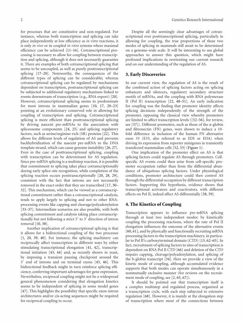
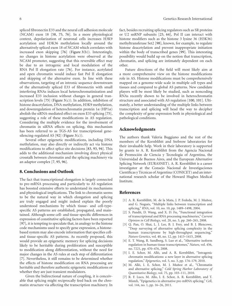
In our current view, the regulation of AS is the result ofthe combined action of splicing factors acting on splicingenhancers and silencers, regulatory secondary structuremotifs of mRNAs, and the coupling with RNA polymeraseII (Pol II) transcription [22, 48–51]. An early indicationfor coupling was the finding that promoter identity affectssplicing decisions independently of the strength of thepromoter, opposing the classical view whereby promotersare limited to affect transcription levels ([52–56], for review,see [57]). Different promoters, such as those of the α-globinand fibronectin (FN) genes, were shown to induce a 10-fold difference in inclusion of the human FN alternativeexon 33 (E33, also referred to as EDI or EDA) whendriving its expression from reporter minigenes in transientlytransfected mammalian cells [52, 53] (Figure 1).
One implication of the promoter effect on AS is thatsplicing factors could regulate AS through promoters. Cell-specific AS events could then arise from cell-specific pro-moter occupation rather than from the differential abun-dance of ubiquitous splicing factors. Under physiologicalconditions, promoter architecture could then control ASthrough the differential occupation by different transcriptionfactors. Supporting this hypothesis, evidence shows thattranscriptional activators and coactivators, with differenteffects on Pol II, indeed affect AS differentially [58, 59].
4. The Kinetics of Coupling
Transcription appears to influence pre-mRNA splicingthrough at least two independent modes: by kineticallycoupling the processing reactions, where the rate of Pol IIelongation influences the outcome of the alternative events[60, 61], and by physically and functionally recruiting mRNAprocessing factors to the transcription machinery, in particu-lar to Pol II’s carboxyterminal domain (CTD) [13, 62–65]. Infact, recruitment of splicing factors to sites of transcription isdependent on RNA Pol II CTD [66] and deletion of the CTDimpairs capping, cleavage/polyadenylation, and splicing ofthe b-globin transcript [34]. Here we provide a view of thekinetic mode of coupling, although accumulated evidencesupports that both modes can operate simultaneously in anonmutually exclusive manner (for reviews on the recruit-ment mode of coupling, see [2, 65, 67]).
It should be pointed out that transcription itself isa complex multistep and regulated process, organized asa transcription cycle, with each step subjected to extensiveregulation [68]. However, it is mainly at the elongation stepof transcription where most of the connections between
Genetics Research International 3
α-gbStrong3SS
Strong3SS
Pol II
Weak3SS
Weak3SS
FN
Exonskipping
Exoninclusion
Pol II
FN-E33
Figure 1: Promoters affect alternative splicing. α-globin/FN hybrid minigenes under the control of different promoters, used in transienttransfections of mammalian cells in culture to assess inclusion levels of the alternatively spliced E33 (EDI or EDA) cassette exon (dark yellow).Inclusion level with the FN promoter is >10-fold higher when compared to the α-globin promoter.
the transcription and splicing machineries actually occur.A role for Pol II elongation on AS had been suggestedbefore the finding of the promoter effect [69] and was latersupported by several lines of evidence. Eperon et al. [69]showed that, in contrast to in vitro conditions, ongoing RNAsynthesis in vivo affects the potential secondary structureof long—but not that of short—RNA substrates, whichin turn affects splicing, pointing at a kinetic link betweentranscription and splicing. Additional evidence came fromexperiments in which RNA Pol II’s local pausing causedby elements inserted into the tropomyosin gene, promotedhigher inclusion of tropomyosin exon 3 [60]. However,more conclusive evidence for a role of elongation on ASregulation was shown in a series of reports demonstratingthat several factors globally impacting Pol II elongationalso affect AS. (i) Replication of AS reporter minigenesgreatly stimulates FN E33 inclusion, and is counteractedby trichostatin A (TSA), a potent inhibitor of histonedeacetylation considered to drive chromatin into an “open”state. This suggested that replication conveys a more compactchromatin structure to the template, thus slowing elongationand leading to higher E33 inclusion [70]. (ii) Drugs knownto inhibit elongation, such as DRB [58, 70], flavopiridol, orcamptothecin [38], favor E33 inclusion. (iii) Transcriptionalactivation by VP16, a factor that promotes both initiationand elongation, decreases E33 inclusion while Sp1, actingonly on initiation, has no effect on E33 inclusion [58]. (iv)The presence of the SV40 transcriptional enhancer near apromoter stimulates Pol II elongation and provokes a 3–10-fold reduction in FN E33 inclusion independently of thepromoter used [71]. (v) Slow mutants of RNA Pol II increaseFN E33 inclusion in human cells, affect AS of the endogenousgene ultrabithorax (Ubx) in Drosophila, and modulatethe inclusion of an artificially created alternative exon inyeast [61, 72]. (vi) DNA-damage signaling triggered by UVirradiation affects the AS of fibronectin, caspase 9, Bcl-x,and other human genes by inducing hyperphosphorylationof Pol II CTD and blocking Pol II elongation [73]. These
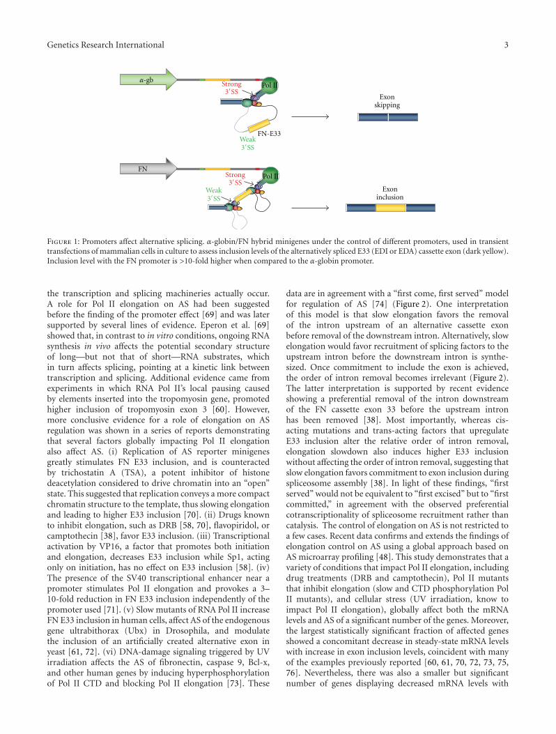
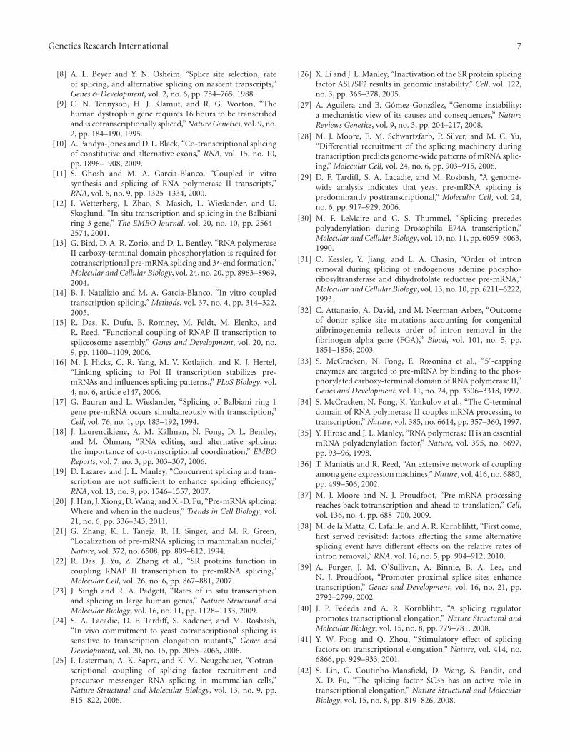
data are in agreement with a “first come, first served” modelfor regulation of AS [74] (Figure 2). One interpretationof this model is that slow elongation favors the removalof the intron upstream of an alternative cassette exonbefore removal of the downstream intron. Alternatively, slowelongation would favor recruitment of splicing factors to theupstream intron before the downstream intron is synthe-sized. Once commitment to include the exon is achieved,the order of intron removal becomes irrelevant (Figure 2).The latter interpretation is supported by recent evidenceshowing a preferential removal of the intron downstreamof the FN cassette exon 33 before the upstream intronhas been removed [38]. Most importantly, whereas cis-acting mutations and trans-acting factors that upregulateE33 inclusion alter the relative order of intron removal,elongation slowdown also induces higher E33 inclusionwithout affecting the order of intron removal, suggesting thatslow elongation favors commitment to exon inclusion duringspliceosome assembly [38]. In light of these findings, “firstserved” would not be equivalent to “first excised” but to “firstcommitted,” in agreement with the observed preferentialcotranscriptionality of spliceosome recruitment rather thancatalysis. The control of elongation on AS is not restricted toa few cases. Recent data confirms and extends the findings ofelongation control on AS using a global approach based onAS microarray profiling [48]. This study demonstrates that avariety of conditions that impact Pol II elongation, includingdrug treatments (DRB and camptothecin), Pol II mutantsthat inhibit elongation (slow and CTD phosphorylation PolII mutants), and cellular stress (UV irradiation, know toimpact Pol II elongation), globally affect both the mRNAlevels and AS of a significant number of the genes. Moreover,the largest statistically significant fraction of affected genesshowed a concomitant decrease in steady-state mRNA levelswith increase in exon inclusion levels, coincident with manyof the examples previously reported [60, 61, 70, 72, 73, 75,76]. Nevertheless, there was also a smaller but significantnumber of genes displaying decreased mRNA levels with
4 Genetics Research International
Weak 3SS
Weak 3SS
Weak 3SS
Weak 3SSStrong 3SS
Strong 3SS Strong 3SS
Fast elongation/no pauses Slow elongation/internal pauses
Exclusion Inclusion
Tim
ePol II Pol II Pol II
Pol II Pol II
Pol II
Weak3SS
First served = first excised First served = first committed
Figure 2: Alternative models for the “first come, first served” mechanism of splice site selection. (a) Fast elongation promotes usage of thestronger downstream 3′ splice site. (b) Slow elongation causes preferential excision of the upstream intron (first served = first excised). (c)Slow elongation causes commitment to E33 inclusion via recruitment of splicing factors (first served = first committed). Both introns areexcised individually and in an order that is independent of elongation. (Based on [38].)
decreased exon inclusion levels [48], consistent with the ideathat inhibition of Pol II elongation can lead to increasedexon inclusion as well as increased exon skipping dependingon the underlying splicing regulatory mechanism involvedin each case [55, 61]. Pol II’s elongation-dependent changesin AS regulation also display a high preference to modulatethe expression levels of genes involved in RNA metabolism,including pre-mRNA splicing factors and other RNA bindingproteins. Interestingly, about one-third of those genes con-tain AS events that introduce a premature termination codon(PTC) when spliced into the mature mRNA, subsequentlyleading to nonsense-mediated mRNA decay (NMD) [48].This represents another example of evolutionarily conservedelongation-coupled events, acting together to coordinate thelevels or RNA binding proteins with their steady-state mRNAlevels.
5. Elongation Links to Chromatin
The promoter effect, together with the kinetic, physical,and functional coupling modes of transcription and AS,immediately shifted the attention to other factors thought tobe restricted to transcriptional regulation, such as the chro-matin structure. It soon became clear that the unanticipatedcomplexity of splicing regulation could not be explainedsolely based on the current models, which lacked a clearconnection to in vivo situations. Chromatin is the naturalsubstrate upon which transcriptional regulation acts in vivo,and major discoveries have recently pointed at chromatin
structure and post-translational histone modifications as keyregulators of AS.
The chromatin role on AS regulation is broad, involvingboth direct (elongation-independent) interactions with thesplicing machinery and effects on AS through changes intranscription elongation. Here, we concentrate on the later,although recent findings on the direct roles of chromatinon splicing have significantly changed our view of howexon-intron architecture is achieved by intimately linkingnucleosome to exon structure ([77–84], for reviews, see [5–7]. In fact, genome-wide mapping of nucleosome positioningon exons at the DNA level may shed light on one of the moststriking puzzles in the field of splicing, that is, how doesthe splicing machinery recognizes, with high fidelity, shortexons (on average 150 bp) “floating” in a “sea” of introns(on average 5.4 kbp), an exon-intron architecture typicalof vertebrate genes [85, 86]. Notably, the average size of amammalian exon is similar to the length of DNA wrappedaround a nucleosome, suggesting a conserved function forthe nucleosome in exon definition [79, 80, 82]. Accordingto the exon definition model, originally postulated by S. M.Berget [87], the spliceosome and auxiliary factors achievethis recognition by preferentially assembling on 3′ and 5′
sites paired across exons and not across introns (i.e., notfollowing an intron definition mode of recognition, typicalin lower eukaryotes like yeast). This favors exon recognitionand acts as a selective force for short exon size. As describedbelow, nucleosome positioning on exons may help in exondefinition by creating roadblocks for Pol II elongation that
Genetics Research International 5
DNA replication
siRNAs targeting downstreamintron
Exon skipping
Reduced elongation/higher exon inclusion
Reduced elongation/higher exon inclusion
DNA
Nucleosome
AGO1 loadedwith intronic
siRNAs
Weak3SS
Weak3SS
Weak3SS
Weak3SS
Strong3SS
Strong3SS
Strong3SS
Pol II
Pol II
Pol II Pol II Pol II
H3K9ac H3K9ac H3K9ac
HP1α
HP1α
H3K9me2H3K27me3
HP1α
AGO1
(a)
(b) (c)TSA
neuron depolarization
Enhanced elongation/lower exon inclusion
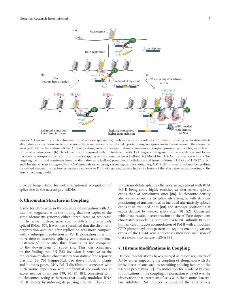
Figure 3: Chromatin couples elongation to alternative splicing. (a) Early evidence for a role of chromatin on splicing: replication affectsalternative splicing. Loose nucleosome assembly (as in transiently transfected reporter minigenes) gives rise to low inclusion of the alternativeexon (yellow) into the mature mRNA. After replication, nucleosome organization becomes more compact, promoting much higher inclusionof the alternative exon. (b) Depolarization of neuronal cells or treatment with TSA triggers intragenic histone acetylation and loosernucleosome compaction which in turn causes skipping of the alternative exon (yellow). (c) Model for TGS-AS. Transfection with siRNAstargeting the intron downstream from the alternative exon (yellow) promotes dimethylation and trimethylation of H3K9 and H3K27 (greenand blue marks, resp.), triggered by siRNA’s guide strand entering a silencing complex containing AGO1. HP1α is recruited and the resultingcondensed chromatin structure generates roadblocks to Pol II elongation, causing higher inclusion of the alternative exon according to thekinetic coupling model.
provide longer time for cotranscriptional recognition ofsplice sites in the nascent pre-mRNA.
6. Chromatin Structure in Coupling
A role for chromatin in the coupling of elongation with ASwas first suggested with the finding that two copies of thesame adenovirus genome, either unreplicated or replicatedin the same nucleus, gave rise to different alternativelyspliced RNAs [57]. It was then speculated that the chromatinorganization acquired after replication was more compact,with a subsequent reduction in Pol II elongation rates andmore time to assemble splicing complexes at a suboptimalupstream 5′ splice site, thus favoring its use comparedto the downstream 5′ splice site. This was confirmedby the finding that FN E33 inclusion is sensitive to thereplication-mediated chromatinization status of the reporterplasmid [58, 70] (Figure 3(a). See above). Both in plantsand humans genes, RNA Pol II distribution correlates withnucleosome deposition with preferential accumulation atexons relative to introns [79, 80, 83, 88], consistent withnucleosomes acting as barriers that locally modulate RNAPol II density by inducing its pausing [89, 90]. This could
in turn modulate splicing efficiency, in agreement with RNAPol II being more highly enriched at alternatively splicedexons than at constitutive ones [88]. Nucleosome densityalso varies according to splice site strength, with strongerpositioning of nucleosomes in included alternatively splicedexons than excluded ones [80] and stronger positioning atexons defined by weaker splice sites [81, 82]. Consistentwith these results, overexpression of the ATPase-dependentchromatin-remodeling complex SWI/SNF subunit Brm inhuman cells, induces accumulation of Pol II with a modifiedCTD phosphorylation pattern on regions encoding variantexons of the CD44 gene and causes increased inclusion ofthese exons into mature mRNA [91].
7. Histone Modifications in Coupling
Histone modifications have emerged as major regulators ofAS by either impacting the coupling of elongation with ASor by direct means such as recruiting splicing factors to thenascent pre-mRNA [7]. An indication for a role of histonemodifications in the coupling of elongation with AS was theobservation that treatment of cells with the histone deacety-lase inhibitor TSA induces skipping of the alternatively
6 Genetics Research International
spliced fibronectin E33 and the neural cell adhesion molecule(NCAM) exon 18 [58, 75, 76]. In a more physiologicalcontext, depolarization of neuronal cells increases H3K9acetylation and H3K36 methylation locally around thealternatively spliced exon 18 of NCAM which correlates withincreased exon skipping [76] (Figure 3(b)). Interestingly,no changes in histone acetylation were observed at theNCAM promoter, suggesting that this reversible effect maybe due to an intragenic and local modulation of theRNA Pol II elongation rate [76]. For instance, acetylatedand open chromatin would induce fast Pol II elongationand skipping of the alternative exon. In line with theseobservations, targeting of an intronic sequence downstreamof the alternatively spliced E33 of fibronectin with smallinterfering RNAs induces local heterochromatinization andincreased E33 inclusion without changes in general tran-scription levels [75] (Figure 3(c)). In addition, inhibition ofhistone deacetylation, DNA methylation, H3K9 methylation,and downregulation of heterochromatin protein 1α (HP1α)abolish the siRNA-mediated effect on exon E33 splicing [75],suggesting a role of these modifications in AS regulation.Considering the multiple evidence for the involvement ofchromatin in siRNA effects on splicing, this mechanismhas been referred to as TGS-AS for transcriptional gene-silencing-regulated AS [92] (Figure 3(c)).
Several other epigenetic modifications, including DNAmethylation, may also directly or indirectly act via histonemodifications to affect splice site decisions [83, 93, 94]. Thisadds to the additional mechanism involving direct physicalcrosstalk between chromatin and the splicing machinery viaan adaptor complex [7, 95, 96].
8. Conclusions and Outlook
The fact that transcriptional elongation is largely connectedto pre-mRNA processing and particularly to AS regulationhas boosted extensive efforts to understand its mechanismsand physiological implications. The link to chromatin seemsto be the natural way in which elongation and splicingare truly engaged and might indeed explain the poorlyunderstood mechanisms by which tissue- and cell-type-specific AS patterns are established, propagated, and main-tained. Although some cell- and tissue-specific differences inexpression of constitutive splicing factors have been reported[97], it is tempting to speculate that, in analogy to the histonecode mechanisms used to specify gene expression, a histone-based system may also encode information that specifies cell-and tissue-specific AS patterns. As recently proposed, thiswould provide an epigenetic memory for splicing decisionslikely to be heritable during proliferation and susceptibleto modification along differentiation, without the need formajor changes in the AS rules at each step of differentiation[7]. Nevertheless, it still remains to be determined whetherthe effects of histone modification on RNA processing areheritable and therefore authentic epigenetic modifications orwhether they are just transient modulators.
Given the bidirectional nature of coupling, it is conceiv-able that splicing might reciprocally feed back on the chro-matin structure via affecting the transcription machinery. In
fact, besides recruiting splicing regulators such as SR proteinsor U2 snRNP subunits [25, 64], Pol II can interact withhistone modifiers such as the histone 3 lysine 36 (H3K36)methyltransferase Set2 [98], known, for example, to regulatehistone deacetylation and prevent inappropriate initiationwithin the body of transcribed genes [99]. This interestingpossibility would build up on the notion that transcription,chromatin, and splicing are intimately dependent on eachother.
Future directions of the field will most likely aim ata more comprehensive view on the histone modificationsrole in AS. Histone modifications must be comprehensivelymapped on a genome-wide scale in multiple cell types andtissues and compared to global AS patterns. New candidateplayers will be most likely be studied, such as noncodingRNAs recently shown to be involved in heterochromatinstructure and associated with AS regulation [100, 101]. Ulti-mately, a better understanding of the multiple links betweentranscription and splicing will head the way to decipherthe complexity of gene expression both in physiological andpathological conditions.
Acknowledgments
The authors thank Valeria Buggiano and the rest of themembers of the Kornblihtt and Srebrow laboratories fortheir invaluable help. Work in their laboratory is supportedby grants to A. R. Kornblihtt from the Agencia Nacionalde Promocion de Ciencia y Tecnologıa of Argentina, theUniversidad de Buenos Aires, and the European AlternativeSplicing Network (EURASNET). A. R. Kornblihtt is a careerinvestigator at the Consejo Nacional de InvestigacionesCientıficas y Tecnicas of Argentina (CONICET) and an inter-national research scholar of the Howard Hughes MedicalInstitute.
References
[1] A. R. Kornblihtt, M. de la Mata, J. P. Fededa, M. J. Munoz,and G. Nogues, “Multiple links between transcription andsplicing,” RNA, vol. 10, no. 10, pp. 1489–1498, 2004.
[2] S. Pandit, D. Wang, and X. D. Fu, “Functional integrationof transcriptional and RNA processing machineries,” CurrentOpinion in Cell Biology, vol. 20, no. 3, pp. 260–265, 2008.
[3] Q. Pan, O. Shai, L. J. Lee, B. J. Frey, and B. J. Blencowe,“Deep surveying of alternative splicing complexity in thehuman transcriptome by high-throughput sequencing,”Nature Genetics, vol. 40, no. 12, pp. 1413–1415, 2008.
[4] E. T. Wang, R. Sandberg, S. Luo et al., “Alternative isoformregulation in human tissue transcriptomes,” Nature, vol. 456,no. 7221, pp. 470–476, 2008.
[5] I. E. Schor, M. Allo, and A. R. Kornblihtt, “Intragenicchromatin modifications: a new layer in alternative splicingregulation,” Epigenetics, vol. 5, no. 3, pp. 174–179, 2010.
[6] M. Allo, I. E. Schor, M. J. Munoz et al., “Chromatinand alternative splicing,” Cold Spring Harbor Laboratory ofQuantitative Biology, vol. 75, pp. 103–111, 2010.
[7] R. F. Luco, M. Allo, I. E. Schor, A. R. Kornblihtt, and T.Misteli, “Epigenetics in alternative pre-mRNA splicing,” Cell,vol. 144, no. 1, pp. 16–26, 2011.
Genetics Research International 7
[8] A. L. Beyer and Y. N. Osheim, “Splice site selection, rateof splicing, and alternative splicing on nascent transcripts,”Genes & Development, vol. 2, no. 6, pp. 754–765, 1988.
[9] C. N. Tennyson, H. J. Klamut, and R. G. Worton, “Thehuman dystrophin gene requires 16 hours to be transcribedand is cotranscriptionally spliced,” Nature Genetics, vol. 9, no.2, pp. 184–190, 1995.
[10] A. Pandya-Jones and D. L. Black, “Co-transcriptional splicingof constitutive and alternative exons,” RNA, vol. 15, no. 10,pp. 1896–1908, 2009.
[11] S. Ghosh and M. A. Garcia-Blanco, “Coupled in vitrosynthesis and splicing of RNA polymerase II transcripts,”RNA, vol. 6, no. 9, pp. 1325–1334, 2000.
[12] I. Wetterberg, J. Zhao, S. Masich, L. Wieslander, and U.Skoglund, “In situ transcription and splicing in the Balbianiring 3 gene,” The EMBO Journal, vol. 20, no. 10, pp. 2564–2574, 2001.
[13] G. Bird, D. A. R. Zorio, and D. L. Bentley, “RNA polymeraseII carboxy-terminal domain phosphorylation is required forcotranscriptional pre-mRNA splicing and 3′-end formation,”Molecular and Cellular Biology, vol. 24, no. 20, pp. 8963–8969,2004.
[14] B. J. Natalizio and M. A. Garcia-Blanco, “In vitro coupledtranscription splicing,” Methods, vol. 37, no. 4, pp. 314–322,2005.
[15] R. Das, K. Dufu, B. Romney, M. Feldt, M. Elenko, andR. Reed, “Functional coupling of RNAP II transcription tospliceosome assembly,” Genes and Development, vol. 20, no.9, pp. 1100–1109, 2006.
[16] M. J. Hicks, C. R. Yang, M. V. Kotlajich, and K. J. Hertel,“Linking splicing to Pol II transcription stabilizes pre-mRNAs and influences splicing patterns.,” PLoS Biology, vol.4, no. 6, article e147, 2006.
[17] G. Bauren and L. Wieslander, “Splicing of Balbiani ring 1gene pre-mRNA occurs simultaneously with transcription,”Cell, vol. 76, no. 1, pp. 183–192, 1994.
[18] J. Laurencikiene, A. M. Kallman, N. Fong, D. L. Bentley,and M. Ohman, “RNA editing and alternative splicing:the importance of co-transcriptional coordination,” EMBOReports, vol. 7, no. 3, pp. 303–307, 2006.
[19] D. Lazarev and J. L. Manley, “Concurrent splicing and tran-scription are not sufficient to enhance splicing efficiency,”RNA, vol. 13, no. 9, pp. 1546–1557, 2007.
[20] J. Han, J. Xiong, D. Wang, and X.-D. Fu, “Pre-mRNA splicing:Where and when in the nucleus,” Trends in Cell Biology, vol.21, no. 6, pp. 336–343, 2011.
[21] G. Zhang, K. L. Taneja, R. H. Singer, and M. R. Green,“Localization of pre-mRNA splicing in mammalian nuclei,”Nature, vol. 372, no. 6508, pp. 809–812, 1994.
[22] R. Das, J. Yu, Z. Zhang et al., “SR proteins function incoupling RNAP II transcription to pre-mRNA splicing,”Molecular Cell, vol. 26, no. 6, pp. 867–881, 2007.
[23] J. Singh and R. A. Padgett, “Rates of in situ transcriptionand splicing in large human genes,” Nature Structural andMolecular Biology, vol. 16, no. 11, pp. 1128–1133, 2009.
[24] S. A. Lacadie, D. F. Tardiff, S. Kadener, and M. Rosbash,“In vivo commitment to yeast cotranscriptional splicing issensitive to transcription elongation mutants,” Genes andDevelopment, vol. 20, no. 15, pp. 2055–2066, 2006.
[25] I. Listerman, A. K. Sapra, and K. M. Neugebauer, “Cotran-scriptional coupling of splicing factor recruitment andprecursor messenger RNA splicing in mammalian cells,”Nature Structural and Molecular Biology, vol. 13, no. 9, pp.815–822, 2006.
[26] X. Li and J. L. Manley, “Inactivation of the SR protein splicingfactor ASF/SF2 results in genomic instability,” Cell, vol. 122,no. 3, pp. 365–378, 2005.
[27] A. Aguilera and B. Gomez-Gonzalez, “Genome instability:a mechanistic view of its causes and consequences,” NatureReviews Genetics, vol. 9, no. 3, pp. 204–217, 2008.
[28] M. J. Moore, E. M. Schwartzfarb, P. Silver, and M. C. Yu,“Differential recruitment of the splicing machinery duringtranscription predicts genome-wide patterns of mRNA splic-ing,” Molecular Cell, vol. 24, no. 6, pp. 903–915, 2006.
[29] D. F. Tardiff, S. A. Lacadie, and M. Rosbash, “A genome-wide analysis indicates that yeast pre-mRNA splicing ispredominantly posttranscriptional,” Molecular Cell, vol. 24,no. 6, pp. 917–929, 2006.
[30] M. F. LeMaire and C. S. Thummel, “Splicing precedespolyadenylation during Drosophila E74A transcription,”Molecular and Cellular Biology, vol. 10, no. 11, pp. 6059–6063,1990.
[31] O. Kessler, Y. Jiang, and L. A. Chasin, “Order of intronremoval during splicing of endogenous adenine phospho-ribosyltransferase and dihydrofolate reductase pre-mRNA,”Molecular and Cellular Biology, vol. 13, no. 10, pp. 6211–6222,1993.
[32] C. Attanasio, A. David, and M. Neerman-Arbez, “Outcomeof donor splice site mutations accounting for congenitalafibrinogenemia reflects order of intron removal in thefibrinogen alpha gene (FGA),” Blood, vol. 101, no. 5, pp.1851–1856, 2003.
[33] S. McCracken, N. Fong, E. Rosonina et al., “5′-cappingenzymes are targeted to pre-mRNA by binding to the phos-phorylated carboxy-terminal domain of RNA polymerase II,”Genes and Development, vol. 11, no. 24, pp. 3306–3318, 1997.
[34] S. McCracken, N. Fong, K. Yankulov et al., “The C-terminaldomain of RNA polymerase II couples mRNA processing totranscription,” Nature, vol. 385, no. 6614, pp. 357–360, 1997.
[35] Y. Hirose and J. L. Manley, “RNA polymerase II is an essentialmRNA polyadenylation factor,” Nature, vol. 395, no. 6697,pp. 93–96, 1998.
[36] T. Maniatis and R. Reed, “An extensive network of couplingamong gene expression machines,” Nature, vol. 416, no. 6880,pp. 499–506, 2002.
[37] M. J. Moore and N. J. Proudfoot, “Pre-mRNA processingreaches back totranscription and ahead to translation,” Cell,vol. 136, no. 4, pp. 688–700, 2009.
[38] M. de la Matta, C. Lafaille, and A. R. Kornblihtt, “First come,first served revisited: factors affecting the same alternativesplicing event have different effects on the relative rates ofintron removal,” RNA, vol. 16, no. 5, pp. 904–912, 2010.
[39] A. Furger, J. M. O’Sullivan, A. Binnie, B. A. Lee, andN. J. Proudfoot, “Promoter proximal splice sites enhancetranscription,” Genes and Development, vol. 16, no. 21, pp.2792–2799, 2002.
[40] J. P. Fededa and A. R. Kornblihtt, “A splicing regulatorpromotes transcriptional elongation,” Nature Structural andMolecular Biology, vol. 15, no. 8, pp. 779–781, 2008.
[41] Y. W. Fong and Q. Zhou, “Stimulatory effect of splicingfactors on transcriptional elongation,” Nature, vol. 414, no.6866, pp. 929–933, 2001.
[42] S. Lin, G. Coutinho-Mansfield, D. Wang, S. Pandit, andX. D. Fu, “The splicing factor SC35 has an active role intranscriptional elongation,” Nature Structural and MolecularBiology, vol. 15, no. 8, pp. 819–826, 2008.
8 Genetics Research International
[43] K. Y. Kwek, S. Murphy, A. Furger et al., “U1 snRNA associateswith TFIIH and regulates transcriptional initiation,” NatureStructural Biology, vol. 9, no. 11, pp. 800–805, 2002.
[44] C. K. Damgaard, S. Kahns, S. Lykke-Andersen, A. L. Nielsen,T. H. Jensen, and J. Kjems, “A 5′ splice site enhances therecruitment of basal transcription initiation factors in vivo,”Molecular Cell, vol. 29, no. 2, pp. 271–278, 2008.
[45] R. D. Alexander, S. A. Innocente, J. D. Barrass, and J.D. Beggs, “Splicing-dependent RNA polymerase pausing inyeast,” Molecular Cell, vol. 40, no. 4, pp. 582–593, 2010.
[46] F. Carrillo Oesterreich, S. Preibisch, and K. M. Neugebauer,“Global analysis of nascent rna reveals transcriptional paus-ing in terminal exons,” Molecular Cell, vol. 40, no. 4, pp. 571–581, 2010.
[47] Y. Brody, N. Neufeld, N. Bieberstein et al., “The in vivokinetics of RNA polymerase II elongation during co-trans-criptional splicing,” PLoS Biology, vol. 9, no. 1, Article IDe1000573, 2011.
[48] J. Y. Ip, D. Schmidt, Q. Pan et al., “Global impact of RNApolymerase II elongation inhibition on alternative splicingregulation,” Genome Research, vol. 21, no. 3, pp. 390–401,2011.
[49] G. E. May, O. Sara, C. J. McManus, and B. R. Graveley,“Competing RNA secondary structures are required formutually exclusive splicing of the Dscam exon 6 cluster,”RNA, vol. 17, no. 2, pp. 222–229, 2011.
[50] J. T. Witten and J. Ule, “Understanding splicing regulationthrough RNA splicing maps,” Trends in Genetics, vol. 27, no.3, pp. 89–97, 2011.
[51] Y. Yang, L. Zhan, W. Zhang et al., “RNA secondary structurein mutually exclusive splicing,” Nature Structural and Molec-ular Biology, vol. 18, pp. 159–168, 2011.
[52] P. Cramer, C. G. Pesce, F. E. Baralle, and A. R. Kornblihtt,“Functional association between promoter structure andtranscript alternative splicing,” Proceedings of the NationalAcademy of Sciences of the United States of America, vol. 94,no. 21, pp. 11456–11460, 1997.
[53] P. Cramer, J. F. Caceres, D. Cazalla et al., “Coupling oftranscription with alternative splicing: RNA pol II promotersmodulate SF2/ASF and 9G8 effects on an exonic splicingenhancer,” Molecular Cell, vol. 4, no. 2, pp. 251–258, 1999.
[54] D. Auboeuf, A. Honig, S. M. Berget, and B. W. O’Malley,“Coordinate regulation of transcription and splicing bysteroid receptor coregulators,” Science, vol. 298, no. 5592, pp.416–419, 2002.
[55] F. Pagani, C. Stuani, E. Zuccato, A. R. Kornblihtt, and F.E. Baralle, “Promoter architecture modulates CFTR exon 9skipping,” Journal of Biological Chemistry, vol. 278, no. 3, pp.1511–1517, 2003.
[56] N. D. Robson-Dixon and M. A. Garcia-Blanco, “MAZelements alter transcription elongation and silencing of thefibroblast growth factor receptor 2 exon IIIb,” Journal ofBiological Chemistry, vol. 279, no. 28, pp. 29075–29084, 2004.
[57] G. Adami and L. E. Babiss, “DNA template effect on RNAsplicing: two copies of the same gene in the same nucleus areprocessed differently,” The EMBO Journal, vol. 10, no. 11, pp.3457–3465, 1991.
[58] G. Nogues, S. Kadener, P. Cramer, D. Bentley, and A.R. Kornblihtt, “Transcriptional activators differ in theirabilities to control alternative splicing,” Journal of BiologicalChemistry, vol. 277, no. 45, pp. 43110–43114, 2002.
[59] D. Auboeuf, D. H. Dowhan, X. Li et al., “CoAA, a nuclearreceptor coactivator protein at the interface of transcriptional
coactivation and RNA splicing,” Molecular and Cellular Biol-ogy, vol. 24, no. 1, pp. 442–453, 2004.
[60] G. C. Roberts, C. Gooding, H. Y. Mak, N. J. Proudfoot, and C.W. J. Smith, “Co-transcriptional commitment to alternativesplice site selection,” Nucleic Acids Research, vol. 26, no. 24,pp. 5568–5572, 1998.
[61] M. de la Mata, C. R. Alonso, S. Kadener et al., “A slow RNApolymerase II affects alternative splicing in vivo,” MolecularCell, vol. 12, no. 2, pp. 525–532, 2003.
[62] A. L. Greenleaf, “Positive patches and negative noodles: link-ing RNA processing to transcription?” Trends in BiochemicalSciences, vol. 18, no. 4, pp. 117–119, 1993.
[63] D. L. Bentley, “Rules of engagement: co-transcriptionalrecruitment of pre-mRNA processing factors,” Current Opin-ion in Cell Biology, vol. 17, no. 3, pp. 251–256, 2005.
[64] M. de la Mata and A. R. Kornblihtt, “RNA polymerase II C-terminal domain mediates regulation of alternative splicingby SRp20,” Nature Structural and Molecular Biology, vol. 13,no. 11, pp. 973–980, 2006.
[65] M. J. Munoz, M. de la Mata, and A. R. Kornblihtt, “The car-boxy terminal domain of RNA polymerase II and alternativesplicing,” Trends in Biochemical Sciences, vol. 35, no. 9, pp.497–504, 2010.
[66] T. Misteli and D. L. Spector, “RNA polymerase II targetspre-mRNA splicing factors to transcription sites in vivo,”Molecular Cell, vol. 3, no. 6, pp. 697–705, 1999.
[67] A. R. Kornblihtt, “Coupling transcription and alternativesplicing,” Advances in Experimental Medicine and Biology, vol.623, pp. 175–189, 2007.
[68] N. J. Fuda, M. B. Ardehali, and J. T. Lis, “Defining mecha-nisms that regulate RNA polymerase II transcription in vivo,”Nature, vol. 461, no. 7261, pp. 186–192, 2009.
[69] L. P. Eperon, I. R. Graham, A. D. Griffiths, and I. C. Eperon,“Effects of RNA secondary structure on alternative splicingof Pre-mRNA: is folding limited to a region behind thetranscribing RNA polymerase?” Cell, vol. 54, no. 3, pp. 393–401, 1988.
[70] S. Kadener, P. Cramer, G. Nogues et al., “Antagonistic effectsof T-Ag and VP16 reveal a role for RNA pol II elongation onalternative splicing,” The EMBO Journal, vol. 20, no. 20, pp.5759–5768, 2001.
[71] S. Kadener, J. P. Fededa, M. Rosbash, and A. R. Kornbli-htt, “Regulation of alternative splicing by a transcriptionalenhancer through RNA pol II elongation,” Proceedings of theNational Academy of Sciences of the United States of America,vol. 99, no. 12, pp. 8185–8190, 2002.
[72] K. J. Howe, C. M. Kane, and M. Ares, “Perturbation oftranscription elongation influences the fidelity of internalexon inclusion in Saccharomyces cerevisiae,” RNA, vol. 9, no.8, pp. 993–1006, 2003.
[73] M. J. Munoz, M. S. P. Santangelo, M. P. Paronetto et al., “DNAdamage regulates alternative splicing through inhibition ofRNA polymerase II elongation,” Cell, vol. 137, no. 4, pp. 708–720, 2009.
[74] M. Aebi and C. Weissman, “Precision and orderliness insplicing,” Trends in Genetics, vol. 3, no. 4, pp. 102–107, 1987.
[75] M. Allo, V. Buggiano, J. P. Fededa et al., “Control ofalternative splicing through siRNA-mediated transcriptionalgene silencing,” Nature Structural and Molecular Biology, vol.16, no. 7, pp. 717–724, 2009.
[76] I. E. Schor, N. Rascovan, F. Pelisch, M. Alio, and A. R.Kornblihtt, “Neuronal cell depolarization induces intragenic
Genetics Research International 9
chromatin modifications affecting NCAM alternative splic-ing,” Proceedings of the National Academy of Sciences of theUnited States of America, vol. 106, no. 11, pp. 4325–4330,2009.
[77] R. Andersson, S. Enroth, A. Rada-Iglesias, C. Wadelius, and J.Komorowski, “Nucleosomes are well positioned in exons andcarry characteristic histone modifications,” Genome Research,vol. 19, no. 10, pp. 1732–1741, 2009.
[78] P. Kolasinska-Zwierz, T. Down, I. Latorre, T. Liu, X. S. Liu,and J. Ahringer, “Differential chromatin marking of intronsand expressed exons by H3K36me3,” Nature Genetics, vol. 41,no. 3, pp. 376–381, 2009.
[79] S. Nahkuri, R. J. Taft, and J. S. Mattick, “Nucleosomes arepreferentially positioned at exons in somatic and spermcells,” Cell Cycle, vol. 8, no. 20, pp. 3420–3424, 2009.
[80] S. Schwartz, E. Meshorer, and G. Ast, “Chromatin organi-zation marks exon-intron structure,” Nature Structural andMolecular Biology, vol. 16, no. 9, pp. 990–995, 2009.
[81] N. Spies, C. B. Nielsen, R. A. Padgett, and C. B. Burge,“Biased chromatin signatures around polyadenylation sitesand exons,” Molecular Cell, vol. 36, no. 2, pp. 245–254, 2009.
[82] H. Tilgner, C. Nikolaou, S. Althammer et al., “Nucleosomepositioning as a determinant of exon recognition,” NatureStructural and Molecular Biology, vol. 16, no. 9, pp. 996–1001,2009.
[83] R. K. Chodavarapu, S. Feng, Y. V. Bernatavichute et al.,“Relationship between nucleosome positioning and DNAmethylation,” Nature, vol. 466, no. 7304, pp. 388–392, 2010.
[84] P. Dhami, P. Saffrey, A. W. Bruce et al., “Complex exon-intronmarking by histone modifications is not determined solely bynucleosome distribution,” PLoS One, vol. 5, no. 8, Article IDe12339, 2010.
[85] M. K. Sakharkar, V. T. K. Chow, and P. Kangueane, “Distri-butions of exons and introns in the human genome,” In SilicoBiology, vol. 4, no. 4, pp. 387–393, 2004.
[86] K. L. Fox-Walsh, Y. Dou, B. J. Lam, S. P. Hung, P. F.Baldi, and K. J. Hertel, “The architecture of pre-mRNAsaffects mechanisms of splice-site pairing,” Proceedings of theNational Academy of Sciences of the United States of America,vol. 102, no. 45, pp. 16176–16181, 2005.
[87] S. M. Berget, “Exon recognition in vertebrate splicing,”Journal of Biological Chemistry, vol. 270, no. 6, pp. 2411–2414, 1995.
[88] A. S. Brodsky, C. A. Meyer, I. A. Swinburne et al.,“Genomic mapping of RNA polymerase II reveals sites of co-transcriptional regulation in human cells,” Genome Biology,vol. 6, no. 8, p. R64, 2005.
[89] C. Hodges, L. Bintu, L. Lubkowska, M. Kashlev, and C. Bus-tamante, “Nucleosomal fluctuations govern the transcriptiondynamics of RNA polymerase II,” Science, vol. 325, no. 5940,pp. 626–628, 2009.
[90] L. S. Churchman and J. S. Weissman, “Nascent transcriptsequencing visualizes transcription at nucleotide resolution,”Nature, vol. 469, no. 7330, pp. 368–373, 2011.
[91] E. Batsche, M. Yaniv, and C. Muchardt, “The humanSWI/SNF subunit Brm is a regulator of alternative splicing,”Nature Structural and Molecular Biology, vol. 13, no. 1, pp.22–29, 2006.
[92] M. Allo and A. R. Kornblihtt, “Gene silencing: small RNAscontrol RNA polymerase II elongation,” Current Biology, vol.20, no. 17, pp. R704–R707, 2010.
[93] E. Hodges, A. D. Smith, J. Kendall et al., “High definitionprofiling of mammalian DNA methylation by array capture
and single molecule bisulfite sequencing,” Genome Research,vol. 19, no. 9, pp. 1593–1605, 2009.
[94] F. C. Oesterreich, N. Bieberstein, and K. M. Neugebauer,“Pause locally, splice globally,” Trends in Cell Biology, vol. 21,no. 6, pp. 328–335, 2011.
[95] R. J. Sims III, S. Millhouse, C. F. Chen et al., “Recognition oftrimethylated histone H3 lysine 4 facilitates the recruitmentof transcription postinitiation factors and pre-mRNA splic-ing,” Molecular Cell, vol. 28, no. 4, pp. 665–676, 2007.
[96] R. F. Luco, Q. Pan, K. Tominaga, B. J. Blencowe, O. M.Pereira-Smith, and T. Misteli, “Regulation of alternativesplicing by histone modifications,” Science, vol. 327, no. 5968,pp. 996–1000, 2010.
[97] A. Hanamura, J. F. Caceres, A. Mayeda, B. R. Franza,and A. R. Krainer, “Regulated tissue-specific expression ofantagonistic pre-mRNA splicing factors,” RNA, vol. 4, no. 4,pp. 430–444, 1998.
[98] T. Xiao, H. Hall, K. O. Kizer et al., “Phosphorylation of RNApolymerase II CTD regulates H3 methylation in yeast,” Genesand Development, vol. 17, no. 5, pp. 654–663, 2003.
[99] J. S. Lee and A. Shilatifard, “A site to remember: H3K36methylation a mark for histone deacetylation,” MutationResearch, vol. 618, no. 1-2, pp. 130–134, 2007.
[100] S. Kishore and S. Stamm, “The snoRNA HBII-52 regulatesalternative splicing of the serotonin receptor 2C,” Science, vol.311, no. 5758, pp. 230–232, 2006.
[101] A. Khanna and S. Stamm, “Regulation of alternative splicingby short non-coding nuclear RNAs,” RNA Biology, vol. 7, no.4, pp. 480–485, 2010.