overlapping splicing regulatory motifs--combinatorial effects on splicing
TRANSCRIPT

Overlapping splicing regulatorymotifs—combinatorial effects on splicingAmir Goren1, Eddo Kim1, Maayan Amit1, Keren Vaknin1, Nir Kfir1, Oren Ram1,2 and
Gil Ast1,*
1Department of Human Genetics and Molecular Medicine, Sackler Faculty of Medicine, Tel-Aviv University,Ramat Aviv 69978, Israel and 2Pathology and Cancer Center Massachusetts General Hospital, Boston HarvardMedical School, Boston Broad Institute, Cambridge, Massachusetts, USA
Received September 1, 2009; Revised December 28, 2009; Accepted January 5, 2010
ABSTRACT
Regulation of splicing in eukaryotes occursthrough the coordinated action of multiple splicingfactors. Exons and introns contain numerousputative binding sites for splicing regulatoryproteins. Regulation of splicing is presumablyachieved by the combinatorial output of the bindingof splicing factors to the corresponding bindingsites. Although putative regulatory sites oftenoverlap, no extensive study has examined whetheroverlapping regulatory sequences provide yetanother dimension to splicing regulation. Here weanalyzed experimentally-identified splicing regula-tory sequences using a computational methodbased on the natural distribution of nucleotidesand splicing regulatory sequences. We uncoveredpositive and negative interplay between overlappingregulatory sequences. Examination of theseoverlapping motifs revealed a unique spatial distri-bution, especially near splice donor sites of exonswith weak splice donor sites. The positively selectedoverlapping splicing regulatory motifs were highlyconserved among different species, implyingfunctionality. Overall, these results suggest thatoverlap of two splicing regulatory binding sites isan evolutionary conserved widespread mechanismof splicing regulation. Finally, over-abundant motifoverlaps were experimentally tested in a reportingminigene revealing that overlaps may facilitate amode of splicing that did not occur in thepresence of only one of the two regulatorysequences that comprise it.
INTRODUCTION
Splicing is a molecular mechanism by which introns areremoved from anmRNAprecursor and exons are ligated toform a mature mRNA (1). Most human genes that encodeproteins contain multiple introns, and intronic sequenceaccounts for 95%of the average 28 000 nt of a transcriptionunit (2,3). Introns are variable in length and sequence, andthus the splicing machinery must be remarkably flexible inorder to correctly recognize and excise all introns.Splicing, which occurs in organisms as diverse as yeast
and human, takes place within the spliceosome—a largecomplex comprising five small nuclear RNPs (U1, U2, U4,U5 and U6 snRNP) and as many as 150 proteins (4–7).The splicing machinery recognizes exons and introns usingmultiple signals, which presumably results in a network ofinteractions across exons and/or introns (8). Four mainsplice signals assist the splicing machinery in recognizingthe proper exon–intron boundaries. These are the 50 and 30
splice sites, located at the 50 and 30 end of introns, and thebranch site and the polypyrimidine tract, which arelocated upstream of the 30 splice site (1,9). In metazoans,splice sites are degenerate and are postulated to provideonly half of the information required for recognition bythe splicing machinery (10,11). Studies of the molecularbasis of splicing revealed the existence of exonic andintronic cis-acting regulatory sequences (ESRs and ISRs,respectively) that bind trans-acting factors and influencesplice-site selection. These cis-acting elements are rela-tively short, usually 4–12 nt, are classified as exonic orintronic splicing enhancers or silencers, and are requiredfor the regulation of both constitutive and alternativesplicing (12–17). Specific binding of splicing regulatoryproteins (such as SR and hnRNP proteins) to thesesplicing regulatory elements assists in the placement ofthe spliceosome on the appropriate splice sites (18,19).
*To whom correspondence should be addressed. Tel: +972 3 640 6893; Fax: +972 3 640 9900; Email: [email protected]
The authors wish it to be known that, in their opinion, the first three authors should be regarded as joint First Authors.
Nucleic Acids Research, 2010, 1–10doi:10.1093/nar/gkq005
� The Author(s) 2010. Published by Oxford University Press.This is an Open Access article distributed under the terms of the Creative Commons Attribution Non-Commercial License (http://creativecommons.org/licenses/by-nc/2.5), which permits unrestricted non-commercial use, distribution, and reproduction in any medium, provided the original work is properly cited.
Nucleic Acids Research Advance Access published January 27, 2010

Through alternative splicing more than one mRNAtranscript is generated from the same mRNA precursor,giving rise to functionally different proteins (20). Thisrequires that exons and introns be defined differentlywithin the same sequence context (1,9,21). Thus, alterna-tive splicing is a fundamental aspect of post-transcriptional gene regulation with significant functionalimplications.The regulation of splicing in eukaryotes is highly
complex and often takes place through the coordinatedaction of multiple splicing regulatory factors. Smith andValcarcel previously suggested that splicing is regulatedby the combinatorial outcome of several regulatorysequences (22) and this hypothesis now has experimentalsupport (23). A recent study examined the tendency ofsplicing factors to bind cooperatively (24); however, noextensive bioinformatic studies have examined whetheroverlapping regulatory sequences exhibit unique functionsand provide another dimension to splicing regulation. Forexample, overlapping regulatory binding motifs maymodulate exon selection by serving as a template for acompetitive inhibition between two different SRproteins. The outcome of such competition may dependon the strength with which each protein binds to itsspecific regulatory sequence or on the expression levelsof the corresponding SR proteins, their level ofphosphorylation, or their spatial distribution within thecell (Figure 1). This could provide another dimensionfor splicing regulation. This type of mechanism is knownto occur during the regulation of transcription, which isaffected by the binding of different transcription factors tooverlapping binding motif sequences (25–28). The use ofseveral splicing regulatory overlapping motifs in acombinatorial fashion could allow cells to express therequired amount of each splice variant at the proper time.Here we describe a bioinformatic method, based on the
natural distribution of nucleotides and splicing regulatorysequences, which indicates that there is a positive andnegative interplay between overlapping splicing regulatorysequences. Specifically, we found 72 splicing regulatorymotifs that overlapped more than expected and 153splicing regulatory motif overlaps that were undernegative selection. Examination of these motif overlapsrevealed a unique spatial distribution; they tend to befound near the 50 splice sites of exons, especially in
exons with weak 50 splice sites. Moreover, theover-abundant overlapping motifs are highly conservedamong different species, implying functionality. As aproof of principle, we experimentally tested the effect ofthe significantly over-abundant motif overlaps on thesplicing pattern in a reporting minigene system. Theresults revealed that the overlaps facilitated a mode ofsplicing that did not occur in the presence of only one ofthe two regulatory sequences that comprise it, nor did itoccur in the absence of the entire overlap. Overall, theresults we present suggest that overlapping splicing regu-latory motifs provide another dimension of splicingregulation.
MATERIAL AND METHODS
Dataset compilation
Exonic and intronic sequences of human (hg18), mouse(mm9), rat (rn4), dog (canFam2), chicken (gal3) andzebrafish (danRer5) RefSeq genes were extracted fromtables in the UCSC genome browser (http://www.genome.ucsc.edu/) (29) using GALAXY (http://main.g2.bx.psu.edu/) (30). Orthologous exons were identifiedusing coordinates downloaded from the UCSC genomebrowser. In order to obtain reliable alignments, weincluded only orthologous exons of the same length withidentity levels higher than 75%.
Construction of the SXN-derived minigenes
The overlapping regulatory motifs and its mutants werecloned into SalI and BamHI sites inside the SXNreporting exon using the following oligos by annealing:50-TCG ATG TTT GCG GCT GCT GGA ATG-30 and50-GAT CCA TTC CAG CAG CCG CAA ACA-30;50-TCG ATG TTA AGG GCT GCT GGA ATG-30 and50-GAT CCA TTC CAG CAG CCC TTA ACA-30;50-TCG ATG TTT GCG GCA AAA GGA ATG-30 and50-GAT CCA TTC CTT TTG CCG CAA ACA-30;50-TCG ATG TTA AGG GCA AAA GGA ATG-30 and50-GAT CCA TTC CTT TTG CCC TTA ACA-30; 50-TCGATG TTC GCG GAG GAG AAT G-30 and 50-GATCCA TTC TCC TCC GCG AAC A-30; 50-TCG ATGTTC TCG GAG GAG AAT G-30 and 50-GAT CCATTC TCC TCC GAG AAC A-30; 50-TCG ATG TTCGCG GAT CAG AAT G-30 and 50-GAT CCA TTC
Figure 1. Possible outcomes of overlapping regulatory sequences. As an example, a potential overlap between the binding sites of SRp55 (dark grayellipse) and SF2 (light gray ellipse) is illustrated. A specific cell type or a cell at a specific time where (A) SF2 expression is down-regulated resultingin binding of SRp55 to its binding site, (B) SRp55 expression is down-regulated resulting in binding of SF2 to its binding site, (C) both SF2 andSRp55 are expressed at significant levels and compete with each other for binding to their overlapping regulatory sequences.
2 Nucleic Acids Research, 2010

TGA TCC GCG AAC A-30; 50-TCG ATG TTC TCGGAT CAG AAT G-30 and 50-GAT CCA TTC TGATCC GAG AAC A-30; All plasmids were confirmed byDNA sequencing.
Transfection, RNA isolation and RT-PCR amplification
The 293T cell line was cultured in Dulbecco’s modifiedEagle’s medium (DMEM), supplemented with 4.5 g/mlglucose (Renium) and 10% fetal calf serum (BiologicalIndustries). Cells were grown to 50% confluence in a10-cm culture dish, under standard conditions, at 37�Cwith 5% CO2. Cells were split at a 1:8 ratio to 6-wellplates 24 h prior to transfection. Transfection was per-formed using 3 ml of FuGENE6 (Roche) with 1 mgof plasmid DNA. Cells were harvested 48 hpost-transfection. Total cytoplasmic RNA was extractedusing TriReagent (Sigma), followed by treatment with 2Uof RNase–free DNase (Ambion). Reverse transcription(RT) was performed on 2 mg of total cytoplasmic RNAfor 1 h at 42�C, using an oligo(dT) and 2U of reversetranscriptase of avian myeloblastosis virus (RT-AMV,Roche). The spliced cDNA products derived from theexpressed minigenes were detected by PCR, usingvector-specific primers: 50-ATC GAT CCT GCA CCTGAC TC-30 and 50-CAG CAT CAG GAG TGG ACAGA-30. Amplification was performed for 30 cycles, consist-ing of 94�C for 30 s, 60�C for 45 s and 72�C for 1min,using ReadyMix (Bio-Lab). The products were resolvedon a 2% agarose gel. PCR products were eluted fromgel and confirmed by DNA sequencing after purification(Wizard, Promega). Every result represents at least threeindependent experiments. The ratio of exon inclusion toexon skipping was determined using the ImageJ tool.
RESULTS
Overlapping SR protein putative binding sites may resultin competitive inhibition between the two SR proteins forbinding to their corresponding sequences. Such competi-tive inhibition might allow a delicate regulation of thesplicing process. If this scenario is common, we expectedfunctional overlapping motifs to be more abundant thanexpected and perhaps over-conserved. In contrast, suchcompetitive inhibition might interfere with the properidentification of a nearby exon–intron junction. In thesecases, we could expect negative selection to act againstsuch overlapping motifs.
The putative binding sites of SR proteins tend tosignificantly overlap
To determine whether putative binding sites of SRproteins have a positive or negative tendency to overlap,we examined putative SR-binding sites that had beenexperimentally identified by SELEX (31–33); we did notanalyze bioinformatically identified putative elementsbecause were concerned they could be sequence-biased.We developed a method to examine whether twoSR putative binding sites that can overlap do so.For example, the putative SRp55-binding sequence TGCAGAmay overlap with the putative SF2-binding sequence
CAGACGA in the context of TGCAGACGA. Briefly, wecalculated the expected frequency of the unified sequence(TGCAGACGA) in the human exons dataset, given theprevalence of the first sequence (TGCAGA) and of theextension (CGA). Estimates were based on the relativefrequency of the four nucleotides (A, C, G and T) ineach of the possible three phases of the codon (0, 1 and2). This was done to avoid any bias that may beintroduced by protein coding constraints. The expectedfrequency was then compared with the observed frequencyof the unified sequence and putative binding sites thatwere significantly over-abundant or under-representedwere identified.First, we generated a pool of all possible hexamers,
heptamers and octamers previously identified as putativeSR-binding sites using any of the five available matrices inthe ESEfinder web server (SRp55, SRp40, SF2/ASF, SF2/ASF BRCA1 and SC35; using threshold of four tomaximize credibility) (34). We then calculated all the the-oretical possibilities for overlap (i.e. all possible unifiedsequences, 192 327 cases). Since we were interested in iden-tifying cases of potential interaction/competitive inhibi-tion between two different SR proteins, we discardedunifications that involved two putative binding sites ofthe same SR protein as such cases might actually reflectone longer binding site for a single SR protein (for aseparate analysis of these sequences see SupplementaryTable S2). Next, we extracted a dataset containing202 839 human RefSeq exons. We discarded exons forwhich we could not extract the definite phase (e.g. exonsin intronless genes, exons in the UTR, etc.) and very shortor very long exons (top and bottom 0.025%); 150 941exons remained. We examined the coding sequence ofeach exon and calculated the prevalence of each of thefour nucleotides (A, C, T and G) in each of the threepossible codon phases (0, 1 and 2).In the next step, we calculated the expected frequency of
all unified sequences. We examined the exons in thedataset to identify the prevalence of each putativebinding site in each of the three phases. For example,the putative SRp55 TGCAGA sequence appeared 1086,6503, 6046 times in phases 0, 1 and 2, respectively. Usingthe nucleotide frequency table in each of the phases wethen calculated the expected prevalence of the unifiedsequence as follows:
X2phaseS¼0
ObsðSeq1jphaseSÞ �YXE¼1
FreqðNuc½E�jphaseNÞ
!
ð1Þ
where phaseS is 0, 1, or 2 and Obs(Seq1|phaseS) is theprevalence of Seq1 in the specific phaseS; X is the numberof extending nucleotides (three in the above example);E ranges from 1 to X, and represents a specific extendingnucleotide (E=1,2,3 for the extending nucleotidesC,G,A, respectively); and Freq(Nuc[E]|phaseN) is the fre-quency of the extending nucleotide in position E, which isin phaseN.After calculating the expected frequency for all possible
unified sequences, we analyzed the dataset of exons toretrieve observed frequencies. For each of the unified
Nucleic Acids Research, 2010 3

sequences, the expected and observed values were sub-jected to a Fisher’s exact test and false discovery rate(FDR; P-value< 0.05) correction for multiple testing(35). This way, we identified potential overlappingbinding sites that were significantly over-abundant orunder-represented. Out of the 192 327 possible unificationsof putative overlapping binding sites, we found 72 unifiedsequences that were significantly over-abundant and153 that were significantly under-represented (seeSupplementary Table S1 for the specific overlaps).Several SR protein-binding sites have a significant
tendency to overlap. However, this phenomenon maynot be unique to splicing regulatory sequences. It maybe that any similar subset of sequences would exhibit thesame tendency to overlap, yielding similar numbers ofover-abundant and under-represented overlaps. In orderto address this issue, we generated 100 random subsets ofsequences, which were not detected as splicing regulatoryelements by any of the ESEfinder matrices (33,34) nor byother known bioinformatic detection methods (15,36,37).Each random subset was composed of the same number ofhexamers, heptamers and octamers as the original dataset.Next, we performed the same statistical analyses to revealsignificant overlaps between the random sequences in eachof the 100 random subsets. We found that the numberof significant over-abundant and under-representedSR-binding site overlaps was significantly higher thanthe one observed for all 100 random datasets containingsplicing inert sequences (P< 0.01 for both theover-abundant and under-represented sequences).We were also interested to examine whether the
SR-binding sites that form the over-abundant andunder-represented overlaps tend to appear in proximity,even if not in overlap. If so, their tendency to appear inoverlap could be only a special case of their generaltendency to be in proximity. We examined the averagedistance between every two SR-binding sites that formedan overlapping motif. This average distance wascalculated for all SR-binding sites that formed asignificantly over-abundant or under-representedoverlapping motif. As control, we calculated the averagedistance between every two SR-binding sites that did notform over-abundant or under-represented overlappingsites. We found that the average distance betweenthe putative SR-binding sites that form an over-abundantor an under-represented overlapping motif did notsignificantly differ from the control dataset (seeSupplementary Figure S1). This suggests that theoverlapping motif itself is important and that the resultswe found could not be explained by mere proximitybetween SR-binding sites.
Comparative analysis revealed high conservation ofover-abundant motif overlaps
We identified 72 significantly over-abundant and 153significantly under-represented overlapping SR proteinputative binding motifs. To shed light on the functionalityof these motif overlaps, we employed a comparativegenomics approach. The common premise underlyingcomparative genomic analyses is that selective pressure
dictates that functional elements evolve at a slower ratethan that of non-functional sequences. Indeed, splicingregulatory elements were shown to be highly conserved,indicative of their significance in splicing regulation(38,39). To examine the conservation level of the signifi-cant overlaps we identified, we extracted a dataset ofhuman exons and their conserved orthologs (>75%nucleotide identity) in mouse (Mus musculus), rat (Rattusnorvegicus), cow (Bos taurus), dog (Canis familiaris),chicken (Gallus gallus) and zebrafish (Danio rerio).We then calculated the percentage of over-abundant andunder-represented SR motif overlaps that were fullyconserved (no mismatches allowed) between the humanexons and their orthologous counterparts. As control forthe significantly over-abundant (or under-represented)motifs, we used all overlap motifs that appeared more(or less) than expected albeit not significantly. Asillustrated in Figure 2, compared to the non-significantmotifs, the 72 over-abundant overlapping motifs weidentified are significantly conserved betweenhuman exons and their orthologous counterparts inmouse (P=4.77e� 60), rat (P=1.68e� 32),cow (P=4.72e� 18), dog (P=1.62e� 04), chicken(P=6.05e� 07) and zebrafish (P=3.13e� 03, �2 withdf=1 for all tests). Examination of the 153under-represented motifs overlaps revealed insignificantresults. We were concerned that some of the mismatcheswe identified do not actually change the ability of thesequence to function as a binding platform for the corre-sponding SR protein, i.e. the k-mer including themismatch still belongs to the same SELEX matrix. Wetherefore repeated this analysis, this time consideringmismatches that still preserve the ‘binding-type’ (givingESEfinder motif score higher than the threshold) asconserved. However, this analysis yielded similar results(see Supplementary Data).
We next employed another statistical approach toconfirm the validity of the conservation level ofthe over-represented motifs. Under the assumption thattwo adjacent N-mers are independent, the expected con-servation level of each overlapping SR-binding motifscould be calculated based on three observations: (i) theconservation level of the putative SR-binding site,Cons(SRbinding); (ii) the conservation level of the corre-sponding extension of N-mers, Cons(extension) and(iii) the number of times the corresponding overlapwas detected in the dataset, Occurrence(SRbinding+extension), as follows:
ConsðSRbindingÞ � ConsðextensionÞ
�OccurenceðSRbindingþ extensionÞ:ð2Þ
After calculating the expected conservation level of eachoverlapping sequence in the orthologous exons betweenhuman and each of the other examined species, weexamined these exons to extract the actual conservationlevel of each of the overlapping motifs. For example, theoverlap TGACTCCAG is comprised of theSRp40-binding site TGACTCC and of the extensionAG, which is derived from the SR-binding site SC35 (GACTCCAG). The expected conservation level of this
4 Nucleic Acids Research, 2010

overlap between human and mouse, for example, wascalculated by multiplying the conservation level of theSRp40-binding site (40%, for example) and the conserva-tion level of the extension AG (77%, for example). Theresult was then multiplied by the number of times weobserved this overlap in the data (128 times, forexample) resulting in 39.22—the expected conservationlevel for the overlap TGACTCCAG.
Generally, the over-abundant motifs were significantlyconserved between human and mouse (P=2.90e� 04),rat (P=3.60e� 03), cow (P=4.88e� 03) and chicken(P=5.14e� 03), but were not significantly conservedbetween human and dog (P=0.19) or human andzebrafish (P=0.89, �2 with df=1 for all tests).Examination of the 153 under-represented motifoverlaps revealed significant, yet opposite results. Thesemotifs were significantly less conserved between human
and mouse (P=2.40e� 17), rat (P=8.34e� 08), cow(P=1.62e� 04) and zebrafish (P=0.043), but notbetween human and dog (P=0.48) or human andchicken (P=0.13, �2 with df=1 was used for all tests).Overall, we used different methods to examine the
conservation level of the over-abundant andunder-represented motifs (see Supplementary Data foranother method). The results imply that theover-abundant motif overlaps we identified are function-ally relevant. These over-abundant motifs are significantlyconserved despite millions of years of evolution since thelast common ancestor of the organisms evaluated, suggest-ing that these motif overlaps play a functional role in theregulation of splicing. In contrast, under-representedmotif overlaps were not significantly conservedcompared to the random data or even exhibit a significantopposite trend. This suggests that the under-represented
Figure 2. Conservation of the over-abundant overlapping regulatory binding motifs. The conservation levels of the overlapping motifs werecalculated between orthologous exons of human and (A) mouse, (B) rat, (C) cow, (D) dog, (E) chicken and (F) zebrafish. These calculationswere performed for both significant (light-gray bars) and control (dark-gray bars) overlaps. The percent conservation level (y-axis) is indicatedfor both the over-abundant and the under-represented overlapping motifs (x-axis).
Nucleic Acids Research, 2010 5
motifs are selected against, perhaps because they interferewith the splicing process.
Abundance of SR overlapping binding motifs correlateswith splice signal strengths
We next examined whether the abundance of SRoverlapping binding motifs in exons correlated with thestrength of the splice sites. The splicing signals play amajor role in the proper recognition of the exon/intronjunctions. For example, the strength of the affinity of U1snRNA to the 50 splice site dictates both constitutive andalternative splicing, as well as regulates the inclusion/skipping ratio in alternative splicing (40). However, thepresence of a strong ESE can compensate for a weaksplice site (41). Thus, if the over-abundant SR overlappingbinding sites are indeed functional in splicing regulation,we would expect them to be more abundant in exons withweak splice signals. Similarly, if the under-represented SRoverlapping binding sites interfere with the splicingprocess, we would expect them to be selected against, espe-cially in exons with weak splice signals.To test these hypotheses we divided our exon dataset
into quartiles according to either their 50 splice site or their30 splice site strength, as computed by the MaxEntScanserver (42). We defined strong or weak exons according totheir presence in the upper or lower quartile, respectively.Next, for each of the exons, we determined the number ofnucleotides that were part of a motif overlap and thenumber of nucleotides that were not (omitting the firstand the last three bases as these are part of the splicesignals). The over-abundant overlapping motifs weresignificantly more abundant in exons with weak 50 splicesite splice signals than in exons with strong ones(P=3.77e� 20, �2 with df=1). Furthermore, we foundthat the under-represented overlapping motifs weresignificantly selected against in exons with weak 50 splicesite splice signals compared to exons with strong ones(P=2.70e� 10, �2 with df=1). Similar results wereobtained when exons were classified according to their30 splice site strengths. Namely, over-abundant motifswere found mainly in exons with weaker splice signalswhereas under-represented motifs were selected againstin this group of exons (over-abundant P=1.69e� 34;under-represented P=1.46e� 26, �2 with df=1).
Negative selection acts against the under-representedsequence overlaps near the splice donor site
The above analyses implied that under-representedsequences interfere with the splicing process as they areselected against. Previous studies indicated that thedensity of exonic splicing regulatory elements is highestnear splice sites and that the relative location of theseelements is crucial for their function (36,38,43).Furthermore, it appears that the effect of exonic splicingregulatory sequences is more prominent near the splicedonor site than the splice acceptor site (36). Thus, if theunder-represented overlapping motifs we identified indeedinterfere with the splicing process, we could expectthey would be selected against specifically near the splicedonor site where they would interfere most with splice
site selection. Furthermore, we could expect such a phe-nomenon to be more prominent in exons with weakersplice signals, which are sub-optimally recognized tobegin with, and hence depend more on auxiliary splicingfactors.
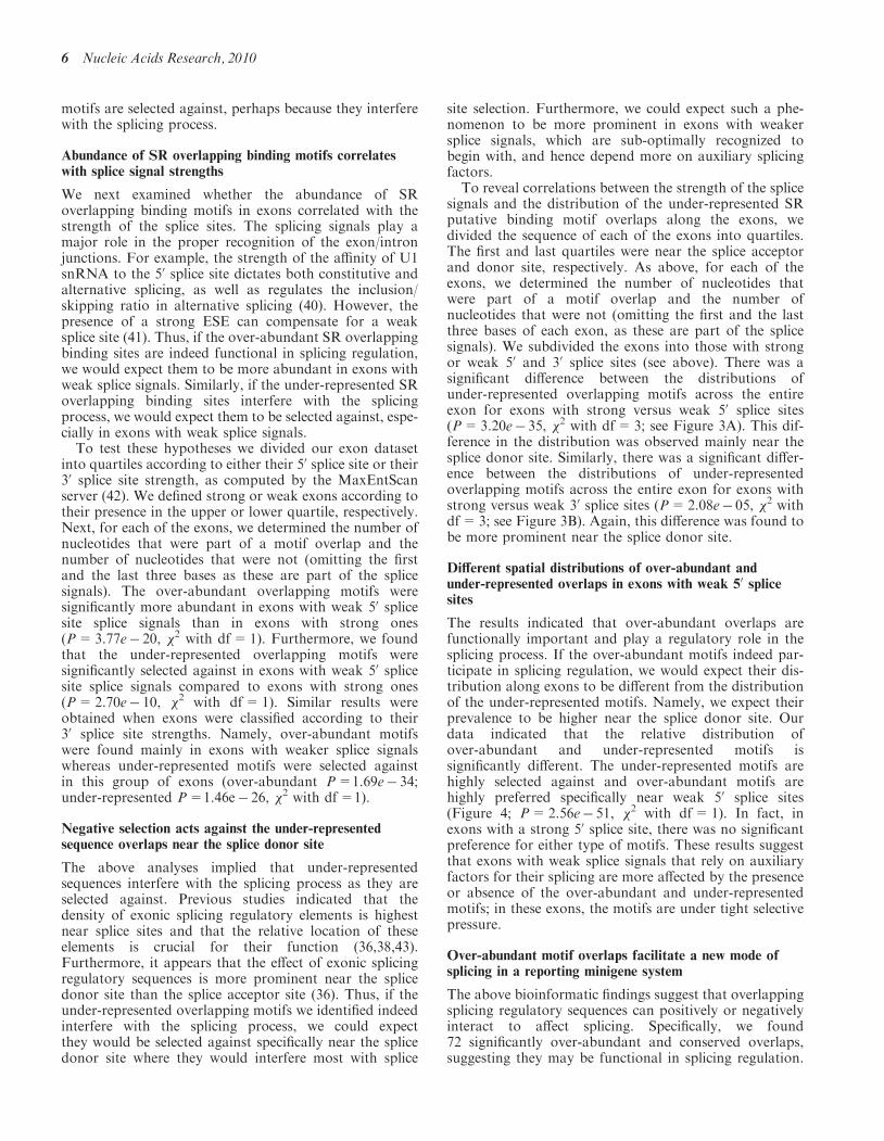
To reveal correlations between the strength of the splicesignals and the distribution of the under-represented SRputative binding motif overlaps along the exons, wedivided the sequence of each of the exons into quartiles.The first and last quartiles were near the splice acceptorand donor site, respectively. As above, for each of theexons, we determined the number of nucleotides thatwere part of a motif overlap and the number ofnucleotides that were not (omitting the first and the lastthree bases of each exon, as these are part of the splicesignals). We subdivided the exons into those with strongor weak 50 and 30 splice sites (see above). There was asignificant difference between the distributions ofunder-represented overlapping motifs across the entireexon for exons with strong versus weak 50 splice sites(P=3.20e� 35, �2 with df=3; see Figure 3A). This dif-ference in the distribution was observed mainly near thesplice donor site. Similarly, there was a significant differ-ence between the distributions of under-representedoverlapping motifs across the entire exon for exons withstrong versus weak 30 splice sites (P=2.08e� 05, �2 withdf=3; see Figure 3B). Again, this difference was found tobe more prominent near the splice donor site.
Different spatial distributions of over-abundant andunder-represented overlaps in exons with weak 50 splicesites
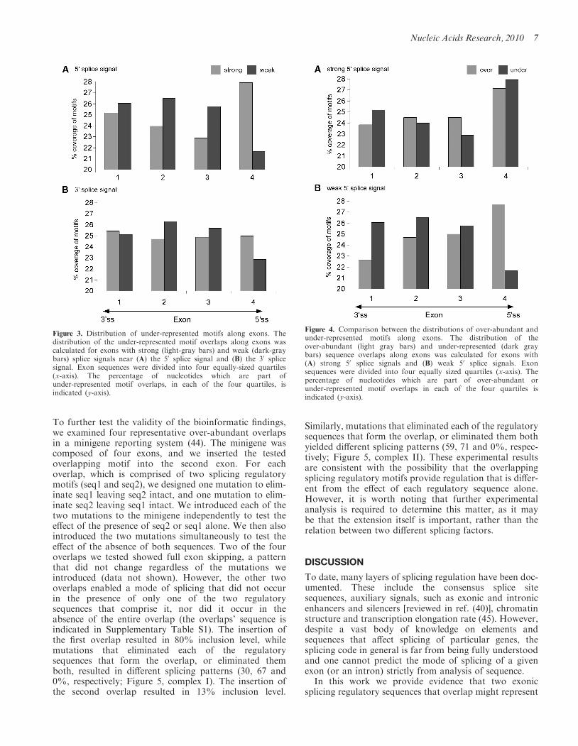
The results indicated that over-abundant overlaps arefunctionally important and play a regulatory role in thesplicing process. If the over-abundant motifs indeed par-ticipate in splicing regulation, we would expect their dis-tribution along exons to be different from the distributionof the under-represented motifs. Namely, we expect theirprevalence to be higher near the splice donor site. Ourdata indicated that the relative distribution ofover-abundant and under-represented motifs issignificantly different. The under-represented motifs arehighly selected against and over-abundant motifs arehighly preferred specifically near weak 50 splice sites(Figure 4; P=2.56e� 51, �2 with df=1). In fact, inexons with a strong 50 splice site, there was no significantpreference for either type of motifs. These results suggestthat exons with weak splice signals that rely on auxiliaryfactors for their splicing are more affected by the presenceor absence of the over-abundant and under-representedmotifs; in these exons, the motifs are under tight selectivepressure.
Over-abundant motif overlaps facilitate a new mode ofsplicing in a reporting minigene system
The above bioinformatic findings suggest that overlappingsplicing regulatory sequences can positively or negativelyinteract to affect splicing. Specifically, we found72 significantly over-abundant and conserved overlaps,suggesting they may be functional in splicing regulation.
6 Nucleic Acids Research, 2010
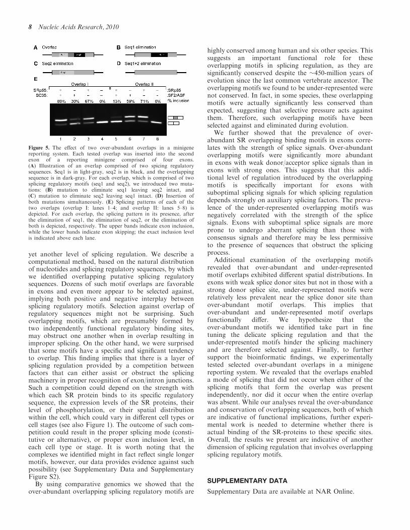
To further test the validity of the bioinformatic findings,we examined four representative over-abundant overlapsin a minigene reporting system (44). The minigene wascomposed of four exons, and we inserted the testedoverlapping motif into the second exon. For eachoverlap, which is comprised of two splicing regulatorymotifs (seq1 and seq2), we designed one mutation to elim-inate seq1 leaving seq2 intact, and one mutation to elim-inate seq2 leaving seq1 intact. We introduced each of thetwo mutations to the minigene independently to test theeffect of the presence of seq2 or seq1 alone. We then alsointroduced the two mutations simultaneously to test theeffect of the absence of both sequences. Two of the fouroverlaps we tested showed full exon skipping, a patternthat did not change regardless of the mutations weintroduced (data not shown). However, the other twooverlaps enabled a mode of splicing that did not occurin the presence of only one of the two regulatorysequences that comprise it, nor did it occur in theabsence of the entire overlap (the overlaps’ sequence isindicated in Supplementary Table S1). The insertion ofthe first overlap resulted in 80% inclusion level, whilemutations that eliminated each of the regulatorysequences that form the overlap, or eliminated themboth, resulted in different splicing patterns (30, 67 and0%, respectively; Figure 5, complex I). The insertion ofthe second overlap resulted in 13% inclusion level.
Similarly, mutations that eliminated each of the regulatorysequences that form the overlap, or eliminated them bothyielded different splicing patterns (59, 71 and 0%, respec-tively; Figure 5, complex II). These experimental resultsare consistent with the possibility that the overlappingsplicing regulatory motifs provide regulation that is differ-ent from the effect of each regulatory sequence alone.However, it is worth noting that further experimentalanalysis is required to determine this matter, as it maybe that the extension itself is important, rather than therelation between two different splicing factors.
DISCUSSION
To date, many layers of splicing regulation have been doc-umented. These include the consensus splice sitesequences, auxiliary signals, such as exonic and intronicenhancers and silencers [reviewed in ref. (40)], chromatinstructure and transcription elongation rate (45). However,despite a vast body of knowledge on elements andsequences that affect splicing of particular genes, thesplicing code in general is far from being fully understoodand one cannot predict the mode of splicing of a givenexon (or an intron) strictly from analysis of sequence.In this work we provide evidence that two exonic
splicing regulatory sequences that overlap might represent
Figure 4. Comparison between the distributions of over-abundant andunder-represented motifs along exons. The distribution of theover-abundant (light gray bars) and under-represented (dark graybars) sequence overlaps along exons was calculated for exons with(A) strong 50 splice signals and (B) weak 50 splice signals. Exonsequences were divided into four equally sized quartiles (x-axis). Thepercentage of nucleotides which are part of over-abundant orunder-represented motif overlaps in each of the four quartiles isindicated (y-axis).
Figure 3. Distribution of under-represented motifs along exons. Thedistribution of the under-represented motif overlaps along exons wascalculated for exons with strong (light-gray bars) and weak (dark-graybars) splice signals near (A) the 50 splice signal and (B) the 30 splicesignal. Exon sequences were divided into four equally-sized quartiles(x-axis). The percentage of nucleotides which are part ofunder-represented motif overlaps, in each of the four quartiles, isindicated (y-axis).
Nucleic Acids Research, 2010 7
yet another level of splicing regulation. We describe acomputational method, based on the natural distributionof nucleotides and splicing regulatory sequences, by whichwe identified overlapping putative splicing regulatorysequences. Dozens of such motif overlaps are favorablein exons and even more appear to be selected against,implying both positive and negative interplay betweensplicing regulatory motifs. Selection against overlap ofregulatory sequences might not be surprising. Suchoverlapping motifs, which are presumably formed bytwo independently functional regulatory binding sites,may obstruct one another when in overlap resulting inimproper splicing. On the other hand, we were surprisedthat some motifs have a specific and significant tendencyto overlap. This finding implies that there is a layer ofsplicing regulation provided by a competition betweenfactors that can either assist or obstruct the splicingmachinery in proper recognition of exon/intron junctions.Such a competition could depend on the strength withwhich each SR protein binds to its specific regulatorysequence, the expression levels of the SR proteins, theirlevel of phosphorylation, or their spatial distributionwithin the cell, which could vary in different cell types orcell stages (see also Figure 1). The outcome of such com-petition could result in the proper splicing mode (consti-tutive or alternative), or proper exon inclusion level, ineach cell type or stage. It is worth noting that thecomplexes we identified might in fact reflect single longermotifs, however, our data provides evidence against suchpossibility (see Supplementary Data and SupplementaryFigure S2).By using comparative genomics we showed that the
over-abundant overlapping splicing regulatory motifs are
highly conserved among human and six other species. Thissuggests an important functional role for theseoverlapping motifs in splicing regulation, as they aresignificantly conserved despite the �450-million years ofevolution since the last common vertebrate ancestor. Theoverlapping motifs we found to be under-represented werenot conserved. In fact, in some species, these overlappingmotifs were actually significantly less conserved thanexpected, suggesting that selective pressure acts againstthem. Therefore, such overlapping motifs have beenselected against and eliminated during evolution.
We further showed that the prevalence of over-abundant SR overlapping binding motifs in exons corre-lates with the strength of splice signals. Over-abundantoverlapping motifs were significantly more abundantin exons with weak donor/acceptor splice signals than inexons with strong ones. This suggests that this addi-tional level of regulation introduced by the overlappingmotifs is specifically important for exons withsuboptimal splicing signals for which splicing regulationdepends strongly on auxiliary splicing factors. The preva-lence of the under-represented overlapping motifs wasnegatively correlated with the strength of the splicesignals. Exons with suboptimal splice signals are moreprone to undergo aberrant splicing than those withconsensus signals and therefore may be less permissiveto the presence of sequences that obstruct the splicingprocess.
Additional examination of the overlapping motifsrevealed that over-abundant and under-representedmotif overlaps exhibited different spatial distributions. Inexons with weak splice donor sites but not in those with astrong donor splice site, under-represented motifs wererelatively less prevalent near the splice donor site thanover-abundant motif overlaps. This implies thatover-abundant and under-represented motif overlapsfunctionally differ. We hypothesize that theover-abundant motifs we identified take part in finetuning the delicate splicing regulation and that theunder-represented motifs hinder the splicing machineryand are therefore selected against. Finally, to furthersupport the bioinformatic findings, we experimentallytested selected over-abundant overlaps in a minigenereporting system. We revealed that the overlaps enableda mode of splicing that did not occur when either of thesplicing motifs that form the overlap was presentindependently, nor did it occur when the entire overlapwas absent. While our analyses reveal the over-abundanceand conservation of overlapping sequences, both of whichare indicative of functional implications, further experi-mental work is needed to determine whether there isactual binding of the SR-proteins to these specific sites.Overall, the results we present are indicative of anotherdimension of splicing regulation that involves overlappingsplicing regulatory motifs.
SUPPLEMENTARY DATA
Supplementary Data are available at NAR Online.
Figure 5. The effect of two over-abundant overlaps in a minigenereporting system. Each tested overlap was inserted into the secondexon of a reporting minigene comprised of four exons.(A) Illustration of an overlap comprised of two spicing regulatorysequences. Seq1 is in light-gray, seq2 is in black, and the overlappingsequence is in dark-gray. For each overlap, which is comprised of twosplicing regulatory motifs (seq1 and seq2), we introduced two muta-tions: (B) mutation to eliminate seq1 leaving seq2 intact, and(C) mutation to eliminate seq2 leaving seq1 intact. (D) Insertion ofboth mutations simultaneously. (E) Splicing patterns of each of thetwo overlaps (overlap I: lanes 1–4; and overlap II: lanes 5–8) isdepicted. For each overlap, the splicing pattern in its presence, afterthe elimination of seq1, the elimination of seq2, or the elimination ofboth is depicted, respectively. The upper bands indicate exon inclusion,while the lower bands indicate exon skipping; the exact inclusion levelis indicated above each lane.
8 Nucleic Acids Research, 2010
FUNDING
Israel Science Foundation (ISF 61/09), Joint Germany-Israeli Research Program (ca-139), Deutsche-IsraelProject (DIP MI-1317) and European AlternativeSplicing Network (EURASNET). AG is supported bythe Adams Fellowship Program of the Israel Academyof Sciences and Humanities and EK is a fellow of theClore Scholars Program. Funding for open accesscharge: EURASNET.
Conflict of interest statement. None declared.
REFERENCES
1. Black,D.L. (2003) Mechanisms of alternative pre-messenger RNAsplicing. Annu. Rev. Biochem., 72, 291–336.
2. Lander,E.S., Linton,L.M., Birren,B., Nusbaum,C., Zody,M.C.,Baldwin,J., Devon,K., Dewar,K., Doyle,M., FitzHugh,W. et al.(2001) Initial sequencing and analysis of the human genome.Nature, 409, 860–921.
3. Venter,J.C., Adams,M.D., Myers,E.W., Li,P.W., Mural,R.J.,Sutton,G.G., Smith,H.O., Yandell,M., Evans,C.A., Holt,R.A.et al. (2001) The sequence of the human genome. Science, 291,1304–1351.
4. Deckert,J., Hartmuth,K., Boehringer,D., Behzadnia,N., Will,C.L.,Kastner,B., Stark,H., Urlaub,H. and Luhrmann,R. (2006) Proteincomposition and electron microscopy structure of affinity-purifiedhuman spliceosomal B complexes isolated under physiologicalconditions. Mol. Cell Biol., 26, 5528–5543.
5. Hartmuth,K., Urlaub,H., Vornlocher,H.P., Will,C.L., Gentzel,M.,Wilm,M. and Luhrmann,R. (2002) Protein composition of humanprespliceosomes isolated by a tobramycin affinity-selectionmethod. Proc. Natl Acad. Sci. USA, 99, 16719–16724.
6. Jurica,M.S. and Moore,M.J. (2003) Pre-mRNA splicing: awash ina sea of proteins. Mol. Cell, 12, 5–14.
7. Zhou,Z., Licklider,L.J., Gygi,S.P. and Reed,R. (2002)Comprehensive proteomic analysis of the human spliceosome.Nature, 419, 182–185.
8. Berget,S.M. (1995) Exon recognition in vertebrate splicing.J. Biol. Chem., 270, 2411–2414.
9. Graveley,B.R. (2001) Alternative splicing: increasing diversity inthe proteomic world. Trends Genet., 17, 100–107.
10. Lim,L.P. and Burge,C.B. (2001) A computational analysis ofsequence features involved in recognition of short introns.Proc. Natl Acad. Sci. USA, 98, 11193–11198.
11. Schwartz,S.H., Silva,J., Burstein,D., Pupko,T., Eyras,E. andAst,G. (2008) Large-scale comparative analysis of splicing signalsand their corresponding splicing factors in eukaryotes.Genome Res., 18, 88–103.
12. Blencowe,B.J. (2000) Exonic splicing enhancers: mechanism ofaction, diversity and role in human genetic diseases. TrendsBiochem. Sci., 25, 106–110.
13. Caceres,J.F. and Kornblihtt,A.R. (2002) Alternative splicing:multiple control mechanisms and involvement in human disease.Trends Genet., 18, 186–193.
14. Cartegni,L., Chew,S.L. and Krainer,A.R. (2002) Listening tosilence and understanding nonsense: exonic mutations that affectsplicing. Nat. Rev. Genet., 3, 285–298.
15. Fairbrother,W.G., Yeh,R.F., Sharp,P.A. and Burge,C.B. (2002)Predictive identification of exonic splicing enhancers in humangenes. Science, 297, 1007–1013.
16. Graveley,B.R. (2000) Sorting out the complexity of SR proteinfunctions. RNA, 6, 1197–1211.
17. Woodley,L. and Valcarcel,J. (2002) Regulation of alternativepre-mRNA splicing. Brief Funct. Genomic Proteomic, 1, 266–277.
18. Sanford,J.R., Ellis,J. and Caceres,J.F. (2005) Multiple roles ofarginine/serine-rich splicing factors in RNA processing.Biochem. Soc. Trans., 33, 443–446.
19. Singh,R. and Valcarcel,J. (2005) Building specificity with nonspecificRNA-binding proteins. Nat. Struct. Mol. Biol., 12, 645–653.
20. Chabot,B. (1996) Directing alternative splicing: cast and scenarios.Trends Genet., 12, 472–478.
21. Maniatis,T. and Tasic,B. (2002) Alternative pre-mRNA splicingand proteome expansion in metazoans. Nature, 418, 236–243.
22. Smith,C.W. and Valcarcel,J. (2000) Alternative pre-mRNAsplicing: the logic of combinatorial control. Trends Biochem. Sci.,25, 381–388.
23. Zhang,X.H., Arias,M.A., Ke,S. and Chasin,L.A. (2009) Splicingof designer exons reveals unexpected complexity in pre-mRNAsplicing. RNA, 15, 367–376.
24. Akerman,M., David-Eden,H., Pinter,R.Y. andMandel-Gutfreund,Y. (2009) A computational approach forgenome-wide mapping of splicing factor binding sites.Genome Biol., 10, R30.
25. Ackerman,S.L., Minden,A.G., Williams,G.T., Bobonis,C. andYeung,C.Y. (1991) Functional significance of an overlappingconsensus binding motif for Sp1 and Zif268 in the murineadenosine deaminase gene promoter. Proc. Natl Acad. Sci. USA,88, 7523–7527.
26. Pollwein,P. (1993) Overlapping binding sites of two differenttranscription factors in the promoter of the human gene for theAlzheimer amyloid precursor protein. Biochem. Biophys. Res.Commun., 190, 637–647.
27. Discenza,M.T., Dehbi,M. and Pelletier,J. (1997) OverlappingDNA recognition motifs between Sp1 and a novel trans-actingfactor within the wt1 tumour suppressor gene promoter.Nucleic Acids Res., 25, 4314–4322.
28. Harrington,R.H. and Sharma,A. (2001) Transcription factorsrecognizing overlapping C1-A2 binding sites positively regulateinsulin gene expression. J. Biol. Chem., 276, 104–113.
29. Karolchik,D., Hinrichs,A.S., Furey,T.S., Roskin,K.M.,Sugnet,C.W., Haussler,D. and Kent,W.J. (2004) The UCSCtable browser data retrieval tool. Nucleic Acids Res., 32,D493–D496.
30. Giardine,B., Riemer,C., Hardison,R.C., Burhans,R., Elnitski,L.,Shah,P., Zhang,Y., Blankenberg,D., Albert,I., Taylor,J. et al.(2005) Galaxy: a platform for interactive large-scale genomeanalysis. Genome Res., 15, 1451–1455.
31. Liu,H.X., Chew,S.L., Cartegni,L., Zhang,M.Q. and Krainer,A.R.(2000) Exonic splicing enhancer motif recognized by human SC35under splicing conditions. Mol. Cell Biol., 20, 1063–1071.
32. Liu,H.X., Zhang,M. and Krainer,A.R. (1998) Identification offunctional exonic splicing enhancer motifs recognized byindividual SR proteins. Genes Dev., 12, 1998–2012.
33. Smith,P.J., Zhang,C., Wang,J., Chew,S.L., Zhang,M.Q. andKrainer,A.R. (2006) An increased specificity score matrix for theprediction of SF2/ASF-specific exonic splicing enhancers.Hum. Mol. Genet., 15, 2490–2508.
34. Cartegni,L., Wang,J., Zhu,Z., Zhang,M.Q. and Krainer,A.R.(2003) ESEfinder: a web resource to identify exonic splicingenhancers. Nucleic Acids Res., 31, 3568–3571.
35. Benjamini,Y. and Hochberg,Y. (1995) Controlling the falsediscovery rate: a practical and powerful approach to multipletesting. J. R. Stat. Soc., 57, 289–300.
36. Goren,A., Ram,O., Amit,M., Keren,H., Lev-Maor,G.,Vig,I., Pupko,T. and Ast,G. (2006) Comparative analysisidentifies exonic splicing regulatory sequences–Thecomplex definition of enhancers and silencers. Mol. Cell, 22,769–781.
37. Zhang,X.H. and Chasin,L.A. (2004) Computational definition ofsequence motifs governing constitutive exon splicing. Genes Dev.,18, 1241–1250.
38. Fairbrother,W.G., Holste,D., Burge,C.B. and Sharp,P.A. (2004)Single nucleotide polymorphism-based validation of exonicsplicing enhancers. PLoS Biol., 2, E268.
39. Parmley,J.L., Chamary,J.V. and Hurst,L.D. (2006) Evidence forpurifying selection against synonymous mutations in mammalianexonic splicing enhancers. Mol. Biol. Evol., 23, 301–309.
40. Ast,G. (2004) How did alternative splicing evolve? Nat. Rev.Genet., 5, 773–782.
41. Ram,O., Schwartz,S. and Ast,G. (2008) Multifactorial interplaycontrols the splicing profile of Alu-derived exons. Mol. Cell Biol.,28, 3513–3525.
Nucleic Acids Research, 2010 9

42. Yeo,G. and Burge,C.B. (2004) Maximum entropy modeling ofshort sequence motifs with applications to RNA splicing signals.J. Comput. Biol., 11, 377–394.
43. Majewski,J. and Ott,J. (2002) Distribution and characterization ofregulatory elements in the human genome. Genome Res., 12,1827–1836.
44. Coulter,L.R., Landree,M.A. and Cooper,T.A. (1997) Identificationof a new class of exonic splicing enhancers by in vivo selection.Mol. Cell Biol., 17, 2143–2150.
45. Kornblihtt,A.R. (2006) Chromatin, transcript elongation andalternative splicing. Nat. Struct. Mol. Biol., 13, 5–7.
10 Nucleic Acids Research, 2010