rickettsia spp. in free ranging small mammals in south
TRANSCRIPT
SU
SA
NN
E SC
HEX
RICKETTSIA
SP
P. IN
SM
ALL M
AM
MA
LS
SUSANNE YVONNE SCHEX
RICKETTSIA SPP. IN FREE RANGING SMALL
MAMMALS IN SOUTH-EASTERN GERMANY
VVB VVB LAUFERSWEILER VERLAGédition scientifique
9 7 8 3 8 3 5 9 5 7 3 6 7
VVB LAUFERSWEILER VERLAGSTAUFENBERGRING 15D-35396 GIESSEN
Tel: 0641-5599888 Fax: [email protected]
VVB LAUFERSWEILER VERLAGédition scientifique
ISBN: 978-3-8359-5736-7 INAUGURAL-DISSERTATION zur Erlangung der tiermedizinischen Doktorwürdeder Tierärztlichen Fakultät der Ludwig-Maximilians-Universität München

Das Werk ist in allen seinen Teilen urheberrechtlich geschützt.
Jede Verwertung ist ohne schriftliche Zustimmung des Autors oder des Verlages unzulässig. Das gilt insbesondere für Vervielfältigungen, Übersetzungen, Mikroverfilmungen
und die Einspeicherung in und Verarbeitung durch elektronische Systeme.
1. Auflage 2011
All rights reserved. No part of this publication may be reproduced, stored in a retrieval system, or transmitted,
in any form or by any means, electronic, mechanical, photocopying, recording, or otherwise, without the prior
written permission of the Author or the Publishers.
st1 Edition 2011
© 2011 by VVB LAUFERSWEILER VERLAG, GiessenPrinted in Germany
VVB LAUFERSWEILER VERLAG
STAUFENBERGRING 15, D-35396 GIESSENTel: 0641-5599888 Fax: 0641-5599890
email: [email protected]
www.doktorverlag.de
Umschlaggestaltung unter Verwendung einer Fotografie von Andreas Ebert
édition scientifique

Aus dem
Department für Veterinärwissenschaften der Tierärztlichen Fakultät
der Ludwig-Maximilians-Universität München
Arbeit angefertigt unter Leitung von:
Univ.- Prof. Dr. Reinhard K. Straubinger, Ph.D.
Angefertigt am Institut für Mikrobiologie der Bundeswehr
(PD Dr. Sandra Essbauer)
Rickettsia spp. in free ranging small
mammals in South-Eastern Germany
Inaugural-Dissertation
zur Erlangung der tiermedizinischen Doktorwürde
der Tierärztlichen Fakultät der Ludwig-Maximilians-Universität München
von
Susanne Yvonne Schex
aus München
München 2011
Gedruckt mit der Genehmigung der Tierärztlichen Fakultät
der Ludwig-Maximilians-Universität München
Dekan: Univ.-Prof. Dr. Braun
Berichterstatter: Univ.-Prof. Dr. Straubinger
Korreferent: Univ.-Prof. Dr. Pfister
Tag der Promotion: 12. Februar 2011

To my family


TABLE OF CONTENTS
I. INTRODUCTION.........................................................................................1
II. LITERATURE REVIEW................................................................................3
1 Rodents as reservoir hosts.................................................................3 1.1 What is a reservoir host? ....................................................................3
1.2 Rodent-associated pathogens in Germany.......................................6
2 Rickettsia............................................................................................11
2.1 General aspects of Rickettsia...........................................................11
2.2 Relevant species of spotted fever group Rickettsia.......................15
III. MATERIALS AND METHODS .....................................................................19
1 Rodent collection...............................................................................19 2 Immunofluorescence tests ...............................................................23 3 DNA extraction...................................................................................24
4 Polymerase chain reaction for the detection of Rickettsial DNA ..................................................................................25
4.1 Real time PCR for the detection of the gltA gene of Rickettsia spp. ...................................................................................25
4.2 PCR for the detection of ompB genes .............................................26 4.3 Mammalia species-specific18S ribosomal RNA PCR .....................27 5 Agarose gel electrophoresis.............................................................29 6 DNA purification.................................................................................29
7 Sequencing and sequence analysis.................................................29
8 Statistical analysis.............................................................................29
IV. RESULTS ...............................................................................................31
1 Trapping results.................................................................................31 2 PCR results ........................................................................................33 3 IFT results...........................................................................................34
4 Sequence analysis.............................................................................35 5 Publication .........................................................................................37
V. DISCUSSION...........................................................................................60
VI. CONCLUSION & OUTLOOK ......................................................................68
VII. SUMMARY..............................................................................................69
VIII. ZUSAMMENFASSUNG ..............................................................................70
IX. REFERENCES.........................................................................................71
X. ABBREVATIONS......................................................................................91
XI. FIGURES................................................................................................93
XII. TABLES.................................................................................................94
XIII. ANNEX ..................................................................................................95
1 Overview of investigations regarding rickettsiae ...........................95 2 Sequencing data ................................................................................96
3 Trapping locations.............................................................................99 4 Publication .......................................................................................101
XIV. ACKNOWLEDGEMENTS .........................................................................115
I. Introduction 1
I. INTRODUCTION
Rodents and other small mammals are associated with several pathogens,
such as RNA and DNA viruses, bacteria and parasites. These differ not
only in their genetic organisation but also in their association with specific
reservoir hosts, their geographical distribution and their transmission
cycles. For many of those pathogens rodents act as a reservoir host (Mills
& Childs, 1998; Haydon et al., 2002; Essbauer et al., 2009b; Ulrich et al.,
2009). Rodent-borne and other zoonoses account for a significant
proportion (60.3%) of Emerging Infectious Diseases (EID). Moreover, the
majority of these infections (71.8%) originates from wildlife and increased
significantly over time. More than 50% of EIDs are caused by bacteria
(Jones et al., 2008).
Rickettsioses are recognized as emerging infections in several parts of the
world (Parola & Raoult, 2001; Fournier & Raoult 2005; Parola et al., 2005;
Brouqui et al., 2007; Rovery et al., 2008). These obligate intracellular
bacteria are transmitted to humans and animals by blood-sucking
arthropods such as insects (i.e. fleas) or arachnids (i.e. ticks and mites).
Nearly half of the currently recognized rickettsioses have been discovered
within the last two decades (Raoult & Roux, 1997). These included at least
ten tick-transmitted rickettsioses characterised between 1993 and 2005
(Fournier & Raoult, 2005). Further, in the last few years, rickettsiae
previously considered as non-pathogenic or mild turn out to implicate
human disease (Blanco & Oteo 2006; Rovery et al., 2008). Moreover,
several species, e.g. R. raoultii have been newly described (Mediannikov
et al., 2008). Table 12 (Annex, p. 95) gives an exemplary overview about
investigations regarding rickettsiae.
Other arthropod-borne pathogens such as Borrelia spp. or Anaplasma
spp. (the latter also belonging to the order Rickettsiales) are known to
exist in natural cycles involving rodents. Although the arthropod-related
part of the rickettsial life cycle is widely agreed, the role of vertebrate
reservoirs is still fairly unclear (Brouqui et al., 2007). In order to gather
further knowledge on this subject, the VICCI Project (Vector borne
I. Introduction 2
infectious diseases in climate change investigations) was established and
funded by the Bavarian State Ministry of the Environment and Public
Health (Bayerisches Staatsministerium für Umwelt und Gesundheit,
StMUG). The present study was conducted in the scope of this project.
One of its major aims was to investigate the role of rodents in the natural
cycle of Rickettsia spp. in Germany. To the best of our knowledge, so far
this is the first time an investigation concerning this aspect of rickettsiology
is performed in Germany.
The present study focused on three aspects:
1) The occurrence of spotted fever group (SFG) rickettsiae in rodents
in the National Park Bavarian Forest (syn. Bohemian Forest) and
nearby areas in Lower Bavaria;
2) The prevalence and genetic diversity of SFG rickettsiae in those
animals by means of
a) molecular-biological analyses and sequencing
b) serological analyses;
3) The assessment of predictors for a positive detection of rickettsial
DNA in wild rodents.
The increase in the emergence and reemergence of rickettsial infections,
coupled with recent characterisation of several proposed new species and
the uncertainties in their natural life cycles make rickettsiology a truly
engaging yet still fairly unexplored field of research. This study aims to
contribute to the already widespread research on rodent-borne pathogens
in Germany (as exemplified in the publication in Annex 4, p. 101).
Furthermore, it aims to extend the state of knowledge on Rickettsia spp.
occurring in Germany, their genetic characterisation and the possible role
of wildlife reservoirs in their epidemiology.
II. Literature Review 3
II. LITERATURE REVIEW
1 Rodents as reservoir hosts
1.1 What is a reservoir host?
A natural reservoir is defined as the long-term host of a pathogen of an
infectious disease. Previous definitions imply that the relevant infectious
agent is nonpathogenic for the reservoir host or that it is carried as a
subclinical and therefore asymptomatic and non-lethal infection. Haydon et
al. (2002) propose that a reservoir is defined as one or more
epidemiologically connected populations or environments in which the
pathogen can be maintained permanently and from which infection is
transmitted to the defined target population.
A significant coevolution is to be expected between reservoir host and
pathogen (Zeier et al., 2005; Ulrich et al., 2009). Cospeciation and
coevolution require a close association between two species. Pathogens
may adapt by minimizing pathology in their reservoir hosts whilst causing
pathologies in the incidental hosts. This is believed to be related with
different immune systems, which respond strongly in order to eliminate the
pathogen but contribute to host pathology (Calisher et al., 2008). In
addition, pathogens that evolved with the reservoir species may have used
cellular receptors and biochemical pathways, which are conserved in later
evolved mammals, for replication. If these cellular receptors and pathways
are preserved, they could contribute to the capacity for transmission of the
agent (Calisher et al., 2006).
Knowledge about maintaining reservoir hosts is essential for disease
prevention and control. In order to better understand reservoir ecology and
its correlation to human or animal disease the following steps are
essential:
1) Determination of the geographic distribution of the host;
2) Determination of the geographic range of the pathogen within the
host range;
II. Literature Review 4
3) Determination of the regional distribution of the host and pathogen
among the distinct habitat types;
4) Assessment of host-pathogen dynamics through prospective,
longitudinal studies;
5) Development of an integrative time- and place-specific predictive
model (Mills & Childs, 1998).
The following examples underline the key function of reservoir host
surveillance in order to control infectious diseases. Only few European
countries monitor wildlife reservoirs for zoonotic diseases, an overview is
given on the website of the World Organisation for Animal Health (OIE)
(World Animal Health Database, WAHID, 2010).
In the United Kingdom, tuberculosis was eradicated from large areas in
the 1970s. Since the Wildlife and Countryside Act 1981, that put the
European badger (Meles meles) under protection, Mycobacterium bovis
infections in cattle have been rising once more (Palmer, 2007). The
badger is considered a true maintenance host for M. bovis, with a regional
tuberculosis prevalence as high as 20.5%. Infected badgers shed large
numbers of M. bovis in saliva, urine and feces and can live 3 to 4 years
following the first documented episode of shedding of M. bovis (Little et al.,
1982). Transmission to cattle is thought to happen through inhalation of
the pathogen from contaminated grass (Palmer, 2007). Little et al. (1982)
showed that removing badgers from cattle farming areas resulted in a
decline in bovine tuberculosis. The British Government is aiming to reduce
tuberculosis in cattle by a combined strategy including culling and
vaccination of badgers in the wild. Moreover, since March 2010, an
injectable badger vaccine received Market Authorisation; an oral
vaccination is currently being researched (Department for Environment,
Food and Rural Affairs, 2010).
On the other hand, in New Zealand, Brushtail Possums are the main
reservoir host for M. bovis. Infected possums exhibit abnormal behaviour
such as rolling that attracts attention of cattle. These then get infected
directly when having contact to the possums (Palmer, 2007). Attempts to
eradicate possums have failed so far. In the USA, the main reservoir for
II. Literature Review 5
M. bovis is the White-Tailed Deer. Control measures aim to reduce deer
density and monitor hunter-killed deer (Palmer, 2007). However, the only
country with a known wildlife reservoir that was able to eradicate
tuberculosis from cattle is Australia. There, the government established a
control program including a substantial element of feral buffalo control, as
the feral buffalo along with the feral pig is considered the main reservoir of
bovine tuberculosis in Australia (Department for Environment, Food and
Rural Affairs, 2010).
As for Europe, the wild boar is considered another important reservoir host
not only for M. bovis, but also for other zoonotic agents such as Brucella
suis, Leptospira spp., Coxiella burnetii, Francisella tularensis, Yersinia
pestis, hepatis E virus (HEV), pseudorabies virus (PRV), porcine circovirus
type 2 (PCV2), classical swine fever virus (CSFV), porcine parvovirus
(PPV), porcine reproductive and respiratory syndrome virus (PRRSV) and
parasites, e.g. Trichinella spp. and Toxoplasma gondii (reviewed in Meng
et al., 2009). These infectious agents are transmissible to domestic pigs
and other animal species including humans. The potential risk of pathogen
transmission to humans and livestock is enhanced by increasing wild boar
populations in some areas, increased chances of contact in suburban
areas and further by hunting and consumption of wild boar meat.
An example for a successful concept for eradication of a disease of
immense importance is rabies. Rabies is a viral disease maintained by
multiple wildlife reservoirs such as foxes, bats and others. Oral vaccination
strategies and surveillance programs were able to eradicate wildlife rabies
in many European countries (Pastoret et al., 1999). In Zimbabwe,
however, besides domestic dogs also jackals are a maintenance and
source population of infections in humans. Since jackals can comprise all
or part of a maintenance community independent of dogs, eliminating
rabies will only be successful if jackal rabies was also controlled (Bingham
et al., 1999a, b).
Being involved in transmission cycles of a large number of pathogens,
rodents form a taxon of high interest in the research of zoonoses.
Worldwide, rats and mice spread over 35 diseases
(www.cdc.gov/rodents). Moreover, with about 2277 recognized species
II. Literature Review 6
(Wilson & Reeder, 2005) and their worldwide occurence (except for
Antarctica) rodents are one of the most successful mammal taxa. In urban
areas their population can increase to numbers 15 times higher than the
human population (Battersby et al., 2002; Easterbrook et al., 2005;
Bonnefoy et al., 2008). Due to enormous range of body size and weight
(from a few grams up to 70 kg), complex social systems and different
reproduction strategies (Wilson & Reeder, 2005) they form a highly
diverse group. Their reproduction cycles are linked to regional, ecological
and climatic factors and in favorable conditions can lead to 10-fold
population increases (Lewellen & Vessey, 1998; Lambin et al., 2006). As a
result, these population fluctuations can in turn lead to disease outbreaks
(Schmaljohn & Hjelle, 1997). Worldwide distribution, rapid reproduction
cycles and high adaptability to new habitats are characteristics that
distinguish rodents from other vertebrate taxa and enable them to play an
important role in the maintenance and transmission of pathogens.
1.2 Rodent-associated pathogens in Germany
The following paragraph gives an overview on the most important rodent-
associated zoonoses in Germany; a summary is shown in Table 1 (p. 10).
More details and examples of investigations conducted by the
Bundeswehr Institute of Microbiology regarding these diseases are shown
in the publication „Nagetier-übertragene Zoonosen: Beispiele aus
Untersuchungen in Süd- und Westdeutschland“ (Annex 4, p. 101).
Leptospirosis is a disease caused by Gram-negative bacteria of the genus
Leptospira, belonging to the family of spirochetes (Johnson, 1996). They
are found worldwide and are currently divided into 16 species, seven of
them being pathogenic for humans. Below species level, Leptospira are
classified into over 250 serogroups due to antigen characteristics (Zoeller,
2009). The pathogen is transmitted via urine of persistent infected rodents;
however, evidence for the carriage of Leptospira has been found in
virtually all mammalian species examined (reviewed in Adler & de la Peña,
2010).
II. Literature Review 7
Francisella tularensis, the agent of Tularemia, is transmitted mainly by
Dermacentor and Ixodes ticks but also by mosquitos (Ellis et al., 2002).
However, a wide variety of rodents and other small mammals are involved
in the environmental cycle (Boyce, 1975; Morner et al., 1992; Berdal et al.,
1996; Tarnvik et al., 1996; Vorou et al., 2007). For instance, Kaysser et al.
(2008) found prevalences up to 10% in rodents from German outbreak
areas.
Borrelia burgdorferi sensu lato is a complex of bacteria of the order
Spirochetales, causing Lyme Borreliosis, a disease in humans and
animals, particularly in dogs (reviewed in Beugnet & Marié, 2009). Borrelia
spp. are transmitted by ticks, at the same time rodents, e.g. Apodemus
mice and Myodes glareolus act as important reservoir hosts, with a high
proportion being seropositive for B. burgdorferi in Central Europe (Gern et
al., 1998; Humair et al., 1999; Stefancíková et al., 2004; Gern, 2008). Also
migratory birds act as reservoirs and long distance vectors of infected ticks
(Vorou et al., 2007). An increase in incidence in humans has been noted
in eastern Germany in the years 2002 and 2003 (Mehnert & Krause,
2005).
Further, Telford et al. (1996) showed that the agent of human granulocytic
ehrlichiosis, Anaplasma phagocytophilum (formerly named Ehrlichia
phagocytophila), an emerging rickettsial disease, is able to infect various
rodent hosts, and that subadult deer ticks become infected and efficiently
transmit infection to rodents. Transmission and propagation occurs in
large mammals such as horses, ruminants, dogs and cats, small
mammals serve as reservoirs (Vorou et al., 2007).
Moreover, also Ehrlichia spp., belonging to the family Anaplasmataceae,
are transmitted by ticks from persistently infected ruminant, cervid and
rodent hosts (Walker et al., 2004). Ehrlichia chaffeensis causes flu-like
illness in humans, in more severe cases multisystemic disease, toxic
shock-like syndrome, or meningitis. Canine Ehrlichiosis, caused by
Ehrlichia canis, is a potentially fatal disease in dogs, infecting monocytes
in the peripheral blood. It is transmitted by the brown dog tick
(Rhipicephalus sanguineus). Other Ehrlichia spp. cause disease in
ruminants, horses, dogs and humans (Selbitz, 2002).
II. Literature Review 8
Babesia spp. are protozoa of the order Piroplasmida and are transmitted
by ixodid ticks. More than 100 Babesia species are known, infecting many
types of mammalian hosts, mostly rodents and birds (Homer et al., 2000).
Babesia spp. cause severe, sometimes malaria-like disease in humans,
ruminants, horses and dogs with symptoms such as apathia, fever,
anemia and ikterus (Eckert et al., 2005). Human babesiosis is
predominantly caused by two Babesia spp.: by B. divergens, a bovine
pathogen, in Europe and by B. microti, a rodent-borne piroplasm with the
white-footed mouse (Peromyscus leucopus) as natural reservoir in North
America (Homer et al., 2000). However, the first autochthonous European
case of human B. microti infection was reported in Germany (Hildebrandt
et al., 2007). In Europe, B. microti was found in yellow-necked mice (A.
flavicollis) and bank voles (M. glareolus) with a prevalence of 16.2% (Beck
et al., 2010).
Coxiella burnetii, the agent of Q-fever, is an obligate intracellular pathogen
with a broad host range, including humans, cattle, sheep, goats and other
domestic animals, birds and arthropods (Selbitz, 2002). Infection follows
inhalation of aerosol particles derived from heavily infected birth products
of sheep, goats and cattle, but C. burnetii is also shed in milk, urine and
feces. Animals become infected by aerosol and by tick bites, in Europe
particularly by Dermacentor marginatus. Epidemiology of C. burnetii
includes a sylvatic cycle (arthropods, birds, mammals including rodents)
and a domestic cycle (Selbitz, 2002).
At least 10 rodent species are considered maintenance and reservoir
hosts in the ecology of tick-borne encephalitis (TBE) virus (Cerny, 1976).
In particular, bank voles (Myodes glareolus) and Apodemus spp. are
present in TBE infection foci and often infestated with Ixodes ricinus ticks,
the main vector of this arbovirus. The infected tick can transmit the virus to
a vertebrate host during feeding and is also able to pass on the virus to a
non-infected tick during co-feeding at the same site on the host (Mansfield
et al., 2009).
Human Cowpox virus (CPXV) infections are often related to contact with
domestic cats, rather than cows. However, wild rodents are a natural
reservoir (Chantrey et al., 1999; Essbauer et al., 2009a). In humans,
II. Literature Review 9
cowpox virus causes papular lesions with surrounding erythema, general
malaise, headache and fever. Cats as well as rats often present papular,
ulcerating lesions, some also present respiratory dysfunctions (Kuczka et
al., 2009). Zoo and circus animals, especially elephants, seem to be highly
susceptible to generalised CPXV infections (Kurth et al., 2008).
Hantaviruses are rodent-borne RNA viruses, belonging to the family
Bunyaviridae. Hantaviruses are host-specific, thus, for instance, Puumala
virus, the main hantavirus species in Europe, is associated with the bank
vole (Myodes glareolus). This association is presumably caused by
coevolution of Hantaviruses and rodent reservoirs (Zeier et al., 2005;
Ulrich et al., 2009). In the specific host, the virus establishes a prolonged
subclinical infection, with virus shedding via urine, feces, and saliva (Lee
et al., 1981; Hutchinson et al., 1998; Vitullo et al., 1987). The virus is
transmitted to humans and to other rodents mainly by inhalation of
aerosolized virus (Mills & Childs, 1998). In humans, Puumala virus
infections cause disease ranging from asymptomatic infection, mild
influenza-like symptoms up to Nephropathia epidemica (NE) or even
hemorrhagic fever with renal syndrome (HFRS), a feverish illness with
kidney failure (Ulrich et al., 2004).
II. Literature Review 10
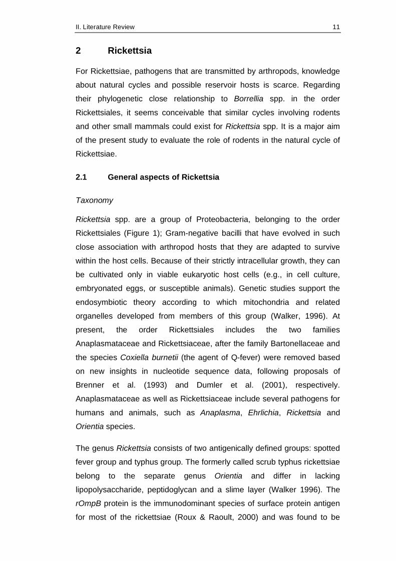
Table 1: Important rodent-associated pathogens in Germany (modified from Pfeffer et al., 2010)
Pathogen Family Vector human disease
Infections in domestic animals
Leptospira spp. Spirochaetacae - Leptospirosis dogs, pigs, ruminants i.a.
Francisella tularensis
Francisellaceae ticks Tularemia lagomorphs, monkeys i.a.
Borrelia spp. Spirochaetacae ticks Lyme-Borreliosis
dogs, horses
Rickettsia spp. Rickettsiaceae ticks, fleas, mites
Spotted fever dogs, cats
Babesia spp. Babesiidae ticks Babesiosis dogs, Bovinae Anaplasma phagocytophilum
Rickettsiaceae ticks Anaplasmosis/ Ehrlichiosis
dogs, horses, ruminants
Coxiella burnetii Coxiellaceae ticks Q-fever i.a. ruminants, dogs, cats
Hantavirus Bunyaviridae - HFRS/NE n.d. Cowpox virus (CPXV)
Poxviridae - Cowpox cats, dogs, zoo animals, pet rats
TBE virus Flaviviridae ticks Tick-borne encephalitis
small rumi-nants, dogs, monkeys
HFRS: hemorrhagic fever with renal syndrome; NE: Nephropathia epidemica n.d.: not determined -: direct transmission without vector
II. Literature Review 11
2 Rickettsia
For Rickettsiae, pathogens that are transmitted by arthropods, knowledge
about natural cycles and possible reservoir hosts is scarce. Regarding
their phylogenetic close relationship to Borrellia spp. in the order
Rickettsiales, it seems conceivable that similar cycles involving rodents
and other small mammals could exist for Rickettsia spp. It is a major aim
of the present study to evaluate the role of rodents in the natural cycle of
Rickettsiae.
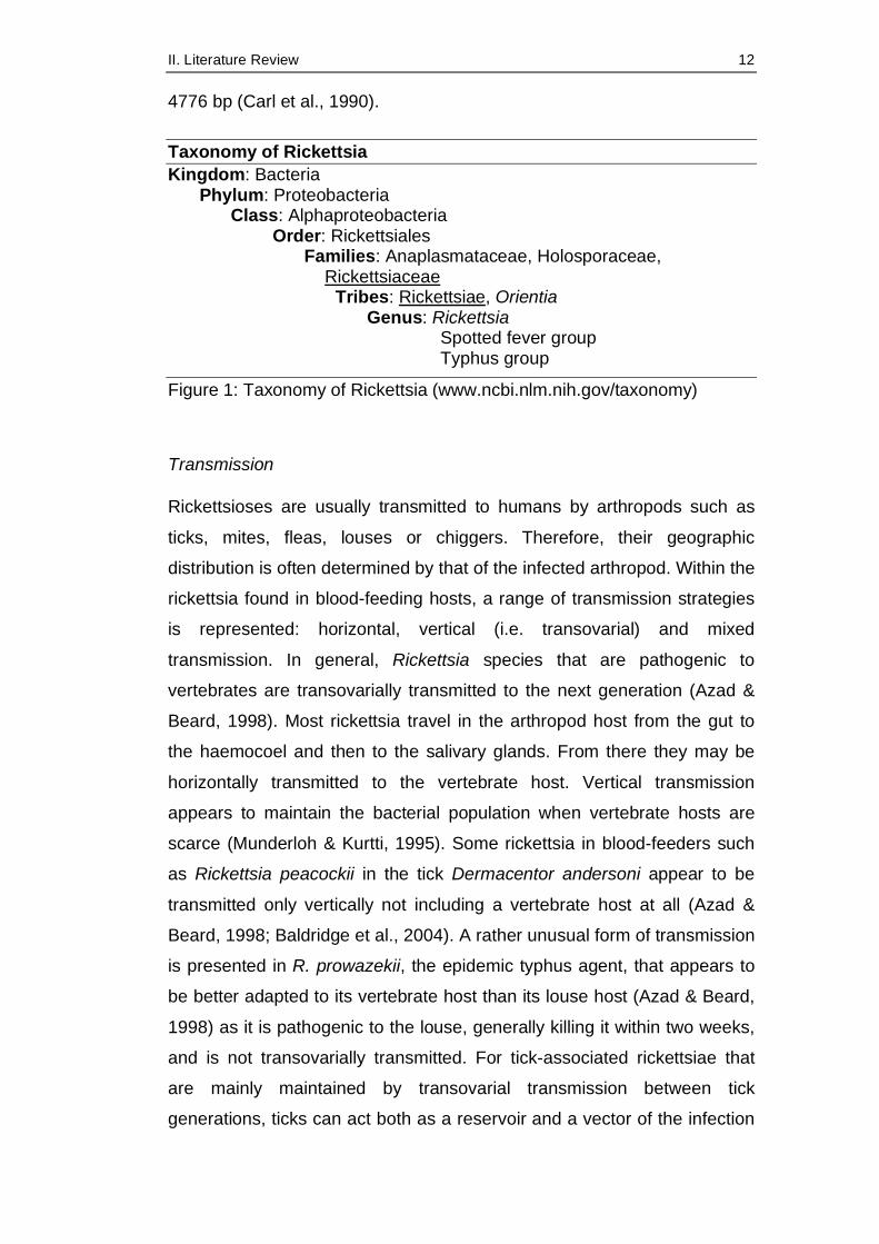
2.1 General aspects of Rickettsia
Taxonomy
Rickettsia spp. are a group of Proteobacteria, belonging to the order
Rickettsiales (Figure 1); Gram-negative bacilli that have evolved in such
close association with arthropod hosts that they are adapted to survive
within the host cells. Because of their strictly intracellular growth, they can
be cultivated only in viable eukaryotic host cells (e.g., in cell culture,
embryonated eggs, or susceptible animals). Genetic studies support the
endosymbiotic theory according to which mitochondria and related
organelles developed from members of this group (Walker, 1996). At
present, the order Rickettsiales includes the two families
Anaplasmataceae and Rickettsiaceae, after the family Bartonellaceae and
the species Coxiella burnetii (the agent of Q-fever) were removed based
on new insights in nucleotide sequence data, following proposals of
Brenner et al. (1993) and Dumler et al. (2001), respectively.
Anaplasmataceae as well as Rickettsiaceae include several pathogens for
humans and animals, such as Anaplasma, Ehrlichia, Rickettsia and
Orientia species.
The genus Rickettsia consists of two antigenically defined groups: spotted
fever group and typhus group. The formerly called scrub typhus rickettsiae
belong to the separate genus Orientia and differ in lacking
lipopolysaccharide, peptidoglycan and a slime layer (Walker 1996). The
rOmpB protein is the immunodominant species of surface protein antigen
for most of the rickettsiae (Roux & Raoult, 2000) and was found to be
II. Literature Review 12
4776 bp (Carl et al., 1990).
Taxonomy of Rickettsia Kingdom: Bacteria
Phylum: Proteobacteria Class: Alphaproteobacteria
Order: Rickettsiales Families: Anaplasmataceae, Holosporaceae,
Rickettsiaceae Tribes: Rickettsiae, Orientia
Genus: Rickettsia Spotted fever group Typhus group
Figure 1: Taxonomy of Rickettsia (www.ncbi.nlm.nih.gov/taxonomy)
Transmission
Rickettsioses are usually transmitted to humans by arthropods such as
ticks, mites, fleas, louses or chiggers. Therefore, their geographic
distribution is often determined by that of the infected arthropod. Within the
rickettsia found in blood-feeding hosts, a range of transmission strategies
is represented: horizontal, vertical (i.e. transovarial) and mixed
transmission. In general, Rickettsia species that are pathogenic to
vertebrates are transovarially transmitted to the next generation (Azad &
Beard, 1998). Most rickettsia travel in the arthropod host from the gut to
the haemocoel and then to the salivary glands. From there they may be
horizontally transmitted to the vertebrate host. Vertical transmission
appears to maintain the bacterial population when vertebrate hosts are
scarce (Munderloh & Kurtti, 1995). Some rickettsia in blood-feeders such
as Rickettsia peacockii in the tick Dermacentor andersoni appear to be
transmitted only vertically not including a vertebrate host at all (Azad &
Beard, 1998; Baldridge et al., 2004). A rather unusual form of transmission
is presented in R. prowazekii, the epidemic typhus agent, that appears to
be better adapted to its vertebrate host than its louse host (Azad & Beard,
1998) as it is pathogenic to the louse, generally killing it within two weeks,
and is not transovarially transmitted. For tick-associated rickettsiae that
are mainly maintained by transovarial transmission between tick
generations, ticks can act both as a reservoir and a vector of the infection
II. Literature Review 13
(Vitale et al., 1989). Unlike the spotted fever group rickettsia, typhus
rickettsia multiply in the epithelium of the intestinal tract of their arthropod
vectors and are excreted in the feces (Perlman et al., 2006), and infection
occurs via the dermis after scratching. They can also be transmitted by
bite in case they gained access to the salivary glands of the arthropod.
Still, there are many unanswered questions about the various transmission
modes of different Rickettsia species, which are closely linked to the
question of their potential pathogenicity. So far, there are only few studies
on this issue in Germany (Pluta, 2010).
Pathogenesis
Rickettsiae are important causes of human disease around the world
(Walker, 1996; Fournier & Raoult, 2005; Brouqui et al., 2007). Diseases
associated with Rickettsiales are for example: Rocky Mountain spotted
fever (RMSF), murine typhus, sylvatic typhus, epidemic typhus, human
monocytic ehrlichiosis, human granulocytic ehrlichiosis, rickettsialpox,
boutonneuse fever and other spotted fevers.
From the portal of entry in the skin, rickettsiae spread via the bloodstream
to infect the endothelium and sometimes the vascular smooth muscle
cells. Rickettsia species enter their target cells, multiply by binary fission in
the cytosol, and damage heavily parasitized cells directly (Walker, 1996),
causing hyperplasia of endothelial cells and thrombus formation, which
leads to obstruction of blood flow and escape of red blood cells into the
surrounding tissue. Papules develope when inflammatory cells follow into
the tissue. Beginning necrosis in the center of the papule causes the
typical clinical sign of rickettsial infection, the eschar.
The pathologic effects of rickettsial diseases originate from the multifocal
areas of endothelial injury and vasculitis with loss of intravascular fluid into
tissue spaces (edema), resultant low blood volume, reduced perfusion of
the organs, and disordered function of the tissues with damaged blood
vessels (e.g., encephalitis, pneumonitis, and hemorrhagic rash) (Fournier
& Raoult, 2005; Macaluso & Azad, 2005).
II. Literature Review 14
For immunodefense, T-lymphocyte-mediated immune mechanisms and
cytokines, including gamma interferon and tumor necrosis factor alpha,
play a more important role than antibodies (Walker, 1996). The treatment
consists of doxycycline or other tetracyclines given over a period of at
least one week (Centers for Disease Control and Prevention, 2000).
In dogs, R. rickettsia causes RMSF symptoms similar to those in humans
(see below). Other tick-transmitted Rickettsia spp. are considered non-
pathogenic for dogs and other domestic animals (Varela, 2003).
Diagnosis
Because of the rather unspecific symptoms (e.g. fever, headache, nausea,
vomiting, muscle aches, rash) the diagnosis of Rickettsioses can
sometimes be difficult. Rickettsial infections can be monitored by
serological assays (e.g. immunofluorescence tests). However, IgM and
IgG antibodies reactive with rickettsia may be undetectable during the first
week of illness (Paddock et al., 1999).
Between the spotted fever group and the typhus group rickettsiae, an
extensive antigenic cross-reaction exists, making immunofluorescence
assays a less helpful tool to distinguish between the species (Ormsbee et
al., 1978). Other serologic tools are the Weil-Felix test, complement
fixation (CF), microagglutination test, latex agglutination, ELISA, and
western immunoblot assays (La Scola & Raoult, 1997).
Molecular diagnosis of rickettsial infection is more sensitive and specific.
Material of choice is a biopsy of an eschar. There are several commonly
used genes for detection of rickettsial DNA such as the Rickettsia genus
specific 17-kDa antigen gene, the 16S rRNA gene, the citrate synthase
gene (gltA), and partial outer membrane proteins B and A (ompB and
ompA) (Reif and Macaluso, 2009).

II. Literature Review 15
2.2 Relevant species of spotted fever group Rickettsia
More than 200 rickettsial species or proposed species exist in the spotted
fever group (http://www.ncbi.nlm.nih.gov/Taxonomy/taxonomyhome.html,
Benson et al., 2008). Those of veterinary or medical interest and those
that are known to occur in Germany are presented in this chapter, their
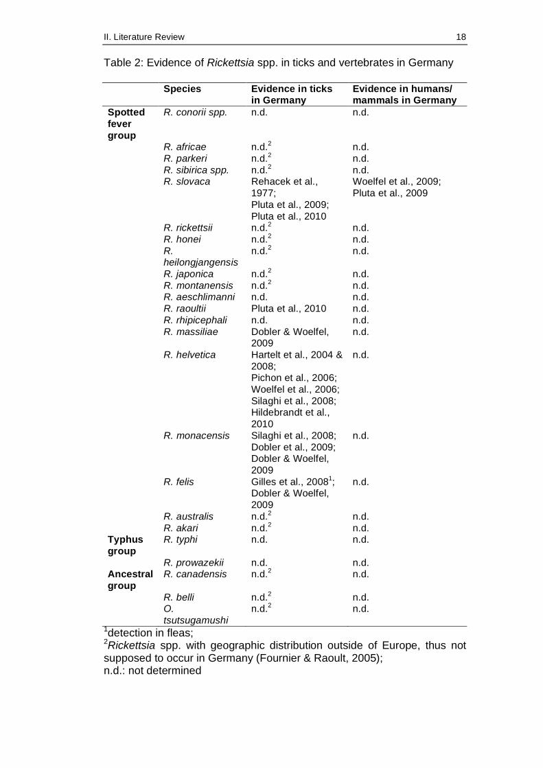
evidence in ticks and vertebrates is summarised in Table 2 (p. 18).
R. rickettsii
The agent of Rocky Mountain spotted fever (RMSF) is associated mainly
with Dermacentor andersoni and D. variabilis but also with Haemaphysalis
spp., Amblyomma spp., Ixodes spp., Rhipicephalus sanguineus and
Otobius lagophilus (McDade & Newhouse, 1986). It occurs widespread
throughout the United States of America, with most cases observed in the
southern and southeastern regions (www.cdc.gov), limited by the
geographic distribution of its arthropod hosts. In Brazil, the R. rickettsii
caused disease is called Brazilian spotted fever, in Mexico fiebre
manchada. Infected human individuals typically but not in every case
present the triad of rash, fever and a history of tick bite. In addition, it can
cause headache, myalgia, nausea, abdominal pain, a skin rash, and – in
more severe cases – neurological problems, acute renal failure or
meningoencephalitis. The mortality rate in humans is on average 1.4%
(Chapman et al., 2006). In dogs, R. rickettsia also causes RMSF
symptoms such as fever, rash, vasculitis and edema, decreased platelet
numbers, joint swelling, myalgia and neorological abnormalities (Warner &
Marsh, 2002; Otis et al., 2004).
R. conorii
Mediterranean spotted fever (MSF) is caused by R. conorii and endemic to
the Mediterranean area, including northern Africa and southern Europe,
with some cases observed also in Kenya, Somalia, South Africa, Turkey,
Bulgaria and Ukraine (Rovery et al., 2008). Zhu et al. (2005) proposed the
creation of the following subspecies because of serotypical, clinical and
geographical differences: R. conorii conorii, R. conorii caspia, R. conorii
israelensis and R. conorii indica. MSF is regarded as an emerging disease
II. Literature Review 16
with case numbers increasing in many countries in the last decade
(reviewed in Rovery et al., 2008). Clinical features are similar to other
spotted fevers, with the eschar sometimes being untypical and furuncle-
like and rarely multiple. The mortality rate in humans can be as high as
32.3% (in Portugal 1997) and makes MSF at least as severe as RMSF
(Rovery et al., 2008).
R. felis
R. felis was first identified in cat fleas (Ctenocephalides felis) by Adams et
al. (1990), later also detected in other flea species and also in ticks and
mites (Ishikura et al. 2003; Choi et al., 2007; Tsui et al., 2007; Oliveira et
al., 2008) and is distributed nearly all over the world. The potential
infection of both insects and acarines makes R. felis unique in the
rickettsial family. Its prevalence in wild-caught arthropods ranges from 0.8
to 100% (depending on geographic location and arthropod species) with
an average percentage of 25% (Reif & Macaluso, 2009). Like other
rickettsial diseases, it can cause various symptoms in humans, such as
fever, rash, headache, myalgia, eschar, visceral and neurological
symptoms, put together as flea-borne spotted fever or cat flea typhus.
Human cases have been reported in 12 countries worldwide, including
Germany (Reif & Macaluso, 2009).
The cat flea is currently the only defined vector and reservoir as
transmission of viable R. felis between mammals and arthropods has not
been shown so far (Reif & Macaluso, 2009).
R. helvetica
R. helvetica was first isolated in 1979 from I. ricinus ticks in Switzerland
(Burgdorfer et al., 1979) and afterwards found in France, Italy, Sweden,
Slovenia, Portugal, Spain, Japan (reviewed in Blanco & Oteo, 2006;
Fournier & Raoult, 2005) and also in Germany (Woelfel et al., 2006;
Dobler & Woelfel, 2009). In 1999, it was implicated in a case of fatal
perimyocarditis in a young human patient in Sweden (Nilsson et al., 1999),
and involvement was proposed in a case of sarcoidosis 2002 (Nilsson et
al., 2002) and in some cases of febrile illness (Fournier et al., 2000;
II. Literature Review 17
Fournier et al., 2004). Infections are present during the hot months and
present with fever, headache, arthralgia and myalgia, but not with a
cutaneous rash (Fournier et al., 2004).
R. slovaca
This SFG rickettsia is transmitted by Dermacentor marginatus ticks with
the wild boar considered as main host (Blanco & Oteo, 2006) and was
found in all European countries were those ticks were screened, including
France, Portugal, Switzerland, former Yugoslavia, Slovakia, Ukraine,
Armenia and Siberia (Sekeyova et al., 1998; Shpyonov et al., 2001). In
Germany, Pluta et al. (2010) detected R. slovaca in five out of 666
Dermacentor ticks collected in Southern Germany and furthermore
describe a clinical case of TIBOLA (Tick-borne lymphadenitis), the disease
caused by R. slovaca in humans, which is also named DEBONEL
(Dermacentor-borne necrosis erythemalymphadenopathy) and which is
characterized by painful lymph nodes, sometimes fever, rarely cutaneous
rash. However, the first case of clinical manifest human infection with R.
slovaca was presented by Woelfel et al. (2009) in a 67-year-old female
following a tick bite. Remarkably, only 50% of patients develop detectable
antibodies, which may be evidence for a rather localized infection
(Fournier & Raoult, 2005).
R. monacensis
A new rickettsia species, R. monacensis, was isolated from I. ricinus from
Munich, Germany (Simser et al., 2002) and found to be the causative
agent of two MSF-like human cases in Spain (Jado et al., 2007), proving
its pathogenic potential for humans for the first time. Dobler et al. (2009)
isolated and characterised two strains in southeastern Germany that grew
interestingly enough at 28° C in cell culture but not at 37° C, which doubts
the potential of growth and pathogenicity in organisms of these two
strains.
II. Literature Review 18
Table 2: Evidence of Rickettsia spp. in ticks and vertebrates in Germany
Species Evidence in ticks in Germany
Evidence in humans/ mammals in Germany
Spotted fever group
R. conorii spp. n.d. n.d.
R. africae n.d.2 n.d. R. parkeri n.d.2 n.d. R. sibirica spp. n.d.2 n.d. R. slovaca Rehacek et al.,
1977; Pluta et al., 2009; Pluta et al., 2010
Woelfel et al., 2009; Pluta et al., 2009
R. rickettsii n.d.2 n.d. R. honei n.d.2 n.d. R.
heilongjangensis n.d.2 n.d.
R. japonica n.d.2 n.d. R. montanensis n.d.2 n.d. R. aeschlimanni n.d. n.d. R. raoultii Pluta et al., 2010 n.d. R. rhipicephali n.d. n.d. R. massiliae Dobler & Woelfel,
2009 n.d.
R. helvetica Hartelt et al., 2004 & 2008; Pichon et al., 2006; Woelfel et al., 2006; Silaghi et al., 2008; Hildebrandt et al., 2010
n.d.
R. monacensis Silaghi et al., 2008; Dobler et al., 2009; Dobler & Woelfel, 2009
n.d.
R. felis Gilles et al., 20081; Dobler & Woelfel, 2009
n.d.
R. australis n.d.2 n.d. R. akari n.d.2 n.d. Typhus group
R. typhi n.d. n.d.
R. prowazekii n.d. n.d. Ancestral group
R. canadensis n.d.2 n.d.
R. belli n.d.2 n.d. O.
tsutsugamushi n.d.2 n.d.
1detection in fleas; 2Rickettsia spp. with geographic distribution outside of Europe, thus not supposed to occur in Germany (Fournier & Raoult, 2005); n.d.: not determined
III. Materials and Methods 19
III. MATERIALS AND METHODS
1 Rodent collection
In the years 2004 and 2005, rodents were collected in October, May and
September at seven locations in the district of Lower Bavaria, including
forests, pasture, blackberry bushes and farmland. Those rodents were
collected for a study investigating Hantaviruses in Lower Bavaria and
sampling areas were chosen according to the occurrence of Hantavirus-
induced Nephropathia epidemica cases. Before trapping, a decoying
method was conducted using plastic cups with apple pieces as bait
located every 5 – 10 m, with 2 – 3x20 bait lines per location (Essbauer et
al., 2006). Trapping (5 – 9 traps/area, 33 traps in total) was performed on
sites with signs of rodent activities.
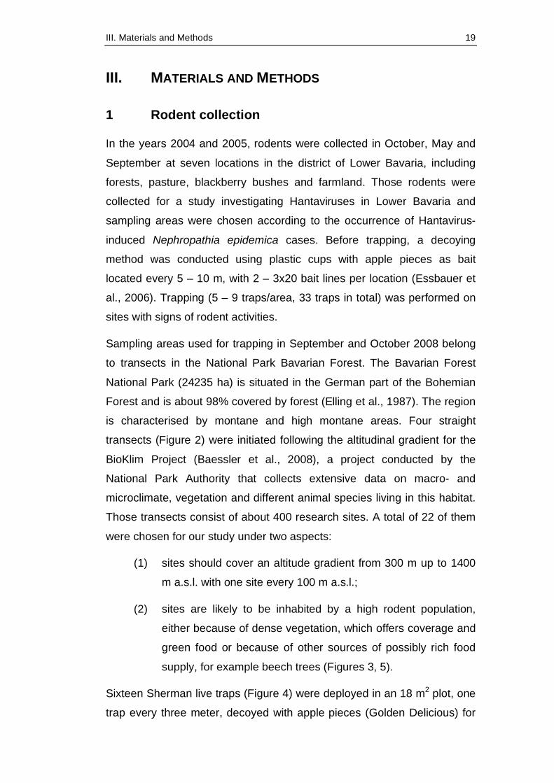
Sampling areas used for trapping in September and October 2008 belong
to transects in the National Park Bavarian Forest. The Bavarian Forest
National Park (24235 ha) is situated in the German part of the Bohemian
Forest and is about 98% covered by forest (Elling et al., 1987). The region
is characterised by montane and high montane areas. Four straight
transects (Figure 2) were initiated following the altitudinal gradient for the
BioKlim Project (Baessler et al., 2008), a project conducted by the
National Park Authority that collects extensive data on macro- and
microclimate, vegetation and different animal species living in this habitat.
Those transects consist of about 400 research sites. A total of 22 of them
were chosen for our study under two aspects:
(1) sites should cover an altitude gradient from 300 m up to 1400
m a.s.l. with one site every 100 m a.s.l.;
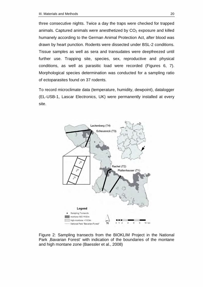
(2) sites are likely to be inhabited by a high rodent population,
either because of dense vegetation, which offers coverage and
green food or because of other sources of possibly rich food
supply, for example beech trees (Figures 3, 5).
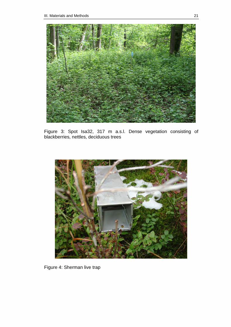
Sixteen Sherman live traps (Figure 4) were deployed in an 18 m2 plot, one
trap every three meter, decoyed with apple pieces (Golden Delicious) for
III. Materials and Methods 20
three consecutive nights. Twice a day the traps were checked for trapped
animals. Captured animals were anesthetized by CO2 exposure and killed
humanely according to the German Animal Protection Act, after blood was
drawn by heart punction. Rodents were dissected under BSL-2 conditions.
Tissue samples as well as sera and transudates were deepfreezed until
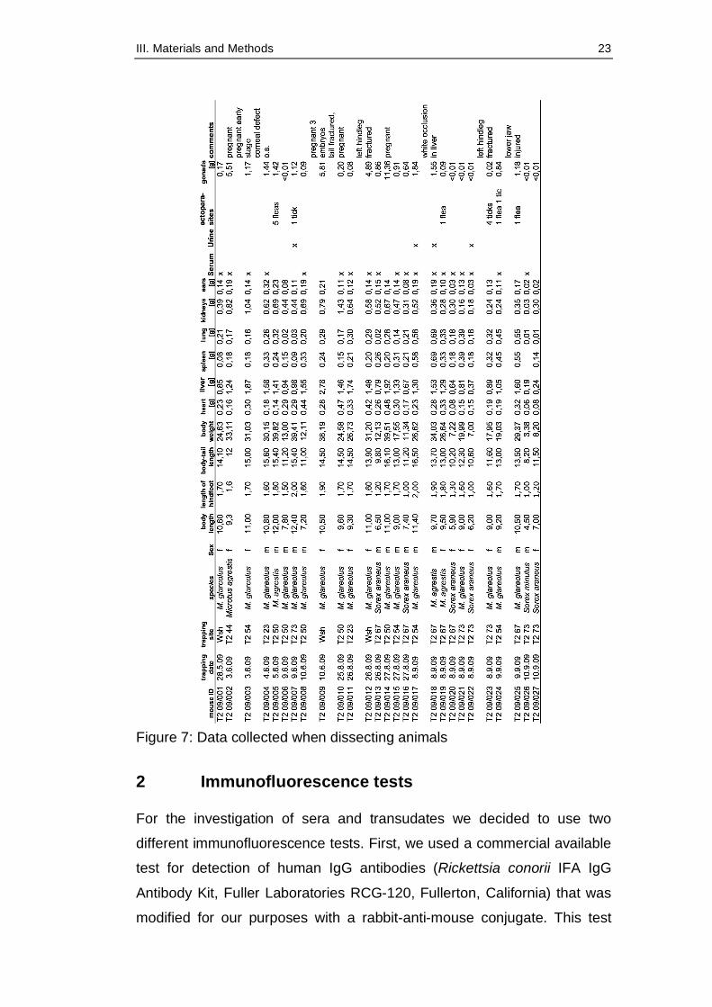
further use. Trapping site, species, sex, reproductive and physical
conditions, as well as parasitic load were recorded (Figures 6, 7).
Morphological species determination was conducted for a sampling ratio
of ectoparasites found on 37 rodents.
To record microclimate data (temperature, humidity, dewpoint), datalogger
(EL-USB-1, Lascar Electronics, UK) were permanently installed at every
site.
Figure 2: Sampling transects from the BIOKLIM Project in the National Park ‚Bavarian Forest’ with indication of the boundaries of the montane and high montane zone (Baessler et al., 2008)
III. Materials and Methods 21
Figure 3: Spot Isa32, 317 m a.s.l. Dense vegetation consisting of blackberries, nettles, deciduous trees
Figure 4: Sherman live trap

III. Materials and Methods 22
Figure 5: Spot Els 8, 578 m a.s.l. Rocky area, vegetation consisting mainly of beech trees
Figure 6: Data collected when trapping animals
Date Site time weather Temp°C trap nr mouse ID species sex age blood UrineEktoparasites
04.08. Isa 32 8:30 rain 15 F 123 DO 09/014 M. glareolus -- --F125 DO 09/015 A. flavicollis m ad 2x -- --F 121 DO 09/016 M. glareolus m juv 1x -- 1 flea
DO 09/017 M. glareolus m juv 1x -- --Igg 35 9:50 cloudy 15 neg -- -- -- -- -- -- --Igg 33 10:40 cloudy 15 neg -- -- -- -- -- -- --Sal 27 11:20 cloudy 17 neg -- -- -- -- -- -- --Els 5 12:40 cloudy 15 F 48 DO 09/018 A. flavicollis -- -- -- --Els 8 12:15 cloudy 15 neg -- -- -- -- -- -- --Isa 32 13:45 cloudy 20 neg -- -- -- -- -- -- --
05.08 Isa 32 8:30 sunny 16 F 124 DO 09/019 M. glareolus m juv 1x -- --sunny F 127 DO 09/020 M. glareolus m juv 1x -- --
Igg 35 9:10 sunny 16 neg -- -- -- -- -- -- --Igg 33 9:45 sunny 19 neg -- -- -- -- -- -- --Sal 27 10:35 sunny 21 neg -- -- -- -- -- -- --Els 5 11:00 sunny 21 F 48 DO 09/021 A. flavicollis m ad 1x -- 2 ticks
F 56 DO 09/022 A. flavicollis m ad 1x x --F 49 DO 09/023 A. flavicollis m ad 1x -- --
DO 09/024 A. flavicollis ad 1x -- --Els 8 11:45 sunny 22 neg -- -- -- -- -- -- --Isa 32 14:00 sunny 24 neg -- -- -- -- -- -- --
06.08. Isa 32 8:20 sunny 19 F121 DO 09/025 A. flavicollis f ad 1x -- --Igg 35 9:00 sunny 20 neg -- -- -- -- -- -- --Igg 33 9:50 sunny 22 F 59 DO 09/026 A. flavicollis m ad 1x -- --
sunny A25 DO 09/027 A. flavicollis f ad 1x -- --Sal 27 10:50 sunny 24 neg -- -- -- -- -- -- --Els 5 12:05 sunny 22 DO 09/028 A. flavicollis
sunny Y24 DO 09/029 A. flavicollisF 52 DO 09/030 A. flavicollis
Els 8 11:30 sunny 22 neg -- -- -- -- -- -- --
III. Materials and Methods 23
Figure 7: Data collected when dissecting animals
2 Immunofluorescence tests
For the investigation of sera and transudates we decided to use two
different immunofluorescence tests. First, we used a commercial available
test for detection of human IgG antibodies (Rickettsia conorii IFA IgG
Antibody Kit, Fuller Laboratories RCG-120, Fullerton, California) that was
modified for our purposes with a rabbit-anti-mouse conjugate. This test
III. Materials and Methods 24
was supposed to detect antibodies against R. conorii, a species that is not
presumed to occur in Germany, but as it is a member of the spotted fever
group we expected to detect antibodies against other SFG rickettsiae with
this test. Additionally, we used an in-house test for the detection of
antibodies against R. helvetica, also a SFG rickettsia, which is
phylogenetically distinct from R. conorii and thus was supposed to detect
those SFG rickettsial antibodies distinct from R. conorii. More details on
the manufacture and conduction of these tests can be found in the
publication (IV.5, p. 41).
3 DNA extraction
As the ear seemed to be the tissue of choice for detection of Borrelia DNA
(Peavy et al., 1997; Kiessling, 2005), we decided to use this tissue for
detection of rickettsia.
QIAamp DNA Mini Kit (Qiagen, Hilden Germany) was used for isolation of
DNA from ear tissue samples. One ear per mouse was placed into a 1.5
ml Eppendorf tube, 180 µl ATL buffer and 20 µl proteinase K were added
and the mixture was placed in a Thermomixer (Eppendorf, Germany) for
about four hours, until the ear material was totally digested. The digesting
process then was inhibited by putting the Eppendorf tubes into a 70° C
water bath. After centrifugation the digested solution was put into a
column, carefully leaving fur in the Eppendorf tube. The extraction process
according to manufacturer’s instruction followed. Nucleic acids from liver
material were extracted using the QIAamp Viral RNA Mini Kit (Qiagen)
following the manufacturer’s instructions. The elution volume was 100 µl.
A tube not containing tissue material was added to every extraction
process as quality control to ensure that no contamination had occurred
during the extraction process.
III. Materials and Methods 25
4 Polymerase chain reaction for detection of
Rickettsial DNA
4.1 Real-time PCR for detection of the gltA gene of Rickettsia spp.
For initial screening of rodent ear and liver tissue for rickettsial DNA a
PanRickettsia real-time PCR established by Woelfel et al. (2008) was
chosen. Woelfel et al. designed one set of primers and one TaqMan probe
(Table 3) to amplify a 70-bp genome region of the citrate synthase (gltA)
gene on the basis of all rickettsial gltA sequences available in GenBank.
The following bacterial species were used for validation of the real-time
PCR assay: Rickettsia helvetica, R. honei, R. rickettsii, R. typhi, R. africae,
R. conorii, R. mongolotimonae, R. IRS4, and R. felis (Woelfel et al., 2008).
A performance a little less successful is achieved with specimens of R.
typhi (Woelfel, personal communication).
The reaction was carried out in a LightCycler 1.5 (Roche, Mannheim,
Germany). Tables 4 and 5 show the reaction and cycling conditions used.
Negative controls (DNA free extraction quality controls or sterile water)
were always included.
All primers were ordered from TIB MOLBIOL, Berlin, Germany.
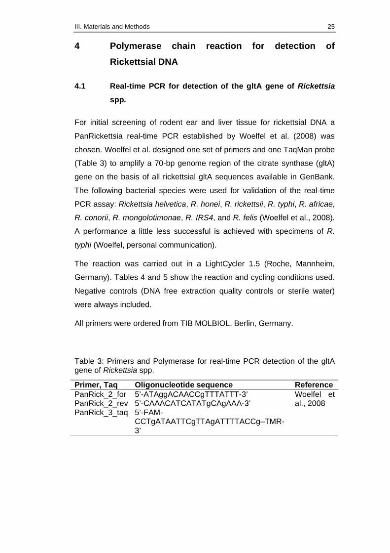
Table 3: Primers and Polymerase for real-time PCR detection of the gltA gene of Rickettsia spp.
Primer, Taq Oligonucleotide sequence Reference PanRick_2_for 5’-ATAggACAACCgTTTATTT-3’ PanRick_2_rev 5’-CAAACATCATATgCAgAAA-3’ PanRick_3_taq 5’-FAM-
CCTgATAATTCgTTAgATTTTACCg–TMR-3’
Woelfel et al., 2008
III. Materials and Methods 26
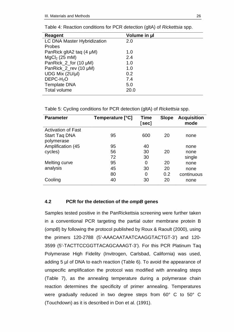
Table 4: Reaction conditions for PCR detection (gltA) of Rickettsia spp.
Reagent Volume in µl LC DNA Master Hybridization Probes
2.0
PanRick gltA2 taq (4 µM) 1.0 MgCl2 (25 mM) 2.4 PanRick_2_for (10 µM) 1.0 PanRick_2_rev (10 µM) 1.0 UDG Mix (2U/µl) 0.2 DEPC-H2O 7.4 Template DNA 5.0 Total volume 20.0
Table 5: Cycling conditions for PCR detection (gltA) of Rickettsia spp.
Parameter Temperature [°C] Time [sec]
Slope Acquisition mode
Activation of Fast Start Taq DNA polymerase
95 600 20 none
95 40 none 56 30 none
Amplification (45 cycles)
72 30 20
single 95 0 20 none 45 30 20 none
Melting curve analysis
80 0 0.2 continuous Cooling 40 30 20 none
4.2 PCR for the detection of the ompB genes
Samples tested positive in the PanRickettsia screening were further taken
in a conventional PCR targeting the partial outer membrane protein B
(ompB) by following the protocol published by Roux & Raoult (2000), using
the primers 120-2788 (5’-AAACAATAATCAAGGTACTGT-3’) and 120-
3599 (5’-TACTTCCGGTTACAGCAAAGT-3’). For this PCR Platinum Taq
Polymerase High Fidelity (Invitrogen, Carlsbad, California) was used,
adding 5 µl of DNA to each reaction (Table 6). To avoid the appearance of
unspecific amplification the protocol was modified with annealing steps
(Table 7), as the annealing temperature during a polymerase chain
reaction determines the specificity of primer annealing. Temperatures
were gradually reduced in two degree steps from 60° C to 50° C
(Touchdown) as it is described in Don et al. (1991).
III. Materials and Methods 27
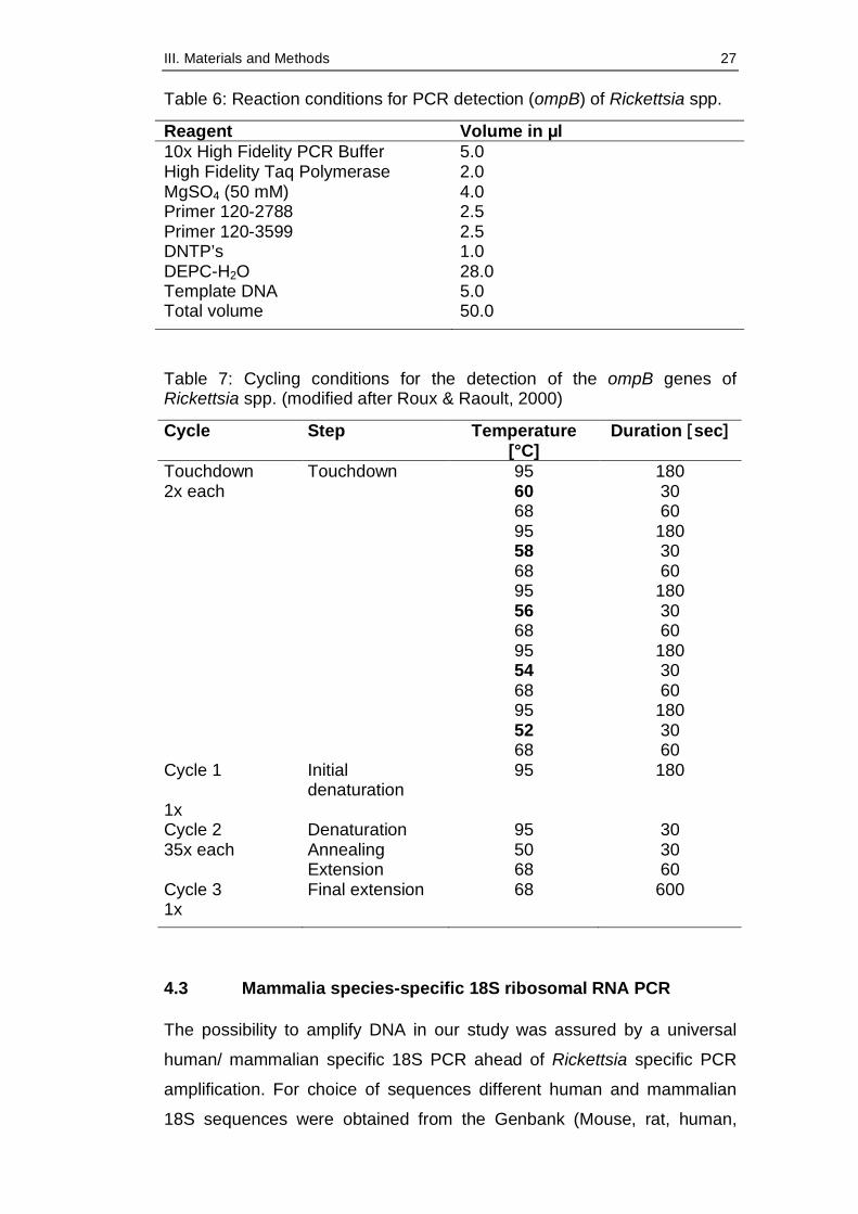
Table 6: Reaction conditions for PCR detection (ompB) of Rickettsia spp.
Reagent Volume in µl 10x High Fidelity PCR Buffer 5.0 High Fidelity Taq Polymerase 2.0 MgSO4 (50 mM) 4.0 Primer 120-2788 2.5 Primer 120-3599 2.5 DNTP’s 1.0 DEPC-H2O 28.0 Template DNA 5.0 Total volume 50.0
Table 7: Cycling conditions for the detection of the ompB genes of Rickettsia spp. (modified after Roux & Raoult, 2000)
Cycle Step Temperature [°C]
Duration [sec]
Touchdown Touchdown 95 180 2x each 60 30 68 60 95 180 58 30 68 60 95 180 56 30 68 60 95 180 54 30 68 60 95 180 52 30 68 60 Cycle 1 Initial
denaturation 95 180
1x Cycle 2 Denaturation 95 30 35x each Annealing 50 30 Extension 68 60 Cycle 3 Final extension 68 600 1x
4.3 Mammalia species-specific 18S ribosomal RNA PCR
The possibility to amplify DNA in our study was assured by a universal
human/ mammalian specific 18S PCR ahead of Rickettsia specific PCR
amplification. For choice of sequences different human and mammalian
18S sequences were obtained from the Genbank (Mouse, rat, human,
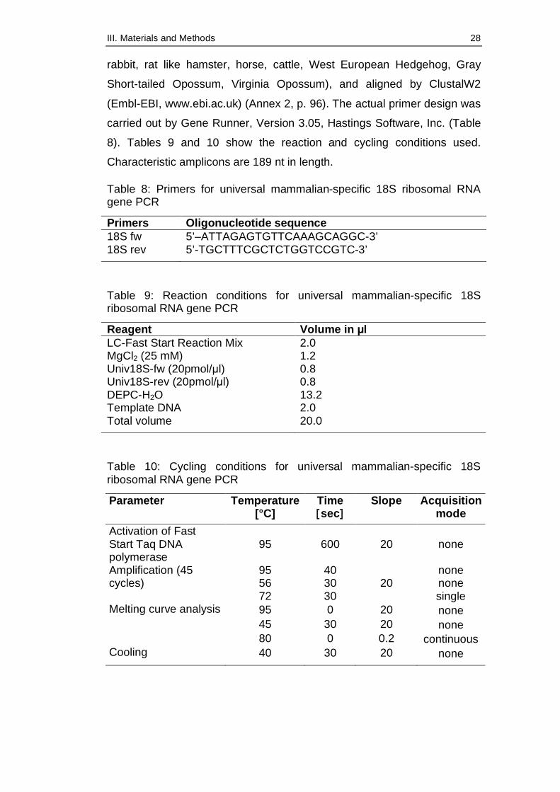
III. Materials and Methods 28
rabbit, rat like hamster, horse, cattle, West European Hedgehog, Gray
Short-tailed Opossum, Virginia Opossum), and aligned by ClustalW2
(Embl-EBI, www.ebi.ac.uk) (Annex 2, p. 96). The actual primer design was
carried out by Gene Runner, Version 3.05, Hastings Software, Inc. (Table
8). Tables 9 and 10 show the reaction and cycling conditions used.
Characteristic amplicons are 189 nt in length.
Table 8: Primers for universal mammalian-specific 18S ribosomal RNA gene PCR
Primers Oligonucleotide sequence 18S fw 5’–ATTAGAGTGTTCAAAGCAGGC-3’ 18S rev 5’-TGCTTTCGCTCTGGTCCGTC-3’
Table 9: Reaction conditions for universal mammalian-specific 18S ribosomal RNA gene PCR
Reagent Volume in µl LC-Fast Start Reaction Mix 2.0 MgCl2 (25 mM) 1.2 Univ18S-fw (20pmol/µl) 0.8 Univ18S-rev (20pmol/µl) 0.8 DEPC-H2O 13.2 Template DNA 2.0 Total volume 20.0
Table 10: Cycling conditions for universal mammalian-specific 18S ribosomal RNA gene PCR
Parameter Temperature [°C]
Time [sec]
Slope Acquisition mode
Activation of Fast Start Taq DNA polymerase
95 600 20 none
95 40 none 56 30 none
Amplification (45 cycles)
72 30 20
single 95 0 20 none 45 30 20 none
Melting curve analysis
80 0 0.2 continuous Cooling 40 30 20 none
III. Materials and Methods 29
5 Agarose gel electrophoresis
Conventional PCR products were visualised under UV light after 1.5%
agarose gel electrophoresis (1.5 g agarose/100 ml TAE Buffer) and
subsequent staining with GelRed™ solution (Biotium Inc., Hayward,
Canada), a fluorescent nucleic acid dye that is less toxic than ethidium
bromide (Biotium Safety Report, 2008). For comparison a standardised
DNA-Ladder was added to each electrophoresis.
6 DNA purification
Amplificates were purified using the QIAquick Gel Extraction Kit (Qiagen,
Hilden Germany) according to manufacturer’s instruction.
7 Sequencing and sequence analysis
After purification, all rickettsial PCR products were sent off for sequencing
to the GATC sequencing service (Konstanz, Germany) using
corresponding specific primers. After evaluating the specificity of results
with Chromas©Lite (www.technelysium.com.au/chromas_lite.html),
sequence and phylogenetic analyses were performed using BioEdit
(Version 7.0.0, Copyright ©1997-2004, Hall 1999). Sequence similarity
searches were made, without the flanking primers, by BLAST analysis
(www.ncbi.nlm.nih.gov.library.vu.edu.au/BLAST/). The obtained
sequences were further analysed with each other and with GenBank
sequences by multiple alignments (www.ebi.ac.uk/clustalw/index.html).
The obtained sequences were submitted to Genbank. A phylogenetic tree
(Figure 13, p. 36) was constructed using Bioedit, Neighbour Joining
method, with R. prowazekii as outgroup, 1000 bootstraps, based on 770 nt
fragment of ompB.
8 Statistical analysis
Data of trapped rodents were collected with Microsoft Excel® and
evaluated with R 2.9.2 (R Core Development Team 2009). To come to an
decision which parameters to chose for statistical analysis with R 2.9.2,
preliminary tests for significance of several parameters such as species,
sex or age were conducted using SPSS® version 12.0.1, SPSS Inc.,

III. Materials and Methods 30
Chicago, IL, USA, using chi-square test, student t-test including Levene’s
test for equal variance, Mann-Whitney test and Spearman rank correlation.
Serological assays were compared using concordance comparison and
kappa-statistics. Statistical tests were performed for ompB PCR-positive
samples for body size (head-body length), body weight and organ weights.
Values of p<0.05 were regarded as significant. For the other variables we
restrained from a formal test and provided only descriptive statistic. In our
final test model with rickettsia-PCR positivity as dependent variable we
included in the following sequence the predictors
1) as control variable the species,
2) occurrence of ectoparasites,
3) body size and
4) elevation.
We applied a sequential analysis of deviance table (Hastie & Pregibon,
1992). That is, the reductions in the residual deviance as each term of the
formula added in turn are given in as the rows of a table, plus the residual
deviances themselves. We first added the species to the model and then
subsequently ectoparasites, body size and elevation. Due to the binomial
dispersion of our dependent variable “Rickettsia-PCR positive” we applied
the chi-square test.
IV. Results 31
IV. RESULTS
1 Trapping results
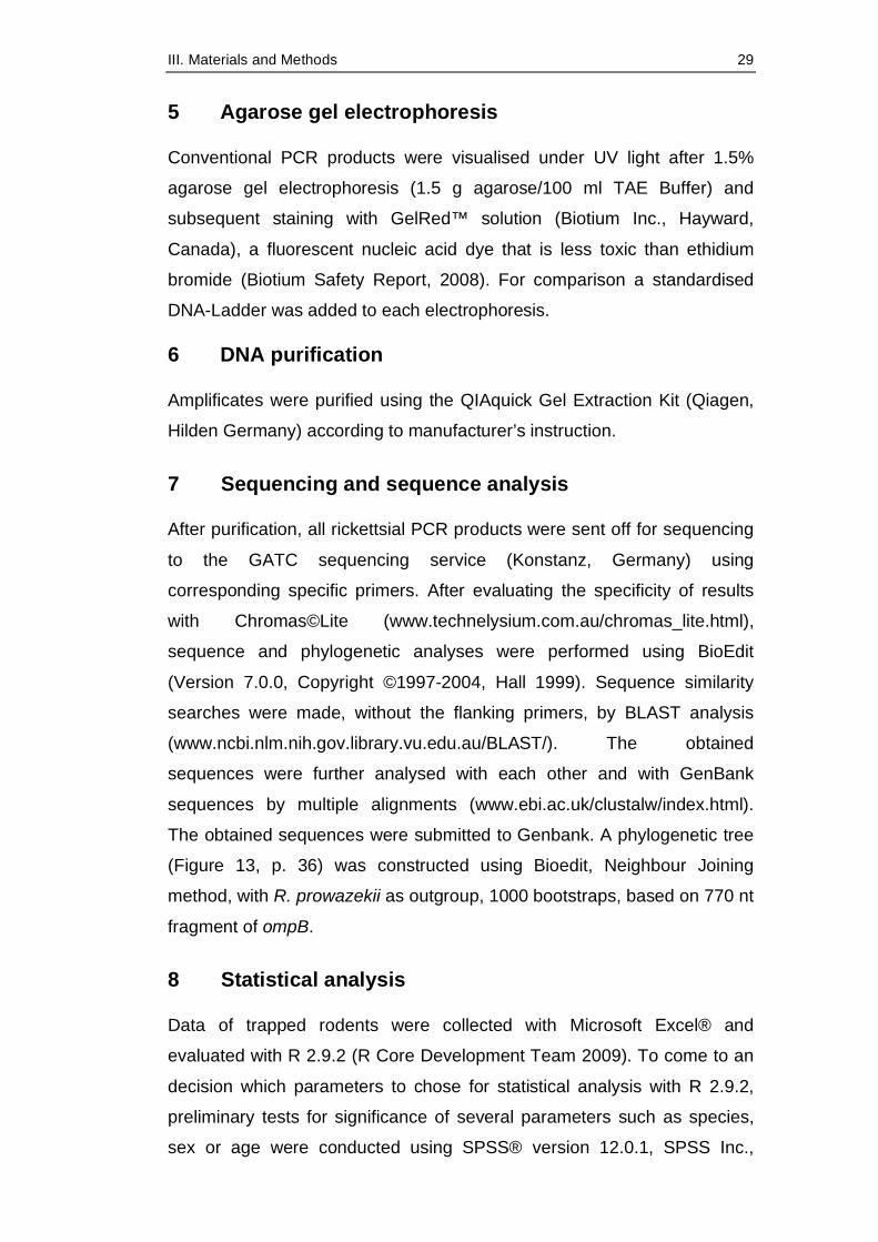
Trapping of small mammals with Sherman live traps was continued in the
years 2009 and 2010 and resulted in a total of 690 trapped individuals,
presented in Table 11. Screenings of animals for Rickettsia collected in
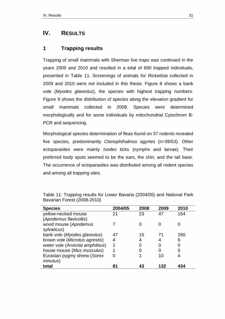
2009 and 2010 were not included in this thesis. Figure 8 shows a bank
vole (Myodes glareolus), the species with highest trapping numbers.
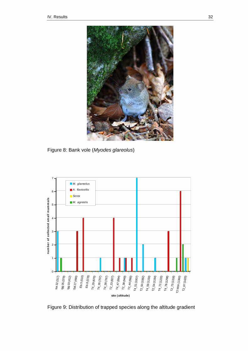
Figure 9 shows the distribution of species along the elevation gradient for
small mammals collected in 2008. Species were determined
morphologically and for some individuals by mitochondrial Cytochrom B-
PCR and sequencing.
Morphological species determination of fleas found on 37 rodents revealed
five species, predominantly Ctenophthalmus agyrtes (n=39/53). Other
ectoparasites were mainly Ixodes ticks (nymphs and larvae). Their
preferred body spots seemed to be the ears, the chin, and the tail base.
The occurrence of ectoparasites was distributed among all rodent species
and among all trapping sites.
Table 11: Trapping results for Lower Bavaria (2004/05) and National Park Bavarian Forest (2008-2010)
Species 2004/05 2008 2009 2010 yellow-necked mouse (Apodemus flavicollis)
21 23 47 164
wood mouse (Apodemus sylvaticus)
7 0 0 0
bank vole (Myodes glareolus) 47 15 71 260 brown vole (Microtus agrestis) 4 4 4 6 water vole (Arvicola amphibius) 1 0 0 0 house mouse (Mus musculus) 1 0 0 0 Eurasian pygmy shrew (Sorex minutus)
0 1 10 4
total 81 43 132 434
IV. Results 32
0
1
2
3
4
5
6
7
nu
mb
er
of
collect
ed
sm
all m
am
mals
Isa
32 (3
17)
Igg
35 (3
79)
Igg
33 (4
12)
Sal 2
7 (4
90)
Els
5 (5
10)
Els
8 (5
78)
T4_2
9 (6
70)
T4_3
5 (7
07)
T4_3
9 (7
67)
T2_2
3 (8
27)
T4_4
7 (8
94)
T2_3
8 (9
49)
T2_4
4 (9
90)
T4_5
1 (1
007)
T2_5
0 (1
082)
T4_5
9 (1
150)
T2_5
4 (1
184)
T4_7
2 (1
220)
T4_7
8 (1
298)
T2_7
3 (1
318)
T2 W
sh (1
360)
T2_6
7 (1
420)
site (altitude)
M. glareolus
A. flavicollis
Sorex
M. agrestis
Figure 8: Bank vole (Myodes glareolus)
Figure 9: Distribution of trapped species along the altitude gradient
IV. Results 33
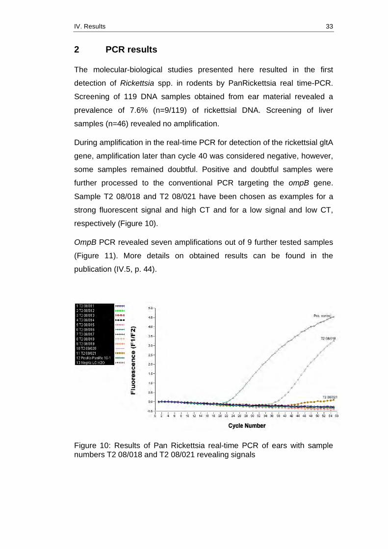
2 PCR results
The molecular-biological studies presented here resulted in the first
detection of Rickettsia spp. in rodents by PanRickettsia real time-PCR.
Screening of 119 DNA samples obtained from ear material revealed a
prevalence of 7.6% (n=9/119) of rickettsial DNA. Screening of liver
samples (n=46) revealed no amplification.
During amplification in the real-time PCR for detection of the rickettsial gltA
gene, amplification later than cycle 40 was considered negative, however,
some samples remained doubtful. Positive and doubtful samples were
further processed to the conventional PCR targeting the ompB gene.
Sample T2 08/018 and T2 08/021 have been chosen as examples for a
strong fluorescent signal and high CT and for a low signal and low CT,
respectively (Figure 10).
OmpB PCR revealed seven amplifications out of 9 further tested samples
(Figure 11). More details on obtained results can be found in the
publication (IV.5, p. 44).
Figure 10: Results of Pan Rickettsia real-time PCR of ears with sample numbers T2 08/018 and T2 08/021 revealing signals

IV. Results 34
Figure 11: OmpB PCR products, showing amplification fragments of approx. 800 bp in length
3 IFT results
Serological investigations of rodent blood and transudates included two
IFATs for detection of R. conorii and R. helvetica antibodies and revealed
an overall seroreactivity of 28.1% (n=32/114). Results of IF-testing are
presented in detail in the publication (IV.5, p. 45). Positive reactions
appear as Rickettsial bodies exhibiting bright apple-green cytoplasmic
fluorescence against a background of orange to red yolk sac matrix
(Figure 12).
Figure 12: Images of IFTs. Left picture: negative sample with red colored Vero-76 cell nuclei stained with Evans blue. Right picture: positive sample with bright green specific fluorescence (dots in cytoplasma and between cells).
IV. Results 35
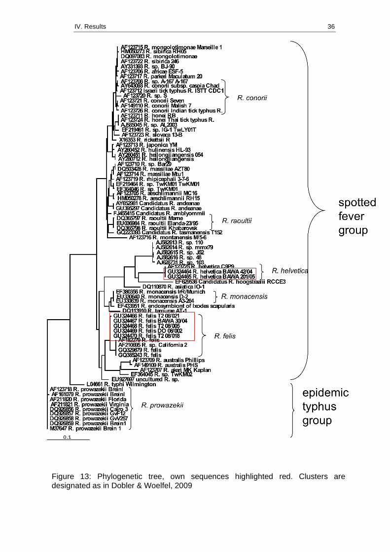
4 Sequence analysis
Successfully amplified ompB PCR amplificates were processed to
sequencing. Sequence analyses resulted in five R. felis and two R.
helvetica ompB nucleotide sequences that were 100% identical among
each other and to the respective type species deposited in GenBank.
Sequences were submitted to Genbank, with accession numbers
GU324464-GU324470. Sequence alignments are presented in Annex 2
(p. 96). A phylogenetic tree shows clusters and relationships between own
nucleotide sequences and those deposited in GenBank (Figure 13).
IV. Results 36
Figure 13: Phylogenetic tree, own sequences highlighted red. Clusters are designated as in Dobler & Woelfel, 2009
IV. Results 37
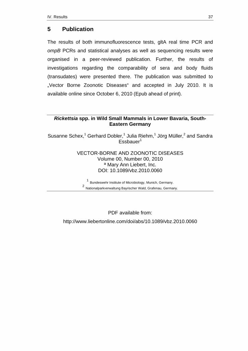
5 Publication
The results of both immunofluorescence tests, gltA real time PCR and
ompB PCRs and statistical analyses as well as sequencing results were
organised in a peer-reviewed publication. Further, the results of
investigations regarding the comparability of sera and body fluids
(transudates) were presented there. The publication was submitted to
„Vector Borne Zoonotic Diseases“ and accepted in July 2010. It is
available online since October 6, 2010 (Epub ahead of print).
Rickettsia spp. in Wild Small Mammals in Lower Bavaria, South-Eastern Germany
Susanne Schex,1 Gerhard Dobler,1 Julia Riehm,1 Jörg Müller,2 and Sandra
Essbauer1
VECTOR-BORNE AND ZOONOTIC DISEASES Volume 00, Number 00, 2010
ª Mary Ann Liebert, Inc. DOI: 10.1089/vbz.2010.0060
1 Bundeswehr Institute of Microbiology, Munich, Germany.
2 Nationalparkverwaltung Bayrischer Wald, Grafenau, Germany.
PDF available from:
http://www.liebertonline.com/doi/abs/10.1089/vbz.2010.0060
IV. Results 38
Rickettsia spp. in Wild Small Mammals in Lower Bavaria, South-Eastern Germany
Susanne Schex,1 Gerhard Dobler,1 Julia Riehm,1 Jörg Müller,2 and Sandra
Essbauer1 1 Bundeswehr Institute of Microbiology, Munich, Germany.
2 Nationalparkverwaltung Bayrischer Wald, Grafenau, Germany.
Abstract
So far, data on the natural cycle of rickettsiae of the tick-borne spotted
fever group (SFG) in Central Europe are barely available. Some studies
showed the occurrence of different Rickettsia species in their arthropod
vectors, but it is unclear which animals might have any kind of reservoir
function. This survey was therefore set up to provide information on the
occurrence of SFG rickettsiae in small mammals in Germany. A total of
124 rodents and insectivores were collected over a period of 3 years in
Lower Bavaria, South-Eastern Germany. Screening for Rickettsia
antibodies was performed using immunofluorescence with Rickettsia
conorii and R. helvetica slides, and the comparability of sera and body
fluids (transudates) was investigated in these assays. Further, real-time
polymerase chain reaction (PCR) was used for screening of Rickettsial
DNA in rodents and insectivores. Ear versus liver tissue was compared to
evaluate the more suitable tissue for detection of specific DNA. Further, a
new PCR targeting the 18S ribosomal nucleic acid was established as
internal control. The results indicated that transudates are a sufficient
alternative to proof infection in cases where no sera are available.
Rickettsial DNA, that is, Rickettsia felis and R. helvetica, was found in
seven animals with the ears proving to be a proper choice for PCR.
Statistical analyses revealed that the presence of ectoparasites and the
body size positively correlated with the occurrence of rickettsial DNA.
Overall, our study suggests that rodents and other small mammals may
act as reservoir hosts for Rickettsia. However, with the course of infection
and its transmission in wild animals still unknown, further investigations
IV. Results 39
are needed to better understand the natural cycle of SFG rickettsiae.
Key Words: Ear—ompB—Reservoir—Rickettsia—Rodent—Serology.
Introduction
Rickettsiae are recognized as emerging infections in several parts of the
world (Parola et al. 2005). The obligate intracellular bacteria are
transmitted to humans and animals by blood-sucking arthropods such as
fleas, ticks, and mites. The geographical distribution of Rickettsia species
depends on the occurrence of their specific transmitting arthropod vector.
In Europe, several Rickettsia species are endemic, such as R. typhi, R.
conorii, R. felis, R. helvetica, R. massiliae, R. sibirica, R. monacensis and
R. slovaca (Parola and Raoult 2001, Parola et al. 2005, Dobler and Wölfel
2009, Dobler et al. 2009). In Germany, however, the occurrence and
geographical extension of rickettsial organisms is not known very well. In
Southern Germany several studies found a widespread distribution of R.
helvetica in Ixodes ricinus ticks (Pichon et al. 2006, Wölfel et al. 2006,
Silaghi et al. 2008, Dobler et al. 2009). Investigation of more than 3500
ticks in Bavaria resulted in the detection of five Rickettsia species: R.
helvetica (prevalence 4.8%), R. felis (0.4%), R. monacensis (0.6%), R. sp.
RpA4 (21%), and R. massiliae (1.7%) (Wölfel et al. 2006, Dobler and
Wölfel 2009). All of these studies investigated the presence of Rickettsia
spp. in their arthropod vectors, but only few data are available on the
natural cycle of rickettsiae of the tick-borne spotted fever group (SFG) in
Central Europe. To sustain a successful life cycle it is likely that wild
animals act as natural reservoirs for Rickettsia. Up to now this
fundamental part of life cycle of Rickettsia spp. is quite unclear, as—
according to the literature—there are only very few studies focusing on
rickettsiae in rodents and other small mammals in Central Europe to this
date (Adjemian et al. 2008, Barandika et al. 2007, Spitalská et al. 2008).
We report a screening of free-ranging small mammals for the presence of
Rickettsia by serological and molecular biological techniques. To the best
of our knowledge this is the first report of Rickettsia found in small
mammals in Germany. In specific, this study had two major aims. First, we
aimed to test the detectability of Rickettsia DNA in liver versus ears. In a
previous study in South Germany we found that rodent ears seem to be an
IV. Results 40
adequate tissue to detect multiple Borrelia species in rodents (Essbauer
and Kiessling, unpublished data) and tried these for the first time for
Rickettsia screening. Second, we aimed to analyze several predictors for a
positive detection of Rickettsia DNA in wild rodents. Due to the character
as major vectors we hypothesized (1) a positive influence of ectoparasites
on a positive detection of Rickettsia. Further, we hypothesized (2) that
body size has a positive influence, because as higher the age (body size),
the longer the susceptibility of an animal. Finally, we also hypothesized (3)
that elevation of trapping site has a negative effect, because the activity
and density of the major vector ticks might decrease with temperature
along the elevation gradient.
Materials and Methods
Trapping of small mammals
Rodents were collected using Sherman live traps in October 2004 and
May and September 2005 at seven locations in the district of Lower
Bavaria, including forests, pasture, black-berry bushes and farmland (Fig.
1). Sampling areas were chosen according to the occurrence of
Hantavirus-induced Nephropathia epidemica cases and trapped by a
decoying method as described in Essbauer et al. (2006) and Mertens et al.
(2009). In September and October 2008, trapping was performed at a total
of 22 sites ranging from 300 up to 1400 m altitude (a.s.l.) in the German
part of the Bohemian Forest. Sixteen Sherman live traps were deployed in
an 18 m2 plot, one trap every 3 m for three consecutive nights, and were
checked twice a day. Animals were anesthetized by CO2 exposure and
killed humanely according to the German Animal Protection Act, after
blood was drawn by heart puncture. Trapping site, species, sex,
reproductive and physical conditions, including parasitic load, location of
ectoparasites and also ectoparasite species, were recorded for each
rodent.
Preparation of tissue samples
Rodents were dissected under BSL-2 conditions. Tissue samples (heart,
lung, liver, spleen, kidneys, and ears) were aseptically removed. Extracted
IV. Results 41
viscera were put into Lysing matrix tubes A (MP Biomedicals), mixed with
MEM supplemented with 3% fetal calf serum (Biochrom AG), and
homogenized with the Fast Prep 120 instrument (Biogene) with a power of
5.0 m/s for 30 s.
Serological investigations
Rodent blood was centrifuged at 285 g for 10 min at 4°C. The derived sera
were stored at -40°C until use. Sera were preliminary diluted 1:20 in
phosphate-buffered saline (PBS), as this dilution proved to be the most
suitable with regard to specificity and sensitivity for screenings in
preliminary tests. As there were blood samples missing from several mice
that could not be captured alive, the heart of each animal was rinsed with
1 mL PBS, with the resulting suspension (referred to as transudate) tested
additionally or instead where no serum was available.
Sera and transudates of wild mice were screened with an in-house
immunofluorescence test for detection of immunoglobulin G (IgG)
antibodies against R. helvetica. R. helvetica (strain AS 819, own
unpublished strain) was propagated on Vero E6 cell lines for 27 days at
34°C. Infected cells were trypsinated and centrifuged at 2000 g for 5 min.
The pellet was resuspended in PBS and 10 µL each spotted on 10-well
antigen slides (Biomérieux). After air-drying, cells were fixed with a 50%
methanol/50% acetone (1:1) solution. Further, R. conorii, which is
phylogenetically distinct from R. helvetica in the SFG, was used for
immunofluorescence tests. It is also crossreactive with Rickettsia from the
SFG but not endemic in Germany. Slides coated with R. conorii–infected
cells were obtained from a commercial supplier (Rickettsia conorii
immunofluorescence assay [IFA] IgG Antibody Kit; Fuller Laboratories
RCG-120).
Both Rickettsia IFA were conducted with a polyclonal rabbit anti-mouse
serum IgG/fluorescein isothiocyanate as a conjugate (dilution 1:20; Dako)
together with Evans blue counterstaining. As positive/negative control
served the controls provided by the commercial available kit as well as
formerly positive tested mouse sera and PBS, respectively, were used.
Slides were read by two independent examiners using a fluorescent
IV. Results 42
microscope.
Nucleic acid isolation
Nucleic acids (NA) were extracted using the QIAamp Viral RNA Mini Kit
(Qiagen) following the manufacturer’s instructions. This test was used
because of investigations regarding RNA–viruses conducted in parallel.
Moreover, it is comparable with other standard DNA extraction kits
regarding the quality of the extracted NA.
For extraction of rickettsial DNA from ears, one ear per mouse in the
aggregate was placed into a 1.5 mL Eppendorf tube and digested using
the QIAamp DNA Mini Kit (Qiagen) as described in the instruction manual.
If not analyzed immediately, the extracted NA were stored at -20°C.
Polymerase chain reactions
Rickettsial polymerase chain reactions.
A real-time (RT) polymerase chain reaction (PCR) targeting citrate
synthase (gltA) was performed using the LightCycler FastStart DNA
Master HybProbe System (Roche) for LightCycler 1.5 following the
protocol published by Wölfel et al. (2008). An uracil–DNA–glycosylase
(UDG) incubation step was added to prevent the re-amplification of
carryover PCR products between reactions. Briefly, 0.5 U UDG Mix
(Roche) was added to each reaction, and the protocol was modified
including a preincubation step for UDG digest at 40°C for 10 min.
A conventional PCR was performed, targeting the partial outer membrane
protein B (ompB) by following the protocol published by Roux and Raoult
(2000). The protocol was then modified with annealing steps.
Temperatures were gradually reduced in two-degree steps from 60°C to
50°C (touchdown). For this, Platinum Taq Polymerase High Fidelity
(Invitrogen) was used, adding 5 µL of DNA to each reaction.
Mammalia species-specific PCRs.
To investigate if there was an inhibition of the PCR (internal control), we
established a universal mammalian-specific 18S ribosomal RNA gene
PCR ahead of Rickettsia-specific PCR amplification. For choice of
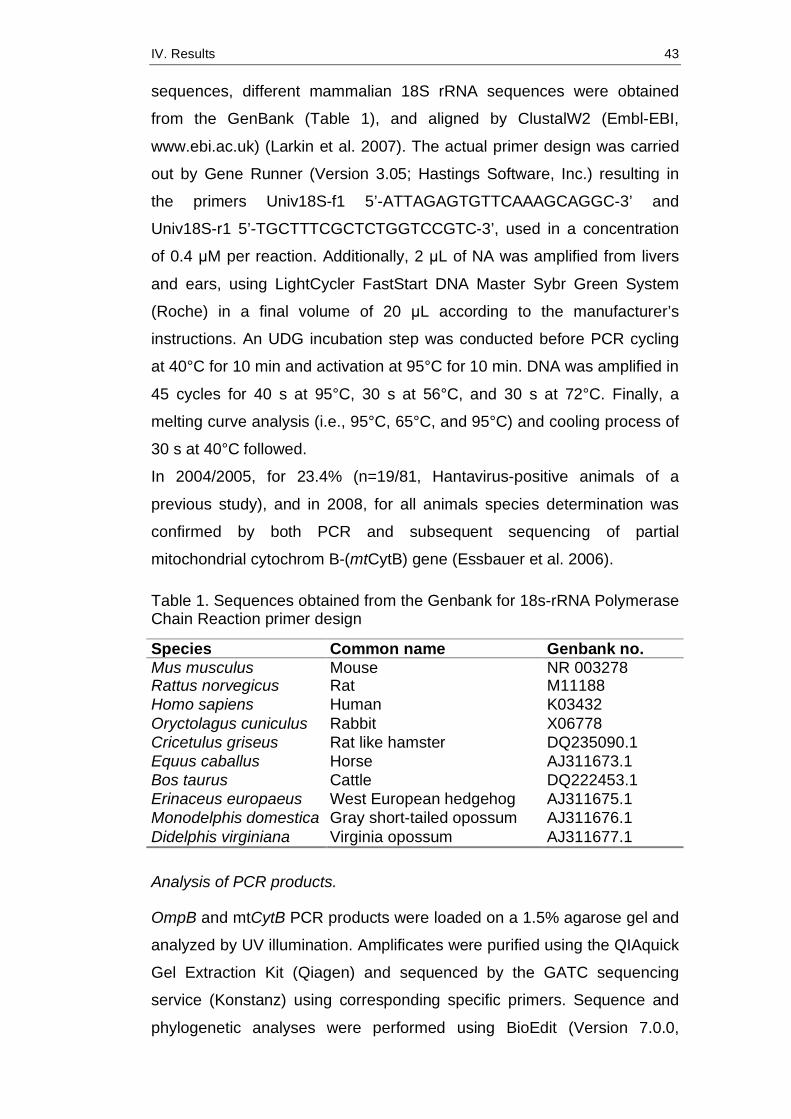
IV. Results 43
sequences, different mammalian 18S rRNA sequences were obtained
from the GenBank (Table 1), and aligned by ClustalW2 (Embl-EBI,
www.ebi.ac.uk) (Larkin et al. 2007). The actual primer design was carried
out by Gene Runner (Version 3.05; Hastings Software, Inc.) resulting in
the primers Univ18S-f1 5’-ATTAGAGTGTTCAAAGCAGGC-3’ and
Univ18S-r1 5’-TGCTTTCGCTCTGGTCCGTC-3’, used in a concentration
of 0.4 µM per reaction. Additionally, 2 µL of NA was amplified from livers
and ears, using LightCycler FastStart DNA Master Sybr Green System
(Roche) in a final volume of 20 µL according to the manufacturer’s
instructions. An UDG incubation step was conducted before PCR cycling
at 40°C for 10 min and activation at 95°C for 10 min. DNA was amplified in
45 cycles for 40 s at 95°C, 30 s at 56°C, and 30 s at 72°C. Finally, a
melting curve analysis (i.e., 95°C, 65°C, and 95°C) and cooling process of
30 s at 40°C followed.
In 2004/2005, for 23.4% (n=19/81, Hantavirus-positive animals of a
previous study), and in 2008, for all animals species determination was
confirmed by both PCR and subsequent sequencing of partial
mitochondrial cytochrom B-(mtCytB) gene (Essbauer et al. 2006). Table 1. Sequences obtained from the Genbank for 18s-rRNA Polymerase Chain Reaction primer design
Species Common name Genbank no. Mus musculus Mouse NR 003278 Rattus norvegicus Rat M11188 Homo sapiens Human K03432 Oryctolagus cuniculus Rabbit X06778 Cricetulus griseus Rat like hamster DQ235090.1 Equus caballus Horse AJ311673.1 Bos taurus Cattle DQ222453.1 Erinaceus europaeus West European hedgehog AJ311675.1 Monodelphis domestica Gray short-tailed opossum AJ311676.1 Didelphis virginiana Virginia opossum AJ311677.1
Analysis of PCR products.
OmpB and mtCytB PCR products were loaded on a 1.5% agarose gel and
analyzed by UV illumination. Amplificates were purified using the QIAquick
Gel Extraction Kit (Qiagen) and sequenced by the GATC sequencing
service (Konstanz) using corresponding specific primers. Sequence and
phylogenetic analyses were performed using BioEdit (Version 7.0.0,

IV. Results 44
Copyright 1997–2004) (Hall 1999).
Statistical analysis
Data of trapped rodents were collected and evaluated with R 2.9.2 (R
Development Core Team 2009). Serological assays were compared using
concordance comparison and kappa-statistics. Descriptive statistic was
performed for ompB-PCR-positive samples for body size (head-body
length), body weight, and organ weights. The number of these PCR-
positive samples, that is, samples that could be sequenced for Rickettsia
species determination, was low and restricted to seven animals in two
rodent species (Myodes glareolus and Apodemus flavicollis). Therefore,
we restricted our final analysis to these seven animals in our data set
comprising 106 animals of the two dominant species. We selected three
predictors only, because several of our variables were intercorrelated
(e.g., body size and body length). For the other variables, we restrained
from a formal test and provided only descriptive statistic. In our final test
model with Rickettsia-PCR positivity as dependent variable, we included in
the following sequence the predictors: (1) as control variable the species,
(2) occurrence of ectoparasites, (3) body size, and (4) elevation. We
applied a sequential analysis of deviance table (Hastie and Pregibon
1992). That is, the reductions in the residual deviance as each term of the
formula is added in turn are given in as the rows of a table, plus the
residual deviances themselves. We first added the species to the model
and then subsequently ectoparasites, body size, and elevation. Due to the
binomial dispersion of our dependent variable Rickettsia-PCR positive, we
applied the chi-squared test.
Results
In 2004/2005 a total of 81 animals were trapped in Lower Bavaria: 21
yellow-necked mice (A. flavicollis), 7 wood mice (Apodemus sylvaticus), 47
bank voles (M. glareolus), 4 brown voles (Microtus agrestis), 1 water vole
(Arvicola amphibius), and 1 house mouse (Mus musculus).
In 2008, a total of 43 animals were trapped in the Bohemian Forest: 15
bank voles (M. glareolus), 23 yellow-necked mice (A. flavicollis), 4 brown

IV. Results 45
voles (M. agrestis), and 1 Eurasian pygmy shrew (Sorex minutus). Table 2
summarizes trapped animals and available samples for all serological and
molecular Rickettsia assays.
Table 2. Summary of investigated rodents and samples available for Rickettsia assays
A Lower Bavaria 04/05 Species number Serum available Transudate available ear available A. amphibius 1 0 1 1 A. flavicollis 21 7 18 21 A. sylvaticus 7 1 6 7 M. agrestis 4 1 4 3 M. musculus 1 0 1 1 M. glareolus 47 20 41 43 Sum 81 29 71 76 B Bohemian Forest 2008 A. flavicollis 23 13 23 23 M. agrestis 4 2 4 4 M. glareolus 15 7 15 15 Sorex minutus 1 1 1 1 Sum 43 23 43 43
Rickettsia serology
Of the 2004/2005 micromammalia, 23 out of 71 (32%; no serum or
transudate available for 10 animals [Table 3]; animals listed in Table 4)
tested rodents showed serological evidence of a past infection with
rickettsiae. Among these are 30% (n=12/40) bank voles (M. glareolus),
21% (n=4/19) yellow-necked mice (A. flavicollis), 66.7% (n=4/6) wood
mice (A. sylvaticus), and 50% (n=2/4) field voles (M. agrestis). In
summary, 18.3% (n=13/71) of the samples showed a reaction in the R.
conorii IF assay and 21.1% (n=14/71) against R. helvetica. There were
five animals that seroreacted in both assays (Tables 3 and 4).
From small mammals collected in 2008, immunofluorescence tests for
antibodies against R. conorii revealed 20.9% (n=9/43) reactive
sera/transudates. For animals where both serum and transudate were
available for testing (n=21), body fluids were tested in several dilution
steps. For the rest of samples (n=22), the investigation was refined to just
transudate. Serum titers ranged from 1:20 to 1:160 (DO 08/009, M.
agrestis), with transudates showing positive reactions, ranging from
IV. Results 46
undiluted up to 1:16. Two animals with reactive sera were tested negative
for transudates and two tested positive. In comparison, reactivity in the R.
helvetica immunofluorescence was found in 10 samples from 10 rodents
(23.3%), with serum titers ranging from undiluted up to 1:320 (DO 08/009,
M. agrestis).
To evaluate and compare the results of the R. helvetica and the R. conorii
IFA, we counted negative and seroreactive samples that revealed the
same reactivity in both assays (in summary, 52 sera and 65 transudates).
Of 52 tested serum samples, 39 reacted concordantly in the R. helvetica
as well as in the R. conorii assay. Therefore, we obtained a high
concordance. Nevertheless, the kappa value was rather low (k=0.35) and
the confidence interval rather broad (0.05 to 0.66), due to the low and
unequally distributed sample numbers. For transudates, 57 of 65 tested
samples were concordant (k=0.43, confidence interval 0.06 to 0.80). In
conclusion, with both IF assays, comparable results for Rickettsia antibody
detection seemed to be obtained. Details of the serological results are
summarized in Tables 3 and 4.
PCR results
Of the 2004/2005 micromammalia, 5.2% (n=4/76) tested samples were
found positive in RT-PCR, targeting rickettsial gltA (i.e., one M. glareolus
and three A. flavicollis). In 2008, 5 out of 43 (11.6%) micromammalia
showed amplification, including 1 M. glareolus, 1 M. agrestis, and 3 A.
flavicollis. In all PCR-positive animals, the ear was found positive with CTs
between 36 and 50. RT-PCR of DNA extracted from liver tissues (n=46)
showed that 100% for the 2008 sampling and three ear-positive animals
for the 2004/2005 sampling had no positive results. Two ear DNA samples
with CT >40 in the gltA-RT-PCR revealed no amplificate in the ompB-
PCR. For the other seven samples Rickettsia species was determined
using ompB-PCR, followed by sequencing and resulted in five R. felis and
two R. helvetica. R. felis was found in four A. flavicollis (three adult males
and one adult female, 2008) and in one M. glareolus (adult male, 2004).

IV. Results 47
Table 3. Overview of results of gltA-Real-Time Polymerase Chain Reaction and Rickettsia serology in small mammal samples from South-East Germany Serology (transsudates, sera if available)* gltA-RT-PCR Anti-R. conorii Anti-R. helvetica Species Pos/total % Pos/total % Pos/total % A Lower Bavaria 2004/05 A. amphibius 0/1 0 0/1 0 0/1 0 A. flavicollis 3/21 14.3 3/19 15.79 2/19 10.53 A. sylvaticus 0/7 0 2/6 33.33 4/6 66.66 M. agrestis 0/3 0 2/4 50.0 0/4 0 M. musculus 0/1 0 0/1 0 0/1 0 M. glareolus 1/43 2.3 6/40 15.0 9/40 22.5 Total 4/76 5.2 13/71 18.3 15/71 21.1 B Bohemian Forest 2008 A. flavicollis 3/23 13.0 5/23 21.7 6/23 26.1 M. glareolus 1/15 6.7 3/15 20 3/15 20 M. agrestis 1/4 25.0 1/4 25 1/4 25 S. minutus 0/1 0 0/1 0 0/1 0 Total 5/43 11.6 9/43 20.9 10/43 23.3 *For detailed data of serological reactive sera of rodents, see Table 4. Pos, positive; RT-PCR, real-time polymerase chain reaction.
R. helvetica was found in one A. flavicollis and in one M. glareolus (both
adult males, 2004/2005). Obtained R. felis or R. helvetica ompB
nucleotide sequences were 100% identical among each other and to the
respective type species deposited in GenBank. GenBank accession
numbers are GU324464–GU324470. Results of the molecular biological
screening of rodents are shown in Tables 3-5.
Ten percent of all DNA extracted from livers and ears were tested for
signs of possible inhibition with the 18s rRNA Sybr Green PCR. Those
10% included animals of M. glareolus, M. agrestis, A. flavicollis, and S.
minutus. As expected, all samples revealed no inhibition, proving that
negative samples were not false negative.
Body conditions of PCR-positive animals
Descriptive statistics showed that there seem to be differences in body
weight, body length, spleen, kidney, gonad, and liver weights for the seven
partial ompB-sequenced animals (for details, see legend of Fig. 2), but not
for seroreactive ones (data not shown).

IV. Results 48
Table 4: Overview of positive samples and comparison between gltA-rtPCR results, serum and transudate titers. A) Lower Bavaria 2004/05 Serology Anti- R. conorii
anti-R. conorii
Anti-R. helvetica Mouse No. Species Sex
gltA- rtPCR Seruma Transudateb Serumc Transudated
BAWA 7/04 A. sylvaticus m neg nt neg nt 1:8 BAWA 12/04 A. flavicollis m neg neg neg 1:80 neg BAWA 16/04 A. flavicollis f neg nt undil nt neg BAWA 18/04 M. glareolus f neg neg neg 1:80 undil BAWA 19/04 A. sylvaticus m neg neg neg 1:20 neg BAWA 20/04 M. glareolus m neg neg nt 1:20 nt BAWA 26/04 M. glareolus nd neg neg neg 1:20 neg BAWA 28/04 A. flavicollis m neg 1:20 neg neg neg BAWA 30/04 A. flavicollis m pos nt neg nt neg BAWA 37/04 M. glareolus f neg neg neg 1:20 neg BAWA 42/04 M. glareolus m pos nt neg nt undil BAWA 101/05 M. agrestis m neg nt undil nt neg BAWA 151/05 M. glareolus m neg nt undil nt neg BAWA 154/05 M. glareolus f neg nt 1:8 nt neg BAWA 158/05 A. flavicollis f neg nt undil nt 1:2 BAWA 159/05 M. glareolus m neg nt 1:4 nt 1:4 BAWA 201/05 A. flavicollis m pos nt neg nt neg BAWA 207/05 M. glareolus f neg undil undil 1:40 neg BAWA 209/05 A. sylvaticus f neg neg 1:2 neg undil BAWA 210/05 A. flavicollis f pose nt neg nt neg BAWA 211/05 M. agrestis m neg undil undil neg neg BAWA 251/05 M. glareolus f neg undil neg neg neg BAWA 258/05 M. glareolus f neg nt undil nt neg BAWA 259/05 A. sylvaticus f neg nt 1:4 nt 1:2 BAWA 261/05 M. glareolus m neg neg neg 1:80 undil A) Bohemian Forest 2008 Serology Anti-R. conorii Anti-R. helvetica Mouse No. Species Sex
gltA-rtPCR Serumf Transudateg Serumh Transudatei
T2 08/009 A. flavicollis m neg nt 1:4 neg neg T2 08/018 A. flavicollis f pos 1:80 undil 1:40 neg T2 08/021 A. flavicollis m pos neg neg neg neg T2 08/014 A. flavicollis f neg neg neg 1:20 neg T4 08/001 A. flavicollis f neg 1:80 undil 1:80 undil DO 08/002 A. flavicollis m pos neg neg neg neg DO 08/005 A. flavicollis f (juv) neg neg neg 1:20 neg DO 08/008 A. flavicollis m neg 1:40 undil 1:40 neg DO 08/009 M. agrestis m pose 1:160 1:16 1:320 1:16 DO 08/001 M.glareolus f neg neg neg 1:20 neg T2 08/005 M.glareolus m pos neg neg neg neg T2 08/006 M.glareolus f (juv) neg 1:40 neg undil neg T2 08/016 M.glareolus m neg 1:80 undil 1:40 undil T4 08/010 M. glareolus m neg nt undil nt 1:4
a13.8% (n=4/29*) of sera; b15.5% (n=11/71) of transudates; c32.0% (n=8/25*) of sera; d11.3% (n=8/71) of transudates; eno amplification in ompB-PCR possible; f27.3% (n=6/22) of sera; g16.3% (n=7/43) of transudates; h40.9% (n=9/22) of sera; i9.3% (n=4/43) of transudates; The asterisk (*) in the above footnotes indicates that numbers differ as for four sera there was not sufficient serum available for both assays. f, female; m, male; juv, juvenile; nt, not tested (no or insufficient serum); pos, positive; neg, negative; undil, undiluted serum; nd, not determined.
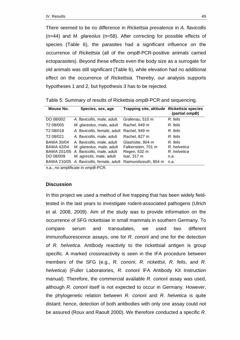
IV. Results 49
There seemed to be no difference in Rickettsia prevalence in A. flavicollis
(n=44) and M. glareolus (n=58). After correcting for possible effects of
species (Table 6), the parasites had a significant influence on the
occurrence of Rickettsia (all of the ompB-PCR-positive animals carried
ectoparasites). Beyond these effects even the body size as a surrogate for
old animals was still significant (Table 6), while elevation had no additional
effect on the occurrence of Rickettsia. Thereby, our analysis supports
hypotheses 1 and 2, but hypothesis 3 has to be rejected.
Table 5: Summary of results of Rickettsia ompB-PCR and sequencing.
Mouse No. Species, sex, age Trapping site, altitude Rickettsia species (partial ompB)
DO 08/002 A. flavicollis, male, adult Grafenau, 510 m R. felis T2 08/005 M. glareolus, male, adult Rachel, 949 m R. felis T2 08/018 A. flavicollis, female, adult Rachel, 949 m R. felis T2 08/021 A. flavicollis, male, adult Rachel, 827 m R. felis BAWA 30/04 A. flavicollis, male, adult Glashütte, 804 m R. felis BAWA 42/04 M. glareolus, male, adult Falkenstein, 701 m R. helvetica BAWA 201/05 A. flavicollis, male, adult Regen, 532 m R. helvetica DO 08/009 M. agrestis, male, adult Isar, 317 m n.a. BAWA 210/05 A. flavicollis, female, adult Raimundsreuth, 804 m n.a. n.a., no amplificate in ompB-PCR.
Discussion
In this project we used a method of live trapping that has been widely field-
tested in the last years to investigate rodent-associated pathogens (Ulrich
et al. 2008, 2009). Aim of the study was to provide information on the
occurrence of SFG rickettsiae in small mammals in southern Germany. To
compare serum and transudates, we used two different
immunofluorescence assays, one for R. conorii and one for the detection
of R. helvetica. Antibody reactivity to the rickettsial antigen is group
specific. A marked crossreactivity is seen in the IFA procedure between
members of the SFG (e.g., R. conorii, R. rickettsii, R. felis, and R.
helvetica) (Fuller Laboratories, R. conorii IFA Antibody Kit Instruction
manual). Therefore, the commercial available R. conorii assay was used,
although R. conorii itself is not expected to occur in Germany. However,
the phylogenetic relation between R. conorii and R. helvetica is quite
distant; hence, detection of both antibodies with only one assay could not
be assured (Roux and Raoult 2000). We therefore conducted a specific R.
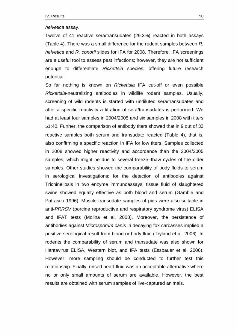
IV. Results 50
helvetica assay.
Twelve of 41 reactive sera/transudates (29.3%) reacted in both assays
(Table 4). There was a small difference for the rodent samples between R.
helvetica and R. conorii slides for IFA for 2008. Therefore, IFA screenings
are a useful tool to assess past infections; however, they are not sufficient
enough to differentiate Rickettsia species, offering future research
potential.
So far nothing is known on Rickettsia IFA cut-off or even possible
Rickettsia-neutralizing antibodies in wildlife rodent samples. Usually,
screening of wild rodents is started with undiluted sera/transudates and
after a specific reactivity a titration of sera/transudates is performed. We
had at least four samples in 2004/2005 and six samples in 2008 with titers
≥1:40. Further, the comparison of antibody titers showed that in 9 out of 33
reactive samples both serum and transudate reacted (Table 4), that is,
also confirming a specific reaction in IFA for low titers. Samples collected
in 2008 showed higher reactivity and accordance than the 2004/2005
samples, which might be due to several freeze–thaw cycles of the older
samples. Other studies showed the comparability of body fluids to serum
in serological investigations: for the detection of antibodies against
Trichinellosis in two enzyme immunoassays, tissue fluid of slaughtered
swine showed equally effective as both blood and serum (Gamble and
Patrascu 1996). Muscle transudate samples of pigs were also suitable in
anti-PRRSV (porcine reproductive and respiratory syndrome virus) ELISA
and IFAT tests (Molina et al. 2008). Moreover, the persistence of
antibodies against Microsporum canis in decaying fox carcasses implied a
positive serological result from blood or body fluid (Tryland et al. 2006). In
rodents the comparability of serum and transudate was also shown for
Hantavirus ELISA, Western blot, and IFA tests (Essbauer et al. 2006).
However, more sampling should be conducted to further test this
relationship. Finally, rinsed heart fluid was an acceptable alternative where
no or only small amounts of serum are available. However, the best
results are obtained with serum samples of live-captured animals.
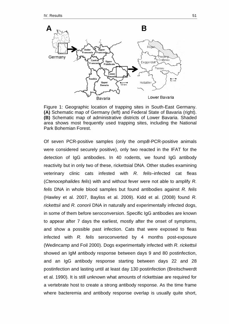
IV. Results 51
Figure 1: Geographic location of trapping sites in South-East Germany. (A) Schematic map of Germany (left) and Federal State of Bavaria (right). (B) Schematic map of administrative districts of Lower Bavaria. Shaded area shows most frequently used trapping sites, including the National Park Bohemian Forest. Of seven PCR-positive samples (only the ompB-PCR-positive animals
were considered securely positive), only two reacted in the IFAT for the
detection of IgG antibodies. In 40 rodents, we found IgG antibody
reactivity but in only two of these, rickettsial DNA. Other studies examining
veterinary clinic cats infested with R. felis–infected cat fleas
(Ctenocephalides felis) with and without fever were not able to amplify R.
felis DNA in whole blood samples but found antibodies against R. felis
(Hawley et al. 2007, Bayliss et al. 2009). Kidd et al. (2008) found R.
rickettsii and R. conorii DNA in naturally and experimentally infected dogs,
in some of them before seroconversion. Specific IgG antibodies are known
to appear after 7 days the earliest, mostly after the onset of symptoms,
and show a possible past infection. Cats that were exposed to fleas
infected with R. felis seroconverted by 4 months post-exposure
(Wedincamp and Foil 2000). Dogs experimentally infected with R. rickettsii
showed an IgM antibody response between days 9 and 80 postinfection,
and an IgG antibody response starting between days 22 and 28
postinfection and lasting until at least day 130 postinfection (Breitschwerdt
et al. 1990). It is still unknown what amounts of rickettsiae are required for
a vertebrate host to create a strong antibody response. As the time frame
where bacteremia and antibody response overlap is usually quite short,
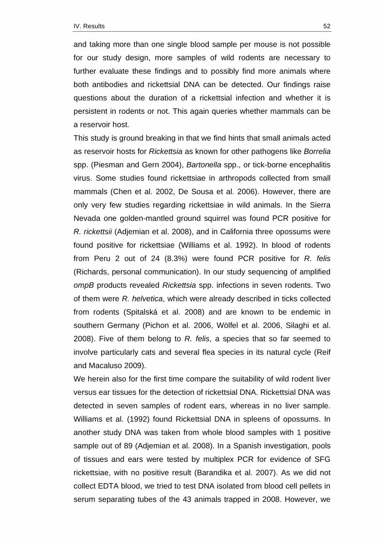
IV. Results 52
and taking more than one single blood sample per mouse is not possible
for our study design, more samples of wild rodents are necessary to
further evaluate these findings and to possibly find more animals where
both antibodies and rickettsial DNA can be detected. Our findings raise
questions about the duration of a rickettsial infection and whether it is
persistent in rodents or not. This again queries whether mammals can be
a reservoir host.
This study is ground breaking in that we find hints that small animals acted
as reservoir hosts for Rickettsia as known for other pathogens like Borrelia
spp. (Piesman and Gern 2004), Bartonella spp., or tick-borne encephalitis
virus. Some studies found rickettsiae in arthropods collected from small
mammals (Chen et al. 2002, De Sousa et al. 2006). However, there are
only very few studies regarding rickettsiae in wild animals. In the Sierra
Nevada one golden-mantled ground squirrel was found PCR positive for
R. rickettsii (Adjemian et al. 2008), and in California three opossums were
found positive for rickettsiae (Williams et al. 1992). In blood of rodents
from Peru 2 out of 24 (8.3%) were found PCR positive for R. felis
(Richards, personal communication). In our study sequencing of amplified
ompB products revealed Rickettsia spp. infections in seven rodents. Two
of them were R. helvetica, which were already described in ticks collected
from rodents (Spitalská et al. 2008) and are known to be endemic in
southern Germany (Pichon et al. 2006, Wölfel et al. 2006, Silaghi et al.
2008). Five of them belong to R. felis, a species that so far seemed to
involve particularly cats and several flea species in its natural cycle (Reif
and Macaluso 2009).
We herein also for the first time compare the suitability of wild rodent liver
versus ear tissues for the detection of rickettsial DNA. Rickettsial DNA was
detected in seven samples of rodent ears, whereas in no liver sample.
Williams et al. (1992) found Rickettsial DNA in spleens of opossums. In
another study DNA was taken from whole blood samples with 1 positive
sample out of 89 (Adjemian et al. 2008). In a Spanish investigation, pools
of tissues and ears were tested by multiplex PCR for evidence of SFG
rickettsiae, with no positive result (Barandika et al. 2007). As we did not
collect EDTA blood, we tried to test DNA isolated from blood cell pellets in
serum separating tubes of the 43 animals trapped in 2008. However, we
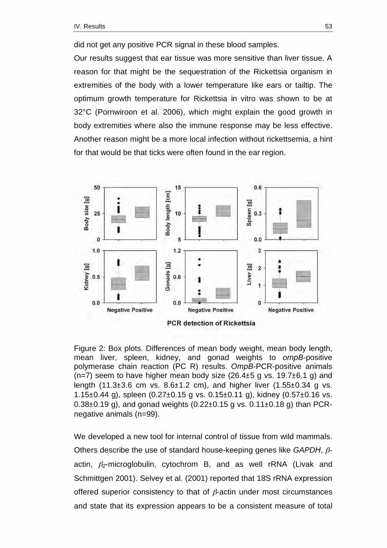
IV. Results 53
did not get any positive PCR signal in these blood samples.
Our results suggest that ear tissue was more sensitive than liver tissue. A
reason for that might be the sequestration of the Rickettsia organism in
extremities of the body with a lower temperature like ears or tailtip. The
optimum growth temperature for Rickettsia in vitro was shown to be at
32°C (Pornwiroon et al. 2006), which might explain the good growth in
body extremities where also the immune response may be less effective.
Another reason might be a more local infection without rickettsemia, a hint
for that would be that ticks were often found in the ear region.
Figure 2: Box plots. Differences of mean body weight, mean body length, mean liver, spleen, kidney, and gonad weights to ompB-positive polymerase chain reaction (PC R) results. OmpB-PCR-positive animals (n=7) seem to have higher mean body size (26.4±5 g vs. 19.7±6.1 g) and length (11.3±3.6 cm vs. 8.6±1.2 cm), and higher liver (1.55±0.34 g vs. 1.15±0.44 g), spleen (0.27±0.15 g vs. 0.15±0.11 g), kidney (0.57±0.16 vs. 0.38±0.19 g), and gonad weights (0.22±0.15 g vs. 0.11±0.18 g) than PCR-negative animals (n=99).
We developed a new tool for internal control of tissue from wild mammals.
Others describe the use of standard house-keeping genes like GAPDH, β-
actin, β2-microglobulin, cytochrom B, and as well rRNA (Livak and
Schmittgen 2001). Selvey et al. (2001) reported that 18S rRNA expression
offered superior consistency to that of β-actin under most circumstances
and state that its expression appears to be a consistent measure of total
IV. Results 54
RNA in a sample throughout the cell cycle.
There are only very few studies that analyze data on rodents and their
associated pathogens statistically as it was done for Cowpox virus
infection by Essbauer et al. (2009). However, statistical analyses are
useful to comprehend interrelations between infection courses of
(reservoir-) hosts and ecological aspects. No formal sample size
estimation was performed before the experiments, because our sampling
of mice in a bad season was not evaluable and no literature about the
expected effect was available. However, the sample size for the logistic
model analysis is reasonable large to test the two parameters of interest
and, obviously, the power was large enough to reject the hypothesis. As
Rickettsia are transmitted by arthropods such as ticks, fleas, and mites,
the ectoparasitic infestation of animals was of special interest. We quite
often found Ixodes ticks (nymphs and larvae) on the investigated small
mammals. Their preferred body spots seemed to be the ears, the chin,
and the tail base, presumably because of the lack of fur there (data not
shown). Fleas found consisted of five different species, mostly
Ctenophthalmus agyrtes. It is conceivable that there was a correlation
between Rickettsia-positive animals and their parasitic load.
There seemed to be a difference between body length, weight, and
several organ weights. PCR-positive animals were heavier and taller, and
their organs were usually heavier, which probably is a hint for mainly adult
animals being infected. The older the animal, the more probable is an
exposure to Rickettsia and Rickettsia-transmitting vectors. Differences in
organ weights are also interesting, regarding the question whether
infection with Rickettsia has clinical effects on small mammals. Clinical
symptoms have been reported in dogs, for example, after infection with R.
rickettsii, but not confirmedly in cats or small mammals. Infection with R.
felis occurs in humans but has not been confirmed by isolation so far
(Pérez-Osorio et al. 2008).
To summarize, we found that IF tests are a useful tool to investigate past
infections with rickettsiae, but are unsuitable for further species
differentiation. They can be used even with transudates, if there is no
serum available, although results are less secure. We found two different
rickettsial species, the more common R. helvetica (Dobler and Wölfel

IV. Results 55
2009) and R. felis, which has not been described in small mammals in
Europe before. Rickettsial organisms can be detected best in mammals
using ear tissue. The question whether rodents or other small mammals
acted as reservoir hosts still needs to be answered. Although we found
seroconverted animals and rickettsial DNA in their tissues, we do not know
if there is a persistent infection and excretion. However, it is conceivable
that infected mammals amplify the cycle by fleas feeding on their infected
blood. For this reason, further research specific to this topic, including
other rickettsial species endemic in Europe, is necessary. Table 6: Results of sequentially generalized linear model with the binary dependent variable Rickettsia ompB-Polymerase Chain Reaction and four predictors.
Variable Deviance p value (chi-squared) Rodent species 2.852 0.091 Ectoparasites 6.460 0.011 Body size 4.938 0.026 Elevation 0.092 0.761 Significant p-values are shown in bold. Acknowledgments
The authors gratefully acknowledge the excellent practical and technical
assistance of H. Weber, R. Reiter, D. Eder, R. Terzioglu, D. Woll, and F.
Goldberg. M. and D. Kiefer’s support in species determination of fleas is
highly appreciated. The authors also thank S. Speck for carefully reading
the article, E. Seibold for help with statistical analysis, and H. Scholz for
critical discussion. This project is supported by a grant from the Bavarian
Ministry of Health, Vector-Borne Diseases in Climate Change
Investigations (VICCI).
Disclosure Statement
No competing financial interests exist.
References
Adjemian, JD, Adjemian, MK, Foley, P, Chomel, BB, et al. Evidence of
multiple zoonotic agents in a wild rodent community in the Eastern
Sierra Nevada. J Wildl Dis 2008; 44:737–742.
Barandika, JF, Hurtado, A, García-Esteban, C, Gil, H, et al. Tick-borne
IV. Results 56
zoonotic bacteria in wild and domestic small mammals in Northern
Spain. Appl Environ Microbiol 2007; 73:6166–6171.
Bayliss, DB, Morris, AK, Horta, MC, Labruna, MB, et al. Prevalence of
Rickettsia species antibodies and Rickettsia species DNA in the blood
of cats with and without fever. J Feline Med Surg 2009; 11:266–270.
Breitschwerdt, EB, Levy, MG, Davidson, MG, Walker, DH, et al. Kinetics of
IgM and IgG responses to experimental and naturally acquired
Rickettsia rickettsii infection in dogs. Am J Vet Res 1990; 51:1312–
1316.
Chen, Z, Chen, M, Zhong, J, Bi, D. Using PCR/RFLP to detect spotted
fever group rickettsia in ticks and rodents collected in Ninghua, Fujian
province. Zhonghua Yu Fang Yi Xue Za Zhi 2002; 36:106–108.
De Sousa, R, Edouard-Fournier, P, Santos-Silva, M, Amaro, F, et al.
Molecular detection of R. felis, R. typhi, and two Genotypes closely
related to Barthonella elizabethae. Am J Trop Med Hyg 2006; 75:727–
731.
Dobler, G, Essbauer, S, Wölfel, R. Isolation and preliminary
characterisation of ‘‘Rickettsia monacensis’’ in south-eastern
Germany. Clin Microbiol Infect 2009; 15 Suppl 2: 263–264.
Dobler, G, Wölfel, R. Typhus and other rickettsioses. Emerging infections
in Germany. Dtsch Arztebl Int 2009; 106: 348–354.
Essbauer, S, Hartnack, S, Misztela, K, Kiessling-Tsalos, J, et al. Patterns
of orthopox virus wild rodent hosts in South Germany. Vector Borne
Zoonot Dis 2009; 9:301–311.
Essbauer, S, Schmidt, J, Conraths, FJ, Friedrich, R, et al. A new Puumala
Hantavirus subtype in rodents associated with an outbreak of
Nephropathia epidemica in South-East Germany in 2004. Epidemiol
Infect 2006; 134:1333–1344.
Gamble, HR, Patrascu, IV. Whole blood, serum, and tissue fluids in an
enzyme immunoassay for swine Trichinellosis. J Food Prot 1996;
59:1213–1217.
Hall, TA. BioEdit: a user-friendly biological sequence alignment editor and
analysis program for Windows 95/98/NT. Nucleic Acids Symp Ser
1999; 41:95–98.
Hastie, TJ, Pregibon, D. Generalized linear models. In: Chambers, JM,

IV. Results 57
Hastie, TJ, eds. Statistical Models in S. Wadsworth & Brooks/Cole,
Pacific Grove, CA 1992 , 6:195-2481992
Hawley, JR, Shaw, SE, Lappin, MR. Prevalence of Rickettsia felis DNA in
the blood of cats and their fleas in the United States. J Feline Med
Surg 2007; 9:258–262.
Kidd, L, Maggi, R, Diniz, PP, Hegarty, B, et al. Evaluation of conventional
and real-time PCR assays for detection and differentiation of Spotted
Fever Group Rickettsia in dog blood. Vet Microbiol 2008; 22;129:294–
303.
Larkin, MA, Blackshields, G, Brown, NP, Chenna, R, et al. ClustalW and
ClustalX version 2. Bioinformatics 2007; 23:2947–2948.
Livak, KJ, Schmittgen, TD. Analysis of relative gene expression data using
real-time quantitative PCR and the 2(-Delta Delta C(T)) method.
Methods 2001; 25:402–408.
Molina, RM, Chittick, W, Nelson, EA, Christopher-Hennings, J, et al.
Diagnostic performance of assays for the detection of anti-Porcine
reproductive and respiratory syndrome virus antibodies in serum and
muscle transudate ("meat juice") based on samples collected under
experimental conditions. J Vet Diagn Invest 2008; 20:735–743.
Parola, P, Davoust, B, Raoult, D. Tick- and flea-borne rickettsial emerging
zoonoses. Vet Res 2005; 36:469–492.
Parola, P, Raoult, D. Ticks and tickborne bacterial diseases in humans: an
emerging infectious threat. Clin Infect Dis 2001; 32:897–928. Erratum:
Clin Infect Dis 33:749.
Pérez-Osorio, CE, Zavala-Velázquez, JE, Arias León, JJ, Zavala- Castro,
JE. Rickettsia felis as emergent global threat for humans. Emerg Infect
Dis 2008; 14:1019–1023.
Pichon, B, Kahl, O, Hammer, B, Gray, JS. Pathogens and host DNA in
Ixodes ricinus nymphal ticks from a German forest. Vector Borne
Zoonot Dis 2006; 6:382–387.
Piesman, J, Gern, L. Lyme borreliosis in Europe and North America.
Parasitology 2004; 129 Suppl:S191–S220.
Pornwiroon, W, Pourciau, SS, Foil, LD, Macaluso, KR. Rickettsia felis from
cat fleas: isolation and culture in a tick-derived cell line. Appl Environ
Microbiol 2006; 72:5589–5595.

IV. Results 58
R Development Core Team. R: A language and environment for statistical
computing. R Foundation for Statistical Computing, Vienna, Austria,
2009. ISBN 3-900051-07-0. Available at www.R-project.org.
Reif, KE, Macaluso, KR. Ecology of Rickettsia felis: a review. J Med
Entomol 2009; 46:723–736.
Roux, V, Raoult, D. Phylogenetic analysis of members of the genus
Rickettsia using the gene encoding the outer-membrane protein
rOmpB (ompB). Int J Syst Evol Microbiol 2000; 50 Pt 4:1449–1455.
Selvey, S, Thompson, EW, Matthaei, K, Lea, RA, et al. Beta-actin—an
unsuitable internal control for RT-PCR. Mol Cell Probes 2001; 15:307–
311.
Silaghi, C, Gilles, J, Höhle, M, Pradel, I, et al. Prevalence of spotted fever
group rickettsiae in Ixodes ricinus (Acari: Ixodidae) in southern
Germany. J Med Entomol 2008; 45:948–955.
Spitalská, E, Boldis, V, Kostanová, Z, Kocianová, E et al. Incidence of
various tick-borne microorganisms in rodents and ticks of central
Slovakia. Acta Virol 2008; 52: 175–179.
Tryland, M, Handeland, K, Bratberg, AM, Solbakk, IT, et al. Persistence of
antibodies in blood and body fluids in decaying fox carcasses, as
exemplified by antibodies against Microsporum canis. Acta Vet Scand
2006; 48:10.
Ulrich, RG, Heckel, G, Pelz, HJ, Wieler, LH, et al. Rodents and rodent
associated disease vectors: the network of ‘‘rodent carrying
pathogens’’ introduces itself [German]. Bundesgesundheitsblatt
Gesundheitsforschung Gesundheitsschutz 2009; 52:352–369.
Ulrich, RG, Schmidt-Chanasit, J, Schlegel, M, Jacob, J, et al. Network
„Rodent-borne pathogens“ in Germany: longitudinal studies on the
geographical distribution and prevalence of Hantavirus infections.
Parasitol Res 2008; 103 Suppl 1:S121– S129.
Wedincamp J, Jr., Foil, LD. Infection and seroconversion of cats exposed
to cat fleas (Ctenocephalides felis Bouché) infected with Rickettsia
felis. J Vector Ecol 2000; 25:123–126.
Williams, SG, Sacci JB, Jr., Schriefer, ME, Andersen, EM, et al. Typhus
and typhuslike rickettsiae associated with opossums and their fleas in
Los Angeles County, California. J Clin Microbiol 1992; 30:1758–1762.
IV. Results 59
Wölfel, R, Essbauer, S, Dobler, G. Diagnostics of tick-borne rickettsioses
in Germany: a modern concept for a neglected disease. Int J Med
Microbiol 2008; 298 S1:368–374.
Wölfel, R, Terzioglu, R, Kiessling, J, Wilhelm, S, et al. Rickettsia spp. in
Ixodes ricinus ticks in Bavaria, Germany. Ann N Y Acad Sci 2006;
1078:509–511. Address correspondence to: Sandra Essbauer Bundeswehr Institute of Microbiology Neuherbergstr. 11 D-80937 Munich Germany E-mail: [email protected]
V. Discussion 60
V. DISCUSSION
In the present study the occurence of SFG rickettsiae in rodents, their
prevalence and genetic diversity was investigated by means of serologic
tests and PCRs, with the major aim to brighten the question if there is a
rodent reservoir for SFG rickettsiae in Germany. For this purpose, rodent
blood as well as tissue samples were collected over a period of three
years in the region of Lower Bavaria, in the year 2008 restricted to the
National Park Bavarian Forest.
In this study, we tried to find statistically significant factors in sero- or PCR-
positive individuals. Only few studies attempted to find a correlation
between pathogen infestation and body condition, as for example for
rodents carrying orthopox viruses (Essbauer et al., 2009a). Statistic tests
such as chi-square test, student t-test, Mann-Whitney test or Spearman
rank correlation were only used for preliminary screenings, since a
Bonferroni correction and a reduction of the significance level needed to
be used in order to maintain the familywise error rate. Therefore, we
present descriptive tests only. In addition, as many aspects of our
serologic test assays are unknown so far, statistical analyses of those
were not included in this thesis.
Seroprevalence for Rickettsia spp. of tested serum samples was about
one third (n=32/114); approximately 8% (n=9/119) of samples showed
positive reaction in PanRickettsia real-time PCR. For comparison,
prevalence of SFG rickettsiae in ticks in Southern Germany has been
shown to vary between 3.5 to 6.2% for R. helvetica (Woelfel et al., 2006;
Hartelt et al., 2004), 30.3% for R. raoultii, 0.75% for R. slovaca (Pluta et
al., 2010), 1.7% for R. massiliae, 0.4% for R. felis and 0.6% for R.
monacensis (Dobler & Woelfel, 2009). Interestingly, the results obtained
from rodents in this study are considerably higher. At least for R. felis and
R. helvetica, it could be concluded that rodent tissue is a promising tool for
detection of Rickettsia, since the prevalence in ticks seems to be lower
than in rodents. As rickettsiae are transmitted by arthropods, it is not
surprising that infestation with parasites had a significant influence on the
V. Discussion 61
occurrence of Rickettsia (all of the ompB-PCR-positive animals carried
ectoparasites). In contrast to this, elevation had no additional effect on the
occurrence of Rickettsia, presumably due to the fact that ectoparasites
were found on rodents of every altitude. Detailed ectoparasitic
investigations will be part of future studies.
As shown in Figure 9 (p. 32), we tried to find patterns of species
distribution along the altitude gradient. However, trapped species
apparently were distributed quite randomly, apart from Sorex spp., which
could only be found in the highest trapping location. Presumably, this is
correlated to habitat factors rather than to elevation itself. Following
longitudinal studies will further investigate these aspects.
The phylogenetic tree (Figure 13, p. 36) shows the close relationship
between our R. helvetica sequence and the R. helvetica sequence
obtained by Roux and Raoult (2000). However, both are quite distinct to
other Rickettsia species. The R. felis sequence obtained in this study
clusters with R. felis sequences obtained in Australia and the USA.
Determination of host-pathogen dynamics and of possible reservoir hosts
has always been one of the foci of rickettsial research. Ticks were
considered not only as vectors but also as maintaining hosts. For R.
conorii conorii, the causative agent of Mediterranean spotted fever (MSF),
Rhipicephalus sanguineus, the brown dog tick, was thought to act as
vector and reservoir (Blanc & Caminopetros, 1932) due to transovarial
transmission. However, infection rates of Rh. sanguineus with R. conorii
conorii were shown to be less than 15% (Raoult et al., 1993). Rickettsia
rickettsii, the agent of Rocky Mountain spotted fever (RMSF) is transmitted
by several tick species, and maintained in infected ticks by transovarial
and transstadial transmission (Macaluso & Azad, 2005). However,
Ricketts (1911) observed that less than 50% of experimentally infected D.
andersoni female ticks transmitted R. rickettsii to their offspring, and Price
(1954) found a transovarial transmission rate of only 30% in D. variabilis
ticks, whereas Burgdorfer et al. (1967, 1975) observed 100% filial infection
in D. andersoni and D. variabilis that were naturally infected. Considering
these possible low passage rates it is, therefore, not very probable that
ovarial passage is sufficient to maintain the natural cycles of R. rickettsii
V. Discussion 62
and R. conorii. Furthermore, R. rickettsii and its acarine hosts do not seem
to have established a fully effective symbiotic relationship, as some tick
infections are incomplete, others can result in decreased fecundity,
viability or even death of the tick (McDade & Newhouse, 1986).
Rickett’s hypothesis of an additional maintenance of R. rickettsii seems to
be far more reasonable. Vertebrate hosts provide a source for
simultaneous feeding of ticks (infected and uninfected), serve as vehicles,
are numerous, support a large number of ticks and – in case of small
mammals – reproduce rapidly. Thus they offer many survival advantages
for the rickettsial organism (McDade & Newhouse, 1986).
Furthermore, Rickettsia spp. have a restricted geographic range when
compared to the transmitting ticks. Although the tick can be found
worldwide, R. conorii conorii is endemic in much smaller regions. The
same is true for the transmission of R. rickettsii by Rh. sanguineus in
Arizona (Demma et al., 2005). One explanation might be that transovarial
transmission may occur for a limited number of passages only and that the
tick may only be the vector of the pathogen (Rovery et al., 2008). On the
other hand other factors such as co-feeding on a vertebrate or
maintenance in a rickettsemic vertebrate may play an essential role in the
transmission cycle.
For R. conorii conorii, several mammals are proposed to act as reservoir
hosts. Firstly, dogs serve as transport hosts as they carry ticks close to
humans and can suffer from febrile illness related to R. conorii conorii
infection (Solano-Gallego et al., 2006). Moreover, in certain zones of
southern Europe, a correlation between the percentage of the canine
population with antibodies to R. conorii conorii and the incidence of MSF in
humans has been found (Herrero-Herrero, 1989). However, dogs are
transient reservoirs due to a transient post-infection rickettsemia; and
therefore do not seem to be an efficient reservoir for R. conorii conorii
(Rovery et al., 2008). Secondly, antibodies to R. conorii conorii were found
in wild rabbits and hares with prevalences up to 76.5% (Ruiz Beltran et al.,
1992). Furthermore, Le Gac (1966) reported a sudden decrease in MSF
cases on the French Mediterranean coast 1952 simultaneous with a lethal
myxomatose outbreak that eradicated the local wild rabbit population. A

V. Discussion 63
reemergence of this pathogen was also correlated with the reappearence
of wild rabbits in 1967 (Rovery et al., 2008). Lastly, hedgehogs and other
small rodents are also potential candidates for the reservoir of R. conorii
since antibodies against rickettsiae have been detected in serum samples
of these animals (Le Gac, 1966).
Antibodies to R. rickettsii were found in at least 53 species, belonging to
the genera Marsupialia, Insectivora, Rodentia, Carnivora and Artiodactyla
(McDade & Newhouse, 1986). In addition, R. rickettsii was isolated from
Microtus pennsylvanicus (Gould & Miesse, 1954) and other small
mammals such as rabbits, opossums, squirrels, voles and mice
(Burgdorfer, 1977), suggesting that a broad variety of vertebrates are
involved in the natural cycle of R. rickettsii.
In general, a natural cycle starts with the development of eggs from an
infected female tick into infected larvae. These can then infect small
rodents when feeding on them. After that, larvae develop into nymphs,
these feed on medium-sized animals and thus can infect them too.
Feeding on an infected animal can infect ticks of every stadium, although
this depends on the magnitude and duration of its rickettsemia. The
minimum level of required rickettsemia to infect 50% of D. andersoni
larvae ranged between 10 and 100 guinea pig infectious doses (GPID) per
0.5 ml blood (Burgdorfer et al., 1966). Rickettsemia levels in turn depend
on the susceptibility of the vertebrate. Some mammals turned out to be
highly susceptible, such as Columbian ground squirrels, golden-mantled
ground squirrels, chipmunks and meadow voles with GPIDs between 100
and 1000 for up to four days (Burgdorfer et al., 1966). On the other hand,
less susceptible mammals such as bushy-tailed wood rats (Burgdorfer et
al., 1966), cotton rats (Shirai et al., 1967) or dogs (Norment & Burgdorfer,
1984) develop much smaller or shorter rickettsemias or none at all.
Rehácek et al. (1992) investigated the susceptibility of European rodent
species (M. glareolus, A. flavicollis, Mus musculus) to rickettsiae (e.g.
Coxiella burnetii, R. conorii, R. slovaca and R. sibirica) and found that for
R. conorii, R. slovaca and R. sibirica a sharp increase of antibody levels,
with high titres on days 4-6 and peak levels about day 11, was
characteristic.
V. Discussion 64
For R. felis, which is mainly transmitted by cat fleas, but was also detected
in mites, no definitive mammalian host has been identified so far (Reif &
Macaluso, 2009). To the authors knowledge this study is the first to
suggest that rodents may also act as a reservoir host for R. felis. In
addition to the results presented here, antibodies to R. felis were also
found in fevrile and non-fevrile cats (Bayliss et al., 2009), dogs (Richter et
al., 2002; Oteo et al., 2006), and opossums (Williams et al., 1992).
Furthermore, in a laboratory experiment Wedincamp and Foil (2000)
demonstrated that previously flea- and Rickettsia-naïve cats
seroconverted within four months after infestation with R. felis-infected cat
fleas. Although these studies show a horizontal transmission from flea to
vertebrate, to the present moment, the transmission from infected
vertebrate to arthropod has not yet been shown for R. felis. Nonetheless,
Weinert et al. (2009) state that only occasional horizontal transmission
from mammal to arthropod may be needed to maintain R. felis in nature.
For R. helvetica, the state of knowledge regarding a possible reservoir
host is fragmentary or inexistent. Several studies found R. helvetica in
ticks in Germany (Woelfel et al., 2006; Dobler & Woelfel, 2009), but no
evidence has been found in rodents previously to this study (Hartelt et al.,
2008).
In our study, we found seropositive animals and rickettsial DNA (R. felis
and R. helvetica) in ear tissue. However, rickettsial DNA found in ear
tissue does not necessarily implicate rickettsemia, as it may be associated
with a local infection after a tick bite. The fact that ticks were often found in
body regions not covered with fur such as the ear, the chin or the tail base
may argue for that, as well as the fact that we could not amplify DNA from
liver tissue. On the other hand, the lower body temperature in body
extremities like the ear could be advantageous for Rickettsia and thus
promote sequestration of the rickettsial organism, as their optimum growth
temperature in vitro was shown to be at 32°C (Pornwiroon et al., 2006).
One of the potential limitations of this study was the type of blood samples
collected. These consisted in serum samples only, which did not enable us
to test full blood samples for rickettsial DNA. Therefore, we cannot draw a
conclusion to a possible rickettsemia. Seropositive animals may only
V. Discussion 65
develope limited, transient infections sufficient for the production of
antibodies, but not sufficient to infect ticks and thus to maintain the
rickettsial life cycle. Moreover, seropositivity indicates that infection has
occurred, but is no evidence of direct or indirect transmission to the
arthropod host, which, for being a reservoir, must exist. Furthermore the
level of seroprevalence does not necessarily provide information as to
whether the vertebrate is a maintenance host (Haydon et al., 2002). In
addition, high seroprevalence at a single point in time may simply indicate
an outbreak in the host population, rather than pathogen persistence (Van
Bressem et al., 2001). The critical issue is the persistence of infection in
the reservoir, which can only be determined through longitudinal studies.
Nevertheless, the high rickettsial prevalence in rodents captured in our
study over a period of three years suggests rickettsial persistence rather
than a single outbreak.
Referring to Mills and Childs (1998), we were able to determine the
possible rodent host species that occur in the region of Lower Bavaria and
their distribution along an altitude gradient. Our data is a basis for further
investigations on the geographic range of the pathogen within the host
range and the regional distribution of the host and pathogen among the
distinct habitat types. Further, our survey conducted over a period of three
years is a starting point for longitudinal monitoring of host-pathogen
dynamics.
Even if the question of rodents acting as a reservoir host or not cannot be
answered definitively, the results of our study are of immense importance
as we were able to detect DNA from R. felis and R. helvetica in small
mammals in Germany for the first time. In contrast to the results of this
study, several investigations screened different mammalian organ tissues
or blood with no positive results at all: Yabsley et al. (2008) investigated
dogs in Grenada for Rickettsia spp. and found them to be seronegative
and PCR negative, however, it has to be noted that the primers they used
targeted the ompA gene. Organ pools of 119 rodents (mainly M. agrestis)
from Germany showed no positive result in PCR screening (Pluta et al.,
2010), neither did organ pools including ear tissue from Spain, tested by
multiplex PCR (Barandika et al., 2007). However, in the USA, rickettsial
V. Discussion 66
DNA was found in three opossum spleens in California (Williams et al.,
1992), and in a whole blood sample of one golden mantled ground squirrel
in the Sierra Nevada (Adjemian et al., 2008). In a survey on rodents
trapped on a military training base in Southern Germany, that was
conducted at the Bundeswehr Institute of Microbiology, in three out of 221
rodents Rickettsia spp. DNA was detected by PCR, the seroprevalence
was 11.8% (Publication Annex 4, p. 101). Two blood samples from
rodents from Peru were found PCR-positive for Rickettsiae (Richards,
personal communication). Apart from the latter, this study is the first to
detect R. felis in rodent tissue, which is a quite thrilling result that several
researchers received with immense interest. It is hoped that this study will
act as a trigger for further investigations regarding the natural cycles of
rickettsiae in Germany.
Furthermore, the molecular biological and serologic methods applied in
this study were shown to be suitable for screening of rodent tissue and of
sera as well as transudates, respectively. Thus they offer useful tools for
diagnosis and further (longitudinal) research regarding rickettsia in
humans as well as in vertebrate and arthropod hosts.
Although being most likely underdiagnosed, the incidence of rickettsial
infections in Germany appears to be rather low so far, compared to other
vector-borne diseases such as Lyme borreliosis or tick-borne encephalitis.
However, Rickettsioses are considered as emerging diseases (Parola &
Raoult, 2001; Fournier & Raoult 2005; Parola et al., 2005; Brouqui et al.,
2007; Vorou et al., 2007; Rovery et al., 2008). Being arthropod-borne
diseases, infection risks are determined by occurrence of the vector, which
in turn is influenced by several factors such as climate, environment, and
the presence of rodents and other small mammal reservoirs (Fritz, 2009).
Climatic conditions are assumed to influence the distribution of ticks and
tick-borne diseases, for example TBE. In the geographic range of TBE
virus, a northern expansion was observed in recent years. However,
socio-economic changes, agricultural activities, employment and income
apparently are more important factors to affect the risk of TBE infection
and exposition for humans (Randolph, 2008).
In a climatic warming scenario, ticks are likely to extend their northern and
V. Discussion 67
altitude range in Europe (Lindgren et al., 2000; Gray et al., 2009; Pluta et
al., 2010). For instance, until now the occurrence of Dermacentor ticks in
Germany has been restricted to few areas. Due to increasing
temperatures, their distribution is likely to be extended, and Rickettsia spp.
might spread with them (Pluta et al., 2010). So far, research regarding
these aspects is at a very early stage in Germany, however, the present
study sets an important starting point for further investigations.
The influence of climate change on reservoir host abundance, such as
rodents, is probably more complicated and still under discussion (Tersago
et al., 2009). Also, socio-economic factors such as changes in human
leisure behaviour in addition to an increase in outdoor activities could
possibly amplify human exposure and likelihood of infections in the future.
As there is no vaccination available, the only way to prevent rickettsial
infection is to avoid contact to the vectors and reservoir hosts. Intensifying
fundamental research regarding host associations and host adaptations of
Rickettsia spp. is essential for risk assessment and disease prevention in
humans, livestock and companion animals.
VI. Conclusion & Outlook 68
VI. CONCLUSION & OUTLOOK
In the present study, seroreactive small mammals and rickettsial DNA,
belonging to the species R. helvetica and R. felis, were detected. The
results are of enourmous importance as this is the first detection of
rickettsial DNA in rodents in Germany. However, due to limitations of study
design, the question of rodents acting as a reservoir could not be
answered definitevely.
For this reason, further investigations are urgently needed, on the one
hand to screen rodent sera over a longer period for possible rickettsemia
and on the other hand to screen more ear samples for rickettsial DNA,
also in other parts of the country, in order to obtain more reliable data and
thus to finally answer the question if rodents and other small mammals
play an important or maybe even essential role in maintaining rickettsial
natural life cycles. The small mammals that were collected subsequently in
the years 2009 and 2010 will be investigated to that effect in the future,
e.g. by generating rickettsial isolates for further characterization.
In the scope of the VICCI project, the collected animals were also
screened for Hantavirus prevalence. The year 2010 seems to be an
outbreak year for human Puumala virus infections as well as for
prevalence in rodents (to date about 18%, in comparison to about 3% in
the recent years). Also regarding this aspect of immense importance for
public health, investigations will be continued in the future.
More statistical analyses will be conducted by ecologists, cooperating with
the National Park Bavarian Forest, in order to get more information about
possible correlations between infection, body parameters and ecological
factors such as habitat elevation or vegetation.
VII. Summary 69
VII. SUMMARY
In recent years, several Rickettsia spp. have been detected in Germany
for the first time, some of them pathogenic for humans. The present study
investigated the prevalence of spotted fever group (SFG) rickettsiae in
small mammals by serological and molecular biological means. In total,
124 rodents and insectivores were collected in the years 2004 and 2005 in
Lower Bavaria and in 2008 in the National Park Bavarian Forest. Sera and
transudates were tested in two immunofluorenscence assay tests, one
commercially available for detection of antibodies against R. conorii and
one in-house test for R. helvetica antibodies. DNA-extracts were taken
from ear and liver tissue. Screening for Rickettsia spp. was performed with
a real-time PCR targeting the citrate synthase gene (gltA), followed by
conventional PCR detecting the rOmpB gene for all gltA positives. An 18S
universal PCR was used as internal control. The overall prevalence for
antibodies against SFG rickettsiae was 28.1%; 7.6% (n=9/119) samples
showed positive reaction in PanRickettsia real-time PCR.
Sequence analysis of all rompB PCR products resulted in five R. felis and
two R. helvetica ompB nucleotide sequences that were 100% identical
among each other and to the respective prototypes deposited in GenBank.
Descriptive statistical tests revealed that prevalence of rickettsial DNA, but
not seroprevalence were related to body weight, body length, spleen,
kidney, gonad and liver weights. As expected, parasites had a significant
influence, as well as body size as a surrogate for adult animals, in contrast
to elevation.
With the first detection of two arthropod-borne Rickettsia spp. (R. helvetica
and R. felis) in small mammals in Germany, this work is an important
contribution and thus a starting point for further research regarding the life
cycle of Rickettsiae and the reservoir role of small animals.
VIII. Zusammenfassung 70
VIII. ZUSAMMENFASSUNG
In den letzten Jahren wurden in Deutschland einige zum Teil
humanpathogene Rickettsienarten neu entdeckt. Die hier vorliegende
Studie untersuchte mit molekularbiologischen und serologischen
Methoden die Prävalenz von Rickettsien der Fleckfiebergruppe in
Kleinsäugetieren. Insgesamt wurden in den Jahren 2004/2005 in
Niederbayern und 2008 im Nationalpark Bayerischer Wald 124 Nagetiere
und Insektivore gesammelt. Seren und Transudate wurden mit zwei
Immunfluoreszenztests getestet, einem kommerziell erhältlichen zum
Nachweis von Antikörpern gegen R. conorii und einem selbsthergestellten
für Antikörper gegen R. helvetica. DNA wurde aus Ohr- und Lebermaterial
extrahiert. Ein Screening aller Proben wurde mit einer real-time PCR mit
Citratsynthase (gltA) als Zielgen durchgeführt, gefolgt von einer
konventionellen PCR mit Zielgen rompB für alle Proben mit positiver
Reaktion. Eine 18S universal PCR diente als interne Kontrolle. Insgesamt
betrug die Antikörperprävalenz für Rickettsien der Fleckfiebergruppe
28.1%; 7.6% (n=9/119) der Proben zeigten eine positive Reaktion in der
PanRickettsien real-time PCR.
Sequenzanalysen für alle rompB-PCR-Produkte ergaben fünf R. felis und
zwei R. helvetica ompB Nukleotidsequenzen, die untereinander und im
Vergleich zu den entsprechenden Genbank-Prototypen zu 100% identisch
waren.
Deskriptive statistische Tests zeigten, dass die Prävalenz rickettsialer
DNA, nicht aber die Seroprävalenz mit Körpergewicht, Körperlänge, Milz-,
Nieren-, Gonaden- und Lebergewichten zusammenhängt. Wie erwartet,
hatten Parasiten einen signifikanten Einfluss, genau wie Körpergröße als
Surrogat für ausgewachsene Tiere, jedoch nicht die Höhe.
Mit der ersten Beschreibung von zwei von Arthropoden übertragenen
Rickettsienarten (R. helvetica und R. felis) in Kleinsäugetieren in
Deutschland liefert diese Arbeit einen wichtigen Beitrag und Ausgangs-
punkt für weitergehende Forschung zum Lebenszyklus von Rickettsien
und zur Rolle von Kleinsäugetieren als potentielle Reservoirwirte.
IX. References 71
IX. REFERENCES
Adams JR, Schmidtmann ET, Azad AF. Infection of colonized cat fleas
(Ctenocephalides felis Bouché) with a rickettsia-like organism. Am J Trop
Med Hyg. 1990; 43:400-409.
Adjemian JD, Adjemian MK, Foley P, Chomel BB, Kasten RW, Foley JE.
Evidence of Multiple Zoonotic Agents in a Wild Rodent Community in the
Eastern Sierra Nevada. J Wildl Dis 2008; 44(3):737-42.
Adler B, de la Peña Moctezuma A. Leptospira and leptospirosis. Vet
Microbiol. 2010; 140(3-4):287-96.
Azad AF, Beard CB. Rickettsial pathogens and their arthropod vectors.
Emerg Infect Dis 1998; 4:179–186.
Baessler C, Foerster B, Moning C, Mueller J. The BIOKLIM Project:
Biodiversity Research between Climate Change and Wilding in a
temperate montane forest–The conceptual framework. Waldoekologie,
Landschaftsforschung und Naturschutz 2008; 7:21–33.
Baldridge GD, Burkhardt N, Simser JA, Kurtti TJ, Munderloh UG.
Sequence and expression analysis of the ompA gene of Rickettsia
peacockii, an endosymbiont of the Rocky Mountain wood tick,
Dermacentor andersoni. Appl. Environ. Microbiol. 2004; 70:6628–6636.
Barandika JF, Hurtado A, García-Esteban C, Gil H, Escudero R, Barral M,
Jado I, Juste RA, Anda P, García-Pérez AL. Tick-Borne Zoonotic Bacteria
in Wild and Domestic Small Mammals in Northern Spain. Appl Environ
Microbiol 2007; 73(19):6166-71.
Battersby SA, Parsons R, Webster JP. Urban rat infestations and the risk
to public health. J Environm Health Res 2002; 1:57-65.
Bayliss DB, Morris AK, Horta MC, Labruna MB, Radecki SV, Hawley JR,
Brewer MM, Lappin MR. Prevalence of Rickettsia species antibodies and
IX. References 72
Rickettsia species DNA in the blood of cats with and without fever. J
Feline Med Surg 2009; 11:266-270.
Beck R, Vojta L, Curković S, Mrljak V, Margaletić J, Habrun B. Molecular
Survey of Babesia microti in Wild Rodents in Central Croatia. Vector
Borne Zoonotic Dis. 2010 Jun 16. [Epub ahead of print]
Benson DA, Karsch-Mizrachi I, Lipman DJ, Ostell J, Sayers EW.
GenBank. Nucleic Acids Res. 2008; 37 (Database issue):D26-31.
Berdal BP, Mehl R, Meidell NK, Lorentzen-Styr AM, Scheel O. Field
investigations of tularemia in Norway. FEMS Immunol Med Microbiol 1996;
13:191–195.
Beugnet F, Marié JL. Emerging arthropod-borne diseases of companion
animals in Europe. Vet Parasitol. 2009; 163(4):298-305.
Bingham J, Foggin CM, Wandeler AI, Hill FWG. The epidemiology of
rabies in Zimbabwe. 1. Rabies in dogs (Canis familiaris). Onderstepoort J
Vet Res 1999a; 66:1–10.
Bingham J, Foggin CM, Wandeler AI, Hill FWG. The epidemiology of
rabies in Zimbabwe. 2. Rabies in jackals (Canis adustus and Canis
mesomelas). Onderstepoort J Vet Res 1999b; 66:11–23.
Blanc G, Caminopetros J. Etudes épidémiologiques et expérimentales sur
la fièvre boutonneuse, faites à l’Institut Pasteur d’Athènes. Arch Inst
Pasteur Tunis. 1932: 343–94.
Blanco JR, Oteo JA. Rickettsiosis in Europe. Ann N Y Acad Sci. 2006;
1078:25-33.
Bonnefoy X, Kampen H, Sweeney K. Public Health significance of urban
pests. WHO Regional Office for Europe, Copenhagen 2008.
Boyce JM. Recent trends in the epidemiology of tularemia in the United

IX. References 73
States. J Infect Dis 1975; 131:197–199.
Brenner DJ, O'Connor SP, Winkler HH, Steigerwalt AG. Proposals to unify
the genera bartonella and rochalimaea, with descriptions of Bartonella
quintana comb. nov., Bartonella vinsonii comb. nov., Bartonella henselae
comb. nov., and Bartonella elizabethae comb. nov., and to remove the
family bartonellaceae from the order Rickettsiales. Int J Syst Bacteriol
1993; 43: 77-786.
Brouqui P, Parola P, Fournier PE, Raoult D. Spotted fever rickettsioses in
southern and eastern Europe. FEMS Immunol Med Microbiol 2007; 49:2-
12.
Burgdorfer W, Aeschlimann A, Peter O, Hayes SF, Philip RN. Ixodes
ricinus: vector of a hitherto undescribed spotted fever group agent in
Switzerland. Acta Trop 1979; 36:357-367.
Burgdorfer W, Brinton LP. Mechanisms of transovarial infection of spotted
fever rickettsiae in ticks. Ann NY Acad Sci 1975; 266:61-72
Burgdorfer W, Friedhoff KT, Lancaster JL. Natural history of tick-borne
spotted fever in the USA. Bull WHO 1966; 35:149-53.
Burgdorfer W, Varma MGR. Trans-stadial and transovarial development of
disease agents in arthropods. Ann Rev Entomol 1967; 12:347-76.
Burgdorfer W. Tick-borne diseases in the United States: Rocky Mountain
spotted fever and Colorado tick fever. Acta Trop 1977; 34:103-26.
Calisher CH, Childs JE, Field HE, Holmes KV, Schountz T. Bats: Important
Reservoir Hosts of Emerging Viruses. Clin Microbiol Rev 2006; 19(3):531–
545
Calisher CH, Holmes KV, Dominguez SR, Schountz T, Cryan P. Bats
prove to be rich reservoirs for emerging viruses. Microbe 2008; 11:521-
528.
IX. References 74
Carl M, Dobson ME, Ching WM, Dash GA. Characterization of the gene
encoding the protective paracrystalline-surface-layer protein of Rickettsia
prowazekii: presence of a truncated identical homolog in R. typhi. Proc
Natl Acad Sci USA 1990; 87:8237-8241.
Centers for Disease Control and Prevention. Consequences of delayed
diagnosis of Rocky Mountain spotted fever in children—West Virginia,
Michigan, Tennessee, and Oklahoma, May-July 2000. MMWR Morb
Mortal Wkly Rep 2000; 49:885-888
Cerny V. Folia parasit 1976; 22:271.
Chantrey J, Meyer H, Baxby D, Begon M, Bown KJ, Hazel SM, Jones T,
Montgomery WI, Bennett M. Cowpox: reservoir hosts and geographic
range. Epidemiol Infect. 1999; 122(3):455-60.
Chapman AS, Murphy SM, Demma LJ, Holman RC, Curns AT, McQuiston
JH. Rocky mountain spotted fever in the United States, 1997-2002. Ann N
Y Acad Sci 2006; 1078:154-5.
Chen M, Fan MY, Bi DZ, Zhang JZ, Huang YP. Detection of Rickettsia
sibirica in ticks and small mammals collected in three different regions of
China. Acta Virol. 1998; 42(1):61-4.
Chen Z, Chen M, Zhong J, Bi D.2002: Using PCR/RFLP to detect spotted
fever group rickettsia in ticks and rodents collected in Ninghua, Fujian
province. Zhonghua Yu Fang Yi Xue Za Zhi. 2002; 36(2):106-8.
Choi YJ, Lee EM, Park JM, Lee KM, Han SH, Kim JK, Lee SH, Song HJ,
Choi MS, Kim LS. Molecular detection of various rickettsiae in mites (acari:
trombiculidae) in southern Jeolla Province, Korea. Microbiol Immunol
2007; 51:307-312.
Department for Environment, Food and Rural Affairs. Bovine Tuberculosis:
The Government’s approach to tackling the disease and consultation on a
badger control policy. 2010 URL: www.defra.gov.uk/corporate/consult/tb-
IX. References 75
control-measures/
Demma LJ, Traeger MS, Nicholson WL, Paddock CD, Blau DM, Eremeeva
ME, Dasch GA, Levin ML, Singleton J Jr, Zaki SR, Cheek JE, Swerdlow
DL, McQuiston JH. Rocky Mountain spotted fever from an unexpected tick
vector in Arizona. N Engl J Med 2005; 353:587–94.
De Sousa R, Edouard-Fournier P, Santos-Silva M, Amaro F, Bacellar F,
Raoult D. Molecular detection of Rickettsia felis, Rickettsia typhi and two
genotypes closely related to Bartonella elizabethae. Am J Trop Med Hyg.
2006; 75(4):727-31.
Dobler G, Essbauer S, Woelfel R. Isolation and preliminary
characterisation of ‚Rickettsia monacensis’ in south-eastern Germany. Clin
Microbiol Infect 2009; 15:263-264.
Dobler G, Woelfel R. Typhus and Other Rickettsioses - Emerging
Infections in Germany. Dtsch Arztebl Int 2009; 106(20):348–354.
Don RH, Cox PT, Wainwright BJ, Baker K, Mattick JS. 'Touchdown' PCR
to circumvent spurious priming during gene amplification. Nucleic Acids
Res. 1991; 19(14):4008.
Dumler JS, Barbet AF, Bekker CP, Dasch GA, Palmer GH, Ray SC,
Rikihisa Y, Rurangirwa FR. Reorganization of genera in the families
Rickettsiaceae and Anaplasmataceae in the order Rickettsiales: unification
of some species of Ehrlichia with Anaplasma, Cowdria with Ehrlichia and
Ehrlichia with Neorickettsia, descriptions of six new species combinations
and designation of Ehrlichia equi and 'HGE agent' as subjective synonyms
of Ehrlichia phagocytophila. Int J Syst Evol Microbiol 2001; 51:2145-65.
Easterbrook JD, Kaplan JB, Glass GE, Pletnikov MV, Klein SL. Elevated
testosterone and reduced 5-HIAA concentrations are associated with
wounding and hantavirus infection in male Norway rats. Horm Behav
2007; 52:474-481.
IX. References 76
Easterbrook JD, Shields T, Klein SL, Glass GE. Norway rat population in
Baltimore, Maryland, 2004. Vector Borne Zoonotic Dis 2005; 5(3):296-299.
Eckert J, Friedhoff KT, Zahner H, Deplazes P. Lehrbuch der Parasitologie
für die Tiermedizin. 2005.
Elling W, Bauer E, Klemm G, Koch H. Klima und Boeden.
Nationalparkverwaltung Bayerischer Wald 1987; 255.
Ellis J, Oyston PC, Green M, Titball RW. Tularemia. Clin Microbiol Rev
2002; 15:631-646.
Essbauer S, Schmidt J, Conraths FJ, Friedrich R, Koch J, Hautmann W,
Pfeffer M, Woelfel R, Finke J, Dobler G, Ulrich R. A new Puumala
hantavirus subtype in rodents associated with an outbreak of
Nephropathia epidemica in South-East Germany in 2004. Epidemiol Infect.
2006; 134(6):1333–1344.
Essbauer S, Hartnack S, Misztela K, Kießling-Tsalos J, Baeumler W,
Pfeffer M. Patterns of orthopox virus wild rodent hosts in South Germany.
Vector-borne and zoonotic diseases 2009a; 9(3):301-311.
Essbauer S, Schex S, Splettstoesser W, Pfeffer M, Ulrich R, Seibold E,
Dobler G, Woelfel R, Baeumler W. Nagetier-uebertragene Zoonosen:
Beispiele aus Untersuchungen in Sued- und Westdeutschland. Mitt. Julius
Kuehn-Inst. 421, 2009b; 37-48.
Fournier PE, Allombert C, Supputamongkol Y, Caruso G, Brouqui P,
Raoult D. Aneruptive Fever Associated with Antibodies to Rickettsia
helvetica in Europe and Thailand. J Clin Microbiol 2004; 42:816-818.
Fournier PE, Grunnenberger F, Jaulhac B, Gastinger G, and Raoult D.
Evidence of Rickettsia helvetica infection in humans, eastern France.
Emerg Infect Dis 2000; 6(4):389–392.
Fournier PE, Raoult D. Mediterranean Spotted Fever and Other Tick-
IX. References 77
Borne Rickettsioses. in: Tick-Borne Diseases of Humans. Edited by
Goodman JL, Dennis DT, Sonenshine D. Washington, 2005; 302-27
Fritz CL. Emerging tick-borne diseases. Vet Clin North Am Small Anim
Pract. 2009; 39(2):265-78.
Gern L, Estrada-Peña A, Frandsen F, Gray JS, Jaenson TG, Jongejan F,
Kahl O, Korenberg E, Mehl R, Nuttall PA. European reservoir hosts of
Borrelia burgdorferi sensu lato. Zentralbl Bakteriol. 1998; 287(3):196-204.
Gern L. Borrelia burgdorferi sensu lato, the agent of lyme borreliosis: life in
the wilds. Parasite. 2008; 15(3):244-7.
Gilles J, Just FT, Silaghi C, Pradel I, Passos LM, Lengauer H, Hellmann K,
Pfister K. Rickettsia felis in fleas, Germany. Emerg Infect Dis 2008;
14(8):1294-6.
Gould DJ, Miesse ML. Recovery of a rickettsia of the spotted fever group
from Microtus pennsylvanicus from Virginia. Proc Soc Exp Biol Med 1954;
85:558-61.
Gray JS, Dautel H, Estrada-Peña A, Kahl O, Lindgren E. Effects of climate
change on ticks and tick-borne diseases in Europe. Interdiscip Perspect
Infect Dis. 2009; 593232.
Hall TA. BioEdit: a user-friendly biological sequence alignment editor and
analysis program for Windows 95/98/NT. Nucleic Acids Symp Ser 1999;
41:95–98.
Hannah MF, Bajic VB, Klein SL. Sex differences in the recognition of and
innate antiviral responses to Seoul virus in Norway rats. Brain Behav
Immun 2008; 22:503-516.
Haydon DT, Cleaveland S, Taylor LH, Laurenson MK. Identifying
reservoirs of infection: a conceptual and practical challenge. Emerg Infect
Dis 2002; 8(12):1468-73.
IX. References 78
Hartelt K, Oehme R, Frank H, Brockmann SO, Hassler D, Kimmig P.
Pathogens and symbionts in ticks: prevalence of Anaplasma
phagocytophilum (Ehrlichia sp.), Wolbachia sp., Rickettsia sp., and
Babesia sp. in Southern Germany. Int J Med Microbiol 2004; 293(37):86-
92.
Hartelt K, Pluta S, Oehme R, Kimmig P. Spread of ticks and tick-borne
diseases in Germany due to global warming. Parasitol Res 2008;
103(1):109-16.
Hastie TJ, Pregibon D. Generalized linear models. In: Statistical Models in
S. Edited by Chambers JM, Hastie TJ. Wadsworth & Brooks/Cole, Pacific
Grove, CA 1992; 6:195-248
Herrero-Herrero JI, Ruiz Beltran R, Martin-Sanchez AM, Garcia EJ.
Mediterranean spotted fever in Salamanca, Spain. Epidemiological study
in patients and serosurvey in animals and healthy human population. Acta
Trop 1989; 46:335–50.
Hildebrandt A, Hunfeld KP, Baier M, Krumbholz A, Sachse S, Lorenzen T,
Kiehntopf M, Fricke HJ, Straube E. First confirmed autochthonous case of
human Babesia microti infection in Europe. Eur J Clin Microbiol Infect Dis.
2007; 26(8):595-601.
Hildebrandt A, Kraemer A, Sachse S, Straube E. Detection of Rickettsia
spp. and Anaplasma phagocytophilum in Ixodes ricinus ticks in a region of
Middle Germany (Thuringia). Ticks and Tick-borne Diseases 2010; 1:52-
56
Hinson ER, Shone SM, Zink MC, Glass GE, Klein SL. Wounding: the
primary mode of Seoul virus transmission among male Norway rats. Am J
Trop Med Hyg 2004; 70:310-317.
Homer MJ, Aguilar-Delfin I, Telford SR III, Krause PJ, Persing DH.
Babesiosis. Clin Microbiol Rev 2000; 13: 451-469.

IX. References 79
Humair PF, Rais O, Gern L. Transmission of Borrelia afzelii from
Apodemus mice and Clethrionomys voles to Ixodes ricinus ticks:
differential transmission pattern and overwintering maintenance.
Parasitology. 1999; 118(Pt 1):33-42.
Hutchinson KL, Rollin PE, Peters CJ. Pathogenesis of North American
hantavirus, Black Creek Canal virus, in experimentally infected Sigmodon
hispidus. Am J Trop Med Hyg 1998; 59:58-65.
Ishikura MS, Ando S, Shinagawa Y, Matsuura K, Hasegawa S, Nakayama
T, Fujita H, Watanabe M. Phylogenetic analysis of spotted fever group
rickettsiae based on GltA, 17-kDA, and rOmpA genes amplified by nested
PCR from ticks in Japan. Microbiol Immunol 2003; 47:823-832.
Jado I, Oteo JA, Aldámiz M, Gil H, Escudero R, Ibarra V, Portu J, Portillo
A, Lezaun MJ, García-Amil C, Rodríguez-Moreno I, Anda P. Rickettsia
monacensis and human disease, Spain. Emerg Infect Dis. 2007;
13(9):1405-7.
Jansen A, Frank C, Koch J, Stark K. Surveillance of vector-borne diseases
in Germany: trends and challenges in the view of disease emergence and
climate change. Parasitol Res 2008; 103(1):11-7.
Johnson RC. Leptospira. In: Barron's Medical Microbiology. Edited by
Barron S, Albrecht T, Castro G, Couch RB, Davis CP, Dianzani F,
McGinnis MR, Niesel DW, Woods GW. Univ of Texas Medical Branch,
1996. (via NCBI Bookshelf)
Jones KE, Patel NG, Levy MA, Storeygard A, Balk D, Gittleman JL,
Daszak P. Global trends in emerging infectious diseases. Nature 2008;
451(7181):990-993.
Kallio ER, Klingstrom J, Gustafsson E, Manni T, Vaheri A, Henttonen H,
Vapalahti O, Lundkvist A. Prolonged survival of Puumala hantavirus
outside the host: evidence for indirect transmission via the environment. J
Gen Virol 2006; 87:2127-2134.
IX. References 80
Kaysser P, Seibold E, Mätz-Rensing K, Pfeffer M, Essbauer S,
Splettstoesser WD. Re-emergence of tularemia in Germany: presence of
Francisella tularensis in different rodent species in endemic areas. BMC
Infect Dis. 2008; 8:157.
Kiessling, JR. Untersuchung zum Vorkommen des Frühsommer-Meningo-
Enzephalitis-Virus und Borrelia burgdorferi in ausgewählten Wildmaus-
und Zeckenpopulationen Bayerns. Dissertation, LMU München:
Tierärztliche Fakultät 2005.
Kurth A, Wibbelt G, Gerber HP, Petschaelis A, Pauli G, Nitsche A. Rat-to-
elephant-to-human transmission of cowpox virus. Emerg Infect Dis 2008;
14(4): 670–671
Kuczka A, Nitsche A, Hoeveler R, Becker C, Kurth A. Kuhpocken - Seltene
Zoonose vermehrt in Deutschland nachgewiesen. Deutsches
Tierärzteblatt 2009; 3:316-319
Lambin X, Bretagnolle V, Yoccoz NG. Vole population cycles in northern
and southern Europe: is there a need for different explanations for single
pattern? J Anim Ecol 2006; 75(2):340-349.
La Scola B, Raoult D. Laboratory diagnosis of rickettsioses: current
approaches to diagnosis of old and new rickettsial diseases. J Clin
Microbiol 1997; 35(11):2715-27.
Le Gac P. Repercussions of myxomatosis on Mediterranean boutonneuse
exanthematic fever [in French]. Bull World Health Organ 1966; 35:143–7.
Lee HW, Lee PW, Baek LJ, Song CK, Seong IW. Intraspecific
transmission of Hantaan virus, etiologic agent of Korean hemorrhagic
fever, in the rodent Apodemus agrarius. Am J Trop Med Hyg 1981;
30:1106-12.
Lewellen RH, Vessey SH. The effect of density dependence and weather
on population size of a polyvoltine species: the importance of time scale.
IX. References 81
Ecol Mongr 1998; 68:571-594.
Lindgren E, Tälleklint L, Polfeldt T. Impact of climatic change on the
northern latitude limit and population density of the disease-transmitting
European tick Ixodes ricinus. Environ Health Perspect. 2000; 108(2):119-
23.
Little TWA, Naylor PF, Wilesmith JW. Laboratory study of Mycobacterium
bovis infection in badgers and calves. Vet Rec 1982; 111:550-557.
Macaluso KR, Azad AF. Rocky Mountain Spotted Fever and other Spotted
Fever Group Rickettsioses. In: Tick-Borne Diseases of Humans. Edited by
Goodman JL, Dennis DT, Sonenshine D. Washington, 2005; 292-301.
Mansfield KL, Johnson N, Phipps LP, Stephenson JR, Fooks AR,
Solomon T. Tick-borne encephalitis virus - a review of an emerging
zoonosis. J Gen Virol 2009; 90:1781-94.
McDade JE, Newhouse VF. Natural History of Rickettsia rickettsii. Annu
Rev Microbiol 1986; 40:287-309.
Mediannikov O, Matsumoto K, Samoylenko I, Drancourt M, Roux V,
Rydkina E, Davoust B, Tarasevich I, Brouqui P, Fournier PE. Rickettsia
raoultii sp. nov., a spotted fever group rickettsia associated with
Dermacentor ticks in Europe and Russia. Int J Syst Evol Microbiol 2008;
58:1635-9.
Mehnert WH, Krause G. Surveillance of Lyme borreliosis in Germany,
2002 and 2003. Eurosurveillance 2005; 10:83–85.
Meng XJ, Lindsay DS, Sriranganathan N. Wild boars as sources for
infectious diseases in livestock and humans. Philos Trans R Soc Lond B
Biol Sci. 2009; 364(1530):2697-707.
Mills JN, Childs JE. Ecologic studies of rodent reservoirs: their relevance
for human health. Emerg Infect Dis 1998; 4(4):529-37.
IX. References 82
Morner T. The ecology of tularemia. Rev Sci Tech Off Int Epiz 1992;
11:1123–1130.
Muhldorfer K, Wibbelt G, Haensel J, Riehm J, Speck S. Yersinia species
isolated from bats, Germany. Emerg Infect Dis. 2010; 16(3):578-80.
Munderloh UG, Kurtti TJ. Cellular and molecular interrelationships
between ticks and prokaryotic tick-borne pathogens. Annu Rev Entomol
1995; 40:221–243.
Nichol ST, Beaty BJ, Elliott RM, Goldbach R, Plyusnin A, Schmaljohn CS,
Tesh RB. Bunyaviridae. In: Fauquet CM, Mayo MA, Maniloff J,
Desselberger U, Ball LA [Eds.]. Classification and Nomenclature of
Viruses. Eighth Report Of The International Committee on taxonomy of
viruses. Elsevier Academic Press, San Diego, 2005; 695-716.
Nilsson K, Lindquist O, Pahlson C. Association of Rickettsia helvetica with
chronic perimyocarditis in sudden cardiac death. Lancet 1999; 354:1169-
1173.
Nilsson K, Påhlson C, Lukinius A, Eriksson L, Nilsson L, Lindquist O.
Presence of Rickettsia helvetica in Granulomatous Tissue from Patients
with Sarcoidosis. J Infs Dis 2002; 185:1128–1138.
Norment BR, Burgdorfer W. Susceptibility and reservoir potential of the
dog to spotted fever-group rickettsiae. Am J Vet Res 1984; 45:1706-10.
Oliveira RP, Oliveira L, Dias C, Silva A Jr, Almeida M, Almada G, Bouyer
D, Galvao M, Mafra C. Molecular identification of Rickettsia felis in ticks
and fleas from an epidemic area for Brazilian spotted fever. Mem Inst
Oswaldo Cruz 2008; 103:191-194.
Ormsbee R, Peacock M, Philip R, Casper E, Plorde J, Gabre-Kidan T,
Wright L. Antigenic relationships between the typhus and spotted fever
groups of rickettsiae. Am J Epidemiol 1978; 108:53-59.
IX. References 83
Oteo JA, Portillo A, Santibanez S, Blanco JR, Perez-Martinez L, Ibarra V.
Cluster of cases of human Rickettsia felis infection from Southern Europe
(Spain) diagnosed by PCR. J Clin Microbiol 2006; 44:2669-2671.
Otis WL, Tarpley HL, Bain PJ, Latimer KS, LeRoy BE. Rocky Mountain
Spotted Fever in Dogs. Veterinary Clinical Pathology Clerkship Program,
2004 URL: vet.uga.edu.
Palmer MV. Tuberculosis: A Reemerging Disease at the Interface of
Domestic Animals and Wildlife. In: Wildlife and Emerging Zoonotic
Diseases: The Biology, Circumstances and Consequences of Cross-
Species Transmission. Editors: Childs JE, Mackenzie JS, Richt JA. 2007
Paddock CD, Greer PW, Ferebee TL, Singleton J Jr, McKechnie DB,
Treadwell TA, Krebs JW, Clarke MJ, Holman RC, Olson JG, Childs JE,
Zaki SR. Hidden mortality attributable to Rocky Mountain spotted fever:
immunohistochemical detection of fatal, serologically unconfirmed
disease. J Infect Dis 1999; 179:1469-1476.
Parola P, Paddock CD, Raoult D. Tick-borne rickettsioses around the
world: emerging diseases challenging old concepts. Clin Microbiol Rev
2005; 18(4):719-56.
Parola P, Raoult D. Ticks and tick-borne bacterial diseases in humans: an
emerging infectious threat. Clin Infect Dis 2001; 32:897-928.
Pastoret PP, Brochier B. Epidemiology and control of fox rabies in Europe.
Vaccine 1999; 17:1750–1754.
Peavy CA, Lane RS, Kleinjan JE. Role of small mammals in the ecology of
Borrelia burgdorferi in a peri-urban park in north coastal California. Exp
Appl Acarol 1997; 21:596- 84
Perlman SJ, Hunter MS, Zchori-Fein E. The emerging diversity of
Rickettsia. Proc Biol Sci 2006; 273(1598):2097-106.
IX. References 84
Pfeffer M, Essbauer S, Nöckler K, Jacob J, Ulrich RG. Aktueller
Kenntnisstand zu Nagetier-übertragenen Zoonosen in Deutschland:
Herausforderungen für die zukünftige Forschung. Rundschau für
Fleischhygiene und Lebensmittelüberwachung 2010; 1:1-8.
Pichon B, Kahl O, Hammer B, Gray JS. Pathogens and host DNA in
Ixodes ricinus nymphal ticks from a German forest. Vector Borne Zoonotic
Dis 2006; 6(4):382-7.
Pluta S, Tewald F, Hartelt K, Oehme R, Kimmig P, Mackenstedt U.
Rickettsia slovaca in Dermacentor marginatus ticks, Germany. Emerg
Infect Dis 2009; 15(12):2077-2078.
Pluta S, Hartelt K, Oehme R, Mackenstedt U, Kimmig P. Prevalence of
Coxiella burnetii and Rickettsia spp. in ticks and rodents in southern
Germany. Ticks Tick Borne Dis 2010; Article in Press.
Pornwiroon W, Pourciau SS, Foil LD, Macaluso KR. Rickettsia felis from
cat fleas: isolation and culture in a tick-derived cell line. Appl Environ
Microbiol 2006; 72(8):5589-95.
Price WH. The epidemiology of Rocky Mountain spotted fever. II. Studies
of the biological survival mechanism of Rickettsia rickettsii. Am J Hyg
1954; 60:292-319.
Randolph SE. Dynamics of tick-borne disease systems: minor role of
climate change. Rev Sci Tech 2008; 27(2):367-81.
Raoult D, Roux V. Rickettsioses as paradigms of new or emerging
infectious diseases. Clin Microbiol Rev 1997; 10(4):694-719.
Raoult D, Tissot-Dupont H, Chicheportiche C, Peter O, Gilot B, Drancourt
M. Mediterranean spotted fever in Marseille, France: correlation between
prevalence of hospitalized patients, seroepidemiology, and prevalence of
infected ticks in three different areas. Am J Trop Med Hyg 1993; 48:249–
56.

IX. References 85
Rehácek J, Liebisch A, Urvoelgyi J, Kovácová E. Rickettsiae of the
spotted fever isolated from Dermacentor marginatus ticks in South
Germany. Zentralbl Bakteriol Orig A 1977; 239(2):275-81.
Rehácek J, Urvoelgyi J, Kocianova E, Jedlicka L. Susceptibility of some
species of rodents to rickettsiae. Fol Parasit 1992; 39:265-284.
Reif KE, Macaluso KR. Ecology of Rickettsia felis: A Review. J Med
Entomol 2009; 46(4):723-736
Richter J, Fournier PE, Petridou J, Haussinger D, Raoult D. Rickettsia felis
infection acquired in Europe and documented by polymerase chain
reaction. Emerg Infect Dis 2002; 8:207-208
Ricketts HT. Contributions to Medical Science by Howard Taylor Ricketts,
1870-1910. Chicago: Univ Chicago Press. 1911; 497 pp.
Rovery C, Brouqui P, Raoult D. Questions on Mediterranean spotted fever
a century after its discovery. Emerg Infect Dis 2008; 14(9):1360-7.
Roux V, Raoult D. Phylogenetic analysis of members of the genus
Rickettsia using the gene encoding the outer-membrane protein rOmpB
(ompB). Int J Syst Evol Microbiol 2000; 50(4):1449-55.
Ruiz Beltran R, Herrero-Herrero JI, Martin-Sanchez AM, Criado-Gutierrez
LA. Role of lagomorpha in the wild cycle of Rickettsia conorii in
Salamanca (Spain). Eur J Epidemiol 1992; 8:136–9.
Schmaljohn C, Hjelle B. Hantaviruses: a global disease problem. Emerg
Infect Dis 1997; 3(2): 95-104.
Schmaljohn CS, Nichol ST. Bunyaviridae. Fields Virology 2007, 5th Ed,
Vol 2, In: Knipe DM, Howley PM [Eds.]. Lippincott Williams & Wilkins,
Philadelphia,1741–1789.
Sekeyova Z, Roux V, Xu WB, Rehacek J, Raoult D. Rickettsia slovaca sp.

IX. References 86
nov., a member of the spotted fever group rickettsiae. Int J Syst Bacteriol
1998; 48:1455-1462.
Selbitz HJ. Bakterielle Krankheiten der Tiere. In: Medizinische
Mikrobiologie, Infektions- und Seuchenlehre. Edited by Rolle M, Mayr A.
2002.
Shirai A, Bozeman FM, Humphries JW, Elisberg BL, Faber JE.
Experimental infection of the cotton rat Sigmodon hispidus with Rickettsia
rickettsii. J Bacteriol 1967; 94:1334-39.
Shpyonov S, Parola P, Rudakov N, Samoilenko I, Tankibaev M,
Tarasevich I, Raoult D. Detection and identification of spotted fever group
rickettsiae in Dermacentor ticks from Russia and central Kazakhstan. Eur
J Clin Microbiol Infect Dis 2001; 20:903-905.
Silaghi C, Gilles J, Hoehle M, Pradel I, Just FT, Fingerle V, Kuechenhoff
H, Pfister K. Prevalence of spotted fever group rickettsiae in Ixodes ricinus
(Acari: Ixodidae) in southern Germany. J Med Entomol 2008; 45(5):948-
55.
Simser JA, Palmer AT, Fingerle V, Wilske B, Kurtti TJ, Munderloh UG.
Rickettsia monacensis sp. nov., a spotted fever group Rickettsia, from
ticks (Ixodes ricinus) collected in a European city park. Appl Environ
Microbiol 2002; 68(9):4559-66.
Solano-Gallego L, Kidd L, Trotta M, Di Marco M, Caldin M, Furnanello T,
Breitschwerdt E. Febrile illness associated with Rickettsia conorii infection
in dogs in Sicily. Emerg Infect Dis 2006; 12:1985–8.
Stefancíková A, Bhide M, Pet'ko B, Stanko M, Mosanskỳ L, Fricova J,
Derdáková M, Trávnicek M. Anti-Borrelia antibodies in rodents: important
hosts in ecology of Lyme disease. Ann Agric Environ Med 2004;
11(2):209-13.
Takahashi M, Murata M, Machida K, Hori E, Kawamura A Jr, Tanaka H.
IX. References 87
Aggregated distribution of infective spots composed of Leptotrombidium
pallidum, highly prevalent with Rickettsia tsutsugamushi, demonstrated by
sentinel voles, Microtus montebelli, on the ground. Jpn J Exp Med 1990;
60(6):325-35.
Tarnvik A, Sandstrom G, Sjostedt A. Epidemiological analysis of tularemia
in Sweden, 1931–1993. FEMS Immunol Med Microbiol 1996; 13:201–204.
Telford SR 3rd, Dawson JE, Katavolos P, Warner CK, Kolbert CP, Persing
DH. Perpetuation of the agent of human granulocytic ehrlichiosis in a deer
tick-rodent cycle. Proc Natl Acad Sci USA. 1996; 93(12):6209-14.
Tersago K, Verhagen R, Servais A, Heyman P, Ducoffre G, Leirs H.
Hantavirus disease (nephropathia epidemica) in Belgium: effects of tree
seed production and climate. Epidemiol Infect 2009; 137(2):250-6.
Tsui PY, Tsai KH, Wenig MH, Hung YW, Liu YT, Hu KY, Lien JC, Lin PR,
Shaio MF, Wang HC, Ji DD. Molecular detection and characterisation of
spotted fever group rickettsiae in Taiwan. Am J Trop Med Hyg 2007;
77:883-890.
Ulrich, RG, Meisel H, Schuett M, Schmidt J, Kunz A, Klempa B, Niedrig M,
Kimmig P, Pauli G, Krueger DH, Koch J. Verbreitung von
Hantavirusinfektionen in Deutschland. Bundesgesundheitsbl
Gesundheitsforsch Gesundheitsschutz 2004; 47:661-670.
Ulrich RG, Schlegel M, Schmidt-Chanasit J, Jacob J, Freise J, Pelz HJ,
Mertens M, Wenk M, Buechner T, Masur D, Sevke K, Meier M, Thiel J,
Triebenbacher C, Buschmann A, Lang J, Loehr PW, Allgoewer R,
Borkenhagen P, Schroeder T, Endepols S, Heidecke T, Stodian I,
Hueppop O, Hornung M, Fiedler W, Krueger F, Ruehe F, Gerstengarbe
FW, Pfeffer M, Wegener W, Bemmann M, Ohlmeyer L, Wolf R, Gehrke A,
Heidecke D, Stubbe M, Zoller H, Koch J, Brockmann SO, Heckel G,
Essbauer SS. Hantaviren und Nagetiere in Deutschland: Das Netzwerk
„Nagetier-uebertragene Pathogene”. Mitt. Julius Kuehn-Inst 421, 2009; 37-
92.

IX. References 88
Van Bressem MF, Van Waerebeek K, Jepson PD, Raga JA, Duignan PJ,
Nielsen O, Di Beneditto AP, Siciliano S, Ramos R, Kant W, Peddemors V,
Kinoshita R, Ross PS, López-Fernandez A, Evans K, Crespo E, Barrett T.
An insight into the epidemiology of dolphin morbillivirus worldwide. Vet
Microbiol 2001; 81:287–304.
Varela AS. Tick-borne Ehrlichiae and Rickettsiae of dogs. In: DD Bowman,
Companion and Exotic Animal Parasitology, International Veterinary
Information Service, New York, 2003.
Vitale G, Di Stefano R, Damiani G, Mansueto S. Characterization of
Sicilian strains of spotted fever group rickettsiae by using monoclonal
antibodies. J Clin Microbiol 1989; 27:1081-1085
Vitullo AD, Hodara VL, Merani MS. Effect of persistent infection with Junín
virus on growth and reproduction of its natural reservoir, Calomys
musculinus. Am J Trop Med Hyg 1987; 37:663-9
Vorou RM, Papavassiliou VG, Tsiodras S. Emerging zoonoses and vector-
borne infections affecting humans in Europe. Epidemiol Infect. 2007;
135(8):1231-47.
Walker DH. Rickettsiae. In: Barron's Medical Microbiology. Edited by
Barron S, Albrecht T, Castro G, Couch RB, Davis CP, Dianzani F,
McGinnis MR, Niesel DW, Woods GW. Univ of Texas Medical Branch,
1996. (via NCBI Bookshelf)
Walker DH, Ismail N, Olano JP, McBride JW, Yu XJ, Feng HM. Ehrlichia
chaffeensis: a prevalent, life-threatening, emerging pathogen. Trans Am
Clin Climatol Assoc. 2004; 115:375-82.
Warner RD, Marsh WW. Rocky Mountain spotted fever. J Am Vet Med
Assoc. 2002; 221(10):1413-7
Wedincamp J Jr, Foil LD. Infection and seroconversion of cats exposed to
cat fleas (Ctenocephalides felis Bouché) infected with Rickettsia felis. J
IX. References 89
Vector Ecol 2000; 25(1):123-6.
Weinert LA, Werren JH, Aebi A, Stone GN, Jiggins FM. Evolution and
diversity of Rickettsia bacteria. BMC Biol 2009; 7:6.
Williams SG, Sacci JB Jr, Schriefer ME, Andersen EM, Fujioka KK,
Sorvillo FJ, Barr AR, Azad AF. Typhus and typhuslike rickettsiae
associated with opossums and their fleas in Los Angeles County,
California. J Clin Microbiol 1992; 30(7):1758-62.
Wilson DE, Reeder DM. Mammal Species of the World. Johns Hopkins
University Press, Baltimore 2005.
Woelfel R, Essbauer S, Dobler G. Diagnostics of tick-borne rickettsioses in
Germany: A modern concept for a neglected disease. J Med Microbiol
2008; 298:368-374
Woelfel R, Terzioglu R, Kiessling J, Wilhelm S, Essbauer S, Pfeffer M,
Dobler G. Rickettsia spp. in Ixodes ricinus ticks in Bavaria, Germany. Ann
N Y Acad Sci 2006; 1078:509-11.
Woelfel S, Schmoldt S, Rieg S, Georgi E, Kern WV, Zoeller L, Dobler G.
First description of TIBOLA (tick borne lymphadenopathy) in Germany.
Medical Corps International Forum 2009; Suppl 28-29
World Animal Health Information Database (WAHID). Copyright: World
Organisation for Animal Health (OIE) 2010. URL:
www.oie.int/wahis/public.php?page=home.
Yabsley MJ, McKibben J, Macpherson CN, Cattan PF, Cherry NA, Hegarty
BC, Breitschwerdt EB, O’Connor T, Chandrashekar R, Paterson T,
Peream LM, Ball G, Friesen S, Goedde J, Henderson B, Sylvester W.
Prevalence of Ehrlichia canis, Anaplasma platys, Babesia canis vogeli,
Hepatozoon canis, Bartonella vinsonii berkhoffii, and Rickettsia spp. in
dogs from Grenada. Vet Parasitol 2008; 151:279-285.
IX. References 90
Zeier M, Handermann M, Bahr U, Rensch B, Müller S, Kehm R, Muranyi
W, Darai G. New ecological aspects of hantavirus infection: a change of a
paradigm and a challenge of prevention--a review. Virus Genes 2005;
30(2):157-80.
Zhu Y, Fournier PE, Eremeeva M, Raoult D. Proposal to create
subspecies of Rickettsia conorii based on multi-locus sequence typing and
an emended description of Rickettsia conorii. BMC Microbiol 2005; 5:11.
Zoeller, L. Leptospiren. In: Lexikon der Infektionskrankheiten des
Menschen: Erreger, Symptome, Diagnose, Therapie und Prophylaxe.
Edited by Darai G, Handermann M, Sonntag HG, Tidona CA, Zoeller
L.Springer Berlin Heidelberg 2009 Part 12, 482-486.
X. Abbrevations 91
X. ABBREVATIONS
ATL animal tissue lysis
BSL biosafety level
bp base pair
°C degree Celsius
CO2 carbon dioxide
CSFV Classical swine fever virus
CPXV Cowpox virus
DEBONEL Dermacentor-borne necrosis erythemalymphadenopathy
DEPC diethylpyrocarbonate
DNA deoxyribonucleic acid
dNTP deoxynucleotide triphosphate
EID emerging infectious disease
g gram
gltA citrate synthase gene
GPID guinea pig infectious dosis
H2O water
HEV hepatitis E virus
HFRS hemorrhagic fever with renal syndrome
IFAT Immun Fluorescence Assay Test
IgG Immunoglobulin G
IgM Immunoglobulin M
kDa kiloDalton
kg kilogram
LC Light Cycler®
m2 square meter
mg milligram
MgCl2 magnesium chloride
MgSO4 magnesium sulfate
ml milliliter
mM millimolar
MSF Mediterranean spotted fever
µg microgram
X. Abbrevations 92
µl microliter
µM micromolar
NE Nephropathia epidemica
ng nanogram
NH3 hydrogen nitride
ompA outer membrane protein A
ompB outer membrane protein B
PCR polymerase chain reaction
PCV2 porcine circovirus type 2
pM pikomolar
PPV porcine parvovirus
PRV pseudorabies virus
PRRSV porcine reproductive and respiratory syndrome virus
PUUV Puumala virus
RMSF Rocky Mountain spotted fever
rompB rickettsial outer membrane protein B
rRNA ribosomal ribonucleic acid
rt PCR real-time polymerase chain reaction
SFG spotted fever group
TAE tris-acetate-EDTA
TBEV tick-borne encephalitis virus
Taq Thermus aquaticus
TIBOLA tick-borne lymphadenitis
U Unit
UDG Uracil-DNA Glycosylase
UV ultra violet
XI. Figures 93
XI. FIGURES
Figure 1: Taxonomy of Rickettsia .............................................................12
Figure 2: Sampling transects....................................................................20
Figure 3: Spot Isa32 .................................................................................21
Figure 4: Sherman live trap ......................................................................21
Figure 5: Spot Els 8 ..................................................................................22
Figure 6: Data collected when trapping animals .......................................22
Figure 7: Data collected when dissecting animals ....................................23
Figure 8: Bank vole (Myodes glareolus) ...................................................32
Figure 9: Distribution of trapped species along the altitude gradient ........32
Figure 10: Results of Pan Rickettsia rt PCR of ears .................................33
Figure 11: OmpB PCR products...............................................................34
Figure 12: Images of IFTs.........................................................................34
Figure 13: Phylogenetic tree.....................................................................36
Figure 14: Topographic map of Lower Bavaria, trapping sites and
transects from 2008...........................................................................99
Figure 15: Topographic map of trapping sites from 2004 and 2005 .......101
XII. Tables 94
XII. TABLES
Table 1: Important rodent-associated pathogens in Germany..................10
Table 2: Evidence of Rickettsia spp. in ticks and vertebrates in
Germany............................................................................................18
Table 3: Primers and Polymerase for real-time PCR detection of the
gltA gene of Rickettsia spp................................................................25
Table 4: Reaction conditions for PCR detection (gltA) of Rickettsia spp. .26
Table 5: Cycling conditions for PCR detection (gltA) of Rickettsia spp.....26
Table 6: Reaction conditions for PCR detection (ompB) of
Rickettsia spp. ...................................................................................27
Table 7: Cycling conditions for the detection of the ompB genes of
Rickettsia spp. ...................................................................................27
Table 8: Primers for universal mammalian-specific 18S ribosomal
RNA gene PCR .................................................................................28
Table 9: Reaction conditions for universal mammalian-specific 18S
ribosomal RNA gene PCR.................................................................28
Table 10: Cycling conditions for universal mammalian-specific 18S
ribosomal RNA gene PCR.................................................................28
Table 11: Trapping results for Lower Bavaria (2004/05) and
National Park Bavarian Forest (2008-2010) ......................................31
Table 12: Examples of Rickettsia spp. with or without vertebrate host .....95
Table 13: Trapping sites and altitude of 2008.........................................100
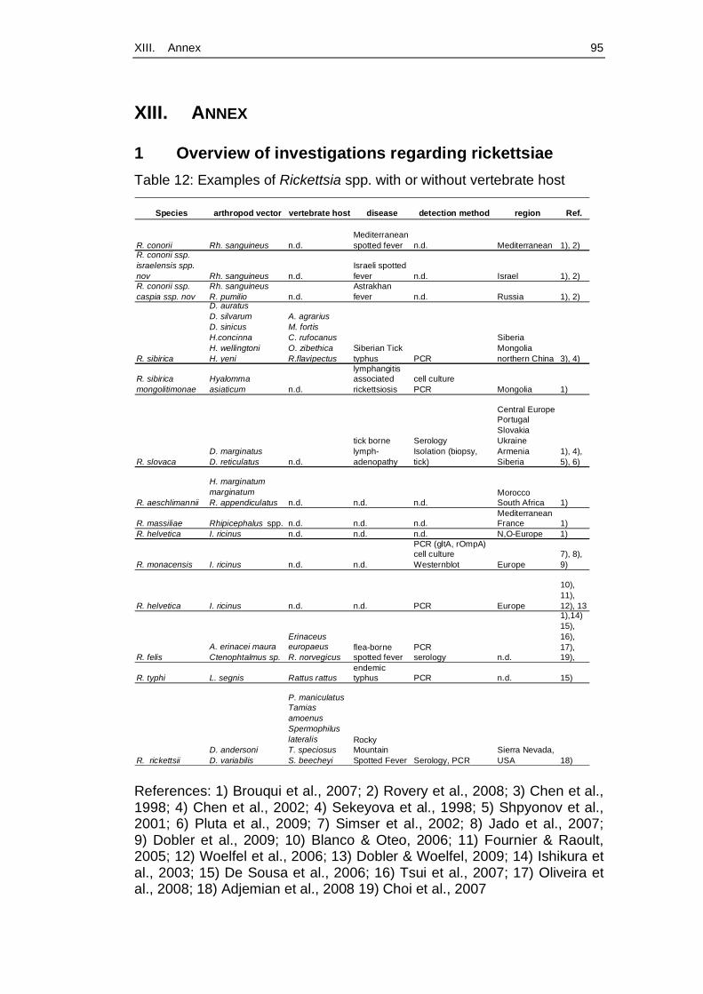
XIII. Annex 95
XIII. ANNEX
1 Overview of investigations regarding rickettsiae Table 12: Examples of Rickettsia spp. with or without vertebrate host
Species arthropod vector vertebrate host disease detection method region Ref.
R. conorii Rh. sanguineus n.d.Mediterranean spotted fever n.d. Mediterranean 1), 2)
R. conorii ssp. israelensis spp. nov Rh. sanguineus n.d.
Israeli spotted fever n.d. Israel 1), 2)
R. conorii ssp. caspia ssp. nov
Rh. sanguineusR. pumilio n.d.
Astrakhan fever n.d. Russia 1), 2)
R. sibirica
D. auratusD. silvarumD. sinicusH.concinnaH. wellingtoniH. yeni
A. agrariusM. fortisC. rufocanusO. zibethicaR.flavipectus
Siberian Tick typhus PCR
SiberiaMongolianorthern China 3), 4)
R. sibirica mongolitimonae
Hyalomma asiaticum n.d.
lymphangitis associated rickettsiosis
cell culturePCR Mongolia 1)
R. slovacaD. marginatusD. reticulatus n.d.
tick borne lymph-adenopathy
SerologyIsolation (biopsy, tick)
Central EuropePortugalSlovakiaUkraineArmeniaSiberia
1), 4), 5), 6)
R. aeschlimannii
H. marginatum marginatumR. appendiculatus n.d. n.d. n.d.
MoroccoSouth Africa 1)
R. massiliae Rhipicephalus spp. n.d. n.d. n.d.Mediterranean France 1)
R. helvetica I. ricinus n.d. n.d. n.d. N,O-Europe 1)
R. monacensis I. ricinus n.d. n.d.
PCR (gltA, rOmpA)cell cultureWesternblot Europe
7), 8), 9)
R. helvetica I. ricinus n.d. n.d. PCR Europe
10), 11), 12), 13
R. felisA. erinacei mauraCtenophtalmus sp.
Erinaceus europaeusR. norvegicus
flea-borne spotted fever
PCRserology n.d.
1),14) 15), 16), 17), 19),
R. typhi L. segnis Rattus rattusendemic typhus PCR n.d. 15)
R. rickettsiiD. andersoniD. variabilis
P. maniculatusTamias amoenusSpermophilus lateralisT. speciosusS. beecheyi
Rocky Mountain Spotted Fever Serology, PCR
Sierra Nevada, USA 18)
References: 1) Brouqui et al., 2007; 2) Rovery et al., 2008; 3) Chen et al., 1998; 4) Chen et al., 2002; 4) Sekeyova et al., 1998; 5) Shpyonov et al., 2001; 6) Pluta et al., 2009; 7) Simser et al., 2002; 8) Jado et al., 2007; 9) Dobler et al., 2009; 10) Blanco & Oteo, 2006; 11) Fournier & Raoult, 2005; 12) Woelfel et al., 2006; 13) Dobler & Woelfel, 2009; 14) Ishikura et al., 2003; 15) De Sousa et al., 2006; 16) Tsui et al., 2007; 17) Oliveira et al., 2008; 18) Adjemian et al., 2008 19) Choi et al., 2007
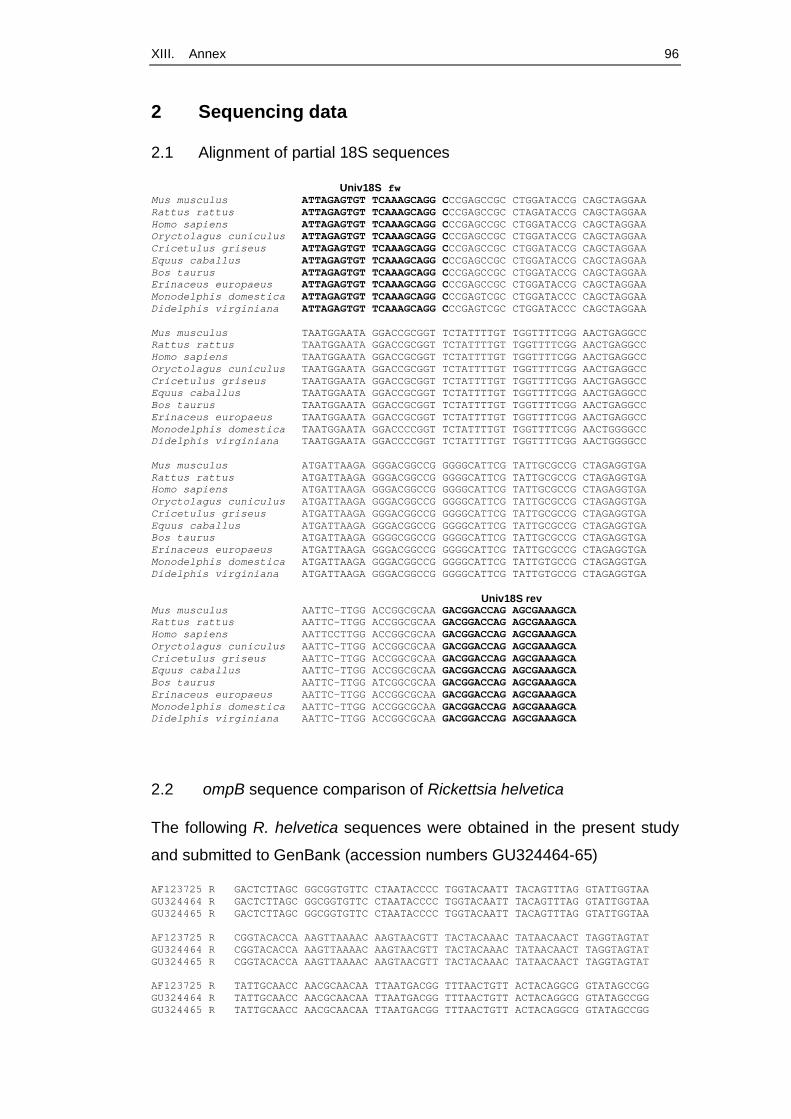
XIII. Annex 96
2 Sequencing data
2.1 Alignment of partial 18S sequences
Univ18S fw Mus musculus ATTAGAGTGT TCAAAGCAGG CCCGAGCCGC CTGGATACCG CAGCTAGGAA Rattus rattus ATTAGAGTGT TCAAAGCAGG CCCGAGCCGC CTAGATACCG CAGCTAGGAA Homo sapiens ATTAGAGTGT TCAAAGCAGG CCCGAGCCGC CTGGATACCG CAGCTAGGAA Oryctolagus cuniculus ATTAGAGTGT TCAAAGCAGG CCCGAGCCGC CTGGATACCG CAGCTAGGAA Cricetulus griseus ATTAGAGTGT TCAAAGCAGG CCCGAGCCGC CTGGATACCG CAGCTAGGAA Equus caballus ATTAGAGTGT TCAAAGCAGG CCCGAGCCGC CTGGATACCG CAGCTAGGAA Bos taurus ATTAGAGTGT TCAAAGCAGG CCCGAGCCGC CTGGATACCG CAGCTAGGAA Erinaceus europaeus ATTAGAGTGT TCAAAGCAGG CCCGAGCCGC CTGGATACCG CAGCTAGGAA Monodelphis domestica ATTAGAGTGT TCAAAGCAGG CCCGAGTCGC CTGGATACCC CAGCTAGGAA Didelphis virginiana ATTAGAGTGT TCAAAGCAGG CCCGAGTCGC CTGGATACCC CAGCTAGGAA Mus musculus TAATGGAATA GGACCGCGGT TCTATTTTGT TGGTTTTCGG AACTGAGGCC Rattus rattus TAATGGAATA GGACCGCGGT TCTATTTTGT TGGTTTTCGG AACTGAGGCC Homo sapiens TAATGGAATA GGACCGCGGT TCTATTTTGT TGGTTTTCGG AACTGAGGCC Oryctolagus cuniculus TAATGGAATA GGACCGCGGT TCTATTTTGT TGGTTTTCGG AACTGAGGCC Cricetulus griseus TAATGGAATA GGACCGCGGT TCTATTTTGT TGGTTTTCGG AACTGAGGCC Equus caballus TAATGGAATA GGACCGCGGT TCTATTTTGT TGGTTTTCGG AACTGAGGCC Bos taurus TAATGGAATA GGACCGCGGT TCTATTTTGT TGGTTTTCGG AACTGAGGCC Erinaceus europaeus TAATGGAATA GGACCGCGGT TCTATTTTGT TGGTTTTCGG AACTGAGGCC Monodelphis domestica TAATGGAATA GGACCCCGGT TCTATTTTGT TGGTTTTCGG AACTGGGGCC Didelphis virginiana TAATGGAATA GGACCCCGGT TCTATTTTGT TGGTTTTCGG AACTGGGGCC Mus musculus ATGATTAAGA GGGACGGCCG GGGGCATTCG TATTGCGCCG CTAGAGGTGA Rattus rattus ATGATTAAGA GGGACGGCCG GGGGCATTCG TATTGCGCCG CTAGAGGTGA Homo sapiens ATGATTAAGA GGGACGGCCG GGGGCATTCG TATTGCGCCG CTAGAGGTGA Oryctolagus cuniculus ATGATTAAGA GGGACGGCCG GGGGCATTCG TATTGCGCCG CTAGAGGTGA Cricetulus griseus ATGATTAAGA GGGACGGCCG GGGGCATTCG TATTGCGCCG CTAGAGGTGA Equus caballus ATGATTAAGA GGGACGGCCG GGGGCATTCG TATTGCGCCG CTAGAGGTGA Bos taurus ATGATTAAGA GGGGCGGCCG GGGGCATTCG TATTGCGCCG CTAGAGGTGA Erinaceus europaeus ATGATTAAGA GGGACGGCCG GGGGCATTCG TATTGCGCCG CTAGAGGTGA Monodelphis domestica ATGATTAAGA GGGACGGCCG GGGGCATTCG TATTGTGCCG CTAGAGGTGA Didelphis virginiana ATGATTAAGA GGGACGGCCG GGGGCATTCG TATTGTGCCG CTAGAGGTGA Univ18S rev Mus musculus AATTC-TTGG ACCGGCGCAA GACGGACCAG AGCGAAAGCA Rattus rattus AATTC-TTGG ACCGGCGCAA GACGGACCAG AGCGAAAGCA Homo sapiens AATTCCTTGG ACCGGCGCAA GACGGACCAG AGCGAAAGCA Oryctolagus cuniculus AATTC-TTGG ACCGGCGCAA GACGGACCAG AGCGAAAGCA Cricetulus griseus AATTC-TTGG ACCGGCGCAA GACGGACCAG AGCGAAAGCA Equus caballus AATTC-TTGG ACCGGCGCAA GACGGACCAG AGCGAAAGCA Bos taurus AATTC-TTGG ATCGGCGCAA GACGGACCAG AGCGAAAGCA Erinaceus europaeus AATTC-TTGG ACCGGCGCAA GACGGACCAG AGCGAAAGCA Monodelphis domestica AATTC-TTGG ACCGGCGCAA GACGGACCAG AGCGAAAGCA Didelphis virginiana AATTC-TTGG ACCGGCGCAA GACGGACCAG AGCGAAAGCA
2.2 ompB sequence comparison of Rickettsia helvetica
The following R. helvetica sequences were obtained in the present study
and submitted to GenBank (accession numbers GU324464-65)
AF123725 R GACTCTTAGC GGCGGTGTTC CTAATACCCC TGGTACAATT TACAGTTTAG GTATTGGTAA GU324464 R GACTCTTAGC GGCGGTGTTC CTAATACCCC TGGTACAATT TACAGTTTAG GTATTGGTAA GU324465 R GACTCTTAGC GGCGGTGTTC CTAATACCCC TGGTACAATT TACAGTTTAG GTATTGGTAA AF123725 R CGGTACACCA AAGTTAAAAC AAGTAACGTT TACTACAAAC TATAACAACT TAGGTAGTAT GU324464 R CGGTACACCA AAGTTAAAAC AAGTAACGTT TACTACAAAC TATAACAACT TAGGTAGTAT GU324465 R CGGTACACCA AAGTTAAAAC AAGTAACGTT TACTACAAAC TATAACAACT TAGGTAGTAT AF123725 R TATTGCAACC AACGCAACAA TTAATGACGG TTTAACTGTT ACTACAGGCG GTATAGCCGG GU324464 R TATTGCAACC AACGCAACAA TTAATGACGG TTTAACTGTT ACTACAGGCG GTATAGCCGG GU324465 R TATTGCAACC AACGCAACAA TTAATGACGG TTTAACTGTT ACTACAGGCG GTATAGCCGG
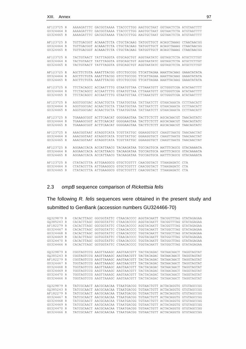
XIII. Annex 97
AF123725 R AAAAGATTTC GACGGTAAAA TTACCCTTGG AAGTGCTAAT GGTAACTCTA ATGTAATTTT GU324464 R AAAAGATTTC GACGGTAAAA TTACCCTTGG AAGTGCTAAT GGTAACTCTA ATGTAATTTT GU324465 R AAAAGATTTC GACGGTAAAA TTACCCTTGG AAGTGCTAAT GGTAACTCTA ATGTAATTTT AF123725 R TGTTGACGGT ACAAACTCTA CTGCTACAAG TATGGTTGCT ACAGCTAAAG CTAATAACGG GU324464 R TGTTGACGGT ACAAACTCTA CTGCTACAAG TATGGTTGCT ACAGCTAAAG CTAATAACGG GU324465 R TGTTGACGGT ACAAACTCTA CTGCTACAAG TATGGTTGCT ACAGCTAAAG CTAATAACGG AF123725 R TACTGTAACT TATTTAGGTA GTGCAGCTGT AGGTAATATC GGTAGCTCTA ATGCTCTTGT GU324464 R TACTGTAACT TATTTAGGTA GTGCAGCTGT AGGTAATATC GGTAGCTCTA ATGCTCTTGT GU324465 R TACTGTAACT TATTTAGGTA GTGCAGCTGT AGGTAATATC GGTAGCTCTA ATGCTCTTGT AF123725 R AGCTTCTGTA AAATTTACGG GTCCTGCCGG TTCATTAGAA AAATTACAAG GAAATATATA GU324464 R AGCTTCTGTA AAATTTACGG GTCCTGCCGG TTCATTAGAA AAATTACAAG GAAATATATA GU324465 R AGCTTCTGTA AAATTTACGG GTCCTGCCGG TTCATTAGAA AAATTACAAG GAAATATATA AF123725 R TTCTACAGCC ACCAATTTTG GTAATGTTAA CTTAAATGTT GCTGGGTCGA ATATAATTTT GU324464 R TTCTACAGCC ACCAATTTTG GTAATGTTAA CTTAAATGTT GCTGGGTCGA ATATAATTTT GU324465 R TTCTACAGCC ACCAATTTTG GTAATGTTAA CTTAAATGTT GCTGGGTCGA ATATAATTTT AF123725 R AGGTGGCGAC ACAACTGCTA TTAATGGTAA TATTAATCTT GTAACAAATA CCTTAACATT GU324464 R AGGTGGCGAC ACAACTGCTA TTAATGGTAA TATTAATCTT GTAACAAATA CCTTAACATT GU324465 R AGGTGGCGAC ACAACTGCTA TTAATGGTAA TATTAATCTT GTAACAAATA CCTTAACATT AF123725 R TGAAAGCGGT ACTTCAACAT GGGGAAGTAA TACTTCTCTT AGCACAACGT TAACAGTATC GU324464 R TGAAAGCGGT ACTTCAACAT GGGGAAGTAA TACTTCTCTT AGCACAACGT TAACAGTATC GU324465 R TGAAAGCGGT ACTTCAACAT GGGGAAGTAA TACTTCTCTT AGCACAACGT TAACAGTATC AF123725 R AAACGGTAAT ATAGGTCATA TCGTTATTGC GGAAGGTGCT CAAGTTAATG TAACAACTAT GU324464 R AAACGGTAAT ATAGGTCATA TCGTTATTGC GGAAGGTGCT CAAGTTAATG TAACAACTAT GU324465 R AAACGGTAAT ATAGGTCATA TCGTTATTGC GGAAGGTGCT CAAGTTAATG TAACAACTAT AF123725 R AGGAACCACA ACCATTAACG TACAAGATAA TGCCAGTGCA AATTTCAGCG GTACAAAATA GU324464 R AGGAACCACA ACCATTAACG TACAAGATAA TGCCAGTGCA AATTTCAGCG GTACAAAATA GU324465 R AGGAACCACA ACCATTAACG TACAAGATAA TGCCAGTGCA AATTTCAGCG GTACAAAATA AF123725 R CTATACCTTA ATTGAAGGCG GTGCTCGTTT CAACGGTACT TTAAGAGATC CTA GU324464 R CTATACCTTA ATTGAAGGCG GTGCTCGTTT CAACGGTACT TTAAGAGATC CTA GU324465 R CTATACCTTA ATTGAAGGCG GTGCTCGTTT CAACGGTACT TTAAGAGATC CTA
2.3 ompB sequence comparison of Rickettsia felis
The following R. felis sequences were obtained in the present study and
submitted to GenBank (accession numbers GU324466-70)
GQ329879 R CACACTTAGC GGCGGTATTC CTAACACCCC AGGTACAATT TACGGTTTAG GTATAGAGAA GQ385243 R CACACTTAGC GGCGGTATTC CTAACACCCC AGGTACAATT TACGGTTTAG GTATAGAGAA AF182279 R CACACTTAGC GGCGGTATTC CTAACACCCC AGGTACAATT TACGGTTTAG GTATAGAGAA GU324467 R CACACTTAGC GGTGGTATTC CTAACACCCC TGGTACAATT TATGGCTTAG GTATAGAGAA GU324468 R CACACTTAGC GGTGGTATTC CTAACACCCC TGGTACAATT TATGGCTTAG GTATAGAGAA GU324469 R CACACTTAGC GGTGGTATTC CTAACACCCC TGGTACAATT TATGGCTTAG GTATAGAGAA GU324470 R CACACTTAGC GGTGGTATTC CTAACACCCC TGGTACAATT TATGGCTTAG GTATAGAGAA GU324466 R CACACTTAGC GGTGGTATTC CTAACACCCC TGGTACAATT TATGGCTTAG GTATAGAGAA GQ329879 R CGGTAGTCCG AAGTTAAAGC AAGTAACGTT TACTACAGAC TATAACAACT TAGGTAGTAT GQ385243 R CGGTAGTCCG AAGTTAAAGC AAGTAACGTT TACTACAGAC TATAACAACT TAGGTAGTAT AF182279 R CGGTAGTCCG AAGTTAAAGC AAGTAACGTT TACTACAGAC TATAACAACT TAGGTAGTAT GU324467 R TGGTAGTCCG AAGTTAAAGC AAGTAACGTT TACTACAGAC TATAACAACT TAGGTAGTAT GU324468 R TGGTAGTCCG AAGTTAAAGC AAGTAACGTT TACTACAGAC TATAACAACT TAGGTAGTAT GU324469 R TGGTAGTCCG AAGTTAAAGC AAGTAACGTT TACTACAGAC TATAACAACT TAGGTAGTAT GU324470 R TGGTAGTCCG AAGTTAAAGC AAGTAACGTT TACTACAGAC TATAACAACT TAGGTAGTAT GU324466 R TGGTAGTCCG AAGTTAAAGC AAGTAACGTT TACTACAGAC TATAACAACT TAGGTAGTAT GQ329879 R TATCGCAACT AACGCAACAA TTAATGACGG TGTAACTGTT ACTACAGGTG GTGTAGCCGG GQ385243 R TATCGCAACT AACGCAACAA TTAATGACGG TGTAACTGTT ACTACAGGTG GTGTAGCCGG AF182279 R TATCGCAACT AACGCAACAA TTAATGACGG TGTAACTGTT ACTACAGGTG GTGTAGCCGG GU324467 R TATCGCAACT AACGCAACAA TTAATGACGG TGTAACTGTT ACTACAGGTG GTGTAGCCGG GU324468 R TATCGCAACT AACGCAACAA TTAATGACGG TGTAACTGTT ACTACAGGTG GTGTAGCCGG GU324469 R TATCGCAACT AACGCAACAA TTAATGACGG TGTAACTGTT ACTACAGGTG GTGTAGCCGG GU324470 R TATCGCAACT AACGCAACAA TTAATGACGG TGTAACTGTT ACTACAGGTG GTGTAGCCGG GU324466 R TATCGCAACT AACGCAACAA TTAATGACGG TGTAACTGTT ACTACAGGTG GTGTAGCCGG
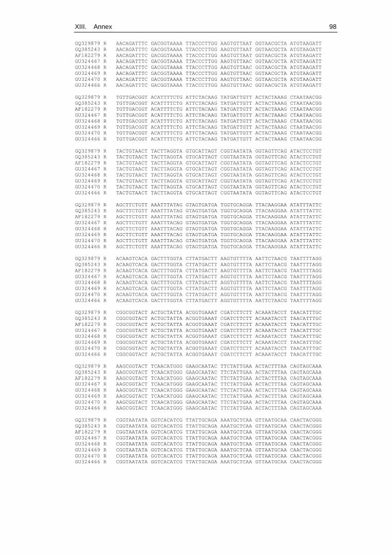
XIII. Annex 98
GQ329879 R AACAGATTTC GACGGTAAAA TTACCCTTGG AAGTGTTAAT GGTAACGCTA ATGTAAGATT GQ385243 R AACAGATTTC GACGGTAAAA TTACCCTTGG AAGTGTTAAT GGTAACGCTA ATGTAAGATT AF182279 R AACAGATTTC GACGGTAAAA TTACCCTTGG AAGTGTTAAT GGTAACGCTA ATGTAAGATT GU324467 R AACAGATTTC GACGGTAAAA TTACCCTTGG AAGTGTTAAC GGTAACGCTA ATGTAAGATT GU324468 R AACAGATTTC GACGGTAAAA TTACCCTTGG AAGTGTTAAC GGTAACGCTA ATGTAAGATT GU324469 R AACAGATTTC GACGGTAAAA TTACCCTTGG AAGTGTTAAC GGTAACGCTA ATGTAAGATT GU324470 R AACAGATTTC GACGGTAAAA TTACCCTTGG AAGTGTTAAC GGTAACGCTA ATGTAAGATT GU324466 R AACAGATTTC GACGGTAAAA TTACCCTTGG AAGTGTTAAC GGTAACGCTA ATGTAAGATT GQ329879 R TGTTGACGGT ACATTTTCTG ATTCTACAAG TATGATTGTT ACTACTAAAG CTAATAACGG GQ385243 R TGTTGACGGT ACATTTTCTG ATTCTACAAG TATGATTGTT ACTACTAAAG CTAATAACGG AF182279 R TGTTGACGGT ACATTTTCTG ATTCTACAAG TATGATTGTT ACTACTAAAG CTAATAACGG GU324467 R TGTTGACGGT ACATTTTCTG ATTCTACAAG TATGATTGTT ACTACTAAAG CTAATAACGG GU324468 R TGTTGACGGT ACATTTTCTG ATTCTACAAG TATGATTGTT ACTACTAAAG CTAATAACGG GU324469 R TGTTGACGGT ACATTTTCTG ATTCTACAAG TATGATTGTT ACTACTAAAG CTAATAACGG GU324470 R TGTTGACGGT ACATTTTCTG ATTCTACAAG TATGATTGTT ACTACTAAAG CTAATAACGG GU324466 R TGTTGACGGT ACATTTTCTG ATTCTACAAG TATGATTGTT ACTACTAAAG CTAATAACGG GQ329879 R TACTGTAACT TACTTAGGTA GTGCATTAGT CGGTAATATA GGTAGTTCAG ATACTCCTGT GQ385243 R TACTGTAACT TACTTAGGTA GTGCATTAGT CGGTAATATA GGTAGTTCAG ATACTCCTGT AF182279 R TACTGTAACT TACTTAGGTA GTGCATTAGT CGGTAATATA GGTAGTTCAG ATACTCCTGT GU324467 R TACTGTAACT TACTTAGGTA GTGCATTAGT CGGTAATATA GGTAGTTCAG ATACTCCTGT GU324468 R TACTGTAACT TACTTAGGTA GTGCATTAGT CGGTAATATA GGTAGTTCAG ATACTCCTGT GU324469 R TACTGTAACT TACTTAGGTA GTGCATTAGT CGGTAATATA GGTAGTTCAG ATACTCCTGT GU324470 R TACTGTAACT TACTTAGGTA GTGCATTAGT CGGTAATATA GGTAGTTCAG ATACTCCTGT GU324466 R TACTGTAACT TACTTAGGTA GTGCATTAGT CGGTAATATA GGTAGTTCAG ATACTCCTGT GQ329879 R AGCTTCTGTT AAATTTATAG GTAGTGATGA TGGTGCAGGA TTACAAGGAA ATATTTATTC GQ385243 R AGCTTCTGTT AAATTTATAG GTAGTGATGA TGGTGCAGGA TTACAAGGAA ATATTTATTC AF182279 R AGCTTCTGTT AAATTTATAG GTAGTGATGA TGGTGCAGGA TTACAAGGAA ATATTTATTC GU324467 R AGCTTCTGTT AAATTTACAG GTAGTGATGA TGGTGCAGGA TTACAAGGAA ATATTTATTC GU324468 R AGCTTCTGTT AAATTTACAG GTAGTGATGA TGGTGCAGGA TTACAAGGAA ATATTTATTC GU324469 R AGCTTCTGTT AAATTTACAG GTAGTGATGA TGGTGCAGGA TTACAAGGAA ATATTTATTC GU324470 R AGCTTCTGTT AAATTTACAG GTAGTGATGA TGGTGCAGGA TTACAAGGAA ATATTTATTC GU324466 R AGCTTCTGTT AAATTTACAG GTAGTGATGA TGGTGCAGGA TTACAAGGAA ATATTTATTC GQ329879 R ACAAGTCACA GACTTTGGTA CTTATGACTT AAGTGTTTTA AATTCTAACG TAATTTTAGG GQ385243 R ACAAGTCACA GACTTTGGTA CTTATGACTT AAGTGTTTTA AATTCTAACG TAATTTTAGG AF182279 R ACAAGTCACA GACTTTGGTA CTTATGACTT AAGTGTTTTA AATTCTAACG TAATTTTAGG GU324467 R ACAAGTCACA GACTTTGGTA CTTATGACTT AGGTGTTTTA AATTCTAACG TAATTTTAGG GU324468 R ACAAGTCACA GACTTTGGTA CTTATGACTT AGGTGTTTTA AATTCTAACG TAATTTTAGG GU324469 R ACAAGTCACA GACTTTGGTA CTTATGACTT AGGTGTTTTA AATTCTAACG TAATTTTAGG GU324470 R ACAAGTCACA GACTTTGGTA CTTATGACTT AGGTGTTTTA AATTCTAACG TAATTTTAGG GU324466 R ACAAGTCACA GACTTTGGTA CTTATGACTT AGGTGTTTTA AATTCTAACG TAATTTTAGG GQ329879 R CGGCGGTACT ACTGCTATTA ACGGTGAAAT CGATCTTCTT ACAAATACCT TAACATTTGC GQ385243 R CGGCGGTACT ACTGCTATTA ACGGTGAAAT CGATCTTCTT ACAAATACCT TAACATTTGC AF182279 R CGGCGGTACT ACTGCTATTA ACGGTGAAAT CGATCTTCTT ACAAATACCT TAACATTTGC GU324467 R CGGCGGTACT ACTGCTATTA ACGGTGAAAT CGATCTTCTT ACAAATACCT TAACATTTGC GU324468 R CGGCGGTACT ACTGCTATTA ACGGTGAAAT CGATCTTCTT ACAAATACCT TAACATTTGC GU324469 R CGGCGGTACT ACTGCTATTA ACGGTGAAAT CGATCTTCTT ACAAATACCT TAACATTTGC GU324470 R CGGCGGTACT ACTGCTATTA ACGGTGAAAT CGATCTTCTT ACAAATACCT TAACATTTGC GU324466 R CGGCGGTACT ACTGCTATTA ACGGTGAAAT CGATCTTCTT ACAAATACCT TAACATTTGC GQ329879 R AAGCGGTACT TCAACATGGG GAAGCAATAC TTCTATTGAA ACTACTTTAA CAGTAGCAAA GQ385243 R AAGCGGTACT TCAACATGGG GAAGCAATAC TTCTATTGAA ACTACTTTAA CAGTAGCAAA AF182279 R AAGCGGTACT TCAACATGGG GAAGCAATAC TTCTATTGAA ACTACTTTAA CAGTAGCAAA GU324467 R AAGCGGTACT TCAACATGGG GAAGCAATAC TTCTATTGAA ACTACTTTAA CAGTAGCAAA GU324468 R AAGCGGTACT TCAACATGGG GAAGCAATAC TTCTATTGAA ACTACTTTAA CAGTAGCAAA GU324469 R AAGCGGTACT TCAACATGGG GAAGCAATAC TTCTATTGAA ACTACTTTAA CAGTAGCAAA GU324470 R AAGCGGTACT TCAACATGGG GAAGCAATAC TTCTATTGAA ACTACTTTAA CAGTAGCAAA GU324466 R AAGCGGTACT TCAACATGGG GAAGCAATAC TTCTATTGAA ACTACTTTAA CAGTAGCAAA GQ329879 R CGGTAATATA GGTCACATCG TTATTGCAGA AAATGCTCAA GTTAATGCAA CAACTACGGG GQ385243 R CGGTAATATA GGTCACATCG TTATTGCAGA AAATGCTCAA GTTAATGCAA CAACTACGGG AF182279 R CGGTAATATA GGTCACATCG TTATTGCAGA AAATGCTCAA GTTAATGCAA CAACTACGGG GU324467 R CGGTAATATA GGTCACATCG TTATTGCAGA AAATGCTCAA GTTAATGCAA CAACTACGGG GU324468 R CGGTAATATA GGTCACATCG TTATTGCAGA AAATGCTCAA GTTAATGCAA CAACTACGGG GU324469 R CGGTAATATA GGTCACATCG TTATTGCAGA AAATGCTCAA GTTAATGCAA CAACTACGGG GU324470 R CGGTAATATA GGTCACATCG TTATTGCAGA AAATGCTCAA GTTAATGCAA CAACTACGGG GU324466 R CGGTAATATA GGTCACATCG TTATTGCAGA AAATGCTCAA GTTAATGCAA CAACTACGGG

XIII. Annex 99
GQ329879 R AACAACAACT ATTAACGTAC AAGATAATGC CAATGCAAAT TTCAGCGATA CGCAAACTTA GQ385243 R AACAACAACT ATTAACGTAC AAGATAATGC CAATGCAAAT TTCAGCGATA CGCAAACTTA AF182279 R AACAACAACT ATTAACGTAC AAGATAATGC CAATGCAAAT TTCAGCGATA CGCAAACTTA GU324467 R AACAACAACT ATTAACGTAC AAGATAATGC CAATGCAAAT TTCAGCGGTA CACAAACTTA GU324468 R AACAACAACT ATTAACGTAC AAGATAATGC CAATGCAAAT TTCAGCGGTA CACAAACTTA GU324469 R AACAACAACT ATTAACGTAC AAGATAATGC CAATGCAAAT TTCAGCGGTA CACAAACTTA GU324470 R AACAACAACT ATTAACGTAC AAGATAATGC CAATGCAAAT TTCAGCGGTA CACAAACTTA GU324466 R AACAACAACT ATTAACGTAC AAGATAATGC CAATGCAAAT TTCAGCGGTA CACAAACTTA GQ329879 R TACTTTAATC CAAGGTGGTG CTAGATTTAA CGGTACTTTA GGAGGTCCTA GQ385243 R TACTTTAATC CAAGGTGGTG CTAGATTTAA CGGTACTTTA GGAGGTCCTA AF182279 R TACTTTAATC CAAGGTGGTG CTAGATTTAA CGGTACTTTA GGAGGTCCTA GU324467 R TACTTTAATC CAAGGTGGTG CTAGATTTAA CGGTACTTTA GGAGGTCCTA GU324468 R TACTTTAATC CAAGGTGGTG CTAGATTTAA CGGTACTTTA GGAGGTCCTA GU324469 R TACTTTAATC CAAGGTGGTG CTAGATTTAA CGGTACTTTA GGAGGTCCTA GU324470 R TACTTTAATC CAAGGTGGTG CTAGATTTAA CGGTACTTTA GGAGGTCCTA GU324466 R TACTTTAATC CAAGGTGGTG CTAGATTTAA GGGTACTTTA GGAGGTCCTA
3 Trapping locations
Figure 14: Topographic map of Lower Bavaria; trapping sites and transects from 2008 indicated with arrows
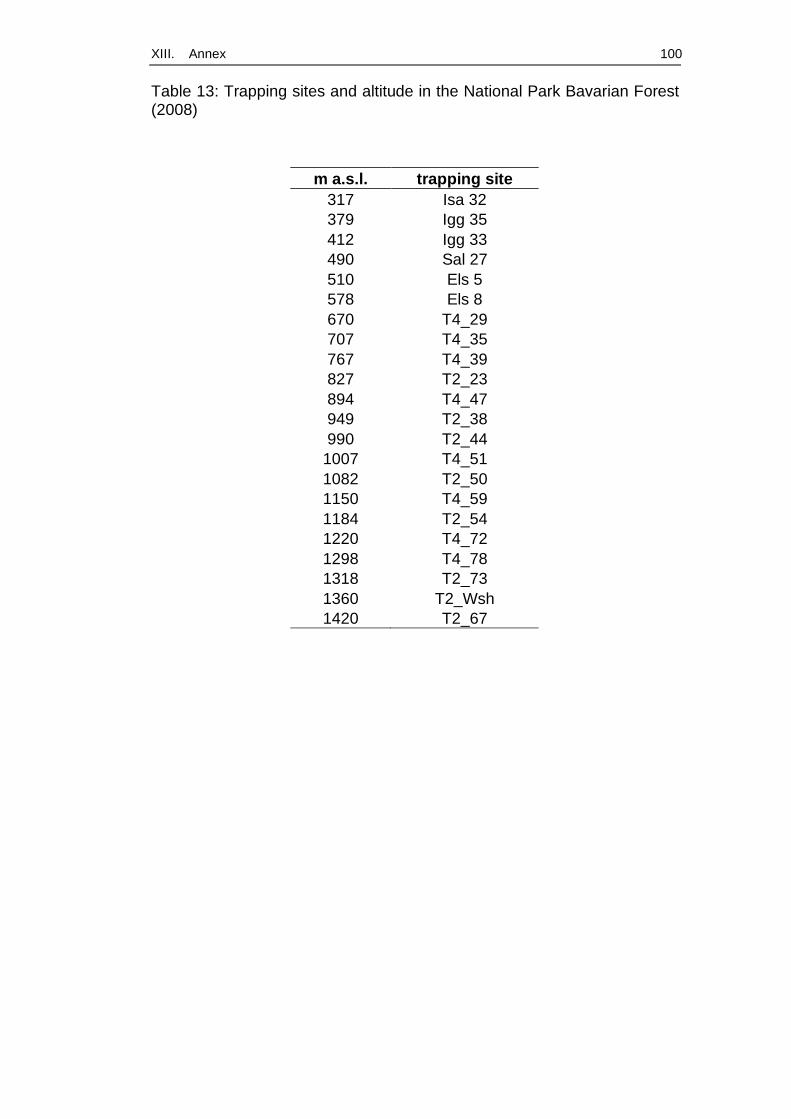
XIII. Annex 100
Table 13: Trapping sites and altitude in the National Park Bavarian Forest (2008)
m a.s.l. trapping site 317 Isa 32 379 Igg 35 412 Igg 33 490 Sal 27 510 Els 5 578 Els 8 670 T4_29 707 T4_35 767 T4_39 827 T2_23 894 T4_47 949 T2_38 990 T2_44
1007 T4_51 1082 T2_50 1150 T4_59 1184 T2_54 1220 T4_72 1298 T4_78 1318 T2_73 1360 T2_Wsh 1420 T2_67

XIII. Annex 101
Figure 15: Topographic map (1:200000) of trapping sites from 2004 and 2005. 1: Falkenstein, 2: Glashütte, 3: Raimundsreuth, 4: Hangenleithen, 5: Mutzenwinkel 6: Langfurth, 7: Schöfweg (generated with Top50, Version 3.0, Bayerisches Landesvermessungsamt Muenchen, 2001)
4 Publication „Nagetier-übertragene Zoonosen: Beispiele aus Untersuchungen in Süd- und Westdeutschland“
Wirbeltierforschung in der Kulturlandschaft
Mitt. Julius Kühn-Inst. 421, 2009 37

XIII. Annex 102
Wirbeltierforschung in der Kulturlandschaft Mitt. Julius Kühn-Inst. 421, 2009 37
Nagetier-übertragene Zoonosen: Beispiele aus Untersuchungen in Süd- und
Westdeutschland
Essbauer, S.S.;1 Schex, S.1; Splettstoesser, W.1; Pfeffer, M.4; Ulrich, R.G.2; Seibold, E.1; Dobler, G.1; Wölfel, R.1; Bäumler,
W.3
1Institut für Mikrobiologie der Bundeswehr, München 2Friedrich-Loeffler-Institut, Institut für neue und neuartige Tierseuchenerreger, Greifswald - Insel Riems 3em. Studienfakultät für Forstwissenschaft & Ressourcenmanagement, Freising 4Institut für Tierhygiene und Öffentliches Veterinärwesen, Leipzig
Zusammenfassung
Nagetiere und andere Kleinsäuger können eine Vielzahl von Krankheitserregern, RNA- und DNA-Viren,
Bakterien und Parasiten, auf den Menschen übertragen, die teilweise lebensbedrohliche Erkrankungen
hervorrufen. In der folgenden Übersicht soll erstmals ein Überblick über Ergebnisse aus drei Untersuchungen
in Deutschland gegeben werden: eine Studie in drei Landkreisen Bayerns von 2001-2004, Untersuchungen in
einem Freilandgehege im Rahmen eines Tularämieausbruchs in Niedersachsen im Jahr 2005, und schließlich
im Jahr 2007 eine Untersuchung an einem Truppenübungsplatz in Baden-Württemberg. Es wurde dabei
exemplarisch die Verbreitung von Zoonoseerregern in Nagetieren und anderen Kleinsäugern näher
untersucht, von drei Viren (Hantaviren, Kuhpockenvirus, Frühsommer-Meningo-Enzephalitis-Virus) und vier
Bakterien (Leptospiren, Francisellen, Borrelien und Rickettsien). Die hier zusammengefassten Erkenntnisse
sind ein erster wichtiger Schritt auf dem Weg zur Erstellung von Verbreitungskarten für die genannten
humanpathogenen Zoonoseerreger in ihren Reservoirwirten und der Definition von entsprechenden
Risikogebieten. Diese Arbeit soll zudem einen Beitrag leisten, einen Anstoß zu verstärkter Zusammenarbeit
von Zoologen, Ökologen, Virologen, Human- und Veterinärmedizinern, Mikrobiologen, Parasitologen,
Genetikern, Epidemiologen, Forstwissenschaftlern und Klimaforschern zu geben.
1. Nagetier-übertragene Krankheitserreger: eine Einleitung
Zoonosen sind Infektionskrankheiten, bei denen der Erreger vom oftmals nicht erkrankten Tierreservoirwirt
auf den Menschen übertragen wird. Bei den mit Nagetieren und anderen Kleinsäugern assoziierten
Zoonoseerregern handelt es sich um verschiedene RNA- und DNA-Viren, Bakterien und Parasiten. Sie
unterscheiden sich nicht nur in ihrer genetischen Organisation, sondern vor allem in ihrer Assoziation mit
spezifischen Reservoirwirten, ihrer geografischen Verbreitung und ihren Übertragungwegen. Diese
Krankheitserreger können beim Menschen verschiedene, zum Teil lebensbedrohliche Erkrankungen
hervorrufen.
Am Institut für Mikrobiologie der Bundeswehr (IMB) werden in Kooperation mit anderen Institutionen seit
dem Jahr 2004 Untersuchungen zu Nagetier- und Kleinsäuger-übertragenen Krankheitserregern durchgeführt.

XIII. Annex 103
Diese Studien sind sowohl in Bundeswehr-internem Interesse, z.B. auf Anfragen von Kommandohygienikern,
als auch im Rahmen einer zivil-militärischen Zusammenarbeit von großer Wichtigkeit für öffentliche
Gesundheitsbehörden. Die bisherigen Felduntersuchungen wurden entweder aufgrund von
Ausbruchsgeschehen (Hantaviren, Tularämie) initiiert oder dienten der Surveillance, um z.B. für
Truppenübungsplätze eine Risikoabschätzung durchführen zu können.
1.1 Übertragungswege der Erreger
Der Mensch kann sich sowohl auf direktem als auch auf indirektem Wege mit Zoonoseerregern infizieren.
Epidemiologisch gesehen stellt der Mensch meist einen Fehlwirt dar, d.h. die Infektion wird von ihm nicht
auf weitere Personen übertragen. Bei den hier betrachteten Erregern sind Nagetiere (oder andere Kleinsäuger,
wie Spitzmäuse oder Hasen) Reservoirwirt und Vektor.
Eine direkte Infektion kann über Urin, Speichel und Kot von Tieren erfolgen, wie es z.B. bei Infektionen mit
Hantaviren, den Erregern der Tularämie (Francisella tularensis) und Leptospirose, Q-Fieber, und Arenavirus-
Infektion bekannt ist. Eine Infektion über das Fell von toten oder lebenden Tieren kann bei Tularämie, bei der
Lymphozytären Choriomeningitis (LCM) oder in manchen Fällen bei der Pest stattfinden. Durch Bisse
werden Rattenbisskrankheit (Spirillum minus, Streptobacillus moniliformis), Leptospiren und
Kuhpockenviren übertragen. Über die Aufnahme kontaminierter Nahrungsmittel, vor allem dem sogenannten
„bush meat“, können, vornehmlich in Afrika, auch Menschen z.B. mit den Erregern der Affenpocken infiziert
werden oder an Pest oder Lassa erkranken.
Eine indirekte Infektion kann über verschiedene blutsaugende Vektoren erfolgen. Einen wichtigen Vektor
stellen Arthropoden dar, die durch Stiche oder Bisse beispielsweise Anaplasmen, Arenaviren, Alphaviren,
Bunyaviren, Flaviviren wie das Frühsommer-Meningo-Enzephalitis-Virus (FSMEV), Borrelien und
Francisellen vom Nagetier auf den Menschen übertragen können. Ein weiterer Infektionsweg ist das
Einatmen von kontaminierten Stäuben, wodurch beim Menschen Infektionen mit Hantaviren,
Tularämieerregern, Leptospiren oder Arenaviren entstehen können.
1.2 Kurzcharakteristik der untersuchten Erreger
Zu den am IMB und in Projekten am Institut für Medizinische Mikrobiologie, Infektions- und
Seuchenmedizin der Ludwig-Maximilians-Universität untersuchten Nagetier- und Kleinsäuger- übertragenen
Erregern zählen Hantaviren, Kuhpockenviren, FSMEV, Leptospiren, Francisellen, Borrelien, sowie
Rickettsien.
1.2.1 Hantaviren
Hantaviren, Familie Bunyaviridae, sind behüllte Viren mit einem segmentierten RNA-Genom negativer
Polarität. Die Infektion des Menschen erfolgt vornehmlich über mit Faeces, Urin und Speichel von infizierten
Nagern kontaminierten Staub, oder auch über direkten Kontakt mit den Nagetieren. Die Wildnager sind
persistent mit den Erregern infiziert, zeigen jedoch keine auffälligen Krankheitssymptome. Jede einzelne
Hantavirus-Art ist in der Regel mit einer bestimmten Nagetierart oder nahe verwandten Arten assoziiert: die
Rötelmaus (Myodes glareolus) überträgt ausschließlich das Puumalavirus, die Gelbhalsmaus (Apodemus
flavicollis) eine genetische Linie des Dobrava-Belgrad- Virus (DOBV-Af), die Brandmaus (Apodemus
agrarius) eine zweite genetische Linie des Dobrava- Belgrad-Virus (DOBV-Aa), die Feldmaus (Microtus
arvalis) das Tulavirus und die Wanderratte (Rattus norvegicus) das Seoulvirus (Übersicht in Schönrich et al.,
2008). In Abhängigkeit von der Hantavirus-Art können beim Menschen grippeähnliche Erkrankungen bis hin zu
Nephropathien (Nephropathia endemica) und hämorrhagischen Fiebern auftreten. Von den in Europa
vorkommenden Hantaviren besitzt das DOBV-Af die höchste, das Tulavirus vermutlich die niedrigste
XIII. Annex 104
Virulenz. In Deutschland sind bisher vor allem Fälle von Puumalavirus-Infektionen (Süd- und
Südwestdeutschland) und einige Fälle von DOBV-Aa-Infektionen (Nordostdeutschland) berichtet worden. Im
Gegensatz dazu verursachen bestimmte nur in Amerika vorkommende Hantaviren, wie Sin Nombre-Virus
und Andesvirus, weitaus schwerere Erkrankungen mit Lungenmanifestation, sogenanntes Hantavirales
kardiopulmonales Syndrom, mit einer Letalität von bis zu 40% (Übersicht in Krüger et al., 2001). Klinisch apparente Hantavirus-Infektionen sind in Deutschland meldepflichtig (Tabellen 1 und 2). Im Jahre
2007 kam es zu einem sehr starken Anstieg der Zahl der gemeldeten Fälle in Baden-Württemberg und Bayern
was auch aus anderen Regionen Europas berichtet wurde (Ulrich et al., 2008a).
1.2.2 Kuhpockenviren
Kuhpockenviren, Gattung Orthopoxvirus, Familie Poxviridae, sind behüllte, komplex aufgebaute Viren. Sie
besitzen ein Genom aus doppelsträngiger, linearer DNA von etwa 220-230 kbp Größe, das für etwa 200
Proteine kodiert.Kuhpockenviren werden zumeist durch freilaufende Katzen (Mäusefänger, „Schmusetiere“),
manchmal auch durch erkrankte Elefanten oder Ratten auf den Menschen übertragen (Essbauer et al., 2006a).
Die erhöhte Jagdaktivität der Katzen in den Sommermonaten führt zu einer saisonalen Verteilung der
Erkrankungen mit Spitzen in den Monaten Juli bis Oktober. Während infizierte Nagetiere in der Regel
symptomlos bleiben, entwickeln Katzen umschriebene, aber auch großflächige Wunden, über die Virus
ausgeschieden wird. Viele Katzen verenden. Beim Menschen verursacht eine Infektion Pocken-ähnliche
Hautausschläge (Exantheme) oft an Händen und Unterarmen, die bei falscher Behandlung auch wochenlang
anhalten können und unter Umständen mit Narbenbildung verheilen. In Deutschland gab es bis zu der hier
vorliegenden Studie keine Daten über Infektionen bei wildlebenden Reservoirtieren (Essbauer et al., 2004,
2006a). Kuhpockenvirus-Infektionen sind in Deutschland bislang nicht meldepflichtig.
1.2.3 Frühsommer-Meningo-Enzephalitis-Virus
Das FSMEV ist ein behülltes, 40-50 nm großes RNA-Virus aus der Familie Flaviviridae. Dieses Arbovirus
wird durch Zecken der Gattung Ixodes (vor allem Ixodes ricinus, Ixodes persulcatus) übertragen. Infektionen
des Menschen können aber auch über orale Aufnahme von Rohmilchprodukten erfolgen. Infektionen mit dem
europäischen Virustyp verlaufen in der Regel milder, während die mit den sibirischen und fernöstlichen
Virustypen mit schweren Erkrankungen und hoher Letalität einhergehen können. In Westeuropa beträgt die
Letalität 0,5-2%. Die Erkrankung verläuft meist biphasisch, mit unspezifischen Symptomen (Fieber, Myalgie,
Kopfschmerz) in der ersten Phase, einem beschwerdefreien Intervall von etwa einer Woche und einer zweiten
Fieberphase, wobei es zur Ausbildung einer aseptischen Meningitis, Meningo-Enzephalitis und im
schlimmsten Fall zur Meningo-Enzephalomyelitis kommen kann. Das FSMEV ist der einzige Nagetier-
assoziierte Erreger in Deutschland, gegen den ein zugelassener Impfstoff zur Anwendung beim Menschen
verfügbar ist (Übersichten in Suess et al., 2004; Dobler et al., 2005) Die Frühsommer-Meningo-Enzephalitis ist in Deutschland meldepflichtig, wobei die meisten Fälle in
Süddeutschland in den Bundesländern Bayern und Baden-Württemberg registriert werden. (Tabellen 1 und
2).
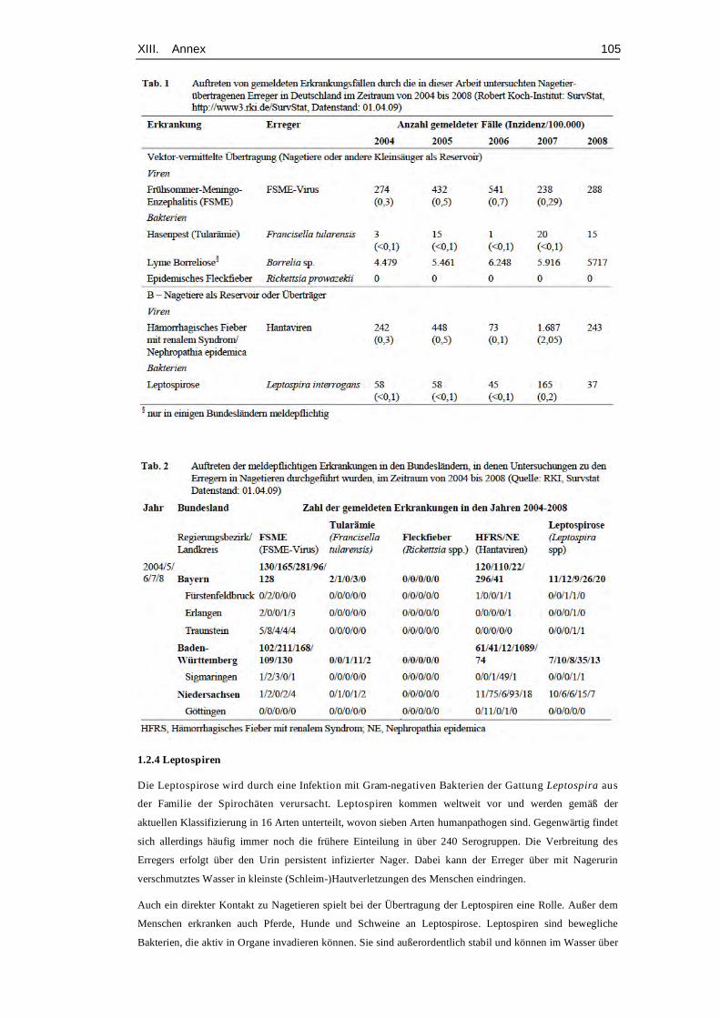
XIII. Annex 105
1.2.4 Leptospiren
Die Leptospirose wird durch eine Infektion mit Gram-negativen Bakterien der Gattung Leptospira aus
der Familie der Spirochäten verursacht. Leptospiren kommen weltweit vor und werden gemäß der
aktuellen Klassifizierung in 16 Arten unterteilt, wovon sieben Arten humanpathogen sind. Gegenwärtig findet
sich allerdings häufig immer noch die frühere Einteilung in über 240 Serogruppen. Die Verbreitung des
Erregers erfolgt über den Urin persistent infizierter Nager. Dabei kann der Erreger über mit Nagerurin
verschmutztes Wasser in kleinste (Schleim-)Hautverletzungen des Menschen eindringen.
Auch ein direkter Kontakt zu Nagetieren spielt bei der Übertragung der Leptospiren eine Rolle. Außer dem
Menschen erkranken auch Pferde, Hunde und Schweine an Leptospirose. Leptospiren sind bewegliche
Bakterien, die aktiv in Organe invadieren können. Sie sind außerordentlich stabil und können im Wasser über
XIII. Annex 106
drei Monate infektiös bleiben. Leptospiren verursachen beim Menschen verschiedene Krankheitsbilder, wie
z.B. die Weil-Krankheit, das sogenannte Feldfieber, die Schweinehüterkrankheit und das Batavia-Fieber. Die
Symptome reichen von milden und unspezifischen, grippeähnlichen Symptomen mit Myalgien und
Konjunktivitis, über ikterische Verläufe mit Fieber und Übelkeit, Meningitis bis zu Nieren- bzw. tödlichem
Multiorganversagen (Palaniappan et al., 2007). Die Letalität schwankt unbehandelt zwischen 5 und 40% (eine
Übersicht zu Erkankungen in Deutschland in Jansen et al., 2005).
Die Leptospirose ist in Deutschland meldepflichtig (Tabellen 1 und 2). Hier kam es im Jahr 2007 –
vergleichbar mit dem Auftreten von Hantavirus-Infektionen – zu einem Anstieg der Erkrankungen in Bayern,
Baden-Württemberg und Niedersachsen, den in den Studien untersuchten Regionen.
1.2.5 Francisellen
Auch die Tularämie (syn. Hasenpest) wird von einem bakteriellen Erreger, Francisella tularensis, verursacht.
Francisellen sind kleine, Gram-negative, pleomorphe, unbewegliche Bakterien, die eine Kapsel bilden können
und ein spezifisches Lipopolysaccharid (LPS) aufweisen. Francisellen werden heute in die Gruppe der γ -
Proteobakterien eingeordnet und sind damit verwandt mit Legionellen und Coxiellen. Zwei Unterarten,
Francisella tularensis ssp. tularensis (Nordamerika) und ssp. holarctica (Europa und Asien) spielen eine
Rolle als humanpathogene Erreger. Sowohl zahlreiche Säugetierspezies (>150) als auch Menschen erkranken.
Zecken, Stechmücken und Bremsen fungieren dabei als Vektoren.
Francisellen bleiben sehr lange infektiös, im Wasser und Boden über Monate hinweg, in gefrorenen
Tierkörpern sogar bis zu 3 Jahre lang.
Drei bis zehn Bakterien über ein Aerosol aufgenommen, reichen bereits für eine Infektion des Menschen aus.
Die Letalität beträgt abhängig von der Unterart 5-10% (Sjöstedt, 2007). Beim Menschen unterscheidet man äußere lokalisierte (ulcero-glanduläre) Formen, die mit Geschwüren,
Lymphadenitis und Konjunktivitis einhergehen, und innere (invasive) Formen, die sich meist in Pneumonien,
aber auch in Abszessen und Magen-Darm-Symptomatik äußern. Betroffene Tiere sterben meist innerhalb von
vier bis sechzehn Tagen, in protrahiert verlaufenden Fällen überleben sie bis zu 60 Tage. Krankheitszeichen
sind äußerlich nicht immer erkennbar, was das Risiko gerade für Jäger noch erhöht.
Die Tularämie ist in Deutschland meldepflichtig (Tabellen 1 und 2). In den Jahren 2005 und 2007 trat die
Erkrankung erstmals seit über 40 Jahren wieder gehäuft auf.
1.2.6 Borrelien
Borrelien gehören wie die Leptospiren zur Familie der Spirochäten. Die in Europa wichtigsten
humanpathogenen Borrelien-Arten zählen zum B. burgdorferi sensu lato-Komplex. Sie werden durch Zecken
der Gattung Ixodes ricinus übertragen, wobei Nagetiere und andere Kleinsäuger als Reservoir dienen. Eine
Infektion beim Menschen kann zur Ausbildung der sogenannten Lyme-Borreliose führen.
Bei dieser Erkrankung unterscheidet man drei Phasen: eine Frühmanifestation meist an der Haut (bei ca. 50%
der Fälle Hautrötung rund um die Stichstelle, Erythema migrans), eine frühe Allgemeinmanifestation vor
allem an inneren Organen (z.B. als Neuroborreliose, Polyradikuloneuritis, Arthritis, Karditis, bei Kindern
auch als lymphozytäre Meningitis), und eine späte Allgemeinmanifestation (chronische Arthritis,
Acrodermatitis chronica atrophicans, chronische Enzephalomyelitis, chronische Polyradikulopathie).
Erkrankungen sind auch bei Hund, Katze, Pferd und Rind nachgewiesen und äußern sich außer durch
unspezifische Allgemeinsymptome auch durch Arthritiden (Fingerle & Wilske, 2006; Wilske et al., 2007). In einigen Bundesländern Deutschlands (Berlin und die neuen Bundesländer) ist die Borreliose seit 2002
meldepflichtig (Tabelle 1).
XIII. Annex 107
1.2.7 Rickettsien
Rickettsien sind obligat intrazelluläre, Gram-negative Bakterien, deren Größe mit 0,3 × 0,7 µm im Bereich
zwischen den kleinsten Bakterien und den größten Viren liegt. Man unterschiedet zwei Gruppen: die
Zeckenbissfieber-Rickettsien (Rickettsia conorii, R. helvetica. R. felis), die durch verschiedene Zecken und
eine Milbenart übertragen werden, und die Fleckfieber-Rickettsien (R. prowazekii und R. typhi), die durch
Flöhe und Läuse übertragen werden. Als Reservoir dienen je nach Rickettsien-Spezies verschiedene Vögel,
Reptilien und Säugetiere, und hier vor allem Nagetiere. Rickettsien befallen die Endothelzellen der kleinen
Blutgefäße und verursachen eine Vaskulitis. Typischerweise äußern sich die Rickettsiosen durch Fieber,
Kopfschmerzen und ein als „Eschar“ bezeichnetes Ulkus an der Einstichstelle des übertragenden
Ektoparasiten sowie ein generalisiertes Exanthem. Die Letalität schwankt je nach verursachender Rickettsien-
Art, kann jedoch für das Epidemische Fleckfieber (R. prowazekii) oder das Rocky Mountain-Fleckfieber (R.
rickettsii) unbehandelt bis zu 30% betragen (Parola et al., 2005; Wölfel et al., 2006). Das Fleckfieber ist in Deutschland meldepflichtig (Tabellen 1 und 2). Seit 2003 wurden keine Fälle gemeldet.
1.3 Aktueller Wissensstand über die Verbreitung der hier vorgestellten Nagetier-assoziierten Erreger
in Deutschland Seit Inkrafttreten des Infektionsschutzgesetzes am 1. Januar 2001, in dem unter anderem auch die
Meldepflicht für humane Infektionen durch bestimmte Zoonoseerreger geregelt ist, hat sich die Datenlage zur
geographischen Verbreitung und Häufigkeit von durch diese Zoonoseerreger verursachten Krankheiten
wesentlich verbessert. Vermutlich ist jedoch die Dunkelziffer für diese Infektionen immer noch sehr hoch, da
viele der hier genannten Infektionen oft nur milde Verläufe und unspezifische Symptome verursachen und
somit wahrscheinlich oft gar nicht oder nicht richtig diagnostiziert werden. Die gemäß IfSG gemeldeten Daten (Tabellen 1 und 2) erlauben nur einen indirekten Einblick in die
geographische Verbreitung der Erreger in Deutschland. Diesbezüglich aussagefähiger sind Untersuchungen in
den Nagetier- und Kleinsäugerreservoirs. Aus diesem Grunde wurden in den Jahren 2001-2004 in drei Landkreisen Bayerns, im Jahr 2005 in einem
Landkreis in Niedersachsen und im Jahr 2007 in einem Landkreis in Baden-Württemberg Untersuchungen
zum Vorkommen der oben beschriebenen Zoonoseerreger in den potentiellen Nagetierreservoirs
durchgeführt, die hier vorgestellt werden sollen.
2. Material & Methoden
2.1. Untersuchungsgebiete
2.1.1 Longitudinalstudie in drei Landkreisen Bayerns während der Jahre 2001-2004
Von September 2001 bis März 2004 wurden in Grafrath bei München, Landkreis (LK) Fürstenfeldbruck, auf
einem im Wald gelegenen Gartengrundstück Mäuse gefangen (Abb. 1). Im LK Erlangen wurden die Gebiete
Frauenaurach (Mischwald), Klosterholz (Wasserschutzgebiet) und Dietenhofen (Hügelwald) von März bis
September 2003 beprobt (Abb. 1). Im LK Traunstein wurden an den Orten Bürgerwald, Geißing, Burkhartsöd
(Moorgebiet) und auf einer Wiese am Stadtrand im gleichen Zeitraum Mäuse gefangen (Abb. 1A, B). Alle
Probeflächen befanden sich in der Nähe von Naherholungsgebieten mit Trimm-Dich-Pfaden und beliebten
Spazierwegen. Bei den Probepunkten handelte es sich meist um Orte, die aufgrund ihrer Vegetation und
Beschaffenheit ideale Lebensbedingungen für Mäuse bieten, z.B. Laubwälder mit dichtem Unterholz,
Brombeer-/Himbeerbewuchs und dicht wachsendem Springkraut (Impatiens glandulifera). Hier fanden
mittels PCR Untersuchungen auf Borrelien und Leptospiren statt.
Antikörper gegen Kuh(Ortho-)pockenviren wurden serologisch nachgewiesen mit einem
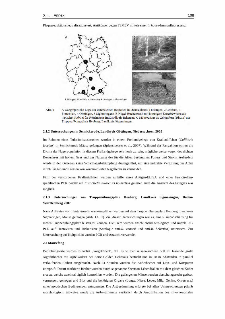
XIII. Annex 108
Plaquereduktionsneutralisationstest, Antikörper gegen FSMEV mittels einer in house-Immunfluoreszenz.
2.1.2 Untersuchungen in Sennickerode, Landkreis Göttingen, Niedersachsen, 2005
Im Rahmen eines Tularämieausbruches wurden in einem Freilandgehege von Krallenäffchen (Callithrix
jacchus) in Sennickerode Mäuse gefangen (Splettstoesser et al., 2007). Während der Fangaktion schien die
Dichte der Nagerpopulation in diesem Freilandgehege sehr hoch zu sein, möglicherweise wegen des dichten
Bewuchses mit hohem Gras und der Nutzung des für die Affen bestimmten Futters und Strohs. Außerdem
wurde in den Gehegen keine Schadnagerbekämpfung durchgeführt, um eine indirekte Vergiftung der Affen
durch Fangen und Fressen von kontaminierten Nagetieren zu vermeiden. Fünf der verstorbenen Krallenäffchen wurden mithilfe eines Antigen-ELISA und einer Francisellen- spezifischen PCR positiv auf Francisella tularensis holarctica getestet, auch die Anzucht des Erregers war
möglich. 2.1.3 Untersuchungen am Truppenübungsplatz Heuberg, Landkreis Sigmaringen, Baden-
Württemberg 2007
Nach Auftreten von Hantavirus-Erkrankungsfällen wurden auf dem Truppenübungsplatz Heuberg, Landkreis
Sigmaringen, Mäuse gefangen (Abb. 1A, C). Ziel dieser Untersuchungen war es, eine Risikoabschätzung für
diesen Truppenübungsplatz leisten zu können. Die Tiere wurden anschließend serologisch und mittels RT-
PCR auf Hantaviren und Rickettsien (Serologie anti-R. conorii und anti-R. helvetica) untersucht. Zur
Untersuchung auf Kuhpocken wurden PCR und Anzucht verwendet.
2.2 Mäusefang
Beprobungsorte wurden zunächst „vorgeködert“, d.h. es wurden ausgewaschene 500 ml fassende große
Joghurtbecher mit Apfelködern der Sorte Golden Delicious bestückt und in 10 m Abständen in parallel
verlaufenden Reihen ausgebracht. Nach 24 Stunden wurden die Köderbecher auf Urin- und Kotspuren
überprüft. Derart markierte Becher wurden durch sogenannte Sherman-Lebendfallen mit dem gleichen Köder
ersetzt, welche zweimal täglich kontrolliert wurden. Die gefangenen Mäuse wurden tierschutzgerecht getötet,
vermessen, gewogen und Blut und die benötigten Organe (Lunge, Niere, Leber, Milz, Gehirn, Ohren u.a.)
unter aseptischen Bedingungen entnommen. Die Artbestimmung erfolgte bei allen Untersuchungen primär
morphologisch, teilweise wurde die Artbestimmung zusätzlich durch Amplifikation des mitochondrialen
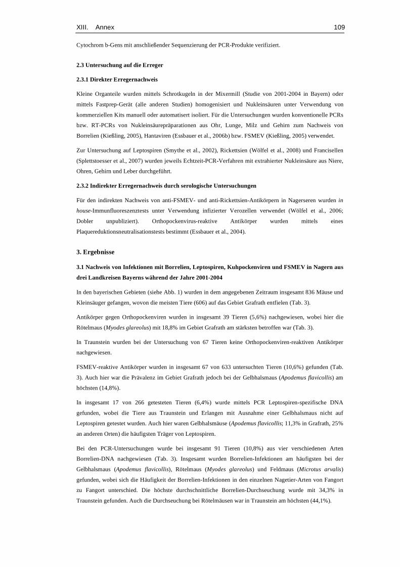
XIII. Annex 109
Cytochrom b-Gens mit anschließender Sequenzierung der PCR-Produkte verifiziert.
2.3 Untersuchung auf die Erreger 2.3.1 Direkter Erregernachweis
Kleine Organteile wurden mittels Schrotkugeln in der Mixermill (Studie von 2001-2004 in Bayern) oder
mittels Fastprep-Gerät (alle anderen Studien) homogenisiert und Nukleinsäuren unter Verwendung von
kommerziellen Kits manuell oder automatisert isoliert. Für die Untersuchungen wurden konventionelle PCRs
bzw. RT-PCRs von Nukleinsäurepräparationen aus Ohr, Lunge, Milz und Gehirn zum Nachweis von
Borrelien (Kießling, 2005), Hantaviren (Essbauer et al., 2006b) bzw. FSMEV (Kießling, 2005) verwendet.
Zur Untersuchung auf Leptospiren (Smythe et al., 2002), Rickettsien (Wölfel et al., 2008) und Francisellen
(Splettstoesser et al., 2007) wurden jeweils Echtzeit-PCR-Verfahren mit extrahierter Nukleinsäure aus Niere,
Ohren, Gehirn und Leber durchgeführt.
2.3.2 Indirekter Erregernachweis durch serologische Untersuchungen
Für den indirekten Nachweis von anti-FSMEV- und anti-Rickettsien-Antikörpern in Nagerseren wurden in
house-Immunfluoreszenztests unter Verwendung infizierter Verozellen verwendet (Wölfel et al., 2006;
Dobler unpubliziert). Orthopockenvirus-reaktive Antikörper wurden mittels eines
Plaquereduktionsneutralisationstests bestimmt (Essbauer et al., 2004).
3. Ergebnisse
3.1 Nachweis von Infektionen mit Borrelien, Leptospiren, Kuhpockenviren und FSMEV in Nagern aus
drei Landkreisen Bayerns während der Jahre 2001-2004
In den bayerischen Gebieten (siehe Abb. 1) wurden in dem angegebenen Zeitraum insgesamt 836 Mäuse und
Kleinsäuger gefangen, wovon die meisten Tiere (606) auf das Gebiet Grafrath entfielen (Tab. 3).
Antikörper gegen Orthopockenviren wurden in insgesamt 39 Tieren (5,6%) nachgewiesen, wobei hier die
Rötelmaus (Myodes glareolus) mit 18,8% im Gebiet Grafrath am stärksten betroffen war (Tab. 3).
In Traunstein wurden bei der Untersuchung von 67 Tieren keine Orthopockenviren-reaktiven Antikörper
nachgewiesen.
FSMEV-reaktive Antikörper wurden in insgesamt 67 von 633 untersuchten Tieren (10,6%) gefunden (Tab.
3). Auch hier war die Prävalenz im Gebiet Grafrath jedoch bei der Gelbhalsmaus (Apodemus flavicollis) am
höchsten (14,8%).
In insgesamt 17 von 266 getesteten Tieren (6,4%) wurde mittels PCR Leptospiren-spezifische DNA
gefunden, wobei die Tiere aus Traunstein und Erlangen mit Ausnahme einer Gelbhalsmaus nicht auf
Leptospiren getestet wurden. Auch hier waren Gelbhalsmäuse (Apodemus flavicollis; 11,3% in Grafrath, 25%
an anderen Orten) die häufigsten Träger von Leptospiren.
Bei den PCR-Untersuchungen wurde bei insgesamt 91 Tieren (10,8%) aus vier verschiedenen Arten
Borrelien-DNA nachgewiesen (Tab. 3). Insgesamt wurden Borrelien-Infektionen am häufigsten bei der
Gelbhalsmaus (Apodemus flavicollis), Rötelmaus (Myodes glareolus) und Feldmaus (Microtus arvalis)
gefunden, wobei sich die Häufigkeit der Borrelien-Infektionen in den einzelnen Nagetier-Arten von Fangort
zu Fangort unterschied. Die höchste durchschnittliche Borrelien-Durchseuchung wurde mit 34,3% in
Traunstein gefunden. Auch die Durchseuchung bei Rötelmäusen war in Traunstein am höchsten (44,1%).
XIII. Annex 110
3.2 Nachweis von Infektionen mit Francisellen und Hantaviren in Nagetieren aus Sennickerode,
Niedersachsen, 2005
In Sennickerode (Abb 1.) wurden insgesamt 144 Tiere gefangen, davon 28 Gelbhalsmäuse (Apodemus
flavicollis), 62 Rötelmäuse (Myodes glareolus), 12 Feldmäuse (Microtus arvalis) und 37 Schermäuse
(Arvicola amphibius, vormals A. terrestris).
Mittels PCR-Untersuchung aller Tiere wurde bei 10 Tieren (6,9%) Francisella tularensis–spezifische DNA
nachgewiesen (Tab. 4). Der höchste Anteil entfiel dabei auf die Schermaus (Arvicola amphibius) mit 6
positiven Tieren (16,2%).
Insgesamt 134 der 144 gefangenen Tiere wurden mittels Hantavirus-S-Segment-spezifischer RT-PCR
untersucht (Essbauer et al., unveröffentlichte Daten). Dabei zeigten 13 Tiere (9,7%) ein Amplifikat der
erwarteten Größe (Tab. 4), wobei der höchste Prozentsatz (41%, 7 von 17 Tieren) auf die Feldmaus (Microtus
arvalis) entfiel.
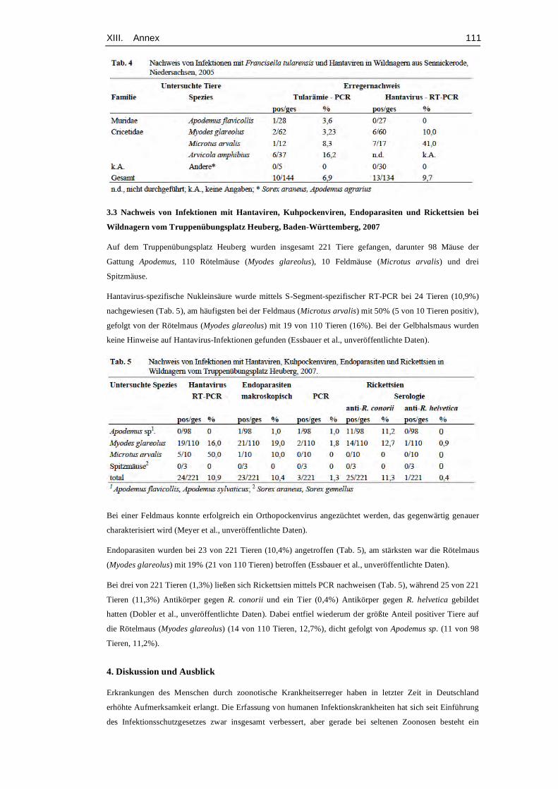
XIII. Annex 111
3.3 Nachweis von Infektionen mit Hantaviren, Kuhpockenviren, Endoparasiten und Rickettsien bei
Wildnagern vom Truppenübungsplatz Heuberg, Baden-Württemberg, 2007 Auf dem Truppenübungsplatz Heuberg wurden insgesamt 221 Tiere gefangen, darunter 98 Mäuse der
Gattung Apodemus, 110 Rötelmäuse (Myodes glareolus), 10 Feldmäuse (Microtus arvalis) und drei
Spitzmäuse. Hantavirus-spezifische Nukleinsäure wurde mittels S-Segment-spezifischer RT-PCR bei 24 Tieren (10,9%)
nachgewiesen (Tab. 5), am häufigsten bei der Feldmaus (Microtus arvalis) mit 50% (5 von 10 Tieren positiv),
gefolgt von der Rötelmaus (Myodes glareolus) mit 19 von 110 Tieren (16%). Bei der Gelbhalsmaus wurden
keine Hinweise auf Hantavirus-Infektionen gefunden (Essbauer et al., unveröffentlichte Daten).
Bei einer Feldmaus konnte erfolgreich ein Orthopockenvirus angezüchtet werden, das gegenwärtig genauer
charakterisiert wird (Meyer et al., unveröffentlichte Daten). Endoparasiten wurden bei 23 von 221 Tieren (10,4%) angetroffen (Tab. 5), am stärksten war die Rötelmaus
(Myodes glareolus) mit 19% (21 von 110 Tieren) betroffen (Essbauer et al., unveröffentlichte Daten).
Bei drei von 221 Tieren (1,3%) ließen sich Rickettsien mittels PCR nachweisen (Tab. 5), während 25 von 221
Tieren (11,3%) Antikörper gegen R. conorii und ein Tier (0,4%) Antikörper gegen R. helvetica gebildet
hatten (Dobler et al., unveröffentlichte Daten). Dabei entfiel wiederum der größte Anteil positiver Tiere auf
die Rötelmaus (Myodes glareolus) (14 von 110 Tieren, 12,7%), dicht gefolgt von Apodemus sp. (11 von 98
Tieren, 11,2%).
4. Diskussion und Ausblick
Erkrankungen des Menschen durch zoonotische Krankheitserreger haben in letzter Zeit in Deutschland
erhöhte Aufmerksamkeit erlangt. Die Erfassung von humanen Infektionskrankheiten hat sich seit Einführung
des Infektionsschutzgesetzes zwar insgesamt verbessert, aber gerade bei seltenen Zoonosen besteht ein

XIII. Annex 112
erhebliches Informationsdefizit. Als sehr hilfreich erweist sich hier insbesondere die öffentlich zugängliche
Plattform „SurvStat“ des Robert-Koch-Institutes, die eine aktuelle Übersicht über die geographische
Verbreitung und Häufigkeit meldepflichtiger Erkrankungen in Deutschland ermöglicht. Dennoch ist die
Dunkelziffer der oft mit unspezifischen fieberhaften Symptomen einhergehenden Erkrankungen weiterhin als
hoch einzuschätzen. Dies liegt zum einen an dem glücklicherweise vergleichsweise seltenen Auftreten dieser
Erkrankungen und der damit verbundenen fehlenden Kenntnis über diese Erkrankungen innerhalb der
Ärzteschaft. Desweiteren führt sicher auch die oft unspezifische und milde Symptomatik zu Fehldiagnosen
und somit zu einem „underreporting. Insgesamt gibt es jedoch auch noch wesentlich zu wenige
Untersuchungen zum Vorkommen seltener Zoonoseerreger beim Menschen und in den entsprechenden
Wildnagerreservoiren.
Erste deutschlandweite Untersuchungen – wie die hier vorgestellten Untersuchungen - zeigen, dass Nagetiere
in Deutschland in nicht unerheblichem Maße mit verschiedenen Krankheitserregern durchseucht sind (Ulrich
et al., 2008a; hier vorgestellte Untersuchungen). So konnte beispielsweise eine breite geographische
Verbreitung und ein stabiles Vorkommen des Puumalavirus in Populationen der Rötelmaus (Myodes
glareolus) und des Tulavirus in Microtus-Populationen gezeigt werden (Ulrich et al., 2008a). Die hier
beschriebenen Untersuchungen belegen diesen Befund für Gebiete in Niedersachsen und Baden-
Württemberg. Diese Studien zeigen eine scheinbare Wirtspräferenz einzelner Zoonoseerreger für bestimmte
Nagetier-Arten, die in weiterführenden Arbeiten zu prüfen wäre. So wurden Leptospiren, Borrelien und
FSMEV besonders in der Gelbhalsmaus (Apodemus flavicollis), Francisellen vor allem in der Schermaus und
Kuhpockenviren insbesondere in der Rötelmaus (Myodes glareolus) gefunden. Interessanterweise war die
Rötelmaus auch in starkem Maße von Endoparasiten befallen, was möglicherweise auch die Durchseuchung
mit anderen Erregern beeinflußt. Die hier zusammengefassten Erkenntnisse sind ein erster wichtiger Schritt
auf dem Weg zur Erstellung von Verbreitungskarten für die genannten humanpathogenen Zoonoseerreger in
ihren Reservoirwirten und der Definition von entsprechenden Risikogebieten. Neben den genannten Forschungsaktivitäten muss die Öffentlichkeit auch für die Problematik der Nagetier-
und anderen Vektor-übertragenen Zoonosen weiter sensibilisiert werden. In diesem Sinne sind einerseits die
Gesamtbevölkerung, andererseits auch beruflich exponierte Risikogruppen, wie Forstarbeiter, Jäger,
Beschäftigte in der Landwirtschaft und Soldaten auf die Gefährdungen hinzuweisen (Ulrich et al., 2007).
Darüber hinaus müssen diese Personengruppen auch stärker mit den gegebenen- falls erforderlichen
Vorsichts- und Hygienemaßnahmen vertraut gemacht werden.
Zusammenfassend ist festzuhalten, dass unsere Kenntnis von den Naturherden, den beteiligten Reseroirwirten
und den entsprechenden Infektketten noch äußerst lückenhaft ist. Insbesondere zu den Ursachen für ein
saisonal oder jährlich gehäuftes Auftreten bestimmter Humaninfektionen bestehen bestenfalls
Arbeitshypothesen. Da an der Lösung dieser Probleme zwangsläufig Bereiche beteiligt sind, die weit über die
Mikrobiologie hinausgehen, ist deren Bearbeitung nur in entsprechenden Netzwerken und
Arbeitsgemeinschaften über Institutsgrenzen hinweg zielführend. Nur in einer synergistischen
Zusammenarbeit von Zoologen, Ökologen, Virologen, Mikrobiologen, Parasitologen, Genetikern,
Epidemiologen, Forstwissenschaftlern und Klimaforschern mit Klinikern der Human- und Veterinärmedizin
können die komplexen Interaktionen zwischen Pathogenen, Reservoirwirten, Vektoren und Prädatoren im
Zusammenhang mit dem Auftreten von Infektionen beim Menschen verstanden werden (Ulrich et al., 2008b).
Danksagung
Wir danken Frau Dr. Judith Kießling, Krystian Mistzela, Dr. Sonja Hartnack und den beteiligten Mitarbeitern
des Instituts für Mikrobiologie der Bundeswehr für die tatkräftige Unterstützung bei der Beprobung der
verschiedenen Punkte in Bayern, Sennickerode und dem Truppenübungsplatz Heuberg.

XIII. Annex 113
Herrn Prof. Hermann Meyer danken wir für die Übermittlung unpublizierter Ergebnisse und für die kritische
Durchsicht des Manuskriptes.
Literatur
Dobler, G., Essbauer, S.S., Wölfel, R., Pfeffer. M. (2005): Interaktionen von Ökologie und Epidemiologie am
Beispiel der Frühsommer-Menigoenzephalitis. Rundgespräche der Kommision für Ökologie,
Bd. 29, „Zur Ökologie von Infektionskrankheiten“, Verlag Dr. Friedrich Pfeil, München, 43-
52.
Essbauer, S.S., Friedewald, S., Hassler. D., Meyer. H., Pfeffer. M. (2006a): Von Ratten und Katzen auf den
Menschen: Kuhpockenviren in Europa. DMW 131 (46), 2381-2382
Essbauer, S.S., Pfeffer, M., Wilhelm, S., Meyer, H. (2004): Zoonotische Pockenviren.
Bundesgesundheitsblatt Gesundheitsforschung Gesundheitsschutz 47, 671-679
Essbauer, S.S., Schmidt, J., Conraths, F.J., Friedrich, R., Koch, J., Hautmann, W., Pfeffer, M., Wölfel, R.,
Finke, E. J., Dobler, G., Ulrich, R.G. (2006b): A new Puumala hantavirus subtype in rodents
associated with an outbreak of severe Nephropathia epidemica in South-East Germany in
2004. Epidem. Infection 134, 1333- 1344
Fingerle, V., Wilske, B. (2006): Ticks, tick bites and how best to remove the tick. MMW Fortschr Med.
148(25), 30-2.
Jansen, A., Schöneberg, I., Frank, C., Alpers, K., Schneider, T., Stark, K. (2005): Leptospirosis in Germany,
1962- 2003. Emerg Infect Dis. 11(7), 1048-54.
Kießling, J. (2005): Untersuchung zum Vorkommen des Frühsommer-Meningo-Enzephalitis-Virus und
Borrelia burgdorferi in ausgewählten Wildmaus- und Zeckenpopulationen Bayerns.
Veterinärmedizinische Fakultät der LMU München, Dissertation
Krüger, D.H., Ulrich, R.G., Lundkvist, Å. (2001): Hantavirus infections and their prevention. Microbes Infect
3, 1129-1144
Palaniappan, R.U., Ramanujam, S., Chang, Y.F. (2007): Leptospirosis: pathogenesis, immunity, and
diagnosis. Current Opinion in Infectious Diseases 20, 284-292.
Parola, P., Paddock, C. D., Raoult, D., 2005: Tick-borne rickettsioses around the world: emerging diseases
challenging old concepts. Clin Microbiol Rev 18, 719-756.
Schönrich, G., Rang, A., Lütteke, N., Raftery, M.J., Charbonnel, N., Ulrich, R.G. (2008): Hantavirus-induced
immunity in rodent reservoirs and humans. Immunol. Reviews (im Druck)
Sjöstedt, A. (2007): Tularemia: History, epidemiology, pathogen physiology, and clinical manifestations.
Annals of the New York Acadamy of Sciences 1105, 1-29
Smythe, L.D., Smith, I.L., Smith, G.A., Dohnt, M.F., Symonds, M.L., Barnett, L.J., McKay, D.B. (2002): A
quantitative PCR (TaqMan) assay for pathogenic Leptospira spp. BMC Infect Dis. 2, 13.
Splettstoesser, W.D., Mätz-Rensing, K., Seibold, E., Tomaso, H., Al Dahouk, S., Grunow, R., Essbauer, S.S.,
Buckendahl, A., Finke, E. J., Neubauerm H. (2007): Re-emergence of Francisella tularensis
in Germany: fatal tularemia in a colony of semi-free living marmosets (Callithrix jacchus).
Epidemiol. Infect. 135(8), 1256-1265
Süss, J., Scrader, C., Falk, U., Wohanka, N. (2004): Tick-borne encephalitis (TBE) in Germany –
epidemiological data, development of risk areas and virus prevalence in field-collected ticks
XIII. Annex 114
and ticks removed from humans. Int J Med Microbiol 293 (Suppl 37), 69-79.
Ulrich, R.G., Heckel, G., Pelz, H.J., Wieler, L.H., Dobler, G., Freise, J., Matuschka, F. R., Jacob, J., Schmidt- Chanasit, J., Gerstengarbe, F.W., Jäkel, T., Süss, J., Ehlers, B., Nitsche, A., Kallies, R., Johne,
R., Günther, S., Henning, K., Grunow, R., Wenk, M., Maul, L., Hunfeld, K.P., Wölfel, R.,
Schares, G., Scholz, H.C., Brockmann, S., Pfeffer, M., Essbauer, S.S. (2008b): Nagetiere und
Nagetier-assoziierte Krankheitserreger – das Netzwerk „Nagetier-übertragene Pathogene“
stellt sich vor. Bundesgesundheitsblatt 225,163-89.
Ulrich, R.G., Koch, J., Schmidt-Chanasit, J., Mertens, M., Pelz, H.J., Jacob, J., Madeja, E.L., Quast, H.,
Freise, J., Groschup, M.H., Conraths, F.J., Dobler, G., Bradt, K., Wegener, W., Essbauer, S.S.
(2007): 2005, ein Jahr der Hantaviren - Quo vadis? Der Hygieneinspektor 9(1), 61-68.
Ulrich, R.G., Schlegel, M., Jacob, J., Pelz, H.J., Mertens, M., Schmidt-Chanasit, J., Wenk, M., Büchner, T.,
Masur, D., Sevke, K., Groschup, M.H., Gerstengarbe, F.W., Pfeffer, M., Oehme, R.,
Wegener, W., Bemmann, M., Ohlmeyer, L., Wolf, R., Zoller, H., Koch, J., Brockmann, S.,
Heckel, G., Essbauer, S.S. (2008a): Network „Rodent-borne pathogens“ in Germany:
Longitudinal studies on the geographical distribution and prevalence of hantavirus infections.
Parasitology Res. (im Druck)
Wilske, B., Fingerle, V., Schulte-Spechtel, U. (2007): Microbiological and serological diagnosis of Lyme
borreliosis. FEMS Immunol Med Microbiol. 49(1), 13-21.
Wilson, D.E., Reeder, D.M. (2005): Mammal Species of the World. Johns Hopkins University Press.
Wölfel, R., Essbauer, S.S., Dobler, G. (2008): Diagnostics of tick-borne rickettsioses in Germany: A modern
concept for a neglected disease. J Med Microbiol 298 (Suppl. 1), 368-374
Wölfel, R., Pfeffer, M., Essbauer, S.S., Dobler, G. (2006): Rickettsiosen in Einsatzgebieten der Bundeswehr –
Aspekte zur Epidemiologie, Diagnostik und Therapie. Wehrmed. Mschr. 50(7), 185-187

XIV. Acknowledgements 115
XIV. ACKNOWLEDGEMENTS
First of all, I would like to thank PD Dr. Sandra Essbauer for supporting me
not only technically, for giving me the opportunity to work on this thesis
and also to attend many scientific conferences. Thank you for being a
great mentor!
I also want to express my gratitude to Prof. Reinhard Straubinger for
supervising this thesis, for constructive feedback and giving incisive
advice.
Without the help of Dr. Jörg Müller, this work would not have been
possible. Thank you for your immense support from the very beginning, for
your help with field work and statistics and for being a friend!
Many thanks also to Dr. Claus Bässler and many more colleagues from
the National Park Bavarian Forest Administration, for being always helpful
and for the most entertaining Christmas and summer parties. Special
thanks to Andreas Ebert for fabulous rodent photos!
I also wish to thank Dr. Stephanie Speck for very helpful advice and for
providing most useful literature.
Dr. Julia Riehm, thank you not only for contribution to the publication but
also for hosting cheerful lunch breaks.
Dr. Doblers technical support and willingness to take time to discuss my
research is highly appreciated.
I wish to express my sincerest thanks to all the people that supported me
in the field as well as in the lab, especially Daniela Eder, Robin Reiter,
Mareike Pollaerts, Harald Weber, Rahime Terzioglu and many more.
Thank you for being both a great help and entertaining company!
I also wish to thank Ricardo Castro César de Sá for great effort spent on
the correction of this manuscript.
Finally, the deepest thanks go to my dear family for always supporting,
encouraging and loving me. Without you, this dissertation would not have
been possible.
SU
SA
NN
E SC
HEX
RICKETTSIA
SP
P. IN
SM
ALL M
AM
MA
LS
SUSANNE YVONNE SCHEX
RICKETTSIA SPP. IN FREE RANGING SMALL
MAMMALS IN SOUTH-EASTERN GERMANY
VVB VVB LAUFERSWEILER VERLAGédition scientifique
9 7 8 3 8 3 5 9 5 7 3 6 7
VVB LAUFERSWEILER VERLAGSTAUFENBERGRING 15D-35396 GIESSEN
Tel: 0641-5599888 Fax: [email protected]
VVB LAUFERSWEILER VERLAGédition scientifique
ISBN: 978-3-8359-5736-7 INAUGURAL-DISSERTATION zur Erlangung der tiermedizinischen Doktorwürdeder Tierärztlichen Fakultät der Ludwig-Maximilians-Universität München