rickettsia phylogenomics: unwinding the intricacies of obligate intracellular life
TRANSCRIPT

Rickettsia Phylogenomics: Unwinding the Intricacies ofObligate Intracellular LifeJoseph J. Gillespie1,2*, Kelly Williams1, Maulik Shukla1, Eric E. Snyder1, Eric K. Nordberg1, Shane M.
Ceraul2, Chitti Dharmanolla1, Daphne Rainey1, Jeetendra Soneja1, Joshua M. Shallom1, Nataraj Dongre
Vishnubhat1, Rebecca Wattam1, Anjan Purkayastha1, Michael Czar1, Oswald Crasta1, Joao C. Setubal1,
Abdu F. Azad2, Bruno S. Sobral1
1 Virginia Bioinformatics Institute at Virginia Tech, Blacksburg, Vigrinia, United States of America, 2 Department of Microbiology and Immunology, University of Maryland
School of Medicine, Baltimore, Maryland, United States of America
Abstract
Background: Completed genome sequences are rapidly increasing for Rickettsia, obligate intracellular a-proteobacteriaresponsible for various human diseases, including epidemic typhus and Rocky Mountain spotted fever. In light ofphylogeny, the establishment of orthologous groups (OGs) of open reading frames (ORFs) will distinguish the corerickettsial genes and other group specific genes (class 1 OGs or C1OGs) from those distributed indiscriminately throughoutthe rickettsial tree (class 2 OG or C2OGs).
Methodology/Principal Findings: We present 1823 representative (no gene duplications) and 259 non-representative (at leastone gene duplication) rickettsial OGs. While the highly reductive (,1.2 MB) Rickettsia genomes range in predicted ORFs from872 to 1512, a core of 752 OGs was identified, depicting the essential Rickettsia genes. Unsurprisingly, this core lacks manymetabolic genes, reflecting the dependence on host resources for growth and survival. Additionally, we bolster our recentreclassification of Rickettsia by identifying OGs that define the AG (ancestral group), TG (typhus group), TRG (transitional group),and SFG (spotted fever group) rickettsiae. OGs for insect-associated species, tick-associated species and species that harborplasmids were also predicted. Through superimposition of all OGs over robust phylogeny estimation, we discern betweenC1OGs and C2OGs, the latter depicting genes either decaying from the conserved C1OGs or acquired laterally. Finally, scrutinyof non-representative OGs revealed high levels of split genes versus gene duplications, with both phenomena confoundinggene orthology assignment. Interestingly, non-representative OGs, as well as OGs comprised of several gene families typicallyinvolved in microbial pathogenicity and/or the acquisition of virulence factors, fall predominantly within C2OG distributions.
Conclusion/Significance: Collectively, we determined the relative conservation and distribution of 14354 predicted ORFsfrom 10 rickettsial genomes across robust phylogeny estimation. The data, available at PATRIC (PathoSystems ResourceIntegration Center), provide novel information for unwinding the intricacies associated with Rickettsia pathogenesis,expanding the range of potential diagnostic, vaccine and therapeutic targets.
Citation: Gillespie JJ, Williams K, Shukla M, Snyder EE, Nordberg EK, et al. (2008) Rickettsia Phylogenomics: Unwinding the Intricacies of Obligate IntracellularLife. PLoS ONE 3(4): e2018. doi:10.1371/journal.pone.0002018
Editor: Adam J. Ratner, Columbia University, United States of America
Received February 6, 2008; Accepted March 7, 2008; Published April 16, 2008
Copyright: � 2008 Gillespie et al. This is an open-access article distributed under the terms of the Creative Commons Attribution License, which permitsunrestricted use, distribution, and reproduction in any medium, provided the original author and source are credited.
Funding: This work is funded through NIAID contract HHSN266200400035C to BSS and NIH grants AI59118 and AI17828 to AFA.
Competing Interests: The authors have declared that no competing interests exist.
* E-mail: [email protected]
Introduction
Rickettsiae are a group of organisms belonging to the class
Alphaproteobacteria, a large and metabolically diverse group of gram-
negative bacteria [1–3]. Within Alphaproteobacteria, the order
Rickettsiales comprises three families: Holosporaceae, Anaplas-
mataceae and Rickettsiaceae [4], of which Rickettsia spp. are
grouped in the latter, along with the monotypic genus Orientia, the
scrub typhus agent [5]. Robust phylogenetic analysis further
suggests that the abundant free-living marine bacterioplankton
Pelagibacter ubique and mitochondria are early-branching groups of
the order [6]. Species in the genus Rickettsia are obligate
intracellular symbionts of plants [7], amoebae [8,9], arthropods
[e.g., 10–13], annelids [14], vertebrates [15] and likely many other
organisms [16]. Most Rickettsia-containing vertebrates are second-
ary hosts that acquired these bacteria via blood-feeding arthropods
or the transdermal inoculation or inhalation of the feces of infected
arthropods. Rickettsia spp. are often parasitic in the secondary
vertebrate host [e.g., 17], and their pathogenicity to some extent
has been well studied. In particular, human rickettsial infections
are known to cause many diseases, including epidemic typhus (R.
prowazekii), murine typhus (R. typhi), murine typhus-like (R. felis),
rickettsial pox (R. akari), Rocky Mountain spotted fever (R. rickettsii),
Boutonneuse fever (R. conorii), and North Asian tick typhus (R.
sibirica). These virulent species of rickettsiae are of great interest
both as emerging infectious diseases [18] and for their potential
deployment as bioterrorism agents [19,20].
Due to both small genome size and medical importance, ten
genome sequences from Rickettsia spp. have been published and
annotated in the last decade [9,21–27], providing a foundation to
PLoS ONE | www.plosone.org 1 April 2008 | Volume 3 | Issue 4 | e2018

study the evolutionary history of these lineages through compar-
ative genomics. Recently, Gillespie et al. [28] proposed a revision
to the long-standing classification of Rickettsia by erecting the
transitional group (TRG) as a distinct lineage that shares
immediate ancestry with the members of the spotted fever group
(SFG) rickettsiae. Coupled with the typhus group (TG) and
ancestral group (AG) rickettsiae, these four rickettsial lineages
comprising 10 sequenced genomes present an opportunity to
create a database that encompasses the distribution of the
predicted open reading frames (ORFs) across all ten annotated
genomes (Figure 1).
Establishing orthology across multiple genomes serves not only
to identify genes with shared evolutionarily histories, but also
facilitates genome annotation [29,30], and significant attention has
focused on algorithms for creating orthologous groups (OGs).
Recent work has centered on the following four aspects: i) overall
improvement of OG assignment in the face of paralogy, ii)
building tools for the cross-querying of taxon-specific databases, iii)
creating databases that house specific gene or protein profiles for
facilitating the identification of orthologs in novel sequences, and
iv) the inclusion of phylogeny estimation into the processes of
assigning orthology and detecting paralogy.
At the PathoSystems Resource Integration Center (PATRIC)
[31], OGs have been preliminarily established for several groups
of organisms, including Rickettsia spp. The advantage of a Rickettsia-
specific database lies not only in the ability to query exclusively
against the 10 genomes currently annotated in our system, but also
to evaluate the results of several algorithmic approaches that
create OGs. Furthermore, PATRIC offers continued updates to
the annotation of rickettsial genes and proteins, and provides
multiple sequence alignments as well as phylogenetic trees, when
applicable, for each OG consisting of two to ten rickettsial taxa.
The database will continually evolve with the addition of newly
sequenced rickettsial genomes, with existing OG assignments
driving the curation process of raw genome data.
In the present study, we report the rickettsial OGs (RiOGs) in
conjunction with a highly robust phylogeny of the core rickettsial
genes, providing an evolutionary framework for interpreting the
genomic characteristics of the four main lineages of Rickettsia.
These data highlight the genetic anomalies previously character-
ized for this genus, such as extremely reduced genomes and the
high presence of putative pseudogenes, and also reveal novel
characteristics including the lack of group-specific virulence factors
and high occurrence of lateral transfer between groups that harbor
plasmids (AG and TRG rickettsiae). Information on the conserved
core genes, as well as those that may be involved in specific
functions that define monophyletic groups, host associations, and
plasmid-related behavior, will be valuable resources for future
TG
TRG
AG
SFGPr
Ty
FeAk
Ri
Co
Si
Ca
Br
Bo
= louse
= flea
= mite
= tick
= plasmid (published)
= plasmid (unpublished)
Insect vectors Acarine vectors
V
V
V
V
AGTGTRG
SFG
TG+TRG
-SFG
derived
core
TRG
TG
AG
TRG + AG
SFG
-TRG
-TG
SFG +
TRG +SFG
TG
AG+SFG
+AG
TG
Figure 1. Venn diagram depicting 15 intersections for the four rickettsial groups. Classification scheme based on molecular phylogenyestimation [28], the topology of which is shown in the lower left; AG = ancestral group, TG = typhus group, TRG = transitional group, SFG = spottedfever group. Genome codes are as follows: Br = R. bellii str. RML369-C, Bo = R. bellii str. OSU 85 389, Ca = R. canadensis str. McKiel, Pr = R. prowazekii str.Madrid E, Ty = R. typhi str. Wilmington, Ak = R. akari str. Hartford, Fe = R. felis str. URRWXCal2, Ri = R. rickettsii str. Sheila Smith CWPP, Co = R. conorii str.Malish 7, and Si = R. sibirica str. 246. Arthropod hosts are illustrated for each genome, and strains known to harbor plasmids are depicted.doi:10.1371/journal.pone.0002018.g001
Rickettsia Phylogenomics
PLoS ONE | www.plosone.org 2 April 2008 | Volume 3 | Issue 4 | e2018

laboratory work (e.g., development of vaccines, diagnostics and
therapeutics) as well as further evolutionary studies of this
intriguing obligate intracellular bacterial group.
Results and Discussion
Synteny and Phylogeny of Rickettsia GenomesWhole genome alignments for the ten analyzed Rickettsia taxa
reveal highly conserved colinearity in six of the seven derived
species (sans R. bellii and R. canadensis) with minimal gene
rearrangements, most of which occur near the predicted origin
of replication termination (Figure 2). However, the R. felis
genome contains several long-range symmetrical inversions in the
central region of the alignment that are not found in other taxa.
Removal of R. felis from the alignment illustrates the highly
conserved synteny across the derived rickettsia taxa (Figure S1-A). Furthermore, switching the positions of R. akari and R. felis in
the alignment (Figure S1-B) demonstrates that these central
inversions in R. felis, as well as a large genome size, are
autapomorphic (uniquely derived) traits within derived rickettsiae.
Among the three AG rickettsiae, R. canadensis (formerly R. canada) is
more colinear with the derived taxa than it is to either R. bellii
strain. Like R. felis, R. canadensis contains several autapomorphic
symmetrical inversions in the central region of the alignment, yet
they are smaller than the long-range inversions found in R. felis. As
previously reported [32], R. bellii str. RML369-C shares little
colinearity with other rickettsial genomes, and our analysis of both
R. bellii genomes is in agreement with this observation. Despite
several long and short range inversions between the R. bellii str.
RML369-C and R. bellii str. OSU 85-389 genomes, few gene
positions are shared with R. bellii and R. canadensis or the derived
taxa (Figure 2), and switching the positions of the R. bellii strains
in the alignment does not result in more conserved synteny
between either strain and the derived taxa (Figure S1-C, D).
Phylogenetic analyses implementing both maximum likelihood
and parsimony of the 731 representative core rickettsial proteins
(discussed below) resulted in robust estimates for these 10 taxa
(Figure 3). The estimated tree topologies are identical in
branching pattern and are congruent with the tree from our
previous analysis of 716 fewer genes [28], suggesting that ten or
more concatenated (and well-behaved, with high signal to noise
ratio) genes are sufficient for obtaining a robust phylogenetic
estimate for these rickettsial taxa. Thus, our recent classification
scheme for Rickettsia consisting of 4 major groups (AG rickettsiae:
R. bellii str. RML369-C, R. bellii str. OSU 85 389, R. canadensis str.
McKiel; TG rickettsiae: R. prowazekii str. Madrid E, R. typhi str.
Wilmington; TRG rickettsiae: R. akari str. Hartford, R. felis str.
URRWXCal2; SFG rickettsiae: R. rickettsii str. Sheila Smith
CWPP, R. conorii str. Malish 7, R. sibirica str. 246) is substantiated
with a phylogenomic approach. In what follows, we use this
evolutionary framework to analyze the distribution and relative
conservation of all predicted genes for these ten rickettsial
genomes.
Predicted OGs: Conservation and RepresentationIn the analysis of the rapidly growing list of rickettsial genomes
we determined that OrthoMCL, a program that applies the
Markov clustering algorithm of Van Dongen [33] to resolve the
many-to-many orthologous relationships present within cross
genome comparisons [34], outperformed more traditional ap-
proaches to establishing OGs, such as bidirectional best BLAST
hits with and without cliques. Thus, we show here the results
generated by OrthoMCL only, which grouped 12887 ORFs into
2082 total OGs (Table 1). The bulk (88%) of these OGs are
Br
Bo
Ca
Pr
Ty
Ak
Fe
Ri
Co
Si
Figure 2. Alignment of 10 rickettsial genomes. Taxa are in the same position as in estimated trees in Figure 3, with taxon abbreviationsexplained in the Figure 1 legend. Alignment created using Mauve [189] after reindexing the R. sibirica genome (see text for details).doi:10.1371/journal.pone.0002018.g002
Rickettsia Phylogenomics
PLoS ONE | www.plosone.org 3 April 2008 | Volume 3 | Issue 4 | e2018

representative (Figure 4A), meaning they include only one CDS
per strain, thus ranging in membership from 2–10 sequences. The
remaining 12% of the OGs are non-representative (Figure 4B)
and include multiple predicted ORFs from at least one member.
Categorization of the OGs into two classes based on distribution
across the rickettsial tree and other attributes, such as presence of
plasmids and common arthropod hosts (Figure 4C–D), reveals
that 69% of the OGs are comprised of single rickettsial groups
(e.g., AG, TG, TRG, and SFG), shared rickettsial groups
(subgeneric), plasmid-harboring genomes, and genomes with
common arthropod hosts (Table 1). These class 1 OGs
(C1OGs) contain 76% of the predicted ORFs grouped into OGs
by OrthoMCL, suggesting that our criteria for distinguishing
biologically interesting protein families based empirically on robust
phylogeny estimation, presence of extra-chromosomal DNA and
shared arthropod hosts is valid. The remaining ORFs grouped
into class 2 OGs (C2OGs) depict gene families drifting or
sporadically lost from the core genetic repertoire of the rickettsial
ancestor [32] or genes acquired laterally (Figure S2). Interest-
ingly, while the majority (71%) of representative OGs qualify as
C1OGs, the non-representative OGs are distributed within
C1OGs and C2OGs in near equal frequency (Table 1),
suggesting minimal conservation for gene duplications and
laterally acquired genes in these rickettsial genomes.
The RiOGs range in membership from two to 31 ORFs, with
few (,3%) OGs exceeding more than 10 ORFs (Table 2).
Representative C1OGs comprise a substantial portion (64%) of
the OGs with membership of 10 or fewer ORFs. Regarding the
OGs with more than 10 members, a range from 4% (R. prowazekii)
to 32% (R. conorii) illustrates the frequencies at which a particular
rickettsial genome contributes to non-representation. As expected
due to their smaller genome sizes and few gene duplications
[21,25], TG rickettsiae make little contribution (avg. 5%) to larger
non-representative OGs as compared to AG (avg. 19%), TRG
(avg. 17%) and SFG (avg. 31%) rickettsiae (Table 2). Thus, these
three latter groups have genomes more tolerant of multicopy
genes, particularly those resulting from transposases and other
insertion sequences, which act to produce elevated levels of
paralogous genes. For instance, analysis of the distribution of
RiOGs containing genes associated with mobile DNA and/or
A B
0.1 substitutions/site 5000 changes
R. bellii str. RML369 C
R. bellii str. OSU 85 389
R. canadensis str. McKiel
R. prowazekii str. Madrid E
R. typhi str. Wilmington
R. akari str. Hartford
R. felis str. URRWXCal2
R. rickettsii str. Sheila Smith CWPP
R. conorii str. Malish 7
R. sibirica str. 246
Wolbachia (D. melanogaster) Wolbachia (D. melanogaster)
R. prowazekii str. Madrid E
R. typhi str. Wilmington
R. bellii str. RML369 C
R. bellii str. OSU 85 389
R. canadensis str. McKiel
R. akari str. Hartford
R. felis str. URRWXCal2
R. rickettsii str. Sheila Smith CWPP
R. conorii str. Malish 7
R. sibirica str. 246
*
*
100
100 100
100
100
100
100
100
100
100 100
100 100
100
100
100
TG
TRG
SFG
AGAG
TG
TRG
SFG
Figure 3. Estimated phylogenies of ten rickettsial taxa based on 731 representative core proteins. (A) Tree from Bayesian analysis. ThreeMCMC chains were primed with a neighbor-joining tree and run independently for 25000 generations in model-jumping mode. Burn-in was attainedby 2500 generations for all chains, and a single tree topology with exclusive use of the Jones substitution model was observed in post burn-in data.The consensus tree shown here thus has 100% support for every branch. Branch support is from the distribution of posterior probabilities from alltrees minus the burn-in. (B) Tree from exhaustive search using parsimony. Branch support is from one million bootstrap replicates.doi:10.1371/journal.pone.0002018.g003
Rickettsia Phylogenomics
PLoS ONE | www.plosone.org 4 April 2008 | Volume 3 | Issue 4 | e2018

horizontal gene transfer (HGT), such as genes coding for proteins
with ankyrin (ANK) and tetratricopeptide repeat (TPR) motifs,
proteins with rickettsial palindromic elements (RPE), proteins
associated with transposable elements (TNP), proteins of toxin-
antitoxin modules (TA), and phage related elements, revealed that
they are nearly non-existent in TG rickettsial genomes (Table 3).
The remaining three lineages, all purportedly containing some
species that harbor plasmids, have elevated levels of most of these
gene groups compared to TG rickettsiae. Interestingly, nearly half
(47%) of the C2OGs are comprised of these six gene groups, while
only a small portion of the C1OGs (5%) and singletons (4%)
contain them (Table 3). Given the probable lateral inheritance of
many of these genes, either as facilitators or products of HGT, it is
evident that they are less conserved and of less importance to
overall rickettsial fitness and survival. However, their contribution
to species- and strain-specific pathogenicity cannot be overlooked.
Interestingly, our observation that these more promiscuous gene
families tend to occur predominantly within C2OGs is congruent
with a recent study demonstrating that barriers to bacterial HGT
are more stringent for single copy genes [35].
A comparison of the distributions of both representative and
non-representative C1OGs and their associated singletons uncov-
ers the high occurrence of singleton genes (53%) per representative
C1OGs (Figure 5). While many singletons may be the product of
gene overprediction (discussed below), some could possibly have
important species- or strain-specific functions, such as host
manipulation. ‘‘False singletons’’, which depict non-representative
OGs with all members from a single genome (Figure 4C),
contribute less (17%) towards non-representation when identical
genes from R. felis plasmids pRF and pRFd are not considered (for
speculation on the existence of pRFd see Gillespie et al. [28]).
Thus the biological causes of non-representation, such as HGT
and gene duplication, tend to occur more within gene families
common across multiple rickettsial genomes rather than in unique
genes within individual genomes. This is congruent with our
determination of the high occurrence within C2OGs of six gene
families typically associated with mobile DNA and/or HGT
(above).
The Nature of Non-RepresentationThe degree of non-representation recovered by OrthoMCL is
not a surprise as Rickettsia genomes are notorious for being highly
reductive [e.g., 36–38], having a high occurrence of split genes and
pseudogenes [e.g., 22,23,32,39,40] and limited conservation in
important host-recognition proteins such as rickettsial outer
membrane protein A (rOmpA) and other cell surface antigens
(Scas) [e.g., 41–57]. Coupled with this, some of the more recently
sequenced genomes (namely both R. bellii strains and R. felis) are
riddled with gene rearrangements and elevated levels of repetitive
elements and transposases [9,27], and the staggering degree of
repetitive sequences and gene duplications in the recently
sequenced genome of Orientia tsutsugamushi [58] suggest the old
paradigms for genome reduction and synteny in Rickettsiaceae
need reevaluation. Furthermore, as we recently predicted [28],
new evidence is mounting for the presence of plasmids in several
members of AG, TRG and SFG rickettsiae (reviewed in Baldridge
et al. [59]), with some proteins having high similarity to
counterparts encoded on rickettsial chromosomes [e.g., 28,60].
All of these factors confound the accurate assignment of gene
orthology across genomes, and it is important to view our results as
algorithm-dependent, which further required manual scrutiny and
adjustment.
Manual inspection of the 259 non-representative OGs via
multiple sequence alignment of each specific case revealed the
high occurrence of split genes versus true gene duplications
(Table 4; Table S1). Including spurious duplications from the
identical R. felis pRF and pRFd plasmids, 387 problematic ORFs
were eliminated or stitched together to create pseudogene ORFs,
resulting in only 80 remaining non-representative OGs defined by
true gene duplications. Notably, elimination of identical pRF and
pRFd plasmid genes created 33 additional R. felis singletons. After
‘‘repairing’’ OGs defined by split ORFs, four distributions
contained the majority of C1OGs, illustrating the instances of
gene decay from the core, -TG, TRG+SFG, and SFG distribu-
tions (Figure 6). Regarding the repaired OGs with a core
distribution, nearly half of the split genes were from the R. bellii str.
OSU 85-389 genome and include critical genes such as those
encoding alanyl- and leucyl-tRNA synthetases and one of the five
virB6 components of the type IV secretion system. OGs containing
split genes with a -TG distribution include two proteins possibly
involved in DNA transformation: a ComEC/Rec2-related protein
and a putative DNA processing protein DprA, plus two phage
related proteins and a TPR motif-containing protein. This
illustrates that genes deleted from the TG genomes involved in
conjugation or other methods of foreign DNA uptake are in the
process of decaying from the remaining rickettsial genomes.
Through the comparison of the proportion of split genes to gene
duplications per rickettsial genome (Table 5), it is evident that
split genes occur more frequently, particularly in SFG rickettsiae,
and that both split genes and gene duplications are nearly
nonexistent in TG rickettsiae. Interestingly, the genomes with
plasmids and elevated levels of transposases and related elements,
namely R. felis and R. bellii, also have elevated levels of gene
duplications.
Table 1. Distribution of representative and non-representative OGs predicted across 14354 ORFs from ten rickettsial genomes,and their categorization into Class 1 and Class 2 OGs.1
Composition2All OGs C1OGs3 C2OGs4
No. OGs No. ORFs No. OGs No. ORFs No. OGs No. ORFs
representative 1823 (88%) 11026 (86%) 1300 (71%) 8910 (81%) 523 (29%) 2116 (19%)
non-representative 259 (12%) 1861 (14%) 145 (56%) 930 (50%) 114 (44%) 931 (50%)
Tot. 2082 12887 1445 (69%) 9840 (76%) 637 (31%) 3047 (24%)
1Of 14354 total ORFs, 12887 were grouped by OrthoMCL, leaving 1467 singletons.2Containing either no duplications per each member within an OG (representative) or at least one member with a duplication within an OG (non-representative).3Class 1 OGs (see Figure 4 for description and Figure 5 and Figure 7 for distribution of representative and non-representative C1OGs across rickettsial phylogeny).4Class 2 OGs (see Figure 4 for description and Figure S2 for distribution of representative and non-representative C2OGs across rickettsial phylogeny).doi:10.1371/journal.pone.0002018.t001
Rickettsia Phylogenomics
PLoS ONE | www.plosone.org 5 April 2008 | Volume 3 | Issue 4 | e2018

Acore
Br Bo Ca Pr Ty Ak Fe Ri Co Si
- TG
Br Bo Ca Pr Ty Ak Fe Ri Co Si
AG + SFG
Br Bo Ca Pr Ty Ak Fe Ri Co Si
- AG (derived)
Br Bo Ca Pr Ty Ak Fe Ri Co Si
B
core + SFG dupl
Br Bo Ca Pr Ty Ak Fe Ri Co Si
TG dupl
Br Bo Ca Pr Ty Ak Fe Ri Co Si
derived + TRG dupl
Br Bo Ca Pr Ty Ak Fe Ri Co Si
R. felis false singleton
Br Bo Ca Pr Ty Ak Fe Ri Co Si
C
AG (rickettsial group)
Br Bo Ca Pr Ty Ak Fe Ri Co Si
TRG + SFG (subgeneric)
Br Bo Ca Pr Ty Ak Fe Ri Co Si
bellii + R. felis (plasmid)
Br Bo Ca Pr Ty Ak Fe Ri Co Si
TG + R. felis (insect host)
Br Bo Ca Pr Ty Ak Fe Ri Co Si
Dbellii + R. sibirica
Br Bo Ca Pr Ty Ak Fe Ri Co Si
R. prowazekii + R. conorii
Br Bo Ca Pr Ty Ak Fe Ri Co Si
R. typhi + R. felis dupl
Br Bo Ca Pr Ty Ak Fe Ri Co Si
highly patchy distribution
Br Bo Ca Pr Ty Ak Fe Ri Co Si
Figure 4. Illustration of representative and non-representative OGs and their categorization into Class 1 and Class 2 OGs. Taxonabbreviations are explained in the Figure 1 legend. Dark circles depict gene presence, while open circles depict gene absence. (A) Representative OGs:orthologous groups with only one ORF per included genome. Our analysis includes ten rickettsial genomes, thus representative OGs only include from 2–10 ORFs. Four examples are shown. (B) Non-representative OGs: orthologous groups with multiple ORFs from at least one included genome, comprised ofeither recent (orthologs) or distant (paralogs) gene duplications (dupl). False singleton OGs are comprised of only one taxon, but with multiple ORFs fromthat taxon (example on right). Four examples are shown. (C) Class 1 OGs (C1OGs): orthologous groups comprising single rickettsial groups (e.g., AG, TG,TRG, and SFG), shared rickettsial groups (subgeneric), plasmid-harboring genomes, and genomes with common arthropod hosts. Two representative (left)and two non-representative (right) C1OGs are shown. (D) Class 2 OGs (C2OGs): orthologous groups with patchy distribution across the rickettsial tree,depicting gene losses and/or genes acquired laterally. Two representative and two non-representative C2OGs are shown.doi:10.1371/journal.pone.0002018.g004
Rickettsia Phylogenomics
PLoS ONE | www.plosone.org 6 April 2008 | Volume 3 | Issue 4 | e2018

Core and Group-Specific C1OGsThe distribution of representative (1300) and non-representative
(79) C1OGs and singletons are shown over our estimated
phylogeny (Figure 7). Singletons (1467) are also shown but
discussed in a separate section below. Of the 1379 C1OGs, 31%
are annotated as hypothetical proteins (HPs), suggesting that a
significant amount of even the conserved genes within these
rickettsial genomes remain to be characterized. Not considering
the bellii C1OG, which contains genes unique to the R. bellii
genomes, the amount of HPs within the C1OGs decreases to 18%.
The core and lineage specific C1OGs are discussed below.
Core rickettsial genes. OrthoMCL grouped 731
representative and 21 non-representative protein families that
are present in all ten analyzed rickettsial genomes (Table S2).
Thus, the genes encoding these proteins define the foundation of
rickettsial biology, such as ‘‘house-keeping’’ functions, as well as
rudimentary processes in host cell recognition, invasion and
survival (but not necessarily virulence as not all Rickettsia spp. are
known pathogens). The distribution of the assigned cellular
functions of each of these core proteins provides insight on the
conservation of cellular activities relative to other bacteria
(Figure 8A). Not surprising, OGs involved in translation
represent the largest functional category (16.14%), as other
cellular functions such as amino acid (2.6%), carbohydrate
(2.1%), nucleotide (2.3%), and lipid (2.2%) synthesis are less
necessary when many of these resources can be obtained from host
cells [61,62]. Analyzing a crude depiction of the R. felis proteome,
Ogawa et al. [40] reached a similar observation as their 172
identified proteins sorted into cellular function categories similar to
those assigned for our core proteins, although with far fewer
members per category (Figure 8B). The core rickettsial protein
distribution across cellular function categories is also similar to
another obligate intracellular pathogen, Chlamydia trachomatis,
suggesting that this lifecycle is defined by reduction of many
genes with conserved cellular functions (save translation) in
facultative intracellular (Yersinia pestis) and extracellular
Table 2. Breakdown of membership (no. ORFs) across 2082 rickettsial OGs.
OGs with 10 or fewer ORFs
No. ORFs No. OGs Representative C1OGs1 Remaining OGs2,3,4
2 585 312 (bellii); 3 (TG); 35 (TRG); 40 (bellii+Fe) 195
3 225 2 (AG); 106 (SFG); 2 (insect) 115
4 128 0 (TG+TRG) 128
5 90 25 (TRG+SFG); 1 (AG+TG); 5 (AG+TRG); 0 (TG+SFG) 59
6 62 3 (tick) 59
7 65 2 (derived); 1 (-SFG) 62
8 65 2 (-bellii); 30 (-TG); 0 (-TRG) 33
9 56 0 56
10 748 731 (core) 17
Tot 2024 1300 724 (523 rep., 201 non-rep.)
Ogs with 11 or more members (all non-representative)
No.ORFs
No.OGs
Distribution4–6
Br Bo Ca Pr Ty Ak Fe Ri Co Si
11 24 23 0 31 7 23 3 16 0 18 2 23 2 31 6 31 8 33 8 34 9
12 14 15 4 15 3 11 3 7 1 7 0 42 8 17 5 17 5 19 6 18 5
13 6 9 3 7 1 7 2 2 0 2 0 11 4 7 1 11 5 11 5 11 5
14 5 8 3 6 1 6 3 1 0 1 0 10 3 7 1 9 3 12 5 10 4
15 3 15 2 19 3 1 0 1 0 1 0 0 0 1 0 2 1 3 1 2 1
17 1 0 0 0 0 0 0 0 0 0 0 0 0 17 1 0 0 0 0 0 0
18 3 2 0 22 3 2 0 0 0 1 0 6 2 2 0 8 2 7 2 8 2
23 1 1 0 2 1 1 0 1 0 0 0 3 1 1 0 4 1 4 1 5 1
31 1 2 1 1 0 0 0 0 0 0 0 1 0 27 1 0 0 0 0 0 0
Tot 58 75 13 103 19 51 11 28 1 30 2 96 20 110 15 82 25 89 28 88 27
% NR 17 19 22 4 7 21 14 31 32 31
1C1OGs (see Figure 4 for description and Figure 5 and Figure 7 for distribution of representative and non-representativeC1OGs across rickettsial phylogeny).2Comprising both representative and non-representative OGs.3Includes some non-representative C1OGs, which are shown in Figure 5 and Figure 7.4Distributions of included C2OGs are shown over rickettsial phylogeny in Figure S25First number is total no. ORFs within OGs; second number depicts no. of ORFs causing non-representation.6Taxon abbreviations are explained in the Figure 1 legend.doi:10.1371/journal.pone.0002018.t002
Rickettsia Phylogenomics
PLoS ONE | www.plosone.org 7 April 2008 | Volume 3 | Issue 4 | e2018

Table 3. Distribution across 10 rickettsial genomes of OGs and singletons containing proteins with ankyrin (ANK) andtetratricopeptide repeat (TPR) motifs, proteins with rickettsial palindromic elements (RPE), proteins associated with transposableelements (TPN), proteins of toxin-antitoxin modules (TA), and phage related proteins.
C1OGs1Tot. OGs Distribution2
ANK TPR RPE TNP TA PHAGE Tot.
R NR R NR R NR R NR R NR R NR R NR R NR ALL
core 731 21 0 0 1 0 10 0 0 0 0 0 0 0 11 0 11
AG 2 0 0 0 0 0 0 0 0 0 0 0 0 0 0 0 0
bellii 312 9 10 0 3 0 0 0 3 5 1 0 1 0 18 5 23
-bellii 2 0 0 0 0 0 0 0 0 0 0 0 0 0 0 0 0
TG 3 0 0 0 0 0 0 0 0 0 0 0 0 0 0 0 0
-TG 30 23 0 0 0 1 1 0 1 0 1 0 0 2 3 3 6
TRG 35 2 1 0 0 0 0 0 0 0 4 0 0 0 5 0 5
SFG 106 7 4 0 0 0 2 0 1 0 0 0 0 0 7 0 7
-SFG 1 0 0 0 0 0 0 0 0 0 0 0 0 0 0 0 0
derived 2 0 0 0 0 0 0 0 0 0 0 0 0 0 0 0 0
AG+TG 1 0 0 0 0 0 0 0 0 0 0 0 0 0 0 0 0
AG+TRG 5 1 0 0 0 0 0 0 1 0 0 0 0 0 1 0 1
TRG+SFG 25 11 0 0 0 1 0 0 0 0 3 0 0 0 3 1 4
bellii+Fe 40 4 1 0 0 0 0 0 0 0 5 0 1 0 7 0 7
insect 2 0 0 0 0 0 0 0 0 0 0 0 0 0 0 0 0
tick 3 1 0 0 0 0 0 0 0 0 0 0 0 0 0 0 0
Tot. 1300 79 16 0 4 2 13 0 6 5 14 0 2 2 55 9 64
(5%)
C2OGs3 % of OGs4Distribution2
ANK TPR RPE TNP TA PHAGE Tot.
R NR R NR R NR R NR R NR R NR R NR ALL
Br 46 3 2 2 4 2 0 1 5 17 0 0 1 25 15 40
Bo 47 3 2 2 4 2 0 1 6 17 0 0 1 25 16 41
Ca 21 0 4 1 0 0 0 0 2 2 0 0 0 3 6 9
Pr 9 1 2 0 0 2 0 0 0 0 0 0 0 3 2 5
Ty 10 0 0 0 0 2 0 0 0 0 0 0 0 2 0 2
Ak 44 1 1 1 1 3 0 0 1 13 0 1 2 19 5 24
Fe 61 1 7 4 2 4 0 1 34 14 0 0 1 24 44 68
Ri 66 0 4 4 1 3 0 3 4 14 0 0 2 24 11 35
Co 69 1 3 4 3 4 0 1 5 14 0 2 2 26 13 39
Si 71 0 5 4 2 3 0 1 4 15 0 2 1 25 12 37
Tot. 10 30 22 17 25 0 8 61 106 0 5 10 176 124 300
(47%)
Singletons5Tot. ORFs Distribution2
ANK TPR RPE TNP TA PHAGE Tot.
R NR R NR R NR R NR R NR R NR R NR R NR ALL
Br 96 1 0 0 0 0 0 0 5 0 0 0 0 0 5 0 5
Bo 112 5 0 0 0 0 0 0 1 1 0 0 0 0 1 1 2
Ca 175 1 1 0 1 0 0 0 0 1 0 0 0 0 2 1 3
Pr 68 0 1 0 0 0 0 0 0 0 0 0 0 0 1 0 1
Rickettsia Phylogenomics
PLoS ONE | www.plosone.org 8 April 2008 | Volume 3 | Issue 4 | e2018

Singletons5Tot. ORFs Distribution2
ANK TPR RPE TNP TA PHAGE Tot.
R NR R NR R NR R NR R NR R NR R NR R NR ALL
Ty 56 1 0 0 0 0 0 0 0 0 0 0 0 0 0 0 0
Ak 284 3 0 0 0 0 0 0 6 1 1 0 1 0 8 1 9
Fe 312 54 4 2 1 0 4 0 2 10 2 1 1 0 14 13 27
Ri 153 0 3 0 0 0 1 0 0 0 1 0 1 0 6 0 6
Co 97 0 1 0 1 0 1 0 0 0 0 0 0 0 3 0 3
Si 114 1 1 0 0 0 1 0 0 0 0 0 0 0 2 0 2
Tot. 1467 66 11 2 3 0 7 0 14 13 4 1 3 0 42 16 58
(4%)
1C1OGs (see Figure 4 for description and Figure 5 and Figure 7 for distribution of representative and non-representative C1OGs across rickettsial phylogeny).2R = representative OGs, NR = non-representative OGs (see Figure 4 for description).3C2OGs (see Figure 4 for description and Figure S2 for distribution of representative and non-representative C2OGs across rickettsial phylogeny).4Percentage of 637 C2OGs present within each rickettsial genome. The 128 distributions of these OGs are illustrated in Figure S2.5ORFs found in only one rickettsial genome. Does not include false singletons (see Figure 4).doi:10.1371/journal.pone.0002018.t003
Table 3. cont.
core
AG
TG - TG
TRG
- TR
G
SFG
- SFG
- bel
lii
deriv
ed
AG
+TG
AG
+TR
G
TG+S
FG
TG+T
RG
TRG
+SFG
belli
i + F
e
inse
ct
tick
Br
Bo
Ca
Pr Ty Ak
Fe Ri
Co
Si
R (2767): 731 312 2 3 30 35 0 106 1 2 2 1 5 0 0 25 40 2 3 96 112 175 68 56 284 312 153 97 114
N (145): 21 9 0 0 23 2 0 7 0 0 0 0 1 0 0 11 4 0 1 1 5 1 0 1 3 54 0 0 1
core + rickettsial group + subgeneric group
plas
mid
singleton
host
A B
belli
i
41 of 54 OGsare identical ORFs from pRF and pRF-delta
Figure 5. Comparison of the distributions of 1300 representative and 145 non-representative class 1 OGs (C1OGs), 66 false singletons,and 1467 singleton ORFs. Slices depict 16 generic and subgeneric groups, false singletons, singletons, plasmid associated groups, and two host-related groups, with outer circle colors depicted in schema. Taxon abbreviations, including subgeneric groups, are explained in the Figure 1 legend. (A)Distribution of 1300 representative C1OGs and 1467 singletons. (B) Distribution of 79 non-representative C1OGs and 66 false singletons.doi:10.1371/journal.pone.0002018.g005
Rickettsia Phylogenomics
PLoS ONE | www.plosone.org 9 April 2008 | Volume 3 | Issue 4 | e2018

(Escherichia coli) pathogenic bacteria. The percentage of ORFs
coding for metabolic genes is lower in the obligate intracellular
bacteria, with exception of the coenzyme transport/metabolism
and lipid transport/metabolism genes of Chlamydia, which equal
and exceed that of the two larger genomes, respectively.
AG rickettsiae. Based on phylogeny estimation of over 30
proteins that placed R. canadensis basal to the TG, TRG and SFG
rickettsiae, we categorized it with both R. bellii strains in the AG
rickettsiae [28], a result recovered here and consistent with several
previous studies [3; consensus tree of Vitorino et al. [63]].
Figure 6. Manual curation of 259 non-representative OGs predicted by OrthoMCL. Schema depicts 179 OGs repaired to representativeafter stitching together split ORFs (larger pie chart) and remaining true non-representative OGs defined by in-paralogs.doi:10.1371/journal.pone.0002018.g006
Table 4. Manual evaluation of 259 non-representative OGs across ten rickettsial genomes.
Cause of non-representation1 No. OGs Tot. ORFs Problem ORFs Remaining non-rep. after manual curation
split genes only 137 1217 280 split 899 ORFs after concatenation; no non-rep. OGs
gene duplications only 66 425 295 duplicated (207 duplications) no change (all bona fide non-rep. OGs)
split genes+gene duplications 6 78 9 split; 6 duplicated 66 ORFs after concatenation; all non-rep. OGs
pRFd only 9 41 9 suspect duplications 32 ORFs; no non-rep. OGs
pRFd only (R. felis doublets) 33 66 33 suspect duplications (pRFd) 33 R. felis singletons
pRFd+gene duplications 7 30 8 suspect duplications (pRFd) 22 remaining ORFs; all non-rep. OGs
pRFd+split genes+gene duplications 1 5 1 split; 2 suspect duplications (pRFd) 2 ORFs after concatenation; both non-rep. OGs
Tot. 259 1862 387 split or spurious ORFs 80 non-rep. OGs with 515 ORFs
1Split genes may be split multiple times, and multiple gene duplications may occur within single genomes (see Table S1).doi:10.1371/journal.pone.0002018.t004
Rickettsia Phylogenomics
PLoS ONE | www.plosone.org 10 April 2008 | Volume 3 | Issue 4 | e2018

Conversely, our analysis of OG distribution recovered only two
proteins that are unique to AG rickettsiae: RiOG_1416 (Type I
restriction-modification system, M subunit) and RiOG_1429 (F
pilus assembly protein TraB). RiOG_1416 is truncated in R. bellii
str. OSU 85-389 and extremely truncated in R. canadensis.
Similarly, RiOG_1429 is truncated in R. canadensis; thus it is
unlikely that either ORF is an important signature for AG
rickettsiae. Furthermore, while both strains of R. bellii share 321
unique representative protein families (Figure 7, Table S3), R.
canadensis only shares two unique proteins with the remaining
derived rickettsiae: RiOG_925 (COG0419: ATPase involved in
DNA repair) and RiOG_927 (methyltransferase family protein),
with the latter likely part of a multigene family with other R. bellii
homologs. Thus, OG distribution provides little evidence for
placing R. canadensis either within AG rickettsiae or as derived. For
instance, of the three derived rickettsial groups, R. canadensis shares
more OGs with SFG (13; Figure S2-C8) than with either TG (3;
Figure S2-B16) or TRG (5, Figure S2-B15) rickettsiae.
However, the three OGs shared between R. canadensis and TG
rickettsiae are all unique sugar transferases, and all three genomes
share an unprecedented 52 lost OGs relative to the remaining
seven rickettsial genomes (Table 6; Figure S2-F3). Interestingly,
R. canadensis shares zero lost genes with either TRG or SFG
rickettsiae. It also shares with R. prowazekii a unique split gene, scaI,
that is the most conserved member of the scas and is present in all
analyzed Rickettsia spp. [57]. Thus, while phylogeny estimation
places R. canadensis basal to the TG, TRG and SFG rickettsiae, and
common OGs suggest an affinity to SFG and TRG rickettsiae over
TG rickettsiae, the mode of gene loss across the lineages branching
off after R. bellii suggests the position of R. canadensis within our
generated phylogeny is well supported, but with possible affinities
with TG rickettsiae, which were originally suggested based on
serological cross reactivity studies [64]. Accordingly, phylogenetic
analysis and signature proteins alone should not be solely used to
characterize rickettsial groups, as shared absence of genes may
reflect relatedness that is difficult to detect otherwise in these
highly reductive genomes.
Interestingly, Vitorino et al. [63] recently demonstrated an
affinity between R. canadensis and R. helvetica based on phylogeny
estimation from eight genes, although they concluded that the
phylogenetic position of R. canadensis was unstable, which is
consistent with previous studies. For instance, like SFG rickettsiae,
R. canadensis was isolated from ixodid ticks and is maintained
transstadially and transovarially [65,66], grows within the nuclei of
its host [65], and contains both rOmpA and rOmpB genes [67,68].
However, like TG rickettsiae, R. canadensis grows abundantly in
yolk sac, lyses red blood cells, is susceptible to erythromycin, and
forms smaller plaques as compared to SFG rickettsiae [69].
Genomic characteristics are just as anomalous, as despite sharing
the same G+C% [26,69] and only a slightly larger genome size
than TG rickettsiae (Figure 7), R. canadensis shares more common
repetitive elements with SFG rickettsiae genomes than with any
other group [26] and has many similar genes found within the tra
cluster of R. massiliae [70]. Switching the position of R. canadensis in
our genome alignment to reflect a derived relationship relative to
TG rickettsiae did not improve synteny with the other rickettsial
genomes, and despite a large central inversion, R. canadensis gene
order is highly conserved with most of the derived taxa (FigureS1-D). In an effort to test a putative affinity between R. canadensis
and R. helvetica (genome sequence unavailable), we selected 16
existing full or partial gene sequences for R. helvetica and estimated
a phylogeny (Figure 9). R. helvetica is supported as basal to the
remaining SFG rickettsiae in an otherwise identical phylogeny
estimated from the 731 core rickettsial genes (Figure 3), thus
refuting an affinity between R. canadensis and R. helvetica. The
recent phylogenies estimated from 16S rDNA and groEL
Table 5. Characterization of 259 non-representative OGs per ten rickettsial genomes1.
Group Genome2 Split genes3 Gene duplications4 Total5 % Non-representation6
AG Br 15 30 15 16 56 31 8%
Bo 22 45 23 16 62 38 10%
Ca 20 41 21 2 11 22 5%
Tot. 57 116 59 34 129 91 23%
TG Pr 3 6 3 0 0 3 0.70%
Ty 1 2 1 2 4 3 0.70%
Tot. 4 8 4 2 4 6 1%
TRG Ak 39 87 48 7 34 46 12%
Fe 23 51 28 45 123 68 17%
Tot. 62 138 76 52 157 114 29%
SFG Ri 59 128 69 7 14 66 17%
Co 52 113 61 6 12 58 15%
Si 56 120 64 5 10 61 15%
Tot. 167 361 194 18 36 185 47%
Tot. (all) 290 623 333 106 326 396
1Not including 52 instances where pRFd ORFs cause or further contribute to non-representation.2Taxon abbreviations are explained in the Figure 1 legend.3Number of split genes, followed by number of ORFs resulting from splits, followed by overestimated ORFs. Note: split genes may be split more than once.4Number of gene duplications, followed by number of duplicated ORFs. Note: some genes are duplicated more than once, and pRF genes are considered duplicationsof R. felis chromosomal orthologs.
5Total number of split ORFs and gene duplication events per genome.6Portion of each genome contributing to total non-representation.doi:10.1371/journal.pone.0002018.t005
Rickettsia Phylogenomics
PLoS ONE | www.plosone.org 11 April 2008 | Volume 3 | Issue 4 | e2018

nucleotide sequences, the VirB4 protein and 14 concatenated
proteins of the T4SS complex, and entire genome sequences
placed R. canadensis between TG and TRG rickettsiae [26];
however, R. bellii was not sampled, likely affecting character
polarity with the absence of an ancestral taxon. Thus, given our
estimation of phylogeny from all available annotated rickettsial
genomes, we are confident in the placement of R. canadensis as
basal to the TG, TRG and SFG rickettsiae, although limited
similarity is apparent to both R. bellii genomes as revealed by OG
distribution and synteny. It is not unreasonable to predict that R.
canadensis will ultimately group within a fifth distinct rickettsial
group once more genomes are sequenced from lesser known
rickettsiae, particularly species non-pathogenic to humans.
TG rickettsiae. Despite being distinct from the other
rickettsial groups with its highly reductive genomes and strictly
insect-specific lifestyles, TG rickettsiae were predicted to contain
only three unique representative OGs: a putative GTP
pyrophosphokinase (RiOG_2080) and two HPs (RiOG_2081
and RiOG_2082). RiOG_2080 is part of a probable multigene
family that is duplicated in most rickettsial genomes. These
enzymes catalyze the synthesis of guanosine 59-triphosphate 39-
diphosphate (pppGpp) as well as guanosine 39,59-
bispyrophosphate (ppGpp) by transferring pyrophosphoryl
groups from ATP to GTP or GDP respectively [71], functioning
as mediators of the stringent response that coordinate a wide range
of cellular activities in reaction to changes in nutritional
abundance [72]. While common in multiple variable copies
across the sampled genomes, the role lineage specific GTP
pyrophosphokinases play in accommodating the different modes of
intracellular replication and intercellular spreading by different
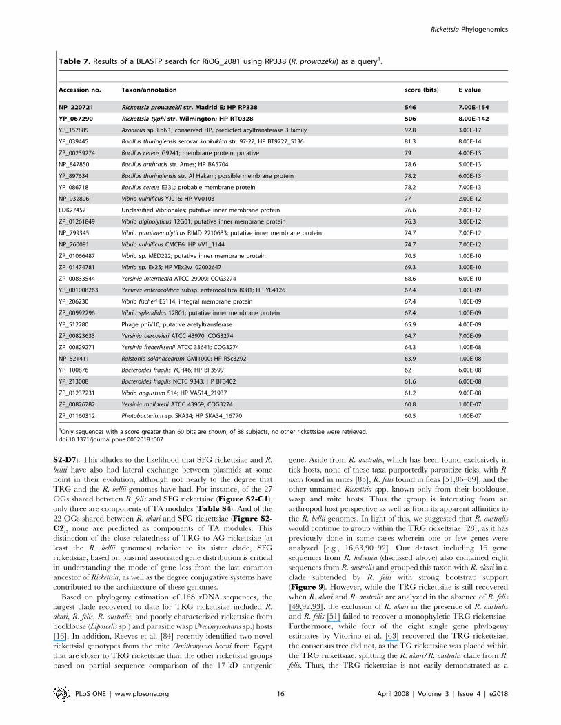
rickettsial groups is worth exploring. RiOG_2081 is an
uncharacterized protein conserved in a limited number of other
bacteria (COG3274) and unknown from non-TG rickettsiae. The
distribution of this protein, a putative membrane associated
acyltransferase, in many pathogenic bacterial species and one
bacteriophage, PhiV10, is interesting (Table 7). Finally,
Br96 (65)1 (1)
Bo112 (79)5 (4)
Ca175 (129)1 (0)
Pr68 (56)0
Ty56 (52)1 (1)
Ak284 (202)3 (2)
Fe312 (218)54 (25)
Ri153 (129)0
Co97 (77)0
Si114 (93)1 (1)
V
V
AG2 (0)0
TG3 (2)0
TRG35 (18)2 (0)
SFG106 (67)7 (5)
core731 (50)21 (2)
derived2 (0)0
tick3 (0)1 (0)
TRG+SFG25 (10)11 (4)
Bo+Fe??
insect2 (1)0
1,522,076
1,159,772
1,587,240*
1,231,060
1,257,710
1,268,755
1,250,021
1,111,523
1,111,496
GenomeSize (bp)
1,429
1,032
1,512
1,337
1,346
1,412
1,234
872
877
ORFsNCBI
31.7
29.0
32.5
32.3
32.5
32.4
32.5
28.9
29.0
85.2
75.2
83.6
77.4
78.5
81.5
77.8
76.2
76.3
Arthropod Host
Plasmid SingletonHost Groups(insect, tick)
1,498bellii312 (222)9 (2)
- bellii2 (0)0
TG+TRG00
1,528,980 31.084.0
bellii +Fe40 (14)4 (4)
1,627
1,158
1,819
1,527
1,614
1,567
1,599
893
888
1,664
ORFsPATRIC
%Coding
%G+C
AG+TG1 (1)0
AG+TRG5 (2) 1 (1)
TG+SFG00
-TG30 (8)23 (7)
-TRG00
-SFG1 (0)0
Phylogenetic1255 (380) 74 (21)
Plasmid40 (14) 4 (4)
Singletons1467 (1100) 66 (34)
Host5 (1)1 (0)
Box schema:
Total2767 (1495) 145 (59)
Distributionrep. OGs (# hypothetical proteins)non-rep. OGs (# hypothetical proteins)
Total (- singletons)1300 (395) 79 (25)
Figure 7. Distribution of representative and non-representative class 1 OGs (C1OGs) and singleton ORFs over estimated rickettsialphylogeny. Boxes depict the distribution of phylogenetic groups, singletons, plasmid associated groups, and host-related groups: Red = AGrickettsiae, aquamarine = TG rickettsiae, blue = TRG rickettsiae, brown = SFG rickettsiae, gray = higher-level groupings, light green = R. bellii strainsonly. Orange boxes depict genes found on the pRF plasmid of R. felis str. URRWXCal2 and chromosomes R. felis and both R. bellii strains (as of thispublication the R. bellii plasmids remain unavailable). Genes specific to single rickettsial genomes (singletons) are in yellow boxes, with taxonabbreviations explained in the Figure 1 legend. Host specific groups are defined by green (insect) and tan (tick) boxes. Genome statistics werecompiled from the PATRIC and NCBI databases. Cladogram is based on trees shown in Figure 3. Inset in dashed box describes general schema foreach box. *Total R. felis genome size: 1,485,148 bp = chromosome; 62,829 bp = pRF and 39,263 bp = pRFd.doi:10.1371/journal.pone.0002018.g007
Rickettsia Phylogenomics
PLoS ONE | www.plosone.org 12 April 2008 | Volume 3 | Issue 4 | e2018

Cellular function categories
A
B
A = RNA processing and modification B = Chromatin structure and dynamics C = Energy production and conversion cf = combined functionD = Cell cycle control, mitosis and meiosis E = Amino acid transport and metabolism F = Nucleotide transport and metabolism G = Carbohydrate transport and metabolism H = Coenzyme transport and metabolism I = Lipid transport and metabolism J = Translation K = TranscriptionL = Replication, recombination and repair M = Cell wall/membrane biogenesis N = Cell motility O = Posttranslational modification, protein turnover, chaperonesP = Inorganic ion transport and metabolism Q = Secondary metabolites biosynthesis, transport and catabolism R = General function prediction only rpe = rickettsial palendromic elementS = Function unknown T = Transduction mechanisms U = Intracellular trafficking and secretion V = Defense mechanisms
V A BC
cf
D
E
F
G
H
I
J
KL
M
N
O
PQ
R
rpe
S
TU
AJBD (2)CE (2)CPEMER (2)ETGMHC
IQLKMIOC (2)QR (2)TKUOVD
Cellular function category
0
50
100
150
200
250
300
A B C cf D E F G H I J K L M N O P Q R rpe S T U V
N
o. o
f pro
tein
s (R
i, Ec
, Yp,
Ct)/
No.
of e
xpre
ssed
gen
es (R
f)
731 rep. core OGs (Ri) Escherichia coli (Ec) Yersinia pestis (Yp) Chlamydia trachomatis (Ct)
Correlation (R )2172 R. felis protiens (Rf)
No. metabolic genes per category; % of coding regions E F G H I P QRi 19; 3.0 17; 2.7 15; 2.4 19; 3.0 16; 2.5 14; 2.2 2; 0.3 Ct 36; 6.5 14; 2.5 28; 5.0 34; 6.1 23; 4.1 16; 2.9 0; 0 Ec 170; 9.2 62; 3.3 141; 7.6 105; 5.7 49; 2.6 131; 7.1 28; 1.5Yp 157; 9.1 56; 3.3 125; 7.3 100; 5.8 42; 2.4 128; 7.4 28; 1.6
Ri vs. Rf Ri vs. Ec Ri vs. Yp Ri vs. Ct Ec vs. Yp Ec vs. Ct Yp vs. Ct
= 0.5576= 0.3844= 0.3791= 0.6913= 0.9891= 0.7555= 0.7582
Figure 8. Bioinformatic analysis of core representative OGs. (A) Assignment of 731 core representative RiOGs to predicted cellular functioncategories. Format follows that established at the COG database (NCBI) except for cf = combined function and rpe = rickettsial palindromic element.(B) Comparison of the distribution of cellular function categories across 731 core rickettsial OGs (Ri), a recent protein expression profile for R. felis [40](Rf), and COGs for three other bacteria: Escherichia coli (Ec), Yersinia pestis (Yp) and Chlamydia trachomatis (Ct). Inset at left shows the number ofgenes per genome for cellular function categories involved in organic and inorganic transport and metabolism (E, F, G, H, I, P, and Q) followed by thepercentage these genes comprise of total protein-encoding genes. Results from a six-way regression analysis are shown in the right inset.doi:10.1371/journal.pone.0002018.g008
Rickettsia Phylogenomics
PLoS ONE | www.plosone.org 13 April 2008 | Volume 3 | Issue 4 | e2018
RiOG_2082 is a small putative ORF that BLASTs to no other
organisms, with the start codon missing in R. typhi.
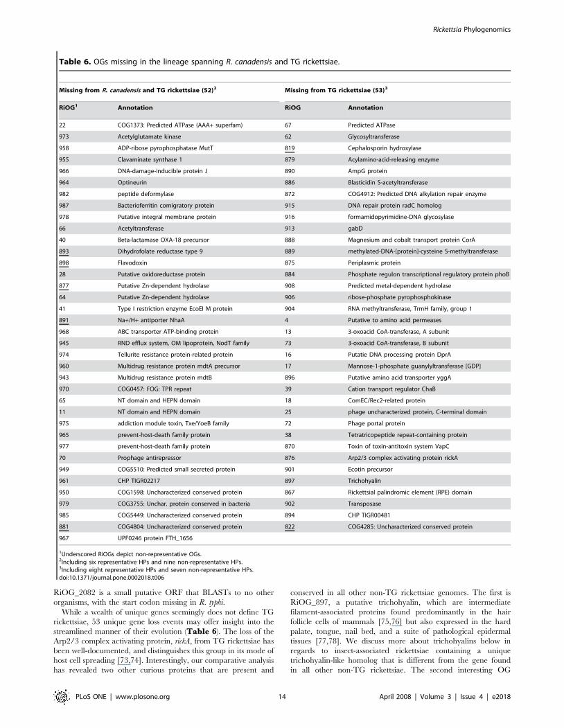
While a wealth of unique genes seemingly does not define TG
rickettsiae, 53 unique gene loss events may offer insight into the
streamlined manner of their evolution (Table 6). The loss of the
Arp2/3 complex activating protein, rickA, from TG rickettsiae has
been well-documented, and distinguishes this group in its mode of
host cell spreading [73,74]. Interestingly, our comparative analysis
has revealed two other curious proteins that are present and
conserved in all other non-TG rickettsiae genomes. The first is
RiOG_897, a putative trichohyalin, which are intermediate
filament-associated proteins found predominantly in the hair
follicle cells of mammals [75,76] but also expressed in the hard
palate, tongue, nail bed, and a suite of pathological epidermal
tissues [77,78]. We discuss more about trichohyalins below in
regards to insect-associated rickettsiae containing a unique
trichohyalin-like homolog that is different from the gene found
in all other non-TG rickettsiae. The second interesting OG
Table 6. OGs missing in the lineage spanning R. canadensis and TG rickettsiae.
Missing from R. canadensis and TG rickettsiae (52)2 Missing from TG rickettsiae (53)3
RiOG1 Annotation RiOG Annotation
22 COG1373: Predicted ATPase (AAA+ superfam) 67 Predicted ATPase
973 Acetylglutamate kinase 62 Glycosyltransferase
958 ADP-ribose pyrophosphatase MutT 819 Cephalosporin hydroxylase
955 Clavaminate synthase 1 879 Acylamino-acid-releasing enzyme
966 DNA-damage-inducible protein J 890 AmpG protein
964 Optineurin 886 Blasticidin S-acetyltransferase
982 peptide deformylase 872 COG4912: Predicted DNA alkylation repair enzyme
987 Bacterioferritin comigratory protein 915 DNA repair protein radC homolog
978 Putative integral membrane protein 916 formamidopyrimidine-DNA glycosylase
66 Acetyltransferase 913 gabD
40 Beta-lactamase OXA-18 precursor 888 Magnesium and cobalt transport protein CorA
893 Dihydrofolate reductase type 9 889 methylated-DNA-[protein]-cysteine S-methyltransferase
898 Flavodoxin 875 Periplasmic protein
28 Putative oxidoreductase protein 884 Phosphate regulon transcriptional regulatory protein phoB
877 Putative Zn-dependent hydrolase 908 Predicted metal-dependent hydrolase
64 Putative Zn-dependent hydrolase 906 ribose-phosphate pyrophosphokinase
41 Type I restriction enzyme EcoEI M protein 904 RNA methyltransferase, TrmH family, group 1
891 Na+/H+ antiporter NhaA 4 Putative to amino acid permeases
968 ABC transporter ATP-binding protein 13 3-oxoacid CoA-transferase, A subunit
945 RND efflux system, OM lipoprotein, NodT family 73 3-oxoacid CoA-transferase, B subunit
974 Tellurite resistance protein-related protein 16 Putatie DNA processing protein DprA
960 Multidrug resistance protein mdtA precursor 17 Mannose-1-phosphate guanylyltransferase [GDP]
943 Multidrug resistance protein mdtB 896 Putative amino acid transporter yggA
970 COG0457: FOG: TPR repeat 39 Cation transport regulator ChaB
65 NT domain and HEPN domain 18 ComEC/Rec2-related protein
11 NT domain and HEPN domain 25 phage uncharacterized protein, C-terminal domain
975 addiction module toxin, Txe/YoeB family 72 Phage portal protein
965 prevent-host-death family protein 38 Tetratricopeptide repeat-containing protein
977 prevent-host-death family protein 870 Toxin of toxin-antitoxin system VapC
70 Prophage antirepressor 876 Arp2/3 complex activating protein rickA
949 COG5510: Predicted small secreted protein 901 Ecotin precursor
961 CHP TIGR02217 897 Trichohyalin
950 COG1598: Uncharacterized conserved protein 867 Rickettsial palindromic element (RPE) domain
979 COG3755: Unchar. protein conserved in bacteria 902 Transposase
985 COG5449: Uncharacterized conserved protein 894 CHP TIGR00481
881 COG4804: Uncharacterized conserved protein 822 COG4285: Uncharacterized conserved protein
967 UPF0246 protein FTH_1656
1Underscored RiOGs depict non-representative OGs.2Including six representative HPs and nine non-representative HPs.3Including eight representative HPs and seven non-representative HPs.doi:10.1371/journal.pone.0002018.t006
Rickettsia Phylogenomics
PLoS ONE | www.plosone.org 14 April 2008 | Volume 3 | Issue 4 | e2018

(RiOG_901) found exclusively in non-TG rickettsiae is an ecotin-
like protein. Ecotin is a dimeric periplasmic protein described in
Escherichia coli that belongs to the protease inhibitor I11 (ecotin)
family (PF03974). Ecotin inhibits several pancreatic serine
proteases, including chymotrypsin, trypsin, elastases, factor X,
kallikrein, as well as a variety of other proteases [79–81]. Eggers et
al. [82] have shown that ecotin protects E. coli from neutrophil
elastase (NE), a mammalian serine protease demonstrated to be
important for neutrophil killing of several gram-negative bacteria.
Specifically, NE cleaves ompA causing increased permeability to
the bacterial outer membrane [83]. Once NE translocates across
the vulnerable outer membrane, it functions in inhibiting bacterial
cell growth and repair, causing cell death. The presence of ecotin
in the periplasm inhibits NE function, thus fostering recovery and
growth of the invading bacterial cells [82]. Given the diversity of
rickettsial outer membrane surface proteins, particularly the Scas
[55], it is reasonable to suggest that one or several surface proteins
present in all non-TG rickettsiae may be dependent upon the
putative NE inhibitory function of RiOG_901.
TRG rickettsiae. Based on the monophyly of its sampled
members (R. felis and R. akari), its strongly supported position in
our estimated rickettsial phylogeny, an affinity with AG rickettsiae
plasmid-associated genes, and the use of both acarines and insects
as primary invertebrate hosts, we erected the TRG rickettsiae as a
third derived lineage of Rickettsia [28]. OrthoMCL predicted 37
OGs unique to TRG rickettsiae (Table 8). Of the three other
rickettsial lineages, TRG shares more common OGs with SFG
rickettsiae (36) than with TG rickettsiae (0) or AG rickettsiae (6)
(Figure 7), reflecting its shared common ancestry with the ‘‘true’’
spotted fever group taxa. However, exclusion of R. canadensis sheds
light on our previously described affinities of TRG rickettsiae with
AG rickettsiae (Table 9). For instance, 26 OGs are shared
between the R. bellii genomes and TRG rickettsiae (Figure S2-C25), with six of these annotated as members of toxin-antitoxin
(TA) modules, and another two annotated as bacteriophage-
derived proteins. Additionally, the R. felis genome shares 44 OGs
with the R. bellii genomes (Figure 7), six of which are annotated as
members of TA modules, with another one annotated as
bacteriophage-derived protein. Furthermore, the R. akari genome
shares 10 OGs with the R. bellii genomes (Figure S2-B23), and
two of these OGs are predicted members of TA modules. This
high presence of TA system components, as well as bacteriophage-
derived proteins, attests to our previous observations that AG (at
least R. bellii) and TRG rickettsiae are linked via conjugative
systems and have a pronounced presence of similar plasmid (and
now phage) related ORFs, likely the end products of various lateral
gene exchanges between these distantly related groups.
Despite the abovementioned characteristics shared between AG
and TRG rickettsiae, the TRG rickettsiae also share three TA
components exclusively with SFG rickettsiae (Table S4).
Additionally, SFG rickettsiae and the R. bellii genomes have three
TA components not found in the other analyzed genomes (Figure
500 changes
R. bellii str. OSU 85 389 *
R. canadensis str. McKiel *
R. australis
R. helvetica
64
92
100100
100
100
100
100
100
R. prowazekii str. Madrid E *
R. typhi str. Wilmington *
R. bellii str. RML369 C *
R. akari str. Hartford *
R. felis str. URRWXCal2 *
R. rickettsii str. Sheila Smith CWPP *
R. conorii str. Malish 7 *
R. sibirica str. 246 *
AG
TG
TRG
SFG
Figure 9. Phylogeny estimation of the ten analyzed rickettsial taxa plus R. helvetica and R. australis based on 16 proteins. See TableS13 for gene names and sequence accession numbers. Tree estimated under parsimony (see text).doi:10.1371/journal.pone.0002018.g009
Rickettsia Phylogenomics
PLoS ONE | www.plosone.org 15 April 2008 | Volume 3 | Issue 4 | e2018
S2-D7). This alludes to the likelihood that SFG rickettsiae and R.
bellii have also had lateral exchange between plasmids at some
point in their evolution, although not nearly to the degree that
TRG and the R. bellii genomes have had. For instance, of the 27
OGs shared between R. felis and SFG rickettsiae (Figure S2-C1),
only three are components of TA modules (Table S4). And of the
22 OGs shared between R. akari and SFG rickettsiae (Figure S2-C2), none are predicted as components of TA modules. This
distinction of the close relatedness of TRG to AG rickettsiae (at
least the R. bellii genomes) relative to its sister clade, SFG
rickettsiae, based on plasmid associated gene distribution is critical
in understanding the mode of gene loss from the last common
ancestor of Rickettsia, as well as the degree conjugative systems have
contributed to the architecture of these genomes.
Based on phylogeny estimation of 16S rDNA sequences, the
largest clade recovered to date for TRG rickettsiae included R.
akari, R. felis, R. australis, and poorly characterized rickettsiae from
booklouse (Liposcelis sp.) and parasitic wasp (Neochrysocharis sp.) hosts
[16]. In addition, Reeves et al. [84] recently identified two novel
rickettsial genotypes from the mite Ornithonyssus bacoti from Egypt
that are closer to TRG rickettsiae than the other rickettsial groups
based on partial sequence comparison of the 17 kD antigenic
gene. Aside from R. australis, which has been found exclusively in
tick hosts, none of these taxa purportedly parasitize ticks, with R.
akari found in mites [85], R. felis found in fleas [51,86–89], and the
other unnamed Rickettsia spp. known only from their booklouse,
wasp and mite hosts. Thus the group is interesting from an
arthropod host perspective as well as from its apparent affinities to
the R. bellii genomes. In light of this, we suggested that R. australis
would continue to group within the TRG rickettsiae [28], as it has
previously done in some cases wherein one or few genes were
analyzed [e.g., 16,63,90–92]. Our dataset including 16 gene
sequences from R. helvetica (discussed above) also contained eight
sequences from R. australis and grouped this taxon with R. akari in a
clade subtended by R. felis with strong bootstrap support
(Figure 9). However, while the TRG rickettsiae is still recovered
when R. akari and R. australis are analyzed in the absence of R. felis
[49,92,93], the exclusion of R. akari in the presence of R. australis
and R. felis [51] failed to recover a monophyletic TRG rickettsiae.
Furthermore, while four of the eight single gene phylogeny
estimates by Vitorino et al. [63] recovered the TRG rickettsiae,
the consensus tree did not, as the TG rickettsiae was placed within
the TRG rickettsiae, splitting the R. akari/R. australis clade from R.
felis. Thus, the TRG rickettsiae is not easily demonstrated as a
Table 7. Results of a BLASTP search for RiOG_2081 using RP338 (R. prowazekii) as a query1.
Accession no. Taxon/annotation score (bits) E value
NP_220721 Rickettsia prowazekii str. Madrid E; HP RP338 546 7.00E-154
YP_067290 Rickettsia typhi str. Wilmington; HP RT0328 506 8.00E-142
YP_157885 Azoarcus sp. EbN1; conserved HP, predicted acyltransferase 3 family 92.8 3.00E-17
YP_039445 Bacillus thuringiensis serovar konkukian str. 97-27; HP BT9727_5136 81.3 8.00E-14
ZP_00239274 Bacillus cereus G9241; membrane protein, putative 79 4.00E-13
NP_847850 Bacillus anthracis str. Ames; HP BA5704 78.6 5.00E-13
YP_897634 Bacillus thuringiensis str. Al Hakam; possible membrane protein 78.2 6.00E-13
YP_086718 Bacillus cereus E33L; probable membrane protein 78.2 7.00E-13
NP_932896 Vibrio vulnificus YJ016; HP VV0103 77 2.00E-12
EDK27457 Unclassified Vibrionales; putative inner membrane protein 76.6 2.00E-12
ZP_01261849 Vibrio alginolyticus 12G01; putative inner membrane protein 76.3 3.00E-12
NP_799345 Vibrio parahaemolyticus RIMD 2210633; putative inner membrane protein 74.7 7.00E-12
NP_760091 Vibrio vulnificus CMCP6; HP VV1_1144 74.7 7.00E-12
ZP_01066487 Vibrio sp. MED222; putative inner membrane protein 70.5 1.00E-10
ZP_01474781 Vibrio sp. Ex25; HP VEx2w_02002647 69.3 3.00E-10
ZP_00833544 Yersinia intermedia ATCC 29909; COG3274 68.6 6.00E-10
YP_001008263 Yersinia enterocolitica subsp. enterocolitica 8081; HP YE4126 67.4 1.00E-09
YP_206230 Vibrio fischeri ES114; integral membrane protein 67.4 1.00E-09
ZP_00992296 Vibrio splendidus 12B01; putative inner membrane protein 67.4 1.00E-09
YP_512280 Phage phiV10; putative acetyltransferase 65.9 4.00E-09
ZP_00823633 Yersinia bercovieri ATCC 43970; COG3274 64.7 7.00E-09
ZP_00829271 Yersinia frederiksenii ATCC 33641; COG3274 64.3 1.00E-08
NP_521411 Ralstonia solanacearum GMI1000; HP RSc3292 63.9 1.00E-08
YP_100876 Bacteroides fragilis YCH46; HP BF3599 62 6.00E-08
YP_213008 Bacteroides fragilis NCTC 9343; HP BF3402 61.6 6.00E-08
ZP_01237231 Vibrio angustum S14; HP VAS14_21937 61.2 9.00E-08
ZP_00826782 Yersinia mollaretii ATCC 43969; COG3274 60.8 1.00E-07
ZP_01160312 Photobacterium sp. SKA34; HP SKA34_16770 60.5 1.00E-07
1Only sequences with a score greater than 60 bits are shown; of 88 subjects, no other rickettsiae were retrieved.doi:10.1371/journal.pone.0002018.t007
Rickettsia Phylogenomics
PLoS ONE | www.plosone.org 16 April 2008 | Volume 3 | Issue 4 | e2018

distinct lineage of rickettsiae unless the taxon and character
sampling is robust enough for this intriguing lineage to emerge
(Figure 9; [28]).
SFG rickettsiae. The majority of the described species of
Rickettsia fall within the SFG rickettsiae. The analyzed spotted fever
group genomes form a monophyletic cluster of taxa with little
sequence divergence relative to the other rickettsial groups
(Figure 3). OrthoMCL predicted 113 OGs that are unique to
SFG rickettsiae (Table 10). Of note, in addition to the four core
rickettsial proline/betaine transporters (Table S2), SFG
rickettsiae contain two variant copies (RiOG_1314 and RiOG_
1332). Other transporters unique to SFG rickettsiae include three
ATPase and permease components of an ABC-type multidrug
transporter (RiOG_1347, RiOG_1364 and RiOG_1365), an
ATP-binding protein similar to ABC transporter (RiOG_1376),
an MSF-like sugar transporter (RiOG_1355), and an RND family
efflux transporter (RiOG_1294). While high numbers of trans-
porters are expected in Rickettsia to counterbalance depleted
metabolic pathways and acquire host resources, it is unclear why
the SFG rickettsiae have elevated levels of unique components of
organic and inorganic transport systems relative to the other three
rickettsial groups. As with TG rickettsiae, there are group-specific
GTP pyrophosphokinases (RiOG_1350 and RiOG_1361) in SFG
rickettsial genomes, and their role in a group-specific stringent
response is worthy of attention. Like AG and TRG rickettsial
genomes, SFG rickettsiae have group-specific ANK repeat
containing proteins, with a particular one (RiOG_1344) similar
to metazoan tankyrases, telomeric repeat binding factor-
interacting ANK-related ADP-ribose polymerases. Aside from
potentially playing key roles in the maintenance of telomere
function [e.g., 94], tankyrases have been implicated in mitogen-
activated protein kinase signaling [95], regulation of cell death
[96,97] and viral inhibition [98].
Using EasyGene [99], a program that ranks prokaryotic
predicted ORFs based on statistical significance, Nielsen and
Krogh [100] determined that the R. conorii str. Malish 7 genome
was over-annotated by 16%, ranking 7th among most over-
annotated replicons in a sample of 143 prokaryotic genomes.
Specifically, EasyGene determined 225 RefSeq genes to be false,
with 34 additional genes predicted by EasyGene that were not
called in the original study [22,23]. Aside from possible gross ORF
over-prediction in all ten rickettsial genomes (discussed below), our
analysis yielded many OGs with imperfect representation within
the SFG group, as 54 OGs are found exclusively in the R. conorii
and R. sibirica genomes (Figure S2-A1), 52 are found exclusively
in the R. rickettsii and R. sibirica genomes (Figure S2-A2), and 36
are found exclusively in the R. rickettsii and R. conorii genomes
(Figure S2-A3). Given that the SFG rickettsial genomes have
elevated split genes as compared to other rickettsial genomes
(Table 5; Table S1), our findings and those of Nielsen and
Krogh [100] hint at a pronounced rate of pseudogenization in
SFG rickettsiae depicted by a patchy distribution of split and
truncated ORFs decaying from the ancestral SFG genome.
One hallmark occurrence of probable pseudogenization in SFG
rickettsiae involves a Sec7-domain-containing protein known in
prokaryotes only from Rickettsia and Legionella species [101]. The
Legionella counterpart of this curious protein, named RalF, is a
guanine nucleotide exchange factor that recruits ADP-ribosylation
factor to occupied phagosomes, permitting Legionella to replicate
free from the host immune system [102]. The rickettsial RalF
xenolog (RiOG_19), including the N-terminal Sec7 domain and
immediate flanking Sec7-capping-domain [103], is present in all
rickettsial genomes except for SFG rickettsiae and R. canadensis,
suggesting a biological mechanism that has been lost from the true
spotted fever group and R. canadensis. Unlike Legionella RalF, which
has a short (44 aa) C-terminal tail containing a type 4 secretion
system (T4SS) signal sequence [104], the rickettsial genes encode
an additional variable domain (97–315 aa) between the Sec7-
capping-domain and the C-terminal tail. Within this third domain
lies a region immediately flanking the predicted T4SS signal
sequence that is extraordinarily rich in proline residues, much like
the P-rich domain of rickA proteins [74]. Interestingly, the SFG
genomes each contain small ORFs corresponding to the tails of the
RalF-like sequences. A similar sequence within the R. canadensis
genome (not annotated) also spans this region yet is riddled with
frame-shift mutations. Given that Rickettsia, unlike Legionella,
quickly lyse the phagosome upon host cell entry, the function of
a RalF xenolog, particularly given its curious distribution in the
rickettsial tree, is worthy of investigation. Finally, full intact RalF
xenologs in both TRG rickettsial genomes further attest the
distinction of this lineage from the SFG rickettsiae [28].
Arthropod Host-Specific OGsSeveral studies have demonstrated the presence of certain
rickettsial species outside of their natural arthropod hosts. For
example, the louse (and less often flea) associated R. prowazekii has
been found in ticks in Africa [105] and Mexico [106], and was also
reported in acarids from flying squirrels in the United States [107].
However, it should be recognized that many blood-feeding
arthropods have a wide range of vertebrate hosts and likely act
as reservoirs for a variety of bacteria that incidentally fall outside of
their natural arthropod vector. To this extent reports of
pathogenic bacteria (i.e., R. prowazekii) in unusual vectors need to
be substantiated beyond simple detection in these foreign hosts,
and caution should be taken when immediately assigning novel
Table 8. OGs present only in TRG rickettsiae.
RiOG1 Annotation (37)2
2043 COG1670: Acetyltransferases, incl. N-acetylases of ribosomal proteins
2078 Predicted acetyltransferase
2062 Predicted hydrolase or acyltransferase
2038 Putative cysteine protease yopT-like
2047 5-Formyltetrahydrofolate cyclo-ligase
1125 alanine racemase
2033 Outer membrane protein A precursor
2037 Outer membrane protein A precursor
2046 Outer membrane protein A precursor
2076 Outer membrane protein A precursor
2049 ABC transporter, ATP-binding protein
2075 Cell surface antigen-like protein Sca7
2059 Ankyrin repeat
2066COG1487: Predicted nucleic acid-binding protein, contains PINdomain
2056 Probable antitoxin of toxin-antitoxin stability system
2069 addiction module toxin, Txe/YoeB family
2050 Virulence-associated protein B
1483 CHP
2068 CHP
1Underscored RiOGs depict non-representative OGs.2Including 18 representative HPs.doi:10.1371/journal.pone.0002018.t008
Rickettsia Phylogenomics
PLoS ONE | www.plosone.org 17 April 2008 | Volume 3 | Issue 4 | e2018

host associations. Given the low frequency of resident bacteria in
many natural arthropod populations [108], substantiation of novel
arthropod hosts can be achieved in the field by robustly sampling
other invertebrate and vertebrate animals from the same locality
that may actually be the true host of the incidentally collected
bacterium. Furthermore, laboratory studies would be needed to
determine the pathogenicity, if any, that the bacterium causes in
its novel host. However, laboratory inoculation of an animal may
result in pathogenesis only because the number of bacteria far
exceeded what occurs in nature, thus compromising an immune
system that under natural circumstances is quite capable of killing
the pathogen. Furthermore, demonstrating laboratory bacterial
infection or vectorization in a foreign host, for example R. conorii in
the body louse [109], may initially prove successful, but eventually
will clear from the host as it would from natural populations. For
instance, Rickettsia have been grown in mosquito cell lines, yet to
our knowledge no wild caught mosquitoes to date have been
shown to act as hosts to any Rickettsia. In fact, based on the analysis
of the highly divergent sca genes in rickettsiae, which are suspected
to directly interact with host cell proteins [47,110], Blanc et al.
[55] concluded that rapid evolution of such important host
colonization genes likely keep Rickettsia host ranges quite narrow.
Given our conservative stance on definitive rickettsial arthropod
hosts, we have chosen to present the predicted genes that are
exclusive to insect associated Rickettsia and tick associated Rickettsia
(as depicted in Figure 7). Because only one analyzed genome is
from a mite-associated species (R. akari), we have no comparative
analysis to describe potential mite-specific rickettsial genes.
However, the list of singleton genes found in R. akari may provide
a start to such an approach (see below).
Insect-associated rickettsiae. Three of the ten analyzed
rickettsial genomes have definitive insect hosts, with R. typhi and R.
felis reported from rodent, shrew and feline [51,86–88] associated
fleas, and R. prowazekii predominantly pathogenic in lice, as well as
fleas in the sylvatic form. Thus these three rickettsial lineages share
common arthropod hosts at least in fleas. Regarding R. typhi, It has
become apparent that the ecology of murine typhus in both south
Texas and southern California, where the classic cycle of R. typhi
involving commensal rats and primarily the rat flea (Xensopsylla
cheopis), has been replaced by the Virginia opossum (Didelphis
virginiana)/cat flea (Ctenocephalides felis) cycle. For instance, Sorvillo
et al. [111] demonstrated the association of 33 cases of locally
acquired murine typhus in Los Angeles County with seropositive
domestic cats and opossums. However, urban rat/flea populations
are still the main reservoir of R. typhi worldwide and particularly in
Table 9. OGs present only in R. bellii strains and TRGrickettsiae.
Present in R. bellii strains and TRG rickettsiae (26)2
RiOG1 Annotation
1245 HicB family
1261 Phage-related transcriptional regulator
1215 phage host specificity protein
1128 Transcriptional regulator
1266 PIN domain containing protein
1256 Antitoxin of toxin-antitoxin system StbD
1262 Cytotoxic translational repressor of toxin-antitoxin (TA) system RelE
1251 Cytotoxic translational repressor of toxin-antitoxin system RelE
1243 Growth inhibitor
1240 putative addiction module antidote protein, CC2985 family
1269 Transposase
1 Probable transposase for insertion sequence element
1260 CHP
Present in R. bellii strains and R. felis (44)3
RiOG Annotation
1437 Cell surface antigen-like protein Sca9
1418 Alkylated DNA repair protein
1423 cAMP-inducible prespore protein D7 precursor
1415 Caspase recruitment domain-containing protein 15
1451 Citrate-proton symporter
1410 Fic family protein
1438 Heavy metal tolerance protein precursor
1420 invasion protein homolog
1442 Lysine efflux permease
1428 Lysozyme
1417 Menaquinone biosynthesis methyltransferase ubiE
1406 nucleotidyltransferase substrate binding protein, HI0074 family
1452 Putative AAA+ superfamily ATPase
1431 Putative integrase/recombinase HI1572
1436 Streptomycin 6-kinase
1414 Ankyrin repeat
1434 phage major capsid protein, HK97 family
1426 Bacterial transcription activator, effector binding domain
1446 Antitoxin of toxin-antitoxin system Phd
1408 prevent-host-death family protein
1427 prevent-host-death family protein
1430 probable addiction module killer protein
1435 Toxin of toxin-antitoxin (TA) system
1450 Transcriptional regulator, AbrB family
1411 CHP
1413 CHP
Present in R. bellii strains and R. akari (10)4
RiOG Annotation
1458 endo/excinuclease amino terminal domain protein
1472 Leucine-rich repeat-containing protein 45
Present in R. bellii strains and R. akari (10)4
RiOG Annotation
1489 Reticulocyte-binding protein 1 precursor
1465 Ankyrin repeat
1466COG1848: Predicted nucleic acid-binding protein, contains PINdomain
1470 Prevent-host-death protein
1480 CHP
1Underscored RiOGs depict non-representative OGs.2Including 12 representative HPs and 1 non-representative HP.3Including 14 representative HPs and 4 non-representative HPs.4Including 3 HPs.doi:10.1371/journal.pone.0002018.t009
Table 9. cont.
Rickettsia Phylogenomics
PLoS ONE | www.plosone.org 18 April 2008 | Volume 3 | Issue 4 | e2018
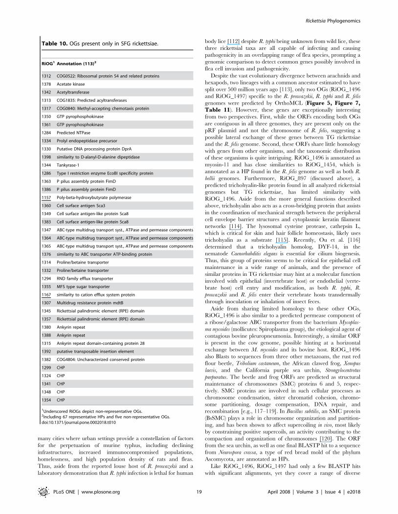
many cities where urban settings provide a constellation of factors
for the perpetuation of murine typhus, including declining
infrastructures, increased immunocompromised populations,
homelessness, and high population density of rats and fleas.
Thus, aside from the reported louse host of R. prowazekii and a
laboratory demonstration that R. typhi infection is lethal for human
body lice [112] despite R. typhi being unknown from wild lice, these
three rickettsial taxa are all capable of infecting and causing
pathogenicity in an overlapping range of flea species, prompting a
genomic comparison to detect common genes possibly involved in
flea cell invasion and pathogenicity.
Despite the vast evolutionary divergence between arachnids and
hexapods, two lineages with a common ancestor estimated to have
split over 500 million years ago [113], only two OGs (RiOG_1496
and RiOG_1497) specific to the R. prowazekii, R. typhi and R. felis
genomes were predicted by OrthoMCL (Figure 5, Figure 7,Table 11). However, these genes are exceptionally interesting
from two perspectives. First, while the ORFs encoding both OGs
are contiguous in all three genomes, they are present only on the
pRF plasmid and not the chromosome of R. felis, suggesting a
possible lateral exchange of these genes between TG rickettsiae
and the R. felis genome. Second, these ORFs share little homology
with genes from other organisms, and the taxonomic distribution
of these organisms is quite intriguing. RiOG_1496 is annotated as
myosin-11 and has close similarities to RiOG_1454, which is
annotated as a HP found in the R. felis genome as well as both R.
bellii genomes. Furthermore, RiOG_897 (discussed above), a
predicted trichohyalin-like protein found in all analyzed rickettsial
genomes but TG rickettsiae, has limited similarity with
RiOG_1496. Aside from the more general functions described
above, trichohyalin also acts as a cross-bridging protein that assists
in the coordination of mechanical strength between the peripheral
cell envelope barrier structures and cytoplasmic keratin filament
networks [114]. The lysosomal cysteine protease, cathepsin L,
which is critical for skin and hair follicle homeostasis, likely uses
trichohyalin as a substrate [115]. Recently, Ou et al. [116]
determined that a trichohyalin homolog, DYF-14, in the
nematode Caenorhabditis elegans is essential for cilium biogenesis.
Thus, this group of proteins seems to be critical for epithelial cell
maintenance in a wide range of animals, and the presence of
similar proteins in TG rickettsiae may hint at a molecular function
involved with epithelial (invertebrate host) or endothelial (verte-
brate host) cell entry and modification, as both R. typhi, R.
prowazekii and R. felis enter their vertebrate hosts transdermally
through inoculation or inhalation of insect feces.
Aside from sharing limited homology to these other OGs,
RiOG_1496 is also similar to a predicted permease component of
a ribose/galactose ABC transporter from the bacterium Mycoplas-
ma mycoides (mollicutes: Spiroplasma group), the etiological agent of
contagious bovine pleuropneumonia. Interestingly, a similar ORF
is present in the cow genome, possible hinting at a horizontal
exchange between M. mycoides and its bovine host. RiOG_1496
also Blasts to sequences from three other metazoans, the rust red
flour beetle, Tribolium castaneum, the African clawed frog, Xenopus
laevis, and the California purple sea urchin, Strongylocentrotus
purpuratus. The beetle and frog ORFs are predicted as structural
maintenance of chromosomes (SMC) proteins 6 and 5, respec-
tively. SMC proteins are involved in such cellular processes as
chromosome condensation, sister chromatid cohesion, chromo-
some partitioning, dosage compensation, DNA repair, and
recombination [e.g., 117–119]. In Bacillus subtilis, an SMC protein
(BsSMC) plays a role in chromosome organization and partition-
ing, and has been shown to affect supercoiling in vivo, most likely
by constraining positive supercoils, an activity contributing to the
compaction and organization of chromosomes [120]. The ORF
from the sea urchin, as well as one final BLASTP hit to a sequence
from Neurospora crassa, a type of red bread mold of the phylum
Ascomycota, are annotated as HPs.
Like RiOG_1496, RiOG_1497 had only a few BLASTP hits
with significant alignments, yet they cover a range of diverse
Table 10. OGs present only in SFG rickettsiae.
RiOG1 Annotation (113)2
1312 COG0522: Ribosomal protein S4 and related proteins
1378 Acetate kinase
1342 Acetyltransferase
1313 COG1835: Predicted acyltransferases
1317 COG0840: Methyl-accepting chemotaxis protein
1350 GTP pyrophosphokinase
1361 GTP pyrophosphokinase
1284 Predicted NTPase
1334 Prolyl endopeptidase precursor
1330 Putative DNA processing protein DprA
1398 similarity to D-alanyl-D-alanine dipeptidase
1344 Tankyrase-1
1286 Type I restriction enzyme EcoBI specificity protein
1363 P pilus assembly protein FimD
1386 P pilus assembly protein FimD
1157 Poly-beta-hydroxybutyrate polymerase
1360 Cell surface antigen Sca3
1349 Cell surface antigen-like protein Sca8
1383 Cell surface antigen-like protein Sca8
1347 ABC-type multidrug transport syst., ATPase and permease components
1364 ABC-type multidrug transport syst., ATPase and permease components
1365 ABC-type multidrug transport syst., ATPase and permease components
1376 similarity to ABC transporter ATP-binding protein
1314 Proline/betaine transporter
1332 Proline/betaine transporter
1294 RND family efflux transporter
1355 MFS type sugar transporter
1167 similarity to cation efflux system protein
1307 Multidrug resistance protein mdtB
1345 Rickettsial palindromic element (RPE) domain
1357 Rickettsial palindromic element (RPE) domain
1380 Ankyrin repeat
1388 Ankyrin repeat
1315 Ankyrin repeat domain-containing protein 28
1392 putative transposable insertion element
1382 COG4804: Uncharacterized conserved protein
1299 CHP
1324 CHP
1341 CHP
1348 CHP
1354 CHP
1Underscored RiOGs depict non-representative OGs.2Including 67 representative HPs and five non-representative OGs.doi:10.1371/journal.pone.0002018.t010
Rickettsia Phylogenomics
PLoS ONE | www.plosone.org 19 April 2008 | Volume 3 | Issue 4 | e2018
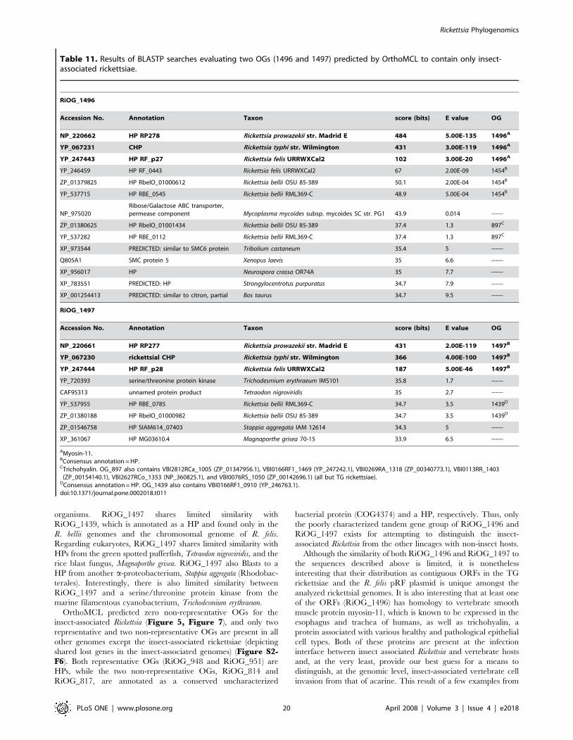
organisms. RiOG_1497 shares limited similarity with
RiOG_1439, which is annotated as a HP and found only in the
R. bellii genomes and the chromosomal genome of R. felis.
Regarding eukaryotes, RiOG_1497 shares limited similarity with
HPs from the green spotted pufferfish, Tetraodon nigroviridis, and the
rice blast fungus, Magnaporthe grisea. RiOG_1497 also Blasts to a
HP from another a-proteobacterium, Stappia aggregata (Rhodobac-
terales). Interestingly, there is also limited similarity between
RiOG_1497 and a serine/threonine protein kinase from the
marine filamentous cyanobacterium, Trichodesmium erythraeum.
OrthoMCL predicted zero non-representative OGs for the
insect-associated Rickettsia (Figure 5, Figure 7), and only two
representative and two non-representative OGs are present in all
other genomes except the insect-associated rickettsiae (depicting
shared lost genes in the insect-associated genomes) (Figure S2-F6). Both representative OGs (RiOG_948 and RiOG_951) are
HPs, while the two non-representative OGs, RiOG_814 and
RiOG_817, are annotated as a conserved uncharacterized
bacterial protein (COG4374) and a HP, respectively. Thus, only
the poorly characterized tandem gene group of RiOG_1496 and
RiOG_1497 exists for attempting to distinguish the insect-
associated Rickettsia from the other lineages with non-insect hosts.
Although the similarity of both RiOG_1496 and RiOG_1497 to
the sequences described above is limited, it is nonetheless
interesting that their distribution as contiguous ORFs in the TG
rickettsiae and the R. felis pRF plasmid is unique amongst the
analyzed rickettsial genomes. It is also interesting that at least one
of the ORFs (RiOG_1496) has homology to vertebrate smooth
muscle protein myosin-11, which is known to be expressed in the
esophagus and trachea of humans, as well as trichohyalin, a
protein associated with various healthy and pathological epithelial
cell types. Both of these proteins are present at the infection
interface between insect associated Rickettsia and vertebrate hosts
and, at the very least, provide our best guess for a means to
distinguish, at the genomic level, insect-associated vertebrate cell
invasion from that of acarine. This result of a few examples from
Table 11. Results of BLASTP searches evaluating two OGs (1496 and 1497) predicted by OrthoMCL to contain only insect-associated rickettsiae.
RiOG_1496
Accession No. Annotation Taxon score (bits) E value OG
NP_220662 HP RP278 Rickettsia prowazekii str. Madrid E 484 5.00E-135 1496A
YP_067231 CHP Rickettsia typhi str. Wilmington 431 3.00E-119 1496A
YP_247443 HP RF_p27 Rickettsia felis URRWXCal2 102 3.00E-20 1496A
YP_246459 HP RF_0443 Rickettsia felis URRWXCal2 67 2.00E-09 1454B
ZP_01379825 HP RbelO_01000612 Rickettsia bellii OSU 85-389 50.1 2.00E-04 1454B
YP_537715 HP RBE_0545 Rickettsia bellii RML369-C 48.9 5.00E-04 1454B
NP_975020Ribose/Galactose ABC transporter,permease component Mycoplasma mycoides subsp. mycoides SC str. PG1 43.9 0.014 ------
ZP_01380625 HP RbelO_01001434 Rickettsia bellii OSU 85-389 37.4 1.3 897C
YP_537282 HP RBE_0112 Rickettsia bellii RML369-C 37.4 1.3 897C
XP_973544 PREDICTED: similar to SMC6 protein Tribolium castaneum 35.4 5 ------
Q805A1 SMC protein 5 Xenopus laevis 35 6.6 ------
XP_956017 HP Neurospora crassa OR74A 35 7.7 ------
XP_783551 PREDICTED: HP Strongylocentrotus purpuratus 34.7 7.9 ------
XP_001254413 PREDICTED: similar to citron, partial Bos taurus 34.7 9.5 ------
RiOG_1497
Accession No. Annotation Taxon score (bits) E value OG
NP_220661 HP RP277 Rickettsia prowazekii str. Madrid E 431 2.00E-119 1497B
YP_067230 rickettsial CHP Rickettsia typhi str. Wilmington 366 4.00E-100 1497B
YP_247444 HP RF_p28 Rickettsia felis URRWXCal2 187 5.00E-46 1497B
YP_720393 serine/threonine protein kinase Trichodesmium erythraeum IMS101 35.8 1.7 ------
CAF95313 unnamed protein product Tetraodon nigroviridis 35 2.7 ------
YP_537955 HP RBE_0785 Rickettsia bellii RML369-C 34.7 3.5 1439D
ZP_01380188 HP RbelO_01000982 Rickettsia bellii OSU 85-389 34.7 3.5 1439D
ZP_01546758 HP SIAM614_07403 Stappia aggregata IAM 12614 34.3 5 ------
XP_361067 HP MG03610.4 Magnaporthe grisea 70-15 33.9 6.5 ------
AMyosin-11.BConsensus annotation = HP.CTrichohyalin. OG_897 also contains VBI2812RCa_1005 (ZP_01347956.1), VBI0166RF1_1469 (YP_247242.1), VBI0269RA_1318 (ZP_00340773.1), VBI0113RR_1403
(ZP_00154140.1), VBI2627RCo_1353 (NP_360825.1), and VBI0076RS_1050 (ZP_00142696.1) (all but TG rickettsiae).DConsensus annotation = HP. OG_1439 also contains VBI0166RF1_0910 (YP_246763.1).doi:10.1371/journal.pone.0002018.t011
Rickettsia Phylogenomics
PLoS ONE | www.plosone.org 20 April 2008 | Volume 3 | Issue 4 | e2018
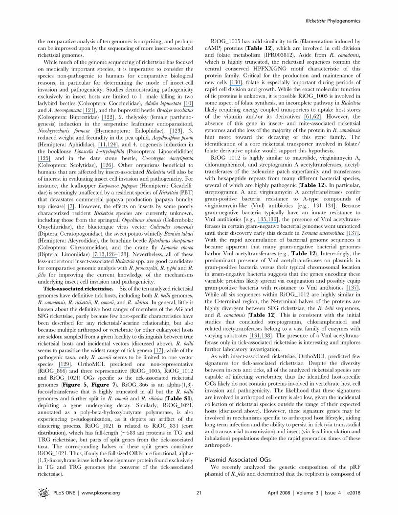
the comparative analysis of ten genomes is surprising, and perhaps
can be improved upon by the sequencing of more insect-associated
rickettsial genomes.
While much of the genome sequencing of rickettsiae has focused
on medically important species, it is imperative to consider the
species non-pathogenic to humans for comparative biological
reasons, in particular for determining the mode of insect-cell
invasion and pathogenicity. Studies demonstrating pathogenicity
exclusively in insect hosts are limited to 1. male killing in two
ladybird beetles (Coleoptera: Coccinelidae), Adalia bipunctata [10]
and A. decempuncata [121], and the buprestid beetle Brachys tessellatus
(Coleoptera: Buprestidae) [122], 2. thelytoky (female partheno-
genesis) induction in the serpentine leafminer endoparasitoid,
Neochrysocharis formosa (Hymenoptera: Eulophidae), [123], 3.
reduced weight and fecundity in the pea aphid, Acrythosiphon pisum
(Hemiptera: Aphididae), [11,124], and 4. oogenesis induction in
the booklouse Liposcelis bostrychophila (Psocoptera: Liposcelididae)
[125] and in the date stone beetle, Coccotrypes dactyliperda
(Coleoptera: Scolytidae), [126]. Other organisms beneficial to
humans that are affected by insect-associated Rickettsia will also be
of interest in evaluating insect cell invasion and pathogenicity. For
instance, the leafhopper Empoasca papayae (Hemiptera: Cicadelli-
dae) is seemingly unaffected by a resident species of Rickettsia (PBT)
that devastates commercial papaya production (papaya bunchy
top disease) [7]. However, the effects on insects by some poorly
characterized resident Rickettsia species are currently unknown,
including those from the springtail Onychiurus sinensis (Collembola:
Onychiuridae), the bluetongue virus vector Culicoides sonorensis
(Diptera: Ceratopogonidae), the sweet potato whitefly Bemisia tabaci
(Hemiptera: Aleyrodidae), the bruchine beetle Kytorhinus sharpianus
(Coleoptera: Chrysomelidae), and the crane fly Limonia chorea
(Diptera: Limoniidae) [7,13,126–128]. Nevertheless, all of these
less-understood insect-associated Rickettsia spp. are good candidates
for comparative genomic analysis with R. prowazekii, R. typhi and R.
felis for improving the current knowledge of the mechanisms
underlying insect cell invasion and pathogenicity.
Tick-associated rickettsiae. Six of the ten analyzed rickettsial
genomes have definitive tick hosts, including both R. bellii genomes,
R. canadensis, R. rickettsii, R. conorii, and R. sibirica. In general, little is
known about the definitive host ranges of members of the AG and
SFG rickettsiae, partly because few host-specific characteristics have
been described for any rickettsial/acarine relationship, but also
because multiple arthropod or vertebrate (or other eukaryote) hosts
are seldom sampled from a given locality to distinguish between true
rickettsial hosts and incidental vectors (discussed above). R. bellii
seems to parasitize the widest range of tick genera [17], while of the
pathogenic taxa, only R. conorii seems to be limited to one vector
species [129]. OrthoMCL predicted one non-representative
(RiOG_866) and three representative (RiOG_1005, RiOG_1012
and RiOG_1021) OGs specific to the tick-associated rickettsial
genomes (Figure 5, Figure 7). RiOG_866 is an alpha-(1,3)-
fucosyltransferase that is highly truncated in all but the R. bellii
genomes and further split in R. conorii and R. sibirica (Table S1),
depicting a gene undergoing decay. Similarly, RiOG_1021,
annotated as a poly-beta-hydroxybutyrate polymerase, is also
experiencing pseudogenization, as it depicts an artifact of the
clustering process. RiOG_1021 is related to RiOG_834 (core
distribution), which has full-length (,583 aa) proteins in TG and
TRG rickettsiae, but parts of split genes from the tick-associated
taxa. The corresponding halves of these split genes constitute
RiOG_1021. Thus, if only the full sized ORFs are functional, alpha-
(1,3)-fucosyltransferase is the lone signature protein found exclusively
in TG and TRG genomes (the converse of the tick-associated
rickettsiae).
RiOG_1005 has mild similarity to fic (filamentation induced by
cAMP) proteins (Table 12), which are involved in cell division
and folate metabolism (IPR003812). Aside from R. canadensis,
which is highly truncated, the rickettsial sequences contain the
central conserved HPFXXGNG motif characteristic of this
protein family. Critical for the production and maintenance of
new cells [130], folate is especially important during periods of
rapid cell division and growth. While the exact molecular function
of fic proteins is unknown, it is possible RiOG_1005 is involved in
some aspect of folate synthesis, an incomplete pathway in Rickettsia
likely requiring energy-coupled transporters to uptake host stores
of the vitamin and/or its derivatives [61,62]. However, the
absence of this gene in insect- and mite-associated rickettsial
genomes and the loss of the majority of the protein in R. canadensis
hint more toward the decaying of this gene family. The
identification of a core rickettsial transporter involved in folate/
folate derivative uptake would support this hypothesis.
RiOG_1012 is highly similar to macrolide, virginiamycin A,
chloramphenicol, and streptogramin A acetyltransferases, acetyl-
transferases of the isoleucine patch superfamily and transferases
with hexapeptide repeats from many different bacterial species,
several of which are highly pathogenic (Table 12). In particular,
streptogramin A and virginiamycin A acetyltransferases confer
gram-positive bacteria resistance to A-type compounds of
virginiamycin-like (Vml) antibiotics [e.g., 131–134]. Because
gram-negative bacteria typically have an innate resistance to
Vml antibiotics [e.g., 135,136], the presence of Vml acetyltrans-
ferases in certain gram-negative bacterial genomes went unnoticed
until their discovery early this decade in Yersinia enterocolitica [137].
With the rapid accumulation of bacterial genome sequences it
became apparent that many gram-negative bacterial genomes
harbor Vml acetyltransferases (e.g., Table 12). Interestingly, the
predominant presence of Vml acetyltransferases on plasmids in
gram-positive bacteria versus their typical chromosomal location
in gram-negative bacteria suggests that the genes encoding these
variable proteins likely spread via conjugation and possibly equip
gram-positive bacteria with resistance to Vml antibiotics [137].
While all six sequences within RiOG_1012 are highly similar in
the C-terminal region, the N-terminal halves of the proteins are
highly divergent between SFG rickettsiae, the R. belii sequences,
and R. canadensis (Table 12). This is consistent with the initial
studies that concluded streptogramin, chloramphenicol and
related acetytransferases belong to a vast family of enzymes with
varying substrates [131,138]. The presence of a Vml acetyltrans-
ferase only in tick-associated rickettsiae is interesting and implores
further laboratory investigation.
As with insect-associated rickettsiae, OrthoMCL predicted few
signatures for tick-associated rickettsiae. Despite the diversity
between insects and ticks, all of the analyzed rickettsial species are
capable of infecting vertebrates; thus the identified host-specific
OGs likely do not contain proteins involved in vertebrate host cell
invasion and pathogenicity. The likelihood that these signatures
are involved in arthropod cell entry is also low, given the incidental
collection of rickettsial species outside the range of their expected
hosts (discussed above). However, these signature genes may be
involved in mechanisms specific to arthropod host lifestyle, aiding
long-term infection and the ability to persist in tick (via transstadial
and transovarial transmission) and insect (via fecal inoculation and
inhalation) populations despite the rapid generation times of these
arthropods.
Plasmid Associated OGsWe recently analyzed the genetic composition of the pRF
plasmid of R. felis and determined that the replicon is composed of
Rickettsia Phylogenomics
PLoS ONE | www.plosone.org 21 April 2008 | Volume 3 | Issue 4 | e2018
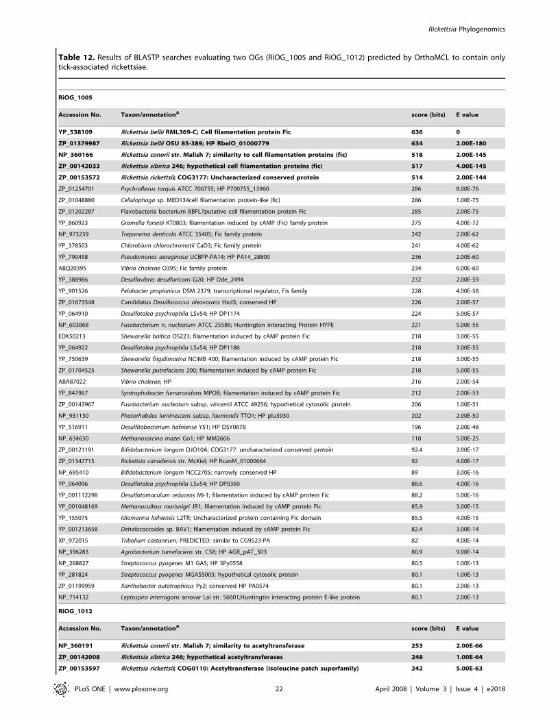
Table 12. Results of BLASTP searches evaluating two OGs (RiOG_1005 and RiOG_1012) predicted by OrthoMCL to contain onlytick-associated rickettsiae.
RiOG_1005
Accession No. Taxon/annotationA score (bits) E value
YP_538109 Rickettsia bellii RML369-C; Cell filamentation protein Fic 636 0
ZP_01379987 Rickettsia bellii OSU 85-389; HP RbelO_01000779 634 2.00E-180
NP_360166 Rickettsia conorii str. Malish 7; similarity to cell filamentation proteins (fic) 518 2.00E-145
ZP_00142033 Rickettsia sibirica 246; hypothetical cell filamentation proteins (fic) 517 4.00E-145
ZP_00153572 Rickettsia rickettsii; COG3177: Uncharacterized conserved protein 514 2.00E-144
ZP_01254701 Psychroflexus torquis ATCC 700755; HP P700755_13960 286 8.00E-76
ZP_01048880 Cellulophaga sp. MED134cell filamentation protein-like (fic) 286 1.00E-75
ZP_01202287 Flavobacteria bacterium BBFL7putative cell filamentation protein Fic 285 2.00E-75
YP_860923 Gramella forsetii KT0803; filamentation induced by cAMP (Fic) family protein 275 4.00E-72
NP_973239 Treponema denticola ATCC 35405; Fic family protein 242 2.00E-62
YP_378503 Chlorobium chlorochromatii CaD3; Fic family protein 241 4.00E-62
YP_790458 Pseudomonas aeruginosa UCBPP-PA14; HP PA14_28800 236 2.00E-60
ABQ20395 Vibrio cholerae O395; Fic family protein 234 6.00E-60
YP_388986 Desulfovibrio desulfuricans G20; HP Dde_2494 232 2.00E-59
YP_901526 Pelobacter propionicus DSM 2379; transcriptional regulator, Fis family 228 4.00E-58
ZP_01673548 Candidatus Desulfococcus oleovorans Hxd3; conserved HP 226 2.00E-57
YP_064910 Desulfotalea psychrophila LSv54; HP DP1174 224 5.00E-57
NP_603868 Fusobacterium n. nucleatum ATCC 25586; Huntington interacting Protein HYPE 221 5.00E-56
EDK50213 Shewanella baltica OS223; filamentation induced by cAMP protein Fic 218 3.00E-55
YP_064922 Desulfotalea psychrophila LSv54; HP DP1186 218 3.00E-55
YP_750639 Shewanella frigidimarina NCIMB 400; filamentation induced by cAMP protein Fic 218 3.00E-55
ZP_01704525 Shewanella putrefaciens 200; filamentation induced by cAMP protein Fic 218 5.00E-55
ABA87022 Vibrio cholerae; HP 216 2.00E-54
YP_847967 Syntrophobacter fumaroxidans MPOB; filamentation induced by cAMP protein Fic 212 2.00E-53
ZP_00143967 Fusobacterium nucleatum subsp. vincentii ATCC 49256; hypothetical cytosolic protein 206 1.00E-51
NP_931130 Photorhabdus luminescens subsp. laumondii TTO1; HP plu3930 202 2.00E-50
YP_516911 Desulfitobacterium hafniense Y51; HP DSY0678 196 2.00E-48
NP_634630 Methanosarcina mazei Go1; HP MM2606 118 5.00E-25
ZP_00121191 Bifidobacterium longum DJO10A; COG3177: uncharacterized conserved protein 92.4 3.00E-17
ZP_01347715 Rickettsia canadensis str. McKiel; HP RcanM_01000664 92 4.00E-17
NP_695410 Bifidobacterium longum NCC2705; narrowly conserved HP 89 3.00E-16
YP_064096 Desulfotalea psychrophila LSv54; HP DP0360 88.6 4.00E-16
YP_001112298 Desulfotomaculum reducens MI-1; filamentation induced by cAMP protein Fic 88.2 5.00E-16
YP_001048169 Methanoculleus marisnigri JR1; filamentation induced by cAMP protein Fic 85.9 3.00E-15
YP_155075 Idiomarina loihiensis L2TR; Uncharacterized protein containing Fic domain 85.5 4.00E-15
YP_001213658 Dehalococcoides sp. BAV1; filamentation induced by cAMP protein Fic 82.4 3.00E-14
XP_972015 Tribolium castaneum; PREDICTED: similar to CG9523-PA 82 4.00E-14
NP_396283 Agrobacterium tumefaciens str. C58; HP AGR_pAT_503 80.9 9.00E-14
NP_268827 Streptococcus pyogenes M1 GAS; HP SPy0558 80.5 1.00E-13
YP_281824 Streptococcus pyogenes MGAS5005; hypothetical cytosolic protein 80.1 1.00E-13
ZP_01199959 Xanthobacter autotrophicus Py2; conserved HP PA0574 80.1 2.00E-13
NP_714132 Leptospira interrogans serovar Lai str. 56601;Huntingtin interacting protein E-like protein 80.1 2.00E-13
RiOG_1012
Accession No. Taxon/annotationA score (bits) E value
NP_360191 Rickettsia conorii str. Malish 7; similarity to acetyltransferase 253 2.00E-66
ZP_00142008 Rickettsia sibirica 246; hypothetical acetyltransferases 248 1.00E-64
ZP_00153597 Rickettsia rickettsii; COG0110: Acetyltransferase (isoleucine patch superfamily) 242 5.00E-63
Rickettsia Phylogenomics
PLoS ONE | www.plosone.org 22 April 2008 | Volume 3 | Issue 4 | e2018

RiOG_1012
Accession No. Taxon/annotationA score (bits) E value
AAA86871 Staphylococcus aureus; VAT B 64.3 2.00E-09
NP_713859 Leptospira interrogans serovar Lai str. 56601; Probable macrolide acetyltransferase 62.4 7.00E-09
YP_517378 Desulfitobacterium hafniense Y51; HP DSY1145 59.7 4.00E-08
ZP_01621907 Lyngbya sp. PCC 8106; acetyltransferase 59.3 6.00E-08
ZP_01370449 Desulfitobacterium hafniense DCB-2; transferase hexapeptide repeat 58.9 7.00E-08
YP_600714 Streptococcus pyogenes MGAS2096; Virginiamycin A acetyltransferase 58.5 9.00E-08
NP_441499 Synechocystis sp. PCC 6803; acetyltransferase 58.2 1.00E-07
ZP_01547157 Stappia aggregata IAM 12614; streptogramin A acetyl transferase 57.8 2.00E-07
ZP_01227803 Aurantimonas sp. SI85-9A1; acetyltransferase 57.8 2.00E-07
YP_001182985 Shewanella putrefaciens CN-32; transferase hexapeptide repeat containing protein 57.8 2.00E-07
NP_624622 Streptomyces coelicolor A3(2); acetyltransferase 57.8 2.00E-07
YP_001038094 Clostridium thermocellum ATCC 27405; HP Cthe_1678 57.4 2.00E-07
YP_870325 Shewanella sp. ANA-3; streptogramin A acetyl transferase 57.4 2.00E-07
YP_001049952 Shewanella baltica OS155; transferase hexapeptide repeat containing protein 57.4 2.00E-07
EDK51061 Shewanella baltica OS223;transferase hexapeptide repeat containing protein 57.4 3.00E-07
ZP_01434328 Shewanella baltica OS195; transferase hexapeptide repeat 57 3.00E-07
NP_717373 Shewanella oneidensis MR-1; streptogramin A acetyl transferase 56.2 5.00E-07
ZP_01595082 Marinomonas sp. MWYL1; streptogramin A acetyl transferase 56.2 5.00E-07
ZP_01439726 Fulvimarina pelagi HTCC2506; streptogramin A acetyl transferase 55.8 6.00E-07
AAK96241 Enterococcus hirae; streptogramin A acetyltransferase 55.8 7.00E-07
AAK91782 Enterococcus faecium; streptogramin A acetyltransferase 55.8 7.00E-07
AAK91783 Enterococcus faecium; streptogramin A acetyltransferase 55.8 7.00E-07
NP_783842 Lactobacillus fermentum; streptogramin A resistance protein 55.5 8.00E-07
AAG21695 Enterococcus faecium; streptogramin A acetyltransferase 55.1 1.00E-06
YP_001175130 Enterobacter sp. 638; streptogramin A acetyl transferase 55.1 1.00E-06
YP_738635 Shewanella sp. MR-7; streptogramin A acetyl transferase 55.1 1.00E-06
NP_385342 Sinorhizobium meliloti 1021; PUTATIVE ACETYLTRANSFERASE PROTEIN 54.7 1.00E-06
YP_702166 Rhodococcus sp. RHA1; probable chloramphenicol O-acetyltransferase 54.7 1.00E-06
ZP_01706265 Shewanella putrefaciens 200; transferase hexapeptide repeat 54.7 2.00E-06
YP_964018 Shewanella sp. W3-18-1; transferase hexapeptide repeat containing protein 54.7 2.00E-06
YP_051279 Erwinia carotovora subsp. atroseptica SCRI1043; streptogramin A acetyl transferase 54.3 2.00E-06
ZP_01691225 Microscilla marina ATCC 23134; virginiamycin A acetyltransferase 54.3 2.00E-06
ZP_01780719 Shewanella baltica OS185; transferase hexapeptide repeat containing protein 54.3 2.00E-06
ZP_01186630 Bacillus weihenstephanensis KBAB4; transferase hexapeptide repeat 54.3 2.00E-06
ZP_01613073 Alteromonadales bacterium TW-7; streptogramin A acetyl transferase 53.9 2.00E-06
YP_927261 Shewanella amazonensis SB2B; streptogramin A acetyl transferase 53.9 2.00E-06
YP_171536 Synechococcus elongatus PCC 6301; acetyltransferase 53.9 3.00E-06
NP_978962 Bacillus cereus ATCC 10987; acetyltransferase, CYSE/LACA/LPXA/NODL family 53.9 3.00E-06
NP_832369 Bacillus cereus ATCC 14579; Virginiamycin A acetyltransferase 53.5 3.00E-06
NP_844991 Bacillus anthracis str. Ames; acetyltransferase, CYSE/LACA/LPXA/NODL family 53.5 3.00E-06
AAF63432 Yersinia enterocolitica; streptogramin A acetyl transferase 53.1 4.00E-06
AAK91784 Enterococcus faecium; streptogramin A acetyltransferase 53.1 4.00E-06
YP_007518 Candidatus Protochlamydia amoebophila UWE25; streptogramin A acetyltransferase 53.1 4.00E-06
AAF24171 Enterococcus faecium; acetyltransferase SatG 53.1 5.00E-06
ZP_00241060 Bacillus cereus G9241; streptogramin A acetyl transferase 53.1 5.00E-06
YP_001214452 Dehalococcoides sp. BAV1; Acetyltransferase (Ile patch superfamily)-like protein 52.8 5.00E-06
ZP_00834849 Yersinia intermedia ATCC 29909; COG0110: Acetyltransferase (Ile patch superfamily) 52.8 5.00E-06
AAK91785 Enterococcus faecium; streptogramin A acetyltransferase 52.8 6.00E-06
Table 12. cont.
Rickettsia Phylogenomics
PLoS ONE | www.plosone.org 23 April 2008 | Volume 3 | Issue 4 | e2018
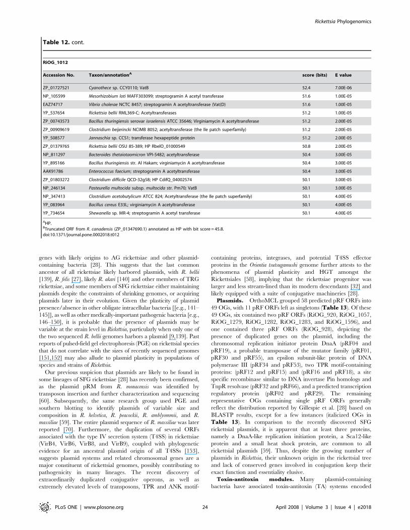
genes with likely origins to AG rickettsiae and other plasmid-
containing bacteria [28]. This suggests that the last common
ancestor of all rickettsiae likely harbored plasmids, with R. bellii
[139], R. felis [27], likely R. akari [140] and other members of TRG
rickettsiae, and some members of SFG rickettsiae either maintaining
plasmids despite the constraints of shrinking genomes, or acquiring
plasmids later in their evolution. Given the plasticity of plasmid
presence/absence in other obligate intracellular bacteria [[e.g., 141–
145]], as well as other medically-important pathogenic bacteria [e.g.,
146–150], it is probable that the presence of plasmids may be
variable at the strain level in Rickettsia, particularly when only one of
the two sequenced R. bellii genomes harbors a plasmid [9,139]. Past
reports of pulsed-field gel electrophoresis (PGE) on rickettsial species
that do not correlate with the sizes of recently sequenced genomes
[151,152] may also allude to plasmid plasticity in populations of
species and strains of Rickettsia.
Our previous suspicion that plasmids are likely to be found in
some lineages of SFG rickettsiae [28] has recently been confirmed,
as the plasmid pRM from R. monacensis was identified by
transposon insertion and further characterization and sequencing
[60]. Subsequently, the same research group used PGE and
southern blotting to identify plasmids of variable size and
composition in R. helvetica, R. peacockii, R. amblyommii, and R.
massiliae [59]. The entire plasmid sequence of R. massiliae was later
reported [70]. Furthermore, the duplication of several ORFs
associated with the type IV secretion system (T4SS) in rickettsiae
(VirB4, VirB6, VirB8, and VirB9), coupled with phylogenetic
evidence for an ancestral plasmid origin of all T4SSs [153],
suggests plasmid systems and related chromosomal genes are a
major constituent of rickettsial genomes, possibly contributing to
pathogenicity in many lineages. The recent discovery of
extraordinarily duplicated conjugative operons, as well as
extremely elevated levels of transposons, TPR and ANK motif-
containing proteins, integrases, and potential T4SS effector
proteins in the Orientia tsutsugamushi genome further attests to the
phenomena of plasmid plasticity and HGT amongst the
Rickettsiales [58], implying that the rickettsiae progenitor was
larger and less stream-lined than its modern descendants [32] and
likely equipped with a suite of conjugative machineries [28].
Plasmids. OrthoMCL grouped 58 predicted pRF ORFs into
49 OGs, with 11 pRF ORFs left as singletons (Table 13). Of these
49 OGs, six contained two pRF ORFs (RiOG_920, RiOG_1057,
RiOG_1279, RiOG_1282, RiOG_1283, and RiOG_1596), and
one contained three pRF ORFs (RiOG_928), depicting the
presence of duplicated genes on the plasmid, including the
chromosomal replication initiator protein DnaA (pRF04 and
pRF19), a probable transposase of the mutator family (pRF01,
pRF30 and pRF55), an epsilon subunit-like protein of DNA
polymerase III (pRF34 and pRF53), two TPR motif-containing
proteins: (pRF12 and pRF15) and (pRF16 and pRF18), a site
specific recombinase similar to DNA invertase Pin homologs and
TnpR resolvase (pRF32 and pRF66), and a predicted transcription
regulatory protein (pRF02 and pRF29). The remaining
representative OGs containing single pRF ORFs generally
reflect the distribution reported by Gillespie et al. [28] based on
BLASTP results, except for a few instances (italicized OGs in
Table 13). In comparison to the recently discovered SFG
rickettsial plasmids, it is apparent that at least three proteins,
namely a DnaA-like replication initiation protein, a Sca12-like
protein and a small heat shock protein, are common to all
rickettsial plasmids [59]. Thus, despite the growing number of
plasmids in Rickettsia, their unknown origin in the rickettsial tree
and lack of conserved genes involved in conjugation keep their
exact function and essentiality elusive.
Toxin-antitoxin modules. Many plasmid-containing
bacteria have associated toxin-antitoxin (TA) systems encoded
RiOG_1012
Accession No. Taxon/annotationA score (bits) E value
ZP_01727521 Cyanothece sp. CCY0110; VatB 52.4 7.00E-06
NP_105599 Mesorhizobium loti MAFF303099; streptogramin A acetyl transferase 51.6 1.00E-05
EAZ74717 Vibrio cholerae NCTC 8457; streptogramin A acetyltransferase (Vat(D) 51.6 1.00E-05
YP_537654 Rickettsia bellii RML369-C; Acetyltransferases 51.2 1.00E-05
ZP_00743573 Bacillus thuringiensis serovar israelensis ATCC 35646; Virginiamycin A acetyltransferase 51.2 2.00E-05
ZP_00909619 Clostridium beijerincki NCIMB 8052; acetyltransferase (the Ile patch superfamily) 51.2 2.00E-05
YP_508577 Jannaschia sp. CCS1; transferase hexapeptide protein 51.2 2.00E-05
ZP_01379765 Rickettsia bellii OSU 85-389; HP RbelO_01000549 50.8 2.00E-05
NP_811297 Bacteroides thetaiotaomicron VPI-5482; acetyltransferase 50.4 3.00E-05
YP_895166 Bacillus thuringiensis str. Al Hakam; virginiamycin A acetyltransferase 50.4 3.00E-05
AAK91786 Enterococcus faecium; streptogramin A acetyltransferase 50.4 3.00E-05
ZP_01803272 Clostridium difficile QCD-32g58; HP CdifQ_04002574 50.1 3.00E-05
NP_246134 Pasteurella multocida subsp. multocida str. Pm70; VatB 50.1 3.00E-05
NP_347413 Clostridium acetobutylicum ATCC 824; Acetyltransferase (the Ile patch superfamily) 50.1 4.00E-05
YP_083964 Bacillus cereus E33L; virginiamycin A acetyltransferase 50.1 4.00E-05
YP_734654 Shewanella sp. MR-4; streptogramin A acetyl transferase 50.1 4.00E-05
AHP.BTruncated ORF from R. canadensis (ZP_01347690.1) annotated as HP with bit score = 45.8.doi:10.1371/journal.pone.0002018.t012
Table 12. cont.
Rickettsia Phylogenomics
PLoS ONE | www.plosone.org 24 April 2008 | Volume 3 | Issue 4 | e2018
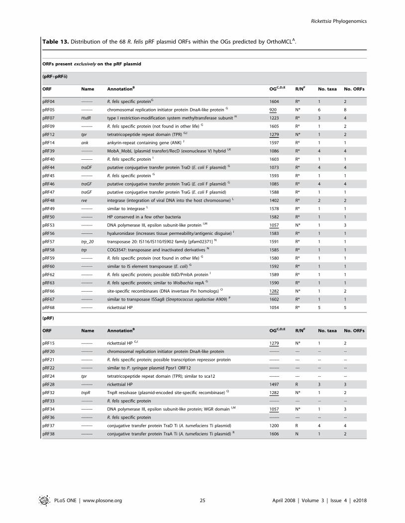
Table 13. Distribution of the 68 R. felis pRF plasmid ORFs within the OGs predicted by OrthoMCLA.
ORFs present exclusively on the pRF plasmid
(pRF+pRFd)
ORF Name AnnotationB OGC,D,E R/NF No. taxa No. ORFs
pRF04 -------- R. felis specific proteinG 1604 R* 1 2
pRF05 -------- chromosomal replication initiator protein DnaA-like protein G 920 N* 6 8
pRF07 HsdR type I restriction-modification system methyltransferase subunit H 1223 R* 3 4
pRF09 -------- R. felis specific protein (not found in other life) G 1605 R* 1 2
pRF12 tpr tetratricopeptide repeat domain (TPR) G,I 1279 N* 1 2
pRF14 ank ankyrin-repeat containing gene (ANK) J 1597 R* 1 1
pRF39 -------- MobA_MobL (plasmid transfer)/RecD (exonuclease V) hybrid I,K 1086 R* 4 4
pRF40 -------- R. felis specific protein I 1603 R* 1 1
pRF44 traDF putative conjugative transfer protein TraD (E. coli F plasmid) G 1073 R* 4 4
pRF45 -------- R. felis specific protein G 1593 R* 1 1
pRF46 traGF putative conjugative transfer protein TraG (E. coli F plasmid) G 1085 R* 4 4
pRF47 traGF putative conjugative transfer protein TraG (E. coli F plasmid) 1588 R* 1 1
pRF48 rve integrase (integration of viral DNA into the host chromosome) L 1402 R* 2 2
pRF49 -------- similar to integrase L 1578 R* 1 1
pRF50 -------- HP conserved in a few other bacteria 1582 R* 1 1
pRF53 -------- DNA polymerase III, epsilon subunit-like protein I,M 1057 N* 1 3
pRF56 -------- hyaluronidase (increases tissue permeability/antigenic disguise) I 1583 R* 1 1
pRF57 trp_20 transposase 20: IS116/IS110/IS902 family [pfam02371] N 1591 R* 1 1
pRF58 trp COG3547: transposase and inactivated derivatives N 1585 R* 1 1
pRF59 -------- R. felis specific protein (not found in other life) G 1580 R* 1 1
pRF60 -------- similar to IS element transposase (E. coli) G 1592 R* 1 1
pRF62 -------- R. felis specific protein; possible tldD/PmbA protein I 1589 R* 1 1
pRF63 -------- R. felis specific protein; similar to Wolbachia repA G 1590 R* 1 1
pRF66 -------- site-specific recombinases (DNA invertase Pin homologs) O 1282 N* 1 2
pRF67 -------- similar to transposase ISSag8 (Streptococcus agalactiae A909) P 1602 R* 1 1
pRF68 -------- rickettsial HP 1054 R* 5 5
(pRF)
ORF Name AnnotationB OGC,D,E R/NF No. taxa No. ORFs
pRF15 -------- rickettsial HP G,I 1279 N* 1 2
pRF20 -------- chromosomal replication initiator protein DnaA-like protein ------- --- -- --
pRF21 -------- R. felis specific protein; possible transcription repressor protein ------- --- -- --
pRF22 -------- similar to P. syringae plasmid Ppsr1 ORF12 ------- --- -- --
pRF24 tpr tetratricopeptide repeat domain (TPR); similar to sca12 ------- --- -- --
pRF28 -------- rickettsial HP 1497 R 3 3
pRF32 tnpR TnpR resolvase (plasmid-encoded site-specific recombinase) Q 1282 N* 1 2
pRF33 -------- R. felis specific protein ------- --- -- --
pRF34 -------- DNA polymerase III, epsilon subunit-like protein; WGR domain I,M 1057 N* 1 3
pRF36 -------- R. felis specific protein ------- --- -- --
pRF37 -------- conjugative transfer protein TraD Ti (A. tumefaciens Ti plasmid) 1200 R 4 4
pRF38 -------- conjugative transfer protein TraA Ti (A. tumefaciens Ti plasmid) R 1606 N 1 2
Rickettsia Phylogenomics
PLoS ONE | www.plosone.org 25 April 2008 | Volume 3 | Issue 4 | e2018
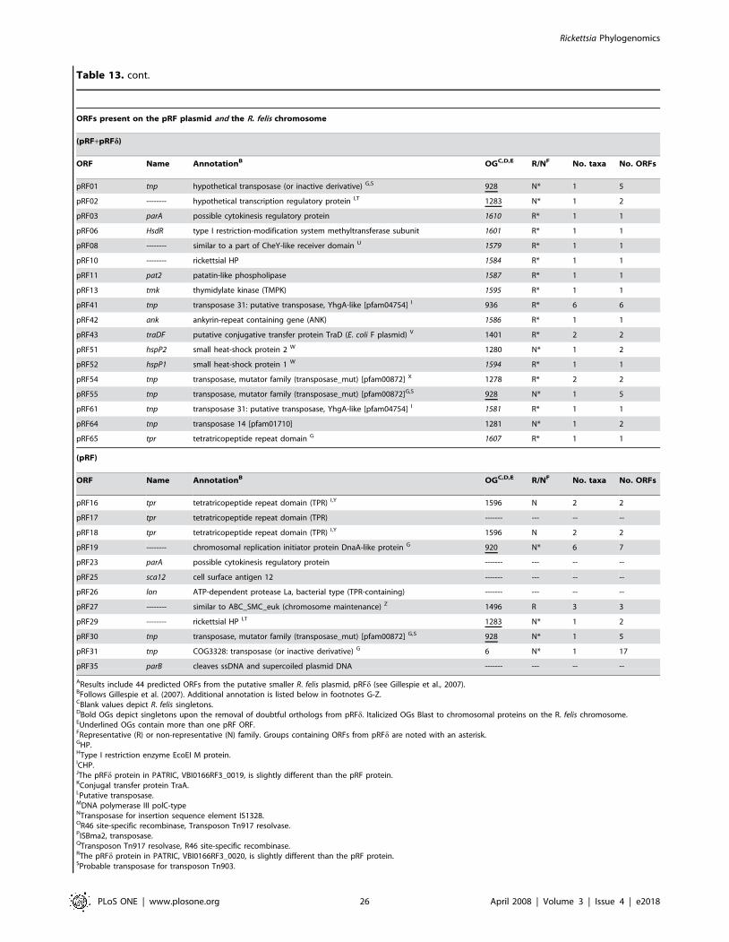
(pRF)
ORF Name AnnotationB OGC,D,E R/NF No. taxa No. ORFs
pRF16 tpr tetratricopeptide repeat domain (TPR) I,Y 1596 N 2 2
pRF17 tpr tetratricopeptide repeat domain (TPR) ------- --- -- --
pRF18 tpr tetratricopeptide repeat domain (TPR) I,Y 1596 N 2 2
pRF19 -------- chromosomal replication initiator protein DnaA-like protein G 920 N* 6 7
pRF23 parA possible cytokinesis regulatory protein ------- --- -- --
pRF25 sca12 cell surface antigen 12 ------- --- -- --
pRF26 lon ATP-dependent protease La, bacterial type (TPR-containing) ------- --- -- --
pRF27 -------- similar to ABC_SMC_euk (chromosome maintenance) Z 1496 R 3 3
pRF29 -------- rickettsial HP I,T 1283 N* 1 2
pRF30 tnp transposase, mutator family (transposase_mut) [pfam00872] G,S 928 N* 1 5
pRF31 tnp COG3328: transposase (or inactive derivative) G 6 N* 1 17
pRF35 parB cleaves ssDNA and supercoiled plasmid DNA ------- --- -- --
AResults include 44 predicted ORFs from the putative smaller R. felis plasmid, pRFd (see Gillespie et al., 2007).BFollows Gillespie et al. (2007). Additional annotation is listed below in footnotes G-Z.CBlank values depict R. felis singletons.DBold OGs depict singletons upon the removal of doubtful orthologs from pRFd. Italicized OGs Blast to chromosomal proteins on the R. felis chromosome.EUnderlined OGs contain more than one pRF ORF.FRepresentative (R) or non-representative (N) family. Groups containing ORFs from pRFd are noted with an asterisk.GHP.HType I restriction enzyme EcoEI M protein.ICHP.JThe pRFd protein in PATRIC, VBI0166RF3_0019, is slightly different than the pRF protein.KConjugal transfer protein TraA.LPutative transposase.MDNA polymerase III polC-typeNTransposase for insertion sequence element IS1328.OR46 site-specific recombinase, Transposon Tn917 resolvase.PISBma2, transposase.QTransposon Tn917 resolvase, R46 site-specific recombinase.RThe pRFd protein in PATRIC, VBI0166RF3_0020, is slightly different than the pRF protein.SProbable transposase for transposon Tn903.
ORFs present on the pRF plasmid and the R. felis chromosome
(pRF+pRFd)
ORF Name AnnotationB OGC,D,E R/NF No. taxa No. ORFs
pRF01 tnp hypothetical transposase (or inactive derivative) G,S 928 N* 1 5
pRF02 -------- hypothetical transcription regulatory protein I,T 1283 N* 1 2
pRF03 parA possible cytokinesis regulatory protein 1610 R* 1 1
pRF06 HsdR type I restriction-modification system methyltransferase subunit 1601 R* 1 1
pRF08 -------- similar to a part of CheY-like receiver domain U 1579 R* 1 1
pRF10 -------- rickettsial HP 1584 R* 1 1
pRF11 pat2 patatin-like phospholipase 1587 R* 1 1
pRF13 tmk thymidylate kinase (TMPK) 1595 R* 1 1
pRF41 tnp transposase 31: putative transposase, YhgA-like [pfam04754] I 936 R* 6 6
pRF42 ank ankyrin-repeat containing gene (ANK) 1586 R* 1 1
pRF43 traDF putative conjugative transfer protein TraD (E. coli F plasmid) V 1401 R* 2 2
pRF51 hspP2 small heat-shock protein 2 W 1280 N* 1 2
pRF52 hspP1 small heat-shock protein 1 W 1594 R* 1 1
pRF54 tnp transposase, mutator family (transposase_mut) [pfam00872] X 1278 R* 2 2
pRF55 tnp transposase, mutator family (transposase_mut) [pfam00872]G,S 928 N* 1 5
pRF61 tnp transposase 31: putative transposase, YhgA-like [pfam04754] I 1581 R* 1 1
pRF64 tnp transposase 14 [pfam01710] 1281 N* 1 2
pRF65 tpr tetratricopeptide repeat domain G 1607 R* 1 1
Table 13. cont.
Rickettsia Phylogenomics
PLoS ONE | www.plosone.org 26 April 2008 | Volume 3 | Issue 4 | e2018

on plasmids, typically as two-component operons, for the control
of plasmid partitioning and stable inheritance [154–157].
Antitoxins, usually highly labile in their mature form, are
constitutively expressed and neutralize the accumulation of their
counterpart toxins, which are more stable. Upon imperfect
segregation of plasmids after cell division, plasmidless daughter
cells are destroyed by elevated toxin levels due to the rapid
breakdown of the unstable antitoxin and lack of its further
synthesis [158,159]. Although originally described as mediators of
bacterial programmed cell death, studies now suggest that TA
modules also act as regulators of the stringent response (reviewed
in [159]) and are widely present on chromosomes of diverse
bacteria [160]. While TA systems are found in many free-living
bacteria, they are typically uncommon among obligate intra-
cellular pathogens [160,161]. However, the genome sequences for
both R. bellii strains and R. felis contain elevated levels of
chromosomally encoded TA loci, the majority of which seem to
be degraded [9,27]. Moreover, these genomes typically retain only
one component of the TA modules, possibly alluding to a
neofunctionalization [153] of the remaining genes for adaptation
to eukaryotic hosts, as has been suggested for at least R. felis toxin
and antitoxin genes [27]. However, given that the reductive nature
of rickettsial genomes may result in high levels of constitutively
expressed loci and reduced operons, and that many antitoxins
contain motifs common to two, three and even four different
DNA-binding-proteins [162], incomplete and noncontiguous
rickettsial TA modules may still interact with one another to
coordinate a response to stress within host cells. Alternatively, the
presence of incomplete TA modules may reflect vertically acquired
plasmid-associated genes that are in the process of
pseudogenization. In support of this, of the numerous TA
components in the R. felis genome, only one VapB antitoxin
(RiOG_941) was recovered in a proteome screen [40].
Our bioinformatic analysis reveals that components of 5 TA
systems (relBE, phd/doc, vapBC/vag, mazEF, and parDE) are
recurrent in all rickettsial genomes save the TG rickettsiae and
R. canadensis (Table S4). Of the predicted 56 toxin and 86
antitoxin ORFs, zero occur in the TG rickettsiae and only two are
found in R. canadensis. The majority of these ORFs occur in the R.
bellii genomes and TRG rickettsiae (avg. of 22 and 26 TA ORFs
per genome, respectively), although slightly lower levels also occur
in SFG rickettsiae (avg. of 14.3 TA ORFs per genome). However,
there are more occurrences of similar TA module components
shared between the R. bellii genomes and TRG rickettsiae (12)
than between SFG rickettsiae and either the R. bellii genomes (1) or
TRG rickettsiae (5) (Table S4). Thus, the presence and
distribution of these TA ORFs correlates to the lineages of
sampled rickettsiae that contain plasmids, and further supports the
TRG rickettsiae having affinities with AG rickettsiae [28].
Furthermore, the R. belli and TRG rickettsiae genomes have
elevated levels of predicted PIN-domain proteins (homologs of the
pilT N-terminal domain), which in eukaryotes function as
ribonucleases [163,164] involved in RNAi and nonsense-mediated
RNA degradation [162,163]. Most of the described prokaryotic
PIN-domain proteins are toxins of chromosomally-encoded TA
operons [159–161] that are present in a diverse array of unrelated
bacteria, likely having arisen due to the advantages they bestow on
competing mobile elements [165,166]. Indeed, the PilT protein of
the pathogenic Neisseria meningitidis has been hypothesized to
interact with the T4SS due to its limited homology to the DotB
protein of the Legionella T4SS [167].
While the exact manner of their origin and current functional
significance is debatable, it is apparent that TA systems have
arisen via HGT in a wide range of bacteria [168]. Given the
distribution of plasmids and associated TA systems in the analyzed
rickettsial genomes, it is likely that conjugation via plasmids befits
some rickettsial lineages with genes important for survival in
stressful environments, allowing for dormancy and slow growth.
However, it remains to be determined if those rickettsial species
that harbor plasmids use TA modules for mediating the
partitioning and stable inheritance of said plasmids.
Singleton ORFsOrthoMCL failed to group 1467 ORFs (10.2% of total
predicted ORFs) from the ten analyzed genomes into any OG
(Figure 5, Figure 7; Table S5, S6, S7, S8, S9, S10, S11,S12, S13, S14). The range across rickettsial groups shows TG
genomes contribute the least (8.5%) and TRG genomes contribute
the most (41%) to the total count of singletons (Figure 10A). The
individual genome contributions to the overall singleton count
range from 4% (R. typhi) to 21% (R. felis), with the rank of all
genomes matching the group ranking (TG,SFG,AG,TRG)
(Figure 10A). However, an inherent bias of these comparisons is
difficult to avoid, as OrthoMCL grouped 321 ORFs present only
from both R. bellii genomes (Figure 5, Figure 7, Table S3).
Accounting for these R. bellii doubletons, the rank and proportion
of singletons per rickettsial group is modified: TG (7%),SFG
(20%),TRG (34%),AG (39%), and illustrates that TRG and AG
genomes are more similar in their number of singleton ORFs
relative to TG and SFG rickettsiae. This brings up a practical
concern with phylogenomic analysis in that sampling one genome
per species (or strain) may not suffice for capturing the true
composition of genes within the bacterial population. This is
consistent with a recent study that cautioned on the very same idea
in relation to vaccine design for Streptococcus agalactiae, an organism
that has a core genome of approximately 80% across various
strains, with the accessory genome quite plastic [169]. Using
mathematics, a rather daunting conclusion was reached suggesting
even after sampling hundreds of additional S. agalactiae genome
sequences, novel genes would still be added to the accessory
genome [169]. Nonetheless, inclusion of the R. bellii doubletons
illustrated the similar composition of singletons in AG and TRG
genomes and further adds to the similarities these genomes share
as a result of related conjugation systems.
Unsurprisingly, the majority of singleton ORFs are annotated as
HPs, ranging from 68% (R. felis) to 95% (R. typhi) across the
TCOG1396: Predicted transcriptional regulators.UCOG3706: Response regulator containing a CheY-like receiver domain and a GGDEF domain.VProtein virD4.WSpore protein SP21.XProbable transposase for insertion sequence element.YCell division cycle protein 27 homolog.ZMyosin-11 (R. prowazekii), M protein, serotype 2.1 precursor (R. typhi).doi:10.1371/journal.pone.0002018.t013
Table 13. cont.
Rickettsia Phylogenomics
PLoS ONE | www.plosone.org 27 April 2008 | Volume 3 | Issue 4 | e2018
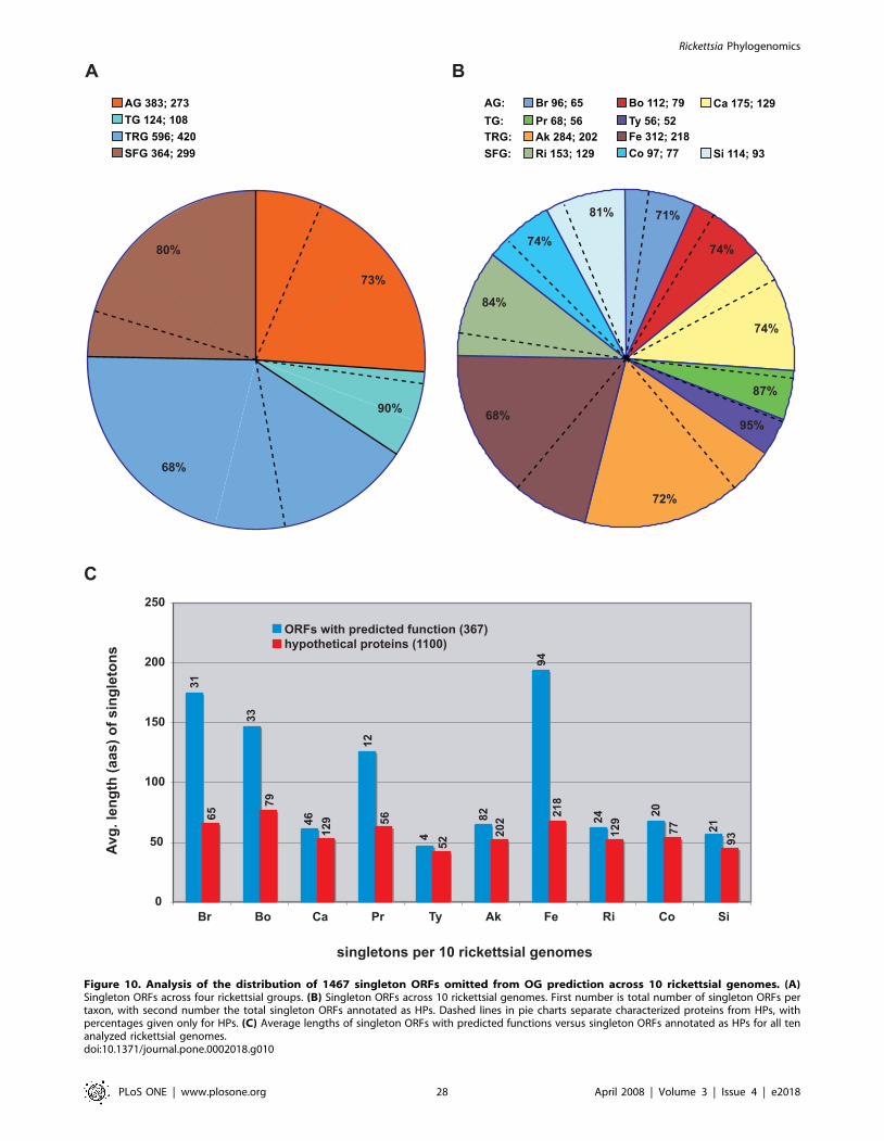
A B
0
50
100
150
200
250
Br Bo Ca Pr Ty Ak Fe Ri Co Si
ORFs with predicted function (367)hypothetical proteins (1100)
Avg.
leng
th (a
as) o
f sin
glet
ons
singletons per 10 rickettsial genomes
C
71%
74%
95%
87%
72%
74%
68%
84%
74%
81%
AG 383; 273TG 124; 108TRG 596; 420SFG 364; 299
73%
90%
68%
80%
6531
7933
4612
9
1256
452
8220
2
9421
8
12924 20
77 2193
Br 96; 65Pr 68; 56Ak 284; 202Ri 153; 129
Bo 112; 79Ty 56; 52
Ca 175; 129
Fe 312; 218Co 97; 77 Si 114; 93
AG:TG:TRG:SFG:
Figure 10. Analysis of the distribution of 1467 singleton ORFs omitted from OG prediction across 10 rickettsial genomes. (A)Singleton ORFs across four rickettsial groups. (B) Singleton ORFs across 10 rickettsial genomes. First number is total number of singleton ORFs pertaxon, with second number the total singleton ORFs annotated as HPs. Dashed lines in pie charts separate characterized proteins from HPs, withpercentages given only for HPs. (C) Average lengths of singleton ORFs with predicted functions versus singleton ORFs annotated as HPs for all tenanalyzed rickettsial genomes.doi:10.1371/journal.pone.0002018.g010
Rickettsia Phylogenomics
PLoS ONE | www.plosone.org 28 April 2008 | Volume 3 | Issue 4 | e2018

analyzed genomes (Figure 10A, B). In an effort to identify the
degree of over-prediction of ORFs, we plotted the average lengths
of singleton ORFs with predicted functions versus singleton ORFs
annotated as HPs for all ten rickettsial genomes (Figure 10C).
The rationale for this is that the majority of singletons under 100
amino acids in length should be HPs, with many having arisen by
chance [100]. Aside from R. felis, R. prowazekii and the R. bellii
genomes, there is minimal difference between the average lengths
of singletons with predicted functions and singletons annotated as
HPs. The much larger average lengths of singletons with predicted
functions versus singleton HPs are expected in the R. bellii and R.
felis genomes, as many of the larger singletons in these genomes are
probable products of HGT events (e.g., larger transposases, ANK-
and TPR-motif containing proteins). This same pattern in the R.
prowazekii singletons, however, is unexpected, yet is skewed in part
due to the presence of several large split ORFs that did not cluster
into their respective OGs. While the shorter singleton HPs may
have arisen by chance, it is likely that some of them are functional
genes that are difficult to homologize with other closely related
sequences, given the problems with assessing percent conservation
across short sequences with even minimal differences. For
instance, small ORFs are found in a variety of protein classes,
including ribosomal proteins, transcriptional regulators, chaper-
onins, thioredoxins, metal ion chelators, proteolipids, stress
proteins, nucleases, and mating pheromones [170]. Of the original
299 Saccharomyces cerevisiae small ORFs annotated as HPs, 170 have
since been assigned cellular functions, with the majority of
information coming from laboratory evidence [171]. Given the
probable plasticity of the accessory genomes of rickettsial strains
(discussed above) and the growing importance small ORFs have
garnered in the literature [e.g., 171–174], the high number of
small singleton HPs in Rickettsia should not be ignored.
Experimental evidence has confirmed the translation of several
small HPs in R. felis [40] and future microarray data will help lend
resolution to this poorly understood characteristic of rickettsial
genomes.
ConclusionThis study analyzed 14354 predicted ORFs from ten rickettsial
genomes and generated OGs ranging from two to 31 sequences for
90 percent of the total ORFs. A conserved core rickettsial genome
consisting of 731 OGs (51% of total predicted ORFs) was
identified, and a phylogeny was estimated from this core genome
to allow for subsequent phylogenomic comparison of the
remaining accessory genome. This robust phylogeny estimate is
congruent with our recent reclassification of rickettsial lineages
into four groups [28] and OGs specific to each group provide the
first signature genes possibly involved in the phenotypic charac-
teristics defining each group. The unstable phylogenetic position of
R. canadensis, coupled with it only sharing three OGs with the R.
bellii genomes, reflects that the base of the rickettsial tree is poorly
defined. However, an unprecedented mode of gene loss was
discovered in the lineage spanning R. canadensis and TG rickettsiae,
illustrating that gene signatures alone may not well-characterize
specific rickettsial groups, but instead the modes of gene loss (and
stricter reliance on host resources) may be the defining features
[175]. Given the emerging diversity of Rickettsia [16], particularly
species associated with medically non-important metazoans and
ancestrally related to the pathogenic species analyzed here, the
origins of pathogenicity from primitive rickettsial symbionts may
not be elucidated without a broader genomic comparison
reflective of the overall diversity within the genus.
As a consequence of distinguishing OGs comprising single
rickettsial groups (e.g., AG, TG, TRG, and SFG), shared
rickettsial groups (subgeneric), plasmid-harboring genomes, and
genomes with common arthropod hosts (C1OGs) from OGs with
a patchy distribution across the rickettsial tree (C2OGs), two
interesting results were obtained. First, C2OGs comprise 31% of
all generated OGs, implying a significant portion of the rickettsial
accessory genome is comprised of gene decay and laterally
acquired genes. Supporting this is the presence of the majority of
split ORFs within C2OGs (Table S1) and the high proportion of
gene families typically associated with the bacterial mobile gene
pool in C2OGs (47%) versus the low proportions in C1OGs (5%)
and singleton ORFs (4%). Second, the ratio of representative OGs
to non-representative OGs is skewed within C1OG distributions
(71–29%) but nearly equal in C2OG distributions (56–44%),
suggesting that gene duplications (paralogs) and HGT events
(xenologs) are more prevalent in C2OGs. Taken collectively, these
observations yield the manner in which the rickettsial genomes
have acquired their variation: a conserved core genome is
supplemented with a highly variable accessory genome that is
comprised of gene decay and many horizontally acquired genes.
However, the nature of the horizontally acquired genes remains
unknown: for example, did the products of HGT arise ancestrally
in the analyzed taxa, becoming shuffled over time through
recombination and high rates of decay, or are HGT products
continually sculpting the variation within the accessory genome
overtop of a highly reductive nature of all genes within the
genome? The recent explosion of reported cases of plasmids in all
rickettsial groups except TG rickettsiae argues for the latter
scenario, and is congruent with our findings of nearly zero
instances of plasmid associated genes, genes typical of HGT events
and gene duplications within TG rickettsial genomes. Thus, while
many Rickettsia seem to be able to accept and pass genes of the
mobile gene pool, the contribution of HGT products to
pathogenicity is unknown and seemingly nonessential to all known
rickettsial pathogens. The role lineage specific virulence factors
play in pathogenic strains is thus an important aspect of future
laboratory work. While HGT was traditionally considered rare in
Rickettsia, we recently suggested, based on a detailed analysis of the
R. felis pRF genes, that it is more common, particularly among
species in which conjugation systems had yet been discovered [28].
Our suspicions have recently been verified [70] and the exact
degree HGT contributes to rickettsial diversification will only be
elicited with the accumulation of more rickettsial genome
sequences. Such endeavors will challenge our existing classification
scheme; however, a preliminary analysis of two recently published
SFG rickettsiae genomes (R. massiliae str. MTU5 and R. africae str.
ESF 5) using genome alignment (Figure S1-E) and phylogeny
estimation (Figure S3) does not overturn our results, and we
predict that OGs generated with the inclusion of these new
genomes will not alter the conclusions reached herein.
Finally, we present two concerns regarding phylogenomic
analysis of Rickettsia. First, the high degree of pseudogenization
in rickettsial genomes means that OG prediction programs and
related methods alone are insufficient for grouping related genes.
Manual inspection of algorithm output is imperative, as the high
occurrence of split genes will lead to overestimation of non-
representative OGs as well as inaccuracies in ORF clustering (see
Table S1). Second, and perhaps more pressing, is the revelation
that rickettsial species may be comprised of highly variable
genomes, particularly across exceedingly divergent strains. Attest-
ing to this, our analysis of predicted OGs included two strains of R.
bellii that shared 321 species-specific genes but contained 97 (str.
RML369-C) and 117 (str. OSU 85 389) strain-specific genes.
Similarly, a recent genomic comparison of R. rickettsii str. Sheila
Smith CWPP with the avirulent R. rickettsii str. Iowa revealed 143
Rickettsia Phylogenomics
PLoS ONE | www.plosone.org 29 April 2008 | Volume 3 | Issue 4 | e2018
deletions and 492 SNPs between the two genomes [176].
Altogether, these issues challenge future genomic studies on
Rickettsia, particularly regarding which species/strains to select for
genome sequencing, but also for justifying approaches for vaccine
design with little understanding of what exactly are rickettsial
virulence factors. The complexity Rickettsia has posed on
laboratory work has plagued researchers for decades, and it is
apparent from our study that genomic comparison is not immune
from these associated difficulties.
Materials and Methods
Gene and protein predictionComplete protocols for manual and automated curation and
annotation of predicted rickettsial ORFs are listed at the PATRIC
website (http://patric.vbi.vt.edu/about/standard_procedures.
php). The number of ORFs per rickettsial genome differ from
the previously published studies (Figure 7).
Generation of orthologous groupsComplete lists (in FASTA format) of all predicted proteins
encoded by each of the ten analyzed rickettsial genomes were used
as templates for evaluating the performance of a suite of OG
prediction methods. All methods began with all-vs-all BLASTP
[177,178] of the complete protein set. The OrthoMCL program
[34], a graph-based clustering method centered on the Markov
clustering algorithm of Van Dongen [33], was compared with
other clustering methods. A reciprocal-best-hit clustering was
performed, in which the blast results were first filtered for
reciprocal best hits. In the resulting OGs, each member was the
reciprocal-best-hit of each other member. Another method used
these reciprocal-best-hit clusters as seed groups, which were
augmented using Hidden Markov Model (HMM) searches of the
complete protein set. A comparison of the resulting OG sets
indicated superior performance by OrthoMCL, using the criteria
of least number of ungrouped singleton ORFs and most number of
OGs with perfect representation (10 ORFs from 10 genomes).
Files containing all results from OrthoMCL are posted on
PATRIC (http://patric.vbi.vt.edu/about/publications.php).
Phylogeny estimationRickettsial protein sequences comprising the 731 core repre-
sentative OGs (dataset 1) were exported from the PATRIC
database and aligned locally using default parameters in the
command-line version of the program MUSCLE [179,180].
Related sequences from Wolbachia (Drosophila melanogaster symbiont)
were included when possible. Alignments were analyzed under
maximum likelihood using Bayesian inference in the program
MrBayes v3.1.2 [181]. A starting tree was generated with BIONJ
using the WAG amino acid substitution matrix [182] and
estimating all parameters with four substitution rate categories
[183]. This tree was used to prime the Bayesian analysis, which
was run in model-jumping mode with a single chain implemented,
assessing burn-in (arrival at a likelihood plateau) as described
previously [184]. We also analyzed the data under parsimony in
an exhaustive search in the program PAUP* version 4.10 (Altivec)
[185]. Branch support was assessed using the bootstrap [186] with
default settings in PAUP*. We performed one million bootstrap
replications. Tree files from both Bayesian and parsimony analyses
were used to draw trees in PAUP*.
The second phylogenetic analysis (dataset 2) incorporating
additional rickettsial taxa for which a genome sequence is not
available (R. helvetica, R. australis) was initiated by performing
BLASTP searches against the NCBI protein database using the
following 16 R. helvetica amino acid sequences as queries: citrate
synthase I (Q59741; RiOG_175), ATP synthase F1 alpha subunit
(AAM93518; RiOG_208), type IV secretion/conjugal transfer
ATPase, VirB4 family (ABG74480; RiOG_225), DNA polymerase
III alpha subunit (CAB56077; RiOG_230), DNA polymerase I
(Q9RLB6; RiOG_231), signal recognition particle-docking protein
FtsY (CAB56072; RiOG_232), recombinase A (ABG74458;
RiOG_245), translation elongation factor Tu (Q8KT99;
RiOG_305), 10 kDa chaperonin 5 (GroES) (ABD93985;
RiOG_335), chaperonin GroEL (ABD93984; RiOG_336), chro-
mosomal replication initiator protein DnaA (ABG74394;
RiOG_356), antigenic heat-stable 120 kDa protein Sca4
(AAL23857; RiOG_432), chaperone protein DnaK (ABG74418;
RiOG_667), DNA-directed RNA polymerase, beta subunit
(AAM93506; RiOG_701), translation elongation factor G
(Q8KTB4; RiOG_708), and outer membrane autotransporter
barrel domain (190 KD antigen precursor sca1) (AAU06440;
RiOG_797). The nr (All GenBank+RefSeq Nucleotide-
s+EMBL+DDBJ+PDB) database was used, coupled with a search
against the Conserved Domains Database. Searches were performed
across ‘all organisms’ with composition-based statistics. No filter was
used. Default matrix parameters (BLOSUM62) and gap costs
(Existence: 11 Extension: 1) were implemented, with an inclusion
threshold of 0.005. Subjects from the ten genomic sequences were
retrieved from BLAST results with the R. helvetica query sequences.
When available (8 out of 16) sequences for R. australis were also
retrieved (Table S15). Fasta-formatted sequence files were aligned
using MUSCLE, with aligned datasets converted to Nexus format
using the program seqConverter.pl, version 1.1 [187]. Each Nexus
file was concatenated manually into a combined executable Nexus
file and analyzed under parsimony in a heuristic search implement-
ing 500 random sequence additions saving 100 trees per replicate.
Branch support was assessed from 1000 bootstrap replications.
The third phylogenetic analysis (dataset 3) used the same query
sequences as the second analysis but performed tBLASTN [188]
searches against the NCBI whole-genome shotgun reads (wgs)
database to retrieve homologous sequences from the unannotated
R. massiliae and R. africae genomes. Parameters were the same as
used in the BLASTP searches, and the data were aligned and
analyzed in the same manner as the second phylogenetic analysis.
Genome alignmentSix genome sequence alignments were performed using Mauve
v.2.0.0 [189]. Unmodified Fasta files for each rickettsial genome
were used as input, except that the R. sibirica genome sequence was
reindexed using the reverse-complement of its circular permuta-
tion from the original position 668301.
Supporting Information
Figure S1 Analysis of synteny across aligned rickettsial genomes.
Taxon abbreviations are explained in the Figure 1 legend. Five
alignments are shown that are all permutations of the alignment
presented in Figure 2. (A) Removal of R. felis. (B) Swapping of R.
felis and R. akari. (C) Swapping of the R. bellii genomes. (D)
Swapping of the R. bellii genomes plus the repositioning of the R.
canadensis genome between TG and TRG rickettsiae. (E)
Inclusion of the recently sequenced genomes of R. massiliae str.
MTU5 and R. africae str. ESF 5, both SFG rickettsiae. Alignments
performed using Mauve (Darling et al., 2004) (see text for details).
Found at: doi:10.1371/journal.pone.0002018.s001 (1.99 MB
DOC)
Figure S2 Distribution of 637 representative and non-represen-
tative class 2 OGs (C2OGs) over estimated rickettsial phylogeny.
Rickettsia Phylogenomics
PLoS ONE | www.plosone.org 30 April 2008 | Volume 3 | Issue 4 | e2018

These OGs likely include pseudogenes, genes with less conserved
functions in rickettsiae, and laterally acquired genes. Black = -
strictly representative OGs, blue = strictly non-representative
OGs, red = both representative and non-representative OGs.
Top numbers depict total number of OGs and bottom numbers
show proportion of hypothetical proteins. Numbers in parentheses
depict the proportion of non-representative OGs made represen-
tative via concatenation of split ORFs (see Table S1). Asterisks
denote distributions that are made entirely representative after
split ORF concatenation (27 of 47 non-representative distribu-
tions; see Table 4 and Table S1).
Found at: doi:10.1371/journal.pone.0002018.s002 (2.99 MB
PDF)
Figure S3 Phylogenetic analysis of 14 rickettsial taxa. Tree
estimated using the same 16 proteins as the analysis in Figure 9,
with the addition of orthologous sequences from the recently
completed genomes of R. massiliae str. MTU5 and R. africae str.
ESF 5 (sequences obtained from WGS reads using tBlastn). Tree
estimated under parsimony (see text for details).
Found at: doi:10.1371/journal.pone.0002018.s003 (0.34 MB
PDF)
Table S1 Distribution and characterization of predicted ORFs
within 259 non-representative OGs across ten rickettsial genomes,
and the results after manual curation.
Found at: doi:10.1371/journal.pone.0002018.s004 (0.18 MB
PDF)
Table S2 Seven hundred-fifty two core rickettsial OGs predicted
across ten analyzed genomes.
Found at: doi:10.1371/journal.pone.0002018.s005 (0.19 MB
PDF)
Table S3 OGs present only in the R. bellii genomes.
Found at: doi:10.1371/journal.pone.0002018.s006 (0.06 MB
PDF)
Table S4 Distribution of putative toxin-antitoxin (TA) systems
within the rickettsial OGs predicted by OrthoMCL.
Found at: doi:10.1371/journal.pone.0002018.s007 (0.07 MB
PDF)
Table S5 Singletons present in the R. bellii str. RML369-C
genome.
Found at: doi:10.1371/journal.pone.0002018.s008 (0.05 MB
PDF)
Table S6 Singletons and false singletons present in the R. bellii
str. OSU 85 389 genome.
Found at: doi:10.1371/journal.pone.0002018.s009 (0.06 MB
PDF)
Table S7 Singletons and false singletons present in the R.
canadensis str. McKiel genome.
Found at: doi:10.1371/journal.pone.0002018.s010 (0.06 MB
PDF)
Table S8 Singletons present in the R. prowazekii str. Madrid E
genome.
Found at: doi:10.1371/journal.pone.0002018.s011 (0.05 MB
PDF)
Table S9 Singletons present in the R. typhi str. Wilmington
genome.
Found at: doi:10.1371/journal.pone.0002018.s012 (0.05 MB
PDF)
Table S10 Singletons and false singletons present in the R. akari
str. Hartford genome.
Found at: doi:10.1371/journal.pone.0002018.s013 (0.07 MB
PDF)
Table S11 Singletons and false singletons present only in the R.
felis genome.
Found at: doi:10.1371/journal.pone.0002018.s014 (0.08 MB
PDF)
Table S12
Found at: doi:10.1371/journal.pone.0002018.s015 (0.06 MB
PDF)
Table S13
Found at: doi:10.1371/journal.pone.0002018.s016 (0.05 MB
PDF)
Table S14
Found at: doi:10.1371/journal.pone.0002018.s017 (0.05 MB
PDF)
Table S15
Found at: doi:10.1371/journal.pone.0002018.s018 (0.05 MB
PDF)
Acknowledgments
Author Contributions
Conceived and designed the experiments: AA BS JG. Performed the
experiments: OC JG JS AP JS KW MS ES EN CD JS NV RW MC.
Analyzed the data: JG KW SC DR. Wrote the paper: AA BS JG JS KW.
References
1. Weisburg WG, Dobson ME, Samuel JE, Dasch GA, Mallavia LP, et al. (1989)
Phylogenetic diversity of the Rickettsiae. J Bacteriol 171: 4202–4206.
2. Olsen GJ, Woese CR, Overbeek R (1994) The winds of (evolutionary) change:
breathing new life into microbiology. J Bacteriol 176: 1–6.
3. Stothard DR, Fuerst PA (1995) Evolutionary analysis of the spotted fever and
typhus groups of Rickettsia using 16S rRNA gene sequences. Syst Appl
Microbiol 18: 52–61.
4. Boone DR, Castenholz RW, Garrity GM (2001) Bergey’s manual of systematic
bacteriology. New York, NY: Springer.
5. Tamura A, Ohashi N, Urakami H, Miyamura S (1995) Classification of
Rickettsia tsutsugamushi in a new genus, Orientia gen. nov., as Orientia tsutsugamushi
comb. nov. Int J Syst Bacteriol 45: 589–591.
6. Williams KP, Sobral BW, Dickerman AW (2007) A robust species tree for the
alphaproteobacteria. J Bacteriol 189: 4578–4586.
7. Davis MJ, Ying ZT, Brunner BR, Pantoja A, Ferwerda FH (1998) Rickettsial
relative associated with papaya bunchy top disease. Curr Microbiol 36: 80–84.
8. Dykova I, Veverkova M, Fiala I, Machackova B, Peckova H (2003) Nuclearia
pattersoni sp n. (Filosea), a new species of amphizoic amoeba isolated from gills of
roach (Rutilus rutilus), and its rickettsial endosymbiont. Folia Parasitol 50:
161–170.
9. Ogata H, LaScola B, Audic S, Renesto P, Blanc G, et al. (2006) Genome
sequence of Rickettsia bellii illuminates the role of amoebae in gene exchanges
between intracellular pathogens. PLoS Genet 2: e76.
10. Werren JH, Hurst GDD, Zhang W, Breeuwer JAJ, Stouthamer R, et al. (1994)
Rickettsial relative associated with male killing in the ladybird beetle (Adalia
bipunctata). J Bacteriol 176: 388–394.
11. Chen DQ, Campbell BC, Purcell AH (1996) A new Rickettsia from a
herbivorous insect, the pea aphid Acyrthosiphon pisum (Harris). Curr Microbiol
33: 123–128.
12. Noda H, Munderloh UG, Kurtti TJ (1997) Endosymbionts of ticks and their
relationship to Wolbachia spp. and tick-borne pathogens of humans and animals.
Appl Environ Microbiol 63: 3926–3932.
13. Fukatsu T, Shimada M (1999) Molecular characterization of Rickettsia sp. in a
bruchid beetle, Kytorhinus sharpianus (Coleoptera: Bruchidae). Appl Entomol
Zool 34: 391–397.
14. Kikuchi Y, Sameshima S, Kitade O, Kojima J, Fukatsu T (2002) Novel clade of
Rickettsia spp. from leeches. Appl Environ Microbiol 68: 999–1004.
15. Weiss E, Moulder JW (1984) The rickettsias and chlamydias. Order l.
Rickettsiales. In: Krieg NR, Holt JG, eds. Bergey’s manual of systematic
bacteriology, Vol. 1. Baltimore: Williams & Wilkins. pp 687–729.
Rickettsia Phylogenomics
PLoS ONE | www.plosone.org 31 April 2008 | Volume 3 | Issue 4 | e2018
16. Perlman SJ, Hunter MS, Zchori-Fein E (2006) The emerging diversity ofRickettsia. Proc Biol Sci 273: 2097–2106.
17. Raoult D, Roux V (1997) Rickettsioses as paradigms of new or emerginginfectious diseases. Clin Microbiol Rev 10: 694–719.
18. Azad AF, Beard CB (1998) Rickettsial pathogens and their arthropod vectors.Emerg Infect Dis 4: 179–186.
19. Azad AF, Radulovic S (2003) Pathogenic rickettsiae as bioterrorism agents.Ann NY Acad Sci 990: 734–738.
20. Azad AF (2007) Pathogenic rickettsiae as bioterrorism agents. Clin Infect Dis45(S1): S52–S55.
21. Andersson SG, Zomorodipour A, Andersson JO, Sicheritz-Ponten T,Alsmark UC, et al. (1998) The genome sequence of Rickettsia prowazekii and
the origin of mitochondria. Nature 396: 133–140.
22. Ogata H, Audic S, Barbe V, Artiguenave F, Fournier PE, et al. (2000) SelfishDNA in protein-coding genes of Rickettsia. Science 290: 347–350.
23. Ogata H, Audic S, Renesto-Audiffren P, Fournier PE, Barbe V, et al. (2001)Mechanisms of evolution in Rickettsia conorii and R. prowazekii. Science 293:
2093–2098.
24. Malek JA, Wierzbowski JM, Tao W, Bosak SA, Saranga DJ, et al. (2004)
Protein interaction mapping on a functional shotgun sequence of Rickettsia
sibirica. Nucleic Acids Res 32: 1059–1064.
25. McLeod MP, Qin X, Karpathy SE, Gioia J, Highlander SK, et al. (2004)Complete genome sequence of Rickettsia typhi and comparison with sequences of
other rickettsiae. J Bacteriol 186: 5842–5855.
26. Eremeeva ME, Madan A, Shaw CD, Tang K, Dasch GA (2005) New
perspectives on rickettsial evolution from new genome sequences of rickettsia,
particularly R. canadensis, and Orientia tsutsugamushi. Ann NY Acad Sci 1063:47–63.
27. Ogata H, Renesto P, Audic S, Robert C, Blanc G, et al. (2005) The genomesequence of Rickettsia felis identifies the first putative conjugative plasmid in an
obligate intracellular parasite. PLoS Biol 3: e248.
28. Gillespie JJ, Beier MS, Rahman MS, Ammerman NC, Shallom JM, et al.
(2007) Plasmids and rickettsial evolution: insight from Rickettsia felis. PLoS ONE2: e266.
29. Tatusov RL, Koonin EV, Lipman DJ (1997) A genomic perspective on proteinfamilies. Science 278: 631–637.
30. Tatusov RL, Galperin MY, Natale DA, Koonin EV (2000) The COG
database: a tool for genome-scale analysis of protein functions and evolution.Nucleic Acids Res 28: 33–36.
31. Snyder EE, Kampanya N, Lu J, Nordberg E, Rajasimha H, et al. (2007) TheVBI PathoSystems Resource Integration Center (PATRIC). Nucleic Acids Res
35: D401–406.
32. Blanc G, Ogata H, Robert C, Audic S, Suhre K, et al. (2007) Reductive
genome evolution from the mother of Rickettsia. PLoS Genet 3: e14.
33. Van Dongen S (2000) Graph clustering by flow simulation. Ph.D thesis,
University of Utrecht, The Netherlands.
34. Li L, Stoeckert CJ Jr, Roos DS (2003) OrthoMCL: identification of ortholog
groups for eukaryotic genomes. Genome Res 13: 2178–2189.
35. Sorek R, Zhu Y, Creevey CJ, Francino MP, Bork P, et al. (2007) Genome-wide
experimental determination of barriers to horizontal gene transfer. Science318: 1449–1452.
36. Andersson SG, Eriksson AS, Naslund AK, Andersen MS, Kurland CG (1996)
The Rickettsia prowazekii genome: A random sequence analysis. Microb CompGenomics 1: 293–315.
37. Andersson SGE, Kurland CG (1998) Reductive evolution of resident genomes.Trends Microbiol 6: 263–268.
38. Andersson JO, Andersson SGE (1999) Insights into the evolutionary process ofgenome degradation. Curr Opin Genet Dev 9: 664–671.
39. Ogata H, Audic S, Abergel C, Fournier PE, Claverie JM (2002) Protein codingpalindromes are a unique but recurrent feature in Rickettsia. Genome Res 12:
808–816.
40. Ogawa M, Renesto P, Azza S, Moinier D, Fourquet P, et al. (2007) Proteome
analysis of Rickettsia felis highlights the expression profile of intracellularbacteria. Proteomics 7: 1232–1248.
41. Gilmore RD Jr, Joste N, McDonald GA (1991) Cloning, expression andsequence analysis of the gene encoding the 120 kDa surface-exposed protein of
Rickettsia rickettsii. Mol Microbiol 5: 3089.
42. Gilmore RD Jr (1993) Comparison of the rompA gene repeat regions of
Rickettsiae reveals species-specific arrangements of individual repeating units.
Gene 125: 97–102.
43. Roux V, Fournier PE, Raoult D (1996) Differentiation of spotted fever group
rickettsiae by sequencing and analysis of restriction fragment lengthpolymorphism of PCR-amplified DNA of the gene encoding the protein
rOmpA. J Clin Microbiol 34: 2058–2065.
44. Xu W, Raoult D (1997) Distribution of immunogenic epitopes on the two
major immunodominant proteins (rOmpA and rOmpB) of Rickettsia conorii
among the other rickettsiae of the spotted fever group. Clin Diagn LabImmunol 4: 753–763.
45. Chen M, Fan MY, Bi DZ, Zhang JZ, Chen XR (1998) Sequence analysis of afragment of rOmpA gene of several isolates of spotted fever group rickettsiae
from China. Acta Virol 42: 91–93. Erratum in: Acta Virol 42:196.
46. Fournier PE, Roux V, Raoult D (1998) Phylogenetic analysis of spotted fever
group rickettsiae by study of the outer surface protein rOmpA. Int J SystBacteriol 48 Pt 3: 839–849.
47. Li H, Walker DH (1998) RompA is a critical protein for the adhesion of
Rickettsia rickettsii to host cells. Microbial Pathogenesis 24: 289–298.
48. Moron CG, Bouyer DH, Yu XJ, Foil LD, Crocquet-Valdes P, et al. (2000)
Phylogenetic analysis of the rompB genes of Rickettsia felis and Rickettsia
prowazekii European-human and North American flying-squirrel strains.
Am J Trop Med Hyg 62: 598–603.
49. Roux V, Raoult D (2000) Phylogenetic analysis of members of the genus
Rickettsia using the gene encoding the outer-membrane protein rOmpB (ompB).
Int J Syst Evol Microbiol 50: 1449–1455.
50. Stenos J, Walker DH (2000) The rickettsial outer-membrane protein A and B
genes of Rickettsia australis, the most divergent rickettsia of the spotted fever
group. Int J Syst Evol Microbiol 50 Pt 5: 1775–1779.
51. Bouyer DH, Stenos J, Crocquet-Valdes P, Moron CG, Popov VL, et al. (2001)
Rickettsia felis: Molecular characterization of a new member of the spotted fever
group. Int J Syst Evol Microbiol 51: 339–347.
52. Croquet-Valdes PA, Diaz-Montero CM, Feng HM, Li H, Barrett ADT, et al.
(2001) Immunization with a portion of rickettsial outer membrane protein A
stimulates protective immunity against spotted fever rickettsiosis. Vaccine 20:
979–988.
53. Diaz-Montero CM, Feng HM, Crocquet-Valdes PA, Walker DH (2001)
Identification of protective components of two major outer membrane proteins
of spotted fever group Rickettsiae. Am J Trop Med Hyg 65: 371–378.
54. Uchiyama T (2003) Adherence to and invasion of Vero cells by recombinant
Escherichia coli expressing the outer membrane protein rOmpB of Rickettsia
japonica. Ann NY Acad Sci 990: 585–590.
55. Blanc G, Ngwamidiba M, Ogata H, Fournier PE, Claverie JM, et al. (2005)
Molecular evolution of rickettsia surface antigens: Evidence of positive
selection. Mol Biol Evol 22: 2073–2083.
56. Jiggins FM (2006) Adaptive evolution and recombination of Rickettsia antigens.
J Mol Evol 62: 99–110.
57. Ngwamidiba M, Blanc G, Raoult D, Fournier PE (2006) Sca1, a previously
undescribed paralog from autotransporter protein-encoding genes in Rickettsia
species. BMC Microbiol 6: 12.
58. Cho NH, Kim HR, Lee JH, Kim SY, Kim J, et al. (2007) The Orientia
tsutsugamushi genome reveals massive proliferation of conjugative type IV
secretion system and host-cell interaction genes. PNAS USA 104: 7981–7986.
59. Baldridge GD, Burkhardt NY, Felsheim RF, Kurttu TJ, Munderloh UG (2007)
Plasmids of the pRM/pRF family occur in diverse Rickettsia species. Appl
Envirom Microbiol 74: 645–652.
60. Baldridge GD, Burkhardt NY, Felsheim RF, Kurttu TJ, Munderloh UG (2007)
Transposon insertion reveals pRM, a plasmid of Rickettsia monacensis. Appl
Envirom Microbiol 73: 4984–4995.
61. Renesto, Ogata H, Audic S, Claverie JM, Raoult D (2005) Some lessons from
Rickettsia genomics. FEMS Microbiol Rev 29: 99–117.
62. Fuxelius HH, Darby A, Min CK, Cho NH, Andersson SG (2007) The genomic
and metabolic diversity of Rickettsia. Res Microbiol 158: 745–753.
63. Vitorino L, Chelo IM, Bacellar F, Ze-Ze L (2007) Rickettsiae phylogeny: A
multigenic approach. Microbiology 153: 160–168.
64. McKiel JA, Bell EJ, Lackman DB (1967) Rickettsia canada: a new member of the
typhus group of rickettsiae isolated from Haemaphysalis leporispalustris ticks in
Canada. Can J Microbiol 13: 503–510.
65. Burgdorfer W (1968) Observations on Rickettsia canada, a recently described
member of the typhus group rickettsiae. J Hyg Epid Microbiol Immunol 12:
26–31.
66. Brinton LP, Burgdorfer W (1971) Fine structure of Rickettsia canada in tissues of
Dermacentor andersoni Stiles. J Bacteriol 105: 1149–1159.
67. Dasch GA, Bourgeois AL (1981) Antigens of the typhus group of rickettsiae:
importance of the species-specific surface protein antigens in eliciting
immunity. In: Rickettsiae and Rickettsial Diseases Burgdorfer W,
Anacker RL, eds. New York: Academic Press. pp 61–70.
68. Ching WM, Dasch GA, Carl M, Dobson ME (1990) Structural analyses of the
120-kDa serotype protein antigens of typhus group rickettsiae: comparison with
other S-layer proteins. Ann NY Acad Sci 590: 334–351.
69. Myers WF, Wisseman JR CL (1981) The taxonomic relationship of Rickettsia
canada to the typhus and spotted fever groups of the genus Rickettsia. In:
Rickettsiae and Rickettsial Diseases Burgdorfer W, Anacker RL, eds. New
York: Academic Press. pp 313–325.
70. Blanc G, Ogata H, Robert C, Audic S, Claverie JM, et al. (2007) Lateral gene
transfer between obligate intracellular bacteria: evidence from the Rickettsia
massiliae genome. Genome Res 17: 1657–1664.
71. Metzger S, Dror IB, Aizenman E, Schreiber G, Toone M, et al. (1988) The
nucleotide sequence and characterization of the relA gene of Escherichia coli. J Biol
Chem 263: 15699–15704.
72. Metzger S, Sarubbi E, Glaser G, Cashel M (1989) Protein sequences encoded
by the relA and the spoT genes of Escherichia coli are interrelated. J Biol Chem
264: 9122–9125.
73. Gouin E, Egile C, Dehoux P, Villiers V, Adams J, et al. (2004) The RickA
protein of Rickettsia conorii activates the Arp2/3 complex. Nature 427: 457–461.
74. Jeng RL, Goley ED, D’Alessio JA, Chaga OY, Svitkina TM, et al. (2004) A
Rickettsia WASP-like protein activates the Arp2/3 complex and mediates actin-
based motility. Cell Microbiol 6: 761–769.
75. Rothnagel JA, Rogers GE (1986) Trichohyalin, an intermediate filament-
associated protein of the hair follicle. J Cell Biol 102: 1419–1429.
Rickettsia Phylogenomics
PLoS ONE | www.plosone.org 32 April 2008 | Volume 3 | Issue 4 | e2018

76. Fietz MJ, McLaughlan CJ, Campbell MT, Rogers GE (1993) Analysis of thesheep trichohyalin gene: potential structural and calcium-binding roles of
trichohyalin in the hair follicle. J Cell Biol 121: 855–865.
77. Hamilton EH, Payne RE Jr, O’Keefe EJ (1991) Trichohyalin: presence in thegranular layer and stratum corneum of normal human epidermis. J Invest
Dermatol 96: 666–672.
78. O’Guin WM, Manabe M (1991) The role of trichohyalin in hair follicledifferentiation and its expression in nonfollicular epithelia. The molecular and
structural biology of hair. Ann NY Acad Sci 642: 51–62.
79. Chung CH, Ives HE, Almeda S, Goldberg AL (1983) Purification fromEscherichia coli of a periplasmic protein that is a potent inhibitor of pancreatic
proteases. J Biol Chem 258: 11032–11038.
80. Seymour JL, Lindquist RN, Dennis MS, Moffat B, Yansura D, et al. (1994)Ecotin is a potent anticoagulant and reversible tight-binding inhibitor of factor
Xa. Biochemistry 33: 3949–3958.
81. Ulmer JS, Lindquist RN, Dennis MS, Lazarus RA (1995) Ecotin is a potentinhibitor of the contact system proteases factor XIIa and plasma kallikrein.
FEBS Lett 365: 159–163.
82. Eggers CT, Murray LA, Delmar VA, Day AG, Craik CS (2004) Theperiplasmic serine protease inhibitor ecotin protects bacteria against neutrophil
elastase. Biochemical Journal 379: 107–118.
83. Belaaouaj A, Kim KS, Shapiro SD (2000) Degradation of outer membrane
protein A in Escherichia coli killing by neutrophil elastase. Science 289:
1185–1188.
84. Reeves WK, Loftis AD, Szumlas DE, Abbassy MM, Helmy IM, et al. (2007)
Rickettsial pathogens in the tropical mite Orithonyssus bacoti (Acari: Macro-
nyssidae) from Egyptian rats (Rattus spp.). Exp Appl Acarol 41: 101–107.
85. Huebner RJ, Jellison WL, Pmerantz C (1948) Rickettsialpox-a newly
recognized rickettsial disease. IV. Isolation of a rickettsia apparently identical
with the causative agent of rickettsialpox from Allodermanyssus sanguineus, arodent mite. Public Health Rep 61: 1677–1682.
86. Adams JR, Schmidtmann ET, Azad AF (1990) Infection of colonized cat fleas,Ctenocephalides felis (Bouche), with a rickettsia-like microorganism. Am J Trop
Med Hyg 43: 400–409.
87. Azad AF, Sacci Jr JB, Nelson WM, Dasch GA, Schmidtmann ET, et al. (1992)Genetic characterization and transovarial transmission of a typhus-like rickettsia
found in cat fleas. PNAS USA 89: 43–46.
88. Zavala-Velazquez JE, Zavala-Castro JE, Vado-Solis I, Ruiz-Sosa JA,Moron CG, et al. (2002) Identification of Ctenocephalides felis fleas as a host of
Rickettsia felis, the agent of a spotted fever rickettsiosis in Yucatan, Mexico.
Vector Borne Zoonotic Dis 2: 69–75.
89. Ogata H, Robert C, Audic S, Robineau G, Blanc G, et al. (2005) Rickettsia felis,
from culture to genome sequencing. Ann NY Acad Sci 1063: 26–34.
90. Sekeyova Z, Roux V, Raoult D (2001) Phylogeny of Rickettsia spp. inferred bycomparing sequences of ’gene D’, which encodes an intracytoplasmic protein.
Int J Syst Evol Microbiol 51: 1353–1560.
91. Ngwamidiba M, Blanc G, Ogata H, Raoult D, Fournier PE (2005)Phylogenetic study of Rickettsia species using sequences of the autotransporter
protein-encoding gene sca2. Ann NY Acad Sci 1063: 94–99.
92. Jado I, Oteo JA, Aldamiz M, Gil H, Escudero R, et al. (2007) Rickettsia
monacensis and human disease, Spain. Emerg Infect Dis 13: 1405–1407.
93. Roux V, Rydkina E, Eremeeva M, Raoult D (1997) Citrate synthase genecomparison, a new tool for phylogenetic analysis, and its application for the
rickettsiae. Int J Syst Bacteriol 47: 252–261.
94. Donigian JR, de Lange T (2007) The role of the poly(ADP-ribose) polymerasetankyrase1 in telomere length control by the TRF1 component of the shelterin
complex. J Biol Chem 282: 22662–22667.
95. Chi NW, Lodish HF (2000) Tankyrase is a golgi-associated mitogen-activatedprotein kinase substrate that interacts with IRAP in GLUT4 vesicles. J Biol
Chem 275: 38437–38444.
96. Kaminker PG, Kim S-H, Taylor RD, Zebarjadian Y, Funk WD, et al. (2001)TANK2, a new TRF1-associated Poly(ADP-ribose) polymerase, causes rapid
induction of cell death upon overexpression. J Biol Chem 276: 35891–35899.
97. Bae J, Donigian JR, Hsueh AJW (2003) Tankyrase 1 interacts with Mcl-1proteins and inhibits their regulation of apoptosis. J Biol Chem 278:
5195–5204.
98. Deng Z, Atanasiu C, Zhao K, Marmorstein R, Sbodio JI, et al. (2005)Inhibition of Epstein-Barr virus OriP function by tankyrase, a telomere-
associated Poly-ADP ribose polymerase that binds and modifies EBNA1. J Virol79: 4640–4650.
99. Larsen TS, Krogh A (2003) EasyGene - a prokaryotic gene finder that ranks
ORFs by statistical significance. BMC Bioinformatics 4: 21.
100. Nielsen P, Krogh A (2005) Large-scale prokaryotic gene prediction andcomparison to genome annotation. Bioinformatics 21: 4322–4329.
101. Cox R, Mason-Gamer RJ, Jackson CL, Segev N (2004) Phylogenetic analysis ofSec7-domain-containing Arf nucleotide exchangers. Mol Biol Cell 15:
1487–1505.
102. Nagai H, Kagan JC, Zhu X, Kahn RA, Roy CR (2002) A bacterial guaninenucleotide exchange factor activates ARF on Legionella phagosomes. Science
295: 679–682.
103. Amor JC, Swails J, Zhu X, Roy CR, Nagai H, et al. (2005) The structure ofRalF, an ADP-ribosylation factor guanine nucleotide exchange factor from
Legionella pneumophila, reveals the presence of a cap over the active site. J Biol
Chem 280: 1392–1400.
104. Nagai H, Cambronne ED, Kagan JC, Amor JC, Kahn RA, et al. (2005) A C-
terminal translocation signal required for Dot/Icm-dependent delivery of the
Legionella RalF protein to host cells. PNAS 102: 826–831.
105. Reiss-Gutfreund RJ (1966) The isolation of Rickettsia prowazeki and mooseri from
unusual sources. Am J Trop Med Hyg 15: 943–949.
106. Medina-Sanchez A, Bouyer DH, Cantara-Rodriguez V, Mafra C, Zavala-
Castro J, et al. (2005) Detection of a typhus group Rickettsia in Amblyomma ticks
in the state of Nuevo Leon, Mexico. Ann NY Acad Sci 1063: 327–332.
107. Bozeman FM, Masiello SA, Williams MS, Elisberg BL (1975) Epidemic typhus
rickettsiae isolated from flying squirrels. Nature 255: 545–547.
108. Weinert LA, Tinsley MC, Temperley M, Jiggins FM (2007) Are we
underestimating the diversity and incidence of insect bacterial symbionts? A
case study in ladybird beetles. Biol Lett 3: 678–681.
109. Houhamdi L, Raoult D (2006) Experimentally infected human body lice
(Pediculus humanus humanus) as vectors of Rickettsia rickettsii and Rickettsia conorii in a
rabbit model. Am J Trop Med Hyg 74: 521–525.
110. Uchiyama T (1999) Role of major surface antigens of Rickettsia japonica in the
attachment to host cell. In: Rickettsiae and rickettsial diseases Kazar J,
Raoult D, eds. Bratislava: Publishing house of the Slovak Academy of Sciences.
pp 182–188.
111. Sorvillo FJ, Gondo B, Emmons R, Ryan P, Waterman SH, et al. (1993) A
suburban focus of endemic typhus in Los Angeles County: association with
seropositive domestic cats and opossums. Am J Trop Med Hyg 48: 269–273.
112. Houhamdi L, Fournier PE, Fang R, Lepidi H, Raoult D (2002) An
experimental model of human body louse infection with Rickettsia prowazekii.
J Infect Dis 186: 1639–1646.
113. Grimaldi D, Engel MS (2005) Evolution of the Insects. New York: Cambridge
University Press. pp 772. ISBN-13: 9780521821490.
114. Steinert PM, Parry DAD, Marekov LN (2003) Trichohyalin mechanically
strengthens the hair follicle: Multiple cross-bridging roles in the inner root
sheath. J Biol Chem 278: 41409–41419.
115. Tobin DJ, Foitzik K, Reinheckel T, Mecklenburg L, Botchkarev VA, et al.
(2002) The lysosomal protease cathepsin L is an important regulator of
keratinocyte and melanocyte differentiation during hair follicle morphogenesis
and cycling. Am J Pathol 160: 1807–1821.
116. Ou G, Koga M, Blacque OE, Murayama T, Ohshima Y, et al. (2007) Sensory
ciliogenesis in Caenorhabditis elegans: Assignment of IFT components into distinct
modules based on transport and phenotypic profiles. Mol Biol Cell 18:
1554–1569.
117. Cobbe N, Heck MM (2000) Review: SMCs in the world of chromosome
biology-from prokaryotes to higher eukaryotes. J Struct Biol 129: 123–143.
118. Holmes VF, Cozzarelli NR (2000) Closing the ring: links between SMC
proteins and chromosome partitioning, condensation, and supercoiling. PNAS
USA 97: 1322–1324.
119. Hirano T (2002) The ABCs of SMC proteins: two-armed ATPases for
chromosome condensation, cohesion, and repair. Genes Dev 16: 399–414.
120. Lindow JC, Britton RA, Grossman AD (2002) Structural maintenance of
chromosomes protein of Bacillus subtilis affects supercoiling in vivo. J Bacteriol
184: 5317–5322.
121. von der Schulenburg JHG, Habig M, Sloggett JJ, Webberley KM, Bertrand D,
et al. (2001) Incidence of male-killing Rickettsia spp. (alphaproteobacteria) in the
ten-spot ladybird beetle Adalia decempunctata L. (Coleoptera: Coccinellidae). Appl
Environ Microbiol 67: 270–277.
122. Lawson ET, Mousseau TA, Klaper R, Hunter MD, Werren JH (2001) Rickettsia
associated with male-killing in a buprestid beetle. Heredity 86: 497–505.
123. Hagimori T, Abe Y, Date S, Miura K (2006) The first finding of a Rickettsia
bacterium associated with parthenogenesis induction among insects. Curr
Microbiol 52: 97–101.
124. Sakurai M, Koga R, Tsuchida T, Meng XY, Fukatsu T (2005) Rickettsia
symbiont in the pea aphid Acyrthosiphon pisum: novel cellular tropism, effect on
host fitness, and interaction with the essential symbiont Buchnera. Appl Environ
Microbiol 71: 4069–4075.
125. Yusuf M, Turner B (2004) Characterisation of Wolbachia-like bacteria isolated
from the parthenogenetic stored-product pest psocid Liposcelis bostrychophila
(Badonnel) (Psocoptera). J Stored Prod Res 40: 207–225.
126. Zchori-Fein E, Borad C, Harari AR (2006) Oogenesis in the date stone beetle,
Coccotrypes dactyliperda, depends on symbiotic bacteria. Physiol Entomol 31:
164–169.
127. Campbell CL, Mummey DL, Schmidtmann ET, Wilson WC (2004) Culture-
independent analysis of midgut microbiota in the arbovirus vector Culicoides
sonorensis (Diptera: Ceratopogonidae). J Med Entomol 41: 340–348.
128. Gottlieb Y, Ghanim M, Chiel E, Gerling D, Portnoy V, et al. (2006)
Identification and localization of Rickettsia in Bemisia tabaci (Homoptera:
Aleyrodidae). Appl Environ Microbiol 72: 3646–3652.
129. Weiss E, Coolbaugh JC, Williams JC (1975) Separation of viable Rickettsia typhi
from yolk sac and L cell host components by renografin density gradient
centrifugation. Appl Microbiol 30: 456–463.
130. Kamen B (1997) ‘‘Folate and antifolate pharmacology’’. Seminars in oncology
24 (5 Suppl 18): S18-30–S18-39.
131. Allignet J, Loncle V, Simenel C, Delepierre M, el Solh N (1993) Sequence of a
staphylococcal gene, vat, encoding an acetyltransferase inactivating the A-type
compounds of virginiamycin-like antibiotics. Gene 130: 91–98.
Rickettsia Phylogenomics
PLoS ONE | www.plosone.org 33 April 2008 | Volume 3 | Issue 4 | e2018

132. Rende-Fournier R, Leclercq R, Galimand M, Duval J, Courvalin P (1993)
Identification of the satA gene encoding a streptogramin A acetyltransferase inEnterococcus faecium BM4145. Antimicrob Agents Chemother 37: 2119–2125.
133. Allignet J, el Solh N (1995) Diversity among the gram-positive acetyltransfer-
ases inactivating streptogramin A and structurally related compounds andcharacterization of a new staphylococcal determinant, vatB. Antimicrob Agents
Chemother 39: 2027–2036.134. Allignet J, Liassine N, el Solh N (1998) Characterization of a staphylococcal
plasmid related to pUB110 and carrying two novel genes, vatC and vgbB,
encoding resistance to streptogramins A and B and similar antibiotics.Antimicrob Agents Chemother 42: 1794–1798.
135. Leclercq R, Courvalin P (1992) Intrinsic and unusual resistance to macrolide,lincosamide, and streptogramin antibiotics in bacteria. Antimicrob Agents
Chemother 35: 1273–1276.136. Verbist L, Verhaegen J (1992) Comparative in-vitro activity of RP 59500.
J Antimicrob Chemother 30(Suppl. A): 39–44.
137. Seoane A, Garcıa-Lobo JM (2000) Identification of a streptogramin Aacetyltransferase gene in the chromosome of Yersinia enterocolitica. Antimicrob
Agents Chemother 44: 905–909.138. Parent R, Roy PH (1992) The chloramphenicol acetyltransferase gene of
Tn2424: a new breed of cat. J Bacteriol 174: 2891–2897.
139. Eremeeva ME, Madan A, Dasch GA (2006) Genome sequence of Rickettsia bellii
OSU 85-389. ;20th Meeting of The American Society for Rickettsiology in
conjunction with the 5th International Conference on Bartonella as EmergingPathogens. September 2–7, 2006. Asilomar Conference Grounds, Pacific
Grove, California, USA. Abstract #11.140. Eremeeva ME, Madan A, Halsell T, Dasch GA (2007) Sequencing and
characterization of Prak1, a 24.4 Kb plasmid from Rickettsia akari. ;21st Meeting
of The American Society for Rickettsiology. September 8–11, 2007. ColoradoSprings, Colorado, USA. Abstract #81.
141. Lusher M, Storey CC, Richmond SJ (1989) Plasmid diversity within the genusChlamydia. J Gen Microbiol 135: 1145–1151.
142. Savinelli EA, Mallavia LP (1990) Comparison of Coxiella burnetii plasmids to
homologous chromosomal sequences present in a plasmidless endocarditis-causing isolate. Ann NY Acad Sci 590: 523–533.
143. Thomas NS, Lusher M, Storey CC, Clarke IN (1997) Plasmid diversity inChlamydia. Microbiology 143: 1847–1854.
144. Willems H, Ritter M, Jager C, Thiele D (1997) Plasmid-homologous sequencesin the chromosome of plasmidless Coxiella burnetii Scurry Q217. J Bacteriol 179:
3293–3297.
145. McClenaghan M, Honeycombe JR, Bevan BJ, Herring AJ (1988) Distributionof plasmid sequences in avian and mammalian strains of Chlamydia psittaci. J Gen
Microbiol 134: 559–565.146. Buell CR, Joardar V, Lindeberg M, Selengut J, Paulsen IT, et al. (2003) The
complete genome sequence of the Arabidopsis and tomato pathogen Pseudomonas
syringae pv. tomato DC3000. PNAS 100: 10181–10186.147. Cazalet C, Rusniok C, Bruggemann H, Zidane N, Magnier A, et al. (2004)
Evidence in the Legionella pneumophila genome for exploitation of host cellfunctions and high genome plasticity. Nat Genet 36: 1165–1173.
148. Feil H, Feil WS, Chain P, Larimer F, DiBartolo G, et al. (2005) Comparison ofthe complete genome sequences of Pseudomonas syringae pv. syringae B728a and
pv. tomato DC3000. PNAS USA 102: 11064–11069.
149. Joardar V, Lindeberg M, Jackson RW, Selengut J, Dodson R, et al. (2005)Whole-genome sequence analysis of Pseudomonas syringae pv. phaseolicola 1448A
reveals divergence among pathovars in genes involved in virulence andtransposition. J Bacteriol 187: 6488–6498.
150. van Passel MWJ, van der Ende A, Bart A (2006) Plasmid diversity in Neisseriae.
Infect Immun 74: 4892–4899.151. Roux V, Raoult D (1993) Genotypic identification and phylogenetic analysis of
the spotted fever group rickettsiae by pulsed-field gel electrophoresis. J Bacteriol175: 4895–4904.
152. Eremeeva M, Balayeva N, Ignatovich V, Raoult D (1995) Genomic study of
Rickettsia akari by pulsed-field gel electrophoresis. J Clin Microbiol 33:3022–3024.
153. Frank AC, Alsmark CM, Thollesson M, Andersson SG (2005) Functionaldivergence and horizontal transfer of type IV secretion systems. Mol Biol Evol
22: 1325–1336.154. Jensen RB, Gerdes K (1995) Programmed cell death in bacteria: Proteic
plasmid stabilization systems. Mol Microbiol 17: 205–210.
155. Yarmolinsky MB (1995) Programmed cell death in bacterial population.Science 267: 836–837.
156. Couturier M, Bahassi EM, Van Melderen L (1998) Bacterial death by DNAgyrase poisoning. Trends Microbiol 6: 269–275.
157. Engelberg-Kulka H, Glaser G (1999) Addiction modules and programmed cell
death and anti-death in bacterial cultures. Annu Rev Microbiol 53: 43–70.158. Hayes F (2003) Toxins–antitoxins: Plasmid maintenance, programmed cell
death, and cell cycle arrest. Science 301: 1496–1499.159. Gerdes K, Christensen SK, Lobner-Olesen A (2005) Prokaryotic toxin–
antitoxin stress response loci. Nature Rev Microbiol 3: 371–382.
160. Pandey DP, Gerdes K (2005) Toxin-antitoxin loci are highly abundant in free-
living but lost from host-associated prokaryotes. Nucleic Acids Res 33:
966–976.
161. Zhang YX, Guo XK, Wu C, Bi B, Ren SX, et al. (2004) Characterization of a
novel toxin-antitoxin module, VapBC, encoded by Leptospira interrogans
chromosome. Cell Res 14: 208–216.
162. Anantharaman V, Aravind L (2003) New connections in the prokaryotic toxin-
antitoxin network: relationship with the eukaryotic nonsense-mediated RNA
decay system. Genome Biol 4: R81.
163. Clissold P, Ponting C (2000) PIN domains in nonsense-mediated mRNA decay
and RNAi. Curr Biol 10: R888–R890.
164. Fatica A, Tollervey D, Dlakic M (2004) PIN domain of Nob1p is required for
D-site cleavage in 20S pre-rRNA. RNA 10: 1698–1701.
165. Cooper TF, Heinemann JA (2000) Postsegregational killing does not increase
plasmid stability but acts to mediate the exclusion of competing plasmids.
PNAS USA 97: 12643–12648.
166. Cooper TF, Heinemann JA (2005) Selection for plasmid post-segregational
killing depends on multiple infection: evidence for the selection of more virulent
parasites through parasite-level competition. Proc R Soc Lond B Biol Sci 272:
403–410.
167. Pujol C, Eugene E, Marceau M, Nassif X (1999) The meningococcal PilT
protein is required for induction of intimate attachment to epithelial cells
following pilus-mediated adhesion. PNAS USA 96: 4017–4022.
168. Arcus VL, Rainey PB, Turner SJ (2005) The PIN-domain toxin-antitoxin array
in mycobacteria. Trends Microbiol 13: 360–365.
169. Tettelin H, Masignani V, Cieslewicz MJ, Donati C, Medini D, et al. (2005)
Genome analysis of multiple pathogenic isolates of Streptococcus agalactiae:
implications for the microbial ‘‘pan-genome’’. PNAS 102: 13950–13955.
170. Basrai MA, Hieter P, Boeke JD (1997) Small open reading frames: beautiful
needles in the haystack. Genome Res 7: 768–771.
171. Kastenmayer JP, Ni L, Chu A, Kitchen LE, Au WC, et al. (2006) Functional
genomics of genes with small open reading frames (sORFs) in S. cerevisiae.
Genome Res 16: 365–373.
172. Wilson GA, Bertrand N, Patel Y, Hughes JB, Feil EJ, et al. (2005) Orphans as
taxonomically restricted and ecologically important genes. Microbiology 151:
2499–2501.
173. Sopko R, Andrews B (2006) Small open reading frames: not so small anymore.
Genome Res 16: 314–315.
174. Wilson GA, Feil EJ, Lilley AK, Field D (2007) Large-scale comparative
genomic ranking of taxonomically restricted genes (TRGs) in bacterial and
archaeal genomes. PLoS ONE 2: e324.
175. Darby AC, Nam-Huyk C, Fuxelius HH, Westberg J, Andersson SGE (2007)
Intracellular pathogens go extreme. Trends Gen 23: 511–520.
176. Ellison DW, Clark TR, Sturdevant DE, Virtaneva K, Porcella SF, et al. (2007)
Genomic comparison of virulent Rickettsia rickettsii Sheila Smith and avirulent
Rickettsia rickettsii Iowa. Infect Immun 76: 542–550.
177. Altschul SF, Gish W, Miller W, Myers EW, Lipman DJ (1990) Basic local
alignment search tool. J Mol Biol 215: 403–410.
178. Altschul SF, Madden TL, Schaffer AA, Zhang J, Zhang Z, et al. (1997) Gapped
BLAST and PSI-BLAST: A new generation of protein database search
programs. Nucleic Acids Res 25: 3389–3402.
179. Edgar RC (2004) MUSCLE: A multiple sequence alignment method with
reduced time and space complexity. BMC Bioinformatics 5: 113.
180. Edgar RC (2004) MUSCLE: Multiple sequence alignment with high accuracy
and high throughput. Nucleic Acids Res 32: 1792–1797.
181. Ronquist F, Huelsenbeck JP (2003) MrBayes 3: Bayesian phylogenetic
inference under mixed models. Bioinformatics 19: 1572–1574.
182. Whelan S, Goldman N (2001) A general empirical model of protein evolution
derived from multiple protein families using a maximum likelihood approach.
Mol Biol Evol 18: 691–699.
183. Guindon S, Gascuel O (2003) A simple, fast, and accurate algorithm to
estimate large phylogenies by maximum likelihood. Syst Biol 52: 696–704.
184. Beiko RG, Harlow TJ, Ragan MA (2005) Highways of gene sharing in
prokaryotes. PNAS USA 102: 14332–14337.
185. Swofford D (1999) PAUP*: Phylogenetic analysis using parsimony (*and other
methods), version 4 ed. Sunderland, MA: Sinauer.
186. Felsenstein J (1985) Confidence limits on phylogenies: An approach using the
bootstrap. Evolution 39: 783–791.
187. Bininda-Emonds O (2006) seqConverter. pl, version 1.1. ed, Institut fur
Spezeille Zoologie und Evolutionsbiologie mit Phyletischem Museum,
Friedrich-Schiller-Universitat Jena.
188. Gertz EM, Yu YK, Agarwala R, Schaffer AA, Altschul SF (2006) Composition-
based statistics and translated nucleotide searches: improving the TBLASTN
module of BLAST. BMC Biol 4: 41.
189. Darling ACE, Mau B, Blattner FR, Perna NT (2004) Mauve: Multiple
alignment of conserved genomic sequence with rearrangements. Genome Res
14: 1394–1403.
Rickettsia Phylogenomics
PLoS ONE | www.plosone.org 34 April 2008 | Volume 3 | Issue 4 | e2018