“intricacies involved in the ocular antimicrobial therapy and development of novel ocular...
TRANSCRIPT
th18 Annual MeetingJuly 31 - August 1, 2010
Centre for Cellular and Molecular Biology
Abstract Book

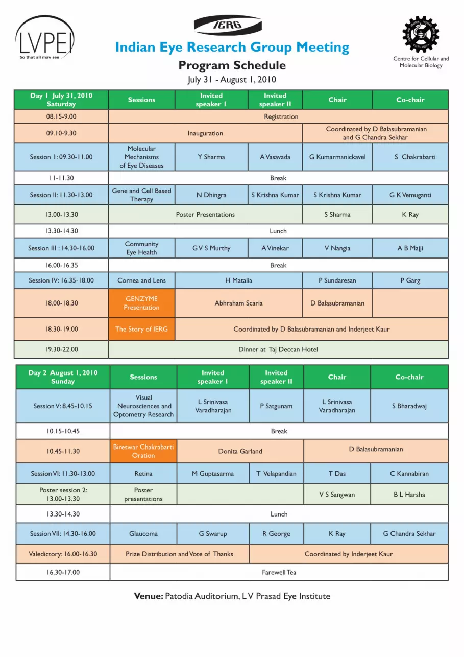
Day 1 July 31, 2010Saturday Sessions Invited
speaker 1Invited
speaker II Chair Co-chair
08.15-9.00 Registration
09.10-9.30 Inauguration Coordinated by D Balasubramanian
and G Chandra Sekhar
Session 1: 09.30-11.00Molecular
Mechanisms of Eye Diseases
Y Sharma A Vasavada G Kumarmanickavel S Chakrabarti
11-11.30 Break
Session II: 11.30-13.00Gene and Cell Based
TherapyN Dhingra S Krishna Kumar S Krishna Kumar G K Vemuganti
13.00-13.30 Poster Presentations S Sharma K Ray
13.30-14.30 Lunch
Session III : 14.30-16.00Community Eye Health
G V S Murthy A Vinekar V Nangia A B Majji
16.00-16.35 Break
Session IV: 16.35-18.00 Cornea and Lens H Matalia P Sundaresan P Garg
18.00-18.30 GENZYMEPresentation Abhraham Scaria D Balasubramanian
18.30-19.00 The Story of IERG Coordinated by D Balasubramanian and Inderjeet Kaur
19.30-22.00 Dinner at Taj Deccan Hotel
Program ScheduleIndian Eye Research Group Meeting
July 31 - August 1, 2010
Day 2 August 1, 2010Sunday Sessions Invited
speaker 1Invited
speaker II Chair Co-chair
Session V: 8.45-10.15 Visual
Neurosciences and Optometry Research
L Srinivasa Varadharajan P Satgunam L Srinivasa
Varadharajan S Bharadwaj
10.15-10.45 Break
10.45-11.30 Bireswar Chakrabarti Oration Donita Garland D Balasubramanian
Session VI: 11.30-13.00 Retina M Guptasarma T Velapandian T Das C Kannabiran
Poster session 2: 13.00-13.30
Poster presentations V S Sangwan B L Harsha
13.30-14.30 Lunch
Session VII: 14.30-16.00 Glaucoma G Swarup R George K Ray G Chandra Sekhar
Valedictory: 16.00-16.30 Prize Distribution and Vote of Thanks Coordinated by Inderjeet Kaur
16.30-17.00 Farewell Tea
Venue: Patodia Auditorium, L V Prasad Eye Institute


PB 1Oral PresentationOral Presentation
Session I: Molecular Mechanisms of Eye Diseases July 31, 2010 Saturday 9.30-11.00 hrs
Chairs: G Kumarmanickavel and Subhabrata Chakrabarti
Time Name Type of Presentation
Abstract No Title
9.30-9.45Abhay Vasavada
Invited talk IIT 001Life and Death beyond Expectation: Lens Epithelial Cells
9.45-10.00 Yogendra Sharma Invited talk IIT 002
What makes a Crystallin a Lens Crystallin: Microbial Versus Lens Beta Gamma-Cryatallins
10.00-10.10 Amita Mishra Free paper IPT 001Evolution of Ca2+-Mediated Stability in Diverse BG-Crystallin Domains
10.10-10.20 Charanya Ramachandran Free paper IPT 002
Cross-Talk between the Camp-PKA And RhoA-Rho Kinase Signaling Pathways in Trabecular Meshwork Cells
10.20-10.30 V Rajanikanth Free paper IPT 003
Structure And Stability of a Single Betagamma-Crystallin Domain of a Protein Brainillin From Mouse Brain Resemble the Lens Gamma-Crystallin
10.30-10.40 Vidyalatha Parsam Free paper IPT 004
Transcript Analysis of Constitutional Mutations in The RB1 Gene in Retinoblastoma Patients Reveals Different Patterns of Missplicing
10.40-10.50 Sriparna Ganguly Free paper IPT 005
A Genome-Wide Association Study In Primary Congenital Glaucoma: Some Preliminary Observations
10.50-11.00 Anshul Arora Free paper IPT 006
Role of Epithelial Mesenchymal Transition of Lens Epithelial Cells in the Regeneration Of Rabbit Lens
Oral Presentations

2 3Oral PresentationOral Presentation2 3Oral PresentationOral Presentation
Session II: Gene & Cells based therapy July 31, 2010 Saturday 11.30-13.00 hrs
Chairs: S Krishna Kumar and Geeta K Vemuganti
11.30-11.45Narender Dhingra
Invited talk IIT 003
Remodeling of Second-Order and Third-Order Retinal Neurons after Photoreceptor Degeneration
11.45-12.00S KirshnaKumar
Invited talk IIT 004Epcam, Myself, Retinoblastoma and the Continuing Journey
12.00-12.10 C Gowri Priya Free paper IPT 007
Characterization of Buccal Mucosal Epithelial Stem Cells and Evaluation of its Efficacy in Corneal Surface Reconstruction
12.10-12.20 Murali MS Balla Free paper IPT 008
Evaluation of Human Y79 Cell Lines for Putative Stem Cell Properties by Single Cell Assay and Gene Expression
12.20-12.30 Nirmala Badhri Narayanan Free paper IPT 009
To Study the Efficacy of Nanoparticle Conjugated Etoposide delivery to Retinoblastoma Cells
12.30-12.40 S Vandhana Free paper IPT 010
Lipogenic Enzyme-Inhibitor Cerulenin shows Pro Apoptotic and Anti-Proliferative Activity in Retinoblastoma Y79 Cells
12.40-12.50 Indumathi Mariappan Free paper IPT 011
Derivation and Characterization of Induced Pluripotent Stem Cells (iPSCs)
12.50-13.00 Sarbani Hazra Free paper IPT 012Animal Models of Ocular Fibrosis: Approaches for Therapeutic Prevention
2 3Oral PresentationOral Presentation2 3Oral PresentationOral Presentation
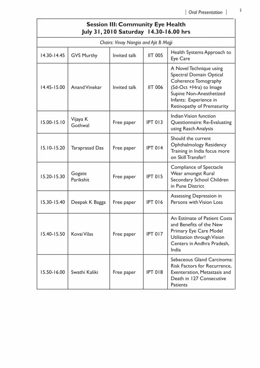
Session III: Community Eye Health July 31, 2010 Saturday 14.30-16.00 hrs
Chairs: Vinay Nangia and Ajit B Majji
14.30-14.45 GVS Murthy Invited talk IIT 005Health Systems Approach to Eye Care
14.45-15.00 Anand Vinekar Invited talk IIT 006
A Novel Technique using Spectral Domain Optical Coherence Tomography (Sd-Oct +Hra) to Image Supine Non-Anesthetized Infants: Experience in Retinopathy of Prematurity
15.00-15.10 Vijaya K Gothwal Free paper IPT 013
Indian Vision function Questionnaire: Re-Evaluating using Rasch Analysis
15.10-15.20 Taraprasad Das Free paper IPT 014
Should the current Ophthalmology Residency Training in India focus more on Skill Transfer!
15.20-15.30 Gogate Parikshit Free paper IPT 015
Compliance of Spectacle Wear amongst Rural Secondary School Children in Pune District
15.30-15.40 Deepak K Bagga Free paper IPT 016Assessing Depression in Persons with Vision Loss
15.40-15.50 Kovai Vilas Free paper IPT 017
An Estimate of Patient Costs and Benefits of the New Primary Eye Care Model Utilization through Vision Centers in Andhra Pradesh, India
15.50-16.00 Swathi Kaliki Free paper IPT 018
Sebaceous Gland Carcinoma: Risk Factors for Recurrence, Exenteration, Metastasis and Death in 127 Consecutive Patients
4 5Oral PresentationOral Presentation4 5Oral PresentationOral Presentation
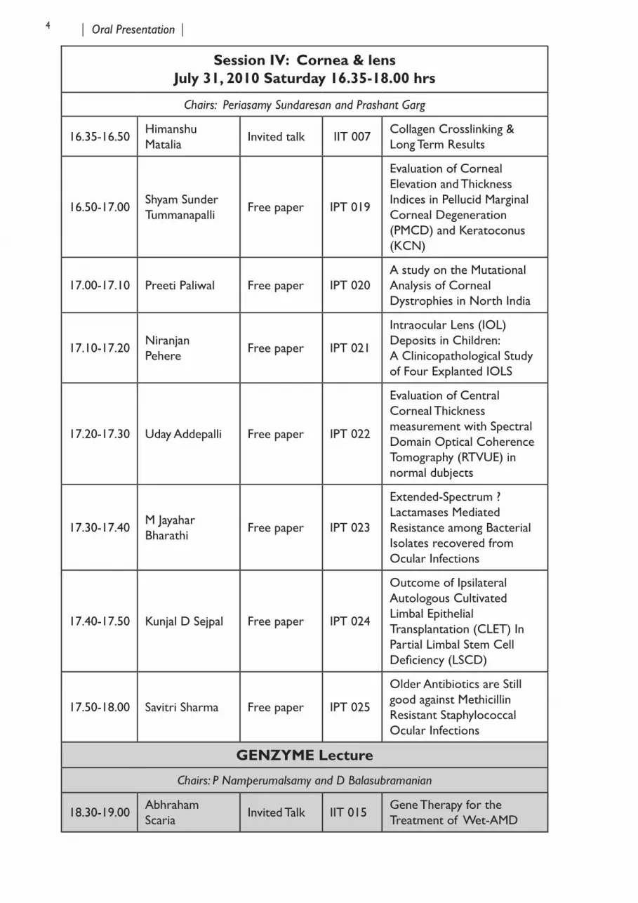
Session IV: Cornea & lens July 31, 2010 Saturday 16.35-18.00 hrs
Chairs: Periasamy Sundaresan and Prashant Garg
16.35-16.50Himanshu Matalia
Invited talk IIT 007Collagen Crosslinking & Long Term Results
16.50-17.00Shyam Sunder Tummanapalli
Free paper IPT 019
Evaluation of Corneal Elevation and Thickness Indices in Pellucid Marginal Corneal Degeneration (PMCD) and Keratoconus (KCN)
17.00-17.10 Preeti Paliwal Free paper IPT 020A study on the Mutational Analysis of Corneal Dystrophies in North India
17.10-17.20 Niranjan Pehere Free paper IPT 021
Intraocular Lens (IOL) Deposits in Children: A Clinicopathological Study of Four Explanted IOLS
17.20-17.30 Uday Addepalli Free paper IPT 022
Evaluation of Central Corneal Thickness measurement with Spectral Domain Optical Coherence Tomography (RTVUE) in normal dubjects
17.30-17.40 M Jayahar Bharathi Free paper IPT 023
Extended-Spectrum ? Lactamases Mediated Resistance among Bacterial Isolates recovered from Ocular Infections
17.40-17.50 Kunjal D Sejpal Free paper IPT 024
Outcome of Ipsilateral Autologous Cultivated Limbal Epithelial Transplantation (CLET) In Partial Limbal Stem Cell Deficiency (LSCD)
17.50-18.00 Savitri Sharma Free paper IPT 025
Older Antibiotics are Still good against Methicillin Resistant Staphylococcal Ocular Infections
GENZYME Lecture
Chairs: P Namperumalsamy and D Balasubramanian
18.30-19.00 Abhraham Scaria Invited Talk IIT 015 Gene Therapy for the
Treatment of Wet-AMD
4 5Oral PresentationOral Presentation4 5Oral PresentationOral Presentation
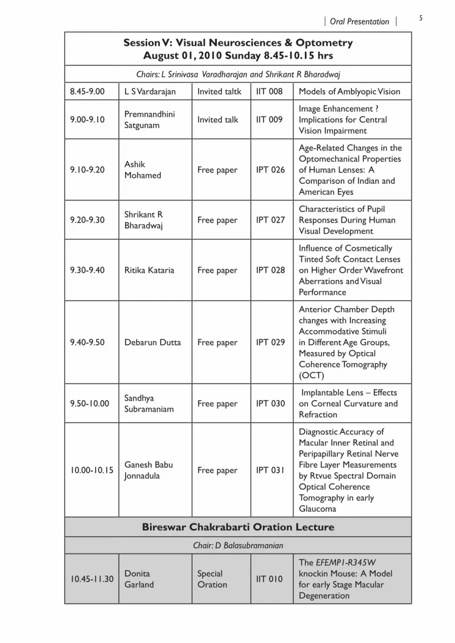
Session V: Visual Neurosciences & Optometry August 01, 2010 Sunday 8.45-10.15 hrs
Chairs: L Srinivasa Varadharajan and Shrikant R Bharadwaj
8.45-9.00 L S Vardarajan Invited taltk IIT 008 Models of Amblyopic Vision
9.00-9.10Premnandhini Satgunam
Invited talk IIT 009Image Enhancement ? Implications for Central Vision Impairment
9.10-9.20Ashik Mohamed
Free paper IPT 026
Age-Related Changes in the Optomechanical Properties of Human Lenses: A Comparison of Indian and American Eyes
9.20-9.30 Shrikant R Bharadwaj Free paper IPT 027
Characteristics of Pupil Responses During Human Visual Development
9.30-9.40 Ritika Kataria Free paper IPT 028
Influence of Cosmetically Tinted Soft Contact Lenses on Higher Order Wavefront Aberrations and Visual Performance
9.40-9.50 Debarun Dutta Free paper IPT 029
Anterior Chamber Depth changes with Increasing Accommodative Stimuli in Different Age Groups, Measured by Optical Coherence Tomography (OCT)
9.50-10.00 Sandhya Subramaniam Free paper IPT 030
Implantable Lens – Effects on Corneal Curvature and Refraction
10.00-10.15 Ganesh Babu Jonnadula Free paper IPT 031
Diagnostic Accuracy of Macular Inner Retinal and Peripapillary Retinal Nerve Fibre Layer Measurements by Rtvue Spectral Domain Optical Coherence Tomography in early Glaucoma
Bireswar Chakrabarti Oration Lecture
Chair: D Balasubramanian
10.45-11.30 Donita Garland
Special Oration IIT 010
The EFEMP1-R345W knockin Mouse: A Model for early Stage Macular Degeneration
6 7Oral PresentationOral Presentation6 7Oral PresentationOral Presentation
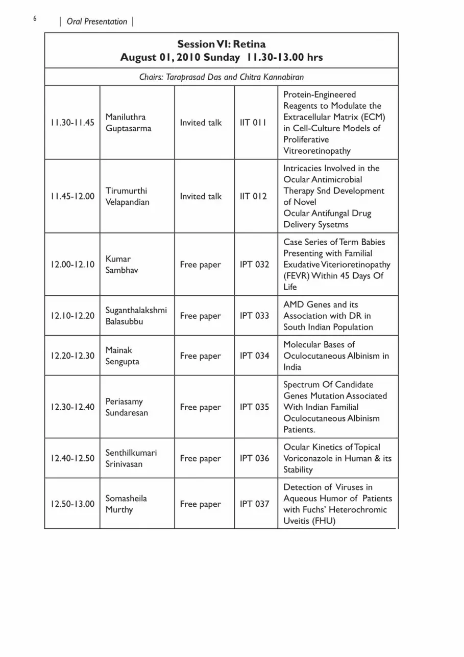
Session VI: Retina August 01, 2010 Sunday 11.30-13.00 hrs
Chairs: Taraprasad Das and Chitra Kannabiran
11.30-11.45Maniluthra Guptasarma
Invited talk IIT 011
Protein-Engineered Reagents to Modulate the Extracellular Matrix (ECM) in Cell-Culture Models of Proliferative Vitreoretinopathy
11.45-12.00TirumurthiVelapandian Invited talk IIT 012
Intricacies Involved in the Ocular Antimicrobial Therapy Snd Development of Novel Ocular Antifungal Drug Delivery Sysetms
12.00-12.10 Kumar Sambhav Free paper IPT 032
Case Series of Term Babies Presenting with Familial Exudative Viterioretinopathy (FEVR) Within 45 Days Of Life
12.10-12.20 Suganthalakshmi Balasubbu Free paper IPT 033
AMD Genes and its Association with DR in South Indian Population
12.20-12.30 Mainak Sengupta Free paper IPT 034
Molecular Bases of Oculocutaneous Albinism in India
12.30-12.40Periasamy Sundaresan Free paper IPT 035
Spectrum Of Candidate Genes Mutation Associated With Indian Familial Oculocutaneous Albinism Patients.
12.40-12.50 Senthilkumari Srinivasan Free paper IPT 036
Ocular Kinetics of Topical Voriconazole in Human & its Stability
12.50-13.00 Somasheila Murthy Free paper IPT 037
Detection of Viruses in Aqueous Humor of Patients with Fuchs’ Heterochromic Uveitis (FHU)
6 7Oral PresentationOral Presentation6 7Oral PresentationOral Presentation
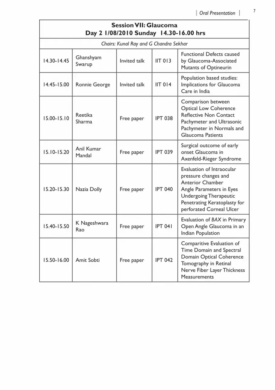
Session VII: Glaucoma Day 2 1/08/2010 Sunday 14.30-16.00 hrs
Chairs: Kunal Ray and G Chandra Sekhar
14.30-14.45Ghanshyam Swarup
Invited talk IIT 013Functional Defects caused by Glaucoma-Associated Mutants of Optineurin
14.45-15.00 Ronnie George Invited talk IIT 014Population based studies: Implications for Glaucoma Care in India
15.00-15.10Reetika Sharma Free paper IPT 038
Comparison between Optical Low Coherence Reflective Non Contact Pachymeter and Ultrasonic Pachymeter in Normals and Glaucoma Patients
15.10-15.20 Anil Kumar Mandal Free paper IPT 039
Surgical outcome of early onset Glaucoma in Axenfeld-Rieger Syndrome
15.20-15.30 Nazia Dolly Free paper IPT 040
Evaluation of Intraocular pressure changes and Anterior Chamber Angle Parameters in Eyes Undergoing Therapeutic Penetrating Keratoplasty for perforated Corneal Ulcer
15.40-15.50 K Nageshwara Rao Free paper IPT 041
Evaluation of BAX in Primary Open Angle Glaucoma in an Indian Population
15.50-16.00 Amit Sobti Free paper IPT 042
Comparitive Evaluation of Time Domain and Spectral Domain Optical Coherence Tomography in Retinal Nerve Fiber Layer Thickness Measurements

8 9Poster SessionsPoster Sessions8 9Poster SessionsPoster Sessions
Poster Session - I July 31, 2010, Saturday
Participant Abstract No. Title of the Presentation
Alpesh PatelIBP001
Lens Epithelial Cell Differentiation in the Pediatric Traumatic Cataracts
Anuradha PalIBP002
Characterization of Secretary Virulence Factors from Pathogenic Fungi Causing Keratitis
A V SaiJyothi IBP003 Tear Fluid Antioxidants Profile in Patients with Keratoconus
B P MohantyIBP004
Arsenic Exposure Alters Lens Aa-Crystallin Profile in vivo and Induces Cataract Formation in Labeo Rohita
Bharath SelviIBP005
Exposure to Homocysteine Negatively Influences Glutathione Synthesis in Human Retinal Pigment Epithelial Cells
Cornelia Reena Joseph IBP006 Real Time PCR in the Diagnosis of Postoperative Endophthalmitis.
Devki Sheth IBP007 Effect of Endoplasmic Reticulum Stress on the Lens Epithelial Cells
Ganeswararao Musada
IBP008
Molecular Genetic Analysis of Norrie Disease Pseudoglioma (NDP) Gene in Familial Exudative Vitreo Retinopathy (FEVR) Patients and Indian Retinopathy of Prematurity (ROP) Babies
GayathriIBP009
Lysyl Oxidase and its Isoforms in Plasma and Aqueous Humor of Pseudoexfoliation Patients
Jambulingam Malathi IBP010 Detection of CMV Retinitis in HIV Infected Individuals: A Comparative Study
K Gopinath
IBP011
Ryanodine Receptor in Lipid Raft Microdomains are Affected by Pharmocological Reagents which Perturb Calcium Dynamics in Muller Glia of Retina.
K Rangachari IBP012 Biophysical Characterization of Human Myocilin and the C-Term Region.
Poster Session 1
8 9Poster SessionsPoster Sessions8 9Poster SessionsPoster Sessions
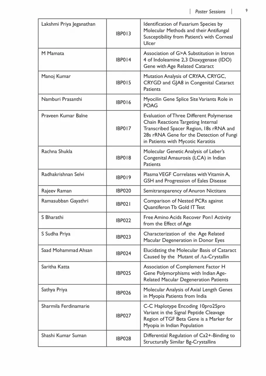
Lakshmi Priya Jeganathan
IBP013
Identification of Fusarium Species by Molecular Methods and their Antifungal Susceptibility from Patient’s with Corneal Ulcer
M MamataIBP014
Association of G>A Substitution in Intron 4 of Indoleamine 2,3 Dioxygenase (IDO)Gene with Age Related Cataract
Manoj KumarIBP015
Mutation Analysis of CRYAA, CRYGC, CRYGD and GJA8 in Congenital Cataract Patients
Namburi Prasanthi IBP016
Myocilin Gene Splice Site Variants Role in POAG
Praveen Kumar Balne
IBP017
Evaluation of Three Different Polymerase Chain Reactions Targeting Internal Transcribed Spacer Region, 18s rRNA and 28s rRNA Gene for the Detection of Fungi in Patients with Mycotic Keratitis
Rachna ShuklaIBP018
Molecular Genetic Analysis of Leber’s Congenital Amaurosis (LCA) in Indian Patients
Radhakrishnan Selvi IBP019 Plasma VEGF Correlates with Vitamin A, GSH and Progression of Eales Disease
Rajeev Raman IBP020 Semitransparency of Anuron Nictitans
Ramasubban Gayathri IBP021 Comparison of Nested PCRs against Quantiferon Tb Gold IT Test
S Bharathi IBP022 Free Amino Acids Recover Pon1 Activity from the Effect of Age
S Sudha Priya IBP023 Characterization of the Age Related Macular Degeneration in Donor Eyes
Saad Mohammad Ahsan IBP024 Elucidating the Molecular Basis of Cataract Caused by the Mutant of Αa-Crystallin
Saritha KattaIBP025
Association of Complement Factor H Gene Polymorphisms with Indian Age-Related Macular Degeneration Patients
Sathya Priya IBP026 Molecular Analysis of Axial Length Genes in Myopia Patients from India
Sharmila Ferdinamarie
IBP027
C-C Haplotype Encoding 10pro25pro Variant in the Signal Peptide Cleavage Region of TGF Beta Gene is a Marker for Myopia in Indian Population
Shashi Kumar Suman IBP028 Differential Regulation of Ca2+-Binding to Structurally Similar Bg-Crystallins
10 11Poster SessionsPoster Sessions10 11Poster SessionsPoster Sessions
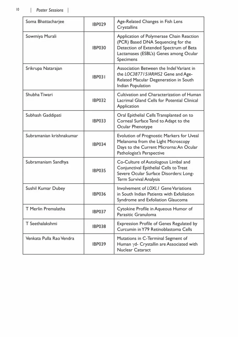
Soma BhattacharjeeIBP029
Age-Related Changes in Fish Lens Crystallins
Sowmiya Murali
IBP030
Application of Polymerase Chain Reaction (PCR) Based DNA Sequencing for the Detection of Extended Spectrum of Beta Lactamases (ESBL’s) Genes among Ocular Specimens
Srikrupa Natarajan
IBP031
Association Between the Indel Variant in the LOC387715/ARMS2 Gene and Age-Related Macular Degeneration in South Indian Population
Shubha Tiwari IBP032
Cultivation and Characterization of Human Lacrimal Gland Cells for Potential Clinical Application
Subhash GaddipatiIBP033
Oral Epithelial Cells Transplanted on to Corneal Surface Tend to Adapt to the Ocular Phenotype
Subramanian krishnakumar
IBP034
Evolution of Prognostic Markers for Uveal Melanoma from the Light Microscopy Days to the Current Microrna: An Ocular Pathologist’s Perspective
Subramaniam Sandhya
IBP035
Co-Culture of Autologous Limbal and Conjunctival Epithelial Cells to Treat Severe Ocular Surface Disorders: Long-Term Survival Analysis
Sushil Kumar DubeyIBP036
Involvement of LOXL1 Gene Variations in South Indian Patients with Exfoliation Syndrome and Exfoliation Glaucoma
T Merlin Premalatha IBP037 Cytokine Profile in Aqueous Humor of Parasitic Granuloma
T SeethalakshmiIBP038
Expression Profile of Genes Regulated by Curcumin in Y79 Retinoblastoma Cells
Venkata Pulla Rao VendraIBP039
Mutations in C-Terminal Segment of Human gd- Crystallin are Associated with Nuclear Cataract
10 11Poster SessionsPoster Sessions10 11Poster SessionsPoster Sessions
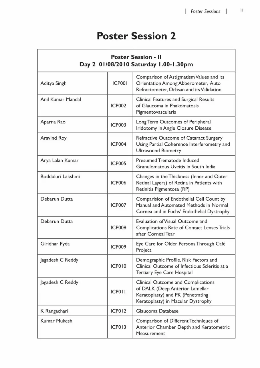
Poster Session 2
Poster Session - II Day 2 01/08/2010 Saturday 1.00-1.30pm
Aditya Singh ICP001Comparison of Astigmatism Values and its Orientation Among Abberometer, Auto Refractometer, Orbsan and its Validation
Anil Kumar MandalICP002
Clinical Features and Surgical Results of Glaucoma in Phakomatosis Pigmentovascularis
Aparna Rao ICP003 Long Term Outcomes of Peripheral Iridotomy in Angle Closure Disease
Aravind Roy ICP004
Refractive Outcome of Cataract Surgery Using Partial Coherence Interferometry and Ultrasound Biometry
Arya Lalan Kumar ICP005 Presumed Trematode Induced Granulomatous Uveitis in South India
Bodduluri LakshmiICP006
Changes in the Thickness (Inner and Outer Retinal Layers) of Retina in Patients with Retinitis Pigmentosa (RP)
Debarun DuttaICP007
Comparision of Endothelial Cell Count by Manual and Automated Methods in Normal Cornea and in Fuchs’ Endothelial Dystrophy
Debarun DuttaICP008
Evaluation of Visual Outcome and Complications Rate of Contact Lenses Trials after Corneal Tear
Giridhar PydaICP009
Eye Care for Older Persons Through Café Project
Jagadesh C ReddyICP010
Demographic Profile, Risk Factors and Clinical Outcome of Infectious Scleritis at a Tertiary Eye Care Hospital
Jagadesh C Reddy
ICP011
Clinical Outcome and Complications of DALK (Deep Anterior Lamellar Keratoplasty) and PK (Penetrating Keratoplasty) in Macular Dystrophy
K Rangachari ICP012 Glaucoma Database
Kumar MukeshICP013
Comparison of Different Techniques of Anterior Chamber Depth and Keratometric Measurement
12 13Poster SessionsPoster Sessions12 13Poster SessionsPoster Sessions
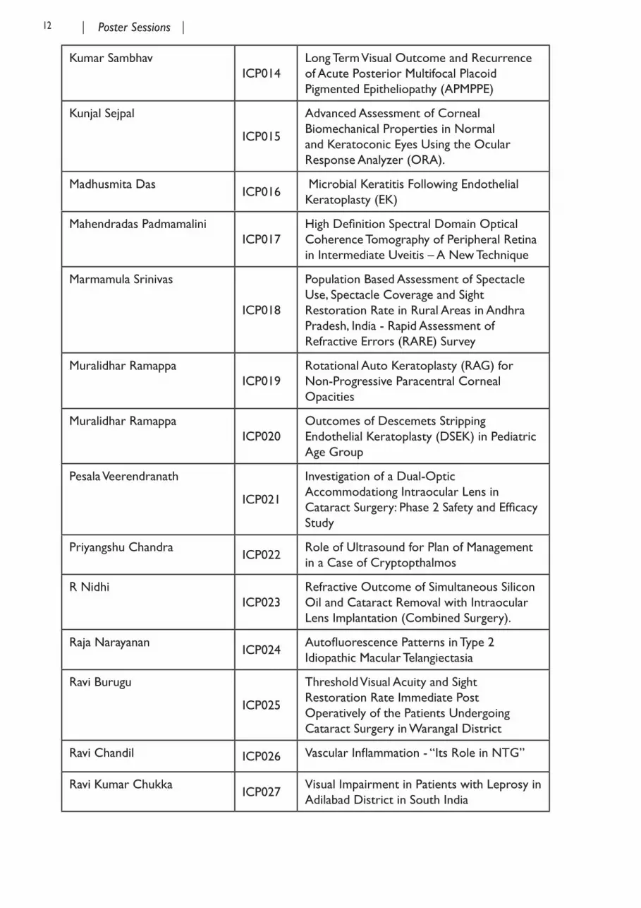
Kumar SambhavICP014
Long Term Visual Outcome and Recurrence of Acute Posterior Multifocal Placoid Pigmented Epitheliopathy (APMPPE)
Kunjal Sejpal
ICP015
Advanced Assessment of Corneal Biomechanical Properties in Normal and Keratoconic Eyes Using the Ocular Response Analyzer (ORA).
Madhusmita DasICP016
Microbial Keratitis Following Endothelial Keratoplasty (EK)
Mahendradas Padmamalini ICP017
High Definition Spectral Domain Optical Coherence Tomography of Peripheral Retina in Intermediate Uveitis – A New Technique
Marmamula Srinivas
ICP018
Population Based Assessment of Spectacle Use, Spectacle Coverage and Sight Restoration Rate in Rural Areas in Andhra Pradesh, India - Rapid Assessment of Refractive Errors (RARE) Survey
Muralidhar RamappaICP019
Rotational Auto Keratoplasty (RAG) for Non-Progressive Paracentral Corneal Opacities
Muralidhar RamappaICP020
Outcomes of Descemets Stripping Endothelial Keratoplasty (DSEK) in Pediatric Age Group
Pesala Veerendranath
ICP021
Investigation of a Dual-Optic Accommodationg Intraocular Lens in Cataract Surgery: Phase 2 Safety and Efficacy Study
Priyangshu Chandra ICP022 Role of Ultrasound for Plan of Management in a Case of Cryptopthalmos
R NidhiICP023
Refractive Outcome of Simultaneous Silicon Oil and Cataract Removal with Intraocular Lens Implantation (Combined Surgery).
Raja Narayanan ICP024 Autofluorescence Patterns in Type 2 Idiopathic Macular Telangiectasia
Ravi Burugu
ICP025
Threshold Visual Acuity and Sight Restoration Rate Immediate Post Operatively of the Patients Undergoing Cataract Surgery in Warangal District
Ravi Chandil ICP026 Vascular Inflammation - “Its Role in NTG”
Ravi Kumar Chukka ICP027 Visual Impairment in Patients with Leprosy in Adilabad District in South India
12 13Poster SessionsPoster Sessions12 13Poster SessionsPoster Sessions
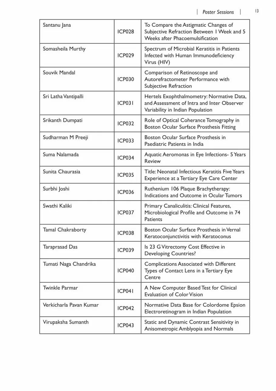
Santanu JanaICP028
To Compare the Astigmatic Changes of Subjective Refraction Between 1 Week and 5 Weeks after Phacoemulsification
Somasheila MurthyICP029
Spectrum of Microbial Keratitis in Patients Infected with Human Immunodeficiency Virus (HIV)
Souvik MandalICP030
Comparison of Retinoscope and Autorefractometer Performance with Subjective Refraction
Sri Latha VantipalliICP031
Hertels Exophthalmometry: Normative Data, and Assessment of Intra and Inter Observer Variability in Indian Population
Srikanth Dumpati ICP032 Role of Optical Coherance Tomography in Boston Ocular Surface Prosthesis Fitting
Sudharman M Preeji ICP033 Boston Ocular Surface Prosthesis in Paediatric Patients in India
Suma Nalamada ICP034 Aquatic Aeromonas in Eye Infections- 5 Years Review
Sunita Chaurasia ICP035 Title: Neonatal Infectious Keratitis Five Years Experience at a Tertiary Eye Care Center
Surbhi Joshi ICP036 Ruthenium 106 Plaque Brachytherapy: Indications and Outcome in Ocular Tumors
Swathi KalikiICP037
Primary Canaliculitis: Clinical Features, Microbiological Profile and Outcome in 74 Patients
Tamal Chakraborty ICP038 Boston Ocular Surface Prosthesis in Vernal Keratoconjunctivitis with Keratoconus
Taraprasad Das ICP039 Is 23 G Vitrectomy Cost Effective in Developing Countries?
Tumati Naga ChandrikaICP040
Complications Associated with Different Types of Contact Lens in a Tertiary Eye Centre
Twinkle Parmar ICP041 A New Computer Based Test for Clinical Evaluation of Color Vision
Verkicharla Pavan Kumar ICP042 Normative Data Base for Colordome Epsion Electroretinogram in Indian Population
Virupaksha Sumanth ICP043 Static and Dynamic Contrast Sensitivity in Anisometropic Amblyopia and Normals


Invited Talks


16 17Invited TalksInvited Talks
Invited talks, Session I, Molecular mechanisms of eye diseases, July 31, 2010, 9.20 -11.00 hrs
Chairs: G Kumarmanickavel and Subhabrata Chakrabarti
IIT 001Life and Death Beyond Expectation: Lens Epithelial CellsAbhay VasavadaIladevi Cataratct and IOL Centre, Ahmedabad, India.
Abstract awaited
IIT 002What Makes a Crystallin a Lens Crystallin: Microbial versus Lens Beta Gamma-Cryatallins?Yogendra SharmaCentre for Cellular and Molecular Biology (CCMB), Hyderabad, India.
There has been a great deal of interests in understanding the evolution of protein domains, since similar folds appear to have common ancestral origin. During divergent or convergent evolution, though there were modifications due to the selection requirements and recruitments, the over-all topology of a protein domain remained largely unaltered. bg-Crystallin domain is among the ancient and divergent folds which define the bg-crystallin superfamily. This superfamily has the members ranging from prokaryotes to mammal, and thus is an interesting example for understanding the diversity and evolution. Topologically similar bg-crystallin fold, found in all three kingdoms, appears to be an example of natures’ extreme engineering designed for Ca2+-binding and domain stability, though its molecular basis is not known and present an evolutionary paradox. Our goal has been to understand how does the Ca2+-dependent generic gain in stability evolved by differentially designed domains.
The canonical Ca2+-binding motif or N/DN/DXXS/TS sequence is modified in crystallin present in higher organism. We examined how creating a canonical sequence in lens crystallin de-stabilizes the protein domain. Some of these changes are found in case of cataract. Our results suggest that due to selective diversification of these domains during evolution; some of the properties, such as Ca2+-induced gains in stability were either retained or lost just by minor modifications in the double clamp Ca2+-binding motif. To retain the stability of the domain without Ca2+, there were extremely intelligent designs in lens homologues achieved during evolution from ancestors to compensate for the Ca2+-induced gain in stability.
18 19Invited TalksInvited Talks18 19Invited TalksInvited Talks
Invited talks, Session II, Genes and Cell Based Therapy, July 31, 2010, 11.30 -13.00 hrs
Chairs: S Krishna Kumar and Geeta K Vemuganti
IIT 003Remodeling of Second-Order and Third-Order Retinal Neurons after Photoreceptor DegenerationNarinder DhingraNational Brain Research Centre, Gurgaon, India.
Abstract awaited
IIT 004EpCAM, Myself, Retinoblastoma and the Continuing Journey Subramanian KrishnakumarVision Research Foundation, Sankara Nethralaya, Chennai, India.
Purpose: To share the experiences in my research work on EpCAM [Epithelial cell adhesion molecule] in retinoblastoma over a duration of 8 years.
Methods: What started as a matter of fact email to Dr Ren-Heidenreich L, after having a publication in cancer Journal 2004,on HLA antigens and when I saw her publication on EpCAM as a tumor associated antigen can be used for immunotherapy, started the eight long years of ongoing work on EpCAM involving Immunohistochemistry, real time PCR, western blotting, FACS, RNA interference, studying the signaling pathways of EpCAM, whole genome microarray, post RNA interference EpCAM MicroRNA and then leading to work on fabrication of Nanocarriers for EpCAM siRNA delivery, fabrication of Recombinant EpCAM antibody, fabrication of EpCAM RNA aptamer using SELEX ( systematic Evolution of Ligands by Exponential Enrichment ) and targeted delivery of drug using EpCAM.
Results: EpCAM is expressed in Retinoblastoma as shown by IHC, Real time PCR and Western blot, FACS, RNAi of EpCAM leads to reduced proliferation and we have identified novel pathways using whole genome microarray and identified the oncomir cluster post EpCAM RNAi. EpCAM antibody conjugated drug loaded nanoparticle is more effective than the native particle. Had an opportunity to meet and spend some in Dr Robert Langer Lab in MT to discuss on aptamer. EpCAM aptamer very specific to EpCAM expressing cancer cells. There is no functional blocking. There appears to be an intracellular cleaving of the intracellular domain of the EpCAM, so there could be limitations in the use of Recombinant EpCAM antibody for therapy.
Conclusions: EpCAM appears to be a promising target in Retinoblastoma as well as other cancers such as breast cancer and liver cancer. It’s for the first time we have identified a target molecule in an orphan disease like retinoblastoma, so we could share the knowledge/technology happening in this area to retinoblastoma. For example there are studies going on the efficacy of Recombinant EpCAM antibody adecatumumab, in breast and prostate cancer
18 19Invited TalksInvited Talks18 19Invited TalksInvited Talks
patients. The work is continuing with the use of Bispecific antibody on retinoblastoma tumor and delivery of suicide gene and now Symporter gene therapy followed by radioactive iodide using a specific tissue specific Promoter.
Invited talks, Session III, Community Eye Health,July 31, 2010, 14.30 - 16.00 hrs
Chairs: Vinay Nangia and Ajit B Majji
IIT 005Health Systems Approach to Eye CareGVS MurthyIndian Institute of Public Health, South Asia Centre for Disability Inclusive Development & Vision Research, PHFI, Hyderabad, India.
A health system consists of all organizations and people whose primary intent is to promote, restore or maintain health. WHO recommends that a health systems perspective in developing countries would increase efficiency and effectiveness of health services and optimize benefits from available resources. The health system approach is as relevant to eye care as it is to any other health service.
A health system approach involves paying attention to 6 ‘building blocks’. These include service delivery, health workforce, health information systems, health financing including financial risk protection, technology and leadership/governance. The objective of health systems approach is to ensure equitable access and universal coverage while providing services of high quality and unquestionable safety.
Eye Care is an excellent case study for a health systems framework. The adoption of the VISION2020: Right to Sight initiative by the Government and NGO consortia paved the way for augmented service delivery with equity being the cornerstone of eye care service delivery in the country. There is a need to evaluate the eye health workforce in terms of its responsiveness , skill and adequacy. It is evident that there is a gap in relation to skills and adequacy across the country and using a health systems approach can help to identify what needs to be done to improve the situation. Eye health information systems need to be strengthened further and the search for appropriate technology to reach out to the population need attention. Financial risk protection efforts in the country were initiated during the early 1990s but universal access has not yet been achieved. Dynamic leadership has guided the success of the eye care services in the country but issues related to governance, especially in the public sector need attention.
20 21Invited TalksInvited Talks20 21Invited TalksInvited Talks
IIT 006A Novel Technique Using Spectral Domain Optical Coherence Tomography (SD-OCT +HRA) (Spectralis, Heidelberg Engineering) to Image Supine Non-Anesthetized Infants: Experience in Retinopathy of PrematurityAnand Vinekar, Munuswamy Sivakumar, Rohit Shetty, Narasimha Krishnan, Padmamalini Mahendradas, Ashwin Mallipatna, K Bhujang ShettyDepartment of Pediatric Retina, Narayana Nethralaya Postgraduate Institute of Ophthalmology, Bangalore, India.
Purpose: To acquire optical coherence tomography (OCT) images of supine, non-anesthetized infants in the office by a novel modification of a commercially available table-top, chin rest system and to describe the experience in imaging Retinopathy of Prematurity.
Methods: Spectralis, a combined HRA+OCT device (Heidelberg Engineering, Heidelberg, Germany) was modified to convert the table-top system into a hand- held device using a two-step modification of the existing system. This device was used to obtain high-resolution OCT images of infants with aggressive posterior retinopathy of prematurity (APROP) to image flat neovascularization (FNV) and in cases with classical ROP to image specific disease characteristics. Age matched normal infants with no ROP were used as controls.
Results: Serial imaging of the exact area of clinical interest (FNV) were obtained and were comparable in cases with APROP. Classical ROP cases revealed macular edema that resolved spontaneously or with treatment. No controls revealed macular edema. The entire procedure was safely completed in the office. The obtained OCT images guided selective laser ablation of the FNV which were missed on clinical examination in the cases of APROP.
Conclusions: With this simple modification and technique, the ability of using the Spectralis, (a combined angiography and OCT imaging device) for imaging supine, non-anesthetized infants has been established. This possibility would allow the dual use of the table-top system to serve also as a hand-held device for pediatric cases that can be imaged in the office setting with limited operating room facility in a busy practice.
Invited talks, Session IV, Cornea and Lens, 31/07/2010, 16.30 -18.00hrs
Chairs: Perisamy Sundaresan and Prasanth Garg
IIT 007Collagen Crosslinking - Long Term ResultsHimansu MataliaNarayana Nethralaya, Bangalore, India.
Collagen crosslinking is emerging as one of the most popular procedures in corneal surgery. We aim to present our 3 year data on crosslinking and outcomes. There are many controversies and unanswered questions in this procedure like longterm effects, safety, risks, age limit, combination procedures with intacs, lasers & many more. This lecture will aim at discussing these issues and present our protocol for treatment of progressive keratoconus.
20 21Invited TalksInvited Talks20 21Invited TalksInvited Talks
GENZYME Lecture, July 31, 2010, 18.45 - 19.15 hrs
Chairs: P Namperumalsamy and D Balasubramanian
IIT 009Gene Therapy for the Treatment of Wet-AMDAbraham ScariaGenzyme Corporation, Framingham, MA, USA
VEGF plays a critical role in neovascular age-related macular degeneration and proliferative diabetic retinopathy. VEGF antagonists are useful for treating such disorders; however current treatments require monthly intravitreal injections. We have designed a soluble anti-VEGF molecule (sFLT01) and delivered it by intravitreal injection of an adeno-associated viral (AAV) vector since AAV vectors are capable of long-term gene expression. AAV2-sFLT01 inhibited retinal neovascularization in the murine oxygen-induced retinopathy model and in the laser-induced choroidal neovascularization (laser-CNV) model in mice. In the eyes of rodents and cynomolgus monkeys, AAV2-sFLT01 gives expression levels persistent for at least one year. We also performed laser-CNV experiments 5 months after vector administration in non-human primates and showed that sFLT01 is effective at inhibiting neovascularization in this model. Results of our 12-month safety study of AAV2-sFLT01 administered intravitreally in cynomolgus monkeys will be discussed. In summary, we have demonstrated long-term efficacy with minimal side effects following intravitreal delivery of AAV-sFLT01 in rodents and non-human primate models. These results suggest an alternate method for the long-term treatment for diseases of ocular neovascularization, without the need for repeated intraocular injections. A Phase I clinical trial has been initiated at three clinical sites in the USA.
Invited talks, Session V, Visual Neurosciences and Optometry, August 1, 2010, 8.45 -10.15hrs
Chairs: L Srinivasa Varadharajan and Srikanth Bharadwaj
IIT 010Models of Amblyopic VisionL Srinivasa Varadharajan, P KabilanSrimathi Sundari Subramanian Department of Visual Psychophysics, Elite School of Optometry, Medical Research Foundation, Chennai, India
The amblyopic visual m is characterized by abnormal spatial integration of visual information. While the clinical management of amblyopia is fairly well established, it is still not fully clear as to what happens to the visual system. Three scenarios, not all mutually exclusive are postulated in the literature: reduction in the number of mechanisms, irregularity of the mechanism arrangements and reduction in the strength of interactions. Histological and electrophysiological studies have shown evidence against the first possibility, while the other two are still debated strongly. In this talk, we will briefly summarize these finding in the literature and present some new data that also supports the first scenario described above. In our experiments, contrast detection thresholds for normal and amblyopic subjects were measured as a function of the

22 23Invited TalksInvited Talks22 23Invited TalksInvited Talks
size of the target. Normal subjects showed continuous decrease in thresholds while amblyopic subjects showed a plateau effect with increased thresholds at intermediate sizes. The difference in results could be explained using different numbers of mechanisms mediating the detection task, in addition to the quality of lateral interaction, in the two set of subjects.
IIT 011Image Enhancement? Implications for Central Vision Impairment Premnandhini SatgunamSchepens Eye Research Institute, Harvard Medical School, USA.
Image enhancement techniques are known to be beneficial for patients with central vision loss. This talk would give a brief overview of the literature in image enhancement. Challenges in evaluation of preference and performance with image enhancement will be highlighted. Results from a recent preference study on normally sighted individuals will be discussed.
Bireswar Chakrabarti Oration, August 1, 2010, 10.45 - 11.30 hrs
Chair: D Balasubramanian
IIT 012The Efemp1-R345W Knockin Mouse: A Model for Early Stage Macular DegenerationDonita GarlandUniversity of Pennsylvania, Philadelphia, PA, USA
Age-related macular degeneration (AMD) is the leading cause of vision loss in the Western world. The prevalence of AMD is rapidly increasing globally with the increasing aged populations. Even though genes have been identified for which mutations cause or are associated with the risk of developing macular degeneration, the underlying molecular mechanisms are not understood. While therapies have been developed that stop neovascularization, there is no therapy for the more common atrophic form of AMD. Thus, there is a critical need to elucidate the mechanisms in early stage AMD in order to prevent the development of AMD and the loss of vision.
To address this need we have developed a mouse model that has the R345W mutation of Efemp1 knocked in. The R345W mutation of EFEMP1 causes Doyne Honeycomb Retinal Dystrophy/Malattia Leventinese (DHRD/ML), an early onset macular degeneration. It is characterized by the formation of drusen at an early age and by macular features of AMD. The Efemp1-R345W mutant mice exhibit major pathogenic features of both DHRD/ML and AMD. The mice develop changes in the RPE and form sub-retinal basal deposits similar to those seen in early AMD, DHRD/ML and other heritable forms of macular degeneration.
We used proteomic approaches to determine the composition of Bruch’s membrane and choroid in Efemp1-R345W mutant and wild type mice. Our results demonstrate that basal deposits were composed of typical Bruch’s membrane components but in altered relative levels. In addition, the accumulation of complement components in Bruch’s membrane with age

22 23Invited TalksInvited Talks22 23Invited TalksInvited Talks
of the mutant mice strongly implicated the complement system in basal deposit formation. A role for the complement system in basal deposit formation was confirmed by generating the Efemp1-R345W knockin/C3 knockout double mutant mice. The double mutant mice showed a marked decrease in basal deposit formation.
The Efemp1-R345W mutant mice and our double mutant mice will be used to further probe the role of the complement system in basal deposit formation and how it is activated and to study the generation of basal deposits and how the processes are regulated.
Invited talks, Session VI, Retina, August 1, 2010, 11.30 -13.00 hrs
Chairs: Dr Taraprasad Das, Dr Chitra Kannabiran
IIT 013Protein-Engineered Reagents to Modulate the Extracellular Matrix (ECM) in Cell-Culture Models of Proliferative VitreoretinopathyMaryada Sharma,1 Vishali Gupta,2 Amod Gupta,2 Manni Luthra-Guptasarma1
1Department of Immunopathology, 2Department of Ophthalmology, Postgraduate Institute of Medical Education and Research, Chandigarh, India.
Purpose: Proliferative vitreoretinopathy (PVR) is an aberrant wound healing process, associated with migration of retinal pigment epithelial (RPE) cells from their original location (the blood-retinal barrier), into the vitreous. In the new environment, these cells undergo extensive proliferation, along with extensive laying-out of an extracellular matrix (ECM). This leads to the formation of epiretinal membranes (ERM) and blindness. Our aim was to simulate the conditions of PVR in a cell culture model system and characterize it with respect to proliferation and migration of RPE, the expression of proteins relevant to the pathology, and the effects of exogenously added collagen and fibronectin. Further, our aim was to develop protein engineered reagents, specifically targeted against fibronectin, to prevent the migration and/or proliferation of RPE cells in PVR.
Methods: D407 RPE cells were cultured in the presence of vitreous, derived from either cadaver eyes or from patients undergoing retinal reattachment surgeries. Besides the changes in phenotype, the behavior of cells in the culture model was examined. The changes in the extracellular matrix components were also evaluated. We employed phage display antibody library screening methods to develop single-chain Fv (scFv) against fibronectin to modulate fibronectin polymerization. This antibody was further engineered to improve its action on the RPE cells in the culture system.
Results: The culture of cell line-derived RPE cells in the presence of patient-derived, pathologic vitreous is myofibroblast-like, with increased expression of α-smooth muscle actin and TGF-β. These RPE cells are capable of increased migration and proliferation with enhanced synthesis and deposition of collagen and fibronectin, as compared to cells cultured in the presence of cadaver-derived vitreous. The effects are more pronounced in the presence of exogenously added collagen. We have developed an scFv antibody (scFv Fn52), through phage display antibody library screening methods, against the N-terminal 30 kDa fragment of fibronectin; addition of this antibody to the cultures results in incomplete polymerization of fibronectin,

24 25Invited TalksInvited Talks24 25Invited TalksInvited Talks
besides reduced proliferation of RPE cells. Engineering of this scFv by incorporation of an “RGDS” tag (scFv Fn52 RGDS) was done; this scFv was found to be more effective in reducing fibronectin assembly, while causing a dramatic reduction in actin stress fiber formation.
Conclusions: Our data shows that it is possible to simulate the pathology associated with PVR by culturing RPE cells (of cell line origin) with pathologic vitreous. Our results with antibodies, directed against fibronectin, in such a cell culture model, suggest that the scFv against the 30 kDa N-terminal region of fibronectin, acting in concert with its RGDS tag, can be an effective “double-edged sword”, and can potentially be useful in the context of PVR, and also in other pathological situations such as tumors, fibrosis and thrombosis.
IIT 014Intricacies Involved in the Ocular Antimicrobial Therapy and Development of Novel Ocular Antifungal Drug Delivery SysetmsThirumurthy Velpandian, Jayabalan Nirmal, Alok Kumar Ravi, Rohit Bisht, Sanjay Sharma , Suproyo Ghose Department of Ocular Pharmacology & Pharmacy, Dr Rajendra Prasad Centre for Ophthalmic Sciences, All India Institute of Medical Sciences, New Delhi, India.
Purpose: Conventionally, antimicrobial drugs developed and approved for systemic infections are re-investigated for ocular infections. Anti-microbial therapy so far remains as an arbitrary approach where drug molecules inherently lacking in ocular penetration capabilities are forced to enter the eye with the help of pharmaceutical drug delivery techniques. Due to the existing problems of delivering the anti-fungal drugs into the eye, we took an attempt to evaluate the suitability of some novel drug delivery systems for natamycin.
Methods: In order to compare ocular drug delivery strategies for ocular infections, the existing studies and methods were revisited using an extensive literature search. We developed novel drug delivery of natamycin using polymeric and implantable drug delivery systems and was studied in invitro, exvivo and invivo experimental models.
Results: An eye specific drug should be developed and the developed drug need to be delivered using smart drug delivery strategies based on physiological mechanisms for their selective enrichment inside the eye. The developed novel drug delivery systems of natamycin showed a significant increase in the ocular bioavailability and also provided a sustained delivery of natamycin in ex-vivo and in-vivo studies.
Conclusions: We insist that, there is a need for eye specific antimicrobial agent and the inevitability of an appropriate drug delivery approach to revolutionize future therapy. Moreover, novel strategies using smart drug delivery systems need to be adopted to achieve a successful therapeutic outcome.
24 25Invited TalksInvited Talks24 25Invited TalksInvited Talks
Invited talks, Session VII, Glaucoma, August 1, 2010, 14.30 -16.00 hrs
Chairs: Kunal Ray and G Chandra Sekhar
IIT 015Functional Defects Caused by Glaucoma-Associated Mutations of OptineurinGhanshyam SwarupCentre for Cellular & Molecular Biology, Hyderabad, India.
Optineurin is a multifunctional protein involved in several functions such as vesicular trafficking from the Golgi to the plasma membrane and NF-kB regulation. Mutations in optineurin are associated with glaucoma, a neurodegenerative eye disease that causes blindness. Genetic evidence suggests that the E50K (Glu50Lys) and H486R (His486Arg) are dominant disease-causing mutations of optineurin. However, functional alterations caused by these mutations are not known. We examined the role of optineurin and its mutants in endocytic recycling and NF-kB regulation. Overexpression of the E50K mutant selectively induced death of retinal ganglion cells which was mediated by oxidative stress (IOVS, 2007). The E50K mutant causes defective endocytic recycling of transferrin receptor as shown by enlarged recycling endosomes, slower dynamics of E50K vesicles and decreased transferrin uptake by the E50K-expressing cells. Our results suggest that optineurin regulates endocytic trafficking of transferrin receptor to the recycling endosomes. The E50K mutant impairs trafficking at the recycling endosomes due to altered interactions with Rab8 and transferrin receptor (BMC Cell Biol. 2010). These results have implications for the pathogenesis of glaucoma caused by the E50K mutation because endocytic recycling is vital for maintaining homeostasis. We identified several novel optineurin-interacting proteins, some of which are involved in signal transduction to the transcription factor NF-kB (Ophthalmic Res. 2009). Optineurin negatively regulates TNFa-induced NF-kB activation. Our results provide an insight into the mechanism of regulation of basal as well as TNFa-induced NF-kB activity by optineurin. The H486R mutant is defective in regulating NF-kB activation due to impaired interaction with a signaling protein.
IIT 016Population Based Studies: Implications for Glaucoma Care in IndiaRonnie GeorgeSankara Nethralaya, Chennai, India.
A number of population based studies on glaucoma have been conducted in India from across the country in the past two decades. Together they provide important insights into the prevalence and incidence of glaucoma in the country. There is a substantial burden of primary glaucoma with an additional risk of secondary disease and those at risk of glaucoma. Most glaucoma in India is still undiagnosed. Risk factors reported in different studies show variations. These findings have implications for glaucoma management in the country. We will report what these findings mean for the ophthalmic practitioner and how they could be applied in the clinic in order to improve glaucoma detection and management.


Oral Presentation

28 29Oral PresentationOral Presentation
Paper Session I, Molecular mechanisms of eye diseases, July 31, 2010, 9.20 -11.00 hrs
Chairs: G Kumarmanickavel and Subhabrata Chakrabarti
IPT 001Evolution of Ca2+-Mediated Stability in Diverse bg-Crystallin Domains Amita Mishra, Shashi Kumar Suman, Yogendra Sharma Centre for Cellular and Molecular Biology, Hyderabad, India
Purpose: Topologically similar bg-crystallin fold, found in all three kingdoms, appears to be an example of natures’ extreme engineering designed for Ca2+-mediated domain stability, though its molecular basis is not known and present an evolutionary paradox. Our goal was to understand how does the Ca2+-dependent generic gain in stability was evolved by differentially designed domains.
Methods: Crystallin domains from various genomes were selected, cloned and overexpressed. We performed the equilibrium unfolding of more than 12 structurally similar, single bg-crystallin domains with canonical Ca2+-binding motif i.e., N/DN/DXXS/TS sequence.
Results: We report that these structurally similar domains are differentially stabilized by Ca2+. While some domains (flavollin and vibrillin), remain unaffected, others (clostrillin) undergo moderate stabilization by Ca2+ (Δc1/2 ~0.5 M). On the other hand, centillin gains very high stability after binding Ca2+ (Δc1/2 >1 M). We have identified causal residues using selective mutations that do not disable the Ca2+-binding but cause increase or decrease the gain in stability by Ca2+. Even homologous mutations (Thr/Ser) alter the gain in stability. A polar or hydrophobic residue at 3rd position nullifies the Ca2+-dependent stabilization. Presence of polar (1st) residue, involved in indirect coordination via water in centillin (F75D), enhances the stability significantly, probably due to solvent displacement. The results demonstrate how the nature of a residue in the N/DN/DXXS/TS motif governs the Ca2+-dependent gain or loss in stability. Such a phenomenon is implicated in the Ca2+-dependent protection of spores and spherule using Protein S and spherulin 3a.
Conclusions: Our analysis generates the possibilty of designing a protein with ultra high stability (in apo form) by calculated combinations at the binding site. We demonstrate the basis of differential Ca2+-dependent stability in various proteins with bg-crystallin domains and its design route taken during evolution depending on the function of the domain in the respective organisms.
IPT 002Cross-Talk between the cAMP-PKA and RhoA-Rho Kinase Signaling Pathways in Trabecular Meshwork CellsCharanya Ramachandran,1 Sangly P Srinivas2
1Sudhakar and Sreekanth Ravi Stem Cell Biology Laboratory, L V Prasad Eye Institute, Hyderabad, India, 2School of Optometry, Indiana University, Bloomington, Indiana, USA

30 31Oral PresentationOral Presentation30 31Oral PresentationOral Presentation
Purpose: Agents that decrease actomyosin contraction of trabecular meshwork (TM) cells increase aqueous humor outflow facility. In this study, we investigated the mechanisms by which elevated intracellular cAMP opposes RhoA-Rho kinase pathway, leading to relaxation of the actomyosin system in TM cells.
Methods: Forskolin (FSK) and rolipram were used to elevate cAMP levels. As a biochemical measure of actomyosin contraction, myosin light chain phosphorylation (pMLC) was assessed. Impact of cAMP on the activation of RhoA and phosphorylation of its PKA target site, Ser188 was assessed by western blot. Inhibitory phosphorylation of the regulatory subunit (MYPT1) of MLC-Phosphatase by Rho kinase was followed using phospho-specific antibodies. Actomyosin contraction was assessed using collagen gel contraction (CGC) and its impact on cell-matrix adhesion was measured in terms of cell-substrate impedance using ECIS.
Results: Treatment with FSK plus rolipram led to 10-fold increase in cAMP, and also a time-dependent increase in the phosphorylation of RhoA at Ser188. Similar treatment led to the inhibition of agonist-induced RhoA activation, formation of stress fibers, and pMLC. Elevated cAMP reduced MYPT1 phosphorylation at the Rho kinase target site of Thr853 but not at Thr696. In the CGC assay, elevated cAMP prevented basal and agonist-induced contractions by >50%. It also reduced TM cellular impedance by 50%. These effects of FSK were similar to those induced by Y-27632, a selective Rho kinase inhibitor.
Conclusions: Elevated cAMP inhibits RhoA activation. This inhibition, presumably through the phosphorylation of its Ser188 residue, underlies the reduced phosphorylation of MYPT1 at Thr853 and consequent reduction in pMLC. The reduction in actomyosin contraction and loss of cell-matrix interaction, mimicking the effect of Rho kinase inhibitors, may underlie the increase in outflow facility in response to FSK perfusion.
IPT 003Structure and Stability of a Single Betagamma-crystallin Domain of a Protein Brainillin from Mouse Brain Resemble the Lens Gamma-CrystallinV Rajanikanth, P Aravind, Aditya K Singh, Shanti Swaroop Srivastava, R Sankarnarayanan, Yogendra Sharma Centre for Cellular and Molecular Biology, Hyderabad, India
Purpose: The purpose of this investigation was to understand the structural features of vertebrate non-lens homologous βg-Crystallins. This is particularly important to understand how structural homologues of vertebrate are similar or dissimilar to the lens crystallins and their recruitment in non-lenticular tissues.
Methods: With the known signature sequence of the members of the βg-crystallin superfamily, we identified a protein in human genome possessing five βg-crystallin domains. Total RNA was isolated from the mouse brain and typical secondβ g-crystallin domain was amplified using the gene specific primers. The recombinant protein was prepared and compared with various crystallin domains for its structure, stability and folding properties.
Results: The second βg-crystallin domain (90 residues) was more typical to lens crystallin.
30 31Oral PresentationOral Presentation30 31Oral PresentationOral Presentation
This domain has a conventional sequence of AB type arrangement of Greek key motif typical of vertebrate crystallins and is also closely similar to the β g-crystallin domains of AIM1 in sequence. Though this domain structurally resembles the eye lens g-crystallin, it has a relatively moderate thermal (49°C, H 1.29 x 10⁴ ± 216 kJ mol-1) and equilibrium stability of C1/2, [GdmCl] of 1.14 M. Brainillin domain upon unfolding retains significant tertiary structure with a considerable loss of secondary structure. During the early equilibrium unfolding (at sub-molar concentrations of GdmCl), the protein is precipitated indicating the aggregation of partially unfolded species, a phenomenon exhibited by a cataract-related mutants of g-crystallin.
Conclusions: This study provides further insight on the sequence-structure relationship of various β g-crystallin domains, and would be used to explore the structural effects of cataract-related mutations seen in lens crystallins. The fact that β g-crystallin domains are also the part of diverse non-lens proteins, redefines the recruitment and evolution of lens crystallins.
IPT 004 Transcript Analysis of Constitutional Mutations in the RB1 Gene in Retinoblastoma Patients Reveals Different Patterns of Missplicing Vidya Latha Parsam,1 Chitra Kannabiran1, Mohd Javed Ali,2 Santosh G Honavar,2
Geeta K Vemuganti3 1Kallam Anji Reddy Molecular Genetics Laboratory, 2Ocular Oncology Service, 3Ophthalmic Pathology Service L V Prasad Eye Institute, Hyderabad, India. Purpose: RNA from the blood of 16 retinoblastoma (Rb) patients was analyzed a) to characterize the effects of mutations detected in genomic DNA including consensus splice site mutations, other exonic substitution mutations or deletions of exons, and b) to identify mutations in cases where no mutations were detectable in genomic DNA.
Methods: Total RNA from fresh blood of all the patients and available family members was isolated using Trizol reagent and first strand cDNA synthesis was prepared using oligo dT. Complete RB1 cDNA was amplified by RT-PCR and was further analyzed to characterize the abnormal transcripts.
Results: Transcript analysis of 2 splice site mutations, IVS22+5 G>C and IVS11-1 G>A identified in genomic DNA of 2 patients, revealed single exon skipping in both cases. A missense substitution of p.Leu218Val in exon 7 found in a proband with bilateral Rb resulted in two abnormal transcripts. In 2 probands with no mutations identified in genomic DNA, RNA analysis was informative- one patient had a deletion of exon 6 and the 2nd patient had more than one aberrant transcript involving exons 21 and 22. Deletions of exons 23-25 and of exon 14 identified by quantitative multiplex PCR of genomic DNA in a two familial cases were confirmed by RNA analysis.
Conclusions: RNA analysis revealed the effects of splice mutations, as well the splicing defect due to a missense substitution. Our study also demonstrates the utility of mRNA screening to enhance detection of mutations in cases with no identifiable mutations in genomic DNA.

32 33Oral PresentationOral Presentation32 33Oral PresentationOral Presentation
IPT 005A Genome-Wide Association Study In Primary Congenital Glaucoma: Some Preliminary ObservationsSriparna,Ganguly,1 Subhabrata Chakrabarti,2 Inderjeet Kaur,2 Anil K Mandal,2 Rajul S Parikh,2 Ravi Thomas,2,3 Luba Kalaydjieva,4 Partha P Majumder1,5
1Indian Statistical Institute, Kolkata, India; 2L V Prasad Eye Institute, Hyderabad, India; 3Queensland Eye Institute, Brisbane, Australia; 4Western Australian Institute for Medical Research, Perth, Australia; 5National Institute of Biomedical Genomics, Kalyani, India
Purpose: Primary Congenital Glaucoma (PCG) is an autosomal recessive disease caused due to developmental defects in the trabecular meshwork and anterior chamber angle of the eye. Mutations in the CYP1B1 gene on GLC3A locus (2p21) have been implicated in PCG that accounts for 20-90% of cases worldwide. In the Indian context, CYP1B1 is involved in ~45% of PCG cases. The present study was aimed at identifying genetic variations in PCG cases that are not explained by CYP1B1 mutations by conducting a genome-wide association study (GWAS).
Methods: Our GWAS cohort comprised of 97 unrelated PCG patients and 70 ethnically matched unaffected controls. All these subjects were genotyped using the Affymetrix 6.0 whole genome genotyping array (906600 SNPs). The analysis of association was performed using PLINK software with Benjamini-Hochberg correction of p-values for multiple testing.
Results: The following SNPs exhibited significant associations with PCG:
Conclusions: Our initial observations indicated the association of SNPs in some novel genes in PCG that need further validatations.
IPT 006Role of Epithelial Mesenchymal Transition of Lens Epithelial Cells in the Regeneration of Rabbit LensKaid SR Johar, Trilok Parmar, Anshul Arora, AR VasavadaIladevi Cataract and IOL Research Centre, Ahmedabad, India.
Purpose: To study the morphology and expression of Epithelial Mesenchymal Transition (EMT) markers during the regeneration of lens in the rabbits.
Associated SNPs Gene Chromosome
Cromo-somal posi-tion of the SNP (NCBI: BUILD 37.1)
p value Corrected p value
rs9557282 Unknown 13 100346889 2.43E-09 0.001
rs9957588 LOC642597 18 5190518 3.39E-08 0.010
rs2185415 CCTDH23 10 73225677 9.24E-08 0.018
rs9846968 ERC2 3 55649073 1.24E-07 0.018
32 33Oral PresentationOral Presentation32 33Oral PresentationOral Presentation
Methods: 30 eyes of 15 rabbits were subjected to extracapsular cataract extraction and the regenerating lenses were obtained from animals at the end of 1, 3, 6, 9 and 12 months. These lenses were processed to make paraffin sections. Serial sections were stained with periodic acid - Schiff-hematoxylin (PAS-H) and immunofluorescence of pax6-alpha smooth muscle actin (aSMA), PCNA and collagen I-collagen IV. Results: At one month, the regenerating lens can be divided into three regions, the central region containing only posterior thin capsule, circumferal region where the anterior and posterior capsule are in firm contact with each other and the peripheral region where elongating lens fiber cells were present. The cells of circumferal region were positive to aSMA and extracellular material (ECM) positive to collagen I. At three months, the peripheral zone enlarged due to formation of new lens fibers and aSMA positive cells were restricted to terminal junction of anterior and posterior capsule. At six months the anterior capsule loose contact with the posterior capsule and at this place new envelop formed where cells were positive to aSMA. At nine month, a new lobe developed adjacent to the larger lobe and it was surrounded by envelope containing cells positive to aSMA. At 12 months, the new lobe enlarged further and was anteriorly covered by fibrous envelop which was positive to aSMA. The aSMA positive regions in 3, 6, 9 and 12 month were also positive to collagen I. The PCNA positive proliferative cells were located at regions positive to aSMA as well at the equatorial regions of each lobe. Conclusions: In regenerating rabbit lens, the EMT of lens epithelial cells may serve variety of functions including synthesis of new envelop and providing an isolated area where regeneration of lens takes place.
Paper Session II, Gene and Cell Based Therapy, July 31, 2010, 11.30-13.00 hrs
Chairs: S Krishna Kumar and Geeta K Vemuganti
IPT 007Characterization of Buccal Mucosal Epithelial Stem Cells and Evaluation of its Efficacy in Corneal Surface ReconstructionC Gowri Priya, P Arpitha, T Lalitha, S Vaishali, NV Prajna, Kim Usha, VR MuthukkaruppanAravind Medical Research Foundation, Dr. G. Venkataswamy Eye Research Institute, Madurai, India
Purpose: To characterize stem cells (SCs) in buccal mucosal epithelium (BME) and to evaluate the clinical efficacy of ex-vivo expanded autologous epithelium with known SC content in corneal surface reconstruction in patients with bilateral limbal stem cell deficiency (LSCD).
Methods: The epithelial cells were isolated from 4x 2mm buccal biopsy and cultured on amnion in culture inserts with 3T3 feeder layer. The SCs were identified on the basis of our two parameter analysis (Arpitha et al., 2005; 2008a, b), characterized for surface markers and colony forming efficiency. The ex-vivo expanded autologous BME was transplanted onto corneal surface in ten patients with LSCD. The clinical outcome was followed for a maximum of two
34 35Oral PresentationOral Presentation34 35Oral PresentationOral Presentation
years. In four cases, penetrating keratoplasty (PKP) was performed at 3-4 months.
Results: A distinct population of small cells expressing high levels of p63 with greater N/C ratio was identified in BME. These cells were confirmed as SCs since they were negative for Cx43, positive for melanoma-associated chondroitin sulfate proteoglycan and showed the ability to form holoclones with the colony forming efficiency of 0.2%. There was a fourfold increase in total number of SCs in cultured epithelium. After transplantation, anatomical and visual improvement was observed in 4/10 LSCD patients including two who underwent PKP postoperatively. The epithelial cells in excised corneal buttons were positive for K5 but still negative for K12, indicating the presence of original transplanted BME.
Conclusions: The two parameter analysis is a specific method to identify and quantify buccal epithelial SCs. Transplantation of such bio-engineered SC rich autologous BME, followed by PKP is a strategy for reconstruction of the corneal surface in bilateral LSCD.
IPT 008Evaluation of Human y 79 Cell Lines for Putative Stem Cell Properties by Single Cell Assay and Gene ExpressionMurali MS Balla,1 Geeta K Vemuganti,1 Chitra Kannabiran,2 Santosh G Honavar,3 Ramesh Murthy3
1Sudhakar and Sreekanth Ravi Stem Cell Biology Laboratory, 2Kallam Anji Reddy Molecular Genetics Laboratory, 3Ocular Oncology Division, L V Prasad Eye Institute, Hyderabad, India
Purpose: Cells from tumors like retinoblastoma are suspected to have two kinds of cell populations, one being quiescent stem like cells, and other dividing cells. In order to investigate and quantify the populations, we studied the human Rb cell line Y79 for their clone forming ability, differential gene expression and cell cycle status.
Methods: Y79 cell line was maintained in RPMI with 10% FCS. Single cell assay was performed to evaluate clone-forming ability. Cells were analyzed for CD133 by Flow cytometry and sorted for CD133+/CD133- populations. These two subpopulations were then evaluated for cell cycle status by propidium iodide labeling, and differential expression of putative stem/progenitor cell markers ABCG2, PROX1 by RT-PCR.
Results: Clone forming ability was noted in 26.6±3.8% cells and CD133 expression in 79.7±1.3% of the cells. RT-PCR analysis of the CD133- population showed the expression of PROX1, which was not detected in the CD133+ cells. Majority of CD133- population (83.3±4.1%) were in G0/G1 phase as assessed by PI staining, while CD 133+ cells were predominantly (81.1±10.6%) in S, G2/M phase.
Conclusions: The Y79 cell line showed presence of cells with clone- forming ability and differential expression of CD133, thus supporting the existence of putative stem-like cells. Expression of PROX1 and quiescence of CD133- cells, further substantiate this hypothesis.
34 35Oral PresentationOral Presentation34 35Oral PresentationOral Presentation
IPT 009To Study the Efficacy of Nanoparticle Conjugated Etoposide Delivery to Retinoblastoma CellsNirmala Badhri Narayanan, M. Moutushy, H. Anju , S. KrishnakumarDepartment of Ocular Pathology, Vision Research Foundation, Sankara Nethralaya, Chennai, India. Purpose: We studied the effective delivery of polymeric nanoparticles entrapped with etoposide versus native drug on the gene expression profile of the retinoblastoma cells using microarray.
Methods: Etoposide loaded nanoparticles were formulated by single oil-in-water emulsion solvent evaporation method. The nanoparticles were further characterized by Fourier transform infrared spectroscopy, Differential scanning colorimetry, Transmission electron microscopy and Scanning electron microscopy. The cellular uptake, apoptotic effect and gene expression profile using Agilent Microarray was studied. Results: Elevated cytotoxic and apoptotic effect of etoposide loaded nanoparticles may be due to greater cellular uptake (6 folds as compared to native drug). The gene expression profile demonstrated that drug nanoparticle treatment up-regulated the expression of potential genes related to cell cycle and cell differentiation, apoptosis (BAD, NFK1A), transporter protein (ABCA5) and anti-angiogenesis (TIMP2) related genes. Down regulation of few oncogenes (RAB36) and genes responsible for proliferation was also demonstrated. About eight genes were upregulated more than one fold in apoptosis, while the gene for both apoptosis and angiogenesis transcription factor (ELF3) increases 3 fold.
Conclusions: These results indicate that polymeric nanoparticle entrapped drug can act as a potential vehicle for effective treatment of retinoblastoma.
IPT 010Lipogenic Enzyme-Inhibitor Cerulenin Shows Pro Apoptotic and Anti-Proliferative Activity in Retinoblastoma Y79 CellsS Vandhana,1 P R Deepa,2 U Jayanthi,1 S Krishna Kumar1 1Department of Ocular Pathology, Vision Research Foundation, Sankara Nethralaya, Chennai, India, 2Biological Sciences Group, Birla Institute of Technology & Science (BITS), Pilani – Chennai Centre, Chennai, India.
Purpose: Cerulenin, a fatty acid synthase inhibitor in prokaryotes and eukaryotes, is a potent antifungal antibiotic that is being studied for anti-cancer efficacy. Here we have evaluated the anti-neoplastic effects of cerulenin and associated molecular pathways in cultured retinoblastoma cells.
Methods: The anti-proliferative effect of cerulenin was studied by MTT assay using 5x10³ Y79 cells treated with increasing drug concentrations for 48hrs. The 50% inhibitory concentration (IC50) was computed. DNA damage was assessed by 2% agarose gel electrophoresis. Microarray gene expression profiling of cells treated with cerulenin at its IC50 was analyzed.

36 37Oral PresentationOral Presentation36 37Oral PresentationOral Presentation
Results: MTT assay revealed a dose-dependent decrease in viability of Y79 cells treated with cerulenin with an IC50 of 7.0 µg/ml. Increasing drug concentrations resulted in fragmented DNA, evidenced by smearing effect of the oligonucleosomal DNA fragments, compared to the intact DNA in controls. Microarray based gene expression profile projected 701 up-regulated and 950 down-regulated genes relative to untreated control. Pro-apoptotic redox gene Cytochrome C is up-regulated 1.2-folds, while the positive regulator of cell cycle, Skp-2 (S-phase kinase associated protein-2) was down-regulated 1.59-folds. Cerulenin treatment activated the tumor suppressor genes - Somatostatin receptor-2, phosphoribosyl transferase domain containing-1, and RAS-like, family10, member B. Conclusions: Cerulenin clearly demonstrated anti-neoplastic activity in retinoblastoma cells, possibly by up-regulating mitochondrial cytochrome C (reactive oxygen species generation), and tumor-supressor genes, and down-regulating anti-proliferative gene Skp-2 that is linked to retinoblastoma protein pathway and halting of cell cycle at G1/S boundary. Further investigations are necessary to validate these findings and explore other signalling molecules/pathways influenced by cerulenin.
IPT 011Derivation and Characterization of Induced Pluripotent Stem Cells (iPSCs)Subbarao Mekala, Savitri Maddileti, Vasundhara Vauhini, Subhash Gaddipatti, Indumathi Mariappan SS Ravi Stem Cell Biology Laboratory, L V Prasad Eye Institute, Hyderabad, India
Purpose: To derive and characterize induced pluripotent stem cell (iPSC) lines and to establish a protocol for differentiating them into retinal cell types.
Methods: We used recombinant retroviruses carrying genes for Oct4, Sox2, Klf4 and cMyc for infecting the mouse embryomic fibroblasts (MEFs) and the transduced cells were cultured in ES medium on inactivated MEFs. The reprogrammed colonies showing ES-like morphology were picked, expanded and characterized using ICC, FACS and RT-PCR for marker expression, authenticated by RAPD fingerprinting and assessed the methylation status of promoter regions of pluripotency genes to confirm successful reprogramming. Differentiation was initiated by EB formation in ES medium devoid of LIF and subsequently cultured with RPE conditioned medium supplementation.
Results: Using retroviral method, we could derive mouse iPSCs at an efficiency of 0.01%. Out of the total 12 clones derived, 4 clones were expanded beyond 20 passages and one clone was successfully characterized. The endogenous copies of all the four transgenes were upregulated in this clone and also positive for Oct4, Nanog, SSEA1 and ALP expression. RAPD fingerprinting confirmed its genotype to be identical to the parental MEFs. The hypomethylated status of the nanog gene promoter confirmed the successfully reprogrammed status. Preliminary results with the differentiation experiment showed the appearance of mildly pigmented RPE-like cells positive for ZO-1 which needs further validation.
Conclusions: The mouse iPS lines generated by us expressed the pluripotency markers and behaved like ES cells and served as the first step towards deriving hiPS cells and differentiating them to retinal cell types.

36 37Oral PresentationOral Presentation36 37Oral PresentationOral Presentation
IPT 012Animal Models of Ocular Fibrosis: Approaches for Therapeutic PreventionSarbani Hazra, Himangshu Palui, Aditya Konar, Debiprasad Jana, Geeta K Vemuganti, Ruchi MittalDept of Veterinary Surgery & Radiology, West Bengal University of Animal and Fishery Sciences, Kolkata.
Purpose: Fibrosis is a phenomenon associated with wound healing; in the eye too fibrosis is associated with different healing processes. In the eye the fibrosis may have severe outcome i.e. visual impairment and blindness some ocular fibrotic conditions which lead to blindness are, corneal ulceration, anterior sub capsular and complete cataract, posterior capsular opacification, pterygium, PVR and retinal pigment epithelial (RPE). In order to understand the pathobiology of TGF beta or any other cytokine on fibrotic conditions appropriate animal models are required. The objective of this study is to create animal models of the fibrotic eye diseases, establish EMT as a phenomenon underlining the pathology, to study the effect of some antifibrotic agents in the animal models.
Methods: The animal models of corneal ulceration, anterior subcapsular, complete cataract, and posterior capsular opacification were created in White New Zealand rabbits. Fibrotic changes and EMT were studied by clinical evaluation, histology of the anterior and posterior capsule, immunohistochemistry of the histological sections with PCNA and immunoblotting with PCNA and alpha-SMA.
Results: Models of anterior subcapsular cataract, complete cataract and posterior capsular opacification corneal ulceration, were established clinically. The histological, immunoblotting findings supported the clinical findings. PCO was established by presence of myofibroblasts in the posterior capsule.
Conclusions: The models of anterior subcapsular cataract, complete cataract and posterior capsular opacification can be used for the studies on pathogenesis of the condition .The models can potentially be used for therapeutic drug trials for studies on prevention of ocular fibrosis.
Paper Session III, Community Eye Health, July 1, 2010, 14.30-16.00 hrs
Chairs: Vinay Nangia and Ajit B Majji
IPT 013Indian Vision Function Questionnaire: Re-Evaluating Using Rasch AnalysisVijaya K Gothwal, Deepak K Bagga, Rebecca SumaliniMeera and L B Deshpande Centre, L V Prasad Eye Institute, Hyderabad, India.
Purpose: Previous psychometric evaluation of the Indian Vision Function Questionnaire (IND-VFQ) focused on classic assessments of reliability and validity. Our aim was to investigate the psychometric properties of the IND-VFQ using the Rasch measurement model and if flawed, to revise the IND-VFQ creating valid measurement scales.
Methods: The 45-item IND-VFQ was administered in a face-to-face interview to 236 visually

38 39Oral PresentationOral Presentation38 39Oral PresentationOral Presentation
impaired adults attending the Vision Rehabilitation Centres, L V Prasad Eye Institute, Hyderabad, India. Rasch analysis was used to assess the psychometric performance of the entire IND-VFQ and its three subscales.
Results: Response categories were used as intended to resulting in retention of original rating scale. The IND-VFQ had good person separation reliability, PSR (i.e. measurement precision) but lacked unidimensionality (i.e. measured more than one construct) invalidating use of total IND-VFQ score. Furthermore, items did not fit the construct, performed differently across subgroups (i.e. differential item functioning) and one subscale was dysfunctional (visual symptoms). However the items were matched well to the participant’s ability (targeting). Segregating items into 2 constructs (visual functioning, VF, scale and psychosocial impact, PI, scale) provided unidimensional measures but some items still misfit in each construct. Deletion of misfitting items provided valid measurement of each construct with adequate PSR (0.87 for VF and 0.81 for PI scale) and targeting (-0.90 logits for VF and -0.29 logits for PI scale). Conclusions: The IND-VFQ consists of two separate unidimensional constructs: visual functioning and psychosocial impact. The 11-item VF and 4-item PI scales have good psychometric properties and are unidimensional.
IPT 014Should the Current Ophthalmology Residency Training in India Focus more on Skill Transfer! Taraprasad Das, Siddharth Kesarwani, Sujata Das, Sriakant K Sahu, Suryasnata Rath, Sanghamitra Dash, Soumyava Basu, Tapas R Padhi, Sudipta Parida, Savitri SharmaL V Prasad Eye Institute, Bhubaneswar Campus, India.
Purpose: To assess the current learning level and evaluate the immediate impact of supervised skill transfer to ophthalmology residents in an Eastern India state.
Methods: In a day long structured skill transfer program 7 common skills essential to ophthalmology residency program was taught. The residents used the visual analogue scale (VAS) at 0-10 scale at beginning and end of the program.
Results: Twenty-seven of 32 residents that attended the program completed both pre and post session VAS score. The sore improved from mean 1.9 to 6.47 and was significant (p<0.0001)
Conclusions: The current residency program needs greater emphasis on skill transfer and periodic program evaluation.
IPT 015Compliance of Spectacle Wear Amongst Rural Secondary School Children in Pune DistrictParikshit Gogate,1,4 Debapriya Mukhopadhyaya,1,2 Ashok Mahadik,3,4 Amit Shinde2 1Dr Gogate’s Eye Clinic, 2Bharti Vidyapeeth School of Optometry, 3District Blindness Control Society, Pune, 4Lions NAB Eye Hospital, Miraj, India

38 39Oral PresentationOral Presentation38 39Oral PresentationOral Presentation
Purpose: To study the compliance of spectacle wear among secondary school children in rural areas of Pune district who were dispensed spectacles one year to six months ago under the sarva siksha abhiyan (education for all scheme) of government of India through the district blindness control society and Zilla Parishad.
Methods: Seven out of twelve talukas were purposely sampled and all the children who were dispensed spectacles were examined by a team of ophthalmic assistant and trained optometrist. The teachers and students were not informed of the visit. The team collected the demographic details of the children. The team observed if the child was wearing his spectacle, if not he/she was asked whether the spectacles were in the bag or at home or they did not possess them anymore. The visual acuity was checked both with (if wearing spectacles) and without their spectacles. Retinoscopy and subjective refraction was performed in those having vision in <6/12 in either eye. The students were asked to give reasons for non-wear in a closed ended questionnaire in the regional language, Marathi.
Results: 18512 children were screened in 2009 of whom 2312 were dispensed spectacles. 1018/2312 students who were dispensed spectacles were re-examined in 2010. 523 (51.4%) were female, the average age was 12.1 yrs (sd ± 0.997,range 8-16). 300 (29.5%) were wearing their spectacles at the time of the visit. 15(2.2%) had them in their bag, another 476 (68.5%) reported having left them at home and 204 (29.4%) reported not having them at all. Compliance was positively correlated with fathers education (p= 0.016), magnitude of refractive error (p= <0.001), female sex (p=0.029) and negatively correlated with the visual acuity of the better eye (p= <0.001) and place of residence (p=0.000). There was no correlation with age. Of those who were non-compliant, 344 wore the spectacles 1year to 7 months ago, 131 last wore the spectacles 3-6 months ago, 105 wore the spectacles 1-2 months ago, 64 children wore the spectacles <30 days and 64 wore < 1 week ago. The causes for non-wear were loss of the spectacles in 67(9.3%), broken the spectacles 125 (17.4%), forgot spectacles at home 117 (16.3%), uses spectacles sometimes 109 (15.2%), parents disapprove spectacles 16 (2.2%), teased about spectacles 142 (19.8%), do not like the spectacles 86 (12%), spectacles causes headache 53 (7.4%), not comfortable with spectacles 65 (9.1%) and felt that they did not need the spectacles 69 (9.6%). Conclusions: Spectacle compliance after six to twelve month was poor amongst rural secondary school children in Pune district of India even for spectacles dispensed free of cost.
IPT 016Assessing Depression in Persons with Vision Loss Deepak K Bagga, Vijaya K GothwalMeera and L B Deshpande Centre For Sight Enhancement, L V Prasad Eye Institute, Banjara Hills, Hyderabad, India.
Purpose: To determine whether: (1) the Centre for Epidemiological Studies – Depression scale (CES-D) and modified Patient Health Questionnaire -9 (PHQ-9) fulfill the requirements of Rasch measurement model; and (2) compare screening efficacy of the two questionnaires for depression in Indian visually impaired population.
40 41Oral PresentationOral Presentation40 41Oral PresentationOral Presentation
Methods: 20-item CES-D and 9-item PHQ-9 were administered to 236 participants (mean age, 39.5 years). Reliability and validity including dimensionality (using principal components analysis, PCA) and differential item functioning (DIF) were assessed using Rasch analysis. Standard summary statistics of screening performance were calculated for Rasch-scaled scores for CES-D and PHQ-9 from Receiver-Operating Characteristic analysis including area under the curve (AUC).
Results: On PCA, the CES-D scale measured > 1 underlying trait (i.e. depression) so we deleted items to restore unidimensionality. This resulted in a 4-item CES-D (CES-D-R) unidimensional scale that fulfilled the requirements of the Rasch model. The modified PHQ-9 also showed adequate measurement precision with all items fitting the model and demonstrating unidimensionality. Participant’s level of depression was lower than that was assessed by items indicating the need for more items to suit lower levels of depression in both the questionnaires. Rasch scaled cut-off scores of -1.82 logits provided the largest AUC (0.97; 92% sensitivity) for CES-D-R compared to -0.86 logits for PHQ-9 (AUC 0.96; 90% sensitivity).
Conclusions: Although the CES-D-R and the modified PHQ-9 are equally efficient measures of screening for risk of depression, CES-D-R has significant advantages owing to its smaller number of items leading to reduced respondent and administrator burden.
IPT 017An Estimate of Patient Costs and Benefits of the New Primary Eye Care Model Utilization Through Vision Centers in Andhra Pradesh, IndiaKovai Vilas International Centre for Advancement of Rural Eye Care, LV Prasad Eye Institute, Hyderabad, India.
Purpose: Little or no work has been carried out in developing countries on costs to patients and patient benefits in accessing primary eye care services. The purpose of this study was to assess the indirect, direct, and overall costs of patients accessing vision care at Vision Center services (New Primary Eyecare Approach) as compared with the nearest private clinic.
Methods: The authors used a standardized questionnaire and a paired sample t test to check the significance of difference of costs. They considered a P value of <.05 as significant in this study.
Results: The total costs were significantly lower for patients who accessed the Vision Centers compared with the costs these patients may have incurred if they had sought services from the nearest town-based clinic (mean in Indian rupees [INR] of 178.4 +/- 48.3, standard error of the mean = 4.2, and INR 366.2 +/- 48.2, standard error of the mean = 4.2, respectively, t test P value <0.001).
Conclusions: Vision Centers, besides providing quality eye care services, offer substantial cost savings to rural populations compared with town-based optical clinics.

40 41Oral PresentationOral Presentation40 41Oral PresentationOral Presentation
IPT 018Sebaceous Gland Carcinoma: Risk factors for Recurrence, Exenteration, Metastasis and Death in 127 Consecutive Patients.Swathi Kaliki, Milind Naik, Santosh G. Honavar, Geeta K. VemugantiL V Prasad Eye Institute, Hyderabad, India.
Purpose: To identify the risk factors that predict recurrence, exenteration, metastasis and death in sebaceous gland carcinoma (SGC).
Methods: Retrospective case series.
Results: Of 127 patients with SGC, 16% had tumor recurrence during a mean follow-up of 117w. Kaplan-Meier (KM) estimate of recurrence was 34% at 5y. Perivascular or perineural invasion on histopathology (p=0.001) predicted recurrence. Orbital exenteration was performed in 22%. KM estimate of exenteration was 27% at 5y. Tumor recurrence (p=0.008) predicted exenteration. Metastasis occurred in 15%, and was estimated to be 17% in 5y. Ill-defined tumor predicted metastasis (p=0.008). Death occurred in 12% and was estimated to be 25% at 5y. Tumor extension to periorbital areas was predictive of death (p=0.011).
Conclusions: SGC has a high probability of recurrence and death. Early detection, meticulous planning, surgical excision under frozen section control, and judicious adjuvant therapy may improve outcome.
Paper Session IV, Cornea and Lens, July 31, 2010, 16.30 -18.00 hrs
Chairs: Perisamy Sundaresan and Prasanth Garg
IPT 019Evaluation of Corneal Elevation and Thickness Indices in Pellucid Marginal Corneal Degeneration (PMCD) and Keratoconus (KCN)Shyam Sunder Tummanapalli, Preeji Mandathara, Vinod kumar Maseedupally, Varsha RathiCornea Service, L V Prasad Eye Institute, Hyderabad, India.
Purpose: To determine and compare the corneal elevation and thickness indices between PMCD and Keratoconus eyes and formulating a reliable model to distinguish the PMCD from KCN corneal morphology with Orbscan IIz parameters.
Methods: In this initial model-building study, the anterior and posterior elevations were analyzed and compared retrospectively in 30 eyes of 20 PMCD patients(group I), 32 eyes of 32 Keratoconus patients(group II) and 30 eyes of 30 normal myopic patients(group III). The diagnosis of PMCD was based on the presence of corneal thinning with ectasia of the normal cornea above the area of thinning with no evidence of scarring, vascularization, or lipid deposition. The diagnosis of Keratoconus was based on any two clinical signs of Keratoconus.
Results: The anterior and posterior best fit sphere (BFS), anterior elevation (AE), posterior elevation (PE), anterior elevation ratio (AER), posterior elevation ratio (PER), Sim K values,
42 43Oral PresentationOral Presentation42 43Oral PresentationOral Presentation
dioptric power at central cornea, location and magnitude of thinnest corneal zone, irregularity indices, the ratio of Mean Dioptric power indices of 3mm and 5mm (MI), location of corneal thinnest point (TP), difference of I-S and N-T dioptric values of 3mm and 5mm radius zones, central including with peripheral and thinnest corneal pachymetry values, the ratio of average nasal and temporal thickness indices to the average inferior and superior thickness indices(NT/IS), the difference of inferior thickness and thickness at the thinnest point (IT-TTP), the difference of superior thickness and central corneal thickness (ST-CCT), the ratio of average of the Nasal and temporal dioptric value to the average of inferior and superior dioptric value(NTD/ ISD)and anterior posterior aconic shape parameters (aconic asphericity, eccentricity and shape factor) obtained from Orbscan IIz were analyzed and compared between the three groups.
Conclusions: The corneal thickness index (CTI) and PMCD Index were designed for the efficacy in classifying the PMCD from KC.
IPT 020A Study on the Mutational Analysis of Corneal Dystrophies in North IndiaPreeti Paliwal, Radhika Tandon, Namrata Sharma, Jaya Gupta, Jeewan Titiyal, Seema Sen, Arundhati Sharma, Rasik B VajpayeeRP Centre, All India Institute of Medical Sciences, New Delhi, India.
Purpose: To study the mutational spectrum of corneal dystrophies in North Indian patients. Corneal dystrophies are a heterogenous group of disorders characterized by corneal opacities leading to progressive visual loss. Identification of their genetic basis led to a new classification system which is based on the mutation, clinical and histopathological features. This is the first comprehensive study on mutational analysis of corneal dystrophies in patients from North India.
Methods: 260 individuals diagnosed with corneal dystrophy formed the study cohort. Detailed family history and clinical features were noted. Blood samples were collected from patients and the available family members for mutation screening of TGFBI, CHST6, SLC4A11, COL8A2 and TACSTD2 genes. Light microscopic and ultra structural studies were done in cases that underwent keratoplasty.
Results: Autosomal dominant (AD) corneal dystrophies were more frequent as compared to autosomal recessive (AR) cases. Consanguinity was seen in 20% of the AR cases. Age at onset for AD dystrophies was third decade and second decade for AR (macular dystrophy). CHED had onset from birth till five years of age while Fuch’s dystrophy had onset in the fourth decade. Molecular analysis identified 24 mutations (TGFBI- 2 novel, 7 reported; CHST6- 4 novel, 6 reported; SLC4A11-1 novel, 3 reported; TACSTD2- 1 novel). No mutations were identified in COL8A2 in Fuch’s dystrophy. Genetic heterogeneity was also documented.
Conclusions: Higher frequency of autosomal dominant dystrophies and lower rate of consanguinity compared to South India was documented. The study stresses the need for genetic analysis to confirm the clinical diagnosis, subytping the dystrophies, counseling and treatment purposes.
42 43Oral PresentationOral Presentation42 43Oral PresentationOral Presentation
IPT 021Intraocular Lens (IOL) Deposits in Children: A Clinicopathological Study of Four Explanted IOLs Niranjan Pehere, Monica Samant, B Sridhar, Lakshman, Pravin V Krishna, Geeta Vemuganti L V Prasad Eye Institute, Kallam Anji Reddy Campus, Hyderabad, India.
Purpose: To report the clinicopathological features of 4 intraocular hydrophilic lenses that developed significant opacification compromising vision in children Methods: Four children aged 03, 04, 08, and 18 months at the time of intraocular lens implantation developed crystal like deposits in the visual axis affecting vision. Increasing nature of the deposits prompted explantation of IOLs at 14, 15, 17, and 25months postoperatively. The explanted IOLs were subjected to histopathology (3) and Electron microscopy (1).
Results: All the children were diagnosed as congenital cataract with no other clinically evident metabolic disorder. Type of lens were Akreos (Bausch & Lomb) in 2; RYCF (Intraocular Care Private Group in 2 eyes (bilateral). There was significant postoperative inflammation following cataract surgery in all cases. During explantation, all these lenses were found to be in the implanted in the sulcus with significant adhesions between iris and the IOL. In RYCF lens, the deposits had ‘spokes of wheel’ appearance while those in Akreos had ‘fish egg’ appearance. The deposits in 3 cases stained positive with alizarin red suggesting calcium deposition. Electron microscopy showed surface erosion and crystal like deposits on surface. Conclusions: We report unusual opacification of hydrophilic IOLs in children which possibly could be related to breakdown of the blood–aqueous barrier caused by postoperative inflammation and sulcus implantation of the IOL. Though definite cause –effect relationship could not be established, one needs to be cautious while implanting hydrophilic IOLs in very young children at risk of amblyopia.
IPT 022Evaluation of Central Corneal Thickness Measurement with Spectral Domain Optical Coherence Tomography (Rtvue) in Normal SubjectsUday Addepalli, Harsha Laxmana Rao, Anjul Kumar, Swathi Chary, Sirisha Senthil, Pravin Krishna Vaddavalli, Garudadri Chandra SekharL V Prasad Eye Institute, Hyderabad, India.
Purpose: To determine repeatability of central corneal thickness (CCT) using RTVue and compare the CCT by RTVue with ultrasonic pachymetry, orbscan and anterior segment optical coherence tomography (ASOCT) and evaluate agreement among these instruments for CCT measurements
Methods: In first cohort, 5 CCT measurements were obtained for 51 normal subjects by RTVue at one visit, to determine the repeatability. In second cohort, CCT measurements were obtained for 65 normal subjects by RTVue, ultrasonic pachymetry, orbscan and ASOCT during the same visit were used to determine the agreement among these instruments.
44 45Oral PresentationOral Presentation44 45Oral PresentationOral Presentation
Results: Repeatability (with 95% confidence interval) of CCT by RTVue as assessed by Intraclass correlation, Standard Deviation, Coefficient of repeatability and within subject coefficient of variation were 0.99 (0.99-0.99), 2.16 (1.87-2.45), 4.23µm (3.64-4.82) and 0.42% (0.28-0.52) respectively. The average CCT (± standard deviation) by RTVue, ultrasonic pachymetry, orbscan and ASOCT were 529.9 ± 24.4 µm, 539.2 ± 25.4 µm, 535.8 ± 26.9 µm and 525.6 ± 24.3 µm respectively (p=0.011). The mean CCT by RTVue was comparable to ultrasonic pachymetry (p=0.15), orbscan (p=0.54) and ASOCT (p=0.77). The 95% limits of agreement on Bland and Altman plots ranged from 20 µm (between RTVue and ASOCT) to 33.9 µm (between RTVue and orbscan).
Conclusions: CCT measurements by RTVue have excellent repeatability. Though the mean CCT measurements by RTVue are comparable to that by ultrasonic pachymetry, orbscan and ASOCT, the 95% limits of agreement between the CCT measurements by all these instruments are wide and cannot be used interchangeably.
IPT 023Extended-Spectrum ß Lactamases Mediated Resistance Among Bacterial Isolates Recovered from Ocular Infections M Jayahar BharathiAravind Eye Hospital, Tirunelveli, Tamil Nadu, India.
Purpose: To evaluate Extended-spectrum ß-lactamases (ESBLs) and AmpC ß-lactamases mediated resistances among gram-negative bacteria recovered from ocular infections. Methods: As per the Clinical Laboratory Standards Institute, M100-S-16 document, a total of 135 gram-negative bacilli were recovered from ocular specimens obtained between September 2008 and August 2009. All 135 isolates were subjected to phenotypic confirmation for ESBLs production by double disk synergy test (DDST), cephalosporin and clavulanate combination disk test and E-test, and for AmpC ß-lactamase, modified double disk approximation method (MDDM) and AmpC disk test. Results: In DDST, 21(15.5%) isolates showed an enhancement in the zone of inhibition by cefpodoxime disk, 19(14%) by cefpodoxime and cefotaxime, 15(11%) by cefpodoxime, cefotaxime and ceftriaxone and 10(7%) isolates were by cefpodoxime, cefotaxime, ceftriaxone and ceftazidime disks. In cephalosporin / clavulante combination disk test, 19(14%) isolates showed an increased zone of inhibition by cefotaxime with clavulanic acid and 10(7%) isolates by cefotaxime with clavulanic acid. In E-test, clavulanate caused > 3 twofold-concentration which decreased in the MIC of ceftazidime against 10(7%) isolates, and in the MIC of cefotaxime against 19(14%) isolates. In MDDM, 25(18.5%) isolates portrayed a blunting in the inhibition zone against cefotaxime or ceftazidime adjacent to cefoxitin. In the AmpC disk test for phenotypic confirmation, indentations were observed in 15(11%) isolates with flattening also occurring in 10(7%) isolates. Conclusions: The incidence of ESBLs and AmpC ß-lactamase mediated resistances are found to be 7% and 18.5% among ocular isolates respectively and are prevalent more among the strains of Escherichia coli and Klebsiella pneumoniae.
44 45Oral PresentationOral Presentation44 45Oral PresentationOral Presentation
IPT 024Outcome of Ipsilateral Autologous Cultivated Limbal Epithelial Transplantation (CLET) in Partial Limbal Stem Cell Deficiency (LSCD)Kunjal D Sejpal, Geeta K Vemuganti, Anees Fatima, Subhash Gaddipati, Shubha Tiwari, Soumya Savy, Virender S. SangwanL V Prasad Eye Institute, Kallam Anji Reddy Campus, Hyderabad, India.
Purpose: To evaluate patients with partial LSCD treated with ipsilateral CLET.
Methods: Retrospective interventional case series of ipsilateral autologous CLET between March 2001 and May 2009. Limbal biopsy obtained from the healthy area of LSCD eye was cultivated on amniotic membrane by explant culture technique.
Results: CLET with biopsy from a healthy limbus site was performed in 45 patients. Mean age was 29 years (range 3-59 years). 36 males and 9 females were included. Chemical injury (58%) was the predominant etiology of LSCD. 22 patients had > 1800 of LSCD. At a median duration of follow-up of 14.1 months (range 1.7-70 months), ocular surface stability was achieved in 58% patients (95% CI: 45.0 - 70.1) and 42% patients failed (95% CI: 27.8 - 56.7). Preoperatively 26% patients had visual acuity worse than 20/200 and postoperatively 58% had vision 20/200 or better. Keratoplasty was performed in 4 patients for visual rehabilitation. Graft rejection was noted in 3 eyes, graft infiltrate in 1 eye and endophthalmitis in 1 eye.
Conclusions: Ipsilateral CLET is a viable option in partial LSCD with a fair success rate.
IPT 025Older Antibiotics are Still Good in Methicillin Resistant Ocular InfectionsSarita Kar,1 Sasmita Panda,2 Savitri Sharma,1 D V Singh,2 Sujata Das,1 K Srikant Sahu,1
Taraprasad Das1
1L V Prasad Eye Institute, Bhubaneswar, India; 2Institute of Life Sciences, Bhubaneswar, India
Purpose: To study the prevalence of methicillin resistance and antimicrobial susceptibility profile of Staphylococci isolated from ocular infections.
Methods: Identification of 70 consecutive Staphylococcal strains isolated from 69 patients examined at L. V. Prasad Eye Institute, Bhubaneswar from April 2007 to March 2010 was done by the ATB, bioM’erieux. Minimum inhibitory concentrations of oxacillin, chloramphenicol, vancomycin and cefazolin by broth dilution technique were determined against all the isolates.
Results: Type of infections included 40- microbial keratitis, 6- endophthalmitis, 5- conjuctivitis, 4- dacryocystitis, and 15 others. S. aureus was the major isolate comprising 48.6% (n=34) followed by 30% S. epidermidis (n=21). The most common infection was Keratitis and 45% of isolates from this group were S. aureus (n=18) followed by 32.5% S. epidermidis (n=13) and 10% S. capitis (n=4). Prevalence of methicillin resistant Staphylococcus aureus (MRSA) and coagulase negative staphylococci (MRCoNS) was 20.6% and 66.7% respectively. Cefazolin was most effective drug against S. aureus (100%) and CoNS (97.2%). Sensitivity of S. aureus strains to vancomycin was 70.6% while the prevalence of vancomycin intermediate S. aureus (VISA) was 23.5%. Higher sensitivity to vancomycin was seen among CoNS (86%) with 8% resistance.
46 47Oral PresentationOral Presentation46 47Oral PresentationOral Presentation
Chloramphenicol susceptibility was very low at 26.5% and 47% against S. aureus and CoNS isolates respectively.
Conclusions: This study reveals that prevalence of methicillin resistance was high in CoNS compared to S. aureus isolated from ocular infections. Cefazolin is a good option for the treatment of methicillin resistant staphylococcal eye infections.
Paper Session V, Visual Neurosciences and Optometry, August 1, 2010, 8.45 - 10.15 hrs
Chairs: L Srinivasa Varadharajan and Srikanth Bharadwaj
IPT 026Age-Related Changes in the Optomechanical Properties of Human Lenses: A Comparison of Indian and American EyesAshik Mohamed,1 Mukesh Taneja,1 Fabrice Manns,2,3 Pesala Veerendranath,1 Derek Nankivil,2 Pravin Vaddavalli,1 Arthur Ho,4,5,6 Virender Sangwan,1 Robert Augusteyn,5,6 Jean-Marie Parel2,3,5
1L V Prasad Eye Institute, Hyderabad, India; 2Ophthalmic Biophysics Center, Bascom Palmer Eye Institute, Univ. of Miami Miller School of Medicine, Miami, FL; 3Dept. of Biomedical Engineering, Univ. of Miami College of Engineering, Coral Gables, FL; 4Brien Holden Vision Institute, Sydney, Australia; 5Vision Cooperative Research Centre, Sydney, Australia; 6School of Optometry and Vision Science, University of NSW, Sydney, Australia.
Purpose: To determine the age-related changes in the optomechanical properties of human crystalline lenses from Indian and American Eye-Banks during simulated accommodation in a lens stretcher.
Methods: Post-mortem human eyes obtained from the Ramayamma International Eye Bank, India (n=57, Age=1day-70yrs, PMT=15-87 hrs) and from several Eye Banks in the United States (n=56, Age=6-85 yrs, PMT=24-120 hrs) were dissected according to a protocol described previously (Parel et al, ARVO 2002) to produce specimens that maintained the lens in its accommodating framework, including the zonules, ciliary body, anterior vitreous and a scleral rim. The specimens were mounted in a lens stretching system (EVAS-I) which simulates accommodation by stretching the outer sclera 2 mm radially in 0.25mm increments. The load, lens equatorial diameter & power were measured at each step. The unstretched lens diameter & power, diameter-load and power-load responses were plotted as a function of age. The age-dependence of the Indian and American eyes were compared.
Results: The unstretched lens diameter increased with age logarithmically, in mm at 7.19+0.56*ln(Age) and 7.44+0.45*ln(Age) for Indian and American eyes respectively. The unstretched lens power decreased with age logarithmically, in D at 54.60-8.44*ln(Age) and 56.80-8.97*ln(Age) for Indian and American eyes respectively. The change in lens diameter and power per unit load also decreased with age showing similar trend in both the populations. No accommodative response was observed in eyes older than 50 years of age.
Conclusions: Over the age range examined, there is no measurable difference between the Indian and American lenses in their optomechanical properties.
46 47Oral PresentationOral Presentation46 47Oral PresentationOral Presentation
IPT 027Characteristics of Pupil Responses During Human Visual DevelopmentShrikant R Bharadwaj,1 Rowan T Candy2
1Bausch & Lomb School of Optometry, L V Prasad Eye Institute, Hyderabad, India; 2Indiana University School of Optometry, Bloomington, Indiana, USA
Purpose: To determine the characteristics of pupil responses in typically developing infants (<1yr), children (2-4yrs) and adults that contribute to retinal image quality during habitual near-visual tasks: i) responses to gradual changes in near-visual demands, ii) consensuality in the two eyes & iii) stability of steady-state responses.
Methods: 60 infants, children and adults watched a high contrast cartoon movie on an LCD screen (4cd/m2 with dark surround) that ramped between 80 & 33cm, with a stable period of 4s at each viewing distance, either binocularly or monocularly. 16 infants & adults also watched the movie binocularly for 1min at a constant 80cm viewing distance. Pupil diameter was recorded (25Hz) in all conditions using the PowerRefractor.
Results: Pupil diameter reduced with target distance (Dpd) for all ages. Dpd was similar in both eyes [binocular: p=0.97; monocular: p=0.61] & transient in many subjects across all age groups. Main effect of age (Mean±1STD Infants=0.33±0.04mm; Children=0.21±0.04mm; Adults=0.54±0.04mm) & viewing condition (Binocular=0.44±0.03mm; Monocular=0.27±0.04mm) on Dpd was significant [p=0.001], but the interaction was not [p=0.51]. Mean Dpd of infants & children were significantly different from that of adults [both p<0.001] but not from that of each other [p=0.34]. RMS deviations of steady-state pupil diameter were similar in infants (0.12±0.05mm) & adults (0.16±0.06mm) [p=0.25].
Conclusions: Near-pupil responses of typically developing infants & children are consensual in the two eyes, with steady-state fluctuations similar to those of adults. Smaller Dpd in infants & children indicate that the developing pupil system may contribute less towards optimizing retinal image quality for slow moving objects than for adults. Larger Dpd in binocular than monocular conditions reflects the importance of binocular cues in driving near-pupil responses during visual development.
IPT 028Influence of Cosmetically Tinted Soft Contact Lenses on Higher Order Wavefront Aberrations and Visual Performance Ritika Kataria, Preeji Shyam Sunder Bausch and Lomb School of Optometry, L V Prasad Eye Institute, Hyderabad, India
Purpose: Tinted contact lenses used mainly for cosmetic reasons have gained increasing popularity over the years. Thus, the aim of the study is to assess the effect of cosmetically tinted soft contact lenses on higher order wavefront aberrations and visual performance.
Methods: Baseline measurements of visual acuity (100% contrast) using the LOGMAR chart, contrast sensitivity using the Pelli-Robson chart and higher order aberrations using the Zywave
48 49Oral PresentationOral Presentation48 49Oral PresentationOral Presentation
II aberrometer ( Bausch and Lomb) were taken for 40 eyes of 40 subjects.(22 female and 18 male; mean age of 22.12 years) All subjects included were emmetropic. The measurements were then repeated with the cosmetic contact lens (Freshlook Colors Ciba Vision ; Pure Hazel). The aberrations were measured for a 5 mm pupil and 6 mm pupil with and without the cosmetic contact lenses, before and after dilatation.
Results: There is a statistically significant decrease in visual acuity (p<0.05) and contrast sensitivity( p<0.05) with the cosmetic contact lenses. There is also a statistically significant increase in higher order aberrations for a 5 mm and 6 mm pupil after dilatation.
Conclusions: Cosmetically tinted soft contact lenses significantly decrease visual performance and increase higher order aberrations. As most of these lenses dispensed are Plano lenses (zero power lenses) the wearer must be informed about the possible minor deterioration in visual performance especially in scotopic conditions.
IPT 029Anterior Chamber Depth Changes with Increasing Accommodative Stimuli in Different Age Groups, Measured by Optical Coherence Tomography (OCT)Debarun Dutta, Tamal Chakraborty, Mukesh Taneja L V Prasad Eye Institute, Kallam Anji Reddy Campus, Hyderabad, India
Purpose: To evaluate changes in anterior chamber depth with different accommodative stimuli in varying age groups by using Optical Coherence Tomography (OCT).
Methods: Evaluation with Visante anterior segment OCT was done with 0, 3.00 and 5.00 Diopter of accommodative stimuli* in 3 different age group of 10-20 years (group A), 20-40 years (group B) and more than 40 years of age (group C).
Results: Total of 78 healthy eyes of 39 individuals in different age groups (36, 20 and 22 eyes in group A, B and C respectively) were evaluated using Visante anterior segment OCT with different accommodative stimuli. The mean ages were 11.33 ± 1.15 years, 24.76 ± 4.97 years and 47.8 ± 4.18 years in group A, B and C respectively and average anterior chamber depths were 3.42 ± 0.22 mm, 2.98 ± 0.33mm and 2.77 ± 0.38 mm for group A, B and C respectively in resting condition. Anterior chamber depth was reduced by 0.223mm, 0.16mm and 0.040 mm with 3.0 Diopter and 0.223mm, 0.14mm and 0.060mm with 5.0 Diopter of accommodative stimuli in group A, B and C respectively.
Conclusions: Anterior chamber depth has significant reduction rate with increasing accommodation stimuli and it is inversely related with age. However this reduction is significantly less in high accommodative stimuli compared with low accommodative stimuli.

48 49Oral PresentationOral Presentation48 49Oral PresentationOral Presentation
IPT 030Implantable Lens–Effects on Corneal Curvature and RefractionSandhya Subramaniam,1 Pravin Vaddavalli,1 Srikanth Dumpati,1 Jennifer Choo,2
Jukka Moilanen, 2 Sylvie Franz,2 Virender Sangwan1
Prof. Brien Holden Eye Research Center, Cornea and Anterior Segment Services, Contact Lens Centre, L V Prasad Eye Institute, Hyderabad, India; Brien Holden Vision Institute, Sydney, Australia.
Purpose: Recent research on refractive error correction is focused on techniques that are both permanent and reversible. This project aims to determine the curvature and refractive error changes secondary to implanting a biocompatible custom developed ‘inlay’, in the corneal stroma of unsighted human eyes.
Methods: Six unilaterally blind patients (Group 1) with clear corneas were enrolled in 2004. A perfluoropolyether (PFPE) hyperopic inlay of 100µ was implanted in the stromal bed in five patients after creating a LASIK flap. Flap was replaced without the inlay in one patient. Baseline and follow-up tests included objective refraction (R) and keratometry (K) in addition to slit lamp examination, IOP, confocal microscopy and pachymetry. A thinner PFPE inlay of about 30µ was implanted in four patients in 2010 (Group 2), using the same test parameters as the earlier study. The change in K and R at one week after inlay implantation was determined.
Results: Group 1 subjects showed a mean increase in K of 7.2±1.7D and a change in refraction of -8.5±2D at one week that remained stable over time up to 60 months. One patient who had inlay removed at 6 months showed K and R return to baseline at the next month visit. Group 2 subjects showed a mean increase in K of 1.2±0.6D and refractive change of -1.75±0.6D at one week.
Conclusions: This preliminary study demonstrates the feasibility of inducing curvature and refraction changes by implantation of corneal inlay. The technique requires further investigations on biological interactions and optical aspects.
IPT 031Diagnostic Accuracy of Macular Inner Retinal and Peripapillary Retinal Nerve Fibre Layer Measurements by RTVue Spectral Domain Optical Coherence Tomography in Early GlaucomaGanesh Babu JonnadulaL V Prasad Eye Institute, Hyderabad, India
Purpose: To compare the diagnostic ability of macular inner retinal and peripapillary retinal nerve fiber layer (RNFL) measurements by RTVue spectral domain optical coherence tomography (SDOCT) in early glaucoma.
Methods: In a cross-sectional, observational study, 64 eyes of 64 normal subjects and 59 eyes of 41 early glaucoma patients underwent macular and RNFL imaging with RTVue. Normal subjects had normal and reliable standard automated perimetry (SAP) results and a normal

50 51Oral PresentationOral Presentation50 51Oral PresentationOral Presentation
ocular examination. Glaucomatous eyes had a repeatable abnormal SAP result that satisfied at least two of the Anderson’s criteria and the mean deviation was better than or equal to - 6 dB. The areas under the receiver operating characteristic curves (AUCs) and the sensitivities at a fixed specificity of 95%, of the macular and RNFL parameters were compared.
Results: The AUCs for the macular inner retinal parameters ranged from 0.587 for the macular inner retinal superior minus inferior thickness average to 0.896 for the ganglion cell complex - focal loss volume (GCC-FLV).The AUCs for the RNFL parameters ranged from 0.520 for the temporal quadrant thickness to 0.784 for the inferior quadrant RNFL thickness. The AUC of the best macular parameter (GCC-FLV) was significantly greater (p=0.01) than the best RNFL parameter (inferior quadrant thickness, figure). Sensitivity at a fixed specificity of 95% was 57.8% for GCC-FLV and was 18.8% for the inferior quadrant RNFL thickness.
Paper Session VI, Retina, August1, 2010, 11.30 -13.00 hrs
Chairs: Taraprasad Das and Chitra Kannabiran
IPT 032Case series of Term Babies Presenting with Familial Exudative Viterioretinopathy (FEVR) within 45 Days of Life Kumar Sambhav, Atul Kumar Sahu, Inderjeet Kaur, R M Ganeshwar, S Jalali L V Prasad Eye Institute, Hyderabad, India
Purpose: Presenting the case series of 5 term babies who manifested with Familial exudative viterioretinopathy (FEVR) before the age of 6 weeks of life.
Methods: Five babies who were present to our paediatric retina OPD with leucocoria in one eye or as a routine check up in case of positive family history of FEVR were evaluated. All five babies were less than 45 days of life. All babies were born at term and with normal birth weight. There of history of consanguinity positive in 2 of the babies and two babies had positive family history. All 5 babies had normal anterior segment finding with clear lens. On posterior segment evaluation first baby had stage II FEVR in both eyes, second had again stage II disease in both eyes, third had stage IV and stage V FEVR in right and left eyes respectively, fourth again had the same presentation as the first baby and the final forth one had stage II FEVR in the right eye and stage V in left. Blood samples were collected for genetic analysis.
Results: In our cohort of five babies variable presentations of FEVR were noted. Babies with stage II disease had good outcome with remission of disease after laser ablation therapy. Babies who needed surgical intervention had poor visual outcome and needed visual rehabilitation.
Conclusions: FEVR can present in neonatal period and there is a need of universal guidelines for assessment of high risk babies with timely intervention especially those with positive family history.
50 51Oral PresentationOral Presentation50 51Oral PresentationOral Presentation
IPT 033AMD Genes and its Association with DR in South Indian PopulationSuganthalakshmi Balasubbu,1 J Fielding Hejtmancik,2 Anand Rajendran,3 Kim Ramasamy,3 Namperumalsamy Perumalsamy,3 Sundaresan Periasamy1 1Dr G Venkataswamy Eye Research Institute, Aravind Medical Research Foundation, Aravind Eye Hospital, Madurai, India; 2Ophthalmic Genetics and Visual Function Branch, NEI, NIH,USA; 3Retina Clinic, Aravind Eye Hospital, Madurai, India. Purpose: To investigate the association of three AMD related genes such as HTRA1, CFH and ARMS2/LOC387715 with the development of diabetic retinopathy (DR) in a South Indian cohort.
Methods: Case-control association study was performed to investigate the association of four SNPs (rs1061170, rs3753394, rs10490924 and rs11200638) in 211 DR and 237 diabetic patients without retinopathy (DNR). The SNP which showed positive association in this screening set was tested further in additional sets of 134 DR and 122 DNR patients. Genotyping was carried out using a combination of direct sequencing and SNaPshot PCR assays. The allele and genotype frequencies were calculated by Chi-square and Fisher exact tests. Hardy-weinberg equilibrium, call rate, minor allele frequencies were also calculated by statistical analysis.
Results: Among the four SNPs screened, one SNP rs11200638 (G>A), in HTRA1 showed marginal significance with DR (P=0.04). The other three SNPs showed insignificant association with DR in south Indian population.
Conclusions: Our results suggest that HTRA1 gene polymorphism may play a role in the development of DR in our population. Large scale case-control association study would help to confirm the association of these polymorphisms in the development of DR.
IPT 034Molecular Bases of Oculocutaneous Albinism in India Mainak Sengupta,1 Moumita Chaki,1 Maitreyee Mondal,1 Swapan Samanta,2 Kunal Ray1
1Molecular and Human Genetics Division, Indian Institute of Chemical Biology (a CSIR unit), Kolkata; 2Department of Ophthalmology, Calcutta National Medical College and Hospital, Kolkata, India
Purpose: Oculocutaneous albinism (OCA) refers to a group of autosomal recessive disorders characterized by hypopigmentation and abnormalities related to ocular development. OCA is one of the four major causes of childhood blindness in India. Mutations in genes regulating melanin-biosynthesis are the basis of four classical OCA types (OCA1-4). We aim to understand the molecular bases of OCA among Indians.
Methods: Blood samples were collected from OCA patients and family members, mostly from eastern and southern India – 251 individuals representing 61 families with 81 patients. Seven genes associated with hypopigmentation were screened for mutation. Nonsynonymous changes in tyrosinase (TYR) were evaluated by functional assays. Eighteen SNPs from 3 OCA genes were genotyped in 552 normal individuals covering various ethnic groups of India.
52 53Oral PresentationOral Presentation52 53Oral PresentationOral Presentation
Results: Our data suggest that defects in TYR cause albinism in 57% (35/61) of the cases. Functional assays with the missense mutations proved that none of mutants are enzymatically active and are retained in the endoplasmic reticulum (ER). Screening of the remaining cases (43%) revealed OCA2 to be the second common locus followed by SLC45A2. Evaluation of SNPs in TYR, OCA2 and SLC45A2 as markers suggested definitive bias for some of the SNPs towards specific populations.
Conclusions: Our investigation suggests that >50% of OCA in India belong to OCA1 category, followed by OCA2 and OCA4. ER retention is the major cause of lack of TYR activity in OCA1 patients. Information on allelic distribution of SNPs would be important for cosegregation analysis of candidate genes in OCA families.
IPT 035Spectrum of Candidate Genes Mutation Associated with Indian Familial Oculocutaneous Albinism PatientsPeriasamy Sundaresan,1 K. Renugadevi,1 Asim Kumar Sil,3 Perumalsamy Vijayalakshmi21Department of Genetics, Dr.G.Venkataswamy Eye Research Institute, Aravind Medical Research Foundation, Aravind Eye Hospital, Madurai, Tamil Nadu, India. 2Paediatric Clinic, Aravind Eye Care System, Aravind Eye Hospital, Madurai, Tamil Nadu, India, 3Vivekananda Mission Asram, West Bengal, India.
Purpose: Albinism is a group of genetic disorder, showing a broad phenotypic spectrum. The phenotype ranges from complete lack of pigmentation in the skin, hair and iris-Oculocutaneous Albinism (OCA) or lack of pigmentation in the iris alone-Ocular Albinism (OA). The phenotypic ranges of OCA and OA are depends on the candidate genes. Purpose of this study is to screen for mutations in all the known candidate genes of OCA and OA1 to identify the mutation in Indian patients showing the positive history of albinism.
Methods: Thirty six familial albinism patients from 23 genetically unrelated families were diagnosed by the standard methods of ophthalmologic views and investigated for the molecular genetic analysis of four classic OCA genes – TYR, P (OCA2), TYRP1, SLC45A2 (MATP) and OA1 gene – GPR143. The genomic DNA of 36 samples were subjected for bidirectional sequence analysis.
Results: Three missense mutations R239W, R299H, G419R and one termination R278X were identified in TYR gene. One novel mutation G485R was identified in P gene. Along with these mutations one novel SNP was identified in both, TYR and P genes and a few reported SNPs were identified in TYR, TYRP1, MATP and GPR143 genes.
Conclusions: Despite the sequence analysis performed to all the five candidate genes, only four probands among 23 had (17.39%) mutations in TYR gene and two probands (8.69%) had a novel mutation in P gene. Our study reports will contribute to the development of mutation detection and early genetic counseling to the South Indian population.
52 53Oral PresentationOral Presentation52 53Oral PresentationOral Presentation
IPT 036Ocular Kinetics of Topical Voriconazole in Human & its StabilitySenthilkumari Srinivasan,1 Lalitha Prajna,2 Venkatesh Prajna Namperumalsamy,3 Haripriya ravind,4 Nirmal Jayabalan,5 Pankaj Gupta,5 Thirumurthy Velpandian5
1Department of Ocular Pharmacology, 2Department of Microbiology, Aravind Medical Research Foundation (AMRF), Dr G Venkataswamy Eye Research Institute, Madurai, India. 3Department of Cornea, 4Intraocular lens & Cataract Clinic, Aravind Eye Hospital (AEH), Madurai, India, 5Department of Ocular Pharmacology and Pharmacy, Dr RP Centre for Ophthalmic Sciences, All India Institute of Medical Sciences, Ansari Nagar, New Delhi, India.
Purpose: The purpose of the present study was to evaluate single dose and multiple dose topical kinetics of voriconazole (VZ) in human.
Methods: For single dose kinetics, 119 patients undergoing cataract surgery were divided into group I and group II and each group was instilled with a single drop (30µl) of either 1% or 0.1% formulations. Aqueous humor was collected at designated time intervals. For multidose kinetics, single drop of 1% VZ was instilled every 1 hr and 2 hrs and aqueous humor was collected at 5th hr and 9th hr respectively after initiation of instillation. The stability and efficacy of the reconstituted formulations was also evaluated for 30 days.
Results: Single dose ocular kinetics of 1% VZ showed a maximum mean aqueous concentration of 3.333 +/- 1.61µg /ml in 30 min whereas 0.1% showed maximum mean aqueous concentration of 0.817 +/- 0.36 µg / ml. In multidose kinetic study, 1hr dosing showed mean aqueous concentration of 7.47 ± 2.14 µg / ml at 5th hr and 4.69± 2.7µg/ml was reached in 2hr dosing group at 9th hr. The reconstituted VZ formulations were stable at all studied temperatures and their efficacy was maintained throughout the study period.
Conclusions: The present study showed that the achieved mean concentration of VZ in both kinetic studies satisfactorily meet the MIC90 for almost all causative fungal organisms. Therefore, the frequency of instillation may be designed for “every 2 hour regimen” to maintain therapeutic concentration for successful therapy.
IPT 037Detection of Viruses in Aqueous Humor of Patients with Fuchs’ Heterochromic Uveitis (FHU)Somasheila Murthy, Swapnali Sabhapandit, AK Reddy, V S Sangwan, PK Balne, V Sumanth L V Prasad Eye Institute, Kallam Anji Reddy Campus, Hyderabad, India
Purpose: To determine the association of various infectious agents in Fuchs’ heterochromic uveitis by polymerase chain reaction of serum and aqueous.
Methods: Prospective, case-control study of Fuchs uveitis (n=25), anterior uveitis (n=15) and normals (n=50), conducted between September 2009 to February 2010. Patients were included based on pre-defined inclusion and exclusion criteria for all the three groups. Patients with posterior segment pathology and diabetes mellitus were excluded. Polymerase chain reaction
54 55Oral PresentationOral Presentation54 55Oral PresentationOral Presentation
of aqueous humour and serum for rubella, herpes simpex virus (HSV), cytomegalo virus (CMV), varicella zoster virus (VZV) and toxoplasma was done using standard primers.
Results: Ninety patients were enrolled in the study in three groups, which were comparable for age, gender and laterality of ocular involvement. All patients had diffuse keratic precipitates in the FHU group, with iris nodules noted in one case each in uveitis and FHU group. PCR was negative for detection of VZV, CMV, toxoplasma and rubella in all three groups, both from serum and aqueous. Only one eye had PCR positive for HSV in the FHU group but this was not statistically significant (p=0.42).
Conclusions: In our study, there was no association of infectious agents with Fuchs’ uveitis. The exact etiology in this disease still remains answered.
Paper Session VII, Glaucoma, August 1, 2010, 14.30 - 16.00 hrsChairs: Kunal Ray and G Chandra Sekhar
IPT 038Comparison Between Optical Low Coherence Reflective Non contact Pachymeter And Ultrasonic Pachymeter In Normals And Glaucoma patientsReetika Sharma, Lalit Tejwani, Anita Panda, Amit Sobti, Tanuj DadaDr R P Centre for Ophthalmic sciences, All India Institute of Medical Sciences, New Delhi, India.
Purpose: To compare central corneal thickness measurements of two pachymetry devices in normals and glaucomatous eyes.
Methods: Central corneal thickness was measured in total of 367 eyes (282 normal, 84 glaucomatous - 54 PACG & 30 POAG) via (I)optical low coherence reflective non contact pachymeter (Nidek NT-530P) followed by (II) ultrasonic pachymeter in an adult population aged between 20-70 yrs.
Results: Mean CCT in normal eyes was comparable by two methods ( 525.993 + 36.09 in group I vs 529.443+ 38.496 microns in group II respectively, p =0.46) with Pearson correlation being 0.882 and Interclass correlation coefficient being 0.9350, p=0.002. For glaucomatous eyes, mean CCT was 530.082+36.63 and 532.294+32.717 microns respectively with Pearson coefficient being 0.735 and ICC being 0.882, p= 0.426.
Conclusions: Non contact pachymetry with the new optical low coherence pachymeter correlates well with ultrasonic pachymetry amd may be used as the standard tmethod for determination of central corneal thickness in routine clinical practice.

54 55Oral PresentationOral Presentation54 55Oral PresentationOral Presentation
IPT 039Surgical Outcome of Early Onset Glaucoma in Axenfeld-Rieger SyndromeAnil Kumar MandalL V Prasad Eye Institute, Kallam Anji Reddy Campus, Hyderabad, India
Purpose: To determine the long term surgical and visual outcomes in children with early onset glaucoma (age < 3 years) associated with Axenfeld-Rieger (AR) syndrome.
Methods: This is a retrospective consecutive, non-comparative case series. Forty four eyes of 24 children who underwent surgery for congenital/ infantile glaucoma from January 1990 to December 2009 by single surgeon were studied. Main outcome measures were preoperative and postoperative intraocular pressures (IOPs), corneal clarity, visual acuities, refractive errors, success rate, time of surgical failure and complications.
Results: There were 38 primary combined trabeculotomy-trabeculectomy (CTT) and 6 primary trabeculectomy surgeries during 24 anesthesias. The intraocular pressure reduced from 27.07 ± 4.88 mmHg to14.88 ± 3.62 mmHg (P<0.0001) with a mean percentage reduction of 45.14%. The probability of success (IOP<21mmHg) by Kaplan-Meier survival analysis was 93% till 5 years, then 88.1%, 82.3%, 70.5% at sixth, seventh and eighth year respectively. Preoperative corneal edema was present in 43/44 eyes (97.72%) and cleared in 42 of them (97.67%). There was one case of intra-operative hyphema which was successfully managed. There was one case with shallow chamber postoperatively which was successfully reformed surgically. There were no sight threatening complications. At final follow-up visit, fifteen (44.14%) patients had best corrected visual acuity 6/18.
Conclusions: Primary CTT is safe and effective for early onset glaucoma associated with AR syndrome. It leads to excellent IOP control and satisfactory visual outcome.
IPT 040Evaluation of Intraocular Pressure Changes and Anterior Chamber Angle Parameters in Eyes Undergoing Therapeutic Penetrating Keratoplasty for Perforated Corneal UlcerNazia Dolly, Anita Panda, M Vanathi, Tanuj DadaDr Rajendra Prasad Centre for Ophthalmic Sciences, AIIMS, New Delhi, India
Purpose: To evaluate intraocular pressure (IOP) changes and anterior chamber angle parameters in patients who underwent therapeutic penetrating keratoplasty (PK) for perforated corneal ulcer.
Methods: A prospective observational clinical case series of 35 eyes of 35 patients who underwent therapeutic PK for perforated corneal ulcer following bacterial keratitis. Examination included slit-lamp biomicroscopy, posterior segment ultrasound evaluation, and ultrasound biomicroscopic (UBM) evaluation of anterior segment in the third month of postoperative period. IOP was recorded at 1, 2, 3 and 6 postoperative months.

56 57Oral PresentationOral Presentation56 57Oral PresentationOral Presentation
Results: Twenty eyes (57.14%) developed glaucoma at 6 months of follow up. 40% of the patients with elevated IOP presented in the first month. Occurrence of elevated IOP was significantly higher in those with preoperative presence of hypopyon (p=0.003). With increase in graft size greater than 8.5mm, the proportion of patients with elevated IOP increased (p=0.012). Anterior chamber angle parameters on UBM revealed that anterior chamber was significantly narrow in patients with post PK glaucoma compared to patients with no glaucoma. Extent of peripheral anterior synechiae showed a positive correlation with IOP rise (r=0.6498).
Conclusions: More than half of the patients developed glaucoma following therapeutic PK for perforated corneal ulcer. Secondary angle closure with peripheral anterior synechiae formation was the mechanism of glaucoma in all cases. Factors associated with development of glaucoma included preoperative presence of hypopyon and large graft size.
IPT 041Evaluation of BAX in Primary Open Angle Glaucoma in an Indian PopulationKollu Nageswara Rao,1 Inderjeet Kaur,1 Anil K Mandal,2 Sirisha Senthil,2 Harsha BL Rao,2 Garudadri Chandrasekhar,2 Subhabrata Chakrabarti11Kallam Anji Reddy Molecular Genetics Laboratory, 2VST Centre for Glaucoma Care, L V Prasad Eye Institute, Hyderabad, India.
Purpose: Glaucoma is a neurodegenerative disease influenced by multiple complex mechanisms affecting an individual’s susceptibility, disease severity and rate of progression. Apoptosis is the contributing factor for RGC (retinal ganglion cells) death following induction of IOP (intraocular pressure) both of which, are involved in glaucoma pathogenesis. Studies on animal models have demonstrated that dosage of BAX is critical for the RGC death and IOP elevation. Based on these observations we hypothesized that BAX gene may play a role in disease pathogenesis and we conducted its screening in a cohort of Indian POAG (primary open angle glaucoma) patients.
Methods: The entire coding region of BAX (OMIM 600040) was screened in a cohort of 242 subjects that included clinically well characterized POAG (n=141) and ethnically matched normal subjects (n=101). Screening was accomplished by resequencing with appropriate primers.
Results: Seven different variations that included a nonsynonymous change (G39W), two silent changes (D98D and V111V) and 4 known SNPs were observed in POAG. The G39W, D98D and V111V variations were absent in the normal controls. Patient harboring the G39W showed complete optic disc cupping and severe visual field defects. SIFT (Sorting Intolerant From Tolerant) analysis indicated that this variation affected the protein function. The 4 SNPs, rs1805419 (p=0.08), rs4645886 (p=0.36), rs11358529 (p=0.65) and rs4645900 (p=0.36) did not exhibit any significant association with POAG.
Conclusions: The present data indicated a lack of involvement of BAX in POAG.

56 57Oral PresentationOral Presentation56 57Oral PresentationOral Presentation
IPT 042Comparitive Evaluation of Time Domain and Spectral Domain Optical Coherence Tomography in Retinal Nerve Fiber Layer Thickness MeasurementsAmit Sobti, Shibal Bhartiya, Mahesh HM Kumar, Ajay Sharma, Sunil K. Mishra, Tanuj DadaDr R P Centre for Ophthalmic Sciences, AIIMS, New Delhi, India.
Purpose: To perform a comparative evaluation of retinal nerve fiber layer (RNFL) measurements between two optical coherence tomography (OCT) systems: the Stratus OCT, a time domain system, and the Cirrus HD-OCT, a spectral domain system (both by Carl Zeiss Meditec, Inc., Dublin, CA) in the Indian patients.
Methods: 116 eyes of 116 subjects (60 eyes with primary open angle glaucoma and 56 eyes of healthy controls) were enrolled for this observational cross-sectional study. Reliability was assessed by the Intra-class correlation coefficient (ICC) and the Bland Altman plot for the limits of agreement for the overall mean RNFL thickness and for each quadrant. Results: The mean age of patients was 48.53 ± 6.62 yrs. (24-68 yrs). Mean RNFL thickness (microns) was noted as (Stratus/Cirrhus OCT) – temporal- 58.30+17.18/56.38 ± 12.51(ICC=0.82), superior- 97.78 ± 29.02/97.73 ±31.16 (ICC=0.87), inferior- 99.95 ± 34.45/103.78 ± 32.03 (ICC=0.98), nasal- 64.28 ± 23.87/63.27 ± 21.33 (ICC=0.85), average- 80.84 ± 21.10/80.52 ± 19.24 (ICC=0.92) in glaucoma patients and an average value of 96.94 ± 13.87/93.90 ± 12.99 (ICC=0.71) in the healthy controls. Bland-Altman plots showed the limits of agreement (95%CI) for average RNFL to be -28.37 to 22.31 microns in the control group. In patients of glaucoma, Bland-Altman plots showed the limits of agreement (95%CI) for average RNFL to be 21.45 to -22.07 microns.
Conclusions: The two OCT systems show excellent correlation in measurement of RNFL thickness in glaucoma patients and normal patients.


Basic Poster Sessions

60 61Basic Poster SessionsBasic Poster Sessions
Poster Session I, Basic Sciences, July 31, 2010
IBP 001Lens Epithelial Cell Differentiation in the Pediatric Traumatic CataractsAlpesh Patel, Trilok Parmar, Anshul Aroara, AR Vasavada, SR Johar KaidIladevi Cataract and IOL Research Centre, Ahmedabad, India Purpose: To study the morphology and differentiation status of cells in anterior and posterior capsular plaque (ACP and PCP) collected from the pediatric traumatic cataract patients.
Methods: 10 samples of ACP and 7 samples of PCP were collected from the pediatric patients having cataract resulted due to trauma. Samples were fixed in paraformaldehyde and paraffin sections were taken. The sections were subjected to hematoxylin-eosin, periodic acid-Schiff staining and immunofluorescence localization of pax6, a-smooth muscle actin (aSMA), PCNA, b-crystallin, collagen I and collagen IV.
Results: Both ACP and PCP were made up of cells and extracellular matrix (ECM). The ACPs were thin, fibrous and less populated with the cells compared to PCP which were densely populated, less fibrous and thick. Undifferentiated pax6 positive cells and PCNA positive cells were more in the PCP. aSMA positive cells were present throughout the ACP and PCP. b-Crystallin was present in certain cells and ECM of ACP and PCP. The ECM of both ACP and PCP contained collagen I. The cells of the plaque were surrounded by collagen IV.
Conclusions: Traumatic pediatric ACP and PCP contain heterogeneous population of cells including undifferentiated LECs, cells that may have resulted due to epithelial mesenchymal transition as well differentiation into lens fiber cells.
IBP 002Characterization of Secretary Virulence Factors from Pathogenic Fungi Causing KeratitisAnuradha Pal, Devarshi Gajjar, S Rajkumar, Krupa Patel, Bharat K Godadra, Abhay VasavadaIladevi Cataract and IOL Research Centre, Gurukul Road, Memnagar, Ahmedabad
Purpose: To analyze proteinaceous and non-proteinaceous secretory virulence factors from pathogenic fungi.
Methodology: The symptom of keratitis was confirmed clinically by consultant ophthalmologist. The scraping material was taken from eye and processed for the microscopic and macroscopic examinations. The organisms were isolated and maintained on Sabouraud’s dextrose agar medium. For molecular identification, the ITS region was amplified and sequenced. PCR- RFLP of the amplicons was carried out with different set of restriction enzymes. The secretory products were extracted from the culture media. The proteinaceous virulence factors were analyzed by zymogram and non-proteinaceous by TLC. Results: Sequencing and PCR-RFLP analysis revealed the presence of different groups of organisms. Results from zymogram showed the presence of four different collagenases (gelatinases) with the molecular weight ranging between 18 and 92 KDa. TLC analysis showed different pigments with Rf value ranging between 0.7 to 0.9. Conclusions: The organisms secrete both proteinaceous and non-proteinaceous virulence factors in the experimental conditions which could be the possible mechanism for fungal establishment in cornea.
62 63Basic Poster SessionsBasic Poster Sessions62 63Basic Poster SessionsBasic Poster Sessions
IBP 003Tear Fluid Antioxidants Profile in Patients with Keratoconus A V SaiJyothi,1 Jenofoar Fowjana,1 Praveen Kumar, 1 S Madhumathi,2 A Amudhaoli,2 A Valarmathi,2 J Varadharajan,2 M Rajeswari,2 Prema Padmanaban,3 N Angayarkanni11Biochemistry and Cell Biology Dept, Vision Research Foundation, 2Contact Lens Dept. Medical Research Foundation, 3Dept. of Cornea services, Medical Research Foundation, Sankara Nethralaya, Chennai, India.
Purpose: Keratoconus (KC) is a progressive, non inflammatory, bilateral disease of the cornea, characterized by paraxial stromal thinning that leads to corneal surface distortion showed an increased oxidative damage in the corneal surface. Tear fluid serves as first barrier to protect eye against external environment with its antioxidant molecules. The current study was done to see if there is altered antioxidant profile in the tear of KC patients with and without Contact lens wear (CLW).
Methods: Tear samples were collected from age and sex matched healthy controls, CLW, KC patients with and without CL wear (approved by institutional ethics board).Tear fluid was collected using Schirmer strips, tear fluid non enzymatic antioxidants namely Cysteine, Ascorbic acid, Glutathione, Uric acid and Tyrosine were estimated within one hour in HPLC- ECDetector (0.9Volts), by isocratic elution in C18 column using 0.2M KH2PO4 (pH: 3.0) with1.0 ml/min flow rate. Tear peroxy nitrate levels in terms of ROS and the total antioxidant capacity (TAC) were determined by fluorescence (DCF-DA) and spectrophotometric method respectively.
Results: Amongst the antioxidants profiled in the tear, Glutathione level was significantly reduced in keratoconus cases, both with and without CL wear (p=0.039; p=0.045) compared to healthy controls. No significant changes were observed in TAC status as well as in the peroxy nitrate levels.
Conclusions: There is no alteration in tear non-enzymatic antioxidants in KC patients with or without contact lens wear. However the reduced tear glutathione observed in the tear of KC patients irrespective of CL wear can be attributed to the reduced synthesis or secretion from conjunctival epithelial cells. Further studies are required to substantiate the same.
IBP 004Arsenic Exposure Alters Lens Aa-Crystallin Profile in vivo and Induces Cataract Formation in Labeo RohitaB P Mohanty, Soma Bhattacharjee, S SamantaCentral Inland Fisheries Research Institute, Biochemistry Laboratory, Barrackpore, Kolkata, India.
Purpose: Cataract, a disease of protein aggregation, is a leading cause of human blindness. Several factors like ageing, drugs and toxicants are known to cause cataract formation. In the present study, we have investigated the impact of arsenic, a known environmental pollutant and carcinogen on lens and lens proteins of Labeo rohita. Methods: Fishes were experimentally exposed to arsenic salt, Sodium meta-arsenite (NaAsO2) at different concentrations, from 5 to 25 ppm, for 10 days. Soluble lens proteins extracts, from both control and arsenic exposed fish, were separated by SDS PAGE and 2-D gel electrophoresis. αA-crystallins were identified by 1- and 2-D immunoblot analysis using anti-α-A-Crystallin antibody. Relative proportion of different crystallins was analyzed to find changes in the protein expression patterns.

62 63Basic Poster SessionsBasic Poster Sessions62 63Basic Poster SessionsBasic Poster Sessions
Results: Arsenic exposure led to damage of eye lens and specifically at 25 ppm concentration, cataract developed. Arsenic exposure at higher concentration (> 25 ppm) altered the lens crystallin profiles of Labeo rohita. αA-crystallins, which are low molecular weight chaperons were identified by 1- and 2-D immunoblot analysis; two protein bands of 19 and 20 kDa were identified as αA-crystallins on 1D immunoblot and these bands resolved in to 15 discrete spots on 2-D immunoblot. Intensity of few of these protein spots gradually decreased in arsenic- exposed fish lens as compared to control. Conclusions: The present study demonstrated that arsenic exposure alters lens αA-crystallin profile in vivo and induces cataract formation in Labeo rohita.
IBP 005Exposure to Homocysteine Negatively Influences Glutathione Synthesis in Human Retinal Pigment Epithelial (RPE) CellsBharath Selvi , K Coral, K N Sulochana Department of Biochemistry and Cell Biology, Vision Research Foundation, Sankara Nethralaya,Chennai, India.
Purpose: An increased homocysteine (hcy) level is considered as a risk factor in the pathogenesis of vascular eye diseases. The role of RPE in protecting neural retina from oxidative stress is challenged by hcy. Glutathione (GSH) synthesized from cysteine (cys), a product from methionine (met) via hcy. In this study, we measured the levels of GSH and the amino acids involved in Met-hcy-cys-GSH axis to understand the effect of hcy on GSH.
Methods: Serum starved ARPE-19 cells were exposed to various concentrations of hcy ranging 5 µM to 100 µM, for 24 hrs with and without cys of the same concentration range and were extracted with 1xPBS. We developed a RP-HPLC method for the analysis of amino acids following pre-column OPA derivitization, elution by 0.05 M acetate buffer pH 7.0 and florescence detection. Protein and GSH were determined spectrophotometrically.
Results: In increasing concentrations of hcy, the GSH levels were significantly decreased from 9 to 4 µg/mg protein. This effect was reversed by the addition of cys. Among the other amino acids, the increase in taurine and serine was significant with the addition of cys.
Conclusions: GSH is mainly produced intracellular in RPE. In this study, we observed that hcy negatively influences GSH synthesis. g-glutamylcysteine ligase (gGCL) is the enzyme responsible for the synthesis of GSH and it shares the common pocket for the binding of both hcy and cys binding. It is possible that hcy is a competitive inhibitor of gGCL and its role in GSH synthesis needs to be studied further.
IBP 006Real Time PCR in the diagnosis of postoperative endophthalmitis.Cornelia Reena Joseph, Lalitha PrajnaDr G Venkataswamy Eye Research Institute, Aravind Medical Research Foundation, Anna Nagar, Madurai, India
Purpose: To assess the utility of Real Time polymerase chain reaction (RT-PCR) in diagnosing post cataract endophthalmitits and to compare its sensitivity and specificity with those of the conventional microbiologic techniques.

64 65Basic Poster SessionsBasic Poster Sessions64 65Basic Poster SessionsBasic Poster Sessions
Methods: A prospective nonrandomized investigation was undertaken to evaluate 33 eyes of 33 patients with clinically suspected post cataract endophthalmitits. Vitreous tap was collected from these patients. Besides routine bacterial culture and sensitivity, vitreous samples were evaluated by RT-PCR. Real Time PCR using Taqman based chemistries targeting the 16S rRNA gene using universal primer and universal probe was performed. The positive samples were further sequenced by using Big dye terminator V.3.1 followed by BLAST analysis for species identification.
Results: Of 33 presumed cases of postoperative endophthalmitis (33 untreated), RT-PCR showed positivity in 60% (20 eyes) and culture in 45% (15 eyes). The sensitivity and specificity of RT-PCR was found to be 60% and 100%. Thirteen eyes showed negativity in 39% by culture as well as by RT-PCR. The time taken for RT-PCR assay was 2 to 3 hours, whereas positive bacterial culture took atleast 1 to 2 days.
Conclusions: The Real Time PCR not only proved an effective rapid method for the diagnosis of postoperative endophthalmitis, but also more sensitive than conventional methods. Barring the potential limitations, RT-PCR can be a fast diagnostic tool and may be useful as an adjunct to identify potential pathogens.
IBP 007Effect of Endoplasmic Reticulum Stress on the Lens Epithelial CellsDevki Sheth, AR Vasavada, SR Kaid Johar Iladevi Cataract and IOL Research Centre, Ahmedabad, India
Purpose: To study the effect of Endoplasmic Reticulum (ER) stress on proliferation, differentiation and cell death of cultured Lens Epithelial Cells (LECs).
Materials: Primary culture of LECs was carried out and ER stress was induced in 70% confluent LECs using 1 um tunicamycin for 24 hrs. Proliferation of cell was detected using immunofluorescence localization of BrdU. LECs differentiation was detected using antibodies against pax6, alpha smooth muscle actin (aSMA), E-cadherin and beta-catenin. TUNEL assay was carried out to detect the apoptotic cells and single cell gel electrophoresis (SCGE) assay to measure extent of DNA damage in individual LECs.
Results: ER stress induced LECs were 3 times less responsive to proliferation compared to the control group. The TUNEL positive cells and the extent of DNA damage as indicated by the image analysis of tail length obtained from SCGE assay were more in the ER stress induced cells than that of control cells. The differentiation studies indicate that the LECs markers were more expressed in ER stress induced cells than control cells.
Conclusions: ER stress maintains LECs in undifferentiated cells along with decreased proliferation and increased apoptosis.
64 65Basic Poster SessionsBasic Poster Sessions64 65Basic Poster SessionsBasic Poster Sessions
IBP 008Molecular Genetic Analysis of Norrie Disease Pseudoglioma (NDP) Gene in Familial Exudative Vitreo Retinopathy (FEVR) Patients and Indian Retinopathy of Prematurity (ROP) BabiesGaneswara Rao Musada,1 Subhadra Jalali,2 Padmaja K Rani,2 Anupama Reddy,3 G Pramod Reddy,3 Inderjeet Kaur1
1Hyderabad Eye Research Foundation, 2Hyderabad Eye Institute, L V Prasad Eye Institute, 3Fernandez Hospital, Hyderabad, India
Purpose: The study aims to screen the NDP gene in Indian patients with FEVR and ROP in order to understand its role in disease pathogenesis. Methods: A total of 70 FEVR cases, 78 premature babies with different stages of ROP and 80 babies with No-ROP (gestational age <35 weeks and birth weight <1700 gms) and 40 full term controls were included in the study with prior informed consent. Genomic DNA was isolated from blood samples and polymerase chain reaction was performed for the entire coding and untranslated region (UTR) of NDP. Amplicons were screened for the detection of variations by bi-directional sequencing method. Result: Overall, five NDP mutations including two novel mutations (c.477delC, c.556C>G), a reported mutation (R121Q), and a single nucleotide polymorphism (rs45501198), were observed in six familial cases of FEVR. The single nucleotide deletion (c.477delC) led to a truncated protein of 31 amino acids and the c.556C>G substitution resulted in H50D change in the Cysteine knot domain of norrin protein (SIFT score: 0.00). Two variations, a 14bp deletion in CT repeat region of 5’UTRand nucleotide substitution (c.1332G>A) in 3’UTR were observed in two ROP babies and two FEVR cases.
Conclusions: The mutations observed in FEVR cases were predominantly confined to coding region whereas in ROP babies variations were observed in the UTR of NDP gene thereby supporting the previous findings that structural changes observed in the norrin may lead to FEVR and changes occurred in norrin expression along with premature birth may predispose babies to ROP.
IBP 009Lysyl Oxidase and its Isoforms in Plasma and Aqueous Humor of Pseudoexfoliation PatientsR Gayathri,1 K Coral,1 L Vijaya,2 A Sripriya,2 S Sripriya,3 Manish Panday,2 N Angayarkanni1 1Biochemistry and 2Cell Biology Dept, 3Dept of Genetics, Vision Research Foundation, Chennai, India
Purpose: Pseudoexfoliation syndrome (PXF) is a generalised, genetically determined, elastic process of the ECM characterised by the excessive production and progressive accumulation of a fibrillar material in various tissues including the outflow pathways. Lysyl oxidase (LOX) catalyzes the covalent cross-link of collagens and elastin in the extracellular environment, thereby determining the mechanical properties of ECM and connective tissues. This study was done to see the changes in the LOX and its isoforms in the plasma and aqueous humor of PXF cases compared to cataract control.
Methods: Plasma and aqueous samples were collected from 17 cataract controls (mean age of 63 ± 9 years, M:8, F:9) and 22 Pseudoexfoliation cases (mean age of 65 ± 7 years, M:15, F:7), with an informed consent. LOX activity was estimated in plasma and aqueous samples by
66 67Basic Poster SessionsBasic Poster Sessions66 67Basic Poster SessionsBasic Poster Sessions
Amplex red fluorescent assay with Ex 563nm and Em 587nm. Western blot analysis was done to detect the presence of LOX and its isoforms.
Results: The mean specific activity of LOX in plasma was 0.0498 ± 0.01and 0.1002 ± 0.06 nM/min/mg protein in PXF and control cases respectively while the aqueous humour showed 5.57 ± 1.5 and 9.79 ± 3.7µM/min/mg protein. Western blot analysis showed the presence of LOX and its isoforms L1 and L2 in both plasma and aqueous.
Conclusions: This study shows the presence of LOX protein and its isoforms L1 and L2 both in plasma and aqueous of cataract and PXF cases.
IBP 010Detection of CMV Retinitis in HIV Infected Individuals: A Comparative StudyJambulingam Malathi , Murali Ishwarya, Yesupadam Samson Moses, Hajib Narahari Madhavan L and T Microbiology Research Centre, Vision Research Foundation, Sankara Nethralaya, Chennai, India.
Purpose: Human Cytomegalovirus causes significant morbidity and mortality in immunocompromised individuals namely transplant recipients and HIV infected individuals. CMV retinitis is the most common form of infection in HIV infected individuals. The aim of the present study is to determine the most appropriate specimens for diagnosis of CMV retinitis in HIV patients by testing their blood and aqueous humor or vitreous humor. Methods: As a preliminary step to this study the aqueous humor and blood of 12 HIV infected individuals were tested for HCMV genome by real time PCR and by qualitative PCR.
Results: Out of the 12 individuals, nine of them were positive for the HCMV genome from blood while all twelve of them were showed the presence of HCMV in their aqueous humor. From the results of the study we infer that aqueous humor is a better choice for the detection of CMV retinitis than blood. Conclusions: Since the study group is very small further investigation is required in this field to understand the better mode of detection of HCMV.
IBP 011Ryanodine Receptor in Lipid Raft Microdomains are Affected by Pharmocological Reagents which Perturb Calcium Dynamics in Muller Glia of RetinaK Gopinath, Nivedita ChatterjeeDepartment of Ocular Pathology, Vision Research Foundation, Sankara Nethralaya, Chennai, India.
Purpose: We have examined the existence of proteins involved in the metabotropic Ca2+ signaling cascade as macromolecular aggregates in membrane microdomains in isolated primary bovine Muller glial cells. Methods: Detergent-based lipid raft extraction and density gradient centrifugation. Western blot analysis, calcium imagingResults: Purinergic stimulation of primary bovine Muller glia showed increased intracellular calcium levels. As evidenced by the changes in calcium release and calcium wave propagation

66 67Basic Poster SessionsBasic Poster Sessions66 67Basic Poster SessionsBasic Poster Sessions
when treated with SERCA inhibitor, Thapsigargin, calcium patterns are affected when the distribution of the intracellular RyR, is perturbed. We showed that the specific activities of RyR markedly increased in the low-density region of the gradient compared with controls. In contrast Cyclopiazonic acid (CPA), another SERCA inhibitor does not affect RyR or calcium dynamics when co-treated with methyl-cyclodextin (ßMCD), which depletes cholesterol and disrupts membrane. Disruption by ß-MCD altered the distribution of caveolin-1, a marker for lipid raft, and RyR to non-raft fractions with higher densities. ßMCD- induced disruption of rafts inhibited agonist-evoked Ca2+ wave propagation in primary bovine Muller glia and attenuated wave speed. Conclusions: These results indicate that in glial cells, Ca2+ signaling proteins exist in organized membrane microdomains, and these complexes include proteins from different cellular membrane systems. Such an organization is essential for Ca2+ wave propagation.
IBP 012Biophysical Characterization of Human Myocilin and the C-Term RegionK Rangachari, P J Eswari Pandaranayaka, J Jeyalaxmi, S Krishnaswamy, Prasanthi Namburi, P Sundaresan, S R Krishnadas Dept. of Genetic Engineering, School of Biotechnology, Madurai Kamaraj University, Madurai, Tamilnadu, India.
Purpose: To study, using biophysical methods the human myocilin and the C-term region.
Methods: Myocilin (55 kDa mol wt, 504 aa, Swissprot Q99972) is localized both intracellularly and extracellularly at multiple sites and may exert diverse biological functions. Based on a model of myocilin built by us, four regions have been identified. These regions are N-term, coiled coil, hinge and C-term. The C-term region contains the olfactomedin domain. This region has been cloned in E.coli for over expression.
Result: In order to study the structural features of the olfactomedin domain containing region we have over expressed it in E.coli by using pET-20b+ vector, purified, optimized refolding conditions. Biophysical studies have been done on full length human myocilin which has also been purified.
Conclusions: The C-term region of Myocilin has been purified and characterized biophysically and needs to be further studied. The characterization of the full length myocilin using spectroscopic and light scattering studies suggests the presence of beta sheet region and the tendency to aggregate.
IBP 013Identification of Fusarium Species by Molecular Methods and their Antifungal Susceptibility from Patient’s with Corneal UlcerLakshmi Priya Jeganathan, N V Prajna, Lalitha Prajna Dr G Venkataswamy Eye Research Institute, Aravind Medical Research Foundation, Anna Nagar, Madurai, India.
Purpose: To investigate the fungal pathogens by molecular techniques and to analyze the antifungal susceptibility from clinical isolates. Identification of a pathogen at the species level will be helpful in better diagnosis which aid in therapy and also for the clinical outcome and source of infection.
68 69Basic Poster SessionsBasic Poster Sessions68 69Basic Poster SessionsBasic Poster Sessions
Methods: 20 Clinical isolates with Fusarium ocular infections were retrieved from storage. Morphologic classification was determined. DNA was extracted and purified, and the Internal Transcribed Spacer (ITS) region was amplified and sequenced. The antifungal susceptibilities of the Fusarium species from clinical isolates were tested with two different antifungal drugs Voriconazole and Natamycin, according to the National Committee for Clinical Laboratory Standards (NCCLS).The ATCC strains of Aspergillus flavus, Aspergillus fumigatus, Candida parapsilosis were used as quality control.
Results: The morphological observation showed up to genus level as Fusarium species. From the 20 samples of the clinical isolates, 14 were Fusarium solani and other 6 were Fusarium solani complex (Nectria haematococca) using ITS primers (1 & 4), which amplifies 575 bp sequence fragment. The antifungal susceptibility (MIC) of clinical isolates was found to be 2µg/ml to 8µg/ml. The antifungal susceptibility of the two different drugs (voriconazole & Natamycin) shows similar range of MIC.
Conclusions: ITS region are highly conserved and multi copy gene. Hence, sequence variation within the ITS may be useful for the species identification. Fusarium species tends to be sensitive to these antifungal drugs.
IBP 014Association of G>A Substitution in Intron 4 of Indoleamine 2,3 dioxygenase (IDO) gene with Age Related CataractM Mamata,1 G Sridhar,2 K Ravi Kumar Reddy,3 T Naga Raju,2 T Padma1
1Department of Genetics, O U, Hyderabad, 2Department of Zooogy, O U, Hyderabad,3Sarojini Devi Eye Hospital, Hyderabad, India
Purpose: IDO is the first rate limiting enzyme in Tryptophan metabolism which catalyzes oxidative degradation of Tryptophan in kynurenine path way. Variations in IDO gene are implicated in cataract development. Hence we attempted to screen for the mutation in patients of Age Related Cataract.
Methods: 210 normal controls and 333 age related cataract cases (110 Nuclear cataract , 110 Cortical cataract and 113 Posterior Sub Capsular types) were screened for mutation in the sequence extending over exons 4 - 5 of IDO gene by SSCP analysis. Mutant samples were sequenced to identify SNP causing mobility shift and were further genotyped by RFLP analysis using Hha I enzyme.
Results: The substitution of G> A (rs4613984) creating a site for Hha I in 4th intron of IDO gene was detected in 6 out of 333 samples of which one was homozygous Nuclear and 5 were heterozygous(2 nuclear and 3 posteror sub capsular). None of the 210 controls showed the variation. The samples are under study for changes in the gene expression caused by the mutation.
Conclusions: The variant (G>A; rs4613984) in intron 4 of IDO found in cataract cases and in none of the controls suggests the possibility of its causative role in the development of Age Related Cataract.
68 69Basic Poster SessionsBasic Poster Sessions68 69Basic Poster SessionsBasic Poster Sessions
IBP 015Mutation analysis of CRYAA, CRYGC, CRYGD and GJA8 in congenital cataract patientsManoj Kumar,1 Khokhar Sudarshan,2 Tushar Agarwal,2 Anita Panda,2 Rima Dada1
1Laboratory for Molecular Reproduction and Genetics, Department of Anatomy, 2Dr RP Centre for Ophthalmic Sciences, All India Institute of Medical Sciences, New Delhi, India.
Purpose: Congenital cataracts are one of the most treatable causes of visual impairment and blindness during infancy. 50% of all congenital cataract cases may have a genetic cause. The purpose of this study was to investigate mutations in a-crystallin, g-crystallin and Connexin 50 (Cx-50 or GJA8) in congenital cataract patients.
Methods: All patients and healthy controls had a comprehensive physical and ophthalmic examination. Different cataract phenotypes were observed in congenital cataract patients. Genomic DNA was extracted from peripheral blood leukocytes using chloroform isoamylalcohol method. Polymerase chain reaction (PCR) amplification and direct sequencing of all coding regions and intron/exon boundaries of a-crystallin, g-crystallin and Connexin 50 were performed.
Results: This study describes the simultaneous mutation analysis of a-crystallin, g-crystallin and Connexin 50 in the same Indian population. Sequencing of the genes detected two novel (p.R48H:CRYGC), (p.L281C:GJA8) and two reported (p.L268L:GJA8), (p.R95R:CRYGD) nucleotide variations. Both the novel changes were non-synonymous which may disrupt the overall conformations of protein structure, found in 16.6% of the patients. The other two nucleotide alterations previously reported in congenital cataract patients were synonymous and fond in 90% of the patients. The p.L281C novel mutation found in GJA8 gene predicted to be deleterious by SIFT analysis. Cx50 is responsible for joining the lens cells into a functional syncytium and mutation in this gene may lead to precipitation of crystallins and hence cataract formation.
Conclusions: These novel mutations may exert effect on the protein-protein interactions and thus stability and solubility of proteins. The increasing availability of more detailed information about the functions of candidate and novel congenital cataract associated genes may make it possible to understand the patho-physiology of congenital cataracts. This study further expands the mutation spectrum of congenital cataract. As causative mutations have not been found in many of the patients analyzed, this study suggests the presence of further novel genes or sequence elements involved in the pathogenesis of cataract.
IBP 016Myocilin Gene Splice Site Variants Role in POAGNamburi Prasanthi,1 P J Eswari Pandaranayaka,3 K Rangachari,3 Subbiah R Krishnadas,2 S Krishnaswamy,3 P Sundaresan1 1Department of Genetics, Aravind Medical Research Foundation, Aravind Eye Hospital, Madurai, Tamilnadu, India; 2Glaucoma Clinic, Aravind Eye Hospital, Madurai, India; 3Centre of Excellence in Bioinformatics, School of Biotechnology, Madurai Kamaraj University, Madurai, Tamilnadu, India
Purpose: To examine the predicted splice site variants role in altering the structure of Myocilin protein in POAG cases.

70 71Basic Poster SessionsBasic Poster Sessions70 71Basic Poster SessionsBasic Poster Sessions
Methods: Several single nucleotide variations found in the myocilin genomic region were collected and examined for their possible role in causing splice site alterations. A model for myocilin built using a knowledge based consensus method was used to map the altered protein products. A total of 150 open angle glaucoma patients and 50 normal age matched control subjects were screened for the predicted polymorphisms by RFLP and Bi-directional Sequencing.
Results: A total of 124 genomic variations were screened and six polymorphisms that lead to altered protein products were detected as possible candidates for the alternative splicing mechanism. Five of these lie in the intronic regions and the one that lies in the exon region corresponds to the previously identified polymorphism, implicated in POAG, which leads to the synonymous change of Tyr347Tyr. Screening of the intronic region of MYOC gene experimentally shows the presence of one of the expected g14072G>A polymorphism and 5 other unpredicted polymorphisms.
Conclusions: Intronic polymorphisms of myocilin gene may alter the myocilin protein products through alternative splicing.
IBP 017Evaluation of Three Different Polymerase Chain Reactions Targeting Internal Transcribed Spacer Region, 18s rRNA And 28s rRNA Gene for the Detection of Fungi in Patients with Mycotic KeratitisPraveen Kumar Balne,1 G Ashok Kumar Reddy,1 Prashant Garg2
1Jhaveri Microbiology Centre, 2Cornea and Anterior Segment Services, L V Prasad Eye Institute, Hyderabad, India
Purpose: To evaluate the three different polymerase chain reactions (PCR) targeting internal transcribed spacer (ITS) region, 18S rRNA and 28S rRNA gene for the detection of fungi in patients with fungal keratitis.
Methods: Ten fungal isolates identified by conventional methods, twenty unidentified fungi and corneal scrapings from 58 patients with clinically suspected micotic keratitis were included in the study. All these samples were subjected to PCRs targeting the ITS region, 18S rRNA region and 28S rRNA region.
Results: All the 30 isolates of fungi (identified and unidentified) were amplified in the three different PCRs targeting three different regions. Out of 58 patients, culture showed the growth of fungus in 35 patients, bacteria in 6 patients and Acanthamoeba in 2 patients and no growth was observed in the remaining 15 patients. Of 35 patients with fungal growth, 32 were positive for fungus in 18S rRNA PCR, 34 in ITS PCR and 28 in 28S rRNA PCR. Of 15 culture negative cases, PCR was positive for fungus in 10(66.6%) patients in 18S rRNA PCR, 11(73.3%) patients in ITS PCR assay and 6 (40%) patients in 28S rRNA PCR. All 3 PCRs were negative for fungus in 6 patients with bacterial growth and 2 patients with Acanthamoeba growth. The sensitivity of 18S rRNA was 91.4%, ITS PCR was 97.1% and 28S rRNA PCR was 80%. All the 3 PCRs showed 100 % specificity.
Conclusions: ITS PCR is highly sensitive and specific for the detection of fungi in patients with mycotic keratitis.
70 71Basic Poster SessionsBasic Poster Sessions70 71Basic Poster SessionsBasic Poster Sessions
IBP 018Molecular Genetic Analysis of Leber’s Congenital Amaurosis (LCA) in Indian PatientsRachna Shukla,1 Shree Divyya,1 Subhadra Jalali,2 Chitra Kannabiran1
1Kallam Anji Reddy Molecular Genetics Laboratory, 2Smt Kannuri-Santhamma Retina Vitreous Service, Hyderabad Eye Research Foundation, L V Prasad Eye Institute, Hyderabad, India
Purpose: Leber’s congenital amaurosis (LCA) is a congenital; severe retinal dystrophy involving both rods and cones. Till date, 14 genes have been reported for LCA, of which guanylate cyclase 2D (GUCY2D) accounts for ~11% of LCA cases in other populations studied. GUCY2D encodes a retinal guanylate cyclase enzyme. The purpose of present study was to screen the GUCY2D gene for pathogenic alterations in LCA. Methods: Probands with LCA were recruited with informed consent. Patients were clinically evaluated and diagnosis was made according to pre-defined criteria. Genomic DNA was isolated according to standard protocol. The GUCY2D gene was screened in probands with specific primers using PCR and direct sequencing. 75 normal controls were also screened in relevant cases.
Results: Screening of GUCY2D gene in 60 probands was done by direct sequencing. This led to the identification of 14 novel and 3 reported variations (His247His, Leu782His & IVS13+36 G/T). Six homozygous variations were found in the coding regions- Ala353Val, Trp640Arg, Gln791Ter, Pro859Pro, Gln939Ter, Leu782His observed in 1 patient each. Gln791Ter, Gln939Ter and Trp640Arg are predicted to be pathogenic. Variations His247His, His314His, Pro753Pro and Ala1032Glu were heterozygous and are benign variants. Non-coding variations were IVS9+21G/T and IVS13+36 G/T (homozygous); IVS2-45C/A and IVS9+27C/T (heterozygous); IVS10+31G/A homozygous as well heterozygous. Conclusions: This is the first study to report GUCY2D gene variations in Indian cohort of LCA patients. So far, GUCY2D mutations reported in other populations were not observed in our study.
IBP 019Plasma VEGF Correlates with Vitamin A, GSH and Progression of Eales DiseaseRadhakrishnan Selvi, Konerirajapuram N Sulochana, Jyothirmoy Biswas, Narayanasamy Angayarkanni Biochemistry and cell biology Department, Vision Research Foundation, Chennai
Purpose: Vascular endothelial growth factor (VEGF) is a potent angiogenic factor in Eales disease (ED). In previous study we reported on the elevated vitreous VEGF levels in ED. In this study we aimed to correlate plasma VEGF levels both clinically and biochemically. Homocysteine (Hcys), associated with vascular diseases such as ARMD, CRVO and cardiovascular disease was also evaluated in ED.
Methods: Prospective cases of ED (n = 20, mean age: 31 ± 9 yrs; M-19, F-1) were recruited, after consent. Blood samples were collected and analyzed for VEGF & Hcys by ELISA, SOD, GSH, GPx, thiols, vitamin A, E& C, TBARS by spectrophotometry.
Results: Correlation co-efficient analysis of VEGF with hcys, TBARS, SOD, GSH, GPx, thiol, vitamin A, C & E, revealed a significant negative correlation with vitamin A (p=0.015) and GSH (p=0.045). One way ANOVA showed a clinical correlation in which increased VEGF was

72 73Basic Poster SessionsBasic Poster Sessions72 73Basic Poster SessionsBasic Poster Sessions
associated with the severity of disease (p=0.01), neovascularisation (NVE) (p=0.04), vitreous hemorrhage (VH) (p=0.02), tractional retinal detachment (TRD) (p=0.04). Decrease in Vitamin A was found to be associated with active periphlebitis (0.05), NVD (0.05), TRD (p=0.001). Hcys was found to be not correlating with the disease severity.
Conclusions: Clinical correlations of the disease namely, NVE, VH and TRD is seen with the plasma levels of VEGF in ED. VEGF which is a known angiogenic factor involved in NVE and VH seen in ED can be targeted for treatment in ED. Improving Vitamin A and GSH levels in ED can be beneficial.
IBP 020Semitransparency of Anuron NictitansRajeev Raman, Yogendra SharmaCentre for Cellular and Molecular Biology (CCMB), Hyderabad, India
Purpose: Molecular basis of transparency has been a subject of intense discussion since certain proteins appear to play important roles in controlling and maintaining the transparency of a lens or a cornea. Whereas in case of lens, crystallins are responsible for its transparency; analogous to the multifunctional lens crystallins, some enzymes and proteins have been found in the unprecedented high concentrations in the corneal epithelium of many species. These proteins have been termed as corneal crystallins. A third but semi-transparent tissue is the nictitating membrane in the family Ranidae. This study has been directed toward the identification of the major protein components in anuran nictitans, supposed to be another class of crystallins, if any.
Methods: Nictitans proteins were isolated and resolved on SDS-PAGE as well as on two-dimensional gel electrophoresis. Selected protein spots were excised and analyzed by linear quadrupole ion trap, MALDI-TOF/TOF and MS/MS analysis. Peptide mass fingerprintings were generated which were analyzed using various databases.
Results: As expected, our analysis showed that nictitating membrane contains a large number of proteins. The majority of proteins belong to structural muscles fibre proteins, keratins and cytokeratins. These are the structural component of the tissue. We have identified ribonuclease A in unusually high concentrations, prompting us to suggest it a nictitan crystallin Vis-à-vis corneal. Possibility of many corneal crystallins, such as aldehyde dehydrogenase and enolase also exists.
Conclusions: Presence of these enzymes in nictitans has never been reported earlier of any species studied so far and may offer a plausible explanation for their semi-transparency, in addition to protein profile. Our data suggest some of these enzymes could be classified as nictitans-crystallins, thus supporting the earlier idea that crystallins are essential for the transparency of a tissue.
IBP 021Comparison of Nested PCRs Against Quantiferon TB Gold IT Test Ramasubban Gayathri, Kulandai Lily Therese, Mahalakshmi Balasubramanian, Jyotirmay Biswas, Hajib N MadhavanL&T Microbiology Research Centre, Vision Research Foundation, Chennai, India.
72 73Basic Poster SessionsBasic Poster Sessions72 73Basic Poster SessionsBasic Poster Sessions
Purpose: To evaluate nPCRs targeting MPB64 gene, IS6110 region and “Quantiferon TB gold IT test” (QFT –IT) as diagnostic tests in clinically suspected tuberculous uveitis patients.
Methodology: QFT –IT (Cellestis, USA) test was performed for 200 plasma samples. nPCR targeting MPB64 gene and IS6110 region was performed from 200 plasma samples following protocols standardized in our laboratory.
Results: Of the 200 plasma samples included in the study, 107 (54.5%)were QFT-IT test positive, 93 were negative and 63 were positive for either one or both the nPCRs and 137 were negative. Among the 107 Quantiferon positive samples, 36 were positive by nPCRs (2 both nPCRs, 11 by MPB64 nPCR and 23 by IS6110 nPCR) and among the 93 Quantiferon negative samples, 27 were positive by nPCRs (5 by both nPCRs, 8 only by MPB64 nPCR and 14 only by IS6110 nPCR). Considering PCR as the gold standard the sensitivity and specificity of QFT-IT test was 55%and 42% respectively.
Conclusions: To the best of our knowledge, this is the first study to compare the results of QFT –IT test and for association of M. tuberculosis in peripheral blood of clinically suspected tuberculous uveitis patients. The nPCRs targeting MPB64 gene and IS6110 region is a valuable adjunct for the association of M. tuberculosis in uveitis patients.
IBP 022Free Amino Acids Recover PON1 Activity from the Effect of AGES Bharathi , K N Sulochana , N AngayarkanniVision Research Foundation, Chennai, India
Purpose: Paraoxonase-1 (PON1) is a calcium dependent esterase associated with high density lipoprotein that prevents accumulation of oxidized LDL. Serum PON1 is low in subjects with Type 1 and Type 2 diabetes leading to dysfunctional HDL with impaired antioxidant capacity. The effect of advanced glycation end products (AGEs) on endothelial dysfunction is well known and the effect of AGE on PON is unknown. Therefore the objective of the study is to test the effect of AGE with and without amino acids on the PON activity in bovine retinal endothelial cells (BREC) as we have previously shown that amino acids mitigate diabetic complications.
Methods: The BREC cells were treated with varying concentrations of AGE (50, 100, 500 and 1000 µg/ml) in DMEM/F12 media for 24 hrs with and without amino acids ( with 5mM Glycine, Glutamic acid, Leucine, Lysine and Cysteine) to test if they augment PON activity. Activity was measured in cell lysates using a spectrophotometric kinetic assay.
Results: There was a dose dependent decrease in the PON activity with increasing concentration of AGE. The amino acids treated cells showed a 10 - 30% increase in PON activity when compared to AGE treated, with a maximal effect seen for glycine followed by glutamic acid, cysteine and lysine.
Conclusions: This study shows the novel beneficial role of amino acids in improving the activity of PON which is a therapeutic target for atherosclerosis, now gaining importance also in diabetic retinopathy.
74 75Basic Poster SessionsBasic Poster Sessions74 75Basic Poster SessionsBasic Poster Sessions
IBP 023Characterization of the Age Related Macular Degeneration in Donor EyesS Sudha Priya,1 T Amala Raja Sundari,1 Anand Rajendran,2 VR Muthukkaruppan1 1Department of Immunology and Cell Biology, Aravind Medical Research Foundation, Dr G Venkataswamy Eye Research Institute, Madurai, 2Retina and Vitreous Service, Aravind Eye Hospital, Madurai, India.
Purpose: Age-related macular degeneration (AMD) is a complex ocular disorder, characterized by progressive and irreversible loss of central vision due to degeneration of macula. In order to understand the aetiology and pathogenesis of AMD in humans, we make use of donor eyes and the present study is to identify/grade AMD and to confirm by histopathology.
Methods: Donor Eyes (15-93 years) were provided by Rotary Aravind International Eye bank. The macular region was digitally imaged through stereo-dissection microscope with fiber optic lighting. A grid was superimposed on the images and graded as per the Minnesota Grading system (MGS) on the basis of drusen size, area of depigmentation and area of atrophy by using illustrator software program. The macular region was processed for histopathological analysis and AMD phenotype was confirmed by following both Alabama age-related macular degeneration grading system (AGS) and Sark’s classification. Macula of young (<40 years) and aged donors (>60 years) without AMD were also examined as control.
Results: Among the 156 donor eyes examined, we found that two belonged to the early AMD (grade 2/3), with soft drusen of size ≥125µm, hypopigmentation and missing of photoreceptor outer segment. The third donor belonged to intermediate type of AMD, wherein retinal pigment epithelial cells located outside the fovea were reduced in number and size. Donor eyes with hard drusen (<63 µm) only were excluded.
Conclusions: On the basis of this study, it is now possible to obtain human macula tissue for functional genomic and proteomic studies to elucidate the age-related changes and pathogenic mechanism of AMD.
IBP 024Elucidating the Molecular Basis of Cataract Caused by the R54c Mutant of Aa-CrystallinSaad M Ahsan, T Ramakrishna, B Raman & Ch Mohan RaoCentre for Cellular and Molecular Biology, Hyderabad, India
Purpose: The purpose of this study was to understand the molecular basis of recessive congenital cataract caused by the R54C mutation in αA-crystallin.
Methods: R54CαA-crystallin was over-expressed in E.coli and purified to homogeneity. Far- and near-UV CD spectroscopy was performed to compare the secondary and tertiary structure of wild-type and R54CαA-crystallin. Gel filtration and DLS were carried out to investigate their quaternary structure. Aggregation of insulin and alcohol dehydrogenase was used to study their chaperone activities. The mutant and the wild type gene were cloned into mammalian expression vectors and transfected in SRA 01/04 (Human Lens Epithelial) and COS-1 (Monkey Kidney) cell lines. Immunofluorescence and immunoprecipitation studies were performed to investigate the localization and Interacting partners of wild-type and mutant αA-crystallin.

74 75Basic Poster SessionsBasic Poster Sessions74 75Basic Poster SessionsBasic Poster Sessions
Results: Unlike other mutations in αA- and αB-crystallin that lead to loss of chaperone activity and result in cataract, the R54C mutation in αA-crystallin did not result in significant change either in its structure or chaperone activity. However, while wild-type αA-crystallin showed a cytoplasmic localization both in SRA and Cos-1 cell lines, R54CαA-crystallin aggregated and mislocalized to the nucleus. αB-crystallin also translocated to the nucleus in these cell lines, indicating a stress-like response.
Conclusions: Though autosomal recessive cataract occurs in patients with R54C mutation in αA-crystallin, there is little or no difference in the structure and chaperone activity of the wild-type and mutant proteins. The differences in the localization of the mutant and the subsequent induction of a stress-like response may be a possible mechanism of the cataract caused.
IBP 025Association of Complement Factor H Gene Polymorphisms with Indian Age-Related Macular Degeneration PatientsSaritha Katta,1 Rajeev Reddy,2 Raja Narayanan,2 Annie Mathai,2 Ajit B Majji,2 Inderjeet Kaur1 1Hyderabad Eye Research Foundation, 2Hyderabad Eye Institute, L V Prasad Eye Institute, Hyderabad, India
Purpose: The Y402H polymorphism in Complement factor H (CFH) has been shown to be significantly associated with age-related macular degeneration (AMD) across different studies worldwide including our study cohort. Recently, variations other than Y402H in CFH gene have been reported to influence AMD susceptibility. The current study aims to study the association of polymorphisms other than Y402H in CFH gene with susceptibility in Indian AMD patients.
Methods: The study cohort comprised of 250 patients and 250 ethnically matched controls based on AREDS criteria with prior informed consent. The 22 exons along with intron-exon boundaries of CFH were screened in the discovery cohort of 20 normal subjects using exon-specific primers by PCR and bi-directional sequencing. Variations observed in the discovery cohort were further screened in the entire study cohort for further validation. Allele and genotype frequencies were computed and Hardy Weinberg analysis was done. Odds ratios were computed to assess the risk conferred by these SNPs.
Results: In the discovery cohort, 26 variations (16 exonic and 10 intronic) were observed of which 16 were reported. Among these, 11 variations (2 novel) were screened in the remaining study cohort. 4 SNPs (exonic) were found to be significantly associated with AMD. Three SNPs (rs800292, rs1061170 and rs2274700) were found to increase the risk of AMD by more than two-fold while one SNP (rs1061147) exhibited decreased risk of AMD [P = 0.0002; OR =0.29; 95%CI: 0.16-0.54].
Conclusions: Variations other than Y402H in Complement factor H seem to play an important role in AMD susceptibility.

76 77Basic Poster SessionsBasic Poster Sessions76 77Basic Poster SessionsBasic Poster Sessions
IBP 026Molecular Analysis of Axial Length Genes in Myopia Patients from IndiaSathya Priya,1 Sharmila Fedrinamarie,1 Srinivasan Sacikala,1 Abinayapriya,2 R R Sudhir,3
Srivatsan Sripriya1
1SN ONGC Department of Genetics and Molecular Biology, Sankara Nethralaya, Chennai; 2Optometrist,Medical Research Foundation, Sankara Nethralaya; 3Department of Preventive Ophthalmology, Sankara Nethralaya, Chennai, India.
Purpose: The human eye refractive state depends on the coordinated contributions of refractive powers of cornea and lens, axial length (AL), refractive indices of the aqueous and vitreous, and person’s age. Axial length is one of the risk predisposing factors for myopia. Chx10 and MFRP genes are shown to regulate the axial length in animal models.
Methods: Complete ophthalmic examination was performed to differentiate cases ( refractive error of –6.00 D or worse) and controls (+0.50 D and –0.50). Genomic DNA was extracted from whole blood by Nucleospin kit method. The coding regions of MFRP and Chx10 genes were screened by direct sequencing (ABI-prism 3100 Avant sequence analyzer) in a cohort of two hundred and eight individuals (myopia: 114; controls: 94). The pathogenic predictions for the observed variations were analyzed using bioinformatic tools like PolyPhen and SIFT. Results: Screening of MFRP genes showed (i) one novel nucleotide change (c.437CàT) in exon 3, a variation in intron 9 (g.2871CàG) and (ii) SNP rs3814762 (C.553GàA) in exon 3 in 2% and 30% of the myopic cohort respectively. Two novel nucleotide changes in Chx10 gene (c.205GàA, c.379CàT) and a missense mutation (p. A349V; c.921GàA) in exon5 were observed in 2% of the study population. However, PolyPhen and SIFT score analysis for the novel nucleotide changes showed that they are benign and neutral.
Conclusions: MFRP and Chx10 genes were screened for possible association with myopia in our population. The frequency of the observed nucleotide variations and pathogenic predictions for the same as analyzed by bioinformatics tools were discussed in the report.
IBP 027C-C Haplotype Encoding 10Pro25Pro Variant in the Signal Peptide Cleavage Region of TGF Beta Gene is a Marker for Myopia in Indian PopulationSharmila Ferdinamarie,1 Ramprabhu,1 Elamparudhi Anuradha,1 Abinaya Priya,2 R R Sudhir,2 Sarangapani Sripriya1
1SNONGC Genetics and Molecular Biology Department, Vision Research Foundation, 2Preventive Opthalmology Department Sankara Nethralaya, Chennai, India.
Purpose: Myopia is one of the most common causes of visual impairment in school age group. A major pathological event is the scleral tissue remodeling and TGF beta involves in this process via myofibroblasts and thus proposed as a candidate gene for myopia.Methods: A cohort of 100 myopic cases and 93 unrelated health controls with refractive error of <-6.00 D or worse and +0.50 to-0.50 D respectively were included after detailed clinical examination. The 2 SNPs rs1800470 and rs1800471 in the signal peptide region of the TGF beta-1 gene were genotyped in ABI Prism 3100 AVANT genetic analyzer and statistical analysis was done. Haplotype effects and interactions with risk predisposing factors like axial length was analyzed using THESIAS software. Bioinformatics analysis using Signal P 3.0 server was performed to study the effect of SNPs in the signal peptide cleavage sites.
76 77Basic Poster SessionsBasic Poster Sessions76 77Basic Poster SessionsBasic Poster Sessions
Results: Distribution of the GC genotype for rs1800471 (Arg25Pro) showed significant difference between the cases and controls with a p value of 0.0002 (OR 0.219 and 95%CI). THESIAS analysis revealed significant association for the C-C haplotype (Pro-Pro) with axial length (p= 0.0239). Signal P analysis revealed altered peptide cleavage score for the C-C haplotype suggesting a possible effect on TGF beta expression. However rs1800470 (Leu10 Pro) was not statistically significant.
Conclusions: In this study, distribution of the GC genotype of rs1800471 showed significant association with myopia. The C-C haplotype (10P25P) was shown to have interaction with axial length and needs further validation by functional studies.
IBP 028Differential Regulation of Ca2+-binding to Structurally Similar bg-Crystallins Shashi Kumar Suman, Amita Mishra, Yogendra Sharma Centre for Cellular and Molecular Biology, Hyderabad, India
Purpose: beta- and gamma-crystallins are the major components of eye lens. Structural homologues are found in some lower eukaryotes and prokaryotes and together form a bg-crystallin superfamily. Numerous members of the bg-crystallin superfamily bind Ca2+ at the double-clamp motif with a consensus sequence of N/D-N/D-X1-X2-S/T-S. However, certain details such as the determinants of Ca2+-induced gain in stability and affinity are not understood.
Methods: To investigate these features and the basis of moderate or high affinity of the canonical motif in the superfamily, we performed extensive but calculated mutations in the six residues of Ca2+-binding site on two bg-crystallin domains that are structurally similar but differ in Ca2+-binding properties.
Results: Replacement of Ser by Thr has dramatic effects on the Ca2+-binding affinity, due to alteration in the local environment by the methyl group of Thr. Replacing Thr by Ser either decreases the Ca2+-binding affinity partially or has no influence (depending on the binding site). However, these proteins confer significant growth advantage to bacteria compared to its wild-type protein. The polarity or hydrophobicity of X1 residue in the N/DN/DX1X2S/TS motif is highly variable and affects the Ca2+-induced gain in thermal stability (via entropic stabilization or destabilization) significantly. If X1 residue is polar, Ca2+ is unlikely to induce any gain in thermal stability (and confers growth disadvantage to E. coli), whereas a hydrophobic residue is linked with the gain of significantly high thermal stability by Ca2+ (and confers significant growth advantage).
Conclusions: Our study demonstrates how the nature of a particular amino acid and micro-environmental factors around binding site govern the Ca2+-binding properties of bg-crystallins and confers growth advantage or disadvantage, thus provides the functional diversity to the domain despite structural similarities. This diversity in Ca2+-binding properties provides differential yet unknown functions in the organisms. Additionally, it is possible that any mutation in this region in lens crystallins would be directly linked with cataract.

78 79Basic Poster SessionsBasic Poster Sessions78 79Basic Poster SessionsBasic Poster Sessions
IBP 029Age-Related Changes in Fish Lens CrystallinsSoma Bhattacharjee, Bimal Mohanty Biochemistry Laboratory, Central Inland Fisheries Research Institute, Barrackpore, Kolkata, India
Purpose: Crystallins are a diverse group of proteins that constitute over 90% of the total soluble proteins of the lens and function to maintain the lens transparency. Loss of lens transparency leads to cataract which is a disease of protein aggregation and is an age-related phenomenon. The present study was undertaken to investigate age-related changes in lens crystallin expression in a tropical fish. Methods: Soluble lens protein extracts of different age groups of freshwater catfish Rita rita were analyzed by 1-D and 2-D gel electrophoresis followed by image analysis. aA-crystallins were identified by using 1-D and 2-D immunoblot analysis. To investigate the changes in lens proteins during aging, gel electrophoresis profiles of soluble lens proteins of fish from lower-, middle- and upper- age groups were compared. Results: SDS-PAGE and 2-D GE analysis showed changes in protein profiles in the molecular weight range of 29-36kDa, which increased in lower age group as compared to middle and upper age groups. Some crystallin family members were identified with immunoreactivity with specific antibodies and profile comparisons with commercially available purified bovine α- and g-crystallins. About 61 protein spots in lower, 75 spots in middle and 45 spots in upper age groups were detected on 2-D gels using PD-Quest software. 2-D image analysis revealed that in case of middle age group four spots were up-regulated and four spots were down-regulated and in upper age group two spots were up regulated and six spots were down-regulated (>2 folds) as compared to lower age group. Conclusions: The present study showed changes in profiles of crystallin expression with age and the spots of interest are under analysis.
IBP 030Application of Polymerase Chain Reaction (PCR) Based DNA Sequencing for the Detection of Extended Spectrum of Beta Lactamases (ESBL’s) Genes Among Ocular SpecimensSowmiya Murali, Jambulingam Malathi, Murugan, Jayavel Padma Priya, Hajib Narahari Madhavan L & T Microbiology Research Centre, Vision Research Foundation, Sankara Nethralaya,18, College Road, Chennai, India.
Purpose: To standardize & apply Uniplex PCR Targeting AmpC, TEM, SHV, OXA genes for detection of ESBL’s among Gram negative bacteria isolated from ocular specimens, compare the results with results of conventional method of detection and to find out the most prevalent type of ESBL’s among ocular isolates.
Methods: Forty Ocular isolates belonging to the family of Enterobacteriaceae isolated from various ocular specimens received at L&T microbiology laboratory – Sankara Nethralaya were taken for the study. PCR for detection of genes targeting AmpC, TEM, SHV, OXA ESBL’s were standardized and applied onto ocular isolates. PCR amplified products were DNA sequenced using ABI 3100 gene sequencer, analyzed by BioEdit software and NCBI BLAST search tool. Conventional method of ESBL detection by Double disk diffusion method using Beta-lactam
78 79Basic Poster SessionsBasic Poster Sessions78 79Basic Poster SessionsBasic Poster Sessions
antibiotics with and without Beta-lactam inhibitor was carried out. The results of PCR and conventional method results were compared.
Results: PCR-DNA sequencing of 40 Ocular isolates showed presence of TEM gene in 11(27.5%). Five among the 11 were TEM-1 and 6 strains were TEM-116. Eleven others (27.5%) were positive for type 1 OXA and 6 more for AmpC gene. Three among the 11 belonged to AmpC-6 and 3 AmpC -8. None were positive for SHV. Only seven out of these 40 isolates were positive by conventional method.
Conclusions: Our data showed that TEM-116, TEM-1 and OXA-1 were the most prevalent ESBL’s followed by AmpC type 8, 6 among ocular isolates. Molecular method showed increased positivity for the detection of ESBL’s than conventional method.
IBP 031Association Between the Indel Variant in the LOC387715/ARMS2 Gene and Age-related Macular Degeneration in South Indian population Srikrupa Natarajan,1 Manoharan Aarthi, Ronnie George,2 Manmath Kumar Das,3 V L Ramprasad, G Kumaramanickavel, Lingam Vijaya,2 S Sripriya,1 Parveen Sen3 1Department of Genetics and Molecular Biology, Vision Research Foundation, Sankara Nethralaya, Chennai, India, 2Department of Glaucoma, 3Department of Vitreo Retinal Clinic, Medical Research Foundation,Sankara Netralaya, Chennai, India.
Purpose: Age-related macular degeneration (AMD) is a slow progressive disease and a leading cause of visual degeneration in developed countries. Several environmental / genetic risk factors predisposing to AMD were identified. ARMS is one of the probable candidate gene identified through genome wide scan at the chromosome locus 10q26. In the current study, the indel variant in 3’UTR of the ARMS2 gene was screened for its possible association with AMD in South Indian population.Methods: Genomic DNA samples from a cohort of 251 individuals from south India were obtained [134 cases and 117 age-matched controls]. The region in the 3’UTR containing the *372_815del443ins54 variant was PCR amplified and sequenced using ABI 3100 Avant Genetic Analyzer. Chi square test was performed for statistical analysis.
Results: In the current study, the indel variant (*372_815del443ins54) has been observed at a frequency of 64% in cases against controls (36%). A highly significant association between the indel variant and AMD (p=1.5x10⁻¹⁰, odds ratio (OR) = 8.42, 95%confidence interval (CI) –3.96 -17.91) was observed in the current study.
Conclusions: ARMS2 has a key role in AMD, pathology possibly through mitochondria-related pathways. The indel variant that removes the poly(A) signal sequence and inserts a AU-rich element in the 3’UTR of the ARMS2 gene mediating a rapid mRNA turnover is shown to be highly associated with AMD. However, further studies in a large sample size and functional analysis are essential to prove the effect of this polymorphisms in disease pathology.

80 81Basic Poster SessionsBasic Poster Sessions80 81Basic Poster SessionsBasic Poster Sessions
IBP 032Cultivation and Characterization of Human Lacrimal Gland Cells for Potential Clinical ApplicationShubha Tiwari, Md Javed Ali, Santosh G Honavar, Milind N. Naik, P Vijay Anand Reddy, Geeta K Vemuganti Sudhakar and Sreekant Ravi Stem Cell Biology Laboratory ,Ophthalmic Pathology Services, Ophthalmic and Facial Plastic Surgery, Orbit and Ocular Oncology , L V Prasad Eye Institute, Hyderabad, India.
Purpose: Xerophthalmia is one of the morbid complications following radiotherapy to the tumors of head and neck. Due to limited benefits of conventional medical treatment for radiation induced-dry eyes, options like cell based therapy are being evaluated. We herein present the data on in-vitro culture and characterization of lacrimal gland cells for potential clinical application.
Methods: After IRB approval and informed consent, fresh lacrimal gland samples were obtained from patients undergoing exenteration. All the tissues were histologically confirmed as normal and free of tumor. Cultures were established using appropriate enzyme cocktail, substrate and medium. The cells were characterized by immunocytochemistry for epithelial markers (E-cadherin, CK3/12) and mesenchymal markers (CD90, vimentin). In-vitro function of these acinar cells was evaluated by ELISA by testing for Ig A levels.
Results: Successful cultures were established from all the samples of human lacrimal gland tissues- both as a monolayer as well as spheres. The epithelial cells were polygonal, with distinct cell borders and secretory granules and were immunoreactive for ABCG2, CK 3/12, connexin and E-cadherin. The 3 D spheres formed in serum free medium, while monolayered epithelial cells were seen on denuded human amniotic membrane and ECM coated dishes. The conditioned media of the lacrimal tissues showed the presence of secretory IgA. In addition, the cultures also showed adherent spindle cells that were positive for CD 90 and vimentin. The epithelial cells were short lived (20-30 days) while the spindle cell survived for 3-4 months, especially in serum containing medium.
Conclusions: Successful 2 and 3 dimensional cultures were established from fresh human lacrimal gland tissues with preserved secretory function. The presence of spindle cells is an intriguing finding which could represent a stromal origin and warrants further studies.
IBP 033Oral Epithelial Cells Transplanted on to Corneal Surface Tend to Adapt to the Ocular PhenotypeSubhash Gaddipati,1,5 R Muralidhar,2,5 Virender S Sangwan,2 Indumathi Mariappan,1 Geeta K Vemuganti,1,3 Dorairajan Balasubramanian4
1Sudhakar and Sreekanth Ravi Stem Cell Biology Laboratory, 2Cornea and Anterior Segment Services, 3Ophthalmic Pathology Services, 4Prof Brien Holden Eye Research Centre, L V Prasad Eye Institute, Hyderabad, India, 5Equal Contribution.
Purpose: To understand the response of oral epithelial cells transplanted on corneal surface to the ocular cues in vivo.
Methods: The corneal button obtained after penetrating keratoplasty (PK) of an eye of a patient with total limbal stem cell deficiency (LSCD) previously treated with cultured oral

80 81Basic Poster SessionsBasic Poster Sessions80 81Basic Poster SessionsBasic Poster Sessions
mucosal epithelial transplantation (COMET) was examined by immunohistochemistry for the expression of keratins (K3/12, K19, K14, K12), p63, p75, PAX6, Ki-67, CD31 and CD34, with limbal, oral and conjunctival controls.
Results: COMET followed by optical-PK has improved visual acuity from counting fingers at 15 cm to 20/40 and rendered a stable ocular surface. The excised corneal tissue showed the presence of stratified epithelium with vasculatures beneath the basement membrane. The endothelial cells of the vasculatures expressed CD31, CD34 and were rarely positive for Ki-67. The epithelial cells of the corneal button expressed K3, K19, Ki-67, p63, p75 and the cornea-specific PAX6 and K12.
Conclusions: This study confirms that the oral cells transplanted to corneal surface survive and stably reconstruct the ocular surface. While they maintain their native gene expression profiles and stemness at the ectopic site, the basal cells tend to respond to corneal cues in vivo and express an ocular phenotype by upregulating PAX6 and K12 expression. This encouraging finding necessitates further studies and a longer follow up.
IBP 034Evolution of Prognostic Markers for Uveal Melanoma from the Light Microscopy days to the Current microRNA: An Ocular Pathologist’s PerspectiveSubramanian KrishnakumarVision Research Foundation, Sankara Nethralaya, Chennai, India.
Purpose: Uveal (ocular) melanoma is a highly aggressive cancer that leads to metastatic death in up to half of patients despite successful local therapy. Biomarkers of metastatic risk are critically needed to institute new adjuvant treatment strategies in high-risk patients. We present our work over years for arriving at prognostic marker in Uveal Melanoma from Indian Melanoma tumor samples Methods: Morphology, light microscopy, Immunohistochemistry, CISH, MicroRNA Results: Earlier, the tumor was enucleated and we looked into the morphological parameters such as tumor diameter, mitosis, nucleolar grade, cell types, vascular patterns and tumor infiltrating lymphocytes. Then various immunomarkers were used to prognosticate the melanomas, however because of the heavy pigmentation in the Indian melanomas there was always a difficulty. Later HLA antigens were thought to be useful in classifying the melanomas to class I (Non metastatic) and class II (metastatic). Then the gene expression studies helped us to classify the melanomas to class I and class II. For the differentiation of the metastatic melanoma from the non metastatic, we have standardized the Chromosomal in situ Hybridization (CISH), one of the more sensitive molecular techniques. The presence of monosomy–3 and the up regulation of the 8q22 confirm the metastatic melanoma. But due to the presence of heavy pigmentation and clonal heterogeneity in melanoma, there arises a difficulty in the identification of metastatic melanoma. In this scenario enter the identification of microRNA’s. We investigated the value of micro-RNA (miRNA) expression patterns in predicting metastatic risk. A genome-wide, microarray-based approach was used to screen for differentially expressed miRNAs using the Agilent miRNA microarray (Agilent Technologies) platform containing probes for 470 human miRNAs. Tumors readily clustered based on miRNA expression into two groups that corresponded to the gene expression-based subtypes: class 1 (low metastatic risk) and class 2 (high metastatic risk).The upregulated oncomirs targeting the metastatic suppressor genes in the metastatic melanoma were miR-196a*, miR-549, miR-497*. The unregulated oncomirs targeting the tumor suppressor genes in the metastatic melanoma
82 83Basic Poster SessionsBasic Poster Sessions82 83Basic Poster SessionsBasic Poster Sessions
were miR-885-5p, miR-585, miR-640, miR512-5p, miR-556-5p, miR135b*, miR-325, miR-99a*, miR-33a. In our target prediction study we have observed that KISS1, a cytoplasmic metastasis suppressor gene is targeted by miR-497. MiR-556-5p targets tumor protein p53 and miR-19b1 targets retinoblastoma associated protein. Conclusions: The presence of these miRNA’s can be used as the biomarker which can predict the metastasis at the earliest and initiate for the systematic prophylaxis. In addition, these results may provide new insights into the role of miRNAs in tumor progression and the metastatic phenotype. However, further studies in the larger cohort will be done to validate these oncomirs and their functional role in melanoma.
IBP 035Co-culture of Autologous Limbal and Conjunctival Epithelial Cells to Treat Severe Ocular Surface Disorders: Long-term Survival AnalysisSandhya Subramaniam,1 Kunjal Sejpal,2 Anees Fatima,3 Subhash Gaddipati,4 Geeta Vemuganti,4 Virender Sangwan2
1Prof. Brien Holden Eye Research Center, 2Cornea and Anterior Segment Services, L V Prasad Eye Institute, Hyderabad, 3Department of Dermatology, Feinberg School of Medicine, Northwestern University, Chicago IL, 4Sudhakar and Sreekanth Ravi Stem Cell Biology Laboratory, L V Prasad Eye Institute, Hyderabad, India
Purpose: The purpose of the study is to describe the long-term survival of an alternate method of ocular surface reconstruction in severe cases involving both limbus and conjunctiva, by co-cultivating limbal and conjunctival epithelium on a single substrate.
Methods: After standardization of the technique, co-cultivated epithelial transplantation was performed in 40 eyes of 39 patients with severe LSCD and conjunctival scarring or symblepharon. Autologous limbal and conjunctival biopsy were cultured on human amniotic membrane by the explant technique using a self designed Perspex ring barrier to segregate the central limbal and peripheral conjunctival epithelia in-vitro. Stability was defined as absence of conjunctivalization in the central 4mm with no fluorescein staining. Penetrating keratoplasty (PKP) was subsequently performed when indicated.
Results: The median survival time of co-cultivated epithelial transplantation was 48 months (95% CI: 6 to 90) by Kaplan-Meier analysis (Mean follow-up duration: 33 29 months, range: 1 – 87). The probability of survival was 60% at 1 year and 45% at four years. BCVA improved to greater than 20/200 in 38% eyes at the last follow-up, compared to 5% eyes before surgery. PKP performed at a mean duration of 12 6 months following co-cultivated transplantation, was successful in 7 out of 10 eyes. Immunohistochemistry in five of the corneal buttons excised, showed an epithelial phenotype similar to cornea in all five.
Conclusions: Synchronous use of cultured limbal and conjunctival epithelium offers a feasible alternative and a simpler one-step surgical approach to treat severe ocular surface disorders involving limbus and conjunctiva.
82 83Basic Poster SessionsBasic Poster Sessions82 83Basic Poster SessionsBasic Poster Sessions
IBP 036Involvement of LOXL1 Gene Variations in South Indian Patients with Exfoliation Syndrome and Exfoliation Glaucoma Sushil Kumar Dubey,1 SR Krishnadas,2 G Haripriya,2 R Sharmila,2 Periasamy Sundaresan1 1Department of Genetics, Dr G Venkataswamy Eye Research Institute, Aravind Medical Research Foundation; 2Aravind Eye Hospital, Madurai, India.
Purpose: Pseudoexfoliation syndrome (PXFS) is characterized by an abnormal accumulation of microfibrillar material in the anterior segment of the eye. PXFS is a major risk factor for glaucoma in various ethnic groups worldwide. The association of two non-synonymous SNPs (rs1048661 G>T and rs3825942 G> A) of lysyl oxidase like-1 gene (LOXL1) with PXFS and pseudoexfoliation glaucoma (PXFG) have been reported earlier. The aim of this study was to investigate the association of LOXL1 gene polymorphisms with PXFS and PXFG in South Indian population.
Methods: We performed a case-control association study using 300 unrelated cases (150 patients with PXFS and 150 patients with PXFG) and 225 controls from South Indian cohorts. Total genomic DNA was isolated from peripheral blood leukocytes and genotyping of LOXL1 SNPs (rs1048661 and rs3825942) was done using PCR-RFLP analysis and direct sequencing. Statistical analysis was done to check the association of these two SNPs with PXFS and PXFG.
Result: The frequency of allele G of rs1048661 as well as rs3825942 was significantly higher in cases than in controls (rs1048661: 0.79 in cases versus 0.68 in controls; OR= 3.66, 95%CI: 2.84, 4.72; rs3825942: 0.96 in cases versus 0.70 in controls; OR= 9.19, 95%CI: 5.94, 14.20). The strongest association was found for the G allele of rs3825942 (G153D) in pseudoexfoliation patients (with and without glaucoma).
Conclusions: Our data confirm that these SNPs are associated with pseudoexfoliation syndrome/glaucoma in South Indian population. However, due to high frequency of risk allele in non-pseudoexfoliation individuals, additional genetic or environmental risk factors other than these LOXL1 SNPs could be associated with the development of exfoliation syndrome as well as exfoliation glaucoma.
IBP 037Cytokine Profile in Aqueous Humor of Parasitic GranulomaT Merlin Premalatha,1 C Gowri Priya,1 S Rathinam,2 VR Muthukkaruppan1
1Department of Immunology & Cell Biology, 2Aravind Medical Research Foundation, Dr G Venkataswamy Eye Research Institute, Madurai, India.
Purpose: Granulomatous anterior uveitis caused by a trematode accounts for one third of the paediatric uveitis cases (Am. J. Ophthalmology, 2002). In order to understand the associated pathogenesis, the profile of infiltrating cells and the levels of cytokines in the aqueous humor (AH) was studied in suspected trematode-induced granulomatous uveitis patients.
Methods: AH and blood samples were collected from 12 patientswith granulomatous anterior uveitis. Ten patients with Fuch’s heterochromic uveitis,5 with lens induced uveitis, and 10 with age-related cataractwere included as control subjects. The infiltrating cells in AH were characterized using Giemsa staining. A cytometric bead array wasused to estimate human Th1/Th2 cytokines present in AH and serum.
84 85Basic Poster SessionsBasic Poster Sessions84 85Basic Poster SessionsBasic Poster Sessions
Results: The presence of eosinophils (% Mean ± SD: 12±25) along with predominant infiltration of neutrophils (38±25) and lymphocytes (42±20) was observed in AH of trematode induced uveitis patients, while macrophages (7±15) were present only in lens induced uveitis patients. In Fuch’s heterochromic uveitis cases minimal number of cells – lymphocytes (12±20) and neutrophils (0.6±0.9) were present in contrast to the absence of cells in AH of cataract controls. The signatory Th1 cytokine IFN-g along with IL-6 and IL-10 were higher in both trematode induced and lens induced uveitis patients compared to Fuch’s uveitis and cataract controls.
Conclusions: The infiltration of eosinophils in AH of granulomatous anterior uveitis cases indicates parasitic infection. These cells along with neutrophils and lymphocytes may serve as an important source of Th1 cytokine (IFN-g) and inflammatory cytokines (IL-6 and IL-10). However, further analysis is essential to understand whether these cytokines are responsible for the formation of granuloma or in the clearance of trematode.
IBP 038Expression Profile of Genes Regulated by Curcumin in Y79 Retinoblastoma CellsT Seethalakshmi, S KrishnakumarSankara Nethralaya, Chennai, India.
Purpose: Curcumin, a natural component of the rhizome of Curcuma longa is found to disrupt every major stages of carcinogenesis, including cell proliferation, growth, survival, angiogenesis and metastasis. We have investigated the cytotoxic effect and expression profiles of genes regulated by curcumin in Y79 retinoblastoma cells.
Methods: An oligonucleotide microarray chip was used to characterize the genes regulated by curcumin in Y79 cells. The gene expression patterns on the arrays were validated by real-time quantitative PCR.
Results: Curcumin inhibited the proliferation of Y79 cells in a concentration and time-dependent manner. We found more than 903 genes were down-regulated and 1319 genes up-regulated at 20µM concentration curcumin in Y79 cells by microarray. We have found that few cell proliferation, cell cycle and apoptotic genes have been regulated upon curcumin treatment. Reverse transcription-polymerase chain reaction (RT-PCR) analysis was used to confirm the results of cDNA microarray, and the results of RT-PCR were consistent with the microarray data.
Conclusions: These results suggest that curcumin induces apoptosis and inhibits cell proliferation in Y79 retinoblastoma cells through regulation of multiple signaling pathways. Thus our study shows that curcumin can be used as a therapeutic molecule for the prevention and treatment of cancer.

84 85Basic Poster SessionsBasic Poster Sessions84 85Basic Poster SessionsBasic Poster Sessions
IBP 039Mutations in C-Terminal Segment of Human gd- Crystallin are Associated with Nuclear CataractVenkata Pulla Rao Vendra,1 Dorairajan Balasubramanian,1 Narayanaswamy Srinivasan2
1C-TRACER, Prof Brien Holden Eye Research Centre, Hyderabad Eye Research Foundation, L V Prasad Eye Institute, Hyderabad, 2Molecular Biophysics Unit, Indian Institute of Sciences, Bangalore, India.Purpose: Mutations in the N-terminal domain of human gD-crystallin (HGCD) are associated with milder forms of cataract while those in the C- terminal domain are associated with nuclear cataract. We address this dichotomous issue by analyzing four C-terminal mutations using solution structural methods, conformational modeling and by In situ experiments comparing them with a typical N-terminal mutant.
Methods: Wild type and mutant proteins were over-expressed in BL-21(DE3) pLysS strain of E.coli, and the recombinant proteins were isolated and purified. Structural characterization of proteins was done by circular dichroism and fluorescence. Genes cloned into pCDNA3.1(+) vector were transfected in to human lens epithelial cells to study the formation of light scattering particles of the protein in situ. Molecular modeling of the wild type and mutants was also done, using standard methods.
Results: While the secondary structure did not change in the mutants,R140X mutant showed a red shift (336 nm) and showing 13 times enhanced flouresence with ANS compared to wild type . Modeling analysis revealed a large number of hydrophobic residues, which are buried in the wild type, are exposed to the solvent in the C-terminal mutant. Wildtype and P23T protein bound Ca2+, the C-terminal mutants did so poorly or not at all. Transfection of His-tagged R140X and E107A into HLE cells led to scattering particles in situ while the wild type did not.
Conclusions: Loss of C-terminal residues in the mutants causes the loss of a Greek key motifs and exposure of normally buried apolar side chains to the solvent, leading to self-aggregation and scattering both in vitro and in cells, thus offering an insight to the mechanism of opacification.


Clinical Poster Sessions

89Clinical Poster Sessions
Poster Session I, Basic Sciences, August 1, 2010
ICP 001Comparison of Astigmatism Values and its Orientation Among Abberometer, Auto Refractometer, Orbsan and its ValidationAditya SinghChaudhary Eye Centre, New Delhi, India
Purpose: To calculate and compare the astigmatism values and its orientation obtained by Subjective refraction with Aberrometer (OPD SCAN - Nidek 510 ARK), Auto Refractometer (Topcon 8800) and Orbscan (Baush & Lomb). Methods: We evaluated 212 right eyes of the same number of subjects, aged between 20 to 40 years. Non cycloplegic masked refraction was done and streak retinoscopy was performed to get starting point for subjective refraction The subjective astigmatism values were compared to Abberometer,Auto refractometer and Orbscan values.The data were analyzed using power vectors and p value was calculated by using paired ‘ t’- test. Graphical representation was done by using Bland –Altmann plot Results: The mean cylindrical power with subjective refraction was -1.078 ± 0.94D, with Orbscan -1.16 ±0.99D, OPD Scan -0.93 ± 0.94D and Topcon -1.25 ± 1.22D. This shows that Orbscan and Topcon overestimates cylindrical values and OPD Scan underestimates the same. Mean value of J0 for subjective refraction was 0.19 ± 0.56D, for Orbscan 0.28 ± 0.59D, OPD Scan 0.14 ± 0.54D and Topcon 0.16 ± 0.69D Mean value of J45 for subjective refraction -0.0064 ± 0.384D, for Orbscan -0.016 ± 0.387D, OPD Scan -0.013 ± 0.36D and Topcon -0.24 ± 0.44D. Sub group analysis was done for With the rule astigmatism, Against the rule astigmatism and Oblique astigmatism Conclusions: Our study shows that Orbscan tends to overestimate WTR astigmatismTopcon slightly over estimates ATR astigmatism while OPD Scan tends to underestimates Oblique astigmatism. However differences are statistically insignificant.
ICP 002Clinical Features and Surgical results of Glaucoma in Phakomatosis PigmentovascularisAnil Kumar Mandal, B Pradeep kumar Reddy, S A Hussain NaqviL V Prasad Eye Institute, Hyderabad, India.
Purpose: To describe the clinical presentation and experience in treating the patients of Phakomatosis Pigemntovascularis at a tertiary care centre.
Methods: Retrospective analysis of the records of twenty four patients presenting with glaucoma in Phakomatosis Pigemntovascularis was done and clinical manifestation and surgical results were analyzed.
90 Clinical Poster Sessions
Results: Median age of patients was 17 months (0 – 252 months, range). 62.5% of patients presented in less than 24 months of age. The children presented with enlargement of eyeball (29.1%, n=7), whitening of the cornea (20.8%, n=5), epiphora (12.5%, n=3). All the patients presented with a facial hemangiomas and ocular melanosis. Pigmentary nevi over the body were recorded only in 50% of patients. The mean IOP at the time of presentation was 26.47± 9.23 mm Hg (10 – 49 mm Hg, range). 27 eyes underwent surgical management, 4 eyes underwent Transscleral cyclophotocoagulation, and 5 eyes were continued on medical therapy. The IOP in cases undergoing surgical management at our centre reduced from 24.61±5.52 mm Hg to 11.80±5.31 mm Hg (p<0.001). Postoperatively 2 eyes had shallow anterior chamber, 6 cases were noted to have hyphaema, 1 case had Descemet’s detachment and 1 case had choroidal detachment. The success probability was 95% at the end of 84 months.
Conclusions: Phakomatosis Pigmentovascularis is a condition with extensive cutaneous vascular malformation and Pigmentary nevi. It is often associated with congenital glaucoma. The management of glaucoma is challenging in these cases and surgical option in the form of Combined Trabeculotomy and Trabeculectomy is safe and effective.
ICP 003Long Term Outcomes of Peripheral Iridotomy in Angle Closure DiseaseAparna Rao, J Ganesh Babu, Aarthi Jude, Madhulata Umesh, A Uday Kumar, Tukaram Kumber, G Chandra Sekhar, Sirisha Senthil L V Prasad Eye Institute, Kallam Anji Reddy Campus, Hyderabad, India.
Purpose: To study the long term outcomes of laser peripheral iridotomy in angle closure disease.
Methods: A retrospective analysis of 101 eyes of 56 patients (41:60=M: F) who underwent Nd:Yag laser iridotomy in the year 2003-2004 was performed. These included 17 primary angle closure suspects (PACS), 15 angle closure (PAC), 62 angle closure glaucoma (PACG) and 7acute angle closure (AACG).
Results: At presentation 36 of the 101 eyes required monotherapy, while 27 (23 of 62 PACG, 4 of 7 AACG) eyes required more than 1 medication for IOP control after LPI. Switch or add on treatment was needed in 22 of 49 medically treated PACG eyes. Plateau iris was seen in 14 eyes of 8 patients. Twenty three eyes underwent trabeculectomy (n=11) or combined surgery (n=12) with a final IOP of 15±3.6mm Hg. Visual field progression was seen in 5 eyes (1 PAC and 2 PACG eyes) over a mean period of 9 and 12±20.4 months, which showed no correlation with the baseline IOP or cup disc ratio. At final follow up of 52±22.6 (13-100) months, 59 of 101 eyes were on at least one anti-glaucoma medication. Patients with higher baseline IOP, and larger cup disc ratio at presentation required >1 medication after LPI over long term follow up (p<0.001 for both).
Conclusions: Laser PI alone achieved adequate IOP control in a third of cases of eyes with primary angle closure disease. Nearly 50% of medically treated PACG required subsequent surgery. Only 4% showed progression on visual fields.
91Clinical Poster Sessions
ICP 004Refractive Outcome of Cataract Surgery Using Partial Coherence Interferometry and Ultrasound BiometryAravind Roy, Sujata Das, Srikant K Sahu, Suryasnata Rath L V Prasad Eye Institute, Bhubaneswar, Orissa, India.
Purpose: To compare the refractive outcome of cataract surgery based on partial coherence interferometry (PCI) to that of contact ultrasound biometry (US).
Methods: Prospective randomized controlled study of 31 patients (June 2009 to May 2010).
Results: The mean numerical error (MNE) (the difference between the refractive outcome 6-weeks postoperatively and the predicted spherical equivalent) was 0.19 diopter (D) (95%CI: 0.01 to 0.37) and -0.64 D (95%CI: -1.40 to 0.12) with the PCI and US, respectively (p=0.018). The mean absolute error MAE (the absolute value of MNE) was 0.30 D (95%CI: 0.17 to 0.43) and 0.94 D (95%CI: 0.30 to 1.58) with the PCI and US, respectively (p=0.027). 94.1% of patients were within +/- 1 D of the predicted final visual refractive error in the PCI group as compared to 57.1% in the US biometry group (p=0.043).
Conclusions: The non-contact optical biometry using the partial coherence laser interferometry principle improves the predictive value for postoperative refraction, and is a reliable tool for the estimation of intraocular lens power.
ICP 005Presumed Trematode Induced Granulomatous Uveitis in South India Arya Lalan Kumar,1 SR Rathinam,2 Lalitha Prajna,1 Veena Tandon3
1Department of Microbiology, AMRF, Madurai, 2Department of Uvea, AEH, Madurai, 3Department of Parasitology, North Eastern Hill University, Shilong, Meghalaya, India
Purpose: To confirm the etiology of the Presumed Trematode Induced granulomatous uveitisin relation to environmental source.
Methods: Children with presumed trematode induced Uveitis were enrolled in this study. Snails were collected from village ponds to obtain cercaria.DNA was extracted from cercaria of snails and from sub conjunctival and anterior chamber granuloma of patients. PCR was carried out targeting ITS2 region of trematode rDNA using universal primers specific for trematode. Bi-directional sequencing and BLAST analysis were done for the identification of trematode at species level.
Results: The present study demonstrated that the identity of the environmental cercaria as Procerovum and Haplorchis species of the family Heterophyidae, using calf liver cercaria (Faciola hepatica ) as positive control. Out of 11 patients samples analysed in which three were found to have trematode DNA and conformed as Procerovum species, which were similar as environmental cercaria.

92 Clinical Poster Sessions
Conclusions: Procerovum and Haplorchis species (Family- Heterophyidae) of trematode are known to cause infection in the fish eye (Hong-Kean et.al ,1999). Our initial experiment also showed the similar results in human eyes. So, this family of trematode could be the causative agent of the granulomatous Uveitis in children of South India.
ICP 006Changes in the Thickness (Inner and Outer Retinal Layers) of Retina in Patients with Retinitis Pigmentosa (RP)Bodduluri Lakshmi, Vodapalli Harikrishna, Subhadra JalaliL V Prasad Eye Institute, Hyderabad, India
Purpose: The aim of the study is “Comparing the changes in the inner retinal and outer retinal layer thickness in patients with Retinitis Pigmentosa measured with RTVue Fourier Domain-Optical Coherence Tomography (FD-OCT) to the normative data”.
Methods: Patients with RP underwent RT Vue OCT. Ganglion cell complex (GCC) and MM6 scan modes are used to compare the thickness of retina in patients with RP to the Normals. The average GCC thickness is compared between patients with RP and Normals. The total retinal (TR) thickness from the MM6 is taken and is compared to the average GCC thickness (one third of the TR thickness). Comparisons are made after dividing the subjects into five categories based on visual acuity criteria.
Results: 100 eyes of 50 patients with RP underwent RTVue OCT imaging. The mean GCC thickness was 94.01 + 13.57 um for controls and 80.46+ 21.46 um for RP patients. According to the VA criteria, the mean GCC thickness taken from GCC scan was 79.98+ 22.21 um was compared with one third of TR thickness measured with MM6 scan i.e 89.54 + 13.87 um for the patients in category 0, the mean GCC thickness was 77.58 + 18.67 um and TR thickness was 85.76+ 12.79 um for category I, 86.74 + 24.45 um and 81.94+ 14.26 um for category II, 74.26+ 15.82 um and 81.91+ 8.86 um for category III, and 81.97 + 16.06 and 91.66+ 12.05 um for category IV.
Conclusions: Eyes with RP had shown significant difference in mean GCC thickness in compared to normals.
ICP 007Evaluation of Visual Outcome and Complications Rate of Contact Lenses Trials After Corneal Tear Debarun Dutta,1 Preeji Mandathara Sudharman,1 Dumpati Srikanth,1 Somasheila I Murthy,2 Varsha Rathi1,2
1Bausch and Lomb Contact Lens Centre, 2Cornea and anterior Segment Services, L V Prasad Eye Institute, Kallam Anji Reddy Campus, Hyderabad.
Purpose: To analyse the visual outcome and contact lens (CL) fitting characteristics in patients with corneal scar following corneal tear repair.
93Clinical Poster Sessions
Methods: Retrospective review of patients who underwent CL trial between January 2003 to December 2009.
Results: There were 46 patients, with mean age of 25.19 years (range 5 to 51). Forty-two (91.3%) were males. The mean time from tear repair to contact lens fitting was 7.53±5.79 (range 2 to 29) months. The mean pre-fit spherical refraction was +6.33 (range 0 to +17) Dioptre and cylinder was 3.13 (range 0 to 12) Dioptre. Thirty-three eyes (52%) received rigid gas permeable (RGP) lenses, 8 eyes were fitted with soft lens and 5 eyes received prosthetic lenses for cosmetic improvement. The mean visual improvement for the RGP group was 0.224±0.387 (p=0.016) and for soft contact lens was -0.025±0.148 log MAR (p=0.045). In eyes where pre-fit astigmatism was <2.75 Dioptres (51.15%), visual improvement was 0.223±0.279 and where astigmatism was >2.75 (45.45%), improvement was 0.113±0.119 log MAR (p=0.107). The visual improvement in horizontal (24.24%), vertical (21.21%), and oblique (51.51%) corneal scars was 0.237±0.233, 0±0.182, and 0.329±0.472 log MAR respectively (p=0.165). Complications included wound gape requiring resuturing in one and papillary conjunctivitis in one eye.
Conclusions: Contact lens is safe and effective to restore vision in patients with post-corneal tear repair scar, obviating the need for penetrating keratoplasty. RGP lenses and pre-fitting astigmatism less than 2.75 diopters were associated with the best success, while vertical scars were associated with poor visual outcome.
ICP 008
Comparision of Endothelial Cell Count by Manual and Automated Methods in Normal Cornea and in Fuchs’ Endothelial Dystrophy Debarun Dutta, Tamal Chakraborty, Sumanth Virupaksha, Pritam Kumar, Somasheila I Murthy L V Prasad Eye Institute, Kallam Anji Reddy Campus, Hyderabad, India.
Purpose: To evaluate the difference in endothelial cell count between automated and manual methods in healthy cornea and Fuchs’ endothelial dystrophy.
Methods: Archived images of endothlelium from the NIDEK Confoscan 4 were used to procure endothelial cell counts by automated and manual methods. Cell count difference between two methods of >250 cells/mm2 was considered significant.
Results: Images from both healthy and Fuchs’ endothelial dystrophy were reviewed by one observer; 100 from both Fuchs’ dystrophy and healthy corneas. In healthy endothelium, the average automated and manual cell count was 2471.24 ± 273.19 and 2444.50 ± 370.30 cells and the mean cell count difference between the two methods was 125.67 ± 86.9 cells (range 402 to 2 cells, p value = 0.0463). In compromised endothelium, the mean cell count difference between the two methods was 633.04 ± 435.03 cells (range 54 to 1631 cells/mm2, p value <0.0001). On dividing into two groups of compromised cornea: manual count >1000 cells/mm2 (49 images) and manual count < 1000cells/mm2 (51 images); the differences between the two methods were 312.96 ± 335.27 (p value <0.0001) & 953.12 ± 246.70 p value <0.0001) cells/mm2.
94 Clinical Poster Sessions
Conclusions: Our study shows that while in healthy corneas, both methods showed similar counts; in the case of diseased cornea, the accuracy of the automated count decreased with decrease in total cells. Manual count is therefore more reliable in compromised endothelium as automated count overestimates the results, and can even give a value in normal range, thus misleading the clinician.
ICP 009Eye Care for Older Persons Through Café ProjectGiridhar Pyda,1 Rohit Khanna,1 B R Shamanna,2 J Killam3
1International Centre for Advancement of Rural Eye Care, L V Prasad Eye Institute, Hyderabad, India, 2Public Health and Health Service Research, Hyderabad, India, 3Eye Sight International, Canada
Purpose: Though 80% of blinding conditions are either preventable or treatable with the available technology, the people in the rural areas do not have access to treatment due to mainly cost (direct and indirect) to access the service. Since blindness is more associated with lower socio economic status, older people who are mostly economically unproductive and dependent on their family members have limited access to eye care service. Thus there has to be a need to find ways of protecting from the cost of medical care. In order to address this issue L V Prasad Eye Institute, Hyderabad with the financial support from Eye Sight International, Canada made an effort to carry out a universal affordable, continuous self sustaining model to provide eye care to all members of the community irrespective of their socio-economic status by participating with a small contribution of Re.1/person/month on a yearly basis to facilitate delivery of eye care.
Methods: The novel experiment named “Community Assisted and Financed Eye care” was initiated in the 16 villages of West Godavari District, Andhra Pradesh for an approximate population of 50,000. The field team collected a payment of Re.1/person/month on a yearly basis for the entire family after explaining the idea of building a community fund for eye care that would be managed by a fund manager. Registrants were provided photo identity cards with payment details for those who were willing to become beneficiaries and for ensuring transparency in money matters. This covered a complete eye examination at a secondary eye centre including cataract surgery with intraocular lens (IOL) where needed and minor surgeries in the economy category of the service provider. The service provider was initially reimbursed through the grant money on verification of records of service delivery. The process of registrations started on 11th Oct 2001 (World Sight Day) and lasted till 31st May 2005. The registrations were renewed every year till 31st May 2005 and service delivery by the base hospital was made available till 28th Feb 2006.
Results: More than 70% of the population in these villages registered for the plan. Of these 65% of families utilized hospital services, with 23,637 outpatients visiting the hospital; 5513 persons above 60 years were examined and 978 got operated.
Conclusions: The project, which concluded in May 2005, resulted in making services accessible to many who might otherwise have not received treatment. The review after completion of
95Clinical Poster Sessions
three years of project reveals that this project has considerable impact over access of elderly adults (>60 years) who might otherwise have not received treatment as one of the most significant barriers to accessing these services is affordability.
ICP 010Demographic Profile, Risk Factors and Clinical Outcome of Infectious Scleritis at a Tertiary Eye Care HospitalJagadesh C Reddy, Somasheila MurthyL V Prasad Eye Institute, Hyderabad, India.
Purpose: To analyse the demographics, risk factors, pathogenic organisms, and the clinical outcome in cases of infectious scleritis.
Methods: Retrospective review of all the medical records of patients of infectious scleritis examined from march 2005 to Dec 2009 in the cornea services of L.V. Prasad Eye Institute, Hyderabad, India was done. Information including patient’s age, predisposing factors,clinical presentation, pathogenic organism, methods of diagnosis, treatment, and outcome were abstracted from the medical records.
Results: Total of 41 microbiologically proven infectious scleritis cases were found . Infectious scleritis comprised of 17.44% (41) of all (235) scleritis cases reported.The age of these patients ranged between 12 and 70 Years (mean 48.52 years). Male : female ratio 7:1.Risk factors were seen in 31 cases ,Injury: 9 cases(22%) ,Surgery:24 cases(58.5%) [ 3-cataract surgery,3-pterygium surgery,1- corneoscleral tear repair +PPV,17- VR surgery for RD ,Steroid usage at presentation 16 cases(39%),Diabetes mellitus : 7 cases(17%), unifocal abcess is seen in 32 cases(78%), multi focal 6 cases(14.5%), Diffuse 3 cases(7.3%) . Fungus was the most common organism 8 cases(19%),Pseudomonas 7 cases (17%),Nocardia 6 cases(14%),Staphylococcus aureus 6 cases (14%),Streptococcus species 4 cases(10%),Mycobacterium chelonae 4 cases (14%) Mixed 2 cases(5%),Corynebacterium species 2 cases (5%) ,Staphylococcus epidermidis1 case (2%),Brevibacterium 1 case (2%) . Corneal Involvement is seen in 5 cases ( 12 %).Duration of treatment ranged from 17 days to 90 days. Visual acuity at final follow up Improved in 17 cases(46%),Stable in 13 cases(35%),Deteriorated 7 cases(19%).
Conclusions: surgery is the major risk factor for infectious scleritis in our series. Fungus was the most common organism isolated followed by pseudomonas. The outcome was better in these cases compared to previous reports.
96 Clinical Poster Sessions
ICP 011Clinical Outcome and Complications of DALK (Deep Anterior Lamellar Keratoplasty) and PK (Penetrating Keratoplasty) in Macular Dystrophy Jagadesh C Reddy, Somasheila MurthyL V Prasad Eye Institute Hyderabad, India.
Purpose: To analyze and compare the visual Acuity, clinical outcome and complications of DALK and PK in Macular dystrophy.
Methods: We reviewed the clinical records of patients who underwent DALK(32eyes) and PK(109eyes) for macular dystrophy between May 2001 and Feb 2009 with a minimum follow up of atleast 12 months. Demographics, Graft clarity, best-corrected visual acuity (BCVA), and complications were compared between DALK and PK.
Results: PK group: Of the 109 eyes of 84 patients, 59 patients had unilateral surgery and 25 bilateral. 48(57.1%) were males and 36(42.9%) females. Age ranged from 18-65 years ( mean 34.09+/- 17.53). Mean. visual acuity was divided into 4 categories, I-20/20-20/40,II- 20/50-20/80,III- 20/100-20/180, IV- <20/200. Visual acuity at 1 year in each category was I (59.6%), II (21.1%), III (4.6%), IV (14.7%). Rejection episodes were seen in 27 eyes(24.77%).At final follow up 84(77.06%) grafts were clear. DALK group: Of the 31 patients 20 were males and 11 females. Age ranged between 5-61(31.87+/-12.90) . 12(38.70%) eyes had a DM perforation during surgery of which 7 (22.58%) were converted to PK.11 eyes were excluded from analysis (7 eyes converted to PK,4 eyes lost to follow-up). Visual acuity at 1 year in each category was (52.3%),II(33.3%), III(9.52%),IV(4.76%).At final follow- up 16(76.19%) grafts were clear.
Conclusions: In the DALK group, most of the complications occurred intraoperatively or in the early phase whereas in PK main complications occurred in the late phase.DALK should be considered in cases with no significant Descemet membrane and endothelial involvement because of the potential advantages offered by retaining the host endothelium.
ICP 012Glaucoma DatabaseK Rangachari, M Dhivya, Pandaranayaka P J , N Prasanthi Eswari, P Sundaresan, S R Krishnadas, S Krishnaswamy Centre of Excellence in Bioinformatics, School of Biotechnology, Madurai Kamaraj University, Madurai, Tamilnadu, India.
Purpose: To develop a SNP and mutation database for the disease glaucoma which affects the optic nerve and causes vision loss.
Methods: Glaucoma is heterogneous in nature, CYP1B1, MYOC, OPTN, and WDR36 genes are chosen as candidate genes which are directly related to the disease phenotype. Genes that are indirectly related to the disease are named as associate genes. The SNPs and mutation involved in these genes are curate manually from literature. This database is constructed using Mysql. PERL programming is used to fetch the data from the Mysql database. The database is supported by a platform that is designed to easily integrate and frequently update.
97Clinical Poster Sessions
Results: The Glaucoma database has been developed to store and maintain the SNP, mutation information relating to this disease. The database currently contains more than 500 entries and is available at http:bicmku.in:8081/glaucoma
Conclusions: The database is used to know about various SNPs and mutations involved in the candidate and associate genes related to glaucoma from different population studies.
ICP 013Comparison of Different Techniques of Anterior Chamber Depth and Keratometric MeasurementMukesh Kumar, Anjula Kumari, Sujata DasL V Prasad Eye Institute, Bhubaneswar, Orissa, India.
Purpose: To compare measurements of anterior chamber depth (ACD) and corneal curvature obtained with the IOL Master to those with Ultrasound A-Scan and Manual Keratometry (Bausch & Lomb) respectively.
Methods: Measurements of ACD and corneal curvature were prospectively obtained in 70 eyes of 35 patients with the IOL Master and compared with those of Ultrasound A-Scan and Manual Keratometry (Bausch & Lomb) at L V Prasad Eye Institute, Bhubaneswar, India.
Results: The mean ACD was 3.205±0.382 and 3.165±0.385 mm in Ultrasound A-Scan and IOL Master respectively (p=0.951). The mean corneal curvature was 44.489±1.497and 44.514±1.452 D in Manual Keratometry and IOL Master respectively (p=0.718). For measurement of ACD, 95% limits of agreement were -0.78 to 0.86 mm for the Ultrasound A-Scan and IOL Master. For measurement of corneal curvature, 95% limits of agreement were -1.8 to 1.7 D for the Manual Keratometry and IOL Master.
Conclusions: Measurements of ACD and corneal curvature with the tested machine do not differ significantly and therefore may be used interchangeably.
ICP 014Long term visual outcome and recurrence of acute posterior multifocal placoid pigmented epitheliopathy (APMPPE)Kumar Sambhav, Atul Kumar Sahu, Annie MathaiL V Prasad Eye Institute, Hyderabad, India.
Purpose: To analyse the visual outcome and recurrence of patients with APMPPE.
Methods: Retrospective consecutive case series of patients with APMPPE at a tertiary eye care centre with minimum follow up of 6 months.
Results: Thirty one patients (36 eyes) out of 83 met the inclusion criteria. Twenty patients were treated with systemic steroids while 11 were observed. Both the groups were matched for age and duration of illness. Visual acuity score in both the groups had non parametric distribution.
98 Clinical Poster Sessions
Median visual acuity score in both the groups was matched at the baseline (p=0.17) and the final median visual acuity score was not statistically different both the groups (intergroup (p=0.89) and intra group (p=0.56) analysis). None of the patients had recurrence in the observation group while one patient had recurrence in the treatment group.
Conclusions: Steroids does not affect the visual outcome of APMPPE nor do they have much influence in preventing recurrences.
ICP 015Advanced Assessment of Corneal Biomechanical Properties in Normal and Keratoconic Eyes using the Ocular Response Analyzer (ORA) Kunjal Sejpal, Johnson Duncan, Yu Fei, Hamilton D RexJules Stein Eye Institute, UCLA, Los Angeles, USA
Purpose: To evaluate corneal biomechanics in normal and keratoconus subjects by analysis of waveform parameters obtained from the ORA(Reichert Instruments).
Methods: Retrospective, comparative case series between April 2005 and April 2009. ORA waveform parameters (41) were compared between groups of patient with manifest keratoconus and normal corneas, which were age and gender matched. The KCN group included patients with manifest keratoconus based on the Keratoconic Severity Score of 3 or more. Results: 73 eyes of 54 keratoconus (KCN) subjects and 115 eyes of 115 normal subjects were included. Mean Corneal Hysteresis (CH) was 8.1+1.5 mm Hg in KCN and 11+1.6 mm Hg in normal eyes (p<0.0001). Mean Corneal Resistance factor (CRF) was 7.5+1.9 mm Hg in KCN and 11.2+1.9 mm Hg in normal eyes (p<0.0001). 34 of remaining 39 parameters were statistically significantly different in the two groups (p<0.05). Receiver operator characteristic (ROC) curves for individual parameters, revealed an area under the curve (AUC) of >0.9 for CH, CRF, area under peak 2 (p2area) and down slope of peak 1 (dslope1). CRF (p=0.010), p2area (p=0.028) and aindex (degree of non-monotonicity of peak 1, p=0.010) were most differentiating parameters, by multivariable logistic regression model with stepwise variable selection, performed after adjusting for age, gender and central corneal thickness. AUC for this model was 0.991 with a sensitivity of 93.2% and specificity of 97.4%. Conclusions: Air pressure curve and infra-red signal analysis provides useful information about corneal biomechanics and can be used to distinguish normal from keratoconic corneas.

99Clinical Poster Sessions
ICP 016Microbial Keratitis Following Endothelial Keratoplasty (EK)Madhusmita Das, Prashant Garg, Pravin K Vadavalli, Somasheila MurthyCornea & Anterior Segment Service, L. V. Prasad Eye Institute, Kallam Anji Reddy Campus, Hyderabad, India.
Purpose: To report the spectrum of infections following Descemet Stripping Endothelial Keratoplasty (DSEK) and Descemet Stripping Automated Endothelial Keratoplasty (DSAEK)
Methods: Retrospective review of records of cases undergoing EK from December 2008 to April 2010
Results: In this period, 334 EKs were performed. Of these, 7 cases (0.02%) developed graft infiltrates. The age ranged from 15 to 52 years and onset of symptoms varied from 3 days to 6 months after surgery. Organisms isolated were Staphylococcus aureus (multidrug resistant), Neisseria meningitidis, Corynebacterium accolens, Streptococcus pneumoniae, Alpha haemolytic Streptococci, Gram negative diplobacilli and Curvularia, Cladosporium and Aspergillus flavus .Interface infection was noted in only one eye, with infiltrates at the site of venting incision. The remaining cases had anterior to midstromal infiltrates. Three eyes resolved medically, 2 eyes underwent therapeutic keratoplasty and 2 patients were lost to follow up.
Conclusions: In our series, microbial keratitis was an infrequent complication, and did not seem to be related to surgical factors unique to EK. Unlike keratitis following penetrating keratoplasty which is due to gram positive cocci in most cases, wide spectrum of microorganisms suggests no common predisposing factor in our cases.
ICP 017High Definition Spectral Domain Optical Coherence Tomography of Peripheral Retina in Intermediate Uveitis – A New Technique Padmamalini Mahendradas, Kavitha Avadhani, Rohit Shetty, Anand Vinekar, Gayathri Bhavimani, Bhujang ShettyNarayana Nethralaya and Post Graduate Institute of Ophthalmology, Bengaluru, India.
Purpose: To report a new technique and the High definition Spectral domain Optical Coherence Tomography (Spectralis) features of peripheral retina in intermediate uveitis.
Methods: Setting: Institutional study. Patient or Study Population: Ten patients with intermediate uveitis of varied etiology (four due to presumed ocular tuberculosis, three due to sarcoidosis and three idiopathic). Observational Procedures: All patients underwent detailed ophthalmic examination with best corrected visual acuity, intraocular pressure, slit lamp biomicroscopy, fundus examination, and Optical Coherence Tomography with the High definition Spectral domain OCT (Spectralis) of the peripheral retina with indentation.OCT findings were evaluated and classified into subtypes. Main Outcome measures: OCT characteristics and frequency of these features in the participants.
100 Clinical Poster Sessions
Results: OCT findings were classified into five subtypes after correlating the findings with the clinical features.
Conclusions: Spectralis allows scanning of the peripheral retina in patients of intermediate uveitis with a slight modification in the technique of scanning. This helps in objective assessment of the lesions.
ICP 018Population based assessment of spectacle use, spectacle coverage and sight restoration rate in rural areas in Andhra Pradesh, India - Rapid Assessment of Refractive Errors (RARE) SurveyMarmamula SrinivasInternational Centre for Advancement of Rural Eye care, Bausch & Lomb School of Optometry, L V Prasad Eye Institute, India.
Purpose: To assess spectacle use, spectacle coverage and sight restoration rate in Mahbubnagar district in south Indian state of Andhra Pradesh.
Methods: Subjects aged 15-49 years were sampled from 55 clusters from 5 administrative divisions of in Mahaboobnagar district and were examined using rapid assessment methodology. A questionnaire was used to assess the spectacle use, duration of use, service provider. Among those who reported history of using spectacle, the reason for discontinuation was asked. The results from the current study were compared with the results of a population based study conducted in the same region 10 years ago. Sight restoration rate was defined as the effective use of spectacles to overcome visual impairment due to uncorrected refractive errors.
Results: 3090 (97%) were available for examination. Prevalence of current spectacle use was 10.5% (95%CI: 9.5-11.5) and previous use was 1.1% (95%CI: 0.7-1.5). The spectacle use was significantly associated with older age, female gender and higher levels of education. The sight restoration rate was 50%. Local optical shops are leading service providers compared to ophthalmologist 10 years ago. Spectacle coverage was 26% for uncorrected refractive errors and 17% for presbyopia.
Conclusions: There is large unmet need for correction of refractive errors and presbyopia. Even though the prevalence of spectacle use was high, the sight restoration rate was low indicating that several individuals are using spectacles despite having no visual impairment. Emergence of local optical shops as a major service means a lower indirect expense for procuring the spectacles.
101Clinical Poster Sessions
ICP 019Rotational Auto Keratoplasty (RAG) for Non-Progressive Paracentral Corneal OpacitiesMuralidhar Ramappa, Niranjan Phere, Sunita Chaurasia, Somasheila Murthy, Virender SangwanL V Prasad Eye Institute, Hyderabad, India.
Purpose: We report outcomes of RAG in children (<16 years) for non-progressive paracentral corneal opacities.
Methods: We retrospectively reviewed 32 eyes of 32 cases with paracentral corneal opacities with adequate peripheral clear cornea that underwent RAG during 1994-2009.
Results: 6.62 + 3.16 yrs of age, M: F=25:7, corneal opacity due to trauma in 62.5% and resolved keratitis in 21.9%.Throughtout follow-up 80.64% graft remained clear & 19.35% had failed grafts (Mean 29.43±39.18 months). Final visual acuity of 20/120 or better was achieved in 43.75% cases with mean astigmatism 4.22D Cyl (SD = 1.86).
Conclusions: RAG is a safe, effective and viable option, particularly in countries with scarce donor corneas and poor post-operative follow-up.
ICP 020Outcomes of Descemets Stripping Endothelial Keratoplasty (DSEK) in Pediatric Age GroupMuralidhar Ramappa, Pravin V Krishna, Jatin Aschar, Sunita Chaurasia, Virender SangwanL V Prasad Eye Institute, Hyderabad, India.
Purpose: To determine functional success of DSEK in children below 14 years of age.
Methods: Retrospective case series of 16 eyes of 16 children with endothelial decompensation, which underwent DSEK from 2008-2010.
Results: 8.06±3.95 yr old (37.5% being < 6yr), M: F=12:4, with endothelial dysfunction due to failed grafts (62.5%), pseudophakic corneal edema (18.75%), CHED (12.5%), bee sting injury (6.25%) underwent DSEK using sheet glide. Lenticule attached in 87.5% throughout (mean 5.06 months) follow-up. 81.25% had clear grafts within 4.16 weeks with average endothelial cell loss of 38.63 %( n=4). Final visual acuity ranged from fixing and following light to 20/50.
Conclusions: DSEK in pediatric age group with endothelial disorders is safe, effective and viable option.
102 Clinical Poster Sessions
ICP 021Investigation of a Dual-Optic Accommodationg Intraocular Lens in Cataract Surgery: Phase 2 Safety and Efficacy StudyPesala Veerendranath, Somasheila I Murthy, Ashik Mohamed, Virender SangwanL V Prasad Eye Institute, Hyderabad, India
Purpose: To evaluate the safety and efficacy of a dual-optic accommodating intraocular lens.
Methods: A prospective nonrandomized, noncomparative interventional case series in which 24 eyes with uncomplicated senile cataract were enrolled to undergo phacoemulsification and placement of the Bausch and Lomb accommodating intraocular lens. Patients were examined at 1st, 7th post-operative day and one, three, six and twelve months post-operatively. The accommodation was evaluated using defocus curves subjectively and objective evaluation was performed using autorefractor and ultrasound biomicroscopy. Primary outcome measures included post-operative uncorrected and best corrected distance and near visual acuities and accommodative range based on amplitude of accommodation.
Results: Twenty-one eyes of 24 patients completed the study. At the end of one year, all patients had best corrected distance visual acuity of 20/40 or better and 14 eyes (66.66%, 95% CI 43.11% to 84.52%) had uncorrected visual acuity of 20/40 or better. The mean minimum add required to reach 0.40 logMAR (N6/J2) or better was 1.75 D ± 0.58 D at one month, 1.78 ± 0.64 D at 3 months, and 2.08 ± 0.66 D at one year. The mean objective accommodation at 3 D was 0.11 D, 0.24 D, and 0 D at one month, 3 months, and one year respectively.
Conclusions: While the dual-optic IOL had comparable uncorrected and best-corrected distance visual acuity and no concerns about safety, it provided approximately one diopter of accommodation in this group.
ICP 022Role of Ultrasound for Plan of Management in a Case of CryptopthalmosPriyangshu Chandra L V Prasad Eye Institute, Hyderabad, India
Purpose: Role of Ultrasound to find out lid and cornea structures and posterior segment status for plan of management in a case of Cryptopthalmos.
Methods: We report a case of I month 10 days old neonate diagnosed Cryptopthalmos, is characterized by skin passing continuously from the forehead to the cheek over the malformed eye. Patient underwent ultrasound biomicroscopy (UBM) (35 MHz) and Ultrasound B scan (10 MHz), to find out lid and cornea structures and posterior segment status.
Results: UBM revealed anterior structure the adherence of lid and cornea and deep anterior chamber depth, which helps during surgical intervention. B scan revealed normal lens structure with acoustically clear vitreous cavity in both eyes with right eye Cupping. A scan showing right eye axial length is slightly enlarged than left eye. It may be appropriate to release ankyoloblepharon

103Clinical Poster Sessions
and explore for intact cornea, following by amniotic membrane transplantation for ocular surface reconstruction as a primary procedure followed by possible penetrating keratoplasty later.
Conclusions: However prognosis is poor. Still B scan and UBM are very useful diagnostic tool for cases like cryptopthalmos to help lot in plan of surgical management.
ICP 023Refractive Outcome of Simultaneous Silicon Oil and Cataract Removal with Intraocular Lens Implantation (Combined Surgery). R Nidhi, Nandkishore Tibra, Sambhav Kumar, Subadhra JalaliL V Prasad Eye Institute, Hyderabad, India
Purpose: To evaluate refractive outcomes after combined surgery using axial length of fellow eye and keratometry of the same eye.
Method: 101 eyes with silicon oil underwent combined surgery. IOL power calculation used axial length of fellow eye and keratometry of same eye. Anisometropics were excluded and had sequential surgery. Refraction within ±0.5D after 6 weeks was considered the benchmark. Results: Mean Log MAR visual acuity improved from 1.2 to 0.8. 77 patients achieved postoperative spherical equivalent of ±1.0 D sphere and 51 of these were within ± 0.5 D sphere. In seven patients final spherical equivalent was = 1.5 to 3.5 D sphere.
Conclusion: Using axial length of the fellow eye in selected cases gives good refractive results after combined surgery.
ICP 024Autofluorescence Patterns in Type 2 Idiopathic Macular TelangiectasiaRaja Narayanan, Jay Chhablani, Inderjeet KaurLV Prasad Eye Institute, Hyderabad, India.
Purpose: To Characterise Fundus autofluorescence (FAF) patterns in type 2A nonproliferative idiopathic macular telangiectasia.
Methods: In a prospective cross-sectional study, 30 eyes from 16 consecutive patients with non-proliferative IMT. Fundus autofluorescence (FAF) imaging was performed with a confocal scanning laser ophthalmoscope (cSLO, Heidelberg Engineering, Dossenheim, Germany). FAF imaging was performed using the 30 degree field of view and a resolution of 1536x1536 pixels, using an optically pumped solid state laser (488 nm) for excitation. A barrier filter at 500 nm suppressed the blue argon excitation light, so that reflectance signals did not contribute to the FAF image.
Results: Thirty eyes of 16 patients (11 women and 5 men) with non-proliferative IMT were examined. The median visual acuity was 20/50 (range 20/20– 20/200). All eyes had type 2

104 Clinical Poster Sessions
IMT which was confirmed by FFA. Loss of normal central hypoautofluorescence was noted in 28 (93.3%) eyes. The remaining two eyes showed distorted central hypoautofluorescence. Grayish retina was present in 15 (50%) eyes on color fundus photo which had corresponding hyperautofluorescence in 10 eyes and hypoautofluorescence in 5 eyes. There was alteration in the pattern of parafoveal FAF in all the eyes. Twenty two (73.3%) eyes showed hypoautofluorescence and 7 (7/30%) eyes showed hyperautofluorescence as compared to normal parafoveal FAF.
Conclusions: Loss of central hypoautofluorescence is a common feature in IMT which needs further exploration.
ICP 025Threshold Visual Acuity and Sight Restoration Rate Immediate Post Operatively of the Patients Undergoing Cataract Surgery in Warangal DistrictRavi BuruguL V Prasad Eye Institute, Hyderabad, India
Purpose: To assess the visual acuity threshold at which patients are undergoing cataract surgery and to assess visual recovery after cataract surgery in government and non-government eye hospitals in Warangal district.
Methods: A total of 585 subjects (42% government 58% (non-government) were selected in 5 hospitals (50.3% female). Pre-operative and post-operative visual acuity (VA) was assessed in both eyes using Snellen’s visual acuity chart. Sight Restoration Rate (SRR) was calculated as no. of eyes blind pre-operatively – no. of eyes blind post-operatively/total cataract surgeries * 100. Blindness is defined as presenting VA<6/60 in the better eye, while visual impairment (VI) was defined as presenting VA<6/18 to 6/60 in the better eye.
Results: Of the 585 individuals examined, 81.9% (95% CI: 78.5-84.9) eyes were blind and 17.6% (95% CI: 14.7-21.0) eyes were visually impaired before surgery; of the total 66.7% (95% CI: 62.7-70.4) persons were visually impaired (VI) and 32.8% persons (95% CI: 29.1-36.8) were blind pre-operatively. Of the 585 subjects who were operated upon in either eye, on presenting 10.6 (95% CI: 8.3-13.4) had normal visual acuity (6/6-6/18), 71.3% (95% CI: 67.4 to 74.9) were visually impaired and 18.1% (95% CI: 15.1-21.5) were blind. On using pinhole, the corresponding values were 31%, 58% and 10%, respectively. A total of 28% individuals underwent a second eye surgery.
Conclusions: Operating on persons with a presenting VA >6/60, surgery on the second eye, and operating on the unilaterally blind would not help restore sight, even though it may have a crucial role in preventing blindness in the future. Increasing longevity and other expectations led to a greater public demand for good vision. Cataract blindness will remain as one of the greatest challenges in delivery of eye care services for the next 20 years, if one has to reach the goal of elimination of avoidable blindness due to cataract in India.

105Clinical Poster Sessions
ICP 026Vascular Inflammation - “Its Role in NTG”Ravi Chandil, B K Jain Sadguru Netra Chikitsalaya Chitrakoot, India.
Purpose: To compare the C-reactive protein (CRP) levels and lipid profiles between normal tension glaucoma (NTG) patients and healthy controls.
Methods: This cross-sectional study included 30 patients with NTG and 30 age matched healthy control subjects. We excluded the patients with cardiovascular risk factors and other systemic diseases. Each patient underwent a blood sampling for CRP and lipid profile analysis. The CRP levels and lipid profiles were compared between NTG patients and healthy controls. Unpaired student t-test applied for comparison.
Results: The mean plasma CRP level was not significant in the NTG cases 7.07±0.69 mg/l compared with the controls 6.77±0.68 mg/l (P= 0.0953).The mean of serum cholesterol, serum triglyceride, LDL, HDL level were not significant in the NTG cases compared with the controls (P> 0.05).There were no significant differences in CRP and lipid profiles between the NTG patients and healthy controls (P>0.05).
Conclusions: Systemic vascular inflammation may not be a major cause in the pathogenesis of normal tension glaucoma in those without history of cardiovascular disease.
ICP 027Visual Impairment in Patients with Leprosy in Adilabad District in South IndiaRavi Kumar Chukka, Sheela Devi Sethu, G.Badri Narayana L V Prasad Eye Institute, Hyderabad, India
Purpose: Leprosy is a chronic infectious disease impairs the affected individuals in many ways including causing eye disorders. The aim of this study was to estimate the magnitude of eye problems among the leprosy patients
Methods: 184 leprosy patients were recruited for this study from the leprosy treatment cell using convinience sampling and 82% of them were examined both by medical officer to assess their general health and deformity status followed by ophthalmological examination by the ophthalmologist. Presenting and pinhole visual acuity (VA) was assessed
Results: Out of 184 leprosy patients 150 cases (82%) had complete examination out of which 72 (48%) were females and 43 (29%) persons were literates. 72 (48%) persons had Multi bacillus leprosy and the rest had Pausi bacillus leprosy. 19 (14%) of the subjects had organ deformities at the time of examination due to leprosy. 76 (51%) subjects had eye problems requiring intervention and among this 12 (16%) subjects had ocular problems caused by leprosy. Only 26 (17%) of the subjects had previously accessed eye care services and all of them have followed the advised treatment earlier. Moderate visual impairment was observed in 39 (26%) cases without correction and the vision level improved with correction in 20 (51%) cases. Blindness related to leprosy was seen 5 (3.3%) patients
106 Clinical Poster Sessions
Conclusions: Ocular involvment was seen in 8% of leprosy patients; corneal lesions and lagophthalmos were seen in two-thirds of these patients.
ICP 028To Compare the Astigmatic Changes of Subjective Refraction Between 1 Week and 5 Weeks after Phacoemulsification Santanu Jana, Souvik Mandal, Sujata DasL V Prasad Eye Institute, Bhubaneswar, Orissa, India.
Purpose: To compare the astigmatic changes of subjective refraction between 1 week & 5 weeks after phacoemulsification.
Methods: Prospective measurement of subjective refraction was done on 50 individual eyes after 1 week & 5 weeks of uneventful phacoemulsification surgery, corneal incision made at 11 to 1 o clock position.
Results: A comparison of 1 week & 5 weeks refraction result shows that 1)The mean spherical equivalent (M) yields very less negative values [-0.06 ±0.43D; p=0.29D], 2) Jackson cross cylinder at axis 0 degree(J0) the cylindrical value J0 in 5 weeks shows more positive value than 1 week [-0.56 ±0.79 D; p=0.00], 3) For the jackson cross cylinder axis 45degree(J45) 5 weeks result are in little negative side.[0.04±0.27D; p=0.353]. By comparing 1 week & 5 weeks subjective refraction, there are no statistical difference found for the M component [-0.06 ±0.43D; p=0.29D] & J45 component [0.04±0.27D; p=0.353]. For the J0 component difference are statistically significant [-0.56 ±0.79 D; p=0.00].
Conclusions: The present result confirms that there are a change in astigmatic component of subjective refraction during post operative follow up period. So the glass prescription to be given after 5 weeks not a week latter of uneventful phacoemulsification.
ICP 029Spectrum of microbial keratitis in patients infected with Human Immunodeficiency Virus (HIV) I S Murthy, Swapnali Sabhapandit, Somasheila I MurthyCornea and Anterior Segment Service, L. V. Prasad Eye Institute, Kallam Anji Reddy campus, Hyderabad, India.
Purpose: To evaluate the spectrum of microbial keratitis in a cohort of patients who are positive for HIV.
Methods: Retrospective case series of HIV+ patients with infectious keratitis seen between January 2000 to December 2009. The demography, initial presentation, nature of organism, drug sensitivity and final outcome based on treatment modification was analyzed.
Results: There were 28 eyes of 28 patients with majority being males (82%). Mean age was

107Clinical Poster Sessions
35.85 ±9.83 years (SD). Mean duration of symptoms was 50.33 ±137.56 days. Presenting visual acuity ranged from 20/60 to light perception. One-fourth of the patients had a referral diagnosis of HIV + at presentation, while the remaining were diagnosed based on clinical features or presence of Herpes zoster Ophthalmicus (HZO), which was the most common association (11 eyes, 39.28%). On microbiology, smear positivity was seen in 10 eyes (35.14%) and culture positivity in 35.7%. 14 eyes (50%) resolved with medical treatment and simple tarsorrhaphy (8 eyes, 28%), 4 eyes (14%) underwent penetrating keratoplasty and one eye underwent evisceration. The final visual outcome ranged was 20/400 or worse in 18 eyes (64%)., while the remaining had visual acuities ranging from 20/40 to 20/200.
Conclusions: In our series, microbial keratitis with past HZO was noted in 40%. Fungal and bacterial keratitis occurred with similar frequency. Although upto half of the cases resolved medically, the final visual outcome was usually poor due to corneal scarring.
ICP 030Comparison Of Retinoscope and Autorefractometer performance with Subjective RefractionSouvik Mandal, Santanu Jana, Priyanka ModiL V Prasad Eye Institute, Bhubaneswar, India.
Purpose: Purpose of the study was to estimate the agreement between Autorefractor (TOPCON; RM-8800) and retinoscopy (HEINE; BETA 200) with subjective subjective refraction.
Methods: Measurement of autorefractor withTopcon RM8800 and retinoscopy was performed on 108 eyes of equal healthy adults without cycloplegia and compared with subjective refraction. The age range was 10 to 40 years( Mean 22.67+5.22). Vector analysis was done for cylindrical component. Paired “t” test and Bland Altman plot was used to compare the data. Medcalc ver 3.0 and MS exel were used.
Results: Measurement of spherical equivalent values with autorefractor yields more negative values (Sub=-2.55+2.8D, CanonRM8800= -2.71+2.97D, retinoscope = -2.66+2.80D). For JCC axis at 45°(J45) Autorefractor shows more positive values(Sub = -0.002+0.263D,CanonRM8800= 0.0347+0.34,Retinoscope=0.005+0.24). For Jackson Cross Cylinder axis 0°(J0) Autorefractor shows less values (Sub=0.088+0.43D, CanonRM8800=0.081+0.58, Retinscope=0.091+0.42). In Bland Altman plot, Autorefractor shows higher degree of interval for all three values of Spherical equivalent, J0 and J45 i.e. +1.7 to -1.3 at 0.2 mean bias, +0.7 to -0.68 at 0.01 mean bias and +1.1 to -1.2 at 0 mean bias respectively. For retinoscopy it is +0.81 to -0.60 at 0.1 mean bias,+0.22 to -0.23 at 0 bias, +0.98 to –0.94 at -0.01 mean bias for Spherical equivalent, J0 and J45 respectively.
Conclusions: Present study shows retinoscopy with experienced optometrist gives better results than autorefractors. But no comments can be done on intra-observer and inter-observer variability for retinoscopic refraction.
108 Clinical Poster Sessions
ICP 031Hertels Exophthalmometry: Normative Data, and Assessment of Intra and Inter Observer Variability in Indian PopulationSri Latha VantipalliBausch and Lomb School of Optometry, L V Prasad Eye Institute, Hyderabad, India.
Purpose: To report normal exophthalmometric measurements (absolute and relative) in Indian population, and determine the intra and inter-observer variability in its measurement using Hertel’s Exophthalmometry.
Methods: Exophthalmometry was performed on 230 normal Indian adults by three trained observers using a single Hertels exophthalmometer. Two sets of readings were obtained by each, in a masked manner. Bar reading of the first observer was maintained by the other two. Inter and intra-observer variability was assessed by calculating intraclass correlation coefficient.
Results: Of the 230 normal Indian adults,49.5% were males and 50.5% were females. Mean age was 26.10 years (SD-9.81).Mean exophthalmometric reading was 15.4mm(SD 2.22) for right eye, and 16.3mm(SD 2.19) for the left .Relative exophthalmometry showed a significant variation of 0.92mm (SD-1.14)between the two eyes (p<0.05). Value was higher in males than in females by 0.5mm and 0.75mm in right and left eyes respectively. Good intraclass (.0.95) and interclass (0.9-0.95) correlation was obtained, showing significant test-retest reliability (0.8-0.97).
Conclusions: Average exophthalmometry reading in normal Indian population is 16mm, with higher values in males. Good correlation was found for intra-observer and inter-observer exophthamometry values.
ICP 032Role of Optical Coherance Tomography in Boston Ocular Surface Prosthesis FittingDumpati Srikanth , Varsha Rathi , Preeji Mandathara SudharmanBausch and Lomb Contact Lens Centre, Cornea and Anterior Segment Services, L V Prasad Eye Institute, Hyderabad, India
Purpose: To describe the role of optical coherence tomography (OCT) in Boston Ocular Surface Prosthesis (BOSP) fitting.
Methods: Prospective Study, OCT was performed in patients who underwent BOSP trial for various indications such as Stevens- Johnson syndrome and keratoconus. OCT was performed prior to the insertion of BOSP lens, anterior segment single & quadrant measurement and then the lens was kept on the eye for one hour and then four hours. OCT was repeated after one hour and then four hours of lens insertion on eye with same map, mainly comparing the height of the vault in both the phases. The vault is the distance between posterior surfaces of the lens to anterior surface of cornea. This was measured with the caliper in millimetres, simultaneously anterior chamber angles were measured pre lens wear and post lens wear at one and four hours of lens insertion.
109Clinical Poster Sessions
Results: 8 eyes of 5 patients (Male: Female 2). Mean age was 36.5(range 18 to 55 years). The mean value of vault height after one hour challenge with BOSP was 0.80mm and after 4 hour challenge was 0.54mm, the difference was statistically significant(p=0.48). The mean time period usage of scleral contact lenses was 16 (range 3 to 29) months.
Conclusions: Optical Coherence Tomography is helpful to determine the vault and the reduction in the vault after four hours of BOSP wear.
ICP 033Boston Ocular Surface Prosthesis in Paediatric Patients in IndiaSudharman M Preeji, D Srikanth, VM Rathi, VS Sangwan Cornea and Anterior Segment Services, L V Prasad Eye Instititute, Hyderabad, India.
Purpose: To study the indications for the fitting of fluid ventilated scleral contact lens in paediatric patients. Methods: We retrospectively reviewed charts of patients of 16 years or less who received BOSP during July 2006 to April 2010. Main goal of the fitting of these lenses was to improve visual acuity in patients with who had associated keratoconus and to improve the ocular microenvironment of the patients who had Ocular Surface Disease. The visual acuity before and after lens wear was noted.
Results: BOSP was dispensed to 15 patients (20 eyes). Indications for fitting of these lenses was keratoconus( n=5 eyes), limbal stem cell deficiency( n=2 eyes), Stevens -Johnson syndrome(n=7 eyes), radiation keratopathy(n=1 eye)combined KC and SJS ( n=2 eyes) and KC and VKC n=1 eye). Mean age of patients was 12.85 years(range 9-16 years). Post-BOSP wear mean LogMAR visual acuity was 0.88 at a mean follow up of 20 months. The average duration of lens wear was 8.33 hours per day. None of patients had any untoward complication such as infectious keratitis. Two patients had broken lenses during the follow up. Average lens wear time was 8.4 hours; topical lubricants, cyclosporine eye drops and steroids as and were continued.
Conclusions: BOSP is useful in paediatric patients who have irregular astigmatism and or keratoconus. Also, these patients have combined VKC with associated keratoconus, SJS with or without keratoconus, co-existing keratoconus and LSCD in vernal keratoconjunctivitis, BOSP helps maintain the health of ocular surface and improves vision in these patients.
ICP 034Aquatic Aeromonas in Eye Infections- 5 Years ReviewSuma Nalamada,1 G Nirmala,1 Prashant Garg,2 Subhadra Jalali21Jhaveri Microbiology Centre, 2Cornea and Anterior Segment Services, Smt.Kanuri Santhamma Vitreoretinal Centre, L V Prasad Eye Institute,Hyderabad,India.
Purpose: To report ocular infections caused by Aeromonas species and their susceptability to ocular antibiotics.
110 Clinical Poster Sessions
Methods: Microbiology records were retrospectively reviewed for 5 years period from Jan 2005 to Dec 2009.The organisms were identified by conventional biochemical tests and by Mini API ID 32 GN strips.Antimicrobial susceptibility testing of the isolates was performed by kirby Bauer Disk diffusion method.
Results: Out of 20 Aeromonas isolates ,8(40%) were isolated from patients with microbial keratitis,5(25%) from endophthalmitis,6(30%) wound infections and1(5%) from orbital cellulitis .Of these isolates,Aeromonas hydrophilia 13(65%) predominated followed by Aeromonas sobria 4(20%), Aeromonas salmonicida 2(10%) Aeromonas species 1(5%). Ninety 90% of the isolates were found sensitive to chloramphenicol, ceftazidime, ciprofloxacin, ofloxacin, tobramycin, gatifloxacin and 85% of the isolates were sensitive to amikacin ,gentamycin and only 50% of isolates were sensitive to moxifloxacin.
Conclusions: Most common ocular infection caused by Aeromonas species is keratitis and Aeromonas hydrophilia is most common species isolated.Majority of the isolates are sensitive to commonly used ocular antibiotics.
ICP 035Neonatal Infectious Keratitis Five Years Experience at a Tertiary Eye Care CenterSunita Chaurasia, Muralidhar Ramappa, Jatin Ashar, Shivani Pahuja, Virender SangwanL V Prasad Eye Institute, Hyderabad, India.
Purpose: To study epidemiology, predisposing factors, clinico-microbial profile and outcomes of neonatal microbial keratitis.
Methods: Retrospective analysis of 42 eyes of 34 cases of neonatal keratitis was done from Jan 2005 - Dec 2009.
Results: 16.91 +7.74 days old, M: F=16:18 presented with NICU stay (14.7%), jaundice (11.76%), lid coloboma (8.82%) and sepsis (2.94%). Corneal scrapings in 23/30 cases (76.66%/88.25%) grew pseudomonas (most common 39.13%, MDR-1/3 cases), staphylococcal sp (21.73%), HSV (13.04%), mixed (13.04%), fungus (8.69%) and gonococcus (4.34%). 33 cases resolved in 33.86 (+ 2) days on therapy, 1 underwent evisceration.
Conclusions: Neonatal keratitis varies in presentation, etiology with good outcome on intensive therapy.
ICP 036Ruthenium 106 Plaque Brachytherapy: Indications and Outcome in Ocular Tumors Surbhi Joshi, Santosh Honavar, Mohd. Javed Ali, Milind Naik, Padma Ganesan, Vijay Anand P Reddy LV Prasad Eye Institute, Hyderabad, India

111Clinical Poster Sessions
Purpose: To evaluate Ruthenium 106 plaque brachytherapy for intraocular and adnexal tumors.
Methods: A retrospective review of 84 patients.
Results: In uveal melanoma (n=28), 68% regressed, 82% had eye salvage and 71% vision salvage. In recurrent or residual ocular surface squamous meoplasia (n=19), 84% regressed, 78% had eye salvage and 63% had vision salvage. In choroidal hemangioma (n=19) 89.6% had regression of subretinal fluid, all had eye salvage and vision improved in 58%. In retinoblastoma (n=15), plaque brachytherapy was used for residual or recurrent tumors. Overall, 69% regressed, 60% had eye salvage, and 33.3% had useful vision. Choroidal metastasis (n=2) showed complete tumor regression.
Conclusions: Ruthenium 106 plaque brachytherapy provides good tumor regression and eye salvage.
ICP 037Primary Canaliculitis: Clinical Features, Microbiological Profile and Outcome in 74 patientsSwathi Kaliki, Milind Naik, Santosh G. HonavarL V Prasad Eye Institute, Hyderabad, India
Purpose: To describe the demographic profile, clinical picture, microbiological profile and treatment outcome of primary canaliculitis.Methods: Retrospective, interventional case series.
Results: Of the 74 patients, 54% were females and 46% were males with a mean age of 48 years. The mean time lapse to diagnosis was 10 months. Lower canaliculus was involved in 65% , upper in 23% and both in 12%. Microbiological work-up was done in 73%, of which 91% yielded positive results. The most common isolated species was staphylococcus (39%). Canalicular expression and topical antibiotics caused resolution in 69% (35/51) and canalicular curettage and topical antibiotics caused resolution in 100% (39/39). 70% resolved completely with single intervention, 19% with 2 interventions, 8% with 3 interventions and 3% needed 4 interventions for complete resolution.Recurrence was noted in 3% cases.
Conclusions: Punctoplasty and canalicular curettage is the gold standand treatment for canaliculitis.
ICP 038Boston Ocular Surface Prosthesis in Vernal Keratoconjunctivitis with KeratoconusTamal Chakraborty, Preeji MS, D Srikanth, Varsha RathiL V Prasad Eye Institute, Hyderabad, India
112 Clinical Poster Sessions
Purpose: To report the role of Boston Ocular Surface Prosthesis (BOSP) in a case of Keratoconus associated with VKC.
Methods: A 20year-old gentleman presented with burning sensation, photophobia,dimness of vision redness and discomfort in both eyes from last 6 years.Uncorrected visual acuity (UCVA) was 6/48 which improved to 6/36 with the manifest refraction in right eye and 6/120 improved to 6/36 in left eye.Slit lamp biomicroscopy revealed signs of VKC and features of keratoconus.He used piggyback lens system in the past for 5 months in both eyes but discontinued due to discomfort. With Rose K2 trial lens visual acuity improved to 6/15 and 6/18 in right eye and left eye respectively.The fit was stable with three point touch fluorescein pattern but the patient had discomfort because of lens awareness. BOSP trial was done in both eyes.The fit was assessed immediately after lens wear,one hour and three hours of lens wear by noting the haptic compression before lens removal and conjunctival staining pattern after lens removal.The best corrected visual acuity was assessed with BOSP of various front surface eccentricity values.
Results: Visual acuity improved to 6/9 and 6/12 in RE and LE respectively with 0.800 eccentricities, and the discomfort reduced.There was no untoward complication secondary to BOSP wear such as infectious keratitis. Conclusions: BOSP plays a role in management of patients have vernal keratoconjunctivitis co-existing with keratoconus where the tolerance to RGP lens is poor due to either active disease or associated dry eye.
ICP 039Is 23 g Vitrectomy Cost Effective in Developing Countries? Taraprasad Das, B R Shamanna, Raja NarayananLV Prasad Eye Institute, Bhubaneswar, India.
Purpose: Cost effectiveness of 23 g vitrectomy was compared with 20g vitrectomy.
Methods: The average time of surgery (33 min for 23g Vs 44 min for 20g vitrectomy) and percentage of recovered vision at one week after surgery (83% for 23g Vs 43% for 20g vitrectomy) was used to calculate both effectiveness and cost effectiveness.
Results: The effectiveness of 23 g vitrectomy was 83.9% and cost effectiveness US$ 542 compared to 68.7% and US$ 663 respectively with 20g vitrectomy. The savings was equal to 3 cataract surgery in India.
Conclusions: 23g vitrectomy is more cost effective even in developing countries.

113Clinical Poster Sessions
ICP 040Complications Associated with Different Types of Contact Lens in a Tertiary Eye CentreTumati Naga Chandrika, A Uday, D SrikanthL V Prasad Eye Institute, Hyderabad, India
Purpose: To determine the Prevalence of contact lens related complications in a Tertiary Eye Centre.
Methods: It is a Retrospective study in which 1255 Medical records were reviewed to identify the complications caused due to the contact lens wear ,of which 190 subjects records were noticed to have complications.
Results: Prevalence of complications like Contact lens-induced papillary conjunctivitis, Corneal hypoxia and Superficial punctate staining were 31%,19.48%,16.86%.our study shows complications were less prevalent in patients wearing Rigid gas permeable lenses, Soft monthly, Soft Biweekly, Daily disposable lenses and patients wearing Soft conventional lenses showed highest rate of complications.
Conclusions: Highest rate of complications were found with Soft conventional lenses. Choice of Lens type, Wearing schedule, Time period, Care system will affect the prevalence of complications.
ICP 041A New Computer Based Test for Clinical Evaluation of Color VisionTwinkle Parmar, Supriyo Ghose, M Vanathi, Dinesh Shrey, Tanuj DadaDr Rajendra Prasad Centre for Ophthalmic Sciences, AIIMS, New Delhi, India.
Purpose: To evaluate the computer based Fansworth Munsell (FM) 100-Hue test and its comparison with manual FM-100 Hue test in normal and congenitally color deficient individuals.
Methods: Fifty color defectives [M:F=49:1; age 29.5±9.5(17-45) yrs] and 200 normal subjects [M:F=129:71; age 28.2±8.0(19-39) yrs] with BCVA = 6/12 were subjected to manual and computer based FM 100 Hue test after initial trial testing in a randomized manner under standard operating conditions recommended by the manufacturer. Parameters evaluated were total error scores (TES), type of color defect and time taken to perform the test.
Results: Mean testing time was 16 ± 1.49 (6-20) and 7.40 ± 1.39(5-13) min with manual and computer based FM 100-Hue test respectively, thus reducing testing time to less than half with the computer based automated software. Karl Pearson’s correlation coefficient for TES with the two tests for grading color discrimination was 0.9122 (p<0.001). Cohen’s agreement coefficient for color defect classification with the two tests was 0.98. Cut off scores for computer based FM-100 Hue test on ROC curve were 22 (sensitivity 83.90%, specificity 88.89%) for superior vs average and poor color discrimination and 58 (sensitivity 96.36%, specificity 96.92) for poor

114 Clinical Poster Sessions
vs superior and average color discrimination. Reliability was assessed by retesting subjects at a later sitting. There was no significant change in TES on retesting (p = 0.65)
Conclusions: Computer based FM 100-Hue test is an effective, reliable and rapid method for classifying color defect and grading color discrimination in color defective and normal subjects.
ICP 042Normative Database for ColorDome Epsion Electroretinogram in Indian PopulationVerkicharla Pavan Kumar, Hari krishna, Padmaja Kumari RaniL V Prasad Eye Institute, Hyderabad, India
Purpose: To evaluate the retinal function in normal subjects with different age and gender using Epsion Colordome ERG to obtain a database of Normative values of ERG conforming to ISCEV standards in Indian population therefore creating a baseline data for abnormal values.
Methods: 46 normal eyes (only right eye )from normal Indian subjects aged 18 to 35 (mean = 26.5) were studied. Full field flash ERG’S were recorded according to ISCEV standard using Burian-Allen electrode. All the subjects were presented with stimulus emitted from the LED stimulator system which included (38steps) 23 different intensities for scotopic responses and 15 different intensities to get photopic responses thus making the result more accurate compared to conventional ERG.
Results: The mean of the responses were calculated for all the 23 scotopic and the 15 photopic responses. Scotopic responses: Mean for the ‘a’ and ‘b’ wave amplitude and implicit time are 5.29+/- 2.86mv and 139.75+/-54.99 ms for 7th step. The 15th step ‘b’ wave showed amp of 387.49 +/- 69.87mv with the implicit time of 50.81+/- 5.76ms..For the last step it is 254.83+/-106.55mv and 37.27+/- 15.32ms. Photopic responses: Amplitudes and implicit time of b wave are 111.65+/-36.29 and 43.83+/- 3.39 respectively for the 1st step i.e Mixed Rod/Cone Response. 116.76+/-29.71 and 34.49 +/- 2.84 for the 8th step (cone mediated flash) ,116.76+/-29.71mv and 38.06+/-2.86ms were found for 12thstep (Response to White Flash). The mean and standard deviation values of amplitudes of 4 oscillatory potentials are: 13.48+/- 2.75 mv, 26.66 +/-5.75 mv, 12.08+/- 4.14 mv, 7.90+/-2.61 mv and Implicit times are 18.23+/-1.05ms, 24.30+/- 1.11 ms, 32.92+/-1.89 ms, 40.05+/-2.78ms respectively. Light and dark adapted flicker amplitudes are 32.92+/-2.62mv, 34.32+/- 9.71mv with implicit times of 25.70+/- 1.63ms and 27.58+/- 1.84ms were recorded.
Conclusions: The study provides the normal ERG parameter values for the Indian population. These values obtained will be incorporated into the epsion clinical protocols to automatically flag those ERG parameters that exceed normal limits during the test. with these results using Epsion Colordome ERG- detection, diagnosis, quantifying the retinal damage ,also to compare and determine the progression of retinal disorders can be made more accurately.

115Clinical Poster Sessions
ICP 043Static and Dynamic Contrast Sensitivity in Anisometropic Amblyopia and NormalsVirupaksha Sumanth,1 B. Venkateshwara Rao2
1L V Prasad Eye Institute, Hyderabad, India, 2Shreya Eye Care Center, Hyderabad, India.
Purpose: Anisometropia is associated with abnormal visual development, information about the static contrast sensitivity is well known but little known about dynamic. The study assessed both static and dynamic contrast sensitivity in all the groups of anisometropic amblyopia and compared with normals. Methods: Total 46 subjects participated in this study with 26 normals (mean age 20.6) and 12anisometropic amblyopia (mean age 19.6). anisometropic amblyopia were again divided into, unilateral (Hyperopia, Myopia) and bilateral (Hyperopia, Myopia). Static and dynamic contrast sensitivity was measured in these groups for individual eye for five spatial frequencies of 0.5, 1, 2, 5, 10, 20. Results: comparison of static and dynamic contrast sensitivity was almost same in both normal and amblyopes in all the range of spatial frequencies. When static and dynamic individually compared between normal and amblyopes had a statistically significant for all range of spatial frequencies except low spatial frequencies (p<0.0001). Comparison of unilateral (Hyperopic and Myopic amblyopes) and bilateral (Hyperopic and Myopic amblyopes ) and normal showed that in unilateral amblyopia both hyperopic and myopic have same reduction in contrast sensitivity. In unilateral amblyopes myopic amblyopes have more loss of both static and dynamic contrast sensitivity than hyperopic.
Conclusions: Thus the conclusion of the study is there was decrease in both static and dynamic contrast sensitivity in anisometropic amblyopia in comparison with normals.


We gratefully acknowledge the support from
Hyderabad Eye Research Foundation, Hyderabad•
CouncilofScientificandIndustrialResearch,Govt.ofIndia•
Genzyme Inc, USA•
L V Prasad Eye InstituteKallam Anji Reddy Campus
L V Prasad Marg, Road No 2, Banjara Hills,
Hyderabad 500 034
Tel: 040 3061 2508/2511 Fax: 040 2354 8271
Email: [email protected], [email protected]
Web: www.lvpei.org