resection of primary tumor in stage 4s neuroblastoma - art
TRANSCRIPT
1 23
Pediatric Surgery International ISSN 0179-0358 Pediatr Surg IntDOI 10.1007/s00383-020-04766-1
Resection of primary tumor in stage 4Sneuroblastoma: a second study by theItalian Neuroblastoma Group
Stefano Avanzini, Isabella Buffoni, AnnaRita Gigliotti, Stefano Parodi, IreneParaboschi, Alessandro Inserra, PatriziaDall’Igna, et al.
1 23
Your article is protected by copyright and
all rights are held exclusively by Springer-
Verlag GmbH Germany, part of Springer
Nature. This e-offprint is for personal use only
and shall not be self-archived in electronic
repositories. If you wish to self-archive your
article, please use the accepted manuscript
version for posting on your own website. You
may further deposit the accepted manuscript
version in any repository, provided it is only
made publicly available 12 months after
official publication or later and provided
acknowledgement is given to the original
source of publication and a link is inserted
to the published article on Springer's
website. The link must be accompanied by
the following text: "The final publication is
available at link.springer.com”.
Vol.:(0123456789)1 3
Pediatric Surgery International https://doi.org/10.1007/s00383-020-04766-1
ORIGINAL ARTICLE
Resection of primary tumor in stage 4S neuroblastoma: a second study by the Italian Neuroblastoma Group
Stefano Avanzini1 · Isabella Buffoni2,7 · Anna Rita Gigliotti3 · Stefano Parodi3 · Irene Paraboschi1,7 · Alessandro Inserra4 · Patrizia Dall’Igna5 · Anna Maria Fagnani6 · Giuseppe Martucciello1,7 · Mario Lima8 · Umberto Caccioppoli9 · Alberto Garaventa2 · Massimo Conte2 · Claudio Granata10 · Angela Rita Sementa11 · Elisa Tirtei12 · Giovanni Erminio3 · Bruno De Bernardi2
Accepted: 11 October 2020 © Springer-Verlag GmbH Germany, part of Springer Nature 2020
AbstractPurpose To clarify the role of primary tumor resection in stage 4S neuroblastoma.Methods We investigated a cohort of 172 infants diagnosed with stage 4S neuroblastoma between 1994 and 2013. Of 160 evaluable patients, 62 underwent upfront resection of the primary tumor and 98 did not.Results Five-year progression-free and overall survival were significantly better in those who had undergone upfront surgery (83.6% vs 64.2% and 96.8% vs 85.7%, respectively). One post-operative death and four non-fatal complications occurred in the resection group. Three patients who had not undergone resection died of chemotherapy-related toxicity. Thirteen patients underwent late surgery to remove a residual tumor, without complications: all but one alive. Outcomes were better in patients diagnosed from 2000 onwards.Conclusion Infants diagnosed with stage 4S neuroblastoma who underwent upfront tumor resection had a better outcome. However, this result cannot be definitely attributed to surgery, since these patients were selected on the basis of their favorable presenting features. Although the question of whether to operate or not at disease onset is still unsolved, this study confirms the importance of obtaining enough adequate tumor tissue to enable histological and biological studies to properly address treatment, to achieve the best possible outcome.
Keywords Neuroblastoma · Stage 4S · Surgery · Primary tumor resection
Electronic supplementary material The online version of this article (https ://doi.org/10.1007/s0038 3-020-04766 -1) contains supplementary material, which is available to authorized users.
* Stefano Avanzini [email protected]
1 Pediatric Surgery Unit, IRCCS Istituto Giannina Gaslini, Largo Gaslini 5, 16147 Genoa, Italy
2 Oncology Unit, IRCCS Istituto Giannina Gaslini, Largo Gaslini 5, 16147 Genoa, Italy
3 Epidemiology and Biostatistics Unit, IRCCS Istituto Giannina Gaslini, Largo Gaslini 5, 16147 Genoa, Italy
4 Division of General and Thoracic Surgery, IRCCS Ospedale Pediatrico Bambino Gesù, Piazza S. Onofrio 4, 00165 Rome, Italy
5 Pediatric Surgery Unit, Department of Women’s and Children’s Health, University of Padua, Via Giustiniani 3, 35128 Padua, Italy
6 Pediatric Surgery Unit, Fondazione IRCCS Ca’ Granda Ospedale Maggiore Policlinico, Via Commenda 10, 20122 Milan, Italy
7 DINOGMI, University of Genoa, Largo Paolo Daneo 3, 16132 Genoa, Italy
8 Pediatric Surgery Unit, University Hospital Authority St. Orsola-Malpighi Policlinic, Via Massarenti 11, 40138 Bologna, Italy
9 Pediatric Surgery Unit, Santobono-PausiliponChildren’sHospital, Via della Croce Rossa 8, 80122 Naples, Italy
10 Radiology Unit, IRCCS Istituto Giannina Gaslini, Largo Gaslini 5, 16147 Genoa, Italy
11 Pathology Unit, IRCCS Istituto Giannina Gaslini, Largo Gaslini 5, 16147 Genoa, Italy
12 Division of Pediatric Oncology, Regina Margherita Children’s Hospital, Piazza Polonia 94, 10126 Torin, Italy
Author's personal copy
Pediatric Surgery International
1 3
Introduction
The term stage 4S neuroblastoma refers to infants up to 1 year of age who are diagnosed with a localized primary tumor associated with remote disease that is confined to liver, skin, and/or bone marrow (< 10% infiltration) [1]. Its natural history is characterized by a period of tumor progression (lasting from a few days to some months) that may lead to death regardless of therapy, or be followed by therapy-induced or spontaneous regression [2], the mecha-nism of which is not fully understood [3]. The probabil-ity of cure is fairly high and has increased from 60% in the1980s [4–7] to the present 90% [8–12].
The therapeutic approach to stage 4S neuroblastoma is not well defined, in particular for what concerns the role of resection of the primary tumor. Two studies have focused on this issue: back in 1992, Martinez et al. analyzed 37 such infants and concluded that resection was associated with a better outcome [13]. A few years later, however, Guglielmi et al. were unable to confirm this favorable effect in a study of 94 Italian patients [14]. Other authors have expressed divergent opinions on the issue. For exam-ple, Stokes et al. [5], Blatt et al. [7], and Katzenstein et al. [8] stated that resection of the primary did not correlate with survival, while Berthold et al. [15] maintained that it could improve outcome, and Evans et al. [4] and Nicker-son et al. [9] advocated primary resection to prevent local recurrence. Finally, a recent Children’s Oncology Group (COG) study suggested that primary resection could be avoided in symptomatic patients requiring emergency chemotherapy [16]. In an attempt to provide new useful information on the question of the advantage of primary tumor resection in infants diagnosed with stage 4S neu-roblastoma, we retrospectively analyzed the records of a large cohort of such infants diagnosed in Italy in the 20-year period following the previous Italian report on this issue [14].
Methods
Between 1994 and 2013, a total of 2310 subjects aged 0–18 years with previously untreated neuroblastoma were diagnosed in 27 institutions of the Italian Neuroblastoma Group and registered in the Registro Italiano Neuroblas-toma (RINB) [17]. Of these, 182 (9.0%) met the diagnostic criteria for stage 4S, 10 of whom were excluded because of insufficient data, leaving 172 for analysis. RINB data were retrieved by reviewing patients’ medical records. In accordance with Hsu et al. [18], presenting symptoms were defined as “minor” or “major”, the latter being: (i)
massive hepatomegaly, i.e., liver enlargement extending beyond the transversal umbilical line; (ii) dyspnea, i.e., tachypnea sometimes requiring O2 supplementation; and (iii) organ dysfunctions, involving one or more of the fol-lowing: gastro-intestinal tract, cardiovascular system, renal function, and coagulation pattern.
Diagnosis and diagnostic work‑up
Tumor diagnosis was based on clinical and biochemi-cal data, supported by adequate imaging, and usually confirmed by the histopathology report. The diagnostic work-up included bone marrow aspirates, local assays of urinary catecholamine metabolites, and serum LDH and ferritin. After the year 2000, histology was centrally reviewed according to the International Neuroblastoma Pathology Classification (INPC) criteria [19]. Biological characteristics of the tumors were assayed at the National Neuroblastoma Laboratory and included MYCN gene and chromosome 1p status, and DNA index [20]. The size of the primary tumor was retrospectively obtained from radiological reports, and the median diameter of 5 cm was taken to identify large masses. The presence of “surgical risk factors” [21], then named “image-defined risk factors” (IDRFs) by the International Neuroblastoma Risk Group (INRG) [22], were retrieved from surgical forms.
Treatment
Irradiation of enlarged livers was rarely performed. Resec-tion of the primary tumor within the first few weeks after diagnosis (upfront resection) was encouraged when fea-sible with minimal risk. Late resection was carried out upon institutional decision. The term resection was defined as either the radical excision of the primary tumor or its excision with minimal residue. Excision that was less than complete, but greater than 50% was defined as partial resection, while biopsy was an operation aimed at obtain-ing a tumor fragment suitable for histological and bio-logical examinations [14]. Chemotherapy was indicated in patients presenting or developing major symptoms: before the year 2000, it was administered in accordance with national protocols and consisted of 2–4 courses of various chemotherapeutic associations. After 2000, it consisted of the association of carboplatin and etoposide, according to an ad hoc SIOPEN protocol [23]. However, in the event of MYCN gene amplification, intensive upfront chemo-therapy, followed by resection of the primary tumor and irradiation of the primary tumor site, was undertaken [24].
Author's personal copy
Pediatric Surgery International
1 3
Statistical analyses
Descriptive statistics are reported as absolute frequencies and percentages for qualitative variables, and as median values with their related interquartile range (IQR) for quantitative variables. To compare proportions between groups, Pearson’s Chi-square and Fisher’s exact test, when appropriate, were applied. Progression-free survival (PFS) and overall survival (OS) were estimated by means of the Kaplan–Meier method, and differences between groups were assessed by means of the log-rank test. Survival estimates referred to the 5 years following diagnosis, and the related 95% confidence intervals (95% CI) were obtained by apply-ing the Kalbfleisch and Prentice method [25]. Multivariable survival analysis, via Cox regression model, was limited to PFS, owing to the very low number of deaths recorded. All tests were two-tailed, and a P value < 0.05 was consid-ered statistically significant. All analyses were performed by means of Stata Statistical Software (Release 13.1, Stata Corporation, College Station, TX, USA).
Results
Of 172 infants diagnosed with stage 4S neuroblastoma, 12 were excluded owing to early fatal disease progression (n = 7) or absence of an identifiable primary (n = 5), leaving 160 for analysis; 40 of these were diagnosed between 1994 and 1999 and 120 between 2000 and 2013. Of the 160 evalu-able patients, 62 underwent upfront resection of the primary tumor and 98 did not. Late surgery was subsequently per-formed in 13 patients (2 of those who had undergone upfront resection, and 11 of those who had not).
Presenting features of the 160 infants evaluated for upfront surgery
Table 1 shows the main features of these 160 patients on diagnosis; 62 (38.8%) were scheduled for upfront resection, while the remaining 98 (61.2%) were scheduled for other kinds of treatment.
Gender and age
Male-to-female ratio was 1.1. Median age was 90 days, with 35.6% diagnosed within the first 2 months of life. No differ-ence was observed between patients who underwent surgery and those who did not.
Symptoms
Sixteen patients (10.0%) were asymptomatic, as the tumor was detected in late pregnancy (n = 2), on post-natal
screening (n = 12), or during follow-up of a neonatal adre-nal mass (n = 2). Thirty-five patients presented with minor symptoms (21.9%); 109 (68.1%) presented with major symp-toms: hepatomegaly in 83 (51.9%), dyspnea in 6 (3.8%), and the combination of both in 19 (11.9%). Patients who underwent surgery were more often asymptomatic (19.4% vs 4.1%) or had minor symptoms (29.0% vs 17.3%), and less frequently presented major symptoms (51.6% vs 78.6%).
Primary tumor site and size, and IDRFs
The primary tumor site was the adrenal in 106 infants (66.3%). The primary tumor size was recorded in 87 patients; in 28, the median diameter was greater than 5 cm. IDRFs were identified in 42 of the 98 patients who under-went this evaluation. Tumor size was similar in both groups, while patients who underwent upfront resection more often had an adrenal primary (83.9% vs 51.1%) and less frequently had IDRFs (21.6% vs 55.7%).
Metastatic sites
The liver was involved in 133 infants, bone marrow in 76, and skin in 16. Liver and skin involvement was more fre-quent in non-surgical patients (88.8% vs 74.2% and 14.3% vs 3.2%, respectively).
Histology and biology
Histology was centrally reviewed in 73 cases and deemed favorable in 67. MYCN gene was assayed in 147 tumors and found to be amplified in 12. Chromosome 1p was found to be deleted in 24 of 121 tumors tested, and the DNA index was di- or tetraploid in 36 of 106. The distribution of histological and biological features did not differ between surgical and non-surgical patients.
Treatment, clinical course, and outcome
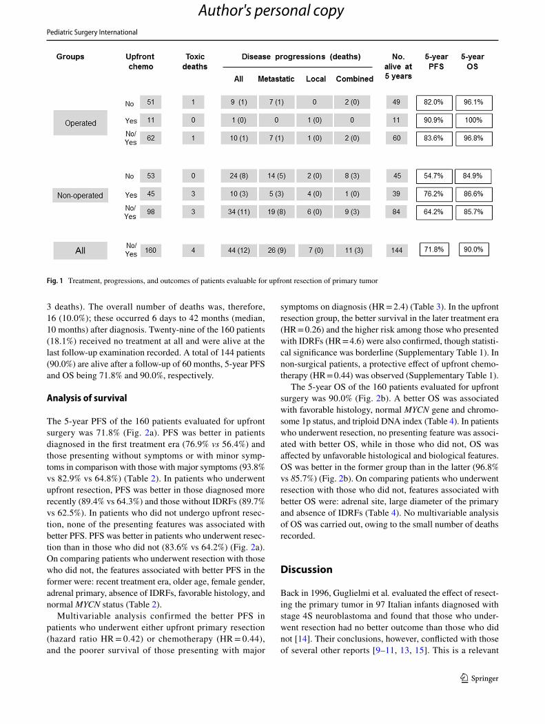
Of the 62 patients who underwent upfront resection, 51 were assigned to observation, one of whom died of bleed-ing 6 days after surgery (Fig. 1). Nine of these 51 suffered disease progression 1–9 months after diagnosis (median 4): metastatic in 7 (one died 19 months after diagnosis) and combined in 2 (both alive); 5-year PFS and OS were, there-fore, 82.0% and 96.1%, respectively. The remaining 11 of the 62 received chemotherapy; one suffered local disease progression at 2½ months and survived; PFS and OS were, therefore, 90.9% and 100%, respectively. Four patients suf-fered surgery-related complications: ischemic renal failure in two, intra-operative tumor rupture in one, and bilateral pleu-ral effusion in one; all survived with appropriate treatment. Two of the 62 patients underwent a second, uncomplicated,
Author's personal copy
Pediatric Surgery International
1 3
operation 2 months after diagnosis, to remove a small tumor residue: both survived.
Of the 98 patients who did not undergo upfront resec-tion, 53 were assigned to observation, 24 (45.3%) of whom suffered disease progression 1–30 months after diagnosis (median 4): metastatic in 14 (5 died), local in 2 (both alive), and combined in 8 (3 died, all with unfavorable biology); PFS and OS were, therefore, 54.7% and 84.9%, respectively. The remaining 45 received chemotherapy, which was com-plicated by toxic death in 3 cases. Ten of the 45 suffered disease progression 1–38 months after diagnosis (median 7):
metastatic in 5 (3 died), local in 4 (no deaths), and combined in 1 (alive); PFS and OS were, therefore, 76.2% and 86.6%, respectively. In one of these 45 patients, a silastic patch was successfully applied to relieve abdominal tension. 11 out of the 98 patients underwent uncomplicated late resection of a residual tumor 3–22 months after diagnosis (median, 5); all but one survived.
In summary, 4 of 160 patients (2.5%) died of therapy-related complications, 44 (27.5%) suffered disease progres-sion, which was only metastatic in 26 (16.3%; 9 deaths), only local in 7 (4.4%; no deaths), and combined in 11 (6.9%;
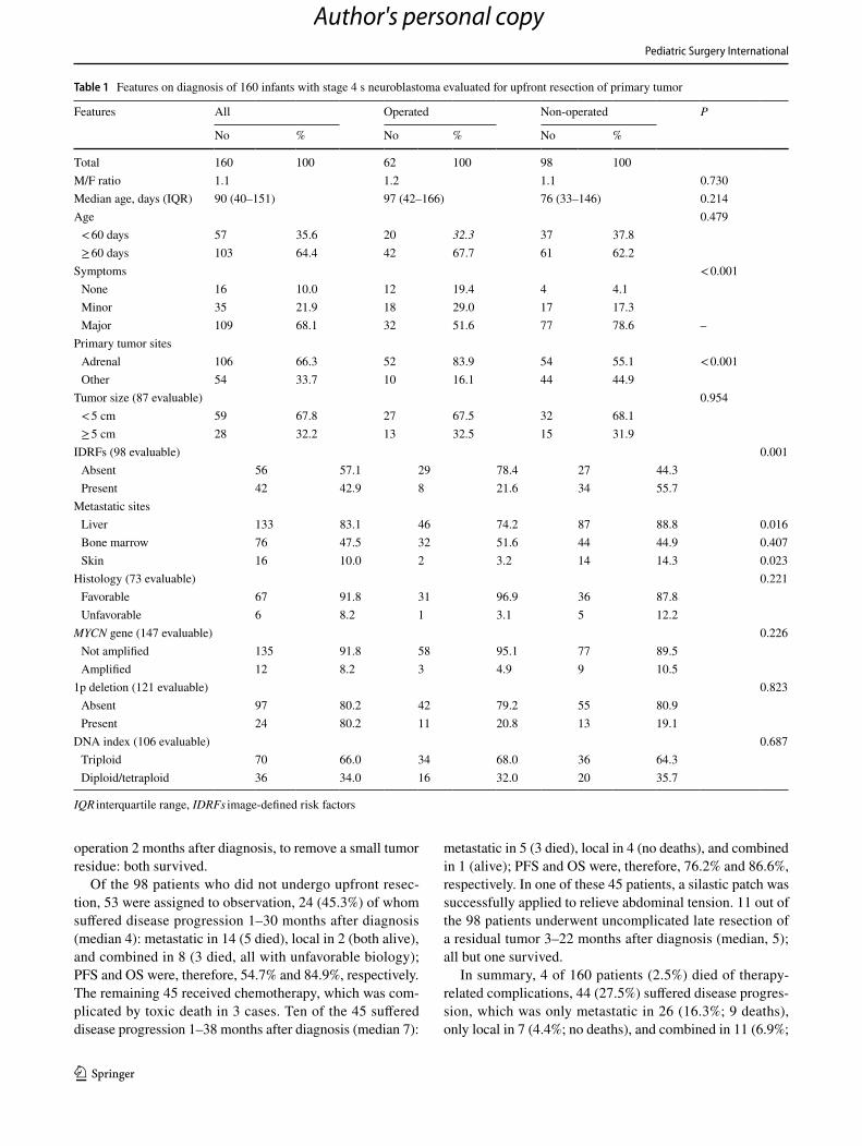
Table 1 Features on diagnosis of 160 infants with stage 4 s neuroblastoma evaluated for upfront resection of primary tumor
IQR interquartile range, IDRFs image-defined risk factors
Features All Operated Non-operated P
No % No % No %
Total 160 100 62 100 98 100M/F ratio 1.1 1.2 1.1 0.730Median age, days (IQR) 90 (40–151) 97 (42–166) 76 (33–146) 0.214Age 0.479 < 60 days 57 35.6 20 32.3 37 37.8 ≥ 60 days 103 64.4 42 67.7 61 62.2
Symptoms < 0.001 None 16 10.0 12 19.4 4 4.1 Minor 35 21.9 18 29.0 17 17.3 Major 109 68.1 32 51.6 77 78.6 –
Primary tumor sites Adrenal 106 66.3 52 83.9 54 55.1 < 0.001 Other 54 33.7 10 16.1 44 44.9
Tumor size (87 evaluable) 0.954 < 5 cm 59 67.8 27 67.5 32 68.1 ≥ 5 cm 28 32.2 13 32.5 15 31.9
IDRFs (98 evaluable) 0.001 Absent 56 57.1 29 78.4 27 44.3 Present 42 42.9 8 21.6 34 55.7
Metastatic sites Liver 133 83.1 46 74.2 87 88.8 0.016 Bone marrow 76 47.5 32 51.6 44 44.9 0.407 Skin 16 10.0 2 3.2 14 14.3 0.023
Histology (73 evaluable) 0.221 Favorable 67 91.8 31 96.9 36 87.8 Unfavorable 6 8.2 1 3.1 5 12.2
MYCN gene (147 evaluable) 0.226 Not amplified 135 91.8 58 95.1 77 89.5 Amplified 12 8.2 3 4.9 9 10.5
1p deletion (121 evaluable) 0.823 Absent 97 80.2 42 79.2 55 80.9 Present 24 80.2 11 20.8 13 19.1
DNA index (106 evaluable) 0.687 Triploid 70 66.0 34 68.0 36 64.3 Diploid/tetraploid 36 34.0 16 32.0 20 35.7
Author's personal copy
Pediatric Surgery International
1 3
3 deaths). The overall number of deaths was, therefore, 16 (10.0%); these occurred 6 days to 42 months (median, 10 months) after diagnosis. Twenty-nine of the 160 patients (18.1%) received no treatment at all and were alive at the last follow-up examination recorded. A total of 144 patients (90.0%) are alive after a follow-up of 60 months, 5-year PFS and OS being 71.8% and 90.0%, respectively.
Analysis of survival
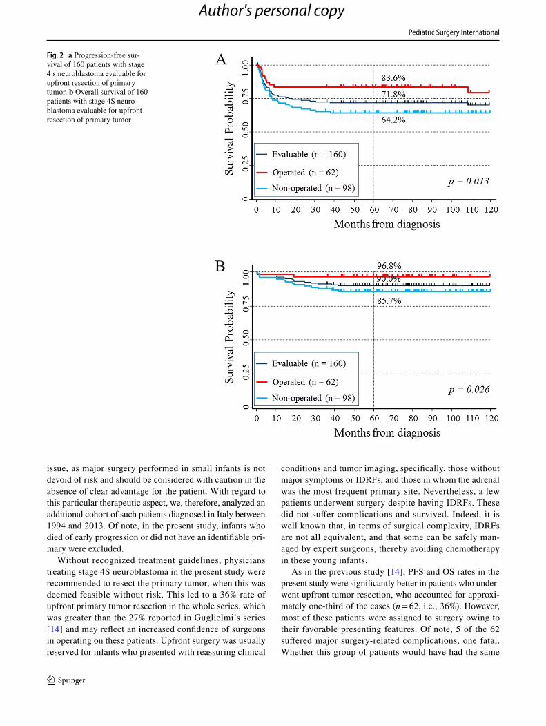
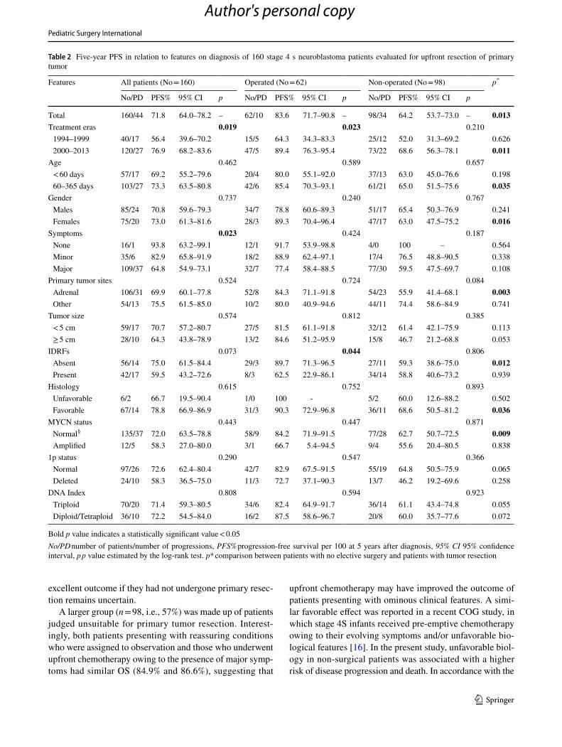
The 5-year PFS of the 160 patients evaluated for upfront surgery was 71.8% (Fig. 2a). PFS was better in patients diagnosed in the first treatment era (76.9% vs 56.4%) and those presenting without symptoms or with minor symp-toms in comparison with those with major symptoms (93.8% vs 82.9% vs 64.8%) (Table 2). In patients who underwent upfront resection, PFS was better in those diagnosed more recently (89.4% vs 64.3%) and those without IDRFs (89.7% vs 62.5%). In patients who did not undergo upfront resec-tion, none of the presenting features was associated with better PFS. PFS was better in patients who underwent resec-tion than in those who did not (83.6% vs 64.2%) (Fig. 2a). On comparing patients who underwent resection with those who did not, the features associated with better PFS in the former were: recent treatment era, older age, female gender, adrenal primary, absence of IDRFs, favorable histology, and normal MYCN status (Table 2).
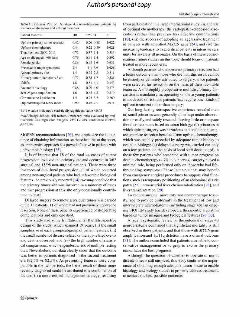
Multivariable analysis confirmed the better PFS in patients who underwent either upfront primary resection (hazard ratio HR = 0.42) or chemotherapy (HR = 0.44), and the poorer survival of those presenting with major
symptoms on diagnosis (HR = 2.4) (Table 3). In the upfront resection group, the better survival in the later treatment era (HR = 0.26) and the higher risk among those who presented with IDRFs (HR = 4.6) were also confirmed, though statisti-cal significance was borderline (Supplementary Table 1). In non-surgical patients, a protective effect of upfront chemo-therapy (HR = 0.44) was observed (Supplementary Table 1).
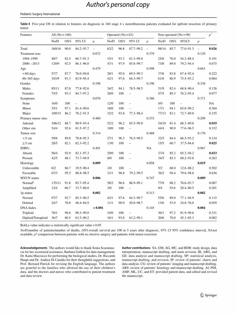
The 5-year OS of the 160 patients evaluated for upfront surgery was 90.0% (Fig. 2b). A better OS was associated with favorable histology, normal MYCN gene and chromo-some 1p status, and triploid DNA index (Table 4). In patients who underwent resection, no presenting feature was associ-ated with better OS, while in those who did not, OS was affected by unfavorable histological and biological features. OS was better in the former group than in the latter (96.8% vs 85.7%) (Fig. 2b). On comparing patients who underwent resection with those who did not, features associated with better OS were: adrenal site, large diameter of the primary and absence of IDRFs (Table 4). No multivariable analysis of OS was carried out, owing to the small number of deaths recorded.
Discussion
Back in 1996, Guglielmi et al. evaluated the effect of resect-ing the primary tumor in 97 Italian infants diagnosed with stage 4S neuroblastoma and found that those who under-went resection had no better outcome than those who did not [14]. Their conclusions, however, conflicted with those of several other reports [9–11, 13, 15]. This is a relevant
Fig. 1 Treatment, progressions, and outcomes of patients evaluable for upfront resection of primary tumor
Author's personal copy
Pediatric Surgery International
1 3
issue, as major surgery performed in small infants is not devoid of risk and should be considered with caution in the absence of clear advantage for the patient. With regard to this particular therapeutic aspect, we, therefore, analyzed an additional cohort of such patients diagnosed in Italy between 1994 and 2013. Of note, in the present study, infants who died of early progression or did not have an identifiable pri-mary were excluded.
Without recognized treatment guidelines, physicians treating stage 4S neuroblastoma in the present study were recommended to resect the primary tumor, when this was deemed feasible without risk. This led to a 36% rate of upfront primary tumor resection in the whole series, which was greater than the 27% reported in Guglielmi’s series [14] and may reflect an increased confidence of surgeons in operating on these patients. Upfront surgery was usually reserved for infants who presented with reassuring clinical
conditions and tumor imaging, specifically, those without major symptoms or IDRFs, and those in whom the adrenal was the most frequent primary site. Nevertheless, a few patients underwent surgery despite having IDRFs. These did not suffer complications and survived. Indeed, it is well known that, in terms of surgical complexity, IDRFs are not all equivalent, and that some can be safely man-aged by expert surgeons, thereby avoiding chemotherapy in these young infants.
As in the previous study [14], PFS and OS rates in the present study were significantly better in patients who under-went upfront tumor resection, who accounted for approxi-mately one-third of the cases (n = 62, i.e., 36%). However, most of these patients were assigned to surgery owing to their favorable presenting features. Of note, 5 of the 62 suffered major surgery-related complications, one fatal. Whether this group of patients would have had the same
Fig. 2 a Progression-free sur-vival of 160 patients with stage 4 s neuroblastoma evaluable for upfront resection of primary tumor. b Overall survival of 160 patients with stage 4S neuro-blastoma evaluable for upfront resection of primary tumor
Author's personal copy
Pediatric Surgery International
1 3
excellent outcome if they had not undergone primary resec-tion remains uncertain.
A larger group (n = 98, i.e., 57%) was made up of patients judged unsuitable for primary tumor resection. Interest-ingly, both patients presenting with reassuring conditions who were assigned to observation and those who underwent upfront chemotherapy owing to the presence of major symp-toms had similar OS (84.9% and 86.6%), suggesting that
upfront chemotherapy may have improved the outcome of patients presenting with ominous clinical features. A simi-lar favorable effect was reported in a recent COG study, in which stage 4S infants received pre-emptive chemotherapy owing to their evolving symptoms and/or unfavorable bio-logical features [16]. In the present study, unfavorable biol-ogy in non-surgical patients was associated with a higher risk of disease progression and death. In accordance with the
Table 2 Five-year PFS in relation to features on diagnosis of 160 stage 4 s neuroblastoma patients evaluated for upfront resection of primary tumor
Bold p value indicates a statistically significant value < 0.05No/PD number of patients/number of progressions, PFS% progression-free survival per 100 at 5 years after diagnosis, 95% CI 95% confidence interval, p p value estimated by the log-rank test. p* comparison between patients with no elective surgery and patients with tumor resection
Features All patients (No = 160) Operated (No = 62) Non-operated (No = 98) p*
No/PD PFS% 95% CI p No/PD PFS% 95% CI p No/PD PFS% 95% CI p
Total 160/44 71.8 64.0–78.2 – 62/10 83.6 71.7–90.8 – 98/34 64.2 53.7–73.0 – 0.013Treatment eras 0.019 0.023 0.210 1994–1999 40/17 56.4 39.6–70.2 15/5 64.3 34.3–83.3 25/12 52.0 31.3–69.2 0.626 2000–2013 120/27 76.9 68.2–83.6 47/5 89.4 76.3–95.4 73/22 68.6 56.3–78.1 0.011
Age 0.462 0.589 0.657 < 60 days 57/17 69.2 55.2–79.6 20/4 80.0 55.1–92.0 37/13 63.0 45.0–76.6 0.198 60–365 days 103/27 73.3 63.5–80.8 42/6 85.4 70.3–93.1 61/21 65.0 51.5–75.6 0.035
Gender 0.737 0.240 0.767 Males 85/24 70.8 59.6–79.3 34/7 78.8 60.6–89.3 51/17 65.4 50.3–76.9 0.241 Females 75/20 73.0 61.3–81.6 28/3 89.3 70.4–96.4 47/17 63.0 47.5–75.2 0.016
Symptoms 0.023 0.424 0.187 None 16/1 93.8 63.2–99.1 12/1 91.7 53.9–98.8 4/0 100 – 0.564 Minor 35/6 82.9 65.8–91.9 18/2 88.9 62.4–97.1 17/4 76.5 48.8–90.5 0.338 Major 109/37 64.8 54.9–73.1 32/7 77.4 58.4–88.5 77/30 59.5 47.5–69.7 0.108
Primary tumor sites 0.524 0.724 0.084 Adrenal 106/31 69.9 60.1–77.8 52/8 84.3 71.1–91.8 54/23 55.9 41.4–68.1 0.003 Other 54/13 75.5 61.5–85.0 10/2 80.0 40.9–94.6 44/11 74.4 58.6–84.9 0.741
Tumor size 0.574 0.812 0.385 < 5 cm 59/17 70.7 57.2–80.7 27/5 81.5 61.1–91.8 32/12 61.4 42.1–75.9 0.113 ≥ 5 cm 28/10 64.3 43.8–78.9 13/2 84.6 51.2–95.9 15/8 46.7 21.2–68.8 0.053
IDRFs 0.073 0.044 0.806 Absent 56/14 75.0 61.5–84.4 29/3 89.7 71.3–96.5 27/11 59.3 38.6–75.0 0.012 Present 42/17 59.5 43.2–72.6 8/3 62.5 22.9–86.1 34/14 58.8 40.6–73.2 0.939
Histology 0.615 0.752 0.893 Unfavorable 6/2 66.7 19.5–90.4 1/0 100 - 5/2 60.0 12.6–88.2 0.502 Favorable 67/14 78.8 66.9–86.9 31/3 90.3 72.9–96.8 36/11 68.6 50.5–81.2 0.036
MYCN status 0.443 0.447 0.871 Normal§ 135/37 72.0 63.5–78.8 58/9 84.2 71.9–91.5 77/28 62.7 50.7–72.5 0.009 Amplified 12/5 58.3 27.0–80.0 3/1 66.7 5.4–94.5 9/4 55.6 20.4–80.5 0.838
1p status 0.290 0.547 0.366 Normal 97/26 72.6 62.4–80.4 42/7 82.9 67.5–91.5 55/19 64.8 50.5–75.9 0.065 Deleted 24/10 58.3 36.5–75.0 11/3 72.7 37.1–90.3 13/7 46.2 19.2–69.6 0.258
DNA Index 0.808 0.594 0.923 Triploid 70/20 71.4 59.3–80.5 34/6 82.4 64.9–91.7 36/14 61.1 43.4–74.8 0.055 Diploid/Tetraploid 36/10 72.2 54.5–84.0 16/2 87.5 58.6–96.7 20/8 60.0 35.7–77.6 0.072
Author's personal copy
Pediatric Surgery International
1 3
SIOPEN recommendations [26], we emphasize the impor-tance of obtaining information on these features at the onset, as an intensive approach has proved effective in patients with unfavorable biology [23].
It is of interest that 18 of the total 44 cases of tumor progression involved the primary site and occurred in 3/62 surgical and 15/98 non-surgical patients. There were three instances of fatal local progression, all of which occurred among non-surgical patients who had unfavorable biological features. As previously reported [14], we may conclude that the primary tumor site was involved in a minority of cases and that progression at this site only occasionally contrib-uted to death.
Delayed surgery to remove a residual tumor was carried out in 13 patients, 11 of whom had not previously undergone resection. None of these patients experienced post-operative complications and only one died.
This study had some limitations: (i) the retrospective design of the study, which spanned 19 years, (ii) the small sample size of each group/subgroup of patient features, (iii) the small number of disease-related or therapy-related events and deaths observed, and (iv) the high number of statisti-cal comparisons, which engenders a risk of multiple testing bias. Nevertheless, our data clearly show that the outcome was better in patients diagnosed in the second treatment era (92.5% vs 82.5%). As presenting features were com-parable in the two periods, the better result of those more recently diagnosed could be attributed to a combination of factors: (i) a more-refined management strategy, resulting
from participation in a large international study, (ii) the use of optimal chemotherapy (the carboplatin–etoposide asso-ciation) rather than previous less effective combinations [10], (iii) the success of adopting an aggressive treatment in patients with amplified MYCN gene [24], and (iv) the increasing tendency to treat critical patients in intensive care units for severely ill neonates. On the basis of these consid-erations, future studies on this topic should focus on patients treated in more recent eras.
Although patients who underwent primary resection had a better outcome than those who did not, this result cannot be entirely or definitely attributed to surgery, since patients were selected for resection on the basis of their favorable features. A thoroughly preoperative multidisciplinary dis-cussion is mandatory, as operating on these young patients is not devoid of risk, and patients may require other kinds of upfront treatment rather than surgery.
Our long-lasting retrospective experience revealed that: (a) small primaries were generally either kept under observa-tion or easily and safely resected, leaving little or no space for other treatments based on tumor biology; (b) primaries in which upfront surgery was hazardous and could not guaran-tee complete resection benefited from upfront chemotherapy, which was usually preceded by adequate tumor biopsy to evaluate biology; (c) delayed surgery was carried out only on a few patients, on the basis of local staff decision; (d) in those few patients who presented with tumor progression despite chemotherapy (4.7% in our series), surgery played a minimal role, being performed only on those who had life-threatening symptoms. These latter patients may benefit from emergency surgical procedures to support vital func-tions, such as temporary positioning of an abdominal silastic patch [27], intra-arterial liver chemoembolization [28], and liver transplantation [29].
To reduce surgical morbidity and chemotherapy toxic-ity, and to provide uniformity in the treatment of low and intermediate neuroblastoma (including stage 4S), an ongo-ing SIOPEN study has developed a therapeutic algorithm based on tumor imaging and biological features [26, 30].
A recent systematic review on the outcome of stage 4S neuroblastoma confirmed that significant mortality is still observed in these patients, and that those with MYCN gene amplification and 1p/11q deletion have a dismal outcome [31]. The authors concluded that patients amenable to con-servative management or surgery to excise the primary tumor have the best prognosis.
Although the question of whether to operate or not at disease onset is still unsolved, this study confirms the impor-tance of obtaining enough adequate tumor tissue to enable histology and biology studies to properly address treatment, to achieve the best possible outcome.
Table 3 Five-year PFS of 160 stage 4 s neuroblastoma patients by features on diagnosis and upfront therapies
Bold p value indicates a statistically significant value < 0.05IDRFs image-defined risk factors, HR hazard ratio evaluated by mul-tivariable Cox regression analysis, 95% CI 95% confidence interval, p p value
Patient features HR 95% CI p
Upfront primary tumor resection 0.42 0.20–0.88 0.022Upfront chemotherapy 0.44 0.22–0.89 0.021Treatment era 2000–2013 0.72 0.37–1.4 0.333Age on diagnosis ≥ 60 days 0.76 0.41–1.4 0.392Female gender 0.86 0.48–1.6 0.631Presence of major symptoms 2.4 1.1–5.6 0.036Adrenal primary site 1.4 0.72–2.8 0.311Primary tumor diameter < 5 cm 0.77 0.35–1.7 0.521IDRFs 1.8 0.81–4.1 0.143Favorable histology 0.88 0.20–4.0 0.873MYCN gene amplification 1.6 0.63–4.3 0.310Chromosome 1p deletion 1.5 0.73–3.2 0.260Diploid/tetraploid DNA index 0.99 0.46–2.1 0.971
Author's personal copy
Pediatric Surgery International
1 3
Acknowledgements The authors would like to thank Sonia Scaramuc-cia for her secretarial assistance, Barbara Galleni for data management, Dr. Katia Mazzocco for performing the biological studies, Dr. Riccardo Haupt and Dr. Andrea Di Cataldo for their thoughtful suggestions, and Prof. Bernard Patrick for revising the English language. The authors are grateful to the families who allowed the use of their children’s data, and the doctors and nurses who contributed to patient treatment and data review.
Author contributions SA, GM, AG, MC, and BDB: study design, data interpretation, manuscript drafting, and main revision. IB, ARG, and GE: data analysis and manuscript drafting. SP: statistical analysis, manuscript drafting, and revision. IP: review of patients’ charts and data analysis. CG: review of patients’ imaging and manuscript drafting. ARS: review of patients’ histology and manuscript drafting. AI, PDI, AMF, ML, UC, and ET: provided patient data, and edited and revised the manuscript;
Table 4 Five-year OS in relation to features on diagnosis in 160 stage 4 s neuroblastoma patients evaluated for upfront resection of primary tumor
Bold p value indicates a statistically significant value < 0.05No/D number of patients/number of deaths, OS% overall survival per 100 at 5 years after diagnosis, 95% CI 95% confidence interval, NA not available, p* comparison between patients with no elective surgery and patients with tumor resection
Features All (No = 160) Operated (No = 62) Non-operated (No = 98) p*
No/D OS% 95% CI p No/D OS% 95% CI p No/D OS% 95%CI p
Total 160/16 90.0 84.2–93.7 – 62/2 96.8 87.7–99.2 – 98/14 85.7 77.0–91.3 – 0.026Treatment eras 0.072 0.379 0.129 1994–1999 40/7 82.5 66.7–91.3 15/1 93.3 61.3–99.0 25/6 76.0 54.2–88.4 0.191 2000—2013 120/9 92.5 86.1–96.0 47/1 97.9 85.8–99.7 73/8 89.0 79.2–94.4 0.075
Age 0.479 0.598 0.663 < 60 days 57/7 87.7 76.0–94.0 20/1 95.0 69.5–99.3 37/6 83.8 67.4–92.4 0.222 60–365 days 103/9 91.3 83.9–95.4 42/1 97.6 84.3–99.7 61/8 86.9 75.5–93.2 0.064
Gender 0.190 0.196 0.338 Males 85/11 87.0 77.8–92.6 34/2 94.1 78.5–98.5 51/9 82.4 68.8–90.4 0.126 Females 75/5 93.3 84.7–97.2 28/0 100 – 47/5 89.3 76.2–95.4 0.077
Symptoms 0.070 0.386 0.371 None 16/0 100 – 12/0 100 – 4/0 100 – NA Minor 35/1 97.1 81.4–99.6 18/0 100 – 17/1 94.1 65.0–99.2 0.304 Major 109/15 86.2 78.2–91.5 32/2 93.8 77.3–98.4 77/13 83.1 72.7–89.8 0.155
Primary tumor sites 0.460 0.533 0.209 Adrenal 106/12 88.7 80.9–93.4 52/2 96.2 85.5–99.0 54/10 81.4 68.3–89.6 0.019 Other site 54/4 92.6 81.5–97.2 10/0 100 – 44/4 90.9 77.6–96.5 0.332
Tumor size 0.314 0.488 0.170 < 5 cm 59/6 89.8 78.8–95.3 27/1 96.3 76.5–99.5 32/5 84.4 66.5–93.2 0.134 ≥ 5 cm 28/5 82.1 62.3–92.2 13/0 100 – 15/5 66.7 37.5–84.6 0.025
IDRFs 0.401 NA 0.967 Absent 56/4 92.9 82.1–97.3 29/0 100 – 27/4 85.2 65.2–94.2 0.033 Present 42/5 88.1 73.7–94.9 8/0 100 – 34/5 85.3 68.2–93.6 0.263
Histology 0.009 0.858 0.019 Unfavorable 6/2 66.7 19.5–90.4 1/0 100 – 5/2 60.0 12.6–88.2 0.502 Favorable 67/3 95.5 86.8–98.5 31/1 96.8 79.2–99.5 36/2 94.4 79.6–98.6 0.636
MYCN status 0.006 0.747 0.009 Normal§ 135/11 91.8 85.7–95.4 58/2 96.6 86.9–99.1 77/9 88.2 78.6–93.7 0.087 Amplified 12/4 66.7 33.7–86.0 3/0 100 – 9/4 55.6 20.4–80.5 0.201
1p status 0.002 0.313 0.002 Normal 97/7 92.7 85.3–96.5 42/1 97.6 84.3–99.7 55/6 89.0 77.1–94.9 0.115 Deleted 24/7 70.8 48.4–84.9 11/1 90.9 50.8–98.7 13/6 53.9 24.8–76.0 0.059
DNA Index < 0.001 0.145 0.004 Triploid 70/1 98.6 90.3–99.8 34/0 100 – 36/1 97.2 81.9–99.6 0.331 Diploid/Tetraploid 36/7 80.5 63.3–90.2 16/1 93.8 63.2–99.1 20/6 70.0 45.1–85.3 0.082
Author's personal copy
Pediatric Surgery International
1 3
Funding This study was supported by Associazione OPEN, Napoli, Italy; Fondo Tumori e Leucemie del Bambino, Genova, Italy; and Fon-dazione Italiana per la Lotta al Neuroblastoma, Genova, Italy. The co-authors GE and ARG were recipients of grants provided by Fondazione Italiana per la Lotta al Neuroblastoma.
Compliance with ethical standards
Conflict of interest The authors declare that they have no conflict of interest.
Ethical approval The RINB structure and protocol were approved by all the ethics committees of each participating center as a retrospective and prospective observational study. The RINB database is located at the secure Italian Inter-University Consortium CINECA headquar-ters in Italy, which is 9001:2015 and 27,001:2013 certified. It can be accessed only by authorized users. This retrospective cohort study was conducted in accordance with the ethical standards of the institutional and national research committees and with the 1964 Helsinki Declara-tion and its later amendments.
Informed consent To be enrolled in the RINB, an informed consent form had to be signed by the patient’s parents or guardians. For this reason, no specific further consent for this retrospective study needed to be sought.
References
1. D’Angio GJ, Evans AE, Koop C (1971) Special pattern of wide-spread neuroblastoma with a favorable prognosis. Lancet 1:1046–1049. https ://doi.org/10.1016/s0140 -6736(71)91606 -0
2. Brodeur GM, Pritchard J, Berthold F et al (1993) Revisions of the international criteria for neuroblastoma diagnosis, staging, and response to treatment. J Clin Oncol 11:1466–1477. https ://doi.org/10.1200/JCO.1993.11.8.1466
3. Brodeur GM (2018) Spontaneous regression of neuroblastoma. Cell Tissue Res. 372:277–286. https ://doi.org/10.1007/s0044 1-017-2761-2
4. Evans AE, Chatten J, D’Angio GJ, Gerson JM, Robinson J, Schnaufer L (1980) A review of 17 IV-S neuroblastoma patients at the Children’s Hospital of Philadelphia. Cancer 45:833–839. https ://doi.org/10.1002/1097-0142(19800 301)45:5%3c833 ::aid-cncr2 82045 0502%3e3.0.co;2-u
5. Stokes SH, Thomas PR, Perez CA, Vietti TJ (1984) Stage IV-S neuroblastoma: results with definitive therapy. Cancer 53:2083–2086. https ://doi.org/10.1002/1097-0142(19840 515)53:10%3c208 3::aid-cncr2 82053 1014%3e3.0.co;2-s
6. De Bernardi B, Pianca C, Boni L, Brisigotti M, Carli M, Bag-nulo S, Corciulo P, Mancini A, De Laurentis C, Di Tullio MT et al (1992) Disseminated neuroblastoma (stage IV and IV-S) in the first year of life. Cancer 70:1625–1633. https ://doi.org/10.1002/1097-0142(19920 915)70:6%3c162 5::aid-cncr2 82070 0631%3e3.0.co;2-6
7. Blatt J, Deutsch M, Wollman MR (1987) Results of therapy in stage IV-s neuroblastoma with massive hepatomegaly. Int J Radiat Oncol Biol Phys 13:1467–1471. https ://doi.org/10.1016/0360-3016(87)90312 -9
8. Katzenstein HM, Bowman LC, Brodeur GM, Thorner PS, Joshi VV, Smith EI, Look AT, Rowe ST, Nash MB, Holbrook T, Alva-rado C, Rao PV, Castleberry RP, Cohn SL (1998) Prognostic significance of age, MYCN oncogene amplification, tumor cell
ploidy, and histology in 110 infants with stage D(S) neuroblas-toma: the Pediatric Oncology Group experience. J Clin Oncol 6:2007–2017. https ://doi.org/10.1200/JCO.1998.16.6.2007
9. Nickerson HJ, Matthay KK, Seeger RC, Brodeur GM, Shimada H, Perez C, Atkinson JB, Selch M, Gerbing RB, Stram DO, Lukens J (2000) Favorable biology and outcome of stage IV-S neuroblastoma with supportive care or minimal therapy: a Chil-dren’s Cancer Group study. J Clin Oncol 18:477–486. https ://doi.org/10.1200/JCO.2000.18.3.477
10. Schleiermacher G, Rubie H, Hartmann O, Bergeron C, Chastagner P, Mechinaud F, Michon J (2003) Neuroblastoma Study Group of the French Society of Paediatric Oncology Treatment of stage 4S neuroblastoma—report of 10 years’ experience of the French Society of Paediatric Oncology (SFOP). Br J Cancer 89:470–476. https ://doi.org/10.1038/sj.bjc.66011 54
11. Taggart DR, London WB, Schmidt ML, DuBois SG, Monclair TF, Nakagawara A, De Bernardi B, Ambros PF, Pearson AD, Cohn SL, Matthay KK (2011) Prognostic value of the stage 4S meta-static pattern and tumor biology in patients with metastatic neu-roblastoma diagnosed between birth and 18 months of age. J Clin Oncol 29:4358–4364. https ://doi.org/10.1200/JCO.2011.35.9570
12. De Bernardi B, Di Cataldo A, Garaventa A, Massirio P, Viscardi E, Podda MG, Castellano A, D’Angelo P, Tirtei E, Melchionda F, Vetrella S, De Leonardis F, D’Ippolito C, Tondo A, Nonnis A, Erminio G, Gigliotti AR, Mazzocco K, Haupt R (2019) Stage 4s neuroblastoma: features, management and outcome of 268 cases from the Italian Neuroblastoma Registry. Ital J Pediatr 45:8–23. https ://doi.org/10.1186/s1305 2-018-0599-1
13. Martinez DA, King DR, Ginn-Pease ME, Haase GM, Wiener ES (1992) Resection of the primary tumor is appropriate for chil-dren with stage IV-S neuroblastoma: an analysis of 37 patients. J Pediatr Surg 27:1016–1020. https ://doi.org/10.1016/0022-3468(92)90549 -m
14. Guglielmi M, De Bernardi B, Rizzo A, Federici S, Boglino C, Siracusa F, Leggio A, Cozzi F, Cecchetto G, Musi L, Bardini T, Fagnani AM, Bartoli GC, Pampaloni A, Rogers D, Conte M, Milanaccio C, Bruzzi P (1996) Resection of primary tumor at diagnosis in stage IV-S neuroblastoma: does it affect the clini-cal course? J Clin Oncol 14:1537–1544. https ://doi.org/10.1200/JCO.1996.14.5.1537
15. Berthold F, Harms D, Lampert F, Niethammer D, Zieschang J (1990) Risk factors in neuroblastoma of infants. Contrib Oncol 41:101–117. https ://doi.org/10.1159/00041 9225
16. Twist CJ, Naranjo A, Schmidt ML, Tenney SC, Cohn SL, Meany HJ, Mattei P, Adkins ES, Shimada H, London WB, Park JR, Matthay KK, Maris JM (2019) Defining risk factors for chemo-therapeutic intervention in infants with stage 4S neuroblastoma: a report from Children’s Oncology Group Study ANBL0531. J Clin Oncol 37:115–124. https ://doi.org/10.1200/JCO.18.00419
17. Haupt R, Garaventa A, Gambini C, Parodi S, Cangemi G, Casale F, Viscardi E, Bianchi M, Prete A, Jenkner A, Luksch R, Di Cat-aldo A, Favre C, D’Angelo P, Zanazzo GA, Arcamone G, Izzi GC, Gigliotti AR, Pastore G, De Bernardi B (2010) lmproved survival of children with neuroblastoma between 1979 and 2005: a report of the Italian Neuroblastoma Registry. J Clin Oncol 28:2331–2338. https ://doi.org/10.1200/JCO.2009.24.8351
18. Hsu LL, Evans AE, D’Angio GJ (1996) Hepatomegaly in neuroblastoma stage 4s: criteria for treatment of the vul-nerable neonate. Med Pediatr Oncol 27:521–528. https ://doi.org/10.1002/(SICI)1096-911X(19961 2)27:6%3c521 ::AID-MPO3%3e3.0.CO;2-N
19. Shimada H, Ambros IM, Dehner LP, Hata J, Joshi VV, Roald B, Stram DO, Gerbing RB, Lukens JN, Matthay KK, Castleberry RP (1999) The International neuroblastoma pathology classifi-cation (the Shimada system). Cancer 86:364–372. https ://doi.
Author's personal copy
Pediatric Surgery International
1 3
org/10.1002/(SICI)1097-0142(19990 715)86:2%3c364 ::AID-CNCR2 1%3e3.0.CO;2-7
20. Scaruffi P, Parodi S, Mazzocco K, Defferrari R, Fontana V, Bonassi S, Tonini GP (2004) Detection of MYCN amplification and chromosome 1p36 loss in neuroblastoma by cDNA micro-array comparative genomic hybridization. Mol Diagn 8:93–100. https ://doi.org/10.1007/BF032 60051
21. Cecchetto G, Mosseri V, De Bernardi B, Helardot P, Monclair T, Costa E, Horcher E, Neuenschwander S, Tomà P, Rizzo A, Michon J, Holmes K (2005) Surgical risk factors in primary surgery for localized neuroblastoma: the LNESG1 Study of the European International Society of Pediatric Oncology Neuroblas-toma Group. J Clin Oncol 23:8483–8489. https ://doi.org/10.1200/JCO.2005.02.4661
22. Monclair T, Brodeur GM, Ambros PF, Brisse HJ, Cecchetto G, Holmes K, Kaneko M, London WB, Matthay KK, Nuchtern JG, von Schweinitz D, Simon T, Cohn SL, Pearson AD, INRG Task Force (2009) The International Neuroblastoma Risk Group (INRG) staging system: an INRG Task Force report. J Clin Oncol 27:298–303. https ://doi.org/10.1200/JCO.2008.16.6876
23. De Bernardi B, Gerrard M, Boni L, Rubie H, Cañete A, Di Cataldo A, Castel V, Forjaz de Lacerda A, Ladenstein R, Ruud E, Brichard B, Couturier J, Ellershaw C, Munzer C, Bruzzi P, Michon J, Pearson AD (2009) Excellent outcome with reduced treatment for infants with disseminated neuroblastoma without MYCN amplification. J Clin Oncol 27:1034–1040. https ://doi.org/10.1200/JCO.2008.17.5877
24. Canete A, Gerrard M, Rubie H, Castel V, Di Cataldo A, Munzer C, Ladenstein R, Brichard B, Bermúdez JD, Couturier J, de Bernardi B, Pearson AJ, Michon J (2009) Poor survival for infants with MYCN-amplified metastatic neuroblastoma despite intensified treatment: the International Society of Paediatric Oncology Euro-pean Neuroblastoma Experience. J Clin Oncol 27:1014–1019. https ://doi.org/10.1200/JCO.2007.14.5839
25. Hosmer DW, Lemeshow S (1999) Applied survival analysis—regression modeling of time to event data. Wiley, New York
26. European Low and Intermediate Neuroblastoma: a SIOPEN Study version 3.0, 30 May 2011 https ://www.docva dis.fr/files /all/JI3RO
1LMf9 PWrlq N9k7X 7w/neuro blast ome_low_inter media te_risk_lines _proto col_versi on_4_2_2_janvi er_2013.pdf.
27. Keene DJ, Minford J, Craigie RJ, Humphrey G, Bruce J (2001) Laparostomy closure in stage 4S neuroblastoma. J Pediatr Surg 46:1–4. https ://doi.org/10.1016/j.jpeds urg.2010.08.064
28. Weintraub M, Bloom AI, Gross E, Revel-Vilk S, Shahroor S, Koplewitz BZ, Freeman AI (2004) Successful treatment of pro-gressive stage 4s hepatic neuroblastoma in a neonate with intra-arterial chemoembolization. Pediatr Blood Cancer 43:148–151. https ://doi.org/10.1002/pbc.20080
29. Holsten T, Schuster T, Grabhorn E, Hero B, Frühwald MC (2017) Liver transplantation as a potentially life-saving measure in neuro-blastoma stage 4S. Pediatr Hematol Oncol 34:17–23. https ://doi.org/10.1080/08880 018.2016.12665 35
30. Schleiermacher G, Michon J, Ribeiro A, Pierron G, Mosseri V, Rubie H, Munzer C, Bénard J, Auger N, Combaret V, Janoueix-Lerosey I, Pearson A, Tweddle DA, Bown N, Gerrard M, Wheeler K, Noguera R, Villamon E, Cañete A, Castel V, Marques B, de Lacerda A, Tonini GP, Mazzocco K, Defferrari R, De Bernardi B, Di Cataldo A, van Roy N, Brichard B, Ladenstein R, Ambros I, Ambros P, Beiske K, Delattre O, Couturier J (2011) Segmental chromosomal alterations lead to a higher risk of relapse in infants with MYCN-non-amplified localized unresectable/disseminated neuroblastoma (a SIOPEN collaborative study). Br J Cancer 107:1418–1422. https ://doi.org/10.1038/bjc.2011.472
31. Raitio A, Rice MJ, Mullassery D, Losty PD (2020) Stage 4S neuroblastoma: what are the outcomes? A systematic review of published studies. Eur J Pediatr Surg. https ://doi.org/10.1055/s-0040-17168 36.10.1055/s-0040-17168 36 (pub-lished online ahead of print, 2020 Sep 15)
Publisher’s Note Springer Nature remains neutral with regard to jurisdictional claims in published maps and institutional affiliations.
Author's personal copy