reproduction of pmws in immunostimulated spf piglets
TRANSCRIPT
HAL Id: hal-00902992https://hal.archives-ouvertes.fr/hal-00902992
Submitted on 1 Jan 2005
HAL is a multi-disciplinary open accessarchive for the deposit and dissemination of sci-entific research documents, whether they are pub-lished or not. The documents may come fromteaching and research institutions in France orabroad, or from public or private research centers.
L’archive ouverte pluridisciplinaire HAL, estdestinée au dépôt et à la diffusion de documentsscientifiques de niveau recherche, publiés ou non,émanant des établissements d’enseignement et derecherche français ou étrangers, des laboratoirespublics ou privés.
Reproduction of PMWS in immunostimulated SPFpiglets transfected with infectious cloned genomic DNA
of type 2 porcine circovirusBéatrice Grasland, Christophe Loizel, Philippe Blanchard, Aurélie Oger,
Anne-Cécile Nignol, Laurent Bigarré, Hervé Morvan, Roland Cariolet, AndréJestin
To cite this version:Béatrice Grasland, Christophe Loizel, Philippe Blanchard, Aurélie Oger, Anne-Cécile Nignol, et al..Reproduction of PMWS in immunostimulated SPF piglets transfected with infectious cloned genomicDNA of type 2 porcine circovirus. Veterinary Research, BioMed Central, 2005, 36 (5-6), pp.685-697.�10.1051/vetres:2005024�. �hal-00902992�
685Vet. Res. 36 (2005) 685–697© INRA, EDP Sciences, 2005DOI: 10.1051/vetres:2005024
Original article
Reproduction of PMWS in immunostimulated SPF piglets transfected with infectious cloned genomic
DNA of type 2 porcine circovirus
Béatrice GRASLANDa*, Christophe LOIZELa, Philippe BLANCHARDa, Aurélie OGERa, Anne-Cécile NIGNOLa, Laurent BIGARRÉa,
Hervé MORVANb, Roland CARIOLETa, André JESTINa
a French agency for food safety (AFSSA), Unit of Viral Genetics and Biosafety, BP 53, 22440 Ploufragan, France
b LDA 22, 05-07 rue du Sabot, BP 54, 22440 Ploufragan, France
(Received 30 September 2004; accepted 3 February 2005)
Abstract – Postweaning multisystemic wasting syndrome (PMWS) is a recently emerged diseaseaffecting pigs. Type 2 porcine circovirus (PCV2) has been associated with this syndrome althoughother factors are required in association with this virus for PMWS expression. The aim of this studywas to investigate whether general immunostimulation (injections of keyhole limpet hemocyaninemulsified in incomplete Freund adjuvant and of thioglycollate medium) could strengthen theseverity of PMWS in six-week-old specific-pathogen-free (SPF) piglets transfected with puretandem-cloned PCV2 DNA by the intramuscular route. Non-immunostimulated piglets transfectedwith the viral clone did not present clinical signs but only mild pathological microlesionscharacteristic of PMWS. These piglets seroconverted and high viral genome loads and infectioustiters were detected in the lymphoid organs at the end of the trial. Mild-to-moderate forms of PMWSwere generally observed in the immunostimulated transfected piglets, as well as one severe form fora piglet (8003) which died. These piglets with mild-to-moderate forms had higher DNA loads thanthe transfected-only animals. Thus, viral replication was enhanced by immunostimulation. This isthe first time that clinical PMWS has been reported in an SPF immunostimulated piglet infectedwith a pure inoculum consisting of tandem-cloned PCV2 DNA. This result confirms that PCV2 isthe agent of PMWS and that immunostimulation could enhance PMWS in SPF piglets transfectedwith a PCV2 DNA clone.
porcine circovirus type 2 / immunostimulation / tandem-cloned DNA / PMWS
1. INTRODUCTION
A new disease of the swine, first describedin Canada in 1991 [10, 17], emerged there-after in several countries across the world[11, 29, 35]. This pathology known as thepostweaning multisystemic wasting syn-
drome (PMWS) [10, 17] particularly affectseight to thirteen-week-old piglets [26]. Today,it still has a major economic impact in pigproducing countries [16].
The characteristic clinical signs includeweight wasting, dyspnea, lymph node enlarge-ment and less frequently pallor, pyrexia and
* Corresponding author: [email protected]
Article published by EDP Sciences and available at http://www.edpsciences.org/vetres or http://dx.doi.org/10.1051/vetres:2005024
686 B. Grasland et al.
diarrhea [18, 34]. The PMWS microlesionsare often located in the lymphoid tissueswhich can present several levels of lym-phocyte depletion with a loss of follicles, aswell as histiocytic infiltration [33]. How-ever, these clinical signs and histopatho-logical lesions must be associated with thepresence of type-2 porcine circovirus (PCV2)in affected tissues for the diagnosis ofPMWS [38]. Indeed, PCV2 was rapidlyassociated with PMWS [2, 12, 28] eventhough initial experimental infections failedto reproduce the full spectrum of clinicalsigns irrespective of the type of PCV2 sus-pension inoculated (homogenates of PCV2infected tissues [5] or virus propagated incell cultures [13, 27]). These studies dem-onstrated that unidentified co-factors werenecessary to trigger the syndrome in PCV2-infected piglets. However, some recent stud-ies have reported reproduction of the com-plete disease in specific-pathogen-free (SPF)piglets [1], and in cesarean-derived colos-trum-deprived piglets [8, 19] but with anon-purified preparation of PCV2. Moreo-ver, the lesions were more acute when thePCV2 inoculates were combined witheither porcine parvovirus (PPV) [3, 21, 22]or porcine reproductive and respiratorysyndrome virus (PRRSV) [19]. The immu-nostimulation also influences the PMWSdevelopment [4, 23, 24, 30].
The effects of co-factors, in a model ofpathogenesis, could be minimized by usinga pure inoculum of PCV2. With this aim,Fenaux et al. [14, 15] investigated the infec-tion ability of a PCV2 molecular DNAclone when injected directly into the liverand inguinal lymph nodes. Only mild patho-logical lesions were observed in four-week-old SPF piglets despite widespread distri-bution of the virus in their organs. As a result,the authors confirmed that although PCV2was the agent associated with PMWS, co-factors are crucial for the development ofsevere clinical PMWS. The infectiousnessof another PCV2 DNA clone was checkedby intramuscular and intraperitoneal injec-tions [32] with similar results [14]. Since noclinical PMWS was reproduced with this
pure PCV2 inoculum, the purpose of ourwork was to evaluate the impact of immu-nostimulation on the PMWS severity levelshowed by six-week-old specific-patho-gen-free piglets after injection of pure tan-dem-cloned PCV2 DNA by the intramus-cular route.
2. MATERIALS AND METHODS
2.1. Animals and experimental design
2.1.1. Animals
The specific-pathogen-free (SPF) pig-lets were free from virus of the African andclassical swine fever, virus of bovine viraldiarrhea, Border disease virus, Aujesky dis-ease virus, parvovirus, porcine reproduc-tive and respiratory syndrome virus, swineinfluenza virus, porcine circovirus type 1and 2, virus of transmissible gastroenteritis,porcine respiratory coronavirus, Mycoplasmahyopneumoniae, Pasteurella multocida,Bordetella bronchiseptica, Actinobacilluspleuropneumonia, Haemophilus parasuis,Streptococcus suis, Treponoma hyodysente-riae [9]. The animals were put in three SPF-growing rooms with air filtration and lowair pressure maintaining a level 3 of biose-curity.
2.1.2. Experimental design
Twenty-six six-week-old SPF pigletswere divided into four groups as shown inTable I. Control piglets (groups 1 and 2)were assigned to the same room but in sep-arate units.
One piglet in group 3 and one in group 4were necropsied before the injections. Eachof the other piglets in these two groups,received 800 µg of tandem-cloned PCV2genomic DNA by intramuscular injectionin four sites (both sides of the neck andboth hips). All animals in groups 2 and 4were immunized with 2 mg of keyhole lim-pet hemocyanin (Sigma-Aldrich, St. Louis,
Immunostimulation of PCV2-transfected pigs 687
MO, USA) emulsified in 4 mL of incom-plete Freund adjuvant (Sigma-Aldrich)(KLH/ICFA) at 4 and 7 days post-transfec-tion (dpt), and with 10 mL of 3% thiogly-collate broth at 4, 7, 11 and 19 dpt. All thereagents injected to the piglets were steri-lized. The KLH/ICFA was administered infour sites by intramuscular (IM) route, likethe DNA, and the thioglycollate broth bythe intraperitoneal (IP) route. One pig ineach group was killed by intravenous boosterof sodium pentobarbital at 12 dpt andanother at 21 dpt in groups 3 and 4. Theexperiment ended on day 32 post-transfec-tion. Rectal temperatures and clinical assess-ments were recorded daily. The pigletswere weighed and blood samples weretaken weekly. At necropsy, tissue samples(tracheobronchial, inguinal, mesenteric, axil-lary and iliac lymph nodes; tonsil, spleen,ileum, lung, liver, thymus and heart) werecollected for laboratory investigations andstored at –80 °C.
2.2. Techniques of virology
2.2.1. Virus isolation
The virus was extracted from tissue sam-ples (31% w/v) with the help of a mechanicalhomogenizer (Bioblock Scientific, Illkirch,France) in ice cooled MEM medium (Gibco,Paisley, UK). The suspensions were centri-
fuged at 2 000 g for 15 min and the super-natants were stored at –80 °C.
2.2.2. Infectious virus titration
PCV2 titration was assessed by an immu-noperoxidase monolayer assay (IPMA)using a previously described protocol [12]with some modifications.
Supernatants of tissue homogenates werefiltered through 0.22 µm and diluted inMEM medium (Gibco) containing penicil-lin/streptomycin (P/S) (Gibco) and 5% fetalcalf serum (FCS) (PAN Biotech GmbH,Aidenbach, Germany). One-hundred micro-liters of each sample were spread over aPK15 mono-layer for 5 h. The cells werethen washed with sterile phosphate bufferedsaline (PBS) (Qbiogene, Illkirch, France)and the PCV2 replication was induced for30 min at 37 °C with 300 mM D-glucosamine(Sigma-Aldrich) in solution in Earle bal-anced salt solution (Sigma-Aldrich). Aftera wash with PBS (Qbiogene), the cells wereincubated in MEM medium with FCS andP/S for 36 h at 37 °C and then fixed with80% cold acetone (Merck) for 10 min at–20 °C. The endogenous peroxidases wereneutralized for 30 min with a solution ofH2O2/methanol (Merck) (1:99).
The fixed cells were then rinsed withPBS and successively incubated, with inter-mediate PBS-0.25% Tween20 wash steps,
Table I. Constitution of the four groups and description of the experimental design.
Group number 1 2 3 4
Number of pigs 5 5 8 8
Trial plan:0 day post-transfection (dpt)
Infectious PCV2 DNA (800 µg/pig) by IM
Infectious PCV2 DNA (800 µg/pig) by IM
4 and 7 dptKLH/ICFA by IM +
Thioglycollate broth by IPKLH/ICFA by IM +
Thioglycollate broth by IP
11 and 19 dpt Thioglycollate broth by IP Thioglycollate broth by IP
The injections of DNA and keyhole limpet hemocyanin emulsified in incomplete Freund adjuvant (KLH/ICFA) were administered by the intramuscular route (IM) and the injections of thioglycollate broth by theintraperitoneal route (IP).
688 B. Grasland et al.
for 1 h at 37 °C with PBS-3% milk, 1 h at37 °C with anti-PCV2 polyclonal serumdiluted 1:300 in PBS-0.05% Tween20-5%milk (PBS-TM) and finally 1 h at 37 °C witha secondary peroxidase-conjugated rabbitanti-swine immunoglobulin (DAKO, Glos-trup, Denmark) diluted 1:150 in PBS-TM.The colour was developed with 3-amino-9-ethyl-carbazole (Serotec, Oxford, UK). ThePCV2 titers were determined using theKaerber method [20] and expressed inTCID50 per gram of tissue.
2.3. Techniques of molecular biology
2.3.1. Inoculum production
2.3.1.1.Production of tandem-cloned PCV2 DNA
The PCV2 genome (GenBank accessionNo. AF201311) was cloned in tandem inpBluescript KS+ (Stratagene, La Jolla, CA,USA) at the SacI site as described by Fenauxet al. [14]. DNA was produced at a concen-tration of 0.8 mg/mL without lipopolysac-charides (0.046 endotoxin unit/µg) byPlasmidFactory (Bielefeld, Germany). Thissolution was diluted to 0.2 mg/mL in endo-toxin-free phosphate buffered saline (PBS)(Sigma-Aldrich) for our experiments.
2.3.1.2. In vitro transfection
Ninety percent-confluent PK15 cells weretransfected with tandem-cloned PCV2 DNAusing Lipofectamine 2000 (Invitrogen,Carlsbad, CA, USA) according to the pro-tocol supplied by the manufacturer. Twelvehours after transfection, the cells were fixedwith 80% cold acetone for 10 min at –20 °Cand PCV2 antigens were assessed by theimmunoperoxidase monolayer assay methoddescribed previously.
2.3.2. Quantification of PCV2 genomes by real-time PCR
Virus DNA was extracted from the tis-sue homogenate supernatants with Qiagen
DNeasy tissue kit (Qiagen, Hilden, Germany)according to the manufacturer’s instructions.The number of PCV2 genome copies wasassessed by a TaqMan real-time PCR [7].Briefly, the PCV2 specific primers5’-GGGAGCAGGGCCAGAATT-3’ (410–427) and 5’-CGCTCTGTGCCCTTT-GAATACT-3’ (473–452) were designedinto the PCV2 ORF2 region (GenBankaccession no. AF201311) and amplifieda 64bp fragment. The TaqMan probe5’-ACCTTAACCTTTCTTATTCTG-3’(430–450) was labeled with the fluorescentreporter dye FAM (6-carboxyfluorescein)at the 5’ end and with the non-fluorescentquencher (NFQ) associated with the minorgroove binder at the 3’ end.
A DNA solution of a plasmid carryinga single copy of the PCV2 genome wasserial diluted and used to generate a stan-dard curve of quantification. The reactionswere performed on an ABI Prism 7000 ther-mocycler (Applied Biosystem, Foster City,CA, USA).
2.4. Detection of PCV2 antigens
The presence of PCV2 antigens in thesamples was detected by immunochemis-try. Fixed tissue sections were deparaffin-ized and rehydrated. The activities of endog-enous peroxidases were neutralized for7 min with hydrogen peroxide (DAKO).The slides were rinsed in PBS (Qbiogene)and incubated for 1 h at 37 °C with 20 µLof rabbit anti-PCV2-coat protein serumdiluted 1:200.
The sections were then treated with goatbiotin-labeled anti-rabbit-immunoglobulinantibody diluted 1:200 for 30 min at 37 °C,then with peroxidase-conjugated avidin(DAKO) for 30 min at room temperatureand finally with diaminobenzidine for10 min at room temperature, with PBSwashes between each step. The slides werethen counterstained with hematoxylin solu-tion, washed and coverslipped.

Immunostimulation of PCV2-transfected pigs 689
2.5. Serology
The PCV2 antibodies in the weekly-col-lected sera were detected and titrated usingan ELISA test based on the recognition ofa recombinant PCV2 capsid protein/GSTfused protein and a GST protein [6]. Thosesamples with an OD ratio higher than 1.5 wereconsidered positive for PCV2 antibodies.
2.6. Gross pathology and histopathology
At necropsy, the macroscopic lesions wereassessed. A fragment of each sampled tis-sue was fixed in formalin (Sigma-Aldrich)for histological examination. The micro-scopic lesions were assessed on haemalum-eosin-safranin stained tissue sections. Thecriteria required to diagnose PMWS werethe commonly-established ones [38].
2.7. Statistical analysis
Relative daily weight gains, PCV2-anti-body responses, genomic loads and infec-tious PCV2 titers were analyzed by the non-parametric U-test of Mann and Whitneyusing the SYSTAT 9 computer softwarepackage (SPSS Inc., Richmond, CA, USA).
3. RESULTS
3.1. In vitro infectivity
The detection of PCV2 antigens by IPMAin 10–15% of the transfected cells con-firmed that our clone was infectious in vitro(data not shown).
3.2. Clinical evaluation
The piglets in the two control groups (non-immunostimulated (1) and immunostimu-lated (2)) remained clinically normal through-out the trial as did the transfected pigletswithout immunostimulation (group 3). How-ever, 2 of the 7 immunostimulated trans-fected piglets (group 4) showed pyrexia(> 40.5 °C) for one day in the second week
after transfection. And one pig (number8003) in this group, presented high rectaltemperatures from day 11 to day 19 post-transfection, associated with prolonged skinpallor. Enlargement of the inguinal lymphnodes (LN) was also detected after palpa-tion of this animal from day 22 post-transfec-tion. But no respiratory distress was noticed.
Compared to the non-immunostimulatedcontrol piglets (group 1), the daily weightgain (DWG) decreased in all the othergroups in the second and third weeks post-transfection (wpt) (Fig. 1A). However growthretardation was only statistically significant(p < 0.05) in the third week for the immu-nostimulated transfected piglets (group 4).In this group, the relative DWG rangedfrom 0.64 to 0.75 in the third wpt for 3 pig-lets (Fig. 1B). In addition, piglet 8003, whichhad suffered a long period of pyrexia, alsoexhibited severe wasting as early as the sec-ond wpt. Its relative DWG was 0.30 in thesecond wpt and 0.04 in the third. This pigletfinally died at 29 dpt.
3.3. Gross and microscopic lesions
No lesions were observed in the controlpiglets throughout the experiment (Figs. 2Aand 2B). The immunostimulated control ani-mals (group 2) displayed minor hypertro-phy only at the draining sites of the thiogly-collate broth injection, namely the inguinaland iliac LN. However, no microlesionswere noted in any of the tissues from thisgroup except the presence of adjuvant lipidvacuoles in the LN.
No gross lesions were apparent in thetransfected animals, with (group 4) or with-out general immunostimulation (group 3),necropsied at 12 and 21 dpt. At 12 dpt, thepiglets of both transfected groups displayeda diffuse interstitial pneumonia with pro-nounced thickening of the alveolar septabut without inflammation of the alveoli.The follicular structure tended to disappearin the LN. At 21 dpt, only moderate inter-stitial pneumonia was still detectable in thepiglets of those groups.

690 B. Grasland et al.
At the end of the trial, the enlargementof at least one LN (the tracheobronchialand/or the axillary LN) was observed in 2of the 5 transfected piglets (group 3) and inall 5 immunostimulated transfected piglets(group 4). Only lung lesions were observedin the transfected piglets (group 3) and werecharacterized as mild-to-moderate intersti-tial pneumonia. At 32 dpt, in immunostim-ulated transfected animals (group 4) apartfrom piglet 8003, examination of the mic-rolesions revealed necrosis of a few hepa-tocytes as well as more pronounced inter-stitial pneumonia around the bronchioles.PCV2 antigens were revealed within thelungs by immunochemistry. According tothe criteria required to diagnose PMWS, 1of these 4 immunostimulated transfectedpiglets showed a mild form of PMWSmeaning moderate microlesions in lungs
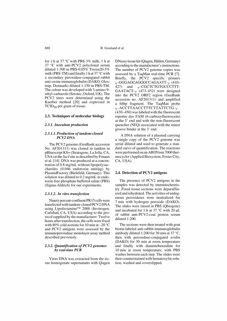
and the presence of PCV2 within the lesionsin tissue. The other piglets presented a mod-erate form with pyrexia and/or decrease ofrelative DWG, moderate microlesions andpresence of PCV2 antigens in the lungs. Asalready mentioned, the last piglet of thisgroup (8003) had well-defined clinical signsand died at 29 dpt. Its thymus was atrophiedand lymphadenopathy of the tracheobron-chial, axillary, inguinal and iliac LN wasdetected. Gastric ulceration was also observed.The kidneys revealed no gross lesions andthus, were not collected for histopatholog-ical examination. Severe lymphocyte deple-tion was noted in all the LN samples and inthe Peyer patches, associated with loss offollicles and histiocytic infiltration (Fig. 3A).PCV2 antigens were detected in the cellcytoplasm and cell nuclei within these tis-sues by immunohistochemistry (Fig. 3B).
Figure 1. (A) Impact of the trans-fection on the relative daily weightgain (rDWG) assessed for eachgroup by comparison of pigletdaily weight gains with those ofcontrol piglets. The asterisk repre-sents a significant difference(p < 0.05) with the control group.(B) Individual relative daily weightgains of the 5 immunostimulatedtransfected piglets that were necrop-sied after the third week post-injec-tion.

Immunostimulation of PCV2-transfected pigs 691
Figure 2. Microscopic sections of organs from a control piglet (A, B) and from the immunostimu-lated transfected pig (number 8003) which died at 29 dpt of PMWS (C, D, E, F). Haemalum-eosin-safranin stain. (A) Normal structure of lungs (×50). (B) Liver with typical portal-zone andwell-defined lobules (×100). (C) Lung with interstitial pneumonia and B.A.L.T. hyperplasia(×50). (D) Inflammation of the alveolar septa (×200). (E) Liver showing inflammatory infiltra-tion of the portal zone and disorganization of the hepatocyte lobular structure (×100). (F) Magni-fication of the inflammatory portal zone (×200) (see www.edpsciences.org/vetres for a colour ver-sion of this figure).
692 B. Grasland et al.
The alveolar septa infiltration with mono-nuclear cells suggested the presence of multi-focal lymphohistiocytic interstitial pneumo-nia (Fig. 2C). Hyperplasia of the Bronchus-associated lymphoid tissue (B.A.L.T.) wasalso observed. The portal zones of the liverpresented lymphocytic inflammation. Theliver plates were disorganized and necrosishad occurred in many hepatocytes (Fig. 2D).According to the lesions and the clinical signs,this piglet suffered from severe PMWS.
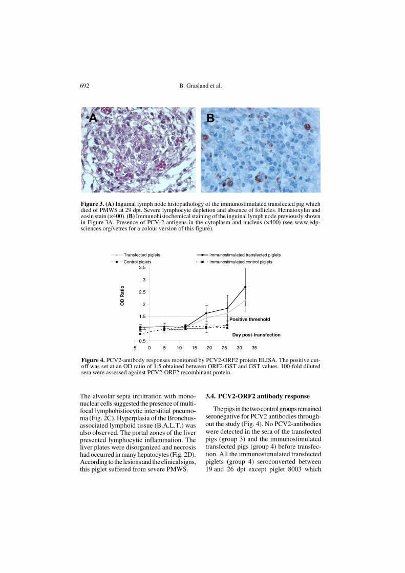
3.4. PCV2-ORF2 antibody response
The pigs in the two control groups remainedseronegative for PCV2 antibodies through-out the study (Fig. 4). No PCV2-antibodieswere detected in the sera of the transfectedpigs (group 3) and the immunostimulatedtransfected pigs (group 4) before transfec-tion. All the immunostimulated transfectedpiglets (group 4) seroconverted between19 and 26 dpt except piglet 8003 which
Figure 3. (A) Inguinal lymph node histopathology of the immunostimulated transfected pig whichdied of PMWS at 29 dpt. Severe lymphocyte depletion and absence of follicles. Hematoxylin andeosin stain (×400). (B) Immunohistochemical staining of the inguinal lymph node previously shownin Figure 3A. Presence of PCV-2 antigens in the cytoplasm and nucleus (×400) (see www.edp-sciences.org/vetres for a colour version of this figure).
Figure 4. PCV2-antibody responses monitored by PCV2-ORF2 protein ELISA. The positive cut-off was set at an OD ratio of 1.5 obtained between ORF2-GST and GST values. 100-fold dilutedsera were assessed against PCV2-ORF2 recombinant protein.
Immunostimulation of PCV2-transfected pigs 693
remained seronegative for 29 dpt. ThePCV2-antibody response increased fromthe second week onwards, the mean of theOD ratios reaching a maximum of 2.7 onday 32 pt for the immunostimulated trans-fected pigs (group 4). In the absence ofimmunostimulation, the complete serocon-version of transfected animals (group 3)was slightly delayed and occurred between19 dpt and the end of the trial, when themean OD ratio was 2.15.
3.5. PCV2 genome load in tissues
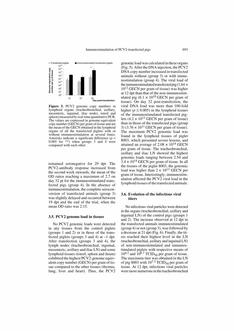
No PCV2 genomic loads were detectedin any tissues from the control piglets(groups 1 and 2) or in those of the trans-fected piglets (groups 3 and 4) at –1 dpt.After transfection (groups 3 and 4), thelymph nodes (tracheobronchial, inguinal,mesenteric, axillary and iliac LN) and somelymphoid tissues (tonsil, spleen and ileum)exhibited the highest PCV2-genome equiv-alent copy number (GECN) per gram of tis-sue compared to the other tissues (thymus,lung, liver and heart). Thus, the PCV2
genomic load was calculated in these organs(Fig. 5). After the DNA injection, the PCV2DNA copy number increased in transfectedanimals without (group 3) or with immu-nostimulation (group 4). The viral load ofthe immunostimulated transfected pig (1.64 ×10¹² GECN per gram of tissue) was higherat 12 dpt than that of the non-immunostim-ulated pig (6.1 × 10¹⁰ GECN per gram oftissue). On day 32 post-transfection, theviral DNA load was more than 100-foldhigher (p ≤ 0.005) in the lymphoid tissuesof the immunostimulated transfected pig-lets (4.2 × 10¹³ GECN per gram of tissue)than in those of the transfected pigs (group3) (3.76 × 10¹¹ GECN per gram of tissue).The maximum PCV2 genomic load wasfound in the lymphoid tissues of piglet8003, which presented severe lesions, andattained an average of 2.08 × 10¹⁴ GECNper gram of tissue. The tracheobronchial,axillary and iliac LN showed the highestgenomic loads ranging between 2.54 and3.4 × 1014 GECN per gram of tissue. In allthe tissues of the piglet 8003, the genomicload was higher than 2 × 10¹³ GECN pergram of tissue. Interestingly, immunostim-ulation affected the PCV2 viral load in thelymphoid tissues of the transfected animals.
3.6. Evolution of the infectious viral titers
No infectious viral particles were detectedin the organs (tracheobronchial, axillary andinguinal LN) of the control pigs (groups 1and 2). The increase observed at 12 dpt inthe transfected animals immunostimulated(group 4) or not (group 3), was followed bya decrease at 21 dpt (Fig. 6). Finally, the tit-ers reached their highest level in the LN(tracheobronchial, axillary and inguinal LN)of non-immunostimulated and immunos-timulated piglets with respective means of104.2 and 106.7 TCID50 per gram of tissue.The maximum titer was obtained in the LNof pig 8003 with 107.3 TCID50 per gram oftissue. At 12 dpt, infectious viral particleswere more numerous in the tracheobronchial
Figure 5. PCV2 genome copy numbers inlymphoid organs (tracheobronchial, axillary,mesenteric, inguinal, iliac nodes, tonsil andspleen) measured by real-time quantitative PCR.The values are expressed in genome equivalentcopy number (GECN) per gram of tissue and arethe mean of the GECN obtained in the lymphoidorgans of all the transfected piglets with orwithout immunostimulation at several times.Asterisks indicate a significant difference (p <0.005 for **) when groups 3 and 4 werecompared with each other.
694 B. Grasland et al.
and axillary LN adjacent to the DNA injec-tion sites than in the inguinal LN (data notshown).
4. DISCUSSION
The aim of this study was to evaluate theimpact of a general immunostimulation onthe pathogenicity of an infectious PCV2molecular DNA clone in six-week-old SPFpiglets.
The piglets transfected with tandem-cloned PCV2 DNA by intramuscular route(group 3) did not present any clinical signs(such as pyrexia and wasting) but rather themild pathological lesions characteristic ofPMWS as previously described [32]. How-ever, the seroconversion profile matchedthat observed in SPF pigs after infectionwith a crude viral isolate [6] thus indicatinga successful production of viral antigensand the consequent response of the immunesystem. A high number of GECN per gramof tissue (3.7 × 10¹¹ GECN per gram) wasfound in the lymphoid organs (tracheobron-chial, inguinal, mesenteric, axillary andiliac LN, tonsil, spleen and ileum) as wellas many infectious particles (104.2 TCID50per gram) at 32 dpt. Significant titers ofinfectious virus were first detected in thetracheobronchial and axillary LN. Hence,the virus replicated efficiently in animals
transfected with the PCV2 genomic cloneand spread subsequently throughout theorganism. A threshold of 1011 GECN pergram of tissue in the lymphoid organs hasbeen suggested for the diagnosis of PMWS[7]. The genomic loads in the lymphoid tis-sues of the non-immunostimulated trans-fected piglets were below or equal to thislevel which would explain the absence ofclinical signs and severe microlesions.
In contrast, mild-to-severe forms ofPMWS were observed in piglets that receiveda systemic immunostimulation in additionto the tandem-cloned PCV2 DNA (group 4)and for the first time, a severe form ofPMWS was reproduced in one of thosepigs. Out of the five piglets remaining at theend of the trial in group 4, one displayed amild form of PMWS while three exhibiteda moderate form. These pigs seroconvertedearlier than the group 3 animals. At the endof the experiment, the genome loads in theirlymphoid tissues were higher than those ofthe transfected-only piglets (group 3). Theinfectious titers in the tracheobronchial,axillary and inguinal LN were similar tothose of group 3. Thus, immunostimulation,consisting of KLH emulsified in ICFA andthioglycollate broth enhanced viral replica-tion in most piglets. The PCV2 genomicloads did not exceed the diagnosis thresholdof 1011 GECN per gram of tissue as sug-gested by Blanchard et al. [7]. This mayexplain why only mild-to moderate formsof PMWS were developed by these piglets.The noticeable exception in this group 4was piglet 8003 which showed severePMWS as shown by (i) growth retardationfrom the second wpt onwards, (ii) pro-longed hyperthermia for nine days and(iii) typical macro- and micro-lesions and(iv) the highest genomic load among all thetransfected animals [37]. High levels ofPCV2 antigens were detected in the lym-phoid organs in which the genomic load andviral titer reached 2.08 × 10¹⁴ GECN and107.3 TCID50 per gram of tissue respec-tively. The suggested threshold of 1011 GECNper gram of tissue was exceeded and clini-cal PMWS was well-characterized in this
Figure 6. Evolution of infectious-PCV2 titers intracheobronchial, axillary and inguinal lymphnodes from transfected piglets immunostimu-lated (in black) or not (in grey). The means areexpressed in TCID50 per gram of tissue.
Immunostimulation of PCV2-transfected pigs 695
piglet. The tests performed at the beginningof the trial to control the presence of severalpathogens in the SPF piglets, the use of ster-ile reagents and pure DNA solution, and thestrict control conditions in our facilities,allowed us to assume that the piglet 8003was only infected by PCV2. However, thispiglet did not seroconvert during the exper-iment. An ELISA test performed on theserum of this piglet revealed that it pos-sessed a normal level of immunoglobulinsat the beginning of the trial (data not shown).Therefore, immunosuppression may haveoccurred in the course of the experimentpossibly due to a rapid decrease in B cellswhich could be confirmed by the massiveloss of lymphoid follicles (B-cell depend-ent areas) in the lymphoid organs. It hadalready been demonstrated that depletion ofthe follicular areas was correlated with thedecrease in the number of circulating B cells[36]. The neutralization of viral particlesby PCV2-specific antibodies could alsoreduce the level of PCV2 antibodies. Indeed,it has already been reported that pigletsshowing severe PMWS and high levels ofgenomic loads have presented low or unde-tectable PCV2 antibody levels [25]. Theimmunostimulation with KLH/ICFA andthioglycollate broth improves PCV2 repli-cation but this impact can be counterbal-anced by the immune response of individ-ual piglets which can reduce the number ofinfectious particles produced.
The KLH associated with incompleteFreund adjuvant stimulates the humoral andcell-mediated immune responses whereasthe thioglycollate medium activates theperitoneal macrophages [23]. The reagentsused in our study mainly triggered the TH2immune response. This specific immuneresponse may have a preponderant role inthe expression of this disease. Other authorshave reported, however, that immunostim-ulations also enhancing the TH2 immuneresponse do not have a critical effect onintensifying PMWS [25, 31]. Resendeset al. failed to reproduce PMWS in conven-tional pigs even with immunostimulation,although they noted that this stimulation
produced a slight increase in viral replica-tion [31]. Ladekjaer-Mikkelsen et al. suc-ceeded in reproducing PMWS in SPF pig-lets, whether immunostimulated or not withKLH emulsified in ICFA. Although thisimmunostimulation was not essential to thedevelopment of PMWS, it apparently influ-enced the immune response to the virus [25]and its impact could differ according to theanimals’ sanitary status and the PCV2 inoc-ulum. Thus despite the conflicting resultsobtained in previous studies, it is apparentfrom our work that the immune stimulationis an important key in the manifestation ofPMWS.
In conclusion, we are reporting for thefirst time the reproduction of clinical PMWSin an SPF immunostimulated piglet infectedwith a pure inoculum of tandem-clonedPCV2 DNA. The severity of the disease isenhanced by other factors such as the immu-nostimulation used in this work which hadan impact on the viral replication. This studyconfirmed that the intramuscular route is anefficient way for in vivo transfection of aninfectious PCV2 DNA clone. However,since the respiratory route has been pro-posed to be the principal route of transmis-sion of this virus, this other way should betested for transfection.
ACKNOWLEDGMENTS
The authors are grateful to Bernard Beaure-paire, Gérard Bennevent, Jean-Claude Rault,André Kéranflec’h and Bruno Jan for the exper-imental work on the animals in the AFSSA facil-ities. We also thank Nadia Amenna for thehistopathology and Isabelle Méderlé-Mangeotfor her helpful discussions.
REFERENCES
[1] Albina E., Truong C., Hutet E., Blanchard P.,Cariolet R., L'Hospitalier R., Mahe D., AlleeC., Morvan H., Amenna N., Le Dimna M.,Madec F., Jestin A., An experimental modelfor post-weaning multisystemic wasting syn-drome (PMWS) in growing piglets, J. Comp.Pathol. 125 (2001) 292–303.
696 B. Grasland et al.
[2] Allan G.M., McNeilly F., Kennedy S., Daft B.,Clarke E.G., Ellis J.A., Haines D.M., MeehanB.M., Adair B.M., Isolation of porcine circo-virus-like viruses from pigs with a wastingdisease in the USA and Europe, J. Vet. Diagn.Invest. 10 (1998) 3–10.
[3] Allan G.M., Kennedy S., McNeilly F., FosterJ.C., Ellis J.A., Krakowka S.J., Meehan B.M.,Adair B.M., Experimental reproduction ofsevere wasting disease by co-infection of pigswith porcine circovirus and porcine parvovi-rus, J. Comp. Pathol. 121 (1999) 1–11.
[4] Allan G.M., McNeilly F., Kennedy S., MeehanB., Ellis J., Krakowka S., Immunostimulation,PCV-2 and PMWS, Vet. Rec. 147 (2000)170–171.
[5] Balasch M., Segales J., Rosell C., DomingoM., Mankertz A., Urniza A., Plana-Duran J.,Experimental inoculation of conventionalpigs with tissue homogenates from pigs withpost-weaning multisystemic wasting syn-drome, J. Comp. Pathol. 121 (1999) 139–148.
[6] Blanchard P., Mahe D., Cariolet R., TruongC., Le Dimna M., Arnauld C., Rose N., EvenoE., Albina E., Madec F., Jestin A., An ORF2protein-based ELISA for porcine circovirustype 2 antibodies in post-weaning multisys-temic wasting syndrome, Vet. Microbiol. 94(2003) 183–194.
[7] Blanchard P., Loizel C., Baudouard M.A.,Nignol A.C., Grasland B., Dory D., MorvanH., Cariolet R., Jestin A., Quantification dugénome du circovirus porcin de type 2 (PCV2)par PCR en temps réel et corrélation avec lamaladie d'amaigrissement du porcelet (MAP),Journ. Rech. Porcine France 36 (2004) 327–332.
[8] Bolin S.R., Stoffregen W.C., Nayar G.P.,Hamel A.L., Postweaning multisystemic wast-ing syndrome induced after experimentalinoculation of cesarean-derived, colostrum-deprived piglets with type 2 porcine circovi-rus, J. Vet. Diagn. Invest. 13 (2001) 185–194.
[9] Cariolet R., Marie P., Moreau G., Robert H.,Summary of the different methods for produc-ing, maintaining and benefiting from pigletsof high health status, J. Rech. Porcine France26 (1994) 1–12.
[10] Clark E., Post-weaning multisystemic wast-ing syndrome, American Association of SwinePractitioners, 1997, pp. 499–501.
[11] Daft B., Nordhausen R.W., Latimer K.S., NiagroF.D., Interstitial pneumonia and lymphaden-opathy associated with circoviral infection in
a six-week-old pig, Proc. Am. Assoc. Vet.Lab. Diagn. 39 (1996) 32.
[12] Ellis J., Hassard L., Clark E., Harding J., AllanG., Willson P., Strokappe J., Martin K.,McNeilly F., Meehan B., Todd D., Haines D.,Isolation of circovirus from lesions of pigswith postweaning multisystemic wasting syn-drome, Can. Vet. J. 39 (1998) 44–51.
[13] Ellis J., Krakowka S., Lairmore M., HainesD., Bratanich A., Clark E., Allan G., KonobyC., Hassard L., Meehan B., Martin K., HardingJ., Kennedy S., McNeilly F., Reproduction oflesions of postweaning multisystemic wastingsyndrome in gnotobiotic piglets, J. Vet.Diagn. Invest. 11 (1999) 3–14.
[14] Fenaux M., Halbur P.G., Haqshenas G., RoyerR., Thomas P., Nawagitgul P., Gill M., TothT.E., Meng X.J., Cloned genomic DNA oftype 2 porcine circovirus is infectious wheninjected directly into the liver and lymphnodes of pigs: characterization of clinical dis-ease, virus distribution, and pathologiclesions, J. Virol. 76 (2002) 541–551.
[15] Fenaux M., Opriessnig T., Halbur P.G., MengX.J., Immunogenicity and pathogenicity ofchimeric infectious DNA clones of patho-genic porcine circovirus type 2 (PCV2) andnonpathogenic PCV1 in weanling pigs, J.Virol. 77 (2003) 11232–11243.
[16] Hardge T., Gausman H., Hasberg W., LangeS., The economic impact of PMWS in thenursery, Intern. Pig Topics 18 (2003) 11–12.
[17] Harding J., Post-weaning multisystemic wast-ing syndrome (PMWS): preliminary epidemi-ology and clinical presentation, AmericanAssociation of Swine Practitioners, 1997,p. 503.
[18] Harding J., Clark E., Recognizing and diag-nosing postweaning multisystemic wastingsyndrome (PMWS), Swine Health Prod. 5(1997) 201–203.
[19] Harms P.A., Sorden S.D., Halbur P.G., BolinS.R., Lager K.M., Morozov I., Paul P.S.,Experimental reproduction of severe diseasein CD/CD pigs concurrently infected withtype 2 porcine circovirus and porcine repro-ductive and respiratory syndrome virus, Vet.Pathol. 38 (2001) 528–539.
[20] Kaerber G., Beitrag zur kollectiven behandlungpharmakologischer. Reihenversuche, Arch.Pharmakol. Exp. Pathol. 162 (1931) 480–483.
[21] Kennedy S., Moffett D., McNeilly F., MeehanB., Ellis J., Krakowka S., Allan G.M., Repro-duction of lesions of postweaning multisys-temic wasting syndrome by infection of
Immunostimulation of PCV2-transfected pigs 697
conventional pigs with porcine circovirustype 2 alone or in combination with porcineparvovirus, J. Comp. Pathol. 122 (2000) 9–24.
[22]Krakowka S., Ellis J.A., Meehan B., KennedyS., McNeilly F., Allan G., Viral wasting syn-drome of swine: experimental reproduction ofpostweaning multisystemic wasting syn-drome in gnotobiotic swine by coinfectionwith porcine circovirus 2 and porcine parvo-virus, Vet. Pathol. 37 (2000) 254–263.
[23] Krakowka S., Ellis J.A., McNeilly F., RinglerS., Rings D.M., Allan G., Activation of theimmune system is the pivotal event in the pro-duction of wasting disease in pigs infectedwith porcine circovirus-2 (PCV-2), Vet.Pathol. 38 (2001) 31–42.
[24] Kyriakis S.C., Saoulidis K., Lekkas S., MiliotisC.C., Papoutsis P.A., Kennedy S., The effectsof immuno-modulation on the clinical andpathological expression of postweaning mul-tisystemic wasting syndrome, J. Comp.Pathol. 126 (2002) 38–46.
[25] Ladekjaer-Mikkelsen A.S., Nielsen J., StadejekT., Storgaard T., Krakowka S., Ellis J.,McNeilly F., Allan G., Botner A., Reproduc-tion of postweaning multisystemic wastingsyndrome (PMWS) in immunostimulated andnon-immunostimulated 3-week-old pigletsexperimentally infected with porcine circovi-rus type 2 (PCV2), Vet. Microbiol. 89 (2002)97–114.
[26] Madec F., Eveno E., Morvan P., Hamon L.,Blanchard P., Cariolet R., Amenna N., MorvanH., Truong C., Mahe D., Albina E., Jestin A.,Post-weaning multisystemic wasting syndrome(PMWS) in pigs in France: clinical observa-tions from follow-up studies on affectedfarms, Livest. Prod. Sci. 63 (2000) 223.
[27] Magar R., Larochelle R., Thibault S., Lamon-tagne L., Experimental transmission of por-cine circovirus type 2 (PCV2) in weaned pigs:a sequential study, J. Comp. Pathol. 123(2000) 258–269.
[28] Meehan B.M., McNeilly F., Todd D.,Kennedy S., Jewhurst V.A., Ellis J.A., HassardL.E., Clark E.G., Haines D.M., Allan G.M.,Characterization of novel circovirus DNAsassociated with wasting syndromes in pigs, J.Gen. Virol. 79 ( Pt 9) (1998) 2171–2179.
[29] Onuki A., Abe K., Togashi K., Kawashima K.,Taneichi A., Tsunemitsu H., Detection of por-cine circovirus from lesions of a pig with wast-ing disease in Japan, J. Vet. Med. Sci. 61(1999) 1119–1123.
[30] Opriessnig T., Yu S., Gallup J.M., Evans R.B.,Fenaux M., Pallares F., Thacker E.L., BrockusC.W., Ackermann M.R., Thomas P., MengX.J., Halbur P.G., Effect of vaccination withselective bacterins on conventional pigsinfected with type 2 porcine circovirus, Vet.Pathol. 40 (2003) 521–529.
[31] Resendes A., Segales J., Balasch M., CalsamigliaM., Sibila M., Ellerbrok H., Mateu E., Plana-Duran J., Mankertz A., Domingo M., Lack ofan effect of a commercial vaccine adjuvant onthe development of postweaning multisys-temic wasting syndrome (PMWS) in porcinecircovirus type 2 (PCV2) experimentallyinfected conventional pigs, Vet. Res. 35(2004) 83–90.
[32] Roca M., Balasch M., Segales J., CalsamigliaM., Viaplana E., Urniza A., Hattermann K.,Mankertz A., Plana-Duran J., Domingo M., Invitro and in vivo characterization of an infec-tious clone of a European strain of porcine cir-covirus type 2, J. Gen. Virol. 85 (2004) 1259–1266.
[33] Rosell C., Segales J., Plana-Duran J., BalaschM., Rodriguez-Arrioja G.M., Kennedy S., AllanG.M., McNeilly F., Latimer K.S., DomingoM., Pathological, immunohistochemical, andin-situ hybridization studies of natural casesof postweaning multisystemic wasting syn-drome (PMWS) in pigs, J. Comp. Pathol. 120(1999) 59–78.
[34]Segales J., Domingo M., Postweaning multi-systemic wasting syndrome (PMWS) in pigs.A review, Vet. Q. 24 (2002) 109–124.
[35] Segales J., Sitjar M., Domingo M., Dee S., DelPozo M., Noval R., Sacristan C., De las HerasA., Ferro A., Latimer K.S., First report of post-weaning multisystemic wasting syndrome inpigs in Spain, Vet. Rec. 141 (1997) 600–601.
[36] Segales J., Alonso F., Rosell C., Pastor J.,Chianini F., Campos E., Lopez-Fuertes L.,Quintana J., Rodriguez-Arrioja G., CalsamigliaM., Pujols J., Dominguez J., Domingo M.,Changes in peripheral blood leukocyte popu-lations in pigs with natural postweaning mul-tisystemic wasting syndrome (PMWS), Vet.Immunol. Immunopathol. 81 (2001) 37–44.
[37] Segales J., Domingo M., Chianini F., Majo N.,Dominguez J., Darwich L., Mateu E., Immu-nosuppression in postweaning multisystemicwasting syndrome affected pigs, Vet. Micro-biol. 98 (2004) 151–158.
[38]Sorden S., Update on porcine circovirus andpost-weaning multisystemic wasting syndrome(PMWS), Swine Health Prod. 8 (2000) 133–136.