reproduction in fungi week 6
TRANSCRIPT

Reproduction in FungiWeek 6
Fungi, have the ability of asexual (sporulation) and sexual reproduction.
If myceliums get mature and stored enough food or environmental conditions becomesuitable for sporulation, the hyphae (generally aerial hyphae) will develop spores indifferent shapes.
When spores get matured they leave the hyphae and become independent. After that theydevelop new fungi by germination in optimal environment and conditions
Fungal spores have different shapes and structures
This feature is used for the fungal classification
The fungal spores are very resistant to environmental conditions. Because of this they canlive for many years and reside infective
There can be found one or more nucleus placed in a spore, at the end of sexual or asexual reproduction.
Around the spore a thick spore protection coat (epispore) and under this coat an endospore that surrounds the protoplasm is found
In some fungal spores there is an extra coat (perispore) can be found that surrounds the spore exterior
In a spore cytoplasm nucleus, vacuoles, lipid granules and enough organic and inorganic compounds are found to build new fungi
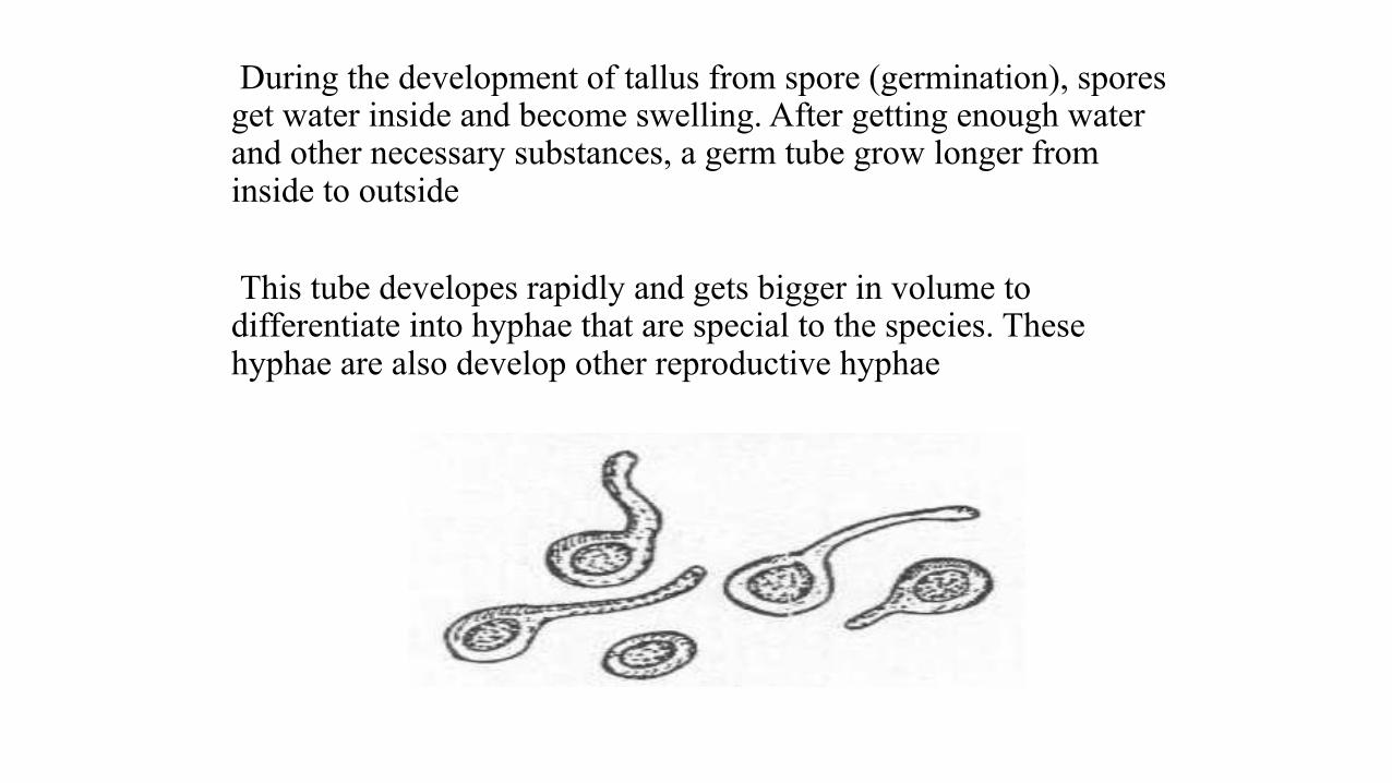
During the development of tallus from spore (germination), spores get water inside and become swelling. After getting enough water and other necessary substances, a germ tube grow longer from inside to outside
This tube developes rapidly and gets bigger in volume to differentiate into hyphae that are special to the species. These hyphae are also develop other reproductive hyphae
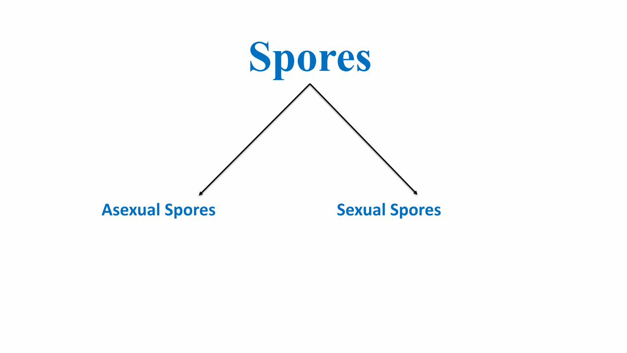
Asexual Spores Sexual Spores
Spores
Asexual Spores
Asexual spores, occurs from single pareteral fungus and thus they will become identic to parenteral fungus genetically
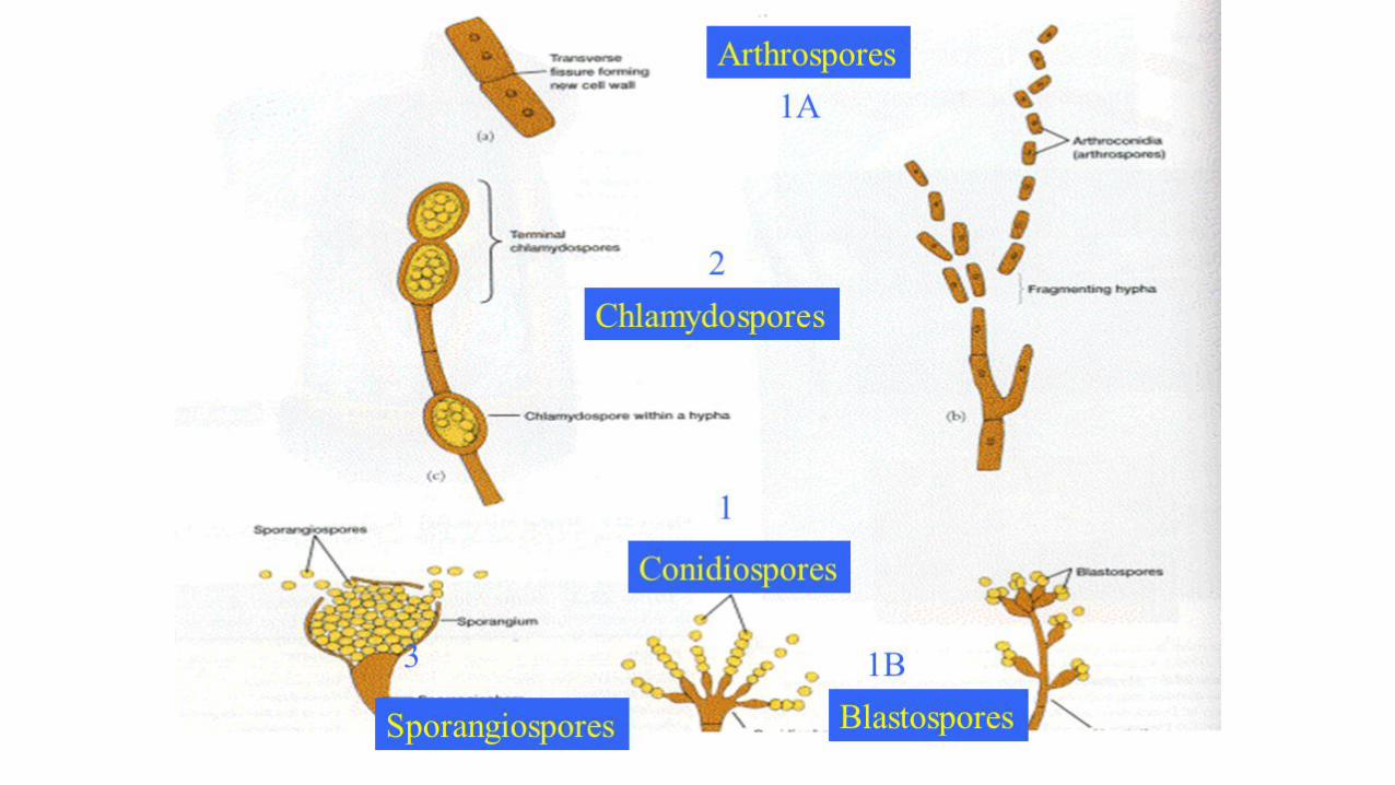
Arthrospore
Blastospore
Clamidospore
Conidiospore
Sporangiospore
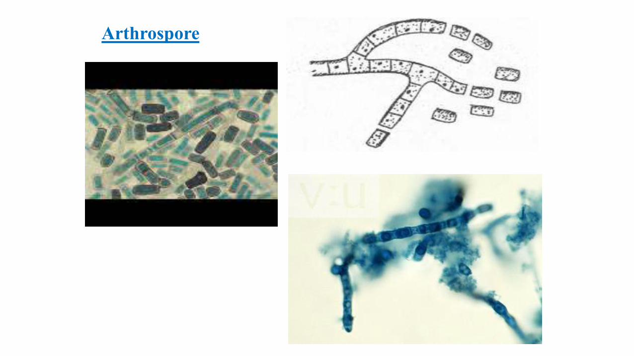
Arthrospore
• No major change is observed in shape of hypha while developing Arthrospore
• Only reproductive hyphae are seperated by septumes (fragmentation)
• Arthrospores which are oval or cylindrical shaped leave the hyphae and become independent. In optimal environmental conditions they germinate and each of them turn into new fungus
• In dermatophytes arthrospores can be observed mostly on skin and hairs, they are not observed on fungal cultures
Arthrospore
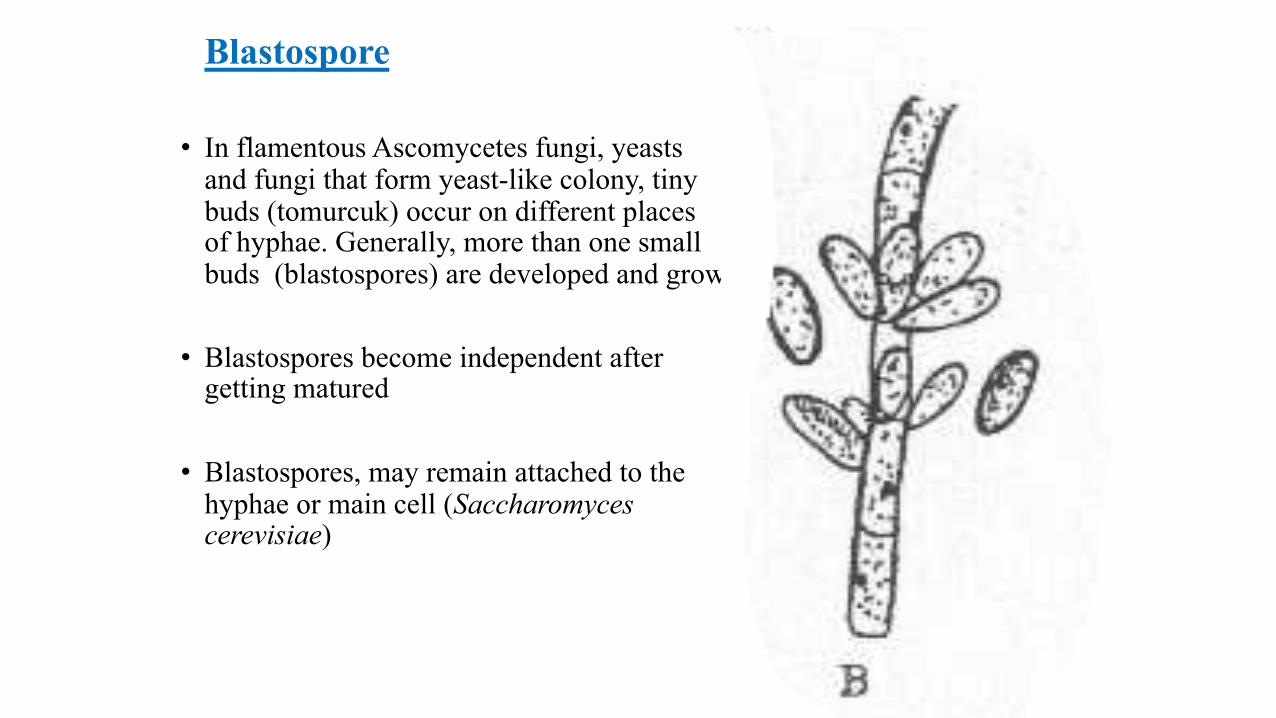
Blastospore
• In flamentous Ascomycetes fungi, yeasts and fungi that form yeast-like colony, tiny buds (tomurcuk) occur on different places of hyphae. Generally, more than one small buds (blastospores) are developed and grow
• Blastospores become independent after getting matured
• Blastospores, may remain attached to the hyphae or main cell (Saccharomyces cerevisiae)
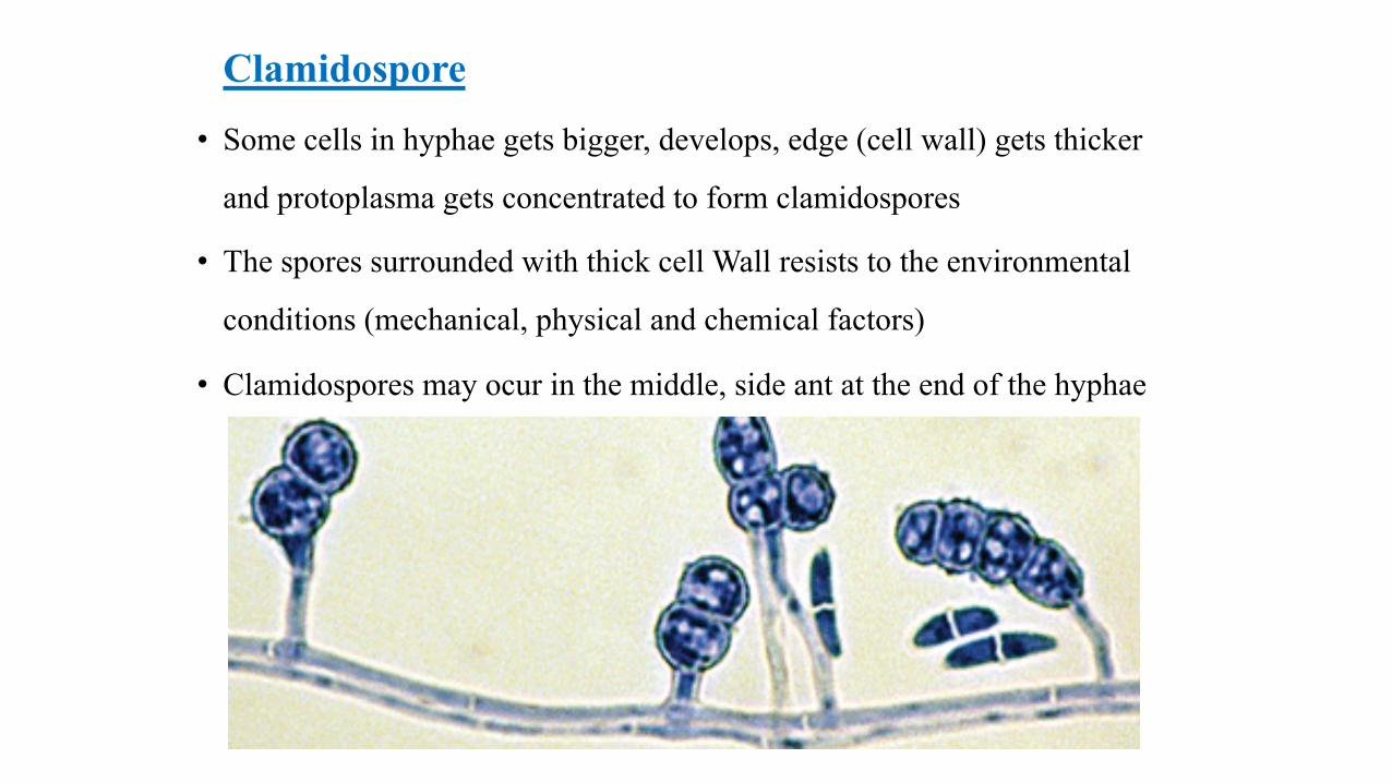
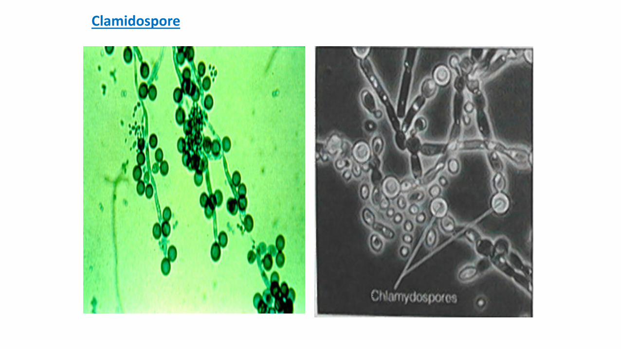
Clamidospore
• Some cells in hyphae gets bigger, develops, edge (cell wall) gets thicker
and protoplasma gets concentrated to form clamidospores
• The spores surrounded with thick cell Wall resists to the environmental
conditions (mechanical, physical and chemical factors)
• Clamidospores may ocur in the middle, side ant at the end of the hyphae
Clamidospore
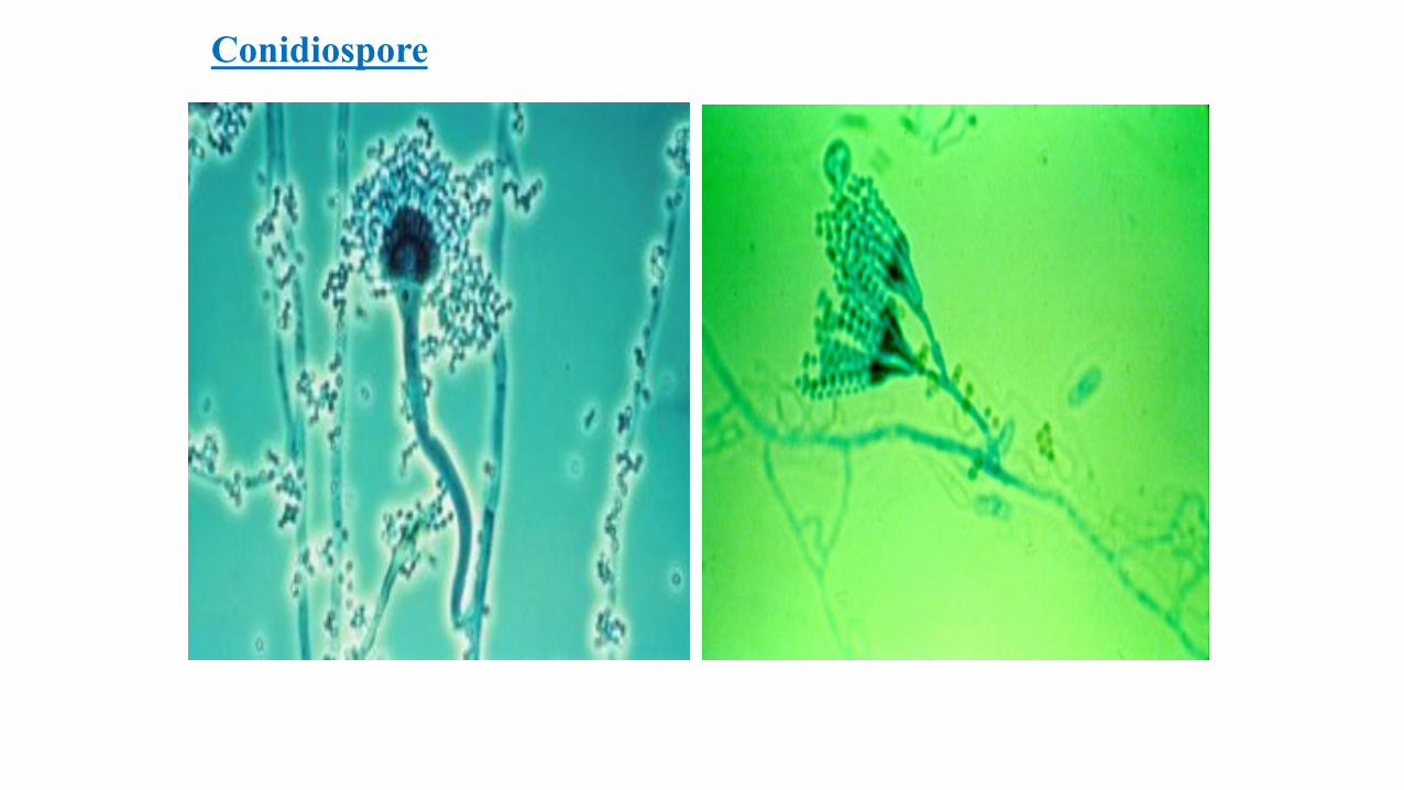
Conidiospore
• Flamentous Ascomycetes and several Deuteromycetes (Fungi imperfecti) fungi exhibites
• Spores (conidia), arise beside and at the end of the special reproductive hyphae (conidiophore)
• These hypahe, occur by the modificaiton and differntiation of aerial hyphae
• Some spores occur directly on fertile hyphae
• Some other spores may occur on a short sterigmata (Sporotrichum)
Conidiospore
• In Dermatophytes (Microsporum, Trichopyton ) 2 different conidium found on the same hyphae
• Single cell types are localised on different places of hyphae and small, oval, spherical shaped (microconidium)
• Multicellular cell types are big spores which are also lemon or puro shaped (makrokonidium) and these spores are seperated into more than one cell by septums (Microsporum canis)
• Conidiums are spherical, round, shuttle or bottle like shaped, big or small sized spores
• The morphological characteristics like the size, shape, arrangement and other features of conidiospores are used to discriminate fungal species (Aspergillus, Penicillium ve Hormodendrum )
Conidiospore
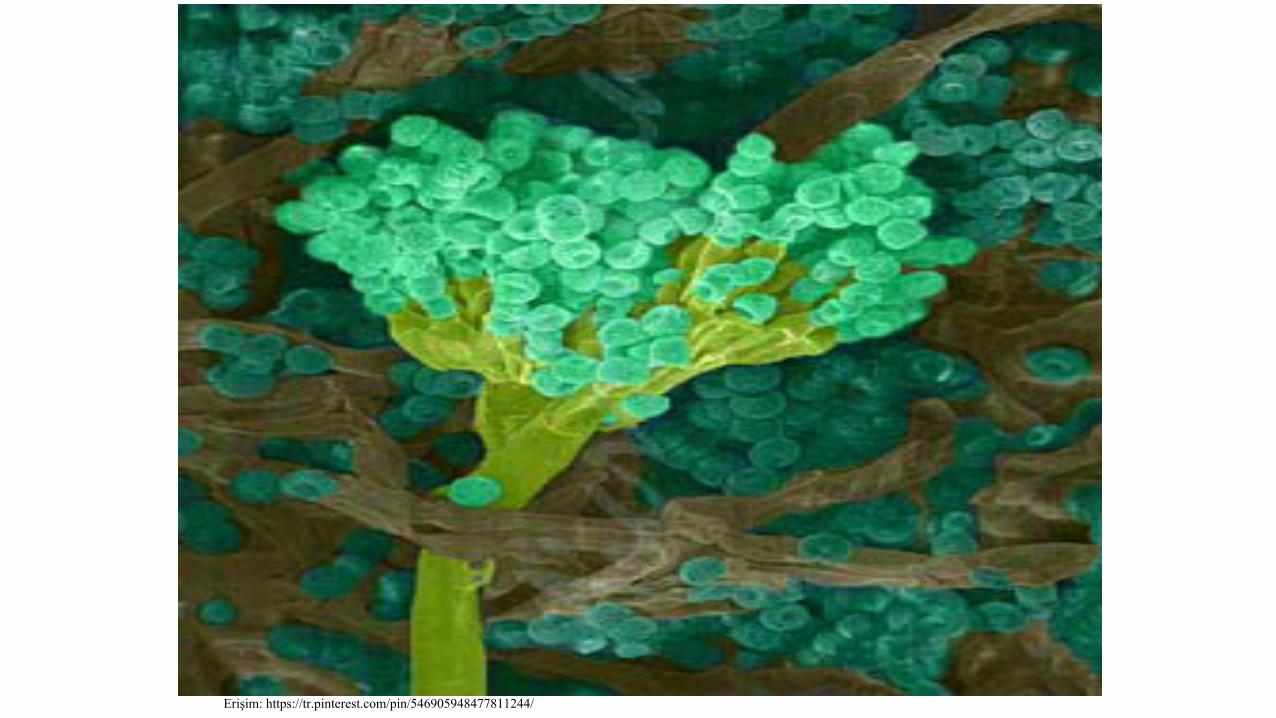
Erişim: https://tr.pinterest.com/pin/546905948477811244/
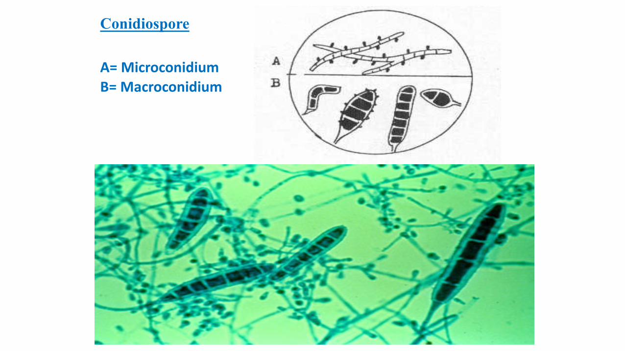
Conidiospore
A= MicroconidiumB= Macroconidium
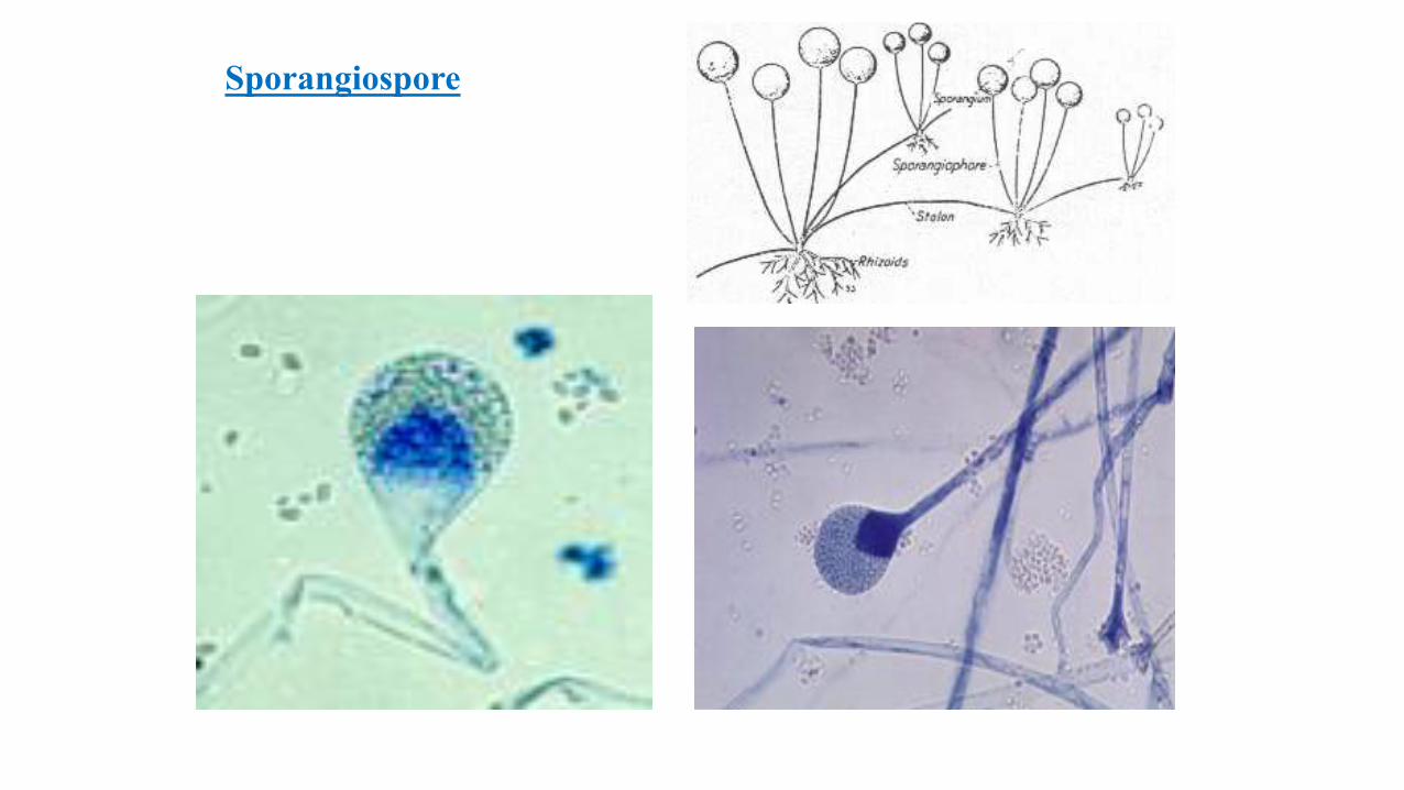
Sporangiospore• Observed in Phycomycetes fungi
• These spores (sporangiospore) are localised at the end of the special hyphae (sporangiophore) which are big and spherical shaped sacs (kese) (sporangium). These small, dehidrated and thick edged spores are located in these sporangiums
• Columella is found at the bottom of sporangiums in order to support
• When these sporangiums are burst spores come out. Optimal environmental conditions lead to the germination of these spores in order to form their own fungal species
• Rhizopus nigricans
Sporangiospore
Sexual Spores In sexual reproduction, sexual spores arise from the fusion of
two distinct sex’s germ cells
There are 4 stages in sexual reproduction1) Developing of gamet or sexual organs of cells2) The fusion of these organs (plasmogami), rapidly or soon nuclear fusion (karyogami) 3) Meiosis in haploid fungi 4) Occurrence and developing of spores

Sexual Spores
• Ascospores
• Basidospores
• Oospores
• Zygospores
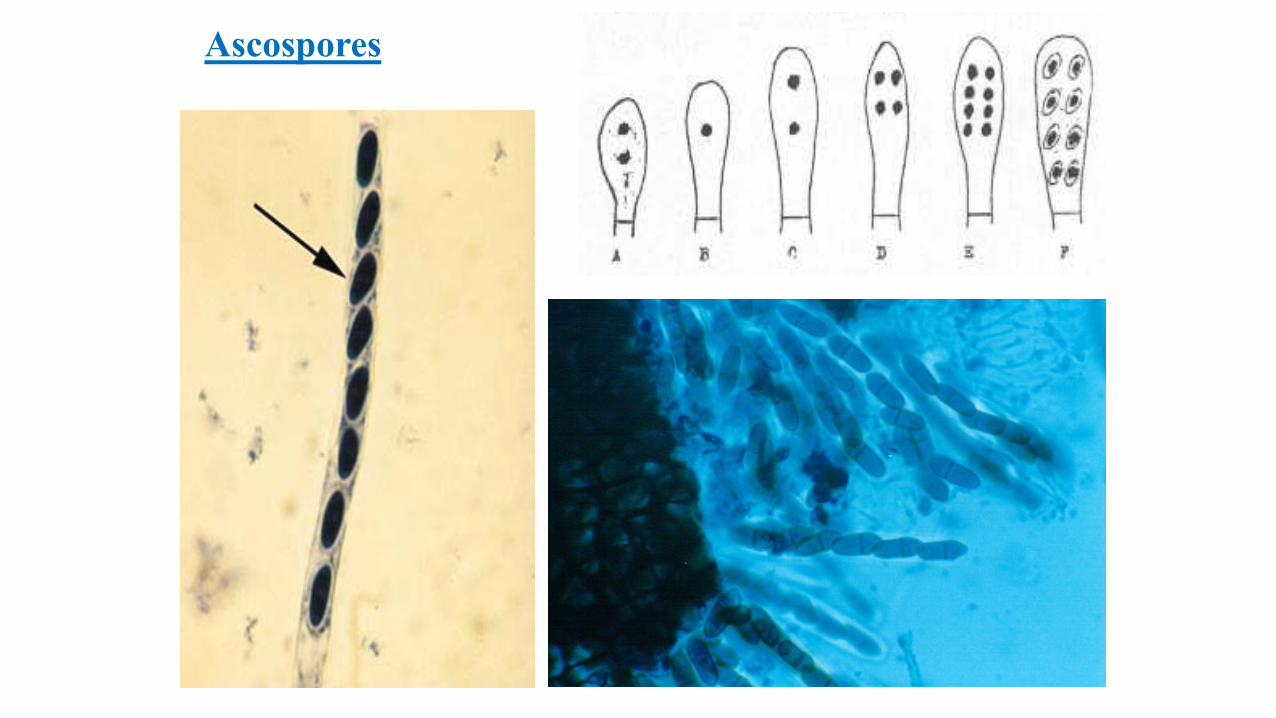
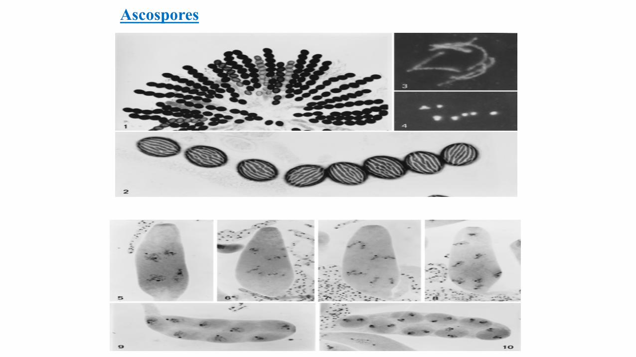
Ascospores• In Ascomycetes fungi sexual spores are prodeuced in enlarged and elongated
cell sacs called ascus
• On the same or distinct hyphae, 2 neighbour cells (ascogonium and anteridium) elongated each other and fused to form ascospores
• First 2 cell membranes melt and get lost
• Then anteridial nucleus goes into ascogonium and new cell become 2 nucleated
• After nucleuses combined, the division of meiosis starts
• After 2 or more divisions nucleuses surrounded by thick protection coat
• Thus 4 or 8 haploid ascospores will occur
• When spores get matured the sac will be teared and spores get out
Ascospores
Ascospores
Basidiosporlar
• These sexual spores are found in Basidiomycetes
• With the enlargement of Basidiums and occurrence of basidiospores they will look
like ascospores
• First the neighbour cells elongated to each other and the membrane get lost
• Then, the nucleus of one cell goes into other’s and become single nucleated
• The single nucleus continues meiosis division and seperated into 4 haploid nucleus
• At the end of Basidiums, for each cell a sterigmata (basidium) will occur and each
nucleuses goes into its own sterigmata to form basidiospores
• Spores leave and spread to the environment
• They form new fungi by germination at the optimum conditions
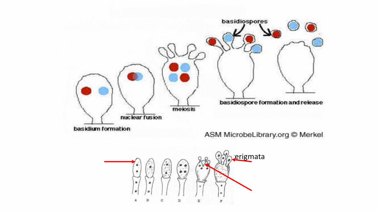
Basidium Basidiospore
Sterigmata
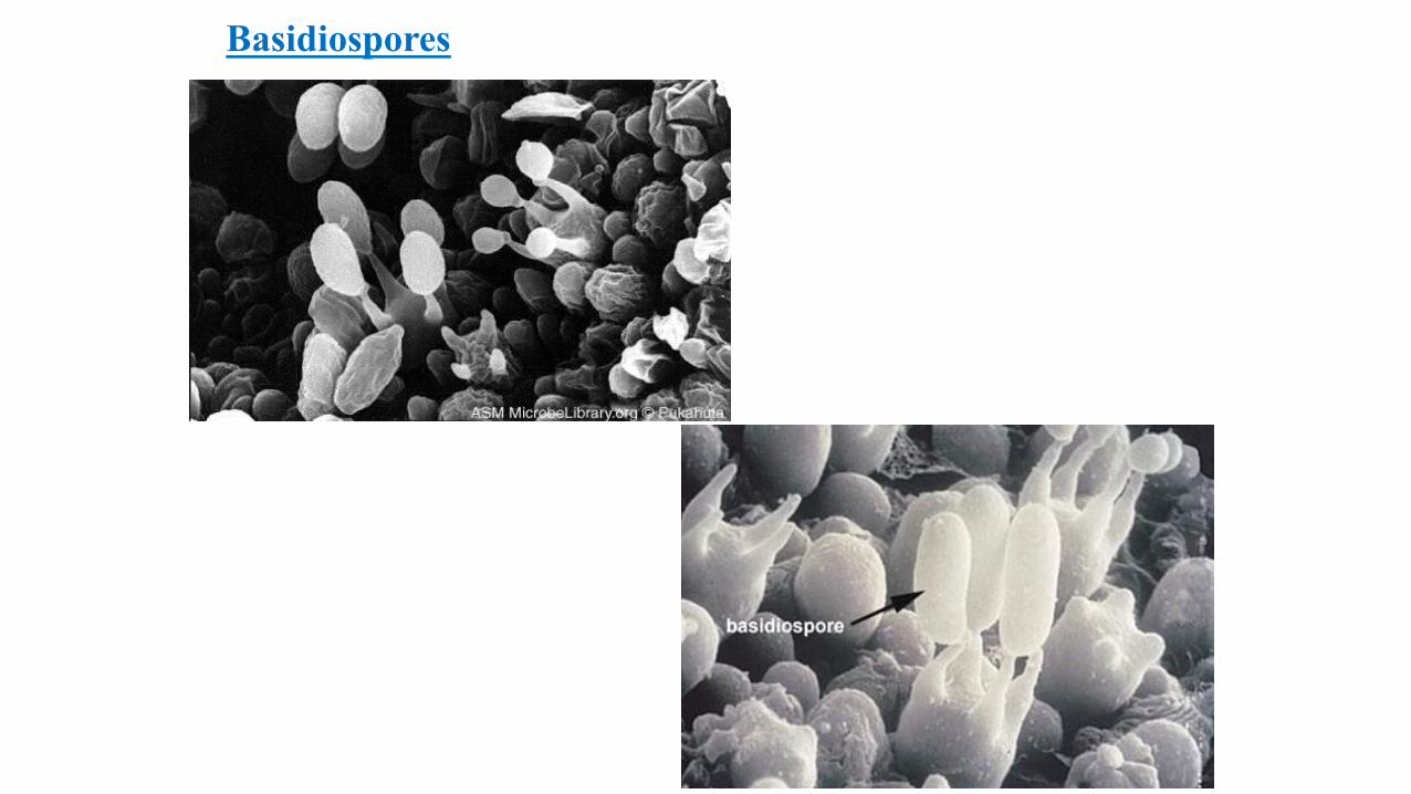
Basidiospores
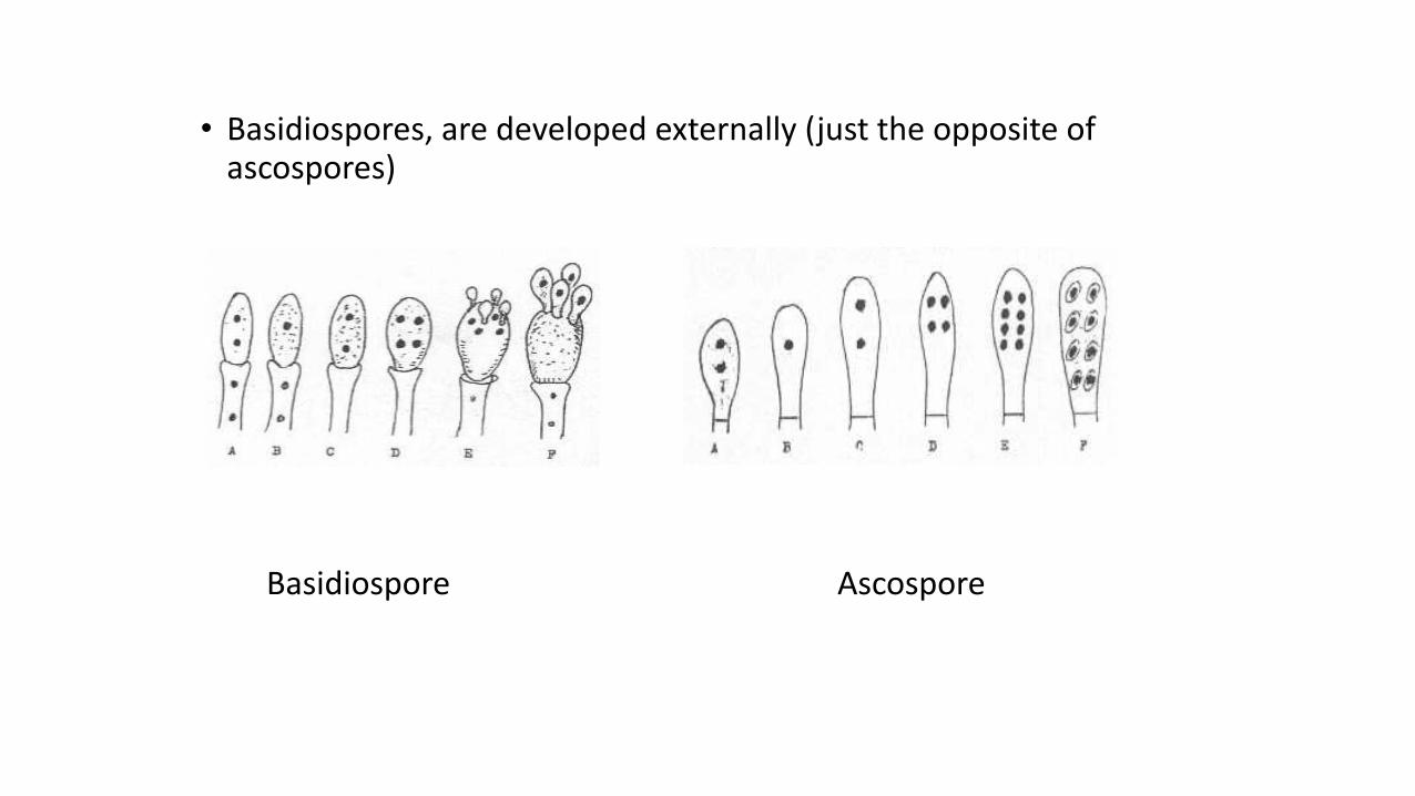
• Basidiospores, are developed externally (just the opposite of ascospores)
Basidiospore Ascospore
Oospores• In Oomycetes class fungi sexual reproduction occures by oospores
• Male gamet (anteridium) is smaller than the female gamet (oogonium) and differs in shape
• Oospores are formed by the joining of these gamets
• Oospores are thick walled, round shaped, resistant to the environmental conditions and full of food
• In Saprolegnia fungi, among the gamets found on the same fungi are flamentous and originated from another hyphae
• Round and spherical shaped gamet elongated to the oogonium and joins by fertilisation tube to form oospores
• The occurence of gamets from the same fungi’s hyphae is called homotallic, from different fungal hyphae is called heterotallic
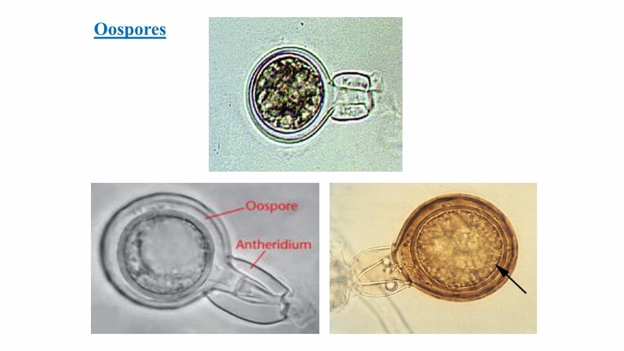
Oospores
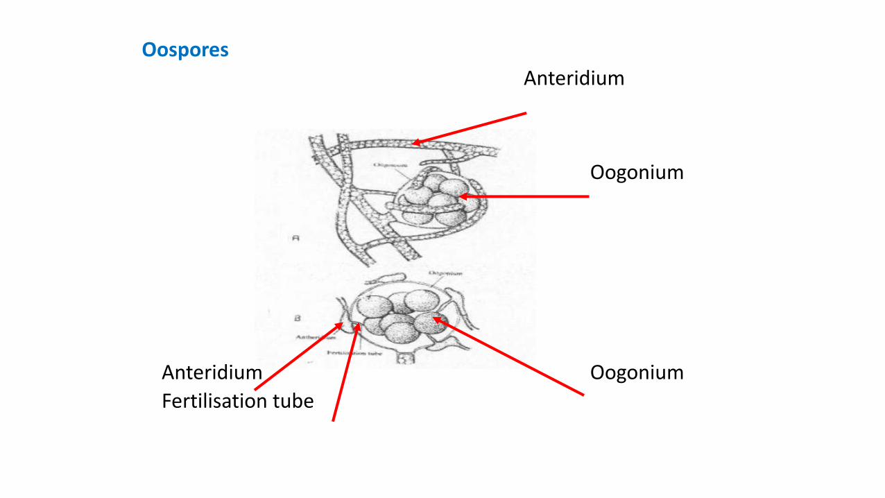
OosporesAnteridium
Oogonium
Anteridium OogoniumFertilisation tube
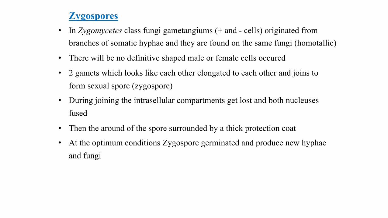
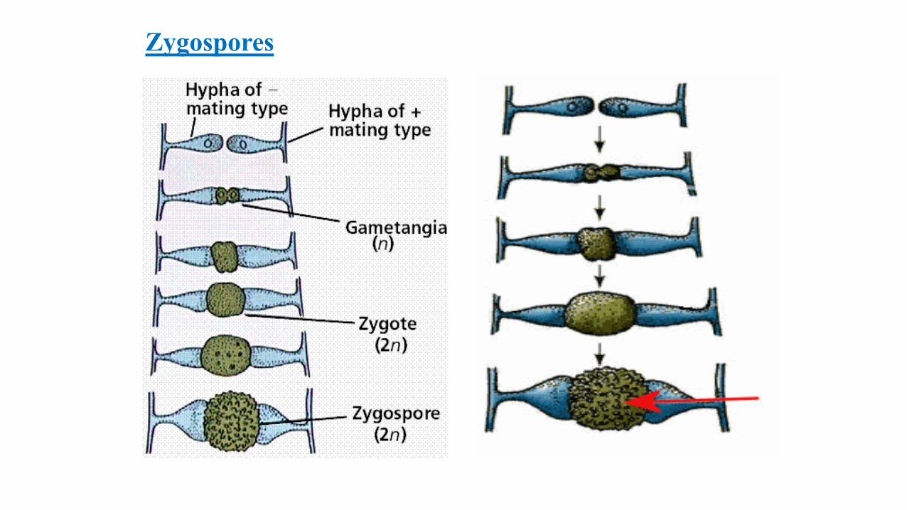
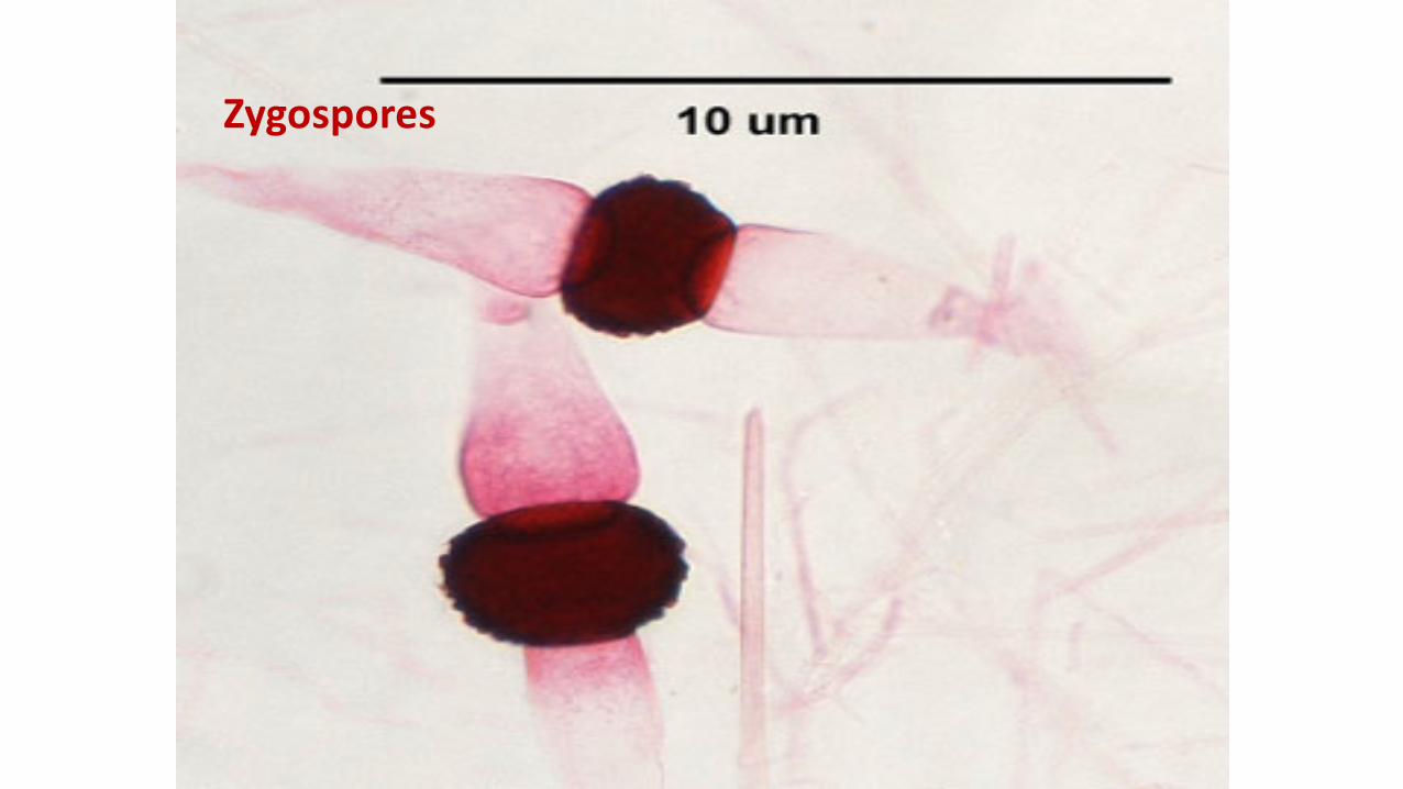
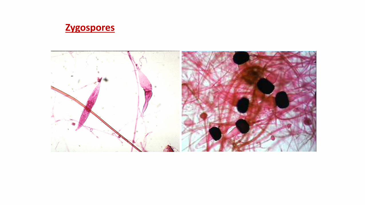
Zygospores• In Zygomycetes class fungi gametangiums (+ and - cells) originated from
branches of somatic hyphae and they are found on the same fungi (homotallic)
• There will be no definitive shaped male or female cells occured
• 2 gamets which looks like each other elongated to each other and joins to form sexual spore (zygospore)
• During joining the intrasellular compartments get lost and both nucleuses fused
• Then the around of the spore surrounded by a thick protection coat
• At the optimum conditions Zygospore germinated and produce new hyphae and fungi
Zygospores
Zygospores
Zygospores