relationships between b cell cytokine production in secondary lymphoid follicles and apoptosis of...
TRANSCRIPT

Relationships Between B Cell Cytokine Production in Secondary Lymphoid Follicles and Apoptosis of Germinal Center B Lymphocytes
Vito Pistoia, Anna Corcione Laboratory of Oncology, Scientific Institute G. Gaslini, Genova, Italy
Key Words. B lymphocytes Cytokines Lymphoid follicles Mantle zone Germinal center
Abstract. In vivo o r in vitro activated human B lymphocytes can produce a wide spectrum of cytokines which are involved in the regulation of hematopoiesis a n d of the inflammatory and immune responses. Three major B cell subsets have been identified in peripheral lymphoid organs: the germinal center (GC), the mantle zone (MZ) and the marginal zone B lymphocytes. GC and MZ B cells can be isolated as CD39- surface (s)IgD- or CD39' sIgD+ cells, respectively. Therefore, it is now possible to investigate the cytokine producing potential of purified GC and MZ B lymphocytes.
In this article, the optimal conditions for the assessment of cytokine production by human B cells are first discussed; thereafter, the spectrum of B lymphocyte-derived cytokines is described together with their possible physiological mean- ing. Next, data concerning the cytokines released in vitro by either GC or MZ B cells are presented. Some cytokines, such as granulocyte colony stim- ulating factor (G-CSF) or granulocyte-macrophage CSF (GM-CSF), are produced only by GC or MZ B lymphocytes, respectively, whereas other cytokines, such as tumor necrosis factor-a (TNF-a), inter- leukin 6 (IL-6) or IL-10 are synthesized by both B cell subsets. Finally, the relationships between B cell-derived cytokines and apoptosis of GC B lym- phocytes are discussed, and a hypothetical model of the cytokine networks in secondary lymphoid follicles is presented. I t is expected that these notions will help to clarify the pathophysiology of lymphoproliferative and autoimmune diseases.
Correspondence: Dr. Vito Pistoia, Laboratory of Oncology, Scientific Institute G. Gaslini, Largo G. Gaslini, 5-16147 Genova, Italy.
Received January 27, 1995; accepted for pub- lication January 27, 1995. OAlphaMed Press 1066- 5099/95/$5.00/0
Introduction
Recent studies have shown that human B lymphocytes, besides being responsible for anti- body production [l], may serve as potent antigen presenting cells (APC) [2] and produce a wide array of cytokines [3-331 or incompletely char- acterized soluble mediators [34-381 which are involved in the regulation of hematopoiesis, the immune response and the inflammatory process. In general, the ability of human B lymphocytes to synthesize cytokines has been investigated using freshly isolated cell suspensions or Epstein-Barr virus (EBV) infected lymphoblastoid cell lines
The availability of specific monoclonal anti- bodies (mAb) has made it possible to dissect discrete subpopulations of human B cells with distinctive functional features. Two main B cell subsets have been isolated from secondary lym- phoid follicles: the mantle zone (MZ) B cells, which are enriched for memory cells, and the germinal center (GC) B cells, which undergo selection and hypermutation of immunoglobulin (Ig) variable (V) region genes [39, 401. These processes shape the Ig gene repertoire and even- tually give rise to cells equipped with high-affin- ity antigen receptors which enter the MZ B cell pool or, alternatively, give rise to plasmablasts [39, 401. A third B cell subpopulation is repre- sented by marginal zone B cells in the spleen (or their equivalent in tonsil, lymph nodes, etc.) that are resident memory cells, as opposed to MZ B cells, which recirculate [41].
Here we shall review the results of recent studies which have addressed the characteriza- tion of cytokines produced by tonsillar B cell subsets and the relationships between apoptosis and cytokine synthesis in GC B cells.
(LCL) [3-381.
STEM CELLS 1995;13:487-500
488 B Cell Cytokine Production and Apoptosis in Secondary Lymphoid Follicles
Conditions for In Vitro Cytokine Production by Human B Lymphocytes
Studies carried out in our laboratory as well as in other laboratories have demonstrated that a) human B lymphocytes can produce numerous cytokines [3-381 and b) in vivo or in vitro activation is an absolute requirement for such production to take place [3-381. These conclusions have been reached by setting up the appropriate experimental conditions and overcoming some methodological problems that will be summarized shortly.
B Cell Sources Most of our experiments have been per-
formed using tonsillar cell suspensions [6, 21, 31, 361, since surgically removed tonsils are easily accessible and allow the recovery of suf- ficient numbers of highly purified B cells for cellular and molecular studies.
The latter achievement is more difficult to obtain when peripheral blood mononuclear cells (MNC) are used as sources of B lymphocytes since B cells represent a minor component, ranging from 1 % to lo%, of circulating MNC. On the other hand, peripheral blood B lympho- cytes represent an almost homogeneous cell population originating from the MZ of the sec- ondary lymphoid follicles [4 I], whereas tonsil- lar B cells are a mixture of MZ and GC B lymphocytes together with a third B cell sub- set anatomically located in the crypth epithe- lium and functionally related to the splenic marginal zone B lymphocytes [4 11. All of these drawbacks must be considered when compar- ing the results obtained with circulating versus tonsillar B lymphocytes. As will be discussed, the best strategy to investigate cytokine pro- duction by freshly isolated B lymphocytes is the use of highly purified B cell subsets.
EBV infected LCL are comprised of trans- formed B cells with unlimited proliferative potential [42]. LCL may be raised by in vitro infection with EBV or by spontaneous out- growth of in vivo infected B lymphocytes [42]. The immunophenotypic features of LCL resem- ble those of normal MZ cells [43]; in spite of their unrestricted proliferative capabilities, LCL do not form colonies in soft agar [44, 451 nor give rise to tumors when injected into immuno- suppressed mice [45, 461, indicating that they are not malignant. Studies carried out with LCL
have shown that they can produce numerous cytokines in vitro [3,4, 9-12, 14, 16-20, 23, 24,
A final approach to the analysis of B cell cytokine production is the study of monoclonal B lymphocyte populations frozen at various mat- urational stages and isolated from the blood, the bone marrow, the lymph nodes or the spleen of patients with lymphoproliferative disorders [6, 13, 21,25, 31, 36,47-501. Since the normal cel- lular counterparts of many malignant B cell pro- liferations have been identified, the data obtained with the latter cell fractions may provide indirect information on the cytokine-producing poten- tial of the corresponding normal B cell subsets.
26, 28-30, 33-35, 381.
The Problem of the Purity of the B Cell Suspensions
Many cytokines are produced by different cell types, raising the general problem of the purity of the cell suspensions which are being tested for their ability to synthesize a given cytokine. Therefore, a number of controls need to be performed in order to prevent minor cell contaminants from causing misleading results. The following example will help clarify what procedures may be employed.
We have recently reported that in vivo activated GC B cells isolated from human ton- sils produce granulocyte colony stimulating factor (G-CSF) in the absence of exogenous stimuli 131 1. Since the cell suspensions used throughout this study contained minor amounts of T cells, natural killer (NK) cells, macrophages, follicular dendritic cells (FDC) and plasma cells, the possibility that G-CSF was produced by a cell other than a B lymphocyte had to be excluded.
T or NK lymphocytes do not express the G-CSF gene upon in vitro stimulation [51, 521, thus making it unlikely that these cell types were responsible for the release of the cytokine in our model system. In contrast, freshly isolated maiig- nant plasma cells may occasionally express the G-CSF gene (B. Klein, personal communication).
Staining for intracytoplasmic immunoglob- ulin (cIg) or the PCA-I surface antigen showed that our tonsillar B cell suspensions contained only 2%-3% plasma cells and/or immunoblasts [3 I]. Furthermore, three monoclonal cell lines derived from patients with multiple myeloma failed to produce G-CSF, even upon stimula- tion with phorbol myristate acetate (PMA) and
Pistoia/Corcione 489
ionomycin (Corcione et al., unpublished obser- vations). Finally, G-CSF production by tonsillar B cell suspensions was virtually abrogated by treatment with a CD20 mAb and complement [31]. Since CD20 is expressed by B lympho- cytes until the stage of immunoblast but is lost at the plasma cell stage [53], these findings mil- itate against contaminant plasma cells being the source of G-CSF in our experimental system.
The demonstration that the G-CSF-pro- ducing cell was CD20' strongly argued also against its macrophagic or FDC origin, since neither cell type expresses CD20 [54]. However, additional controls were carried out. First, staining of the tonsillar B cell suspensions with CD14 or CD68 mAb demonstrated that CD14' or CD68' cells were usually 1% or fewer. CD 14 mAb react with monocytes-macrophages as well as FDC [54], whereas the CD68 mAb used preferentially detects GC-associated macrophages. The negligible amount of FDC in the suspensions was confirmed by microscopic observation of Giemsa-stained cytocentrifuged preparations [31].
In spite of the low proportions of FDC and in consideration of the potent antigen-presenting properties of such cells [ 5 5 ] , tonsillar B cells were exposed to a CD14 cytotoxic mAb and complement before being tested for the ability to produce G-CSF. This treatment left the synthe- sis of the cytokine unaltered, cogently arguing against an involvement of FDC or macrophages in our model system [31].
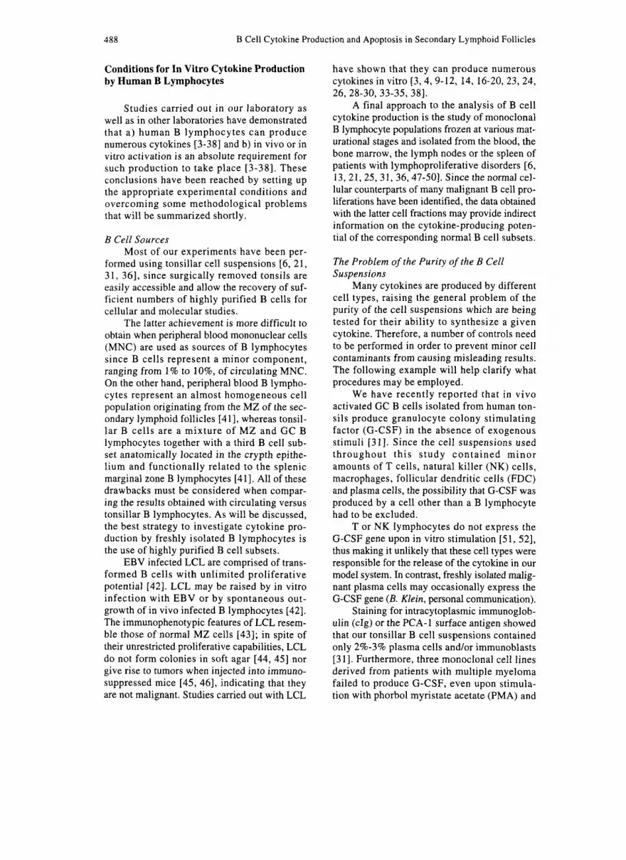
Two additional pieces of evidence definitely supported the conclusion that G-CSF was derived from in vivo activated GC B lymphocytes rather than from a contaminant cell type: a) Northern blot analysis showed that G-CSF mRNA was clearly detectable in cultured cells (Fig. l), and since the method has a minimum threshold of sensitivity of 2%-3%, the possibility that G-CSF transcripts originated from minor contaminants was virtually ruled out; and b) purified mono- clonal B cells isolated from the invaded lymph nodes of patients with follicular center cell lym- phoma (FCCL), which represents the neoplastic counterpart of GC B cells [56, 571, produced G-CSF in culture in the absence of stimuli [31].
B Cell Activation Is an Absolute Requirement for In Vitro Cytokine Production
As mentioned, B cells produce cytokines only upon in vitro or in vivo activation [3-381.
This conclusion is supported by the observa- tion that, when total tonsillar B lymphocyte sus- pensions were size-fractionated on Percoll density gradients, low density in vivo activated, but not high density, resting B cells proved capable of cytokine synthesis in unstimulated cultures [5 , 6, 21, 361. The latter cells, how- ever, acquired the ability to produce cytokines following incubation with polyclonal B cell acti- vators such as Staphylococcus aureus cowan (SAC) or anti-p immunoglobulin [5,6,21,36].
This scheme, which fits for interleukin 1 (IL- l), granulocyte-macrophage CSF (GM-CSF) or other factors [5, 6, 21, 361, does not apply to G-CSF which is produced by low density, but not high density, B cells even following in vitro stimulation [31]. The explanation for this finding is that most GC B cells migrate in the low density fraction of the Percoll gradient.
Other stimuli which may induce B cells to release cytokines or upregulate their basal rate of production are EBV [3, 4, 9-12, 14, 16-20, 23, 24, 26, 28-30, 33-35, 381 and PMA 1581. EBV may stimulate B cells in short-term cul- tures by acting as a nonspecific mitogen [7] or promote cytokine gene expression in established LCL [3, 4, 9-12, 14, 16-20, 23, 24, 26, 28-30, 33-35, 381.
When freshly isolated neoplastic B cell populations are studied, they may need to be stimulated in vitro in order to produce cytokines
1 2 3 4 5 6 7 t i
28s- I I I
18s- I Fig. 1. Northern blot analysis of G-CSF gene expres- sion in purified tonsillar GC B cells. Cells were cul- tured in the absence of stimuli for 0, 2, 8, 16 and 24 h. Total RNA was subsequently extracted and hybridized to a G-CSF-specific cDNA probe. Lanes 1-5 are GC B cells harvested after 0, 2, 8, 16 and 24 h in culture. Lane 6 is the negative control, i.e., the K562 ery- throleukemia cell line. Lane 7 is the positive control, i.e., normal peripheral blood mononuclear cells that had been cultured for 6 h in the presence of phyto- hemagglutinin and PMA.

490 B Cell Cytokine Production and Apoptosis in Secondary Lymphoid Follicles
Table I. Cytokines produced by normal human B lymphocytes C ytokine SourceP Reference
IL- 1 c1 LCL, fresh B cells
IL-4 LCL 28 IL-5 LCL 18,26 IL-6 fresh B cells, LCL 13, 19, 25, 27 IL-7 LCL 33 IL-8 fresh B cells 32 IL- 10 fresh B cells, LCL 27,29 IL- 12 LCL 17 TGF-P 1 fresh B cells 8,27 IFN-CX fresh B cells 7 IFN-y LCL 23 TNF-a LCL, fresh B cells 11,25, 27 TNF-p LCL, fresh B cells 14,30 GM-CSF fresh B cells 21 G-CSF fresh B cells 31 M-CSF fresh B cells, LCL 16
“B cell sources are indicated as LCL or freshly isolated (fresh) B cells. The latter cells were tested for cytokine gene expression after short-term culture or, less frequently, immediately after isolation. bReferences [4-61 and [9] concern data obtained by using IL-1 bioassays when tests for IL-1 c1 or IL-IP were not yet available.
4-6,9 (IL-1lb; 12, 15 (IL-la) IL- 1 p LCL, fresh B cells 4-6,9 (IL-l)b; 12, 15 (IL-la)
[6,21, 36,471. However, spontaneous synthesis of certain cytokines may occur [32,48-SO] sug- gesting the existence of in vivo activated cells within the malignant clones.
The Spectrum of Cytokines Produced by Human B Lymphocytes and Their Potential Physiological Roles
Cytokines produced by human B lympho- cytes are listed in Table I. When such cytokines are somewhat arbitrarily grouped, it appears that they include inflammatory molecules, such as I L - l a and -p, tumor necrosis factor (TNF)-a and -p, IL-6 and IL-8; interferon (1FN)-a and -y; “immunosuppressive” cytokines, such as IL-4, IL-10 and transforming growth factor (TGF)-pl; and CSFs, i.e., G-CSF, GM-CSF and macrophage CSF (M-CSF). If one attempts to envisage what the in vivo meaning of B cell cytokine synthesis may be, some points should be stressed: a) since stimulants such as SAC or anti-p antibodies bind to surface immunoglobulin (sIg), it is conceiv- able that they mimic the in vivo situation whereby antigen binds to sIg and activates signal transduction pathways culminating in cytokine production [59]; b) cytokines are by definition
short-range intercellular mediators; their pre- dominant effects are paracrine and/or autocrine and therefore restricted to the local environment where they are released. Thus, for example, CSFs produced by B lymphocytes homing in the bone marrow may contribute to promote the pro- liferation and differentiation of committed hemo- poietic progenitor cells [S 11. Likewise, release of IL-7 by bone marrow B lymphocytes would favor the differentiation of early pre-B cells [60]. However, the functional role of the same cytokines released by resident B cells located in peripheral lymphoid organs would be quite different, since a long distance interaction of such molecules with bone marrow cells would be extremely unlikely. An intermediate situation might be that of recirculating MZ B cells, which could release a given cytokine both in lymphoid organs and in other anatomical sites encountered during their migratory process [41]; c) many of the cytokines listed in Table I are B cell tropic,
and TGF-P1 [8,61-671 all exert regulatory func- tions on B cell proliferation andor differentiation in various experimental systems. These data are fully consistent with a paracrine andor autocrine model of cytokine production and utilization by B cells; and d) some B cell-derived cytokines,
thus, IL-1, IL-6, IL-10, IFN-y, TNF-a, TNF-P
Pistoia/Corcione 49 1
such as IFN-y or TNF-a might modulate the antigen-presenting functions of human B cells as well as bystander macrophages or FDC, for example by upregulating the expression of major histocompatibility complex (MHC) molecules 1681.
Although additional hypotheses may be proposed to assign a physiological role to B cell-derived cytokines, it is reasonable to con- clude that the latter cytokines contribute to amplify many cellular responses of the B cells themselves and of different neighboring cell types, such as macrophages, T cells, NK cells and FDC. Since most B lymphocytes, with some possible exceptions [25, 381, do not appear to produce IL-2, their ability to synthesize a wide spectrum of cytokines is not sufficient to bypass the absolute need for T cell help.
Cytokine Production by Human B Cell Subpopulations
Recently it was shown that distinctive immunophenotypic markers allow the separa- tion of GC from MZ B cells. MZ B cells express CD39 and sIgD (which are virtually absent on GC B cells) and sIgM [69]. In contrast, GC B cells express CD10, CD38 and sIgG (which are almost undetectable in MZ B cells) and are partly sIgM+ [69]. Furthermore, GC B cells selectively express peanut agglutinin binding sites [70] and a subset of GC B cells expresses the neutral phospholipid antigen CD77 [71].
Two groups have independently investi- gated the expression of cytokine encoding genes in freshly isolated GC or MZ B cells. Schena et al., using Northern blot analysis, found that IL-lp, IL-6, TNF-a and TGF-p1 genes were expressed in MZ B cells, whereas GC B cells displayed a weaker positivity for TNF-a and TGF-P1 mRNA [25]. In contrast, Butch et al., using a more sensitive reverse transcriptase-poly- merase chain reaction (RT-PCR) technique, failed to detect any expression of 10 cytokine genes either in MZ or in GC B cells [72].
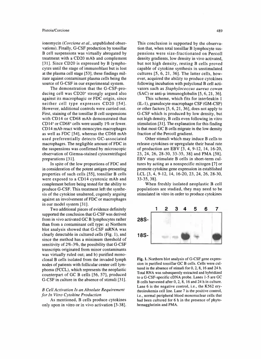
These discrepancies prompted us to inves- tigate the ability of isolated GC and M Z B cells to produce a panel of cytokines in vitro. The fol- lowing strategy was employed: a) the two cell fractions were prepared from large, in vivo acti- vated B cells by immune rosetting for CD39 and sIgD; and b) GC and MZ cells were subsequently
cultured for 24-48 h in the absence of stimuli, and the resulting supernatants were tested for the pres- ence of cytokines by immunoassays or bioassays.
The cytokines selected for the study were: TNF-a, IL-6, IL-10 and GM-CSF, whose pro- duction by normal and/or neoplastic human B cells has already been reported [13, 21, 24, 25, 29,48,49, 731; and G-CSF, which is produced by a few murine B lymphoma cell lines [72,73].
Many of the data presented are preliminary and will be published elsewhere in definitive form (Corcione et al., manuscript in preparation).
G-CSF
11-6
GY-CSF
11-10
TNF-0
-I-'- r
o m (w ?w
whnl
6 m WI <w m zm
Fig. 2. In vitro cytokine production by GC and MZ large B cells isolated from human tonsils. The two subsets were purified as CD39-, sIgD- and CD39+, sIgD+ cells, respectively, and cultured for 24 h in the absence of stimuli. Supernatants were subsequently harvested and tested for the presence of G-CSF, IL-6, GM-CSF, IL-10 and TNF-a by ELISA. Results are expressed as pg/ml.
492 B Cell Cytokine Production and Apoptosis in Secondary Lymphoid Follicles
Figure 2 reports the results of two representative experiments carried out for each cytokine and corroborated by mRNA studies (data not shown). G-CSF was produced exclusively by GC B cells, whereas GM-CSF synthesis was restricted to MZ B cells. TNF-a, IL-6 and IL-10 were produced by both CC and MZ B cells, with a trend toward a higher release of TNF-a and IL-6 in MZ cells and of IL-10 in GC cells (Fig. 2). It should be noted that all of the data presented in Figure 2 were obtained by enzyme-linked immunosorbent assays (ELISA), which detect cytokines by their immunoreactivity. However, at least for GM-CSF and G-CSF, evidence was collected that they were biologically active [21, 3 11.
Cytokine-producing, in vivo activated B cells exist in certain disease states, in which they might contribute to the pathogenesis of some clinical manifestations. For example, in situ studies with tonsils from patients with acute infectious mononucleosis have shown that EBV-infected B cells express TNF-P, TNF-a and, to a lesser extent, IL-6 mRNA [74]. All of these cytokines are endowed with potent pro- inflammatory activities. Such findings suggest that our in vitro observations may bear some relevance to B cell pathophysiology.
We next investigated whether or not rest- ing tonsillar B cells could be triggered to pro- duce G-CSF, TNF-a or GM-CSF upon in vitro exposure to the following stimuli: SAC or anti-p antibodies in the presence or absence of IL-2 or IL-4; PMA and ionomycin; and a CD40 mAb with or without IL-4. CD40 is a cell surface mol- ecule that plays a key role in B lymphocyte pro- liferation and differentiation through a T cell-dependent mechanism [75-781. Activated CD4' T cells express a 39 kDa glycoprotein (gp39) which represents the physiological lig- and of CD40 (CD40L) [79, 801. The direct B cell-T cell interaction brings CD40 and CD40L in contact and initiates B lymphocyte prolifera- tion [79, 801. Activated B cells expand in the presence of IL-4 and undergo Ig isotype switch- ing [8 11. An X-linked immunodeficiency disor- der, the hyper-IgM syndrome, is characterized by overproduction of IgM in the virtual absence of IgG or IgA. A genetic defect of CD40L in helper T cells is responsible for the pathogenesis of the disease [82-861.
Most of the experiments were carried out with SAC, whereas additional stimuli were tested only if SAC was ineffective. The fol- lowing results (data not shown) were obtained:
a) G-CSF, which is produced by in vivo acti- vated GC, but not MZ, B cells (Fig. 2) [31] was not detected in resting tonsillar B cells, since most of them originate from the MZ or the marginal zone [41]. This hypothesis is sup- ported by the failure to detect G-CSF gene expression in circulating B lymphocytes, which, as mentioned, derive from the MZ [41]; b) GM-CSF is produced by in vivo activated MZ B cells (Fig. 2) and, consistently, was released by resting tonsillar B cells following in vitro stimulation [21]; and c) TNF-a, which is produced by both MZ and GC B cells (Fig. 2) , was not found in resting tonsillar B cells upon in vitro activation with the whole panel of stimuli. This finding may suggest that MZ B cells receive special stimuli in vivo which induce them to express the TNF-a gene and cannot be easily mimicked by the in vitro signals employed in this study.
In conclusion, these observations indicate that, although cellular activation is absolutely required to trigger cytokine production in human B cells, the expression of some cytokine encoding genes is differentially regulated in different B lym- phocyte subsets (e.g., G-CSF is expressed in GC but not in MZ cells, whereas GM-CSF behaves in the opposite manner). Thus, the production of certain cytokines may serve as a functional marker for discrete B cell subsets.
Modulation of Cytokine Production by Human B Cell Subsets: The Case of G-CSF
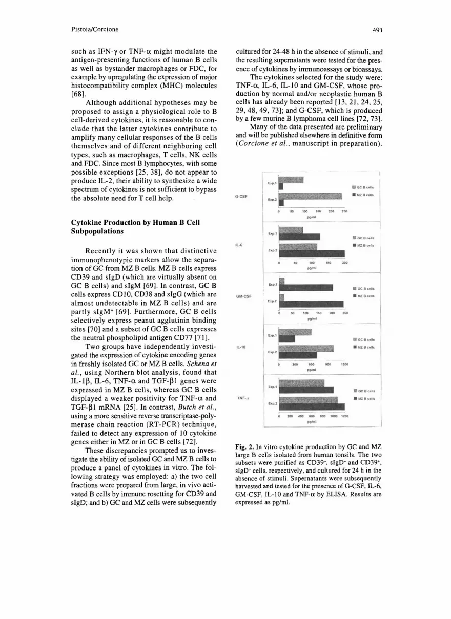
In a subsequent series of studies, we inves- tigated whether the spontaneous production of G-CSF by human GC B cells could be modu- lated by various in vitro signals. Initially, the following stimuli were tested: SAC, anti-p anti- bodies and EBV. All of them share the ability to activate B cells in a T cell-independent man- ner. As shown in Fig. 3, which reports the results of a representative experiment, SAC and anti-p antibodies had virtually no effect on G-CSF pro- duction, whereas short-term exposure to EBV (24-48 h) resulted in an increased release of the cytokine as compared with the control cultures (Corcione et al., manuscript in preparation).
The quick effect of EBV on G-CSF syn- thesis suggested that the virus acted by its abil- ity to polyclonally activate B lymphocytes rather than through B cell infection. This conclusion

PistoidCorcione 493
i CDZlmAb
1FH.a
Fig. 3. Modulation of in vitro G-CSF production by GC B cells. Cells were purified as above and cul- tured for 48 h with medium alone, SAC, anti-p anti- bodies, EBV, a CD21 mAb, rIFN-a2, a CD40 mAb, rIL-4 or the combination of CD40 mAb and IL-4. Supernatants were subsequently harvested and tested for the presence of G-CSF by ELISA. Results are expressed as pg/ml.
was reinforced by the failure to generate B cell lines with GC features upon culture of purified GC cells with EBV, in accordance with previous data [87 ] .
Since EBV binds to the cell surface through the CD21 receptor [88] and CD21 is part of a molecular complex involved in B lymphocyte activation [89], we next tested whether other lig- ands of CD21, i.e., a CD21 mAb and IFN-a [90], could mimic the effects of EBV. As shown in Figure 3, soluble CD21 mAb proved as effec- tive as EBV in augmenting G-CSF production by GC B cells, whereas the same CD21 mAb in insoluble form (data not shown) or IFN-a (Fig. 3) did not change the background release of the cytokine observed in the control cultures.
Additional stimuli that were tested for the ability to modulate G-CSF synthesis by GC B cells were a CD40 mAb, IL-4 and the combina- tion of the CD40 mAb and IL-4. As shown in Figure 3, both the CD40 mAb and IL-4 tested alone induced a statistically significant increase of G-CSF production by GC B cells. The com- bination of CD40 mAb and IL-4 was even more effective than the single components (Fig. 3). These results indicate that the spontaneous pro- duction of G-CSF by GC B cells can be modu- lated by both T cell-independent (EBV, CD21) and T cell-dependent (CD40 mAb, IL-4) stim- uli. A relationship between induction of GC B
cell proliferation and increased G-CSF release was ruled out by the observation that SAC, which was ineffective at upregulating G-CSF synthesis, was mitogenic for GC B lymphocytes (Corcione et al., unpublished observation). Conversely, the CD40 mAb or IL-4 tested alone were weakly or not at all mitogenic, respectively, although they both augmented G-CSF release (data not shown).
Relationships Between Cytokine Production in Secondary Lymphoid Follicles and Apoptosis of GC B Cells
A peculiar feature of GC B cells is their high death rate due to apoptosis [91]. Apoptosis, also known as programmed cell death, is part of the normal life of a cell as opposed to necro- sis, which represents an accidental type of cell death [91]. Apoptosis is characterized by spe- cific morphological features such as cell shrink- age, rapid blebbing of the plasma membrane (often referred to as zeiosis) and nuclear col- lapse with chromatin condensation which may lead to the formation of the so-called apoptotic bodies, i.e., chromatin fragments surrounded by plasma membrane [91].
DNA degradation associated with apoptosis may be investigated by a variety of methods, which include electrophoretic separation with the typical “ladder” pattern and flow cytomet- ric analysis of propidium iodide stained cell sus- pensions showing the presence of a hypodyploid peak. The size of this peak is directly related to the amount of apoptotic cells [91]. The first method is qualitative, whereas the second allows a quantitative estimate of apoptotic cells. It should be stressed that apoptosis may take place in certain experimental systems in the absence of detectable DNA fragmentation [91, 921.
Mature GC are comprised of two main areas, the dark and light zones [39,40]. B cells which have been activated by antigen in T cell-depen- dent areas of the lymphoid tissues colonize the follicles and undergo clonal expansion associ- ated with hypermutation of Ig-variable region genes [39, 401. Actively dividing B cell blasts seed in the dark zone and subsequently generate nonproliferating centrocytes which settle in the light zone [39,40]. Here, FDC pick up immune complexes containing antigen in native form and present it to centrocytes. A minority of centro- cytes are positively selected through the
494 B Cell Cytokine Production and Apoptosis in Secondary Lymphoid Follicles
sIg-mediated interaction with antigen held on FDC, whereas the remaining centrocytes die by apoptosis [39, 401. Positively selected centro- cytes, in turn, process and present antigen to T helper cells [93], which become activated and express CD40L which binds to centrocyte-asso- ciated CD40 [39, 761. The direct T cell-B cell interaction leads to the proliferation and sub- sequent differentiation of centrocytes into MZ memory B cells or, alternatively, plasmablasts [39,40].
In vitro studies have shown that GC B cells can be rescued from apoptosis by various stim- uli, such as anti-Ig antibodies [94], CD40 mAb [76] or CD40L [95] with or without rIL-4, CD21 mAb [96], CD38 mAb [97], EBV [98], the com- bination of I L - l a and soluble recombinant (r)CD23 1991 and IL-10 [loo]. We therefore rea- soned that an increased viability of GC B cells could account for the upregulation of G-CSF release upon incubation with certain stimuli.
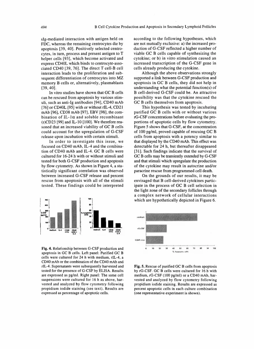
In order to investigate this issue, we focused on CD40 mAb, IL-4 and the combina- tion of CD40 mAb and IL-4. GC B cells were cultured for 16-24 h with or without stimuli and tested for both G-CSF production and apoptosis by flow cytometry. As shown in Figure 4, a sta- tistically significant correlation was observed between increased G-CSF release and percent rescue from apoptosis with all of the stimuli tested. These findings could be interpreted
mdlum IL.4 CMO clyo mAb mAb+ IL.4
according to the following hypotheses, which are not mutually exclusive: a) the increased pro- duction of G-CSF reflected a higher number of viable GC B cells capable of synthesizing the cytokine; or b) in vitro stimulation caused an increased transcription of the G-CSF gene in cells already producing the cytokine.
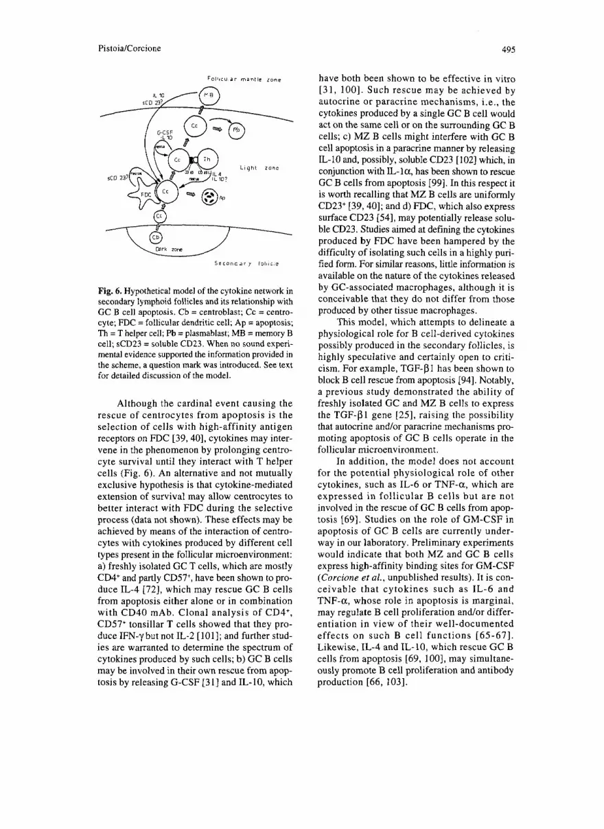
Although the above observations strongly supported a link between G-CSF production and apoptosis in GC B cells, they did not help in understanding what the potential function(s) of B cell-derived G-CSF could be. An attractive possibility was that the cytokine rescued the GC B cells themselves from apoptosis.
This hypothesis was tested by incubating purified GC B cells with or without various rG-CSF concentrations before evaluating the pro- portions of apoptotic cells by flow cytometry. Figure 5 shows that G-CSF, at the concentration of 100 pg/ml, proved capable of rescuing GC B cells from apoptosis with a potency similar to that displayed by the CD40 mAb. This effect was detectable for 24 h, but thereafter disappeared [31]. Such findings indicate that the survival of GC B cells may be transiently extended by G-CSF and that stimuli which upregulate the production of the cytokine may result in autocrine and/or paracrine rescue from programmed cell death.
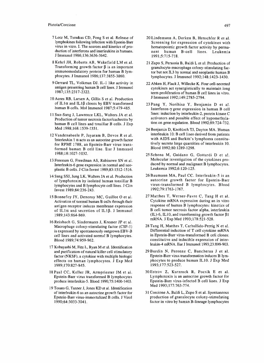
On the grounds of our results, it may be envisaged that B cell-derived cytokines partic- ipate in the process of GC B cell selection in the light zone of the secondary follicles through a complex network of cellular interactions which are hypothetically depicted in Figure 6.
Fig. 4. Relationship between Ci-CSF production and apoptosis in GC B cells. Left panel: Purified GC B cells were cultured for 24 h with medium, rIL-4, a CD40 mAb or the combination of the CD40 mAb and rIL-4. Supernatants were subsequently harvested and tested for the presence of G-CSF by ELISA. Results are expressed as pglml. Right panel: The same cell suspensions were cultured for 16 h as above, har- vested and analyzed by flow cytometry following propidium iodide staining (see text). Results are. expressed as percentage of apoptotic cells.
Fig. 5. Rescue of purified GC B cells from apoptosis by rG-CSF. GC B cells were cultured for 16 h with medium, rG-CSF (100 pg/ml) or a CD40 mAb, har- vested and analyzed by flow cytometry following propidium iodide staining. Results are expressed as percent apoptotic cells in each culture combination (one representative experiment is shown).
Pistoia/Corcione 495
F o l l t c u l a r mantle zone
IL 10
L i g h t z o n e
S s c o n d a r y l o l l i c l c
Fig. 6. Hypothetical model of the cytokine network in secondary lymphoid follicles and its relationship with GC B cell apoptosis. Cb = centroblast; Cc = centro- cyte; FDC = follicular dendritic cell; Ap = apoptosis; Th = T helper cell; Pb = plasmablast; MB = memory B cell; sCD23 = soluble CD23. When no sound experi- mental evidence supported the information provided in the scheme, a question mark was introduced. See text for detailed discussion of the model.
Although the cardinal event causing the rescue of centrocytes from apoptosis is the selection of cells with high-affinity antigen receptors on FDC [39,40], cytokines may inter- vene in the phenomenon by prolonging centro- cyte survival until they interact with T helper cells (Fig. 6). An alternative and not mutually exclusive hypothesis is that cytokine-mediated extension of survival may allow centrocytes to better interact with FDC during the selective process (data not shown). These effects may be achieved by means of the interaction of centro- cytes with cytokines produced by different cell types present in the follicular microenvironment: a) freshly isolated GC T cells, which are mostly CD4' and partly CD57+, have been shown to pro- duce IL-4 [72], which may rescue GC B cells from apoptosis either alone or in combination with CD40 mAb. Clonal analysis of CD4', CD57' tonsillar T cells showed that they pro- duce IFN-)I but not IL-2 [ lo l l ; and further stud- ies are warranted to determine the spectrum of cytokines produced by such cells; b) GC B cells may be involved in their own rescue from apop- tosis by releasing G-CSF [3 I] and IL- 10, which
have both been shown to be effective in vitro [31, 1001. Such rescue may be achieved by autocrine or paracrine mechanisms, i.e., the cytokines produced by a single GC B cell would act on the same cell or on the surrounding GC B cells; c) MZ B cells might interfere with GC B cell apoptosis in a paracrine manner by releasing IL-10 and, possibly, soluble CD23 [lo21 which, in conjunction with &la , has been shown to rescue GC B cells from apoptosis [99]. In this respect it is worth recalling that MZ B cells are uniformly CD23' [39,40]; and d) FDC, which also express surface CD23 [54], may potentially release solu- ble CD23. Studies aimed at defining the cytokines produced by FDC have been hampered by the difficulty of isolating such cells in a highly puri- fied form. For similar reasons, little information is available on the nature of the cytokines released by GC-associated macrophages, although it is conceivable that they do not differ from those produced by other tissue macrophages.
This model, which attempts to delineate a physiological role for B cell-derived cytokines possibly produced in the secondary follicles, is highly speculative and certainly open to criti- cism. For example, TGF-PI has been shown to block B cell rescue from apoptosis [94]. Notably, a previous study demonstrated the ability of freshly isolated GC and MZ B cells to express the TGF-P1 gene [25], raising the possibility that autocrine and/or paracrine mechanisms pro- moting apoptosis of GC B cells operate in the follicular microenvironment.
In addition, the model does not account for the potential physiological role of other cytokines, such as IL-6 or TNF-a, which are expressed in follicular B cells but are not involved in the rescue of GC B cells from apop- tosis [69]. Studies on the role of GM-CSF in apoptosis of GC B cells are currently under- way in our laboratory. Preliminary experiments would indicate that both MZ and GC B cells express high-affinity binding sites for GM-CSF (Corcione et al., unpublished results). It is con- ceivable that cytokines such as IL-6 and TNF-a, whose role in apoptosis is marginal, may regulate B cell proliferation and/or differ- entiation in view of their well-documented effects on such B cell functions [65-671. Likewise, IL-4 and IL-10, which rescue GC B cells from apoptosis [69, 1001, may simultane- ously promote B cell proliferation and antibody production [66, 1031.

496 B Cell Cytokine Production and Apoptosis in Secondary Lymphoid Follicles
Three final points should be considered: a) what i s the GC B cell type responsible for cytokine production? b) what are the mechanisms involved in cytokine-mediated rescue of GC B cells from apoptosis? and c) can the in vitro data on B cell cytokine production be transposed to the in vivo situation?
As mentioned, GC B cells are comprised of centroblasts and centrocytes which share most surface markers [39]. A possible exception is sIg, which is barely detectable in centroblasts but is expressed in centrocytes [39]. For a num- ber of technical reasons, it is virtually impossible so far to isolate pure cell fractions of centro- blasts and centrocytes. Therefore the question of whether G-CSF and IL-10 are produced by either cell type or both of them remains unanswered.
Most of the stimuli which prevent the entry of GC B cells in apoptosis, such as, for example, the combination of I L - l a and soluble CD23 or IL-10, induce de novo expression of the bcl-2 proto-oncogene [loo, 1041. No information is so far available on the mechanisms associated with G-CSF-mediated rescue of GC B cells from apoptosis. Nonetheless, other mechanisms are involved in GC B cell rescue from apoptosis, as shown by the finding that CD40L does not induce bcl-2 protein in the same cells [105].
Last, the question of whether the data on the in vitro production of cytokines by GC B cells can be confidently transferred to the in vivo situation deserves discussion. The already mentioned paper by Schena et al. suggests that cytokine production may take place in vivo in both GC and MZ B cells [25]. This conclusion would be supported by the following consid- erations: a) EBV-infected I% cells [3, 4, 9-12, 14, 16-20, 23, 24, 26, 28-30, 33-35, 381 as well neoplastic B lymphocytes [32,48-501 can pro- duce constitutively certain cytokines; b) in our studies, cytokine production by low density GC or MZ B cells, but not by high density B cells, occurred in the absence of exogenous stimuli. Therefore, cultured low density B cells may simply complete a functional pro- gram which was already going on in vivo; and c ) G-CSF mRNA was already detectable in GC B cells after a 2 h incubation (Fig. l ) , sug- gesting that cytokine gene transcription was conceivably fostered, rather than induced de novo, in cultured cells.
In conclusion, the data reviewed herein sup- port the emerging role of B cell-derived
cytokines in the regulation of B cell survival, proliferation and differentiation as well as in the functional activation of other cell types, such as T cells, NK cells, macrophages and FDC. It may be expected that this information will prove use- ful for a better understanding of the pathophys- iology of B cell lymphoproliferative disorders and of autoimmune diseases.
Acknowledgments
We thank Dr. Manlio Ferrarini for long-last- ing collaboration, suggestions and criticisms which have largely contributed to produce many of the data presented in this article. We also thank Mr. Tommaso Carlucci for the secretarial assis- tance and Dr. Fabio Chiotto for the help in the preparation of Figure 6. This work has been supported by grants from Consiglio Nazionale delle Ricerche, Progetto Finalizzato A.C.R.O. (n. 93.02316.PF39) and A.I.R.C.’94 to V.P.
References
1 Abbas AK, Lichtman AH, Pober JS. B cell acti- vation and antibody production. In: Abbas AK, Lichtman AH, Pober JS, eds. Cellular and Molecular Immunology. Philadelphia: W.B. Saunders Company, 1991: 186-203.
2 Lanzavecchia A. Receptor-mediated antigen uptake and its effect on antigen presentation to class-I1 restricted T lymphocytes. Annu Rev Immunol 1990;8:773-793.
3 Williamson BD, Carswell EA, Rubin BY et al. Human tumor necrosis factor produced by human B cell lines: synergistic cytotoxic interaction with human interferon. Proc Natl Acad Sci USA
4Scala G, Kuang YD, Hall RE et al. Accessory cell function of human B cells. I. Production of both interleukin 1-like activity and an interleukin 1 inhibitory factor by an EBV-transformed human B cell line. J Exp Med 1984;136:3636-3652.
5Matsushima K , Procopio A, Abe H et al. Production of interleukin- 1 activity by normal peripheral blood B lymphocytes. J Immunol
6 Pistoia V, Cozzolino F, Rubartelli A et al. In vitro production of interleukin- I by normal and malig- nant human B lymphocytes. J Immunol
1983;80:5397-5401.
1985;135:1132-1136.
1986; 136: 1688-1 692.
Pistoia/Corcione 497
7 Lotz M, Tsoukas CD, Fong S et al. Release of lymphokines following infection with Epstein-Ban virus in vitro. I. The sources and kinetics of pro- duction of interferons and interleukins in humans. J Immunol 1986;136:3636-3642.
8 Kehrl JH, Roberts AB, Wakefield LM et al. Transforming growth factor p is an important immunomodulatory protein for human B lym- phocytes. J Immunol 1986;137:3855-3860.
9 Gerrard TL, Volkman DJ. IL-I like activity in antigen presenting human B cell lines. J Immunol 1987; 1352317-2322.
10 Acres RB, Larsen A, Gillis S et al. Production of I L l a and IL lp clones by EBV transformed human B cells. Mol Immunol 1987;5:479-485.
11 Sun-Sang J, Lawrence LKL, Walters JA et al. Production of tumor necrosis factor/cachectin by human B cell lines and tonsillar B cells. J Exp Med 1988;168: 1539-155 1 .
12Vandenabeele P, Jayaram B, Devos R et al. Interleukin 1 CI acts as an autocrine growth factor for RPMI 1788, an Epstein-Barr virus trans- formed human B cell line. Eur J Immunol 1988;18:1027-1032.
13 Freeman G, Freedman AS, Rabinowe SN et al. Interleukin-6 gene expression in normal and neo- plastic B cells. J Clin Invest 1989;83:1512-1516.
14 Sung SSJ, Jung LK, Walters JA et al. Production of lymphotoxin by isolated human tonsillar B lymphocytes and B lymphocyte cell lines. J Clin Invest 1989;84:236-243.
15 Bonnefoy JY, Denoroy MC, Guillot 0 et al. Activation of normal human B cells through their antigen receptor induces membrane expression of I L l a and secretion of IL lP . J Immunol
16Reisbach G, Sindermann J, Kremer JP et al. Macrophage colony-stimulating factor (CSF- 1) is expressed by spontaneously outgrown EBV-B cell lines and activated normal B lymphocytes.
17 Kobayashi M, Fitz L, Ryan M et al. Identification and purification of natural killer cell stimulatory factor (NKSF), a cytokine with multiple biologic effects on human lymphocytes. J Exp Med
18 Paul CC, Keller JR, Armpriester JM et al. Epstein-Barr virus transformed B lymphocytes produce interleukin-5. Blood 1990;75: 1400-1403.
19 Tosato G, Tanner J, Jones KD et al. Identification of interleukin-6 as an autocrine growth factor for Epstein-Barr virus-immortalized B cells. J Virol
1989; 1431864-869.
Blood 1989;74:959-962.
1989;170:827-845.
1990;64:3033-3041.
20 Lindemann A, Dorken B, Henschler R et a]. Screening for expression of cytokines with hematopoietic growth factor activity by perma- nent human B-cell lines. Leukemia 1991 5715-71 8.
21 Zupo S, Perussia B, Baldi L et al. Production of granulocyte-macrophage colony-stimulating fac- tor but not IL3 by normal and neoplastic human B lymphocytes. J Immunol 1992;148:1423-1430.
22 Abken H, Fluck J, Willecke K. Four cell-secreted cytokines act synergistically to maintain long term proliferation of human B cell lines in vitro. J Immunol 1992;149:2785-2794.
23Pang Y, Norihisa Y, Benjamin D et al. Interferon-y gene expression in human B cell lines: induction by interleukin-2, protein kinase C activators and possible effect of hypomethyla- tion on gene regulation. Blood 1992;89:724-732.
24 Benjamin D, Knobloch TJ, Dayton MA. Human interleukin 10: B cell lines derived from patients with AIDS and Burkitt’s lymphoma constitu- tively secrete large quantities of interleukin 10.
25Schena M, Gaidano G, Gottardi D et al. Molecular investigation of the cytokines pro- duced by normal and malignant B lymphocytes. Leukemia 1992;6: 120-125.
26Baumann MA, Paul CC. Interleukin-5 is an autocrine growth factor for Epstein-Barr virus-transformed B lymphocytes. Blood
27Matthes T, Werner-Favre C, Tang H et al. Cytokine mRNA expression during an in vitro response of human B lymphocytes: kinetics of B cell tumor necrosis factor alpha, interleukin (1L)-6, ILlO, and transforming growth factor pl mRNA. J ExpMed 1993;178:521-528.
28 Tang H, Matthes T, Carballido-Perrig N et al. Differential induction of T cell cytokine mRNA in Epstein-Barr virus-transformed B cell clones: constitutive and inducible expression of inter- leukin-4 mRNA. Eur J Immunol 1993;23:899-903.
29Burdin N, Peronne C, Bancherau J et al. Epstein-Barr virus transformation induces B lym- phocytes to produce human ILlO. J Exp Med
30Estrov Z, Kurzrock R, Pocsik E e t al. Lymphotoxin is an autocrine growth factor for Epstein-Barr virus-infected B cell lines. J Exp Med 1993;177:763-774.
31 Corcione A, Baldi L, Zupo S et al. Spontaneous production of granulocyte colony-stimulating factor in vitro by human B-lineage lymphocytes
Blood 1992;80:1289-1298.
1992;79:1763-1767.
1993; 177523327.

498 B Cell Cytokine Production and Apoptosis in Secondary Lymphoid Follicles
is a distinctive marker of germinal center cells. J Immunol 1994; 153:2868-2877.
32Francia di Celle P, Carbone A, Marchis D et al. Cytokine gene expression in B-cell chronic lym- phocytic leukemia: evidence of constitutive interleukin-8 (IL8) mRNA expression and secre- tion of biologically active IL-8 protein. Blood
33 Benjamin D, Sharma V, Knobloch TJ et al. Human B cell lines constitutively secrete IL-7 and express IL-7 receptors. J Immunol
34Gordon J, Levy SC, Melamed MD et al. Immortalised B lymphocytes produce B cell growth factor. Nature 1984;310: 145-147.
35 Uckun FM, Vallera DA, Wee SL. B lymphocyte regulation of human hematopoiesis. J Immunol
36Pistoia V, Ghio R, Roncella S et al. Production of colony stimulating activity by normal and neoplastic human B lymphocytes. Blood
37 Feldman L, Dainiak N. B lymphocyte-derived ery- throid burst-promoting activity is distinct from other known lymphokines. Blood 89;73:1814-1821.
38 Benjamin D, Hartmann DP, Bazar LS et al. Human B cell lines can be triggered to secrete interleukin-2-like molecule. Cell Immunol
1994;84:220-228.
1994;152:4749-4757.
1985; 135138 17-3823.
1987;69: 1340-1349.
1989;121:30-48.
39 Mac Lennan I. Germinal centers. Annu Rev Immunol 1994;12: 117-139.
40 Berek C, Berger A, Ape1 M. Maturation of the immune response in germinal centers. Cell
41 Mac Lennan I, Chan E. The dynamic relation- ship between B-cell populations in adults. Immunol Today 1993;14:29-34.
42 Klein G. Viral latency and transformation: the strategy of Epstein-Barr virus. Cell 1989;58:5-8.
43Rowe M, Rowe DT, Gregory CD e t al. Differences in B cell growth phenotype reflect novel patterns of Epstein-Barr virus latent gene expression in Burkitt’s lymphoma. EMBO J
44 Nilsson K, PontCn J. Classification and biological nature of established human hematopoietic cell lines. Int J Cancer 1975;15:321-341.
45 Roncella S, Caretto P, Forni G et al. Studies on the oncogenjc potential of Epstein-Barr-virus (EBV)-infected B cells in AIDS-related disor- ders. Int J Cancer 1989;(suppl4):78-82.
1991;67:1121-1129.
1987;6:2743-275 1.
46Nilsson K, Giovanella BC, Stehlin JS et al. Tumorigenicity of human hematopoietic cell lines in athymic nude mice. Int J Cancer
47 Frishman J, Long B, Knospe W et al. Genes for interleukin 7 are transcribed in leukemic cell sub- sets of individuals with chronic lymphocytic leukemia. J Exp Med 1993; 177:955-964.
48 Cordingley FT, Bianchi A, Hoffbrand AV et al. Tumor necrosis factor as an autocrine tumor growth factor for chronic B-cell malignancies. Lancet 1988;1:969-972.
49 FOB R, Massaia M, Cardona S et al. Production of tumor necrosis factor-a by B-cell chronic lym- phocytic leukemia cells: a possible regulatory role of TNF in the progression of the disease.
5OBuschle M, Campana D , Carding SR e t al. Interferon-y inhibits apoptotic cell death in B cell chronic lymphocytic leukemia. J Exp Med
51 Clark SC, Kamen R. The human hematopoietic colony-stimulating factors. Science 1987;236:
52Cuturi MC, Anegon F, Sherman R e t al. Production of hematopoietic colony-stimulating factors by human natural killer cells. J Exp Med
53 Bhan AK, Nadler LM, Stashenko P et al. Stages of B cell differentiation in human lymphoid tis- sue. J Exp Med 1981;154:737-751.
54 Schriever F, Freedman AS, Freeman G e t al. Isolated human follicular dendritic cells display a unique antigenic phenotype. J Exp Med
55 Schriever SC, Nadler LM. The central role of follicular dendritic cells in lymphoid tissues. Adv Immunol 1992;51:243-284.
56 Jaffe ES, Shevach EM, Frank MM et al. Nodular lymphoma-evidence for origin from follicular B lymphocytes. N Engl J Med 1984;290:813-817.
57 Poppema S, Kaleta J, Hugh J et al. Neoplastic changes involving follicles: morphological, immunophenotypic and genetic diversity of lym- phoproliferations derived from germinal center and mantle zone. Immunol Rev 1992;126: 163-190.
58 Goldfeld AE, Strominger JL, Doyle C. Human tumor necrosis factor a gene regulation in phor- bol ester stimulated T and B cell lines. J Exp Med
59 Pleiman CM, D’Ambrosio D, Cambier JC. The B cell antigen receptor complex: structure and signal transduction. lmmunol Today 1994; 15:393-399.
1977; 19:337-344.
Blood 1990;76:393-401.
1993;177:213-218.
1229- 1237.
1989;169:569-587.
1989;169:2043-2060.
1991;174:73-81.
PistoidCorcione 499
60 Henney CS. Interleukin 7: effects on early events in lymphopoiesis. Immunol Today 1989;lO: 170-173.
61 Falkoff RJ, Muraguchi A, Hong J-X et al. The effects of interleukin 1 on B cell activation and proliferation. J Immunol 1983;13 12301-805.
62Romagnani S , Giudizi MG, Biagiotti R et al. Recombinant human IFN-y promotes prolifera- tion of anti-Ig activated human B lymphocytes. J Immunol 1986; 136:35 13-3516.
63 Kehrl JH, Miller A, Fauci AS. Effect of tumor necrosis factor on mitogen-activated human B cells. J Exp Med 1987;166:786-791.
64 Kehrl JH, Alvarez-Mon M, Delsing GA et al. Lymphotoxin is an important T cell-derived growth factor for human B cells. Science
65 Muraguchi A, Hirano T, Tang B et al. The essen- tial role of B cell stimulatory factor 2 (BSF-2/IL6) for the terminal differentiation of B cells. Nature
1987;238:44-46.
1988;167:332-334.
66 Rousset F, Garcia E, Defrance T et al. Interleukin 10 is a potent growth and differentiation factor for activated human B lymphocytes. Proc Natl Acad Sci USA 1992;89:1890-1893.
67 Macchia D, Almerigogna F, Parronchi P et al. Membrane tumor necrosis factor-alpha is involved i n the polyclonal B-cell activation induced by HIV-infected human T cells. Nature
68 Watanabe Y, Jacob CO. Regulation of MHC class I1 antigen expression opposing effects of tumor necrosis factor-cr on IFN-y-induced HLA-DR and its expression depends on the maturation and dif- ferentiation stage of the cell. J Immunol
69Liu YD, Joshua DE, Williams GT et al. Mechanism of antigen-driven selection in ger- minal centers. Nature 1989;342:929-932.
70 Butch AW, Nahm W. Functional properties of human germinal center B cells. Cell Immunol
71 Mangeney M, Richard Y, Coulaud D et al. CD77: an antigen of germinal center B cells entering apoptosis. Eur J Immunol 1991;21:1131-1140.
72Butch AW, Chung GH, Hoffmann JW et a]. Cytokine expression by germinal center cells. J Immunol 1993; 150:39-47.
73 O'Garra A, Stapleton G, Dhar V et al. Production of cytokines by mouse B cells: B lymphomas and normal B cells produce interleukin 10. Int Immunol 1990;2:823-834.
1993;363:464-466.
199 1 ; 146:899-905.
1992; 140~33 1-344.
74Foss HD, Herbst H, Hummel M et al. Patterns of cytokine gene expression in infectious mononucleosis. Blood 1994;83:707-712.
75 Gordon J, Millsum MJ, Guy GR et al. Resting B lymphocytes can be triggered directly through the CDw40 (Bp 50) antigen. Comparison with IL4-mediated signall ing. J Immunol
76 Bancherau J, De Paoli P, Valle A et al. Long-term human B cell lines dependent on interleukin-4 and antibody to CD40. Science 1991;251:70-73.
77Splawski JB, Fu SM, Lipsky PE. Immuno- regulatory role of CD40 in human B cell differ- entiation. J Immunol 1993;150:1276-1284.
78 Bancherau J, Bazan F, Blanchard D et al. The CD40 antigen and its ligand. Annu Rev Immunol
79Lederman S, Yellin MJ, Krichevsky A et a]. Identification of a novel surface protein on acti- vated CD4' T cells that induces contact-depen- dent B cell differentiation (help). J Exp Med
80 Noelle RJ, Roy M, Shepherd DM et al. A 39-kDa
1988;140: 1425-1432.
1994; 12188 1-922.
1992; 175: I09 1- 1 101.
protein on activated helper T cells binds CD40 and transduces the signal for cognate activation of B cells. Proc Natl Acad Sci USA 1992;89:6550-6554.
Gascan H, Aversa G , Gauchat J-F et al. Membranes of CD4+ T cells expressing T cell receptor (TcR) ap or TcR yS induce IgE synthe- sis by human B cells in the presence of IL-4. Eur J Immunol 1992;22: 11 33-1 141.
Allen RC, Armitage RJ, Conley ME et al. CD40 ligand gene defects responsible for X-linked hyper-IgM syndrome. Science 1993;259:990-993.
83 Aruffo A, Farrington M, Hollenbaugh D et al. CD40 ligand, gp39, is defective in activated T cells from patients with X-linked hyper-IgM syndrome. Cell 1993;72:29 1-300.
84 Di Santo JP, Bonnefoy JY, Gauchat JF et al. CD40 ligand mutations in X-linked immunodeficiency with hyper-IgM. Nature 1993;361:541-543.
85 Korthauer U, Graf D, Mages HW et al. Defective expression of T cell CD40 ligand causes X-linked immunodeficiency with hyper-IgM. Nature 1993;361:539-541.
86 Fuleinhan R, Ramesh N, Loh R et al. Defective expression of CD40 ligand in X chromosome-linked immunoglobulin deficiency with normal or elevated IgM. Proc Natl Acad Sci USA
87Gregory CD, Edwards CF, Milner A e t al. Isolation of a normal B cell subset with a
1993;90:2170-2173.
500 B Cell Cytokine Production and Apoptosis in Secondary Lymphoid Follicles
Burkitt-like phenotype and transformation in vitro with Epstein-Barr virus. Int J Cancer
88 Nemerow GR, Wolfert R, McNaughton R et al. Identification and characterization of the Epstein-Barr virus receptor on human B lym- phocytes and its relationship to the C3d comple- ment receptor (CR2). J Virol 1985;55:347-351.
89 Tedder TF, Zhu LJ, Engel P. The CD19/CD21 signal transduction complex of B lymphocytes. Immunol Today 1994;15:437-442.
90Delcayre AX, Salas F, Mathur S et al. Epstein Barr viruskomplement C3d receptor is an inter- feron areceptor. EMBO J 1991;10:919-926.
91 Cohen JJ, Duke RC, Fadok VA et al. Apoptosis and programmed cell death in immunity. Annu Rev Immunol 1992; 10:267-293.
92 Sellins KS, Cohen JJ. Hyperthermia induces apoptosis i n thymocytes. Radiat Res
93 Kosco MH, Szakal AK, Tew JG. In vivo-obtained antigen by germinal center B cells to T cells in vitro. J Immunol 1988;140:354-360.
94 Holder MJ, Knox K, Gordon J. Factors modifying the survival of germinal center B cells. Glucocorticoids and transforming growth factor p, but not cyclosporin A or anti CD19, block surface immunoglobulin-mediated rescue from apoptosis. Eur J Immunol 1992;22:2725-2728.
95Holder MJ, Wang H, Milner AE et al. Suppression of apoptosis in normal and neoplas- tic human B lymphocytes by CD40 ligand. Eur J Immunol 1993;23:2368-2371.
96 Bonnefoy JY, Henchoz S, Hardie D et al. A sub- set of anti-CD21 antibodies promotes the rescue of germinal center B cells from apoptosis. Eur J Immunol 1993;23:969-972.
1988;42:213-219.
1991;126:88-95.
97 Zupo S, Rugari E, Dono M et al. CD38 signalling by agonistic monoclonal antibody prevents apop- tosis of human germinal center B cells. Eur J Immunol 1994;24: 1218-1225.
98Gregory CD, Dive C, Henderson S et al . Activation of Epstein-Barr virus latent genes pro- tects human B cells from apoptosis. Nature
99 Liu YJ, Cairns JA, Holder MJ et at. Recombinant 25 Kd CD23 and interleukin 1 01 promote survival of germinal center B cells: evidence for bifurcation in the development of centrocytes rescued from apoptosis. Eur J Immunol 1991;21:1107-1114,
100 Levy Y, Brouet JC. Interleukin-10 prevents spon- taneous death of germinal center B cells by induc- tion of the bcl-2 protein. J Clin Invest
101Velardi A, Mingari MC, Moretta L et al. Functional analysis of germinal center CD4+ cells with natural killer cell-related features. Divergence from typical T helper cells. J Immunol 1986;137:2808-2813.
102 Sarfati M, Bron D, Lagneaux L et al. Elevation of IgE binding factors in serum of patients with B cell-derived chronic lymphocytic leukemia.
103 Defrance T, Vanbervliet B, Aubry JP et al. B cell growth-promoting activity of recombinant human interleukin-4. J Immunol 1987;139:1135-1141.
104 Liu YJ, Mason DY, Johnson GD et al. Germinal center cells express Bcl-2 protein after activa- tion by signals which prevent their entry into apoptosis. Eur J Immunol 1991;21:1905-1910.
105Holder MJ, Wang H, Milner AE et al. Suppression of apoptosis in normal and neoplas- tic human B lymphocytes by CD40 ligand is inde- pendent of Bcl-2 induction. Eur J Immunol
1991 ;349:612-614.
1994;93:424-428.
Blood 1988;71:94-98.
1993;23:2368-237 1.